Cardiac involvement in Wilson disease: Review of the ...
14
REVIEW ARTICLE Cardiac involvement in Wilson disease: Review of the literature and description of three cases of sudden death Kevin Chevalier 1,2 | Nadia Benyounes 3 | Michaël Alexandre Obadia 1,2 | Clélie Van Der Vynckt 3 | Erwan Morvan 1,2 | Thierry Tibi 3 | Aurélia Poujois 1,2 1 Department of Neurology, Rothschild Foundation Hospital, Paris, France 2 National Reference Center for Wilson's Disease and Other Copper-Related Rare Diseases, Rothschild Foundation Hospital, Paris, France 3 Department of Cardiology, Rothschild Foundation Hospital, Paris, France Correspondence Aurélia Poujois, National Reference Center for Wilson's Disease and Other Copper-Related Rare Diseases, Rothschild Foundation Hospital, 44 Avenue Mathurin Moreau, 75019 Paris, France. Email: [email protected] Communicating Editor: Johan Lodewijk Karel Van Hove Abstract Wilson disease (WD) is a rare genetic condition that results from a build-up of copper in the body. It requires life-long treatment and is mainly characterized by hepatic and neurological features. Copper accumulation has been reported to be related to the occurrence of heart disease, although little is known regarding this association. We have conducted a systematic review of the liter- ature to document the association between WD and cardiac involvement. Thirty-two articles were retained. We also described three cases of sudden death. Cardiac manifestations in WD include cardiomyopathy (mainly left ven- tricular (LV) remodeling, hypertrophy, and LV diastolic dysfunction, and less frequently LV systolic dysfunction), increased levels of troponin, and/or brain natriuretic peptide, electrocardiogram (ECG) abnormalities, and rhythm or conduction abnormalities, which can be life-threatening. Dysautonomia has also been reported. The mechanism of cardiac damage in WD has not been elucidated. It may be the result of copper accumulation in the heart, and/or it could be due to a toxic effect of copper, resulting in the release of free oxygen radicals. Patients with signs and/or symptoms of cardiac involvement or who have cardiovascular risk factors should be examined by a cardiologist in addi- tion to being assessed by their interdisciplinary treating team. Furthermore, ECG, cardiac biomarkers, echocardiography, and 24-hours or more of Holter monitoring at the diagnosis and/or during the follow-up of patients with WD need to be evaluated. Cardiac magnetic resonance imaging, although not always available, could also be a useful diagnostic tool, allowing assessment of the risk of ventricular arrhythmias and further guidance of the cardiac workup. KEYWORDS arrhythmias, cardiomyopathy, copper, dysautonomia, sudden death, Wilson disease Synopsis Cardiac manifestations in Wilson disease (WD) are not uncommon and can be life-threatening. The indications of electrocardiogram, cardiac biomarkers, echocardiography, and 24-hour Holter monitoring at the diagnosis and/or Kevin Chevalier and Nadia Benyounes should be considered joint first authors. Received: 5 March 2021 Revised: 12 July 2021 Accepted: 14 July 2021 DOI: 10.1002/jimd.12418 J Inherit Metab Dis. 2021;44:1099–1112. wileyonlinelibrary.com/journal/jimd © 2021 SSIEM. 1099
-
Upload
khangminh22 -
Category
Documents
-
view
1 -
download
0
Transcript of Cardiac involvement in Wilson disease: Review of the ...
R E V I EW AR T I C L E
Cardiac involvement in Wilson disease: Review of theliterature and description of three cases of sudden death
Kevin Chevalier1,2 | Nadia Benyounes3 | Michaël Alexandre Obadia1,2 |
Clélie Van Der Vynckt3 | Erwan Morvan1,2 | Thierry Tibi3 | Aurélia Poujois1,2
1Department of Neurology, RothschildFoundation Hospital, Paris, France2National Reference Center for Wilson'sDisease and Other Copper-Related RareDiseases, Rothschild Foundation Hospital,Paris, France3Department of Cardiology, RothschildFoundation Hospital, Paris, France
CorrespondenceAurélia Poujois, National ReferenceCenter for Wilson's Disease and OtherCopper-Related Rare Diseases, RothschildFoundation Hospital, 44 AvenueMathurin Moreau, 75019 Paris, France.Email: [email protected]
Communicating Editor: JohanLodewijk Karel Van Hove
Abstract
Wilson disease (WD) is a rare genetic condition that results from a build-up of
copper in the body. It requires life-long treatment and is mainly characterized
by hepatic and neurological features. Copper accumulation has been reported
to be related to the occurrence of heart disease, although little is known
regarding this association. We have conducted a systematic review of the liter-
ature to document the association between WD and cardiac involvement.
Thirty-two articles were retained. We also described three cases of sudden
death. Cardiac manifestations in WD include cardiomyopathy (mainly left ven-
tricular (LV) remodeling, hypertrophy, and LV diastolic dysfunction, and less
frequently LV systolic dysfunction), increased levels of troponin, and/or brain
natriuretic peptide, electrocardiogram (ECG) abnormalities, and rhythm or
conduction abnormalities, which can be life-threatening. Dysautonomia has
also been reported. The mechanism of cardiac damage in WD has not been
elucidated. It may be the result of copper accumulation in the heart, and/or it
could be due to a toxic effect of copper, resulting in the release of free oxygen
radicals. Patients with signs and/or symptoms of cardiac involvement or who
have cardiovascular risk factors should be examined by a cardiologist in addi-
tion to being assessed by their interdisciplinary treating team. Furthermore,
ECG, cardiac biomarkers, echocardiography, and 24-hours or more of Holter
monitoring at the diagnosis and/or during the follow-up of patients with WD
need to be evaluated. Cardiac magnetic resonance imaging, although not
always available, could also be a useful diagnostic tool, allowing assessment of
the risk of ventricular arrhythmias and further guidance of the cardiac
workup.
KEYWORD S
arrhythmias, cardiomyopathy, copper, dysautonomia, sudden death, Wilson disease
SynopsisCardiac manifestations in Wilson disease (WD) are not uncommon and can belife-threatening. The indications of electrocardiogram, cardiac biomarkers,echocardiography, and 24-hour Holter monitoring at the diagnosis and/or
Kevin Chevalier and Nadia Benyounes should be considered joint first authors.
Received: 5 March 2021 Revised: 12 July 2021 Accepted: 14 July 2021
DOI: 10.1002/jimd.12418
J Inherit Metab Dis. 2021;44:1099–1112. wileyonlinelibrary.com/journal/jimd © 2021 SSIEM. 1099
during the follow-up of patients with WD need to be evaluated. Cardiac mag-netic resonance imaging, although less available, may provide valuable infor-mation regarding the amount of myocardial fibrosis, thereby allowingassessment of the risk of ventricular arrhythmias. As WD is a rare disease,there has been a paucity of large-scale studies, although they would nonethe-less be particularly useful.
1 | INTRODUCTION
Wilson disease (WD) is a rare genetic condition, withfewer than 1000 patients in France,1 that is due to abuild-up of copper in the body. It requires life-long treat-ment and is mainly characterized by hepatic and neuro-logical features due to copper accumulation. Copperaccumulation has been reported to be related to theoccurrence of heart disease.2
As early as 1987, it was stated in the literature that“Cardiac death in Wilson disease is a rare occurrence ofhistoric interest. It is suggested that possible cardiacinvolvement should be added to the clinical picture ofWilson disease involving the hepatic and central nervoussystem.”3 For the first time, the patterns of cardiac involve-ment in WD were identified as cardiomyopathy, arrhyth-mias and conduction abnormalities, dysautonomia, anddeath from cardiac dysfunction.
However, the epidemiology of this condition hasremained poorly characterized, probably due to the lackof routine cardiac evaluation in WD patients. Indeed, thelargest cohort to date of WD patients did not include car-diac follow-up.4 In a review of the literature, heart dis-ease (cardiomyopathies and arrhythmias) was reported tobe rare: “myocardial copper accumulation can cause car-diomyopathy and arrhythmias, even if clinical manifesta-tions are rare.”5
Conversely, in a study based on medico-administrative data including 463 WD patients, a 29%higher incidence of atrial fibrillation (AF) and a 55%higher risk of incident congestive heart failure (HF) werereported after adjusting for confounders. Moreover, HFoccurred significantly earlier in WD patients comparedwith non-WD patients (mean age 66 ± 18 years and 72 ±18 years respectively; P < .0001).6
The National Reference Center for Wilson's Diseasein Paris (France) manages a significant number of WDpatients, with a cohort of approximately 300 patients in2019. During the follow-up of these patients, the occur-rence of cardiac abnormalities such as HF, rhythm andconduction disorders, and cases of sudden death havebeen noted. Given the occurrence of three recentunexplained early sudden deaths (1/100 vs 1/1000 suddendeaths in the general population), we conducted a
systematic review of the literature to better document therelationship between WD and cardiac involvement.
2 | MATERIALS AND METHOD
A systematic literature search was performed to identifythe published case reports and studies regarding WD andits cardiac involvement. The literature search was con-ducted systematically using PubMed and it followed thePreferred Reporting Items for Systematic Reviews andMeta-Analyses (PRISMA) guidelines (http://www.prisma-statement.org). Studies published between 1959and December 2020 were included. The following searchterms, without any search filters, were used in PubMed:Wilson disease AND cardiac OR heart OR dysautonomiaOR cardiomyopathy OR arrhythmia OR rhythmic ORsudden death NOT Wilson [Author]. Only publicationsin English and French were retained. Articles regardingadult and pediatric WD patients were included. The rele-vant articles were selected by considering the titles,followed by the abstracts. All of the titles and abstractswere screened and cross-checked by two authors (KC andNB). Articles unrelated to WD and/or to the cardiovascu-lar system were excluded. Previous reviews of the litera-ture and their related articles were included. This reviewwas based on full-text articles only.
Three cases of WD patients who were regularlyfollowed in our expert center and who die of suddendeath were also described.
3 | RESULTS
In the first step, 568 publications were identified. Ofthese, 531 were excluded because they did not mentionWD (N = 203), were not concerning the cardiovascularsystem (N = 52) or did not mention both WD and thecardiovascular system (N = 264). An abstract was notavailable for nine publications and the full text wasnot available for one publication. Duplicate articles(N = 2) were excluded. After reviewing the full texts, weexcluded five articles (four did not mention cardiacinvolvement in WD and one was a response letter).
1100 CHEVALIER ET AL.
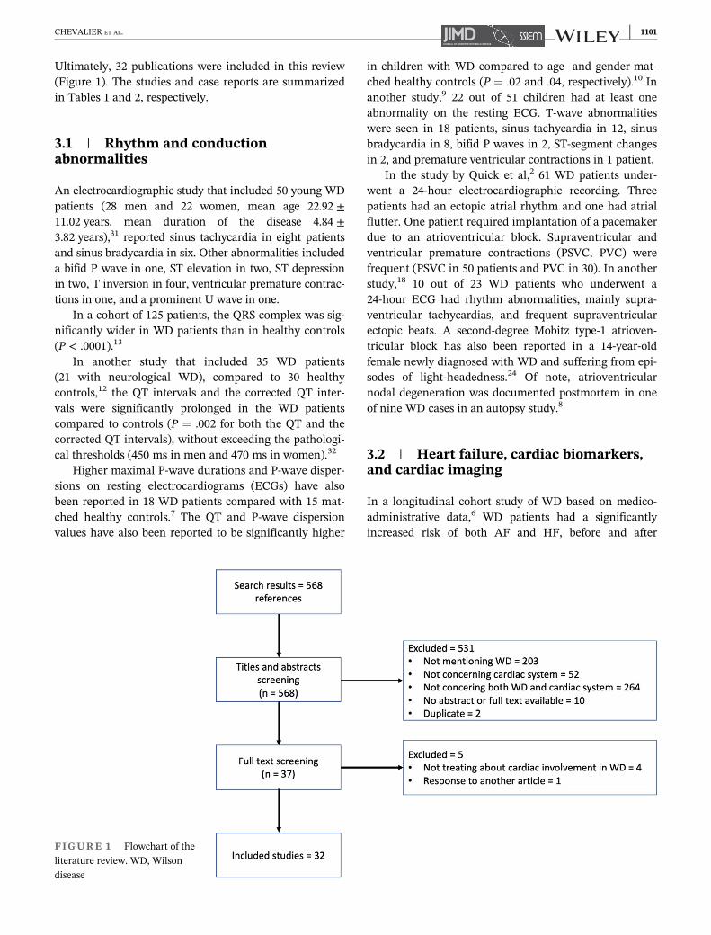
Ultimately, 32 publications were included in this review(Figure 1). The studies and case reports are summarizedin Tables 1 and 2, respectively.
3.1 | Rhythm and conductionabnormalities
An electrocardiographic study that included 50 young WDpatients (28 men and 22 women, mean age 22.92 ±11.02 years, mean duration of the disease 4.84 ±3.82 years),31 reported sinus tachycardia in eight patientsand sinus bradycardia in six. Other abnormalities includeda bifid P wave in one, ST elevation in two, ST depressionin two, T inversion in four, ventricular premature contrac-tions in one, and a prominent U wave in one.
In a cohort of 125 patients, the QRS complex was sig-nificantly wider in WD patients than in healthy controls(P < .0001).13
In another study that included 35 WD patients(21 with neurological WD), compared to 30 healthycontrols,12 the QT intervals and the corrected QT inter-vals were significantly prolonged in the WD patientscompared to controls (P = .002 for both the QT and thecorrected QT intervals), without exceeding the pathologi-cal thresholds (450 ms in men and 470 ms in women).32
Higher maximal P-wave durations and P-wave disper-sions on resting electrocardiograms (ECGs) have alsobeen reported in 18 WD patients compared with 15 mat-ched healthy controls.7 The QT and P-wave dispersionvalues have also been reported to be significantly higher
in children with WD compared to age- and gender-mat-ched healthy controls (P = .02 and .04, respectively).10 Inanother study,9 22 out of 51 children had at least oneabnormality on the resting ECG. T-wave abnormalitieswere seen in 18 patients, sinus tachycardia in 12, sinusbradycardia in 8, bifid P waves in 2, ST-segment changesin 2, and premature ventricular contractions in 1 patient.
In the study by Quick et al,2 61 WD patients under-went a 24-hour electrocardiographic recording. Threepatients had an ectopic atrial rhythm and one had atrialflutter. One patient required implantation of a pacemakerdue to an atrioventricular block. Supraventricular andventricular premature contractions (PSVC, PVC) werefrequent (PSVC in 50 patients and PVC in 30). In anotherstudy,18 10 out of 23 WD patients who underwent a24-hour ECG had rhythm abnormalities, mainly supra-ventricular tachycardias, and frequent supraventricularectopic beats. A second-degree Mobitz type-1 atrioven-tricular block has also been reported in a 14-year-oldfemale newly diagnosed with WD and suffering from epi-sodes of light-headedness.24 Of note, atrioventricularnodal degeneration was documented postmortem in oneof nine WD cases in an autopsy study.8
3.2 | Heart failure, cardiac biomarkers,and cardiac imaging
In a longitudinal cohort study of WD based on medico-administrative data,6 WD patients had a significantlyincreased risk of both AF and HF, before and after
FIGURE 1 Flowchart of the
literature review. WD, Wilson
disease
CHEVALIER ET AL. 1101
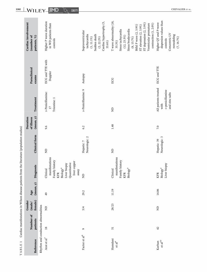
TABLE
1Cardiac
man
ifestation
sin
Wilson
diseasepa
tien
tsfrom
theliterature(pop
ulationstud
ies)
Referen
ceNumbe
rof
patients
Gen
der
(male/
female)
Age
(mea
n,y
)Diagn
osis
Clinical
form
Duration
ofillness
(mea
n,y
)Treatmen
tParac
linical
exam
s
Cardiacinvo
lvem
ent
(numbe
rof
patients,%
)
Rhythm
andcondu
ctionabnormalities
Aratet
al7
18ND
49Clin
ical
man
ifestation
sFam
ilyhistory
KFR
Biology
a
Liver
biop
syQua
ntitative
liver
copp
erassay
ND
9.6
D-Pen
icillam
ine:
17Trien
tine:1
ECGan
dTTEwith
Dop
pler
Higher
Pwavedu
ration
inWDpa
tien
tsthan
controls
Factoret
al8
95/4
29.2
ND
Hepatic:7
Neu
rologic:2
6.2
D-Pen
icillam
ine:9
Autop
sySu
praven
tricular
tach
ycardia
(1,1
1.1%
)Su
dden
death
(2,2
2.2%
)Cardiac
hyp
ertrop
hy(5,
55.6%)
Ham
dani
etal
951
28/23
11.19
Clin
ical
man
ifestation
sFam
ilyhistory
KFR
Biology
a
ND
1.48
ND
ECG
Twaveabnormality(18,
35.3%)
Sinus
tach
ycardia
(12,23.5%)
Sinus
brad
ycardia
(8,1
5.7%
)Bifid
Pwave(2,3
.9%)
STelevation(2,3
.9%)
STdepression
(2,3
.9%)
Ven
tricular
prem
ature
contraction
(1,2
.0%)
Karhan
etal
1042
ND
14.06
KFR
Biology
a
Liver
biop
sy
Hepatic:3
9Neu
rologic:3
7.0
Allpa
tien
tstreated
with
D-pen
icillam
ine
andzincsalts
ECGan
dTTE
Higher
QTan
dP-wave
dispersion
values
than
controls
Con
centricLV
remod
eling
(7,1
6.7%
)
1102 CHEVALIER ET AL.
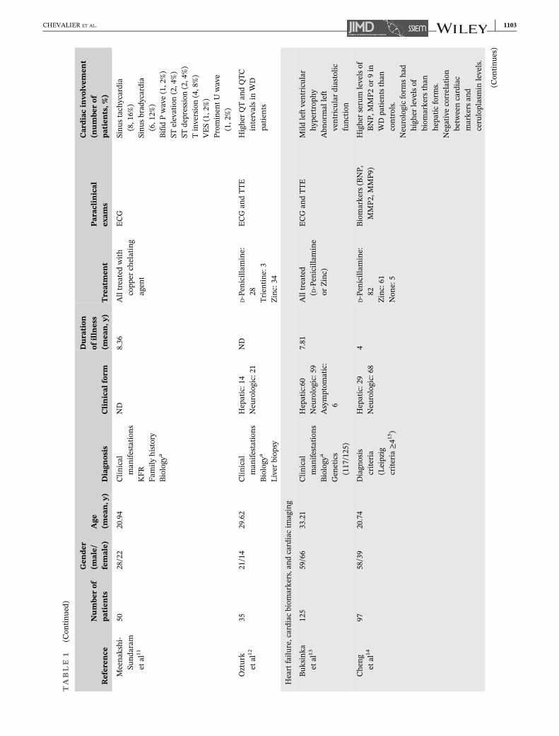
TABLE
1(Con
tinued)
Referen
ceNumbe
rof
patients
Gen
der
(male/
female)
Age
(mea
n,y
)Diagn
osis
Clinical
form
Duration
ofillness
(mea
n,y
)Treatmen
tParac
linical
exam
s
Cardiacinvo
lvem
ent
(numbe
rof
patients,%
)
Meenak
shi-
Sunda
ram
etal
11
5028/22
20.94
Clin
ical
man
ifestation
sKFR
Fam
ilyhistory
Biology
a
ND
8.36
Alltreatedwith
copp
erch
elating
agen
t
ECG
Sinus
tach
ycardia
(8,1
6%)
Sinus
brad
ycardia
(6,1
2%)
Bifid
Pwave(1,2
%)
STelevation(2,4
%)
STdepression
(2,4
%)
Tinversion(4,8%)
VES(1,2
%)
Prom
inen
tU
wave
(1,2
%)
Ozturk
etal
1235
21/14
29.62
Clin
ical
man
ifestation
sBiology
a
Liver
biop
sy
Hepatic:1
4Neu
rologic:21
ND
D-Pen
icillam
ine:
28Trien
tine:3
Zinc:34
ECGan
dTTE
Higher
QTan
dQTC
intervalsin
WD
patien
ts
Heartfailure,cardiacbiom
arkers,andcardiacim
aging
Buk
sinka
etal
13125
59/66
33.21
Clin
ical
man
ifestation
sBiology
a
Gen
etics
(117/125)
Hepatic:60
Neu
rologic:59
Asymptom
atic:
6
7.81
Alltreated
(D-Pen
icillam
ine
orZinc)
ECGan
dTTE
Mild
leftventricular
hyp
ertrop
hy
Abn
ormal
left
ventricular
diastolic
function
Chen
get
al14
9758/39
20.74
Diagn
osis
criteria
(Leipzig
criteria
≥41
5 )
Hepatic:2
9Neu
rologic:68
4D-Pen
icillam
ine:
82Zinc:61
Non
e:5
Biomarkers
(BNP,
MMP2
,MMP9
)Higher
serum
levelsof
BNP,
MMP2
or9in
WDpa
tien
tsthan
controls.
Neu
rologicform
shad
higher
levelsof
biom
arkers
than
hepaticform
s.Negativecorrelation
betw
eencardiac
markers
and
ceruloplasmin
levels.
(Con
tinue
s)
CHEVALIER ET AL. 1103
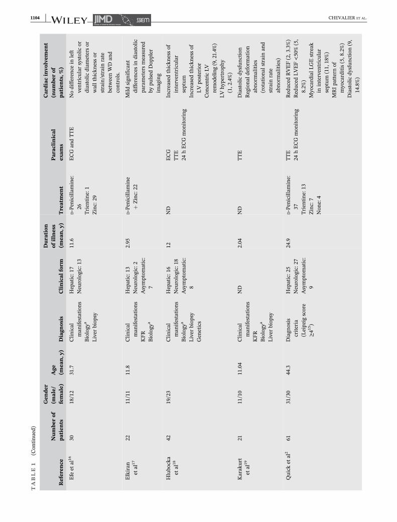
TABLE
1(Con
tinued)
Referen
ceNumbe
rof
patients
Gen
der
(male/
female)
Age
(mea
n,y
)Diagn
osis
Clinical
form
Duration
ofillness
(mea
n,y
)Treatmen
tParac
linical
exam
s
Cardiacinvo
lvem
ent
(numbe
rof
patients,%
)
Efe
etal
1630
18/12
31.7
Clin
ical
man
ifestation
sBiology
a
Liver
biop
sy
Hepatic:1
7Neu
rologic:13
11.6
D-Pen
icillam
ine:
26Trien
tine:1
Zinc:29
ECGan
dTTE
Nodifferen
cein
left
ventricular
systolicor
diastolic
diam
etersor
wallthickn
essor
strain/strainrate
betw
eenWDan
dcontrols.
Elkiran
etal
1722
11/11
11.8
Clin
ical
man
ifestation
sKFR
Biology
a
Hepatic:1
3Neu
rologic:2
Asymptom
atic:
7
2.95
D-Pen
icillam
ine
+Zinc:22
Mild
sign
ifican
tdifferen
cesin
diastolic
parametersmeasured
bypu
lsed
Dop
pler
imaging
Hlubo
cka
etal
1842
19/23
Clin
ical
man
ifestation
sBiology
a
Liver
biop
syGen
etics
Hepatic:1
6Neu
rologic:18
Asymptom
atic:
8
12ND
ECG
TTE
24hECGmon
itoring
Increasedthickn
essof
interven
tricular
septum
Increasedthickn
essof
LVpo
sterior
Con
centricLV
remodeling(9,21.4%
)LVhyp
ertrop
hy
(1,2
.4%)
Karak
urt
etal
1921
11/10
11.04
Clin
ical
man
ifestation
sKFR
Biology
a
Liver
biop
sy
ND
2.04
ND
TTE
Diastolicdy
sfun
ction
Regional
deform
ation
abnormalities
(rotational
strain
and
strain
rate
abnormalities)
Quick
etal
261
31/30
44.3
Diagn
osis
criteria
(Leipzig
score
≥41
5 )
Hepatic:2
5Neu
rologic:27
Asymptom
atic:
9
24.9
D-Pen
icillam
ine:
37Trien
tine:13
Zinc:7
Non
e:4
TTE
24hECGmon
itoring
Reduced
RVEF(2,3
.3%)
Reduced
LVEF<50%(5,
8.2%
)MyocardialL
GEstreak
ininterven
tricular
septum
(11,18%)
MRIpa
tternof
myocarditis(5,8
.2%)
Diastolicdy
sfun
ction(9,
14.8%)
1104 CHEVALIER ET AL.
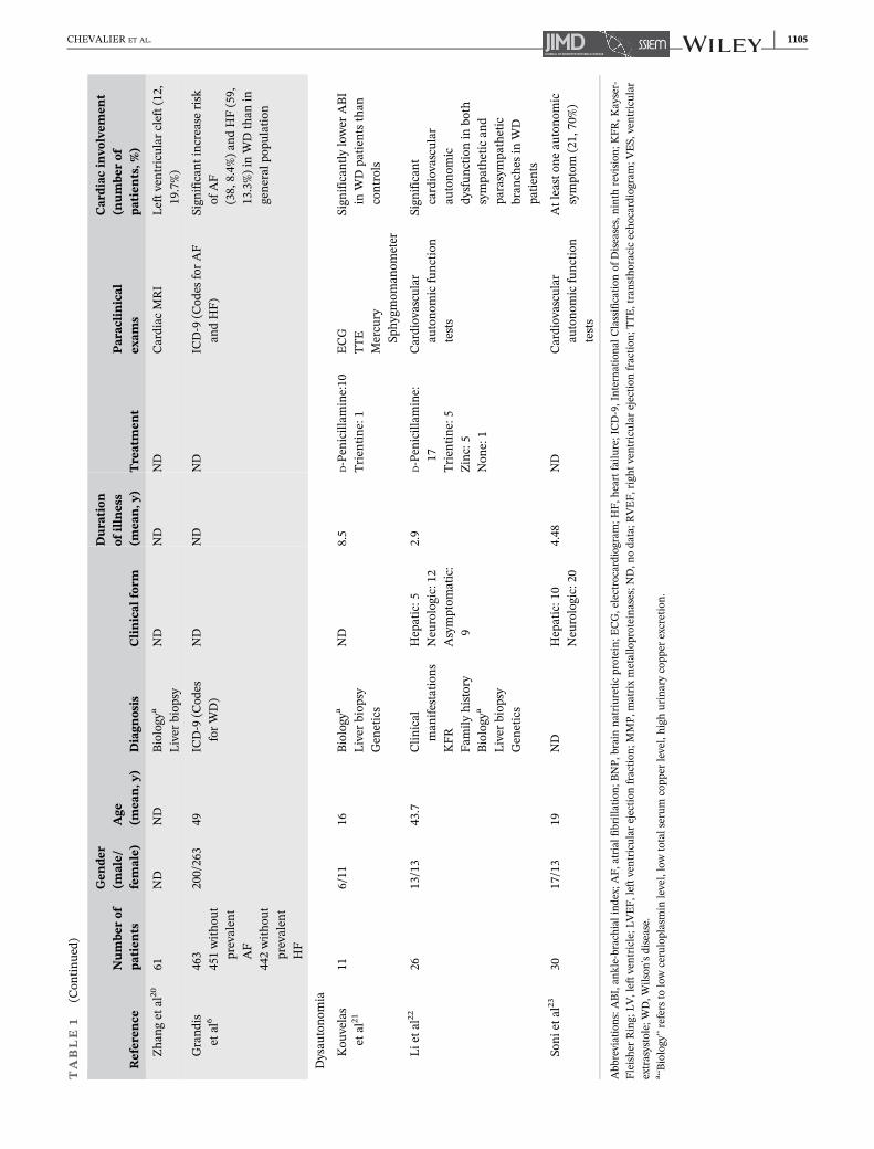
TABLE
1(Con
tinued)
Referen
ceNumbe
rof
patients
Gen
der
(male/
female)
Age
(mea
n,y
)Diagn
osis
Clinical
form
Duration
ofillness
(mea
n,y
)Treatmen
tParac
linical
exam
s
Cardiacinvo
lvem
ent
(numbe
rof
patients,%
)
Zhan
get
al20
61ND
ND
Biology
a
Liver
biop
syND
ND
ND
Cardiac
MRI
Leftventricular
cleft(12,
19.7%)
Grandis
etal
6463
451withou
tprevalen
tAF
442withou
tprevalen
tHF
200/263
49IC
D-9
(Cod
esforWD)
ND
ND
ND
ICD-9
(Cod
esforAF
andHF)
Sign
ifican
tincrease
risk
ofAF
(38,8.4%
)an
dHF(59,
13.3%)in
WDthan
ingeneral
popu
lation
Dysau
tonom
ia
Kou
velas
etal
2111
6/11
16Biology
a
Liver
biop
syGen
etics
ND
8.5
D-Pen
icillam
ine:10
Trien
tine:1
ECG
TTE
Mercury
Sphygmom
anom
eter
Sign
ifican
tlylower
ABI
inWDpa
tien
tsthan
controls
Lietal
2226
13/13
43.7
Clin
ical
man
ifestation
sKFR
Fam
ilyhistory
Biology
a
Liver
biop
syGen
etics
Hepatic:5
Neu
rologic:12
Asymptom
atic:
9
2.9
D-Pen
icillam
ine:
17Trien
tine:5
Zinc:5
Non
e:1
Cardiovascular
autonom
icfunction
tests
Sign
ifican
tcardiovascular
autonom
icdy
sfun
ctionin
both
sympa
thetican
dpa
rasympa
thetic
bran
ches
inWD
patien
ts
Sonietal
2330
17/13
19ND
Hepatic:1
0Neu
rologic:20
4.48
ND
Cardiovascular
autonom
icfunction
tests
Atleaston
eau
tonom
icsymptom
(21,
70%)
Abb
reviations:ABI,an
kle-brachialindex;AF,atrialfibrilla
tion
;BNP,
brainnatriureticprotein;E
CG,electrocardiogram
;HF,h
eartfailure;IC
D-9,International
Classificationof
Diseases,ninth
revision
;KFR,K
ayser-
Fleisher
Ring;
LV,leftventricle;L
VEF,leftventricular
ejection
fraction
;MMP,
matrixmetalloproteinases;N
D,n
oda
ta;R
VEF,rightventricular
ejection
fraction
;TTE,transthoracicechocardiogram;V
ES,
ventricular
extrasystole;W
D,W
ilson
'sdisease.
a “Biology”refers
tolow
ceruloplasmin
level,low
totalserum
copp
erlevel,highurinarycopp
erexcretion.
CHEVALIER ET AL. 1105
TABLE
2Cardiac
man
ifestation
sin
Wilson
diseasepa
tien
tsfrom
theliterature(caserepo
rts)
Referen
ceNumbe
rof
patients
Gen
der
(male/
female)
Age
(mea
n,
y)Diagn
osis
Clinical
form
Duration
ofillness(m
ean,y
)Treatmen
tParac
linical
exam
sCardiacinvo
lvem
ent
Rhythm
andcondu
ctionabnormalities
Bajajet
al24
1Fem
ale
14Clinical
man
ifestation
sKFR
Biology
a
Neu
rologic
0Non
eECGan
dTTE
Seconddegree
Mob
itztype
1atrioven
tricularnod
alblack
Heartfailu
re,cardiac
biom
arkers,andcardiacim
aging
Ada
ret
al25
1Fem
ale
16Biology
aHepatic
0Non
eECGan
dTTE
Tak
otsubo
cardiomyopa
thy
Azevedo
etal
261
Male
10Clinical
man
ifestation
sFam
ilyhistory
Biology
a
Hepatic
0D- Pe
nicillamine
ECG
Phon
ocardiogram
Vectorcardiogram
Myocardium
biop
sy
Sign
sof
myocardiald
amageon
ECG,
vectorcardiogram
,rad
ioscop
ican
dradiologicexam
inations
Highcopp
ercontentin
the
myocardium
Böttigeran
dMollerberg2
71
Male
17Clinical
man
ifestation
sKFR
Biology
a
Autop
sy
Neu
rologic
0Non
eAutopsy
Hyp
ertrop
hicleftventricle
Highcopp
ercontentin
the
myocardium
Cardiac
arrestor
deathfrom
cardiacdy
sfun
ction
Bob
bioet
al28
1Male
38ND
ND
ND
Liver tran
splantation
ECG
TTE
Implan
table
cardioverter
defibrillator
Resuscitatedcardiacarrestfrom
ventricularfibrillation
New
episod
esof
ventricular
fibrillationdetected
onim
plan
table
cardioverter
defibrillator
Kad
uket
al29
1Male
14Clinical
man
ifestation
sFam
ilyhistory
Biology
a
Liver
biop
sy
Hepatic
0.25
Non
eAutopsy
Biven
tricular
heartfailure
lead
ingto
death
Kua
n30
22/0
30Clinical
man
ifestation
sKFR
Biology
a
Neu
rologic
3,5
D-Pen
icillam
ine:
2Autopsy
Death
from
ventricularfibrillation(1)
Death
from
cardiacfailure
(1)
Abb
reviations:ECG,electrocardiogram
;KFR,K
ayser-Fleisher
Ring;ND,n
oda
ta;T
TE,transthoracicechocardiogram.
a “Biology”refers
tolowceruloplasmin
level,lowtotalserum
copp
erlevel,highurinarycopp
erexcretion.
1106 CHEVALIER ET AL.
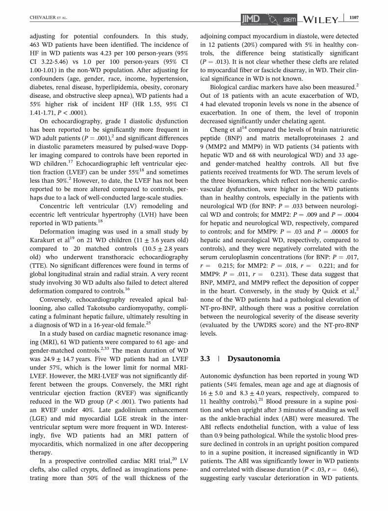
adjusting for potential confounders. In this study,463 WD patients have been identified. The incidence ofHF in WD patients was 4.23 per 100 person-years (95%CI 3.22-5.46) vs 1.0 per 100 person-years (95% CI1.00-1.01) in the non-WD population. After adjusting forconfounders (age, gender, race, income, hypertension,diabetes, renal disease, hyperlipidemia, obesity, coronarydisease, and obstructive sleep apnea), WD patients had a55% higher risk of incident HF (HR 1.55, 95% CI1.41-1.71, P < .0001).
On echocardiography, grade I diastolic dysfunctionhas been reported to be significantly more frequent inWD adult patients (P = .001),2 and significant differencesin diastolic parameters measured by pulsed-wave Dopp-ler imaging compared to controls have been reported inWD children.17 Echocardiographic left ventricular ejec-tion fraction (LVEF) can be under 55%18 and sometimesless than 50%.2 However, to date, the LVEF has not beenreported to be more altered compared to controls, per-haps due to a lack of well-conducted large-scale studies.
Concentric left ventricular (LV) remodeling andeccentric left ventricular hypertrophy (LVH) have beenreported in WD patients.18
Deformation imaging was used in a small study byKarakurt et al19 on 21 WD children (11 ± 3.6 years old)compared to 20 matched controls (10.5 ± 2.8 yearsold) who underwent transthoracic echocardiography(TTE). No significant differences were found in terms ofglobal longitudinal strain and radial strain. A very recentstudy involving 30 WD adults also failed to detect altereddeformation compared to controls.16
Conversely, echocardiography revealed apical bal-looning, also called Takotsubo cardiomyopathy, compli-cating a fulminant hepatic failure, ultimately resulting ina diagnosis of WD in a 16-year-old female.25
In a study based on cardiac magnetic resonance imag-ing (MRI), 61 WD patients were compared to 61 age- andgender-matched controls.2,33 The mean duration of WDwas 24.9 ± 14.7 years. Five WD patients had an LVEFunder 57%, which is the lower limit for normal MRI-LVEF. However, the MRI-LVEF was not significantly dif-ferent between the groups. Conversely, the MRI rightventricular ejection fraction (RVEF) was significantlyreduced in the WD group (P < .001). Two patients hadan RVEF under 40%. Late gadolinium enhancement(LGE) and mid myocardial LGE streak in the inter-ventricular septum were more frequent in WD. Interest-ingly, five WD patients had an MRI pattern ofmyocarditis, which normalized in one after decopperingtherapy.
In a prospective controlled cardiac MRI trial,20 LVclefts, also called crypts, defined as invaginations pene-trating more than 50% of the wall thickness of the
adjoining compact myocardium in diastole, were detectedin 12 patients (20%) compared with 5% in healthy con-trols, the difference being statistically significant(P = .013). It is not clear whether these clefts are relatedto myocardial fiber or fascicle disarray, in WD. Their clin-ical significance in WD is not known.
Biological cardiac markers have also been measured.2
Out of 18 patients with an acute exacerbation of WD,4 had elevated troponin levels vs none in the absence ofexacerbation. In one of them, the level of troponindecreased significantly under chelating agent.
Cheng et al14 compared the levels of brain natriureticpeptide (BNP) and matrix metalloproteinases 2 and9 (MMP2 and MMP9) in WD patients (34 patients withhepatic WD and 68 with neurological WD) and 33 age-and gender-matched healthy controls. All but fivepatients received treatments for WD. The serum levels ofthe three biomarkers, which reflect non-ischemic cardio-vascular dysfunction, were higher in the WD patientsthan in healthy controls, especially in the patients withneurological WD (for BNP: P = .033 between neurologi-cal WD and controls; for MMP2: P = .009 and P = .0004for hepatic and neurological WD, respectively, comparedto controls; and for MMP9: P = .03 and P = .00005 forhepatic and neurological WD, respectively, compared tocontrols), and they were negatively correlated with theserum ceruloplasmin concentrations (for BNP: P = .017,r = �0.215; for MMP2: P = .018, r = �0.221; and forMMP9: P = .011, r = �0.231). These data suggest thatBNP, MMP2, and MMP9 reflect the deposition of copperin the heart. Conversely, in the study by Quick et al,2
none of the WD patients had a pathological elevation ofNT-pro-BNP, although there was a positive correlationbetween the neurological severity of the disease severity(evaluated by the UWDRS score) and the NT-pro-BNPlevels.
3.3 | Dysautonomia
Autonomic dysfunction has been reported in young WDpatients (54% females, mean age and age at diagnosis of16 ± 5.0 and 8.3 ± 4.0 years, respectively, compared to11 healthy controls).21 Blood pressure in a supine posi-tion and when upright after 3 minutes of standing as wellas the ankle-brachial index (ABI) were measured. TheABI reflects endothelial function, with a value of lessthan 0.9 being pathological. While the systolic blood pres-sure declined in controls in an upright position comparedto in a supine position, it increased significantly in WDpatients. The ABI was significantly lower in WD patientsand correlated with disease duration (P < .03, r = �0.66),suggesting early vascular deterioration in WD patients.
CHEVALIER ET AL. 1107
In another study, orthostatic hypotension was reportedin 6 out of 50 WD patients.11
Cardiovascular autonomic reflexes were studied clini-cally and electrophysiologically in 30 WD patients who werecompared with 30 age- and gender-matched controls.23 Theheart rate response to the Valsalva maneuver and RR inter-val variations were significantly abnormal in WD patientscompared to controls, and the latency for the sympatheticskin response (SSR) was significantly prolonged.
In another study,11 the SSR and RR interval variabilityon deep breathing were reported in 13 out of 50 WDpatients. Dysautonomia was significantly more commonamong patients with a neurological presentation. This hasbeen confirmed by another study.22 The WD patients had ahigher heart rate at rest, a lower Valsalva ratio, a smallerheart rate increase during isometric hand grips, and alower baroreflex sensitivity during nearly all of the cardio-vascular autonomic function examinations compared withhealthy controls. Again, dysautonomia was more pro-nounced in patients with neurological symptoms.
3.4 | Cardiac arrest or death fromcardiac dysfunction in WD
In 1982, two cardiac deaths in WD patients were reportedby Kuan et al.30 The first case was a 19-year-old boy witha 10-month history of tremor and a diagnosis of WDmade 1 month before his admission. An ECG revealedatrial premature beats and ST elevation in most leads. Heexhibited an episode of ventricular fibrillation that led tohis death. The second case was a 41-year-old male whosecardiac clinical examination and ECG were normal.Despite a 6-year treatment with D-penicillamine, hedeveloped cardiomyopathy and congestive HF that wasrefractory to medical therapy and that led to his death.More recently, cardiac arrest was reported in a 38-year-old man, 1 year after curative liver transplantation forWD.28 The patient presented with ventricular fibrillationrequiring advanced cardiopulmonary resuscitation andfour defibrillations. TTE revealed a dilated left ventriclewith global hypokinesia and an LVEF of 30%. Coronaryangiography was normal. Three days after admission, acontrol TTE revealed normalized cardiac dimensions andfunction. Cardiac MRI revealed myocardial fibrosis.Endomyocardial biopsy confirmed the fibrosis, withoutinflammation. Furthermore, excessive deposition of cop-per and iron were documented: copper at 10.1 μg/g wetweight, with a normal range of 2.46-4.13 μg/g and iron at163.3 μg/g wet weight, with a normal range of35.2-71.3 μg/g. The patient received an implantablecardioverter-defibrillator. During 20 months of follow-up,he experienced a total of eight episodes of sustained
ventricular tachycardia, which were successfully treatedby the device.
The National Reference Center for Wilson's Diseasein Paris (France) has encountered three unexplained sud-den deaths in young WD patients in 2019 (over a cohortof 300 patients). These cases are described in Table 3.
Case 1 was a 33-year-old woman. WD was diagnosedas part of a familial screening. She had no other pastmedical history. Hepatic cytolysis was present at diagno-sis. She first received D-penicillamine (Trolovol, 900 mg/d), switched to zinc acetate (Wilzin, 150 mg/d) after1 year. The follow-up was uneventful. Abdominal ultra-sound, Fibroscan, liver biology, and cerebral MRI werenormal. The last biological results are included inTable 3. Neither ECG nor TTE was performed. Suddendeath occurred 1 month after the last follow-up. Theautopsy was not able to explain the reasons for death,and did not document any organic lesion. Hence, ventric-ular fibrillation was suspected to be responsible for thesudden unexplained death, as reported presciently.28
Case 2 was a 32-year-old man without past medicalhistory. WD was diagnosed a year before as he com-plained of progressive dysarthria, hypophonia, moderatedysphagia, and depression. At diagnosis, total serum cop-per was 5.84 μmol/L (N > 12.7 μmol/L), exchangeablecopper 3.86 μmol/L (0.62 < N < 1.15 μmol/L), relativeexchangeable copper (REC) 66.1% (N < 8%), andurinary copper excretion 2.1 μmol/24 hours (0.30 < N< 0.60 μmol/24 hours). Copper in the cerebrospinal fluidwas increased (1.4 μmol/L for a normal range:0.19-0.38 μmol/L). Bilateral Kayser Fleischer rings werepresent. UWDRS (Unified Wilson's Disease Rating Scale)was 21. Trientine dichlorhydrate (Trientine) was initi-ated. Despite medical therapy, the neurological stateworsened progressively. Hence, liver transplantation wasdecided 1 year after WD diagnosis. Preoperative electro-cardiogram and TTE were normal. The patient improvedpostoperatively and was discharged to a rehabilitationcenter. He died suddenly 3 months after the transplanta-tion. Of note, he was seen in a good condition 3 hoursbefore the discovery of the body. At autopsy, the onlyreported abnormalities were a small pulmonary embo-lism which cannot explain the sudden death and aknown lesion of the right pallidum. Here again, suddendeath from cardiac etiology was suspected.
Case 3 was a 45-year-old man. WD has been diag-nosed at the age of 6, revealed by hepatic cytolysis. Hispast medical history was marked by alcohol dependenceand chronic C hepatitis as a complication of the use ofintravenous drugs. He was receiving D-Penicillamine(Trolovol 600 mg/d), allowing stabilization of the disease(no neurological symptoms, normal brain MRI and noKayser-Fleischer rings). Hepatic steatosis was present.
1108 CHEVALIER ET AL.
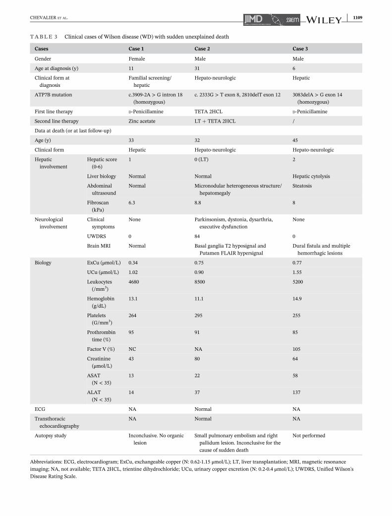
TABLE 3 Clinical cases of Wilson disease (WD) with sudden unexplained death
Cases Case 1 Case 2 Case 3
Gender Female Male Male
Age at diagnosis (y) 11 31 6
Clinical form atdiagnosis
Familial screening/hepatic
Hepato-neurologic Hepatic
ATP7B mutation c.3909-2A > G intron 18(homozygous)
c. 2333G > T exon 8, 2810delT exon 12 3083delA > G exon 14(homozygous)
First line therapy D-Penicillamine TETA 2HCL D-Penicillamine
Second line therapy Zinc acetate LT + TETA 2HCL /
Data at death (or at last follow-up)
Age (y) 33 32 45
Clinical form Hepatic Hepato-neurologic Hepato-neurologic
Hepaticinvolvement
Hepatic score(0-6)
1 0 (LT) 2
Liver biology Normal Normal Hepatic cytolysis
Abdominalultrasound
Normal Micronodular heterogeneous structure/hepatomegaly
Steatosis
Fibroscan(kPa)
6.3 8.8 8
Neurologicalinvolvement
Clinicalsymptoms
None Parkinsonism, dystonia, dysarthria,executive dysfunction
None
UWDRS 0 84 0
Brain MRI Normal Basal ganglia T2 hyposignal andPutamen FLAIR hypersignal
Dural fistula and multiplehemorrhagic lesions
Biology ExCu (μmol/L) 0.34 0.75 0.77
UCu (μmol/L) 1.02 0.90 1.55
Leukocytes(/mm3)
4680 8500 5200
Hemoglobin(g/dL)
13.1 11.1 14.9
Platelets(G/mm3)
264 295 255
Prothrombintime (%)
95 91 85
Factor V (%) NC NA 105
Creatinine(μmol/L)
43 80 64
ASAT(N < 35)
13 22 58
ALAT(N < 35)
14 37 137
ECG NA Normal NA
Transthoracicechocardiography
NA Normal NA
Autopsy study Inconclusive. No organiclesion
Small pulmonary embolism and rightpallidum lesion. Inconclusive for thecause of sudden death
Not performed
Abbreviations: ECG, electrocardiogram; ExCu, exchangeable copper (N: 0.62-1.15 μmol/L); LT, liver transplantation; MRI, magnetic resonanceimaging; NA, not available; TETA 2HCL, trientine dihydrochloride; UCu, urinary copper excretion (N: 0.2-0.4 μmol/L); UWDRS, Unified Wilson'sDisease Rating Scale.
CHEVALIER ET AL. 1109
The evolution was marked by fluctuant hepatic cytolysisdue to chronic C hepatitis and alcoholism, without cir-rhosis or hepatocarcinoma. In 2018, he had a generalizedtonic-clonic seizure and the brain MRI showed cerebellarand frontal hemorrhagic lesions. A dural fistula wassuspected. On last biological control, exchangeable cop-per was normal (0.77 μmol/L), liver enzymes were ele-vated (ASAT 58 UI/L [normal <34], ALAT 137 UI/L[normal <50]) and gamma GT at 340 UI/L (normal12-64). No ECG or TTE was performed during the follow-up. The death occurred suddenly in jail, without any pre-cursor symptom. The autopsy was not performed.
4 | DISCUSSION
This review of the literature and the description of thethree sudden deaths demonstrate that cardiac involve-ment is not an uncommon finding in WD patients.Although the mechanisms of myocardial impairment inWD are still unclear, anatomopathological examinationsare available and can help elucidate these mechanisms.
One of the first cases of cardiomyopathy associatedwith an increased copper content of the myocardium wasreported by Böttiger and Möllerberg in 1959, in a postmor-tem examination of a 17-year-old boy suffering fromhepatolenticular degeneration (HLD) who died after anunfortunate sternal puncture.27 A relationship wassuggested between copper deposition and cardiac damage.In 1978, the case of a 10-year-old boy with WD who hadsigns of myocardial damage (on ECG, vectorcardiogram,radioscopic and radiologic examinations, and hemody-namic study) was described by Azevedo et al. Biopsy of themyocardium by intracavitary puncture revealed a highlevel of copper deposition (155 μg/g of lyophilized tissue,which is 10 times the normal heart content).26 In this shortcommunication, the average copper contents of normaland WD heart tissues were reported. In another autopsycase report of a 14-year-old boy by Kaduk et al,29 postmor-tem atomic absorption spectrophotometry of the myocar-dium revealed a 100-fold increase in copper, whileultrastructural examination revealed all the features of car-diomyopathy at the cellular level. Another autopsy studyby Factor et al8 analyzed nine WD patients, newly diag-nosed (two patients) or chronically treated (seven patients,disease duration between 3 years and 17 years), comparedwith three controls. Cardiac hypertrophy was found in fiveof the nine WD patients, all chronically treated. All of thecases exhibited interstitial and replacement fibrosis,intramyocardial small vessel sclerosis, and focal inflamma-tion. A 15-year-old boy had severe atherosclerosis of the leftmain coronary artery and one patient had atrioventricularnodal degeneration. Two patients, followed up and treatedfor 4 years, died suddenly. One of them had the most
severe myocardial alterations. WD appears to be related tosudden cardiac death. This could be due to inflammation(myocarditis) secondary to copper deposits and copperaccumulation in the heart2,33 or metabolic changes similarto those observed in the liver of WD patients and animalmodels.34 Like iron, copper can produce reactive oxygenspecies, thereby inducing DNA strand breaks and the oxi-dation of nucleotide bases.35 In the heart, these oxygen rad-icals can damage cardiomyocytes.18,35 Furthermore, as inhemochromatosis, iron deposits may be involved, sinceceruloplasmin is also implicated in iron trafficking.36,37
Hence, decoppering treatments can decrease the incidenceof cardiomyopathy.2
There is a contrast between the studies that havereported no or mild cardiac involvement in WD, such asthis series of 125 WD patients who underwent ECG plusechocardiography, and where the main cardiac manifesta-tions were mild (LV hypertrophy, impaired LV diastolicfunction),13,38 or that echocardiographic series using strainand strain rate in 30 WD patients,16 and others,2,18,33 vsthe case reports of sudden cardiac death,8,28,30 includingour three case reports (see Table 3). This may be becausesome studies excluded severely ill patients16 and becausethe patients included in the majority of these studies weremostly well treated, asymptomatic, or exhibited only mildsymptoms, while cardiac damage may be more severe inless well-treated patients. Furthermore, cardiac involve-ment may be less prevalent in the relatively young patientswho were included in these studies and may appear laterduring the progression of the disease. Therefore, webelieve that cardiac explorations would be useful through-out the follow-up of these patients.
ECG abnormalities are frequent, they can occur at anearly age,9,10 and they can be secondary to the depositionof copper in the heart. A correlation between P-wave dis-persion and serum copper levels has been reported.7 Fur-thermore, P-wave dispersion has been associated with anincreased risk of AF.39-41 Indeed, in the Grandis et alpopulation-based study,6 the WD patients' incidence of AFwas 2.59 per 100 person-years (95% CI 1.83-3.55) vs 0.74per 100 person-years (95% CI 0.74-0.75) for the rest of thecohort, representing a 29% higher incidence of AF(HR 1.29, 95% CI 1.15-1.45, P < .0001). The reportedhigher risk of conduction abnormalities in WD patientscould also be secondary to copper deposits in the intracar-diac conduction pathways,2,8,24 while cardiac fibrosis, asdocumented by cardiac MRI and endomyocardial biopsy,could be responsible for ventricular arrhythmias andpredicted by QT dispersion.28,30,42 Resting ECG is a widelyavailable, easy to perform, and fast tool for rhythm assess-ment, and it should hence be performed in all WD patientsat baseline and during follow-up. Cardiac monitoring forat least 24 hours would also be useful at the time of WDdiagnosis and during follow-up, as this would help detect
1110 CHEVALIER ET AL.
rhythm and conduction abnormalities as well as stratifica-tion of the risk of severe ventricular arrhythmias.
Furthermore, morphological evaluation of myocardialinvolvement might be useful for patient care, as well asfor epidemiological purposes, in systematic and largeseries. Indeed, there is a lack of data regarding myocar-dial involvement in WD patients, due to a lack oflarge-scale studies, as cardiac imaging is not routinelyperformed in WD patients. TTE is the most readily avail-able tool for morphological analysis of cardiac structureand function, and it should hence be performed first, atbaseline and during follow-up. It allows measurement ofleft atrial and ventricular volumes and function, LVmass, LV filling pressures, and pulmonary arterial pres-sures. To date, only sporadic LV systolic dysfunction hasbeen reported in WD patients. Takotsubo cardiomyopa-thy can readily be diagnosed by TTE.25 However, tissueDoppler imaging (DTI) for LV filling pressure evaluationand myocardial deformation, which may identify earlymyocardial alterations before LV systolic dysfunction,were not available in the majority of the reported studies.Cardiac MRI additionally allows documentation of car-diac fibrosis and quantification of LGE.
Finally, biomarkers of cardiac damage, mainly tropo-nin and BNP, are thought to be associated with the sever-ity of WD and should be assessed.2 The duration of thedisease appears to be related to the risk of cardiacinvolvement,8,11 although neither the type of treatmentnor the form of the disease (except for the associationbetween neurological forms and dysautonomia) could beassociated with the cardiac events. It has been hypothe-sized that myocardial copper overload may be responsiblefor inflammation of the myocardium (myocarditis),which would improve with appropriate therapy for WD.2
To date, the frequency with which one should sched-ule follow-up testing for the cardiac rhythm, morphology,and blood tests is not yet known. Large studies with along observational period are needed to establish guide-lines in this regard.
5 | CONCLUSION
The spectrum of cardiac involvement in WD is broad andcan involve de novo patients or patients treated chroni-cally irrespective of their clinical hepatic or neurologicalphenotype. The mechanism of cardiac damage in WDmay be the consequence of copper accumulation in theheart and the consequence of a toxic effect of copper,with the release of free oxygen radicals.
We suggest that all WD patients should undergo atleast one ECG and ECG monitoring, troponin, and BNPmeasurements, and a TTE at the time of diagnosis andduring their follow-up, and patients with signs or
symptoms suggestive of cardiac involvement or who havecardiovascular risk factors should be examined by a car-diologist in addition to assessment by their interdisciplin-ary treating team. When available, cardiac MRI mayprovide prognostic information. Prospective studies thatinclude extensive and comprehensive cardiovascularevaluation of WD patients are warranted and would helpwith the specification of the spectrum of cardiac involve-ment in WD and how to document and treat them.
ACKNOWLEDGMENTSAlexandra Iglesias provided non-funded proofreading.Sophie Domingues-Montanari, PhD, provided nativeEnglish editing and was funded by the authors.
CONFLICT OF INTERESTThe authors declare no conflict of interest.
AUTHOR CONTRIBUTIONSKevin Chevalier and Nadia Benyounes wrote the manu-script. Aurélia Poujois supervised the drafting, edition,and revision of the manuscript. Michaël AlexandreObadia, Clélie Van Der Vynckt, Erwan Morvan, andThierry Tibi edited the manuscript.
INFORMED CONSENTThis article does not contain any studies with human oranimal subjects performed by any of the authors.
ORCIDKevin Chevalier https://orcid.org/0000-0001-7823-1827Aurélia Poujois https://orcid.org/0000-0002-5640-7534
REFERENCES1. Poujois A, Woimant F, Samson S, Chaine P, Girardot-
Tinant N, Tuppin P. Characteristics and prevalence of Wilson'sdisease: a 2013 observational population-based study in France.Clin Res Hepatol Gastroenterol. 2018;42(1):57-63.
2. Quick S, Reuner U, Weidauer M, et al. Cardiac and autonomicfunction in patients with Wilson's disease. Orphanet J Rare Dis.2019;14(1):22. https://ojrd.biomedcentral.com/articles/10.1186/s13023-019-1007-7
3. Kuan P. Cardiac Wilson's disease. Chest. 1987;91(4):579-583.4. Taly AB, Meenakshi-Sundaram S, Sinha S, Swamy HS,
Arunodaya GR. Wilson disease: description of 282 patientsevaluated over 3 decades. Medicine (Baltimore). 2007;86(2):112-121.
5. Ala A, Walker AP, Ashkan K, Dooley JS, Schilsky ML. Wilson'sdisease. Lancet. 2007;369(9559):397-408.
6. Grandis DJ, Nah G, Whitman IR, et al. Wilson's disease andcardiac myopathy. Am J Cardiol. 2017;120(11):2056-2060.
7. Arat N, Kacar S, Golbasi Z, et al. P wave dispersion is pro-longed in patients with Wilson's disease. World J Gastroenterol.2008;14(8):1252-1256.
8. Factor SM, Cho S, Sternlieb I, Scheinberg IH, Goldfischer S.The cardiomyopathy of Wilson's disease: myocardial alterations
CHEVALIER ET AL. 1111

in nine cases. Virchows Arch A Pathol Anat Histol. 1982;397(3):301-311.
9. Hamdani SSB, Cheema HA, Saeed A, Suleman H, Sehar T.Electrocardiographic manifestations in paediatric Wilson dis-ease. J Ayub Med Coll Abbottabad. 2018;30:22-25.
10. Karhan AN, Aykan HH, Gümüs E, et al. Assessment of cardiacfunction and electrocardiographic findings in patients withWilson's disease. Cardiol Young. 2019;29(9):1183-1188.
11. Meenakshi-Sundaram S, Taly AB, Kamath V, Arunodaya GR,Rao S, Swamy HS. Autonomic dysfunction in Wilson's disease– a clinical and electrophysiological study. Clin Auton Res.2002;12(3):185-189.
12. Ozturk S, Gurbuz AS, Efe SC, Iliaz R, Banzragch M, Demir K.QTc interval is prolonged in Wilson's disease with neurologicınvolvement. Acta Clin Belg. 2018;73:328-332.
13. Buksi�nska-Lisik M, Litwin T, Pasierski T, Członkowska A. Car-diac assessment in Wilson's disease patients based on electro-cardiography and echocardiography examination. Arch MedSci. 2019;15(4):857-864.
14. Cheng N, Wang H, Dong J, et al. Elevated serum brain natri-uretic peptide and matrix metalloproteinases 2 and 9 in Wil-son's disease. Metab Brain Dis. 2015;30(4):1087-1091.
15. Ferenci P, Caca K, Loudianos G, et al. Diagnosis and pheno-typic classification of Wilson disease: diagnosis and phenotypicclassification of Wilson disease. Liver Int. 2003;23(3):139-142.
16. Efe SC, Gurbuz AS, Ozturk S, Demir K. Strain and strain rateechocardiography variables in adult Wilson's disease patients: aspeckle tracking echocardiography study. J Clin Ultrasound.2020;48(6):324-329.
17. Elkiran O, Karakurt C, Selimoglu A, et al. Subclinical diastolicdysfunction in children with Wilson's disease assessed by tissueDoppler echocardiography: a possible early predictor of cardiacinvolvement. Acta Cardiol. 2013;68(2):181-187.
18. Hlubock�a Z, Marecek Z, Linhart A, et al. Cardiacinvolvement in Wilson disease. J Inherit Metab Dis. 2002;25(4):269-277.
19. Karakurt C, Çelik S, Selimo�glu A, Varol _I, Karabiber H,Yolo�glu S. Strain and strain rate echocardiography in childrenwith Wilson's disease. Cardiovasc J Afr. 2016;27(5):307-314.
20. Zhang K, Reuner U, Weidauer M, et al. Left ventricular clefts –incidental finding or pathologic sign of Wilson's disease.Orphanet J Rare Dis. 2019;14(1):244. https://ojrd.biomedcentral.com/articles/10.1186/s13023-019-1238-7
21. Kouvelas G, Bassareo PP, Pisano V, Demelia L, Nurchi AM,Mercuro G. Premature vascular deterioration in young patientsaffected by Wilson's disease: a pilot study. J Pediatr NeonatalIndivid Med. 2017;6(2):e060201.
22. Li K, Lindauer C, Haase R, et al. Autonomic dysfunction inWilson's disease: a comprehensive evaluation during a 3-yearfollow up. Front Physiol. 2017;8:778. http://journal.frontiersin.org/article/10.3389/fphys.2017.00778/full
23. Soni D, Shukla G, Singh S, Goyal V, Behari M. Cardiovascularand sudomotor autonomic dysfunction in Wilson's disease—limited correlation with clinical severity. Auton Neurosci. 2009;151(2):154-158.
24. Bajaj BK, Wadhwa A, Singh R, Gupta S. Cardiac arrhythmia inWilson's disease: an oversighted and overlooked entity!J Neurosci Rural Pract. 2016;7(4):587-589.
25. Adar T, Chen S, Mizrahi M. A heartbreaking case of Wilson'sdisease: Takotsubo cardiomyopathy complicating fulminanthepatic failure. Transpl Int. 2014;27(11):e109-e111.
26. Azevedo EM, Scaff M, Barbosa ER, Neto AEG, Canelas HM.Heart involvement in hepatolenticular degeneration. Acta Neu-rol Scand. 1978;58(5):296-303.
27. Böttiger LE, Möllerberg H. Increased copper content of hyper-trophic myocardium. Acta Med Scand. 2009;165(6):413-416.
28. Bobbio E, Forsgard N, Oldfors A, et al. Cardiac arrest inWilson's disease after curative liver transplantation: a life-threatening complication of myocardial copper excess? ESCHeart Fail. 2019;6(1):228-231.
29. Kaduk B, Metze K, Schmidt P-F, Brandt G. Secondaryathrocytotic cardiomyopathy ? Heart damage due to Wilson'sdisease. Virchows Arch A Pathol Anat Histol. 1980;387(1):67-80.
30. Kuan P. Fatal cardiac complications of Wilson's disease. AmHeart J. 1982;104:314-316.
31. Meenakshi-Sundaram S, Sinha S, Rao M, et al. Cardiac involve-ment in Wilson's disease—an electrocardiographic observation.J Assoc Physicians India. 2004;52:294-296.
32. Goldenberg I, Moss AJ, Zareba W. QT interval: how to measureit and what is “normal”. J Cardiovasc Electrophysiol. 2006;17(3):333-336.
33. Quick S, Weidauer M, Heidrich FM, et al. Cardiac manifestationof Wilson's disease. J Am Coll Cardiol. 2018;72(22):2808-2809.
34. Aggarwal A, Bhatt M. Wilson disease. Curr Opin Neurol. 2020;33(4):534-542.
35. Gaetke L. Copper toxicity, oxidative stress, and antioxidantnutrients. Toxicology. 2003;189(1-2):147-163.
36. Bonaccorsi di Patti MC, Cutone A, Polticelli F, et al. Theferroportin-ceruloplasmin system and the mammalian ironhomeostasis machine: regulatory pathways and the role of lac-toferrin. Biometals. 2018;31(3):399-414.
37. Roeser HP, Lee GR, Nacht S, Cartwright GE. The role of ceru-loplasmin in iron metabolism. J Clin Invest. 1970;49(12):2408-2417.
38. Dzieżyc K, Litwin T, Członkowska A. Other organ involvementand clinical aspects of Wilson disease. Handbook of ClinicalNeurology. Elsevier; 2017;142:157-169. https://linkinghub.elsevier.com/retrieve/pii/B9780444636256000136
39. Chandy J, Nakai T, Lee RJ, Bellows WH, Dzankic S, Leung JM.Increases in P-wave dispersion predict postoperative atrialfibrillation after coronary artery bypass graft surgery. AnesthAnalg. 2004;98:303-310.
40. Perzanowski C, Ho AT, Jacobson AK. Increased P-wave disper-sion predicts recurrent atrial fibrillation after cardioversion.J Electrocardiol. 2005;38(1):43-46.
41. Senen K, Turhan H, Erbay AR, et al. P-wave duration and P-wave dispersion in patients with dilated cardiomyopathy. Eur JHeart Fail. 2004;6(5):567-569.
42. Malik M, Batchvarov VN. Measurement, interpretation andclinical potential of QT dispersion. J Am Coll Cardiol. 2000;36(6):1749-1766.
How to cite this article: Chevalier K,Benyounes N, Obadia MA, et al. Cardiacinvolvement in Wilson disease: Review of theliterature and description of three cases of suddendeath. J Inherit Metab Dis. 2021;44(5):1099-1112.https://doi.org/10.1002/jimd.12418
1112 CHEVALIER ET AL.