Calixarenes as New Platforms for Drug Design
20
Current Drug Discovery Technologies, 2009, 6, 000-000 1 1570-1638/09 $55.00+.00 © 2009 Bentham Science Publishers Ltd. Calixarenes as New Platforms for Drug Design Ângelo de Fátima* ,1 , Sergio Antonio Fernandes 2 and Adão Aparecido Sabino 1 1 Grupo de Estudos em Química Orgânica e Biológica (GEQOB), Departamento de Química, ICEx, Universidade Fed- eral de Minas Ferais (UFMG), Pampulha, Belo Horizonte, MG, Brazil, 31270-901 2 Grupo de Química Supramolecular & Biomimética (GQSB), Departamento de Química, Centro de Ciências Exatas, Universidade Federal de Viçosa (UFV), Cidade Universitária, s/n, Centro, Viçosa, MG, Brazil, 36570-000 Abstract: Calixarenes, macrocyclic compounds of phenolic units linked by methylene groups at the 2,6-positions, present some of the requirements to serve as platforms for the design and synthesis of biological active compounds. They are also interesting host molecules for chemical biology study purposes. Their basic molecular scaffold has potential ability for molecule recognition; it is promptly synthesized in large amounts, and might be easily modified for maximizing molecular interactions toward relevant guest molecules. Calixarenes present well-defined conformational properties and cavities with molecular dimensions that enable to encapsulate guest drugs. Calixarenes have been shown to have antiviral, antibac- terial, antifungal, and anticancer activities (including HIV as target). We provide here an overview of the use of calixare- nes either as new chemical entity of distinct biological activities or as host for bioactive guest molecules. The importance of calixarenes for drugs development is discussed. The use of Nuclear Magnetic Resonance (NMR) and Mass Spectrome- try (MS) techniques for the study of calixarenes as biological molecule hosts is also described. 1. INTRODUCTION Calixarenes have been targets of basic and applied sci- ences in the past three decades. They are macrocyclic com- pounds of phenolic units linked by methylene or sulfur groups at the 2,6-positions and with defined upper and lower rims and a central annulus (Fig. (1)). Calixarenes, together with cyclodextrins, cucurbiturils, porphyrins, and crown ethers, constitute the major classes of macrocyclic organic host compounds. Calixarenes possess structural features that are desirable for the design and development of new drugs. Among these features are i) variable conformation, ii) cavity of suitable size for ions and small molecules inclusion, iii) capability of forming complexes with larger molecules, iv) capability of creating ditopic ligands with binding sites at the upper and lower rim of the parental compound, v) capability of combining ligands for the formation of molecular sensors, vectors, and switches. Many pharmacological properties are described for calixarenes [antiviral (including HIV), antibac- terial, antifungal, and anticancer activities) [1, 2]. Fig. (1). Basic structure of calixarenes. *Address correspondence to this author at the Grupo de Estudos em Química Orgânica e Biológica (GEQOB), Departamento de Química, ICEx, UFMG, Avenida Presidente Antônio Carlos, 6627, Campus Pampulha, Belo Horizonte, MG, Brazil, 31270-901, Brazil; Tel: +55-31-3409-6373; Fax: +55-31-3409-5700; E-mail: [email protected] New discoveries are being made regarding the ability of calixarenes to promote supramolecular recognitions, espe- cially in the field of “host-guest” chemistry. Calixarenes’ supramolecular chemistry is composed of two or more chemical entities (molecules or ions) held together in unique structural relationships by non-covalent intermolecular inter- actions such as a hydrogen bond, ion pairing, and/or ion- and - forces. However, only few techniques are currently available for structural characterization of host-guest com- plexes. The host-guest molecular interactions are the corner- stone that prompts the application of calixarenes in pharma- ceutical, catalyst, food chemistry, and sensor technology fields. Understanding how these compounds exert their ac- tivities in biological systems as well as their interactions in host-guest complexes is essential for the development and application of new therapeuticals. In this review, we briefly discuss the biological activities of calixarenes. Examples of Nuclear Magnetic Resonance (NMR) and Mass Spectrometry (MS) techniques for the study of calixarenes as host of biological molecules are listed and limitations of these techniques described. 2. BIOLOGICAL ACTIVITIES OF CALIXARENES 2.1. Antiviral Activity Only few reports, essentially in the form of patents [3-8], describe the antiviral properties of calixarenes. Recently, Motornaya and coworkers reported the synthesis and anti- herpes activity of aminoadamantylcalixarenes 1 and 2 (Fig. 2) [9]. The approach adopted was the combination of an aminoadamantane moiety, that exhibit high efficacy in the prophylaxis and treatment of viral infections, with calixarene platforms. Aminoadamantylcalixarene 1 (50 to 125 μg/ml) reduced the infection of monkey kidney (Vero-B) cells by the herpes simplex virus 2 (HSV-2) by 1000-fold when O O O X O X X X n R 1 R 1 R 1 R 1 R 2 R 2 R 2 R 2 Upper rim Annulus Lower rim n = 1, 3, 5 X = CH 2 , S
-
Upload
independent -
Category
Documents
-
view
4 -
download
0
Transcript of Calixarenes as New Platforms for Drug Design
Current Drug Discovery Technologies, 2009, 6, 000-000 1
1570-1638/09 $55.00+.00 © 2009 Bentham Science Publishers Ltd.
Calixarenes as New Platforms for Drug Design
Ângelo de Fátima*,1, Sergio Antonio Fernandes2 and Adão Aparecido Sabino1
1Grupo de Estudos em Química Orgânica e Biológica (GEQOB), Departamento de Química, ICEx, Universidade Fed-
eral de Minas Ferais (UFMG), Pampulha, Belo Horizonte, MG, Brazil, 31270-901
2Grupo de Química Supramolecular & Biomimética (GQSB), Departamento de Química, Centro de Ciências Exatas,
Universidade Federal de Viçosa (UFV), Cidade Universitária, s/n, Centro, Viçosa, MG, Brazil, 36570-000
Abstract: Calixarenes, macrocyclic compounds of phenolic units linked by methylene groups at the 2,6-positions, present some of the requirements to serve as platforms for the design and synthesis of biological active compounds. They are also interesting host molecules for chemical biology study purposes. Their basic molecular scaffold has potential ability for molecule recognition; it is promptly synthesized in large amounts, and might be easily modified for maximizing molecular interactions toward relevant guest molecules. Calixarenes present well-defined conformational properties and cavities with molecular dimensions that enable to encapsulate guest drugs. Calixarenes have been shown to have antiviral, antibac-terial, antifungal, and anticancer activities (including HIV as target). We provide here an overview of the use of calixare-nes either as new chemical entity of distinct biological activities or as host for bioactive guest molecules. The importance of calixarenes for drugs development is discussed. The use of Nuclear Magnetic Resonance (NMR) and Mass Spectrome-try (MS) techniques for the study of calixarenes as biological molecule hosts is also described.
1. INTRODUCTION
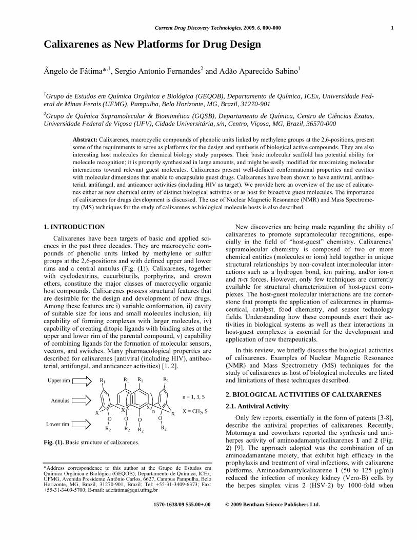
Calixarenes have been targets of basic and applied sci-ences in the past three decades. They are macrocyclic com-pounds of phenolic units linked by methylene or sulfur groups at the 2,6-positions and with defined upper and lower rims and a central annulus (Fig. (1)). Calixarenes, together with cyclodextrins, cucurbiturils, porphyrins, and crown ethers, constitute the major classes of macrocyclic organic host compounds. Calixarenes possess structural features that are desirable for the design and development of new drugs. Among these features are i) variable conformation, ii) cavity of suitable size for ions and small molecules inclusion, iii) capability of forming complexes with larger molecules, iv) capability of creating ditopic ligands with binding sites at the upper and lower rim of the parental compound, v) capability of combining ligands for the formation of molecular sensors, vectors, and switches. Many pharmacological properties are described for calixarenes [antiviral (including HIV), antibac-terial, antifungal, and anticancer activities) [1, 2].
Fig. (1). Basic structure of calixarenes. *Address correspondence to this author at the Grupo de Estudos em Química Orgânica e Biológica (GEQOB), Departamento de Química, ICEx, UFMG, Avenida Presidente Antônio Carlos, 6627, Campus Pampulha, Belo Horizonte, MG, Brazil, 31270-901, Brazil; Tel: +55-31-3409-6373; Fax: +55-31-3409-5700; E-mail: [email protected]
New discoveries are being made regarding the ability of calixarenes to promote supramolecular recognitions, espe-cially in the field of “host-guest” chemistry. Calixarenes’ supramolecular chemistry is composed of two or more chemical entities (molecules or ions) held together in unique structural relationships by non-covalent intermolecular inter-actions such as a hydrogen bond, ion pairing, and/or ion- and - forces. However, only few techniques are currently available for structural characterization of host-guest com-plexes. The host-guest molecular interactions are the corner-stone that prompts the application of calixarenes in pharma-ceutical, catalyst, food chemistry, and sensor technology fields. Understanding how these compounds exert their ac-tivities in biological systems as well as their interactions in host-guest complexes is essential for the development and application of new therapeuticals.
In this review, we briefly discuss the biological activities of calixarenes. Examples of Nuclear Magnetic Resonance (NMR) and Mass Spectrometry (MS) techniques for the study of calixarenes as host of biological molecules are listed and limitations of these techniques described.
2. BIOLOGICAL ACTIVITIES OF CALIXARENES
2.1. Antiviral Activity
Only few reports, essentially in the form of patents [3-8], describe the antiviral properties of calixarenes. Recently, Motornaya and coworkers reported the synthesis and anti-herpes activity of aminoadamantylcalixarenes 1 and 2 (Fig. 2) [9]. The approach adopted was the combination of an aminoadamantane moiety, that exhibit high efficacy in the prophylaxis and treatment of viral infections, with calixarene platforms. Aminoadamantylcalixarene 1 (50 to 125 μg/ml) reduced the infection of monkey kidney (Vero-B) cells by the herpes simplex virus 2 (HSV-2) by 1000-fold when
OO O
X
OXX
X n
R1R1 R1 R1
R2 R2 R2R2
Upper rim
Annulus
Lower rim
n = 1, 3, 5
X = CH2, S

2 Current Drug Discovery Technologies, 2009, Vol. 6, No. 3 Fátima et al.
compared with compound 1-non treated cells. The minimum inhibitory concentration (MIC) for 1 was determined as 10 μg/ml and the maximum tolerated concentration [MTC, de-fined as half of the cytotoxic concentration against Vero-B cells] was 125 μg/ml (Fig. (2)). The chemotherapeutic safety index (SI) for compound 1 is 12.5 (defined as the ration MTC/MIC) [9]. Aminoadamantylcalixarene 2 was unable to either inhibit the cytopathic effect of HSV-2 or reduce its infectious action in Vero-B cells. These results suggest that the hydroxyl groups in compound 1 play an important role in its antiherpes activity (Fig. 2) [9].
2.2. Antifungal Activity
Paquet and coworkers have described an elegant strategy for the design of new antifungal agents synthesizing hybrids of calix[4]arene and amphotericin B (compounds 3 and 4; Fig. 2) [10]. These hybrids were tested against Saccharomy-ces cerevisiae (BY4741). The antifungal activities of calix-
arenes 3 and 4 (Fig. 2) were comparable or higher than that for for amphotericin B [5; MIC = 0.30 μM], exhibiting MIC values of 0.10 and 0.25 μM, respectively. These calixarenes were 10-fold less hemotoxic than amphotericin B (5) [10]. Furthermore, hybrids 3 and 4 (Fig. 2) retained the ability to induce K+ leakage from vesicles, suggesting an efficient channel formation in the membrane as for the mechanism of amphotericin B [11, 12]. Additionally, hybrids 3 and 4 (Fig. 2) caused significant reduction in K+ efflux from cholesterol-containing vesicles, indicating selectivity improvement over amphotericin B (5) and probable high discrimination be-tween ergosterol- (usually found in yeast cell membrane) and cholesterol-containing (usually found in mammalian cell membrane) lipid bilayers. This explain the much lower he-motoxicity of hybrids 3 and 4 (Fig. 2) when compared to amphotericin B (5) [10]. These findings drive the attention for the development of new calixarenes hybrids using other antifungal agents that are available commercially.
Fig. (2). Structure of some calixarenes and the antifungal amphotericin B (5).
OR2OR2 OR2
R1R1 R1 R1
OR2
H3N
Cl
R1 = and R2 = H (1)
H3N
Cl
R1 = and R2 = nBu (2)
Me
O
HOMe
Me
O OH OH
OH
OH OH O
OHOH
CO2H
O O
HO
N
Me
OH
N
O
HNO
O
R3
CH2
4
R3 = H (3)
R3 = tBu (4)
Me
O
HOMe
Me
O OH OH
OH
OH OH O
OHOH
CO2H
O O
HO
NH2
Me
OHAnfotericin B (5)
OHOH OHOH
n
n = 1, R4 = SO3H (6)
n = 3, R4 = SO3H (7)
n = 5, R4 = SO3H (8)
n = 3, R4 = N=N-Ph-SO3H (9)
R4 R4 R4 R4

Calixarenes as New Platforms for Drug Design Current Drug Discovery Technologies, 2009, Vol. 6, No. 3 3
Calixarenes 6-9 (Fig. 2) were evaluated against fungi that are pathogenic for Fusarium solani f. sp. Mori (Rosellinia necatrix and Colletotrichum dematium) [13]. Compounds 6-9 inhibited completely the C. dematium growth and partially (60-70%) the R. necatrix growth. Calixarene-based com-pounds are promising as agents for the minimization of eco-nomic loss in agriculture caused by fungus-related crop dis-eases.
2.3. Antibacterial Activity
The antibacterial activity of calixarenes was first reported in 1955 in studies applying the calixarene derivative macro-cyclon (10; Fig. 3) in tuberculosis and other mycobacteriosis models [14]. More recent studies demonstrate that macrocy-clon (10) affects the lipid metabolism in cells suggesting that its antimycobacterial activity is correlated with the inhibition of triglyceride lipase and phospholipases [15, 16]. Addition-ally, L-arginine metabolism and inducible nitric oxide syn-thase (iNOS) appears to be involved in the mechanism of action of macrocyclon (10) for the control of Mycobacterium tuberculosis growth [17].
Calix[4]arenes-based vancomycin derivatives were also reported to present antibacterial activity [18]. Casnati and coworkers studied antibacterial activity of peptide-calix[4]arenes 11-14 (Fig. 3) against Staphylococcus aureus
[strains 663 (penicillin sensitive), 853 (penicillin resistant) and 1131 (methicillin resistant)], Staphylococcus epider-midis, and Bacillus cereus in comparison with the antibiotic vancomycin. These compounds showed an anti-gram-positive activity from moderate to good, although slightly inferior to vancomycin, and no activity against gram-negative bacteria [18]. The enantiomers peptide-calix[4]arene 12 and 13 (Fig. 3) presented the same range of potency against the bacteria studied. The protection of the basic nitrogen in 12 and 13 structures with tert-butyloxycarbonyl group (Boc) (peptide-calix[4]arene 11) or the enlarging in the peptide bridge as in peptide-calix[4]arene 14 caused a significant reduction in the anti-bacterial activity of these compounds. Indeed, MIC values obtained for the calixarenes studied were in the range of 32-64 mg/l (compound 11), 4-16 mg/l (compound 12), 4-8 mg/l (compound 13), and 32 mg/L (compound 14) [18].
A tetra-para-guanidinoethyl-calix[4]arene (15) and its monomeric equivalent (16) (Fig. 3) had their antibacterial activity evaluated against the bacterial reference strains Es-cherichia coli (ATCC25922), Staphylococcus aureus (ATCC25923 and ATCC29213) and Enterococcus faecalis (ATCC29212), Pseudomonas aeruginosa (ATCC27853) and the clinical isolates Penicillinase-producing E. coli (EcR1), methicillin-resistant S. aureus (MRSA), vancomycin-resistant E. faecium (EfR1), vancomycin- and teicoplanin-
Fig. (3). Structure of calixarenes 10-15 and compound 16.
OR2
R1
R1
OR2
OR2
R1
OR2
R1
OR2
R1R1
OR2OR2
OR2
R1
R1
Macrocyclon (10)
R1 = C8H17 and R2 = (CH2CH2O)n-H where n
is an average of 12.5
OO OO
NNH HN
N N
O O
O O
R5
OO OO
R1 = R3 = H, R2 = R4 = CH3, R5 = Boc (11)
R1 = R3 = R5 = H, R2 = R4 = CH3 (12)
R1 = R3 = R5 = CH3, R2 = R4 = H (13)
R2
R3R1
R4
(14)
N
H
NH HN
N N
O O
O O
HN NH
OO
OHOH HOOH
NH HN NH HN NHH2N
NH NHH2N NH2HN NH
NH2
NH
H2N
OH
CF3COOH
4 CF3COOH
(15) (16)

4 Current Drug Discovery Technologies, 2009, Vol. 6, No. 3 Fátima et al.
resistant E. faecalis (EfR2), and P. aeruginosa overexpress-ing efflux pumps (PaR1) [19,20]. The monomer 16 was de-void of activity (MIC 512 mg/ml) for all strains, whereas tetra-para-guanidinoethyl-calix[4]arene (15) presented an antibacterial spectrum similar to hexamidine, a known anti-bacterial agent [20]. The MIC values for compound 15 were 4 mg/ml against E. coli (ATCC25922), 8 mg/ml against the two strains of S. aureus (ATCC 29213 and ATCC25923) and 32 mg/ml against P. aeruginosa (ATCC27853) and E. fae-calis (ATCC29212) [20]. MICs values for compound 15 against the clinical isolates ranged from 2 to 64 mg/ml [20]. Both tetra-para-guanidinoethyl-calix[4]arene (15) and its monomeric equivalent 16 showed no apparent cytotoxic ef-fect regardless the period of cell incubation (IC50 > 256 mg/ml; exposure longer than 48h) against human keratino-cytes (HaCat) and human pulmonary embryonic fibroblasts (MRC-5). In contrast, loss of cell viability was seen after 24h of cell exposure to hexamidine (IC50 = 36-37 mg/ml) for both HaCat and MRC-5 cell lines [20].
Lamartine and coworkers screened fifty seven calixare-nes for antimicrobial compounds toward a variety of plant pathogen bacteria [13]. Among all calixarenes evaluated against Corynebacterium dematium only four of them (com-pounds 6-9; Fig. (2)) exhibited relatively high antibacterial activity. Compounds 6-9 contain a common SO3H group in the basic form, which might play a role in their antibiotic activities. Calixarenes 6-8 were active at concentrations of 10,000 ppm (parts per million). Compound 9, however, was 10-fold more active than compounds 6-8 [13].
2.4. Antiangiogenic and Anticancer Activities
Platelet-derived growth factor (PDGF) is a potent inducer of growth and motility in several cell types such as fibro-blasts, endothelium, and smooth muscle. It plays a key role in cell proliferation, angiogenesis, wound healing, chemo-taxis, and apoptosis inhibition [21, 22]. PDGF also stimu-lates the proliferation and migration of endothelial cells lead-ing to the formation of new blood vessels, a essential process for tumors growth [23]. Over-expression of PDGF receptor (PDGFR) occurs in many carcinomas [24]. PDGF binds PDGFR, a receptor tyrosine kinase (RTK), causing the re-ceptor dimerization and consequent autophosphorylation. This triggers the recruitment of a series of signaling proteins and activation of the corresponding signal transduction pathways [24]. The development of new molecules that are able to disrupt the interaction of PDGF with its receptor is crucial for the therapeutics of patient with cancer diseases
A number of calixarenes have been described as antian-giogenic and anticancer agents [24-27]. Calix[4]arene 17 (Fig. (4)) showed to be a selective toward PDGFR (IC50 = 250 nM) over other tyrosine kinases receptors (> 100 μM). In nude mouse models calix[4]arene 17 showed significant inhibition of tumor growth and angiogenesis [24, 26]. Hu-man glioblastoma (U87MG)-implanted nude mice treated daily with compound 17 at 50, 100 or 200 mg/kg had the tumor growth inhibited by 56, 81 and 88%, respectively [24, 26]. Similar results were obtained in nude mice implanted with human lung adenocarcinoma (A-549) and rat glioma (C6). The daily treatment with 17 caused no signs of toxicity
Fig. (4). Structure of calixarenes 17-20.
OO O
R1R1 R1 R1
O
OR1OR1 OR2OR2
NHO
ONH
HN
O
NH
HN
NH
O
OOHO
OH
O
NHO
O O
O OHI = II =
R1 = I (17)
R1 = II (18)
R1 = R2 = III (19)
R1 = H, R2 = IV (20)
NH
O
NMe2
NH
NH2
NH
III = IV =

Calixarenes as New Platforms for Drug Design Current Drug Discovery Technologies, 2009, Vol. 6, No. 3 5
even after one month of experiment [24, 26]. Some limita-tions of calix[4]arene 17 application includes the difficulty of its synthesis and aggregation in water due to limited solu-bility. These drawbacks led to the synthesis of a second-generation of PDGF antagonists where calix[4]arene 18 (Fig. 4) showed to be the most effective [25]. Calix[4]arene 18 presents simple acyclic isophthalate groups functionalized with an acidic and a hydrophobic group replacing the peptide loops existing in the calix[4]arene 17 structure. This changes significantly reduced the molecular weight without of this calix[4]arene without compromising its activity. Calix[4]arene 18 blocked the phosphorylation of PDGF re-ceptor at an IC50 of 190 nM [25].
Other examples of calixarenes with antiangiogenic and antitumor activities were described recently by Dings and coworkers [27]. Two calixarenes out of the twenty three syn-thesized (19 and 20; Fig. 4) showed to be highly effective in the inhibition of endothelial cell proliferation at an IC50 value of c.a. 2.0 μM. Anginex, the positive control, presented an IC50 value of c.a. 5.0 μM [27]. Calixarenes 19 and 20 also presented efficacy against the growth of human ovarian (MA148) and mouse melanoma (B16) in mouse models. Reduction of the tumor by 58, 53 and 62% in MA148-mice model occurred 28-day post-treatment with molar equivalent dose of 2.4 mg/kg compound 19, 2.7 mg/kg compound 20, and 10 mg/kg anginex, respectively. In B16-mice model, a more aggressive melanoma, reduction in tumor growth by 42, 70, and 57% was achieved by treatment with compounds 19-20 and anginex (at same concentration listed above), re-spectively, 7-8-day post-intraperitoneal injection. No cyto-toxic effects, assessed by changes in the mice behavior, body weight or hematocrit or creatin levels, were caused by calix-arenes 19 and 20 or anginex [27].
2.5. Antioxidant Activity
Consoli and coworkers described the synthesis and anti-oxidant activities of caffeolyl- and sinapyl-calix[4]arene de-
rivatives (21 and 22, respectively; Fig. (5)) [28]. The free radical scavenging activity of 21 and 22 was determined by the 2,2-diphenyl-1-picrylhydrazyl radical (DPPH˙) assay and the results showed that 21 and 22 possess rate constant val-ues (in methanol) of 18.5 and 72.5 M-1s-1 and stoichiometric factors n (number of DPPH˙ radicals quenched per antioxi-dant molecule) of 5.9 and 2.7, respectively [28]. These val-ues were higher than those obtained for the reference com-pounds (23 and 24; Fig. (5)), but a 4-fold increase was not observed [28]. These hydroxycinnamic acid-calix[4]arenes are the first examples of effective antioxidant calixarene hy-brids.
2.6. Anti-thrombotic Activity
The coagulation cascade involves sequential limited pro-teolysis of successive factors [29] and the spatial expansion of the coagulation event is controlled by surface (thrombo-modulin, heparansulfate proteoglycan, etc) and circulating inhibitors [30]. Calixarenes 25-33 (Fig. (6)) were described to have in vitro anticoagulant activity [31]. Calixarene 30 had a considerable effect on the partial activated-tromboplastin time (APTT) and on the thrombin time (TT) when compared with the others. Calixarenes 30 and 26 acti-vated heparin cofactor II (HCII), a thrombin inhibitor, at 500 μM [30]. All calixarenes studied showed to activate anti-thrombin (AT) at concentrations that were 10-50-fold lower than that for heparin. The calixarene anticoagulant activity was increased by the addition of a pendant group containing a carboxylate in its structure (compound 30). The molecule size also played an important role in the inactivation of blood cascade coagulation as attested by the higher activity of calix[8]arene over calix[6]arene and calix[4]arene. The mechanism of action by which calixarenes 25-33 inhibit boold coagulation seems to be via their interaction with the serine proteases inhibitors HCII and AT [30].
Fig. (5). Structure of calixarenes 21, 22, and reference compounds 23 and 24.
OO O
R1R1 R1 R1
O
HN
O
I =
II =
R1 = I (21)
R1 = II (22)
OH
OH
HN
O
OH
OCH3
OCH3
NH
O
OH
OCH3
OCH3
O
NH
O
OH
OH
O
(23) (24)

6 Current Drug Discovery Technologies, 2009, Vol. 6, No. 3 Fátima et al.
2.7. DNA Recognition and Gene Transfection
The ability of calixarene guanidinium derivatives (34-36; Fig. (7)) to bind plasmid DNA was reported elsewhere [31]. DNA-binding studies of calixarenes 34-36 were performed using the plasmid pEGFP-Ci and monitored by agarose gel electrophoresis mobility shift assay (EMSA). Calix[4]arene 34 started binding the plasmid DNA at 12.5 μM with com-plete plasmid complexation achieved when compound 34 was applied at 100-200 μM [31]. Zadmard and Schrader also showed that calix[4]arene dimmers with six protonatable aniline groups at the upper rims and connected by a suitable flexible linker are able to recognize double-stranded DNA [32].
Calixarenes 37-41 (Fig. (7)) were shown to bind and transfect pDs2-mito, a plasmid that leads to the expression and accumulation of a fluorescent protein in mitochondria [33]. Transfection experiments were performed in Chinese hamster ovary (CHO) cells and the DNA-binding ability checked by electrophoresis studies using a 3,000 Kb plasmid (pGEM-EYFP or pEGFP-N1) [33]. Calixarenes 40 and 41 are monomers of the calixarenes 37-39. The asymmetric calixarene 39 showed a binding activity that was stronger than the symmetrically functionalized calixarene 37. The glycyl amino derivative calixarene 41 presented significant DNA binding ability that was comparable with that of calix-arenes 37 and 39, but lower activity than calixarene 38. The glycyl residue appears to play an important role in the DNA-binding ability of calixarenes 38 and 41 [33]. Compound 38, a multicalixarene featuring aliphatic amines, was the only one effective in the transfection of CHO cells with pDs2-mito exhibiting an efficiency of 10% when compared to FuGene that displays 50% [33].
2.8. Enzyme Inhibition
Calixarenes structural features such as rigid or flexible conformations of the ring and ability of multiple interactions by using different ligands at upper or down rim in their car-bon backbone many researchers make these molecules at-tractive for the studies of protein surface recognition. Many calixarenes were shown to have potential as inhibitors of L-lysyl oxidase [34], -chymotrypsin [35, 36], cholinesterase [37], alkaline phosphatases [38-39], trypase [40], transglu-taminase [41]. The surface area of many proteins, DNA, and RNA can be modulated by protein surface recognition using multivalent calixarenes which could furnish new templates for further drug design and biochemical technologies.
3. NUCLEAR MAGNETIC RESONANCE (NMR)
STUDIES OF SUPRAMOLECULAR RECOGNITION
BY CALIXARENES
Nuclear magnetic resonance (NMR) spectroscopy is one the most important methods for structural elucidation of or-ganic compounds, particularly in the solution state. NMR spectroscopy is valuable for monitoring the intricate struc-ture of chemical modifications of calixarenes derivatives. Synthetic variations usually lead to spin systems much more complicated than those for underlying, highly symmetrical frameworks. Moreover, NMR techniques have become popular for the studies of calixarenes in host-guest com-plexes systems.
Pharmaceutical application of calixarenes for drug pro-tection or targeting now requires structural characterization of the administered compounds, including the supramolecu-lar complexes. NMR spectroscopy is also a powerful tool for in vitro studies of the calixarenes interactions with biological macromolecules such as nucleic acids, proteins, or cell membranes and maybe in the near future this technique can be also used for in vivo interactions. NMR techniques pri-marily target the forces and binding modes that drive the non-covalent associations between calixarenes and other molecules [42]. Some parameters are crucial for the charac-terization of these complexes, such as stoichiometry (Job's method) [43], complexed population (%pbound), forma-tion/dissociation constant (Ka), and relative positioning of the carrier/guest inclusion complex, determined by pulsed field gradient spin-echo (PGSE) [44-46] and/or nuclear Overhauser effect (NOE) [47] experiments.
We want to illustrate the roles played by NMR spectro-scopic techniques in the study of dynamic processes affect-ing inclusion complexes. The literature focusing on NMR applications in the calixarenes and supramolecular chemistry is extremely vast and only selected examples and relevant dynamic problems in host-guest chemistry (calixarene-bioactive molecule interactions) are presented and discussed here.
3.1. Nuclear Magnetic Resonance (NMR) Studies on
Calixarenes Supramolecular Chemistry
Specht and coworkers have investigated the water-soluble calixarenes 25-27 (Fig. (6)) as host candidates for the photolabile cholinergic ligand 42 (Fig. (8)), a choline precur-sor. All of these calixarenes formed stable 1:1 complexes with 42 (assessed by NMR titration) whose dissociation con-
Fig. (6). Structure of calixarenes 25-33.
OHOH OR1OH
n
n = 1, R1 = H (25) n = 1, R1 = CH2CO2H (28) n = 1, R1 = CH2CH2NH2 (31)
n = 3, R1 = H (26) n = 3, R1 = CH2CO2H (29) n = 3, R1 = CH2CH2NH2 (32)
n = 5, R1 = H (27) n = 5, R1 = CH2CO2H (30) n = 5, R1 = CH2CH2NH2 (33)
HO3SHO3S SO3H SO3H

Calixarenes as New Platforms for Drug Design Current Drug Discovery Technologies, 2009, Vol. 6, No. 3 7
stants were determined by DOSY NMR. The strongest inter-actions were observed with calix[8]arene 27, where the dis-sociation constants were similar to those of acetylcho-linesterase (AChE) and butyrylcholinesterase, two choliner-gic enzymes. The high affinities of these calixarenes for 42 are comparable to those of the biological recognition sites in cholinesterases suggesting that the p-sulfonated calixarenes
mimic the binding sites of cholinesterases [48]. Based on NOE experiments and molecular-modeling, the larger calix[8]arene 27 formed ditopic binding complexes by si-multaneously binding to cholinergic moiety and photolabile group of 42. The smaller calix[4]arene 25 and calix[6]arene 26 molecules formed monotopic complexes with 42 [49].
Fig. (7). Structure of calixarenes 34-41.
OR2OR2 OR2OR2
n
n = 1, R1 = NC(NH2)NH2+Cl-, R2 = CH2CH2CH3 (34)
n = 3, R1 = NC(NH2)NH2+Cl-, R2 = CH3 (35)
n = 5, R1 = NC(NH2)NH2+Cl-, R2 = CH3 (36)
R1R1 R1 R1
OO
OO
OHN O NH
R3R3
OHN O NH
R3 R3
OO O
HNNH NH HN
O
II =
OO O
NH3H3N H3N NH3
O
I =
Cl Cl Cl Cl
H3N NH3
O
O
H3N
O
NH3O
Cl
ClCl
Cl
R3 = I (37)
R3 = II (38)
O OO O
O
HN
O
NH
R3R3
HNNH
O O
R3 R3
R3 = I (39)
OO O
NH3H3N H3N NH2
O
Cl Cl Cl Cl
OO O
HNNH NH HN
O
H3N NH3
O
O
H3N
O
NH3O
Cl
ClCl
Cl
(40)(41)

8 Current Drug Discovery Technologies, 2009, Vol. 6, No. 3 Fátima et al.
In view of the potential application of calixarenes for the preparation of new therapeutic formulations, Fernandes and coworkers proposed the use of calixarene 26 to increase the bioavailability of tetracaine 43 and proparacaine 44 and de-crease their systemic toxicity in anesthesia procedures [50]. HR-DOSY 1H NMR allowed the determination of the frac-tion of complex population (%pbound = 70%) and the apparent association constant for 26/43 (Ka = 3889 M–1). These results confirm that a strong association takes place between 26 and 43 and the negatively charged sulfonic groups of 26 are re-sponsible for such strong interaction. Studies carried out at pH 10 revealed that the association of the uncharged form of 43 with 26 is considerably weak, which does not protect the anesthetic from alkaline hydrolysis. The proposed topology of 26/43 complexes was established using ROESY 1D (NOE in the rotating frame) and was driven by ion pair interac-tions.
Calixarenes are proven to be useful for the detoxification caused by xenobiotics (Unpublished data). Accidental or intentional drug toxicity in humans and animals is a health concern especially because of the lack of specific pharma-ceutical antidotes for the majority of drug intoxications (e.g. pyrrolizidine alkaloid toxins). Successful detoxification re-quires the selective and rapid adsorption of the toxins from the organism. Para-sulfonic acid calix[6]arene 26 forms a stable 1:1 complex with retronecine 45 (Fig. (8)) (checked by NMR titration). The dissociation constants were deter-mined by DOSY 1H NMR and led to evaluation of the frac-tion of complex population (%pbound = 56 %) and the appar-ent association constants (Ka = 1446 M–1). Interactions be-tween the calix[6]arene 26 and retronecine 45 was proven to be strong suggesting this calixarene as a promising detoxify-ing agent of alkaloids pyrrolizidines (Unpublished data).
The ability of p-sulfonated calixarenes and their deriva-tives to form complex with various amino acids, peptides,
and oligopeptides has been studied since the last decade [51-59]. Arena and coworkers were the first to report in physio-logical conditions quantitative values of the inclusion of naturally occurring amino acids into a series of calixarenes (25; Fig. (5) and 46-49; Fig. (9)) [58]. Inclusion of arginine (53) and lysine (54) into some p-sulfonated calix[n]arenes (n = 4, 6, 8) (50-52; Fig. (9)) was also investigated by exploring the acidic region only [52]. The highest association constants for calix[4]arene 50 were obtained with the basic amino ac-ids arginine (53) and lysine (54) (association constant pH 8: 50/53: 1.5 x 10³ M-1, 50/54: 0.74 x 10³ M-1) [53]. The asso-ciation constant for calix[4]arene 50 and phenylalanine (55) was 0.63 x 10² M-1 [58]. NMR studies have yielded struc-tural information for the different complexes and showed that lateral aliphatic chains or aromatic portion of the amino acids are embedded into the cavity of the calixarenes. Com-plexation of p-sulfonate calixarenes with di- and tripeptides composed of lysine and arginine (lysyl–lysine, arginyl–arginine, lysyl–arginine, arginyl–lysine, lysyl–lysyl–lysine and arginyl–arginyl–arginine) has also been studied by 1H NMR and microcalorimetry [54]. For calix[4]arene 50, only a 1:1 stoichiometry was observed. The binding process was controlled by the favorable enthalpy resulting mainly from the tight inclusion of the non-polar part of the guest into the hydrophobic cavity of the host through van der Waals inter-actions. The favorable entropy accompanying the desolva-tion of the charged groups upon ionic interaction also plays important roles. The NMR data and the thermodynamic properties of association show that lysyl–lysine adopts a very compact folded structure upon binding by the tetrameric host [54]. The mixed dipeptides that bear a lysine residue behave like lysyl–lysine, the lysine residue being preferentially complexed in the ligand cavity. Addition of a third chain perturbs this very nice arrangement. The affinity for the tripeptide becomes more important because the entropy of binding is now more favorable.
Fig (8). Structure of compounds 42-45.
Fig. (9). Structure of calixarenes 46-52, and amino acids 53-55.
ON -I
(42)
N
H
ON
O
H
Cl-
ON
O
NH2
O
(43) (44)
N
OHHHO
(45)
OR3OR2 OR3OR2
n
n = 1, R1 = SO3H, R2 = R3 = CH2COOH (46)
n = 1, R1 = H, R2 = R3 = CH2COOH (47)
n = 1, R1 = SO3H, R2 = H, R3 = CH2CH2OCH2CH3 (48)
n = 1, R1 = SO3H, R2 = R3 = CH2CH2OCH2CH3 (49)
n = 1, R1 = SO3Na, R2 = R3 = H (50)
n = 3, R1 = SO3Na, R2 = R3 = H (51)
n = 5, R1 = SO3Na, R2 = R3 = H (52)
R1R1 R1 R1
H2N N OH
O
NH2
NH
H(53)
H2NOH
O(54)
OH
O
NH2 (55)
NH2

Calixarenes as New Platforms for Drug Design Current Drug Discovery Technologies, 2009, Vol. 6, No. 3 9
Both 1:1 and 1:2 stoichiometry complexes are observed with p-sulfonate calix[6]arene 51 and these peptides. The calix[6]arene 51 binds two lysyl–lysine guests in a non-cooperative manner, which suggests that adoption of 1,2,3-alternate type conformation in solution similarly to the ob-served in solid state. Thermodynamic data and dissymmetry of the NMR spectra suggest that only the central side chain of the tripeptide is turned towards the interior of the partial cone. A very rigid structure is then obtained by stacking of the three guanidinium groups of the guest over the phenyl units of the calixarene 51. This indicates the importance of
– contributions for the complexation. The complexation of arginyl-arginyl-arginine tripeptide by 51 appears to be a re-markable case of tight binding for which the important fa-vorable enthalpy change is almost compensated by an unfa-vorable entropy change, yielding an affinity that is weaker than that of lysyl-lysyl-lysine tripeptide. Complex behavior, associated with higher order stoichiometry and probably aggregation, occurs in the case of the p-sulfonate calix[8]arene (52).
The complexation properties of the mono-functionalized derivatives 28, 29, 31, 32 (Fig. (6)), and 56-76 (Fig. (10)) with regard to amino acids have also been investigated in solution [51]. The association constants show a 1:1 stoichiometry and the pendant group at the lower rim strongly modifies their interactions with amino acids as compared to the parental p-sulfonate calixarenes 50-52. While the cationic amino acids arginine (53) and lysine (54) bind strongly to all derivatives, the binding of the other amino acids is dependent on the nature of the pendant arms. Then, strong binding was achieved with aspartic acid (77), serine (78), and tryptophan (79) (Fig. (11)). Moreover, 1H NMR studies suggest that the mono-substitution leads to a cone conformation for the calix[4]arene derivative and a ‘‘taco’’ conformation for calix[6]arene and calix[8]arene derivatives.
Four crystalline complexes prepared by the inclusion complexation of the 1,10-phenanthrolinium ion 80 (Fig. (10)) with p-sulfonate thiacalix[4]arene 81 (Fig. (11)) and p-
Fig. (10). Structure of calixarenes 56-76.
Fig. (11). Structure of compounds 77-79 and calixarenes 81 and 82.
OHOH HOOR2
n
n = 1, R1 = R2 = H (56) n = 3, R1 = H, R2 = CH2CH2NH2 (67)
n = 3, R1 = R2 = H (57) n = 3, R1 = SO3H, R2 = CH2CONH2 (68)
n = 5, R1 = R2 = H (58) n = 5, R1 = C(CH3)3, R2 = H (69)
n = 1, R1 = H, R2 CH2COOCH2CH3 (59) n = 5, R1 = C(CH3)3, R2 CH2COOCH2CH3 (70)
n = 1, R1 = H, R2 = CH2COOH (60) n = 5, R1 = C(CH3)3, R2 = CH2COOH (71)
n = 1, R1 = H, R2 = CH2CN (61) n = 5, R1 = C(CH3)3, R2 = CH2CN (72)
n = 1, R1 = H, R2 = CH2CH2NH2 (62) n = 5, R1 = C(CH3)3, R2 = CH2CH2NH2 (73)
n = 1, R1 = SO3H, R2 = CH2CONH2 (63) n = 5, R1 = SO3H, R2 = CH2COOH (74)
n = 3, R1 = H, R2 = CH2COOCH2CH3 (64) n = 5, R1 = SO3H, R2 = CH2CONH2 (75)
n = 3, R1 = H, R2 = CH2COOH (65) n = 5, R1 = SO3H, R2 = CH2CH2NH2 (76)
n = 3, R1 = H, R2 = CH2CN (66)
R1R1R1 R1
HOOH
O
O
NH2
HO OH
O
NH2
NH
OH
O
H2N
N N
OHOH HO
SO3Na
X
NaO3S NaO3S SO3Na
OHXX
X n
n = 1, X = S (81)
n = 2, X = CH2 (82)
(77) (77)
(79) (80)

10 Current Drug Discovery Technologies, 2009, Vol. 6, No. 3 Fátima et al.
sulfonate calix[5]arene 82 (Fig. (11)) upon varying the acid-ity of the solution were recently reported [60]. The degree of compactness of the capsules increased with the enlargement of the calixarene cavity, which is affected significantly by both the penetration depth of 80 (Fig. (11)) and the structure of the dimer 80. Three “bis-molecular” capsules possessing different degrees of compactness were constructed by the complexation of 25, 81, and 82 with 80 at pH 1-2. The in-creasing in the medium acidity by using 1 M HCl changed the complexation orientation of 80 within 25 and 82 from the original vertical mode to a horizontal mode, which prevented the dimerization of 1,10-phenanthrolinium guests required for capsule formation. The 2D ROESY NMR experiment showed that there is consistency between the binding interac-tions in solution and in the solid-state crystal structures. The experiment also showed that the binding ability of 25 and 82 with 79 in 1 M HCl is lower than that in solution at pH 1–2, which is mainly a result of the protonation of the sulfonate groups in 25 and 82. These observations demonstrate une-quivocally that the pH value is a critical factor for the ma-nipulation and design of supramolecular architectures based on 25 and 82 as the choice of guest molecule.
4. MASS SPECTROMETRIC TECHNIQUES FOR
STUDIES OF SUPRAMOLECULAR RECOGNITION
BY CALIXARENES
Nowadays, the mass spectrometry can be considered one of the most analytical techniques for the investigation and characterization of compounds, going from structurally sim-ple molecules to macromolecules with thousands of atomic units (e.g. polymers and proteins). The experiments with electrons conducted by J. J. Thomson in the end of 19th cen-tury made the mass spectrometry grow rapidly to become a powerful tool for molecular elucidation. Among the several sources of ionization, special attention is given to those able to transfer molecules from solution to gas phase in a soft way allowing the measurement of even weakly closed non-covalent interactions. Thus, ESI (Electrospray Ionization) and MALDI (Matrix Assisted Laser Desorption Ionization) are the best choices for the analysis of supramolecular spe-
cies (e.g. calixarenes) and their ability to self assemble and recognize other molecules.
4.1. Electrospray Ionization Mass Spectrometry (ESI-
MS)
Prior to the development of electrospray ionization by John Fenn [61], mass spectrometry was restricted to the analysis of small molecules with low polarity and high vola-tility. Consequently, compounds with high molecular weight as macrocicles, polymers, proteins, organometallics, and other ionic compounds could not be analyzed at high exten-sion by mass spectrometry.
In electrospray ionization the analyte is already ionized in solution, which is possible for compounds that have basic or acid sites or are ionic salts. The liquid then passes through a capillary tube that is applied to a difference of negative or positive potential resulting in a spray of charged droplets that undergoes desorption or ejection of ions to the mass ana-lyzer. This technique transfers gently ions from liquid to gas phase and the observed species in gas phase can be related to those in solution. This is convenient for the detection of iso-lated molecules and aggregated species under weakly inter-molecular non-covalent interaction, such as cation- or hy-drogen bonds common in aggregates of calixarenes.
The first attempt to get information on the interaction be-tween calixarenes and a guest molecule by using ESI-MS was not easy. The protic solvents required for the protona-tion of species in solution tended to disrupt the strongly hy-drogen-bonded aggregates. The initial alternative to detect these soft complexed calixarenes was achieved by Goolsby [62] from the evaluation of sodium and potassium ions selec-tively bound to a series of functionalized calix[4]arenes (Fig. (12)).
Each calixarene was mixed with equal concentration of Na+ and K+ and the resulting solution was analyzed by ESI-MS because the charged host-guest adducts were passive to be detect by the mass spectrometer. The measurements clearly showed strong affinity of calixarenes 84, 85 and 86
Fig. (12). Calix[4]arenes evaluated upon metal ion binding selectivity.
OROR ROOR OROR ROOR
R = CH2CO2Et R = (CH2)4CH3
OROR ROOR
R = O(CH2)2OEt
OROR ROOR
R = O(CH2)2OEt
R1
R1= CH2OCH2CH2CH3
(82) (83)
(84)(85)

Calixarenes as New Platforms for Drug Design Current Drug Discovery Technologies, 2009, Vol. 6, No. 3 11
for potassium over sodium whereas calixarene 83 showed binding selectivity to sodium (Table 1).
Table 1. ESI Results for Alkaline Metals and calix[4]arenes
Interactions
Calix[n]arene Relative Abundance
(Calix-Na)+
(Calix-K)+
83 85 15
84 30 70
85 15 85
86 20 80
The functionalization of calix[4]arenes with urea sub-stituents in their upper rims (compound 87) leads to the di-merization through a cyclic array of complementary hydro-gen bond [63,64]. When two of these calixarenes are con-nected through a specific linker they can arrange to form
molecular capsules (compound 88), which are potential hosts for small molecules (Fig. (13)).
A charged guest (as ammonium salt) able to readily en-capsulate and provide ion labels for the complexes is needed for the investigation of supramolecular assembling systems [65]. The interaction cation- of calixarene homodimers with a cationic guest as well as the free rotation around of spacer provide several possible arrangements between the dimer-ized host and the guest (Fig. (14)).
Brody and coworkers [66] have used ESI-MS to observe the interaction between dimer of calix[4]arene 88 and N-methyl quinuclidinium (92) as cationic guest in an attempt to check aggregates in solution. They have found that the cap-sule form (89) is dominant in solution even at high concen-
tration of the dimer calixarene 88. These results showed that intramolecular assemblies are preferred over intermolecular ones. The stability of the capsule 89 was also investigated by using mono calix[4]arene with different urea substituents and 92 as guest (Fig. (15)).
Fig. (13). Structure of self-assembled calixarenes (molecular capsules).
Fig. (14). Disposition of calixarene dimer and a guest molecule.
O NH
O
O
N
NH
NH
O
O
N
H
R
H
O
N
R
N
O
H
R
N
O R
H OHN
O
O
N
NH
NH
O
O
N
H
R
H
O
N
R
N
O
H
R
N
OR
H
O
NH
O
O
NNH
NH
O
O
N
H
RH
O
NR
NO H
RN
O
R H
(CH2)6
(86)
(87)
R = C6H4-n-C7H15
n
Calixarene dimer
Guest
(88) (89)(90)

12 Current Drug Discovery Technologies, 2009, Vol. 6, No. 3 Fátima et al.
This experiment revealed that an addition of calixarene 94 into solution containing the aggregated 89 promotes par-tial disruption of intramolecular bonds in favor of the het-erodimer 95 form, what was attributed to well matched acid/base interaction between arylurea and sulfonylurea resi-dues [67] (Fig. (16)).
This work led to the evaluation of supramolecular inter-action among calixarenes and dimers of calixarenes with quaternary amines. The affinity of eight cationic amines 92, 96-102 with calixarene 93 through ESI-MS (Fig. (17)) was then evaluated [68].
The authors were able to estimate the relative binding constants among the guests studied and the calixarene ho-modimer 88 (Fig. (18)). This study highlights the importance of ESI-MS as an analytical technique for determining whether or not a due compound is a potential guest for hosts as calixarenes. The biological activities of some amines and alkaloids take place when these compounds present as qua-ternary amines. ESI-MS is an ideal approach to verify if a determined compound has the potential to be used in host-guest supramolecular drug delivery of these systems.
The majority of works on self assembly of calixarenes with organic compounds are based on the use of non-
Fig. (15). Urea derivatized calix[4]arenes and guest.
Fig. (16). Guest recognition of calixarenes homodimer and heterodimer.
Fig. (17). Structure of some cationic amines (guests).
OR2
NH
OR2
OR2
NNH
NH
OR2
O
N
H
R1H
O
NR1
NOH
R1N
O
R1 H
(92)R1 = p-C6H4CH3
R2 = n-C3H7
(93)R1 = p-Ts
R2 = n-C10H21
N
CH3
(91)
(93)
(88) (94)
N
CH3
N
H
NN N
N NN
(95) (91) (96) (97) (98)
(99) (100) (101)

Calixarenes as New Platforms for Drug Design Current Drug Discovery Technologies, 2009, Vol. 6, No. 3 13
coordinative solvent systems and with low polarity to avoid displacement of the guest from the calixarene molecule. However, the use of non-polar solvents does not reproduce the conditions in biological systems where water is predomi-nant. Then, the understanding of how the host and guest in-teracts in vivo becomes an issue. From this limitation, Rein-houdt and Calama [69,70] have proposed the synthesis and utilization of a group of water-soluble calixarenes that are inversely charged (functionalized at the upper rim with amino acid, amidinium, sulfonate, carboxylic acid and phos-phonate moieties) (Fig. (19)).
By using ESI-MS, it was verified that calixarene 103 as-sembles with calixarene 104 (Fig. (20)) by electrostatic at-traction forming molecular capsules in 1:1 proportion that was detected as a double charged sodium adduct with m/z 1134 [(103 .104) + 2Na]2+. This capsule encapsulates N-methyl quinuclidinium (92) (NMQ), without undergoing dissociation as attested by ESI-MS signal of m/z 2347.7 [(103 .104) + NMQ]+.
Other guests (Fig. (21)) were also shown to undergo en-capsulation in aqueous medium [69, 70]. The compound 111 achieved better results because less water-soluble com-pounds would search for the calixarenes’ cavities to over-come poor solubility in the medium.
Takeoka and coworkers [71] have used another important approach where pyridinium salt was attached to the lower rim of calix[4]arene through specific spacer (compound 112)
and analyzed by ESI-MS experiments. The molecule 112 was aggregated in solution, but unlike capsules, formed oli-gomers where the pyridinium residue was included inside the cavity of a neighboring calixarene via cation- interactions (Fig. (22)).
Fig. (18). Relative binding affinity of guests to calixarene homodimer capsules.
Fig. (19). Calixarenes soluble in aqueous medium.
Fig. (20). Molecular capsules formation between opposite ionic calixarenes.
N
CH3
N
H
NN N N
>> >~~
~~>
(97) (91) (95) (99) (100)(96)
R 4
R = OC2H4OC2H5
HN NCl-
+
R 4
R = OC2H4OC2H5
SO3Na
R 4
R = OC2H4OC2H5
COOH
R 4
R = OC2H4OC2H5
PO3H2
R 4
R = OC2H4OC2H5
OHN
O
OH(102)
(103) (104)(49) (105)
R
RRR
HN
OO
OO
NH
O OH
O
NH
OO
HN
OO
O
RRR
R
NH2`RHN
NHR`H2N`RHN NH2 NH2`RHN
-
-
- -
+
++
+
R= OCH2CH2OEt
R`= CH2CH2CH3
102 : 103

14 Current Drug Discovery Technologies, 2009, Vol. 6, No. 3 Fátima et al.
Calixarenes with five units of phenolic aromatics also were used to access their self-assembling capabilities with charged ammonium organic group. The interaction, in solu-tion, between tert-butyl calix[5]arenes functionalized in up-per rim 113-115 and alkydiamonium ions 116-119 (Fig. (23)) were investigated [72].
The structure of these double charged guests, whose charge sites are separated by a spacer of appropriate length yielded at least two host-guest arranges. Again, through competition experiments and dissociative analysis in gas
phase (ESI-MS), it was concluded that both assemblies co-exist in solution. Nevertheless, the formation of the su-pramolecular-structured moiety with two host to one guest (121) is favored over that where alkyldiamonium and calix[5]arenes species are associated at 1:1 ratio (120; Fig. (24)). Furthermore, the combination between host and guest shows different binding affinities. These results support the evaluation of guests of interest in medicinal chemistry that possess multi valences.
The versatility of calixarenes regarding the molecular recognition can be evidenced again in Fukazawa’s efforts [73]. His research group has shown the imprisonment of large and neutral species (fullerenes C60 and C70) inside calix[5]arenes’ cavities (Fig. (25)). An ingenious strategy
was adopted to reach this goal where just one of the aromat-ics ring of calix[5]arene was functionalized at upper rim with a bipyridine unit (122). Coordination of these two residues with Ag+ furnished two calixarenes with cavities large enough to accommodate one fullerene molecule. This ap-proach has the advantage of bringing together the host units by metal-chelation and making the supramolecular structure feasible due to the metal presence, which favors the detec-tion by ESI-MS. In fact these proposed supramolecular as-semblies were totally supported by ESI-MS analysis that allowed measuring the isotope pattern of the complex that
Fig. (22). Oligomerization between calixarenes linked to cationic guest.
Fig. (23). Calix[5]arenes and alkydiamonium studied.
Fig. (21). Guest molecules evaluated for encapsulation with calix-arenes in aqueous medium.
N
N N
N
N
HN
H3C
O OH
H3C
HO NH2
NH
CH3
O
H2N
CH3
(106) (107)(108)
(109) (110)
HO
HO
HO
O
N+
OH OHOH O
NBr-
+
HO
HO
HO
O
N+HO
HO
HO
O
N+
Br-
Br-
Br-
n
Oligomer(111)
OR OROR OR
R = Bn (112)
R = (CH2)3CH(CH3)2 (113)
R = CH2CO2C(CH3)3 (114)
+H3N (CH2)n NH3+
n = 8 (115)
n = 9 (116)
n = 10 (117)
n = 12 (118)
n+ +
n = 2,3,4,6
2

Calixarenes as New Platforms for Drug Design Current Drug Discovery Technologies, 2009, Vol. 6, No. 3 15
nicely matched with the calculated data. This experiment also provided evidence for the calixarene ability to bind fullerene C60 stronger than fullerene C70.
4.2. Matrix Assisted Laser Desorption Ionization (MALDI)
The mass spectrometry equipped with MALDI (Matrix Assisted Laser Desorption Ionization) ionization source 7originated after the Electrospray Ionization [74]. It became immediately a complementary technique for the investiga-tion of molecules in gas phase where now it is possible to ionize macromolecules as proteins, peptides, polymers and supramolecular architectures of large dimensions. The proc-
ess of ionization is very simple although its mechanism is not fully elucidated. In this case, ionization is based on the short irradiation of laser pulses over a surface doped with a solid mixture containing the compound of interest (labeled matrix) and another compound that will help the ionization process. Generally, the matrix is formed by a small molecule projected to absorb the laser energy selectively and undergo a rapid sublimation and gaseous expansion. Analytes are also simultaneously evaporated and gently ionized through a se-ries of chemical events. This ionization process is more en-ergetic than ESI because part of the energy absorbed by ma-trix is transferred to the analytes. Nevertheless, this process is still soft enough to permit the ionization of non-volatile
Fig. (24). Possible associations between alkydiamonium guests and calix[5]arenes.
Fig. (25). Fullerenes C60 and C70 involved by two calix[5]arenes through ions Ag+ anchor.
OROR
OR OR
+
RORO
OR
OR
+
2
2
RORO
OROR
+
2
H3N+
n
n
(119) (120)
HO
N
N
OH
HO
HO
HO
Me
Me
Me
Me
C60 or C70OH
N
N
OH
OH
OH
OH
Me
Me
Me
HO
N
N
HO
HO
HO
HO
Me
Me
Me
Ag+
C60 or C70
Ag+
(121)

16 Current Drug Discovery Technologies, 2009, Vol. 6, No. 3 Fátima et al.
and thermally label large molecules and subsequent transfer to the gas phase, and then to the mass analyzer without suf-fering appreciable dissociation or even decomposition. Con-sidering that normally the used proportion of the matrix is greater than analyte, the region of low mass in the spectrum becomes highly populated with matrix signals. This makes the MALDI-MS adequate for the analysis of large molecules where the signals fall in the matrix-free mass region and the ratio signal/noise is high, which simplifies the mass spec-trum interpretation.
Although the low mass region is not commonly used in MALDI analysis, a careful choice of a matrix or even an analyte allows the use of MALDI for the analysis of low mass molecules. In this regard, the interaction between amino acids and calix[4]arenes and calix[6]arenes, both functionalized just in the lower rim (Fig. (26)), was investi-
gated [75]. It was observed a ratio of 1:1 among the calix-arenes and the amino acids. The assembling between calix[6]arene 124 and the studied amino acids was stronger than that for calix[4]arenes.
MALDI-MS is widely used to investigate large arranged systems, but a few examples of its use to evaluate small as-sembled calixarenes with guest are available [76]. Prins and coworkers found a highly organized system between calix[4]arenes functionalized with melamine (125-130; Fig. (27)) and 5,5-diethylbarbiturate (DEB) (131). Each mela-mine unit was associated with two units of DEB yielding in solution a final and highly ordered assembly where three units of calixarenes were joined by six units of DEB (origi-nating double rosettes). These were detected by MALDI-MS using Ag+ labeling technique to favor the formation of posi-tively charged hydrogen bonded assemblies.
Fig. (26). Calixarenes used in interactions with amino acids.
Fig. (27). Formation of double rosette-type molecular assembly.
OR
OR
OR
OR
R = H, CH2COOEt
OR
OR
OR
OR
OROR
R = H, CH2COOR'
R' = H, CH2COOEt
(122)(123)
PrOOPr OPr
PrO
NN
NN
N N
NN
N
N
N
N
H
H
H
R'
HH
H H
H
R'R
R
N N
O
HH
OO
R= H, R'= butyl (124)
R= NO2, R'= butyl (125)
R= CN, R'= butyl (126)
R= H, R'= bnzyl (127)
R= NH2, R'= butyl (128)
R= NHC(=O)CH3, R'= butyl (129)
N N
NN
N
H H
NX
OPr OPr
NH
O
O
O
H
H
N
N O
OH
H
O
H
2
+
H
3Double Rosettes
(130)

Calixarenes as New Platforms for Drug Design Current Drug Discovery Technologies, 2009, Vol. 6, No. 3 17
This type of interaction and association of guest mole-cules with calixarenes introduces a new way of delivering drugs. Their association constants can also be measured by MALDI-MS. Calixarenes bridged with other calixarene se-ries have carried on more complex supramolecular structure where three calixarene units can be associated with twelve DEB units in one arrange known as tetra rosettes (Fig. (28)) [77].
Reinhoudt and coworkers [78] also showed that these tetra rosettes can be associated with other guests and that MALDI-MS could be used to detect the interaction of these aggregated with phenolic derivatives (Fig. (29)). They also determined that associations with guests may take place ei-ther outside the complex through exo-hydrogen bonded re-ceptors or inside by endo-hydrogen bonded receptors. Again, the MALDI-MS technique was essential to characterize and detect very large and heavy supramolecular species resulted from these associations.
Other important applications of MALDI-MS include the analysis of calix[n]arene-guest complexes in dymanic com-binatorial libraries [79] where two or more types of double rosettes are mixed for achieving a new entity (suspension of double rosettes) of stronger association constants with guests. This application is an important approach for the mo-lecular recognition-based search of biologically active com-pounds that interact with calixarenes.
CONCLUDING REMARKS
Calixarenes have been attracting much interest during the last three decades due to their capacity of non-covalently binding other molecules. The search for new chemical enti-ties with potential as drugs is a challenge especially because cells and microorganisms are constantly acquiring resistance to commercial drugs.
Calixarenes are shown to be excellent platforms and/or hosts for many useful bioactive molecules. A deep under-
Fig. (28). Formation of tetra rosette-type molecular assembly.
PrO
N N
O
HH
OO
R= CH2C(CH3)2CH2NHBoc, X= NHCONH (131)
R= (R)-CH(C6H5)CH3 , X= NHCONH (132)
Tetra Rosettes
PrOOPr OPr
PrO
N
NN
N
N
N
NNN
N
N
NH
H
H
R'
HH
H H
H
PrOOPrOPr
PrO
N
NN
N
N
N
N N N
N
N
NH
H
H
R'
HH
HH
H
X
X
N N
O
HH
OO
N N
O
HH
OO
N N
O
HH
OO
3
PrOOPr OPr
PrO
NN
NN
N
N
NNN
N
N
N
H H
H
R
HH
H H
H
OPrOPrPrO
NN
NN
N
N
N N
N
N
N
N
HH
H
R
HH
HH
H
X
X
N N
O
HH
OO
(130)

18 Current Drug Discovery Technologies, 2009, Vol. 6, No. 3 Fátima et al.
standing of how these calixarene-guest molecule complexes exert their activities in biological systems is essential for their future development and application in therapeutics. In this context, NMR and Mass spectrometry are proven to be excellent techniques for the complete investigation of these supramolecular systems.
Mass spectrometry is the ideal way to observe this type of molecular recognition because allows for the investigation of aggregates and macro aggregates in gas phase revealing intrinsic non-covalent interactions without solvent interfer-ences. These data, together with those obtained from NMR in solution, bring a comprehensive understanding of how these platforms interact with guests and behave in biological environments.
The use of calixarenes as hosts of biological active mole-cules will contribute significantly for the improvement of pharmacokinetic and drug delivery.
ACKNOWLEDGEMENTS
The authors thank FAPEMIG (Fundação de Amparo a Pesquisa no Estado de Minas Gerais) and CNPq (Conselho Nacional de Desenvolvimento Científico e Tecnológico) for financial support. Dr. Luzia V. Modolo (The Samuel Roberts Noble Foundation, USA) and Dr. Fernando C. Macedo Júnior (Universidade Estadual de Londrina, Brazil) for criti-cal reading of the manuscript. AF and AAS were supported by the Programa de Auxílio à Pesquisa de Doutores Recém-Contratados of Universidade Federal de Minas Gerias (UFMG).
REFERENCES
[1] Da Silva E., Coleman A.W.: Biopharmaceutical applications of calixarenes. J. Drug. Del. Sci. Tech. 14(1), 3, (2004).
[2] Perret F., Lazar A.N., Coleman A.W.: Biochemistry of the para-sulfonato-calix[n]arenes. Chem. Commun. 2425, (2006).
[3] Hwang K.M., Qi Y.M., Liu S.Y., Choy W., Chen J.: WO9403164 (1994).
[4] Harris S.J.: WO9519974 (1995). [5] Harris S.J.: WO200244121 (2002). [6] Coveney D., Costello B.: EP1367044 (2003). [7] Kral V., Cigler P., Konvalinka J., Kozisek M., Prejdova J., Gruner
B., Plesek J., Lepsik M., et al.: WO2005073240 (2005). [8] Coveney D., Costello B.: US2005113454 (2005).
[9] Motornaya A.E., Alimbarova L.M., Shokova E.A., Kovalev V.V.: Synthesis and antiherpetic activity of N-(3-amino-1-
adamantyl)calix[4]arenes. Pharm. Chem. J. 40(2), 68, (2006). [10] Paquet V., Zumbuehl A., Carreira E.M.: Biologically active ampho-
terecin B-calix[4]arene conjugates. Bioconjugate Chem. 17(6), 1460, (2006).
[11] Hartsel S.C., Hatch C., Ayenew W.: How does amphotericin B work? Studies on model membrane systems. J. Liposome Res. 3(3), 377, (1993).
[12] Milhaud J., Ponsinet V., Takashi, M., Michels B.: Interactions of
the drug amphotericin B with phospholipids membranes containing or not ergosterol: new insight into the role of ergosterol. Biochim. Biophys. Acta 1558(2), 95, (2002).
[13] Lamartine R., Tsukada M., Wilson D., Shirata A.: Antimicrobial
activity of calix[n]arene. C. R. Chimie 5(3), 163, (2002). [14] Conforth J.W., Hart P.D., Nicholls G.A., Rees R.J.W., Stock J.A.:
Antituberculous effects of certain surface-active polyoxyethylene ethers. Br. J. Pharmacol. Chemother. 10(1), 73, (1955).
[15] Jain M.K., Jahagirdar D.V.: Effect of antituberculous calixarenes on phopholipase A2, susceptibility and on fusion of phospholipids
bilayers. Biochem. J. 227(3), 789, (1985). [16] Hart P.D., Armstrong J.A., Brodaty E.: Calixarenes with host-
mediated potency in experimental tuberculosis: further evidence that macrophage lipids are involved in their mechanism of action. Infect. Immun. 64(4), 1491, (1996).
[17] Colston M.J., Hailes H.C., Stavropoulos E., Hervé A.C., Hervé G., Goodworth K.J., Hill A.M., Jenner P., et al.: Antimycobacterial calixarenes enhance innate defense mechanism in murine macro-
phages and induce control of Mycobacterium tuberculosis infection in mice. Infect. Immun. 72(11), 6318, (2004).
[18] Casnati A.C., Fabbi M., Pelizzi, N., Pochini A., Sansone F., Ungaro R.: Synthesis, antimicrobial activity and binding properties of
calix[4]arene based vancomycin mimics. Bioorg. Med. Chem. Lett. 6(22), 2699, (1996).
[19] Mourer M., Duval R.E., Finance C., Regnouf-de-Vains J.B.: Func-tional organization and gain of activity: the case of the antibacte-
rial tetra-para-guanidinoethyl-calix[4]arene. Bioorg. Med. Chem. 16(11), 2960, (2006).
[20] Grare M., Mourer M., Fontanay S., Regnouf-de-Vains J.B., Fi-nance C., Duval R.E.: In vitro activity of tetra-para-
guanidinoethyl-calix[4]arene against susceptible and antibiotic-resistance Gram-negative and Gram-positive bacteria. J. Antimi-crob. Chemother. 60, 575, (2007).
[21] Jones S.M., Kazlauskas A.: Growth factor-dependent signaling and
cell cycle progression. FEBS Lett. 490(3), 110, (2001). [22] Tallquist M., Kazlauskas A.: PDGF signaling in cells and mice.
Cytokine Growth Factor Rev. 15(4), 205, (2004). [23] Kumar R., Yoneda J., Bucana C.D., Fidler I.J.: Regulation of dis-
tinct steps of angiogenesis by different angiogenic molecules. Int. J. Oncol. 12(4), 749, (1998).
[24] Sebti S.M., Hamilton A.D.: Design of growth factor antagonist with antiangiogenic and antitumor properties. Oncogene 19(56), 6566, (2000).
Fig. (29). Phenolic guests evaluated for interaction with tetra rosettes.
HO
HO OH
O2N OMe
X
X= p-NO2 (133a) X= m-NO2 (133b)
X= p-CN (133c) X= p-CF3 (133d)
X= p-CH3 (133e) X= p-COOMe (133f)
X= p-Br (133g) X= p-F (133h)
X= H (133i) X= p-OMe (133j)
X= p-OH (133k) X= m-OH (133l)
X= o-OH (133m)
(134)
(135)

Calixarenes as New Platforms for Drug Design Current Drug Discovery Technologies, 2009, Vol. 6, No. 3 19
[25] Zhou H., Wang D., Baldini L., Ennis E., Jain R., Carie A., Sebti S.M., Hamilton A.D.: Structure-activity studies on a library of po-tent calix[4]arene-based PDGF antagonists that inhibit PDGF-
stimulated PDGFR tyrosine phosphorilation. Org. Biomol. Chem. 4(12), 2376, (2006).
[26] Blaskovic M.A., Lin Q., Delarue F.L., Sun J., Park H.S., Coppola D., Hamilton A.D., Sebti S.M.: Design of GFB-11, a platelet-
derived growth factor binding molecule with antiangiogenic and anticancer activity against human tumors in mice. Nat. Biotechnol. 18(10), 1065, (2000).
[27] Dings R.P.M., Chen X., Hellebrekers D.M.E.I., van Eijk L.I., Zhang Y., Hoye T.R., Griffioen A.W., Mayo K.H.: Design of non-peptide topomimetics of antiangiogenic proteins with antitumor ac-
tivities. J. Natl. Cancer Inst. 98(13), 932, (2006). [28] Consoli G.M.L., Galante E., Daquino C., Granata G., Cunsolo F.,
Geraci C.: Hydroxycinnamic acid clustered by a calixareneplat-form: radical scavenging and antioxidant activity. Tetrahedron Lett. 47(37), 6611, (2006).
[29] Hockin M.F., Jones K.C., Everse S.J., Mann K.G.: A model for the
stoichiometric regulation of blood coagulation. J. Biol. Chem. 277(21), 18322, (2002).
[30] Shriver Z., Liu D., Sasisekharan R.: Emerging views of heparin sulfate glycosaminoglycan structure/activity relationships modulat-
ing dynamic biological functions. Trends Cardiovasc. Med. 12(2), 71, (2002).
[31] Da Silva E., Ficheux D., Coleman A.W.: Anti-thrombotic activity of water-soluble calixarenes. J. Incl. Phenom. Macrocycl. Chem. 52(3), 201, (2005).
[32] Zadmard R., Schrader T.: DNA recognition with large calix-
arenedimmers. Angew. Chem. Int. Ed. 45(17), 2703, (2006). [33] Lalor R., DiGesso J.L., Mueller A., Matthews S.E.: Efficient gene
transfection with functionalized multicalixarenes. Chem. Commun. 4907, (2007).
[34] Hulmes D., Coleman A., Aubert-Foucher E.: WO2000007585 (2000).
[35] Park H.S., Lin Q., Hamilton A.D.: Protein surface recognition by synthetic receptors: a route to novel submicromolar inhibitors for
-chymotrypsin. J. Am. Chem. Soc. 121(1), 8, (1999). [36] Park H.S., Lin Q., Hamilton A.D.: Modulation of protein-protein
interactions by synthetic receptors: design of molecules that disrupt serine protease-protenaceous inhibitors interaction. Proc. Natl. Acad. Sci. USA 99(8), 5105, (2002).
[37] Stoikova E.E., Evtugyn G.A., Belyakova S.V., Khrustalev A.A., Stoikov I.I., Antipin I.S., Budnikov H.C., Konovalov A.I.: 1,3-Disubstituted p-tert-butylcalix[4]arenes as cholinesterase inhibi-
tors. J. Inclusion Phenom. Macrocycl. Chem. 39(3-4), 339, (2001). [38] Vovk A.I., Kalchenko V.I., Cherenok S.A., Kukhar V.P., Muzy-
chka O.V., Lozynsky M.O.: Calix[4]arene methylenebisphos-phonic acids as calf intestine alkaline phosphatases inhibitors. Org. Biomol. Chem. 2, 3162, (2004).
[39] Cherenok S., Vovk A., Muravyova I., Shivanyuk A., Kukhar V., Lipkowski J., Kalchenko V.: Calix[4]arene -aminophosphonic acids: asymmetric synthesis and enantioselective inhibition of an
alkaline phosphatases. Org. Lett. 8(4), 549, 2006. [40] Mecca T., Consoli G.M.L., Geraci C., Cunsolo F.: Designed
calix[8]arene-based ligands for selective tryptase surface recogni-tion. Bioorg. Med. Chem. 12(19), 5057, (2004).
[41] Francese S., Cozzolino A., Caputo I., Esposito C., Martino M., Gaeta C., Troisi F., Neri P.: Transglutaminase surface recognition
by peptidocalix[4]arene diversomers. Tetrahedron Lett. 46(10), 1611, (2005).
[42] Schneider H.-J., Hacket F., Rüdiger V.: NMR studies of cyclodex-trins and cyclodextrin complexes. Chem. Rev. 98(5), 1755, (1998).
[43] Job P.: Formation and stability of inorganic complexes in solution. Ann. Chim. 9, 113, (1928).
[44] Laverde Jr A., da Conceição G.J.A., Queiroz S.C.N., Fujiwara F.Y., Marsaioli A.J.: An NMR tool for cyclodextrin selection in
enantiomeric resolution by high-performance liquid chromatogra-phy. Magn. Reson. Chem. 40(7), 433, (2002).
[45] Fernandes S.A., Cabeca L.F., Marsaioli A.J., de Paula E.: Investigation of tetracaine complexation with beta-cyclodextrins
and p-sulphonic acid calix[6]arenes by nOe and PGSE NMR. J. Incl. Phenom. Macrocycl. Chem. 57(1-4), 395, (2007).
[46] De Araujo D.R., Tsuneda S.S., Cereda C.M.S., Carvalho F.G.F., Preté P.S.C., Fernandes S.A., Yokaichiya F., Franco M.K.K.D., et
al.: Development and pharmacological evaluation of ropivacaine-
2-hydroxypropyl- -cyclodextrin inclusion complex. Eur. J. Pharm. Sci. 33(1), 60, (2008).
[47] Mo H., Pochapsky T.C.: Intermolecular interactions characterized
by nuclear Overhauser effects. Prog. NMR Spectrosc. 30, 1, (1997).
[48] Specht, A., Ziarelli F., Bernard, P., Goeldner M., Peng L.: para-Sulfonated calixarenes used as synthetic receptors for complexing
photolabile cholinergic ligand. Helv. Chim. Acta 88(10), 2641, (2005).
[49] Specht, A., Bernard, P., Goeldner M., Peng L.: Mutually induced formation of Host-Guest complexes between p-sulfonated
calix[8]arene and photolabile cholinergic ligands. Angew. Chem. Int. Ed. 41(24), 4706, (2002).
[50] Cabeça, L.F., Fernandes S.A., de Paula E. Marsaioli A.J.: Topology of a ternary complex (proparacaine- -cyclodextrin-liposome) by
STD NMR. Magn. Reson. Chem. 46(6), 832, (2008). [51] da Silva E., Coleman A.W.: Synthesis and complexation properties
towards amino acids of mono-substituted p-sulphonato-calix-[n]-arenes. Tetrahedron 59(37), 7357, (2003).
[52] Douteau-Guevel N., Coleman A.W., Morel J.-P., Morel-Desrosiers N.: Complexation of basic amino acids by water-soluble calixare-
nesulphonates as a study of the possible mechanisms of recognition of calixarenesulphonates by proteins. J. Phys. Org. Chem. 11(10), 693, (1998).
[53] Douteau-Guevel N., Coleman A.W, Morel J.-P., Morel-Desrosiers N.: Complexation of the basic amino acids lysine and arginine by three sulfonatocalixarenes (n = 4, 6 and 8) in water: microcalo-
rimetric determination of the Gibbs energies, enthalpies and en-tropies of complexation. J. Chem. Soc. Perkin Trans. 2, 629, (1999).
[54] Douteau-Guevel N., Perret F., Coleman A.W., Morel J.-P., Morel-Desrosiers N.: Binding of dipeptides and tripeptides containing ly-sine or arginine by p-sulfonatocalixarenes in water: NMR and mi-
crocalorimetric studies. J. Chem. Soc. Perkin Trans. 2, 524, (2002). [55] Kalchenko O.I., Perret F., Morel-Desrosiers N., Coleman A.W.: A
comparative study of the determination of the stability constants of inclusion complexes of p-sulfonatocalix[4]arene with amino acids
by RP-HPLC and 1H NMR. J. Chem. Soc. Perkin Trans. 2, 258, (2001).
[56] Buschmann H.-J., Mutihac L., Schollmeyer E.: Complexation of some amino acids and peptides by p-sulfonatocalix[4]arene and
hexasodium p-sulfonatocalix[6]arene in aqueous solution. J. Incl. Phenom. Macrocycl. Chem. 46(3-4), 133, (2003).
[57] Arena G., Casnati A., Contino A., Magrì A., Sansone F., Sciotto D., Ungaro U.: Inclusion of naturally occurring amino acids in wa-
ter soluble calix[4]arenes:a microcalorimetric and 1H NMR inves-tigation supported by molecular modeling. Org. Biomol. Chem. 4, 243, (2006).
[58] Arena G., Contino A., Gulino F.G., Magri A., Sansone F., Sciotto D., Ungaro R.: Complexation of native L- -aminoacids by water soluble calix[4]arenes. Tetrahedron Lett. 40(18), 1597, (1999).
[59] Yakovenko A.V., Boyko V.I., Kalchenko V.I., Baldini L., Casnati A., Sansone F., Ungaro R.: N-Linked peptidocalix[4]arene bi-
sureas as enantioselective receptors for amino acid derivatives. J. Org. Chem. 72(9), 3223, (2007).
[60] Liu Y., Guo D.-S., Zhang H.-Y., Ma Y.-H., Yang E.-C.: The struc-ture and thermodynamics of calixarene complexes with dipyridines
and phenanthroline in aqueous solution studied by microcalorime-try and NMR spectroscopy. J. Phys. Chem. B, 110(7), 3428, (2006).
[61] Fenn J.B., Mann M., Meng C.K., Wong S.F., Whitehouse C.M.: Electrospray ionization for mass spectrometry of large bio-
molecules. Science 246(4926), 64, (1989). [62] Goolsby B.J., Brodbelt J.S., Adou E., Blanda M.: Determination of
alkali metal ion binding selectivities of calixarenes by matrix-assisted laser desorption ionization and electrospray ionization in
a quadrupole ion trap. Int. J. Mass Spectrom. 193(1-2), 197, (1999).
[63] Mogck O., Paulus E.F., Böhmer V., Thondorf I., Vogt W.J.: Hy-drogen-bonded dimmers of tetraurea calix[4]arenes: unambiguous
proof by single crystal X-Ray analysis. Chem. Commun. 2533 (1996).
[64] Castellano R.K., Rudkevich D.M., Rebeck J.Jr.: Tetramethoxy calix[4]arenes revisited: conformational control through self-
assembly. J. Am. Chem. Soc. 118(41), 10002, (1996). [65] Lhoták P., Shinkai S.: Cation- interactions in calixarene and
related systems. J. Phys. Org. Chem. 10, 273, (1997).

20 Current Drug Discovery Technologies, 2009, Vol. 6, No. 3 Fátima et al.
[66] Brody M. S., Schalley, C.A., Rudkevick D.M., Rebek J.Jr.: Synthe-
sis and Characterization of a Unimolecular Capsule. Angew. Chem. Int. Ed. 38(11), 1640, (1999).
[67] Castellano R.K., Kim B.H., Rebek J.Jr.: Chiral capsules: asymmet-ric binding in calix[n]arene-based dimers. J. Am. Chem. Soc. 119(51), 12671, (1997).
[68] Schalley C.A., Castellano R.K., Brody M.S., Rudkevich D.M., Siuzdak G., Rebek J.Jr.: Investigating molecular recognition by mass spectrometry: characterization of calixarene-based self-
assembling capsule hosts with charged guests. J. Am. Chem. Soc. 121(19), 4568, (1999).
[69] Corbellini F., Knegtel R.M.A., Grootenhuis P.D.J., Calama M.C., Reinhoudt D.N.: Water-soluble molecular capsules: self-assembly
and binding properties. Chem. Eur. J. 11(1), 298, (2005). [70] Corbellini F., van Leeuwen F.W.B., Beijleveld H., Kooijman H.,
Spek A.L., Verboom W., Calama M.C., Reinhoud D.N.: Multiple ionic interactions for noncovalent synthesis of molecular capsules
in polar solvents. New J. Chem. 29, 243, (2005). [71] Ishihara S., Takeoka S.: Host-guest assembly of pyridinium-
conjugated calix[4]arene via cation – interaction. Tetrahedron Lett. 47(2), 181, (2006).
[72] Garozzo D., Gattuso G., Kohnke F.H., Paola M., Notti A., Occhip-inti S., Pappalardo S., Parisi M.F., et al.: Guest-induced capsular
assembly of calix[5]arenes. Tetrahedron Lett. 43(43), 7663, (2002).
[73] Haino T., Araki H., Yamanaka Y., Fukazawa Y.: Fullerene recep-
tor based on calix[5]arene through metal-assisted self-assembly. Tetrahedron Lett. 42(18), 3203, (2001).
[74] Karas M., Hillenkamp F.H.: Laser desorption ionization of proteins with molecular masses exceeding 10 000 daltons. Anal. Chem. 60(20), 2299, (1988).
[75] Stone M.M., Franz A.H., Lebrilla C.B.: Non-covalent
calix[n]arene-amino acids complexes fomed by MALDI-MS. J. Am. Soc. Mass. Spectrom. 13(8), 964, (2002).
[76] Prins L.J., Timmerman P., Reinhoudt D.N.: Non-covalent synthesis of organic nanostructures. Pure Appl. Chem. 70(8), 1459, (1998).
[77] Timmerman P., Weidmann J.-L., Jolliffe, K.A., Prins L.J., Rein-houdt D.N., Shinkai S., Frish L., Cohen Y.: NMR diffusion spec-
troscopy for the characterization of multicomponent hydrogen-bonded assemblies in solution. J. Chem. Soc. Perkin Trans. 2, 2077, (2000).
[78] Kerckhoffs J.M.C.A., Ishi-I T., Paraschiv V., Timmerman P., Ca-lama M.-C., Shinkai S., Reinhoudt D.N.: Complexation of phenolic guests by endo- and exo-hydrogen-bonded receptors. Org. Biomol. Chem. 1, 2596, (2003).
[79] Kerchhoffs J.M.C.A., Timoneda M.A.M-., Reinhoudt D.N., Calam M.C-.: Dynamic combinatorial libraries based on hydrogen-bonded molecular boxes. Chem. Eur. J. 13(8), 2377, (2007).