c3ta15350a1.pdf - The Royal Society of Chemistry
11
1 Fabrication and Photocatalytic Properties of a Visible-light Responsive Nanohybrid Based on Self-assembling of Carboxyl Graphene and ZnAl Layered Double Hydroxides Zhujian Huang, abc Pingxiao Wu,* abc Beini Gong, d Yueping Fang, e and Nengwu Zhu ab a College of Environment and Energy, South China University of Technology, Guangzhou, 510006, P.R. China E-mail: [email protected]; Fax: +86-20-39383725 b The Key Lab of Pollution Control and Ecosystem Restoration in Industry Clusters, Ministry of Education, Guangzhou, 510006, P.R. China c The Key Laboratory of Environmental Protection and Eco-Remediation of Guangdong Regular Higher Education Institutions, Guangzhou, 510006, P.R. China d State Key Laboratory of Biocontrol, SUN YAT-SEN University, Guangzhou 510006, P.R. China e Institute of Biomaterial, College of Science, South China Agricultural University, Guangzhou, 510642, P.R. China Electronic Supplementary Information Experiment section Fig. S1 TGA curve of rCG/LDO nanohybrid. P5 Fig. S2 Photograph of the formamide solution of CG (a) and photograph of exfoliated LDHs nanosheets dispersed in formamide solution (b). P5 Fig. S3 Tapping-mode AFM image of CG nanosheets (a) and their corresponding height profile (b). P6 Fig. S4 XRD pattern of pristine ZnAl-LDHs. P6 Fig. S5 SEM image of pristine ZnAl-LDHs. P7 Fig. S6 Tapping-mode AFM image of exfoliated LDH nanosheets (a) and their corresponding height profile (b). P7 Fig. S7 Schematic diagram of the process of electrostatical self-assembly between the negatively charged CG monolayer and the positively charged ZnAl-LDH nanosheets. P8 Fig. S8 Nitrogen adsorption-desorption isotherms of samples: (a) ZnAl-LDH, (b) CG/LDH nanohybrid and (c) rCG/LDO nanohybrid. P8 Fig. S9 XRD pattern of rCG/LDO nanohybrid. P9 Fig. S10 Raman spectra of rCG/LDO nanohybrid. P9 Fig. S11 TEM image of r CG/LDO nanohybrid P10 Fig. S12 Schematic representation of the photocatalytic degradation of dye in presence of rCG/LDO nanohybrid under visible light. P11 Electronic Supplementary Material (ESI) for Journal of Materials Chemistry A. This journal is © The Royal Society of Chemistry 2014
-
Upload
khangminh22 -
Category
Documents
-
view
1 -
download
0
Transcript of c3ta15350a1.pdf - The Royal Society of Chemistry

1
Fabrication and Photocatalytic Properties of a Visible-light Responsive
Nanohybrid Based on Self-assembling of Carboxyl Graphene and ZnAl
Layered Double Hydroxides
Zhujian Huang,abc Pingxiao Wu,*abc Beini Gong,d Yueping Fang,e and Nengwu Zhuab
a College of Environment and Energy, South China University of Technology, Guangzhou, 510006, P.R. China
E-mail: [email protected]; Fax: +86-20-39383725b The Key Lab of Pollution Control and Ecosystem Restoration in Industry Clusters, Ministry of Education, Guangzhou,
510006, P.R. Chinac The Key Laboratory of Environmental Protection and Eco-Remediation of Guangdong Regular Higher Education
Institutions, Guangzhou, 510006, P.R. Chinad State Key Laboratory of Biocontrol, SUN YAT-SEN University, Guangzhou 510006, P.R. Chinae Institute of Biomaterial, College of Science, South China Agricultural University, Guangzhou, 510642, P.R.
China
Electronic Supplementary Information
Experiment section
Fig. S1 TGA curve of rCG/LDO nanohybrid. P5Fig. S2 Photograph of the formamide solution of CG (a) and photograph of exfoliated LDHs nanosheets
dispersed in formamide solution (b). P5Fig. S3 Tapping-mode AFM image of CG nanosheets (a) and their corresponding height profile (b). P6Fig. S4 XRD pattern of pristine ZnAl-LDHs. P6Fig. S5 SEM image of pristine ZnAl-LDHs. P7Fig. S6 Tapping-mode AFM image of exfoliated LDH nanosheets (a) and their corresponding height
profile (b). P7Fig. S7 Schematic diagram of the process of electrostatical self-assembly between the negatively
charged CG monolayer and the positively charged ZnAl-LDH nanosheets. P8Fig. S8 Nitrogen adsorption-desorption isotherms of samples: (a) ZnAl-LDH, (b) CG/LDH nanohybrid
and (c) rCG/LDO nanohybrid. P8Fig. S9 XRD pattern of rCG/LDO nanohybrid. P9Fig. S10 Raman spectra of rCG/LDO nanohybrid. P9Fig. S11 TEM image of rCG/LDO nanohybrid P10Fig. S12 Schematic representation of the photocatalytic degradation of dye in presence of rCG/LDO
nanohybrid under visible light. P11
Electronic Supplementary Material (ESI) for Journal of Materials Chemistry A.This journal is © The Royal Society of Chemistry 2014
2
Supporting Information
Experiment details:
Preparation of exfoliated ZnAl-LDH nanosheets.
The precursor of ZnAl-layered double hydroxides (LDH) crystals in the nitrate form was prepared by
the method of hexamethylenetetramine (HMT) hydrolysis.1 Chemicals such as Zn(NO3)2·6H 2O (0.06 mol),
Al(NO3)3·9H2O (0.02 mol), NaNO3 (0.01 mol) and HMT (0.125 mol) were mixed together and dissolved
into an aqueous solution of 250 cm3 under vigorous stirring. The resulting suspension was stirred for 48 h at
refluxing condition. The obtained LDH was recovered by filtration, washed with water, and dried at 50 ℃ in
vacuum for 24 h.
The colloidal solution of exfoliated LDHs was achieved by vigorously stirring of LDH power (1.0 g/L)
in the formamide for 7 days, and this reaction was under Ar atmosphere to avoid CO2 contamination. The
unexfoliated LDHs were removed by centrifugation at 3000 rpm. Then, a transparent colloidal solution was
obtained, which can induce a clean Tyndall effect.
Preparation of carboxyl graphene colloidal solution
Graphene oxides (GO) was obtained by the oxidation of graphite powder using an improved Hummer’s
method.2 400 mL 9:1 mixture of concentrated H2SO4/H3PO4 was added to a mixture of graphite flakes (3.0 g,
1 wt equiv). With stirring and cooling in an ice bath, KMnO4 (18.0 g, 6 wt equiv) was slowly added into the
reaction mixture, producing a slight exotherm to 35 ~ 40 °C. The reaction was then heated to 50 °C and
stirred for 12 h. The reaction was cooled to room temperature and poured onto ice (400 mL) containing 30%
H2O2 (3 mL). The solution was then filtered through a metal sieve (300 µm). The filtrate was centrifuged
(8,000 rpm for 2 h), and the supernatant was decanted off. The remaining material was then washed in
succession with 200 mL of water, 200 mL of 30% HCl, 200 mL of ethanol, and water again until solution pH

3
reached 5.0 ~ 6.0. The resulting suspension was filtered with a 0.45 µm membrane and then lyophilized to
produce a fluffy GO powder. The obtained GO powder was dried at 50 ℃ in vacuum for 24 h.
The carboxyl graphene (CG) was synthesized from the obtained GO power with chloroacetic acid under
strongly basic conditions.3 0.5 g GO powder was dispersed in aqueous solution (2 mg/mL), and then bath
sonicated for 1 h to give a clear solution. NaOH (1.2 g) and chloroacetic acid (Cl-CH2-COOH) (1.0 g) were
added to the GO suspension and bath sonicated for 12 h to convert the OH groups to COOH via conjugation
of acetic acid moieties giving GO-COOH. The resulting CG dispersion was washed, filtrated and dried at 50
℃ in vacuum for 24 h. Then, a brown exfoliated solution was obtained by vigorously stirring of CG power
(0.5 g/L) for 24 h in the formamide under Ar atmosphere, and ultrasound treatment for 4 h. The obtained
exfoliated also had a clean Tyndall light scattering.
Preparation of CG/LDHs nanohybrid
The colloidal solution of exfoliated LDH nanosheets was added dropwise into the CG solution with
vigorously stirring under Ar atmosphere. Then, chocolate flocculent was obtained. The solid products were
then centrifuged at 6000 rpm, washed with formamide and ethanol, and then vacuum dried at 60 ℃. The
final product used as photocatalyst was obtained by vacuum calcination of CG/LDHs nanohybrid at 700 ℃
for 1 h, labeled as rCG/LDO (rCG/layered double oxides) nanohybrid.
Photocatalytic test
A 300 W Xe arc lamp (PLS-SXE) equipped with a cutoff filter (λ=420 nm) was applied as visible light
source, and the annular quartz reactor was thermostated at 25 ℃ by a circulating water jacket. The
photocatalyst (50 mg) was suspended in 100 mL aqueous solution of 10 mg L−1 methylene blue (MB) or 20
mg L−1 Orange G (OG). The solution was stirred in dark for 30 min to establish an adsorption–desorption
equilibrium. During the irradiation, samples were taken from the solutions at certain time intervals and

4
analyzed with a Shimadzu UV-2450 spectrophotometer.
Characterization
Thermogravimetric analysis (TGA) of the obtained product was carried out with a Mettler Toledo
thermal analyzer to determine the percentage of graphene in the synthesized material. Five mg sample was
first stabilized at 100 ℃ in air for 30 min and then heated from 100 ℃ to 750 ℃ at 5 ℃ min-1 in 50 mL min-1
of air. X-ray diffraction (XRD) patterns were measured with a Rigaku D/MAX-IIIA X-ray diffractometer
equipped with Cu Kα radiation (λ = 0. 15406 nm). The microstructure of samples’ external surface was
observed by a field emission scanning electron microscope (SEM) (Hitachi, S-4700) with voltage of 20 kV
and current of 10 μA. SEM samples were prepared by drop-drying the samples from their aqueous
suspensions onto silicon substrates. High resolution transmission electron microscopy (HRTEM)
observations were carried out on a JEOL JEM-3010 electron microscope with an accelerating voltage of 200
kV. HRTEM samples were prepared by drop-drying the samples from their diluted aqueous suspensions onto
copper grids. Atomic force microscopy (AFM) images were recorded on a Cypher S atomic force
microscope (Asylum Research, Oxford Instruments) and AFM support substrate is clean exfoliated mica
plate. The carboxyl graphene nanosheets suspension was conducted ultrasonic treatment for 4 h, and then
dripped onto the mica surface. The substrate was washed briefly with D.I. water, blow-dried with air, and
imaged by tapping mode AFM.
5
Figures
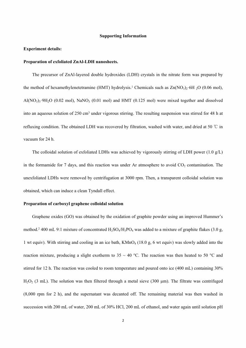
Fig. S1 TGA curve of rCG/LDO nanohybrid.
TGA was conducted to measure the percentage of rCG in the nanohybrid. No large mass loss was
observed resulted from that rCG/LDO nanohybrid was obtained after high vacuum calcination at 700 ℃. The
slight increase of mass probably resulted from that the sample adsorbed moisture in the flowing air or
baseline drift, and a similar phenomenon was observed for graphene/Ta2O5 sample.4 Rapid weight loss of 0.7%
from rCG combustion in rCG/LDO nanohybrid occurred at temperatures ranging from 480 to 620 ℃.
Therefore, the ratio of rCG/LDO is approximately 0.7 wt%.
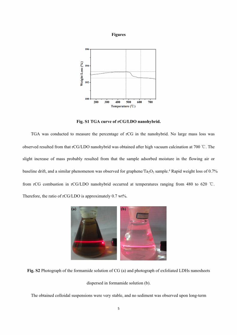
Fig. S2 Photograph of the formamide solution of CG (a) and photograph of exfoliated LDHs nanosheets
dispersed in formamide solution (b).
The obtained colloidal suspensions were very stable, and no sediment was observed upon long-term

6
standing.
Fig. S3 Tapping-mode AFM image of CG nanosheets (a) and their corresponding height profile (b).
Fig. S4 XRD pattern of pristine ZnAl-LDHs.
7
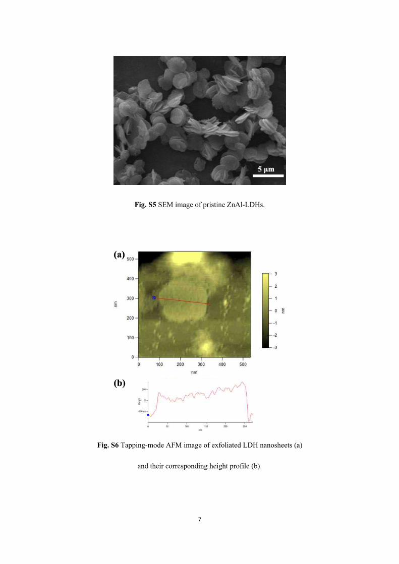
Fig. S5 SEM image of pristine ZnAl-LDHs.
Fig. S6 Tapping-mode AFM image of exfoliated LDH nanosheets (a)
and their corresponding height profile (b).

8
Fig. S7 Schematic diagram of the process of electrostatical self-assembly between the negatively charged CG monolayer and the positively charged ZnAl-LDH nanosheets.
Fig. S8 Nitrogen adsorption-desorption isotherms of samples: (a) ZnAl-LDH, (b) CG/LDH nanohybrid and
(c) rCG/LDO nanohybrid.
The pristine ZnAl-LDH presents a relatively weak adsorption of N2 with no distinct hysteresis loop,
which can be classified into typical Ⅲ adsorption–desorption isotherm. So ZnAl-LDH should not be porous
materials. Both of nanohybrids (CG/LDH and rCG/LDO) show hysteresis loops at P/P0 > 0.45 and
negligible N2 adsorption at P/P0 < 0.40, indicating that most porosity arises from mesopores formed by the
house-of-cards type stacking structure.5 According to the IUPAC classification, the samples can be classified
as Brunauer-Deming-Deaming-Teller (BDDT), typical IV adsorption-desorption isotherm, which is
characteristic of mesoporous materials with three-dimensional interconnected pore geometry and a high
energy of adsorption.

9
Fig. S9 XRD pattern of rCG/LDO nanohybrid.
Upon vacuum calcining CG/LDH nanohybrid in a tube furnace at 700℃ for 1 h, the XRD patterns (Fig.
S9) indicated that the structure of CG/LDH nanohybrid has been destroyed and are consistent with previous
studies of the thermal decomposition of LDHs,6 which have shown that zinc oxide phases are formed by a
topotactic transformation of the close-packed oxygen layers.7 In the XRD patterns of the calcined CG/LDH
nanohybrid, the peaks marked (220), (311) and (400) correspond to the characteristic reflections of a gahnite-
like phase.

10
Fig. S10 Raman spectra of rCG/LDO nanohybrid.
CG was used as a precursor to synthesize the nanohybrid, and would be converted into graphene after
high vacuum calcination at 700℃for 1 h, which can be confirmed by the G band of the Raman spectra. The
final product was obtained by high vacuum calcination of CG/LDHs nanohybrid at 700℃ for 1 h. In order to
investigate the C sp2 of the calcined product, Raman spectra of CG/LDO was determined, as shown in Fig.
S10. Compared with CG/LDH (without calcination), the D/G intensity ratio of rCG/LDO is larger,
suggesting a decrease in the average size of the sp2 domains upon reduction of the CG. As reported in the
hybrid composite CG/LDH (Fig. 3) exhibits the characteristics G and D bands at 1328 and 1597 cm-1,
respectively. Calcination caused the expected blue shift of the G band to 1586 cm-1 corresponding to the
conversion of CG into graphene.8, 9 After high vacuum calcination, CG would transform into graphene:
graphene-COOH graphene + CO2.
Fig. S11 TEM image of rCG/LDO nanohybrid.
The TEM image of rCG/LDO nanohybrid showed that the CG/LDH nanohybrid after vacuum
calcination were in the form of translucent plate-like flakes, with an average diameter close to 50-90 nm.

11
Fig. S12 Schematic representation of the photocatalytic degradation of dye in presence of rCG/LDO nanohybrid under visible light.
References
1. L. Li, R. Z. Ma, Y. Ebina, N. Iyi and T. Sasaki, Chem. Mater., 2005, 17, 4386-4391.2. D. C. Marcano, D. V. Kosynkin, J. M. Berlin, A. Sinitskii, Z. Z. Sun, A. Slesarev, L. B. Alemany, W. Lu and J. M.
Tour, Acs Nano, 2010, 4, 4806-4814.3. X. Sun, Z. Liu, K. Welsher, J. T. Robinson, A. Goodwin, S. Zaric and H. Dai, Nano Res., 2008, 1, 203-212.4. H. Sun, S. Liu, S. Liu and S. Wang, Appl. Catal. B-Environ., 2014, 146, 162-168.5. J. L. Gunjakar, T. W. Kim, H. N. Kim, I. Y. Kim and S.-J. Hwang, J. Am. Chem. Soc., 2011, 133, 14998-15007.6. Y. Q. Sun, Y. M. Zhou, Z. Q. Wang and X. Y. Ye, Appl. Surf. Sci., 2009, 255, 6372-6377.7. J. Sun, H. M. Liu, X. Chen, D. G. Evans, W. S. Yang and X. Duan, Chem. Commun., 2012, 48, 8126-8128.8. K. N. Kudin, B. Ozbas, H. C. Schniepp, R. K. Prud'homme, I. A. Aksay and R. Car, Nano Lett., 2007, 8, 36-41.9. M. Latorre-Sanchez, P. Atienzar, G. Abellan, M. Puche, V. Fornes, A. Ribera and H. Garcia, Carbon, 2012, 50,
518-525.