BST_2015Vol9No3_pp138_206.pdf - BioScience Trends
80
www.biosciencetrends.com BST BioScience Trends
-
Upload
khangminh22 -
Category
Documents
-
view
1 -
download
0
Transcript of BST_2015Vol9No3_pp138_206.pdf - BioScience Trends

www.biosciencetrends.com
BSTBioScience Trends


www.biosciencetrends.com
BioScience Trends is one of a series of peer-reviewed journals of the International Research and Cooperation Association for Bio & Socio-Sciences Advancement (IRCA-BSSA) Group and is published bimonthly by the International Advancement Center for Medicine & Health Research Co., Ltd. (IACMHR Co., Ltd.) and supported by the IRCA-BSSA and Shandong University China-Japan Cooperation Center for Drug Discovery & Screening (SDU-DDSC).
BioScience Trends devotes to publishing the latest and most exciting advances in scientific research. Articles cover fields of life science such as biochemistry, molecular biology, clinical research, public health, medical care system, and social science in order to encourage cooperation and exchange among scientists and clinical researchers.
BioScience Trends publishes Original Articles, Brief Reports, Reviews, Policy Forum articles, Case Reports, News, and Letters on all aspects of the field of life science. All contributions should seek to promote international collaboration.
ISSN: 1881-7815 Online ISSN: 1881-7823
CODEN: BTIRCZIssues/Year: 6
Language: EnglishPublisher: IACMHR Co., Ltd.
Editor-in-Chief:Norihiro KOKUDO The University of Tokyo, Tokyo, Japan
Co-Editors-in-Chief:Xue-Tao CAO Chinese Academy of Medical Sciences, Beijing, ChinaRajendra PRASAD University of Delhi, Delhi, IndiaArthur D. RIGGS Beckman Research Institute of the City of Hope, Duarte, CA, USA
Chief Director & Executive Editor:Wei TANG The University of Tokyo, Tokyo, Japan
Senior Editors:Xunjia CHENG Fudan University, Shanghai, ChinaYoko FUJITA-YAMAGUCHI Beckman Research Institute of the City of Hope, Duarte, CA, USANa HEFudan University, Shanghai, ChinaKiyoshi KITAMURA The University of Tokyo, Tokyo, JapanMisao MATSUSHITA Tokai University, Hiratsuka, JapanMunehiro NAKATATokai University, Hiratsuka, JapanTakashi SEKINE
Toho University, Tokyo, JapanRi SHOYamagata University, Yamagata, JapanYasuhiko SUGAWARA The University of Tokyo, Tokyo, Japan
Managing Editor:Jianjun GAOQingdao University, Qingdao, China
Web Editor:Yu CHEN The University of Tokyo, Tokyo, Japan
Proofreaders:Curtis BENTLEY Roswell, GA, USAChristopher HOLMES The University of Tokyo, Tokyo, JapanThomas R. LEBON Los Angeles Trade Technical College, Los Angeles, CA, USA
Editorial OfficePearl City Koishikawa 603, 2-4-5 Kasuga, Bunkyo-ku, Tokyo 112-0003, JapanTel: +81-3-5840-8764Fax: +81-3-5840-8765E-mail: [email protected]
Editorial Board
i

www.biosciencetrends.com
Editorial Board Members
Girdhar G. AGARWAL(Lucknow, India)Hirotsugu AIGA(Geneva, Switzerland)Hidechika AKASHI(Tokyo, Japan)Moazzam ALI(Geneva, Switzerland)Ping AO(Shanghai, China)Hisao ASAMURA(Tokyo, Japan)Michael E. BARISH(Duarte, CA, USA)Boon-Huat BAY(Singapore, Singapore)Yasumasa BESSHO(Nara, Japan)Generoso BEVILACQUA(Pisa, Italy)Shiuan CHEN(Duarte, CA, USA)Yuan CHEN(Duarte, CA, USA)Naoshi DOHMAE(Wako, Japan)Zhen FAN(Houston, TX, USA)Ding-Zhi FANG(Chengdu, China)Yoshiharu FUKUDA(Ube, Japan)Rajiv GARG(Lucknow, India)Ravindra K. GARG(Lucknow, India)Makoto GOTO(Tokyo, Japan)Demin HAN(Beijing, China)David M. HELFMAN(Daejeon, Korea)Takahiro HIGASHI
(Tokyo, Japan)De-Xing HOU(Kagoshima, Japan)Sheng-Tao HOU(Ottawa, Canada)Yong HUANG(Ji'ning, China)Hirofumi INAGAKI(Tokyo, Japan)Masamine JIMBA(Tokyo, Japan)Kimitaka KAGA(Tokyo, Japan)Ichiro KAI(Tokyo, Japan)Kazuhiro KAKIMOTO(Osaka, Japan)Kiyoko KAMIBEPPU(Tokyo, Japan)Haidong KAN(Shanghai, China)Bok-Luel LEE(Busan, Korea)Mingjie LI(St. Louis, MO, USA)Shixue LI(Ji'nan, China)Ren-Jang LIN(Duarte, CA, USA)Xinqi LIU(Tianjin, China)Daru LU(Shanghai, China)Hongzhou LU(Shanghai, China)Duan MA(Shanghai, China)Masatoshi MAKUUCHI(Tokyo, Japan)Francesco MAROTTA(Milano, Italy)Yutaka MATSUYAMA(Tokyo, Japan)
Qingyue MENG(Beijing, China)Mark MEUTH(Sheffi eld, UK)Satoko NAGATA(Tokyo, Japan)Miho OBA(Odawara, Japan)Fanghua QI(Ji'nan, Shandong)Xianjun QU(Beijing, China)John J. ROSSI(Duarte, CA, USA)Carlos SAINZ-FERNANDEZ(Santander, Spain)Yoshihiro SAKAMOTO(Tokyo, Japan)Erin SATO(Shizuoka, Japan)Takehito SATO(Isehara, Japan)Akihito SHIMAZU(Tokyo, Japan)Zhifeng SHAO(Shanghai, China)Judith SINGER-SAM(Duarte, CA, USA)Raj K. SINGH(Dehradun, India)Peipei SONG(Tokyo, Japan)Junko SUGAMA(Kanazawa, Japan)Hiroshi TACHIBANA(Isehara, Japan)Tomoko TAKAMURA(Tokyo, Japan)Tadatoshi TAKAYAMA(Tokyo, Japan)Shin'ichi TAKEDA(Tokyo, Japan)Sumihito TAMURA
(Tokyo, Japan)Puay Hoon TAN(Singapore, Singapore)Koji TANAKA(Tsu, Japan)John TERMINI(Duarte, CA, USA)Usa C. THISYAKORN(Bangkok, Thailand)Toshifumi TSUKAHARA(Nomi, Japan)Kohjiro UEKI(Tokyo, Japan)Masahiro UMEZAKI(Tokyo, Japan)Junming WANG(Jackson, MS, USA)Ling WANG(Shanghai, China)Xiang-Dong Wang(Boston, MA, USA)Hisashi WATANABE(Tokyo, Japan)Lingzhong XU(Ji'nan, China)Masatake YAMAUCHI(Chiba, Japan)Aitian YIN(Ji'nan, China)George W-C. YIP(Singapore, Singapore)Xue-Jie YU(Galveston, TX, USA)Benny C-Y ZEE(Hong Kong, China)Yong ZENG(Chengdu, China)Xiaomei ZHU(Seattle, WA, USA)
(as of April 25, 2015)
BioScience TrendsEditorial and Head OfficePearl City Koishikawa 603, 2-4-5 Kasuga, Bunkyo-ku, Tokyo 112-0003, Japan
Tel: +81-3-5840-8764, Fax: +81-3-5840-8765E-mail: [email protected]: www.biosciencetrends.com
ii

www.biosciencetrends.com
Advances in diagnosis, treatments, and molecular mechanistic studies of traumatic brain injury.Chunyu Lu, Jufeng Xia, Bin Wang, Yitian Wu, Xiaohui Liu, Yong Zhang
The role of autophagy in bacterial infections.Nayeli Shantal Castrejón-Jiménez, Kahiry Leyva-Paredes, Juan Carlos Hernández-González, Julieta Luna-Herrera, Blanca Estela García-Pérez
Polyphosphate-induced matrix metalloproteinase-3-mediated proliferation in rat dental pulp fibroblast-like cells is mediated by a Wnt5 signaling cascade.Nobuaki Ozeki, Hideyuki Yamaguchi, Naoko Hase, Taiki Hiyama, Rie Kawai, Ayami Kondo, Kazuhiko Nakata, Makio Mogi
Bu-Shen-Ning-Xin decoction suppresses osteoclastogenesis via increasing dehydroepiandrosterone to prevent postmenopausal osteoporosis.Yuyan Gui, Xuemin Qiu, Yingping Xu, Dajin Li, Ling Wang
Evaluation of medical staff and patient satisfaction of Chinese hospitals and measures for improvement. Min Li, Chengyu Huang, Xiangchan Lu, Siyuan Chen, Pan Zhao, Hongzhou Lu
MVsCarta: A protein database of matrix vesicles to aid understanding of biomineralization.Yazhou Cui, Quan Xu, Jing Luan, Shichang Hu, Jianbo Pan, Jinxiang Han, Zhiliang Ji
Risk factors for recurrence of primary spontaneous pneumothorax after thoracoscopic surgery.Haibo Huang, Hua Ji, Hui Tian
Reviews
138 - 148
149 - 159
Original Articles
160 - 168
169 - 181
182 - 189
Brief Reports
190 - 192
193 - 197
CONTENTS Volume 9, Number 3, 2015
iii

www.biosciencetrends.com
Castleman disease of the mesentery as the great mimic: Incidental fi nding of one case and the literature review.Ang Lv, Chunyi Hao, Honggang Qian, Jiahua Leng, Wendy Liu
China upgrades surveillance and control measures of Middle East respiratory syndrome (MERS).Jianjun Gao, Peipei Song
Expected role of medical technologists in diabetes mellitus education teams.Kazuhiko Kotani, Takahiro Imazato, Keizo Anzai; Kyushu Diabetes Testing Study Group
Case Report
198 - 202
News
203 - 204
Letter
205 - 206
Guide for Authors
Copyright
CONTENTS (Continued )
iv
www.biosciencetrends.com
BioScience Trends. 2015; 9(3):138-148. 138
Advances in diagnosis, treatments, and molecular mechanistic studies of traumatic brain injury
Chunyu Lu1,*,**, Jufeng Xia2,*, Bin Wang1, Yitian Wu1, Xiaohui Liu1, Yong Zhang1
1 Department of Neurosurgery, The People's Hospital of Huaibei, Anhui, China;2 Institute of Biochemistry and Cell Biology, Shanghai Institutes for Biological Sciences, Chinese Academy of Sciences, Shanghai,
China.
1. Introduction
Traumatic brain injury (TBI), also known as intracranial injury, occurs when an external force damages the brain. TBI can be classified by severity (mild, moderate, or severe), mechanism (closed or penetrating head injury), or other characteristics (1). TBI is a main cause of death and disability around the world especially in soldiers, children, and young men. Males suffer TBIs more frequently than females. Each year about 1.7 million Americans are saved in emergency rooms after suffering a TBI of some severity; of these, 52,000 die of TBI and other secondary injuries and another 275,000 are hospitalized and survive (2). Neurological damage from TBI does not only occur at the moment of focal impact upon the head but also involves secondary injury over the ensuing hours and days. This injury,
which includes changes in cerebral blood flow and intracranial pressure, leads to substantial damage following the original injury. Besides cell death, a series of physiological changes including diffuse axonal injury (DAI), micrangium damage, and diffuse neuronal injury can also occur on a microscopic scale in the cerebral parenchyma following trauma and lead to subsequent morbidity. Clinical symptoms of these physiological changes include loss of consciousness, dizziness, headaches, inattention, and hypomnesia (3). Currently, patients with moderate to severe trauma will in all probability receive treatment in an intensive care unit after a neurosurgical procedure (4). Treatment depends on the patient's stage of recovery. In the acute stage, the primary objective of the surgeon is to stabilize the patient and do one's best to prevent further damage because slight damage can worsen the primary injury caused by trauma (4). Rehabilitation is the primary treatment for interim and latter stages of recovery (4). Prognosis worsens with the severity of injury. Permanent disability is considered to occur in 10% of mild injuries, 66% of moderate injuries, and 100% of severe injuries (5). Most patients in a coma or with a subarachnoid hemorrhage or DAI are considered
Summary Traumatic brain injury (TBI) is a main cause of death and disability around the world especially in soldiers, children, and young men. Since its clinical diagnosis and treatment cannot predict its prognosis, novel diagnostic techniques need to be developed, insight into its molecular mechanisms needs to be gleaned, and alternative and complementary medicine (ACM) approaches to its treatment need to be developed. This review summarizes the new diagnostic methods used in clinical practice, such as imaging of structural abnormalities after TBI and measurement of prognosis-related biomarkers. This review also describes the cellular mechanisms of traditional Chinese medicine in terms of intracellular signaling pathways, the extracellular microenvironment, and stem cells. This review concludes by describing experimental and clinical studies of the use of traditional Chinese medicine as a form of ACM to treat TBI. This review helps to understand advances in the field of TBI diagnosis and treatment.
Keywords: Traumatic brain injury, signaling pathway, inflammation microenvironment, stem cells, traditional Chinese medicine
DOI: 10.5582/bst.2015.01066Review
*These authors contributed equally to this works. **Address correspondence to:Dr. Chunyu Lu, Department of Neurosurgery, The People's Hospital of Huaibei, No.66, Huaihai-Xi-Lu, Huaibei 235000, Anhui, China.E-mail: [email protected]

www.biosciencetrends.com
BioScience Trends. 2015; 9(3):138-148.
to have a bad prognosis (6-8). Thus, there is an urgent need for novel therapies, medicines, biomarkers to predict prognosis, and treatment alternatives. This review begins by briefly discussing advances in clinical diagnosis and management of TBI. This review then focuses on studies of the biomechanics of and rehabilitation from TBI. This review then summarizes the potential usefulness of alternative treatments of TBI. This review concludes by offering ideas on the direction for future research into TBI treatments and their clinical use.
2. Clinical diagnosis and treatment
TBI has been studied since 1650-1550 BC and there are methods of assessing and managing the progression of TBI, but its prognosis remains, so more effective forms of clinical treatment of TBI should be sought (9).
2.1. Novel methods of assessing TBI
Formalin-fixed, paraffin-embedded archival tissue (PEAT) specimens were obtained from a total of 95 primary ALM (42 males and 53 females, mean age T)
2.1.1. Imaging of structural abnormalities
Concussion, based on the current definition, is a symptom of TBI. Concussions were conventionally considered to be simply physiological injuries, caused by a metabolic disorder of the brain as a result of alternations in ionic gradients, a disruption of sodium, potassium, and calcium channels, an imbalance in neurotransmitters, and inflammation (10). This standpoint has been substantiated by a series of metabolic and functional imaging studies in humans (11,12) and animals (10). Nevertheless, studies of the usefulness of advanced structural neuroimaging methods, such as susceptibility weighted imaging and diffusion tensor imaging, have revealed subtle structural abnormalities in white matter and brain microvasculature in a significant proportion of patients with a TBI, and especially in patients with severe TBI (11-13). Histopathological results from patients with a TBI who subsequently died from their injuries suggested that DAI is a key pathologic cause of TBI (14,15). Advanced neuroimaging studies, and especially those involving diffusion tensor imaging (DTI), support this contention. DTI is used to test the diffusion of water along the axis of white matter tracts and can discern the interruption of water diffusion within 2 weeks of persistent TBI in people with a normal MRI scan (16,17). Although DTI results are potential biomarkers of TBI, studies of DTI differ in their description of changes in diffusion, facilities use different imaging protocols, facilities use different methods of quality assurance and methods of analysis, and facilities have failed to provide sufficient normative data. These
problems need to be resolved. A diffuse microhemorrhage (DM) is another physiological cause of TBI. DM has long been considered to be a physiological cause of severe TBI, but abnormalities in cerebrovascular reactivity and cerebral blood flow have also been found in mild TBI, and especially in people who have suffered multiform mild TBIs and who have enduing post-concussive symptoms (18,19) Data from neuroimaging studies (20) using T2*-weighted gradient echo imaging, which is sensitive to DM, found DM in deep white matter in 23 of 98 patients who suffered a TBI. Other abnormities indicative of TBI have been identified by computed tomography (CT) and high-resolution magnetic resonance imaging (MRI). These abnormalities include focal contusions, traumatic subarachnoid haemorrhage, and extra-axial hematomas. MRI is clearly much more sensitive than CT at verifying the presence of subtle abnormities. A multicenter study of 98 patients revealed that 27 (28%) had an aberrant MRI scan an average of 12 days after injury (20). In that study, a subarachnoid haemorrhage confirmed by a CT scan and multiple foci of hemorrhagic axonal injury identified by MRI were related to more severe disability three months after TBI (20). After TBI, functional magnetic resonance imaging (fMRI) has revealed changes in dynamic functional connectivity and the pattern of brain activity in a resting state as well as changes in cognitive test results. Changes in test results and fMRI results in a resting state have been noted even when patients perform well on cognitive tests and are allowed to return to regular activities (11,12,21). Quantitative electroencephalogram has been used to identify a physiological disorder after TBI and provide evidence of enduring neuronal malfunction at a certain point after clinical symptoms disappear (22). Although these advanced methods of imaging and electroencephalography seem to be more sensitive than current methods of clinical diagnosis, they still are mainly in the research stage.
2.1.2. Biomarkers
The diagnosis of TBI can be difficult if the injury is not witnessed, no evidence of a wound exists, a CT scan is normal, or if the diagnosis was delayed for 24 hours or longer. To aid in the diagnosis of TBI and decrease the dependency on self-reports, biomarkers of TBI in the blood, saliva, urine, and cerebrospinal fluid (CSF) have attracted the attention of researchers (23). Serum is the most often researched biomarker reservoir (24). The most extensively researched biomarkers in the blood are glial fibrillary acidic protein (GFAP) and ubiquitin C-terminal hydrolase-L1 (UCH-L1). A receiver operating characteristic (ROC) analysis has indicated that the area under the curve is greater than 0.87 for
139

www.biosciencetrends.com
BioScience Trends. 2015; 9(3):138-148. 140
medication is used only if seizures occur (34,35). Numerous studies have indicated that phenytoin (PHT) can be used to prevent seizures soon after TBI, but other anti-seizure drugs such as levetiracetam (LEV) are also being used in clinical practice. PHT has its drawbacks, such as cognitive side effects and effects on physical recovery (36). Over the past few years, certain new drugs such as zonisamide and vigabatrin have been used clinically in the US, the UK, Australia, and Japan for both adjunctive therapy and monotherapy for partial seizures (simple, complex, and secondarily generalized), generalized seizures (tonic, tonic-clonic, and atypical absence), and combined seizures (37-39). Coma-inducing medication is sometimes used by doctors to induce a temporary coma (a deep state of unconsciousness) (40). Because the metabolism of the brain has been significantly altered during a TBI and areas of the brain may lack a sufficient blood flow, coma-inducing drugs are used to profoundly inactivate the brain so that it consumes less oxygen (41). This is especially helpful if blood vessels, stressed by elevated pressure in the brain, are unable to carry the normal amount of nutrients and oxygen to brain cells (42). Although coma-inducing medications protect the brain, the brain as a whole is, by definition, not receiving the blood it needs.
3. Studies of mechanisms
3.1. Signaling pathways
Recent clinical therapies cause various adverse reactions and have not yielded satisfactory results (43,44). Thus, a great deal of work needs to be done to explore the molecular mechanisms of TBI and develop more targeted therapies. An obvious inflammatory response occurs following a TBI. In the immediate phase following the primary trauma, the inflammatory reaction aggravates cell damage and worsens prognosis (45). The nuclear factor kappa B (NF-κB) signaling pathway has long been considered to be an inflammation-related signaling pathway, mainly based on the function of NF-κB in the expression of proinflammatory genes including cytokines, chemokines, and adhesion molecules (46). NF-κB is also considered to play a significant part in the regulation of apoptosis (47). Several studies at different facilities have found that NF-κB, as a downstream element of a series of receptors such as toll-like receptor 4 (TLR-4) and tumor necrosis factor receptor-associated factor 6 (TRAF6), is activated in specimens of animal or human brains (48-50). Thus, NF-κB is considered to be a target by which to decrease inflammation and apoptosis after TBI. Several research teams are developing various NF-κB inhibitors, such as SN50, nerve growth factors, and pituitary adenylate cyclase-activating polypeptide (PACAP), to suppress
both GFAP and UCHL1 (25). The high sensitivity and specificity of these biomarkers mean that they can distinguish between individuals who have suffered a TBI and healthy people (0.87, 95% CI 0.83-0.90, and 0.91, 95% CI 0.88-0.94, for GFAP and UCHL1, respectively) and differentiate between individuals who have suffered a TBI and who have an abnormal CT scan and those who have a normal CT scan (0.71, 95% CI 0.64-0.78, and 0.88, 95% CI 0.84-0.93, for GFAP and UCHL1, respectively) (25,26). However, GFAP and UCHL1 do not have sufficient sensitivity and specificity to predict the prognosis for a complicated TBI (25). Biomarkers were detected in professional hockey players before the season and again after a TBI, and these players had increased concentrations of microtubule-stabilized tau protein in the blood after a TBI (27). The range of this rise in the concentration of tau protein is associated with the duration of post-concussive symptoms. Despite advances in animal models and human studies and evidence that biomarkers can potentially facilitate the diagnosis of TBI, further confirmation is needed. Given the variety of pathological mechanisms involved in TBI, a set of biomarkers with sufficient sensitivity and specificity needs to be developed for general clinical use (25).
2.2. Clinical treatment
During TBI, a few cells in the brain are directly mechanically damaged, but more cells are injured as a result of trauma-induced biochemical changes, which is what is referred to as secondary injury. Based on guidance from the Mayo Clinic (28), the following medications may be used to prevent secondary injury to the brain immediately after a trauma. Diuretics, which are a group of substances that promote the production of urine, are used to treat heart failure, liver cirrhosis, hypertension, water poisoning, certain kidney diseases, and TBI (29). Drugs such as high ceiling/loop diuretics, thiazides, carbonic anhydrase inhibitors, potassium-sparing diuretics, calcium-sparing diuretics, and osmotic diuretics decrease the amount of fluid in tissues and increase micturition (30-32). Diuretics, given intravenously to people who have suffered a TBI, help decrease pressure inside the brain. However, they have serious side effects, such as hypovolemia, hypokalemia, hyperkalemia, hyponatremia, metabolic alkalosis, and metabolic acidosis (33). Anticonvulsants, also known as anti-epileptic drugs or anti-seizure drugs, are a diverse group of pharmacological agents used to treat epileptic seizures. People who have suffered a moderate to severe TBI are at risk of having seizures during the first week after injury. An anti-seizure drug may be given during the first week to avoid any additional brain damage that might be caused by a seizure. Additional anti-seizure

www.biosciencetrends.com
BioScience Trends. 2015; 9(3):138-148.141
the up-regulation of NF-κB in brain tissue (47,50,51). Glycogen synthase kinase 3 beta (GSK-3β) is one of the most important downstream elements of NF-κB (52), so several research teams have focused on changes in its expression and its potential as a target for TBI treatment. An animal model indicated that GSK-3β is involved in neuronal survival after TBI (53). Lin et al. found that transfection of GSK-3β small-interfering RNA increased cell survival in Sprague-Dawley rats (54). In the inflammatory response after TBI, the Janus kinase/signal transducer and activator of transcription (JAK/STAT) pathway is found to be activated and to increase cell apoptosis in the cortical pericontusional zone (55). The same research team also reported that recombinant human erythropoietin (rhEPO) increased the level of p-JAK2 and p-STAT3 expression, decreasing apoptosis and promoting cell survival. As TBI progresses, reactive oxygen species (ROS) are produced in brain tissue and lead to cellular apoptosis (56). The nuclear factor (erythroid-derived 2)-like 2 (NFE2L2 or Nrf2) signaling pathway regulates the level of expression of antioxidant proteins that protect against oxidative injury induced by trauma and inflammation, and this pathway has increasingly attracted attention in studies of the molecular mechanism of ROS in TBI (57). In rat and mouse experiments, activation of the Nrf2 pathway has been found to inhibit ROS-induced damage in brain tissue (58). Catenin beta 1 (β-catenin) is a dual function protein, regulating the coordination of cell-cell adhesion and gene transcription (59). β-catenin was found to increase in astrocytes in gliogenesis after TBI in the adult brain and it was found to be involved in neuronal survival (60). Several substances have been found to activate the β-catenin pathway to alleviate cell injury. Up-regulation of serum- and glucocorticoid-regulated kinase (SGK) was reported to protect against neuronal apoptosis via the β-catenin signaling pathway (61). Interestingly, in this study the β-catenin signaling pathway was activated by GSK-3β, which indicates that there may be a signaling pathway in TBI. In addition, up-regulation of survivin, a key component in the β-catenin pathway, was found to promote neurogenesis following TBI (62). Many other signaling pathways have also been investigated, such as the Notch pathway, PTEN pathway, ERK pathway, and p38MAPK pathway. These pathways may be a novel target for new TBI therapies (63-66).
3.2. Microenvironment
The cell microenvironment consists of elements that directly affect conditions around a cell or a cell cluster, and these elements play a direct or indirect role in affecting cell behavior biophysically or biochemically (67). The cell microenvironment consists of (i) an extracellular matrix (ECM), (ii) cytokines, hormones, and other bioactive materials around cells produced
from autocrine, endocrine, and paracrine secretions, (iii) exosomes between cells, and (iv) mechanical forces created by the movement of tissue or the movement of physiological fluids such as blood. Neuro-inflammation represents an important pathological process in secondary injury after TBI (68). Resident astrocytes and microglia are usually the initial cells that promote an inflammatory cascade following tissue injury, and bioactive proteins associated with the activation of these cells are often used as biomarkers of TBI (69,70). Astrogliosis, an abnormal increase in the number of astrocytes due to the destruction of nearby neurons, has been defined in the context of both neuroprotection and neurodegeneration (71,72). Activated astrocytes are able to secret pro-inflammatory cytokines, chemokines, and matrix metalloproteinases (MMPs) that degrade the extracellular matrix and lead to further disintegration of the blood-brain barrier (73,74). However, astrocytes are also able to secrete molecules that promote repair and regeneration after central nervous system (CNS) damage (75,76). Exosomes are cell-derived vesicles that exist in a number of biological fluids such as blood and urine and in used cell culture medium (77). Exosomes interact with the plasma membrane of a target cell by ligand-to-receptor binding, fusion, internalization, or a combination of these actions. If the exosomes fuse with recipient cells, they can transfer their cargo, including bioactive lipids, cytokines, growth factors, receptors, and hereditary material, to the addressee cell (78). In a study, TBI-derived exosomes induced the emergence of pro-inflammatory cytokines, including IL-1β. IL-1β is produced primarily by microglia and acts as a pro-inflammatory pyrogen, up-regulating expression of other cytokines, proteases, and MMPs (79). Previous studies reported that specific microRNAs are associated with the progression of neurological disorders, leading to the initiation and progression of complications associated with a TBI (80). MicroRNAs delivered by exosomes produced by injured brain cells do present an advantage since they are sensitive, clinically accessible biomarkers that can improve the diagnosis of TBI and that can function as prognostic markers after treatment. Mechanotransduction refers to the various mechanisms by which cells translate a mechanical stimulus into an electro-chemical signal (81). The role of pathological cellular mechanotransduction in brain tissue remains unclear. However, several studies have indicated that it may be an initiator of TBI. In an in vitro study, quick deformation of three-dimensional collagen gels led to a decrease in embedded neuronal viability when the collagen concentration increased, indicating the potential impact of cell-ECM interactions on injury (82). Another study suggested that ECM influences axonal injury by activating Rho signaling pathways; up-regulation of RhoA was accompanied by fluid percussion brain injury (83).

www.biosciencetrends.com
BioScience Trends. 2015; 9(3):138-148. 142
3.3. Stem cells
Among acute neuropathological conditions, TBI is one of the major causes of death and disability around the world (84). Cell transplantation may be a therapy for TBI. Whether the production of new neurons leads to a recovery of function, axonal sprouting, synaptic plasticity, or neosynaptogenesis is unknown. The rate at which these new neurons are generated and the rate of functional recovery are known to be very low after TBI (85). Studies initially focused on neuronal restoration after TBI. One year after transplantation of neural precursor cells (NPCs) into the striatum of mice, the mice had improved long-term survival and improved motor functions without tumor formation (86). Fetus-derived immortalized neural stem cells (NSCs) were transplanted into the injured cortex, leading to recovery of motor function but no cognitive improvement (87). After these NSCs were transplanted into the hippocampus, cognitive improvement was noted but there was no improvement in motor function (88). A study in 2005 transplanted NSCs into patients who suffered a TBI (89). In both studies, the NSCs moved from the site of implantation to the site of the injury. In addition, the experimental group displayed improved recovery in comparison to the control group. At that point, fMRI revealed improved activity at the site of the injury, positron emission tomography (PET) revealed that patients were improving, somatosensory evoked potentials (SEP) revealed slight improvement until six months after transplantation, and Disability Rating Scale (DRS) scores quickly rose six months after transplantation. A clinical study transplanted bone marrow mononuclear cells in 10 children (from age 5 to 14) with a Glasgow Coma Scale score of 5 to 8 (90). This study noted no adverse effects during the six months after transplantation. These children were also evaluated with the Pediatric Logistic Organ Dysfunction (PELOD) test, and no adverse effects on white matter, gray matter, or cerebrospinal fluid (CSF) were noted. The main obstacle to stem cell transplantation in TBI is the recovery of motor function and cognition. However, recovery depends on the injured area where stem cells are implanted (91). After stem cells are implanted into the hippocampus, for example, these implanted cells are more apt to survive than when they are implanted into various areas of the neocortex. In addition, different types of progenitor or stem cells seem to perform various functions after transplantation. Mesenchymal stem cells (MSCs) are used for neurotrophic support, progenitor oligodendrocytes are used to establish remyelination in white matter, and neural progenitor cells play a role in cell replacement.
4. Alternative and complementary medicine
Although conventional medications have been widely
used in the clinical treatment of TBI, mounting evidence suggests that conventional medications for treatment of TBI have a number of drawbacks. Anti-convulsants induce amnesia, ataxia, and diplopia, anti-depressants induce blurred vision, confusion, and dizziness, and anti-psychotics induce headaches. Thus, complementary and alternative medicine, such as traditional Chinese medicine, may need to supplement treatments for TBI (92). Alternative medicine is any practice, approach, or medication that is thought to have the healing effects of medicine but that does not originate from evidence gathered using the scientific method (93). This form of medicine includes a large number of health care practices, products, and therapies. Complementary medicine is a form of alternative medicine used in combination with conventional medicine in the belief, albeit not proven using the scientific method, that it complements the treatment (94). Complementary and alternative medicine is referred to as CAM. Traditional Chinese medicine (TCM) is one type of CAM, and TCM stems from medical practices with common concepts that have developed in China for more than 2,000 years. TCM includes various herbal medicines, acupuncture, massage, exercise, and diet therapy (95). In experimental and clinical studies, animals and patients were given different CAM after TBI to assist in recovery when conventional medicine was unable to improve the condition of or prognosis for the control group (96). Both TCM (Table 1) and its bioactive components (Table 2) are being studied at the experimental or clinical level. However, most of these studies involve experiments in animal models. Neuroprotection after TBI is key. Several TCMs display anti-inflammatory and/or anti-oxidant action. A Xingnaojing injection was found to have a protective effect in rats with a TBI. It may have a protective effect by alleviating brain edema and inhibiting the production of reactive oxygen species (ROS) in rats (97). Manasamitra vatakam was also reported to prevent brain damage from TBI-induced neurotoxicity by increasing superoxide dismutase (SOD) and 70 kilodalton heat shock proteins (HSP70) in rats (98). Studies of a Qingkailing injection and early treatment with MLC601 suggested that these TCMs reduce TBI-induced brain damage by blocking mitochondria-mediated signaling pathways in rats (99,100). Following primary trauma, the inflammatory response promotes neural cell damage and worsens prognosis, so studies have focused on the anti-inflammatory action of TCMs. In a rat model, a modified Shengyu decoction (MSD) was reported to be a potential therapy for TBI because it decreased the inflammatory response after TBI. MSD inhibits the inflammatory reaction by decreasing the levels of TNF-α, IL-1β, GFAP-, and Iba1-positive cells and by increasing the level of IL-10 (101). Another important aspect after TBI is neurogenesis. In addition to its anti-apoptotic action, MLC601 was also reported

www.biosciencetrends.com
BioScience Trends. 2015; 9(3):138-148.143
to promote motor recovery in an animal model (100). A Xingnaojing injection and MLC901 were reported to improve nerve impairment and recovery of cognitive function by increasing S100 calcium-binding protein beta (S100B) and neuron-specific enolase (NSE) in rats (97,102). Qin-Nao-Yi-Zhi-Fang was found to counteract glutamate excitotoxicity after TBI by inhibiting nitric oxide (103). In addition, the effects of Ginseng total saponins and MSD on neurogenesis have been studied. These TCMs may improve neurorestoration in an animal model of TBI animal model by increasing the nerve growth factor (NGF), glial cell line-derived neurotrophic factor (GDNF), neural cell adhesion
molecule (NCAM), and neural stem/progenitor cells (NSCs) (104,105). Several clinical studies have treated TBI with TCM. A rhubarb extract was reported to be able to decrease patients' body temperature, intracranial pressure, and hemorrhaging in the digestive tract, but the mechanism of this action remains unclear (106). Panax notoginseng saponin may have neuroprotective action by attenuating edema and hematoma (107). Bioactive components of TCMs have also been studied as treatments for TBI over the years. Osthole, isolated from the TCM Cnidium monnieri, was found to reduce neurological deficits, cerebral edema, and hippocampal neuron loss by inhibiting mitochondria-
Table 1. List of TCMs used in the treatment of TBI
TCM
Qin-Nao-Yi-Zhi-Fang
Qingkailing injection
Modified Shengyu decoction
MLC601
Xingnaojing injection
MLC901
Ginseng total saponins
Modified Shengyu decoction
Manasamitra
Rhubarb
Panax notoginseng saponin
Therapeutic targets
Rat cerebral neuronal cells
Rats
Rats
Rats
Rats
Rats
Rats
Rats
Rats
Humans
Humans
Ref.
(102)
(98)
(100)
(99)
(96)
(101)
(103)
(104)
(97)
(105)
(106)
BT: body temperature; ICP: intracranial pressure; HITDT: hemorrhage in the digestive tract; S100B: S100 calcium-binding protein beta; NSE: neuron-specifi c enolase; VEGF: vascular endothelial growth factor; NGF: nerve growth factor; GDNF: glial cell line-derived neurotrophic factor; NCAM: neural cell adhesion molecule; NSC: neural stem/progenitor cell; HSP70: 70 kilodalton heat shock proteins; SOD: superoxide dismutase.
Action
Counteracts glutamateexcitotoxicity
Anti-apoptotic action
Anti-inflammatory action
Anti-apoptotic action,improves motor recovery
Anti-oxidant,induces neurogenesis
Induces neurogenesis
Induces neurogenesis
Induces neurogenesis
Anti-oxidant
Decreases BT, ICP, and HITDT
Neuroprotective action
Mechanism
Inhibits nitric oxide
Inhibits caspase-3
Decreases TNF-α and IL-1β and increases IL-10
Decreases TNF-α and IL-1 and increases IL-10
Increases S100B and NSE
Increases S100B and NSE; regulates aquaporin 4; increases VEGF
Increases NGF, GDNF, NCAM, and NSC
Increases NGF, GDNF, NCAM, etc.
Increases HSP70, SOD, etc.
Not indicated
Attenuates edema and hematoma
Table 2. List of TCMs used in the treatment of TBI
Components
Osthole
Curculigoside
Ginsenoside Rbeta1
Z-ligustilide
Curcumin
Salvianolic acid B
Triptolide
Herbs
Cnidium monnieri
Curculigo orchioides Gaertn.
Panax ginseng
Angelica sinensis
Curcuma longa
Salvia miltiorrhiza Bunge
Tripterygium wilfordii Hook. f.
Ref.
(107)
(108)
(109)
(110)
(111)
(112)
(113)
ND: neurological defi cits; CE: cerebral edema; HNL: hippocampal neuron loss; BBB: blood-brain barrier; CV: cerebral vasospasm.
Therapeutic targets
Rats
Cortex neurons
Rats
Rats
Mice
Mice
Rats
Effects
Reduces ND, CE, and HNL
Reduces neuronal cell loss
Reduces ND, CE, and BBB disruption
Reduces ND, CE, and BBB disruption, and reduces CV
Attenuates inflammation
Attenuates inflammation
Attenuates inflammation
Mechanisms
Inhibits mitochondrial pathways; inhibits ROS release
Inhibits mitochondrial pathways; inhibits ROS production
Inhibits mitochondrial and p53 pathways
Inhibits mitochondrial and p53 pathways
Inhibits TLR4/MyD88/NF-κB pathways
Decreases TNF-α, IL-1β; increases IL-10 and TGF-β1
Decreases TNF-α, IL-1, IL-4, IL-6, IL-8, IL-17, and IL-23

www.biosciencetrends.com
BioScience Trends. 2015; 9(3):138-148. 144
mediated signaling pathways and inhibiting ROS release in rats (108). Curculigoside, a component of Curculigo orchioides Gaertn., was found to reduce neuronal cell loss by blocking mitochondria-mediated signaling pathways and inhibiting ROS production in cortex neurons (109). Two research teams reported that ginsenoside Rbeta1 and Z-ligustilide, respectively extracted from Panax ginseng and Angelica sinensis, were able to reduce neurological deficits, cerebral edema, and disruption of the blood-brain barrier in rats by inhibit mitochondria-mediated and p53 signaling pathways (110,111). Curcumin was found to suppress the inflammatory response after TBI by inhibiting the TLR4/MyD88/NF-κB signaling pathway in mice (112). Chen et al. reported that salvianolic acid B, the most abundant component in Salvia miltiorrhiza Bunge, inhibits the inflammatory reaction by decreasing TNF-α and IL-1β and by increasing IL-10 and TGF-β1 in mice (113). Triptolide, a major bioactive compound in Tripterygium wilfordii Hook. f., was found to attenuate the inflammatory response by decreasing TNF-α, IL-1,
IL-4, IL-6, IL-8, IL-17, and IL-23 in rat models (114). Although a number of studies have examined TCM treatments for TBI, their molecular mechanisms have not been clearly indicated and there are few data from clinical studies of the components of TCMs in particular.
5. Conclusion
TBI is one of the leading causes of death and disability worldwide and it has attracted considerable attention from doctors and researchers. More accurate methods of diagnosis and more effective treatments are urgently needed in clinical practice. New methods of imaging and novel biomarkers were developed to provide more accurate results, but drug development is quite slow because it needs to be based on in-depth knowledge of the molecular mechanisms of TBI. Thus, researchers have extensively explored intracellular signaling pathways and the extracellular microenvironment (Figure 1). Their results may leads to new therapies to
Figure 1. Cellular signaling pathways and microenvironment in TBI

www.biosciencetrends.com
BioScience Trends. 2015; 9(3):138-148.145
treat TBI. Due to the relatively high cost of novel drug development and how long that development takes, larger numbers of laboratories and pharmaceutical manufacturers are using the original ingredients in TCMs or isolating their bioactive components to develop drugs. Great progress has been made in experimental and clinical studies, but there is still a vast gap between TCM development and its clinical use worldwide.
References
1. Teasdale G, Jennett B. Assessment of coma and impaired consciousness. A practical scale. Lancet. 1974; 2:81-84.
2. Faul M, Coronado V. Epidemiology of traumatic brain injury. Handb Clin Neurol. 2015; 127:3-13.
3. A l e x a n d e r M P. M i l d t r a u m a t i c b r a i n i n j u r y : Pathophysiology, natural history, and clinical management. Neurology. 1995; 45:1253-1260.
4. National Institute of Neurological Disorders and Stroke. Traumatic Brain Injury: Hope Through Research. http://www.ninds.nih.gov/disorders/tbi/detail_tbi.htm (accessed February 3, 2015).
5. Frey LC. Epidemiology of posttraumatic epilepsy: A critical review. Epilepsia. 2003; 44 Suppl 10:11-17.
6. Maas AI, Stocchetti N, Bullock R. Moderate and severe traumatic brain injury in adults. Lancet Neurol. 2008; 7:728-741.
7. Parikh S, Koch M, Narayan RK. Traumatic brain injury. Int Anesthesiol Clin. 2007; 45:119-135.
8. Zink BJ. Traumatic brain injury outcome: Concepts for emergency care. Ann Emerg Med. 2001; 37:318-332.
9. Sanchez GM, Burridge AL. Decision making in head injury management in the Edwin Smith Papyrus. Neurosurg Focus. 2007; 23:E5.
10. Giza CC, Hovda DA. The new neurometabolic cascade of concussion. Neurosurgery. 2014; 75 Suppl 4:S24-33.
11. McAllister TW, Saykin AJ, Flashman LA, Sparling MB, Johnson SC, Guerin SJ, Mamourian AC, Weaver JB, Yanofsky N. Brain activation during working memory 1 month after mild traumatic brain injury: A functional MRI study. Neurology. 1999; 53:1300-1308.
12. Slobounov SM, Gay M, Zhang K, Johnson B, Pennell D, Sebastianelli W, Horovitz S, Hallett M. Alteration of brain functional network at rest and in response to YMCA physical stress test in concussed athletes: RsFMRI study. Neuroimage. 2011; 55:1716-1727.
13. Yuh EL, Cooper SR, Mukherjee P, et al. Diffusion tensor imaging for outcome prediction in mild traumatic brain injury: A TRACK-TBI study. J Neurotrauma. 2014; 31:1457-1477.
14. Oppenheimer DR. Microscopic lesions in the brain following head injury. J Neurol Neurosurg Psychiatry. 1968; 31:299-306.
15. Clark JM. Distribution of microglial clusters in the brain after head injury. J Neurol Neurosurg Psychiatry. 1974; 37:463-474.
16. Ling JM, Peña A, Yeo RA, Merideth FL, Klimaj S, Gasparovic C, Mayer AR. Biomarkers of increased diffusion anisotropy in semi-acute mild traumatic brain injury: A longitudinal perspective. Brain. 2012; 135:1281-1292.
17. Wilde EA, McCauley SR, Hunter JV, Bigler ED, Chu Z,
Wang ZJ, Hanten GR, Troyanskaya M, Yallampalli R, Li X, Chia J, Levin HS. Diffusion tensor imaging of acute mild traumatic brain injury in adolescents. Neurology. 2008; 70:948-955.
18. Len TK, Neary JP. Cerebrovascular pathophysiology following mild traumatic brain injury. Clin Physiol Funct Imaging. 2011; 31:85-93.
19. Bailey DM, Jones DW, Sinnott A, Brugniaux JV, New KJ, Hodson D, Marley CJ, Smirl JD, Ogoh S, Ainslie PN. Impaired cerebral haemodynamic function associated with chronic traumatic brain injury in professional boxers. Clin Sci (Lond). 2013; 124:177-189.
20. Yuh EL, Mukherjee P, Lingsma HF, Yue JK, Ferguson AR, Gordon WA, Valadka AB, Schnyer DM, Okonkwo DO, Maas AI, Manley GT; Transforming Research and Clinical Knowledge in Traumatic Brain Injury (TRACK-TBI) Investigators. Magnetic resonance imaging improves 3-month outcome prediction in mild traumatic brain injury. Ann Neurol. 2013; 73:224-235.
21. Shumskaya E, Andriessen TM, Norris DG, Vos PE. Abnormal whole-brain functional networks in homogeneous acute mild traumatic brain injury. Neurology. 2012; 79:175-182.
22. McCrea M, Prichep L, Powell MR, Chabot R, Barr WB. Acute effects and recovery after sport-related concussion: A neurocognitive and quantitative brain electrical activity study. J Head Trauma Rehabil. 2010; 25:283-292.
23. Zetterberg H, Smith DH, Blennow K. Biomarkers of mild traumatic brain injury in cerebrospinal fluid and blood. Nat Rev Neurol. 2013; 9:201-210.
24. Papa L, Ramia MM, Kelly JM, Burks SS, Pawlowicz A, Berger RP. Systematic review of clinical research on biomarkers for pediatric traumatic brain injury. J Neurotrauma. 2013; 30:324-338.
25. Diaz-Arrastia R, Wang KK, Papa L, Sorani MD, Yue JK, Puccio AM, McMahon PJ, Inoue T, Yuh EL, Lingsma HF, Maas AI, Valadka AB, Okonkwo DO, Manley GT; Transforming Research and Clinical Knowledge in Traumatic Brain Injury (TRACK-TBI) Investigators. Acute biomarkers of traumatic brain injury: Relationship between plasma levels of ubiquitin C-terminal hydrolase-L1 and glial fibrillary acidic protein. J Neurotrauma. 2014; 31:19-25.
26. Okonkwo DO, Yue JK, Puccio AM, Panczykowski DM, Inoue T, McMahon PJ, Sorani MD, Yuh EL, Lingsma HF, Maas AI, Valadka AB, Manley GT; Transforming Research and Clinical Knowledge in Traumatic Brain Injury (TRACK-TBI) Investigators. GFAP-BDP as an acute diagnostic marker in traumatic brain injury: Results from the prospective transforming research and clinical knowledge in traumatic brain injury study. J Neurotrauma. 2013; 30:1490-1497.
27. Shahim P, Tegner Y, Wilson DH, Randall J, Skillbäck T, Pazooki D, Kallberg B, Blennow K, Zetterberg H. Blood biomarkers for brain injury in concussed professional ice hockey players. JAMA Neurol. 2014; 71:684-692.
28. Mayo Clinic. Diseases and Conditions: Traumatic brain injury. http://www.mayoclinic.org/diseases-conditions/traumatic-brain-injury/basics/treatment/con-20029302 (accessed May 15, 2014).
29. Shankaran S, Liang KC, Ilagan N, Fleischmann L. Mineral excretion following furosemide compared with bumetanide therapy in premature infants. Pediatr Nephrol. 1995; 9:159-162.
30. Bakhireva LN, Barrett-Connor E, Kritz-Silverstein D,

www.biosciencetrends.com
BioScience Trends. 2015; 9(3):138-148.
Morton DJ. Modifiable predictors of bone loss in older men: A prospective study. Am J Prev Med. 2004; 26:436-442.
31. Rejnmark L, Vestergaard P, Pedersen AR, Heickendorff L, Andreasen F, Mosekilde L. Dose-effect relations of loop- and thiazide-diuretics on calcium homeostasis: A randomized, double-blinded Latin-square multiple cross-over study in postmenopausal osteopenic women. Eur J Clin Invest. 2003; 33:41-50.
32. Rejnmark L, Vestergaard P, Heickendorff L, Andreasen F, Mosekilde L. Loop diuretics increase bone turnover and decrease BMD in osteopenic postmenopausal women: Results from a randomized controlled study with bumetanide. J Bone Miner Res. 2006; 21:163-170.
33. Boron WF. Regulation of intracellular pH. Adv Physiol Educ. 2004; 28:160-179.
34. Rogawski MA, Löscher W. The neurobiology of antiepileptic drugs for the treatment of nonepileptic conditions. Nat Med. 2004; 10:685-692.
35. Meldrum BS, Rogawski MA. Molecular targets for antiepileptic drug development. Neurotherapeutics. 2007; 4:18-61.
36. Szaflarski JP, Nazzal Y, Dreer LE. Post-traumatic epilepsy: Current and emerging treatment options. Neuropsychiatr Dis Treat. 2014; 10:1469-1477.
37. Inoue S, Yazawa S, Murahara T, Yamauchi R, Shimohama S. Dramatic seizure reduction with levetiracetam in adult Dravet syndrome: A case report. Rinsho Shinkeigaku. 2015; 55:151-154.
38. Taghdiri MM, Bakhshandeh Bali MK, Karimzadeh P, Ashrafi MR, Tonekaboni SH, Ghofrani M. Comparative efficacy of zonisamide and pregabalin as an adjunctive therapy in children with refractory epilepsy. Iran J Child Neurol. 2015; 9:49-55.
39. Wallander KM, Ohman I, Dahlin M. Zonisamide: Pharmacokinetics, efficacy, and adverse events in children with epilepsy. Neuropediatrics. 2014; 45:362-370.
40. Lee MW, Deppe SA, Sipperly ME, Barrette RR, Thompson DR. The efficacy of barbiturate coma in the management of uncontrolled intracranial hypertension following neurosurgical trauma. J Neurotrauma. 1994; 11:325-331.
41. Byrne S, Hardiman O. Willful modulation of brain activity in disorders of consciousness. N Engl J Med. 2010; 362:1936.
42. Kang BS, Jung KH, Shin JW, Moon JS, Byun JI, Lim JA, Moon HJ, Kim YS, Lee ST, Chu K, Lee SK. Induction of burst suppression or coma using intravenous anesthetics in refractory status epilepticus. J Clin Neurosci. 2015; 22:854-858.
43. Perel P, Roberts I, Sena E, Wheble P, Briscoe C, Sandercock P, Macleod M, Mignini LE, Jayaram P, Khan KS. Comparison of treatment effects between animal experiments and clinical trials: Systematic review. BMJ. 2007; 334:197.
44. Ghajar J, Carney N. Intracranial-pressure monitoring in traumatic brain injury. N Engl J Med. 2013; 368:1749.
45. Corrigan F, Vink R, Turner RJ. Inflammation in acute CNS injury: A focus on the role of substance P. Br J Pharmacol. 2015. doi: 10.1111/bph.13155.
46. Lawrence T. The nuclear factor NF-kappaB pathway in inflammation. Cold Spring Harb Perspect Biol. 2009; 1:a001651.
47. Sun YX, Dai DK, Liu R, Wang T, Luo CL, Bao HJ, Yang R, Feng XY, Qin ZH, Chen XP, Tao LY. Therapeutic effect
of SN50, an inhibitor of nuclear factor-κB, in treatment of TBI in mice. Neurol Sci. 2013; 34:345-355.
48. Li W, Wang C, Peng J, Liang J, Jin Y, Liu Q, Meng Q, Liu K, Sun H. Naringin inhibits TNF-α induced oxidative stress and inflammatory response in HUVECs via Nox4/NF-κ B and PI3K/Akt pathways. Curr Pharm Biotechnol. 2014; 15:1173-1182.
49. Chen J, Wu X, Shao B, Zhao W, Shi W, Zhang S, Ni L, Shen A. Increased expression of TNF receptor-associated factor 6 after rat traumatic brain injury. Cell Mol Neurobiol. 2011; 31:269-275.
50. Lv Q, Lan W, Sun W, Ye R, Fan X, Ma M, Yin Q, Jiang Y, Xu G, Dai J, Guo R, Liu X. Intranasal nerve growth factor attenuates tau phosphorylation in brain after traumatic brain injury in rats. J Neurol Sci. 2014; 345:48-55.
51. Mao SS, Hua R, Zhao XP, Qin X, Sun ZQ, Zhang Y, Wu YQ, Jia MX, Cao JL, Zhang YM. Exogenous administration of PACAP alleviates traumatic brain injury in rats through a mechanism involving the TLR4/MyD88/NF-κB pathway. J Neurotrauma. 2012; 29:1941-1959.
52. Songin M, Jeśko H, Czapski G, Adamczyk A, Strosznajder RP. GSK-3beta and oxidative stress in aged brain. Role of poly(ADP- -ribose) polymerase-1. Folia Neuropathol. 2007; 45:220-229.
53. Zhao S, Fu J, Liu X, Wang T, Zhang J, Zhao Y. Activation of Akt/GSK-3beta/beta-catenin signaling pathway is involved in survival of neurons after traumatic brain injury in rats. Neurol Res. 2012; 34:400-407.
54. Lin CJ, Chen TH, Yang LY, Shih CM. Resveratrol protects astrocytes against traumatic brain injury through inhibiting apoptotic and autophagic cell death. Cell Death Dis. 2014; 5:e1147.
55. Zhao J, Xu Y, Zong Y, Zhang S, Song Y, Yu K, Li Z, Ji Y, Qiu Y, Chen F. Inhibition of Stat3 expression induces apoptosis and suppresses proliferation in human leukemia HL-60 cells. Hematology. 2011; 16:232-235.
56. Marklund N, Clausen F, Lewander T, Hillered L. Monitoring of reactive oxygen species production after traumatic brain injury in rats with microdialysis and the 4-hydroxybenzoic acid trapping method. J Neurotrauma. 2001; 18:1217-1227.
57. Cheng ZG, Zhang GD, Shi PQ, Du BS. Expression and antioxidation of Nrf2/ARE pathway in traumatic brain injury. Asian Pac J Trop Med. 2013; 6:305-310.
58. Jin W, Kong J, Lu T, Wang H, Ni H, Wu J, Dai Y, Jiang J, Liang W. Erythropoietin prevents secondary brain injury induced by cortical lesion in mice: Possible involvement of Nrf2 signaling pathway. Ann Clin Lab Sci. 2011; 41:25-32.
59. Kraus C, Liehr T, Hülsken J, Behrens J, Birchmeier W, Grzeschik KH, Ballhausen WG. Localization of the human beta-catenin gene (CTNNB1) to 3p21: A region implicated in tumor development. Genomics. 1994; 23:272-274.
60. White BD, Nathe RJ, Maris DO, Nguyen NK, Goodson JM, Moon RT, Horner PJ. Beta-catenin signaling increases in proliferating NG2+ progenitors and astrocytes during post-traumatic gliogenesis in the adult brain. Stem Cells. 2010; 28:297-307.
61. Wu X, Mao H, Liu J, Xu J, Cao J, Gu X, Cui G. Dynamic change of SGK expression and its role in neuron apoptosis after traumatic brain injury. Int J Clin Exp Pathol. 2013; 6:1282-1293.
62. Zhang L, Yan R, Zhang Q, Wang H, Kang X, Li J, Yang S, Zhang J, Liu Z, Yang X. Survivin, a key component of the
146

www.biosciencetrends.com
BioScience Trends. 2015; 9(3):138-148.
Wnt/β-catenin signaling pathway, contributes to traumatic brain injury-induced adult neurogenesis in the mouse dentate gyrus. Int J Mol Med. 2013; 32:867-875.
63. Wang K, Zhang L, Rao W, Su N, Hui H, Wang L, Peng C, Tu Y, Zhang S, Fei Z. Neuroprotective effects of crocin against traumatic brain injury in mice: Involvement of notch signaling pathway. Neurosci Lett. 2015; 591:53-58.
64. Wang G, Shi Y, Jiang X, Leak RK, Hu X, Wu Y, Pu H, Li WW, Tang B, Wang Y, Gao Y, Zheng P, Bennett MV, Chen J. HDAC inhibition prevents white matter injury by modulating microglia/macrophage polarization through the GSK3β/PTEN/Akt axis. Proc Natl Acad Sci U S A. 2015; 112:2853-2858.
65. Zhang C, Zhu J, Zhang J, Li H, Zhao Z, Liao Y, Wang X, Su J, Sang S, Yuan X, Liu Q. Neuroprotective and anti-apoptotic effects of valproic acid on adult rat cerebral cortex through ERK and Akt signaling pathway at acute phase of traumatic brain injury. Brain Res. 2014; 1555:1-9.
66. Wei L, Zhang Y, Yang C, Wang Q, Zhuang Z, Sun Z. Neuroprotective effects of ebselen in traumatic brain injury model: Involvement of nitric oxide and p38 mitogen-activated protein kinase signalling pathway. Clin Exp Pharmacol Physiol. 2014; 41:134-138.
67. Barthes J, Özçelik H, Hindié M, Ndreu-Halili A, Hasan A, Vrana NE. Cell microenvironment engineering and monitoring for tissue engineering and regenerative medicine: The recent advances. Biomed Res Int. 2014; 2014:921905.
68. Cederberg D, Siesjö P. What has inflammation to do with traumatic brain injury? Childs Nerv Syst. 2010; 26:221-226.
69. Diaz-Arrastia R, Kochanek PM, Bergold P, Kenney K, Marx CE, Grimes CJ, Loh LT, Adam LT, Oskvig D, Curley KC, Salzer W. Pharmacotherapy of traumatic brain injury: State of the science and the road forward: Report of the Department of Defense Neurotrauma Pharmacology Workgroup. J Neurotrauma. 2014; 31:135-158.
70. Hernandez-Ontiveros DG, Tajiri N, Acosta S, Giunta B, Tan J, Borlongan CV. Microglia activation as a biomarker for traumatic brain injury. Front Neurol. 2013; 4:30.
71. Myer DJ, Gurkoff GG, Lee SM, Hovda DA, Sofroniew MV. Essential protective roles of reactive astrocytes in traumatic brain injury. Brain. 2006; 129:2761-2772.
72. Zamanian JL, Xu L, Foo LC, Nouri N, Zhou L, Giffard RG, Barres BA. Genomic analysis of reactive astrogliosis. J Neurosci. 2012; 32:6391-6410.
73. Carpentier PA, Begolka WS, Olson JK, Elhofy A, Karpus WJ, Miller SD. Differential activation of astrocytes by innate and adaptive immune stimuli. Glia. 2005; 49:360-374.
74. Kim JM, Oh YK, Lee JH, Im DY, Kim YJ, Youn J, Lee CH, Son H, Lee YS, Park JY, Choi IH. Induction of proinflammatory mediators requires activation of the TRAF, NIK, IKK and NF-kappaB signal transduction pathway in astrocytes infected with Escherichia coli. Clin Exp Immunol. 2005; 140:450-460.
75. Kim JH, Min KJ, Seol W, Jou I, Joe EH. Astrocytes in injury states rapidly produce anti-inflammatory factors and attenuate microglial inflammatory responses. J Neurochem. 2010; 115:1161-1171.
76. Madathil SK, Carlson SW, Brelsfoard JM, Ye P, D'Ercole AJ, Saatman KE. Astrocyte-specific overexpression of insulin-like growth factor-1 protects hippocampal neurons and reduces behavioral deficits following traumatic brain
injury in mice. PLoS One. 2013; 8:e67204.77. van der Pol E, Böing AN, Harrison P, Sturk A, Nieuwland
R. Classification, functions, and clinical relevance of extracellular vesicles. Pharmacol Rev. 2012; 64:676-705.
78. Atay S, Gercel-Taylor C, Taylor DD. Human trophoblast-derived exosomal fibronectin induces pro-inflammatory IL-1β production by macrophages. Am J Reprod Immunol. 2011; 66:259-269.
79. Kuhlow CJ, Krady JK, Basu A, Levison SW. Astrocytic ceruloplasmin expression, which is induced by IL-1beta and by traumatic brain injury, increases in the absence of the IL-1 type 1 receptor. Glia. 2003; 44:76-84.
80. Abe M, Bonini NM. MicroRNAs and neurodegeneration: Role and impact. Trends Cell Biol. 2013; 23:30-36.
81. B i s w a s A , M a n i v a n n a n M , S r i n i v a s a n M A . Vibrotactile sensitivity threshold: Nonlinear stochastic mechanotransduction model of the Pacinian Corpuscle. IEEE Trans Haptics. 2015; 8:102-113.
82. Cullen DK, Lessing MC, LaPlaca MC. Collagen-dependent neurite outgrowth and response to dynamic deformation in three-dimensional neuronal cultures. Ann Biomed Eng. 2007; 35:835-846.
83. Garland P, Broom LJ, Quraishe S, Dalton PD, Skipp P, Newman TA, Perry VH. Soluble axoplasm enriched from injured CNS axons reveals the early modulation of the actin cytoskeleton. PLoS One. 2012; 7:e47552.
84. Freire MA. Pathophysiology of neurodegeneration following traumatic brain injury. West Indian Med J. 2012; 61:751-755.
85. Lim DA, Huang YC, Alvarez-Buylla A. The adult neural stem cell niche: Lessons for future neural cell replacement strategies. Neurosurg Clin N Am. 2007; 18:81-92.
86. Shear DA, Tate MC, Archer DR, Hoffman SW, Hulce VD, Laplaca MC, Stein DG. Neural progenitor cell transplants promote long-term functional recovery after traumatic brain injury. Brain Res. 2004; 1026:11-22.
87. Riess P, Zhang C, Saatman KE, Laurer HL, Longhi LG, Raghupathi R, Lenzlinger PM, Lifshitz J, Boockvar J, Neugebauer E, Snyder EY, McIntosh TK. Transplanted neural stem cells survive, differentiate, and improve neurological motor function after experimental traumatic brain injury. Neurosurgery. 2002; 51:1043-1052.
88. Bakshi A, Shimizu S, Keck CA, Cho S, LeBold DG, Morales D, Arenas E, Snyder EY, Watson DJ, McIntosh TK. Neural progenitor cells engineered to secrete GDNF show enhanced survival, neuronal differentiation and improve cognitive function following traumatic brain injury. Eur J Neurosci. 2006; 23:2119-2134.
89. Zhu W, Mao Y, Zhao Y, Zhou LF, Wang Y, Zhu JH, Zhu Y, Yang GY. Transplantation of vascular endothelial growth factor-transfected neural stem cells into the rat brain provides neuroprotection after transient focal cerebral ischemia. Neurosurgery. 2005; 57:325-333
90. Cox CS Jr, Baumgartner JE, Harting MT, Worth LL, Walker PA, Shah SK, Ewing-Cobbs L, Hasan KM, Day MC, Lee D, Jimenez F, Gee A. Autologous bone marrow mononuclear cell therapy for severe traumatic brain injury in children. Neurosurgery. 2011; 68:588-600.
91. Batista CE, Mariano ED, Marie SK, Teixeira MJ, Morgalla M, Tatagiba M, Li J, Lepski G. Stem cells in neurology--Current perspectives. Arq Neuropsiquiatr. 2014; 72:457-465.
92. Traumatic Brain Injury. TBI Medication Chart. http://www.traumaticbraininjuryatoz.org/Moderate-to-Severe-TBI/Treatment-Stages-of-Moderate-to-Severe-TBI/TBI-
147

www.biosciencetrends.com
BioScience Trends. 2015; 9(3):138-148.
Medication-Chart (accessed Mar 15, 2015).93. Angell M, Kassirer JP. Alternative medicine--The risks of
untested and unregulated remedies. N Engl J Med. 1998; 339:839-841.
94. Ernst E, Resch KL, White AR. Complementary medicine. What physicians think of it: A meta-analysis. Arch Intern Med. 1995; 155:2405-2408.
95. Shang A, Huwiler K, Nartey L, Jüni P, Egger M. Placebo-controlled trials of Chinese herbal medicine and conventional medicine comparative study. Int J Epidemiol. 2007; 36:1086-1092.
96. Gau BS, Yang HL, Huang SJ, Lou MF. The use of complementary and alternative medicine for patients with traumatic brain injury in Taiwan. BMC Complement Altern Med. 2012; 12:211.
97. Tu Y, Yang XP, Shang CZ. [Protective effect of Xingnaojing injection on traumatic brain injury]. Zhongguo Ying Yong Sheng Li Xue Za Zhi. 2014; 30:230-232, 236.
98. Thirunavukkarasu SV, Venkataraman S, Raja S, Upadhyay L. Neuroprotective effect of Manasamitra vatakam against aluminium induced cognitive impairment and oxidative damage in the cortex and hippocampus of rat brain. Drug Chem Toxicol. 2012; 35:104-115.
99. Lv L, Liu Y, Shi HF, Dong Q. Qingkailing injection attenuates apoptosis and neurologic deficits in a rat model of intracerebral hemorrhage. J Ethnopharmacol. 2009; 125:269-273.
100. Tsai MC, Chang CP, Peng SW, Jhuang KS, Fang YH, Lin MT, Tsao TC. Therapeutic efficacy of Neuro AiD™ (MLC 601), a traditional Chinese medicine, in experimental traumatic brain injury. J Neuroimmune Pharmacol. 2015; 10:45-54.
101. Zhao GW, Wang Y, Li YC, Jiang ZL, Sun L, Xi X, He P, Wang GH, Xu SH, Ma DM, Ke KF. The neuroprotective effect of modified "Shengyu" decoction is mediated through an anti-inflammatory mechanism in the rat after traumatic brain injury. J Ethnopharmacol. 2014; 151:694-703.
102. Quintard H, Lorivel T2, Gandin C2, Lazdunski M2, Heurteaux C3. MLC901, a Traditional Chinese Medicine induces neuroprotective and neuroregenerative benefits after traumatic brain injury in rats. Neuroscience. 2014; 277:72-86.
103. Zhang J, Li L, Chen X, Zhang B, Wang Y, Yamamoto K. Effects of a traditional Chinese medicine, Qing Nao Yi Zhi Fang, on glutamate excitotoxicity in rat fetal cerebral neuronal cells in primary culture. Neurosci Lett. 2000; 290:21-24.
104. Hu BY, Liu XJ, Qiang R, Jiang ZL, Xu LH, Wang GH,
Li X, Peng B. Treatment with ginseng total saponins improves the neurorestoration of rat after traumatic brain injury. J Ethnopharmacol. 2014; 155:1243-1255.
105. Chen MM, Zhao GW, He P, Jiang ZL, Xi X, Xu SH, Ma DM, Wang Y, Li YC, Wang GH. Improvement in the neural stem cell proliferation in rats treated with modified "Shengyu" decoction may contribute to the neurorestoration. J Ethnopharmacol. 2015; 165:9-19.
106. Gu J, Zhang X, Fei Z, Wen A, Qin S, Yi S, Chen Y, Li X. [Rhubarb extracts in treating complications of severe cerebral injury]. Chin Med J (Engl). 2000; 113:529-531.
107. Xu D, Huang P, Yu Z, Xing DH, Ouyang S, Xing G. Efficacy and safety of Panax notoginseng saponin therapy for acute intracerebral hemorrhage, meta-analysis, and mini review of potential mechanisms of action. Front Neurol. 2015; 5:274.
108. He Y, Qu S, Wang J, He X, Lin W, Zhen H, Zhang X. Neuroprotective effects of osthole pretreatment against traumatic brain injury in rats. Brain Res. 2012; 1433:127-136.
109. Tian Z, Yu W, Liu HB, Zhang N, Li XB, Zhao MG, Liu SB. Neuroprotective effects of curculigoside against NMDA-induced neuronal excitoxicity in vitro. Food Chem Toxicol. 2012; 50:4010-4015.
110. Li Y, Tang J, Khatibi NH, Zhu M, Chen D, Zheng W, Wang S. Ginsenoside Rbeta1 reduces neurologic damage, is anti-apoptotic, and down-regulates p53 and BAX in subarachnoid hemorrhage. Curr Neurovasc Res. 2010; 7:85-94.
111. Chen D, Tang J, Khatibi NH, Zhu M, Li Y, Wang C, Jiang R, Tu L, Wang S. Treatment with Z-ligustilide, a component of Angelica sinensis, reduces brain injury after a subarachnoid hemorrhage in rats. J Pharmacol Exp Ther. 2011; 337:663-672.
112. Zhu HT, Bian C, Yuan JC, Chu WH, Xiang X, Chen F, Wang CS, Feng H, Lin JK. Curcumin attenuates acute inflammatory injury by inhibiting the TLR4/MyD88/NF-κB signaling pathway in experimental traumatic brain injury. J Neuroinflammation. 2014; 11:59.
113. Chen T, Liu W, Chao X, Zhang L, Qu Y, Huo J, Fei Z. Salvianolic acid B attenuates brain damage and inflammation after traumatic brain injury in mice. Brain Res Bull. 2011; 84:163-168.
114. Zheng Y, Zhang WJ, Wang XM. Triptolide with potential medicinal value for diseases of the central nervous system. CNS Neurosci Ther. 2013; 19:76-82.
(Received May 13, 2015; Revised June 8, 2015; Accepted June 16, 2015)
148

www.biosciencetrends.com
BioScience Trends. 2015; 9(3):149-159.149
The role of autophagy in bacterial infections
Nayeli Shantal Castrejón-Jiménez1, Kahiry Leyva-Paredes1, Juan Carlos Hernández-González2, Julieta Luna-Herrera1, Blanca Estela García-Pérez1,*
1 Departamento de Inmunología, Escuela Nacional de Ciencias Biológicas, Instituto Politécnico Nacional, Prolongación de Carpio y Plan de Ayala S/N, México, México;
2 Área Académica de Medicina Veterinaria y Zootecnia, Instituto de Ciencias Agropecuarias-UAEH. Av Universidad km. 1. Ex-hacienda de Aquetzalpa A.P. 32, Tulancingo, Hgo. México.
*Address correspondence to:Dr. Blanca Estela García Pérez. ENCB, Instituto Politécnico Nacional. Prolongación de Carpio y Plan de Ayala S/N, 11340, México City, México.E-mail: [email protected]
1. Introduction
The catabolic pathway of autophagy is essential for homeostasis, acting as a mechanism for cell survival during stress and maintaining cellular integrity by regenerating metabolic precursors and removing subcellular components (1-3) . In the cell , the autophagic pathway has several functions including selective degradation of intracellular pathogens, removal of damaged organelles or excess thereof, and elimination of potentially toxic protein aggregates. The pathway is also involved in the degradation of proteins and other macromolecules to deliver essential anabolic nutrients under conditions of nutritional stress or inanition. Autophagy is closely related to maintaining the health of the organism and that its malfunction or absence contributes to the development of certain diseases like cancer, Huntington's disease, Parkinson's disease, and cardiomyopathy-related myodegeneration (4). Several studies have linked autophagy with the innate and adaptive immune responses, including roles
in regulatory and effector functions, tolerance and inflammation. The peptides produced by autophagic degradation can be presented to T cells through major histocompatibility complex class I (MHC I) and MHC II molecules (5). Furthermore, autophagy is an effector in Th1/Th2 polarization (6-8). In innate immunity, autophagy is the effector response of activation receptors such as Toll-like receptors (TLRs) and NOD-like receptors (NLRs) in response to pathogens and damage-associated molecular pattern molecules (DAMPs) (9-14). Initially, autophagy was considered to be a nonselective process. However, it has been shown that this process can selectively remove protein aggregates (aggrephagy); organelles such as peroxisomes (pexophagy), mitochondria (mitophagy), endoplasmic reticulum (reticulophagy), and ribosomes (ribophagy); lipids (lipophagy); and bacteria and viruses (xenophagy) (4,15-20). Selective autophagy can act as a quality-control mechanism in the cell, ensuring the degradation of cytoplasmic components or microorganisms that escape canonical autophagy (21,22). The autophagy is in some cases an efficient pathway to degradation of microorganisms but, several pathogens have taken advantage the autophagy machinery to survive and replicate. Here, we will review these dual functions of autophagy in the bacterial infections.
Summary Autophagy is a highly conserved catabolic process for the degradation of cytosolic components including damaged organelles, protein aggregates, and intracellular bacteria through a lysosome-dependent pathway. Autophagy can be induced in response to stress conditions. Furthermore, autophagy has been described as involved in both innate and adaptive immune responses, and several studies have shown that certain microorganisms can be eliminated by the autophagic route in a process known as xenophagy. However, several pathogens have developed different strategies to evade or exploit autophagy to ensure their survival. Here, we review the role of autophagy in response to bacterial pathogens.
Keywords: Autophagy, xenophagy, selective autophagy, pathogens
DOI: 10.5582/bst.2015.01035Review

www.biosciencetrends.com
BioScience Trends. 2015; 9(3):149-159.
2. Nonselective autophagy
The main feature of autophagy is the formation of membranous organelles called autophagosomes (Figure 1). The formation of these structures is controlled by proteins encoded by autophagy-related genes (ATGs). In total, 37 ATG proteins have been described in yeast, of which ATG 1-10, ATG 12-14, ATG 16-18, and ATG 17, 29, and 31 are essential in the formation of autophagosomes (23). These proteins are hierarchically organized into functional complexes that regulate several steps of the autophagic process. The formation of autophagosomes can be divided into three stages: initiation, nucleation and elongation (2). At the transcriptional level, the regulation of autophagy is coupled to the lysosomal pathway by transcription factor EB (TFEB) (24) and to other proteolytic systems via FOXO3a (25). However, autophagy is activated through fast signaling pathways in response to stress conditions and membrane remodeling in the cytoplasm and these signals occur more rapidly than the transcriptional changes in the nucleus do (26). The molecular machinery used in autophagosome formation can be divided into four groups: i) the UNC51-like kinase (ULK) complex, which includes UNC51-like Ser/Thr kinases ULK1 and ULK2, ATG 13, FAK family kinase-interacting protein of 200 kDa (FIP200) and ATG101 which is activated by mTORC1; ii) the complex formed by the phosphatidylinositol 3-kinase class III (PI3K) Vps34 and BECN1, which labels the site of autophagosome generation by increasing the local concentration of phosphatidylinositol 3-phosphate; iii) the transmembrane proteins ATG9 and VMP1, which are involved in recruiting membrane for autophagosome formation; and iv) two systems of
ubiquitin conjugation, or ATG12-like and MAP1LC3 (also known as LC3, GABARAP and GATE-16).
3. Selective autophagy
Approximately 1-1.5% of cellular proteins are catabolized by autophagy every hour. Under homeostatic conditions, the proportion of basal autophagy that contributes to synthesis and energy production is not clear. However, it is known that basal autophagy acts as a quality-control mechanism for cytoplasmic components and is crucial in several postmitotic cells, such as neurons and hepatocytes. Although this quality control is partly achieved by nonselective autophagy, increasing evidence suggests that under special conditions, autophagy can selectively degrade aberrant proteins, lipids, dysfunctional organelles, and microorganisms (Figure 1). Selective autophagy can occur constitutively and can also be induced in response to cellular stress (2). This type of autophagy can be classified according to the substrate degraded: protein aggregates (aggrephagy) (27,28); peroxisomes (pexophagy) (19,29); mitochondria (mitophagy) (18,30); glycogen (glycophagy) (31); endoplasmic reticulum (reticulophagy) (15); zymogen granules (zymophagy) (32); lipids (lipophagy) (20); ribosomes (ribophagy) (33); and bacteria, viruses and protozoa (xenophagy) (34). Selective autophagy is based on the recognition of specific substrates for degradation. This process is dependent on receptors that bind to LC3 through a small motif called an LC3-interacting region (LIR). The motif LIR is a degenerate sequence of amino acids with a core corresponding to W/F/Y-XX-L/I /V (where X can be any amino acid) (35). The receptors involved in selective autophagy recognize ubiquitinated substrates,
150
Figure 1. Schematic representation of autophagy. Non selective autophagy is a bulk degradation system that involves the isolation and elongation membrane to form a specialized double-membrane vesicle called the autophagosome. The autophagosome envelops target cytosolic materials and fuses with lysosome to give rise to a structure of enzymatic degradation known as autolysosome. Selective autophagy is a process that involves the specific degradation of targets as aggregated proteins, mitochondria and pathogens. The ubiquitination of substrates and the cargo adaptor proteins play an important role in selective autophagy.

www.biosciencetrends.com
BioScience Trends. 2015; 9(3):149-159.151
with S. typhimurium and lacking NDP52 showed an accumulation of ubiquitinated bacteria in the cytosol, thus demonstrating the importance of this receptor in xenophagy (41). A receptor that was recently described as a selective autophagy receptor is optineurin (OPTN) (42). Wild and collaborators demonstrated that Salmonella enterica coated with ubiquitin is recognized by OPTN, promoting xenophagy. Moreover, they showed that the protein TBK1 phosphorylates OPTN at Serine 177, enhancing the binding affinity for LC3 and removing bacteria from the cytosol. Mutant TBK or OPTN in cells or silenced TBK or OPTN resulted in increased intracellular bacterial growth (42). Although the precise molecular mechanisms of selective autophagy have not been established, a growing number of receptors responsible for the recognition of specific substrates have been identified. Moreover, recent s tudies have shown that phagosomes containing bacteria, dead cells and latex particles can recruit LC3. This is another type of selective autophagy, in which LC3 can be conjugated to phagosomes. The association of LC3 with phagocytosis (LC3-associated phagocytosis, or LAP) promotes degradation of the material containing-phagosome by induction of phagolysosomal fusion. In both LAP and autophagy, the presence of LC3 is necessary for the degradation of cargo by lysosomal enzymes. In contrast
and this recognition is mediated by adaptor molecules that are attached to ubiquitin at one end and to other members of the LC3 family (LC3/GABARAP/GATE-16) on the other end (36) (Figure 2). The first receptor involved in selective autophagy to be identified was p62 (also known as sequestosome-1 (SQSTM1)) (27,28,37). It is well known that p62 is a scaffold protein that has an important role in signaling pathways involving NF-kB (38). However, p62 colocalizes with protein inclusions in diseases such Alzheimer's disease, Pick's disease, Lewy body dementia, and Parkinson's disease (39,40). Studies with autophagy-knockout mice have shown that p62 plays an important role in regulating the formation of protein aggregates, and the binding of this receptor allows LC3 protein degradation by the autophagic pathway (37). These studies showed that if autophagy is blocked, p62 cannot be degraded, leading to excessive accumulation of aggregated proteins and severe hepatomegaly and liver dysfunction (37). After the discovery of p62, it was demonstrated that nuclear dot protein, 52 kDa (NDP52) is another receptor involved in the autophagic process. Studies by Thurston and colleagues in 2009 showed that NDP52 is a receptor involved in autophagy that, similar to p62, binds to ubiquitinated S. typhimurium. In this same study, it was shown that the binding of ubiquitin to NDP52 depends on the receptor's zinc fingers' detection of polyubiquitinated bacteria. Cells infected
Figure 2. The role of autophagy in bacterial infections. (A) In some bacterial infections autophagy plays a role as a defense mechanism. Some bacteria (for example S. pyogenes, GAS) induce the selective autophagy when disrupt membrane of the endocytic vacuole and escape to cytosol. Another bacterium, like M. tuberculosis, is sequestered in autophagosome and the autophagosome maduration and fusion with lysosome are allowed. In both cases, pathogens are eliminated in autolysosome by enzymatic degradation (Xenophagy). (B) In contrast, L. monocytogenes avoid autophagy and survives into cell. P. gingivalis and C. burnetti, use the autophagy pathway to generate a replicative niche in which they can survive and replicate actively. These pathogens take advantage of the host intracellular trafficking pathway and autophagy.

www.biosciencetrends.com
BioScience Trends. 2015; 9(3):149-159. 152
to canonical autophagy, defined by the formation of a double-membrane autophagosome, in LAP, LC3 is recruited directly to the phagosome (and is conjugated to phosphatidylethanolamine in this compartment) (12,43). LC3 can also be conjugated directly to entotic vacuoles, macropinosomes, or phagosomes harboring apoptotic cells, and this conjugation is dependent on ATG7, ATG5, and class III Vps34 (44).
4. Xenophagy
The exact mechanism of bacterial recognition by autophagy has not been elucidated. However, it is known that this process requires ubiquitination (45). Autophagy receptors such as p62 (SQSTM1), neighbor of BRCA1 gene 1 (NBR1), NDP52 and OPTN are a subset of pattern recognition receptors (PRRs) called SQSTM1/p62-like receptors (SLRs). These receptors recognize ubiquitinated substrates, recruit membrane to autophagosomes and interact with LC3 (26). The first report indicating that bacteria could be eliminated by autophagy was published three decades ago. In 1984, Rikihisa observed autophagosome formation in polymorphonuclear cells from guinea pigs infected by Rickettsia conorii (46). Before the discovery of the components of the autophagic machinery, it was difficult to unequivocally identify autophagosomes due to a lack of markers. It was also complicated to follow the dynamics and fate of intracellular bacteria, and it was difficult to determine the importance of the association of autophagosomal membranes and bacteria in host defense (4). However, with new tools available to identify autophagosomes and to block autophagy in infected cells, it has become clear that autophagy has a crucial role in the elimination of pathogens (7,47).
4.1. Streptococcus pyogenes
The capacity to eliminate bacteria by the autophagic pathway was initially demonstrated in S. pyogenes, also known as Group A Streptococcus (GAS). Streptolysin O (SLO), a member of the family of cytolysins, which are pore forming and dependent on cholesterol, is a major virulence mechanism of GAS (48). SLO allows the bacteria to escape from the endosome and into the cytosol. In 2004, Nakagawa and colleagues showed that in a GAS-infected HeLa cell line, 80% of the bacteria were captured by autophagosomes and were eliminated after the fusion of the autophagosomes with lysosomes. In contrast, in GAS-infected ATG5-/- cells (deficient in autophagy), the bacteria survived and multiplied within the cells (47). It was also demonstrated that SLO was necessary for the autophagic process. Assays showed that SLO-mutant bacteria were not sequestered in autophagic structures and survived longer than the wild-type strain did. It was also shown that GAS-infected HeLa cells deficient in SLO remained in endosomes and
did not escape to the cytosol, suggesting that bacterial exposure to the cytosol can function as the activation signal for autophagy. Additionally, the researchers demonstrated that the CD46 receptor induces autophagy and GAS removal by activated BECN1 and PtdIns3K (48). In addition to the ATG proteins, members of Rab GTPase family are located in autophagosomes containing GAS and are involved in autophagosome formation. For example, Rab7 mediates late endosome formation, and Rab23 regulates intracellular vesicle transport (48,49). Rab9A is required for the fusion of lysosomes with autophagosomes, and this GTPase is involved in the transport of proteins from late endosomes to the trans-Golgi (50). Rab9A and Rab23 are not involved in autophagy induced by starvation (classical or canonical autophagy), suggesting that they have a unique role in xenophagy (51). Studies in human oropharyngeal keratinocytes infected with GAS showed that GAS uses both SLO (required for association with ubiquitin) and streptolysin S (SLS, which is required for association with galectin 8) to damage the vacuolar membrane. Consequently, adapters bind ubiquitin or galectin 8, and autophagy is induced. However, although autophagy is induced, this study showed that SLO promotes bacterial survival in human oropharyngeal keratinocytes and, together with NAD glycohydrolase (a toxin that is encoded in the same operon), inhibits the fusion of GAS containing-autophagosomes with lysosomes (52). Therefore, in human oropharyngeal keratinocytes, GAS infection does not induce a xenophagic response, and bacterial toxins inhibit the formation of mature autolysosomes and allow bacterial survival. In the keratinocyte, the cell-bacterium interaction is more complex than that observed in HeLa cells, in which autophagy kills most GAS bacteria during early infection (47). GAS is a model of intracellular bacteria whose strategy to manipulate the host autophagy pathway is currently under investigation. Recently, Barnett and colleagues provide evidence about the ability of GAS to produce a protease that degrades host proteins that target bacteria to autophagy in order to evade autophagy and replicate efficiently in the cytosol of infected epithelial cells (53).
4.2. Salmonella typhimurium
Other studies have shown that Salmonella enterica serovar Typhimurium (S. typhimurium) is also eliminated by the autophagic pathway. S. typhimurium is a facultative intracellular bacterium that usually resides in a Salmonella-containing vacuole (SCV). A unique feature of cells infected by Salmonella is the presence of tubular structures from the SCV, which are often spread throughout the cytosol of the cell. These tubules include Salmonella-induced filaments (SIFs) and sorting nexin3 (SNX3) (54,55). In this compartment, S. typhimurium can replicate and modify

www.biosciencetrends.com
BioScience Trends. 2015; 9(3):149-159.153
the fate of the SCV through its type III secretion system (T3SS, encoded in Salmonella pathogenicity islands 1 and 2 (SPI-1 and SPI-2)) and is capable of damaging the eukaryotic cell membranes. This pathogen forms a pore in the SCV via SPI-1 T3SS to escape into the cytosol, where it obtains nutrients for its growth (56). In the cytosol, S. typhimurium is coated by polyubiquitinated proteins, which are detected by the p62 adapter, after which p62 colocalizes with LC3 and LAMP1, resulting in infection control (57,58). Another adapter protein that has an important role in the control of S. typhimurium infection is OPTN. Wild and colleagues demonstrated that activation of the kinase TBK1 (which activates the transcription of type I interferons (IFNs)) phosphorylates Serine 177 of OPTN, enhancing the affinity of LC3 for the binding site of OPTN and, as a consequence, the removal of bacteria. In addition, the researchers showed that mutants of OPTN and OPTN silencing or TBK1 defects lead to intracellular proliferation of S. typhimurium (42). Sugars such as β-galactoside are other host molecules that are also involved in the interaction of bacteria with the autophagy receptors. Under normal conditions, β-galactoside is located at the luminal surface of the endosome, and when damage occurs in the SCV, this sugar molecule is exposed to the cytosol. This β-galactoside is then recognized by its cytosolic receptor galectin 8, which binds to NDP52 and recruits LC3 to the damaged SCV. It is noteworthy that the recruitment of NDP52 to the bacteria is through NDP52 binding to galectin 8, and not to ubiquitin (59). Therefore, ubiquitin signals and sugar contribute to the xenophagy of bacteria in damaged vacuoles. The existence of three adapters (p62, OPTN, and NDP52) ensures the elimination of bacteria present into the host cell cytosol, although the process of pathway activation is not clear at this time (51).
4.3. Mycobacterium tuberculosis
In M. tuberculosis infection, autophagy seems to be a mechanism contributing to this bacterium's elimination. One of the main features of the pathogenesis of tuberculosis is the ability of M. tuberculosis to infect and survive in alveolar macrophages. Intracellular bacilli are able to arrest phagosome maturation and phagolysosomal fusion (60), inhibiting bactericidal activity and the processing and presentation of mycobacterial antigens (61). Numerous bacterial lipids and proteins have been implicated in the arrest of the phagosome and are involved in the modulation of cytokine secretion (62,63). Infection by M. tuberculosis induces granuloma formation, and within this structure, MTB can be maintained in a dormant state for a long time (latency). Latency is the state in which M. tuberculosis persists asymptomatically in billions of people (64). Most infected individuals
remain asymptomatic and do not get sick. In the latent state, a large number of immune mediators are produced, and particularly IFN-γ, TNF-α, and IL-12 (65), and different T cells are activated, including CD4+, CD8+, T γδ, NKT, T reg and Th17 cells (66-68). This immunological control can be damaged with aging, nutritional changes, environmental changes, HIV infection, or immunosuppressive treatments (69). Several research groups have shown that autophagy has an important role in the control of tuberculosis. In 2004, Gutierrez and colleagues showed that autophagy induced by starvation, pharmacologically induced by rapamycin, or immunologically induced by IFN-γ had the ability to efficiently inhibit the intracellular replication of tuberculous bacilli in macrophages (7). These results were corroborated in different contexts by several groups, which also confirmed that M. tuberculosis can be eliminated by stimulating the autophagic pathway. Two different research groups showed that stimulating MTB-infected cells with different TLR agonists decreased the survival of M. tuberculosis (10,13). Moreover, Alonso and colleagues showed that autolysosomes containing ubiquitinated fragments could act as mycobactericidal peptides (70). Biswas and colleagues reported that autophagy induced by ATP/P2X7 leads to the elimination of mycobacteria (9). As mentioned above, IFN-γ has a valuable role in the response to M. tuberculosis because this cytokine induces a protective response. IFN-γ may also induce xenophagy and, in conjunction with the GTPase LRG-47 in mice (IRGM in humans), contribute to controlling mycobacterial infection in macrophages treated with IFN-γ compared with mice deficient in LRG-47, which quickly succumb to bacterial infection (71). It is also known that vitamin D is important in mycobacterial infection, that low levels of vitamin D in the serum are associated with reactivation of disease and that peripheral blood cells treated with vitamin D ex vivo can enhance immunity (72,73). In 2009, Yuk and colleagues showed that the active form of vitamin D, or 1, 25-dihydroxyvitamin D (1,25D3), induced autophagy in human monocytes via cathelicidin, which activated transcription of the autophagy-related genes BECN1 and Atg5. 1,25D3 also induced the colocalization of mycobacterial phagosomes with autophagosomes in human macrophages in a cathelicidin-dependent manner (74). A recent study showed that approximately 30% of M. tuberculosis-containing phagosomes were selectively labeled by LC3 and ATG12 at 4 h post-infection. Evidence indicates that M. tuberculosis causes damage to the phagosomal membrane via ESAT-6 and ESX-1 type VII secretion systems. Autophagy is induced following the phagosomal membrane damage, which allows selective autophagy adapters such as p62, NDP52 and LC3 to access the mycobacteria-containing phagosome. In the same study, it was

www.biosciencetrends.com
BioScience Trends. 2015; 9(3):149-159. 154
shown that M. tuberculosis DNA may function as a signal to activate selective autophagy, possibly after activation of TBK1 and STING, molecules required for ubiquitin-mediated autophagy (75). In an in vivo model consisting of Atg5fl/fl LysM-Cre transgenic mice (deficient in autophagy), it was demonstrated that these mice are highly susceptible to mycobacterial infection, highlighting autophagy as a determinant of host resistance to M. tuberculosis infection in vivo (76).
4.4. Legionella pneumophila
Certain bacteria inhibit autophagy by interfering directly with components of autophagy. An example is Legionella pneumophila, a bacterium that induces autophagy type IV system secretion (T4SS, also known as the secretion system Dot/Icm)-dependent manner. Moreover, this pathogen continuously replicates within acidic lysosomal vacuoles in macrophages and inhibits immediate delivery to the lysosomes, thus persisting in immature autophagosomal vacuoles (77). Legionella is internalized into a phagosome enveloped by endoplasmic reticulum structures (a mechanism that favors bacterial replication), to which components of autophagy such as ATG7 and LC3 are recruited sequentially to be eventually eliminated by lysosomes. However, it has recently been shown that L. pneumophila can evade autophagy during infection of human embryonic kidney 293 cells through the secretion of T4SS and the effector protein RavZ. RavZ is a cysteine protease with ATG4-like function whose direct target is the amide bond between tyrosine and glycine at the carboxyl terminus of LC3, which is covalently bonded to phosphatidylethanolamine (PE) when autophagy is induced. Then, RavZ irreversibly separates LC3 of PE and the autophagosome formation is inhibited (78). This is the first evidence of an effector protein can mimic the function of the components of autophagy in a host to modify a protein critical for autophagy.
4.5. Shigella flexneri
S. flexneri is another example of a bacterium that interferes with components of autophagy. S. flexneri is a gram-negative pathogen that has the ability to escape from the endosome and into the cytosol. In the cytosol, Shigella uses the surface protein IcsA to recruit N-WASP and the Arp2/3 complex to form actin tails for motility (79). Autophagy is activated by the recognition of IcsA by ATG5, mediated by tectonic beta-propeller repeat-containing protein 1 (TECPR1), which binds to ATG5 and promotes autophagosome-lysosome fusion (80,81). Shigella can manipulate the autophagic pathway by secreting factors via T3SS. Among these factors, IcsB and IcsA (VirG) are essential molecules that play a crucial role in bacterial escape from autophagy. IcsB competitively binds to IcsA, reducing binding of
ATG5 and recruitment of TECPR1. This disables the recognition of Shigella by the autophagic machinery (81,82). In studies with mutant IcsB (-/-), it has been demonstrated that LC3 is efficiently recruited and that intracellular bacterial replication decreases with respect to the wild-type strain (82). TECPR1 has an important role in autophagy; TECPR1-deficient mouse embryonic fibroblasts were defective for selective autophagy and supported increased intracellular multiplication of Shigella. Furthermore, depolarized mitochondria and misfolded protein aggregates accumulated in the TECPR1-knockout cells. A TECPR1-dependent pathway is important in targeting bacterial pathogens for selective autophagy (81). Furthermore, it is known that the Shiga toxin induces autophagy in THP-1 cells and human macrophages and promotes death of kidney epithelial cells through a mechanism dependent on autophagy. In toxin-sensitive cells, toxins are translocated to the endoplasmic reticulum, and calpain, caspase-3 and caspase-8 are activated, resulting in the cleavage of ATG5 and beclin-1 (83). Moreover, it is known that Shigella encodes another effector protein, VirA. VirA plays an important role in the evasion of autophagy, functioning as an inhibitor of GTPase -Rab1 (a small GTPase that has an important role in the formation of autophagosomes), so the suppression of autophagy contributes to the intracellular survival of Shigella (84). To restrict bacterial motility and escape autophagy, the septins (in contrast to actin and microtubules, septins assemble into nonpolar filaments, and they associate with cellular membranes, actin filaments and microtubules), regarded as the fourth component of the cytoskeleton (85,86), are recruited to IcsA-induced actin polymerization sites to form septin cage-like structures with ubiquitinated proteins and autophagy receptors (p62, NBR1 and NDP52) (87,88). Shigella provides an example of a bacterium that can be a target for autophagy through an ubiquitin-independent (recognized by ATG5-TECPR1) or ubiquitin-dependent (recognized by autophagy receptors) mechanism (89).
4.6. Listeria monocytogenes
L. monocytogenes is another example of a pathogen that can evade recognition by the autophagic machinery. In mouse macrophages, L. monocytogenes is internalized by phagocytosis. Inside the phagosome, the bacterium forms pores by secreting listeriolysin O (LLO), and it replicates in the cytosol after escaping from the phagosome (90). Phospholipase C (PLC) of L. monocytogenes, in the form of PI-PLC and PC-PLC works synergistically with LLO to lyse phagosomes and to promote invasion of the bacterium into the cytosol, and PLC also inhibits autophagy (91,92). During the initial phase of infection by Listeria (approximately 2 h post-infection), autophagy plays a crucial role in host immune defense. L. monocytogenes replicates efficiently

www.biosciencetrends.com
BioScience Trends. 2015; 9(3):149-159.155
in mouse embryonic fibroblasts deficient in ATG5 compared with the wild type, suggesting a vital role for autophagy in inhibiting intracellular replication (91,92). In 2008, Zhao and colleagues showed that the ATG5 protein is essential for immunity to Listeria infection in vivo (93). In the cytosol, Listeria uses its surface-expressed ActA protein to directly recruit the Arp2/3 complex and to form actin tails for motility, conferring the ability to spread to other cells (94). At the same time, ActA prevents ubiquitination and the recruitment of autophagy receptors (p62 and NDP52) to Listeria (89,95).This mechanism has been proposed to help the bacteria to escape autophagy (91,96). ActA-deficient L. monocytogenes is not able to recruit the Arp2/3 complex; instead, ActA binds p62 and LC3, and finally, the bacteria are removed by xenophagy (95). Another protein that acts similarly to Act A is InIK (97). ActA-deficient L. monocytogenes increases the expression of InIK, allowing the bacteria to survive compared with the wild type. The InIK protein has a redundant role in ActA-deficient L. monocytogenes, replacing ActA, which enables the bacteria to escape autophagy. Together, these studies indicate that L. monocytogenes has dual mechanisms to regulate autophagy. Although activation of autophagy in an LLO-dependent manner is an important mechanism in defense against infection, L. monocytogenes has developed diverse mechanisms of evasion involving several virulence factors, such as PLCs, ActA and InIK.
4.7. Coxiella burnetii
In contrast to bacteria that try to evade autophagy, certain bacteria exploit autophagy and promote the formation of autophagic vacuoles in which to multiply. Coxiella burnetii, the etiologic agent of Q fever, is an obligate intracellular bacterium. This microorganism has efficiently adapted to survive and replicate in the harsh environment of large, acidified phagolysosome-like vacuoles, although the mechanism of its resistance to acid hydrolases is largely unknown. The metabolism of C. burnetii is activated, at least in part, by the low pH found within the phagolysosome (98). Once internalized, Coxiella begins to accumulate along with LC3 in vacuoles called Coxiella-containing vacuoles (CCVs) (98,99). For LC3 to remain in the CCV, bacterial protein synthesis is required; Romano and colleagues showed that treatment with chloramphenicol prevents the association of LC3 (100). Coxiella requires the T4SS to create specialized lysosome-vacuoles to allow bacterial replication (101,102). Furthermore, it is known that Coxiella delays endosome-lysosome fusion, enabling the bacterium to replicate. When autophagy is induced by starvation conditions or treatment with rapamycin, surprisingly, the percentage of infected cells and the size and development of the CCV increase, as does C. burnetii replication, indicating that autophagy
promotes these phenomena (100,103). Coxiella uses the autophagic process to its advantage and survives in cells.
4.8. Porphyromonas gingivalis
P. gingivalis is a periodontal pathogen that is also associated with cardiovascular disease. P. gingivalis activates autophagy after being internalized. Although this bacterium is sequestered in autophagosomes, it evades the formation of autolysosomes. In human coronary ar tery endothel ia l cel ls (HCAECs), numerous intracellular P. gingivalis bacteria were located in multimembranous vacuoles resembling autophagosomes. Vacuoles containing P. gingivalis colocalize with Rab5 and HsGsa7p (ATG7) early after internalization. At later times, P. gingivalis colocalizes with BiP (Binding immunoglobulin protein) and then progresses to a vacuole that contains BiP and lysosomal glycoprotein 120. Late endosomal markers and lysosomal cathepsin L do not colocalize with P. gingivalis. The intracellular survival of P. gingivalis decreases with pretreatment with the autophagy inhibitors 3-methyladenine and wortmannin, resulting in a marked decrease in bacterial survival with respect to untreated cells. These results suggest that P. gingivalis requires the induction of autophagy to avoid lysosomal degradation and remain in cells (104,105).
5. Conclusions
In the xenophagy, the pathogens elimination is mediated by lysosomal pathway. Some bacterial pathogens, such as C. burnetti and P. gingivalis inhibit the autolysosome formation, take advantage of autophagy and use the autophagy machinery to establish a replicative niche in autophagosomes. In contrast, M. tuberculosis is eliminated efficiently when autophagy is induced. However, pathogens have developed several mechanisms to avoid autophagy (Figure 2). Some intracellular pathogens have the ability to escape from endosome to cytosol. In this context, the autophagy can be induced by different signal: i) the bacterial cytosolic location, ii) the phagosomal membrane damage, or iii) the bacterial protein secretion. In the cytosol, the pathogens as S. typhimurium can be a target of autophagy through ubiquitin-dependent mechanism mediated by p62, NDP52 and OPTN or ubiquitin independent process through ATG5-TECPR1 as described to S. flexneri. The induction of autophagy results in the clearance of some pathogens, but in other cases, the pathogens evade the autophagy through virulence factors production, which prevent the ubiquitination or degrade key proteins of autophagy. Thus, autophagy plays an important role in the immune response of the infected cells. Like a coin, autophagy could be showing two faces; i) an efficient

www.biosciencetrends.com
BioScience Trends. 2015; 9(3):149-159. 156
mechanism to kill and eliminate intracellular pathogens, and ii) a modulated pathway by pathogens to survive and replicate. Based on these studies, it is evident that autophagy can be an effect ive immune response for the elimination of intracellular bacteria. Recent studies suggest that induction of autophagy could function as a treatment for certain infectious diseases and could be an effective strategy in vitro and in vivo (106,107). However, only few pathogenic bacteria have been studied in detail, and it is currently unclear whether this can be widely applied to a variety of different bacteria. It is known that a variety of different stimuli can activate xenophagy, such as treatment with IFN-γ and stimulation of TLRs (10,13,108). Considering the variety of mechanisms that allow certain pathogens to prevent autophagy, it is necessary to better understand these mechanisms to determine an effective strategy for manipulating autophagy to counteract bacterial invasion in different diseases. In summary, there are currently more questions than answers in the field of autophagy, so more research is needed to clarify the role of autophagy in eliminating pathogens.
Acknowledgements
NSCJ and KLP would like to acknowledge CONACYT and BEIFI for their fellowships. BEGP and JLH received fellowships from COFAA, EDI and SNI. The authors acknowledge the financial support from "Fondo Sectorial de Investigación para la Educación" Conacyt 222001.
References
1. Levine B, Kroemer G. Autophagy in the pathogenesis of disease Cell. 2008; 132:27-42.
2. Mizushima N, Komatsu M. Autophagy: Renovation of cells and tissues. Cell. 2011; 147:728-741.
3. Ravikumar B, Sarkar S, Davies JE, et al. Regulation of mammalian autophagy in physiology and pathophysiology. Physiol Rev. 2010; 90:1383-1435.
4. Levine B, Deretic V. Unveiling the roles of autophagy in innate and adaptive immunity. Nat Rev Immunol. 2007; 7:767-777.
5. English L, Chemali M, Duron J, Rondeau C, Laplante A, Gingras D, Alexander D, Leib D, Norbury C, Lippé R, Desjardins M. Autophagy enhances the presentation of endogenous viral antigens on MHC class I molecules during HSV-1 infection. Nat Immunol. 2009; 10:480-487.
6. Andrade RM, Wessendarp M, Gubbels MJ, Striepen B, Subauste CS. CD40 induces macrophage anti-Toxoplasma gondii activity by triggering autophagy-dependent fusion of pathogen-containing vacuoles and lysosomes. J Clin Invest. 2006; 116:2366-2377.
7. Gutierrez MG, Master SS, Singh SB, Taylor GA, Colombo MI, Deretic V. Autophagy is a defense mechanism inhibiting BCG and Mycobacterium tuberculosis survival in infected macrophages. Cell. 2004; 119:753-766.
8. Ling YM, Shaw MH, Ayala C, Coppens I, Taylor GA,
Ferguson DJ, Yap GS. Vacuolar and plasma membrane stripping and autophagic elimination of Toxoplasma gondii in primed effector macrophages. J Exp Med. 2006; 203:2063-2071.
9. Biswas D, Qureshi OS, Lee WY, Croudace JE, Mura M, Lammas DA. ATP-induced autophagy is associated with rapid killing of intracellular mycobacteria within human monocytes/macrophages. BMC Immunol. 2008; 9:35.
10. Delgado MA, Elmaoued RA, Davis AS, Kyei G, Deretic V. Toll-like receptors control autophagy. EMBO J. 2008; 27:1110-1121.
11. Saitoh T, Fujita N, Jang MH, et al. Loss of the autophagy protein Atg16L1 enhances endotoxin-induced IL-1beta production. Nature. 2008; 456:264-268.
12. Sanjuan MA, Dillon CP, Tait SW, Moshiach S, Dorsey F, Connell S, Komatsu M, Tanaka K, Cleveland JL, Withoff S, Green DR. Toll-like receptor signalling in macrophages links the autophagy pathway to phagocytosis. Nature. 2007; 450:1253-1257.
13. Xu Y, Jaganna th C , L iu XD, Shara fkhaneh A, Kolodziejska KE, Eissa NT. Toll-like receptor 4 is a sensor for autophagy associated with innate immunity. Immunity. 2007; 27:135-144.
14. Yano T, Mita S, Ohmori H, Oshima Y, Fujimoto Y, Ueda R, Takada H, Goldman WE, Fukase K, Silverman N, Yoshimori T, Kurata S. Autophagic control of listeria through intracellular innate immune recognition in drosophila. Nat Immunol. 2008; 9:908-916.
15. Bernales S, McDonald KL, Walter P. Autophagy counterbalances endoplasmic reticulum expansion during the unfolded protein response. PLoS Biol. 2006; 4:e423.
16. Deretic V, Levine B. Autophagy, immunity, and microbial adaptations. Cell Host Microbe. 2009; 5:527-549.
17. Filimonenko M, Isakson P, Finley KD, Anderson M, Jeong H, Melia TJ, Bartlett BJ, Myers KM, Birkeland HC, Lamark T, Krainc D, Brech A, Stenmark H, Simonsen A, Yamamoto A. The selective macroautophagic degradation of aggregated proteins requires the PI3P-binding protein. Alfy Mol Cell. 2010; 38:265-279.
18. Geisler S, Holmström KM, Skujat D, Fiesel FC, Rothfuss OC, Kahle PJ, Springer W. PINK1/Parkin-mediated mitophagy is dependent on VDAC1 and p62/SQSTM1. Nat Cell Biol. 2010; 12:119-131.
19. Iwata J, Ezaki J, Komatsu M, Yokota S, Ueno T, Tanida I, Chiba T, Tanaka K, Kominami E. Excess peroxisomes are degraded by autophagic machinery in mammals. J Biol Chem. 2006; 281:4035-4041.
20. Singh R, Kaushik S, Wang Y, Xiang Y, Novak I, Komatsu M, Tanaka K, Cuervo AM, Czaja MJ. Autophagy regulates lipid metabolism. Nature. 2009; 458:1131-1135.
21. Behrends C, Fulda S. Receptor proteins in selective autophagy. Intern J Cell Biol. 2012; 2012:1-9.
22. Yen WL, Klionsky D. How to live long and prosper: Autophagy, mitochondria, and aging. Physiology. 2008; 23:248-262.
23. Nakatogawa H, Suzuki K, Kamada Y, Ohsumi Y. Dynamics and diversity in autophagy mechanisms: Lessons from yeast. Nat Rev Mol Cell Biol. 2009; 10:458-467.
24. Settembre C, Di Malta C, Polito VA, Garcia Arencibia M, Vetrini F, Erdin S, Erdin SU, Huynh T, Medina D, Colella P, Sardiello M, Rubinsztein DC, Ballabio A. TFEB links autophagy to lysosomal biogenesis. Science. 2011; 332:1429-1433.
25. Masiero E, Agatea L, Mammucari C, Blaauw B, Loro E,

www.biosciencetrends.com
BioScience Trends. 2015; 9(3):149-159.
Komatsu M, Metzger D, Reggiani C, Schiaffino S, Sandri M. Autophagy is required to maintain muscle mass. Cell Metab. 2009; 10:507-515.
26. Deretic V. Autophagy: An emerging immunological paradigm. J Immunol. 2012; 189:15-20.
27. Bjørkøy G, Lamark T, Brech A, Outzen H, Perander M, Overvatn A, Stenmark H, Johansen T. p62/SQSTM1 forms protein aggregates degraded by autophagy and has a protective effect on huntingtin-induced cell death. J Cell Biol. 2005; 171:603-614.
28. Pankiv S, Clausen TH, Lamark T, Brech A, Bruun JA, Outzen H, Overvatn A, Bjorkoy G, Johansen T. p62/SQSTM1 binds directly to Atg8/LC3 to facilitate degradation of ubiquitinated protein aggregates by autophagy. J Biol Chem. 2007; 282:24131-24145.
29. Kim PK, Hailey DW, Mullen RT, Lippincott-Schwartz J. Ubiquitin signals autophagic degradation of cytosolic proteins and peroxisomes. Proc Natl Acad Sci U S A. 2008; 105:20567-20574.
30. Novak I, Kirkin V, McEwan DG, Zhang J, Wild P, Rozenknop A, Rogov V, Löhr F, Popovic D, Occhipinti A, Reichert AS, Terzic J, Dötsch V, Ney PA, Dikic I. Nix is a selective autophagy receptor for mitochondrial clearance. EMBO Rep. 2010; 11:45-51.
31. Jiang S, Wells CD, Roach PJ. Starch-binding domain-containing protein 1 (Stbd1) and glycogen metabolism: Identification of the Atg8 family interacting motif (AIM) in Stbd1 required for interaction with GABARAPL1. Biochem Biophys Res Commun. 2011; 413:420-425.
32. Grasso D, Ropolo A, Lo Ré A, Boggio V, Molejón MI, Iovanna JL, Gonzalez CD, Urrutia R, Vaccaro MI. Zymophagy, a novel selective autophagy pathway mediated by VMP1-USP9x-p62, prevents pancreatic cell death. J Biol Chem. 2011; 286:8308-8324.
33. Kraft C, Deplazes A, Sohrmann M, Peter M. Mature ribosomes are selectively degraded upon starvation by an autophagy pathway requiring the Ubp3p/Bre5p ubiquitin protease. Nat Cell Biol. 2008; 10:602-610.
34. Levine, B. Eating oneself and uninvited guests: Autophagy-related pathways in cellular defense. Cell. 2005; 120:159-162.
35. Noda NN, Kumeta H, Nakatogawa H, Satoo K, Adachi W, Ishii J, Fujioka Y, Ohsumi Y, Inagaki F. Structural basis of target recognition by Atg8/LC3 during selective autophagy. Genes to Cells. 2008; 13:1211-1218.
36. Johansen T, Lamark T. Selective autophagy mediated by autophagic adapter proteins. Autophagy. 2011; 7:279-296.
37. Komatsu M, Waguri S, Koike M, et al. Homeostatic levels of p62 control cytoplasmic inclusion body formation in autophagy-deficient mice. Cell. 2007; 131:1149-1163.
38. Moscat J, Diaz-Meco MT, Wooten MW. Signal integration and diversification through the p62 scaffold protein. Trends Biochem Sci. 2007; 32:95-100.
39. Kuusisto E, Salminen A, Alafuzoff I. Ubiquitin-binding protein p62 is present in neuronal and glial inclusions in human tauopathies and synucleinopathies. Neuroreport. 2001; 12:2085-2090.
40. Zatloukal K, Stumptner C, Fuchsbichler A, Heid H, Schnoelzer M, Kenner L, Kleinert R, Prinz M, Aguzzi A, Denk H. p62 Is a common component of cytoplasmic inclusions in protein aggregation diseases. Am J Pathol. 2002; 160:255-263.
41. Thurston TL Ryzhakov G, Bloor S, Von Muhlinen N, Randow F. The TBK1 adaptor and autophagy receptor NDP52 restricts the proliferation of ubiquitin-coated
bacteria. Nat Immunol. 2009; 10:1215-1221.42. Wild P, Farhan H, McEwan DG, Wagner S, Rogov VV,
Brady NR, Richter B, Korac J, Waidmann O, Choudhary C, Dötsch V, Bumann D, Dikic I. Phosphorylation of the autophagy receptor optineurin restricts Salmonella growth. Science. 2011; 333:228-233.
43. Huang J, Canadien V, Lam GY, Steinberg BE, Dinauer MC, Magalhaes MA, Glogauer M, Grinstein S, Brumell JH. Activation of antibacterial autophagy by NADPH oxidases. Proc Natl Acad Sci U S A. 2009; 106:6226-6231.
44. Florey O, Kim SE, Sandoval CP, Haynes , CM, Overholtzer M. Autophagy machinery mediates macroendocytic processing and entotic cell death by targeting single membranes. Nat Cell Biol. 2011; 13:1335-1343.
45. Shaid S, Brandts CH, Serve H, Dikic I. Ubiquitination and selective autophagy. Cell Death Differ. 2013; 20:21-30.
46. R i k i h i s a Y. G l y c o g e n a u t o p h a g o s o m e s i n polymorphonuclear leukocytes induced by rickettsiae. Anat Rec. 1984; 208:319-327.
47. Nakagawa I, Amano A, Mizushima N, Yamamoto A, Yamaguchi H, Kamimoto T, Nara A, Funao J, Nakata M, Tsuda K, Hamada S, Yoshimori T. Autophagy defends cells against invading group A Streptococcus. Science. 2004; 306:1037-1040.
48. Sakurai A, Maruyama F, Funao J, Nozawa T, Aikawa C, Okahashi N, Shintani S, Hamada S, Ooshima T, Nakagawa I. Specific behavior of intracellular Streptococcus pyogenes that has undergone autophagic degradation is associated with bacterial streptolysin O and host small G proteins Rab5 and Rab7. J Biol Chem. 2010; 285:22666-22675.
49. Joubert PE, Meiffren G, Grégoire IP, Pontini G, Richetta C, Flacher M, Azocar O, Vidalain PO, Vidal M, Lotteau V, Codogno P, Rabourdin-Combe C, Faure, M. Autophagy induction by the pathogen receptor CD46. Cell Host Microbe. 2009; 6:354-366.
50. Nozawa T, Aikawa C, Goda A, Maruyama F, Hamada S, Nakagawa I. The small GTPases Rab9A and Rab23 function at distinct steps in autophagy during Group A Streptococcus infection. Cell Microbiol. 2012; 14:1149-1165.
51. Huang J, Brumell JH. Bacteria-autophagy interplay: A battle for survival. Nat Rev Microbiol. 2014; 12:101-114.
52. O'Seaghdha M, Wessels MR. Streptolysin O and its co toxin NAD-glycohydrolase protect group A Streptococcus from xenophagic killing. PLoS Pathog. 2013; 9:e1003394.
53. Bakowski MA, Braun V, Brumell JH. Salmonella-containing vacuoles: Directing traffic and nesting to grow. Traffic. 2008; 9:2022-2031.
54. Schroeder N, Mota LJ, Méresse S. Salmonella-induced tubular networks. Trends Microbiol. 2011; 19:268-277.
55. Perrin AJ, Jiang X, Birmingham CL, So NS, Brumell JH. Recognition of bacteria in the cytosol of mammalian cells by the ubiquitin system. Curr Biol. 2004; 14:806-811.
56. Birmingham CL, Smith AC, Bakowski MA, Yoshimori T, Brumell JH. Autophagy controls Salmonella infection in response to damage to the Salmonella-containing vacuole. J Biol Chem. 2006; 281:11374-11383.
57. Zheng YT, Shahnazari S, Brech A, Lamark T, Johansen T, Brumell JH. The adaptor protein p62/SQSTM1 targets invading bacteria to the autophagy pathway. J Immunol. 2009; 183:5909-5916.
58. Thurston TL, Wandel MP, Von Muhlinen N, Foeglein
157

www.biosciencetrends.com
BioScience Trends. 2015; 9(3):149-159.
A, Randow F. Galectin 8 targets damaged vesicles for autophagy to defend cells against bacterial invasion. Nature. 2012; 482:414-418.
59. Barnett TC, Liebl D, Seymour LM, Gillen CM, Lim JY, Larock CN, Davies MR, Schulz BL, Nizet V, Teasdale RD, Walker MJ. The globally disseminated M1T1 clone of group A Streptococcus evades autophagy for intracellular replication. Cell Host Microbe. 2013; 14:675-82.
60. Armstrong JA, Har t PD. Phagosome-lysosome interactions in cultured macrophages infected with virulent tubercle bacilli. Reversal of the usual nonfusion pattern and observations of bacterial survival. J Exp Med. 1975; 142:1-16.
61. Ramachandra L, Smialek JL, Shank SS, Convery M, Boom WH, Harding CV. Phagosomal processing of Mycobacterium tuberculosis antigen 85B is modulated independently of mycobacterial viability and phagosome maturation. Infect Immun. 2005; 73:1097-1105.
62. Philips JA. Mycobacterial manipulation of vacuolar sorting. Cell Microbiol. 2008; 10:2408-2415.
63. Steinhäuser C, Heigl U, Tchikov V, et al. Lipid-labeling facilitates a novel magnetic isolation procedure to characterize pathogen-containing phagosomes. Traffic. 2013; 14:321-336.
64. Manabe YC, Bishai WR. Latent Mycobacterium tuberculosis-persistence, patience, and winning by waiting. Nat Med. 2000; 6:1327-1329.
65. Flynn JL, Chan J. Immunology of tuberculosis. Annu Rev Immunol. 2001; 19:93-129.
66. Behar S, Boom W. Unconventional T cells. In: Handbook of tuberculosis: Immunology and cell biology (Kaufmann S, Britton W, eds.). Wiley-VCH, Weinheim, Germany, 2008; pp. 157-183.
67. Lewinsohn DM, Briden AL, Reed SG, Grabstein KH, Alderson MR. Mycobacterium tuberculosis reactive CD8+ T lymphocytes: The relative contribution of classical versus non-classical HLA restriction. J Immunol. 2000; 165:925-930.
68. Ottenhoff H, Lewinsohn D, Lewinsohn D. Human CD4 and CD8 cell responses to Mycobacterium tuberculosis: Antigen specif ici ty, function, implicat ions and applications. In: Handbook of tuberculosis: Immunology and cell biology (Kaufmann S, Britton D, eds.). Wiley-VCH, Weinheim, Germany, 2008; pp. 119-155.
69. Saunders BM, Britton WJ. Life and death in the granuloma: Immunopathology of tuberculosis. Immunol Cell Biol. 2007; 85:103-111.
70. Alonso S, Pethe K, Russell DG, Purdy GE. Lysosomal killing of Mycobacterium mediated by ubiquitin-derived peptides is enhanced by autophagy. Proc Natl Acad Sci U S A. 2007; 104:6031-6036.
71. MacMicking JD, Taylor GA, McKinney JD. Immune control of tuberculosis by IFN-gamma-inducible LRG-47. Science. 2003; 302:654-659.
72. Martineau AR, Wilkinson RJ, Wilkinson KA, Newton SM, Kampmann B, Hall BM, Packe GE, Davidson RN, Eldridge SM, Maunsell ZJ, Rainbow SJ, Berry JL, Griffiths CJ. A single dose of vitamin D enhances immunity to mycobacteria. Am J Respir Crit Care Med. 2007; 176:208-213.
73. Nnoaham KE, Clarke A. Low serum vitamin D levels and tuberculosis: A systematic review and meta-analysis. Int J Epidemiol. 2008; 37:113-119.
74. Yuk JM, Shin DM, Lee HM, Yang CS, Jin HS, Kim KK, Lee ZW, Lee SH, Kim JM, Jo EK. Vitamin D3
induces autophagy in human monocytes/macrophages via cathelicidin. Cell Host Microbe. 2009; 6:231-243.
75. Watson RO, Manzanillo PS, Cox JS. Extracellular M. tuberculosis DNA targets bacteria for autophagy by activating the host DNA-sensing pathway. Cell. 2012; 150:803-815.
76. Castillo EF, Dekonenko A, Arko-Mensah J, Mandell MA, Dupont N, Jiang S, Delgado-Vargas M, Timmins GS, Bhattacharya D, Yang H, Hutt J, Lyons CR, Dobos KM, Deretic V. Autophagy protects against active tuberculosis by suppressing bacterial burden and inflammation. Proc Natl Acad Sci U S A. 2012; 109:E3168-3176.
77. Amer AO, Swanson MS. Autophagy is an immediate macrophage response to Legionella pneumophila. Cell Microbiol. 2005; 7:765-778.
78. Choy A, Dancourt J, Mugo B, O'Connor TJ, Isberg RR, Melia TJ, Roy CR. The Legionella effector RavZ inhibits host autophagy through irreversible Atg8 deconjugation. Science. 2012; 338:1072-1076.
79. Haglund CM, Welch, MD. Pathogens and polymers: Microbe-host interactions illuminate the cytoskeleton. J Cell Biol. 2011; 195:7-17.
80. Chen D, Fan W, Lu Y, Ding X, Chen S, Zhong Q. A mammalian autophagosome maturation mechanism mediated by TECPR1 and the Atg12-Atg5 conjugate. Mol Cell. 2012; 45:629-641.
81. Ogawa M, Yoshikawa Y, Kobayashi T, et al. A Tecpr1-dependent selective autophagy pathway targets bacterial pathogens. Cell Host Microbe. 2011; 9:376-389.
82. Ogawa M, Yoshimori T, Suzuki T, Sagara H, Mizushima N, Sasakawa C. Escape of intracellular Shigella from autophagy. Science. 2005; 307:727-731.
83. Lee MS, Cherla RP, Jenson MH, Leyva-Illades D, Martinez-Moczygemba M, Tesh VL. Shiga toxins induce autophagy leading to differential signalling pathways in toxin-sensitive and toxin-resistant human cells. Cell Microbiol. 2011; 13:1479-1496.
84. Dong N, Zhu Y, Lu Q, Hu L, Zheng Y, Shao F. Structurally distinct bacterial TBC-like GAPs link Arf GTPase to Rab1 inactivation to counteract host defenses. Cell. 2012; 150:1029-1041.
85. Kinoshita M. Diversity of septin scaffolds. Curr Opin Cell Biol. 2006; 18:54-60.
86. Sirajuddin, M, Farkasovsky M, Hauer F, Kühlmann D, Macara IG, Weyand M, Stark H, Wittinghofer A. Structural insight into filament formation by mammalian septins. Nature. 2007; 449:311-315.
87. Mostowy S, Bonazzi M, Hamon MA, Tham TN, Mallet A, Lelek M, Gouin E, Demangel C, Brosch R, Zimmer C, Sartori A, Kinoshita M, Lecuit M, Cossart P. Entrapment of intracytosolic bacteria by septin cage-like structures. Cell Host Microbe. 2010; 8:433-444.
88. Mostowy S, Sancho-Shimizu V, Hamon MA, Simeone, R, Brosch R, Johansen T, Cossart P. p62 and NDP52 proteins target intracytosolic Shigella and Listeria to different autophagy pathways. J Biol Chem. 2011; 286:26987-26995.
89. Mostowy S. Autophagy and bacterial clearance: A not so clear picture. Cell Microbiol. 2013; 15:395-402.
90. Dussurget O, Pizarro-Cerda J, Cossart P. Molecular determinants of Listeria monocytogenes virulence. Annu Rev Microbiol. 2004; 58:587-610.
91. Birmingham CL, Canadien V, Gouin E, Troy EB, Yoshimori T, Cossart P, Higgins DE, Brumell JH. Listeria monocytogenes evades killing by autophagy during
158

www.biosciencetrends.com
BioScience Trends. 2015; 9(3):149-159.
colonization of host cells. Autophagy. 2007; 3:442-451.92. Py BF, Lipinski MM, Yuan J. Autophagy limits Listeria
monocytogenes intracellular growth in the early phase of primary infection. Autophagy. 2007; 3:117-125.
93. Zhao Z, Fux B, Goodwin M, et al. Autophagosome-independent essential function for the autophagy protein Atg5 in cellular immunity to intracellular pathogens. Cell Host Microbe. 2008; 4:458-469.
94. Gouin E, Welch MD, Cossart P. Actin-based motility of intracellular pathogens. Curr Opin Microbiol. 2005; 8:35-45.
95. Yoshikawa Y, Ogawa M, Hain T, Yoshida M, Fukumatsu M, Kim M, Mimuro H, Nakagawa I, Yanagawa T, Ishii T, Kakizuka A, Sztul E, Chakraborty T, Sasakawa C. Listeria monocytogenes ActA mediated escape from autophagic recognition. Nat Cell Biol. 2009; 11:1233-1240.
96. Lam GY, Cemma M, Muise AM, Higgins DE, Brumell JH. Host and bacterial factors that regulate LC3 recruitment to Listeria monocytogenes during the early stages of macrophage infection. Autophagy. 2013; 9:985-995.
97. Dortet L, Mostowy S, Samba-Louaka A, Gouin E, Nahori MA, Wiemer EA, Dussurget O, Cossart P. Recruitment of the major vault protein by InlK: A Listeria monocytogenes strategy to avoid autophagy. PLoS Pathog. 2011; 7:e1002168.
98. Heinzen RA, Scidmore MA, Rockey DD, Hackstadt T. Differential interaction with endocytic and exocytic pathways distinguish parasitophorous vacuoles of Coxiella burnetii and Chlamydia trachomatis. Infect Immun. 1996; 64:796-809.
99. Berón W, Gutierrez MG, Rabinovitch M, Colombo MI. Coxiella burnetii localizes in a Rab7-labeled compartment with autophagic characteristics. Infect Immun. 2002; 70:5816-5821.
100. Romano PS, Gutiérrez MG, Berón W, Rabinovitch M, Colombo MI. The autophagic pathway is actively modulated by phase II Coxiella burnetii to efficiently replicate in the host cell. Cell Microbiol. 2007; 9:891-909.
101. Beare PA, Gilk SD, Larson, CL, Hill J, Stead CM, Omsland A, Cockrell DC, Howe D, Voth DE, Heinzen RA. Dot/Icm type IVB secretion system requirements for Coxiella burnetii growth in human macrophages. MBio. 2011; 2:e00175-11.
102. Carey KL, Newton HJ, Lührmann A, Roy CR. The Coxiella burnetii Dot/Icm system delivers a unique repertoire of type IV effectors into host cells and is required for intracellular replication. PLoS Pathog. 2011; 7:e1002056.
103. Gutierrez MG, Vázquez CL, Munafó DB, Zoppino FC, Berón W, Rabinovitch M, Colombo MI. Autophagy induction favours the generation and maturation of the Coxiella-replicative vacuoles. Cell Microbiol. 2005; 7:981-993.
104. Dorn BR, Dunn WA Jr, Progulske-Fox A. Invasion of human coronary artery cells by periodontal pathogens. Infect Immun.1999; 67:5792-5798.
105. Dorn BR, Dunn WA Jr, Progulske-Fox A, Dortet L, Mostowy S, Samba-Louaka A, Gouin E, Nahori MA, Wiemer EA, Dussurget O, Cossart P. Porphyromonas gingivalis traffics to autophagosomes in human coronary artery endothelial cells. Infect Immun. 2001; 69:5698-56708.
106. Campbell GR, Spector SA. Autophagy induction by vitamin D inhibits both Mycobacterium tuberculosis and human immunodeficiency virus type 1. Autophagy. 2012; 8:1523-1525.
107. Shoj i -Kawata S , Sumpter R, Leveno M, e t a l . Identification of a candidate therapeutic autophagy-inducing peptide. Nature. 2013; 494:201-206.
108. Deretic V, Delgado M, Vergne I, Master S, De Haro S, Ponpuak M, Singh S. Autophagy in immunity against Mycobacterium tuberculosis: A model system to dissect immunological roles of autophagy. Curr Top Microbiol Immunol. 2009; 335:169-188.
(Received March 24, 2015; Revised May 15, 2015; Re-revised June 10, 2015; Accepted June 18, 2015)
159

www.biosciencetrends.com
BioScience Trends. 2015; 9(3):160-168. 160
Polyphosphate-induced matrix metalloproteinase-3-mediated proliferation in rat dental pulp fibroblast-like cells is mediated by a Wnt5 signaling cascade
Nobuaki Ozeki1, Hideyuki Yamaguchi1, Naoko Hase1, Taiki Hiyama1, Rie Kawai1, Ayami Kondo2, Kazuhiko Nakata1, Makio Mogi2,*
1 Department of Endodontics, School of Dentistry, Aichi Gakuin University, Nagoya, Aichi, Japan;2 Department of Medicinal Biochemistry, School of Pharmacy, Aichi Gakuin University, Nagoya, Japan.
*Address correspondence to:Dr. Makio Mogi, Department of Medicinal Biochemistry, School of Pharmacy, Aichi Gakuin University, 1-100 Kusumoto, Chikusa-ku, Nagoya, Aichi 464-8650, Japan.E-mail: [email protected]
1. Introduction
Inorganic polyphosphate [Poly(P)] is a linear polymer with tens to hundreds of orthophosphate residues linked by high-energy phosphoanhydride bonds. In mammals, Poly(P) is found in cells of the brain, heart, lung, and liver, and in erythrocytes (1-3). The most studied and well-known role of Poly(P) is the promotion of intracellular calcification (4). Poly(P) induces alkaline phosphate activity and up-regulates osteopontin and osteocalcin gene expression in osteoblastic cells (5,6).
Because Poly(P) has been identified as a biopolymer in mammalian cells and is being used as an additive in food and cosmetics, it is deemed safe for use in medications. Furthermore, although several studies have focused on Poly(P) as a differentiation factor, only one previous report (7) has studied the proliferation of cells treated with Poly(P). Matrix metalloproteinase (MMP)-3 and interstitial collagenase (MMP-1) are produced by fibroblasts in response to increased levels of inflammatory cytokines caused by disease, such as periodontitis and rheumatoid arthritis, and dental pulp injury (8,9). MMP-3 has been implicated in joint and soft tissue destruction associated with these conditions, where it participates in the inflammatory response (10-13). Synthesis of MMP-3 is tightly controlled in vivo (12,14). Although it is intuitive that dental pulp destruction may be a
Summary Although it is known that inorganic polyphosphate [Poly(P)] induces differentiation of osteoblasts, there are few reports concerning its effects on cell proliferation, especially in fibroblasts. Because we found that Poly(P) stimulates the proliferation of purified rat dental pulp fibroblast-like cells (DPFCs), matrix metalloproteinase (MMP)-3 small interfering RNA (siRNA) was transfected into purified rat DPFCs to investigate whether MMP-3 activity is induced by Poly(P) and/or is associated with cell proliferation in DPFCs. Real-time quantitative polymerase chain reaction, Western blots, an MMP-3 activity assay, and an enzyme-linked immunosorbent assay to assess cell proliferation were used in this study. Poly(P) induced expression of MMP-3 mRNA and protein, and increased MMP-3 activity and cell proliferation. Silencing of MMP-3 expression with siRNA yielded potent and significant suppression of Poly(P)-induced MMP-3 expression and activity, and decreased cell proliferation. Poly(P) also increased mRNA and protein levels of Wnt5 and the Wnt receptor Lrp5/Fzd9. Although exogenous MMP-3 could not induce Wnt5, exogenous Wnt5 was found to increase MMP-3 activity and, interestingly, the proliferation rate of DPFCs. Transfection with Wnt5a siRNA suppressed the Poly(P)-induced increase in MMP-3 expression and suppressed cell proliferation. These results demonstrate the sequential involvement of Wnt5 and MMP-3 in Poly(P)-induced proliferation of DPFCs, and may have relevance in our understanding and ability to improve wound healing following dental pulp injury.
Keywords: Inorganic polyphosphate, differentiation, osteogenic cells, Lrp5
DOI: 10.5582/bst.2015.01041Original Article

www.biosciencetrends.com
BioScience Trends. 2015; 9(3):160-168.
function of MMPs, our previous study showed that proinflammatory cytokine-induced MMP-3 actually accelerates wound healing following dental pulp injury (15-17) and promotes cell proliferation of odontoblasts derived from mouse induced pluripotent stem (iPS) cells and embryonic stem (ES) cells (16,18,19). Dental pulp is a highly innervated tissue with sensory axons mainly distributed in the dentin-pulp complex. In addition, dental pulp consists predominantly of fibroblasts with a small population of odontoblasts and blood vessels (20,21). This heterogeneous mix of cells would likely confound any assessment of the effects of modifiers. Although we previously demonstrated that proinflammatory cytokine-induced MMP-3 regulates cell proliferation of partially purified rat dental pulp cells (18,19), the effect of Poly(P) on purified dental pulp fibroblast-like cells (DPFCs) has not been defined. Wnt signaling plays an important role in the development and maintenance of many organs and tissues by regulating cell growth, differentiation, functions, and death through various signaling pathways (22). Wnt proteins constitute a large family of secreted glycoprotein ligands, which are responsible for important developmental processes, and have been increasingly implicated in the tissue homeostasis of adult organisms (23). Several Wnt isoforms (Wnt5a, Wnt7a, and Wnt11) are involved in interleukin-1-induced differentiation of articular chondrocytes (24,25). Wnt5a activates various signaling cascades in diverse biological systems (26), and regulates chondrogenesis and cartilage development by promoting chondrocyte differentiation and inhibiting chondrocyte maturation (27). Although Wnt5a has been linked to regulation of MMP-1, MMP-3, and MMP-7 in various cell types (28), there is currently no evidence that Wnt5 influences the expression of MMPs in fibroblasts. Recently, Poly(P) has been suggested to participate in apoptosis as well as modulation of the mineralization process in bone tissue (29,30). We previously reported that Poly(P)-induced MMP-3 increases the proliferation of iPS cell-derived odontoblast-like cells (7). Here, we examined whether Wnt5a and Wnt5b signals are associated with the expression of MMPs in purified DPFCs, which may occur in inflamed dental pulp. Our study aimed to delineate the degree of involvement of Wnt5 in the expression of MMPs in DPFCs and the factors that regulate this process. We show, for the first time, that Wnt5 up-regulates MMP-3 in DPFCs, leading to enhanced cell proliferation.
2. Materials and Methods
2.1. Materials
Type-65 Poly(P) with an average chain length of 65 phosphate residues was prepared from sodium tripolyphosphate (Taihei Chemical Industrial Co., Ltd., Osaka, Japan). Concentrations of Poly(P) are shown
in terms of phosphate residues (6,7). Twenty grams of sodium tripolyphosphate was dissolved in 200 mL distilled water, 32 mL of 96% ethanol was added to the solution, and the precipitate was collected as Poly(P). As a control, sodium phosphate buffer (pH 6.9) was used instead of Poly(P).
2.2. Cell culture
DPFCs were isolated from rat incisors and cultured using a protocol described previously (19). In all experiments, DPFCs were used at passages 2-5. Cells were seeded in six-well tissue culture plates at a density of 1 × 10⁵ cells/cm2. The study protocol was reviewed and approved by the Animal Experimentation Committee of the School of Dentistry, Aichi Gakuin University, Japan (Approval No: 277). The proportion of platelet-derived growth factor receptor (PDGFR)-α positive cells in the total fibroblast-like cell population is a measure of the purity of DPFCs (31). As shown in Figure 1A, the purity of the DPFCs was estimated to be 97.4 ± 4.51% (n = 3) by flow cytometric analysis. To expose the cells to Poly(P), the culture medium was replaced with alpha-minimal essential medium containing 10% fetal bovine serum and Poly(P), and then the cells were cultured for 7 days. The culture medium was changed every 3 days.
2.3. Flow cytometry
Flow cytometry was conducted using standard procedures (32,33). Cells (1 × 10⁶ per mL) were incubated with predetermined optimal concentrations of primary antibodies for 1 h at 4°C, washed, and then incubated with FITC-conjugated secondary antibodies (affinity-purified anti-rat antibodies; Jackson ImmunoResearch Laboratories Inc., West Grove, PA, USA). The cells were then stained with propidium iodide (1 μg/mL; Sigma-Aldrich, St. Louis, MO, USA) for 1 h at 4°C and analyzed using a FACSCalibur (Becton, Dickinson and Co., Franklin Lakes, NJ, USA). To detect rat PDGFR-α, we used an anti-mouse PDGFR-α polyclonal antibody (sc-338; Santa Cruz Biotechnology Inc., Santa Cruz, CA, USA). The antibodies had no significant cross-reactivity with other proteins (data not shown). For surface marker analysis, data were typically collected from 10,000 cells and analyzed with CellQuest Pro 4.1 software (BD Biosciences, San Jose, CA, USA). Unstained cells and cells incubated with the secondary antibody only were both used as negative controls. Background staining was similar to that using the isotype control antibody.
2.4. Real-time quantitative polymerase chain reaction (qPCR) analysis
Real-time qPCR was performed in triplicate for all samples and standards with approximately 25 ng
161

www.biosciencetrends.com
BioScience Trends. 2015; 9(3):160-168. 162
were separated on SDS-polyacrylamide gels (12%) for Western blot analysis using anti-MMP-3, -tissue inhibitor of metalloproteinase (TIMP)-1, -TIMP-2, -TIMP-3, -Wnt5a, -Wnt5b, -Lrp5, -Fzd9, -MMP-1, -MMP-2, -MMP-9, -MMP-13, and -β-tubulin polyclonal antibodies (sc-6839, sc-5538, sc-6835, sc-6836, sc-365370, sc-109464, sc-21390, sc-33509, sc-13595, sc-6840, sc-30073, and sc-9935, respectively; Santa Cruz Biotechnology Inc.), and an anti-MMP-1 antibody (ab118529; Abcam, Cambridge, UK). Visualization and quantification of blotted protein bands were performed using a Multi Gauge-Ver3.X (Fujifilm, Tokyo, Japan).
2.6. Measurement of MMP-3 activity
The protocol for measurement of MMP-3 activity has been described previously (34) and is now a commercially available MMP-3 activity assay kit (SensoLyte™ 520 MMP-3 assay kit; AnaSpec, San Jose, CA, USA). Prior to detection, MMP-3 was immunoprecipitated from the culture medium using a goat anti-MMP-3 antibody (sc-6839; Santa Cruz Biotechnology Inc.) and protein A/G-agarose for 6 h at 4°C. After centrifugation, the agarose pellet was resuspended in MMP-3 assay buffer (supplied in the assay kit) containing the MMP-3 substrate 5- FAM-Arg-Pro-Lys-Pro-Val-Glu-Nva-Trp-Arg-Lys-QXLTM520-NH2 fluorescence resonance energy transfer peptide (35). MMP-3 activity was then determined according to the manufacturer's instructions.
2.7. Cell proliferation assay and microscopic analysis
Cell proliferation was evaluated using a bromo-deoxyuridine(BrdU)-cell proliferation enzyme-linked immunosorbent assay (ELISA; Roche Applied Science, Mannheim, Germany) as described previously (36,37). In addition, cell proliferation was evaluated visually with a BZ-9000 microscope (Keyence, Osaka, Japan) using a BrdU immunohistochemistry kit (Abcam) according to the manufacturer's instructions.
2.8. Silencing of MMP-3 gene expression by small interfering RNA (siRNA) transfection
Commercially available MMP-3 siRNA (Santa Cruz Biotechnology Inc.) was transfected into cultured cells using the siRNA reagent system (Santa Cruz Biotechnology Inc.) according to the manufacturer's protocol. GAPDH siRNA and a control siRNA with no known homology to any vertebrate sequence (Thermo Scientific, Lafayette, CO, USA) were used as positive and negative controls, respectively.
2.9. Statistical analysis
Data presented in bar graphs are the means ± standard
RNA, 0.25 mL RT Mix (Qiagen Quantitect RT Mix, Qiagen Inc., Valencia, CA, USA), and 1.25 mL of 20× Primer/Probe Mix (rat MMP-3, Rn00591740_m1; human MMP-1 (rat available), Hs00899658_m1; rat MMP-2, Rn01538170_m1; rat MMP-9, Rn00579162_m1; rat MMP-13 , Rn01448194_m1; rat Wnt5a , Rn01402000_m1; rat Wnt5b, Rn01492357_m1; rat Wnt3a, Rn01470643_m1; rat Wnt6, Rn00437351_m1; rat Wnt10a, Rn01401164_m1; rat Wnt11, Rn01510237_m1; rat Lrp5, Rn01451428_m1; rat Lrp6, Rn01492711_m1; rat Ror1, Rn01763806_m1; rat Ror2, Rn01757507_m1; rat Ryk, Rn01403818_m1; rat Fzd2, Rn00597004_s1; rat Fzd9, Rn00596271_s1). The standard curve method was used for relative quantification of gene expression. Glyceraldehyde-3-phosphate dehydrogenase (GAPDH) and 18S rRNA served as controls. Analysis was performed using the ∆∆Ct method.
2.5. Western blot analysis
Cells were cultured for 6 h with or without Poly(P) and then lysed using cell lysis buffer (Cell Signaling Technology Japan, Tokyo, Japan). Protein lysates
Figure 1. Optimization of Poly(P)-induced proliferation of DPFCs. (A) Flow cytometric analysis of fibroblast-specific PDGFR-α expression. Using an anti-PDGFR-α polyclonal antibody and secondary antibody, flow cytometry was performed to estimate the number of cells expressing PDGFR-α. Negative control values (secondary antibody alone) were subtracted from test values to calculate the mean fluorescence intensity. Data are representative of three independent experiments. (B) A BrdU-cell proliferation ELISA was employed to evaluate the proliferation of Poly(P)-treated and untreated (control) cells for up to 7 days. Cells were cultured in the absence or presence of the indicated concentrations of Poly(P) in triplicate wells. Data are means ± S.D. Differences between control and Poly(P)-treated groups were assessed by the Mann-Whitney U-test. * p < 0.05 and ** p < 0.01 vs. control.
A
B

www.biosciencetrends.com
BioScience Trends. 2015; 9(3):160-168.163
deviation (S.D.) of four to six independent experiments. Statistical significance was assessed using the Mann-Whitney U-test. A p-value of less than 0.05 was considered statistically significant.
3. Results
3.1. Poly(P) alters DPFC proliferation
We first analyzed the effect of Poly(P) on the cell proliferation of DPFCs using the BrdU-cell proliferation ELISA. As a result, we found that Poly(P) increased cell proliferation in a dose-dependent manner. Poly(P)
at a concentration of 0.1 mM was optimal to enhance cell growth (p < 0.05) (Figure 1B). More than 0.2 mM Poly(P) resulted in potent inhibition of cell proliferation.
3.2. Poly(P) induces expression of MMP-3 mRNA and protein, and MMP-3 activity in DPFCs
MMP-3 induction by Poly(P) was assessed by qPCR and Western blot analyses. Cells were untreated or treated with Poly(P) for 1, 3, 5, or 7 days. Whereas no induction of MMP-3 mRNA was found in untreated cells (Figure 2A, upper left), we did find MMP-3 mRNA and protein expression in Poly(P)-treated cells at days 3, 5, and 7
Figure 2. Evaluation of Poly(P)-induced MMP-3 mRNA and protein expression, and MMP-3 activity in DPFCs. (A and B) qPCR analysis of Poly(P)-induced MMP-3 mRNA expression compared with untreated cells (left upper panels) at 24 h. Western blot analysis of MMP-3 and β-tubulin protein levels following stimulation with Poly(P) (lower panels) compared with untreated cells (lower left panels). Representative blots of three independent experiments are shown. (C and D) Effects of 0.1 mM Poly(P) on the amount of active MMP-3 released from cultured cells. The cells were incubated in serum-free medium in the absence or presence of 0.1 mM Poly(P) for 12 h (grey bars) or 24 h (black bars). Data are means ± S.D. of three independent experiments. ** p < 0.01. (E) mRNA levels of other MMPs in DPFCs. Cells were incubated with 0.1 mM Poly(P) and then subjected to qPCR analyses of MMP-1, MMP-2, MMP-9, MMP-13, and MMP-3 mRNA expression relative to the control (18S rRNA). Data are means ± S.D. of four independent experiments. (F) Images below each panel show Western blot analysis of MMP-1, MMP-2, MMP-9, MMP-13, MMP-3, and β-tubulin protein expression. Images are representative of three independent experiments. (G and H) Western blot analyses of TIMP-1, TIMP-2, and TIMP-3 compared with untreated cells (left panels). Data are representative of three independent experiments.

www.biosciencetrends.com
BioScience Trends. 2015; 9(3):160-168. 164
of culture (p < 0.01) (Figure 2B, upper right and lower row). To assess MMP-3 activity induced by Poly(P), we used an immunoprecipitation-MMP-3 activity assay. MMP-3 activity was significantly induced in Poly(P)-treated cells at days 3, 5, and 7 of culture (p < 0.01) (Figure 2D), whereas no induction of MMP-3 was found in untreated cells (Figure 2C). Bone-associated cells also express other MMPs such as MMP-1, MMP-2, MMP-9, and MMP-13 (38,39). Although we assessed whether other MMPs were induced by 0.1 mM Poly(P) in DPFCs, except for MMP-3, there were no significant increases in their mRNA or protein expression in DPFCs (Figures 2E and 2F). MMP-3 activity is regulated precisely after secretion at the post-translational level as a precursor zymogen and by TIMPs (40). Although it is known that TIMP-2 and TIMP-3 are inducible by cytokine stimulation (40), we confirmed that TIMP-1, TIMP-2, and TIMP-3 proteins were continuously expressed at stable levels under all experimental conditions [untreated or treated with Poly(P)] (Figures 2G and 2H).
3.3. Effect of MMP-3 siRNA on cell proliferation
We found that Poly(P) increased cell proliferation in a time-dependent manner (Figure 3A). The cells were cultured in the absence or presence of Poly(P) for 1, 3, 5, and 7 days, and then cell proliferation was examined by the BrdU-cell proliferation ELISA (p < 0.05) (Figure 3A). Next, we examined the effect of MMP-3 siRNA on Poly(P)-induced changes in cell proliferation. Compared with untransfected and control siRNA-transfected cells, MMP-3 silencing considerably decreased the number of proliferating DPFCs following Poly(P) stimulation (p < 0.05) (Figure 3B). The reduced proliferative potential was estimated to be similar to the control level. These results were confirmed by microscopic analysis of cell proliferation (images in Figure 3B, lower row).
3.4. Poly(p)-induction of Wnt5a and Wnt5b mRNA and protein expression
DPFCs were cultured in the presence of 0.1 mM Poly(P). Induction of Wnt5a and Wnt5b mRNA and protein was assessed by qPCR and Western blot analyses (Figures 4A and 4B), respectively. The mRNA and protein levels of both factors were increased by Poly(P). Bone-associated cells also express other Wnt proteins such as Wnt3a, Wnt6, Wnt10a, and Wnt11 (41-43). To assess whether the induction of Wnt5a and Wnt5b by Poly(P) is a specific response in DPFCs, we evaluated the expression of these other Wnt proteins following treatment with the same concentrations of Poly(P). However, there were no significant increases in the mRNA expression of these Wnts in response to Poly(P) (Figure 4C).
We examined whether the Wnt receptor, Lrp5 and Fzd9, was present in DPFCs, and whether its expression was influenced by Poly(P). Lrp5 and Fzd9 mRNA and protein were constitutively expressed in DPFCs, and their levels were elevated by Poly(P) treatment (0.1 mM; Figures 4D and 4E). However, several other Wnt5 receptors are also known, including Lpr6, Ror1, Ror2, Ryk, and Fzd2 (44-46). We therefore measured the mRNA and protein expression of these receptors in DPFCs. There was no evidence of Lpr6, Ror1, Ror2, Ryk, or Fzd2 expression in either unstimulated or Poly(P)-stimulated cells (Figure 4D). Conversely, Fzd9
Figure 3. Effect of MMP-3 siRNA on cell proliferation. (A) To evaluate the effect of 0.1 mM Poly(P) on DPFC growth, the cells were cultured in the absence or presence of Poly(P) for 1, 3, 5, and 7 days, and then cell proliferation was examined by the BrdU-cell proliferation ELISA. * p < 0.05 and ## p < 0.01 vs. control day 0; † p < 0.05. Data are presented as means ± S.D. of three independent experiments. (B) DPFCs were transfected with MMP-3 siRNA for 24 h before evaluating their proliferative status by the BrdU-cell proliferation ELISA (upper panels) and MMP-3 protein expression by Western blots (upper row). Data are means ± S.D. of four independent experiments. ** p < 0.01 vs. control (day 0); ## p < vs. control siRNA; † p < 0.05. Cell proliferation at 24 h after siRNA transfection was also evaluated by microscopy with a BrdU immunohistochemistry kit (lower panels). Nuclei of proliferating cells were stained dark brown. Scale bars = 100 μm.

www.biosciencetrends.com
BioScience Trends. 2015; 9(3):160-168.165
was constitutively expressed in unstimulated cells, and its mRNA and protein levels were both increased by Poly(P) (0.1 mM; Figures 4D and 4E).
3.5. Effect of exogenous Wnt5a and Wnt5b on MMP-3 expression and cell proliferation
We tested whether exogenous Wnt5a and Wnt5b enhanced MMP-3 expression in DPFCs. MMP-3 protein expression and activity, and cell proliferation were all
slightly increased by both Wnt5a and Wnt5b at 30 ng/mL, and dramatically increased by these factors at 50 ng/mL (p < 0.05; Figures 5A-5D). However, the effects of Wnt5a and Wnt5b at 30 and 100 ng/mL were equivalent, indicating a significant decrease in the magnitude of the effects at this higher dose (Figures 5A-5D). In contrast, although exogenous MMP-3 increased cell proliferation of DPFCs, there were no significant increases in Wnt5a or Wnt5b protein expression in DPFCs (Figure 5E).
3.6. Evaluation of the expression order during Poly(P)-induced cell proliferation by siRNA silencing
Using several specific siRNAs, we examined the sequential order through which Wnt5a and MMP-
Figure 4. Poly(P)-induced expression of Wnt5a and Wnt5b mRNA and protein in DPFCs. DPFCs were incubated with Poly(P) (0 and 0.1 mM). (A and B) qPCR analysis of Wnt5a and Wnt5b mRNA expression relative to the control (S18 mRNA). Data are means ± S.D. of four independent experiments. ** p < 0.01. Western blot analysis of Wnt5a, Wnt5b, and β-tubulin protein levels following stimulation with Poly(P) (lower panels). Blots are representative of three independent experiments. (C) Expression of other Wnts in DPFCs. Cells were incubated with Poly(P) (0 and 0.1 mM) prior to qRT-PCR analysis of Wnt3a, Wnt6, Wnt10a, and Wnt11 mRNA expression compared to the control (S18 mRNA). Data are means ± S.D. of four independent experiments. (D) Expression of other Wnt5 receptors in DPFCs. Cells were incubated with Poly(P) (0 and 0.1 mM) prior to qPCR analysis of Lrp5, Lrp6, Ror1, Ror2, Ryk, Fzd2, and Fzd9 mRNA expression compared with the control (S18 mRNA). Data are means ± S.D. of four independent experiments. ** p < 0.01 vs. control (day 0); ## p < vs. control siRNA; † p < 0.05. (E) Images below each panel show Western blot analysis of Lrp5, Fzd9, and β-tubulin protein expression. Images are representative of three independent experiments.
Figure 5. Effect of exogenous Wnt5a and Wnt5b on MMP-3 activity and cell proliferation in DPFCs. (A and B) Cells were incubated for 12 h (grey bars) and 24 h (black bars) with 0, 10, 30, 50, and 100 ng/mL exogenous Wnt5a or Wnt5b prior to analysis. Data are means ± S.D. of four independent experiments. * p < 0.05; ** p < 0.01. (C and D) Effect of exogenous Wnt5a and Wnt5b on cell proliferation. Cells were incubated with Wnt5a and Wnt5b (0, 10, 30, 50, and 100 ng/mL) for 24 h. Their proliferation status was then evaluated using the BrdU-cell proliferation ELISA (graphs). ELISA data are means ± S.D. of four independent experiments. * p < 0.05; ** p < 0.01. (E) Effects of exogenous MMP-3 on DPFC proliferation, and Wnt5a and Wnt5b protein expression. Cells were incubated in serum-free medium in the absence or presence of various concentrations of MMP-3 (0, 10, 30, 50, and 100 ng/mL) for 24 h before cell proliferation was evaluated using the BrdU-cell proliferation ELISA. Images below each panel show Western blot analysis of Wnt5a, Wnt5b, and β-tubulin protein expression. Images are representative of three independent experiments. Data are means ± S.D. of three independent experiments. * p < 0.05 and ** p < 0.01 vs. control.

www.biosciencetrends.com
BioScience Trends. 2015; 9(3):160-168. 166
3 are expressed in DPFCs by Western blot analysis. Poly(P)-induced expression of MMP-3 was inhibited by Wnt5a and MMP-3 siRNAs (Figures 6A and 6B),
whereas Poly(P)-induced Wnt5a expression was not inhibited by MMP-3 siRNA (Figure 6B). Thus, Poly(P)-induced MMP-3 was required for Wnt5a and MMP-3 induction. Taken together with the results shown in Figure 5, this signaling cascade appears to be Poly(P)→Wnt5a→MMP-3, which is intimately involved in the cell proliferation of DPFCs.
4. Discussion
In our previous study, a cytokine cocktail induced expression of MMP-3 that regulated cell proliferation and suppressed apoptosis in mouse ES cell-derived odontoblast-like cells (47) and human skeletal muscle stem cell-derived odontoblast-like cells (48). Interleukin-1β and the cytokine cocktail also induced MMP-3-regulated cell proliferation and suppressed apoptosis in partially purified rat dental pulp cells (18,19). Here, we used a novel source of fibroblast-like cells derived from rat dental pulp cells to demonstrate that Poly(P) modulates MMP-3 in a manner similar to that in mouse and human cells treated with proinflammatory cytokines. Dental pulp tissue consists of fibroblasts, blood vessels, neuronal cells, and odontoblasts (20,21). Because this heterogeneous mix of cells would likely confound any assessment of the effects of Poly(P), we conducted our experiments using highly pure rat DPFCs as shown in Figure 1A. These cells provide an excellent in vitro model to study the pathophysiological mechanism(s) of wound healing following dental pulp injury (18,19). Our study showed that Poly(P)-treated DPFCs are a novel in vitro dental pulp model of regeneration mediated by Wnt5. A low concentration of Poly(P) (0.1 mM) induced MMP-3 expression in DPFCs (Figure 2A), leading to enhanced cellular proliferation in a time-dependent manner (Figure 1B). In addition to the effect of MMP-3 on the proliferation of odontoblastic cells, the present findings suggest that targeting MMP-3 and Wnt5 genes in these fibroblasts may be a treatment modality for suppurative pulpitis because they are a predominate cell population in dental pulp. The exact mechanism and molecular pathways underlying Poly(P)-induced MMP-3 up-regulation of DPFC proliferation remain unclear. A recent report from our laboratory has demonstrated that MMP-3 is associated with the cytokine-induced Wnt5 signaling pathway with Wnt5a capable of up-regulating MMP-3 in odontoblast-like cells (49). Because we demonstrated that Poly(P)-induced Wnt5 also resulted in induction of MMP-3 and increased cell proliferation in the current study, similar pathways are implicated in both cytokine- and Poly(P)-induced MMP-3 up-regulation in DPFCs. We found no basal expression of Wnt5 in DPFCs (Figures 4A and 4B), suggesting that the mechanism of Poly(P)-induced cell proliferation is clearly different from normal cell growth. In agreement with our previous report
Figure 6. Evaluation of the expression order during Poly(P)-induced cell proliferation by gene silencing. (A and B) DPFCs were transfected with Wnt5a and MMP-3 siRNAs for 24 h before evaluating their proliferative status by the BrdU-cell proliferation ELISA (upper panels), and Wnt5a and MMP-3 protein expression by Western blots (upper row). Data are means ± S.D. of four independent experiments. ** p < 0.01 vs. control (day 0); ## p < 0.01 vs. control siRNA; † p < 0.05. Cell proliferation at 24 h after siRNA transfection was also evaluated by microscopy with the BrdU immunohistochemistry kit (lower panels). Nuclei of proliferating cells were stained dark brown. Scale bars = 100 μm.

www.biosciencetrends.com
BioScience Trends. 2015; 9(3):160-168.167
(49), we found that only Lrp5 and Fzd9 were induced by Poly(P) in DPFCs. Because Lrp5 and Fzd9 form a complex (50), this complex may act as the Wnt5 receptor in DPFCs. Because we found that Poly(P) induced MMP-3-regulated DPFC proliferation and induces odontoblast-like cell proliferation (7), the use of Poly(P) represents a potentially superior therapeutic approach for dental pulp injury. Recently, we established conditions for efficient conversion of human muscle stem cells to an odontoblast lineage (48). We will use these cells to revisit our previous and current studies on rat fibroblast-like cells to determine the extent to which our findings are relevant in the human system. Poly(P) also enhanced differentiation of DPFCs into osteogenic cells (data not shown), although we were unable to determine precisely how many DPFCs had differentiated into osteogenic cells. However, phenotypic characterization based on calcification and the levels of alkaline phosphatase, osteocalcin, and osteopontin suggested that a large proportion of the DPFC population differentiated into osteogenic cells. In summary, we have demonstrated that Wnt5 responds to Poly(P) by up-regulating MMP-3 expression via the Wnt signaling pathway in purified rat DPFCs. This pathway leads to increased cell proliferation of rat DPFCs. These results provide new insights into the role of Wnt5 in fibroblasts and may have relevance in our understanding and ability to improve wound healing following dental pulp injury.
Acknowledgements
We thank Dr. Randall H. Kramer for supplying reagents and contributing to helpful discussions. This work was supported by a Grant-in-Aid-for Scientific Research (A) (Grant No. 25253101; to SY), a Grant-in-Aid-for Scientific Research (C) (Grant No. 26462905; to KN) and a Grant-in-Aid-for Scientific Research (C) (Grant No. 26462904; to NO) from the Ministry of Education, Culture, Sports, Science and Technology of Japan.
References
1. Kornberg A, Rao NN, Ault-Riche D. Inorganic polyphosphate: a molecule of many functions. Annu Rev Biochem. 1999; 68:89-125.
2. Leyhausen G, Lorenz B, Zhu H, Geurtsen W, Bohnensack R, Muller WE, Schroder HC. Inorganic polyphosphate in human osteoblast-like cells. J Bone Miner Res. 1998; 13:803-812.
3. Schrode r HC, Kurz L , Mul l e r WE, Lorenz B . Polyphosphate in bone. Biochemistry (Mosc). 2000; 65:296-303.
4. Shiba T, Nishimura D, Kawazoe Y, Onodera Y, Tsutsumi K, Nakamura R, Ohshiro M. Modulation of mitogenic activity of fibroblast growth factors by inorganic polyphosphate. J Biol Chem. 2003; 278:26788-26792.
5. Kawazoe Y, Shiba T, Nakamura R, Mizuno A, Tsutsumi
K, Uematsu T, Yamaoka M, Shindoh M, Kohgo T. Induction of calcification in MC3T3-E1 cells by inorganic polyphosphate. J Dent Res. 2004; 83:613-618.
6. Tsutsumi K, Saito N, Kawazoe Y, Ooi HK, Shiba T. Morphogenetic study on the maturation of osteoblastic cells as induced by inorganic polyphosphate. PLoS One. 2014; 9:e86834.
7. Ozeki N, Hase N, Yamaguchi H, Hiyama T, Kawai R, Kondo A, Nakata K, Mogi M. Polyphosphate induces matrix metalloproteinase-3-mediated proliferation of odontoblast-like cells derived from induced pluripotent stem cells. Exp Cell Res. 2015; 333:303-315.
8. Birkedal-Hansen H. Role of matrix metalloproteinases in human periodontal diseases. J Periodontol. 1993; 64:474-484.
9. Paula-Silva FW, da Silva LA, Kapila YL. Matrix metalloproteinase expression in teeth with apical periodontitis is differentially modulated by the modality of root canal treatment. J Endod. 2010; 36:231-237.
10. Araujo AA, Souza TO, Moura LM, Brito GA, Aragao KS, Araujo LS, Medeiros CA, Alves MS, Araujo RF, Jr. Effect of telmisartan on levels of IL-1, TNF-alpha, down-regulated COX-2, MMP-2, MMP-9 and RANKL/RANK in an experimental periodontitis model. J Clin Periodontol. 2013; 40:1104-1111.
11. de Araujo Junior RF, Souza TO, de Medeiros CA, de Souza LB, Freitas Mde L, de Lucena HF, do Socorro Costa Feitosa Alves M, de Araujo AA. Carvedilol decrease IL-1beta and TNF-alpha, inhibits MMP-2, MMP-9, COX-2, and RANKL expression, and up-regulates OPG in a rat model of periodontitis. PLoS One. 2013; 8:e66391.
12. Tian J, Chen JW, Gao JS, Li L, Xie X. Resveratrol inhibits TNF-alpha-induced IL-1beta, MMP-3 production in human rheumatoid arthritis fibroblast-like synoviocytes via modulation of PI3kinase/Akt pathway. Rheumatol Int. 2013; 33:1829-1835.
13. Tseng WY, Huang YS, Chiang NY, Chou YP, Wu YJ, Luo SF, Kuo CF, Lin KM, Lin HH. Increased soluble CD4 in serum of rheumatoid arthritis patients is generated by matrix metalloproteinase (MMP)-like proteinases. PLoS One. 2013; 8:e63963.
14. Azuma Y, Kosaka K, Kashimata M. Phospholipase D-dependent and -independent p38MAPK activation pathways are required for superoxide production and chemotactic induction, respectively, in rat neutrophils stimulated by fMLP. Eur J Pharmacol. 2007; 568:260-268.
15. Eba H, Murasawa Y, Iohara K, Isogai Z, Nakamura H, Nakamura H, Nakashima M. The anti-inflammatory effects of matrix metalloproteinase-3 on irreversible pulpitis of mature erupted teeth. PLoS One. 2012; 7:e52523.
16. Hiyama T, Ozeki N, Mogi M, Yamaguchi H, Kawai R , N a k a t a K , K o n d o A , N a k a m u r a H . M a t r i x metalloproteinase-3 in odontoblastic cells derived from ips cells: unique proliferation response as odontoblastic cells derived from ES cells. PLoS One. 2013; 8:e83563.
17. Zheng L, Amano K, Iohara K, Ito M, Imabayashi K, Into T, Matsushita K, Nakamura H, Nakashima M. Matrix metalloproteinase-3 accelerates wound healing following dental pulp injury. Am J Pathol. 2009; 175:1905-1914.
18. Ozeki N, Yamaguchi H, Hiyama T, Kawai R, Nakata K, Mogi M, Nakamura H. IL-1beta-induced matrix metalloproteinase-3 regulates cell proliferation in rat dental pulp cells. Oral Dis. 2015; 21:97-105.
19. Yamaguchi H, Ozeki N, Kawai R, Tanaka T, Hiyama

www.biosciencetrends.com
BioScience Trends. 2015; 9(3):160-168.
T, Nakata K, Mogi M, Nakamura H. Proinflammatory cytokines induce stromelysin-1-mediated cell proliferation in dental pulp fibroblast-like cells. J Endod. 2014; 40:89-94.
20. Couve E, Osorio R, Schmachtenberg O. The amazing odontoblast: activity, autophagy, and aging. J Dent Res. 2013; 92:765-772.
21. Simon SR, Berdal A, Cooper PR, Lumley PJ, Tomson PL, Smith AJ. Dentin-pulp complex regeneration: from lab to clinic. Adv Dent Res. 2011; 23:340-345.
22. Cadigan KM, Nusse R. Wnt signaling: a common theme in animal development. Genes Dev. 1997; 11:3286-3305.
23. Nusse R. Wnt signaling in disease and in development. Cell Res. 2005; 15:28-32.
24. Hwang SG, Ryu JH, Kim IC, Jho EH, Jung HC, Kim K, Kim SJ, Chun JS. Wnt-7a causes loss of differentiated phenotype and inhibits apoptosis of articular chondrocytes via different mechanisms. J Biol Chem. 2004; 279:26597-26604.
25. Maeda K, Kobayashi Y, Udagawa N, Uehara S, Ishihara A, Mizoguchi T, Kikuchi Y, Takada I, Kato S, Kani S, Nishita M, Marumo K, Martin TJ, Minami Y, Takahashi N. Wnt5a-Ror2 signaling between osteoblast-lineage cells and osteoclast precursors enhances osteoclastogenesis. Nat Med. 2012; 18:405-412.
26. Mikels AJ, Nusse R. Purified Wnt5a protein activates or inhibits beta-catenin-TCF signaling depending on receptor context. PLoS Biol. 2006; 4:e115.
27. Yang Y, Topol L, Lee H, Wu J. Wnt5a and Wnt5b exhibit distinct activities in coordinating chondrocyte proliferation and differentiation. Development. 2003; 130:1003-1015.
28. Pukrop T, Klemm F, Hagemann T, Gradl D, Schulz M, Siemes S, Trumper L, Binder C. Wnt 5a signaling is critical for macrophage-induced invasion of breast cancer cell lines. Proc Natl Acad Sci U S A. 2006; 103:5454-5459.
29. Kawano MM. Inorganic polyphosphate induces apoptosis specifically in human plasma cells. Haematologica. 2006; 91:1154A.
30. Orriss IR, Key ML, Brandao-Burch A, Patel JJ, Burnstock G, Arnett TR. The regulation of osteoblast function and bone mineralisation by extracellular nucleotides: The role of p2x receptors. Bone. 2012; 51:389-400.
31. Liu T, Zhang J, Zhang J, Mu X, Su H, Hu X, Liu W, Zhao E, Li W. RNA interference against platelet-derived growth factor receptor alpha mRNA inhibits fibroblast transdifferentiation in skin lesions of patients with systemic sclerosis. PLoS One. 2013; 8:e60414.
32. Ozeki N, Lim M, Yao CC, Tolar M, Kramer RH. Alpha7 integrin expressing human fetal myogenic progenitors have stem cell-like properties and are capable of osteogenic differentiation. Exp Cell Res. 2006; 312:4162-4180.
33. Yao CC, Ziober BL, Sutherland AE, Mendrick DL, Kramer RH. Laminins promote the locomotion of skeletal myoblasts via the alpha 7 integrin receptor. J Cell Sci. 1996; 109 ( Pt 13):3139-3150.
34. Koyama Y, Tanaka K. Endothelins stimulate the production of stromelysin-1 in cultured rat astrocytes. Biochem Biophys Res Commun. 2008; 371:659-663.
35. Candelario-Jalil E, Taheri S, Yang Y, Sood R, Grossetete M, Estrada EY, Fiebich BL, Rosenberg GA. Cyclooxygenase inhibition limits blood-brain barrier disruption following intracerebral injection of tumor necrosis factor-alpha in the rat. J Pharmacol Exp Ther. 2007; 323:488-498.
36. Mogi M, Togari A. Activation of caspases is required for osteoblastic differentiation. J Biol Chem. 2003; 278:47477-47482.
37. Ozeki N, Mogi M, Nakamura H, Togari A. Differential expression of the Fas-Fas ligand system on cytokine-induced apoptotic cell death in mouse osteoblastic cells. Arch Oral Biol. 2002; 47:511-517.
38. Hayami T, Kapila YL, Kapila S. MMP-1 (collagenase-1) and MMP-13 (collagenase-3) differentially regulate markers of osteoblastic differentiation in osteogenic cells. Matrix Biol. 2008; 27:682-692.
39. Xu J, Wang W, Kapila Y, Lotz J, Kapila S. Multiple differentiation capacity of STRO-1+/CD146+ PDL mesenchymal progenitor cells. Stem Cells Dev. 2009; 18:487-496.
40. Visse R, Nagase H. Matrix metalloproteinases and tissue inhibitors of metalloproteinases: structure, function, and biochemistry. Circ Res. 2003; 92:827-839.
41. Jullien N, Maudinet A, Leloutre B, Ringe J, Haupl T, Marie PJ. Downregulation of ErbB3 by Wnt3a contributes to wnt-induced osteoblast differentiation in mesenchymal cells. J Cell Biochem. 2012; 113:2047-2056.
42. Koizumi Y, Kawashima N, Yamamoto M, Takimoto K, Zhou M, Suzuki N, Saito M, Harada H, Suda H. Wnt11 expression in rat dental pulp and promotional effects of Wnt signaling on odontoblast differentiation. Congenit Anom (Kyoto). 2013; 53:101-108.
43. Wang C, Ren L, Peng L, Xu P, Dong G, Ye L. Effect of Wnt6 on human dental papilla cells in vitro. J Endod. 2010; 36:238-243.
44. Ge X, Ma X, Meng J , Zhang C, Ma K, Zhou C. Role of Wnt-5A in interleukin-1beta-induced matrix metalloproteinase expression in rabbit temporomandibular joint condylar chondrocytes. Arthritis Rheum. 2009; 60:2714-2722.
45. Kessenbrock K, Dijkgraaf GJ, Lawson DA, Littlepage LE, Shahi P, Pieper U, Werb Z. A role for matrix metalloproteinases in regulating mammary stem cell function via the Wnt signaling pathway. Cell Stem Cell. 2013; 13:300-313.
46. Ma B, van Blitterswijk CA, Karperien M. A Wnt/beta-catenin negative feedback loop inhibits interleukin-1-induced matrix metalloproteinase expression in human articular chondrocytes. Arthritis Rheum. 2012; 64:2589-2600.
47. Ozeki N, Yamaguchi H, Kawai R, Hiyama T, Nakata K, Mogi M, Nakamura H. Cytokines induce MMP-3-regulated proliferation of embryonic stem cell-derived odontoblast-like cells. Oral Dis. 2014; 20:505-513.
48. Ozeki N, Mogi M, Yamaguchi H, Hiyama T, Kawai R, Hase N, Nakata K, Nakamura H, Kramer RH. Differentiation of Human Skeletal Muscle Stem Cells into Odontoblasts Is Dependent on Induction of alpha1 Integrin Expression. J Biol Chem. 2014; 289:14380-14391.
49. Ozeki N, Hase N, Hiyama T, Yamaguchi H, Kawai R, Kondo A, Nakata K, Mogi M. IL-1beta-induced, matrix metalloproteinase-3-regulated proliferation of embryonic stem cell-derived odontoblastic cells is mediated by the Wnt5 signaling pathway. Exp Cell Res. 2014; 328:69-86.
50. Kikuchi A, Yamamoto H. Tumor formation due to abnormalities in the beta-catenin-independent pathway of Wnt signaling. Cancer Sci. 2008; 99:202-208.
(Received April 3, 2015; Revised June 5, 2015; Accepted June 13, 2015)
168

www.biosciencetrends.com
BioScience Trends. 2015; 9(3):169-181.169
Bu-Shen-Ning-Xin decoction suppresses osteoclastogenesis via increasing dehydroepiandrosterone to prevent postmenopausal osteoporosis
Yuyan Gui1,2,*, Xuemin Qiu1,2,*, Yingping Xu1,2, Dajin Li1,2,**, Ling Wang1,2,3,**
1 Obstetrics and Gynecology Hospital, Fudan University, Shanghai, China;2 Laboratory for Reproductive Immunology, Hospital & Institute of Obstetrics and Gynecology, IBS, Fudan University Shanghai
Medical College, Shanghai, China;3 Shanghai Key Laboratory of Female Reproductive Endocrine Related Diseases, Shanghai, China.
1. Introduction
Osteoporosis, characterized by a decrease in bone mass and micro-architectural alterations, is a progressive bone disease resulting in bone fragility and an increased risk of fractures. The disease may be classified as primary or secondary. Advanced age and being female are the major risk factors for primary osteoporosis (1). Accordingly, the form of osteoporosis most common in women after menopause is referred to as primary type 1 or postmenopausal osteoporosis (PMO), and this
condition is due to a dearth of estrogen. The underlying mechanism in osteoporosis is an imbalance between bone resorption and formation regulated by osteoclasts and osteoblasts, respectively. A dearth of estrogen after menopause increases bone resorption, and this is associated with increased production of pro-inflammatory cytokines such as the receptor activation of nuclear factor κB ligand (RANKL) (2). RANKL plays a pivotal role in osteoclastogenesis. Mature osteoclasts are multinucleated giant cells that differentiate from hematopoietic stem cells following stimulation by two key factors, macrophage-colony-stimulating factor (M-CSF) and RANKL (3). Hormone replacement therapy (HRT) in postmenopausal women increases estrogen levels and prevents postmenopausal bone loss. However, there is debate about its safety. According to the Women's Health Initiative Study, longer term estrogen replacement was found to cause an unacceptable increase in the risk of heart attack, stroke,
Summary Bu-Shen-Ning-Xin decoction (BSNXD), a traditional Chinese medicine, has been used to prevent and treat age-related diseases such as postmenopausal osteoporosis (PMO) for decades. This study sought to investigate the underlying mechanisms of BSNXD in terms of receptor activation of nuclear factor κB ligand (RANKL)-induced osteoclastogenesis in vitro because of the critical roles of bone resorption in the development and progression of osteoporosis. In mice, serum levels of dehydroepiandrosterone (DHEA), dehydroepiandrosterone sulfate (DHEAS), and 17-β-estradiol (E2) were evaluated with an enzyme immunoassay kit after ovariectomy. Levels of DHEA and DHEAS increased significantly following administration of BSNXD while the level of E2 did not. In addition, tartrate-resistance acid phosphatase staining showed that DHEA profoundly inhibited RANKL-induced osteoclastogenesis in vitro in a dose-dependent manner via estrogen receptor α (ERα) but not via estrogen receptor β or androgen receptors. Cytotoxicity was not detected in the 3-(4,5-dimethylthiazol-2-yl)-2,5-diphenyltetrazolium bromide (MTT) assay. These data suggest that BSNXD prevents PMO by increasing DHEA via the ERα pathway to suppress osteoclastogenesis.
Keywords: Bu-Shen-Ning-Xin decoction, DHEA, osteoclastogenesis, estrogen receptor α
DOI: 10.5582/bst.2015.01011
*These authors contributed equally to this works. **Address correspondence to:Dr. Ling Wang and Dr. Dajin Li, Obstetrics & Gynecology Hospital of Fudan University, 413 Zhaozhou Road, Shanghai 200011, China.E-mail: [email protected] (Wang L.); [email protected] (Li DJ)
Original Article

www.biosciencetrends.com
BioScience Trends. 2015; 9(3):169-181.
and breast and uterine cancer (4). Consequently, other potential therapeutic interventions have been examined, including traditional Chinese medicines (TCMs). TCMs have been used in Asian countries such as China, Japan, and Korea for more than 2,500 years to prevent and treat various diseases (5). In these areas, TCMs have developed into an integral part of complementary and alternative medicine. Traditional Chinese herbal medicines often consist of a combination of individual herbs specifically formulated to increase therapeutic efficacy and reduce adverse effects (5). Bu-Shen-Ning-Xin decoction (BSNXD), a traditional Chinese medicine, contains dried Rehmannia Root, common Anemarrhena rhizome, bark of the Chinese Corktree, Barbary Wolfberry fruit, Chinese Dodder seed, Shorthorned Epimedium, Spina Date seed, and Oriental Waterplantain rhizome (Table 1). BSNXD has been used for hundreds of years to treat and prevent menopause-related disorders and age-related diseases (6), including osteoporosis. BSNXD has a beneficial effect on bone metabolism (7), but the mechanisms of that effect are unclear. In order to ascertain the effects of BSNXD on PMO, an animal model of ovariectomized (OVX) female mice was used to deplete ovarian hormones. The current authors previously found that pharmacological serum from OVX mice treated with BSNXD promoted the proliferation and inhibited the apoptosis of mouse osteoblasts (7). The aim of the present study was to investigate how BSNXD improved PMO in terms of osteoclasts in OVX mice. This study measured the serum levels of dehydroepiandrosterone (DHEA), dehydroepiandrosterone sulfate (DHEAS), and 17-β-estradiol (E2) in OVX mice administered a low, moderate, or high dose of BSNXD. The effects of DHEA on murine osteoclastogenesis were then tested in vitro. Results indicated that DHEA played an important role in the inhibitory effect of BSNXD against osteoclastogenesis.
2. Materials and Methods
2.1. Chemicals and reagents
Minimum essential medium (MEM) without phenol red and fetal bovine serum (FBS) were obtained from Gibco-BRL (Gaithersburg, MD, USA). Penicillin, streptomycin, and a kit for the 3-(4,5-dimethylthiazol-2-yl)-2,5-diphenyltetrazolium bromide (MTT) assay were purchased from the Beyotime Institute of Biotechnology (Shanghai, China). M-CSF was supplied by R&D Systems (Minneapolis, MN, USA). RANKL was obtained from Peprotech (Rocky Hill, NJ, USA). DHEA, E2, flutamide, and a Leukocyte Acid Phosphatase Kit were purchased from Sigma-Aldrich Co (Saint Louis, MO, USA). MPP and R,RTHC were obtained from Tocris Cookson Inc. (Ellisville, MO, USA). An active
DHEA enzyme immunoassay (EIA) kit was purchased from Diagnostic System Laboratories, Inc. (DSL, Webster, Texas, USA). A DHEAS EIA kit was supplied by IBL (Fujioka, Japan). An E2 EIA kit was supplied by BioCheck Inc. (Burlingame, CA, USA).
2.2. Preparation of BSNXD extract
BSNXD was obtained from the pharmacy of the Hospital of Obstetrics and Gynecology, Fudan University, Shanghai, China. BSNXD includes 8 crude herbs as listed in Table 1, and BSNXD was formulated in accordance with traditional Chinese medicine theory and the clinical experience of the authors. The ingredients were decocted in a 10-fold volume of water at 90°C for 60 min. An aqueous extract was prepared by boiling the herbs three times to yield a decoction (1 g/mL, w/v). Products were prepared in accordance with good manufacturing practices (GMPs) at the Institute of Obstetrics and Gynecology, Fudan University.
2.3. Mice
The mice used were 95 8-week-old female BALB/c mice with a body mass between 20 and 30 g that were purchased from the Laboratory Animal Facility of the Chinese Academy of Sciences (Shanghai, China). The experimental animals were housed and handled in accordance with the guidelines of the Chinese Council for Animal Care. All mice were habituated to housing conditions for 3 days and then housed four per cage on a reversed 12 h light and 12 h darkness cycle. Food and water were available ad libitum at room temperature.
2.4. Experimental and drug administration
In accordance with the Principles of Laboratory Animal Care (National Institutes of Health publication number 85-23, revised 1985), 80 mice underwent bilateral oophorectomy, while a sham-operated group (15 mice) underwent surgery but no ovariectomy. The OVX mice were randomly divided into 5 groups (OVX, OVX + low-dose BSNXD, OVX + mod-dose BSNXD, OVX + high-dose BSNXD, and OVX + E2 group; n = 16 each group). Unfortunately, 5 mice died during anesthesia. These dead mice were excluded from analysis. OVX mice treated with saline (n = 15) served as controls. OVX + high-dose BSNXD group, OVX + mod-dose BSNXD group, and OVX + low-dose BSNXD group mice were orally administered 0.5 mL of evaporated BSNXD extract twice daily (with 2, 1, or 0.5 g/mL of raw herbs respectively, w/v, n = 15). These doses correspond to 18-, 9-, and 4.5-fold of the human adult dose based on an established formula for human-mouse drug conversion. As previously described, the OVX + E2 group was treated with E2 (100 μg/kg per day orally, n = 15) (8,9).
170

www.biosciencetrends.com
BioScience Trends. 2015; 9(3):169-181.171
means of the Leukocyte Acid Phosphatase Kit. TRAP-positive multinucleated cells (TRAP+ MNCs; more than five nuclei) were counted under a microscope. Results from at least six independent experiments are shown.
2.7. Cell viability assay
Cell viability was determined using the MTT assay in accordance with the protocol provided by the manufacturer. Briefly, BMMs (5 × 10⁴ cells/well) were cultured in M-CSF-containing MEM with serial concentrations of DHEA for 12, 24, and 48 h. Afterwards, 0.5 mg/mL of MTT reagent was added at 37°C for 4 h before the end of culturing. Following removal of the supernatant, insoluble formazan dye was dissolved in 200 μL of dimethyl sulfoxide (DMSO), and the absorbance at 550 nm was measured by using a microplate reader. The cell viability was quantified as the relative decrease in the absorbance at 550 nm in comparison to untreated control cells. Values are expressed as the percent viability in the sample vs. the control culture, which was set as 100%.
2.8. Statistical analysis
All values were expressed as the mean ± SEM. The difference between experimental groups was analyzed using ANOVA and a Kruskal-Wallis test, with p < 0.05 being considered significant. Data were analyzed using SPSS software.
3. Results
3.1. Administration of BSNXD increased serum DHEA but not E2
A previous study showed that BSNXD up-regulated serum levels of the estrogen receptor (ER) without affecting E2 in OVX rabbits (6). Hence, a question was whether BSNXD prevented osteoporosis by affecting the production/metabolism of ER ligands, such as DHEA, DHEAS, and E2, in mice. The serum levels of these
All mice were weighed and then sacrificed after the last treatment in order to collect blood samples and tissues for further investigation at 20 weeks of age. The ovariectomy was successful in all OVX animals based on a lack of ovarian tissue and atrophied uterine horns.
2.5. Determination of DHEA, DHEAS, and E2 in drug-derived serum samples
At the end of experiment, all the mice were sacrificed by asphyxiation with CO2. Blood was collected via a cardiac puncture, and volumes up to 1 mL were often obtained from each mouse. Serum samples were prepared using centrifugation and then stored at -20°C for determination of DHEA, DHEAS, and E2 concentrations. A DHEA, DHEAS, or E2 EIA kit was used to measure these steroid concentrations in accordance with the manufacturer's protocol.
2.6. Primary osteoclast culture and osteoclastogenesis in vitro
A primary osteoclast culture and osteoclastogenesis in vitro was accomplished as previously described (10). Briefly, bone marrow-derived monocyte/macrophage precursor cells (BMMs) from the femurs and tibias of 20-week-old mice in the OVX group were cultured in MEM without phenol red supplemented with FBS in the presence of 10 ng/mL M-CSF for 2 days. These cells were then allowed to differentiate into osteoclasts using 50 ng/mL RANKL and 10 ng/ml M-CSF for 3 days. To estimate the effect of DHEA on RANKL-induced osteoclastogenesis in vitro, the cells were exposed to a series of concentrations of DHEA (10-6-10-9 M DHEA) or a solvent control along with RANKL stimulation for 72 h. One hour before exposure to 10-7 M DHEA, other osteoclasts were also treated with 10-6 M MPP (estrogen receptor α antagonist), 10 nM R,RTHC (estrogen receptor β antagonist), or 10 μM flutamide (androgen receptor antagonist) in MEM without phenol red (11-13). Afterwards, osteoclastogenesis was evaluated with tartrate-resistance acid phosphatase (TRAP) staining by
Table 1. The composition and preparation of the herbal preparation BSNXD
Crude herb
Dried Rehmannia rootCommon Anemarrhena rhizomeBark of Chinese CorktreeBarbary Wolfberry fruitChinese Dodder seedShorthorned Epimedium Spina Date seedOriental Waterplantain rhizome
Content
15 g 15 g 9 g 15 g 12 g 12 g 9 g 12 g
Latin name
Radix Rehmanniae ExsiccataAnemarrhena asphodeloides BungePhellodendron amurense Rupr.Fructus Lycii barbariCuscuta chinensisEpimedium brevicornum MaximZiziphus jujuba Mill. var. spinosaAlisma plantago-aquatica Linn.
Notes: Prepared according to the traditional method. The crude herbs above (×15) were mixed, immersed in deionized water (10 times the total weight of herbs), and then boiled at 90°C for 60 min for the fi rst decoction. An aqueous extract was prepared by boiling the herbs three times to make a decoction. The three extracts were combined and concentrated with a rotary evaporator (Model N1000, Eyela, Japan). The yield of the BSNXD extract was 742.5 mL with 2 g/mL (w/v) of raw herbs.

www.biosciencetrends.com
BioScience Trends. 2015; 9(3):169-181. 172
steroid hormones decreased significantly in OVX mice compared to the sham-operated mice. A moderate and high dose of BSNXD significantly increased serum levels of DHEA and DHEAS (p < 0.01) but had no effect on E2 (p > 0.05, Figures 1A and 1B). Administration of E2 significantly increased the serum concentration of E2 (p < 0.01), but levels of DHEA and DHEAS were relatively unaltered (p > 0.05, Figure 1C).
3.2. DHEA inhibited osteoclastogenesis
To investigate the effect of DHEA on RANKL-induced osteoclastogenesis in BMMs, BMMs from OVX mice were exposed to a series of concentrations of DHEA (10-6-10-9 M DHEA) or a solvent control along with RANKL stimulation for 72 h. Results indica ted tha t 10 -7-10 -8 M DHEA profoundly inhibited the formation of TRAP-positive cells (p < 0.01, Figures 2A and 2B).
3.3. DHEA had no effect on osteoclast viability
To determine the mechanism by which DHEA inhibited osteoclastogenesis in vitro, the cytotoxicity of DHEA was examined using the MTT assay. BMMs were viable at each time-point, as shown in Figure 3. As indicated by the MTT assay, treatment with 10-5-10-9 M DHEA did not significantly affect the cell viability of BMMs in comparison to the controls (p > 0.05, Figure 3).
3.4. DHEA inhibited osteoclastogenesis via an estrogen receptor α-dependent pathway
Since DHEA did not affect osteoclast viability, DHEA might possibly inhibit osteoclast differentiation via some other pathway. Although no specific intra-nuclear receptors of DHEA have been identified to date (14), studies have found that both ERs (15) and androgen receptors (ARs) (15) are involved in mediating the
Figure 1. Administration of BSNXD increased serum levels of DHEA and DHEAS but not E2. Mice were sacrificed after 3 months of treatment, and sera were collected for determination of DHEA (A), DHEAS (B), and E2 (C) concentrations. Steroid concentrations in sera were measured using a DHEA, DHEAS, or E2 enzyme immunoassay kit. Data are expressed as the mean ± SEM (n = 10). * p < 0.05, ** p < 0.01.
Figure 2. DHEA profoundly inhibited the formation of TRAP-positive cells in a dose-dependent manner. BMMs from OXV mice were exposed to a series of concentrations (10-6-10-9 M) of DHEA or a solvent control at the same time as RANKL stimulation for 72 h to estimate the effect of DHEA on osteoclastogenesis. Osteoclastogenesis was evaluated with TRAP staining (A). Data are expressed as the mean ± SEM (n = 10). * p < 0.05, ** p < 0.01 (B).

www.biosciencetrends.com
BioScience Trends. 2015; 9(3):169-181.173
effects of DHEA in an organ-dependent manner. Accordingly, a concentration of 10-7 M of DHEA was used to determine whether subtypes of ERs or ARs are involved in the effects of DHEA on osteoclast differentiation. Pretreatment of BMMs with 10-6 M MPP, 10 nM R,RTHC, or 10 μM FLUT did not affect osteoclastogenesis in the control group. However, pretreatment of BMMs with 10-6 M MPP for 1 h before treatment with 10-7 M DHEA for 72 h significantly increased osteoclastogenesis (p < 0.01). In contrast, treatment with 10nM R,RTHC or 10 μM FLUT had no such effect (p > 0.05, Figure 4).
4. Discussion
Bone metabolism is a lifelong dynamic process consisting of osteoblast-mediated bone formation and osteoclast-mediated bone resorption. Generally,
bone mass peaks in puberty and then decreases. Bone loss accelerates in postmenopausal women due to a dearth of estrogen. A dearth of estrogen disrupts the skeletal homeostasis and results in a high rate of bone remodelling (15), with bone resorption exceeding bone formation. Currently available drugs for the treatment of osteoporosis can be divided into two categories: anti-resorptive agents and bone-forming agents (16). Anti-resorptive agents like estrogen are usually recommended as first-line therapy, which is also called HRT, for women with PMO. There are numerous contraindications limiting the use of HRT, so traditional medicines may constitute a viable alternative. In Asian countries, TCM has a long history of helping to fight disease and even guide modern treatments of conditions such as inflammatory diseases (17) and age-related diseases (18). According to TCM theory, the Shen is the kidney system and refers to the kidneys as well as the gonads and the adrenal glands. The Shen is responsible for maintaining the energy that facilitates growth and development. When people age, the Shen decreases. In TCM, doctors call this phenomenon the Shen-Xu (weakness of the kidney system), and Bu-Shen (complementing the kidney system) is used to maintain the balance in the body. According to TCM, the Shen controls the growth and development of many organs including bone. After menopause, women suffer Shen-Xu and experience accelerating bone loss. Numerous studies of formulations containing ingredients associated with Bu-Shen have found that these preparations prevent or ameliorate PMO. As an example, Bu-Shen-Hua-Yu extract decreased the serum levels of interleukin-6 (IL-6), increased the number of osteoblasts, and reduced the number of osteoclasts to prevent PMO (19). Bu-Shen-Zhuang-Gu granules are a "kidney-tonifying" herbal preparation that has been found to enhance bone mineral density and bone architecture and strength, thus preventing and ameliorating OVX-induced bone loss (20). The clinical experience of the current authors has indicated that BSNXD indeed delays bone loss in PMO. There are eight crude herbs in this formulation (Table 1), and each crude herb contains numerous chemical substances such as iridoids, phenylethanoid glucosides, flavonoids, lignans, and saponins (Table 2) (21-63). Each crude herb has its own special biological activities (Table 2). An extract of dried Rehmannia root has anti-inflammatory action (28), anti-apoptotic action (27) and antioxidant action (26). Iridoid glycosides are the principal active components of dried Rehmannia root. Catalpol is the most studied iridoid glycoside. Catalpol is considered to be a potential therapeutic for treatment of neurodegenerative diseases such as Alzheimer's disease and Parkinson's disease (55). In addition, acteoside suppresses RANKL-mediated osteoclastogenesis by inhibiting c-Fos induction and
Figure 3. DHEA had no effect on osteoclast viability. There were no significant differences in the effect of 10-6-10-9 M of DHEA on BMM viability. Data are expressed as the mean ± SEM (n = 10). * p < 0.05, ** p < 0.01.
Figure 4. DHEA inhibited osteoclastogenesis via an ERα-dependent pathway. BMMs from OVX mice were treated with 10-6 M MPP, 10 nM R,RTHC, or 10 μM flutamide in MEM without phenol red 1 h before exposure to the solvent control or 10-7 M DHEA for 72 h. Osteoclastogenesis was evaluated with TRAP staining. Data are expressed as the mean ± SEM (n = 10). * p < 0.05, ** p < 0.01.

www.biosciencetrends.com
BioScience Trends. 2015; 9(3):169-181. 174
Table 2. The constituents of crude herbs in BSNXD
Crude herbs
DriedRehmanniaroot
CommonAnemarrhenarhizome
Bark of theChinese Corktree
Main components
iridoid glycosides
phenylethanoidglucosides
ionones
flavonoids
phenolic acids
lignans
steroid saponins
xanthones
lignans
anemaran
flavonoids
alkaloids
Specific constituents
catalpolrehmaglutosidesmyobontioside Aaucubinajugolgeniposide6-O-E-feruloyl ajugolgardoside methyl esterjioglutoside Bmonomellittoside
martinosideacteosidesculpolnisdesalidrosideleucosceptoside Apurpureaside C decaffeoylacteoside
frehmaglutin Afrehmaglutin Bfrehmaglutin Cfrehmaglutin D neo-rehmannioside rehmamegastigmane rehmapicrogenin A aeginetic acid 5-O-β-D-quinovoside aeginetic aciddihydroxy-β-ionone
apigenindiosmetinluteolinluteolin-7-o-β-D-glucuronide
p-hydroxybenzoic acidgentisic acidprotocatechuic acid1,2,4-trihydroxybenzenevanillic acid
Hierochin D yemuoside YM1 lariciresinolpinoresinol 4-O-glucoside
sarsasapogeintimosaponinmarkogein neogitogeindosgenin
mangiferinneomangiferinismangiferin
cis-hinokiresinolmonomethy-cis-hinokiresinol oxy-cis-hinokiresinol
icarisid Ibaohuoside
berberinephellodendrinejatrorrhizine palmatine berberubine tetrahydrojiatrorrhizine phellodendrine tetrahydroberberine tetrahydropalmatine oxyberberine magnoflorine menisperine dictamnineγ-fagarine skimmi-anine rutaecarpine 7-hydroxyrutaecarpine 7,8-dehydroxyrutaecarpine 7,8-dihydroxyrutaecarpine candicine N-methylflindersine
Biological activities
An extract of dried Rehmannia root has anti-inflammatory (28), anti-apoptotic (27), and antioxidant action (26). Iridoid glycosides are the principal active components of dried Rehmannia root. Catalpol is the most studied iridoid glycoside. Catalpol is considered to be a potential therapy for treatment of neurodegenerative diseases such as Alzheimer's disease and Parkinson's disease (55). Acteosides suppress RANKL-mediated osteoclastogenesis by inhibiting c-Fos induction and the NF- κB pathway and by attenuating ROS production (29).
An extract of the common Anemarrhena rhizome has antitumor, anti-inflammatory, and antioxidant action (56). Steroid saponins are the principal active components of the common Anemarrhena rhizome (30). An adequate supply of steroidal saponins from Anemarrhena asphodeloides prevented OVX-induced bone loss in rats by promoting bone formation but did not inhibit bone resorption (31).
An extract of the bark of the Chinese Corktree i n h i b i t s t h e c e l l u l a r i m m u n e r e s p o n s e Phellodendrine and magnoflorine are thought to be the main active ingredients responsible for this biological activity (34). The extract of this herb suppresses the synthesis of collagen and promoted the synthesis of proteoglycans in chondrocytes (33). Alkaloids are the principal active components of the bark of the Chinese Corktree (33). Berberine is the most studied alkaloid. Berberine is reported to have anti-inflammatory (48), anti-apoptotic (49), antioxidant (50), antitumor (51), anti-hypertensive, and renoprotective action (52).
(To continue)

www.biosciencetrends.com
BioScience Trends. 2015; 9(3):169-181.175
Table 2. The constituents of crude herbs in BSNXD (continued)
Crude herbs
BarbaryWolfberry fruit
Chinese Dodderseed
Main components
flavonoids
phenolic ramification
terpenes
lactones
sterols
lycium barbarumpolysaccharides
carotenoids
flavonoids
flavonoids
steroids
polysaccharides
alkaloids
terpenes
lignans
Specific constituents
phellamurinamuresin phellochinin A phellavin phellatin hyperin phellozide quercetin-3-O-β-D-galactoside dihydrokaempferol
syringin methyl-3-O-femloyl-quini acid methyl-3-O-feruloylquinate methyl-5-O-feruloylquinate amurenlaetone A amurenlaetone B amurenamide A coniferin
niloticin dihydroniloticin niloticin acetate dihydroniloticin acetate friedelin Piscidinol A Hispidol B Bourjotinolone A Hispidone
dictamnolidenomilin obakunone obakunonic acid obaeulaetone kihadanin Akihadanin B
7- dehydro-stigmasterol β- itosterol γ- itosterol campesterol stigmasterol
β-caroteneβ-cryptoxanthinzeaxanthin
quercetingentisic acidchlorogenic acidquercetin-3-rhamnoside
quercetinkaempferolhyperosideastragalinhyperin
β-sitosterolstigmasterolcampesterolcholesterol
CHC-1H3CS-ACS-BCS-C
scutamine matrine sophoranol methyIcytisine
marageninaustraliside A
sesamincuscutoside Acuscutoside Bneocuscutoside Aneocuscutoside Bneocuscutoside C
Biological activities
Barbary Wolfberry fruit has various biological act ivi t ies , including ant i-aging (59) and antioxidant action (58), immunity-enhancing action (60), and neuroprotective action (57). Studies have focused on the antioxidant and immunomodulatory properties of this fruit in a range of age-related diseases such as atherosclerosis, neurodegeneration, and diabetes (61). The antioxidative action of this fruit is mainly attributed to polysaccharides and flavonoids (60). In Raw246.7 cells, polysaccharides induced expression of TNF-α and IL-1β via activation of NF-κB and AP-1 to modulate immunoreaction (35).
Chinese Dodder seed has been used to improve sexual function, prevent senescence, and regulate the immune system. An extract of the seed protected murine osteoblastic MC3T3-E1 cells against injury induced by tertiary butyl hydroperoxide (62). Flavonoids are the principal active components of this herb (36). Flavonoids increase the level of estrogen in the peripheral blood of mice (40). Flavonoids also promote osteoblastic activity in vitro (39), they maintain the balance between bone formation and bone resorption, and they enhance bone mineral density in ovariectomized mice (37). Glycoside in this herb has been found to have anti-aging action and to enhance memory by inducing PC12 cell differentiation (62).

www.biosciencetrends.com
BioScience Trends. 2015; 9(3):169-181. 176
Table 2. The constituents of crude herbs in BSNXD (continued)
Crude herbs
ShorthornedEpimedium
Spina Date seed
Main components
flavonoids
lignans
phenolic glycosides
alkaloids
flavonoids
saponins
terpenes
steroids
fatty acids
alkaloids
Specific constituents
icariinicariin Iicariin IIepimedin Aepimedin Bepimedin Cbaohuoside I ginkgetinisoginkgetinbilobetinquercetinbreviflavone A breviflavone BSempervirenoside A Sempervirenoside Bliquiritigenin5"-Methoxyhydnocarpin Methoxyhydnocarpin-D apigeninanthocyanincayratinindaidzein
icariside E6 icariside E7icariol A1 icariol A2hydnocarpin 5′′-methoxyhydnocarpin hydnocarpin- D 5′-methoxyhydnocarpin-D 5′,5′′-dimethoxyhydno- carpin-D
icariphenol 4-hydroxy-2-prenylpheno-1-O-β-D-gluco-pyranoside icariside A5 thalictosidesalidroside
magnoflorineepimediphine
spinosin Zivulgarin 6" '- pcoumaroylspinosin 6" '- sinapoylspinosin swertisin 6" '-p-hydroxylbenzoylspinosin6" '-dihydrophaseoylspinosin 6'',6" '-diferuloylspinosinspinorhamnoside
jujuboside Ajujuboside A1 jujuboside C jujuboside D jujuboside Ejujuboside G jujuboside H acetyljujuboside B protojujuboside A protojujuboside B protojujuboside B1
betullinic acid betulin ceanothic acid alphitolic acid methyl betulinate alphitolic acid methylester
daucosterol
A variety of unsaturated fatty acids
sanjoinine A sanjoinine Bsanjoinine Dsanjoinine Fsanjoinine G1nuciferine nornuciferine norisocorydine
Biological activities
Shorthorned Epimedium is one of the most active ingredients in BSNXD that prevents and ameliorates postmenopausal osteoporosis. Flavonoids are the principal active components of this herb. Flavonoids up-regulate the expression of estrogen receptor α and β in the hypothalamus and hippocampus of ovariectomized mice, delaying bone loss without affecting the uterus (45). Icariin is the most studied flavonoid, and it has a wide range of biological activities. Studies have reported that icariin promotes the activity of mesenchymal stem cells and osteoblasts in vitro (43). Icariin also inhibits osteoclastogenesis in vitro (44).
The health benefits of Spina Date seed include antitumour, anti-inflammatory, antiobesity, immunostimulatory, and antioxidant action (63). The extract of this herb has a sedative-hypnotic effect (46) and it promotes hematopoietic functions in blood-deficient animals by up-regulating the expression of erythropoietin in liver cells (64). Flavonoids and saponins are the principal active components of Spina Date seed. Saponins and flavonoids are active ingredients that suppress the central nervous system (46). Flavonoids and saponins in Spina Date seed also have antibacterial and antifungal action (53).

www.biosciencetrends.com
BioScience Trends. 2015; 9(3):169-181.
the nuclear factor kappa-light-chain-enhancer of activated B cells (NF- κB) pathway and attenuating the production of reactive oxygen species (ROS) (29). An extract of common Anemarrhena rhizome has antitumor, anti-inflammatory, and antioxidant action (56). Steroid saponins are the principal active components of common Anemarrhena rhizome (30). An adequate supply of steroidal saponins in Anemarrhena asphodeloides prevented OVX-induced bone loss in rats through the promotion of bone formation but not the inhibition of bone resorption (31). An extract of the bark of the Chinese Corktree inhibits the cellular immune response, and phellodendrine and magnoflorine are believed to be the main active ingredients responsible for this biological activity (34). An extract of this herb suppressed synthesis of collagen and promoted synthesis of proteoglycans in chondrocytes (33). Alkaloids are the principal active components of the bark of the Chinese Corktree (33). Nerberine is the most studied alkaloid. Berberine is reported to have anti-inflammatory (48), anti-apoptotic (49), antioxidant (50), antitumor (51), and anti-hypertensive and renoprotective action (52). The Barbary Wolfberry fruit has various biological activities including anti-aging (59) and antioxidant action (58), immunity-enhancing action (60), and neuroprotective action (57). Studies have focused on the antioxidant and immunomodulatory properties of this fruit in a range of age-related diseases such as atherosclerosis, neurodegeneration, and diabetes (61). The antioxidative activity of this fruit is mainly attributed to polysaccharides and flavonoids (60). In Raw246.7 cells, polysaccharides induced expression of tumor necrosis factor-α (TNF-α) and interleukin-1β (IL-1β) via activation of NF-κB and activator protein 1 (AP-1) to modulate immunoreaction (35).
Chinese Dodder seed has been used to improve sexual function, prevent senescence, and regulate the immune system. An extract of the seed protected murine osteoblastic MC3T3-E1 cells from injury induced by tertiary butyl hydroperoxide (62). Flavonoids are the principal active components of this herb (36). Flavonoids increased the level of estrogen in peripheral blood of mice (40). Flavonoids also promoted osteoblastic activity in vitro (39), maintaining the balance between bone formation and bone resorption and enhancing bone mineral density in OVX mice (37). The glycoside in this herb has been found to counteract aging and enhance memory by inducing PC12 cell differentiation (62). Shorthorned Epimedium is one of the key active ingredients in BSNXD that prevents and ameliorates PMO. Flavonoids are the principal active components of this herb. Flavonoids up-regulated the expression of ER α and β in the hypothalamus and hippocampus of OVX mice, delaying bone loss without affecting the uterus (45). Icariin is the most studied flavonoid, and it has a wide range of biological activities. A study has reported that icariin promotes the activity of mesenchymal stem cells and osteoblasts in vitro (43). Moreover, icariin also inhibited osteoclastogenesis in vitro (44). Spina Date seed provides health benefits through its antitumor, anti-inflammatory, anti-obesity, immunostimulating, and antioxidant action (63). The extract of this herb has a sedative-hypnotic effect (46), and it promoted hematopoietic functions in a blood-deficient animal model by up-regulating erythropoietin expression in liver cells (64). Flavonoids and saponins are the principal active components of Spina Date seed. Saponins and flavonoids are active ingredients that suppress the central nervous system (46). Flavonoids and saponins in Spina Date seed also have antibacterial and antifungal action (53).
177
Table 2. The constituents of crude herbs in BSNXD (continued)
Crude herbs
OrientalWaterplantainrhizome
Main components
triterpenes
sesquiterpenoids
diterpenes
Specific constituents
coclaurine zizyphusine N - methylasimilobine magnoflorine amphibine 5 - hydroxy - 6 - methoxynoraporphine sanjoinenine
alisol A alisol B alisol C 23 acetate
alismolalismaxide orientalol Aorientalol B orientalol C sulfoorientalol Asulfoorientalol B sulfoorientalol C sulfoorientalol D orientalol D orientalol E
oriediterpenoloriediter-penoside
Biological activities
An extract of the Oriental Waterplantain rhizome has antithrombus and anti-hyperlipidemia action, it ameliorates atherosclerosis, it has hypoglycemic and anti-aging action, it enhances diuresis and immunoregulation (47). Triterpenes are the main components responsible for diuresis (47).

www.biosciencetrends.com
BioScience Trends. 2015; 9(3):169-181.
The extract of the Oriental Waterplantain rhizome has anti-thrombus and anti-hyperlipidemia action, action against atherosclerosis, hypoglycemic action, anti-aging action, diuretic action, and immunoregulatory action (47). Triterpenes are the main components responsible for diuresis (47). Acteoside in dried Rehmannia root suppresses RANKL-mediated osteoclastogenesis by inhibiting c-Fos induction and the NF-κB pathway and by attenuating ROS production (29). Flavonoids in Shorthorned Epimedium delayed bone loss without affecting the uterus in OVX mice (45), and icariin inhibited osteoclastogenesis in vitro (44). These findings are in accordance with the current results. However, flavonoids in Chinese Dodder seed increased the level of estrogen in the peripheral blood of mice (40), and this finding does not accord with the current results. During the process of preparing the BSNXD extract, the structures of the flavonoids may have been transformed or unknown substances that decreased estrogen levels may have been generated. Solitary herbs may delay bone loss, but an herbal preparation of BSNXD contains a complex mixture of ingredients that may be metabolized in organs. Thus, pharmacological serum might produce this effect in vivo. Previous work has shown that BSNXD pharmacological serum promoted the proliferation and suppressed the apoptosis of murine osteoblasts via the mitogen-activated protein kinase (MAPK) pathway (7). Therefore, certain substances presumably changed in serum after treatment with BSNXD. DHEA and its sulfate ester, also referred as age-related hormones, are the most abundant steroids in humans. These hormones are produced from cholesterol mainly in the adrenals but also in the ovaries (65). Levels of DHEA peak during the third decade of life and then decline with age (66). A previous study by the current authors found that BSNXD up-regulated ER rather than E2 in OVX rabbits (6). BSNXD pharmacological serum and DHEA affect osteoblasts via the MAPK pathway (11,67). BSNXD might prevent osteoporosis by increasing other ER ligands besides E2 in mice, such as DHEA or DHEAS, and thus delay a loss in bone mass. The present study proved the authors' hypothesis. DHEA, DHEAS, and E2 concentrations in serum all decreased significantly in OVX mice compared to sham-operated mice. This result coincides with the findings of Park JH et al. (68). After treatment with a moderate or high dose of BSNXD, levels of DHEA and DHEAS were significantly elevated but levels of E2 were not. The administration of E2 significantly increased the serum concentration of E2 without changing the concentration of DHEA or DHEAS. Given the wealth of evidence indicating the beneficial effect of DHEA on skeletal mass (65,68-71), BSNXD may delay bone loss in PMO by enhancing production of DHEA and DHEAS rather than E2. Furthermore, the
current results also provide scientific proof for use of BSNXD by women when HRT is contraindicated. Osteoclastogenesis by BMMs is tightly regulated by complex mechanisms associated with processes including cell viability and differentiation. The proliferation and survival of osteoclast precursors depends on M-CSF, and the formation and survival of differentiated osteoclasts depends on RANKL (72). Although some studies have reported that DHEA inhibited bone resorption by activating osteoblastic viability (67) and up-regulating osteoprotegerin/RANKL (OPG/RANKL) (71), no studies have reported its direct effect on osteoclast differentiation. The current findings suggest that DHEA profoundly inhibited RANKL-induced osteoclastogenesis in vitro via estrogen receptor α (ERα) but not estrogen receptor β (ERβ) or AR. Thus, BSNXD presumably prevented PMO by increasing DHEA to suppress osteoclastogenesis. In humans, DHEA is produced by the adrenal gland and metabolized into testosterone, estrogen, and other biologically active steroids by several tissues, including the brain, liver, kidney, and gonads (69). Accordingly, after menopause all estrogens are made locally from DHEA by the mechanisms of intracrinology (73). DHEA activates ERβ to nearly the same extent as E2 and it activates ERα to a lesser extent (38,66). The current data showed that administration of BSNXD increased the serum concentration of DHEA but not that of E2. After oral administration of BSNXD, compounds absorbed into the bloodstream may become active compounds like DHEA and its metabolites. BSNXD-induced metabolism may encourage the adrenals to secrete larger quantities of DHEA and its metabolites. The effect of DHEA on RANKL-induced osteoclastogenesis was examined in BMMs, and DHEA profoundly inhibited osteoclastogenesis via an ERα-dependent pathway but did not affect the cell viability of BMMs. Nonetheless, the specific mechanism by which BSNXD enhances the production of DHEA needs to be investigated further. There may be other substances besides DHEA that have a beneficial effect on skeletal mass, so these substances need to be studied. Furthermore, the specific signaling pathway by which DHEA inhibits osteoclast differentiation has yet to be determined. In conclusion, this study has revealed that BSNXD inhibits osteoclast differentiation. This action is related to up-regulation of DHEA via ERα independent of estrogen metabolites. This finding suggests that this herbal preparation is a good approach to treating PMO.
Acknowledgements
This work was supported by the Science and Technology Commission of Shanghai Municipality 2015 YIXUEYINGDAO project No. 15401932200 (L Wang), the FY2008 JSPS Postdoctoral Fellowship
178

www.biosciencetrends.com
BioScience Trends. 2015; 9(3):169-181.
for Foreign Researchers P08471 (L Wang), National Natural Science Foundation of China No. 30801502 (L Wang), the Shanghai Pujiang Program No. 11PJ1401900 (L Wang), and the National Natural Science Foundation of China No. 81401711 (XM Qiu).
References
1. Bernabei R, Martone AM, Ortolani E, Landi F, Marzetti E. Screening, diagnosis and treatment of osteoporosis: A brief review. Clin Cases Miner Bone Metab. 2014; 11:201-207.
2. Roggia C, Gao YH, Cenci S, Weitzmann MN, Toraldo G, Isaia G, Pacifici R. Up-regulation of TNF-producing T cells in the bone marrow: A key mechanism by which estrogen deficiency induces bone loss in vivo. Proc Natl Acad Sci U S A. 2001; 98:13960-13965.
3. Novack DV, Teitelbaum SL. The osteoclast: Friend or foe? Annu Rev Pathol. 2008; 3:457-484.
4. Vera PGC, Rada GG. Risks and benefits of estrogen plus progestin in healthy postmenopausal women. principal results from the women's health initiative randomized controlled trial. Rev Med Chile. 2003; 131:951-953.
5. Wang L, Zhou GB, Liu P, Song JH, Liang Y, Yan XJ, Xu F, Wang BS, Mao JH, Shen ZX, Chen SJ, Chen Z. Dissection of mechanisms of Chinese medicinal formula Realgar-Indigo naturalis as an effective treatment for promyelocytic leukemia. Proc Natl Acad Sci USA. 2008; 105:4826-4831.
6. Wang L, Qiu XM, Hao Q, Li DJ. Anti-inflammatory effects of a Chinese herbal medicine in atherosclerosis via estrogen receptor beta mediating nitric oxide production and NF-kappaB suppression in endothelial cells. Cell Death Dis. 2013; 4:e551.
7. Wang YD, Cui KM, Zhao H, Li DJ, Wang WJ, Zhu Y. Bushen Ningxin Decoction pharmacological serum promotes the proliferation and suppresses the apoptosis of murine osteoblasts through MAPK pathway. J Ethnopharmacol. 2009; 122:221-226.
8. Wang L, Wang YD, Wang WJ, Li DJ. Differential regulation of dehydroepiandrosterone and estrogen on bone and uterus in ovariectomized mice. Osteoporos Int. 2009; 20:79-92.
9. Tyagi AM, Srivastava K, Kureel J, Kumar A, Raghuvanshi A, Yadav D, Maurya R, Goel A, Singh D. Premature T cell senescence in Ovx mice is inhibited by repletion of estrogen and medicarpin: A possible mechanism for alleviating bone loss. Osteoporosis Int. 2012; 23:1151-1161.
10. Fujita K, Iwasaki M, Ochi H, et al. Vitamin E decreases bone mass by stimulating osteoclast fusion. Nat Med. 2012; 18:589-594.
11. Wa n g L , Wa n g Y D , Wa n g W J , Z h u Y, L i D J . Dehydroepiandrosterone improves murine osteoblast growth and bone tissue morphometry via mitogen-activated protein kinase signaling pathway independent of either androgen receptor or estrogen receptor. J Mol Endocrinol. 2007; 38:467-479.
12. Chen YJ, Lee MT, Yao HC, Hsiao PW, Ke FC, Hwang JJ. Crucial role of estrogen receptor-alpha interaction with transcription coregulators in follicle-stimulating hormone and transforming growth factor beta1 up-regulation of steroidogenesis in rat ovarian granulosa cells. Endocrinology. 2008; 149:4658-4668.
13. Somponpun S, Sladek CD. Role of estrogen receptor-beta in regulation of vasopressin and oxytocin release in vitro. Endocrinology. 2002; 143:2899-2904.
14. Samaras N, Samaras D, Frangos E, Forster A, Philippe J. A review of age-related dehydroepiandrosterone decline and its association with well-known geriatric syndromes: Is treatment beneficial? Rejuvenation Res. 2013; 16:285-294.
15. Manolagas SC, O'Brien CA, Almeida M. The role of estrogen and androgen receptors in bone health and disease. Nat Rev Endocrinol. 2013; 9:699-712.
16. Nardone V, D'Asta F, Brandi ML. Pharmacological management of osteogenesis. Clinics. 2014; 69:438-446.
17. Langhorst J, Wulfert H, Lauche R, Klose P, Cramer H, Dobos GJ, Korzenik J. Systematic review of complementary and alternative medicine treatments in inflammatory bowel diseases. J Crohns Colitis. 2015; 9:86-106.
18. Tizabi Y, Hurley LL, Qualls Z, Akinfiresoye L. Relevance of the anti-inflammatory properties of curcumin in neurodegenerative diseases and depression. Molecules. 2014; 19:20864-20879.
19. Ouyang L, Zhang Q, Ruan X, Feng Y, Wang X. Treatment effect of extract on postmenopausal osteoporosis. Exp Ther Med. 2014; 7:1687-1690.
20. Wei QS, Wang HB, Wang JL, Fang B, Zhou GQ, Tan X, He W, Deng WM. Combination treatment with whole body vibration and a kidney-tonifying herbal Fufang prevent osteoporosis in ovariectomized rats. Orthop Surg. 2015; 7:57-65.
21. Liu YF, Liang D, Luo H, Hao ZY, Wang Y, Zhang CL, Zhang QJ, Chen RY, Yu DQ. Hepatoprotective iridoid glycosides from the roots of Rehmannia glutinosa. J Nat Prod. 2012; 75:1625-1631.
22. Liu YF, Liang D, Luo H, Hao ZY, Wang Y, Zhang CL, Ni G, Chen RY, Yu DQ. Ionone glycosides from the roots of Rehmannia glutinosa. J Asian Nat Prod Res. 2014; 16:11-19.
23. Lee SY, Kim JS, Choi RJ, Kim YS, Lee JH, Kang SS. A new polyoxygenated triterpene and two new aeginetic acid quinovosides from the roots of Rehmannia glutinosa. Chem Pharm Bull (Tokyo). 2011; 59:742-746.
24. Fu GM, Shi SP, Ip FC, Pang HH, Ip NY. A new carotenoid glycoside from Rehmannia glutinosa. Nat Prod Res. 2011; 25:1213-1218.
25. Zhang YL, Feng WS, Zheng XK, Cao YG, Lv YY, Chen H, Kuang HX. Three new ursane-type triterpenes from the leaves of Rehmannia glutinosa. Fitoterapia. 2013; 89:15-19.
26. Huang C, Cui Y, Ji L, Zhang W, Li R, Ma L, Xing W, Zhou H, Chen B, Yu J, Zhang H. Catalpol decreases peroxynitrite formation and consequently exerts cardioprotective effects against ischemia/reperfusion insult. Pharm Biol. 2013; 51:463-473.
27. Hu L, Sun Y, Hu J. Catalpol inhibits apoptosis in hydrogen peroxide-induced endothelium by activating the PI3K/Akt signaling pathway and modulating expression of Bcl-2 and Bax. Eur J Pharmacol. 2010; 628:155-163.
28. Zhang X, Jin C, Li Y, Guan S, Han F, Zhang S. Catalpol improves cholinergic function and reduces inflammatory cytokines in the senescent mice induced by D-galactose. Food Chem Toxicol. 2013; 58:50-55.
29. Lee SY, Lee KS, Yi SH, Kook SH, Lee JC. Acteoside suppresses RANKL-mediated osteoclastogenesis by inhibiting c-Fos induction and NF-kappaB pathway and
179

www.biosciencetrends.com
BioScience Trends. 2015; 9(3):169-181.
attenuating ROS production. PloS one. 2013; 8:e80873. 30. Guo J, Xue R, Jiang WX. Research Progress on
Metabolism of Timosaponins in vivo and in vitro. Journal of Beijing Union University. 2014; 28:13-17. (in Chinese)
31. Nian H, Qin LP, Chen WS, Zhang QY, Zheng HC, Wang Y. Protective effect of steroidal saponins from rhizome of Anemarrhena asphodeloides on ovariectomy-induced bone loss in rats. Acta Pharmacol Sin. 2006; 27:728-734.
32. Ji X, Feng YF. Advances in studies on saponins in Anemarrhena asphodeloides. Chinese Traditional and Herbal Drugs. 2010; 4:12-15. (in Chinese)
33. Hu YM, Su GH, Sze SC, Ye W, Tong Y. Quality assessment of Cortex Phellodendri by high-performance liquid chromatography coupled with electrospray ionization mass spectrometry. Biomed Chromatogr. 2010; 24:438-453.
34. Mori H, Fuchigami M, Inoue N, Nagai H, Koda A, Nishioka I, Meguro K. Principle of the bark of Phellodendron amurense to suppress the cellular immune response: Effect of phellodendrine on cellular and humoral immune responses. Planta Med. 1995; 61:45-49.
35. Rukeya J, Sun YJ, Zhong LZ, Shen Y, Ye XQ. A review of phytochemical composition and bio-active of Lycium Barbarum Fruit (Goji). Journal of Chinese Institute of Food Science and Technology. 2013; 13:161-172. (in Chinese)
36. L i J P, Wa n g J , Z h a n g Y W, H u S P, Wa n g L L . Advancement on the research of China Dodder. China Medical Herald. 2009; 6:5-6. (in Chinese)
37. Cai XG, Zhao SX. Effects of flavonoids from Cuscuta chinensis on skeleton of ovariectomized female rats. Pharmacology and Clinics of Chinese Materia Medica. 2007; 23:27-29. (in Chinese)
38. Engdahl C, Lagerquist MK, Stubelius A, Andersson A, Studer E, Ohlsson C, Westberg L, Carlsten H, Forsblad-d'Elia H. Role of androgen and estrogen receptors for the action of dehydroepiandrosterone (DHEA). Endocrinology. 2014; 155:889-896.
39. Xie YM, Qin LL, Yu XD, Bao AD, Deng WL. Comparative study on effects of osteopractic total flavone, yinyanghuo total flavone and tusizi total flavone to osteoblasts cultured in vitro. Chinese Journal of Information on TCM. 2005; 12:22-24. (in Chinese)
40. Luo KY, Yang DL, Xu M. Effects of total flavones from cuscuta chinensis on gonadal hormone in animal model of ovulation failure. Chinese Journal of Experimental Traditional Medical Formulae. 2013; 19:258-260. (in Chinese)
41. Yuan H, Cao SP, Chen SY, Guo LN, Zheng J, Lin RC. Research progress on chemical constituents and quality control of Epimedii Folium. Chinese Traditional and Herbal Drugs. 2014; 45:3630-3640. (in Chinese)
42. Islam MN, Kim U, Kim DH, Dong MS, Yoo HH. High-performance liquid chromatography-based multivariate analysis to predict the estrogenic activity of an Epimedium koreanum extract. Biosci Biotechnol Biochem. 2012; 76:923-927.
43. Xiao Q, Chen A, Guo F. Effects of Icariin on expression of OPN mRNA and type I collagen in rat osteoblasts in vitro. Journal of Huazhong University of Science and Technology. J Huazhong Univ Sci Technolog Med Sci. 2005; 25:690-692.
44. Cui J , Zhu M, Zhu S , Wang G, Xu Y, Geng D. Inhibitory effect of icariin on Ti-induced inflammatory
osteoclastogenesis. J Surg Res. 2014; 192:447-453. 45. Wu M, Zhao S, Ren L. Effects of total flavonoids of
Epimedium sagittatum on the mRNA expression of the estrogen receptor alpha and beta in hypothalamus and hippocampus in ovariectomized rats. Zhong Nan Da Xue Xue Bao Yi Xue Ban. 2011; 36:15-20.
46. Zhang JW, Zhao Q. Research progress of biology characteristics and chemical constituents of Semen Ziziphi Spinosae. China Journal of Chinese Medicine. 2013; 28:550-552. (in Chinese)
47. Zhu YL, Peng GP. Progress in the study on chemical constituents of Alisma orientalis. Natural Product Research and Development. 2006; 18:348-351. (in Chinese)
48. Fan X, Wang J, Hou J, Lin C, Bensoussan A, Chang D, Liu J, Wang B. Berberine alleviates ox-LDL induced inflammatory factors by up-regulation of autophagy via AMPK/mTOR signaling pathway. J Transl Med. 2015; 13:92.
49. Chen K, Li G, Geng F, Zhang Z, Li J, Yang M, Dong L, Gao F. Berberine reduces ischemia/reperfusion-induced myocardial apoptosis via activating AMPK and PI3K-Akt signaling in diabetic rats. Apoptosis. 2014; 19:946-957.
50. Ruiz A, Zapata M, Sabando C, Bustamante L, von Baer D, Vergara C, Mardones C. Flavonols, alkaloids, and antioxidant capacity of edible wild berberis species from patagonia. J Agric Food Chem. 2014; 62:12407-12417.
51. Yi TT, Zhuang LH, Song G, Zhang B, Li GD, Hu TH. Akt Signaling is associated with the berberine-induced apoptosis of human gastric cancer cells. Nutr Cancer. 2015; 67:523-531.
52. Guo Z, Sun H, Zhang H, Zhang Y. Anti-hypertensive and renoprotective effects of berberine in spontaneously hypertensive rats. Clin Exp Hypertens. 2015; 1-8.
53. Shad AA, Ahmad S, Ullah R, AbdEl-Salam NM, Fouad H, Ur Rehman N, Hussain H, Saeed W. Phytochemical and biological activities of four wild medicinal plants. ScientificWorldJournal. 2014; 2014:857363.
54. Xu MY, Lee SY, Kang SS, Kim YS. Antitumor activity of jujuboside B and the underlying mechanism via induction of apoptosis and autophagy. J Nat Prod. 2014; 77:370-376.
55. Jiang B, Shen RF, Bi J, Tian XS, Hinchliffe T, Xia Y. Catalpol: A potential therapeutic for neurodegenerative diseases. Curr Med Chem. 2015; 22:1278-1291.
56. Takeda Y, Togashi H, Matsuo T, Shinzawa H, Takeda Y, Takahashi T. Growth inhibition and apoptosis of gastric cancer cell lines by Anemarrhena asphodeloides Bunge. J Gastroenterol. 2001; 36:79-90.
57. Amagase H, Nance DM. A randomized, double-blind, placebo-controlled, clinical study of the general effects of a standardized Lycium barbarum (Goji) Juice, GoChi. J Altern Complement Med. 2008; 14:403-412.
58. Amagase H, Sun B, Borek C. Lycium barbarum (goji) juice improves in vivo antioxidant biomarkers in serum of healthy adults. Nutr Res. 2009; 29:19-25.
59. Li XM, Ma YL, Liu XJ. Effect of the Lycium barbarum polysaccharides on age-related oxidative stress in aged mice. J Ethnopharmacol. 2007; 111:504-511.
60. Potterat O. Goji (Lycium barbarum and L. chinense): Phytochemistry, pharmacology and safety in the perspective of traditional uses and recent popularity. Planta medica. 2010; 76:7-19.
61. Chang RC, So KF. Use of anti-aging herbal medicine,
180

www.biosciencetrends.com
BioScience Trends. 2015; 9(3):169-181.
Lycium barbarum, against aging-associated diseases. What do we know so far? Cell Mol Neurobiol. 2008; 28:643-652.
62. Gao JM, Li R, Zhang L, Jia LL, Ying XX, Dou DQ, Li JC, Li HB. Cuscuta chinensis seeds water extraction protecting murine osteoblastic MC3T3-E1 cells against tertiary butyl hydroperoxide induced injury. J Ethnopharmacol. 2013; 148:587-595.
63. Gao QH, Wu CS, Wang M. The jujube (Ziziphus jujuba Mill.) fruit: A review of current knowledge of fruit composition and health benefits. J Agric Food Chem. 2013; 61:3351-3363.
64. Chen J, Lam CT, Kong AY, Zhang WL, Zhan JY, Bi CW, Chan GK, Lam KY, Yao P, Dong TT, Tsim KW. The extract of Ziziphus jujuba fruit (jujube) induces expression of erythropoietin via hypoxia-inducible factor-1alpha in cultured Hep3B cells. Planta medica. 2014; 80:1622-1627.
65. Panjari M, Davis SR. DHEA for postmenopausal women: A review of the evidence. Maturitas. 2010; 66:172-179.
66. Warner M, Gustafsson JA. DHEA - a precursor of ERbeta ligands. J Steroid Biochem Mol Biol. 2015; 145C:245-247.
67. Wang YD, Tao MF, Cheng WW, Liu XH, Wan XP, Cui K. Dehydroepiandrosterone indirectly inhibits human osteoclastic resorption via activating osteoblastic viability by the MAPK pathway. Chin Med J (Engl). 2012; 125:1230-1235.
68. Park JH, Aizawa K, Iemitsu M, Sato K, Akimoto T, Agata U, Maeda S, Ezawa I, Omi N. DHEA administration activates local bioactive androgen metabolism in cancellous site of tibia of ovariectomized rats. Calcif Tissue Int. 2011; 89:105-110.
69. Rutkowski K, Sowa P, Rutkowska-Talipska J, Kuryliszyn-Moskal A, Rutkowski R. Dehydroepiandrosterone (DHEA): Hypes and hopes. Drugs. 2014; 74:1195-1207.
70. Park J, Aizawa K, Akimoto T, Iemitsu M, Agata U, Maeda S, Lim K, Omi N. Dehydroepiandrosterone administration increased trabecular mass and dihydrotestosterone levels in the cancellous region of the tibia in young female rats. Horm Metab Res. 2014; 46:651-655.
71. W a n g Y D , W a n g L , L i D J , W a n g W J . Dehydroepiandrosterone inhibited the bone resorption through the upregulation of OPG/RANKL. Cell Mol Immunol. 2006; 3:41-45.
72. Koide N, Kaneda A, Yokochi T, Umezawa K. Inhibition of RANKL- and LPS-induced osteoclast differentiations by novel NF-kappaB inhibitor DTCM-glutarimide. Int Immunopharmacol. 2015; 25:162-168
73. Labrie F. Intracrinology in action: Importance of extragonadal sex steroid biosynthesis and inactivation in peripheral tissues in both women and men. J Steroid Biochem Mol Biol. 2015; 145:131-132.
(Received February 4, 2015; Revised April 27, 2015; Re-revised May 11, 2015; Accepted June 7, 2015)
181

www.biosciencetrends.com
BioScience Trends. 2015; 9(3):182-189. 182
Evaluation of medical staff and patient satisfaction of Chinese hospitals and measures for improvement
Min Li1,*, Chengyu Huang2,*, Xiangchan Lu3,*, Siyuan Chen2, Pan Zhao2, Hongzhou Lu1,4,*
1 Shanghai Public Health Clinical Center, Fudan University, Shanghai, China;2 Chongqing Infectious Disease Medical Center, Chongqing, China;3 The Fourth People’s Hospital of Nanning City, Nanning, Guangxi, China;4 Huashan Hospital affiliated with Fudan University, Shanghai, China.
1. Introduction
Patient satisfaction is one of the important criteria for evaluating credentials of hospitals and performance of hospitals in China (1-6). Self-assessment conducted monthly at hospitals in Shanghai using patient satisfaction questionnaires developed by third-party agencies indicate that patient satisfaction is above 96%. Results from the annual assessments of 2014 conducted by third-party agencies indicate that patient satisfaction with comprehensive hospitals was higher than that with specialized hospitals, that out-patient satisfaction
rate was 91.03%, while in-patient satisfaction rate was 97.56%, and satisfaction rate with some medical facilities was as high as 100%. These results contradict data collected by Wen Xueguo and others, showing that medical disputes in China are increasing. The incidence of medical dispute has a negative correlation with the level of credentials of the hospital, with the shape of an "inverse-pyramid". Patient violence has damaged clinicians' enthusiasm for providing care, creating a negative influence on the stability of the medical staff and the development of elite medical experts for the future. Incidents of patients attacking or even killing their doctors have been reported frequently (7), leading to a vicious circle for the relationship between clinicians and patients (8-12). Several factors are responsible for the fact that patient satisfaction surveys in China fail to reflect the reality on the ground (2,13). These include the fact that the survey questionnaires only reflect patient satisfaction using two aspects of the hospitals' services,
Summary Our goal is to establish criteria for evaluating satisfaction of medical staff and patients of Chinese hospitals and propose measures for improvement. A survey was conducted among medical staff and patients of infectious disease hospitals in three locations, i.e., Shanghai, Chongqing, and Nanning. The analyses included item analysis, factor analysis, reliability analysis, Pearson correlation and one-way analysis of variance. For the patient group, Kaiser-Meyer-Olkin (KMO) = 0.973, Cronbach's α = 0.962 and the Pearson correlation coefficients among the five dimensions of satisfaction ranged from 0.583 to 0.795. For the medical staff group, KMO = 0.972, Cronbach's α = 0.970, and the Pearson correlation coefficients among the five dimensions of satisfaction ranged from 0.603 to 0.854. The means on the five dimensions of satisfaction for the patient group were 0.74 to 1.34, 0.81 to 1.17, 0.78 to 1.07, 0.89 to 1.34, and 0.71 to 1.10. The means on the five dimensions of satisfaction for the medical staff group were 0.17 to 1.03, ‒ 0.16 to 0.60, ‒ 0.18 to 0.74, 0.23 to 0.72, and ‒ 0.39 to 0.37. The clinicians were less satisfied with the hospitals than the patients. Medical staff and patients in Shanghai were relatively more satisfied. Improving the evaluation criteria and survey methods with respect to medical staff and patient satisfaction with Chinese hospitals may increase clinician and patient satisfaction and improve the health care environment in China.
Keywords: Chinese hospitals, medical staff, patients, satisfaction, evaluation criteria
DOI: 10.5582/bst.2015.01043
*These authors contributed equally to this works. **Address correspondence to:Dr. Hongzhou Lu, Department of Infectious Diseases and Party Committee Office, Shanghai Public Health Clinical Center, Fudan University, Caolang Road No. 2901, Jinshan District, Shanghai, China.E-mail: [email protected]
Original Article

www.biosciencetrends.com
BioScience Trends. 2015; 9(3):182-189.
i.e. tangibles (such as efficiency, regulations for charging fees and service quality) and reliability. They neglected patients' evaluation of the hospitals soft and hardware facilities, convenience, the patients' needs and their trust for the professionalism of the medical staff, responsiveness to patients and whether the hospitals are considerate toward the patients. In other words, the surveys neglected the five dimensions of service quality, i.e., tangibles, reliability, responsiveness, assurance and empathy (14). They tend to neglect the real reasons for patients' dissatisfaction in the process of seeking medical care. Although Almeida RS (2015) believes that there is no gold standard for the evaluation of patient satisfaction, such assessments should include certain dimensions (15). Ching-Sheng Chang (2013) believes that improving service quality in the three dimensions of reliability, assurance and empathy may indirectly increase patient satisfaction (16). Ping Lei (2012) believes that improving service quality may increase patient satisfaction (17). The research of Min Li (2014-2015) and others suggest that improving service quality may increase the satisfaction levels of medical staff and patients (18-22). In 1982, the Finnish scholar Christian Gronroos first proposed the concept of perceived service quality, and in 1984 he proposed the perceived quality model. In 1985, American scholars Parasuraman, Zeithaml and Berry (PZB) put forward the gaps model on the basis of the perceived quality model. PZB simplified the gaps model to develop a service qualities (SERQUAL) scale with five dimensions in 1988, and in 1991 they reworded the negative valence questions into positive-valence questions to make the final version of the ServQual scale. The ServQual questionnaire tool has been widely applied to measure performance in the service industries, including medical services at hospitals worldwide (14,23-29). In this study, Separate Satisfaction Survey Questionnaires were developed for medical staff and patients respectively, using the ServQual Scale as the theoretical basis (24,27-28), with the purpose of establishing and verifying the criteria for evaluating satisfaction of medical staff and patients with hospitals. Comparisons were made among hospitals in three locations, i.e., Shanghai, Chongqing and Nanning of Guangxi in terms of satisfaction of patients and medical staff. The study provides useful information for improving the evaluation of satisfaction of medical staff and patients with Chinese hospitals.
2. Materials and Methods
2.1. Subjects
Shanghai has the highest Gross Domestic Product (GDP) in Greater China. Chongqing is China's largest municipality, a city-group with large urban, rural, mountainous and water-reservoir areas. Nanning is the
capital of Guangxi Zhuang Autonomous Region and the core city of the Beibu Gulf Economic Zone. At the end of 2013, Shanghai had 24.1515 million permanent residents, a gross domestic product (GDP) of 2,160.212 billion Yuan, and a per capita GDP of 90,1000 Yuan (30). Chongqing had a population of 33.5842 million registered residents, a GDP of 1265.669 billion Yuan and per capita GDP of 42,800 Yuan (31); Nanning had a population of registered residents of 7.2443 million, a GDP of 280.354 billion Yuan and per capita GDP of 39,000 Yuan (32). Shanghai is a first-tier city in China. Chongqing is a second-tier city, while Nanning is a third-tier city. The three hospitals for infectious disease surveyed in this study are similar in scale, each with approximately 500 beds. In this study, subjects were medical staff and patients from three hospitals for infectious diseases from three cities, i.e., Shanghai, Chongqing and Nanning.
2.2. Questionnaire
A ServQual Scale for Evaluating Patients' perception of Service Quality and a ServQual Scale for Evaluating Clinicians' perception Service Quality were designed on the basis of literature review. Both scales included two components: i) respondent information and ii) the 22 questions in the five dimensions of the ServQual scale. Surveys from patients are performed randomly and in clinicians by the method of cluster-sampling. After removing those questionnaires which contain only one single answer or logic errors or repeating answers or missing values, which accounts for more than 5% of the questionnaires: In patient group, 2,115 copies of questionnaire were issued, and 1,987 questionnaires were recovered, the effective questionnaires were 1,304, and the effective recovery rate was 65.63%. In medical staff group, 1,139 copies of questionnaire were issued, 1,086 questionnaires were recovered, the effective questionnaires were 963, and the effective recovery rate was 88.6%.
2.3. Codes
i) Respondent informations: Gender: 1 for male, 2 for female. Age: 1 for those equal or less than 29 years old, 2 for 30-39 years old, 3 for 40-49 years old, 4 for 50-59 years old, 5 for those equal to or over 60 years old. Area: 1 for Shanghai, 2 for Chongqing, 3 for Nanning. ii) Contents of the questionnaire: questions about Tangibles were labeled T1-4; Reliability, L5-9; Response, S10-13, Assurance, A14-17; Empathy, E18-22. Response to each question was scored using a Likert-type scale 5-point method: -2 was very dissatisfied, ‒ 1 was not satisfied, 0 was neutral, 1 was satisfied, 2 was very satisfied. All 22 questions were framed positively. iii) Patients group. Education: 1 for primary school and below, 2 for middle school, 3 for undergraduate, 4 for master's degree
183

www.biosciencetrends.com
BioScience Trends. 2015; 9(3):182-189. 184
medical staff group and the patient group were 0.583-0.795 and 0.603-0.854, respectively. They had a highly correlated significant difference.
3.4. Descriptive statistics
3.4.1. Patient information
A total of 38.42%, 12.19%, and 49.39% patients were subjected to the test in 3 infectious disease hospitals occurring in Shanghai, Chongqing, and Nanning, respectively. Patients from Shanghai were mainly city male outpatients, who were aged 39 and below, with secondary and university education; Patients from Chongqing were mainly rural male inpatients with secondary education, who were aged 39 and below; Patients from Nanning were mainly rural male patients with only secondary education, who were aged 39 and below. The proportion of inpatient to outpatient was 1:1. Patients who are more than 60 years old accounted for 17.1% in this group. The results are shown in Table 1.
3.4.2. Medical staff information
A total of 38.73%, 18.90%, and 42.37% medical staff were subjected to the test in 3 infectious disease hospitals distributed in Shanghai, Chongqing, and Nanning, respectively. The medical staff tested were mainly female nurses, who were aged 39 and below, with university or below education. The proportion is the same as that of the Chinese public hospital personnel structure. In Shanghai, the medical staff tested with university or above education account for 19.3%. In Chongqing, medical staff respondents were mainly made up of first-line clinical medical staff, most of medical staff
and above. Place of residence: 1 for city, 2 for rural areas. Medical treatment mode: 1 for outpatients, 2 for hospitalization patients. Medical staff group. Education: 1 for junior college and below, 2 for undergraduate students, 3 for Master's degree, 4 for Doctor's degree. Occupation: 1 for doctor, 2 for nurse, 3 for technician, 4 for management staff, 5 for support staff.
2.4. Data entry and statistics analyses
The Statistical Package for the Social Science (SPSS) 22.0 statistics package was used for data entry, proofreading, and statistical analysis, including project analysis, reliability analysis and validity analysis, Pearson correlation, descriptive statistics, one-way analyses of variance (33-35).
3. Results
3.1. Project analysis
Medical staff group and the patient group were analyzed through critical ratios, independent samples t test, 2 sets of scales including 22 items which had significant difference, and 22 problems were kept.
3.2. Reliability analysis and validity analysis
Patient group: Kaiser-Meyer-Olkin (KMO) was 0.973, Cronbach's Alpha was 0.962. Medical staff group: KMO was 0.972. Cronbach's Alpha was 0.970. Results showed 2 versions questionnaires were reliable and valid.
3.3. Pearson correlation
The Pearson correlation (two tailed) of 5 dimensions of
Table 1. Sociological characteristics of patients of 3 infectious diseases hospitals in this study
Content
Gender Male FemaleAge ≤ 29 age 30-39 age 40-49 age 50-59 age ≥ 60 ageEducation primary school and below middle school undergraduate master's degree and aboveHome City RuralMedicalPay Outpatient patients Hospitalization patients
n = 501
365135
147173 88 55 36
25218236 18
315180
414 87
Percentage
72.926.9
29.334.517.611.0 7.2
5.043.547.1 3.6
62.935.9
82.617.4
n = 159
97 62
43 54 27 22 12
37 75 47 /
68 89
18140
Percentage
61.039.0
27.034.017.013.8 7.5
23.347.229.6 /
42.856.0
11.388.1
n = 644
393247
118172146 95110
146358122 4
281355
315329
Percentage
61.038.4
18.326.722.714.817.1
22.755.618.9 0.6
43.655.1
48.951.1
Shanghai Chongqing Nanning
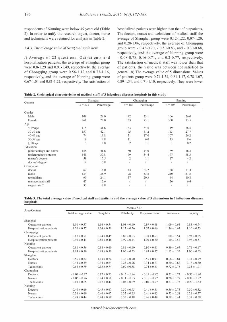
www.biosciencetrends.com
BioScience Trends. 2015; 9(3):182-189.185
respondents of Nanning were below 49 years old (Table 2). In order to unify the research object, doctor, nurse and technicians were retained for analysis in Table 2.
3.4.3. The average value of ServQual scale item
i ) Average of 22 ques t ions . Outpa t ien ts and hospitalization patients: the average of Shanghai group were 0.8-1.29 and 0.91-1.49, respectively, the average of Chongqing group were 0.56-1.12 and 0.73-1.16, respectively, and the average of Nanning group were 0.67-1.04 and 0.81-1.22, respectively. The satisfaction of
hospitalized patients were higher than that of outpatients. The doctors, nurses and technicians of medical staff: the average of Shanghai group were 0.12-1.22, 0.07-1.20, and 0.28-1.06, respectively, the average of Chongqing group were - 0.43-0.70, - 0.50-0.83, and - 0.30-0.68, respectively, and the average of Nanning group were - 0.08-0.78, 0.16-0.71, and 0.2-0.77, respectively. The satisfaction of medical staff was lower than that of patients, the value was between not satisfied to general. ii) The average value of 5 dimensions: Values of patients group were 0.74-1.34, 0.81-1.17, 0.78-1.07, 0.89-1.34, and 0.71-1.10, respectively. They were lower
Table 2. Sociological characteristics of medical staff of 3 infectious diseases hospitals in this study
Content
Gender Male FemaleAge ≤ 29 age 30-39 age 40-49 age 50-59 age ≥ 60 ageEducation junior college and below undergraduate students master's degree doctor's degreeOccupation doctor nurse technicians management staff support staff
n = 373
108261
118157 74 18 3
155141 58 14
67134 90 47 33
Percentage
29.070.0
31.642.119.8 4.8 0.8
41.637.815.5 3.8
18.035.924.112.6 8.8
n = 182
42133
63 75 31 11 2
80 99 2 /
44 98 37 / /
Percentage
23.173.1
34.641.217.0 6.0 1.1
44.054.4 1.1 /
24.253.820.3 / /
n = 408
106300
149113107 35 1
189197 17 /
128210 44 26 /
Percentage
26.073.5
36.527.726.2 8.6 0.2
46.348.3 4.2 /
31.451.510.8 6.4 /
Shanghai Chongqing Nanning
Table 3. The total average value of medical staff and patients and the average value of 5 dimensions in 3 infectious diseases hospitals
Area/Content
Shanghai Outpatient patients Hospitalization patientsChongqing Outpatient patients Hospitalization patientsNanning Outpatient patients Hospitalization patientsShanghai Doctors Nurses TechniciansChongqing Doctors Nurses TechniciansNanning Doctors Nurses Technicians
Total average value
1.01 ± 0.57 1.20 ± 0.57
0.87 ± 0.51 0.99 ± 0.41
0.81 ± 0.56 1.03 ± 0.50
0.56 ± 0.82 0.44 ± 0.59 0.64 ± 0.79
- 0.07 ± 0.77- 0.06 ± 0.76 0.08 ± 0.65
0.40 ± 0.69 0.36 ± 0.60 0.48 ± 0.44
Tangibles
1.16 ± 0.541.34 ± 0.51
0.74 ± 0.450.88 ± 0.46
0.88 ± 0.600.98 ± 0.51
1.03 ± 0.740.94 ± 0.640.93 ± 0.74
0.17 ± 0.750.24 ± 0.580.47 ± 0.44
0.45 ± 0.670.40 ± 0.670.44 ± 0.56
Reliability
1.08 ± 0.60 1.17 ± 0.56
0.88 ± 0.63 0.99 ± 0.44
0.81 ± 0.60 1.06 ± 0.53
0.38 ± 0.90 0.23 ± 0.76 0.60 ± 0.88
- 0.16 ± 0.86- 0.11 ± 0.85 0.03 ± 0.69
0.30 ± 0.73 0.32 ± 0.65 0.55 ± 0.48
Responsiveness
0.89 ± 0.68 1.07 ± 0.66
0.78 ± 0.67 1.00 ± 0.50
0.80 ± 0.61 0.99 ± 0.57
0.55 ± 0.93 0.34 ± 0.73 0.74 ± 0.81
- 0.14 ± 0.92- 0.18 ± 0.97- 0.04 ± 0.77
0.41 ± 0.81 0.41 ± 0.65 0.46 ± 0.49
Assurance
1.09 ± 0.641.34 ± 0.67
1.00 ± 0.541.10 ± 0.52
0.89 ± 0.651.12 ± 0.55
0.66 ± 0.840.60 ± 0.620.72 ± 0.78
0.25 ± 0.730.26 ± 0.790.23 ± 0.73
0.56 ± 0.750.52 ± 0.580.59 ± 0.44
Empathy
0.83 ± 0.74 1.10 ± 0.73
0.93 ± 0.55 0.98 ± 0.51
0.71 ± 0.67 1.00 ± 0.63
0.31 ± 0.99 0.18 ± 0.80 0.33 ± 1.01
- 0.37 ± 0.90- 0.39 ± 0.92- 0.23 ± 0.83
0.30 ± 0.82 0.21 ± 0.77 0.37 ± 0.59
Mean ± S.D.

www.biosciencetrends.com
BioScience Trends. 2015; 9(3):182-189. 186
than that evaluated on only 2 dimensions in Shanghai hospitalization patients; Average value of medical staff group were 0.17-1.03, - 0.16-0.60, - 0.18-0.74, 0.23-0.72, and - 0.39-0.37, respectively. Satisfaction of Medical staff in empathy, responsiveness, and reliability were low; satisfaction of Chongqing group were lower. The mean of 5 dimensions can be seen in Table 3.
3.4.4. One-way analysis of variance
One-way analysis of variance was performed between the average values of the 5 dimensions of the ServQual scale and patients and medical staff of Shanghai, Chongqing, and Nanning. The results are summarized in Tables 4 and 5. The satisfaction for tangibles of Shanghai patients was higher than that of Chongqing and Nanning patients; the satisfaction for Reliability and Assurance
of Shanghai patients were higher than that of Nanning patients (Table 4). From Table 5, we can see the satisfaction on 5 dimensions of Shanghai and Nanning medical staffs were higher than that of Chongqing's; and the satisfaction for tangibles of Shanghai medical staff was higher than that of Nanning's (Table 5).
4. Discussion
The medical staff showed lower levels of satisfaction than the patients. Empathy and responsiveness scores were relatively low for both medical staff and patients. Reliability scores were relatively low for the medical staff. Medical staff in general showed relatively low levels of satisfaction. When medical staffs show a negative attitude in dealing with patients, patients tend to develop biased perceptions of the medical treatment.
* * * p < 0.001
Table 4. One-way analysis of variance between the average values of the 5 dimensions of ServQual scales and patients in 3 infectious diseases hospitals
Dependent variable/Area
Tangibles Shanghai (A) Chongqi (B) Naning (C)Reliability Shanghai (A) Chongqi (B) Naning (C)Assurance Shanghai (A) Chongqi (B) Naning (C)
N
501159644
501159644
501159644
Mean
1.190.870.93
1.100.980.93
1.131.091.01
S.D.
0.5370.4630.558
0.5960.4680.577
0.6520.5210.611
Between GroupsWithin GroupsTotal
Between GroupsWithin GroupsTotal
Between GroupsWithin GroupsTotal
SS
23.182378.223401.406
7.840426.529434.369
3.998495.456499.454
df
213011303
213011303
213011303
MS
11.591 0.291
3.9200.328
1.9990.381
F
39.871***
11.957***
5.249***
Scheffe (C)
A > BC
A > C
A > C
* * * p < 0.001
Table 5. One-way analysis of variance between the average values of the 5 dimensions of ServQual scales and medical staffs in 3 infectious diseases hospitals
Dependent variable/Area
Tangibles Shanghai (A) Chongqi (B) Naming (C)Reliability Shanghai (A) Chongqi (B) Naming (C)Responsiveness Shanghai (A) Chongqi (B) Naming (C)Assurance Shanghai (A) Chongqi (B) Naming (C)Empathy Shanghai (A) Chongqi (B) Naming (C)
N
291179382
291179382
291179382
291179382
291179382
Mean
0.96 0.27 0.42
0.38- 0.09 0.34
0.52- 0.14 0.42
0.65 0.25 0.55
0.26- 0.35 0.26
S.D.
0.6960.6060.654
0.8460.8220.663
0.8210.9160.693
0.7250.7570.628
0.9150.8950.767
Between GroupsWithin GroupsTotal
Between GroupsWithin GroupsTotal
Between GroupsWithin GroupsTotal
Between GroupsWithin GroupsTotal
Between GroupsWithin GroupsTotal
SS
68.834368.639437.473
28.818495.299524.117
52.432527.914580.346
18.153404.930423.083
51.906609.869661.775
df
2849851
2849851
2849851
2849851
2849851
MS
34.417 0.434
14.409 0.583
26.216 0.622
9.076 0.477
25.953 0.718
F
79.265***
24.699***
42.161***
19.030***
36.129***
Scheffe (C)
A > BC;C > B
AC > B
AC > B
AC > B
AC > B

www.biosciencetrends.com
BioScience Trends. 2015; 9(3):182-189.187
When thorough and effective communication between clinicians and patients are lacking, patient violence are bound to occur. This situation is compatible with the results from the research of Wen X, et al. (7).
4.1. Improving the medical staff's and patients' perception of empathy
Among the medical staff at the three hospitals, the scores on empathy were lowest among the five dimensions. This is different from that of Anbori A's report which showed high empathy, high reliability and high assurance. The different maybe due to the different aims in terms of quality service between public hospitals and private hospitals (23). The patients also showed relatively low levels of satisfaction on the dimension of empathy. Empathy refers to the service provider's ability to stand in the shoes of the customer and meet the customer's requirements (14). If the hospital fails to take the perspective of the medical staff, the medical staff would not be able to stand in the shoes of the patients when providing services. This situation will lead to frequent disputes between clinicians and patients (7-12).
4 .2 . Improving the pat ients ' experience wi th responsiveness
At all three hospitals, patients showed low levels of satisfaction with responsiveness. Responsiveness refers to the service provider's initiative and intrinsic motivation to help the customer and provide convenient service (14). Although the level of medical expertise is directly related to patient satisfaction (36-38), patients are unlikely to attack the medical staff because of low satisfaction with the tangibles, but the improvement of tangibles is needed (29). However, patients may violently assault the clinicians when they repeatedly ask questions and seek help without getting satisfactory responses.
4.3. Enhancing clinicians' perception of reliability
The medical staff at all three hospitals showed low satisfaction with reliability. Reliability refers to service providers' ability to deliver the promised service in a precise and reliable manner. It can predict turnover of staff (14,23,39). Public hospitals in China have been forced into the market economy. Hospitals face multiple sources of pressure arising from evaluations of the facilities' credentials and the presidents' performance, medical insurance and competition within the industry. The hospitals pass on the performance requirements to various business units but lack the ability to fulfill their obligations to the medical staff. This situation has led to turnover of staff between different hospitals and outside of the medical care system. Many medical staff
members have stopped doing clinical work and left medical care facilities (9,10).
4.4. Constantly improving the management of services delivered by the hospital operators
Among the three hospitals, the medical staff and patients showed different levels of satisfaction. Shanghai's medical staff showed relatively good perception on the five dimensions. Patients in Shanghai showed relatively good perception on three dimensions. These results may be related to the local economic conditions. Shanghai enjoys geographical advantages in China. Its hospital operators are equipped with management concepts more similar to international standards, paying attention to satisfaction of both patients and medical staff. However, patients in Shanghai showed no difference from their counterparts in Chongqing and Nanning on responsiveness and empathy. Medical staff in Nanning fared better than those in Chongqing on the dimension of tangibles. Patients in Chongqing fared better than those in Nanning on empathy. This study shows that medical staff and patient satisfaction is related to the management concepts of hospital operators. Patient satisfaction is the source of customer satisfaction (25), management improvement can improve the quality of service (26). Hospital operators in Shanghai need to strengthen the education of medical staff on responsiveness and empathy to patients. Hospital operators in Nanning need to pay close attention to improve the medical staff's services to increase patient satisfaction. Hospital operators in Chongqing need to pay close attention to patients' needs and try their best to meet those needs. Thus, the criteria for medical staff and patient satisfaction developed in this study are suitable for Chinese hospitals. These criteria can reflect the management principles of different hospital operators.
4.5. Improving the research method with satisfaction survey
Hospitals in Shanghai conduct a Patient Satisfaction Survey through third-party agencies. At the end of each year the researchers survey 100-200 randomly-drawn respondents from the main questionnaire, the "Ten-Thousand-People Survey" to calculate patient satisfaction for the hospital over the entire year (2). This practice often causes discontent from the hospitals under evaluation. Based on the results of this study, we recommend that surveys on clinician and patient satisfaction should be conducted each quarter for each hospital using a variety of methods, such as over the phone, online, by mail and on-site. The assessors should first examine the homogeneity of samples and then analyze the data with professional statistical software. Results obtained this way may reflect clinician and

www.biosciencetrends.com
BioScience Trends. 2015; 9(3):182-189. 188
patient satisfaction with Chinese hospitals more closely and convincingly. Moreover, when a hospital conducts self-assessments, valid samples should be used for statistical analysis. In conclusion, to improve satisfaction evaluation indicators of Chinese hospital medical staff and patients, and research methods, paying attention to the satisfaction of the medical staff will help to enhance the satisfaction of the medical staff and patients of China hospitals, and help to improve the China medical environment.
Acknowledgements
This work was supported by the 12th Five-Year Infectious Disease Research Project (No. 2012ZX10001-003), the 12th Five-Year Major Science and Technology Project on Discovery of Major New Drugs (No. 2012ZX09303013), the National 863 Project "Study the Key Technology of Personnel Protection and Lab Tracking of pathogenic microorganism" (2014AA021403), Key research project of the Party, Education and Health building of Shanghai of 2014 (201420), and Scientific research in hospital construction project of Chinese Medical Doctor Assoclation. The authors wish to thank D.B.Lowrie, PhD, Shanghai Public Health Clinical Center, Fudan University.
References
1. National Health and Family Planning Commission of the People's Republic of China. The detailed rules for the implementation of three level general hospital accreditation standards (2011 edition). http://www.nhfpc.gov.cn/yzygj/s3585u/201112/06f754a213d8413787904e9e6439d88b.shtml (accessed December 23, 2014).
2. Shanghai Municipal Commission of Health and Family Planning. Style construction of health system in Shanghai City and the measures for the implementation of medical service satisfaction survey and evaluation work. http://www.wsjsw.gov.cn/wsj/n429/n432/n1487/n1515/userobject1ai79522.html (accessed May 28, 2014).
3. Wang MX, Ou L, Gao Y. Transi t ion of hospital accreditation management in China. Chin Med J (Engl). 2013; 126:4816-4817.
4. Zhen Y, Wang YY, Zhang Y, Gong WT, Jin XY, Shang SM. Exploration and practice of patient satisfaction evaluation in Beijing municipal hospitals. Zhong Guo Yi Yuan Guan Li. 2014; 34:23-25. (in Chinese)
5. Peng WP, Liu QC, Song SX, Wang W, Zhang HY, Jiang W, Yan XP, Liu HF. Continuous improvement of medical quality based on hospital level evaluation standard. Zhong Guo Yi Yuan Guan Li. 2014; 34:59-60. (in Chinese)
6. Zhuang JH, Zhang L, Wu XL. The research of performance appraisal on the head of the public hospital based on principal agent relationship. Zhong Guo Yi Yuan Guan Li. 2007; 27:30-33. (in Chinese)
7. Wen XG, Fang ZW. Blue Book of China's Medical Reform. Annual report on reform of medical and health system in China (2014-2015). Social Sciences Academic Press, Beijing, China, 2014; pp. 209-243. (in Chinese)
8. Cai WZ, Deng L, Liu M, Yu M. Antecedents of medical workplace violence in South China. J Interpers Villence. 2011; 26:317-327.
9. Wu D, Wang Y, Lam KF, Hesketh T. Health system reforms violence against doctors and job satisfaction in the medical profession: A cross-sectional survey in Zhejiang Province, Eastern China. BMJ Open. 2014; 12:e006431.
10. Huang JJ, Yang JO, Li B. Investigation on the impact of violent attacks against medical staff of medical students, vocational values. Xin Jiang Yi Ke Da Xue Xue Bao. 2014; 7:227-230. (in Chinese)
11. Ma Q. Analysis of the causes of medical violent injury. Zhong Hua Yi Xue Za Zhi. 2014; 94:1367-1370. (in Chinese)
12. Liu J, Wan LH. Importance and recommendations for the system establishment of the doctor protection. Chong Qing Yi Xue. 2013; 42:2296-2298. (in Chinese)
13. Li X, Zhang H, Wang J, Li F, Chen J, Chen J. Assessing patient satisfaction with medication related services in hospital settings: A cross-sectional questionnaire survey in China. Int J Clin Pharmacol Ther. 2014; 52:587-597.
14. Babakus E, Mangold WG. Adapting the SERVQUAL scale to hospital services: An empirical investigation. Health Serv Res. 1992; 26:767-786.
15. Almeida RS, Bourliataux-Lajoinie S, Martins M. Satisfaction measurement instruments for healthcare service users: A systematic review. Cad Saude Publica. 2015; 31:11-25.
16. Chang CS, Chen SY, Lan YT. Service quality, trust, and patient satisfaction in interpersonal-based medical service encounters. BMC Health Serv Res. 2013; 13:22.
17. Lei P, Jolibert A. A three-model comparison of the relationship between quality, satisfaction and loyalty: An empirical study of the Chinese healthcare system. BMC Health Serv Res. 2012; 12:436-446.
18. Li M, Wu YL, Yuan T, Shi H, Huang BY, Lu HZ. Exploration of medical service quality in the hospital based on ServQual questionnaire survey. Zhong Guo Yi Yuan Guan Li. 2014; 34:40-43. (in Chinese)
19. Li M, Lu HZ. ServQual scale for assessment of perceived service quality of hospital staff. Zhong Guo Wei Sheng Zhi Liang Guan Li. 2014; 21:56-59. (in Chinese)
20. Li M, Li X, Lu HZ. Comparative study of perceived service quality of outpatient patients and clinic doctors from a hospital in Shanghai and a center for disease control and prevention in Guangdong Province. Zhong Guo Yi Yao Dao Bao. 2014; 11:109-115. (in Chinese)
21. Li M, Cai RT, Huang CY. Application of structural equation model to evaluate the perception of service quality of medical staffs of infectious disease department in Chinese hospital. Infection International (Electronic Edition). 2015; 3:179-185.
22. Li M, Cai RT, Lu HZ. Investigation and analysis of Shanghai a hospital medical staff occupation burnout. Zhong Guo Wei Sheng Zhi Liang Guan Li. 2015; 22:89-93. (in Chinese)
23. Anbori A, Ghani SN, Yadav H, Daher AM, Su TT. Patient satisfaction and loyalty to the private hospitals in Sana'a, Yemen. Int J Qual Health Care. 2010; 22:310-315.

www.biosciencetrends.com
BioScience Trends. 2015; 9(3):182-189.189
24. Clark WR, Clark LA. Measuring functional service quality using SERVQUAL in a high-dependence health service relationship. Health Care Manag (Frederick). 2007; 26:306-317.
25. Sajjadi H, Vameghi M, Ghazinour M, Khodaeiardekani M. Caregivers' quality of life and quality of services for Children with cancer: A review from Iran. Glob J Health Sci. 2013; 5:173-182.
26. Mohammadi A, Mohammadi J. Evaluating quality of health services in health centres of Zanjan district of Iran. Indian J Public Health. 2012; 56:308-313.
27. Butt MM, de Run EC. Private healthcare quality: Applying a SERVQUAL model. Int J Health Care Qual Assur. 2010; 23:658-673.
28. Hart M. Improving the quality of NHS out-patient clinics: The applications and misapplications of TQM. Int J Health Care Qual Assur. 1996; 9:20-27.
29. Nekoei-Moghadam M, Amiresmaili M. Hospital services quality assessment: Hospitals of Kerman University of Medical Sciences, as a tangible example of a developing country. Int J Health Care Qual Assur. 2011; 24:57-66.
30. Shanghai Municipal Bureau of Statistics. NBS Survey Office in Shanghai. Statistical cmmunique of 2013 Shanghai national economic and social development. http://www.stats-sh.gov.cn/sjfb/201402/267416.html (accessed February 26, 2014).
31. Chongqing Municipal Bureau of Statistics. Statistical communique of 2013 Chongqing national economic and social development. http://www.cqtj.gov.cn/html/tjsj/tjgb/14/03/7005.html (accessed March 13, 2014).
32. Nanning Municipal Bureau of Statistics. Statistical communique of 2013 Nanning national economic. http://www.nnrb.com.cn/html/2014-05/10/content_85577.htm (accessed May 10, 2015).
33. Wu ML. Questionnaire and Statistical Analysis: SPSS Performance and Application. Chongqing University Press, Chongqing, China, 2010; pp. 157-355. (in Chinese)
34. Wentong Zhang, Chunwei Kuang. Basic Statistic Analysis Course on SPSS. Higher Education Press, Beijing, China, 2011; pp.251-342. (in Chinese)
35. Wentong Zhang, Wei Dong. Advanced Statistic Analysis Course on SPSS. Higher Education Press, Beijing, China, 2013; pp.3-27. (in Chinese)
36. Guo HY, Wang L, Gong WT, Shang SM, Wang ZW, Xie H. Comprehensive evaluation of patient satisfaction in public hospitals in Beijing city. Zhong Guo Wei Sheng Tong Ji. 2014; 31:488-491. (in Chinese)
37. Liu ZH. Research on the model of SEM multi level in patients with large comprehensive hospital based on customer satisfaction. Zhong Guo Wei Sheng Tong Ji. 2014; 31:878-881. (in Chinese)
38. Liu WW, Yuan SW, Cao JT, Li ZJ, Tao JJ, Zhang Z, Lu L, Xie ZH, Ma J. Survey of satisfaction of outpatients in pilot hospitals of public hospital reform in Beijing. Shang Hai Jiao Tong Da Xue Xue Bao (Medical Science). 2013; 31:724-734. (in Chinese)
39. Shu Q. Service quality, relationship quality and customer satisfaction ‒ model, method and application. Science Press, Beijing, China, 2010; pp. 103-105. (in Chinese)
(Received March 2, 2015; Revised April 13, 2015; Re-revised April 19, 2015; Accepted April 20, 2015)
Supplemental Data
Patients
T1: A clean and comfortable environment for medical treatment.
T2: Modern and advanced medical equipment.T3: Medical treatment clearly marked.T4: Doctors and nurses with neat and professional appearance.L5: The hospital ensures the doctors and nurses can be on time
for duty.L6: The hospital is interested in solving medical problems.L7: The hospital is reliable.L8: The medical treatment processes are accurate and concise.L9: The documents or reports promised by the hospital can be
delivered on time and clearly.S10: The hospital can satisfy the patients' immediate needs.S11 The hospital is willing to help the patients solve problems.S12: The hospital can timely process the patients' complaints.S13: The doctors and nurses can timely provide assistance to
the patients even when they are busy.A14: The doctors and nurses are reliable.A15: The patients feel at ease in the treatment process.A16: The doctors and nurses are polite to the patients.A17: The doctors and nurses have enough professional
knowledge to answer the patients' questions.E18: The doctors and nurses can show concern for each patient.E19: The doctors and nurses can provide personalized care to
patients.E20: The doctors and nurses know the patients' needs.E21: The hospital considers the patients' interests first.E22: The hospital pays attention to the medical needs of the
patients.
Medical Staff
T1: A clean and comfortable work environment.T2: Modern and advanced work equipment.T3: Office area clearly marked.T4: Staff with neat and professional appearance.L5: The staff working time and intensity are appropriate.L6: The hospital is interested in solving the problems at work.L7: The hospital is reliable.L8: The working processes are clear and concise.L9: The welfare benefits promised by the hospital can be
realized.S10: The hospital can satisfy the staff's working needs.S11: The hospital is willing to help the staff to solve working
problems.S12: The hospital can timely process the staff's complaints.S13: The hospital can process the staff's major and unexpected
events actively.A14: The hospital is reliable.A15: The staff feel comfortable at work.A16: The staff are friendly and polite to each other.A17: The hospital pays attention to the training of the staff's
professional knowledge and skills.E18: The hospital can show concern for each staff member.E19: The hospital can give personalized care to staff.E20: The hospital knows the staff's needs.E21: The hospital pays attention to the staff's interests.E22: The hospital pays attention to the needs of the staff.

www.biosciencetrends.com
BioScience Trends. 2015; 9(3):190-192. 190
MVsCarta: A protein database of matrix vesicles to aid understandingof biomineralization
Yazhou Cui1,*, Quan Xu2,*, Jing Luan1, Shichang Hu2, Jianbo Pan2, Jinxiang Han1,**, Zhiliang Ji2,**
1 Shandong Medicinal Biotechnology Center, Shandong Academy of Medical Sciences, Key Laboratory for Biotech-Drugs Ministry of Health, Ji'nan, Shandong, China;
2 State Key Laboratory of Stress Cell Biology, School of Life Sciences, Xiamen University, Xiamen, Fujian, China.
1. Introduction
Matrix vesicles (MVs) are small (20-200 nm) membrane particles bedded off from the plasma membrane of mineralizing chondrocytes, osteoblasts, and odontoblasts (1). MV-like particles have also been observed in the media of cultured mineralizing cells (MMV) or a number of ectopic calcifications such as vascular smooth muscle cells (VSMC-MV) (2,3). Usually, the MVs or MV-like particles are released into the pre-mineralized organic matrix prior to the onset of matrix mineralization, and act as a key constituent
in both physiological and pathological mineralization. Current evidences support two possible mechanisms for MVs involving mineralization: first, they modulate the local Pi/Ppi ratio in the mineralizing matrix; secondly, they provide initial mineral nucleation sites for mineralization (4,5). Although originated from the plasma membranes, the composition of MVs is different from that of the membranes they budded off from (6). The principle components of MV modulating mineralization are proteins and lipids. A panel of MVs proteins have been identified and functionally clarified including some enzymes [Alkaline phosphatase (TNAP), Phospho-1, Na+/K+ATPase, nucleot ide pyrophosphatase/phosphod ies te rase I -1 (NPP1/PC-1) , ma t r ix metalloproteinase-2 (MMP-2), matrix metalloproteinase-3 (MMP-3), matrix metalloproteinase-13 (MMP-13)], transport proteins (annexin 5, annexin 2, annexin 6, annexin 11, annexin 4, annexin 1, annexin 7, Pit 1, Pit 2), and integrin proteins (integrins β1, β5, αV, α11, α1, α3) (4). These proteins are shown to be linked to the two key functions of MV in modulating mineralization. While progress has been made in the study of MVs,
Summary Matrix vesicles (MVs) are membranous nanovesicles released by chondrocytes, osteoblasts, and odontoblasts. They play a critical role in modulating mineralization. Here, we present a manually curated database of MV proteins, namely MVsCara to provide comprehensive information on MVs of protein components. In the current version, the database contains 2,713 proteins of six organisms identified in bone, cartilage, tooth tissues, and cells capable of producing a mineralized bone matrix. The MVsCarta database is now freely assessed at http://bioinf.xmu.edu.cn/MVsCarta. The search and browse methods were developed for better retrieval of data. In addition, bioinformatic tools like Gene Ontology (GO) analysis, network visualization and protein-protein interaction analysis were implemented for a functional understanding of MVs components. Similar database hasn't been reported yet. We believe that this free web-based database might serve as a useful repository to elucidate the novel function and regulation of MVs during mineralization, and to stimulate the advancement of MV studies.
Keywords: Matrix vesicles, database, mineralization, proteomics
DOI: 10.5582/bst.2015.01061Brief Report
*These authors contributed equally to this works. **Address correspondence to:Dr. Zhiliang Ji, State Key Laboratory of Stress Cell Biology, School of Life Sciences, Xiamen University, Xiamen, Fujian, 361102. China.E-mail: [email protected]
Dr. Jinxiang Han, Shandong Medicinal Biotechnology Center, Shandong Academy of Medical Sciences, Key Laboratory for Biotech-Drugs Ministry of Health, Jinan, 250062. China.E-mail: [email protected]

www.biosciencetrends.com
BioScience Trends. 2015; 9(3):190-192.
much still remains to be clarified. A comprehensive picture of MVs components is very helpful to ascertain the following key issues in biomineralization: how MVs are formed and regulated; how MVs participated in the physiological and pathological process. With the rapid development of proteomic tools, high-throughput analyses of MVs protein profiles are blossoming in recent years (3,7-10). Data on the MVs protein component have been generated. Given the value of the already generated large amounts of data and the likely increase of MVs studies in the future, a public compendium to store and retrieve such data is required. Here, we constructed a web-based compendium of MVs, namely "MVsCarta" (http://bioinf.xmu.edu.cn/MVsCarta), to catalogue the MVs protein components. The information collected in MVsCarta was derived from exhaustive literature research of both proteomic and functional studies, then was further annotated manually. We believe that this free web-based community resource will aid researchers to better understand the role of MVs in mineralization and trigger new studies on MVs.
2. Materials and Methods
2.1. Data Retrieval and preprocessing
The information of martrix vesicles was mainly obtained by querying the PubMed literature database (http://www.ncbi.nlm.nih.gov/pubmed/) using the combinational keywords of "matrix vesicles" as well as either "mineralization" or "calcification". A further selection was undertaken to pick up the articles containing the high-throughput proteomic studies, individual validation and function studies on MVs proteins. Subsequently, manual information retrieval was performed to extract the information regarding isolation and/or validation of MVs and identification and/or validation of MVs proteins from the full texts or supplemental data. Before uploaded into the database, the data were further cleaned by removing the incomplete or ambiguous records, and integrated by adopting unique IDs. For instance, all the MV proteins were represented by the UniProt access number (UniProt AC). In addition, all the MV proteins were assigned into either type of MMV and Collagenase-Released Matrix Vesicles (CRMV) based on their extracellular forms.
2.2. Database construction
MVsCarta (version 1.0) was constructed on Red Hat Linux release 9 operating system and the data were managed by the Relational Database Management System (RDBMS) Oracle 10g. Friendly interfaces and search engines were designed using the JSP technology and run on a Tomcat server.
3. Results and Discussion
The MVsCarta provides three methods for data retrieval: QUICK SEARCH, ADVABCED SEARCH, and BROWSE. The quick search method on the homepage and other Web pages allows foolproof text search using any single complete or partial keyword of gene symbol, protein name, UniProt AC, MV type, gene ID or organism. Keyword combination and wild characters like "*, +" are not supported yet. The records that meet the query criteria will be listed in the ascending alphabet order of gene symbols. Empty input in the text field of the quick search form (which is not recommended) will respond the full list of MV genes. Clicking on the gene symbol will redirect the user to the detailed information page, where comprehensive information of the MV proteins is given in five sections when available. The first section is gene description, which demonstrates the basic information of the MV protein like gene symbol, Entrez gene ID, protein name, and UniProt AC. Cross-links to the Entrez gene database and the UniProt protein knowledge base are provided. In the section of experimental evidences, one or multiple experimental evidences of MV proteins are given, including the organism, sample, MV isolation and validation method, MV type (CRMV or MMV), protein identification and validation method, and reference. The third section provides functional annotation of MV protein that whether the protein is involved in the mineralization or calcification when available. While in the fourth section, sub-cellular localization of the MV protein is given. In the last section of "Bioinformatic Analysis", two hyperlinks to the Gene Ontology analysis and the Interaction analysis are provided for rapid functional analysis of the designated MV genes/proteins along with its protein sequence in fasta format. As an alternative solution, the MVsCarta database offers an advanced search method for accurate data retrieval that allows user to specify the keyword search under three optional categories of MV types, samples and organisms. In addition to the keyword search methods, the MVsCarta also offers the browse method for direct access to the database. All the MV proteins were pre-assigned into several groups subject to one of the following four characteristic categories: organisms; sample materials (including 3 tissues and 3 cell types); MV types (MMV or CRMV); and gene symbol in the ascending alphabet order. The user is allowed to rapidly access a group of MV proteins by selecting one from above four categories. As an open-access database, the data of MVsCarta can be downloaded via the query form in the "DOWNLOAD" page. The query results are output in the tab-delimited text format. To aid better understanding the functional characteristics of MV proteins, three bioinformatic tools were developed and implemented into the database. The "Gene Ontology (GO)" analysis allows retrieving
191

www.biosciencetrends.com
BioScience Trends. 2015; 9(3):190-192. 192
Acknowledgements
This study was supported by National Natural Science Foundation of China (81371909) and the Key Projects in the National Science & Technology Support Program during the Twelve Five-Year Plan Period from Ministry of Science and Technology of the People's Republic of China (2013BAI07B01, 2013BAI07B02). We will also thank Mr. Yinbo Li for his kind help on network construction and database management.
References
1. Golub EE. Biomineralization and matrix vesicles in biology and pathology. Semin Immunopathol 2011; 33:409-417.
2. Kapustin AN, Shanahan CM. Calcium regulation of vascular smooth muscle cell-derived matrix vesicles. Trends Cardiovasc Med. 2012; 22:133-137.
3. Xiao Z, Camalier CE, Nagashima K, Chan KC, Lucas DA, de la Cruz MJ, Gignac M, Lockett S, Issaq HJ, Veenstra TD, Conrads TP, Beck GR, Jr. Analysis of the extracellular matrix vesicle proteome in mineralizing osteoblasts. J Cell Physiol. 2007; 210:325-335.
4. Golub EE. Role of matrix vesicles in biomineralization. Biochim Biophys Acta. 2009; 1790:1592-1598.
5. Wuthier RE, Lipscomb GF. Matrix vesicles: Structure, composition, formation and function in calcification. Front Biosci (Landmark Ed). 2011; 16:2812-2902.
6. Wuthier RE. Lipid composition of isolated epiphyseal cartilage cells, membranes and matrix vesicles. Biochim Biophys Acta. 1975; 409:128-143.
7. Balcerzak M, Malinowska A, Thouverey C, Sekrecka A, Dadlez M, Buchet R, Pikula S. Proteome analysis of matrix vesicles isolated from femurs of chicken embryo. Proteomics. 2008; 8:192-205.
8. Kapustin AN, Davies JD, Reynolds JL, McNair R, Jones GT, Sidibe A, Schurgers LJ, Skepper JN, Proudfoot D, Mayr M, Shanahan CM. Calcium regulates key components of vascular smooth muscle cell-derived matrix vesicles to enhance mineralization. Circ Res. 2011; 109:e1-12.
9. Rosenthal AK, Gohr CM, Ninomiya J, Wakim BT. Proteomic analysis of articular cartilage vesicles from normal and osteoarthritic cartilage. Arthritis Rheum. 2011; 63:401-411.
10. Thouverey C, Malinowska A, Balcerzak M, Strzelecka-Kiliszek A, Buchet R, Dadlez M, Pikula S. Proteomic characterization of biogenesis and functions of matrix vesicles released from mineralizing human osteoblast-like cells. J Proteomics. 2011; 74:1123-1134.
(Received April 26, 2015; Revised June 10, 2015; Accepted June 13, 2015)
the gene ontology information by inputting a gene/protein name or its corresponding ID in the text fields of the query form. The "Interaction Analysis" provides visualization of protein interactions of the query MV protein with other proteins. The protein-protein interaction information was derived from the STRING (Search Tool for the Retrieval of Interacting Genes/Proteins) database. The MV proteins in the interaction map are highlighted in blue. These two tools were also implemented into the detail information page in the section of "Bioinformatic Analysis" for convenient access. For better understanding the relations between MV proteins, a dynamic network analysis tool was implemented. User is asked to submit a list of MV proteins separated by semicolon via the query form. At least two proteins/genes are required for network analysis. These query MV proteins that may be linked together within one gene network or form several isolated clusters. The implemented network analysis tool is generally compatible with current popular web browsers. These analyses on MV components provide global insights into the mechanisms involved in MV biogenesis as well as the role of MVs in mineralization or calcification. Currently, the MVsCarta comprises 2,713 entries, 2,391 unique genes and 19 sample materials. These MV proteins were mainly isolated from the mineralized cultured cells in vitro and the mineralized tissues in vivo (bone, cartilage, and tooth), involving six different organisms such as Homo sapiens, Mus musculus, Rattus norvegicus, Rabbit, Gallus gallus, and Bos taurus. The MVsCarta database is expected to update regularly when the data is available. To aid rapid update, an on-line submission form was designed in the "Feedback" as well for acceptance of user-submitted data. In summary, MVsCarta is the first online resource that provides comprehensive information of MVs protein components. With the increasing studies on lipid composition of MVs become available, lipidomics data will be incorporated to MVsCarta in the future to better understand the important role of cellular lipid metabolism in biological mineralization. In addition, more analysis tools are desired to implement to explore the biological and functional significance of the MVs components such as predicting calcium-binding sites in MV proteins. Nevertheless, this database will serve as a useful repository to elucidate the novel function and regulation of MVs during mineralization, and to stimulate the advancement of MV studies.

www.biosciencetrends.com
BioScience Trends. 2015; 9(3):193-197.193
Risk factors for recurrence of primary spontaneous pneumothorax after thoracoscopic surgery
Haibo Huang1,2, Hua Ji2, Hui Tian1,*
1 Department of Thoracic Surgery, Qilu Hospital, Medical College of Shandong University, Ji'nan, Shandong, China;2 Department of Thoracic Surgery, Yuhuangding Hospital of Yantai City, Yantai, Shandong, China.
*Address correspondence to:Dr. Hui Tian, Department of Thoracic Surgery, Qilu Hospital, Medical College of Shandong University. No.107, Wenhuaxilu, Ji'nan, Shandong 250012, China.E-mail: [email protected]
1. Introduction
Primary spontaneous pneumothorax (PSP) is a common clinical problem occurring in previously healthy subjects, with a reported annual incidence of 7.4 to 18 per 100,000 population in males and an incidence of 1.2 to 6 per 100,000 population in females (1,2). PSP also remains a significant clinical problem because of its high rate of recurrence, which has been variously reported as being from 20-60% with conservative treatment (3,4). Thoracoscopic surgery has been widely used to treat PSP with less pain, lower morbidity, and a shorter hospital stay than an open thoracotomy. However, several studies have reported that the rate of PSP recurrence after thoracoscopic surgery ranged from 5%
to 10% (5,6), and this figure is thought to be higher than that after open thoracotomy. Recurrent attacks require additional hospitalization and hospitalization costs. Moreover, postoperative recurrence is a very embarrassing and challenging event for the thoracic surgeon. Preventing recurrence is difficult because its pathogenesis and mechanisms are not well known. If risk factors contributing to postoperative recurrence of PSP can be identified and improved, then a lower rate of recurrence could be achieved. The aim of this study was to identify these risk factors.
2. Patients and Methods
After a consent waiver was obtained from the patient or a family member, medical records of 412 patients who experienced PSP after undergoing thoracoscopic surgery from January 2008 to December 2012 at Qilu Hospital at Shandong University were retrospectively reviewed with the approval of the Institutional Review Board of this hospital. Patient records were reviewed
Summary Primary spontaneous pneumothorax recurs at a certain rate after thoracoscopic surgery, and risk factors for that recurrence are in question. The medical records of 248 patients with primary spontaneous pneumothorax who were followed for more than 2 years after thoracoscopic surgery were reviewed and retrospectively analyzed. Univariate and multivariate binary logistic regression analysis were used to identify possible risk factors. Twelve patients experienced the recurrence of primary spontaneous pneumothorax. Patients who experienced the recurrence of primary spontaneous pneumothorax were younger than patients who experienced no recurrence. A larger proportion of the patients who experienced recurrence did not undergo pleurodesis. The variables age, height, weight, body mass index, duration of air leakage, and pleurodesis (performed or not) with a p value less than 0.2 in univariate analysis were entered in multivariate analysis. A younger age and not undergoing pleurodesis were associated with a higher risk of postoperative ipsilateral recurrence of primary spontaneous pneumothorax. Not undergoing pleurodesis and a younger age are possible risk factors for recurrence of primary spontaneous pneumothorax after thoracoscopic surgery. Thoracic surgeons should pay more attention to pleurodesis, especially in younger patients.
Keywords: Primary spontaneous pneumothorax, thoracoscopy, recurrence, risk factor
DOI: 10.5582/bst.2015.01070Brief Report

www.biosciencetrends.com
BioScience Trends. 2015; 9(3):193-197.
and sex, age, height, weight, body mass index (BMI), history of smoking, whether PSP occurred on the left or right side, the frequency of ipsilateral attacks, the frequency of contralateral attacks, the surgical procedure, operating time, the postoperative duration of chest tube drainage, the duration of postoperative hospitalization, postoperative duration of air leakage, and follow-up time were recorded. To avoid any bias in the rate of PSP recurrence as a result of too short a follow-up, this study only examined the records of 248 patients with complete follow-up data (outpatient follow-up or telephone follow-up) and a follow-up of more than 2 years. All operations were performed by the same surgical team including 5 surgeons. All patients were placed in the lateral decubitus position and underwent surgery under general anaesthesia. All patients underwent single-lung ventilation with a double-lumen endotracheal tube. All patients underwent three-port thoracoscopic surgery. The lungs and thoracic cavity were carefully explored. If any bullae or blebs were found, they were resected with a stapling device. Two surgical stapling devices were used: the EndoGIA (Auto Suture Company, Norwalk, CT, USA) and ECHELON (Ethicon Endo-Surgery, Inc., Cincinnati, OH, USA). Two pleurodesis procedures were used in this study. Mechanical pleurodesis was performed by complete mechanical pleural abrasion with dry gauze. Chemical pleurodesis was performed by complete pleural abrasion with 10% povidone-iodine-soaked gauze, and 50 milliliters of 10% povidone-iodine (Aqueous Betadine 10%, Qilu Pharmaceutical Co., Ltd., Ji'nan, Shandong, China) was left in the thoracic cavity. After surgery, the chest tube was raised 30 centimeters higher than the drainage port for 24 h to avoid the outflow of povidone-iodine. Recurrence was defined as a further pneumothorax, which was confirmed with a chest X-ray or computed tomography of the chest, occurring more than 30 days after discharge with full lung expansion. Statistical analysis was performed using the Statistical Package for Social Sciences for Windows (v. 19.0; IBM SPSS Inc., Armonk, NY, USA). Univariate and multivariate binary logistic regression were used to determine risk factors. Variables with a p value less than 0.2 in univariate analysis were entered in multivariate analysis. Differences were considered to be statistically significant when p < 0.05.
3. Results and Discussion
3.1. Univariate analysis of clinical demographic variables
This study retrospectively reviewed the medical records of 248 patients (226 males and 22 females). Twelve patients experienced recurrence during follow-up, with a recurrence rate of 4.84%. Patients had a median age
of 19 years (range, 14 to 38 years). The median age of patients who did not experience a recurrence of PSP was 19 years (range: 14 to 38 years), and the median age of those who did experience a recurrence of PSP was 17 years (range: 14 to 21 years). Twenty-nine patients had a history of smoking (1 patient with recurrence of PSP and 28 patients with no recurrence of PSP). PSP occurred on the left side in 115 patients and on the right side in 133 patients. Forty-five patients had contralateral PSP. The height and weight of each patient were measured on the day of admission and the patient’s BMI was calculated. The mean frequency of attacks before surgery was 2.25 for patients with recurrence of PSP and 2.23 for patients with no recurrence of PSP. The clinical demographics of patients with recurrence of PSP and patients with no recurrence of PSP are shown in Table 1. There were no statistically significant differences between the two groups in terms of sex, the side on which PSP occurred, height, weight, BMI, history of smoking, frequency of attacks, follow-up time, or contralateral PSP. However, patients with recurrence of PSP were younger than patients with no recurrence of PSP (p = 0.007).
3.2. Univariate analysis of surgery-related variables
All patients underwent thoracoscopic surgery. Of these, 29 (2 patients with recurrence of PSP and 27 patients with no recurrence of PSP) exhibited no bullae or blebs during the operation. Bulla or bleb resection was done with the EndoGIA stapling device in 111 patients and the ECHELON device in 108 patients. Twenty-seven patients (9 with recurrence of PSP and 18 with no recurrence of PSP) did not undergo pleurodesis because inflammation of the pleura was evident during surgery. Mechanical pleurodesis was performed in 112 patients and chemical pleurodesis was performed in 109 patients. The mean postoperative duration of chest tube drainage and air leakage was 1.67 days in the patients with recurrence of PSP and 0.85 days in the patients with no recurrence of PSP. The mean duration of chest tube drainage was 3.75 days for patients with recurrence of PSP and 3.61 days for patients with no recurrence of PSP. Surgery-related variables are shown in Table 2. There were no statistically significant differences between the groups in terms of operating time, duration of chest tube drainage, the stapling device used, and whether bullae or blebs were resected. The postoperative duration of air leakage for patients with recurrence of PSP was longer than that for patients with no recurrence of PSP (p = 0.001). A larger proportion of patients with recurrence of PSP did not undergo pleurodesis than did patients with no recurrence of PSP (p = 0.001).
3.3. Multivariate analysis
The variables age, height, weight, BMI, pleurodesis
194

www.biosciencetrends.com
BioScience Trends. 2015; 9(3):193-197.195
age, being male, and having a lower BMI are risk factors for recurrence after the first attack of PSP (3,7). However, whether these variables are risk factors for the postoperative recurrence of PSP is still unclear. The current study found that a younger age was one risk factor for postoperative recurrence of PSP while sex, height, and BMI were not. Although the pathogenesis of PSP is still somewhat unclear, PSP tends to be a condition caused by the imbalanced development of the lungs and body (8). In younger
(performed or not), and duration of air leakage with a p value less than 0.2 were included in multivariate logistic regression analysis (Table 3). Results indicated that a younger age and not undergoing pleurodesis were risk factors for postoperative ipsilateral recurrence of PSP (p = 0.003 and 0.001, respectively). PSP is more common in young male patients with a low BMI (also referred to as an ectomorphic body type (1)), and this finding was corroborated by the current study. Several studies have found that a younger
Table 1. Univariate analysis of clinical demographic variables (n = 248)
Characteristics
Total Median age (years) range Sex Male Female Side Left Right Height (meters)Weight (kilograms)Body Mass IndexHistory of smoking Yes No History of contralateral PSP Yes No Duration of hospitalization (days)Frequency of attacksFollow-up (months)
Recurrence of PSP
12 17
14-21
10 2
7 5
1.71 ± 0.0552.46 ± 6.4917.67 ± 1.78
111
210
11.58 ± 8.04 2.25 ± 0.45
50.42 ± 13.10
Non-recurrence of PSP
23619
14-38
216 20
108 128
1.73 ± 0.0555.42 ± 7.19 18.5 ± 1.82
28208
43193
8.62 ± 3.322.23 ± 0.55
50.14 ± 16.30
p value
0.007
0.33
0.39
0.140.160.120.71
0.89
0.220.920.95
Table 2. Univariate analysis of surgery-related variables (n = 248)
Characteristics
Bulla or bleb resection Yes NoPleurodesis Pleurodesis not performed Mechanical pleurodesis Chemical pleurodesisStapling device None ECHELON stapling device EndoGIA stapling deviceDuration of air leakage (days)Duration of chest tube drainage (days)Operating time (minutes)
Recurrence of PSP
10 2
921
264
1.67 ± 0.65 3.75 ± 0.6258.33 ± 4.92
Non-recurrence of PSP
20927
18110108
27102107
0.85 ± 0.733.61 ± 0.76
63.24 ± 18.62
p value
0.58
0.0010.580.60
0.690.440.480.0010.540.36
Table 3. Multivariate analysis of variables with a p value less than 0.2 in univariate analysis
Characteristics
Pleurodesis not performedAgeHeightWeightBody Mass IndexDuration of air leakage
Odds ratio
100.260.527
95% confidence interval
16.04–626.600.33–0.80
p value
0.0010.0030.230.210.160.12

www.biosciencetrends.com
BioScience Trends. 2015; 9(3):193-197. 196
patients (and especially male adolescents), the body develops rapidly and a younger age also means the lungs and body will need a longer time to grow. Therefore, PSP is more common at a younger age. In younger patients, the imbalanced development of the lungs and body is not corrected by surgery, so postoperative recurrence of PSP is more common. Smoking is thought to play an important role in the pathogenesis of PSP (9). However, whether smoking is a risk factor for the recurrence of PSP is controversial (3,10,11). In the current study, smoking was not a risk factor for postoperative recurrence of PSP. Few of the patients in this study had a history of smoking. This implies that smoking is not the main factor responsible for PSP in these patients. Thus, smoking is not related to the postoperative recurrence of PSP. Resection of bullae and blebs is one of the main objectives of surgery for PSP according to the guidelines of the British Thoracic Society (BTS) (12). In the past, overlooking bullae or blebs and incomplete resection of these air-filled blisters with fibrous walls were thought to be the main reason for increased recurrence of PSP after thoracoscopic surgery (13). However, advances in technology and thoracoscopic procedures are believed to have decreased the possibility of overlooking bullae or blebs or incomplete resection of these air-filled blisters with fibrous walls during thoracoscopic surgery. A recent study found that the rate of PSP recurrence in video-assisted thoracic surgery was comparable to that in a thoracotomy (14). No bullae or blebs were detected in only a few patients (29 patients, 11.69%) in the current study. Not resecting bullae or blebs was not a risk factor for postoperative ipsilateral recurrence of PSP. Pleurodesis is the other main objective of surgery to treat PSP according to the guidelines of the BTS (12). Only a small number of patients (27 patients) did not undergo pleurodesis in the current study. In these 27 patients, the pleura exhibited inflammation, and this inflammation may have led to pleural symphysis, like the aseptic inflammatory changes caused by pleurodesis. There were also concerns about pleurodesis causing complications such as postoperative bleeding since the pleura exhibited inflammation. During follow-up, however, patients who had not undergone pleurodesis had a markedly higher rate of PSP recurrence. Thus, not undergoing pleurodesis is a risk factor for postoperative recurrence of PSP. There are several pleurodesis procedures in clinical use (15): mechanical pleurodesis, such as pleural abrasion and parietal pleurectomy, and chemical pleurodesis with different agents. Whether the pleurodesis procedure is a risk factor for the postoperative recurrence of PSP is still controversial (16,17). Apical parietal pleurectomy is another common method of mechanical pleurodesis for PSP. Given the possibility of complications like severe bleeding and severe chest pain and increased operating
time, a pleurectomy was not performed at this hospital. Since 10% povidone-iodine is inexpensive and easily available, it was used for chemical pleurodesis at this hospital. Results indicated that neither approach was associated with a higher risk of PSP recurrence. A longer duration of chest tube drainage and a longer duration of air leakage were once thought to decrease the effects of a pleurodesis and they thought to be risk factors for postoperative recurrence of PSP. However, the current results indicated that a longer duration of chest tube drainage and a longer duration of air leakage were not associated with a higher risk of PSP recurrence. This study has several limitations. This study was retrospective in design and it was conducted at a single facility. There were only 12 cases of PSP recurring, so selection bias may have occurred, limiting the power of multivariate analysis. The role of pleurodesis cannot be considered definite until randomized controlled trials are conducted with a larger sample. In conclusion, the current study revealed that not undergoing pleurodesis and a younger age are possible risk factors for ipsilateral recurrence of PSP after thoracoscopic surgery. Although the role of pleurodesis cannot be considered definite without conducting randomized controlled trials, thoracic surgeons should pay more attention to pleurodesis, especially in younger patients.
References
1. Sahn SA, Heffner JE. Spontaneous pneumothorax. N Engl J Med. 2000; 342:868-874.
2. Noppen M, De Keukeleire T. Pneumothorax. Respiration. 2008; 76:121-127.
3. Lippert HL, Lund O, Blegvad S, Larsen HV. Independent risk factors for cumulative recurrence rate after first spontaneous pneumothorax. Eur Respir J. 1991; 4:324-331.
4. Sadikot RT, Greene T, Meadows K, Arnold AG. Recurrence of spontaneous pneumothorax. Thorax. 1997; 52:805-809.
5. Bertrand PC, Regnard JF, Spaggiari L, Levi JF, Magdeleinat P, Guibert L, Levasseur P. Immediate and long-term results after surgical treatment of primary spontaneous pneumothorax by VATS. Ann Thorac Surg. 1996; 61:1641-1645.
6. Sawada S, Watanabe Y, Moriyama S. Video-assisted thoracoscopic surgery for primary spontaneous pneumothorax: Evaluation of indications and long term outcome compared with conservative treatment and open thoracotomy. Chest. 2005; 127:2226-2230.
7. Huang TW, Lee SC, Cheng YL, Tzao C, Hsu HH, Chang H, Chen JC. Contralateral recurrence of primary spontaneous pneumothorax. Chest. 2007; 132:1146-1150.
8. Fujino S, Inoue S, Tezuka N, Hanaoka J, Sawai S, Ichinose M, Kontani K. Physical development of surgically treated patients with primary spontaneous pneumothorax. Chest. 1999; 116:899-902.
9. Wallaert B, Gressier B, Marquette CH, Gosset P, Remy-Jardin M, Mizon J, Tonnel AB. Inactivation of alpha 1-proteinase inhibitor by alveolar inflammatory cells from

www.biosciencetrends.com
BioScience Trends. 2015; 9(3):193-197.197
smoking patients with or without emphysema. Am Rev Respir Dis. 1993; 147:1537-1543.
10. Cheng YL, Huang TW, Lin CK, Lee SC, Tzao C, Chen JC, Chang H. The impact of smoking in primary spontaneous pneumothorax. J Thorac Cardiovasc Surg. 2009; 138:192-195.
11. Uramoto H, Shimokawa H, Tanaka F. What factors predict recurrence of a spontaneous pneumothorax? J Cardiothorac Surg. 2012; 7:112-116.
12. MacDuff A, Arnold A, Harvey J; BTS Pleural Disease Guidel ine Group. Management of spontaneous pneumothorax: British Thoracic Society Pleural Disease Guideline 2010. Thorax. 2010; 65:18-31.
13. Kim KH, Kim HK, Han JY, Kim JT, Won YS, Choi SS. Transaxillary minithoracotomy versus video assisted thoracic surgery for spontaneous pneumothorax. Ann Thorac Surg. 1996; 61:1510-1512.
14. Joshi V, Kirmani B, Zacharias J. Thoracotomy versus VATS: Is there an optimal approach to treat ing pneumothorax? Ann R Coll Surg Engl. 2013; 95:61-64.
15. Rodriguez-Panadero F, Montes-Worboys A. Mechanisms of pleurodesis. Respiration. 2012; 83:91-98.
16. Min X, Huang Y, Yang Y, Chen Y, Cui J, Wang C, Huang Y, Liu J, Wang J. Mechanical pleurodesis does not reduce recurrence of spontaneous pneumothorax: A randomized trial. Ann Thorac Surg. 2014; 98:1790-1796.
17. Huh U, Kim YD, Cho JS, I H, Lee JG, Lee JH. The effect of thoracoscopic pleurodesis in primary spontaneous pneumothorax: Apical parietal pleurectomy versus pleural abrasion. Korean J Thorac Cardiovasc Surg. 2012; 45:316-319.
(Received May 28, 2015; Revised June 10, 2015; Accepted June 13, 2015)

www.biosciencetrends.com
BioScience Trends. 2015; 9(3):198-202. 198
Castleman disease of the mesentery as the great mimic: Incidental finding of one case and the literature review
Ang Lv1, Chunyi Hao1, Honggang Qian1, Jiahua Leng1, Wendy Liu2,*
1 Department of Hepato-Pancreato-Biliary Surgery, Peking University school of Oncology, Beijing Cancer Hospital & Institute, Beijing, China;
2 Department of Pathology, University Hospitals Case Medical Center, Cleveland, OH, USA.
*Address correspondence to:Dr. Wendy Liu, Department of Pathology, University Hospitals Case Medical Center, 11100 Euclid Avenue, Cleveland, OH44106, USA.E-mail: [email protected]
1. Introduction
Castleman disease (CD), also known as benign giant lymph node hyperplasia, was first described as benign, localized mediastinal lymphadenopathy by Dr. Benjamin Castleman in 1956 (1). Subsequently it was expanded to represent a diverse group of nonneoplastic lymphoproliferative disorders involving a variety of nodal and extranodal sites with various histologic patterns (2). The usual location of this disease is the mediastinum (70%). Although extrathoracic sites have been reported in the neck, axilla, pelvis, and retroperitoneum, CD located in the mesentery is very rare (2-4), and the etiology is still unclear. Based on the histopathologic features, Castleman disease is classified as 2 types, the hyaline vascular (HV-CD) and plasma cell (PC-CD) variant, representing 80%-90% and 10%-20% of CD cases, respectively. Clinically and radiologically, CD can also be classified
as unicentric type and multicentric type. The unicentric type is a localized disease, usually asymptomatic and most often seen in HV-CD. The multicentric type, which is characterized by disseminated lymphadenopathy, is almost always associated with systemic symptoms, and is dominated by PC-CD. Nevertheless, HV-CD and PC-CD can exhibit considerable clinical and histologic overlap, and a so-called mixed variant of CD is occasionally seen (5). Mesenteric CD is a rare disease easily to be ignored in differential diagnosis when an abdominal mass is found. As a result of nonspecific symptoms and shared radiologic features with other entities by imaging, it is very difficult to differentiate mesenteric CD from other neoplastic diseases preoperatively, such as gastrointestinal stromal tumor (GIST), ectopic pheochromocytoma, and lymphoma. To our knowledge, only 53 cases of mesenteric CD have been reported worldwide in the English literature (6-17), and most of the cases were under other clinical suspicion and were definitively diagnosed as CD following surgical resection and histopathologic examination. With different clinical diagnosis, the optimal treatment decision or surgical approach may vary, therefore a correct preoperative diagnosis is preferred. In this article, we report one case of hyaline vascular
Summary Castleman disease is an uncommon benign lymphoproliferative disorder characterized by hyperplasia of lymphoid follicles. More commonly described in the mediastinum, its occurrence in the mesentery is exceedingly rare, which is easily to be ignored in differential diagnosis when an abdominal mass is found. We report the case of an asymptomatic 71-year-old woman with a homogenous and hypervascular mass at the inner side of duodenojejunal junction. Based on the clinical suspicion of a gastrointestinal stromal tumor, a surgical resection was performed. Final diagnosis of the mass was hyaline vascular variant of Castleman disease. Here, we summarize the clinicopathological and radiological features of this disease by literature review, which may be helpful to bring awareness of this entity and improve the clinical decision making when similar scenarios are encountered.
Keywords: Differential diagnosis, clinicopathological, radiological, treatment decision
DOI: 10.5582/bst.2015.01065Case Report

www.biosciencetrends.com
BioScience Trends. 2015; 9(3):198-202.
variant CD in the mesentery of the duodenojejunal junction, where this disease has not been reported previously in literature. We also summarize the clinicopathological and radiological features of this disease by comprehensive literature review, which may be helpful to improve the clinical decision making when similar scenarios are encountered.
2. Case report
A 71-year-old female patient was admitted to a local hospital due to osteoarthritis of her right knee. A 4 × 5 cm hypoechoic mass was incidentally found by a routine abdominal ultrasound examination. After the patient was transferred to our hospital, further investigation with abdominal contrast-enhanced magnetic resonance imaging (MRI) showed a sharply demarcated soft tissue mass measuring 4.5 × 4.1 cm, with slightly low and high signal intensity on T1- and T2-weighted images, respectively. The mass was located at the inner side of duodenojejunal junction, just beneath the inferior border of the pancreas, and adjacent to the left side of superior mesenteric artery (SMA). After the contrast medium was injected, the mass appeared to be homogeneous and moderately enhanced in both the arterial phase and portal venous phase (Figure 1). Her past medical history included hypertension and coronary heart disease. Physical examination, routine laboratory tests including C-reactive protein (CRP), erythrocyte sedimentation rate (ESR), as well as computed tomography (CT) of the thorax revealed no obvious abnormalities. In addition, the levels of tumor markers including a-fetoprotein (AFP), carcinoembryonic antigen (CEA), carbohydrate antigen 19-9 (CA 19-9) and carbohydrate antigen 125 (CA 125) were all within the normal limits. Based on imaging findings, a GIST was highly suspected with differential diagnosis including a leiomyoma or neurogenic tumor. Thus, the patient
underwent laparotomy. During the operation, the tumor was confirmed to derive from the mesentery of the duodenojejunal junction. After the tumor was carefully separated from the superior mesenteric vein (SMV), SMA and the pancreas, the segment of the mesentery containing the mass was resected along with the transverse portion of duodenum and initial part of jejunum (pancreas-sparing segmental duodenectomy). A duodenojejunal side-to-side anastomosis was performed to reconstruct the alimentary tract. Grossly, the mass measured 4 × 4 × 3.5 cm, was firm and well-demarcated from surrounding tissue. The cut surface was grey-yellow, homogeneous and firm (Figure 2). Microscopic examination showed a large lymph node with preserved architecture and numerous follicles throughout the cortex and medulla. The lymphoid follicles were surrounded by a broad mantle zone composed of concentric rings of small lymphocytes forming the so-called "onion skin" pattern (Figure 3). The follicles contained a small germinal center that is lymphocyte depleted with prominent dendritic cells and hyaline deposits on Periodic Acid-Schiff (PAS) stain. The interfollicular areas were composed of numerous high endothelial venules with plump endothelial
199
Figure 2. Resected specimen. Resected specimen showing the cut surface of a 4 × 4 cm mass (left) and adjacent duodenum.
Figure 1. MRI images of the lesion. Abdominal MRI showing a sharply demarcated soft tissue mass located at the inner side of duodenojejunal junction, just beneath inferior border of pancreas, and adjacent to the left side of superior mesenteric artery. The mass was homogeneous and moderately enhanced in arterial phase and portal venous phase.

www.biosciencetrends.com
BioScience Trends. 2015; 9(3):198-202. 200
is significantly lower than traditionally reported 80%-90% in mediastinal CD cases. All cases presented as solitary lesions (unicentric type), and there was a female predominance with thirty-eight (71.7%) female patients. This predominance was most prominent in HV variant patients with 88.5% of them female (23/26), while no fender difference in PC variant or mixed variant. The average age of reported patients was only 27.5 years (range: 2-77 years) with about half patients (47.2%) under 20, and only five patients over 60 years. The present patient was the second oldest one compared to all previously reported HV-CD patients. The average mass size was 5.8 cm (2 cm to 17 cm), and 90% of cases were within a range from 3 cm to 7 cm. Clinically, over half of reported patients were asymptomatic or with complains of nonspecific symptoms, such as abdominal pain and an abdominal mass. A small portion of patients experienced constitutional symptoms such as weight loss, fever or growth retardation. It is worth noting that laboratory findings in most patients with the PC variant showed systemic manifestations including anemia, an elevated sedimentation rate, and hypergammaglobulinemia, while only a small portion of the patients with the HV variant presented these abnormalities.
3.2. Radiological features
Mesenteric CD often presents as a homogeneous, hypoechoic mass on ultrasound. The most characteristic feature of mesenteric CD at computed tomography (CT) scan is a well-defined, homogeneous, single intraabdominal mass of soft tissue attenuation with or without satellite nodules. The enhanced CT and angiography findings usually demonstrate the homogeneity and hypervascularity of the mass (18). On MRI, the lesion could be isointense or slightly hypointense compared with that of normal muscle on T1-weighted images and hyperintense on T2-weighted images. After intravenous injection of contrast
cells and were associated with small lymphocytes, eosinophils and plasma cells. The histologic findings were diagnostic of Castleman disease, hyaline vascular variant. The postoperative course of the patient was uneventful and she was discharged on the 14th postoperative day.
3. Discussion
3.1. Clinicopathological features
We reviewed the English literature of mesenteric Castleman disease through a search on Pubmed database, and the clinicopathological findings of 53 cases of mesenteric CD reported previously were summarized in Table 1. Histologically, the HV and PC variants constituted 26 and 21 of all cases, respectively, and 6 cases were of the mixed variant. Interestingly, the proportion of HV variant in mesenteric CD (49%)
Figure 3. Microscopic findings. Microscopic view of the specimen showing the typical appearance of the hyaline vascular variant of Castleman disease, with a broad mantle zone composed of concentric rings of small lymphocytes surrounding a lymphocyte-depleted small lymphoid follicle forming so-called "onion skin" appearance.
Table 1. Clinicopathological fi ndings of mesenteric Castleman disease reported previously
Items
Gender Female MaleMean age (yrs) (range)Mean tumor size (cm) (range)Symptoms Constitutional symptoms (fever, weight loss, growth retardation, etc) Local or no symptoms (abdominal pain, abdominal mass, asymptomatic)Laboratory fi ndings Abnormal (anemia, elevated ESR/CRP, hypergammaglobulinemia, etc) Normal
Mixed CD (n = 6)
3 3 13.8 (2-29) 4.9 (4-6.5)
4 2
6 0
HV-CD (n = 26)
23 3 33.0 (9-74) 5.9 (2-17)
7 19
12 14
HV, hyaline-vascular; PC, plasma cell; ESR, erythrocyte sedimentation rate; CRP, C-reactive protein.
PC-CD(n = 21)
12 9 24.3(8-77) 5.7(3-10)
13 8
19 2
Overall(n = 53)
38 15 27.5(2-77) 5.8(2-17)
24 29
37 16

www.biosciencetrends.com
BioScience Trends. 2015; 9(3):198-202.201
medium, the enhancement of mesenteric HV-CD was characterized by homogeneous high enhancement in the early phase of dynamic enhancement, persistent enhancement in the de layed phase , wi th the enhancement mode similar to that of large arteries. But in some cases with tumors larger than 5 cm, during early stage of enhancement, the interior of the tumor could be seen to have distinct radial or fissured non-enhanced areas. The non-enhancement areas were reduced or vanished in delayed scan on CT, and MRI scans of corresponding areas gave low signals on the non-enhanced T2-weighted images (19). Pathologic examinations of the areas revealed abundance of parallel fibrous tissue. In the present case, the 4 cm isolated mass was found incidentally without any abdominal and systemic symptoms, and the ultrasonography, enhanced CT and MRI revealed a homogenous and hypervascular mass, which was in accordance to above radiological features.
3.3. Differential diagnosis
For abdominal soft tissue mass showing a clear boundary, homogenous and hypervascular imaging features, and with or without systemic manifestations, Castleman disease should be included in the differential diagnosis, especially for female patients. However, the above radiological features are not specific for CD, and the most commonly stated preoperative radiologic differential diagnosis are hypervascular mesenchymal tumor, such as GIST, and neurogenic tumor, such as ectopic pheochromocytoma. GISTs are the most common mesenchymal neoplasms with varying malignant potential based on the risk-stratification scheme. The gold standard for treatment of localized primary GIST is complete excision with negative margins. Small GISTs often appear as round tumors with strong and homogeneous arterial enhancement and a persistent enhancement pattern. However, large GISTs (> 5 cm) appear as lobulated tumors with mild heterogeneous gradual enhancement, and they frequently exhibited intratumoral cystic changes (20). Most of mesenteric CD have a higher enhancement than that of GIST, but if mesenteric CD gives a moderate or a mild enhancement, it is difficult to distinguish it from hypervascular mesenchymal tumors including GIST. Another distinguishing radiological finding is the absence or rare presence of cystic necrotic degeneration in the mesenteric CD. This might be attributed to abundance of blood supply, good collateral circulation and low susceptibility of lymphatic follicles to necrosis. However, one study reported cystic necrotic degeneration were found in about 20% of the CD cases (19). Ectopic pheochromocytoma comprises approximately 10-25% cases of pheochromocytoma, 30% of which could be malignant, and surgical resection is still the most effective treatment (21). The enhancement mode
of ectopic pheochromocytoma may also be similar to those of mesenteric CD. However, the signal intensity of ectopic pheochromocytoma on T2-weighted image is usually stronger than that of CD, and the interior of the lesions may reveal highly uneven density and signals accompanied by cystic central necrosis (19). The clinical and laboratory examinations can also contribute to the differentiation of these two diseases. Most patients with functional ectopic pheochromocytoma show paroxysmal hypertension clinically, and in laboratory examination show elevation of catecholamine and its metabolic product 3-methoxyl-4-hydroxyl mandelic acid. On the other hand, based on microscopic findings, mesenteric CD should be differentiated from a number of benign and neoplastic conditions particularly neoplastic lymphoproliferative disorder. The hyalinied follicles in CD may mimic early follicular lymphoma. The prominent mantle zone in CD may also mimic mantle zone lymphoma. Rare cases of early interfollicular Hodgkin lymphoma may show CD-like changes including the regression of residual germinal centers and hypervascularity. However, CD lacks Reed-Sternberg cells. Immunophenotyping, in situ hybridization, and PCR for immunoglobulin heavy chain rearrangements aid in the evaluation of B-cell clonality and are helpful in differentiating CD from a lymphoma.
3.4. Treatment decision
Due to lack of specific radiographic feature for CD, an endoscopic or ultrasound-guided fine-needle biopsy should be recommended when CD is suspected, as the optimal treatment decision or surgical approach may vary with different clinical diagnosis. Irrespective of the histopathologic subtype, the unicentric type CD always shows a benign biologic behavior. For cases with symptoms such as anemia, the symptoms may show complete reversal after surgical resection of the tumor (15). For asymptomatic unicentric mesenteric CD, the necessity of surgical resection remains unclear. Complete surgical excision is usually curative of the mesenteric CD, since there is no reported case of recurrence after total excision of the solitary mass. On the contrary, the multicentric type Castleman disease follows a more aggressive course and is associated with poor prognosis. The therapeutic approach of the multicentric type remains controversial, as many treatment regimens have been proposed, including surgery, chemotherapy, corticotherapy or combination of these (22,23). Although all cases of mesenteric CD reported previously were unicentric type, an accurate staging of the disease, including a thorough clinical examination for the detection of suspicious lymph nodes in the axilla, neck or groin and CT of the thorax should be performed in order to exclude the presence of extra-mesenteric disease.

www.biosciencetrends.com
BioScience Trends. 2015; 9(3):198-202. 202
For a benign tumor, a surgical approach balancing complete resection and organ-preserving may be more beneficial for patients. For the present case, if the diagnosis of mesenteric Castleman disease was made preoperatively, a surgical procedure preserving mesenteric vessels and duodenum may be possible, and the duodenojejunal anastomosis may be avoided. Considering that the patient was without symptoms, close follow-up may also be an electable alternative. However, because the endoscopic or ultrasound-guided approach had difficulty in pointing out the tumor location, and a fine-needle biopsy for a hypervascular tumor is regarded as potentially dangerous, a definitive preoperative diagnosis was not made. In summary, for abdominal soft tissue mass showing a clear boundary, homogenous and hypervascular imaging features, and with or without systemic manifestations, mesenteric Castleman disease should be included in the differential diagnosis, especially for female patients. GIST, ectopic pheochromocytoma and lymphoma could be top differential diagnosis from this disease. Endoscopic or ultrasound-guided fine-needle biopsy should be recommended to further clasify the tumor so an optimal treatment decision can be recommended to the patient.
Acknowledgements
We are grateful to Dr. Zhong-Wu Li and Dr. Min Zhao for helpful discussion in preparing the manuscript.
References
1. Castleman B, Iverson L, Menendez VP. Localized mediastinal lymphnode hyperplasia resembling thymoma. Cancer. 1956; 9:822-830.
2. Keller AR, Hochholzer L, Castleman B. Hyaline-vascular and plasma-cell types of giant lymph node hyperplasia of the mediastinum and other locations. Cancer. 1972; 29:670-683.
3. Glazer M, Rao VM, Reiter D, McCue P. Isolated Castleman disease of the neck: MR findings. AJNR Am J Neuroradiol. 1995; 16:669-671.
4. Meador TL, McLarney JK. CT features of Castleman disease of the abdomen and pelvis. AJR Am J Roentgenol. 2000; 175:115-118.
5. Cronin DM, Warnke RA. Castleman disease: An update on classification and the spectrum of associated lesions. Adv Anat Pathol. 2009; 16:236-246.
6. Ohta M, Yamamoto M, Tagawa T, Tsujita E, Matsuyama A, Okazaki J, Utsunomiya T, Tsutsui S, Fujihara M, Ishida T. Laparoscopy-assisted resection for Mesenteric Castleman's disease: Report of a case. Surg Today. 2011; 41:1405-1409.
7. Shiote Y, Yamane H, Ninomiya T, Kamei H. Hyaline vascular type Castleman's disease of the mesentery. Intern Med. 2012; 51:2489-2490.
8. Rajeshwara K V, Clement R S D'Souza, Elroy Saldanha, Klien Dantis, Preethi Rai. Unicentric mesenteric Castleman's disease- A diagnostic quandary- A case report. J Clin Diagn Res. 2013; 7:573-575.
9. deVries IA, van Acht MM, Demeyere T, Lybeert ML, de Zoete JP, Nieuwenhuijzen GA. Neoadjuvant radiotherapy of primary irresectable unicentric Castleman's disease: A case report and review of the literature. Radiat Oncol. 2010; 5:7.
10. Bass LM, Barsness K, Benya E, Proytcheva M, Kagalwalla A. Mesenteric Castleman disease detected by capsule endoscopy. J Pediatr Gastroenterol Nutr. 2013; 57:e3-5.
11. Re icha rd KK, Rob ine t t S , Fouca rMK. C lona l cytogenetic abnormalities in the plasma cell variant of Castlemandisease. Cancer Genet. 2011; 204:323-327.
12. Li FF, Zhang T, Bai YZ. Mesenteric Castleman's disease in a 12-year-old girl. J Gastrointest Surg. 2011; 15:1896-1898.
13. Hwang MR, Chang HJ, Kim MJ, Seo GJ, Yoo SB, Park JW, Choi HS, Oh JH. Castleman's disease of the mesorectum: Report of a case. Surg Today. 2011; 41:271-275.
14. Al-Natour S, Sawalhi S, Al-Muhtady D, Hijazi E. Mesenteric Castleman's disease: Case report and literature review. Asian J Surg. 2010; 33:150-153.
15. El Demellawy D, Herath C, Truong F, Nasr A, AlowamiS. Localized early mesenteric Castleman's disease presenting as recurrent intestinal obstruction: A case report. Diagn Pathol. 2009; 4:42.
16. Bejjani J, Lemieux B, Gariepy G, YounanR. Complete anemia reversal after surgical excision of mesenteric hyaline-vascular unicentric Castleman disease. Can J Surg. 2009; 52:E197-198.
17. Sari S, Okur A, Yeşilkaya E, Dalgiç B, Karadeniz C, Oğuz A, Gürsel T, Sönmez K, Gönül II. Localized abdominal Castleman disease masquerading as malabsorption syndrome. J Pediatr Hematol Oncol. 2008; 30:618-620.
18. Malara F, Price D, Fabiny R. Mesenteric Castleman's disease: Ultrasound, computed tomography and angiographic appearance. Australas Radiol. 2000; 44:109-111.
19. Zhou LP, Zhang B, Peng WJ, Yang WT, Guan YB, Zhou KR. Imaging findings of Castleman disease of the abdomen and pelvis. Abdom Imaging. 2008; 33:482-488.
20. Yu MH, Lee JM, Baek JH, Han JK, Choi BI. MRI features of gastrointestinal stromal tumors. AJR Am J Roentgenol. 2014; 203:980-991.
21. Manger WM. An overview of pheochromocytoma: History, current concepts, vagaries, and diagnostic challenges. Ann N Y Acad Sci. 2006; 1073:1-20.
22. Herrada J, Cabanillas F, Rice L, Manning J, Pugh W. The clinical behavior of localized and multicentric Castleman Disease. Ann Intern Med. 1998; 128:657-662.
23. Bowne WB, Lewis JJ, Filippa DA, Niesvizky R, Brooks AD, Burt ME, Brennan MF. The management of unicentric and multicentric Castleman's disease: A report of 16 cases and a review of the literature. Cancer. 1999; 85:706-717.
(Received May 9, 2015; Revised June 2, 2015; Accepted June 9, 2015)

www.biosciencetrends.com
BioScience Trends. 2015; 9(3):203-204.203
China upgrades surveillance and control measures of Middle East respiratory syndrome (MERS)
Jianjun Gao1,*, Peipei Song2
1 School of Pharmaceutical Sciences, Qingdao University, Qingdao, China;2 Graduate School of Frontier Sciences, The University of Tokyo, Kashiwa-shi, Japan.
Middle East respiratory syndrome coronavirus (MERS-CoV), first identified in 2012 in Saudi Arabia, caused 1,179 laboratory-confirmed cases of human infection in 25 countries in Middle East, Africa, Europe, Asia, and North America by June 3, 2015 according to the statistics disclosed by World Health Organization (WHO) (1). The first confirmed case of MERS in China, a man from the Republic of Korea, was claimed by National Health and Family Commission of People’s Republic of China (NHFPC) on May 29, 2015 and is currently being treated in isolation in Huizhou Municipal Central Hospital, Huizhou, Guangdong province (2). This case comes from a family in which his father and elder brother were diagnosed with MERS and hospitalized in Korea earlier, suggesting an epidemiological link between the human cases. Chinese health authorities have taken appropriate measures to prevent further transmission of the virus. By June 4, all the 78 close contacts of the patient had been found and isolated for observation, and there has no evidence of infection in the close contacts thus
far (3). To further strengthen the disease management and improve the epidemic situation report quality in medical and disease control organizations, NHFPC published the second edition of MERS Prevention and Control Plan on June 5, 2015 and specified it as the guideline for dealing with this disease in China (4). Comparing with the first edition that was announced in September 2013, this updated Plan refines the descriptions in case-finding and establishment of case-monitoring systems. In addition, tougher regulations on close contacts of confirmed patients and suspected cases are introduced in this new Plan. Regulations on patients with fever of unknown origins NHFPC requires that health care workers in medical and health institutions at various levels and of various kinds shall strengthen the awareness of the diagnosis and report of MERS cases. For patients with fever of unknown origins, their travel histories or other suspected exposure experiences within the past 14 days should be asked. Specifically, it should be clarified whether the patients and their close contacts have travelled to Middle East countries such as Saudi Arabia, United Arab Emirates, Qatar, and Jordan. The new Plan adds the Republic of Korea as the at-risk country for travelling. In addition, medical staffs are required to ask whether the patients have experience of contacting suspected animals such as dromedary in the updated Plan. Once the patients are consistent with the definition of MERS, they should be timely reported to
Summary Three years after the identification of Middle East respiratory syndrome coronavirus (MERS-CoV) in Saudi Arabia, the first case of MERS in China was reported on May 29, 2015. Although the Chinese government issued the MERS Prevention and Control Plan in 2013, a novel edition was released on June 5, 2015 to better cope with the current epidemic situation. The revised Plan refines the descriptions in case-finding and establishment of case-monitoring systems. In addition, tougher regulations on close contacts of confirmed patients and suspected cases are introduced in this new Plan. It is expected these countermeasures will play a greater role in surveilling and controlling MERS in China.
Keywords: MERS-CoV, NHFPC, fever of unknown origins, epidemic
DOI: 10.5582/bst.2015.01082News
Released online in J-STAGE as advance publication June 11, 2015.
*Address correspondence to:Dr. Jianjun Gao, Department of Pharmacology, School of Pharmaceutical Sciences, Qingdao University, 38 Dengzhou Road, Qingdao 266021, Shandong, China.E-mail: [email protected]

www.biosciencetrends.com
BioScience Trends. 2015; 9(3):203-204.
the local disease control prevention institutions. Regulations on the confirmed and suspected cases NHFPC requires that the health care institutes that undertake the task of MERS treatment shall be well-prepared in terms of medical personnel, medicines, facilities, and protective equipment. In the administration of patients’ treatment, the Plan presents that the MERS patients should be treated in isolation and the attended medical staff should be equipped with effective protection measures. For suspected patients, isolation for observation and treatment is also necessary for those who are not excluded from MERS-CoV infection. Prevention and protection measures should also be taken for health care workers and relevant personnel until the clinical syndrome such as fever and cough disappear or MERS-CoV infection is excluded. Regulations on close contacts of confirmed and suspected cases In the first edition, only the close contacts of confirmed patients demands medical observation at home and those with relevant syndrome should be further isolated. Close contacts of suspected cases should be just registered. In the revised edition, more rigorous regulations are presented for these two kinds of cases. In the current stage, all the close contacts of confirmed patients should be isolated for observation during which body temperature and acute respiratory or MERS-relevant symptoms should be monitored. For those close contacts of suspected cases, NHFPC requires that both registration and health
follow-up should be carried out. These people should be informed that they shall notify medical institutes if symptoms such as fever, cough, and diarrhea appear. From severe acute respiratory syndromes (SARS) to MERS, China has developed a relatively integrated monitoring and responding system in the past ten years. For example, four-level frame system from county to country has been established for disease prevention and control, which guarantees a rapid response for infectious diseases. However, more MERS cases will possibly be imported into China, as pointed by the Officials of Chinese health authorities, requiring cautious countermeasures of the government in the future.
References
1. Summary and risk assessment of current situation in Republic of Korea and China. World Health Organization. http://www.who.int/csr/disease/coronavirus_infections/risk-assessment-3june2015/en/ (Accessed June 7, 2015).
2. News. China reports first MERS case. http://en.nhfpc.gov.cn/2015-06/01/content_20874634.htm (Accessed June 8, 2015).
3. News. Circular of epidemic situation. http://www.gdwst.gov.cn/a/yiqingxx/2015060513703.html (Accessed June 8, 2015).
4. MERS Prevention and Control Plan (2nd edition). http://www.nhfpc.gov.cn/jkj/s3577/201506/f47f22f52614406798df6363d3e2d199.shtml (Accessed June 8, 2015).
(Received June 8, 2015; Accepted June 9, 2015)
204

www.biosciencetrends.com
BioScience Trends. 2015; 9(3):205-206.205
Expected role of medical technologists in diabetes mellitus education teams
Kazuhiko Kotani1,2,*, Takahiro Imazato3, Keizo Anzai4; Kyushu Diabetes Testing Study Group1 Division of Community and Family Medicine, Jichi Medical University, Shimotsuke, Tochigi, Japan;2 Department of Clinical Laboratory Medicine, Jichi Medical University, Shimotsuke, Tochigi, Japan;3 Health Care Center, Sasebo Chuo Hospital, Sasebo, Nagasaki, Japan;4 Division of Metabolism & Endocrinology, Department of Internal Medicine, Saga University Faculty of Medicine, Saga, Japan.
Diabetes mellitus (DM) is a global health issue and clinical laboratory activities are of great concern in DM practice (1). DM education is crucial for patient management (2,3) and is often performed by a professional team consisting of physicians, nurses, pharmacists and dietitians. Some teams include medical technologists (MTs) (2,3). However, the content of education for which MTs are responsible is not always uniform on DM education teams. Furthermore, the role of MTs within DM education teams has not been studied, unlike that for other professionals such as nurses (4). The present survey was thus conducted using a questionnaire that asked the question, "what roles are MTs expected to play on your DM education team?", to other professional members in facilities across the Kyusyu area, a wide region in the south of Japan. Because many DM patients are covered by facilities with a primary care function, which often have active DM education teams (5), these facilities were enrolled in this survey. The area included about 380 facilities at which DM-expert physicians worked in primary care capacities. The questionnaire was randomly distributed
to approximately one-third of the facilities. Then, self-reported responses given in a free-writing manner were returned anonymously. A total of 89 questionnaires were completed. The major expected roles of MTs in DM education teams are listed in Table 1. Overall, the role was not only for patients but for team members. Naturally, most roles were associated with laboratory examinations and results. There was a relatively relevant need for education regarding self-monitoring of blood glucose tests. Although MTs generally specialize in performing precise examinations, items such as ensuring quality control on examinations did not necessarily rank high. Good communication with patients (providing information, guidance and teaching) and team members (presenting an interpretation of the results, providing information) was widely required. The expected role of MTs in DM education teams was described from the viewpoints of other professional members, while there were several limitations to this survey (i.e., small sample size, qualitative study design, lack of detailed information regarding the answerers and facilities). Since the roles of MTs have not been characterized to date, the present data, obtained based on real needs, would be useful for establishing the significance of MTs within the scope of DM education teams. In particular, training communication with both patients and team members is necessary for MTs to be recognized as professionals of DM education. Allowing
Summary The expected role of medical technologists within diabetes mellitus education teams was surveyed. In addition to items regarding laboratory examinations and results themselves, good communication with patients and education team members was highly required. When medical technologists sufficiently follow this role, it would aid patients to cope with life with diabetes mellitus.
Keywords: Communication, diabetes education, professionalism, SMBG
DOI: 10.5582/bst.2015.01058Letter
*Address correspondence to:Dr. Kazuhiko Kotani, Division of Community and Family Medicine or Department of Clinical Laboratory Medicine, Jichi Medical University, 3311-1 Yakushiji, Shimotsuke-City, Tochigi, 329-0498, Japan.E-mail: [email protected]

www.biosciencetrends.com
BioScience Trends. 2015; 9(3):205-206.
MTs to successfully follow this role may help patients to cope with life with DM.
References
1. Jeha GS, Haymond M. Understanding and interpreting laboratory test results in the clinical management of diabetes mellitus. Pediatr Endocrinol Rev. 2007; 5(Supple 1):608-628.
2. Hatano Y. Participation of medical technologists in educational hospitalization of diabetics--involvement in short-term hospitalization of diabetics on weekends. Rinsho Byori. 2005; 53:1051-1060. (in Japanese)
3. Sato I, Jikimoto T, Ooyabu C, Kusuki M, Okano Y, Mukai M, Kawano S, Kumagai S. Contribution of medical technologists in team medical care of diabetics. Rinsho Byori. 2006; 54:816-823. (in Japanese)
4. Meetoo DD, McAllister G, West A. Assessing glycaemic control: Self-monitoring of blood glucose. Br J Nurs. 2011; 20:919-920, 922, 924-925.
5. Kruger DF, Lorenzi GM, Dokken BB, Sadler CE, Mann K, Valentine V. Managing diabetes with integrated teams: Maximizing your efforts with limited time. Postgrad Med. 2012; 124:64-76.
(Received April 22, 2015; Revised June 29,2015; Accepted June 30, 2015)
206
Table 1. Summary of major answers to the question "what roles are MTs expected on your DM education team?"
Answers
For patients Provision of information on DM-related examinations and their results Guidance (e.g., skills or pitfalls of glucometer usages) for SMBG Lecture in DM education classes
For team members Presentation of interpretation of examinations and their results from MTs' views in the meeting Provision of information on examinations (e.g., recommendation, introduction of new tests) Quality control regarding examinations (e.g., glucometers)
N (%*)
47 (53%)34 (38%) 9 (10%)
18 (20%)16 (18%)11 (12%)
SMBG, self-monitoring of blood glucose; DM, diabetes mellitus; MT, medical technologists. * Total number was 89.

Guide for Authors1. Scope of Articles
BioScience Trends is an international peer-reviewed journal. BioScience Trends devotes to publishing the latest and most exciting advances in scientific research. Articles cover fields of life science such as biochemistry, molecular biology, clinical research, public health, medical care system, and social science in order to encourage cooperation and exchange among scientists and clinical researchers.
2. Submission Types
O r i g i n a l A r t i c l e s s h o u l d b e w e l l -documented, novel, and significant to the field as a whole. An Original Article should be arranged into the following sections: Title page, Abstract, Introduction, Materials and Methods, Results, Discussion, Acknowledgments, and References. Original articles should not exceed 5,000 words in length (excluding references) and should be limited to a maximum of 50 references. Articles may contain a maximum of 10 figures and/or tables.
Brief Reports definitively documenting either experimental results or informative clinical observations will be considered for publication in this category. Brief Reports are not intended for publication of incomplete or preliminary findings. Brief Reports should not exceed 3,000 words in length (excluding references) and should be limited to a maximum of 4 figures and/or tables and 30 references. A Brief Report contains the same sections as an Original Article, but the Results and Discussion sections should be combined.
Reviews should present a full and up-to-date account of recent developments within an area of research. Normally, reviews should not exceed 8,000 words in length (excluding references) and should be limited to a maximum of 100 references. Mini reviews are also accepted.
Policy Forum articles discuss research and policy issues in areas related to life science such as public health, the medical care system, and social science and may address governmental issues at district, national, and international levels of discourse. Policy Forum articles should not exceed 2,000 words in length (excluding references).
Case Reports should be detailed reports of the symptoms, signs, diagnosis, treatment, and follow-up of an individual patient. Case reports may contain a demographic profile of the patient but usually describe an unusual or novel occurrence. Unreported or unusual
side effects or adverse interactions involving medications will also be considered. Case Reports should not exceed 3,000 words in length (excluding references).
News articles should report the latest events in health sciences and medical research from around the world. News should not exceed 500 words in length.
Letters should present considered opinions in response to ar t ic les publ ished in BioScience Trends in the last 6 months or issues of general interest. Letters should not exceed 800 words in length and may contain a maximum of 10 references.
3. Editorial Policies
Ethics: BioScience Trends requires that authors of reports of investigations in humans or animals indicate that those studies were formally approved by a relevant ethics committee or review board.
Conflict of Interest: All authors are required to disclose any actual or potential conflict of interest including financial interests or relationships with other people or organizations that might raise questions of bias in the work reported. If no conflict of interest exists for each author, please state "There is no conflict of interest to disclose".
S u b m i s s i o n D e c l a r a t i o n : W h e n a manuscript is considered for submission to BioScience Trends, the authors should confirm that 1) no part of this manuscript is currently under consideration for publication elsewhere; 2) this manuscript does not contain the same information in whole or in part as manuscripts that have been published, accepted, or are under review elsewhere, except in the form of an abstract, a letter to the editor, or part of a published lecture or academic thesis; 3) authorization for publication has been obtained from the authors’ employer or institution; and 4) all contributing authors have agreed to submit this manuscript.
Cover Letter: The manuscript must be accompanied by a cover letter signed by the corresponding author on behalf of all authors. The letter should indicate the basic findings of the work and their significance. The letter should also include a statement affirming that all authors concur with the submission and that the material submitted for publication has not been published previously or is not under consideration for publication elsewhere. The cover letter should be submitted in PDF format. For example of Cover Letter, please visit http://www.biosciencetrends.com/downcentre.php (Download Centre).
C o p y r i g h t : A s i g n e d J O U R N A L PUBLISHING AGREEMENT ( JPA) form must be provided by post, fax, or as a scanned file before acceptance of the article. Only forms with a hand-written signature are accepted. This copyright will ensure the widest possible dissemination of information. A form facilitating transfer of copyright can be downloaded by clicking the
appropriate link and can be returned to the e-mail address or fax number noted on the form (Please visit Download Centre). Please note that your manuscript will not proceed to the next step in publication until the JPA Form is received. In addition, if excerpts from other copyrighted works are included, the author(s) must obtain written permission from the copyright owners and credit the source(s) in the article.
Suggested Reviewers: A list of up to 3 reviewers who are qualified to assess the scientific merit of the study is welcomed. Reviewer information including names, affiliations, addresses, and e-mail should be provided at the same time the manuscript is submitted online. Please do not suggest reviewers with known conflicts of interest, including participants or anyone with a stake in the proposed research; anyone from the same institution; former students, advisors, or research collaborators (within the last three years); or close personal contacts. Please note that the Editor-in-Chief may accept one or more of the proposed reviewers or may request a review by other qualified persons.
Language Editing: Manuscripts prepared by authors whose native language is not English should have their work proofread by a native English speaker before submission. If not, this might delay the publication of your manuscript in BioScience Trends. The Editing Support Organization can provide English proofreading, Japanese-English translation, and Chinese-English translation services to authors who want to publish in BioScience Trends and need assistance before submitting a manuscript. Authors can visit this organization directly at http://www.iacmhr.com/iac-eso/support.php?lang=en. IAC-ESO was established to facilitate manuscript preparation by researchers whose native language is not English and to help edit works intended for international academic journals.
4. Manuscript Preparation
Manuscripts should be written in clear, grammatically correct English and submitted as a Microsoft Word file in a single-column format. Manuscripts must be paginated and typed in 12-point Times New Roman font with 24-point line spacing. Please do not embed figures in the text. Abbreviations should be used as little as possible and should be explained at first mention unless the term is a well-known abbreviation (e.g. DNA). Single words should not be abbreviated.
Title Page: The title page must include 1) the title of the paper (Please note the title should be short, informative, and contain the major key words); 2) full name(s) and affiliation(s) of the author(s), 3) abbreviated names of the author(s), 4) full name, mailing address, telephone/fax numbers, and e-mail address of the corresponding author; and 5) conflicts of interest (if you have an actual or potential conflict of interest to disclose, it must be included as a footnote on the title page of the manuscript; if no conflict of

interest exists for each author, please state "There is no conflict of interest to disclose"). Please visit Download Centre and refer to the title page of the manuscript sample.
Abstract: The abstract should briefly state the purpose of the study, methods, main findings, and conclusions. For article types including Original Article, Brief Report, Review, Policy Forum, and Case Report, a one-paragraph abstract consisting of no more than 250 words must be included in the manuscript. For News and Letters, a brief summary of main content in 150 words or fewer should be included in the manuscript. Abbreviations must be kept to a minimum and non-standard abbreviations explained in brackets at first mention. References should be avoided in the abstract. Key words or phrases that do not occur in the title should be included in the Abstract page.
Introduction: The introduction should be a concise statement of the basis for the study and its scientific context.
Materials and Methods: The description should be brief but with sufficient detail to enable others to reproduce the experiments. Procedures that have been published previously should not be described in detail but appropriate references should simply be cited. Only new and significant modifications of previously published procedures require complete description. Names of products and manufacturers with their locations (city and state/country) should be given and sources of animals and cell lines should always be indicated. All clinical investigations must have been conducted in accordance with Declaration of Helsinki principles. All human and animal studies must have been approved by the appropriate institutional review board(s) and a specific declaration of approval must be made within this section.
Results: The description of the experimental results should be succinct but in sufficient detail to allow the experiments to be analyzed and interpreted by an independent reader. If necessary, subheadings may be used for an orderly presentation. All figures and tables must be referred to in the text.
Discussion: The data should be interpreted concisely without repeating material already presented in the Results section. Speculation is permissible, but it must be well-founded, and discussion of the wider implications of the findings is encouraged. Conclusions derived from the study should be included in this section.
Acknowledgments: All funding sources should be credited in the Acknowledgments section. In addition, people who contributed to the work but who do not meet the criteria for authors should be listed along with their contributions.
References: References should be numbered in the order in which they appear in the text. Citing of unpublished results, personal communications, conference abstracts, and theses in the reference list is not recommended but these sources may be mentioned in the text. In the reference list,
cite the names of all authors when there are fifteen or fewer authors; if there are sixteen or more authors, list the first three followed by et al. Names of journals should be abbreviated in the style used in PubMed. Authors are responsible for the accuracy of the references. Examples are given below:
Example 1 (Sample journal reference): Inagaki Y, Tang W, Zhang L, Du GH, Xu WF, Kokudo N. Novel aminopeptidase N (APN/CD13) inhibitor 24F can suppress invasion of hepatocellular carcinoma cells as well as angiogenesis. Biosci Trends. 2010; 4:56-60.
Example 2 (Sample journal reference with more than 15 authors): Darby S, Hill D, Auvinen A, et al. Radon in homes and risk of lung cancer: Collaborative analysis of individual data from 13 European case-control studies. BMJ. 2005; 330:223.
Example 3 (Sample book reference): Shalev AY. Post-traumatic stress disorder: diagnosis, history and life course. In: Post-traumatic Stress Disorder, Diagnosis, Management and Treatment (Nutt DJ, Davidson JR, Zohar J, eds.). Martin Dunitz, London, UK, 2000; pp. 1-15.
Example 4 (Sample web page reference): Ministry of Health, Labour and Welfare of Japan . Die ta ry re fe rence in takes for Japanese. http://www.mhlw.go.jp/houdou/2004/11/h1122-2a.html (accessed June 14, 2010).
Tables: All tables should be prepared in Microsoft Word or Excel and should be arranged at the end of the manuscript after the References section. Please note that tables should not in image format. All tables should have a concise title and should be numbered consecutively with Arabic numerals. If necessary, additional information should be given below the table.
Figure Legend: The figure legend should be typed on a separate page of the main manuscript and should include a short title and explanation. The legend should be concise but comprehensive and should be understood without referring to the text. Symbols used in figures must be explained.
Figure Preparation: All figures should be clear and cited in numerical order in the text. Figures must fit a one- or two-column format on the journal page: 8.3 cm (3.3 in.) wide for a single column, 17.3 cm (6.8 in.) wide for a double column; maximum height: 24.0 cm (9.5 in.). Please make sure that the symbols and numbers appeared in the figures should be clear. Please make sure that artwork files are in an acceptable format (TIFF or JPEG) at minimum resolution (600 dpi for illustrations, graphs, and annotated artwork, and 300 dpi for micrographs and photographs). Please provide all figures as separate files. Please note that low-resolution images are one of the leading causes of article resubmission and schedule delays. All color figures will be reproduced in full color in the online edition of the journal at no cost to authors.
Units and Symbols: Units and symbols
conforming to the International System o f U n i t s ( S I ) s h o u l d b e u s e d f o r physicochemical quantities. Solidus notation (e.g. mg/kg, mg/mL, mol/mm2/min) should be used. Please refer to the SI Guide www.bipm.org/en/si/ for standard units.
Supplemental data: Supplemental data might be useful for supporting and enhancing your scientific research and BioScience Trends accepts the submission of these materials which will be only published online alongside the electronic version of your article. Supplemental files (figures, tables, and other text materials) should be prepared according to the above guidelines, numbered in Arabic numerals (e.g., Figure S1, Figure S2, and Table S1, Table S2) and referred to in the text. All figures and tables should have titles and legends. All figure legends, tables and supplemental text materials should be placed at the end of the paper. Please note all of these supplemental data should be provided at the time of initial submission and note that the editors reserve the right to limit the size and length of Supplemental Data.
5. Submission Checklist
The Submission Checklist will be useful during the final checking of a manuscript prior to sending it to BioScience Trends for review. Please visit Download Centre and download the Submission Checklist file.
6. Online Submission
Manuscr ipts should be submit ted to BioScience Trends online at http://www.biosciencetrends.com. The manuscript file should be smaller than 5 MB in size. If for any reason you are unable to submit a file online, please contact the Editorial Office by e-mail at [email protected].
7. Accepted Manuscripts
Proofs: Galley proofs in PDF format will be sent to the corresponding author via e-mail. Corrections must be returned to the editor ([email protected]) within 3 working days.
Offprints: Authors will be provided with electronic offprints of their article. Paper offprints can be ordered at prices quoted on the order form that accompanies the proofs.
Page Charge: Page charges will be levied on all manuscripts accepted for publication in BioScience Trends ($140 per page for black white pages; $340 per page for color pages). Under exceptional circumstances, the author(s) may apply to the editorial office for a waiver of the publication charges at the time of submission. (Revised February 2013)
Editorial and Head Office: Pearl City Koishikawa 6032-4-5 Kasuga, Bunkyo-kuTokyo 112-0003 JapanTel: +81-3-5840-8764Fax: +81-3-5840-8765E-mail: [email protected]

JOURNAL PUBLISHING AGREEMENT (JPA)-----------------------------------------------------------------------------------------------------------------------------------------Manuscript No.:
Title:
Corresponding Author:----------------------------------------------------------------------------------------------------------------------------------------The International Advancement Center for Medicine & Health Research Co., Ltd. (IACMHR Co., Ltd.) is pleased to accept the above article for publication in BioScience Trends. The International Research and Cooperation Association for Bio & Socio-Sciences Advancement (IRCA-BSSA) reserves all rights to the published article. Your written acceptance of this JOURNAL PUBLISHING AGREEMENT is required before the article can be published. Please read this form carefully and sign it if you agree to its terms. The signed JOURNAL PUBLISHING AGREEMENT should be sent to the BioScience Trends office (Pearl City Koishikawa 603, 2-4-5 Kasuga, Bunkyo-ku, Tokyo 112-0003, Japan; E-mail: [email protected]; Tel: +81-3-5840-8764; Fax: +81-3-5840-8765).
1. Authorship CriteriaAs the corresponding author, I certify on behalf of all of the authors that:1) The article is an original work and does not involve fraud, fabrication, or plagiarism.2) The article has not been published previously and is not currently under consideration for publication elsewhere. If accepted by BioScience Trends, the article will not be submitted for publication to any other journal.3) The article contains no libelous or other unlawful statements and does not contain any materials that infringes upon individual privacy or proprietary rights or any statutory copyright.4) I have obtained written permission from copyright owners for any excerpts from copyrighted works that are included and have credited the sources in my article.5) All authors have made significant contributions to the study including the conception and design of this work, the analysis of the data, and the writing of the manuscript.6) All authors have reviewed this manuscript and take responsibility for its content and approve its publication. 7) I have informed all of the authors of the terms of this publishing agreement and I am signing on their behalf as their agent.
2. Copyright Transfer AgreementI hereby assign and transfer to IACMHR Co., Ltd. all exclusive rights of copyright ownership to the above work in the journal BioScience Trends, including but not limited to the right 1) to publish, republish, derivate, distribute, transmit, sell, and otherwise use the work and other related material worldwide, in whole or in part, in all languages, in electronic, printed, or any other forms of media now known or hereafter developed and the right 2) to authorize or license third parties to do any of the above. I understand that these exclusive rights will become the property of IACMHR Co., Ltd., from the date the article is accepted for publication in the journal BioScience Trends. I also understand that IACMHR Co., Ltd. as a copyright owner has sole authority to license and permit reproductions of the article. I understand that except for copyright, other proprietary rights related to the Work (e.g. patent or other rights to any process or procedure) shall be retained by the authors. To reproduce any text, figures, tables, or illustrations from this Work in future works of their own, the authors must obtain written permission from IACMHR Co., Ltd.; such permission cannot be unreasonably withheld by IACMHR Co., Ltd.
3. Conflict of Interest DisclosureI confirm that all funding sources supporting the work and all institutions or people who contributed to the work but who do not meet the criteria for authors are acknowledged. I also confirm that all commercial affiliations, stock ownership, equity interests, or patent-licensing arrangements that could be considered to pose a financial conflict of interest in connection with the article have been disclosed.---------------------------------------------------------------------------------------------------------------------------------------------------------
Corresponding Author's Name (Signature):
Date:
BioScience Trends (www.biosciencetrends.com)Pearl City Koishikawa 603, 2-4-5 Kasuga, Bunkyo-ku, Tokyo 112-0003, Japan; E-mail: office@biosciencetrends; Tel: +81-3-5840-8764; Fax: +81-3-5840-8765