Br. J. Cancer (1979) 40, 234 - CiteSeerX
10
Br. J. Cancer (1 979) 40, 234 ANTIBODY-DEPENDENT CELLULAR CYTOTOXICITY TO HUMAN COLON-TUMOUR CELLS. I. LACK OF TUMOUR SPECIFICITY IN A POPULATION STUDY J. SHOHAM AND A. COHEN From The Genetic Institute, Sheba Medical Centre, Tel-Hashonter, Israel Receive(d 30 November 1978 Accepted 12 April 1979 Summary.-The humoral and cellular components of the antibody-dependent cellular cytotoxicity (ADCC) against allogeneic human colonic tumour cell lines were evaluated. The 2 colon cell lines used in this study (HT-29 and ACC-20) were found by immunofluorescence to have carcinoembryonic antigen (CEA) on their surface, and to become sensitive to the lytic effect of unstimulated lymphocytes after coating with heterologous anti-CEA. This reaction was used to evaluate the ADCC activity of mononuclear cells from the peripheral blood of patients with gastrointestinal cancer (mostly local extensive colo-rectal). Remarkable variability was found in the lytic capability (2-50% specific lysis) of both cancer and non-cancer mononuclear cells, with no significant difference between them. Sera from 127 cancer patients and 91 non-cancer patients were tested, using the reaction with heterologous anti-CEA as positive control and as a reference point. In 46 cases (21 %) the sera were reactive in this system, and 43 of them were of Blood Group 0. However, there was no difference between the cancer patients and the normal controls. The antigenic determinant involved in this reaction is not the Blood Group A specificity but, most probably, a polypeptide common to CEA and A (as shown in the following publication). In addition, trials for the elimination of the non-tumour-specific reaction, by absorption or inhibition, failed to disclose a tumour-specific one. The value of the ADCC assay in monitoring human tumour immunity, and possible ways of eliminating reactivity to normal antigens in this system, are discussed in the light of these findings. ANTIBODY-DEPENDENT cellular cyto- toxicity (ADCC) is a well defined in vitro reaction (Lovchik & Hong, 1977; Perlman, 1976) of unknown biological significance. The specificity of the reaction is deter- mined by the antibodies that coat the target cells. The nonspecific cellular effec- tor component is the K lymphocyte (Perlman, 1976) or sometimes the macro- phage (Kohl et al., 1977) or polymorpho- nuclear leucocyte (Gale & Zighelboim, 1975). All of them recognize the Fc portion of the coating antibody. This reaction has been implicated in various in vivo immune processes, includ- ing those against tumours (Lamon et al., 1976), virus-infected cells (Pearson & Orr, 1976), transplants (Jeannet & Vassalli, 1976) and autoimmunity (Feldman et al., 1976). However, although there is some evidence that ADCC does occur in these conditions, it has been impossible so far to demonstrate directly their relative im- portance among the multitude of immune reactions taking place in vivo. As far as tumour immunity is concerned, serum and lymphocyte cooperation was demonstrated in vitro in mice bearing Moloney sarcoma virus-induced or 3-methylcholanthrene- induced sarcomas and mammary-tumour virus-induced adenocarcinoma (Pollack et al., 1972), as well as in rats with Gross virus-induced lymphoma (Ortiz de Lan- dazuri et al., 1974). Successful inhibition of murine neuroblastoma by ADCC was demonstrated in a modified Winn's test Correspondence to: Dr J. Shoham, The Genetic Institute, Sheba Medical Centre, Tel-Hashomei, Israel.
-
Upload
khangminh22 -
Category
Documents
-
view
2 -
download
0
Transcript of Br. J. Cancer (1979) 40, 234 - CiteSeerX

Br. J. Cancer (1 979) 40, 234
ANTIBODY-DEPENDENT CELLULAR CYTOTOXICITY TO HUMANCOLON-TUMOUR CELLS.
I. LACK OF TUMOUR SPECIFICITY IN A POPULATION STUDY
J. SHOHAM AND A. COHEN
From The Genetic Institute, Sheba Medical Centre, Tel-Hashonter, Israel
Receive(d 30 November 1978 Accepted 12 April 1979
Summary.-The humoral and cellular components of the antibody-dependentcellular cytotoxicity (ADCC) against allogeneic human colonic tumour cell lines wereevaluated. The 2 colon cell lines used in this study (HT-29 and ACC-20) were foundby immunofluorescence to have carcinoembryonic antigen (CEA) on their surface, andto become sensitive to the lytic effect of unstimulated lymphocytes after coating withheterologous anti-CEA. This reaction was used to evaluate the ADCC activity ofmononuclear cells from the peripheral blood of patients with gastrointestinal cancer(mostly local extensive colo-rectal). Remarkable variability was found in the lyticcapability (2-50% specific lysis) of both cancer and non-cancer mononuclear cells,with no significant difference between them. Sera from 127 cancer patients and 91non-cancer patients were tested, using the reaction with heterologous anti-CEA aspositive control and as a reference point. In 46 cases (21%) the sera were reactive inthis system, and 43 of them were of Blood Group 0. However, there was no differencebetween the cancer patients and the normal controls. The antigenic determinantinvolved in this reaction is not the Blood Group A specificity but, most probably, apolypeptide common to CEA and A (as shown in the following publication). Inaddition, trials for the elimination of the non-tumour-specific reaction, by absorptionor inhibition, failed to disclose a tumour-specific one. The value of the ADCC assay inmonitoring human tumour immunity, and possible ways of eliminating reactivity tonormal antigens in this system, are discussed in the light of these findings.
ANTIBODY-DEPENDENT cellular cyto-toxicity (ADCC) is a well defined in vitroreaction (Lovchik & Hong, 1977; Perlman,1976) of unknown biological significance.The specificity of the reaction is deter-mined by the antibodies that coat thetarget cells. The nonspecific cellular effec-tor component is the K lymphocyte(Perlman, 1976) or sometimes the macro-phage (Kohl et al., 1977) or polymorpho-nuclear leucocyte (Gale & Zighelboim,1975). All of them recognize the Fc portionof the coating antibody.
This reaction has been implicated invarious in vivo immune processes, includ-ing those against tumours (Lamon et al.,1976), virus-infected cells (Pearson & Orr,1976), transplants (Jeannet & Vassalli,
1976) and autoimmunity (Feldman et al.,1976). However, although there is someevidence that ADCC does occur in theseconditions, it has been impossible so far todemonstrate directly their relative im-portance among the multitude of immunereactions taking place in vivo. As far astumour immunity is concerned, serum andlymphocyte cooperation was demonstratedin vitro in mice bearing Moloney sarcomavirus-induced or 3-methylcholanthrene-induced sarcomas and mammary-tumourvirus-induced adenocarcinoma (Pollacket al., 1972), as well as in rats with Grossvirus-induced lymphoma (Ortiz de Lan-dazuri et al., 1974). Successful inhibition ofmurine neuroblastoma by ADCC wasdemonstrated in a modified Winn's test
Correspondence to: Dr J. Shoham, The Genetic Institute, Sheba Medical Centre, Tel-Hashomei, Israel.

ADCC TO HUMAN COLONIC TUMOUR CELLS. I
(Byfield et al., 1976). With human tumoursthe condition is much less clear, and thereare few publications suggesting thatspecific ADCC might occur in a limitednumber of cases (Kodera & Bean, 1975;Hellstrom et al., 1973; Hersey et al., 1973;Hakala & Lange, 1974). However, theremarkable sensitivity of this reaction tolow antibody levels (Zighelboim et al.,1973; Perlman, 1976) justify furtherevaluation of its applicability to themonitoring of antitumour activity inhumans.The present study was undertaken in
order to use the ADCC reaction in thehuman colonic-tumour system using serumor lymphocytes from patients and matchedcontrols against human colonic tumourcell lines. Our observations, and those ofothers (Von Kleist et al., 1975), suggestthat carcinoembryonic antigen (CEA) andother surface antigens are present un-changed on these cells, despite their longperiod in vitro. In addition, their morpho-logy in culture and histology as tumoursin nude mice suggest that their tumourcharacteristics are well preserved. Pre-liminary distinct ADCC reactions withboth heterologous anti-CEA and somepatients' sera encouraged further study inthis direction.
MATERIALS AND METHODS
Cells and cell cultures.-The cell lines usedin these experiments originated from adeno-carcinoma of the colon (HT-29, ACC-20),malignant melanoma (SK-mel, IgR3, 8342,8322) and adenocareinoma of the endo-metrium (Endo 1-11 and Endo-2). ACC-20 isa cell line developed in our laboratory fronmmalignant effusion of a patient with cancer ofthe colon. The other cell lines were kindlygiven to us by Dr Brunner (SK-mel andIgR3) and Dr Sordat (HT-29 and the endo-metrial lines), both of the Swiss Institute forExperimental Cancer Research, Lausanne,and Dr G. Moore (8342, 8322), DenverGeneral Hospital, Denver, Colorado, U.S.Only colonic-tumour cells were positive forcell-surface immunofluorescence with anti-CEA serum. Cells were cultured in plastic
flasks (Falcon) in Dulbecco's modified Eagle'smedium, high glucose (GIBCO) with 10%foetal calf serum (GIBCO). Routine passageof cells in a monolayer (HT-29, ACC-20, IgR3,8342, 8322) was made once a week with 0 25%trypsin solution (1: 300) or by dilution forcells in suspension (SK-mel and endometriallines). There was no detectable bacterial,fungal or mycoplasmal contamination ofcultures used in these studies.Sera.-Goat anti-CEA serum, lyophilized
globulin fraction (ammonium sulphate pre-cipitation) was kindly given to us by Dr Goldfrom McGill University, Montreal, Canada.This antiserum was dialysed and absorbed ona mixture of normal human lung, liver andcolonic tissues and normal human serum.Normal goat serum (NGS) was used as a con-trol. 218 samples of human sera were col-lected. Of those, 127 were from cancerpatients (61 with local extensive colo-rectalcancer, 15 with gastric and 12 with pan-creatic cancers, 23 with melanoma Stage I orII and 16 with other tumours). 91 sera werefrom people without cancer (38 of themhealthy young donors and 53 controlsmatched for age, sex and also, if possible,disease states such as atherosclerotic cardio-vascular disease, hypertension or diabetesmellitus).Mononuclear cells.-Blood samples were
drawn into heparinized syringes and allowedto settle for 30-45 min. The leucocyte-richplasma was separated and layered on Ficoll-Hypaque solution for density separation oflymphocytes (Thorsby & Bratlie, 1970). Cellsat interface were aspirated and washed x 3in serum-free medium. These preparationscontained 92-97% mononuclear cells, most ofthem (70-85%) with the morphologicalappearance of small lymphocytes. Such cellsamples contained 54-7 + 10-2% and 50 7 +117% E rosettes in normal adults and cancerpatients respectively (Shoham et al., to bepublished), and 5-10% B rosettes (with mousered blood cells). The "null" cell populationwas not further analysed for the presence ofnon-lymphocyte white blood cells (Zucker-Franklin, 1974; Currie et al., 1978). Neverthe-less, we will refer conventionally to thesemononuclear cell preparations as peripheral-blood lymphocytes (PBL) with the under-standing that other white blood cells presentin these preparations may contribute to theADCC reaction (Kohl et al., 1977; Gale &Zighelboim, 1975). Viability was tested by
-) Q P
J. SHOHAM AND M. COHEN
eosin-Y exclusion, 95-100% of the cells beingviable.Assay of ADCC.-Target cells (from the
above-mentioned cell lines) were harveste(dand incubated with 200 ,tCi 51Cr for 90 min at37°C, washed x 2, incubated uvvith goat anti-CEA, normal goat serum or human testserum, for 30 min at 37°C, then washed x 2.100 ,ul of the target cells (105/ml) were mixedwith 100 ,ul of mononuclear cells (107/ml) orwith medium and incubated for 18 h. Then200 1l medium was added to each tube,which was vortexed and centrifuged. 200 pi ofthe supernatant was transferred to anothertube, and the results read in a Packard Auto-Gamma Spectrometer. The results were cal-culated according to the following formula:
0 chromium release= A-x100A+Ax10Where A is the original tube (cell pellet+ 200,ul residual supernatant) and A is the tubewNith the 200 ,ul transferred supernatant.Maximal release was determined on freeze-thawed cells and was found to be very close tothe total count; 00 specific lysis = 00 experi-mental release- 0 spontaneous release.
RESULTS
Basic features of the assay systemAntibody-dependent cellular cytotox-
icity to human colonic-tumour cells
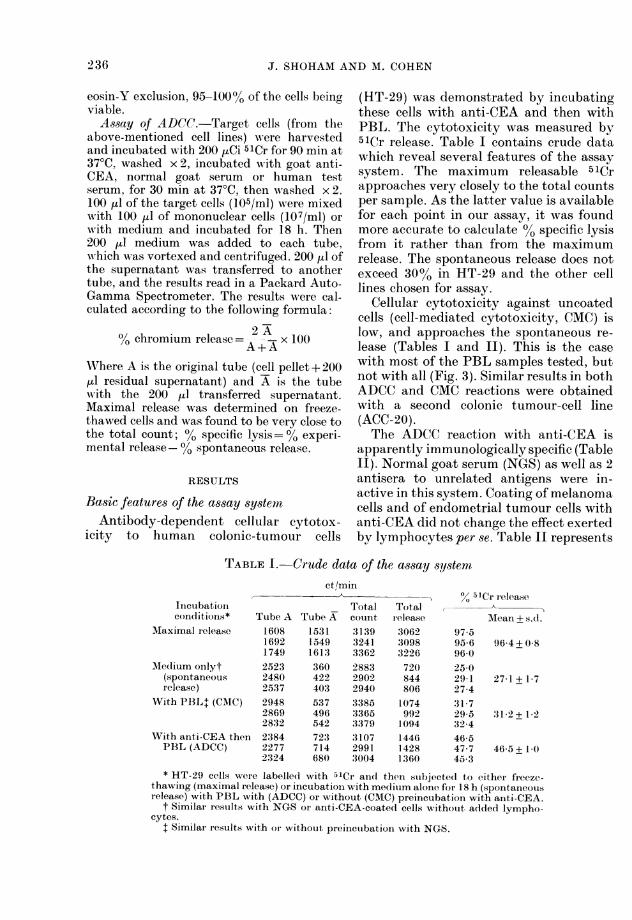
(HT-29) was demonstrated by incubatingthese cells with anti-CEA and then withPBL. The cytotoxicity was measured by51Cr release. Table I contains crude datawhich reveal several features of the assaysystem. The maximum releasable 51Crapproaches very closely to the total countsper sample. As the latter value is availablefor each point in our assay, it was foundmore accurate to calculate 00 specific lysisfrom it rather than from the maximumrelease. The spontaneous release does notexceed 30%o in HT-29 and the other celllines chosen for assay.
Cellular cytotoxicity against uncoatedcells (cell-mediated cytotoxicity, CMC) islow, and approaches the spontaneous re-lease (Tables I and II). This is the casewith most of the PBL samples tested, butnot with all (Fig. 3). Similar results in bothADCC and CMC reactions were obtainedwith a second colonic tumour-cell line(ACC-20).The ADCC reaction with anti-CEA is
apparently immunologically specific (TableII). Normal goat serum (NGS) as well as 2antisera to unrelated antigens were in-active in this system. Coating of melanomacells and of endometrial tumour cells withanti-CEA did not change the effect exertedby lymphocytes per se. Table II represents
TABLE I. Crude data of the assay systemct/min
Incubationcondlitions* Tube A
AMaximal release 160816921749
AMedlium onlyt 2523(spontaneous 2480release) 2537
With PBL+ (CAIC) 294828692832
With anti-CEA then 2:384PBL (ADCC) 2277
2324
Tube A153115491613360422403537496542723714680
Totalcount313932413362288329022940338533653379.310729913004
Totalrelease30623098322672084480610749921094144614281360
% 51CI release
Mean + s.d.97.595-696 025-029-127-431 729 532-446-547-745-3
96'4 + 0-8
27-1 + 1-7
31-2+1 2
46-5 + 1-0
* HT-29 cells weie labelled with 51Cr and theni stubjected to either freeze-thawing (maximal release) or incubationi with medlium alone for 18 h (spontaneousrelease) with PBL with (ADCC) or without (CMC) preincubation with anti-CEA.
t Similar restults with NGS or anti-CEA-coated cells without added lympho-cytes.
I Similar iesults with oIr without preincuibation with NGS.
236
ADCC TO HUATAN COLONIC TUMOUR CELLS. I
o% vC release +s. d.
Medillu§ 1PB1273 ±+ 1*7 4650-94'830-5 I6 3:237 + 02292 ±+ (9 :31*2+ 1 218 8+1 8 29-53+11209±+ 0(4 :31 7 + 0718 2 + 11 26 8 + 1 6
* (Conulitioiis as in Table I.t ACC-20 gave similar results.I Two other mnelainomna cell lines and( 2 endlo-
inetrial cell lines also exhibitedt the samen lytic effectwith or writhout coatiig wvith anti-CEA.
§ Spontaneous release.
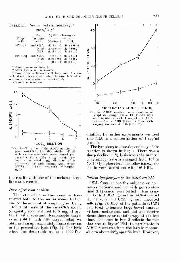
LOGIo DILUTIONLTtr.1. litat)ii of the ADCC activity of
goat anti-CEA. 104 51Cr-labelled H1-29cells wvere coate(l with reconstituted pi'e-paration of anti-CEA (8 mg protein/mIl=log 1) oIn serial loglo (liluitions of it( O -O ) (or with inor mal goat sertumNXGS and then with 10(i lympho-cyte.s.
the resuilts witlh otne of the mnelanioma celllines as a control.
I)ose-effect relationshipsThe lytic effect in this assay is (lose-
related both to the sertum concentrationand to the amounit of lymphocytes. UsingI 0-fold (lilutions of the anti-CEA serum
(originally reconstituted to 8 mg/ml pro-tein) with constant lymphocyte: targetratio (100:1 writh 104 target cells) we
obtained an approximatelv linear decreasein the percentage lysis (Fig. 1). The lyticeffect wvas detectable ul) to a I 000-fold
-n
CL)
LYMPHOCYTE/TARGET RATIOFicn. 2.-ADCC reaction as a funiction of
lymphocyte/target ratio. 104 HT-29 cellswere inctubated with 1 mg/ml anti CEA(0 O) or NGS ( O---- C1), then wvvith
vairyinig amouints of PBL (105- 106).
diluition. In further experiments we used
anti-CEA in a concentration of 1 mg/mlprotein.The lymphocyte-dose-dependency of the
reaction is shown in Fig. 2. There was a
sharp decline in lysis when the numberof lymphocytes was changed from 106 to5 x 105 lymphocytes. The following experi-menits were carried out with 106 PBL.
Patient lymphocytes as the tested variablePBL from 41 healthy subjects or non-
cancer patients and 25 with gastrointes-tinal (CI) cancer were tested in this assayfor both ADCC against anti-CEA-coatedHT-29 cells and CMC against uncoatedcells (Fig. 3). Most of the patients (21 /25)had local extensive large-bowel tumourwithout metastasis, and did not receivechemotherapy or radiotherapy at the testtime. The score in Fig. 3 reflects the factthat the ability of PBL to participate inADCC flulctuates from the barely measur-able to about 60%o specific lysis. However,
T'ABLE 11. Sertam, acnd cell controlsforspec~Jicity*
.1 re-
Tr,'I,l.g(et inceuibate(dce?lls w-ith
;H'T- 2 9 t anti-CEA
PBSSIK-nicI anti-CEA
N(XSPBS
237
c- ) It

J. SHOHAM AND M. COHIEN
a I
0
0
00
0
0
0
0
0
0@0009
0
0
0
*0
000000
0go
00
0
0
00
0@00
0
m.o32.m.@
@0
0
0
@0
0* *-
0g0
* 0
00
0
0
@000
* 0 0
0@001 ":".
ASSAY TYPE: ADCC CMCI ;.; ,ADCC CMC,
NON -CANCER
ADCC CMC
PBL SOURCE: NON-CANCER POOLS OF 2 GI CANCER
FIG. 3. ADCC and CMC activity of PBL from people without cancer and patients with gastro-intestinal (GI) cancer, mostly (21/25) local extensive colo-rectal adlenocarcinoma before radiationor chemotherapy. In the non-cancer group the activity of the indiviclual samples was compared tothat of pools from 2 donors.
in most of the cases the reaction was the samples respectively. Only 6/41 andclearly demonstrated (10O% or more specific 5/27 had 1000 or more specific lysis.lysis in 33/41 non-cancer and 23/25 cancer Statistical analysis by Wilcoxon's testpatients). In contrast, with uncoated indicates that there is no difference in bothtarget cells there was no measurable ADCC and CMC between the non-cancer
lymphocytotoxicity in 27/41 and 16/27 of and cancer group.
238
60
50
40
000
00
00
00
C-)
L)wa.ul)
I00@00
es0
0
30
20
00
00
0
0
0000
000
0@000@0
0
00
@000
10
I0
0
0
00
* *0* 0
00
0
@0000
I
nO _ w * _
,^_^ ,_XXX= -
. I

AD)CC TO HUMAN COLONIC TUMOUR CELLS. I
Patient sera as the tested variiableHT-29 cells were incubated with humnclal
sera taken from non-cancer or cancerpatients, and then with PBL. Anti-CEA-coated [IT-29 cells served as a positivecontrol. Here also we obtained a spectrumof activities from zero (equial to spon-taneous release) to those approachingmaximal release. However, the variabilityintroduced by the lymphocytes (Fig. 3)precludes comparison of results with differ-ent lymphocyte preparation.
Pooling lymphocytes (usually from 2donors) enabled us to conmpare largernumbers of seratunder uniform conditionswithout significantly changing the ADCCreactivity (Fig. 3) and this served as a
partial answer to this problem. It has tobe mentioned, however, that CMC' ofpooled PBL against uncoated HT-29 cellswas significantly higher (P < 0 01) tha,nthat of unpooled PBL. Moreover, anti-CEA wAas used as a reference serum foreach PBL sample, and the results with thepatients' sera were related to those of anti-CEA by dividing the ° specific lysis (SL)of each one of the tested sera by that ofanti-CEA. The results were regarded asthe "cytotoxicity index" (CI). The CI ofanti-CEA was, therefore, always I O((Table III).The CI introduced a remarkable unifor-
mity to the results of each serunm tested
witlh several PBL specimenis, or eveni indifferent experiments.
Sera from 218 sutbjects Awere t,ested (91healt,hv individuals or non-cancer patientsand 127 with cancer of various origins). Itwas founled in repeated experiments that aCI < 0 7 is iinsignificant, as compared tothe background, and therefore a CI of 0-7was t,aken as the boundary between posi-tive and negative reactions. By thisdefinition 46/218 (21 %) of the sera werepositive, of which 19/91] (21 oo) were fromthe non-cancer group and 26/127 (20 4%)in the cancer grouip. The difference isobviously insignificant.XVhen the results were correlated with
the blood group of these subjects (Fig. 4),it emerged that almost all the reactors(43/46) were of Blood (roup 0. However,only 43/83 (5lo%) of the Blood G;roup 0sera reacted. Blood Group A sera did notreact at all, whereas 2 sera of Blood GroupB andl I of Group AB also reacted. Theresults with Blood Group 0 sera werefurther analysed by the Mann-WhitneyU test, as a continuous score wAitfhout re-garding the 0-7 index cut. This anialysisalso confirmed the absence of differencebetween cancer and non-cancer patients.Moreover, when thle results of Blood-GrIoup ( cancer sera were scored separ-ately according to the primary tumour, nodifference among them could be demon-
TABLE III. Conversion of 0% specific lysis (SL) to cytotoxicity index (CI) using theresponse with goat anti-CEA as a reference value
Serutimt
ait i-CEA S-8 X -2-2 S-78[)BSL* -__
Exp). sample S, C.11 SL C( SL CI SL CII A 45-7 1 .0 36-2 0(79 40() 0588 NJ)
BS 8 4 1.0 7,2 0-86 7 8 0-93 NDCX :s34 5 1*0 29-5 0 X8.5 2'7 1 (-78 1158 0-:34I) 22-1 1.0 24 6 1-13 183: 0-82 ND
2 E 18 9 1)0 16.3 0-86 ND 510l26F' 274 1 .0 25 1 092 ND ND
3 C 12-5 1t0 ND ND 4 7 0 36* Seveni PBL samples w'Oere use(d in 3 experlinents for- the ADCC assay; ainti-
CEA w-as incluide(d in all the experiments and the restults with it are uisedl asreference CI (1-0). The results with the htumain seria ar-e irelated to it. S-8 ancl S-22are 2 serulm samples taken from the same patienrt on 2 diffeient occasions an(d alepositive (CI> 0-7, see text) and 8-78 is fiom aniother patient and(i nlegative(CI< 0-7).
23-

J. SHOHAM AND M. COHEN
1.6
Xw
0
5z
0
0L-
X-
0
0
0
0
0)
.4
1.2
1.0
1.8
1.6
).4
>.2
l0SERUM FROM: NC C. ,NC CA INC C, INC C,
BLOOD GROUP: A B AB 0
FIGURE 4. Activity of sera from cancer patients (C) and noni-cancer (NC), arianged accor-ding to theblood group andl expressed as cytotoxicity indlex.
1.4
1.2
xw
az
t
0
0
CL)
1.0
08
06
0.4
02
TUMOUR TYPE: GI MELANOMA BREAST OTHER
Fia. 5. Activity of sera from cancer patientswith different tumours, an(1 Blood Group 0,expressed as cytoxicity index.
strated (Fig. 5). The single positive GroupAB serum was from multiparous women,
and was found to contain HLA antibodies.The titre of the positive human sera
was lower than that of anti-CEA, and
approached background activity withdilutions of -A- or --l- (Fig. 6), without a
difference between cancer and non-cancer
sera, even when initially highly ADCC-active sera were selected.
DISCUSSION
The 2 components of the ADCC(cellular and humoral) were tested fortheir usefulness in the monitoring ofhuman tumour immunity in allogeneiccombination with colonic tumour celllines.As far as the cellular aspect is concerned,
there is remarkable variability in theability of mononuclear cells of differentsubjects to participate in the ADCCreaction. Such variability has also beenfound in other studies (Lovehik & Hong,1977; Korithavongs et al., 1974). However,the cells of healthy people and GI cancerpatients do not differ in this characteristicin our system, which tests mostly patientswith local extensive colo-rectal cancer.
* 00
_ 0 >* 0
0S S_ 0 50
* 0 0~~~~~~~~~~~~~~~~~~~~~~0* S *i. t:~~~~:
S O" - : .
*:
X
t~~ ~ ~~~~ ~~ E :0. * a
n)
-240
I
I
-
-
r

241ADCC TO HUMAN COLONIC TUMOUR CELLS. I
1.50
x
w
z
0
0X
1.00
0.50
1/2 1/4 1'8 1/16 1/32 I/64SERUM DILUTION
FIGURE 6i. Titration of ADCC activity of huimani positive sera of 3 healthy pleisoiS (A A) or3 canceir patienits ( *). The patienit sera were selected for high initial cytotoxicity. The(lashed horizontal lines represent the results with anti-CEA ((definedl as CI= 1 0) ain(l NGS, repre-senting background lysis.
Other patients with more aclvanced(lisease also showed the same pattern of'distribution (data Inot shown). There is noagreement among other workers regardingthis activity in cancer patients. The worksof Lovehik & Hong (1977), Elhilali et al.(1976) and Peter et al. (1975a) support, our'observations, wrhereas Ting & Terasaki(1974) found (lepressed ADCC-effectoractivity in cancer patients. It, has to beemphasized that the Ficolle-Hypaque-separated mononuclear cell preparationsmay contain substantial numbers ofmacrophages (Zucker-Franklin, 1974) and
chloroacetate-esterase-positive cells and
that the percentage of the last-mentioniedcells may be especially high in cancer
patients (Currie et al., 1978). Althoughsuch cells may participate in ADCC (Kohlet al., 1977; GXale & Zighelboim, 1975)along with K lymphocytes, quantitativeand kinetic differences in this activity mayexist among them. Taking all these datatogether, and in view of the remarkablevariability in this activity in both healthypersons and cancer patients, we do not feelthat testing for lymphocyte activity in
ADCC has a place in monitoring theimmuLne status of cancer patients.Twenty-one per cent of the sera tested
\ \ anti CEA
S~~~~~GA~~~~~~~~~~~~~~. mN w s .~~
0
l~~N

242 .J. SHfOHAM AND M. COHEN
were found to be reactive in this system,and to cause colonic tumour cell lysis toabout the same degree as heterologousanti-CEA does with undiluted serum. Thetitre of the heterologous anti-CEA wasabout 20-fold higher than that of thepositive allogeneic sera. However, onceagain no difference was found betweencancer patients and normal controls.Further analysis revealed that almost allthe positive sera (43/46) were of Blood-Group 0 persons, with 2 of Group B, I ofG(roup AB and none of Group A. However,not all 0-type sera were reactive. Thus,the reaction was apparently against Aantigen or A-like determinant on the CEAmolecule, which may or may not mask amore specific reaction towards otherdeterminants on the CEA molecule orother colonic-tumour associated antigens.These possibilities are further analysed inthe following publication (Shoham &Cohen, 1979), which brings evidence thatthe reaction observed is to an antigenicdeterminant common to CEA and A, andmaybe normal colon antigen too. Thisdeterminant most probably resides in theprotein portion of the molecule. The in-hibition of this common activity did notexpose tumour-specific activity.Lymphocyte dependent antibody (LDA)
activity was looked for in otherhuman tumour-cell systems. Hellstrom etal. (1973) showed increased lymphocyte-mediated tumour-cell destruction in theirtest systems with allogeneic combinationof sera from 7 cancer patients (withdifferent tumours) out of a "much largerpatient material" (unspecified) with noreference to normal antigens. The samereservation applies to the work of Hakala& Lange (1974), who found LDA activityin 2/40 transitional-cell carcinoma patients.In other publications a large panel ofallogeneic target-tumour cells of the samehistological type was used in order to solvethe problem of immune specificity.Ferrone & Pellegrino (1977) used severalmelanoma cell lines in a complement-dependent microcytotoxicity assay. Theyfailed to find melanoma-specific activity as
compared to serum activity in othercancer patients or correlation to diseasestage. Hersey et al. (1973) found LDA insera from AML patients to a panel of allo-geneic AML myeloblasts, which they feltmay be directed to a leukaemia-associatedantigen. However, some of these patientshad received immunotherapy with allo-geneic cells, and all of them had beengiven multiple transfusions. Thus it ismore plausible that the observed activitywas related to HLA antigens. This notionis further supported by the recent work ofGale & MacLennan (1977). Autochthonouscombinations may avoid this confusion.Kodera & Bean (1975) used such com-binations and found LDA activity in 4/16patients, which was apparently related todisease state. A similar study by Peter etal. (1975b) in a smaller group of patientsfailed to show such activity. However, thenumbers in the last 2 studies are toosmall to warrant any firm conclusion on thesignificance of this activity, and thedifficulties encountered in using autoch-thonous combinations preclude their large-scale use. An alternative approach is toeliminate the activity to HLA and blood-group antigens by selective absorption orby inhibition with Fab fragments ofappropriate polyspecific serum. Thepotential of this approach is demonstratedin the following publication (Shoham &Cohen, 1979). However, in the particularcase of colonic tumour cells it failed toexpose any specific tumour activity.
This work was supported in part by research grants6/74 and 3/7;5 fIom the Israel Cancer Association.
REFERENCESBYFIELD, J. E., ZERUBAVEL, R. & FONKALSRUD,
E. E. (1976) Murine neuroblastoma cured in vivoby antibody dependent cellular cytotoxicity ieac-tion. Nature, 264, 783.
CURRIE, G. A., HEDLEY, D. Wr., NYHIOLMI, 1B. E. &TAYLOR, S. A. (1978) Contaminatioin of mono-nuclear cell suspensions obtained from cancerpatients by the Boyum metho(i. Br. J. Can-cer, 38,555.
ELHILALI, M. M., BRITTON, S., B3ROSMAN, S. &F'AHEY, J. L. (1976) Critical evaluation of lympho-cyte function in urological cancer patients. CancerRes., 36, 132.
FELDMAN, J. L., BECKER, AM. J., AIOULSOPOULOS, H.& 4 others (1976) Antibodly dependent cell

ADCC TO HUMAN COLONIC TUMOUR CELLS. I 243
mediated cytotoxicity in selected autoimmunediseases. J. Clin. Invest., 58, 173.
FERRONE, S. & PELLEGRINO, M. A. (1977) Cytotoxicantibodies to cultured melanoma cells in the seraof melanoma patients. J. Natl Cancer Inst., 58,1201.
GALE, D. G. & MACLENNAN, I. C. M. (1977) Cyto-toxic antibody in acute myeloblastic leukaemiaduring immunotherapy: lack of tumour speci-ficity. Br. J. Cancer, 35, 280.
GALE, R. P. & ZIGHELBOIM, J. (1975) Polymorpho-nuclear leukocytes in antibody dependent cellularcytotoxicity. J. Immnunol., 114, 1047.
HAKALA, T. T. & LANGE, P. H. (1974) Serum in-duced lymphoid cell mediated cytotoxicity tohuman transitional cell carcinoma of the genito-urinary tract. Science, 184, 795.
HELLSTROM, I., HELLSTR6M, K. E. & WARNER, G. A.(1973) Increase of lymphocyte mediated celldestruction by certain patient sera. Int. J. Cancer,12, 348.
HERSEY, P., MACLENNAN, I. C. M., CAMPBELL, A. C.,HARRIS, R. & FREEMAN, C. B. (1973) Cytotoxicityagainst human leukemic cells. Clin. Exp. Immunol.,14, 159.
JEANNET, M. & VASSALLI, P. (1976) The role oflymphocyte dependent antibody in kidney trans-plantation. Transplantation, 22, 493.
KODERA, Y. & BEAN, M. A. (1975) Antibody de-pendent cell-mediated cytotoxicity for humanmonolayer target cells bearing blood group andtransplantation antigens for melanoma cells. Int.J. Cancer, 16, 579.
KOHL, S., STARR, S. E., OLESKE, J. Al., SHORE, S. L.,ASHMAN, R. B. & NAHMIAS, A. J. (1977) Humanmonocyte macrophage mediated, antibody de-pendent cytotoxicity to herpes simplex virusinfected cells. J. Immunol., 118, 729.
KORITHAVONGS, T., HOLLMAN, V. C. & DOSSETTOR,J. B. (1974) Effector cell activity in antibody-mediated cell-dependent immune lympholysis.J. Immunol., 113, 1178.
LAMON, E. W., HALE, P. & WHITTEN, H. D. (1976)Antibody dependent cell mediated cytotoxicitywith autochthonous lymphocytes and sera afterinfection with Moloney sarcoma virus. J. NatlCancer Inst., 56, 349.
LOVCHIK, J. C. & HONG, R. (1977) Antibody de-pendent cell-mediated cytolysis: analysis andprojections. Prog. Allergy, 22, 1.
ORTIZ DE LANDAZURI, M., KEDAR, E. & FAHEY, J. L.(1974) Synergistic cooperation between isoanti-serum and immune lymphoid cells: in vitro studieswith a synergistic rat lymphoma. J. Immunol.,112, 2102.
PEARSON, G. R. & ORR, T. W. (1976) Antibody de-pendent lymphocyte cytotoxicity against cellsexpressing Epstein-Barr virus antigens. J. NatlCancer Inst., 56, 485.
PERLMAN, P. (1976) Cellular immunity: antibody-dependent cytotoxicity (K-cell activity). Clin.Immunobiol., 3, 107.
PETER, H. H., PARIE-FISCHER, J., FRIDMAN, W. H.& 4 others (1975a) Cell mediated cytotoxicity invitro of human lymphocytes against a tissue cul-ture melanoma cell line (IgR3). J. Inmunol., 115,539.
PETER, H. H., KALDEN, J. R., SEELAND, P., DIEHL,V. & ECKERT, G. (1975b) Humoral and cellularimmune reactions in vitro against allogeneic andautologous human melanoma cells. Clin. Exp.Immunol., 20, 193.
POLLACK, S., HEPPNER, G., BRAUN, R. J. & NELSON,K. (1972) Specific killing of tumor cells in vitro inthe presence of normal lymphoid cells and serafrom hosts immune to the tumor antigens. Int. J.Cancer, 9, 316.
SHOHAM, J. & COHEN, M. (1979) Antibody-dependentlymphocyte cytotoxicity to human colon-tumourcells. II. Analysis of the antigens involved. Br. J.Cancer, 40, 244.
THORSBY, E. & BRATLIE, A. (1970) A rapid metho(dfor preparation of pure lymphocyte suspensions.In Histocompatibility Testing. Ed. P. Terasaki.Copenhagen: Munksgaard. p. 655.
TING, A. & TERASAKI, P. I. (1974) Depressed lympho-cyte mediated killing of sensitized targets incancer patients. Cancer Res., 34, 2694.
VON KLEIST, S., CHANY, E., BURTIN, P., KING, M. &FOGH, J. (1975) Immunohistology of antigeneicpatterns of a continuous cell line from a humancolon tumour. J. Natl Cancer Inst., 55, 555.
ZIGHELBOIM, J., BONAVIDA, B. & FAHEY, J. L.(1973) Evidence for several cell populations activein antibody dependent cellular cytotoxicity.J. Immunol., 111, 1737.
ZUCKER-FRANKLIN, D. (1974) The percentage ofmonocytes among "mononuclear" cell fractionsobtained from normal human blood. J. Immunol.,112, 234.