Birbal - Scholarly Publications Leiden University
30
Cover Page The handle http://hdl.handle.net/1887/138387 holds various files of this Leiden University dissertation. Author: Birbal, R.S. Title: Advances in endothelial keratoplasty Issue Date: 2020-11-17
-
Upload
khangminh22 -
Category
Documents
-
view
2 -
download
0
Transcript of Birbal - Scholarly Publications Leiden University

Cover Page
The handle http://hdl.handle.net/1887/138387 holds various files of this Leiden University dissertation. Author: Birbal, R.S. Title: Advances in endothelial keratoplasty Issue Date: 2020-11-17

Chapter 1General Introduction and Thesis Outline
General Introduction and Thesis Outline
Chapter 1

1
General Introduction and Thesis Outline 13
GENERAL INTRoDUCTIoN
The human cornea is the most anterior, transparent structure of the globe. It
serves as a barrier to protect intraocular structures and provides about two-
thirds of the entire refractive power of the eye.1 The cornea measures 11-12
mm horizontally, and 10-11 mm vertically, with a central radius of curvature of
approximately 8 mm. It has an average thickness of 500 to 600 µm.1-3 With a
high degree of innervation by the ophthalmic branch of the trigeminal nerve
(approximately 300-400x that of the epidermis), it is one of the most sensi-
tive tissues in the human body.1 The cornea is uniquely avascular, and acquires
its nutrients from the tear film or aqueous humor.1,3,4 The lack of vasculariza-
tion contributes to corneal clarity, optical performance, and relative immune
privilege.1,3 The cornea is amenable to transplantation and eye banks play an
important role in procurement, storage, and allocation of corneal tissue for
transplantation.
ANAToMY AND PHYSIoLoGY oF THE HUMAN CoRNEA
The human cornea is a transparent tissue with a high degree of spatial organi-
zation and a strong correlation between structure and function. It consists of
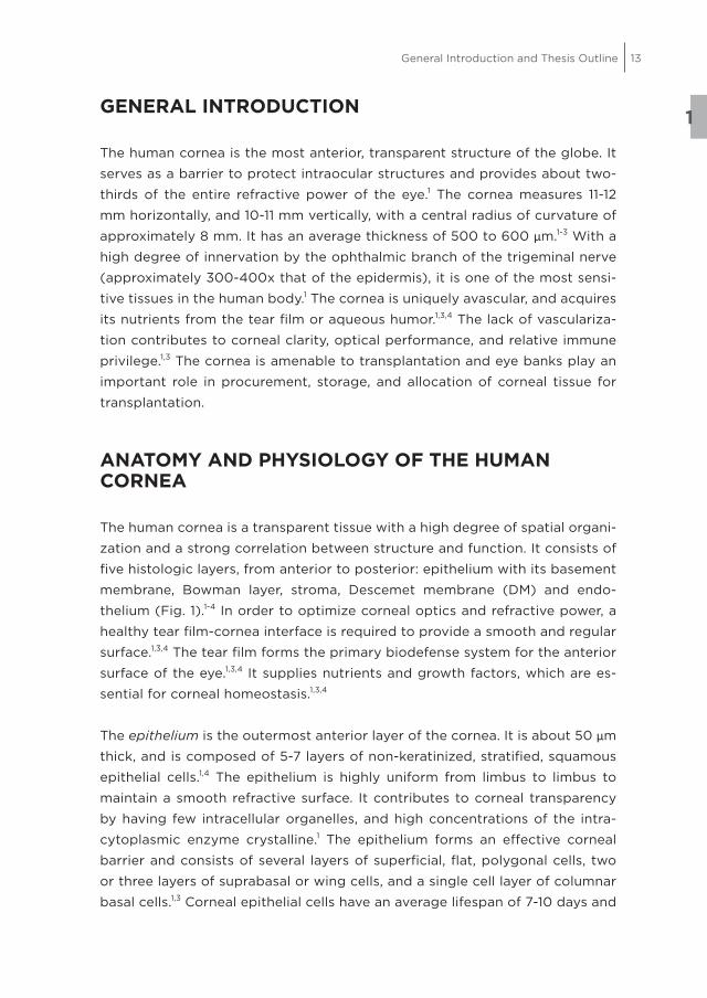
five histologic layers, from anterior to posterior: epithelium with its basement
membrane, Bowman layer, stroma, Descemet membrane (DM) and endo-
thelium (Fig. 1).1-4 In order to optimize corneal optics and refractive power, a
healthy tear film-cornea interface is required to provide a smooth and regular
surface.1,3,4 The tear film forms the primary biodefense system for the anterior
surface of the eye.1,3,4 It supplies nutrients and growth factors, which are es-
sential for corneal homeostasis.1,3,4
The epithelium is the outermost anterior layer of the cornea. It is about 50 µm
thick, and is composed of 5-7 layers of non-keratinized, stratified, squamous
epithelial cells.1,4 The epithelium is highly uniform from limbus to limbus to
maintain a smooth refractive surface. It contributes to corneal transparency
by having few intracellular organelles, and high concentrations of the intra-
cytoplasmic enzyme crystalline.1 The epithelium forms an effective corneal
barrier and consists of several layers of superficial, flat, polygonal cells, two
or three layers of suprabasal or wing cells, and a single cell layer of columnar
basal cells.1,3 Corneal epithelial cells have an average lifespan of 7-10 days and
14
complete epithelial turnover takes place on a weekly basis.1,3,4The epithelial
basement membrane is 40-60 nm thick, and is composed of type IV collagen
and laminin secreted by basal cells.1,3,4
Figure 1. Schematic representation of the anatomical layers of the human cornea.
Bowman layer (BL) is an acellular layer positioned just beneath the epithelial
basement membrane.1-4 The anterior surface is very smooth, while the pos-
terior surface extends into the anterior stroma.1-4 It is approximately 8-14 µm
thick, and thins with age.1,5 In contrast to the underlying stromal collagen fi brils
(diameter 32-36 nm) that run uniformly parallel across the corneal to form
characteristic lamellae, BL consists of smaller, randomly interwoven collagen
fi brils (24-27 nm).6 These fi brils are primarily composed of collagen types I
and III and form a dense, felt-like sheet.7BL does not regenerate after injury
and to date, the physiologic function of BL remains to be elucidated.1,3
The stroma provides the largest portion of the structural framework of the
cornea. It accounts for nearly 90% of the total corneal thickness and mea-
sures an average of 500 µm in humans.1,3,4 The stroma contributes to corneal
transparency, mechanical strength, and tectonic stability. It is made up of col-
lagen fi bers embedded in an extracellular matrix (ECM) composed of mainly
water, inorganic salts, proteoglycans, and glycoproteins.8 Keratocytes are the
major cell type of the stroma and are scattered among the stromal lamellae.1,3,4
They are involved in maintaining stromal homeostasis and hold the potential
to create collagen molecules and glycosaminoglycans, while also creating
matrix metalloproteases (MMPs).1,3,4 Most of the keratocytes reside in the an-

1
General Introduction and Thesis Outline 15
terior stroma and contain corneal crystallins that are responsible for reducing
backscatter.9 In a healthy cornea, keratocytes remain dormant. They transform
into myofibroblasts in response to various types of injury and participate in
wound repair by producing ECM, secreting cytokines and collagen-degrading
enzymes, and by contracting the edges of the wound. The collagen fibers
(mainly types I and V) are structured in parallel bundles and organized in
parallel-arranged lamellae.1,3,4 Human stroma consists of 200-250 distinct
lamella.1,3,4 Each of them is aligned at right angles relative to fibers in adjacent
lamellae.10 The stroma is thicker peripherally than centrally, and as the collagen
fibrils approach the limbus they may change direction to run circumferentially.11
The ultrastructure of the lamellae varies, based on the stromal depth: deeper
layers are more strictly organized than superficial layers.3 The high degree of
spatial organization of stromal fibers and extracellular matrix contributes to
corneal transparency and rigidity. The posterior lamellae in the central cornea
are more hydrated than the anterior lamellae and are believed to have less
interlacing, resulting in easier swelling of the posterior stroma compared with
the anterior stroma.3 Stromal collagen fibrils are surrounded by specialized
proteoglycan, consisting of keratan sulfate or chondroitin sulfate/dermatan
sulfate side chains, which help regulate hydration and structural properties.3
In 2013, Dua studied the effect on corneal biomechanics and cleavage planes
of injecting air into the posterior stroma as is done in deep anterior lamellar
keratoplasty (DALK) with the big bubble (BB) technique. He proposed that
there exists another, distinct, well-defined layer between the posterior stroma
and Descemet membrane.12 This acellular, 6-12 µm thick tissue was coined
“Dua’s layer”, later renamed the “Dua-Fine layer”.13 It has been the source of
much controversy and debate. Other groups have postulated that while this
layer has a unique cohesiveness and configuration, it does not represent a
distinct and separate corneal layer. Rather, the BB technique helps to describe
the mechanical posterior stromal response to non-physiologic stress.14,15
Descemet membrane (DM) is located directly behind the posterior stroma and
is the basement membrane of the corneal endothelium. DM gradually increases
in thickness from 3 μm at birth to 10-12 μm in adulthood.3 It is continually se-
creted by the corneal endothelium. Three distinct zones may be distinguished:
a thin non-banded zone adjacent to the stroma (0.3 μm), an anterior banded
zone (2-4 μm) and a posterior, amorphous, non-banded zone (>4 μm), that
thickens with age. DM primarily consists of collagen types IV and VIII, laminin,
and fibronectin.16,17 DM, with its adjacent endothelium, can be peeled off from

16
the posterior stroma as a single sheet. Once completely detached, DM will
spontaneously curl into a single or double roll.18,19
The endothelium is the innermost posterior layer of the human cornea and
measures 4 μm in thickness in adulthood. This monolayer consists of tightly-
packed hexagonal cells and appears as a honeycomb mosaic when viewed
posteriorly.3 The endothelium plays a key role in preserving corneal transpar-
ency by maintaining the cornea in a relative state of deturgescence.1 The
‘pump-leak’ hypothesis proposes that the endothelium in a healthy cornea
achieves corneal clarity by maintaining a state of equilibrium between two
fluid transport pathways. A low-resistance apical junction between the en-
dothelial cells allows fluid from the anterior chamber to ‘leak’ into the stroma
(passive diffusion), whereas Na+/K+- and bicarbonate-dependent Mg2+-ATPase
pumps create local osmotic gradients, thereby actively returning fluid from
the stroma to the anterior chamber.1 Dysfunction of either of these path-
ways can result in corneal edema and reduced corneal transparency. The
endothelial cell density (ECD) is approximately 6000 cells/mm2 at birth and
gradually decreases to about 3500 cells/mm2 by the age of 5 years as the
eyes grow.20,21 During adulthood, ECD decrease slows down to an annual
decrease of approximately 0.6%.22,23 Apart from aging, accelerated cell loss
may be caused by a genetic predisposition, prior intraocular surgery, trauma,
elevated intraocular pressure, diabetes mellitus, and chronic anterior chamber
inflammation.24 Endothelial cells do not regenerate in vivo. When cells are lost,
an endothelial defect will be restored by expansion (polymegathism) and ac-
tive migration of adjacent cells. During this process, loss of hexagonality of
the cells may occur (pleomorphism).3,25,26 When the ECD count decreases to
the extent that the overall remaining endothelial pumping capacity fails to
maintain the equilibrium between the beforementioned pathways, endothelial
decompensation may occur, resulting in irreversible corneal edema, reduced
corneal clarity, pain and vision loss.27

1
General Introduction and Thesis Outline 17
Common indications for endothelial keratoplasty
Fuchs Endothelial Corneal Dystrophy
Fuchs endothelial corneal dystrophy (FECD) is the most common corneal
dystrophy and currently one of the leading indications for corneal transplan-
tation.28 It was first described in 1910 by the Austrian ophthalmologist Ernst
Fuchs and is a slowly progressive, bilateral corneal disease. Hallmark features
of FECD include accumulation of wart-like excrescences of DM better known
as ‘guttae’, thickening of DM, endothelial cell pleomorphism and polymegath-
ism and loss of endothelial cells (Figs. 2,3).29-32 With advancing disease, stro-
mal edema may compromise visual function, with vision being worse in the
morning and improving during the day. In end-stage disease, epithelial bullae
may develop, evolving into subepithelial fibrosis and corneal vascularization.
Based on the time of onset of disease, two clinical subtypes of FECD may be
distinguished: early-onset FECD (3-40 years) and late-onset FECD (>40 years),
with the late-onset form being more common.33 The early-onset form of FECD
has been associated with autosomal dominant Q455K, Q455V and L450W
mutations in the gene encoding the alpha 2 subunit collagen 8 (COL8A2).
Men and women are equally affected. In contrast to early-onset FECD, a fe-
male predominance of 3:1 has been reported for late-onset FECD. Currently, 5
causal genes (TCF4, AGBL1, LOXHD1, SLC4A11 and ZEB1) and 4 causal loci on
chromosomes 5, 9, 13, and 18 have been identified in individuals with late-onset
FECD. Expanded repeats of the trinucleotide cytosine-thymine-guanine (CTG
repeats) in the 3rd intron of TCF4 within chromosome 18q21.1 may be the most
commonly identified genetic contributor to FECD.34 Despite the identification
of some genetic factors, the exact pathophysiology of FECD remains unclear
and is thought to be a combination of both environmental and genetic factors.
Both subtypes display a similar linear rate of disease progression.

18
12
Fi
gure
2. S
pecu
lar m
icro
scop
y im
ages
dis
play
ing
heal
thy
endo
thel
ium
(lef
t im
age)
and
diff
eren
t sta
ges,
from
m
oder
ate
to a
dvan
ced,
of F
uchs
end
othe
lial c
orne
al d
ystro
phy
(imag
es fr
om le
ft to
righ
t).
Bullo
us k
erat
opat
hy
Bullo
us k
erat
opat
hy d
evel
ops a
s a re
sult
of e
ndot
helia
l dec
ompe
nsat
ion
due
to e
ndot
helia
l
inju
ry c
ause
d by
var
ious
con
ditio
n or
eve
nts s
uch
as b
irth
inju
ry o
r int
raoc
ular
surg
ery,
incl
udin
g co
mpl
icat
ed c
atar
act s
urge
ries,
glau
com
a su
rger
ies,
or v
itreo
retin
al su
rger
ies.
Sym
ptom
s may
pre
sent
in th
e im
med
iate
pos
t-tra
umat
ic p
erio
d or
yea
rs a
fter t
he in
jury
. With
adva
ncin
g co
rnea
l ede
ma,
pat
ient
s ofte
n m
anife
st w
ith (s
ub)e
pith
elia
l bul
lae
resu
lting
in
pain
ful c
orne
al m
icro
-def
ects
whe
n th
ey ru
ptur
e.35
In a
dvan
ced
stag
es, s
ubep
ithel
ial f
ibro
sis,
with
or w
ithou
t BL
disr
uptio
n, m
ay d
evel
op.35
Figu
re 3
. Slit
-lam
p an
d sp
ecul
ar
mic
rosc
opy
imag
es o
f eye
s with
Fuc
hs
endo
thel
ial c
orne
al d
ystro
phy
(FEC
D)
and
bullo
us k
erat
opat
hy (B
K).
FECD BK
Fig
ure
2. S
pec
ular
mic
rosc
op
y im
ages
dis
pla
ying
hea
lthy
end
oth
eliu
m (
left
imag
e) a
nd d
iff e
rent
sta
ges
, fro
m m
od
erat
e to
ad
vanc
ed, o
f F
uchs
end
o-
thel
ial c
orn
eal d
ystr
op
hy (
imag
es f
rom
left
to
rig
ht).

1
General Introduction and Thesis Outline 19
Bullous keratopathy
Bullous keratopathy develops as a result of endothelial decompensation due
to endothelial injury caused by various conditions or events such as birth injury
or intraocular surgery, including complicated cataract surgeries, glaucoma
surgeries, or vitreoretinal surgeries. Symptoms may present in the immediate
post-traumatic period or years after the injury. With advancing corneal edema,
patients often manifest with (sub)epithelial bullae resulting in painful corneal
micro-defects when they rupture.35 In advanced stages, subepithelial fi brosis,
with or without BL disruption, may develop.35
12
Figure 2. Specular microscopy images displaying healthy endothelium (left image) and different stages, from moderate to advanced, of Fuchs endothelial corneal dystrophy (images from left to right). Bullous keratopathy
Bullous keratopathy develops as a result of endothelial decompensation due to endothelial
injury caused by various condition or events such as birth injury or intraocular surgery,
including complicated cataract surgeries, glaucoma surgeries, or vitreoretinal surgeries.
Symptoms may present in the immediate post-traumatic period or years after the injury. With
advancing corneal edema, patients often manifest with (sub)epithelial bullae resulting in
painful corneal micro-defects when they rupture.35 In advanced stages, subepithelial fibrosis,
with or without BL disruption, may develop.35
Figure 3. Slit-lamp and specular microscopy images of eyes with Fuchs endothelial corneal dystrophy (FECD) and bullous keratopathy (BK).
FEC
D
BK
Figure 3. Slit-lamp and specular microscopy images of eyes with Fuchs endothelial corneal dystrophy (FECD) and bullous keratopathy (BK).

20
CoRNEAL TRANSPLANTATIoN
History of corneal transplantationReplacing diseased corneal tissue has been under consideration for a long
time, with major changes occurring in recent years. The first description of
keratoprosthesis originates from the French surgeon Guillaume Pellier de
Quengsy.36 During the French revolution in 1789, he hypothesized that a
transparent material could be used to replace an opaque cornea in order to
restore vision. In 1796, Erasmus Darwin proposed the first corneal trephine
and postulated that the cornea might heal secondary to forming a transparent
scar.37 In 1813, Karl Himley proposed replacing opaque animal corneas with
corneas from other animals, but it was not until 1818 that his student Franz
Reisinger initiated these experimental animal corneal transplants.38In 1824,
Reisinger coined the term ‘keratoplasty’ and proposed using animal tissue to
replace human corneas. His animal experiments, however, failed to produce
clear grafts. In 1837, the Irish surgeon Samuel Bigger reported his first suc-
cessful penetrating graft on a pet gazelle blinded by extensive corneal scar-
ring.39 In 1838, inspired by Bigger, New York-based ophthalmologist Richard
Kissam performed the first recorded corneal xenograft, from a 6-month old
pig, on a young Irishman in 1838.40 While increased light perception occurred
immediately after the operation, the cornea opacified within the first fortnight
and was absorbed within one month after the operation. For the remainder
of the 19th century, the pioneers of corneal transplantation could be divided
into two main groups: those who favored full-thickness allografts (Henry
Powers) and those who favored partial-thickness lamellar xenografts (Arthur
von Hippel).38,41 In 1905, the first successful human allograft was performed by
Eduard Zirm.42 The recipient was a farmer who had sustained bilateral alkali
burns while cleaning out a chicken coop with lime 16 months earlier. Zirm used
donor tissue from the enucleated eye of an 11-year old boy whose eye had
been blinded by a penetrating injury to the sclera. The eye was enucleated
and the one donor cornea was used to procure two full-thickness grafts of 5
mm in diameter. While the graft in the right eye failed, the graft in the left eye
remained clear and improved the visual acuity of the recipient from counting
fingers preoperatively to 6/36 at 6 months after the operation. Since then,
innumerable ophthalmologists and scientists have contributed to improving
the technique, and in the century thereafter, penetrating keratoplasty (PK)
became the mainstay of care in the treatment of all corneal disorders regard-
less of which layer was diseased.

1
General Introduction and Thesis Outline 21
History and evolution of endothelial keratoplastyWhile the lamellar approach was already described with xenografts by Arthur
von Hippel in 1888, it was not pursued in the decades thereafter. This was
possibly because lamellar transplants were perceived to be technically more
challenging than full-thickness transplants. While PK can yield an optically
transparent cornea, it is also prone to potential complications such as poor
wound healing, suture-related problems, high astigmatism, allograft rejection,
graft failure, and unsatisfying visual outcomes, with many patients requiring
contact lenses to reach their full visual potential after keratoplasty.43,44
Nevertheless, Charles Tillett performed the first posterior lamellar endothelial
transplant underneath a manually dissected stromal flap in a patient with
FECD in 1956.45 In the 1960s, Barraquer et al. applied a similar technique which
unfortunately also proved relatively unsuccessful.46 These early attempts may
have failed due to lack of suitable instrumentation to dissect thin corneal lay-
ers and limited understanding of endothelial cell physiology, resulting in early
complications, and/or insufficient visual outcomes. As a result, the concept of
endothelial keratoplasty was, once again, abandoned.
It was not until 1998, that Melles et al. introduced a technique for posterior
lamellar keratoplasty (PLK), currently known as endothelial keratoplasty (EK),
in which a posterior lamellar disc was excised from the recipient cornea and a
same-size donor disc, consisting of posterior stroma, DM and endothelium, was
implanted through a limbal scleral incision.47 Although technically challenging,
this technique provided clinical outcomes surpassing PK and circumvented
many PK-associated complications.48 In 2001, this technique was popularized
as deep lamellar endothelial keratoplasty (DLEK) in the United States by Terry
et al. (Fig. 4). In the initial PLK/DLEK technique, a donor disc was implanted
into the recipient cornea through a 9-mm sclerocorneal incision and positioned
against the recipient posterior cornea by means of an air-bubble.47 In 2000,
the initial technique was modified by Melles et al., folding the donor disc like
a ‘taco’ to enable insertion through a self-sealing 5-mm tunnel incision.49 This
technique was popularized as small incision DLEK. Worldwide adoption was
tempered by the technical difficulty of the procedure, which necessitated
manual dissection of both donor and host tissue.
To simplify the technique, Melles et al. abandoned recipient stromal dissection
and introduced ‘descemetorhexis’, a new approach in which only recipient
DM and endothelium were stripped, using a reversed Sinskey hook.50 Des-

22
cemetorhexis was followed by implantation of a taco-folded donor disc, which
was subsequently positioned onto the denuded host posterior stroma with
an air-bubble. This approach was first performed clinically in 2001 and was
later popularized by Price et al. as Descemet stripping endothelial keratoplasty
(DSEK).50-52 Gorovoy et al. further simplified the technique by introducing an
automated microkeratome to dissect the donor graft from a corneoscleral
button mounted on an artificial anterior chamber. 53 This modification changed
the nomenclature to Descemet stripping automated endothelial keratoplasty
(DSAEK) (Figs. 4, 5). After these refinements in technique, the worldwide
adoption of DSEK/DSAEK grew exponentially and it became the preferred
treatment option for corneal endothelial disorders.54
Although DSEK/DSAEK represents a massive improvement compared to its
predecessors, it still has some drawbacks. Even after technically successful
transplantations, final visual acuity is variable and occasionally unsatisfyingly
low. This has among others been ascribed to the presence of varying thickness
of posterior stroma within the donor graft.54-60
In 2002, Melles et al. further refined the concept of endothelial keratoplasty
by completely eliminating the posterior stroma from the donor graft, allowing
selective replacement of bare DM with its endothelial layer. 49 This technique
was coined Descemet membrane endothelial keratoplasty (DMEK) and was
first performed successfully in a patient in 2006 (Figs. 4, 5).61
Descemet Membrane Endothelial KeratoplastyAfter its introduction in 2006, the surgical procedure was further refined and
standardized and as a result, a standardized ‘no-touch’ DMEK-technique was
introduced in 2011.62 The technique entailed scoring and descemetorhexis
under air followed by an air-fluid exchange and implantation of a DMEK graft,
ideally folded into a double roll with the curls facing upward, into the recipient
anterior chamber. The DMEK graft was then unfolded over the iris by means of
an air bubble injected in between the two curls and corneal tapping, and lifted
against the recipient posterior stroma by inserting an air bubble underneath
the DMEK graft. At the end of the surgery, a complete air fill of the anterior
chamber was maintained for 60 minutes, after which an air-liquid exchange
was performed to pressurize the eye and promote graft adherence.
Since its implementation, DMEK has shown to provide faster visual rehabilita-
tion, improved visual outcomes, and lower graft rejection rates compared with
1
General Introduction and Thesis Outline 23
PK g
raft
DLEK
gra
ft DS
(A)E
K gr
aft
DMEK
gra
ft He
mi-D
MEK
gra
ft Qu
arte
r-DM
EK g
raft
DM
Endo
thel
ium
Stro
ma
DM
Endo
thel
ium
Stro
ma
DM
Endo
thel
ium
DM
Epith
eliu
m
Bow
man
Laye
r St
rom
a
Endo
thel
ium
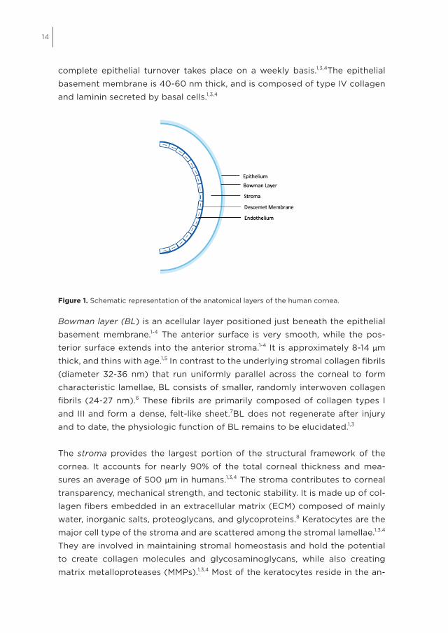
Fig
ure
4. S
chem
atic
ove
rvie
w d
isp
layi
ng t
he e
volu
tio
n o
f p
ost
erio
r ke
rato
pla
sty
tech
niq
ues,
fro
m l
eft
to r
ight
: P
enet
rati
ng k
erat
op
last
y (P
K),
Dee
p
lam
ella
r en
do
thel
ial k
erat
op
last
y (D
LE
K),
Des
cem
et s
trip
pin
g (
auto
mat
ed)
end
oth
elia
l ker
ato
pla
sty
(DS
(A)E
K)
and
co
nven
tio
nal,
Hem
i- a
nd Q
uart
er-
Des
cem
et m
emb
rane
end
oth
elia
l ker
ato
pla
sty
(DM
EK
, Hem
i-D
ME
K a
nd Q
uart
er-D
ME
K).
DM
= D
esce
met
mem
bra
ne

24
earlier EK-techniques.63-70 In 2015, the American Academy of Ophthalmology
evaluated the clinical efficacy, effectiveness and safety of DMEK by means of
a systematic review.71 The assessment revealed that 11 studies with 6-month
clinical outcomes after DMEK reported that 32% to 85% of eyes achieved a
BCVA of 20/25 or better, and 12 studies reported that 17% to 67% achieved a
BCVA of 20/20 or better. Comparison of final visual acuity levels after DMEK
and DSEK showed that, after surgery, a higher percentage in the DMEK group
achieved a BCVA of 20/25 or better (50% vs 6%, 67% vs 31%, 53% vs 15% and
55% vs 13%) and a BCVA of 20/20 or better (46% vs 13%). Complications of
DMEK include graft detachment, graft failure, allograft rejection, and endothe-
lial cell loss. The mean rejection rate of 22 studies was 1.9% (range, 0% - 5.9%)
during follow-up periods ranging from 6 months to 8 years. This is lower than
the mean rejection rate of 10% (range, 0% - 45.5%) reported after DSEK.
Owing to its excellent results, an increasing number of corneal surgeons are
adopting DMEK globally, and with increasing surgical experience complication
rates are decreasing.71 DMEK is nowadays increasingly employed in challeng-
ing cases such as eyes with anterior chamber intraocular lens implants and
eyes with glaucoma drainage devices.72-76
Corneal graft failure
Corneal graft failure is an irreversible loss of corneal transparency due to graft
dysfunction and thereby may become an indication for repeat keratoplasty. Graft
failure is considered “primary”, if the cornea never cleared to regain satisfactory
vision after the transplant surgery, or “secondary”, if the cornea initially cleared,
but then decompensated at a later time point.77 Predisposing risk factors for
graft failure include previous graft failure, glaucoma (especially previous tube
shunt surgery), peripheral anterior synechiae, corneal vascularization, immuno-
logic allograft rejection, and ocular surface disease, especially lack of tears.78
Signs of corneal graft failure include increased corneal thickness and corneal
edema. Initial treatment consists of topical corticosteroid and hypertonic saline
drops. Definitive treatment requires a repeat corneal transplantation.
Auxiliary techniques
As DMEK may still be perceived as relatively challenging in preparing and
handling of the delicate donor graft, alternative keratoplasty techniques
such as Ultra-thin DSAEK (in which a thin layer of posterior stroma (<100
µm) is transplanted as part of the donor lenticule), pre-Descemet endothelial
keratoplasty (PDEK) (in which an even thinner layer of posterior stroma ‘the
1
General Introduction and Thesis Outline 25
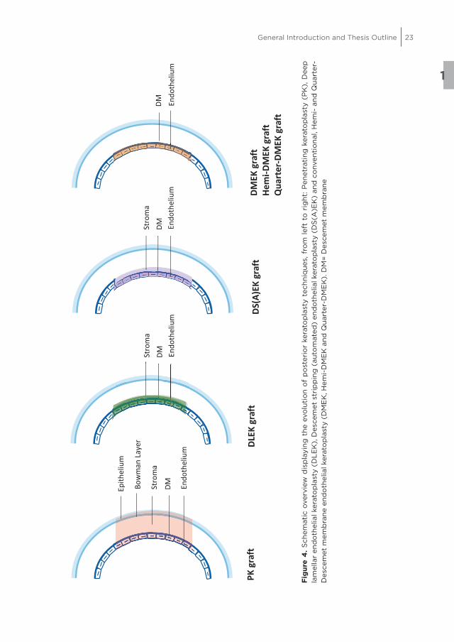
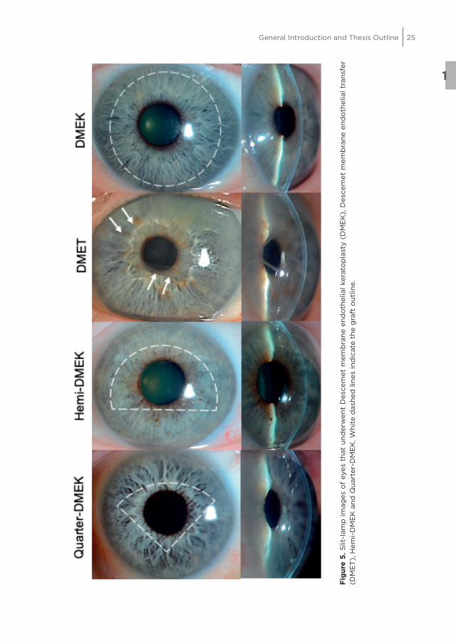
Fig
ure
5. S
lit-l
amp
imag
es o
f ey
es t
hat
und
erw
ent
Des
cem
et m
emb
rane
end
oth
elia
l ker
ato
pla
sty
(DM
EK
), D
esce
met
mem
bra
ne e
ndo
thel
ial t
rans
fer
(DM
ET
), H
emi-
DM
EK
and
Qua
rter
-DM
EK
. Whi
te d
ashe
d li
nes
ind
icat
e th
e g
raft
out
line.

26
pre-Descemet layer’ (<20 µm) is transplanted with the donor lenticule), and
DMEK with a stromal rim (DMEK-S) were introduced as a middle way to allow
for easier preparation and handling of the DMEK graft combined with visual
outcomes possibly equaling those of DMEK.79-81
MoDIFIED DMEK TECHNIQUES
Descemet Membrane Endothelial TransferThe clinical observation that corneas showed resolution of corneal edema in
the first few weeks after DMEK/DSAEK, despite (partial) graft detachment or
in the absence of a DMEK graft, led to the introduction of Descemet membrane
endothelial transfer (DMET), which consists of a descemetorhexis followed by
insertion of the almost completely free-floating Descemet roll (i.e., with the
graft contacting the posterior cornea only at the corneal incision) in 2008
(Fig. 5).82-98 While preliminary results showed that DMET was effective in the
management of eyes with FECD, it was not in eyes with BK.85,98 This prompted
the hypothesis that host endothelial cells in eyes with FECD still had some
regenerative capacity and had retained the potential to migrate to bare stro-
mal areas to repopulate them. This hypothesis was reinforced by case reports
which reported corneal clearance after ‘descemetorhexis only’.86,87,89,93,99,100
However, mixed results have been reported for the latter technique, with a
significant number of corneas failing to clear. A major drawback of DMET and
‘descemetorhexis only’ is that host peripheral endothelial cell migration is a
relatively slow process and that, if corneal clearance occurs at all, it may take
up to several months.
Hemi- and Quarter-Descemet membrane endothelial keratoplastyAs there is a substantial shortage of donor tissue for endothelial keratoplasty
worldwide, which has not yet been met by the implementation of beforemen-
tioned techniques, further refinements of DMEK were introduced.28 In 2014,
Hemi-DMEK was introduced aiming to potentially double the availability of
endothelial donor tissue (Fig. 5).101 Hemi-DMEK represents a DMEK modifica-
tion that differs from conventional DMEK only in graft shape. In Hemi-DMEK,
an ‘untrephined’, full-diameter, semicircular (half-moon shaped) graft is
utilized rather than a circular trephined Descemet graft.102 As a Hemi-DMEK
graft is untrephined and a conventional DMEK graft is trephined, both have a
comparable graft surface area and a comparable number of endothelial cells
is transplanted. Preliminary Hemi-DMEK studies have yielded visual outcomes

1
General Introduction and Thesis Outline 27
similar to those following conventional DMEK.103-105 Longer-term studies are
needed to determine whether the outcomes remain stable.
Mixed clinical outcomes after DMET and Hemi-DMEK and ‘descemetorhexis
only’ led to the development of Quarter-Descemet membrane endothelial
keratoplasty (Quarter-DMEK).106 Quarter-DMEK is a hybrid technique that aims
to combine the advantages of both DMEK (fast corneal clearance) and ‘des-
cemetorhexis only’ (host peripheral endothelial cell stimulation). In this rela-
tively new technique, merely one quarter of a full-diameter donor Descemet
graft is transplanted into eyes where FECD is limited to the central 6-7 mm
optical zone of the cornea (Fig. 5). The first case report of Quarter-DMEK was
published in 2016.106 Quarter-DMEK showed promising visual acuity outcomes,
but had a few drawbacks, including a higher rate of postoperative graft de-
tachment, a steeper decline in endothelial cell density in the first 6 months
after surgery and prolonged corneal clearance in some parts of the cornea.107
Additional studies are needed to determine the efficacy of Quarter-DMEK
relative to conventional circular DMEK.
EYE BANKING AND CoRNEAL TRANSPLANTATIoN
Since the establishment of the first eye bank by Dr. Townley Paton in 1944,
eye banks continue to play a key role in procuring, evaluating and distributing
donated ocular tissue for transplantation and research. The evolution of PK
to selective, lamellar EK was facilitated by a strong, symbiotic relationship
between corneal surgeons and eye banks, especially since dissecting lamellar
grafts has been perceived as more challenging than preparing full-thickness
PK grafts. A successful outcome after keratoplasty largely depends on viable
corneal endothelium.108 Hence, the morphologic and functional status of the
endothelium is the most important determinant for donor cornea suitability
for transplantation and maintaining endothelial cell viability from the time of
donor tissue retrieval until transplantation. Currently, two preservation meth-
ods are being applied by eye banks: hypothermic storage at 2-6ºC and organ
culture storage at 30-37ºC.109 Prolonged storage of donor tissue allows for
extensive donor screening and facilitates surgical scheduling.
As with other endothelial keratoplasty techniques, donor tissue for DMEK may
be prepared by corneal surgeons prior to surgery (surgeon-cut) or by expe-
rienced tissue specialists in an eye bank; this may take place up to 2 weeks

28
before surgery (pre-cut).19,20,110,111 Pre-cut tissue may reduce overall intervention
costs and surgery time, and allows for post-processing evaluation of the donor
graft, providing corneal surgeons with accurate information about the donor
tissue prior to surgery.112
Various techniques have been described for DMEK graft preparation, which
may broadly be classified into those based on manual peeling and those aim-
ing to achieve detachment of Descemet membrane (DM) by either injecting
air or liquid between DM and the posterior stroma. Lie et al.18 described the
initial technique for DMEK graft preparation. A donor corneoscleral rim was
mounted onto a custom-made fixation device with the endothelial side up.
DM was cut anterior to the trabecular meshwork and pushed towards the
center of the corneoscleral button. Grasping the outer edge of the graft, DM
was loosened over 180 degrees and stripped for two-thirds. By submerging
the rim in balanced salt solution (BSS), superficial trephination and complete
stripping of DM were facilitated, after which the isolated graft spontaneously
formed a roll with the endothelial layer facing outward. Groeneveld-van Beek
et al.19 modified the technique into the standardized “no-touch” technique,
in which DM with the adjacent trabecular meshwork is loosened over 360
degrees rather than over 180 degrees and trephined on a soft contact lens
instead of on the anterior cornea. The latter technique allows complete strip-
ping of DM and facilitates further handling of the graft. It allows the user to
obtain the maximum possible graft size, minimizes endothelial cell damage
in the trephination area and leaves the anterior cornea intact and eligible for
anterior lamellar keratoplasty. All preparation techniques feature different
strengths and weaknesses which will be discussed in this thesis.
CoRNEAL IMAGING TECHNIQUES AFTER ENDoTHELIAL KERAToPLASTY
Non-invasive corneal imaging modalities have proven to be useful diagnostic
tools for evaluating graft adherence and graft function after EK. While slit-
lamp biomicroscopy is the mainstay of corneal evaluation, Scheimpflug imag-
ing and anterior segment optical coherence tomography (AS-OCT) may aid in
assessing corneal optics and complications. Additionally, specular microscopy
allows for analysis of endothelial cell density (ECD) and morphology.

1
General Introduction and Thesis Outline 29
Slit-lamp biomicroscopy is readily available in all ophthalmic clinical settings
and aids in the assessment of graft adherence and corneal transparency after
endothelial keratoplasty.113 In the presence of corneal edema, however, it is not
always possible to conclusively determine whether the DMEK graft is com-
pletely attached or not. ‘Flat detachments’, i.e. when the DMEK graft is not
attached and positioned just parallel to the recipient posterior stroma, may
be especially challenging to correctly interpret without the aid of imaging
technology.114 Auxiliary corneal imaging techniques, preferably AS-OCT, can
be implemented to ensure that a (partially) detached graft in an eye with
severe corneal edema does not go undetected. These techniques may, addi-
tionally, help to differentiate between a detached DMEK graft and an attached
graft showing delayed corneal clearance, which may occur for instance due to
a ‘shock to the donor endothelial cells’ pumping function.115
Corneal tomography analysis of the anterior segment utilizes a camera (based
on the rotating Scheimpflug principle) perpendicular to a slit beam which can
capture up to 100 images in two seconds (e.g. Pentacam HR). These images
are used to create a 3-D model of the anterior segment of the eye and to
provide quantitative data such as central radii, corneal asphericity, maps of
curvature and elevation, chamber angle, chamber volume and chamber eleva-
tion as well as lens transparency.116 Pentacam Scheimpflug imaging can aid
with evaluating corneal astigmatism after keratoplasty and graft adherence
after endothelial keratoplasty.117 A drawback of this technique may be that,
particularly in corneas with extensive corneal edema, backscatter may occur,
which may impede adequate visualization of the graft and correct interpreta-
tion of graft adherence.114,117 In addition, the Scheimpflug Pentacam uses Zernike
polynomials to provide data on corneal wavefront aberrations. This can be
valuable in detecting corneal irregularities which may explain unsatisfactory
vision after endothelial keratoplasty.118-120 Densitometry analysis can provide
information on stromal opacities possibly affecting the quality of vision and
the Pentacam can be applied to analyze the refractive stability of the cornea
after endothelial keratoplasty. 121,122
AS-OCT is a non-invasive imaging modality that provides both quantitative
and qualitative information. It has a broad range of clinical applications. It
generates two- and -three-dimensional cross-sectional images of tissue by
integrating multiple axial scans (A-scans) into a composite lateral beam of
light, the B-scan.123,124 Time domain AS-OCT utilizes a light source emitting at
1310 nm, which offers the advantage of minimized scatter and high penetra-

30
tion.123,124 This technique is particularly suited for imaging structural details in
optical scattering media such as an edematous cornea, when slit-lamp biomi-
croscopy and Pentacam may fail to provide conclusive information. Recently,
high-speed Fourier domain OCT (FD-OCT) has been introduced, which offers
improved spatial resolution compared to time domain OCT. FD-OCT allows in
vivo high-speed, high-resolution imaging of weakly backscattering tissues and
can detect changes within a 10 µm range in corneal tissue.125,126 Pre-operatively,
AS-OCT may be employed to assess the thickness of the recipient cornea and
to estimate the potential size of the graft. Intraoperatively, the OCT may be
employed to visualize and assess graft orientation in DMEK surgery; especially
in the presence of severe corneal edema, it may lead to faster graft positioning
with less graft manipulation.127 Postoperatively, AS-OCT may aid in detecting
complications such as graft dislocation, anterior chamber angle narrowing, and
pupillary block.114,122 In addition, AS-OCT can precisely specify the extent and
planarity of graft detachments. In the immediate postoperative period, when
there is still an air-bubble in the anterior chamber, AS-OCT images should be
interpreted with care as the edges of the air-bubble may reveal themselves
as a separate line and may therefore mimic graft detachment. However, the
air-bubble commonly presents as a relatively smooth line in comparison to a
graft detachment.
Specular microscopy is a non-invasive imaging modality. It is currently the
most widely applied diagnostic tool for evaluating the corneal endothelium,
as it allows for in vivo visualization and analysis of the endothelium.128-130 It is
based on the reflection of the incoming light generated by the difference in
refractive index of the endothelial cells and the aqueous humor.128 As the main
objective of endothelial keratoplasty is to regain endothelial function and
subsequently corneal transparency, the donor endothelium should be closely
monitored during the postoperative course.128-130 Endothelial cell density is
a key quantitative corneal endothelial parameter for evaluating the clinical
outcome after keratoplasty, and polymegathism (cell size variability) and
pleomorphism (cell shape variability, loss of hexagonal shape) are important
qualitative indicators.131,132 Image quality may be compromised by corneal
pathology such as scarring or edema, which can increase light scattering in
the stroma from collagen lamellae and keratocytes.133 Commercial specular
microscopes are usually provided with an automatic ECD analysis program.
However, sufficient quality of the acquired images, with clearly displayed cell
borders, and manual correction, is usually required to ensure reliable ECD
measurements.128,130,134

1
General Introduction and Thesis Outline 31
AIM AND oUTLINE oF THIS THESIS
This thesis focuses on donor tissue preparation for DMEK and evaluates the
feasibility and clinical outcomes of DMEK, DMET, Hemi-DMEK and Quarter-
DMEK in the management of corneal endothelial disorders.
The first part of this thesis concerns donor tissue preparation for DMEK.
We tested whether the technique of DMEK graft dissection influences the
clinical outcome after DMEK. Chapter 2 provides an overview of the current
harvesting techniques available for DMEK and a discussion of these techniques.
The second part of this thesis concerns the clinical outcome of selective,
minimally-invasive and potentially tissue-sparing surgical treatment options
for corneal endothelial disorders. We hypothesize that complete and lasting
corneal rehabilitation may not always require a (nearly) fully, centrally at-
tached large DMEK graft. We evaluated the six-month clinical results of 1000
consecutive DMEK cases and evaluated whether whether outcomes are influ-
enced by surgical indication and preoperative lens status (Chapter 3).
Subsequently, we evaluated the five-year graft survival and clinical outcomes
of 500 consecutive DMEK cases (Chapter 4). The feasibility and clinical out-
comes of DMEK in eyes with a glaucoma drainage device are being described
in Chapter 5. The next three chapters focus on the different endothelial
grafting techniques, evaluating subtotal detachment of the DMEK graft after
a DMEK procedure or intended DMET (Chapter 6), and outcomes of Hemi-
DMEK (Chapter 7) and of Quarter-DMEK performed for FECD (Chapter 8).

32
REFERENCES 1. Sabet SJ, Adampoulou A. Basic structure and function of the human cornea and adnexal
structures. In: Copeland RA and Afshari NA (Ed.). Copeland and Afshari’s Principles and
Practice of Cornea. Jaypee Brothers Medical Publishers, 2013
2. Duong QH. Corneal embryology. In: Copeland RA and Afshari NA (Ed.). Copeland and
Afshari’s Principles and Practice of Cornea. Jaypee Brothers Medical Publishers, 2013
3. DelMonte DW, Kim T. Anatomy and physiology of the cornea. J Cataract Refract Surg
2011;37:588-98
4. Sridhar MS. Anatomy of cornea and ocular surface. Indian J Ophthalmol 2018;66:190-4
5. Germundsson J, Karanis G, Fagerholm P, et al. Age-related thinning of Bowman’s layer
in the human cornea in vivo. Invest Ophthalmol Vis Sci 2013 11;54:6143-9
6. Marshall J, Grindle CF. Fine structure of the cornea and its development. Trans Ophthal-
mol Soc U K 1978;98:320-8
7. Wilson SE, Hong JW. Bowman’s layer structure and function: critical or dispensable to
corneal function? A hypothesis. Cornea 2000;19:417-20
8. Torricelli AA, Wilson SE. Cellular and extracellular matrix modulation of corneal stromal
opacity. Exp Eye Res 2014;129:151-60
9. Jester JV, Moller-Pedersen T, Huang J, et al. The cellular basis of corneal transparency:
evidence for ‘corneal crystallins’. J Cell Sci 1999;112:613-22
10. Maurice DM. The transparency of the corneal stroma. Vision Res. 1970;10:107-8
11. Meek KM, Boote C. The organization of collagen in the corneal stroma. Exp Eye Res
2004;78:503-12
12. Dua HS, Faraj LA, Said DG, et al. Human corneal anatomy redefined: a novel pre-
Descemet’s layer (Dua’s layer). Ophthalmology 2013;120:1778-85
13. Myron Y and Sassani J Ocular Pathology 8th Edition Elsevier, 2019
14. Jester JV, Murphy CJ, Winkler M, et al. Lessons in corneal structure and mechanics to
guide the corneal surgeon. Ophthalmology 2013;120:1715-7
15. Schwab IR. Who’s on first? Ophthalmology 2013;120:1718-9
16. Johnson DH, Bourne WM, Campbell RJ. The ultrastructure of Descemet’s membrane. I.
Changes with age in normal corneas. Arch Ophthalmol 1982;100:1942-7
17. Bourne WM, Johnson DH, Campbell RJ. The ultrastructure of Descemet’s membrane III.
Fuchs’ dystrophy. Arch Ophthalmol 1982;100:1952-5
18. Lie JT, Birbal R, Ham L, et al. Donor tissue preparation for Descemet membrane endo-
thelial keratoplasty. J Cataract Refract Surg 2008;34:1578-83
19. Groeneveld-van Beek EA, Lie JT, van der Wees J, et al. Standardized ‘no-touch’ donor
tissue preparation for DALK and DMEK: harvesting undamaged anterior and posterior
transplants from the same donor cornea. Acta Ophthalmol 2013;91:145-50
20. Bourne WM. Biology of the corneal endothelium in health and disease. Eye 2003;17:912-
8
21. Mergler S, Pleyer U. The human corneal endothelium: new insights into electrophysiol-
ogy and ion channels. Prog Retin Eye Res 2007;26:359-78
22. Murphy C, Alvarado J, Juster R, et al. Prenatal and postnatal cellularity of the hu-
man corneal endothelium. A quantitative histologic study. Invest Ophthalmol Vis Sci
1984;25:312-22

1
General Introduction and Thesis Outline 33
23. Bourne WM, Nelson LR, Hodge DO. Central corneal endothelial cell changes over a
ten-year period. Invest Ophthalmol Vis Sci 1997;38:779-82
24. Capella JA. Regeneration of endothelium in diseased and injured corneas. Am J Oph-
thalmol 1972;74:810-7
25. Matsubara M, Tanishima T. Wound-healing of corneal endothelium in monkey: an auto-
radiographic study. Jpn J Ophthalmol 1983;27:444-50
26. Joyce NC. Cell cycle status in human corneal endothelium. Exp Eye Res 2005;81:629-38
27. van Dooren BTH. The corneal endothelium reflected: Studies on surgical damage to
the corneal endothelium and on endothelial specular microscopy. Erasmus University
Rotterdam 2006, June 21
28. Gain P, Jullienne R, He Z, et al. Global Survey of Corneal Transplantation and Eye Bank-
ing. JAMA Ophthalmol 2016;134:167-73
29. Fuchs E. Dystrophia epithelialis corneae. Graefes Arch Clin Exp Ophthalmol 1910:478–
508
30. Wilson SE, Bourne WM. Fuchs’ dystrophy. Cornea 1988;7:2-18
31. Gordon K Klintworth. Corneal dystrophies. Orphanet J Rare Dis 2009;4:7
32. Hussain Elhalis, Behrooz Azizi, Ula V. Jurkunas. Fuchs endothelial corneal dystrophy.
Ocul Surf 2010; 8: 173–184
33. Villareal G, Kallay L, Vedana G, et al. Epidemiology and genetic basis of Fuchs endo-
thelial corneal dystrophy. In: Cursiefen C, Jun AS (Ed.). Current treatment options for
Fuchs endothelial dystrophy. Springer International Publishing Switzerland, 2017
34. Wieben ED, Aleff RA, Tosakulwong N, et al. A common trinucleotide repeat expansion
within the transcription factor 4 (TCF4, E2-2) gene predicts Fuchs corneal dystrophy.
PLoS One 2012;7:e49083
35. Morishige N, Sonoda KH. Bullous keratopathy as a progressive disease: evidence from
clinical and laboratory imaging studies. Cornea 2013;32 Suppl 1:S77-83
36. Pellier de Quengsy G. Précis ou cours d’opérations sur la chirurgie des yeux par M.G.
Pellier de Quengsy, fils. Paris: Didot; 1789
37. Darwin E. Zoonomia or the laws of organic life. Published by P. Byrne and W. Jones,
Dublin 1796
38. Moffatt SL, Cartwright VA, Stumpf TH. Centennial review of corneal transplantation.
Clin Exp Ophthalmol 2005; 33:642-57
39. Bigger SLL. An inquiry into the possibility of transplanting the cornea with a view
to relieving blindness (hitherto deemed incurable) caused by several diseases of that
structure. Dublin J Med Sci 1837;11:408–17
40. Kissam R. Ceratoplastics in man. NY J Med 1844;2:281–2
41. Power H. On transplantation of the cornea. IV International Congress of Ophthalmol-
ogy 1873;IV:172–6
42. Zirm EK. Eine erfolgreiche totale keratoplastik. Arch Ophthalmol 1906;64:580–93
43. Williams KA, Muehlberg SM, Lewis RF, Coster DJ. How successful is corneal transplanta-
tion? A report from the Australian Corneal Graft Register. Eye 1995;9:219-27
44. Frost NA, Wu J, Lai TF, et al. A review of randomized controlled trials of penetrating
keratoplasty techniques. Ophthalmology 2006;113:942-9
45. Tillett CW. Posterior lamellar keratoplasty. Am J Ophthalmol 1956;41:530-3
46. Barraquer JI. Lamellar keratoplasty. (Special techniques). Ann Ophthalmol 1972;4:437-
69

34
47. Melles GR, Eggink FA, Lander F, et al. A surgical technique for posterior lamellar kera-
toplasty. Cornea 1998;17:618-26
48. Melles GR, Lander F, van Dooren BT, et al. Preliminary clinical results of posterior lamellar
keratoplasty through a sclerocorneal pocket incision. Ophthalmology 2000;107:1850-6;
discussion 1857
49. Melles GR, Lander F, Rietveld FJ. Transplantation of Descemet’s membrane carrying
viable endothelium through a small scleral incision. Cornea 2002;21:415-8
50. Melles GR, Wijdh RH, Nieuwendaal CP. A technique to excise the descemet membrane
from a recipient cornea (descemetorhexis). Cornea 2004;23:286-8
51. Melles GRJ, Kamminga N. Techniques for posterior lamellar keratoplasty through a
scleral incision. Ophthalmologe 2003;100:689–95
52. Price FW Jr, Price MO. Descemet’s stripping with endothelial keratoplasty in 50 eyes: a
refractive neutral corneal transplant. J Refract Surg 2005;21:339–45
53. Gorovoy MS. Descemet-stripping automated endothelial keratoplasty. Cornea
2006;25:886-9
54. Anshu A, Price MO, Tan DT, et al. Endothelial keratoplasty: a revolution in evolution.
Surv Ophthalmol 2012;57:236-52
55. Price MO, Price FW Jr. Endothelial keratoplasty - a review. Clin Exp Ophthalmol
2010;38:128-40
56. Melles GR. Posterior lamellar keratoplasty: DLEK to DSEK to DMEK. Cornea 2006;25:879-
81.
57. Dapena I, Ham L, Melles GR. Endothelial keratoplasty: DSEK/DSAEK or DMEK--the thin-
ner the better? Curr Opin Ophthalmol 2009;20:299-307
58. Li JY, Terry MA, Goshe J, et al. Three-year visual acuity outcomes after Descemet’s
stripping automated endothelial keratoplasty. Ophthalmology 2012;119:1126-9
59. Dirisamer M, Parker J, Naveiras M, et al. Identifying causes for poor visual outcome
after DSEK/DSAEK following secondary DMEK in the same eye. Acta Ophthalmol
2013;91:131-9
60. Letko E, Price DA, Lindoso EM, et al. Secondary graft failure and repeat endothelial
keratoplasty after Descemet’s stripping automated endothelial keratoplasty. Ophthal-
mology 2011;118:310-4
61. Melles GRJ, Ong TS, Ververs B, van der Wees J. Descemet membrane endothelial kera-
toplasty (DMEK). Cornea 2006;25:987–90
62. Dapena I, Moutsouris K, Droutsas K, et al. Standardized “no-touch” technique for des-
cemet membrane endothelial keratoplasty. Arch Ophthalmol 2011;129:88-94
63. Guerra FP, Anshu A, Price MO, et al. Descemet’s membrane endothelial keratoplasty:
prospective study of 1-year visual outcomes, graft survival, and endothelial cell loss.
Ophthalmology 2011;118:2368–73
64. Parker J, Dirisamer M, Naveiras M, et al. Outcomes of Descemet membrane endothelial
keratoplasty in phakic eyes. J Cataract Refract Surg 2012;38:871–7
65. Ham L, Balachandran C, Verschoor CA, et al. Visual rehabilitation rate after isolated
Descemet membrane transplantation: Descemet membrane endothelial keratoplasty.
Arch Ophthalmol 2009;127:252–5
66. Tourtas T, Laaser K, Bachmann BO, et al. Descemet membrane endothelial keratoplasty
versus Descemet stripping automated endothelial keratoplasty. Am J Ophthalmol
2012;153:1082–90

1
General Introduction and Thesis Outline 35
67. Anshu A, Price MO, Price FW, Jr. Risk of corneal transplant rejection significantly reduced
with Descemet’s membrane endothelial keratoplasty. Ophthalmology 2012;119:536–40
68. Price MO, Jordan CS, Moore G, et al. Graft rejection episodes after Descemet stripping
with endothelial keratoplasty: part two: the statistical analysis of probability and risk
factors. Br J Ophthalmol 2009;93:391–5
69. Dapena I, Ham L, Netuková M, et al. Incidence of early allograft rejection after Descemet
membrane endothelial keratoplasty. Cornea 2011;30:1341–5
70. Price FW, Jr, Price MO. Evolution of endothelial keratoplasty. Cornea 2013;32:S28–32
71. Deng SX, Lee WB, Hammersmith KM, et al. Descemet membrane endothelial kerato-
plasty: safety and outcomes: A report by the American Academy of Ophthalmology.
Ophthalmology 2018;125:295-310
72. Eye Bank Association of America. 2019 Eye Banking Statistical Report Washington, DC:
Eye Bank Association of America; 2020
73. Park CY, Lee JK, Gore PK, et al. Keratoplasty in the United States: a 10-year review from
2005 through 2014. Ophthalmology 2015;122:2432–42
74. Wiaux C, Baghdasaryan E, Lee OL, et al. Outcomes after Descemet stripping endothe-
lial keratoplasty in glaucoma patients with previous trabeculectomy and tube shunt
implantation. Cornea 2011;30:1304–11
75. Anshu A, Price MO, Price FW. Descemet’s stripping endothelial keratoplasty: long-term
graft survival and risk factors for failure in eyes with preexisting glaucoma. Ophthalmol-
ogy 2012;119:1982–7
76. Aldave AJ, Chen JL, Zaman AS, et al. Outcomes after DSEK in 101 eyes with previous
trabeculectomy and tube shunt implantation. Cornea 2014;33:223–9
77. Baydoun L, Melles GR. Refining the terminology of graft failure in reports on endothelial
keratoplasty outcomes. JAMA Ophthalmol 2016;134:125-6
78. Price MO, Thompson RW Jr, Price FW Jr. Risk factors for various causes of failure in
initial corneal grafts. Arch Ophthalmol 2003;121:1087-92
79. Busin M, Patel AK, Scorcia V, et al. Microkeratome-assisted preparation of ultrathin
grafts for descemet stripping automated endothelial keratoplasty. Invest Ophthalmol
Vis Sci 2012;53:521-24
80. Agarwal A, Dua HS, Narang P, et al. Pre-Descemet’s endothelial keratoplasty (PDEK). Br
J Ophthalmol 2014;98:1181–5
81. Studeny P, Farkas A, Vokrojova M, et al. Descemet membrane endothelial keratoplasty
with a stromal rim (DMEK-S). Br J Ophthalmol 2010;94:909–14
82. Balachandran C, Ham L, Verschoor CA, et al. Spontaneous corneal clearance despite
graft detachment in Descemet membrane endothelial keratoplasty (DMEK). Am J
Ophthalmol 2009;148:227–34
83. Price FW Jr, Price MO. Comment on “Spontaneous corneal clearance despite graft
detachment after Descemet membrane endothelial keratoplasty.” Am J Ophthalmol
2010;149:173–4; author reply 174–5
84. Zafirakis P, Kymionis GD, Grentzelos MA, et al. Corneal graft detachment without
corneal edema after Descemet stripping automated endothelial keratoplasty. Cornea
2010;29:456–8
85. Dirisamer M, Ham L, Dapena I, et al. Descemet membrane endothelial transfer: “Free-
floating” donor Descemet implantation as a potential alternative to “keratoplasty.”
Cornea 2012;31:194–7

36
86. Moloney G, Petsoglou C, Ball M, et al. Descemetorhexis without grafting for Fuchs
endothelial dystrophy—supplementation with topical ripasudil. Cornea 2017;36:642–8
87. Iovieno A, Neri A, Soldani AM, et al. Descemetorhexis without graft placement for
the treatment of Fuchs endothelial dystrophy: pre-liminary results and review of the
literature. Cornea 2017;36:637–41
88. Galvis V, Tello A, Berrospi RD, et al. Descemetorhexis without endothelial graft in Fuchs
dystrophy. Cornea 2016;35:e26–8
89. Borkar DS, Veldman P, Colby KA. Treatment of Fuchs endothelial dystrophy by Des-
cemet stripping without endothelial keratoplasty. Cornea 2016;35:1267–73
90. Moloney G, Chan UT, Hamilton A, et al. Descemetorhexis for Fuchs’ dystrophy. Can J
Ophthalmol 2015;50:68–72
91. Koenig SB. Planned descemetorhexis without endothelial keratoplasty in eyes with
Fuchs corneal endothelial dystrophy. Cornea 2015;34:1149–51
92. Satué Palacián M, Sánchez Pérez A, Idoipe Corta M, et al. Descemetorhexis and corneal
clearing: a new perspective on the treatment of endothelial diseases. Arch Soc Esp
Oftalmol 2014;89:1–3
93. Koenig SB. Long-term corneal clarity after spontaneous repair of an iatrogenic des-
cemetorhexis in a patient with Fuchs dystrophy. Cornea 2013;32:886–8
94. Shah RD, Randleman JB, Grossniklaus HE. Spontaneous corneal clearing after Des-
cemet’s stripping without endothelial replacement. Ophthalmology 2012;119:256–60
95. Bleyen I, Saelens IE, van Dooren BT, et al. Spontaneous corneal clearing after Des-
cemet’s stripping. Ophthalmology 2013;120:215
96. Braunstein RE, Airiani S, Chang MA, et al. Corneal edema resolution after “descemeto-
rhexis.” J Cataract Refract Surg 2003;29:1436–9
97. Ziaei M, Barsam A, Mearza AA. Spontaneous corneal clearance despite graft removal
in Descemet stripping endothelial keratoplasty in Fuchs endothelial dystrophy. Cornea
2013;32:e164–6
98. Dirisamer M, Yeh RY, van Dijk K, et al. Recipient endothelium may relate to corneal clear-
ance in Descemet membrane endothelial transfer. Am J Ophthalmol 2012;154:290–6
99. Rao R, Borkar DS, Colby KA, et al. Descemet membrane endothelial keratoplasty after
failed Descemet stripping without endothelial keratoplasty. Cornea 2017;36:763–6
100. Parker J, Verdijk RM, Müller TM, et al. Histopathology of failed Descemet membrane
endothelial transfer. Eye Contact Lens 2018;44 Suppl 1:S361-4
101. Lam FC, Baydoun L, Dirisamer M, et al. Hemi-Descemet membrane endothelial kerato-
plasty transplantation: a potential method for increasing the pool of endothelial graft
tissue. JAMA Ophthalmol 2014;132:1469–73
102. Lie JT, Lam FC, Groeneveld-van Beek EA, et al. Graft preparation for Hemi-Descemet
membrane endothelial keratoplasty (Hemi-DMEK). Br J Ophthalmol 2016;100:420–4
103. Lam FC, Baydoun L, Satué M, et al. One year outcome of Hemi-Descemet membrane
endothelial keratoplasty. Graefes Arch Clin Exp Ophthalmol 2015;253:1955–8
104. Gerber-Hollbach N, Parker J, Baydoun L, et al. Preliminary outcome of Hemi-Descemet
membrane endothelial keratoplasty for Fuchs endothelial dystrophy. Br J Ophthalmol
2016;100:1564–8
105. Müller TM, Baydoun L, Melles GR. 3-Year update on the first case series of Hemi-
Descemet membrane endothelial keratoplasty. Graefes Arch Clin Exp Ophthalmol
2017;255:213–5

1
General Introduction and Thesis Outline 37
106. Müller TM, Lavy I, Baydoun L, et al. Case report of Quarter-Descemet membrane endo-
thelial keratoplasty for fuchs endothelial dystrophy. Cornea 2017;36:104–7
107. Zygoura V, Baydoun L, Ham L, et al. Quarter-Descemet membrane endothelial kera-
toplasty (Quarter-DMEK) for Fuchs endothelial corneal dystrophy: 6 months clinical
outcome. Br J Ophthalmol 2018;102:1425–30
108. Stocker FW. The endothelium of the cornea and its clinical implications. Trans Am
Ophthalmol Soc 1953;51:669-786
109. Pels E, Rijneveld WJ. Organ culture preservation for corneal tissue. Technical and qual-
ity aspects. Dev Ophthalmol 2009;43:31-46
110. Price MO, Giebel AW, Fairchild KM, et al. Descemet’s membrane endothelial kerato-
plasty: prospective multicenter study of visual and refractive outcomes and endothelial
survival. Ophthalmology 2009;116:2361–8
111. Kruse FE, Laaser K, Cursiefen C, et al. A stepwise approach to donor preparation and
insertion increases safety and outcome of Descemet membrane endothelial kerato-
plasty. Cornea 2011;30:580–7
112. Price MO, Baig KM, Brubaker JW, et al. Randomized, prospective comparison of precut
vs surgeon-dissected grafts for Descemet stripping automated endothelial kerato-
plasty. Am J Ophthalmol 2008;146:36-41
113. Bennett TJ, Barry CJ. Ophthalmic imaging today: an ophthalmic photographer’s view-
point - a review. Clin Exp Ophthalmol 2009;37:2-13
114. Moutsouris K, Dapena I, Ham L, et al. Optical coherence tomography, Scheimpflug
imaging, and slit-lamp biomicroscopy in the early detection of graft detachment after
Descemet membrane endothelial keratoplasty. Cornea 2011;30:1369-75
115. Yeh RY, Quilendrino R, Musa FU, et al. Predictive value of optical coherence tomography
in graft attachment after Descemet’s membrane endothelial keratoplasty. Ophthalmol-
ogy 2013;120:240-5
116. Konstantopoulos A, Hossain P, Anderson DF. Recent advances in ophthalmic anterior
segment imaging: a new era for ophthalmic diagnosis? Br J Ophthalmol 2007;91:551-7
117. Dirisamer M, van Dijk K, Dapena I, et al. Prevention and management of graft detach-
ment in Descemet membrane endothelial keratoplasty. Arch Ophthalmol 2012;130:280-
91
118. Rudolph M, Laaser K, Bachmann BO, et al. Corneal higher-order aberrations after Des-
cemet’s membrane endothelial keratoplasty. Ophthalmology 2012;119:528-35
119. van Dijk K, Droutsas K, Hou J, et al. Optical quality of the cornea after Descemet mem-
brane endothelial keratoplasty. Am J Ophthalmol 2014;158:71-9 e1
120. Dapena I, Yeh RY, Baydoun L, et al. Potential causes of incomplete visual rehabilitation
at 6 months postoperative after Descemet membrane endothelial keratoplasty. Am J
Ophthalmol 2013;156:780-8
121. Ham L, Dapena I, Moutsouris K, et al. Refractive change and stability after Descemet
membrane endothelial keratoplasty. Effect of corneal dehydration-induced hyperopic
shift on intraocular lens power calculation. J Cataract Refract Surg 2011;37:1455-64
122. Kwon RO, Price MO, Price FW Jr, et al. Pentacam characterization of corneas with
Fuchs dystrophy treated with Descemet membrane endothelial keratoplasty. J Refract
Surg 2010;26:972-9
123. Ramos JL, Li Y, Huang D. Clinical and research applications of anterior segment optical
coherence tomography - a review. Clin Exp Ophthalmol 2009;37:81-9

38
124. Salomão MQ, Esposito A, Dupps WJ Jr. Advances in anterior segment imaging and
analysis. Curr Opin Ophthalmol 2009;20:324-32
125. Wojtkowski M, Leitgeb R, Kowalczyk A, et al. In vivo human retinal imaging by Fourier
domain optical coherence tomography. J Biomed Opt 2002;7:457– 63
126. Knecht PB, Kaufmann C, Menke MN, et al. Use of intraoperative fourier-domain anterior
segment optical coherence tomography during Descemet stripping endothelial kerato-
plasty. Am J Ophthalmol 2010;150:360-5 e2
127. Saad A, Guilbert E, Grise-Dulac A, et al. Intraoperative OCT-Assisted DMEK: 14 Con-
secutive Cases. Cornea 2015;34:802-7
128. McCarey BE, Edelhauser HF, Lynn MJ. Review of corneal endothelial specular microsco-
py for FDA clinical trials of refractive procedures, surgical devices, and new intraocular
drugs and solutions. Cornea 2008;27:1-16
129. Cavanagh HD, El-Agha MS, Petroll WM, et al. Specular microscopy, confocal microscopy,
and ultrasound biomicroscopy: diagnostic tools of the past quarter century. Cornea
2000;19:712-22
130. Raecker ME, McLaren JW, Kittleson KM, et al. Endothelial image quality after Descemet
stripping with endothelial keratoplasty: a comparison of three microscopy techniques.
Eye Contact Lens 2011;37:6-10
131. Ham L, van Luijk C, Dapena I, et al. Endothelial cell density after Descemet membrane
endothelial keratoplasty: 1- to 2-year follow-up. Am J Ophthalmol 2009;148:521-7
132. Price FW Jr, Price MO. Does endothelial cell survival differ between DSEK and standard
PK? Ophthalmology 2009;116:367-8
133. Maurice DM. A scanning slit optical microscope. Invest Ophthalmol 1974;13:1033–7
134. Thuret G, Deb-Joardar N, Zhao M, et al. Agreement between two non-contact specular
microscopes: Topcon SP2000P versus Rhine-Tec. Br J Ophthalmol 2007;91:979-80