Biomarker Analysis of Stored Blood Products: Emphasis on Pre-Analytical Issues
17
Int. J. Mol. Sci. 2010, 11, 4601-4617; doi:10.3390/ijms11114601 International Journal of Molecular Sciences ISSN 1422-0067 www.mdpi.com/journal/ijms Review Biomarker Analysis of Stored Blood Products: Emphasis on Pre-Analytical Issues Julien Delobel 1,2 , Olivier Rubin 1,2 , Michel Prudent 1 , David Crettaz 1 , Jean-Daniel Tissot 1, * and Niels Lion 1 1 Service Régional Vaudois de Transfusion Sanguine, route de la Corniche 2, CH-1066 Epalinges, Switzerland; E-Mails: [email protected] (J.D.); [email protected] (O.R.); [email protected] (M.P.); [email protected] (D.C.); [email protected] (N.L.) 2 Faculté de Biologie et Médecine, Université de Lausanne, rue du Bugnon 21, CH-1011 Lausanne, Switzerland * Author to whom correspondence should be addressed; E-Mail: [email protected]; Tel.: +41-21-314-65-89; Fax: +41-21-314-65-78. Received: 18 October 2010; in revised form: 10 November 2010 / Accepted: 14 November 2010 / Published: 17 November 2010 Abstract: Millions of blood products are transfused every year; many lives are thus directly concerned by transfusion. The three main labile blood products used in transfusion are erythrocyte concentrates, platelet concentrates and fresh frozen plasma. Each of these products has to be stored according to its particular components. However, during storage, modifications or degradation of those components may occur, and are known as storage lesions. Thus, biomarker discovery of in vivo blood aging as well as in vitro labile blood products storage lesions is of high interest for the transfusion medicine community. Pre-analytical issues are of major importance in analyzing the various blood products during storage conditions as well as according to various protocols that are currently used in blood banks for their preparations. This paper will review key elements that have to be taken into account in the context of proteomic-based biomarker discovery applied to blood banking. Keywords: labile blood products; aging and storage lesions; biomarkers; pre-analytics; proteomics OPEN ACCESS
-
Upload
independent -
Category
Documents
-
view
2 -
download
0
Transcript of Biomarker Analysis of Stored Blood Products: Emphasis on Pre-Analytical Issues
Int. J. Mol. Sci. 2010, 11, 4601-4617; doi:10.3390/ijms11114601
International Journal of
Molecular Sciences ISSN 1422-0067
www.mdpi.com/journal/ijms
Review
Biomarker Analysis of Stored Blood Products: Emphasis on
Pre-Analytical Issues
Julien Delobel 1,2
, Olivier Rubin 1,2
, Michel Prudent 1, David Crettaz
1, Jean-Daniel Tissot
1,* and
Niels Lion 1
1 Service Régional Vaudois de Transfusion Sanguine, route de la Corniche 2, CH-1066 Epalinges,
Switzerland; E-Mails: [email protected] (J.D.); [email protected]
(O.R.); [email protected] (M.P.); [email protected] (D.C.);
[email protected] (N.L.) 2 Faculté de Biologie et Médecine, Université de Lausanne, rue du Bugnon 21, CH-1011 Lausanne,
Switzerland
* Author to whom correspondence should be addressed;
E-Mail: [email protected]; Tel.: +41-21-314-65-89; Fax: +41-21-314-65-78.
Received: 18 October 2010; in revised form: 10 November 2010 / Accepted: 14 November 2010 /
Published: 17 November 2010
Abstract: Millions of blood products are transfused every year; many lives are thus
directly concerned by transfusion. The three main labile blood products used in transfusion
are erythrocyte concentrates, platelet concentrates and fresh frozen plasma. Each of these
products has to be stored according to its particular components. However, during storage,
modifications or degradation of those components may occur, and are known as storage
lesions. Thus, biomarker discovery of in vivo blood aging as well as in vitro labile blood
products storage lesions is of high interest for the transfusion medicine community.
Pre-analytical issues are of major importance in analyzing the various blood products
during storage conditions as well as according to various protocols that are currently used
in blood banks for their preparations. This paper will review key elements that have to be
taken into account in the context of proteomic-based biomarker discovery applied to blood
banking.
Keywords: labile blood products; aging and storage lesions; biomarkers; pre-analytics;
proteomics
OPEN ACCESS
Int. J. Mol. Sci. 2010, 11
4602
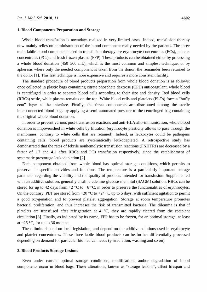
1. Blood Components Preparation and Storage
Whole blood transfusion is nowadays realized in very limited cases. Indeed, transfusion therapy
now mainly relies on administration of the blood component really needed by the patients. The three
main labile blood components used in transfusion therapy are erythrocyte concentrates (ECs), platelet
concentrates (PCs) and fresh frozen plasma (FFP). These products can be obtained either by processing
a whole blood donation (450–500 mL), which is the most common and simplest technique, or by
apheresis where only the needed component is taken from the donor, the remainder been returned to
the donor [1]. This last technique is more expensive and requires a more consistent facility.
The standard procedure of blood products preparation from whole blood donation is as follows:
once collected in plastic bags containing citrate phosphate dextrose (CPD) anticoagulant, whole blood
is centrifuged in order to separate blood cells according to their size and density. Red blood cells
(RBCs) settle, while plasma remains on the top. White blood cells and platelets (PLTs) form a “buffy
coat” layer at the interface. Finally, the three components are distributed among the sterile
inter-connected blood bags by applying a semi-automated pressure to the centrifuged bag containing
the original whole blood donation.
In order to prevent various post-transfusion reactions and anti-HLA allo-immunisation, whole blood
donation is impoverished in white cells by filtration (erythrocyte plasticity allows to pass through the
membranes, contrary to white cells that are retained). Indeed, as leukocytes could be pathogens
containing cells, blood products are systematically leukodepleted. A retrospective study has
demonstrated that the rates of febrile nonhemolytic transfusion reactions (FNHTRs) are decreased by a
factor of 1.7 and 4.1 after RBCs and PCs transfusion respectively, since the establishment of
systematic prestorage leukodepletion [2].
Each component obtained from whole blood has optimal storage conditions, which permits to
preserve its specific activities and functions. The temperature is a particularly important storage
parameter regarding the viability and the quality of products intended for transfusion. Supplemented
with an additive solution, generally a saline-adenine-glucose-mannitol (SAGM) solution, RBCs can be
stored for up to 42 days from +2 °C to +6 °C, in order to preserve the functionalities of erythrocytes.
On the contrary, PLT are stored from +20 °C to +24 °C up to 5 days, with sufficient agitation to permit
a good oxygenation and to prevent platelet aggregation. Storage at room temperature promotes
bacterial proliferation, and thus increases the risk of transmitted bacteria. The dilemma is that if
platelets are transfused after refrigeration at 4 °C, they are rapidly cleared from the recipient
circulation [3]. Finally, as indicated by its name, FFP has to be frozen, for an optimal storage, at least
at −25 °C, for up to 36 months.
These limits depend on local legislation, and depend on the additive solutions used in erythrocyte
and platelet concentrates. These three labile blood products can be further differentially processed
depending on demand for particular biomedical needs (-irradiation, washing and so on).
2. Blood Products Storage Lesions
Even under current optimal storage conditions, modifications and/or degradation of blood
components occur in blood bags. These alterations, known as “storage lesions”, affect lifespan and
Int. J. Mol. Sci. 2010, 11
4603
quality of the stored blood products [4]. Even though it is unknown if storage lesions are consequences
of the natural aging of blood components, these lesions are well described in the literature.
2.1. Erythrocyte Concentrates Storage Lesions
Red blood cells storage lesions can be classified in different categories, depending on their physical
or chemical properties [5,6].
First, some biochemical changes related to the energy metabolism occur. It appears that components
such as ATP, which is necessary for multiple cellular processes, and 2,3-DPG, which plays an
important role in oxygen release, rapidly decrease during the storage. ATP level is considerably
diminished after 5 weeks of storage while the 2,3-DPG is almost null after 2 weeks of storage [7]. The
low concentration of 2,3-DPG increases hemoglobin affinity for oxygen, which cannot be delivered
anymore. However, these levels are rapidly recovered in blood circulation after transfusion of the
erythrocyte concentrate. It is also known that intracellular sodium and potassium levels are altered
through storage. Indeed, Na+/K
+ pumps are inactive at 4 °C, thus allowing high sodium influx and
potassium loss [7].
Then, RBCs storage induces biomechanical changes. The erythrocytes rheological properties, such
as shape, deformability, aggregability, and intracellular viscosity are altered during storage [8]. All
these changes impact the red blood cells ability to pass through microvessels, thus altering their
oxygenation capacities. This is very problematic in blood banking, and neither SAGM nor
phosphate-adenine-glucose-guanosin-saline-mannitol (PAGGSM), the two mostly used additive
solutions, are described to prevent these storage lesions [9].
Finally, modifications also take place at the protein level. Indeed, erythrocytes are subjected to
oxidative lesions, which result in protein oxidative modifications [10,11], hemichrome formation and
Band 3 clustering. Storage-induced protein degradation appears to be greatly reduced when oxygen is
removed and blood is stored under helium [12]. Antonelou et al. have shown that erythrocyte proteins
are less oxidized when RBCs are stored in CPD-SAGM compared to storage in CPD-Adenine [13].
Mechanisms of oxidative damage along the development of storage lesions and investigations of the
effect of blood anaerobic storage conditions have been recently described by Yoshida and
Shevkoplyas [14].
Some aspects of RBC aging occurring in vivo and in vitro during storage are similar. For example,
the increase of intracellular calcium level induces microvesiculation and externalization of
negatively-charged membrane phospholipids (phosphatidylserine) [15]. RBCs clearance from blood
circulation is thought to be immunologically mediated. Several studies have led to the so-called Band 3
clustering model [16,17]. Oxidized hemoglobin compounds aggregate to form hemichromes that
accumulate at the inner side of erythrocytes membrane, covalently bound with cytoskeletal proteins
such as spectrin, thus inducing alterations of RBC deformability. Interaction of hemichromes with
cytoplasmic domains of Band 3 leads to Band 3 clustering. This conformational change of the major
erythrocyte membrane protein is recognized by naturally occurring anti-Band 3 auto-antibodies
(nAbs) [18–20]. Paleari et al. have demonstrated that the number of these RBC-bound IgGs increases
in old population enriched fractions (through Percoll density fractionation), as well as in some selected
clinical cases of patients with altered RBC survival [21]. These observations lead to the conclusion that
Int. J. Mol. Sci. 2010, 11
4604
RBC-bound IgG is a biomarker of aging. Hemicromes, Band 3 dimerization and erythrocyte
recognition by nAbs are thus additional RBCs aging markers that can be investigated.
2.2. Platelet Concentrates Storage Lesions
Platelet Storage Lesions (PSL) consist in morphological changes, platelet activation, platelet
proteolysis and platelet surface receptor expression [22,23]. Changes of platelet membrane
glycoproteins are also reported in numerous papers [24–26].
The normal platelet discoid shape (also referred as resting shape) is found to be lost after 5 to 7 days
of storage at 22 °C. At this storage time, mainly spherical or fragmented platelets remain. Granule
release and platelet activation occur during PLT storage, as indicated by the accumulation of
β-thromboglobulin and platelet factor 4 in the storage medium, and the increase in surface levels of
P-selectin (CD62P), respectively.
In vitro, loss of aggregation functionality is also observed. There is a significantly storage-
dependant decrease of platelet aggregation response to a number of agonists used alone, such as
adenosine diphosphate (ADP), epinephrine, collagen and arachidonic acid [27]. However, these
aggregation agonists seem to have a synergistic action since pairs of them have been shown to restore
platelet aggregation, even after five days of storage [27]. Shapira et al. have also shown that platelet
prothrombinase activity and membrane phosphatidylserine exposure are enhanced during PCs blood
banking storage [28].
As blood components natural aging and storage lesions can directly affect cells, and also have
implications at the protein level, the analysis of biomarkers of such phenomenon must be ruled out by
important pre-analytical considerations.
3. Analysis of Blood Products Aging and Storage Lesions Biomarkers: Pre-Analytical Issues
3.1. Importance of Pre-Analytics in Biomarker Discovery Field
In order to be used in medicine, a biomarker has to be both sensitive and specific. Indeed, a
biomarker of a given physiological state must allow the identification of this state (sensitivity) and
must not be relevant to another physiological state (specificity). However, only few of them meet all
criteria making an efficient biomarker suitable for clinical uses.
In the field of biomarker discovery, an important issue is to control sample preparation steps and
pre-analytical factors. Specificities of a biomarker have to be the same everywhere following the same
protocol. For that reason, the standardization of procedure controlling pre-analytical steps (sampling,
conservation and preparation) has to be elaborated.
As discussed before, the three main labile products are differentially processed and stored in order
to maintain their viability and quality for transfusion therapy. Standards are published by competent
authorities in each country. However, even if the procedure is similar, there are still minor changes
applied worldwide. Indeed, blood labile products have to meet definite standards, whatever the way of
production and the material used. For example, quality of platelet concentrates varies according to the
platelet additive solution used [29].

Int. J. Mol. Sci. 2010, 11
4605
3.2. Proteomics in Blood Transfusion
3.2.1. Proteomic Tools for Biomarker Discovery
Proteomics has been largely used in the field of biomarker discovery [30,31] as well as in
transfusion medicine [32,33] since proteins are the effective form of genes, and are thus more able to
inform about the physiological state. Indeed, contrary to the genome, the proteome composition
fluctuates during time, changes among cell population and is physiological state-dependent.
Clinical proteomics is a major asset for diagnosis, prognosis or evolutionary biomarkers. Varieties
of improved proteomic technologies allow separation, identification and quantification of proteins,
here potential biomarkers. Gel-based technologies (two-dimensional gel electrophoresis, 2DE, and
two-dimensional differential gel electrophoresis, 2D-DIGE), as well as chromatographic techniques
(e.g., reverse phase liquid chromatography, RPLC, and strong cation exchange liquid chromatography,
SCX-LC) have been developed in order to fractionate proteins or peptides from complex samples.
Then, proteins can be analyzed by mass spectrometry (MS) consisting in the ionization of proteins or
peptides in order to identify them according to their mass-to-charge ratio (m/z). The two commonly
used sources of ionization are matrix-assisted laser desorption/ionization (MALDI) and electrospray
ionization (ESI). MALDI source is preferentially combined with time-of-flight (TOF) analyzer,
whereas the ESI source is coupled with quadrupole (Q), ion trap (IT), fourier transform ion cyclotron
resonance (FTICR), Orbitrap or combinations of these analyzers. Peptide mass fingerprinting (PMF) or
de novo sequencing by tandem mass spectrometry (MS/MS or MS²), combined with powerful
bioinformatic tools allow protein identification by database interrogation. Protein quantitation [34,35]
can be achieved thanks to diverse treatments before or after the sampling step [36] (metabolic stable
isotope labeling of amino acids in cell culture, SILAC [37], and chemical addition of isotope-coded
affinity tags, ICAT [38], or isobaric tags for relative and absolute quantitation, iTRAQ [39]), by
spiking samples before MS analysis with known amount of an isotopically labeled analyte (absolute
quantitation of proteins, AQUA [40]) and even by recently developed label-free methods, based on MS
or chromatography data-processing (spectral counting [41] or total ion current, TIC, integration [42]).
Surface enhanced lazer desorption/ionization time-of-flight (SELDI-TOF), also known as
ProteinChip Array technology, is widely used in biomarker discovery [43,44]. A solid-phase
chromatographic surface allows sample decomplexification according to chemical or biochemical
properties. Users can design their own chromatographic surface to target specific protein population.
Bound proteins are then ionized and analyzed as in classical MALDI-TOF MS analysis. However,
SELDI-TOF MS analysis does not lead to protein identification, but only gives a mass list of proteins
showing relative abundance differences between samples. Once targeted, these proteins have to be
further purified then identified by standard mass spectrometry. Caputo et al. have developed such kind
of methods for protein identification on ProteinChip surfaces, i.e., on-chip digestion and MS/MS
sequencing by hybrid quadrupole-TOF [45].
Recent advances in all these proteomic techniques have improved reproducibility, but the problem
of inter-laboratory reproducibility still subsists. To ensure this reproducibility, a control of the initial
state of samples, with standardization of sample collection, storage and preparation conditions, is
required. According to Lehmann et al. [46], the following pre-analytical steps have to be controlled:
Int. J. Mol. Sci. 2010, 11
4606
the sampling procedure and the type of container, the temperature and the time before transport
(if transport needed), the time between sample collection and sample processing, the process itself
(centrifugation, aliquoting, type of secondary container), the storage (temperature and duration) and
the pre-treatment just before proteomic analysis.
The general complexity of a proteome, and particularly the huge dynamic range of protein
abundance composing the blood proteome, makes necessary the fractionation of blood samples before
proteomic analysis, by depletion or fractionation processes. Then, other sample preparation can be
required depending on the type of analysis to be performed, and cannot be standardized to all
biomarker discovery studies.
3.2.2. Involvement of Proteomics in Blood Transfusion
3.2.2.1. Erythrocyte Proteome Investigations
Red blood cell is an abundant and easily obtained biological material having the particularity to be
nucleus- and organelle-free. These characteristics have made of it a model of choice for membrane
studies. Nowadays, thanks to development of proteomic tools and technologies, erythrocyte proteome
begins to be well documented. Several major studies about RBC proteome have been published last
decade, showing a great improvement in terms of number of identified proteins, in cytosolic as well as
membrane extracts.
In 2002, a first study combining one dimensional (sodium dodecyl sulphate polyacrylamide gel
electrophoresis, SDS-PAGE) and 2D electrophoresis, allows identification of 84 unique proteins from
ghost preparation by MALDI-TOF MS analysis. Classic SDS-PAGE reveals 25 proteins of high
hydrophobicity and high molecular weight, undetectable with 2DE [47]. Two years later, the use of
LC-MS has enabled Goodman and coworkers to identify 181 proteins after tryptic digestion of both
cytoplasmic and membrane preparations [48]. Then, Pasini et al. have increased the number of
identified proteins to 566 (314 membrane-associated proteins and 252 cytoplasmic proteins),
combining high-accuracy and high-sensitivity protein identification technologies to analyze trypsin-
digested differentially extracted proteins from cytoplasmic and membrane fractions. They have tested
different extraction methods leading to differences in number of identified proteins. This number also
appeared to vary according to interrogated database [49]. In 2008, the peptide ligand library
technology [50] made possible the exploration of red blood cell hidden proteome by reducing the
erythrocyte dynamic protein concentration range, which permits Roux-Dalvai et al. to identify 1578
soluble proteins by nano-LC-LTQ-Orbitrap MS/MS analysis [51]. A more recent study reveals 222
identified proteins. Even though this number may appear low in comparison with other studies, the
authors have set up an interesting hemoglobin depletion strategy, derived from the existing
HemogloBind™ reagent [52]. In 2010, van Gestel et al. have explored RBC membrane proteome
combining blue native protein separation (BN/SDS-PAGE), CyDyes labeling-based quantitation and
LC-MS/MS identification [53]. This allowed them to identify 524 proteins, of which 155 are
membrane proteins. The CyDyes labeling led to quantitation of a 40% decrease in spectrin levels in
erythrocyte membrane fraction of a patient presenting RBC membrane disorder. This interesting
approach thus seems to be applicable to biomarker discovery. Lately, efforts have been made in the
Int. J. Mol. Sci. 2010, 11
4607
establishment of interactome and network maps, as reported by Goodman et al. [54] and updated by
D’Alessandro et al. [55].
3.2.2.2. Platelet Proteomic Analyses
Platelet quality was usually determined from measurable characteristics such as pH, platelet
morphology and hypotonic shock responses. Protein analysis of platelets during storage was first
achieved in the end of the 1980s by identifying variation in actin [56,57]. However, actin
polymerization is linked to the method used in the preparation of PCs and it has been shown that this
process is partially reversible after 1 day of storage [58], which is highlighting the implication of
pre-analytics in biomarker discovery. Since the introduction of proteomics, new tools have been
brought in order to characterize platelet storage lesions. As explained previously, lesions occur during
storage of PCs and can alter several proteins and mechanisms. The proteome analysis of platelets
during the period of storage may reveal different biomarkers, potentially important for improving
platelet quality [59]. Proteomic analysis of platelets in vivo or during storage was achieved at the
beginning of the last decade [59,60]. Using gel-based proteomics, different groups identified several
hundreds to thousands of PLT proteins [61–63]. Based on peptidic-centric methods, 641 proteins were
reported by Gevaert and coworkers, including hydrophobic membrane proteins [64]. Later, different
studies have focused on changes during storage periods, typically between a 1-day and a 7-day storage.
Thiele et al. have shown by DIGE and MS that 97% of cytosolic proteins do not change over a 9-day
storage period [65]. However, in the 3 remaining percent, they highlighted that, for instance, spetin 2
and gelsolin show changes in 2DE. They claimed that these proteins, affected during apoptosis, may be
suitable markers for platelet alteration during storage. Using a similar approach but focusing on
supernatant, it has been also shown that levels of proteins increase during storage, which may have
some implications for transfusion recipients [66]. More recently, Devine and coworkers have used
complementary proteomic methods (2DE, DIGE, iTRAQ and ICAT) to investigate protein changes
between days 1 and 7 of PLT storage [67]. Hence, 503 proteins changed concentration was reported
thus completing the PLT proteome.
Beyond these analyses, the impact of such potentially biomarkers has to be integrated within
molecular mechanisms leading to platelet storage. As explained by Schubert and Devine, biological
interpretation of proteomic data has to be taken into account [59]. A comprehensive view of different
mechanisms related to biomarkers may be required to understand how to improve platelet quality.
Even though such an approach is far to be completed nowadays, changes in molecular mechanisms
have been described. Schubert et al. have identified 12 proteins connected in one potential signaling
pathway underlying storage lesion development [68]. They have shown that PI3-kinase-dependent
Rap1 activation leads to integrin IIb3 activation and PLT degranulation. In addition, a PI3-kinase
inhibitor incubated with PLT for 7 days plays a role in Rap1 activation and seems to improve PLT
integrity and quality during storage. A global view, including protein-protein interactions [69], is one
of the tools able to provide a wide picture of the storage lesions and mechanisms involved during such
modifications.
Int. J. Mol. Sci. 2010, 11
4608
All these studies have potential influence on the future of platelet quality and safety during storage,
and of course, as it is explained within this review, pre-analytic considerations are inherent to
these studies.
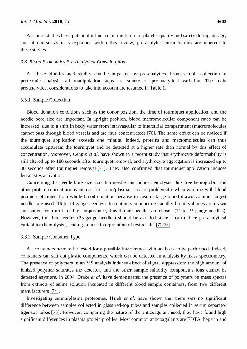
3.3. Blood Proteomics Pre-Analytical Considerations
All these blood-related studies can be impacted by pre-analytics. From sample collection to
proteomic analysis, all manipulation steps are source of pre-analytical variation. The main
pre-analytical considerations to take into account are resumed in Table 1.
3.3.1. Sample Collection
Blood donation conditions such as the donor position, the time of tourniquet application, and the
needle bore size are important. In upright position, blood macromolecular component rates can be
increased, due to a shift in body water from intravascular to interstitial compartment (macromolecules
cannot pass through blood vessels and are thus concentrated) [70]. The same effect can be noticed if
the tourniquet application exceeds one minute. Indeed, proteins and macromolecules can thus
accumulate upstream the tourniquet and be detected at a higher rate than normal by this effect of
concentration. Moreover, Cengiz et al. have shown in a recent study that erythrocyte deformability is
still altered up to 180 seconds after tourniquet removal, and erythrocyte aggregation is increased up to
30 seconds after tourniquet removal [71]. They also confirmed that tourniquet application induces
leukocytes activation.
Concerning the needle bore size, too thin needle can induce hemolysis, thus free hemoglobin and
other protein concentrations increase in serum/plasma. It is not problematic when working with blood
products obtained from whole blood donation because in case of large blood drawn volume, largest
needles are used (16 to 19-gauge needles). In routine venipuncture, smaller blood volumes are drawn
and patient comfort is of high importance, thus thinner needles are chosen (21 to 23-gauge needles).
However, too thin needles (25-gauge needles) should be avoided since it can induce pre-analytical
variability (hemolysis), leading to false interpretation of test results [72,73].
3.3.2. Sample Container Type
All containers have to be tested for a possible interference with analyses to be performed. Indeed,
containers can salt out plastic components, which can be detected in analysis by mass spectrometry.
The presence of polymers in an MS analysis induces effect of signal suppression: the high amount of
ionized polymer saturates the detector, and the other sample minority components ions cannot be
detected anymore. In 2004, Drake et al. have demonstrated the presence of polymers on mass spectra
from extracts of saline solution incubated in different blood sample containers, from two different
manufacturers [74].
Investigating serum/plasma proteomes, Hsieh et al. have shown that there was no significant
difference between samples collected in glass red-top tubes and samples collected in serum separator
tiger-top tubes [75]. However, comparing the nature of the anticoagulant used, they have found high
significant differences in plasma protein profiles. Most common anticoagulants are EDTA, heparin and
Int. J. Mol. Sci. 2010, 11
4609
sodium citrate. Their different action ways lead to these differences in proteome composition.
Moreover, they are also known to interfere with subsequent analysis. EDTA is known to interfere in
enzymatic activity assays, whereas heparin is not advised for some ELISA-based assay kit.
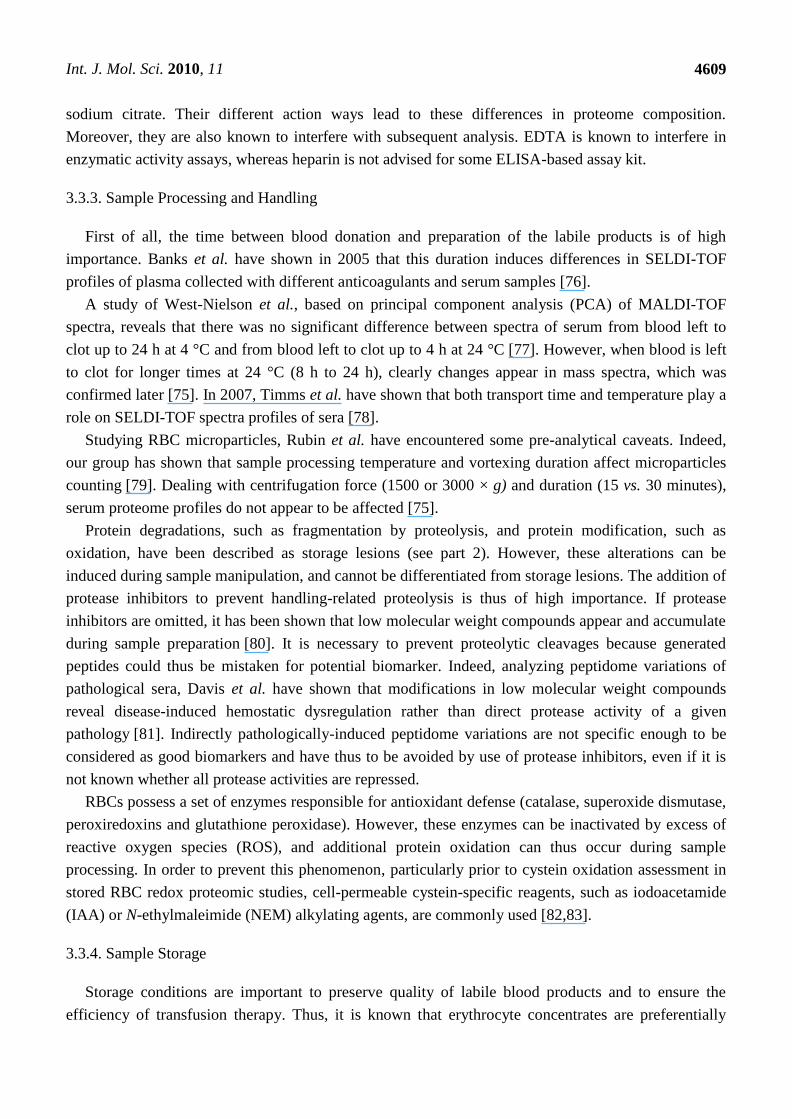
3.3.3. Sample Processing and Handling
First of all, the time between blood donation and preparation of the labile products is of high
importance. Banks et al. have shown in 2005 that this duration induces differences in SELDI-TOF
profiles of plasma collected with different anticoagulants and serum samples [76].
A study of West-Nielson et al., based on principal component analysis (PCA) of MALDI-TOF
spectra, reveals that there was no significant difference between spectra of serum from blood left to
clot up to 24 h at 4 °C and from blood left to clot up to 4 h at 24 °C [77]. However, when blood is left
to clot for longer times at 24 °C (8 h to 24 h), clearly changes appear in mass spectra, which was
confirmed later [75]. In 2007, Timms et al. have shown that both transport time and temperature play a
role on SELDI-TOF spectra profiles of sera [78].
Studying RBC microparticles, Rubin et al. have encountered some pre-analytical caveats. Indeed,
our group has shown that sample processing temperature and vortexing duration affect microparticles
counting [79]. Dealing with centrifugation force (1500 or 3000 × g) and duration (15 vs. 30 minutes),
serum proteome profiles do not appear to be affected [75].
Protein degradations, such as fragmentation by proteolysis, and protein modification, such as
oxidation, have been described as storage lesions (see part 2). However, these alterations can be
induced during sample manipulation, and cannot be differentiated from storage lesions. The addition of
protease inhibitors to prevent handling-related proteolysis is thus of high importance. If protease
inhibitors are omitted, it has been shown that low molecular weight compounds appear and accumulate
during sample preparation [80]. It is necessary to prevent proteolytic cleavages because generated
peptides could thus be mistaken for potential biomarker. Indeed, analyzing peptidome variations of
pathological sera, Davis et al. have shown that modifications in low molecular weight compounds
reveal disease-induced hemostatic dysregulation rather than direct protease activity of a given
pathology [81]. Indirectly pathologically-induced peptidome variations are not specific enough to be
considered as good biomarkers and have thus to be avoided by use of protease inhibitors, even if it is
not known whether all protease activities are repressed.
RBCs possess a set of enzymes responsible for antioxidant defense (catalase, superoxide dismutase,
peroxiredoxins and glutathione peroxidase). However, these enzymes can be inactivated by excess of
reactive oxygen species (ROS), and additional protein oxidation can thus occur during sample
processing. In order to prevent this phenomenon, particularly prior to cystein oxidation assessment in
stored RBC redox proteomic studies, cell-permeable cystein-specific reagents, such as iodoacetamide
(IAA) or N-ethylmaleimide (NEM) alkylating agents, are commonly used [82,83].
3.3.4. Sample Storage
Storage conditions are important to preserve quality of labile blood products and to ensure the
efficiency of transfusion therapy. Thus, it is known that erythrocyte concentrates are preferentially
Int. J. Mol. Sci. 2010, 11
4610
stored at 4 °C whereas platelet concentrates are stored at 22 °C to prevent cold activation of platelets.
Finally, fresh frozen plasma is stored at −25 °C at least.
Thus, whole blood storage cannot ensure optimal preservation of each blood components. In labile
blood products aging field, samples are ECs, PCs or FFPs, and are stored according to blood banking
legislation. In pathological-case biomarker research, samples are either serum or plasma, and are not
subject to any storage legislation. Several studies aimed at investigating the reproducibility of analysis
after storage at −20/−30 °C or −80 °C, and after freeze-thaw cycles [75,84,85]. It appears that storage
at −20 °C instead of the recommended −80 °C storage does not induce loss of information. Samples
stored at −20/−30 °C can thus be used for biomarker discovery [86].
Some proteins have been shown to be highly temperature-sensitive: cryoproteins. Described for the
first time in the 1930’s by Wintrobe [87] as myeloma’s patient sera precipitating at cold temperature,
cryoproteins are serum or plasma proteins having the property to precipitate below the physiological
temperature. Most of the time composed of immunoglobulins (mainly IgM) these precipitates are
called cryoglobulins (CG), and are classified under three categories according to the universally
adopted classification of Brouet [88]. Other proteins have this particularity to precipitate at low
temperature, as cryofibrinogen (CF) (for instance, see the study on clinical cases of patients with either
CF alone or combined CF plus CG [89]) and cold agglutinins. Proteomic techniques are well adapted
to determine the protein composition of cryoprecipitates [90]. Cryoproteins are potential biomarkers,
and pre-analytical precautions have to be taken for their investigation. Indeed, blood must be sampled
in anticoagulant-free, 37 °C pre-heated container, handled and centrifuged at 37 °C, in order to prevent
cryoprotein elimination by precipitation before analysis.
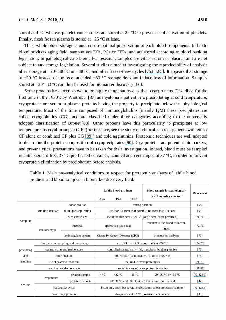
Table 1. Main pre-analytical conditions to respect for proteomic analyses of labile blood
products and blood samples in biomarker discovery field.
Labile blood products Blood sample for pathological-
case biomarker research References
ECs PCs FFP
Sampling
sample obtention
donor position resting position [68]
tourniquet application less than 30 seconds if possible, no more than 1 minute [69]
needle bore size avoid too thin needle (21–23 gauge needles are preferred) [70,71]
container type
material approved plastic bags vacuette®-like blood collection
tubes [72,73]
anticoagulant content Citrate Phosphate Dextrose (CPD) depends on analyses [73]
processing
and
handling
time between sampling and processing up to 24 h at +4 °C or up to 4 h at +24 °C [74,75]
transport time and temperature controlled transport at +4 °C, must be as brief as possible [76]
centrifugation prefer centrifugation at +4 °C, up to 3000 × g [73]
use of protease inhibitors required to avoid proteolysis [78,79]
use of antioxidant reagents needed in case of redox proteomic studies [80,81]
storage
temperature original sample +4 °C +22 °C −25 °C −20/−30 °C or −80 °C [73,82,83]
proteinic extracts −20/−30 °C and −80 °C stored extracts are both suitable [84]
freeze/thaw cycles better only once, but several cycles do not affect proteomic patterns [73,82,83]
case of cryoproteins always work at 37 °C (pre-heated containers) [87]

Int. J. Mol. Sci. 2010, 11
4611
4. Conclusions
Pre-analytics is of very high importance for all kind of test, assay and discovery field, and more
importantly in biomarker discovery field, due to the direct clinical implications. Uncontrolled
pre-analytical parameters may lead to false interpretation of results.
In clinical proteomics, reproducibility is indispensable and is straightly correlated with
pre-analytical considerations. Standardization of sample harvesting, process, and storage has to be set
up in order to minimize pre-analytical variations. Biomarker discovery is such a complicated field that
there is no time to waste in wondering if an observed protein variation is due to different physiological
states or simply to differences in sample handling or storage. Many small-scale standardized protocols
exist in laboratories and lots of pre-analytical parameters have been investigated so far. Time is now
about setting up worldwide-scale standardization of sample handling, process and storage.
The human proteome organization (HUPO) has set up the plasma proteome project (HPPP), in
order to perform a comprehensive analysis of plasma and serum protein constituents in people. A
specimen collection and handling committee (SCHC) was created to evaluate pre-analytical variables
that can potentially impact experiment results. In this purpose, Rai et al. have conducted in 2005 a
proteomic study, comparing serum and plasma analyses, evaluating storage and handling conditions as
well as the use of protease inhibitors [91]. They were thus able to present general recommendations
helping researchers to set up more robust plasma proteome studies. This is an important step towards
the establishment of standardized protocols for proteome studies, which is primordial in the biomarker
discovery field.
This kind of initiative is important for the transfusion medicine community and for the whole
clinical medicine community as well. Reviews about importance of the pre-analytics in many fields are
regularly published nowadays, which means that procedures standardization is a real
contemporary thought.
Acknowledgments
We would like to thank the Service de Transfusion Sanguine de la Croix Rouge Suisse for the grant
supporting this work.
References
1. Council of Europe, Guide to the Preparation, Use and Quality Assurance of Blood Components.
Recommendation n° R (95) 15 on the Preparation, Use and Quality Assurance of Blood
Components, 14th ed.; Council of Europe Press: Strasbourg, France, 2008.
2. Yazer, M.H.; Podlosky, L.; Clarke, G.; Nahirniak, S.M. The effect of prestorage WBC reduction
on the rates of febrile nonhemolytic transfusion reactions to platelet concentrates and RBC.
Transfusion 2004, 44, 10–15.
3. Snyder, E.L.; Rinder, H.M. Platelet storage-time to come in from the cold? N. Engl. J. Med. 2003,
348, 2032–2033.
4. Zubair, A.C. Clinical impact of blood storage lesions. Am. J. Hematol. 2010, 85, 117–122.
Int. J. Mol. Sci. 2010, 11
4612
5. van de Watering, L.M.G.; Brand, A. Effects of storage of red cells. Transfus. Med. Hemother.
2008, 35, 359–367.
6. Kor, D.J.; van Buskirk, C.M.; Gajic, O. Red blood cell storage lesion. Bosn. J. Basic Med. Sci.
2009, 9, S21–S27.
7. Bennett-Guerrero, E.; Veldman, T.H.; Doctor, A.; Telen, M.J.; Ortel, T.L.; Reid, T.S.; Mulherin,
M.A.; Zhu, H.M.; Buck, R.D.; Califf, R.M.; McMahon, T.J. Evolution of adverse changes in
stored RBCs. Proc. Natl. Acad. Sci. USA 2007, 104, 17063–17068.
8. Berezina, T.L.; Zaets, S.B.; Morgan, C.; Spillert, C.R.; Kamiyama, M.; Spolarics, Z.; Deitch,
E.A.; Machiedo, G.W. Influence of storage on red blood cell rheological properties. J. Surg. Res.
2002, 102, 6–12.
9. Zehnder, L.; Schulzki, T.; Goede, J.S.; Hayes, J.; Reinhart, W.H. Erythrocyte storage in
hypertonic (SAGM) or isotonic (PAGGSM) conservation medium: Influence on cell properties.
Vox Sang. 2008, 95, 280–287.
10. Kriebardis, A.G.; Antonelou, M.H.; Stamoulis, K.E.; Economou-Petersen, E.; Margaritis, L.H.;
Papassideri, I.S. Progressive oxidation of cytoskeletal proteins and accumulation of denatured
hemoglobin in stored red cells. J. Cell. Mol. Med. 2007, 11, 148–155.
11. Kanias, T.; Acker, J.P. Biopreservation of red blood cells - the struggle with hemoglobin
oxidation. FEBS J. 2010, 277, 343–356.
12. D'Amici, G.M.; Rinalducci, S.; Zolla, L. Proteomic analysis of RBC membrane protein
degradation during blood storage. J. Proteome Res. 2007, 6, 3242–3255.
13. Antonelou, M.H.; Kriebardis, A.G.; Stamoulis, K.E.; Economou-Petersen, E.; Margaritis, L.H.;
Papassideri, I.S. Red blood cell aging markers during storage in citrate-phosphate-dextrose-saline-
adenine-glucose-mannitol. Transfusion 2010, 50, 376–389.
14. Yoshida, T.; Shevkoplyas, S.S. Anaerobic storage of red blood cells. Blood Transfus. 2010, 8,
220–236.
15. Tissot, J.-D.; Rubin, O.; Canellini, G. Analysis and clinical relevance of microparticles from red
blood cells. Curr. Opin. Hematol. 2010, 17, 571–577.
16. Kay, M.M.; Wyant, T.; Goodman, J. Autoantibodies to band 3 during aging and disease and aging
interventions. Ann. N. Y. Acad. Sci. 1994, 719, 419–447.
17. Hornig, R.; Lutz, H.U. Band 3 protein clustering on human erythrocytes promotes binding of
naturally occurring anti-band 3 and anti-spectrin antibodies. Exp. Gerontol. 2000, 35, 1025–1044.
18. Kay, M.M.; Goodman, S.R.; Sorensen, K.; Whitfield, C.F.; Wong, P.; Zaki, L.; Rudloff, V.
Senescent cell antigen is immunologically related to band 3. Proc. Natl. Acad. Sci. USA 1983, 80,
1631–1635.
19. Kay, M.M. Localization of senescent cell antigen on band 3. Proc. Natl. Acad. Sci. USA 1984, 81,
5753–5757.
20. Kay, M.M.; Flowers, N.; Goodman, J.; Bosman, G. Alteration in membrane protein band 3
associated with accelerated erythrocyte aging. Proc. Natl. Acad. Sci. USA 1989, 86, 5834–5838.
21. Paleari, R.; Ceriotti, F.; Azzario, F.; Maccioni, L.; Galanello, R.; Mosca, A. Experiences in the
measurement of RBC-bound IgG as markers of cell age. Bioelectrochemistry 2004, 62, 175–179.
22. Seghatchian, J.; Krailadsiri, P. The platelet storage lesion. Transf. Med. Rev. 1997, 11, 130–144.
23. Shrivastava, M. The platelet storage lesion. Transfus. Apher. Sci. 2009, 41, 105–113.
Int. J. Mol. Sci. 2010, 11
4613
24. George, J.N.; Pickett, E.B.; Heinz, R. Platelet membrane glycoprotein changes during the
preparation and storage of platelet concentrates. Transfusion 1988, 28, 123–126.
25. Bessos, H.; Seghatchian, M.J.; Cutts, M.; Murphy, W.G. Glycoprotein-IB and glycoprotein-
IIB/IIIA in the quality assessment of platelet concentrates during storage. Blood Coagul.
Fibrinolysis 1992, 3, 633–636.
26. Jaremo, P.; Rubachdahlberg, E.; Solum, N.O. Correlation of light transmission changes to
changes of platelet glycoprotein-IB during storage of platelet concentrates. Thromb. Res. 1993,
69, 467–477.
27. Diminno, G.; Silver, M.J.; Murphy, S. Stored human-platelets retain full aggregation potential in
response to pairs of aggregating agents. Blood 1982, 59, 563–568.
28. Shapira, S.; Friedman, Z.; Shapiro, H.; Presseizen, K.; Radnay, J.; Ellis, M.H. The effect of
storage on the expression of platelet membrane phosphatidylserine and the subsequent impact on
the coagulant function of stored platelets. Transfusion 2000, 40, 1257–1263.
29. van der Meer, P.F.; Kerkhoffs, J.L.; Curvers, J.; Scharenberg, J.; de Korte, D.; Brand, A.; de
Wildt-Eggen, J. In vitro comparison of platelet storage in plasma and in four platelet additive
solutions, and the effect of pathogen reduction: A proposal for an in vitro rating system. Vox
Sang. 2010, 98, 517–524.
30. Hale, J.E.; Gelfanova, V.; Ludwig, J.R.; Knierman, M.D. Application of proteomics for discovery
of protein biomarkers. Brief. Funct. Genomic Proteomic 2003, 2, 185–193.
31. Aebersold, R.; Anderson, L.; Caprioli, R.; Druker, B.; Hartwell, L.; Smith, R. Perspective: A
program to improve protein biomarker discovery for cancer. J. Proteome Res. 2005, 4,
1104–1109.
32. Thadikkaran, L.; Siegenthaler, M.A.; Crettaz, D.; Queloz, P.A.; Schneider, P.; Tissot, J.D. Recent
advances in blood-related proteomics. Proteomics 2005, 5, 3019–3034.
33. Queloz, P.A.; Thadikkaran, L.; Crettaz, D.; Rossier, J.S.; Barelli, S.; Tissot, J.D. Proteomics and
transfusion medicine: Future perspectives. Proteomics 2006, 6, 5605–5614.
34. Simpson, K.L.; Whetton, A.D.; Dive, C. Quantitative mass spectrometry-based techniques for
clinical use: Biomarker identification and quantification. J. Chromatogr. B 2009, 877, 1240–1249.
35. Anderson, N.L.; Anderson, N.G.; Pearson, T.W.; Borchers, C.H.; Paulovich, A.G.; Patterson,
S.D.; Gillette, M.; Aebersold, R.; Carr, S.A. A human proteome detection and quantitation
project. Mol. Cell. Proteomics 2009, 8, 883–886.
36. Kline, K.G.; Sussman, M.R. Protein quantitation using isotope-assisted mass spectrometry. Ann.
Rev. Biophys. 2010, 39, 291–308.
37. Ong, S.E.; Blagoev, B.; Kratchmarova, I.; Kristensen, D.B.; Steen, H.; Pandey, A.; Mann, M.
Stable isotope labeling by amino acids in cell culture, SILAC, as a simple and accurate approach
to expression proteomics. Mol. Cell. Proteomics 2002, 1, 376–386.
38. Gygi, S.P.; Rist, B.; Gerber, S.A.; Turecek, F.; Gelb, M.H.; Aebersold, R. Quantitative analysis of
complex protein mixtures using isotope-coded affinity tags. Nat. Biotechnol. 1999, 17, 994–999.
39. Ross, P.L.; Huang, Y.N.; Marchese, J.N.; Williamson, B.; Parker, K.; Hattan, S.; Khainovski, N.;
Pillai, S.; Dey, S.; Daniels, S.; Purkayastha, S.; Juhasz, P.; Martin, S.; Bartlet-Jones, M.; He, F.;
Jacobson, A.; Pappin, D.J. Multiplexed protein quantitation in Saccharomyces cerevisiae using
amine-reactive isobaric tagging reagents. Mol. Cell. Proteomics 2004, 3, 1154–1169.
Int. J. Mol. Sci. 2010, 11
4614
40. Gerber, S.A.; Rush, J.; Stemman, O.; Kirschner, M.W.; Gygi, S.P. Absolute quantification of
proteins and phosphoproteins from cell lysates by tandem MS. Proc. Natl. Acad. Sci. USA 2003,
100, 6940–6945.
41. Liu, H.; Sadygov, R.G.; Yates, J.R., III. A model for random sampling and estimation of relative
protein abundance in shotgun proteomics. Anal. Chem. 2004, 76, 4193–4201.
42. Asara, J.M.; Christofk, H.R.; Freimark, L.M.; Cantley, L.C. A label-free quantification method by
MS/MS TIC compared to SILAC and spectral counting in a proteomics screen. Proteomics 2008,
8, 994–999.
43. Issaq, H.J.; Veenstra, T.D.; Conrads, T.P.; Felschow, D. The SELDI-TOF MS approach to
proteomics: Protein profiling and biomarker identification. Biochem. Biophys. Res. Commun.
2002, 292, 587–592.
44. De Bock, M.; de Seny, D.; Meuwis, M.A.; Chapelle, J.P.; Louis, E.; Malaise, M.; Merville, M.P.;
Fillet, M. Challenges for biomarker discovery in body fluids using SELDI-TOF-MS. J. Biomed.
Biotechnol. 2010, 2010, 906082.
45. Caputo, E.; Moharram, R.; Martin, B.M. Methods for on-chip protein analysis. Anal. Biochem.
2003, 321, 116–124.
46. Lehmann, S.; Roche, S.; Allory, Y.; Barthelaix, A.; Beaudeux, J.-L.; Berger, F.; Betsou, F.; Borg,
J.; Dupuy, A.; Garin, J.; Quillard, M.; Lizard, G.; Peoc'h, K.; Riviere, M.; Ducoroy, P.
Recommandations préanalytiques pour les analyses de protéomique clinique des fluides
biologiques. Ann. Biol. Clin. 2009, 67, 629–639.
47. Low, T.Y.; Seow, T.K.; Chung, M.C.M. Separation of human erythrocyte membrane associated
proteins with one-dimensional and two-dimensional gel electrophoresis followed by identification
with matrix-assisted laser desorption/ionization-time of flight mass spectrometry. Proteomics
2002, 2, 1229–1239.
48. Kakhniashvili, D.G.; Bulla, L.A.; Goodman, S.R. The human erythrocyte proteome - Analysis by
ion trap mass spectrometry. Mol. Cell. Proteomics 2004, 3, 501–509.
49. Pasini, E.M.; Kirkegaard, M.; Mortensen, P.; Lutz, H.U.; Thomas, A.W.; Mann, M. In-depth
analysis of the membrane and cytosolic proteome of red blood cells. Blood 2006, 108, 791–801.
50. Boschetti, E.; Righetti, P.G. The art of observing rare protein species in proteomes with peptide
ligand libraries. Proteomics 2009, 9, 1492–1510.
51. Roux-Dalvai, F.; de Peredo, A.G.; Simo, C.; Guerrier, L.; Bouyssiee, D.; Zanella, A.; Citterio, A.;
Burlet-Schiltz, O.; Boschetti, E.; Righetti, P.G.; Monsarrat, B. Extensive analysis of the
cytoplasmic proteome of human erythrocytes using the peptide ligand library technology and
advanced mass spectrometry. Mol. Cell. Proteomics 2008, 7, 2254–2269.
52. Alvarez-Llamas, G.; de la Cuesta, F.; Barderas, M.G.; Darde, V.M.; Zubiri, I.; Caramelo, C.;
Vivanco, F. A novel methodology for the analysis of membrane and cytosolic sub-proteomes of
erythrocytes by 2-DE. Electrophoresis 2009, 30, 4095–4108.
53. van Gestel, R.A.; van Solinge, W.W.; van der Toorn, H.W.; Rijksen, G.; Heck, A.J.; van Wijk, R.;
Slijper, M. Quantitative erythrocyte membrane proteome analysis with Blue-native/SDS PAGE. J.
Proteomics 2010, 73, 456–465.
54. Goodman, S.R.; Kurdia, A.; Ammann, L.; Kakhniashvili, D.; Daescu, O. The human red blood
cell proteome and interactome. Exp. Biol. Med. (Maywood) 2007, 232, 1391–1408.
Int. J. Mol. Sci. 2010, 11
4615
55. D'Alessandro, A.; Righetti, P.G.; Zoha, L. The red blood cell proteome and interactome: An
update. J. Proteome Res. 2010, 9, 144–163.
56. Snyder, E.L.; Dunn, B.E.; Giometti, C.S.; Napychank, P.A.; Tandon, N.N.; Ferri, P.M.; Hofmann,
J.P. Protein-changes occurring during storage of platelet concentrates - a two-dimensional
gel-electrophoretic analysis. Transfusion 1987, 27, 335–341.
57. Snyder, E.L.; Horne, W.C.; Napychank, P.; Heinemann, F.S.; Dunn, B. Calcium-dependent
proteolysis of actin during storage of platelet concentrates. Blood 1989, 73, 1380–1385.
58. Estebanell, E.; Diaz-Ricart, M.; Lozano, M.; Mazzara, R.; Escolar, G.; Ordinas, A. Cytoskeletal
reorganization after preparation of platelet concentrates, using the buffy coat method, and during
their storage. Haematologica 1998, 83, 112–117.
59. Schubert, P.; Devine, D.V. Proteomics meets blood banking: Identification of protein targets for
the improvement of platelet quality. J. Proteomics 2010, 73, 436–444.
60. Egidi, M.G.; D'Alessandro, A.; Mandarello, G.; Zolla, L. Troubelshooting in platelet storage
temperature and new perspectives through peoteomics. Blood Transfus. 2010, 8, s73–s81.
61. Marcus, K.; Immler, D.; Sternberger, J.; Meyer, H.E. Identification of platelet proteins separated
by two-dimensional gel electrophoresis and analyzed by matrix assisted laser
desorption/ioniztion-time of flight-mass spectrometry and detection of tyrosine-phosphorylated
proteins. Electrophoresis 2000, 21, 2622–2636.
62. O'Neill, E.E.; Brock, C.J.; von Kriegsheim, A.F.; Pearce, A.C.; Dwek, R.A.; Watson, S.P.;
Hebestreit, H.F. Towards complete analysis of the platelet proteome. Proteomics 2002, 2,
288–305.
63. Garcia, A.; Prabhakar, S.; Brock, C.J.; Pearce, A.C.; Dwek, R.A.; Watson, S.P.; Hebestreit, H.F.;
Zitzmann, N. Extensive analysis of the human platelet proteome by two-dimensional gel
electrophoresis and mass spectrometry. Proteomics 2004, 4, 656–668.
64. Martens, L.; van Damme, P.; van Damme, J.; Staes, A.; Timmerman, E.; Ghesquiere, B.; Thomas,
G.R.; Vandekerckhove, J.; Gevaert, K. The human platelet proteome mapped by peptide-centric
proteomics: A functional protein profile. Proteomics 2005, 5, 3193–3204.
65. Thiele, T.; Steil, L.; Gebhard, S.; Scharf, C.; Hammer, E.; Brigulla, M.; Lubenow, N.; Clemetson,
K.J.; Volker, U.; Greinacher, A. Profiling of alterations in platelet proteins during storage of
platelet concentrates. Transfusion 2007, 47, 1221–1233.
66. Glenister, K.M.; Payne, K.A.; Sparrow, R.L. Proteomic analysis of supernatant from pooled
buffy-coat platelet concentrates throughout 7-day storage. Transfusion 2008, 48, 99–107.
67. Thon, J.N.; Schubert, P.; Duguay, M.; Serrano, K.; Lin, S.J.; Kast, J.; Devine, D.V.
Comprehensive proteomic analysis of protein changes during platelet storage requires
complementary proteomic approaches. Transfusion 2008, 48, 425–435.
68. Schubert, P.; Thon, J.N.; Walsh, G.M.; Chen, C.H.I.; Moore, E.D.; Devine, D.V.; Kast, J. A
signaling pathway contributing to platelet storage lesion development: Targeting PI3-kinase-
dependent Rap1 activation slows storage-induced platelet deterioration. Transfusion 2009, 49,
1944–1955.
69. Qureshi, A.H.; Chaoji, V.; Maiguel, D.; Faridi, M.H.; Barth, C.J.; Salem, S.M.; Singhal, M.;
Stoub, D.; Krastins, B.; Ogihara, M.; Zaki, M.J.; Gupta, V. Proteomic and phospho-proteomic
Int. J. Mol. Sci. 2010, 11
4616
profile of human platelets in basal, resting state: Insights into integrin signaling. PLoS One 2009,
4, e7627.
70. Maw, G.J.; Mackenzie, I.L.; Taylor, N.A. Redistribution of body fluids during postural
manipulations. Acta Physiol. Scand 1995, 155, 157–163.
71. Cengiz, M.; Ulker, P.; Meiselman, H.J.; Baskurt, O.K. Influence of tourniquet application on
venous blood sampling for serum chemistry, hematological parameters, leukocyte activation and
erythrocyte mechanical properties. Clin. Chem. Lab. Med. 2009, 47, 769–776.
72. Ernst, D.J.; Ernst, C. Phlebotomy tools of the trade. Home Healthc. Nurse 2002, 20, 151–153.
73. Lippi, G.; Salvagno, G.L.; Montagnana, M.; Brocco, G.; Guidi, G.C. Influence of the needle bore
size used for collecting venous blood samples on routine clinical chemistry testing. Clin. Chem.
Lab. Med. 2006, 44, 1009–1014.
74. Drake, S.K.; Bowen, R.A.R.; Remaley, A.T.; Hortin, G.L. Potential interferences from blood
collection tubes in mass spectrometric analyses of serum polypeptides. Clin. Chem. 2004, 50,
2398–2401.
75. Hsieh, S.Y.; Chen, R.K.; Pan, Y.H.; Lee, H.L. Systematical evaluation of the effects of sample
collection procedures on low-molecular-weight serum/plasma proteome profiling. Proteomics
2006, 6, 3189–3198.
76. Banks, R.E.; Stanley, A.J.; Cairns, D.A.; Barrett, J.H.; Clarke, P.; Thompson, D.; Selby, P.J.
Influences of blood sample processing on low-molecular-weight proteome identified by surface-
enhanced laser desorption/ionization mass spectrometry. Clin. Chem. 2005, 51, 1637–1649.
77. West-Nielsen, M.; Hogdall, E.V.; Marchiori, E.; Hogdall, C.K.; Schou, C.; Heegaard, N.H.H.
Sample handling for mass spectrometric proteomic investigations of human sera. Anal. Chem.
2005, 77, 5114–5123.
78. Timms, J.F.; Arslan-Low, E.; Gentry-Maharaj, A.; Luo, Z.; T'Jampens, D.; Podust, V.N.; Ford, J.;
Fung, E.T.; Gammerman, A.; Jacobs, I.; Menon, U. Preanalytic influence of sample handling on
SELDI-TOF serum protein profiles. Clin. Chem. 2007, 53, 645–656.
79. Rubin, O.; Crettaz, D.; Tissot, J.D.; Lion, N. Pre-analytical and methodological challenges in red
blood cell microparticle proteomics. Talanta 2010, 82, 1–8.
80. Olivieri, E.; Herbert, B.; Righetti, P.G. The effect of protease inhibitors on the two-dimensional
electrophoresis pattern of red blood cell membranes. Electrophoresis 2001, 22, 560–565.
81. Davis, M.T.; Patterson, S.D. Does the serum peptidome reveal hemostatic dysregulation? In
Systems Biology: Applications and Perspectives; Bringmann, P., Butcher, E.C., Parry, G., Weiss,
B., Eds.; Springer: New York, NY, USA, 2007; Volume 61, pp. 23–44.
82. Cox, A.G.; Peskin, A.V.; Paton, L.N.; Winterbourn, C.C.; Hampton, M.B. Redox potential and
peroxide reactivity of human peroxiredoxin 3. Biochemistry 2009, 48, 6495–6501.
83. Le Moan, N.; Tacnet, F.; Toledano, M.B. Protein-thiol oxidation, from single proteins to
proteome-wide analyses. Methods Mol. Biol. 2009, 476, 175–192.
84. Traum, A.Z.; Wells, M.P.; Aivado, M.; Libermann, T.A.; Ramoni, M.F.; Schachter, A.D.
SELDI-TOF MS of quadruplicate urine and serum samples to evaluate changes related to storage
conditions. Proteomics 2006, 6, 1676–1680.
Int. J. Mol. Sci. 2010, 11
4617
85. Ulmert, D.; Becker, C.; Nilsson, J.A.; Piironen, T.; Bjork, T.; Hugosson, J.; Berglund, G.; Lilja,
H. Reproducibility and accuracy of measurements of free and total prostate-specific antigen in
serum vs plasma after long-term storage at-20 degrees C. Clin. Chem. 2006, 52, 235–239.
86. Insenser, M.; Martínez-García, M.Á.; Nieto, R.M.; San-Millán, J.L.; Escobar-Morreale, H.F.
Impact of the storage temperature on human plasma proteomic analysis: Implications for the use
of human plasma collections in research. Proteomics Clin. Appl. 2010, 4, 739–744.
87. Wintrobe, M.M.; Buell, M.V. Hyperproteinemia associated with multiple myeloma - With report
of a case in which an extraordinary hyperproteinemia was associated with thrombosis of the
retinal veins and symptoms suggesting Raynaud's disease. Bull. Johns Hopkins Hosp. 1933, 52,
156–165.
88. Brouet, J.-C.; Clauvel, J.-P.; Danon, F.; Klein, M.; Seligmann, M. Biologic and clinical
significance of cryoglobulins: A report of 86 cases. Am. J. Med. 1974, 57, 775–788.
89. Blain, H.; Cacoub, P.; Musset, L.; Costedoat-Chalumeau, N.; Silberstein, C.; Chosidow, O.;
Godeau, P.; Frances, C.; Piette, J.C. Cryofibrinogenaemia: A study of 49 patients. Clin. Exp.
Immunol. 2000, 120, 253–260.
90. Robert, D.; Barelli, S.; Crettaz, D.; Bart, P.A.; Schifferli, J.A.; Betticher, D.; Tissot, J.D. Clinical
proteomics: Study of a cryogel. Proteomics 2006, 6, 3958–3960.
91. Rai, A.J.; Gelfand, C.A.; Haywood, B.C.; Warunek, D.J.; Yi, J.; Schuchard, M.D.; Mehigh, R.J.;
Cockrill, S.L.; Scott, G.B.; Tammen, H.; Schulz-Knappe, P.; Speicher, D.W.; Vitzthum, F.; Haab,
B.B.; Siest, G.; Chan, D.W. HUPO Plasma Proteome Project specimen collection and handling:
towards the standardization of parameters for plasma proteome samples. Proteomics 2005, 5,
3262–3277.
© 2010 by the authors; licensee MDPI, Basel, Switzerland. This article is an open access article
distributed under the terms and conditions of the Creative Commons Attribution license
(http://creativecommons.org/licenses/by/3.0/).