Belgian Journal of Paediatrics - BVK-SBP
84
Articles Hirschsprung Disease and Congenital Anomalies of the Kidney and Urinary Tract (CAKUT): a genetic disorder or just a coincidence? Fate of a sickle cell child abandoned at birth Management of neonatal hypertension Epidemiology of invasive meningococcal disease in Belgium and implications for use of meningococcal vaccines in children and adolescents The analgesic effect of Virtual Reality in paediatric procedural pain: a systematic review How to position impedance-pH probes in pediatric patients: a pilot trial Case Report An interesting case “out of paper” with celiac disease leading to xylophagia: case report and review of literature Case report: Plastic bronchitis in a previously healthy child Lenticulostriate infarction presenting as a central facial nerve palsy, caused by post-varicella arteriopathy in a 5-year-old girl Case report of a boy with autism who refuses to eat The outcome of posterior reversible encephalopathy syndrome (PRES) in children: a systematic review and case-report of a 16-year old girl with systemic lupus erythematosus and PRES Traumatic brain injury or else? A iatrogenic cause of encephalopathy in a 9-year old boy – a rare side effect of a commonly used drug Made in Belgium Beta-lactam hypersensitivity: Epidemiology and optimized diagnosis Optimisation of long-term outcomes in paediatric inflammatory bowel disease patients: role of therapeutic drug monitoring and endoscopic remission Paediatric Cochrane Corner Use of reflective materials during phototherapy in neonates with unconjugated hyperbilirubinaemia: worth reflecting upon BELGISCHE VERENIGING VOOR KINDERGENEESKUNDE SOCIÉTÉ BELGE DE PÉDIATRIE 2020 - Volume 22 - number 3 - September V.U./E.R. S. Cadranel (ULB), M. Raes (KUL) UZ Leuven, Herestraat 49, 3000 Leuven E-mail: [email protected] QUARTERLY ISSN 2466-8907 (printed version) ISSN 2566-1558 (digital version) Belgian Journal of Paediatrics Publication of the Belgian Society of Paediatrics Belgische Vereniging voor Kindergeneeskunde Société Belge de Pédiatrie
-
Upload
khangminh22 -
Category
Documents
-
view
0 -
download
0
Transcript of Belgian Journal of Paediatrics - BVK-SBP

Articles Hirschsprung Disease and Congenital Anomalies of the Kidney and Urinary Tract (CAKUT): a genetic disorder or just a coincidence?
Fate of a sickle cell child abandoned at birth
Management of neonatal hypertension
Epidemiology of invasive meningococcal disease in Belgium and implications for use of meningococcal vaccines in children and adolescents
The analgesic effect of Virtual Reality in paediatric procedural pain: a systematic review
How to position impedance-pH probes in pediatric patients:a pilot trial
Case Report An interesting case “out of paper” with celiac disease
leading to xylophagia: case report and review of literature
Case report: Plastic bronchitis in a previously healthy child
Lenticulostriate infarction presenting as a central facial nerve palsy, caused by post-varicella arteriopathy in a 5-year-old girl
Case report of a boy with autism who refuses to eat
The outcome of posterior reversible encephalopathy syndrome (PRES) in children: a systematic review and case-report of a 16-year old girl with systemic lupus erythematosus and PRES
Traumatic brain injury or else?
A iatrogenic cause of encephalopathy in a 9-year old boy –a rare side effect of a commonly used drug
Made in Belgium Beta-lactam hypersensitivity: Epidemiology and optimized
diagnosis
Optimisation of long-term outcomes in paediatric infl ammatory bowel disease patients: role of therapeutic drug monitoring and endoscopic remission
Paediatric Cochrane Corner Use of refl ective materials during phototherapy in neonates
with unconjugated hyperbilirubinaemia: worth refl ecting upon
BELGISCHE VERENIGINGVOOR KINDERGENEESKUNDESOCIÉTÉ BELGE DE PÉDIATRIE
2020 - Volume 22 - number 3 - September
V.U./E.R. S. Cadranel (ULB), M. Raes (KUL)
UZ Leuven, Herestraat 49, 3000 Leuven
E-mail: [email protected]
QUARTERLYISSN 2466-8907 (printed version)ISSN 2566-1558 (digital version)
Belgian Journalof Paediatrics
BBBBBBBBBBBBBBBBBBJJJJJJJJJBBBJBBBJBBBJBBBJJJJJJJJJBBBJBBBJBBBJBBBBBBJBBBJBBBJBBBBelgian JournalPPPPPPPPPPPPJJJ
Publication of the Belgian Society of Paediatrics
Belgische Vereniging voor KindergeneeskundeSociété Belge de Pédiatrie

CHARTE SÉCURITÉ INGRÉDIENTS
DÈS LA NAISSANCE TESTÉ DERMATOLOGIQUEMENT
Je veuxmieux dormir
INNOVATION STELATOPIA®
LE 1ER SOUS-PYJAMAPOUR PEAU À
TENDANCE ATOPIQUEAnti-grattage1
Sommeil amélioré dès 7 jours2
1. Étude clinique, évaluation examinateurs auprès de 66 sujets bébés-enfants à peau à tendance atopique pendant 28 jours 2. Étude consommateurs en partenariat avec l’Association Française de l’Eczéma, résultat à 7 jours sur 22 bébés-enfants
Coutures extérieures
Fibres 100% coton
DIF
FUSIO
N D’ACTIFS APAISANTS
100%
D’ORIGINE NATURELLE
SAVOIR-FAIREFRANÇAIS
PEAU À TENDANCE ATOPIQUE
testé substances nocives
PPAAPPAPP
IISSAANNTTSS
UURREELLLLEE

115
Contents• Editorial 117
• Tribute to Samy 118
• In Memoriam 119,120
• Articles
Hirschsprung Disease and Congenital Anomalies of the Kidney and Urinary Tract (CAKUT): a genetic disorder or just a coincidence? J. Eelen, M. Miserez, D. Mekahli, I. Hoffman 126
Fate of a sickle cell child abandoned at birth I. Thomas, S. Van Steirteghem, K. Ismaili, G. Casimir 130
Management of neonatal hypertension P. Cattrysse, D. Mekahli, E. Levtchenko, L. Thewissen, A. Smits 132
Epidemiology of invasive meningococcal disease in Belgium and implications for use of meningococcal vaccines in children and adolescents M. Raes, W. Mattheus, F. Strubbe, S. Klein 138
The analgesic effect of Virtual Reality in paediatric procedural pain: a systematic review J. Derammelaere, J. Smeulders, J. Toelen 146
How to position impedance-pH probes in pediatric patients: a pilot trial Y. Jaddioui, K. Van De Maele, Y. Vandenplas 156
• Case Report
An interesting case “out of paper” with celiac disease leading to xylophagia: case report and review of literature E. Box, T. Claeys 160
Case report: Plastic bronchitis in a previously healthy child K. de Schaetzen, M. Traen, C. Van Rossem, E. Duval, S. Verhulst 164
Lenticulostriate infarction presenting as a central facial nerve palsy, caused by post-varicella arteriopathy in a 5-year-old girl L. Van den Bossche, H. Verhelst, N. Herregods, L. Vallaeys, P. Seynaeve 166
Case report of a boy with autism who refuses to eat N. Willemyns, R. Verheije, T. Stals, N. Goemans, K. Jansen, G. Buyse, L. Lagae, L. De Waele, L. Breysem, E. Ortibus 170
The outcome of posterior reversible encephalopathy syndrome (PRES) in children: a systematic review and case-report of a 16-year old girl with systemic lupus erythematosus and PRES S. Jeen, K. Jansen, V. Labarque 174
Traumatic brain injury or else? S. D. K. Kingma, A. Jonckheere, E. Moens 180
A iatrogenic cause of encephalopathy in a 9-year old boy – a rare side effect of a commonly used drug S. Bogovic, F. Lemmens 182
• Made in Belgium
Beta-lactam hypersensitivity: Epidemiology and optimized diagnosis A. Van Gasse 184
Optimisation of long-term outcomes in paediatric in�ammatory bowel disease patients: role of therapeutic drug monitoring and endoscopic remission K. van Hoeve 188
• Paediatric Cochrane Corner
Use of re�ective materials during phototherapy in neonates with unconjugated hyperbilirubinaemia: worth re�ecting upon A. C. Vanhove, T. Bekkering, F. Cools 191
• Editorial Policy 192
Founding editorsL. Corbeel, W. Proesmans
Chief EditorsC. Chantrain, M. Raes
Associate EditorsC. Barrea, S. Cadranel, O. Danhaive, I. Decuyper, S. De Rechter, N. Francotte, L. Hoste, L. Panneel, A. Rochtus, Y. Vandenplas, K. van Hoeve, A. Vuckovic, M. Wojciechowski
SecretariatN. Meignen
UniversitiesY. Vandenplas (VUB)
BVK-SBP Executive CommitteeM. Raes, President F. Smets, Vice-president G. Buyse, Secretary P. Smeesters, Secretary D. Dewolf, Treasurer A. Malfroot, Past-president D. Van Gysel, International societies Associations
A. Bael (VVK)P. Philippet (GBPF)
Belgian Academy of PaediatricsA. De Guchtenaere, President S. Moniotte, vice-presidentT. Jonckheer, SecretaryP. Philippet, treasurer
Editorial Board
CHARTE SÉCURITÉ INGRÉDIENTS
DÈS LA NAISSANCE TESTÉ DERMATOLOGIQUEMENT
Je veuxmieux dormir
INNOVATION STELATOPIA®
LE 1ER SOUS-PYJAMAPOUR PEAU À
TENDANCE ATOPIQUEAnti-grattage1
Sommeil amélioré dès 7 jours2
1. Étude clinique, évaluation examinateurs auprès de 66 sujets bébés-enfants à peau à tendance atopique pendant 28 jours 2. Étude consommateurs en partenariat avec l’Association Française de l’Eczéma, résultat à 7 jours sur 22 bébés-enfants
Coutures extérieures
Fibres 100% coton
DIF
FUSIO
N D’ACTIFS APAISANTS
100%
D’ORIGINE NATURELLE
SAVOIR-FAIREFRANÇAIS
PEAU À TENDANCE ATOPIQUE
testé substances nocives
PPAAPPAPP
IISSAANNTTSS
UURREELLLLEE

BELGISCHE VERENIGING VOOR KINDERGENEESKUNDE
SOCIETE BELGE DE PEDIATRIE
BELGISCHE VERENIGING VOOR KINDERGENEESKUNDE
SOCIETE BELGE DE PEDIATRIE
BJP_2009_partners.indd 1 21/09/2020 15:48

117
Uw vragen of commentaarVos questions ou commentaires
Comité de rédaction - RedactieraadM. Raes - S. Cadranel
Gasthuisberg - Kindergeneeskunde
Herestraat 49 - 3000 LeuvenE-mail [email protected]
BELGISCHE VERENIGING
VOOR KINDERGENEESKUNDE
SOCIÉTÉ BELGE DE PÉDIATRIE
Editorial
Dear Colleagues,
Writing this editorial gives a very strange feeling.
Not only Covid-19 is still confusing us, but also other events.
We are overwhelmed by sadness and joy. Sadness about losses, joy about beautiful memories. (and promising future prospects)
On July 20th 2020 we lost Prof Paul Casaer. As chief editor he was one of the pioneers of the modern version of our Belgian Journal of Paediatrics (BJP). In this issue tribute is paid to this remarkable man, known to many of us not only as an exceptional and passionate clinician, mentor and teacher, leader, a wonderful colleague, but especially as a warm personality, a good friend. He managed our Journal with a fruitful mind, accurate decisions and a correct scienti� c generosity. We want to express our deep sorrow to Mrs Casaer and Paul‘s family. The void and griefs are great, beautiful are the memories.
Some months ago, Prof Samy Cadranel announced his resignation as chief editor of our BJP. Great panic within the board, how to replace such a brilliant Master in pediatrics, our rock in the sea, never-stopping-searching to optimize the quality of our Journal. Warm thoughts and anecdotes can be read by co-editors who had the pleasure to share his intelligence, his diplomacy, his creativity, his joviality and “plaisanterie et joie de vivre”.
The cover of this issue is referring to his brain-child, a pretty small organism that made him world famous.
Thanks, dear Samy for so much beauty we can look after! You paved the way for many of us and especially for the younger generation, that we welcome with open arms in our renewed editorial board.
Indeed, our board has been extended with young enthusiastic colleagues who want to spent time and energy to the co-editorship of our Journal.
Christophe Chantrain (CHC, MontLégia, Liège) has accepted to become the successor of Samy Cadranel and will join Marc Raes, as the French speaking chief editor of the BJP.
The current issue of our Journal is a transition issue between the former and the present editorial policy with for the last time some articles in Dutch.
Very interesting original manuscripts, systematic reviews, intriguing case reports and two marvelous PhD theses are published.
The association between Hirschsprung disease and CAKUT, based on a common genetic pathway is described. Our role as pediatricians to create a better future for children confronted with abandonment is discussed. The de� nite version of the manuscript about neonatal hypertension is published. Data about the epidemiology of invasive meningococcal disease in Belgium helps us to decide about the positioning and the use of available meningococcal vaccines in children and adolescents. A systematic review provides evidence that Virtual Reality might be a promising distraction method to improve procedural pain experience in some children. Starting from a case report, attention is stressed on the possible vague physical presenting symptoms and complaints of celiac disease.
Plastic Bronchitis is a rare disease with high morbidity and mortality to be considered in any rapid respiratory deterioration, even in previously healthy children. Urgent bronchoscopic intervention is paramount, both diagnostically as therapeutically. Varicella-zoster infection, complicated by cerebral arteriopathy, is reported. Pediatric scurvy is a rare disease and should be suspected in children with malabsorption or restricted diets, presenting with musculoskeletal symptoms. Posterior Reversible Encephalopathy Syndrome (PRES) is a clinical and radiological picture characterized by neurological and radiological abnormalities.
How a “simple” fall can uncover a severe neurometabolic disease is illustrated. The combination of seizures and acute encephalopathy in a previous healthy child is always a very challenging paediatric emergency.
In our PhD-related Made in Belgium session, Athina Van Gasse (UZAntwerpen) illustrates how complex the unraveling of beta-lactam hypersensitivity could be. Karen Van Hoeve (UZLeuven) describes the discovey of prognostic diagnostic factors for therapeutic strati� cation in paediatric IBD patients.
Does using re� ective curtains improve the effectiveness of phototherapy of unconjugated hyperbilirubinaemia in newborn infants? You can � nd the answer in our Cochrane Corner.
The decision to cancel the 48th annual BVK/SBP congress 2020 is already announced. Arrangements have started to prepare the March 2021 meeting, organized by the pediatric team of UGent, in close collaboration with the colleagues and scienti� c committee of HUDERF. A very actual and exciting title was chosen: “The Changing Face of Pediatrics”.
We are looking forward meeting all of you in person, face-to-face, hopefully in March 2021
At the latest, we also want to invite you to renew your BVK/SBP membership. This support enables the society to accomplish her scienti� c objectives on a national level as optimal and independently as possible, in consultation and collaboration with the Belgian Academy of Pediatrics, who coordinates the existing of� cial pediatric societies in Belgium. Many advantages are offered to the BVK/SBP members: BJP, website, annual congress, educational events…Thanks to your contribution, projects of young researchers and trainees can be encouraged and stimulated.
We hope all of you enjoyed relaxing holidays being recharged to face the busy and challenging winter months.
On behalf of the entire editorial board,
We wish you much reading pleasure
Warm regards
Marc Raes and Christophe Chantrain, editors-in-chief
BELGISCHE VERENIGING VOOR KINDERGENEESKUNDE
SOCIETE BELGE DE PEDIATRIE
BELGISCHE VERENIGING VOOR KINDERGENEESKUNDE
SOCIETE BELGE DE PEDIATRIE
BJP_2009_partners.indd 1 21/09/2020 15:48

118
PAUL CASAER (1940 – 2020)
Dear Samy,
Some months ago you let us know that you want us to look for a successor as French speaking chief-editor of the Belgian Journal of Paediatrics (BJP). Imagine! How to replace such an eminent icon in the �eld of pediatrics, such an intelligent scientist, such a polyvalent man, such a charming personality? Mission impossible!
In 2006, Professor Lucien Corbeel, founder of the Tijdschrift van de Belgische Kinderarts/Journal du Pédiatre Belge, transmitted the editorship to you and Paul Casaer. In the �rst issue of 2007, prof Dirk Matthys described both of you as “eminences grises” in the Belgian pediatric �eld. He certainly didn’t focus on your scarce grey hairs, but for sure on your grey brain cells.
In 2015, I was asked to succeed the late professor Casaer and to become your “compagnon de route” as Dutch speaking editor-in-chief of the Belgian Journal of Paediatrics. What an honor for me!
I one of the mails we exchanged during the last months, I stated that I learned a lot of you, and that I admired your outstanding diplomacy, your brilliant memory, your writing art, your friendly interactions, your wise advices, your permanent preparedness to help whenever you could…
Mark W added to this list of qualities your incredible creativity, spontaneity and joviality that allowed for lively and engaging discussions. He also referred to the in between countless anecdotes and memories or stories about books, art, travel, family, grandchildren. He called you “El matador del Helicobacter”, referring to your life’s work and worldwide fame about this bacterium you (re)discovered as a pathogen in children with peptic ulcers and chronic gastritis but also other less frequent gastrointestinal manifestations.
Nadine F is grateful she was integrated in a sympathetic and dynamic editorial board with a good ambiance and nice motivation and discovered another aspect of you, Samy besides the professor well-known for his erudition and scienti�c and medical competence: warm and always inviting, inexhaustible source of pleasant anecdotes and sparkling histories about medicine. A pillar of our Journal.
As new French-speaking editor-in-chief, Christophe Ch wants to thank you for your efforts to create cohesion, open-mindness and dynamism in the editorial committee. He wants to continue your commitment to represent and to respect the diversity of our Belgian paediatric community and in particular our young colleagues in training.
As a young member of the editorial board, Christophe B was especially impressed by your charisma, your enthusiasm and your professionalism that encouraged him to continue his engagement to this review.
Anne R wants to thanks you for your warm-heartedness, your infectious enthusiasm, good advice and to the point suggestions. She describes your leadership as contagious for young and old.
Since her start in the board, Stéphanie DR got to know you as an amiable, gentle person, always open for respectful discussions; an observant and thoughtful man with a never ceasing interest in science and research. As formal co-editor and close colleague-friend, Françoise B admired your didactic and linguistic qualities, your fruitful and constructive discussions. She enjoyed your “plaisanterie” in between the serious work and “studious” atmosphere within our editorial board. As editorial secretary since 2013, Natacha M knew you as a warm, enthusiastic and dynamic person. Always ready to tell us “une belle petite anecdote”.
Dear Samy, we want to thank you from the deepest of our hearts for showing us the way and for so much beauty we can look after. We will remember you as “our” warm, affectionate sweet Samy that we all cherish.
Marc Raes Mark Wojciechowski Nadine Francotte Christophe Chantrain Christophe Barrea Anne Rochtus Stéphanie De Rechter Natacha Meignen
Tribute to Samy

119
It is with great sadness that the we share the passing of emeritus professor Paul Casaer on July 30th 2020.
In 1975 Paul Casaer was the pioneer of pediatric neurology in Flanders. He was the founding director of the pediatric neurology department in Leuven, which he developed into a large international program with subspecialty care programs. Professor Casaer was a gifted clinician, a talented teacher and educator for students and residents/fellows, and a successful clinical researcher. Paul Casaer was the initiator and co-founder of the Pediatric Rehabilitation Centre Pulderbos. He was co-founder (1976) and the � rst president of the Belgian Society of Pediatric Neurology (BSPN), the � rst president of the European Society of Pediatric Neurology (ESPN), and secretary-general and president of the International Child Neurology Association (ICNA). Along with professor Victor Dubowitz he was co-founding editor-in-chief of the European Journal of Pediatric Neurology.
In addition to his career and achievements in the pediatric neurology � eld, Paul Casaer greatly contributed to the advancement of pediatrics. He was a pediatrician with heart and soul. Between 1995 and 2005 he was the chairman of the university children’s hospital in Leuven. He contributed to the Belgian Society of Pediatrics as executive board member, and he was co-founder and president of the Belgische Academie voor Kindergeneeskunde.
Paul Casaer will be deeply missed. We will remember him, with great respect and gratitude, as an exceptional and passionate clinician, mentor and teacher, leader, a wonderful colleague, a warm personality, a good friend.
We extend our sincerest condolences to his family and friends, and the many colleagues around the world who had the privilege of being trained by him and of working with him.
Gunnar Buyse, MD PhD
Chairman of Pediatrics, University Hospitals LeuvenPresident, Belgian Society of Pediatric NeurologyExecutive Board Member, Belgian Society of Pediatrics
In Memoriam
PAUL CASAER (1940 – 2020)

120
In 2015, when Professor Paul Casaer decided to resign as chief-editor of our journal then known as TBK/JPB (Tijdschrift van de Belgische KInderarts-Journal du Pédiatre Belge) both current chief-editors wrote a tribute to him in Dutch (“De leermeester, dank betuiging” by Marc Raes) and in French (“Un septennat heureux ou une coopération fructueuse” by Samy Cadranel).
Since then, the review has changed name, becoming the BJP (Belgian Journal of Paediatrics) and this decision, in view of obtaining an international recognition and reference in Pub Med, had been properly discussed and was warmly welcomed by the retiring Paul.
We know that the decease of such an important character in our paediatric community will be duly evoked by the numerous colleagues, students and fellows who worked with him. He directed the Department of Paediatrics of UZ Leuven, as a respected chief, during many years. Paul was an eminent paediatric neurologist, renowned not only in Belgium and Europe but also internationally and his research, including research in Africa (see his CV in PubMed), has contributed to improve this speci� c scienti� c � eld.
We wish to focus on his direction of our review. When Acta Paediatrica Belgica merged with European Journal of Paediatrics (EJP), late Prof. Lucien Corbeel bravely founded and managed a new paediatric Belgian Journal with the aim of giving a local platform, mainly to our specializing fellows. Indeed, the EJP gave little space to our researchers and late Prof Dirk Matthys, the then president of BVK/SBP, suggested to directly assume the responsibility of the publication through the BVK / SBP. He asked the French speaking Samy Cadranel and the Dutch speaking Paul Casaer, both recently retired, to accept the job...which they joyfully did and celebrated with a beer (pintje or chope) during the 2007 Annual Congress of the BVK/SBP.
The collaboration between the two chief editors and their deputies was excellent and the tribute to Paul’s retirement in 2015 was quali� ed (TBK/JPB 2015; 17 (1): 6) as « Seven happy years of fruitful cooperation”. Indeed, the editorial board meetings were caring and friendly with the obvious wish to get consensus and avoid all forms of ill will. The soft speaking voice of Paul matched ideally with the more passionate Samy’s voice. However, on the run, it was easy to observe that the two were pursuing the same goal and becoming good friends.
After he became a co-editor in 2002, Marc Raes succeeded Paul as Dutch speaking chief- editor in 2015. In his tribute Marc Raes stressed Paul’s phenomenal scienti� c knowledge, his constructive and diplomatic editorial guidance and his never-ending efforts to further optimize the quality of “his” Journal. Paul’s Art to stimulate Science was praised.
Our current chief editors, Marc Raes and Samy Cadranel, deeply regret the death of Prof Paul Casaer and, together with all the members of the editorial board, wish to express their deep sorrow to Mrs Casaer and Paul’s family.
Let us quote the beautiful epitaph inserted in the announcement of Paul’s passing away. It describes very accurately the feelings Paul Casaer could induce in those working with him.
Groot is de leegte en het verdriet, mooi zijn de herinneringen.Great are the void and the grief, beautiful are the memories
Samy Cadranel and Marc Raes on behalf of the members of the editorial board.
In Memoriam
PAUL CASAER (1940 – 2020)
121
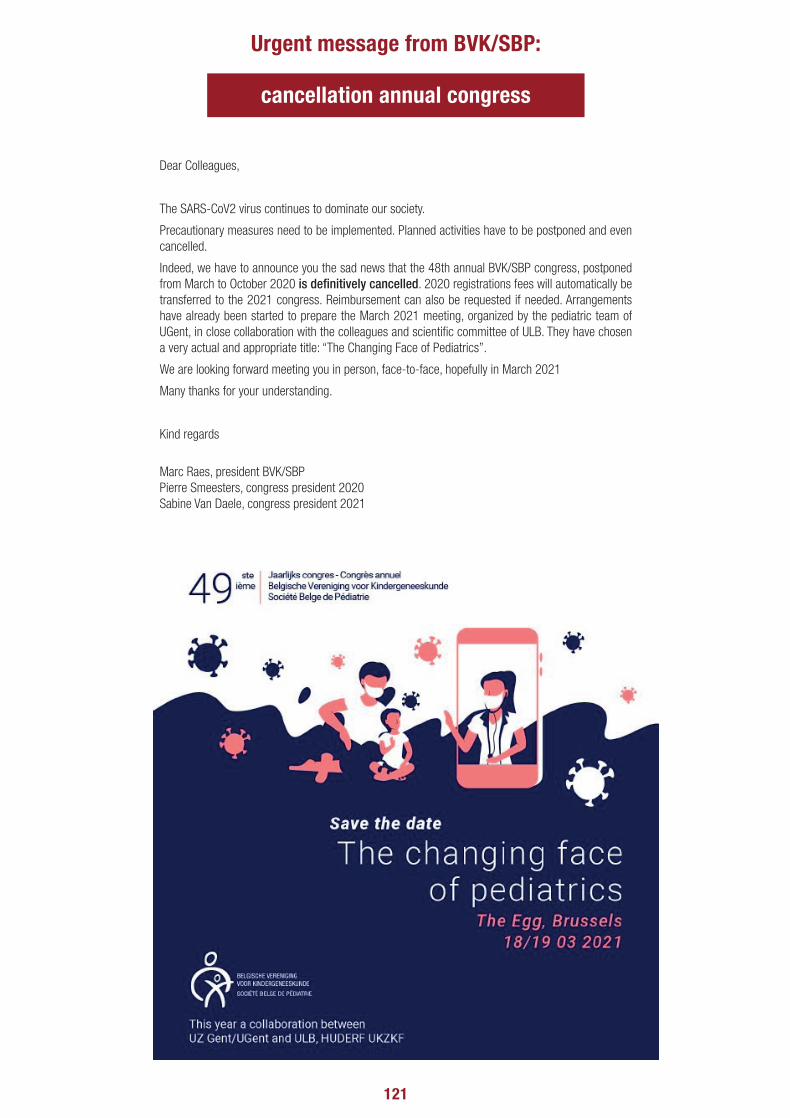
Urgent message from BVK/SBP:
PAUL CASAER (1940 – 2020)
cancellation annual congress
Dear Colleagues,
The SARS-CoV2 virus continues to dominate our society.
Precautionary measures need to be implemented. Planned activities have to be postponed and even cancelled.
Indeed, we have to announce you the sad news that the 48th annual BVK/SBP congress, postponed from March to October 2020 is de�nitively cancelled. 2020 registrations fees will automatically be transferred to the 2021 congress. Reimbursement can also be requested if needed. Arrangements have already been started to prepare the March 2021 meeting, organized by the pediatric team of UGent, in close collaboration with the colleagues and scienti�c committee of ULB. They have chosen a very actual and appropriate title: “The Changing Face of Pediatrics”.
We are looking forward meeting you in person, face-to-face, hopefully in March 2021
Many thanks for your understanding.
Kind regards
Marc Raes, president BVK/SBP Pierre Smeesters, congress president 2020 Sabine Van Daele, congress president 2021

122We care for children BELGISCHE VERENIGING
VOOR KINDERGENEESKUNDE
SOCIETE BELGE DE PEDIATRIE
BELGISCHE VERENIGINGVOOR KINDERGENEESKUNDE
SOCIETE BELGE DE PEDIATRIE
SBP - RENOUVELLEMENT ADHÉSION 2018
BELGISCHE VERENIGINGVOOR KINDERGENEESKUNDE
SOCIETE BELGE DE PEDIATRIE
BELGISCHE VERENIGINGVOOR KINDERGENEESKUNDE
SOCIETE BELGE DE PEDIATRIE
Les cotisations annuelles restent inchangés:
-120€ pour les pédiatres
-60€ pour les assistants en pédiatrie
-60€ pour les pédiatres pensionnés
Les avantages d’une adhésion à la Société sont nombreux:
À part le tarif réduit d’inscription au congrès annuel, les membres bénéficient, via le site de la SBP, d’un accès à la bibliothèque virtuelle CEBAM (Belgian Center for Evidence Based Medecine). Je vous rappelle que l’accès au volet pédiatrique du site de la CEBAM permet la lecture et le téléchargement d’articles des principaux journaux pédiatriques (entre autres Pediatrics, Journal of Pediatrics, Pediatric Infectious Disease Journal).
Le site donne également accès aux “big five” (New England Journal of Medicine, Lancet, BMJ, JAMA et Annals of Internal Medicine) ainsi qu’à de nombreuses bases de données d’Evidence Based Medecine (Cochrane Library) ainsi qu’à des livres électroniques.
Les membres reçoivent gratuitement le BJP (Belgian Journal of Paediatrics), journal aussi accessible online sur le site web www.bvk-sbp.be. Vous y trouverez à part des contributions scientifiques de chez nous, aussi les comptes rendu de l’Académie belge de Pédiatrie ainsi que des liens bien utiles.
Les membres profitent aussi de l’accès gratuit au Club Privilege What’s Up Doc.
Je vous remercie de contribuer à la réalisation de notre avenir, en renouvelant votre cotisation.
Très cordialement.
Prof Dr Anne MALFROOT
Présidente BVK/SBP
Cher Collègue,
Vous pourrez renouveler votre cotisation annuelle 2018 à partir du 1er Octobre 2017. Votre adhésion courra jusqu’au 30 Septembre 2018 et vous permettra de vous inscrire à un tarif réduit au futur Congrès Annuel de la SBP qui aura lieu le 8 et 9 Mars 2018.
La SBP est avant tout une association de scientifiques sensibilisés aux aspects médicaux et sociaux et auxquels sont confrontés l’enfant, l’adolescent ainsi que leurs parents. Depuis bientôt 100 ans, cette mission reste toujours aussi primordiale.
L’adhésion de tout pédiatre établi et de tout assistant en formation de pédiatrie contribue à optimaliser et moderniser le rayonnement et l’extension de notre association, en tant que porte-parole scientifique reconnu au niveau national non seulement au sein de notre propre discipline, mais tout autant vis-à-vis d’autres professionnels de la santé, des autorités, de nos patients et de leur famille et aussi d’un plus large public.
La procédure pour devenir ou rester membre de notre société est simple, mais uniquement possible en passant par le site web www.bvk-sbp.be.
Renouvellement cotisation 2021
Cher collègue,
La Société belge de pédiatrie (BVK/SBP) s’efforce de soutenir ses membres dans le domaine scienti� que. Elle organise annuellement un congrès national pédiatrique de deux jours. Elle est responsable de la publication du peer-reviewed Belgian Journal of Paediatrics offrant des numéros thématiques sur divers sujets ainsi que des contributions locales. Elle soutient explicitement les jeunes scienti� ques. Elle propose un site Web avec des liens vers des e-learnings et des formations universitaires, des informations sur différents congrès, des publications scienti� ques, des dossiers et des recommandations pratiques, ainsi que l’accès à la littérature médicale internationale par l’intermédiaire de la bibliothèque numérique du CEBAM et le Journal Club mensuel. Elle tient ses membres informés des problèmes actuels dans le domaine des soins de santé et formule des conseils, en collaboration avec des experts, tant auprès des membres qu’à d’autres fournisseurs et organismes de soins de santé. Grâce à des prix scienti� ques, elle soutient la recherche scienti� que. Elle contribue à la formation des boursiers en pédiatrie et soutient la mise en œuvre des sous-spécialisations pédiatriques en Belgique. La Société belge de pédiatrie représente l’ensemble des aspects médicaux et psychosociaux de l’enfant, de l’adolescent et de leur famille et s’efforce d’être un forum scienti� que pour répondre aux questions à ce sujet.
Les membres du Conseil d'administration souhaitent donc vous inviter à devenir membre de notre société. Grâce à votre contribution, le BVK/SBP peut réaliser ses missions scienti� ques de manière optimale et aussi indépendante que possible, en collaboration avec l’Académie belge de pédiatrie, qui coordonne les organisations pédiatriques of� cielles existantes en Belgique.
En plus du soutien de la mission scienti� que du BVK,/SBP l’adhésion offre plusieurs avantages : via le site web du BVK/SBP, les membres ont accès à la bibliothèque numérique du CEBAM (Belgian center for Evidence-Based Medicine). Cet accès permet de lire et de télécharger les articles des principaux journaux pédiatriques (Pediatrics, Journal of Pediatrics, Pediatric Infectious Disease Journal, etc.). Le site donne également accès aux « big � ve » (New England Journal of Medicine, Lancet, BMJ, JAMA et Annals of Internal Medicine) ainsi qu’à de nombreuses bases de données de la Evidence Based Medicine (Cochrane Library, etc.) et de livres électroniques. La vue d’ensemble mensuelle des articles internationaux les plus intéressants dans le domaine de la pédiatrie se trouve dans la rubrique: Journal Club. Le seul « peer-reviewed » journal national de pédiatrie: «Belgian Journal of Paediatrics» peut être consulté en ligne.
En tant que membre de la société, vous béné� ciez d’un tarif très avantageux lorsque vous vous inscrivez au congrès scienti� que annuel de la BVK/SBP. Grâce à votre adhésion, nous pouvons continuer à stimuler les (jeunes) chercheurs dans leur recherche par une bourse de promotion scienti� que.
L’adhésion à la société belge de pédiatrie est réservée à tous les médecins qui travaillent ou en formation en pédiatrie. L’adhésion court jusqu’en septembre 2021
Les cotisations annuelles restent inchangées :
- 120 € pour les pédiatres
- 60 € pour les assistants
- 60€ pour les pédiatres pensionnés
Vous pourrez renouveler votre cotisation par le site web : www.bvk-sbp.be
Cordialement,
également au nom de tous les membres du conseil d’administration de la société belge de pédiatrie
Dr Marc RaesPrésident SBP/BVK
Bescherming in a�e vertrouwen
Pampers Premium Protection: ons zachtste comfort met een onverslaanbare huidbescherming.
*Op basis van de verdeling van Pampers Premium Protection™ luiers maat 0-2 in Belgische materniteiten, jan-sep 2020.
7744_PampersBE_PP_printad_JUN20_BEFR_A4.indd 2 08/06/2020 17:57

123We care for children BELGISCHE VERENIGING
VOOR KINDERGENEESKUNDE
SOCIETE BELGE DE PEDIATRIE
BELGISCHE VERENIGINGVOOR KINDERGENEESKUNDE
SOCIETE BELGE DE PEDIATRIE
SBP - RENOUVELLEMENT ADHÉSION 2018
BELGISCHE VERENIGINGVOOR KINDERGENEESKUNDE
SOCIETE BELGE DE PEDIATRIE
BELGISCHE VERENIGINGVOOR KINDERGENEESKUNDE
SOCIETE BELGE DE PEDIATRIE
Les cotisations annuelles restent inchangés:
-120€ pour les pédiatres
-60€ pour les assistants en pédiatrie
-60€ pour les pédiatres pensionnés
Les avantages d’une adhésion à la Société sont nombreux:
À part le tarif réduit d’inscription au congrès annuel, les membres bénéficient, via le site de la SBP, d’un accès à la bibliothèque virtuelle CEBAM (Belgian Center for Evidence Based Medecine). Je vous rappelle que l’accès au volet pédiatrique du site de la CEBAM permet la lecture et le téléchargement d’articles des principaux journaux pédiatriques (entre autres Pediatrics, Journal of Pediatrics, Pediatric Infectious Disease Journal).
Le site donne également accès aux “big five” (New England Journal of Medicine, Lancet, BMJ, JAMA et Annals of Internal Medicine) ainsi qu’à de nombreuses bases de données d’Evidence Based Medecine (Cochrane Library) ainsi qu’à des livres électroniques.
Les membres reçoivent gratuitement le BJP (Belgian Journal of Paediatrics), journal aussi accessible online sur le site web www.bvk-sbp.be. Vous y trouverez à part des contributions scientifiques de chez nous, aussi les comptes rendu de l’Académie belge de Pédiatrie ainsi que des liens bien utiles.
Les membres profitent aussi de l’accès gratuit au Club Privilege What’s Up Doc.
1er Octobre 2017
La SBP est avant tout une association de scientifiques sensibilisés aux aspects médicaux et sociaux et auxquels sont confrontés l’enfant, l’adolescent ainsi que leurs parents. Depuis bientôt 100 ans, cette mission reste toujours aussi primordiale.
L’adhésion de tout pédiatre établi et de tout assistant en formation de pédiatrie contribue à optimaliser et moderniser le rayonnement et l’extension de notre association, en tant que porte-parole scientifique reconnu au niveau national non seulement au sein de notre propre discipline, mais tout autant vis-à-vis d’autres professionnels de la santé, des autorités, de nos patients et de leur famille et aussi d’un plus large public.
La procédure pour devenir ou rester membre de notre société est simple, mais uniquement possible en passant par le site web www.bvk-sbp.be.
Bescherming in a�e vertrouwen
Pampers Premium Protection: ons zachtste comfort met een onverslaanbare huidbescherming.
*Op basis van de verdeling van Pampers Premium Protection™ luiers maat 0-2 in Belgische materniteiten, jan-sep 2020.
7744_PampersBE_PP_printad_JUN20_BEFR_A4.indd 2 08/06/2020 17:57

124We care for children BELGISCHE VERENIGING
VOOR KINDERGENEESKUNDE
SOCIETE BELGE DE PEDIATRIE
BELGISCHE VERENIGINGVOOR KINDERGENEESKUNDE
SOCIETE BELGE DE PEDIATRIE
BVK - HERNIEUWING LIDMAATSCHAP 2018
BELGISCHE VERENIGINGVOOR KINDERGENEESKUNDE
SOCIETE BELGE DE PEDIATRIE
BELGISCHE VERENIGINGVOOR KINDERGENEESKUNDE
SOCIETE BELGE DE PEDIATRIE
De jaarlijkse bijdragen blijven ongewijzigd:
-120€ voor de kinderartsen
-60€ voor kinderartsen in opleiding
-60€ voor de kinderartsen op rust
De voordelen van een BVK lidmaatschap zijn talrijk:
Naast een verlaging van het inschrijvingsgeld van het jaarlijks congres krijgen de leden, via de site van de BVK, toegang tot de numerieke bibliotheek van de CEBAM (Belgian Center for Evidence Based Medicine). De toegang tot het pediatrisch luik van de CEBAM maakt het mogelijk de artikels van de belangrijkste pediatrische tijdschriften te lezen en te downloaden (Pediatrics, Journal of Pediatrics, Pediatric Infectious Disease Journal, etc.).
De site geeft ook toegang tot de “big five” (New England Journal of Medicine, Lancet, BMJ, JAMA en Annals of Internal Medicine) alsook tot talrijke databases van Evidence-Based Medicine zoals de Cochrane Library en andere elektronische boeken.
Verder krijgen de leden ook gratis het tijdschrift BJP (Belgian Journal of Paediatrics) toegestuurd, dat tevens online kan geraad-pleegd worden via www.bvk-sbp.be, website waarvan het deel artsen enkel toegankelijk is voor leden.
De BJP bevat naast interessante wetenschappelijke bijdragen en guidelines ook de verslagen van de Belgische Academie voor Kindergeneeskunde en nuttige links.
Leden hebben ook gratis toegang tot Privilege Club What’s Up Doc.
Hartelijke dank voor uw lidmaatschap waardoor u uw eigen professionele discipline steunt en een bijdrage levert aan de bevordering van de gezondheid van het kind in België!
Met collegiale groeten,
Prof Dr. Anne MALFROOT
Voorzitter BVK/SBP
Beste Collega,
U kan uw volgend jaarlijks lidmaatschap 2018 vanaf 1 oktober 2017 hernieuwen. Dit lidmaatschap zal lopen tot 30 september 2018.
Door uw bijdrage als lid van onze vereniging kan u zich aan een voordelig tarief inschrijven voor het komende Jaarlijks Congres van de BVK op 8 en 9 maart 2018. De procedure om lid te worden of te blijven is eenvoudig en gebeurt via de website www.bvk-sbp.be.
De BVK is in de eerste plaats een vereniging van zorgverleners en wetenschappers bezorgd om de medische en sociale aspecten waarmee het kind, de adolescent alsook de ouders geconfronteerd worden. Sinds bijna 100 jaar, blijft deze opdracht primordiaal.
Het lidmaatschap van elke kinderarts en iedere kinderarts in opleiding, draagt bij tot het optimaliseren en het moderniseren van de uitstraling van onze vereniging, alsook tot de erkenning als wetenschappelijke woordvoerder op nationaal en internationaal vlak.
Dit geldt niet alleen binnen onze eigen discipline, maar ook tegenover andere professionals in de gezondheidssector, onze patiënten en hun familie en tegenover een nog breder publiek.
Hernieuwing lidgeld 2021
Beste Collega,
De Belgische Vereniging voor Kindergeneeskunde (BVK/SBP) wil haar leden ondersteunen op wetenschappelijk vlak. Ze organiseert jaarlijks een tweedaags nationaal congres en verzorgt de publicatie van de Belgian Journal of Paediatrics, met themanummers omtrent diverse onderwerpen en vooral bijdragen van eigen bodem en steunt expliciet jonge wetenschappers. Haar website biedt links naar E-learnings en universitaire opleidingen, congres informatie, wetenschappelijke publicaties, dossiers en richtlijnen. Via de website krijgen leden toegang tot de internationale medische literatuur via de digitale bibliotheek van CEBAM en de maandelijkse Journal Club. BVK/SBP houdt haar leden op de hoogte van actuele problemen binnen de gezondheidzorg en formuleert adviezen, in overleg en samenwerking met experten, zowel naar de leden als naar andere zorgverstrekkers en instanties. Via wetenschappeljke prijzen steunt ze het wetenschappelijk onderzoek. Ze draagt bij aan de opleiding van de fellows kindergeneeskunde en bouwt mee aan de verdere implementatie van de pediatrische subspecialisaties in België. De Belgische Vereniging voor Kindergeneeskunde behartigt alle medische en psycho-sociale apecten van het kind, de adolescent en hun families en streeft ernaar het nationale wetenschappelijk forum te zijn om vragen daaromtrent te beantwoorden.
De Raad van bestuur wenst u dan ook uit te nodigen om lid te worden van onze vereniging. Door uw bijdrage kan de BVK haar wetenschappelijke opdrachten zo optimaal en zo onafhankelijk mogelijk realiseren, in samenwerking met de Belgische Academie voor Kindergeneeskunde, die de bestaande of� ciële pediatrische organisaties in België coördineert.
Naast de steun aan de wetenschappelijke missie van de BVK biedt het lidmaatschap meerdere voordelen: via de website van de BVK, hebben de leden toegang tot de numerieke bibliotheek van de CEBAM (Belgian Center for Evidence Based Medicine). Die toegang maakt het mogelijk de artikels van de belangrijkste pediatrische tijdschriften te lezen en te downloaden (Pediatrics, Journal of Pediatrics, Pediatric Infectious Disease Journal, etc.). De site geeft ook toegang tot de “big � ve” (New England Journal of Medicine, Lancet, BMJ, JAMA en Annals of Internal Medicine) alsook tot talrijke databases van Evidence-Based Medicine (Cochrane Library, etc) en elektronische boeken. Het maandelijks overzicht van de meest interessante internationale artikels binnen het domein van de kindergeneeskunde vinden zij terug onder de rubriek: Journal Club. Het enige nationale peer-reviewed tijdschrift voor kindergeneeskunde: “Belgian Journal of Paediatrics”kan online worden geconsulteerd.
Als lid van de vereniging geniet u van een zeer voordelig tarief bij de inschrijving voor het jaarlijks tweedaags wetenschappelijk congres van de BVK. Mede door uw lidmaatschap kunnen wij (jonge) researchers via een aanmoedingsbeurs blijven stimuleren in hun wetenschappelijk onderzoek.
Het lidmaatschap tot de BVK is voorbehouden aan alle artsen die werkzaam of in opleiding zijn in de kindergeneeskunde. Lidmaatschap loopt tot september 2021
De jaarlijkse bijdragen blijven ongewijzigd:
-120€ voor de kinderartsen
- 60€ voor de assistenten
- 60€ voor de kinderartsen op rust.
Lid worden kan via de website www.bvk-sbp.be
Hartelijk dank voor jullie steun en engagement.
Genegen groeten,
mede in naam van alle leden van de Raad Bestuur van de BVK
Dr Marc RAES
Voorzitter BVK/SBP
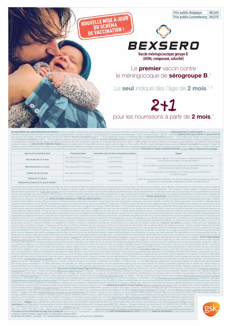
RÉSUMÉ ABRÉGÉ DES CARACTÉRISTIQUES DU PRODUIT Veuillez vous référer au Résumé des Caractéristi ques du Produit pour une informati on complète concernant l’usage de ce médicament. DÉNOMINATION DU MÉDICAMENT Bexsero suspension injectable en seringue préremplie Vaccin méningococc ique groupe B (ADNr, composant, a dsorbé) - EU/1/12/812/001 Classe pharmacothérapeuti que : vaccins méningococciques, Code ATC : J07AH09 COMPOSITION QUALITATIVE ET QUANTITATIVE Une dose (0,5 ml) conti ent : Protéine de fusion recombinante NHBA de Neisseria meningiti dis groupe B 1, 2, 3 50 microgrammes Protéine recombinante NadA de Neisseria meningiti dis groupe B 1, 2, 3 50 microgrammes Protéine de fusion recombinante fHbp de Neisseria meningiti dis groupe B 1, 2, 3 50 microgrammes Vésicules de membrane externe (OMV) de Neisseria meningiti dis groupe B, souche NZ98/254 mesurée en tant que proporti on de l’ensemble des protéines contenant l’anti gène PorA P1.4 2
25 microgrammes 1 produite dans des cellules d’E. coli par la technique de l’ADN recombinant 2 adsorbée sur hydroxyde d’aluminium (0,5 mg Al³+) 3 NHBA (anti gène de liaison à l’héparine de Neisseria), NadA (adhésine A de Neisseria), fHbp (protéine de liaison du facteur H) INDICATIONS THÉRAPEUTIQUES Bexsero est indiqué pour l’immunisati on acti ve des sujets à parti r de l’âge de 2 mois contre l’infecti on invasive méningococcique causée par Neisseria meningiti dis de groupe B. L’impact de l’infecti on invasive à diff érentes tranches d’âge ainsi que la variabilité épidémiologique des anti gènes des souches du groupe B dans diff érentes zones géographiques doivent être pris en compte lors de la vaccinati on. Voir rubrique 5.1 du RCP complet pour plus d’informati ons sur la protecti on contre les souches spécifi ques au groupe B. Ce vaccin doit être uti lisé conformément aux recommandati ons offi cielles. POSOLOGIE ET MODE D’ADMINISTRATION Posologie Tableau 1.Résumé de la posologie
Age lors de la première dose Primovaccinati on Intervalles entre les doses de primovaccinati on Rappel
Nourrissons de 2 à 5 moisTrois doses de 0,5 ml chacune 1 mois minimum Oui, une dose entre l’âge de 12 et 15 mois avec un intervalle d’au moins 6 mois
entre la primovaccinati on et la dose de rappel b, cDeux doses de 0,5 ml chacune 2 mois minimum
Nourrissons de 6 à 11 mois Deux doses de 0,5 ml chacune 2 mois minimum Oui, une dose au cours de la deuxième année avec un intervalle d’au moins 2 mois entre la primovaccinati on et la dose de rappel c
Enfants de 12 à 23 mois Deux doses de 0,5 ml chacune 2 mois minimum Oui, une dose avec un intervalle de 12 à 23 mois entre la primovaccinati on et la dose de rappel c
Enfants de 2 à 10 ansDeux doses de 0,5 ml chacune 1 mois minimum Selon les recommandati ons offi cielles, une dose de rappel peut être envisagée chez les sujets
présentant un risque conti nu d’expositi on à infecti on méningococciquedAdolescents (à parti r de 11 ans) et adultes*
a La première dose ne doit pas être administrée avant l’âge de 2 mois. La sécurité et l’effi cacité de Bexsero chez les nourrissons de moins de 8 semaines n’ont pas encore été établies. Aucune donnée n’est disponible. b En cas de retard, la dose de rappel ne doit pas être administrée au-delà de l’âge de 24 mois. c Voir rubrique 5.1 du RCP complet. La nécessité et le moment d’administrati on d’une dose de rappel n’ont pas encore été déterminés. d Voir rubrique 5.1 du RCP complet. * Il n’existe aucune donnée chez les adultes de plus de 50 ans. Mode d’administrati on Le vaccin est administré par une injecti on intramusculaire profonde, de préférence dans la face antérolatérale de la cuisse chez le nourrisson ou dans la région du muscle deltoïde du haut du bras chez les sujets plus âgés. Des sites d’injecti on disti ncts doivent être uti lisés si plusieurs vaccins sont administrés simultanément. Le vaccin ne doit pas être injecté par voie intraveineuse, sous-cutanée ni intradermique et ne doit pas être mélangé avec d’autres vaccins dans la même seringue. Pour les instructi ons concernant la manipulati on du vaccin avant administrati on, voir la rubrique 6.6 du RCP complet. CONTRE-INDICATIONS Hypersensibilité aux substances acti ves ou à l’un des excipients menti onnés à la rubrique 6.1 du RCP complet. MISES EN GARDE SPÉCIALES ET PRÉCAUTIONS D’EMPLOI Comme pour les autres vaccins l’administrati on de Bexsero doit être reportée chez des sujets souff rant de maladie fébrile sévère aiguë. Toutefois, la présence d’une infecti on mineure, telle qu’un rhume, ne doit pas entraîner le report de la vaccinati on. Ne pas injecter par voie intravasculaire. Comme pour tout vaccin injectable, un traitement médical approprié et une surveillance adéquate doivent toujours être disponibles en cas de réacti on anaphylacti que consécuti ve à l’administrati on du vaccin. Des réacti ons en rapport avec l’anxiété, y compris des réacti ons vaso-vagales (syncope), de l’hyperventi lati on ou des réacti ons en rapport avec le stress peuvent survenir lors de la vaccinati on comme réacti on psychogène à l’injecti on avec une aiguille (voir rubrique « Eff ets indésirables »). Il est important que des mesures soient mises en place afi n d’éviter toute blessure en cas d’évanouissement. Ce vaccin ne doit pas être administré aux pati ents ayant une thrombocytopénie ou tout autre trouble de la coagulati on qui serait une contre-indicati on à une injecti on par voie intramusculaire, à moins que le bénéfi ce potenti el ne soit clairement supérieur aux risques inhérents à l’administrati on. Comme tout vaccin, la vaccinati on par Bexsero peut ne pas protéger tous les sujets vaccinés. Il n’est pas att endu que Bexsero assure une protecti on contre la totalité des souches de méningocoque B en circulati on. Comme pour de nombreux vaccins, les professionnels de santé doivent savoir qu’une élévati on de la température corporelle peut survenir suite à la vaccinati on des nourrissons et des enfants (de moins de 2 ans). L’administrati on d’anti pyréti ques à ti tre prophylacti que pendant et juste après la vaccinati on peut réduire l’incidence et la sévérité des réacti ons fébriles post-vaccinales. Un traitement anti pyréti que doit être mis en place conformément aux recommandati ons locales chez les nourrissons et les enfants (de moins de 2 ans). Les personnes dont la réponse immunitaire est altérée soit par la prise d’un traitement immunosuppresseur, une anomalie généti que ou par d’autres causes, peuvent avoir une réponse en anti corps réduite après vaccinati on. Des données d’immunogénicité sont disponibles chez les pati ents présentant un défi cit en complément, une asplénie ou une dysfoncti on splénique. Les personnes ayant des défi cits hétréditaires du complément (par exemple les défi cits en C3 ou C5) et les personnes recevant un traitement inhibiteur de l’acti vati on de la fracti on terminale du complément (par exemple, l’éculizumab) ont un risque accru de maladie invasive due à Neisseria meningiti dis du groupe B, même après avoir développé des anti corps après vaccinati on par Bexsero. Il n’existe aucune donnée sur l’uti lisati on de Bexsero chez les sujets de plus de 50 ans et il existe des données limitées chez les pati ents att eints de maladies chroniques. Le risque potenti el d’apnée et la nécessité d’une surveillance respiratoire pendant 48 à 72 heures doivent soigneusement être pris en compte lors de l’administrati on des doses de primovaccinati on chez des grands prématurés (nés à 28 semaines de grossesse ou moins), en parti culier chez ceux ayant des antécédents d’immaturité respiratoire. En raison du bénéfi ce élevé de la vaccinati on chez ces nourrissons, l’administrati on ne doit pas être suspendue ou reportée. Le capuchon de la seringue peut contenir du latex de caoutchouc naturel. Bien que le risque de développer des réacti ons allergiques soit très faible, les professionnels de santé doivent évaluer le rapport bénéfi ces/risques avant d’administrer ce vaccin à des sujets présentant des antécédents connus d’hypersensibilité au latex. La kanamycine est uti lisée au début du procédé de fabricati on et est éliminée au cours des étapes ultérieures de la fabricati on. Les taux de kanamycine éventuellement détectables dans le vaccin fi nal sont inférieurs à 0,01 microgramme par dose. L’innocuité de Bexsero chez les sujets sensibles à la kanamycine n’a pas été établie. Traçabilité Afi n d’améliorer la traçabilité des médicaments biologiques, le nom et le numéro de lot du produit administré doivent être clairement enregistrés. EFFETS INDÉSIRABLES Résumé du profi l de sécurité La sécurité de Bexsero a été évaluée lors de 17 études, dont 10 essais cliniques randomisés contrôlés portant sur 10565 sujets (âgés de 2 mois minimum) ayant reçu au moins une dose de Bexsero. Parmi les sujets vaccinés par Bexsero, 6837 étaient des nourrissons et des enfants (de moins de 2 ans), 1051 étaient des enfants (entre 2 et 10 ans) et 2677 étaient des adolescents et des adultes. Parmi les nourrissons ayant reçu les doses de primovaccinati on de Bexsero, 3285 ont reçu une dose de rappel au cours de leur deuxième année de vie. Chez les nourrissons et les enfants (de moins de 2 ans), les réacti ons indésirables locales et systémiques les plus fréquemment observées lors des essais cliniques étaient : sensibilité et érythème au site d’injecti on, fi èvre et irritabilité. Dans les études cliniques menées chez les nourrissons vaccinés à 2, 4 et 6 mois, la fi èvre (≥ 38 °C) était rapportée chez 69% à 79 % des sujets lorsque Bexsero était co-administré avec des vaccins de routi ne (contenant les anti gènes suivants : pneumococcique heptavalent conjugué, diphtérie, tétanos, coqueluche acellulaire, hépati te B, poliomyélite inacti vée et Haemophilus infl uenzae de type b), contre 44% à 59 % des sujets recevant les vaccins de routi ne seuls. Une uti lisati on plus fréquente d’anti pyréti ques était également rapportée chez les nourrissons vaccinés par Bexsero et des vaccins de routi ne. Lorsque Bexsero était administré seul, la fréquence de la fi èvre était similaire à celle associée aux vaccins de routi ne administrés aux nourrissons pendant les essais cliniques. Les cas de fi èvre suivaient généralement un schéma prévisible, se résolvant généralement le lendemain de la vaccinati on. Chez les adolescents et les adultes, les réacti ons indésirables locales et systémiques les plus fréquemment observées étaient : douleur au point d’injecti on, malaise et céphalée. Aucune augmentati on de l’incidence ou de la sévérité des réacti ons indésirables n’a été constatée avec les doses successives du schéma de vaccinati on. Liste tabulée des eff ets indésirables Les eff ets indésirables (consécuti fs à la primovaccinati on ou à la dose de rappel) considérés comme étant au moins probablement liés à la vaccinati on ont été classés par fréquence. Les fréquences sont défi nies comme suit : Très fréquent : (≥ 1/10) Fréquent : (≥ 1/100 à < 1/10) Peu fréquent : (≥ 1/1 000 à < 1/100) Rare : (≥ 1/10 000 à < 1/1 000) Très rare : (< 1/10 000) Fréquence indéterminée : (ne peut être esti mée sur la base des données disponibles) Dans chaque groupe de fréquence, les eff ets indésirables sont présentés par ordre de sévérité décroissante. Outre les événements rapportés lors des essais cliniques, les réacti ons spontanées rapportées dans le monde pour Bexsero depuis sa commercialisati on sont décrites dans la liste ci dessous. Comme ces réacti ons ont été rapportées volontairement à parti r d’une populati on de taille inconnue, il n’est pas toujours possible d’esti mer de façon fi able leur fréquence. Ces réacti ons sont, en conséquence, listées avec une fréquence indéterminée. Nourrissons et enfants (jusqu’à l’âge de 10 ans) Aff ecti ons du système immunitaire Fréquence indéterminée : réacti ons allergiques (y compris réacti ons anaphylacti ques) Troubles du métabolisme et de la nutriti on Très fréquent : troubles alimentaires Aff ecti ons du système nerveux Très fréquent : somnolence, pleurs inhabituels, céphalée Peu fréquent : convulsions (y compris convulsions fébriles) Fréquence indéterminée : épisode d’hypotonie-hyporéacti vité, irritati on des méninges (des signes d’irritati on des méninges, tels qu’une raideur de la nuque ou une photophobie, ont été rapportés sporadiquement peu de temps après la vaccinati on. Ces symptômes ont été de nature légère et transitoire) Aff ecti ons vasculaires Peu fréquent : pâleur (rare après le rappel) Rare : syndrome de Kawasaki Aff ecti ons gastro-intesti nales Très fréquent : diarrhée, vomissements (peu fréquents après le rappel) Aff ecti ons de la peau et du ti ssu sous-cutanéTrès fréquent : rash (enfants âgés de 12 à 23 mois) (peu fréquent après le rappel) Fréquent : rash (nourrissons et enfants âgés de 2 à 10 ans) Peu fréquent : eczéma Rare : urti caire Aff ecti ons musculo-squeletti ques et systémiques Très fréquent : arthralgies Troubles généraux et anomalies au site d’administrati on Très fréquent : fi èvre (≥ 38 °C), sensibilité au niveau du site d’injecti on (y compris sensibilité sévère au site d’injecti on défi nie par des pleurs lors d’un mouvement du membre ayant reçu l’injecti on), érythème au site d’injecti on, gonfl ement du site d’injecti on, indurati on au site d’injecti on, irritabilité Peu fréquent : fi èvre (≥ 40 °C) Fréquence indéterminée : réacti ons au site d’injecti on (incluant un gonfl ement étendu du membre vacciné, vésicules au point d’injecti on ou autour du site d’injecti on et nodule au site d’injecti on pouvant persister pendant plus d’un mois) Adolescents (à parti r de 11 ans) et adultes Aff ecti ons du système immunitaire Fréquence indéterminée : réacti ons allergiques (y compris réacti ons anaphylacti ques) Aff ecti ons du système nerveux Très fréquent : céphalée Fréquence indéterminée : syncope ou réacti on vaso-vagale à l’injecti on, irritati on des méninges (des signes d’irritati on des méninges, tels qu’une raideur de la nuque ou une photophobie, ont été rapportés sporadiquement peu de temps après la vaccinati on. Ces symptômes ont été de nature légère et transitoire) Aff ecti ons gastro-intesti nales Très fréquent : nausées Aff ecti ons musculo-squeletti ques et systémiques Très fréquent : myalgies, arthralgies Troubles généraux et anomalies au site d’administrati on Très fréquent : douleur au point d’injecti on (y compris douleur sévère au point d’injecti on défi nie par une incapacité à mener à bien des acti vités quoti diennes normales), gonfl ement du site d’injecti on, indurati on au point d’injecti on, érythème au site d’injecti on, malaise Fréquence indéterminée : fi èvre, réacti ons au site d’injecti on (incluant gonfl ement étendu du membre vacciné, vésicules au point d’injecti on ou autour du site d’injecti on et nodule au site d’injecti on pouvant persister plus d’un mois) Déclarati on des eff ets indésirables suspectés La déclarati on des eff ets indésirables suspectés après autorisati on du médicament est importante. Elle permet une surveillance conti nue du rapport bénéfi ce/risque du médicament. Les professionnels de santé déclarent tout eff et indésirable suspecté via le système nati onal de déclarati on : Belgique Agence fédérale des médicaments et des produits de santé Division Vigilance Boîte Postale 97 B-1000 Bruxelles Madou Site internet: www.afmps.be e-mail: adversedrugreacti [email protected] Luxembourg Centre Régional de Pharmacovigilance de Nancy Bâti ment de Biologie Moléculaire et de Biopathologie (BBB) CHRU de Nancy – Hôpitaux de Brabois Rue du Morvan 54 511 VANDOEUVRE LES NANCY CEDEX Tél : (+33) 3 83 65 60 85 / 87 Fax : (+33) 3 83 65 61 33 E-mail : [email protected] ou Directi on de la Santé Division de la Pharmacie et des Médicaments Allée Marconi - Villa Louvigny L-2120 Luxembourg Tél. : (+352) 2478 5592 Fax : (+352) 2479 5615 E-mail : [email protected] Link pour le formulaire : htt p://www.sante.public.lu/fr/politi que-sante/ministere-sante/directi on-sante/div-pharmacie-medicaments/index.htmlTITULAIRE DE L’AUTORISATION DE MISE SUR LE MARCHÉ GSK Vaccines S.r.l., Via Fiorenti na 1, 53100 Sienne, Italie DATE D’APPROBATION DU TEXTE 04/2020 (v10) MODE DE DELIVRANCE Sur prescripti on médicale. 1. Medini D, Stella M, Wassil J, Vaccine 2015; 33; 2629-2636. 2. Bexsero SMPC.PM-BE-BEX-ADV-200001 - Juin 2020 - E.R.: GlaxoSmithKline Pharmaceuti cals s.a., av Pascal 2-4-6, 1300 Wavre
Prix public Belgique 86,52€Prix public Luxembourg 84,07€
Le premier vaccin contrele méningocoque de sérogroupe B.1
Le seul indiqué dès l’âge de 2 mois.1,2
pour les nourrissons à partir de 2 mois.1
NOUVELLE MISE À JOUR
DU SCHÉMA DE VACCINATION !
2020-136-GSKBEX-Ad A4 2+1 juin 2020 FR_BAT.indd 1 26/06/20 10:18
125We care for children BELGISCHE VERENIGING
VOOR KINDERGENEESKUNDE
SOCIETE BELGE DE PEDIATRIE
BELGISCHE VERENIGINGVOOR KINDERGENEESKUNDE
SOCIETE BELGE DE PEDIATRIE
BVK - HERNIEUWING LIDMAATSCHAP 2018
BELGISCHE VERENIGINGVOOR KINDERGENEESKUNDE
SOCIETE BELGE DE PEDIATRIE
BELGISCHE VERENIGINGVOOR KINDERGENEESKUNDE
SOCIETE BELGE DE PEDIATRIE
De jaarlijkse bijdragen blijven ongewijzigd:
-120€ voor de kinderartsen
-60€ voor kinderartsen in opleiding
-60€ voor de kinderartsen op rust
De voordelen van een BVK lidmaatschap zijn talrijk:
Naast een verlaging van het inschrijvingsgeld van het jaarlijks congres krijgen de leden, via de site van de BVK, toegang tot de numerieke bibliotheek van de CEBAM (Belgian Center for Evidence Based Medicine). De toegang tot het pediatrisch luik van de CEBAM maakt het mogelijk de artikels van de belangrijkste pediatrische tijdschriften te lezen en te downloaden (Pediatrics, Journal of Pediatrics, Pediatric Infectious Disease Journal, etc.).
De site geeft ook toegang tot de “big five” (New England Journal of Medicine, Lancet, BMJ, JAMA en Annals of Internal Medicine) alsook tot talrijke databases van Evidence-Based Medicine zoals de Cochrane Library en andere elektronische boeken.
Verder krijgen de leden ook gratis het tijdschrift BJP (Belgian Journal of Paediatrics) toegestuurd, dat tevens online kan geraad-pleegd worden via www.bvk-sbp.be, website waarvan het deel artsen enkel toegankelijk is voor leden.
De BJP bevat naast interessante wetenschappelijke bijdragen en guidelines ook de verslagen van de Belgische Academie voor Kindergeneeskunde en nuttige links.
Leden hebben ook gratis toegang tot Privilege Club What’s Up Doc.
vanaf 1 oktober 2017
www.bvk-sbp.be
De BVK is in de eerste plaats een vereniging van zorgverleners en wetenschappers bezorgd om de medische en sociale aspecten waarmee het kind, de adolescent alsook de ouders geconfronteerd worden. Sinds bijna 100 jaar, blijft deze opdracht primordiaal.
Het lidmaatschap van elke kinderarts en iedere kinderarts in opleiding, draagt bij tot het optimaliseren en het moderniseren van de uitstraling van onze vereniging, alsook tot de erkenning als wetenschappelijke woordvoerder op nationaal en internationaal vlak.
Dit geldt niet alleen binnen onze eigen discipline, maar ook tegenover andere professionals in de gezondheidssector, onze patiënten en hun familie en tegenover een nog breder publiek.
RÉSUMÉ ABRÉGÉ DES CARACTÉRISTIQUES DU PRODUIT Veuillez vous référer au Résumé des Caractéristi ques du Produit pour une informati on complète concernant l’usage de ce médicament. DÉNOMINATION DU MÉDICAMENT Bexsero suspension injectable en seringue préremplie Vaccin méningococc ique groupe B (ADNr, composant, a dsorbé) - EU/1/12/812/001 Classe pharmacothérapeuti que : vaccins méningococciques, Code ATC : J07AH09 COMPOSITION QUALITATIVE ET QUANTITATIVE Une dose (0,5 ml) conti ent : Protéine de fusion recombinante NHBA de Neisseria meningiti dis groupe B 1, 2, 3 50 microgrammes Protéine recombinante NadA de Neisseria meningiti dis groupe B 1, 2, 3 50 microgrammes Protéine de fusion recombinante fHbp de Neisseria meningiti dis groupe B 1, 2, 3 50 microgrammes Vésicules de membrane externe (OMV) de Neisseria meningiti dis groupe B, souche NZ98/254 mesurée en tant que proporti on de l’ensemble des protéines contenant l’anti gène PorA P1.4 2
25 microgrammes 1 produite dans des cellules d’E. coli par la technique de l’ADN recombinant 2 adsorbée sur hydroxyde d’aluminium (0,5 mg Al³+) 3 NHBA (anti gène de liaison à l’héparine de Neisseria), NadA (adhésine A de Neisseria), fHbp (protéine de liaison du facteur H) INDICATIONS THÉRAPEUTIQUES Bexsero est indiqué pour l’immunisati on acti ve des sujets à parti r de l’âge de 2 mois contre l’infecti on invasive méningococcique causée par Neisseria meningiti dis de groupe B. L’impact de l’infecti on invasive à diff érentes tranches d’âge ainsi que la variabilité épidémiologique des anti gènes des souches du groupe B dans diff érentes zones géographiques doivent être pris en compte lors de la vaccinati on. Voir rubrique 5.1 du RCP complet pour plus d’informati ons sur la protecti on contre les souches spécifi ques au groupe B. Ce vaccin doit être uti lisé conformément aux recommandati ons offi cielles. POSOLOGIE ET MODE D’ADMINISTRATION Posologie Tableau 1.Résumé de la posologie
Age lors de la première dose Primovaccinati on Intervalles entre les doses de primovaccinati on Rappel
Nourrissons de 2 à 5 moisTrois doses de 0,5 ml chacune 1 mois minimum Oui, une dose entre l’âge de 12 et 15 mois avec un intervalle d’au moins 6 mois
entre la primovaccinati on et la dose de rappel b, cDeux doses de 0,5 ml chacune 2 mois minimum
Nourrissons de 6 à 11 mois Deux doses de 0,5 ml chacune 2 mois minimum Oui, une dose au cours de la deuxième année avec un intervalle d’au moins 2 mois entre la primovaccinati on et la dose de rappel c
Enfants de 12 à 23 mois Deux doses de 0,5 ml chacune 2 mois minimum Oui, une dose avec un intervalle de 12 à 23 mois entre la primovaccinati on et la dose de rappel c
Enfants de 2 à 10 ansDeux doses de 0,5 ml chacune 1 mois minimum Selon les recommandati ons offi cielles, une dose de rappel peut être envisagée chez les sujets
présentant un risque conti nu d’expositi on à infecti on méningococciquedAdolescents (à parti r de 11 ans) et adultes*
a La première dose ne doit pas être administrée avant l’âge de 2 mois. La sécurité et l’effi cacité de Bexsero chez les nourrissons de moins de 8 semaines n’ont pas encore été établies. Aucune donnée n’est disponible. b En cas de retard, la dose de rappel ne doit pas être administrée au-delà de l’âge de 24 mois. c Voir rubrique 5.1 du RCP complet. La nécessité et le moment d’administrati on d’une dose de rappel n’ont pas encore été déterminés. d Voir rubrique 5.1 du RCP complet. * Il n’existe aucune donnée chez les adultes de plus de 50 ans. Mode d’administrati on Le vaccin est administré par une injecti on intramusculaire profonde, de préférence dans la face antérolatérale de la cuisse chez le nourrisson ou dans la région du muscle deltoïde du haut du bras chez les sujets plus âgés. Des sites d’injecti on disti ncts doivent être uti lisés si plusieurs vaccins sont administrés simultanément. Le vaccin ne doit pas être injecté par voie intraveineuse, sous-cutanée ni intradermique et ne doit pas être mélangé avec d’autres vaccins dans la même seringue. Pour les instructi ons concernant la manipulati on du vaccin avant administrati on, voir la rubrique 6.6 du RCP complet. CONTRE-INDICATIONS Hypersensibilité aux substances acti ves ou à l’un des excipients menti onnés à la rubrique 6.1 du RCP complet. MISES EN GARDE SPÉCIALES ET PRÉCAUTIONS D’EMPLOI Comme pour les autres vaccins l’administrati on de Bexsero doit être reportée chez des sujets souff rant de maladie fébrile sévère aiguë. Toutefois, la présence d’une infecti on mineure, telle qu’un rhume, ne doit pas entraîner le report de la vaccinati on. Ne pas injecter par voie intravasculaire. Comme pour tout vaccin injectable, un traitement médical approprié et une surveillance adéquate doivent toujours être disponibles en cas de réacti on anaphylacti que consécuti ve à l’administrati on du vaccin. Des réacti ons en rapport avec l’anxiété, y compris des réacti ons vaso-vagales (syncope), de l’hyperventi lati on ou des réacti ons en rapport avec le stress peuvent survenir lors de la vaccinati on comme réacti on psychogène à l’injecti on avec une aiguille (voir rubrique « Eff ets indésirables »). Il est important que des mesures soient mises en place afi n d’éviter toute blessure en cas d’évanouissement. Ce vaccin ne doit pas être administré aux pati ents ayant une thrombocytopénie ou tout autre trouble de la coagulati on qui serait une contre-indicati on à une injecti on par voie intramusculaire, à moins que le bénéfi ce potenti el ne soit clairement supérieur aux risques inhérents à l’administrati on. Comme tout vaccin, la vaccinati on par Bexsero peut ne pas protéger tous les sujets vaccinés. Il n’est pas att endu que Bexsero assure une protecti on contre la totalité des souches de méningocoque B en circulati on. Comme pour de nombreux vaccins, les professionnels de santé doivent savoir qu’une élévati on de la température corporelle peut survenir suite à la vaccinati on des nourrissons et des enfants (de moins de 2 ans). L’administrati on d’anti pyréti ques à ti tre prophylacti que pendant et juste après la vaccinati on peut réduire l’incidence et la sévérité des réacti ons fébriles post-vaccinales. Un traitement anti pyréti que doit être mis en place conformément aux recommandati ons locales chez les nourrissons et les enfants (de moins de 2 ans). Les personnes dont la réponse immunitaire est altérée soit par la prise d’un traitement immunosuppresseur, une anomalie généti que ou par d’autres causes, peuvent avoir une réponse en anti corps réduite après vaccinati on. Des données d’immunogénicité sont disponibles chez les pati ents présentant un défi cit en complément, une asplénie ou une dysfoncti on splénique. Les personnes ayant des défi cits hétréditaires du complément (par exemple les défi cits en C3 ou C5) et les personnes recevant un traitement inhibiteur de l’acti vati on de la fracti on terminale du complément (par exemple, l’éculizumab) ont un risque accru de maladie invasive due à Neisseria meningiti dis du groupe B, même après avoir développé des anti corps après vaccinati on par Bexsero. Il n’existe aucune donnée sur l’uti lisati on de Bexsero chez les sujets de plus de 50 ans et il existe des données limitées chez les pati ents att eints de maladies chroniques. Le risque potenti el d’apnée et la nécessité d’une surveillance respiratoire pendant 48 à 72 heures doivent soigneusement être pris en compte lors de l’administrati on des doses de primovaccinati on chez des grands prématurés (nés à 28 semaines de grossesse ou moins), en parti culier chez ceux ayant des antécédents d’immaturité respiratoire. En raison du bénéfi ce élevé de la vaccinati on chez ces nourrissons, l’administrati on ne doit pas être suspendue ou reportée. Le capuchon de la seringue peut contenir du latex de caoutchouc naturel. Bien que le risque de développer des réacti ons allergiques soit très faible, les professionnels de santé doivent évaluer le rapport bénéfi ces/risques avant d’administrer ce vaccin à des sujets présentant des antécédents connus d’hypersensibilité au latex. La kanamycine est uti lisée au début du procédé de fabricati on et est éliminée au cours des étapes ultérieures de la fabricati on. Les taux de kanamycine éventuellement détectables dans le vaccin fi nal sont inférieurs à 0,01 microgramme par dose. L’innocuité de Bexsero chez les sujets sensibles à la kanamycine n’a pas été établie. Traçabilité Afi n d’améliorer la traçabilité des médicaments biologiques, le nom et le numéro de lot du produit administré doivent être clairement enregistrés. EFFETS INDÉSIRABLES Résumé du profi l de sécurité La sécurité de Bexsero a été évaluée lors de 17 études, dont 10 essais cliniques randomisés contrôlés portant sur 10565 sujets (âgés de 2 mois minimum) ayant reçu au moins une dose de Bexsero. Parmi les sujets vaccinés par Bexsero, 6837 étaient des nourrissons et des enfants (de moins de 2 ans), 1051 étaient des enfants (entre 2 et 10 ans) et 2677 étaient des adolescents et des adultes. Parmi les nourrissons ayant reçu les doses de primovaccinati on de Bexsero, 3285 ont reçu une dose de rappel au cours de leur deuxième année de vie. Chez les nourrissons et les enfants (de moins de 2 ans), les réacti ons indésirables locales et systémiques les plus fréquemment observées lors des essais cliniques étaient : sensibilité et érythème au site d’injecti on, fi èvre et irritabilité. Dans les études cliniques menées chez les nourrissons vaccinés à 2, 4 et 6 mois, la fi èvre (≥ 38 °C) était rapportée chez 69% à 79 % des sujets lorsque Bexsero était co-administré avec des vaccins de routi ne (contenant les anti gènes suivants : pneumococcique heptavalent conjugué, diphtérie, tétanos, coqueluche acellulaire, hépati te B, poliomyélite inacti vée et Haemophilus infl uenzae de type b), contre 44% à 59 % des sujets recevant les vaccins de routi ne seuls. Une uti lisati on plus fréquente d’anti pyréti ques était également rapportée chez les nourrissons vaccinés par Bexsero et des vaccins de routi ne. Lorsque Bexsero était administré seul, la fréquence de la fi èvre était similaire à celle associée aux vaccins de routi ne administrés aux nourrissons pendant les essais cliniques. Les cas de fi èvre suivaient généralement un schéma prévisible, se résolvant généralement le lendemain de la vaccinati on. Chez les adolescents et les adultes, les réacti ons indésirables locales et systémiques les plus fréquemment observées étaient : douleur au point d’injecti on, malaise et céphalée. Aucune augmentati on de l’incidence ou de la sévérité des réacti ons indésirables n’a été constatée avec les doses successives du schéma de vaccinati on. Liste tabulée des eff ets indésirables Les eff ets indésirables (consécuti fs à la primovaccinati on ou à la dose de rappel) considérés comme étant au moins probablement liés à la vaccinati on ont été classés par fréquence. Les fréquences sont défi nies comme suit : Très fréquent : (≥ 1/10) Fréquent : (≥ 1/100 à < 1/10) Peu fréquent : (≥ 1/1 000 à < 1/100) Rare : (≥ 1/10 000 à < 1/1 000) Très rare : (< 1/10 000) Fréquence indéterminée : (ne peut être esti mée sur la base des données disponibles) Dans chaque groupe de fréquence, les eff ets indésirables sont présentés par ordre de sévérité décroissante. Outre les événements rapportés lors des essais cliniques, les réacti ons spontanées rapportées dans le monde pour Bexsero depuis sa commercialisati on sont décrites dans la liste ci dessous. Comme ces réacti ons ont été rapportées volontairement à parti r d’une populati on de taille inconnue, il n’est pas toujours possible d’esti mer de façon fi able leur fréquence. Ces réacti ons sont, en conséquence, listées avec une fréquence indéterminée. Nourrissons et enfants (jusqu’à l’âge de 10 ans) Aff ecti ons du système immunitaire Fréquence indéterminée : réacti ons allergiques (y compris réacti ons anaphylacti ques) Troubles du métabolisme et de la nutriti on Très fréquent : troubles alimentaires Aff ecti ons du système nerveux Très fréquent : somnolence, pleurs inhabituels, céphalée Peu fréquent : convulsions (y compris convulsions fébriles) Fréquence indéterminée : épisode d’hypotonie-hyporéacti vité, irritati on des méninges (des signes d’irritati on des méninges, tels qu’une raideur de la nuque ou une photophobie, ont été rapportés sporadiquement peu de temps après la vaccinati on. Ces symptômes ont été de nature légère et transitoire) Aff ecti ons vasculaires Peu fréquent : pâleur (rare après le rappel) Rare : syndrome de Kawasaki Aff ecti ons gastro-intesti nales Très fréquent : diarrhée, vomissements (peu fréquents après le rappel) Aff ecti ons de la peau et du ti ssu sous-cutanéTrès fréquent : rash (enfants âgés de 12 à 23 mois) (peu fréquent après le rappel) Fréquent : rash (nourrissons et enfants âgés de 2 à 10 ans) Peu fréquent : eczéma Rare : urti caire Aff ecti ons musculo-squeletti ques et systémiques Très fréquent : arthralgies Troubles généraux et anomalies au site d’administrati on Très fréquent : fi èvre (≥ 38 °C), sensibilité au niveau du site d’injecti on (y compris sensibilité sévère au site d’injecti on défi nie par des pleurs lors d’un mouvement du membre ayant reçu l’injecti on), érythème au site d’injecti on, gonfl ement du site d’injecti on, indurati on au site d’injecti on, irritabilité Peu fréquent : fi èvre (≥ 40 °C) Fréquence indéterminée : réacti ons au site d’injecti on (incluant un gonfl ement étendu du membre vacciné, vésicules au point d’injecti on ou autour du site d’injecti on et nodule au site d’injecti on pouvant persister pendant plus d’un mois) Adolescents (à parti r de 11 ans) et adultes Aff ecti ons du système immunitaire Fréquence indéterminée : réacti ons allergiques (y compris réacti ons anaphylacti ques) Aff ecti ons du système nerveux Très fréquent : céphalée Fréquence indéterminée : syncope ou réacti on vaso-vagale à l’injecti on, irritati on des méninges (des signes d’irritati on des méninges, tels qu’une raideur de la nuque ou une photophobie, ont été rapportés sporadiquement peu de temps après la vaccinati on. Ces symptômes ont été de nature légère et transitoire) Aff ecti ons gastro-intesti nales Très fréquent : nausées Aff ecti ons musculo-squeletti ques et systémiques Très fréquent : myalgies, arthralgies Troubles généraux et anomalies au site d’administrati on Très fréquent : douleur au point d’injecti on (y compris douleur sévère au point d’injecti on défi nie par une incapacité à mener à bien des acti vités quoti diennes normales), gonfl ement du site d’injecti on, indurati on au point d’injecti on, érythème au site d’injecti on, malaise Fréquence indéterminée : fi èvre, réacti ons au site d’injecti on (incluant gonfl ement étendu du membre vacciné, vésicules au point d’injecti on ou autour du site d’injecti on et nodule au site d’injecti on pouvant persister plus d’un mois) Déclarati on des eff ets indésirables suspectés La déclarati on des eff ets indésirables suspectés après autorisati on du médicament est importante. Elle permet une surveillance conti nue du rapport bénéfi ce/risque du médicament. Les professionnels de santé déclarent tout eff et indésirable suspecté via le système nati onal de déclarati on : Belgique Agence fédérale des médicaments et des produits de santé Division Vigilance Boîte Postale 97 B-1000 Bruxelles Madou Site internet: www.afmps.be e-mail: adversedrugreacti [email protected] Luxembourg Centre Régional de Pharmacovigilance de Nancy Bâti ment de Biologie Moléculaire et de Biopathologie (BBB) CHRU de Nancy – Hôpitaux de Brabois Rue du Morvan 54 511 VANDOEUVRE LES NANCY CEDEX Tél : (+33) 3 83 65 60 85 / 87 Fax : (+33) 3 83 65 61 33 E-mail : [email protected] ou Directi on de la Santé Division de la Pharmacie et des Médicaments Allée Marconi - Villa Louvigny L-2120 Luxembourg Tél. : (+352) 2478 5592 Fax : (+352) 2479 5615 E-mail : [email protected] Link pour le formulaire : htt p://www.sante.public.lu/fr/politi que-sante/ministere-sante/directi on-sante/div-pharmacie-medicaments/index.htmlTITULAIRE DE L’AUTORISATION DE MISE SUR LE MARCHÉ GSK Vaccines S.r.l., Via Fiorenti na 1, 53100 Sienne, Italie DATE D’APPROBATION DU TEXTE 04/2020 (v10) MODE DE DELIVRANCE Sur prescripti on médicale. 1. Medini D, Stella M, Wassil J, Vaccine 2015; 33; 2629-2636. 2. Bexsero SMPC.PM-BE-BEX-ADV-200001 - Juin 2020 - E.R.: GlaxoSmithKline Pharmaceuti cals s.a., av Pascal 2-4-6, 1300 Wavre
Prix public Belgique 86,52€Prix public Luxembourg 84,07€
Le premier vaccin contrele méningocoque de sérogroupe B.1
Le seul indiqué dès l’âge de 2 mois.1,2
pour les nourrissons à partir de 2 mois.1
NOUVELLE MISE À JOUR
DU SCHÉMA DE VACCINATION !
2020-136-GSKBEX-Ad A4 2+1 juin 2020 FR_BAT.indd 1 26/06/20 10:18
126
Hirschsprung Disease and Congenital Anomalies of the Kidney and Urinary Tract (CAKUT): a genetic disorder or just a coincidence?Julie Eelen1, Marc Miserez2, Djalila Mekahli3, Ilse Hoffman4
1 Department of Pediatrics, University Hospitals Leuven, Leuven, Belgium 2 Department of Abdominal Surgery, University Hospitals Leuven, Leuven, Belgium 3 Department of Pediatric Nephrology, University Hospitals Leuven, Belgium 4 Department of Pediatric Gastroenterology, Hepatology and Nutrition, University Hospitals Leuven, Leuven, Belgium
Article
AbstractHirschsprung disease (HD) and congenital anomalies of the kidney and urinary tract (CAKUT), two pathological entities with their own, complex, genetic background, could be associated based on a common genetic pathway. Although this association has been described in literature, little is known on its prevalence and clinical relevance. We performed a literature research on prevalence and described genotypes of the combination of these entities. In addition, we performed a retrospective cohort study of the UZ Leuven HD population. The literature search and cohort study con�rmed the underestimated prevalence of the association between both diseases and the lack of genetic characterization of this population. We emphasize the need for a systematic nephrological ultrasound screening and genetic investigation, with focus on RET-GDNF-GFRalpha1-complex, of all HD patients.
KeywordsHirschsprung; CAKUT; RET; Genetics; Associated anomalies
Hirschsprung disease (HD) is de�ned as a congenital absence of parasympathetic ganglia in the distal gut, more speci�cally in the submucosal plexus of Meissner and myenteric plexus of Auerbach, in combination with submucosal neural hyperplasia. It extends from the anorectal junction to the rectum and colon with a variable height. The resulting aganglionic segment fails to relax, causing a tonically contracted bowel segment. This leads to a functional obstruction, resulting in a proximal megacolon and explaining the clinical symptoms of the disease1.
HD has an incidence of 1/5000 live births. The occurrence of the disease can be sporadic or familial. Depending on the length of the aganglionic segment, it is subdivided in a short- and long-segment HD. Extension up to the splenic �exure is called short-segment disease and is present in up to 80% of patients. Most frequently the disease will affect only the recto-sigmoid. Long-segment disease extends proximal to the splenic �exure (up to 20% of patients). Within this group, total colonic aganglionosis can be present, with or without inclusion of the terminal ileum. The disease is more common among boys, with a 4:1 male/female ratio. The treatment of HD is surgical. Thanks to the improved management, the mortality rate is below 1% 1-3.
The pathophysiology of HD is failure of normal development of the enteric nervous system following abnormal migration, proliferation, differentiation and/or survival of neural crest cells. It is thereby regarded as a neurocristopathy1,4-6.
It is not surprising that changes in genes contributing to the development of the enteric nervous system can interfere with colonization of hindgut nerve cells3-5. So far, over 20 genes have been associated with HD. The involvement of heterogeneous genetic pathways in HD also explains why HD is a multigenetic disorder which can occur both isolated (70%) or associated with other anomalies (18%)/chromosomal abnormalities (12%) in which almost all organs and tissues can be involved. The most frequently associated disorder is trisomy 211,3.
The most important gene in the pathogenesis of HD is REarranged during Transfection (RET). RET has an in�uence on growth and differentiation in different organs including the enteric nervous system1,7-10. Other genes belonging to the RET-signaling cascade that have been identi�ed to date are Gliall Cell Derived Neurotrophic Factor (GDNF), a RET ligand, and GDNF Family Receptor alpha 1 (GFRalpha1), a co-receptor of RET. Different studies have demonstrated that formation of the GDNF-RET-GFRalpha1 complex in�uences migration of neurons and is necessary to prevent aganglionosis1,6,11.
The RET-signaling pathway is also of importance in the development of the kidneys and urinary tract 12-14. CAKUT (congenital anomalies of the kidney and urinary tract) is an acronym and collective name for different clinical entities affecting the kidneys or structures of the urinary tract. It includes renal agenesis, renal hypoplasia, renal dysplasia, hydronephrosis, vesicoureteral re�ux, megaureter, ureteropelvic junction/vesicoureteric junction obstruction, posterior urethral valves, duplex collecting system and horseshoe kidney12,15. CAKUT represent a broad range of disorders from mild, asymptomatic malformations to severe, life-threatening pathologies. It is one of the most important causes of chronic renal failure in children (40-50% of children with chronic renal failure worldwide) and represents approximately 30% of all congenital malformations. In 30% of the cases there is an associated, nonrenal anomaly with more than 200 syndromes described. CAKUT, like HD, is a genetically heterogenous disorder where HNF1Beta and PAX2 are currently the two most known affected genes8,16-19. Mutations in the RET pathway are reported in 5% of living patients with CAKUT, in 7% of fetuses with CAKUT and in 30% of fetuses with unilateral or bilateral renal agenesis8. RET is important in the development of the kidneys and urinary tract, by its interaction with GDNF and GFRalpha1. RET, GDNF, GFRalpha1 null mice die at birth due to bilateral renal aplasia or agenesis. Mice with aberrant RET expression display a spectrum of renal anomalies, suggesting the importance of RET in renal development. Within the kidney RET mainly is important for number of
Introduction

127
Hirschsprung Disease and Congenital Anomalies of the Kidney and Urinary Tract (CAKUT): a genetic disorder or just a coincidence?Julie Eelen1, Marc Miserez2, Djalila Mekahli3, Ilse Hoffman4
1 Department of Pediatrics, University Hospitals Leuven, Leuven, Belgium 2 Department of Abdominal Surgery, University Hospitals Leuven, Leuven, Belgium 3 Department of Pediatric Nephrology, University Hospitals Leuven, Belgium 4 Department of Pediatric Gastroenterology, Hepatology and Nutrition, University Hospitals Leuven, Leuven, Belgium
Articlenephrons and kidney size. Furthermore, it is an important component in maturation of the lower urinary tract, involved in the insertion of the Wolf�an duct into the cloaca. Both loss and overexpression of RET can cause lower tract anomalies7-8.
Based on the common genetic background of kidney and enteric nervous system development, an association between HD and CAKUT has been hypothesized and reported in case series. To explore and specify this association, a review of the literature in combination with a retrospective cohort study in UZ Leuven was performed.
MethodsA review of the literature published between 1955 and 2019 was conducted on the electronic databases such as PubMed , Cochrane Library, ScienceDirect and Lime. The used key words were ‘Hirschsprung’, ‘CAKUT’, ‘Congenital Anomalies of the Kidney and Urinary Tract’, ‘RET’, ‘Genetics’, ‘Associated anomalies’ . Publications reporting the association of HD AND CAKUT were included when they described clinical cases or genetic background.
Further on, a retrospective cohort study from the UZ Leuven database was performed after approval of the ethic committee. Patients with a con�rmed diagnosis of HD through anatomopathological examination on biopsies of the rectum were included. As the study focused on non-syndromic patients, HD patients with associated anomalies other than CAKUT were excluded. Following data were retrospectively collected from the patient �le: gender, HD characteristics, renal phenotype, genetic background.
Results1. Review of the literature:
Ten studies13,20-28 reported on the association of HD AND CAKUT (Table 1). A total of 1315 patients with HD were screened for the presence of CAKUT and 95 cases were reported with a HD CAKUT association. This resulted in a total prevalence of 7.2%. Gender was reported in only 45% (43 cases) but revealed a male predominance (34/43, 79%, male/female ratio: 3.7:1.0). The aganglionosis extend, reported in 54% (51 cases), included classical rectosigmoid disease (short-segment) in 33, long-segment aganglionosis in 9, total colonic aganglionosis in 8 and total intestinal aganglionosis in 1 patient. CAKUT phenotypes consisted of 25 renal hypoplasia, 24 hydronephrosis, 23 vesicoureteral re�ux, 14 megaureter, 8 renal dysplasia, 6 renal agenesis, 6 duplex collecting system, 2 posterior urethral valves and 1 horseshoe kidney. After the introduction of the CAKUT acronym in the late 1990s, four studies were published, including the only three prospective studies13,26-28. These four studies, including 408 patients with HD, identi�ed 56 cases of associated CAKUT leading to a prevalence of 13%. There is an important difference in prevalence between the seven retrospective studies (4%) and the three prospective studies (19,7%).
Genetic investigation was done in only one study13. They screened for mutations in RET, GDNF and GFRalpha1 in a total of 34 patients with HD, either associated (n=12) or not associated (n=22) with CAKUT, and compared to a group with isolated CAKUT (n=27). They found 4 RET-mutations in the HD without CAKUT group and 1 RET-mutation in the HD with CAKUT group. A GDNF-mutation was found in 2 HD without CAKUT patients and in 1 isolated CAKUT patient. There were no mutations found in the GFRalpha1-gene.
Nr Reference + name (date of publication)
n patients with HD
HD phenotype (n)
n patients with associated CAKUT (%)
CAKUT phenotype
Genetics Study characteristics
1 20Lee et al. (1965) 40 Unknown 1 (2.0) HN No Retrospective - Methodological limitations (not clear if all patients had an ultrasound of the kidneys)
2 21Ehrenpreis et al. (1970) 62 Unknown 7 (11.0) HN, VUR, DCS No Retrospective
3 22Swenson et al. (1973) 501 Unknown 12 (2.5) MU No Retrospective – Methodological limitations (not clear if all patients were investigated and which investigation was used
4 23Klein et al. (1984) 26 Unknown 3 (11.5) VUR, RH, RA No Retrospective – methodological limitations (not clear if all patients were investigated and which investigation was used)
5 24Yazbeck et al. (1986) 64 Unknown 8 (12.5) VUR, RH, HN, RD, UV
No Retrospective
6 25Van der Sanden et al. (1994)
214 Short segment-HD (3), Long segment-HD (5)
8 (3.0) VUR, RH, RA, HN, RD
No Retrospective – methodological limitations (not clear if all patients were investigated and which investigation was used)
7 26Amiel et al. (2008) 160 Unknown 7 (4.4) RA, RD No Retrospective – methodological limitations (not clear if all patients were investigated and which investigation was used)
1067 46 (4) Summary Retrospective Studies
8 13Pini Prato et al. (2009) 84 Short segment-HD (13), Long segment-HD (3), Total colonic aganglionosis (5)
21 (25.0) RH, HN, DCS, VUR, HSK
1 mutation in RET in patient with HD and CAKUT
Prospective
9 27Pini Prato et al. (2013) 106 Short segment-HD (17), Long segment-HD (1), Total colonic aganglionosis (3), Total intestinal aganglionosis (1)
22 (20.7) RD, RH, VUR, DCS, HN, UV
No Prospective
10 28Granéli et al. (2019) 58 Unknown 6 (10.0) RH, HN, MU, RA, RD
No Prospective
248 49 (19,7) Summary Prospective Studies
Table 1: Overview of the literature *Abbreviations: HN: hydronephrosis; VUR: vesicoureteral reflux; DCS: duplex collecting system; MU: megaureter; RH: renal hypoplasia; RA: renal agenesis; RD: renal dysplasia; UV: urethral valves; HSK: horseshoe kidney

128
2. Retrospective study UZ Leuven
In UZ Leuven 57 patients with a con�rmed diagnosis of HD on anatomopathological examination, born between 1980 and 2014, were identi�ed. Seven patients were excluded because of a con�rmed underlying genetic disorder/syndrome (4 trisomy 21; 3 Waardenburg syndrome) (Figure 1). Of the 50 included patients, the male/female ratio was 3.5:1.0. Eight patients (16%) had a long-segment HD and 42 patients (84%) a short-segment. Nine patients (18%) had a familial history of HD, of whom four had a long-segment HD.
Thirty-three patients (66%) underwent an ultrasound of the abdomen/kidneys and urinary tract. In six (18.2%) CAKUT was diagnosed (4 hydronephrosis, 1 megaureter, 1 ectopic kidney, 3 vesicoureteral re�ux, 1 posterior urethral valves and 1 duplex collecting system). Genetic investigations for RET were performed in 19 patients (38%). The CAKUT status of �ve of these patients is unknown as they did not receive an abdominal ultrasound. A mutation in RET was found in six patients which were all considered pathogenic (table 2). One patient had an in-frame deletion of one amino acid, p.Phe150del, of which the pathogenic characteristics of are not known. Of the six patients with a RET mutation, �ve received an ultrasound but none was diagnosed with CAKUT. The only HD-CAKUT patient receiving genetic investigation for RET mutations, didn’t have a mutation. Eight patients received an evaluation of other genes (other than the RET-GDNF-GFRalpha1-pathway). Four of them had the HD-CAKUT association. In 50% (2/4) a genetic anomaly was found. One patient had a deletion of 2,58 MB in chromosome 7q and the other patient a DF508-mutation.
DiscussionHD is a multifactorial disorder with a low, incomplete and sex dependent penetrance and a variable expression according to the length of the aganglionic segment and the severity of the obstructive symptoms3. Probably, the involvement of and interaction of multiple genes with low penetrance, but also environmental modi�ers and other possible variable factors, will in�uence the clinical picture of HD29. HD stands, therefor, as a model for different genetic disorders with complex patterns of inheritance3. Like in HD, genetic factors also play an important role in the pathogenesis of CAKUT. The hypothesis of an association between HD and CAKUT, based on the partial, common genetic background, is not new. The �rst indications were acquired through mice models with absent expression of RET or GDNF or mice with mutations in these genes. They displayed anomalies in the renal system as well as in the enteric nervous system3,7,8,12,30-33.
Before the use of CAKUT as acronym for different clinical entities affecting the kidneys or other structures of the urinary tract in 1999, most studies report on urogenital malformations. The prevalence of urogenital malformations in patients with HD varied around 6%12,34-36. The literature review focused on studies where it was possible to distract the data following the CAKUT criteria. There is a signi�cant higher incidence of CAKUT in patients with HD (7.2%) compared to the general population (0,4-2%). It is important to recognize that retrospective studies report a signi�cantly lower prevalence (4%) than prospective studies (19.7%).
The discrepancy in the results between retrospective studies and prospective studies is probably a consequence of lacking data or delayed diagnosis of an asymptomatic patient with CAKUT. This underestimation of the real prevalence of CAKUT in patients with HD, advocates for systematic screening for CAKUT in every patient with HD.
Only one study described the genetic investigations in 34 patients with HD of which 7 (21%) had a mutation in RET or GDNF13. Only one of these patients had the HD-CAKUT combination. This makes it impossible to draw any conclusion about a possible underlying genetic association.
The retrospective cohort study demonstrated the importance of systematic ultrasound screening as this was done in only 66% of cases and genetic testing of RET in only 38%. The CAKUT prevalence of 18.2%, is however, in accordance with the prospective studies 13,27,28. This con�rms the higher incidence of CAKUT in patients with HD compared to the general population and argues for systematic ultrasound screening of the kidneys and urinary tract in patients with HD.
The genetic screening performed in 38% of the cohort discovered a genetic diagnosis in 15% but none of these patients had the HD-CAKUT association. Although RET-mutations are common within HD, the penetrance is low. Moreover, there is no precise hotspot and mutations in RET associated with HD occur in all of the 20 exons, as well in the coding as in the non-coding sequences10. This together with the retrospective character of our study can be an explanation for the lack of genetic results.
Figure 1: * patient 1: dysganglionosis without aganglionosis on APO; patient 2: chronic obstipation without
proven HD; patient 3: familial presence of HD without disease in the patient;** 4 patients: Trisomy 21; 3 patients: Waardenburg Syndrome
Patient Sex Mutation in RET HD CAKUT Other
1 M Exon 6 (c.1196C>T) Familial – Total colon aganglionis no DCDA-twin – brother with cheilo-pallato – gnathoschisis
2 F Exon 6 (c.1196C>T) Familial – long form no no
3 M c.448_450delTTC Not familial – short form (rectosigmoïd)
no Exocrine pancreatic insuf�ciency
4 M Exon 13 (c.2508C>T) Not familial – long form Uncertain* No
5 M Exon 10 (c.1879+1del15. – heterozygote deletie)
Not familial – short form no No
6 F Exon 9 ( c.1672T>C ) Familial – short form no No
Table 2: Patients with mutations in RET *Patient 4 didn’t have an US of the abdomen/kidneys which makes it impossible to diagnose CAKUT

129
In our study there were 4 patients with mutations in other genes than RET as well. Two patients of them were also diagnosed with CAKUT. Literature review however didn’t show an association between these mutations/genes and HD or CAKUT 29-31.
The literature search and cohort study con�rmed the underestimated prevalence of the HD-CAKUT association and the lack of the genetic characterization of this population. Systematic screening and genetic investigation with focus on RET-GDNF-GFRalpha1-complex, is warranted in every patient with HD. Accumulation of data might help to understand this multifactorial congenital disorder and the link with CAKUT.
ConclusionBased on the literature review and the cohort study, an association between HD and CAKUT is present in almost 1/5 patients with HD. We emphasize the need for a systematic ultrasound screening for the presence of CAKUT in every patient with HD. Despite the possible role of RET, described in different animal models, it is not yet clear if this is the case in humans due to lack of data. To con�rm this genetic association larger HD patient groups with prospective genetic and ultrasound evaluation will be necessary.
1. D’Cunha Burkardt D, Graham JM, Short SS, Frykman PK. Advances in Hirschsprung Disease Genetics and Treatment Strategies: An Update for the Primary Care Pediatrician. Clinical Pediatrics. 2014;53(1):71-81
2. Wallace AS, Anderson RB. Genetic interactions and modi�er genes in Hirschsprung's disease. World Journal of Gastroenterology. 2011;17(45):4937-4944
3. Amiel J, Lyonett S. Hirschsprung disease, associated syndromes, and genetics: a review. Journal of Medical Genetics. 2001;38:729–739
4. Bolande RP. The neurocristopathies: a unifying concept of disease arising in neural crest maldevelopment. Human Pathology. 1973;5:409-429
5. Kuo BR, Erickson CA. Regional differences in neural crest morphogenesis. Cell Adhesion and Migration. 2010;4(4):567-585
6. Nishiyama C, Uesaka T, Manabe T, Yonekura Y, Nagasawa T, Newgreen DF, Youngs HM, Enomoto H. Trans-mesenteric neural crest cells are the principal source of the colonic enteric nervous system. nature NEUROSCIENCE. 2012;15(9):1211-1218
7. Jain S. The many faces of RET dysfunction in kidney. Organogenesis. 2009;5:177-190
8. Keefe Davis T, Hoshi M, Jain S. To bud or not to bud: the RET perspective in CAKUT. Pediatr Nephrol. 2014;29:597–608
9. Parkash V, Goldman A. Comparison of GFL-GFRalpha complexes: further evidence relating GFL bend angle to RET signalling. Acta Crystallographica Section F Structural Biology and Crystallization Communications. 2009;65:551–558
10. Lantieri F, Griseri P, Amiel J, Martucciello G, Ceccherini I, Romeo G, Lyonnet S. The molecular genetics of Hirschsprung’s disease. In: Hirschsprung’s disease and allied disorders. 3rd edition. 2008;p.63-78
11. Butler Tjaden NE, Trainor PA. The developmental etiology and pathogenesis of Hirschsprung disease. Transl Res. 2013;162:1-15.
12. Hofmann AD, Duess JW, Puri P. Congenital anomalies of the kidney and urinary tract (CAKUT) associated with Hirschsprung’s disease: a systematic review. Pediatric Surgergy International. 2014;30:757-761
13. Pini Prato A, Musso M, Ceccherini I, Mattioli G, Giunta C, Ghiggeri GM, Jasonni V, Hirschsprung Disease and Congenital Anomalies of the Kidney and Urinary Tract (CAKUT) A Novel Syndromic Association. Medicine. 2009; 88:83-90
14. Sadler TW. Urogenital system. In: Medical Embryology. 2006;229-236.
15. Pope IV JC, Brock III JW, Adams MC, Douglas Stephens F, Ichika Wa I. How They Begin and How They End: Classic and New Theories for the Development and Deterioration of Congenital Anomalies of the Kidney and Urinary Tract, CAKUT. Journal of the American Society of Nephrology. 1999;10:2018–2028
16. Rodriguez MM. Congenital Anomalies of the Kidney and the Urinary Tract (CAKUT). Fetal and Pediatric Pathology. 2014;1–28
17. Stoll C, Dott B, Alembik Y, Roth M. Associated nonurinary congenital anomalies among infants with congenital anomalies of the kidney and urinary tract (CAKUT). European Journal of Medical Genetics. 2014;57:322-328
18. Sahay M. Congenital anomalies of kidney and urinary tract (CAKUT), Clinical Queries: nephrology 2. 2013;2:156-165
19. Vivante A, Kohl S, Hwang D, Dworschak GC, Hildebrandt F. Single-gene causes of congenital anomalies of the kidney and urinary tract (CAKUT) in humans. Pediatric Nephrology. 2014;29:695–704
20. Lee YTN, DeWeese MS. Hirschsprung’s disease. Analysis of Forty Surgical Patients. American Journal of Surgery. 1965;110:750-758
21. Ehrenpreis Th, Ericsson NO, Livaditis A. Anomalies of the Urinary Tract in Patients with Hirschsprung’s disease. Z. Kinderchir. 1970;8:89-91
22. Swenson O, Sherman JO, Fisher JH. Diagnosis of congenital megacolon: an analysis of 501 patients. Journal of Pediatric Surgery. 1973;8:578-594
23. Klein MD, Coran AG, Wesley JR, Drongowski RA. Hirschsprung’s disease in the newborn. Journal of Pediatric Surgery. 1984;19:370-374
24. Yazbeck S, O’Regan S. Hirschsprung’s disease and urinary tract infection: unrecognized association. Nephron. 1986;43:211-213
25. Van der Sanden MJH. The hindbrain neural crest and the development of the enteric nervous system. In: Proefschrift, Erasmus University Rotterdam. 1994;121-138
26. Amiel J, Sproad-Emison E, Garcia-Barcelo M. Hirschsprung disease, associated syndromes and genetics: a review. Journal of Medical Genetics. 2008;45:1-14
27. Pini Prato A, Rossi V, Mosconi M, Holm C, Lantieri F, Griseri P, Ceccherini I, Mavilio D, Jasonni V, Tuo G, Derchi M, Marasini M, Magnano G, Granata C, Ghiggeri G, Priolo E, Sposetti L, Porcu A, Buffa P, Mattioli G. A prospective observational study of associated anomalies in Hirschsprung’s disease. Orphanet Journal of Rare Diseases. 2013;8:184
28. Granéli C., Sima HM, Börjesson A, Hagelsteen K, Arnbjörnsson E, Stenström P. Urinary tract anomalies and urinary tract dysfunction in children with Hirschsprung disease – Is follow-up indicated? Journal of Pediatric Surgery. 2019;54:2012-2016
29. Kapur RP. Practical pathology and genetics of Hirschsprung’s disease. Seminars in Pediatric Surgery. 2009;18:212-223
30. Schuchardt A, D’Agati V, Larsson-Blomberg L, Costantini F, Pachnis V. Defects in the kidney and enteric nervous system of mice lacking the tyrosine kinase receptor Ret. Nature. 1994;367:380-383
31. Pichel JG, Shen L, Sheng HZ, Granholm AC, Drago J, Grinberg A., Lee EJ, Huang SP, Saarma M, Hoffer BJ, Sariola H, Westphal H. Defects in enteric innervation and kidney development in mice lacking GDNF. Nature. 1996;382:73–76
32. Enomoto H, Araki T, Jackman A, Heuckeroth RO, Snider WD, Johnson EM. GFR alpha1-de�cient mice have de�cits in the enteric nervous system and kidneys. Neuron. 1998;21:317–324
33. Cacalano G, Farinas I, Wang LC, Hagler K, Forgie A, Moore M, Armanini M, Phillips H, Ryan AM, Reichardt LF, Hynes M, Davies A, Rosenthal A. GFRalpha1 is an essential receptor component for GDNF in the developing nervous systemand kidney. Neuron. 1998;21:53–62
34. Moore SW. The contribution of associated congenital anomalies in understanding Hirschsprung’s disease. Pediatric Surgery International. 2006;22:305–315
35. Moore SW, Rode H, Millar AJ, Albertyn R, Cywes S. Familial aspects of Hirschsprung disease. European Journal of Pediatric Surgery. 1991;1:97-107
36. Ryan ET, Ecker JL, Christakis NA, Folkman J. Hirschsprung’s disease: associated abnormalities and demography. Journal of Pediatric Surgery. 1992;27:76-81
REFERENCES:

130
Fate of a sickle cell child abandoned at birthI. Thomas1, S. Van Steirteghem1, K. Ismaili², G. Casimir²
1 Pediatrics Department, Ambroise Paré University Hospital, Belgium 2 Queen Fabiola university Children’s Hospital, Belgium.
Article
AbstractAbandonment of a child is generally understood as an act by which a parent expresses, explicitly or tacitly, his renunciation of his duty of protection, support and education of his child, which should be therefore assumed by a third party.
Sickle cell disease is a genetic disease inherited in an autosomal recessive pattern. Characteristic features of this disorder include haemolytic anaemia, repeated infections, and periodic episodes of pain. Sickle cell disease is the most common inherited blood disorder in Africa.
There are no apparent links between these two subjects and yet when they are present in the same child, they will signi�cantly in�uence its future.
This leads us to re�ect on the place of children in our society and on the essential role of the paediatrician.
KeywordsAbandonment, adoption, placement, sickle cell anemia
The abandonment of children has always existed and is a great source of distress. Our aim in this article is to describe its history and focus on our experience with a case at the Ambroise Paré Hospital in Mons (Belgium).
The question of child abandonment has arisen throughout human history. In order to avoid infanticide and abandonment on the public highway, societies have tried to organize the modalities.
In the Roman Empire, the «abandoned» were assigned as slaves to those who collect-ed them or were adopted by the state as children of the Fatherland.
In Europe, since the early Middle Ages, the Church has tried to �ght against infanticide by encouraging abandonment1. The fate of abandoned children was more favourable if they were harboured by religious congregations or in hospitals run by these com-munities. The children were “fostered” by a nursing mother until they were 3 or 4 years old. Between the ages of 6 and 8, they were entrusted to a schoolmaster and then to a foster family to learn a trade. This family was committed to raise them as if they were their own children.
In the 17th century, many children were abandoned at church gates, hospitals or with midwives. The usual causes were non-marital births, or the inability of legitimate parents to take care of their child. The Church favoured abandonment at the cost of any method of contraception or abortion which it strongly condemned.
In 1630, Saint Vincent de Paul, desperate about the fate of abandoned children, en-couraged the recruitment of nannies to �ght against this scourge. His work will be recognized and of�cialised forty years later with the creation of the «Hôpital des En-fants-Trouvés» in Paris.
In the 18th century, the increase in abandonment was due to the misery of the working classes, but also to the increase in illegitimate births linked to the freedom of morals that characterizes this century. The writings accompanying the abandoned children were quite eloquent: “January 1789, Rouen. I was born today, January 7, of legitimate marriage. My father and mother, suffering from extreme misery, were unable to make me receive baptism and to render me the services that my tender youth compel them to give me. It is only with the most humiliating af�iction and the most sensitive pain that they abandon me and expose me while waiting for heaven to favour them to be able to call me back into the bosom of my family"2.
In the 19th century, the frequency and causes of abandonment remained the same. In 1811, a Napoleonic decree formalized the “foundling wheels”. These turnstiles al-lowed parents to drop off their child anonymously and safely. The opening of the tour-niquet was on the street side; they only had to drop the child in the baby box, to ring
the bell and the box turned towards the inside of the hospice where a sister was taking the child. Families at that time left signs of recognition in the children’s nappies in order to identify them in the hope of future restitution. In 1863, the foundling wheels were re-moved and replaced with admission of�ces to whom mothers could entrust their child.
In the 20th century, French legislation created «under X» childbirth, ensuring total ano-nymity about the mother’s identity. It became the main form of abandonment in France.
Today, precarious socio-economic conditions remain the main cause of abandonment. Women who resort to abandonment are often young or even adolescent girls, without resources and faced with an unwanted pregnancy. Since the 1920s, “nursery” centres and maternity homes have been set up to admit mothers and their babies when they leave the maternity ward. Unlike in France, childbirth under X doesn’t exist in Belgium.
Clinical case We lack anamnestic data concerning the biological parents of this child as well as the circumstances of his conception. The mother is of Cameroonian origin and stays in Belgium with a student visa in precarious �nancial conditions. She was not followed during pregnancy, goes to hospital shortly before term and indicates that she wants to abandon her baby.
In the blood sample taken during this consultation, thrombocytopenia (101 000/mm³) and anaemia (11 g/dl) were observed with lowered ferritin (8 µg/l). Haemoglobin elec-trophoresis shows a lowered haemoglobin A of less than 60% and the presence of 40% haemoglobin S, which is compatible with heterozygous sickle cell disease. The rest of biology was unremarkable.
At an estimated gestational age of 41 weeks, she gives birth to a male baby (birth-weight 3825g, Apgar 9/10/10). She doesn’t want to see him or know his sex. The nursing team chooses a �rst name, he is admitted to the neonatal centre andpro-gresses very well. Routine blood sampling is normal apart from thrombocytopenia (85 000/mm³). Knowing the mother’s haematological status, an electrophoresis of the haemoglobin is performed. It reveals the absence of haemoglobin A (< 0.5%), a predominant haemoglobin F (89%) and the presence of haemoglobin S (11%). There was no intra-erythrocyte enzyme de�ciency.
During his stay at the neonatal centre, the child is referred to the haematology consul-tation at the Queen Fabiola Children’s University Hospital and prophylactic treatment with antibiotics (amoxicillin 30 mg/kg/day in 2 doses) and folic acid (1 mg 1x/day) is prescribed. He then has a favourable medical evolution but without any parental presence.
Introduction

131
Fate of a sickle cell child abandoned at birthI. Thomas1, S. Van Steirteghem1, K. Ismaili², G. Casimir²
1 Pediatrics Department, Ambroise Paré University Hospital, Belgium 2 Queen Fabiola university Children’s Hospital, Belgium.
ArticleThe mother wished to give birth under “X”, but this provision doesn’t exist in Belgium, so the child’s �le is entrusted to our social workers and to the adoption service of the “Of�ce de la Naissance et de l’Enfant” (ONE).
As the weeks went by, the empathy of the staff in the paediatric ward, with little knowl-edge of adoption legislation, prompted several spontaneous initiatives within the team to adopt this child when it appeared that his pathology would make the search for a family even more dif�cult. In the end, these spontaneous initiatives did not materialise because any adoption procedure requires preparation and supervision by an accredit-ed body, both for international and national adoptions.
The ONE’s adoption service has sought the help of a non-pro�t organisation service approved by the Ministry of Youth Assistance which specialises in the adoption of children with special needs. From its experience, this organisation knew that it would be very dif�cult to �nd an adoptive family. They had already been confronted with a child with the same pathology and with a very heavy medical history. As a result, they turned to the Youth Assistance Service (Service d’Aide à la jeunesse - SAJ) in order to consider a foster placement. This solution, which was unexpected to say the least, went against the very principle of adoption, as it required, among other things, to lift mother’s anonymity.
A staff member, who had taken the necessary measures to become a foster family was entrusted with the child at the dawn of his 3 months.
The child’s staturo-ponderal development was quite favourable with growth at the 90th percentile. At the age of 5 months blood examination revealed anaemia (haemo-globin 7.8 g/dl) with low haematocrit (21.5%) and a mean corpuscular volume of 76 �, justifying the start of hydroxyurea treatment.
From psycho-social point of view, his host family started an adoption procedure after a few months, which is still ongoing.
DiscussionSickle cell disease, also known as sickle cell anaemia or haemoglobinopathy S is the most common genetic disease in the world, with a worldwide incidence of 310000 births each year3.
It is caused by the substitution of a single amino acid residue on the β haemoglobin chain. This genetic mutation located on chromosome 11 leads to the replacement of the glutamate in position 6 by a valine, giving haemoglobin S (HbS). Under deoxygen-ation conditions, this haemoglobin polymerizes, which stiffens the erythrocytes and deforms them into a "sickle". Sickle cell disease is clinically expressed only in subjects homozygous for HbS (HbSS) or in those who are heterozygous for HbS with other hemoglobinopathies. In contrast, heterozygous subjects for HbS (HbAS) are healthy carriers.
In patients, irreversible erythrocyte sickling induces three major categories of clinical manifestations: chronic haemolytic anaemia with acute episodes of aggravation, sus-ceptibilities to bacterial infections and vaso-occlusive manifestations.
In severe forms, management will require the use of transfusions (even exchange transfusion), treatment to increase foetal haemoglobin (hydroxyurea) levels or hemato-poietic stem cell transplantation.
In Belgium, when a woman is faced with an unwanted pregnancy and doesn’t wish to reveal it, it is currently impossible for her to give birth with partial or complete anonymity.
The adage “mater semper certa est” is universally applicable4. The woman who gives birth is the legal mother and designated as such in the birth certi�cate, which estab-lishes �liation. Even if she wishes to abandon, she will not avoid her involvement in an adoption procedure. The court requests the opinion of the biological mother, even if the latter expresses the wish not to participate in the said proceedings, including in the case that she wishes to hide the existence of her newborn baby from her family and friends.
In the French community, the mother is allowed to reconsider her decision for two months, at the end of which she will have to sign her �nal abandonment agreement.
In France, in order to simplify legal procedures, parturients can give birth "under X", thus leaving no trace of their identity. Children are then deprived of the right to know their biological origins, with all the consequences for their health, both physical and psychological5.
Today in Europe, regulations on the anonymity of the biological mother differ from country to country. A legal framework for anonymous delivery is planned in France, Italy, Austria and Luxembourg. France has set up an organisation allowing abandoned children to �nd their origins if they wish. In contrast, Bulgaria, Romania and Germany have adopted a policy of tolerance in this respect. “Baby boxes” are still allowed in
Germany, Italy and Austria. In Belgium, there are two, one in Antwerp since 2000 and another in Evere since 2017.
Several bills providing for “discreet” delivery have emerged in Belgium. The idea is to reconcile as well as possible the woman’s right to control her body and the child’s right to know his origins. Discreet childbirth considers the distress of mothers by allowing a birth certi�cate to be drawn up which does not mention the name of the woman giving birth, while ensuring the preservation of information in a register accessible to the child, mother and presumed father. In this way, the child's right to know his origins is respected, even though the information doesn’t guarantee any legal bond of �liation, thus protecting the woman in distress who is unable to assume the responsibilities of her motherhood6.
In 2018, in the Wallonia-Brussels Federation, 43 mothers or couples started abandon-ment procedures. Out of all these situations, 12 (28%) resulted in an adoption plan, including 2 as part of a binding measure (Youth Court). Of the 31 (72%) other cases, 17 went back on their initial plan to entrust the child to adoption, 3 were redirected to another adoption agency, 4 situations were taken into care by a Youth Support Service and the remaining 7 situations were still unsolved as at December 31, 2018 7.
One of the solutions to abandonment is so-called "long-term" foster care, because it suggests that it will persist until the child reaches the age of majority. The placement is entrusted to foster families who submit an application. Their request is studied by the Provincial Foster Care Service, and professionals such as psychologists and social workers select and accompany them.
There are also authorised adoption services that include for children with special needs. They intervene at 3 levels. With biological parents who are faced with the handicap of their unborn or already born child for support purposes. With children with disabilities by looking for suitable homes able to adopt them. Finally, with candidate adoptive families by informing them as best as possible about the disability or illness of the child they are about to adopt.
One can wonder about the role of the paediatrician who is faced with such a situation. Should it strictly be limited to care? Doesn’t he or she have a role to play, complemen-tary to that of other childhood professionals such as medico-social workers, social workers and psychologists?
With further training on this aspect of childhood and its legal aspects, he can undoubt-edly provide informed advice and guidance to the various stakeholders.
ConclusionSociety has helped to reduce child abandonment by introducing preventive systems such as contraception, but while it may have decreased over time, it has not disap-peared. This act is often the consequence of misery, precariousness, or simply the impossibility of taking care of a child.
In addition, the creation of a whole series of structures or channels for the care of these children made it possible to put them in the best conditions for a better future and more favourable social and emotional development.
However, the fact that an abandoned child is also suffering from a serious pathology further limits the possibilities of reception.
The paediatrician has a vision and a sometimes different knowledge of the history, the experience of the child in question and this vision can sometimes be useful for people who will have to make a choice about the future of this child.
It would therefore be appropriate to include training in this delicate subject in the pae-diatric curriculum, allowing paediatricians to play a full role in supporting and caring for these children in great dif�culty
1. Sbaffi L., La représentation sociale de l’infanticide : une histoire d’influence ? Faculté de droit et de criminologie, Université catholique de Louvain, 2018
2. Dandrin J., Enfants trouvés, enfants ouvriers, 17e-19e siècle, Aubier-Montaigne, 1982
3. Gulbis B. La drépanocytose : affection exotique ou problème de santé publique en Belgique? Revue Médicale de Bruxelles 2005 ; 26, S309-13.
4. Bouchat AS, Mater semper certa est : passé, présent et avenir d’un adage. Est-il encore cohérent de fonder la filiation maternelle sur l’accouchement? Faculté de droit et de criminologie, Université catholique de Louvain, 2017.
5. Iacub M. (consulté le 29/03/2020). Naitre sous X [Internet]. https://www.cairn.info/revue-savoirs-et-cliniques-2004-1-page-41.htm#.
6. Marchant et d’Ansembourg E, Placement d’enfants et familles d’accueil : vers une nouvelle reconnaissance du lien d’affection au détriment du lien du sang ? Analyse de la problématique au regard du statut des familles d’accueil. Faculté de droit et de criminologie, Université catholique de Louvain, 2015.
7. Office de la naissance et de l’enfance, L’ONE en chiffres, 2018.
REFERENCES:
132
Management of neonatal hypertensionPascaline Cattrysse 1, Djalila Mekahli 2,3, Elena Levtchenko 2,3, Liesbeth Thewissen 4, Anne Smits 3,4
1 Department of Pediatrics, University Hospitals Leuven, Leuven, Belgium 2 Department of Pediatric Nephrology, University Hospitals Leuven, Leuven, Belgium 3 Department of Development and Regeneration, KU Leuven, Leuven, Belgium 4 Neonatal Intensive Care Unit, University Hospitals Leuven, Leuven, Belgium
Article
AbstractNeonatal hypertension remains a rare condition. However, it is important for pediatricians to be able to interpret blood pressure (BP) measurements in neonates, since unrecognized and untreated hypertension can result in severe organ damage. This narrative review summarizes the current evidence on hypertension in neonates based on the recent literature. The de�nition of hypertension, considerations on optimal BP measurements in neonates as well as the etiology, diagnostic workup, and brie�y the treatment are reviewed.
Overall, neonatal hypertension, is de�ned as BP above 95th percentile. In neonates, intra-arterial (invasive) measurement is gold standard for BP measurement. Determinants of neonatal BP, including both maternal and neonatal factors, are not yet completely clear. The etiology of neonatal hypertension is variable, with umbilical arterial catheterization, renovascular, renal parenchymal and pulmonary diseases as most frequent causes. Although the general prognosis is good, it is essential to apply a systematic diagnostic workup in which the con�rmation of the hypertension ensuring the right measurement technique should be the �rst step.
Keywordshypertension, blood pressure, neonate
With advances in neonatal care, awareness for neonatal hypertension is increasing although it still remains a rare condition. Nevertheless it is important for pediatricians to be able to interpret blood pressure (BP) measurements in newborns since untreated and unrecognized hypertension can result in severe end organ damage including shock, encephalopathy, congestive heart failure and even death(1). The incidence of hypertension in neonates is dif�cult to determine given the lack of a standardized de�nition, and the fact that it often remains unrecognized. The estimated overall incidence is between 0.2 and 3.0% (systolic or diastolic BP > 95th percentile for age)(2,3).
Hypertension seems to be more common in patients with patent ductus arteriosus, bronchopulmonary dysplasia (BPD), intraventricular hemorrhage and umbilical arterial catheterization (UAC)(4). In a retrospective study of 1987, Friedman and Hustead reported an incidence of hypertension of 2.6% in 650 infants in follow-up after discharge from the neonatal intensive care unit (NICU)(5). Mean age at diagnosis was 2 months post-term. More recently (2018) Kraut investigated the incidence of neonatal hypertension from the multicenter AWAKEN database. They reported an incidence of 5.5%(6). The higher incidence compared to old reports may re�ect increased awareness of hypertension in neonates. In addition, neonatal care has changed dramatically last decades. These observations support the recommendation of the Fourth Report on BP management in children and the American Academy for Pediatrics to routinely monitor BP also after NICU discharge(7).
We will �rst discuss normal BP values, the (physiological) changes in BP during the �rst days and weeks of life, determinants of BP, as well as optimal BP measurement techniques in neonates. Subsequently, we aim to provide the pediatrician with a current overview on neonatal hypertension, including de�nition, diagnosis and etiology. We will discuss very shortly the treatment and the long term prognosis for this population.
De�nitionAlthough a consensus de�nition of neonatal hypertension is currently lacking, it is arbitrarily de�ned as: a BP >2 standard deviations above baseline for neonates
of similar age(1), or a systolic BP (SBP) or diastolic BP (DBP) >95th percentile for neonates of similar size, gestational age (GA) and postnatal age (PNA)(2,8). When BP is between the 95th and 99th percentile without organ damage, it is considered moderate hypertension. Malignant hypertension concerns a BP above the 99th percentile with or without end organ damage(2).
Optimal BP measurement in neonatesThe gold standard to measure BP in neonates is intra-arterial (invasive) BP measurement. This can be done through an UAC or catheterization of a peripheral artery (e.g. radial or posterior tibial artery)(1). This method is commonly used in unstable preterm, and critically-ill term neonates. Non-invasive BP measurement is usually performed by an automated oscillometric device. This is a reasonable alternative to invasive BP measurement but it may underestimate SBP in small for gestational age (SGA) neonates and overestimate SBP and DBP in critically-ill neonates(2). Although the reason for this misestimation is unclear, König speculate that overestimation of arm-measurements and underestimation of leg-measurements can be related to minimal muscle mass in preterm infants, in particular upper limbs(9). Dionne showed that the �rst BP reading is usually less accurate than subsequent readings. It is therefore recommended to measure BP three times and average the results(8). If the �rst reading is elevated, take the average of the following two measurements(2). An appropriately sized cuff is needed for optimal measurement. The cuff should cover at least 85% of the limb circumference and at least two thirds of the length of the limb segment(10). Measurements should preferably be taken at the right upper arm(1,10). The infant should be in supine position and asleep or quietly awake at least 30 minutes after the last feed because crying can increase SBP up to 17–25 mmHg when compared with quiet neonates(1,10). Pejovic showed lower BP when the infant was asleep compared to awake(10).
Normal BP values in neonatesDe�ning normative data for BP in neonates is challenging since multiple factors may have an impact. Neonatal BP is mainly determined by GA, postmenstrual age (PMA), and birth weight (BW), with PMA as strongest predictor(10,11). Also appropriateness for GA, PNA and neonatal disease determine BP. Other factors
Introduction
Hereby we publish the final version of the manuscript entitled: "Management of neonatal hypertension”. By mistake, de non-revised draft was published in the previous issue. Our apologies to the authors and the reviewers.

133
Management of neonatal hypertensionPascaline Cattrysse 1, Djalila Mekahli 2,3, Elena Levtchenko 2,3, Liesbeth Thewissen 4, Anne Smits 3,4
1 Department of Pediatrics, University Hospitals Leuven, Leuven, Belgium 2 Department of Pediatric Nephrology, University Hospitals Leuven, Leuven, Belgium 3 Department of Development and Regeneration, KU Leuven, Leuven, Belgium 4 Neonatal Intensive Care Unit, University Hospitals Leuven, Leuven, Belgium
Articlesuch as maternal health and maternal medication in�uence BP as well(1).
Zubrow provided normal BP and 95th percentiles for the �rst days of life based on BW, PMA and GA for neonates admitted to the NICU(11). Figure 1 shows normal trends in SBP, DBP and mean BP (MBP) according to BW and GA on the �rst days of life, as well as trends according to GA for the �rst week PNA, as published by Pejovic (10). Table 1 represents normative values for neonates older than two weeks of age based on PMA derived by Dionne based on literature data(8). Around the world NICU’s are using these reference values to evaluate BP in neonates.
Determinants of neonatal BPBoth maternal and neonatal factors determine neonatal BP.
Maternal factors in�uencing neonatal BP include maternal underlying health (e.g. hypertension, preeclampsia) and medication, but also type of anesthesia and mode of delivery. However, evidence on the effect of some of these factors on neonatal BP is often not conclusive.
A. Maternal factors:
1. Spinal anesthesia during delivery results in lower SBP on day 1 in comparison to epidural, general or no anesthesia, which can cause fetal acidosis(12). Spinal anesthesia is often associated with maternal hypotension. This sudden decrease in maternal BP results in liberation of maternal moderators which in turn could affect neonatal SBP(12).
2. Maternal BP (such as chronic hypertension or preeclampsia) also seems to in�uence neonatal BP. A possible mechanism is considered through endothelial dysfunction with reduced production of vasodila-tors to modify the pressure response. Findings suggest that endothe-lial dysfunction might be present in the neonate as well, affecting BP similarly as in the preeclamptic mother resulting in higher neonatal BP during the �rst week of life(13). Other authors however found that maternal preeclampsia results in higher risk for neonatal hypoten-sion(14). Further research is needed to explore the impact of maternal BP on neonatal BP, and more speci�cally, the long term effects into child- and adulthood(15).
3. Administration of corticosteroids prior to preterm delivery results in higher BP in preterms during the �rst 24-48 hours of life. In 163 neonates, indeed a signi�cantly higher SBP (p=0.04) and mean ar-terial BP (p=0.04) at birth has been reported in cases with versus without exposure to antenatal steroids(16). Consequently, there is less requirement for BP support resulting in reduced mortality and mor-bidity in these preterm neonates(15). Seliem et al found an eightfold increase in the risk of neonatal hypertension after antenatal steroid administration(17). However, results differ between studies. LeFlore et al compared BP in very low birth weight (VLBW) neonates who re-ceived antenatal corticosteroids versus VLBW neonates who did not and found no signi�cant difference in SBP, DBP or MBP during the �rst 72h of life(18).
B. Neonatal factors in�uencing neonatal BP include GA, PMA, BW and appropriateness for GA. In a large multicenter study Zubrow et al re-corded BP 8-hourly in 600 NICU patients. They showed that BP on day 1 is strongly correlated with BW and GA(11). Lower BW resulted in lower BP. However, a large Australian study, including over 400 healthy term neonates, documented that BP on day 1 was not in�uenced by GA or BW(19).
Pejovic observed that GA remains a signi�cant predictor of BP throughout the �rst 30 days of life(10). Different mechanisms contribute to the increase of BP with increasing PNA e.g. decreased activity of vasodilators and intrinsic changes in vascular smooth muscle function(10). Appropriateness for GA has an impact on neonatal BP as well. Smal compared BP in SGA neonates with neonates appropriate for gestational age (AGA) and observed a similar BP rise during the �rst week of life in both groups. In the SGA neonates however, BP was inversely correlated with BW. The SGA neonates with the lowest BW had the highest BP(20). When the fetus is exposed to high levels of endogenous cortisol to compensate for placental insuf�ciency, an alteration in hypothalamic-pituitary axis is seen which results in chronic elevation of cortisol levels in the fetus and predisposition for hypertension(20).
Etiology of neonatal hypertensionMost frequent causes of neonatal hypertension are renal etiologies, including renovascular and renal parenchymal diseases, and catheter-associated thromboembolism(2,21). Mainly umbilical arterial catheterization is associated with an increased risk of hypertension, most often due to renal artery thrombosis. Besides this, pulmonary, cardiac, endocrine, exogenous (e.g. drug-induced), neurologic etiologies, or hypertension secondary to neoplasmata can also occur(2,21). Nevertheless, in 50% of cases no explanation for hypertension can be found(22).
Renovascular etiologies
Umbilical arterial catheterization is correlated with an increased risk of hypertension. Up to 9% of neonates with UAC develop hypertension(4). Most often this is due to thrombus formation affecting either the renal arteries and/or the aorta. UAC is associated with a signi�cant increase in plasma tissue factor (TF). Placement of an UAC is associated with mechanical vascular endothelial injury resulting in direct exposure of blood to TF. This initiates the extrinsic coagulation pathway resulting in increased risk for thrombus formation(23). The thrombus may subsequently embolize the renal parenchyma(2).
Figure 1: Increase in systolic (a), diastolic (b), and mean (c) blood pressure during the �rst
month of life in infants classi�ed by estimated gestational age: A ≤ 28 weeks, B
29–32 weeks, C 33–36 weeks, D ≥37 weeks. Reproduced from Pejovic et al(10)
with permission from Springer Nature.

134
However, hypertension can occur even when thrombi cannot be demonstrated(8). It is recommended to remove the UAC after 5 to 7 days since longer duration is associated with an increased risk for thrombi(1,24). For each additional day of UAC in situ, adjusted Odds Ratio of developing thrombosis is reported to be 1.2 (95% CI: 1.1, 1.3)(24)
Renal venous thrombosis mostly occurs in a setting with high risk prothrombotic disorders such as maternal diabetes or factor V Leiden mutation. Mechanical compression of one or both renal arteries due to an abdominal mass or severe hydronephrosis can result in hypertension as well. Other vascular abnormalities such as idiopathic arterial calci�cation, renal artery stenosis due to congenital Rubella infection and aortic coarctation are other well known risk factors for development of hypertension(8,25).
Few case reports have described mid-aortic syndrome as a rare cause of neonatal hypertension(26,27).
Renal parenchymal disease
Renal parenchymal disease causing hypertension can be explained by a combination of mechanisms, including impaired renal sodium and water excretion resulting in volume expansion, activation of the Renin-Angiotensin System (RAS) with excessive release of vasoconstrictors, vasodilator de�ciency (e.g. nitric oxide) and sympathetic activation due to increased vascular resistance and cardiac output resulting in vasoconstriction(28).
While these mechanisms are usually common, the underlying disease can differ (congenital or acquired). Polycystic Kidney Disease (PKD) is a well-known congenital cause of neonatal hypertension. Both autosomal recessive and dominant PKD may present with hypertension in the neonatal period.
Less frequently, neonatal hypertension is caused by multicystic dysplastic kidneys(29).
Acquired renal parenchymal disease includes interstitial nephritis, severe acute tubular necrosis, cortical necrosis and rarely unilateral renal hypoplasia. Recently, additional analyses on a multicenter database on neonatal acute kidney injury (AKI) epidemiology (AWAKEN study) showed a signi�cant association with neonatal hypertension and AKI (de�ned as a rise in serum creatinine of ≥ 0.3 mg/dL or ≥50% from previous lowest value and/or urine output <1ml/kg/h on PNA day 2 to 7)(6,30). In this study, additional risk factors for hypertension were vaginal delivery, outborn, Caucasian race, congenital heart disease and hyperbilirubinemia(6). In addition, urologic abnormalities like ureteropelvic-junction obstruction can cause hypertension, which is reported to remain even after correction of the obstruction(31).
Pulmonary disease
Studies demonstrated a possible relationship between neonatal systemic hypertension and chronic lung disease of prematurity or BPD(2). Alagappan found an overall incidence of hypertension of 6.8% compared to 12% in neonates with BPD(32). More recently, Sahu reported BPD as a signi�cant risk factor for development of hypertension with an odds ratio of nearly �ve(33). Again, the underlying pathophysiology is considered multi-factorial. Some authors suggest hypoxemia to play a role, while others claim that due to pulmonary hypertension higher SBP is required to maintain pulmonary blood supply(2). In addition, changes in the angiotensin-renin system due to angiotensin converting enzyme 2 in the lung may play a role in BPD(34). Furthermore, infants with BPD are often administered steroids which also might contribute to systemic hypertension(35). In a multicenter study (2017) of preterms with and without chronic lung disease, cases with hypertension presented in both groups around 40 weeks PMA and resolved within another 25 weeks. Resolution of hypertension occurred in all cases(22). Recently, Farnbach et al explored the etiologic spectrum of hypertension in preterm neonates over time. While high renin-hypertension was more common in the past, they suggested that increasing incidence of transient low-renin hypertension may at least in part be phthalate-induced (e.g. phthalates in respiratory therapy tubing and IV tubing)(36). However, this topic needs further study.
Other causes
Table 2 provides an overview of known categories of causes of neonatal hypertension. After the major categories described above, some additional, less frequent causes are summarized below. In the category of cardiovascular etiologies, aortic coarctation is most common and hypertension may persist or
Postconceptional age
50th percentile
95th percentile
99th percentile
44 weeks
SBP 88 105 110
DBP 50 68 73
MAP 63 80 85
42 weeks
SBP 85 98 102
DBP 50 65 70
MAP 62 76 81
40 weeks
SBP 80 95 100
DBP 50 65 70
MAP 60 75 80
38 weeks
SBP 77 92 97
DBP 50 65 70
MAP 59 74 79
36 weeks
SBP 72 87 92
DBP 50 65 70
MAP 57 72 71
34 weeks
SBP 70 85 90
DBP 40 55 60
MAP 50 65 70
32 weeks
SBP 68 83 88
DBP 40 55 60
MAP 48 62 69
30 weeks
SBP 65 80 85
DBP 40 55 60
MAP 48 65 68
28 weeks
SBP 60 75 80
DBP 38 50 54
MAP 45 58 63
26 weeks
SBP 55 72 77
DBP 30 50 56
MAP 38 57 63
Table 1: Estimated BP values after 2 weeks of age in infants from 26 to 44 weeks postconceptional age.
Reproduced from Dionne et al (8) with permission from Springer Nature SBP, Systolic Blood PressureDBP, Diastolic blood pressureMAP, Mean arterial pressure

135
reappear even after surgical correction. Blood pressure in these patients should be monitored closely, even after discharge(37). Although not fully understood, decreased compliance of the arterial wall after correction of coarctation probably plays a role(38). Other underlying pathologies such as acute neurologic diseases (e.g. seizures, intracranial hypertension, pain), endocrinologic causes (e.g. congenital adrenal hyperplasia, hyperaldosteronism, hyperthyroidism, pseudohypoaldosteronism type II), or tumors (due to mechanical compression of the renal vasculature or ureters, or due to catecholamines release) can cause hypertension as well(39,40). Hypertension in neonates can also be evoked by extrinsic factors such as administration of total parenteral nutrition (TPN) or maternal and neonatal drug (ab)use. Prolonged administration of TPN may induce hypertension due to salt and water overload, vitamin A and D intoxication and hypercalcemia(41). Maternal use of cocaine or heroin during pregnancy on the other hand has an impact on the developing kidney, resulting in hypertension(42,43). Drugs administered to neonates evoking hypertension include corticosteroids, bronchodilators and vasopressors, but also caffeine, high doses of adrenergic agents, prolonged used of pancuronium, or phenylephrine ophthalmic drops may raise BP(8,21).
Diagnostic evaluation An overview of the recommended workup is represented in Table 3.
After identi�cation and con�rmation of hypertension, a detailed neonatal and familial history, and a full physical examination are needed. This can be indicative for underlying etiology, e.g. absent femoral pulses indicating aortic coarctation and speci�c dysmorphic features in syndromic abnormalities(1,8). Femoral pulses therefore need to be checked routinely when a neonate presents at a medical consultation. Further initial workup consists of a blood analysis (complete blood count, electrolytes and renal function)(1,10), urine analysis (to assess urinary protein, creatinine and micro-albumin to diagnose renal parenchymal disease)(8), and initial imaging (i.e. chest X-ray, renal ultrasound with Doppler and cranial ultrasound)
Renal ultrasound with Doppler has to be obtained in all hypertensive neonates since it is an accurate, inexpensive and noninvasive examination to determine renovascular abnormalities. Cranial ultrasound may exclude intraventricular hemorrhage, due to hypertension. Echocardiography needs to be part of the initial imaging in cases with e.g. cardiomegaly or circulatory failure(44,45). Although acute hypertensive crisis is rare in neonates, it can present with circulatory failure. Findings of aortic regurgitation with left ventricular hypocontractility hereby should alert for possible underlying hypertension(45). Echocardiography can provide critical markers(44). Depending on results and/or suspected diagnosis, subspecialty advice (e.g. pediatric nephrologist, cardiologist) with additional investigations are needed. For example, quanti�cation of cortisol, plasma renin activity and aldosterone or evaluation of thyroid function should be done in case of clinical suspicion, while echocardiography is e.g. indicated to con�rm a diagnosis of aortic coarctation(46). In essence, when hypertension is con�rmed and initial workup has been performed, we recommend to contact a pediatric nephrologist. Early referral is important especially to guide optimal treatment. When a cardiac etiology is suspected based on clinical examination, it is recommended to directly refer to a pediatric cardiologist.
TreatmentNeonatal hypertension has a low incidence but can result in severe co-morbidity. Consequently, it is recommended to discuss treatment options with experts in specialized centers as soon as hypertension is con�rmed, to avoid delay in initiation of treatment and to choose the optimal antihypertensive drug. External factors causing hypertension need to be identi�ed and corrected(8). Complications and end-organ damage should be treated accordingly. In speci�c cases surgery may be warranted(8). Recommendations for pharmacological treatment of hypertension in neonates are scarce and mostly based on expert opinion or evidence in older children. The fourth report on BP management in children recommends initiating treatment when BP is consistently above the 99th percentile adjusted for age and weight. One should aim to correct BP to be <90th percentile(7). Few
BP= Blood Pressure, BUN = Blood Urea Nitrogen, RBC= red blood cell count, TSH= Thyroid Stimulating Hormone, fT4 = Free thyroxine
Category Example
Renovascular(8) Umbilical arterial catheterization(4)
Renal venous thrombosisRenal artery thrombosisRenal artery stenosis (e.g. due to congenital Rubella)Mid-aortic syndrome(26,27)
Mechanical compression on renal arteriesIdiopathic arterial calci�cation(25)
Renal parenchymal disease Polycystic Kidney DiseaseDysplastic kidneys(29)
Unilateral renal hypoplasiaSevere acute tubular necrosisInterstitial nephritisCortical necrosisAcute Kidney InjuryUreteropelvic-junction obstruction(32)
Cardiovascular Aortic coarctation
Pulmonary disease Bronchopulmonary Dysplasia(32-33,35)
Endocrinologic (22) Congenital adrenal hyperplasia Primary hyperaldosteronism HyperthyroidismPseudohypoaldosteronism type II
Drugs / exogenous exposures(41) * Maternal drug use(42-43) - Cocaine, heroin* Neonatal drugs(8) - Corticosteroids,
bronchodilators, vasopressors, caffeine, high doses of adrenergic agents, prolonged use of pancuronium, phenylephrine ophthalmic drops
* Other: nutritional (salt and water overload, vitamin A and D intoxication and hypercalcemia due to prolonged TPN), phthalate exposure(36)
Tumors(40) Wilms tumorNephroblastoma
Neurologic(39) SeizuresIntracranial hypertenisonPain
Identify and con�rm hypertension (using appropriate equipment, serial measurement, reference values)
History
- Familial history- Personal history (clinical course)(1,8)
- Postnatal interventions (for example umbilical arterial catheterization)- Drugs (current and previous)- Perinatal asphyxia- Meconium aspiration
Physical examination(1,8)
- BP in 4 extremities (3 measurements, quietly awake or sleeping)- Femoral pulses (should routinely be checked when a neonate presents at general
practitioner, pediatric or public health consultation)- Cardiac (heart murmur, cardiac failure)- Abdominal (masses, epigastric bruit)- Dysmorphic features (Turner syndrome, Williams syndrome, hyperandrogenicity in
congenital adrenal hyperplasia)
Blood analysis(1,10)
- Complete blood count (thrombosis could present as thrombopenia)- Electrolytes (sodium, potassium, calcium, bicarbonate) - Renal function (BUN, creatinine)
Urine analysis(8)
- Proteinuria, micro-albuminuria- Creatinine- RBC
Initial imaging
- Renal ultrasound with Doppler- Chest X-ray (cardiomegaly, congestive heart failure)- Cranial ultrasound (intraventricular hemorrhage)- Echocardiography (in case of e.g. cardiomegaly or circulatory failure)(44,45)
Depending on results and/or suspected diagnosis, additional investigations may be indicated and advised by subspecialist (e.g. pediatric nephrologist, cardiologist):
* (Blood) cortisol, plasma renin activity, aldosterone, TSH, fT4* (Urine) catecholamines* Full abdominal ultrasound* Echocardiography(44,45)
Table 2: Causes of hypertension in neonates Table 3: Diagnostic evaluation of the hypertensive neonate
136
1. Sharma D, Farahbakhsh N, Shastri S, Sharma P, Sharma D, Farahbakhsh N, et al. Neonatal hypertension. J Matern Neonatal Med. 2017;30(5):540–50.
2. Harer MW, Kent AL. Neonatal hypertension: an educational review. Pediatr Nephrol. 2019;34(6):1009–18.
3. Baker-Smith C, Flinn S, Flynn J. Diagnosis, evaluation and management of high blood pressure in children and adolescents. Pediatrics. 2018;142(3):e20182096.
4. Singh H, Hurley R, Myers T. Neonatal Hypertension: Incidence and Risk Factors. Am J Hypertens. 1992;5:51–5.
5. Friedman A, Hustead V. Hypertension in babies following discharge from a neonatal intensive care unit. Pediatr Nephrol. 1987;1:30–4.
6. Kraut EJ, Boohaker LJ, Askenazi DJ, Fletcher J, Kent AL, Selewski DT, et al. Incidence of neonatal hypertension from a large multicenter study [Assessment of Worldwide Acute Kidney Injury Epidemiology in Neonates—AWAKEN]. Pediatr Res. 2018;84:279–89.
7. National High Blood Pressure Education Program Working Group on High Blood Pressure in Children and Adolescents. The Fourth Report on the Diagnosis, Evaluation, and Treatment of High Blood Pressure in Children and Adolescents. Pediatrics. 2004;114:555–76.
8. Dionne JM, Abitbol CL, Flynn JT. Hypertension in infancy: Diagnosis, management and outcome. Pediatr Nephrol. 2012;27:17–32.
9. König K, Casalaz DM, Burke EJ, Watkins A. Accuracy of non-invasive blood pressure monitoring in very preterm infants. Intensive Care Med. 2012;38(4):670–6.
10. Pejovic B, Peco-Antic A, Marinkovic-Eric J. Blood pressure in non-critically ill preterm and full-term neonates. Pediatr Nephrol. 2007;22:249–57.
11. Zubrow AB, Hulman S, Kushner H, Falkner B. Determinants of blood pressure in infants admitted to neonatal intensive care units: a prospective multicenter study. Philadelphia Neonatal Blood Pressure Study Group. J Perinatol. 1995;15(6):470–9.
12. Sedaghat N, Ellwood D, Shadbolt B, Kecskes Z, Falk M, Brussel T, et al. The effect of mode of delivery and anaesthesia on neonatal blood pressure. Aust New Zeal J Obstet Gynaecol. 2008;48(2):172–8.
13. Swarup J, Balkundi D, Brozanski BS, Roberts JM, Yanowitz TD. Effect of preeclampsia on blood pressure in newborn very low birth weight infants. Hypertens Pregnancy. 2005;24:223–34.
14. Teng RJ, Wu TJ, Sharma R, Garrison RD, Hudak ML. Early neonatal hypotension in premature infants born to preeclamptic mothers. J Perinatol. 2006;26(8):471–5.
15. Kent AL, Chaudhari T. Determinants of neonatal Blood pressure. Curr Hypertens Rep. 2013;15(5):426–32.
16. Nair G V., Omar SA. Blood pressure support in extremely premature infants is affected by different courses of antenatal steroids. Acta Paediatr Int J Paediatr. 2009;98:1437–43.
17. Seliem WA, Falk MC, Shadbolt B, Kent AL. Antenatal and postnatal risk factors for neonatal hypertension and infant follow-up. Pediatr Nephrol. 2007;22:2081–7.
18. Leflore JL, Engle WD, Rosenfeld CR. Determinants of blood pressure in very low birth weight neonates: Lack of effect of antenatal steroids. Early Hum Dev. 2000;59(1):37–50.
19. Kent AL, Kecskes Z, Shadbolt B, Falk MC. Normative blood pressure data in the early neonatal period. Pediatr Nephrol. 2007;22(9):1335–41.
20. Smal JC, Uiterwaal CSPM, Bruinse HW, Steendijk P, Van Bel F. Inverse relationship between birth weight and blood pressure in growth-retarded but not in appropriate for gestational age infants during the first week of life. Neonatology. 2009;96(2):86–92.
21. Starr MC, Flynn JT. Neonatal hypertension: cases, causes, and clinical approach. Pediatr Nephrol. 2019;34(5):787–99.
22. Jenkins RD, Aziz JK, Gievers LL, Mooers HM, Fino N, Rozansky DJ. Characteristics of hypertension in premature infants with and without chronic lung disease: a long-term multi-center study. Pediatr Nephrol. 2017;32(11):2115–24.
23. Tay SP, Cheong SK, Boo NY. Elevated plasma tissue factor levels in neonates with umbilical arterial catheter-associated thrombosis. Malays J Pathol. 2006;28:41–8.
24. Boo NY, Wong NC, Syed Zulkifli SZ, Lye MS. Risk factors associated with umbilical vascular catheter-associated thrombosis in newborn infants. J Paediatr Child Health. 1999;35:460–5.
25. Milner LS, Heitner R, Thomson PD, Levin SE, Rothberg AD, Beale P, et al. Hypertension as the major problem of idiopathic arterial calcification of infancy. J Pediatr. 1984;105(6):934–8.
REFERENCES: antihypertensive drugs have been studied in neonates. The �nal choice of drug is often based on expertise and knowledge of the treating physician according to the underlying etiology(8). The major drug classes used are diuretics, calcium channel blockers, beta-adrenergic blockers, direct vasodilators and angiotensin converting enzyme inhibitors. Since a detailed discussion on treatment options (i.e. individual compounds and dosing) is beyond the scope of this review, we refer the interested reader to respective references on this topic(7,8,21,47–55).
PrognosisOverall, of infants in the NICU diagnosed with hypertension, 41% are discharged with oral antihypertensive treatment(17). As mentioned above, hypertension associated with BPD almost always resolves(36). General prognosis of neonatal hypertension is good but depends on underlying etiology and the presence of end-organ damage. Hypertension secondary to UAC usually resolves over time(56,57). Certain conditions may warrant chronic treatment such as underlying renal diseases. Currently, no studies are available comparing long-term effects of short versus long-term treatment on renal and cardiovascular outcomes.
Mackenzie(58) and Keller(59) suggested that reduced nephron mass plays an important role in the development of hypertension in adulthood. Observational studies in low birth weight neonates show lower nephron number at birth and an increased risk for development of hypertension later in life(58,59). Raaijmakers. investigated that among young adolescents, extremely low birth weight (ELBW) infants, had higher BP, a 5- to 9-fold higher risk of prehypertension or hypertension, and smaller kidney size with lower glomerular �ltration rate derived from serum cystatin C, compared to those born at term. These observations are in line with other reports, but the add-on value of their study was that, in ELBW children, the high BP was associated with lower plasma renin activity, which was not explained by any difference in the 24-hour sodium excretion. They suggested that the pathogenesis of hypertension after preterm birth is therefore unlikely to be mediated through a RAS-dependent mechanism(60).
ConclusionAlthough neonatal hypertension is a rather uncommon condition, it may have a major impact on the child’s health. The etiology of neonatal hypertension is highly variable, with UAC, renovascular, renal parenchymal and pulmonary diseases as most frequent causes. Nevertheless, in preterm infants, etiology of hypertension remains unknown in almost 50% of cases. The �rst step in evaluation of neonatal hypertension is to con�rm the hypertension ensuring the appropriate measurement technique. After initial workup, early referral to a pediatric nephrologist or cardiologist is indicated. Although prognosis is usually good, both treatment and long-term outcome of neonatal hypertension needs future research.

137
26. Das BB, Recto M, Shoemaker L, Mitchell M, Austin EH. Midaortic syndrome presenting as neonatal hypertension. Pediatr Cardiol. 2008;29:1000–1.
27. Harmon CM, Azzuqa AA, Ranganathan S, Mahmood B, Good M. Mid-Aortic Syndrome in a Preterm Infant: A Rare Cause of Hypertension. J Pediatr. 2015;167(2):492-e 1.
28. Campese VM, Mitra N, Sandee D. Hypertension in renal parenchymal disease: Why is it so resistant to treatment? Kidney Int. 2006;69:967–73.
29. Susskind MR, Kim KS, King LR. Hypertension and multicystic kidney. Urology. 1989;34(6):362–6.
30. Jetton J, Booaker L, Sethi S, Wazir S, Rohatgi S, Soranno D, et al. Incidence and outcomes of neonatal acute kidney injury (AWAKEN): a multicentre, multinational, observational cohort study. Lancet Child Adolesc Heal. 2017;1:184–94.
31. Gilboa N, Urizar R. Severe hypertension in newborn after pyeloplasty of hydronephrotic kidney. Urology. 1983;22(2):179–82.
32. Alagappan A, Malloy M. Systemic hypertension in very low-birth weight infants with bronchopulmonary dysplasia: incidence and risk factors. Am J Perinatol. 1998;15:3–8.
33. Sahu R, Pannu H, Yu R, Shete S, Bricker JT, Gupta-Malhotra M. Systemic Hypertension Requiring Treatment in the Neonatal Intensive Care Unit. J Pediatr. 2013;163(1):84–8.
34. Anderson C, Hillman NH. Bronchopulmonary Dysplasia: When the Very Preterm Baby Comes Home. Mo Med. 2019;116(2):117–22.
35. Abman SH, Warady BA, Lum GM, Koops BL. Systemic Hypertension in Infants with Severe Bronchopulmonary Dysplasia. J Pediatr. 1984;104(6):928–31.
36. Farnbach K, Iragorri S, Al-Uzri A, Rozansky D, Forbush R, Jenkins R. The changing spectrum of hypertension in premature infants. J Perinatol. 2019;39:1528–34.
37. O’Sullivan JJ, Derrick G, Darnell R. Prevalence of hypertension in children after early repair of coarctation of the aorta: A cohort study using casual and 24 hour blood pressure measurement. Heart. 2002;88:163–6.
38. Suradi H, Hijazi Z. Current management of coarctation of the aorta. Glob Cardiol Sci Pract. 2015;44.
39. Eden O, Sills J, Brown J. Hypertension in acute neurological diseases of childhood. Dev Med Child Neurol. 1977;19(4):437–45.
40. Weinblatt ME, Heisel MA, Siegel SE. Hypertension in children with neurogenic tumors. Pediatrics. 1983;71:947–51.
41. Koo WWK, Tsang RC. Elevated serum calcium and osteocalcin levels from calcitriol in preterm infants. In: MacDonald M, Seshia M, Mullett M, editors. Avery’s neonatology: pathophysiology and management of the newborn 6th edn. 6th ed. Lippincot-Williams and Wilkins, Philadelphia; 2005. p. 847–75.
42. Horn PT. Persistent hypertension after prenatal cocaine exposure. J Pediatr. 1992;121:288–91.
43. Dube S, Jhaveri R, Rosenfeld W, Evans H, Khan F, Spergel G. Urinary catecholamines, plasma renin activity and blood pressure in newborns: effects of narcotic withdrawal. Dev Pharmacol Ther. 1981;3:83–7.
44. Peterson AL, Frommelt PC, Mussatto K. Presentation and Echocardiographic Markers of Neonatal Hypertensive Cardiomyopathy. Pediatrics. 2006;118(3):e782-785.
45. Louw J, Brown S, Thewissen L, Smits A, Eyskens B, Heying R, et al. Neonatal circulatory failure due to acute hypertensive crisis : clinical and echocardiographic clues. Cardiovasc J Afr. 2013;24(3):72–5.
46. Crossland DS, Furness JC, Abu-Karb M, Sadagopan SN, Wren C. Variability of four limb blood pressure in normal neonates. Arch Dis Child Fetal Neonatal Ed. 2004;89(4):325–7.
47. Treluyer H, Hubert P, Jouvet P, Couderc S, Cloup M. Intravenous nicardipine in hypertensive children. Eur J Pediatr. 1993;152:712–4.
48. Miyashita Y, Peterson D, Rees JM, Flynn JT. Isradipine for treatment of acute hypertension in hospitalized children and adolescents. J Clin Hypertens. 2010;12:850–5.
49. Perlman J, Volpe J. Neurologic complications of captopril treatment of neonatal hypertension. Pediatrics1. 1989;83:47–52.
50. Rose JC, Mayer SA. Optimizing blood pressure in neurological emergencies. Neurocrit Care. 2004;1:287–99.
51. Flynn JT, Tullus K. Severe hypertension in children and adolescents: Pathophysiology and treatment. Pediatr Nephrol. 2009;24:1101–12.
52. Engelhardt B, Elliott S, Hazinski TA. Short- and long-term effects of furosemide on lung function in infants with bronchopulmonary dysplasia. J Pediatr. 1986;109:1034–9.
53. Nakagawa T, Sartori S, Morris A, Schneider D. Intravenous nicardipine for treatment of postcoarctectomy hypertension in children. Pediatr Cardiol. 2004;25:26–30.
54. Tenney F, Sakarcan A. Nicardipine is a safe and effective agent in pediatric hypertensive emergencies. Am J Kidney Dis. 2000;35:E20.
55. Michael J, Groshong T, Tobias J. Nicardipine for hypertensive emergencies in children with renal disease. Pediatr Nephrol. 1998;12:40–2.
56. Adelman R. Long-term follow-up of neonatal renovascular hypertension. Pediatr Nephrol. 1987;1:35–41.
57. Caplan MS, Cohn RA, Langman CB, Conway JA, Shkolnik A, Brouillette RT. Favorable outcome of neonatal aortic thrombosis and renovascular hypertension. J Pediatr. 1989;115:291–5.
58. Mackenzie H, Lawler E, Brenner B. Congenital oligonephropathy: The fetal flaw in essential hypertension? Kidney Int Suppl. 1996;55:S30-34.
59. Keller G, Zimmer G, Mall G, Ritz E, Amann K. Nephron number in patients with primary hypertension. Vol. 348, New England Journal of Medicine. 2003. p. 101–8.
60. Raaijmakers A, Zhang ZY, Claessens J, Cauwenberghs N, Van Tienoven TP, Wei FF, et al. Does extremely low birth weight predispose to low-renin hypertension? Hypertension. 2017;69:443–9.

138
Epidemiology of invasive meningococcal disease in Belgium and implications for use of meningococcal vaccines in children and adolescentsMarc Raes1, Wesley Mattheus2, Florence Strubbe3, Sarah Klein3
1 Jessa Hospital, Department of Pediatrics, Hasselt, Belgium 2 Sciensano, Human Bacterial Diseases, Brussels, Belgium 3 GSK, Medical Department, Wavre, Belgium
Article
AbstractInvasive meningococcal disease (IMD) cases are mostly caused by six serogroups of Neisseria meningitidis (MenA, B, C, W, X, Y). Despite its low incidence, IMD poses an important burden, especially in infants and children who can suffer from lifelong severe sequelae or even succumb to the disease. In Europe, including Belgium, MenB causes most of IMD cases in infants and children. However, increases of MenW and MenY cases have been recently observed. These epidemiological changes led to the revision of immunisation programmes in some European countries. In Belgium, recommendations have been updated in 2019, recommending replacement of the previous MenC vaccination by a quadrivalent MenACWY conjugate vaccine at 15 months of age, with an additional dose at 15-16 years. Additionally, two MenB vaccines are now available for individual childhood and adolescent immunisation. This review summarises the epidemiology of N. meningitidis in Belgium, discussing the latest available surveillance data and the recent changes in vaccines recommendations.Vaccination of children can help protecting against a disease that leaves little time to react. Moreover, targeting the adolescent population may further reduce transmission and then decrease incidence of IMD due to some serogroups, by reducing the high bacterial carriage observed in this age group. Immunisation programmes using MenB and MenACWY vaccines have the potential to broadly prevent IMD. In Belgium, these vaccines are optional and administered at own cost. Medical doctors need to inform parents about the different serogroups and the protection conferred by each vaccine to help them in their decision-making process.
KeywordsBelgium, vaccine, meningococcal disease, Neisseria meningitidis, epidemiology
Invasive meningococcal disease (IMD) is caused by Neisseria meningitidis (N. meningitidis), a gram-negative bacterium that is mostly carried asymptomatically in the upper respiratory tract of ~10-20% of people 1, 2. Transmission of the bacterium occurs through close contact with a carrier or infected person via respiratory secretions or saliva. At least 12 serogroups of N. meningitidis have been identi�ed based on capsular polysaccharides; 6 of which (A, B, C, W, X, and Y) are responsible for most of IMD worldwide 3, 4.
IMD is a rare, but potentially devastating and life-threatening disease 4. Meningitis and/or septicaemia are the most common presentations. Less common manifestations include pneumonia, arthritis, otitis media and epiglottitis 5, 6. IMD can easily be misdiagnosed as early symptoms are similar to other common viral illnesses such as in�uenza 7. Later onset of symptoms such as neck stiffness and purpuric rash may accompany the progression of the disease 7, 8. In young children, clinical manifestations can be more insidious, with non-speci�c signs such as irritability and lethargy 9.
IMD has a rapid disease course and causes signi�cant morbidity and mortality, even despite appropriate medical treatment 4. Approximately 8-15% of cases are fatal, often within 24-48 hours after the onset of symptoms, and 20% of survivors may have lifelong physical, neurological, or psychological sequelae, or a combination of sequelae 4. Major sequelae including limb amputations, seizures, major hearing loss, and cognitive impairment are seen in 10% of children 10, 11.
Most cases of IMD occur in previously healthy subjects without underlying medical conditions 12-15. The disease can affect individuals of all ages, but the highest rates of IMD are found in infants below 12 months and children up to 5 years of age. There is a second smaller peak of incidence in adolescents/young adults (aged 15-24) where nasopharyngeal carriage rates peak (~23%) 1, 16.
In Europe, 2018 incidence rates of IMD ranged from 0.0 to 1.8 per 100 000 population and seem to be highly unpredictable in nature, across countries, and over time 16. N. meningitidis serogroup B (MenB) caused 48% of all IMD cases and 68% of IMD in children under the age of 5 years 16. MenB is known to predominate in Europe since many years, although recently there have been increasing trends in serogroup W (MenW) and Y (MenY) 17. As a result, several countries (e.g. the Netherlands in 2017, the United Kingdom in 2015) have now implemented vaccination against serogroups A, C, W, and Y (MenACWY) in adolescents and/or infants in their immunisation plan 18, 19.
Similarly, the Belgian Superior Health Council (SHC) has recently reviewed its previous positions about the vaccination of children and adolescents against IMD 20. The SHC now recommends vaccination with a quadrivalent MenACWY conjugate vaccine in replacement of the previous vaccination against serogroup C (MenC) at the age of 15 months, and to administer an additional MenACWY vaccine dose in adolescents aged 15-16 years (with catch up vaccination in 15-19-year-olds). With regards to MenB, the SHC con�rms that the recombinant 4-component protein vaccine against MenB (4CMenB, Bexsero, GSK), that has been available since 2017 21, has proven ef�cacy in children under the age of 2 years and has no major side effects 20, 22. The SHC recommends considering MenB vaccination with 4CMenB on an individual basis for children aged 2 months to 5 years, adolescents aged 15-19 years and at-risk groups. A second MenB vaccine (MenB-FHbp, Trumenba, P�zer), available since September 2019, may also be considered on an individual basis for adolescents aged 15-19 years and at-risk groups. To date, the SHC has not recommended the inclusion of MenB vaccination in the national immunisation calendar, but the SHC plans to perform a regular re-assessment of this advice based, notably, on the evolving epidemiological situation in Belgium 20.
Introduction

139
Epidemiology of invasive meningococcal disease in Belgium and implications for use of meningococcal vaccines in children and adolescentsMarc Raes1, Wesley Mattheus2, Florence Strubbe3, Sarah Klein3
1 Jessa Hospital, Department of Pediatrics, Hasselt, Belgium 2 Sciensano, Human Bacterial Diseases, Brussels, Belgium 3 GSK, Medical Department, Wavre, Belgium
ArticleEpidemiological surveillance is devised to trace the incidence of diseases over time and to detect sporadic and regional outbreaks. Such regimented approaches help establish and evaluate management strategies and timely vaccination programmes for disease control. In Belgium, the surveillance of IMD is based on the mandatory noti�cation to the regional health authorities or the Belgian Meningococcal Reference Centre that characterizes isolates from peripheral laboratories since 1971 23. Despite the publication of annual surveillance reports of IMD in Belgium, no recent analysis of the collated information is available. The purpose of this narrative review is to provide an up to date overview of the epidemiology of IMD in Belgium in light of the recent changes in SHC recommendations and the available vaccines.
Materials and MethodsWe compiled data from the Belgian Scienti�c Institute of Public Health (Sciensano) 24-27 and published literature about IMD surveillance and immunization in Belgium.
Results
Epidemiology in Belgium
Incidence, prevalence and serogroup distributionA �rst epidemic of IMD, since beginning of measurement in 1950, occurred in 1969, lasted until 1976 and was driven by MenB disease. Peak incidence rates with up to 5 cases per 100 000 population and 519 cases were reported in 1972 29. Most cases were caused by MenB serotype 2 (B:2b:P1.2) 28, 29. Afterwards, the incidence dropped to interepidemic rates around 1/100 000 population per year. 28
A second rise in IMD occurred in the ‘90s; until 1996 driven by MenB serotype 4 (B:4:P1.4) 30 and later by MenC (mainly C:2a and C:2b, belonging to the hypervirulent ST-11/ET-37 and the ST-8 complexes, respectively) 31. In 2001, MenC cases peaked and became the predominant cause of IMD in Belgium, of which overall incidence was 3.55 per 100 000 population (Figure 1) 31. As a result, regional immunisation campaigns with MenC vaccines were put in place from 2001 to control the outbreak (targeting children 1-5 years and 1-18 years in Wallonia and Flanders, respectively) 31, 32. Since 2002, MenC vaccination has been included in the national immunisation programme for children aged 12-15 months, which has led to a signi�cant decrease in MenC IMD in all age groups, from 188 cases in 2001 to less than 10 cases per year in most recent years24-26.
At the same time, a natural downward trend in the number of MenB cases was observed, even though it remained by far the predominant serogroup 24-26, 31. The reason for this decline remains unknown and should not be linked to cross-protection of the vaccine 33.
From 1997 till 2012, MenB P3.4 was the most frequent serotype 31. Since 2008, the overall IMD incidence has reached a steady state of around 1 case per 100 000 population annually, with MenB still representing 50-83% of cases (or 53-104 cases annually) 24-26. In 2019, 53 cases of MenB were reported, representing 50% of cases 26. More recently, slight increases in MenY and MenW cases have been observed since 2011 and 2015 respectively 24. In 2019, 17 cases of MenY and 28 cases of MenW were reported, together accounting for 42.1% of all cases in Belgium 26. The hypervirulent serotype W clonal complex 11 (cc11) (UK 2013 clone) that has been emerging in Europe since several years (especially in neighbouring countries such as the Netherlands 34 and the United Kingdom 35), was also �rst isolated in Belgium in 2017 24. In 2019, 14 out of 28 MenW cases were linked to cc11 and mainly observed in Flanders (personal communication, Wesley Mattheus, Sciensano database, 2020) 27. In the southern part of the country, another MenW clone (ST-9316) is circulating 27. This contributes to a further increase in the number of MenW cases, in contrast to MenY, that seems to be decreasing or at least stabilizing 24-26. Sporadic cases of MenX also occurred recently 24.
Age-specific trends IMD is known to predominate in infants under the age of 1 year (15.3 cases per 100 000 in 2019), young children aged 1-4 years (3.0 cases per 100 000), and adolescents aged 15-24 years (1.2 cases per 100 000) but age-speci�c distribution seems to be serogroup dependent 26, 36. Most recent 2019 data show that MenB disease occurs in all age groups but mainly affects very young children below 5 years of age (37.7% of MenB cases in 2019, 3.3 cases per 100 000), and more speci�cally children under the age of 1 year (17.0% of MenB cases, 7.6 cases per 100 000) 26, 36. The peak MenB incidence among infants under the age of 1 year was at 4-6 months of age in 2018 24. A secondary and smaller peak in incidence occurs in adolescents aged 15-24 years (20.8% of MenB cases) (Figure 2). MenW has become relatively more common in young children below 5 years of age and in adults above 65 years with 35.7% of MenW cases in each group. Sporadic cases of MenC occur as of adolescence (15 years of age and older). In the age group of infants below 1 year of age, MenB accounted for 9 out of 18 cases (50%), followed by MenW (7 cases) and MenY (2 cases) 26.
Figure 1: Absolute number of IMD cases per serogroup in Belgium and overall IMD incidence per 100 000 population (1991-2019 data)
Data obtained from the annual reports of the Belgian Scienti�c Institute of Public Health (Sciensano) 24-26. IMD, invasive meningococcal disease

140
Case fatality Over the period 2012-2019, the case fatality rate (CFR) attributable to IMD varied from 5.1% in 2015 to 13.1% in 2019 24-26. Major differences were identi�ed according to serogroup. In 2019, the highest CFR was found for MenC (37.5%, 3 out of 8 cases), followed by MenY (17.7%, 3 out of 17 cases), MenW (14.3%, 4 out of 28 cases) and MenB (7.6%, 4 out of 53 cases) 26. However, given the low number of cases (e.g. for MenC), values may not be representative and should be interpreted with caution.
Regional variations During the ‘70s epidemic, MenB (B:2b:P1.2 strain) was predominant in Namur, Brabant (Brussels) and Mons area 29, 37. In the early ‘80s, a new MenB strain (B:4:P1.4) was later �rst isolated in the Netherlands, where it was most prevalent in the Southwestern part (Rotterdam, Breda) causing signi�cant disease 29, 30,
38. The same strain, together with B:non-typeable:P1.4, entered Belgium in the early ‘90s through the province of Antwerp, spreading to other provinces 29, 30. In 1994, Wallonia was reached and the phenotypes became prevalent in all Belgian provinces by 1995 30, 38.
Towards 2000, the overall IMD incidence in Flanders rose. Particularly, in the province of Antwerp, IMD increased from 3.9 to 9.1 per 100 000 population because of a MenC disease outbreak in 2001 39. Phenotype C:2a:P1.2,5 was most prominent with 40% of isolates in 2001, and was previously also documented in England, Wales and the Netherlands 40-42. Incidence rates in other provinces remained relatively unchanged 39. Following the outbreak, a large routine and catch-up MenC vaccination programme was initiated. This led to an overall 88% reduction in MenC disease from 2001 to 2004, with a sharper decrease in the province of Antwerp (92%) 31, 39.
In late 2006, geographical cluster of MenB, non-serotypeable (B:NT:P1.14, cc-269) was identi�ed in Flanders 43.
More recently in 2019, MenY and MenW have become more prominent in Flanders with 8 and 18 cases respectively, together accounting for 53% of all Flemish cases (personal communication, Wesley Mattheus, Sciensano database, 2020). MenW cases in Flanders were mainly linked to the hypervirulent cc11 (UK 2013 clone) 27. Very little changes were observed in serogroup distribution in Wallonia and Brussels where MenB remained by far the most prevalent serogroup up till 2018 24, 25. However, an increase of MenW was observed in 2019, mainly due to the ST-9316 clone 27 also observed in Northern France 44.
Available vaccines against IMD Monovalent and polyvalent polysaccharide and conjugated vaccines that provide protection against 4 of the 6 major disease-causing meningococcal serogroups (A, C, W, and Y) have been available for many years 45. However, low immunogenicity of MenB polysaccharides and potential cross-reactivity due to the similarity with human tissue antigens has hampered the development of a polysaccharide conjugate vaccine against MenB 46. Focus for development therefore shifted to the use of surface-exposed (subcapsular) proteins. Using a new “reverse vaccinology” technique, speci�c antigen targets were identi�ed through whole-genome sequencing47. In March 2017, 4CMenB became available in Belgium for infant vaccination as of the age of 2 months 21, followed by MenB-FHbp in September 2019, approved for use in individuals 10 years of age and older 20. Speci�c proteins expressed at the surface of the bacteria were identi�ed. Out of these proteins, three (factor H binding protein [fHbp], Neisserial adhesin A [NadA], and Neisserial heparin binding antigen [NHBA]) were selected for inclusion in 4CMenB, together with a 4th component, an outer membrane vesicle derived from an epidemic New Zealand strain. Together, these antigens can offer broad protection against the diverse disease-causing MenB strains 48, 49. The three surface proteins, fHbp, NadA, and NHBA, are non-exclusive for MenB and preliminary data suggest that 4CMenB might also provide some level of cross-protection against other non-B serogroups 50-52. MenB-FHbp on the other hand, includes two variants of the same surface-expressed fHbp protein, and elicited bactericidal responses above the correlate of protective level against non-MenB disease-causing serogroup isolates.53 Although direct protection of subjects at risk is important, reducing meningococcal carriage and transmission can also be crucial for reducing IMD in a population setting. However, both MenB vaccines did not demonstrate an effect on carriage in adolescents nor indirect protection to unvaccinated adolescents through herd immunity 54-56. Of note, no vaccine against MenX is available to date.
Vaccines included in the Belgian vaccination calendarAfter a substantial increase in MenC disease incidence in the late ‘90s and early 2000, a conjugate vaccine against MenC was introduced in 2002 in the Belgian infant vaccine schedule at 15 months, together with a large-scale catch-up vaccination programme in 2001-2004. The widespread MenC vaccination campaign also generated important herd immunity amongst non-vaccinated individuals and signi�cant decreases in overall MenC incidence 31. The use of quadrivalent MenACWY vaccines was mainly restricted to travel medicine in case of travelling to endemic areas (e.g. for pilgrims of the Hajj) 57. In response to
Figure 2: Number of cases of IMD by age group and per serogroup in Belgium in 2019
Data obtained from the 4th 2019 quarter annual reports of the Belgian Scienti�c Institute of Public Health (Sciensano) 26. IMD, invasive meningococcal disease

141
the rising number of MenY and hypervirulent MenW cases in Belgium and other European countries, and to the decline in protective antibodies against MenC in adolescents previously vaccinated, SHC recommendations are now in place to vaccinate against MenACWY at the age of 15 months and 15-16 years 20. Thus far, these recommendations have not been implemented by the regional authorities and MenACWY vaccination remains at own cost for both age groups. The same applies to MenB vaccination, for which the recommendations are in place on an individual basis for children aged 2 months to 5 years, adolescents aged 15-19, and at-risk groups.
Discussion IMD is an acute and serious disease with relatively high mortality rates and long-term morbidity, often in otherwise healthy subjects. Recent Belgian epidemiology data show that IMD is endemic and that its overall incidence is currently low with around 1 case per 100 000 population per year. However, IMD incidence remains high in key age groups such as infants and children below 5 years of age. Outbreaks can also occur unpredictably as it happened in the past. Epidemiology of IMD is dynamic with changes that may be explained by secular trends, the emergence of hypervirulent strains or modi�cations of vaccination strategies, underlying the importance of epidemiological surveillance 17.
When comparing the most recent available data (2018) to other European countries, Belgium (1.0 per 100 000 population) was amongst the highest reported incidence rates for IMD (European average 0.6 per 100 000 population), following countries such as Ireland (1.8 per 100 000 population), the Netherlands (1.2 per 100 000 population), the United Kingdom (1.2 per 100 000 population), and Lithuania (1.1 per 100 000 population) 16. All have updated their national recommendations for IMD vaccination 19, 58-61. Ireland, the United Kingdom, and Lithuania have included MenB vaccination into their national calendar 58-
60. At the time of introduction (2015), the United Kingdom and Ireland reported MenB incidence rates of 16.1 and 16.9 per 100.000 population aged below 1 year 16 (9.7 per 100 000 in Belgium 25, 36). Ireland, the United Kingdom, and the Netherlands have included MenACWY vaccination in adolescents 19, 58, 61. The Netherlands also included MenACWY in young children at the age of 14 months 19. Epidemiologic surveillance is important across countries to predict changes in epidemiology and to assess the impact of broader serogroup coverage. However, the decision to introduce a vaccine in a given country should not be based on epidemiology alone. Other factors such as vaccine immunogenicity, safety, cost-effectiveness, and ease of implementation should also be taken into account.
Despite its decline over the last two decades, MenB remains the leading cause of IMD in Belgium. Highest incidence rates are found in infants, who seem to be particularly at risk, making it an important health concern to paediatricians and parents. Therefore, MenB vaccination early in the �rst year of life could further reduce the overall burden of IMD, even before attending day-care. The most effective way to control MenB disease would be to interrupt transmission in all age groups by obtaining herd immunity, a strategy that has proven to be effective with MenC vaccination. However, data from a large randomized, controlled trial conducted among South Australian adolescents did not show any effect of 4CMenB on MenB carriage, suggesting that providing direct protection to those who are most at risk seems the best strategy to help prevent MenB disease 54. MenB vaccines are now available through the private health sector for individual protection. Clinical studies with 4CMenB were conducted in infants, toddlers, adolescents, and adults 62, and the vaccine has been proven to be effective in national immunisation programmes. In 2015, the United Kingdom was the �rst country to implement 4CMenB into their routine, publicly-funded, national immunisation programme, as a reduced 2+1 dose schedule at 2, 4, and 12 months of age. The impact of the programme after 3 years shows a reduction of 75% of MenB disease in fully vaccine-eligible children irrespective of vaccination status or predicted MenB strain coverage, a good acceptance of the vaccine, and an acceptable safety pro�le 63. Further real-world data is now rapidly accumulating, as other countries such as Italy 64, Ireland 58, Lithuania 60, Portugal 65, Andorra 66, and certain autonomous communities in Spain 67, 68, have also implemented universal mass vaccination programmes for infants against MenB. These data will also be crucial in determining the long-term protection of the MenB vaccines and their potential cross-protection against non-B serogroups. Both MenB vaccines, i.e. 4CMenB and MenB-FHbp, have been successfully used during university outbreaks 69. Adolescents and young adults indeed represent the second peak in the distribution of IMD across age groups 16. Cross-sectional surveys, conducted in conjunction with MenB vaccination campaigns in a US college where an outbreak occurred, evidenced that meningococcal bacteria carriage was 20 to 24% among student 55. MenB was the most frequent (4% carriage), though the
majority of the samples were non-groupable 55. Meningococcal carriage in Dutch adolescents was also found to be 16% during a cross-sectional study conducted in 2013-2014 70. Even though Belgian data about bacterial carriage are limited 71, the Belgian SHC proposes to consider MenB vaccination for children from 2 months old to the age of 5 years, adolescents 15-19 years old and at-risk groups 20. The reasons for not including MenB vaccination into the calendar include low MenB incidence, feasibility (the need to start early at the age of 2 months in co-administration with routine vaccines which would require 3 injections at the same time), the poor cost-effectiveness of the vaccine, and the lack of demonstrated herd immunity 20.
MenACWY vaccines have shown to have a signi�cant impact on targeted serogroups 72. In the United Kingdom, the introduction of MenC conjugate vaccine for infants in 1999 in the national programme has resulted in signi�cant and sustained reduction of MenC disease through individual and herd protection 73. The same drastic reduction in all age groups was also seen in Belgium when the vaccine was introduced in 2002 in the national calendar, although only children and adolescents were vaccinated 31. Many countries, including the United Kingdom and Canada have meanwhile introduced booster vaccinations in adolescents, the main (asymptomatic) carriers of the bacteria, to maintain high antibody levels, preventing acquisition of carriage, and thus limiting spreading of MenC 74, 75. In Belgium, few MenC cases still occur despite several years of routine vaccination. As a result, Belgium has now followed the same example by advising the administration of a MenC vaccine booster dose, recommended to be with the quadrivalent conjugate MenACWY vaccine, for adolescents 20. The latter has the advantage of countering the increased incidence of MenY and MenW cases recently observed in Belgium and other European countries, by providing broad individual and herd protection against IMD, especially when combined with a temporary catch-up in 15-19-year-olds and vaccination in children aged 13-15 months. It should be noted that the effect of conjugate MenACWY vaccines on the carriage of MenW and the hypervirulent cc11 clone has not yet been demonstrated 76.
Though more common in older age groups, MenY and MenW cases also occur in young children. MenACWY vaccination at 15 months with one dose, as recommended by the SHC, can provide direct protection to young children as of 15 months of age, but preventing disease in the younger infants will require very high MenACWY coverage rates in adolescents to obtain herd immunity. Alternatively, multiple doses of MenACWY vaccine in a 1+1 or 2+1 schedule would be required for directly protecting those aged below 12 months of age 77. As previously mentioned, 4CMenB contains four antigens that are shared with other N. meningitidis serogroups and has the potential to offer protection beyond MenB disease 50-52. It has also shown to induce cross-protective bactericidal activity against MenW cc11 and MenX isolates from Africa 78. Especially, the hypervirulent MenW ST-11, cc11 (UK2013) strain requires further attention as it is characterised by an atypical gastrointestinal presentation (with nausea, vomiting, and diarrhoea) and high mortality rates 79. MenACWY vaccination in young children and adolescents, however, remains at own cost in Belgium.
Early diagnosis and chemoprophylaxis can help prevent severe sequelae and deaths due to IMD, and should be started as soon as the disease is suspected 80. However, prevention through vaccination is the best defence against a disease that leaves little time to react. In a process of individual clinical decision-making, medical doctors need to discuss the availability of meningococcal vaccines with parents, based on unbiased and correct information, especially when prevention through vaccination remains optional and at own cost. Financial cost must be balanced against the risks for young children or adolescents, taking into account the possible life-long morbidity, as well as possible fatal outcome, that are not always included in economic cost-effectiveness models 81. Parents need to be aware that there are different serogroups and that not all serogroups are covered by the actual national vaccination programme. In terms of timing, MenB vaccination needs to be discussed early in the �rst year of life in order to have suf�cient protection before reaching the peak incidence around the age of 5 months. MenACWY vaccination follows later in life at the age of 13-15 months and in adolescence, according to recent SHC recommendations.
Conclusion IMD is a rare but severe disease. The epidemiology is dynamic and unpredictable. Shifts in epidemiology have led to reactive vaccination recommendations in several countries, including Belgium. This review highlights the changes in IMD epidemiology and meningococcal vaccination recommendations in Belgium. Vaccines are currently available for the �ve main disease-causing serogroups.

142
Healthcare professionals should be encouraged to inform parents on all available meningococcal vaccines, even on those not routinely recommended. Figure 3 summarizes the context, outcomes, and impact of this literature review for healthcare professionals.
Trademark Bexsero is a trademark owned by or licensed to the GSK group of companies.
Trumenba is a trademark owned by or licensed to P� zer.
Con� icts of interest MR reports having received personal fees from the GSK group of companies for participation to scienti� c meetings and webinars, outside the submitted work. WM is employed by Sciensano, the Belgian Institute for health, which received grant from the Belgian Federal Government during the conduct of this work. FS and SK are employed by the GSK group of companies. FS holds shares in the GSK group of companies. The authors declare no other � nancial and non-� nancial relationships and activities.
FundingGlaxoSmithKline Biologicals SA sponsored the literature review and took in charge all costs associated with the development and publication of this manuscript.
Acknowledgements The authors would like to thank Business & Decision Life Sciences platform for editorial assistance, manuscript coordination, and medical writing support, on behalf of GSK. Grégory Leroux coordinated manuscript development and editorial support.
Figure 3 : Plain language summary
PlainLanguageSummary
§ Overall, invasive meningococcal disease incidence has remained fairly stable in the last ten years in Belgium.§ Slight increases in the incidence of serogroups W and Y have recently been reported, as in other European countries.§ Serogroup B remains the leading cause of the disease and its incidence is highest in infants.§ Vaccination against serogroup B can be achieved by two newly available vaccines: Trumenba (Pfizer) and Bexsero (GSK). The latter is indicated
in infants from 2 months of age.§ It is now recommended to repla ce previous vaccination against serogroup C with a vaccine protecting against serogroups A, C, W, and Y
simultaneously (MenACWY) at 15 months of age. MenACWY vaccination is also now recommended in adolescents.
§ Invasive meningococcal disease is rare but severe, with rapid onset, progression, and high rate of sequelae and mortality.§ Infants and young children are especially at risk of infection. Among infants, the incidence peaks at around 5 months of age.§ This disease is mainly caused by six different serogroups (A, B, C, W, X, and Y) of a bacterium. For five of them (serogroups A, B, C, W, Y),
infection can be prevented by vaccination.§ In Belgium, vaccines protecting against meningococcal disease are optional and administered at own cost.§ We provide an up-to-date overview of the invasive meningococcal disease epidemiology in Belgium and we discuss the recent changes in
vaccination recommendations.
§ Medicaldoctors should informparentsaboutthedifferentserogroupscausing invasivemeningococcal diseaseand theprotection providedbytheavailablemeningococcal vaccines inordertohelp themintheirowndecision-making process.
§ Withtheincidence peakoftheserogroupB diseasebeing around5months ofage,discussionswith parentsaboutmeningococcal vaccinationshould takeplaceearlyintheirchild’s life.
What isnew?
What isthe impact?
What isthe context?
143
1. Christensen H, May M, Bowen L, Hickman M, Trotter CL. Meningococcal carriage by age: a systematic review and meta-analysis. Lancet Infect Dis. 2010;10(12):853-61.
2. Neal KR, Nguyen-Van-Tam JS, Jeffrey N, Slack RC, Madeley RJ, Ait-Tahar K, et al. Changing carriage rate of Neisseria meningitidis among university students during the first week of term: cross sectional study. BMJ. 2000;320(7238):846-9.
3. Rouphael NG, Stephens DS. Neisseria meningitidis: biology, microbiology, and epidemiology. Methods Mol Biol. 2012;799:1-20.
4. World Health Organization. Meningococcal meningitis. 2018. Available from: https://www.who.int/en/news-room/fact-sheets/detail/meningococcal-meningitis. [Last accessed 04 February 2020].
5. Jafri RZ, Ali A, Messonnier NE, Tevi-Benissan C, Durrheim D, Eskola J, et al. Global epidemiology of invasive meningococcal disease. Popul Health Metr. 2013;11(1):17.
6. Rosenstein NE, Perkins BA, Stephens DS, Popovic T, Hughes JM. Meningococcal disease. N Engl J Med. 2001;344(18):1378-88.
7. Thompson MJ, Ninis N, Perera R, Mayon-White R, Phillips C, Bailey L, et al. Clinical recognition of meningococcal disease in children and adolescents. Lancet. 2006;367(9508):397-403.
8. Stephens DS, Greenwood B, Brandtzaeg P. Epidemic meningitis, meningococcaemia, and Neisseria meningitidis. Lancet. 2007;369(9580):2196-210.
9. Bosis S, Mayer A, Esposito S. Meningococcal disease in childhood: epidemiology, clinical features and prevention. J Prev Med Hyg. 2015;56(3):E121-4.
10. Sadarangani M, Scheifele DW, Halperin SA, Vaudry W, Le Saux N, Tsang R, et al. Outcomes of invasive meningococcal disease in adults and children in Canada between 2002 and 2011: a prospective cohort study. Clin infect Dis. 2015;60(8):e27-35.
11. Viner RM, Booy R, Johnson H, Edmunds WJ, Hudson L, Bedford H, et al. Outcomes of invasive meningococcal serogroup B disease in children and adolescents (MOSAIC): a case-control study. Lancet Neurol. 2012;11(9):774-83.
12. Ladhani SN, Ramsay M, Borrow R, Riordan A, Watson JM, Pollard AJ. Enter B and W: two new meningococcal vaccine programmes launched. Arch Dis Child. 2016;101(1):91-5.
13. Poland GA. Prevention of meningococcal disease: current use of polysaccharide and conjugate vaccines. Clin Infect Dis. 2010;50 Suppl 2:S45-53.
14. Steffen R. The risk of meningococcal disease in travelers and current recommendations for prevention. J Trav Med. 2010;17 Suppl:9-17.
15. Zhou H, Gao Y, Xu L, Li M, Li Q, Li Y, et al. Distribution of serogroups and sequence types in disease-associated and carrier strains of Neisseria meningitidis isolated in China between 2003 and 2008. Epidemiology Infect. 2012;140(7):1296-303.
16. European Centre for Disease Prevention and Control. Surveillance Atlas of Infectious Diseases. Available from: https://www.ecdc.europa.eu/en/meningococcal-disease/surveillance-and-disease-data/atlas. [Last accessed 03 July 2020].
17. Whittaker R, Dias JG, Ramliden M, Ködmön C, Economopoulou A, Beer N, et al. The epidemiology of invasive meningococcal disease in EU/EEA countries, 2004-2014. Vaccine. 2017;35(16):2034-41.
18. Campbell H, Edelstein M, Andrews N, Borrow R, Ramsay M, Ladhani S. Emergency Meningococcal ACWY Vaccination Program for Teenagers to Control Group W Meningococcal Disease, England, 2015-2016. Emerg Infect Dis. 2017;23(7):1184-7.
19. Knol MJ, Ruijs WL, Antonise-Kamp L, de Melker HE, van der Ende A. Implementation of MenACWY vaccination because of ongoing increase in serogroup W invasive meningococcal disease, the Netherlands, 2018. Euro Surveill. 2018;23(16):pii=18-00158.
20. Belgian Superior Health Council. Advisory 9485 - Meningococcal vaccination. 2019. Available from: https://www.health.belgium.be/sites/default/files/uploads/fields/fpshealth_theme_file/shc_9485_meningococcal_vaccination_2019_1.pdf. [Last accessed 04 February 2020].
21. Belgian Superior Health Council. Advies vand de hoge gezondheidsraad nr. 9125. 2017. Available from: https://www.health.belgium.be/sites/default/files/uploads/fields/fpshealth_theme_file/hgr_advies_9125_meningokokken.pdf. [Last accessed 02 March 2020].
22. Joint Committee on Vaccination and Immunisation. Minute of the meeting on 03 October 2018. 2018. Available from: https://app.box.com/s/iddfb4ppwkmtjusir2tc/file/349905639306. [Last accessed 04 February 2020].
23. Sciensano. Epidemiologie van infectieziekten. Invasieve meningokokkeninfecties. Available from: https://epidemio.wiv-isp.be/ID/diseases/Pages/Meningo.aspx. [Last accessed 07 April 2020].
24. Sciensano. Epidemiologie van infectieziekten. Epidemiologische surveillance van invasieve meningokokkeninfecties - 2017-2018. Available from: https://epidemio.wiv-isp.be/ID/Pages/Publications.aspx. [Last accessed 04 February 2020].
25. Sciensano. Neisseria meningitidis - 2012-2015 Annual Reports. Available from: https://nrchm.wiv-isp.be/fr/centres_ref_labo/neisseria_meningitidis/Rapports/Forms/AllItems.aspx. [Last accessed 05 March 2020].
26. Sciensano. Infoblad meningokokkeninfecties 2019 (T4). Available from: https://www.sciensano.be/fr/biblio/infoblad-meningokokkeninfecties-2019-t4. [Last accessed 30 March 2020].
27. Mattheus W, Theeten H. Meningokokken ABCWY? Anno 2020. Valentijn Vaccinatiesymposium; Antwerp; 2020. Available from: https://www.uantwerpen.be/images/uantwerpen/container54669/files/Meningokokken%20ABCWY%20-%20Anno%202020_pptx.pdf. [Last accessed 07 April 2020].
28. De Maeyer S, Seba JM, Reginster G. Epidemiology of meningococcal meningitis in Belgium. J Infect. 1981;3(1 Suppl):63-70.
29. Carion F, Van Looveren M, Goossens H. New epidemiological features of meningococcal disease in Belgium. Euro Surveill. 1997;2(10):80.
30. Van Looveren M, Carion F, Vandamme P, Goossens H. Surveillance of meningococcal infections in Belgium. Clin Microbiol Infect. 1998;4(4):224-8.
31. Mattheus W, Hanquet G, Collard JM, Vanhoof R, Bertrand S. Changes in Meningococcal Strains in the Era of a Serogroup C Vaccination Campaign: Trends and Evolution in Belgium during the Period 1997–2012. PLoS One. 2015;10(10):e0139615.
32. Theeten H, Van Damme P, Carion F, Van Looveren M. Vaccinatie tegen meningokokken in België. Tijdschr voor Geneeskunde. 2003;59:1239-51.
33. Centers for Disease Control and Prevention. Chapter 14: Meningococcal Disease. In: Hamborsky J, Kroger A, Wolfe S, editors. Epidemiology and Prevention of Vaccine-Preventable Diseases, 13th Edition. Washington DC: Public Health Foundation; 2015. p. 231-246.
34. Knol MJ, Hahné SJM, Lucidarme J, Campbell H, de Melker HE, Gray SJ, et al. Temporal associations between national outbreaks of meningococcal serogroup W and C disease in the Netherlands and England: an observational cohort study. Lancet Public Health. 2017;2(10):e473-e82.
35. Ladhani SN, Beebeejaun K, Lucidarme J, Campbell H, Gray S, Kaczmarski E, et al. Increase in endemic Neisseria meningitidis capsular group W sequence type 11 complex associated with severe invasive disease in England and Wales. Clin Infect Dis. 2015;60(4):578-85.
36. Belgian Federal Government. Statbel. Belgium in figures. Available from: https://statbel.fgov.be/en. [Last accessed 13 July 2020].
37. Reginster G, Seba JM, De Maeyer, S. Serotypage des souches de N. meningitidis serogroupe B isolées en Belgique. Microbia. 1979;5(2):3-16.
38. Van Looveren M, Vandamme P, Hauchecorne M, Wijdooghe M, Carion F, Caugant DA, et al. Molecular epidemiology of recent belgian isolates of Neisseria meningitidis serogroup B. J Clin Microbiol. 1998;36(10):2828-34.
39. De Schrijver K, Maes I. An outbreak of serogroup C meningococcal disease in the province of Antwerp (Belgium) in 2001-2002. Eur J Epidemiol. 2003;18(11):1073-7.
40. Davison KL, Ramsay ME, Crowcroft NS, Lieftucht A, Kaczmarski EB, Trotter CL, et al. Estimating the burden of serogroup C meningococcal disease in England and Wales. Commun Dis Public Health. 2002;5(3):213-9.
41. Samuelsson S. Meningococcal disease--still a major challenge. Commun Dis Public Health. 2002;5(3):178-80.
42. Hubert B, Caugant DA. Recent changes in meningococcal disease in Europe. Euro Surveill. 1997;2(10):69-71.
43. Bertrand S, Van Meervenne E, De Baere T, Vanhoof R, Collard JM, Ruckly C, et al. Detection of a geographical and endemic cluster of hyper-invasive meningococcal strains. Microbes Infect. 2011;13(7):684-90.
44. Deghmane AE, Haeghebaert S, Hong E, Jousset A, Barret AS, Taha MK. Emergence of new genetic lineage, ST-9316, of Neisseria meningitidis group W in Hauts-de-France region, France 2013-2018. J Infect. 2020. [Epub ahead of print].
45. Balmer P, Burman C, Serra L, York LJ. Impact of meningococcal vaccination on carriage and disease transmission: A review of the literature. Hum Vaccin Immunother. 2018;14(5):1118-30.
46. Finne J, Bitter-Suermann D, Goridis C, Finne U. An IgG monoclonal antibody to group B meningococci cross-reacts with developmentally regulated polysialic acid units of glycoproteins in neural and extraneural tissues. J Immunol. 1987;138(12):4402-7.
47. Giuliani MM, Adu-Bobie J, Comanducci M, Aricò B, Savino S, Santini L, et al. A universal vaccine for serogroup B meningococcus. Proc Natl Acad Sci U S A. 2006;103(29):10834-9.
48. Pizza M, Scarlato V, Masignani V, Giuliani MM, Aricò B, Comanducci M, et al. Identification of vaccine candidates against serogroup B meningococcus by whole-genome sequencing. Science. 2000;287(5459):1816-20.
49. Serruto D, Bottomley MJ, Ram S, Giuliani MM, Rappuoli R. The new multicomponent vaccine against meningococcal serogroup B, 4CMenB: immunological, functional and structural characterization of the antigens. Vaccine. 2012;30 Suppl 2:B87-97.
50. Pizza M, Biolchi A, De Angelis G, Moschioni M, Tomei S, Brunelli B, et al. 4CMenB, a multicomponent meningococcal vaccine developed for serogroup B meningococci elicits cross-reactive immunity also against serogroups C, W and Y. ESPID; Ljubljana, Slovenia 2019. p. 1477. Available from: https://espidmeeting.org/wp-content/uploads/sites/21/2019/05/ESPID-2019-Accepted-Abstracts-Listed.pdf [Last accessed 02 March 2020].
51. Ladhani SN, Giuliani MM, Biolchi A, Pizza M, Beebeejaun K, Lucidarme J, et al. Effectiveness of Meningococcal B Vaccine against Endemic Hypervirulent Neisseria meningitidis W Strain, England. Emerg Infect Dis. 2016;22(2):309-11.
52. Bianchi A, Fantoni S, Prugnola A. Meningococcal B vaccine and the vision of a meningitis free world. J Prev Med Hyg. 2015;56(3):E140-3.
REFERENCES:
144
53. Harris SL, Tan C, Andrew L, Hao L, Liberator PA, Absalon J, et al. The bivalent factor H binding protein meningococcal serogroup B vaccine elicits bactericidal antibodies against representative non-serogroup B meningococci. Vaccine. 2018;36(45):6867-74.
54. Marshall HS, McMillan M, Koehler AP, Lawrence A, Sullivan TR, MacLennan JM, et al. Meningococcal B Vaccine and Meningococcal Carriage in Adolescents in Australia. N Engl J Med. 2020;382(4):318-27.
55. Soeters HM, Whaley M, Alexander-Scott N, Kanadanian KV, MacNeil JR, Martin SW, et al. Meningococcal Carriage Evaluation in Response to a Serogroup B Meningococcal Disease Outbreak and Mass Vaccination Campaign at a College—Rhode Island, 2015–2016. Clin Infect Dis. 2017;64(8):1115-22.
56. McNamara LA, Thomas JD, MacNeil J, Chang HY, Day M, Fisher E, et al. Meningococcal Carriage Following a Vaccination Campaign With MenB-4C and MenB-FHbp in Response to a University Serogroup B Meningococcal Disease Outbreak-Oregon, 2015-2016. J Infect Dis. 2017;216(9):1130-40.
57. Genzondheid en Wetenschap. Preventie van infecties op reis 2019. Available from: https://www.gezondheidenwetenschap.be/richtlijnen/preventie-van-infecties-op-reis. [Last accessed 07 April 2020].
58. Health Service Executive. Chapter 13 - Meningococcal Infection. 2019. Available from: https://www.hse.ie/eng/health/immunisation/hcpinfo/guidelines/chapter13.pdf. [Last accessed 02 March 2020].
59. Joint Committee on Vaccination and Immunisation. JCVI position statement on use of Bexsero® meningococcal B vaccine in the UK. 2014. Available from: https://assets.publishing.service.gov.uk/government/uploads/system/uploads/attachment_data/file/294245/JCVI_Statement_on_MenB.pdf. [Last accessed 02 March 2020].
60. Lietuvos Respublikos sveikatos apsaugos ministras. Dėl Lietuvos Respublikos sveikatos apsaugos ministro 2015 m. birželio 12 d. įsakymo Nr. V-757 „Dėl Lietuvos Respublikos vaikų profilaktinių skiepijimų kalendoriaus patvirtinimo" pakeitimo. 2018. Available from: https://e-seimas.lrs.lt/portal/legalAct/lt/TAD/f4a925d0f50f11e79a1bc86190c2f01a?positionInSearchResults=0&searchModelUUID=1561434a-b283-4be2-87f5-4f556ad37c32. [Last accessed 02 March 2020].
61. National Health Service. MenACWY vaccine overview. 2017. Available from: https://www.nhs.uk/conditions/vaccinations/men-acwy-vaccine/. [Last accessed 02 March 2020].
62. European Medicines Agency. Bexsero - Summary of product characteristics. 2012. Available from: https://www.ema.europa.eu/en/documents/product-information/bexsero-epar-product-information_en.pdf. [Last accessed 07 April 2020].
63. Ladhani SN, Andrews N, Parikh SR, Campbell H, White J, Edelstein M, et al. Vaccination of Infants with Meningococcal Group B Vaccine (4CMenB) in England. N Engl J Med. 2020;382(4):309-17.
64. Ministero della salute. Piano Nazionale Prevenzione Vaccinale 2017-2019. 2017. Available from: http://www.salute.gov.it/imgs/C_17_pubblicazioni_2571_allegato.pdf. [Last accessed 02 March 2020].
65. Comissão de Vacinas da Sociedade de Infeciologia Pediátrica e da Sociedade Portuguesa de Pediatria. Recomendações sobre vacinas extra programa nacional de vacinação. 2018. Available from: http://www.spp.pt/UserFiles/file/Comissao_de_Vacinas/Vacinas%20extra-PNV%20-%20RecomendaCOes%20SIP-SPP%202018.pdf. [Last accessed 02 March 2020].
66. Govern d'Andorra - Ministeri de Salut. Recomanacions sobre l'administració de la vacunació enfront del meningococ B (Bexsero). 2016. Available from: https://www.salut.ad/images/stories/Salut/pdfs/temes_salut/Recomanacions_Vacuna_Bexero.pdf. [Last accessed 02 March 2020].
67. Gobierno de Canarias. Canarias tendrá el calendario vacunal más avanzado de España con la inclusión de nuevas vacunas contra la meningitis bacteriana. 2019. Available from: https://www.gobiernodecanarias.org/noticias/hemeroteca/s/Salud_Publica/106097/canarias-tendra-calendario-vacunal-mas-avanzado-espana-inclusion-nuevas-vacunas-meningitis-bacteriana. [Last accessed 02 March 2020].
68. Junta de Castilla y León. Calendario vacunal para toda la vida. 2019. Available from: https://www.saludcastillayleon.es/profesionales/es/vacunaciones/calendario-vacunal-toda-vida. [Last accessed 02 March 2020].
69. Soeters HM, McNamara LA, Blain AE, Whaley M, McNeil JR, Hariri S, et al. University-Based Outbreaks of Meningococcal Disease Caused by Serogroup B, United States, 2013–2018. Emerg Infect Dis. 2019;25(3):434-40.
70. van Ravenhorst MB, Bijlsma MW, van Houten MA, Struben VMD, Anderson AS, Eiden J, et al. Meningococcal carriage in Dutch adolescents and young adults; a cross-sectional and longitudinal cohort study. Clin Microbiol Infect. 2017;23(8):573.e1-.e7.
71. De Wals P, Gilquin C, De Maeyer S, Bouckaert A, Noel A, Lechat MF, et al. Longitudinal study of asymptomatic meningococcal carriage in two Belgian populations of schoolchildren. J Infect. 1983;6(2):147-56.
72. Vuocolo S, Balmer P, Gruber WC, Jansen KU, Anderson AS, Perez JL, et al. Vaccination strategies for the prevention of meningococcal disease. Hum Vaccin Immunother. 2018;14(5):1203-15.
73. Borrow R, Abad R, Trotter C, van der Klis FR, Vazquez JA. Effectiveness of meningococcal serogroup C vaccine programmes. Vaccine. 2013;31(41):4477-86.
74. Borrow R, Alarcón P, Carlos J, Caugant DA, Christensen H, Debbag R, et al. The Global Meningococcal Initiative: global epidemiology, the impact of vaccines on meningococcal disease and the importance of herd protection. Expert Rev Vaccines. 2017;16(4):313-28.
75. Perrett KP, Winter AP, Kibwana E, Jin C, John TM, Yu LM, et al. Antibody persistence after serogroup C meningococcal conjugate immunization of United Kingdom primary-school children in 1999-2000 and response to a booster: a phase 4 clinical trial. Clin Infect Dis. 2010;50(12):1601-10.
76. Oldfield NJ, Green LR, Parkhill J, Bayliss CD, Turner DPJ. Limited impact of adolescent meningococcal ACWY vaccination on Neisseria meningitidis serogroup W carriage in University students. J Infect Dis. 2018;217(4):608-16.
77. European Medicines Agency. Nimenrix - Summary of product characteristics. 2012. Available from: https://www.ema.europa.eu/en/documents/product-information/nimenrix-epar-product-information_en.pdf. [Last accessed 21 April 2020].
78. Hong E, Giuliani MM, Deghmane AE, Comanducci M, Brunelli B, Dull P, et al. Could the multicomponent meningococcal serogroup B vaccine (4CMenB) control Neisseria meningitidis capsular group X outbreaks in Africa? Vaccine. 2013;31(7):1113-6
79. Campbell H, Parikh SR, Borrow R, Kaczmarski E, Ramsay ME, Ladhani SN. Presentation with gastrointestinal symptoms and high case fatality associated with group W meningococcal disease (MenW) in teenagers, England, July 2015 to January 2016. Euro Surveill. 2016;21(12).
80. National Institute for Health and Care Excellence. Meningitis (bacterial) and meningococcal septicaemia in under 16s: recognition, diagnosis and management. Clinical guideline. 2010. Available from: https://www.nice.org.uk/guidance/cg102/resources/meningitis-bacterial-and-meningococcal-septicaemia-in-under-16s-recognition-diagnosis-and-management-pdf-35109325611205. [Last accessed 07 April 2020].
81. Martinón-Torres F. Deciphering the Burden of Meningococcal Disease: Conventional and Under-recognized Elements. J Adolesc Health. 2016;59(2 Suppl):S12-20.
Nutrilon® Prosyneo HANeocate®
LCPNutrilon® Pepti Syneo™Neocate®
Junior
RISQUED’ALLERGIE
ALLERGIEPROUVÉE
SYMPTÔMES PERSISTANTS SUR eHF/
ALLERGIE COMPLEXE
PHF eHF AAF
VOTRE PARTENAIRE EN ALLERGIEPOUR CHAQUE ENFANT À RISQUE D’ALLERGIE OU ALLERGIQUE
LES BONS CONSEILS
POUR PLUS D’INFORMATIONS OU DES CONSEILS, CONTACTEZ NOS DIÉTÉTICIENSDE NUTRICIA CARELINE: 0800 99 486 (POUR NUTRILON) ET 0800 16 685 (POUR NEOCATE)
Important : L’allaitement maternel est l’alimentation idéale pour les bébés. Nutrilon Prosyneo est un lait pour nourrissons et doit être utilisé sur les conseils d’un médecin. Nutrilon Pepti Syneo 1 et 2 sont des denrées alimentaires destinées à des fi ns médicales spéciales, pour les besoins nutritionnels en cas d’allergie aux protéines de lait de vache. Neocate LCP et Neocate Junior sont des denrées alimentaires destinées à des fi ns médicales spéciales. Pour les besoins nutritionnels en cas d’allergie aux protéines du lait de vache, pollyallergies alimentaires sévères, autres indications pour lesquelles un régime constitué d’une alimentation élémentaire est recommandé. À utiliser uniquement sous supervision d’un médecin. Informations exclusivement réservées au corps (para)médical. E.R.: S.A. Nutricia Belgique – Quai des Usines 160, 1000 Bruxelles - 09/2020
Nutrilon® Prosyneo HANutrilon® Prosyneo HANeocate®

145
Nutrilon® Prosyneo HANeocate®
LCPNutrilon® Pepti Syneo™Neocate®
Junior
RISQUED’ALLERGIE
ALLERGIEPROUVÉE
SYMPTÔMES PERSISTANTS SUR eHF/
ALLERGIE COMPLEXE
PHF eHF AAF
VOTRE PARTENAIRE EN ALLERGIEPOUR CHAQUE ENFANT À RISQUE D’ALLERGIE OU ALLERGIQUE
LES BONS CONSEILS
POUR PLUS D’INFORMATIONS OU DES CONSEILS, CONTACTEZ NOS DIÉTÉTICIENSDE NUTRICIA CARELINE: 0800 99 486 (POUR NUTRILON) ET 0800 16 685 (POUR NEOCATE)
Important : L’allaitement maternel est l’alimentation idéale pour les bébés. Nutrilon Prosyneo est un lait pour nourrissons et doit être utilisé sur les conseils d’un médecin. Nutrilon Pepti Syneo 1 et 2 sont des denrées alimentaires destinées à des fi ns médicales spéciales, pour les besoins nutritionnels en cas d’allergie aux protéines de lait de vache. Neocate LCP et Neocate Junior sont des denrées alimentaires destinées à des fi ns médicales spéciales. Pour les besoins nutritionnels en cas d’allergie aux protéines du lait de vache, pollyallergies alimentaires sévères, autres indications pour lesquelles un régime constitué d’une alimentation élémentaire est recommandé. À utiliser uniquement sous supervision d’un médecin. Informations exclusivement réservées au corps (para)médical. E.R.: S.A. Nutricia Belgique – Quai des Usines 160, 1000 Bruxelles - 09/2020
Nutrilon® Prosyneo HANutrilon® Prosyneo HANeocate®
146
The analgesic effect of Virtual Reality in paediatric procedural pain: a systematic review.Jolein Derammelaere a*, Jane Smeulders a*, Jaan Toelen a,b
a UZ Leuven, department of Peditatrics, Faculty of Medicine b UZ Leuven, department of Pediatircs, department of Development and Regeneration * Both authors contributed equally to the article (shared �rst authors)
Article
AbstractProcedural pain is an important source of fear and distress for children. Distraction is a widely used non-pharmacological approach to manage paediatric pain. A new distraction method is Virtual Reality (VR) technology; it combines multiple senses to provide a feeling of immersion into a virtual world. Most reviews so far assessed the effect of VR distraction in adults. This systematic review of randomised controlled trials aims to evaluate the analgesic effect of VR distraction in procedural pain in children. A systematic search was conducted using MEDLINE (through PubMed), Embase, CENTRAL and Web of Science from the earliest date until January 2020. By determining a set of inclusion and exclusion criteria, 30 trials were retrieved. Selected studies were grouped by type of procedure. Children distracted by VR during painful procedures had overall less pain when compared to standard of care. The analgesic effect is better using active VR distraction than passive VR distraction. Mainly for minor procedures (e.g. wound care, phlebotomy), VR technology seems to be an appropriate technique to redirect children’s attention away from the painful stimulus. However, most included evidence has low quality and is sensitive to bias resulting in uncertainty about the reliability and validity of the results. This paper provides support for further implementation of VR technology in daily medical practice on the children’s ward, but more work is needed to determine which children might bene�t from VR and how VR can play a role within a comprehensive policy for procedural comfort in sick children.
Keywordsvirtual reality, procedural pain, distraction, analgesia
Pain is a personal, unpleasant experience involving both physical and emotional aspects of the body. A lot of the diagnostic and therapeutic tools we have at our disposal nowadays invoke a certain degree of pain. Especially for children, these procedures are a source of fear and distress. Chronically ill children have to undergo a large number of painful experiences as part of the diagnosis, monitoring and treatment of their condition. For young children with cancer, repetitive procedural pain often causes more distress than the disease itself1. Adverse emotional experiences arising from this pain do not decrease with time, they may even get worse if pain is not adequately managed2 and they cause acute stress symptoms or profound psychological consequences at adult age3,4.
Managing procedural pain is one of the important aims of modern clinical care. Multiple approaches can be used either in monotherapy or in combination therapy. First of all, there is the pharmacological pain approach. This ranges from local anaesthetics (e.g. EMLA cream (Eutectic Mixture Lidocaïne and Prilocaïne)) to systemic medications to procedural sedation. Another option is the non-pharmacologic approach; this subject has been extensively studied during the last decade. Non-pharmacologic interventions can be divided into non-psychological interventions (e.g. acupuncture, cold application) and psychological interventions. Psychological interventions can be cognitive (converting negative or unrealistic thoughts (cognitions) into more positive ones) or behavioral (replacing negative or maladaptive behaviour with positive behaviour)5. Techniques include hypnosis, deep breathing, positive reinforcement, relaxation training, distraction… Distraction can be adapted by any person in the room and can be done using visual, auditory or kinaesthetic distractors, or a combination of these. Commonly used techniques are talking to the child, watching television6 or a movie on the smartphone or tablet7often causing substantial pain and distress. Distraction has been shown to reduce child-reported pain, but there is currently little published about the effects of using iPad technology as a distraction tool. Our primary objective was to compare the reduction of pain and distress using iPad distraction (games, movies, books of the child’s choice, listening to music8, playing with a doll… Cognitive behavioral therapy uses a combination or variation of strategies targeting cognitions (thoughts) or behaviors, or both. CBT for pain management aims to help children cope with their pain and distress caused by painful procedures. A wide variety of techniques to perform CBT
is available. Which approach is desirable and most suitable, depends on multiple factors; the age and mental state of the child, the type of procedure to be performed and underlying conditions.
A more recent technique to distract children from painful procedures is Virtual Reality (VR). There are 2 types of VR application; non-immersive and immersive VR. In non-immersive VR, the user watches the virtual world on a computer screen and communicates through a keyboard and a mouse. By using immersive VR technology, the user is wearing a head-mounted 3D display (HMD) through which he or she can interact dynamically with a computer-generated environment. There is a motion sensing system enabling users to see their movements re�ected into the virtual world. By using a HMD, there are limited visual and auditory cues from the world outside the computer generated environment so there is a real sense of immersion into this virtual world. “Immersion” in this context is often described as the subjective sensation that the user is connected with the world outside his or her physical body and is able to interact with it. This is lacking in the non-immersive type of VR, since the user still receives stimuli from the outside (real) world. It is exactly this sense of immersive attention that sets immersive VR apart from other distraction methods such as watching a movie9. The stimuli used for VR vary from passively watching virtual environments, to actively engaging in a video game.
Some studies already assessed the effect of VR technology in chronic and acute pain, in adults and children10,11. In this study, the aim is to systematically assess the analgesic effect of immersive VR technology in procedural pain in children.
Materials and methods
Information sources and search strategyA systematic review of the literature on the analgesic effect of Virtual Reality in acute procedural pain in children was performed according to the Preferred Reporting Items for Systematic Reviews and Meta-Analyses (PRISMA) statement12. This search was conducted using four electronic databases: MEDLINE (through PubMed), Embase, Cochrane Central Register of Controlled Trials (CENTRAL) and Web of Science. ISRCTN registry and ClinicalTrials.gov were searched for the identi�cation of ongoing trials. Additional articles were obtained by manually
Introduction

147
The analgesic effect of Virtual Reality in paediatric procedural pain: a systematic review.Jolein Derammelaere a*, Jane Smeulders a*, Jaan Toelen a,b
a UZ Leuven, department of Peditatrics, Faculty of Medicine b UZ Leuven, department of Pediatircs, department of Development and Regeneration * Both authors contributed equally to the article (shared �rst authors)
Articlechecking the reference lists of the included studies and related articles search. Duplicates were identi�ed and eliminated using the Mendeley software. Search terms included the following Medical Subject Headings (MeSH) terms and free text terms: ‘virtual reality’, ‘paediatric’, ‘children’, ‘pain’, ‘distraction’, ‘analgesia’. No language or time restrictions were placed on these searches.
Selection criteriaA set of inclusion and exclusion criteria was established.
Inclusion criteriaInclusion criteria are based on the ‘PICO’-framework (Population, Intervention, Comparison, Outcome). Only studies with the following requirements were included:
- Type of participants: children and adolescents (3-21 years old) undergoing an acute painful procedure. - Type of intervention: distraction by VR technology (immersive type). - Comparison: Standard of Care (SOC); standard analgesic care, passive distraction or no distraction. - Outcome: all measures of self-reported pain and observer-rated pain, physiological measures.
- Study design: Randomised Controlled clinical Trials (RCTs) and pilot and feasibility studies with a minimum sample size of 20 participants.
Exclusion criteriaStudies that met one or more of the following criteria were excluded for selection.
Only published articles were considered; unpublished data, ongoing trials, preliminary results and abstracts were not implemented in this paper.
Study selectionWe used three steps to select the appropriate articles. First, the literature was searched using a high sensitivity-low speci�city assessment. All identi�ed studies were imported into the Mendeley software. Duplicates were detected and
removed. In a second step, title and abstract were screened according to the prede�ned set of inclusion and exclusion criteria. The third and �nal step was to analyse the full text of the eligible works. Records were excluded if no full text was available.
Data extractionA prede�ned set of data was extracted from each of the 30 included trials. The following elements were retrieved: authors, year of publication, study design (within- or between-subject design), patient characteristics (sample size, age range), underlying medical condition, inclusion criteria, exclusion criteria, type of acute procedure, form of VR application, virtual environment, comparison, outcome measures, results. When data were missing the study authors were contacted to provide the necessary information.
Results
Study selectionThe search was conducted on the 14th of January 2020. Using the literature search strategy as described above 444 publications were identi�ed. After duplicate removal by Mendeley, 295 titles were retrieved. By screening title and abstract, 246 studies were excluded for several reasons. Manual reference tracking and related article search added another 2 studies. Eventually 49 studies were selected for full-text review. Subsequently another 19 were excluded for a variety of reasons; a sample size of less than 20 participants (n=11), non-immersive type of virtual reality (n=4), a control other than SOC (n=3), only preliminary results (n=1). Finally, 30 articles were included. The PRISMA �ow diagram -shown in �gure 1- gives a summary of the process of study selection.
In this review the 29 included trials were divided into six groups according to the type of procedural pain. This makes a comparison more useful, since different procedures may have different clinical implications and pain levels.
Key �ndingsIn the following section, the main �ndings of the 30 trials included will be listed.
Cold Pressor Trials A cold pressor trial is a test in which the hand of the participant is immersed into ice cold water. The participant is told to keep it there as long as possible and give a sign to the experimenter when it starts to hurt. This is considered as the ‘pain threshold’. When the pain gets unbearable, the participant can remove the hand from the water. This is called the ‘maximum pain tolerance’ time point. These two measures are useful in the research of pain and analgesia. All �ve cold pressor trials included in this paper conducted one baseline trial during which pain tolerance and pain threshold were measured (table 1). To assess the effect of repeated exposure and habituation, a control subgroup underwent a second baseline trial. After this, the two interventional trials were conducted in counterbalanced order. Two of the trials compared an active VR game with passive VR distraction (this is watching the same game through the same VR Head Mounted Display (HMD), but without game-based interaction). Three other trials compared an interactive VR game with the same game on television or computer screen, without VR technology. In all trials, VR technology signi�cantly improves pain threshold and maximum pain tolerance compared to baseline. Interactive VR distraction is signi�cantly better than passive VR distraction, as concluded by Law et al.13 and Dahlquist et al.14. Especially maximum pain tolerance improved signi�cantly compared to baseline and passive VR

148
distraction. On the other hand, no unambiguous advantage of VR distraction over computer game distraction is found in any of the trials. Dahlquist et al.15 found that the effect on pain tolerance is higher in older children than in younger ones. Only in older children, VR is signi�cantly better than a videogame. Pain threshold is signi�cantly higher in videogame distraction than in VR distraction, in all age groups. Also Dahlquist et al.16 and Sil et al.17 showed signi�cant advantages of VR compared to baseline, but no difference between VR distraction and playing a videogame.
VenipunctureEleven recent studies and one older, small study (Gold et al.18) have compared VR distraction to standard of care in venipuncture on children (table 2). In addition, 3 studies made a comparison with other means of analgesia or distraction: application of external cold and vibration (Gerçeker et al.19), using a kaleidoscope (Koç Özkan et al.20) or watching television (Dumoulin et al.21). In 8 studies there was a positive effect of using virtual reality on pain and/or anxiety. 3 smaller studies (Gold et al.18, Dumoulin et al.21, Dunn et al.22) and 1 larger study (Caruso et al.23) did not �nd a difference between virtual reality and SOC. In Gold et al.18, there was a signi�cantly lower affective pain (unpleasantness of pain) increase between pre- and post-procedure in the VR group compared to the control group. However, no signi�cant absolute differences in pain intensity or affective pain were observed between both groups. Compared with other means of distraction, VR was better than kaleidoscope, but not better than external cold and vibration or watching television (this study also did not demonstrate a difference with SOC). Toledo del Castillo et al.24 compared VR with topical analgesic cream and VR without topical analgesic cream. The VR + topical cream group had signi�cantly lower self-reported, parent-reported and health provider-reported pain scores. For anxiety, there was only a signi�cant difference in health provider-reported measures. Secondary analyses in Gold et al.25 showed that patients with a higher ‘anxiety sensitivity’ bene�t more from VR intervention than others.
Wound careFive trials included in this paper assessed the analgesic effect of VR in the treatment of burn wounds or other chronic wounds (table 3). Treatment of burn wounds includes dressing changes (Kipping et al.31, Jeffs et al.32, Hoffman et al.33) and physical therapy to counter the decreased range of motion and contractions associated with severe burns (Schmitt et al.34). Treatment of chronic wounds on lower limbs (Hua et al.35) is grouped with burn wounds, given the same repetitive character of painful dressing procedures. The results of these studies show mostly the advantage of VR distraction. Hua et al. detected an overall signi�cant reduction in pain in the VR group compared to control. Pain measures were both reported by the children and the caregivers, and were taken before, during and after dressing changes. The most prominent analgesic effect of VR was found during dressing changes. Also duration of dressing change was signi�cantly reduced in the VR group, as well as heartrate during the procedure. In Jeffs et al., children undergoing burn wound care were distracted by the “SnowWorld” virtual environment. They did not experience signi�cantly less pain than SOC control group, although there was a trend towards the advantage of VR technology. Compared to passive distraction however, VR technology gave signi�cantly better results. Only in the VR group there was a decrease from pre-procedural pain to procedural pain. Hoffman et al. also used the “SnowWorld” virtual environment. In this pilot study, VR reduced worst pain rating, pain unpleasantness and time spent thinking about pain, although pain ratings remained high. Kipping et al. showed a lower trend for the mean pain scores in the VR-group but this difference did not reach signi�cance. Also physiological measures and duration of dressing change did not signi�cantly improve using VR technology. Only the FLACC (Face, Legs, Activity, Cry, Consolability) pain scale had a lower score during dressing removal, which is generally considered to be more painful than dressing application. Also the amount of rescue doses of nitrous oxide was signi�cantly lower in the VR-group. Concerning physical therapy, Schmitt et al proved signi�cantly lower cognitive, affective and sensory pain on the �rst study day. On the other 4 study days the results were less consistent. On these days, an attrition bias was introduced as there were 23 subjects who did not continue beyond the initial study day, as a result of changes in their clinical care regimen. VR did not result in a signi�cant increase in joint range-of-motion compared to control group.
Port accessTwo trials in this review assessed the analgesic effect of VR technology during port access procedure (table 4). Gershon et al.36 compared VR technology with a computer game and SOC for distraction from pain during this port access-procedure. Only heartrate and nurse-reported VAS (visual analogue scale) were signi�cantly reduced in VR group compared to SOC. Also nonverbal indices of distress measured by the
behavioural pain assessment scale CHEOPS (Children’s Hospital of Eastern Ontario Pain Scale) indicated that children in the VR group had signi�cantly less torso and leg muscle tension than the SOC group. No signi�cant difference was found in the overall CHEOPS score. In general, all self-reported scores on the VAS were relatively low, ranging from 0 to approximately 30. Younger children did experience more distress with this procedure than older ones. Wolitzky et al.37 found a signi�cant difference regarding CHEOPS and pulse rate during port access, in favour of VR technology compared with no distraction. Retrospective distress rating (self-report of pain and anxiety) was not signi�cantly reduced in the VR group.
Dental treatmentFour RCT’s examined the analgesic effect of VR distraction during dental treatment (more speci�cally Inferior Alveolar Nerve (IAN) block) (table 5). Three of the studies (Aminabadi et al.38, Niharika et al.39 and Koticha et al.40) used the split mouth study design. In the split mouth study design, there are two study treatments that are randomly assigned to left or right dentition sites. In Aminabadi et al.38 and Niharika et al.39, a statistically signi�cant difference was detected between the two treatment sessions; children distracted by VR technology experienced less pain and less anxiety, regardless of the order of treatments. Koticha et al.40 did not demonstrate a difference in anxiety between VR and no VR, pain was not considered in this study. There was a signi�cant difference in physiologic measures (lower pulse rate) in favour of VR. Al-Habadi et al.41 divided 102 children in 3 groups; the passive VR group, the non-VR distraction group and a control group which did not receive any form of distraction. Children �rst received a topical anaesthetic gel, after which distraction was started and IAN block was administered. No advantage of VR technology over both other groups was found. Watching a movie on a tablet resulted in signi�cantly less pain compared to control. The two studies that could not demonstrate an effect in favour of VR, both included children with higher scores on the Frankl behaviour rating scale (i.e. showed more positive behaviour towards the dentist). Niharika et al. included children with a lower score on the Frankl behaviour scale, Aminabadi et al. did not mention this score.
Lumbar punctureOnly one trial examined the effect of VR distraction during lumbar puncture (table 6). A pilot study of Wint et al42 selected 30 adolescents undergoing lumbar puncture. A comparison was made between passive VR distraction combined with SOC, or SOC only. SOC in this trial consisted of topical EMLA cream, parents at bedside and procedural sedation. No statistical difference was found on the VAS pain scores between the control and experimental groups.
DiscussionPain can be experienced in a spectrum; minor medical procedures (e.g. venipuncture) are considered to be less painful than more invasive procedures (e.g. surgery, the manipulation of a displaced fracture). Obviously, for the latter, appropriate pharmacologic analgesia is necessary and several guidelines and recommendations describe the best techniques to do so. However, the degree of expected pain should not be a consideration in the context of pain reduction. Regardless of the level of pain, the primary aim is to enhance periprocedural comfort and sense of control by the optimal reduction of pain and distress, in order to allow a successful and time-effective medical procedure without the use of physical or psychological restraint.
Distraction is a successful and commonly used method to prevent pain. Especially for mild pain in minor medical procedures well-applied distraction may reduce distress to an acceptable level. Virtual reality is a recent distraction method in which multiple senses are involved to provide full immersion into a virtual world.
Using functional MRI (fMRI) researchers have discovered that cortical areas associated with attentional processes and pain modulation are more active during distraction, while areas associated with pain perception show less activity43. In a fMRI study speci�cally for VR distraction, pain is reduced by modulating the brain’s response to a painful stimulation44. All �ve pain regions in the brain showed signi�cant reductions in brain activity using VR distraction, compared to no distraction. Both sensory and emotional aspects of pain processing are involved.
Current neurocognitive and cognitive-affective models of attention and pain propose that pain is evolutionarily predisposed to have an alarm function. It interrupts and captures our attention, to form a motivation for action and escape certain behaviours45. The theory of ‘attentional modulation of pain’ has been demonstrated in behavioural studies using the primary task paradigms.

149
Table 1: overview of cold pressor trials. HMD: head mounted device; RCT: randomized controlled trial; VR: virtual reality.
Author Study design Participants Inclusion criteria Exclusion criteria Intervention Control Outcomes Results
Law et al., 201013 RCT (within-subjects design)
N=79, 6-15 years old
Age 6-15 years old
- Contra-indication to exposure to cold temperatures
- Mental retardation- Hearing/vision
impairments- Impaired language
- Interactive VR HMD distraction with Nintendo Wii game (Aqua garden® game), verbal commands
- Passive VR distraction (watching pre-recorded footage from same game through VR helmet)
Wearing VR helmet without distraction
Pain tolerance Signi�cantly higher pain tolerance in VR group. Passive distraction is better than baseline (p=0.001) and interactive distraction is better than passive distraction (p=0.001) and baseline (p=0.001).
Dahlquist et al., 200714
RCT (within-subjects design)
N=40, 5-13 years old
Age 5-13 years old
Not mentioned. - VR HMD helmet with joystick (active; playing Finding Nemo® game)
- VR HMD helmet (passive; watching Finding Nemo® game)
No distraction - Pain threshold - Pain tolerance
Pain threshold and mainly pain tolerance are higher with distraction, and the highest during interactive VR distraction (p<0.01).
Sil et al., 201417 RCT (within-subjects design)
N=62, 6-13 years old
Age 6-13 years old
- Contra-indication to exposure to cold temperatures
- Open wounds on hands
- Mental retardation- Hearing/vision
impairments- Motor disability
VR HMD interactive distraction: Secret rings® game through Nintendo Wii®
- Same videogame on television (non-VR)
No distraction - Pain tolerance Signi�cantly higher pain tolerance during VR and non-VR distraction compared to baseline (p<0.001). No signi�cant advantage of VR distraction over videogame distraction (p=0.73).
Dahlquist et al., 200915
RCT (within-subjects design)
N=41, 6–14 years old
Age 6-14 years old
- Contra-indication to cold exposure
- Mental retardation- Hearing/vision
impairments- Vestibular dif�culties - Motor disability
- VR HMD helmet with joystick (active: playing Free Dive® game)
- Free Dive® videogame on computer screen with joystick
No distraction - Pain threshold - Pain tolerance
- Higher pain tolerance and pain threshold with VR compared to no distraction (p<0.001), this effect is larger in older children (p<0.05)
- No bene�t of VR over computer game in pain tolerance (p>0.30)
- Pain threshold is higher with computer game than with VR (p<0.01)
Dahlquist et al., 201016
RCT (within-subjects design)
N=50, 6-10 years old
Age 6-10 years old
- Contra-indication to cold exposure
- Mental retardation- Hearing/vision
impairments- Vestibular dif�culties- Motor disability
- VR HMD helmet with joystick (active: playing Ice Age 2® game)
- Ice Age 2® videogame on computer screen with joystick
No distraction - Pain threshold - Pain tolerance
- Higher pain tolerance (p<0.001) and pain threshold (p<0.002) with VR compared to no distraction
- No signi�cant difference between VR HMD distraction and computer game in pain tolerance (p>0.91) and pain threshold (p>0.05).
Here, participants are instructed to perform a cognitive task while being exposed to a task-irrelevant pain stimulus. Task performance clearly diminishes during pain, because attention is diverted to the painful sensation46. To prevent this “bottom-up” mechanism of pain being in the centre of our attention, a “top-down” system is needed to deliberately use central cognitive resources to redirect attention away from this pain. This is how proper distraction could provide analgesia during painful procedures. Researchers found that the more demanding this distraction task is, the better its analgesic effect47. Evidence cited here is based on research in adults.
In line with this theory, one can predict that active distraction, which involves more central executive functioning, is better than passive distraction. This hypothesis is supported by multiple trials which compared the analgesic effect of active distraction to passive distraction48.
Type of VR
The hypothesis that active distraction is better than passive, is supported in this review of paediatric procedural analgesia. In two of the cold pressor trials included in this paper, active VR was compared to passive VR13,14, and in 3 other trials active VR was compared to an active videogame15-17aged 6-14 years, underwent one or two baseline cold pressor trials followed by two distraction trials in which they played the same videogame with and without the helmet in counterbalanced order. Pain threshold (elapsed time until the child reported pain. There is a signi�cant difference in analgesia between passive VR distraction and active VR distraction, while no difference is found between playing an interactive video- or VR game. These �ve trials corroborate the theory that the most important element of distraction is the interactivity of the distraction and not the form in which it is presented. Also secondary analyses of other trials included in this paper show that engagement in the distraction and procedural pain were inversely related.
The way VR is presented to the participant is mostly through goggles on a head-mounted device, with or without headphones. In this manner real-world visual and/or auditory stimuli are blocked and full immersion in the virtual world
is obtained. Instead of using a head-mounted device, Jeffs et al.31 made use of VR goggles mounted on a tripod device and Hoffman et al.33 made use of VR goggles on an arm holder to allow patients with head burns to participate in the study. This type of device is less immersive, because the participant has the ability to move his view away from the virtual environment. Also here, the VR group had signi�cantly better results than passive non-VR distraction.
An important observation is that technical aspects which increase the level of immersion (e.g. better quality of display, sound through headphones) lead to a signi�cant difference in level of analgesia49–51.
In the studies discussed here, a wide variety of VR devices and apps were used. Some are speci�cally designed for analgesia (e.g. SnowWorld), others use existing games or movies. No comparison between these was made.
Types of procedural pain
It is important to determine the range of pain intensity in which VR distraction can be useful to provide analgesia. For example, the results of both trials about port access are not consistent. A possible explanation for this result is low pain stimulus of the port access procedure. For many patients the VAS-score ratings were only between 20 to 30 out of 10036, suggesting that they did not experience a lot of pain during the procedure. A topical anaesthetic was used during the procedure, that may additionally have limited the distress experienced by participants, so VR distraction may provide limited no to additional value.
In contrast, venipuncture is a very common and low invasive procedure that can nonetheless be a source of signi�cant distress for children. For this intervention, VR is a successful form of distraction. Most of the trials included in this paper found that procedural pain is signi�cantly reduced while being distracted by VR technology. However, the study of Walther-Larsen30 didn’t �nd a difference when compared to adequate standard care.
For some of the most invasive procedures - like treatment and rehabilitation of burn wounds - signi�cant advantages of VR over SOC or non-VR distraction have been shown32-35. Both for painful dressing changes or physical therapy, VR can be an adjunctive tool to provide analgesia. Standard of care for procedural

150
pain caused by burn wounds typically involves systemic acetaminophen, opioids and/or benzodiazepines, yet burn patients still report severe pain during burn wound care52. Overall, data included in this review suggest that VR is likely an effective way to reduce pain in children for dressing changes and physical therapy, but like we discuss below, adequate standard of care should be applied �rst..
Cold pressor trials included in this paper show that VR successfully reduces pain in children in an arti�cial setting. However, generalizability of these trials is limited. Procedural pain differs from pain induced in these trials regarding perceived controllability. In cold pressor trials, children can stop an experimental procedure at any time. There are also differences in terms of the level of anxiety that is associated with the pain stimulus and the clinical environment.
Age effect and comparison to effects in adults
Two of the trials included in this paper13,15 study if age was an important predictor in the analgesic effect of VR distraction in children. In both trials, older children demonstrated a greater response to interactive VR distraction than did younger children. A possible explanation might be that older children further developed their attention regulation abilities, so they can more selectively focus their attention to the goals of the videogame. It is also possible that older children obtained greater bene�t of VR distraction because of their more mature expressive language skills. Dahlquist et al. stated that the VR HMD they used was designed for adults, and that the helmet may not have blocked the sound and visual �eld of the surrounding environment in younger children. More research is needed to draw conclusions about the effect of age in VR technology.
When the effect of VR analgesia in children is compared to the effect in adults, multiple trials suggest that children report a greater sense of presence into the virtual environments compared with adults53,54. This is because children process virtual experiences differently than adults55; the prefrontal cortex is not yet fully developed, and by consequence children may be more inclined to believe that the
virtual experiences are ‘real’. This is why virtual environments should be carefully selected and contextualized, and adapted to the age of the child. This difference in immersion however, does not automatically translate into a difference in VR analgesic effect. Multiple trials have shown that also in adults, VR might be a safe, non-pharmacologic adjunctive analgesic treatment. Most clinical trials about the analgesic effect of VR technology in procedural pain in adults are in the context of arti�cially evoked painful stimuli49 or burn wounds56.
Aside from age, individual differences and other personal traits appear to in�uence the effect of VR on pain perception in certain individuals. Future research should focus on matching certain types of VR content with age, coping styles and preferences by the use of qualitative or inductive approaches to understand the complex phenomenon of individual health care experiences in its natural context57.
Practical implications
In the context of using VR in the clinic it is important to consider if VR technology is worth the cost and the effort by nurses and medical staff. Possible disadvantages to take into consideration are the extra time needed to install the equipment and explain the game to the child or older patient. Also hygienic implications are possible challenges for this technology; the equipment is shared by numerous patients so infection control measures are imperative. As a possible solution, some consumer systems have washable covers to protect the parts of the headset that come into contact with patients’ skin. A recent study evaluating the usability of VR technology in children with cancer concludes that its use is acceptable and safe in the context of nosocomial spread of infectious agents5acceptability8.
The most common adverse effect of VR technology is nausea. In this paper, 10 of 30 trials reported possible side effects. Only in 3 of the trials, a small percentage of participants experienced mild nausea25, 27, 34. By using modern VR systems with higher frame rates, VR motion sickness has become an uncommon side effect.
Cost price of VR technology varies with the system that is used. There are phone-
Table 2: overview of venipuncture trials. CAS: coloured analogue scale; CCLS: certi�ed child life specialist; ED: emergency department; FAS: Facial Affective Scale; FPSR: Faces Pain Scale-Revised; HMD: head mounted
device; RCT: randomized controlled trial; SOC: standard of care; VAS: visual analogue scale; VR: virtual reality; WBFPS: Wong Baker faces pain scale.
Author Study design Participants Inclusion criteria Exclusion criteria Intervention Comparison Outcomes Results
Gold et al., 201725 RCT (be-tween-sub-jects design)
N=143, 10-21 years old
10-21 years old - Cognitive disability - History of seizure- Certain medication- Infection- Visual/auditory im-
pairment
VR distraction (n=70) (active: playing Bear Blast (appliedVRTM) using the SamsungGalaxy S6 mobile-based Gear VR goggles (ages 13–21 years) or the Google Pixel mobile-based Merge VR goggles (ages 10–12 years) + SOC.
SOC: interaction with phleboto-mist and televi-sion (n=73)
- Pain: VAS, CAS, FPSR
- Anxiety: VAS, FAS
Less procedural pain (FPSR) (p<0.05), less procedural anxiety (VAS) (p<0.01), better affect (FAS) (p<0.001). VAS and CAS not signi�cantly different between conditions.
Gerçeker et al., 201819 RCT (be-tween-sub-jects design)
N=121, 7-12 years old
7-12 years old Chronic or congenital disease.
- VR distraction; Samsung headset with Samsung Gal-axy S5 Note phone (passive; watching movie by choice) (n=40)- External cold and vibration (plastic bee) (n=41)
SOC (n=40) Pain: FACES No signi�cant difference between intervention groups (p>0.05).
Aydin et al., 201926 RCT (be-tween-sub-jects design)
N=120, 9-12 years old
Venipuncture at antecubital site
- Developmental problems that prevent communication
- Wearing glasses- Pain before the inter-
vention- Chronic disease that
causes pain
VR distraction (n=60): head-set (passive: watching 3D ‘Aquarium’)
No distraction (n=60)
Pain: VAS, WBFPS
Less pain (VAS p < 0.05; WBFPS p < 0.01) in the experimental group
Chan et al., 201927 RCT (be-tween-sub-jects design)
N=123 (emergency department) and N=131 (outpatient), 4-11 years old
Suf�cient English ability to complete study instruments
- Critical medical illness or deteriorating clinical status
- Medical conditions that preclude VR use
- Inability to consent/assent
VR distraction (ED: n=64, pathology: n=63): Google Pixel XL/Google Daydream (active: underwater ad-venture)+/- systemic and/or topical analgesia
SOC: child-life therapy, toys, books, elec-tronic devices, systemic and/or topical analgesia (ED: n=59; pa-thology: n=68)
- Change in baseline pain (FPS-R)
- Anxiety (visual analogue ther-mometer)
- ED: baseline pain was moderate (4, maximum score 10); VR produced signi�cant reduction in pain and anxiety, SOC not
- Outpatient: no baseline pain; less increase in pain in VR group; reduction in anxiety in VR group, not in SOC group
Gerçeker et al., 201928 RCT (be-tween-sub-jects design)
N=136, 5-12 years old
5-12 years old - Chronic or genetic disease
- Visual problem-
VR distraction: Samsung Gear Oculus headset con-nected to Samsung Galaxy S5 Note mobile phone (pas-sive: watching rollercoaster (n=45) or ocean rift (n=45))
No distraction (n=46)
- Pain after the procedure: WBFPS
- Fear before and after the proce-dure: Child Fear Scale
- Anxiety before and after the procedure: Chil-dren’s Anxiety Meter
- Lower pain scores in VR groups (p<0.05)
- Lower fear and anxiety scores after the procedure in VR groups (p<0.05)
- Reduction in fear and anxiety in VR groups, increase in fear and anxiety in control group

151
Özkan et al., 201920 RCT (be-tween-sub-jects design)
N=139, 4-10 years old
- Routine health checkup
- No chronic, neurological and mental disease
- Body tempera-ture between 36.5 and 37.1 °C
- No pain before the venipuncture
- Able to speak and understand Turkish
No exclusion criteria described
- VR distraction (n=46) through goggles compat-ible with iPhone 6 plus (passive: watching a video)
- Kaleidoscope (n=46)
No distraction (n=43)
- Pain: VAS, WBFPS
- Anxiety: Chil-dren’s Fear Scale
- Lower pain scores in kaleido-scope and VR groups compared to control (p=0.000)
- Lower pain scores in VR group compared to kaleidoscope group (p<0.05)
- Lower anxiety scores in kaleido-scope and VR groups compared to control (p=0.000)
- Lower anxiety scores in VR group compared to kaleidoscope group (p=0.000)
Toledo del Castillo et al., 201924
Cohort study N=58, 4-15 years old
Able to use VR and validated scales
- Clinically unstable- Receiving other types
of analgesia during procedure
- Cognitive impairment- Not possible to use
the scales
VR distraction (n=38) with Woxter Neo VR1 glasses �tted with a mobile device (passive: watching a video)
SOC, not speci-�ed (n=20)
- Pain (WBFPS 4-6 y and VAS 7-15 y)
- Anxiety: Chil-dren’s fear Scale
- Less pain (p<0.001) and anxiety (p=0.001) in VR group
- Lower pain scores in VR + anal-gesic cream group compared to VR - analgesic cream group
Chen et al., 202029 RCT (be-tween-sub-jects design)
N=136, 7-12 years old
- Conscious- Needing intrave-
nous injection- Able to commu-
nicate in manda-rin or Taiwanese
- Caregivers able to read Chinese
- Developmental delay, epilepsy or heart disease
- Undergoing chemo-therapy
- Visually or hearing impaired
- Nearsighted more than 8.0 dioptres or farsighted more than 5.0 dioptres
- Head trauma in the past month
- Obese children- Children who required
blood transfusions- Children who received
two or more intrave-nous injections and had their blood drawn only one time
VR distraction (n=68): Xiaozhai V4 HMD using iPhone (passive: watching 1 of 4 virtual environments (roller coasters, space ex-ploration, wildlife park, travel destinations))
SOC (not speci-�ed) (n=68)
- Pain: WBFPS- Fear: Children’s
Fear Scale
Signi�cantly less pain and fear in VR group (p<0.05)
Dumoulin et al., 201921 RCT (be-tween-sub-jects design)
N=59, 8-17 years old
8-17 years old - Cognitive impairment- No good command of English or French- Epilepsy or migraine- Vomiting at the time of the procedure
- VR distraction (n=20): eMagin z800 HMD using a PC running on Windows XP (active: shooting �ies, game developed by UQO Cyberpsychology Lab using Virtools 4) +/- anaesthetic cream or Paineze spray- Watching television (n=24): Looney Tunes or Animal Planet’s Funniest Animals) +/- anaesthetic cream or Paineze spray
Distraction by the Child Life Program (n=15): nonprocedural talk, I-Spy book or 20Q ball +/- anaesthetic cream or Paineze spray
- Change in pain: VAS- Change in fear of pain
- Comparable reduction in pain intensity in all 3 groups- Larger reduction in fear of pain in VR group
Dunn et al., 201922 Pilot study (be-tween-subjects design)
N=26, 6-18 years old
Severe haemophilia Visual, cognitive or hearing impairment
Active VR distraction (n=16) through HMD, not further speci�ed
SOC (smart devices, bubbles and videos) (n=9)
Pain and anxiety: modi�ed VAS/FACES pain scale
No signi�cant difference in procedural pain and anxiety between VR and SOC groups
Walther-Larsen et al., 201930
RCT (be-tween-subjects design)
N=64, 7-16 years old
- Venous cannulation before planned IV induction at anesthetic department- Danish speaking
- ASA score > 2- Children on analgesia or sedatives- Cognitive impairment- Psychiatric diagnosis- Headache, dizziness, recent head injury, epilepsy and other conditions in which application of VR goggles was judged to be potentially harmful- When a topical anesthetic was not properly applied before the invasive pro-cedure
Active VR distraction: playing Seagull Splash (n=28) through Samsung Galaxy S6 mobile-based Gear VR goggles + SOC
SOC (topical numb-ing cream, position-ing and distraction by playing a 2-di-mensional game on a smartphone, led by an experienced specialized pedi-atric pain nurse) (n=31)
- Patient satisfaction (0-100 scale)- Pain (VAS)
No signi�cant difference in pain scores, greater patient satisfaction in VR group (p = 0.05)
based VR systems such as Gear VR or Google Cardboard, which consist of a smartphone wrapped in an inexpensive case with lenses. In more powerful VR systems, such as Oculus Rift, the content displayed on the headset is generated by a desktop or by a video game console instead of a smartphone59sometimes severely limiting their physical capacities. With the advent of affordable consumer-grade equipment, clinicians have access to a promising and engaging intervention for pediatric pain, both acute and chronic. In addition to providing relief from acute and procedural pain, virtual reality (VR. These systems are more expensive than the phone-based systems. If the current trends in price reduction, increase in portability, and practicality in use continue, VR systems are likely to become increasingly better adapted for the regular clinical use in paediatric patients. Design of apps speci�cally can raise costs, while it is not proven that this is more effective than using existing apps.
VR technology can’t replace other measures in pain control such as topical numbing cream, positioning, sucrose feeding or breastfeeding (children aged < 1 year) and distraction. Therefore, it’s important to have and adhere to high-quality evidence-based local protocols for pain management. More studies should be done that compare VR technology with optimal standard care, which was not the case in most of the studies described here. Also, studies that compare distraction by VR technology and other digital and non-digital distraction are needed60.
Other questions that have to be answered is how long the effect of VR distraction persists (e.g. with longer or repeated procedures) and whether it should be used primarily or secondarily (once procedural distress has arisen). As we know little to nothing on the long term effects of immersive VR with possible manipulation of the immature brain, it could be, for now, safer to use it secondarily rather than primarily.

152
Table 3: overview of wound care trials. APTT: adolescent pediatric pain tool; FLACC: face, legs, activity, cry, consolability; HMD: head mounted device; RCT: randomized controlled trial; ROM: range of motion; SOC:
standard of care; VAS: visual analogue scale; VR: virtual reality.
Author Study design Participants Inclusion criteria Exclusion criteria Intervention Comparison Outcomes Results
Hua et al., 201534 RCT (be-tween-sub-jects design)
N=65, 4-16 years old
Chronic wounds on lower limbs requir-ing active dressing changes
- Non-Chinese speaking
- Visual or auditory disability
- Comorbidity- Sedative medication- Requiring surgery
VR HMD with joystick (active; playing Ice Age 2® game) (n=33)
Standard distraction: toys, television, books, parental comfort (n=32)
- Pain: FLACC scale, FACES, VAS
- Pulse rate, O2 saturation
- Length of dressing changes
- Both FACES pain score and VAS-score show signi�cantly less pain in VR group before, during and after dressing change. FLACC-score was lower in VR group during and after dressing change.
- Time length of dressing was signi�cantly reduced in VR-group (p=0.003).
- Lower pulse rates (p=0.013) in VR group but no difference in O2 between groups.
Schmitt et al., 201033 RCT (with-in-subjects design)
N=54, 6-19 years old
- Burn wounds - Requiring physi-
cal therapy
- Susceptibility to motion sickness
- Burns on head- History of seizures
VR distraction through HMD (SnowWorld® pain control virtual environment) + standard analgesic care
Standard anal-gesic care alone : opioid +/- ben-zodiazepine
- Pain: 0-100 graphic rating scale
- Subjective assessments
- Maximum ROM
- Signi�cantly less cognitive, affective and sensory pain (p<0.05) on �rst study day. Less consistent results on other 4 study days.
- No signi�cant increase in maximum joint range-of-motion (p=0.21).
Jeffs et al., 201431 RCT (between-subjects design)
N=28, 10-17 years old
- Speaking English - Undergoing burn
wound care for the �rst time (without sedation)
- History of motion sickness
- History of seizure- Incarcerated minors/
foster care- Cognitive disability
- VR distraction through goggles on mounted device; SnowWorld® pain control virtual environment (n=8)
- Passive non-VR distrac-tion : watching video on television (n=10)
SOC: typical care and communication by the staff (n=10)
Pain : APTT VR group had less pain than passive distraction group (p=0.029) but not less than SOC group (p=0.32). Only in the VR group there was a decrease from preprocedural pain to wound care procedural pain.
Hoffman et al., 201293 Pilot study (within-sub-jects, with-in-wound care design)
N=48, 6-17 years old
- Compliant and able to complete evaluations
- No history of psychiatric disorders
- No delirium, psy-chosis or organic brain disorder
- Able to commu-nicate in English or Spanish
- Moderate or higher worst pain during wound care
- Burn size < 10% TBSA
- Not capable of completing the study measures
- No wound cleaning sessions required
- History of signi�cant cardiac, endocrine, neurologic, metabolic, respiratory, gastroin-testinal or genitouri-nary impairment
- Receiving prophylaxis for alcohol or drug withdrawal
- Burns of eyes, eyelids or face so severe the burns precluded use of VR equipment
- History of severe motion sickness
VR distraction through MX90 VR goggles (NVIS.com) on a goggle arm holder (active: playing ‘Snow World’ with a wireless mouse), undergoing burn wound care + usual pain medications
SOC (usual pain medications)
Worst pain, pain unpleasantness and time spent thinking about pain using graph-ic rating scales
- VR reduced ‘worst pain’ ratings (p<0.001)
- VR reduced pain unpleasantness and time spent thinking about pain
Kipping et al., 201130 RCT (be-tween-sub-jects design)
N=41, 11-17 years old
- Burn wounds - Requiring �rst
conscious dress-ing change
- Total Burn Surface Area of >1%.
- Cognitive impairment- Burn locations that
impact use of VR- Visual or hearing
impairment- Non-English speaking- Child safety and
protection issues
VR HMD with joystick play-ing Chicken Little® or Need for Speed® game (n=20)
Standard distraction by choice: TV, stories, no distraction…(n=21)
- Pain: VAS, FLACC scale
- Pulse rate, O2 saturation
- Length of procedure
- No signi�cant difference in VAS-score between VR group and control during dressing removal (p=0.16) and application (p=0.40), signi�cantly less pain (FLACC score) in VR group during dressing removal (p=0.02) but not during dressing application (p=0.23).
- No signi�cant difference in length of procedure, O2 or pulse rate.
Medical devices don’t have to adhere to the strict regulations for medication; but also in this �eld, it’s important only to use VR devices and apps that have proven to be effective and safe in high-quality trials and for which safety is permanently followed. This applies the more for young children with developing brains and vision. Occulus, one of the companies that produce VR headsets, disclaim that young children should not use the technology as “improper sizing in children younger than 13 can lead to discomfort or adverse health effects, and younger children are in a critical period of visual development”. As always, we should adhere to the principle of ‘primum non nocere’.
Limitations
This systematic review has some limitations. First, the methodologic quality of most of the trials was moderate and of some of the trials rather low. Complete blinding of participants and personnel is not possible given the type of intervention. Allocation concealment and blinding of outcome assessment was in most studies insuf�cient. Also, a minimum amount of 20 participants was an
inclusion criteria for a trial in this review. Even then, most trials in this context were rather small. This limits the generalizability of the results.
All of the trials included used self-reported pain measurement to assess the pain experienced during intervention. To account for recollection bias, most of the trials combined self-reported measures with other measures, such as behavioural observation and physiologic measures (e.g. pulse rate, O
2
saturation). Unfortunately there are no absolute biomarkers or clinical signs that form a comprehensive measurement of the procedural pain experience.
‘Standard of care’ is interpreted differently in trials. This might be due to cultural differences. For example, in Kipping et al.31, children in the control group who received SOC for burn wound treatment had access to TV, stories, music, caregivers or no distraction. In Jeffs et al.32, SOC was the typical care and communication provided by the staff nurses during burn wound care, without an alternative type of distraction. Especially in the wound care trials, none of the control groups had adequate procedural pain management. Before

153
Table 4: overview of port access trials. CHEOPS: Children’s Hospital of Eastern Ontario pain scale; HMD: head mounted device; RCT: randomized clinical trial; VAS: visual analogue scale; VR: virtual reality.
Author Study design Participants Inclusion criteria Exclusion criteria Intervention Comparison Outcomes Results
Gershon et al., 200435 Pilot and fea-sibility Study (between-sub-jects design)
N= 59, 7-19 years old
- Having cancer- Undergoing port
access proce-dure
- Non-English speaking- Not with their legal
guardian
- VR HMD with joystick (active: Virtual Gorilla program) (n=22)
- Virtual Gorilla program displayed on computer screen with joystick (n=15)
No distraction (n=22)
- Pain: VAS, CHEOPS
- Pulse rate
- No signi�cant difference in child-reported pain.
- Signi�cant difference in nurse-reported VAS and pulse-rate (both p<0.05) between VR and no distraction.
- No signi�cant difference in overall CHEOPS score.
- No signi�cant difference between VR distraction and computer display distraction.
Wolitzky et al., 200536 RCT (be-tween-sub-jects design)
N=20, 7-14 years old
- Cancer- Undergoing port
access proce-dure
Not mentioned VR distraction (HMD) with joystick (Gorilla® environ-ment) (n=10)
No distraction (n=10)
- Pain (VAS, CHEOPS)
- Pulse rate
CHEOPS score and pulse rate were lower in VR group (p<0.01 and p<0.05), VAS-score was not (p=0.10).
Table 5: overview of dental treatment trials. FLACC: face, legs, activity, cry, consolability; HMD: head mounted device; IAN: inferior alveolar nerve; LA: local anesthesia; MCDAS: Modi�ed Child Dental Anxiety Scale; RCT:
randomized controlled trial; VR: virtual reality; WBFPS: Wong Baker Faces Pain Scale.
Author Study design Participants Inclusion criteria Exclusion criteria Intervention Comparison Outcomes Results
Aminabadi et al., 201237 RCT (within-subjects design)
N=120, 4-6 years old
- Routine dental care
- Requiring restor-ative treatment
- Anxiety disorders- Previous invasive
painful medical or dental history
VR eyeglasses (passive; watching Tom and Jerry® episode) + topical and local anaesthesia
No distraction, with topical and local anaesthesia
- Pain: FACES- Anxiety: Faces
version of the Modi�ed Child Dental Anxiety Scale (MCDAS)
Signi�cant advantage in both groups of VR over no VR (p<0.001).
Niharika et al., 201838 RCT (with-in-subjects design)
N=40, 4-8 years old
- No anxiety dis-order at the �rst visit according to SCARED ques-tionnaire
- First attendance- Presence of at
least 2 carious primary molars requiring pulp therapy
- Frankl behavior rating 2
- Visual or auditory de�cits
- VR distraction using Anti Tank Virtual Reality 3D glasses and Google VR box (passive: watching cartoon “doreman”) + tell-show-do technique + topical and local anaesthesia
- Tell-show-do technique of behavioral management+ topical and local anaesthesia
- Pain: WBFPS- Anxiety: faces
version of the modi�ed child dental anxiety scale (MCDAS)
- Oxygen satura-tion and pulse rate
- Decrease in pain and anxiety scores when using VR (p<0.001)
- Lower pulse rate with VR in group A (p<0.05), not in group B
- No difference in oxygen saturation levels
Al-Halabi et al. 201840 RCT (be-tween-subjects design)
N=102, 6-10 years old
- No previous dental experience
- Categorized under positive ratings of Frankl scale 5 who required LA administration in the mandibular arch (IAN block)
Systemic or mental disorders
- Passive VR distraction through ‘VR box’ HMD watching cartoon episode + local anaesthesia
- Watching cartoon episode on tablet + local anaes-thesia
Basic behavior guidance tech-niques without distraction + local anaesthesia
- Pain: FACES, FLACC
- Pulse rate
No advantage of VR distraction over tablet or control (p=0.67 and p=0.62).
Koticha et al., 201939 RCT (with-in-subjects design)
N=30, 6-10 years old, undergoing extraction of bilateral prima-ry molars
- Bilateral nonre-storable primary molars- Frankl behavior
rating 3-4
- Medically compro-mised children
- Children with special health care needs
- Patients who had a bad experience with dentist
- Frankl behavior rating 1-2
VR distraction using BlackBug Virtual Reality Glasses on an Apple iPhone 6 (passive: watching cartoon “chotta bheem or doremon”) + topi-cal and local anaesthesia
No VR glasses, with topical and local anaesthesia
- Anxiety: Ven-ham’s picture test
- Pulse rate- Oxygen satu-
ration
- Increase in anxiety in VR group (p=0.03), no difference in anxiety before and after the procedure in control group
- No difference between groups for anxiety and oxygen saturation
- Pulse rate after extraction signi�-cantly less in VR group (p<0.03) (108.4 vs 112.2/min)
- No difference in pulse rate before and after the procedure in VR group, increase in pulse rate in control group
- No difference in oxygen saturation before and after the procedure
implementing VR, procedural pain management should be optimized. In the study of Walther-Larsen et al.30, the control group followed a protocol with topical numbing cream, positioning and distraction by a specialized nurse. No difference between this group and the VR group was observed; on the other hand, VR wasn’t worse than the time- and staff-consuming intervention in the control group. VR could be an alternative on moments where less staff is available e.g. weekends or night-shifts.
There is a wide variety of pain assessment tools and scores which can be used as an outcome measure in interventional studies about pain.
The differences between trials and the inadequate pain management in control groups greatly in�uence the comparison. Moreover, the studies included here comprise different procedures, wide age range and different types of VR experiences. In our opinion, this makes a formal meta-analysis impossible. Eilers et al. did make a formal meta-analysis but mention high heterogeneity61. In the future, it will be important to answer the question for who VR might work during which procedure.
ConclusionThis systematic review provides evidence that immersive VR technology might be a promising distraction method to improve procedural experience in some children. Especially wound care and IV access could be proper indications for VR distraction. The effect of active VR distraction is better than passive VR distraction. More large trials of good methodologic quality are needed to further investigate its analgesic effect and to draw conclusions about which type and content of VR are the most suited for which types of patients in which indications. Hard evidence for wide application within the pediatric population is still lacking.

154
Table 6: overview of lumbar puncture trials. VAS: visual analogue scale; VR: virtual reality.
Author Study design Participants Inclusion criteria Exclusion criteria Intervention Comparison Outcomes Results
Wint et al., 200241 Pilot study (between-sub-jects design)
N=30, 10-19 years old
- Having cancer- Undergoing at
least a second LP
- Any ethnic origin
- Non-English speaking- Auditory or visual
impairment
VR glasses; passive dis-traction (watching video Escape®) + SOC (n=17)
SOC: procedural sedation with fentanyl and midazolam, top-ical anaesthesia, full explanation and parental presence (n=13)
Pain: VAS No statistical difference was found between VR and control group (p=0.77).
1. Hedström M, Haglund K, Skolin I, von Essen L. Distressing Events for Children and Adolescents With Cancer: Child, Parent, an Nurse Perceptions. J Pediatr Oncol Nurs. 2003;20(3):120-32.
2. Hockenberry MJ, McCarthy K, Taylor O, Scarberry M, Franklin Q, Louis CU, Torres L. Managing Painful Procedures in Children With Cancer. J Pediatr Hematol Oncol. 2011;33(2):119-27.
3. Stoddard FJ, Saxe G, Ronfeldt H, Drake JE, Burns J, Edgren C, Sheridan R. Acute Stress Symptoms in Young Children With Burns. J Am Acad Child Adolesc Psychiatry. 2006;45(1):87-93.
4. Bakker A, Maertens KJP, Van Son MJM, Van Loey NEE. Psychological consequences of pediatric burns from a child and family perspective: A review of the empirical literature. Clin Psychol Rev. 2013;33(3):361-71.
5. Birnie K, Noel M, Chambers C, Uman L, Parker J. Psychological interventions for needle-related procedural pain and distress in children and adolescents. Cochrane Database Syst Rev. 2018 Oct 4;10(10).
6. Bellieni CV, Cordelli DM, Raffaelli M, Ricci B, Morgese G, Buonocore G. Analgesic effect of watching TV during venipuncture. Arch Dis Child. 2006;91(12):1015-7.
7. Ma K, Ali S, Dow N, Vandermeer B, Issawi A, Scott Kin S, Beran T et al. LO87: iPad distraction during intravenous cannulation in the pediatric emergency department: a randomized clinical trial. CJEM. 2018;20(S1):S38.
8. Klassen JA, Liang Y, Tjosvold L, Klassen TP, Hartling L. Music for Pain and Anxiety in Children Undergoing Medical Procedures: A Systematic Review of Randomized Controlled Trials. Ambul Pediatr. 2008;8(2):117-28.
9. Lambert V, Matthews A, Hicks P, Boran L, Devane D. Virtual reality simulation for reducing pain in children. Cochrane Database Syst Rev. August 2013 (8).
10. Shahrbanian S, Ma X, Aghaei N, Korner-Bitensky N, Keivan, Moshiri Simmonds MJ. Use of virtual reality (immersive vs. non immersive) for pain management in children and adults: A systematic review of evidence from randomized controlled trials. Eur J Exp Biol. 2012;2(5).
11. Malloy K, Milling L. The effectiveness of virtual reality distraction for pain reduction: A systematic review. Clin Psychol Rev. 2010 Dec;30(8):1011-8.
12. Moher D, Liberati A, Tetzlaff J, Altman DG, PRISMA Group. Preferred reporting items for systematic reviews and meta-analyses: the PRISMA statement. PLoS Med. 2009 Jul 21;6(7).
13. Law EF, Dahlquist LM, Sil S, et al. Videogame distraction using virtual reality technology for children experiencing cold pressor pain: the role of cognitive processing. J Pediatr Psychol. 2010;36(1):84-94.
14. Dahlquist L, McKenna K, Jones K, Dillinger L, Weiss K, Ackerman C. Active and passive distraction using a head-mounted display helmet: Effects on cold pressor pain in children. Health Psychol. 2007 Nov;26(6):794-801.
15. Dahlquist LM, Weiss KE, Dillinger Clendaniel L, Law EF, Ackerman CS, McKenna KD. Effects of Videogame Distraction using a Virtual Reality Type Head-Mounted Display Helmet on Cold Pressor Pain in Children. J Pediatr Psychol. 2009;34(5):574-84.
16. Dahlquist LM, Weiss KE, Law EF, et al. Effects of videogame distraction and a virtual reality type head-mounted display helmet on cold pressor pain in young elementary school-aged children. J Pediatr Psychol. 2010;35(6):617-25.
17. Sil S, Dahlquist LM, Thompson C, Hahn A, Herbert L, Wohlheiter K, Horn S. The effects of coping style on virtual reality enhanced videogame distraction in children undergoing cold pressor pain. J Behav Med. 2014;37(1):156-65.
18. Gold J, Kim S, Kant A, Joseph M, Rizzo A. Effectiveness of virtual reality for pediatric pain distraction during IV placement. Cyberpsychol Behav. 2006 Apr;9(2):207-12.
19. Gerçeker GÖ, Binay �, Bilsin E, Kahraman A, Yılmaz HB. Effects of Virtual Reality and External Cold and Vibration on Pain in 7- to 12-Year-Old Children During Phlebotomy: A Randomized Controlled Trial. J PeriAnesthesia Nurs. 2018 Dec;33(6):981-9.
20. Koç Özkan T, Polat F. The Effect of Virtual Reality and Kaleidoscopie on Pain and Anxiety Levels during Venipuncture in Children. J Perianesth Nurs. 2019 Nov 20 [Epub ahead of print].
21. Dumoulin S, Bouchard S, Ellis J, Lavoie KL, Vézina MP, Charbonneau P. A Randomized Controlled Trial on the Use of Virtual Reality for Needle-Related Procedures in Children and Adolescents in the Emergency Department. Games Health J. 2019;8(4):285-93.
22. Dunn A, Patterson J, Biega CF, Grishchenko A, Luna J, Stanek JR, Strouse R. A Novel Clinician-Orchestrated Virtual Reality Platform for Distraction During Pediatric Intravenous Procedures in Children With Hemophilia: Randomized Controlled Trial. JMIR Serious Games. 2019;7(1):e10902.
23. Caruso TJ, George A, Menendez M, De Souza E, Khoury M, Kist MN, Rodriguez ST. Virtual reality during pediatric vascular access: A pragmatic, prospective randomized, controlled trial. Paediatr Anaesth. 2019 Nov 30 [Epub ahead of print].
24. Toledo Del Castillo B, Pérez Torres JA, Morente Sánchez L, Escobar Castellanos M, Escobar Fernández L, González Sánchez MI, Rodríguez Fernández R. Reducing the pain in invasive procedures during paediatric hospital admissions: Fiction, reality or virtual reality? An Pediatr (Barc). 2019;91(2):80-7.
25. Gold JI, Mahrer NE. Is Virtual Reality Ready for Prime Time in the Medical Space? A Randomized Control Trial of Pediatric Virtual Reality for Acute Procedural Pain Management. J Pediatr Psychol. 2018 Apr 1;43(3):66-275.
26. Aydın A�, Özyazıcıo�lu N. Using a Virtual Reality Headset to Decrease Pain Felt During a Venipuncture Procedure in Children. J Perianesth Nurs. 2019;34(6):1215-21.
27. Chan E, Hovenden M, Ramage E, Ling N, Pham JH, Rahim A et al. Virtual Reality for Pediatric Needle Procedural Pain: Two Randomized Clinical Trials. J Pediatr. 2019;209:160-7.
28. Gerçeker GÖ, Ayar D, Özdemir EZ, Bekta� M. Effects of virtual reality on pain, fear and anxiety during blood draw in children aged 5-12 years old: A randomised controlled study. J Clin Nurs. 2019 Dec 30 [Epub ahead of print].
29. Chen YJ, Cheng SF, Lee PC, Lai CH, Hou IC, Chen CW. Distraction using virtual reality for children during intravenous injections in an emergency department: A randomised trial. J Clin Nurs. 2020;29(3-4):503-10.
30. Walther-Larsen S, Petersen T, friis SM, Aagaard G, Drivenes B, Opstrup P. Immersive virtual reality for pediatric procedural pain: a randomized clinical trial. Hosp Pediatr. 2019 Jul;9(7):501-7.
31. Kipping B, Rodger S, Miller K, Kimble R. Virtual reality for acute pain reduction in adolescents undergoing burn wound care: A prospective randomized controlled trial. Burns. 2012 Aug;38(5):650-7.
32. Jeffs D, Dorman D, Brown S, Files A, Graves T, Kirk E et al. Effect of Virtual Reality on Adolescent Pain During Burn Wound Care. J Burn Care Res. 2014;35(5):395-408.
33. Hoffman HG, Rodriguez RA, Gonzalez M, Bernardy M, Peña R, Beck W et al. Immersive Virtual Reality as an Adjunctive Non-opioid Analgesic for Pre-dominantly Latin American Children With Large Severe Burn Wounds During Burn Wound Cleaning in the Intensive Care Unit: A Pilot Study. Front Hum Neurosci. 2019;13:262.
34. Schmitt Y, Hoffman H, Blough D, Patterson DR, Jensen MP, Soltani M et al. A randomized, controlled trial of immersive virtual reality analgesia, during physical therapy for pediatric burns. Burns. 2011 Feb;37(1):61-8.
35. Hua Y, Qiu R, Yao W-Y, Zhang Q, Chen X-L. The Effect of Virtual Reality Distraction on Pain Relief During Dressing Changes in Children with Chronic Wounds on Lower Limbs. Pain Manag Nurs. 2015;16(5):685-91.
36. Gershon J, Zimand E, Pickering M, Rothbaum B, Hodges L. A pilot and feasibility study of virtual reality as a distraction for children with cancer. J Am Acad Child Adolesc Psychiatry. 2004 Oct;43(10):1243-9.
37. Wolitzky K, Fivush R, Zimand E, Hodges L, Rothbaum B. Effectiveness of virtual reality distraction during a painful medical procedure in pediatric oncology patients. Psychology & Health, 2005;20(6),817-24.
38. Aminabadi NA, Erfanparast L, Sohrabi A, Ghertasi Oskouei S, Naghili A. The Impact of Virtual Reality Distraction on Pain and Anxiety during Dental Treatment in 4-6 Year-Old Children: a Randomized Controlled Clinical Trial. J Dent Res Dent Clin Dent Prospects. 2012;6(4):117-24.
REFERENCES:

155
39. Niharika P, Reddy NV, Srujana P, Srikanth K, Daneswari V, Geetha KS. Effects of
distraction using virtual reality technology on pain perception and anxiety levels
in children during pulp therapy of primary molars. J Indian Soc Pedod Prev Dent.
2018;36(4):364-9.
40. Koticha P, Katge F, Shetty S, Patil DP. Effectiveness of Virtual Reality Eyeglasses as a
Distraction Aid to Reduce Anxiety among 6-10-year-old Children Undergoing Dental
Extraction Procedure. Int J Clin Pediatr Dent. 2019;12(4):297-302.
41. Al-Halabi MN, Bshara N, Abstract ZA, Alhalabi MN, Street A-M. Effectiveness of
Audio Visual Distraction Using Virtual Reality Eyeglasses versus Tablet Device in
Child Behavioural Management during Inferior Alveolar Nerve Block. Anaesth, Pain &
Intensive Care 2018;22(1):55-61.
42. Sander Wint, Eshelman D, Steele J GC. Effects of distraction using virtual reality
glasses during lumbar punctures in adolescents with cancer. Oncol Nurs Soc.
2002;29(1):E5-10.
43. Bantick SJ, Wise RG, Ploghaus A, Clare S, Smith SM, Tracey I. Imaging how attention
modulates pain in humans using functional MRI. Brain. 2002;125(Pt 2):310-9.
44. Hoffman HG, Richards TL, Bills AR, et al. Using FMRI to Study the Neural Correlates of
Virtual Reality Analgesia. CNS Spectr. 2006 Jan;11(1):45-51.
45. Wohlheiter KA, Dahlquist LM. Interactive Versus Passive Distraction for Acute Pain
Management in Young Children: The Role of Selective Attention and Development. J
Pediatr Psychol. 2013;38(2):202-12.
46. Eccleston C, Crombez G. Pain demands attention: A cognitive–affective model of the
interruptive function of pain. Psychol Bull. 1999;125(3):356-66.
47. Legrain V, Bruyer R, Guérit J-M, Plaghki L. Involuntary orientation of attention
to unattended deviant nociceptive stimuli is modulated by concomitant visual
task difficulty. Evidence from laser evoked potentials. Clin Neurophysiol. 2005
Sep;116(9):2165-74.
48. Nilsson S, Enskar K, Hallqvist C, Kokinsky E. Active and Passive Distraction in
Children Undergoing Wound Dressings. J Pediatr Nurs. 2013 Apr;28(2):158-66.
49. Hoffman HG, Seibel EJ, Richards TL, Furness TA, Patterson DR, Sharar SR. Virtual
Reality Helmet Display Quality Influences the Magnitude of Virtual Reality Analgesia. J
Pain. 2006;7(11):843-50.
50. Gutierrez-Maldonado J, Gutierrez-Martinez O, Cabas-Hoyos K. Interactive and passive
virtual reality distraction: effects on presence and pain intensity. Stud Health Technol
Inform. 2011;167:69-73.
51. Johnson S, Coxon M. Sound can enhance the analgesic effect of virtual reality. R Soc
Open Sci. 2016 Mar 30;3(3).
52. Pardesi O, Fuzaylov G. Pain Management in Pediatric Burn Patients: Review of Recent
Literature and Future Directions. J Burn Care Res. 2017;38(6):335-47.
53. Schaik P Van, Turnbull T, Wersch A Van, Drummond S. Presence Within a Mixed
Reality Environment. CyberPsychology Behav. 2004;7(5):540-52.
54. Sharar SR, Carrougher GJ, Nakamura D, Hoffman HG, Blough DK. Factors Influencing
the Efficacy of Virtual Reality Distraction Analgesia During Postburn Physical
Therapy: Preliminary Results from 3 Ongoing Studies. Arch Phys Med Rehabil.
2007;88(12):S43-S49.
55. Baumgartner T, Speck D, Wettstein D, Masnari O, Beeli G, Jäncke L. Feeling present
in arousing virtual reality worlds: prefrontal brain regions differentially orchestrate
presence experience in adults and children. Front Hum Neurosci. 2008;2:8.
56. Carrougher GJ, Hoffman HG, Nakamura D, Lezotte D, Soltani M, Leahy L et al. The
Effect of Virtual Reality on Pain and Range of Motion in Adults With Burn Injuries. J
Burn Care Res. 2009;30(5):785-91.
57. Koller D, Goldman RD. Distraction techniques for children undergoing procedures: a
critical review of
58. pediatric research. J Pediatr Nurs. 2012 Dec;27(6):652-81.
59. Birnie KA, Kulandaivelu Y, Jibb L, Hroch P, Positano K, Roberston S et al. Usability
Testing of an Interactive Virtual Reality Distraction Intervention to Reduce
Procedural Pain in Children and Adolescents With Cancer. J Pediatr Oncol Nurs.
2018;35(6):406-16.
60. Won AS, Bailey J, Bailenson J, Tataru C, Yoon IA, Golianu B. Immersive Virtual Reality
for Pediatric Pain. Child (Basel). 2017;4(7).
61. Gates M, Hartling L, Shulhan-Kilroy J, MacGregor T, Guitard S, Wingert A et al. Digital
Technology Distraction for Acute Pain in Children: A Meta-analysis. Pediatrics. 2020
Feb;145(2):e20191139.
62. Eijlers R, Utens E, Staals L, de Nijs P, Berghmans J, Wijnen R et al. Systematic
Review and Meta-analysis of Virtual Reality in Pediatrics: Effects on Pain and Anxiety.
Anesth Analg. 2019 Nov;129(5):1344-53.

156
How to position impedance-pH probes in pediatric patients: a pilot trialYesra Jaddioui, Kristel Van De Maele, Yvan Vandenplas
KidZ Health Castle, UZ Brussel, Vrije Universiteit Brussel, Brussels, Belgium
Article
AbstractBackground: Multichannel intraluminal -impedance (MII) - pH recording is frequently used for the diagnosis of gastroesophageal re�ux disease in children. The location of the probe is essential to obtain a reliable recording.
Objective: Positioning of the electrode through radiologic or �uoroscopic control is recommended as the standard. In order to decrease radiation, different attempts have been made to develop equations to obtain a correct positioning of the probe without radiation.
Methods: We prospectively compared the location of the MII-pH probe in 30 children according to i) the location according to �uoroscopy, which is considered the gold standard; ii) the location according to a formula developed by our nurses, and iii) the location according to another equation which was recently developed.
Results: According to the global results, both equations adequately predict the correct location of the probe. However, probably due to inadequate measurements, signi�cant differences are observed in a few individual patients.
Conclusions: The equation developed in our unit is easy to apply and predicts in the majority of cases the correct location of the MII-pH electrode, resulting in decreased manipulations and less radiation exposure. As a consequence, radiologic control of the exact position of the probe remains recommended.
Keywordselectrode positioning, equation, multiple intraluminal impedance, pH-impedance monitoring, pH metry, probe, gastro-esophageal re�ux
Oesophageal pH monitoring has been for years the standard to measure gastro-oesophageal reflux. However, per definition, pH monitoring will detect only acid reflux. Multichannel intraluminal impedance (MII), the inverse measurement of intraluminal conductivity, detects the flow of luminal contents, and measures therefore reflux independent of pH (1). Nowadays, MII-pH recording is a recommended investigation for the diagnosis of gastro-oesophageal reflux disease since it will measure acid and non-acid reflux since impedance and pH recordings are combined (1). Impedance rings are 1.5 cm or 2 cm apart from each other in infant and pediatric catheters, respectively. The procedure can be performed in all age groups. The major indications for MII-pH recording are extra-oesophageal symptoms such as coughing, wheezing and failure of treatment. The symptom-association analysis allows to conclude if there is a time assication between a symptom and a reflux episode.
The correct location of the pH-monitoring and MII-pH probe is of major importance for an accurate interpretation of the result of the investigation (1). The catheter is inserted transnasally. The current guidelines of the European and North-American Societies of Paediatric Gastroenterology, Hepatology and Nutrition (ESPGHAN/NASPGHAN) recommend that the pH electrode must be placed at a distance of two vertebral bodies above the diaphragm, and that this position should be confirmed by radiology (X-Ray or fluoroscopy) (1).
The aim of this pilot study was to evaluate a method developed by the nurses of our proper team to anticipate a positioning of the probe as correct as possible when radiologically controlled, and compare our method to a recently developed, albeit unpublished, formula in another center. A correct placement of the probe without the need of radiologic control would be of bene�t for the patient and nursing staff, and simplify the manipulations related to the investigation.
Material and MethodsThirty consecutive children referred for MII-pH recording because of extra-oesophageal symptoms possibly related to gastro-oesophageal reflux were
included in this pilot study. Prior to the fluoroscopic control, the nursing staff located the probe at a distance calculated according to a simple equation developed by the KidZ Health Castle team: “distance (nose tip to ear canal) + (nose tip with head in neutral position to nipple line) in cm”. Through years of experience and “try and error’, the nurses developed this simple equation which was up to now not validated. Children with scoliosis or other anatomic problems were excluded.
The final location of the MII-pH probe was determined by the nursing staff under radiologic control by fluoroscopy, which should be two vertebral bodies above the left diaphragm in 30 consecutive patients (1).
Colleagues from another center developed the following equation (Equation 2): “5.6 + (height in cm * 0,12) + (sternal fork-xyphoid process length * 0.57)” (unpublished).
The formula developed by our team was compared to the outcome of the other equation, and both were compared to the correct location according to the radiologic correct location.
We did not calculate other statistical parameter than the correlation coefficients.
Results:Table 1 summarizes the findings for the 30 MII-pH recordings. The age of the children ranged between 2 months and 12 years.
The correlation coefficient between one of both equations and the fluoroscopic controlled location was above 0.90. According to the KidZ Health Castle equation, the probe was almost exactly located (a difference of maximal 1 cm between the distance according to the equation and according to fluoroscopy) in 18/30 children (60%). According to “equation 2”, a difference of 0 to 1 cm was observed in 20/30 patients (66%). A difference of more than 2 cm was observed in 3/30 (10%) children for the KHC-equation and in 2/30 (7%) children according the Equation 2.
Introduction

157
How to position impedance-pH probes in pediatric patients: a pilot trialYesra Jaddioui, Kristel Van De Maele, Yvan Vandenplas
KidZ Health Castle, UZ Brussel, Vrije Universiteit Brussel, Brussels, Belgium
Article
Discussion Overall, both equations predicted a correct location, defined as a difference of 0 to maximal 1 cm, of the MII-electrode in the majority of the children (60 vs 66%). The formula scored equally well independent of the length of the children, since the age of the children included varied from 2 months to 12 years. However, depending on the equation used, in 3/30 (10%) or 2/30 (7%) children the probe needed to be relocated with more than 2 cm. Any alternative for radiologic control should result in an accurate placement of the probe. A difference of 1 centimeter or less was not considered as a clinically relevant difference because head movements and respiration cause a greater displacement. None of both equations provides satisfactory outcome in children with anatomic malformation such as scoliosis. Regarding the HKC equation, it is very important that the head is in neutral horizontal position.
Although the correlation coef�cients with �uoroscopic distance are above 0.90 for both equations, we estimate that it cannot be excluded that a relocation of the probe with over 2 cm could possibly change the result of the investigation, and thus cause erroneous interpretation of the MII-pH recording what would have a signi�cant clinical impact for the patient. Independent of the indication of the pH monitoring or MII-pH recording, interpretation of the data should be adequate, and therefore correct location of the pH measuring sensor is mandatory. As a consequence, radiologic control remains the gold standard.
Due to the increased radiation sensitivity of children, the potential risk of multiple radiographic examinations should be as much as possible minimalized. The guidelines of the European and North-American Societies of Paediatric Gastroenterology, Hepatology and Nutrition (ESPGHAN/NASPGHAN) recommend a radiography or fluoroscopy to determine the location of the MII-pH probe (1). Historically, different formulas to estimate the correct location of the probe were developed with the goal to avoid radiology and thus decrease exposure to radiation. The avoidance of the necessity for a radiologic control would also simplify the methodology of the MII-pH recording and decrease the burden for the patient and the health care professionals.
Several formulas can be found in the medical literature to estimate the correct MII-pH probe location. Most of them take into consideration the patients’ height. Historically, the Strobel formula (0.252 × length in centimetre + 5) was the �rst attempt to avoid radiologic control and was developed more than 40 years ago (2). However, this formula was considered inadequate as it was shown to overestimate the oesophageal length: the taller the child, the greater the inaccuracy (2). Moreau et al developed a different formula based on 116 children (L = 0.216 (H) + 7.13) resulting in a correlation coef�cient of 0.85 with the radiologic control (3).
In 1991, Staiano and Clouse evaluated in 213 children and adults if anthropometric variables could be used to accurately predict sphincter location across all age ranges (4). But once more, results were disappointing. The regression equation that best predicted the location in children younger than two years of age (“L = 0.22[H] + 4.92”, where L is the location in centimetre from the nares and H is the height in centimetre) still resulted in an error of more than 1 cm in 10 % (4). In the older patient groups, the error of the predicted lower oesophageal sphincter location was greater than two cm in more than 25%, even when more age speci�c equations were used (4).
A Spanish group tested different formulas which were used in their institutions to estimate the best catheter positioning length in adults according to formulas used in children (5). The tested equations were: “9.31 + height in cm × 0.197” (Hospital de Navarra ) and “9.31 + height in cm × 0.179” (Hospital Infantil Vall d’Hebron, Barcelona) (5). The formula from Barcelona came out as the best, but differences up to 6 cm in adults were observed (5).
Colleagues from the Great Ormond Street Hospital developed a table (GOSH-Table) based on height intervals of 144 children and showed a correlation between desired catheter position (“approximately two vertebral bodies above the diaphragm”) for the whole group of 0.95 (6). For the same group of children, these authors also calculated the correlation between desired catheter position and Strobel, which was 0.84, and Moreau, with a correlation of 0.85 (6).
Table 1. Distances in cm from nose tip to exact location of the MII-pH probe.
Patient n° Weight / kg Height / cm Fluoroscopy KHC Equation Difference Equation 2 Difference
1 9.75 75. 20 21 1 20 0
2 8.19 70.9 21 231 2 21.5 0.5
3 5.30 59.0 18 18 0 17.5 0.5
4 5.00 57.5 18 18 0 18 0
5 4.50 54.4 15 17 2 16.5 1.5
6 5.98 59.0 17 17 0 18.5 1.5
7 38.25 149.0 30.5 30 0.5 30.5 0
8 5.05 58.7 17.5 17.5 0 17.5 0
9 10.0 80.5 20 21 1 21 0
10 7.2 66.5 18 23 5 20.5 2.5
11 7.0 61.5 17.5 20 2.5 17.5 0
12 5.02 54.0 17 17 0 17 0
13 4.18 56.0 17 19 2 17. 0
14 6.60 64.5 19 16 3 19. 0
15 40.80 146.5 33 32 1 32 1
16 36.50 138.0 34 33 1 32 2
17 8.34 71.0 20 20 0 20.5 0.5
18 9.28 72.0 21 20 1 19.0 2
19 7.81 68.0 16 17 1 20.5 4.5
20 7.85 73.0 19 19 0 20 1
21 47.40 145.0 34 35 1 32 2
22 12.15 82.8 21 21 0 22 1
23 10.6 83.5 21 22 1 20.5 0.5
24 6.05 61 18 18 0 18 0
25 8.96 71.6 18 20 2 19 1
26 13.8 97.4 23 23 0 23.5 0.5
27 9.75 75.0 20 22 2 21.5 1.5
28 8.19 70.9 22 24 2 23 1
29 5.00 57.5 17 19 2 19 2
30 5.3 59.0 18 18 0 18 0
Legend. KHC: KidZ Health Castle; Difference: difference in cm between distance according to equation and fluoroscopy.

158 www.spa.be
HET BELANG VAN HET MICROBIOOM BIJ DE GEBOORTE
JP MANS Het microbioom van de baby wordt vanaf de geboorte gevormd door het contact met de
vaginale flora na een bevalling via de natuurlijke weg. De kolonisatie met bacteriën gebeurt geleidelijk aan en
in een welbepaalde volgorde. De eerste darmbacteri-en hebben zuurstof nodig om zich te vermenigvul-digen (enterokokken, stafylokokken,…). Vervolgens is het de beurt aan de bacteriën die zich zonder dit gas ontwikkelen (bacteroïden, clostridium, bifidobacterium…) 2. Onder invloed van borst-
voeding, meer variatie in de voeding, medi-sche behandelingen en de omgeving gaat
de samenstelling van het microbioom evolueren. Omstreeks de leeftijd van
3 jaar stabiliseert het zich.
SPA® REINEwater en het microbioom van de baby
Tijdens de eerste levensmaanden speelt het darmmicrobioomeen belangrijke rol. Gesprek met adviseur menselijke voedingJean-Pierre Mans, die uitleg geeft over de rol van het microbioom en over de noodzaak om de rijkdom ervan te behouden om op termijn de ontwikkeling van infecties of van ziektes zoals diabetes of obesitaste voorkomen1.
EEN GOEDE HYDRATATIEVOOR EEN GOED MICROBIOOM
JP MANS Bij het jonge kind speelt hydratatie een belangrijke rol omdat het lichaam van de
zuigeling voor bijna 80% uit water bestaat, tegen-over 60% op volwassen leeftijd 3. De baby heeft dus zeer veel water nodig: 150ml/kg/j4. Licht gemine-raliseerd water en vooral borstvoeding vervullen heel wat functies, waaronder het transport van
de voedingsstoffen, de vitamines en de mine-ralen die nodig zijn om de opname ervan te
vergemakkelijken.
EEN GEWIJZIGD MICROBIOOM HERSTELLEN
JP MANS Eén van de risico’s bij diarree of gastro-enteritis bij de baby is dehydratatie (tot 15%
van het gewicht van de baby, vooral vóór de leeftijd van zes maanden). In dit geval, en als het kind borstvoeding
krijgt, moet de moeder daarmee doorgaan. Er moet trou-wens een rehydratatieoplossing worden toegediend die gereconstitueerd is met een licht gemineraliseerd water. Als er bovendien een antibioticakuur werd opgestart, moet het darmmicrobioom opnieuw worden gekoloniseerd5. 80% van de immuuncellen van het menselijk lichaam bevin-den zich immers in het spijsverteringskanaal. Een ge-
wijzigd darmmicrobioom verhoogt het risico dat de baby later infecties of ziekten ontwikkelt 1. In deze
context kan men zijn toevlucht nemen tot probiotica, waarvan bepaalde stam-
men hun nut hebben bewezen.
(1) Harstra et al: Insights into the role of the microbiome in obesity and type 2 diabates, Diabetes care 2015; 38 : 159-65.(2) Rémy Burcelin, unité INSERM 1048 / Université P. Sabatier & Institut des maladies métaboliques et cardiovasculaires, Hôp. Rangueil, Toulouse.(3) Fusch et al Water turnover of healthy children measured by deuterated water elimination, Eur J Pediat. 1993; 152 : 110-4.(4) Hoge Gezondheidsraad, advies nr 8309 (Herziening 2009).(5) Probiotics in the treatment and prevention of acute infectious diarrhea in infants and children; J Pediatr Gastroenterol Nutr. 2001 Oct;33 Suppl 2: S17-25.
Spa steunt
Tijdloos puur
A Polish group reported that according to observations from 353 children aged 0-18 years in whom the position of the pH sensor was determined radiographically, that the following mathematical formula (“3.2 + 0.2 × body length or height in centimetre”) could guide to the best location of the probe (7). However, the desired location was only obtained in 71.7% of the patients (7).
Recently, a new equation was developed based on 45 paediatric patients (3 months -13 years old) : “5.6 + (height in cm * 0.12) + (sternal fork-xyphoid process length * 0.57)”, which was reported to reach a correlation of 0.93 (unpublished data). The accuracy of this equation was also tested in our pilot trial.
The mean difference between the final length according to fluoroscopy and the distance estimated through the KHC equation was 0.63 cm and according to Equation 2 was 0.45 cm. As a mean, this difference is likely to lack any clinical significance if we consider the change in position of the electrode caused by changes induced by head movements or respiration by the patient. However, according to both equations, the observed difference with fluoroscopy was more than 4 cm in 1/30 patients. This was likely due to inaccurate measurements by the health care professionals, but it does reflect clinical reality.
A major limitation of this pilot trial is the small sample size. We suggest future research including larger samples in order to evidence the influence of this or other variables in the insertion length.
ConclusionsBoth equations that were evaluated in this study estimate the appropriate insertion length of a MII-pH-impedance catheter in a more precise way than the equations that were previously proposed. Up to now, radiologic control remains the gold standard, and thus required in order to obtain an accurate interpretation of the investigation. However, the more correct the estimated distance can be predicted, the less subsequent re-location of the probe that will be needed during fluoroscopy. Both equations provided a comparable predictive adequacy, but the KidZ Health Castle equation is easier to calculate. This will result in a decreased radiation exposure, and decrease the discomfort and burden for patient and health care provider. However, as of today, fluoroscopic or radiologic control remains the gold standard for appropriate probe location for oesophageal pH or MII-pH recording.
1. Rosen R, Vandenplas Y, Singendonk M, Cabana M, DiLorenzo C, Gottrand F, et al. Pediatric Gastroesophageal Reflux Clinical Practice Guidelines: Joint Recommendations of the North American Society for Pediatric Gastroenterology, Hepatology, and Nutrition and the European Society for Pediatric Gastroenterology, Hepatology, and Nutrition. J Pediatr Gastroenterol Nutr. 2018r;66:516-554.
2. Strobel CT, Byrne WJ, Ament ME, Euler AR. Correlation of esophageal lengths in children with height: Application to the Tuttle test without prior esophageal manometry. J Pediatr.1979;94:81-84.
3. Moreau B, Kambites S, Lévesque D. Esophageal length: esophageal manometry remains superior to mathematical equations. J Pediatr Gastroenterol Nutr. 2013;;57:236-239. .
4. Staiano A, Clouse RE. Value of subject height in predicting lower esophageal sphincter location. Am J Dis Child. 1991;145:1424-1427.
5. Molina A, Villar ME, Pérez A, Ayuso L, Hernández S, Goñi C. Placement of pH-monitoring probes using height-related formulas. Is it an applicable method to adults? Gastroenterol Hepatol. 2016;39:261-264
6. Mutalib M, Sintusek P, Punpanich D, N Thapar, K Lindley. A new method to estimate catheter length for esophageal multichannel intraluminal impedance monitoring in children. Neurogastroenterol Motil 2015;27:728–733.
7. Nowak J, Jonczyk-Potoczna K, Adamczak D, Lisowska A, Walkowiak J. Esophageal pH monitoring in children: a simple mathematical formula for pH probe positioning. J Pediatr Gastroenterol Nutr. 2015;61:212-214.
REFERENCES:

159www.spa.be
HET BELANG VAN HET MICROBIOOM BIJ DE GEBOORTE
JP MANS Het microbioom van de baby wordt vanaf de geboorte gevormd door het contact met de
vaginale flora na een bevalling via de natuurlijke weg. De kolonisatie met bacteriën gebeurt geleidelijk aan en
in een welbepaalde volgorde. De eerste darmbacteri-en hebben zuurstof nodig om zich te vermenigvul-digen (enterokokken, stafylokokken,…). Vervolgens is het de beurt aan de bacteriën die zich zonder dit gas ontwikkelen (bacteroïden, clostridium, bifidobacterium…) 2. Onder invloed van borst-
voeding, meer variatie in de voeding, medi-sche behandelingen en de omgeving gaat
de samenstelling van het microbioom evolueren. Omstreeks de leeftijd van
3 jaar stabiliseert het zich.
SPA® REINEwater en het microbioom van de baby
Tijdens de eerste levensmaanden speelt het darmmicrobioomeen belangrijke rol. Gesprek met adviseur menselijke voedingJean-Pierre Mans, die uitleg geeft over de rol van het microbioom en over de noodzaak om de rijkdom ervan te behouden om op termijn de ontwikkeling van infecties of van ziektes zoals diabetes of obesitaste voorkomen1.
EEN GOEDE HYDRATATIEVOOR EEN GOED MICROBIOOM
JP MANS Bij het jonge kind speelt hydratatie een belangrijke rol omdat het lichaam van de
zuigeling voor bijna 80% uit water bestaat, tegen-over 60% op volwassen leeftijd 3. De baby heeft dus zeer veel water nodig: 150ml/kg/j4. Licht gemine-raliseerd water en vooral borstvoeding vervullen heel wat functies, waaronder het transport van
de voedingsstoffen, de vitamines en de mine-ralen die nodig zijn om de opname ervan te
vergemakkelijken.
EEN GEWIJZIGD MICROBIOOM HERSTELLEN
JP MANS Eén van de risico’s bij diarree of gastro-enteritis bij de baby is dehydratatie (tot 15%
van het gewicht van de baby, vooral vóór de leeftijd van zes maanden). In dit geval, en als het kind borstvoeding
krijgt, moet de moeder daarmee doorgaan. Er moet trou-wens een rehydratatieoplossing worden toegediend die gereconstitueerd is met een licht gemineraliseerd water. Als er bovendien een antibioticakuur werd opgestart, moet het darmmicrobioom opnieuw worden gekoloniseerd5. 80% van de immuuncellen van het menselijk lichaam bevin-den zich immers in het spijsverteringskanaal. Een ge-
wijzigd darmmicrobioom verhoogt het risico dat de baby later infecties of ziekten ontwikkelt 1. In deze
context kan men zijn toevlucht nemen tot probiotica, waarvan bepaalde stam-
men hun nut hebben bewezen.
(1) Harstra et al: Insights into the role of the microbiome in obesity and type 2 diabates, Diabetes care 2015; 38 : 159-65.(2) Rémy Burcelin, unité INSERM 1048 / Université P. Sabatier & Institut des maladies métaboliques et cardiovasculaires, Hôp. Rangueil, Toulouse.(3) Fusch et al Water turnover of healthy children measured by deuterated water elimination, Eur J Pediat. 1993; 152 : 110-4.(4) Hoge Gezondheidsraad, advies nr 8309 (Herziening 2009).(5) Probiotics in the treatment and prevention of acute infectious diarrhea in infants and children; J Pediatr Gastroenterol Nutr. 2001 Oct;33 Suppl 2: S17-25.
Spa steunt
Tijdloos puur

160
An interesting case “out of paper” with celiac disease leading to xylophagia: case report and review of literatureEveline Box1, Tania Claeys2
1 UZ Leuven, department of pediatrics 2 AZ Sint-Jan Brugge, department of pediatrics, pediatric gastro-enterology
Case Report
AbstractWe describe a young girl presenting with the intriguing symptom of xylophagia, subsequently being diagnosed with celiac disease. Our goal is to review literature on prevalence and etiology of pica and to raise awareness on the less obvious presentations of celiac disease and its possible consequences. In this case, diagnosis was established and treatment was initiated rapidly after �rst atypical presentation on consultation, with good clinical outcome. We want to stress the need for further population-based re-search on pica, given the potential and dangerous consequences of ingestion of nonnutritive substances.
KeywordsPica, xylophagia, failure to thrive, celiac disease
Celiac disease is a well-known auto-immune disease caused by intolerance to gliadin, a component of gluten. It causes a chronic in�ammation in the small bowel, and most patients develop typical symptoms such as diarrhea, abdominal pain and distention, failure to thrive and sometimes nutritional de�ciencies. Diagnosis in children can nowadays be made through blood analysis and speci�c genetic HLA-testing. In doubtful cases, small bowel biopsies are necessary (1). As pediatricians, we are familiar with these common symptoms of celiac disease and its treatment; however, this case underscores the need to consider its possibility in less familiar complaints. We present a six-year-old girl with pica behavior, consequently being diagnosed with celiac disease.
Case reportA six year old girl was seen in the outpatient clinic because of the excessive intake of paper. There were no important problems in her medical history. The familial history learned that her father was known with psoriasis. The ingestion of paper (known as “pica” in literature, more speci�cally “xylophagia”) started six months before, and increased since then. The parents were most concerned about the failure of intake of normal nutrition and secondary weight loss. Other complaints were abdominal pain and fatigue.
At the consultation, no immediate triggering (psychological) factors could be withheld. Clinical examination, including an extensive neurological examination,
did not reveal anything except a tired-appearing young girl with manifest weight loss of 2 kg. She was diagnosed with pica. Blood analysis showed an important iron de�ciency anemia with a hemoglobin of 7,2 g/dL. [Table 1] This anemia could provoke pica, yet why would this girl have such an important iron de�ciency?
Iron supplementation was started, yet one week later more blood results were known and screening for celiac disease showed an immeasurably high titer of anti-tissue transglutaminase antibodies (>128U/ml) with normal total IgA. In 2012, ESPGHAN (the European Society for Paediatric Gastroenterology Hepatology and Nutrition) guidelines were adjusted to permit omission of duodenal biopsies in selected cases.(1) However, in Belgium intestinal biopsies are still necessary to qualify for a medical reimbursement by the national health insurance and therefore gastroscopic evaluation was planned on short-term. This con�rmed the diagnosis of celiac disease (chronic duodenitis with severe villous atrophy, Marsh classi�cation 3C). The girl was put on a rigorous gluten free diet.
Clinical checkup one month later showed a child in good clinical condition, with a favorable weight gain [Figure 1] and most of all: almost disappearance of xylopaghia. Biochemically there was a slowly increasing hemoglobin with still low iron stores. Anti-tissue transglutaminase antibodies however remained immeasurably high.
Introduction
Parameter Reference range 30/08/2019 24/05/2019 27/03/2019 12/02/2019 20/11/2018 11/10/2018
Hemoglobin (g/dl) 11,5-14,5 12,6 12,3 10,2 7,9 8,1 7,2
MCV (Fl) 75-87 75,6 66,4 57,7 49,7 51 48
MCH (pg) 25-31 25,5 23,5 17,2 15 14,8 13,1
Serum iron (mcg/dL) 37-145 62 76 67 19 14 13
Ferritin (mcg/L) 15-150 180 253 194 3 2 5
Transferrin saturation (%) 20-50 22 29 19 4 3 3
Anti-gliadin IgG (U/mL) 26 17 51 35
Anti-tissue transglutaminase antibodies (TGA-IgA) (U/mL)
0-7 123 144 627 > 128(absolute value of 1310 after dilution)
> 128 (absolute value of 654 after dilution)
> 128 (absolute value of 1405 after dilution)
IgA (g/L) 0,34-3,05 0,76 0,89 0,93 1,21 1,01
Table 1

161
An interesting case “out of paper” with celiac disease leading to xylophagia: case report and review of literatureEveline Box1, Tania Claeys2
1 UZ Leuven, department of pediatrics 2 AZ Sint-Jan Brugge, department of pediatrics, pediatric gastro-enterology
Case Report
The clinical biologists were asked to determine the absolute value of antibodies. This revealed - after dilution - a meaningful decrease (1405 U/ml before diet, 654 U/ml on gluten-free diet). A further slow recovery of the intestinal mucosa is expected (when continuing a rigorous diet), with secondary resolution of the iron de�ciency and no come back of pica. In the following months, there was a biochemical recovery and total disappearance of the xylophagia.
DiscussionDe�nition
Pica has been an intriguing symptom for over centuries, being �rstly described by Hippocrates in 400 BCE (2-6). Its name is derived from the animal world in which a magpie - with scienti�c name ‘pica pica’ - is a bird with peculiar eating habits since it is an opportunistic omnivore (7). Consequently, “pica” is used to describe a disorder that makes people crave for nonnutritive substances (5-7).
Considering the DSM-V criteria (Diagnostic and Statistical Manual of Mental Disorders), pica is a feeding disorder (next to rumination disorder and avoidant/restrictive food intake disorder,) more than an eating disorder (anorexia nervosa, bulimia nervosa, binge eating disorder and other speci�ed disorders) (8-10). To be diagnosed with pica, a patient must display (a) persistent eating of non-nutritive substances for a period of at least one month, (b) which is inappropriate to the developmental level of the individual, (c) which is not part of a culturally supported of socially normative practice and (d) if it is occurring in the presence of another mental disorder (e.g. autism, schizophrenia) or during a medical condition (e.g. pregnancy), it is severe enough to warrant independent clinical attention (9, 11, 12).
Pica covers different types of cravings, from pagophagia (snow, iced substances) and hyalophagy (glass) over geophagia (earth or soil-like substrates), xylophaghia (paper or wood) and trichophagia (hair) (7, 8, 12).
Most patients are rather selective in their cravings for only one or a couple of nonnutritive substances (13).
Epidemiology
Review of literature mostly con�rms how little is known about this condition and even though widely spread prevalence rates are mentioned within particular populations, most authors agree that prevalence rates are severely underestimated (7, 14).
First of all: in the absence of complications mentioned below, diagnoses depends on self-reportage which is likely to be limited by a sense of shame (7, 12). Secondly, in some cultures pica is accepted as a normal behavior (see
below) and standardized criteria (e.g. DSM V criteria) and de�nitions for pica are not consequently used (4, 7). Thirdly, as pica is under-researched, we lack population-based prevalence rates and etiological models, with consequently only few evidence-based diagnostic and therapeutic options (7, 8).
What is agreed on in literature is that women (and most speci�cally during pregnancy), children and mentally-retarded patients are most susceptible, as are people living in low socioeconomic and underdeveloped areas (5, 7, 9, 11, 14). As the DSM V criteria state, pica is considered normal in young age given the developmental stage and the normal phase of oral exploration, yet one expects a decrease with age. Frequency ranges between 6,7 and 18,5 percent in children, yet go as high as 58 percent in adults with iron de�ciency anemia and even 68 percent in pregnant women (4, 6, 8, 11, 15-17). Male or female predominance differs in literature (2, 7, 8, 11, 17).
Clinical signs and complications
In the majority of cases, patients will show no abnormalities on physical examination. Nevertheless, some children are malnourished and might present with abdominal complaints as did our patient. Other possible clinical manifestations depend on type of craving and its potential evolution to complications (13).
The necessity of more knowledge is in fact driven mostly by these potential and dangerous consequences that can be divided into �ve distinct categories: toxicity as a result of ingestion (e.g. encephalopathy, peripheral neuropathy, cognitive impairment), gastro-intestinal obstruction (e.g. vomiting, abdominal distention), excessive intake of calories, nutritional de�cits (e.g. pallor, tachycardia when anemic) given the altered ‘normal’ intake and infectious risks (e.g. parasitic infection, toxoplasmosis) (7, 8, 11-14).
Etiology
Regarding the etiology of pica, controversy persists. Possible etiological factors can be divided in four categories: (a) developmental abnormalities, (b) sociocultural habits, (c) psychosocial and (d) nutritional factors (7, 11, 12, 18).
Considering developmental abnormalities, it has to be acknowledged that up to the age of three, pica is acceptable if the behavior of the child is not compulsive. Mentally retarded patients are more susceptible to develop pica as are patients with an autism spectrum disorder (11). On the other hand, in some countries and ethnic groups, the eating of certain substances is considered normal (e.g. eating of clay as part of a cleansing ritual in some African tribes) (9). Pica is more prevalent in households with low socioeconomic status (7, 11, 12, 19, 20).
Figure 1

162
In otherwise healthy children, psychological factors can contribute to the development of pica (e.g. traumatic experiences, parental neglect, bullying, maternal deprivation). Lastly, nutritional de�cits and a diet lacking iron, zinc and calcium are described in literature in association with a higher prevalence of pica (4, 11, 14).
In our patient, we did not have immediate indications of psychosocial dif�culties, there were no sociocultural particularities and we noted a normal development. Therefore, nutritional abnormalities seemed most likely and worthy of further investigation.
Literature gives distinct opinions on a link between pica and nutritional de�ciencies (17). Most authors describe an association between iron-de�ciency (with or without anemia) and pica, however with uncertainty about pica as a consequence or as a cause of this iron-de�ciency (“chicken or the egg”) (2, 4, 5, 7, 11, 21). Interestingly, in the nineties two comparable case reports - celiac disease in childhood in association with iron de�ciency and pica - were published (7, 22, 23). Korman et al. describe three children with geophagia and iron de�ciency with consequent diagnosis of celiac disease. In these cases, pica was assumed to result from iron de�ciency secondary to malabsorption. After introduction of a gluten-free diet, all children showed a rapid resolution of pica and a signi�cant weight gain, growth spurt and correction of anemia. The author states that iron de�ciency in celiac disease is multifactorial: anorexia is common and may result in diminished iron intake, impaired absorption of iron was observed in some patients and iron may be lost because of rapid turnover of epithelial cells as well as because of occult intestinal blood loss. Inhibition of intestinal absorption of iron after ingestion of non-nutritive ingredients with secondary low iron status, was described by other authors (4, 14, 21, 24, 25).
The strength of the relationship between pica and iron de�ciency was studied by Miao et al, given previous studies lacking controls and rigorous blinding.(4) This recent meta-analysis, including over 6.000 individuals with pica behavior, did
con�rm a signi�cant increased risk for anemia and low hemoglobin, hematocrit and plasma zinc in the presence of pica (4). As there is a rapid resolution of pica when iron supplementation is started, even before normalization of the iron status itself, pica might rather be a consequence than a cause of iron de�ciency (4, 14). A possible explanation is a decrease in dopaminergic transmission in presence of an iron-de�ciency, leading to pica. This is suggested by an exacerbation of pica under therapy with neuroleptics (14, 24, 26).
Despite the direction of this relationship, it is important for physicians to be aware of the possibility of pica in a child being diagnosed with an unexplained iron-de�ciency of iron-de�cient anemia, especially in a high-risk population: children with underlying developmental disorders and children growing up in families with low socioeconomic status.
Little is known about a possible relationship between pica and blood levels of calcium and zinc (5, 7).
ConclusionIn this case, the diagnosis of celiac disease with secondary de�cient absorption of nutrients with the onset of iron de�ciency anemia and presumably a subsequent presentation with xylopaghia, was made within one month after presentation, with a minimum of examinations. After con�rmation of celiac disease with intestinal biopsies, the patient was put on a gluten free diet with good clinical evolution and resolution of pica.
Take home message: as pediatricians (in training), we are familiar with the common symptoms of celiac disease. However, this case underscores the need to consider the possibility of this disease in more vague physical complaints since it remains a fascinating yet little understood entity in which a high index of suspicion is necessary for diagnosis.
The authors have no con�ict of interest to declare.
1. Husby S, Koletzko S, Korponay-Szabó I, Kurppa K, Mearin ML, Ribes-Koninckx C, et al. European Society Paediatric Gastroenterology, Hepatology and Nutrition Guidelines for Diagnosing Coeliac Disease 2020. J Pediatr Gastroenterol Nutr. 2020;70(1):141-56.
2. Kettaneh A, Eclache V, Fain O, Sontag C, Uzan M, Carbillon L, et al. Pica and food craving in patients with iron-deficiency anemia: a case-control study in France. Am J Med. 2005;118(2):185-8.
3. Borgna-Pignatti C, Zanella S. Pica as a manifestation of iron deficiency. Expert Rev Hema-tol. 2016;9(11):1075-80.
4. Miao D, Young SL, Golden CD. A meta-analysis of pica and micronutrient status. Am J Hum Biol. 2015;27(1):84-93.
5. Lumish RA, Young SL, Lee S, Cooper E, Pressman E, Guillet R, et al. Gestational iron defi-ciency is associated with pica behaviors in adolescents. J Nutr. 2014;144(10):1533-9.
6. Young SL. Pica in pregnancy: new ideas about an old condition. Annu Rev Nutr. 2010;30:403-22.
7. Rose EA, Porcerelli JH, Neale AV. Pica: common but commonly missed. J Am Board Fam Pract. 2000;13(5):353-8.
8. Hartmann AS, Poulain T, Vogel M, Hiemisch A, Kiess W, Hilbert A. Prevalence of pica and rumination behaviors in German children aged 7-14 and their associations with feeding, eating, and general psychopathology: a population-based study. Eur Child Adolesc Psychiatry. 2018;27(11):1499-508.
9. Placek CD. Ethnomedical and Sociocultural Factors of Pica Substances in Rural South In-dia. Ecol Food Nutr. 2017;56(3):238-55.
10. Association AP. Diagnostic and Statistical manual of mental disorders. 5th ed. Washington DC: American Psychiatric Association; 2013.
11. Mohamed El nemer F, Doaa Mahmoud A, Salah Eldin M, Eldin Moustafa Khalil H. Preva-lence of pica among children attending pediatrics clinic at El-Menoufiya university hospital. Egypt: American Journal of BioScience; 2014. p. 147-52.
12. Blinder BJ, Salama C. An Update on Pica: Prevalence, Contributing Causes, and Treat-ment. Psychiatric Times; 2008.
13. Leung AKC, Hon KL. Pica: A Common Condition that is Commonly Missed - An Update Review. Curr Pediatr Rev. 2019;15(3):164-9.
14. Nafil H, Tazi I, Mahmal L. [Prevalence of pica in iron deficiency anemia in Marrakech (Mo-rocco)]. Med Sante Trop. 2015;25(3):273-5.
15. Rector WG. Pica: its frequency and significance in patients with iron-deficiency anemia due to chronic gastrointestinal blood loss. J Gen Intern Med. 1989;4(6):512-3.
16. Barltrop D. The prevalence of pica. Am J Dis Child. 1966;112(2):116-23.
17. Sadeghzadeh M, Khoshnevisasl P, Sadeghzadeh S. The relation between pica and iron deficiency in children in Zanjan, Islamic Republic of Iran: a case-control study. East Mediterr Health J. 2017;23(6):404-7.
18. Sayetta RB. Pica: an overview. Am Fam Physician. 1986;33(5):181-5.
19. Castiglia PT. Pica. J Pediatr Health Care. 1993;7(4):174-5.
20. Lacey EP. Broadening the perspective of pica: literature review. Public Health Rep. 1990;105(1):29-35.
21. Geissler PW, Mwaniki DL, Thiong'o F, Michaelsen KF, Friis H. Geophagy, iron status and anaemia among primary school children in Western Kenya. Trop Med Int Health. 1998;3(7):529-34.
22. Santos JA, Werlin SL. Celiac disease in childhood presenting with pica: case report. Wis Med J. 1996;95(8):581-2.
23. Korman SH. Pica as a presenting symptom in childhood celiac disease. Am J Clin Nutr. 1990;51(2):139-41.
24. Thomas FB, Falko JM, Zuckerman K. Inhibition of intestinal iron absorption by laundry starch. Gastroenterology. 1976;71(6):1028-32.
25. Horner RD, Lackey CJ, Kolasa K, Warren K. Pica practices of pregnant women. J Am Diet Assoc. 1991;91(1):34-8.
26. Singh NN, Ellis CR, Jr DC, Singh YN. Does diminished Dopaminergic Neurotransmission Increase Pica? : Journal of Child and Adolescent Psychopharmacology; 2009.
REFERENCES:
VEILIGHEIDSCHARTERINGREDIËNTEN
VANAF DE GEBOORTEDERMATOLOGISCH GETEST
Ik wilbeter slapenINNOVATIE STELATOPIA®
1STE VERZACHTENDONDERGOED VOOR DE HUIDMET NEIGING TOT ATOPIE
Anti-krabben1
Verbeterde slaap vanaf 7 dagen2
1. Klinische studie, evaluatie examinators bij 66 baby’s-kinderen met een huid met neiging tot atopie gedurende 28 dagen.
2. Consumentenstudie in partnership met de Franse vereniging van eczema, resultaat vanaf dag 7 bij 22 baby’s-kinderen.
Naden aan de buitenzijde
100% katoenvezels
VERSPREIDI
NGVAN
VERZACHTENDACTIEF
100%
VANNATUURLIJKE
OOR
SPRONG
Getest op schadelijke stoffen
VERVAARDIGD IN FRANKRIJK
HUID MET NEIGINGTOT ATOPIE
Klinische studie, evaluatie examinators bij 66 baby’s-kinderen met een huid met neiging tot atopie gedurende 28 dagen.
Consumentenstudie in partnership met de Franse vereniging van eczema, resultaat vanaf dag 7 bij 22 baby’s-kinderen.
TTEENNDDAACC
AAC
AATTIIEEFF
OORR
SSPPRROONNGG

163
VEILIGHEIDSCHARTERINGREDIËNTEN
VANAF DE GEBOORTEDERMATOLOGISCH GETEST
Ik wilbeter slapenINNOVATIE STELATOPIA®
1STE VERZACHTENDONDERGOED VOOR DE HUIDMET NEIGING TOT ATOPIE
Anti-krabben1
Verbeterde slaap vanaf 7 dagen2
1. Klinische studie, evaluatie examinators bij 66 baby’s-kinderen met een huid met neiging tot atopie gedurende 28 dagen.
2. Consumentenstudie in partnership met de Franse vereniging van eczema, resultaat vanaf dag 7 bij 22 baby’s-kinderen.
Naden aan de buitenzijde
100% katoenvezels
VERSPREIDI
NGVAN
VERZACHTENDACTIEF
100%
VANNATUURLIJKE
OOR
SPRONG
Getest op schadelijke stoffen
VERVAARDIGD IN FRANKRIJK
HUID MET NEIGINGTOT ATOPIE
Klinische studie, evaluatie examinators bij 66 baby’s-kinderen met een huid met neiging tot atopie gedurende 28 dagen.
Consumentenstudie in partnership met de Franse vereniging van eczema, resultaat vanaf dag 7 bij 22 baby’s-kinderen.
TTEENNDDAACC
AAC
AATTIIEEFF
OORR
SSPPRROONNGG
164
Case report: Plastic bronchitis in a previously healthy childKirsten de Schaetzena, Marijke Traenb, Carolin Van Rossema, Els Duvalc, Stijn Verhulsta,
a Department of Pediatrics Antwerp University Hospital, Edegem, Belgium b Department of Pediatrics, AZ Monica, Deurne, Belgium c Pediatric Intensive Care, Antwerp University Hospital, Edegem, Belgium
Case Report
AbstractIn endobranchial plastic bronchitis, �rm casts develop in the bronchial tree, causing respiratory obstruction. It presents with unspeci�c symptoms and imaging, but can evolve into life-threatening respiratory distress. Although most often seen after cardiac surgery, previously healthy children are also at risk. Cast removal by urgent bronchoscopy is necessary to treat plastic bronchitis. There is currently no standard treatment strategy. Therapy includes cast removal by bronchoscopy, together with inhaled or instilled mucolytics and �brinolytics for cast disruption when removal is dif�cult. We present the case of a two-year-old, healthy boy, who presented with symptoms of pneumonia. Rapid respiratory deterioration developed with need of high pressure ventilation. Chest X-ray initially showed an in�ltrate rapidly evolving into complete obliteration of the left lung. Plastic bronchitis was diagnosed with an urgent bronchoscopy.
KeywordsPlastic bronchitis, pediatrics, pneumology, bronchoscopy, dyspnea
In plastic bronchitis (PB) �rm and cohesive “plastic-like” branching casts develop in the tracheobronchial tree, hereby leading to potentially life-threatening respiratory obstruction. PB is a rare disease, developing in 4% of patients post Fontan surgery but with no known incidence or prevalence in non-cardiac surgery patients1. It occurs predominantly in patients after cardiac surgery or with underlying respiratory diseases.
Histologically two types of casts are seen: type I casts containing in�ammatory cells and �brin and type II casts which are hypocellular with predominantly mucin. Type I casts are most often seen in in�ammatory lung disease, where type II casts are more often associated with congenital heart disease ².
Initial presentation is often unspeci�c, mimicking other causes of respiratory distress like pneumonia or an asthmatic exacerbation. The most frequent symptoms are cough, dyspnea and wheezing. Life threatening respiratory distress can arise very quickly. Due to the rarity of the disease and the unspeci�c symptoms and radiological imaging, diagnosis is often missed.
In this case report, we present a previously healthy 2-year-old boy with PB which rapidly evolved into a medical emergency. Due to the high morbidity and mortality of PB, it is important to raise awareness of this rare condition.
Case reportA two-year-old boy presented with mild cough and fever for four days and progressive dyspnea for two days. Medical history revealed neonatal early onset sepsis and a ventricular septum defect with spontaneous closure at follow up. He was otherwise healthy without respiratory pathology such as asthma. On examination a moderately sick child was seen with a fever of 39°C, tachypnea, a grunting breathing pattern and crepitations over the left lung on auscultation. His vital functions were stable with oxygen saturation of 100%. Community acquired pneumonia was suspected and IV antibiotics were prescribed. While placing the intravenous access, rapid respiratory deterioration developed in the course of minutes. Bag-valve-mask ventilation was started, followed by intubation which was hindered due to thick mucus obstructing the view and passage of the tube. After intubation, high pressures and FiO
2 were needed to
ensure adequate oxygenation and ventilation. Chest X-ray showed an in�ltrate in the left lung with correct tube placement (Figure 1A). On arrival at the pediatric intensive care unit two hours later saturation was only 88% despite FiO
2 of
1.0. X-ray showed a complete left lung atelectasis (Figure 1B) which persisted after endotracheal instillation with NaCl 0.9% and aspiration, and subsequent
stepwise recruitment with PEEP (positive end expiratory pressure) up to 15 mmHg. Urgent �exible bronchoscopy (Olympus® BF type 3c40, Tokyo, Japan) was performed trough the endotracheal tube. A large viscous atelectatic plug which could not be aspirated was seen in the left main bronchus. In suspicion of plastic bronchitis, saline solution and 300 mg of acetylcysteine was installed: some �rm casts were aspirated which showed the typical appearance of casts shaped in the pattern of bronchi (Figure 2). Instillation of 2500 units of the mucolytic dornase alfa into the left main bronchus was used to reduce mucosal viscoelasticity which facilitated removal of most of the bronchial casts. After additional instillation of 5 mg tissue plasminogen activator (tPA) in the left main bronchus, the remaining peripherally located casts were also removed. Control bronchoscopy showed mild edema of the bronchi, subsequent X-rays showed resolution of the atelectasis. The following hours were characterized by the development of ARDS (acute respiratory distress syndrome) possibly triggered by a combination of hypoxia, infection and in�ammation. The child was sedated, paralyzed and ventilated with lung protective ventilation in prone position. Physiotherapy was performed twice daily. Inhalation therapy with 5 mg tPA four times daily was continued for 4 days. Intravenous clindamycin and cefotaxim were started. After 3 days clindamycin was stopped, cefotaxim was continued for 7 days. No bacterial neither viral pathogen could be isolated from repeated endotracheal cultures or nasopharyngeal aspirates. Cardiac ultrasound was normal, screening for immunological disorders or cystic �brosis was negative. Pathological examination of the casts showed large amounts of in�ammatory cells, mostly eosinophils. After 9 days he was successfully extubated. The child recovered completely without any complications. Our diagnosis was pneumonia complicated by type I PB.
DiscussionPB is characterized by the formation of endobronchial casts, diagnosed when patients cough up cast material or when casts are found during bronchoscopy, as was the case in our patient. The casts are histologically classi�ed as either in�ammatory (type I) seen most often in in�ammatory or infectious lung disease, or non-in�ammatory (type II), the latter being more commonly found in children with underlying cardiac defects. Etiology remains unclear: in type 2 abnormal lymphatic drainage or elevated pulmonary venous pressure may lead to lymph �uid accumulation in the airways forming rubbery or caulk-like plugs. Dysregulated mucous secretion caused by in�ammation might play a role in type 1 cast formation, especially in asthmatic or infectious patients 2.
Introduction

165
Case report: Plastic bronchitis in a previously healthy childKirsten de Schaetzena, Marijke Traenb, Carolin Van Rossema, Els Duvalc, Stijn Verhulsta,
a Department of Pediatrics Antwerp University Hospital, Edegem, Belgium b Department of Pediatrics, AZ Monica, Deurne, Belgium c Pediatric Intensive Care, Antwerp University Hospital, Edegem, Belgium
Case ReportInitial signs of PB are usually unspeci�c with cough, dyspnea and wheezing. The rapid development of severe respiratory distress should prompt further investigation. The condition might be mistaken for pneumonia, foreign body aspiration or status asthmaticus. In our case the moderately sick child presenting at the consultation room without marked respiratory distress developed life threatening respiratory collapse in a course of minutes. Although chest X-ray often shows the affected lung not expanding normally, this is not a diagnostic �nding by itself. Imaging can show lung in�ltrates, atelectasis or hyperin�ation of the contralateral lung. As in our patient, initial radiological images can also show limited or no changes.
Most clinicians consider PB when respiratory symptoms are seen in children after cardiac surgery, mostly Fontan procedure. Structural heart disease and cardiac surgery are associated with an increased risk of PB (4-14%) 1. Sickle cell anemia and asthma are other common underlying pathologies 3. It can be triggered by (viral) infections, with in�uenza A being the most frequent pathogen. However, as shown in our case report, previously healthy children might also develop PB and evolve from being moderately sick to a life-threatening condition in a matter of hours. Prognosis is generally good except in cases associated with cardiac diseases in which mortality rates are up to 29% 4.
Currently there is no standardized treatment. Acute treatment is focused on either facilitating expectoration or removal of the casts to guarantee adequate ventilation and oxygenation. In some cases this can be achieved by physiotherapy with inhaled bronchodilators, corticosteroids or mucolytics to try to disrupt the cast formation. Due to its rarity and aspeci�c symptoms PB is often underdiagnosed in children, leading to a life threatening emergency when the casts obstruct the mayor airways. Urgent bronchoscopy by an experienced pediatric pulmonologist is essential, but often challenging due to the consistency
of the casts which makes them too tick for suctioning and too soft to be taken out by forceps. Instilled or inhaled mucolytics such as acetylcysteine of dornase alfa can be used for disruption of the casts 2,5. Inhalation of heparin or tPA can have a �brinolytic action on the �brin containing casts 6. If unsuccessful direct administration of tPA in the airways might be helpful in disrupting the plastic like casts, making it possible to remove them 7. Acetylcysteine, dornase alfa and tPA were subsequently installed in our patient to �nally remove all casts. Multiple �broscopic attempts are often needed, as was also the case in our patient.
Following the acute removal, further therapy depends on the underlying condition. Inhalation of hypertonic salt and use of low-dose oral azithromycin could reduce mortality by anti-in�ammatory action 8. In cardiac patients dynamic contrast magnetic resonance lymphangiograpy might visualize lymphatic abnormalities. In those cases targeted lymphatic duct embolization can be performed preserving the thoracic duct 10. Sometimes heart transplantation is used to avoid repetitive cast formation and risk of asphyxia 9.
ConclusionPB is a rare disease with high morbidity and mortality, sometimes secondary to underlying diseases but often without predisposing factors. The non-speci�city of its symptoms could lead to wrongful diagnosis of more common diseases as asthmatic exacerbation or pneumonia, further complicating its management. Any rapid respiratory deterioration, even in previously healthy children, should prompt the clinician to consider the diagnosis of PB. Initial unimpressive thoracic X-rays do not exclude it, as seen in our case. Urgent bronchoscopic removal of the casts is paramount, both diagnostically as therapeutically. To facilitate removal, inhaled mucolytics or �brinolytics, or instillation of tPA might be necessary to disrupt the casts.
Figure 1 : Chest X-ray: A) after intubation and B) two hours later
Figure 2 : One of the bronchial casts
1. Caruthers RL, Kempa M, Loo A, Gulbransen E, Kelly E, Erickson SR, et al. Demographic characteristics and estimated prevalence of fontan-associated plastic bronchitis. Pediatr Cardiol. 2013;34(2): 256-61
2. Kunder R, Kunder C, Sun H, Berry G, Messner A, Frankovich J, et al. Pediatric Plastic Bronchitis: Case Report and Retrospective Comparative Analysis of Epidemiology and Pathology. Case Rep Pulmonol. 2013.
3. Madsen P, Shah SA, Rubin BK. Plastic bronchitis: new insights and a classification scheme. Paediatr Respir Rev. 2005;6(4):292-300.
4. Brogan TV, Finn LS, Pyskaty DJ Jr, Redding GJ, Ricker D, Inglis A et al. Plastic bronchitis in children: a case series and review of the medical literature. Pediatr Pulmonol. 2002;34(6):482-7.
5. Eason DE, Cox K, Moskowitz WB. Aerosolised heparin in the treatment of Fontan-related plastic bronchitis. Cardiol Young. 2014;24(1):140-2.
6. Heath L, Ling S, Racz J, Mane G, Schmidt L, Myers JL et Al. Prospective, longitudinal study of plastic bronchitis cast pathology and responsiveness to tissue plasminogen activator. Pediatr Cardiol. 2011;32(8):1182-9.
7. Pérez Ruiz E, López Castillo MC, Caro Aguilera P, Pérez Frías J. Management and Treatment of Pediatric Plastic Bronchitis. Arch. Bronconeumol. 2017;53(8):467-468.
8. Schultz KD, Oermann CM. Treatment of cast bronchitis with low-dose oral azithromycin. Pediatr Pulmonol. 2003;35(2):139-43.
9. De Popas M, Veress LA, Ahmed F, Rausch CM, Annam A, Gupta A. Percutaneous thoracic duct intervention to treat plastic bronchitis related to Fontan palliation. Pediatr Pulmonol. 2017;1–5.
10. Laubisch JE, Green DM, Mogayzel PJ, Reid Thompson W. Treatment of plastic bronchitis by orthotopic heart transplantation. Pediatr Cardiol. 2011;32(8):1193-5
REFERENCES:

166
Lenticulostriate infarction presenting as a central facial nerve palsy, caused by post-varicella arteriopathy in a 5-year-old girlLiesje Van den Bossche a, Helene Verhelst b, Nele Herregods c, Lore Vallaeys d, Patrick Seynaeve e
a UZ Gent, dienst Pediatrie b UZ Gent, dienst Kinderneurologie c UZ Gent, dienst Kinderradiologie d AZ Groeninge Kortrijk, dienst Kinderneurologie e AZ Groeninge Kortrijk, dienst radiologie
Case Report
AbstractCerebral vasculopathy leading to arterial stroke is a serious but uncommon complication of varicella-zoster-virus (VZV) infection. Diagnosis is based on a recent (less than one year) history of varicella infection, signs and symptoms of transient ischemic attack or stroke, and typical vascular anomalies on neuroimaging.
We report a case of post-varicella arteriopathy in a 5-year-old girl, who presented with a left-sided central facial nerve palsy and discrete hemiparesis after a brief period of occipital headache. Brain magnetic resonance imaging (MRI) showed an acute lenticulostriatal infarction on the right side, and magnetic resonance angiography revealed vasculitis of the right middle cerebral artery (M1) territory. Cerebrospinal �uid examination showed a positive PCR for varicella zoster.
Based on the diagnosis of VZV vasculopathy, methylprednisolone and acyclovir were administered, in addition to antithrombotic therapy using aspirin. In the weeks after this treatment there was a clinical improvement, and no new strokes occurred.
In addition to this case report, a short review of the literature was performed.
KeywordsArterial stroke, post-varicella arteriopathy, facial nerve palsy
Varicella is een frequent voorkomende kinderziekte die wordt veroorzaakt door het Varicella Zostervirus (VZV) en kent over het algemeen een mild verloop met typische vesiculaire rash. Ondanks het meestal benigne en zel�imiterende verloop kan deze infectie verantwoordelijk zijn voor heel wat complicaties, gaande van secundaire bacteriële huidinfecties tot ernstige respiratoire en neurologische sequelen.
Hoewel zeldzaam, wordt varicella geassocieerd met een breed spectrum aan neurologische complicaties waaronder ataxie, encefalitis, meningitis, transverse myelitis en ischemische cerebrovasculaire incidenten.
Deze acute ischemische incidenten zouden enerzijds het gevolg kunnen zijn van een tijdelijke trombo�lie in kader van veralgemeende infectie, doch lijken voornamelijk te worden veroorzaakt door vasculopathie ten gevolge van de varicella zoster infectie.
Wij beschrijven een casus van een ischemisch herseninfarct ten gevolge van een postvaricella arteriopathie bij een 5-jarig meisje met als aanmeldingsklacht een discrete centrale nervus facialisparese links voorafgegaan door een kortdurend moment van occipitale hoofdpijn.
CasusbeschrijvingEen 5-jarig meisje zonder relevante medische voorgeschiedenis wordt naar spoed doorverwezen door de huisarts omwille van een acuut ontstane afhangende mondhoek links. Enkele uren voorafgaand aan het opmerken van de afhangende mondhoek maakte zij een kortdurende episode van occipitale hoofdpijn door, welke spontaan verdween. Tijdens deze episode zou zij één maal hebben gebraakt. Ongeveer drie weken voordien had ze een varicella primo-infectie.
Bij klinisch nazicht wordt motorische uitval van de linker onderste gelaatshelft weerhouden, passend bij een centrale nervus facialisparese. Tevens wordt een zeer discreet hemibeeld links vastgesteld. Urgente CT-scan (computed tomography) van de hersenen zonder contrast in de eerste uren na onset van de symptomen is normaal. Bloedcontrole toont geen stijging van de in�ammatoire parameters, normaal hemogram, normaal ionogram, normale nier- en leverfunctie.
Lumbaalvocht toont geen leucorachie, protidorachie, noch glucorachie. Borrelia serologie is negatief. Varicella PCR (polymerase chain reaction) op cerebrospinaal vocht is positief.
MRI (Magnetic resonance imaging) van de hersenen, uitgevoerd 72 uur na opname, toont signi�cant oedeem met beperkt massa-effect ter hoogte van de nucleus caudatus, putamen, globus pallidus en crus anterior van de capsula interna, passend bij een lenticulostriataal infarct rechts (Figuur 1), waarvoor doorverwijzing naar een universitair centrum voor verdere diagnostiek en behandeling.
Daar wordt aanvullend een MRA (Magnetic Resonance Angiography) uitgevoerd die een onregelmatige a�ijning van het M1 segment van de arteria cerebri media rechts toont, verdacht voor vasculitis (Figuur 2). Gezien de lokalisatie van het letsel en de positieve varicella PCR, meest waarschijnlijk passend bij post-varicella arteriopathie.
Een anti-in�ammatoire behandeling onder de vorm van stootdosissen methylprednisolone 20 mg/kg intraveneus gedurende 5 dagen wordt gestart. De behandeling wordt verdergezet met prednisolone oraal aan een dosis van 2 mg/kg/d in 2 giften gedurende 1 maand, met nadien traag afbouwschema. Tevens werd behandeld met intraveneus aciclovir aan een dosis van 1500 mg/m2 per dag in 3 giften gedurende 2 weken, en werd als antitrombotische behandeling aspirine geassocieerd aan 3 mg/kg per dag. Controle beeldvorming na stopzetten van prednisolone toont afname van het oedeem en de zwelling in de geïnfarceerde zone regio met sequelaire gliose en volumeverlies (Figuur 3). Er is echter een persisterende onregelmatige a�ijning van het M1 segment van de arteria cerebri media rechts, waarop de behandeling met aspirine wordt gecontinueerd.
Tevens werd een intensief, multidisciplinair revalidatieschema opgestart. Onder de ingestelde therapie doen zich geen nieuwe ischemische events voor en er is een gunstige klinische evolutie van de nervus facialisparese en het hemibeeld. Ook op herhaalde beeldvorming (na 1 en 3 maanden) wordt een gunstige evolutie gezien. Verdere controle beeldvorming wordt voorzien 5, 9, 15 en 27 maanden na de initiële presentatie.
Inleiding

167
Lenticulostriate infarction presenting as a central facial nerve palsy, caused by post-varicella arteriopathy in a 5-year-old girlLiesje Van den Bossche a, Helene Verhelst b, Nele Herregods c, Lore Vallaeys d, Patrick Seynaeve e
a UZ Gent, dienst Pediatrie b UZ Gent, dienst Kinderneurologie c UZ Gent, dienst Kinderradiologie d AZ Groeninge Kortrijk, dienst Kinderneurologie e AZ Groeninge Kortrijk, dienst radiologie
Case Report
BesprekingIschemische cerebrovasculaire incidenten:
Acute ischemische cerebrovasculaire incidenten (CVA) komen zelden voor bij kinderen. De incidentie wordt geschat op 1,3 tot 1,6/100.000 1, 2 met een onverklaarde mannelijke predominantie, waarvan het grootste deel zich presenteert voor de leeftijd van 2 jaar. De meest frequente vorm van presentatie is focaal neurologische uitval (met voornamelijk hemiparese), doch ook convulsies en eerder diffuse neurologische afwijkingen zoals verminderd bewustzijn of non-specifieke symptomen zoals apneus, hoofdpijn en braken, komen voor 2.
De onderliggende etiologie verschilt sterk van deze bij volwassenen. Daar waar bij volwassen voornamelijk arteriosclerose als meest voorkomende oorzaak wordt gezien, lijkt bij kinderen een onderliggende arteriopathie de belangrijkste etiologische factor te zijn. Deze wordt bij ongeveer de helft (49%) van de kinderen met een acuut arterieel ischemisch incident op beeldvorming vastgesteld. De meest voorkomende arteriopathieën zijn dissecties (13%), post-varicella arteriopathie (12%), transiënte cerebrale arteriopathie (8%) en Moya-Moya disease (8%) 1.
Andere, minder frequent voorkomende etiologieën omvatten (congenitale) cardiale aandoeningen, waarbij de cerebrovasculaire incidenten vaak procedure gerelateerd zijn, protrombotische aandoeningen en ernstige acute ziektes zoals meningitis, sepsis en asfyxie 1, 2.
Varicella en post-varicella arteriopathie (PVA)
Het Varicella Zostervirus (VZV) behoort tot de Herpesviridae, een groep van neurotrope dubbelstrengige DNA (Deoxyribonucleic acid) virussen welke zeer frequent voorkomen en waartegen bij ongeveer 80-90% van de volwassenen antistoffen worden teruggevonden. De subgroep van de α-herpesvirussen (waaronder onder meer het herpes simplex type 1 en 2, maar ook het varicella zostervirus) heeft de uitzonderlijke eigenschap
om naast het veroorzaken van een acute infectie, tevens nadien latent aanwezig te blijven in het zenuwweefsel en van daar uit te reactiveren 3, 4. Het VZV is het enige gekende humane virus met de mogelijkheid tot replicatie in de arteriële bloedvaten, waarna het zich zowel via de sensoriële zenuwbanen als de bloedvaten kan verspreiden 5.
Varicella primo-infectie is een zeer frequent voorkomende en uiterst besmettelijke aandoening binnen de pediatrische populatie met een piekincidentie rond de leeftijd van 4-5 jaar. De primo-infectie veroorzaakt een typisch papulovesiculair exantheem dat spontaan geneest binnen de 7-10 dagen. Over het algemeen kent de primo-infectie een eerder mild en zelflimiterend verloop, doch ernstige complicaties kunnen, voornamelijk (maar niet uitsluitend) binnen hoog risicogroepen zoals neonaten, immuungecompromiteerden en adolescenten, voorkomen. De meest frequent voorkomende complicaties zijn ernstige bacteriële surinfecties van de huidletsels en (pleura)pneumonieën, maar ook minder gekende neurologische complicaties zoals meningo-encefalitis en cerebellitis kunnen voorkomen 3, 4.
Varicella is tevens geassocieerd met een verhoogd risico op ischemische cerebrovasculaire accidenten bij kinderen. In de eerste zes maanden na het doormaken van een varicella infectie zou het risico op een ischemisch CVA 4-6 maal hoger liggen dan in de standaard populatie 6-8. Ondanks deze risicostijging blijft het absolute risico uiteraard beperkt, gezien de lage baseline prevalentie in de pediatrische populatie. De feitelijke incidentie van deze ernstige complicatie is daarom moeilijk te bepalen, doch wordt geschat op ongeveer 1/15.000 pediatrische varicella patiënten 6, 9. Post-varicella ischemische CVA’s kunnen enerzijds worden veroorzaakt door veralgemeende infectie met geassocieerde trombofilie, doch lijken voornamelijk gerelateerd te zijn aan een VZV geassocieerde arteriopathie, ook post-varicella arteriopathie (PVA) genoemd.
Mogelijks wordt de feitelijke incidentie van PVA onderschat, en dit door een aantal diagnostische problemen en afwezigheid van eenzijdige diagnostische criteria. Enerzijds wordt, in geval van een CVA, vaak geen varicella serologie of PCR bepaald op cerebrospinaal vocht 8 en zijn er meerdere arteriopathieën die het klinisch beeld, met de typische vasculitislocalisaties van een PVA kunnen nabootsen. Anderzijds zijn de bevindingen op cerebrospinaal vocht niet steeds conclusief. Zo is er, bij ongeveer 1/3 van de PVA patiënten 10, geen pleiocytose aanwezig, kan het virus slechts zeer zelden worden gekweekt en is virale PCR op cerebrospinaal vocht onvoldoende sensitief met negatieve resultaten bij tot 70% van de patiënten met sterk klinisch vermoeden van PVA 8. Dit komt vermoedelijk door het laattijdig optreden van neurologische symptomen (soms maanden na de primo-infectie), waarbij men weet dat de PCR voor viraal DNA reeds 1-3 weken na het optreden van de acute infectie negativeert 6. Antilichaam diagnostiek met detectie van intrathecale productie van VZV IgG (Immunoglobuline G) is betrouwbaarder in geval van laattijdige diagnostiek en vasculopathieën, en is tot 100% sensitief gebleken in studies bij volwassen, hoewel er nog onvoldoende data ter beschikking zijn voor de pediatrische populatie 6, 10.
PVA komt over het algemeen voor bij jonge, voorheen gezonde kinderen binnen het eerste jaar na doormaken van een varicella-infectie, met het grootste risico binnen de eerste zes maanden 9. De meest typische klinische presentatie bestaat uit hemiparese, doch een breed spectrum aan neurologische afwijkingen is mogelijk en is uiteraard afhankelijk van de plaats van de stenose 8, 9, 11.
Arteriële stenoses ten gevolge van post-varicella arteriopathie zijn meestal unilateraal en komen typisch voor in de supraclinoïdale arteria cerebri interna (ICA), het A1 of A2 segment van de a. cerebri anterior en het M1 en M2 segment van de arteria cerebri media 4, 9, 11, 12. De infarctzones worden voornamelijk gezien in de gebieden die worden bevloeid door de lenticulostriatale takken van de hiervoor vermelde arterieën, zoals de basale ganglia en capsula interna 8, 9, 11, 12. Een arterieel infarct in één van bovenstaande gebieden, in combinatie met een recent (minder dan twaalf maanden geleden) doorgemaakte varicella-infectie en in afwezigheid van andere risicofactoren voor trombose, is sterk suggestief voor PVA.
Normale radiologische bevindingen bij initiële presentatie lijken de diagnose van post-varicella arteriopathie echter niet uit te sluiten. Zo werden in een retrospectieve studie bij 23 kinderen met typische PVA, bij 1 patiënt geen afwijkingen vastgesteld op de initiële MRI, en was de angiografie bij 4 anderen eveneens normaal. Het is daarom aangewezen om, bij verdenking van PVA met normale initiële beeldvorming, deze te herhalen aangezien
Figuur 2 : Digitale MR substractie angiogra�e met onregelmatige a�ijning van het M1 segment van de rechter arteria cerebri media.
Figuur 1 : MRI, uitgevoerd 72 uur na opname, toont een uitgebreide infarctzone lenticulostriataal rechts, zichtbaar als een hyperintense zone op T2 fatsat (A) en FLAIR (B), en als zones van difussierestrictie op de diffusie gewogen opnames (B1000 (C) en ADCmap (D)).
Figuur 3 : Evolutie van de hyperintense zone op FLAIR beelden bij opname (A), na 1 maand (B) en na 3 maanden (C).

168
progressie van de stenose een verhoogd risico geeft op recurrente CVA of TIA’s (Transient Ischemic Attack) 12.
De onderliggende pathogenese van deze VZV geassocieerde arteriopathie is tot op heden nog niet volledig uitgeklaard. Hoewel het aantonen van varicella zoster virus in de wand van de aangetaste arteriën bij patiënten die zijn overleden aan de gevolgen van PVA meest waarschijnlijk wijst op een transaxonale migratie van het virus vanuit de craniale ganglia naar de grotere arteriën, waar virusreplicatie zorgt voor inflammatie met endotheliale schade en bijgevolg trombose 4, 7, 9, 12. Deze hypothese wordt ondersteund door histologische studies waarbij virale partikels en een lymfocytair infiltraat met destructie van de lamina elastica interna werden aangetoond 8.
Varicella zoster virusstammen vertonen onderling weinig verschil in virulentie, wat suggereert dat een onderliggend defect in het immuunsysteem van de patiënten vermoedelijk een rol speelt in het ontwikkelen van ernstige ziekte en sequelen. Hoewel het reeds gekend is dat patiënten met een onderliggende immuundeficiëntie een verhoogd risico hebben op het ontwikkelen van een ernstige varicella-infectie, lijken de eerder beschreven sequelen echter vaak voor te komen bij overigens gezonde kinderen. Dit leidt tot de hypothese dat een specifieke afwijking van de VZV immuniteit betrokken is bij het ziekte proces. Zo werd in een recente studie een mogelijks verband aangetoond met onderliggende mutaties in het POLR3A en POLR3C gen, welke coderen voor RNA polymerase III en zo leiden tot een verstoorde interferonproductie met invloed op de virusreplicatie in geval van een VZV infectie 13.
Bij de meeste kinderen met typische PVA kent de vasculaire stenose een monofasisch verloop, met occasionele progressie tot zes maanden na de eerste presentatie, gevolgd door een regressie over een periode van ongeveer twee jaar zonder restenose bij follow-up 12. De snelle progressie en reversibiliteit van de arteriële stenose wordt onderlijnd door de afwezigheid van collaterale bloedvatvorming 12, doch in de literatuur wordt ook klinische beterschap in afwezigheid van, of met incomplete radiologische regressie, beschreven 8. Aangezien verdere progressie van de arteriële stenose, meer dan zes maanden na de initiële presentatie atypisch is voor PVA, dient in dat geval een alternatieve diagnose (zoals onderliggende vasculopathie (vb moyamoya syndroom, dissecties, Transiënte focale cerebrale arteriopathie …), hematologische maligniteit, coagulopathie, cardiale of metabole aandoeningen) te worden overwogen 9.
Bij ongeveer 1/3 van de patiënten treden echter, gedurende de eerste maanden na initiële presentatie, recurrente CVA/TIA op, ondanks antitrombotische therapie. Dit heeft vermoedelijk te maken met acute arteriële beschadiging en progressie van de stenose gedurende de eerste maanden 8, 9, 12. Eenmaal de periode van regressie van de vasculaire stenose zich inzet, wat kan worden opgevolgd via herhaalde beeldvorming, doen zich slechts zeer zelden nog TIA’s of CVA’s voor.
Tot op heden is er geen eenduidig therapieprotocol voor de behandeling van VZV geassocieerd CVA bij kinderen. Corticoïden, met als achterliggende rationale onderdrukking van de inflammatie ten gevolge van virale replicatie in de bloedvatwand, worden frequent voorgesteld. In deze casus werd gekozen voor behandeling met corticoïden gezien de acute presentatie van de klachten, het duidelijk aanwezige oedeem op beeldvorming en de (nog) positieve PCR op het cerebrospinal vocht. Tot op heden zijn er echter geen studies die het gunstige effect van hoge dosissen corticoïden op de uiteindelijke outcome kunnen aantonen 9, 11. Bovendien is het mogelijk dat, gezien het immunosuppressief effect, toediening van corticoïden de virale replicatie net zou kunnen bevorderen. Vaak wordt een antivirale behandeling onder de vorm van aciclovir opgestart, doch gezien het vaak lange interval tussen infectie en optreden van de neurologische symptomen is het effect van deze behandeling vermoedelijk ook eerder beperkt 8.
Verschillende studies tonen bovendien een spontaan gunstig verloop van de vasculaire letsels en neurologische symptomen, ongeacht de therapie 5, 9.
Antitrombotische therapie onder de vorm van acetylsalicylzuur lijkt, gezien het risico op recurrente CVA, aangewezen. Recente studies tonen namelijk een daling van het risico op recurrente ischemische cerebrovasculaire accidenten ten gevolge van een arteriopathie bij kinderen onder behandeling met acetylsalicylzuur. De optimale duur van de antitrombotische behandeling is nog onduidelijk, hoewel het aannemelijk lijkt om de behandeling verder te zetten tot regressie of stabilisatie van de vasculaire letsels 8, 11. Tot op heden zijn er geen eenduidige richtlijnen beschikbaar omtrent de optimale frequentie van beeldvormig gedurende de follow-periode. In bovenstaande casus werd
ervoor gekozen om, in de eerste opvolgingsfase, frequent beeldvorming (per 2 maanden) onder de vorm van MR hersenen te voorzien teneinde vroegtijdige complicaties en respons op de initiële behandeling te kunnen evalueren. Nadien kan dit interval worden uitgebreid tot een 6-tal maanden (of uiteraard sneller bij kliniek), om de evolutie van de bloedvatwandafwijkingen op te volgen en daarop de duur van de antitrombotische behandeling te baseren. Bij een gunstige evolutie met verdwijnen van de typische vasculitisletsels zal verder jaarlijkse opvolging worden voorzien.
De neurologische outcome van PVA is over het algemeen gunstig. Toch worden bij tot 70% van de patiënten, na een gemiddelde follow-up van twee jaar, persisterende neurologische uitvalsverschijnselen gerapporteerd 9, 11. Deze zijn meestal mild en bestaan voornamelijk uit persisterende, meestal discrete hemiparese, maar ook gedragsproblematiek zou voorkomen bij tot ongeveer 1/3 van de patiënten 9. Andere, meer uitgesproken neurologische complicaties zoals bijvoorbeeld progressieve hemidystonie ten gevolge van infarctering in de basale ganglia kunnen echter ook voorkomen, dus follow-up op langere termijn in een aangepaste revalidatiesetting is noodzakelijk.
BesluitEen ischemisch cerebrovasculair accident ten gevolge van arteriopathie is een zeldzame, doch welbeschreven complicatie na het doormaken van een varicella-infectie. Verschillende studies tonen een 4 tot 6 maal hoger risico op ischemisch CVA bij kinderen in de eerste zes maanden na het doormaken van een varicella primo-infectie.
De diagnose van PVA dient te worden overwogen bij alle, voorheen gezonde kinderen, die zich presenteren met een acuut ischemisch accident binnen het jaar na doormaken van een acute varicella infectie, en in het bijzonder bij infarctering ter hoogte van de basale ganglia en stenose van de supraclinoïdale arteria cerebri interna (ICA), het A1 of A2 segment van de arteria cerebri anterior of het M1 en M2 segment van de arteria cerebri media.
Idealiter bestaat de diagnostische work-up uit MRI, MRA en analyse van het cerebrospinaal vocht voor detectie van VZV antistoffen en, hoewel minder sensitief, VZV DNA.
Verdere uitwerking met opsporen van andere, mogelijks behandelbare risicofactoren voor arteriële stroke (zoals cardiale afwijkingen, auto-immune- of protrombogene aandoeningen) is steeds noodzakelijk, gezien de diagnose van PVA vooralsnog eerder een uitsluitingsdiagnose blijft.
De pathogenese van PVA is tot op heden onvoldoende duidelijk, doch zou vermoedelijk berusten op een combinatie van zowel infectieuze als inflammatoire mechanismen. Het effect van behandeling met corticoïden of antivirale therapie op de uiteindelijke uitkomst werd nog niet aangetoond en dient verder te worden onderzocht. Antitrombotische behandeling door middel van acetylsalicylzuur lijkt effectief in de preventie van recurrente CVA, doch de optimale duur van de behandeling blijft ter discussie staan.
Gezien het monofasisch verloop met spontane regressie over een periode van twee jaar en minimaal risico op recurrente stroke eenmaal de regressie van de vasculaire stenose zich instelt, zou herhaalde beeldvorming niet mogen ontbreken in de follow-up, waardoor de duur van de antitrombotische therapie eventueel kan worden afgestemd op de radiologische bevindingen.
Conflicts of interest:
Er zijn geen conflicts of interest voor elk van de hierboven vermelde auteurs.

169
Differentiaal diagnose in geval van arteriële (en recurrente) stroke bij kinderen.
Hematologische aandoeningen*
- Protrombotische aandoeningen (Gestoorde APC resistentie, factor V Leiden, hyperlipoproteïnemie(a), proteïne C/S deficiëntie, antitrombine III deficiëntie, antifosfolipiden syndroom, diffuse intravasculaire coagulatie)
- Ferriprieve anemie, polycythemie, thrombocytose
- Hemoglobinopathieën (sikkelcelanemie)
- Hematologische maligniteit
Cardiale afwijkingen*
- Congenitale cardiopathie
- Iatrogeen
- Endocarditis/myocarditis
- Ritmestoornissen
Vasculopathie
- Transiënte focale cerebrale arteriopathie (FCA/TCA)*
- Post-varicella arteriopathie
- Vasculitis (Primaire angiitis van het centraal zenuwstelsel (PACNS), Sys-teemlijden)*
- Moyamoya vasculopathie*
- Fibromusculaire dysplasie *
Metabole en bindweefselaandoening*
- Mitochondriale aandoeningen (MELAS)
- Bindweefselziekten zoals syndroom van Marfan of Ehler-Danlos
- Lysosomale afwijkingen zoals ziekte van Fabry
- Stoornissen in ureumcyclus (OTC deficiëntie)
- Aminoacidurie, glutaaracidemie type I
Infectie
- Pharyngitis, sinusitis, otitis media, mastoiditis
- Meningitis
- Sepsis
- Varicella zoster virus, parvovirus, neuroborreliose, …
Aandoeningen van hoofd en hals
- Trauma
- Maligniteit
- Intracraniële arterioveneuze malformities*
- Neurochirurgie
- Carotisdissectie*
*Verhoogd risico op recurrente strokes.
Referenties:deVeber GA, Kirton A, Booth FA, et al. Epidemiology and Outcomes of Arterial Ischemic Stroke in Children: The Canadian Pediatric Ischemic Stroke Registry. Pediatric neurology 2017;69:58-70.Mallick AA, Ganesan V, Kirkham FJ, et al. Childhood arterial ischaemic stroke incidence, presenting features, and risk factors: a prospective population-based study. The Lancet Neurology 2014;13:35-43.
Tabel
1. deVeber GA, Kirton A, Booth FA, et al. Epidemiology and Outcomes of Arterial Ischemic Stroke in Children: The Canadian Pediatric Ischemic Stroke Registry. Pediatric neurology 2017;69:58-70.
2. Mallick AA, Ganesan V, Kirkham FJ, et al. Childhood arterial ischaemic stroke incidence, presenting features, and risk factors: a prospective population-based study. The Lancet Neurology 2014;13:35-43.
3. Freer G, Pistello M. Varicella-zoster virus infection: natural history, clinical manifestations, immunity and current and future vaccination strategies. The new microbiologica 2018;41:95-105.
4. Helmuth IG, Molbak K, Uldall PV, Poulsen A. Post-varicella Arterial Ischemic Stroke in Denmark 2010 to 2016. Pediatric neurology 2018;80:42-50.
5. Bartolini L, Gentilomo C, Sartori S, Calderone M, Simioni P, Laverda AM. Varicella and stroke in children: good outcome without steroids. Clinical and applied thrombosis/hemostasis : official journal of the International Academy of Clinical and Applied Thrombosis/Hemostasis 2011;17:E127-130.
6. Grahn A, Studahl M. Varicella-zoster virus infections of the central nervous system - Prognosis, diagnostics and treatment. The Journal of infection 2015;71:281-293.
7. Thomas SL, Minassian C, Ganesan V, Langan SM, Smeeth L. Chickenpox and risk of stroke: a self-controlled case series analysis. Clinical infectious diseases : an official publication of the Infectious Diseases Society of America 2014;58:61-68.
8. Reis AF, Pais P, Monteiro JP. Chickenpox and stroke in children: case studies and literature review. Acta paediatrica (Oslo, Norway : 1992) 2014;103:e176-180.
9. Miravet E, Danchaivijitr N, Basu H, Saunders DE, Ganesan V. Clinical and radiological features of childhood cerebral infarction following varicella zoster virus infection. Developmental medicine and child neurology 2007;49:417-422.
10. Elkind MS. The varicella zoster virus vasculopathies: clinical, CSF, imaging, and virologic features. Neurology 2009;72:1028-1030; author reply 1129-1030.
11. Askalan R, Laughlin S, Mayank S, et al. Chickenpox and stroke in childhood: a study of frequency and causation. Stroke 2001;32:1257-1262.
12. Lanthier S, Armstrong D, Domi T, deVeber G. Post-varicella arteriopathy of childhood: natural history of vascular stenosis. Neurology 2005;64:660-663.
13. Ogunjimi B, Zhang SY, Sorensen KB, et al. Inborn errors in RNA polymerase III underlie severe varicella zoster virus infections. The Journal of clinical investigation 2017;127:3543-3556.
REFERENCES:

170
Case report of a boy with autism who refuses to eatNele Willemyns1, Rosalind Verheije1, Tinneke Stals2, Nathalie Goemans2, Katrien Jansen2, Gunnar Buyse2, Lieven Lagae2, Liesbeth De Waele2, Luc Breysem3, Els Ortibus2
1 Department of Pediatrics, University Hospitals Leuven , Belgium 2 Department of Pediatric Neurology, University Hospitals Leuven, Belgium 3 Department of Radiology, University Hospitals Leuven, Belgium
Case Report
AbstractScurvy is caused by prolonged vitamin C de�ciency. It has a variable disease spectrum with bone, dental, gingival, hematological, dermatological and systemic manifestations. Pediatric scurvy is rare and therefore seldom suspected. We report a case of a 3 year old boy with autism, presenting with refusal to walk, fatigue and bleeding symptoms. A broad differential diagnosis was considered, including non-accidental injury. Diagnosis was �nally made based on diet history, radiological �ndings and symptoms resolution with vitamin C substitution. Awareness of the disease, especially in certain risk groups, can facilitate early diagnosis, avoiding invasive diagnostic exams and hospitalization.
Keywords scurvy, vitamin C, autism, restrictive diet, pediatric
A prolonged vitamin C (ascorbic acid) deficiency results in scurvy, which is characterized by a variety of hematological (bleeding symptoms, anemia), dermatological (hyperkeratosis, petechiae, ecchymosis), dental (gingival bleeding, swelling), bone (osteopenia, epiphyseal bone destruction, subperiosteal hemorrhage, bone pain), muscle (myalgia, muscle weakness) and systemic symptoms (fever, lethargy). It was described as early as in ancient Egypt and has long been the main cause of death in sailors 1–4. Since the link between dietary intake of vitamin C and scurvy was discovered by Sir James Lind in 1753, prevalence has decreased importantly and the disease became relatively unknown, especially in developed countries 4,5.
As the presenting clinical picture is heterogeneous, a broad differential diagnosis has to be ruled out, leading to a delay in diagnosis and treatment.
By presenting a case of pediatric scurvy in a boy with autism spectrum disorder, we want to raise awareness for risk groups, highlight the importance of a thorough history record and enhance recognition of symptoms to facilitate early diagnosis and management.
Case ReportA 3-year old boy was referred to our hospital, for the evaluation of progressive walking difficulties since 3 weeks. He had been treated with non-steroidal anti-inflammatory drugs for several days, because of working diagnosis of muscle strain, without positive effect.
He presented to our emergency department with muscle weakness, fatigue and a tendency to fall. There was no history of trauma or infection. At the age of 2.5 years, he had been diagnosed with autism spectrum disorder, after being referred for delayed motor and language development and a progressive selective eating pattern. He did not receive any vitamins or food supplements.
Clinically, he had normal tendon reflexes and a subtle symmetrical muscle weakness of the lower limbs (4/5). He was unable to walk and stand up from a seated position. His weight was 15.2kg (-1.0 SD) and length was 106 cm (-0,1SD). On further examination, an anal fissure and a sacral hematoma were seen. Upon these clinical findings, parents reported him to have severe constipation.
Investigations prior to hospital admission, including radiological imaging of the lower extremities, ultrasound of the hip and CT of the skull, all were
reported negative. Complete blood count, ionogram, creatine kinase and markers of inflammation were evaluated. The sedimentation rate was ele-vated (40 mm/h [≥ 1 - ≤ 10mm/h]) and an iron deficiency anemia (Hemo-globin 8.0g/dl [11.5 - 13.5g/dl], mean corpuscular volume 57.7 fL [75.0 - 87.0], iron 17µg/dl [65 – 165 µg/l], ferritin 15µg/l [30 – 400µg/l],) was found.
He was admitted for further investigations. Because of elevated inflammatory parameters, a central inflammatory or auto-immune disease had to be excluded. Cerebrospinal fluid was negative, including antibodies against Mycoplasma, Epstein Barr, Cytomegalovirus and Borrelia. Antiganglioside and paraneoplastic antibodies came back negative as well.
Whereas an MRI (Magnetic Resonance Imaging) of the spine was normal, MRI of the brain showed a subacute subdural hematoma (figure 1). The latter finding together with the anal fissure, sacral hematoma and unexplained neurologic presentation, led to a suspicion of non-accidental craniocerebral trauma, which was further investigated.
Fundoscopy was normal. A full body radiological examination showed fractures of the right humerus and distal radius, but also multiple metaphysic abnormalities were seen. Following expert radiological advice, this atypical radiological image, described as alternating dense metaphyseal lines and metaphyseal lucencies, led to an extensive further diagnostic work-up, as described in Table 1. The abnormalities were already visible on the radiological images prior to admission, but were all described as normal. Gum bleeding
Introduction
Figure 1 : MRI of the brain showing the subacute subdural hematoma

171
Case report of a boy with autism who refuses to eatNele Willemyns1, Rosalind Verheije1, Tinneke Stals2, Nathalie Goemans2, Katrien Jansen2, Gunnar Buyse2, Lieven Lagae2, Liesbeth De Waele2, Luc Breysem3, Els Ortibus2
1 Department of Pediatrics, University Hospitals Leuven , Belgium 2 Department of Pediatric Neurology, University Hospitals Leuven, Belgium 3 Department of Radiology, University Hospitals Leuven, Belgium
Case Reportwas noted during admission, after which it became clear that the boy was
known to have frequent gum and nose bleeds over the past months.
Dietary history revealed that the patient’s diet had consisted of milk,
chocolate paste, bread and cookies over the last several months (see table
2). Dietary work-up showed an insufficient intake of vitamin E, C, D, A, B6 and
zinc. Substitution of vitamin C (150mg/day) and vitamin D (2000IU/day) was
started. He was also treated with 6mg/kg/day elementary iron.
Biochemical evaluation revealed a severe vitamin C deficiency (undetectable
[0.5 - 1.5 mg/dl]), low levels of vitamin A (275µg/l [300 – 650µg/L]) and
a vitamin D deficiency (25-OH-vitamin D 6.7µg/l [11.0-60.0 µg/l]). At that
point, partial recovery of muscle strength was already seen. The patient was
discharged from the hospital and followed up by a multidisciplinary team,
comprising a hematologist, a neurologist and a nutritional team for diet
adjustment. Complete resolution of symptoms was seen after 2 months.
Plasma vitamin C level was normalized at that point (1.4mg/dl).
DiscussionThis case report emphasizes the risks of a restricted diet, which can be seen in children with autism spectrum disorder and possibly lead to nutritional de�ciencies. Meta-analysis of eating patterns in this clinical population showed that 48-89% of these children have feeding problems mainly characterized by a restricted diet6. Multiple cases of autistic children with scurvy have been described in the literature2,3,5,7. In this group of patients, regression in behavior as an effect of scurvy have been described, as was also seen in our case.
Whereas Golriz et al published a series of 32 children with scurvy, who all had an underlying medical condition, cases of dietary de�ciency in otherwise healthy children have been described3,5. Combined nutritional de�cits are commonly seen (thiamine, pyridoxine, cobalamin and vitamin D)1,3.
Due to poor knowledge of the condition and signi�cant overlap of symptoms with more frequent illnesses such as osteomyelitis, septic arthritis, leukemia, etc., several cases have been published with a delayed diagnosis, after many unnecessary and often invasive investigations. It therefore remains necessary to call for attention to this disease and to ask for a precise dietary history especially in the known risk groups (table 3)1,7.
Standardised diagnostic criteria for scurvy are lacking. Diagnosis is based on a clinical suspicion, dietary history and treatment response. A low serum vitamin C level is diagnostic, however, serum levels do not re�ect body storage well and are in�uenced by recent ingestion of ascorbic acid1,8. Mild signs of in�ammation with an elevated sedimentation rate can be seen, as well as iron de�ciency anemia (by reduced absorption of iron, hemorrhage and coexistent hematopoietic nutrient de�ciencies)2,7.
In case of prolonged insuf�cient vitamin C intake, there is a period of several weeks to months before symptoms occur. Early symptoms include fatigue, anorexia, failure to thrive, irritability, mood changes, follicular hyperkeratosis, bone pain, weakness, bruising, gingival swelling and bleeding2. Advanced and more severe symptoms include subperiosteal and cerebral hemorrhage, pseudoparalysis, infection and convulsions. Diffuse spontaneous bleeding and heart failure can eventually lead to death1,8.
Musculoskeletal manifestations as the presenting symptom are common in children, as was also the case in our patient2. The involvement is typically symmetrical1. Subperiostal hemorrhages cause bone pain with limping and refusal to walk3. Although typical radiologic �ndings are well de�ned in literature (see �gure 2), early subtle radiological signs such as osteopenic bone are non-speci�c and often overlooked. Underdiagnosis of the disease due to radiologists’ unfamiliarity with the imaging �ndings of the disease has been reported2,3,5. Since abnormal metaphyseal �ndings that are described in scurvy, can simulate metaphyseal fractures, non-accidental trauma has to be ruled out.
Table 1: Work up of the differential diagnosis in our case, based on the atypical radiological image of multiple metaphysic abnormalities.
DIFFERENTIAL DIAGNOSIS PERFORMED EXAMINATION RESULTS
Non accidental injury Full body radiographyOphthalmologic examMRI brain
Multiple fracturesNormalSub acute subdural hematoma
Rickets PTH, Calcium, phosphate, alkaline phosphataseVitamin D
Normal
Low 25-OH-vitamin D
Leukemia Blood smearPeripheral blood count
NormalMicrocytic anemiaNormal plateletsNormal white blood cells
Nutritional problems
Copper de�ciency Serum level copper normal
Hypervitaminosis A Serum level vitamin A low
Scurvy Serum level vitamin C Undetectable
Multifocal osteomyelitis In�ammation
Bone scintigraphy
Low CRP and sedimentation ratioNormal
Menkes syndrome(intermediate form)
GeneticsLactateCopper metabolism
Not performedNormalNormal copper, high ceruloplasmin
Prostaglandin therapy Did not receive prostaglandins.
Metabolic disease(metaphysic dysplasia) Organic aciduria MPS, oligosacharidose mucolipidose Wilson disease Osteogenesis imperfecta
Expert metabolic advice
Urine organic acidNormal metabolic tests
CeruloplasminGenetics
Normal
NormalNot performed
Abbrevations: CRP c-reactive protein ; MPS Mucopolysacharidose; PTH parathyroid hormone
Table 2: Summary of the dietary anamnesis.
Calculated energy intake showed a daily intake of 1433 kcal per day (recommendation: 1175kcal per day). The dietary history revealed an insuf�cient intake of vitamin E, D, C, A, B6 and zinc.
Meal Products Quantity
Breakfast Semi skimmed chocolate milkWhite or brown breadChocolate paste
600 ml3 or 4 slices
Snack Nic nac cookies or other 1 portion, not always
Lunch Semi skimmed chocolate milk 330 ml
Snack Nic nac cookies or otherSemi skimmed chocolate milk
1 portion, not always330ml
Diner White or brown breadVegetable soupChocolate paste or minced meat
1 or 2 slices2 or 3 times a week
Bedtime Semi skimmed chocolate milk 330ml
Table 3: Pediatric risk groups, prone to develop scurvy.
Reduced intake of vitamin C Poor absorption
Neurodevelopmental delay 5 Severe gastritis/enteritis (ex. In�ammatory bowel disease, NEC)8
Autism spectrum disorder 5 Intestinal GVHD8
Obesity 3 Gastric bypass surgery 3
Multiple food allergy9 Drug interactions: Phenobarbital 3,8
Fructose intolerance5 Chemotherapy (nausea and vomiting)1,5
Low socioeconomic status 3
Eating disorders 3
Chemotherapy 5
Very picky eaters 3
Diet of sole boiled milk as infant 8
Tube feeding as sole nutritional source – when not properly stored (decrease of vitamin C content) 3
Dysfagia 5
Iron overload due to multiple transfusion (sickle cell disease or thalassemia) – causes acceleration of vitamin C catabolism5
Abbrevations: NEC necrotizing enterocolitis; GVHD graft versus host disease

172
This heterogeneous clinical picture can be explained by the multiple biochemical reactions in which vitamin C plays a role1. Its main function is the synthesis and stabilization of collagen and connective tissues in skin, cartilage, bone, dentine and blood vessels1. De�ciency leads to fragility of these tissues which results in diffuse bleeding symptoms, frizzy hair, poor wound healing and bone deformation3. Furthermore, vitamin C is involved in the metabolism of neurotransmitters (dopamine, norephinephrine), cholesterol (formation of steroid hormones and bile acids) and prostaglandins and acts as a co-factor in de synthesis of carnitine, important in the energy metabolism and muscle function1,3,7,8. Important anemia in scurvy patients is seen because vitamin C is used in the conversion of folic acid to folinic acid, which enhances formation of red blood cells and it also reduces iron to its better absorbable ferrous state1,3.
Humans depend on the nutritional intake of vitamin C as they lack the enzyme l-gulonolactone oxidase to convert glucose into ascorbic acid1,3. The daily recommended intake of vitamin C is 15-45 mg for children of 1 to 13 years old and 65-75mg for children of 14 to 18 years old7. Fruits (citrus fruits, currants) and vegetables are the main sources. Importantly, dietary products can lose their vitamin C content because of storage or cooking3.
In case of nutritional de�ciency, vitamin C stores have to be quickly replenished in order to prevent further clinical deterioration. However, treatment regimens are not standardized. Therapeutic dosages described in the literature range from 100-300mg/day for children1,2. The rate of absorption when orally ingested is dose-dependent: the lower the dose, the higher the absorption. Studies revealed that uptake is most ef�cient when 100mg per day of vitamin C is administered3. Recommendations for duration of therapy vary from 1 to 3 months or until clinical signs have resolved. In the meantime, adequate dietary intake of ascorbic acid is reintroduced. Rapid recovery of symptoms is typically seen once supplementation is started and this evolution can help to con�rm the diagnosis. In the meantime, analgesics can be used to treat symptoms of pain. Complete resolution of symptoms is expected.
ConclusionPediatric scurvy is currently an uncommon disease. In children with malabsorption or restricted diets, presenting with musculoskeletal symptoms, it should be kept in mind and a detailed clinical and dietary history needs to be performed before extensive investigations are done. Early recognition facilitates the simple treatment of this potentially life threatening disease and rapid recuperation once treatment has started is seen.
Figure 2 : Radiologic characteristics described in scurvy 1 Signs that were seen in our case are marked on the picture.
- Ground glass appearance of the bone / osteopenia (*)- Pencil-thin cortices- Fraenkel’s Line – White dense line of metaphyseal calci�cation (a)- Trummerfeld zone – adjacent radiolucent band (b)- Pelkan spur – lateral metaphyseal breaking/fracture- Wimberger sign – central rarefaction with peripheral calci�cation of epiphyses
1. Agarwal A, Shaharyar A, Kumar A, Bhat MS, Mishra M. Scurvy in pediatric age group - A disease often forgotten? J Clin Orthop Trauma. 2015;6(2):101–7.
2. Weinstein M, Babyn P, Zlotkin S. An orange a day keeps the doctor away: scurvy in the year 2000. Pediatrics. 2001;108(3):e55.
3. Popovich D, McAlhany A, Adewumi AO, Barnes MMK. Scurvy: Forgotten But Definitely Not Gone. J Pediatr Health Care. 2009;23(6):405–15.
4. Lind JA. A Treatise on the Scurvy. Stewart CP, Cuthrie D, eds. Edinburgh, Scotland: Edinburgh University Press. 1753.
5. Golriz F, Donnelly LF, Devaraj S, Krishnamurthy R. Modern American scurvy — experience with vitamin C deficiency at a large children’s hospital. Pediatr Radiol. 2017;47(2):214–20.
6. Sharp WG, Berry RC, McCracken C, Nuhu NN, Marvel E, Saulnier CA, et al. Feeding problems and nutrient intake in children with autism spectrum disorders: A meta-analysis and comprehensive review of the literature. J Autism Dev Disord. 2013;43(9):2159–73.
7. Burhop J, Gibson J, De Boer J, Heydarian C. Do You C What i C: Emergency Department Evaluation and Diagnosis of Pediatric Scurvy in an Autistic Child with a Restricted Diet. Pediatr Emerg Care. 2020;36(1):E1–3.
8. Irvine I, Walshe T, Capra M, Hayes R. Scurvy: an unusual complication of paediatric cancer treatment. Skeletal Radiol. 2019;48(6):995–8.
9. Vázquez Gomis R, Izquierdo Fos I, Vázquez Gomis C, Pastor Rosado J. Restricted diet in fruits causes scurvy in a child of 7 years old. Endocrinol Diabetes y Nutr. 2017;64(2):119–20.
REFERENCES:
Prot i n en toute con i n
Pampers Premium Protection : nos langes les plus doux avec une protection imbattable.*Distribution statistique de langes Pampers Premium Protection™ sur les tailles 0 à 2 en maternités belges, jan-sep 2020.
7744_PampersBE_PP_printad_JUN20_BEFR_A4.indd 1 08/06/2020 17:57

173
Prot i n en toute con i n
Pampers Premium Protection : nos langes les plus doux avec une protection imbattable.*Distribution statistique de langes Pampers Premium Protection™ sur les tailles 0 à 2 en maternités belges, jan-sep 2020.
7744_PampersBE_PP_printad_JUN20_BEFR_A4.indd 1 08/06/2020 17:57

174
The outcome of posterior reversible encephalopathy syndrome (PRES) in children: a systematic review and case-report of a 16-year old girl with systemic lupus erythematosus and PRESSamantha Jeen1, Katrien Jansen2,3, Veerle Labarque4,5.
1 UZ Leuven, Dienst kindergeneeskunde 2 UZ Leuven, Dienst kindergeneeskunde, Kinderneurologie 3 Katholieke Universiteit Leuven, Departement Ontwikkeling en Regeneratie 4 UZ Leuven, Dienst kindergeneeskunde, Kinderhematologie en - Oncologie 5 Katholieke Universiteit Leuven, Departement Cardiovasculaire wetenschappen
Case Report
AbstractBackground: Posterior Reversible Encephalopathy Syndrome (PRES) is a clinical and radiological picture characterized by neurological symptoms and radiological abnormalities with a predominance in the posterior region of the brain. Case histories suggest persistent neurocognitive problems are possible, but the outcome in children has not yet been extensively described. Method: a systematic review of the literature with speci�c attention to the neurological and radiological outcome of PRES in children, supplemented with a case description from our own pediatric tertiary center. Results: A total of 410 children with PRES were reported. Full clinical recovery was seen in 78%. Neurocognitive problems were described in 5 children, but no one was formally tested. Regarding radiological outcome, normalization of the abnormalities was seen in most cases, but persistent lesions were seen in 22% with follow-up imaging between 2.5 days and 42 months after the acute event. Conclusion: Not all children fully recover after PRES. Detailed information on the neurocognitive outcome is lacking. Prospective studies will be needed in the future to assess the importance of minor sequels on neurocognitive level in more detail.
KeywordsPRES; children; clinical outcome; neurological outcome, sequelae
In 1996 werd Reversible Posterior Leukoencephalopathy Syndrome (RPLS) voor het eerst beschreven door Hinchey et al. als een reversibel klinisch-radiologisch beeld. Tegenwoordig is het beter gekend onder de naam Posterior Reversible Encephalopathy Syndrome of afgekort PRES1. Het ontstaat meestal acuut of in de loop van enkele uren en wordt klinisch gekenmerkt door neurologische symptomen, zoals hoofdpijn, visusstoornissen (bijvoorbeeld diplopie, gezichtsvelduitval of corticale blindheid), veranderingen in het bewustzijn (verwardheid tot coma) en epilepsie. De intensiteit en ernst van de klinische manifestaties varieert enorm en natuurlijk hoeven niet alle symptomen aanwezig te zijn; de aanwezigheid van één symptoom is echter wel zeldzaam en moet doen denken aan een andere diagnose2-4. Nucleaire Magnetische Resonantie (NMR) van de hersenen toont bij PRES typisch hyperintense letsels op FLAIR en T2-gewogen beelden. De afwijkingen zijn voornamelijk (sub)corticaal en gelokaliseerd in de pariëtale en occipitale cortex5.
Zoals de naam PRES doet vermoeden, is het meestal een reversibele aandoening, doch niet zelden is er permanente neurologische schade en in een aantal gevallen kan het zelfs tot de dood leiden6-9. Door sommigen wordt dan ook geopteerd om de naam te veranderen naar ‘Potential Reversible Encephalopathy Syndrome’8.
De meeste studies omtrent outcome beschrijven echter resultaten bij volwassenen. Over kinderen wordt de laatste jaren stilaan meer gepubliceerd, maar vooral in case-reports en kleinere case-series met uiteenlopende settingen. Daarenboven is er zeer weinig gekend over het neurocognitief functioneren na PRES6-9.
Hier beschrijven we een casus van een 16-jarig meisje dat normaal secundair onderwijs volgde en na het doormaken van PRES concentratie- en gedragsproblemen vertoonde. Aan de hand van een systematische review willen we de outcome van PRES bij kinderen beter in kaart brengen.
Materialen en MethodeZoekresultaten – Op 22/11/2019 werd een systematische literatuurreview
uitgevoerd. Volgende MeSH termen, gecombineerd door de Booleaanse operatoren AND en OR, werden gebruikt in de zoekmachines PubMed en Embase: “Child”, “Children”, “Infant”, “Adolescent”, “Posterior Reversible Encephalopathy Syndrome”, “Clinical outcome”, “Clinical patient outcome”, “Clinical therapeutic outcome”, “Clinical therapy outcome”, “Clinical treatment outcome”, “Neurologic outcome”. We selecteerden case reports, case series en originele studies (retrospectief, prospectief, observationeel) over kinderen en adolescenten tot maximaal 18 jaar oud met PRES vanaf januari 2000 tot heden. Bovendien werd de referentielijst van reviewartikels nagekeken voor niet-gevonden referenties. Enkel artikels in het Engels of Nederlands waarvan de volledige tekst voor ons beschikbaar was, werden geïncludeerd. Artikels zonder gegevens over de outcome van PRES werden geëxcludeerd. De lijst van artikels werd door twee onafhankelijke auteurs gecontroleerd op relevantie. Na de initiële screening op basis van titel en abstract, werd de volledige tekst bestudeerd. Indien de studie ook over volwassenen rapporteerde, werd deze enkel geïncludeerd als de relevante data voor de kinderen/adolescenten apart geanalyseerd konden worden.
Data extractie en analyse – Voor elke individuele patiënt werden gegevens verzameld over leeftijd, geslacht, onderliggende diagnose, symptomen bij presentatie van PRES en risicofactoren. Hypertensie werd gede�nieerd als een systolische en/of diastolische bloeddruk ≥ p95 + 12 mmHg of ≥ 130/80 mmHg (laagste waarde in rekening te nemen), volgens de de�nitie van de American Academy of Pediatrics (AAP) van 201710. Daarnaast werd nagegaan wat de klinische (herstel, epilepsie, sequellen, mortaliteit en reden van overlijden) en radiologische outcome was, alsook de duur van opvolging.
ResultatenCasusbeschrijving – Een meisje van 16 jaar werd binnengebracht met de MUG wegens een focale epilepsieaanval thuis (clonieën rechter hand) met secundaire generalisatie na hevige stekende hoofdpijn pariëtaal links in de ochtend. In de voorgeschiedenis weerhielden we lupus nefritis en pericarditis met hypertensie
Introductie
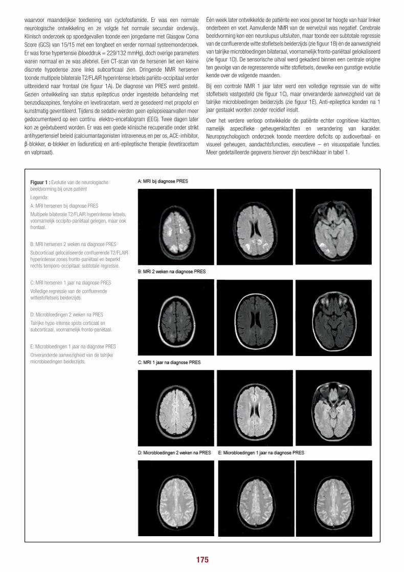
175
The outcome of posterior reversible encephalopathy syndrome (PRES) in children: a systematic review and case-report of a 16-year old girl with systemic lupus erythematosus and PRESSamantha Jeen1, Katrien Jansen2,3, Veerle Labarque4,5.
1 UZ Leuven, Dienst kindergeneeskunde 2 UZ Leuven, Dienst kindergeneeskunde, Kinderneurologie 3 Katholieke Universiteit Leuven, Departement Ontwikkeling en Regeneratie 4 UZ Leuven, Dienst kindergeneeskunde, Kinderhematologie en - Oncologie 5 Katholieke Universiteit Leuven, Departement Cardiovasculaire wetenschappen
Case Reportwaarvoor maandelijkse toediening van cyclofosfamide. Er was een normale neurologische ontwikkeling en ze volgde het normale secundair onderwijs. Klinisch onderzoek op spoedgevallen toonde een jongedame met Glasgow Coma Score (GCS) van 15/15 met een tongbeet en verder normaal systeemonderzoek. Er was forse hypertensie (bloeddruk = 229/132 mmHg), doch overige parameters waren normaal en ze was afebriel. Een CT-scan van de hersenen liet een kleine discrete hypodense zone links subcorticaal zien. Dringende NMR hersenen toonde multipele bilaterale T2/FLAIR hyperintense letsels pariëto-occipitaal verder uitbreidend naar frontaal (zie �guur 1A). De diagnose van PRES werd gesteld. Gezien ontwikkeling van status epilepticus onder ingestelde behandeling met benzodiazepines, fenytoïne en levetiracetam, werd ze gesedeerd met propofol en kunstmatig geventileerd. Tijdens de sedatie werden geen epilepsieaanvallen meer gedocumenteerd op een continu elektro-encefalogram (EEG). Twee dagen later kon ze geëxtubeerd worden. Er was een goede klinische recuperatie onder strikt antihypertensief beleid (calciumantagonisten intraveneus en per os, ACE-inhibitor, β-blokker, α-blokker en lisdiuretica) en anti-epileptische therapie (levetiracetam en valproaat).
Één week later ontwikkelde de patiënte een voos gevoel ter hoogte van haar linker onderbeen en voet. Aanvullende NMR van de wervelzuil was negatief. Cerebrale beeldvorming kon een neurolupus uitsluiten, maar toonde een subtotale regressie van de con�uerende witte sto�etsels beiderzijds (zie �guur 1B) én de aanwezigheid van talrijke microbloedingen bilateraal, voornamelijk fronto-pariëtaal gelokaliseerd (zie �guur 1D). De sensorische uitval werd gekaderd binnen een centrale origine ten gevolge van de regresserende witte sto�etsels, dewelke een gunstige evolutie kende over de volgende maanden.
Bij een controle NMR 1 jaar later werd een volledige regressie van de witte sto�etsels vastgesteld (zie �guur 1C), maar onveranderde aanwezigheid van de talrijke microbloedingen beiderzijds (zie �guur 1E). Anti-epileptica konden na 1 jaar gestaakt worden zonder recidief insult.
Over het verdere verloop ontwikkelde de patiënte echter cognitieve klachten, namelijk aspeci�eke geheugenklachten en verandering van karakter. Neuropsychologisch onderzoek toonde meerdere de�cits op audioverbaal- en visueel geheugen, aandachtsfuncties, executieve – en visuospatiale functies. Meer gedetailleerde gegevens hierover zijn beschikbaar in tabel 1.
Figuur 1 : Evolutie van de neurologische beeldvorming bij onze patiënt
Legenda:
A: MRI hersenen bij diagnose PRES
Multipele bilaterale T2/FLAIR hyperintense letsels, voornamelijk occipito-pariëtaal gelegen, maar ook frontaal.
B: MRI hersenen 2 weken na diagnose PRES
Subcorticaal gelocaliseerde con�uerende T2/FLAIR hyperintense zones fronto-pariëtaal en beperkt rechts temporo-occipitaal: subtotale regressie.
C: MRI hersenen 1 jaar na diagnose PRES
Volledige regressie van de con�uerende wittesto�etsels beiderzijds.
D: Microbloedingen 2 weken na PRES
Talrijke hypo-intense spots corticaal en subcorticaal, voornamelijk fronto-pariëtaal.
E: Microbloedingen 1 jaar na diagnose PRES
Onveranderde aanwezigheid van de talrijke microbloedingen beiderzijds.

176
Resultaten zoekstrategie – Na de gecombineerde manuele en database zoektocht werden 85 artikels gevonden, waarvan 60 via PubMed, 21 via Embase en 4 via een referentielijst. Na eliminatie van duplicaten bleven in totaal 78 artikels over. Conform de exclusiecriteria werden nog eens 19 artikels verwijderd op basis van titel en abstract en 15 op basis van volledige tekst, zodat een totaal van 44 studies geïncludeerd werd in onze systematische review. Een overzicht van dit proces wordt weergegeven in �guur 2. We includeerden 25 case reports en/of case series en 19 retrospectieve studies.
Patiënt karakteristieken – In totaal werden 410 kinderen geïncludeerd in deze review. Het betroffen 213 jongens en 197 meisjes, respectievelijk 52% en 48%. De leeftijden varieerden van 1 jaar tot 18 jaar. De onderliggende diagnoses waren uiteenlopend en kunnen ondergebracht worden in verschillende categorieën: (1) oncologisch (n = 273) waarvan 222 hematologische en 33 vaste tumoren; bij 18 kinderen werd het type tumor niet verder gespeci�eerd; (2) hematopoïetische stamceltransplantatie (n = 106), waarvan bij 89 kinderen omwille van maligniteit; (3) orgaantransplantatie (n = 12); (4) auto-immune of reumatologische
aandoening (n = 19); (5) nefrologische pathologie (n = 42) en (6) andere (n = 47). Figuur 3 geeft meer details hierover weer.
Klinische manifestatie en neurologische beeldvorming – De kinderen presenteerden zich met verschillende symptomen: in 86% van de kinderen was er sprake van epilepsie (n = 353), bij 52% was er een verminderd bewustzijn (n = 211), hoofdpijnklachten traden op in 35% (n = 142) en visusstoornissen werden gezien in zo’n 30% (n = 123). De meeste kinderen vertoonden meer dan één symptoom: bij minstens 18 kinderen werd toch slechts 1 symptoom beschreven (≥ 4%).
De aan- of afwezigheid van risicofactoren werd bij 374 patiënten beschreven (91%). In 81% (n = 303) hiervan was hypertensie aanwezig op het moment van PRES. De toediening van corticosteroïden kwam voor in 43% (n = 161) en het gebruik van calcineurine-inhibitoren in 30% (n = 112). Resultaten van een NMR onderzoek waarop PRES bevestigd werd, waren beschikbaar in 98% van de kinderen (n = 400). Slechts voor 17% van de kinderen was duidelijk hoelang na het ontstaan van klinische symptomen beeldvorming uitgevoerd werd en dit varieerde van 1 uur tot 13 dagen.
Table 1
Geheugen Score Interpretatie
Minimental State Examination (MMSE) 24/30 - Oriëntatie in tijd (-2)- Oriëntatie in ruimte (-1)- Delayed recall (-1)- Herhalen (-1)
Verminderd
Auditory Verbal Learning Test (AVLT) Prestatie op 5 trials- Trial 1: 4 (< Pc 1)- Trial 2: 6 (< Pc 1)- Trial 3: 3 (< Pc 1)- Trial 4: 7 (< Pc 1)- Trial 5: 5 (< Pc 1)
Som: 25 (< Pc 1)
Ondermaats
Rey Visual Design Learning Test (RVDLT) Prestatie op 5 trials- Trial 1: 3 (Pc 7)- Trial 2: 3 (< Pc 1)- Trial 3: 4 (< Pc 1)- Trial 4: 6 (< Pc 1)- Trial 5: 7 (< Pc 1)
Som: 23 (< Pc 1)
Ondermaats
Logisch geheugen: Vlaamse Dementie Batterij (VDB-2) Aantal elementen: 15/23 Betekenis: 5/5
Gemiddeld
Rey Fifteen Item Memory Test (FIT) 9/15 (= cut-off) Cut-off
Aandacht en Executief Functioneren Score Interpretatie
Digit Span (Wechsler Memory Scale-Revised) Voorwaarts: maximaal 3 (< Pc 1) Achterwaarts: maximaal 2 (< Pc 1)
OndermaatsIngeperkte aandachtspanne, gerichte aandacht en werkgeheugen
Stroop Color Word Task Woord: 48’’ (Pc 4)Kleur: 76’’ (< Pc 1)Interferentie: 127’’ (< Pc 1)Interferentie factor (I.F.): 65 (< Pc 1)Fouten: 14
Vertraagd werktempoPathologische interferentiefactorVerstoorde selectieve aandacht, cognitieve �exibiliteit en responsinhibitie
Trail Making Test (TMT) a-versie: 42’’ (Pc 27)b-versie: 94’’ (Pc 6)b/a: 2.23
Subtest A: gemiddeldVerwerkingssnelheid: matig gemiddeld Set –shifting en visueel alterneren: behouden
Bourdon-Wiersma Test Gem. rijtijd: 10.56 (Pc 86)Gem. rijafw: 2.27 (< Pc 1)Weglatingen: 73 (< Pc 1)Fouten: 8
Regeltijd: gemiddeldRegelafwijking: ondermaatsOnnauwkeurige werkhouding en verminderde volgehouden aandacht
Visual Elevator Task Score: 7/10 (Pc 25)Timing score: 3.74 (Pc 25-50)
GemiddeldGemiddeld werktempo
Woordvloeiendheid Letters: 28 (Pc 11)Categorie: 21 (Pc 24)
Letterwoordvlotheid: discreet ingeperktSemantische woordvlotheid: behouden
Stemming Score Interpretatie
Beck Depression Inventory (BDI) 6/63 Geen depressieve kenmerken
Visuospatiale Perceptie Score Interpretatie
The Visual Object And Space Perception Battery (VOSP) Objectdecisie: 14/20 Verminderd
Intellectueel Functioneren Score
Nederlandse Leestest voor Volwassenen (NLV) Score: 40IQ-schatting: 76 (Pc 5-9)
Zwak begaafd intellectueel functioneren
Taal Score Interpretatie
Boston Naming Test (BNT) Score: 52/60 (Pc 38) Gemiddeld
Visuoconstructieve Praxis Score Interpretatie
Tekenen Kubus: 0/1 Verminderd in rekening brengen van perspectief bij tekenen van �guren

177
Klinische outcome – In totaal zijn 91 kinderen overleden, waarvan 9 ten gevolge van PRES (2%). Bij 19 kinderen (5%) werd herval van PRES beschreven. Gegevens over morbiditeit waren bij 69% van de kinderen beschikbaar (n = 284). De duur van opvolging liep echter ver uiteen van enkele dagen tot 9 jaar. In 78% van deze kinderen (n = 222) werd een gunstige klinische outcome beschreven zonder de aanwezigheid van enige restsymptomen. Persisterende afwijkingen werden gezien bij 53 kinderen (19%): diagnose van epilepsie of gebruik van anti-epileptica op moment van publicatie (n = 26; 9%), onvolledige recuperatie van klinisch beeld (n = 5; 2%), milde cognitieve beperking (n = 2; 0.7%), blijvende paraplegie (n = 2; 0.7%), visusstoornissen (n = 2; 0.7%), ADHD (n = 1; 0.4%), chronische vermoeidheid (n = 1; 0.4%) en niet verder gespeci�eerde afwijkingen (n = 14; 5%). In een klein derde van de kinderen (n = 129; 31%) waren de beschikbare gegevens onvoldoende gedetailleerd om tot een gunstige of ongunstige uitkomst te kunnen besluiten.
Radiologische outcome – Follow-up beeldvorming was beschikbaar in 302 kinderen, wat overeenkomt met 74%. De tijd tot controle beeldvorming werd niet overal gedocumenteerd, maar was beschikbaar in 253 kinderen en liep uiteen van enkele dagen tot 42 maanden. In 23 kinderen werd beeldvorming meerdere keren herhaald en werd verdere regressie van de letsels over verloop van tijd aangetoond (range 3 – 126 maanden). In 78% (n = 236) van de 302 kinderen met minstens 1 controle NMR werden geen restletsels meer aangetoond, terwijl bij 22% (n = 66) nog persisterende afwijkingen op NMR gezien werden: verbetering, maar onvolledige resolutie van de hyperintensiteit (n = 17; 6%), bloeding (n = 12; 4%), corticale en laminaire necrose (n = 7; 2%), focale atro�e (n = 3; 1%), toename van bestaande lesies (n = 2; 0.7%), infarct (n = 1; 0.3%), gliose (n = 1; 0.3%) of niet nader omschreven (n = 26; 9%). Bij 3 kinderen werden meerdere afwijkingen genoteerd: necrose en bloeding in 2 patiënten; blijvende hyperintense letsels en bloeding in 1 patiënt.
DiscussiePosterior Reversible Encephalopathy Syndrome (PRES) is een klinisch-radiologisch syndroom met een breed spectrum van (sub)acute neurologische symptomen in combinatie met tekenen van vasogeen oedeem in de (sub)corticale posterieure regio’s van de hersenen op beeldvorming1. Het pathofysiologische mechanisme van PRES is nog niet volledig opgehelderd, maar waarschijnlijk spelen zowel hyper-en hypoperfusie als een direct cytotoxisch effect op het vasculaire endotheel een rol3,7,11-12. In de literatuur wordt bij kinderen epilepsie het meest beschreven naast een veranderd bewustzijn en hoofdpijn4,13-16. Visusstoornissen worden frequenter bij volwassenen gezien14. Ook in de casussen beschreven in deze review was epilepsie veruit het meest frequente symptoom in 86%, bewustzijnsveranderingen werden in de helft van de kinderen gezien (52%). Toch werd de diagnose bij onze casussen soms vrij laat gesteld (tot 13 dagen na begin van klachten), terwijl Roth et al. aantoonde dat snelle herkenning en behandeling door eliminatie van mogelijk uitlokkende factoren essentieel is voor een gunstige korte en lange termijn prognose17.
Gegevens over outcome in de literatuur zijn vooral bij volwassenen gerapporteerd. Neurologisch herstel wordt beschreven in 70-90% van de gevallen9,17-19. In onze systematische review kunnen we een goede neurologische outcome bij 78% van de kinderen noteren. Toch verloopt PRES niet altijd even gunstig. Mortaliteit in de literatuur varieert tussen 5 en 19%, afhankelijk van de onderliggende
Figuur 2 : Studie selectie
Figuur 3 : Overzicht van de patiëntkarakteristieken
Legenda:
A: Overzicht onderliggende diagnose
B: Overzicht type maligniteit

178
etiologie9,20-22. Epilepsie wordt in 1-4% gezien23-24 en herval van PRES komt voor in 5-10%17,25. Wij weerhouden hier een mortaliteit ten gevolge van PRES van 2%, herval van 5% en een slechte neurologische outcome bij 19%. Daarnaast toonde onze eigen casus aan dat deze cijfers mogelijks de neurologische outcome onderschatten. Het meisje in kwestie vertoonde immers recuperatie van het klinisch neurologische beeld, kon anti-epileptica stoppen zonder recidief insult, maar ontwikkelde later neuropsychologische afwijkingen die aan de hand van testing beter in kaart gebracht konden worden (zie tabel 1). Natuurlijk kan niet met zekerheid gezegd worden dat deze afwijkingen volledig ten gevolge van PRES zijn, aangezien er geen voorgaande neuropsychologische toetsing beschikbaar is. Het feit dat ze voordien normaal secundair onderwijs gevolgd heeft, suggereert dit echter wel. Daarenboven beschrijven Stroescu et al. in 2011 twee volwassen patiënten met PRES bij wie neuropsychologische investigatie een gelijkaardig patroon vertoonde, namelijk vooral een beperking in de spatio-perceptuele en gerelateerde functies, naast de executieve functies26.
Beeldvorming kon bij onze patiënt enkel talrijke microbloedingen in het licht stellen, wat als vermoedelijke verklaring van de neuropsychologische afwijkingen werd gezien. Dergelijke microbloedingen worden bij meer dan de helft van de volwassen patiënten met PRES beschreven (58%)27, maar zijn mogelijks zeldzamer bij kinderen (13.5%)28. Bloeding (intraparenchymateus en/of subarachnoïdaal) werd in de literatuur reeds geassocieerd aan een slechte outcome (Odds Ratio 4.93)29, maar er zijn geen gegevens over de gevolgen van microbloedingen. In onze literatuur search werden nog 5 andere kinderen met klinisch neurocognitieve stoornissen beschreven. Geen enkele hiervan werd onderzocht door middel van gestandaardiseerde neurocognitieve testen en het is ook onduidelijk of zij al dan niet afwijkingen vertoonden op controle beeldvorming. Anderzijds werden ook de andere patiënten niet neurocognitief getest, waardoor we niet kunnen inschatten of sommige patiënten subklinische neurocognitieve problemen vertoonden. Gezien er vooral retrospectieve studies gepubliceerd zijn, is het immers ook mogelijk dat er niet altijd controle beeldvorming plaatsvond bij (schijnbaar) volledige klinische recuperatie met een optimistisch inschatten van de outcome als gevolg.
De sterktes en beperkingen van deze review hangen uiteraard samen met de kwaliteit en rapportering van gegevens in de geïncludeerde studies. Eerst en vooral is er een zeer grote variabiliteit in de duur van klinische en radiologische opvolging. Gungor et al. toonde echter aan dat radiologisch herstel vaak pas na meer dan 1 maand optreedt, waardoor verdere regressie of zelfs volledige resolutie van de gerapporteerde letsels in de loop van de tijd nog zou opgetreden zijn bij patiënten met kortere opvolging30. Bovendien kan een bias in het al dan niet uitvoeren van controle beeldvorming op geleide van de kliniek de gegevens beïnvloeden. Tot slot maakten het ontbreken van data of onvoldoende details een juiste interpretatie en inschatting vaak moeilijk. Dit neemt echter niet weg dat deze systematische review aangeeft dat PRES bij kinderen zeker niet altijd reversibel is en dat ook neurocognitieve problemen aanwezig kunnen zijn, mogelijks zelfs zonder epilepsie of motorische sequellen.
ConclusieEr zijn hoegenaamd geen gegevens beschikbaar in de huidige literatuur omtrent de neurocognitieve outcome van PRES bij kinderen. In deze review wordt een gunstige klinische en radiologische outcome na PRES beschreven in slechts 78%, waaruit blijkt dat PRES niet zo onschuldig en reversibel is. In onze casus lijkt er daarenboven een verminderd cognitief functioneren te zijn. Het is dan ook van groot belang om een beter zicht te krijgen op de morbiditeit en neurocognitie na het doormaken van PRES. Als eerste stap hiertoe zou het nuttig kunnen zijn gegevens van alle Belgische patiënten samen te brengen, maar in de toekomst dienen best prospectieve follow-up studies met neurocognitieve testing opgezet te worden.
4. Liman TG, Bohner G, Heuschmann PU, Endres M, Siebert E. The clinical and radiological spectrum of posterior reversible encephalopathy syndrome: the retrospective Berlin PRES study. J Neurol 2012; 259: 155-164.
5. Bartynski WS, Boardman JF. Distinct imaging patterns and lesion distribution in posterior reversible encephalopathy syndrome. AJNR Am J Neuroradiol 2007; 28: 1320–1327.
6. Covarrubias DJ, Luetmer PH, Campeau NG. Posterior reversible encephalopathy syndrome: prognostic utility of quantitative diffusion-weighted MR images. AJNR Am J Neuroradiol 2002; 23:1038.
7. Rykken JB, McKinney AM. Posterior Reversible Encephalopathy Syndrome. Semin Ultrasound CT MRI 2014; 35: 118-135.
8. Narbone MC, Musolino R, Granata F, Mazzù I, Abbate M, Ferlazzo E. PRES: posterior or potentially reversible encephalopathy syndrome? Neurol Sci 2006; 27: 187-189.
9. Liman TG, Bohner G, Endres M, Siebert E. Discharge status and in-hospital mortality in posterior reversible encephalopathy syndrome. Acta Neurol Scand 2014; 130: 34–39.
10. Flynn JT, Kaelber DC, Baker-Smith CM, Blowey D, Carroll AE, Daniels SR, et al. Subcommitee on Screening and Management of High Blood Pressure in Children. Clinical Practice Guideline for Screening and Management of High Blood Pressure in Children and Adolescents. Pediatrics 2017; 140(3): e20171904.
11. Bartynski WS. Posterior Reversible Encephalopathy Syndrome, Part 2: Controversies Surrounding Pathophysiology of Vasogenic Edema. AJNR Am J Neuroradiol 2008; 29: 1043-1049.
12. Marra et al. Posterior reversible encephalopathy syndrome: The endothelial Hypotheses. Medical Hypotheses 2014; 82: 619–622.
13. Fugate JE, Claassen DO, Cloft HJ, et al. Posterior reversible encephalopathy syndrome: associated clinical and radiologic �ndings. Mayo Clin Proc 2010; 85: 427–432.
14. Siebert E, Bohner G, Endres M, Liman TG. Clinical and radiological spectrum of posterior reversible encephalopathy syndrome: does age make a difference? A retrospective comparison between adult and pediatric patients. PLoS One 2014; 9: e115073.
15. Gümüş H, Per H, Kumandaş S, Yikilmaz A. Reversible posterior leukoencephalopathy syndrome in childhood: report of nine cases and review of the literature. Neurol Sci 2010; 31: 125–131.
16. Siebert E, Spors B, Bohner G, et al. Posterior reversible encephalopathy syndrome in children: radiological and clinical �ndings - a retrospective analysis of a German tertiary care center. Eur J Paediatr Neurol 2013; 17: 169–175.
17. Roth C, Ferbert A. Posterior reversible encephalopathy syndrome: long-term follow-up. J Neurol Neurosurg Psychiatry 2010; 81: 773–777.
18. Burnett MM, Hess CP, Roberts JP, Bass NM, Douglas VD, Josephson SA. Presentation of reversible posterior leukoencephalopathy syndrome in patients on calcineurin inhibitors. Clin Neurol Neurosurg 2010; 112: 886–891.
19. Cui HW, Lei RY, Zhang SG, Han LS, Zhang BA. Clinical features, outcomes and risk factors for posterior reversible encephalopathy syndrome in systemic lupus erythematosus: a case control study. Lupus 2019 jul; 28(8): 961-969.
20. Legriel S, Schraub O, Azoulay E, Hantson P, Magalhaes E, Coquet I, Bretonniere C et al. Determinants of recovery from severe posterior reversible encephalopathy syndrome. PLoS One 2012; 7: e44534.
21. Alhilali LM, Reynolds AR, Fakhran S. A multi-disciplinary model of risk factors for fatal outcome in posterior reversible encephalopathy syndrome. J Neurol Sci 2014; 347: 59-65.
22. Hinduja A, Habetz K, Raina S, Ramakrishnaiah R, Fitzgerald RT. Predictors of poor outcome in patients with posterior reversible encephalopathy syndrome. Int J Neurosci 2017; 127:135–144.
23. Datar S, Singh T, Rabinstein AA, Fugate JE, Hocker S. Long-term risk of seizures and epilepsy in patients with posterior reversible encephalopathy syndrome. Epilepsia 2015; 56: 564-568.
24. Heo K, Cho KH, Lee MK, Chung SJ, Cho YJ, Lee BI. Development of epilepsy after posterior reversible encephalopathy syndrome. Seizure 2016; 34: 90-94.
25. Li R, Mitchell P, Dowling R, Yan B. Is hypertension predictive of clinical recurrence in posterior reversible encephalopathy syndrome? J Clin Neurosci 2013; 20: 248–252.
26. Stroescu I, Salinas CM, Nahab FB, Stringer AY. Long-term neurocognitive and neuroimaging outcomes in posterior reversible encephalopathy syndrome: two case reports and implications. Clin Neuropsychol 2011; 25(8): 1386-1402.
27. McKinney AM, Sarikaya B, Gustafson C, Truwit CL. Detection of microhemorrhage in Posterior Reversible Encephalopathy Syndrome using susceptibility-weighted imaging. Am J Neuroradiol may 2012; 33(5): 896-903.
28. Khan RB, Sadighi ZS, Zabrowski J, Gajjar A, Jeha S. Imaging patterns and outcome of posterior reversible encephalopathy syndrome during childhood cancer treatment. Pediatr Blood Cancer 2016; 63(3): 523-526.
29. Chen Z, Zhang G, Lerner A, Wang AH, Gao B, Liu J. Risk factors for poor outcome in posterior reversible encephalopathy syndrome: systematic review and meta-analysis. Quant Imaging Med Surg 2018; 8: 421–432.
30. 30. Gungor S, Kilic B, Tabel Y, Selimoglu A, Ozgen U, Yilmaz S, Sigirci A. Clinical and Imaging Findings in Childhood Posterior Reversible Encephalopathy Syndrome. Iran J Child Neurol. Winter 2018; 12(1): 16-25.
1. Hinchey J, Chaves C, Appignani B, Breen J, Pao L, Wang A et al. A reversible posterior leukoencephalopathy syndrome. N Engl J Med 1996; 334(8):494–500.
2. Kwon S, Koo J, Lee S. Clinical spectrum of reversible posterior leukoencephalopathy syndrome. Pediatr Neurol 2001; 24: 361-364.
3. Bartynski WS. Posterior Reversible Encephalopathy Syndrome, Part 1: Fundamental Imaging and Clinical Features. AJNR Am J Neuroradiol 2008; 29: 1036-1042.
REFERENCES:
Publieksprijs België 86,52€
VERKORTE SAMENVATTING VAN DE PRODUCTKENMERKEN Gelieve de Samenvatti ng van de Productkenmerken te raadplegen voor de volledige informati e over het gebruik van dit geneesmiddel. NAAM VAN HET GENEESMIDDEL Bexsero suspensie voor injecti e in voorgevulde spuit Meningokokken groep Bvaccin (rDNA, component, geadsorbeerd) - EU/1/12/812/001 Farmacotherapeuti sche categorie: meningokokkenvaccins, ATCcode: J07AH09 KWALITATIEVE EN KWANTITATIEVE SAMENSTELLING Een dosis (0,5 ml) bevat: Recombinant Neisseria meningiti dis groep B NHBAfusieeiwit 1, 2, 3 : 50 microgram Recombinant Neisseria meningiti dis groep B NadAeiwit 1, 2, 3: 50 microgram Recombinant Neisseria meningiti dis groep B fHbpfusieeiwit 1, 2, 3: 50 microgram Buitenmembraanvesikels (BMV) van Neisseria meningiti dis groep Bstam NZ98/254, gemeten als hoeveelheid totaal eiwit dat PorA P1.4 bevat 2: 25 microgram 1 Geproduceerd in E. colicellen door recombinantDNAtechnologie 2 Geadsorbeerd aan aluminiumhydroxide (0,5 mg Al³+) 3 NHBA (Neisseria heparinebindend anti geen), NadA (Neisseriaadhesine A), fHbp (factor Hbindend eiwit) THERAPEUTISCHE INDICATIES Bexsero is g eïndiceerd voor de acti eve immunisati e van personen van 2 maanden en ouder tegen invasieve meningokokkenziekte veroorzaakt door Neisseria meningiti dis groep B. Bij het vaccineren moet rekening worden gehouden met het eff ect van invasieve ziekte bij verschillende lee ijdsgroepen, evenals met de variabiliteit van de epidemiologie van anti genen voor groep Bstammen in verschillende geografi sche gebieden. Zie rubriek 5.1 van de volledige SPK voor informati e over bescherming tegen specifi eke groep Bstammen. Dit vaccin dient te worden gebruikt in overeenstemming met offi ciële aanbevelingen. DOSERING EN WIJZE VAN TOEDIENING DoseringTa el 1 Samenva ng van de dosering
ee ijd ij eerste dosis Primaire immunisati e Intervallen tussen primaire doses ooster
uigelingen van tot en met maanden aDrie doses, elk van 0,5 ml Niet minder dan 1 maand Ja, één dosis tussen 12 en 15 maanden oud met een interval van ten minste 6 maanden tussen de primaire serie en
de boosterdosis b, cTwee doses, elk van 0,5 ml Niet minder dan 2 maanden
uigelingen van tot en met 11 maanden Twee doses, elk van 0,5 ml Niet minder dan 2 maanden Ja, één dosis in het tweede levensjaar met een interval van minimaal 2 maanden tussen de primaire serie en de boosterdosis c
inderen van 1 tot en met maanden Twee doses, elk van 0,5 ml Niet minder dan 2 maanden Ja, één dosis met een interval van 12 tot en met 23 maanden tussen de primaire serie en de boosterdosis c
inderen van tot en met 10 jaarTwee doses, elk van 0,5 ml Niet minder dan 1 maand Een boosterdosis dient overwogen te worden bij personen met een blijvend risico op blootstelling aan
meningokokkenziekte, op basis van offi ciële aanbevelingen dAdolescenten (11 jaar of ouder) en vol assenen*
a De eerste dosis moet niet worden gegeven op de lee ijd jonger dan 2 maanden. De veiligheid en werkzaamheid van Bexsero bij zuigelingen jonger dan 8 weken zijn nog niet vastgesteld. Er zijn geen gegevens beschikbaar. b In geval van uitstel mag de booster niet later dan op een lee ijd van 24 maanden worden gegeven. c Zie rubriek 5.1 van de volledige SPK. De noodzaak voor en ti jdsplanning van een boosterdosis na dit vaccinati eschema is niet vastgesteld d Zie rubriek 5.1 van de volledige SPK. *Gegevens over volwassenen ouder dan 50 jaar ontbreken. ijze van toediening Het vaccin wordt toegediend via een diepe intramusculaire injecti e, bij voorkeur in het anterolaterale gedeelte van de dij bij zuigelingen, of in de streek van de deltaspier van de bovenarm bij oudere personen. Als meer dan één vaccin tegelijk wordt toegediend, moeten afzonderlijke injecti eplaatsen worden gebruikt. Het vaccin mag niet intraveneus, subcutaan of intradermaal worden toegediend, en mag niet worden gemengd met andere vaccins in dezelfde spuit. Voor instructi es over het hanteren van het vaccin voorafgaand aan toediening, zie rubriek 6.6 van de volledige SPK. CONTRAINDICATIES Overgevoeligheid voor de werkzame stof(fen) of voor een van de in rubriek 6.1 van de volledige SPK vermelde hulpstof(fen). BIJZONDERE WAARSCHUWINGEN EN VOORZORGEN BIJ GEBRUIK Zoals dat voor alle vaccins geldt, dient ook toediening van Bexsero te worden uitgesteld bij personen die lijden aan een acute, ernsti ge, met koorts gepaard gaande ziekte. De aanwezigheid van een lichte infecti e, zoals verkoudheid, mag echter niet leiden tot uitstel van vaccinati e. Niet intravasculair injecteren. Zoals dat voor alle injecteerbare vaccins geldt, dienen passende medische behandeling en toezicht alti jd direct beschikbaar te zijn voor het geval zich na toediening van het vaccin een anafylacti sche reacti e voordoet. Reacti es die verband houden met angst, waaronder vasovagale reacti es (syncope), hyperventi lati e of stressgerelateerde reacti es, kunnen in relati e met vaccinati e voorkomen als psychogene reacti e op de naaldinjecti e (zie rubriek Bijwerkingen ). Het is belangrijk dat er passende procedures zijn om letsel als gevolg van fl auwvallen te voorkomen. Dit vaccin mag niet worden toegediend aan personen met trombocytopenie of een bloedstollingsstoornis die een contraindicati e voor intramusculaire injecti e vormt, tenzij het mogelijke voordeel duidelijk opweegt tegen het risico van toediening. Zoals dat voor alle vaccins geldt, beschermt vaccinati e met Bexsero mogelijk niet alle gevaccineerden. Bexsero wordt niet geacht bescherming te bieden tegen alle circulerende meningokokken Bstammen. Zoals dat voor veel vaccins geldt, moet het medisch personeel zich ervan bewust zijn dat een temperatuurssti jging kan optreden na vaccinati e van zuigelingen en kinderen (jonger dan 2 jaar). Profylacti sche toediening van anti pyreti ca gelijkti jdig met en meteen na vaccinati e kan de incidenti e en intensiteit van koortsreacti es na vaccinati e verminderen. Anti pyreti sche medicati e dient te worden gestart volgens de lokale richtlijnen bij zuigelingen en kinderen (jonger dan 2 jaar). Personen met een immunodefi ciënti e, door het gebruik van immunosupressieve therapie, een geneti sche stoornis, of door een andere oorzaak, kunnen een verlaagde anti lichaamrespons hebben bij acti eve immunisati e. Immunogeniciteitsgegevens zijn beschikbaar van personen met complementdefi ciënti e, asplenie of miltdisfuncti es. Personen met familiale complementdefi ciënti es (bijvoorbeeld C3- of C5-defi ciënti es) en personen die behandelingen ondergaan die de terminale complementacti vati e remmen (bijvoorbeeld eculizumab) hebben een hoger risico op een invasieve ziekte veroorzaakt door Neisseria meningiti dis groep B, zelfs als deze personen anti lichamen ontwikkelen na vaccinati e met Bexsero. Er zijn geen gegevens over het gebruik van Bexsero bij personen ouder dan 50 jaar en beperkte gegevens bij pati ënten met chronische medische aandoeningen. anneer de primaire immunisati eserie aan zeer premature zuigelingen (geboren na 28 weken zwangerschap) wordt toegediend, moet rekening worden gehouden met een potenti eel risico op apneu en de noodzaak van controle van de ademhaling gedurende 4872 uur, vooral bij zuigelingen met een voorgeschiedenis van onvolgroeide longen. Aangezien het voordeel van vaccinati e groot is bij deze groep zuigelingen, moet vaccinati e niet worden onthouden of uitgesteld. De dop van de injecti espuit bevat mogelijk natuurlijk rubber (latex). Hoewel het risico op het ontwikkelen van allergische reacti es zeer klein is, moet het medisch personeel de voor en nadelen goed afwegen voordat dit vaccin wordt toegediend aan personen met een bekende voorgeschiedenis van overgevoeligheid voor latex. Kanamycine wordt aan het begin van het producti eproces gebruikt en wordt in latere producti estadia verwijderd. Indien aanwezig, bedraagt het kanamycineniveau in het uiteindelijke vaccin minder dan 0,01 microgram per dosis. Veilig gebruik van Bexsero bij personen die gevoelig zijn voor kanamycine is niet vastgesteld. Terugvinden herkomst Om het terugvinden van de herkomst van biologicals te verbeteren moeten de naam en het batchnummer van het toegediende product goed geregistreerd worden. BIJWERKINGEN Overzicht van het veiligheidsprofi el De veiligheid van Bexsero is geëvalueerd in 17 onderzoeken, inclusief 10 gerandomiseerde gecontroleerde klinische studies met 10.565 proefpersonen (vanaf de lee ijd van 2 maanden) die minimaal één dosis Bexsero toegediend kregen. Van de personen die Bexsero toegediend kregen, waren 6.837 zuigelingen en kinderen (jonger dan 2 jaar), 1.051 kinderen (van 2 tot 10 jaar) en 2.677 adolescenten en volwassenen. Van de proefpersonen die de primaire immunisati eserie voor zuigelingen van Bexsero toegediend kregen, kregen 3.285 een boosterdosis in het tweede levensjaar. De meest voorkomende lokale en systemische bijwerkingen bij zuigelingen en kinderen (jonger dan 2 jaar) die in klinische studies zijn waargenomen, waren gevoeligheid en erytheem op de injecti eplaats, koorts en prikkelbaarheid. In klinische onderzoeken bij zuigelingen gevaccineerd op de lee ijd van 2, 4 en 6 maanden, is bij 69% tot 79% van de proefpersonen melding gemaakt van koorts (≥ 38°C) wanneer Bexsero gelijkti jdig werd toegediend met standaardvaccins (die de volgende anti genen bevatt en: 7valent pneumokokkenconjugaat, di erie, tetanus, acellulair pertussis, hepati ti s B, geïnacti veerde poliomyeliti s en Haemophilus infl uenzae type b) in vergelijking met 44% tot 59% van de proefpersonen die alleen de standaardvaccins kregen toegediend. Bij zuigelingen die Bexsero en standaardvaccins toegediend kregen, is ook vaker melding gemaakt van het gebruik van anti pyreti ca. anneer alleen Bexsero werd toegediend, kwam koorts bij zuigelingen even vaak voor als bij standaardzuigelingenvaccins die ti jdens klinische studies werden toegediend. Eventuele koorts volgde in het algemeen een voorspelbaar patroon, waarbij de meeste koortsgevallen de dag na de vaccinati e over waren. De meest voorkomende lokale en systemische bijwerkingen waargenomen bij adolescenten en volwassenen waren pijn op de injecti eplaats, malaise en hoofdpijn. Er is geen toename waargenomen in de incidenti e of ernst van bijwerkingen bij opeenvolgende doses in de vaccinati ereeks. Tabel met bijwerkingen Bijwerkingen (na primaire immunisati e of boosterdosis) die ten minste als mogelijk gerelateerd aan de vaccinati e kunnen worden beschouwd, zijn naar frequenti e ingedeeld. De frequenti e is als volgt geclassifi ceerd: Zeer vaak: (≥1/10) Vaak: (≥1/100, <1/10) Soms: (≥1/1.000, <1/100) Zelden: (≥1/10.000, <1/1.000) Zeer zelden: (<1/10.000) Niet bekend: (kan met de beschikbare gegevens niet worden bepaald) De bijwerkingen worden binnen elke frequenti egroep gerangschikt in afl opende volgorde van ernst. Naast de meldingen uit klinische onderzoeken, zijn ook de wereldwijd ontvangen vrijwillige meldingen over bijwerkingen van Bexsero sinds de introducti e op de markt in de volgende lijst opgenomen. Aangezien deze bijwerkingen vrijwillig zijn gemeld door een populati e van onbekende omvang, is het niet alti jd mogelijk om een betrouwbare schatti ng van de frequenti e te geven en worden ze daarom hier vermeld met de frequenti e Niet bekend.
uigelingen en inderen (tot en met 10 jaar) Immuunsysteemaandoeningen Niet bekend: allergische reacti es (waaronder anafylacti sche reacti es) Voedings en stofwisselingsstoornissen Zeer vaak: eetstoornissen Zenuwstelselaandoeningen Zeer vaak: slaperigheid, ongewoon huilen, hoofdpijn Soms: insulten (inclusief febriele insulten) Niet bekend: hypotoon-hyporesponsieve episode, meningeale prikkeling (tekenen van meningeale prikkeling zoals sti j eid van de nek of fotofobie zijn kort na de vaccinati e sporadisch gemeld. Deze symptomen waren mild en van voorbijgaande aard). Bloedvataandoeningen Soms: bleekheid (zelden na booster) Zelden: ziekte van Kawasaki Maagdarmstelselaandoeningen Zeer vaak: diarree, braken (soms na booster) Huid en onderhuidaandoeningen Zeer vaak: huiduitslag (kinderen van 12 tot en met 23 maanden) (soms na booster) Vaak: huiduitslag (zuigelingen en kinderen van 2 tot en met 10 jaar) Soms: eczeem Zelden: urti caria Skeletspierstelsel en bindweefselaandoeningen Zeer vaak: artralgie Algemene aandoeningen en toedieningsplaatsstoornissen Zeer vaak: koorts (≥38°C), gevoeligheid op de injecti eplaats (inclusief ernsti ge gevoeligheid op de injecti eplaats, gedefi nieerd als huilen wanneer de geïnjecteerde ledemaat wordt bewogen), erytheem op de injecti eplaats, zwelling op de injecti eplaats, verharding op de injecti eplaats, prikkelbaarheid Soms: koorts (≥40°C) Niet bekend: injecti eplaatsreacti es (inclusief uitgebreide zwelling van de gevaccineerde ledemaat, blaren op of rondom de injecti eplaats en een nodus op de injecti eplaats die meer dan een maand kan aanhouden) Adolescenten (van 11 jaar en ouder) en vol assenen Immuunsysteemaandoeningen Niet bekend: allergische reacti es (waaronder anafylacti sche reacti es) Zenuwstelselaandoeningen Zeer vaak: hoofdpijn Niet bekend: syncope of vasovagale reacti es op een injecti e, meningeale prikkeling (tekenen van meningeale prikkeling zoals sti j eid van de nek of fotofobie zijn kort na de vaccinati e sporadisch gemeld. Deze symptomen waren mild en van voorbijgaande aard). Maagdarmstelselaandoeningen Zeer vaak: misselijkheid Skeletspierstelsel en bindweefselaandoeningen Zeer vaak: myalgie, artralgie Algemene aandoeningen en toedieningsplaatsstoornissen Zeer vaak: pijn op de injecti eplaats (inclusief ernsti ge pijn op de injecti eplaats, gedefi nieerd als niet in staat normale dagelijkse acti viteiten uit te voeren), zwelling op de injecti eplaats, verharding op de injecti eplaats, erytheem op de injecti eplaats, malaise Niet bekend: koorts, injecti eplaatsreacti es (inclusief uitgebreide zwelling van de gevaccineerde ledemaat, blaren op of rondom de injecti eplaats en een nodus op de injecti eplaats die meer dan een maand kan aanhouden) Melding van vermoedelijke bijwerkingen Het is belangrijk om na toelati ng van het geneesmiddel vermoedelijke bijwerkingen te melden. Op deze wijze kan de verhouding tussen voordelen en risico’s van het geneesmiddel voortdurend worden gevolgd. Beroepsbeoefenaren in de gezondheidszorg wordt verzocht alle vermoedelijke bijwerkingen te melden via het nati onale meldsysteem: elgi Federaal agentschap voor geneesmiddelen en gezondheidsproducten Afdeling Vigilanti e Postbus 97 B-1000 Brussel Madou ebsite: www.fagg.be e-mail: adversedrugreacti [email protected] u em urg Centre Régional de Pharmacovigilance de Nancy Bâti ment de Biologie Moléculaire et de Biopathologie (BBB) CHRU de Nancy – Hôpitaux de Brabois Rue du Morvan 54 511 VANDOEUVRE LES NANCY CEDEX Tél : (+33) 3 83 65 60 85 / 87 Fax : (+33) 3 83 65 61 33 E-mail : [email protected] ou Directi on de la Santé Division de la Pharmacie et des Médicaments Allée Marconi - Villa Louvigny L-2120 Luxembourg Tél. : (+352) 2478 5592 Fax : (+352) 2479 5615 E-mail : [email protected] Link pour le formulaire : htt p://www.sante.public.lu/fr/politi que-sante/ministere-sante/directi on-sante/div-pharmacie-medicaments/index.html HOUDER VAN DE VERGUNNING VOOR HET IN DE HANDEL BRENGEN GSK Vaccines S.r.l, Via Fiorenti na 1, 53100 Siena, Italië DATUM VAN DE GOEDKEURING VAN DE TEKST 04/2020 (v10) AFLEVERINGSWIJZE Op medisch voorschri . 1. Medini D, Stella M, Wassil J, Vaccine 2015; 33; 2629-2636. 2. Bexsero SMPC. PM-BE-BEX-ADV-200002 - Juni 2020 - V. .: GlaxoSmithKline Pharmaceuti cals s.a., av Pascal 2-4-6, 1300 Wavre
Vaccin tegen meningokokken van groep B(rDNA, component, geadsorbeerd)
Het eerste vaccin tegen meningokokken van serogroep B.1
Het enige geïndiceerd vanaf 2 maanden.1,2
voor zuigelingen vanaf de leeftijd van 2 maanden.1
NIEUWE UPDATE
VACCINATIESCHEMA!
2020-136-GSKBEX-Ad A4 2+1 juin 2020 NL_BAT.indd 1 26/06/20 10:18

179
Publieksprijs België 86,52€
VERKORTE SAMENVATTING VAN DE PRODUCTKENMERKEN Gelieve de Samenvatti ng van de Productkenmerken te raadplegen voor de volledige informati e over het gebruik van dit geneesmiddel. NAAM VAN HET GENEESMIDDEL Bexsero suspensie voor injecti e in voorgevulde spuit Meningokokken groep Bvaccin (rDNA, component, geadsorbeerd) - EU/1/12/812/001 Farmacotherapeuti sche categorie: meningokokkenvaccins, ATCcode: J07AH09 KWALITATIEVE EN KWANTITATIEVE SAMENSTELLING Een dosis (0,5 ml) bevat: Recombinant Neisseria meningiti dis groep B NHBAfusieeiwit 1, 2, 3 : 50 microgram Recombinant Neisseria meningiti dis groep B NadAeiwit 1, 2, 3: 50 microgram Recombinant Neisseria meningiti dis groep B fHbpfusieeiwit 1, 2, 3: 50 microgram Buitenmembraanvesikels (BMV) van Neisseria meningiti dis groep Bstam NZ98/254, gemeten als hoeveelheid totaal eiwit dat PorA P1.4 bevat 2: 25 microgram 1 Geproduceerd in E. colicellen door recombinantDNAtechnologie 2 Geadsorbeerd aan aluminiumhydroxide (0,5 mg Al³+) 3 NHBA (Neisseria heparinebindend anti geen), NadA (Neisseriaadhesine A), fHbp (factor Hbindend eiwit) THERAPEUTISCHE INDICATIES Bexsero is g eïndiceerd voor de acti eve immunisati e van personen van 2 maanden en ouder tegen invasieve meningokokkenziekte veroorzaakt door Neisseria meningiti dis groep B. Bij het vaccineren moet rekening worden gehouden met het eff ect van invasieve ziekte bij verschillende lee ijdsgroepen, evenals met de variabiliteit van de epidemiologie van anti genen voor groep Bstammen in verschillende geografi sche gebieden. Zie rubriek 5.1 van de volledige SPK voor informati e over bescherming tegen specifi eke groep Bstammen. Dit vaccin dient te worden gebruikt in overeenstemming met offi ciële aanbevelingen. DOSERING EN WIJZE VAN TOEDIENING DoseringTa el 1 Samenva ng van de dosering
ee ijd ij eerste dosis Primaire immunisati e Intervallen tussen primaire doses ooster
uigelingen van tot en met maanden aDrie doses, elk van 0,5 ml Niet minder dan 1 maand Ja, één dosis tussen 12 en 15 maanden oud met een interval van ten minste 6 maanden tussen de primaire serie en
de boosterdosis b, cTwee doses, elk van 0,5 ml Niet minder dan 2 maanden
uigelingen van tot en met 11 maanden Twee doses, elk van 0,5 ml Niet minder dan 2 maanden Ja, één dosis in het tweede levensjaar met een interval van minimaal 2 maanden tussen de primaire serie en de boosterdosis c
inderen van 1 tot en met maanden Twee doses, elk van 0,5 ml Niet minder dan 2 maanden Ja, één dosis met een interval van 12 tot en met 23 maanden tussen de primaire serie en de boosterdosis c
inderen van tot en met 10 jaarTwee doses, elk van 0,5 ml Niet minder dan 1 maand Een boosterdosis dient overwogen te worden bij personen met een blijvend risico op blootstelling aan
meningokokkenziekte, op basis van offi ciële aanbevelingen dAdolescenten (11 jaar of ouder) en vol assenen*
a De eerste dosis moet niet worden gegeven op de lee ijd jonger dan 2 maanden. De veiligheid en werkzaamheid van Bexsero bij zuigelingen jonger dan 8 weken zijn nog niet vastgesteld. Er zijn geen gegevens beschikbaar. b In geval van uitstel mag de booster niet later dan op een lee ijd van 24 maanden worden gegeven. c Zie rubriek 5.1 van de volledige SPK. De noodzaak voor en ti jdsplanning van een boosterdosis na dit vaccinati eschema is niet vastgesteld d Zie rubriek 5.1 van de volledige SPK. *Gegevens over volwassenen ouder dan 50 jaar ontbreken. ijze van toediening Het vaccin wordt toegediend via een diepe intramusculaire injecti e, bij voorkeur in het anterolaterale gedeelte van de dij bij zuigelingen, of in de streek van de deltaspier van de bovenarm bij oudere personen. Als meer dan één vaccin tegelijk wordt toegediend, moeten afzonderlijke injecti eplaatsen worden gebruikt. Het vaccin mag niet intraveneus, subcutaan of intradermaal worden toegediend, en mag niet worden gemengd met andere vaccins in dezelfde spuit. Voor instructi es over het hanteren van het vaccin voorafgaand aan toediening, zie rubriek 6.6 van de volledige SPK. CONTRAINDICATIES Overgevoeligheid voor de werkzame stof(fen) of voor een van de in rubriek 6.1 van de volledige SPK vermelde hulpstof(fen). BIJZONDERE WAARSCHUWINGEN EN VOORZORGEN BIJ GEBRUIK Zoals dat voor alle vaccins geldt, dient ook toediening van Bexsero te worden uitgesteld bij personen die lijden aan een acute, ernsti ge, met koorts gepaard gaande ziekte. De aanwezigheid van een lichte infecti e, zoals verkoudheid, mag echter niet leiden tot uitstel van vaccinati e. Niet intravasculair injecteren. Zoals dat voor alle injecteerbare vaccins geldt, dienen passende medische behandeling en toezicht alti jd direct beschikbaar te zijn voor het geval zich na toediening van het vaccin een anafylacti sche reacti e voordoet. Reacti es die verband houden met angst, waaronder vasovagale reacti es (syncope), hyperventi lati e of stressgerelateerde reacti es, kunnen in relati e met vaccinati e voorkomen als psychogene reacti e op de naaldinjecti e (zie rubriek Bijwerkingen ). Het is belangrijk dat er passende procedures zijn om letsel als gevolg van fl auwvallen te voorkomen. Dit vaccin mag niet worden toegediend aan personen met trombocytopenie of een bloedstollingsstoornis die een contraindicati e voor intramusculaire injecti e vormt, tenzij het mogelijke voordeel duidelijk opweegt tegen het risico van toediening. Zoals dat voor alle vaccins geldt, beschermt vaccinati e met Bexsero mogelijk niet alle gevaccineerden. Bexsero wordt niet geacht bescherming te bieden tegen alle circulerende meningokokken Bstammen. Zoals dat voor veel vaccins geldt, moet het medisch personeel zich ervan bewust zijn dat een temperatuurssti jging kan optreden na vaccinati e van zuigelingen en kinderen (jonger dan 2 jaar). Profylacti sche toediening van anti pyreti ca gelijkti jdig met en meteen na vaccinati e kan de incidenti e en intensiteit van koortsreacti es na vaccinati e verminderen. Anti pyreti sche medicati e dient te worden gestart volgens de lokale richtlijnen bij zuigelingen en kinderen (jonger dan 2 jaar). Personen met een immunodefi ciënti e, door het gebruik van immunosupressieve therapie, een geneti sche stoornis, of door een andere oorzaak, kunnen een verlaagde anti lichaamrespons hebben bij acti eve immunisati e. Immunogeniciteitsgegevens zijn beschikbaar van personen met complementdefi ciënti e, asplenie of miltdisfuncti es. Personen met familiale complementdefi ciënti es (bijvoorbeeld C3- of C5-defi ciënti es) en personen die behandelingen ondergaan die de terminale complementacti vati e remmen (bijvoorbeeld eculizumab) hebben een hoger risico op een invasieve ziekte veroorzaakt door Neisseria meningiti dis groep B, zelfs als deze personen anti lichamen ontwikkelen na vaccinati e met Bexsero. Er zijn geen gegevens over het gebruik van Bexsero bij personen ouder dan 50 jaar en beperkte gegevens bij pati ënten met chronische medische aandoeningen. anneer de primaire immunisati eserie aan zeer premature zuigelingen (geboren na 28 weken zwangerschap) wordt toegediend, moet rekening worden gehouden met een potenti eel risico op apneu en de noodzaak van controle van de ademhaling gedurende 4872 uur, vooral bij zuigelingen met een voorgeschiedenis van onvolgroeide longen. Aangezien het voordeel van vaccinati e groot is bij deze groep zuigelingen, moet vaccinati e niet worden onthouden of uitgesteld. De dop van de injecti espuit bevat mogelijk natuurlijk rubber (latex). Hoewel het risico op het ontwikkelen van allergische reacti es zeer klein is, moet het medisch personeel de voor en nadelen goed afwegen voordat dit vaccin wordt toegediend aan personen met een bekende voorgeschiedenis van overgevoeligheid voor latex. Kanamycine wordt aan het begin van het producti eproces gebruikt en wordt in latere producti estadia verwijderd. Indien aanwezig, bedraagt het kanamycineniveau in het uiteindelijke vaccin minder dan 0,01 microgram per dosis. Veilig gebruik van Bexsero bij personen die gevoelig zijn voor kanamycine is niet vastgesteld. Terugvinden herkomst Om het terugvinden van de herkomst van biologicals te verbeteren moeten de naam en het batchnummer van het toegediende product goed geregistreerd worden. BIJWERKINGEN Overzicht van het veiligheidsprofi el De veiligheid van Bexsero is geëvalueerd in 17 onderzoeken, inclusief 10 gerandomiseerde gecontroleerde klinische studies met 10.565 proefpersonen (vanaf de lee ijd van 2 maanden) die minimaal één dosis Bexsero toegediend kregen. Van de personen die Bexsero toegediend kregen, waren 6.837 zuigelingen en kinderen (jonger dan 2 jaar), 1.051 kinderen (van 2 tot 10 jaar) en 2.677 adolescenten en volwassenen. Van de proefpersonen die de primaire immunisati eserie voor zuigelingen van Bexsero toegediend kregen, kregen 3.285 een boosterdosis in het tweede levensjaar. De meest voorkomende lokale en systemische bijwerkingen bij zuigelingen en kinderen (jonger dan 2 jaar) die in klinische studies zijn waargenomen, waren gevoeligheid en erytheem op de injecti eplaats, koorts en prikkelbaarheid. In klinische onderzoeken bij zuigelingen gevaccineerd op de lee ijd van 2, 4 en 6 maanden, is bij 69% tot 79% van de proefpersonen melding gemaakt van koorts (≥ 38°C) wanneer Bexsero gelijkti jdig werd toegediend met standaardvaccins (die de volgende anti genen bevatt en: 7valent pneumokokkenconjugaat, di erie, tetanus, acellulair pertussis, hepati ti s B, geïnacti veerde poliomyeliti s en Haemophilus infl uenzae type b) in vergelijking met 44% tot 59% van de proefpersonen die alleen de standaardvaccins kregen toegediend. Bij zuigelingen die Bexsero en standaardvaccins toegediend kregen, is ook vaker melding gemaakt van het gebruik van anti pyreti ca. anneer alleen Bexsero werd toegediend, kwam koorts bij zuigelingen even vaak voor als bij standaardzuigelingenvaccins die ti jdens klinische studies werden toegediend. Eventuele koorts volgde in het algemeen een voorspelbaar patroon, waarbij de meeste koortsgevallen de dag na de vaccinati e over waren. De meest voorkomende lokale en systemische bijwerkingen waargenomen bij adolescenten en volwassenen waren pijn op de injecti eplaats, malaise en hoofdpijn. Er is geen toename waargenomen in de incidenti e of ernst van bijwerkingen bij opeenvolgende doses in de vaccinati ereeks. Tabel met bijwerkingen Bijwerkingen (na primaire immunisati e of boosterdosis) die ten minste als mogelijk gerelateerd aan de vaccinati e kunnen worden beschouwd, zijn naar frequenti e ingedeeld. De frequenti e is als volgt geclassifi ceerd: Zeer vaak: (≥1/10) Vaak: (≥1/100, <1/10) Soms: (≥1/1.000, <1/100) Zelden: (≥1/10.000, <1/1.000) Zeer zelden: (<1/10.000) Niet bekend: (kan met de beschikbare gegevens niet worden bepaald) De bijwerkingen worden binnen elke frequenti egroep gerangschikt in afl opende volgorde van ernst. Naast de meldingen uit klinische onderzoeken, zijn ook de wereldwijd ontvangen vrijwillige meldingen over bijwerkingen van Bexsero sinds de introducti e op de markt in de volgende lijst opgenomen. Aangezien deze bijwerkingen vrijwillig zijn gemeld door een populati e van onbekende omvang, is het niet alti jd mogelijk om een betrouwbare schatti ng van de frequenti e te geven en worden ze daarom hier vermeld met de frequenti e Niet bekend.
uigelingen en inderen (tot en met 10 jaar) Immuunsysteemaandoeningen Niet bekend: allergische reacti es (waaronder anafylacti sche reacti es) Voedings en stofwisselingsstoornissen Zeer vaak: eetstoornissen Zenuwstelselaandoeningen Zeer vaak: slaperigheid, ongewoon huilen, hoofdpijn Soms: insulten (inclusief febriele insulten) Niet bekend: hypotoon-hyporesponsieve episode, meningeale prikkeling (tekenen van meningeale prikkeling zoals sti j eid van de nek of fotofobie zijn kort na de vaccinati e sporadisch gemeld. Deze symptomen waren mild en van voorbijgaande aard). Bloedvataandoeningen Soms: bleekheid (zelden na booster) Zelden: ziekte van Kawasaki Maagdarmstelselaandoeningen Zeer vaak: diarree, braken (soms na booster) Huid en onderhuidaandoeningen Zeer vaak: huiduitslag (kinderen van 12 tot en met 23 maanden) (soms na booster) Vaak: huiduitslag (zuigelingen en kinderen van 2 tot en met 10 jaar) Soms: eczeem Zelden: urti caria Skeletspierstelsel en bindweefselaandoeningen Zeer vaak: artralgie Algemene aandoeningen en toedieningsplaatsstoornissen Zeer vaak: koorts (≥38°C), gevoeligheid op de injecti eplaats (inclusief ernsti ge gevoeligheid op de injecti eplaats, gedefi nieerd als huilen wanneer de geïnjecteerde ledemaat wordt bewogen), erytheem op de injecti eplaats, zwelling op de injecti eplaats, verharding op de injecti eplaats, prikkelbaarheid Soms: koorts (≥40°C) Niet bekend: injecti eplaatsreacti es (inclusief uitgebreide zwelling van de gevaccineerde ledemaat, blaren op of rondom de injecti eplaats en een nodus op de injecti eplaats die meer dan een maand kan aanhouden) Adolescenten (van 11 jaar en ouder) en vol assenen Immuunsysteemaandoeningen Niet bekend: allergische reacti es (waaronder anafylacti sche reacti es) Zenuwstelselaandoeningen Zeer vaak: hoofdpijn Niet bekend: syncope of vasovagale reacti es op een injecti e, meningeale prikkeling (tekenen van meningeale prikkeling zoals sti j eid van de nek of fotofobie zijn kort na de vaccinati e sporadisch gemeld. Deze symptomen waren mild en van voorbijgaande aard). Maagdarmstelselaandoeningen Zeer vaak: misselijkheid Skeletspierstelsel en bindweefselaandoeningen Zeer vaak: myalgie, artralgie Algemene aandoeningen en toedieningsplaatsstoornissen Zeer vaak: pijn op de injecti eplaats (inclusief ernsti ge pijn op de injecti eplaats, gedefi nieerd als niet in staat normale dagelijkse acti viteiten uit te voeren), zwelling op de injecti eplaats, verharding op de injecti eplaats, erytheem op de injecti eplaats, malaise Niet bekend: koorts, injecti eplaatsreacti es (inclusief uitgebreide zwelling van de gevaccineerde ledemaat, blaren op of rondom de injecti eplaats en een nodus op de injecti eplaats die meer dan een maand kan aanhouden) Melding van vermoedelijke bijwerkingen Het is belangrijk om na toelati ng van het geneesmiddel vermoedelijke bijwerkingen te melden. Op deze wijze kan de verhouding tussen voordelen en risico’s van het geneesmiddel voortdurend worden gevolgd. Beroepsbeoefenaren in de gezondheidszorg wordt verzocht alle vermoedelijke bijwerkingen te melden via het nati onale meldsysteem: elgi Federaal agentschap voor geneesmiddelen en gezondheidsproducten Afdeling Vigilanti e Postbus 97 B-1000 Brussel Madou ebsite: www.fagg.be e-mail: adversedrugreacti [email protected] u em urg Centre Régional de Pharmacovigilance de Nancy Bâti ment de Biologie Moléculaire et de Biopathologie (BBB) CHRU de Nancy – Hôpitaux de Brabois Rue du Morvan 54 511 VANDOEUVRE LES NANCY CEDEX Tél : (+33) 3 83 65 60 85 / 87 Fax : (+33) 3 83 65 61 33 E-mail : [email protected] ou Directi on de la Santé Division de la Pharmacie et des Médicaments Allée Marconi - Villa Louvigny L-2120 Luxembourg Tél. : (+352) 2478 5592 Fax : (+352) 2479 5615 E-mail : [email protected] Link pour le formulaire : htt p://www.sante.public.lu/fr/politi que-sante/ministere-sante/directi on-sante/div-pharmacie-medicaments/index.html HOUDER VAN DE VERGUNNING VOOR HET IN DE HANDEL BRENGEN GSK Vaccines S.r.l, Via Fiorenti na 1, 53100 Siena, Italië DATUM VAN DE GOEDKEURING VAN DE TEKST 04/2020 (v10) AFLEVERINGSWIJZE Op medisch voorschri . 1. Medini D, Stella M, Wassil J, Vaccine 2015; 33; 2629-2636. 2. Bexsero SMPC. PM-BE-BEX-ADV-200002 - Juni 2020 - V. .: GlaxoSmithKline Pharmaceuti cals s.a., av Pascal 2-4-6, 1300 Wavre
Vaccin tegen meningokokken van groep B(rDNA, component, geadsorbeerd)
Het eerste vaccin tegen meningokokken van serogroep B.1
Het enige geïndiceerd vanaf 2 maanden.1,2
voor zuigelingen vanaf de leeftijd van 2 maanden.1
NIEUWE UPDATE
VACCINATIESCHEMA!
2020-136-GSKBEX-Ad A4 2+1 juin 2020 NL_BAT.indd 1 26/06/20 10:18

180
Traumatic brain injury or else?Sandra Deborah Karina Kingma1, An Jonckheere1, Els Moens1,2
1 Department of Pediatric Neurology, Koningin Mathilde Moeder- en Kindcentrum, University Hospital of Antwerp, Belgium 2 Department of Pediatrics, ZNA Jan Pal�jn, Lange Bremstraat 70, 2170 Merksem, Belgium
Case Report
AbstractWe discuss the case of a 7 year old boy that presented with unconciousness and lethargy after a fall down the stairs of unknown cause. Due to loss of conciousness, lethargy, and the circumstances of the trauma, a brain CT was performed. This did not show intracranial hemorrhage, but diffuse white matter abnormalities. After further examinations, our patient was diagnosed with cerebral X-linked adrenoleukodystrophy (X-ALD), a devastating neurometabolic disease. His brothers were diagnosed at an early stage and underwent haematopoietic stem cell transplantation. In this article, we discuss X-ALD and the importance of critically questioning circumstances of the trauma.
Keywordsperoxisomal disorder, leukodystrophy, neurodegeneration, traumatic brain injury
A 7 year old Erithrean boy presented at the emergency department of a sec-ondary care hospital after a fall down the stairs. There was immediate loss of conciousness during an uncertain amount of time. After the fall, he was drowsy, but did not vomit. The reason for the fall was unknown. His medical history was uneventful, except for a referral to the otorhinolaryngologist due to hearing problems. Afterwards, school reported short attention span and learning diffi-culties since the year before the fall. Family history was uneventful.
Initial examination revealed a frontal hematoma and a swollen clavicula on the right side. Quick neurological assessment showed a Glasgow Coma Scale (GCS) of 14 and mild lethargy. An X-ray showed a fracture of the right clavicula. Computed tomography (CT) of the brain, performed because of the unknown cause in addition to lethargy and unconciousness, showed no evidence for intracranial bleeding or skull fractures. However, there were diffuse low-density white matter abnormalities with calcium deposits in the parietal lobes (figure 1).
Parents reported regression shortly after the fall: he was unable to dress himself, to cycle, had frequent falls and urinary incontinence. He had devel-oped visual impairment. Neurological assessment revealed ataxia. Magnetic resonance imaging (MRI) showed cerebral demyelinisation with white matter hypersignal on T2 sequences at the medulla oblongata, pons, splenium of the corpus callosum and parieto-occipital lobes (figure 2). The MRI severity score (Loes scale) was 16.5. After injection of gadolinium, there was peripheral lesion enhancement on T1 sequences. Blood analysis showed increased very long-chain fatty acids (VLCFA) and high adrenocorticotrope hormone (ACTH) with low to normal cortisol, suggesting adrenoleukodystrophy with adreno-cortical insufficiency. Genetic analysis revealed a pathogenic variant in the ABCD1 gene on the X-chromosome (c.1628C>T) confirming the diagnosis of X-linked adrenoleukodystrophy (X-ALD). He was treated with steroids for his adrenocortical insufficiency and became more active. Unfortunately, his neurological condition was too advanced for haematopoietic stem cell trans-plantation (HSCT) to be effective. His disease progressed quickly and he died 2 years after diagnosis. The family was screened and his two brothers also had increased VLCFA levels and adrenocortical insufficiency, in the absence of neurological abnormalities. They had abnormalities on MRI and the same pathogenic variant on the ABCD1 gene, which confirmed the diagnosis of X-ALD. They were treated with steroids and HSCT was performed prior to symptom onset.
DiscussionOur patient was diagnosed with X-ALD. X-ALD is a X-linked peroxisomal dis-ease characterized by the deficient degradation and accumulation of VLCFAs. Patients are asymptomatic at birth, but virtually all male patients develop adrenocortical insufficiency during childhood and myelopathy during adult-hood. About 40% of the boys develop cerebral demyelinisation. When ce-rebral ALD occurs in childhood, patients present with regression, declining school performance, and behavioural problems1. Initially, these symptoms progress slowly. MRI may show abnormal hypersignal of white matter on T2 and FLAIR sequences, and hyposignal on T1 sequences. The disease may suddenly become inflammatory, which is characterized by demyelinating le-sion enhancement on MRI by gadolinium injection2an ATP-binding-cassette (ABC. This is followed by a quick onset of focal neurological deficits, such as visual or auditory agnosia, hemiparesis, dysarthria, and sometimes epilepsy1, such as in our patient. About 10% of the boys may not develop the active inflammatory and devastating stage of the disease2an ATP-binding-cassette (ABC. Most females develop attenuated symptoms of myelopathy after the
Case-report
Figure 1 : CT shows white matter abnormalities with calcium deposites in the parietal lobes.
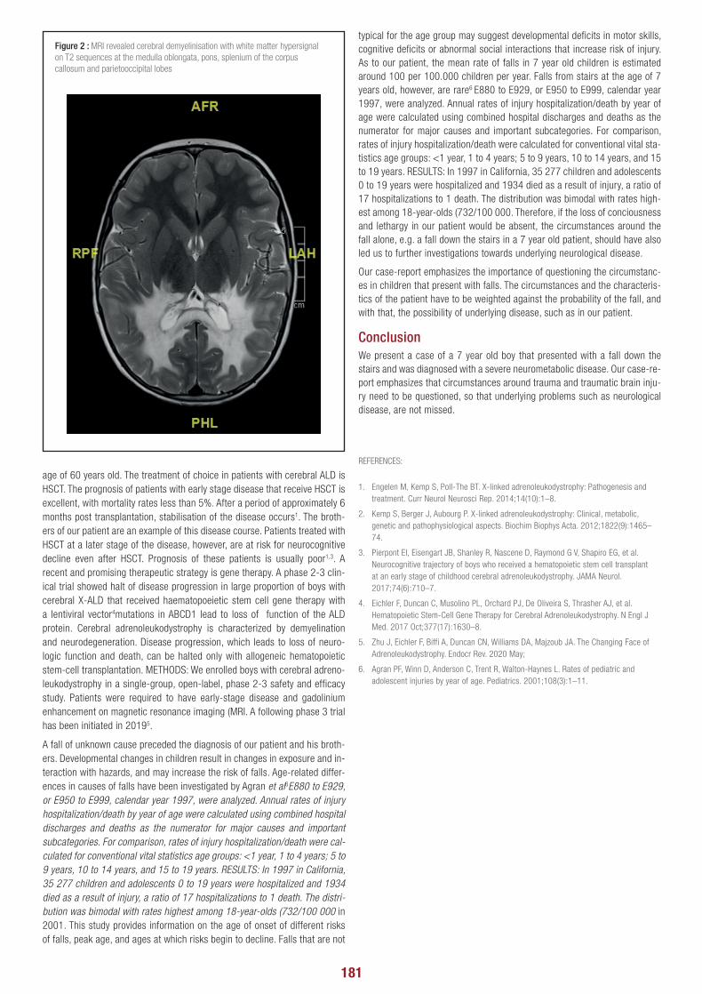
181
Traumatic brain injury or else?Sandra Deborah Karina Kingma1, An Jonckheere1, Els Moens1,2
1 Department of Pediatric Neurology, Koningin Mathilde Moeder- en Kindcentrum, University Hospital of Antwerp, Belgium 2 Department of Pediatrics, ZNA Jan Pal�jn, Lange Bremstraat 70, 2170 Merksem, Belgium
Case Report
age of 60 years old. The treatment of choice in patients with cerebral ALD is HSCT. The prognosis of patients with early stage disease that receive HSCT is excellent, with mortality rates less than 5%. After a period of approximately 6 months post transplantation, stabilisation of the disease occurs1. The broth-ers of our patient are an example of this disease course. Patients treated with HSCT at a later stage of the disease, however, are at risk for neurocognitive decline even after HSCT. Prognosis of these patients is usually poor1,3. A recent and promising therapeutic strategy is gene therapy. A phase 2-3 clin-ical trial showed halt of disease progression in large proportion of boys with cerebral X-ALD that received haematopoeietic stem cell gene therapy with a lentiviral vector4mutations in ABCD1 lead to loss of function of the ALD protein. Cerebral adrenoleukodystrophy is characterized by demyelination and neurodegeneration. Disease progression, which leads to loss of neuro-logic function and death, can be halted only with allogeneic hematopoietic stem-cell transplantation. METHODS: We enrolled boys with cerebral adreno-leukodystrophy in a single-group, open-label, phase 2-3 safety and efficacy study. Patients were required to have early-stage disease and gadolinium enhancement on magnetic resonance imaging (MRI. A following phase 3 trial has been initiated in 20195.
A fall of unknown cause preceded the diagnosis of our patient and his broth-ers. Developmental changes in children result in changes in exposure and in-teraction with hazards, and may increase the risk of falls. Age-related differ-ences in causes of falls have been investigated by Agran et al6E880 to E929, or E950 to E999, calendar year 1997, were analyzed. Annual rates of injury hospitalization/death by year of age were calculated using combined hospital discharges and deaths as the numerator for major causes and important subcategories. For comparison, rates of injury hospitalization/death were cal-culated for conventional vital statistics age groups: <1 year, 1 to 4 years; 5 to 9 years, 10 to 14 years, and 15 to 19 years. RESULTS: In 1997 in California, 35 277 children and adolescents 0 to 19 years were hospitalized and 1934 died as a result of injury, a ratio of 17 hospitalizations to 1 death. The distri-bution was bimodal with rates highest among 18-year-olds (732/100 000 in 2001. This study provides information on the age of onset of different risks of falls, peak age, and ages at which risks begin to decline. Falls that are not
typical for the age group may suggest developmental deficits in motor skills, cognitive deficits or abnormal social interactions that increase risk of injury. As to our patient, the mean rate of falls in 7 year old children is estimated around 100 per 100.000 children per year. Falls from stairs at the age of 7 years old, however, are rare6 E880 to E929, or E950 to E999, calendar year 1997, were analyzed. Annual rates of injury hospitalization/death by year of age were calculated using combined hospital discharges and deaths as the numerator for major causes and important subcategories. For comparison, rates of injury hospitalization/death were calculated for conventional vital sta-tistics age groups: <1 year, 1 to 4 years; 5 to 9 years, 10 to 14 years, and 15 to 19 years. RESULTS: In 1997 in California, 35 277 children and adolescents 0 to 19 years were hospitalized and 1934 died as a result of injury, a ratio of 17 hospitalizations to 1 death. The distribution was bimodal with rates high-est among 18-year-olds (732/100 000. Therefore, if the loss of conciousness and lethargy in our patient would be absent, the circumstances around the fall alone, e.g. a fall down the stairs in a 7 year old patient, should have also led us to further investigations towards underlying neurological disease.
Our case-report emphasizes the importance of questioning the circumstanc-es in children that present with falls. The circumstances and the characteris-tics of the patient have to be weighted against the probability of the fall, and with that, the possibility of underlying disease, such as in our patient.
ConclusionWe present a case of a 7 year old boy that presented with a fall down the stairs and was diagnosed with a severe neurometabolic disease. Our case-re-port emphasizes that circumstances around trauma and traumatic brain inju-ry need to be questioned, so that underlying problems such as neurological disease, are not missed.
Figure 2 : MRI revealed cerebral demyelinisation with white matter hypersignal on T2 sequences at the medulla oblongata, pons, splenium of the corpus callosum and parietooccipital lobes
1. Engelen M, Kemp S, Poll-The BT. X-linked adrenoleukodystrophy: Pathogenesis and treatment. Curr Neurol Neurosci Rep. 2014;14(10):1–8.
2. Kemp S, Berger J, Aubourg P. X-linked adrenoleukodystrophy: Clinical, metabolic, genetic and pathophysiological aspects. Biochim Biophys Acta. 2012;1822(9):1465–74.
3. Pierpont EI, Eisengart JB, Shanley R, Nascene D, Raymond G V, Shapiro EG, et al. Neurocognitive trajectory of boys who received a hematopoietic stem cell transplant at an early stage of childhood cerebral adrenoleukodystrophy. JAMA Neurol. 2017;74(6):710–7.
4. Eichler F, Duncan C, Musolino PL, Orchard PJ, De Oliveira S, Thrasher AJ, et al. Hematopoietic Stem-Cell Gene Therapy for Cerebral Adrenoleukodystrophy. N Engl J Med. 2017 Oct;377(17):1630–8.
5. Zhu J, Eichler F, Biffi A, Duncan CN, Williams DA, Majzoub JA. The Changing Face of Adrenoleukodystrophy. Endocr Rev. 2020 May;
6. Agran PF, Winn D, Anderson C, Trent R, Walton-Haynes L. Rates of pediatric and adolescent injuries by year of age. Pediatrics. 2001;108(3):1–11.
REFERENCES:

182
A iatrogenic cause of encephalopathy in a 9-year old boy – a rare side effect of a commonly used drugSarah Bogovic a, Francis Lemmens b
a Department of Pediatrics, University Hospital of Antwerp, Edegem, Belgium b Department of Pediatrics, Ziekenhuis Oost-Limburg, Genk, Belgium
Case Report
AbstractA 9-year old boy presented with convulsions and was treated with lorazepam and valproate. Within hours he developed an encephalopathy. Laboratory tests and magnetic resonance imaging (MRI) of the central nervous system imaging were normal. After cessation of valproate symptoms rapidly resolved, diagnosis of valproate-induced encephalopathy (VIE) was assumed.
VIE is a complication of valproate therapy, most commonly met in cases with polypharmacy or underlying disease. In our patient, due to timing and absence of risk factors, VIE initially was mistaken for a symptom of underlying disease. Cessation of valproate relieved encephalopathy and prevented permanent damage. In encephalopathy, it is important to consider iatrogenic causes.
KeywordsValproic acid ; Valproate ; Valproate-induced ; Pediatric ; Encephalopathy ; Delirium
Acute agitation, delirium or encephalopathy can have many different causes. In paediatric practice, central nervous system (CNS) infection, intoxication, status epilepticus, intracranial processes or metabolic disturbances are most common. So the initial work-up should always include blood and cerebrospinal �uid (CSF) culture, meningitis/encephalitis polymerase chain reaction panel (PCR-panel) of CSF, toxicology screening, blood gases (including lactate), glycaemia, plasma amino acids and acylcarnitine and organic acids on urine. Though less frequent, iatrogenic causes should not be overlooked, as is shown in our 9-year old patient.
Case presentationA 9-year old boy with previously blank medical history presented after a �rst tonic-clonic epileptic seizure while traveling with his parents on a trip through Belgium. Spontaneous resolution seemed to take place after ten minutes. Serum electrolytes, ammonia level and liver tests were normal. There was no fever nor other signs of infection in patient history, clinical examination and lab results. Toxicologic screening in urine was negative. A few hours after admission a second tonic-clonic seizure was observed with need for benzodiazepine administration (lorazepam intravenously (IV) 0.1 mg/kg). Since the recurrence risk was esteemed as high a treatment with valproate was started (loading dose 20mg/kg IV followed by continuous IV administration of 1mg/kg/hour).
Within the next hours the boy developed an encephalopathic state with agitation, aggressive behaviour and lack of adequate interaction. A computed tomography (CT) scan and MRI of the brain showed no abnormalities. An electroencephalogram (EEG) did not demonstrate epileptic activity, however a slower base-line rhythm compatible with encephalopathy was present. Lumbar puncture showed no evidence of CNS infection or other pathology (leukocytes 1/mm3 (normal 0-7), glucose 69 mg/dl (40_80), protein 17,7 mg/dl (5-40), lactate 1,7 mmol/L (1,1-2,1).
Acyclovir was added to the therapy and the child was transferred to a paediatric intensive care unit (PICU) (university centre). In the meantime, meningo-encephalitis PCR-panel testing proved negative.
After cessation of valproate and acyclovir (since Herpes was negative) in the PICU, there was a quick and complete resolution of encephalopathic symptoms in less than 24 hours. Levetiracetam was started and for the remaining time of hospitalisation no further events occurred.
Valproate-induced encephalopathy (VIE) is a complication of valproate therapy, most commonly described in patients with polypathology (especially neuropsychiatric) and polypharmacy.1-4 It is most likely thought to be caused by interference in the urea cycle with resulting increase in serum ammonia levels, even without disturbance in liver function.5,-7 This effect is only partially dose-dependent, the serum ammonia level does not always correlate with encephalopathic symptoms and hyperammonemia can occur with valproate serum levels within therapeutic range. 5,7
Most reported cases developed symptoms within days to weeks after (re)starting valproate or elevating valproate dose. Our case has a different timing with development of encephalopathy within hours. This can be partially explained by the loading dose that was administered intravenously. Risk factors for development of valproate-induced encephalopathy are young age (under 3 years of age), polypharmacy and especially combination of valproate with phenobarbital or topiramate, underlying urea cycle defect, underlying psychomotor retardation or psychiatric disease. None of these predisposing factors applied to our patient.
In the university center no other cause of encephalopathy was identi�ed. Further blood tests showed normal values for glycaemia, lactate, electrolytes, liver and thyroid function and copper metabolism. Ammonia level was not determined again.
The quick resolution of symptoms after discontinuation of valproate treatment seems to indicate that our patient suffered from VIE. Since valproate and ammonia levels were not checked at that moment, the exact mechanism will remain unknown.
The most likely mechanism causing VIE seems to be direct inhibition of hepatic mitochondrial carbamylphosphate synthetase (CPS 1) 8, the �rst enzyme of the urea cycle ,and/or indirect interference with the carnitine handling (valproate inhibits transport and increases renal excretion of carnitine). Increasing ammonia- and decreasing carnitine levels are the result and hyperammonemia has been suggested to be the main cause of encephalopathy. However, several cases of non-hyperammonemic valproate-induced encephalopathy are reported in literature. 9,10 An underlying mitochondropathy should be considered in those cases, especially if blood lactate is raised.
Introduction Discussion
183
A iatrogenic cause of encephalopathy in a 9-year old boy – a rare side effect of a commonly used drugSarah Bogovic a, Francis Lemmens b
a Department of Pediatrics, University Hospital of Antwerp, Edegem, Belgium b Department of Pediatrics, Ziekenhuis Oost-Limburg, Genk, Belgium
Case ReportRemarkable in our patient is that there was no predisposing history, no abnormalities in any blood test, no co-administration of other drugs, and development of an encephalopathic state in no more than a few hours after the start of valproate therapy. As such it was very dif�cult to recognize the encephalopathy as an adverse effect of the treatment rather than as a symptom of the underlying disease. By presenting our case we want to raise special awareness of this possibility, as valproate is one of the most widely-used drugs in treatment of childhood epilepsy, even in encephalopathy. In Belgium, valproate is still the �rst-choice for epileptic seizures.
ConclusionThe combination of seizures and acute encephalopathy in a previous healthy child is a challenging paediatric emergency. Status epilepticus, infection, intoxication, intracranial process and metabolic disease should all be ruled out. Though less common, iatrogenic causes should also be considered. In our patient valproate-induced encephalopathy occurred as a rare adverse event, caused by a widely-used drug.
Our case shows that this entity can be dif�cult to recognize and can easily be mistaken for a symptom of underlying disease rather than an adverse effect of treatment. Even in the absence of risk factors it is important to always consider the latter option, since early recognition is crucial to stop the responsible drug (In this case valproate), resolve symptoms and prevent permanent damage.
We are also convinced that, especially in young children, levetiracetam rather than valproate, should become the �rst choice for treatment of a �rst episode of seizures, certainly if the previous history of the child is not well known since underlying disease can increase the risk of serious adverse effects. 11
1. Chopra A, Kolla BP, Mansukhani MP, Netzel P, Frye MA, Valproate-induced hyperammonemic encephalopathy: an update on risk factors, clinical correlates and management, General Hospital Psychiatrics 2012; 34(3):290-298
2. Dealberto MJ. Valproate-induced hyperammonaemic encephalopathy: review of 14 cases in psychiatric setting. International Clinical Psychopharmacology 2007; 22(6): 330-7
3. Muraleedharan A, Palappallil DS, Gangadhar R, Das S. Valproate induced hyperammonemic delirium. Journal of clinical and diagnostic research 2015; 9(12): FR01-FR03
4. Pradeep RJ. Valproate monotherapy induced-delirium due to hyperammonemia: a report of three adult cases with different types of presentation. Indian J Psychiatry 2008; 50(2) :121-3
5. Segura-Bruna N, Rodriguez-Campello A, Puente V, Roquer J. Valproate-induced hyperammonemic encephalopathy. Acta Neurol Scand 2006; 114(1): 1-7
6. Turnbull DM, Dick DJ, Wilson L, Sherratt HS, Alberti KG. Valproate causes metabolic disturbance in normal man. Journal of Neurology, Neurosurgery and Psychiatry 1986; 49(4): 405-10
7. Yamamoto Y, Takahashi Y, Imai K, Mishima N, Yazawa R, Takahashi Y et al. Risk factors for hyperammonemia in pediatric patients with epilepsy. Epilepsia 2013; 54(6): 983-9
8. Marini AM, Zaret BS, Beckner RR. Hepatic and renal contributions to valproic acid-induced hyperammonemia. Neurology 198; 38 (3): 365-71
9. De Michele G, Sorrentino P, Nesti C, Rubegni A, Ruggiero L, Peluso S et al. Reversible valproate induced subacute encephalopathy associated with MT-ATP8 variant in the mitochondrial genome. Frontiers in neurology 2018; 9: 728
10. Farooq O, Zunga PM, Dar MI, Rather AQ, Rashid S, Basu J et al. Non-Hyperammonemic valproate encephalopathy. Ann Neurosci. 2014; 21(2): 76-9
11. Perucca E. Pharmacological and therapeutic properties of valproate: a summary after 35 years of clinical experience. CNS Drugs. 2002;16(10):695-714.
REFERENCES:

184
Made in Belgium
Beta-lactam hypersensitivity: Epidemiology and optimized diagnosisPhD thesis presented on 26th of October 2020 at Universiteit Antwerpen, Antwerpen, Belgium
Athina Van Gasse
Promotors: Didier Ebo, Vito Sabato, Margo Hagendorens
β-lactam antibiotics are a group of antimicrobial agents including penicillins, cephalosporins, carbapenems and monobactams (an overview is shown in �gure 1).
β-lactam antibiotics, especially penicillins, are one of the predominant causes of drug hypersensitivity reactions. The majority of β-lactam hypersensitivity reactions is categorised as immediate (< 1 hour) humoral likely IgE-mediated (type I) and nonimmediate (> 1 hour), mainly cellular (type IV), hypersensitivity reactions. Immediate reactions mostly present with urticaria and/or angioedema, bronchospasm but anaphylaxis and death can occur 1. A maculopapular exanthema is the predominating presentation of nonimmediate type IV hypersensitivity reactions 2, 3.
Alternatively, unveri�ed and false “β-lactam allergies”, mainly to the �rst-line preparations natural penicillin and aminopenicillins, have evolved into a worldwide plague with serious �nancial and medical consequences.
According to the literature, as much as 10-20% of the population reports a “penicillin allergy” that was never accurately documented 4-7. However, over 90% of these individuals tolerate the alleged culprit(s) during controlled drug challenges and are at unnecessary risk for more expensive and suboptimal second-line antibiotic treatments 8-36. Actually, “penicillin allergy” is associated with erroneous avoidance and unnecessary substitutions, prolonged hospitalizations, more readmissions, poorer outcomes, increased costs and increased rates of Clostridium difficile and antimicrobial resistance, e.g. vancomycin 37-44.
This dissertation aims at improving awareness of this scourge and motivating treating physicians for referral of their patients for correct diagnosis in order to con�rm or discard clinical suspicion.
Hence, we present data on the prevalence of self-reported “penicillin allergy” in a Belgian outpatient population. To the best of our knowledge, such data have
not been published yet. From our survey it appears that as much as 12% of the individuals attending the outpatients’ clinic of Allergology and Paediatric Allergology of the Antwerp University Hospital claim to have a “penicillin allergy”. However, in 91% of these individuals this claim appears to be false, as they tolerated these antibiotics during a controlled drug challenge. Importantly, as these cases could be safely delabelled, we were able to improve their antibiotic stewardship, mainly by safeguard them against the unnecessary risk for use of suboptimal and expensive second-line treatments. However, almost 10% of the participants who were successfully delabelled remained reluctant for any subsequent intake which was reinforced by other physicians. Efforts to improve general awareness of clinicians and strengthen their believe in the bene�t of con�rmatory testing to establish correct diagnosis in their patients are needed.
From the literature and our experience, it is clear that diagnosis of penicillin allergy cannot rest upon history alone but needs judicious diagnostic work-up by a trained physician and that correct diagnosis is necessary in every case of both witnessed or self-reported “penicillin allergy”. We show that the label of “penicillin allergy” generally already originates in childhood and that in most cases information about the index reaction (culprit, clinics, timing,…), regrettably is lacking or simply limited to the description of an ‘unde�ned rash’. Consequently, we argue �rmly for a more detailed documentation of any potential drug hypersensitivity reaction. History and clinical presentation are critical to allow identi�cation of the underlying pathophysiological mechanism and guidance of subsequent con�rmatory testing.
However, diagnosis of β-lactam hypersensitivity reactions is not always straightforward and optimization of the diagnostic approach of β-lactam hypersensitivities is needed. Therefore, one of the main aims of this thesis was to study the performance of new or recently introduced diagnostics.
Figure 1: Overview of β-lactam antibiotics available for medical use in Belgium

185
Made in BelgiumFirst, we investigated the potential of CD154, a member of the TNF superfamily that is primarily, but not exclusively, expressed on activated CD4+ve T helper lymphocytes, as an alternative readout for drug-speci�c T-lymphocyte activation in the context of nonimmediate hypersensitivity reactions to amoxicillin and amoxicillin clavulanic acid. We showed that a CD154-based lymphocyte activation test (CD154-LAT) is a safe and quick in vitro diagnostic test, especially in cases with mild cutaneous nonimmediate drug hypersensitivity reactions to amoxicillin or amoxicillin clavulanic acid, who required drug challenges because of equivocal or negative skin tests. Although CD154 upregulation was also observed in some amoxicillin (clavulanic acid) tolerant individuals, the median upregulation was numerically lower than in patients with amoxicillin hypersensitivity responsive in the SD154-LAT. However, larger studies are required to con�rm these promising data. Alternatively, it is obvious that the value of a CD154-based lymphocyte activation test should additionally be evaluated in severe cutaneous nonimmediate drug hypersensitivity reactions, where intradermal testing and drug challenges are contraindicated for safety reasons.
Further, we demonstrated that a prolonged drug challenge extending over several consecutive days is of limited use in the diagnosis of nonimmediate drug hypersensitivity reactions to amoxicillin and amoxicillin clavulanic acid, as 116 prolonged drug challenges have to be performed to identify one single patient demonstrating a mild maculopapular exanthema. Consequently, we currently dissuade to systematically impose prolonged drug challenges as a �nal diagnostic in nonimmediate drug hypersensitivity reactions to amoxicillin and amoxicillin clavulanic acid, especially as potential pharmacological adverse events (e.g. diarrhoea, mycoses, and the development of antimicrobial resistance) outweigh the bene�ts. Taking into account the accuracy, safety and cost of our diagnostics, the traditionally recommended diagnostic algorithm that offers a 1-day drug challenge as a �nal diagnostic in patients with negative work-up for amoxicillin (clavulanic acid) is still appropriate.
In our country cefazolin, a �rst-generation cephalosporin antibiotic mainly used in perioperative prophylaxis, has evolved to one of the major causes of perioperative anaphylaxis with serious consequences of misdiagnosis. Con�rmatory diagnosis generally starts with skin tests and, if needed, potentially dangerous intravenous challenges. Therefore, and having access to sera of patients with a well-documented cefazoline allergy, we explored the performance of a recently developed speci�c (s)IgE assay to cefazolin. We observed that when the sIgE result for cefazolin was corrected for total (t)IgE by use of a sIgE/tIgE ratio, predictive values were comparable to sIgE assays for other β-lactam antibiotics. In contrast, sIgE to cefazolin was not able to discriminate between cefazolin allergic patients and control individuals. However, the sIgE/tIgE ratio does not substitute skin testing and sensitivity of the ratio drops over time.
Finally, it is known that cross-reactivity among β-lactam antibiotics is unpredictable but mostly related to the R1-side chain (see �gure 2). Besides, physicians tend to overestimate cross-reactivity and too often other β-lactam antibiotics are unnecessarily forbidden. However, we demonstrated that �ne structural differences in the R1-side chain can suf�ce to explain absence of cross-reactivity and clinical tolerance. Clearly, molecules with a similar, but not identical R1-side chain, should not be precluded or administered using a desensitization protocol, provided they test negative in skin testing.
In conclusion, self-reported “penicillin allergy” has grown into a vast epidemic with unacceptable consequences for the individual patient and the society. Moreover, cross-reactivity among β-lactam antibiotics is often overestimated. Therefore, con�rmatory testing is absolutely needed in every patient with a history of signs/symptoms that are possibly caused by β-lactam antibiotics. In contrast to a prolonged drug challenge, a CD154-LAT seems to be an attractive instrument to document mild cutaneous nonimmediate drug hypersensitivity reactions to amoxicillin or amoxicillin clavulanic acid. A sIgE to cefazolin is of no added value to the diagnosis of immediate cefazolin hypersensitivity. Further efforts are required to optimize and harmonize a cost-conscious diagnostic approach of “penicillin allergy” and “β-lactam allergy” in general.
Figure 2: Chemical core structures of β-lactam antibiotics. The β-lactam ring (red) is common to all β-lactam antibiotics. Penicillins share a stable 5-membered thiazolidine ring, cephalosporines contain an unstable 6-membered dihydrothiazine ring. “R” denotes the side chains that vary among the antibiotics
1. Blanca M, Romano A, Torres MJ, Fernandez J, Mayorga C, Rodriguez J, et al.
Update on the evaluation of hypersensitivity reactions to betalactams. Allergy
2009; 64:183-93.
2. de Haan P, Bruynzeel DP, van Ketel WG. Onset of penicillin rashes: relation between
type of penicillin administered and type of immune reactivity. Allergy 1986; 41:75-
8.
3. Schiavino D, Nucera E, De Pasquale T, Roncallo C, Pollastrini E, Lombardo C, et
al. Delayed allergy to aminopenicillins: clinical and immunological findings. Int J
Immunopathol Pharmacol 2006; 19:831-40.
4. Solensky R. Hypersensitivity reactions to beta-lactam antibiotics. Clin Rev Allergy
Immunol 2003; 24:201-20.
5. Macy E, Poon KYT. Self-reported antibiotic allergy incidence and prevalence: age
and sex effects. Am J Med 2009; 122:778.e1-7.
6. Shah NS, Ridgway JP, Pettit N, Fahrenbach J, Robicsek A. Documenting Penicillin
Allergy: The Impact of Inconsistency. PLoS One 2016; 11:e0150514.
7. Desai SH, Kaplan MS, Chen Q, Macy EM. Morbidity in Pregnant Women Associated
with Unverified Penicillin Allergies, Antibiotic Use, and Group B Streptococcus
Infections. Perm J 2017; 21.
8. Graff-Lonnevig V, Hedlin G, Lindfors A. Penicillin allergy--a rare paediatric
condition? Arch Dis Child 1988; 63:1342-6.
9. Saxon A, Adelman DC, Patel A, Hajdu R, Calandra GB. Imipenem cross-reactivity
with penicillin in humans. J Allergy Clin Immunol 1988; 82:213-7.
10. Surtees SJ, Stockton MG, Gietzen TW. Allergy to penicillin: fable or fact? Bmj 1991;
302:1051-2.
11. Gonzalez J, Guerra F, Moreno C, Miguel R, Daza JC, Sanchez Guijo P. Assessment
of a self-designed protocol on patients with adverse reactions to beta-lactam
antibiotics. Allergol Immunopathol (Madr) 1992; 20:184-9.
12. Gadde J, Spence M, Wheeler B, Adkinson NF, Jr. Clinical experience with penicillin
skin testing in a large inner-city STD clinic. Jama 1993; 270:2456-63.
13. Pichichero ME, Pichichero DM. Diagnosis of penicillin, amoxicillin, and
cephalosporin allergy: reliability of examination assessed by skin testing and oral
challenge. J Pediatr 1998; 132:137-43.
REFERENCES:

186
14. Dodek P, Phillips P. Questionable history of immediate-type hypersensitivity to
penicillin in Staphylococcal endocarditis: treatment based on skin-test results
vers-us empirical alternative treatment--A decision analysis. Clin Infect Dis 1999;
29:1251-6.
15. Arroliga ME, Wagner W, Bobek MB, Hoffman-Hogg L, Gordon SM, Arroliga AC. A
pilot study of penicillin skin testing in patients with a history of penicillin allergy
admitted to a medical ICU. Chest 2000; 118:1106-8.
16. Solensky R, Earl HS, Gruchalla RS. Penicillin allergy: prevalence of vague history in
skin test-positive patients. Ann Allergy Asthma Immunol 2000; 85:195-9.
17. Salkind AR, Cuddy PG, Foxworth JW. The rational clinical examination. Is this
patient allergic to penicillin? An evidence-based analysis of the likelihood of
penicillin allergy. Jama 2001; 285:2498-505.
18. Kalogeromitros D, Rigopoulos D, Gregoriou S, Papaioannou D, Mousatou V,
Katsarou-Katsari A. Penicillin hypersensitivity: value of clinical history and skin
testing in daily practice. Allergy Asthma Proc 2004; 25:157-60.
19. Park MA, Li JT. Diagnosis and management of penicillin allergy. Mayo Clin Proc
2005; 80:405-10.
20. Park M, Markus P, Matesic D, Li JT. Safety and effectiveness of a preoperative
allergy clinic in decreasing vancomycin use in patients with a history of penicillin
allergy. Ann Allergy Asthma Immunol 2006; 97:681-7.
21. Wohrl S, Vigl K, Stingl G. Patients with drug reactions -- is it worth testing? Allergy
2006; 61:928-34.
22. Wong BB, Keith PK, Waserman S. Clinical history as a predictor of penicillin skin
test outcome. Ann Allergy Asthma Immunol 2006; 97:169-74.
23. Raja AS, Lindsell CJ, Bernstein JA, Codispoti CD, Moellman JJ. The use of
penicillin skin testing to assess the prevalence of penicillin allergy in an emergency
department setting. Ann Emerg Med 2009; 54:72-7.
24. Silva R, Cruz L, Botelho C, Cadinha S, Castro E, Rodrigues J, et al. Work up of
patients with history of beta-lactam hypersensitivity. Allergol Immunopathol (Madr)
2009; 37:193-7.
25. Lin E, Saxon A, Riedl M. Penicillin allergy: value of including amoxicillin as a
determinant in penicillin skin testing. Int Arch Allergy Immunol 2010; 152:313-8.
26. Kamboj S, Yousef E, McGeady S, Hossain J. The prevalence of antibiotic skin test
reactivity in a pediatric population. Allergy Asthma Proc 2011; 32:99-105.
27. Caubet JC, Kaiser L, Lemaitre B, Fellay B, Gervaix A, Eigenmann PA. The role of
penicillin in benign skin rashes in childhood: a prospective study based on drug
rechallenge. J Allergy Clin Immunol 2011; 127:218-22.
28. Kopac P, Zidarn M, Kosnik M. Epidemiology of hypersensitivity reactions to
penicillin in Slovenia. Acta Dermatovenerol Alp Pannonica Adriat 2012; 21:65-7.
29. Sagar PS, Katelaris CH. Utility of penicillin allergy testing in patients presenting
with a history of penicillin allergy. Asia Pac Allergy 2013; 3:115-9.
30. Rimawi RH, Cook PP, Gooch M, Kabchi B, Ashraf MS, Rimawi BH, et al. The impact
of penicillin skin testing on clinical practice and antimicrobial stewardship. J Hosp
Med 2013; 8:341-5.
31. Fernandez TD, Mayorga C, Ariza A, Corzo JL, Torres MJ. Allergic reactions to
antibiotics in children. Curr Opin Allergy Clin Immunol 2014; 14:278-85.
32. Kaya A, Erkocoglu M, Senkon OG, Ekici FK, Toyran M, Cetin, II, et al. Confirmed
penicillin allergy among patients receiving benzathine penicillin prophylaxis for
acute rheumatic fever. Allergol Immunopathol (Madr) 2014; 42:289-92.
33. Salden OA, Rockmann H, Verheij TJ, Broekhuizen BD. Diagnosis of allergy against
beta-lactams in primary care: prevalence and diagnostic criteria. Fam Pract 2015;
32:257-62.
34. Meng J, Thursfield D, Lukawska JJ. Allergy test outcomes in patients self-reported
as having penicillin allergy: Two-year experience. Ann Allergy Asthma Immunol
2016; 117:273-9.
35. Marwood J, Aguirrebarrena G, Kerr S, Welch SA, Rimmer J. De-labelling self-
reported penicillin allergy within the emergency department through the use of skin
tests and oral drug provocation testing. Emerg Med Australas 2017.
36. Har D, Solensky R. Penicillin and Beta-Lactam Hypersensitivity. Immunol Allergy
Clin North Am 2017; 37:643-62.
37. Satta G, Hill V, Lanzman M, Balakrishnan I. beta-lactam allergy: clinical implications
and costs. Clin Mol Allergy 2013; 11:2.
38. Macy E, Contreras R. Health care use and serious infection prevalence associated
with penicillin "allergy" in hospitalized patients: A cohort study. J Allergy Clin
Immunol 2014; 133:790-6.
39. Li M, Krishna MT, Razaq S, Pillay D. A real-time prospective evaluation of clinical
pharmaco-economic impact of diagnostic label of 'penicillin allergy' in a UK
teaching hospital. J Clin Pathol 2014; 67:1088-92.
40. Bourke J, Pavlos R, James I, Phillips E. Improving the Effectiveness of Penicillin
Allergy De-labeling. J Allergy Clin Immunol Pract 2015; 3:365-34.e1.
41. Macy E, Shu YH. The Effect of Penicillin Allergy Testing on Future Health Care
Utilization: A Matched Cohort Study. J Allergy Clin Immunol Pract 2017; 5:705-10.
42. Blumenthal KG, Li Y, Banerji A, Yun BJ, Long AA, Walensky RP. The Cost of Penicillin
Allergy Evaluation. J Allergy Clin Immunol Pract 2017.
43. Blumenthal KG, Ryan EE, Li Y, Lee H, Kuhlen JL, Shenoy ES. The Impact of a
Reported Penicillin Allergy on Surgical Site Infection Risk. Clin Infect Dis 2018;
66:329-36.
44. Mattingly TJ, 2nd, Fulton A, Lumish RA, Williams AMC, Yoon S, Yuen M, et al. The
cost of self-reported penicillin allergy: a systematic review. J Allergy Clin Immunol
Pract 2018.
BJP_2009_ultragenyx.indd 1 21/09/2020 14:13

187
BJP_2009_ultragenyx.indd 1 21/09/2020 14:13

188
Made in Belgium
Optimisation of long-term outcomes in paediatric in�ammatory bowel disease patients: role of therapeutic drug monitoring and endoscopic remissionPhD thesis presented on 13th of May 2020 at KU Leuven, Leuven, Belgium
Karen van Hoeve a,b
Promotor: Séverine Vermeire b,c Co-promotor: Ilse Hoffman a and Ann Gils d
a Department of Paediatric gastroenterology & Hepatology & Nutrition, University Hospitals Leuven, Leuven, Belgium b TARGID, Department of Chronic Diseases & Metabolism (CHROMETA), KU Leuven, Leuven, Belgium c Department of Gastroenterology & Hepatology, University Hospitals Leuven, KU Leuven, Leuven, Belgium d Laboratory for Therapeutic and Diagnostic Antibodies, Department of Pharmaceutical and Pharmacological Sciences, KU Leuven, Leuven, Belgium
In�ammatory bowel diseases (IBDs) are chronic, relapsing-remitting disorders of the gastro-intestinal tract that are subclassi�ed as Crohn’s disease (CD) and ulcerative colitis (UC) 1-2. Approximately 20-25% of newly diagnosed IBD patients present during childhood, with an incidence of 6.5-9.3 per 100.000 and 1.0-2.4 per 100.000 for CD and UC, respectively 3-4. The clinical presentation depends highly on the disease location and behaviour, which �uctuates over time. The majority of patients will experience (bloody) diarrhea, abdominal pain, weight loss, growth impairment, fever and fatigue 1-2. In addition, uncontrolled in�ammation puts them at risk for disease-associated complications, such as �brosis, �stulas, repeated surgical resections and cancer.
The exact aetiology of IBD has not been fully elucidated yet 5-7. There is a general consensus that the disease arises from a complex interplay of environmental fac-tors in combination with dysbiosis of the gut microbiota that leads to an excessive activation of the intestinal immune system and subsequent in�ammation in a genetic predisposed host (see �gure 1).
Since IBD is still a life-long and incurable disease, the medical manage-ment remains challenging, particularly in children 1-2. The aggressive nature of paediatric IBD has been well established, with a more extensive anatomi-cal involvement at diagnosis in comparison to adults 8. Therefore, biological agents are more often necessary to obtain endoscopic remission, which has emerged as the new treatment goal 9. In addition, a more holistic approach is applicable in children addressing in particularly growth, puberty, and bone health; which can be impacted directly by the disease but also indirectly by the effects of treatment, and this at a period where the psychosocial de-velopment is most vulnerable 1-2. Finally, the therapeutic armamentarium of children with IBD is still limited in comparison to adults as both the required randomised trials and evidence lag behind. Thus, especially in children, the focus of management should be on optimising our available treatment op-tions in the best possible way to overcome these challenges and to treat inflammation adequately.
Introduction
Figure 1: Pathogenesis of in�ammatory bowel disease. This �gure was created with images adapted from Servier Medical Art.

189
Made in BelgiumSeveral strategies have been proposed to achieve these goals. The general aim of this doctoral thesis was to improve the long-term outcome by further exploring and unravelling these strategies, speci�cally in paediatric IBD patients.
Predicting response to conventional therapy in childrenThe key goal in IBD management is to achieve deep remission, as early as pos-sible in this lifelong disease, and this with minimal toxicity. The time to achieve this goal is thought to be as important as reaching this goal itself 10-14. Especially in the early stages of the disease, there exists a window of opportunity to pre-vent disease progression and disability 14-15. The introduction of an accelerated step-up therapy (based on patients’ risk factors) or even top-down therapy in association with a tight control of in�ammation has led to the expectations that we could change the natural course of the disease. A top-down approach will start immediately with biologics instead of initiating treatment with conventional therapy (with e.g. 5-aminosalicylates, steroids and immunomodulators) and defer biologics until patients �ares or become refractory (i.e. step-up therapy). This top-down approach is based on the superiority of in�iximab (IFX) over immuno-modulators in achieving corticosteroid-free remission, especially when given early after diagnosis 11-14. It is noteworthy that not all patients require the same intense treatments. Currently, there are insuf�cient data to predict the disease course at diagnosis, and therefore to decide which patients will bene�t most from early biologics, and who will not need early or even any biologics. Indeed, a top-down approach will certainly not be the ideal approach for all patients. In addition to the risk of over-treating, the high cost of these biologics will also limit its early use (especially since most local governments reimbursement regimes will only sup-port a step-up approach). Thus, validated prognostic markers are urgently wanted that can identify at diagnosis which patients will progress and require more potent treatment. We found that especially a high disease burden at diagnosis predicted the need for step-up therapy with either biologics or surgery 16. Patients were more likely to fail conventional therapy in case of higher C-reactive protein (CRP), lower albumin and growth impairment for CD and higher Paediatric Ulcerative Colitis Activity Index (PUCAI) score and lower iron levels for UC. The need for step-up therapy signi�cantly increased with the number of risk factors. Since, this effect was already seen early during follow-up under conventional therapy, we probably can expect to catch the window of opportunity to modify the natural disease course when providing these high-risk patients an accelerated step-up therapy together with a tight control of in�ammation.
In addition, we have shown that only one-third of paediatric IBD patients treated with conventional therapy remained free of biologics or surgery 5-years after diagnosis and more than two-thirds of patients required step-up treatment 16. Therefore, also the therapeutic potential of these intrinsically powerful biologics needs to be maximised.
Therapeutic drug monitoring of anti- tumour necrosis factor agentsWhile we wait impatiently for new biologics to become available for paediatric indications, anti- tumour necrosis factor (TNF) agents are the only approved biologics in paediatric IBD until now. TNF alpha is a pro-in�ammatory cytokine that plays a key role in the intestinal in�ammatory cascade. Neutralising TNF alpha with these agents has been shown to diminish in�ammation and induce apoptosis of TNF-producing immune cells 17. Currently, two anti-TNF alpha agents are approved for the treatment of paediatric IBD: in�iximab and adalimumab, respectively a chimeric and a fully human, recombinant monoclonal antibody directed against TNF alpha 1-2.
Prevention of anti-drug antibody formation and loss of response remain the two most important challenges in the management of IBD with respect to anti-TNF alpha agents 18. Therefore, therapeutic drug monitoring (TDM) has been proposed as one of the ways to optimize both initial response and long-term continuation of anti-TNF therapy. TDM is a tool based on clinical and laboratory measurements, typically drug and anti-drug antibody serum concentrations, which enables clinical decision-making in order to achieve the highest possible response without side effects. Research in TDM is rapidly expanding but mainly in the adult literature and TDM studies in children are still under investigated 18. The objective of the second part of this doctoral thesis was therefore to study that adequate drug exposure to IFX would lead to better long-term outcomes in paediatric IBD and to de�ne the optimal drug exposure.
The optimal drug exposure (or therapeutic window) might however be patient-speci�c, vary over time throughout the treatment course (during induction phase, at the end of induction and during the maintenance phase) and might also depend on the treatment endpoint (such as clinical, biological or endoscopic remission). Therefore, our research has guided us through these three important phases of IFX therapy with respect to all different treatment endpoints. Speci�c attention went to endoscopic remission, since this has emerged as the Holy Grail instead of focusing on relieving symptoms alone 9. However, data on this topic were lacking in paediatric IBD.
We demonstrated a strong exposure-response effect in children receiving maintenance IFX therapy, and for the �rst time a clear association between IFX trough levels and endoscopic remission in this population 19. In the era where mucosal healing is the new gold standard, this is an important observation. Secondly, we showed that an adequate IFX exposure at the end of induction therapy was essential for a favourable long-term outcome. Patients achieving endoscopic remission at month six or clinical and/or biological remission at week 52 had already signi�cantly higher post-induction IFX trough levels 20. In fact, these trough levels were the only independent predictor for these different outcomes. A cut-off value of ≥ 5.0 µg/ml was deemed necessary to reach endoscopic remission both at the end of induction as during maintenance phase 19-20, which is in line with previously published adult literature. Furthermore, also the IFX exposure during the induction phase could predict endoscopic outcome at month six. Since, IFX concentrations could already discriminate remitters from non-remitters from week 4 after IFX initiation onwards. Threshold levels for different time points, including intermediate and trough levels during induction, were determined in order to achieve the highest possible response (see �gure 2). Our results make it more feasible to translate the �ndings to routine clinical practice where patients usually present in the clinic at different time points and not only at trough.
Not only was an adequate drug exposure during induction but also during maintenance therapy essential for a long and durable response, including endoscopic remission 19. Patients with mucosal healing at latest endoscopy (after a median follow-up of 1.4 years) had also a higher overall exposure to IFX during maintenance therapy in comparison to patients with lack of response with an excellent endoscopic outcome at latest follow-up of 72.5% 19. Although, pro-active TDM-based treat-to-target strategies are currently not considered standard of care for IBD patients, our data support the use of pro-active TDM in children to improve long-term outcome. We therefore suggest applying pro-active TDM at least once after induction and then one time per year during stable maintenance therapy. Ideally, pro-active TDM is also implemented during induction therapy to prevent primary non-response driven by pharmacokinetic reasons, and to optimise IFX exposure to reach better long-term endoscopic outcome. During induction there is still a high in�ammatory burden resulting in a higher drug clearance. In this respect, it has been shown that most paediatric patients will have subtherapeutic post-induction IFX trough levels when using the current standard regimen 20-22. Therefore, determining an IFX concentration already at week 4 can help the physician to adjust dose and/or interval of the next infusion at week 6, and consequently improve further drug exposure. However, further prospective studies are now warranted to judge the cost-effectiveness of this approach in real-world practice (especially since it is safe to switch to cheaper biosimilars 23).
Finally, the purpose of TDM is to dose the drug in such a way that the optimal drug exposure is achieved for each separate patient. Therefore, speci�c covariates needs to be determined that can explain the differences in drug exposure between patients. It has been known that IFX trough levels are greatly in�uenced by the degree of intestinal in�ammation 24. Our research con�rmed this association, as especially patients with a lower haemoglobin at IFX initiation had lower IFX exposure during induction 19-20. Thus, these patients could probably bene�t from higher IFX doses already at the start and doses could be adjusted afterwards by TDM to achieve remission. In addition, lower serum IFX levels were found in patients with lower weight and stunted growth at IFX initiation 19-20. Therefore, probably a more intensive treatment schedule than prescribed in the label is warranted to prevent under-exposure to IFX in younger patients. Nonetheless, development of pharmacokinetic models and prospective trials are crucial to further elucidate the ultimate treatment regimen prior to routine implementation into clinical practice.

190
Figure 2: Optimisation of outcomes in paediatric IBD patients
Legend: conc.: concentration; CD: Crohn’s disease; CRP: C-reactive protein; IBD: in�ammatory bowel disease; IFX: in�iximab; PUCAI: Paediatric Ulcerative Colitis Activity Index; SD: standard deviation; TNF: tumour necrosis factor; UC: ulcerative colitis; wk.: week. iatric IBD patients
SummaryTo conclude, we discovered prognostic markers that could stratify at diagnosis, which patients were more likely to require surgery or biologics and thus, could bene�t from accelerated step-up therapy. In addition, we demonstrated the importance of an adequate IFX exposure during induction and maintenance therapy in paediatric IBD patients. Since higher IFX trough levels throughout the different phases of the treatment course (induction, at the end of induction and maintenance phase) were signi�cantly associated with better endoscopic outcome. IFX target concentrations were suggested for these different phases, speci�cally in children, what is necessary to implement TMD in clinical practice and to improve outcome. Our concluding �ndings are summarised in �gure 2.
1. Ruemmele FM, Veres G, Kolho KL, Levine A, Escher JC, Amil Dias J, et al. Consensus guidelines of ECCO/ESPGHAN on the medical management of pediatric Crohn's disease. J Crohns Colitis. 2014;8(10):1179-207.
2. Turner D, Ruemmele FM, Orlanski-Meyer E, Grif�ths AM, de Carpi JM, Bronsky J, et al. Management of paediatric ulcerative colitis, part 1: ambulatory care-an evidence-based guideline from European Crohn's and Colitis Organization and European Society of Paediatric Gastroenterology, Hepatology and Nutrition. J Pediatr Gastroenterol Nutr. 2018;67(2):257-91.
3. Sýkora J, Pomahačová R, Kreslová M, Cvalínová D, Štych P, Schwarz J. Current global trends in the incidence of pediatric-onset in�ammatory bowel disease. World J Gastroenterol. 2018;24(25):2741-63.
4. Benchimol EI, Bernstein CN, Bitton A, Carroll MW, Singh H, Otley AR, et al. Trends in Epidemiology of Pediatric In�ammatory Bowel Disease in Canada: Distributed Network Analysis of Multiple Population-Based Provincial Health Administrative Databases. Am J Gastroenterol. 2017;112(7):1120-34.
5. Ananthakrishnan AN. Epidemiology and risk factors for IBD. Nat Rev Gastroenterol Hepatol. 2015;12(4):205-17.
6. Jostins L, Ripke S, Weersma RK, Duerr RH, McGovern DP, Hui KY, et al. Host-microbe interactions have shaped the genetic architecture of in�ammatory bowel disease. Nature.2012;491 (7422):119-24.
7. Khor B, Gardet A, Xavier RJ. Genetics and pathogenesis of in�ammatory bowel disease. Nature. 2011;474 (7351):307-17.
8. Chaparro M, Garre A, Ricart E, Iglesias-Flores E, Taxonera C, Domènech E, et al. Differences between childhood- and adulthood-onset in�ammatory bowel disease: the CAROUSEL study from GETECCU. Aliment Pharmacol Ther. 2019;49(4):419-28.
9. Peyrin-Biroulet L, Sandborn W, Sands BE, Reinisch W, Bemelman W, Bryant RV, et al. Selecting therapeutic targets in in�ammatory bowel disease (STRIDE): determining therapeutic goals for treat-to-target. Am J Gastroenterol. 2015;110(9):1324-38.
10. Levine A, Chanchlani N, Hussey S, Ziv-Baran T, Escher JC, Amil Dias J, et al. Complicated disease and response to initial therapy predicts early surgery in paediatric Crohn's disease: results from the Porto group GROWTH study. J Crohns Colitis. 2020;14(1):71-8.
11. Ling J, Buurman D, Ravikumara M, Mews C, Thacker K, Grover Z. Accelerated step-up in�iximab use is associated with sustained primary response in pediatric Crohn’s disease. Dig Dis Sci. 2018;63(4):1003-10.
12. Lee YM, Kang B, Lee Y, Kim MJ, Choe YH. In�iximab "top-down" strategy is superior to "step-up" in maintaining long-term remission in the treatment of pediatric Crohn disease. Pediatr Gastroenterol Nutr. 2015;60(6):737-43.
13. Walters TD, Kim MO, Denson LA, Grif�ths AM, Dubinsky M, Markowitz J, et al. Increased effectiveness of early therapy with anti-tumor necrosis factor- α vs an immunomodulator in children with Crohn's disease. Gastroenterology. 2014;146(2):383-91.
14. Panchal H, Wagner M, Chatterji M, Taouli B, McBride R, Patterson JR, et al. Earlier anti-tumor necrosis factor therapy of Crohn's disease correlates with slower progression of bowel damage. Dig Dis Sci. 2019;64(11):3274-83.
15. Danese S, Fiorino G, Fernandes C, Peyrin-Biroulet L. Catching the therapeutic window of opportunity in early Crohn’s disease. Curr Drug Targets. 2014;15(11):1056-63.
16. van Hoeve K, Hoffman I, D'Hoore A, Ferrante M, Vermeire S. Long-term outcome of immunomodulator use in pediatric patients with in�ammatory bowel disease. Dig Liver Dis. 2020;52(2):164-72.
17. Neurath M.F. Cytokines in in�ammatory bowel disease. Nat Rev Immunol. 2014;14(5):329-42.
18. van Hoeve K, Hoffman I, Vermeire S. Therapeutic drug monitoring of anti-TNF therapy in children with in�ammatory bowel disease. Expert Opin Drug Saf. 2018;17:185-96.
19. van Hoeve K, Dreesen E, Hoffman I, Van Assche G, Ferrante M, Gils A, et al. Higher in�iximab trough levels are associated with better outcome in paediatric patients with in�ammatory bowel disease. J Crohns Colitis. 2018;12(11):1316-25.
20. van Hoeve K, Dreesen E, Hoffman I, Van Assche G, Ferrante M, Gils A, et al. Adequate In�iximab Exposure During Induction Predicts Remission in Paediatric Patients With In�ammatory Bowel Disease. J Pediatr Gastroenterol Nutr. 2019;68(6):847-53.
21. Guido AJ, Crandall W, Homan E, Dotson J, Maltz RM, Donegan A, et al. Improving post-induction anti-tumor necrosis factor therapeutic drug monitoring in pediatric in�ammatory bowel disease. J Pediatr Gastroenterol Nutr. 2020;70(1):48-54.
22. Clarkston K, Tsai YT, Jackson K, Rosen MJ, Denson LA, Minar P. Development of in�iximab target concentrations during induction in pediatric Crohn disease patients. J Pediatr Gastroenterol Nutr. 2019;69(1):68-74.
23. van Hoeve K, Dreesen E, Hoffman I, Van Assche G, Ferrante M, Gils A, et al. Ef�cacy, pharmacokinetics and immunogenicity is not affected by switching from in�iximab originator to a biosimilar in pediatric patients with in�ammatory bowel disease. Ther Drug Monit. 2019;41(3):317-24.
24. Yarur AJ, Jain A, Sussman DA, Barkin JS, Quintero MA, Princen F, et al. The association of tissue anti-TNF drug levels with serological and endoscopic disease activity in in�ammatory bowel disease: the ATLAS study. Gut. 2016;65(2):249-55.
REFERENCES:
Paediatric Cochrane Corner

191
In collaboration with Cebam, Cochrane Belgium (http://belgium.cochrane.org)
Use of re�ective materials during phototherapy in neonates with unconjugated hyperbilirubinaemia: worth re�ecting uponAnne-Catherine Vanhove a,b, Trudy Bekkering a, Filip Cools a
a Cochrane Belgium, Belgian Centre for Evidence-Based Medicine (Cebam) b Centre for Evidence-Based Practice (CEBaP) of the Belgian Red Cross-Flanders
Question Does using re�ective curtains improve the effectiveness of phototherapy of unconjugated hyperbilirubinaemia in newborn infants?
ContextNeonatal jaundice occurs in about 60% of otherwise healthy new-borns and is usually due to an increased breakdown of haemoglobin while liver function is still immature, leading to unconjugated hyperbilirubinaemia. Although in most infants the jaundice does not lead to morbidity, the hyperbilirubinaemia can become so severe that bilirubin is deposited in the brain which could result in permanent brain damage. Phototherapy has replaced exchange transfusion as the standard treatment since the late 1950’s as the latter, although effective in removing bilirubin, was associated with many complications. In phototherapy, the energy provided by the speci�c wavelength light changes the structure of bilirubin allowing it to be excreted via the liver or the kidneys directly. It has already been shown that increasing the light intensity increases the rate of decline in bilirubin. A potentially inexpensive and easy way to increase the light intensity would be to hang re�ective materials from the sides of the phototherapy unit which would re�ect the dispersed phototherapy light back onto the baby. This systematic review addresses the question whether the use of re�ective materials in combination with phototherapy is more effective in reducing unconjugated hyperbilirubinaemia as compared to phototherapy alone.
Criteria for study selectionThe Cochrane review included studies of term and preterm neonates up to 14 days (term) or 21 days (preterm) of age receiving phototherapy for unconjugated hyperbilirubinaemia. Studies had to compare the use of phototherapy in combination with curtains of re�ective material of any type to the use of phototherapy alone or to other intensi�ed phototherapy. The main outcome of interest was the decline in serum bilirubin levels per unit of time.
Summary of the resultsThe review identi�ed 12 trials with 1288 babies in total. Eleven studies compared phototherapy with re�ective materials to phototherapy alone and one study compared a single phototherapy light bank with re�ective materials to a double bank without re�ective materials. The re�ective materials used in the studies consisted of curtains on three or four sides of the cot made of white plastic (5 studies) , white linen (2 studies) or aluminium (3 studies) with two studies not specifying the material.The use of re�ective curtains probably results in a larger bilirubin decline at four to eight hours compared to not using curtains (MD: 14.61 µmol/L lower (95% CI*: 19.8 lower to 9.42 lower) (0.85 mg/dL lower (95%CI: 1.16 mg/dL lower to 0.55 mg/dL lower)); 3 studies, 281 infants, moderate-certainty evidence). Nine studies with a total of 893 participants reported a faster decline in bilirubin over 24 hours in the intervention group with curtains but the decline varied widely between studies from -5.00 µmol/L (-0.26 mg/dL) to -100 µmol/L (-5.57 mg/dL) and it was not meaningful to estimate an overall size of the effect. Due to this substantial inconsistency between studies as well as lack of blinding and possible selection bias, the evidence for this outcome was very uncertain (very
low-certainty evidence). Subgroup analyses by type of re�ective material used in the intervention group and by baseline serum bilirubin level did not explain the differences between the studies as heterogeneity remained very high within the subgroups. Duration of phototherapy was reduced in four studies, but once again results differed substantially between studies from -1.6 hours to -22.27 hours making a meta-analysis to calculate an overall effect size not meaningful. The evidence was also very uncertain for this outcome (very low-certainty evidence). The use of re�ective curtains probably reduces the duration of hospital stay by almost 2 days (MD: -41.08 hours (95% CI: -45.92 to -36.25); 2 studies, 179 infants, moderate-certainty evidence). Exchange transfusion was reported by two studies, but both reported none in either group. None of the studies reported on costs, parental or medical staff satisfaction, breastfeeding outcomes or neurodevelopmental outcomes.Only one study with 156 infants compared the combination of phototherapy and re�ective curtains to double phototherapy. The evidence suggests that there is no difference in decline in bilirubin (MD: 0.17 µmol/L (95% CI -8.58 to 8.92); low-certainty evidence) nor in the duration of phototherapy (MD: 4.04 hours (95% CI -1.56 to 9.64); low-certainty evidence).Adverse events were reported in nine trials and were similar between groups. Five of those nine trials reported that no adverse events occurred in either group. Due to the rarity of the adverse events, precise information regarding risks is lacking.
ConclusionThe use of re�ective curtains during phototherapy seems to result in a greater decline in serum bilirubin levels and a shorter duration of hospital stay. However, the effect varied greatly across studies and this heterogeneity could not be explained by type of material used nor by baseline bilirubin level. Unfortunately, the inconsistency in effect size limits the applicability of the results in clinical practice. The studies do not show an increase in adverse events when using curtains, but additional studies are needed as we are still uncertain about the possible harms.
Implications for practiceAlthough the effect size is unpredictable, the evidence supports the use of re�ective curtains to treat unconjugated hyperbilirubinaemia. Whether it also reduces the risk for an exchange transfusion remains unclear.
Access the full text of these reviews via the Cebam Digital Library for Health (www.cebam.be/nl/cdlh or www.cebam.be/fr/cdlh)
^ CI : con�dence interval
Paediatric Cochrane Corner
Vuorio A, Kuoppala J, Kovanen PT, Humphries SE, Tonstad S, Wiegman A, Drogari E, Ramaswami U. Statins for children with familial hypercholesterolemia. Cochrane Database of Systematic Reviews 2019, Issue 11. Art. No.: CD006401. DOI: 10.1002/14651858.CD006401.pub5.
REFERENCE:

192
Aims and scopeThe Belgian Journal of Paediatrics (BJP) is the Of�cial Journal of the Belgian Society of Paediatrics.
The Belgian Journal of Paediatrics publishes peer reviewed original articles, review articles, short communications and case reports on all aspects of paediatrics. In addition, all of�cial reports of the Belgian Academy of Paediatrics are published in the BJP.
The BJP aims to connect all Belgian paediatricians with stimulating, scienti�cally sound, peer-reviewed articles.
The Journal is published quarterly. The printed version is sent to all Belgian paediatricians and paediatricians in training, the electronic version is only accessible to the members of the Belgian Society of Paediatrics.
EditorsEditors-in-Chief: M. Raes, C. ChantrainAssociate editors: C. Barrea, S. Cadranel, O. Danhaive, I. Decuyper, S. Derechter,
N. Francotte, L. Hoste, L. Panneel, A. Rochtus, Y. Vandenplas, K. van Hoeve, A. Vuckovic, M. Wojciechowski
Editorial of�ce: N. Meignen, UZ Leuven, Herestraat 49, 3000 LeuvenPublisher: Vivactis,Gustave Demey Avenue 57, B-1160 Auderghem, Belgium.Owner: Belgische Vereniging voor Kindergeneeskunde –
Société Belge de Pédiatrie
Editorial principlesEditorship: All papers at Belgian Journal of Paediatrics are previewed by a member of the editorial team. Those papers that are of suf�cient novelty and impact for publication are forwarded for peer review. Other papers are returned without review after decision of an editor-in-chief. If one of the editors-inchief or associate editors has a con�ict of interest with a submitted manuscript or with the authors, he or she will abstain from the editorial board decision process.
Invited Editors: Invited Editors are appointed by the Editors to coordinate the compilation of a special chapter of the Journal dedicated to a particular subject. They choose the topics of the chapter, they contact the authors with expertise in the �eld and watch for the deadlines for review. In addition, they write an editorial letter for the special chapter.
Manuscript submission: Guidelines for preparing and submitting manuscripts are described in the section ‘Instructions for authors’. No publication fee is charged. The editors will ensure the con�dentiality of the author’s work.
Authorship criteria: Authors should meet the criteria for authorship according to the "Uniform Requirements for Manuscripts Submitted to Biomedical Journals: Writing and Editing for Biomedical Publication" available at www.icmje.org. Only persons that have substantially contributed to all of the following are considered as authors: conception and design, acquisition, analysis and interpretation of data; drafting the article or revising it critically; �nal approval of the version published. The �rst and, if different, the corresponding author should declare on the title page that these criteria have been satis�ed. Persons who have contributed to the study or manuscript but who do not ful�l the criteria for authorship have to be listed under a heading ‘acknowledgments’. Financial and material support should also be acknowledged. Any change in authors after initial submission must be approved by all authors and must be explained to the Editor. The Editor may contact any of the authors and / or contributors to verify whether they agree to any change. The authors are fully responsible for the propositions and statements in their article. Although the Editors try to recognize and reject misconduct to the best of their ability, neither the Editorial Board of the Belgian Journal of Paediatrics, nor the Executive Board of the Belgian Paediatric Society are responsible for malpractice by authors.
Misconduct: Reviewers of the manuscripts and readers of the journal are encouraged to report malpractice. Whenever misconduct is effectively established, the Editors will notify the author's institution and inform the readers of the Journal.
Copyright: By accepting publication in the Belgian Journal of Paediatrics authors automatically transfer copyright to the journal.
Ethical standards: Human subjects research requires ethics committee approval. This should be documented in the ‘methods’ section of the paper. No information possibly identifying patients should be included in the paper, unless the information
is essential for scienti�c purposes and a written informed consent for publication was obtained. This should be added as a separate page to the manuscript. Even when consent was given, identifying details should be omitted if not essential. Special attention should be given to patient’s images, names, initials, hospital numbers. The registration number and the site of registry of clinical trials should be provided on the title page. When reporting experiments on animals, authors should indicate whether the institutional rules and/or national legislation for the care and use of laboratory animals was respected.
Negative studies: The Belgian Journal of Paediatrics agrees with the International Committee of Medical Journal Editors statement regarding the obligation to publish negative studies.
Duplicate or prior publication: Only original manuscripts that have not been published before (except in the form of an abstract or as part of a published lecture or a thesis) can be accepted. Reproduction of material from other sources: any written or illustrative material that has been or will be published elsewhere must be duly acknowledged and accompanied by the written consent of the copyright holder.
Publication embargo: Every submission accepted for publication in the Belgian Journal of Paediatrics is under embargo until it is published. This means that until then it may not be disclosed to third parties. However, an abstract presentation of data contained in a manuscript at a scienti�c meeting is acceptable, as is publication of abstracts in print and online conference proceedings, but authors should not distribute copies of the original manuscript.
Peer review: All received papers will be peer reviewed by at least 2 external reviewers, solicited by the Editors and not belonging to the same institution as the authors. Following the review process, a decision will be made to accept as such or with minor or major revisions, or to reject. In case of controversy or strong disagreement regarding the merits of the work, an additional review may be solicited or one of the journal’s editors might give an evaluation. The reviewers’ names will be blinded to the authors. Manuscripts will be submitted again to the reviewers after revision by the authors. The editors are responsible for the �nal decision to accept or reject a manuscript. Authors will be noti�ed about the decision and, if the manuscript is accepted, the timing of publication. Roles and responsibilities of peer reviewers are described in the section ‘Instructions for peer reviewers’.
Advertising: Advertisers are not allowed to in�uence or modify the content of accepted articles before publication. Advertisement of products like alcohol, tobacco or products known to be harmful for children’s health are not allowed in the journal. Editors have the �nal authority to accept advertisements in each published issue of the Journal. Each advertisement is clearly identi�ed as such and is not inserted in the �ow of an article. The Belgian Society of Paediatrics oversees the advertising policy of the Journal. The Editors are not responsible for the advertising on linked sites of the electronic version of the journal.
Complaints: Complaints regarding Editorial decisions have to be addressed to the Editorial Of�ce ([email protected]). All complaints will be analysed by the Editorial Team and a detailed answer will be provided.
Instructions for authorsSubmission information
The Belgian Journal of Paediatrics publishes the following types of manuscripts:
Research Articles: Research articles are papers reporting the results of original research (clinical study, clinical trial, meta-analysis). Articles are limited to 250 words for the Abstract, 500 words for the Introduction, 1500 words for the Discussion and overall 4500 words, 30 references and eight �gures or tables. Note that BJP does not permit supplementary material, hence all of the methods and results must be described in the body of the paper. We ask authors to aim for accuracy, clarity and brevity and to not describe results in detail that are clearly shown in a table or �gure. We encourage the use of the EQUATOR reporting guidelines (https://www.equator-network.org). For clinical trials and clinical studies the number and place of approval by an ethical committee has to be mentioned in the methodology section, as well as the registration number and the site of registry for clinical trials.
Review Articles: Review articles are broadly based and are meant to cover an
Editorial Policy
192

193
Editorial Policy
193
important �eld in an authoritative way. Reviews should include an abstract of no more than 250 words and have a mean text range between 1500-4000 words, with up to 30 references.
- Systematic Review: A PRISMA style �ow diagram has to be included (http://prismastatement.org/PRISMAStatement/FlowDiagram.aspx).
- Narrative Review: A narrative review gives an update on the current understanding of the pathophysiology, diagnosis and treatment of a disease. A narrative review may be illustrated by one or more case descriptions.
Case Reports: Case reports are limited to an abstract of 100 words, main text of 1500 words, three tables and/or �gures, and 10 references. Authors are encouraged to follow the CARE Case Report Guidelines (https://www.care-statement.org).
Short Communications: Short Communications are limited to an abstract of 100 words, main text of 1500 words, 1 table and/or 1 �gure, and 10 references.
- Brief communication: Contains reports of original research. Can include any of the study types listed under Research Articles.
- Made in Belgium: Summary of a PhD thesis defended in Belgium. The title of the PhD thesis must be followed by a subtitle “PhD thesis presented on [date] at [university or high school], [city], Belgium. The author is the PhD student. Promotors and co-promoters are listed under the author.
- Focus on symptoms: A short schematic or algorithmic approach to symptoms with which a clinician is regularly confronted. For this article type, no abstract is requested.
Correspondence to the Editor: Correspondence to the Editor should be limited to 400 words and may include one table or �gure, if essential, and �ve or fewer references. Two types of correspondence are accepted:
- Letters to the Editor addressing previously published articles.
- Personal opinion: can address any important topic in paediatric medicine, public health, research, discovery, prevention, ethics and commentaries on all aspects of paediatrics.
Reviews of books: Book reviews related to paediatrics can be submitted by authors who want to share their experience with the readers of the journal. Book reviews should not exceed 500 words. The editors take the decision whether or not to publish.
Submission information
Manuscripts must be submitted in Word (single-spaced) and sent by e-mail to the editor at [email protected].
Authors should meet the criteria for authorship according to the "Uniform Requirements for Manuscripts Submitted to Biomedical Journals: Writing and Editing for Biomedical Publication" available at www.icmje.org. Each person listed as an author is expected to have participated in the manuscript to a signi�cant extent. Although the editors and reviewers make every effort to ensure the validity of published manuscripts, the �nal responsibility rests with the authors, not with the Journal, its editors, or the publisher.
Language: Manuscripts must be submitted in English. The chosen English spelling, UK or US spelling, must be used consistently throughout the article. It is recommended that authors, who are not very familiar with English, get assistance writing the article.
Title page: All submissions should be accompanied by a separate title page providing all the following information:
a) Title of the manuscript
b) Author full names and af�liations, no academic degrees. Please clearly indicate, in order, the given name(s) and family name(s) of each author and check that all names are accurately spelled. Indicate all af�liations with a lower-case superscript letter immediately after the author's name and prior to the appropriate address.
c) Corresponding author contact information: name and address for correspondence, telephone number, e-mail address.
d) Up to �ve keywords in English
e) Number of words in the Abstract, Introduction, Discussion, and body of the manuscript.
f) Number of �gures and tables.
g) The contribution of each author (see authorship criteria)
h) In case of clinical trial, the registration number and the site of registry should be provided
i) A statement that the manuscript has not been and will not be submitted to any other journal while it is taken into consideration by the Belgian Journal of Paediatrics
j) The authors may suggest the names and mail addresses of 2 to 4 possible peer reviewers Information that may allow identi�cation of patients: no information possibly identifying patients should be included in the paper, unless the information is essential for scienti�c purposes. A signed release from the patient or legal guardian authorizing publication should be added as a separate page to the manuscript.
Abstracts: Abstracts should not contain abbreviations or references. Abstracts for Research articles must be limited to 250 words and must be structured into subsections for: Objective, Methods, Results, Interpretation. Abstracts for Review articles are limited to 250 words and for Case reports or Short Communications to 100 words and should not include subsections.
Abbreviations: Always spell out abbreviations at �rst mention and place the acronym or abbreviation in parentheses immediately after. Abbreviations should be limited to terms that are both long and frequently repeated in the text. Try to avoid using more than six abbreviations in a paper, otherwise the text appears to be written in code.
Text: Organise the manuscript according to the instructions in the article type section. Sections must appear in the following order: Abstract, Introduction, Materials and Methods, Results, and Discussion, Conclusion, Acknowledgements, Author Contributions, Con�icts of Interest, References and Figure/Table Legends. Acknowledgements should include individuals who have contributed to the work (provided materials, technical assistance, etc.), but do not ful�l the criteria for authorship; all such individuals should agree to being included in this way before the manuscript is submitted. The Acknowledgements should also include sources of �nancial support for the work.
Data Analysis: Description of data analysis should provide the speci�c methods used, their rationale, the underlying assumptions, whether data met those assumptions, and how any missing data were handled Units of measurement and laboratory values: Follow internationally accepted rules and conventions: use the international system of units (SI). If other units are mentioned, please give their equivalent in SI. If applicable, normal values should be given in brackets when the value is �rst stated.
Drugs and other products: non-proprietary names of drugs or other products should be used, unless a speci�c trade name is essential for discussion.
Eponyms and acronyms: Eponyms should be used in their non-possessive form (e.g. Down syndrome and not Down’s syndrome). Acronyms should be avoided, if this is not possible they should be fully explained when �rst used.
Tables: Tables should be printable in a single page in portrait orientation. They should be typed in the same font as the rest of the paper, as text tables (rather than as �gures). Tables should be numbered in order of appearance in the text. Tables and their legends should appear after the References and Figure Legends, as part of the main document of the paper. Screen captured tables are not allowed.
Figures: All �gures must be submitted in JPEG format. Do not submit your �gures embedded in a Microsoft Word or Adobe PDF document (i.e., as a .DOC or a .PDF �le). The resolution must be at least 600 dpi. Figures should be cited in order of appearance. Each �gure must have a legend. Figure legends should appear after the References, as part of the main document of the paper.
Please do not include extra text (including keys and headings) in the artwork, spell out keys and headings in the �gure legend instead. Photographs of recognizable persons should be accompanied by a signed release from the patient or legal guardian authorizing publication, as described above. Masking eyes to hide identity is not suf�cient.
Patient privacy, informed consent and ethical standards: If the work involves the use of human subjects, the author should ensure that the work has been

194
carried out in accordance with The Code of Ethics of the World Medical Association (Declaration of Helsinki) for experiments involving humans.
Authors should include a statement in the manuscript that informed consent was obtained for experimentation with human subjects. For clinical trials and clinical studies the number and place of approval by an ethical committee has to be mentioned in the methodology section, as well as the registration number and the site of registry for clinical trials. The privacy rights of human subjects must always be observed. Race / ethnicity, gender or religion should only be mentioned if relevant to the content or purpose of the article.
Animal rights: All animal experiments should comply with the ARRIVE guidelines and should be carried out in accordance with EU Directive 2010/63/EU for animal experiments, or the National Institutes of Health guide for the care and use of Laboratory animals (NIH Publications No. 8023, revised 1978) and the authors should clearly indicate in the manuscript that such guidelines have been followed. The sex of animals must be indicated, and where appropriate, the in� uence (or association) of sex on the results of the study.
References: Arrange references in order of � rst appearance in the text. The references must be formatted according to Vancouver style.
Reference numbers in the text must be put at the end of the sentence, between brackets and inside the punctuation. Separate by a comma if more than one reference is cited, for example (1,5,8). For sequences of consecutive numbers, the � rst and last number of the sequence should be separated by a hyphen, for example (1-4). Only published papers or papers in press should be included in the reference list. Personal communications or unpublished data must be cited in parentheses in the text with the author’s names, the source and year.
- The reference list, numbered in the order of mention in the text, must appear at the end of the manuscript. [Authors]. [Title of the Article]. [Name of the Journal] [Publication date], [Volume number] [(Issue number)]: [Starting page]-[End page]. According to the Uniform Requirements the � rst six authors are named, followed by at al. if there’s more than six. Authors are referenced as their surname followed by initials, the two separated by a comma. Names of journals are preferably abbreviated if such standard abbreviation exists. If in a journal a volume page numbering goes uninterrupted, the number of the issue may be omitted.
Examples:
Gonzalez-Aguero A, Vicente-Rodriguez G, Gomez-Cabello A, Ara I, Moreno LA, Casajus JA. A combined training intervention programme increases lean mass in youths with Down syndrome. Res Dev Disabil. 2011;32(6):2383-8.
Bervoets L, Van Noten C, Van Roosbroeck S, Hansen D, Van Hoorenbeeck K, Verheyen E, et al. Reliability and Validity of the Dutch Physical Activity Questionnaires for Children (PAQ-C) and Adolescents (PAQ-A). Arch Public Health. 2014;72(1):47.
For a chapter in a book: list [Authors].[Title (of chapter]). In: [Editors]. [Title (of book].[Place of publication]: [Publisher], [year]. [Start and end page].
Example:
Meltzer PS, Kallioniemi A, Trent JM. Chromosome alterations in human solid tumors. In: Vogelstein B, Kinzler KW, editors. The genetic basis of human cancer. New York: McGraw-Hill; 2002. p. 93-113.
More examples of other published and unpublished material can be found on the website of the U.S. National Library of Medicine: https://www.nlm.nih.gov/bsd/uniform_requirements.html.
Refer to the List of Journals Indexed in Index Medicus for abbreviations of journal names, or access the list at http://www.nlm.nih.gov/archive/20130415/tsd/serials/lji.html.
Disclosure of potential con� icts of interest: The corresponding author should disclose any con� ict of interest for any of the authors. The disclosure declaration must be written in a separate paragraph after the conclusion and before the references
After submission
Manuscripts must comply with the guidelines described in the instructions for authors. After submission, the manuscripts are � rst reviewed editorially. Manuscripts not prepared according to the instructions for authors will be returned to the author(s) before starting the review process.
All manuscripts considered for publication undergo peer review. The editors assign a least 2 external reviewers. The reviewers’ names are blinded to the authors. Reviewers are requested to maintain the con� dentiality of the review process: not sharing, discussing with third parties, or disclosing information from the reviewed paper.When resubmitting a manuscript after review the authors should indicate clearly their responses to the reviewers’ comments. A document in which the reviewers’ comments are answered point by point should be provided with the revised manuscript and include a copy of the original manuscript with track changes displaying the changes made.
After acceptance
Corresponding authors will receive electronic page proofs to check the copy-edited and typeset article before publication. Portable document format (PDF) � les of the typeset pages and support documents will be sent to the corresponding author by e-mail. It is the author’s responsibility to ensure that there are no errors in the proofs. Changes that have been made to conform to journal style will stand if they do not alter the authors' meaning. Only the most critical changes to the accuracy of the content will be made. The publisher reserves the right to deny any changes that do not affect the accuracy of the content. Proofs must be checked carefully, and corrections returned within 1 week of reception.
Publication embargo: Publication embargo as described in the editorial policy section applies until effective publication of an accepted manuscript.
Corrections: Requests to publish corrections should be sent to the editorial of� ce. Corrections are reviewed by the editors and published in the next journal issue.Copyright: By accepting publication in the Belgian Journal of Paediatrics authors automatically transfer copyright to the journal. Authors will receive a PDF � le for personal use only.
Reprints: Reprints are available for members of the Belgische Vereniging voor Kindergeneeskunde – Société Belge de Pédiatrie from the website of the society at http://bvk-sbp.be.
Instructions for peer reviewersReview of a submitted manuscript by at least 2 external reviewers is solicited by the editors. The reviewers’ names will be blinded to the authors.Reviewers should only agree if they feel quali� ed to review a manuscript and are able to return the review within a reasonable time-frame of maximum 3 weeks. If they cannot review, it is helpful to make suggestions for alternative reviewers.Reviewers must refuse to review a manuscript in case of any potentially con� icting or competing interest.Reviewers are requested to maintain con� dentiality about the manuscripts and the information they contain.Reviewers must provide a fair, honest, and unbiased assessment of the strengths and weaknesses of the manuscript. Comments to the authors will be passed in full to authors. The reviewers can also provide additional con� dential comments to the editors, which will not be passed to the authors.If the reviewer has concerns about misconducted during the elaboration or submission of the manuscript he must notify the editor. This also applies to the case where the reviewer notices important similarities between the manuscript and a published article.
Instructions for invited editorsEach year, a number of issues address a special chapter dedicated to a particular subject. Two guest editors, a Dutch-speaking and a French-speaking, are responsible for the content of these chapters.
A number of 6 manuscripts per chapter is expected. If more than 6 articles are needed to elaborate the topic of the chapter correctly, the editors can decide to spread the chapter over 2 issues.
The tasks of the invited editors are:
- To make choice of topics- To invite authors- To supervise the manuscripts in terms of content- To watch over the deadline for publication- To write an editorial introducing the chapter
Editorial review and solicitation of peer reviewers will be done by the editorial team of the BJP
www.spa.be
Spa soutient la
L’IMPORTANCEDU MICROBIOTE À LA NAISSANCE
JP MANS Le microbiote du bébé se constitue dès la naissance au contact de la flore vaginale après
un accouchement par voie basse. La colonisation bacté-rienne se déroule progressivement et dans un ordre précis. Les premières bactéries intestinales ont be-soin d’oxygène pour se multiplier (entérocoques, staphylocoques,…). Viennent ensuite les bactéries qui se développent en l’absence de ce gaz (bactéroïdes, clostridium, bifidobactérium…) 2.
Sous l’influence de l’allaitement, de la diver-sification alimentaire, des traitements mé-
dicaux et de l’environnement, la compo-sition du microbiote va évoluer pour
se stabiliser vers l’âge de 3 ans.
SPA® REINEl’eau et le microbiote du bébé
Dès les premiers mois de la vie,le microbiote intestinal joue un rôle important. Entretien avec le conseilleren nutrition humaine Jean-Pierre Mansqui explique le rôle du microbiote etla nécessité d’en maintenir la richesse pour prévenir, à terme, le développement d’infections ou de maladies comme le diabète, l’obésité et l’asthme 1.
(1) Harstra et al: Insights into the role of the microbiome in obesity and type 2 diabates, Diabetes care 2015; 38 : 159-65.(2) Rémy Burcelin, unité INSERM 1048 / Université P. Sabatier & Institut des maladies métaboliques et cardiovasculaires, Hôp. Rangueil, Toulouse.(3) Fusch et al Water turnover of healthy children measured by deuterated water elimination, Eur J Pediat. 1993; 152 : 110-4.(4) Conseil Supérieur de la Santé, N° 8309, Recommandations nutritionnelles pour la Belgique, révision 2009.(5) Probiotics in the treatment and prevention of acute infectious diarrhea in infants and children; J Pediatr Gastroenterol Nutr. 2001 Oct;33 Suppl 2: S17-25.
UNE BONNE HYDRATATION POUR UN BON MICROBIOTE
JP MANS Chez le petit enfant, l’hydratation est particulièrement importante puisque le corps
du nourrisson est constitué à presque 80% d’eau contre 60% à l’âge adulte 3. Dès lors, les besoins en eau du bébé sont très élevés: 150ml/kg/j 4. L’eau faiblement minéralisée et surtout l’allaitement maternel vont jouer de nombreux rôles dont ce-
lui du transport des nutriments, des vitamines et des minéraux vers le microbiote pour fa-
ciliter leur assimilation.
RESTAURERUN MICROBIOTE ALTÉRÉ
JP MANS En cas de diarrhée ou de gastro-entérite chez le bébé, un des risques est la déshy-
dratation (jusque 15% du poids du bébé, en particu-lier avant 6 mois). Dans ce cas et si l’enfant est nourri au
sein, l’allaitement doit être poursuivi. Par ailleurs, une solution de réhydratation reconstituée avec une eau fai-blement minéralisée doit être administrée. De plus, si une antibiothérapie a été initiée, il faudra recoloniser le micro-biote intestinal 5. En effet, 80% des cellules immunitaires du corps humain se situent au niveau du tube digestif. Un microbiote intestinal altéré va augmenter le
risque de développer par la suite des infections ou des maladies 1. Dans ce contexte on pourra
recourir aux probiotiques dont certaines souches ont démontré leur utilité.
éternellement pure

195www.spa.be
Spa soutient la
L’IMPORTANCEDU MICROBIOTE À LA NAISSANCE
JP MANS Le microbiote du bébé se constitue dès la naissance au contact de la flore vaginale après
un accouchement par voie basse. La colonisation bacté-rienne se déroule progressivement et dans un ordre précis. Les premières bactéries intestinales ont be-soin d’oxygène pour se multiplier (entérocoques, staphylocoques,…). Viennent ensuite les bactéries qui se développent en l’absence de ce gaz (bactéroïdes, clostridium, bifidobactérium…) 2.
Sous l’influence de l’allaitement, de la diver-sification alimentaire, des traitements mé-
dicaux et de l’environnement, la compo-sition du microbiote va évoluer pour
se stabiliser vers l’âge de 3 ans.
SPA® REINEl’eau et le microbiote du bébé
Dès les premiers mois de la vie,le microbiote intestinal joue un rôle important. Entretien avec le conseilleren nutrition humaine Jean-Pierre Mansqui explique le rôle du microbiote etla nécessité d’en maintenir la richesse pour prévenir, à terme, le développement d’infections ou de maladies comme le diabète, l’obésité et l’asthme 1.
(1) Harstra et al: Insights into the role of the microbiome in obesity and type 2 diabates, Diabetes care 2015; 38 : 159-65.(2) Rémy Burcelin, unité INSERM 1048 / Université P. Sabatier & Institut des maladies métaboliques et cardiovasculaires, Hôp. Rangueil, Toulouse.(3) Fusch et al Water turnover of healthy children measured by deuterated water elimination, Eur J Pediat. 1993; 152 : 110-4.(4) Conseil Supérieur de la Santé, N° 8309, Recommandations nutritionnelles pour la Belgique, révision 2009.(5) Probiotics in the treatment and prevention of acute infectious diarrhea in infants and children; J Pediatr Gastroenterol Nutr. 2001 Oct;33 Suppl 2: S17-25.
UNE BONNE HYDRATATION POUR UN BON MICROBIOTE
JP MANS Chez le petit enfant, l’hydratation est particulièrement importante puisque le corps
du nourrisson est constitué à presque 80% d’eau contre 60% à l’âge adulte 3. Dès lors, les besoins en eau du bébé sont très élevés: 150ml/kg/j 4. L’eau faiblement minéralisée et surtout l’allaitement maternel vont jouer de nombreux rôles dont ce-
lui du transport des nutriments, des vitamines et des minéraux vers le microbiote pour fa-
ciliter leur assimilation.
RESTAURERUN MICROBIOTE ALTÉRÉ
JP MANS En cas de diarrhée ou de gastro-entérite chez le bébé, un des risques est la déshy-
dratation (jusque 15% du poids du bébé, en particu-lier avant 6 mois). Dans ce cas et si l’enfant est nourri au
sein, l’allaitement doit être poursuivi. Par ailleurs, une solution de réhydratation reconstituée avec une eau fai-blement minéralisée doit être administrée. De plus, si une antibiothérapie a été initiée, il faudra recoloniser le micro-biote intestinal 5. En effet, 80% des cellules immunitaires du corps humain se situent au niveau du tube digestif. Un microbiote intestinal altéré va augmenter le
risque de développer par la suite des infections ou des maladies 1. Dans ce contexte on pourra
recourir aux probiotiques dont certaines souches ont démontré leur utilité.
éternellement pure
SPA publi red Percentile A4+ dec2019 FR-NL 4.indd 1SPA publi red Percentile A4+ dec2019 FR-NL 4.indd 1SPA publi red Percentile A4+ dec2019 FR-NL 4.indd 1 5/03/20 12:165/03/20 12:165/03/20 12:16

Nutrilon® Prosyneo HANeocate®
LCPNutrilon® Pepti Syneo™Neocate®
Junior
RISICOOP ALLERGIE
BEWEZENALLERGIE
AANHOUDENDE SYMPTOMEN OP eHF/COMPLEXE ALLERGIE
PHF eHF AAF
UW PARTNER IN ALLERGIEVOOR IEDERE ZUIGELING MET (KANS OP) ALLERGIE
HET JUISTE ADVIES
VOOR MEER INFORMATIE OF ADVIES, NEEM CONTACT OP MET ONZE DIËTISTENVAN NUTRICIA CARELINE: 0800 99 486 (VOOR NUTRILON) EN 0800 16 685 (VOOR NEOCATE)
Belangrijk: Borstvoeding is de ideale voeding voor baby’s. Nutrilon Prosyneo is een zuigelingenmelk en wordt gebruikt op advies van een arts. Nutrilon Pepti Syneo 1 en 2 zijn dieetvoedingen voor medisch gebruik bij koemelkeiwitallergie. Neocate LCP en Neocate Junior zijn dieetvoedingen voor medisch gebruik bij koemelkallergie, meervoudige voedselallergie en andere aandoeningen waarbij een elementaire voeding wordt aanbevolen. Uitsluitend onder medisch toezicht gebruiken. Deze informatie is uitsluitend bedoeld voor (para)medici. V.U.: N.V. Nutricia België, Werkhuizenkaai 160, 1000 Brussel - 09/2020
Neocate®Nutrilon® Prosyneo HANutrilon® Prosyneo HANutrilon® Prosyneo HA