Behavioral evaluation of consciousness in severe brain damage
17
S. Laureys (Ed.) Progress in Brain Research, Vol. 150 ISSN 0079-6123 Copyright r 2005 Elsevier B.V. All rights reserved CHAPTER 28 Behavioral evaluation of consciousness in severe brain damage Steve Majerus 1,# , Helen Gill-Thwaites 3 , Keith Andrews 4 and Steven Laureys 2,#,à 1 Department of Cognitive Sciences, University of Liege, Liege, Belgium 2 Dept of Neurology and Cyclotron Research Center, University of Liege, Liege, Belgium 3 Occupational Therapy Department, Royal Hospital for Neuro-disability, London, UK 4 Institute of Complex Neuro-disability, Royal Hospital for Neuro-disability, London, UK Abstract: This paper reviews the current state of bedside behavioral assessment in brain-damaged patients with impaired consciousness (coma, vegetative state, minimally conscious state). As misdiagnosis in this field is unfortunately very frequent, we first discuss a number of fundamental principles of clinical eval- uation that should guide the assessment of consciousness in brain-damaged patients in order to avoid confusion between vegetative state and minimally conscious state. The role of standardized behavioral assessment tools is particularly stressed. The second part of this paper reviews existing behavioral assess- ment techniques of consciousness, showing that there are actually a large number of these scales. After a discussion of the most widely used scale, the Glasgow Coma Scale, we present several new promising tools that show higher sensitivity and reliability for detecting subtle signs of recovery of consciousness in the post-acute setting. Introduction The evaluation of consciousness in severely brain- damaged patients is of major importance for their daily management. Consciousness is a multifaceted concept that, in a simplified manner, can be divided into two major components: the level of conscious- ness (i.e., arousal, wakefulness or vigilance) and the content of consciousness (i.e., awareness of the en- vironment and of the self) (Plum and Posner, 1983). Arousal is supported by numerous brain- stem neuronal populations (previously called re- ticular activating system) that directly project to both thalamic and cortical neurons (see Fig. 1). Therefore, depression of either brainstem or global hemispherical function may cause reduced wake- fulness. Awareness is thought to be dependent up- on the functional integrity of the cerebral cortex and its reciprocal subcortical connections; each of its many aspects resides to some extent in anatom- ically defined regions of the brain. Unfortunately, for the time being, consciousness cannot be measured objectively by any machine. Its estimation requires the interpretation of several clinical signs. Many scoring systems have been de- veloped for the quantification and standardization of the assessment of consciousness. The present paper will discuss the strengths and pitfalls of a behavioral assessment of consciousness in patients, with a special focus on patients in a vegetative state, and discuss new promising assessment tools. Neurophysiological assessment of consciousness as well as the prognostic value of assessment in patients with impaired consciousness will not be à Corresponding author. Tel.: +32 4 366 23 04; Fax: +32 4 366 29 46; E-mail: [email protected] # Steve Majerus is a Postdoctoral Researcher and Steven Laureys is a Research Associate, both at the Belgian National Fund for Scientific Research (FNRS) DOI: 10.1016/S0079-6123(05)50028-1 397
-
Upload
khangminh22 -
Category
Documents
-
view
2 -
download
0
Transcript of Behavioral evaluation of consciousness in severe brain damage
S. Laureys (Ed.)
Progress in Brain Research, Vol. 150
ISSN 0079-6123
Copyright r 2005 Elsevier B.V. All rights reserved
CHAPTER 28
Behavioral evaluation of consciousness in severebrain damage
Steve Majerus1,#, Helen Gill-Thwaites3, Keith Andrews4 and Steven Laureys2,#,�
1Department of Cognitive Sciences, University of Liege, Liege, Belgium2Dept of Neurology and Cyclotron Research Center, University of Liege, Liege, Belgium3Occupational Therapy Department, Royal Hospital for Neuro-disability, London, UK
4Institute of Complex Neuro-disability, Royal Hospital for Neuro-disability, London, UK
Abstract: This paper reviews the current state of bedside behavioral assessment in brain-damaged patientswith impaired consciousness (coma, vegetative state, minimally conscious state). As misdiagnosis in thisfield is unfortunately very frequent, we first discuss a number of fundamental principles of clinical eval-uation that should guide the assessment of consciousness in brain-damaged patients in order to avoidconfusion between vegetative state and minimally conscious state. The role of standardized behavioralassessment tools is particularly stressed. The second part of this paper reviews existing behavioral assess-ment techniques of consciousness, showing that there are actually a large number of these scales. After adiscussion of the most widely used scale, the Glasgow Coma Scale, we present several new promising toolsthat show higher sensitivity and reliability for detecting subtle signs of recovery of consciousness in thepost-acute setting.
Introduction
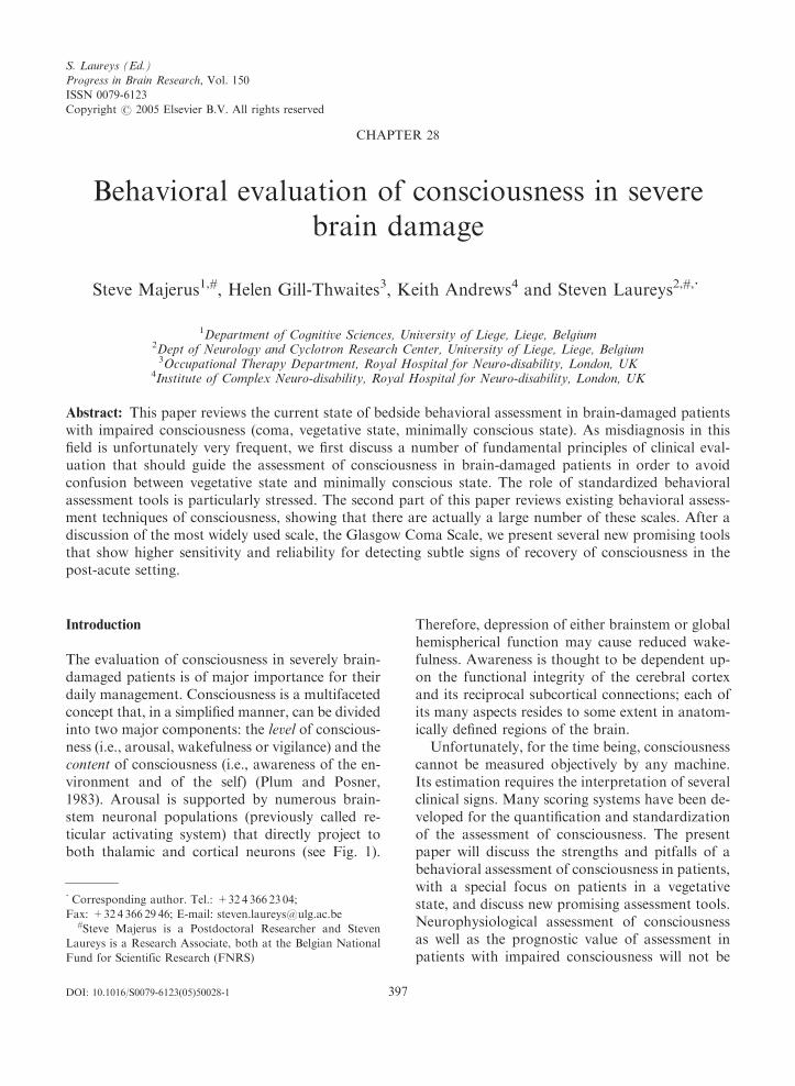
The evaluation of consciousness in severely brain-damaged patients is of major importance for theirdaily management. Consciousness is a multifacetedconcept that, in a simplified manner, can be dividedinto two major components: the level of conscious-ness (i.e., arousal, wakefulness or vigilance) and thecontent of consciousness (i.e., awareness of the en-vironment and of the self) (Plum and Posner,1983). Arousal is supported by numerous brain-stem neuronal populations (previously called re-ticular activating system) that directly project toboth thalamic and cortical neurons (see Fig. 1).
Therefore, depression of either brainstem or globalhemispherical function may cause reduced wake-fulness. Awareness is thought to be dependent up-on the functional integrity of the cerebral cortexand its reciprocal subcortical connections; each ofits many aspects resides to some extent in anatom-ically defined regions of the brain.
Unfortunately, for the time being, consciousnesscannot be measured objectively by any machine.Its estimation requires the interpretation of severalclinical signs. Many scoring systems have been de-veloped for the quantification and standardizationof the assessment of consciousness. The presentpaper will discuss the strengths and pitfalls of abehavioral assessment of consciousness in patients,with a special focus on patients in a vegetativestate, and discuss new promising assessment tools.Neurophysiological assessment of consciousnessas well as the prognostic value of assessment inpatients with impaired consciousness will not be
�Corresponding author. Tel.: +32 4 366 23 04;
Fax: +32 4 366 29 46; E-mail: [email protected]#Steve Majerus is a Postdoctoral Researcher and Steven
Laureys is a Research Associate, both at the Belgian National
Fund for Scientific Research (FNRS)
DOI: 10.1016/S0079-6123(05)50028-1 397
considered here as they are the issue of other pa-pers in this volume (see Kotchoubey; Guerit; Jen-nett this volume).
Clinical evaluation of consciousness
Arousal and awareness are not on-off phenomenabut are part of a large continuum. At the bedside,arousal is assessed by the presence of spontaneousor stimulation-induced eye opening. It ranges fromcoma (no eye opening), stupor (eye opening fol-lowing vigorous external stimuli), through sleep
(eye opening following moderate external stimuli)and alert waking (spontaneous eye opening).Awareness refers to the collective thoughts andfeelings of an individual. Clinically, we are limitedto the appraisal of the patient’s capacity to perceivethe external world and to voluntarily interact withit (i.e., perceptual awareness). In practice, this isevaluated by careful and repeated examination ofthe capacity to formulate reproducible, voluntary,purposeful and sustained behavioral responses toauditory, tactile, visual or noxious stimuli (e.g., byasking the patient to follow command, to visuallydiscriminate between Yes/No cards by pointing oreye movements). Much more difficult is the eval-uation of patients’ self-recognition in a mirror; thiscan be done by putting a mark (colored versus in-visible) on the patient’s face and by determining
whether the patient will touch this mark when be-ing shown his face in a mirror (Gallup, 1997). Ob-viously, the patient needs to be well aroused inorder to perform the cognitive processes requiredfor awareness. Hence, patients in a coma are un-aware because they cannot be aroused. However,as illustrated by patients in a vegetative state,arousal is only a necessary and not a sufficientcondition for awareness. Indeed, patients in a veg-etative state are aroused (as shown by preservedspontaneous eye opening and sleep–wake cycles)but show no sign of awareness (i.e., no sign ofcommand following or any other voluntary be-havior). When the first signs of voluntary behaviorappear, the patient may be in a minimally conscious
state: here the patient is partially conscious, as ev-idenced by the presence of limited but reproduciblesigns of awareness (inconsistent command follow-ing, inconsistent but intelligible verbalization, sus-tained visual fixation, localization of sound andnoxious stimuli) (Giacino et al., 2002; Giacino andWhyte, 2005; Giacino, this volume).
Diagnosing and misdiagnosing signs ofconsciousness
The diagnosis of the vegetative state depends onbehavioral assessment of the responses obtainedfrom the patient. It is not a pathological or even
Fig. 1. A simplified scheme of consciousness and its two major components: arousal and awareness. Note: the gray area represents the
reticular activating system encompassing the brainstem and thalamus; the arrow near the brainstem denotes the progressive disap-
pearance of brainstem reflexes during rostral-caudal deterioration (i.e., evolution from coma to brain death). V ¼ vertical; H ¼ hor-
izontal. (Reproduced with permission from Laureys et al., 2002.)
398
neuro-physiological diagnosis. While there havebeen exciting developments in the use of functionalMRI (Magnetic Resonance Imaging) scanning (seeSchiff, this volume; Owen, this volume), brainmapping and other neuro-physiological approach-es (see Kotchoubey, this volume; Guerit, this vol-ume) these are primarily aids to diagnosis ratherthan a method of diagnosis. Consider, for in-stance, the patient whose neuro-physiological in-vestigations suggest that there is some integrated‘‘higher-level’’ cerebral function in response tostimulation — but where there is no behavioralevidence that the person is aware of his environ-ment — who shows no evidence of communicationor understanding of what others are communicat-ing with him. Where does that leave the patient,the family and the caring team? While it mightincite to reexamine the clinical responses and striveharder to demonstrate any awareness, if the pa-tient continues with no meaningful responses andremains clinically vegetative, then we would arguethat the patient is in the vegetative state.
This, however, does lead us on to questioninghow sensitive our clinical–behavioral assessmentsare. Giacino and Zasler (1995) have pointed outthe limitations of clinical assessment in the iden-tification of ‘‘internal awareness’’ in a patient whootherwise lacks the motor function to demonstratehis awareness. The concept that we are only able toinfer the presence or absence of conscious experi-ence is a long-standing philosophical issue, whichhas been pointed out by Bernat (1992) and TheMulti-Society Task Force (1994) in the specificcontext of the vegetative state. The InternationalWorking Party on the Vegetative State (Andrews,1996) discussed this point in detail and criticizedthe use of the term ‘‘meaningful response’’ on thegrounds that it requires a considerable amount ofsubjective interpretation on the part of the ob-server and that what was meaningful for the pa-tient may not be considered meaningful by thosetreating the patient. Similarly the term ‘‘purposefulresponse’’ was criticized because of the subjectiveinterpretation and that a withdrawal reflex couldbe considered as purposeful in that it removes thelimb, for instance, from danger.
This is where there must be some concern. Forinstance there are several studies that have
described the misdiagnosis of the vegetative state.In a group of long-term patients in a nursing homein the USA, Tresch et al. (1991) found that 18% ofthose diagnosed as being in the persistent vegeta-tive state were aware of themselves or their envi-ronment. Childs et al. (1993) report that 37% ofpatients admitted more than 1 month post-injurywith a diagnosis of coma or persistent vegetativestate had some level of awareness. In anotherstudy (Andrews et al., 1996), 43% of patients ad-mitted to a profound brain injury unit at least 6months following their brain damage (i.e. could beexpected to be stable) were found to have beenmisdiagnosed. While these figures cause concernthey at least emphasize that bedside diagnosis waspossible — otherwise they would not have beenidentified as having been misdiagnosed.
So why are patients misdiagnosed? One strikingfinding was that 65% of the ‘‘misdiagnosed’’ pa-tients were either blind or very severely visuallyimpaired in the form of marked visual field defectsand/or visual perceptual disorders (Andrews et al.,1996). This has obvious implications for assess-ment since one of the prime features for assessingwhether a patient is non-vegetative is eye tracking.If the patient has visual impairment, then he willnot follow objects and therefore eye tracking willbe absent even in a mentally alert individual.
Since all patients followed verbal commands, itis assumed that none were deaf or had severehearing impairment. This, however, is a possibilityand should be considered. This also emphasizesthe importance of assessing a wide range of stimuli(touch, taste and smell as well as visual or audi-tory), a range of frequent observations with stand-ardized assessment tools and optimal patientmanagement (e.g., with the patient in seating po-sition) to ensure that disturbance of one modalityis not the cause of missing evidence of awareness.
Making the diagnosis of vegetative state
Previous studies have shown that misdiagnosed‘‘vegetative’’ patients were at the ‘‘severe’’ level ofthe Glasgow Outcome Scale (Jennett and Teas-dale, 1977), being totally physically dependent forall care needs (Andrews et al., 1996). The only
399
method that any of us can use to demonstrate ourawareness to others is through some form of mo-tor activity — speech, facial expression, eye-track-ing, limb movement, shrugging shoulders,nodding-shaking the head, etc. For 88% of thepatients, pressing a buzzer was the only functionalmovement, although one patient later developedan ability to point with a finger and another pa-tient became able to write words; the other twopatients communicated by eye pointing (Andrewset al., 1996).
The importance of physical function was dra-matically demonstrated by one patient where re-sponses were not identified until 25 weeks afteradmission, though it was obvious from subsequentconversations with him that he had not been veg-etative for some time. This patient was admittedwith very severe joint contractures, which requiredsurgical release and a prolonged physical manage-ment program before he could be seated appro-priately in a special seating system. Only when hewas satisfactorily seated was sufficient muscle tonereleased for him to indicate with a slight shouldershrug that he was aware — he was able to carryout simple mental mathematical calculations andwas aware of his immediate physical and socialenvironment (Andrews et al., 1996).
Another difficulty is the relevance of the blinkresponse to awareness. The patient may blink tomenace but appear not to be attentive. Note that atleast one authority (Working Group of the RoyalCollege of Physicians, 1996) has regarded a blinkto threat as evidence of cortical connection andtherefore indicating that the patient is not vegeta-tive. This is a very questionable approach since theconcept of the vegetative state is the demonstra-tion of awareness, and not of whether there aresome cortical connections. The Multi-Society TaskForce (1994) urges caution in making the diagnosisof the vegetative state if there is blinking to threatbut does not go as far as to claim that if presentthat it indicates that the patient is no longer veg-etative. Actually one of the difficulties is taking toolittle notice of the blink response — or more rel-evantly the speed of the blink response. Often toolittle time is given to waiting for the response.There is often a delay between stimulation and re-sponse when there is awareness, as though the
brain was having to work out the response to give.Of course, this leads to the problem of how long towait and the risk of spontaneous blinking beinginterpreted as a volitional response. This requires aconsiderable amount of experience to interpret.One clue is that the blink is often of a differentquality to reflex blinking — either in the slownessof the blink or the length of time the eye is keptclosed. As noted by Whyte and others, differenti-ation of spontaneous eye blinks from those that arepurposeful can be done by systematically recordingeye-blinks under different conditions (e.g., at rest,to inappropriate command, to appropriate com-mand) and statistically determining whether thefrequency of the response is significantly higherfollowing administration of the appropriate com-mand, relative to the other conditions (see, e.g.,Whyte and DiPasquale, 1995).
There are, of course, other signs that may causea misdiagnosis that the patient is aware when infact the responses are reflex in nature. For in-stance, there may be roving eye movements andthe patient’s eyes may seem to briefly follow mov-ing objects. The movement is usually inconsistentand never sustained. For instance, the patient’seyes may turn toward a sound or a sudden move-ment but does so only briefly and does not focuson the source of stimulation. This can catch outthe unwary who interpret this as awareness. Whatis probably happening is that the subcortical cen-ters that alert the brain to incoming stimuli, e.g.,the superior colliculi for vision and the thalamusfor tactile sensations, are still active but the alert-ing mechanism does not reach ‘‘higher level’’ cor-tical interpretation. This situation is seen, forinstance, in cortical blindness where although thevisual cortex is damaged the patient will still turntoward a visual stimulus even though he cannot‘‘see’’ it.
Some staff and family interpret the withdrawalresponse as being an indication that the patient isaware of the noxious stimulus. It would be morerelevant if the patient pushed away the stimulus.Another confusing feature for many carers is thenon-volitional grasp reflex. This can cause consid-erable concern to relatives or carers who feel thatthe patient recognizes them when they hold hishand. This is particularly reinforced when the grip
400
tightens as there is an attempt to pull the hand orfingers away. This is supportive of the diagnosis ofa grasp reflex rather than supportive of a mean-ingful response.
What can be even more confusing are the frag-ments of coordinated movement, such as scratch-ing or even moving hands toward a noxiousstimulus. These must always be taken seriouslyas indicating awareness but do occur in the veg-etative patient usually affecting the same repetitivemovement on each occasion. They are probablylong-learned automatic response activities. How-ever, scratching oneself on different locations de-pending on the irritant’s source would beindicative of a minimally conscious state.
Chewing movements or grinding of teeth (towhich can be added constant movement of thetongue) again cause concern to relatives and carersfeeling that the patient is indicating that he isthirsty or hungry. Grunting and groaning pro-voked by noxious stimuli can also often be inter-preted as indicating an attempt to communicate.This can cause disagreement between family andclinicians when some relatives claim to be able to‘‘understand’’ the words spoken when others onlyhear sounds. These are, however, commonly foundfeatures in the vegetative state. The skills is to de-cide whether the responses are contingent on thequality of the external stimulus.
Factors influencing the diagnosis
The International Working Party (Andrews, 1996)pointed out that the assessments in general use arebased on a series of behavioral patterns. The cli-nician is, therefore, dependent on overt responsesthat depend on a number of factors including:
a. The physical ability of the patient to respond— this has been discussed above.
b. The desire or willingness (if the patient isaware) of the patient to respond. It is not un-usual for members of the family to obtain re-sponses that the professional members of theteam are not able to. This is probably notsurprising since the members of the family aremore likely to be ‘‘sensitive’’ to the responsesseen. On the other hand, the family may be
desperate for a response and easily misinter-pret the reflex responses. Patients may also bemore willing to respond to family or to somemembers of the staff rather than to others.Let us face facts — some staff are better atrelating than others.
c. The ability to observe accurately. This is par-ticularly relevant since profound brain dam-age is a rare condition and few professionalshave seen sufficient patients to have gainedthat level of experience required to produce‘‘expertise’’.
d. The time available for observation and as-sessment. Time is one of the major factors inassessing the profoundly brain-damaged pa-tients. They do not conveniently have theirbest levels of awareness at the time set asidefor the formal assessment. This requires flex-ibility of the assessor to take advantage of thewindows of opportunity and to take advan-tage of the observations of other members ofthe team and members of the family.
e. The lack of available and reliable assessmenttools. It is not so much that there is a short-age of tools — see discussion below — butthat they are not used in more general acute,or even neurological or rehabilitation, units.
f. The patient is not always seen by a skilledteam to address all of these issues.
g. The family and carers and those who knowthe patient best are not always involved asmuch as they should be.
h. Patients are assessed by some assessors whoare unfamiliar with the patient — leading tomeaningful responses being missed.
There are several principles to the accurate as-sessment of the person thought to be in the veg-etative state:
1. That the patient should be healthy. Evensimple conditions such as constipation,chronic urinary tract infection (usually as-sociated with long-term catheterization) orbronchial infections can prevent optimal re-sponses from being obtained.
2. The patients should be in a good nutritionalstate. The earlier use of gastrostomy feedinghas altered this pattern but still some patients
401
admitted from general units have a low BodyMass Index, emphasizing the difficulty inmanaging people with such complex medicaland physical disabilities.
3. As many sedating drugs as possible shouldbe withdrawn, or at least decreased to thelowest effective dose — these include antis-pasticity drugs and antiepileptic drugs. Inthe case of antiepileptic drugs, which are stillrequired to control fitting, drugs with theleast sedative effect should be used.
4. Complications and consequences of neuro-logical imbalance should be prevented —this includes high muscle tone and contrac-tures by the provision of special seating,good bed and sitting posture to control ab-normal muscle tone. These complications inthe long term increase the amount of nurs-ing care required, which, since the patientmay live for many years, increases the costof care considerably.
5. Controlled posture is important. Most doc-tors have been trained to examine patientson the bed. Experience suggests that patientsare more likely to be alert when sitting up(presumably due to greater stimulation of theascending reticular activating system). Awell-supporting seating system is essentialto reduce sufficient muscle tone to allowmovement of limbs that can be used forcommunication purposes, e.g. to press atouch-sensitive switch.
6. Providing a controlled environment of sen-sory regulation to avoid sensory overload ofthe severely damaged brain. Since it is likelythat profoundly brain-damaged patientshave problems with selective attention, sen-sory input should be simple and interspersedwith periods of rest. It is, therefore, logicalto assess for cognitive responses after a pe-riod of rest rather than after a period of ac-tivity, such as being washed and dressed orafter physiotherapy. This requires staff andfamily to understand the importance ofavoiding over-stimulation prior to the as-sessment.
7. Assessments should be short (to avoid tiringthe patient), repeated (to identify windows
of opportunity) and over a prolonged periodof time (to accommodate the learning proc-ess of both the patient and the assessor).One-off short assessment of the patient whois lying in bed is likely to result in a misseddiagnosis even by the most experiencedclinician.
8. The ability to generate a behavioral re-sponse fluctuates from day to day and hourto hour, and even minute to minute, de-pending on fatigue factors, general health ofthe patient and the underlying neurologicalcondition.
9. Observation needs to take into account de-layed responses. Assimilation of even basicinformation is often slow and therefore re-sponse time may be delayed. Because of this,information provided at any one timeshould be simple, consistent, repeated aftera period of rest, and allow for a delayed re-sponse.
10. Communication requires skilled techniquesand sensitivity for the method by which thepatient wants to communicate.
11. Families and other carers have a very im-portant role in identifying the best responsesand the optimal conditions for assessment.While there are some relatives who interpretreflex responses as being meaningful, there isno doubt that members of the family areoften more sensitive to early changes thaneven very experienced clinical staff.
Consciousness scales
There are many scales designed to monitor the re-covery of consciousness in brain-damaged patients.In this section, we will first address the GlasgowComa Scale (GCS), which remains the most widelyused scale in the acute and subacute setting. Wewill then review other existing tools, by focusingmore specifically on three new and promisingassessment instruments, the Coma RecoveryScale-Revised (CRS-R; Giacino et al., 2004), theWessex Head Injury Matrix (WHIM; Shiel et al.,2000), and the Sensory Modality Assessment and
402
Rehabilitation Technique (SMART; Gill-Thwaites,1997, 1999).
Glasgow coma scale
Teasdale and Jennett (1974) developed the GCS asan aid in the clinical assessment of post-traumaticunconsciousness. It was devised as a formalscheme to overcome the ambiguities that arosewhen information about comatose patients waspresented and groups of patients compared. TheGCS has three components: eye (E), verbal (V)and motor (M) response to external stimuli (seeFig. 2). The scale consisted of 14 points, but waslater adapted to 15, with the division of the motorcategory ‘‘flexion to pain’’ into two further cate-gories. The best or highest responses are recorded.So far, more than 2390 publications have appearedto its use (MEDLINE search performed in Feb-ruary 2005, limited to title and abstract word). It isa component of the Acute Physiology and ChronicHealth Evaluation (APACHE) II score, the (Re-vised) Trauma Score, the Trauma and Injury Se-verity Score (TRISS) and the Circulation,Respiration, Abdomen, Motor, Speech (CRAMS)Scale, demonstrating the widespread adoption ofthe scale.
The observation of spontaneous eye opening‘‘indicates that the arousal mechanisms of thebrainstem are active’’ (Teasdale and Jennett,1974). As previously stated, recovered arousaldoes not imply the recovery of awareness. Patientsin a vegetative state have awakened from their co-ma but remain unaware of their environment andself. Most comatose patients who survive willeventually open their eyes, regardless of the sever-ity of their cerebral damage (Jennett, 1972). In-deed, less than 4% of head-damaged patientsnever open their eyes before they die (Bricolo etal., 1980). The eye opening in response to speechtests the reaction ‘‘to any verbal approach, wheth-er spoken or shouted, not necessarily the com-mand to open the eyes’’ (Teasdale and Jennett,1974). Again, this response is observed in the veg-etative state where ‘‘awakening’’ can be induced bynon-specific auditory stimulation. In these pa-tients, it is recommended to differentiate between a
reproducible response to command and to non-sense speech. Eye opening in response to painshould be tested by stimulation at the level of thelimbs, because the grimacing associated with sup-raorbital or jaw-angle pressure may cause eye clo-sure.
The presence of verbal responses indicates therestoration of a high degree of interaction with theenvironment (i.e., awareness). An oriented con-versation implies awareness of the self (e.g., thepatient can answer the question: ‘‘What is yourname?’’) and environment (e.g., the patient cor-rectly answers the questions: ‘‘Where are we?’’ and‘‘What year/month is it?’’). Confused speech is re-corded when the patient is capable of producinglanguage, for instance phrases and sentences, butis unable to answer the questions about orienta-tion. When the patient presents intelligible artic-ulation but exclaims only isolated words in arandom way (often swear words, obtained byphysical stimulation rather than by a verbal ap-proach), this is scored as ‘‘inappropriate speech’’.Incomprehensible sounds refer to moaning andgroaning without any recognizable words. Thisrudimentary vocalization does not necessitateawareness and is thought to depend upon subcor-tical functioning as it can be observed in anence-phalic children and vegetative patients.
The motor response first assesses whether thepatient obeys simple commands, given in verbal,gestural or written form. A non-specific soundstimulus may induce a reflex contraction of thepatient’s fingers or alternatively such a reflex re-sponse can result from the physical presence of theexaminer’s fingers against the palm of the patient(i.e., grasping reflex). Before accepting that thepatient is truly obeying commands, it is advised totest that the patient will also release and squeezeagain to repeated commands. If there is no re-sponse a painful stimulus is applied. First, pressureis applied to the fingernail bed with a pencil. Ifflexion is observed, stimulation is then applied toother sites (applying pressure to the supraorbitalridge, pinching the trapezium or rubbing the ster-num) to differentiate between localization (i.e., anoxious stimulus applied at more than one sitecauses a limb to move so as to attempt to remove itby crossing the midline), withdrawal flexion (i.e., a
403
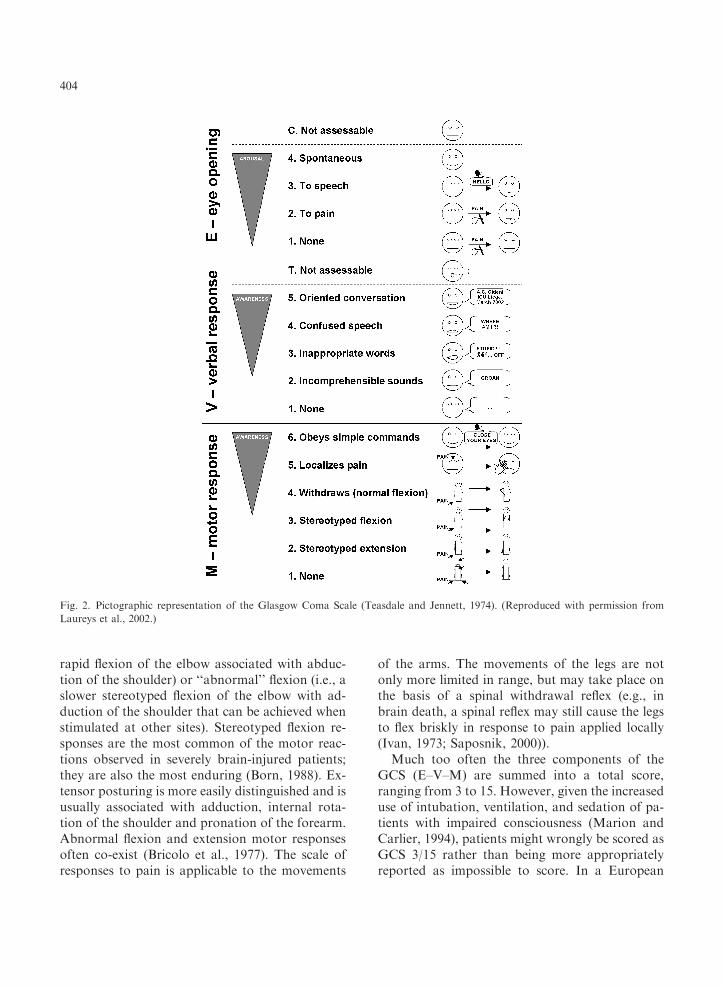
rapid flexion of the elbow associated with abduc-tion of the shoulder) or ‘‘abnormal’’ flexion (i.e., aslower stereotyped flexion of the elbow with ad-duction of the shoulder that can be achieved whenstimulated at other sites). Stereotyped flexion re-sponses are the most common of the motor reac-tions observed in severely brain-injured patients;they are also the most enduring (Born, 1988). Ex-tensor posturing is more easily distinguished and isusually associated with adduction, internal rota-tion of the shoulder and pronation of the forearm.Abnormal flexion and extension motor responsesoften co-exist (Bricolo et al., 1977). The scale ofresponses to pain is applicable to the movements
of the arms. The movements of the legs are notonly more limited in range, but may take place onthe basis of a spinal withdrawal reflex (e.g., inbrain death, a spinal reflex may still cause the legsto flex briskly in response to pain applied locally(Ivan, 1973; Saposnik, 2000)).
Much too often the three components of theGCS (E–V–M) are summed into a total score,ranging from 3 to 15. However, given the increaseduse of intubation, ventilation, and sedation of pa-tients with impaired consciousness (Marion andCarlier, 1994), patients might wrongly be scored asGCS 3/15 rather than being more appropriatelyreported as impossible to score. In a European
Fig. 2. Pictographic representation of the Glasgow Coma Scale (Teasdale and Jennett, 1974). (Reproduced with permission from
Laureys et al., 2002.)
404
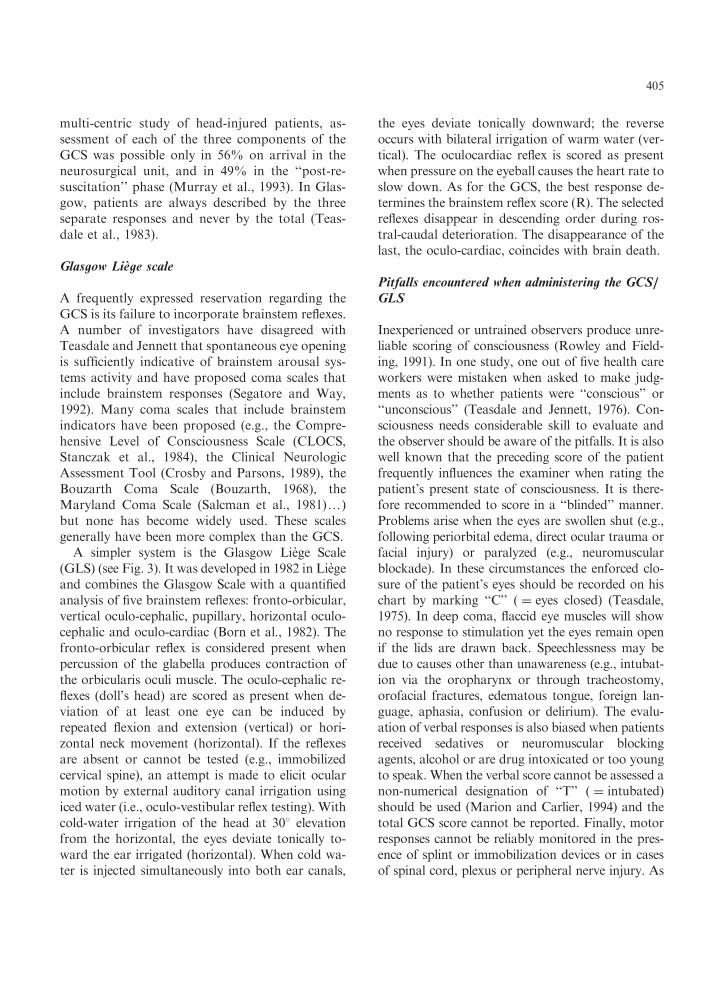
multi-centric study of head-injured patients, as-sessment of each of the three components of theGCS was possible only in 56% on arrival in theneurosurgical unit, and in 49% in the ‘‘post-re-suscitation’’ phase (Murray et al., 1993). In Glas-gow, patients are always described by the threeseparate responses and never by the total (Teas-dale et al., 1983).
Glasgow Liege scale
A frequently expressed reservation regarding theGCS is its failure to incorporate brainstem reflexes.A number of investigators have disagreed withTeasdale and Jennett that spontaneous eye openingis sufficiently indicative of brainstem arousal sys-tems activity and have proposed coma scales thatinclude brainstem responses (Segatore and Way,1992). Many coma scales that include brainstemindicators have been proposed (e.g., the Compre-hensive Level of Consciousness Scale (CLOCS,Stanczak et al., 1984), the Clinical NeurologicAssessment Tool (Crosby and Parsons, 1989), theBouzarth Coma Scale (Bouzarth, 1968), theMaryland Coma Scale (Salcman et al., 1981)y)but none has become widely used. These scalesgenerally have been more complex than the GCS.
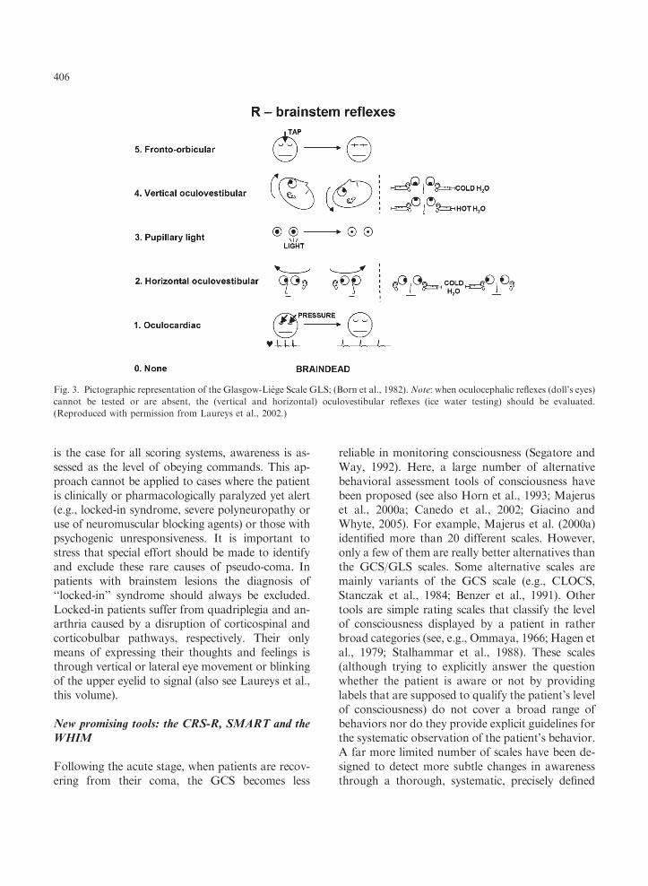
A simpler system is the Glasgow Liege Scale(GLS) (see Fig. 3). It was developed in 1982 in Liegeand combines the Glasgow Scale with a quantifiedanalysis of five brainstem reflexes: fronto-orbicular,vertical oculo-cephalic, pupillary, horizontal oculo-cephalic and oculo-cardiac (Born et al., 1982). Thefronto-orbicular reflex is considered present whenpercussion of the glabella produces contraction ofthe orbicularis oculi muscle. The oculo-cephalic re-flexes (doll’s head) are scored as present when de-viation of at least one eye can be induced byrepeated flexion and extension (vertical) or hori-zontal neck movement (horizontal). If the reflexesare absent or cannot be tested (e.g., immobilizedcervical spine), an attempt is made to elicit ocularmotion by external auditory canal irrigation usingiced water (i.e., oculo-vestibular reflex testing). Withcold-water irrigation of the head at 301 elevationfrom the horizontal, the eyes deviate tonically to-ward the ear irrigated (horizontal). When cold wa-ter is injected simultaneously into both ear canals,
the eyes deviate tonically downward; the reverseoccurs with bilateral irrigation of warm water (ver-tical). The oculocardiac reflex is scored as presentwhen pressure on the eyeball causes the heart rate toslow down. As for the GCS, the best response de-termines the brainstem reflex score (R). The selectedreflexes disappear in descending order during ros-tral-caudal deterioration. The disappearance of thelast, the oculo-cardiac, coincides with brain death.
Pitfalls encountered when administering the GCS/GLS
Inexperienced or untrained observers produce unre-liable scoring of consciousness (Rowley and Field-ing, 1991). In one study, one out of five health careworkers were mistaken when asked to make judg-ments as to whether patients were ‘‘conscious’’ or‘‘unconscious’’ (Teasdale and Jennett, 1976). Con-sciousness needs considerable skill to evaluate andthe observer should be aware of the pitfalls. It is alsowell known that the preceding score of the patientfrequently influences the examiner when rating thepatient’s present state of consciousness. It is there-fore recommended to score in a ‘‘blinded’’ manner.Problems arise when the eyes are swollen shut (e.g.,following periorbital edema, direct ocular trauma orfacial injury) or paralyzed (e.g., neuromuscularblockade). In these circumstances the enforced clo-sure of the patient’s eyes should be recorded on hischart by marking ‘‘C’’ ( ¼ eyes closed) (Teasdale,1975). In deep coma, flaccid eye muscles will showno response to stimulation yet the eyes remain openif the lids are drawn back. Speechlessness may bedue to causes other than unawareness (e.g., intubat-ion via the oropharynx or through tracheostomy,orofacial fractures, edematous tongue, foreign lan-guage, aphasia, confusion or delirium). The evalu-ation of verbal responses is also biased when patientsreceived sedatives or neuromuscular blockingagents, alcohol or are drug intoxicated or too youngto speak. When the verbal score cannot be assessed anon-numerical designation of ‘‘T’’ ( ¼ intubated)should be used (Marion and Carlier, 1994) and thetotal GCS score cannot be reported. Finally, motorresponses cannot be reliably monitored in the pres-ence of splint or immobilization devices or in casesof spinal cord, plexus or peripheral nerve injury. As
405
is the case for all scoring systems, awareness is as-sessed as the level of obeying commands. This ap-proach cannot be applied to cases where the patientis clinically or pharmacologically paralyzed yet alert(e.g., locked-in syndrome, severe polyneuropathy oruse of neuromuscular blocking agents) or those withpsychogenic unresponsiveness. It is important tostress that special effort should be made to identifyand exclude these rare causes of pseudo-coma. Inpatients with brainstem lesions the diagnosis of‘‘locked-in’’ syndrome should always be excluded.Locked-in patients suffer from quadriplegia and an-arthria caused by a disruption of corticospinal andcorticobulbar pathways, respectively. Their onlymeans of expressing their thoughts and feelings isthrough vertical or lateral eye movement or blinkingof the upper eyelid to signal (also see Laureys et al.,this volume).
New promising tools: the CRS-R, SMART and theWHIM
Following the acute stage, when patients are recov-ering from their coma, the GCS becomes less
reliable in monitoring consciousness (Segatore andWay, 1992). Here, a large number of alternativebehavioral assessment tools of consciousness havebeen proposed (see also Horn et al., 1993; Majeruset al., 2000a; Canedo et al., 2002; Giacino andWhyte, 2005). For example, Majerus et al. (2000a)identified more than 20 different scales. However,only a few of them are really better alternatives thanthe GCS/GLS scales. Some alternative scales aremainly variants of the GCS scale (e.g., CLOCS,Stanczak et al., 1984; Benzer et al., 1991). Othertools are simple rating scales that classify the levelof consciousness displayed by a patient in ratherbroad categories (see, e.g., Ommaya, 1966; Hagen etal., 1979; Stalhammar et al., 1988). These scales(although trying to explicitly answer the questionwhether the patient is aware or not by providinglabels that are supposed to qualify the patient’s levelof consciousness) do not cover a broad range ofbehaviors nor do they provide explicit guidelines forthe systematic observation of the patient’s behavior.A far more limited number of scales have been de-signed to detect more subtle changes in awarenessthrough a thorough, systematic, precisely defined
Fig. 3. Pictographic representation of the Glasgow-Liege Scale GLS; (Born et al., 1982). Note: when oculocephalic reflexes (doll’s eyes)
cannot be tested or are absent, the (vertical and horizontal) oculovestibular reflexes (ice water testing) should be evaluated.
(Reproduced with permission from Laureys et al., 2002.)
406
and reliable observation of the patient’s behaviors.These scales are supposed to be more sensitive thanprevious scales as they include a much largernumber of items (e.g., Davis, 1991; Giacino et al.,1991; Coma/Near Coma Scale; Rappaport et al.,1992; Visual Response Evaluation, Coma ExitChart, Freeman, 1996; CRS, Giacino et al., 2004).However, it must be noted that studies specificallyaimed at providing empirical evidence for this the-oretically superior sensitivity are often lacking.
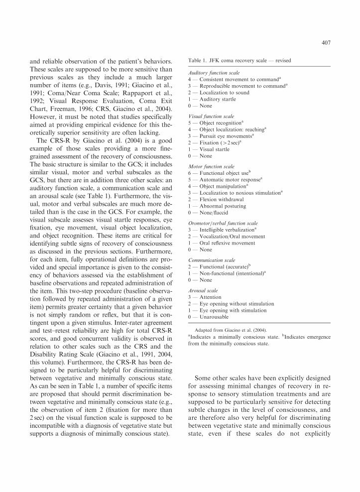
The CRS-R by Giacino et al. (2004) is a goodexample of those scales providing a more fine-grained assessment of the recovery of consciousness.The basic structure is similar to the GCS; it includessimilar visual, motor and verbal subscales as theGCS, but there are in addition three other scales: anauditory function scale, a communication scale andan arousal scale (see Table 1). Furthermore, the vis-ual, motor and verbal subscales are much more de-tailed than is the case in the GCS. For example, thevisual subscale assesses visual startle responses, eyefixation, eye movement, visual object localization,and object recognition. These items are critical foridentifying subtle signs of recovery of consciousnessas discussed in the previous sections. Furthermore,for each item, fully operational definitions are pro-vided and special importance is given to the consist-ency of behaviors assessed via the establishment ofbaseline observations and repeated administration ofthe item. This two-step procedure (baseline observa-tion followed by repeated administration of a givenitem) permits greater certainty that a given behavioris not simply random or reflex, but that it is con-tingent upon a given stimulus. Inter-rater agreementand test–retest reliability are high for total CRS-Rscores, and good concurrent validity is observed inrelation to other scales such as the CRS and theDisability Rating Scale (Giacino et al., 1991, 2004,this volume). Furthermore, the CRS-R has been de-signed to be particularly helpful for discriminatingbetween vegetative and minimally conscious state.As can be seen in Table 1, a number of specific itemsare proposed that should permit discrimination be-tween vegetative and minimally conscious state (e.g.,the observation of item 2 (fixation for more than2 sec) on the visual function scale is supposed to beincompatible with a diagnosis of vegetative state butsupports a diagnosis of minimally conscious state).
Some other scales have been explicitly designedfor assessing minimal changes of recovery in re-sponse to sensory stimulation treatments and aresupposed to be particularly sensitive for detectingsubtle changes in the level of consciousness, andare therefore also very helpful for discriminatingbetween vegetative state and minimally consciousstate, even if these scales do not explicitly
Table 1. JFK coma recovery scale — revised
Auditory function scale
4 — Consistent movement to commanda
3 — Reproducible movement to commanda
2 — Localization to sound
1 — Auditory startle
0 — None
Visual function scale
5 — Object recognitiona
4 — Object localization: reachinga
3 — Pursuit eye movementsa
2 — Fixation (42 sec)a
1 — Visual startle
0 — None
Motor function scale
6 — Functional object useb
5 — Automatic motor responsea
4 — Object manipulationa
3 — Localization to noxious stimulationa
2 — Flexion withdrawal
1 — Abnormal posturing
0 — None/flaccid
Oromotor/verbal function scale
3 — Intelligible verbalizationa
2 — Vocalization/Oral movement
1 — Oral reflexive movement
0 — None
Communication scale
2 — Functional (accurate)b
1 — Non-functional (intentional)a
0 — None
Arousal scale
3 — Attention
2 — Eye opening without stimulation
1 — Eye opening with stimulation
0 — Unarousable
Adapted from Giacino et al. (2004).aIndicates a minimally conscious state. bIndicates emergence
from the minimally conscious state.
407
highlight those items that are supposed to differ-entiate between these two states (e.g., WesternNeuro Sensory Stimulation Profile (WNSSP), An-sell and Keenan, 1989; Sensory Stimulation As-sessment Measure, Rader and Ellis, 1994). One ofthese is the SMART (Gill-Thwaites, 1997). Al-though the tool originated from the parameters ofthe GCS, further extended by Freeman (1996), ithas been further enhanced since its inception in1988 with extensive evidence from both clinicalpractice and research (Wilson et al., 1991, 1993,1996a, b; Gill-Thwaites and Munday, 1999; Wil-son and Gill-Thwaites, 2000). The final designcategorizes all behavioral responses observed inmore than 300 patients in vegetative or minimallyconscious states (as previously mentioned, thislatter category defines patients who present somesigns of awareness but behavioral responses arestill very elementary, inconsistent and sometimesdifficult to elicit; see Giacino, this volume for ex-tensive review).
The SMART was designed to identify evidenceof the patient’s awareness through a graded as-sessment of the level of sensory, motor and com-municative responses to a structured and regulatedsensory program and also as a treatment tool toguide future treatment to enhance the patient’spotential responses. The SMART comprises twocomponents, including the informal componentthat consists of information from family and care-rs in respect of observed behaviors and informa-tion pertaining to the patients’ pre-morbidinterests, likes and dislikes. This component en-courages active participation from families andcarers, ensures that all responses seen to be day-to-day activity are recorded and categorized and thatthe treatment is relevant to the patients’ interest,thus optimizing the opportunity for a meaningfulresponse to stimuli. The SMART’s formal assess-ment comprises of the SMART Behavioral Ob-servation Assessment and Sensory Assessment andis conducted in 10 sessions within a 3-week periodwith an equal number of sessions in the morningand afternoon. This time frame provides frequentassessments over a short time frame to determinewhether the behavioral responses observed areboth consistent and repeatable. The behavioralobservation enables the assessor to become
familiar with the patients’ reflexive, spontaneousand purposeful behavior during a 10-minute peri-od prior to the commencement of the SMARTSensory Assessment.
The Sensory Assessment has eight modalitiesincluding the five sensory modalities (visual, audi-tory, tactile, olfactory and gustatory) and alsomotor function, functional communication andwakefulness/arousal. Consisting of 29 standard-ized techniques, the SMART provides opportunityfor patients to exhibit their full behavioral reper-toire, in each of the different sensory modalities.For example, to assess the patients’ responseswithin the auditory modality, a range of stand-ardized auditory stimuli are presented, includingloud sound, voice and a variety of specifically se-lected verbal instructions. The verbal instructionsare carefully selected from the patient’s behavioralrepertoire exhibited as being potentially meaning-ful in the SMART Behavioral Observation, suchas ‘‘raise your eyebrows’’, ‘‘move your thumb’’, toprovide the patient with the best opportunity tofollow any one or more instructions.
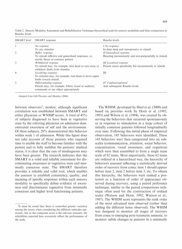
The SMART’s 5-point hierarchical scale is con-sistent and comparable across all of the sensorymodalities. The five levels range from ‘‘no re-sponse’’ (level 1) through ‘‘reflexive’’ (level 2),‘‘withdrawal’’ (level 3), ‘‘localizing’’ (level 4) and‘‘discriminating’’ responses (level 5). This 5-pointscale relates directly to the description of RanchoLevels 1–4 (Hagen et al., 1979); a consistent re-sponse (on five consecutive assessments) atSMART level 5 in any one of the sensory modali-ties demonstrates a meaningful response and thusindicates that the patient is showing behaviors in-dicative of a minimally conscious state or higherlevels of function (Table 2).
Recent research (Gill-Thwaites and Munday,2004) has established the reliability and validity ofthe SMART on 60 subjects diagnosed in vegetativestate on admission and assessed at two monthlyintervals. The Rancho level (Hagen et al., 1979)ratings were derived from referring physicians,SMART and WNSSP (Ansell and Keenan, 1989)for each subject and the scores of each were com-pared. The intra-observer intra-class correlation(ICC) was 0.97 and inter-observer ICC was 0.96,which implied very little variation within and
408
between observers1; modest, although significantcorrelation was established between SMART andeither physician or WNSSP scores. A total of 45%of subjects diagnosed to have been in vegetativestate by the referring physician on admission dem-onstrated awareness of self and the environment.Of these subjects, 28% demonstrated this behaviorwithin week 1 of admission. While this figure doesnot take account of those patients who requiredtime to enable the staff to become familiar with thepatient and to fully stabilize the patients’ medicalstatus, it is clear that the rate of misdiagnosis mayhave been greater. The research indicates that theSMART is a valid and reliable assessment for dis-criminating awareness in vegetative state and min-imally conscious state. The SMART thereforeprovides a reliable and valid tool, which enablesthe assessor to establish consistency, quality, andmeaning of specific responses within each sensorymodality to specifically define evidence of aware-ness and discriminates vegetative from minimallyconscious and higher level functioning patients.
The WHIM, developed by Shiel et al. (2000) andbased on previous work by Horn et al. (1992,1993) and Wilson et al. (1994), was created by ob-serving the behaviors that occurred spontaneouslyor in response to stimulation in a large cohort ofinitially comatose patients followed longitudinallyover time. Following this initial phase of empiricalobservation, 145 behaviors were identified. These145 behaviors were then categorized into six sub-scales (communication, attention, social behavior,concentration, visual awareness, and cognition)which were then assembled to form a single mainscale of 62 items. Most importantly, these 62 itemsare ordered in a hierarchical way, the hierarchy ofbehaviors assessed reflecting a statistically derivedorder of recovery from coma: item 1 should appearbefore item 2, item 2 before item 3, etc. To obtainthis hierarchy, the behaviors were ranked a pos-teriori as a function of order of appearance ob-served during recovery, using a paired preferencetechnique, similar to the paired comparisons tech-nique often used for the construction of ordinalscales (Watson and Horn, 1992; Watson et al.,1997). The WHIM score represents the rank orderof the most advanced item observed (rather thanadding the different items observed). The WHIMwas designed to monitor all stages of recoveryfrom coma to emerging post-traumatic amnesia, tomonitor subtle changes in patients in a minimally
Table 2. Sensory Modality Assessment and Rehabilitation Technique hierarchical scale for sensory modalities and their comparison to
Rancho levels
SMART level SMART response Rancho levels
1 No response I No response
To any stimulus In deep sleep and unresponsive to stimuli
2 Reflex response II Generalized response
To stimuli reflexive and generalized responses, i.e.
startle, flexor or extensor pattern
Reacting inconsistently and non-purposefully to stimuli
3 Withdrawal response III Localized response
To stimuli may, for example, turn head or eyes away or
withdraw limbs from stimulus
Patient reacts specifically but inconsistently to stimuli
4 Localizing response III
To stimulus may, for example, turn head or move upper
limbs toward stimuli
5 Differentiating response IV Confused-agitated
Patient may, for example, follow visual or auditory
commands or use object appropriately
And subsequent Rancho levels
Adapted from Gill-Thwaites and Munday (2004).
1It must be noted that there is somewhat greater variation
among the scores when considering the different subscales sep-
arately, but as the composite score is the relevant measure, the
reliabilities reported here accurately reflect the performance of
the scale.
409
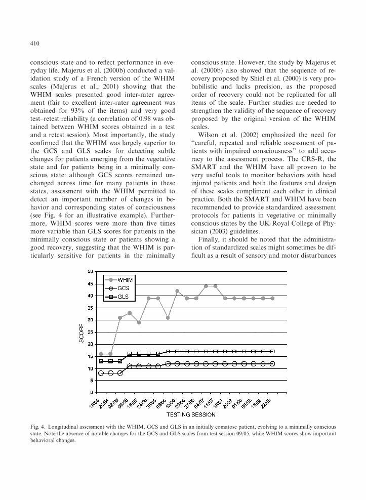
conscious state and to reflect performance in eve-ryday life. Majerus et al. (2000b) conducted a val-idation study of a French version of the WHIMscales (Majerus et al., 2001) showing that theWHIM scales presented good inter-rater agree-ment (fair to excellent inter-rater agreement wasobtained for 93% of the items) and very goodtest–retest reliability (a correlation of 0.98 was ob-tained between WHIM scores obtained in a testand a retest session). Most importantly, the studyconfirmed that the WHIM was largely superior tothe GCS and GLS scales for detecting subtlechanges for patients emerging from the vegetativestate and for patients being in a minimally con-scious state: although GCS scores remained un-changed across time for many patients in thesestates, assessment with the WHIM permitted todetect an important number of changes in be-havior and corresponding states of consciousness(see Fig. 4 for an illustrative example). Further-more, WHIM scores were more than five timesmore variable than GLS scores for patients in theminimally conscious state or patients showing agood recovery, suggesting that the WHIM is par-ticularly sensitive for patients in the minimally
conscious state. However, the study by Majerus etal. (2000b) also showed that the sequence of re-covery proposed by Shiel et al. (2000) is very pro-babilistic and lacks precision, as the proposedorder of recovery could not be replicated for allitems of the scale. Further studies are needed tostrengthen the validity of the sequence of recoveryproposed by the original version of the WHIMscales.
Wilson et al. (2002) emphasized the need for‘‘careful, repeated and reliable assessment of pa-tients with impaired consciousness’’ to add accu-racy to the assessment process. The CRS-R, theSMART and the WHIM have all proven to bevery useful tools to monitor behaviors with headinjured patients and both the features and designof these scales compliment each other in clinicalpractice. Both the SMART and WHIM have beenrecommended to provide standardized assessmentprotocols for patients in vegetative or minimallyconscious states by the UK Royal College of Phy-sician (2003) guidelines.
Finally, it should be noted that the administra-tion of standardized scales might sometimes be dif-ficult as a result of sensory and motor disturbances
Fig. 4. Longitudinal assessment with the WHIM, GCS and GLS in an initially comatose patient, evolving to a minimally conscious
state. Note the absence of notable changes for the GCS and GLS scales from test session 09/05, while WHIM scores show important
behavioral changes.
410
that will make impossible the scoring of a numberof items needing a given sensory or motor modality.This is particularly important as these sensory andmotor impairments are a frequent cause of misdi-agnosis. In these cases, the use of individualizedassessment techniques is recommended. For exam-ple, Whyte et al. (1995, 1999) proposed a methodfor a reliable assessment of visual attention andcommand following in these patients (see alsoGiacino and Whyte, 2005). The principle of thismethod is to find, for an individual patient, at leastone behavior with which the patient seems to pro-duce voluntary responses. This behavior is likely tobe different in each patient and depends on hisparticular sensory and motor impairments. Oncethis behavior has been detected, the second aim isto determine whether this behavior is really volun-tary, by determining the baseline frequency of thisbehavior, and by determining increases in frequen-cy of this behavior over time and as a result ofstimulation (e.g., on command). In order to con-sider a behavior as volitional, the patient has torespond more frequently when required to producethe behavior than during baseline and he must re-spond less frequently when instructed not to pro-duce the behavior. This method permits to obtaindiscrimination scores between the three conditions(behavior on, behavior off, baseline) for which sta-tistical significance can be tested. Using such indi-vidualized assessment methods in combination withthe standardized assessment scales presented above,both the sensitivity and reliability of behavioral as-sessment of altered states of consciousness is likelyto be maximized. The CRS-R has incorporatedparts of these individualized assessment techniquesas described by Whyte et al. (1997, 1999).
As obvious time constraints in the clinical settingwill not allow to assess every patient with each ofthe presented scales, we will conclude this section byproviding some guidelines for selecting the mostappropriate scale, depending on the question that isasked and the state the patient is in. In the acutesetting, the GCS remains the ‘‘gold standard’’ in theevaluation of coma. By virtue of its simplicity, it isthe most universally utilized consciousness scaleworldwide and seems, despite its drawbacks, des-tined to be used in emergency medicine and inten-sive care for some time. In the post-coma phase, and
to differentiate between vegetative and minimallyconscious state, the CRS-R (Giacino et al., 2004), inconjunction with the individualized assessment tech-nique proposed by Whyte et al. (1995, 1999), mightbe the best solution as it was specifically designedfor making this differential diagnosis. When follow-ing a patient longitudinally and documenting subtleprogresses in the recovery of consciousness, theSMART and WHIM could be more appropriate.The WHIM seems more practical for assessmentsmade on a daily basis as time needed to administerthis scale is only about 10min (range 2–35min),while administration of the SMART takes between30 and 40min. The WHIM has been shown to beparticularly sensitive for patients in the minimallyconscious state and patients showing slow but rel-atively good recovery. On the other hand, one im-portant advantage of the SMART is that it alsoassesses responses to a sensory stimulation program,which is not the case for the other scales. Finally, theSMART appears to be particularly suitable for pa-tients in the vicinity of the vegetative state. Althoughthe WHIM also shows a good sensitivity for thisstate, the inclusion of an olfactory function subscalein the SMART provides an additional opportunityfor detecting subtle signs of responsiveness.
Conclusions
Assessment of awareness is not a matter of all ornothing. Recovery of awareness is a very gradualprocess, with sometimes great leaps forwards, butmore often subtle changes, and also sometimessetbacks. For the patient emerging from coma, it isof utmost importance that the medical staff adaptsits assessment to the level of awareness the patientis currently in. The subtlest signs of awareness, aswell as their fluctuation, have to be reliably cap-tured as they are the only means for avoiding mis-diagnosis, but also for communicating with thesepatients. This implies the use of standardized, sen-sitive and individualized assessment tools that cov-er a wide range of possible, although sometimesminimal, behaviors in all sensory modalities. Themajor challenge of the years to come will not be todevelop new tools, but to effectively implement
411
those existing ones in the daily practice of carers ofpatients with impaired consciousness.
References
Andrews, K., Murphy, L., Munday, R. and Littlewood, C.
(1996) Misdiagnosis of the vegetative state: retrospective
study in a rehabilitation unit. Bri. Med. J., 313: 13–16.
Andrews, K. (1996) International Working Party on the Man-
agement of the Vegetative State: summary report. Brain Inj.,
10: 797–806.
Ansell, B.J. and Keenan, J.E. (1989) The Western neuro sensory
stimulation profile: a tool for assessing slow to recover head
injured patients. Arch. Phys. Med. Rehabil., 70: 104–108.
Benzer, A., Mitterschiffthaler, G., Marosi, M., Luef, G., Puh-
ringer, F., De La Renotiere, K., Lehner, H. and Schmutz-
hard, E. (1991) Prediction of non-survival after trauma:
Innsbruck Coma Scale. Lancet, 338: 977–978.
Bernat, J.L. (1992) The boundaries of the persistent vegetative
state. J. Clin. Ethics, 3: 176–180.
Born, J.D. (1988) The Glasgow-Liege Scale. Prognostic value
and evaluation of motor response and brain stem reflexes
after severe head injury. Acta Neurochir., 95: 49–52.
Born, J.D., Hans, P., Dexters, G., Kalangu, K., Lenelle, J.,
Milbouw, G. and Stevenaert, A. (1982) Practical assessment
of brain dysfunction in severe head trauma. Neurochirurgie,
28: 1–7.
Bouzarth, W.F. (1968) Neurosurgical watch sheet for cranio-
cerebral trauma. J. Trauma, 8: 29–31.
Bricolo, A., Turazzi, S., Alexandre, A. and Rizzuto, N. (1977)
Decerebrate rigidity in acute head injury. J. Neurosurg., 47:
680–689.
Bricolo, A., Turazzi, S. and Feriotti, G. (1980) Prolonged post-
traumatic unconsciousness: therapeutic assets and liabilities.
J. Neurosurg., 52: 625–634.
Canedo, A., Grix, M.C. and Nicoletti, J. (2002) An analysis of
assessment instruments for the minimally responsive patient
(MRP): clinical observations. Brain Injury, 16: 453–461.
Childs, N.L., Mercer, W.N. and Childs, H.W. (1993) Accuracy
of diagnosis of persistent vegetative state. Neurology, 43:
1465–1467.
Crosby, L. and Parsons, L.C. (1989) Clinical neurologic as-
sessment tool: development and testing of an instrument to
index neurologic status. Heart Lung, 18: 121–129.
Davis, A.L. (1991) The visual response evaluation: a pilot study
of an evaluation tool for assessing visual responses in low-
level brain injured patients. Brain Injury, 5: 315–320.
Freeman, E.A. (1996) The coma exit chart: assessing the patient
in prolonged coma and vegetative state. Brain Injury, 10:
615–624.
Gallup Jr., G.G. (1997) On the rise and fall of self-conception in
primates. Ann. NY Acad. Sci., 818: 72–82.
Giacino, J.T., Ashwal, S., Childs, N., Cranford, R., Jennett, B.,
Katz, D., Kelly, J., Rosenberg, J., Whyte, J., Zafonte, R. and
Zasler, N. (2002) The minimally conscious state: definition
and diagnostic criteria. Neurology, 58: 349–353.
Giacino, J.T., Kezmarsky, M.A., DeLuca, J. and Cicerone,
K.D. (1991) Monitoring rate of recovery to predict outcome
in minimally responsive patients. Arch. Phys. Med. Rehab.,
72: 897–901.
Giacino, J.T. and Zasler, N.D. (1995) Outcome after severe
traumatic brain injury: Coma vegetative state and the min-
imally responsive state. J. Head Trauma Rehab., 10: 40–56.
Giacino, J.T., Kalmar, K. and Whyte, J. (2004) The JFK Coma
Recovery Scale - Revised: measurement characteristics and
diagnostic utility. Arch. Phys. Med. Rehab., 85: 2020–2029.
Giacino, J.T. and Whyte, J. (2005) The vegetative and min-
imally conscious states: current knowledge and remaining
questions. J. Head Trauma Rehab., 20: 30–50.
Gill-Thwaites, H. (1997) The sensory modality assessment and
rehabilitation technique — a tool for the assessment and
treatment of patients with severe brain injury in a vegetative
state. Brain Injury, 11: 723–734.
Gill-Thwaites, H. and Munday, R. (1999) The sensory modality
assessment and rehabilitation technique (SMART): a com-
prehensive and integrated assessment and treatment protocol
of the vegetative state and minimally responsive patient. Ne-
uropsychol. Rehabil., 9: 305–320.
Gill-Thwaites, H. and Munday, R. (2004) The sensory modality
assessment and rehabilitation technique (SMART). A valid
and reliable assessment for vegetative state and minimally
conscious state patients. Brain Injury, 18: 1255–1269.
Hagen, C., Malkmus, D. and Durham, P. (1979) Levels of
Cognitive Function Rehabilitation of Head Injured Adults:
Comprehensive Physical Management. Profession Staff As-
sociation of Rancho Los Amigos Hospital Inc., Downey,
CA.
Horn, S., Watson, M., Wilson, B.A. and McLellan, D.L. (1992)
The development of new techniques in the assessment and
monitoring of recovery from severe head injury: a prelimi-
nary report and case history. Brain Injury, 6: 321–325.
Horn, S., Shiel, A., McLellan, D.L., Campbell, M., Watson, M.
and Wilson, B.A. (1993) A review of behavioural assessment
scales for monitoring recovery in and after coma with pilot
data on a new scale of visual awareness. Neuropsychol.Re-
habil, 3: 121–137.
Ivan, L.P. (1973) Spinal reflexes in cerebral death. Neurology,
23: 650–652.
Jennett, B. (1972) Prognosis after severe head injury. Clin. Ne-
urosurg., 19: 200–207.
Jennett, B. and Teasdale, G. (1977) Aspects of coma after se-
vere head injury. Lancet, i: 878–881.
Laureys, S., Majerus, S. and Moonen, G. (2002) Assessing
consciousness in critically ill patients. In: Vincent J.L. (Ed.),
Yearbook of Intensive Care and Emergency Medicine.
Springer, Berlin, pp. 715–727.
Majerus, S., Van der Linden, M. and Damas, F. (2000a) Les
etats de conscience alteree: comment les definir et comment
les evaluer? Rev. Neuropsychol., 10: 219–254.
Majerus, S., Van der Linden, M. and Shiel, A. (2000b) Wessex
Head Injury Matrix and Glasgow/Glasgow-Liege Coma
Scale: a validation and comparison study. Neuropsychol.
Rehabil., 10: 167–184.
412
Majerus, S., Azouvi, P., Fontaine, A., Marlier, N., Tissier, A.-
C. and Van der Linden, M. (2001). Adaptation franc-aise de
la Wessex Head Injury Matric - 62 items. Unpublished test
manual.
Marion, D.W. and Carlier, P.M. (1994) Problems with initial
Glasgow Coma Scale assessment caused by prehospital treat-
ment of patients with head injuries: results of a national sur-
vey. J. Trauma, 36: 89–95.
Murray, L.S., Teasdale, G.M., Murray, G.D., Jennett, B.,
Miller, J.D., Pickard, J.D., Shaw, M.D., Achilles, J., Bailey,
S. and Jones, P. (1993) Does prediction of outcome alter
patient management? Lancet, 341: 1487–1491.
Ommaya, A.K. (1966) Trauma to the nervous system. Ann.
Roy. Coll. Surg., 39: 317–347.
Plum, F. and Posner, J.B. (1983) The Diagnosis of Stupor and
Coma. Davis, F.A., Philadelphia, PA.
Rader, M.A. and Ellis, D.W. (1994) The sensory stimulation
assessment measure (SSAM): A tool for early evaluation of
severely brain-injured patients. Brain Injury, 8: 309–321.
Rappaport, M., Dougherty, A.M. and Kelting, D.L. (1992)
Evaluation of coma and vegetative states. Arch. Phys. Med.
Rehab., 73: 628–634.
Rowley, G. and Fielding, K. (1991) Reliability and accuracy of
the Glasgow Coma Scale with experienced and inexperienced
users. Lancet, 337: 535–538.
Royal College of Physicians. (2003) The Vegetative State.
Guidance on diagnosis and management. Publication Unit of
the Royal College of Physicians, London.
Salcman, M., Schepp, R.S. and Ducker, T.B. (1981) Calculated
recovery rates in severe head trauma. Neurosurgery, 8:
301–308.
Saposnik, G., Bueri, J.A., Maurino, J., Saizar, R. and Garretto,
N.S. (2000) Spontaneous and reflex movements in brain
death. Neurology, 54: 221–223.
Segatore, M. and Way, C. (1992) The Glasgow Coma Scale:
time for change. Heart Lung, 21: 548–557.
Shiel, A., Horn, S., Wilson, B.A., McLellan, D.L., Watson, M.
and Campbell, M. (2000) The Wessex Head Injury Matrix
main scale: a preliminary report on a scale to assess and
monitor patients recovery after severe head injury. Clin. Re-
habil., 14: 408–416.
Stalhammar, D., Starmark, J.E., Holmgren, E., Eriksson, N.,
Nordstrom, C.H., Fedders, O. and Rosander, B. (1988) As-
sessment of neurological responsiveness in acute cerebral
disorders. Multicenter study on the Reaction Level Scale
(RLS85). Acta Neurochir., 90: 73–80.
Stanczak, D.E., White 3rd, J.G., Gouview, W.D., Moehle,
K.A., Daniel, M., Novack, T. and Long, C.J. (1984) Assess-
ment of level of consciousness following severe neurological
insult. A comparison of the psychometric qualities of the
Glasgow Coma Scale and the Comprehensive Level of Con-
sciousness Scale. J. Neurosurg., 60: 955–960.
Teasdale, G. (1975) Acute impairment of brain function-1. As-
sessing ‘conscious level’. Nurs. Times, 71: 914–917.
Teasdale, G. and Jennett, B. (1974) Assessment of coma and
impaired consciousness. A practical scale. Lancet, 2: 81–84.
Teasdale, G. and Jennett, B. (1976) Assessment and prognosis
of coma after head injury. Acta Neurochir., 34: 45–55.
Teasdale, G., Jennett, B., Murray, L. and Murray, G. (1983)
Glasgow Coma Scale: to sum or not to sum. Lancet, 2: 678.
The Multi-Society Task Force on PVS. (1994) Medical aspects
of the persistent vegetative state (First of Two Parts). New
Engl. J. Med., 330: 1499–1508.
Tresch, D.D., Farrol, H.S., Duthie, E.H., Goldstein, M.D. and
Lane, P.S. (1991) Clinical characteristics of patients in the
persistent vegetative state. Arch. Int. Med., 151: 930–932.
Watson, M. and Horn, S. (1992) Paired preferences technique:
an alternative method for investigating sequences of recovery
in assessment scales. Clin. Rehabil., 6: 170.
Watson, M., Horn, S., Shiel, A. and McLellan, D.L. (1997) The
application of a paired comparisons technique to identify
sequence of recovery after severe head injury. Neuropsychol.
Rehabil., 7: 441–458.
Whyte, J. and DiPasquale, M. (1995) Assessment of vision and
visual attention in minimally responsive brain injured pa-
tients. Arch. Phys. Med. Rehab., 76: 804–810.
Whyte, J., DiPasquale, M. and Vaccaro, M. (1999) Assessment
of command-following in minimally conscious brain injured
patients. Arch. Phys. Med. Rehab., 80: 1–8.
Wilson, B.A., Shiel, A., Watson, M., Horn, S. and McLellan,
D.L. (1994) Monitoring behaviour during coma and post-
traumatic amnesia. In: Uzell B. and Christensen A.L. (Eds.),
Progress in the Rehabilitation of Brain Injured People. Law-
rence Erlbaum Associates Inc., Hillsdale, NJ.
Wilson, F.C., Harper, J., Watson, T. and Morrow, J.I. (2002)
Vegetative state and minimally responsive patients: regional
survey, long-term case outcome and service recommenda-
tions. NeuroRehabilitation, 17: 231–236.
Wilson, S.L., Powell, G.E., Elliot, K. and Thwaites, H. (1991)
Sensory stimulation in prolonged coma — four single case
studies. Brain Injury, 5: 393–401.
Wilson, S.L., Powell, G.E., Elliot, K. and Thwaites, H. (1993)
Evaluation of sensory stimulation as a treatment for pro-
longed coma — seven single experimental case studies. Ne-
uropsychol. Rehabil., 3: 191–201.
Wilson, S.L., Brock, D., Powell, G.E., Thwaites, H. and Elliot,
K. (1996a) Constructing arousal profiles for vegetative state
patients — a preliminary report. Brain Injury, 10: 105–113.
Wilson, S.L., Powell, G.E., Brock, D. and Thwaites, H. (1996b)
Vegetative state and response to sensory stimulation: an
analysis of twenty four cases. Brain Injury, 10: 807–818.
Wilson, S.L. and Gill-Thwaites, H. (2000) Early indications of
emergence from vegetative state derived from assessment
with the SMART — a preliminary report. Brain Injury, 14:
319–331.
Working Group of the Royal College of Physicians. (1996) The
permanent vegetative state. J Roy. Coll. Phys. Lond., 30:
119–121.
413