August 26-30, 2002 - Paris, France - Annales de Toxicologie ...
200
Annal es de Toxicologie Analytique Société Française de Toxicologie Analytique Volume XIV - Numéro 3 - 2002 ISSN 0768-598X RECUEIL DES RÉSUMÉS P ttf R/ 1 S 2 z 0 0 2 ABSTRACTS BOOK August 26 - 30, 2002 Hôtel Le Méridien Etoile • Paris, France Article available at http://www.ata-journal.org or http://dx.doi.org/10.1051/ata/2002012
-
Upload
khangminh22 -
Category
Documents
-
view
0 -
download
0
Transcript of August 26-30, 2002 - Paris, France - Annales de Toxicologie ...

Annal es de Toxicologie
Analytique Soc ié té F r ança i s e de Toxicolog ie Ana l y t i que
Volume XIV - Numéro 3 - 2002 ISSN 0768-598X
RECUEIL DES RÉSUMÉS
P ttf R/ 1 S 2 z 0 0 2
ABSTRACTS BOOK
August 26 - 30, 2002 Hôtel Le Méridien Etoile • Paris, France
Article available at http://www.ata-journal.org or http://dx.doi.org/10.1051/ata/2002012

Under the auspices of : * Ministère de la Jeunesse, de l'Éducation Nationale et de la Recherche
• Ministère de la Justice
With the support of : • Société Française de Toxicologie Analytique • Compagnie Nationale des Biologistes et Analystes Experts • Société de Toxicologie Clinique • Société de Médecine Légale et de Criminologie de France
• TIAFT 2000 , Helsinki • TIAFT 2 0 0 1 , Prague
This congress is sponsored by : • Dade Behring • Abbott • Microgenics • Biosite • Beckman • Roche Diagnostics • Applied Biosystems • Laboratoire Toxlab • Waters • Cozart Bioscience Ltd • Thermo Finnigan • Drug Free Enterprises • Agilent • Uldmed • Lipomed • Bruker Daltonique • Medichem/Promochem • Life Point • Micromass • Elsevier • Varían • Bio-Rad Laboratories • Orasure • Bionisis

Annales de Toxicologie Analytique, vol. XIV, n° 3,2002
The International Association of Forensic Toxicologists 40 t h International Meeting
August 26-30, 2002 • Paris, France TIAFT executive committee
President : Prof. Robert WENNIG Secretary : Mark B. LEWIS Treasurer : Prof. Olaf H. DRUMMER
TIAFT 2002 organising committee in Pascal KINTZ, chairman Institut de Médecine Légale, Strasbourg
Marc DEVEAUX, secretary Institut de Médecine Légale, Lille
Jean-Pierre GOULLÉ, secretary Groupe Hospitalier, Le Havre
Jean-Pierre ANGER, treasurer Faculté de Pharmacie, Rennes
Michel LHERMÏTTE, scientific co-ôrdinator Faculté de Pharmacie, Lille
Pierre MARQUET, proceedings editor Centre Hospitalier Universitaire, Limoges
Patrick MURA, sponsors' contacts Centre Hospitalier Universitaire, Poitiers
Véronique DUMESTRE-TOULET, Internet Laboratoire BlOffice, Bordeaux
Marie-Hélène GHYSEL, social events Laboratoire de Police Scientifique, Lille
Gilbert PEPIN, public relations Laboratoire Toxlab, Paris
FOREWORD The 40th Meeting of the International Association of Forensic Toxicologists, so-called Paris-2002, will be the first in France and one of the largest in term of number of attendees and scientific presentations. Numerous abstracts were received, and the work of the scientific committee was difficult, due to their overall qualities. In consequence, selection of the oral papers was particularly hard, but all presentations (oral and poster) will allow valuable scientific discussion. The present volume contains 222 abstracts (73 oral and 149 poster presentations), including various topics, such as drugs of abuse, new analytical tools, clinical forensic toxicology, alternative specimens, alcohol drugs and driving, postmortem toxicology and free topics. We hope that the TIAFT Paris-2002 meeting will be the right place for scientific and friendship exchanges. Enjoy your stay in Paris ! !
For the Scientific Committee Prof Michel Lhermitte
PARIS
pascal, kintz @ wanadoo.fr
mdeveaux® easynet.fr
jgoulle @ ch-havre.fr
anger @ univ-rennesl.fr
mlhermitte @ chru-lille.fr
marquet @ unilim.fr
p.mura@ chu-poitiers.fr
maheghy @ wa ika9. com
labtoxlab @aol. com
165

Annales de Toxicologie Analytique, vol. XIV, n° 3,2002
Monday, August 26 08.30 - 17.30 : IATDM-CT council, Hôtel Méridien
14.00 - 18.00 : registration opens, Hôtel Méridien 19.00 - 22.00 : welcome reception offered by SFTA, Tour Eiffel
Tuesday, August 27 08.30 - 18.00 : registration opens, Hôtel Méridien
08.30 - 10.00 : IATDM-CT AGM, Hôtel Méridien
08.30 - 10.00 : TIAFT executive board meeting, Hôtel Méridien, room Gauguin 10.00 - 11.30 : TIAFT regional representatives meeting, Hôtel Méridien, room Gauguin 10.00 - 12.00 : TIAFT young scientists meeting, Hôtel Méridien, room Corot
12.00 - 13.30 : IATDM-CT drugs of abuse committee meeting, Hôtel Méridien
All the scientific oral program will take place rooms Dufy and Derain
14.00 -14.30 : opening ceremony, Hôtel Méridien 14.30 - 15.45 : scientific session : Drugs of abuse 1, Hôtel Méridien 15.45 - 16.00 : TIAFT 40th birthday, Hôtel Méridien 16.00 - 16.30 : coffee break, Hôtel Méridien 16.30 - 18.15 : scientific session : Drugs of abuse 2, Hôtel Méridien 19.30 : official welcome address, Cour de Cassation
Wednesday, August 28 08.30 - 12.00 : registration opens, Hôtel Méridien
All the poster program will take place room Maillot
08.30 - 14.45 : poster session 1 - 2 - 3 Clinical toxicology Alternative specimens Alcohol, drugs and driving
09.00 - 10.45 : scientific session : New analytical tools 1, Hôtel Méridien 10.45 - 11.15 : coffee break sponsored by Waters, Hôtel Méridien 11.15 - 13.00 : scientific session : New analytical tools 2, Hôtel Méridien 13.00 - 14.00 : lunch, Hôtel Méridien with aperitif sponsored by Drug Free Entreprises 14,00 : departure to Château de Versailles, Hôtel Méridien 15.00 - 17.30 : exhibition preparation, Hôtel Méridien
20.00 : dinner in a guinguette sponsored by Abbott, Joinville le Pont 23.00 - 24,00 : return from the dinner
166
Program TIAFT 2002

Annales de Toxicologie Analytique, vol. XIV, n° 3, 2002
Thursday, August 29 0 8 . 3 0 - 18.00 : registration opens, Hôtel Méridien
08.30 - 18.15 : poster session 4 - 5 New analytical tools Postmortem toxicology
Joint session with the International Association of Therapeutic Drug Monitoring and Clinical Toxicology (IATDM-CT)
09.00 - 10.30 : scientific session : Clinical forensic toxicology 1, Hôtel Méridien 10.30 - 11.00 : coffee break sponsored by Applied Biosystems, Hôtel Méridien 11.00 - 12.30 : scientific session : Clinical forensic toxicology 2, Hôtel Méridien
12.30 - 14.00 : lunch sponsored by Lipomed, Hôtel Méridien
Joint session with the Society of Hair Testing
14.00 - 15.45 : scientific session : Alternative specimens, Hôtel Méridien 15.45 - 16.15 : coffee break sponsored by Biorad, Hôtel Méridien 16.15 - 18.00 : scientific session : Alcohol, drugs and driving, Hôtel Méridien
19.00 - 21.45 : Wine tasting sponsored by Microgenics, Musée du vin (19.00 to 20.15 : Group 1 and 20.30 to 21.45 : Group 2)
Friday, August 30 08.00 - 12.00 : registration opens, Hôtel Méridien
08.00 - 16.00 : poster session 6 - 7 Drugs of abuse Free topics
08.30 - 10.30 : scientific session : Postmortem toxicology, Hôtel Méridien 10.30 - 11.00 : coffee break sponsored by Orasure, Hôtel Méridien 11.00 - 12.30 : scientific session : Free topics 1, Hôtel Méridien
12.30 - 14.00 : lunch, Hôtel Méridien
14.00 - 15.45 : scientific session : Free topics 2, Hôtel Méridien 15.45 - 16.00 : coffee break, Hôtel Méridien 16.00 - 18.30 : business meeting, Hôtel Méridien
20.00 : farewell banquet sponsored by Dade Behring, Pavillon Dauphine

Annales de Toxicologie Analytique, vol. XIV, n° 3, 2002
168
SCIENTIFIC PROGRAM Tuesday, August 27 Drugs of abuse 1 : 14.30 to 15.45 Chair : Marilyn Huestis and Laurent Rivier i
Impact factors of forensic science and toxicology journals - what do the numbers really mean? Jones A.W. 2
The first documented fatality in London due to GHB overdose Lemos N.P.. Lee T.D., Holt D.W. 3 Simple extraction of gamma-hydroxybutyrate in human whole blood by headspace solid-phase microextraction (SPME) Ishii A.. Kurihara R., Hirata K., Hirata Y., Hamajima M., Watanabe-Suzuki K., Suzuki O., Katsumata Y. 4 Characteristics of cocaine using patients presenting to an inner city emergency department Blaho K.E.. Park L.J., Gresham H.W. 5 Ethyl ecgonidine and nor-ecgonidine, two new metabolites of cocaine smoking, in human urine Paul B.D.. Addison J. W.
Drugs of abuse 2 : 16.30 to 18.15 Chair : Vina Spiehler and Alain Verstraete 6 Analytical aspects of Volatile Substance Abuse (VSA): about a case report Gaulier J.M.. Faict T.W., Sayer H., Fabre M., Lachâtre G.
Determination of nalbuphine in samples from nalbuphine abusers and rat Chung H.. Park M., Han E., Choi H., Sohn H., Choi C , Yoo Y. 8 Substance abuse and in-custody deaths Blaho K.E.. Beauvois E. J. 9 A nine-years experience of workplace drug testing in Brazil Wong A.. Tawil N., Yonamine M., Silva O.A. 10 4-mcthoxY amphetamine on the illicit Belgian drug market as a brown powder: synthesis and correlations with findings in the deceased's body fluids Waumans P . . Bruneel N., Tytgat J. 11 Urinary excretion profiles of ll-nor-9-carboxy-A9-tetrahydrocannabinol : a A9-THC-COOH to creatinine ratio study Fraser A.D.. Worth D. 12 Comparison of daily saliva, urine, sweat, and skin wipes, among cocaine users Smith F.P.. Kidwell D.A., Kidwell J.D., Shinohara F., Harper C , Roarty K., Bernadt K., McCaulley R.A.
Annales de Toxicologie Analytique, vol. XIV, n° 3, 2002
Wednesday, August 28 New Analytical tools 1 : 09.00 to 10.45 Chair : Akira Ishii and Franco Tagliaro 13
Comparative study of simplified sample preparation on ionization efficiency of ESI and APCI and development of a sensitive LC-MS/MS method for the analysis of multiple drugs of abuse in biological fluids Dams R.. Murphy C , Choo R., Lambert W., Huestis M. 14
Equivalence testing between commercial SPE sorbents for the sample clean-up in systematic toxicological analysis using LC-MS/MS Decaestecker T.. Coopman E., Van Peteghem C , Van Bocxlaer J. 15 LC/MS/MS and GC/MS determination of codeine disposition in classical and alternative biological matrices Kolber I., Labarthe A., Schneider S., Yegles M., Wennig R. 16
Potentials of ion trap collisional spectroscopy for the LC-ESI-MS-MS determination of buprenorphine and nor-buprenorphine in blood, urine and hair samples Favretto P . . Tedeschi L., Maietti S., Castagna R, Frison G., Ferrara S.D. 17
Screening, identification and validated quantification of fourteen neuroleptics and their metabolites in plasma by APCI-LC-MS Kratzsch C Weber A.A., Kraemer T., Maurer H.H. 18 Transfer of a general unknown screening procedure for drugs and toxic compounds on a prototype hybrid RF/DC quadrupole-linear ion-trap mass spectrometer Marquer P.. Saint-Marcou F., Gamble T.N., Leblanc J.C.Y., Guiller A. 19 LC-MS analyses for the screening, confirmation and quantification of about 60 drugs in whole blood from autopsy cases Krogh M.. Syversen P.V., Hasvold I., Gulliksen M., Johansen U., Johnsen L., Olsen L.H., Ripel A., Christophersen A.S.
New Analytical tools 2 : 11.15 to 13.00 Chair : Carmen Jurado and Patrice Mangin 20 Determination of LSD, iso-LSD, nor-LSD and 2-oxo-3-hydroxy-LSD in blood and urine samples by liquid chromatography-electrospray-ion trap multiple mass spectrometry Favretto P . . Frison G., Maietti L., Tedeschi L., Ferrara S.D. 21 Simultaneous determination of eight drugs of abuse and codeine in saliva by liquid chromatography tandem mass spectrometry Mortier K.A.. Lambert W.E., Van Bocxlaer J .F , Deforce D.L., Van Peteghem CH. , De Leenheer A.P. 22 Fully automated gas chromatographic / mass spectrometric detection of cannabinoids in hair samples using headspace solid-phase microextraction or headspace solid-phase dynamic extraction Musshoff F.. Lachenmeier D.W., Kroener L. } Madea B.

Annales de Toxicologie Analytique, vol. XIV, n° 3, 2002
23
GC-MS determination of eleven amphetamine analogs and ephedrines in plasma, urine and hair samples after derivatization with 2,2,2 trichloroethyl chloroformate Frison G.. Tedeschi L., Favretto D., Ferrara S.D. 24 Contribution of the Raman spectroscopy in the characterization of ecstasy derivatives Belhadj-Tahar H.. Molnar Y.G., Payoux P., Coulais Y., Costes J.P., Robert L., Esquerre J.P., Bousseksou A. 25 Detection of quaternary amines by capillary electrophoresis-UV Scott F.J.. Miller M.L. 26 NMR spectroscopy as a useful tool for diagnosis of poisonings Imbenotte M.. Azaroual N., Cartigny B., Vermeersch G., Lhermitte M.
Session 1 Clinical Forensic Toxicology i Enzymatic hydrolysis improves the sensitivity of EMIT screening for urinary benzodiazepines Borrey D., Meyer E., Duchateau L., Lambert W.. Van Peteghem C , De Leenheer A.P 2 A simple and rapid method for the determination of carboxyhemoglobin and total hemoglobin in toxicological laboratories. Cruz A., Bal M.J., Quíntela O., Concheiro M., Gallardo E., López-Rivadulla M. 3 Determination of local anesthetics in human plasma using solid phase microextraction and GC-MS Gallardo E., Quíntela O., Cruz A., López-Rivadulla M. 4 HPLC/Photodiode array detection combined with ESI/MS detection : a powerful tool for large screening of bioactive molecules in complex biological matrices. Elaboration of an UV/ESI/MS spectra library enabling fast and reliable compound identification Humbert L.. Grisel F., Bondoux G., Lhermitte M. 5 Effects of intestinal motility on ethanol absorption Isobe E.. Tsukamoto S., Hirose M., Nagoya T. 6 On-column derivatization for determination of amphetamine and methamphetamine in biological materials by GO MS Nishida M,» Namera A., Yashiki M., Kojima T. 7
A fatal forensic intoxication study with Fenarimol: comparative analysis by HPLC/DAD and HPLC/DAD/MSD. Proenca P.. Pinho Marques E., Teixeira H., Castanheira F., Barroso M., Ávila S., Vieira D.N. 8
LC-MS determination of urinary 5-hydroxytryptophol glucuronide Stephanson N.. Beck O., Dahl H., Helander A.
170

Annales de Toxicologie Analytique, vol. XIV, n° 3, 2002
9 The mortality structure in cases of acute searing liquids poisonings (1992-2001) Tchernov N.V.. Sarmanaev S.Kh., Akhmetov I.R., Kondrashova S.R., Salmanov A.A, Bessolitzina A.M., Akhmerova O.P., Teregulova Z.S. 10 Tissue and plasma determination of 4-methyl-pyrazole in methanol acute poisoning Wallemacq P., Di Fazio V., Vanbinst R., Kônig J., Hantson Ph. il Detection of massive cephazolin concentrations in CSF associated with neurotoxicity Wallemacq P.. Di Fazio V., Cartier E., Govaerts D. 12 Simultaneous quantification of psychotherapeutic drugs in human plasma and whole blood by tandem mass spectrometry Wood M.. Morris M. 13 Three complicated body packer cases in Loghman Hospital in Tehran Abolmasoumi Z.. Mahshid A., Hossein H,, 14 Application of acetone, methanol and isopropanol for recognition of people addicted to alcohol Zuba P . . Gubala W., Parczewski A., Piekoszewski W.
Session 2 Alternative specimens 15 Detection of the use of low doses of benzodiazepines using oral fluid by the Cozart RapiScan System and Microplate EIA Baldwin P . , Hussain M., Jehanli A., Hand C. 16 Hair analysis for opiates. Evaluation of washing and incubation procedures Balíková M. A.. Habrdová V. 17 Buprenorphine in saliva De Giovanni N„ Fucci N.. Chiarotti M. 18 Effect of oral fluid collection method on speed of salivation and drug recovery following codeine administration Fernandes V., Baldwin D., Jehanli A. 19 The chiral analysis of methadone and its two main metabolites (EDDP and EMDP) in biological matrices by LC-MS-MS and CE Kelly T.. Dawson M., Poble P., Conn C. 20 Influx and efflux of drugs in pigmented and non-pigmented melanocytes Martin S., Borges C , Rollins P . , Wiikins P . 21 Rapid detection of opiates in oral fluid using the VPUnk™ System: a new technology platform for on-site drug testing Niedbala R.S.. Burton J., Fasolka S., Feindt H.H., Jinks C , Kuntz C , Parmar G., Waga J., Salamone S J .

Annales de Toxicologie Analytique, vol. XIV, n° 3, 2002
22 Windows of detection for opiates using oral fluids Niedbala R.S.. Salamone S.J., Hunter P., Clarke J., Feeley B. 23 Inter-individual dose/concentration relationship for methadone in hair Paterson S.. Cordero R., McPhillips M., Carman S. 24 Rapid and sensitive cocaine analysis in hair using ChromatoProbe device. Pieraccini G., Moneti G., Villanelli F , Marsili R., Chiarotti M. 25 Incorporation of toluene and xylene metabolites into rat hair Saito T.. Kusakabe T., Takeichi S. 26 Investigation into the hair analysis of eight benzodiazepines and their incorporation rates into rat hair Scott K.S.. Nakahara Y. 27 Tandem mass spectrometry for the analysis of drugs of abuse in human hair Sims D.N.. Stockham P.C. 28 Determination of opiates and amphetamine in hair of detoxification and methadone treatment patients addicted to home made pol i sh heroin" Stanaszek R.. Piekoszewski W., Karakiewicz B, Kozielec T. 29 Hair analysis for detection of drugs: the use of multiple and single sections on the interpretation of drug use for medical-legal purposes Tsanaciis L.M.. Wicks J.F.C. 30 Gestational drug exposure profile in neonates by GC-MS hair analysis and prediction of withdrawal syndrome Vinner E^Vignau J., Thibault D., Codaccioni X., Brassart C , Humbert L., Lhermitte M. 31 SPME-GC/MS and headspace-GC analyses of THC, amphetamine, methamphetamine, cocaine and ethanol in saliva samples Yonamine M.. Moreau R.L.M., Silva O.A.
Session 3 Alcohol, Drugs and Driving 32 Ethyl glucuromde concentrations in two successive urinary voids from drinking drivers ; relationship to creatinine content, blood-and urine-ethanol and phase of ethanol metabolism Bergstrôm J.. Helander A., Jones A.W. 33 Carbohydrate deficient transferrin (CDT) as a predictor of "drunk driving" risk Bortolotti F.. Trettene M., Gottardo R., Bernini M., Ricossa C , Ferrari A., Tagliaro F. 34 Turbidimetric determination of carbohydrate-deficient transferrin on Roche/Hitachi analyzers - results of a multicenter evaluation Domke I.. Helander A., Janssens P., Van Pelt J., Schwarz M., Soyka M., Springer B., Weigl G.
172

Annaíes de Toxicologie Analytique, vol XIV, n° 3, 2002
35
Alcohol and drugs in drivers suspected of driving under the influence of an intoxicant in Ireland Furney P.. Flynn K., Harrington G., Leavy C P , Cusack D A . 36 Accidents and driving under the influence of drugs Moeller M.R.. Engei O. 37
Enantioselective determination of amphetamine like designer drugs in DUID cases ? A chi-ral look at plasma samples from a controlled study with MDMA and from clinical toxico-logical cases Peters F T . Samyn N., Kraemer T., De Boeck G„ Earners C , Maurer H.H. 38 Liquid chromatography-electrospray ionization mass spectrometry for the determination of selected benzodiazepines Quíntela P . . Cruz A., de Castro A., López-Rivadulla M. 39 Driving while influence of alcohol : a retrospective study of blood alcohol concentrations in Guadeloupe, FWI (1990 - 2000) Ragoucy-Sengler C . Bangou J., Temmar H., Deveaux M. 40
The sensitive determination of ethylglucuronide as a marker for alcohol consumption by LC/Negative lonspray-MS/MS Schaefer P.. Thierauf A., Mueller C.A., Vogt S., Weinmann W. 41 Blood/breath ratio at low alcohol levels : a controlled study Skâle A.G.. Sl0rdal L., Wethe G., M0rland J. 42 Alcohol, drugs and driving problems in Czech Republic Stablová R.. Valenta V. 43 Ethanol in blood and breath after professional tasting of alcoholic beverages Vevelstad M.S.. M0rland J. 44
Comparison of clinical and biological data from hospitalised drivers involved in non fatal traffic accidents Vincent F , Eysseric H.. Barjhoux C E . , Saviuc P., Jourdil N., Mallaret M., Bessard J., Mura R, Bessard G. 45 Presence of alcohol and drugs in road users killed in accidents in Slovenia in 2001 Zorec Karlovsek M., Kozelj G., Pezdir T., Kustrin A. 46
A contribution to the evaluation of changes to the Road Traffic Safety Act Zorec Karlovsek M.. Prezelj M.

Annales de Toxicologie Analytique, vol. XIV, n° 3, 2002
Thursday, August 29 Joint session with the International Association of Therapeutic Drug Monitoring and Clinical Toxicology Clinical forensic toxicology 1 : 09.00 to 10.30 Chair : Hans Maurer and Albert Fraser 27 Gamma hydroxybutyric acid (GHB) concentrations in humans and factors affecting endogenous production: a volunteer study Elliot S.P. 28 Plasma concentrations of MDMA, GHB and other drugs and medical problems in subjects needing emergency medical care at nocturnal dance parties in Ghent, Belgium Verstraete A.G.. Monsieurs K., Van de Velde E., Rousseau F., Van Sassenbroeck D.K., Buylaert W. 29 Drug facilitated sexual assault - How far can toxicological screening go ? Lewis J.H. 30 Identification of thiopental and pentobarbital in head and pubic hair by SPME and GC-MS-MS in a case of drug-facilitated sexual assault Frison G., Favretto D., Tedeschi L., Ferrara S.D. 31 Some unusual analytical approaches to forensic toxicological cases Kala M. 32 Do TIAFT members care about iatrogenic poisonings ? Uges D.R.A.
Clinical forensic toxicology 2 : 11.00 to 12.30 Chair : Corinne Charrier and Donald Uges 33 GC-MS studies on the metabolism and toxicological analysis of the new pyrrolidinohexa-nophenone designer drug 4'methyl-alphapyrrolidinohexano-phenone (MPHP) Springer P . . Peters F.T , Fritschi G., Maurer H.H. 34 GC-MS studies on the metabolism and toxicological analysis of the designer drug parame-thoxymethamphetamine (PMMA) Staack R.F.. Fehn J., Maurer H.H. 35 Acute nitrobenzene poisoning with severe associated methemoglobinemia : identification in blood by GC-FID /GC-MS Martínez M.A.. Ballesteros S., Almarza E., Sánchez de la Torre C , Búa S. 36 Analysis of perhexiline and its hydroxy metabolite in serum Couch R. 37 Fatal poisoning in childhood, England & Wales 1968-2000 Flanagan R.J.. Rooney C. 38 Human serum paraoxonase (PONÍ) activity in acute organophosphorous insecticide poisoning Akgür S.A.. Óztürk P., Solak L, Moral A.R., Ege B.
174

Annales de Toxicologie Analytique, vol. XIV, n° 3, 2002
Joint session with the Society of Hair Testing Alternative specimens : 14.00 to 15.45 Chair : Christian Staub and Hans Sachs 39 Proficiency test for the analysis of hair for drugs of abuse, organized by the Society of Hair Testing Jurado C . Sachs H. 40 Determination of ketamine in human hair by GC-MS after derivatization with 2,2,2 tri-chloroethyl chtoroformate Tedeschi L., Frison G.. Castagna F , Ferrara S.D. 41 Segmental hair analysis of benzodiazepines with ion spray LC-MS-MS : an application to psychiatric after care Kronstrand R.. Nystrôm I., Josefsson M. 42
Fentanyl in human hair by Liquid Chromatography-Tandem Mass Spectrometry LeBeau M.A.. Montgomery M.A., Schafif J.E., Quenzer C.F 43 Determination of cathinone, cathine, norephedrin and metabolites in hair of Yemenite khat chewers Sporkert F . Pragst R, Bachus R., Al-Warith H., Harms L.
High prevalence of 6-acetylmorphine in morphine positive oral fluid specimens Cone E.J.. Presley L., Niedbala R.S. 45
Comparison of Cozart Oral Fluid Cocaine ELISA and GC/MS results following controlled administration of cocaine HC1 Huestis M.A.. Barnes A. J., Schepers R., Kim I , Mooichan E. T., Oyler J., Wilson L., Cooper G., R e i d C , H a n d C .
Alcohol, Drugs and Driving : 16.15 to 18.00 Chair : Manfred Moeller and Wayne Jones 46
Driving under the influence of drugs in Victoria, Australia Gerostamoulos J.. McCaffrey P., Drummer O.H.
The growing incidence of drugs of abuse in New South Wales traffic deaths Hodda A. 48 Drugs and driving in Sweden in 2001 - experience from a new legislation Ahlner J.. Holmgren P 49 A study of driving behavior in cocaine-related motor vehicle fatalities in metropolitan Detroit Isenschmid D.S.. Hepler B.R., Kanluen S. 50 Limitations of Syva RapidCup d.a.u.™ 6 in Miami-Dade County driving under the influence (DUI) roadside testing : a comparison with laboratory Roche OnLine® immunoassay and confirmation by GC/MS Ravmon L.P.. Gennaro W.D., Walls H.C., Steele B.W.
Annales de Toxicologie Analytique, vol. XIV, n° 3, 2002
51 Prevalence of illicit drugs in blood samples of young drivers involved in accidents in Mecklenburg-Vorpommern, Germany Rentsch P . . Marschner P., Below E. 52 Statistical evaluation of analytical findings from corresponding blood and oral fluid taken at the roadside Kauert G.F.. Moeller M.R., Maurer H.J., Steinmeyer S., Toennes S.W.
]^^0 S td*S Session 4 New analytical tools 47 Comparison of Accustrip rapid test with laboratory testing for amphetamines, opiates, cannabis, cocaine and benzodiazepines Beck P . . Nordgren H., Ramo T. 48 Automated headspace solid-phase microextraction and capillary gas chromatography analysis of ethanol in postmortem specimens P e Martinis B.S.. Martin C C S . 49 Detection and complete separation of very different acid and neutral drugs by means of a combination of GC-ion trap-MS and HPTLC-UV-spectrometry Demme U.. Ahrens B., Werner R., Klein A, 50 Qualitative screening of blood for 240 therapeutic and illegal drugs using liquid chromatography/ tandem mass spectrometry Gergov M.. Ojanperâ L, Vuori E. 51 An efficient and accurate blood sample collection, storage and reporting system Giguiere W . Benson S., Jafari H., Cvejic S. 52 Ultrasonic derivatization procedures : a new rapid and effective method in STA for GC-MS sample preparation Hallbach J. 53
Selectivity of substance identification by HPLC-DAD in toxicological analysis using a UV spectra library of 2,682 compounds Herzler M.. Herre S., Pragst F. 54 Development of a rapid, on-site diagnostic test for buprenorphine and norbuprenorphine in urine Hussain M., Fernandes V , Baldwin D., Jehanli A. 55 Evaluation of LC-MS-MS for rational quantification of a number of neuroleptics in human body fluids and tissues Josefsson M.. Andersson J.
176

Annales de Toxicologie Analytique, vol, XIV, n° 3, 2002
56
Analysis of probenecid in urine by Liquid Chromatography - Tandem Mass Spectrometry (LC-MS-MS) Kelly T , Dawson M. 57 Rapid and simple quantitation of methamphetamine using homogeneous time-resolved fluoroimmunoassay based on luminescence resonance energy transfer from europium to cyanine dye (Cy5) Kimura H.. Takagi K., Mukaida M., Matsumoto K. 58 Usefulness of ICP-MS for the determination of trace metals in various matrices Labat L.. Dehon B., Dhorne C , Lhermitte M. 59 Some important remarks on LC/MS-APCI determination of drugs in body fluids. Psilocin example Lechowicz W.. Skulska A., Parczewski A. 60 A new sensitive procedure for quantification of manganese in tissues by use of electron spin resonance Minakata K.. Suzuki O. 61 Yohimbine and 11-OH-yohimbine analysis by LC/MS and LC/MS/MS Montgomery M A . . Jufer R.A., LeBeau M.A. 62 Combining an ESI-CID mass spectra and a UV-spectra library of drugs with an Access database for clinical and forensic-toxicological analysis Mueller C.A.. Vogt S., Schaefer P., Weinmann W. 63
Use of LC-MS-MS for direct detection of drugs of abuse in diluted urine Nordgren H.. Beck O. 64
Qualitative screening analysis of autopsy urine samples by improved LC/TOF/MS method Pelander A.. Ojanperà L, Gergov M., Vuori E. 65
Precise gas chromatography with retention time locking in broad scale toxicological screening for drugs in blood Rasanen I.. Kontinen I., Nokua J., Ojanperà I., Vuori E. 66
Surface-Ionization methods of detection, identification and quantitative analysis of opiates in biosamples Rasulev U.Kh.. Khasanov U., Iskhakova S.S., Usmanov D.T., Mikhailin A.V. 67 Surface-Ionization Mass-Spectrometry: high sensitivity detection of carbamazepine in post-mortem materials Rasulev U.Kh.. Khasanov U , Nabiev U.O., Shakhitov M.M., Islamov T.Kh. 68 Validation of tandem analytical methods with Ion lrap Mass Spectrometry techniques Sánchez B.J F. 69 Computer-assisted evaluation of mass spectrometric data in systematic toxicological analyses Stimpfl Th.. Vycudilik W., Demuth W., Varmuza K.

Annales de Toxicologie Analytique, vol. XIV, n° 3, 2002
70 Selective extraction of scopolamine from biological fluids, using a molecularly imprinted polymer prepared for hyoscyamine Theodoridis G.. Kantifes A., Manesiotis P., Raikos N., Tsoukali H. 71 Development of a rapid and sensitive method for the quantification of benzodiazepines in human plasma by LC-MS/MS Wood M.. De Boeck G., Samyn N., Maes V, Morris M. 72 Simultaneous identification and quantification of beta-receptor blocking agents in human urine by LC/ion trap mass spectrometry Wust B.. Thevis M. 73 Enantioselective analysis of amphetamines in saliva with capillary electrophoresis Zimmcrmann J.R.. Duchstein H.J.
Session 5 Postmortem toxicology 74 Lethal intoxications in the Institute of Forensic Medicine in Greifswald - an analysis over the last fifty years Below E.. Lignitz E. 75 Increased postmortem concentrations of K+ in the vitreous humour in heroin overdose deaths Bortolotti F., Gottardo R., Trettene M., Cittadini F., Tagliaro R. Mango M. 76 Findings in a fatality involving the neuromuscular blocking agent vecuronium Cirimele V . Kintz P., Pépin G., Ludes B. 77 A comprehensive study on the determination of cyanide in forensic blood samples by head-space gas chromatography with electron capture detector P a o K.L.. Lee C.W. 78 Forensic intoxications by new antidepressants : report of 22 cases Peveaux M.. Ferroul P . , Leman C., Tournel G., Hédouin V, Gosset P . 79 Effect of putrefaction on the antidepressant amitriptyline (Tryptizole®) Elkaradawy M.H.. Eldin M.F., Elmahdi M.L. 80 Further evidence for the presence of GHB in post mortem biological fluid: implications for the interpretation of findings Elliott S .P. 81
Formic acid in tissue as indicator parameter in methanol intoxication : a proposition of poisoning index Ferrari L. A.. Giannuzzi L., Nardo C. A., Arado M. G., Nieto R. R. 82 Morphine and 6-MAM in blood: possible risk factors for sudden death in 192 heroin users Fugelstad A.. Ahlner J., Brandt L., Ceder G., Eksborg S., Rajs J., Beck O.
178

Annales de Toxicologie Analytique, vol. XTV, n° 3, 2002
83 Post mortem detection of taxol (paclitaxel) by LC-EI/MS-MS in a case of suicide due to massive ingestion of yew's needles (Taxus baccata) Gaillard Y. Blaise P., Barbier T., Pépin G. 84 An unusual cause of death: suffocation due to leaves of common ivy (Hederá helix). Detection of hederacoside C, alpha-hederin and hederagenin by LC-EI/MS-MS Gaillard Y.. Blaise P., Darré A., Barbier T., Pépin G. 85 Toxicity of flecainide Gerostamoulos J.. Lynch M., Drummer O.H. 86 Performance of immunoassays in screening for opiates, cannabinoids and amphetamines in whole blood Hino Y. Ojanperà I., Rasanen L, Vuori E. 87 An autopsy case of mixed-drug intoxication involving anti-arrhythmic drugs and cardiac glycoside Kinoshita H.. Taniguchi T., Nishiguchi M., Ouchi H., Minami T., Utsumi T., Motomura H., Tsuda T., Ohta T, Aoki S., Komeda M., Kubota A., Hishida S. 88 Amphetamine and derivatives related deaths in the aspect of forensic toxicology Klys M., Brandys J., Bystrowska B., Bujak -Gicycka B. 89 Acetonitrile related death Lo D.S.T.. Yao YJ., Leong H.T., Koh H.H., Chew ES. 90 Antidepressant poisoning causing death. A case report Novakova E.. Bilek M. 91
A method for the simultaneous determination of clobazam and desmethylclobazam in postmortem blood by HPLC/MS/MS Oxley A.M., Lee T.D., Lemos N.P. Holt D.W. 92 Rare fatal poisoning case by ethylene glycol Raikos N.. Tsoukali H, Psaroulis D., Zaggelidou H. 93 Analysis of a fatal pholedrine intoxication using LC/MS/MS Romhild W.. Bartels H., Ghanem A., Schôning R., Wittig H., Krause D. 94 Fatal poisoning with moclobemide, metoprolol and bromazepam Samková H.. Brzobohatá A., Spacková M., Pivnicka J., Hirt M. 95 A double suicide by propofol, thiamylal sodium, suxamethonium chloride and pancuronium bromide injections Shinozuka T.. Terada M., Nakajima R., Takei R., Ohue O., Watanabe S., Yamamoto K., Murai T. 96 Fatal intoxication with propamocarb Stanková M.. Kurka P., Dvorácek I, 97 An autopsy case on the detection of phénobarbital, cocaine, morphine and 6-monoacetyl-morphine Terada M.. Watanabe R., Shinozuka T., Masui S., Inoue H., lino M., Terao T., Murai T., Tanaka E., Honda K., Matoba R.

Annales de Toxicologie Analytique, vol. XIV, n° 3, 2002
98 Twelve death cases of body packer syndrome in Tehran (April 1999 - December 2000) Abolmasoumi Z., Mahfoozi A., Afshar M., Hassanian H.
Friday, August 30 Postmortem toxicology : 08.30 to 10.30 Chair : Marina Stajic and Olaf Drummer 53 Fatal interaction of drugs and alcohol Ojanpera I.. Koski A., Vuori E. 54 Postmortem distribution of MK-801 (dizocilpine), a legal mimic of phencyclidine Mozayani A.. Shrode P., Danielson T.J. 55 Fatalities with methadone in Norway 1991-2001 Hilberg T.. Teige B., Bj0rneboe A., M0rland J. 56 The correlation between postmortem benzodiazepine blood and liver concentrations Boratto M.. Mclntyre I.M., Drummer O.H. 57 Codeine and morphine blood levels increase during blood loss Kugelberg F.C.. Holmgren P., Druid H. 58 GC-MS determination of 2-chlorobenzylidene malononitrile (CS gas) metabolites in postmortem liver specimens Sihn Y.S.. Anderson R,A. 59 Ibogaine related fatality Marker E.K., Stajic M. 60 Postmortem redistribution of three beta-blockers (atenolol, metoprolol, propranolol) in rabbits Dupuis C , Pélissier A.L.. Gaulier J.M., Marquet P., Lachâtre G.
»
Free topics 1 : 11.00 to 12.30 Chair : Any a Pierce and Robert Wennig 61 Gene doping - new analytical challenges in doping control ? Mueller R. K.. Edelmann J., GroBe J., Kleemann W. J. 62 GC-ion trap-MS in doping control: urinary determination of 19-norandrosterone and 19-noretiocholanolone at subnanogram per millilitre levels Tedeschi L., Favretto P . . Frison G., Maietti S., Castagna F , Ferrara S.D. 63 Dark Agouti rats as a human poor metabolizer model for forensic questions - Studies on the (meth)amphetamine formation from precursor drugs Kraemer T.. Pflugmann, T , Peters F.T., Maurer H.H. 64 Kinetics of kavain and its metabolites after oral application Tarbah F.. Mahler H., Kardel B., Weinmann W.5 Daldrup Th.
180

Annales de Toxicologie Analytique, vol. XIV, n° 3, 2002
65
Kava (Piper methysticum Forstf,) side effects and toxicity: study of 29 heavy kava drinkers and 2 cases of acute hepatitis in occasional kava drinkers in New Caledonia Barguil Y.. Kritsanida M., Cabalion P., Duhet D., Mandeau A., Poncet C. 66 Two pediatric overdose deaths involving hydrocodone, chlorpheniramine, brompheniramine and pseudoephedrine Mc Cutcheon R„ Hall B., Schroeder P., Peacock E., Bay ardo R.
Free topics 2 : 14.00 to 15.45 Chair : Ed Cone and Ilkka Ojanpera 67
The forensic toxicology of A9-tetrahydrocannabinoI (THC) Drummer C H . . Chu M., Gerostamoulos J. 68
Homoharringtonine overdosage. Analytical, pharmacokinetics and clinical aspects Bardin C . Ferey K., Batista R., Vekhoff A., Havard L., Marie J.P., Robin J.P., Chast F. 69
How determining a drug concentration in blood could help to revise a package insert : the example of metformin Lalau J.D.. Lacroix Ch. 70
How natural are "natural herbal medicines"? Bogusz M.J.. Al Tufail M., Hassan H. 71
Legal herbal drugs : studies on the metabolism of the Eschscholtzia californica alkaloids californine, protopine and lauroscholtzine as basis for the development of toxicological analysis procedures Paul L.D., Maurer H.H. 72
Tissue distribution of trichloroethylene in a case of an accidental acute intoxication by inhalation Coopman V.A.E.. De Letter E.A., Cordonnier J.A.C.M., Piette M.H.A. 73
A stuctured approach in the optimization of a headspace and PTV-based injection for the analysis of volatile poisons Bouche M.P.. Praisler M., De Leenheer A.P., Van Bocxlaer J.F.
Posters Session 6 Drugs of abuse 99
Fluoxetine-HCl induced intrauterine foetal growth retardation and skeletal malformations Ali M.Q.. Sharf-El Deen U.A., Rady M. L, El Menshawy O.M., Bakry S.A. 100 New Cannabis-benzodiazepines association form of drug of abuse Ben Reguiga M.. Massias L., Certain A., Seraissol P., Farinotti R.

Annales de Toxicologie Analytique, vol. XIV, n° 3, 2002
101
182
Study of the enantiomeric ratio of methadone and EDDP in hair, urine and serum by capillary electrophoresis Berens G., Yegles M.. Wennig R. 102 Prevalence of drug intoxications in patients presenting at hospital emergency departments Capolaghi B.. Desch G., Cano Y., de St Hermine C , Dosha I., Feuillu A., Gaillard C. Gruson A., Hervochon F , Lawson E., Pellae I., Szymanowicz A., Thuillier F., Tournoy M.H., Turnet M.M. 103 GC-MS/MS analysis of buprenorphine at picograms levels in biological samples Chiarotti M.. Marsili R. 104 Determination of the designer drugs MDMA, MDA, MDEA and MBDB in whole blood, urine and saliva using a HPLC system with native fluorescence detection Concheiro M., Cruz A., Punín E., Quiniela O., Bermejo A.M., López-Rivadulla M. 105 Trace impurities of seized methamphetamine hydrochloride in the Philippines Dayrit RM., Dumlao M.C. 106 Paramethoxyamphetamine : the South Australian experience Felgate P.P.. Sims D.N., Kirkbride K.P, Felgate H.E., James RA. , Vozzo D.C., Kotsakis C. 107 Identification of 11 opiates in urine with high performance liquid chromatography Havard L.. Dupeyron J.P., Vautier S., Sandouk P., Chast F. 108 Toxicoepidemiology among opiates users during police detention Havard L.. Dupeyron J.P., Fleury R, Batista R., Gamier M„ Chast R 109 Confirmation of amphetamine, methamphetamine, MDA and MDMA in immunoassay positive urine samples using disk solid-phase extraction and GC-MS Huang Z.P., Zhang S.Y 110 Clinical-toxicological investigation of drug abusers in Hungary Jeszenszky E.. Molnar A., Hideg Z., Kerner A., Varga T. i l l Reducing false positives from environmental contamination and increasing drug detection in the PharmChek™ Sweat Patch Kidwell D.A.> Long M.J. 112
LC/APCI-MS analysis of opiates and their metabolites in rat urine after inhalation of opium Kikura-Hanajiri R.. Kaniwa N., Ishibashi M., Makino Y , Kojima S. 113 Impurity profiling analysis of methamphetamine synthesized by three different methods JECim Lee J ^ Físo JB,, ïC.irn. 3* CJbt.un ...jHt, . Yoo iT 114 A survey of illicit drug use in Stockholm's methadone program Korkmaz S.. Beck O., Stenbacka M., Davstad I., Leifman A., Romelsjo A. 115 Quest for the ultimate amphetamine immunoassay screening; evaluation of five immunoassays at different cutoff levels Langen M.C.J.. Van Hoof F.W.J.M., Olijsîager E.J.H., Rommers M.K., Egberts A.C.G.

Annales de Toxicologie Analytique, vol. XIV, n° 3, 2002
116 Evaluation of the cross-reactivity of several amphetamines with different amphetamine immunoassays Langen M.C.J.. Van Hoof F.W.J.M., Egberts A.C.G. 117
Investigation of cocaine in urine and pubic hair of pharmacodependent patient under ambulatory treatment Lárez A., Henríquez E., Bolaños A.. Valles A., Carrasquel J., Cheng B., Colina J. 118
Forensic cases involving the use of GHB in the Netherlands Lusthof K.J., Smink B.E., Bosnian I.J. 119
Medicolegal problems in Germany related to the substitution with methadone Musshoff F,. Lachenmeier D.W, Madea B. 120
Simutaneous screening and quantitation of 39 drugs in blood by GC-MS Mvkkànen S.. Gunnar T., Arinierni K., Lillsunde P. 121
Analysis of amphetamines in human urine by headspace solid phase microextraction (HS-SPME) and gas chromatography Raikos N.. Christopoulou K., Theodoridis G., Tsoukali H., Psaroulis D. 122
Evaluation of OnTrak TesTcard 9 panel drug-testing device for rapid immunoassay screening of nine drugs of abuse in urine Tsai J.S.C.. Deng D.Z., Terrett L., Warnecke N., Henckel D., Demirtzoglou D., Adams I., Huang J., Kobetic R., Gatin M., Landis D, 123
EMIT II Plus amphetamine immunoassay method optimization and validation with respect to a low cutoff Van Hoof F.W.J.M.. Langen M.C.J., Olijslager E.J.H., Rommers M.K., Egberts A.C.G. 124 Rapid, sensitive direct method for the identification of gammahydroxybutyric acid (GHB) in urine Vasiliades J.. Ford K. 125
Medico-legal aspects of drug abuse in Latvia Volgram J.. Khodasevitch T., Khodasevitch L. 126
Detection of heroin in urinary samples through analysis of 6-monoacetylmorphine Von Euler M.. Villen T., Svensson J.O., Stahle L. 127
Routine monitoring opiate and amphetamine use in heroin and pervitine abuse treatment patients : comparison of EMIT II plus, EMIT d.a.u., FPIA and GC-MS results Vonsek V.. Zitta R., Cízek J., Nedvídková J., Haklová L., Cerníková B., Psenicková R., Palicka V. 128
Epidemiological study of alcohol consumption in general population of Dharan, Nepal Yadav B. N.

Annales de Toxicologie Analytique, vol. XIV, n° 3, 2002
129
184
Solid phase microextraction and GC-MS for confirmation of amphetamine, methamphetamine, MDA and M D M A in immunoassay positive urine samples Zhang S.Y., Huang Z.P.
Session 7 Free topics 130 Assessment of the neurotoxic risks of disinfectants based on isopropanol Below H.. Pitten F.A., Kempe B., Gilgenast O., Kramer A. 131 An evaluation of the results of laboratories participating in the QUARTZ Forensic Toxicology Scheme Boley N.. Forrest R., Mac Donald S., Ossleton D., Paterson S., Williams K. 132 Oximeter in forensic toxicology : rapid determination of carboxyhemoglobin in blood Brehmer C.. Iten P. X. 133 Assessment of the interference of turbidity, hemoglobin and bilirubin on the determination of salicylemia with Trinder's method Douki W.. Mezzour H., Ben Amor A., Najjar M.F. 134
A comparative study of the protective effect of some antidotes on the pancreas in paraquat intoxicated rat ELSehely W„ Sharaf El Din N. 135 Plants and chemical submission in Tunisia Ghorbal H.. Bousnina M., Hedhiri S., BenSalah N., Amamou M., Hedhili A. 136 Performing toxin analysis in a resuscitation and emergency care environment Gligor R. 137 Fatal ingestion of magic mushroom : a case report Gonmori K.. Yoshioka N. 138 Tramadol metabolite ratios and CYP2D6 genotypes in postmortem samples Koski A.. Levo A., Sajantila A., Ojanperà I., Vuori E. 139 Strychnine intoxication: a case report Margalho C., Barroso M., Teixeira H.M., Avila S.. Frías E., Proença P, Pinho Marques E. 140 Availability of drug assays in brain-injured patients Morris R.G.. Kennedy M. 141
Detection of a carcinostatic vinca in the cutaneous tissues by immunohistology Mukaida M., Kimura H., Murayama T., Matsuzaki Y, Masuda T. 142 Rapid high-performance liquid chromatographic measurement of amisulpride in human plasma. Application to management of acute intoxications Péhourcq F.. Ouariki S., Bégaud B.
Annales de Toxicologie Analytique, vol. XIV, n° 3, 2002
143 Toxicokinetic and residue cytotoxicity study of meptquat chloride in goat Sahu C . Ghosh M. 144 Validation of an ion trap gas chromatographic tandem mass spectrometry method for determination of nandrolone metabolites in human urine Sánchez B.J.F. 145 Effects of the plant growth regulators as abscisic acid, 4-chlorophenoxyacetic acid, gibbe-rellic acid and maleic hydrazide on swiss-albino Mus musculus mice's liver and muscle glycogens Seker D. 146 Dermal absorption of kerosene components in rats and the influence of its amount and area of exposure Tsujino Y.. Hieda Y., Kimura K., Dekio S. 147 Effect of ethanol on isolation stress induced physiological and biochemical alterations D'SouzaU.J.A. 148 Relationships between cadmium, copper, mercury, zinc levels and metallothionein in the liver and kidney cortex of Korean Yoo Y.C.. Lee S.K., Yang J.Y., Kim K.W., Lee S.Y., Oh S.M., Chung K.H. 149 Comparison of ethanol pharmacokinetic in females and males Zuba P . . Gubaca W., Piekoszewski W.

Annales de Toxicologie Analytique, vol. XTV, n° 3, 2002
186

Annales de Toxicologie Analytique, vol. XTV, n° 3, 2002
ABSTRACTS OF
ORAL PRESENTATIONS
187
Annales de Toxicologie Analytique, vol. XIV, n° 3, 2002
188

Annales de Toxicologie Analytique, vol. XIV, n° 3, 2002
1 Impact factors of forensic science and toxicology journals - what do the numbers really mean ?
Jones A.W. Department of Forensic Toxicology, University Hospital, 581 85 Linkoping, Sweden.
The quality and prestige afforded a particular scientific journal depends on many factor such as the editorial standards, speed of handling manuscripts, timeliness of publication, size of the circulation, potential for on-line search and retrieval, and not least the rigor of the peer-review process. The concept of journal impact factor (IF) has emerged as a quantitative mark of distinction and prestige and is seemingly highly regarded by publishers, editors, science administrators and also authors. The impact factors of journals where articles are published are being mistakenly used as surrogates for quality, importance and influence of the work concerned. When assessing the performance or scientific output of university departments, when allocating funding for research or when judging candidates for appointments and promotion, journal impact factors are being increasingly scrutinized. By definition, the IF of a journal in a given year is the ratio between the number of citations that year to articles published in the journal in the preceding two years divided by the number of citable items published in the same two years. Impact factors are accordingly derived from a breakdown of the list of references attached to the end of the manuscript. An underlying assumption is that by citing a particular author's work this establishes a scholarly link or influence on one's own work. Journal impact factors range from zero to about 40 and in a relatively small discipline such as forensic science the IF of the journals are lower than for broad subject categories like chemistry, biochemistry or immunology. Self citations, that is, when ajournai predominantly cites its own articles is a confounding influence but this can be adjusted for when the IFs are calculated. It is important to note that IF represents the citation frequency for the average article published in a journal and not a specific article. Accordingly, an article appearing in Nature or Science, which are journals with high impact factors, does not necessarily mean that a certain article will become highly cited. Changes in the numerator (citations) or denominator (citable items) of the ratio alters the impact factor calculation. Original articles and reviews are considered citable items, although letters-to-the-editor, editorial commentary, and opinion also attract citations. Whenever these latter items become highly cited, this tends to increase the numerator and boosts the impact factor. Including several review articles in each issue of the journal will also increase the numerator because reviews tend to attract a greater proportion of citations. Increasing the number of cited articles in reference lists (high citation density) and by including many recently published works (within 2 years), is another way to enhance the journal IF. Errors in copying references from one article to another such as the wrong journal name or incorrect year of publication are problematic. The application of journal IFs for evaluating the work of individual scientists is controversial, although the fact remains that those journals with highest impact factors have manuscript rejection rates for unsolicited papers often exceeding 90 %, The saying "you cannot judge a book by its cover" applies equally well to scientific articles, which should not be judged by the impact factor of the journal where they appear. To assess the work of an individual scientist necessitates an article by article citation count and not simply summing and averaging impact factors of the journals concerned. To gauge the true usefulness of a person's contributions to forensic science and toxicology one needs to look beyond impact factor and citation counts. For example, one might consider whether the articles contain new ideas or innovations that have proven useful in routine forensic casework or are widely relied upon in courts of law as proof source.
Annales de Toxicologie Analytique, vol. XIV, n° 3, 2002
2
3 Simple extraction of gamma-hydroxybutyrate in human whole blood by headspace solid-phase microextraction (SPME)
Ishii A.{". Kurihara R.(2), Hirata K.(I>, Hirata Y.(1), Hamajima M. ( l ), Watanabe-Suzuki K.(3), Suzuki 0. ( 3 ), Katsumata Y.(l)
(1) Department of Legal Medicine, Fujita Health University School of Medicine, 1-98 Kutsukake-cho, Toyoake, Aichi 470-1192, Japan (2) Department of Legal Medicine and Bioethics, Nagoya University Graduate School of Medicine, 65 Tsuruma-cho, Showa-ku, Nagoya 464-8550, Japan (3) Department of Legal Medicine, Hamamatsu University School of Medicine, 1-20-1 Handayama, Hamamatsu 431-3192, Japan
Gamma-hydroxybutyrate (GHB) in human whole blood was found to be measurable using headspace solid-phase microextraction (SPME). The procedure involves the conversion of GHB to gamma-butyro-lactone (GBL) with acid catalysis ; gamma-valerolactone (GVL) was used as internal standard (IS). After heating a vial containing whole blood sample with GHB and IS at 80° for 5 min in the presence of H3PO4
solution, a Carboxen/polydimethylsiloxane-coated fiber was exposed to the headspace to allow adsorption of GBL and IS. The fiber needle was then injected into a capillary gas chromatography port. Although the extraction efficiency of GHB was no more than 0.7 %, the calibration curve showed good linearity in the range of 10-200 ug/ml, and intra-day and inter-day assay coefficients of variation were 3.29 and 4.14 % (n = 4), respectively. The detection limit was about 2 pg/ml whole blood. The present SPME method for GHB is sensitive enough to be adopted in forensic toxicology and clinical pharmacology; it is a simple method for screening GHB without using an apparatus such as GC-MS.
190
The first documented fatality in London due to GHB overdose
Lemos N.P.. Lee T.D., Holt D.W. Forensic Toxicology, Analytical Unit, St George's Hospital Medical School, University of London, London SW17 ORE, England, UK.
y-hydroxybutyrate (GHB) is one of the increasingly popular club drugs whose use have dramatically increased since the 1990s. It is an analogue of y-aminobutyric acid with significant sedative properties. Some authors have reported significant driver impairment due to GHB whereas others have questioned the origin of any measured GHB, since its production has been shown to occur even in stored ante-mortem blood. Our laboratory was asked to measure the concentration of GHB and other drugs in a post-mortem blood specimen from of a 41-year old female. Using calibrators in blood, we established that a previously published method gave very poor extraction efficiency. We adapted this method and obtained significantly better recovery. GHB-De was used as the internal standard, saturated ammonium chloride buffer and ethyl acetate were added to the blood specimen and the mixture was mechanically agitated for 5 minutes. After centrifugation, the organic layer was evaporated in a Speed-vac® and the residue was derivatised in ethyl acetate using BSTFA with 1 % TMCS for 20 minutes at 70° C. The derivatised analytes were separated by GC and identified and quantified using single ion monitoring (SIM) with MS detection. Using a freshly-prepared standard curve made in drug-free human blood, the specimen from the deceased was found to contain 249 mg/L GHB. It was also found to contain diazepam (0.2 mg/L), desmethyldiazepam (0.3 mg/L) as well as codeine (1.7 mg/L). The Coroner reported a verdict of drug overdose due to GHB intoxication. This is the first such case recorded in London and pathologists, toxicologists and other medico-legal specialists working in this area are urged to become familiar with GHB and its effects.

Annales de Toxicologie Analytique, vol. XIV, n° 3, 2002
4 Characteristics of cocaine using patients presenting to an inner city emergency department
Blaho K.E.. Park L.J., Gresham H.W., Department of Emergency Medicine and Clinical Toxicology, UTMG, Memphis, TN, USA
We have previously reported no correlation between blood cocaine or metabolite concentrations and the severity of clinical findings in patients presenting to an emergency department (ED). In a larger prospective study of consecutive patients (N=3059) with recent cocaine use we surveyed demographics, chief complaint as well as outcome. Cocaine use was determined by patient report or urine drug screen. Among this series of cocaine users, the most common presenting complaints were nonspecific pain, abdominal pain, chest pain, suicide gestures/overdose, change in mental status and exacerbation of chronic disease. The mean age was 38±9 years (range: 17-64 years), the majority of patients were male (63 %). Seventy five percent of patients were discharged from the ED, 25 % were admitted to the hospital with diagnoses that included CNS catastrophe, pneumonia, DKA, and exacerbation of chronic diseases. Crack smoking continues to be the route of choice followed by crack ingestion (74 % and 16% respectively). Polysubstance abuse was noted in 77 % of patients, the three most common drugs used in combination with cocaine were tobacco, alcohol and marijuana. Approximately 20 % of all patients presenting to an inner city ED have recently used cocaine. The majority of those are discharged, but cocaine continues to be a major factor in exacerbation of chronic disease such as hypertension, sickle cell disease, diabetes and asthma.
5 Ethyl ecgonidine and nor-ecgonidine, two new metabolites of cocaine smoking, in human urine
Paul B.D., Addison J. W. Division of Forensic Toxicology, Office of the Armed Forces Medical Examiner, Armed Forces Institute of Pathology, Rockville, MD 20850, USA
Little is known about metabolic profiles of the concurrent use of smoking cocaine (COC) and drinking alcohol. When COC is smoked, methyl ecgonidine (MED) is consumed as a pyrolytic compound. The amount of MED inhaled depends on the purity of cocaine and the temperature of the smoking device. At temperatures of 255-420 °C, the amount of COC converted to MED is 50-80 %. Consumption of ethanol with smoking COC may initiate metabolic ethanol-transesterification of MED to ethyl ecgonidine (EED). Both esters are likely to produce ecgonidine (ED) as a major metabolite. The presence of EED and an oxidative metabolite, nor-ecgonidine (NED), in urine has been investigated. Urine specimens submitted to us for forensic investigation and tested positive for COC by immunoassay were selected in this study. Specimens from four postmortem cases and three living persons were extracted by a silica-based -Cg and -SO3H solid-phase extraction technique and analyzed by gas chromatogra-phy-mass spectrometry using selected-ion monitoring. The ions for MED were m/z 181, 166 and 152, and for EED were m/z 195, 166, and 138. The ED and NED were detected as pentyl derivatives using ions m/z 237, 208, and 138 for ED, and m/z 293, 264, and 236 for NED. In all four PM and three LV specimens MED (10-399 mg/mL), ED (125-2937 ng/mL), and NED (3.1-163 ng/mL) were detected. EED was detected in all PM specimens (12-39 ng/mL) and in one LV specimen (3.7 ng/mL). To support ethanol-transesterification of MED to EED, the specimens were tested for ethanol and found to contain 40-332 mg/dL of ethanol in the EED-positive PM specimens. Ethanol was not detected in the EED-positive LV specimen. It is possible that the detection time of EED is longer than that of alcohol. Typical concentrations of MED and its metabolites are ED>MED>NED>EED. COC (129-4564 ng/mL) and benzoyiecgonine (1114-277,819 ng/mL) were detected in all specimens. The molar ratios of the two major non-pyrolytic and pyrolytic metabolites in the specimens vary considerably (BZ:ED; 961:14 to 4:13 umol/L). The major reason is likely due to the variation in pyrolysis of COC to MED. While the presence of EED, ED, or NED in urine is an indication of smoking cocaine, the presence of EED is a strong indication of concurrent use of smoking cocaine and drinking alcohol.

Annales de Toxicologie Analytique, vol. XTV, n° 3, 2002
6 Analytical aspects of Volatile Substance Abuse (VSA ): about a case report
Gaulier J.M. ( I ). Faict T,W.(3), Sayer H,">, Fabre M.<3), Lachâtre G.(l)
(1) Service de Pharmacologie et Toxicologie, CHU Dupuytren, 87042 Limoges - France (2) Médecin légiste, Expert près la Cour d'Appel, 63000 Clermont-Ferrand - France (3) Centre antipoison, 31059 Toulouse - France
Volatile Substance Abuse (VSA) represents an increasing phenomenon in our society owing to the availability and the low cost of the related compounds. In forensic toxicology, the main difficulties are due to evaporation of these compounds from post-mortem samples and to the lack of reference data for interpretation. Through a case report, the authors present the substances of interest, propose analytical methods and illustrate the difficulties of analytical investigation for VSA, A 17 year-old boy, student in a chemistry institute, was found dead in his bedroom by his mother at daybreak. The corpse was found in sitting position on a bed, a plastic bag placed on the head without any links around the neck. The assumptions of a homicide or a suicide were ruled out by the officers in charge of the investigation. Several chemical substances (as pure substances or in domestic/industrial mixtures) such as oxidants (ferric chloride, ethyl methyl ketone peroxide, ...), acids (phosphoric acid, acetic acid,...) and petroleum derivatives (acetone, ethoxyethyl, developer for photography, stain remover called "eau écarla-te", ...) were found in the belongings of the young boy. The absence of external lesions evocative of an aggression was established by the autopsy performed two days later. Autopsy findings included serious pulmonary lesions associated with hemorrhagic digestive ulcerations. The biological samples collected and the plastic bag were sent to the laboratory for forensic toxicological analysis. A large screening of drugs and toxic compounds in blood and urine was performed using both high-performance liquid chromatography coupled to a diode array detector (HPLC-DAD) and gas chromatography - mass spectrometry (GC-MS). More selective analyses for several classes of drugs of abuse were carried out with various ad-hoc methods using HPLC-DAD, liquid chromatography-electrospray-mass spectrometry (LC-ES-MS) and GC with various detection modes. In particular, a headspace (HS) - gas chromatography - mass spectrometry (GC/MS) technique was used to screen for residues of volatile substances on the surface of the plastic bag and for volatile substances and metabolites in blood, lungs, urine and gastric content. The main analytical findings was the presence of alkanes (heptane, methyl-2- pentane, methyl-3-hexane, methylcyclohexane) in the gastric content. Literature data, VSA practices, the long time-delay elapsed between death and autopsy, preservation of the biological samples before analysis, and in-lab experiments on evaporation of volatile substances were taken in account to interpret this result. The present fatality was attributed to a sudden sniffing death syndrome certainly due to a VSA with a gasoline-based cleaner like "eau écarlate", associated with a hypoxic recreation practice using a plastic bag.
References : 1- Fukunaga T., Yamamoto H., Tanegashima À., Yamamoto Y, Nishi K., Forensic Sci Int, 1996, 82 : 193-200 2- Barriere A., Gonzalez D,, Meyrieux J., Dissait F., Cah Anesthesiol, 1987, 35 : 125-7.
192
Annales de Toxicologie Analytique, vol. XIV, n° 3, 2002
7 Determination of nalbuphine in samples from nalbuphine abusers and rat
Chung FL Park M., Han E., Choi H., Sohn H., Choi C , Yoo Y. National Institute of Scientific Investigation, 331-1 Shinwol-dong, Yangchon -ku, Seoul 158-707, Korea
Nalbuphine is a partial agonist and narcotic with potent analgesic properties. Because of the limited availability of methamphetamine, the abuse of nalbuphine as an alternative for methamphetamine began in 1991. Even though the effects of nalbuphine and methamphetamine are different, drug abusers considered them similar because they are administered in a same way of iv administration. Due to its prevalent abuse, the government started to control it as a psychotropic agent since January 2001. To establish the rapid determination of nalbuphine in urine, GC/MS method for nalbuphine was developed to apply to drug abuser's urine. The urinary excretion of nalbuphine was studied after ip administration of nalbuphine to rats. For human urine, the concentration of nalbuphine was measured and the level of cutoff for positive result was studied. For pharmacokinetic study, venous blood samples were drawn at 1, 5, 10, 15, 30, 45, 60, 90 and 120 minutes after iv administration of 2 mg/kg nalbuphine to rats. Nalbuphine and its metabolites in urine were hydrolyzed and extracted by solid- phase extraction. They were well resolved by GC/MS after TMS derivatization. Two metabolites, nornabuphine and 6-ketonalbu-phine were detected in both drug abuser and rats' urine. The percentages of recovery were found to be 100.7 %, 109.0 % at 1 and 5 ug/ml with CV value of less than 10 % in rat urine. Urinary excretion of nalbuphine revealed that 93.1 % of nalbuphine was excreted in 6 h and no nalbuphine was detected in rat urine collected from 24 to 48 h. In human urine the cut off value of 0.1 ug/ml was set for positive result and 0.05 ug/ml of nalbuphine was detected by this method. By this method, in 2000, nalbuphine was detected in 30 urines, while in first three months of 2001, 138 urines were positive for nalbuphine. The combination of nalbuphine with methamphetamine and cannabis was also determined in 14 and 30 specimens in 2000 and 2001 respectively. Pharmacokinetic data revealed the elimination half-life of 21.23 ± 8.99 min, C max of 1055.67 ± 422.23 ng/ml and AUC of 80526.7 ± 73079 ng.min/ml after 2 mg/kg dosing to rats.
8 Substance abuse and in-custody deaths
Blaho K.E.. Beauvois E. J. Department of Emergency Medicine, UTMG, Memphis, TN , USA
In custody deaths represent a potentially costly legal forum. Despite common accusations of improper care, the majority of in custody deaths are not unexpected and are not a result of improper care. We retrospectively reviewed 27 in custody deaths from for a local law enforcement agency. All were male, the mean age was 44 ± 9 years with an age rage of 22-64 years. Of the 27 inmates who died in custody, 24 had a history of substance abuse, most notably alcohol (19/21). Of the 19 with alcohol abuse, nearly half had end stage liver disease and/or hepatitis. Cocaine use was documented in 10 patients, opioid use in 4. Twenty four deaths were ruled as natural, three were of unknown cause. All 27 patients had prior admissions to the hospital and were discharged back to jail. In the 27 cases we reviewed, all but 3 deaths are attributable to substance abuse, most notably alcohol and/or cocaine. The most common mechanisms of death is sudden cardiac death, presumably from arrhythmias. Other modes of death include seizures and cerebral vascular accidents. Substance abuse is a significant risk factor for in custody deaths in the midsouth portion of the United States.

Annales de Toxicologie Analytique, vol. XIV, n° 3. 2002
9
10 4-methoxyamphetamine on the illicit Belgian drug market as a brown powder: synthesis and correlations with findings in the deceased's body fluids
Waumans P . . Bruneel N., Tytgat J. K.U.Leuven, Laboratorium voor Toxicologie, E. Van Evenstraat 4, 3000 Leu ven, Belgium
4-Methoxyamphetamine (para-methoxyamphetamine or PMA) appeared for the first time on the illicit drug market in Canada and the United States in 1973. During the 1980s, only few PMA intoxications were reported, but since the 1990s PMA resurfaced in Australia, North-America and some European countries: Germany, Austria, Spain. In 2001, PMA appeared for the first time on the illicit drug market in Belgium as a brown powder or as pills bearing the "xTc" logo. The screening of PMA as a powder with GC/MS and GC-HSPME/MS revealed us valuable information about the synthesis of the product, which enabled us to draw analogies with several fatal PMA intoxications. By combining police reports, confiscated goods and the findings of our research, we can present a previously undocumented method of PMA synthesis. Aside from previously described impurities (5-(4*~ methoxyphenyl)pyrimidine), we also found new synthetic by-products (N-[4-methoxyphenylisopropyl]-4-methoxy benzy lketiimine). The GC/MS profiles of the PMA powders have been compared with GC/MS screenings of body fluids of 4 people who died as a consequence of PMA intoxication.
194
A nine-years experience of workplace drug testing in Brazil
Wong A. (1 ). Tawil N / 0 , Yonamine M.(2), Silva O.AP (1) Maxilab, Rua Haiti, 148, 04040-010, S.Paulo, Brazil (2) College of Pharmaceutical Sciences, Av.Prof. Lineu Prestes, 580,05508-900, S.Paulo, Brazil
Brazil's geographic location and sheer size have made it an important player in the global illicit drug scene. Surrounded by major drug producing countries, and itself an important producer of alcohol, tobacco, marijuana and other drugs, Brazil has played a key role in drug traffic and use. Intense repression of the drug trade has had impressive, but limited success. Workplace drugtesting (WDT) has been used as a deterrent to drug consumption and to increase safety. Although there is a lack of legislation promoting or banning WDT, there are no specific rules or by-laws regulating or mandating it. However, there is a sufficient body of statutes that give it legal support, and so an increasing number of companies have adopted a Drug Testing Program, The drug test is always performed with the informed written consent of all the screened persons (employees and job applicants). WDT in Brazil is a 3-tiered program in which: (a) employees are given instructive seminars on the impact of drug use to self, family and workplace and information on the Program; (b) drug testing is performed in urine samples (rarely in hair) by EMIT screening and confirmation by GC-MS; (c) the positive cases are referred to counseling, treatment and rehabilitation by specialized personnel, while no positive cases may be subject to outright dismissal. MAXILAB Diagnósticos and the Laboratorio de Análises Toxicológicas of the college of Pharmaceutucal Sciences are the main laboratories in Brazil where this kind of program has been performed. In the nine-year period (1992-2001), a total of 30032 urine samples were analysed. The distribution according to the kind of activity was as follow : 46.6 % from manufactories industries, 43.8 % from the transportation sector, 9.8 % among service providers. The obtained results were : 1.8 % of all analysed samples were found to be positive for the presence of drugs. The substances present were : cannabinoi'ds (57.30 %), cocaine (20.15 %), amphetamine-methamphetamine (17.38 %) and associated drugs (5.17 %). The 3-tiered program in Brazil has been highly successful and gained wide support from employee assistance program by employees has been nearly universal.

Annales de Toxicologie Analytique, vol. XIV, n° 3, 2002
11 Urinary excretion profiles of ll-nor-9-carboxy-Á9-tetrahydrocannabinoI: a A9-THC-COOH to creatinine ratio study
Fraser A.D.. Worth D. Toxicology Laboratory, Pathology & Laboratory Medicine, Queen Elizabeth II Health Sciences Centre and Dalhousie University, Halifax, Nova Scotia, Canada
Subjects with a history of chronic marijuana use were screened for cannabinoid use in urine specimens with the EMIT II Plus cannabinoids assay with a cut-off value of 50 ng/mL, All specimens that tested presumptively positive by the immunoassay were submitted for confirmatory analysis for the major urinary cannabinoid metabolite (A9 -THC-COOH) by GC-MS with a cut-off value of 15 ng/mL. Creatinine was analyzed in each specimen as an index of dilution. Huestis reported (Huestis MA, Cone EJ, J Anal Toxicol 1998; 22: 445-54) that serial monitoring of cannabinoid metabolite (A9-THC-COOH) to creatinine ratios in paired urine specimens collected at least 24 hours apart could differentiate new drug use from residual A9-THC-COOH excretion. The best accuracy (85.4 %) for predicting new marijuana use was a A9-THC-COOH/creatinine ratio >0.5 (dividing the A9-THC-COOH to creatinine ratio of specimen 2 by the specimen 1 ratio). In a previous study in this laboratory (Fraser AD, Worth D, J Anal Toxicol, 1999; 23: 531-5), urine specimens were collected from chronic marijuana users at least 24 hours apart and dilute urine specimens (creatinine values <2.2 umol/L) were excluded from the data analysis. The objective of the current study was to determine whether creatinine corrected urine specimens positive for cannabinoids could differentiate new marijuana use from the excretion of residual A9 -THC-COOH in chronic users of marijuana or hashish based on the Huestis 0.5 ratio. Urine specimens (N-946) were collected from 37 individuals at least 48 hours between specimen collections and all urine specimens irrespective of creatinine concentration were included in the data review. Overall, the mean urinary A9 -THC-COOH concentration was 302.4 ng/mL, mean A 9 -THC-COOH/creatinine ratio (ng/mL A 9 -THC-COOH/mmol/L creatinine) was 29.3 and the Huestis ratio calculation indicated new drug use in 83 % of all sequentially paired urine specimens in this population. The data was sub-divided into 3 groups (A~C) based on the mean A9 -THC-COOH/creatinine values for the 37 individuals. Interindividual A 9 -THC-COOH/creatinine mean values ranged from 2.2 ~ 13.8 in group A (264 specimens collected from 15 subjects) where 80.7 % of paired specimens indicated new drug use. In group B, mean A9 -THC-COOH/creatinine values ranged from 15.3 - 37.8 in 444 specimens obtained in 14 subjects and 83.3 % of paired specimens indicated new drug use. In group C, individual mean A 9 -THC-COOH/creatinine values were >40.l (41.3 to 132.5) in 238 urine specimens collected from 8 subjects and 85.3 % of paired urine specimens indicated new marijuana use. Correcting A 9 -THC-COOH excretion for urinary dilution and comparing A 9 -THC-COOH/creatinine concentration ratios of sequentially paired specimens (collected at least 48 hours apart) provided an objective indicator of new marijuana use in this population.

Annales de Toxicologie Analytique, vol. XIV, n° 3, 2002
12 Comparison of daily saliva, urine, sweat, and skin wipes, among cocaine users
Smith F.P.">. Kidwell D.A.<2>, Kidwell J.D.(2>, Shinohara F.(2), Harper C. ( , ), Roarty K.<!i, Bernadt K.('\ McCaulley R.A. ( I )
(1) Dept. of Justice Sciences, The University of Alabama at Birmingham, Birmingham, AL 35294, USA (2) Chemistry Division, Naval Research Laboratory, Washington, DC 20375, USA
This study (1) compares drug-use monitoring matrices, (2) measures possible environmental contamination in recent cocaine (COC) users, and (3) evaluates a modified CEDIA immunoassay (IA) with the Cozart ELISA assay for COC in diverse matrices. Unique aspects of the study included daily monitoring of 10 subjects for four weeks, multiple monitoring methods, and continued illicit dmg use by some participants in cocaine dependence treatment! In addition to daily urine, saliva, and skin wipes, PharmChek™ sweat patches were applied on alternating arms at approximately four-day intervals after cleaning with iso-propanol pads (preswabs). The preswabs were saved for analysis. The participants gave informed consent. All samples were analyzed by GC/MS for COC, benzoylecgonine (BE), cocaethylene (CE), ecgonine methyl ester, "heroin," amphetamine, methamphetamine, PCP, and MDMA. "Heroin" measurements included morphine and 6-acetyl morphine by the procedure used; none was detected in this population. Two immunoassays screened specimens for cocaine : a modified, manual Microgenics CEDIA and a Cozart ELISA. CEDIA's best LOD was 81 ng/mL, compared with LODs and LOQs of 0.4 - 2.9 ng/mL and 2.5 to 25 ng/mL for the Cozart ELISA. Cozart correlated with GC/MS results for COC concentrations <2000 ng/ swab (n=80), showing a r2 value of 0.75. Three of the volunteers' urine tested positive for COC throughout the study, three had periods of apparent use, and four showed virtually no use. With respect to environmental contamination, trace amounts of drugs were found on the skin (<50 ng/swab on either hands or forehead) of urine-negative subjects. In contrast, larger quantities of COC were found on individuals with BE-positive urines. Urine COC concentrations among frequent users (ranges 48-6800, 3-196,0-8700 ng/mL) were exceeded by CE (ranges 264-24000, 3-3300, 1-16000 ng/mL) and BE (912-160000,77-49000, 120-126000 ng/mL), suggesting alcohol ingestion. In contrast, patch COC amounts among regular users (390-3220, 0-112 236-4070 ng/patch) exceeded CE (0-32, 0-52, 38-598 ng/patch) and BE (32-366, none, 46-298 ng/patch). Some patch concentrations exceeded literature reports. Preswabs contain valuable information for interpreting the source of positive patch results. Preswabs contained substantial COC (38-1160, 0-152, 34-762 ng/swab) prior to patch application; therefore, it is not clear that patch results represent only current use, prior use, contamination, or a combination. Fingertip swabs showed drug persistence (COC 962-6200, 0-1380; BE 124-1140, 0-232 ng/swab), with larger concentrations in forehead swabs: COC (0-8820, 0-432, 142-21200 ng/swab), CE (0-364, 0-178, 0-406 ng/swab), and BE (36-2060, 0-58, 0-834 ng/swab). Saliva specimens tested negative, except for the three regular users (COC - 11-237, 0-72, 1-7; BE 0-402, 4-190, 0-15 ng/mL). The number of positive results in urine exceeded those in saliva and patches. More research is needed to determine whether contamination contributed to the observed sweat patch positives. We will review environmental exposure, detail modifications, and discuss markers for environmental contamination in sweat analysis.
196

Annales de Toxicologie Analytique, vol. XIV, n° 3, 2002
13 Comparative study of simplified sample preparation on ionization efficiency of ESI and APCI and development of a sensitive LC-MS/MS method for the analysis of multiple drugs of abuse in biological fluids
Dams R."-2). Murphy C. ( ! ), Choo R.<", Lambert W.<2), Huestis M.m
(1) National Institute on Drug Abuse, 5500 Nathan Shock Drive, Baltimore, MD, 21224, USA (2) Laboratory of Toxicology, Ghent University, Harelbekestraat 72, B-9000 Gent, Belgium.
Liquid chromatography (LC) combined with atmospheric pressure mass spectrometry (MS) is a very powerful technique suitable for the analysis of biological fluids. Both electrospray ionization (ESI) and atmospheric pressure chemical ionization (APCI) are compatible with on-line analysis of LC effluent. One advantage of LC-ESI-MS and LC-APCI-MS over the more traditional GC-MS, is that the LC-MS techniques do not require derivitization of compounds prior to ionization. Furthermore, LC-MS provides for a specific, sensitive analysis with minimal sample preparation. By minimizing sample handling and clean up prior to the quantitative analysis we also reduce sample analysis time and the risk of additional sample loss. However, matrix suppression due to the presence of endogenous matrix compounds has been noted in ESI. The purpose of the present work was to simplify the sample handling and clean-up of biological fluids (plasma, urine, and saliva) prior to LC-MS analysis of a large range of drugs of abuse, i.e. opioids, cocaine, methadone and their metabolites. Four different sample preparation techniques were investigated, i.e. solid-phase extraction, protein precipitation, dilute-and-inject and direct injection. We evaluated the influence of residual endogenous matrix components after sample handling on the ionization efficiency of ESI and APCI for 7 major compounds, namely morphine, codeine, 6-acetylmorphine, cocaine, cocaethylene, propoxyphene, and methadone. All LC-MS experiments were carried out on an LCQ Deca XP Ion Trap Mass Spectrometer interfaced to a Surveyor HPLC system (ThermoFinnigan, CA). The instrument could be fitted with either an ESI or APCI source, both operated in positive ion mode. Chromatographic separation was performed on a Synergi Polar RP column (150 x 2.0 mm , 4pm), protected by a guard column with identical packing material (4 x 2.0 mm) (Phenomenex, CA). Gradient elution with (A) 10 mM ammonium formate in water, 0.001 % formic acid (pH=4.5) and (B) acetonitrile, at a flow rate of 300pl/min was applied. The initial gradient conditions were 5 % B, increased to 26 % B in 13 min, with a final composition of 90 % B in 9 min. The column was flushed for 2 min at 90 % B. The initial gradient conditions were reestablished in 3 min and the column was equilibrated for an additional 7 min. Identification and quantitation were performed by single, and multiple ion reaction monitoring (SRM,MRM). Electrospray ionization proved to be more susceptible to the presence of matrix compounds than APCI. Conversely, the presence of matrix compounds had little to no effect on APCI. Subsequently, a quantitative LC-APCI-MS/MS method for a large number of drugs of abuse, namely opioids, cocaine, methadone, and their metabolites was developed and validated. The following preliminary data, on a limited number of compounds, were obtained. Calibration, using deuterated internal standards, was done by linear regression analysis. Linearity was obtained with an average correlation coefficient (r*) >0.991. Intra-day reproducibility of the method was evaluated at 10 ng on column and proved to be less than 8 % (% RSD) for all compounds. Limits of detection (LOD, with S/N >3) and quantitation (LOQ, with S/N >10) were established between 20-100 pg on column and 50-300 pg on column, respectively. A range of LODs and LOQs was noted for the various sample preparation procedures; all were clinically relevant. LC-APCI-MS/MS provided a fast, efficient method for the quantitation of a wide variety of illicit drugs from a number of different biological matrices. Finally, the method will be applied in a controlled in-utero study.

Annales de Toxicologie Analytique, vol. XIV, n° 3, 2002
14 Equivalence testing between commercial SPE sorbents for the sample clean-up in systematic toxicological analysis using LC-MS/MS
Decaestecker T. ( l i. Coopman E. ( 0, Van Peteghem C.(2), Van Bocxlaer J.m
(1) Laboratory of Medical Biochemistry and Clinical Analysis, Ghent University, Harelbekestraat 72, B-9000 Gent, Belgium (2) Laboratory of Toxicology, Ghent University, Harelbekestraat 72, B-9000 Gent, Belgium
The widespread use of SPE nowadays is not only indebted to its receptivity to high throughput automation, but also to the striking rise of commercially available innovative SPE sorbents in the course of the last few years. Indeed, a whole gamut of packings is onto the market: from strong apolar to polar, beyond mixed-mode, ion exchange, polymeric and even combinations of both the latter. The aim of this study was to evaluate 17 different SPE sorbents, classified in 5 different categories, for the sample preparation in STA, using a RapidTrace SPE Workstation, allowing fully automated extraction. For conciseness, only neutral and basic compounds were chosen for this sorbent equivalency test. In order to respect the characteristics of each packing, an extraction procedure was formulated per individual column, by analogy with the enclosed instructions. In addition, five or more variations on this approach were drawn up per sorbent. In each case, 1 mL sample (aqueous, blood) was spiked with a mix of 18 neutral and basic compounds belonging to different relevant toxicological compound classes, and applied to the extraction column, if necessary preceded by pH adjustment. Evaluation of these different extracts for recovery and clean-up potential was performed using fast LC with triple stage quadrupole MS/MS detection. Since every method was tested in triplicate, over 300 extractions were carried out as such. The results obtained in this first phase give an indication of the sorbent class mostly suited for sample cleanup in STA. In a second phase, further optimisation of the extraction procedure of the selected sorbent(s) was undertaken using structured chemometric approaches.
15 LC/MS/MS and GC/MS determination of codeine disposition in classical and alternative biological matrices
Kolber I., Labarthe A., Schneider S., Yegles M., Wennig. R. Laboratoire National de Santé - Toxicologie, CRP-Santé, Centre Universitaire, 162a. av. de la Faïencerie, L-1511 Luxembourg
Several discordant results related to codeine disposition in body specimens are available in the literature. For a better understanding of codeine biotransformation a comprehensive investigation of metabolites was initiated. For this purpose a single oral dose of codeine phosphate tablets corresponding to 0.75 mg/kg bw was administered to 4 healthy male volunteers. Free and/or conjugated codeine metabolites were determined in serum after 1 h, in urine, saliva and sweat during 72 h and in daily beard hair during 96 h after codeine consumption. The analytes were isolated by solid phase extraction. When available, deuterated internal standards were used for quantification. For GC/MS analysis total metabolites were determined after enzymatic hydrolysis. Furthermore, to avoid keto-enol tautomerism, hydroxylamine was used to transform the keto-opiates into the corresponding oxime derivatives. All extraction residues were derivatised with MSTFA. For LC/MS/MS analysis the instrument was operated in the electrospray ionisation mode for simultaneous quantification of free and conjugated analytes. As an illustration, urine results showed a mean peak concentration of free parent drug and metabolites : codeine 13.1 mg/L after 2 h ; morphine 2.2 mg/L after 7 h ; hydrocodone 0.3 mg/L after 24 h ; dihydro-codeine 0.1 mg/L after 24 h. Free hydromorphone could not be detected in urine. As dihydrocodeine (Codicontin, Paracodine), hydrocodone (Dicodid) and hydromorphone (Dilaudid) are also available as medications or illicit drugs, our results show that careful evaluation of analytical results in drugs- of- abuse testing is necessary to minimize risk of toxicological misinterpretation.
198

Annales de Toxicologie Analytique, vol. XIV, n° 3, 2002
16 Potentials of ion trap collisional spectroscopy for the LC-ESI-MS-MS determination of buprenorphine and nor-buprenorphine in blood, urine and hair samples
Favretto P. , Tedeschi L., Maietti S., Castagna F., Frison G., Ferrara S.D. Forensic Toxicology and Antidoping, University Hospital, Via Falloppio 50,1-35121 Padova, Italy
An LC-ESI-MS-MS method has been developed for the analysis of buprenorphine and nor-buprenorphine in biological fluids. Analytes are isolated from urine and blood, after addition of D4-buprenorphine as internal standard, by solid phase extraction on BondElut Certify cartridges. Hair preparation involves external decontamination, mechanical pulverization, overnight incubation in acidic medium and neutralization prior to extraction. Enzymatic hydrolysis with ?-glucuronidase can be performed in order to distinguish between free and total buprenorphine. Chromatographic separation is accomplished by gradient elution on a cyanopropyl 2.1 x 150 mm column. Positive ion electrospray ionization and mass spectrometric analyses are carried out using an ion trap mass spectrometer. The use of this mass analyzer allows effective collisional experiments to be performed on ESI-generated MH+ species. Abundant product ions are therefore produced which can be monitored together with precursor ions without loosing sensitivity. Thus, assay selectivity is neatly increased with respect to LC-ESI-MS-MS methods performed on triple quadrupoles, where precursor ions only are monitored. The method exhibits good linearity and limits of detection in the sub-ng/ml range for both buprenorphine and nor-buprenorphine in blood and urine samples. The described method has been applied for the determination of both analytes in urine and hair of addicts under substitutive therapy. Buprenorphine-glu-curonide and nor-buprenorphine-glucuronide were also tentatively detected in positive urine samples non subjected to hydrolysis.
17 Screening, identification and validated quantification of fourteen neuroleptics and their metabolites in plasma by APCI-LC-MS
Kratzsch C . Weber A.A., Kraemer T., Maurer H.H. Department of Experimental and Clinical Toxicology, University of Saarland, D-66421 Homburg (Saar), Germany
Neuroleptics may lead to serious complications in case of overdose like severe extrapyramidal symptoms, cardiovascular effects, sedation or even coma. They have become an increasingly interesting target for therapeutic drug monitoring (TDM), especially in non-compliant patients, Therefore, an LC-MS assay was developed for screening, identification and validated quantification of amisulpride, bromperidol, clozapine, nor-clozapine, clozapine-N-oxide, droperidol, flupenthixol, fluphenazine, haloperidol, olanzapine, per-azine, pimozide, risperidone, 9-hydroxy-risperidone, sulpiride, zotepin and zuclopenthixol in plasma. After solid-phase extraction of 0.5 mL of plasma using Confirm HCX cartridges, the neuroleptics were separated on a Superspher 60 RP Select B column (325 x 2 mm I.D., guard column: 10 x 2 mm I.D.) using fast gradient elution (ammonium formate buffer/acetonitrile). The compounds were screened for and identified using an APCI-LC-MSD (SL version) in the scan mode with fragmentor voltages of 100 and 200 V, and quantified in the SIM mode at 100 V using calibration curvesf The presence of neuroleptics was successfully screened for by mass chromatography with selected ions followed by library search of the underlying full APCI mass spectra with our new LCMS reference library. The quantification assay was found to be selective for the tested compounds. The assay was linear from subtherapeutic to overdose concentrations of each compound (e.g. 0.0001-0.0025 mg/L for flupenthixol or 0.05-1.0 mg/L for clozapine). The recoveries ranged from 75.1 (zuclopenthixol) to 101.6 % (sulpiride). The LODs (S/N 3) in the scan mode screening ranged from 0.0001 mg/L (fluphenazin) to 0.02 mg/L (clozapine). Intra and interday accuracy and precision were within the required limits. The LC-MS assay has proven to be appropriate for screening, identification and quantification of neuroleptics in plasma after intake of therapeutic as well as of toxic dosages. It was successfully applied in clinical toxicology and therapeutic drug monitoring.

Annales de Toxicologie Analytique, vol. XIV, n° 3, 2002
18 Transfer of a general unknown screening procedure for drugs and toxic compounds on a prototype hybrid RF/DC quadrupole-linear ion-trap mass spectrometer
Marquer. P.{1). Saint-Marcou F. ( i\ Gamble T.N.(2), Leblanc J.C.Y.<2\ Guiller A. | 2 )
(1) Department of Pharmacology and Toxicology, University Hospital, Limoges, France (2) Applied Biosystems/MDS Sciex Concord, ON, Canada.
Single MS, MS/MS and MS/MS with information-dependent acquisition (IDA) have been investigated for general unknown screening (GUS) of drugs and toxic compounds. Single-MS techniques with in-source collision induced dissociation are not reproducible on different types of instruments, while a simple MS/MS strategy is not really compatible with GUS, as a limited number of pre-defined ions must be selected before fragmentation. Preliminary studies showed the potential of IDA, an auto-adaptive MS/MS product-ion scan mode where, at each unit time, the m/z ratios above a given intensity threshold are selected for fragmentation. We evaluated herein, for GUS using IDA, a prototype quadrupole-linear ion-trap mass spectrometer (QqQlinear ion trap, Applied-Biosystems/Sciex) capable of several high sensitivity ion trap mass spectrometer scans. Ionisation and mass spectral conditions of the LC-QqQlinear ion-trap instrument were optimised using standard solutions of 9 test-compounds. Several MS/MS modes for the fragmentation of selected ions were compared: the usual mode, the so-called "enhanced" parent ion scan mode (EPI), and the MS 3 mode. Solid-phase extracts of serum spiked with the 9 compounds were analysed in parallel with this technique and with a reference LC-MS method where low and high fragmentation conditions in the positive and the negative ion modes are alternated. A CI 8, 5 um (150x1 mm i.d.) column and a gradient elution of aceto-nitrile in pH3, 2 mM ammonium formate, were used for both. On-the-fly detection of the ions above the intensity threshold was performed in the linear ion-trap using the so-called "enhanced" MS mode (EMS). With the prototype instrument, the EPI mode with two alternated fragmentation energies gave the best mass spectral information and signal intensity. Reconstituted spectra obtained by adding low-fragmented and highly-fragmented mass spectra, both in the positive (+20 and +50 eV) and negative (-15 and -40 eV) modes, were more informative than MS 3 spectra. Higher signal intensity was obtained with the LC-QqQlinear ion-trap instrument than with the single-quadrupole API 100 LC-MS instrument (Applied-Biosystems/Sciex) used for the reference technique, with equivalent or slightly better signal-to-noise ratio. The reconstructed mass spectra obtained using IDA and the EIS mode were devoid of contaminant ions and more informative than the reconstructed single-MS spectra. After optimisation of the IDA intensity threshold for the detection of tiny chromatographic peaks in noise, 6 out of the 9 compounds (milrinone, glafenin, lorazepam, fluometuron, piretanide and warfarin) could be unambiguously identified at the concentration of 0.1 mg/1 in serum, in the positive or negative modes, or in both, versus only 3 by LC/MS. All of them could be identified at 1 mg/1 by both techniques. These preliminary results show that the sensitivity and mass structural information brought by this prototype LC-QqQlinear ion-trap instrument will probably elicit the design of an efficient toxicological GUS procedure complementary to those developed using GC-MS for polar, non-volatile, high-molecular weight or thermally labile compounds.
200
Annales de Toxicologie Analytique, vol. XIV, n° 3, 2002
19 LC-MS analyses for the screening, confirmation and quantification of about 60 drugs in whole blood from autopsy cases
Krogh ML Syversen P.V., Hasvold L, Gulliksen M., Johansen U., Johnsen L., OIsen L.H., Ripel A., Christophersen A.S. National Institute of Forensic Toxicology, P. O. Box 495 Sentrum, N-0105 Oslo, Norway
LC-MS (ESI) methods for the screening, confirmation and quantification of about 60 commonly prescribed drugs is reported. The LC-MS methods were developed for the analyses of whole blood from autopsy cases. The methods include benzodiazepines, antidepressants, anti-psychotics, analgesics, anti-epileptics and muscle relaxants. The analysis of compounds with large differences in chemical structure and concentrations was made possible with the LC-MS methods developed. Sample preparation was achieved by precipitation and separation was performed on a XTerra MSC18 column. For both screening and confirmation purposes all drugs were detected using both the "molecular ion" and a specific fragment ion for identification. The quantitative results were calculated as the mean of the results obtained by both the screening method and the confirmation method. The limits of quantification (LOQ) found were found to be satisfactory for the determination of all the compounds of interest. The LC-MS methods were found to reduce sample preparation, reduce total analysis time as well as lower the limits of quantification. The LC-MS methods have been used for the analysis of several hundred samples from autopsy cases and the results obtained were compared to thr results obtaind by the conventional GC-NPD/ECD/MS, HPLC-UV/FL/ECD methods. Data from the comparison will be presented.
20 Determination of LSD, iso-LSD, nor-LSD and 2-oxo-3-hydroxy-LSD in blood and urine samples by liquid chromatography-electrospray-ion trap multiple mass spectrometry
Favretto P., Frison G., Maietti L., Tedeschi L., Ferrara S.D. Forensic Toxicology and Antidoping, University Hospital, Via Falloppio 50,1-35121 Padova, Italy
A method has been developed for the simultaneous quali-quantitative determination of LSD, its epimer iso-LSD, and its main metabolites nor-LSD and 2-oxo-3-hydroxy-LSD in blood and urine samples. It is based on liquid chromatography and multiple mass spectrometry detection on an ion trap mass spectrometer under positive ion electrospray ionisation conditions. Sample preparation involves a simple liquid/liquid extraction of analytes from biological fluids with chloroform, after the addition of D3-LSD as internal standard. Chromatographic separation is accomplished on a 2,1 mm narrowbore LC column with a slightly polar (cyanopropyl) stationary phase which shows a mixed reversed and normal phase separation mechanism, and using a mobile phase (4 mM ammonium formate / acetonitrile ) with a flow rate of 0.3 ml/min. Under these conditions LSD and related analytes, including the urinary metabolite 2-oxo-3-hydroxy-LSD, can be efficiently separated in a relatively short time (8 min), Collisionally induced dissociation experiments on electrospray-produced MH+ precursor ions were optimized in order to achieve both high specificity and sensitivity. Two product ions are monitored for each analyte in the analytical scan function, thus allowing to avoid significant interferences from biological fluids extracts. Recoveries for all analytes are in the range 85 - 105 %. Limits of quantitation in both urine and blood are 20 pg/ml for LSD, iso-LSD and nor-LSD and 100 pg/ml for the more polar metabolite 2-oxo-3-hydroxy-LSD. The assay is linear over a blood/urine range of 20 pg/ml - 10 ng/ml for LSD and iso-LSD. Compared with existing methods for LSD analysis, the multianalyte method presented here is simpler and faster and can be conveniently applied for forensic toxicology purposes due to its high sensitivity and selectivity.
201

Annales de Toxicologie Analytique, vol. XIV, n° 3, 2002
21 Simultaneous determination of eight drugs of abuse and codeine in saliva by liquid chromatography tandem mass spectrometry
Mortier K.A.">. Lambert W.E.<'>, Van Bocxlaer J.F.<2), Deforce D.L.<3), Van Peteghem C.H.S", De Leenheer A.P. ( , )
(1) Laboratory of Toxicology, (2) Laboratory of Medical Biochemistry and Clinical Analysis, (3) Laboratory of Pharmaceutical Biotechnology, Ghent University, Harelbekestraat 72, B-9000 Ghent, Belgium
Saliva is an interesting matrix for roadside or workplace drug testing compared to the more classical urine and blood. Sampling can be easily observed to prevent cheating and can proceed non-invasively. Because of this, authorities can sample independently and medical personnel is redundant at this stage. A possible drawback is the small volume that can be obtained. However, nowadays sensitive techniques are available to circumvent this issue. The detection of drugs of abuse in saliva is often performed by gas chromatography coupled to mass spectrometry. However, with this technique sample preparation is quite demanding and often derivatization is needed. Liquid chromatography coupled to mass spectrometry does not require this additional step. In addition, equal or better sensitivity can be achieved and several compounds can be determined simultaneously. A method using high-performance liquid chromatography coupled to tandem mass spectrometry is described for the determination of drugs of abuse in saliva. The method is able to simultaneously quantify amphetamines (amphetamine, methamphetamine, MDA, MDMA and MDEA), opiates (morphine and codeine), cocaine and benzoylecgonine, by using 3 internal standards. Only 200 uL of saliva was spent for analysis. The sample preparation was simple and consisted of mixed mode phase solid phase extraction. Reversed phase chromatography was performed on a narrow bore phenyl type column at a flow rate of 0.2 ml/min under gradient conditions. The effluent was brought into a quadrupole time of flight instrument by electrospray ionization, without the use of a splitter. Quantification was performed by quadratic regression curves, with the lowest point at 2 ng/ml saliva for all compounds. A validation was performed including within- (RSD < 12 %) and between-day precision (RSD < 17 %), accuracy (< 12 % deviation) and recovery (52.3 - 98.8 %). Eventually, the method was applied on samples from presumed drug users.

Annales de Toxicologie Analytique, vol. XIV, n° 3, 2002
22 Fully automated gas chromatographic / mass spectrometric detection of cannabinoids in hair samples using headspace solid-phase microextraction or headspace solid-phase dynamic extraction
Musshoff F.. Lachenmeier D.W., Kroener L., Madea B. Institute of Legal Medicine, Rheinische Friedrich-Wilhelms-University, Bonn, Germany
A fully automated procedure is described for the detection of cannabinoids in human hair samples using alkaline hydrolysis and headspace solid-phase microextraction (HS-SPME) followed by on-fiber derivati-sation and gas chromatography / mass spectrometry (GC-MS). Ten mg of hair were washed with deioni-sed water, petroleum ether and dichloromethane. After addition of deuterated internal standards the sample was hydrolyzed with sodium hydroxide and directly submitted to HS-SPME. After adsorption of analytes for an on-fiber derivatisation procedure the fiber was directly placed into the headspace of a second vial containing N-methyl-N-trimethylsilyl-trifluoroacetamide (MSTFA) before GC-MS analysis. The limit of detection was 0.05 ng/mg for A9-tetrahydrocannabinol (THC), 0.08 ng/mg for cannabidiol (CBD), and 0.14 ng/mg for cannabinol (CBN). Absolute recoveries were in the range between 0.3 and 7.5 %. Linearity was proved over a range from 0.1 to 20 ng/mg with coefficients of correlation from 0.998 to 0.999. Intra-and interday precision were determined at 2 different concentrations and resulted in ranges between 1.9 to 7.2 % (intraday) and 3.3 to 12.6 % (interday). In comparison to conventional methods of hair analysis this automated HS-SPME / GC-MS procedure is substantially faster. It is easy to perform without use of solvents and with minimal sample quantities, but with the same degree of sensitivity and reproducibility. The application of the method to hair samples from several forensic cases is described. A further development of SPME is the solid-phase dynamic extraction (SPDE) technique based on an inside needle capillary absorption trap using a hollow needle with an internal coating of polydimethylsiloxane as extraction and preconcentration medium. Compared to SPME we found a higher extraction rate coupled with a faster automated operation.
203

Annales de Toxicologie Analytique, vol. XIV, n° 3, 2002
23 GC-MS determination of eleven amphetamine analogs and ephedrines in plasma, urine and hair samples after derivatization with 2,2,2 trichloroethyl chloroformate
Frison G.. Tedeschi L., Favretto D., Ferrara S.D. Forensic Toxicology and Antidoping, University Hospital, ViaFalloppio 50,1-35121 Padova, Italy
A new analytical approach, based on liquid/liquid extraction, derivatization with 2,2,2 trichloroethyl chloroformate and gas chromatography-mass spectrometry, has been developed for the quali-quantitative analysis of amphetamine analogs and ephedrines in plasma, urine and hair samples. Analytes under study were amphetamine, methamphetamine, 2,5DMA (2,5-dimethoxyamphetamine), MDA (3,4-methyIenedioxyamphetamine), MDMA (3,4-methylenedioxymethamphetamine), DOM (2,5dimethoxy-4-methylamphetamine), MDEA (3,4-methylenedíoxyethylamphetamine), MBDB (N-methyl-l-(3,4-methylenedioxyphenyl)-2-butanamine), BDB (3,4-(methylenedioxyphenyl)-2-butanami-ne), TMA (3,4,5-trimethoxyamphetamine), DOB (4-bromo-2,5-dimethoxyamphetamine), ephedrine, pseudoephedrine, and phenylpropanolamine. Sample preparation involves a basic extraction of the analytes, using Extrelut columns, from plasma, urine or aqueous supernatants obtained from hair samples incubated with acid methanol, after the addition of the internal standard MDPA (3,4-methylenedioxypropylamphetamine), and a subsequent derivatization to produce 2,2,2 trichloroethyl carbamate derivatives. GC-MS analysis is carried out by means of quadrupole or ion trap instruments, injecting 2-ul aliquots at 250° C under splitless conditions, using a 30-m slightly polar capillary column with an oven temperature program from 50° C to 300° C and a carrier gas (He) flow of 1 ml/min. MS acquisition mode is Selected Ion Monitoring (SIM) under EI ionization (quadrupole) or multiple mass spectrometry under CI ionization (ion trap). EI mass spectra of 2,2,2 trichloroethyl carbamates show weak but characteristic high-mass molecular ions as well as intense diagnostic fragment ions for each analyte, characterized by complex ion clusters due to the isotope effect of three chlorine atoms in the derivatized molecules. CI mass spectra show strong pro-tonated molecular ions which can be selectively stored in the ion trap, and collided with gas to obtain characteristic product ions. Using the quadrupole mass spectrometer, linearity for most analytes can be obtained over a range of 10 -2000 ng/ml (plasma and urine) and 0.20 - 20 ng/mg (hair). Corresponding limits of detection are in the range 2 - 5 ng/ml and 0 .10-0.15 ng/mg. Main advantages of this analytical approach are its specificity and sensitivity. Applications deal with clinical and forensic toxicology as well as antidoping control.
204
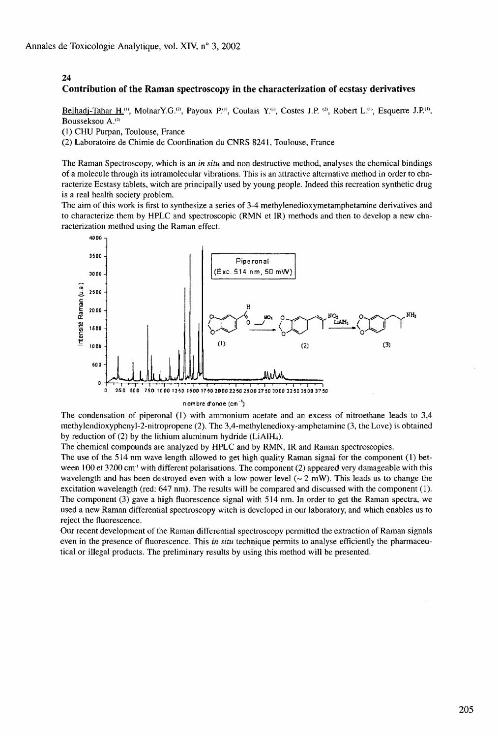
Annales de Toxicologie Analytique, vol. XIV, n° 3, 2002
24 Contribution of the Raman spectroscopy in the characterization of ecstasy derivatives
Belhadj-Tahar H.">. MolnarY.G.(2), Payoux R<», Coulais Y.f,), Costes J.P. <2>, Robert L.(l>, Esquerre J.P.( , ), Bousseksou A/ 2 ' (1) CHU Purpan, Toulouse, France (2) Laboratoire de Chimie de Coordination du CNRS 8241, Toulouse, France
The Raman Spectroscopy, which is an in situ and non destructive method, analyses the chemical bindings of a molecule through its intramolecular vibrations. This is an attractive alternative method in order to characterize Ecstasy tablets, witch are principally used by young people. Indeed this recreation synthetic drug is a real health society problem. The aim of this work is first to synthesize a series of 3-4 methylenedioxymetamphetamine derivatives and to characterize them by HPLC and spectroscopic (RMN et IR) methods and then to develop a new characterization method using the Raman effect.
40 DO
3300
3000 to b 2500
§ 2000 -
f 1500 -i c tu
— 1000 H
50 0
Piperonal
(Exc . 514 nm, 50 m W )
B D i O. 0 0 ^ — C u r (2) (3)
T ' I • ' ' I " I " ' — r " ' 1 " " ! I • 1 i—• j i i i i ' i •—i 0 25D S0O 750 1000 1ÎSD 1500 1750 2000 2250 2500 27 50 30 00 32 50 3500 37 50
nombre d'onde (cm"')
The condensation of piperonal (1) with ammonium acetate and an excess of nitroethane leads to 3,4 methylendioxyphenyl-2-nitropropene (2). The 3,4-methylenedioxy-amphetamine (3, the Love) is obtained by reduction of (2) by the lithium aluminum hydride ( L Í A I H 4 ) .
The chemical compounds are analyzed by HPLC and by RMN, IR and Raman spectroscopies. The use of the 514 nm wave length allowed to get high quality Raman signal for the component (I) between 100 et 3200 cm"1 with different polarisations. The component (2) appeared very damageable with this wavelength and has been destroyed even with a low power level (~ 2 mW), This leads us to change the excitation wavelength (red: 647 nm). The results will be compared and discussed with the component (1). The component (3) gave a high fluorescence signal with 514 nm. In order to get the Raman spectra, we used a new Raman differential spectroscopy witch is developed in our laboratory, and which enables us to reject the fluorescence. Our recent development of the Raman differential spectroscopy permitted the extraction of Raman signals even in the presence of fluorescence. This in situ technique permits to analyse efficiently the pharmaceutical or illegal products. The preliminary results by using this method will be presented.
Annales de Toxicologie Analytique, vol. XIV, n° 3, 2002
25 Detection of quaternary amines by capillary electrophoresis-UV
Scott F.J.<'>. Miller M.L.{2> (1) The George Washington University, Washington, DC, 20052 (2) FBI Laboratory, FBI Academy, Quantico, VA 22135
A universal method for detection of quaternary ammonium neuromuscular blocking agents is needed, as these compounds are suspected agents in homicide cases, particularly those involving hospital personnel. Quaternary ammonium compounds are inherently difficult to extract, as they are highly water soluble. They are designed to break down rapidly at physiological pH and temperature. Capillary electrophoresis (CE) with ultra-violet (UV) detection is a simple, rapid detection method ideally suited to this class of drugs due to their charged character. Due to their similar structures and chemical activity, a chiral recognition agent, beta-cyclodextrin is needed in the run buffer to ensure adequate separation. Direct and indirect detection are used simultaneously because several of the compounds have weak UV chromophores, which necessitate the inclusion of an indirect detection agent, para-aminopyridine. The nine quaternary ammonium drugs in recent use in the United States, as well as two primary metabolites can be separated and detected in a single run at 10 to 100 ppm levels
26 NMR spectroscopy as a useful tool for diagnosis of poisonings
Imbenotte M.(1>. Azaroual N.<2>, Cartigny B. ( 3 ), Vermeersch G.(2), Lhermitte M. n 3 )
(1) Laboratoire de Toxicologie, Faculté des Sciences Pharmaceutiques et Biologiques, Lille, (2) Laboratoire de Physique et d'Application RMN UPRESA CNRS 8009 (Université de Lille 2), (3) Laboratoire de Biochimie et de Biologie Moléculaire, Hôpital Calmette, CHRU Lille, France.
In order to analyse a wide range of xenobiotics and their metabolites present in biological fluids, such as urine, serum and plasma, NMR spectroscopy can be used. Due to the high content of water in biological specimens and the possible presence of macromolecules, specific 'H NMR acquisition sequences (selective presaturation and TOCSY) had to be elaborated and optimized. A large variety of xenobiotics (therapeutic agents, pesticides, solvents, alcohols) can be characterized and quantified directly without preparation of the sample. NMR investigations were applied to acute poisonings by drugs, such as salicylates, chloroquine, and valproic acid. For salicylate poisoning, the three major metabolites of acetylsalicylic acid have been assigned in crude urine, and rapid identification of lysine revealed the origin of the intoxication, namely lysine acetylsalicylate (Aspegic®). An urine sample from a 41-year-old man attempting suicide with chloroquine was analysed by one-dimensional and TOCSY 'H NMR spectroscopy, revealing chloroquine (462 mg/L) and monodesethylchloroquine (140 mg/L). Valproic acid as its glucuronide was identified in urine samples from two poisoned patients. Concerning pesticides, !H NMR and 3 , P NMR spectroscopies were used to diagnose acute poisoning with the organophosphorous glyphosate. Plasma concentrations (7.7 and 7.8 mrnol/L, respectively) were quite similar to chromatographic determinations (7,0 mmol/L), and the isopropyl resonances were used as markers for the herbicide formulation (Roundup®) implicated in this case. Paraquat (Gramoxone®) was identified and quantified by its two aromatic signals at 8.49 and 9.02 ppm, for two acutely poisoned patients (183 and 93 mg/L). An intentional poisoning with tetrahydrofuran (THF) was also investigated. Serum and urine samples were collected consecutively to the ingestion of this solvent. THF was characterized by its resonances at 1.90 and 3.76 ppm, and quantified at 813 and 850 mg/L in the two biological fluids, respectively. Moreover, two other compounds were significantly detected : lactate and gamma-hydroxybutyric acid (GHB). 'H NMR spectroscopic analysis of serum and urine samples from three poisoned patients revealed methanol and ethylene glycol and in the same spectrum the corresponding metabolites formate and glycolate. Supplementary informations on the level of lactic acidosis were provided. Compared with the reference chromatographic or spectrophotometric methods, requiring time-consuming extraction and/or derivatization steps, NMR spectroscopy allows the determination of many exogenous and endogenous compounds, without any preselection of the analytes. Consequently, metabolic disturbances can be established (organic anions, aminoacids) and the specific data collected could also help the clinican in evaluating the effectiveness of elimination or antidotal procedures.
206

Annales de Toxicologie Analytique, vol. XIV, n° 3, 2002
27 Gamma hydroxybutyric acid (GHB) concentrations in humans and factors affecting endogenous production: a volunteer study
Elliot S.P. Regional Laboratory for Toxicology, City Hospital NHS Trust, Dudley Road, Birmingham B18 7QH, U.K.
The endogenous nature of the drug of abuse gamma hydroxybutyric acid (GHB) has caused various interpretative problems for toxicologists. This is of particular importance in clinical and forensic investigations e.g. drug-facilitated sexual assault (DFSA). In such cases it is invariably required to determine whether any GHB detected is due to endogenous production or exogenous ingestion. In order to obtain further data for the presence of endogenous GHB in humans and to investigate any factors that may affect this, a volunteer study was undertaken. The GHB concentrations in 119 urine specimens from GHB-free subjects and 25 urine specimens submitted for toxicological analysis were determined. The results of all 144 subjects showed a maximal urinary GHB concentration of 3 mg/L. Analysis of 15 in-life plasma specimens submitted for toxicological analysis detected no measurable GHB (less than 2.5 mg/L). Studies in a male and female volunteer where differing dietary food groups were ingested at weekly intervals, showed significant intra-individual fluctuation with overall mean urine GHB concentrations of 0.72 mg/L (range 0-2.55 mg/L) and 0.65 mg/L (range 0-2.74 mg/L), respectively. Urinary concentrations did not appear to be affected by the particular dietary groups studied. Overall, the GHB concentrations measured by GC-FID and GC-MS in all volunteer studies did not approach concentrations observed in clinical overdose situations (typically >200 mg/L plasma/urine). The concentrations lend further support to the proposed urinary and plasma interpretative cut-offs of 10 mg/L and 4 mg/L, respectively, where below this it is not possible to determine whether any GHB detected is endogenous or exogenous in nature.
Annales de Toxicologie Analytique, vo l XIV, n° 3, 2002
28
208
Plasma concentrations of MDMA, GHB and other drugs and medical problems in subjects needing emergency medical care at nocturnal dance parties in Ghent, Belgium
Verstraete A.G.'". Monsieurs K., Van de Velde E.°\ Rousseau F., Van Sassenbroeck D.K., Buylaert W. (1) Laboratory of Clinical Biology Emergency Department, Ghent University Hospital, De Pintelaan 185, B-9000 Gent, Belgium
A marked increase in the frequency of drug-related medical problems at nocturnal dance parties has been observed. Rave parties are particularly associated with excessive consumption of illicit drugs such as ecstasy and gamma-hydroxybutyrate (GHB). The main purpose of this study was to measure blood drug levels and to compare them to medical problems related to the use of recreational drugs during "I love techno" (event A), one of Europe's largest indoor rave parties, attended by 37,000 people in Ghent on November 10, 2001. To place these data in a wider perspective, we also collected data on drug-related medical problems during "De Nacht" (event B), a traditional New Year's Eve dance party held on December 31, 2001 at the same location and attended by 12,000 people. During both dance events, a medical station, staffed by emergency physicians, registered nurses and emergency medical technicians, was set up near the dance hall for triage, treatment of minor medical problems and initial management of life-threatening events. Data on all patients evaluated in this medical station were registered prospectively. For additional diagnostic and/or therapeutic measures patients were transported by ambulance to an emergency department. Data on drug use were based on information provided by the patient (or a bystander), the clinical presentation and/or standardized toxicological screening on a plasma sample. Toxicological analysis was only performed on patients with severe neurological complications such as coma or seizures. Amphetamine, ecstasy, cocaine, cannabis, GHB and opiates were determined by GC-MS and ethanol was measured with an enzymatic method. The numbers of patients with drug-related medical problems were 61 for event A (16.5/10,000 attendants) and 18 for event B (15.0/10,000 attendants). The most frequent medical problems were vomiting/abdomi-nal pain (n=14), coma (n=9), agitation/anxiety (n=9), drunkenness (n=9) and epileptic fits for event A and drunkenness (n=12) and vomiting/abdominal pain (n=5) for event B. The number of intoxicated patients judged to be in need of an evaluation in an emergency department was 18 for event A (4.9/10,000 attendants) and 4 for event B (3.3/10,000 attendants). In these patients, the dominant abused drug was ecstasy (n=8, MDMA concentrations 66-510 ng/mL, MDA <10~60 ng/mL), GHB (n=7; 98-197 Mg/mL), ethanol (n=3, 0.05-0.96 g/L), cocaine (n=l, benzoylecgonine 359 ng/mL), and cannabis (n=l, THC 9 ng/mL, THCCOOH 191 ng/mL) during event A, and ethanol (n=3, 1.5-2.8 g/L) or ecstasy (n=l, MDMA 1500 ng/mL, MDA 44 ng/mL) during event B. Our data suggest that the incidence of medical problems (including drug-related problems) at rave parties is not increased tremendously as compared with traditional dance parties. At rave parties, however, mainly illicit drugs are used, more frequently leading to severe intoxication.
Annales de Toxicologie Analytique, vol. XIV, n° 3, 2002
29 Drug facilitated sexual assault - How far can toxicological screening go ?
Lewis J.H. Toxicology Unit, Pacific laboratory Medicine Services, Northern Sydney Health, Australia
Drug screening using immunoassay, high performance thin layer chromatography (HPTLC) and gas chro-matography/mass spectrometry (GC/MS) of urine specimens taken from alleged drug-facilitated sexual assault victims, has in the past revealed very little. Recently, a more thorough toxicological analysis conducted on specimens taken from 10 victims of alleged drug-facilitated sexual assault, found drugs in only 2 cases. All victims claimed their drinks had been spiked with drugs and they had total memory loss prior to the assault. The majority of victims did not present for treatment until 12-24 hours after the alleged assault. The analysis consisted of alcohol (alcohol dehydrogenase), immunoassay for benzodiazepines, cannabis, opiates, cocaine and amphetamines, broad spectrum screening by HPTLC and full GC/MS Scan of urine extracts. Additional targeted analyses were conducted for drugs likely to give symptoms as described to -the examining physician - dissociation and short term memory loss. These tests included GC/MS in the selected ion monitoring mode for benzodiazepines (LOD 5ng/mL), GHB (LOD 5ug/mL), ketamine (LOD 50ng/rnL) and propofoi (LOD 50ng/ml), and chloral hydrate (as trichloroethanol) by GC-ECD (LOD 80ng/mL). Drugs were detected in only 2/10 specimens - temazepam (80ng/rnL) was detected in 1 sample and methy-lamphetamine/MDMA in the other. Apart from alcohol (4 cases), no drugs were detected in the other 8 samples. Despite claims of flunitrazepam and temazepam being commonly used "date-rape" drugs, there was little evidence of their use, as detection of even low dose benzodiazepines is easily accomplished 24 hours after ingestion. The use of GHB and ketamine cannot be discounted, as they would be difficult to identify by the time specimens were taken. There are however, limits to the number of targeted toxicological analyses that can be performed in cases of alleged drug-facilitated sexual assault. Without evidence to support allegations of drug administration, further complicated by the time lag from assault to examination, the toxi-cologist cannot perform unlimited tests. Such negative laboratory results may however, have the effect of encouraging sexual assault victims to seek medical attention as soon as possible, rather than wait until the following day.

Annales de Toxicologie Analytique, vol. XIV, n° 3, 2002
30
210
Identification of thiopental and pentobarbital in head and pubic hair by SPME and GC-MS-MS in a case of drug-facilitated sexual assault
Frison G., Favretto D., Tedeschi L., Ferrara S.D. Forensic Toxicology and Antidoping, University Hospital, ViaFalloppio 50,1-35121 Padova, Italy
A woman was hospitalized for a minor surgical operation. After her complete awakening from anaesthesia (propofol infusion, local analgesia with mepivacain and ropivacain), she was transferred from the operating room to her ward. Inside the elevator used for transfer she was made unconscious, by means of a drug injected in the infusion set, and sexually assaulted. Although gynaecological and psychological inspections as well as citologic and genetic tests on biological specimens were carried out, no blood or urine samples were collected for toxicological analyses. A month later we were requested from the victim to analyse her hair in an attempt to identify the sedative drug(s) involved. Three head and one pubic hair samples, as well as an additional head hair sample fifteen days later, were collected. Such samples were stored until the fluid contained in the infusion set was analysed and traces of thiopental were discovered. Since no thiopental had been therapeutically administered before and during hospitalization of the victim, that drug became the target for hair analyses. A Solid-Phase Microextraction (SPME) and Gas Chromatography-Multiple Mass Spectrometry (GC-MS-MS) method was developed for determination of thiopental and its metabolite pentobarbital. Samples (50 mg powdered hair) were stirred overnight at 30° C with 2 ml of 0.1 N sodium bicarbonate after the addition of the internal standard (IS) ethyltolylbarbituric acid. Supernatants were transferred to 2-ml vials, slightly acidified with solid phosphate buffer, and direct immersion SPMEs were performed with a 50 pm carbowax/templated resin fiber for 20 min at 30° C under stirring. GC-MS-MS analysis on an ion trap instrument was carried out by fiber desorption at 250û C in the GC injector under splitless conditions, chromatographic separation on a 30-m Chrompack CP-Sil 8 capillary column with an oven temperature program from 50° C to 300° C, and a carrier gas (He) flow of 1 ml/min. Chemical ionisation was obtained with acetone. M + H+ ionic species (m/z 243, 227, 247 for thiopental, pentobarbital and IS, respectively) were obtained, selectively stored in the ion trap, and collided with He to obtain characteristic product ions (m/z 173, 157, 218 for thiopental, pentobarbital and IS, respectively). Quantitation limits of the method were 0.1 ng/mg for both barbiturates. Linearity for both analytes was obtained in the concentration range of 0.1 - 10 ng/mg. Thiopental and pentobarbital were identified in all 1.5 cm proximal ends of head hair samples as well as in pubic hair. Corresponding distal ends of each head hair sample were negative. Concentrations ranged from 0.15 to 0.30 and 0.20 to 0.40 ng/mg of thiopental and pentobarbital, respectively. These results demonstrated that a presumably single administration of thiopental was in fact used for committing sexual assault.

Annales de Toxicologie Analytique, vol. XTV, n° 3, 2002
Some unusual analytical approaches to forensic toxicological cases
Kala M. Institute of Forensic Research, ul. Westerplatte 9, 31-033 Krakow, Poland
It is well known that systematic toxicological analysis (STA) must be very extensive, because tens of thousands of toxicologically relevant substances exist. Due to its complexity, STA is a very difficult task for analysts. Toxicologists are often not certain whether obtained results are reliable, even when they take into consideration all analytical principles and commonly used procedures. Some unusual analytical approaches to forensic cases are presented and analytical pitfalls are discussed. The chosen forensic cases concern identification of : a residue of an unknown substance in an empty ampoule by TLC, UV-Spectrophotometry, enquiries at the local pharmacy and medications market ; a red dye, without standard, by confirmation of its structure by FTIR ; a brown powder by microscopic examination ; the decomposition product and metabolite of Atracurium besilate by GC-MS and HPLC ; valproic acid in milk by GC-MS and EMIT (Viva Dade-Behring) ; digoxin in non biological and biological materials by GC-MS, LC-MS and EMIT.
32 Do TIAFT members care about iatrogenic poisonings ?
Uges D.R.A. Laboratory for Clinical and Forensic Toxicology and Drug monitoring, University (Hospital) Groningen, P.O.Box 30.001, 9700 RB Groningen, The Netherlands
By definition iatrogenic poisonings are caused by a physician, pharmacist, nurse or even an undergraduate. Therefore, a (para-) medical professional is responsable for patient's harm. In the past patients or their representative accepted these incidents as calculated risks, or even did not know the patient's situation had deteriorated by an avoidable fault. Nowadays the patient, and even more often his injury lawyer, wants compensation for extra suffering, costs and remaining damage. Unfortunately, allowance and compensation are also claimed if there is no poisoning. Therefore the definition of a poisoning has to be clear: A human medical poisoning is a medical or social unacceptable condition, as a result of being under influence of an exogenous substance in a dose, which is too high for the individual. The iatrogenic poisoning has to be distinguished from the calculated and acceptable side effects, an absolute fault, a deliberated overdose, or an unforeseeable or unpredictable poisoning. Diazepam is administered as anticonvulsant, with muscle weakness and somnolence as acceptable side effects. But the administration of diazepam to the ambulant elderly is not accepted, as the risk on falling with a broken hip is too big. The forensic toxicologist has to judge the case and gives evidence whether it is a poisoning or not. (E.g. Very or too high dose of morphine to a cancer patient with unbearable pain). Furthermore, he concerns what might be the cause and what could be the consequences. Beside emphatic mistakes, insufficient knowledge or experience, (e.g. potassium suppression together with an unknown potassium sparing diuretic) the overdose may be caused by a genetic deviance (e.g. poor metabolizers). For an independent toxicologist it is often difficult, or even hardly possible to differentiate between unforeseeable and foreseeable interactions or deviant pharmacokinetic profile, as a cause of the poisoning. Flucytosine can be a lifesaving drug for patients suffering from fungal infection. If the flucytosine serum level rises above 100 mg/l the risk on a severe bone marrow depression becomes unacceptable high. Does that mean a calculated side effect or an acceptable poisoning, even if the doctor is not asking for therapeutic drug monitoring, or his hospital does not have the possibility to provide these services? The difference between side effect and poisoning is not only a semantic.

Annales de Toxicologie Analytique, vol. XIV, n° 3, 2002
33 GC-MS studies on the metabolism and toxicological analysis of the new pyrrolidinohexa-no-phenone designer drug 4'methyl-alpha-pyrrolidinohexanophenone (MPHP)
Springer D. j l ). Peters F.T.<'>, Fritschi G.a\ Maurer H.H.(1)
(1) Department of Experimental and Clinical Toxicology, University of Saarland, D-66421 Homburg (Saar), (2) Hessisches Landeskriminalamt, D-65187 Wiesbaden, Germany
After alpha-pyrrolidinopropiophenone and some of its derivatives, a new alpha-pyrrolidinophenone-type drug with an elongated side chain has appeared on the illicit drug market: 4'methyl-alpha-pyrrolidino-hexanophenone (MPHP). The aim of our study was to identify its metabolites and develop a toxicological detection procedure in urine by GC-MS techniques. MPHP (10 mg/kg body mass for metabolism studies, 1 mg/kg for detection studies corresponding to a therapeutic dose of the related drug amfepramone) was given to male Wistar rats by gastric intubation and urine was collected over 24 h. The metabolites were isolated either after enzymatic cleavage of conjugates or directly by solid-phase extraction using Confirm HCX cartridges. The metabolites (either underivatized, acetylated, methylated, ethylated, silylated or methylated and acetylated) were separated and identified by GC-MS in the EI and/or PCI mode. For detection, mass chromatography followed by library search (PMW_tox4) was performed. After enzymatic hydrolysis, the compounds shown in the scheme below were detected as metabolites. The compounds in brackets (parent drug and intermediate metabolites) could not be detected. No significant differences in the amount of the metabolites were noted in hydrolyzed and native urine extracts. The toxicological detection procedure focussed on the main metabolite 4'carboxy-pyrrolidinohexanophenone, which could most sensitively be detected after silylation.
From these results, the following partly overlapping metabolic pathways could be concluded: MPHP undergoes oxidation of the tolyl methyl group to the corresponding hydroxymethyl or carboxy compound, hydroxylation of the pyrrolidine ring followed by dehydrogenation to the corresponding lactam, hydroxy-lation of the side chain or reduction of the keto group to the corresponding secondary alcohol. Assuming similar metabolism and dosages in man, the intake of MPHP can be proven by detection of the main metabolite in urine.
212

Annales de Toxicologie Analytique, vol. XIV, n° 3, 2002
34 GC-MS studies on the metabolism and toxicological analysis of the designer drug parame-thoxymethamphetamine (PMMA)
Staack R.F.">. Fehn J.(3), Maurer H.H.(" (1) Department of Experimental and Clinical Toxicology, University of Saarland, D-66421 Homburg (Saar), (2) Bayerisches Landeskriminalamt, D-80636 München, Germany
Several fatalities after intake of para-methoxyamphetamine (PMA) and of combinations with its N-methyl derivative para-methoxymethamphetamine (PMMA) have been reported during the last years. The aim of our study was to identify the metabolites of PMMA, a controlled substance, and to study their detectabi-lity within our STA procedure in urine. PMMA (10 mg/kg body mass for metabolism studies, 0.5 mg/kg for detection studies, corresponding to a common drug user's dose) was given to Wistar rats by gastric intubation and urine was collected over 24 h. For identification, the metabolites were isolated after enzymatic cleavage of conjugates or directly by liquid-liquid extraction followed by heptafluorobutyrylation or acetylation and/or methylation. For STA, one aliquot each of acid hydrolyzed and of unhydrolyzed urine was extracted and acetylated (cf. J Anal Toxicol 24, 2000, 340). The metabolites were separated and identified by GC-MS in the EI and PCI mode. For STA, mass chromatography was used with specific ions followed by library search (PMW_tox4). After enzymatic hydrolysis, the following metabolites could be identified: phydroxy-MA (pholedrine), dihydroxy-MA, hydroxy-methoxy-MA, p-hydroxy-ephedrine (oxilofrine), PMA, phydroxy- and hydroxy-methoxy-amphetamine. The phenolic metabolites could be detected only in minor amounts in native urine. The detection studies showed that only the main metabolite pholedrine could be detected in the urine samples after low dosage. PMMA is extensively metabolized, mainly by O-demethylation to p-hydroxy-MA. Minor pathways are: ring hydroxylation, O-demethylation followed by side chain hydroxylation or ring hydroxylation followed by methylation. A further pathway was N-demethylation followed by O-demethylation and/or ring hydroxylation and finally methylation to hydroxy-methoxy-amphethamine. Assuming similar metabolism in man, PMMA intake should be detectable in human urine via its O-deme-thyl metabolite phydroxy-MA (pholedrine). As PMMA is excreted almost completely metabolized, a differentiation of the intake of PMMA or the therapeutic drug pholedrine is not possible via urine analysis. Only after higher dosages, unique PMMA metabolites can be found in urine.

Annales de Toxicologie Analytique, vol. XIV, n° 3, 2002
35 Acute nitrobenzene poisoning with severe associated methemoglobinemia : identification in blood by GC-FID /GC-MS
Martínez M.A.(1>. Ballesteros S.(2), Almarza E. (1), Sánchez de la Torre C.(1), Búa S.í3)
(1) Departamento de Química, Instituto Nacional de Toxicología, Ministerio de Justicia, CI Luis Cabrera 9, 28002 Madrid, Spain (2) Servicio de Información Toxicológica, Centro Antitóxico Español. Instituto Nacional de Toxicología, Madrid, Spain (3) Unidad de Cuidados Intensivos (UCI), Hospital de Móstoles, CI Río Júcar, s/n, 28935 Móstoles, Madrid, Spain
A rare fatal case of self poisoning with nitrobenzene following oral ingestion is reported. On presentation to the hospital, severe methemoglobinemia (70 %) was observed in a 82 year old male who had ingested 250 mL of an unknown substance in the previous 24 hours. Patient's family alleged that the substance was '"White Spirit". The ingestion occurred in a mechanical garage owned by the patient's son and the container was unlabeled. Methylene blue and exchange transfusion were the therapeutic methods applied in the treatment of the methemoglobinemia. In spite of methylene blue administration, methemoglobinemia persisted in 30 %. Therefore exchange transfusion was made with reduction to 18 %. Forty eight hours after ingestion a blood sample was collected in ICU and sent to our laboratory. A comprehensive toxicological screening for solvents was performed in the blood sample. This included ethanol and volatile analysis by headspace GC-FID. Blood analysis revealed the presence of 3.2 jug/mL of nitrobenzene. The toxic was isolated after liquid-liquid extraction of 3 mL of blood with 1 mL of diethyl ether using n-octyl-benzene as internal standard. The organic layer was injected in split mode first for GC-FID screening analysis and later for GC-MS confirmation in scan mode of the obtained results. Both gas chromatographs were equipped with a methylsilicone capillary columns. The quantitation analysis was performed by GC-FID using a calibration curve in the range 0.1-5.0 ug/niL. Limit of detection (LOD) and limit of quantitation (LOQ) were 79 ng/mL and 263 ng/mL, respectively. Recovery of nitrobenzene in the studied range were more than 93.0 %, with intra-assay and inter-assay precision less than 8.4 % and 12.0 %, respectively. An excellent linearity (r2 0.999) was observed from LOQ up to 5.0 ug/mL. In spite of all therapeutic measures, refractory shock with asystolia happened 3 days after admission. To date, it is the first case of nitrobenzene poisoning where analytical data are described and analytical method are also described and validated. These results complete the previous data on nitrobenzene in humans.
214

Annales de Toxicologie Analytique, vol. XTV, n° 3, 2002
36 Analysis of perhexiline and its hydroxy metabolite in serum
Couch R. Toxicology Unit, Department of Clinical Chemistry, LabPlus, Auckland Hospital, New Zealand.
Perhexiline is a antianginal drug. It can cause serious hepatic and neurological toxicity if the serum concentration is above the therapeutic range of 150-600 pg/L. The metabolism of perhexiline to monohy-droxyperhexiline, catalyzed by the cytochrome P450 2D6, is subject to genetically determined metabolic variants resulting in toxic concentrations of the parent drug. Perhexiline and hydroxyperhexiline were quantitated following solvent extraction, no derivatization, gas capillary chromatography on a 5 % phenyl methyl silicone column and detection by NPD. The internal standard for perhexiline was hexadiline and for hydroxyperhexiline was trihexylphenidyl with retention times of 8.10, 7.38, 11.44 and 9.85 minutes respectively. For perhexiline the intra- and inter-assay precision at 600 pg/L was 2.3 % and 6.2 % respectively, and for hydroxyperhexiline at 2000 pg/L, the intra-assay precision was 3.1 %. Participation in an Australian interlaboratory proficiency program during 2001, for perhexiline, gave at 350 pg/L (n=6), a CV% of 7.1 and a bias (%) of 1.0. Analysis of 1052 patient specimens gave concentrations of perhexiline that were below, within and above the therapeutic range in 29.9,55.5 and 14.5 % of the cases. 60 patients had a serum perhexiline level greater than 1000 pg/L, In 18 of these specimens hydroxyperhexiline was not detected. In others the ratio of hydroxyperhexiline was less than 3.5. Levels of hydroxyperhexiline and perhexiline can jointly be used to indicate perhexiline toxicity.
37 Fatal poisoning in childhood, England & Wales 1968-2000
Flanagan R.J.(I). Rooney C.® (1) Medical Toxicology Unit, Guy's & St Thomas' Hospital Trust, Avonley Road, London SE 14 5ER, (2) Office for National Statistics, SESAG/Health and Care Division, Room B7/04, 1 Drummond Gate, London SW1V 2QQ, UK
Analysis of mortality data for England & Wales coded according to the International Classification of Diseases (ICD) shows that deaths from acute poisoning in children aged less than 10 years declined from 169 in 1968 to between 40-50 per year in 1996-2000. Further analysis shows that (i) most these deaths occurred in fires and (ii) were due to inhalation of products of combustion rather than burns. When the ninth revision of the ICD (ICD-9) was introduced in 1979 there was a shift in coding these deaths from poisoning with carbon monoxide to poisoning with 'other gases, fumes, and vapours'. These 'fire deaths' do not appear as poisonings in mortality statistics based on a single underlying cause of death, and cannot be tabulated in many countries. Coding of fatal poisoning from 'drugs, and other solid and liquid substances', which have declined steadily in children since 1968 (n = 28) to 1-12 per annum in 1996-2000, was not affected by the introduction of ICD-9. Factors which have helped reduce mortality and morbidity from poisoning in this age group include: (i) the widespread introduction of child resistant closures (CRCs), (ii) greater emphasis on safety in the home, (iii) improved access to poisons information, (iv) improved treatment, (v) the withdrawal of hazardous preparations such as Safapryn (paracetamol and enteric coated aspirin), (vi) the increased use of blister packaging, and (vii) changes in prescribing patterns. On the other hand there is increasing awareness of the possibility of deliberate poisoning by a caretaker (non-accidental poisoning, Munchausen Syndrome by Proxy). In line with this latter consideration, there are now very few deaths in children certified as being due to accidental poisoning with drugs in England & Wales. In most of these deaths, a verdict of homicide or an open verdict is recorded by the coroner or by a higher court. Opioids and tricyclic antidepressants are amongst the compounds encountered. With paracetamol (acetaminophen) especially serious accidental poisoning is almost unknown in young children - the amount ingested is usually small, and hepatic sul-phation capacity and glutathione stores are increased compared to those of adults. Serious liver damage and death have only been reported in children after chronic paracetamol poisoning.

Annales de Toxicologie Analytique, vol. XIV, n° 3, 2002
38
216
Human serum paraoxonase (PONÍ) activity in acute organophosphorous insecticide poisoning
Akgür S.A.(I). Óztürk P.">, Solak IP, Moral A.R.(2), Ege B.(1> (1) Dept. of Forensic Medicine, (2) Emergency Medicine, Fac. of Medicine, Ege University, 35100, Izmir, Turkey
Human serum paraoxonase/arylesterase (PONÍ) and perhaps other mammalian paraoxonases catalyze the hydrolysis of certain organophosphate pesticides (OP) and nerve gases and so may alter significantly an individual susceptibility to the toxicity of these chemicals. Phosphates (P=0) are biologically active as acetylcholinesterase (AChE) inhibitors whereas, organophos-phorothioates (P=S) need bioactivation to their phosphate analogues (oxon) to become biologically active. These oxon forms are hydrolysed by PONÍ. Serum PONÍ has been shown to be polymorphic in human populations. Studies show significant inter-ethnic differences in the distribution of PONÍ phenotypes.lt is reported that distribution of low-activity phenotypes is 63 % in Turkish population. The A-type of PONÍ (low activity- now called Q isozyme) can be several times less efficient than the B-type (high activity-R isozyme) in hydrolyzing paraoxon, but most organophosphates are hydrolyzed appreciably better by the Q- than the R-isozyme. Thus, both the level and the type of PONÍ must be taken into consideration in evaluating the protective role of PONÍ against such compounds that may be substrates catalytically inactivated by the enzyme. So we aimed to investigate PONÍ activity in acute OP poisoning cases. The study group consisted of 28 subjects ( 17 female, mean age 28; 11 male, mean age 37) who were ingested organophosphorous insecticides intentionally for suicidal purposes and admitted to Emergency Medicine Department of Faculty of Medicine, Ege University. Blood samples were obtained by venipuncture; sera were kept frozen at -70° C until paraoxonase, arylesterase and cholinesterases analysis. Anticoagulated blood samples were used for insecticide analysis by Gas Chromatography- Nitrogen Phosphorus Dedector (GC-NPD) using HP 1 capillary column.
^ Methamidophos, methyl-parathion, dichlorvos, dimethoate, diazinon and malathion were the detected organophosphorous compounds in blood samples. The activity levels for salt stimulated PONÍ, basal PONÍ and arylesterase were found as 78.83 (35.39-186.13), 39.97 (2.49-80.43) m mol min-lL-1 and 126.26 (36.34-288.24) mmol min-lL-1 respectively. On the other hand the activity levels for butyrylcholinestera-se (BTC) and AChE were found as 797.23 (106.3-3823) U/L and 4.65 (0.21-30.29) Rappaport Uml-1. There was a correlation between percent stimulation of PONÍ and BTC activities (r=0.446, p<0.05) and also between BTC and AChE activities (r=0.867, p<0.01). Correlations were made using the Pearson correlation test. The subjects with low PONÍ activity consisted 86 % of the cases.

Annales de Toxicologie Analytique, vol. XIV, n° 3, 2002
39 Proficiency test for the analysis of hair for drugs of abuse, organized by the Society of Hair Testing
Jurado C. ( i>. Sachs H.<2)
(1) Instituto Nacional de Toxicología, P.O. Box 863, 41080 Sevilla, Spain (2) Institut für Rechtsmedizin, Frauenlobstr. 7a, 80337 Miinchen, Germany
One of the aims of the Society of Hair Testing (SoHT) is the development of Proficiency Tests, for all labs which perform hair analysis to be able to produce comparable results. For this reason the SoHT organized a Proficiency Test last year. Fifteen laboratories interested in the analysis of human hair participated in the study. The samples included one drug-free hair sample and two from drug users. The hair samples were sent in the form of short segments and they were previously checked for homogeneity by three reference labs. Participants were requested to analyze the samples following the standard procedure used routinely in their laboratories. The substances present in the samples included opiates, cocaine and metabolite, cannabis and amphetamines. All the labs analyzed opiate and cocaine compounds, while only eight analyzed amphetamines and cannabis as well. Results have shown that the labs performed very well qualitatively, since they successfully identified all the analytes that they tested, with the exception of two false results. However, scatter in quantitative results was high. Various methods were used to extract drugs from the hair by acid, basic and methanol extractions, and enzyme digestion. GC/MS was applied by all the labs, with the exception of two, which used GC-MS/MS and LC-MS/MS, respectively. Six laboratories previously performed screening tests by RÍA, ELISA or EMIT.
40 Determination of ketamine in human hair by GC-MS after derivatization with 2,2,2 tri-chloroethyl chloroformate
Tedeschi L., Frison G., Castagna F., Ferrara S.D. Forensic Toxicology and Antidoping, University Hospital, Via Falloppio 50,1-35121 Padova, Italy
Ketamine, an anesthetic agent available since 1972 and capable to produce a dissociative state and hallucinogenic effects, is becoming a popular recreational drug. A case is presented involving a driver, suspected of taking psychoactive substances, submitted to both clinical and toxicological ascertainments for regranting his driving licence. Multiple urine samples and one hair sample were negative for common psychoactive drugs. As he admitted the past use of ketamine, the same hair sample was analysed for this hallucinogenic drug by GC-MS after derivatization with 2,2,2 tri-chloroethyl chloroformate. Sample preparation involved external decontamination, mechanical pulverization, addition of the internal standard MDPA (3,4-methylenedioxypropylamphetamine), overnight incubation with acid methanol at 45° C, basic extraction using Extrelut columns and derivatization to produce 2,2,2 trichloroethyl carbamate derivatives. GC-MS analysis was carried out injecting 2-pl aliquots at 250° C under splitless conditions, using a 30-m slightly polar capillary column with an oven temperature program from 50° C to 300° C and a carrier gas (He) flow of 1 ml/min. MS acquisition mode was Selected Ion Monitoring (SIM) under EI ionisation. Due to the isotope effect of four chlorine atoms in the derivatized molecule, EI mass spectrum of the obtained ketamine 2,2,2 trichloroethyl carbamate derivative shows a weak but high-mass molecular ion at m/z 411 as well as an intense diagnostic fragment ion at m/z 348 (base peak), both characterized by complex ion clusters. These features, along with the relatively high retention time of the derivative, allowed good specificity and sensitivity. The method limit of detection for ketamine was 0.1 ng/mg and hair ketamine concentration of sample under study was 1.0 ng/mg.

Annales de Toxicologie Analytique, vol. XIV, n° 3, 2002
41 Segmenta! hair analysis of benzodiazepines with ion spray LC-MS-MS : An application to psychiatric after care
Kronstrand R., Nystrôm I., Josefsson M. National Board of Forensic Medicine, Department of Forensic Chemistry University Hospital, SE-581 85 Linkôping, Sweden.
The aims of this study were to develop an LC-MS-MS method for the analysis of benzodiazepines in human hair. The method was tested by analyzing hair samples from forensic and clinical psychiatric patients where benzodiazepines had been prescribed during hospitalization and after care. Hair samples were obtained at discharge from the hospital and then at six-month intervals. Two-cm segments of the hair samples (10-30 mg) were washed with isopropanol, three times with phosphate buffer and again with iso-propanol, dried and weighed, and digested with proteinase K before solid phase extraction with BondElute Certify columns. Diazepam, nordiazepam, oxazepam, alprazolam, OH-alprazolam, nitrazepam, 7-amino-nitrazepam, flunitrazepam, 7-aminoflunitrazepam, clonazepam, and 7-aminoclonazepam were quantified in MRM mode using one transition for each analyte and deuterated internal standard. The calibration range was for high-dose compounds 2.5-100 ng/sample and for the low-dose compounds 0.5-20.0 ng/sample. In the hair samples analyzed diazepam, flunitrazepam, nitrazepam, and clonazepam was detected together with their metabolites. Alprazolam was not detected in any sample. Segmental hair analysis revealed differences in drug deposition in hair before and after release from psychiatric treatment. Both increases and decreases of hair drug concentrations were seen after release even though the prescribed dose was the same. This was taken as signs of noncompliance during the after care period. One patient prescribed nitrazepam (10 mg/day), clonazepam (1 mg/day), and diazepam (10 mg/day) showed hair analysis results that indicated an increased intake of nitrazepam whereas the intake of diazepam decreased and the clonazepam concentrations in hair were constant indicating compliance with medication. We conclude that the extraction and LC-MS-MS procedures were adequate to detect benzodiazepines in hair and that segmental hair analysis may provide important retrospective information about compliance in medication.
Fentanyl in human hair by Liquid Chromatography-Tandem Mass Spectrometry
LeBeau M.A., Montgomery M.A., Schaff J.E., Quenzer C.F FBI Laboratory, 935 Pennsylvania Avenue, NW, Washington, DC, 20535, USA
Fentanyl is a potent, synthetic narcotic analgesic. It is available as a citrate salt for injectable administration or in a transdermal patch for the management of chronic pain. It has been reported as a commonly abused substance by healthcare professionals. Hair samples were collected from a registered nurse suspected of narcotic theft and abuse. These samples were cut into 3-cm lengths to approximate 3-month growth periods, segregated, and finely cut with scissors. Aliquots (23.5 - 43.8 mg) of the cut hair were methanol washed ( 2 x 1 mL x 1 minute). The methanol washes were collected, spiked with d5-fentanyl (25.0 ng), and taken to dryness for later analysis. The washed hair samples were dried at 40° C for 30 minutes and pulverized in a Mini-Beadbeater (Biospec Products, Bartlesville, OK, USA) for 10 minutes. The pulverized hair samples were spiked with d5-fentanyl (25.0 ng) and extracted with warm methanol (40° C) for 18 hours. Following centrifugation, the methanol extracts were passed through a 0.2 urn syringe filter, and taken to dryness under nitrogen at 40° C. The extracts were reconstituted in 25 uL of methanol for analysis. The analysis was performed on a ThermoFinnigan TSQ LC/MS/MS system in the parents of 250 m/z mode. Using a methanokdeionized watenammonium hydroxide (95:5:0.3) mobile phase with a flow rate of 0.3 mL/min through an Alltech Altirna C18 (15cm x 2.1mm x 5 urn) column, fentanyl and its deuterated analog eluted at approximately 1.8 minutes. Using this method, fentanyl was identified in the nurse's hair at concentrations of 20 - 93 pg/mg. No fentanyl was detected in the wash samples. These findings were supported by positive results from radioimmunoassay analyses of separate hair extracts.
218
Annales de Toxicologie Analytique, vol. XIV, n° 3, 2002
43 Determination of cathinone, cathine, norephedrin and metabolites in hair of Yemenite khat chewers
Sporkert F.<". Pragst F.(l>, Bachus R.(2>, Al-Warith H. i 3 ), Harms L. ( 2 )
(1) Institute of Legal Medicine, University Hospital Charité, Humboldt University, Hannoversche StraBe 6, D-10115 Berlin, Germany (2) Department of Neurology, University Hospital Charité, Humboldt University, Schumannstr. 20/21 D-10117 Berlin, Germany (3) Yemen-German Hospital, Sanaa, Yemen
Chewing of fresh leaves of the khat bush Catha Edulis is very common in certain countries of East Africa and the Arab Peninsula. It has stimulating, anorectic and communicative properties. Insomnia, nervousness, agitation and trembling, sometimes accompanied by aggressivity, and psychosis may occur as possible side effects after regular intake. The main psychoactive compounds of khat are cathinone, cathine and norephedrine. The objective of this study was to determine the concentrations of these drugs in hair of kath chewers and to examine a possible correlation between the consumption history, the hair concentrations and the neurological impairments found in some of the chewers. Hair samples of 14 Yemenite khat chewers were collected and the volunteers were interviewed with respect to their consumption history. The frequency of khat chewing was between 4 and 56 h/week (mean 26 h/week) for 3 to 30 years (mean 17 years). The following analytical method for the determination of cathinone, cathine and norephedrine from hair was developed: 20 mg hair were extracted for two hours at 45° C with phosphate buffer pH 2.0 under permanent shaking followed by a standard solid phase extraction procedure on a mixed phase column, derivatization with heptafluorobutyric acid anhydride and GC-MS-SDVI separation and quantification using Ds-methamphetamine as the internal standard. The limits of quantification were 0.2 ng/mg for cathinone and 0.1 ng/mg for cathine and norephedrine. The diastereomeres cathine and norephedrine were satisfactorily separated in the chromatograms. All three substances were found in all 14 cases. With the exception of two extreme cases the concentrations ranged from 0.23 to 1.53 ng/mg (mean 0.70 ng/mg) for cathinone, from 0.40 to 2.94 ng/mg (mean 1.84 ng/mg) for cathine, and from 0.26 to 2.29 ng/mg (mean 1.24 ng/mg) for norephedrine. Very high concentrations (9.1, 13.3 and 8.2 ng/mg, and 22.6, 25.0 and 14.1 ng/g for the three compounds) were measured in the samples of two volunteers with the most intensive kath consumption ( 49 and 56 h/week). Five of the volunteers suffered from epilepsy, dystonia or chronic headache. A possible genesis of these diseases from the chronically excessive kath consumption is considered.

Annales de Toxicologie Analytique, vol. XIV, n° 3, 2002
44 High prevalence of 6-acetylmorphine in morphine positive oral fluid specimens
Cone E.J. ( I ). Presley L.(2>, Niedbala R.SP (1) ConeChem Research, Severna Park, MD, USA (2) LabOne, Lenexa, KS, USA (3) OraSure Technologies, Bethlehem, PA, USA
Interpretation of positive urine test results for opiates can be problematic because of multiple sources of licit and illicit opioids and complicated metabolic patterns arising from related opioids. However, identification of 6-acetylmorphine (6AM), a specific metabolite of heroin, is considered to be definitive evidence of heroin use. Although 6AM has been identified in oral fluid following controlled heroin administration, no prevalence data is available for 6AM in oral fluid specimens collected in the workplace. We evaluated the prevalence of positive test results for drugs in 77,218 oral fluid specimens collected over a 10-month period (January-October, 2001) from private workplace testing programs. Specimens were analyzed at LabOne by Intercept™ immunoassay at manufacturer's recommended cutoff concentrations and confirmed by GC-MS-MS. For opiates, a total of 127 confirmed positive opioid results were reported for codeine (COD) and 48 positive results for morphine (MOR) giving an overall positive prevalence rate for opiates of 0.23 %. For comparison, the prevalence rate for opiate positives was 0.30 % in the Quest Diagnostics' Drug Testing Index for private workplace drug tests (January and June, 2001). Oral fluid specimens that confirmed positive for MOR (cutoff concentration = 30 ng/mL) were analyzed by GC-MS-MS for 6AM (cutoff concentration = 3 ng/mL). Of the 48 positive MOR specimens, 32 (66.7 %) specimens were positive for 6AM. Concentrations of 6AM in oral fluid ranged from 3-4085 ng/mL. The mean ratio (±SEM) of 6AM/MOR was 0.33 ± 0.06. Mean concentrations (ranges) of 6AM and associated MOR and COD concentrations for the 48 positive MOR specimens are shown in the table.
Confirmed Positive #Specimens MOR, ng/mL COD, ng/mL 6AM, ng/mL MOR only 13 116(30-2997) MOR, COD only 3 556(45-1275) 114(39-261) -MOR, 6AM only 15 183 (30-540) - 72 (3-534) MOR, COD, 6AM 17 1259 (36-4595) 196 (49) (30-777) 754 (6-4095)
The confirmation of 6AM in 66.7 % of positive MOR oral fluid specimens indicates that oral fluid testing for opioids may offer some advantage over urine in differentiating heroin from other opioids in workplace drug testing programs.
220

Annales de Toxicologie Analytique, vol. XIV, n° 3,2002
45 Comparison of Cozart Oral Fluid Cocaine ELISA and GC/MS results following controlled administration of cocaine HCI
Huestis M.A. ( I>. Barnes A. IP, Schepers R.(I), Kim L<n, Moolchan E.T.( ,), Oyler J.<1\ Wilson LP, Cooper G.(2), Reid C.!2), Hand CP (1) Chemistry and Drug Metabolism, IRP, NIDA, NIH, Baltimore, MD 21124, USA, (2) Cozart Bioscience Ltd, Oxfordshire, OX144RU, UK
The advantages of oral fluid drug testing include the ease and non-invasiveness of specimen collection and a reduction in the potential for specimen substitution and adulteration. These advantages have created interest in monitoring drug use through oral fluid testing in driving under the influence, drug treatment, criminal justice, and workplace drug testing programs. One of the advantages of urine drug testing is the large database of information from controlled drug administration, epidemiological and case studies for interpretation of results. To add to the data available for the evaluation of oral fluid as an alternative test matrix, we have performed studies with eighteen (11 male, 7 female) healthy volunteers with a history of cocaine abuse. The 18 subjects provided informed consent to participate in this IRB approved study and resided on the closed research unit for approximately 12 weeks. Volunteers (14AA, 2C, 2H) received 3 low dose (75 mg/70kg) subcutaneous injections of cocaine HCI on alternating days, and after a 3-week interval, 3 high dose (150mg/70kg) injections. Oral fluid was collected following citric acid candy stimulation and expectoration, and smaller subsets were collected with Salivette neutral cotton and citric acid treated cotton swabs. Specimens (N=l,468) were frozen at -20° C until analysis with the Cozart Cocaine Microplate EIA (ELISA) for the semi-quantitative analysis of cocaine and metabolites. SPE followed by GC/MS was employed for confirmation of cocaine and 12 metabolites (LOQ 2.5 ng/mL). For the purposes of this comparison, two different screening test cutoffs (10 and 20 ng/mL) and three GC/MS cutoffs [2.5, 8 and 10 ng/mL cocaine, ecgonine methyl ester (EME), and/or benzoylecgonine (BE)] were evaluated. There are as yet no established cutoff concentrations for the analysis of cocaine and metabolites in oral fluid, although we are aware of studies utilizing 10 ng/mL for both screen and confirmation testing and also the proposed SAMHSA guidelines of 20 ng/mL for the screen and 8 ng/mL of cocaine and/or benzoylecgonine for the confirmation. The lowest calibrator used for the ELISA was 10 ng/mL (approximately 50 %B/Bo) and for our GC/MS method 2,5 ng/mL, hence the third cutoff pair evaluated. For the ELISA, interassay precision (based on ng/mL of the quality control specimens) over 19 different assays was 15.1 % CV at a mean concentration of 16.7 ng/mL and 11.5 % CV at a mean concentration of 81.8 ng/mL. With the 10, 8 and 2.5 ng/mL GC/MS cutoffs for cocaine, EME and/or BE, 52.7 %, 54.3 %, and 59.1 % of the oral fluid specimens were positive by GC/MS. Sensitivity, specificity and efficiency were determined from the number of true positive (TP), true negative (TN), false positive (FP) and false negative (FN) results. Sensitivity, specificity and efficiency were as follows: for the 10 ng/mL screen and confirm cutoffs 99.0 %, 58.9 % and 80.0 %; for the 10 ng/mL screen and 2.5 ng/mL confirmation cutoffs, 97.7 %, 66.1 % and 84.7 %, and for the suggested SAMHSA 20 ng/mL screen and 8 ng/mL confirmation cutoffs 92.1 %, 84.6% and 88.7 %, respectively. The cross reactivities for the antibodies employed in the ELISA screen were reported to be 72 % for cocaine, 100 % for BE, <0.1 % for EME and 221 % for cocae-thylene (CE), suggesting that some of the FP results could be due to the presence of CE. However, all oral fluid specimens with CE>=2.5 ng/mL by GC/MS were found to be TP specimens. The presence of CE was not, therefore, a factor in the lower specificity of the assay. The additive effect of low concentrations of multiple cocaine metabolites on the immunoassay may have contributed to unconfirmed positive ELISA results. Many of the unconfirmed positive ELISA results were on specimens obtained more than 24 h after cocaine administration, when concentrations of cocaine and metabolites were below the GC/MS LOQ of 2.5 ng/mL. This study showed high sensitivity for the ELISA screen using all of the cut-off regimens. The use of the suggested SAMHSA regimen of 20 ng/mL screening cut-off and 8 ng/mL GC/MS cut-off gave the best results for specificity, an increase of up to 25.9 %, with a decrease in sensitivity of less than 6.8 %. Application of the SAMHSA cutoff reduced the identification of TP by 112 specimens, but decreased FP results by 183 specimens. The Cozart Oral Fluid Cocaine ELISA provides an adequate screening procedure for the identification of cocaine exposure.
221

Annales de Toxicologie Analytique, vol. XIV, n° 3, 2002
46 Driving under the influence of drugs in Victoria, Australia
Gerostamoulos J.. McCaffrey P., Drummer O.H. Victorian Institute of Forensic Medicine, Department of Forensic Medicine, Monash University, 57-83 Kavanagh Street, Southbank, Victoria, 3006, Australia
In much of Australia there exists the provision for the taking of blood specimens from drivers suspected of driving under the influence of drugs. On 1 December 2000, the Road Safety Act of Victoria was amended to give Victoria Police the power to require a driver to undergo an assessment of impairment. The prevalence of drugs in Victorian drivers suspected of driving under the influence could now be assessed. One hundred specimens from motor vehicle drivers apprehended by police trained in drug recognition and who failed a standard roadside impairment test were submitted for analysis. Blood stored in vacutainer tubes containing preservative were screened for drugs of abuse using enzyme-linked immunosorbent assay (ELISA) and confirmed using standard chromatographic techniques. Ninety-six percent were positive for one or more drugs. The number of male drivers greatly exceeded the number of female drivers (85 % vs 15 %). The most prevalent drugs were benzodiazepines (64 %), opioids (43 %) and cannabis as THC (30 %), Amphetamines were detected in 8 % of drivers, however this figure is increasing as more specimens are being analysed. Methadone was detected in 10 % of all drivers. The most frequent combination of drugs was benzodiazepines and opioids (25 %). Alcohol was also present in 3 % of drivers. In most of the cases, drugs detected reflected a combination of illicit and other drugs There were very few drivers who legitimately had prescriptions for many of the legal drugs - including benzodiazepines and opioids (codeine, etc.). The use of the standard roadside impairment test as an initial method for detecting drug use has been confirmed in at least 96 % of drivers using psychotropic drugs.
47 The growing incidence of drugs of abuse in New South Wales traffic deaths
Hodda A. Division of Analytical Laboratories, ICPMR, PO Box 162 Lidcombe 2141, NSW, Australia
The involvement of drugs in traffic accidents has been well documented and NSW has had legislation allowing for the drug testing of intoxicated drivers, for more than 15 years. The impact of this legislation is difficult to clearly judge but random breath testing legislation, introduced at the same time, has definitely reduced the incidence of alcohol in traffic accident deaths. Coronial toxicology regularly found 50 % of deaths had alcohol present, this has now been reduced to an incidence of approximately 30 % of deaths. The trend of fatal accidents and drug detection has not been so positive and the incidence of drugs of abuse have been steadily increasing. The analytical results reviewed will only consider the period 1995 to 2001. The fatalities in NSW fell from 620 in 1995 to 537 in 2001. The seven year period allows the study of the toxicology of almost 2500 traffic deaths (60 % of all deaths). The results will also be compared to the results of toxicology on over 6000 drug affected drivers. All analytical procedures relied on GC-MS confirmation and analysis. Significant issues include the doubling of cannabinoids in deceased drivers (from 8 % in 1995 to 16 % in 2001), the mean level of detected THC was 0.016 mg/L and of THC car-boxylic acid was 0.052 mg/L. Morphine has remained at an incidence rate of about 10% during the period (which is unexpected as drug addict deaths have plummeted and drug intelligence suggest a heroin drought). The sympathomimetic group of drugs has shown a dramatic increase in incidence. Methamphetamine incidence has risen from 0.7 % to 4.8 % and seems to have peaked at 6.3 % in 2000. The mean blood concentration in 2001 was found to be 0.26 mg/L (the median was 0.22 mg/L). The deceased drivers in this group were found to have multiple drugs present but mainly other sympathomimetic amine stimulants. It was also common to find this group to be cannabis users. A comparison with arrested drug intoxicated drivers revealed the incidence of sympathomimetic amine use to have increased by only 50 % in the period. Several observations noted in this study indicate a greater risk of fatality than might have been expected from other population drug usage information. The increased use of methamphetamine by drug addicts and transport workers may be a warning of increased traffic deaths on our roads.
222

Annales de Toxicologie Analytique, vol. XIV, n° 3, 2002
48 Drugs and driving in Sweden in 2001 - experience from a new legislation
Ahlner J.. Holmgren P. National Board of Forensic Medicine, Department of Forensic Chemistry, University Hospital, SE-581 85 Linkôping, Sweden
A new legislation concerning driving under influence of drugs was introduced in Sweden in 1999. Any finding of a classified narcotic substance in a blood specimen from an individual suspected for driving under influence of drugs leads to a charge for impaired driving. The law requires blood testing and is a zero-tolerance law. Since the introduction of the law the number of tests taken on suspected individuals have increased 8-fold. In 2001 4648 cases of suspected drunk driving were investigated at the department. 3659 different individuals were involved in the 4648 cases, i.e. a number of persons were suspected for driving under the influence (DUI) more than once. One individual was tested at 12 different occasions! About 6 % were negative for both alcohol and drugs. Alcohol alone, despite suspected intake of drugs was found in 12 % of the cases. The remaining 82 % were positive for drugs and/or pharmaceuticals. Amphetamine was the most prevalent finding, 2613 (56 %) cases were found. THC was found in 1183 cases and opiates in 752 cases. A finding of more than one illicit drug was common as well as a combination of illicit drugs and benzodiazepines. Benzodiazepines were found in 1765 cases, of those only 8 % were benzodiazepines alone or in combination with other pharmaceuticals, the rest were in combination with illegal drugs. Zopiclone and Zolpidem were found in 60 and 75 cases respectively. Other pharmaceuticals alone were found in very few cases. Alcohol, illegal drugs, benzodiazepines, zopiclone and Zolpidem are the substances found in individuals suspected for driving under influence of drugs. Screening for other pharmaceuticals is not motivated.
49 A study of driving behavior in cocaine-related motor vehicle fatalities in metropolitan Detroit
Isenschmid D.S.. Hepler B.R., Kanluen S. Wayne County Medical Examiner's Office, Detroit, MI, USA
The Wayne County Medical Examiner's Office in Detroit, MI (WCMEO) serves a population of about 2 million people. The WCMEO investigates about 12,000 deaths each year. Of these, about 3,200 cases are brought into the office and about 2,600 autopsies are performed. During a two-year period between 1996-1998, there were 253 motor vehicle fatalities where the decedents were determined to be the operators of the vehicle. Recent cocaine use, as determined by the identification of cocaine and/or its metabolites in the blood, occurred in 25 (10 %) of these cases. A review of accident and witness reports demonstrated that aggressive driving (as determined by high speed and/or loss of control) was the most common finding in all of the accidents, occuring in all but three cases. Ethanol was detected in 14 (56 %) of the 25 cases; of these, 10 (71 %) were also positive for parent cocaine and/or ethylcocaine in blood, confirming the high incidence of acute combined cocaine and ethanol use. All cases for which cocaine and/or metabolites in addition to ethanol were detected involved loss of control of the vehicle. In fatalities where cocaine and/or metabolites were detected in the absence of ethanol (N=l 1), 4 drivers lost control of their vehicle, 2 drivers caused accidents due to illegal maneuvers, 4 drivers were determined not at fault, and in one case the driver was 50 % at fault. Police chases occurred in 4 cases, 3 of which were positive for parent cocaine in blood. The limited data suggest that while high risk driving is associated with cocaine use (with or without ethanol), fault is more likely to occur when ethanol is present, indicating that ethanol may play a larger role in accident occurrence than cocaine. There was no evidence to suggest that the concentration of cocaine and metabolites in blood was related to accident occurance. These data agree with other studies comparing crash responsibility with drug and alcohol use and are consistent with increased risk-taking associated with stimulant abuse. In this presentation the details of these cases will be reviewed and considered in light of other reports in the literature.

Annales de Toxicologie Analytique, vol. XIV, n° 3, 2002
50
224
Limitations of Sy va RapidCup d.a.u.™ 6 in Miami-Dade County driving under the influence (DUI) roadside testing : a comparison with laboratory Roche OnLine®immunoassay and confirmation by GC/MS
Raymon L.P.. Gennaro W.D., Walls H.C., Steele B.W University of Miami School of Medicine, Miami, FL, USA
Certain drug classes, illegal or not, can adversely affect central nervous system function to the extent of causing impaired driving. As part of the Drug Recognition Expert (DRE) training program, Miami-Dade County DRE officers have used a urine collection cup and drug test device at the roadside on 122 individuals. The RapidCup provides qualitative results for the determination of amphetamines, cannabinoids, cocaine, methamphetamine, opiates and phencyclidine. In parallel, a urine sample was sent to the laboratory and was screened by immunoassay using OnLine reagent for the presence of amphetamines, cannabinoids, cocaine, opiates, barbiturates and benzodiazepines. Each sample also received a weak acid, basic and neutral drug screen by GC/MS. More specific GC/MS methods, involving derivatization and quantitation of the drug or metabolite further confirmed positive screens. Only 18 % of the individuals had negative results for all drugs tested. Prevalence was 50, 42, 20, 13, 9, and 2 %, for cannabinoids, cocaine, benzodiazepines, amphetamines, opiates and barbiturates, respectively. PCP and GHB were never confirmed and ketamine, antihistamines, antidepressants and NSAIDS were identified in 2, 2, 3 and 13 % of the urine samples. The sensitivity of the RapidCup assay was 37, 92, 90, 45 and 0 % for amphetamines (including methamphetamine), cocaine, cannabinoids, opiates and PCP. The specificity was 78, 100, 91, 100 and 65 % for the same analytes. The sensitivity of the OnLine assay was 7,100, 84, 82 and 83 % for amphetamines, cocaine, cannabinoids, opiates and benzodiazepines. The specificity was 100, 100, 97, 100 and 100 % for the same analytes. The sensitivity of the GC/MS screen was 44,67 and 83 % for amphetamines, opiates and benzodiazepines. The specificity was 100 % for all three analytes. These data suggest limitations to the use of RapidCup in the context of DUI roadside testing. The lack of benzodiazepine screening is critical due to the high prevalence and severe impairment associated with the use of these drugs. The predictive value (PV) of a positive phencyclidine was 0 % (35 false positives out of 122). The PV of a negative opiate was only 25 %, and only 58 % and 84 % for amphetamines and cannabinoids, respectively, resulting in numerous false negatives. OnLine reagents showed high PV for a positive sample, 100 % for amphetamines, cocaine, opiates and benzodiazepines and 98 % for cannabinoids. However, OnLine results in only 42 % PV for a negative amphetamine, and 50 % for a negative opiates. GC/MS screening in addition to immunoassay screening is therefore warranted. Derivatization is needed for confirmation of opiates, amphetamines, benzodiazepines and cannabinoids by GC/MS. The specificity of the screen methodologies used in the present study is often much higher than their sensitivity for the analyte. The authors recommend that RapidCup should not be used as a screening tool in forensic drug testing applied to DUI cases for the following reasons: the lack of screening for benzodiazepines, the high false positive rates for PCP and the low sensitivity for opiates and amphetamines at concentrations commonly encountered in DUI cases.

Annales de Toxicologie Analytique, vol. XIV, n° 3, 2002
51 Prevalence of illicit drugs in blood samples of young drivers involved in accidents in Mecklenburg-Vorpommern (Germany)
Rentsch D.(l>. Marschner P.( ,J, Below E. l 2 )
(1) Institute of Legal Medicine, University of Rostock, St.-Georg-Str. 108a, 18055 Rostock, Germany (2) Institute of Legal Medicine, University of Greifswald, KuhstraBe 30, 17487 Greifswald, Germany
The use of illicit drugs by young people in Eastern Germany (formerly GDR) is at present an expanding process. In order to raise data about the prevalence of drug abuse we reanalysed blood samples (n = 673) of young drivers (< 25 years) involved in accidents from November 2000 until July 2001 in the state Mecklenburg-Vorpommern (area: 23,170 km2; inhabitants: 1.8 million). The young people were only suspected by the police for driving under the influence of alcohol. Despite the fact that a growing number of new sufficient immunoassays are now available their main drawbacks such as potentially false negative results or low specificity still remain. For this reasons we decided to develop a combined GC/MS-SIM method to detect all forensic relevant illicit drugs and their main metabolites which are commonly used for forensic interpretations. The method involves prior automated solid phase extraction using a mixed ion exchange/C8-mechanism at pH = 6 (chromabond drug; M&N) for basic drugs a liquid/liquid extraction step with 1-chlorobutane under acidic conditions to isolate the cannabinoides. Starting from only 0.75 ml serum we were able to determine the following analytes simultaneously: XHC, THC-OH, THC-COOH, amphetamine, methamphetamine, MDMA, MDEA, MDA, morphine, 6-MAM, codeine, DHC, methadone, cocaine, benzoylecgonine, ecgo-ninemethylester. Validation data of this combined method will be presented. Although detection limits were lower we introduced cut off-values, i.e. 2 ng/ml for THC or 5 ng/ml for THCCOOH, to exclude non relevant serum concentrations. Using this method larger epidemiological studies are feasible without an immunological screening. This can result in a better comparability between them. Among the illicit drugs the use of cannabis took the first place by far (23.0 %) followed by amphetamine type drugs (10.2 %) and cocaine (4.6 %). Approximately in every third sample illicit drugs or their metabolites were present. Comparisons with earlier studies (1997, 1999/2000) show strong increases of the amounts of all illicit drugs with the exception of the opiates.
225

Annales de Toxicologie Analytique, vol. XIV, n° 3, 2002
52 Statistical evaluation of analytical findings from corresponding blood and oral fluid taken at the roadside
Kauert G.F.<'>.. Moeller M.R.(2), Maurer HJP\ Steinmeyer S.(4), Toennes S.W.(I> (1) Institute of Forensic Toxicology, Frankfurt/Main, Germany (2) Institute of Legal Medicine, Homburg/Saar, Germany (3) State Police Department, Saarbruecken, Germany (4) Draeger Safety AG & Co KgaA, Luebeck, Germany
Oral fluid is considered to be the material of choice for the easy and non-invasive pretesting of drivers for the assessment of drug influence. In a cooperation of two university institutes and a state police department in Germany a study was initiated to acquire serum and oral fluid samples in 200 cases of suspected driving under the influence of drugs of abuse. Oral fluid was collected using a novel sampling/testing prototype device (Draeger DrugTest® System). The aim of the study was to evaluate oral fluid as predictor of positive/negative blood sample analysis results. Serum was analyzed using established GC-MS procedures for confirmation of EIA-positive samples. Oral fluid was recovered from the sampling device using 1 ml buffer/MeOH (1:1, v/v) and 0.5 ml were analyzed using mixed-mode solid-phase extraction (acidic and basic fraction), derivatization (trimethylsilyla-tion) and GC-MS for THC, cocaine and metabolites, opiates, methadone, amphetamine and derivatives. Serum and oral fluid samples of 154 cases were evaluated up to now and in 134 cases at least one substance was detected (87 %). In 60 of these more than one substance was found. The comparison of the qualitative analytical results revealed discrepancies between findings in oral fluid and serum in 8 of 88 THC-positives (9 %), in 4 of 25 cocaine-positives (16 %), in 5 of 32 opiate-positives (16 %), in 6 of 36 amphetamine-positives (1.7 %) and in 4 of 29 MDMA-positives (14 %). With reference to the serum results the findings in oral fluid were in 8 % of all cases "false positive" and in 10 % "false negative". This indicates, that oral fluid samples are useful to screen for recent drug intake, e.g. at roadside controls. Besides different analytical procedures and sample volumes (1 ml serum vs. ca. 0.25 ml oral fluid), the discrepancies could also be due to the unpredictable origin of analytes in oral fluid (e.g. contamination of the oral cavity, especially THC).
53 Fatal interaction of drugs and alcohol
Ojanpera I.. Koski A., Vuori E. Department of Forensic Medicine, University of Helsinki, PO. Box 40, 00014 Helsinki, Finland
As early as in the 80s, it was shown that the median concentrations of barbiturates are lower in fatal barbiturate-alcohol poisonings than in fatal poisonings caused solely by barbiturates. Nowadays, the fatal poisonings are no more caused by barbiturates but also the current drugs are often found in combination with alcohol. In the Finnish postmortem data, the median blood alcohol concentration (BAC) in pure alcohol poisonings has remained constant from year to year and therefore constitutes a stable point of reference. To this reference, we have compared the median BAC in various types of combined drug-alcohol poisoning, including ten of the most common drugs causing fatal poisonings. The study included 1115 fatal poisonings, which had occurred between 1995 and 2000, and in which alcohol, a single drug or both had been found in postmortem femoral blood. Low therapeutic concentrations of common benzodiazepines were allowed in the drug-alcohol poisoning cases, since it was found that their presence did not affect the distribution of BAC. In the citalopram-alcohol poisonings, the median BAC was not different from that in pure alcohol fatalities, but in the respective groups involving diltiazem and zopiclone, the median BAC was lower. The effect was stronger with temazepam and levomepromazine, and very strong with amitriptyline, doxepine and dextropropoxyphene. The most dangerous combination was alcohol and promazine.
226
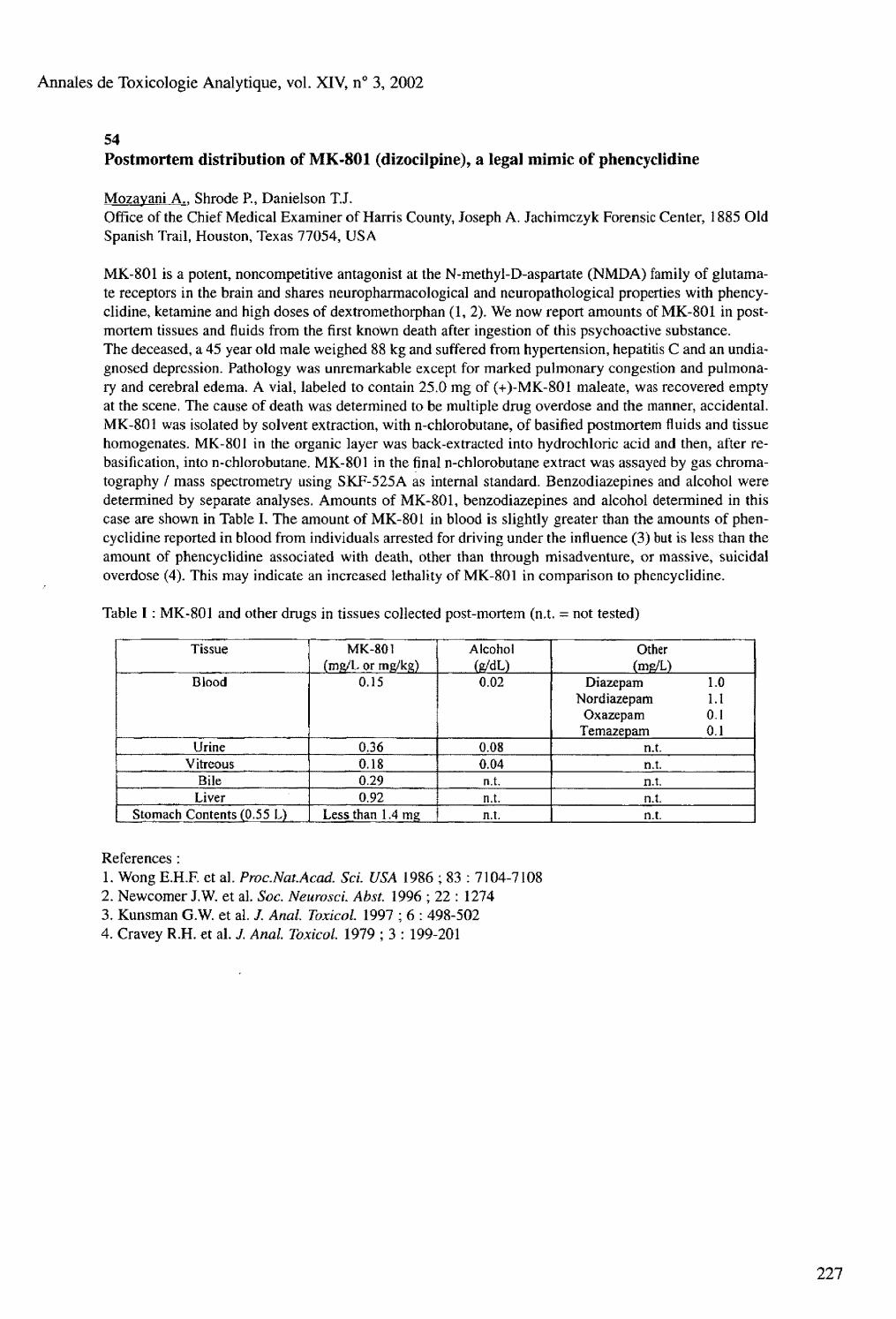
Annales de Toxicologie Analytique, vol. XIV, n° 3, 2002
54
Table I : MK-801 and other drugs in tissues collected post-mortem (n.t. = not tested)
Tissue MK-801 (mg/L or mg/kg)
Alcohol (R/dL)
Other (ms/L)
Blood 0.15 0.02 Diazepam 1.0 Nordiazepam 1.1
Oxazepam 0.1 Temazepam 0.1
Urine 0.36 0.08 n.t. Vitreous 0.18 0.04 n.t.
Bile 0.29 n.t. n.t. Liver 0.92 n.t. n.t.
Stomach Contents (0.55 L) Less than 1.4 mg n.t. n.t.
References : 1. Wong E.H.F. et al. Proc.Nat.Acad. Sci. USA 1986 ; 83 : 7104-7108 2. Newcomer J.W. et al. Soc. NeuroscL Abst 1996 ; 22 : 1274 3. Kunsman G.W. et al. /. Anal. Toxicol. 1997 ; 6 : 498-502 4. Cravey R.H. et al. J. Anal. Toxicol. 1979 ; 3 : 199-201
Postmortem distribution of MK-801 (dizocilpine), a legal mimic of phencyclidine
Mozayani A.. Shrode P., Danielson T.J. Office of the Chief Medical Examiner of Harris County, Joseph A, Jachimczyk Forensic Center, 1885 Old Spanish Trail, Houston, Texas 77054, USA
MK-801 is a potent, noncompetitive antagonist at the N-methyl-D-aspartate (NMDA) family of glutamate receptors in the brain and shares neuropharmacological and neuropathological properties with phencyclidine, ketamine and high doses of dextromethorphan (1, 2). We now report amounts of MK-801 in postmortem tissues and fluids from the first known death after ingestion of this psychoactive substance. The deceased, a 45 year old male weighed 88 kg and suffered from hypertension, hepatitis C and an undiagnosed depression. Pathology was unremarkable except for marked pulmonary congestion and pulmonary and cerebral edema. A vial, labeled to contain 25.0 mg of (+)-MK-801 maléate, was recovered empty at the scene. The cause of death was determined to be multiple drug overdose and the manner, accidental. MK-801 was isolated by solvent extraction, with n-chlorobutane, of basified postmortem fluids and tissue homogenates. MK-801 in the organic layer was back-extracted into hydrochloric acid and then, after re-basification, into n-chlorobutane. MK-801 in the final n-chlorobutane extract was assayed by gas chromatography / mass spectrometry using SKF-525A as internal standard. Benzodiazepines and alcohol were determined by separate analyses. Amounts of MK-801, benzodiazepines and alcohol determined in this case are shown in Table I. The amount of MK-801 in blood is slightly greater than the amounts of phencyclidine reported in blood from individuals arrested for driving under the influence (3) but is less than the amount of phencyclidine associated with death, other than through misadventure, or massive, suicidal overdose (4). This may indicate an increased lethality of MK-801 in comparison to phencyclidine.
Annales de Toxicologie Analytique, vol. XIV, n° 3, 2002
55 Fatalities with methadone in Norway 1991-2001
Hilberg T.. Teige B., Bj0rneboe A., M0rland J. National Institute of Forensic Toxicology, Oslo, Norway
Methadone is a synthetic opioid that is prescribed primarily for drug assisted rehabilitation of heroin addicts. When administered on a daily basis, considerable tolerance develops, so that patients taking this drug regularly can tolerate doses that are toxic, or even lethal, to non-tolerant persons. During the years 1991-2001 methadone was detected in 60 medico-legal autopsy cases, of which 38 cases from the last two years. The cause of death was in most cases poisoning. About 10% were due to methadone intoxication only, while the rest were mixed intoxications with diazepam (39 %), fiunitrazepam (39 %), heroin/morphine (26 %) and ethanol (21 %) as the most frequent additional substances. In most cases methadone was considered to have contributed significantly to the cause of death. The annual number of cases is highly correlated with the annual national sales of methadone (1^=0.81, p<0.001), corresponding to one death for about every 50 annual dosages of heroin substitution. There was information of methadone prescription in only a minority of the cases, indicating that the drug frequently had been obtained outside organised methadone programs.
56 The correlation between postmortem benzodiazepine blood and liver concentrations
Boratto M.. Mclntyre I.M., Drummer O.H. Victorian Institute of Forensic Medicine, Department of Forensic Medicine Monash University, 57 - 83 Kavanagh Street, Southbank, Melbourne, Victoria 3006, Australia
Temazepam, diazepam and nordiazepam concentrations were compared in human femoral blood and liver to determine if any correlation exists that might assist in the interpretation of benzodiazepine toxicology. Blood and liver concentrations ranged from 0.06-3.0 mg/L and 0.08-4.3 mg/kg respectively for temazepam (n=ll), 0.03-3.7 mg/L and 0.09-2.1 mg/kg for diazepam (n=16) and 0.04-3.0 mg/L and 0.24-3.5 mg/kg for nordiazepam (n=16). The correlation coefficient (r2) of blood/liver concentration were 0.879, 0.646 and 0.872 (p<0.01) respectively for temazepam, diazepam and nordiazepam. The liver to blood ratio ranged between 0.9-7.3, 0.4-4.3 and 1.2-6.0 with a mean and SD of 2.3 ± 1.8, 1.4 ± 0.90 and 2.5 ± 1.1 respectively for temazepam, diazepam and nordiazepam. These studies demonstrate that there is a significant linear correlation between blood and liver benzodiazepine concentrations.
228
Annales de Toxicologie Analytique, vol, XIV, n° 3, 2002
57 Codeine and morphine blood levels increase during blood loss
Kugelberg F.C.">. Holmgren P.(2>, Druid H.<3)
(1) Department of Clinical Pharmacology, Linkoping University, Sweden (2) Department of Forensic Chemistry, Linkoping University, Sweden (3) Department of Forensic Medicine, Karolinska Institute, SE-171 77 Stockholm, Sweden
During extensive blood loss, a number of compensatory mechanisms come into action to keep up the blood pressure, such as increased pulse, vasoconstriction and decreased glomerular filtration. If the blood loss continues, a plasma volume refill will take place by transfer of interstitial fluid into the blood. Drugs present in this fluid may follow and cause a rise or a drop in blood drug concentration, depending on their levels and accessibility in the restoration fluid. The drug levels found in postmortem blood may thus not reflect the levels at the time of the trauma (e.g. an automobile accident). This study was carried out to evaluate the possible changes of codeine, and its metabolite morphine, in whole blood during a standardized exanguination in the rat. Female Sprague-Dawley rats were given a suspension containing 5 mg codeine (codeine phosphate semi-hydrate, Kodein Recip, Recip AB, Arsta, Sweden) orally through a feeding tube during a brief C O 2 anesthesia. Three doses were administered at 45 min interval (corresponding to tm for codeine in the rat) to build up the tissue levels. Anesthesia was induced and maintained using isofluorane, and the body temperature was carefully monitored at 37.5 ± I o C. The left femoral artery was dissected free and a thin catheter was inserted. Blood loss was accomplished in eight rats by slowly withdrawing 0.8 mL blood at 10 min intervals, except for the first and 8 , h samples when 1.0 mL was withdrawn. Control rats were subjected to the same protocol, but blood was withdrawn only at 0 and 70 min. Samples collected at 0, 10, 30, 50 and 70 min were analyzed for codeine and morphine using a GC-MS method. The ratio between the final and the initial blood level for codeine and morphine was calculated for each animal, and was used to compare exanguinated rats with the controls. The results are displayed in Table I. The mean initial blood concentration of codeine was 0.15 pg/g blood for both experimental rats and controls, and mean initial morphine level 0.23 and 0.26 pg/g blood for experimental and control rats, respectively. The control rats showed a gradual decrease in codeine and morphine concentrations, as could be inferred from the continuing metabolism. In contrast, the levels of both codeine and morphine increased in the rats subjected to blood loss, and the levels at 70 min were significantly different from controls. It is concluded that blood loss can affect blood drug levels. Further studies using drugs with larger Vd and different plasma protein binding are ongoing.
Table I. Final/initial blood drug level ratio, hematocrit (Hct), body weight (B.W.) and number of rats (N). Values are mean ± SD. ^=0.014, ^=0.021, 3p<0.001, n.s. = not significant (experimental group vs. controls ; unpaired two-tailed Student's t-test).
Codeine ratio Morphine ratio Hct (%) B.W. (g) N Exp group 1.28 ±0 .44 ' 1.41 ±0 .34 2 34 ± 2 3 265 ± 11 n.s. 8 Controls 0.70 ± 0.38 0.88 ±0.47 4 2 ± 18 271 ±18 8
229

Annales de Toxicologie Analytique, vol. XIV, n° 3, 2002
58 GC-MS determination of 2-chlorobenzyüdene malononitrile (CS gas) metabolites in postmortem liver specimens
Sinn Y.S."». Anderson R.A.(2> (1) National Institute of Scientific Investigation Central Office, Whamdong, Yusunggu, Daejon, Korea (2) Department of Forensic Medicine and Science, University of Glasgow, Glasgow, United Kingdom
Riot control agents include 2-chlorobenzylidine malononitrile (CS) and chloroacetophenone (CN), of which CS is by far the most important worldwide. CS is a white, crystalline solid, sparingly soluble in water that is dispersed as an aerosol or smoke, causing intense sensory irritation when in contact with the mucous membranes of the eye and respiratory tract. Acute exposure produces lacrimation, burning and pain in the skin and eyes, conjunctivitis, coughing, irritation of throat, dizziness, nausea, and vomiting. Following administration, CS is rapidly metabolised by reduction, hydrolysis and conjugation. The major urinary metabolites of CS in rats were reported to be 2-chlorohippuric acid (2-CHA) and 2-chIoro-mer-capturic acid (2-CMA)(l-3). To our knowledge, no method currently exists for the detection and analysis of CS or its metabolites in biological specimens from people exposed during law enforcement operations. The aims of this study were to develop a GC/MS method for the analysis of these two metabolites in postmortem liver specimens. A new derivatising reagent, trimethylsilyldiazomethane (4) was incorporated into the procedure. 2-chlorohippuric and 2-chloromercapturic acid were synthesised to provide reference standards. Liver samples were from fatalities allegedly exposed to CS gas prior to death. The samples were homogenised with an equal volume of water. 1 ml of each supernatant was acidified and extracted by SPE on an Isolute Cig column. The eluates were evaporated to dryness under nitrogen at 40° C, dissolved in 0.3 ml of 30 % methanol in toluene and derivatised with trimethylsilyldiazomethane solution (Sigma-Aldrich) at 40° C for 1 hour. After evaporation and reconstitution in ethyl acetate containing n-hexadecane as internal standard, the extracts were analysed by GC/MS using a Finigan Thermo Quest Trace-MS instrument operated in the selected ion monitoring mode. The ions used were as follows (quantification ion underlined): internal standard - m/z 99, 113, 127, 141, 226; methylated 2-CHA - m/z 139,168, 195, 227; methylated 2-CMA - m/z 117,125, 176, 242. The GC used an HP-1 column (30 m x 0.25 mm i.d. x 0.25 urn film thickness) programmed from 100° C (2.0 min) to 280° C at 12° C/min and held for 5 minutes. Calibration curves were constructed over the range 0-500 ng/ml approximately (R = 0.9998 and 0.9995, respectively for 2-CHA and 2-CMA). The concentrations of 2-CHA and 2-CMA in postmortem liver were in the ranges 69-947 ng/g and 60-1306 ng/g respectively, whereas in blank blood the concentrations were 46 and 7 ng/g respectively. Further control data for liver specimens will be accumulated. Analysis of 2-CHA and 2-CMA by GC or GC/MS has usually involved ethereal diazomethane, which is hazardous in preparation and use. Trimethylsilyldiazomethane is less toxic, available as a stable solution and has the same reactivity as diazomethane. Our results suggest that this reagent has significant potential in forensic toxicology.
References : 1- Cucinel S.A. et al. Fed. Proc. 1971 ; 30 : 86-91 2- Brewster K. et al. Xenobiotica 1987 ; 17 : 911-924 3- Rietvelt E.C. et al. Arch. Toxicol. 1983 ; 54 : 139-144 4- Hashimoto N. et al. Chem. Pharm. Bull. 1981 ; 29 : 1475-1478
230

Annales de Toxicologie Analytique, vol. XIV, n° 3, 2002
59 Ibogaine related fatality
Marker E.K., Stajic M. Forensic Toxicology Laboratory Office of Chief Medical Examiner, 520 First Avenue, New York, NY 10022, USA
Ibogaine is one of the psychoactive indole alkaloids from the shrub, Tabemanthe iboga. Beginning in 1985, a series of patents was issued for the use of ibogaine for treatment of addiction to morphine, heroin, cocaine, amphetamine, alcohol, nicotine and poly-drug dependancy syndrome. Since ibogaine use is not approved in the United States and the drug is expensive and difficult to obtain on the illicit market, ibogaine related fatalities are rare in this country. The décédant, a 36 year old male with history of chronic substance abuse, was found unresponsive by his friend, approximately 5 hours after last seen alive. Drug paraphernalia and empty alcohol bottles were present at the scene. Routine toxicologic blood analysis detected the presence of morphine (22 ng/mL) and benzoylecgonine (633 ng/mL) as well as an unknown substance subsequently identified as ibogaine. Ibogaine concentrations were 9225 ng/rnL in subclavian blood, 18563 ng/kg in brain and 18137 ng/kg in liver.
231

Annales de Toxicologie Analytique, vol. XIV, n° 3, 2002
60
232
Postmortem redistribution of three beta-blockers (atenolol, metoprolol, propranolol) in rabbits
Dupuis C. (1 ), Félissier A.L. (2 ). Gaulier J.M. (1), Marquet P.(1>, Lachâtre G.(1)
(1) Service de Pharmacologie et Toxicologie, CHU Dupuytren, Limoges, France (2) Service de Médecine Légale, Faculté de Médecine, Marseille, France
Postmortem drug levels in biological matrices may fluctuate according to the sampling site and the time interval between death and sampling (1,2). The knowledge of these variations, called postmortem redistribution, is crucial as it influences result interpretation. The present study was the first one of a series designed to try and find general rules based on physical or chemical properties (pKa, Kp, etc.), for postmortem redistribution of drugs. Postmortem redistribution of three betablockers (with similar pKa but different Kp) was studied in rabbits. A mixture of the 3 betablockers at 1 mg/kg each was administered intravenously to 18 rabbits twice daily for two days. Euthanasia occurred one hour after the last administration, and autopsy at TO, T2, T6, T12, T24, T48 hours after death (three rabbits at each time). Right cardiac blood (RCB), left cardiac blood (LCB), and blood from the inferior vena cava (so-called peripheral blood, PB) were sampled. Numerous other biofluids and tissues (liver, heart, lung, etc.) were also collected. Betablockers were extracted from blood using Extrelut® SPE columns, and then identified and determined using high performance liquid chromatography with diode array detection (HPLC-DAD). The quantification of the analytes was made at 230 nm. This validated method exhibited a limit of quantitation of 50 pg/L for the three betablockers. The calibration curves were linear from 50 to 2000 pg/L with a good precision and accuracy: intra- and inter-assay CV and bias values were lower than 10 % at 50 pg/L for the three compounds. In PB, RCB and LCB, drug concentration variations were observed over the 48 hours following death, A concentration decrease, lower for propranolol than for atenolol and metoprolol, occurred during the first hours; subsequently, a concentration increase was observed for the three betablockers. In RCB, concentration variations were more important for propranolol. In PB, maximal concentrations were reached earlier, and concentration fluctuations were less important for propranolol than for atenolol and metoprolol. During the first hours, the end of a distribution phenomenon can explain the observed concentration decrease. The lower decrease for propranolol in the 3 types of blood is consistent with its lipid solubility, as it can be explained by a faster distribution after death, almost completed at the time of the first sample. A redistribution from myocardium and/or liver, more extensive for propranolol because of its important Kp, can be responsible for the secondary concentration increase observed in RCB. The earlier increase in PB may be due to the redistribution of the drugs from the liver, especially for atenolol and metoprolol. In LCB, the concentration changes are certainly complicated by redistribution from the lungs. The on-going determination of the three betablockers in myocardium, liver and lung tissues will help investigate these hypotheses.
References 1. Pelissier-Alicot A.L. et al. Ann. Toxicol. Anal. 2001 ; 13 : 1-17 2. Prouty R.W. et al. J. Forensic Sci. 1990 ; 35 : 243-270

Annales de Toxicologie Analytique, vol. XIV, n° 3, 2002
61 Gene doping - new analytical challenges in doping control ?
Mueller R. K.. Edelmann J., Grobe J., Kleemann W. J. Leipzig University Institute of Legal Medicine, Johannisallee 28, D-04103 Leipzig, Germany
While the great majority of classical doping agents can be detected and quantified with certainty by the arsenal of instrumental analysis commonly used in doping laboratories according to the very tight accreditation requirements, the introduction of new doping "strategies" means the introduction of new techniques for doping detection. Changes of doping definition ("doping list") with respect to the futural responsibility (from 2003) of the World Antidoping Agency WADA (especially of the Committee Health, Medical and Research) for this globally valid regulation require new considerations towards detectability with results beyond reasonable doubt like in Forensic Toxicology. Possibilities and problems of this development are reported in the light of the discussions of the WADA Committee responsible for the scientific development of doping control.
62 GC-ion trap-MS in doping control : urinary determination of 19-norandrosterone and 19-noretiocholanolone at subnanogram per millilitre levels
Tedeschi L., Favretto P.. Frison G., Maietti S., Castagna F., Ferrara S.D. Forensic Toxicology and Antidoping, University Hospital of Padova, Via Falloppio 50,1-35121 Padova, Italy
A specific and sensitive method was developed for the quali-quantitative determination in urine of 19-norandrosterone (19-NA) and 19-noretiocholanolone (19-NE), main metabolites of the anabolic androgenic steroid nandrolone as well as of other 19-norsteroids. The total steroid fraction, extracted from urine, after addition of Ds-noretiocholanolone, by solid phase extraction on Cis cartridges, was hydrolysed by b-glucuronidase at pH 7.4. Liquid/liquid extraction was then accomplished with n-pentane at pH 9. Derivatization to produce bis-TMS derivatives was obtained by MSTFA / NH4 / dithyoerythrytol. GC-MS-MS analysis was carried out on a Saturn 2000 ion trap instrument, injecting 2-ul aliquots at 250° C under splitless conditions, using a 30-m Chrompack CP-Sil 8 capillary column with an oven temperature program from 150° C to 300° C and a carrier gas (He) flow of 1 ml/min. The ion trap was operated under EI ionisation, and multiple mass spectrometry experiments were optimized in order to achieve the best selectivity and sensitivity. In particular, MS-MS experiments were performed for both 19-NA and 19-NE on El-produced [M - CH3] + species at m/z 405 which were selectively stored into the ion trap and collided with helium under resonant conditions. Specific product ions were then produced at m/z 315 and 225, which were monitored in the analytical scan function. The limit of quantitation for both analytes in multiple mass spectrometry mode resulted to be 100 pg/mL, which even permits to identify 19-NA endogenous levels in blank urine samples. Furthermore, ion trap peculiar features allow 19-NA and 19-NE, at concentrations of 2 ng/mL (IOC threshold level for male athletes), to produce significant full scan mass spectra, which exhibit well defined, constant ion ratios and can be highly informative for identification, once good chromatographic separation from interferences is provided. The method was applied to verify if nutritional supplements used by athletes, and trace contaminated with 19-norsteroids, produce increased levels of 19-NA and/or 19-NE in urine.
233

Annales de Toxicologie Analytique, vol. XIV, n° 3, 2002
63 Dark Agouti rats as a human poor metabolizer model for forensic questions - Studies on the (meth)amphetamine formation from precursor drugs
Kraemer T.. Pflugmann, T., Peters F.T., Maurer H.H. Department of Experimental and Clinical Toxicology, University of Saarland, D-66421 Homburg (Saar), Germany
The Dark Agouti (DA) rat strain serves as an animal model for the human cytochrome P450 2D6 poor metabolizer (PM), whereas Wistar (WI) rats correspond to the human extensive metabolizer type. There still is need of elucidating the role of 2D6 polymorphism for the high interindividual differences in amphetamine (AM) and/or methamphetamine (MA) findings after application of AM/MA precursor drugs. The aim of our study was to test whether the DA model was suitable also for understanding those forensically important differences. Urinary excretion and plasma levels of AM and MA after intake of the model substance selegiline (N-propinyl-methamphetamine) should be investigated in DA and Wl rats. Forensic implications of corresponding differences whicht might occur should be discussed. DA and WI rats received a single oral dose of selegiline (3 mg/kg bm) by gastric intubation (n=5, each group). 24 h urine samples were collected and tested using the Abbott TDx immunoassay Amphetamine/Methamphetamine II. For quantification of AM and MA in the urine samples, GC-MS was used in SIM mode (ions m/z 240, 254 and 244, 258 for deuterated IS) after solid-phase extraction (HCX) and heptafluorobutyrylation. Blood samples were taken 2, 4 and 6 h after application. For quantification of AM and MA in these samples, GC-MS was used in NICI mode (SIM ions m/z 388, 402 and 399, 407 for deuterated IS) after solid-phase extraction (HCX) and derivatization with heptafluorobutyrylprolyl chloride. Quantification was performed using calibration curves. The TDx results in the 24 h urine samples of the two rat strains were significantly different (P value 0.0003). The medians of the measured concentrations were 10,000 ng/mL in the DA group and 2,065 ng/mL in the WI group. GC-MS quantification confirmed these results. AM and MA concentrations were significantly (P values 0.004, each) higher in the urine samples of the DA group. The medians of the urine MA concentrations were 14,081 ng/mL for the DA rats and 1,165 ng/mL for the Wl rats. The medians of the AM concentrations were 22,710 ng/mL (DA) and 2,430 ng/mL (WI). The AM plasma levels differed significantly (P<0,05) between the two groups in the 2, 4 and 6 h blood samples, with medians being 63, 45.8 and 30.9 ng/mL in the respective 2, 4 and 6 h samples of the DA rats and 41.8, 26.5, 19.6 ng/mL in the corresponding samples of the WI rats. The MA plasma levels differed significantly between the two groups (P<0.05) in the 2 and 4 h blood samples (medians 38.0 and 15.7 ng/mL for DA rats; medians 28.6 and 8.9 ng/mL for WI rats). The DA rats showed significantly higher AM/MA plasma and urine concentrations after selegiline ingestion. 2D6 polymorphism seems to be of importance for that. As shown in former in-vitro studies, N-deal-kylation is not catalyzed by 2D6. Besides, this would have resulted in lower AM/MA levels for the DA group. Therefore, other metabolic steps (e.g. hydroxylation) might be catalyzed by the polymorphic 2D6, which could explain the higher AM/MA levels in the PM model by the lack of hepatic elimination. Corresponding in-vitro studies are in progress.
234
Annales de Toxicologie Analytique, vol. XIV, n° 3, 2002
64 Kinetics of kavain and its metabolites after oral application
Tarbah F. (1). Mahler H. ( l>, Kardel B. ( 1 ), Weinmann W.(2), Daldrup Th, ( ! )
(1) Institute of Legal Medicine, Heinrich-Heine University, P.O. Box 10 10 07, D-40001 Duesseldorf, Germany (2) Institute of Legal Medicine, University Hospital Freiburg, Albertstrasse 9, D-79104 Freiburg, Germany
The metabolism of the wide-spread used phytopharmaceutical kavain is performed by the human liver cell-line Hep-G2 resulting in 13 kavain metabolites1. In the present study a high performance liquid chromatography (HPLC-DAD) assay method for the simultaneous determination of kavain and its main metabolites (p-hydroxykavain, p-hydroxy-5,6-dehydrokavain and p-hydroxy-7,8-dihydrokavain) in serum and urine was developed and validated. The main metabolic pathways were hydroxylation of the phenyl ring and conjugation with glucuronic acid and sulphate, reduction of the 7,8-double bond, hydroxylation with subsequent dehydration and opening of the lactone ring. The metabolites were mainly excreted in the form of their conjugates. All kavain metabolites were detectable in serum and urine except for phydroxy-7,8-dihydrokavain which was found in urine only. Confirmation of the results and identification of the metabolites were carried out by LC/MS or LC/MS/MS. Kinetics of kavain and its metabolites in serum were investigated after administration of a single oral dose (800 mg d,l-kavain). Within time periods between 1 and 4 hours after uptake, the serum concentrations ranged between 10 ng/ml and 40 ng/ml for kavain, 125 ng/ml and 300 ng/ml for p-hydroxykavain, 40 ng/ml and 90 ng/ml for phydroxydehydrokavain as well as 30 ng/ml and 50 ng/ml for dehy-drokavain.
Reference : 1- Tarbah F. et al., Problems of Forensic Sciences, Special issue: 37 t h TIAFT triennial meeting XLII : 173-180.

Annales de Toxicologie Analytique, vol. XIV, n° 3, 2002
65
236
Kava (Piper methysticum Forst. f.) side effects and toxicity : study of 29 heavy kava drinkers and 2 cases of acute hepatitis in occasional kava drinkers in New Caledonia
Barguil Y. w . Kritsanida M. ( ,-2), Cabalion P. (2\ Duhet D.<2\ Mandeau A. ( , ), Poncet C. ( , )
(1) Laboratoire de Biochimie, Centre Hospitalier de Nouvelle-Calédonie, Av. Paul Doumer, BP J5, 98849 Nouméa, Nouvelle-Calédonie (2) IRD, Laboratoire des substances naturelles terrestres, Promenade Roger Laroque, BP A5, 98848 Nouméa, Nouvelle-Calédonie
New Caledonia is a South Pacific island where kava beverage (aqueous extract of the root of Piper methysticum Forst. f.) was introduced from Vanuatu around 20 years ago. It is used for its sedative properties due to lipophylic cc-pyrones (kavalactones). Following recent accidents with liver failure in Europe after oral intake of kava medications, we studied the biological parameters of 29 heavy kava drinkers. We compared them to those of 2 New Caledonian cases of acute hepatitis after light oral intake of kava. 29 volunteers (19 men and 10 women), heavy kava drinkers (mean: 6.56 units (cups prepared by the mela-nesian way: «70mL of kava, =650mg of kavalactones) /day, 6.04 days/week, nearly 25g of total kavalac-tones/week, since more than 5 years), participated. The presence of kavalactones in their urine was verified by HPLC/DAD. 2 women who developed an acute hepatitis, light kava drinkers (<5 units/day, for less than one month) took part in the study. Blood and urine biochemistry, tests for hepatitis A, B, C, serum protein electrophoresis and ultrasonography were performed. Among the 29 heavy kava drinkers, 17 men and 8 women had an isolated increase of gamma-glutamyl-transferase, 3 times the reference range. The only important clinical sign was skin dryness (ichtyosis in 17 volonteers). After significant diminution of kava consumption, GGT returned to normal values, skin dryness disappeared. Ultrasonography, blood tests for hepatitis A, B, C and serum protein electrophoresis gave normal results for all the volunteers. In the 2 cases of acute hepatitis, all biological signs of liver function alteration were present. Hepatitis in New Caledonia and in Europe occurred after intake of small amounts of kavalactones (<10g/week and <2g/week in each case). Clinical signs occurred both within 1 -2 months. On the contrary, heavy drinkers had only an isolated increase of GGT, which may suggest an hepatic enzymatic induction with no other clinical sign than a skin dryness. Kava consumption may lead to an enzymatic induction of the cytP450, by which kavalactones are possibly metabolized. This could explain kava hepatotoxicity: a formation of a toxic metabolite may occur, which would be either the cause of an immuno-allergic reaction or/and accumulated in poor metabolizers (idiosyncratic cases) (1). Russmann et al. have already showed a CYP2D6 deficiency in one case of kava hepatotoxicity in Switzerland (1). Deficiency of retinoids (metabolized by the cyt. P450) in ichtyosis would be explained as well.
Reference 1- Russmann S. et al. Annals of Internal Medicine 2001 ; 135 : 1

Annales de Toxicologie Analytique, vol. XIV, n° 3, 2002
66 Two pediatric overdose deaths involving hydrocodone, chlorpheniramine, brompheniramine, and pseudoephedrine
McCutcheon R.. Hall B., Sehroeder P., Peacock E., Bay ardo R. Office of the Chief Medical Examiner, P.O. Box 1748, Austin, Texas 78767, USA
It is not uncommon for a child to visit an emergency room after ingesting an unknown amount of a cold or cough syrup preparation. It is uncommon however, for this type of exposure to result in the death of the child. The death of a four-year-old female and a four-month-old male are reported. In case #1, a four-year-old female had been diagnosed with Strep throat, based on clinical presentation only, the day prior to her death. She was mistakenly given an adult formulation cough syrup containing hydrocodone and guaifenesin. Heart blood, vitreous, and bile specimens were submitted for toxicological analysis. Acetone was detected in the blood and bile at 0.003% and 0.008% respectively. Hydrocodone and chlorpheniramine were detected in the heart blood at concentrations of 0.67 and 0.23 mg/L, respectively. The bile contained hydrocodone at a concentration 0.30 mg/L. Chlorpheniramine was detected in the bile at a concentration of less than 0.05 mg/L. The death was ruled an accident due to an overdose of hydrocodone. In case #2, a four-month-old male was given an unidentified cold preparation containing brompheniramine and pseudoephedrine. Heart blood and vitreous specimens were submitted for analysis. Blood and vitreous were negative for volatiles. Brompheniramine and pseudoephedrine were detected in the blood at concentrations of 0.86 and 16 mg/L, respectively. The parents originally denied giving any medication to the infant. Other witnesses reported seeing the father pour cough syrup directly from the medicine bottle into the mouth of the baby. The death was ruled a homicide due to an overdose of brompheniramine and pseudoephedrine. Many pediatric exposures to cough and cold preparations are reported by poison control centers in the US each year. Very few result in major toxic consequences. Only one pediatric case of fatal overdose due to pseudoephedrine was identified. The reported blood concentration was 13 mg/L. Three pediatric overdoses deaths due to hydrocodone toxicity were identified. The blood hydrocodone concentrations were 0.20, 0.30 and 0.32 mg/L, respectively.
67
The forensic toxicology of A9-tetrahydrocannabinol (THC)
Drummer O.H.. Chu M., Gerostamoulos J. Victorian Institute of Forensic Medicine and Department of Forensic Medicine, Monash University, 57-83 Kavanagh St., Southbank, Victoria 3006, Australia This presentation reviews the role of THC in forensic cases, particularly in road crashes, drug deaths and homicides. In all cases THC was confirmed in blood using GC-MS and SIM. Cannabis as THC or carboxy-THC was the most frequent drug detected in fatally injured drivers in Australia with a frequency of 13 %. THC was detected in about 70 % of these cases at a median blood concentration of 10 ng/mL. THC was found to be only stable in both glass and plastic containers when stored for up to one week at +20° C & -20° C. No significant difference in blood concentration was found in blood taken from the heart, femoral and subclavian regions (n=17). In almost 4000 cases, the risk attributed to THC of increasing crash likelihood was 2.8 times a drug-free driver, which is similar to the risk attributed to alcohol concentrations between 0.10 and 0.15 g%. The risk increased to nearly seven-fold at THC concentrations equal to or greater than 5 ng/mL. THC was also detected in 19 % of homicides and 15 % of drug deaths at concentrations similar to drivers. Alcohol was the most common co-associated drug (44 % of cases) with a mean of 0,17 g%. Benzodiazepines were also detected in 40 % of cases. Cannabis is an important drug from a forensic perspective due to its ability to cause behavioral toxicity.
237

Annales de Toxicologie Analytique, vol. XIV, n° 3, 2002
68 Homoharringtonine overdosage. Analytical, pharmacokinetics and clinical aspects
Bardin C. ( I ). Ferey K. ( l i, Batista R.0 ), Vekhoff A.(2\ Havard L. 0 ), Marie J.R(2), Robin J,P.<3), Chast F.<1> (1) Service de Pharmacie-Pharmacologie-Toxicologie (2) Département d'Onco-Hématologie, Hôtel-Dieu, 1 Place du Parvis Notre-Dame, 75004 Paris, France (3) Oncopharm, 72 avenue O. Maessian, 72000 Le Mans, France
Homoharringtonine (HHT) is a cytotoxic alkaloid isolated from the evergreen tree Cephalotaxus harring-tonia. Clinical studies have indicated that HHT is effective in treating acute myeloid leukemia and chronic myeloid leukemia. Like for other anticancer drugs, its therapeutic range is very narrow. However, little is known about HHT pharmacokinetics and toxicokinetics. We report the first case of HHT acute overdosage. A simple, very sensitive and highly selective reverse-phase high-performance liquid chromatography method with fluorescence detection was developed in our laboratory for the quantification of HHT in human plasma. The mobile phase consisted of 0.01 mol/L sodium phosphate bibasic (pH 4.0), acetonitri-le and tetrahydrofurane (80: 50: 5). Spectrofluorimeter was operated at an excitation wavelength of 280 nm, and emission was monitored at 320 nm. The method was validated over the range of 1.0 to 60 ug/L using 1000 uL of plasma. Recovery of HHT from human plasma was 70,5 %. Accuracy, within-day and between-day precision were less than 7.5 % for the complete concentration range. HHT and the internal standard (quinidine) were extracted from plasma into methylene chloride. Organic phase was evaporated to dryness and dry residues were reconstituted with 200 uL of mobile phase. Seventy five uL were injected in the HPLC system, A 53-year old male was treated by a first course of HHT 4.5 mg/day administered by intravenous continuous infusion using a 7-day infusor device. On the second day, patient collapsed with an acute face oedema. Elevation of liver enzymes, acute renal failure and hyperkaliemia occurred. Infusion was immediately stopped. An error of administration caused by the infusion device made the delivery of HHT 3 to 4 times higher than normal dosage causing a dramatic overdosage. These toxic effects resolved spontaneously after three days. Five blood samples for measurements of HHT plasma concentrations were drawn. Concentration of HHT in plasma were 112.6, 14.0, 5.7, 3.9 and 3.0 pg/L respectively 8h, 16h, 28h, 40h 52h after stopping infusion. HHT concentrations were 5 to 10 times as high than concentrations reported with usual HHT dosage. During HHT acute overdosage, results show a biphasic elimination of HHT with a quick first step, correlated with regression of toxic symptoms.
238
Annales de Toxicologie Analytique, vol. XIV, n° 3, 2002
69 How determining a drug concentration in blood could help to revise a package insert : the example of metformin
Lalau J.D.(". Lacroix Ch.<s
(1) Service d'Endocrinologie-Nutrition, CHU, 80054 Amiens, France (2) Laboratoire de Pharmacocínétique, Centre Hospitalier, BP 24, 76083 Le Havre, France
In the package insert for metformin, it appears that this drug may be associated with lactic acidosis, a condition rare but with a high rate of mortality. However, the link between metformin and LA may be causal, associated or coincidental. We therefore investigated this link by determining metformin concentration in plasma (HPLC) in 49 metformin-treated patients with lactic acidosis and available metformin concentration (National Drug effect Adverse Effect): mortality was not associated with high metformin concentrations -the median concentrations being instead 3 times higher in patients who survived - but with concurrent pathologies. Looking for reports of so-called 'metformin-associated lactic acidosis' in the literature, plasma metformin concentration was determined in only 4 of 26 patients, of whom 1 had a normal value. Thus, taking into consideration both metformin concentration and concurrent pathologies, it is possible to coin new terms to describe lactic acidosis: metfomùn-unrelated, with a high mortality rate, metfornún-induced, with no direct fatality, metformin-associated, with an intermediate prognosis, depending upon the severity of the concurrent pathology. However, metformin has a short half-life in plasma and performing blood sampling rapidly after admission for metformin determination is not necessarily the primary preoccupation in the context of emergency. We therefore used erythrocytes as a putative marker for a deep compartment: metformin concentrations decreased much more slowly in erythrocytes than in plasma. Such a slower decline may help for the retrospective discussion of the link between metformin and lactic acidosis. In conclusion, when discussing mortality in metformin-treated patients with lactic acidosis on the basis of blood metformin concentration, it is no longer acceptable to talk in terms of mean mortality rates; the unexpected survival of many patients with lactic acidosis joined with the vascular effects of the drug gives rise to the provocative premise that high metformin concentrations might have been protective.
70 How natural are "natural herbal medicines" ?
Bogusz M.J.. Al Tufail M., Hassan H. King Faisal Specialist Hospital and Research Centre, P.O.Box 3354, 11211 Riyadh, Kingdom of Saudi Arabia
Herbal products, known and used for centuries in traditional medicine in Far and Middle East, are finding their way also in industrialized countries. The main cause of growing popularity of herbal remedies is the belief, that they are natural, hence well tolerated and not harmful. Moreover, in most countries herbáis are classified as dietary supplements and are not subjected to the same safety and efficacy standards as therapeutic drugs. In consequence, herbáis products are very often not only of poor hygienic quality, unclear labeled and defective packed, but also are deliberately tampered with synthetic drugs in order to achieve claimed therapeutic action. Heavy contamination with toxic metals is frequently observed. In 2000/2001, in several samples of herbal remedies various synthetic drugs were identified, like e.g. psychoactive phenethylamines in "slimming pills", antidepressants in pills "good for nerves", cardiac glycosides in pills "good for heart", or periactin in pills "for gaining weight". High concentrations of heavy metals were often found in ointments, "beauty" creams or in preparations against cancer. Hair dyes, labeled as natural henna, contained high concentrations of p-phenylenediamine. Our experience indicates that the market for herbal products should be controlled and the products should be examined mainly for dangerous compounds of synthetic origin and toxic elements. From the analytical point of view, the use of gas chromatography-mass spectrometry for the analysis of organic compounds in herbal preparations is a must. An advent of liquid chromatography-mass spectrometry will expand the detection possibilities, providing that the library of herbal compounds will be established. The use of ICP-MS for element analysis is highly recommended. The analytical aspects of examination of herbal medicines as well as examples from the casework will be presented.

Annales de Toxicologie Analytique, vol. XIV, n° 3, 2002
71 Legal herbal drugs : studies on the metabolism of the Eschscholtzia californica alkaloids californine, protopine and lauroscholtzine as basis for the development of toxicological analysis procedures
Paul L.D.. Maurer H.H. Department of Experimental and Clinical Toxicology, University of Saarland, D-66421 Homburg (Saar), Germany
Eschscholtzia californica CHAM., also called California poppy, a papaveraceae and the state flower of California, is native in California and the South of the USA. The herb is described to evoke mild marijuana-like euphoria after ingestion or smoking. The aim of our study was to identify the metabolites of the three alkaloids californine, protopine and lauroscholtzine in rat urine by GC-MS techniques after oral application of the corresponding pure substances. Each of the pure alkaloids was given to male Wistar rats by gastric intubation and urine was collected over 24 hours (25 mg/kg body mass each). The alkaloids and their metabolites were isolated either after enzymatic cleavage of conjugates or directly by liquid liquid extraction. The metabolites (underivatized, ace-tylated, trifluoroacetylated or methylated) were separated and identified by GCMS in the EI and PCI mode (expérimentais according to: Drug Metabol. Dispos. 28, 2000, 239). The following main metabolites of californine could be identified in the urine extracts after enzymatic hydrolysis: N-demethyl, mono- and di-demethylene californine, isomers of mono- and di-demethylene-methyl californine and combinations of these metabolites. For protopine, only two isomers of one mono-demethylenemethyl metabolite, and for lauroscholtzine, phenanthrene analogues including Ndemethyl and Odemethyl metabolites were detected. In the native urine extracts the phenolic hydroxy metabolites could only be detected in minor amounts. From these results, the following metabolic pathways could be concluded: Ndemethylation and/or single or double demethylenation with consecutive catecholOmethylation of one of the hydroxy groups for californine. Protopine only undergoes extensive demethylenation of the 3,4-methylenedioxy group (confirmed by fragmentation pattern) followed by catecholOmethylation. The phenolic hydroxy metabolites were found to be nearly completely conjugated. Lauroscholtzine undergoes ring cleavage to the phenanthrene derivatives. However, these derivatives could also be formed artificially during sample preparation. In addition, several demethylation pathways could be postulated. Unfortunately, due to the lack of data about common dosages, no statement could be made about the detectability in case of abuse. Assuming similar metabolism in man, the analytical targets in case of a poisoning with Eschscholtzia should be the main metabolites demethylenemethyl californine and demethylenemethyl protopine in urine.
240
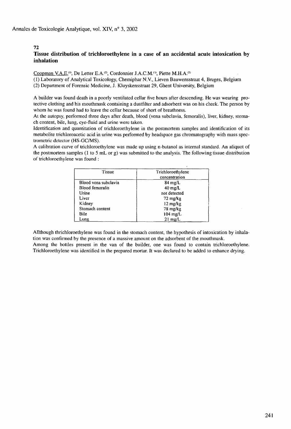
Annales de Toxicologie Analytique, vol. XIV, n° 3, 2002
72 Tissue distribution of trichloroethylene in a case of an accidental acute intoxication by inhalation
Coopman V.A.E.">. De Letter E.A.*2), Cordonnier J.A.C.M. ( Î\ Piette M.H.A.(2> (1) Laboratory of Analytical Toxicology, Chemiphar N.V., Lieven Bauwensstraat 4, Bruges, Belgium (2) Department of Forensic Medicine, J. Kluyskensstraat 29, Ghent University, Belgium
A builder was found death in a poorly ventilated cellar five hours after descending. He was wearing protective clothing and his mouthmask containing a dustfilter and adsorbent was on his cheek. The person by whom he was found had to leave the cellar because of short of breathness. At the autopsy, performed three days after death, blood (vena subclavia, femoralis), liver, kidney, stomach content, bile, lung, eye-fluid and urine were taken. Identification and quantitation of trichloroethylene in the postmortem samples and identification of its metabolite trichloroacetic acid in urine was performed by headspace gas chromatography with mass spec-trometric detector (HS-GC/MS). A calibration curve of trichloroethylene was made up using n-butanol as internal standard. An aliquot of the postmortem samples (1 to 5 mL or g) was submitted to the analysis. The following tissue distribution of trichloroethylene was found :
Tissue Trichloroethylene concentration
Blood vena subclavia 84 mg/L Blood femoralis 40 mg/L Urine not detected Liver 72 mg/kg Kidney 12 mg/kg Stomach content 78 mg/kg Bile 104 mg/L Lung 21 mg/L
AHthough thrichloroethylene was found in the stomach content, the hypothesis of intoxication by inhalation was confirmed by the presence of a massive amount on the adsorbent of the mouthmask. Among the bottles present in the van of the builder, one was found to contain trichloroethylene. Trichloroethylene was identified in the prepared mortar. It was declared to be added to enhance drying.
241

Annales de Toxicologie Analytique, vol. XIV, n° 3, 2002
73 A stuctured approach in the optimization of a headspace and PTV-based injection for the analysis of volatile poisons
Bouche M.P.(1). Praisler U.(2\ De Leenheer A.P.(1), Van Bocxlaer J.F.(1)
(1) Laboratory of Medical Biochemistry and Clinical Analysis, Ghent University, Harelbekestraat 72, B-9000 Gent, Belgium (2) Department of Physics, University of Galati, Strada Domneasca 47, RO-6200 Galati, Romania
Both headspace extraction and programmed temperature vaporization (PTV) have undoubtedly conquered their place respectively as an important sample clean-up and injection technique in capillary GC, also in hyphenated toxicological analysis. The combination of these techniques can be valuable, provided the application of a suitable preconcentration e.g. cryogenic condensation. Indeed, headspace samples usually hold the key analyte at a very low concentration, which implies the injection of a large volume of gas in order to place enough analyte mass onto the capillary column. Mostly, the very small column internal diameter prevents direct loading of such gas volumes. However, when using this intricate combination of PTV, headspace sampling and cryogenic focussing, one has to adjust a large number of instrumental parameters (e.g. up to 20 parameters depending on instrumental factors). Logically, this substantially complicates straightforward optimization of the injection procedure, thus analysis efficiency. For modelling and optimizing the injection efficiency from a quantitative chromatographic viewpoint, with use of a HP 6890-5973 MSD, equipped with a headspace autosampler (Gerstel Multi Purpose Sampler) and a CIS-4 (Gerstel) injector, we introduced the robustness and valid statistical significance tests of what is currently known as experimental design. By employing such computational techniques, all relevant injection parameters can be easily identified, isolated and optimized, and this by performing only a limited number of experiments. For screening of the important factors, e.g. a Plackett-Burman design with fold-over was tested. As such, the influence of 16 factors on 10 chromatographic response variables was evaluated (80 experimental runs, performed in 2 blocks). This first study eventuated in the determination of only four statistically relevant parameters requiring consecutive optimization. The subsequent application of a rotatable central-composite design led to the identification of relevant two-factor interactions, as well as to the final optimization of, in this case, injection volume (1 mL), desorption time (0.7 min) and split (0.7:1); and trap temperature (-30° C). This elaborated optimization procedure was assessed for the analysis of compound A, a highly volatile and nephrotoxic degradation product of the inhalation anaesthetic sevoflurane. The particular approach can be freely transposed towards other volatile poisons (e.g. nitrites) relevant to both clinical and forensic toxicology, resulting in the fast development of a dedicated, fully elaborated and quantitative analysis method. Accordingly, a considerable amount of time and money can be saved and analytical productivity is increased in an area characterized by continuously new analytical problems for a limited number of repetitive samples.
242

Annales de Toxicologie Analytique, vol. XTV, n° 3, 2002
ABSTRACTS OF
POSTER PRESENTATIONS

Annales de Toxicologie Analytique, vo l XIV, n° 3, 2002
244

Annales de Toxicologie Analytique, vol. XIV, n° 3, 2002
1 Enzymatic hydrolysis improves the sensitivity of EMIT screening for urinary benzodiazepines
Borrey D. ( i ), Meyer E. ( a, Duchateau L.(2>, Lambert W. ( l ). Van Peteghem C. (1), De Leenheer A.P.(1)
(l)Laboratorium voor Toxicologie, Universiteit, Harelbekestraat 72, B-9000)Gent, Belgium (2) Laboratorium voor Fysiologie, Biochemie en Biométrie, Universiteit Gent, Salisburylaan 133, B-9820 Merelbeke, Belgium
We evaluated the relevance of hydrolyzing urine samples prior to screening with the EMIT d.a.u. Benzodiazepine assay. A total of 530 authentic patient urine samples were collected from volunteers with a high prevalence of benzodiazepine (mis-)use. All samples were screened with EMIT both before (EMIT) and after (EMIT-H) enzymatic hydrolysis. Regardless of the screening results all samples were subsequently analyzed by a recently developed GC-MS procedure, used as a reference method. The sensitivity increased from 67 % (95 % CI: 60 - 74 %) for the EMIT test to 87 % (95 % CI: 81 - 92 %) for the EMIT-H test, while the specificity decreased from 100 % (95 % CI: 99 - 100 %) for the EMIT test to 96 % (95 % CI: 93 - 98 %) for the EMIT-H test The largest increase in EMIT-response was observed for samples containing flurazepam or di-K-clorazepate, but especially the sensitivity for lormetazepam and bromazepam was improved after pretreatment of the samples with 6-glucuronidase. To study the effect of changing the cutoff value on sensitivity and specificity, the receiver-operating characteristic (ROC) curves for the two EMIT tests were derived and compared. From the ROC curves it was clear that EMIT-H outperforms EMIT as for a fixed specificity, the corresponding sensitivity was systematically higher for the EMIT-H test. We therefore recommend that enzymatic hydrolysis should be routinely included in the EMIT screening procedure for urinary benzodiazepines because the obtained gain in sensitivity is substantially larger than the concomitant loss in specificity.

Annales de Toxicologie Analytique, vol. XIV, n° 3, 2002
2
246
A simple and rapid method for the determination of carboxyhemoglobin and total hemoglobin in toxicological laboratories
Cruz A.. Bal M.J., Quiniela O., Concheiro M., Gallardo E., López-Rivadulla M. Forensic Toxicology Service. Legal Medicine Institute. University of Santiago of Cornpostela. San Francisco s/n. 15705.Santiago of Compostela. Spain
The determination of the % of carboxyhemoblobin is an usual practice in clinical and forensic toxicological laboratories. The interpretation of this value is better when the total hemoglobin concentration is known, but this value not always is easy to obtain in the toxicological laboratories. To this propose we report a rapid and unexpensive spectrophotometric method, which involves the first and the third derivative spectra of a 0.1 % dilution of the sample in distilled water(v/v) in the range of 700-500 nm. The procedure also permits the calculation of the % of Methemoglobin. A Perkin Elmer Lambda 2 UV/VIS spectrophotometer interfaced to a computer running the software PECCS. An Izasa STKS autoanalyser was used to determine hemoglobin. Pure oxygen, carbon monoxide, and nitrogen, potassium ferricyanide and crystalline sodium nitrite, and a 13 % hemoglobin standard. Fresh blood samples, collected in Venoject tubes (Terumo) containing EDTA as anticoagulant. Standards of oxyhemoglobin (OxyHb), deoxyhemoglobin (DeoxyHb), carboxy-hemo-globin (COHb) and methemoglobin (MetHb) were prepared from a stock standard containing 13 g % of hemoglobin. After obtain the best spectrophotometric conditions for the determination of carboxihemo-globin, the quantitative study between 700-500 nm, was done: In the 3 r d derivative mode (D3), to calculate the carboxyhemoglobin concentration, and in the 1s t derivative mode (Dl), to calculate the Total hemoglobin concentration (as methemoglobin). The 13 % g hemoglobin standard was then used to prepare solutions of increasing concentrations of Hb (as COHb or MetHB) and the D3 and Dl (respectively) spectra were recorded. Samples of known concentrations of Hb, determined by a Coulter autoanalyzer, were analysed by the methods here developed, in order to obtain the correlation between them. In D3 only carboxyhemoglobin absorbs at 565 nm with a positive peak that is not affected by the other components, and that peak is proportional to the COHb content in the sample ( Y = 00.27X + 0.0013; r = 0.0049). In Dl the Methemoglobin has a maximum at 645 nm and a minimum at 620 nm, and the 1D645-620nm value is proportional to the Hb content (Y- 0.254 X + 0.007 ; r = 0.9997). The results of the two methods are linerarly correlated with those provided by a Coulter STKS autoanalyser. The proposed spectrophotometric method allows the determination of the carboxyhemoglobin saturation percentage and the total hemoglobin concentration in the sample, by measuring 3Ds65 nm (to obtain the [COHb]), then saturing the sample with potassium ferricyanide (to obtain MetHb), and recording the 'D645-620 nm (to obtain the Total Hb concentration). The % of COHb is calculate by a simple rule of three. This affords better interpretation of the analytical results as it allows the absolute amount of functional hemoglobin in an individual to be determined.
Annales de Toxicologie Analytique, vol. XIV, n° 3, 2002
3 Determination of local anesthetics in human plasma using solid phase microextraction and GC-MS
Gallardo E., Quíntela O., Cruz A., López-Rivadulla M. Forensic Toxicology Service. Institute of Legal Medicine. University of Santiago de Compostela. Spain
The performance of solid-phase microextraction (SPME) in combination with Gas Chromatography-Mass Spectrometry, to quantify mepivacaine, bupivacaine and ropivacaine in human plasma was studied. The analysis was performed using a mass-spectrometric detector in SIM mode and SKF as internal standard. For extraction, Carbowax / divinylbenzene, and polydimethylsiloxane fibers were tested. Absorption and desorption times were studied for all analytes. The Carbowax / divinylbenzene fiber gave the highest recovery in plasma samples as compared to the other fibers. The effects of temperature and agitation of the sample were studied. The validation of the method showed that the chromatographic selectivity was satisfactory and all compounds were well separated. The absolute retention times of mepivacaine, bupivacaine, ropivacaine and the internal standard were 14,1, 15.9, 17.15, and 16.2 minutes respectively. The assay had a linearity of 2500 ng/mL ; a LOD and LOQ were calculated for each drug. An average recovery of 98 %, and an average day-to-day imprecision of <10 % for both drugs. A patient kinetic study (n = 17) using this GC-MS method revealed a usefulness and quickly to control the plasma levels of these drugs. Of the 30 different drugs tested, only procaine, tetracaine articaine and lidocaine were extracted but did not interfere with the measurement of the drugs of interest.
247
Annales de Toxicologie Analytique, vol. XIV, n° 3, 2002
4
248
HPLC/Photodiode array detection combined with ESI/MS detection : a powerful tool for large screening of bioactive molecules in complex biological matrices. Elaboration of an UV/ESI /MS spectra library enabling fast and reliable compound identification
Humbert L. ( , ) , Grisel F. < 2 ), Bondoux G. ( 2\ Lhermitte M. (1) Laboratoire de Biochimie & Biologie Moléculaire, Hôpital Calmette, 59037 Lille Cedex, France (2) Waters European Headquarter, Rond Point des Sangliers, Rue Jacques Monod, 78280 Guyancourt
Large screening of xenobiotic molecules in biological fluids is mainly achieved by using gas chromatography with mass spectrometry detection (GC/MS) and/or liquid chromatography with photodiode array detection (LC/PDAD or LC/DAD). In addition to these techniques, immunoassays may be used for some specific molecules. GC/MS operated in EI became a very popular technique due to its ability to produce standardized mass spectra that are independent of the instrumentation. However, many analytes require time-consuming chemical derivatization steps in order to increase their volatility and enhance the sensitivity. LC/DAD generates UV spectra that can be stored in libraries (i.e. : ToxLab) and enables fast and reliable identification using the retention time as an additive specific criterion. However, new bioactive molecules are more and more active and detection limits using UV detection may become problematic for some of those compounds. It can be even worse with molecules that do not exhibit any absorbance such as Meprobamate or Valproic acid. Most of the LC/MS instrumentation available on the market do not provide any ESI spectra library due to apparent limitations of the API techniques. This work describes the methodology used to create an UV/ESI/MS (electrospray/MS) spectra library starting first by the development of a generic chromatographic method and then the compilation of UV and MS spectra obtained from various drugs and toxicants. Each molecule (about 500 molecules) is dissolved in an adequate solvent and diluted to reasonable concentration. Chromatography is achieved using a Waters Alliance 2690 equipped with a XTerra column (150 mm x 2.1 mm - 3.5 um) protected by a precolumn (same phase, 10 mm x 2.1 mm) operated at 200 pl/min in gradient mode (5-95 % in 26 min). Mobile phase is based on Water/acetonitrile with 5 mM ammonium formate at pH 3. UV acquisition is carried out using a Waters 996 Photodiode Array scanning from 210 to 400 nm. MS acquisitions were achieved in Full scan (100-650 amu) using 7 consecutive functions (1 channel in negative ESI and 6 channels in positive ESI with different cone voltages). Instruments control and data acquisition are done using a MassLynx 3.5 Workstation. In LC/MS, electrospray ionization technique involves an ionization process which is totally different of electron ionization thus preventing the use of commercial EI Mass Spectral Data Bases. Using in-source CID with an electrospray source, most of the analyte lead to mass spectra that exhibit at least 3 to 4 specific fragments ions sometimes in both polarities. The library contains exhaustive mass spectral information constituted by CID spectra of the species at 6 cone voltages in positive ion mode and one in negative ion mode(or more if relevant). This breadth of library information will help mitigate spectral differences that may exist among different MS. This work has already permitted to create a library that contains more than 150 molecules representing around 750 mass spectra and 150 UV spectra. That spectral information can be combined with retention time to constitute a perfect additional tools for general unknown screening when specificity and sensitivity are required. As upcoming development, we will also include automatic semi quantitation as a one-step process : Positive identification and semi quantitation using an internal standard.

Annales de Toxicologie Analytique, vol. XIV, n° 3, 2002
5 Effects of intestinal motility on ethanol absorption
Isobe E. ( l ). Tsukarnoto S.(1), Hirose M. 0 1, Nagoya T.(2)
(1) Department of Legal Medicine, Nihon University School of Medicine, 30-1 Oyaguchi Kami-cho, Itabashi-Ku, Tokyo 173-8610, lapan (2) Department of Common-Use Facilities for Medical Research, Nihon University School of Medicine, 30-1 Oyaguchi Kami-cho, Itabashi-Ku, Tokyo 173-8610, Japan
The effect of the intestinal motility on ethanol absorption in rat was investigated. The drug of intestinal motility were used to carbachol, a cholinergic agent and caffeine, a derivative of xanthine, its known to relax smooth muscle. They were orally given to the rats 1 hour before administration of ethanol (2 g/kg). Blood and stomach were obtained 0.5, 1, 3 hours after ethanol administration. The ethanol, acetaldehyde and acetate concentrations in each specimen were measured by headspace gas chromatography, respectively. The rate of intestinal absorption of ethanol was measured using the small intestine of Wistar rats according to Creine and Wilson method (J Appl Physiol 12: 145-146, 1958). Rats intestine are removed, and one end of a 5 cm segment of ileum is closed with a ligature while the other end is tied around the lower part of the cannula, i ml of Krebs-Ringer potassium solution (pH7.4) is injected into the intestine, then the intestine is soaked into another tube containing Krebs-Ringer potassium solution with 0.25 mg/ml ethanol. The injected solution was measured for ethanol levels at 10, 20 and 30 minutes in presence or absence of drugs. Administration by carbacol and caffeine showed increased and decreased the ethanol level in the blood but decreased and increased that in the stomach. The rate of intestinal absorption of ethanol was found that the increase in the ethanol absorption is depending on time. In the presence of carbachol and caffeine reduced ethanol absorption about over 10 % and less then 30 % of control. Based on these results, we speculate that intestinal motility by increase and decrease were augment and suppress alcohol absorption.
6 On-Column derivatization for determination of amphetamine and methamphetamine in biological materials by GC/ MS
Nishida M„ Namera A., Yashiki M., Kojima T. Department of Legal Medicine, Hiroshima University Faculty of Medicine, 1-2-3 Kasumi, Minami-ku, Hiroshima 734-8551, Japan
A simple determination method of amphetamine (AP) and methamphetamine (MA) in human blood was developed using on-column derivatization and gas chromatography - mass spectrometry (GC-MS). AP and MA were adsorbed on the surface of Extrelut and then derivatized the N-propoxycarbonyl derivatives using propylchloroformate. Pentadeuterated MA was used as an internal standard. The recoveries of AP and MA from the spiked blood (urine) were 89.7 and 90.3 % (88.2 and 92.5 %), respectively. The calibration curves showed linearity in the range of 12.5 - 2,000 ng/g for AP and MA in blood and urine, and 2.5 - 200 ng/10mg in hair. The coefficients of variation of intraday and interday were 0.42 to 4.58 % (blood), 0.68 to 3.60 % (urine), and 1.20 to 8.40 % (hair). Furthermore, this proposed method was applied to some medico-legal cases of MA intoxication. MA and its metabolite AP were detected in the blood samples, and the correlation of the blood level of amphetamines and the behaviors of the victims was in good agreement with the criteria proposed by Nagata [Jpn. J, Legal Med., 37 (1983) 513].
249

Annales de Toxicologie Analytique, vol. XIV, n° 3, 2002
7 A fatal forensic intoxication study with fenarimol : comparative analysis by HPLC/DAD and HPLC7DAD/MSD
Proenca P.. Pinho Marques E., Teixeira H., Castanheira E, Barroso M., Ávila S., Vieira D.N. National Institute of Legal Medicine (NILM)-Delegation of Coimbra, Portugal.
Fenarimol (Rubigan®) is a pyrimidine ergosterol biosynthesis inhibitor used as a systemic fungicide. The authors present a fatal intoxication case due to fenarimol analysed in the Forensic Toxicology Laboratory-NILM and used to compare two different HPLC techniques: an HPLC system with a diode detector (DAD) and an HPLC system with a DAD and a mass detector with an electrospray interface (MSD) in regard to selectivity and sensitivity. We report the first case in the literature of fatal fenarimol self-poisoning, so it was not possible to compare with published therapeutic and toxic data. All biological samples were submitted to a liquid extraction procedure. Concentration levels calculated were in the magnitude of 99,0 mg/mL in gastric content, 2,0 mg/g in liver and 0,5 mg/g in kidney. No blood was available at autopsy. The detection and quantification limits of fenarimol in blood and liver, the linearity and the precision in both techniques were determined.
8 LC-MS determination of urinary 5-hydroxytryptophol glucuronide
Stephanson N. ;", Beck 0 . i U , Dahl H. (2 ), Helander A.<2)
(1) Departments of Clinical Pharmacology (2) Clinical Neuroscience Karolinska Hospital & Institute, Stockholm, Sweden.
5-Hydroxytryptophol glucuronide (GTOL), the urinary excretion product of serotonin, has been proposed as a marker of acute ethanol intake. We undertook to develope an LC-MS method for determination of GTOL in human urine. A deuterium labelled GTOL analogue was used as internal standard. Samples were prepared for analysis by simply diluting the urine with distilled water containing internal standard. An injection volume of 10 ul and a flow rate of 200 ul/min were used, with a gradient mobile phase consisting of 25 mmol formic acid with 2 to 50 % acetonitrile using a CI8 HyPurity column (100x2.1x5). The retention time for GTOL was about 8 min and the entire analysis time was 15 min. The limit of detection was about 1 ng/ml, and the response curve was linear up to 5000 ng/ml. Preliminary data has indicated prolonged detection times of about 5-10 hours after ethanol has disappeared from urine. This method can be useful for monitoring relapse drinking in clinical outpatient treatment and also in forensic medicine.
250

Annales de Toxicologie Analytique, vol. XIV, n° 3, 2002
9
N° Type of Corrosive Admitted Lethality Mortality 1 Acetic Acid 518 85 284 2 Hydrochloride Acid 59 10 13 3 Sulfur Acid 78 6 47 4 Liquor Ammonia 137 1 17 5 Bleaches 201 2 10 6 Other Acids 43 12 16 7 Other Alkalis 24 1 3 S Unknown Corrosives 47 16 79 9 ALL 1107 133 469
As can be seen from the table above, 75 % of people poisoned by corrosives die prior to hospitalization. Notwithstanding the specialized medical treatment during hospitalization, lethality in the cases of acetic acid poisonings reaches 16,4 % (the average rate in Russia is 17-25 %). Thus, one of the directions along which one can expect to achieve improvement of the outcomes of acute searing liquids poisonings and particularly, acetic acid poisonings, can be the organization of specialized medical aid during the pre-hospitalization period.
References : 1- Wax PM, Sentzov VG. J Toxicol Clin Toxicol 1997 ; 2 : 175-189 2- Borisovsky V, Birtanov E. J Toxicol Clin Toxicol 1998 ; 5 : 447 3- Cox R., Brooks J. Burns, http : / emedicine. Com/emerg/ 2000 : 16p 4- Sarmanaev S. Kh. J. Toxicol clin toxicol 2000 ; 38 : 579-580 5- Sarmanaev S. Kh., Yamanaeyva I.E. J. Przeglad Lekarski 2001 ; 58(4) : 395 6- Tomilin V.V.,et al. Medicolegal evaluation, 1987. N û 4. XXX. P. 43-45 7- Kakhanovsky F.N., Bubon VS. Medicolegal evaluation, 1983, N°3. XXVI, P. 34-35
The mortality structure in cases of acute searing liquids poisonings (1992-2001)
Tchernov N.V.. Sarmanaev S.Kh., Akhmetov I.R., Kondrashova S.R., Salmanov A.A, Bessolitzina A.M., Akhmerova O.R, Teregulova Z.S. Ufa (Russia).
The interest to acute poisonings by searing liquids in Russia in the recent decade (1,2,3) has been caused by a high mortality rate, which stays at the same level as in previous years (4,5,6) A retrospective study of mortality of the deceased due to acute poisonings in the Republic of Bashkortostan (Russia) during 1992-2001 conducted on the material of acts of the Bureau of Forensic Medicine Analysis and medical charts of 18079 patients of the Ufa Toxicological Center for the same period. The rate mortality caused by acute poisonings in Republic of Bashkortostan during the last decade amounts to 14274. The mortality rate in cases of acute searing liquids poisonings amounts to 469. More than a half of these were poisonings by acetic acid - 284. Hospitalization for the 10 years equals 18079 patients. Hospitalization of people with acute poisonings by searing liquids is 1106. Lethal outcome is registered in 133 cases (out of these, 85 were acetic acid poisonings).

Annales de Toxicologie Analytique, voL XTV, n° 3, 2002
10
252
Tissue and plasma determination of 4-methyl-pyrazole in methanol acute poisoning
Wallemacq P.. Di Fazio V., Vanbinst R., Kônig J.5 Hantson Ph. Laboratory of Toxicology and Intensive Care Unit, University Hospital St Luc, UCL, B-1200 Brussels, Belgium
Four patients (3 male/1 female), admitted in intensive care unit for acute methanol poisoning between December 1999 and November 2001, were treated by fomepizole or 4-methylpyrazole (4-MP), an alcohol dehydrogenase inhibitor, administered either orally or iv. They displayed on admission methanol blood levels ranging from 0.4 to 3,3 g/L. Loading doses of 15 mg/kg 4-MP (ranging from 700-1300 mg) were administered iv or orally. Iterative doses were given at 12 hours interval for a maximum of 3 days. During hemodialysis, a continuous infusion of 4-MP was proposed at a rate of 1 mg/kg/h. A minimum of 12 blood samples were collected for each patient during the treatment, for 4-MP plasma determinations, and a sample of hepatic tissue (160 mg) was removed during the autopsy of one of the patients death 34 h after admission. Samples were kept frozen until analysis. A gas chromatographic method has been developed for 4-MP determinations, with a splitless capillary injector and a nitrogen phosphorus detector. The injector and detector T° were 280 and 350° C respectively. The carrier gas was helium. The extraction was obtained by addition of K2C03, propylphenazone (PP) as internal standard, and ethyl acetate to 300 uL of plasma sample. 2 pL of the upper organic layer were injected into the system. Retention times of 4-MP and PP were 3.98 and 9.56 min. The analytical performances of the method showed a LOD around 1 pg/mL, and coefficient of variations < 6.34 % within the range of 5-50 pg/mL. Plasma determinations of 4-MP displayed concentrations ranging from 1.4-21.6 pg/mL. Peak concentration (mean±sd) was 18.5±2.6 pg/mL, without significant difference according to the route of administration (po or iv) confirming the high oral bio-availability of 4-MP. Elimination half-life's (mean±sd) observed during either the first administration (non steady state and during hemodialysis), or the second administration were 6.78±2.9 h and 17.2±3.0 h respectively. Interestingly, 4-MP hepatic concentration was 12 pg/g of hepatic tissue. At that time of sampling - when the patient died - plasma concentration was <1 pg/mL. Since methanol is mainly oxidised within the liver, hepatic 4-MP concentration should better reflect the enzymatic inhibition. The tissue level observed would suggest that tissue redistribution occurs slowly and that even though 4-MP plasma level decreased below the active concentration of 1 fJg/mL, enzymatic inhibition may last several hours later.

Annales de Toxicologie Analytique, vol. XTV, n° 3, 2002
11 Detection of massive cephazolin concentrations in CSF associated with neurotoxicity
Wailemacq P.(1). Di Fazio V.(!>, Carlier E.(2>, Govaerts D. ( î )
(1) Laboratory of Toxicology, University Hospital St Luc, UCL, B-1200 Brussels, Belgium (2) Intensive Care Unit, and Laboratory, CHU A Vesale, Montigny-le-Tilleul
A 35 year-old man, underwent in June 2001 an open surgical removal of the nucleus pulposus (left L5). He developed during the recovery period an intense pain rapidly followed by epileptic seizures associated with encephalopathy without any identified cause, except a potential toxicity of cephazolin used for prophylaxis during surgery (cephazolin 2g iv and lg topical into the surgical wound). To test this hypothesis, blood and CSF samples were drawn at different times post operative for cephazolin concentration determinations: during 4 days for blood, and the day following surgery for CSF. Samples were kept frozen until analysis. Cephazolin determinations were obtained by HPLC-UV, using cefepim as internal standard. The chromatographic conditions consisted of a mixture of 83 % 1 -octane sulfonate buffer/17 % acetonitrile as mobile phase, adjusted to a flow rate of 1 mL/min through a C18 25 cm column maintained at 35° C. Detection was obtained at 260 nm. The performances of the analytical procedure displayed LOD and LOQ around 0.5 and 1 pg/mL respectively, and CV < 10 % in the analytical range of 1-50 pg/mL. Plasma cephazolin concentrations varied between 52 and 14 pg/mL, displaying rebounds and secondary peaks, preventing us therefore to determine pharmacokinetic parameters such as the elimination rate constant. The day following surgery, the cephazolin CSF/plasma ratio reached value up to 135, with CSF antibiotic concentrations > 4500 pg/mL. This represents huge amounts of drug never reported in the literature so far. Since, cephazolin iv does not appreciably diffuse into the CSF being highly bound to plasma proteins, such massive amounts arise from in loco administration, and are expected to contribute to the observed rebounds in the concentrations and to the prolonged elimination phase. It is concluded that the local administration of cephazolin during neurosurgery most likely caused the severe neurological disorders observed post opera-tively in this patient, and should definitely be avoided.
253

Annales de Toxicologie Analytique, vol. XIV, n° 3, 2002
254
Simultaneous quantification of psychotherapeutic drugs in human plasma and whole blood by tandem mass spectrometry
Wood M L Morris M. Clinical Applications Group, Micromass UK Limited, Floats Rd, Wythenshawe, Manchester, M23 9LZ, UK.
Recent figures indicate that approximately a quarter of the world's population will suffer from a diagno-sable mental disorder at some point in their lives. Mental illness affects individuals of all ages and from all societies and countries. Depression, schizophrenia, anxiety, epilepsy and substance abuse are amongst the most common conditions in the developed countries, with most people suffering from more than one mental disorder at a given time. First-line treatment usually comprises psychotherapy and psychotherapeutic medication. For many of these medications however, the relationship between dose and clinical response is poorly delineated which is likely to be as a result of wide inter-individual variation in ADME. Thus, therapeutic drug monitoring (TDM) provides a valuable means by which to establish target therapeutic concentrations, to determine the potential for toxicity and also to verify compliance. Amongst the techniques often used for this purpose, such as immunoassay (e.g. EMIT) and chromatography, the usefulness of the former, may be limited due to cross-reactivity between structurally similar compounds. Although chromatographic methods can offer better specificity, they may provide insufficient sensitivity in situations where very low therapeutic drug concentrations are used. Thus, there is a need for an analytical technique that is both sensitive and specific. To the end, we have developed a simple and rapid LC-MS/MS method that allows the simultaneous quantification of several of the most commonly prescribed psychotherapeutic agents including; tricyclic antidepressants (TCA's) selective serotonin reuptake inhibitors (SSRFs) and antipsychotics in a single analysis. Psychotherapeutic drugs were isolated from human plasma by simple protein precipitation and subsequently analysed using reversed phase HPLC-MS/MS using a micromass Quattro micro triple quadrupole mass spectrometer. Quantification of the drugs was performed using multiple reaction monitioring (MRM). The developed method has a chromatographic run time of less than 6 minutes and enables the simultaneous quantification of several psychotherapeutics in a single analysis. LOD of 1 ug/L or better were achieved. The utility of the method was assessed by comparing the LS-MS/MS analysis of plasma samples, collected from patients currently receiving treatment with various psychotherapeutic drugs, with the analysis as performed by routine HPLC. These results are presented in addition to the accuracy and precision of the developed method. The technique has also been applied to the analysis of whole blood samples.

Annales de Toxicologie Analytique, vol. XIV, n° 3, 2002
13 Three complicated body packer cases in Loghman Hospital in Tehran
Abolmasoumi Z.. Mahshid A., Hossein H., Forensic Medicine & Toxicology, Tehran - Iran
Transfering of the packets of ilicit drugs through gastrointestinal tract and taking through this way is called body packing. The effects of the body packer syndrome is serious poisoning arising from tearing or leaking of the packets and obstruction of the gastrointestinal tract. According to this matter diagonistic and treatment action must be accomplished about these cases immediately. There are not exactlly statistics of these drug smugglers in the world because the majority of them repel the packets without any problem and a few of them are arrested or hospitalized and few of them need surgery. In this research 3 cases of these persons who involved of the BP (body packer) syndrome are investigated and the treatment action accomplishment upon them are compared with the international standard. Study is a case report that reffering to the files of the persons with BP (body packer) diagnosis who are hospitalized in the poisoning Dept. of the Loghman hospital and need surgery, are accomplished. Clinical trials and resultance, laboratory tests, surgery techniques and progressive note are described and at the end compare with each other so that there is not any need to statistical analysis. The comparison of these three cases leads us that one of them (case2), younger than the other and without underline disease and with the lowest number of packets the is dead because of the surgery delay and wrong method of surgery. Moreover taking care to the matter that these packet act as a timing bomb and moving or forcing these packet could be faced with the risk of the tearing and leaking of the ilicit drugs so that gastric lavage and endoscopy should be avoid. The main purpose of this research is not to describe the facing circumstances with these cases and diagnostic methods and treatment but to introduce these cases to physician (especially surgeons) to be aware of these cases reffering to the clinic and taking care about the treatment of these persons (emergency) and the best technique for extracting these packets to save their life who stepped to this dark way unknowingly-

Annales de Toxicologie Analytique, vol. XIV, n° 3, 2002
14 Application of acetone, methanol and isopropanol for recognition of people addicted to alcohol
Zuba D. ( 1 ). Gubaca W. ( i ), Parczewski A. ( U \ Piekoszewski W. ( 1 5 )
(1) Institute of Forensic Research, Westerplatte 9, 31-033 Krakow, Poland (2) Department of Analytical Chemistry, Jagiellonian University, Ingardena 3, 30-060 Krakow (3) Department of Clinical and Industrial Toxicology, Collegium Medicum, Jagiellonian University, Krakow, Poland
The possibility of volatile compounds application for recognition of persons addicted to alcohol was evaluated. Acetone, methanol, ethanol isopropanol were determined by means of gas chromatography. In sample preparation solid-phase microextraction (SPME) and headspace analysis were applied. The mentioned compounds were quantified in the blood of 138 alcohol addicts, patients of the Toxicological Clinic, Collegium Medicum, Jagiellonian University, Krakow, Poland, and 13 social drinkers (control group). The concentrations of volatiles were significantly higher in alcohol addicts. The mean ethanol concentration in the group of alcoholics amounted to 3.20 g/1 (at admission to the hospital) whereas in control group - 0.77 g/1 (peak level of the blood alcohol curve). Mean concentrations of the other volatiles were: acetone - 11.1 in alcoholics and 1.75 mg/1 in control group; methanol - 26.6 mg/1 and 3.4 mg/1; and isopropanol - 5.0 and 0.8 mg/1. In the group of alcohol addicts there was a strong correlation between the concentrations of acetone, methanol and isopropanol. Notwithstanding, in most of the persons form this group methanol was metabolized during the first 18 hours of treatment in hospital, when ethanol was still present in the body. The calculated elimination rate on the basis of results obtained between 6 and 18 hours after admission was around 0.2 h 1 , which is in the same range as in other studies when ethanol concentration was below 0.2 g/1. The concentrations of volatiles were compared to the levels of recently used markers of alcohol addiction, e.g. GGTP, AspAT, A1AT, AP, MCV, as welt as the results of basic laboratory tests. The correlations between variables form different groups were rather weak. Only the non-parametric tests showed the correlation between methanol levels and AI AT, AspAT, AP and MCV, as well as acetone and AspAT and A1AT. In order to evaluate and visualízate data the chemometric methods, i.e. cluster and factor analysis, were applied. The proposed approach enables distinction between the group of alcoholics and the social drinkers.
256

Annales de Toxicologie Analytique, vol. XIV, n° 3, 2002
15 Detection of the use of low doses of benzodiazepines using oral fluid by the Cozart RapiScan System and Microplate EIA
Baldwin P. . Hussain M., Jehanli A., Hand C. Cozart Bioscience Limited, Abingdon, England
The use of benzodiazepines either therapeutically or as drugs of abuse can create analytical problems with the use of saliva (oral fluid) as the test specimen due to the unfavourable plasma: saliva ratio (S/P = 0.01 to 0.08) resulting in low concentrations of the drugs in saliva. The wide range of benzodiazepines used worldwide presents additional analytical difficulties when designing immunoassay screening methods. To address these problems we have developed sensitive immunoassays, in both on-site and laboratory immunoassay format which we have applied to the measurement of benzodiazepines in oral fluid. The antibody chosen for both the microplate and Cozart RapiScan system was optimised for both sensitivity and broad cross-reactivity. The EIA used antibody-coated 96 well microplates, was calibrated with diazepam at 0, 1, 10 and 100 ng/mL and used a horseradish peroxidase-!abelled benzodiazepine derivative as label. The Cozart RapiScan benzodiazepines assay was part of a 5-drug panel test, used a qualitative cut-off to distinguish positive from negative samples which relates to 30 ng/mL for diazepam, oxazepam and desmethyldiazepam; 15 ng/ml for temazepam and nitrazepam; 30 ng/mL for alprazolam and 75 ng/mL for triazolam. Samples were collected from patients being prescribed diazepam (doses of between 10 and 60 mg per day) to aid their withdrawl from heroin as part of a methadone replacement programme (N=19) and from drug free volunteers (N=35). Samples were collected using the Cozart RapiScan oral fluid collector4. This collector introduces a 1 in 3 dilution of the oral fluid sample. Oral fluid samples from the 19 patients taking diazepam had benzodiazepines concentrations versus the diazepam calibrators of between 8 and 93 ng/mL (this is equivalent to 24 and 279 ng/mL in undiluted oral fluid). These 19 samples were all positive by the Cozart RapiScan system. All 35 samples from drug free volunteers had undetectable concentrations (<1 ng/mL) by EIA and were all negative by the Cozart RapiScan system. Detection of therapeutic use of diazepam was shown to be possible using oral fluid as specimen with both laboratory based EIA and on-site screening using the Cozart RapiScan system.
257

Annales de Toxicologie Analytique, vol. XIV, n° 3, 2002
16 Hair analysis for opiates. Evaluation of washing and incubation procedures
Balíková M. A., Habrdová V, Charles University in Prague, 1st Faculty of Medicine, Institute of Forensic Medicine and Toxicology, 121 08 Prague 2, Czech Republic
Hair analysis of drugs of abuse has been a subject of interest from clinical, social or forensic views for years because of broad time detection window after intake in comparison to urine or blood. However, the correct and reliable interpretation of opiates findings in an authentic hair sample requires optimalisation and standardisation of decontamination and incubation procedures. Comparing various published methods, we have found some variability in them and hardly any univocal recommended procedure for starting with a method directly. Therefore various combinations of solvents of various polarity as washing solvents were tested for removing opiates from external surface of real hair samples. The yields of opiates from these washings were compared with the yields from interior of the hair matrix after digestion with various procedures. The opiates after digestion were extracted on columns with mixed solid phase and analysed by GC-MS in standard EI mode after silylation. The efficiencies of neutral (Sôrensen buffer pH 7.4), acid (0.1 M HCI) and basic (2M NaOH) digestion of hair matrix were evaluated and the relative recoveries for morphine, codeine, dihydrocodeine and hydrocodone were calculated using the published attitute (1). As it is very problematic to imitate the reference hair sample with known amount of analytes incorporated inside, which can be used for calibration to get close estimate of quantities of analytes inside the solid authentic sample, the total digestion of hair sample in basic medium was found very important reference basis for quantitative determinations (1). The ratios of hydrolysis of labile 6-acetylmorphine or ace-tylcodeine were tested and evaluated in practical routine conditions of acid or neutral digestion of hair. Comparing the three methods of incubation of authentic hair samples, the methods using 2M NaOH or 0.1 M HCI yielded higher recoveries of total equivalents of morphine or codeine, whereas the incubation in Sôrensen buffer allowed the reflection of real ratios of labile metabolites and/or parents compounds in an original sample. Acknowledgement : This study has been supported by the grant of MSM 111100005. 1- A. Polettini, C. Strameshi, C. Vignali, M. Montagna: Forensic Sci Int. 84 (1997) 259-269.
258

Annales de Toxicologie Analytique, vol. XIV, n° 3, 2002
17 Buprenorphine in saliva
De Giovanni N., Fucci N.. Chiarotti M. Istituto di Medicina Legale, Università Cattolica del Sacro Cuore L.go F. Vito 1 00168 Roma - Italy
Buprenorphine is a narcotic analgesic widely used in the treatment of moderate-to-severe pain ; recently it has been employed as a substitution drug for opioid addiction. In Italy buprenorphine is now becoming an alternative in the therapeutic treatment of heroin abusers. Pharmacokinetic of this molecule is well known: dosages are low and consequently plasmatic concentrations are very low too ; it is then necessary to have a very sensitive and specific technique for its detection. Moreover in our knowledge, very few literature reports are available for buprenorphine detection in saliva samples; the present study is one of the first approach in Italy. Being very difficult plasma drawing, especially for long-term heroin abusers, it could be very important its detection in saliva instead of plasma. For this reason our Laboratory studied an easy technique for the buprenorphine detection applying the procedure to saliva samples. A liquid/liquid extraction at pH 8.0 , with a mixture of chloroform and isopropanol (9:1 ) is performed, and the residue is derivatized with BSTFA (1 % TMCS). A gas-chromatographic analysis coupled with electron impact mass spectrometry at 70 eV was developed : a 12 m x 0,2 mm i.d. capillary column, 0,33 microns film thickness metylsilicone was used with a temperature program. The column was connected to a mass spectrometer operating in SIM mode choosing 450 m/z as target ion and 482 m/z, 506 m/z as qualifiers. Buprenorphine -d4 was used as internal standard ( 454-486-510 m/z). The regression line was linear from 2 to 20 ng/ml. Free saliva samples spiked with buprenorphine and true saliva coming from heroin addicts in treatment with buprenorphine (sublingual administration) were submitted to the procedure just described. Urine and serum samples coming from the same patients were simultaneously analyzed. While buprenorphine concentration in urine was about 50-100 nanograms/milliliter, saliva showed lower concentrations, still detectable with the present method. Hence the approach with saliva samples was very satisfactory for the peculiarity of the matrix, being easier the sampling respect to serum.
Annales de Toxicologie Analytique, vol. XIV, n° 3, 2002
18 Effect of oral fluid collection method on speed of salivation and drug recovery following codeine administration
Fernandes V.. Baldwin D. Jehanli A. Cozart Bioscience Ltd., Abingdon, England
Recently, interest in oral fluid testing for drugs of abuse has increased. Oral fluid can be collected non-invasively and under direct observation. Oral fluid drug levels may reflect those in the blood and give a measure of impairment. We have looked at the effect of collection method on drug recovery and speed of sample collection. Codeine was used as a model for opiate drugs. Five healthy volunteers took 1 tablet of codeine phosphate (16 mg) with 200 mL of water. Oral fluid was collected before taking the tablet and at fixed intervals afterward up to 7 hours. 200 mL of water was drunk before every sample collection. Two collection methods were used: spitting and Cozart Rapiscan Oral Fluid Collection Device. The device consists of an absorbent pad that collects 1 ml fluid (confirmed by an indicator dye). The pad is then placed in a tube containing 2 ml of buffer. Both sets of samples were analysed for the presence of codeine using an enzyme immunoassay for opiates. Samples collected with the Cozart Rapiscan collector were also analysed using Cozart Rapiscan oral fluid opiate test kit. The experiment was repeated after 5 days with the subjects sucking a saliva-stimulating tablet during sample collection (Salivix™, mildly acidic, sugar-free pastilles used for the treatment of dry mouth, produced by Provalis Healthcare Ltd., Deeside, England). The dose-response profile of codeine in the different oral fluid samples was almost identical. Codeine levels peaked between 30-60 minutes after ingestion. We found no significant difference in the levels of codeine recovered in the oral fluid collected by spitting and that collected using the Cozart Rapiscan oral fluid collection device (mean ± sd, water plus spitting: 550 ± 300 ng/mL; water plus collector: 520 ± 270 ng/mL ). Drinking water prior to sampling had no effect on the level of drug either. Likewise, sucking the Salivix tablet had no effect on the level of codeine detected in the oral fluid by the ELISA or by using the Cozart Rapiscan oral fluid opiate test kit (mean ± sd, Salivix plus spitting: 500 ± 290 ng/mL; Salivix plus collector: 540 ±215 ng/mL). Codeine was detected in all the samples collected up to 7 hours reaching levels of about 50-80 ng/mL. Oral fluid collection by the Cozart device took between 0.5- 5.0 minutes to collect 1ml. Drinking water prior to sample collection had no effect on the speed of collection. However, sucking a Salivix tablet while collecting the sample, dramatically reduced collection time to less than 1 minute. Our results with codeine, which belongs to the opiate family of drugs, shows that it is possible to speed up oral fluid sampling using stimulants without effecting recovery of the drug. Cozart Rapiscan oral fluid collection device can also be used without significant reduction in drug recovery.
260

Annales de Toxicologie Analytique, vol. XTV, n° 3, 2002
19 The chiral analysis of methadone and its two main metabolites (EDDP and EÏ logical matrices by LC-MS-MS and CE
Kelly T. Dawson M. Doble P., Conn C. Department of Chemistry, Materials and Forensic Science. Univerj Broadway NSW 2007, Sydney, Australia
logy, PO Box 123
Racemic methadone is administered to addicts under^d^T^^ú^Re" maintenance therapy (MMT) in Australia. The enantiomers of methadone possess^fj^^^^gfmacological effects, and previous studies have demonstrated enantioseiective m e t a b o b j 0 ^ ^ ^ ™ E ! n e to it's two main metabolites, EDDP and EMDP. Therefore, a stereoselective rnej j^^^JJJpRjuantifying methadone and its metabolites in biological samples would be of b e n e f ^ ^ ^ ^ J B w î n g of MMT patients. In particular, the analysis of hair samples would provide a meaj|^WB^PWg-term monitoring of MMT patients could be achieved. This study presents the analyjiE |^^^^P^>niers of methadone, EDDP and EMDP in hair samples by LC-MS-MS utilising a o ^ ^ ^ ^ ^ ^ H I ^ A G P ) stationary phase. Capillary Electrophoresis (CE) with the utilisation of v a r i o j ^ ^ ^ ^ ^ ^ ^ ^ i n additives was also investigated. Both methods were developed using central c o m o ^ ^ ^ ^ ^ ^ p R i t a l design processes and the application of artificial neutral networks (ANN) to
graphic response surfaces.
Annales de Toxicologie Analytique, vol. XIV, n° 3, 2002
20 Influx and efflux of drugs in pigmented and non-pigmented melanocytes
Martin S., Borges C , Rollins D., Wilkins D. Center for Human Toxicology, University of Utah, Salt Lake City, Utah 84112, USA
To establish an in vitro model of drug incorporation into hair and to elucidate the potential roles of hair cell selectivity and hair color in the incorporation of certain drugs into hair, several drugs and their metabolites were analyzed for influx and efflux into cultured cells. Based on previous in vivo studies of drug incorporation in rat hair, it was predicted that drugs that are predominately positively charged at physiologic pH would be retained by pigmented cells to a greater extent than non-pigmented cells. Pigmented melanocytes (mouse melan-a cells) were cultured in the presence (+) and absence (-) of the tyrosinase inhibitor phenylthiocarbamide (PTC). This procedure gave rise to pigmented and non-pigmented melanocytes of the same cell line, thereby facilitating a direct comparison of pigment effect. In uptake (influx) experiments, all cells were: 1) cultivated in 25 cm2 flasks to -90 % confluency; 2) dosed with media containing drugs (1 and 10 um; pH 7.0) and allowed to incubate for a fixed time, and ; 3) rinsed with 5 mL chilled phosphate buffered saline (3x to ensure that all drug-containing media had been washed away). Cells were then lysed and a protein assay performed to normalize data for cell content. Quantitative drug concentrations were determined by LC/MS/MS. Efflux protocols were identical to influx protocols except that blank (drug-free) media was added to dosed cells, incubated for an allotted time, collected, and assessed for redistribution of drug from the cells to the media. Drugs: Acetaminophen (ACETA; pKa 9.5), amphetamine (AMP; pKa 9.8), cocaine (COC; pKa 8.5), and diazepam (DIAZ; pKa 3.3). Structurally-related compounds included: acetaminophen glucuronide (ACETGLUC), N- acetylamphetamine (N-AcAMP), and benzoylecgonine (BE). Pigmented melanocytes (-PTC) take up and retain more COC and AMP than non-pigmented melanocytes (+PTC). Levels of drug uptake were dependent on melanin content. In contrast, there was no difference in the amount of DIAZ and ACETA retained by PM and NPM. Also, none of the cells take up BE or N-AcAp above background levels. Interestingly, while NPM quickly efflux most of the influxed drug, PM are slow to efflux and only efflux up to ~60 % of influxed drug, if efflux media is not refreshed. (If efflux media is periodically refreshed, PM will eventually redistribute essentially all influxed drug back into the media). These results demonstrate that pigmented ceils take up greater amounts of positively charges drugs, and efflux it more slowly than non-pigmented cells. This is consistent with previous studies of in vivo incorporation of these same drugs into animal hair. As predicted, drugs expected to be positively charged at physiologic pH were retained by pigmented cells to a significantly greater extent than by non-pigmented cells. Results were consistent with those observed in studies with these same drugs in rat hair, suggesting that the cell model can be used to complement in vivo studies of drug incorporation into hair cells. Acknowlegements: NIH grants DA09096, DA 07820. The authors would like to thank Dr. Dorothy Bennett and Simon Hill of the Department of Anatomy and Developmental Biology, St. George's Hospital Medical School, Cranmer Terrace, London, UK for their generous provision of the melan-a cells.
262

Annales de Toxicologie Analytique, vol. XIV, n° 3,2002
21
Morphine (ng/mL)
Agreement Site 1
Agreement Site 2
Agreement Site 3
Total Agreement
% Agreement
20 20/20 19/19 20/20 59/59 100 30 20/20 20/20 19/20 59/60 98 50 19/20 19/20 19/20 57/60 95 60 20/20 20/20 19/20 59/60 98
Additional studies showed the assay to cross-react with a broad range of opioids and to be insensitive to common interférants that might be present in the mouth. Possible interférants, such as, common drinks, foods, and medicines were tested either by direct spikes into the test or from samples collected from individuals who had just ingested the substance. All substances tested did not effect the test results. Finally, the clinical performance of the test was determined from specimens collected at drug treatment centers. The agreement between the UPlink opiates test with Intercept and GC-MS-MS was greater than 90 % demonstrating the reliability of UPlink to yield test results similar to that of laboratory-based tests.
Rapid detection of opiates in oral fluid using the VFlink™ System: A new technology platform for on-site drug testing
Niedbala R.S.. Burton J., Fasolka S., Feindt H.H., Jinks C , Kuntz C, Parmar G., Waga J., Salamone S.J. OraSure Technologies, Bethlehem, PA, USA
The UPlink™ system is a new on-site technology platform that has been developed for the rapid detection of drugs of abuse in oral fluid. The UPlink system uses AN imrnunochromatographic method that employs Up-Converting Phosphor Technology (UPT) as the reporter. UPT is based on nanometer inorganic phosphor particles that up-convert infrared light to visible light. The exquisite sensitivity and low background of the UPlink label provide for a highly reproducible immunoassay with little interference. The data presented focuses on the performance of UPlink to specifically determine the semiquantitative concentration of a series of opiates in oral fluid. Non-clinically, the assay was tested for a number of parameters including precision, sensitivity, cross-reactivity, and effects of interférants. Clinically, the assay was tested in five locations using multiple operators in order to demonstrate accuracy as compared to Intercept™ oral fluid drug test and GC-MS-MS. In non-clinical studies, the precision of the test was determined by testing twenty replicate specimens at three sites using concentrations of ±25 and ±50 % of the target cutoff concentration of 40 ng/mL. The results of the precision are listed in the following table :

Annales de Toxicologie Analytique, vol. XIV, n° 3, 2002
22 Windows of detection for opiates using oral fluids
Niedbala R.S.. Salamone S.J., Hunter R, Clarke J., Feeley B. OraSure Technologies, Inc. 150 Webster Street Bethlehem, PA , USA
As the growth of oral fluids testing for drugs of abuse continues, questions on the windows of detection for substances of interest remain. In this study oral fluids were collected using the Intercept Oral Fluids Collection Device from over 500 individuals who self-reported abusing heroin within the last 12 hours. Following collection, the samples were tested using the corresponding microtiter plate EIA designed for use with the Intercept collector. The EIA used is specific for a broad range of opiates. The kit is calibrated against morphine and is 65 % and 43 % cross-reactive to 6-acetylmorphine and diacetylmorphine, respectively. Across the population studied, the window of detection for opiates was as long as 144 hours with the majority of individuals detectable between 0.5 and 60 hours by EIA. The results of this study are consistent with other reports on the clinical sensitivity of opiates in oral fluid. Previous studies have shown the time to peak level of opiates in oral fluid and blood to be two hours whereas the peak level in urine was 12 hours. Additionally, opiates were detected in oral fluids and blood in as short as 30 minutes while appreciable levels of opiates in urine were not detected for the first four hours. Thus, it appears that in oral fluid, as well as blood, opiates are detectable for significant periods of time and is significantly more sensitive than urine in the first two to four hours following opiate use
23 Inter-individual dose/concentration relationship for methadone in hair
Paterson S. (1 ). Cordero R.(1), McPhillips M. (2 ), Carman S.m
(1/Toxicology Unit, Imperial College of Science, Technology & Medicine, St Dunstan's Road, London W6 8RP, UK (2) Substance Misuse Service, Brent, Kensington, Chelsea and Westminster Mental Health Trust, UK
Hair samples were collected from sixty patients receiving long-term methadone maintenance, fifty were taking the drug orally and ten were receiving the drug by intravenous injection. The amount of methadone present in the hair samples was measured using methanolic extraction, derivatisation of the extracts with MTBSTFA followed by electron impact GC/MS operating in selected ion monitoring mode. The limit of detection for the assay was 0.15 ng/mg hair. The dose/concentration relationship for methadone in hair was investigated. No inter-individual correlation between prescribed dose and concentration of methadone in hair was observed.
264

\nnales de Toxicologie Analytique, vol. XIV, n° 3, 2002
24 Rapid and sensitive cocaine analysis in hair using ChromatoProbe device
Pieraccini G. ( , ), Moneti G.(1), Villanelli F.<2), Marsili R. i3), Chiarotti M. ( 3 )
(1) C.I.S.M. , Université di Firenze, Italy (2) Varían Italia, Leinl, Torino, Italy (3) Istituto di Medicina Legale, Université Cattolica, Roma, Italy
We evaluated the performances of ChromatoProbe for the direct analysis of drug of abuse in a single hair sample. Starting from the work of Amirav and co-workers (1), we tested the Varían ChromatoProbe on actual hair samples that were previously analysed by SPME extraction and CG-MS detection (2,3) using d3-cocaine as internal standard. Hair samples were taken from male Caucasian abusers, in which cocaine concentration was previously measured and ranging from 0,7 ng/mg to 13,7 ng/mg. Compared to the traditional method, the use of Chromatoprobe seems to offer a good extraction efficiency and, above all, the chance of revealing very low amount of cocaine in a single hair in a short time. A very limited sample preparation was necessary: The hair was washed with MeOH, cut in 0,5cm length pieces and one of them inserted in the ChromatoProbe vial together with 20 pL of MeOH. The sample is then inserted in the temperature programmable injection port via the ChromatoProbe and the GC run started. A short capillary GC column (Chrompack CPSil 8MS, 15 m x 0.25 mm, 0,12 film thickness) was employed and a total GC run time of 11 minutes. The instrumental sensitivity was enhanced working in positive Chemical Ionisation mode and adopting the MS/MS technique. A Varían Saturn 2200 was used. The quasi-molecular ion of cocaine ([M-H1]+, Th 304) was obtained using vapours of acetonitrile as reagent species: this specific ion was isolated and submitted to CID in resonant mode, recording the product ion spectrum in the range 145-306 Th. Only one major fragment was obtained at 182 Th, together with less abundant fragments at 272 and 150 Th. The results obtained on positive hair samples will be shown, compared to «négative» hair drug free samples. As observed by Amirav and co-workers, the use of a polar solvent, preferably methanol, plays an important role in the extraction of cocaine from the hair matrix. A 5 to 10 fold increase of response was observed adding MeOH to the hair in the ChromatoProbe vial if compared to an «équivalent» sample without any solvent. Very high signal-to-noise ratios were observed on the 182 Th ion trace also in hair containing less than 1 nanogram of cocaine for milligram, indicating that the limit of detection is probably in the order of at least 10-50 ppb.
1 - Wainhaus S.B.,et a l , J. Am. Soc. Mass Spectrom., 1998, 9 (12), 1311-1320. 2 - Strano-Rossi S. and Chiarotti M., / Analytical Toxicology 23, 7-11, 1999 3 - Chiarotti M. and Marsili R., Proceeding of the 22st International Symposium on Chromatography & Electrophoresis. May 2000, Riva del Garda - Italy.
265

Annales de Toxicologie Analytique, vol. XTV, n° 3, 2002
26 Investigation into the hair analysis of eight benzodiazepines and their incorporation rates into rat hair
Scott K.S., Nakahara Y. National Institute of Health Sciences 1-18-1, Kamiyoga, Setagaya-ku, Tokyo(158), Japan
The hair analysis of eight benzodiazepines (chlordiazepoxide, diazepam, estazolam, flunitrazepam, flura-zepam, medazepam, oxazepam and triazolam) using rat hair was investigated. Each of the benzodiazepines was injected daily into three Dark Agouti rats for 10 days at 10 mg/kg. The back hair of the rats was shaved prior to the first injection and again on the 28th day after the initial administration. To investigate the optimum extraction conditions, 10 mg hair samples incorporated with diazepam, flurazepam or medazepam were extracted by 7 different methods (Proteinase K, methanol-ammonia, methanol-trifluoroacetic acid, Soerensens buffer, 1M NaOH, ^-glucuronidase/ arylsulfatase, Biopurase). The method which yielded the highest recoveries for the three drugs investigated was an acidic methanol extraction. Using this extraction procedure, the incorporation rates (ICR: the ratio of the hair concentration and the plasma AUC) of the eight benzodiazepines into rat hair were investigated. The ICRs ranged were found to range from 0.002 for flunitrazepam to 0.049 for flurazepam.
266
Incorporation of toluene and xylene metabolites into rat hair
Saito T.. Kusakabe T., Takeichi S. Department of Forensic Medicine, Tokai University School of Medicine, Bohseidai, Isehara, Kanagawa 259-1193,Japan
Although methamphetamine is mainly used as drug of abuse, thinner abuse by the young generation is also a social problem in Japan. Hair analysis of narcotic drugs including methamphetamine greatly advanced for recent decade. However, hair analysis of organic solvent abuse such as thinner have not been studied. Thinner is mainly composed from toluene and xylenes, and we studied the incorporation of those metabolite hippuric acid (HA) and o-, m-, and p-methyl hippuric acids (o-, m-, p-MUA) into hair. In the present study, we studied in vivo binding of HA and o-, m-, and p-MHA to dark agouti rats hair. The back hair of rat was shaved with animal electric shaver before exposure. Studies performed in vivo exposed of 30 min/day at 3 kinds of different concentration toluene and xylenes for 10 times/2 weeks. The newly grown hair was tweezed out from the root with tweezers at 7th of the last exposure. Hair samples were then washed, extracted, derivatized, and analyzed by GC-MS. HA and o-, m-, and p-MRA were not detected in the unexporsed rats hair. However, the each metabolite concentration in the hair also changed with the dependence on the exposure concentration. Mean concentrations ranged from ND (not detected) to 7.6 ng/mg, from ND to 13.8 ng/mg, from ND to 10.1 ng/mg, and from ND to 9.2 ng/ml hair for HA, o-, m-, and p-MHA, respectively. These results indicated that the each metabolite concentration in hair is an effective index of thinner exposure.

Annales de Toxicologie Analytique, vol. XTV, n° 3, 2002
27 Tandem mass spectrometry for the analysis of drugs of abuse in human hair
Sims D.N.. Stockham P.C. Forensic Science SA, 21 Divett Place, Adelaide SA 5000, Australia
As part of a study to monitor the drug use of patients undergoing a heroin withdrawal trial we investigated methods which would allow drug screening to be conducted on hair segments. Patients were undergoing maintenance treatment with methadone, 1-alpha-acetylmethadol (LAAM) or naltrexone. In order to assess patients self reported drug use hair samples were collected and examined to reveal drug use over the preceding month. Drugs of particular interest were heroin, monoacetylmorphine, morphine, amphetamine, methylamphetamine, methylenedioxy-methylamphetamine and diazepam. We sought a simple extraction step followed by identification of multiple drugs in a single chromatographic run. Approximately 20 mg of each hair sample was incubated overnight at 45° C in methanol. The solvent was evaporated to dryness and the residue derivatized with pentafluoropropionic anhydride. The concentration of heroin and its metabolites in hair are typically in the range 0.1-5 ng/mg. Thus for 20 mg hair samples the total amount of drug present is typically 2-100 ng. Accurate determination of these drug concentrations requires sensitive assays. Trial analyses were conducted by gas chromatography/mass spectrometry in both selected ion monitoring mode (SIM) using a benchtop quadrupole and tandem MS mode using an ion trap. Minimal sample preparation of the hair produced some difficult matrices. Tandem MS provided improved signal to noise ratios for low drug concentrations compared with SIM. In addition, a product ion spectrum has the potential to provide a higher quality identification than the 3 ion ratios commonly used in SIM. Approximately 300 hair samples have been successfully analysed using tandem MS.

Annales de Toxicologie Analytique, vol. XIV, n° 3, 2002
28 Determination of opiates and amphetamine in hair of detoxification and methadone treatment patients addicted to home made "polish heroin"
Stanaszek R.(!>, Piekoszewski W. ( l 2 ), Karakiewicz J3,(3), Kozielec T.(3)
(1) Institute of Forensic Research, Cracow, Poland (2) Department of Clinical and Industrial Toxicology, School of Medicine, Jagiellonian University, Cracow, Poland (3) Deparment of Family Medicine, Pomeranian Medical Academy, Szczecin, Poland
One of the most commonly abused form of opiates in Poland is a liquid home made product, called "kom-pot" or "Polish heroin", produced from poppy straw or poppy head juice. The main constituents of "kom-pot" are morphine (range 1.2-50 mg/ml), acetylcodeine (0-12.5 ng/ml), codeine (0,2-^1 mg/ml), 6-MAM (0-2.5 mg/ml), and heroin (0.2 -2 mg/ml). Although the new trend among the Polish opiates addicts is to use the solid heroin for instance "brown sugar" 71 % of the examined patients appeared to be mixed dependent on opiates, amphetamines, cannabis, solvents and other drugs. The main aim of the study was to evaluate and validate the analytical procedures of determination of main opiates (morphine, codeine, 6-MAM), methadone and amphetamines amphetamine (AMP), ephedrine (EP), methamphetamine (MA), methcathinone (MTC), PMA, MDA, MDMA, MDEA) in hair. The elaborated methods were applied to analyse hair samples of 107 subjects from the detoxification and methadone treatment programme form West Pomeranian Region (Szczecin) and 6 children living with addicted parents. For determination of opioids hair stands after decontamination with isopropanol, phosphate buffer and dichloromethane were cut into 2 cm segments and pulverised in a ball mill. The samples were than hydro-lysed in 0,1 M HCI (50° C, overnight), extracted on Bond Elute Certify columns and derivatised with the mixture of PFPA and PFPOH. Extracts were analysed by ion trap GC/MS in a full scan EI mode. In case of amphetamines determination hair samples were decontaminated. The target substances were extracted with n-bytyl chloride after alkaline (IM NaOH) digestion and analysed with LC-APCI-MS in SIM mode. The ranges for measured concentrations (ng/mg) were 0.3-32.4 for morphine, 0.5-12,5 for codeine, 0.5-6.1 for 6-MAM, 2.2-80.0 for methadone, 0.15-62.52 for AMP, 0.17-17.28 for EP, 0.10-16.52 for MA, 0.52-2.08 for MTC, 0.10-0.88 MDMA and 0.10-0.16 for MDEA. There were no cases which were positive for PMA and MDA. The results of sectional analysis agreed with the self reported drug histories. The results show that morphine is the predominant opiate metabolite found in hair of home made "Polish heroin" abusers. In addition the good correlation was found between medical examination and review and the results of hair analysis for drugs of abuse. The application of the developed methods in monitoring of the abstinence in the methadon treatment programs revealed 40 % of these who did not comply. They usually took amphetamines (37 %) and opiates less frequently (19 %) during the substitution therapy. High concentration of methadone in hair may be due to high doses of this drug applied in the therapy. The analysis of hair samples taken from children who were living with parents addicted to "Polish heroin" were negative both for opiates and amphetamines.
268

Annales de Toxicologie Analytique, vol. XIV, n° 3, 2002
29 Hair analysis for detection of drugs: the use of multiple and single sections on the interpretation of drug use for medical-legal purposes
Tsanaclis L.M.. Wicks J.F.C. Tricho-Tech Limited, The Cardiff Medicentre, Cardiff, CF14 4UJ, UK
Hair analyses are commonly requested in cases where it is necessary to evaluate drug-use history, such as pre-employment and workplace testing, for monitoring compliance, on police investigation cases or to assess drug use or abstinence for medical legal purposes. Quantitative results from a large number of hair samples showed a wide range of levels between drug users (1). Factors that affect quantities of drugs detected in the hair include variation of drug incorporation in hair, hair site and growth rate, and use of cosmetics (2). In view of the wide inter-individual differences, this paper was designed to discuss the ability of different sectional analysis to interpret drug use pattern using real cases with: a) Single month section to check whether there has been drug use or complete abstinence b) Single section covering 3 months to evaluate whether there has been an indication of drug use c) Multiple sectional analysis covering 1 or 3 months to evaluate drug use pattern. Examples are :
Results in ng/mg hair Month 0-1 Month 1 -2 Month 2-3 CASE 1 Conclusion : Use of cocaine within the time period of the sample Benzoylecgonine 2.9 Not tested Not tested Cocaine 35,6 Not tested Not tested CASE 2 Conclusion : Ceased to use Cocaine Benzoylecgonine Not detected 0.2 Not tested Cocaine Not detected 1.0 Not tested CASE 3 Conclusion : Less use of Cocaine more recently Benzoylecgonine 0.8 1.1 2.3 Cocaine 5.9 9.1 15.5
Month 0-3 Month 3-6 Month 6-9 CASE 4 Conclusion : Use of Opiates including Heroin within the time period Morphine 0.2 Not tested Not tested Codeine Not detected Not tested Not tested 6 MAM 0.7 Not tested Not tested Heroin 1.3 Not tested Not tested Dihydrocodeine Not detected Not tested Not tested CASE 5 Conclusion : Use of Opiates including Heroin at least twice within the time periods Morphine 3.6 4,0 4.3 Codeine 0.7 0.5 0.5 6 MAM 2.8 2.1 3,7 Heroin 0.8 0.3 1.1 Dihydrocodeine 0.8 0.3 0.4
1 - Tsanaclis L.M, Edwards D.M., Wicks J.F.C. Poster presented at TIAFT, Prague 2001. 2 - Wennig R. Forensic Sci JnL 107 (2000) 5-12.

Annales de Toxicologie Analytique, vol. XIV, n° 3, 2002
30 Gestational drug exposure profile in neonates by GC-MS hair analysis and prediction of withdrawal syndrome
Vinner E . w . Vignau J.(2>, Thibault D. ( 3 ), Codaccioni X. (3 ), Brassart C. ( , ), Humbert L. w , Lhermitte M.<!)
(1) Unité Fonctionnelle de Toxicologie, Laboratoire de Biochimie et de Biologie Moléculaire - Hôpital Calmette, CHRU de Lille, France (2) Centre d'information et de traitement des dépendances, Lille, France (3) Pôle maternité, Hôpital Jeanne de Flandre - CHRU de Lille, France
The increasing interest in toxicological hair analysis as a marker of use or exposure of individuals to xeno-biotics like illicit substances or therapeutic drugs has been made feasible by the expansion of mass spectrometry which has improved the sensitivity of substances detection. Newborn infants exposed to drugs in utero can suffer from a more or less important withdrawal syndrome few days after the birth. Resulting of an opiate exposure, the withdrawal syndrome can benefit from a morphine treatment,. Diagnosis of a withdrawal syndrome could not be quickly call to mind because of atypical symptoms presented by neonates : colic pains, agitation, cries... and especially when historical data concerning the drug habits of the mothers are not available. To try to assess and measure toxicological factors predicting the appearance and the severity of withdrawal syndromes in neonates of addicted mothers, and to reinforce the clinical diagnosis, a protocol approved by the ethical committee was introduced from 01/06/99 to 31/12/01. On the basis of a volontary participation and after informed consent were included all prenatal consultant parturients with drug addicts and women who have given birth without prenatal consultation but with psychoactive drug addicts or discovered such as. Were excluded pregnant women with only consumption of psychoactive therapeutic drugs and/or alcohol. Neonatal urines were collected in four parts on the nyctohemera, meconium samples were collected on the 48 H after the birth and hair was collected in accordance with the Society Hair Testing. Preliminary results concerning the immunological detection and the GC-MS quantification of opiates, cannabinoids, cocainics and methadone in the three matrix are related in 17 mother/neonate couples. A gestational opiates exposure profile without or with substitutive molecules or other associated substances is drawn up in parallel to the clinical data concerning the withdrawal syndrome. A withdrawal syndrome seems to appear more frequently after a gestational exposure to an association of opiates/substitu-tive molecules (8 over 10 withdrawal syndromes of the study). The participation of other molecules (cocaine and benzodiazepines) must also be taken into account. Substitution with buprenorphine or methadone is nevertheless the standard of care for the opiate dependent parturients as regards the risks incured by fœtus and neonates without a such treatment. GC-MS hair analysis additional of other matrix determinations could allow the prediction of the appearance of a withdrawal syndrome and give information or reinforce the clinical diagnosis.
270

Annales de Toxicologie Analytique, vol. XIV, n° 3, 2002
31 SPME-GC/MS and Headspace-GC analyses of THC, amphetamine, methamphetamine, cocaine and ethanol in saliva samples
Yonamine M.. Moreau R.L.M., Silva O.A. College of Pharmaceutical Sciences, Toxicology, University of S. Paulo, USP, Av. Professor Lineu Prestes, 580 B13B CEP: 05508-900, Sâo Paulo, SP, Brazil
Saliva has been presented as an alternative biological matrix in analyses to verify recent alcohol and drug use since several studies has shown a relationship between salivary drug concentration and blood levels. Saliva can be easily and noninvasively sampled, allowing the direct supervision of collection in field, in order to avoid the possibility of adulteration by the donor. The perspective is that this kind of sample can be used in the future as a replacement of other biological specimens, such as blood and urine, to monitor suspected intoxicated drivers. However, only a small volume of saliva can be obtained (l-3ml) by the available collector devices. This could be a limitation in analyses to identify several drugs in a unique sample. In the present work, a method was developed aiming the simultaneous detection of tetrahydrocannabinol (THC), amphetamine, methamphetamine, cocaine and ethanol in saliva samples. The samples were collected by Salivette® device containing citric acid to stimulate salivation. After Salivette® centrifugation, lml of saliva was placed in a vial together with 1ml of n-propanol solution 0,6g/l (internal standard). This solution was submitted to an initial procedure of headspace for the ethanol determination by gas chroma-tography/flame ionisation detector (GC/FID). After this procedure, two consecutive solid phase micro-extraction (SPME) were performed in order to extract the drugs from the remaining solution. THC was extract by submersing the poly dimethyl si loxane fibre (100mm) in the vial for 20 minutes. Amphetamine, methamphetamine and cocaine were extracted by SPME by immersion of the fibre in the alkalinised solution (pH=10). The amphetamine derivatization was carried out directly in the solution by adding 2ml of butylchloroformate. Gas chromatography/mass spectrometry (GC/MS) was used to identify this analytes in selected ion monitoring (SIM). The following ions were chosen for SIM analysis: THC: 299, 231, 314; amphetamine: 117, 144, 162; methamphetamine: 102, 158, 176 and cocaine: 182, 272, 303. The developed method allowed the use of the same aliquot of saliva to identify the substances present in the sample without loss of the analytes (thermic decomposition or volatilisation) after the headspace procedure (heating the sample at 70° C for 30 minutes).

Annales de Toxicologie Analytique, vol. XIV, n° 3, 2002
32 Ethyl glucuronide concentrations in two successive urinary voids from drinking drivers; relationship to creatinine content, blood- and urine-ethanol and phase of ethanol metabolism
Bergstrom J. ;1 ). Helander A. (1), Jones A.W.(2)
(1) Alcohol Laboratory, Department of Clinical Neuroscience, Karolinska Institute & Hospital, Stockholm, Sweden (2) Department of Forensic Toxicology, University Hospital, Linkoping, Sweden
Most of the dose of ethanol a person consumes (95 %) is metabolized in the liver by class I isozymes of alcohol dehydrogenase and the remainder (5 %) is excreted unchanged in urine, sweat and exhaled air. Furthermore, a very small fraction of the ethanol ingested (< 1 %) undergoes a phase II conjugation reaction with UDP-glucuronosyltransferase producing water soluble ethyl glucuronide (EtG). This non-oxida-tive trace metabolite of ethanol has attracted considerable interest as a specific biochemical marker for recent consumption of alcohol. Our intention was to evaluate the inter-relationships between urine- and blood-ethanol concentrations (UAC and BAC) and urinary EtG in a population of alcohol consumers, namely individuals apprehended for drunk driving. To these end, we used an LC-MS method with deuterium labelled internal standard for quantitative analysis of EtG in urine samples. Specimen of venous blood and two successive urine voids were obtained about 1 hour apart from 100 drunk drivers [87 men, mean age 42 y (SD 14) and 13 women, mean age 46 y (SD 14)]. The BAC and UAC were determined by headspace GC and urinary creatinine was measured on a Hitachi 717 automated analyzer, according to Jaffe's method. The mean BAC was 1.93 g/L (range 0.05-3.80 g/L) being less than UAC in both first void (mean 2.53 g/L, range 0,33-5.23 g/L) and second void (mean 2.35 g/L, range 0.23^4,74 g/L). BAC and UAC were highly correlated for both first (r = 0.96) and second (r = 0.98) voids and the corresponding UAC/BAC ratios were 1.52 (SD 1.25) and 1.30 (SD 0.71). All urine samples contained measurable amounts of EtG and the concentrations present were positively associated with creatinine content (r = 0.64 for first void and r = 0.62 for second void), which implies that dilution of the specimens needs to be considered when evaluating results. The correlation coefficients between BAC and urinary EtG in both first and second voids were close to zero, although when EtG was adjusted for creatinine content of the specimens (EtG/creatinine), significant associations were found with BAC (r = 0.55 for first void and r = 0.53 for second void). On average, the UAC decreased between first and second voids (mean difference 0.18 g/L; paired t-test, t = 7.5) whereas urinary EtG showed instead an increase by 0.27 mg/L although not reaching statistical significance (P> 0.05). The results of this study show that analysis of urinary EtG is a reliable marker for recent alcohol consumption but that the dilution of the specimens, as reflected in creatinine content, needs to be carefully considered.
272

Annales de Toxicologie Analytique, vol. XIV, n° 3, 2002
33 Carbohydrate deficient transferrin (CDT) as a predictor of "drunk driving" risk
Bortolotti F.<». Trettene M.(1), Gottardo R.m, Bernini M.(2>, Ricossa CP, Ferrari A.(3), Tagliaro F. (1 )
(1) Department of Public Medicine, Unit of Forensic Medicine, University of Verona, Verona, Italy (2) Chair of Forensic Medicine, University of Brescia, Brescia, Italy (3) Laboratorio Territoriale Analisi Chimico Cliniche e Microbiologiche, ULSS 20, Verona, Italy
Carbohydrate deficient transferrin (CDT), is the collective name of a group of minor glycoforms of serum transferrin, which is widely used as a marker of chronic alcohol abuse (1). The major glycoform of human transferrin (Tf) is tetrasialo-Tf, whereas the components of CDT include asíalo, monosialo and disialo-Tf, the last of which is by far the most represented. In several countries, included Italy, CDT determination is adopted to exclude alcohol abuse behaviours in the investigation of the physical fitness to obtain the driving license. In this frame, since 1999 CDT determination is carried out at the Verona University by using an approach based on immunometric screening (%CDT-TIA Axis Shield) and confirmation by capillary electrophoresis (CE) focused on disialo-Tf determination (2). More than 1,000 samples have been analysed with both techniques, using different versions of the immunoassay, only the latest of which provided a good correlation with the results from CE (Fig.l).
correlation %C0T-T1A vs CE
%C0T-T1A
The application of CDT analysis by CE on a non selected group of male subjects from the general population (110) showed a average percentage of disialo-Tf on tetrasialo-Tf = 1.35 % ± 0.46 (SD). On this basis a cut-off of 2.27 % (mean + 2SD) was chosen. Only 3 subjects from this group exceeded the cut-off level. The CDT analysis was also carried out in a group of 23 subjects caught by the Police for "drunk driving" (most of them had caused traffic accidents with victims). The results showed a disialo-Tf average concentration of 3.97 % ± 3.27 (SD). A highly significant increase of disialo-Tf versus the general population was demonstrated by using the Student's t test (P<0.001). About 62 % of the subjects caught for drunk driving exceed the cut-off of disialo-Tf, while only about 2,7 % exceeded this level in the general population. In conclusion, the present data support the use of CDT analysis not only for the diagnosis of chronic alcohol abuse, but also for inferring the risk of drunk driving.
References : 1 - Arndt T. Clin, Chem, 2001 ; 47 (1) : Í3-27 2 - Bortolotti F. et al. Forensic Sci. Int. 2002 ; in press

Annales de Toxicologie Analytique, vol. XIV, n° 3, 2002
34
274
Turbidimetric determination of carbohydrate-deficient transferrin on Roche/Hitachi analyzers - results of a multicenter evaluation
Domke I.">, Helander A.<2>, Janssens P.(3\ Van Pelt J.<4>, Schwarz M.<5>, Soyka M.(5>, Springer B.<61, Weigl G.(6)
(1) Roche Diagnostics GmbH, Mannheim, Gennany (2) Dept. of Clinical Neuroscience and Clinical Chemistry, Karolinska Institute, Stockholm, Sweden (3) Dept. of Clinical Chemistry, Ziekenhuis Rijnstate, Arnhem, The Netherlands (4) Dept. of Clinical Chemistry, Ziekenhuizen Noord-Limburg, Venlo, The Netherlands (5) Psychiatrische Klinik der Ludwig-Maximilian-Univesitat, Miinchen, Germany (6) Zentrallabor, Otto-Wagner-Spital, Wien, Austria
Carbohydrate-deficient transferrin (CDT) is an indicator for long-term elevated alcohol consumption and can be measured by commercial immunoassays. International consensus has been reached that these tests should be calibrated against a sensitive HPLC method separating the a-, mono- and di-sialo transferrin iso-forms. The results should be expressed as a percentage of total transferrin. The recently developed Tina-quant® % CDT assay (Roche Diagnostics GmbH) fulfills these criteria. It was the goal of the present study to test the performance characteristics of this new assay under routine conditions. The study was performed in five laboratories on one Roche/Hitachi 911, one Roche/Hitachi 912, two Roche/Hitachi 917, and one Roche/Modular P analysis system. CDT and transferrin are determined turbidimetrically in the Tina-quant® % CDT test. After iron saturation the CDT isoforms are separated on ion-exchange microcolumns. The measuring range extends from 1.0 to 24 mg/L transferrin. A six-point calibration curve is generated automatically on Roche/Hitachi systems. The method is standardized against an HPLC method (Axis-Shield) with UV detection, using a pre-column and strong anion exchange analytical column (SAX). Run conditions: mobile phase A 20 mM bis-tris buffer pH 6.5, mobile phase B 20 mM bis-tris pH 6.5 + 0.5 M NaCl, mobile phase C 20 mM bis-tris pH 5.8. Column: Amersham Pharmacia Resource Q 1 mL. Detection: 470 nm. Injection volume: 800 uL (serum sample lipopresipitated, Fe-saturated and diluted in mobile phase A). B gradient: linear, 0-10 % B from 3-23 min. C gradient: 0 % from 0-3 min, 90 % C from 3-23 min. Run time : 25 min. Inter-assay precision of the HPLC method determined over 6 days is 3.8 % CV (mean % CDT value 3.1 %). Linearity of the method is confirmed throughout the concentration range tested (12 to 45 pg/mL disialotransferrin; correlation coefficient r = 0.975). Within-run and total precision of the Tina-quant® % CDT assay were determined in controls and human pool sera with column separation for each replicate. At low % CDT values in the range 1.8 to 2.4 % CDT, within-run CVs from 4.0-9.8 % and total CVs from 7.0-13.7 % were obtained. At higher % CDT values (3.5 to 4.9 %), within-run CVs ranged from 3.1-6.0 % and total CVs from 4.4-10.0 %. Accuracy was judged in an interlaboratory survey. 10 sera were distributed to all participants. The target values of these samples (1.7 to 6.6 % CDT) were assigned by the Axis-Shield HPLC method. Two additional Roche/Modular P analyzers were also included in this experiment. The recovery of the assigned values ranged from 93 to 117 %. Considering all measurements in the six laboratories interlaboratory CVs of 9.1-10.4 % were obtained at % CDT values in the range 1.7 to 2.8 %, and of 6.3-7.4 % in the range 3.0 to 6.6 %, respectively. The comparability between HPLC (x) and the Tina-quant® % CDT test (y) was also judged by Passing-Bablok regression analysis. The following equation was obtained: y = 1.03 x - 0.1, r = 0.98 (n = 74). Method comparison studies using different routine specimens between Tina-quant® % CDT (y) and Axis-Shield % CDT (Microliter test) were carried out in three laboratories. Slopes from 0.98 to 1.11, intercepts from 0.0 to 0.2 % CDT, and correlation coefficients from 0.96 to 0.99 were found. The new test was compared with two other HPLC procedures. Systematic deviations were obtained in these studies: x = Clin-Rep HPLC (RECIPE pic), y = 0.88 x + 1.4; x = modified Jeppson's HPLC, y = 1.17 x + 0.2. However, an acceptable correlation coefficient of 0.96/0.95 was found in both method comparison studies. Precise and accurate results are obtained using the new Tina-quant® % CDT test. The assay is rated to be valuable and practicable for determination of CDT in the routine on automated Roche/Hitachi analysis systems.
Annales de Toxicologie Analytique, vol. XIV, n° 3, 2002
35 Alcohol and drugs in drivers suspected of driving under the influence of an intoxicant in Ireland
Fumey P.. Flynn K., Harrington G., Leavy C.P., Cusack D.A. Medical Bureau of Road Safety, Department of Forensic Medicine, University College, Dublin, Ireland
The Medical Bureau of Road Safety is the independent Forensic body responsible for the chemical testing of intoxicants under the Road Traffic Act in Ireland. An intoxicant is defined as alcohol or drugs or alcohol and drugs. While the legal limits are set for alcohol at 80 mg/100ml in blood, 107 mg/100ml in urine and 35 pg/100ml for breath, there are also graded penalties dependent on concentration. The law does not set prohibited concentrations for drugs nor does it distinguish between legal and illegal drugs. This study outlines the trends in alcohol and drug driving in 2000 and 2001. Specimens were analysed for alcohol using Headspace GC and Evidential Breath Testing (EBT). Blood and urine specimens were analysed for the presence of drugs using Cozart ELISA kits. Confirmation analysis was carried out by the State Laboratory using GC/MS or LC/MS. In 2000, there were 10,134 specimens for alcohol analysis and 78 of them were further analysed for the presence of a drug or drugs on the request of the Gardai (Irish Police). In 2001, 12,503 specimens were analysed for alcohol and 130 of these samples were analysed for drugs. The Cozart ELISA kits used for drug screening were as follows: cannabanoids, amphetamines, methamphetamines, cocaine, opiates, methadone and benzodiazepines. Of the 78 drug requests of 2000, 71 screened positive for drugs and 7 were negative. Confirmation results showed 56 samples were confirmed positive, 11 not confirmed and 4 insufficient samples. Cannabis was the most common while cocaine was the least common. For 2001, out of the 130 requests, 113 samples screened positive. Not all confirmatory analysis is complete but as yet, out of 110 samples, 92 were confirmed positive, 17 not confirmed and 1 insufficient sample. The most common drug was again cannabis and least common cocaine.
36 Accidents and driving under the influence of drugs
Moeller M.R.<'>. Engel 0.<2)
(1) Institute of Legal Medicine, Saarland University, 66421 Homburg, Germany (2) Thomas Georg Institute of Biométrie, Saarland University, 66421 Homburg, Germany
The prevalence of accidents in DUID cases is not sufficiently elucidated. Statistically significant data about the influence of drugs/drugs of abuse on road traffic accidents are not available. In 308 cases, blood specimens of drivers, who had a road traffic accident, were sent in 1998-2000 to the Institute of Legal Medicine to be analyzed for drugs/drugs of abuse, due to the order of the police. In 171 cases, the BAC was below 0.1 %. The toxicological data of these cases were linked to the physical investigations during blood sampling, the police reports, and the public prosecutors files. With permission of the prosecution authorities we traced back the files of the police and the Public Prosecution Service respectively, and compared the results with the toxicological and medical findings. The search was limited to cases with BAC below 0.1 %, because only in these cases, a statement of drug effects (with no or only few alcohol) on driving ability seems to make sense. In 171 of the 308 cases we had BAC results below 0,1 %. In 31 % personal injuries, in 7 % fatalities were registrated. 21 % of the subjects left the scene of the accident, before police could make the necessary investigation. Without alcohol (and including alcohol) the following partition of the results within the posi-tiv cases were found: THC 40 % (54 %), opiate29 % (12 %), codeinel9 % (15 %), other stimulants 42 % (37 %). From the limited number of cases presented here, we can conclude, that the risk of single vehicle accidents was enhanced with subjects who were only under the influence of drugs, compared to those, who had consumed alcohol or alcohol with drugs, because they are over represented using the wrong driveway or leaving the driveway.
275

Annales de Toxicologie Analytique, vol. XTV, n° 3, 2002
37
276
Enantioselective determination of amphetamine like designer drugs in DUID cases ? A chiral look at plasma samples from a controlled study with MDMA and from clinical toxicological cases
Peters F.T.(". Samyn N.<2>, Kraemer T.(1>, De Boeck G.(2>, Lamers C ( 3 ) , Maurer HBP (1) Department of Experimental and Clinical Toxicology, University of Saarland, Homburg/Saar, Germany (2) National Institute of Criminalistics and Criminology, Brussels, Belgium (3) Experimental Psychopharmacology Unit, Brain & Behaviour Institute, Maastricht University, Maastricht, The Netherlands
The amphetamine like designer drugs MDA, MDMA and MDEA are chiral substances whose enantiomers exhibit different pharmacological and toxicological properties. This may be important for the interpretation of analytical results concerning these substances e.g. in the context of driving under the influence of drugs (DUID). Plasma samples from a controlled study with MDMA (1) and plasma samples from 37 clinical toxicological cases were analyzed and the results were compared. The enantiomers of MDA, MDMA and MDEA were extracted by solid phase extraction (HCX) and derivatized to the corresponding diastereomers with heptafluorobutyrylprolyl chloride (HFBPC1). The diastereomers were then separated by gas chromatography (HP5MS, 30 m) and detected by negative ion chemical ionization mass spectrometry (NICIMS). MDA-D5 and MDMA-D5 were used as internal standards. Quantification was performed using calibration curves. Samples containing concentrations higher than the calibration range were reassayed using reduced sample volumes. The method was validated according to international guidelines. The method was linear from 1-50 ng/mL for each enantiomer of MDA and from 5-250 ng/mL for each enantiomer of MDMA and MDEA. Precision and accuracy data were within required limits for all analytes. In the samples from the controlled study, the following concentrations were measured : for ¿?-MDA up to 1.8 ng/mL and for S-MDA up to 8.0 ng/mL with R/S ratios ranging from 0,17 to 0.38 ; for /?-MDMA 11.4-119.2 ng/mL and 5-MDMA for 7.8-93.7 ng/mL with R/S ratios ranging from 1.16 to 2.07. In the clinical toxicological samples the following concentrations were measured : for J?-MDA up to 4985 ng/mL and for S-MDA up to 3151 ng/mL with R/S ratios ranging from 0.14 to 2.59 ; for /?-MDMA up to 3107 ng/mL and forS-MDMA up to 1216 ng/mL with R/S ratios ranging from 1.10 to 17.14. MDEA was not present at concentrations above LOQ in any of the samples. The samples from the controlled experiment show that even only five hours after ingestion the R/S ratios reached values considerably different from 1. The clinical toxicological samples show that these ratios can even reach values greater than 10. Considering the different pharmacological properties of the enantiomers, values for total MDMA might therefore only be of limited use e.g. for the assessment of driving impairment.
References 1- Samyn N. et a l , TIAFT 2001, Prague

Annales de Toxicologie Analytique, vol XIV, n° 3, 2002
38 Liquid chromatography-electrospray ionization mass spectrometry for the determination of selected benzodiazepines
Quíntela O., Cruz A., de Castro A., López-Rivadulla M. Forensic Toxicology Service. Institute of Legal Medicine. University of Santiago de Compostela, Spain
A simple, rapid, and sensitive method, which allowed us to simultaneously determination of six benzodiazepines (midazolam, alprazolam, lorazepam, nordiazepam, oxazepam, 7-aminoflunitrazepam,) in urine, plasma, and saliva samples, was investigated with liquid chromatography-electrospray ionization mass spectrometry (LC-ESI-MS). Prazepam was used as internal standard The separation of benzodiazepines was carried out in reversed-phase conditions by using acetonitrile/ 2 mM ammonium formiate in water as a mobile phase in gradient mode with a XTerra column. The quantitative study was explored by operating in selected-ion recording (SIR) mode (table I). The calibration curves were linear in the range from 2 ng/mL to 1000 ng/mL.
Compounds SIR Midazolam 326,1:291,2 Alprazolam 309,1 ;281,2 Lorazepam 321;303,1 Nordiazepam 271,2 Oxazepam 287 ;269,1 7-aminoflunitrazepam 284 Prazepam(I.S) 325,1;271,05
At the optimized capillary and fragmentor voltages, the characteristic ions for each compound, clearly showed up in the spectra and it is possible to use the LC-MS to identify these compounds. The method was applied to the analysis of biological samples in cases where positive results for benzodiazepines in driving traffic controls were obtained. Three cases where alprazolam (n = 2) and nordiazepam (n = 1) were involved are presented. Table II. shows the results obtained in saliva, urine and blood samples.
Table II. Compound detected Saliva (ng/mL) Urine (ng/mL) Blood (ng/mL)
Alprazolam 3,3 125 45 Alprazolam ND 201 57
Nordiazepam 11,2 238 160

Annales de Toxicologie Analytique, vol. XIV, n° 3, 2002
39 Driving while influence of alcohol : a retrospective study of blood alcohol concentrations in Guadeloupe, FWI (1990 - 2000)
Ragoucy-Sengler C. ( l ), Bangou J.(1), Temmar H. a ï, Deveaux M.(2)
(1) Laboratory of Biochemistry, CHU Pointe à Pitre, Guadeloupe, FWI, France (2) Institute of Forensic Medicine, Faculty of Medicine, University of Lille 2, France
Guadeloupe island (French West Indies) is remarkable for the high frequency of traffic accidents (in 2000 : 116 for a total population of 450 000 i.). A retrospective study was carried out in order to compare the evolution of blood alcohol concentrations (BAC) and the evolution of alcoholic beverages consumption and its effects on traffic accidents. We collected all the data from the laboratory of Biochemistry and Pharmaco-Toxicology of the university hospital of Pointe à Pitre, regarding all the road traffic accidents occurring in 1990, 1996 and 2000 in Guadeloupe and surrounding islands. The statistical data processing was carried out on SPSS 8.0 software with a significativity threshold of 0.05. A total of 250 results were suitable for the study in 1990, 220 in 1996 and 146 in 2000. Main results are summarized in the table 1 :
BAC > 0.8 g/L 1990 1996 2000 % of tota! 66% 67% 63%
Mean (maximum) g/L 2.17(4.83) 2.32 (4.16) 1.96 (3.97)
The differences were considered as significant. Considering the 1.20 g/L threshold, the BAC decrease was even more significant : 85.1% of measured blood alcohol contents were higher than this threshold in 1996 and 76.2% in 2000. Considering only fatal road traffic accidents (n=83 in 1996 and n=116 in 2000), average BAC was of 1.09 g/L (0 - 3.27) in 1996 and 1.50 g/L (0.54 - 2.74) in 2000. This difference being close to the significativity (test of Mann-Whitney, p=0.06). BAC does not present a significant difference according to the hour of the day. Epidemiological informations collected during of this study showed that drinking habits did not change for ten years : rum represents allways the first alcohol consumed, than is coming beer. Even social behavior with alcohol is not easily appreciable, this type of targeted study can however estimate the tendencies. Thus, if the alcohol consumption tends to decrease, on the other hand, the implication of alcohol in the real severity of the traffic accidents is increasing in spite of numerous prevention campaigns and a modification of the legislation. These results are compared with those registered in mother country France.
278

Annales de Toxicologie Analytique, vol. XIV, n° 3, 2002
40 The sensitive determination of ethylglucuronide as a marker for alcohol consumption by LC/Negative Ionspray-MS/MS
Schaefer P.. Thierauf A., Mueller C.A., Vogt S., Weinmann W. Institute of Forensic Medicine, Freiburg, Germany
Ethylglucuronide (ETG) is a phase II metabolite of ethanol. ETG is detectable several days after consumption of alcohol in serum and urine with LC/MS/MS, although ethanol may already be eliminated. The determination of ETG is very useful for monitoring alcohol abstinence of patients being in treatment for alcoholism. However, for urine analysis the LC/MS/MS method lacks stability due to possible matrix effects of polar compounds. For 0.1 - 0.2 ml urine and serum samples internal standard (D5-ETG, 500 ng/ml) was added prior to protein-precipitation, evaporation to dryness, redissolving, dilution with LC-eiuent (0.1 % HCOOH). 10 pi sample were analyzed using a polar-endcapped phenyl-propyl-RP column (Synergy Polar RP, 250 x 2 mm, 4 pm) with 0.1 % HCOOH (200 pl/min flow-rate, isocratic, 10 min analysis time) and the addition of 200 pl/min acetonitrile post-column via a tee-junction prior to the spray-needle. MS/MS analysis: triple-qua-drupol-MS (Sciex API 365) with negative turboionspray. Multiple-reaction monitoring using transitions 221/75 amu for ETG and 226/75 amu for D5-ETG. By post-column addition of acetonitrile prior to electrospraying the sensitivity for the detection of ETG in negative mode ESI could be improved significantly. Thus, the dilution of samples was possible and still sufficient sensitivity was achieved even after dilution by factor 8. The advantage of dilution was seen when urine samples with high matrix load (e.g. with high creatinine concentration or dark yellow to brown urine samples) were analyzed. With some of these urine samples, the matrix load was so high, that an overloading of the column lead to peak-broadening when undiluted samples were injected. In these cases, sample dilution led to better chromatographic resolution. With post-column addition of acetonitrile, a lower limit of quantitation of 0.1 mg/L was achieved. No matrix effects were found when analyzing serum samples. The LC/MS/MS method with post-column addition of acetonitrile has been validated and has been applied to clinical and forensic urine and serum samples.
Annales de Toxicologie Analytique, vol. XIV, n° 3, 2002
Blood/breath ratio at low alcohol levels : a controlled study
Skâle A.G.. SlOrdal L., Wethe G., M0rland J. National Institute of Forensic Toxicology, P.O.Box 495 Sentrum, N-0105 Oslo, Norway
Most studies of blood/breath alcohol concentration ratios (R) have been performed with alcohol levels higher than 0,05 %. Since Norway recently has introduced a legal limit of 0,02 % for roadside traffic it was of interest to study R also at lower blood alcohol concentrations (BAC). Twenty four subjects, 22-64 year old, 9 men and 15 women volunteered for the study. They were given alcohol to a theoretical maximum BAC of 0,05-0,06 %. The calculated amounts of alcohol had to be consumed within 5 min. The first breath test was taken 15 min after drinking was finished, and breath alcohol concentration (BrAC) was determined by an Intoxilyzer 5000N evidential instrument. Breath tests were then taken every 15 min until BrAC was lower than 0,10 mg/L. New breath tests were then taken every 30 min until no alcohol was detected. Every breath test result was based upon two independent tests. A blood sample was taken through a venflon in right antecubital-vein between the two breath tests, and was analysed for alcohol by headspace gas chromatography. The median time to obtain maximum alcohol concentration was 30 min (range 15-75 min). A large inter-and intra-individual variability in R was observed with values ranging from 1058 (BAC 0,009 %, BrAC 0,011 mg/L) to 9090 (BAC 0,010 %, BrAC 0,011 mg/L). The maximum intra-individual difference in this study was R ranging from 1726 (BAC 0,058 %, BrAC 0,336 mg/1) to 8333 (BAC 0,010 %, BrAC 0,012 mg/1). The minimum individual difference was ranging from 2089 (BAC 0,042 %, BrAC 0,201 mg/!) to 2666 (BAC 0,008 %, BrAC 0,030 mg/1). The variability in R for all tests with BAC > 0,01 % was from 1254 to 4062 (224 %). A legal limit at 0,1 mg/1 for traffic law enforcement controlled with Intoxilyzer 5000N would correspond to BAC from 0.013 % to 0.041 %. The study showed further that the R depended on body weight, on time after alcohol intake, but was independent of sex and age. R tended to be lower during the first hour after alcohol intake and in lighter subjects, while higher R values were found in late phases after intake and in heavier subjects.
42 Alcohol, drugs and driving problems in Czech Republic
Stablová R.. Valenta V. Police Academy, Prague and Traffic Department of Police, Prague, Czech Republic
In the last five years the increasing consumation of alcohol and drugs brought Czech Republic to one of the top consuments in Europe. This situation is also reflected in the number and character of trasffic accidents. During traffic control presence of alcohol in a driver system is tested only. Positive breath screening test leads to medical examination, blood sampling and blood alcohol determination in a toxicological laboratory. Zero tolerance of alcohol or other psychoactive substances in required by law but in case of alcohol, the cut off value 0,2 g/kg is applied in practice. The new traffic law (No 361/2000Sb) gives possibility to the traffic police to ask blood or urine samplex for drug determination if a suspicion of impaired driving is reasonable. Reliable road side devices for drugs are not available at present and blood or urine sampling for drug examination are performed only exeptionaly (fatalities, crashes with injuries). The number of traffic accidents has increased form 152,000 in 1993 to 185,664 in 2001. However, the increase of accidents caused by alcohol has increased from 8,888 in 2000 to 9,191 in 2001. At present a special training programme for traffic police has been introduced to recognize drugged drivers, as it is succesfully practised in several countries. It is expected that during the next year the traffic police staff will be trained enough to start with recognition drivers under influence of drugs.
280

Annales de Toxicologie Analytique, vol. XIV, n° 3, 2002
43 Ethanol in blood and breath after professional tasting of alcoholic beverages
Vevelstad M.S.. M0rland J. National Institute of Forensic Toxicology, Oslo, Norway
An experimental alcohol-tasting was conducted with eight professional tasters to determine whether tasting without swallowing implies a risk of blood or breath alcohol levels exceeding the Norwegian statutory driving limits of 0.02 g % and 0.1 mg/L (0.021 g/210 L), respectively. The subjects tasted and spitted either 750 mL wine (11.5-13 % v/v) or 300 rriL liquor (diluted to approx. 20 % v/v) within periods ranging from 30 to 90 min, as 45 regular and equal mouthfuls, having alcohol in the mouth for 50 % of each period (i.e. exposition periods of 15 to 45 min). Breath alcohol concentrations (BrAC) were measured before and 15 (16-20) min after the tasting period by means of an Intoxilyzer 5000N, with simultaneous blood alcohol concentration (BAC) measurements. One subject reached a low positive BAC (0.007 g%) after tasting, far beneath the statutory limit. Within 20 minutes or less all subjects attained negative BrAC. Professional tasting of alcoholic beverages containing up to 20 % (v/v) seems to imply no risk of exceeding statutory driving limits for alcohol in breath or blood sampled 15 min or later after tasting. To avoid trace positive breath readings on roadside screening devices, a posttest water mouthwash and thirty minutes waiting could be recommended prior to driving.
44 Comparison of clinical and biological data from hospitalised drivers involved in non fatal traffic accidents
Vincent F. ( , ), Evsseric Wy\ Barjhoux C.E.W, Saviuc P. ( , ), Jourdil N. í ! \ Mallaret M. ( l ), Bessard J/", Mura P.(2>, Bessard G. ( , )
(1) Fédération de Toxicologie Clinique et Biologique, CHU, Grenoble, France
We took part in a French multicentric study, the aim of which was to determine the frequency of the consumption of licit and illicit psychoactive drugs for drivers implicated in harmful but non lethal traffic accidents. In the Grenoble area, in order to complete this study, a clinical examination was performed for each driver, looking for behavioural and clinical disorders due to psychoactive substances. The second aim of this work was to try to establish a clinico-biological comparison. Between the 02/01/2000 and the 08/31/2001, we included 160 motor vehicle drivers (over 16 years of age) who were injured and admitted to emergency room in Grenoble hospital. These patients were paired by sex and age (± 3 years) with control patients who were admitted to E.R. for a medical pathology (except : traumatism, suicide attempt, psychomotor agitation). Psycho-Active Drugs (PAD) were systematically searched for in blood samples : alcohol by gas chromatography-FED, cannabinno'ids, cocaine, morphine, amphetamine and their derivatives by mass spectrometry, psychotropic drugs as benzodiazepines (BZD), barbiturates, ... by gas chromatography-mass spectrometry and HPLC/DAD. The clinical examination was performed according to the French « E » form (08/27/2001 Decree). Comparisons were made with Chi-square and Kruskal-Wallis tests (Statview® 5.0) ; odd ratio (OR) was calculated with Epi-Info®. 147 apparied records were eligible. The average age was 36.7, and men were 3 times as frequent as women. PAD positivity was 3-fold as frequent in injured drivers as in control patients, the sole alcohol 9fold as frequent, the sole cannabis 1.6-fold as frequent and combination alcohol-cannabis over 24-fold as frequent. In both populations, neither cocaine nor amphetamines were detected and psychotropic drug-use (BZD, barbiturates...) was similar. The clinical examination of injured patients showed the preponderant role of alcohol with a very high frequency of several clinical parameters related to its use such as behavioural disorders, psychic troubles, speaking difficulties and alcohol smelling breath. The detection of clinical effects of cannabis probably lacked sensitivity due to the strong effects of alcohol, the low tetrahydrocannabinol blood concentrations, the low specificity of the table used for the clinical examination and the difficult working conditions in an emergency ward.

Annales de Toxicologie Analytique, vol. XTV, n° 3, 2002
45 Presence of alcohol and drugs in road users killed in accidents in Slovenia in 2001
Zorec Karlovsek M.. Kozelj G., Pezdir T., Kustrin A. Institute for Forensic Medicine, Faculty of Medicine, University of Ljubljana, Korytkova 2, 1000 Ljubljana, Slovenia
In 2001 the toxicological laboratory at the Institute for Forensic Medicine in Ljubljana carried out a study to measure the incidence of drugs in road accident fatalities. According to figures provided by the Slovenian Ministry of the Interior, 287 people were killed in traffic accidents in 2001. Blood and blood and urine samples collected from 207 fatally injured drivers and other victims were analysed for the presence of alcohol (ethanol) using head space gas chromatography. BAC was over 0.1 g/kg in 54.4 % of samples analysed, with a mean value of 1.63 g/kg and a median of 1.56 g/kg. Urine samples were screened for the presence of drugs (opiates, methadone, benzodiazepines, cocaine, amphetamines, THC, barbiturates) using immunological methods. Positive results after screening were confirmed using GC/MS. Illicit and licit drugs in blood were determined using only GC/MS. Drug analyses were completed for 169 samples. In 22 cases (13 %) the presence of drugs or their metabolites in blood or/and urine was determined : 10 benzodiazepines (6 midazolam , 2 oxazepam, 1 diazepam, 1 bromazepam), 6 morphine, 5 XHC, 4 tramadol , 2 methadone, 1 citalopram, 1 ketamine. A detailed study of positive drug cases ascertained that in 9 cases the presence of midazolam, tramadol, ketamine and morphine was the consequence of medical treatment. There were 6 cases of the presence of drugs being detected only in urine: 4 THC-COOH, 1 THC-COOH and oxazepam and 1 oxazepam. In 7 cases (4.1 %) driving under the influence of drugs and of drugs and alcohol was confirmed : 2 (tramadol), 1 (methadone, morphine, codeine), 1 (methadone, ethanol), 1 (morphine, ethanol), 1 (diazepam, ethanol), 1 (citalopram, bromazepam).
46 A contribution to the evaluation of changes to the Road Traffic Safety Act
Zorec Karlovsek M. (< ). Prezelj M. ( 2 )
(1) Institute for Forensic Medicine, Medical Faculty, University of Ljubljana, Korytkova 2, 1000 Ljubljana, Slovenia (2) Clinical Centrum Ljubljana, Institute for clinical chemistry an biochemistry, Zaloska 7, 1000 Ljubljana, Slovenia
On 1 May 1998, the new Road Traffic Safety Act entered into force and introduced stricter penalties regarding alcohol and drugs. According to the threshold values (0.5, 1.1 and 1.5 g/kg) exceeded, the penalty can be a fine, penalty points deducted, or revocation of driver's licence. The effect of this act was first reported by policemen, with the average concentration of breath alcohol (BrAC) in all positive cases (BrAC>0.1mg/l) at controls and traffic accidents increased from 0.56mg/l in the year before the act entered into force to 0.62mg/l in the year after. The average blood alcohol concentration (BAC) in blood samples that were sent for alcohol determination was 1,12 g/kg in the year before and significant higher (l,16g/kg) in the year after in all positive cases (BAC>0.1g/kg). In the laboratory, the following biochemical markers of chronic alcohol abuse in the blood samples were recorded : carbohydrate-deficient transferrin (% CDT, TinaQuant Roche Diagnostics, Mannheim) and gamma-glutamyl-transferase (GGT). In a random group of samples (N=49) taken in April 1998, 66.7 % of all % CDT values were higher than 6 % in samples where BAC was above 0.1 g/kg ; 23.1 % of the samples contained GGT in excess of 0.62 ukat/L ; in 12.8 % of the samples % CDT and GGT were above reference. In April 1999 (N=50), 68,4 % of % CDT values in samples with BAC>0.1 g/kg were higher than 6 % ; in 44.7 %, GGT values were higher than 0.62 ukat/L ; 26.3 % contained both % CDT and GGT above reference values. These results indicate that the portion of road users with alcohol-related problems increased, and that increased penalties do not deter alcohol addicted persons from participating in road traffic.
282

Annales de Toxicologie Analytique, vol. XIV, n° 3, 2002
47 Comparison of Accustrip rapid test with laboratory testing for amphetamines, opiates, cannabis, cocaine and benzodiazepines
Beck P.. Nordgren H., Râmô T. Department of Clinical Pharmacology, Karolinska Hospital, Stockholm, Sweden
Recently, simple devices for on-site drug testing of urine have been developed for clinical use. The present work aimed at validating such device by comparison with laboratory screening (Online and Cedia) and confirmation (GC-MS). The Accustrip device is a product based on immunochromatography, which has been marketed over 5 years in Sweden and has gained customer acceptance. Urine specimens were randomly selected from routine flow of clinical samples in the laboratory. About 100 negative and 100 positive specimens were tested for each parameter. Any deviating results between rapid test and instrumental (Modular P) screening initiated a GC-MS confirmation assay. The sensitivity of the rapid test was 93 % for amphetamine, 98 % for opiates, 100 % for cannabis, 100 % for cocaine and 93 % for benzodiazepines. The specificity of the rapid test was 100 % for amphetamine, 95 % for opiates, 100 % for cannabis, 97 % for cocaine and 98 % for benzodiazepines. Overall the result showed a rather good agreement of the rapid test as compared with the instrumental laboratory screening.
48 Automated headspace solid-phase microextraction and capillary gas chromatography analysis of ethanol in postmortem specimens
De Martinis B.S.. Martin C C S . Department of Pathology, Center of Legal Medicine, Faculty of Medicine, University of Sâo Paulo. Rua Tenente Catâo Roxo 2418, Ribeirâo Preto, Sâo Paulo, 14051-140, Brazil
Solid-phase microextraction (SPME) is a relatively new solventless sample preparation technique that allows simultaneous sampling, extraction, pre-concentration and introduction of analytes from a sample matrix in a single procedure. This methodology has been used for the analysis of several drugs of forensic toxicology interest including volatile compounds. This paper describes a methodology for analysis of ethanol and other volatile compounds using automatic headspace solid-phase microextraction (HS-SPME) and capillary gas chromatography in postmortem specimens. The methodology was initially developed using standard solutions of acetaldehyde, acetone, methanol, ethanol and isobutanol as internal standard, and then applied to postmortem samples of blood, urine and vitreous humor obtained from postmortem cases during medico-legal autopsies. To date, there are no published paper regarding alcohol analysis in vitreous humor specimens using HS-SPME and limited literature analyzing blood and urine samples. HS-SPME analysis showed that, under optimized conditions, ethanol and isobutanol (internal standard) were well separated from other volatile compounds such as acetaldehyde, acetone and methanol considered to be potential interferents in ethanol analysis. The calibration curves for each volatile compound demonstrated good linearity throughout the concentration range from 0.001 to 1.0 g/dL and the detection limit of ethanol in the studied specimens was approximately 0.0001 g/dL

Annales de Toxicologie Analytique, vol. XIV, n° 3, 2002
49
284
Detection and complete separation of very different acid and neutral drugs by means of a combination of GC-ion trap-MS and HPTLC-UV-spectrometry
Demme U.. Ahrens B., Werner R., Klein A. Institute of Forensic Medicine, Friedrich-Schiller-University, Fiirstengraben 23, D-07740 Jena, Germany
Although acid and neutral drugs in comparison to basic substances represent a minor part of toxicologi-cally relevant drugs, they must not be unconsidered in Systematic Toxicological Analysis (STA) - in general unknown cases. Frequently prescribed classes of drugs, such as antiinflammatory, analgesic and antipyretic drugs (NSAID's), or antiseizure drugs contain a lot of acid or neutral active substances. These drugs show a great variety in their physical-chemical properties, such as volatility, polarity, UV-absorbance and mass spectra. In addition to these different properties, the influence of biological matrices (blood, urine a. o.) is often stronger in the case of drug isolation from acid pH-range than in the case of drug isolation from basic pH-range. Because of these reasons, it can be difficult to detect a larger number of acid and neutral drugs by only one analytical procedure. We demonstrate a procedure for the detection of acid and neutral substances which is based on the simultaneous investigation of one and the same extract by means of GC-ion trap-MS and HPTLC-UV-remission spectrometry. In spite of using two analytical procedures, the method is simple and quickly applicable and therefore suited for analyses in cases of emergency. The HPTLC can be performed one or two dimensionally- the latter based on the work by Iten, presented at the TIAFT-meeting 1993 in Leipzig. GC-MS is especially suitable for the detection of drugs without any UV-absorption, such as topiramate, trichlorethanol or valproic acid. The influence of some isolation procedures (LLE and SPE) on the limits of detection and the intensity of biological background will also be discussed. The variability in the choice of solvent systems in HPTLC in combination with the high separation power of capillary GC and high specifity of Full Scan mass spectra allows a complete separation of almost all acid and neutral drugs under study, some exceptions will be discussed. The described combination of two analytical procedures permits a detection of the large majority of toxicological relevant acid (and neutral) drugs (the list of drugs will be presented) of the German drug market (,Rote Liste 2001 '). However, some drugs can only be detected in cases of overdose or intoxication. The reasons for this and for the poor or negative detectability of some compounds will be explained.

Annales de Toxicologie Analytique, vol. XIV, n° 3, 2002
50 Qualitative screening of blood for 240 therapeutic and illegal drugs using liquid chromatography/ tandem mass spectrometry
Gergov M,. Ojanperà I., Vuori E. Department of Forensic Medicine, University of Helsinki, P.O.Box, 40, FIN-00014 University of Helsinki, Finland
A liquid chromatography-tandem mass spectrometry (LC-MS/MS) method is presented for the qualitative screening of 240 drugs in blood samples. The sample pretreatment involves liquid-liquid extraction of basic compounds followed by a second extraction of acidic compounds. The combined extracts are introduced to LC in which separation of drugs is carried out with a C-18 reversed phase type column. A gradient of acetonitrile-ammonium acetate buffer (10 mmol/L ; 0.1 % formic acid ; pH 3.2) is CH3CN 20 % -* 100 % in 10 minutes, resulting in acceptable peak shapes for all target compounds. The mass spectrometric analysis is performed on a triple stage quadrupole mass analyzer equipped with a Turbo Ion Spray ion source in the positive ionization mode. Screening is performed using multiple reaction monitoring (MRM) where the compounds are divided into three groups by the fragmentation energy used: 20eV, 35eV and 50 eV. The drugs are then identified on the basis of their liquid chromatographic retention time, protonated molecular ion and one fragment ion. Two internal standards are used to control the basic and acidic extraction efficiency and the condition of the instrumentation. In the preliminary studies of 20 blood samples, the results were in good agreement with the results obtained by our routine screening and quantitation method by GC.
51 An efficient and accurate blood sample collection, storage and reporting system
Giguiere W.. Benson S., Jafari H., Cvejic S. Park-Gilman Clinics, Inc., 1523 Rollins Road, Burlingame, California 94010, USA
One of the most frequently challenged legal issues in a forensic case in which a blood sample has been collected focuses on its collection, preservation, storage, or the chain of custody. The aim of this study is to develop a methodology that maximizes the accuracy of the items reported. The system should allow rapid access to individual, and/or group records, with corresponding statistical analysis available to the user upon request. All forensic blood samples collected in the county of San Mateo (CA) for the first half of the year 2002 were entered into the database for analysis. A review of current methodologies for accuracy and precision, and interviews of users (police, phleboto-mists, clerical support staff) provided the primary source for improvement and optimization. A review of commercially available computer software packages was conducted to identify an appropriate database system that could be used in this analysis. The software program used in this analysis allows for immediate statistical summaries of any, or all of the categories collected. Results can be tested for predictive ability.The authors have designed a methodology for blood sample collection which relies upon user-friendly electronic input forms. The database allows for historical, multidimensional trend analysis of samples collected. Results can be reported in textual, or graphic format to produce individual reports. Collective data can be analyzed for its predictive ability. Forensic blood samples collected in San Mateo County for the first hail of the year 2002 are used to demonstrate the functionality and effectiveness of the electronic system that will be presented.The authors have designed an efficient forensic blood sample methodology that lends itself to easy inclusion into a comprehensive database. The commercially available database software program allows the user an infinite number of computations, interpretations, or statistical analyses of the data entered. The accuracy, efficiency, and data reliability of a forensic phlebotomy can be greatly improved by the adoption of such a program.

Annales de Toxicologie Analytique, vol. XIV, n° 3, 2002
52
286
Ultrasonic derivatization procedures : a new rapid and effective method in STA for GC-MS sample preparation
Hallbach J. Institut fur Klinische Chemie, Krankenhaus Bogenhausen, München, Germany
Clinical toxicology screening procedures need a validated laboratory report within 2 hours (1). Therefore the time requirements of all analytical steps should be minimized. On the other hand the used technologies should have the potency to identifícate such various substances as pharmaceuticals, chemicals, drugs and herbal products. The most reliable technic today for this purpose is still GC-MS after appropriate derivatization of extracts from urine samples. Recently Maurer and Kraemer accelerated the derivatization procedure by introducing short time microwave irridiation (2, 3). Here a similar technic using an ultrasonic bath for accelaration of the derivatization step in STA was evaluated. 2.5 ml of the urine sample were enzymatically hydrolysed at 56° C for 10 min. and then combined with 2.5 ml native urine. This sample was extracted (liquid/liquid, Toxilab A) and the extract was dried with N 2 . The residue was dissolved in ethyl acetate and half part of the final extract was derivatisated with MSTFA (20 pi added to 50 pi ethylacetate solution) in an ultrasonic bath for 3 minutes. Thereafter the combined extract was analysed on a 30 m DB 5-HT column (J&W) by GC-MS using the Pfleger-Maurer library for substance identification. The total time of the procedure (hydrolysation, extraction, derivatization and GC-MS analysis) needs normally 80-90 min. and can significantly be reduced to 50-60 min. by the much faster ultrasonic derivatization in comparison to the normal procedure (30 min. at 60° C). The recoveries of hardly detectable substances without derivatization as acetaminophen, codeine, lidocaine, paroxetine and especially benzodiazepines (lorazepam, nordazepam, oxazepam, temazepam) were improved with this methodology. The substances were detected in the full scan mode and the signal intensity was typically found in the range of 90-150 % compared with the signals found with the standard procedure. In addition, the ultrasonic méthode variant can not only be used for MSTFA derivatization, but showed also very good results with the acety-lation procedure. The total time of our STA using a simple ultrasonic bath for derivatization needs condi-siderable less time than the standard technic. This technic seems to have handling advantages compared with the microwave irridiation procedure of others (2, 3) and is operating 24 hours daily in our laboratory since last year.
1- Hallbach J, Külpmann WR, Maurer HH, Pragst F. Clin Chem Lab Med 2001, Spec. Suppl. S73. 2- Kraemer T, Weber AA, Maurer HH. Proceedings of the Xth GTFCh Symposium in Mosbach, Helm-Verlag, Heppenheim, 1997. 3- Maurer HH, Peters FT, Paul LD, Kraemer T. J ChromatogrB, 25, 401-409, 2001.
Annales de Toxicologie Analytique, vol. XIV, n° 3, 2002
53 Selectivity of substance identification by HPLC-DAD in toxicological analysis using a UV spectra library of 2,682 compounds
Herzler M.. Herré S., Pragst F. Institute of Legal Medicine, Humboldt-University, Hannoversche Str. 6, D-10115 Berlin, Germany
The UV spectra and relative retention times RRT of 2,682 toxicologically relevant substances were measured by high performance liquid chromatography with diode array detection (HPLC-DAD) in an aceto-nitrile/phospate buffer (pH 2.3) mixture on a RP8 column and were arranged in a spectra database which, to the authors' knowledge, is the most extensive UV spectra library currently available for use in toxicological and pharmacological analysis. In this work the spectra library was evaluated in terms of spectrum specificity and method selectivity. A complete survey of the molecular structures of all database entries showed the presence of 1,650 different absorption systems (chromophores or chromophore combinations). The specificity of the UV spectrum for substance identification was determined by calculation of the similarity indices SI of all possible substance pairs within the database with a SI > 0.9990 indicating spectral identity. In a similar way the relative retention time RRT was evaluated for all possible pairs : two compounds were declared indistinguishable, if the RRT of at least one of them fell into the RRT error window of the other. Individual RRT error windows were calculated from retention data of standard compounds acquired over a period of more than two years. While the use of RRT alone produced rather unsatisfactory identification results, 1,619 substances (60.4 %) were unambiguously identified by their UV spectrum only. This rate was increased to 84.2 % by the combination of spectrum and RRT. Individual list lengths of indistinguishable compounds were determined for the substances most frequently encountered in daily routine analysis in the authors' department. The selectivity parameters Discrimination Power and Mean List Length were calculated (DP = 0.9999, MLL = 1.253) and compared with literature data which was only available for much smaller substance collectives. The results of this work rank among or surpass the best of those published earlier, confirming the role of HPLC-DAD as one of the most reliable methods for substance identification in toxicological analysis.

Annales de Toxicologie Analytique, vol. XIV, n° 3, 2002
54
288
Development of a rapid, on-site diagnostic test for buprenorphine and norbuprenorphine in urine
Hussain M., Feraandes V, Baldwin D., Jehanli A. Cozart Bioscience Ltd., Abingdon, England
Buprenorphine is a semi-synthetic, slow-acting opiate that is 20-40 times more potent than morphine. It produces long-lasting morphine-like pain relief but, in contrast to morphine, induces mild and delayed withdrawal symptoms. Hence, buprenorphine is increasingly being used for the treatment of heroin addiction. Buprenorphine, like other opiates, has been abused in heroin addict population. Therefore, drug monitoring is important to ensure treatment compliance and to detect abuse. To our knowledge, no on-site test for buprenorphine is available. We report here the development of a rapid semi-quantitative, near-patient test for buprenorphine in urine. The test is based on lateral flow immunochromatography and utilises the recently developed Cozart Rapiscan electronic device that eliminates operator-bias subjective assessment of the results. The test uses the principle of competitive inhibition immunoassay in a classical lateral flow technology. Norbuprenorphine-carrier protein conjugate is immobilised on a nitrocellulose strip to which a pad containing dried gold-labelled anti-buprenorphine antibodies is attached. The gold-labelled antibody is hydrated by the addition of urine (4 drops, 110 pL). The antibody and urine are allowed to migrate along the nitrocellulose pad for 10 minutes. If no drug is present in the urine, the antibody binds to the immobilised drug giving an intense red line. If the drug is present in the urine sample, the line intensity will be reduced proportionately to the level of the drug. The line intensity is determined, by image capture, using the Cozart Rapiscan instrument. The test results are expressed either as positive/negative against pre-determined cut off line intensity, or as arbitrary unit. The test detects both buprenorphine and norbuprenorphine and is designed with a cut off value of 50 ng/mL for buprenorphine. The rapid test was evaluated using urine samples obtained from healthy drug-free volunteers (n = 17), drug addicts attending drug rehabilitation clinic who are not taking buprenorphine (n = 18) and addicts on buprenorphine therapy (n = 13). The urine samples were also analysed for the presence of buprenorphine by competitive enzyme-linked immunoassay (Cozart Buprenorphine microplate assay) with 0.5ng/mL detection limit for buprenorphine. All the urine samples from the normal volunteers and non-buprenorphine drug addicts were negative by both the ELISA test and the rapid urine test. Urine samples from 7 of the buprenorphine-treated addicts had drug levels above 50 ng/ml as determined by the ELISA. All of them scored positive in the rapid test. Urine samples from the other 6 addicts had drug levels less than 50 ng/mL by the ELISA. Only one sample with 25 ng/ml buprenorphine came out positive in the rapid test. The urine buprenorphine test described in this work provides a convenient near-patient method for assessing patient's compliance with treatment and drug abuse. The test takes 10 minutes to perform, requires no prior sample treatment and is free of operator bias.

Annales de Toxicologie Analytique, vol. XTV, n° 3, 2002
55 Evaluation of LC-MS-MS for rational quantification of a number of neuroleptics in human body fluids and tissues Josefsson M.. Andersson J. National Board of Forensic Medicine, Department of Forensic Chemistry, University Hospital, SE-581 85 Linkôping, Sweden
A study of liquid chromatography triple quadrupole mass spectrometry (LC-MS-MS) with positive electrospray ionisation (EI) for the quantification of selected drugs in human tissues and body fluids such as blood, urine and hair is described. The possibility to quantify up to twenty neuroleptics within a single LC-MS-MS analysis was evaluated. A P.E. Sciex API2000 instrument was used throughout the study. The most commonly prescribed neuroleptics on the Swedish market (i.e., Buspirone, Chloropromazine, Chlorprothixene, Clozapine, Dixyrazine, Flupentihixol, Fluphenazine, Haloperidol, Hydroxyzine, Levomepromazine, Melperone, Olanzapine, Perphenazine, Pimozid, Risperidon, Thioridazine, Ziprazidone and Zuclopenthixol) and some of their metabolites were included in the study. Extensive fragmentation studies were performed by product ion-scanning experiments during constant infusion of single analytes. The fragmentation conditions were optimised in every case in order to obtain both specific fragments together with high signal intensity. A markedly difference in collision energy needed to achieve fragments of the selected parent ions was seen. However, the transition of a major fragment of the molecular ion (M+l) was finally selected. For sensitive quantification, MRM analysis (Multiple Reaction Monitoring) was performed on one specific transition for each analyte. Thus the assay generated nearly twenty ion-chromatogram (XIC) with high specificity within a single run. Furthermore, the chromatographic conditions were optimised by studies on a mixture of all the selected neuroleptics. The best separation, with individual retention times for every component, was obtained on a Zorbax SB-CN column within a nine minutes gradient run. Although a more than tenfold difference in signal response was seen between analytes, detection levels down to the lower ng-ml"1 level was achieved. Studies on authentic human urine, blood and hair samples showed that the proposed assay have a high selectivity for the selected analytes. The results show that LC-MS-MS is a powerful tool for fast quantification of a broad range of neuroleptics in a single run with high specificity and high selectivity.
56 Analysis of probenecid in urine by Liquid Chromatography - Tandem Mass Spectrometry (LC-MS-MS)
Kelly T.. Dawson M. Department of Chemistry, Materials and Forensic Science. University of Technology, Sydney PO Box 123 Broadway NSW 2007
The objective of this preliminary study was to develop a rapid and sensitive method for the detection of probenecid in urine by high-performance liquid chromatography/triple quadrupole mass spectrometry (HPLC/MS/MS). Urine, both untreated and treated with glucuronidase, was extracted with ethyl acetate after the addition of an internal standard. The concentrated extract was analysed on a Zorbax SB column (2.1 x 150 mm) with a mobile phase of 20 mM formic acid : acetonitrile (50 : 50). Ionisation of each analyte was achieved by negative ion ElectroSpray ionisation (ESI) with multiple reaction monitoring (MRM) of the molecular ion and most predominant fragment ion. Total run time was less than five minutes, allowing for very rapid throughput of samples. The developed method has a limit of quantitation of 0.4 mg/mL and the estimated limit of detection is 100 ng/mL (S/N 3 : 1). A study of the excretion of probenecid and probenecid glucu-ronide in urine of a patient treated with the drug is presented. In this preliminary study, probenecid glucu-ronide was found to be present for up to one month after dosing with the drug. This rapid and reliable method capable of determining trace amounts of probenecid in biological fluids is highly relevant to the field of doping control, and is the first application of this analytical technique for the detection of probenecid in urine.
Annales de Toxicologie Analytique, vol XIV, n° 3, 2002
57
290
Rapid and simple quantitation of methamphetamine using homogeneous time-resolved fluoroimmunoassay based on luminescence resonance energy transfer from europium to cyanine dye (Cy5)
Kimura H.">. Takagi K.<", Mukaida M.,2>, Matsumoto K.(3)
(l)Department of Forensic Medicine, Juntendo University School of Medicine, Hongo 2-1-1, Tokyo, Japan (2) Department of Forensic Medicine, National Defense Medical College, Japan (3) Department of Chemistry, Waseda University, Japan
Immunoassays including ELIS As are widely used in drug analysis because of their adaptability to samples without pretreatment. We already reported a highly sensitive quantitation method of methamphetamine (MA) by time-resolved fluoroimuunoassay (TR-FIA) using newly synthesized BHHCT-Euî+ as a label. In the present work, a rapid and simple homogeneous TR-FIA based on luminescence resonance energy transfer from europium (Eu) to cyanine dye (Cy5) has been developed for the quantitation of MA. A newly synthesized Eu chelate (BHHCT-Eu3+) was labeled to a N-(4-aminobutyl)-MA and BSA conjugate (MA-BSA), as an energy donor, and Cy5 was labeled to anti-MA as an energy acceptor. The close proximity between the two labels in the immunocomplex permits energy transfer from the excited Eu3+
donor (>^x=340 nm, A«m=615 nm) to the acceptor Cy5 (ÀeX=643 nm, ÀeX-670 nm). Therefore, by measuring the sensitized emission of Cy5 with time-resolved assay, immunocomplex of MA-BSA and anti-MA can be measured in the homogeneous solution without separation steps. To the wells of a microtiter, 50 uL of the diluted MA standard solution, or a sample solution, 25 uL of the BHHCT-Eu3+ labeled MA-BSA solution, and 50 uL of the Cy5 labeled anti-MA were added simultaneously. After incubation at 37° C for 30 min, the plate was subjected to measurement on a time-resolved fluorometer, with excitation at 340 nm, delay time of 50 ps, and window time of 0.4 ms. Fluorescence intensities of Eu and Cy5 were measured at 615 nm and 680 nm, respectively. By this assay, MA could be assayed in the range 1-1000 ng/mL. The intra-assay CV of the assay was about 1.98 % at 8 different concentrations. The lowest measurable concentration of this assay is almost the same as that of our heterogeneous assay using the same Eu chelate as a label. As urine and serum samples are generally employed in the screening of drugs, their effects on the assay were examined. Quenching of Eu fluorescence (donor signal) by urine or serum samples was observed but Cy5/Eu (acceptor-to-donor ratio) constantly depended on the dilutions of samples. We assayed 20 urine samples and the obtained data showed a good correlation to those obtained by the established gas chromatography (r=0.94). Time-resolved fluorometry adds merit to this assay because the background interference can be eliminated and this enables a highly sensitive and selective detection. The homogeneous assay using Eu-Cy5 energy transfer is time-saving without any washing procedures and is most suitable for screening possible drugs.

Annales de Toxicologie Analytique, vol. XIV, n° 3, 2002
58 Usefulness of ICP-MS for the determination of trace metals in various matrices
Labat L„ Dehon B., Dhorne C , Lhermitte M. Laboratoire de Biochimie et Biologie Moléculaire, CHRU Lille, Hôpital Calmette, Avenue du Pr Leclerc 59037 Lille cedex, France
Inductively coupled mass spectrometry (ICP-MS) is a powerful technique for trace element analysis. But interferences with matrice components prevent their determination in biological fluid without heavy pretreatment (1). Then, routine measurement is still commonly achieved by atomic absorption spectrometry (AAS). The possibility of determining trace metals in biological matrices using ICP-MS was studied (2). We report here a simple and fast method, validated and applied to different matrices. Analysis of selenium (82Se) in plasma and lead (2 MPb) in whole blood and in urine are described. Samples were analysed using an Agilent 7500a ICP-MS system. They are introduced into the ICP using babington nebuliser with argon gas. UTAK and Seronorm reference materials were used for method validations. Biological samples (0.5 mL) were diluted to 5 mL with nitric acid (1 %), and different percentages of triton X-100 and butanol (table I).
Triton X-100 (%) Butanol (%) Element whole blood 0.1 0.2 2 0 4Pb plasma 0.1 0.8 8 2Se urine 0.1 0.5 2 W Pb
Table I : samples dilutions
For each matrice, concentrations were determined in patient samples with ICP-MS and with AAS as a reference method and the results have been compared. Linearity was investigated between 1 ug/L to 40 ug/L for 8 2Se in plasma, 0.5 to 7 ug/100 mL for 2 0 4Pb in whole blood and urine. Limit of detection was 0.5 ug/L for 82Se and 0.2 ug/100 mL for 2 MPb. Determination of trace elements in the different matrices was validated with standard addition. Repeatability and reproducibility were good (CV < 3.2 %). We observed good correlations between ICP-MS and AAS results for the three matrices (r2 >0.96). ICP-MS used with simple and fast sample preparation offers several advantages over other techniques for trace element analysis in terms of low detection limits and rapid sample throughput. Residual carbon in biological matrices which provokes matrix interferences and may enhance signal intensity under optimised ICP conditions : this phenomenon decreased dramatically with the dilution treatment proposed. This study shows that S 2Se in plasma and 2 WPb in whole blood or in urine can be measured routinely and precisely by ICP-MS with simple dilution procedures.
1- Machat J et al. Anal Bioanal. Chem (2002) 372 : 576-81. 2- Schütz A et al. Occup Environ Med (1996) 53 ; 736-40.

Annales de Toxicologie Analytique, vol. XIV, n° 3, 2002
59 Some important remarks on LC/MS-APCI determination of drugs in body fluids. Psilocin example
Lechowicz W. ( t ). Skulska A. (2 ), Parczewski A. < U )
(1) Institute of Forensic Research, Cracow, Poland (2) Jagiellonian University, Cracow, Poland
The experimental factors that affect reliability of LC/MS-APCI method of psilocin determination in body fluids has been detected and examined in the course of the method development. There is a long process of optimisation of every step of the method, before its validation, that tunes the procedure and make it robust. At the very beginning of the method development attention should be paid to all critical steps of the procedure that concern instrumentation. LC/MS requires more care than other techniques because complex fragmentation and ionisation reactions take place. This is especially valid in case of polar drugs, psilocine inclusive. In the present work the results of optimisation of psilocin fragmentation are presented; the optimum ranges of fragmentor voltage were sought for. It was found that higher signal variations (lover repeatability) corresponded to higher signal levels, not to the sloppy fragments of the intensity vs. voltage curve. Consequently, signal to 'analytical noise' ratio has to be used as an optimisation parameter at least at that step of procedure development. Another problem that arose during the method development was signal quenching. In the experiments performed strong influence of water contaminant both on the analyte's and noise signals was found. Signal to noise ratio for pseudomolecular ion (m/z=205) decreased significantly in presence of water while for other ions the ratio was unaffected. In such cases the use of deuterated internal standards may not give proper results. Limitations of LC/MS technique should be taken into account in selection of the solvent used in dissolution of dry residue especially in the gradient mode. Adsorption on vessel walls vs. solubility has to be considered in case of pico- or nanograms amounts of analytes are determined. The results of investigation of psilocin adsorption on glass, silanised glass and polypropylene are also presented.
60 A new sensitive procedure for quantification of manganese in tissues by use of electron spin resonance
Minakata K.. Suzuki O. Department of Legal Medicine, Hamamatsu University School of Medicine, 1-20-1, Handayama, Hamamatsu 431-3192, Japan
Manganese (Mn) is an industrial toxin. Chronic poisoning occurs in miners exposed to Mn dust. The disease is characterized by an encephalitic appearance and progresses to resemble Parkinson's disease. Unfortunately, colorimetric quantification method which is sensitive and specific to Mn has not been reported yet. A conventional flame atomic absorption spectrometry method requires 1 g of liver containing 1 ppm Mn, which is the highest concentration among soft tissues. Application of ESR method is ex ami-ned in the present work because Mn(+2) is paramagnetic and can be measured by an ESR method. Mn takes only +2 state in acid solution although it has several vallence states from -2 to +7. In ESR spectrum Mn(+2) in acid solution shows characteristic 6 lines. The g-values of the 3rd and 4th lines are 2.037 and 1.979, respectively, and the hyperfine splitting between them is 9.6 mT. Tissues were wet-ashed with cone nitric acid, and the diluted ashed solution was put into a quartz capillary and measued by a JEOL JES FX2XG ESR specrometer. In osteogenic disorder Shionogi rats intoxicated with paraquat, the Mn level was found to decrease to 0.6-fold of the control in liver and 0.7-fold in kidney, respectively. For the quantification, 1 mg of wet liver or kidney is enough, and the limit of detection is 60 pg of Mn. The ashed sample is stable for more than a year. Unlike the atomic absorption method in which the sample is burned out, the sample used in ESR method can be used for another purpose.
292

Annales de Toxicologie Analytique, vol. XIV, n° 3, 2002
61 Yohimbine and 11-OH-yohimbine analysis by LC/MS and LC/MS/MS
Montgomery M.A.. Jufer R.A., LeBeau M.A. Federal Bureau of Investigation, Washington D.C. USA
Yohimbine is an alkaloid that acts as an alpha2~adrenoreceptor antagonist. It has been indicated for treatment of impotence, with some claim of aphrodisiac properties. It is commonly available in health food stores. Yohimbine is a highly lipophilic molecule. It is usually taken orally, after which it is rapidly absorbed and eliminated. Its two main metabolites are 10- and 11-hydroxy-yohimbine. Both have been identified in urine. 11-hydroxy-yohimbine is also pharmacologically active. Side effects of the drug can include elevated blood pressure, elevated heart rate, nervousness, dizziness, and headache. References to the analysis of yohimbine in the forensic field are limited. A bottle of unknown liquid suspected to be yohimbe (a mixture of yohimbine and other alkaloids) was submitted to the laboratory. Yohimbine was identified in the liquid using thin layer chromatography (TLC), liquid chromatography-mass spectrometry (LC-MS), and liquid chromatography-mass spectrome-try-mass spectrometry (LC-MS-MS). The LC system consisted of an Altima C18 column with a mobile phase containing 60 % acetonitrile, 40 % water, and 0.01 % ammonium hydroxide. Full scan analysis was performed with a scan range of 100-500 amu. Tandem MS was performed by taking product ions of m/z 355. A urine specimen was also submitted to the laboratory for the analysis of yohimbine and metabolites. A liquid/liquid extraction for yohimbine was performed with ethyl acetate at pH 11, back extracted into dilute HCÎ and re-extracted into ethyl acetate at pH 11. The resulting extract was concentrated and analyzed. The limit of detection for yohimbine in urine was determined to be at least 1 ng/ml. A separate extraction was performed to look for the 11-hydroxy metabolite in the urine sample. The sample was hydrolysed overnight with beta-glucuronidase, and extracted with methylene chloride at pH 7. Analysis was by LC-MS-MS using the previously mentioned LC parameters. Tandem MS was performed by taking product ions of m/z 371. The limit of detection for 11-OH-yohimbine in urine was determined to be at least 5 ng/ml. Neither yohimbine nor its 11-OH metabolite were detected in the questioned urine specimen.

Annales de Toxicologie Analytique, vol, XIV, n° 3, 2002
62
294
Combining an ESI-CID mass spectra and a UV-spectra library of drugs with an access database for clinical and forensic-toxicological analysis
Mueller C.A.. Vogt S., Schaefer P., Weinmann W. Institute of Legal Medicine, University of Freiburg, Germany
An ESI-CID mass spectra library [1] and a UV-spectra library [2] of drugs, poisons and pesticides have been combined for the identification of xenobiotics in systematic toxicological analysis using a Microsoft Access database, ESI/CID (in-source CID) mass spectra were generated with a SCIEX API 365 in single quadrupole mode using three declustering potentials. UV-spectra are acquired online with an Agilent DAD. Characteristic m/z values and mass spectra of drugs were implemented into an Access database which can be obtained with the Pragst UV-spectra library [2], For mass spectra library search, a macro for up to five characteristic m/z values in the Access database was developed. The database has been used successfully for the identification of drugs in forensic cases. Case reports: I.) In a clinical case - a suspected intoxication of a 2 year old child - trace amounts of fle-cainide, a frequently used antiarrhythmic, has been identified in serum and in urine. (Serum: 20 ng/mL, urine: 28 ng/mL flecainide). II.) A 47 year old alcoholized male, with an acute renal failure and an third-degree AV block was suspected having tried to commit suicide. Taking the angiotensin antagonist irbesartan and the beta-blocker nebivolol as prescribed medication, these two substances became suspicious having been taken in a overdose. In serum, a toxic concentration of the calcium channel antagonist diltiazem (1445 ng/mL) was found besides irbesartan. In urine diltiazem, irbesartan and lidocaine (from the catheter) were detected. Nebivolol was not found neither in serum nor in urine. III.) A 77 old female caused a serious car accident involving a bus. She and two other people died, 21
people were injured. As it was known from the lady's doctor she used to take the following medication: theophylline (a broncholytic), verapamil (a calcium channel antagonist), amiodarone (an antiarrhythmic), phenprocoumon (an anticoagulant) and acemetacin (an antirheumatic). By solid phase extraction and LC-ESI/CID-MS quantitation with calibration standards the following drugs were detected: theophylline (565 ng/mL), amiodarone (70 ng/mL), verapamil (10 ng/mL) - all of them in subtherapeutic levels. Regular therapeutic levels are: for theophylline 8 - 20 pg/mL, for amiodarone 1- 2 pg/mL, and for verapamil 20 - 250 ng/mL.
1- Weinmann W, et al. J Am Soc Mass Spectrom 1999, 10,1028 - 1037. 2- F. Pragst, et 2&.UV Spectra of Toxic Compounds, Veriag Dr. Dieter Helm, 2001
Annales de Toxicologie Analytique, vol. XIV, n° 3, 2002
63 Use of LC-MS-MS for direct detection of drugs of abuse in diluted urine
Nordgren H.. Beck O. Institution of Medicine, Division of Clinical Pharmacology, Karolinska Institutet, Sweden
Techniques are needed which can screen large numbers of samples for many different analytes. LC-MS-MS offers a possibility to solve this problem. We were able to inject and analyse diluted urine by LC-MS-MS for the detection of 23 analytes (phenylethylamines, hypnotics and one piperazine). The preliminary positive samples were confirmed by LC-MS-MS including a sample preparation with SPE. The methodology shows a sensitivity of ~10-500 ng/mL and specificity of -14-59 %. A gradient ranging from 5 to 80 % methanol and 10 mM ammonium acetate was used with a flow rate of 400 mL/min. The sample was diluted 1:10 with ultrapure watercontaining the internal standard (MDMA-Ds) and 10 uL was injected onto the column (HyPurity Advance, 2.1x30 mm, 3 pm, combined with a guard column 2.0x10 mm, 3 pm) The mass spectrometer used was a triple quadrupole instrument (Sciex 2000, Applied Biosystems). It was coupled to an interface with atmospheric pressure chemical ionisation and was run in a positive ion mode. The methodology was applied in over 400 clinical patient urine-samples. We have detected, for example, zopiclone and MDMA in these samples.
64 Qualitative screening analysis of autopsy urine samples by improved LC/TOFMS method
Pelander A.. Ojanpera L, Gergov M., Vuori E. Department of Forensic Medicine, P.O. Box 40, FIN-00014 University of Helsinki, Finland
Broad scale drug screening is a major challenge in analytical toxicology. In the existing methods, including TLC, GC, GC/MS, LC/DAD, LC/MS, and LC/MS/MS, identification is based on the use of reference substances. However, new or rare compounds and metabolites are not generally available. The current generation of time-of-flight mass analyzers (TOFMS) enable continuous mass measurement with reasonable (5000) resolution and high mass accuracy (5pprn). This permits the monoisotopic mass based on elemental formula to be used as an identification parameter. A concept for broad scale screening analysis of urine samples based on liquid chromatography-TOFMS (LC/TOFMS) has been described earlier (1). In the present study the method has been further improved in terms of chromatographic and spectral identification parameters. The sample pretreatment involved solid phase extraction of a 2 ml urine sample with 1ST HCX mixed mode cartridges. The chromatographic separation was performed with a 2 mm x 10 mm (3 mm) Luna C18(2) column and an Agilent 1100 binary pump liquid chromatograph, using 19 min gradient elution with 5 mmol ammoniumacetate/0.1% formic acid and acetonitrile. The flow rate was 0.3 ml/niin, and the eluent was introduced to the mass analyzer without splitting. An Applied Biosystems Mariner TOF instrument equipped with a Sciex Turbo Ion Spray source operated in the positive ion mode was used for the mass analysis. An internal mass scale calibrator (Jeffamine D-230®) was introduced via an external valve prior to the elution of sample components in every run. The method was applied to the qualitative analysis of authentic autopsy urine samples. Case studies showed that the findings with the present LC/TOFMS method were in agreement with those obtained with reference methods (TLC, GC, GC/MS). In addition, metabolite identification provided further confirmation of the results.
1- Gergov M., Boucher B., Ojanperâ I.,Vuori E. Mass Spectrom. 15 (2001) 521-526.

Annales de Toxicologie Analytique, vol. XIV, n° 3, 2002
65
Mean precision (CV%)* without RTL HP-5 DB-17
Mean precision (CV%) with RTL HP-5 DB-17
Rl 0.10 0.22 0.06 0.14 RRT 0.21 0.30 0.06 0.06 RT 0.80 0.85 0.09 0.14
mean coefficient of variation of retention parameters of fourteen drugs
The results indicated that the RTL methods are superior to the non-RTL methods in terms of chromatographic precision with all three retention parameters. Additionally, the effect was achieved with continuous loading with biological samples, which suggests the method is feasible in forensic toxicology.
296
Precise gas chromatography with retention time locking in broad scale toxicological screening for drugs in blood
Rasanen !.<». Kontinen I. ( 1 2 ), Nokua J.(1>, Ojanperà I.(1>, Vuori E. ( l )
(1) Department of Forensic Medicine, RO. Box 40, FÎN-00014 University of Helsinki, Finland (2) Laboratory of Analytical Chemistry, P.O. Box 55, FIN-00014 University of Helsinki, Finland
The success of substance monitoring by chromatography depends largely on the precision of the retention parameter used with substance libraries. In gas chromatography (GC), retention index (RI) techniques have proved to be the most feasible solution in managing large libraries, especially on an interlaboratory basis, while the relative retention time (RRT) is routinely used with in-house libraries of limited size. The absolute retention time (RT) is considered rather useless in library applications using pure chromatographic techniques. Recently, the concepts of method translation and retention time locking (RTL) in GC have been introduced. RTL allows chromatograms to be reproduced accurately from one GC to another or during a long period of time. RTL has been successfully applied to multiresidue screening of pesticides in fruit and vegetable extracts. A few years ago, the authors developed a series of dual-column GC screening methods for acidic and basic drugs and benzodiazepines in the blood, utilising special retention index standards and dedicated software. The present study evaluates the impact of the novel RTL option on the basic drug screen by comparing the long-term precision of various retention parameters with and without using RTL. The RTL was originally based on the constant pressure of carrier gas, but in this study the constant flow mode was used in Agilent Technologies 6890 Series plus gas chromatograph. The data was collected by Chemstation software and the data processing was performed by SC Chrombooster software (Sunicom, Helsinki, Finland). The table shows the results based on 128 runs of blood samples spiked with fourteen representative drugs during an 18-week period. During that time also authentic post-mortem blood samples were analysed.

Annales de Toxicologie Analytique, vol. XIV, n° 3, 2002
66 Surface-Ionization methods of detection, identification and quantitative analysis of opiates in biosamples
Rasulev U.Kh.. Khasanov U., Iskhakova S.S., Usmanov D.T., Mikhailin A.V. Arifov Institute of Electronics, 700143 Tashkent, Uzbekistan
The results of systematic studies of biosample extracts of users of illicit heroin, opium "juice", opium "tea" and opiates themselves by the methods of surface-ionization thermodesorption spectroscopy (SI/TDS) and surface-ionization mass-spectrometry (SI/MS) are presented in this work. The basis of these methods is unique selectivity and high efficiency of surface ionization (SI) of nitrogen bases that are narcotics and their metabolites under study. For experiments a SI/TDS indicator of narcotics, described in [1], connected with a computer and operating in air, a mass-spectrometer MH-1201B with a SI ion source having an oxidized tungsten band as an emitter, as well as conventional TLC and chromato-mass-spectrometer HP-6890 were used. For calibration of SI/TDS the chromatographically pure preparations of morphine, codeine, heroin, acetylmorphine, acetylcodeine, papaverine, noscapine and their mixtures were used. Opiates from urine and blood were extracted by sodium hi sulphate with adding concentrated HCI. After heating and cooling, the 50 % solution of trychloracetic acid was added up to 7 %, which resulted in precipitation of protein substances. The solution was neutralized by ammonia up to pH 6.0-7.0, saturated by sodium bicarbonate Na and extracted by the mixture of butanol-chloroform (1:9). After filtration and evaporation the residue was dissolved in chloroform. The thermodesorption (TD) spectra characteristic of each narcotic were obtained. The maximums of the TD spectra of codeine and morphine are shifted by ~50° C, and for heroin it is located between codeine and morphine. The TD spectra of biosample extracts of addicts of illicit heroin, opium juice and opium tea differ between each other in width and the maximums are shifted by 2-5° C. This small difference is explained by the metabolism process and defined by the prevailing content of morphine and normorphine. The presence in the extracts of acetylcodeine, acetylmorphine, codeine, papaverine and noscapine identified by the SI/MS methods helps to define a nature of used narcotics and has an impact to the "tails" of the TD spectra. The developed SI/TDS method allows registration of ultra-trace amounts of opiates at a picogram level, identification and determination of their quantity according to calibration curves within a linear range of 3-4 orders of a magnitude. The TD and SI mass spectra characteristic of opiates, biosample extracts of users of illicit heroin, opium "juice" and opium "tea" are presented. High selectivity of the SI method allows analysis of biosample extracts without their preliminary chromatographic separation with sensitivity essentially higher than that of conventional TLC and GC/MS methods with electron ionization.
1- U. Kh. Rasulev, et al. Journal of Chromatography A, 896 (2000) 3-18.
297
Annales de Toxicologie Analytique, vol. XIV, n° 3, 2002
67 Surface-Ionizatïon Mass-Spectrometry: high sensitivity detection of carbamazepine in post-mortem materials
Rasulev U.Kh.(1>, Khasanov LU». Nabiev U.O. ( 1 ), Shakhitov M.M. ! a, Islamov T.Kh,ra
(1) Arifov Institute of Electronics, Tashkent, Uzbekistan (2) Republican Bureau of Forensic-Medicine Expertise, Tashkent, Uzbekistan (3) Republican Research Criminal Center, Tashkent, Uzbekistan
High sensitivity and selectivity of surface ionization (SI) to nitrogen bases, a few lines in and special character of the SI mass-spectra allow suggesting a possibility of analysis of carbamazepine anticonvulsant and its metabolites in biosamples without their preliminary chromatographic separation. In this work the results of experimental studies and analysis of 4 cases of mortal poisoning with carbamazepine by the SI/MS method are presented as compared with conventional methods of TLC and GC/MS with electron ionization. Modernized for the SI studies, a static magnetic mass-spectrometer MH-1201B with an emitter from an oxidized tungsten band was used, as well as a chromato-mass-spectrometer HP-6890. For the analysis the commercial samples of carbamazepine and extracts from post-mortem materials - stomach, intestine and kidney - were used. Extraction was made by the liquid-liquid method. 100 g of the ground post-mortem material were mixed with double quantity of distilled water and the saturated solution of oxalic acid was added up to pH=2.5 according to a universal indicator. The mixture was kept for 2 hours with often mixing. The solid phase was separated from liquid. The liquid was extracted 4 times with chloroform, 2 times in 15 ml and 2 times in 10 ml, and put into a measuring bulb. The liquid obtained in such a way was studied. Carbamazepine molecules are ionized by the SI with the high efficiency. The SI mass-spectrum of carbamazepine has a few lines, the base line is a line of ion current with m/z=192 correspondent to dibenzoa-zotropylic structure. In the biosamples carbamazepine can be easily identified according to this line with the detection limit at a picogram level. Also the results of analysis of post-mortem materials - stomach, intestine and kidney are presented in this work. The carbamazepine metabolites are found in kidney. Analysis of mortal poisoning with carbamazepine and comparison of analytical possibilities of the SI/MS, TLC and GC/MS methods have shown that the sensitivity of the SI/MS method is essentially higher than that of GC/MS (HP-6890) with electron ionization and this method allows the reliable identification without preliminary chromatographic separation.
298

Annales de Toxicologie Analytique, vol. XIV, n° 3, 2002
68 Validation of tandem analytical methods with Ion Trap Mass Spectrometry techniques
Sánchez B.J.F. Antidoping Laboratory, Calle 100 y Aldabo, CP 10800, Ciudad Habana, Cuba
Tandem mass spectrometry, or MS/MS is a well-established analytical technique, which can serve as a separation and identification method for mixtures, and has been used in trace determination of selected components in complex matrices. In tandem MS (MS-MS), a precursor ion is mass-selected and typically fragmented by «collision-induced dissociation», followed by mass analysis of the resulting product ions. The technique requires two mass analyzers in series (or a single mass analyzer that can be used sequentially) to analyze the precursor and product ions. There are many types of instruments useful to do MS/MS, for instance: The two sector MS/MS (B-B configuration and Triple Quadrupole System), The hybrid Instruments (BEQQ and EBQQ configuration), The Four-Sector MS/MS instruments and The Ion Trap System. The Ion Trap is a three-dimensional quadrupole device consisting of two end-cap electrodes and a central ring electrode to which some radio frequency are applied. In the Ion Trap Instrument working in MS/MS mode all ions of m/z less than a preselected value may exclude from the trap by application of appropriate RF voltage, permitting the isolation of the desire parent ion. Applying a voltage to the end caps electrodes of the trap the parent ions are accelerated causing dissociation after collision with an inert gas (Collision Induced Dissociation). In such a way daughter ions are trapped and mass analyzed. Fundamentally, there are two types of Ion Trap Instruments, those designed from VARÍAN and from FÏN-NIGAN Corporation. Both of them, making use of the same principle of operation, but controlling different parameters may carry out the MS/MS events. The process of validating a method cannot be separated from the actual development of the method conditions. The first step in the method development and validation cycle should be to set minimum requirements, which are essentially acceptance specifications for the method and also in all analytical validation method must be consider the so-called «critica! factors», that meaning the optimization of some parameter important to get the best result, which could be include in the prevalidation phase of the method. The objective of this paper was to propose a working scheme to validate the critical factors in any MS/MS method using the Finnigan Polaris Q Ion Trap Mass Spectrometer. Besides Isolation (width and time) and excitation (voltage, time and maximum excitation energy) parameters was also include the ion source temperature as critical factors to be evaluated before to carry out the traditional demonstration of specificity, linearity, accuracy, range, detection and quantitation limit and so on. We conclude that this paper is useful as a guide for a prevalidation phase working with Polaris Q Ion Trap Mass Spectrometry.

Annales de Toxicologie Analytique, vol. XIV, n° 3, 2002
69 Computer-assisted evaluation of mass spectrometry data in systematic toxicological analyses
Stimpfl Th. ( 1 ). Vycudilik W.(1>, Demuth W.(2>, Varmuza KP (1) Institute of Forensic Medicine, University of Vienna, Semengasse 2, A-1090 Vienna, Austria (2) Institute of Chemical Engineering, Laboratory for Chemometrics, Vienna University of Technology, Getreidemarkt 9/166, A-1060 Vienna, Austria
A main objective of forensic toxicological analysis is the identification of a general unknown poison in biological material. Based on a sample preparation method that provides reproducible extraction results and therefore similar extraction profiles for each matrix (1), a first attempt for a procedure to filter the total number of acquired mass spectra in general unknown cases was introduced at the 38* TIAFT international meeting in Helsinki. After a GC/MS screening, the total ion current chromatogram was simplified by an automated, computer-assisted subtraction procedure, and "suspicious" substances could be identified in the reduced chromatogram by using "standard" spectral libraries (2). Such a procedure would save time, allow the forensic toxicologist to focus on mass spectra of uncommon compounds and improve the chance for the identification of a "general unknown" in complex biological materials. The report of a "negative" case (which means "no toxic substance found") would be based on experimental evidence. The procedure presented in Helsinki made use of "macros" within the ChemStation software (Agilent Technologies), where the possibilities to generate and apply libraries are limited. Therefore the software FORGE (Forensic GC/MS Data Exploration) was developed under MATLAB® (Version 6.0.0.88 Release 12, The Math Works Inc.). FORGE contains a number of new algorithms for the evaluation of complex mass spectrometric data. This new procedure "simplifies" the total ion current chromatogram after a GC/MS screening by comparing the obtained spectra with a "negative library" received from standard extracts of samples where no toxic compounds could be detected and the case history gave no indication for an intoxication. Conspicuous mass spectra from the case under investigation are searched against the PMW library (3). In both procedures (reverse search against "negative library" and forward search against PMW library) automatically determined retention index windows are used. In this presentation preliminary results of this approach for an automated systematic toxicological analysis are discussed.
1- Stimpfl T., Jurenitsch J. and Vycudilik W. (2001),, J. Anal. Toxicol. 25 (2) : 125-129 2- Stimpfl Th., Schopper B. and Vycudilik W. Proceedings of TIAFT 2000, 210-213 3- K. Pfleger, H.H. Maurer and A. Weber, Mass spectral and GC data of drugs, poisons, pesticides, pollutants and their metabolites, 2nd ed. VCH Publishers, Weinheim, Germany, 1992.
300

Annales de Toxicologie Analytique, vol. XIV, n° 3, 2002
70 Selective extraction of scopolamine from biological fluids, using a molecularly imprinted polymer prepared for hyoscyamine
Theodoridis G.(". Kantifes A.*0, Manesiotis P.(1), Raikos N.<2), Tsoukali H. l 2 )
(1) Laboratory of Analytical Chemistry, Aristotle University, 54006 Thessaloniki, (2) Laboratory of Forensic Medicine & Toxicology, Faculty of Medicine, Aristotle University, 540 06 Thessaloniki, Greece
Scopolamine and Hyoscyamine are close analogue belladonna alkaloids derived from Datura Stramonium and other plant species. Several cases of human and animal acute poisoning have been attributed to the consumption of these alkaloids. Additionally the alkaloids are also used as pharmaceutical preparations. Therefore their determination in biological fluids is often the subject of systematic toxicological analysis. Molecular imprinting is now an established methodology for the production of artificial receptors to be used in separations and chemical analysis. Solid Phase Extraction (SPE) is one of the major application fields for molecularly imprinted polymers (MIPs). A significant problem often encountered in that aspect is the "bleeding" of template molecules remaining trapped within the polymer. No matter how minute quantity may leek from the MIP, this may interfere in real samples analysis altering the experimental results. An elegant solution to this problem is the utilisation of ananalyte analogue, or a so-called "dummy template" during polymerisation. By this approach even if leakage of the "dummy template" will occur, this will not interfere with the analysis, provided that a separation step capable of separating the two analogue molecules is used. Methacrylic MIPs selective for Scopolamine were produced using Hyoscyamine(a close structural analogue of the same family of drugs) as a template molecule. The produced MIPs were used as media for SPE, exhibiting selective binding of the analyte from biological samples. Human and calf urine and serum were processed on the MIP under various extraction protocols. The best results were obtained by retention of the analyte by non-selective hydrophobic interactions. Subsequent washing and desorption steps facilitated molecular recognition resulting to a selective sample preparation scheme. Recoveries of -95 % from biological samples wereachieved.
1- J. Matsui, K. Fujiwara and T. Takeuchi, Anal Chem, 72 (2000) 1810. 2- B. Sellergren and L.I. Andersson, / Org.Chem, 55 (1990) 3381.

Annales de Toxicologie Analytique, vol. XIV, n° 3, 2002
71 Development of a rapid and sensitive method for the quantification of benzodiazepines in human plasma by LC-MS/MS
Wood M.<2). De Boeck G.(1), Samyn N.(i>, Maes V.<3), Morris M.i2> (1) National Institute of Criminalistics and Criminology (NICC), Section Toxicology, Vilvoordsesteenweg 98, 1120 Brussels, Belgium (2) Micromass UK Limited, Floats Road, Wythenshawe, Manchester M23 9LZ, UK (3) Department of Clinical Chemistry-Toxicology, Academic Hospital, Free University of Brussels
LC-MS/MS is emerging as the tool of choice for rapid analysis and detection of biologically active compounds in complex mixtures. Target analysis of benzodiazepines in biological samples is of great importance for clinical and forensic toxicologists alike. Here we describe the development of a rapid and sensitive method for the simultaneous quantitation of a panel of 10 benzodiazepines. They were isolated from human plasma using a simple acetonitrile precipitation step and subsequently analysed using reversed-phase HPLC-MS/MS. LC was performed using a Waters Alliance 2690 system. Chromatography was achieved using a Zorbax SB-phenyl column (2.1 x 150 mm, 5pm) with gradient elution with an acetonitrile, methanol and ammonium formate (pH3) mixture delivered at a flow rate of 0.25 mL/min. A Quattro Ultima triple quadrupole mass spectrometer (Micromass UK Ltd.) fitted with "Z"-Spray ion interface was used for all analyses. Ionisation was achieved using electrospray in the positive ionisation mode (ES+). Quantification of the benzodiazepines was performed using multiple reaction monitoring (MRM) and integration of the area under the specific MRM chromatograms. In all cases the benzodiazepines were quantified by reference to the integrated area of their respective deuterated analogues. The developed method, which requires only 25 uL of biological sample, has a total analysis time of less than 30 minutes (including sample preparation) and enables the simultaneous quantification of Alprazolam, Clonazepam, Diazepam, Flunitrazepam, Lorazepam, Nordiazepam, Oxazepam, Prazepam, Temazepam and Triazolam in a single chromatographic run of less than 15 minutes. The method was validated demonstrating satisfactory precision, reproducibility, LOD's and LOQ's. Limits of detection of 0.1 pg/L or better were obtained. Linear responses in plasma were obtained for all the benzodiazepines over the range investigated (1-800 pg/L). Subsequently 50 samples collected from current users were analysed using an ELISA pre-screening method for benzodiazepines (Cozart Bioscience Ltd., Oxfordshire) and quantified with LC-MS/MS.
302
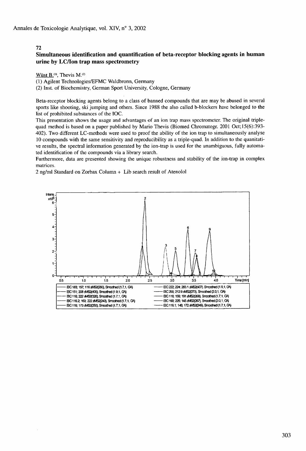
Annales de Toxicologie Analytique, vol. XIV, n° 3, 2002
72 Simultaneous identification and quantification of beta-receptor blocking agents in human urine by LC/Ion trap mass spectrometry
Wüst B. ( 1 >. Thevis M.<2> (1) Agilent Technologies/EFMC Waldbronn, Germany (2) Inst, of Biochemistry, German Sport University, Cologne, Germany
Beta-receptor blocking agents belong to a class of banned compounds that are may be abused in several sports like shooting, ski jumping and others. Since 1988 the also called b-blockers have belonged to the list of prohibited substances of the IOC. This presentation shows the usage and advantages of an ion trap mass spectrometer. The original triple-quad method is based on a paper published by Mario Thevis (Biomed Chromatogr. 2001 Oct;15(6):393-402). Two different LC-methods were used to proof the ability of the ion trap to simultaneously analyse 10 compounds with the same sensitivity and reproducibility as a triple-quad. In addition to the quanitati-ve results, the spectral information generated by the ion-trap is used for the unambiguous, fully automated identification of the compounds via a library search. Furthermore, data are presented showing the unique robustness and stability of the ion-trap in complex matrices. 2 ng/ml Standard on Zorbax Column + Lib search result of Atenolol
IrtensJ
[rrin]
BC183 157; 116 iMS2(260), 3roolted (1.7,1, GK¡ BC222; 224; 283.1 iM32(4C7l SiDOthed (1.9,1. G*) BC151; 208 JMS3405), Shotted (19,1, Gft) BC2S5; 2129 WGSCTi), aradted (10.1. Grç BC116; 222 *4S2P26^ SnxothsJ (1.7,1, BC116; 159,191 iMS2(26S), Smxthed (1.7,1, m BCl1fi2; 163; 222AB2Ç240), Sraatted(1.7,1, GO} BC190; 225; 145iNB2(267),Smoothed(20,1.QS} BC116; 173iMS2C25Û3i Smoothed (1.7.1, GA} BC 11&1; 146; 172 *M52P4S), Smoothed (1.7,1, GA)

Annales de Toxicologie Analytique, voL XIV, n° 3, 2002
73 Enantioselective analysis of amphetamines in saliva with capillary electrophoresis
Zimmermann J.R., Duchstein H.J. Universitat Hamburg, Institut für Pharmazie, Bundesstr. 45, 20146 Hamburg, Germany
The analysis of drugs of abuse (amphetamines) in saliva gained some interest in recent years. Saliva is used in the fields of workplace and roadside testing. Positive amphetamine findings can be caused by drugs of abuse and some medications. Therefore it might be necessary to distinguish between the legal consumption of medications and the illegal intake of drugs of abuse. This is possible by identification of typical metabolites of medications or analysing the enantiomeric distribution of amphetamines. Capillary electrophoresis (CE) is a promising, convenient and economic method to perform enantioselecti ve analysis. A method has been developed to separate and quantitate the following racemic amphetamine derivatives: amphetamine, methamphetamine, methylendioxyamphetamine (MDA) and methylendioxymethampheta-mine (MDMA) in saliva. L-ephedrin or deprenyl were used as internal standards. A HP 3D CE with ultraviolet- diode array detection (DAD) equipped with a uncoated fused-silica capillary (80,5 cm, 50 um I.D.) was used. A 75 mM KH2PO4 solution at pH 2,5 containing 18 mg/ml of native p-cyclodextrine was used as migration buffer. The samples were injected electrokinetically after performing a SPE procedure. The separation voltage was kept at 25 kV, the temperature was fixed at 15° C. All enantiomeres could be separated within 50 min with a sensitivity of appr. 10 ng/ml for each enantiomer. Calibration curves were linear in the range of 10-400 ng/ml for each component. This method was applied for the metabolic analysis of famprofazone and deprenyl in saliva after intake by healthy volunteers. For both drugs positive urine and blood amphetamine findings were reported earlier. In the saliva samples amphetamine and methamphetamine could be found. For famprofazone a mixture of amphetamine and methamphetamine enantiomers, for deprenyl only L-amphetamine and L-methamphe-tamine were detected.
304

Annales de Toxicologie Analytique, vol. XIV, n° 3, 2002
74 Lethal intoxications in the Institute of Forensic Medicine in Greifswald - an analysis over the last fifty years
Below E.. Lignitz E. Institute of Forensic Medicine of the Ernst-Moritz-Arndt University , KuhstraBe 3017489 Greifswald, Germany
Apparently, lethal intoxications as cause of death are still rarely found in unnatural deaths investigated in institutes of forensic medicine. In the Institute of Forensic Medicine at the University of Greifswald, 10-15 % of post-mortem autopsies displayed an intoxication during the last several decades with a possible decreasing tendency. 13,819 autopsies were carried out in our institute - situated in a low-populated rural area - during the last fifty years with the confirmed death cause intoxication in 1,589 times. In this study, especially the intoxication causes and the substance classes of the poisonous agents have been investigated. In addition, we analyzed the frequency of intoxications as well as sex and age of the deceased. Surprisingly, CO-intoxications were found most frequently with an incidence of 50 %, followed by alcohol intoxications with 21 %. The latter was not unexpected taking into account the habits of the local population. Medical drugs and narcotics take only the third place, although the abuse of modern narcotic drugs is already visible even in the far east of Germany. The spectrum of substances which are abused, taken accidentally or deliberately is continuously changing, reflecting scientific progress in the pharmaceutical industry as well as fashion tendencies. Therapeutic use is almost always followed by abuse. Our results confirm prior experiences concentrating mostly on other poisons like heavy metals or herbicides etc. In addition, we could demonstrate the influence of political conditions on use and distribution of illegal drugs in Germany. Our study clearly demonstrates that insufficient equipment or analytical methods are no longer the reason for any problems uncovering lethal intoxications. They are rather due to insufficient investigations of the corpses (without considering the possibility of an intoxication as differential diagnosis) and to frequent mistakes of the prosecutor's office in deaths without signs of physical violence. These facts may explain the above mentioned decreasing tendency of intoxications, but they also clarify that this tendency probably does not correspond to reality.

Annales de Toxicologie Analytique, vol. XIV, n° 3, 2002
75 Increased postmortem concentrations of K+ in the vitreous humour in heroin overdose deaths
Bortolotti F.<'>, Gottardo R.U), Trettene M.(", Cittadini F, i K 2 ), Tagliaro F.">, Marigo M. ( i )
(1) Department of Public Medicine, Unit of Forensic Medicine, University of Verona, Verona, Italy (2) Institute of Forensic Medicine, Catholic University of the Sacred Heart, Rome, Italy
The rise of K+ concentration in the extracellular fluids after death has long since been proposed to infer the time since death. This increase is caused by the release of K+ from the intracellular compartments, according to the intra-extracellular concentration gradient, due to the cessation of the active transport mechanisms (Na7K+ pump). Vitreous humour, because of its easy accessibility and protection from external environment, is the extracellular fluid currently used for this purpose Although many authors have described a highly significant correlation between vitreous K+ concentration and postmortem interval (PMI), a great inter-subject variability was reported, which affects the interpretation of the results and inference of PMI in real cases. Recently, a neat improvement in reproducibility has been obtained by using microsampling of vitreous humour coupled to capillary ion analysis (CIA)(1). This method can be summarised as follows. Vitreous humour samples (50 pi) are collected using plastic syringes and diluted 1:20 with water containing the internal standard (Ba~). The electrophoretic separations are carried out in uncoated fused-silica capillaries (75 pm ID, 50 cm effective length) using a pH 4.5 buffer composed of 5 mmol/1 imidazole, 5 mmol/1 18-crown-6 ether and 6 mmol/1 HIBA. Detection is by indirect UV absorption at 214 nm wavelength. Samples from 35 medico-legal autopsies or external examinations of cases of acute thraumatic deaths, in which the time of death was exactly known (ranging 7-144 hours), were studied with the described method, showing an excellent correlation between vitreous K+ and PMI (see Fig.l). The same method has been applied to 15 vitreous humour samples from subjects deceased from heroin overdoses. In this group the correlation between vitreous K+ and PMI was described by a different equation (see Fig,2), where the intercept on the Y axis is much higher than in the former group, whereas the slope is substantially the same. This phenomenon can be attributed to an antemortem increase of extracellular potassium caused by respiratory acidosis related to the heroin induced respiratory depression. In conclusion, the present study highlights terminal acidosis as a potential source of error in inferring PMI on the basis of vitreous potassium determination.
Fig. 1 K+ vs PMI in acute thraumatic deaths Fig.2 K+ vs PMI in heroin overdoses
u a ta m n too 120 wc iao 6 & 4 0 « so ico *2a «0
PMI (hours) PMI (hours)
Reference: 1 - Tagliaro F. et al., J Chromatogr B 1999 ; 733 : 273-279
306

Annales de Toxicologie Analytique, vol, XTV, n° 3, 2002
76 Findings in a fatality involving the neuromuscular blocking agent vecuronium
Cirimele V.<». Kintz P.(1>, Pépin G.(2>, Ludes B.(l> (1) Institut de Médecine Légale, 11 rue Humann, F-67085 Strasbourg, France (2) Laboratoire Toxlab, 7 rue Jacques Cartier, F-75018 Paris, France
A fatality due to intravenous self-administration of the neuromuscular bloking agent vecuronium (Norcuron®) is presented. A 34-year-old female nurse in a department of surgery was found dead at the hospital. Blood and urine specimens collected during the autopsy constituted the 6 t h quality control (domain: forensic toxicology) of the French Society of Analytical Toxicology (S.F.T.A.). An original screening procedure for seven quaternary nitrogen muscle relaxants (d-tubocurarine, alcuronium, vecuronium, pancuronium, atracurium, mivacurium and rocuronium) was developed using HPLC-ESI-MS after ion-pair extraction with methylene chloride at pH 5.4. Vecuronium and its deacetylated metabolite were identified and quantified in biological fluids. Vecuronium concentrations were 1.2 and 0.6 mg/L in blood and urine, respectively. 3-OH-vecuronium concentrations were 4.4 and 0,7 mg vecuronium equivalent/L in blood and urine, respectively. The procedure was entirely validated and appears sensitive enough as a screening test for curares in forensic investigations (S/N > 5 at 0.1 mg/L in full scan mode).
77 A comprehensive study on the determination of cyanide in forensic blood samples by head-space gas chromatography with electron capture detector
Pao K.L.. Lee C.W. Government Laboratory of the Hong Kong SAR, China
When cyanide is analyzed in a postmortem blood sample by the Aldridge colorimetric method for forensic toxicological examination, the condition of the blood sample will affect the accuracy and reproducibility of the determination especially if the blood is putrefied. This is due to the artificial formation of cyanide from plasma thiocyanate and also the instability of cyanide in blood. Determination of cyanide in postmortem blood sample by headspace gas chromatography equipped with an electron-capture detector was developed. This method involves liberating cyanide into a headspace as hydrogen cyanide and converting the latter into cyanogen chloride by chloramine T. Thiocyanate interference is completely suppressed by ascorbic acid. Release of cyanide from blood is facilitated by silver sulfate. A comprehensive study on the optimization of the various experimental conditions for the method will be discussed. The detection limit was 0.03 pg/mL and linearity was obtained from 0.1-100 pg/mL with coefficient of correlation being 0.99. Good recovery of cyanide was obtained for blood matrices which varied from normal to putrefied blood and blood containing cavity fluid. This method is particularly useful for analyzing putrefied blood samples in cyanide poisoning cases.
Annales de Toxicologie Analytique, vol. XIV, n° 3, 2002
78 Forensic intoxications by new antidepressants : report of 22 cases
Deveaux M.. Ferroul D., Leman C , Tournel G., Hédouin V., Gosset D. Institut of Legal Medicine, Faculty of Medicine, University of Lille 2, France
A new generation of antidepressants has been available for about ten years. Despite they appear to be relatively safe in overdosage, they can be associated with some fatal intoxications, especially with the concomitant use of others psychoactive substances. We reviewed 22 cases of forensic intoxications by selective serotonin reuptake inhibitors (SSRIs) (citalopram, fluoxetine, fluvoxamine, paroxetine, sertraline) and noradrenaline serotonin reuptake inhibitors (NaSRIs) (milnacipran, venlafaxine). An analytical method is described, involving extraction of drugs and active metabolites by chloroform/2-propanol/n-heptane at pH 9.5 and analysis by HPLC-DAD with a variable flow-rate of the mobile phase. Doxepine was used as internal standard. Validation of this method was established and a good linearity was found from 20 to 1000 ng/ml for each compound. Toxicologic and circumstances of deaths were examined in 22 forensic deaths during 4 years (1998-2001). None of the cases was attributed to a SSRI or a NaSRI intoxication only. Deaths involving psychoactive drugs were generally multiple drugs intoxications (n = 15). Ethyl alcohol was found in 11 cases. Only one death was due to a combination of citalopram and fluoxetine. Other cases (n = 6) were non toxic deaths. Concentrations determined to have resulted in death are in accordance with those recently published in the literature [1,2]. The higher concentrations we found were : citalopram 246 ng/ml, fluvoxamine 618 ng/ml, fluoxetine 380 ng/ml, paroxetine 1010 ng/ml, milnacipran 275 ng/ml, venlafaxine 6700 ng/ml. We did not observe any acute intoxication involving sertraline. In this retrospective study, fatalities with SSRIs or NaSRIs are exclusively due to the simultaneous presence of other psychoactive drugs and/or alcohol. The increasing use of these new antidepressants apparently did not coincide with the number of deaths caused by these drugs. Moreover, it seems difficult to determine precise lethal concentrations for each SSRI or NaSRI, even if the drug is taken alone.
references : 1- Goeringer K.E. et al. J Forensic Sci 2000 ; 45 (3) : 633-648 2- Frey R. et al. Eur Neuropsychopharmacol 2000 ; 10 : 133-142
79 Effect of putrefaction on the antidepressant amitriptyline (Tryptizole®)
Efkaradawy M.H.(i>. Eldin M.F.(", Elmahdi M.L<» (1) Medicolegal Department, Ministry of Justice, Egypt (2) Faculty of Medicine, Al Azhar University, Egypt
Amitriptyline(AMI) is a tricyclic antidepressant drug which is widely used for treatment of patients of different ages, that are suffering from depression. This study deals with the effect of putrefaction on AMI level in soft tissue of different body organs. An experimental model in rat was developed to this study. Animals were given a lethal dose of AMI according to body weight, they were divided into 14 groups (each 10 rats). The level of the drug was measured immediately after death in group 1 (Gl), the other rats from group 2 to group 14 (G2 -G14) were hurried in sand and every week we took one group from them and measure the level of AMI. In Gl, G2 and G3, AMI is measured in each organ separately (brain, heart, stomach, liver, kidney). In G4 to G14 the soft tissue organs were used as one mass (dough) due to the effect of putrefaction. The results showed that there was a highly significant reduction of AMI level in G2 in all organs as compared to Gl, and it was significantly reduced in G4 to G13 as compared to Gl. In G14 the drug couldn't be detected in soft tissue dough. It is concluded that the level of AMI in soft tissue is progressively reduced after death with the progression of putrefaction.
308

Annales de Toxicologie Analytique, vol. XIV, n° 3, 2002
80 Further evidence for the presence of GHB in post mortem biological fluid: implications for the interpretation of findings
Elliott S.P. Regional Laboratory for Toxicology, City Hospital NHS Trust, Dudley Road, Birmingham B18 7QH, United Kingdom
Analysis and interpretation of the drug of abuse gamma hydroxy butyric acid (GHB) in fatalities has become very problematic. This is primarily due to the endogenous nature of the compound, in addition to varying methods of detection, resulting in variable data in post mortem biological fluid. As in ante mortem investigations it is necessary to differentiate between endogenous production and exogenous ingestion. In order to assist this interpretation, GHB concentrations were measured routinely in both blood and urine specimens in 37 fatalities received during a 3 month period. In all cases GHB was not implicated in the cause of death, there was no apparent correlation between manner of death and resultant GHB concentrations. Mean concentrations of GHB determined in post mortem blood were found to be 12,1 mg/L (n=36) and 12.4 mg/L (n=16) (unpreserved and sodium fluoride preserved samples, respectively) and 5.0 mg/L in unpreserved urine (n=36) and 4.5 mg/L in sodium fluoride preserved (n=14) urine samples. Concentrations ranged from 2-29 mg/L and 4-25 mg/L in unpreserved and preserved blood, respectively and 0-12 mg/L and 0-10 mg/L in unpreserved and preserved urine, respectively. Vitreous humor was available in 2 of the cases analysed (GHB = 1 mg/L and 3 mg/L) and in each case concentrations were far lower than that detected in the post mortem blood and urine. The data support the potential use of sodium fluoride preserved samples for interpretation of GHB concentrations, particularly if there has been an extended post mortem interval. In addition, interpretative cut-offs can be proposed for both post mortem blood and urine based on the GC-MS method used. At blood concentrations less than 30 mg/L and at urine concentrations less than 20 mg/L it is possible that any GHB detected could represent only endogenous GHB production. The data further support that it is not possible to infer GHB ingestion if it is detected in both post mortem blood and urine, as previously believed.

Annales de Toxicologie Analytique, vol. XIV, n° 3, 2002
81 Formic acid in tissue as indicator parameter in methanol intoxication : a proposition of index
Ferrari L. A. ( l ). Giannuzzi L.<2>, Nardo C. A.1 0, Arado M. G.(», Nieto R. R.(l> (1) Laboratorio de Toxicología y Química Legal - Dirección Gral. Asesorías Periciales-Suprema Corte de Justicia Pcia. Bs. As, Argentina (2) Cátedra Toxicología y Química Legal- Facultad de Ciencias Exactas, Universidad Nacional de La Plata, Argentina
Fifteen cases of fatal massive methanol intoxication have been studied considering formic acid as main metabolite and survival time with and without ethanol therapeutical treatment. So we classified methanol -poisoning cases in three groups according to survival time (few hours, up to three days and up to ten days). The correlation between formic acid in different tissue (blood, brain, kidney, lung, liver) and survival time of victims blood were studied. Blood and tissue formic acid were performed by head space-GC with FID detector; previous transformation in methyl formate according to Abolin technique slightly modified by the authors. The formic acid was in the range 0.03-1.10 g/ L in the organs belonging to three group studied. Formic acid showed good relationship for blood and brain, and poor between blood and remainder tissue. The best correlation between organs was found for lung vs kidney for each group defined (r2 = 0.91, 0.84 and 0.87 respectively). These data suggested that the used of blood and brain can improve the interpretation of formic acid concentration. Also we defined equation Poisoning Index, PI = (formic acid concentration in blood /0.5).100 (0,5 g/L is the concentration informed by Mahieu in several methanol poisoning) as a parameter in order to define the lethality degree due to formic acid in cases of methanol poisoning. PI showed good relationship with the sum of formic acid in different tissue victims appertaining to three group defined (r2 = 0.91), PI allowed us discriminate the victims whom received therapeutical treatment and survived in different time. PI > 100 indicated state of severe intoxication and short time survival and PI<100 the victims survived more than three days up to ten days. Respecting to victims whom received not therapeutical treatment and dead immediately: 100>PI>40. These results show the importance to perform formic acid in different organs taking account the survival time of victims in order to predict the methanol intoxication in postmortem cases.
82 Morphine and 6-MAM in blood : possible risk factors for sudden death in 192 heroin users
Fugelstad A., Ahlner J., Brandt L., Ceder G,, Eksborg S, Rajs J., Beck O. Departments of Clinical Pharmacology, Clinical Neurosciences, and Karolinska Pharmacy, Karolinska Hospital, Department of Forensic Medicine, Karolinska Institute, Stockholm, Department of Forensic Chemistry, University Hospital, Linkoping, Sweden
The aim of this investigation was to detect risk factors for sudden death from heroin injection. Out of all fatal cases of suspected heroin death in a metropolitan area (Stockholm) only cases with morphine and 6-MAM present in blood were included. Blood concentrations of morphine ranged from 50 to 1200 ng/g, and 6-MAM from 1 to 80 ng/g. Codeine was detected in 96 % of the subjects. In the majority of cases poly-drug use was indicated by the forensic investigation. The most common findings were alcohol and benzodiazepines. However, in one quarter of the cases other combinations of abused drugs were found. Abstinence from heroin and use of alcohol were two risk factors revealed by uni- and multivariate analysis of the correlation between these factors and morphine or 6-MAM blood concentrations. For 6-MAM there was also a correlation of blood levels with the presence of THC and benzodiazepines. Despite a high frequency of heart abnormalities no correlation of these conditions with morphine or 6-MAM blood concentrations could be observed.
310

Annales de Toxicologie Analytique, vol. XIV, n° 3, 2002
83
84 An unusual cause of death: suffocation due to leaves of common ivy (Hederá helix). Detection of hederacoside C, a-hederin and hederagenin by LC-EI/MS-MS
Gaillard Y.(1>. Blaise P.<2>, Darré A.( ,), Barbier T.,2>, Pépin G. f , )
(1) Laboratoire d'Expertises TOXLAB, 7 rue Jacques Cartier, 75018 Paris, France. (2) Thermo Finnigan France, 12 avenue des Tropiques, ZA de Courtaboeuf, BP 141, 91944 Les Ulis cedex, France.
We report one fatal case of asphyxia due to leaves of common ivy. Macroscopic examination of the corpse during the autopsy disclosed an incredible quantity of leaves of Hederá helix in the mouth and throat of the decedent. In order to rule out the possibility of poisoning by the toxic saponins contained in the plant we have developed an efficient LC-EI/MS-MS assay of hederacoside C, a-hederin and hederagenin in biological fluids and plant material. Sample clean up involved solid phase extraction of the toxins on Og cartridges followed by LC analysis under reversed phase conditions in the gradient elution mode. Solute identification was performed using full scan MS-MS spectrum of the analytes. Oleandrine was used as internal standard. Under these conditions, saponins in powdered dried leaves of Hederá helix were measured at a concentration of 21.83 mg/g for hederacoside C, 0.41 mg/g for a-hederin and 0.02 mg/g for hederagenin. No toxin was detected in cardiac blood, femoral blood and urine of the deceased, but hederacoside C was quantified at 857 ng/ml in the gastric content. These findings led us to conclude that the man committed suicide and that the death was due to suffocation by leaves of common ivy.
Post mortem detection of taxol (paclitaxel) by L£_EI/rVI£>_]VlS! in a case of suicide due to massive ingestion of yew's needles (Taxus baccata)
Gaillard Y.<'>. Blaise P.(2>, Barbier TP, Pépin G. ( l )
(1) Laboratoire d'Expertises TOXLAB, 7 rue Jacques Cartier, 75018 Paris, France (2) Thermo Finnigan France, 12 avenue des Tropiques, ZA de Courtaboeuf, BP 141, 91944 Les Ulis cedex, France
We are reporting on one fatal case of suicide due to the massive ingestion of needles of yew. This paper describes for the first time the postmortem quantitation of taxol in various specimens in a fatality due to ingestion of Taxus baccata. On scene near the corpse, the police investigators noticed the presence of branches of yew. A small letter found nearby left clear indication of a suicide. At the autopsy, the coroner discovered a great quantity of intact yew's needles in the stomach. LC-EI/MS-MS assay of taxol (paclitaxel) in biological fluids and plant material was developed to support the death. Sample clean-up involved liquid-liquid extraction followed by LC analysis under reversed phase conditions in the gradient elution mode. Solute identification was performed using full scan MS-MS spectra of the analyte. Oleandrine was used as internal standard. Under these conditions, taxol in powdered dried material of Taxus baccata was measured at a concentration of 9.6 pg/g for the needles, 7.6 pg/g for the stern and 21.3 pg/g for the seed (and not detected in the flesh of the aril). Concentrations in the biological specimens were: 0.9 ng/ml in cardiac blood, 19.3 ng/ml in bile, 1.5 ng/g in liver, 0.5 ng/ml in vitreous humor, 172.6 ng/ml in gastric juice and not detected in peripheral blood and kidney.

Annales de Toxicologie Analytique, vo l XIV, n° 3, 2002
85
86 Performance of immunoassays in screening for opiates, cannabinoids and amphetamines in whole blood
Hiño Y., Ojanpera L, Rasanen I., Vuori E. Department of Forensic Medicine, P.O. Box 40, FIN-00014 University of Helsinki, Finland
Several immunoassay methods for screening of abused drugs in whole blood were evaluated in postmortem forensic toxicology. Blood samples known to be positive or negative for opiates, cannabinoids and amphetamines by gas chromatography-mass spectrometry were analyzed by EMIT II Plus and EMIT d.a.u., Syva RapidTest and Triage 8 after acetone precipitation. In these experiments, EMIT immunoassay method was modified using Dade Behring VIVA analyzer to detect the substances more sensitively, and using the modified method, low concentrations of abused drugs were detected in blood samples. The sensitivity of the modified EMIT method was 86-100 %, whereas the values were below 86 % with the other methods. The specificity of all immunoassay methods for opiates and cannabinoids was above 80 % but less for amphetamines. A problem with sample rejection arose in a few cases with the EMIT amphetamines assay. The modification of the EMIT immunoassay system presented here seems to be useful for screening of drugs of abuse in postmortem blood samples especially when urine is not available.
312
Toxicity of flecainide
Gerostamoulos I . Lynch M., Drummer O.H. Victorian Institute of Forensic Medicine, Department of Forensic Medicine, Monash University, 57-83 Kavanagh St., Southbank, Victoria 3006, Australia
Flecainide (Tambocor®) is a Class 1-antiarrythmic drug prescribed primarily for the treatment of ventricular dysrhythmias. Flecainide also has local anaesthetic properties that depress myocardial contractility. In Australia flecainide is usually available in 100 mg tablets. Following oral administration, 30 % of a flecainide dose is excreted in urine unchanged, while 50 % is metabolised by the liver to conjugated metabolites. Several minor metabolites are also found in urine (< 3 % of the dose) and only 5 % is excreted in faeces, There has been only one report in Australia describing a near fatal flecainide intoxication in a woman who had taken 8 g of flecainide (1). To the best of our knowledge there have been no reports in Australia of death due to flecainide toxicity. In this report, we describe in detail one fatality in which flecainide toxicity was considered to be the primary cause of death and discuss the possible contribution of flecainide in other cases. The concentration of flecainide in postmortem specimens is discussed in relation to other drugs as well as some of the difficulties associated with the interpretation of postmortem drug levels.
references 1- Brimacombe J. et al. Med. J. AusL, 1991 ; 155 (5) : 349
Annales de Toxicologie Analytique, vol. XIV, n° 3, 2002
87 An autopsy case of mixed-drug intoxication involving anti-arrhythmic drugs and cardiac glycoside
Kinoshita H. (1 ). Taniguchi T.( l ), Nishiguchi M. ( ,\ Ouchi H. ( l ), Minami T.(1), Utsumi T.i2), Motomura H.<31, Tsuda T. ( , ), Ohta T.<1}, Aoki S.(1), Komeda M. ( i ), Kubota A. (4 ), Hishida S.(1> (1) Department of Legal Medicine, (2) Department of Opthalmology, (3) Forensic Science Laboratory, Hyogo Prefectural Police Headquarters, Japan (4) Department of Surgical Pathology, Hyogo College of Medicine, Japan
We describe here a case of mixed-drug intoxication involving anti-arrhythmic drugs and cardiac glycoside. A 39 year-old male was found in dead in his bed. A lot of empty packets of drugs were found from his pouch at his bedside. He had been undergoing psychiatric treatment for over 4 years, and occasionally, had hinted at committing suicide. A Japanese well nourished male, 170 cm in height and 71 kg in weight. No injury was founded. The heart weighed 400 g and contained 600 ml of blood without coagulum, The brain weighed 1540 g, and was slightly edematous. The left and right lungs were weighed 460 g and 565 g in weight, respectively, and were slightly congested. There was approximately 400 g of stomach contents containing rice and vegetables along with a white granule. The myocardium showed mild fibrosis. The liver showed mild fatty degeneration. There were no notable changes other than congestion in other organs. We detected digoxin (14.6 ng/ml), verapamil hydrochloride (8.4 pg/ml) and metoprolol tartrate (3.8 ug/ml) from the postmortem blood by radioimmunoassay and high performance liquid chromatography. We concluded that the cause of his death was an overdose of digoxin and verapamil hydrochloride, based on the results of the toxicological examination. We should do a detailed investigation of the subject's past history and check the drugs prescribed when a drug overdose is suspected.
88 Amphetamine and derivatives related deaths in the aspect of forensic toxicology
Klys M., Brandys J., Bystrowska B., Bujak -Gizycka B. Institute of Forensic Medicine CM UJ , 16 Grzegórzecka, 31-531 Kraków, Poland
Amphetamine and derivatives are the crucial theme in the problems of forensic toxicology. The paper is based on the study of 20 cases of deaths in which amphetamine and/or derivatives were involved as the cause of death. Firstly, autopsies were carried out in the Institute of Forensic Medicine in Kraków and body fluids and tissues were collected. Secondly, toxicological examinations of the autopsy specimens were performed with the use of HPLC/MS method in chemical positive ionization mode (APCI) after liquid - liquid extraction. The toxicological findings obtained in particular cases indicate that the majority of cases under consideration were complex. Among all 20 cases only two, probably suicidal deaths, were a result of amphetamine abuse. Some of them which were violent deaths (murder, gun-shot, hanging, drowning) of people under the influence of drugs. The majority of cases which were probably fatal accidents caused by the interaction of various mixtures of xenobiotics which included also opiates, cocaine, benzodiazepines besides amphetamine and derivatives. The concentrations of xenobiotics detected in these cases fit a relatively large range. One case is worth mentioning. This is a death of a permanent amphetamine user who survived two weeks with hematoma in brain in which amphetamine was detected postmortem. The raport also discuss amphetamine and derivatives metabolic problems and contains useful data for medicolegal purposes.

Annales de Toxicologie Analytique, vol. XIV, n° 3, 2002
89
90 Antidepressants poisoning causing death. A case report
Nováková E.. Bfiek M. Institute of Forensic Medicine and Toxicology, 1st. Medical School and Hospital, Charles University, Na boji_ti 3, Prague 2, Czech Republic
The corpse of a man was found in a car. The engine was going and the heating was opened. There were no signs of violence. Blood, urine, stomach content and tissues ( liver, kidney, spleen) were the specimens delivered for analysis after autopsy. Both carboxylhaemoglobin and alcohol in blood were negative. Immunochemical screening for drugs of abuse in urine was positive for benzodiazepines. TLC was used for screening and identification of other drugs in stomach content, urine and tissues after liquid -liquid extraction. Fluoxetine, dothiepin, bromazepam, flunitrazepam and/or their metabolites were found depending on the analyzed specimen. There was also quinine and metabolites in urine. Neither flunitrazepam nor aminoflunitrazepam were detected in tissues. Dothiepin blood level of 1.79 mg/ml and nordothiepin of 0.24 mg/ml were determined by HPLC. Concentration of dothiepin and nordothiepin was determined in tissues. The isolation from 5g of discrete tissues was performed by incubation with trypsine followed by solid phase extraction on Chem Elut columns. Recovery of the isolation was 35-45 % for fluoxetine and 50-60 % for dothiepin depending on concentration. HPTLC with densitometric detection on Camag TLC Scanner II coupled with integrator was used for quantitative analysis. Calibration curve from spiked negative liver was prepared in the range 50-400 mg/g for fluoxetine and 10-40 mg/g for dothiepin. Blank samples were analyzed too. There were following concentrations found in the tissues : liver- fluoxetine 97 pg/g, dothiepin 7 pg/g, kidney- fluoxetine 114 pg/g, dothiepin 10 pg/g, spleen- fluoxetine 105 pg/g, dothiepin 29 pg/g. Lethal concentrations found in the literature survey varied between 12.8- 54 mg/kg for fluoxetine and 2-14 mg/kg for dothiepin. Concentrations of dothiepin found in blood and tissues corresponded with lethal concentrations, concentration of fluoxetine was nearly twice as high as those published. As there were no significant finding during the autopsy the death was attributed to brain and lung oedema after ingestion of big amounts of antidepressants together with benzodiazepines in connection with breathing of warm air in a confined room inside the car.
314
Acetonitrile related death
Lo D.S.T.. Yao Y.J., Leong HT., Koh H.H., Chew ES. Centre for Forensic Science, Health Sciences Authority, 11 Outram Road, 169078 Singapore
A 32 years old male was arrested and a can, believed to contain thinner, was seized from him. He subsequently died while in police custody. Postmortem examinations were performed and samples of femoral blood, buccal swab, liver, nostril swab, stomach contents, bile and urine were submitted for toxicological analysis. The blood yielded an acetonitrile level of 21 mg per 100 ml of blood and 2 pg of toluene per ml of blood. A cyanide level of 12.4 pg per ml of blood was also detected. The urine was found to contain 24 mg of acetonitrile per 100 ml of urine and 11 mg of acetonitrile was detected in 100 g of liver. Headspace gas chromatography using 5 % Carbowax 20M on 100/120 Carbopack B column was used for the initial screening of alcohols and volatiles on the blood and urine samples. Subsequent confirmation and quantitation of acetonitrile was carried out on the same instrument but using RTX 1301 (crossbond 6 % cyanophenyl - 94 % dimethyl polysiloxane) column. The quantitation of cyanide was carried out on a Hewlett Packard 5890 series II GC/NPD equipped with a HP 19395A Headspace Sampler and a 25m x 0.53 mm i.d, fused silica Porapak Q PLOT column from Supelco. This is the first reported case of death in Singapore contributed by a combination of acetonitrile and cyanide poisoning.

Annales de Toxicologie Analytique, vol. XIV, n° 3, 2002
91 A method for the simultaneous determination of clobazam and desmethylclobazam in postmortem blood by HPLC/MS/MS
Oxley A.M., Lee T.D., Lemos N.P.. Holt D.W. Forensic Toxicology, Analytical Unit, St George's Hospital Medical School, University of London, London SW17 ORE, England, United Kingdom
Clobazam is a new-generation benzodiazepine increasingly used in the treatment of epilepsy. It differs from other benzodiazepines in that its structure contains nitrogen in position 1 and 5 of the heterocyclic ring, instead of positions 1 and 4. Literature reports indicate that this contributes to its apparently greater safety margin than most other drugs in the benzodiazepine family. Our laboratory undertook the task of determining the concentrations of clobazam and any other drugs present in the post-mortem blood of a 34-year old female with a history of epileptic seizures and prescriptions for various anti-epileptic drugs. Phosphate buffer (pH 7.0) and MTBE were added to the blood specimen together with prazepam as internal standard. The mixture was mechanically agitated for 5 minutes and, following centrifugation, the organic layer was evaporated in a SpeedVac®. The resulting residue was reconstituted in 80 % methanol and injected onto a Supercoil LC-18-DB ODS analytical column (15 cm x 4.6 mm i.d., 5 um particle size) fitted with w Newguard C-18 precolumn (Sigma-Aldrich), which was maintained at 50° C with a Perkin-Elmer series 200 column oven (PE Biosystems). Detection was by tandem mass spectrometry (HPLC/MS/MS) using a Sciex API 2000 triple quadrupole mass spectrometer (PE Biosystems). The limits of detection and quantitation as well as the linearity of the assay were determined and using a freshly prepared set of calibrators in drug-free blood, the specimen was found to contain 0.11 mg/L clobazam and 2.15 mg/L desmethylclobazam. In addition, lamotrigine (5.3 mg/L) and lisinopril (<0,05 mg/L) were found present in the specimen. This is a simple, quick and reliable method for the detection and quantitation of clobazam and desmethylclobazam in post mortem blood by HPLC/MS/MS which can easily be adapted for other benzodiazepines with similar properties to clobazam.
92 Rare fatal poisoning case by ethylene glycol
Raikos N„ Tsoukali H, Psaroulis D., Zaggelidou H. Laboratory of Forensic Medicine & Toxicology, Faculty of Medicine, Aristotle University, 540 06 Thessaloniki, Greece
Ethylene glycol is the main constituent of antifreeze and coolant mixtures. Intoxication upon drinking it occurs quite infrequently. In the present work, autopsy findings and the results of toxicological analysis of a fatal poisoning case by ethylene glycol are presented. Conditions of specimens preparation, identification with gas liquid chromatography and the distribution of the substance in post mortem specimens are reported. The following concentrations of ethylene glycol in blood, liver and kidney respectively, were measured : 170 mg/100 ml, 260 mg/100 g and 200 mg/100 g. Finally, the therapeutic measures which have to be taken in time to protect organs from severe damage and to prevent death are reported

Annales de Toxicologie Analytique, vol. XIV, n° 3, 2002
93
316
Analysis of a fatal pholedrine intoxication using LC/MS/MS
Rômhild W.(1>. Barrels H.C1>, Ghanem A.(2>, Schôning R.<», Wittig WlK Krause D.<"> (1) Institute of Forensic Medicine, (2) Division of Cardiology, University Hospital, Leipziger Str. 44, 39120 Magdeburg, Germany
Pholedrine (4'-hydroxymethamphetamine) is a cardiovascular agent (sympathomimetic) exerting an anti-hypotensive and adrenergic effect. High doses may cause a drop in the peripheral circulation and increase the blood pressure, heart rate and body temperature up to a state of central respiratory paralysis. A 15-year-old girl suffering from a somnolent condition and requiring mandatory resuscitation was admitted to the intensive care unit. The ambulance doctor reported a heavy state of agitation and hallucinations before the comatose condition occurred. Clinical findings: heart rate 140 bpm, increase to 170 bpm, body temperature 43.8° C. Resuscitation failed and was ceased after 2.18 h. Following the GC/MS method, the toxicological emergency analysis revealed a considerable amount of pholedrine. However, this method failed in quantitation. A method for determining pholedrine in human body fluids utilising high-performance liquid chromato-graphy/tandem-mass spectrometry (LC/MS/MS) with a turbo ion source in the positive mode is presented. Dl 1 -methamphetamin was used as the internal standard. Samples (blood and urine) were prepared by SPE extraction. SPEC-C18AR/MP3 yielded a maximum extraction result (67 %) compared to the liquid/liquid extraction, Extrelut NT3 or other SPE methods. Chromatographic separation was achieved on an RP-18 stationary phase applying gradient elution with A/B from 50/50 rising to 30/70 (A = 5 mM ammonium acetate, 0.02 % acetic acid, 5 % acetonitrile, B = methanol/acetonitrile 3/1, 0.02 % acetic acid). Post column 0.2 pl/min of pure acetic acid were added to optimize ionisation. Detection was carried out in the multiple reaction mode (MRM) m/z 166 - 107 for pholedrine and m/z 161 - 97 for the internal standard Dll-methamphetamin. The chromatograms did not indicate any interference by other substances. The calibration curve was linear (regression coefficient r = 0.999) in the range of 1 to 100 ng/ml. Samples of higher concentrations were diluted to suite the working range. The limit of detection (LOD) of pholedrine was 0.8 ng/ml with an S/N ratio of 3. The limit of quantitation (LOQ) was 3 ng/ml. Pholedrine concentrations in the blood, the urine and the liver of the deceased were determined (heart blood : 23.0 pg/ml, RSD 5 %; urine : 1120 pg/ml, RSD 8 %; liver : 27.3 pg/g, RSD 6.6 %).
Annales de Toxicologie Analytique, vol. XIV, n° 3, 2002
94 Fatal poisoning with moclobemide, metoprolol and bromazepam
Samkova H., Brzobohata A.,.Spackova M, Pivnicka J., Hirt M. Institute of Forensic Medicine of St.Anne's University Hospital Brno and Faculty of Medicine of Masaryk University Brno, Czech Republic
A case of a suicide - fatal poisoning with drugs - of a 54-year-old man is presented. The man was found by his wife at home - he was lying on the ftoor and he showed no signs of life. Packings of pharmaceutical preparations Aurorix®, Betaloc® SR 200, Lexaurin® and Mono Mack® depot (containing active ingredients - moclobemid, metoprolol, bromazepam and isosorbide) were spread around the body. The man left a suicide note saying he did not want to live any longer. His wife mentioned that recently her husband had been drinking too much alcohol. The man was transported by ambulance to the intensive care unit of the internaI medicine department of the hospital. Already at his admission to hospital, clinical death with respiratory and cardiac arrest was stated, and samples of blood and urine (8ml) were taken for toxicological analysis . Cardiopulmonary resuscitation was started immediateIy but it was unsuccessful and after 40 minutes the doctors certified that death had occurred. Autopsy was performed in the Institute of Forensic Medicine in Brno. The autopsy findings were nonspecific, and histological examination was performed. Samples of urine, blood, kidney and liver were taken for toxicological analysis. The toxicological analysis was performed using FPIA, TLC, HPLCIDAD, GCIMS and GCINPD methods. Detection of moc1obemide, metoprolol, phenobarbital and bromazepam in urine, gastric contents, liver and kidneys was performed by TLC. These findings were confirmed by the GCIMS method and caffeine was detected in urine and in the liver. Quantification of bromazepam in blood by the GCIECD method determined the concentration of 0,58 mglL. The blood concentration of phenobarbital determined by the GCINPD method was 8,05 mglL. Quantification of metoprolol and moclobemide in biological materials was performed by the HPLCIDAD method (Waters Spherisorb S5 OD/CN column, 150 x 4,6 mm; gradient elution with the acetonitrile-water (70/30) mobile phase buffered with TEAP buffer). The results were as follows :
1 Biolooical material Moclobemide Metoprolol Blood 1439 mg/L 264 mg/L Liver 6,3 mo/kg 2,7 mg/kg
Kidney 18,7 mg/kg 7,3 mg/kg
Further, the blood ethanol concentration of 0,54 g/kg was determined by the HS/GCIFID method. Ethanol was detected also in the brain and lung tissues. This case is presented as one of the first cases of fatal poisoning with the drug Aurorix® (moclobemide) in the Czech Republic.
317

Annales de Toxicologie Analytique, vol. XIV, n° 3, 2002
95 A double suicide by propofol, thiamylal sodium, suxamethonium chloride and pancuronium bromide injections
Shinozuka T.<". Terada MP, Nakajima R<», Takei R. ( , ), Ohue 0. ( , ), Watanabe S.!", Yamamoto K. , l\ Murai T. ( , )
(1) Department of Legal Medicine, School of Medicine, Keio University, 35 Shinanomachi, Shinjuku-ku, Tokyo 160-8582, Japan (2) Department of Legal Medicine, Course of Social Medicine, Osaka University Graduate School of Medicine, Suita-Shi, Osaka 565-0871, Japan
A 61-year-old man (Case-1) and a 20-year-old woman (Case-2) were found dead on a bed in the room of his home. A hypodermic needle attached to a drip was inserted into the dorsum of the left hand, and wads of cotton wool were inserted into the nasal cavity. Empty ampules of Diprivan® (propofol), Isozol® (thia-rnylal sodium), Succin® (suxamethonium chloride) and Mioblock® (pancuronium bromide) in a plastic bag were found near the bodies. Autopsy finding revealed no significant pathological abnormalities expect injection marks on the their hand. We considered the possibility of the drugs intoxication by drip infusion in both cases. Since propofol, thiamylal sodium, suxamethonium chloride and pancuronium bromide were detected by drug screening, the concentrations of these drugs in postmortem whole bloods were determined using high-performance liquid chromatography, gas chromatography-mass spectrometry and liquid chromatography-mass spectrometry, respectively. In the case-1, the whole blood concentration of propofol, thiamylal and suxamethonium were 0.35 pg/ml, 20.0 ng/ml and 2.93 pg/ml, respectively. In the case-2, the whole blood concentration of propofol, thiamylal, suxamethonium and pancuronium were 0.50 pg/ml, 17.0 ng/ml, 3.20 pg/ml and 0.80 pg/ml, respectively. These blood levels exceeded therapeutic levels, showed toxic or fatal levels
96 Fatal intoxication with propamocarb
Stanková M.. Kurka LP. Dvorácek Institute of Forensic Medicine, Faculty Hospital, Ostrava, Czech Republic
Authors describe a case of fatal intoxication with carbamate fungicide of propamocarb. Intoxications with pesticides or other chemical substances used in agriculture are in the industrial region of Ostravian agglomeration rather rare. We have experienced only six cases of fatal intoxication since 1985: paraquat -1 case, metathion - 2 cases, arborol - 1 case, A1P - 1 case, propamocarb - 1 case. Carbamates (dérivâtes of carba-minic acid) are used as insecticides, herbicides and fungicides. It is a very heterogenous group of substances which can be devided into 4 sub-groups with various levels of toxicity - supertoxic, extremely toxic, highly toxic, average toxic and mild toxic. The case A man at the age of 32 was taken to hospital to a surgical ward with the diagnosis of gastritis acuta or possible pancreatitis caused after a dietary mistake. His health state got rapidly worse. This patient's relatives brought the information about the possible consumption of some toxic solution - about 36 hours after this possible consumption. Unfortunately the patient died in a very short time after his transporting to intensive care unit. For toxicological investigation we used: gastric contents, urine and blood samples taken during his hospitalization, and liver, kindey and vitreous humour samples taken at the autopsy.
318

Annales de Toxicologie Analytique, vol. XIV, n° 3, 2002
97 An autopsy case on the detection of phénobarbital, cocaine, morphine and 6-monoacetyl-morphine
Terada M. ( i>. Watanabe R.<]>, Shinozuka T.(2\ Masui S.1 0, Inoue H.(1), lino M.°\ Terao T. ( ,\ Murai T.<2>, Tanaka E.<3i, Honda K.(3), Matoba R."> (1) Department of Legal Medicine, Course of Social Medicine, Osaka University Graduate School of Medicine F3, Suita-City, Osaka 565-0871, Japan (2) Department of Legal Medicine, School of Medicine, Keio University, Shinjuku-Ku, Tokyo 160-8582, Japan (3) Department of Legal Medicine, Institute of Community Medicine, University of Tsukuba, Tsukuba-City, Ibaragi-Ken 305-8575, Japan
A 30-year-old women (daughter) and a 54-year-old women (her mother) were found that they fell down in the car which an engine idled in that time. They were transferred to the hospital, but the daughter was already died in arrival and her mother died after nine days of treatment. Three white powder wrapped in white paper from a brassiere of this woman, and white powder in small bottle and two tablets from a bag of her mother were found, respectively. The autopsy findings not showed abnormalities except moderate congestion and edema of the various organs. We considered the possibility of some intoxication. The postmortem (daughter) gastric contents and whole blood were used for toxicological analysis. Barbiturates, cocaine and opiates were detected in gastric content homogenate by drug of abuse screening (Triage), Phénobarbital and cocaine, morphine and 6-monoacetylmorphine were identified in the gastric content samples and whole blood samples by GC-MS and GC-MS/MS, respectively. These drugs were quantitated in the whole blood samples by GC-NPD or GC-MS/MS. The concentration of phénobarbital, cocaine, morphine and 6-monoacetylmorphine in whole blood were 20.2 ug/ml, 1.1 pg/ml, 486.7 ng/ml and 10.7 ng/ml, respectively. The blood levels of phénobarbital, cocaine and morphine showed therapeutic, fatal and toxic levels, respectively. Other toxicological analysis data in the blood were as follows : ethanol 0.13 g/1; Hb-CO 0%; heroin not detectable
98 Twelve death cases of body packer syndrome in Tehran (April 1999 - December 2000)
Abolmasoumi Z.. Mahfoozi A., Afshar M., Hassanian H. Legal Medicine Organization, 124 Western Sanaii Alley,Ashtiani Sq.,Khaghani Cross-road, Tehran, Iran
During the last decade, increase of the drug traffic and those customs, have led the smuggler to attempt various methods, one of this methods of ilicit drug smuggling is body packing. Smuggling by intra-abdominal concealment, is called "body packing". In this research, mortality due to body packing is investigated. This study is performed as a descriptional evaluation (case series) on all corpses were refered to Legal Medicine Organization of Tehran, between April 1999 and December 2000. Clinical trials consist of several parameters such as : sex, age, marriage, addiction, job, education level, type of opioids and their weight and number of packets and results of T.L.C (in blood & urine) ; surgery is discussed. Continental system is used in Iran and 0.06 % of refered corpses to Legal Medicine Organization of Tehran were body packer. There were only 12 cases. All of them were men. Average age of body packer were 43 years old (Max=62, Min=20). The minimum weight of the packet's content was below 20 grams and maximum weight was 1400 grams (mid=501 grams). The minimum number of packets were one packet and maximum number of packets were 48. 25 % were putrified and one of corpse was mummified because of lying in special climate (Qhom's salt-marsh area). None of them have academic education and they haven't any governmental job (eg.employee and army). Nine of them lived in cities. 25 % of them were IV addicts. The corpse finding site was terminals (17 %), roads (58 %) and cities (25 %), Estimating of the methods for transfering, packet contents, methods of wrapping, cause of death and results of diagnostic and therapy methods can effective character in decrease of number of body packer and law execution. We hope with this research we can help medical science and law by introducing the best diagnostic and treatment methods.

Annales de Toxicologie Analytique, vol. XIV, n° 3, 2002
99 Fluoxetme-HCl induced intrauterine foetal growth retardation and skeletal maj
Ali M.Q.. Sharf-El Deen U.A., Rady M. L, El Menshawy O.M., Bakry S.A. Zoology Department, Faculty of Sciences, Al-Azhar University, Nasr city, Cj
Fluoxetine is an antidepressant drug which is widely used in m a m ^ P ^ ^ ^ ^ ^ ^ y p t as Prozac®. It is a fluorinated methyle phenoxy derivative of phenylpropylarnine^P^^^M The present work was designed to evaluate the effects^^l^^^^pi^i th different doses on the mice embryos at various developmental stages. E i g h t y j | ^ ^ B ^ ^ p ^ e r e administrated oral doses (0.052, 0.104 and 0.208 mg/mouse/day) of fluoxetineJ|^f^^^Ppancy up to both 15th and 19* days of gestation. The effects of fluoxetine were o b s e o ^ ^ ^ ^ ^ ^ r e n t pictures of malformations. The pregnant mice treated with fluo^É^^jp^ved with different pictures of instability, nervousness, twitching of head, agitation, h ^ | ^ B \ ^ ^ P ^ i d marked reduction in food intake as well as reduction in the body weights. The r e j g ^ ^ t ^ ^ l ^ f f e c t s of the fluoxetine were revealed in uteri of pregnant mice treated groups on boJj^^^^^k^^ays of gestation . The toxic effects of this drug showed remarkable reduction in th^^^^^^^P^hology, length and number of implantation sites as well as reduction in the number Q ^ ^ ^ ^ ^ ^ ^ H ^ S . Increase in the number of dead and resorbed mouse embryos was dose depen-d e n t ^ ^ ^ ^ ^ ^ H K t work recorded a reduction in crown rump of most treated mouse embryos. The trea-
J^^^^^^P^erved with several malformations as diminution in size, exenocephalia and various pictures ^^^^Pmrnalformations.
100 New Cannabis-benzodiazepines association form of drugs of abuse
Ben Reguiga M.. Massias L,, Certain A., Seraissol P., Farinotti R. Toxicology and Pharmacokinetics Department - Bichat-Claude Bernard Hospital (AP-HP) 46, rue Henri Huchard, 75018 Paris, France
European parallel market of drugs of abuse presents several kinds of cannabis derivatives, varying from herbal presentations to resins and other smoking forms. Mostly, all these forms contains exclusively can-nabinoi'd derivatives, such as delta-9-tetrahydrocannabinol (A9-THC), cannabinol and cannabidiol. Although, recently, our laboratory received an exceptional homogenous vegetable sample, seeming to enclose some parts of Cannabis sativa plant (seeds and stems), and said to have sedative and anti-tinnitus properties, when chewed by its consumers. The first screening step was a rapid analysis by FPIA (AXSYM, ^BBCHT), done on a methanolic extract, and which showed the presence of high amounts of cannabinoids. Further investigations, were realised by gas chromatography (HP 6890 series)- mass spectroscopy detector (MSD 5973). A 20 mg of dried and pulverized sample was extracted successively with 1 ml of methanol, ethyl acetate and dichloromethane. The three fractions were mixed and the total extract was evaporated, then derivatized with BSTFA-TMCS 1 %. 2 pi of this sample were injected into the GC. The analysis confirmed the presence of cannabinoids, and specially TMS-derived A9-Tetrahydrocannabinol (A9-THC-TMS) and cannabinol. Furthermore, we detected the presence of another main component, a benzodiazepin derivative, clonazepam. The presence of clonazepam, a chemical synthesized compound, in the vegetable sample, proves that the cannabis fragments were blended with the benzodiazepin compound. This presence may also explain the pharmacological effects required by the drug consumers.
320
Annaies de Toxicologie Analytique, vol. XIV, n° 3, 2002
101 Study of the enantiomeric ratio of methadone and EDDP in hair, urine and serum by capillary electrophoresis
Berens G., Yegles ML Wennig R. Laboratoire National de Santé, Toxicologie, CRP-Santé, Centre Universitaire, Luxembourg
As for methadone (MTD) the (R)-enantiomer is pharmacologically more active than the (S)-enantiomer, several studies have already been published describing methods for a stereoselective determination of MTD. To our knowledge, only one study has described a chiral separation by capillary electrophoresis (CE) of MTD in only one hair specimen after consumption of (R)-MTD. The aim of our study was to develop a method using chiral CE to determine the (R)/(S) ratio of MTD and its main metabolite 2-ethylidene-l,5-dimethyl-3,3-diphenylpyrrolidine (EDDP) in hair, serum and urine. Specimens were provided from subjects undergoing racemic (R, S)-MTD maintenance programme and from deceased persons where MTD had been detected in autopsy material. For urine a liquid-liquid extraction at pH 9.5 was performed, whereas for serum and hair a solid phase extraction was done using Cleanscreen columns. Capillary zone electrophoresis was operated at pH 2.5 in presence of heptakis-(2,6-di-0-methyl)-beta-cyclodextrin (DIMEB). Diphenhydramine was used as internal standard. In 7 hair specimens, the (R)/(S) enantiomeric ratio of MTD was shown to be 1.08 (range 0.82-1.65), whereas no EDDP could be detected using the described CE method. Furthermore, in 27 urine specimens the mean (R)/(S) ratio of MTD was 1.58 (range 1.15-3.07). Finally, in 9 serum specimens the mean (R)/(S) ratio of MTD and EDDP respectively was 1.13 (range 0.82-1.77) and 1.26 (range 1.10-1.64). As already shown by a few other authors in urine only the (R)-MTD was predominant, whereas in serum this was not the case. The hair data suggest a predominance of the (R)-MTD. This study demonstrates that capillary electrophoresis can be used for stereoselective separation of methadone in hair. Moreover, CE columns represent a low-cost alternative to chiral HPLC columns.

Annales de Toxicologie Analytique, vol. XIV, n° 3, 2002
102 Prevalence of drug intoxications in patients presenting at hospital emergency departments
Capolaghi B.{,K Desch G., Cano Y., De St Hermine C , Dosba L, Feuillu A„ Gaillard C , Gruson A., Hervochon F., Lawson E., Pellae I., Szymanowicz A,, Thuillier F., Tournoy M.H., Turnet MM. (1) Laboratoire de biochimie, CHR, Thionville, and Collège National de Biochimie, France
Only limited data is available today concerning levels of intoxication of patients arriving at Emergency Departments of French Hospitals. With its broad national coverage, the CNBH (Collège National de Biochimie des Hôpitaux) was able to rapidly perform a 601 patient multicenter trial, evenly spread over the country. All medical centers involved in the trial used the same urinary screening immunoassay, the single use Triage®8 Drugs of Abuse Panel (Biosite, San Diego, USA) which simultaneously screens for all major legal and illegal classes of psychoactive substances (opiates, cannabis, amphetamines, cocaine, barbiturics, benzodiazepines, tricyclic antidepressants and methadone). Inclusion criteria : patients arriving at the ED with at least one of the following conditions: road accident victims (drivers only), behavioral or perception impairment, voluntary intoxication and alcohol abuse. Data collection :15 participating sites spread throughout the country, 601 patient files completed by Dec 1, 2001. Statistical Analysis: 595 patient data files were deemed acceptable. Triage®8 Method: Competitive immunoassay (presence of a band indicates positive sample). Comparison methods : EMIT, FPIA, Immunochromatography (Dade RapidTest, Dako, Cortez), Remedi. Demographic data : Average age : 37 yrs ; male : 57 % (329/580), female : 43 % (251/580), Drugs abusers : 14 % (61/432). Inclusion criteria distribution : Road accident : 15 %, behavioral or perception impairment : 29 %, voluntary intoxication : 32 %, Alcohol abuse : 24 %. Screening results : negative patients : 44 % (263/595), positive patients 56 % (332/595), only one drug class : 72 % (239/332), polyintoxication : 28 % (93/332). For the population of declared drug abusers : 16 % neg (10/64) and 84,3 % pos (54/64) with 91 % of polyintoxication. Positive results distribution : BZO : 51 %, THC: 20 %, OPI : 12 %, MTD : 6 %, TCA : 5 %, Barb : 3 %, Coc : 2 %, Amp/Metamp : 2 %.
Triage® 8/EM1T Triage® 8/FFP1A Triage®8/others MTD 97% 99% 100 BZO 87% 94% 83% COC 100% 100% 98% AMP/metAMP 97% 97% 100% THC 97% 95% 97% OPI 99% 100% 100% BARB 100% 100% 95% TCA 100% 100% 100%
This study, based on 595 patient data files, provides a snapshot image of the existing situation in France and provides new data on the prevalence of intoxication in patients arriving at Hospital Emergency Departments. A positive result for at least one psychoactive drug was found in 56 % of the cases. In 72 % of these, only one substances was implicated, while 28 % had polyintoxications. The drug classes that more most frequently discovered are : Benzodiazepines (51 %), Cannabinoids (20 %), Opiates (12 %). For declared drug abusers polyintoxication was found in 91 % of cases. Cointoxication of BZO and TCA was found in 4 % of cases, this association was observed more frequently for patients admitted for voluntary intoxication (1.1 %).
322

Annales de Toxicologie Analytique, vol. XIV, n° 3, 2002
103 GC-MS/MS analysis of buprenorphine at picograms levels in biological samples
Chiarotti M.. Marsili R. Institute of Legal Medicine, Catholic University of Sacred Heart, L.go F. Vito 1, 00168 Rome, Italy
Recently there is an increasing interest over the analysis of buprenorphine in biological samples, because this compound is widely used during the rehabilitation programs carried out on heroin addicts. Hence, chemical monitoring of buprenorphine levels in biological fluid is often required in order to adjust useful anti-craving doses for this drug. These kind of analysis are generally carried out by immunochemical methods that give mainly qualitative results. Moreover the low buprenorphine amounts that can be found in biological samples, it is necessary the use of very sensitive technique to achieve accurate quantitative detection as well as to perform successfully confirmatory analyses in forensic field. Because capillary gas chromatography coupled to ms-ms detection systems GC-MS" (such as ion trap) can results in superlative technique able to offer unsurpassed sensitivity and specificity, we tested the use of the GC-MS" technique in this kind of analysis with the aim to develop a suitable confirmatory procedure for buprenorphine in biological matrix, also when this compound is present in trace levels. The proposed method is based on the GC/MS2 analysis of buprenorphine after sylanization using deuterated buprenorphineD4 as deuterated internal standard to reach the necessary accuracy in quantitative results. The drug is extracted from biological fluids by liquid / liquid solvent extraction at controlled pH as generally carried out for other opiates (morphine and related compounds) after enzymatic hydrolysis. BSTFA derivatization was chosen to ensure suitable gas chromatographic by-products and easy analytical procedure carried out in mild derivatizing condition. Gas chromatographic settings on Trace GC 2000 apparatus- Thermoquest: a capillary column HP5 trace analysis (12 m X 0.2 mm i.d. 0,33 micron film thickness of 5 % phenyl methyl silicone) was used in temperature programme an initial isotherm a 200° C for 0.2 min, then linear temperature increments (15 c
C/min) to 290° C. Final isotherm of 10 min. The injector (split less for 1.0 min) port was settled at 270° C. After the injection the injector port was maintained in split flow mode (15ml/min). Carrier gas (helium) at 1 ml/min constant flow. Mass spectrometric settings on Polaris Q apparatus-Thermoquest: MS2 detection of buprenorphine and D4 buprenorphine (i.s.) was performed in electronic impact (70 e.V.) isolating the ions (with a window +/- 1) 450 and 454 for i.s. (base peaks) and monitoring the product ions. Main parameters related to the mass spectrometer can be summarized as follows: source temperature: 220° C; scan event 1 (ms fragmentation SIM): 450, 454. Scan event 2 (ms2 fragmentation) scan mode MS 2; isolation time 8 millisec; collision energy 1.00; collision time 15 millisec; max ion time 25 millisec. Mass range for product ions (buprenorphine): 120-450 m/z. According to our analytical procedure, buprenorphine amount as low as 50 pg can be easily detected and quantified with a signal to noise ratio of 60 (calculated on the 295 ion), ensuring suitable specificity due to the match with the characteristic fragmentation pattern (MS2 - EI) of the base peak of buprenorphineTMS at correct retention time (verifiable by the use of deuterated internal standard).

Annales de Toxicologie Analytique, vol. XIV, n° 3, 2002
104 Determination of the designer drugs MDMA, MDA, MDEA and MBDB in whole blood, urine and saliva using a HPLC system with native fluorescence detection
Concheiro M., Cruz A., Punín E., Quíntela O., Bermejo A.M., López-Rivadulla M. Toxicology Service, Institute of Legal Medicine, University of Santiago de Compostela, Spain
We report a method for the simultaneous determination of 4 designer drugs, MDA, MDMA, MDEA and MDBD, in whole blood, urine and saliva. The method involves a liquid-liquid extraction procedure followed by HPLC-fiuorescence detection. The sample (0.5-1 mL) is extracted with Toxi-Tubes A and 20 uL of the reconstitued residue are injected in the HPLC system, which is composed by a fluorescence detector (Waters 474) and a Kromasil 100 C8 5 urn 25 x 0.46 column (Teknokroma). The methylenedioxy-amphetamine derivatives detection is possible due to the native fluorescence of these compounds (derivatization is not necessary), using a excitation wavelength of 285 nm and a emission wavelength of 320 nm. The mobile phase, KH2P04 0.03M and acetoni-
, trile (750/250), at a flow rate of 1 mL/rnin allows the separation of the four components in only 10 minutes. The procedure is rapid, simple and adequate for the identification and quantitation (range 5 - 1000 ng/mL) of the designer drugs studied.
105 Trace impurities of seized methamphetamine hydrochloride in the Philippines
Dayrit F.M. ( , ), Dumlao M.C.<2>
(1) Chemistry Department, Ateneo de Manila University, Loyola Heights, Quezon City, Philippines (2) Instrumentation Section, Chemistry Division, Philippine National Police Crime Laboratory, Camp Crame, Quezon City, Philippines
Methylamphetamine hydrochloride is very rampant in the street as abused drugs here in the Philippines. Identification of trace impurities present in those samples and their amounts and establish profile based on their impurities would be an immense contribution to the advancement on the intelligence gathering and operational work of Philippine National Police and other law enforcement authorities. The five hundred (500) kilograms seized methamphetamine hydrochloride from Quezon Province was used for this study. A weight of 0.15 g of homogenized methamphetamine sample is dissolved in 5 ml of pH 10.5 buffer solution and extracted with 1 ml of ethyl acetate. Diphenylamine was used as internal standard. The organic layer is transferred to a small auto sampler vial. The sample should be analyzed on the day of extraction. A gas chromatographic system equipped with a GC-MS (HP 6890 MSD 5972, equipped with HP 7673 Injector) was used. The spectra show a lot of peaks. Each peak was then compared with the MS Library. Indeed some of these impurities present in the sample were identified.
324

Annales de Toxicologie Analytique, vol. XIV, n° 3, 2002
106 Paramethoxy amphetamine: the South Australian experience
Felgate P.P.. Sims D.N., Kirkbride K.P., Felgate H.E., James R.A., Vozzo P.C., Kostakis C. Forensic Science SA, 21 Divett Place, Adelaide, South Australia 5000, Australia
Paramethoxyamphetamine (PMA) is a ring substituted phenethylamine derivative which has been used illicitly in Australia since late 1994. Initially PMA had been sold under the «ecstasy» name normally associated with methylenedioxy- methylamphetamine (MDMA) but is now knowingly sought out as a party drug; colloquially known as «death». Our studies have indicated that PMA has been synthesized using the Leuckardt reaction and the manufacture of PMA is by design rather than accident. It would appear that South Australia is the source of PMA in Australia. The first Australian example of illicit preparations containing PMA was found in South Australia, and throughout the period 1995-2001 this state has recorded many more seizures of PMA than the rest of Australia. Although the appearance of tablets and capsules has varied over the years since 1995, the level of drug present has generally remained in the range between 50 and 70mg. Routine analysis of illicit seizures since 1995 has never revealed the presence of other amphetamine-type stimulants such as MDMA or paramethoxy methylamphetamine (PMMA) in PMA preparations, unlike North America and Europe where mixtures with PMMA have been reported. During the period September 1995 to October 2001 eleven PMA related deaths have occurred in South Australia. In these cases femoral blood PMA levels ranged from 0.24 to 23 mg/L, mean 3.9, median 1.7. Significantly all the PMA deaths were thought to involve oral administration. Other members of the amphetamine drug type were found in nine of the eleven cases. Other States of Australia have recorded five PMA related deaths. Blood MA levels in three non-fatal cases and three cases where death was due to other causes have also been measured. The number of PMA related deaths in South Australia are double those due to other amphetamines while at the same time seizures of PMA doses have remained at less than 5 % of the total number of amphetamine type drug seizures. This highlights the apparent greater toxicity of PMA. PMA is now available in many parts of the world and would appear to be more toxic than MDMA. When used orally, hallucinogenic blood PMA levels range up to about 0.4 mg/l. Recent reports from North America and Europe have strengthened the view that levels above 0.5 mg/L appear likely to be associated with toxic effects and may be lethal especially in combination with other amphetamines.

Annales de Toxicologie Analytique, vol. XIV, n° 3, 2002
107 Identification of 11 opiates in urine with high performance liquid chromatography
Havard L„ Dupeyron J.R, Vautier S., Sandouk P., Chast F. Laboratoire de Toxicologie and Urgences Médico-Judiciaires, Hôtel-Dieu, 1 place du Parvis Notre-Dame, Paris, France
In forensic médecine, presence of opiates in urine has to be established with a reference method. Furthermore, opiate compounds are often very close and difficult to differentiate though immunoassays (as morphine, codeine and monoacetylmorphine). From a toxicokinetic point of view, some substances with short elimination half life, cannot be found more than 24 h after absorption (morphine). Screening and confirmation have to be performed in less than 96 h, which represents the maximal duration of police custody. We use high performance liquid chromatography (HPLC) coupled to diode array detection drived by Millenium software (Waters®) for identification of illicit opiates in individual urine. Urine samples (30 mL) came from "Forensic Emergency Department". They are screened with EMIT® d.a.u.® reagents with a Cobas® Mira + autoanalyser. Urines positive for "opiates" are studied with HPLC: hydrolysis with P-glucuronidase at 40° C during 12 hours at pH=5.0. Extraction on Isolute HCX® column with fixation step at pH=4.0 and therefore elution at pH=8.0, with ammonium acetate buffer. Evaporation to dryness and dissolution in 0,1 mL of mobile phase and injection of 50 uL Chromatographic device (Waters®) include a 4 lines HPLC pump, an automatic sampler, a Symmetry Cs column and detection monitoring at 220 nm. Elution is performed by gradient during 20 min. Taking into account retention time and absorption, all the products are separated and identified as shown in the next table. The detection limit is estimated to 100 ng/mL. Time of analysis in our conditions is 48 h.
RT (min) A max Pholcodine 4,9 210 Normorphine 5,2 210 Morphine 6,2 210 Norcodeine 10,7 212 Codeine 11,0 212 6-MAM 11,4 205 Codethyline 11,9 212 Hydrocodone 12,8 210 Norbuprenorphine 13,9 212 Buprenorphine 16,2 212 Methadone 17,0 195
This method allows separation with reasonable resolution of 11 opiates in a 20 minutes runtime; applied to 750 urine samples we confirm about 90% of them for opiates compounds.
326

Annales de Toxicologie Analytique, vol. XIV, n° 3, 2002
108 Toxicoepidemiology among opiates users during police detention
Havard L.<'>. Dupeyron J.P.(I), Fleury F. t !\ Batista R.(1), Gamier M.<2), Chast F.(1>
(1) Laboratoire de Toxicologie, (2) Urgences Médico-Judiciaires, Hôtel-Dieu, 1 place du Parvis Notre-Dame, Paris, France
The aim of this study was to establish the prevalence of isolate or associate opiate use in individuals admitted in the clinical forensic department (Urgences Médico-Judiciaires). In order to check illicit opiates use, urinary samples are screened using immunochemical method (EMIT ® d.a.u.®). In case of positive immunochemical assay for opiates, a second analytical step includes identification with high performance liquid chromatography (HPLC) and diode array detection (DAD). Morphine is the main substance identified (64.4 % of urine samples), alone (26 % of morphine positive samples) or associated with codeine and norcodeine (67 %), with normorphine (25.4 %), and with buprenorphine and norbuprenorphine (9.4 %), Codeine was present in 432/770 samples (56 %). Codeine is the main substance in 72/432 positive samples (16.6 %); it is associated mainly to morphine (23.3 %), to morphine and norcodeine (16.4 %), to morphine and normorphine (11.1 %), to norcodeine, morphine, normorphine (10 %) and to 6 monoacetylmor-phine (6-MAM) (5.5 %). 6-MAM was present in 3.5 % of the samples (never alone), associated to morphine (96 %), codeine (92 %), normorphine (65 %), norcodeine (15 %), norbuprenorphine, buprenorphine, pholcodine (4 %), and to hydrocodone (8 %). The presence of 6-MAM in urine is related to heroin abuse. Morphine may be a metabolite of 6-MAM or of heroin. Combining HPLC with diode array detection is proved to be useful for identification of opiates in urine. The complete analytical procedure is compatible with confirmation results within the duration of police custody.
327

Annales de Toxicologie Analytique, vol. XIV, n° 3, 2002
109
328
Confirmation of amphetamine, methamphetamine, MDA, and MDMA in immunoassay positive urine samples using disk solid-phase extraction and GC-MS
Huang Z.P.(Î>, Zhang S.Y.<2> (1) Forensic Sciences Institute, Fujian Provincial Department of Public Security, Fuzhou 350003, P. R. China (2) Lab of Forensic Sciences, Fujian Public Security College, Fuzhou 350007, P. R. China
Abuse of amphetamines has dramatically increased in recent years in recreational places in People Republic of China. Amphetamines in urine were commonly screened by immunoassays. Positive samples were then confirmed by liquid-liquid extraction and gas chromatography-mass spectrometry. A method was developed in our laboratories at present for confirmation of amphetamine, methamphetamine, 3,4-methylenedioxylamphetamine (MDA), and 3,4-methylenedioxylmethamphetamine (MDMA) in immunoassay positive urine samples using mixed phase disc solid phase extraction (SPE) and gas chromatography-mass spectrometry (GC-MS). The method was proved to be very quick since faster flow-rate can be applied and less time needed for solvent evaporation. Moreover, it has cleaner eluant due to the fraction of acid and neutral impurities can be washed away prior to our target compounds elution. This method was very suitable for routine practice. The SPE disks used were SPEC.PLUS.C18AR/MP3 cartridges with 70 mg bed mass and 10 ml specimens reservoir, providing hydrophobic and cation exchange interactions. The top layer is C18AR, and the lower is MP3 (a mixture of a strong cation exchanger and a slightly polar moiety). The disc was conditioned with 0.5 mL of methanol and 0.5 mL of sodium phosphate buffer (pH6), successively. Before applied to the disc, urine sample was diluted with 8 mL of buffer. After washing with 0.5 mL distilled water, 1,0M acetic acid solution, and methanol, respectively, analytes were eluted in ImL ethyl acetate containing 2 % (v/v) ammonium hydroxide. After chromatographic standard (CS) was added in, the eluant was evaporated to 0.2 mL under nitrogen at ambient temperature. One uL of the eluant was injected into the GC. The total ion chromatogram was much cleaner than that of liquid-liquid extraction. Recoveries of these amphetamines were above 70 %. Linearity was obtained up to 2 pg/mL of amphetamines in urine samples. With amphetamine, methamphetamine, MDA, MDMA monitored at m/z 44, 58, 44 and 58, respectively, the limits of detect reached ng/mL level. The developed method was assessed in application for immunoassays positive authentic urine samples, and at least one of the compounds was detected.

annales de Toxicologie Analytique, vol. XIV, n° 3, 2002
110 Clinical-toxicological investigation of drug abusers in Hungary
Jeszenszky E.0*, Molnar A.0>, Hideg Z. a ) , Kemer A. (2 ), Varga T.(3)
(1) Headquarters of Csongrad County, Szeged, Hungary (2) National Institute of Forensic Toxicology, Budapest, Hungary (3) University of Szeged, Institute of Forensic Medicine, Szeged, Hungary
In Hungary from the early 1990's to date the number of drug abusers has increased about ten times. Soft and hard drugs have taken the place of substitutes and medicine. Current regulation punish for driving under the influence of alcohol or drugs. Authors decided to workout a drug recognition protocol, which can also be used in Hungary, to assess the reliability of immunological fast tests and to determine the conditions of influence of drugs based on medical and toxicological results. In Csongrad county - one of the most "infected" areas in Hungary - suspects in drug crimes have been investigated. Symptoms of 100 examined persons (84 male, 16 female) have been recorded in a German-type protocol. Urine samples have been screened for immunological tests: THC, OPI, AMPH, METAM-PH, COC, BENZO. The confirmation and the quantitative investigation have been made by FPIA, REMEDÍ HS, HPLC DAD, GC/MS methods. It can be stated by the examinations that sensitivity and specificity of clinical symptoms were very different. The results of toxicological investigations were negative in 64 cases from blood samples and 22 cases from urine samples. The most often detected drug was THC, followed by polytoxicomania and opiate consumption. The reliability of immunological fast tests showed literature date: sensitivity of AMPH was 60 %, THC was 92.9 %, specificity of THC was 86.5 %, and all other data were 100 %. Types and quality of drugs and the time of consumption were recorded bases on the reports of abusers. Time passed from intake and the concentration values of each drugs, considering the measured data in the time of sampling had no relation. The clinical symptoms were positive in 62.5 % of the cases, and they could be detected only in urine. Clinical tests and symptoms in the protocol used in Hungary are insufficient, they need to be completed. Specificity and sensitivity of fast tests have proved satisfactory and they support drug abuse. Due to the uncertain data reported by the abusers kinetic data are not possible to be calculated for drugs. The influence of drugs in about 2/3 of the cases is excluded by the presence of positive symptoms, positive urine results and negative serum results at the same time.
329
Annales de Toxicologie Analytique, vol. XIV, n° 3, 2002
111
330
Reducing false positives from environmental contamination and increasing drug detection in the PharmChek™ Sweat Patch
Kidwell D.A. (1). Long M.J.® (l)Chemistry Division, Naval Research Laboratory, Washington, DC 20375, USA (2)Department of Chemistry, American University, Washington, DC 20016, USA
This paper describes a continuation of research on false positives in the PharmChekTM sweat patch resulting from prior skin contamination with drugs of abuse (1, 2). The skin of volunteers was contaminated with a varying concentration of drugs between 100 ng and 5 ug in a 10 cm2 area. Despite normal hygienic washing, passage of time, and repeated cleansing of the skin with 70 % isopropanol swabs, patch results tested positive, similar to, and greater than results reported for drug users. The drug concentrations observed in the patch were roughly proportional to the amount of applied drugs. When the patch was applied 24 hours after contamination with 5 pg of each drug, 106 ng cocaine, 14 ng benzoylecgonine, 32 ng heroin, 51 ng amphetamine, 60 ng methamphetamine, 93 ng MDMA were observed, even though the skin was cleansed with a normal hygienic shower and repeated alcohol swabbing. We propose a model for drug binding to skin by ionic interactions (similar to that in hair), in which non-polar solvents (such as the widely-used isopropanol) should be incapable of disrupting the drug-skin interaction. Drugs bind to skin tightly. Skin decontamination, including swabbing the skin with various dilute acids, before applying the patch failed to reduce prior contamination significantly. Hand-cleaner (which contains both an abrasive and an organic solvent) removed the most drug contamination. Drug equilibration between the pad in the patch and the skin make dose-response correlations difficult. The amount of sweating also affects the amount of drugs transferred to the patch from the skin. A nonvolatile transfer fluid, such as glycerol added to the patch greatly increased (up to 8x) the transfer of drug from the skin into the patch. This paper will review prior patch contamination results and the current efforts to decontaminate skin and increase transfer of drugs to the absorptive component of the patch. Based on these results and previous research, criteria are proposed to detect prior contamination if the patch results are questioned.
References 1- Kidwell DA.et al. Forensic Sci Int 2001 ; 116 : 89-106. 2- Long M. et al. NRL Memorandum Report 6170-01-8597, December 19, 2001.

Annales de Toxicologie Analytique, vol. XIV, n° 3,2002
112 LC/APCI-MS analysis of opiates and their metabolites in rat urine after inhalation of opium
Kikura-Hanajiri R.(». Kaniwa N. ( , ), Ishibashi M. ( , ), Makino Y.(2>, Kojima S.<J)
(1) National Institute of Health Sciences, 1-18-1, Kamiyoga, Setagaya, Tokyo 158-8501, Japan, (2) Kanto-Shin'etsu Regional Bureau of Health and Welfare, 2-4-14, Nakameguro, Meguro, Tokyo 153-0061, Japan
To examine the urinary excretion of opiates and their metabolites following inhalation exposure of opium to rats, analytical procedures for simultaneous determination of compounds in opium, the vapor derived by the volatilization of opium and the urine of rats exposed to the opium vapor were developed using liquid chromatography/atmospheric pressure chemical ionization mass spectrometry (LC/APCI-MS). Six compounds were determined in the opium, namely morphine, codeine, thebaine, noscapine, papaverine and meconin. All 6 were extracted 2.5 % acetic acid solution and subjected to LC/APCI-MS analysis. The separation was performed on an ODS column in acetonitrile-50 mM ammonium formate buffer (pH 3.0) by a linear gradient program and quantitative analysis was carried out in the selected ion monitoring mode ([M+H]+). For the analysis of the volatilization of opium, the opium (1 g) was added to a glass pipe, which was then heated at 300° C for 20 min. Negative pressure (air flow rate; 200 ml/min) was used to draw the vapor through a series of glass wool and methanol traps. The total amount of each compound in the vapor was estimated by measurement of the compounds trapped in the glass wool and methanol. Wister rats (n=3) were exposed to the vapor derived from the volatilization system and the urinary amounts (0-72 hr) of the six opiates and metabolites including morphine-3-glucronide (M3G) and morphine-6-glucroni-de (M6G) were measured after solid-phase extraction. The calibration curves for those compounds in the rat urine were linear over the concentration range 10 — 500 ng/ml. The recoveries for each analyte in the rat urine sample spiked with standard solution were generally greater than 80 %, and the relative standard deviation for the analytical procedure was less than 8 % with the exception of meconin. After inhalation exposure of opium to rats, M3G (5.45-14.38 pg/ml), morphine (2.27-4.65 pg/ml), meconin (0.54-1.85 pg/ml), codeine (0.54-1.85 pg/ml), noscapine (0.34-0.40 pg/ml) and papaverine (0.01-0.04 pg/ml) were detected in the urine. However, only trace levels of thebaine were observed despite it being one of the major alkaloids found in the opium. On the other hand, a relatively large amount of meconin was detected in the vapor and the urine as compared with the opium. It is suggested that the presence of meconin in biological fluids could be indicative of opium ingestion by inhalation.

Annales de Toxicologie Analytique, vol. XIV, n° 3, 2002
113 Impurity profiling analysis of methamphetamine synthesized by three different methods
Kim E., Lee J., Han E., Kim S.">. Chung H., Yoo Y, National Institute of Scientific Investigation, 331-1 Shinwol-dong, Yangchon-ku, 158-707 Seoul, Korea (1) Dan Kook University, Chonan, Kyunggi-do, Korea
The knowledge of impurities in methamphetamine is important not only that the impurities could have additional harmful effects on the methamphetamine users but the impurities can provide useful intelligence to forensic scientist and law enforcement agency concerning illicit methamphetamine products. To investigate the pattern of impurity from illicit methamphetamines by various synthetic methods, methamphetamines were synthesized from ephedrine through three different methods - Nagai, Moscow and Emde. For the impurity profiling analysis, all steps of analysis were followed by the UNDCP standard impurity profiling method. About 30 mg of synthetized methamphetamine was dissolved in lmL of phosphate buffer and extracted with 200 uL of ethylacetate, which contains two different internal standards of dioctylsebacate and diphenylarnine. The extract was analyzed by GC using Ultra-2 capillary column (0.2mm x 25m x 0.33 pm). Chromatograms of the synthetized methamphetamines by three different methods were showed distinct patterns by their synthetic method by GC-FID. In Nagai and Moscow methods, ephedrine was identified, while in Emde Method chloroephedrine was identified as the main impurity, and l,2-dimethyl-3-phenyla-ziridine and N-methyl-l-phenylpropamine, methamphetamine dimer and N-benzyl methamphetamine were also identified as the impurities by GC/MS.
114 A survey of illicit drug use in Stockholm's methadone program
Korkmaz S. (1). Beck O."», Stenbacka M.<2J\ Davstad Leifman A.<2-3!, Romelsjo A.<2-34)
( 1.) Karolinska Institute, Department of Medicine, Division of Clinical Pharmacology, Stockholm, Sweden (2) Stockholm Addiction Centre, Stockholm, Sweden (3) Karolinska Institutet, Department for Public Health Sciences and Clinical Neuroscience, Stockholm, Sweden (4) Centre for Social Alcohol and Drug Research, Stockholm University, Sweden
Relapse to opiate abuse and other addictive drugs is a common problem in methadone treatment programs. Stockholm's Methadone Program applies an intense follow-up of relapses by urine analysis. Detection of such illicit drug use is the main cause of involuntary discharges from the program. Survey of illicit drug use over time and in patients involuntarily discharged from the program may provide valuable information for future planning of such programs. Patients (n=212) admitted to Stockholm's Methadone Program during 1995-2000 and their urine toxicology test results are examined. Screening analysis of opiates, central stimulants (including cocaine), cannabis and benzodiazepines were carried out by immunochemical techniques A total of 55000 samples analyzed, of which 11.5 % were positive for at least one drug. Over 90 % of patients had at least one urine sample tested positive. Samples tested positive for central stimulating agents were stable over time (approx. 30 %). The fraction of positive samples for cannabis showed a decrease during this period. Samples tested positive for opiates and benzodiazepines showed decreases during 1999-2000. Fraction of positive urine samples in patients remained in the program showed a clear decrease in a five-year follow up (from 84 % to 40 %).
332

Annales de Toxicologie Analytique, vol. XTV, n° 3, 2002
115 Quest for the ultimate amphetamine immunoassay screening; evaluation of five immunoassays at different cutoff levels
Langen M.C.J.. van Hoof F.W.J.M., Olijslager E.J.H., Rommers M.K., Egberts A.C.G. Dutch Laboratory for Drugs and Doping, Hospital Pharmacy Midden-Brabant, TweeSteden Hospital 5000 LA Tilburg, The Netherlands
The challenge in drug of abuse (DOA) screening is to find a balance between false positive and false negative screening results. Because of the nature of an amphetamine immunoassay, cross-reactivity of different amphetamines and ecstasy-analogues remains a problem. A high cutoff will result in a high specificity (less false positives) but a low sensitivity (more false negatives). Lowering cutoff improves sensitivity (less false negatives) but also gives a decrease in specificity (more false positives). To evaluate the performance of five amphetamine immunoassays with respect to sensitivity and specificity at different cutoff levels. The assays tested were : 1 EMIT II Plus monoclonal (Dade Behring), 2 EMIT dau monoclonal (Dade Behring), 3 EMIT dau class (Dade Behring), 4 Abuscreen Online HS amphetamine/MDMA (Roche), 5 Cedia amphetamine/ecstacy (Microgenics). All samples were processed according to the manufacturers protocol, except for assay I and 2 for which we developed a low cutoff protocol with double sample volume. Screening was performed on an ILab-600 clinical analyser (Instrumentation Laboratory). We selected 95 amphetamine urine samples from our routine DOA screening program which were above the limit of detection of the immunoassay. The selected urine samples were screened with all assays using three cutoff levels (300, 500 and 1000 ng/mL). All samples were analysed with GC/MS. In case GC/MS analysis showed an amphetamine above the limit of detection we considered the sample as positive. The prevalence of amphetamine and ecstacy-analogues was 22%.
Test Cutoff Sens% Spec% PPV% NPV% 1 300 90 77 53 97
4 86 92 75 96 500 76 95 80 93
86 93 78 96 1000 71 96 83 92
86 93 78 96
2 300 86 91 72 96 5 86 91 72 96
500 81 92 74 94 86 95 82 96
1000 76 93 76 93 81 100 100 95
3 300 81 95 81 95 500 76 95 80 93 1000 71 95 79 92
Which assay is best ? From this study no clear answer can be given to the question which assay is best. Other characteristics like cross-reactivity, handling procedures, cost and the goal of the analysis may reveal differences between test and may be relevant in making a choice.

Annales de Toxicologie Analytique, vol. XIV, n° 3, 2002
116
Results : ng/mL 1 2 3 4 5
A 300 305 846 276 500 178 MDMA <150 <150 <150 281 452
500 360 1418 465 >600 294 <150 221 <150 482 >2000
1000 527 >2000 1170 >600 568 171 455 <150 >600 >2000
2500 >1000 >2000 >2000 >600 1305 378 1359 221 >600 >2000
MA 300 258 319 348 >600 274 MDEA <150 <150 <150 <75 873
500 498 443 646 >600 555 <150 <150 <150 <75 >2000
1000 >1000 1125 1410 >600 1195 <150 <150 158 113 >2000
2500 >1000 >2000 >2000 >600 >2000 333 <150 251 301 >2000
MDA 300 324 808 <150 112 231 500 370 1481 <150 263 493 1000 457 >2000 <150 >600 1362 2500 827 >2000 193 >600 >2000
Amphetamines have different cross-reactivities with different immunoassays which may have important consequences for testing for amphetamines. Each laboratory should be aware of these differences when using group reaction immunoassay screening methods. Further improvement of the performance of amphetamine immunoassays is necessary.
334
Evaluation of the cross-reactivity of several amphetamines with different amphetamine immunoassays
Langen M.C.J.. van Hoof F.W.J.M., Egberts A.C.G. Dutch Laboratory for Drugs and Doping, Hospital Pharmacy Midden-Brabant, TweeSteden Hospital. PO Box 90107, 5000 LA Tilburg, The Netherlands
Immunoassay screening methods are group reaction methods. The possibility of detection of amphetamine or amphetamine-analogues (mis)use depends upon the cross-reactivity and concentration of the ingested amphetamine. Immunoassays may therefore differ in their capability of detecting amphetamine-analogues like MDMA, MDEA and MDA. To evaluate the cross-reactivity of amphetamine and amphetamine-analogues at different concentrations with five different immunoassays. The assays tested were : 1 EMIT II Plus monoclonal (Dade Behring), 2 EMIT dau monoclonal (Dade Behring), 3 EMIT dau class (Dade Behring), 4 Abuscreen Online HS amphetamine/MDMA (Roche), 5 Cedia amphetamine/ecstacy (Microgenics). All samples were processed according to the manufacturers protocol, except for assay 1 and 2 for which we developed a low cutoff protocol using double sample volume. Blanc urine samples were spiked with 300, 500, 1000 or 2500 ng/mL amphetamine (A), methamphetamine (MA), MDMA, MDEA or MDA.

Annales de Toxicologie Analytique, vol. XIV, n° 3, 2002
117 Investigation of cocaine in urine and pubic hair of pharmacodependence patients under ambulatory treatment
Lárez A. ( l ), Henríquez E.{2), Bol años A.1*', Valles A. 0 ), Carrasquel J. ( l ), Cheng B.m, Colina J.(1)
(1) Universidad Central de Venezuela (2) Ministerio de Justicia, Venezuela (3) Universidad de Carabobo, Venezuela
(under the whole responsability of the authors) The present study is based in the quali-quantitative investigation of cocaine in urine and pubis hair of 6 masculine patients under ambulatory treatment in the José Félix Rivas Foundation of the Aragua State, using for it, in the case of the urine sample, the Inmunoassay technique of Polarized Fluorescence, and in the sample of pubic hair, that of Ultraviolet Spectrophotometry (U.V.) and that of Gas Chromatography, integrated with the Mass Spectrometry (CG-MS) technique. The results were negative for the urine and positive for the pubic hair, in the spectrum and value of the Ultraviolet absorbance to the wave longitude of 233 nm. referred to a standard of cocaine of 20 p.p.m.. and with the time of retention and structural spectrum provided by the Gas Chromatography and Mass Spectrometry, corroborated equally with the Standard one signal. The quantitative investigation, was executed with the Ultraviolet Spectrophotometry technique, being expressed the values in range between 11,71 and 45.92 p.p.m. /100 mg in pubic hair with standard deviation between 0.78 and 2.03.

Annales de Toxicologie Analytique, vol. XIV, n° 3, 2002
118
336
Forensic cases involving the use of GHB in the Netherlands
Lusthof K.J., Smink B.E., Bosnian I.J. Netherlands' Forensic Institute, Dept. Toxicology, P.O.Box 3110, 2280 GC, Rijswijk, The Netherlands
GHB (Gamma Hydroxy Butyric Acid) is a compound, that has gained increasing popularity as a drug of abuse. Next to its euphoric effects, it is also used by body builders to increase muscle mass; it is also said to increase sexual pleasure. Historically, GHB was used as an anaesthetic and as a treatment for narcolepsy and alcoholism. The central depressant activity of GHB is probably synergistic with that of other central depressant drugs, such as alcohol, opiates, methadone and benzodiazepines. Many cases have been published in the literature on hospitalization and even death after the use of GHB. The analysis of GHB after exogenous intake or administration can be difficult. GHB levels in blood or urine may be low when the time interval between administration en sampling is longer than 6, resp. 12 hours. This is caused by a short half-life and extensive metabolism. Another problem is the apparent formation of GHB in blood tubes containing citrate solution, as well as in post mortem material. Finally, GHB levels in blank urine may be up to 10 mg/L or more, due to endogenous production. The specificity of the analysis may be a problem due to the low molecular mass of GHB. As GHB is not routinely found in systematical toxicological analyses, a specific analysis, usually involving lactone formation or derivatization, is required. As a result, many cases involving GHB may be missed by hospitals and forensic institutes. From the second half of 1999 through the first half of 2001, the department of Toxicology from the Netherlands* Forensic Institute found 30 biological samples positive on GHB (blood positive or urine > 3 mg/1). Analysis of GHB was initiated because of specific information from the police, or when the suspect was drowsy and medication was not found. In many more cases, only non-biological samples were presented to our lab (bottles, ampoules, tablets, etc). The toxicological results of the biological samples were as follows: In cases of an unknown cause of death (n=12), GHB concentrations in post mortem blood ranged from 6-40 mg/1. Three other cases were body builders, who died unexpectedly. In three cases, a contribution of GHB to the death could be excluded. The range of 6-40 mg/1 covers effects like drowsiness, but not serious toxicity of GHB. The contribution of GHB to the death of the victims remains unclear. In cases of driving under the influence of alcohol and/or drugs (n=9), GHB concentrations in blood (n=7) ranged from 22-194 mg/1 and in urine (n=2) from 100-732 mg/1. High concentrations of GHB corresponded with observations of extreme sleepiness or temporary loss of conscience. Indications for the use of GHB were generally obtained after questioning the driver, or by the presence of bottles in the car. In cases of supposed chemical submission (n=9), 4 urine samples contained very low GHB concentrations, around 4 mg/1. After considering the circumstances and the analytical results, two cases were concluded to involve drugging by using GHB; concentrations in blood were 13 and 251 mg/I. In three cases of violent death with possible drugging, GHB concentrations in post mortem blood ranged from 10-29 mg/1. The role of GHB in these cases remains unclear. In conclusion, the data show that GHB is used in the Netherlands in traffic and in cases of chemical submission. However, the incidence cannot be concluded from the toxicological data, as this seriously underestimates the use of GHB. The role of GHB in fatal cases remains unclear; more research into "background" concentrations of GHB in post mortem material is required. In cases of chemical submission, urine should be analyzed, because GHB is longer present in urine than in blood. The police should be informed that the urine sample should be collected in a vessel that does not contain a citrate solution.

Annales de Toxicologie Analytique, vol. XIV, n° 3, 2002
119 Medicolegal problems in Germany related to the substitution with methadone
Musshoff F . Laehenmeier D.W, Madea B. Institute of Legal Medicine, Rheinische Friedrich-Wilhelms-University, Bonn, Germany
In Germany methadone substitution of heroine abusers has arisen during the last years and resulted in various medicolegal problems, mainly concerning methadone-related fatalities, or the prescription of methadone and criminal prosecution concerning physicians, as well as the problem of driving under the influence of methadone. A substantial number of patients died within days of entering a methadone maintenance program. Otherwise, after a relaxing of regulations concerning the so-called take-home prescription rules an increase of methadone available on the black market was observed. In some areas there are more methadone-related fatalities than to heroine abuse, and mainly persons who are not patients in an official maintenance program were involved. In Germany the prescription of narcotic agents which are used for substitution is legal according to the acknowledged rules of medical art and the guidelines of the law. Any unauthorised prescription to patients, with the risk of uncontrolled or unsupervised intake or handing over are treated as circumstantial evidence that one is dealing not with a legal prescription but a punishable act of gaining. There are a lot of cases against physicians who were sentenced for offences against prescription rules concerning methadone, for some part in coincidence with physical injury. Some examples are given. With the rising numbers of methadone-substituted drug users more persons who take part in motorized traffic under the influence of methadone were found. In the years 1997 to June 2001, in the Bonn area we found methadone in the blood of road users in 125 cases. In only five cases methadone was the only intoxicating agent, in most of the cases one up to five additional drugs were found. The most common of these were benzodiazepines (in 60 % of the cases), followed by morphine (40 %), alcohol (37 %), cannabinoids (31 %), cocaine (30 %), anti-depressants (4 %), and amphetamines (1 %). In more than 70 % of the cases, substitution was performed under the supervision of a physician. The question arises to what extent the guidelines for the test of fitness to obtain a driver's licence are being followed. This suggests that the permission may be given after a successful methadone substitution of at least one year. Additional drug abuse must be excluded. Self-responsibility, therapeutic compliance as well as exclusion of personality disorders have to be proved.
120 Simultaneous screening and quantitation of 39 drugs in blood by GC-MS
Mykkanen S.. Gunnar T., Ariniemi K., Lillsunde P. National Public Health Institute, Laboratory of Substance Abuse. Mannerheimintie 166, FIN-00300 Helsinki, Finland
A rapid GC-MS method is presented for the simultaneous screening and quantitation of 39 different drugs in blood. The method includes e.g. benzodiazepines, opiates, cannabinoids and tricyclic antidepressants. The sample treatment involved liquid-liquid extraction followed by silylation as a derivatization technique. Screening and quantitation were performed by Agilent Network GC-MS 6890/5973 with HP 35 % PH ME siloxane column in SIM mode. Apart from large variety of different kind of substances and low detection limits, the method showed in validation tests good reliability at the relevant concentrations. The linearity varied between 2- 20000 ng/ml depending on the typical concentration levels found in blood. The limits of quantitation were 2-2000 ng/ml. The intra-assay relative standard deviations were 2,3 - 22,2 % and the inaccuracy 0,04- 44,7 % on cutt-off levels.
337

Annales de Toxicologie Analytique, vol. XIV, n° 3, 2002
121 Analysis of amphetamines in human urine by headspace solid phase microextraction (HS-SPME) and gas chromatography
Raikos N.(l>. Christopoulou K.<2>, Theodoridis G.(2), Tsoukali H. ( , ), Psaroulis D.<» (1) Laboratory of Forensic Medicine & Toxicology, Faculty of Medicine, Aristotle University, 540 06 Thessaloniki, Greece (2) Laboratory of Analytical Chemistry, Department of Chemistry, Aristotle University, 540 06 Thessaloniki, Greece
Solid phase microextraction (SPME) is under investigation for its usefulness in the determination of a widening variety of volatiles and semivolatiles analytes in biological fluids and materials. Semivolatiles are increasingly under study as analytical targets, and difficulties with small partition coefficients and long equilibration times have been identified. Amphetamines were selected as semivolatiles exhibiting these limitations and methods to optimize their determination were investigated. A polydimethylsiloxane (PDMS) -coated SPME fiber 100pm was used for the extraction of the amphetamines from human urine. Amphetamines determination was made by using gas chromatography (GC) with flame-ionization detection. Temperature, time and salt saturation were optimized to obtain consistent extraction. A simple procedure for the analysis of amphetamine and methamphetamine in urine was developed and another for MDA, MDMA and MDEA using HS-SPME and GC/FID. Higher recoveries were obtained for amphetamine (19.5-47 %) and methamphetamine (20-38.1 %) than MDA (5.1-6.6 %), MDMA (7-9.6 %) and MDEA (5.4-9.6 %). The developed methods were applied to the analysis of human urine to detect amphetamines.
338

Annales de Toxicologie Analytique, vol. XIV, n° 3, 2002
122 Evaluation of OnTrak TesTcard 9 panel drug-testing device for rapid immunoassay screening of nine drugs of abuse in urine
Tsai J.S.C.. Deng D.Z., Terrett L., Warnecke N,, Henckel D., Demirtzoglou D., Adams L, Huang J., Kobetic R., Gatin M., Landis D. Roche Diagnostics Corporation, 9115 Hague Road, Indianapolis, IN 46250, USA
The OnTrak TesTcard 9 device is an onsite drug-testing tool for the qualitative detection of nine abused drugs or drug metabolites in urine. In this study we evaluated three lots of OnTrak TesTcard 9 devices and assessed the precision and accuracy performance of the following nine drug assays: amphetamines, barbiturates, benzodiazepines, cocaine, methamphetamine, morphine, phencyclidine, TCA (tricyclic antidepressants), and THC. The cutoff concentrations for the NIDA-5 assays are the same as those mandated by the current SAMHSA guidelines. The respective target analytes and cutoff concentrations for the other 4 assays (barbiturates, benzodiazepines, methamphetamine, and TCA) are as follows: secobarbital (200 ng/rnL), oxazepam (100 ng/mL), d-methamphetamine (500 ng/mL), and imipramine (1000 ng/mL). The precision performance of OnTrak TesTcard 9 was determined by testing three different lots of devices using multi-analyte urine standards containing drugs or drug metabolites at various concentrations. Each lot was tested over 5 days at six drug levels using 105 replicates per level each day. Test results were interpreted by three analysts who were "blind" as to the origin of the samples. All three lots showed precision performance in the range of 98 %-10Q % when tested at drug levels of 0 %, 25 %, and 150 % of their respective cutoff concentrations for all nine assays. For near-cutoff performance, precision data at 50 % and 75 % of the cutoff concentrations showed lot and assay-dependent variations whereas precision results at 125% of the cutoff concentrations were in the range of 96 % to 100 % for all assays. The accuracy performance of OnTrak TesTcard 9 was evaluated using 730 clinical specimens that have been pre-screened for the nine drug assays in clinical drug-testing laboratories. All clinical urine specimens were screened by automated immunoassays; 100 % of the screened-positive samples and 15 % of the screened-negative specimens were also analyzed by GC/MS. All three lots of OnTrak TesTcard 9 showed good accuracy for differentiating negative specimens and positive specimens that contained drugs at 25 % or higher above their assay cutoff levels as determined by GC/MS analysis. The OnTrak TesTcard 9 panel drug-testing device has a built-in reservoir that allows easy sample application such as instantaneous sample dipping or simple pipetting. The device provides rapid, qualitative, immunoassay screenings that differentiate negative from positive samples and, subsequently, the positive screening results should be confirmed by quantitative methods such as LC/MS or GC/MS.
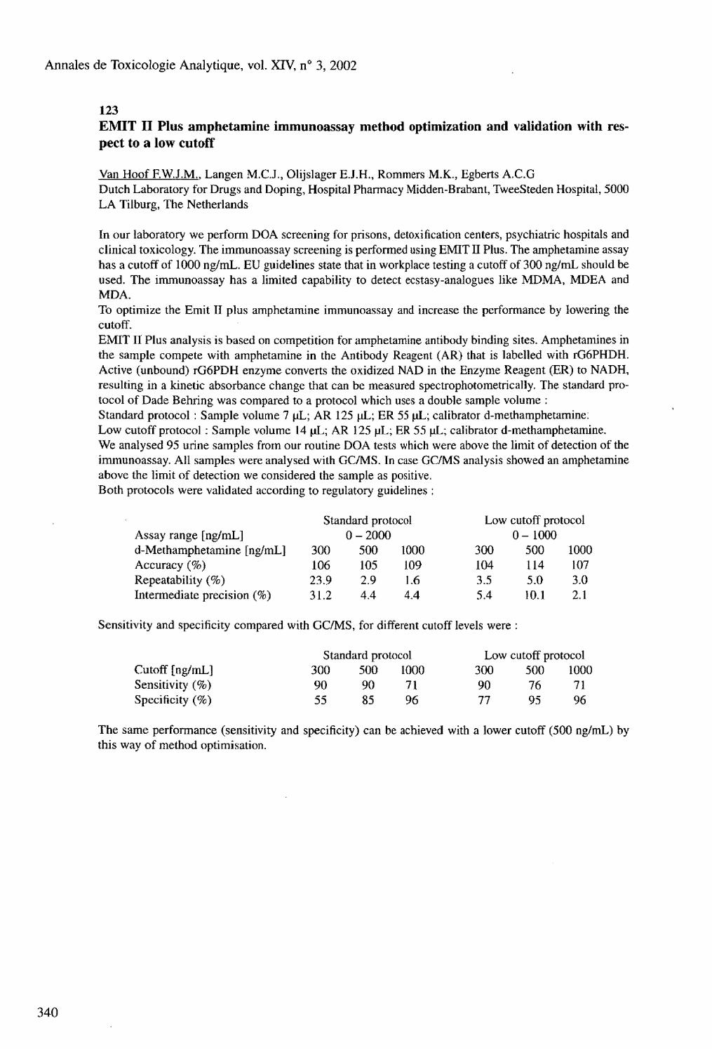
Annales de Toxicologie Analytique, vol. XIV, n° 3, 2002
123
Assay range [ng/mL] 0 - 2000 0 - 1000 d-Methamphetamine [ng/mL] 300 500 1000 300 500 1000 Accuracy (%) 106 105 109 104 114 107 Repeatability (%) 23.9 2.9 1.6 3.5 5.0 3.0 Intermediate precision (%) 31.2 4.4 4.4 5.4 10.1 2.1
Sensitivity and specificity compared with GC/MS, for different cutoff levels were :
Standard protocol Low cutoff protocol Cutoff [ng/mL] 300 500 1000 300 500 1000 Sensitivity (%) 90 90 71 90 76 71 Specificity (%) 55 85 96 77 95 96
The same performance (sensitivity and specificity) can be achieved with a lower cutoff (500 ng/mL) by this way of method optimisation.
340
EMIT II Plus amphetamine immunoassay method optimization and validation with respect to a low cutoff
Van Hoof F.W.J.M.. Langen M.C.J., Olíjslager E.J.H., Rommers M.K., Egberts A.C.G Dutch Laboratory for Drugs and Doping, Hospital Pharmacy Midden-Brabant, TweeSteden Hospital, 5000 LA Tilburg, The Netherlands
In our laboratory we perform DOA screening for prisons, detoxification centers, psychiatric hospitals and clinical toxicology. The immunoassay screening is performed using EMIT II Plus. The amphetamine assay has a cutoff of 1000 ng/mL. EU guidelines state that in workplace testing a cutoff of 300 ng/mL should be used. The immunoassay has a limited capability to detect ecstasy-analogues like MDMA, MDEA and MDA. To optimize the Emit II plus amphetamine immunoassay and increase the performance by lowering the cutoff. EMIT II Plus analysis is based on competition for amphetamine antibody binding sites. Amphetamines in the sample compete with amphetamine in the Antibody Reagent (AR) that is labelled with rG6PHDH. Active (unbound) rGóPDH enzyme converts the oxidized NAD in the Enzyme Reagent (ER) to NADH, resulting in a kinetic absorbance change that can be measured spectrophotometrically. The standard protocol of Dade Behring was compared to a protocol which uses a double sample volume : Standard protocol : Sample volume 7 uL; AR 125 uL; ER 55 uL; calibrator d-methamphetamine: Low cutoff protocol : Sample volume 14 uL; AR 125 pL; ER 55 uL; calibrator d-methamphetamine. We analysed 95 urine samples from our routine DOA tests which were above the limit of detection of the immunoassay. All samples were analysed with GC/MS. In case GC/MS analysis showed an amphetamine above the limit of detection we considered the sample as positive. Both protocols were validated according to regulatory guidelines :
Standard protocol Low cutoff protocol

Annales de Toxicologie Analytique, vol. XIV, n° 3, 2002
124 Rapid, sensitive direct method for the identification of gammahydroxybutyric acid (GHB) in urine
Vasiliades J.. Ford K. Toxicology Labs Inc., 4472 South 84th St., Omaha, NE 68127, USA
The use and abuse of the date rape drug, gammahydroxybutyric acid (GHB) has seen a large increase in the last years. The clinical or toxicology laboratory may therefore be called upon to rapidly identify GHB use by looking at an individuals urine. In a previous communication (1), we presented a quantitative method for the determination of gammahydroxybutyric acid in urine which used acid and heat to convert GHB to GHL (gammahydroxybutyric acid lactone). We present in this report a more rapid sensitive direct procedure for the identification of GHB in urine. The advantage of this procedure is that no extraction, derivatization or heating step is required prior to gas chromatographic (GC) analysis. GHB or its salt is in dynamic equilibrium with its lactone (GHL). We take advantage of this equilibrium for our direct analysis. The lactone which is formed is analyzed by GC with a flame ionization detector and an alcohol column, 1.82 m x 2 mm ID glass column, 60/80 Carbopack B/ 5 % Carbowax 20M (Supelco, Bellefonte, PA, 16823). Ethylene glycol (EG) can be used as internal standard. Gas chromatography conditions are: injector 200° C, detector 300° C, column 175° C, carrier gas helium with a flow rate of 30 rnL/min. Retention time for GHL and EG are 2.3 and 1.5 minutes respectively at 175° C. Three known positive GHB cases were evaluated by this new procedure. GHB was easily identified in each case. Advantages of this procedure are the small sample volume needed, 1-2 microliters of urine is used, and the short preparation time, no preparation time is needed, prior to analysis. There are no extraction, derivatization or heating steps required prior to analysis. We conclude that a simple, rapid sensitive and inexpensive method has been presented for the identification of GHB in urine. The sensitivity of the method is less than 25 mg/L.
reference : 1- Vasiliades J. et al. Clin Chem 2000 ; 46, Al 85
125 Medico-legal aspects of drug abuse in Latvia
Vol gram J.. Khodasevitch T., Khodasevitch L. Latvian State Centre for Forensic Medical Examinations, 2 Hipokrâta Str., LV-1038, Riga; and Centre of Drug Abuse Prevention and Treatment, 55 Hospitalu Str., LV-1013, Riga, Republic of Latvi.
According to official data, there are more than 20,000 persons who deal with drugs in Latvia. ( Population-2,5 millions of inhabitants ). All relevant indicators characterising the situation have become worse. In comparison with 1999 some indicators have become 2-3 times worse, e.g. number of non-fatal intoxications, drug-related death, driving in condition of drug intoxication etc. The number of registered and treated addictive patients as well the number of drug and psychotropic substances users have been on the increase already for several consecutive years. The structure of addiction patient contingent reveals a rapid increase in the group of opiate addiction and slight growth of sedative and soporofic as well as amphetamine-like substance addiction. There have been an increase of confiscated amount of heroin, marijuana, amphetamine, MDMA, cocaine and pills of psychotropic substances. Such drugs as methamphetamine, DOB have been registered in Latvia. 994 drug-related crimes have been registered in 2001 (increase- 60 %). New type of hallucinogen-Dicarbine (syn.Carbidine) is widely abused in prisons. An amount of nonfatal intoxications with drugs and psychotropic substances has increased. Different heroin combinations with benzodiazepines, tramadol, ethanol, Zolpidem, zopiclone, diphenhydramine, tricyclic antidepressants were common. Heroin overdosage (alone or in combinations ) was a cause of death in 55 % of drug-related death in 2001. Despite substantial national efforts,drug abuse remains a serious public health and security problem for the society.
Annales de Toxicologie Analytique, vol. XIV, n° 3, 2002
126 Detection of heroin in urinary samples through analysis of 6-monoacetylmorphine
Von Euler M.. Villén T., Svensson J.A., Stâhle L. Karolinska Instituted Division of Clinical Pharmacology, Huddinge University Hospital, 141 86 Stockholm, Sweden
A low concentration of morphine in a urinary sample may come from different sources. To avoid this interpretation problem the cut-off for positive verification is 2000 ng/ml in some countries. In Sweden a cutoff level of 300 ng/ml has been chosen. Such levels may be due to intake of poppy-seeds shortly before urine sampling, intake of analgesic compounds containing morphine, or heroin. To facilitate the interpretation we routinely include the heroin-metabolite 6-monoacetylacetate (6-MAM) along with morphine-3-glucuronide (M3G) in the LC-MS verification analysis. Interestingly, in several samples positive in the immunological screening-test only 6-MAM could be detected. To further investigate this all specimens with positive 6-MAM were selected (n=180). In 11 % of these specimens (n=18) 6-MAM was detected although the M3G concentrations were below cut off (300 ng/mL), in several of the cases M3G concentration was close to the limit of detection. The 18 samples with this excretion pattern came from 7 different individuals, all but one with previously known heroine abuse. There have been sporadic reports about similar findings in the literature. In our material the verification has been done with LC-MS instead of GC-MS, where total morphine is analysed, and this may explain the more frequent finding. The concentration of M3G and 6-MAM are analysed separately in all verifications of an opioid-positive immunological screening result. When GC-MS is performed M3G and 6-MAM are hydrolysed into morphine and accumulated to a total morphine concentration. The specimens with 6-MAM without M3G in urine were found sporadically and they were analysed at the same time as samples containing both 6-MAM and M3G. Moreover, such samples occurred in some patients at several occasions. Thus, we do not believe that this is an artefact finding but rather that it implicates some particular circumstances of the heroin intake. The heroin intake could have taken place shortly before testing, the first urine void after administration of heroin have been shown to have the peak 6-MAM concentrations. Also, differences in pseudo-cholinesterase activity, genetically or acquired, may influence the rate of 6-MAM de-acetylation. To elucidate this further studies are required. However, our finding demonstrates that it is valuable to routinely measure both 6-MAM and M3G when verifying opioid-positive immunological screening results to facilitate interpretation of the finding of lower amounts of morphine in a urine specimen.

Annales de Toxicologie Analytique, vol. XTV, n° 3, 2002
127 Routine monitoring opiate and amphetamine use in heroin and pervitine abuse treatment patients : comparison of EMIT II plus, EMIT d.a.u., FPIA and GC-MS results
Vorísek V.. Zitta R.( Cízek J., Nedvídková J., Haklová L., Cerníková B., Psenicková R., Palicka V. Institute of Clinical Biochemistry and Diagnostics, University Hospital, Hradec Králové, CZ-500 05, Czech Republic (1) Dade Behring Austria GmbH, Czech branch office, Konevova 210, Praha 3, CZ-130 00, Czech Republic (2) Abuse Drugs Detoxication Centre, University Hospital, Hradec Králové, CZ-500 05, Czech Republic
In this work, we examined semiquantitative EMIT II plus opiate , EMIT d.a.u. amphetamine class results and analogous FPIA results in 29 urine samples collected from 27 randomly selected detoxication centre patients (22 males and 5 females, aged 16 to 40) during two-month therapeutic period. Although the European Union recommends relative useful opiate and amphetamine screening cut-off levels 300ng/ml in urine, we like use our internal cut-off value at 200 ng/ml level for both of the analyte groups. It should minimize the risk of false negative results. This microstudy was designed to compare the efficacy of our multianalytical screening routine system. Positive opiates were confirmed in 5 cases and amphetamines in 3 cases. Additionally we identified mor-fine and methamphetamine with its metabolite amphetamine in one sample from 2 abuse treatment patients („multiabusers"). False positive or negative results were not detected. The FPIA average positive level for amphetamines was 601.19 ng/ml, average negative level was 27.941 ng/ml. The average EMIT positive amphetamines were target at 868.833 ng/ml level and the negative amphetamine average for this commercial method was 6.354 ng/ml. The average EMIT positive opiates level was 977.128 ng/ml and the average level for negative opiates by EMIT was 44.920 ng/ml. The average values for positive and negative FPIA opiates were 812.375 and 28.505 ng/ml, respectively. GC-MS confirmed analytes in positively screened samples were: morphine, methamphetamine, amphetamine and ephedrine. Total amount of positive results was 27.6 %.

Annales de Toxicologie Analytique, vol. XIV, n° 3, 2002
128 Epidemiological study of alcohol consumption in general population of Dharan, Nepal
Yadav B. N. Department of Forensic Medicine and Toxicology, B. P. Koirala Institute of Health Science, Dharan, Nepal
Of all the drugs which human being have used and abused in the course of their chequered history, alcohol is almost certainly the oldest and also the most widely used because it is so easily produced. Alcohol has always been used in Nepal. Alcoholic beverages are culturally accepted and social tolerance for alcohol use and alcohol dependence is quite high, so alcohol has not been considered a drug for serious concern either by the Government or by any social organization. Alcohol could be the number one problem (drug) if we seriously consider the magnitude and extent of the problem it has created in Nepal. Alcoholic drinks in various forms have long been consumed in Nepal. Alcohol is necessary on most occasions among men is relatively frequent and is well tolerated by many communities, but there is strong social disapproval of female drunkenness, it is not uncommon to see female alcoholics in the country especially in the hilly and mountainous regions. A "Matwali" is a person who is allowed to drink alcoholic beverage by virtue of his birth. A high percentage of Nepalese population belongs to this category and many of them take alcoholic beverages either on social occasions or on a regular basis. People who do not belong to this category are not supposed to consume alcoholic beverage even on social occasions. But there seems to be very steady rise in the number of people belonging to this category who consume alcoholic beverages. People in Nepal generally believe that alcohol is remedy for cold, pain, physical tiredness, and so on. In fact, alcohol is extensively used for many ailments, especially in the rural areas. Most of the unskilled and semi-skilled workers in Nepal believe that they can function better if they take small amount of alcohol form time to time. More over, alcohol has become a status symbol for many people. Parties, get-togethers, or festivities are considered incomplete if alcoholic beverages are not served. According to the 1991 figures form the department of Excise, the sale of alcoholic beverages seems to be increasing rapidly. Since there is no export of alcoholic beverages from Nepal. All beverages are sold and consumed within the country. If we take into account home production, under-reporting of commercial production, liquor brought in form duty free shops and liquor imports, even more alcoholic beverage are consumed in Nepal. The number of distilleries and breweries is also increasing. Even light drinking may adversely interact with other medication; temporary heavier drinking can exacerbate most medical illness; and alcoholism can masquerade as many different medical disorder and psychiatric syndromes. Alcohol abuse is generally acknowledged cause of, or to say the least, and important contributing factor to, accidents, homicides, and suicides as well. Therefore, it is felt that the study of the overall prevalence of alcohol consumption, the vulnerable age groups the ethnic distribution, the role of socio-economic factors, age, sex, and type of liquor shall help to find out the quantum and magnitude of the problem so that the government can plan effective measures to control the menace of alcohol abuse in Nepal.
344
Annales de Toxicologie Analytique, vol. XIV, n° 3, 2002
129 Solid phase microextraction and GC-MS for confirmation of amphetamine, methamphetamine, MDA, and MDMA in immunoassay positive urine samples
Zhang S.Y.(Î). Huang Z.P.í2)
(1) Lab of Forensic Sciences, Fujian Public Security College, Fuzhou 350007, P. R. China (2) Forensic Sciences Institute, Fujian Provincial Department of Public Security, Fuzhou 350003, P. R. China
Amphetamines are powerful stimulants of central nervous system (CNS). Their abuse in recreational places has drastically increased in the past decade in P. R. China. Amphetamine, methamphetamine, 3,4-methylenedioxylamphetamine (MDA) and 3,4-methylenedioxyl-methamphetamine (MDMA) are most commonly available among these compounds. Immunoassay was routinely used in screening of these compounds in urine, however, significant cross-reactivity occurred. Gas chromatography-mass spectrometry (GC-MS) is often utilized for confirmation of these compounds in the immunoassay positive urine samples. The aim of present study is to establish a method using solid-phase microextraction (SPME) and GC-MS for practical confirmation of amphetamine, methamphetamine, MDA, and MDMA in immunoassay positive urine samples. The urine sample was saturated with NaCl and alkalized with NaOH. A fiber coating 100pm polydime-thylsiloxane (PDMS) attached on manual SPME assembly (Supelco) was placed in headspace of 1ml of urine sample in a 20-ml vial. Analytes were absorbed onto the fiber in a period of time and desorbed in GC injection port at 250° C for over 5 min. Derivation was not carried out for these amphetamines in the experiment. The enrichment of the SPME fiber was extremely high with extraction at 100° C for 10 to 20 min. Analysis of a sample was finished within 30 min. In selected ion monitoring (SIM) mode. The detection of limits of the method reached ng/ml level, and linearity calibration curve was obtained up to 1 pg/ml. Reproducibility was improved by internal standard method, and developed precision will be expected with automatic SPME. The background of total ion chromatogram (TIC) was fairly clean, and no interference occurred in analysis. The method was assessed in analysis of amphetamine, methamphetamine, MDA and MDMA in immunoassay positive authentic urine specimens. This method was solvent-free, speedily, sensitive, simple and easy to automation. It is very convenient in practical identification.
Annales de Toxicologie Analytique, vol. XIV, n° 3, 2002
130 Assessment of the neurotoxic risks of disinfectants based on isopropanol
Below H.. Pitten F.A., Kempe B., Gilgenast O., Kramer A. Institute for Hygiene and Environmental Medicine, University Greifswald, HainstraBe 26, D-17493 Greifswald, Germany
As far as we know, possible neurotoxic risks of local anti-infective agents and hand-disinfectants have never been determined prior to their introduction for use by humans, but were always discerned later as undesirable side-effects, partly in extreme situations. Isopropanol is extensively used as a hand-disinfectant agent with long periods of exposure. In our own studies, the following preparations were compared in the course of a 90-day open field test of activity changes induced in mice (BALB/c OlaHsd inbred strain) after epidermal application: Betaisodona® solution (povidone-iodine), Octenisept® (octinidine dihydrochloride), Spitaderm® (chlo-rhexidine dihydrochloride), Polyalcohol hand-antiseptic® (isopropanol) and 0.5 % hexachlorophene in olive oil (positive control). The exposure selected during this process was well above any values which might be achieved even in excessive preparation applications. No indications of any changes induced by the preparations were detected histopathologically in the liver, spleen, kidneys, pancreas, brainstem, cerebellum, frontal brain and the area of skin exposed. The "isopropanol" group corresponded to expectations from the influence of alcohol, with significant increases in activity (animal activity meter, field changes in the open field test). This is in conformity with the data in the literature (Palissa and Becker 1986; Burleigh-Flayer et al. 1998). The activity increases induced by the isopropanol group were restricted to the daytime however, and no significant changes in relation to the negative control were detected at night. No changes in activity were detected after discontinuing the exposure either. The learning and memory behavior of rats was also investigated in a labyrinth test. The exposure selected here was also above the levels used in normal application of the preparations. No indications of neurotoxic effects were detected here either. The conclusion of the above is that isopropanol induces an exitation effect with an increase in motility, as is the known effect of other alcohols. Signs of any toxic effects were not observed, however, and the resting phases are not influenced. This conforms with the results of studies to date, which do not indicate any neurotoxic risks when isopropanol is used for the purpose of hand-disinfection. The lack of evidence of any neurotoxic risks incurred by inhalational exposure to isopropanol, as may be expected from hand-disinfection, is specifically referred to in the results of an epidemiological study in the process of manufacture (Maizlish et al. 1985).
346

Annales de Toxicologie Analytique, vol. XIV, n° 3, 2002
131 An evaluation of the results of laboratories participating in the QUARTZ Forensic Toxicology Scheme
Boley N.(1>, Forrest R.(2>, Mac Donald S.°\ Ossleton D. ( 4 ), Paterson S. ( i ), Williams K. ( , )
Steering Committee of the Quartz Forensic Toxicology Scheme (1) LGC, Queens Road, Teddington, Middlesex, United Kingdom (2) University Department of Forensic Pathology, Medico-legal Centre, Watery Street, Sheffield, United Kingdom (3) Birmingham City Laboratories, Valepitts Road, Garretts Green, Birmingham, United Kingdom (4) Forensic Science Service, Suite C, Loddon Vale House, Hurricane Way, Woodley, Reading, Berkshire, United Kingdom (5) Toxicology Unit, Imperial College of Science, Technology & Medicine, St Dunstan's Road, London, United Kingdom
Quartz (Quality Assurance in Forensic Toxicology) is a scheme run by the above steering committee on behalf of the United Kingdom Forensic Toxicology Forum (UKFTF) and is operated in accordance with the International Harmonised Protocol for the Proficiency Testing of (Chemical) Analytical Laboratories, ISO/IEC Guide 43:1997, and the ILAÇ Guidelines for the Requirements of Providers of Proficiency Testing Scheme, ILAC G 13.s. The scheme consists of four rounds annually, each round of two samples which are prepared by spiking transfusion blood with appropriate substances of interest. Results are submitted back to the scheme's co-ordinator who issues the relevant report detailing the results, summarised methodology and any relevant comments by the steering committee. Each round comprises of a quantitative and qualitative sample. For the quantitative sample, participants are notified of the substances present and asked to determine their concentrations. The qualitative sample may contain up to four unknown substances which participants are asked to identify. Case histories are provided to aid interpretation and investigation of both samples. The case histories and the concentrations of substances in the samples are fictitious but are designed to reflect typical histories encountered in the laboratory. The results of the last four round of the scheme will be presented covering the time period March 2001 to February 2002. A common theme for the identification and quantitation of amphetamines was developed during these four rounds and the performance of the laboratories will be discussed. Round 4. Quantitation sample : Paracetamol, mean 279 mg/1, range 160-334 ml/1, SD 33, reference value 339 mg/1. Dihydrocodeine, mean 1.1 mg/1, range 0.75-1.55 ml/1, SD 0.22, reference value 1 mg/1. Identification sample : drug added 3° piclone, number of correct identifications 3/9, number of incorrect identifications 6/9 Round 5. Quantitation sample: methadone, mean 0.4 mg/1, range 0.26-0.58 ml/1, SD 0.08, reference value 0.4 mg/1. Identification sample: drug added amphetamine, number of correct identifications 10/16, number of incorrect identifications 5/16 Round 6. Quantitation sample : MDMA, mean 0.292 mg/1, range 0.23-0.45 ml/1, SDvO.057, reference Value 0.3 mg/1 ; MDA, mean 0.05 mg/1, range 0.03-0.08 ml/1, SD 0.015, reference value 0.05 mg/1. Identification sample : drug 1 added MDMA, number of correct identifications 13/14, number of incorrect identifications 1/14 ; drug 2 added MDA, number of correct identifications 9/15, number of incorrect identifications 0/16, number reporting no MDA, 5/14. Round 7. Quantitation sample : Paracetamol: mean 202 mg/1, range 99-278 ml/1, SD 19, reference value 220 mg/1. Propoxyphene: mean 1.75 mg/1, range 0.84-2.9 ml/1, SD 0.34, reference value 2 mg/1. Identification sample : drugs aadded amphetamine and benzoylecgonine, number of totally correct identifications 8/15, number missing amphetamine 3/15, number missing benzoylecgonine 4/15, number missing both 0/15.

Annales de Toxicologie Analytique, vol. XIV, n° 3, 2002
132 Oximeter in forensic toxicology : rapid determination of carboxyhemoglobin in blood
Brefamer C . Iten P. X. Institute of Legal Medicine, University of Zurich,Winterthurerstr. 190, CH-8057 Zurich, Switzerland
Carbon monoxide intoxications, both suicidal and accidental, are frequent. In forensic toxicology, carboxyhemoglobin (CO-Hb) in blood is commonly quantified with spectrophotometric or gaschromatogra-phic methods. We have compared the spectrophotometric method of Maehly and a gaschromatographic method with a thermal conductivity detector (GC-TCD) with a third method: the Oximeter. Application of the Oximeter is customary in clinical toxicology. Several CO-Hb containing blood samples of deceased persons were analysed. Results of all three methods were comparable for low concentrations (ca. 10 % CO-Hb) as well as for high concentrations (ca. 80 % CO-Hb) regardless of the viscosity of the blood samples. The advantages of the Oximeter when compared to Maehlys method and GC-TCD are extreme short time of analysis (less than 1 min.), very small blood volume required (less than 0.1 ml) and easy handling. We conclude that the application of the Oximeter is not limited to analyses of blood samples from living persons (e.g. in clinical toxicology); it can as well be used for the determination of CO-Hb in post mortem blood samples. Hence it is a useful and time saving tool in forensic toxicology.
133 Assessment of the interference of turbidity, hemoglobin and bilirubin on the determination of salicylemia with Trinder's method
Douki W., Mezzour H., Ben Amor A., Najjar M.F. Laboratory of Biochemistry - Toxicology, CHU Monastir, 5000 Tunisia
We studied the interference of turbidity owed to hypertriglyceridemia, of hemoglobin and bilirubin on the determination of salicylemia by the Trinder's colorimetric method, whose based on the quantification of a purple hexacoordinate complex, formed between iron chloride III and salicylic acid, measurable at 540 nm. The blood sampling was performed without anticoagulant and measurements done on a Hitachi U-2000 analyzer. We carried out this study according to the IFCC and the SFBC's protocols. The assessment of the method showed a good precision (repetability: CV 0.21 to 2.74 %), (reproducibility: interserial CV 0.29 to 6.67 %).The reaction was linear over 2000 mg/1, with a detection limit equal to 3.0 mg/1. Finally, the study showed no interference of neither hemoglobin, nor turbidity nor bilirubin on salicylemia determination within a concentration margin up to 800 mg/L This simple, fast and low coast method, seems to be reliable. Besides, it adjusts well to all prescriptions of this dosage, including in post-mortem.
348

Annales de Toxicologie Analytique, vol. XIV, n° 3, 2002
134 A comparative study of the protective effect of some antidotes on the pancreas in paraquat intoxicated rat
ELSehely W.. Sharaf El Din N. Forensic Medicine and toxicology and histology department, Faculty of Medicine, Alexandria University, Egypt
Paraquat (PQ) is widely used herbicide. Human PQ intoxication is of frequent incidence which occurs either accidental or suicidal. This work aimed at studying pancreatic toxicity following paraquat intoxication as well as the possible effects of promising anti dotes, Ssdiurn thiosulphat (Nath) and deferroxamine (DF). Forty five adults albino rats were used in this study, they were classified into four groups. Group I used as negative and positive group. GroupII PQ intoxicated rats, in a dose of 30 mg/kg, group III PQ and Nath treated rats. Group IV, which received PQ and deferroxamine. Pancreas sections were examined histologically by routine stain (H&E) using light LM and ultra structurally by EM. of pancreas revealed massive degenerative and necrotic changes affecting both exocrine and endocrine parenchyma. By using Nath the endocrine parenchyma was saved while the exocrine one showed focal degenerative changes. On the other hand DF showed better protection than Nath as antidote for pancreatic PQ toxicity.
135 Plants and chemical submission in Tunisia
Ghorbal H.. Bousnina M., Hedhiri S., BenSalah N., Amamou M., Hedhili A. Laboratoire de toxicologie du CAMU. 10, rue Raspail, Mont-Fleury, Tunis, Tunisie
In Tunisia plants are always used as drugs in traditional médecine andaliments. Many cases of chemical submissions are repertoried evry year. The aim of thiswork is to study this submission in Tunisia through the activity of theToxicology Laboratory of the Anti-Poisons Center of Tunis. It's a retrospective study of 100 files including more than 15 % cases ofplant submissions colliged between January 1990 and decem-ber 2001. The patients are males in majority and between 18 and 56 years old,comingfrom Tunis and suburbus,the Cap-Bon and the south of the country. The diagnostic was based on botanic recognition and both clinical andanalytical diagnostic. There are some cases of submission caused by drugs (Cannabis, benzodiazepines, barbiturics, phenothia-zines). For these cases the diagnosticwas based on urine and blood analysis by an immunologic method (FPIA) which is confirmed by GC/MS and spectrophotometric method. For plants, the mostly cases met were caused by those which belong to Solanacées family : 73 % Datura stramonium Land, only 27 % by Mandragora officinalis. The plants used were usually mixed with alcohol, tea or legmi (sap of dates). The seeds of Datura stramonium were prepared in a decoction with tea. The leaves and the roots of the mandragore were decocted too, but the fruit (berries) was consumed directly. The results were obtained after analysis of stomach washing and urine by thin layer chomatography and confirmation by GC/MS. In conclusion, the chemical submission by plants and other drugs is a practice which is growing in size and represent a social and legal problem in Tunisia, The plants used for the submission goals are easly found in the country and are easily prepared.
Annales de Toxicologie Analytique, vol. XIV, n° 3, 2002
136 Performing toxin analysis in a resuscitation and emergency care environment
Gligor R. Western University "V. Goldis" Arad, Romania
Due to the large proportion of intoxication, among overall cases admitted to emergency rooms, toxicological analysis needs to be performed in clinical settings as a clue diagnostic test intended to improve the care of the patient, identification of the toxin(s) being of highly therapeutic relevance. An actual issue need to be taken into account is the question of how reliable are the clinical and the common laboratory investigations as a substitute for the toxicological analyses ? How cost and diagnostic effective is a symptomatic treatment in balance with a systematic toxicological screening of all patients with intoxication suspiciousness ? A significant number of patients brought to the emergency room in a comatose state are suspected of accidentally or voluntary intoxication, often with only a supposed or vague indication of the trigger substance. In such emergency cases every second counts, restoring and maintenance of the minimal vital functions being the priority. Consequently in many situation samples are collected just after resuscitative efforts have been done. With this in view the following aspects worth although being considered: - the impact of the pH changes (alkalosis or acidosis) and of the drugs used during resuscitations on the diagnostic toxicological screening, - the possible influences of cross-reactions on the clinical decision timings and - the degree of influence of the liver failure on the metabolic pathway and consequently on the blood level of the intoxicant and its metabolites with consequences on their identification and quantification. This paper will outline the work of toxicologists and illustrate how modern advances in scientific methodology may be applied in solving the complex problems that are encountered when facing poisoned patients in Emergency Departments.
137 Fatal ingestion of magic mushroom : a case report
Gonmori K.. Yoshioka N. Department of Forensic Medicine, Akita University School of Medicine, Akita 010-8543, Japan
A 27-year-old man who was missing since 12 hours was found dead in an irrigation canal in January. It was suspected that he went outdoors from the window of his room in the middle of the night. His room scattered about trash, cloths and compact disc, where two cultivating pots for magic mushroom were found and tiny mushroom and the roots of mushroom were still remained in the pot. We tried to grow mushroom from the roots in our laboratory and obtained the good shape mushroom considered to be Psilocybe subcubensis two weeks later. Concentration of psilocybin and psilocin in blood and urine from the victim, and from mushroom cultured in our laboratory were measured by LC-MS/MS. The results are shown Table I. We conclude that he might be hallucinatory condition by ingesting mushroom, which resulted in developing his strange behavior leading to the death from low temperature. It is generally explained that psilocybin transforms into psilocin rapidly in the body however, we could detect only psilocybin in the blood not psilocin. The reason for the discrepancy of our results and general explain is not clear.
Table I. Concentration of psilocybin and psilocin (ND : not detected)
Blood Urine Mushroom
Psilocybin Psilocin
1.4 ng/ml ND
ND 162 ng/ml
34.6 pg/wet g 10.3 pg/wet g
350
Annales de Toxicologie Analytique, vol. XIV, n° 3, 2002
138 Tramadol metabolite ratios and CYP2D6 genotypes in postmortem samples
Koski A.. Levo A., Sajantila A., Ojanpera L, Vuori E. Department of Forensic Medicine, P.O. Box 40, FIN-00014 University of Helsinki, Finland
Tramadol is an opiod drug that is metabolized to O-desmethyltramadol exclusively by the cytochrome P450 enzyme 2D6, encoded by the CYP2D6 gene. This gene is highly polymorphic and some of the alleles are defective, duplicated or missing. To determine whether abnormal tramadol metabolism due to a defective CYP2D6 gene could be seen in postmortem samples, we compared the tramadol metabolite ratios with the CYP2D6 genotypes. The study included 33 cases autopsied between 1998 and 2000. In these cases, DNA samples in the form of blood stain papers had been collected at the autopsy and tramadol had been found in postmortem femoral blood samples. Large deletions or amplifications and 18 selected SNPs were analyzed in the CYP2D6 locus. Tramadol was analyzed using GC, and O-desmemyltramadol and nortramadol were analyzed using RPLC-MS-MS. A good correlation between genotype and phenotype was found, but no tramadol-associated poisonings coincided with a defective CYP2D6 gene.
139 Strychnine intoxication: a case report
Margalho C , Barroso M., Teixeira H.M., Ávila S.. Frías E., Proença P, Pinho Marques E. National Institute of Legal Medicine - Delegation of Coimbra, Portugal
Strychnine is an alkaloid extracted from the plant of Strychnos nux-vomica. Initially used on the manufacturing of rodenticides and pharmaceutical preparations, the utilization of this compound has decayed due to its high toxicity. In spite of being prohibited, it is still possible to find illegally commercialised strychnine in Portugal, continuing this to be associated with forensic intoxications. The authors present a postmortem case of strychnine detection in human blood. The toxicological examination requested by the pathologist was the determination of alcohol, benzodiazepines and opiates. The result was negative for all of them. Our laboratory includes on the routine procedures a general GC/MSD screening, which has revealed the presence of strychnine, otherwise left undetected. The sample was then re-extracted by a liquid/liquid procedure and analysed by GC/MSD. A concentration of 2 pg/mL of strychnine was calculated, which may have been implicated in the death of the individual.

Annales de Toxicologie Analytique, vol. XIV, n° 3, 2002
140
352
Availability of drug assays in brain-injured patients
Morris R.G. f l ). Kennedy M.<2)
(1) Cardiology & Clinical Pharmacology, The Queen Elizabeth Hospital, Woodville, South Australia, Australia (2) Clinical Pharmacology & Toxicology, St Vincent's Hospital, Darlinghurst, NSW, Australia
The availability of drug assays for a range of therapeutically administered as well as illicit drugs in a time frame that supports clinical management in brain-injured patients in intensive care units has previously been raised by our group (1). A survey was conducted of 159 intensive care units in Australasia. Data were collected regarding bed numbers, drug assays/screen available, turn-around times, availability of clinical pharmacologist/toxicologist
' interpretative support, as well as a question regarding decision times for terminating life support in brain-injury where drugs were suspected as contributing to the comatose state (in the absence of 4-vessel angiography). Responses were received from 40 % (64 centres) within 3-months. 40 % indicated access to urinary screens, 56 % had standard immunoassays (eg., phenytoin), 11 % had more complex assays (eg., clonazepam) and 3 % had access to highly sophisticated assays for unusual compounds (eg., phencyclidine). Whilst 8 % had clinical pharmacologist support on site, 26 % claimed such support was readily available. Assay turn-around time ranged from <1 day (63 %) for standard assays, through to >5 days (16 %) for the more complex tests. Expert interpretation of results was available on only 30 % of tests. Answers to the time to terminate life-support question were skewed to the right, where 0 % indicate they would terminate in <12 h, 5 % <24 h, 12 % <48 h, 16 % <72 h, and 55 % said they would never terminate in the absence of 4-vessel angiography. Clearly this was a controversial area and perhaps the question too simplistic. However, the results suggested that current level of analytical and interpretative expertise available to clinical staff in this complex area is less than desirable in Australasia. This was compounded by the general reduction in clinical pharmacology services generally (medical and scientific) over the past decade (2). This result is in conflict with the increasing demands on clinical staff to justify their actions, including legal issues that may follow such clinical scenarios in the coroner's courts, etc.
References : 1- Kennedy M. et al. MedJAust. 1996 ; 165 : 394-9 2- Morris R.G. et al, Ther Drug Monit. 1998 ; 20 : 598-601.

Annales de Toxicologie Analytique, vol. XTV, n° 3, 2002
141 Detection of a carcinostatic vinca in the cutaneous tissues by immunohistology
Mukaida M. 0 ). Kimura H.<2), Murayama T.(1>, Matsuzaki Y.(", Masuda T. ( , )
(1) Department of Forensic Medicine, National Defense Medical College, Namiki 3-2, Tokorozawa, Japan (2) Department of Forensic Medicine, Juntendo University School of Medicine, Japan
It is well known that drugs for medical cure often have side effects on patients. Especially, carcinostatics have strong side effects and they result in injuries of various organs. For example, it is known that overdose administration of a vinca injures cutaneous tissues or mucous membrane of small intestine. But metabolism or kinetics of vinca taken into the cutaneous tissues has not been reported. For an exact detection of localized vinca, immunohistological study has been developed. Mouse cutaneous tissues were examined. Vinca (40 pg/mouse) was administered to four mice (C57BL, males, 25-28 g) by a single intravenous injection into the cauda. One mouse was anesthetized with ether and was killed 15 min after administration, and others were killed 45 min, 90 min and 24 hours after administration. The dorsal cutaneous tissues and labial marginal tissues were cut. They were frozen in OCT compound and were cut into a series of 5 pm thick sections. The sections were treated with a reducing agent (sodium borohydrate). The cutaneous tissue sections were fixed in 0.05 M bicarbonate buffered solution (pH 9,0) containing 1% paraformaldehyde and 0.2 % glutaraldehyde. After they were incubated with methanol containing hydrogen peroxide to inhibit the activity of endogenous peroxidase, 0.2 % casein was added. Anti-vinca sera were obtained from rabbits which were immunized with vinca~KLH complex. For enzymatic immunostaining, affinity-chromatographically purified antibody was used. The tissue sections were reacted with anti-vinca. Then, after these specimens were reacted with biotinylated anti-rabbit-IgG and avidin conjugated horse radish peroxidase, a 3-3' diaminobenzidine tetrahydrochloride was added as a substrate. The nuclei were stained with hematoxylin. The localization and distribution of vinca in the cutaneous and labial tissue sections of mice were observed. In the hair bulb, only a few cells showed a positive reaction with a lapse of time after administration. Strong positive reactions were observed in the bulge (a part of outer root sheath of hair) and in the sebaceous gland. The degree of reaction decreased after 90 min and positive reaction disappeared from the bulge after 24 hours. And the positive reaction to vinca in the tissue changed with the passage of time, as was previously reported in kinetics study using isotope-labeled-vinca. The detection of localized vinca in cutaneous tissues by immunohistological staining suggests that vinca has strong affinity to the sebaceous gland and the bulge, hair follicular stem cells. The positive reaction of sebaceous gland shows that vinca has strong affinity for the grease in the sebaceous gland and it is extracorporeally excreted from the sebaceous gland. Vinca also suppresses proliferation of cells. These characteristic properties of vinca cause injury to the cutaneous tissues, which results in alopecia.

Annales de Toxicologie Analytique, vol. XIV, n° 3, 2002
142
Patient n° conc.of amisulpride (n°/ml) Patient n° conc.of amisulpride (ng/ml) 1 107 5 121 2 564 6 14824 3 8135 7 5602 4 1200 8 < 2 0
The depicted concentrations are those measured at admission to the hospital. In this study, six patients were above the concentrations of amisulpride generally found at therapeutic dosages (concentrations < 200 ng/ml). The patient number 6 presented a cardiac toxicity with a QT prolongation.
143 Toxicokinetic and residue cytotoxicity study of mepiquat chloride in goat
Sahu C . Ghosh M. Department of Zoology, University of Kalyani, Kalyani, 741235 India
Mepiquat chloride (MCI) (1-1-dimethyl piperidinium chloride) is a quarternary nitrogen compound. It is used widely in nature as plant growth regulators. In order to study its toxicokinetics and residue cytotoxicity, the compound was administered orally at the dose of 300 mg/kg. b. w. to black Bengal goats (Capra capra). The control animals however, were fed with equal volume of distilled water. Blood samples were collected at 0.25, 0.50, 0.75,1, 2, 4, 6, 8, 10, 12, 36, 48 and 72 firs post administration (pd). MCI from blood was separated with ferric chloride-1, 2,dichloroethane solution and the concentration of MCI in the extract was measured through U-V Spectrophotometer at 364 nm. The blood level of mepiquat chloride was plotted on semi logarithmic paper against time. Standard toxicokinetic parameters were fitted to the experimental data and analyzed as per standard formulae. Vital organs like liver, lung and kidney were collected to ascertain MCI induced cytotoxicity. The results showed that MCI in the blood of goat could be detected to the level of 8.28 ± 1.76 pg ml ' at 0.25 hr. pd, which gradually increased and reached maximum concentration at 24 hrs (CB max: 32.8± 2.88 pg ml'). The minimum concentration was however detected at 72 hr. pd. (CB min.: 15.4 ± 1.56 pg mf!)-Mean values of different toxicokinetic parameters showed that absorption rate constant value (K a 0.10 ±0.01) was low, suggesting poor absorption of the compound from G.I. tract. The elimination half-life( ti/2 B 38.66 ±1.86) was large indicating retention of compound in the body for long period (i.e. slow elimination from the body). The mean blood concentration time profile of MCI showed a 'two compartment open model kinetics'. Microscopic examination of the vital organs exhibited various types of pathological changes, which was specially marked in lung tissue.
354
Rapid high-performance liquid chromatographic measurement of amisulpride in human plasma. Application to management of acute intoxications
Péhourcq F.. Ouariki S., Bégaud B. Laboratoire de Pharmacologie, Université Victor Segalen, 33076 Bordeaux cedex, France
Amisulpride, a substituted benzamide derivative, is a second-generation (atypical) antipsychotic and is effective as maintenance therapy in patients with schizophrenia. For toxicological purpose, a specific and accurate RP-HPLC assay was developed for the determination of amisulpride in human plasma. This method involved a two-step extraction in the presence of viloxazine, as internal standard. The compounds were chromatographied on a reversed-phase Spherisorb® C8 column with a mobile phase consisting of 0.06 M phosphate buffer pH 6.4 - acetonitrile - triethylamine - Pic B5® (750 : 250 : 0.5 : 15, v/v) and detected at 280 nm. A linear response was observed over the concentration range 100-1000 ng/ml. A good accuracy (bias < 5 %) was achieved for all quality controls, with intra-day and inter-day variation coefficients inferior to 4.3 %. The lower limit of quantification was 20 ng/ml, without interferences of endogenous or exogenous components. This rapid method (run time < 6 min) was used to manage eight intoxications involving amisulpride.

Annales de Toxicologie Analytique, vol. XIV, n° 3, 2002
Validation of an Ion Trap Gas Chromatographic Tandem Mass Spectrometry method for determination of nandrolone metabolites in human urine
Sánchez B.J.F. Antidoping Laboratory, Calle 100 y Aldabo, CP 10800, Ciudad Habana, Cuba
In 1980 the International Olympic Committee (IOC) included anabolizant steroids in the list of forbidden substances. Nandrolone is an anabolic androgenic steroid occasionally abused by athletes. Usually, nandrolone metabolites could be present in very low concentrations limit in human urine. The IOC criteria set forth for this analyte is that urine will be consider as positive when the signal intensity acquired is above the signal intensity of a positive control urine enriched with a concentration of 2 ng/mL for men and 5 ng/mL for women. This level of detection is easy to achieve with tandem mass spectrometry technique. An Ion Trap Gas Chromatographic Tandem Mass Spectrometry method was validated to quantify simultaneously nandrolone metabolites, norandrosterone and ethiocolanolone in urine samples. Methyltestosterone internal standard was added to 2.5 mL of urine and extracted in Detectabuse columns. The residues were evaporated and BIS-O-TMS derivatives were obtained. Detection was performed in MS/MS mode using the m/z 405 as parent tons for nandrolone metabolites and m/z = 446 for methytes-tosterone internal standard. The daughter monitored ions were m/z 405, m/z 315 and m/z = 225 for both nandrolone metabolites. The internal standard daughter monitored ions were m/z = 356 and m/z = 301. Studies on specificity, linearity, accuracy, precision, detection limit, and quantitation limit were performed and discussed. Also prevalidation phase studies on the optimization of isolation and excitation parameter to get MS/MS events in the instrumental method are presented We concluded that this method takes advantage of the sensitivity and selectivity of the electron ionization couple with MS/MS.
145 Effects of the plant growth regulators as abscisic acid, 4-chlorophenoxyacetic acj| rellic acid and maleic hydrazide on swiss-albino Mus musculus mice's liver ai cogens
-chlorophenoxyacetic acid >e used in agriculture as normal
Mediterrannean University, Science Faculty, Biology Department, AntaJ
In this study, acute effects of plant growth regulators like abscisic (4-CPA), gibberellic acid (GA3) and maleic hydrazide (MI dose and their four times over-doses were investigated. They were injected intraperitonally into Mus musqdá^J^^Fbb&erve their liver and muscle glycogen levels, blood glucose, serum alkaline p h o s p h a ^ | ^ l a c t a t e dehydrogenase activities, on fourth, eighth and 24* hours. The effects of the plant growth regulatg^^.'^fcrftrparameters were not important in the case of the normal dose of fourth hour but w e r e ^ ^ ^ ^ ^ p m i over-doses. At the eighth hour, although effects of normal doses on some parametei|^^3^^prcant, it was seen that over-doses were more important. While any important effect of no^^^^^J&n studied parameters were seen at the 24 t h hour, it has been determined that the effects^^^^^H^were still available. When we look a y ^ ^ ^ ^ H ^ c t s of studied plant growth regulators on parameters, the most increasing effects weretf^^^^^P^th inhibitors ABA and MH. One of the plant growth stimulators, GA3 has made an e f f e c i ^ ^ ^ ^ ^ ^ ^ a s i n g and decreasing. It was another stimulator, 4-CPA, which was less effective on
'show that, acute effecte of over-dosed plant growth regulators on Mus musculus mices star-fourth hour, was increased at the eighth and could not have been eliminated completely in
spWof being tolerated almostly at the 24 t h hour.

Annales de Toxicologie Analytique, vol. XIV, n° 3, 2002
146
Dermal absorption is a major route of exposure to kerosene as well as inhalation. We previously reported that trimethylbenzens (TMBs) (15-20 % of kerosene in proportion) were absorbed much more than aliphatic hydrocarbons (AHCs) (80-85 %) and the tissue distribution of TMBs following dermal exposure have been demonstrated in rats. The purpose of this study is to evaluate the influence of kerosene amount and area of exposure upon the dermal absorption of kerosene components. How soon the kerosene components appear in blood during dermal exposure is another interest in this study. Kerosene components were analyzed by capillary gas chromatography/mass spectrometry (GC/MS). Pseudocumene (PSC) and undecane (Cl 1) were chosen as an indicator of TMBs and AHCs at data analysis since these components were detected at the most in quantity in biological samples, respectively. Thirty two rats were randomly divided into four groups. All rats had a catheter with a left femoral vein under anesthesia, and a small piece of cotton soaked with kerosene (the area and kerosene amount of exposure described below) was applied to the abdominal skin for two hours.
A 0.5 ml of blood was withdrawn from the catheter before and 5,10, 20, 30, 45, 60, 90 and 120 min after the dermal exposure started, and the solid tissue samples were harvested at 120 min. PSC were detected at 5 minutes and gradually increased in all groups. The time course changes in PSC levels in blood were significantly different between group I and II or I and III, and almost identical between group II and III. The blood levels in group IV were significantly high compared with any other groups. Similar trends were observed in tissue samples at 120 min. Small amount of CI 1 were detected in most of blood samples, however significant differences among groups and significant increases during exposure were not observed. These results suggest that 1) kerosene components, especially TMBs are absorbed at enough levels to detect in blood within a few minutes after dermal exposure to kerosene, 2) the TMB levels in blood are influenced by the amount of kerosene rather than exposure area, 3) measurement of TMB levels in blood is useful to estimate the amount of exposure to kerosene.
Group I : I I : III : IV :
4 cm 2 (2.0 cmx2.0 cm) 4 cm 2 (2,0 cmx2.0 cm) 16 cm2 (4.0 cmx4.0 cm) 64 cm2 (8.0 cmx8.0 cm)
Area of exposure Amount of exposure I ml of kerosene 4 ml of kerosene 4 ml of kerosene 16 ml of kerosene
356
Dermal absorption of kerosene components in rats and the influence of its amount and area of exposure
Tsujino Y.<'-2>. Hieda Y.<!>, Kimura K.«>, Dekio S.<2)
(1) Department of Legal Medicine (2) Department of Dermatology, Shimane Medical University, Izumo, Japan

Annales de Toxicologie Analytique, vol. XIV, n° 3, 2002
147 Effect of ethanol on isolation stress induced physiological and biochemical alterations
D' Souza U.J.A. (1) Department of Physiology, School of Medical Sciences 16150, Kubang Kerian Kelantan, Malaysia (2) K.M.C Mangalore, India
There are very few reports on the role of alcohol on stress induced physiological and biochemical alterations and hence this study was aimed to investigate the effect of ethanol on isolation stress induced alterations in albino rats of Wistar strain. Rats were isolated separately in individual cages (one rat in each cage) covered on all sides by black thick papers so that the animal was totally cut off from the contact of other rats for a duration of 8, 15 and 32 days separately. Control groups (saline) were simultaneously maintained and animals were handled as per ethical guidelines with food and water adlibitum. In separate groups lg and 2 g/kg body weight ethanol was also injected (i.p) to the isolated groups- single dose per day (daily at 9 a.m.) till the stipulated duration of isolation. At the end of scheduled duration rats were sacrificed (anaesthesia-Nembutal, 40mg/kg) and blood and serum samples were collected for estimation of total leucocytes, differential leucocytes, blood sugar, cholesterol, serum transaminases (SGOT and SGPT) by standard procedures. Different organs, like heart ,liver, brain and adrenals were carefully removed and the wet weight of each was recorded. Wet weights of organs were computed for 100 g of body weight of respective animal. For each group mean and standard error of mean were calculated. Data were analysed by ANOVA (oneway, LSD test), p<0.05 was considered as the level of significance. Results show that there was a significant increase in organ weight, decrease in leucocytes count, blood sugar, cholesterol level but an increase in serum transaminases levels following stress but at the end of 32 days, there was an adaptation of stress parameters. Both the doses of ethanol resulted in a significant increase in these stress parameter responses, specially alcohol further deteriorated the stress adaptation by enhancing the stress response in a dose dependent manner. This study concludes that ethanol enhances the stress induced physiological and biochemical alterations basically exhibiting a stress response accentuating property (stressogenic), and hence the common belief of antistress effect of alcohol is not true with regard to the parameters of our study.
148 Relationships between cadmium, copper, mercury, zinc levels and metallothionein in the liver and kidney cortex of Korean
Yoo Y.C.(1>. Lee S.K."\ Yang J.Y.(1>, Kim K.W.( , ), Lee S.Y.<'>, Oh S.M.<", Chung K.H.(2>
(1) National Institute of Scientific Investigation, Seoul, Korea (2) College of Pharmacy, Sungkyunkwan University, Suwon, Korea
In order to elucidate the relationships between cadmium, copper, mercury, zinc levels and metallothionein in the liver and kidney cortex of Korean, the levels of Cd, Cu, Hg, Zn and metallothionein (MT) were determined in the kidney cortex and liver of 50 subjects deceased in the period 2000-2001 in the area of Seoul and Kyonggi Province of Korea. The mean age of the population studied was 36.3 ± 12.3 years. The tissues were digested with microwave digestion system and the elements were determined by inductively coupled plasma atomic emission spectrometry. MT was determined by the Cd-hemoglobin affinity assay. The determined levels (mean ± SD) were: 37.3 ± 27.1 micrograms Cd/g wet weight; 49.2 ± 17.3 micrograms Zn/g wet weight; 2.5 ± 0.57 microgram Cu/g wet weight; 0.26 ± 0.31 micrograms Hg/g wet weight, 4.9 ± 5.1 mg MT/g wet weight in renal cortex and 2.6 ± 1.9 micrograms Cd/g wet weight; 49.3 ± 18.9 micrograms Zn/g wet weight; 6.2 ± 2.8 micrograms Cu/g wet weight; 0.10 ± 0.15 micrograms Hg/g wet weight, 1.4 ± 1.9 mg MT/g wet weight in the liver. An age-dependent increase of Cd was observed in the kidney cortex and liver. A positive linear relationship between Cd and Zn, Cd and Cu was observed in the kidney cortex and liver. Positive relationships between Cd and MT in the kidney cortex, Hg and MT in the liver were observed. No other correlation was found between Cu and MT, Zn and MT in either organs.
357

Annales de Toxicologie Analytique, vol. XIV, n° 3, 2002
149 Comparison of ethanol pharmacokinetic in females and males
Zuba P.. Gubala W., Piekoszewski W. Institute of Forensic Research, Westerplatte 9, 30-031 Krakow, Poland
A group of 24 volunteers, 12 women and 12 men, participated in experiments and they consumed ethanol in the form of vodka (0.7 g of per kg of body weight for men, and 0.6 g/kg b.w. for women). Samples of venous blood were obtained through an indwelling catheter before ingestion of alcohol and then in 15 minutes intervals timed from the end of drinking. Blood alcohol concentrations were determined by means of headspace gas chromatography. The pharmacokinetic calculations were done using first-order absorption and zero-order elimination models. The following equations were applied :
dC (k-D
dt
dt
V
\
J
60
V c y Max lOH
where : k a is absorption rate constant, ? 6 0 - zero order rate of elimination, D - dose of alcohol, V - apparent volume of distribution after oral dose, V M a x - maximum velocity of ethanol elimination, K M -Michaelis' constant and C E t 0 H - ethanol concentration. The study showed that ethanol was absorbed slower in females compared with males. The absorption rate constants amounted to 0.039 ± 0.024 for females and 0.067 ± 0.026 Ir1 for males, when absorption half-time to 0.40 ± 0.21 and 0.21 ± 0.10 h, respectively. It caused differences in shape of blood alcohol curves. Time to peak concentration for females was 1.06 ± 0.25 h and it was slightly longer in relation to males (0.87 ± 0.28 h). Both experimental and extrapolated to zero time the maximum ethanol concentrations were significantly lower in females, and amounted to 0.606 ±0.118 and 0.785 ± 0.165 g/L (experimental) as well as 0.777 ± 0.139 and 0.951 ± 0.202 g/L (extrapolated) for females and males, respectively. The statistically significant differences were also observed for other pharmacokinetic parameters, i.e. area under the concentration-time curve (AUC), area under the first moment curve (AUMC) and mean residence time (MRT). The distinctions in many pharmacokinetic parameters can be caused by the fact that according to Widmarks' formula the different amounts of alcohol were consumed by women and men, whereas the calculated apparent volumes of distribution after oral dose were very similar for both groups (0.795 ± 0.145 and 0.777 ± 0.215 L/kg for females and males, respectively). This finding might be explained by change in life style and diet of the women from the time when Widmark created his formula. In our opinion the same factor, equivalent to volume of distribution, should be used in back calculation of alcohol concentration.
358


Société Française de Toxicologie Analytique
HISTORY The Société Française, de Toxicologie Analytique works to develop and promote analytical toxicology and is composed of members (pharmacists, physicians, ingeneers,...) from France and other countries, implied in various working field :
• Forensic Toxicology • Clinical Toxicology • Environmental Toxicology • Workplace drug testing
: MEMBERS In 2002 France
Belgium Switzerland Luxembourg Germany Canada
by an
236 27
3 1 2 1
member is Recommendation necessary Membership fees : 100 Euros/year (journal included)
General Secretary : Marc DEVEAUX Institut de Médecine Légale Place Théo Varlet - F59000 LILLE Tél. : +33 3 20 62 12 23 Fax : +33 3 20 62 12 29 E-mail : [email protected]
ORGAN ISOTGf* Administrative Board 12 members
(election each 3 years) Board Président Vice-président Vice-president
Pascal KiNTZ Patrick MURA Gilbert PÉPIN
(Strasbourg) (Poitiers)
(Paris) General secretary Marc DEVEAUX (Lille) Vice-secretary Marie-Hélène GHYSEL (Lille) Treasurer Jean-Pierre ANGER (Rennes) Vice-treasurer Jean-Pierre GOULLÉ(le Havre)
• Other members Bernard CAPOLAGHI (Thionville), Véronique DUMESTRE-TOULET (Artigues), Anne GRUSON (Arras), Michel LHER-MITTE (Lille), Pierre MARQUE! (Limoges).
Various very active Committees composed each of more or less 10-15 members :
• Forensic Toxicology • Clinical Toxicology « Alternative specimens • Drugs and traffic safety • Quality control and accreditation • Doping • Environmental and Occupational Toxicology
A n annual congress is organised since 10 years.
Moreover, SFTA is frequently implicated in organisation
of meetings in collaboration with other scientific
societies (Société de Toxicologie Clinique, Journées
Scientifiques de Forum Labo, Société de Médecine
Légale).
A t the end of the annual meeting, SFTA offers many prices :
• SFTA master : to congratulate a member for personnel works in the field of Toxicology
• Best oral presentation and best poster presentation prices : attribued to young scientists (150€)
• Best publication in Annales de Toxicologie Analytique (150 €) • PhD price (450 Euros) : to congratulate a PhD thesis in the
field of Analytical Toxicology
JOURNAL
Officiel publication of SFTA :
Annales de Toxicologie Analytique Periodicity : 4 times a year, since 10 years
WEB SITE www.sfta.org always up-to-date
discussion forum
recommended methodolog ies
many links connected wi th international websites

Join SFTA by Internet : h t t p : / / w w w . s f f a . o r g
c
¡le Edit View Ge
1
Favorites Too
A
s Window Aide
É 1 ,m S
SFTA home psg
14:32
i
1 1
il
*
::: *U
WÊÊÊÊ #\ ... : .. „ , ...
1
• I Home
ÉL-
J '* *'
f'
/L ml fl
1
• í Í m m i î A ï
Fre
l J •
5
ffC'lffSih'
A ï
Fre
Welcome to the website of the
nch Society of analytical Toxicology
té Française de Tôxieolegîê analytique i Socié
Welcome to the website of the
nch Society of analytical Toxicology
té Française de Tôxieolegîê analytique
aillai r
1 Annates de Toxieolcqie Analytiques r «2,2002, vol XIV - abstracts
111 JB-''s 13 ¡SI 111 sim rrar1- bn
, ' . I
1 L P A i
J J i s
TIAFT «setimj 1 PARIS Inscripti»»
=====
î
S
íesfypdcte; 03/07/02
Hl
il - «„«„,«„,«<„
Impressions DELAI RE - Siret 390 489 300 00017