Augmented nitric oxide generation in neutrophils: Oxidative and pro-inflammatory implications in...
10
Free Radical Research, December 2009; 43(12): 1195–1204 ISSN 1071-5762 print/ISSN 1029-2470 online © 2009 Informa UK Ltd. (Informa Healthcare, Taylor & Francis AS) DOI: 10.3109/10715760903247256 Correspondence: Dr Madhu Dikshit, Head, Cardiovascular Pharmacology Unit, Division of Pharmacology, Central Drug Research Institute, Lucknow-226 001, India. Tel: (+91)-522 2612411-18 (Extn: 4254). Fax: (+91)-522-2623938; Email: [email protected];madhu_ [email protected] Introduction Current research considers endothelial dysfunction during hypertension in conjunction with oxidative stress and inflammation, which is also associated with atherosclerosis, coronary artery disease, endotoxin shock and target organ damage [1–6]. Increased expression of leukocyte adhesion molecules, chemo- kines, specific growth factors, heat shock proteins, endothelin-1 and angiotensin in vascular endothe- lium has been observed during hypertension. The contribution of neutrophils, the forerunners to inflam- matory loci in the overall inflammatory status during hypertension, has, however, been largely ignored. Both genetic and acquired forms of hypertension are prone to oxidative damage to aortic endothelium due to a decline in vasodilatory NO [7–10], excessive gen- eration of superoxide due to the enhanced expression of p22 phox, a NADPH oxidase sub-unit and eNOS uncoupling [9–11]. Even increment of eNOS expres- sion in the aortic endothelium [11] failed to overcome endothelial dysfunction. Neutrophils are the highest producers of reactive oxygen species (ROS), generate NO and are equipped with proteolytic artillery, which inflicts endothelial damage [12,13]. Activated neutrophils scavenge vaso- dilatory NO through NADPH oxidase or MPO catalysed reactions [13,14]. Neutrophil necrosis also contributes to the development of secondary Augmented nitric oxide generation in neutrophils: Oxidative and pro-inflammatory implications in hypertension MADHUMITA CHATTERJEE 1 , ROHIT SALUJA 1 , SHIKHA TEWARI 2 , MANOJ KUMAR BARTHWAL 1 , SUDHIR KUMAR GOEL 2 , & MADHU DIKSHIT 1 1 Cardiovascular Pharmacology Unit, Central Drug Research Institute, and 2 Petroleum Toxicology Division, Indian Institute of Toxicological Research, Lucknow, India (Received 16 June 2009; revised 30 July 2009) Abstract The present study explores expression of NOS and pro-inflammatory cytokines, NOS catalysis and NO mediated modula- tion of oxidative response and apoptosis in neutrophils from spontaneously hypertensive rats (SHR). Neutrophils from SHR showed ~3-fold increments in iNOS expression, 1.5-fold increments in nOS expression and calcium independent NOS catalysis, whereas GTPCH expression was unaltered. Although phagocytic potential was comparable, neutrophils from SHR demonstrated augmented oxidative burst, which was reduced by NOS inhibition or in the presence of NO scavenger. SHR neutrophils also exhibited enhanced MPO catalysis and [C 2 ] i levels. Levels of TNF- and IFN- were comparable, but IL-1 and CRP levels in SHR plasma were (p < 0.05) elevated. This study evidenced significantly enhanced expression of IL-1 in SHR neutrophils whereas those of TNF- and IFN- were unaltered. Moreover neutrophils from SHR exhibited (p < 0.01) delayed apoptotic response and sustained NO generation, as evident from elevated nitrite levels in neutrophil culture supernatant above the control levels. Results obtained indicate an augmented NO generation from neutrophils during hypertension which might fortify their attribute to the oxidative and inflammatory stress in SHR, emphasizing the importance of neutrophils in hypertension. Keywords: Neutrophil, nitric oxide synthase, hypertension, phagocytosis, oxidative burst, cytokines, apoptosis Free Radic Res Downloaded from informahealthcare.com by (ACTIVE) Karolinska Institutet University Library For personal use only.
Transcript of Augmented nitric oxide generation in neutrophils: Oxidative and pro-inflammatory implications in...

Free Radical Research, December 2009; 43(12): 1195–1204
Augmented nitric oxide generation in neutrophils: Oxidative and pro-inflammatory implications in hypertension
MADHUMITA CHATTERJEE1, ROHIT SALUJA1, SHIKHA TEWARI2,MANOJ KUMAR BARTHWAL1, SUDHIR KUMAR GOEL2, & MADHU DIKSHIT1
1Cardiovascular Pharmacology Unit, Central Drug Research Institute, and 2Petroleum Toxicology Division, Indian Institute of Toxicological Research, Lucknow, India
(Received 16 June 2009; revised 30 July 2009)
Free
Rad
ic R
es D
ownl
oade
d fr
om in
form
ahea
lthca
re.c
om b
y (A
CT
IVE
) K
arol
insk
a In
stitu
tet U
nive
rsity
Lib
rary
Fo
r pe
rson
al u
se o
nly.
AbstractThe present study explores expression of NOS and pro-inflammatory cytokines, NOS catalysis and NO mediated modula-tion of oxidative response and apoptosis in neutrophils from spontaneously hypertensive rats (SHR). Neutrophils from SHR showed ~3-fold increments in iNOS expression, 1.5-fold increments in nOS expression and calcium independent NOS catalysis, whereas GTPCH expression was unaltered. Although phagocytic potential was comparable, neutrophils from SHR demonstrated augmented oxidative burst, which was reduced by NOS inhibition or in the presence of NO scavenger. SHR neutrophils also exhibited enhanced MPO catalysis and [C 2 ]i levels. Levels of TNF- and IFN- were comparable, but IL-1 and CRP levels in SHR plasma were (p < 0.05) elevated. This study evidenced significantly enhanced expression of IL-1 in SHR neutrophils whereas those of TNF- and IFN- were unaltered. Moreover neutrophils from SHR exhibited (p < 0.01) delayed apoptotic response and sustained NO generation, as evident from elevated nitrite levels in neutrophil culture supernatant above the control levels. Results obtained indicate an augmented NO generation from neutrophils during hypertension which might fortify their attribute to the oxidative and inflammatory stress in SHR, emphasizing the importance of neutrophils in hypertension.
Keywords: Neutrophil, nitric oxide synthase, hypertension, phagocytosis, oxidative burst, cytokines, apoptosis
Introduction
Current research considers endothelial dysfunction during hypertension in conjunction with oxidative stress and inflammation, which is also associated with atherosclerosis, coronary artery disease, endotoxin shock and target organ damage [1–6]. Increased expression of leukocyte adhesion molecules, chemo-kines, specific growth factors, heat shock proteins, endothelin-1 and angiotensin in vascular endothe-lium has been observed during hypertension. The contribution of neutrophils, the forerunners to inflam-matory loci in the overall inflammatory status during hypertension, has, however, been largely ignored. Both genetic and acquired forms of hypertension are
ISSN 1071-5762 print/ISSN 1029-2470 online © 2009 Informa UK Ltd. (IDOI: 10.3109/10715760903247256
Correspondence: Dr Madhu Dikshit, Head, Cardiovascular PharmacoLucknow-226 001, India. Tel: (+91)-522 2612411-18 (Extn: 4254). [email protected]
prone to oxidative damage to aortic endothelium due to a decline in vasodilatory NO [7–10], excessive gen-eration of superoxide due to the enhanced expression of p22 phox, a NADPH oxidase sub-unit and eNOS uncoupling [9–11]. Even increment of eNOS expres-sion in the aortic endothelium [11] failed to overcome endothelial dysfunction.
Neutrophils are the highest producers of reactive oxygen species (ROS), generate NO and are equipped with proteolytic artillery, which inflicts endothelial damage [12,13]. Activated neutrophils scavenge vaso-dilatory NO through NADPH oxidase or MPO catalysed reactions [13,14]. Neutrophil necrosis also contributes to the development of secondary
nforma Healthcare, Taylor & Francis AS)
logy Unit, Division of Pharmacology, Central Drug Research Institute, Fax: (+91)-522-2623938; Email: [email protected]; madhu_

1196 M. Chatterjee et al.
Free
Rad
ic R
es D
ownl
oade
d fr
om in
form
ahea
lthca
re.c
om b
y (A
CT
IVE
) K
arol
insk
a In
stitu
tet U
nive
rsity
Lib
rary
Fo
r pe
rson
al u
se o
nly.
inflammation [13]. Previous studies documented an increase in the neutrophil counts in hypertensive sub-jects [15]. Neutropenia-induced hypotension along with iNOS and IFN-y induction in aortic endothe-lium has been recently reported [14].
The clinical importance of investigating neutrophil NOS in the context of hypertension is manifested by NO mediated modulation of vascular homeos-tasis [16–19]. NO prevents neutrophil-endothelium adverse interactions by reducing CD11b/CD18b expression [17], platelet aggregation [16] and intra-vascular thrombosis [18]. Since neutrophils outnumber other circulating leukocytes by a wide margin, an alteration at the level of neutrophil NOS might influence the vascular and circulating cell responses.
Previous study from this lab reported a significant increment in nitrite content in neutrophils, while a reduction was seen in the aortic tissue [19] which reflected a down-regulation in NOS catalysis in the aorta whereas an up-regulation in neutrophils. With recent reports depicting neutrophil mediated regulation of vas-cular tone [14], the present study intends to evaluate the comparative status of NOS isoform expression by real-time PCR analysis, the relative contribution of calcium-dependent and -independent catalysis, NO mediated modulation of neutrophil oxidative stress and apoptosis, the level of calcium (an essential element in supporting constitutive eNOS and nNOS catalysis), the contribu-tion of MPO to oxidative generation of free radicals along with the level of inflammatory mediators known to up-regulate iNOS expression in plasma and neutro-phils from SHR and normotensive control WKY rats.
Methods
Animal studies
Four-month old male SHR and matched normotensive Wistar rats (WKY) were provided with chow pellets and water ad libitium. All experimentations were approved by Institutional ethical committee and conformed to the Guide for the Care and Use of Laboratory Animals.
Isolation of peripheral blood neutrophils
Neutrophils were isolated as reported earlier [19]; briefly, by dextran sedimentation and differential cen-trifugation through Histopaque gradients 1119 and 1083. Neutrophils collected at the interface were washed twice and resuspended in HBSS. Cell viabil-ity (>98%) was estimated by Trypan blue exclusion.
Expression of NOS isoforms by real time PCR
Total RNA was extracted using Tri-reagent. For qRT-PCR analysis of nNOS and iNOS, cDNA was synthesized from 5 g of total RNA. DNAse treat-ment was done prior to cDNA preparation.
–tubulin was adopted for the normalization of PCR data. The primers for nNOS, iNOS and -tubulin were as reported earlier [19]. PCR conditions were 94°C for 10 min, 20 s at 94°C and annealing and extension at 60°C for 1 min through 40 cycles. Data were evaluated by the relative quantification method (2– C
T) [20].
NOS catalysis in neutrophils
NOS activity in neutrophil (1 107 cells) lysate was performed in the absence (5 mM EGTA was used) or presence of CaCl2 (2 mM) as reported previously [21]. NO synthesis has been reported as pmol of L-[3H] citrulline/30 min/107 cells.
Expression of GTPCH, TNF- , IFN- and IL-1
Total RNA (5 g) was reverse-transcribed with MMLV reverse transcriptase using oligo dT as per the manufacturer’s instructions. The primers and PCR conditions for TNF- , IFN- and IL-1were selected as reported earlier [22-24] and for GTPCH primers were F 5 GGCCCCGCAGCG AGGAG GATAAC 3 R 5 AGGCTGCAAGGCT-TCTGTG ATGG 3 and PCR condition 94°C 5 min, 94°C 1 min, 65°C 1 min, 72°C 1 min and 72°C 10 min, for 35 cycles. The sequence for GTPCH, TNF- , IFN- and IL-1 amplified 427, 295, 288 and 543-bp amplified products, respectively, while -actin gave a 244 bp amplified product.
ELISA estimations for TNF- , IFN- , IL-1 and CRP
ELISA analysis for TNF- , IFN- and IL-1 in neu-trophil lysates and plasma and CRP in plasma were done as per instructions of the manufacturer for the specific ELISA kits used. After stopping of reaction the plates were read on a microplate reader (Bio-Tek, Power Wave XS-USA) at 450 nm and 570 nm in the case of TNF- , IFN- and IL-1 and 450 nm and 630 nm in the case of CRP.
Phagocytosis assay
Heat-inactivated and FITC-labelled (50 ng/ml) E.coliwere incubated with neutrophils for 30 min and anal-ysed by flow cytometry (BD, FACS Caliber with argon laser). To differentiate between phagocy-tosed and adherent bacteria, Trypan blue (20 l of 2 mg/ml) was added to quench the fluorescence of the adherent bacterial population and reanalysed on a flow cytometer [25].
Free radical generation by flow cytometry
Free radical generation was evaluated as reported previously [19,21] following stimulation with arachi-donic acid (AA), bacterial peptide fMLP (3 M) and
Potential roles of neutrophil NOS in hypertension 1197
Free
Rad
ic R
es D
ownl
oade
d fr
om in
form
ahea
lthca
re.c
om b
y (A
CT
IVE
) K
arol
insk
a In
stitu
tet U
nive
rsity
Lib
rary
Fo
r pe
rson
al u
se o
nly.
E.coli bacteria (1:50) for 30 min. Pre-treatment with NOS inhibitor 7-NI (1 mM), NO scavenger cPTIO (1 mM) and MPO inhibitor 4-aminobenzoicacid hydrazide (ABAH) (100 M) was given for 30 min before adding stimuli. Samples were acquired on a flow cytometer and mean fluorescence intensity was recorded for at least 10 000 neutrophils. Data was analysed using Cell-Quest pro software. Mean stimulation index (MSI) Mean fluorescence of activated neutrophils/Mean fluorescence of resting neutrophils.
Myeloperoxidase activity
Total MPO activity was evaluated in neutrophil lysate as reported earlier using O-dianisidine (200 M) and H2O2 (150 M) at 37°C [26]. Optical density recorded at 463 nm was converted to units of con-centration by using molar extinction co-efficient for oxidized o-dianisidine 10 062 [M cm]–1.
Measurement of [Ca ]i in neutrophils
Determination of [Ca2 ]i was carried out with Fluo-3 AM by spectrofluorimetry. Briefly, the ace-toxy-methyl (AM) ester of Fluo-3 (Fluo-3 AM), a calcium-sensitive dye, was dissolved in a 0.1% non-ionic detergent Pluronic F-127. Isolated neutrophils were loaded with the membrane permeable dye for 30 min at 37°C, which is converted intracellularly to the membrane impermeable Fluo-3 free acid. Fluo-3 (excitation 488 nm and emission 530 nm) bears neg-ligible fluorescence as a free acid, which is enhanced upon binding to Ca2
1 . Intracellular ionized calcium was calculated using the formula [Ca2 ]I 5 Kd [F-Fmin]/[Fmax-F], where F is baseline fluorescence measured following a 10-min incubation of isolated neutrophils; Fmax is the fluorescence measured in the presence of 1 mM CaCl2 and 5 M of the calcium ionophore A23187; Fmin is determined immediately after the addition of 2 mM MnCl2 to the ionophore-treated cells; Kd is the dissociation constant of Fluo-3 bound to Ca2 and equals 400 nM at 37°C. Data was analysed with the help of Eclipse software for spectrofluorimetry [27].
Culture of neutrophils
Neutrophils (2 106 cells/ml) were cultured in RPMI-1640 medium with 1% glutamine supplemented with 10% heat inactivated FCS, 100 U/ml penicillin G, 100 ng/ml streptomycin at 37°C, 5% CO2 humified atmosphere. Neutrophils were harvested at specified time points, fixed in 70% ethanol at 4°C for 1 h washed (HBSS +10.1% BSA) and incubated with PI (50 g/ml) and RNase A (100 g/ml) for 30 min at 37°C and analysed by flow cytometry [28].
Nitrite estimation in culture supernatant
Nitrite content in neutrophil culture supernatant was estimated by harvesting cells at specified time points and incubating the culture supernatant with Griess reagent for 30 min at 37°C [19–21].
Analysis of results and statistical evaluation
Results are Mean SEM of at least three indepen-dent experiments in each group. Comparison between two groups was performed by unpaired Student’s t-test and multiple comparisons were made by one-way ANOVA followed by Newman-Keul’s post-analysis test. Results were considered significant at p < 0.05.
Results
Expression and catalytic activity of NOS in neutrophils
A significant augmentation (3.2 0.9-fold) in iNOS expression was evidenced in neutrophils obtained from SHR as compared to WKY (Figure 1A), while that of nNOS was 1.5 0.2-fold in comparison to WKY.
A co-induction of GTPCH, the initial enzyme of BH4 biosynthetic pathway, is often deciphered under inflammatory situations when iNOS is trig-gered. However, we did not encounter any alteration in GTPCH expression (Figure 1B) among SHR neutrophils.
An increment in the levels of total and protein asso-ciated nitrite and NO generation (DAF-2T fluores-cence) in SHR neutrophils was documented earlier [19]. Since an increment in iNOS expression was observed, NOS catalysis in neutrophil lysates was evaluated in the presence and absence of calcium in the reaction buffer to differentiate the relative catalytic contribution of calcium-dependent (cNOS/nNOS) and calcium-independent (iNOS) isoforms. There was a significant increment (p < 0.01) in NOS catalysis among SHR neutrophils, most of which was attributed to calcium independent activity (6.3 0.9 pmol/30 min/107 cells for calcium-independent and 2.5 1pmol/30 min/107 cells for calcium-dependent NOS, respectively). NOS catalysis in WKY was also attrib-uted to both calcium-dependent (1.9 0.4 pmol/30 min/107 cells) and independent (2.8 0.4 pmol/30 min/107 cells) isoforms (Figure 1C). In WKY neutro-phils the calcium-dependent and -independent NOS activities are comparable and not significantly differ-ent; however, in SHR most of NOS catalysis was attributed to the calcium-independent isoform. Moreover calcium-independent catalysis in SHR and WKY neutrophils was significantly different but calci-um-dependent analysis was comparable. This is in accord with the normalized (with respect to relative
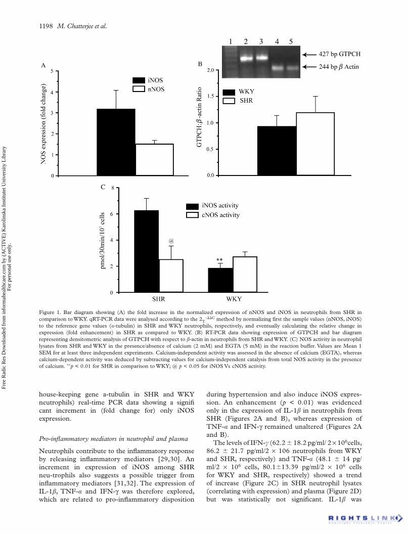
1198 M. Chatterjee et al.
Figure 1. Bar diagram showing (A) the fold increase in the normalized expression of nNOS and iNOS in neutrophils from SHR in comparison to WKY. qRT-PCR data were analysed according to the 2T
– C method by normalizing first the sample values (nNOS, iNOS) to the reference gene values ( -tubulin) in SHR and WKY neutrophils, respectively, and eventually calculating the relative change in expression (fold enhancement) in SHR as compared to WKY. (B) RT-PCR data showing expression of GTPCH and bar diagram representing densitometric analysis of GTPCH with respect to -actin in neutrophils from SHR and WKY. (C) NOS activity in neutrophil lysates from SHR and WKY in the presence/absence of calcium (2 mM) and EGTA (5 mM) in the reaction buffer. Values are Mean 1 SEM for at least three independent experiments. Calcium-independent activity was assessed in the absence of calcium (EGTA), whereas calcium-dependent activity was deduced by subtracting values for calcium-independent catalysis from total NOS activity in the presence of calcium. p < 0.01 for SHR in comparison to WKY; @ p < 0.05 for iNOS Vs cNOS activity.
Free
Rad
ic R
es D
ownl
oade
d fr
om in
form
ahea
lthca
re.c
om b
y (A
CT
IVE
) K
arol
insk
a In
stitu
tet U
nive
rsity
Lib
rary
Fo
r pe
rson
al u
se o
nly.
house-keeping gene a-tubulin in SHR and WKY neutrophils) real-time PCR data showing a significant increment in (fold change for) only iNOS expression.
Pro-inflammatory mediators in neutrophil and plasma
Neutrophils contribute to the inflammatory response by releasing inflammatory mediators [29,30]. An increment in expression of iNOS among SHR neu-trophils also suggests a possible trigger from inflammatory mediators [31,32]. The expression of IL-1 , TNF- and IFN- was therefore explored, which are related to pro-inflammatory disposition
during hypertension and also induce iNOS expres-sion. An enhancement (p < 0.01) was evidenced only in the expression of IL-1 in neutrophils from SHR (Figures 2A and B), whereas expression of TNF- and IFN- remained unaltered (Figures 2A and B).
The levels of IFN- (62.2 18.2 pg/ml/ 2 106cells, 86.2 21.7 pg/ml/2 106 neutrophils from WKY and SHR, respectively) and TNF- (48.1 14 pg/ml/2 106 cells, 80.1 13.39 pg/ml/2 106 cells for WKY and SHR, respectively) showed a trend of increase (Figure 2C) in SHR neutrophil lysates (correlating with expression) and plasma (Figure 2D) but was statistically not significant. IL-1 was
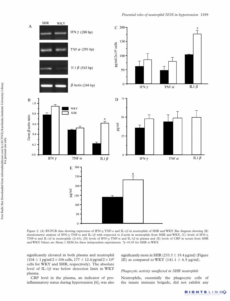
Potential roles of neutrophil NOS in hypertension 1199
Figure 2. (A) RT-PCR data showing expression of IFN- , TNF- and IL-1 in neutrophils of SHR and WKY. Bar diagram showing (B) densitometric analysis of IFN- , TNF- and IL-1 with respected to -actin in neutrophils from SHR and WKY, (C) levels of IFN- ,TNF- and IL-1 in neutrophils (2 10), (D) levels of IFN- , TNF- and IL-1 in plasma and (E) levels of CRP in serum from SHR and WKY. Values are Mean 1 SEM for three independent experiments. p <0.05 for SHR vs WKY.
Free
Rad
ic R
es D
ownl
oade
d fr
om in
form
ahea
lthca
re.c
om b
y (A
CT
IVE
) K
arol
insk
a In
stitu
tet U
nive
rsity
Lib
rary
Fo
r pe
rson
al u
se o
nly.
significantly elevated in both plasma and neutrophil (104 1 pg/ml/2 106 cells, 177 12.4 pg/ml/2 106
cells for WKY and SHR, respectively). The absolute level of IL-1 was below detection limit in WKY plasma.
CRP level in the plasma, an indicator of pro-inflammatory status during hypertension [6], was also
significantly more in SHR (233.3 19.4 g/ml) (Figure 2E) as compared to WKY (141.1 6.5 g/ml).
Phagocytic activity unaffected in SHR neutrophils
Neutrophils, essentially the phagocytic cells of the innate immune brigade, did not exhibit any
1200 M. Chatterjee et al.
Free
Rad
ic R
es D
ownl
oade
d fr
om in
form
ahea
lthca
re.c
om b
y (A
CT
IVE
) K
arol
insk
a In
stitu
tet U
nive
rsity
Lib
rary
Fo
r pe
rson
al u
se o
nly.
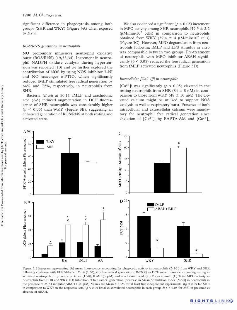
significant difference in phagocytosis among both groups (SHR and WKY) (Figure 3A) when exposed to E.coli.
ROS/RNS generation in neutrophils
NO profoundly influences neutrophil oxidative burst (ROS/RNS) [19,33,34]. Increment in neutro-phil NADPH oxidase catalysis during hyperten-sion was reported [13] and we further explored the contribution of NOS by using NOS inhibitor 7-NI and NO scavenger c-PTIO, which significantly reduced fMLP stimulated free radical generation by 64% and 72%, respectively, in neutrophils from SHR.
Bacteria (E.coli at 50:1), fMLP and arachidonic acid (AA) induced augmentation in DCF fluores-cence of SHR neutrophils was considerably higher (p 0.05) than WKY (Figure 3B), suggesting an enhanced generation of ROS/RNS at both resting and activated state.
Figure 3. Histogram representing (A) mean fluorescence accountingfollowing challenge with FITC-labelled E.coli (1:50), (B) free radicalactivated neutrophils in presence of E.coli (1:50), fLMP (3 M) anneutrophils from SHR and WKY. (D) Inhibition of free radical generthe presence of MPO inhibitor ABAH (100 M). Values are Mean in comparison to WKY in the respective sets, p < 0.05 basal vs stimuabsence of ABAH.
We also evidenced a significant (p 0.05) increment in MPO activity among SHR neutrophils (59.3 2.2( M/min/107 cells) in comparison to neutrophils obtained from WKY (39.4 4 M/min/107 cells) (Figure 3C). However, MPO degranulation from neu-trophils following fMLP and LPS stimulus in vitro was comparable between two groups. Pre-treatment of neutrophils with MPO inhibitor ABAH signifi-cantly (p < 0.05) reduced the free radical generation from fMLP activated neutrophils (Figure 3D).
Intracellular [Ca2 1Ji in neutrophils
[Ca2 ]i was significantly (p < 0.05) elevated in the resting neutrophils from SHR (84 8 nM) in com-parison to those from WKY (48 10 nM). The ele-vated calcium might be utilized to support NOS catalysis as well as respiratory burst. Presence of both intracellular and extracellular calcium were manda-tory for neutrophil free radical generation since chelation of [Ca2 ]i by BAPTA-AM and [Ca2 ]e
for phagocytic activity in neutrophils (2 10 ) from WKY and SHR generation (ONOO–) as DCF mean fluorescence among resting vs d arachidonic acid (2 M) as stimuli. (C) Total MPO activity in
ation [decrease in Mean Stimulation Index (MSI)] in neutrophils in SEM for at least five independent experiments. #p < 0.05 for SHR lated neutrophils in each group. & p < 0.05 for MSI in presence vs
Potential roles of neutrophil NOS in hypertension 1201
Free
Rad
ic R
es D
ownl
oade
d fr
om in
form
ahea
lthca
re.c
om b
y (A
CT
IVE
) K
arol
insk
a In
stitu
tet U
nive
rsity
Lib
rary
Fo
r pe
rson
al u
se o
nly.
by EGTA reduced the fMLP activated free radical formation by 74% and 67%, respectively, in SHR and 79% and 73%, respectively, in WKY.
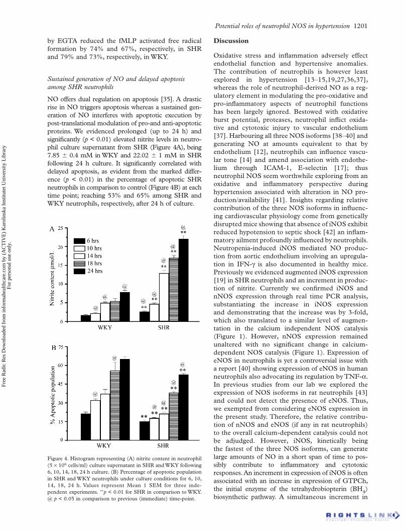
Sustained generation of NO and delayed apoptosis among SHR neutrophils
NO offers dual regulation on apoptosis [35]. A drastic rise in NO triggers apoptosis whereas a sustained gen-eration of NO interferes with apoptotic execution by post-translational modulation of pro-and anti-apoptotic proteins. We evidenced prolonged (up to 24 h) and significantly (p < 0.01) elevated nitrite levels in neutro-phil culture supernatant from SHR (Figure 4A), being 7.85 0.4 mM in WKY and 22.02 1 mM in SHR following 24 h culture. It significantly correlated with delayed apoptosis, as evident from the marked differ-ence (p < 0.01) in the percentage of apoptotic SHR neutrophils in comparison to control (Figure 4B) at each time point; reaching 53% and 65% among SHR and WKY neutrophils, respectively, after 24 h of culture.
Figure 4. Histogram representing (A) nitrite content in neutrophil(5 106 cells/ml) culture supernatant in SHR and WKY following 6, 10, 14, 18, 24 h culture. (B) Percentage of apoptotic population in SHR and WKY neutrophils under culture conditions for 6, 10, 14, 18, 24 h. Values represent Mean 1 SEM for three inde-pendent experiments. p < 0.01 for SHR in comparison to WKY. @ p < 0.05 in comparison to previous (immediate) time-point.
Discussion
Oxidative stress and inflammation adversely effect endothelial function and hypertensive anomalies. The contribution of neutrophils is however least explored in hypertension [13–15,19,27,36,37], whereas the role of neutrophil-derived NO as a reg-ulatory element in modulating the pro-oxidative and pro-inflammatory aspects of neutrophil functions has been largely ignored. Bestowed with oxidative burst potential, proteases, neutrophil inflict oxida-tive and cytotoxic injury to vascular endothelium [37]. Harbouring all three NOS isoforms [38–40] and generating NO at amounts equivalent to that by endothelium [12], neutrophils can influence vascu-lar tone [14] and amend association with endothe-lium through ICAM-1, E-selectin [17]; thus neutrophil NOS seem worthwhile exploring from an oxidative and inflammatory perspective during hypertension associated with alteration in NO pro-duction/availability [41]. Insights regarding relative contribution of the three NOS isoforms in influenc-ing cardiovascular physiology come from genetically disrupted mice showing that absence of iNOS exhibit reduced hypotension to septic shock [42] an inflam-matory ailment profoundly influenced by neutrophils. Neutropenia-induced iNOS mediated NO produc-tion from aortic endothelium involving an upregula-tion in IFN- is also documented in healthy mice. Previously we evidenced augmented iNOS expression [19] in SHR neutrophils and an increment in produc-tion of nitrite. Currently we confirmed iNOS and nNOS expression through real time PCR analysis, substantiating the increase in iNOS expression and demonstrating that the increase was by 3-fold, which also translated to a similar level of augmen-tation in the calcium independent NOS catalysis (Figure 1). However, nNOS expression remained unaltered with no significant change in calcium-dependent NOS catalysis (Figure 1). Expression of eNOS in neutrophils is yet a controversial issue with a report [40] showing expression of eNOS in human neutrophils also advocating its regulation by TNF- .In previous studies from our lab we explored the expression of NOS isoforms in rat neutrophils [43] and could not detect the presence of eNOS. Thus, we exempted from considering eNOS expression in the present study. Therefore, the relative contribu-tion of nNOS and eNOS (if any in rat neutrophils) to the overall calcium-dependent catalysis could not be adjudged. However, iNOS, kinetically being the fastest of the three NOS isoforms, can generate large amounts of NO in a short span of time to pos-sibly contribute to inflammatory and cytotoxic responses. An increment in expression of iNOS is often associated with an increase in expression of GTPCh, the initial enzyme of the tetrahydrobiopterin (BH4)biosynthetic pathway. A simultaneous increment in
1202 M. Chatterjee et al.
Free
Rad
ic R
es D
ownl
oade
d fr
om in
form
ahea
lthca
re.c
om b
y (A
CT
IVE
) K
arol
insk
a In
stitu
tet U
nive
rsity
Lib
rary
Fo
r pe
rson
al u
se o
nly.
the expression of GTPCH when iNOS expression is enhanced ensures ready availability of NOS cofactor BH4 to ensure effective catalysis. We there-fore set to determine the expression of GTPCH in SHR neutrophils, noting an increment in the expression of iNOS. Previously a decline in expres-sion of GTPCH expression was documented in the adrenal cortex of SHR, notwithstanding an upregu-lation in iNOS expression [44]. Presently we evi-denced that expression of GTPCH remained unchanged despite augmented iNOS expression in SHR neutrophils. iNOS over-expression coupled with inadequate BH4 has been suggested to con-tribute to development of hypertension in SHR, furthermore chronic supplementation of BH4 at a pre-hypertensive stage was shown to suppress development of hypertension in SHR [44]. This might cause a limiting situation for BH4 to support NOS catalysis and therefore might bring about uncoupling of NOS, therefore causing generation of superoxide instead of NO as reported previously in endothelial cells. Future experiments could be directed towards determination of the absolute levels of BH4 in WKY and SHR neutrophils and explore the possibility of NOS uncoupling, possibly owing to limiting BH4 availability in SHR neutrophils.
Enhancement of iNOS expression among SHR neu-trophils [19] predictably reflects a pro-inflammatory trigger [34,36]. These cytokines suffice indepen-dently or act synergistically to increase iNOS tran-scription depending on the cytokine response profile of different cells. We evidenced a significant elevation in the expression and levels of IL-1 in neutrophils and plasma from SHR, which is presumed to trigger transcription of iNOS.
Phagocytosis acts as a trigger for respiratory burst and an alteration at the levels of phagocytic uptake could account for a change in respiratory burst potential as well. However, the present data nullifies such possibilities. We did not encounter any changes in the phagocytic potential or the extent of bacterial uptake in neutrophils from SHR and WKY groups. Change in the extent of free radical generation or respiratory burst could possibly be due to changes at the level of NADPH oxidase system and also the contribution of NOS uncoupling [10]. It is well documented that superoxide released by neutrop-hils not only serves for inflammatory immune response but also inflicts vascular dysregulation [13–15,19,27,36,37]. We project the possibility of NOS and NO mediated modulation of oxidative burst in SHR neutrophils contemplated from 7-NI and cPTIO affected decline in superoxide generation from activated neutrophils. Enhanced eNOS expres-sion in endothelium was previously documented to support superoxide generation over NO synthesis [10]. SHR neutrophils presumably pose similar
possibilities, having significant increments in iNOS expression.
A simultaneous augmentation in NO and super-oxide generation [19] and the resultant production of peroxynitrite in SHR neutrophils scavenged the NO that otherwise could have been implemented for relaxation [14], preventing platelet aggregation in the near vicinity [12,16] or in preventing adverse en-dothelium-neutrophil interactions [17]. DCFD H2DA employed in the present flow cytometric analy-sis of neutrophils has the inherent property to detect the generation of ROS and also RNS, notably ONOO–.Previous studies have utilized this property of DCFD-H2DA to show generation of ONOO– in neutrophil-induced endothelial cell activation [36] which we contemplated could be investigated in neutrophils themselves. Currently we report an elevation of ONOO– generation in neutrophils themselves suggest-ing the possibility that neutrophil derived ONOO–
can inflict oxidative damage to target tissues, offer potentially innumerable post-translational modifica-tions of proteins affecting cardiovascular physiology [45]. ONOO– liberated from neutrophils might also contribute to the elevated levels of stable NO metabo-lites in plasma, as reported earlier in SHR [19]. Therefore, enhanced levels of NO in SHR neutrophils add to free radical load by generating secondary oxi-dizing and nitrating species, which might influence redox signalling events in vascular smooth muscle cells, affecting their growth, differentiation, apoptosis and secretion of inflammatory mediators [46].
Currently we also deciphered the possible contri-bution of intracellular calcium and MPO in adding to the oxidative mechanisms of neutrophils. In animal models and in hypertensive subjects, increased excretion of calcium in urine was reported [27]. Further correlation between BP levels and enhanced neutrophil [Ca2+]i was also suggested [27]. We evi-denced significant elevation of [Ca2+]i in SHR neu-trophils and as presumed both [Ca2+]i and [Ca2+]ewere mandatory to instigate an oxidative burst and also support calcium-dependent NOS catalysis. Now whether a therapeutic approach involving calcium modulators in hypertension could check the oxidative pandemonium of neutrophils needs to be assessed. On the other hand an increment in MPO repertoire among SHR neutrophils projects the possibility of inflicting oxidative injury at target sites upon chemo-tactic neutrophil infiltration or transcytosis of MPO into the aortic endothelium or target organs. MPO might also serve as a marker of neutrophil infiltration to target organs. Lack of detectable difference in degranulation in vitro, however, cannot nullify poten-tial stimuli in vivo that would trigger degranulation of the significantly large MPO reservoir in SHR as com-pared to controls. A reduction in free radical genera-tion from neutrophils in the presence of ABAH also suggests MPO involvement.

Potential roles of neutrophil NOS in hypertension 1203
Free
Rad
ic R
es D
ownl
oade
d fr
om in
form
ahea
lthca
re.c
om b
y (A
CT
IVE
) K
arol
insk
a In
stitu
tet U
nive
rsity
Lib
rary
Fo
r pe
rson
al u
se o
nly.
Effective apoptotic clearance of neutrophils is as crucial as their microbicidal actions for resolution of inflammation. The most common way of detecting or studying neutrophil apoptosis is to either determine the exposure of phosphatidyl serine by means of Annexin V or the degradation of DNA by DNA inter-calating agents such and PI. Since exposure of PS is an early event in neutrophil apoptosis and we intended to study later time points, we chose the latter, which is sufficient to show apoptotic neutrophil percentage by monitoring the sub-diploid peak in PI histogram analysis by flow cytometry.
Previously we documented enhanced level of total and protein associated nitrite in neutrophil lysates which reflects the level or reservoir of NO metabolites in SHR and WKY neutrophils. In the current study we have evaluated the extent of NO generation from SHR and WKY neutrophils under culture condition by evaluating nitrite content not in the neutrophil lysate as before but in the neutrophil culture super-natant where it is released by the neutrophils. Nitrite content does not indicate the real time NO generation as DAF but it indicates the cumulative or accumula-tive NO generated during the period the neutrophils have been cultured. Therefore this total nitrite level is an accumulation of nitrite generated by neutrophils since the culture was seeded. We suggest that these prolonged and higher levels of NO generation in SHR neutrophils as compared to the WKY neutrophils could block apoptosis, as suggested in other studies as well. However, to prove the absolute involvement of NO in regulating apoptosis as in this study would need the presence of NOS inhibitors in culture. Then again, isoform-specific NOS inhibition is a debated and challenging issue as the inhibitors differ in their relative specificity and potential to inhibit a specific NOS isoform. Furthermore, neutrophils have a short life-span and the vehicle (as DMSO for 7-NI which is known to inhibit both iNOS and cNOS at higher concentrations like 1 mM) of NOS inhibitor could also affect the survival potential of these granulocytes. However, alternative approaches to silent NOS activ-ity in neutrophils could be thought of and accom-plished in future studies. Nevertheless, this in vivo would imply that neutrophils having fortified oxida-tive burst potential survive longer in circulation and promote pro-inflammatory process during hyperten-sion to predispose hypertensive subjects to oxidative and inflammatory adversities. Results obtained indicate the augmented inflammatory mediators and neutrophil mediated increase in free radical genera-tion in SHR.
Conclusion
This study hypothesizes that the augmentation in NO generation from neutrophils of SHR possibly promote
oxidative/nitrosative damage and inflammatory insult. Exploration of the role of neutrophil NOS in SHR might provide helpful insight into the understanding of the pathology of hypertension.
Acknowledgeement
We acknowledge the excellent technical assistance of Mr. A.L. Vishwakarma and Ms. M. Chaturvedi for the flow cytometric studies. The Senior Research Fel-lowship from the Council of Scientific and Industrial Research, New Delhi, India to RS and Indian Coun-cil of Medical Research, New Delhi, India to MC is acknowledged. This study was supported by a finan-cial grant to MD from the Department of Biotechnol-ogy, New Delhi, India.
Declaration of interest: The author reports no con-flicts of interest. The author alone is responsible for the content and writing of the paper.
References
Grundy SM. Inflammation, hypertension, and the metabolic [1]syndrome. JAMA 2003;290:3000–3002.Dhalla NS, Temsah RM, Netticadan T. Role of oxidative stress [2]in cardiovascular diseases. J Hypertens 2000;18:655–673.Ferroni P, Basili S, Paoletti V, Davi G. Endothelial dysfunc-[3]tion and oxidative stress in arterial hypertension. Nutr Metab Cardiovasc Dis 2006;16:222–233.Li JJ, Chen JL. Inflammation may be a bridge connecting [4]hypertension and atherosclerosis. Med Hypotheses 2005;64: 925–929.Olzinski AR, McCafferty TA, Zhao SQ, Behm DJ, Eybye ME, [5]Maniscalco K, Bentley R, Frazier KS, Milliner CM, Mirabile RC, Coatney RW, Willette RN. Hypertensive target organ damage is attenuated by a p38 MAPK inhibitor:Role of sys-temic blood pressure and endothelial protection. Cardiovasc Res 2005;66:170–178.Paffen E, deMaat MPM. C-reactive protein in atherosclerosis: [6]a causal factor? Cardiovasc Res 2006;71:30–39.Grunfeld S, Hamilton CA, Mesaros S, McClain SW, [7]Dominiczak AF, Bohr DF, Malinski T. Role of superoxide in the depressed nitric oxide production by the endothe-lium of genetically hypertensive rats. Hypertension 1995;26: 854–857.Hegde LG, Srivastava P, Kumari R, Dikshit M. Alterations in [8]the vasoreactivity of hypertensive rat aortic rings: role of nitric oxide and superoxide radicals. Clin Exp Hypertension 1998;20:885–901.Kerr S, Brosnan MJ, McIntyre M, Reid JL, Dominiczak AF, [9]Hamilton CA. Superoxide anion production is increased in a model of genetic hypertension: role of the endothelium. Hypertension 1999;33:1353–1358.Bouloumie A, Bauersachs J, Ling W, Scholkens BA, Wiemer [10]G, Fleming I, Busse R. Endothelial dysfunction coincides with an enhanced nitric oxide synthase expression and super-oxide anion production. Hypertension 1997;30:934–941.Ulker S, McMaster D, McKeown PP, Bayraktutan U. [11]Impaired activities of antioxidant enzymes elicit endothelial dysfunction in spontaneous hypertensive rats despite enhanced vascular nitric oxide generation. Cardiovasc Res 2003;59:488–500.

1204 M. Chatterjee et al.
Free
Rad
ic R
es D
ownl
oade
d fr
om in
form
ahea
lthca
re.c
om b
y (A
CT
IVE
) K
arol
insk
a In
stitu
tet U
nive
rsity
Lib
rary
Fo
r pe
rson
al u
se o
nly.
Salvemini D, de Nucci G, Gryglewski RJ, Vane JR. Human [12]neutrophils and mononuclear cells inhibit platelet aggrega-tion by releasing a nitric oxide-like factor. Proc Natl Acad Sci USA 1989;86:6328–6332.Kristal B, Shurtz-Swirski R, Chezar J, Manaster J, Levy R, [13]Shapiro G, Weissman I, Shasha SM, Sela S. Participation of peripheral polymorphonuclear leukocytes in the oxidative stress and inflammation in patients with essential hyperten-sion. Am J Hypertens 1998;11:921–928.Morton J, Coles B, Wright K, Gallimore A, Morrow JD, Terry [14]ES, Anning PB, Morgan BP, Dioszeghy V, Kiihn H, Chaitidis P, Hobbs AJ, Jones SA, O’Donnell VB. Circulating neutrophils maintain physiological blood pressure by suppressing bacteria and IFNgamma-dependent iNOS expression in the vascula-ture of healthy mice. Blood 2008;111:5187–5194.Jackson MH, Collier A. Neutrophil count and activation in [15]vascular disease. Scot Med J 1992;37:41–43.Nicolini FA, Mehta JL. Inhibitory effect of unstimulated neu-[16]trophils on platelet aggregation by release of a factor similar to endothelium-derived relaxing factor (EDRF). Biochem Pharmacol 1990;40:2265–2269.Kosonen O, Kankaanranta H, Uotila J, Moilanen E. Inhi-[17]bition by nitric oxide-releasing compounds of E-selectin expression in and neutrophil adhesion to human endothelial cells. Eur J Pharmacol 2000;394:149–156.Sethi S, Dikshit M. Modulation of polymorphonuclear [18]leukocytes function by nitric oxide. Thromb Res 2000;100: 223–247.Chatterjee M, Saluja R, Kanneganti S, Chinta S, Dikshit M. [19]Biochemical and molecular evaluation of neutrophil NOS in spontaneously hypertensive rats. Cell Mol Biol 2007;53: 84–92.Herrmann G, Hlushchuk R, Baum O, Scotti AL. Nitric oxide [20]synthase protein levels, not the mRNA, are down-regulated in olfactory bulb interneurons of reeler mice. J Chem Neuro-anat 2007;33:87–96.Sharma P, Barthwal MK, Dikshit M. NO synthesis and its [21]regulation in the arachidonic-acid-stimulated rat polymor-phonuclear leukocytes. Nitric Oxide 2002;7:119–126.Pachori AS, Melo LG, Hart ML, Noiseux N, Zhang L, [22]Morello F, Solomon SD, Stahl GL, Pratt RE, Dzau VJ. Hypoxia-regulated therapeutic gene as a preemptive treat-ment strategy against ischemia/reperfusion tissue injury. Proc Natl Acad Sci USA 2004;101:12282–12287.Dejucq N, Dugast I, Ruffault A, van der Meide PH, Jegou B. [23]Interferon- and expression in the rat testis. Endocrinology 1995;136:4925–4931.van Tol EAF, Holt L, Li FL, Kong FM, Rippe R, Yamauchi M, [24]Pucilowska J, Lund PK, Sartor RB. Bacterial cell wall poly-mers promote intestinal fibrosis by direct stimulation of myofibroblasts. Am J Physiol Gastrointest Liver Physiol 1999;277:G245–G255.Bredius RGM, de vries EE, Troelslral A, Alphen LV, Weening [25]RS, Van de Winkle J, Out TA. Phagocytosis of Staphylococ-cus aureus and Hemophilus influenze Type B opsonized by poly-clonal Human IgGl and IgG2 antibodies. J Immunol 1993;151:1463–1472.Graff G, Gamache DA, Brady MT, Spellman JM, Yanni JM. [26]Improved myeloperoxidase assay for quantitation of neutro-phil influx in a rat model of endotoxin-induced uveitis. J Phar-macol Toxicol Meth 1998;39:169–178.Sela S, Shurtz-Swirski R, Farah R, Levy R, Shapiro G, Chezar [27]J, Shasha SM, Kristal B. A link between polymorphonuclearleukocyte intracellular calcium, plasma insulin, and essential hypertension. Am J Hypertens 2002;15:291–295.Hur J, Kang MK, Park JY, Lee SY, Bae YS, Lee SH, Park YM, [28]Kwak JY. Pro-apoptotic effect of high concentrations of his-tamine on human neutrophils. Int Immunopharmacol 2003;3:1491–1502.
This paper was first published online on Early Online on 21 September 2009.
Kato T, Kitagawa S. Regulation of neutrophil functions [29]by proinflammatory cytokines. Int J Hematol 2006;84:205–209.Nathan C. Inducible nitric oxide synthase: what difference [30]does it make? J Clin Invest 1997;100:2417–2423.Egan LJ, Mays DC, Huntoon CJ, Bell MP, Pike MG, [31]Sandborn WJ, Lipsky JJ, McKean DJ. Inhibition of inter-leukin-1-stimulated NF-kappaB RelA/p65 phosphorylation by mesalamine is accompanied by decreased transcriptional activity. J Biol Chem 1999;274:26448–26453.Lorsbach RB, Murphy W, Raval P, Snowman AM, Snyder SH, [32]Russell SW Expression of nitric oxide synthase gene in mouse macrophages activated for tumor cell killing. Molecular basis for synergy between interferon-y and liposacchar-ide. J Biol Chem 1993;268:1908–1913.Sethi S, Singh MP, Dikshit M. Nitric oxide-mediated aug-[33]mentation of polymorphonuclear free radical generation after hypoxia-reoxygenation. Blood 1999;93:333–340.Seth P, Kumari R, Dikshit M, Srimal RC. Modulation of rat [34]peripheral polymorphonuclear leukocyte response by nitric oxide and arginine. Blood 1994;84:2741–2748.Kim PK, Zamora R, Petrosko P, Billiar TR. The regulatory [35]role of nitric oxide in apoptosis. Int Immunopharmacol 2001;1:1421–1441.Sohn HY, Krotz F, Zahler S, Gloe T, Keller M, Theisen K, [36]Schiele TM, Klauss V, Pohl U Crucial role of local peroxyni-trite formation in neutrophil-induced endothelial cell activa-tion. Cardiovasc Res 2003;57:804–815.Ofosu-Appiah W, Sfeir G, Smith D, Richard T. Neutrophil-[37]mediated damage to vascular endothelium in the sponta-neously hypertensive rat. Clin Immunol Immunopathol 1997;83:293–301.Cedergren J, Follin P, Forslund T, Lindmark M, [38]Sundqvist T, Skogh T Inducible nitric oxide synthase (NOS II) is constitutive in human neutrophils. APMIS 2003;111: 963–968.Greenberg SS, Ouyang J, Zhao X, Giles TD. Human [39]and rat neutrophils constitutively express neural nitric oxide synthase mRNA. Nitric Oxide 1998;2:203–212.de Frutos, Sanchez dM, Farre J, Gomez J, Romero J, [40]Marcos-Alberca P, Nunez A, Rico L, Lopez-Farre A. Expres-sion of an endothelial-type nitric oxide synthase isoform in human neutrophils: modification by tumor necrosis factor-alpha and during acute myocardial infarction. J Am Coll Cardiol 2001;37:800–807.Clapp BR, Hingorani AD, Kharbanda RK, Mohamed-Ali V, [41]Stephens JW, Vallance P, MacAllister RJ. Inflammation-induced endothelial dysfunction involves reduced nitric oxide bioavailability and increased oxidant stress. Cardiovasc Res 2004;64:172–178.Liu VWT, Huang PL. Cardiovascular roles of nitric oxide: A [42]review of insights from nitric oxide synthase gene disrupted mice. Cardiovasc Res 2008;77:19–29.Saini R, Patel S, Saluja R, Sahasrabuddhe AA, Singh MP, [43]Habib S, Bajpai VK, Dikshit M. Nitric oxide synthase localization in the rat neutrophils: immunocytochemical, molecular, and biochemical studies. J Leukoc Biol 2006;79: 519–528.Hong HJ, Hsiao G, Cheng TH, Yen MH. Supplementation [44]with tetrahydrobiopterin suppresses the development of hypertension in spontaneously hypertensive rats. Hyperten-sion 2001;38:1044–1048.Peluffo G, Radi R. Biochemistry of protein tyrosine nitra-[45]tion in cardiovascular pathology. Cardiovasc Res 2007;75:291–302.Clempus RE, Griendling KK. Reactive oxygen species signal-[46]ing in vascular smooth muscle cells. Cardiovasc Res 2006;71:216–225.