T cell homeostatic proliferation elicits effective antitumor autoimmunity

ATP Released by Electrical Stimuli Elicits Calcium Transientsand Gene Expression in Skeletal Muscle*
Received for publication, August 18, 2009, and in revised form, October 5, 2009 Published, JBC Papers in Press, October 12, 2009, DOI 10.1074/jbc.M109.057315
Sonja Buvinic‡, Gonzalo Almarza‡, Mario Bustamante‡, Mariana Casas‡, Javiera Lopez§, Manuel Riquelme¶,Juan Carlos Saez¶, Juan Pablo Huidobro-Toro§, and Enrique Jaimovich‡1
From the ‡Centro de Estudios Moleculares de la Celula, Facultad de Medicina, Instituto de Ciencias Biomedicas, Universidad deChile, Casilla 70005, Santiago 7, Chile and the §Centro de Regulacion Celular y Patología and ¶Departamento de CienciasFisiologicas, Facultad de Ciencias Biologicas, Pontificia Universidad Catolica de Chile, Casilla 114-D, Santiago 8, Chile
ATP released from cells is known to activate plasma mem-brane P2X (ionotropic) or P2Y (metabotropic) receptors. Inskeletalmuscle cells, depolarizing stimuli induce both a fast cal-cium signal associated with contraction and a slow signal thatregulates gene expression. Here we show that nucleotidesreleased to the extracellular medium by electrical stimulationare partly involved in the fast component and are largely respon-sible for the slow signals. In rat skeletalmyotubes, a tetanic stim-ulus (45 Hz, 400 1-ms pulses) rapidly increased extracellularlevels of ATP, ADP, and AMP after 15 s to 3 min. ExogenousATP induced an increase in intracellular free Ca2� concentra-tion, with an EC50 value of 7.8 � 3.1 �M. Exogenous ADP, UTP,and UDP also promoted calcium transients. Both fast and slowcalcium signals evoked by tetanic stimulation were inhibited byeither 100 �M suramin or 2 units/ml apyrase. Apyrase alsoreduced fast and slow calcium signals evoked by tetanus (45 Hz,400 0.3-ms pulses) in isolated mouse adult skeletal fibers. Alikely candidate for the ATP release pathway is the pannexin-1hemichannel; its blockers inhibited both calcium transients andATPrelease.Thedihydropyridine receptor co-precipitatedwithboth the P2Y2 receptor and pannexin-1. As reported previouslyfor electrical stimulation, 500 �M ATP significantly increasedmRNA expression for both c-fos and interleukin 6. Our resultssuggest that nucleotides released during skeletalmuscle activitythrough pannexin-1 hemichannels act through P2X and P2Yreceptors to modulate both Ca2� homeostasis and musclephysiology.
Activation of skeletal muscle cells promotes a contractileresponse through “excitation-contraction” coupling. This is acomplex process that allows coupling between membranedepolarization and Ca2� release from the sarcoplasmic reticu-lum, inducing a fast increase in intracellular free Ca2� levelsthat allows interactions of filaments required for muscle con-traction. Two major proteins account for the excitation-con-traction coupling process: the dihydropyridine receptor
(DHPR),2 located in transverse (T) tubules of the plasmamem-brane, and the ryanodine receptor (RyR), a Ca2� channel pres-ent in the sarcoplasmic reticulum. In skeletal muscles, DHPRacts both as a voltage sensor and a slow L-type Ca2� channel(Cav1.1). Action potential propagation through muscle fiberspromotes a transientT-tubulemembrane depolarization, evok-ing conformational changes of the DHPR that are transmittedto the RyR, which opens and releases calcium to the cytosol forcontraction development (1–4).In addition to taking part in contraction, intracellular free
Ca2� controls different muscle cells processes, including met-abolic pathway activation, differentiation, hypertrophy, andgene expression (5–7). We have previously shown that tetanicelectrical stimulation of skeletalmyotubes evokes a fast calciumtransient during stimulation and a slow calcium transient thatpeaks 60–100 s later. The fast signal is blocked by ryanodine,and the slow component is dependent on 1,4,5-inositoltrisphosphate (IP3) generation and Ca2� release from the sar-coplasmic reticulum through IP3 receptors (8, 9). We havedemonstrated that the slow calcium signal is dependent onDHPR activation, G�� complex release, phosphoinositide 3-ki-nase and phospholipase C activation, and IP3 increases (8–10).The slow calcium transient evoked by membrane depolariza-tion has been associated with nuclear Ca2� accumulationrelated to expression of early genes (5) and with activation oftranscription pathways and expression of a number of skeletalmuscle genes (11–13). Slow calcium signals have been assigneda role in the “excitation-transcription” process, which has pos-sible effects on and implications for cell homeostasis mainte-nance. This process may shed light on muscle cell plasticityinduced by physiological activity.Mediators between DHPR activation and the G�� complex
remain unknown.One option is that theG�� complex interactsdirectly with DHPR and could be released after its activation. Ithas been shown that voltage-activatedCa2� channels (L-, P/Q-,N-, andR-types) have binding sites forG�� (14–16). Activationof these channels through G�� has been demonstrated, but theopposite process, i.e. release of G�� by the channel, has not
* This work was supported by Grants from the Fondo de Investigacion Avan-zada en Areas Prioritarias (Chile) 15010006, and the Fondo Nacional deDesarrollo Cientifico y Tecnologico (Chile) 1080120, FONDAP 13980001,and FONDECYT 3080016.
1 To whom correspondence should be addressed: Inst. de Ciencias Biomedi-cas, Facultad de Medicina, Universidad de Chile, Casilla 70005, Santiago 7,Chile. E-mail: [email protected].
2 The abbreviations used are: DHPR, dihydropyridine receptor; �-ATP, etheno-ATP; ERK, extracellular signal-regulated kinase; FDB, flexor digitorum bre-vis; fluo3-AM, fluo3-acetoxymethylester; GAPDH, glyceraldehyde-3-phos-phate dehydrogenase; GPCR, G protein-coupled receptor; HPLC, highpressure liquid chromatography; IL, interleukin; IP3, 1,4,5-inositol trisphos-phate; RT, reverse transcription; RyR, ryanodine receptor; T, transverse;Pnx, pannexin.
THE JOURNAL OF BIOLOGICAL CHEMISTRY VOL. 284, NO. 50, pp. 34490 –34505, December 11, 2009© 2009 by The American Society for Biochemistry and Molecular Biology, Inc. Printed in the U.S.A.
34490 JOURNAL OF BIOLOGICAL CHEMISTRY VOLUME 284 • NUMBER 50 • DECEMBER 11, 2009
at HA
RV
AR
D U
NIV
ER
SIT
Y, on D
ecember 5, 2009
ww
w.jbc.org
Dow
nloaded from

been confirmed. Another possibility for DHPR is that it eitheractivates a G protein-coupled receptor (GPCR) directly or fos-ters the release of a ligand for such receptors that will activatethe pathway of heteromeric G�/G��. This is a likely possibility,because a slow calcium signal takes several seconds to develop,suggesting the activation of signaling pathways instead of directinteractions. It is important to note that in physiological condi-tions, the skeletal muscle is subjected simultaneously to manystimuli, including membrane depolarization, as well as hor-mones, metabolites, and extracellular molecules that can acti-vate GPCRs and control cellular processes. It is interesting thento analyze the possible interactions between voltage sensors(such as DHPR) and different GPCRs as a basic mechanismpossibly involved in the modulation of muscle plasticity.Extracellular ATP, a co-transmitter in adrenergic and cho-
linergic synapses, has several functions in the central andperipheral nervous systems (17, 18). In addition, autocrine/paracrine ATP signaling occurs in many cell types (epithelium,endothelium, fibroblasts, etc.) that release nucleotides (ATP,UTP) to the extracellularmediumunder resting conditions andafter physiopathological events, such as hypoxia, cell swelling,shear stress, or inflammation, through nonlytic mechanisms(19, 20). Depending on the cell type, ATP release by exocytosisor through ABC transporters, stretch- and voltage-activatedchannels, or connexin hemichannels has been described (19–21). Recently, pannexin (Pnx) hemichannels have also beenproposed as relevant ATP conduits (22, 23). The ATP (or UTP)released is quickly metabolized to the di- and monophosphatenucleotides through ectonucleotidases located at the cell sur-face or the extracellular matrix. Finally, they are converted intonucleosides to be recaptured by cells (24). ATP, UTP, and theirmetabolites have been implicated in the regulation of physio-logical processes such as epithelial secretion, vascular tone,platelet aggregation, pain sensitivity, and actions as trophic fac-tors (20, 25–29).There are two main families of receptors for extracellular
nucleotides: the P2X receptors, which are ion channels acti-vated by purine triphosphate nucleotides; and the P2Y recep-tors, GPCRs activated by both di- and triphosphate purine orpyrimidine nucleotides. Seven P2X receptor subtypes (P2X1–7)and eight P2Y receptor subtypes (P2Y1,2,4,6,11,12,13,14) have beencloned and characterized pharmacologically (25, 29). Particu-larly in skeletal muscle, both short term (30) and long term (31)responses mediated by activation of nucleotide receptors havebeen described. The role of ATP as a co-transmitter with ace-tylcholine at the neuromuscular junction (17, 32) has also beendocumented. However, a role for ATP in signaling pathwaysinvolved in long term events such as proliferation or muscledevelopment has just begun to be explored (33). ATP is releasedfrom skeletal muscle cells in response to muscle contraction(34, 35).Moreover, P2Y receptors are able to increase both theirsensitivity to agonists and their activity responding to changesinmembrane potential (36–38). Several P2Y receptor subtypesregulate the activity of voltage-dependent Ca2� (N-type) andK� (M-type) channels and modulate the cystic fibrosis trans-membrane conductance regulator Cl� channel (25). Recently,functional nucleotide receptors (P2X4, P2X5, P2X7, P2Y1, andP2Y4) have been described in rat myotubes that evoke calcium
transients when stimulatedwith exogenous nucleotides (39). Indifferentiated cells derived from human skeletal muscle, theactivation of P2Y1 receptors by exogenous agonists promotescalcium transients dependent on the IP3 pathway but indepen-dent of RyRs, which leads to ERK1/ERK2 activation (40). Con-sidering all of this background, extracellular ATP and itsmetabolites could be proposed as muscle plasticity regulators,which opens some very interesting questions about the interac-tion of nucleotide receptors with proteins such as DHPR, rele-vant to excitation-contraction and excitation-transcriptionevents.In this work we have found evidence that ATP is released by
the physiological activity of skeletal muscle cells and is metab-olized extracellularly, that P2Y/P2X functional receptors areexpressed in this system, and that calcium transients can beevoked upon nucleotide exposure. Moreover, calcium tran-sients evoked by tetanic electrical activity can be inhibited byinterfering with extracellular nucleotide pathways. We havepostulated that extracellular nucleotides can be crucial media-tors between electrical stimulation and both the fast and slowcalcium transients involved, respectively, inmuscle activity andplasticity.
EXPERIMENTAL PROCEDURES
Reagents—ATP, ADP, UTP, UDP, and apyrase grade VIIfrom potato, suramin, MRS2179, cytosine arabinoside, penicil-lin, streptomycin, and amphotericin B were obtained fromSigma-Aldrich. Dulbecco’s modified Eagle’s medium-F12,bovine serum, and fetal bovine serum were from Invitrogen.Collagenase type II was from Worthington Biochemical Corp.Pnx1 mimetic blocking peptide 10pnx1 (WRQAAFVDSY) wasfrom Biosynthesis Inc. (Lewisville, TX). Fluo3-acetoxymethyl-ester (fluo3-AM) was obtained from Molecular Probes(Eugene, OR). CompleteTMMini protease inhibitors were fromRoche Applied Science, and protein A/G-agarose was fromSantaCruzBiotechnology (SantaCruz, CA).Mouse anti-DHPR�-1 antibodywas fromAffinity Bioreagents (Golden, CO). Rab-bit anti-P2Y2 receptor antibody was from Zymed LaboratoriesInc. (South San Francisco, CA). Rabbit anti-Pnx1 antibody wasgenerated at the laboratory of Dr. Juan Carlos Saez, PontificiaUniversidad Catolica, Santiago, Chile, and its characterizationwill be published elsewhere. Secondary horseradish peroxi-dase-conjugated anti-rabbit and anti-mouse antibodies werefrom Pierce Biotechnology. Enhanced chemiluminescence(ECL) reagents were from Amersham Biosciences.Cell Culture—Neonatal rat myotubes were cultured as
described previously (41). Briefly, muscle tissue from the hindlimbs of 12–24-h postnatal rat pups was dispersed mechani-cally and then treated with 0.2% (w/v) collagenase for 15 minwith mild agitation. The suspension was filtered through aNytex membrane or lens tissue paper and spun down at lowspeed. Then 10–15 min of preplating was performed for theenrichment of myoblasts; cells were plated at densities of 3.5 �105 cells/dish (35 mm) or 9.5 � 105 cells/dish (60 mm). Theplating medium was Dulbecco’s modified Eagle’s medium-Ham’s F-12, 10% bovine serum, 25% fetal calf serum, 100mg/li-ter penicillin, 50 mg/liter streptomycin, and 2.5 mg/literamphotericin B. For fluorescence measurements, cells were
ATP Release Induces Muscle Calcium Transients
DECEMBER 11, 2009 • VOLUME 284 • NUMBER 50 JOURNAL OF BIOLOGICAL CHEMISTRY 34491
at HA
RV
AR
D U
NIV
ER
SIT
Y, on D
ecember 5, 2009
ww
w.jbc.org
Dow
nloaded from

plated on round coverslips pretreatedwith a 1% gelatin solutionplaced in the culture dishes for 30 min. To eliminate remainingfibroblasts, 10 mM cytosine arabinoside was added on the thirdday of culture. After 36 h in culture, fetal calf serum concentra-tion was reduced to 1.8% (v/v) to induce differentiation. Myo-tubes in the dish, some spontaneously contracting, with an esti-mated purity of 90% were visible after the fifth day of culture;these were used for experiments after 5–7 days in culture.Muscle Fiber Culture—BALB/c mice (5–7 weeks old) were
used in this study. Animal care andmanipulationwere in agree-ment with protocols approved by the bioethical committee ofthe University of Chile. Isolated muscle fibers from mouseflexor digitorum brevis (FDB) were obtained by enzymaticdigestion of the whole muscle with collagenase for 90 min at450–500 units/ml followed bymechanic dissociation with fire-polished Pasteur pipettes. Isolated fibers were seeded onMatri-gel-coated coverslips in Dulbecco’s modified Eagle’s mediumsupplemented with 10% horse serum. Fibers where used at 20 hafter seeding.CalciumMeasurements in Rat SkeletalMyotubes—Cytosolic
Ca2� images were obtained from single, nonspontaneouslycontracting myotubes preloaded with fluo3-AM using a fluo-rescence microscope (Olympus TO41, New Hyde Park, NY)equipped with a cooled charge-coupled device camera andimage acquisition system (MCD 600, Spectra Source,WestlakeVillage, CA) or an inverted confocal microscope (LSM 5 Pascal,Carl Zeiss, Jena, Germany). Myotubes were washed twice withKrebs buffer (145 mM NaCl, 5 mM KCl, 1 mM CaCl2, 1 mM
MgCl2, 5.6mM glucose, 10mMHEPES, pH 7.4) and loaded with5.4 �M fluo3-AM (added from a stock in dimethyl sulfoxide;20% pluronic acid) for 30min at 37 °C. After loading, myotubeswere washed with Krebs buffer and used within 2 h. For theCa2�-free condition, the washout and the measurement wereperformed in Ca2�-free saline (145 mM NaCl, 5 mM KCl, 2 mM
MgCl2, 2 mM EGTA, 5.6 mM glucose, 10 mM HEPES, pH 7.4).Cell-containing coverslips were mounted in a 1-ml capacitychamber and placed in the microscope for fluorescence meas-urements after excitationwith a filter system or a 488 nmwave-length argon laser beam. Cells were stimulated using a pair ofexternal platinum electrodes placed within 3 mm of each otherand set 1–2 mm over the cells. The electrical field stimulation(45Hz, 400 1-ms pulses) was performed as described previously(9). Control experiments were performed showing that thisstimulus could be repeatedly applied to a myotube with no del-eterious effect. Fluorescence images were collected every 6 s(epifluorescence microscopy) or 1.9 ms (line scan, confocalmicroscopy). Intracellular Ca2� was expressed as a percentageof fluorescence intensity relative to basal fluorescence. Fluores-cence data (F) normalized with respect to basal fluorescence(F0) were expressed as (F � F0)/F0.Calcium Measurements in Isolated Fibers—Isolated FDB
fibers were incubated for 30 min with 5 �M fluo3-AM at roomtemperature, and electrical stimulation was applied with a cou-ple of platinum electrodes connected through an isolation unitto a stimulator. Fibers were stimulated with a tetanus protocol(45 Hz, 400 0.3-ms pulses). During stimulation experiments,fibers were kept in Krebs buffer. A series of images during stim-ulation experiments was obtained with a confocal microscope.
After excitation with a 488 nmwavelength argon laser, fluores-cence images were collected every 1.8 s and analyzed frame byframe. Images from each experiment were processed identi-cally before quantization by outlining cell fluorescence; fluores-cence data (F) were normalized with respect to basal fluores-cence (F0) and expressed as (F � F0)/F.mRNA Determinations—Total RNA from rat skeletal myo-
tubes was extracted with TRIzol� reagent (42). The reversetranscription (RT) reaction was performed with 1 �g of totalRNA using an oligo(dT) primer. PCR was carried out usingforward and reverse primers specific for P2X or P2Y receptorsubtypes (43–47), interleukin-6 (IL-6), or c-fos as detailedin Table 1. Glyceraldehyde 3-phosphate dehydrogenase(GAPDH) mRNA amplification was used as the internal con-trol. After an initial denaturing for 10 min at 94 °C, amplifica-tions using of theRTproductswere carried out for 25–30 cyclesas follows: denaturing at 94 °C for 30 s, annealing at 54 °C (P2Yreceptor subtypes), 56 °C (IL-6, GAPDH), or 58 °C (P2X recep-tor subtypes, c-fos) for 30 s, and extension at 72 °C for 30 s. Aftercompletion of the cycles, a final 10-min extension at 72 °C wascarried out. mRNA for all P2X and P2Y receptor subtypes wasdetected in whole rat brain as a control formethod efficacy (notshown). PCRproductswere analyzed by electrophoresis in 1.5%agarose gels. Amplifications without the RT step were made toexclude possible contamination with genomic DNA.Obtaining Samples for Extracellular Nucleotide Measure-
ment—Rat skeletal myotubes grown on 35-mm plates wereused 5–7 days after seeding. The extracellular medium wasreplaced by 1.5 ml of Krebs solution 1 h before the nucleotideassay. To obtain a homogeneous electrical stimulation of all thecells in the dish, we built a stimulation device consisting of arow of six platinumwires intercalated 1 cm apart with alternatepolarity across a circular plastic holder that fit in the dish. Thetwo terminalswere connected to a stimulator unitwith controlsfor the frequency, intensity, and duration of each pulse (9). Cellswere stimulated with a tetanus protocol (45 Hz, 400 1-mspulses), and extracellular aliquots were removed from 0 to 60min post-stimulus. One dish/point was used to avoid ATPrelease for changes in extracellular volume. After samples werecollected, cells from each plate were lysed, and proteins werequantified by a Coomassie Plus protein assay (Pierce). Data areexpressed as pmol of extracellular nucleotide/mg of protein.Nucleotide Separation by HPLC with Fluorescence Detection—
Extracellular nucleotides were derivatized for sensitive quanti-tation of adenyl purines as fluorescent 1,N6-etheno species.Each extracellular sample (200 �l) was added over 100 �l ofice-cold phosphate-citrate buffer (77mMNa2HPO4, 61mM cit-ric acid, pH 4) andmaintained on ice until derivatization. Afterthe addition of 10 �l of chloroacetaldehyde, samples wereheated for 40 min at 80 °C as described elsewhere (48, 49). Thereaction was stopped by being incubated on ice for 5min. Aftersampleswere kept for 24 h at 4 °C, an automatedMerck/HitachiHPLC apparatus equippedwith a fluorescence detector (VWR-Hitachi) was used for the identification and quantification ofethenylated species at excitation and emission wavelengths of230 and 420 nm, respectively. Each sample (20 �l) was injectedinto a reverse-phase column (Chromolith� PerformanceRP-18e, 100-3 mm) equilibrated with the mobile phase
ATP Release Induces Muscle Calcium Transients
34492 JOURNAL OF BIOLOGICAL CHEMISTRY VOLUME 284 • NUMBER 50 • DECEMBER 11, 2009
at HA
RV
AR
D U
NIV
ER
SIT
Y, on D
ecember 5, 2009
ww
w.jbc.org
Dow
nloaded from

(200 mM NaH2PO4�H2O, 200 mM Na2HPO4�2H2O, 5 mM tet-rabutylammonium, adjusted to pH 6 using H3PO4) at 1.5ml/min. Data from test samples were compared against knownconcentrations of derivatized adenosine, AMP, ADP, and ATPanalyzed in parallel.ATP Detection by Luciferin/Luciferase Assay—Fifty �l of
extracellular samples was added to 20 �l of CellTiter-Glo�luminescent cell viability assay (Promega, Madison, WI). Aftera 10-min incubation in the dark, samples were quantified in aluminometer. In parallel, a standard curve from 1 fmol to 100pmol of ATP was performed using the same kit. The linealrange was obtained from 100 fmol to 10 pmol of ATP. Samplereadings were interpolated on a standard curve to detect ATPconcentrations under each condition.Myotube Stimulation with Conditioned Buffer—ATP activity
released by electrical stimulation over calcium transients wasassessed. Skeletal myotubes grown on 35-mm plates were used5–7 days after seeding. The extracellular medium was replacedby 1 ml of Krebs solution 1 h before the assay. The whole platewas stimulated electrically with tetanus (45 Hz, 400 1-ms puls-es); 0.7ml of the extracellular buffer was removed 15 s later andplaced over fluo3-loadedmyotubes in 0.3ml of buffer. Intracel-lular calcium transients were recorded as detailed previously.Co-Immunoprecipitation Assay—Skeletal myotubes were
solubilized for 1 h in 200 �l of lysis buffer (20 mMTris-HCl, pH7.8, 0.1% Nonidet P-40, 5 mM EDTA, 10 mM EGTA, 140 mM
NaCl, 10% glycerol, and protease inhibitors). A 20-min,15,000 � g supernatant fraction was incubated for 30 min with
10 �g of protein A/G-agarose as a preclearing strategy. Thebeads were spun down by centrifugation and washed threetimes with 200 �l of washing buffer (25 mM HEPES, pH 7.5,0.2% Nonidet P-40, 140 mMNaCl, 0.1% bovine serum albumin,10% glycerol, and protease inhibitors). After the preclearingstep, whole cell extracts were incubated for 4 h with mouseanti-DHPR �-1 (1:400), rabbit anti-P2Y2 receptor (0.8 �g/ml),or rabbit anti-Pnx1 (1:200) antibodies and then incubated for30 min with 50 �g of protein A/G-agarose beads. The beadspellet was washed three times with washing buffer. Proteinswere resolved by SDS-PAGE in 7–10% gels, transferred to poly-vinylidene difluoride filters, and blottedwith the correspondingantibody.Statistical Analysis—Data of n experiments were expressed
as mean � S.E. The significance of difference among treat-ments was evaluated using a t test for unpaired data or analysisof variance followed by Dunnett’s post test for multiple com-parisons. A p value of �0.05 was considered statisticallysignificant.
RESULTS
mRNA for P2X and P2Y Receptor Subtypes Expressed inSkeletal Myotubes
Screening was performed for all P2Y and P2X receptor sub-types. mRNA for several P2Y and for all P2X receptor subtypeswas detected by RT-PCR in newborn rat skeletalmyotubes (Fig.1A). Strong bands for P2Y1, P2Y2, P2Y4, and P2Y11 receptors
TABLE 1PCR primers used for detection of mRNA in myotubes derived from neonatal rat primary culture and mouse C2C12 cell line
mRNA Forward (5�–3�) Reverse (5�–3�) PCR product
bpRat samplesP2Y1 CCCTAACTATGATGCAGCTT GCTGCATCTTTATCACCCTG 151P2Y2 GGCCCGAGAGCTCTTTAGC GCAAAGCGGACCAGTCTCT 386P2Y4 GGGGACAAGTATCGAAACCA GCCCCTGCAGTTAGTTCCCTT 208P2Y6 GACACCTGTGTTTCGGGGAC CCTCTACAGGAGGGGCCTT 259P2Y11 ACTGGTGGTTGAGTTCCTGG TCAGGTGGGAGAAGCTGAGT 410P2Y12 CAGAAATTCCTTGATGAGCA ATGTGGTGATTTCCTTGGAG 175P2Y13 TGTGCACTTTCTCATCCGTG TTGCCAGGAAGAGAGTTG 570P2Y14 CGCAATATATTCAGCATTGTGC TCAGGAAAGCACAGATACTTTG 317P2X1 GTTCAGCATGAAGACAGGCA GTATAGATGTGTGAGGGGCC 136P2X2 TGTGACTGGGAAACAGAAACC AGGAGATGGCAGGGAACC 114P2X3 AAGAAGGGCTGCTATTTCTGC AGGCATGCAAGGGGTAAAG 126P2X4 TCTGGTGTGCTGTTGGCTGG ACCTGAGAGAGCCTCCTTCC 152P2X5 ACTTAGGGAAGAGCAAACTCC AGCAAGAGCTGAACTGCACA 156P2X6 AGGCTAGGGTGAAAGCAACA GCAGGAATATCAGGTTCTTTGG 201P2X7 TAAAGTTTGGATGTGGCTTGG TCTGTGTGGTGTGTGGTGTG 156IL-6 CCAATTTCCAATGCTCTCCT ACCACAGTGAGGAATGTCCA 190c-fos AGGCCGACTCCTTCTCCAG CAGATAGCTGCTCTACTTTGC 298GAPDH CAACTTTGGCATTGTGGAAG CTGCTTCACCACCTTCTTG 295
Mice samplesP2Y1 TGGCGTGGTGTACCCTCTCAAGTC CGGGACAGTCTCCTTCTGAATGTA 558P2Y2 CTGGAACCCTGGAATAGCAC GCTGGTGGTGACGAAGTAGA 513P2Y4 AGCCCAAGTTCTGGAGATGGTG GGTGGTTCCATTGGCATTGG 492P2Y6 CACCTGTGATTTGGCAACTG TCTTGGCAAATGGATGTGAA 336P2Y11 ACTGGTGGTTGAGTTCCTGG TCAGGTGGGAGAAGCTGAGT 410P2Y12 CACCTCAGCCAATACCACCT AACATGAAGGCCCAGATGAC 453P2Y13 GAAGAGAGGCACATGCAACA TTACTAATGCCAGGCCAACC 345P2Y14 CAGTGCATGGAGCTCAAAAA GCAGCCGAGAGTAGCAGAGT 347P2X1 CATTGTGCAGAGAACCCAGAA ATGTCCTCCGCATACTTGAAC 776P2X2 ACGTTCATGAACAAAAACAAG TCAAAGTTGGGCCAAACCTTTGG 360P2X3 CTGTATATCAGACTTCTTCACCTACGA TTATGTCCTTGTCGGTGAGGTTAG 596P2X4 GAGAATGACGCTGGTGTGCC TTGGTGAGTGTGCGTTGCTC 356P2X5 TCCACCAATCTCTACTGC CCAGGTCACAGAAGAAAG 400P2X6 CGATTCACTCTCCAGTCCG GGTCCTCCAGTAGAAACCG 427P2X7 AAGTCTCTGCCTGGTGTC GGCATATCTGAAGTTGTAGC 401
ATP Release Induces Muscle Calcium Transients
DECEMBER 11, 2009 • VOLUME 284 • NUMBER 50 JOURNAL OF BIOLOGICAL CHEMISTRY 34493
at HA
RV
AR
D U
NIV
ER
SIT
Y, on D
ecember 5, 2009
ww
w.jbc.org
Dow
nloaded from

FIGURE 1. Molecular and pharmacological determination of P2Y/P2X receptors in rat skeletal myotubes. A, mRNA for several P2Y and P2X receptorsubtypes is expressed in skeletal myotubes derived from newborn rat primary cultures. Total RNA was extracted from differentiated skeletal myotubes, and themRNA for all the P2X/P2Y receptor subtypes was assessed by RT-PCR and detected in 1.5% agarose gels on the basis of their estimated molecular weight. Arepresentative gel of three different RNA extractions is presented. B–E, natural agonists for P2Y/P2X receptors evoke calcium transients in skeletal myotubes.The effect of ATP, ADP, UTP and UDP (500 �M) over intracellular calcium changes was assessed in skeletal myotubes. A sequence of images was taken with thecharge-coupled device camera attached to the epifluorescence microscope side port, which was equipped with the correct filters to capture fluo3-AMfluorescence to monitor the intracellular Ca�2 level. The analyzed regions of interest were from whole myotubes, considering both cytosolic and nuclearcomponents. Stimuli were applied where indicated by an arrow and maintained throughout the recording period. Traces correspond to mean � S.E. For eachcondition, 20 –50 cells coming from 4 –10 independent coverslips were quantified. In B, ATP was assessed either in regular Krebs buffer (solid circles, 1 mM Ca�2)or in calcium-free Krebs buffer (open circles, 0 Ca�2 plus 2 mM EGTA). All other nucleotides were assessed in regular Krebs buffer.
ATP Release Induces Muscle Calcium Transients
34494 JOURNAL OF BIOLOGICAL CHEMISTRY VOLUME 284 • NUMBER 50 • DECEMBER 11, 2009
at HA
RV
AR
D U
NIV
ER
SIT
Y, on D
ecember 5, 2009
ww
w.jbc.org
Dow
nloaded from

were detected in myotubes derived from newborn rat primaryculture (Fig. 1A). Although a pale band was observed for theP2Y6 receptor, no mRNA detection was observed for P2Y12,P2Y13, or P2Y14 in our samples. When P2X mRNA expressionwas assessed, all subtypes were detected as selective bands ofdifferent intensities (Fig. 1A). Predominant bands for P2X4 andP2X5 receptors were observed. The mRNA for each P2X andP2Y receptor subtype was detected in whole rat brain as a con-trol of method efficacy (not shown). As a positive control,GADPH mRNA was detected in all experiments.Given that primary cultures can contain not onlymuscle cells
but also fibroblasts, we assessed P2Y and P2X receptor mRNAexpression in skeletal myotubes derived from the C2C12 mousecell line. In these cells, we detected all of the P2Y receptor sub-types, including P2Y12–14, which was absent in the primary cul-tures. All P2X receptor subtypes were also detected in thesesamples (not shown).
Exogenous Nucleotides Evoke Calcium Transients in SkeletalMyotubes
We tested exogenous nucleotide applications for the produc-tion of calcium transients in skeletal myotubes. 500 �M ATP,ADP,UTP, orUDP evoked intracellular Ca2� increases in thesecells, with different amplitudes and kinetics (Fig. 1, B–E). Therelative intensity of the fluorescence peak obtained was ATP �ADP � UTP � UDP. ADP, UTP, or UDP, which activate onlyselective P2Y receptors, showed an initial Ca2� increase thatclearly returned to basal values in the first 60–100 s. In contrast,Ca2� transients evoked by ATP, which activated both the P2Xand P2Y receptors, showed an initial increase with a slow sub-sequent reduction that did not reach basal values and lasted atleast 4.5min after the stimulus started. This result suggests thatthe ATP-evoked Ca2� transient is composed of two or morekinetic components. To distinguish between ATP effects overP2X or P2Y receptors, we assessed Ca2� transients evoked byATP in a calcium-free buffer. This maneuver removed P2Xresponses and saved P2Y-mediated events. Under this condi-tion, we observed that the Ca2� signal was smaller and cameback to basal values, whereas the signal in the Ca2�-containingmedium was larger and appeared to have more than one com-ponent (Fig. 1B). Considering the total area under the Ca2�
transients for the initial 100 s, nearly 20% of the ATP signal wasstill present in the calcium-free medium.An ATP concentration-response curve was obtained for
Ca2� transients. Both maximal fluorescence and the percent-age of activated cells increased in a concentration-dependentmanner with ATP (Fig. 2, A and B). An analysis of the time topeak of Ca2� transients evoked by different ATP concentra-tions rendered two maximal values, at 0.1 and 10 �M ATP,suggesting the presence of more than one type of ATP receptor(Fig. 2C).
Disruption of the Extracellular ATP Pathway Strongly InhibitsCalcium Transients Evoked by Electrical Stimulation in BothNeonatal Rat Myotubes and Adult Muscle Fibers
As reported previously (9), a tetanic electrical stimulation (45Hz, 400 1-ms pulses) elicits two distinguishable Ca2� tran-
FIGURE 2. Quantitation and kinetics of calcium transients evoked by ATPin skeletal myotubes. ATP increases intracellular Ca2� in a concentration-dependent manner in skeletal myotubes. Cells were incubated with 0.001�M-1 mM ATP, and calcium transients were measured as described in thelegend for Fig. 2. From the calcium signals we analyzed the maximal fluores-cence reached (A), the percentage of activated cells (B), and the time to peak(C). Values were expressed as mean � S.E. Each coverslip was used to assayonly one ATP concentration. Each bar represents values obtained in 12–25cells from three independent coverslips.
ATP Release Induces Muscle Calcium Transients
DECEMBER 11, 2009 • VOLUME 284 • NUMBER 50 JOURNAL OF BIOLOGICAL CHEMISTRY 34495
at HA
RV
AR
D U
NIV
ER
SIT
Y, on D
ecember 5, 2009
ww
w.jbc.org
Dow
nloaded from

sients, a fast one related to excitation-contraction coupling anda slow one related to gene expression (Fig. 3, A–C).Blockade of Purinergic Receptors—We assessed the Ca2�
transients evoked by tetanic electrical stimulation in the pres-ence of 100 �M suramin, a nonselective antagonist for all P2X
and P2Y receptor subtypes, or 0.1 �M MRS2179, a selectiveblocker of the P2Y1 receptor subtype. Cells were incubatedwithblockers 20 min before and during the protocol. Suraminreduced by 50–60% both the fast and slowCa2� signals evokedby tetanic electrical stimulation (Fig. 3A andTable 2).MRS2179
ATP Release Induces Muscle Calcium Transients
34496 JOURNAL OF BIOLOGICAL CHEMISTRY VOLUME 284 • NUMBER 50 • DECEMBER 11, 2009
at HA
RV
AR
D U
NIV
ER
SIT
Y, on D
ecember 5, 2009
ww
w.jbc.org
Dow
nloaded from

reduced the fast Ca2� signal by only 15% and the slow calciumsignal evoked by tetanus by about 30% (Fig. 3B and Table 2).These results suggest that a combination of several P2X/P2Yreceptor subtypes may mediate Ca2� responses induced byelectrical stimulation of skeletal muscle cells.Extracellular ATP Metabolization—Incubation of cells with
apyrase, an enzyme that metabolizes extracellular ATP toAMP, strongly reduced Ca2� transients evoked by electricalstimulation in a time-dependent manner. 7 min of incubationwith apyrase reduced the initial component of the slow Ca2�
signal evoked by tetanus by 80%, with no significant changesover the fast Ca2� signal (Fig. 3C and Table 2). Although theslow Ca2� signal was completely abolished after 1 h of incuba-tion with apyrase, the tetanus-evoked fast Ca2�signal wasreduced only 40% (Fig. 3C and Table 2).To better analyze the effects of apyrase over the fast Ca2�
component, assays were performed in skeletal myotubes usinga line scan acquisition in a confocal microscope. This protocolallowed us to obtain images every 1.9ms so as to resolve the fastCa2� signal (Fig. 3D). We used a stimulation frequency of 0.33Hz, described as a promoter of only the fast but not the slowcalcium signals, in our system (9). Under these conditions, a20-min treatment with apyrase decreased the amplitude ofelectrical stimulation-evoked calcium signals from 0.63 � 0.02to 0.52 � 0.03 arbitrary units (p � 0.01, n � 99–106 cells).Moreover, when the decaying phase of calcium transients wasfit by a first-order exponential function, the exponential timeconstantsweremodified from462.18� 16.08 to 377.32� 14.98ms after apyrase treatment (p � 0.001, n � 99–106 cells). Therise time of the calcium signal was not altered after apyraseincubation. Representative traces of these protocols are shown(Fig. 3D). Then, without extracellular ATP and ADP, the teta-nus-evoked calcium signals, which were smaller in magnitude,
returned faster to basal levels. These results suggest that extra-cellular ATP has a role in promoting both fast and slow calciumsignals evoked by tetanic electrical stimulation.To assess the role of extracellular nucleotides in an adult
skeletal model, we isolated skeletal fibers frommouse FDB andtested the effect of apyrase on calcium transients evoked bytetanus (45 Hz, 400 0.3-ms pulses). In this model, tetanus-evoked calcium signals are somehowdifferent than those foundin newborn-derived skeletal myotubes.We observed the corre-sponding fast calcium transient during tetanus stimulation fol-lowed by a slow return to basal values lasting 15–20 s (Fig. 3E,closed circles). This “foot-like” component has been related tothe slow calcium transient of myotubes.3 Extracellular ATPdegradation by apyrase treatment reduced by 20% the fast cal-cium signal evoked by tetanus in skeletal fibers (Fig. 3E, opencircles). The slow calcium signal evoked by tetanus in this sys-tem was also much reduced after 20 min of apyrase incubation,reaching basal values less than 5 s after tetanus stimulationended (Fig. 3E, open circles). Thus, as in the primary culturedmyotubes, extracellular ATP is an important mediator for cal-cium transients evoked by electrical stimulation in adult skele-tal fibers.
ATP and Its Metabolites Increased in the Extracellular Mediumafter Tetanic Stimulation or K� Depolarization of SkeletalMyotubes
Themeasurement of ATP release after electrical stimulationusing the luciferin/luciferase assay showed two peaks. At 15 safter the stimulus there was a 25-fold increase in extracellularATP (Fig. 4A). A second peak of extracellular ATP was
3 M. Casas, R. Figueroa, G. Jorquera, M. Escobar, J. Molgo, and E. Jaimovich,manuscript in preparation.
FIGURE 3. Blockade of the P2Y/P2X receptor signaling reduces calcium transients evoked by tetanic electrical stimulation. A, fast and slow calciumtransients evoked by tetanus (45 Hz, 400 1-ms pulses) are strongly reduced after general P2Y/P2X blockade using 100 �M suramin for 20 min prior to and duringthe protocol. A sequence of images shows fluo3 fluorescence to monitor the intracellular Ca�2 level (representative of n � 33– 43 cells, five coverslips, threedifferent cultures). B, a small reduction in calcium transients evoked by tetanus is observed when a specific P2Y1 receptor antagonist (MRS2179) was added for20 min prior to and during the protocol (n � 107–149 cells, six coverslips, three different cultures). C, apyrase reduced in a time-dependent manner the calciumtransients evoked by tetanus (n � 13– 47 cells, four coverslips, three different cultures). A–C, experiments were performed using the epifluorescence micro-scope. For each coverslip, calcium transients evoked by tetanus before and after the inhibitor were tested. Values are expressed as mean � S.E. D, to accuratelydetect changes in the fast calcium signal, we used a single pulse protocol (0.33 Hz, 1-ms pulses) and acquired fluorescence data every 1.9 ms by line scanconfocal microscopy. As shown in the representative tracing, there was an evident decrease in the fast signal amplitude after 20 min of incubation with apyrase.E, isolated fibers from mouse FDB muscle loaded with fluo3-AM were stimulated with a couple of platinum electrodes with a tetanic protocol (45 Hz, 400 0.3-mspulses). Fluorescence images were acquired in a confocal microscope every 1.8 s and analyzed frame by frame. Calcium transients evoked by tetanus wereassessed before and after incubation with 2 units/ml apyrase for 20 min. Apyrase treatment evoked a significant reduction in both fast and slow calciumtransients promoted by tetanus (n � 3 fibers, two different cultures). Values are expressed as mean � S.E.
TABLE 2Effect of several blockers over maximal fast and slow calcium signal evoked by tetanic stimulation (45 Hz, 400 pulses, 1 ms each)Datawere analyzed usingDunnett’s t test for one-tail comparison between several treatments and control.n, number of cells analyzed. Controls are derived from17differentcultures. For treatments, cells correspond to 3–5 different cultures. NS, not significant.
Blocker Fast calcium signal p value Slow calcium signal p value n
%maximal value % maximal valueControl 100.2 � 3.4 100.1 � 5.5 2822 units/ml apyrase, 7 min 98.7 � 8.1 NS 21.8 � 7.2 �0.05 132 units/ml apyrase, 30 min 83.1 � 9.7 NS 22.1 � 5.7 �0.01 162 units/ml apyrase, 60 min 62.2 � 6.9 �0.05 15.3 � 6.5 �0.01 20100 �M suramin, 20 min 47.5 � 7.2 �0.01 36.3 � 8.1 �0.01 430.1 �M MRS2179, 20 min 82.8 � 2.7 �0.01 68.4 � 7.2 �0.01 139100 �M oleamide, 20 min 61.6 � 10.2 NS 61.0 � 25.9 NS 15100 �M oleamide, 30 min 35.8 � 6.0 �0.05 8.8 � 3.2 �0.05 17100 �M anti-Pnx1, 20 min 49.4 � 7.7 �0.01 53.8 � 9.6 �0.05 25100 �M anti-Pnx1, 30 min 42.0 � 7.0 �0.01 27.9 � 7.0 �0.01 24
ATP Release Induces Muscle Calcium Transients
DECEMBER 11, 2009 • VOLUME 284 • NUMBER 50 JOURNAL OF BIOLOGICAL CHEMISTRY 34497
at HA
RV
AR
D U
NIV
ER
SIT
Y, on D
ecember 5, 2009
ww
w.jbc.org
Dow
nloaded from

observed 15min after the tetanus, which sequentially tended toreturn to basal levels between 30 and 60 min after the stimulus(Fig. 4A). Measuring both the extracellular ATP and the intra-cellular ATP content, we detected that in the resting conditionextracellularATP constitutes 0.48� 0.05%of the total ATP andincreases to 9.2 � 2.4% 15 s after tetanic electrical stimulation.To assess a different depolarization stimulus, we measured
ATP released under high potassium conditions (70 mM KCl).
Extracellular ATP quickly increased at 15 s after the KCl stim-ulus, with a second peak at 15 min such as occurred with elec-trical depolarization (Fig. 4B).When analyzing the first 5min ofATP release induced by electrical stimulation or K� depolar-ization, a very fast increase was evidenced, with maximal incre-ments at 15 s (Fig. 4, A and B, insets).We assessed the role of ATP released after electrical stimu-
lation over calcium transients in skeletal myotubes. We stimu-
FIGURE 4. ATP release from skeletal myotubes after titanic electrical stimulation or K�-evoked depolarization. A, skeletal myotubes were depolarized byelectrical stimulation (45 Hz, 400 1-ms pulses). Aliquots of the extracellular medium were removed at the indicated times after the stimulus. ATP at the sampleswas measured by luciferin/luciferase assay and quantitated using a calibration standard curve. B, skeletal myotubes were depolarized by 70 mM KCl, andextracellular ATP changes were evaluated as in A. Values are expressed as mean � S.E. In A and B, enlargements of the x axis during the first 5 min are shownin the insets. C, conditioned saline from electrically stimulated myotubes evokes calcium transients in fluo3-loaded myotubes. Extracellular medium derivedfrom nonstimulated myotubes (control) or from myotubes stimulated electrically (45 Hz, 400 1-ms pulses) in the presence and absence of apyrase (2 units/ml)was added to fluo3-loaded myotubes as described under “Experimental Procedures.” Changes in fluorescence were continually recorded by epifluorescencemicroscopy as described under “Experimental Procedures.” D, calcium transient elicited by the addition of 0.1 �M ATP is shown for comparison.
ATP Release Induces Muscle Calcium Transients
34498 JOURNAL OF BIOLOGICAL CHEMISTRY VOLUME 284 • NUMBER 50 • DECEMBER 11, 2009
at HA
RV
AR
D U
NIV
ER
SIT
Y, on D
ecember 5, 2009
ww
w.jbc.org
Dow
nloaded from

lated fluo3-loaded myotubes with a conditioned buffer derivedfrom the extracellular medium of tetanus-activated myotubes.In this condition, intracellular calcium increased significantly(Fig. 4C). No changes in intracellular calcium were observedwhen conditioned buffer derived from nonstimulated cells orcells stimulated with tetanus in the presence of apyrase wereused (Fig. 4C). Considering previous measurements of ATPrelease taken 15 s after tetanus stimulation, the final ATP con-centration in the fluo3-loaded cells was 0.10� 0.02 �M (n � 8),which is at the lower limit of concentrations able to elicit cal-cium signals showing both fast and slow kinetics (Fig. 4D). Thisresult reinforces the role of ATP and its metabolites as extra-cellular signaling molecules during muscle activity.To quantify in parallel the ATP andmetabolites (ADP, AMP,
and adenosine) in the extracellular medium after electricalstimulation, we used HPLC separation coupled with fluores-cence detection. Just 30 s after the stimuli, extracellular ATPwas increased significantly, reaching a 30-fold maximal incre-ment 1 min later. Extracellular ATP returned to basal levels 60min after tetanus stimulation (Fig. 5A). Extracellular ADP andAMP reached 2.7- and 2-fold increases at 3 min after electricalstimulation (Fig. 5, B and C). Extracellular adenosine remainedat basal levels until 15 min after tetanus stimulation, increasing2.5-fold at 30–60 min (Fig. 5D). These results are congruentwith ATP release evoked by electrical stimulation followed byextracellular metabolization to render sequentially ADP, AMP,and adenosine. It is very interesting to note that ATP, ADP, andAMP showed two peaks of extracellular increments over time,the first at 1–3 min and the second at 15 min after tetanicelectrical stimulation (Fig. 5, A–C).To study the ectonucleotidase activities in our system, we
assessed the ability of skeletal myotubes to metabolize anetheno-ATP standard (�-ATP) applied in the extracellularbuffer solution. The use of an �-ATP standard makes the mea-surement independent of endogenously released nucleotides.At different times, aliquots were removed and processed tomeasure �-ATP or its derivatives (�-ADP, �-AMP, and �-aden-osine) by HPLC. A significant reduction in �-ATPwas detectedfrom 15 s to 60 min, with sequential increases in �-ADP,�-AMP, and �-adenosine levels (Fig. 5, insets).
Blockade of ATP Release through Pannexin HemichannelsReduces Calcium Transients Evoked by Electrical Stimulation
ATP release to the extracellular medium through connexinor pannexin hemichannels has been established in different sys-tems (22, 23, 50, 51). The nonselective blockade of hemichan-nels using 100 �M oleamide applied 30 min before and duringthe protocol reduced by 65 and 90%, respectively, the fast andslow calcium signals evoked by tetanus (Table 2). Consideringthat adult skeletal muscles do not form gap junctions andexpress connexin subunits poorly (10, 52–55), we addressed thepossibility that pannexin hemichannels were the route for ATPrelease in our system. Incubationwith the anti-pannexin-1 pep-tide 10panx1 (100 �M) 20–30 min before and during the proto-col strongly reduced both the fast and slow calcium signalsevoked by tetanus (Fig. 6A and Table 2). Interestingly, washoutof the peptide for up to 60 min totally restored the fast but notthe slow calcium signals evoked by tetanus (Fig. 6B). Incubation
with either oleamide or 10panx1 totally blocked the ATP releaseafter tetanic stimulation (Fig. 6C).
DHPR Co-immunoprecipitates with Pannexin-1 and P2Y2
Nucleotide Receptor
Taking into account the results and considering the possibil-ity of a multiprotein complex in skeletal myotubes, we assessedthe co-immunoprecipitation among DHPR (voltage sensor),pannexin-1 (ATP-releasing hemichannel), and the P2Y2metabotropic receptor, chosen because of its strong detectionby RT-PCR in our system. By immunoblot of total cell extractswe detected the two reactive bands described previously forDHPR (56–58) between 150 and 220 kDa (Fig. 6D). A weaker,additional band at 250 kDa also was detected frequently. Wealso detected DHPR by immunoblot in samples derived fromDHPR, pannexin-1, and P2Y2 receptor immunoprecipitation(Fig. 6D). This result raises the possibility of a putative proteincomplex among these three components in the membrane ofskeletal myotubes to integrate depolarization with associatedfast and slow calcium responses.
Exogenous ATP Promotes Gene Expression in SkeletalMyotubes
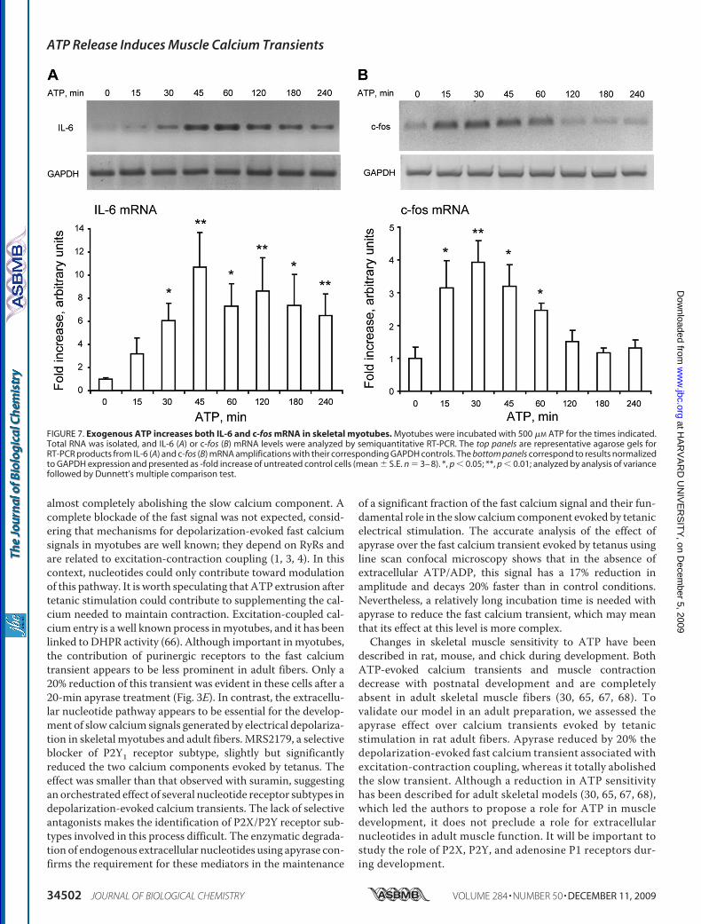
The stimulation of myotubes with 500 �M ATP significantlyincreased the mRNA for IL-6 (30–240 min, Fig. 7A) and c-fos(15–60 min; Fig. 7B) as detected by semiquantitative RT-PCR.Although there was some variability in the fold over basalincrease in these results, in all experiments there was a clearmRNA induction after the addition of ATP.
DISCUSSION
In the present work we have demonstrated that nucleotidesreleased to the extracellular medium are the essential media-tors in promoting slow calcium signals and can significantlyaffect fast calcium transients evoked by tetanic stimulation inskeletal muscle cells. As slow calcium signals have been relatedto the regulation of gene expression (5, 11, 12), our data placeextracellular nucleotides as potential modulators of physiolog-ical processes such as skeletal muscle activity and plasticity.Several reports have described the short and long term
effects of exogenous nucleotides in skeletal muscles derivedfrom different species (human, rat, mouse, chicken, and frog);calciummobilization, ERK activation, and gene expression reg-ulation have been observed after skeletal cells incubation withP2X/P2Y receptor agonists (39, 40, 59–64). On the other hand,ATP is released from skeletalmuscle cells in response tomusclecontraction (34, 35). Previous data fromour laboratory indicatethat tetanic electrical stimulation evokes a fast calcium signallinked to excitation-contraction coupling and a slow calciumsignal related to gene expression control (5, 8, 9). In the presentwork we focused on the study of extracellular nucleotidesreleased after tetanic stimulation and their putative role as pro-moters of calcium signals normally related to membranedepolarization.We detected mRNA expression for several P2Y and all P2X
receptor subtypes in skeletal myotubes derived from rat pri-mary cultures. Although the protein expressionwas establishedonly for the P2Y2 receptor, the functional data and pharmacol-
ATP Release Induces Muscle Calcium Transients
DECEMBER 11, 2009 • VOLUME 284 • NUMBER 50 JOURNAL OF BIOLOGICAL CHEMISTRY 34499
at HA
RV
AR
D U
NIV
ER
SIT
Y, on D
ecember 5, 2009
ww
w.jbc.org
Dow
nloaded from

FIGURE 5. Nucleotides release and metabolization after tetanic stimulation. A–D, to measure ATP and its metabolites at the extracellular medium, skeletalmyotubes were stimulated electrically with a tetanus protocol (45 Hz, 400 1-ms pulses), and aliquots of the medium were removed at the indicated timesthereafter. Nucleotide samples (ATP, ADP, AMP, and adenosine) were derivatized as described under “Experimental Procedures,” resolved and detected usingan HPLC coupled to fluorescence detection, and quantitated using a calibration standard curve. Values are the mean � S.E. (n � 6 – 8 series from sixindependent primary cultures). To assess the ectonucleotidases activities of skeletal myotubes, a standard of �-ATP was applied to the extracellular medium.Insets, at different times, extracellular aliquots were removed to quantify �-ATP (A), �-ADP (B), �-AMP (C), and �-adenosine (D) by HPLC as an indicator of the ATPmetabolism ability of these cells. Values are the mean � S.E. (n � 3–5 series from three independent primary cultures).
ATP Release Induces Muscle Calcium Transients
34500 JOURNAL OF BIOLOGICAL CHEMISTRY VOLUME 284 • NUMBER 50 • DECEMBER 11, 2009
at HA
RV
AR
D U
NIV
ER
SIT
Y, on D
ecember 5, 2009
ww
w.jbc.org
Dow
nloaded from

ogy are consistent with the presence of multiple receptors forextracellular nucleotides and suggest that these receptorsshould be relevant for muscle physiology. Future work shouldunveil the role of particular nucleotide receptor subtypes inboth skeletal myotubes and adult muscle fibers.The exogenous addition of natural P2Y/P2X receptor agonists
evokes calcium transients in skeletal myotubes. The ATP-evokedcalciumtransient iscomposedof twoormorekineticcomponents;this is in accordancewith reported data showing that activation ofionotropic P2X4, P2X5, and P2X7 and metabotropic P2Y1 andP2Y4 purinoreceptors participates in the ATP-evoked calciumtransients of multinucleated mice myotubes (39). The biphasic
kinetics of the calcium signal evoked by ATP has been describedalso in human andmice culturedmyotubes (65). In thosemodels,ATP-evoked calcium transients have an early, fast component fol-lowed by a second, more gradual increase in [Ca2�] (65). Afterremovalof extracellularcalcium, theATP-evokedcalciumsignal isreduced in amplitude; the slope of the fast initial componentdecreases and comes back to basal values, suggesting a strong rolefor P2X receptors on these transients. The remaining 20%calciumresponse in the absence of extracellular calcium could be attrib-uted to P2Y receptors.Treatments focused on disrupting the purinergic pathway,
which reduced the fast calcium signal by less than 60% while
FIGURE 6. Pannexin-1 hemichannels are involved in ATP release during tetanic stimulation of skeletal myotubes. A, fast and slow calcium componentsevoked by tetanic electrical stimulation (45 Hz, 400 1-ms pulses) are strongly reduced after pannexin-1 hemichannel blockade using 100 �M of the 10pnx1
peptide 20 –30 min prior to and during the protocol (n � 25–79 cells, five coverslips, five different cultures). For each coverslip, calcium transients evoked bytetanus before and after incubation with the peptide were tested. B, the same protocol as described in A was achieved, but after 10pnx1 peptide incubation cellswere washed for 60 min, we reassessed the effect of the tetanus over calcium transients. Values are expressed as mean � S.E. (n � 19 –23 cells, two coverslips,two different cultures). C, ATP release evoked by tetanic electrical stimulation was abolished by 100 �M oleamide (a nonselective blocker for connexin andpannexin hemichannels) or 100 �M
10pnx1 peptide. Skeletal myotubes were incubated 20 min before and during the assay with the indicated blocker. Aliquotsof the extracellular medium were removed at the indicated times after electrical stimulation, and ATP was measured by a luciferin/luciferase assay as describedunder “Experimental Procedures.” Values were normalized to 0 control for each treatment and expressed as the mean � S.E. (n � 4 series from four indepen-dent primary cultures). **, p � 0.01, Dunnett’s t test, one-tail comparison between each time and their own 0 control. D, DHPR co-precipitates with P2Y2receptor and pannexin-1 in skeletal myotubes. DHPR was resolved and detected by immunoblot (IB) in samples derived from whole lysates (—) or immuno-precipitated (IPP) previously with anti-DHPR, anti-P2Y2, or anti-Pnx1 as described under “Experimental Procedures.”
ATP Release Induces Muscle Calcium Transients
DECEMBER 11, 2009 • VOLUME 284 • NUMBER 50 JOURNAL OF BIOLOGICAL CHEMISTRY 34501
at HA
RV
AR
D U
NIV
ER
SIT
Y, on D
ecember 5, 2009
ww
w.jbc.org
Dow
nloaded from
almost completely abolishing the slow calcium component. Acomplete blockade of the fast signal was not expected, consid-ering that mechanisms for depolarization-evoked fast calciumsignals in myotubes are well known; they depend on RyRs andare related to excitation-contraction coupling (1, 3, 4). In thiscontext, nucleotides could only contribute toward modulationof this pathway. It is worth speculating that ATP extrusion aftertetanic stimulation could contribute to supplementing the cal-cium needed to maintain contraction. Excitation-coupled cal-cium entry is a well known process inmyotubes, and it has beenlinked to DHPR activity (66). Although important inmyotubes,the contribution of purinergic receptors to the fast calciumtransient appears to be less prominent in adult fibers. Only a20% reduction of this transient was evident in these cells after a20-min apyrase treatment (Fig. 3E). In contrast, the extracellu-lar nucleotide pathway appears to be essential for the develop-ment of slow calcium signals generated by electrical depolariza-tion in skeletal myotubes and adult fibers.MRS2179, a selectiveblocker of P2Y1 receptor subtype, slightly but significantlyreduced the two calcium components evoked by tetanus. Theeffect was smaller than that observed with suramin, suggestingan orchestrated effect of several nucleotide receptor subtypes indepolarization-evoked calcium transients. The lack of selectiveantagonists makes the identification of P2X/P2Y receptor sub-types involved in this process difficult. The enzymatic degrada-tion of endogenous extracellular nucleotides using apyrase con-firms the requirement for these mediators in the maintenance
of a significant fraction of the fast calcium signal and their fun-damental role in the slow calciumcomponent evoked by tetanicelectrical stimulation. The accurate analysis of the effect ofapyrase over the fast calcium transient evoked by tetanus usingline scan confocal microscopy shows that in the absence ofextracellular ATP/ADP, this signal has a 17% reduction inamplitude and decays 20% faster than in control conditions.Nevertheless, a relatively long incubation time is needed withapyrase to reduce the fast calcium transient, which may meanthat its effect at this level is more complex.Changes in skeletal muscle sensitivity to ATP have been
described in rat, mouse, and chick during development. BothATP-evoked calcium transients and muscle contractiondecrease with postnatal development and are completelyabsent in adult skeletal muscle fibers (30, 65, 67, 68). Tovalidate our model in an adult preparation, we assessed theapyrase effect over calcium transients evoked by tetanicstimulation in rat adult fibers. Apyrase reduced by 20% thedepolarization-evoked fast calcium transient associated withexcitation-contraction coupling, whereas it totally abolishedthe slow transient. Although a reduction in ATP sensitivityhas been described for adult skeletal models (30, 65, 67, 68),which led the authors to propose a role for ATP in muscledevelopment, it does not preclude a role for extracellularnucleotides in adult muscle function. It will be important tostudy the role of P2X, P2Y, and adenosine P1 receptors dur-ing development.
FIGURE 7. Exogenous ATP increases both IL-6 and c-fos mRNA in skeletal myotubes. Myotubes were incubated with 500 �M ATP for the times indicated.Total RNA was isolated, and IL-6 (A) or c-fos (B) mRNA levels were analyzed by semiquantitative RT-PCR. The top panels are representative agarose gels forRT-PCR products from IL-6 (A) and c-fos (B) mRNA amplifications with their corresponding GAPDH controls. The bottom panels correspond to results normalizedto GAPDH expression and presented as -fold increase of untreated control cells (mean � S.E. n � 3– 8). *, p � 0.05; **, p � 0.01; analyzed by analysis of variancefollowed by Dunnett’s multiple comparison test.
ATP Release Induces Muscle Calcium Transients
34502 JOURNAL OF BIOLOGICAL CHEMISTRY VOLUME 284 • NUMBER 50 • DECEMBER 11, 2009
at HA
RV
AR
D U
NIV
ER
SIT
Y, on D
ecember 5, 2009
ww
w.jbc.org
Dow
nloaded from

ATP release has been demonstrated in vivo in contracting ratand cat skeletal muscles by collecting samples frommicrodialy-sis probes inserted in themuscle (34, 35) or ex vivo after a 10-Hzstimulation of mouse hemidiaphragm (69). Enzyme pathwaysfor extracellular ATP metabolization have been also demon-strated in skeletal muscles (70–72). In this work, we deter-mined that ATP is quickly released from skeletal myotubesafter tetanic electrical stimulation or K� depolarization. In allprevious works ATP has beenmeasuredminutes after the elec-trical stimulation (32, 34, 35, 69); we detected an initial peak ofATP release between 15 s and 3 min after tetanus stimulation.The size and shape of slow calcium transients elicited by theconditioned media of electrically stimulated myotubes is com-patible with that of ATP-elicited transients in a bulk concentra-tion range of 0.1 to 1 mM. We have to consider that the localATP concentration near the membrane during release must bemuch higher than that measured in the bulk medium (19).Extracellular nucleotides reach up to millimolar concentra-
tion after cell damage or under nonlytic stimuli conditions suchas hypoxia, inflammation, or cell swelling (19, 73–80). In excit-able cells such as neurons, chromaffin cells, and platelets, ATPis stored in vesicles and released by calcium-dependent exocy-tosis (81–83). In non-excitable cells, several mechanisms forATP release have been proposed: 1) through ABC transporters(84, 85); 2) through stretch- or voltage-activated channels (86–92); 3) through connexin or pannexin hemichannels (19–23).Mechanisms for ATP release from skeletal muscle cells havenot been studied. Considering that pannexin hemichannelshave emerged as conduits for ATP release in several cellular
systems and that they are activatedby membrane depolarization,mechanical stress, or intracellularcalcium (22, 93), we analyzed theirpossible role in our system. Theexpression of pannexin-1 mRNA inadult skeletal muscle has beenreported, and pannexin-1 hemi-channels have been located in T-tu-bules of adult skeletal muscle.4Here, we have gathered evidencesuggesting that pannexin-1 hemi-channels are essential for both ATPrelease and tetanus-evoked calciumtransients in skeletal myotubes.Recently, functional units by physi-cal association between P2 recep-tors and hemichannel proteins havebeen proposed (94). Using tran-scriptomic strategies, several asso-ciations have been suggested; co-immunoprecipitation assays havevalidated an interaction amongP2X7 receptors, pannexin-1, con-nexin-43, actin, and vinculin inwhole brain and cultured astrocytes(94). A supramolecular complexincluding P2Y1 receptors, con-nexin-43, actin, and vinculin in cul-
tured astrocytes has also been postulated (94). In agreementwith these data, in our model we have shown the co-immuno-precipitation of either pannexin-1 or P2Y2 receptor withDHPR, opening the possibility that such supramolecular com-plexes could be modulating calcium changes evoked by muscleactivity. We first analyzed the interaction with P2Y2 receptorsbecause of their strong mRNA expression in our system; how-ever, other P2X/P2Y receptor subtypes could also be tested asputative components of this complex under resting or stimu-lated conditions.As demonstrated previously for membrane depolarization
(5, 11–13), the addition of ATP was able to stimulate mRNAexpression for an immediate early gene such as c-fos and for anexercise-induced gene such as IL-6; this reinforces the idea ofATP being the intermediate step between electrical stimulationand intracellular pathways leading to gene expression. The pre-cise time course of events that account for depolarization sens-ing, ATP release, activation of nucleotide receptors, and cal-cium movements in skeletal muscle cells still remains to beestablished, and further work is needed to unveil the mecha-nisms and characterize the molecular complex that is begin-ning to emerge and appears to be involved in activity-modu-lated muscle plasticity. A hypothetical model is depicted in Fig.8. In this model, membrane depolarization is proposed to acti-vate Cav1.1 dihydropyridine receptors, and its conformational
4 M. A. Riquelme, L. Cea, M. V. L. Bennett, and J. C. Saez, unpublishedobservation.
FIGURE 8. Hypothetical schematic model of the proposed role of purinergic receptors in both excitation-contraction and excitation-transcription coupling in skeletal muscle cells. A multimeric protein complexis suggested to exist in the T-tubule membrane, including the dihydropyridine receptor (Cav1.1, DHPR), thepurinergic metabotropic P2Y2 receptor, and the pannexin-1 molecule (PnX1). Membrane depolarization willinduce the opening of an ATP pathway via PnX1 after a conformational change of the adjacent DHPR. Aheterotrimeric G protein will be attached to the P2Y2 receptor, and upon ATP binding, the �� subunit willsequentially activate phosphoinositide 3-kinase (not shown) and phospholipase C (PLC) to produce IP3 andactivate calcium release for the slow calcium transient. Other proteins such as RyR, known to interact withDHPR and P2X receptors (ionotropic, calcium-permeating channel), participate in the fast calcium transient,but whether they are part of the same complex has not yet been evidenced.
ATP Release Induces Muscle Calcium Transients
DECEMBER 11, 2009 • VOLUME 284 • NUMBER 50 JOURNAL OF BIOLOGICAL CHEMISTRY 34503
at HA
RV
AR
D U
NIV
ER
SIT
Y, on D
ecember 5, 2009
ww
w.jbc.org
Dow
nloaded from

change will in turn activate pannexin-1 channels to releaseATP.Although a direct role for extracellular ATP has not yet been
demonstrated in skeletal muscle pathologies, a mild degenera-tive muscle disease, sarcoglycanopathy, has been associatedwith the loss of an ecto-ATPase on the surface of the skeletalmuscle fibers (70, 95). Furthermore, the altered expression of ahuman P2X receptor gene in certain degenerative or hyperpro-liferative muscle disorders has been suggested (96). Anincreased sensitivity to ATP in dystrophic myotubes has beenalso established (97). Considering that our data place extracel-lular nucleotides and their surface receptors as essential medi-ators between muscle activity and plasticity, it is possible thatderegulation of any of the components of this pathway mayaccount for a muscle pathology.
Acknowledgment—We thank Monica Silva for cell cultures.
REFERENCES1. Grabner,M., Dirksen, R. T., Suda, N., and Beam, K.G. (1999) J. Biol. Chem.
274, 21913–219192. Lamb, G. D. (2002) Front. Biosci. 7, d834–d8423. Protasi, F., Paolini, C., Nakai, J., Beam, K. G., Franzini-Armstrong, C., and
Allen, P. D. (2002) Biophys. J. 83, 3230–32444. Tanabe, T., Beam, K. G., Adams, B. A., Niidome, T., and Numa, S. (1990)
Nature 346, 567–5695. Carrasco, M. A., Riveros, N., Ríos, J., Muller, M., Torres, F., Pineda, J.,
Lantadilla, S., and Jaimovich, E. (2003) Am. J. Physiol. Cell Physiol. 284,C1438–C1447
6. Jaimovich, E., and Carrasco, M. A. (2002) Biol. Res. 35, 195–2027. Semsarian, C., Wu, M. J., Ju, Y. K., Marciniec, T., Yeoh, T., Allen, D. G.,
Harvey, R. P., and Graham, R. M. (1999) Nature 400, 576–5818. Eltit, J.M., García, A.A., Hidalgo, J., Liberona, J. L., Chiong,M., Lavandero,
S., Maldonado, E., and Jaimovich, E. (2006) J. Biol. Chem. 281,12143–12154
9. Eltit, J. M., Hidalgo, J., Liberona, J. L., and Jaimovich, E. (2004) Biophys. J.86, 3042–3051
10. Araya, R., Eckardt, D., Riquelme,M. A.,Willecke, K., and Saez, J. C. (2003)Cell Commun. Adhes. 10, 451–456
11. Juretic, N., García-Huidobro, P., Iturrieta, J. A., Jaimovich, E., and Riveros,N. (2006) Am. J. Physiol. Cell Physiol. 290, C1428–C1436
12. Juretic, N., Urzua, U., Munroe, D. J., Jaimovich, E., and Riveros, N. (2007)J. Cell. Physiol. 210, 819–830
13. Valdes, J. A., Hidalgo, J., Galaz, J. L., Puentes, N., Silva, M., Jaimovich, E.,and Carrasco, M. A. (2007) Am. J. Physiol. Cell Physiol. 292,C1960–C1970
14. DeWaard, M., Hering, J., Weiss, N., and Feltz, A. (2005) Trends Pharma-col. Sci. 26, 427–436
15. Herlitze, S., Garcia, D. E., Mackie, K., Hille, B., Scheuer, T., and Catterall,W. A. (1996) Nature 380, 258–262
16. Ikeda, S. R. (1996) Nature 380, 255–25817. Silinsky, E. M. (1975) J. Physiol. 247, 145–16218. von Kugelgen, I., and Starke, K. (1985) J. Physiol. 367, 435–45519. Lazarowski, E. R., Boucher, R. C., and Harden, T. K. (2003)Mol. Pharma-
col. 64, 785–79520. Schwiebert, E. M., and Zsembery, A. (2003) Biochim. Biophys. Acta 1615,
7–3221. Leybaert, L., Braet, K., Vandamme, W., Cabooter, L., Martin, P. E., and
Evans, W. H. (2003) Cell Commun. Adhes. 10, 251–25722. Bao, L., Locovei, S., and Dahl, G. (2004) FEBS Lett. 572, 65–6823. Huang, Y. J., Maruyama, Y., Dvoryanchikov, G., Pereira, E., Chaudhari, N.,
and Roper, S. D. (2007) Proc. Natl. Acad. Sci. U.S.A. 104, 6436–644124. Zimmermann, H. (1999) Trends Pharmacol. Sci. 20, 231–23625. Abbracchio, M. P., Burnstock, G., Boeynaems, J. M., Barnard, E. A., Boyer,
J. L., Kennedy, C., Knight, G. E., Fumagalli, M., Gachet, C., Jacobson, K. A.,and Weisman, G. A. (2006) Pharmacol. Rev. 58, 281–341
26. Burnstock, G. (2006) Pharmacol. Ther. 110, 433–45427. Buvinic, S., Briones, R., and Huidobro-Toro, J. P. (2002) Br. J. Pharmacol.
136, 847–85628. Buvinic, S., Poblete, M. I., Donoso, M. V., Delpiano, A. M., Briones, R.,
Miranda, R., and Huidobro-Toro, J. P. (2006) J. Physiol. 573, 427–44329. North, R. A. (2002) Physiol. Rev. 82, 1013–106730. Wells, D. G., Zawisa, M. J., and Hume, R. I. (1995)Dev. Biol. 172, 585–59031. Ryten, M., Dunn, P. M., Neary, J. T., and Burnstock, G. (2002) J. Cell Biol.
158, 345–35532. Cunha, R. A., and Sebastiao, A. M. (1993) Pflugers Arch. 424, 503–51033. Araya, R., Riquelme, M. A., Brandan, E., and Saez, J. C. (2004) Brain Res.
Brain Res. Rev. 47, 174–18834. Li, J., King, N. C., and Sinoway, L. I. (2003) J. Appl. Physiol. 95, 577–58335. Li, J., King, N. C., and Sinoway, L. I. (2005) Circulation 111, 2748–275136. Martinez-Pinna, J., Gurung, I. S., Vial, C., Leon, C., Gachet, C., Evans, R. J.,
and Mahaut-Smith, M. P. (2005) J. Biol. Chem. 280, 1490–149837. Martinez-Pinna, J., Tolhurst, G., Gurung, I. S., Vandenberg, J. I., and Ma-
haut-Smith, M. P. (2004) J. Physiol. 555, 61–7038. Stanfield, P. (2006) Nat. Cell Biol. 8, 1323–132539. Deli, T., Szappanos, H., Szigeti, G. P., Cseri, J., Kovacs, L., and Csernoch, L.
(2007) Pflugers Arch. 453, 519–52940. May, C., Weigl, L., Karel, A., and Hohenegger, M. (2006) Biochem. Phar-
macol. 71, 1497–150941. Jaimovich, E., Reyes, R., Liberona, J. L., and Powell, J. A. (2000) Am. J.
Physiol. Cell Physiol. 278, C998–C101042. Chomczynski, P., and Sacchi, N. (1987) Anal. Biochem. 162, 156–15943. Communi, D., Gonzalez, N. S., Detheux, M., Brezillon, S., Lannoy, V.,
Parmentier, M., and Boeynaems, J. M. (2001) J. Biol. Chem. 276,41479–41485
44. Rodrigues, R. J., Almeida, T., Richardson, P. J., Oliveira, C. R., and Cunha,R. A. (2005) J. Neurosci. 25, 6286–6295
45. Scrivens,M., andDickenson, J.M. (2006) Eur. J. Pharmacol. 543, 166–17346. da Silva, R. L., Resende, R. R., and Ulrich, H. (2007) Exp. Physiol. 92,
139–14547. Hayato, R., Ohtubo, Y., and Yoshii, K. (2007) J. Physiol. 584, 473–48848. Lazarowski, E. R., Tarran, R., Grubb, B. R., van Heusden, C. A., Okada, S.,
and Boucher, R. C. (2004) J. Biol. Chem. 279, 36855–3686449. Todorov, L. D., Mihaylova-Todorova, S., Craviso, G. L., Bjur, R. A., and
Westfall, D. P. (1996) J. Physiol. 496, 731–74850. Reigada, D., Lu, W., Zhang, M., and Mitchell, C. H. (2008) Neuroscience
157, 396–40451. Schenk, U., Westendorf, A. M., Radaelli, E., Casati, A., Ferro, M., Fuma-
galli, M., Verderio, C., Buer, J., Scanziani, E., and Grassi, F. (2008) Sci.Signal. 1, ra6
52. Araya, R., Eckardt, D., Maxeiner, S., Kruger, O., Theis, M., Willecke, K.,and Saez, J. C. (2005) J. Cell Sci. 118, 27–37
53. Balogh, S., Naus, C. C., andMerrifield, P. A. (1993)Dev. Biol. 155, 351–36054. Belluardo,N., Trovato-Salinaro, A.,Mudo, G., andCondorelli, D. F. (2005)
Cell Tissue Res. 320, 299–31055. von Maltzahn, J., Wulf, V., and Willecke, K. (2006) Cell Commun. Adhes.
13, 55–6056. De Jongh, K. S., Merrick, D. K., and Catterall, W. A. (1989) Proc. Natl.
Acad. Sci. U.S.A. 86, 8585–858957. De Jongh, K. S.,Warner, C., Colvin, A. A., andCatterall,W. A. (1991) Proc.
Natl. Acad. Sci. U.S.A. 88, 10778–1078258. Lai, Y., Seagar, M. J., Takahashi, M., and Catterall, W. A. (1990) J. Biol.
Chem. 265, 20839–2084859. Choi, R. C., Man, M. L., Ling, K. K., Ip, N. Y., Simon, J., Barnard, E. A., and
Tsim, K. W. (2001) J. Neurosci. 21, 9224–923460. Choi, R. C., Siow, N. L., Cheng, A. W., Ling, K. K., Tung, E. K., Simon, J.,
Barnard, E. A., and Tsim, K. W. (2003) J. Neurosci. 23, 4445–445661. Deli, T., Toth, B. I., Czifra, G., Szappanos, H., Bíro, T., and Csernoch, L.
(2006) J. Muscle Res. Cell Motil. 27, 617–63062. Sandona, D., Danieli-Betto, D., Germinario, E., Biral, D., Martinello, T.,
Lioy, A., Tarricone, E., Gastaldello, S., and Betto, R. (2005) FASEB J. 19,1184–1186
ATP Release Induces Muscle Calcium Transients
34504 JOURNAL OF BIOLOGICAL CHEMISTRY VOLUME 284 • NUMBER 50 • DECEMBER 11, 2009
at HA
RV
AR
D U
NIV
ER
SIT
Y, on D
ecember 5, 2009
ww
w.jbc.org
Dow
nloaded from

63. Szigeti, G. P., Szappanos, H., Deli, T., Cseri, J., Kovacs, L., and Csernoch, L.(2007) Pflugers Arch. 453, 509–518
64. Tsim, K. W., and Barnard, E. A. (2002) Neurosignals 11, 58–6465. Cseri, J., Szappanos, H., Szigeti, G. P., Csernatony, Z., Kovacs, L., and
Csernoch, L. (2002) Pflugers Arch. 443, 731–73866. Bannister, R. A., Pessah, I. N., and Beam, K. G. (2009) J. Gen. Physiol. 133,
79–9167. Collet, C., Strube, C., Csernoch, L., Mallouk, N., Ojeda, C., Allard, B., and
Jacquemond, V. (2002) Pflugers Arch. 443, 771–77868. Ryten,M., Hoebertz, A., and Burnstock, G. (2001)Dev. Dyn. 221, 331–34169. Vizi, E. S., Nitahara, K., Sato, K., and Sperlagh, B. (2000) J. Auton. Nerv.
Syst. 81, 278–28470. Delgado, J., Moro, G., Saborido, A., andMegías, A. (1997) Biochem. J. 327,
899–90771. Kirley, T. L. (1997) J. Biol. Chem. 272, 1076–108172. Megías, A., Martínez-Senac, M. M., Delgado, J., and Saborido, A. (2001)
Biochem. J. 353, 521–52973. Eltzschig, H. K., Eckle, T., Mager, A., Kuper, N., Karcher, C., Weissmuller,
T., Boengler, K., Schulz, R., Robson, S. C., andColgan, S. P. (2006)Circ. Res.99, 1100–1108
74. Filtz, T. M., Li, Q., Boyer, J. L., Nicholas, R. A., and Harden, T. K. (1994)Mol. Pharmacol. 46, 8–14
75. Grierson, J. P., and Meldolesi, J. (1995) J. Biol. Chem. 270, 4451–445676. Grygorczyk, R., and Hanrahan, J. W. (1997) Am. J. Physiol. Cell Physiol.
272, C1058–C106677. Lazarowski, E. R., Homolya, L., Boucher, R. C., and Harden, T. K. (1997)
J. Biol. Chem. 272, 24348–2435478. Parr, C. E., Sullivan, D.M., Paradiso, A.M., Lazarowski, E. R., Burch, L. H.,
Olsen, J. C., Erb, L., Weisman, G. A., Boucher, R. C., and Turner, J. T.(1994) Proc. Natl. Acad. Sci. U.S.A. 91, 3275–3279
79. Watt, W. C., Lazarowski, E. R., and Boucher, R. C. (1998) J. Biol. Chem.273, 14053–14058
80. Yegutkin, G., Bodin, P., and Burnstock, G. (2000) Br. J. Pharmacol. 129,
921–92681. Burnstock, G. (1997) Neuropharmacology 36, 1127–113982. Evans, R. J., Derkach, V., and Surprenant, A. (1992) Nature 357, 503–50583. Sorensen, C. E., and Novak, I. (2001) J. Biol. Chem. 276, 32925–3293284. Braunstein, G. M., Zsembery, A., Tucker, T. A., and Schwiebert, E. M.
(2004) J. Cyst. Fibros. 3, 99–11785. Hazama, A., Fan, H. T., Abdullaev, I., Maeno, E., Tanaka, S., Ando-Akat-
suka, Y., and Okada, Y. (2000) J. Physiol. 523, 1–1186. Benz, R., Wojtczak, L., Bosch,W., and Brdiczka, D. (1988) FEBS Lett. 231,
75–8087. Braunstein, G.M., Roman, R.M., Clancy, J. P., Kudlow, B. A., Taylor, A. L.,
Shylonsky, V. G., Jovov, B., Peter, K., Jilling, T., Ismailov, II, Benos, D. J.,Schwiebert, L. M., Fitz, J. G., and Schwiebert, E. M. (2001) J. Biol. Chem.276, 6621–6630
88. Caldwell, R. A., Clemo, H. F., and Baumgarten, C.M. (1998)Am. J. Physiol.Cell Physiol. 275, C619–C621
89. Roman, R.M., Feranchak, A. P., Davison, A. K., Schwiebert, E.M., and Fitz,J. G. (1999) Am. J. Physiol. Gastrointest. Liver Physiol. 277,G1222–G1230
90. Sackin, H. (1995) Kidney Int. 48, 1134–114791. Sugita, M., Yue, Y., and Foskett, J. K. (1998) EMBO J. 17, 898–90892. Taylor, A. L., Kudlow, B. A., Marrs, K. L., Gruenert, D. C., Guggino,W. B.,
and Schwiebert, E. M. (1998) Am. J. Physiol. Cell Physiol. 275,C1391–C1406
93. Shestopalov, V. I., and Panchin, Y. (2008) Cell. Mol. Life Sci. 65, 376–39494. Iacobas, D. A., Suadicani, S. O., Iacobas, S., Chrisman, C., Cohen, M. A.,
Spray, D. C., and Scemes, E. (2007) J. Membr. Biol. 217, 83–9195. Betto, R., Senter, L., Ceoldo, S., Tarricone, E., Biral, D., and Salviati, G.
(1999) J. Biol. Chem. 274, 7907–791296. Urano, T., Nishimori, H., Han, H., Furuhata, T., Kimura, Y., Nakamura, Y.,
and Tokino, T. (1997) Cancer Res. 57, 3281–328797. Yeung, D., Zablocki, K., Lien, C. F., Jiang, T., Arkle, S., Brutkowski, W.,
Brown, J., Lochmuller, H., Simon, J., Barnard, E. A., and Gorecki, D. C.(2006) FASEB J. 20, 610–620
ATP Release Induces Muscle Calcium Transients
DECEMBER 11, 2009 • VOLUME 284 • NUMBER 50 JOURNAL OF BIOLOGICAL CHEMISTRY 34505
at HA
RV
AR
D U
NIV
ER
SIT
Y, on D
ecember 5, 2009
ww
w.jbc.org
Dow
nloaded from
Copyright © 2022 FDOKUMEN