Asymmetric localization of numb autonomously deter-mines sibling neuron identity in the Drosophila C
6
INTRODUCTION How cell diversity is generated is a central question in devel- opmental biology. While a great deal has been learned recently about how extrinsic cues between cells are used to control cell determination, it is not clear to what extent intrinsic cues regulate cell fates between two cells (Greenwald and Rubin, 1992; Posakony, 1994; Jan and Jan, 1995). Perhaps the best examples of intrinsic cues are the regionally localized gene products in the early Drosophila embryo: pole plasm triggers germ cell fate (St Johnston, 1993), bicoid activates anterior embryonic cell fates (Driever, 1993), and nanos specifies posterior cell identity (St Johnston, 1993). However, few examples are known where a determinant (RNA or protein) is responsible for differential specification of cell fate due to asymmetric localization into one daughter cell. Mutations exist which perturb binary cell fate decisions of sibling cells in several organisms (Horvitz and Herskowitz, 1992), but the relevant gene products are not asymmetrically localized or their distribution has not been determined. Only one gene, numb, is known to encode an asymmetri- cally localized protein that is required for a binary cell fate decision. numb is a novel cytoplasmic or membrane-associated protein that specifies cell identity in the external sensory bristle organ (ES organ) lineage in the Drosophila peripheral nervous system (PNS) (Uemura et al., 1989; Rhyu et al., 1994). The 4- cell ES organ develops from a sense organ precursor (SOP) that divides to produce two daughter cells, A and B. The A cell divides to produce a bristle cell and a socket cell, while the B cell makes a neuron and a glial cell. Both cell interactions and numb localization are required to specify SOP daughter cell fates. Inactivation of the Delta-Notch signaling between the daughters suppresses A cell fate (Parks and Muscavitch, 1993; Hartenstein and Posakony, 1990). In contrast, numb mutants result in both cells adopting the A cell fate; whereas ectopic expression of numb generates two B cells (Uemura et al., 1989; Rhyu et al., 1994). Thus, Delta-Notch-mediated cell interac- tions inhibit B cell development, whereas numb protein promotes B cell development. Although numb is asymmetri- cally localized during mitosis to one of the SOP daughter cells, it is not known which daughter cell inherits the protein (Rhyu et al., 1994). Given the uncertainty about which SOP daughter cell contains numb protein, a number of models have been proposed to explain how the asymmetric localization of numb protein may trigger B cell fate (Posakony, 1994; Jan and Jan, 1995). In one model, numb is segregated to the cell that ulti- mately acquires the B cell fate and functions cell- autonomously to make sure this cell becomes resistant to the Delta-Notch mediated inhibitory signaling by its neighbor (for example, by down-regulating Notch function). This model would result in numb functioning to control the fate of the cell that inherits it. In an alternate model, numb is segregated to the cell that ultimately acquires the A cell fate and functions to decrease the ability of this cell to inhibit B cell development in its neighbor (for example, by down-regulating Delta function). This second model would result in numb function- ing to control the fate of the neighboring cell that does not inherit numb. In order to answer the question of autonomy, we have examined the role of numb in the Drosophila CNS instead 3489 Development 121, 3489-3494 (1995) Printed in Great Britain © The Company of Biologists Limited 1995 The central nervous system (CNS) represents an excellent model system for examining how a multitude of unique cell fates are specified. We find that asymmetric localization of the numb protein autonomously controls a binary cell fate decision in the Drosophila CNS. The simplest lineage in the Drosophila CNS is that of the MP2 precursor: it divides unequally to generate the dMP2 and vMP2 neurons. Both are interneurons but project in different directions: dMP2 projects its axon posteriorly while vMP2 projects anteri- orly. During MP2 mitosis, numb is localized into dMP2 and excluded from vMP2. Loss of numb transforms dMP2 into vMP2, whereas ectopic numb produces the opposite trans- formation of vMP2 into dMP2. Thus, numb is asymmetri- cally localized in the dividing MP2 and is necessary and sufficient to autonomously specify dMP2 neuronal identity. Key words: numb, Drosophila, localized determinant, odd-skipped, neuroblast SUMMARY Asymmetric localization of numb autonomously determines sibling neuron identity in the Drosophila CNS Eric P. Spana 1 , Casey Kopczynski 2 , Corey S. Goodman 2 and Chris Q. Doe 1, * 1 Howard Hughes Medical Institute, Department of Cell and Structural Biology, University of Illinois, Urbana, IL 61801, USA 2 Howard Hughes Medical Institute, Division of Neurobiology, Department of Cell and Molecular Biology, University of California, Berkeley, CA 94720, USA *Author for correspondence
-
Upload
independent -
Category
Documents
-
view
1 -
download
0
Transcript of Asymmetric localization of numb autonomously deter-mines sibling neuron identity in the Drosophila C

3489Development 121, 3489-3494 (1995)Printed in Great Britain © The Company of Biologists Limited 1995
Asymmetric localization of numb autonomously determines sibling neuron
identity in the Drosophila CNS
Eric P. Spana1, Casey Kopczynski2, Corey S. Goodman2 and Chris Q. Doe1,*1Howard Hughes Medical Institute, Department of Cell and Structural Biology, University of Illinois, Urbana, IL 61801, USA2Howard Hughes Medical Institute, Division of Neurobiology, Department of Cell and Molecular Biology, University of California,Berkeley, CA 94720, USA
*Author for correspondence
The central nervous system (CNS) represents an excellentmodel system for examining how a multitude of unique cellfates are specified. We find that asymmetric localization ofthe numb protein autonomously controls a binary cell fatedecision in the Drosophila CNS. The simplest lineage in theDrosophila CNS is that of the MP2 precursor: it dividesunequally to generate the dMP2 and vMP2 neurons. Bothare interneurons but project in different directions: dMP2projects its axon posteriorly while vMP2 projects anteri-
orly. During MP2 mitosis, numb is localized into dMP2 andexcluded from vMP2. Loss of numb transforms dMP2 intovMP2, whereas ectopic numb produces the opposite trans-formation of vMP2 into dMP2. Thus, numb is asymmetri-cally localized in the dividing MP2 and is necessary andsufficient to autonomously specify dMP2 neuronal identity.
Key words: numb, Drosophila, localized determinant, odd-skipped,neuroblast
SUMMARY
INTRODUCTION
How cell diversity is generated is a central question in devel-opmental biology. While a great deal has been learned recentlyabout how extrinsic cues between cells are used to control celldetermination, it is not clear to what extent intrinsic cuesregulate cell fates between two cells (Greenwald and Rubin,1992; Posakony, 1994; Jan and Jan, 1995). Perhaps the bestexamples of intrinsic cues are the regionally localized geneproducts in the early Drosophila embryo: pole plasm triggersgerm cell fate (St Johnston, 1993), bicoid activates anteriorembryonic cell fates (Driever, 1993), and nanos specifiesposterior cell identity (St Johnston, 1993). However, fewexamples are known where a determinant (RNA or protein) isresponsible for differential specification of cell fate due toasymmetric localization into one daughter cell. Mutations existwhich perturb binary cell fate decisions of sibling cells inseveral organisms (Horvitz and Herskowitz, 1992), but therelevant gene products are not asymmetrically localized ortheir distribution has not been determined.
Only one gene, numb, is known to encode an asymmetri-cally localized protein that is required for a binary cell fatedecision. numb is a novel cytoplasmic or membrane-associatedprotein that specifies cell identity in the external sensory bristleorgan (ES organ) lineage in the Drosophila peripheral nervoussystem (PNS) (Uemura et al., 1989; Rhyu et al., 1994). The 4-cell ES organ develops from a sense organ precursor (SOP)that divides to produce two daughter cells, A and B. The A celldivides to produce a bristle cell and a socket cell, while the Bcell makes a neuron and a glial cell. Both cell interactions and
numb localization are required to specify SOP daughter cellfates. Inactivation of the Delta-Notch signaling between thedaughters suppresses A cell fate (Parks and Muscavitch, 1993;Hartenstein and Posakony, 1990). In contrast, numb mutantsresult in both cells adopting the A cell fate; whereas ectopicexpression of numb generates two B cells (Uemura et al., 1989;Rhyu et al., 1994). Thus, Delta-Notch-mediated cell interac-tions inhibit B cell development, whereas numb proteinpromotes B cell development. Although numb is asymmetri-cally localized during mitosis to one of the SOP daughter cells,it is not known which daughter cell inherits the protein (Rhyuet al., 1994).
Given the uncertainty about which SOP daughter cellcontains numb protein, a number of models have beenproposed to explain how the asymmetric localization of numbprotein may trigger B cell fate (Posakony, 1994; Jan and Jan,1995). In one model, numb is segregated to the cell that ulti-mately acquires the B cell fate and functions cell-autonomously to make sure this cell becomes resistant to theDelta-Notch mediated inhibitory signaling by its neighbor (forexample, by down-regulating Notch function). This modelwould result in numb functioning to control the fate of the cellthat inherits it. In an alternate model, numb is segregated to thecell that ultimately acquires the A cell fate and functions todecrease the ability of this cell to inhibit B cell developmentin its neighbor (for example, by down-regulating Deltafunction). This second model would result in numb function-ing to control the fate of the neighboring cell that does notinherit numb. In order to answer the question of autonomy, wehave examined the role of numb in the Drosophila CNS instead

3490 E. P. Spana and others
of PNS. The CNS has the advantage of identified lineageswhich have stereotyped divisions and good molecular markersfor each cell fate.
The CNS is formed by neural precursors (neuroblasts) thatdelaminate from the ventral neuroectoderm to form a stereo-typed pattern in which each neuroblast can be uniquely identi-fied (Doe, 1992). The formation and initial specification ofneuroblast identity is governed by cell interactions (Goodmanand Doe, 1993; Campos-Ortega, 1993; Chu-LaGraff and Doe,1993). Subsequently, each neuroblast divides asymmetrically to‘bud off’ a series of smaller ganglion mother cells (GMCs) fromits dorsal surface; each GMC then divides equally to producetwo postmitotic neurons or glia (Goodman and Doe, 1993). Eachneuroblast produces an essentially invariant lineage (Udolph etal., 1993), and some aspects of their lineage occur normally invitro (Huff et al., 1989). The autonomy and invariance of neuro-blast lineages suggest that intrinsic factors may be involved inspecifying GMC or neuronal determination.
Here we focus on the simplest CNS lineage: the MP2precursor. Late in embryonic stage 8, a bilateral pair of MP2precursors form adjacent to the ventral midline in each segment(Doe, 1992). (Embryonic stages according to Campos-Ortegaand Hartenstein, 1985.) Although MP2 is morphologicallyidentical to a neuroblast, it has a different cell divisionprogram. Whereas neuroblasts bud off an average of fivesmaller GMCs from their dorsal surface, the MP2 divides onlyonce to produce two postmitotic neurons: a slightly largerdorsal cell, dMP2, and a slightly smaller ventral cell, vMP2.Over the next hour, these sibling neurons shift into the samedorsoventral plane, with the vMP2 always located anterior tothe dMP2. By stage 13 each neuron has a distinctive axon pro-jection: vMP2 projects anteriorly in the ‘vMP2/pCC’ fascicleand dMP2 projects posteriorly to pioneer the ‘dMP2/MP1’fascicle (Lin et al., 1994). We find that numb protein is asym-metrically localized to dMP2 during MP2 mitosis. Loss ofnumb transforms dMP2 into vMP2 and ectopic expression ofnumb in vMP2 transforms that cell into the dMP2 fate. Thusnumb protein is necessary and sufficient to specifyautonomously the dMP2 fate in the MP2 lineage.
MATERIALS AND METHODS
Drosophila strains The numb mutant lines tested include numb1 pr cn Bc/ CyO-ftz lacZ,numb2 pr cn Bc/CyO-ftz lacZ (Uemura et al., 1989), and l(2)3235/CyO from the Spradling P element lethal collection. All threegive the same phenotype in the MP2 lineage by either odd-skipped(odd) expression or 22C10 axonal projections. To identify the MP2lineage unambiguously, we used the enhancer trap line AJ96 (Menneand Klambt, 1994). This line expresses β-galactosidase strongly in theMP2 lineage starting at approximately stage 10 and maintainsexpression in dMP2 and vMP2 through stage 16. For ectopicexpression of numb, we used the hs-numb line 2.4C (Rhyu et al.,1994).
Antibody staining and confocal microscopyStainings were performed essentially as described in Doe (1992).Rabbit anti-numb (Rhyu et al., 1994) and mouse anti-β-galactosidase(Promega) were each used at a dilution of 1:1000 after preabsorbingagainst wild-type embryos. Mouse anti-22C10 (Fujita et al., 1982)was used at 1:10. Rabbit anti-odd (Ward and Coulter, unpublished)
was used at a dilution of 1:5000 after preabsorbtion. Rabbit anti-eve(Frasch et al., 1986) was used at 1:5000. Species-specific secondaryantibodies coupled to DTAF, LRSC or Cy5 (Jackson Immuno-research) were used at 1:400. Embryos were viewed on a BioRadMRC 1000 confocal microscope. For numb expression experiments,the stage of the cell cycle was determined by staining for DNA usingsonicated para-phenylenediamine (Lundell and Hirsh, 1994). even-skipped (eve) expression in the stage 16 nerve cord was examined ondissected nerve cords as in Campbell et al. (1994). Since the axonsand cell bodies of dMP2 and vMP2 lay in different focal planes,multiple sections through the axonal projections were merged togetherand a single section of each pair of cell bodies was montaged ontothe axons. The montages were performed using Adobe Photoshop 3.0for Macintosh.
Heat-shock experimentsEmbryos of the genotype yw or yw; hs-numb 2.4C were collected for1 hour at room temperature on thin grape collection caps and aged 5hours and 40 minutes at 25°C (early stage 11). They were then givena 30 minute heat shock in a 37°C water bath and allowed to age at25°C until early stage 13. These embryos were then fixed and stainedfor 22C10 and odd and viewed by confocal microscopy.
RESULTS
numb is localized to the dMP2 neuron during theMP2 divisionnumb protein is observed in the cytoplasm of all neuroecto-dermal cells and in ‘crescents’ at the dorsal side of each neuro-blast at mitosis (Rhyu et al., 1994). Just as every neuroblastsegregates numb protein to the dorsal cortex, so too is numblocalized to the dorsal cortex of the MP2 precursor. We usedconfocal microscopy to document numb protein localization inthe dividing MP2 precursor; MP2 was unambiguously identi-fied by expression of the MP2-specific enhancer trap line AJ96.The stage of the cell cycle was assayed by staining for DNA.At stage 10, before MP2 mitosis, numb is uniformly distrib-uted around the cortex of MP2 (data not shown). At late stage10, during MP2 mitosis, numb protein is localized to the dorsalcortex of the precursor (Fig. 1A-C). At stage 11, followingMP2 division, all detectable numb protein is segregated intothe dorsal dMP2 neuron and excluded from the ventral vMP2neuron (Fig. 1D-F). The asymmetric localization of numb intodMP2 led us to investigate whether numb is required for thespecification of dMP2 or vMP2 cell fate.
Loss of numb transforms dMP2 into vMP2A good marker for the mature dMP2 neuron fate is the oddprotein. odd is detected in the MP2 precursor (Fig. 2A) andin the new-born dMP2 and vMP2 neurons (Fig. 2B); it isextinguished in vMP2 during stage 11 but maintained indMP2 through stage 13 (Fig. 2C). odd is also expressed inthe nearby MP1 neurons. In embyos lacking zygotic numbfunction (hereafter called ‘numb embryos’), the MP2precursor forms normally, expresses odd (Fig. 2D) anddivides unequally to produce a larger dMP2 and a smallervMP2 (Fig. 2E). Although MP2 still divides unequally, bothneurons extinguish odd expression during stage 11 and com-pletely lack odd expression at stage 13 (Fig. 2F); this isprecisely the time odd is normally down-regulated in vMP2,suggesting that both neurons have adopted the vMP2 fate.

3491numb autonomously specifies cell fate
numb embryos show no change in the expression of odd inthe MP1 neurons.
To characterize further the loss-of-function numb phenotypein the MP2 progeny, we scored the direction of their axon pro-jections. dMP2 and vMP2 are among the earliest neurons toextend axons, and the 22C10 antibody can be used to selec-tively label their axons (plus the aCC motoneuron) at earlystage 13. In wild-type embryos, the vMP2 axon projects ante-riorly and the dMP2 axon projects posteriorly (Fig. 2C). Innumb embyros, both MP2 daughter neurons differentiate asvMP2, as determined by their anterior axon projections (Fig.2F). The numb phenotype is fully penetrant: in every segment,dMP2 is transformed into a vMP2 neuron that lacks oddexpression and has an anterior axon projection.
Ectopic numb transforms vMP2 into dMP2Since numb protein is necessary for the dMP2 fate, we testedwhether it is sufficient to transform vMP2 into a dMP2 fate.We ectopically expressed numb using a heat-shock promoter-numb transgene (Rhyu et al., 1994) just after the MP2precursor division at early stage 11, and assayed dMP2 andvMP2 cell fate at stage 13. We find that ectopic expression ofnumb is sufficient to transform vMP2 into the dMP2 cell fate.In 29/100 of hemisegments scored, both MP2 siblings maintainodd expression and have posterior axon projections, charac-teristic of dMP2 neurons (Fig. 3B). Control embyros exposedto the same heat-shock conditions have normal dMP2 andvMP2 neurons (Fig. 3A). As with the loss of numb function,the physical asymmetry of the MP2 division is not altered, onlythe fate of the daughter cells.
numb does not affect all sibling neuron fatesSince numb is expressed in all neuroblasts and plays such acritical role in the specification of the dMP2 neuron, welooked for defects in other sibling neuons in the CNS. Wefind no change in the RP2/RP2sib neurons produced from thefirst GMC of neuroblast 4-2 (Doe, 1992). One eve-positiveRP2 is found per hemisegment (Fig. 4); if numb is requiredto specify sibling fates in this lineage, either zero or two RP2sshould be observed. The first GMC of neuroblast 1-1produces the sibling aCC and pCC neurons (Doe, 1992). Ingrasshopper, the fates of these neurons are specified by cellinteractions (Kuwada and Goodman, 1985). In both wild-typeand numb embyros, only one aCC neuron is found perhemisegment (data not shown). The only CNS phenotype thatwe have observed is a reduction in the number of eve-expressing EL neurons from 10 to about 4 in each abdominalhemisegment. The lack of a strong phenotype in the CNS isnot likely due to perdurance of maternal numb protein, sincematernally contributed protein cannot be detected by latestage 8, the beginning of CNS development (E. P. S. and C.Q. D., unpublished results).
DISCUSSION
The expression and function of numb in the MP2 lineage is astriking example of a determinant necessary and sufficient tospecify cell fate (Fig. 5). numb is asymmetrically localizedduring mitosis of the MP2 precursor into the dMP2 daughtercell and excluded from the vMP2 daughter cell. Absence of
numb transforms dMP2 into a vMP2 neuron, whereas ectopicnumb protein in vMP2 gives the opposite transformation ofvMP2 into a dMP2 neuron.
Although we observe a completely penetrant dMP2/vMP2phenotype in numb embryos, we find no global perturbationof GMC development, despite the asymmetric localization ofnumb protein into each newborn GMC. Perhaps in neuroblastlineages there are other proteins that provide a similarfunction in the absence of numb. This redundancy may notbe present in the unique MP2 lineage, perhaps due to itsunusual division pattern. One gene that shows potential tofulfill this role is prospero (pros). Similar to numb, prosprotein is asymmetrically localized into the budding GMCduring neuroblast mitosis (Spana and Doe, 1995). Unlikenumb, pros is localized to the GMC nucleus where it requiredfor correct GMC specification. numb; pros double mutantembryos show an additive eve phenotype (not synergistic),suggesting that pros and numb have independent functions inthe CNS (E. P. S. and C. Q. D., unpublished results).
Loss of numb does not affect the unequal cleavage of theMP2 precursor; rather, numb protein is localized in responseto a pre-existing cellular asymmetry. This asymmetry is sharedbetween the MP2 precursor and the neuroblasts despite theirdifferent division patterns. MP2 divides once to produce twocells of almost the same size, the slightly larger one is borndorsally and the smaller, ventrally. Neuroblasts divide asym-metrically to bud off a small GMC on the dorsal side while thelarger neuroblast remains ventral. In both MP2 and neuro-blasts, numb is localized to the dorsal cell. One potential sourceof this cellular asymmetry may lie in the neurectoderm fromwhich these cells arise. The apical-basal polarity of the neurec-toderm is dependent on the products of the crumbs and stardustgenes (Tepass and Knust, 1993). This polarity may be main-tained by the delaminated neuroblasts.
Since we see a requirement for the numb protein in the cellthat receives it, we propose that numb acts in a cell-autonomous fashion in the MP2 lineage. Although autonomyis usually determined using genetic mosaics, traditional geneticmosaics in the sibling neurons would not be useful since it isthe numb protein that is required, not the genetic background.We believe that, in this case, the simplest model of numb func-tioning in the cell in which it is observed should be used. Giventhese results, we would predict that numb also functions in acell-autonomous fashion in the ES organ lineage (Jan and Jan,1995), thus placing numb protein in the B cell following SOPdivision. The ES organ lineage requires both asymmetric numblocalization as well as cell interactions mediated by the Delta-Notch signaling pathway (Parks and Muskavitch, 1993 andHartenstein and Posakony, 1990). numb acts antagonisticallyto the Delta-Notch signaling pathway and our results supporta model in which numb inhibits Notch function (or targets ofNotch signaling) in the cell that assumes the B cell fate in theES lineage. The parallel function of numb protein in the ESand MP2 lineages leads to the prediction that the Delta-Notchsignaling pathway might also function in the MP2 lineage.Indeed, preliminary data show that Delta mutants have aphenotype opposite that of numb mutants in the MP2 lineage(E. P. S. and C. Q. D., unpublished results).
The role of numb and possibly the Delta-Notch signalingpathway in the MP2 lineage makes the MP2 lineage anexcellent model system for specification of neuronal fates.
3492 E. P. Spana and others
Fig. 1. numb protein is asymmetrically localized to thedorsal cortex of MP2 at mitosis and subsequentlysegregated to the dMP2 neuron. Embryos containing theAJ96 enhancer trap insert (which specifically labelsMP2) were immunolabeled to reveal the expression of β-galactosidase in MP2 (red), the distribution of numbprotein (green), and DNA to identify mitotic cells (notshown). Anterior is to the left and dorsal is up. (A-C)MP2 precursor in mitosis in a stage 10 embryo. (A) β-galactosidase fills the MP2 cell. (B) numb protein isasymmetrically localized to the dorsal cortex of MP2.(C) Merged image of A and B. (D-F) The dMP2 andvMP2 progeny immediately after division. (D) β-galactosidase in the larger dorsal dMP2 (large arrow)and the smaller ventral vMP2 (small arrow). (E) numbprotein is specifically localized in dMP2 and excludedfrom vMP2. (F) Merged image of D and E.
Fig. 2. dMP2 is transformed into vMP2 in numb embryosas shown by odd expression and axonal projection. Wild-type (A-C) and numb1 (D-F) embryos were stained forodd protein expression. (C,F) Embryos were doublelabeled with odd (red) and 22C10 (green). See Materialsand Methods for how the digital montages in C and Fwere created. (A,B,D,E) Lateral views with dorsal up andanterior to the left; (C,F) ventral views with anterior up.(A) A wild-type stage 10 embryo expresses odd in theMP2. (B) In early stage 11, MP2 divides to produce twoneurons that both express odd. dMP2 is the larger cell inthe more dorsal position (large arrow) and vMP2 is thesmaller ventral cell (small arrow). (C) At stage 13, odd isstill found in dMP2 (large white arrow), but is no longerexpressed in vMP2 (small white arrow). The adjacentMP1 neurons (arrowheads) also express odd and markthe position of the ventral midline. The dMP2 axonprojects posteriorly(large gray arrow)and the vMP2 axonprojects anteriorly(small grey arrow).(D) In numb embryos,odd is expressednormally in MP2. (E)Both siblings expressodd normally(arrows). (F) At stage13, the larger ‘dMP2’neuron does notexpress odd (largewhite arrow) and hasan anterior axonprojection (large grayarrow), reflecting atransformation to thevMP2 fate. Thesmaller vMP2 neuronis normal: it lacks oddexpression (smallwhite arrow) and hasan anterior projection(small gray arrow).The MP1 neurons(arrowheads) expressodd normally.
1
2
3493numb autonomously specifies cell fate
Wild Type
MP2A
numb-
MP2B
vMP2
dMP2
vMP2
vMP2
Dorsal Up Anterior Up
vMP2
dMP2
vMP2
vMP2
hs-numb
MP2C
dMP2
dMP2
dMP2
dMP2
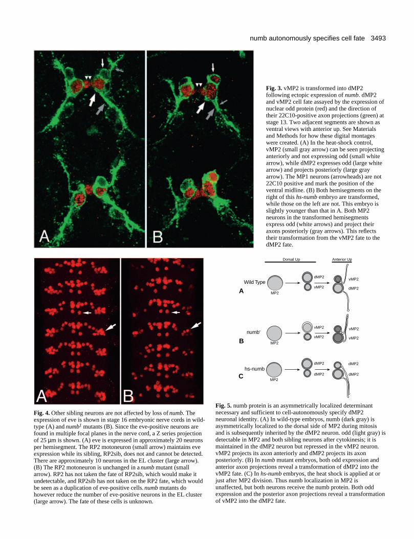
Fig. 5. numb protein is an asymmetrically localized determinantnecessary and sufficient to cell-autonomously specify dMP2neuronal identity. (A) In wild-type embryos, numb (dark gray) isasymmetrically localized to the dorsal side of MP2 during mitosisand is subsequently inherited by the dMP2 neuron. odd (light gray) isdetectable in MP2 and both sibling neurons after cytokinesis; it ismaintained in the dMP2 neuron but repressed in the vMP2 neuron.vMP2 projects its axon anteriorly and dMP2 projects its axonposteriorly. (B) In numb mutant embryos, both odd expression andanterior axon projections reveal a transformation of dMP2 into thevMP2 fate. (C) In hs-numb embryos, the heat shock is applied at orjust after MP2 division. Thus numb localization in MP2 isunaffected, but both neurons receive the numb protein. Both oddexpression and the posterior axon projections reveal a transformationof vMP2 into the dMP2 fate.
Fig. 3. vMP2 is transformed into dMP2following ectopic expression of numb. dMP2and vMP2 cell fate assayed by the expression ofnuclear odd protein (red) and the direction oftheir 22C10-positive axon projections (green) atstage 13. Two adjacent segments are shown asventral views with anterior up. See Materialsand Methods for how these digital montageswere created. (A) In the heat-shock control,vMP2 (small gray arrow) can be seen projectinganteriorly and not expressing odd (small whitearrow), while dMP2 expresses odd (large whitearrow) and projects posteriorly (large grayarrow). The MP1 neurons (arrowheads) are not22C10 positive and mark the position of theventral midline. (B) Both hemisegments on theright of this hs-numb embryo are transformed,while those on the left are not. This embryo isslightly younger than that in A. Both MP2neurons in the transformed hemisegmentsexpress odd (white arrows) and project theiraxons posteriorly (gray arrows). This reflectstheir transformation from the vMP2 fate to thedMP2 fate.
Fig. 4. Other sibling neurons are not affected by loss of numb. Theexpression of eve is shown in stage 16 embryonic nerve cords in wild-type (A) and numb1 mutants (B). Since the eve-positive neurons arefound in multiple focal planes in the nerve cord, a Z series projectionof 25 µm is shown. (A) eve is expressed in approximately 20 neuronsper hemisegment. The RP2 motoneuron (small arrow) maintains eveexpression while its sibling, RP2sib, does not and cannot be detected.There are approximately 10 neurons in the EL cluster (large arrow).(B) The RP2 motoneuron is unchanged in a numb mutant (smallarrow). RP2 has not taken the fate of RP2sib, which would make itundetectable, and RP2sib has not taken on the RP2 fate, which wouldbe seen as a duplication of eve-positive cells. numb mutants dohowever reduce the number of eve-positive neurons in the EL cluster(large arrow). The fate of these cells is unknown.

3494 E. P. Spana and others
Genes with phenotypes similar to numb or Notch in the ESlineage, genes involved in the Delta-Notch signalling pathwayand genes already known to be expressed in the MP2 lineagecan now be examined in the MP2 lineage for a role in cell fatespecification. It will be interesting to see whether any of thesegenes function with numb in the specification of the dMP2 andvMP2 neurons.
We thank E. Ward and D. Coulter for the odd-skipped antibody,and M. Rhyu and Y. N. Jan for the numb antibody, numb1 and numb2
mutants, and heat-shock-numb stock. E. P. S. was supported by anNIH Cell and Molecular Biology Predoctoral training grant andfunding from the Howard Hughes Medical Institute to C. Q. D.; C.K. is a Jane Coffin Childs Postdoctoral Fellow; C. S. G. is an Inves-tigator with the Howard Hughes Medical Institute; C. Q. D. is anAssistant Investigator with the Howard Hughes Medical Institute.
REFERENCES
Campbell, G., Goring, H., Lin, T., Spana, E., Anderson, S., Doe, C. Q. andTomlinson, A. (1994). RK2, a glial-specific homeodomain protein isrequired for embyonic nerve cord condensation and viability in Drosophila.Development 120, 2957-2966.
Campos-Ortega, J. A. (1993). Early neurogenesis in Drosophilamelanogaster. In The Development of Drosophila (eds. M. Bate and A.Martinez-Arias), pp 1091-1130. Cold Spring Harbor, NY: Cold SpringHarbor Press.
Campos-Ortega, J. A. and Hartenstein, V. (1985). The EmbryonicDevelopment of Drosophila melanogaster. Berlin Heidelberg New York:Springer-Verlag.
Chu-LaGraff, Q. and Doe, C. Q. (1993). Neuroblast specification andformation regulated by wingless in the Drosophila CNS. Science 261, 1594-1597.
Doe, C. Q. (1992). Molecular markers for identified neuroblasts and ganglionmother cells in the Drosophila central nervous system. Development 116,855-863.
Driever, W. (1993). Maternal control of anterior development in theDrosophila embryo. In The Development of Drosophila (eds. M. Bate and A.Martinez-Arias), pp 301-324. Cold Spring Harbor, NY: Cold Spring HarborPress.
Frasch, M., Hoey, T., Rushlow, C., Doyle, H. and Levine, M. (1986).Characterisation and localisation of the even-skipped protein of Drosophila.EMBO J 6, 749-759.
Fujita, S. C., Zipursky, S. L., Benzer, S., Ferrus, A. and Shotwell, S. L.(1982). Monoclonal antibodies against the Drosophila nervous system. Proc.Natl. Acad. Sci. USA 79, 7929-7933.
Goodman, C. S. and Doe, C. Q. (1993). Embryonic development of the
Drosophila Central Nervous System. In The Development of Drosophila(eds. M. Bate and A. Martinez-Arias), pp 1131-1206. Cold Spring Harbor,NY: Cold Spring Harbor Press.
Greenwald, I., and Rubin, G. M. (1992). Making a difference: The role ofcell-cell interactions in establishing separate identities for equivalent cells.Cell 68, 271-281.
Hartenstein, V. and Posakony, J. W. (1990). A dual function of the Notchgene in Drosophila sensillum development. Dev. Biol. 142, 13-30.
Horvitz, H. R. and Herskowitz, I. (1992). Mechanisms of asymmetric celldivision: Two Bs or not two Bs, that is the question. Cell 68, 237-255.
Huff, R., Furst, A., and Mahowald, A. P. (1989). Drosophila embryonicneuroblasts in culture: autonomous differentiation of specificneurotransmitters. Dev. Biol. 134, 146-157.
Jan, Y. N. and Jan, L. Y. (1995). Maggot’s hair and bug’s eye: Role of cellinteractions and intrinsic factors in cell fate specification. Neuron 14, 1-5.
Kuwada, J. Y. and Goodman, C. S. (1985). Neuronal determination duringembryonic development of the grasshopper nervous system. Dev. Biol. 110,114-126.
Lin, D. M., Fetter, R. D., Kopczynski, C., Grenningloh, G. and Goodman,C. S. (1994). Genetic analysis of fasciclin II in Drosophila: defasciculation,refasciculation and altered fasciculation. Neuron 13, 1055-1069.
Lundell, M. J. and Hirsh, J. (1994). A new visible light DNA fluorochromefor confocal microscopy. BioTechniques 16, 434-440.
Menne, T. V. and Klambt, C. (1994). The formation of commisures in theDrosophila CNS depends on the midline cells and on the Notch gene. Dev.Biol. 120, 123-133.
Parks, A. L. and Muskavitch, M. A. T. (1993). Delta function is required forbristle organ determination and morphogenesis in Drosophila. Dev. Biol.157, 484-496.
Posakony, J. W. (1994). Nature versus nurture: Asymmetric cell divisions inDrosophila bristle development. Cell 76, 415-418.
Rhyu, M. S., Jan, L. Y. and Jan, Y. N. (1994). Asymmetric distribution ofnumb protein during division of the sensory organ precursor cell confersdistinct fates to daughter cells. Cell 76, 477-491.
Spana, E. P. and Doe, C. Q. (1995). The prospero transcription factor isasymmetrically localized to the cell cortex during neuroblast mitosis inDrosophila. Development 121, 3489-3494.
St Johnston, D. (1993). Pole plasm and the posterior group genes. In TheDevelopment of Drosophila (eds. M. Bate and A. Martinez-Arias), pp 325-363. Cold Spring Harbor, NY: Cold Spring Harbor Press.
Tepass, U. and Knust, E. (1993). crumbs and stardust act in a genetic pathwaythat controls the organization of epithelia in Drosophila melanogaster. Dev.Biol. 159, 311-326.
Udolph, G., Prokop, A., Bossing, T. and Technau, G. M. (1993). A commonprecursor for glia and neurons in the embryonic CNS of Drosophila givesrise to segment-specific lineage variants. Development 118, 765-775.
Uemura, T., Shepherd, S., Ackerman, L., Jan, L. Y. and Jan, Y. N. (1989).numb, a gene required in determination of cell fate during sensory organformation in Drosophila embyros. Cell 58, 349-360.
(Accepted 10 July 1995)