Assessment of conservation state of gothic wall paintings - ADDI
195
Department of Analytical Chemistry Assessment of conservation state of gothic wall paintings: from scientific diagnostic to a new monitoring approach Report to complete for the International PhD degree Ilaria Costantini February 2019 (cc)2019 ILARIA COSTANTINI
-
Upload
khangminh22 -
Category
Documents
-
view
1 -
download
0
Transcript of Assessment of conservation state of gothic wall paintings - ADDI

Department of Analytical Chemistry
Assessment of conservation state of
gothic wall paintings: from scientific
diagnostic to a new monitoring approach
Report to complete for the International PhD degree
Ilaria Costantini
February 2019
(cc)2019 ILARIA COSTANTINI
ACKNOWLEDGMENTS
I am very grateful to its research group of the Department of Analytical Chemistry,
Ikerkuntza eta Berrikuntza Analitikoa (IBeA).
This PhD work has been developed thanks to the financial support received from:
- CTP projects (ref CTP09-P04 and CTP12-P10) founded by Working Community
of the Pyrenees, Basque Government.
- project IT-742-13 for Consolidated Research Groups, funded by the Basque
Country Government.
My thankfulness goes also to the technical support provided by Dr. Alfredo Sarmiento
(in the Singular Coupled Multispectroscopy Laboratory, LASPEA), Dr. Javier Sanguesa
(in the X-Ray Service of Rocks and Minerals Unit), from the Research General Services
of the UPV/EHU (SGIKER) and Dr. Xabier Murelaga (Department of Stratigraphy and
Palaeontology of the University of the Basque Country). MONARIS research group
from the University of Pierre et Marie Curie (UPMC, Paris, France) is also gratefully
acknowledged
In addition, I am grateful to Diputación Foral de Álava, and to Valderejo Natural Park.
In also thank Dra. Maria Dolores Rodriguez Lazo (Department of Painting in Faculty
of Fine Arts, University of the Basque Country).
I would also like to express my gratitude to Dr. Fernando Benito Lopez (Analytical
Microsystems & Materials for Lab-on-a-Chip Group, University of the Basque
Country) for giving me the opportunity to use your knowledge for my investigation.

I would like to thank Dr Pier Paolo Lottici and Dr. Danilo Bersani for trust in me and
for introducing me in the IBeA research group, Dr. Juan Manuel Madariaga for giving
me the opportunity to do this PhD, and my supervisor Dr. Kepa Castro for the support
and the dedication to carry out this work.
I would also like to thank all researchers forming part of the IBeA group for the
professional and human support and for the good friendships born during the years.


I
INDEX
CHAPTER 1: Introduction…………………….…………………….....................................…….1
. Multia alyti al app oa h fo the study of e i o e tal st esso s……..…..……….2
1.2 Medieval wall painti gs……………………………………………………………………..….………..8
1.3 Characterizatio of ediae al all pai ti g’s ate ials…………..…….…………….10
. . Study of a ti ue pig e ts………………………………………………….…………………10
1.3.2 Further than in situ a alysis……………………………………………………..……..……. 5
. . U usual fi di gs……………………………………………………………………………....…… 8
1.4 The study of the state of conservation…………….………………………..………………….20
1.4.1 Degradation due to anthropic factors…………………………………..……..………… 5
1.4.2 Natural degradation……………………………………………………….….……………….… 8
1.4.3 Biodeterioration processes………………………………………………………………….…32
1.5 Why is it important to apply diagnostic techniques?........................................35
1.6 Fi al e a ks……………………………………………………………………………..……………..….. 8
References ………………………………………………………………………………………………………….
CHAPTER 2: Objectives………………………………………………………………………….…………..51
CHAPTER 3: Emplacements a d sa ples…………………………………………….………………53
. Sai t Stephe ’s hu h i Ri e a de Valde ejo Ála a, Spai …………………………54
. . Wall pai ti gs des iptio ………………………………………………….…………….…… 6
. . Co se atio state………………………………………………………………………………...58
. . Sa pli g……………………………………………………………………………………………….. 9
. Sai t Stephe ’s hapel of Mo ta i Bolza o, Italy …………………..………….………..61
. . Wall pai ti gs des iptio ……………………………………………………………..…..….62
3.2.2 Conservation state………………………………………………………………………..……….64
3.2.3 Sampling…………………………………………………………………………………..………..….66
II
References…………………………………………………………………………………..……….…………….. 8
CHAPTER 4: Experimental techniques…………………………………………………………………69
4.1 General purpose instruments and tools………………………………………………..……...69
4.2 Hand-held and portable analytical techniques for in situ analyses………..……….73
4.2.1 X-Ray Fluorescence Spectroscopy…………………………………………………….……73
4.2.2 Raman Spectroscopy…………………………………………..………………………………...74
4.3 Bench top analytical techniques for non-dest u ti e la o ato y a alyses….….77
4.3.1 Scanning Electron Microscopy (SEM) Energy Dispersive X-Ray
Spectrometer EDS ……………………………………………………………………………….……….77
4.3.2 Raman Spect os opy…………………………………………………...............………..…..77
4.3.4 X-Ray Diff a tio XRD ……………………………………………………………………….... 9
4.4 Isolation and characterization of biodeteriogens genomic DNA sequences via
PCR amplification.....................................................................................................80
References ……………………………………………………………………………………………………….…..81
CHAPTER 5: Investigation of mural paintings blackening in the chapel of Saint
Stephe ’s i Mo ta i Italy ……………………………………………..…………………………..…..83
. Results……………………………………………………………………………..……………………………87
5.1.1 In situ a alysis………………………………………………………………………………..………87
5.1.2 Laboratory analyses………………………………………………………………..……………..89
5.2 Final remarks ………………………………………………………………………………………….….105
References …………………………………………………………………………………………..………….….109
CHAPTER 6: in situ and laboratory analyses for the understanding of a
controversial restoration work……………………………………..…………………………………. 15
6.1 In situ a alyses…………………………………………………………………………………………….116
6.2 Laboratory analyses…………………………………………………………………………………....122
6.3 Final remarks……………………………………………………………………………………………….128
References………………………………………………………………………………………………………….131
III
CHAPTER 7: Study of natural impact on wall paintings and building materials of
the abandoned church of Ribera de Valderejo…………………………………………..…….135
. Results……………………………………………………………………………………………..….………137
7.1.1 Study of soluble and insoluble efflorescence salts………………………………. 37
. . Cha a te izatio of iopati as…………………………………………………………..…143
7.2 Final remarks……………………………………………………………………………………..………..147
References …………………………………………………………………………………………….…………… 50
CHAPTER 8: PALME software as an alternative tool for semiquantification of salt
efflorescence……………………………………………………………………………….………..……….. 53
. Methodology……………………………………………………………………………………………….155
. . Sa ples p epa atio ………………………………………………………….………………..155
. PALME soft a e…………………………………………………………………..……………..………157
. Results …………………………………………………………………………………………..……..…….158
. Fi al e a ks ………………………………………………………………………………………..……. 68
References ……………………………………………………………………………………………….………...170
CHAPTER 9: Development and testing of a microfluidic sensor device for detection
of ammonium in buildings materials……………………………………………………………….. 73
. Methodology……………………………………………………………….…………………………….. 78
9.1.1 Realizatio of efe e e se so s………………………………………………………….. 78
. . A alysis of efe e e se so s……………………………………………………………….181
9.1.3 Realization of the microfluidic sensor device …………………………………..….185
9.1.4 Preparation of mortar mockups…………………………………………………………… 86
9.2. Microfluidic sensor device testing on mortar mockups………………………….…..187
. Fi al e a ks……………………………………………………………………………………………....195
References…………………………………………………………………………………..…………………...…197
CHAPTER : Fi al co clusio s…………………………………….………………………………..…199

IV
CHAPTER 11: Scientific publications…………………………………………………….…………..207
ANNEX………………………………………………………………………………………………………..……209

1
CHAPTER 1
Introduction
Cultural Heritage represents the basis of our identity, the legacy that the past
civilizations have left us and, for this reason, we must protect it for the future
generations. The preservation of our heritage is a current topic of great interest, in
particular due to the increase in the last 30-40 years of threatening factors, such as
environmental contamination, due to the industrialization, wars and political
instability.
The te Cultu al He itage usuall i di ates o je ts a d uildi gs that ha e a
historical, artistic and monetary value, although also the intangible heritage that
includes traditions, knowledge, dialects, etc. is part of it. It is a dynamic concept,
because new discoveries from all over the world enrich it every day.
Different types of objects, books, archaeological pieces, paintings, statues, oil and
wall paintings, porcelain, jewellery and textiles belong to the cultural heritage and
each item is composed of different materials, organic and inorganic, which have a
variable aging that affect its durability. Undoubtedly, the most threatened artworks
are all those in the open air, as well as those that are kept in places where it is not
possible to have a controlled atmosphere, such as architectural works, statues,
fountains and wall paintings inside churches and castles [1, 2].
CHAPTER 1
2
[Titolo del documento]
1.1 Multianalytical approach for the study of
environmental stressors
The factors that promote the deterioration may be intrinsic or extrinsic to the work
of art. For example, several studies have shown that the instability and the reactivity
to the atmospheric agents of some pigments employed on wall paintings can cause
aesthetic damage [3]. Many examples of chromatic variations are known in the
literature, the most frequent being of blackening of lead based pigments such as
minium and lead white [4], or the degradation of malachite [5], cinnabar [6] or natural
earths [7]. In the recent work by Coccato et al. [8] the effect of different parameters
on the stability of most common medieval pigments were shown underlining the
complexity in the conservation of polychrome artworks.
Additionally, the formation of soluble salts, in form of carbonate, sulphate and
nitrate, represents one of the most evident and dangerous cause of degradation for
a porous building material, on the surface (efflorescence) or within the porous
structure (subefflorescence), although the stone has been considered for centuries
one of the most resistant materials [9]. The variations in temperature in presence of
water, both infiltration of rainwater or risen by capillarity, cause cycles of salt
crystallization and hydration and dehydration provoking the contraction and
expansion of the material, which over time, undergoes micro fractures and
subsequently disintegrates [10]. In particular, the formation of nitrate salts, such as
ammonium nitrate, is one of the major causes of disintegration of carbonaceous
materials since ammonium ions are highly reactive species and can attack the alkaline
substances of which the masonries are composed [11]. The identification and study
of nitrate salts that may be caused by natural factors, such as the degradation of
organic material in soil, or of anthropic nature following the wet deposition of NOx
gases in areas with a high level of environmental pollution, is a fundamental step for
the protection of building materials, stones, plasters and bricks [12].
Introduction
3
[Titolo del documento]
Moreover, the interaction between objects and structures and atmospheric
pollutants is increasingly studied due to the high levels of air contamination present
in many European cities. Air pollution is characterized by the high level of acidic gases,
which in presence of the water rain become into acids (H2CO3, H2SO4, HNO3) [13, 14].
The action of these acidic compounds favours the degradation of the building
materials and ancient and modern artworks in the open air [15]. These processes
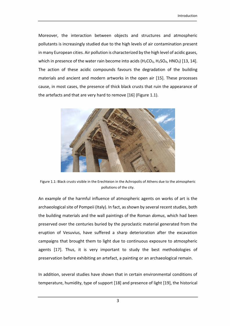
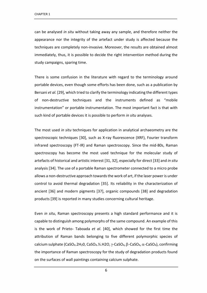
cause, in most cases, the presence of thick black crusts that ruin the appearance of
the artefacts and that are very hard to remove [16] (Figure 1.1).
Figure 1.1: Black crusts visible in the Erechteion in the Achropolis of Athens due to the atmospheric
pollutions of the city.
An example of the harmful influence of atmospheric agents on works of art is the
archaeological site of Pompeii (Italy). In fact, as shown by several recent studies, both
the building materials and the wall paintings of the Roman domus, which had been
preserved over the centuries buried by the pyroclastic material generated from the
eruption of Vesuvius, have suffered a sharp deterioration after the excavation
campaigns that brought them to light due to continuous exposure to atmospheric
agents [17]. Thus, it is very important to study the best methodologies of
preservation before exhibiting an artefact, a painting or an archaeological remain.
In addition, several studies have shown that in certain environmental conditions of
temperature, humidity, type of support [18] and presence of light [19], the historical

CHAPTER 1
4
[Titolo del documento]
and modern building materials can represent a suitable substrate for biological
colonization [20, 21]. The a ote oid o ga i pig e ts β-carotene, zeaxanthin and
astaxanthin) present in photosynthetic organisms are the most common molecules
identified on substrates and are the main responsible for the aesthetic damage due
to the formation of green, pink or brown patinas on the surfaces of the artefacts [22].
A well know study by Ciferri [23] on the damage caused by the presence of
microorganisms on cultural heritage shows very extensively the most widespread
species on organic and inorganic substrates and the damage that they are able to
promote. Indeed, although it may seem that the presence of biological patinas exerts
only an aesthetic damage, microorganism such as fungi or lichens can penetrate into
the material on which they have grown, with hyphae and thallus respectively, causing
cracks. In addition, from the point of view of chemical damage, oxalic acid (H2C2O4),
secreted by microorganisms, reacts with the calcium carbonate of the substrate
causing the formation of calcium oxalate that, depending on the degree of hydration,
is recognized as whewellite (CaC2O4·H2O) or weddellite (CaC2O4·2H2O), two insoluble
mineral species that contribute to the formation of salt efflorescence [24].
A well know study by Edwards et al. [25], was carried out on the frescoes of Palazzo
Farnese, Caprarola, Italy, where, due to the chemical and structural damage caused
by the massive colonization of lichens (Dirina massiliensis forma sorediata), around
the 80% of the Renaissance frescoes were destroyed in some areas. The analyses
indicated a significant chemical activity in lichen encrustations, in which some
metabolic products such as erythrine, lecanoric acid and polyphenolic acids were
identified together with calcium oxalate hydrate, formed by the irreversible reaction
of oxalic acid with the calcium carbonate of the substrate [26]. On the other hand,
some studies have highlighted the presence of calcium oxalate resulting from the
degradatio of o ga i i de s edia [27].
Introduction
5
[Titolo del documento]
Being this the state-of-the-art, it is interesting to see the way in which all mentioned
degradations perception has changed, as well as the philosophy in which
conservation and restorations works are carried out.
In past years, the evaluation of the conservation status of an artefact was carried out
by curators and restorers according to an empirical method developed over the years
without carrying out chemical-physical investigations. This lack of materials
knowledge sometimes led to wrong diagnosis and to unsuitable restoration
interventions accordingly. In this way, rather than protect a work of art these
interventions worsened its aesthetics and reduced its durability in time. Only in a few
cases the analytical techniques were used, for example, to verify the suitability and
effectiveness of chemical products used in cleaning procedure [28].
Du i g the last de ade, due to the i ease of de a s factors, it has been necessary
to operate with a renewed attitude and a great attention has been paid to the study
of materials. For this reason, different professionals - conservators, restorers and
diagnosticians - have shared their historical, artistic and scientific knowledge to
characterize and study not only the original materials but also especially the
degradation products, and consequently, identify the factors that caused them.
Therefore, it has been sometimes possible to reduce these factors and choose the
best restoration and conservation treatment. This was possible especially thanks to
the development of new non-invasive diagnostic methodologies, sometimes adapted
from other scientific fields, that have been employed for the study of Cultural
Heritage. Currently, restoration interventions are carried out only after a chemical
study of materials and each artwork is considered as an individual case.
In this sense, portable and mobile diagnostic techniques are the most adequate tools
with considerable advantages for the study of works of art. Thanks to them, it has
been possible to extend the scientific study to the so-called immobile works of art
that cannot be transported to the laboratory and consequently cannot be analysed
by benchtop instruments. Nowadays, statues, architectural works and wall paintings
CHAPTER 1
6
[Titolo del documento]
can be analysed in situ without taking away any sample, and therefore neither the
appearance nor the integrity of the artefact under study is affected because the
techniques are completely non-invasive. Moreover, the results are obtained almost
immediately, thus, it is possible to decide the right intervention method during the
study campaigns, sparing time.
There is some confusion in the literature with regard to the terminology around
portable devices, even though some efforts has been done, such as a publication by
Bersani et al. [29], which tried to clarify the terminology indicating the different types
of non-dest u ti e te h i ues a d the i st u e ts defi ed as o ile
i st u e tatio o po ta le i st u e tatio . The ost i po ta t fa t is that ith
such kind of portable devices it is possible to perform in situ analyses.
The most used in situ techniques for application in analytical archaeometry are the
spectroscopic techniques [30], such as X-ray fluorescence (XRF), Fourier transform
infrared spectroscopy (FT-IR) and Raman spectroscopy. Since the mid-80s, Raman
spectroscopy has become the most used technique for the molecular study of
artefacts of historical and artistic interest [31, 32], especially for direct [33] and in situ
analysis [34]. The use of a portable Raman spectrometer connected to a micro probe
allows a non-destructive approach towards the work of art, if the laser power is under
control to avoid thermal degradation [35]. Its reliability in the characterization of
ancient [36] and modern pigments [37], organic compounds [38] and degradation
products [39] is reported in many studies concerning cultural heritage.
Even in situ, Raman spectroscopy presents a high standard performance and it is
capable to distinguish among polymorphs of the same compound. An example of this
is the work of Prieto- Taboada et al. [40], which showed for the first time the
attribution of Raman bands belonging to five different polymorphic species of
calcium sulphate (CaSO4.2H20, CaSO4.½.H2O, -CaSO4, -CaSO4, -CaSO4), confirming
the importance of Raman spectroscopy for the study of degradation products found
on the surfaces of wall paintings containing calcium sulphate.
Introduction
7
[Titolo del documento]
Together with Raman spectroscopy, XRF spectroscopy is also a very valuable
technique for in situ analyses since it provides the elemental composition being used
generally as the first screening of the materials [41]. As well as XRF spectroscopy,
laser-induced breakdown spectroscopy (LIBS) is another technique devoted to the
elemental characterization of materials, and presents some advantages over the XRF
approach, like the possibility to detect light elements. However, LIBS is still not used
as often as XRF although a detailed study was reported more than fifteen years ago
by Anglos [42].
In addition to the success of portable instrumentation, researches have showed that
the best results are obtained when portable techniques are supported by laboratory
analyses. In these cases, portable techniques have been fundamental to identify the
areas of interest in which perform further micro-sampling without unnecessarily
affecting the underlying surfaces. Through the use of laboratory techniques, such as
scanning electron microscope combined with energy-dispersive X-ray spectroscopy
(SEM-EDS), micro X- a fluo es e e μ-XRF), X-ray diffraction (XRD) [43] or
chromatographic techniques (PY-GC-MS, GC-MS) [44], it is possible to obtain
information that could not be obtained only by in situ analysis. In fact, the signal
obtained by in situ analysis, depending on the technique used, can provide
information that relates not only to the surface but also to the underlying layers,
making more complex the interpretation of the results. For this reason, SEM, micro-
XRF and Raman imaging techniques have revealed to be very useful to obtain the
elemental and molecular distribution of compounds in a sample, especially if it has
been previously embedded in resin to be analysed as a cross section to identify the
stratigraphic distribution of compounds in the layers [45, 46]. This preparation of the
sample is particularly important in the case of wall paintings as they are characterized
by a succession of different layers, in particular in the case of the fresco technique,
which needs some substrate preparation layers (arriccio and intonaco), above which
the paint layer is applied. Moreover, during its history, wall paintings may be

CHAPTER 1
8
[Titolo del documento]
subjected to restoration interventions and new materials from the modern era,
pigments, consolidating materials and varnishes can be found on the surface.
Thus, thanks to the point by point analysis carried out using laboratory instruments,
the materials that make up each pictorial layer can be well characterized as well as
the presence of possible degradation products.
Indeed, among the immovable artefacts, the wall paintings are undoubtedly one of
the most studied categories because they are the most at risk due to the permanent
interaction with the surrounding environment. Therefore, the study of these
artworks provides information not only about the raw materials but also about the
environment in which they are located. In this way an explanation of the reactions
between the original compounds and the chemicals in the environment can be
explained and the subsequent adequate restoration procedures can be applied [47].
1.2 Medieval wall paintings
After the revaluation of medieval art, excellent results have been obtained in recent
years concerning the study of wall paintings. The medieval era covers a very long
period that includes almost a millennium of art history. According to the most
widespread periodization, the Middle Ages would start in 476 AD, the year of the fall
of the Western Roman Empire, and would end in 1492, the year of the discovery of
America. Generally, the main currents that are recognized by the art historians
include a phase following Early- Christian art, Byzantine art, pre-Romanesque and
Romanesque, and Gothic. However, the attempt to classify them in main periods and
styles could be complex due to local currents that spread to different locations in
Europe and in the Middle East.
The strong break with the classical art from which it strongly differs for the symbolic
character, in contrast to the realism, was the main cause of lack of appreciation that
it had espe iall i the Re aissa e pe iod i hi h ith Re aissa e it as usual
Introduction
9
[Titolo del documento]
to indicate a rebirth of the arts after the dark medieval ages [48]. This denigration for
an art that was considered of a lower level, and therefore, not worthy of being
safeguarded, was the main cause of abandon of medieval art and even of its voluntary
destruction in the phenomenon of iconoclasm when many works of art were lost
forever. The icons were burned and the mosaics were destroyed as well as the wall
paintings inside the churches. In some cases, these were covered under layers of
plaster or hidden with altarpieces.
Around the middle of the 19th century, medieval art conceived as a true artistic
current was rediscovered, thanks above all to the influence of romantic culture
promoted by writers such as John Ruskin, Eugène Viollet-le-Duc, and Pugin, because
they were the first to question the conservation of the architectural heritage that
arose in the Middle Ages and consequently also the treasures located inside them.
Although the paintings preserved in the castles had the purpose of celebrating the
nobility, those located inside the churches and the monasteries had an educational
function and represented the material instrument that connected the believers to
God. Unfortunately, many of them are at risk of conservation and for this reason
several studies have been carried out in recent years and have allowed to bring to
light pictorial cycles belonging to this era and the historical-artistic studies have
rediscovered new meanings of enigmatic paintings until now unknown.
This introduction aims to show the results obtained in the last twenty years in the
study of medieval wall paintings through the use of portable non-destructive and
micro-destructive techniques, highlighting their reliability in the characterization of
original materials and modern materials used during restoration work carried out in
modern times. Thanks to the study of the materials that compose the pictorial layers
it has been possible to obtain important information about the identification of
pictorial techniques as well as the determination of the period of execution, the
studies of provenance of the raw materials and the attribution to an author or a
studio. In particular, the numerous works that have underlined the usefulness of the
CHAPTER 1
10
[Titolo del documento]
scientific approach for the diagnostic of the conservation state of the paintings thanks
to the identification of degradation products caused by the impact of natural and
environmental stressors will be mentioned along this chapter.
1.3 Characterizatio of ediae al all pai ti g’s materials
1.3.1 Study of antique pigments
At first, the analytical techniques were mainly used for the characterization of original
materials of medieval wall paintings. The compositions of plasters for the
identification of the execution techniques and the pigments used were studied in
depth, especially through the use of laboratory techniques applied on fragments for
surface analysis or in cross-section.
Certainly, the research group headed by H.G.M. Edwards was one of the first to prove
the reliability of spectroscopic techniques for the study of works of art. One of his
first researches on the study of medieval works involved the use of Fourier transform
Raman spectroscopy for the identification of pigments on red samples of English
medieval wall paintings [49] where the signals of vermilion (HgS) on a substrate of
calcium carbonate and siliceous material were identified. In addition, a mixture of
vermilion and red iron oxide (α-Fe2O3) was found approximately mixed in a ratio of
1:3 from a comparison of intensity of spectra of standard pigments.
Numerous researches were carried out on the study of medieval wall paintings
preserved in the religious buildings in the north of Spain. The construction of
churches and monasteries in the Middle Ages in this area was due to the need to
provide an accommodation to pilgrims who stopped in villages along the way heading
to Santiago de Compostela in Galicia (Spain). It is precisely through the comparison
of some fragments taken from two churches in the province of Castilla y León (Spain),
that was verified by Edwards et al. [50] an antique controversy regarding the use of
Introduction
11
[Titolo del documento]
the term "minium" with which both cinnabar and red lead were defined. This work
showed the potential of the FT-Raman spectroscopy for the identification and
quantification of ancient pigments used in the mixture through the construction of
calibration curves. The confusion in the interpretation of ancient recipes for pigment
blends is unavoidable and was exacerbated by the practice of adulteration of
cinnabar with red lead generally for economic reasons.
However, the research carried out on the murals of the Church of Saints Cosmo and
Damian at Basconcillos del Tozo [51] (Castilla y León, Spain) demonstrated a
hierarchical application in the use of red pigments in which the most important
biblical figures were painted with pure cinnabar while mixtures of cinnabar and red
lead or red ochre were employed in other areas. Moreover, thanks to the application
of FT-Raman spectroscopy, two types of cinnabar were identified from two different
locations, one of which, from the Almadén mine, is supposed to be considered more
precious for its use in the paintings. The same pigment was found also in some
microsamples taken from the wall paintings from the thirteenth century Convento de
la Peregrina, Sahagún (León, Spain) [52].
Of particular interest are the studies conducted on microsamples from murals in the
monastery of Saint Baudelio [53] located to the south of the Duero river. The
chromatic palette composed of haematite, charcoal/soot, litharge, goethite, minium
and copper salt (probably malachite or verdigris) was completely different from the
one found in the northern paintings in which cinnabar, realgar, orpiment, barytes and
lapis lazuli were present. This discovery would confirm the theory of art historians
that the monastery of Saint Baudelio was colonized by an enclosed monastic order,
so the materials employed were limited only to those minerals available locally.
Another similar case is represented by medieval frescoes located in the Palencia
region of Spain [54]. Some fragments from four churches, belonging to different
periods (from fifteenth century to twelfth century) and with influences of different
pictorial schools were studied and only in one case cinnabar was used.
CHAPTER 1
12
[Titolo del documento]
Some researchers have highlighted the lack of availability of raw materials in the
Middle Ages and the difficulty of the purchase for economic reasons. This problem
did not concern only the availability of red pigment such as cinnabar and was
remarkable for the blue pigments. Indeed, since they were imported from Germany
(azurite (2Cu(CO3)2 Cu(OH)2)) or from Afghanistan (lapis lazuli (Na8(Al6Si6O24)Sn)) they
were expensive and therefore were reserved for important areas of wall paintings.
Indeed, in the iconographic tradition, the use of the blue pigment takes on a symbolic
value - it was used to paint the Virgin's mantle - as shown in the study of blue
fragments from medieval mural paintings in the South West of France [55]. Thanks
to spectrocolorimetric analysis and microanalysis with SEM-EDS, the use of copper
based pigments was pointed out, lapis lazuli and azurite (non-local source), employed
for the most important figures, while aerinite (local), a rare blue Fe(II)/Fe(III)-
containing aluminosilicate from the Pyrenees region, was used for the background.
In the absence of these pigments, the artists resorted to using more accessible
pigments, carbon black or lime white for example.
The same blue pigment aerinite was used for the painting of Romanesque frescoes in
the Pyrenean region of Catalonia and in Andorra [56] and its use was probably
dictated both by local availability and by economic factors. A detailed work published
by Pérez-Arantegui et al. [57] has allowed to obtain new analytical data concerning
the microstructure and the chemical composition of the aerinite pigment identified
on Romanesque wall paintings in Aragón (Spain).
The use of azurite for blue areas and posnjakite (Cu4(SO4)(OH)6·H2O) and atacamite
(Cu2(OH)3Cl) for green decorations was confirmed also by the study of fragments
from the mural paintings of the Tournai Cathedral, (Belgium) [58]. In other cases, as
i the Peć Mo aste [59], a black layer of coal was identified under a layer of green
and blue colour in order to obtain a more intense blue hue, a typical method applied
in Byzantine times.

Introduction
13
[Titolo del documento]
A series of wall paintings dating from the Romanesque period to the 16th century
were uncovered during restoration projects at the Pyrenean Church of Santa Eulàlia
of U ha i the Val d A a Spai [60]. The study through different analytical,
elemental and molecular techniques showed the use of traditional colours of the
medieval period (haematite, goethite, vermilion, red lead, carbon black and calcite)
and a variation of the palette based on the period of realization of the works. Only in
Romanesque paintings the aerinite was identified and for the first time its spectra
were shown in the context of a work of art. On the other hand, surprisingly, in the
16th century wall paintings a mixture of lime white and charcoal black was identified
to o tai a lue olou .
In fact, the work by Daniel et al. [55] highlighted the use of pigments suggested by a
different conception of colour in the medieval period that does not differ from
theories of contemporary colours. The false blues were used because some grey
shades could be perceived as bluish. In this work the false-blue was considered a
typology and a classification of it was proposed.
The use of false-blue was also found in early medieval frescoes of the Longobard
temple of Cividale del Friuli (Italy) [61] in which the blue colour is given by the
diffusion properties of the black and white particles of lime and carbon black
respectively. In this work a particular technique of achievement was underlined. In
fact, SEM images identified the use of textile fibres incorporated in the pictorial film.
Considering the scarce availability of cotton in Europe in the Middle Ages it can be
assumed that this material was imported by the workers of oriental origin.
Pigment analyses have sometimes revealed the money available for the execution of
the paintings. For example, in the case of the study of 14th century wall paintings from
the Church of the Holy Mother of God Hodegetria (Serbia) [59], Raman spectroscopy
showed that the artist's palette was exceptionally limited, consisting of inexpensive
pigments of local origin, testifying that the founder member did not belong to a noble
family.
CHAPTER 1
14
[Titolo del documento]
On the contrary, the multi-analytical study based on the use of elemental and
molecular techniques, - employed in the study of materials used for the decoration
of the o aste of Žiča [62] (Serbia), - was fundamental for the identification of
expensive pigments such as lapis lazuli and cinnabar, as well as gilding with golden
tin, which confirmed the desire of the Serbian aristocracy to assert its economic
power. This was also the case of the materials found in the Gothic wall painting of
the Dominican Monastery in Ptuj (Slovenia) [63], where the use of expensive
pigments such as azurite and cinnabar indicated a wealthy client. Moreover, of great
interest was the discovery of the lead–tin yellow type I in the wall paintings belonging
to 14th century since, until that moment it was considered that it appeared on the
market only in the first half of the 15th century.
In the study by Edwards et al. [64] on a post-medieval wall painting in the church of
St. Peter and St. Paul at Upton (United Kingdom), an unusual palette of pigments was
identified, which included expensive pigments such as cinnabar and lapis lazuli and
materials from the nineteenth century such as barites and chrome yellow. The
hypothesis formulated by the researchers is that an ancient mural painting could
have been repainted in modern times. A similar research using micro-Raman
spectroscopic was reported by Minceva-Sukarova et al. [65] on the study of pigments
used i edie al all pai ti g i S eta Bogo odi a hu h i Lešok No th- Western
Macedonia) in which a distinction was made between original and new materials
from two layer paint from the 17th and from the 19th century.
Thanks to technological development, new portable instruments have been tested to
make direct measurements on the artworks for the identification of pigments and
the execution techniques. They have proved to be particularly suitable for
archaeometric study, above all because of their non-invasive feature. This means that
sampling can be avoided since the portable instrumentation allows to perform a
satisfactory investigation of the material. Although some pitfalls have been pointed
out in the use of mobile instrumentation (the instrumental set-up, the positioning of
the probe head and stability problems for vibrations of the scaffold, problems related
Introduction
15
[Titolo del documento]
to environmental conditions [66] or to the presence of some materials [67]), the
spectroscopic portable techniques have been the most used ones for the study of the
conservation state of wall paintings in the last years.
The completely non-destructive work carried out by using only mobile tools (X-ray
fluorescence and Raman spectroscopy) to examine the 15th century mediaeval vault
pai ti gs i the Ou Lad s Cathed al A t e p, Belgiu [68] can be used as an
example of the difficulties that during an in situ analysis can arise. In fact, the Raman
signals from calcium carbonate, belonged to small particles left after removing the
layer of plaster applied over the paintings during the iconoclast wave, made difficult
the characterization of the pigments by Raman spectroscopy. Despite this, lead
white, calcite, gypsum, lead tin yellow, and vermilion were identified.
Another research work carried out completely in non-invasive way by using just a
portable XRF analyser, concerned the study of medieval wall paintings of
Yemrehanna Krestos Church, Ethiopia [69]. This research was the first research of
Ethiopian wall paintings. The correlation between the element concentration and the
multivariate statistical analysis was used to identify the most probable compounds
and to classify the groups of pigments used. Some authors state that the in situ ED-
XRF analyses are, in most cases, sufficient to guide in situ conservation and
restoration interventions, as shown by Ferrero et al. [70] in the study of a wall
painting of the 14th and 15th centuries. The identification of inorganic key elements
allowed the characterization of the pigments and the techniques used by the artists.
This approach, however, is sometimes criticized because the same elemental
composition can belong to different molecular composition, thus, data
misinterpretation can occur.
1.3.2 Further than in situ analysis
Although some researches have shown that it is possible to obtain exhaustive results
only through the use of portable techniques, other publications had underlined the
CHAPTER 1
16
[Titolo del documento]
need to continue the study through laboratory analysis. In this sense, the portable
techniques are extremely useful for identifying a particular area in order to minimize
the sampling.
The simultaneous use of portable XRF and Raman spectroscopies was tested for the
study of the range of pigments used in the mural paintings and frescos of the Little
Ch istophe ha e i the Mai To Hall of Gdańsk, Pola d [71]. Subsequently,
the statistical processing of the spectra was applied to minimize the number of
sample extractions to complete the study using micro-Raman spectroscopy in the
laboratory. The presence of copper, lead, iron, mercury and calcium together with
traces of antimony, cadmium, barium and molybdenum, was in accordance with the
composition of the identified pigments by Raman spectroscopy.
In order to solve the problem of the lack of written source concerning the techniques
of execution of Nubian wall paintings [72], different analytical techniques such as
portable X-ray fluorescence, laser ablation inductively coupled plasma mass
spectrometry and Raman spectroscopy were applied to identify the raw materials
and distinguish the use of different pigments. The use of complementary techniques
made it possible to observe some variations in the composition of the pigments in
samples from different archaeological sites and were useful for evaluating the
existence of possible variations among the local Nubian workshops.
The comparison between the materials used in different wall paintings belonging to
the same area can be useful for the identification of the origin of the workers or for
the attribution to an artist as shown in the work by Lampakis et al. [73] where thanks
to the combined use of micro-Fourier transform infrared and micro-Raman
spectroscopy some fragments of Byzantine wall paintings from three Thessalian
monasteries (Greece) were studied. The similarities in the materials and in the
technique supported the hypothesis that the same artist may have worked in the
three churches. On the other hand, the vibrational techniques FT-IR a d μ- Raman
were employed on fragments from the medieval Monastery of Karaach-Teke [74] in
Introduction
17
[Titolo del documento]
which it was evident the Byzantine influence on the technology and the thematic-
aesthetic features of Byzantine iconography in Bulgaria. In the research on Russian-
Byzantine wall paintings from Sweden [75], dating back to the 12th century, the
isotopic composition of lead pigments was studied using a mass spectrometer to
clarify the provenance of the materials found in the paintings. The results showed
that the materials were not only locally sourced but rather came from both Russia
and Germany as well.
In another study [76], the natural origin of azurite and malachite found in some
murals was demonstrated by the presence of copper and zinc arsenates, while
artificial malachite was identified by uniform spherulitic particles.
In the same way the objective of the study of wall paintings preserved within two
Byzantine churches in Kastoria, (Greece) [77] was to find similarities in the materials
to hypothesize the presence of a single hand in the realization of the paintings.
Thanks to the use of SEM-EDS and Raman spectroscopy on fragments, similar
pigments were identified for both churches. However, the wall paintings could not
be attributed to a single iconographer due to some differences noted for the
realization of the green tone that in one case was obtained from green earth and in
the other from mixtures of iron oxides and calcite.
A similar approach was employed for the comparative evaluation of the results
obtained studying the materials of wall paintings in the Protaton church [78] and in
the Thessaloniki cathedral [79] by non-invasive analysis with XRF, mid-FT-IR, UV-Vis
diffuse reflectance spectroscopy and laboratory analyses using micro-FT-IR, SEM-EDS
and micro-Raman spectroscopy, which had strengthened the hypothesis of the
attribution of the paintings to the same Panselinos School.
The results of the research by Cheilakou et al. [80] on the study of Byzantine wall
paintings from Rethymno, Crete, demonstrated the reliability of the portable non-
invasive technique of diffuse reflectance spectroscopy (FORS) for the in situ
CHAPTER 1
18
[Titolo del documento]
characterization of pigments used in wall paintings, also thanks to the comparison
with the information obtained with laboratory techniques such as ESEM-EDS, ATR-
FT-IR and micro-Raman spectroscopy. The same FORS technique was successfully
used together with X-ray fluorescence spectrometry and Raman spectroscopy also in
the study of mural paintings attributed to Giacomino di I ea i Valle d Aosta Ital
[81]. From a technical point of view, FORS and XRF confirmed their wide potential as
preliminary methods for the identification of pigments, reducing the need to use
other analytical laboratory techniques such as Raman spectroscopy or SEM-EDS.
Regarding the study of materials, the analyses showed the presence of graphite as
black pigment, instead of the more common carbon black, probably related to the
presence of graphite deposits in Valle d'Aosta.
1.3.3 Unusual findings
Although in most cases there was not a large variation in the palette of medieval
painters, diagnostic techniques have proved to be extremely useful to identify the
use of uncommon pigments and peculiar processes of realization in the wall
paintings.
An interesting example of medieval repointings was identified on the walls of Miravet
Castle [82] in Spain thanks to the use of X-ray diffraction, SEM-EDS and FT-IR. The
repointing, based on a lime-rich mortar, was made not only for waterproofing the
wall but it had a decorative function too. The decoration, obtained with engraving
and with the use of red and black pigments, enhances the visual aspect of the joints.
In the work by Kakoulli et al. [83] the earliest evidence for asbestos composites
connected to the production of Byzantine wall painting in Cyprus was found. In
particular, the use of secondary electron images showed the fibrous nature of the
particles in the finish coating at the interface between the red cinnabar paint layer
and the plaster. The fibrous nature of the particles supported the presence of an
asbestiform mineral, identified as chrysotile. The discovery suggests that asbestos
Introduction
19
[Titolo del documento]
was discovered and used in Cyprus since ancient times for its good physical
properties.
Concerning the use of rare pigments, the study of fragments from the 13th century
wall paintings discovered under the Cathedral of Siena [84] (Italy), shown, through
the joint use of XRD, SEM-EDS and micro-Raman spectroscopy, the discovery of
crocoite (PbCrO4) and chrysocolla, whose use in medieval European paintings was not
previously reported (Figure 1.2). Besides, thanks to microstratigraphy, it was possible
to know that three painting techniques, fresco, tempera and lime paint, even with
organic vegetable fibres, were combined according to the desired effects or the
characteristics of the pigments. In addition, the use of gold leaf applied on a ground
preparation together with white lead, red lead and verdigris with an organic binder
was found. Yellow crocoite was also found in Northern Bohemia [85], after non-
invasive (XRF) and microdestructive laboratory analyses (micro-Raman and powder
X-ray micro-diffraction).
The presence of glushinskite, a magnesium oxalate, was reported by Chiari and Scott
[86]. It was used as white pigment in the wall paintings of the Church of Saint Fiorenzo
in Bastia, Mondovì (Italy). In the same publication another peculiarity was found in
frescoes preserved in an Italian church. Lead sulphate anglesite, generally identified
as a lead-based pigment degradation product, appeared to be present as a pigment
willingly used by artists in the Saint Andrea Church in Vercelli, (Italy).
I the o k of Š a o á et al. [76] for the first time, cumengeite, (Pb21Cu20Cl42(OH)40),
was identified as a blue pigment in Bohemian Gothic murals paintings using micro-
XRD. Rare copper pigments are also described in the literature linked to their use in
medieval art. In the study of a thirteenth-century wall painting in the Gozzoburg city
palace in Krems, Austria [87], thanks to the use of SEM-EDS and XRD copper
trihydroxy-chloride (clinoatacamite) was identified as a pigment probably
synthesized specifically for these painting. Other green copper pigments, such as
conichalcite (CaCuAsO4(OH)) and dolerophanite (Cu2(SO4)O) were found by using

CHAPTER 1
20
[Titolo del documento]
Raman spectroscopy in the wall paintings of the Voronet Monastery (Romania) [88].
Aceto et al. [89] reported the use of a mixture of rare green copper arsenate, such as
olivenite (Cu2(AsO4)(OH)), cornwallite (Cu5(AsO4)2(OH)4) and conichalcite
(CaCu(AsO4)(OH)) on mural paintings of Ala di Stura (Piedmont, Italy). In addition, in
the same study it was suggested the presence of skutterudite mineral, (Co,Ni)xAs3-x,
used for the production of smalt. This mineral could have probably been excavated
from Punta Corna mine, in the nearby valley of Viù.
Figure 1.2: The pigment chrysocolla (Ccl) is plunge in lime binder; the layer is secco-applied on the underlying plaster (pl). An azurite (Az) inclusion is highlighted. Transmitted plane polarized light (a),
crossed polarizers (b), BSE image obtained at SEM-EDS (c) and compositional spectrum of chrysocolla (d) (source: Mugnaini et al.2006 [84]).
1.4 The study of the state of conservation
In the last few years the number of researches focused on the use of portable
techniques, used alone or together with other laboratory techniques for the study of
the state of conservation of paintings, is increasing since their reliability has been
extensively tested not only for the identification of original materials but, above all,
Introduction
21
[Titolo del documento]
for the recognition of secondary products with the aim of identifying the factors that
generated them.
Some materials used to create the wall paintings, such as binders and pigments, have
proved to be particularly unstable over time under certain environmental conditions
as described in the work by Vandenabeele et al. [66] on the in situ analysis, with
Raman spectroscopy, of mediaeval wall paintings. Some degradation products, such
as the metacinnabar (HgS), were responsible for a localized darkening that had a
strong impact on the visual appreciation of the paintings. Besides, the clinoatacamite
(Cu2Cl(OH)3) was observed in the laboratory due to the degradation of both malachite
and azurite under the influence of moisture.
In the study of bohemian gothic mural paintings [76], in situ X-ray fluorescence
analyses were applied in some areas that had undergone a colour change from blue
to green colour. The analyses showed the presence of copper pigments. Further
laboratory analysis of micro samples using optical microscopy, scanning electron
microscopy and micro-diffraction of X-ray microscopy verified the transformation of
natural azurite into atacamite and paratacamite due to the action of salts and
moisture in the fragments selected from the wall paintings.
Although malachite is a quite stable pigment to saline attack, its degradation can be
accelerated by the presence of oxalic acid, as it has been reported, produced by the
activity of microorganisms and evidenced by the presence of calcium oxalate
weddellite [76]. The same hypothesis about the degradation of malachite was
proposed in the in situ totally non-destructive study of a wall painting in Santa María
de Lemoniz [90] (Basque Country, Spain) using a portable Raman spectrometer. For
the first time, the mechanism for the transformation of malachite into basic copper
sulphates was proposed by integrating Raman data with thermodynamic speciation
studies. The study showed the presence of calcium oxalate, which favoured the
degradation of the pigment because it provided the acid medium necessary to reduce
the pH.
CHAPTER 1
22
[Titolo del documento]
The degradation of copper-based pigments was also found in the investigation of the
Madonna and Child Enthroned with Saints of Ambrogio Lorenzetti [91], where some
areas painted with azurite showed numerous colour changes that were attributed to
the presence of copper chloride. Mugnaini et al. [84] found that the environmental
factors, such as the presence of strong humidity, caused the transformation of azurite
into paratacamite and malachite, as well as the presence of salt efflorescence and
biological patinas. In this case the combined use of non-invasive diagnostic imaging
techniques and laboratory techniques was proposed in the study where the digital
mapping Geographic Information System (GIS) was applied for the study of the state
of conservation of the paintings. The increase in humidity in the walls and the high
activity of chloride ions were also the causes of the transformation of copper
carbonate pigments (azurite and/or malachite) to green copper chlorides, as
atacamite (Cu2Cl(OH)3) [85].
Lead pigments also suffer chemical transformations that can be studied. In the
research by Domínguez Vidal et al. [92] by using in situ Raman spectroscopy for the
study of one of the main halls of the Palace of the Lions in the Alhambra (Spain) it
was possible to identify calomel (HgCl)2 as a secondary product of cinnabar, and
anglesite (PbSO4) as the last stage of degradation of minium after plattnerite (PbO2)
formation.
The presence of light yellow mimetite (Pb5(AsO4)3Cl) was found by Hradil et al. [85]
as a degradation product caused by the alteration of the orpiment and the minium
used in the mixture. In the absence of arsenic, the minium decomposed into brown-
black plattnerite easily identified by the micro-XRD, but not by the micro-Raman, as
the lead dioxide has a weak Raman spectrum difficult to be registered because of its
thermal decomposition under the effect of the laser [93]. Together with mimetite,
laurionite (PbCl(OH)) whose formation is linked to a white lead saline corrosion
process, was also found.
Introduction
23
[Titolo del documento]
Minium degradation, together with azurite degradation was also identified during a
multi-analytical approach through the use of non-destructive techniques proposed
for the material characterization of the 16th century wall paintings from Ribeira Sacra
(Galicia, Spain) [94]. In situ non-invasive XRF and Visible reflectance spectroscopy
(Vis-RS) analyses were generally effective. However, laboratory analyses were
necessary to identify the transformations of red lead and azurite into lead dioxide
and paratacamite respectively.
The analytical study published by Daniilia et al. [95] was focused on the study of the
conservation state of the 15th century wall paintings in the monastery of Christ
Antiphonite in Cyprus. Degradation phenomena were studied in relation to the
surrounding environmental characteristics, through optical microscopy (OM),
scanning electron microscopy (SEM-EDS), micro-Raman and FT-IR spectroscopy. It
was assumed that the transformation of red lead from orange to black plattnerite
may have been induced not only by the effect of temperature, light and humidity but
also by the presence of chlorine salts due to the proximity of the monastery to the
sea. On the other hand, in areas where an alteration of the blue pigment was evident,
a leaching phenomenon of alkali from smalt particles was concluded, because the
decreasing in the potassium oxide content confirmed thanks to electron microscopy
measurements.
A similar case study was reported in the work by Nevin et al. [96] about the original
technique of one of a series of post-Byzantine wall paintings in Galata, Cyprus. Micro-
FT-IR and Raman were successfully employed for the characterization of alteration
products, such as the alteration of red lead, and the discoloration and the loss of
smalt pigment. In particular, hydrated copper oxalate, analogous to the naturally
occurring blue-green mineral moolooite (CuC2O4), and calcium oxalates were
identified by point by point and imaging analyses (FT-IR reflectance imaging) on cross
sections, assuming the influence of the organic binder in the formation of those
products.
CHAPTER 1
24
[Titolo del documento]
In the research on Pre-Ro a es ue u als at Kostol a pod T í ečo , Slo akia [97],
an interesting aspect was discovered by in situ measurements with portable X-ray
fluorescence. In this case, copper compounds were identified although no green or
blue hues were visible. The hypothesis that an organic binder had been used to
stabilize copper-based pigments in an alkaline environment was confirmed by the
MALDI-TOF-MS technique. However, organic binders proved to be less stable over
time. Therefore, the explanation of the loss of colour was attributed to different
intrinsic and extrinsic factors to the work of art: firstly, to the corrosive action exerted
by the salts and consequently the secondary products were washed by the wall
moisture. Furthermore, the X-ray microdiffraction technique also confirmed the
presence of blackening of the minium, which was transformed, probably due to a
natural degradation process, into brown-black plattnerite.
The chemical transformation of red lead into plattnerite was also detected by Raman
spectroscopy in the study of wall painting on Protaton Church [78]. On the contrary,
in the study of wall paintings in Thessaloniki cathedral [79] there was no evidence of
lead dioxide but the Raman analyses showed the presence of lead sulphate and lead
carbonate as decay products of red lead. This degradation could be attributed to a
series of environmental factors such as changes in temperature and humidity and
atmospheric contamination. The colours of the Carolingian and Romanesque
paintings have also changed considerably over time [98], specially areas where red
lead, massicot or red lake were used, producing blackening and the red lead
conversion into plattnerite.
The study by Kotulanova et al. [99] carried out on fragments from wall painting in
Kostol a pod T í ĕ o , Slo akia - where different products due to the
degradation of red lead were found - and on reference sample, showed the influence
of natural inorganic salts and synthetic salts in the darkening of lead based pigments.
Introduction
25
[Titolo del documento]
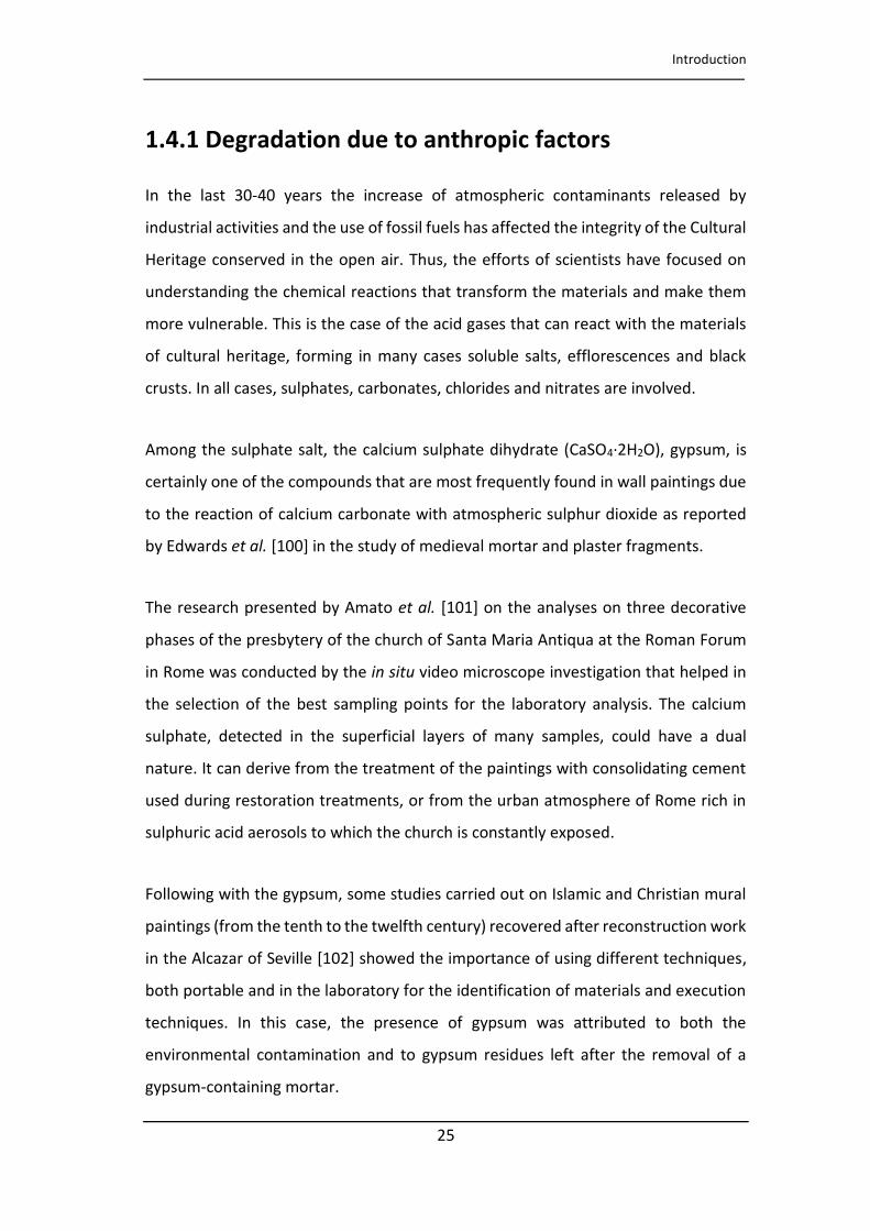
1.4.1 Degradation due to anthropic factors
In the last 30-40 years the increase of atmospheric contaminants released by
industrial activities and the use of fossil fuels has affected the integrity of the Cultural
Heritage conserved in the open air. Thus, the efforts of scientists have focused on
understanding the chemical reactions that transform the materials and make them
more vulnerable. This is the case of the acid gases that can react with the materials
of cultural heritage, forming in many cases soluble salts, efflorescences and black
crusts. In all cases, sulphates, carbonates, chlorides and nitrates are involved.
Among the sulphate salt, the calcium sulphate dihydrate (CaSO4·2H2O), gypsum, is
certainly one of the compounds that are most frequently found in wall paintings due
to the reaction of calcium carbonate with atmospheric sulphur dioxide as reported
by Edwards et al. [100] in the study of medieval mortar and plaster fragments.
The research presented by Amato et al. [101] on the analyses on three decorative
phases of the presbytery of the church of Santa Maria Antiqua at the Roman Forum
in Rome was conducted by the in situ video microscope investigation that helped in
the selection of the best sampling points for the laboratory analysis. The calcium
sulphate, detected in the superficial layers of many samples, could have a dual
nature. It can derive from the treatment of the paintings with consolidating cement
used during restoration treatments, or from the urban atmosphere of Rome rich in
sulphuric acid aerosols to which the church is constantly exposed.
Following with the gypsum, some studies carried out on Islamic and Christian mural
paintings (from the tenth to the twelfth century) recovered after reconstruction work
in the Alcazar of Seville [102] showed the importance of using different techniques,
both portable and in the laboratory for the identification of materials and execution
techniques. In this case, the presence of gypsum was attributed to both the
environmental contamination and to gypsum residues left after the removal of a
gypsum-containing mortar.
CHAPTER 1
26
[Titolo del documento]
Unfortunately, the presence of gypsum is recurrent. In the research by Daniilia et al.
[103] about wall paintings from St Stephen's monastery at the Meteora (Greece),
some conservation problems were found. Beyond the vandalism that they suffered
during the World War II and the Civil War, the study showed the presence of a whitish
layer of gypsum on the painted surface that affected significantly the aesthetic
impact of the mural paintings. Calcium carbonate sulphatation process was also seen
in situ without sampling in the study of 15th century wall paintings in a chapel of St.
Orso Priory palace (Aosta, Italy) [104]. Besides the main inorganic pigments, the
collected Raman spectra also enabled the identification of some decay products, such
as calcium sulphate.
Gypsum is not the only compound that can form crusts on the surface of the wall
paintings. Analyses of crust formations on Byzantine wall paintings in some churches
from Mani Peninsula (Greece) [105] revealed that they were made of
hydromagnesite (Mg(CO3)4(OH)·4(H2O)), calcite and small amounts of gypsum, the
latter present as a product of calcium carbonate degradation. Consequently,
hydromagnesite should have been the result of the degradation of a magnesium
product, favoured by the presence of moisture. The wall paintings of all the churches
under study had been probably covered in the same period with a superficial layer of
magnesium lime. In addition, the presence of hydromagnesite also in the preparatory
layer was due to its higher solubility than calcite and it was easily dissolved and
diffused within the mortar. This was an interesting result, as dolomitic or magnesian
limes did not appear often in the Greek area and their use was usually related to the
mineralogy and geology of raw materials available in different areas.
Unfortunately, together with gypsum, other salts can cause damages. Analyses
a ied out ith the salts fou d i the Assu ptio s hu h of Alaiza (Basque Country,
Spain) [106] demonstrated the connection between the deterioration of the wall
paintings and the agricultural activity in the area surrounding the church. The use of
fertilizers such as NH4NO3 and (NH4)2SO4, generates cations and anions that rise from
Introduction
27
[Titolo del documento]
the ground to the walls through infiltration water causing the decarbonation of the
calcite and the subsequent disintegration of the plaster of the wall paintings.
Another anthropic degradation factor often identified in the studies of wall paintings
is represented by the use of candles inside places of worship, such as churches and
monasteries. An example is the study of the thirteenth century wall paintings
discovered under the Siena Cathedral [84]. Darkening of colours as well as
dehydration of yellow ochre was attributed to soot deposits from lighted candles or
oil lamps and from the heat given off by the candles.
Similarly, the carbon particles identified on the surface of all samples from a post-
medieval wall painting [64] could be ascribed to the soot by candles or oil lamps that
provided illumination inside the church as well as the carbon particles, identified by
Raman spectroscopy, in an Italian crypt [107].
The evidence of the burning of substance such as wax or oil can be demonstrated
also by the chemical transformation of some pigments, as hypothesized in the
research by Damiani et al. [90], in which the transformation of azurite into copper
oxide tenorite was probably provoked by the heat from the candles. The thermal
degradation of the azurite converted into tenorite was found also on the mural
paintings preserved inside the cathedral of Thessaloniki [78], due to the high
temperatures released by a fire. Moreover, this incident has caused a total absence
of yellow tones in some areas affected by the fire. On the contrary, the presence of
red iron oxides was due to the dehydration of yellow ochre during the fire.
In the research by Andreotti et al. [108] in which was proposed a laser cleaning
method on the wall paintings from the monumental cemetery of Pisa, the study of
the surface deposits shown the presence of materials of anthropic origin. Indeed,
nitrocellulose used in a past restoration and contamination of pure lead were
identified. The presence of lead on the surface of the fragments is due to the fires
resulting from the bombardment of the Second World War, when the lead roof

CHAPTER 1
28
[Titolo del documento]
melted and the fused lead fell on the wall paintings. The removal of lead deposits was
difficult because of the strong adhesion of lead drops. Furthermore, the interactions
between the painted surface and the high temperatures had probably caused the
alteration of the underlying pigment consisting of iron oxides.
1.4.2 Natural degradation
Particular attention, in recent publications, has been paid to the study of salt
naturally formed efflorescences, composed mainly of sulphate, nitrates and
carbonates, since they represent one of the natural degradation products that most
endanger the integrity of the masonry and consequently also the substrate of the
wall paintings. Generally, the study of the composition of soluble salts is useful to
identify a specific marker that indicates the cause that generated them.
For this purpose, spectroscopic techniques, especially Raman spectroscopy, are
particularly useful since are able to differentiate degradation products that can
coexist in construction materials and on pictorial surfaces. An example of the
reliability of the use of diagnostic techniques in the characterization of degradation
products is the work published by Iordanidis et al. [109] where Raman spectroscopy
and ESEM-EDS were successful employed for the characterization of salt
efflorescence on fragments from mural paintings in three different churches
belonging to post-byzantine period. The compounds responsible for the decay were
calcite, dolomite, gypsum, alite, nitratine (NaNO3), natron (Na2CO3·10H2O) and
mirabilite (Na2SO4·10H2O), all caused by temperature and humidity variations.
Although in many studies gypsum is present as a product of sulphuric anhydride
contamination, it has been identified as a component of medieval mortars as
documented in the research of Damiani et al. [91] where it was macroscopically
visible in the surface of wall paintings along fractures and detachments in areas
where the humidity found preferential flow paths showing that the original materials
of the masonry were the sources of sulphates (Figure 1.3).
Introduction
29
[Titolo del documento]
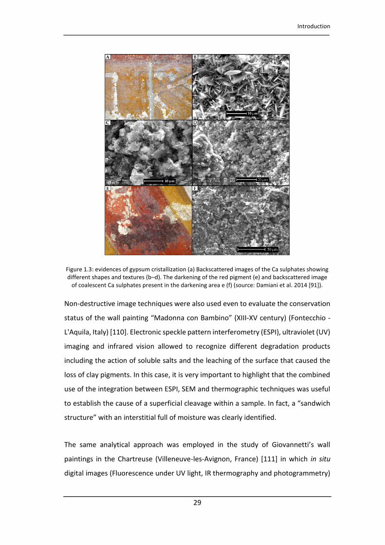
Figure 1.3: evidences of gypsum cristallization (a) Backscattered images of the Ca sulphates showing different shapes and textures (b–d). The darkening of the red pigment (e) and backscattered image
of coalescent Ca sulphates present in the darkening area e (f) (source: Damiani et al. 2014 [91]).
Non-destructive image techniques were also used even to evaluate the conservation
status of the wall painting Mado a o Ba i o XIII-XV century) (Fontecchio -
L'Aquila, Italy) [110]. Electronic speckle pattern interferometry (ESPI), ultraviolet (UV)
imaging and infrared vision allowed to recognize different degradation products
including the action of soluble salts and the leaching of the surface that caused the
loss of clay pigments. In this case, it is very important to highlight that the combined
use of the integration between ESPI, SEM and thermographic techniques was useful
to establish the cause of a superficial lea age ithi a sa ple. I fa t, a sa d i h
st u tu e ith a i te stitial full of oistu e as lea l ide tified.
The sa e a al ti al app oa h as e plo ed i the stud of Gio a etti s all
paintings in the Chartreuse (Villeneuve-les-Avignon, France) [111] in which in situ
digital images (Fluorescence under UV light, IR thermography and photogrammetry)
CHAPTER 1
30
[Titolo del documento]
provided information about the original and new materials used during past
restoration work and structural aspects. The mural paintings were affected mainly by
erosion and chromatic alteration. In situ and in laboratory analysed samples shown
that in some areas the surface deformations were caused by the presence of
hygroscopic salts composed of calcium and sodium sulphate.
Most of the studies on the state of conservation of building materials have
highlighted the danger of the presence of nitrate salts on carbonaceous materials
that are the main component of brick mortar and wall paintings. In consequence, it
is very important to identify these compounds in order to limit the presence of the
factors that caused their formation, when possible. For example, in the research by
Pérez-Alonso et al. [112] on the study of mortars and wall paintings of Santa María
de Hermo (Spain), carried out by the use of portable Raman instrumentation,
extensive decay of the carbonaceous materials caused by the absorption of nitrates
from the ground was found. Their presence was explained by the migration of
nitrates from the old cemetery attached to the church wall.
Portable techniques (visible reflectance and Raman spectroscopies) and in the
laboratory (attenuated total reflectance-Fourier transform infrared spectroscopy
(ATR-FT-IR) and micro-Raman spectroscopies) were applied for the study of the
conservation state of medieval wall paintings preserved in a crypt in Puglia region
(Italy) [107]. The study was focused on the characterization of degradation products
caused mainly by moisture problems due primarily to the fact that the crypt was
excavated in the tuff, a highly porous and hygroscopic material. On-site Raman
spectroscopy allowed the identification of sulphates, nitrates and calcium oxalates
(whewellite and weddellite) that were also quantified together with chlorides,
fluorides, nitrites and acetates by ion chromatography, thus, obtaining a clearer
picture of the poor state of conservation caused by physical factors (water
infiltration), chemical (reactions of materials with atmospheric particulate) and
biological ones favoured by the high humidity.

Introduction
31
[Titolo del documento]
In the study of wall paintings located in two church in the Basque Country [113], the
origin of the nitrate salts (potassium nitrate and calcium nitrate) and the presence of
ammonium sulphate was ascertained. Their presence could be related to the reaction
between calcium carbonate and gypsum, present in the mortar, with the ammonium
nitrate from the soil and transported by infiltration waters through capillary rising
towards the paintings.
In the study of medieval wall paintings from the Basque Country presented by
Veneranda et al. [114], in situ analyses allowed the characterization of different types
of nitrate salts, such as nitrocalcite (Ca(NO3)2·4H2O) and natron, in a high hydration
state suggesting that they were formed due to high humidity.
Another study in which conservation problems were presented, but in this case in a
low-contamination rural environment, is the work presented by Madariaga et al.
[115], in which it was highlighted how the degradation processes generated by
natural factors can be dangerous as much as the impact of industrial atmospheres
without a conservation plan. Using in situ analyses it was possible to demonstrate
that the greatest damage was caused by the presence of salt efflorescence, which
thanks to Raman spectroscopy were characterized in the laboratory. An extensive
presence of nitrates (potassium and sodium nitrate) was confirmed both on the wall
paintings and on the building materials. The source of the nitrate salts was the
nitrogen compounds from the ground (mainly ammonium nitrate from the plant and
grass decomposition) that were absorbed by the porous materials due to capillary
rise of water infiltration favoured by a very damaged masonry and that lost cohesion
over the years.
Another detailed study in which different environmental factors have accelerated the
degradation of wall paintings is shown in the study concerning the Carolingian wall
paintings and Romanesque in the Müstair convent [116, 117] (Switzerland),
influenced by a saline system originated by rising damp and percolation. Thanks to
the use of complementary diagnostic techniques, the study showed that the decay
CHAPTER 1
32
[Titolo del documento]
was produced by the presence of soluble salts, nitrates and sulphates, whose
formation was influenced by seasonal variations in air humidity.
As the nitrate salts are the most responsible for the degradation of porous materials,
the research of Maguregui et al. [12] presented a protocol to assess its impact on
construction materials using non-invasive and microdestructive analytical techniques
with chemometric and thermodynamic data analysis. The church under study
showed evidence of problems deriving from the infiltration of soluble salts from
natural sources represented by the level of the ground above the zero level of the
church and which represents the main entry point for the nitrate salts from the
outside. Subsequently, they spread by capillarity involving both the masonry and the
wall painting.
In the work published by Martínez-Arkarazo et al. [47] portable Raman monitoring
was performed to follow the progress in two cleaning operations based on new
technologies: a wall painting affected by the impact of nitrate and a black crusted
stone altarpiece. Nitrate and gypsum Raman bands decreased after treatment, which
allowed the restorers to make decisions in situ and suggested when to stop the
cleaning operation.
1.4.3 Biodeterioration processes
In certain environmental conditions building materials may be subjected to
biodeterioration processes, visible in the form of patinas or encrustations, generated
by the colonization of microorganisms such as algae, moss, lichens or fungi. Besides
the aesthetic damage, that is the most evident and the first that can be identified
over pictorial surfaces, the penetration of fungal hyphae and lichen thallus can cause
serious structural damage such as the deterioration of plasters and the consequent
loss of the paint layer in the case of wall paintings.
Introduction
33
[Titolo del documento]
One of the first works in which a degradation due to biological attack was identified
by the use of Raman spectroscopy was published in 1999 by Edwards and Rull [118].
Although the ha a te isti i atio al spe t a of al iu o alate o oh d ate, β-
carotene, chlorophyll, and para-depside phenolic acids were obtained from
fragments of the outer wall of the convent, the degradation was so strong that the
damage was extended to the medieval wall paintings in the interior. Indeed, the
presence of calcium oxalate dihydrate weddellite was confirmed on fragments of wall
paintings, while was evident a chromatic damage due to the leaching of red ochre
caused by lichenic acids.
The complementary use of different laboratory analytical techniques (optical
microscopy, scanning electron microscopy, micro-infrared spectroscopy and micro-
X-ray diffraction with synchrotron light and gas chromatography-mass spectrometry)
was essential to identify the nature of black spots visible on the surface of micro
samples taken in the Saint Michael's Chapel from the Royal Monastery of Pedralbes
(Barcelona) [119]. Complex mixtures of calcium salts of low molecular weight organic
acids (LMWOA) were determined and their presence was explained by the reaction
between the acids produced by the filamentous fungi and the deposited aerial
calcium compounds on the surface of paintings. The analyses shown that these
substances did not have a strong ability to penetrate the painted layers and that the
dark tone was probably given by the presence of a colorant or pigment produced by
fungi, of a polar nature as being soluble in water, and therefore easily removable.
A mechanical and aesthetic damage was identified during the study of the frescoes
of the Chapel of the Holy nail, Siena [120] (Italy), in which the scanning (SEM) and
transmission electron microscopy (TEM) together with a molecular study allowed to
confirm that the cause of the chromatic alteration of the paintings was the presence
of a fungus. It accumulated in its hyphae large concentrations of copper, which
generally inhibits the colonization of bacteria, taken from the azurite, which was
transformed into malachite due to the dissolution of carbonates by organic acids
occurred, promoting the leaching of copper.
CHAPTER 1
34
[Titolo del documento]
The same medieval frescoes were studied a few years later in order to clarify the
cause of the presence of some blackened areas [121]. The analyses conducted
(scanning microscopy with X-ray scattering microanalysis) attested that the
blackening of the paints was closely linked to the use of Paraloid B72 acrylic resin
during some restoration interventions carried out in the 60s. Thanks to some
laboratory tests on fragments, it was noticed that the presence of the resin film
inhibited the interaction of the painting with the environment, including
transpiration and gaseous exchange. Paraloid-treated and untreated samples
showed potential deleterious species on the surface in quiescent state while Bacteria
of the genus Bacillus were isolated only from untreated samples, proving the fact that
the Paraloid resin is resistant to the colonization of bacteria. Moisture combined with
different chemical compounds accelerated chemical oxidation and blackening. The
hypothesis stated was that the Firmicutes bacteria presumably produced
extracellular material, which retains water and provides a carbon source available to
other bacteria. Numerous alteration products linked to biodegradation, such as
oxalates (calcium dihydrate weddellite and monohydrate whewellite) have been
detected in Italian wall paintings [91].
In the research by Tortora et al. [122] the analysis by non-destructive and micro-
invasive techniques were extremely useful for the chemical and structural studies of
edie al all pai ti gs i O e L A uila, Ital a d fo the de elop e t of a detailed
plan for the restoration work. The in situ techniques allowed to identify structural
problems caused by rising damp together with biodegradation and calcium oxalate
formation.
The identification of whewellite by using XRD [94] showed that the degradation of
the organic binder used to mix copper pigments was probably accelerated by the
presence of microorganisms in a high humidity environment. However, in the wall
paintings located in the Thessaloniki cathedral [78], the formation of calcium oxalates
was probably originated from the yellowing of a protein material, employed by
restorers to consolidate the surface during the restoration works after a fire.
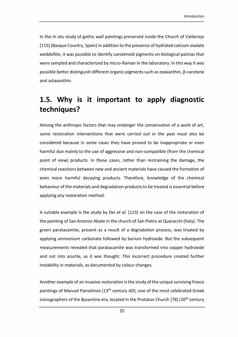
Introduction
35
[Titolo del documento]
In the in situ study of gothic wall paintings preserved inside the Church of Valderejo
[115] (Basque Country, Spain) in addition to the presence of hydrated calcium oxalate
weddellite, it was possible to identify carotenoid pigments on biological patinas that
were sampled and characterized by micro-Raman in the laboratory. In this way it was
possible better distinguish different organic pig e ts su h as zea a thi , β-carotene
and astaxanthin.
1.5. Why is it important to apply diagnostic
techniques?
Among the anthropic factors that may endanger the conservation of a work of art,
some restoration interventions that were carried out in the past must also be
considered because in some cases they have proved to be inappropriate or even
harmful due mainly to the use of aggressive and non-compatible (from the chemical
point of view) products. In these cases, rather than restraining the damage, the
chemical reactions between new and ancient materials have caused the formation of
even more harmful decaying products. Therefore, knowledge of the chemical
behaviour of the materials and degradation products to be treated is essential before
applying any restoration method.
A suitable example is the study by Dei et al. [123] on the case of the restoration of
the painting of San Antonio Abate in the church of San Pietro at Quaracchi (Italy). The
green paratacamite, present as a result of a degradation process, was treated by
applying ammonium carbonate followed by barium hydroxide. But the subsequent
measurements revealed that paratacamite was transformed into copper hydroxide
and not into azurite, as it was thought. This incorrect procedure created further
instability in materials, as documented by colour changes.
Another example of an invasive restoration is the study of the unique surviving fresco
paintings of Manuel Panselinos (13th century AD), one of the most celebrated Greek
iconographers of the Byzantine era, located in the Protaton Church [78] (10th century

CHAPTER 1
36
[Titolo del documento]
AD) on Mount Athos (Greece). Thanks to the use of in situ ultraviolet fluorescence
images, superficial salts deposits, flaking and detachments were identified as the
main decaying products/processes. They were formed due to the addition of a
cement coating during a restoration intervention in modern times, that prevented
the transpiration of moisture from the surface, with the subsequent formation of
new salt deposits.
Unfortunately, modern cements are behind many described degradations in cultural
heritage. For example, the use of alkaline materials such as Portland cement in the
Müstair convent [117] influenced the compositions of the salts already present in the
walls and increased their quantity. Moreover, another cause of the decay was
represented by the installation of a heating system, which caused strong seasonal
variations in relative humidity.
In the study carried out by Irazola et al. [113] by using portable instrumentation
(Figure 1.4) it was possible to characterize some decaying compounds in two wall
paintings in churches of the Basque Country region (Spain). To better characterize the
degradation processes, some microsamples were analysed in the laboratory. The
superimposing Raman images on the SEM-EDS images of the cross-sections were
used to explain the spatial distribution of anhydrite (CaSO4), caused by a
phenomenon of dehydration of gypsum in the mortar due to the application of
Paraloid resin during past restoration work, which prevented the transpiration of
water from the surface. A similar case was studied by Veneranda et al. [114] where
bassanite (CaSO4·0.5H2O) was identified as a consequence of the gypsum
dehydration due to the use of an acrylic resin, and its distribution in the sample was
established by Raman imaging in a cross section sample.
In other cases, the lack of a scientific approach to the study of materials before
carrying out restorations has led to the application of not compatible procedures and
colours with the original appearance of the paintings, as described in the research on
late Gothic paintings in the church of Ribera de Valderejo (see chapter 6). For
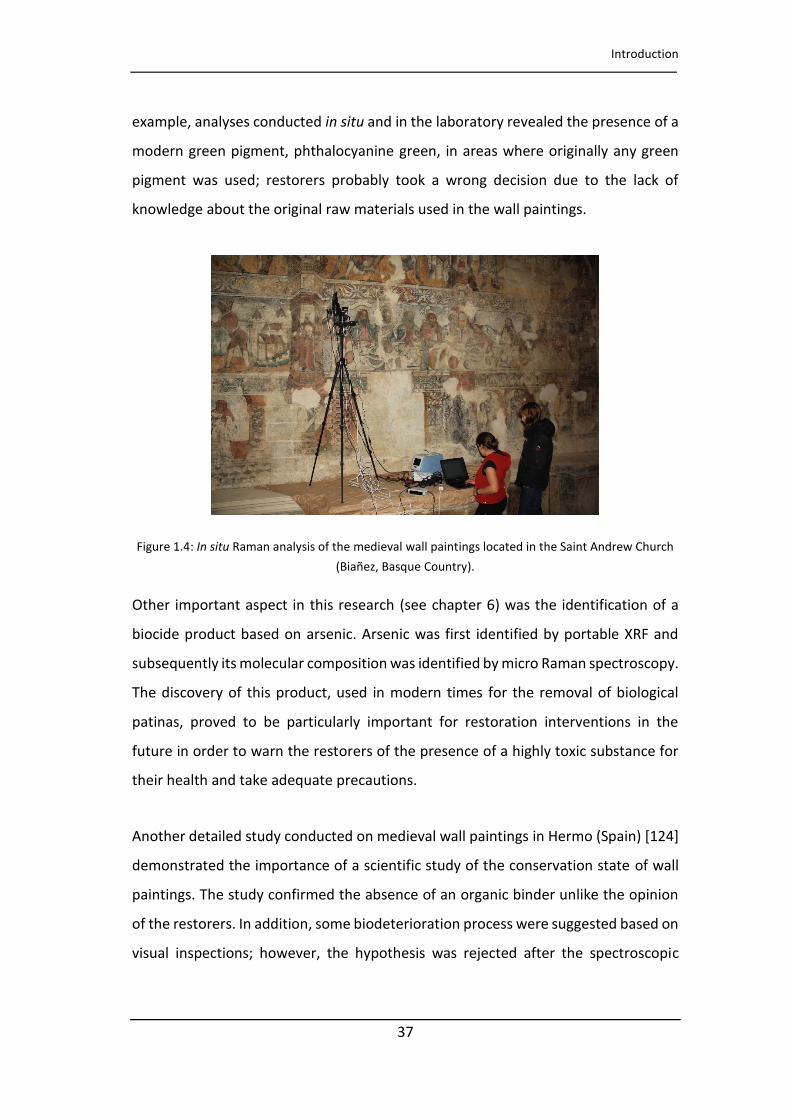
Introduction
37
[Titolo del documento]
example, analyses conducted in situ and in the laboratory revealed the presence of a
modern green pigment, phthalocyanine green, in areas where originally any green
pigment was used; restorers probably took a wrong decision due to the lack of
knowledge about the original raw materials used in the wall paintings.
Figure 1.4: In situ Raman analysis of the medieval wall paintings located in the Saint Andrew Church
(Biañez, Basque Country).
Other important aspect in this research (see chapter 6) was the identification of a
biocide product based on arsenic. Arsenic was first identified by portable XRF and
subsequently its molecular composition was identified by micro Raman spectroscopy.
The discovery of this product, used in modern times for the removal of biological
patinas, proved to be particularly important for restoration interventions in the
future in order to warn the restorers of the presence of a highly toxic substance for
their health and take adequate precautions.
Another detailed study conducted on medieval wall paintings in Hermo (Spain) [124]
demonstrated the importance of a scientific study of the conservation state of wall
paintings. The study confirmed the absence of an organic binder unlike the opinion
of the restorers. In addition, some biodeterioration process were suggested based on
visual inspections; however, the hypothesis was rejected after the spectroscopic
CHAPTER 1
38
[Titolo del documento]
analyses revealed the formation of soluble salts (sulphate and above all nitrates) as
the main deterioration cause.
1.6 Final remarks
This introduction gives an overview of the results obtained in the study of medieval
wall paintings and emphasizes the usefulness of different analytical techniques for
the study of the materials. In particular, portable analytical techniques have proved
to be reliable in the characterization of ancient and modern pigments and binder
media, and in particular in the investigation of the conservation state of wall
paintings. The advantages for the preservation of works of art in the use of portable
tools are evident above all because they are completely non-invasive, a fundamental
requirement for the study of immovable objects especially medieval wall paintings,
many of which are already strongly damaged due to neglect and too invasive
restoration interventions.
Since the mediaeval era was characterized by a stagnation of economy, the use of
poor materials of natural origin and local origin for the realization of mural paintings
was widespread. Because of the limited access to pigments, new techniques and
unusual pigments were used to make up for the lack of raw materials.
Almost all the studies related to the identification of degradation products have
shown a close connection between the conservation status of paintings and the
characteristics of the environment in which they were found. In fact, wall paintings,
after their realization, must be considered as objects in continuous evolution because
new compounds, according to environmental changes, can be formed in a continuous
exchange with the surrounding environment. At the same time, it is not possible to
attribute to just one agent a certain secondary product because different factors,
both natural and anthropic, contribute to the degradation of materials. For this
reason, studies concerning the conservation of cultural heritage should focus not only
on the work of art itself but also on the environmental study with the aim of
Introduction
39
[Titolo del documento]
identifying the environmental stressors that limit the durability of the materials, in
an exchange of knowledge that involves scientists, biologists, conservators and art
historians.
This chapter highlights that the spread in the use of diagnostic techniques has
developed at the same time a change in sensitivity in the study of cultural heritage
for which each artwork is unique and must be preserved for future generations. Some
researches showed that the application of unsuitable restoration interventions or the
use of products incompatible with the original materials, applied without a thorough
knowledge of the materials, can lead to dangerous choices for the preservation of
the artworks. Therefore, this introduction underlines the importance of using a
scientific approach for the study of the original materials before carrying out
restoration operations and for the development of innovative procedures for
cleaning, restoration and conservation previously tested in the laboratory in order to
suggest to restorers the best method to apply. It is precisely the concept that will be
the leitmotif of this PhD project as demonstrated through the case studies and the
experimental researches in the following chapters.
CHAPTER 1
40
[Titolo del documento]
References
[1] M. Sablier, P. Garrigue (2014) Cultural heritage and its environment: an issue of interest for Environmental Science and Pollution Research, Environmental Science
and Pollution Research, 21, 5769-5773.
[2] Air Pollution and Cultural Heritage, Ed. Saiz Jimenez © 2004 Taylor & Francis Group, London, ISBN 90 5809 682 3.
[3] G. D. Smith, Robin J. H. Clark (2002) The role of H2S in pigment blackening, Journal
of Cultural Heritage,3, 101-105.
[4] S. Aze, J. M. Vallet, A. Baronnet, O. Grauby (2006) The fading of red lead pigment in wall paintings: tracking the physico-chemical transformations by means of complementary micro-analysis techniques, European Journal of Mineralogy, 18, 835-843.
[5] K. Castro, A. Sarmiento, I. Martínez-Arkarazo, J. M. Madariaga, L. A. Fernández (2008) Green Copper Pigments Biodegradation in Cultural Heritage: From Malachite to Moolooite, Thermodynamic Modeling, X-ray Fluorescence, and Raman Evidence, Analytical Chemistry, 80, 4103-4110.
[6] M. Cotte, J. Susini, N. Metrich, A. Moscato, C. Gratziu, A. Bertagnini, M. Pagano (2006) Blackening of Pompeian Cinnabar Paintings: X-ray Microspectroscopy Analysis, Analytical Chemistry, 78, 7484-7492.
[7] M. Maguregui, U. Knuutinen, I. Martínez-Arkarazo, K. Castro, J. M. Madariaga (2011) Thermodynamic and Spectroscopic Speciation to Explain the Blackening Process of Hematite Formed by Atmospheric SO2 Impact: The Case of Marcus Lucretius House (Pompeii), Analytical Chemistry, 83, 3319-3326.
[8] A. Coccato, L. Moens, P. Vandenabeele (2017) On the stability of mediaeval inorganic pigments: a literature review of the effect of climate, material selection, biological activity, analysis and conservation treatments, Heritage Science, 5, 12. https://doi.org/10.1186/s40494-017-0125-6.
[9] R. A. J. Wüst, C. Schlüchter (2000) The Origin of Soluble Salts in Rocks of the Thebes Mountains, Egypt: The Damage Potential to Ancient Egyptian Wall Art, Journal of
Archaeological Science, 27, 1161-1172.
[10] R. Rossi-Manaresi, A. Tucci (1991) Pore structure and the disruptive or cementing effect of salt crystallization in various types of stone, Studies in Conservation, 36, 53-58.
[11] A. Arnold, K, Zehnder, Monitoring Wall Paintings Affected by Soluble Salts, The Conservation of Wall Paintings pp103-136, Proceedings of a symposium organized by the Courtauld Institute of Art and the Getty Conservation Institute, London, July 13-16, 1987, Sharon Cather Editor.
[12] M. Maguregui, A. Sarmiento, I. Martínez-Arkarazo, M. Angulo, K. Castro, G. Arana, N. Etxebarria, J. M. Madariaga (2008) Analytical diagnosis methodology to

Introduction
41
[Titolo del documento]
evaluate nitrate impact on historical building materials, Analytical and Bioanalytical
Chemistry, 391, 1361-370.
[13] J. Aramendia, L. Gómez-Nubla, I. Arrizabalaga, N. Prieto-Taboada, K. Castro, J. M. Madariaga (2013) Multianalytical approach to study the dissolution process of weathering steel: The role of urban pollution, Corrosion Science, 76, 154-162.
[14] S. J. Haneef, J. B. Johnson, C. Dickinson, G. E. Thompson, G. C. Wood (1992) Effect of dry deposition of NOx and SO2 gaseous pollutants on the degradation of calcareous building stones, Atmospheric Environment. Part A. General Topics, 26, 2963-2974.
[15] O. Gómez-Laserna, N. Prieto-Taboada, H. Morillas, I. Arrizabalaga, M. A. Olazabal, G. Arana, J. M. Madariaga (2015) Analytical study to evaluate the origin and severity of damage caused by salt weathering in a historical Palace House: the attack of infiltration water, Analytical Methods, 7, 4608-4615.
[16] P. Pouli, E. Papakonstantinou, K. Frantzikinaki, A. Panou, G. Frantzi, C. Vasiliadis, C. Fotakis (2016) The two-wavelength laser cleaning methodology; theoretical background and examples from its application on CH objects and monuments with emphasis to the Athens Acropolis sculptures, Heritage Science, 4, 1-11.
[17] M. Maguregui, U. Knuutinen, I. Martínez-Arkarazo, A. Giakoumaki, K. Castro, J. M. Madariaga (2012) Field Raman analysis to diagnose the conservation state of excavated walls and wall paintings in the archaeological site of Pompeii (Italy), Journal
of Raman Spectroscopy, 43, 1747-1753.
[18] M. Veneranda, N. Prieto-Taboada, S. Fdez-Ortiz de Vallejuelo, M. Maguregui, H. Morillas, I. Marcaida, K. Castro, J. M Madariaga, M. Osanna (2017) Biodeterioration of Pompeian mural paintings: fungal colonization favoured by the presence of volcanic material residues, Environmental Science and Pollution Research, 24, 19599-19608.
[19] C. Schabereiter-Gurtner, C. Saiz-Jimenez, G. Piñar, W. Lubitz, S. Rolleke (2002) Altamira cave Paleolithic paintings harbor partly unknown bacterial communities, Microbiology Letters, 211, 7-11.
[20] O. Pepe, L. Sannino, S. Palomba, M. Anastasio, G. Blaiotta, F. Villani, G. Moschetti (2010) Heterotrophic microorganisms in deteriorated medieval wall paintings in southern Italian churches, Microbiological Research, 165, 21-32.
[21] K. Sterflinger, G. Piñar (2013) Microbial deterioration of cultural heritage and works of art — tilting at windmills?, Applied Microbiology and Biotechnology, 97, 9637-9646.
[22] T. Rosado, A. Reis, J. Mirão, A. Candeias, P. Vandenabeele, A. T. Caldeira (2014) Pink! Why not? On the unusual colour of Évora Cathedral, International
Biodeterioration & Biodegradation, 94, 121-127.
[23] O. Ciferri (2002) The role of microorganisms in the degradation of cultural heritage, Studies in Conservation, 47, 35-45.
[24] M. Maguregui, U. Knuutinen, J. Trebolazabala, H. Morillas, K. Castro, I. Martínez-Arkarazo, J. M. Madariaga (2012) Use of in situ and confocal Raman spectroscopy to

CHAPTER 1
42
[Titolo del documento]
study the nature and distribution of carotenoids in brown patinas from a deteriorated wall painting in Marcus Lucretius House (Pompeii), Analytical and Bioanalytical
Chemistry, 402, 1529-1539.
[25] H. G. M. Edwards, D. W. Farwell, M. R. D. Seaward, C. Giacobini (1991) Preliminary Raman microscopic analyses of a lichen encrustation involved in the biodeterioration of renaissance frescoes in Central Italy, International
Biodeterioration, 27, 1-9.
[26] H. G. M. Edwards, D. W. Farwell, R. Jenkins, M. R. D. Seaward (1992) Vibrational Raman spectroscopic studies of calcium oxalate monohydrate and dihydrate in lichen encrustations on renaissance frescoes, Journal of Raman Spectroscopy, 23, 185-189.
[27] M. Singha, B. R. Arbad (2015) Characterization of 4th–5th century A.D. earthen plaster support layers of Ajanta mural paintings, Construction and Building Material, 82, 142-154.
[28] M. Matteini, A. Moles, The reconversion of oxidized white lead in mural paintings: a control after a five years period, ICOM Commetee for Conservation 6th Triennial Meeting Ottawa 1981.
[29] D. Bersani, C. Conti, P. Matousek, F. Pozzi, P. Vandenabeele (2016) Methodological evolutions of Raman spectroscopy in art and archaeology, Analytical
Methods, 8, 8395-8409.
[30] G. Bitossi, R. Giorgi, M. Mauro, B. Salvadori, L. Dei (2005) Spectroscopic techniques in Cultural Heritage conservation: a survey, Applied Spectroscopy.
Reviews, 40, 187-228.
[31] D. Bersani, J. M. Madariaga (2012) Applications of Raman spectroscopy in art and archaeology, Journal of Raman Spectroscopy, 43, 1523-1528.
[32] B. Guineau (1984) Analyse non-destructive des pigments par microsonde Raman laser: exemples de l'azurite et de la malachite, Studies in Conservation, 29, 35-41.
[33] G. Simsek, P. Colomban, F. Casadio, L. Bellot-Gurlet, G. Zelleke, K. T. Faber, V. Milande, L. Tilliard (2015) O ‐Site Ide tifi atio of Ea l Böttge Red Sto e a e Usi g Portable XRF/Raman Instruments: 2, Glaze & Gilding Analysis, Journal of the American
Ceramic Society, 98, 3006-3013.
[34] A. Rousaki, C. Vázquez, V. Aldazábal, C. Bellelli, M. Carballido Calatayud, A. Hajduk, E. Vargas, O. Palacios, P. Vandenabeele, L. Moens (2017) The first use of portable Raman instrumentation for the in situ study of prehistoric rock paintings in Patagonian sites, Journal of Raman Spectroscopy, 48, 1459-146.
[35] G. D. Smith, L. Burgio, S. Firth, R. J. H. Clark (2001) Laser-induced degradation of lead pig e ts ith efe e e to Botti elli s T io fo d A o e, Analytica Chimica Acta, 440, 185-188.
[36] I. M. Bell, R. J. H. Clark, P. J. Gibbs (1997) Raman spectroscopic library of natural and synthetic pigments (pre- ≈ 8 AD), Spectrochimica Acta Part A: Molecular and
Biomolecular Spectroscopy, 53, 2159-2179.
Introduction
43
[Titolo del documento]
[37] P. Ropret, S. A. Centeno, P. Bukovec (2008) Raman identification of yellow synthetic organic pigments in modern and contemporary paintings: Reference spectra and case studies, Spectrochimica Acta Part A: Molecular and Biomolecular
Spectroscopy, 69, 486-497.
[38] P. Vandenabeele, B. Wehling, L. Moens, H. Edwards, M. De Reu, G. Van Hooydonk (2000) Analysis with micro-Raman spectroscopy of natural organic binding media and varnishes used in art, Analytica Chimica Acta, 407, 261-274.
[39] J. M. Madariaga, M. Maguregui, S. Fdez-Ortiz De Vallejuelo, U. Knuutinen, K. Castro, I. Martinez-Arkarazo, A. Giakoumaki, A. Pitarch (2014) In situ analysis with po ta le Ra a a d ED‐XRF spe t o ete s fo the diag osis of the fo atio of efflorescence on walls and wall paintings of the Insula IX 3 (Pompeii, Italy), Journal of
Raman Spectroscopy, 45, 1059-1067.
[40] N. Prieto-Taboada, O. Gomez-Laserna, I. Martínez-Arkarazo, M. A. Olazabal, J. M. Madariaga (2014), Raman Spectra of the Different Phases in the CaSO4-H2O System, Analytical Chemistry, 86, 10131-10137.
[41] P. Moioli, C. Seccaroni (2000) A al sis of a t o je ts usi g a po ta le ‐ray fluorescence spectrometer, X-Ray Spectrometry, 29, 48-52.
[42] D. Anglos (2001) Laser-Induced Breakdown Spectroscopy in Art and Archaeology, Applied Spectroscopy, 55, 186-205.
[43] D. Ajò, U. Casellato, E. Fiorin, P. A. Vigato (2004) Ci o Fe i s f es oes: a study of painting materials and technique by SEM-EDS microscopy, X-ray diffraction, micro FT-IR and photoluminescence spectroscopy, Journal of Cultural Heritage, 5, 333-348.
[44] H. Brecoulaki, A. Andreotti, I. Bonaduce, M. P. Colombini, A. Lluveras (2012) Characterization of organic media in the wall-pai ti gs of the Pala e of Nesto at Pylos, Greece: evidence for a secco painting techniques in the Bronze Age, Journal of
Archaeological Science, 39, 2866-2876.
[45] D. Lau, C. Villis, S. Furman, M. Livet (2008) Multispectral and hyperspectral image analysis of elemental and micro-Raman maps of cross-sections from a 16th century painting, Analytica Chimica Acta, 610, 15-24.
[46] N. Prieto-Taboada, O. Gomez-Laserna, I. Martínez-Arkarazo, M. A. Olazabal, J. M. Madariaga (2013) Relevance of cross-section analysis in correct diagnosis of the state of conservation of building materials as evidenced by spectroscopic imaging, Analytical Chemistry, 85, 9501-9507.
[47] I. Martínez-Arkarazo, A. Sarmiento, M. Maguregui, K. Castro, J. M. Madariaga (2010) Portable Raman monitoring of modern cleaning and consolidation operations of artworks on mineral supports, Analytical and Bioanalytical Chemistry, 2010, 397, 2717-2725.
[48] P. Rydzewski, Medieval Art, Art and Human Experience, Copyright © 1967 Pergamon Press Ltd., pp. 122-137.
CHAPTER 1
44
[Titolo del documento]
[49] H. G. M. Edwards, C. J. Brooke, J. K. F. Tait (1997) Fourier Transform Raman Spectroscopic Study of Pigments from English Mediaeval Wall Paintings, Journal of
Raman Spectroscopy, 28, 95-98.
[50] H. G. M. Edwards, D. W. Farwell, E. M. Newton, F. Rull Perez (1999) Minium; FT-Raman non-destructive analysis applied to an historical controversy, Analyst, 124, 1323-1326.
[51] H. G. M. Edwards, D. W. Farwell, F. Rull Perez, S. Jorge Villar, (1999) Spanish mediaeval frescoes at Basconcillos del Tozo: a Fourier transform Raman spectroscopic study, Journal of Raman Spectroscopy, 30, 307-311.
[52] F. Rull Pérez, H. G. M. Edwards, A. Rivas, L. Drummond (1999) Fourier transform Raman spectroscopic characterization of pigments in the mediaeval frescoes at Convento de la Peregrina, Sahagun, Léon, Spain. Part 1-preliminary study, Journal of
Raman Spectroscopy, 30, 301-305.
[53] H. G. M. Edwards, F. Rull, P. Vandenabeele, E. M. Newton, L. Moens, J. Medina, C. García (2001) Mediaeval Pigments in the Monastery of San Baudelio, Spain: A Raman Spectroscopic Analysis, Applied Spectroscopy, 55, 71-76.
[54] S. E. J. Villar, H. G. M. Edwards, J. Medina, F. Rull Pérez (2006), Raman spectroscopic analysis of mediaeval wall paintings in the Palencia region, Spain, Journal of Raman Spectroscopy, 37, 1078-1085.
[55] F. Daniel, A. Mounier, P. Ricarrere, Of some blue and bluish grey pigments in medieval mural paintings in the South West of France, Proceedings of the 39th International Symposium for Archaeometry, Leuven 2012, 329-334.
[56] A. Palet Casas, J. D. Andrés Llopis (1992) The identification of aerinite as a blue pigment in the Romanesque frescoes of the Pyrenean Region, Studies in
Conservation, 37, 132-136.
[57] J. Pérez-Arantegui, C. Pardos, J. L. Abad, J. R. García (2013) Microcharacterization of a Natural Blue Pigment Used in Wall Paintings During the Romanesque Period in Northern Spain, Microscopy and Microanalysis, 19, 1645-1652.
[58] L. Lepot, S. Denoe, B. Gilbert (2006) The technique of the mural paintings of the Tournai Cathedral, Journal of Raman Spectroscopy, 37, 1098-1103.
[59] M. Ma ić-Stoja o ić, D. Bajuk-Bogda o ić, S. Uskoko ić-Marko ić, I. Hol lajt e -Antu o ić 8 Spectroscopic analysis of XIV century wall paintings from Pat ia hate of Peć Mo aste , Se ia, Spectrochimica Acta Part A: Molecular and
Biomolecular Spectroscopy, 191, 469-477.
[60] R. J. H. Clark, R. R. Hark, N. Salvadó, S. Butí, T. Pradell (2010) Spectroscopy study of mural paintings fromthe Pyrenean Church of Saint Eulàlia of Unha, Journal of
Raman Spectroscopy, 41, 1418-1424.
[61] A. Zucchiatti, P. Prati, A. Bouquillon, L. Giuntini, M. Massi, A. Migliori, A. Cagnana, S. Roascio (2004) Characterizatio of ea l edie al f es oes μ-PIXE, SEM and Raman spectroscopy, Nuclear Instruments and Methods in Physics Research Section
B: Beam Interactions with Materials and Atoms, 219-220, 20-25.
Introduction
45
[Titolo del documento]
[62] I. Holclajtner-A tu o ić, M. Stoja o ić-Ma ić, D. Bajuk-Bogda o ić, R. Žikić, S. Uskoko ić-Ma ko ić Multi-analytical study of techniques and palettes of wall pai ti gs of the o aste of Žiča, Se ia, Spectrochimica Acta Part A: Molecular and
Biomolecular Spectroscopy, 156, 78-88.
[63] M. Gutman, M. Lesar-Kikelj, A. Mlade o ič, V. Čo al-Sed ak, A. K iž a , S. Kramard (2014) Raman microspectroscopic analysis of pigments of the Gothic wall painting from the Dominican Monastery in Ptuj (Slovenia), Journal of Raman
Spectroscopy, 45, 1103-1109.
[64] H. G. M. Edwards, D. W. Farwell, C. J. Brooke (2005) Raman spectroscopic study of a post-medieval wall painting in need of conservation, Analytical and Bioanalytical
Chemistry, 383, 312-321.
[65] B. Minceva-Sukarova, O. Grupce, V. Tanevska, L. Robeva-Cukovska, S. Mamucevska-Miljkovic (2007) Micro-Raman spectroscopic studies of Byzantine cultural heritage in Republic of Macedonia, Macedonian Journal of Chemistry and
Chemical Engineering, 26, 103-110.
[66] P. Vandenabeele, K. Lambert, S. Matthys, W. Schudel, A. Bergmans, L. Moens (2005) In situ analysis of mediaeval wall paintings: a challenge for mobile Raman spectroscopy, Analytical and Bioanalytical Chemistry, 383, 707-712.
[67] J. L. Pérez-Rodríguez, M. D. Robador, M. A. Centeno, B. Siguenza, A. Duran (2014) Wall paintings studied using Raman spectroscopy: A comparative study between various assays of cross sections and external layers, Spectrochimica Acta Part A:
Molecular and Biomolecular Spectroscopy, 120, 602-609.
[68] A. Deneckere, W. Schudel, M. Van Bos, H. Wouters, A. Bergmans, P. Vandenabeele, L. Moens (2010) In situ investigations of vault paintings in the Antwerp cathedral, Spectrochimica Acta Part A: Molecular and Biomolecular
Spectroscopy, 75, 511-519.
[69] K. F. Gebremariam, L. Kvittingen, F.G. Banica (2013) Application of a portable XRF analyzer to investigate the medieval wall paintings of Yemrehanna Krestos Church, Ethiopia, X-Ray Spectrometry, 42, 462-469.
[70] J. L. Ferrero, C. Roldán, D. Juanes, E. Rollano, C. Morera (2002) Analysis of pigments from Spanish works of art using a portable EDXRF spectrometer, X-Ray
Spectrometry, 3, 441-447.
[71] M. Sa zak, A. Ka ińska, G. Ra zuk, M. Fe etti, R. Je d zeje ski, G. Śli iński (2009) Complementary use of the Raman and XRF techniques for non-destructive analysis of historical paint layers, Applied Surface Science, 255, 5542-5545.
[72] O. S ta, K. Rozu , M. Choińska, D. Zielińska, G. Z. Żukowska, A. Kijowska, B. Wagner (2014) Analytical procedure for characterization of medieval wall-paintings by X-ray fluorescence spectrometry, laser ablation inductively coupled plasma mass spectrometry and Raman spectroscopy, Spectrochimica Acta Part B: Atomic
Spectroscopy, 101, 140-148.

CHAPTER 1
46
[Titolo del documento]
[73] D. Lampakis, I. Karapanagiotis, O. Katsibiri (2017) Spectroscopic Investigation Leading to the Documentation of Three Post-Byzantine Wall Paintings, Applied
Spectroscopy, 71, 129-140.
[74] T. Zorba, K.S. Andrikopoulos, K.M. Paraskevopoulos, E. Pavlidou, K. Popkonstantinov, R. Kostova, V. Platnyov, S. Daniilia (2007) Infrared and Raman Vibrational Spectroscopies Reveal the Palette of Frescos Found in the Medieval Monastery of Karaach Teke, Annali di Chimica, by Società Chimica Italiana, 97, 491-503.
[75] A. G. Nord, K. Tronner, K. Billström, B. Strandberg Zerpe (2017) Analysis of mediaeval Swedish paintings influenced by Russian-Byzantine art, Journal of Cultural
Heritage, 23, 162-169.
[76] S. Š a o á, D. H adil, J. H adilo á, E. Kočí, P. Bezdičk (2009) Micro-analytical evidence of origin and degradation of copper pigments found in Bohemian Gothic murals, Analytical and Bioanalytical Chemistry, 395, 2037-2050.
[77] A. Iordanidis, J. García-Guinea, A. Strati, A. Gkimourtzina (2014) A comparative study of pigments from the wall paintings of two Greek Byzantine churches, Analytical Letters, 47, 2708-2721.
[78] S. Daniilia, S. Sotiropoulou, D. Bikiaris, C. Salpistis, G. Karagiannis, Y. Chryssoulakis, B. A. Price, J. H. Carlson (2000) Pa seli os B za ti e all pai ti gs i the Protaton Church, Mount Athos, Greece: a technical examination, Journal of
Cultural Heritage, 1, 91-110.
[79] S. Sotiropoulou, S Daniilia, C. Miliani, F. Rosi, L. Cartechini, D. Papanikola-Bakirtzis (2008) Microanalytical investigation of degradation issues in Byzantine wall paintings, Applied Physics A, 92, 143-150.
[80] E. Cheilakou, M. Troullinos, M. Koui (2014) Identification of pigments on Byzantine wall paintings from Crete (14th century AD) using non-invasive Fiber Optics Diffuse Reflectance Spectroscopy (FORS), Journal of Archaeological Science, 41, 541-555.
[81] L. Appolonia, D. Vaudan, V. Chatel, M. Aceto, P. Mirti (2009) Combined use of FORS, XRF and Raman spectroscopy in the study of mural paintings in the Aosta Valley (Italy), Analytical and Bioanalytical Chemistry, 395, 2005-2013.
[82] S. Boularand, P. Giráldez, L. Ventolà, M. Vendrell-Saz (2011) Templar joint repointings: materials, techniques and paint decoration in Miravet Castle, Spain, Archaeometry, 53, 743-752.
[83] I. Kakoulli, S. V. Prikhodko, A. King, C. Fischer (2014) Earliest evidence for asbestos composites linked to Byzantine wall paintings production, Journal of
Archaeological Science, 44, 148-153.
[84] S. Mugnaini, A. Bagnoli, P. Bensi, F. Droghini, A. Scala, G. Guasparri (2006) Thirteenth century wall paintings under the Siena Cathedral (Italy). Mineralogical and petrographic study of materials, painting techniques and state of conservation, Journal of Cultural Heritage, 7, 171-185.
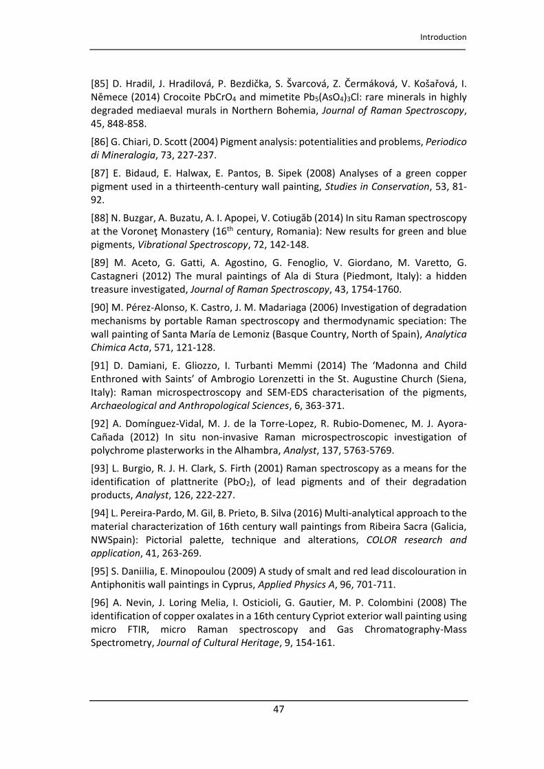
Introduction
47
[Titolo del documento]
[85] D. H adil, J. H adilo á, P. Bezdička, S. Š a o á, Z. Če áko á, V. Košařo á, I. Ně e e 2014) Crocoite PbCrO4 and mimetite Pb5(AsO4)3Cl: rare minerals in highly degraded mediaeval murals in Northern Bohemia, Journal of Raman Spectroscopy, 45, 848-858.
[86] G. Chiari, D. Scott (2004) Pigment analysis: potentialities and problems, Periodico
di Mineralogia, 73, 227-237.
[87] E. Bidaud, E. Halwax, E. Pantos, B. Sipek (2008) Analyses of a green copper pigment used in a thirteenth-century wall painting, Studies in Conservation, 53, 81-92.
[88] N. Buzgar, A. Buzatu, A. I. Apopei, V. Cotiugă 2014) In situ Raman spectroscopy at the Vo o eţ Mo aste th century, Romania): New results for green and blue pigments, Vibrational Spectroscopy, 72, 142-148.
[89] M. Aceto, G. Gatti, A. Agostino, G. Fenoglio, V. Giordano, M. Varetto, G. Castagneri (2012) The mural paintings of Ala di Stura (Piedmont, Italy): a hidden treasure investigated, Journal of Raman Spectroscopy, 43, 1754-1760.
[90] M. Pérez-Alonso, K. Castro, J. M. Madariaga (2006) Investigation of degradation mechanisms by portable Raman spectroscopy and thermodynamic speciation: The wall painting of Santa María de Lemoniz (Basque Country, North of Spain), Analytica
Chimica Acta, 571, 121-128.
[91] D. Damiani, E. Gliozzo, I. Turbanti Memmi (2014) The Mado a a d Child E th o ed ith Sai ts of Ambrogio Lorenzetti in the St. Augustine Church (Siena, Italy): Raman microspectroscopy and SEM-EDS characterisation of the pigments, Archaeological and Anthropological Sciences, 6, 363-371.
[92] A. Domínguez-Vidal, M. J. de la Torre-Lopez, R. Rubio-Domenec, M. J. Ayora-Cañada (2012) In situ non-invasive Raman microspectroscopic investigation of polychrome plasterworks in the Alhambra, Analyst, 137, 5763-5769.
[93] L. Burgio, R. J. H. Clark, S. Firth (2001) Raman spectroscopy as a means for the identification of plattnerite (PbO2), of lead pigments and of their degradation products, Analyst, 126, 222-227.
[94] L. Pereira-Pa do, M. Gil, B. P ieto, B. Sil a Multi‐a al tical approach to the material characterization of 16th century wall paintings from Ribeira Sacra (Galicia, NWSpain): Pictorial palette, technique and alterations, COLOR research and
application, 41, 263-269.
[95] S. Daniilia, E. Minopoulou (2009) A study of smalt and red lead discolouration in Antiphonitis wall paintings in Cyprus, Applied Physics A, 96, 701-711.
[96] A. Nevin, J. Loring Melia, I. Osticioli, G. Gautier, M. P. Colombini (2008) The identification of copper oxalates in a 16th century Cypriot exterior wall painting using micro FTIR, micro Raman spectroscopy and Gas Chromatography-Mass Spectrometry, Journal of Cultural Heritage, 9, 154-161.
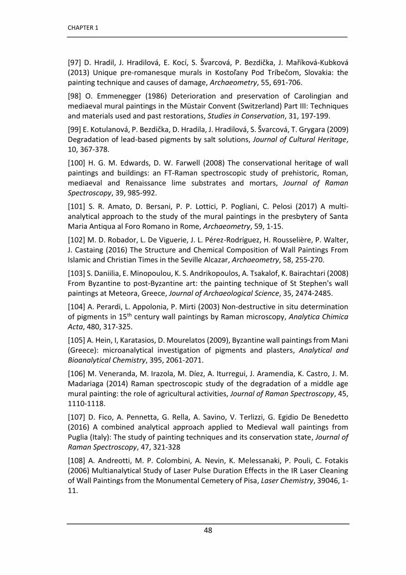
CHAPTER 1
48
[Titolo del documento]
[97] D. H adil, J. H adilo á, E. Ko í, S. Š a o á, P. Bezdička, J. Maříko á-Kubková (2013) U i ue p e‐ o a es ue u als i Kostoľa Pod T í ečo , Slo akia: the painting technique and causes of damage, Archaeometry, 55, 691-706.
[98] O. Emmenegger (1986) Deterioration and preservation of Carolingian and mediaeval mural paintings in the Müstair Convent (Switzerland) Part III: Techniques and materials used and past restorations, Studies in Conservation, 31, 197-199.
[99] E. Kotula o á, P. Bezdička, D. H adila, J. H adilo á, S. Š a o á, T. G ga a (2009) Degradation of lead-based pigments by salt solutions, Journal of Cultural Heritage, 10, 367-378.
[100] H. G. M. Edwards, D. W. Farwell (2008) The conservational heritage of wall pai ti gs a d uildi gs: a FT‐Ra a spe t os opi stud of p ehisto i , Ro a , mediaeval and Renaissance lime substrates and mortars, Journal of Raman
Spectroscopy, 39, 985-992.
[101] S. R. Amato, D. Bersani, P. P. Lottici, P. Pogliani, C. Pelosi (2017) A multi-analytical approach to the study of the mural paintings in the presbytery of Santa Maria Antiqua al Foro Romano in Rome, Archaeometry, 59, 1-15.
[102] M. D. Robador, L. De Viguerie, J. L. Pérez-Rodríguez, H. Rousselière, P. Walter, J. Castaing (2016) The Structure and Chemical Composition of Wall Paintings From Islamic and Christian Times in the Seville Alcazar, Archaeometry, 58, 255-270.
[103] S. Daniilia, E. Minopoulou, K. S. Andrikopoulos, A. Tsakalof, K. Bairachtari (2008) From Byzantine to post-Byzantine art: the painting technique of St Stephen's wall paintings at Meteora, Greece, Journal of Archaeological Science, 35, 2474-2485.
[104] A. Perardi, L. Appolonia, P. Mirti (2003) Non-destructive in situ determination of pigments in 15th century wall paintings by Raman microscopy, Analytica Chimica
Acta, 480, 317-325.
[105] A. Hein, I, Karatasios, D. Mourelatos (2009), Byzantine wall paintings from Mani (Greece): microanalytical investigation of pigments and plasters, Analytical and
Bioanalytical Chemistry, 395, 2061-2071.
[106] M. Veneranda, M. Irazola, M. Díez, A. Iturregui, J. Aramendia, K. Castro, J. M. Madariaga (2014) Raman spectroscopic study of the degradation of a middle age mural painting: the role of agricultural activities, Journal of Raman Spectroscopy, 45, 1110-1118.
[107] D. Fico, A. Pennetta, G. Rella, A. Savino, V. Terlizzi, G. Egidio De Benedetto (2016) A combined analytical approach applied to Medieval wall paintings from Puglia (Italy): The study of painting techniques and its conservation state, Journal of
Raman Spectroscopy, 47, 321-328
[108] A. Andreotti, M. P. Colombini, A. Nevin, K. Melessanaki, P. Pouli, C. Fotakis (2006) Multianalytical Study of Laser Pulse Duration Effects in the IR Laser Cleaning of Wall Paintings from the Monumental Cemetery of Pisa, Laser Chemistry, 39046, 1-11.

Introduction
49
[Titolo del documento]
[109] A. Iordanidis, J. García-Guinea, A. Strati, A. Gkimourtzina, A. Papoulidou (2011) Byzantine wall paintings from Kastoria, northern Greece: Spectroscopic study of pigments and efflorescing salts, Spectrochimica Acta Part A: Molecular and
Biomolecular Spectroscopy, 78, 874-887.
[110] S. Sfarra, C. Ibarra-Castanedo, M. Tortora, L. Arrizza, G. Cerichelli, I. Nardi, X. Maldague (2016), Diagnostics of wall paintings: A smart and reliable approach, Journal of Cultural Heritage, 18, 229-241.
[111] J. M. Vallet, D. Syvilay, V. Detalle, D. Giovannacci, L. De Luca, C. Stefani, J. L. Bodnar, G. Walker, O. Guillon, M, Feillou, B. Trichereau, D. Martos-Le Vif, K. Mouhoubi, P. Marron, N. Martin-Beaumont, F. De Banes Gardonne, Development of a NDT toolbox dedicated to the conservation of wall paintings: application to the frescoes chapel in the Chartreuse Villeneuve-les-Avignon (Francia), Digital Heritage 2013, Marseille.
[112] M. Pérez-Alonso, K. Castro, I. Martínez-Arkarazo, M. Angulo, M. A. Olazabal, J. M. Madariaga (2004) Analysis of bulk and inorganic degradation products of stones, mortars and wall paintings by portable Raman microprobe spectroscopy, Analytical
and Bioanalytical Chemistry, 379, 42-50.
[113] M. Irazola, M. Olivares, K. Castro, M. Maguregui, I. Martínez-Arkarazo, J. M. Madariaga (2012) In situ Raman spectroscopy analysis combined with Raman and SEM‐EDS i agi g to assess the o se atio state of th century wall paintings, Journal of Raman Spectroscopy, 43, 1676-1684.
[114] M. Veneranda, M. Irazola, A. Pitarch, M. Olivares, A. Iturregui, K. Castro, J. M. Madariaga (2014) I ‐situ a d la o ato Ra a a al sis i the field of ultu al heritage: the case of a mural painting, Journal of Raman Spectroscopy, 45, 228-237.
[115] J. M. Madariaga, I. Costantini, K. Castro (2019) Analysis of natural degradation of medieval mural paintings in the open air abandoned church of Ribera, North of Spain, Raman Spectroscopy in Art and Archaeology, Volume 2, 213-233.
[116] A. Arnold, A. Kung, K. Zehnder (1986) Deterioration and preservation of Carolingian and mediaeval mural paintings in the Müstair Convent (Switzerland) Part I: Decay mechanisms and preservation, Studies in Conservation, 31, 190-194.
[117] F. Mairinger, M. Schreiner (1986) Deterioration and preservation of Carolingian and mediaeval mural paintings in the Müstair Convent (Switzerland) Part I: Decay mechanisms and preservation, Studies in Conservation, 31, 195-196.
[118] H. G. M. Edwards, F. Rull Pérez (1999) Lichen Biodeterioration of the Convento de la Peregrina, Sahagun, Spain, Biospectroscopy, 5, 47-52.
[119] N. Salvadó, S. Butí, T. Pradell, V. Beltran, G. Cinque, J. Juanhuix, L. Font, R. Senserrich (2016) Low molecular weight organic acid salts, markers of old fungi activity in wall paintings, Analytical Methods, 8, 1637-1645.
[120] C. Milanesi, F. Baldi, R. Vignani, F. Ciampolini, C. Faleria, M. Cresti (2006) Fungal deterioration of medieval wall fresco determined by analysing small fragments containing copper, International Biodeterioration & Biodegradation, 57, 7-13.
CHAPTER 1
50
[Titolo del documento]
[121] C. Milanesi, F. Baldi, S. Borin, L. Brusetti, F. Ciampolini, F. Iacopini, M. Cresti (2009) Fungal deterioration of medieval wall fresco determined by analysing small fragments containing copper, International Biodeterioration & Biodegradation, 63, 844-850.
[122] M. Tortora, S. Sfarra, M. Chiarini, V. Daniele, G. Taglieri, G. Cerichelli (2016) Non-destructive and micro-invasive testing techniques for characterizing materials, structures and restoration problems in mural paintings, Applied Surface Science, 387, 971-985.
[123] L. Dei, A. Ahle, P. Baglioni, D. Dini, E. Ferroni (1998) Green degradation products of azurite in wall paintings: identification and conservation treatment, Studies in
Conservation, 43, 80-88.
[124] M. Pérez-Alonso, K. Castro, M. Álvarez, J.M. Madariaga (2004) Scientific analysis e sus esto e s e pe tise fo diag osis p io to a esto atio p o ess: the ase of
Santa Maria Church (Hermo, Asturias, North of Spain), Analytica Chimica Acta, 524, 379-389.
51
CHAPTER 2
Objectives
This PhD thesis concerns the study of some late Gothic mural paintings, belonging to
Basque and Italian territory, using non destructive and micro-destructive analytical
techniques. The main objective is to retrace the history of these artworks through
the characterization of original materials and new materials employed in the past
restauration works carried out in modern era. In addition, it will pay a particular
attention to the conservation state of the murals and to the study of degradation
products with the aim to understand the factors that have caused the observed
damages.
The study of the murals paintings of the Basque territory is part of the Research
Projects CTP09-P04 and CTP12-P10, founded by the Basque Country Government,
thanks to which medieval mural paintings, located in the north of Spain, have been
studied by the IBeA research group.
One of the last example of gothic mural paintings in the Basque Country is located in
the Saint Stephen’s church in the abandoned village of Ribera de Valderejo (Álava),
in a rural area outside of direct sources of pollution.
The murals have an unusual aspect since some portions of wall paintings are
characterized by a grey colour. Thus, our objective was to check if the area is really
uncontaminated and understand the degradation phenomena that caused the
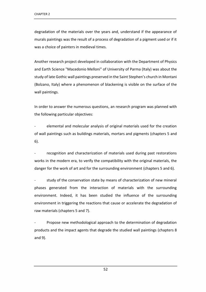
CHAPTER 2
52
[Titolo del documento]
degradation of the materials over the years and, understand if the appearance of
murals paintings was the result of a process of degradation of a pigment used or if it
was a choice of painters in medieval times.
Another research project developed in collaboration with the Department of Physics
and Earth Science "Macedonio Melloni" of University of Parma (Italy) was about the
study of late Gothic wall paintings preserved in the Saint Stephen’s church in Montani
(Bolzano, Italy) where a phenomenon of blackening is visible on the surface of the
wall paintings.
In order to answer the numerous questions, an research program was planned with
the following particular objectives:
- elemental and molecular analysis of original materials used for the creation
of wall paintings such as buildings materials, mortars and pigments (chapters 5 and
6).
- recognition and characterization of materials used during past restorations
works in the modern era, to verify the compatibility with the original materials, the
danger for the work of art and for the surrounding environment (chapters 5 and 6).
- study of the conservation state by means of characterization of new mineral
phases generated from the interaction of materials with the surrounding
environment. Indeed, it has been studied the influence of the surrounding
environment in triggering the reactions that cause or accelerate the degradation of
raw materials (chapters 5 and 7).
- Propose new methodological approach to the determination of degradation
products and the impact agents that degrade the studied wall paintings (chapters 8
and 9).

53
CHAPTER 3
Emplacement and samples
The wall paintings analysed in this PhD work belong to the Saint Stephen’s church in
Ribera de Valderejo (Álava) and to the Saint Stephen’s chapel in Montani (Bolzano)
located in the Basque and Italian territory (Figure 3.1) and belonging to the Gothic
and Late-Gothic period respectively.
The study campaigns were mainly carried out using portable non-destructive
diagnostic techniques. However, as will be discussed later, it was necessary to take
micro samples only in selected areas in order to complete the analysis in the
laboratory.
The state of conservation of the buildings and consequently of the wall paintings
conserved inside them is very different due mainly to the state of abandonment in
which the church of Ribera de Valderejo stands, contrary to that of Montani.
However, in both cases the pictorial surfaces present some problems that will be
discussed below.
Different diagnostic techniques were used in order to identify the original materials
used for the realization of wall paintings, for the study of products used in modern
times during restoration interventions and to verify the suitability of these
treatments. Thus, a conservation status study was carried out through the
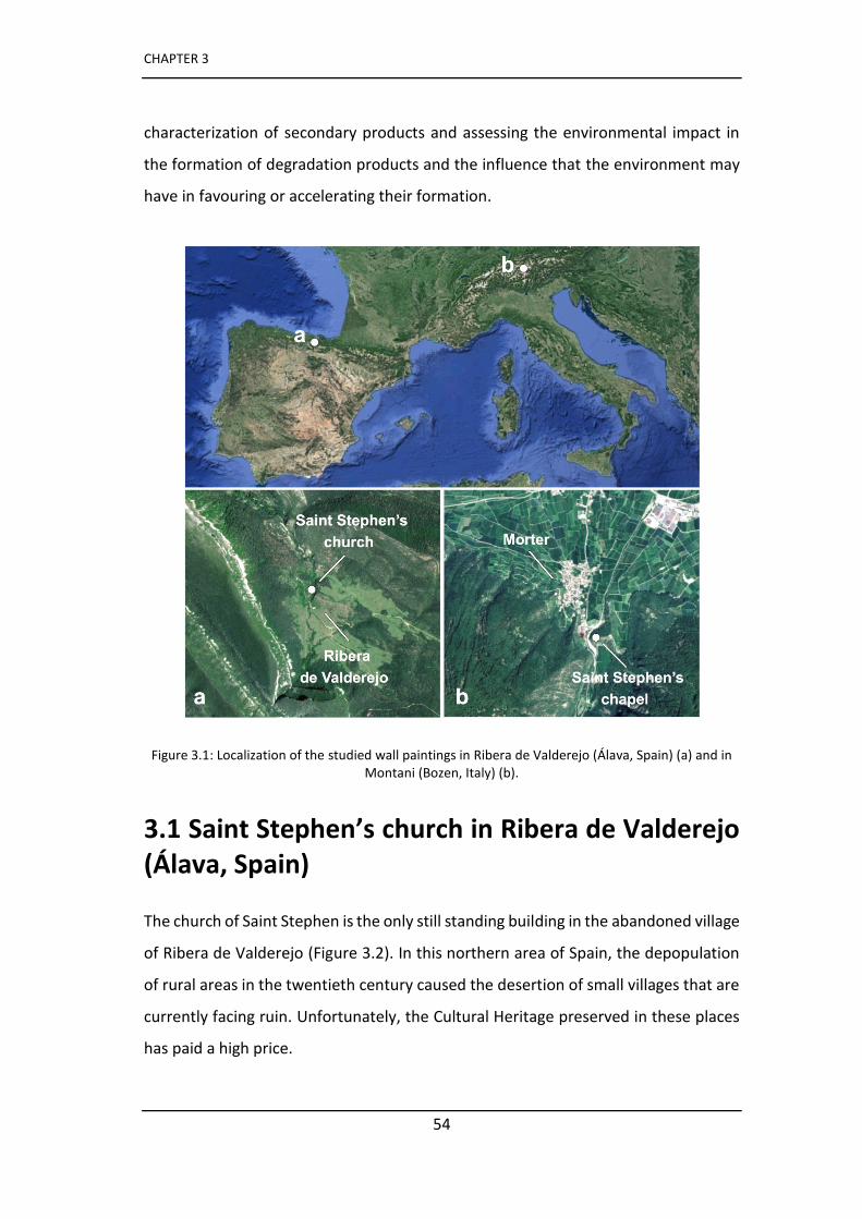
CHAPTER 3
54
characterization of secondary products and assessing the environmental impact in
the formation of degradation products and the influence that the environment may
have in favouring or accelerating their formation.
Figure 3.1: Localization of the studied wall paintings in Ribera de Valderejo (Álava, Spain) (a) and in Montani (Bozen, Italy) (b).
3.1 Saint Stephen’s church in Ribera de Valderejo (Álava, Spain)
The church of Saint Stephen is the only still standing building in the abandoned village
of Ribera de Valderejo (Figure 3.2). In this northern area of Spain, the depopulation
of rural areas in the twentieth century caused the desertion of small villages that are
currently facing ruin. Unfortunately, the Cultural Heritage preserved in these places
has paid a high price.
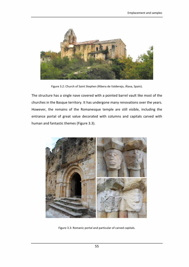
Emplacement and samples
55
Figure 3.2: Church of Saint Stephen (Ribera de Valderejo, Álava, Spain).
The structure has a single nave covered with a pointed barrel vault like most of the
churches in the Basque territory. It has undergone many renovations over the years.
However, the remains of the Romanesque temple are still visible, including the
entrance portal of great value decorated with columns and capitals carved with
human and fantastic themes (Figure 3.3).
Figure 3.3: Romanic portal and particular of carved capitals.
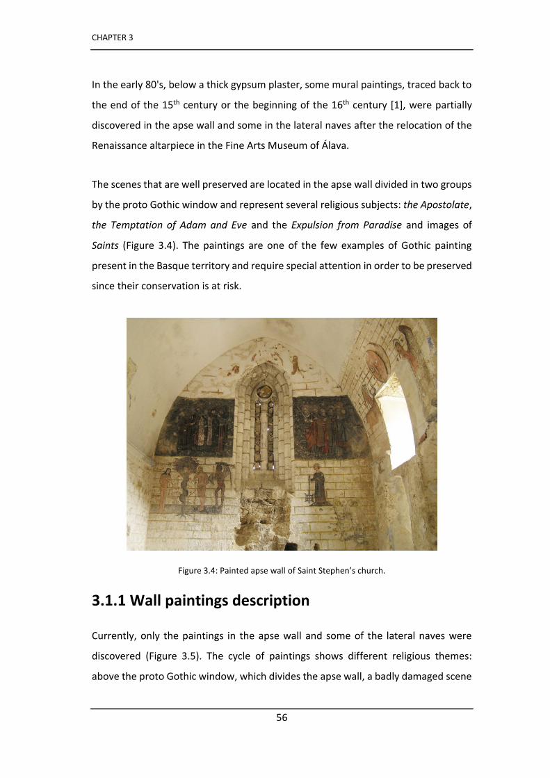
CHAPTER 3
56
In the early 80's, below a thick gypsum plaster, some mural paintings, traced back to
the end of the 15th century or the beginning of the 16th century [1], were partially
discovered in the apse wall and some in the lateral naves after the relocation of the
Renaissance altarpiece in the Fine Arts Museum of Álava.
The scenes that are well preserved are located in the apse wall divided in two groups
by the proto Gothic window and represent several religious subjects: the Apostolate,
the Temptation of Adam and Eve and the Expulsion from Paradise and images of
Saints (Figure 3.4). The paintings are one of the few examples of Gothic painting
present in the Basque territory and require special attention in order to be preserved
since their conservation is at risk.
Figure 3.4: Painted apse wall of Saint Stephen’s church.
3.1.1 Wall paintings description
Currently, only the paintings in the apse wall and some of the lateral naves were
discovered (Figure 3.5). The cycle of paintings shows different religious themes:
above the proto Gothic window, which divides the apse wall, a badly damaged scene
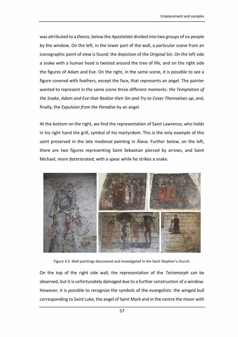
Emplacement and samples
57
was attributed to a Deesis, below the Apostolate divided into two groups of six people
by the window. On the left, in the lower part of the wall, a particular scene from an
iconographic point of view is found: the depiction of the Original Sin. On the left side
a snake with a human head is twisted around the tree of life, and on the right side
the figures of Adam and Eve. On the right, in the same scene, it is possible to see a
figure covered with feathers, except the face, that represents an angel. The painter
wanted to represent in the same scene three different moments: the Temptation of
the Snake, Adam and Eve that Realize their Sin and Try to Cover Themselves up, and,
finally, the Expulsion from the Paradise by an angel.
At the bottom on the right, we find the representation of Saint Lawrence, who holds
in his right hand the grill, symbol of his martyrdom. This is the only example of this
saint preserved in the late medieval painting in Álava. Further below, on the left,
there are two figures representing Saint Sebastian pierced by arrows, and Saint
Michael, more deteriorated, with a spear while he strikes a snake.
Figure 3.5: Wall paintings discovered and investigated in the Saint Stephen’s church.
On the top of the right side wall, the representation of the Tetramorph can be
observed, but it is unfortunately damaged due to a further construction of a window.
However, it is possible to recognize the symbols of the evangelists: the winged bull
corresponding to Saint Luke, the angel of Saint Mark and in the centre the moon with
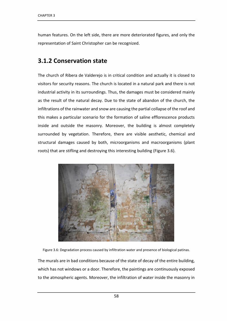
CHAPTER 3
58
human features. On the left side, there are more deteriorated figures, and only the
representation of Saint Christopher can be recognized.
3.1.2 Conservation state
The church of Ribera de Valderejo is in critical condition and actually it is closed to
visitors for security reasons. The church is located in a natural park and there is not
industrial activity in its surroundings. Thus, the damages must be considered mainly
as the result of the natural decay. Due to the state of abandon of the church, the
infiltrations of the rainwater and snow are causing the partial collapse of the roof and
this makes a particular scenario for the formation of saline efflorescence products
inside and outside the masonry. Moreover, the building is almost completely
surrounded by vegetation. Therefore, there are visible aesthetic, chemical and
structural damages caused by both, microorganisms and macroorganisms (plant
roots) that are stifling and destroying this interesting building (Figure 3.6).
Figure 3.6: Degradation process caused by infiltration water and presence of biological patinas.
The murals are in bad conditions because of the state of decay of the entire building,
which has not windows or a door. Therefore, the paintings are continuously exposed
to the atmospheric agents. Moreover, the infiltration of water inside the masonry in

Emplacement and samples
59
some areas is causing the detachment of the plaster with the consequent loss of the
paint layer.
In these paintings there are extensive areas of grey colour especially in places that
should be green (representing the grass in the scenes). In contrast, the only green
part seems to be a repainting made in modern times. From existing documents, we
know that over the years some restoration works have been carried out, but they still
do not clarify why the grass is grey in the paintings. A first intervention was done in
1983. In 1992 it was suggested to move the murals at the Museum of Fine Arts to
prevent their loss because the building was already in a complete state of disrepair.
This proposal that could have definitively secured the wall paintings was rejected and
it was decided to make an in situ restoration treatment.
3.1.3 Sampling
During the mural painting studies, it was not possible to clarify some important
aspects only with the portable instruments, thus, micrometric wall painting samples
were taken with a scalpel in areas of already deteriorated frescoes, in order to not
cause further damage to the artwork. Some samples were collected in the Original
Sin representation, which is the most interesting area for research. They were
embedded in resin in the laboratory to study the stratigraphy (Figure 3.7) and
additional analyses were conducted using benchtop techniques able to provide
complementary information.
In addition, in order to study the composition of some salts efflorescence, some
samples were collected to be analyzed in the laboratory to understand the effects of
the surrounding natural environment on the onset of these secondary products.
These samples were collected using a scalpel and stored in a plastic sample bag
(Figure 3.8). The analysis in the laboratory was carried out both on efflorescence as
it is and in the form of pellets.
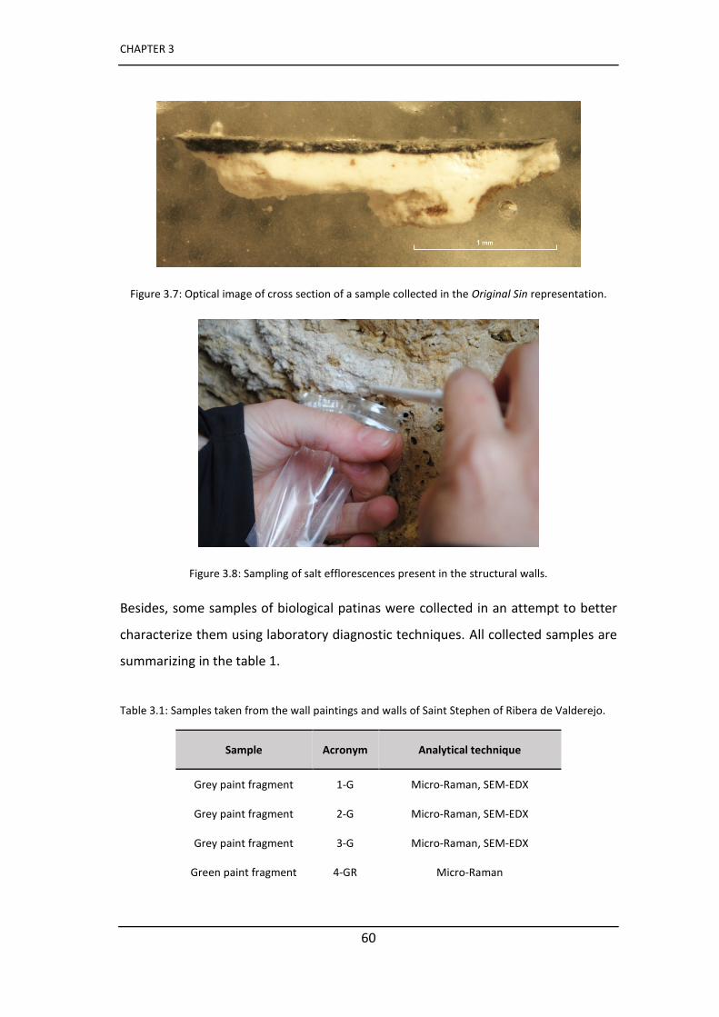
CHAPTER 3
60
Figure 3.7: Optical image of cross section of a sample collected in the Original Sin representation.
Figure 3.8: Sampling of salt efflorescences present in the structural walls.
Besides, some samples of biological patinas were collected in an attempt to better
characterize them using laboratory diagnostic techniques. All collected samples are
summarizing in the table 1.
Table 3.1: Samples taken from the wall paintings and walls of Saint Stephen of Ribera de Valderejo.
Sample Acronym Analytical technique
Grey paint fragment 1-G Micro-Raman, SEM-EDX
Grey paint fragment 2-G Micro-Raman, SEM-EDX
Grey paint fragment 3-G Micro-Raman, SEM-EDX
Green paint fragment 4-GR Micro-Raman
Emplacement and samples
61
Salt efflorescence 1- SALT Micro-Raman
Salt efflorescence 2- SALT Micro-Raman
Salt efflorescence 3-SALT Micro-Raman
Salt efflorescence 4-SALT Micro-Raman
Salt crust 5- SALT Micro-Raman
Salt crust 6- SALT Micro-Raman
Structural stone 1-ST Micro-Raman and XRD
Biological patina 1-BIO Micro-Raman, DNA extraction
Biological patina 2-BIO Micro-Raman, DNA extraction
Biological patina 3-BIO Micro-Raman, DNA extraction
Biological patina 4-BIO Micro-Raman, DNA extraction
Biological patina 5-BIO Micro-Raman, DNA extraction
3.2 Saint Stephen’s chapel of Montani (Bolzano, Italy)
The chapel of Saint Stephen’s is a splendid example of preservation of late medieval
artistic heritage. It is perched in a rock spur above the open barrier between the Val
Martello and the village of Montani (Figure 3.9).
The structure consists of a single nave with a wooden ceiling, divided by profiled
cornices in narrow squares, with the exception of the spaces previously covered by
five painted wooden tables dating back to 1430, which were removed for security
reasons and are now preserved in the civic museum of Bolzano.
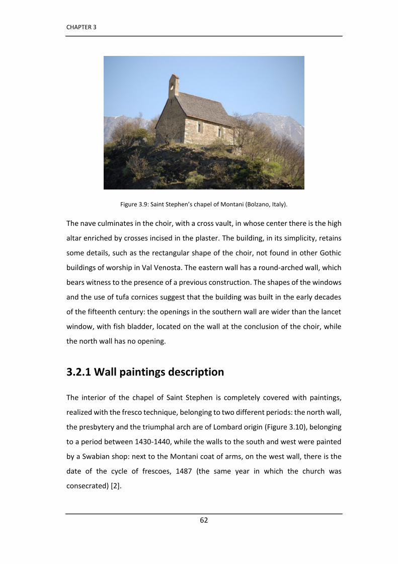
CHAPTER 3
62
Figure 3.9: Saint Stephen’s chapel of Montani (Bolzano, Italy).
The nave culminates in the choir, with a cross vault, in whose center there is the high
altar enriched by crosses incised in the plaster. The building, in its simplicity, retains
some details, such as the rectangular shape of the choir, not found in other Gothic
buildings of worship in Val Venosta. The eastern wall has a round-arched wall, which
bears witness to the presence of a previous construction. The shapes of the windows
and the use of tufa cornices suggest that the building was built in the early decades
of the fifteenth century: the openings in the southern wall are wider than the lancet
window, with fish bladder, located on the wall at the conclusion of the choir, while
the north wall has no opening.
3.2.1 Wall paintings description
The interior of the chapel of Saint Stephen is completely covered with paintings,
realized with the fresco technique, belonging to two different periods: the north wall,
the presbytery and the triumphal arch are of Lombard origin (Figure 3.10), belonging
to a period between 1430-1440, while the walls to the south and west were painted
by a Swabian shop: next to the Montani coat of arms, on the west wall, there is the
date of the cycle of frescoes, 1487 (the same year in which the church was
consecrated) [2].
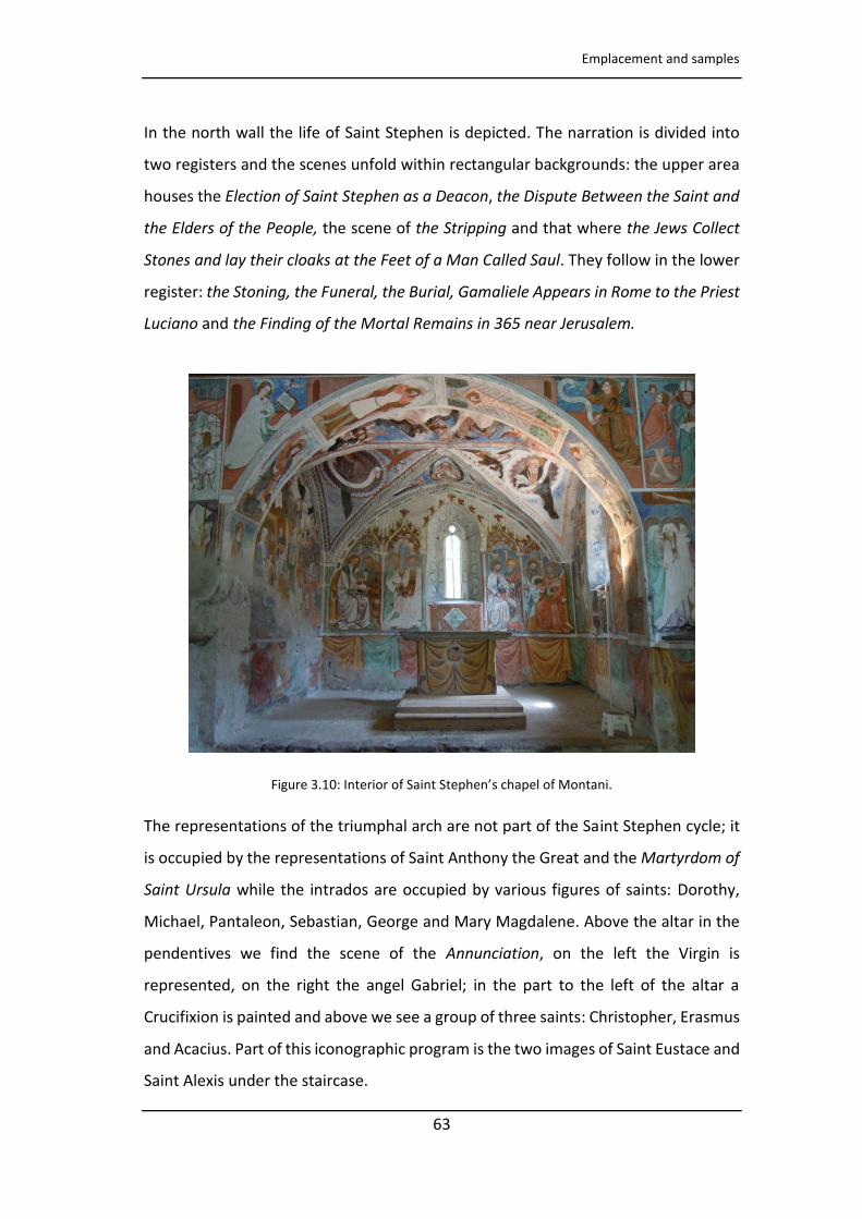
Emplacement and samples
63
In the north wall the life of Saint Stephen is depicted. The narration is divided into
two registers and the scenes unfold within rectangular backgrounds: the upper area
houses the Election of Saint Stephen as a Deacon, the Dispute Between the Saint and
the Elders of the People, the scene of the Stripping and that where the Jews Collect
Stones and lay their cloaks at the Feet of a Man Called Saul. They follow in the lower
register: the Stoning, the Funeral, the Burial, Gamaliele Appears in Rome to the Priest
Luciano and the Finding of the Mortal Remains in 365 near Jerusalem.
Figure 3. : Interior of Saint Stephen’s chapel of Montani.
The representations of the triumphal arch are not part of the Saint Stephen cycle; it
is occupied by the representations of Saint Anthony the Great and the Martyrdom of
Saint Ursula while the intrados are occupied by various figures of saints: Dorothy,
Michael, Pantaleon, Sebastian, George and Mary Magdalene. Above the altar in the
pendentives we find the scene of the Annunciation, on the left the Virgin is
represented, on the right the angel Gabriel; in the part to the left of the altar a
Crucifixion is painted and above we see a group of three saints: Christopher, Erasmus
and Acacius. Part of this iconographic program is the two images of Saint Eustace and
Saint Alexis under the staircase.
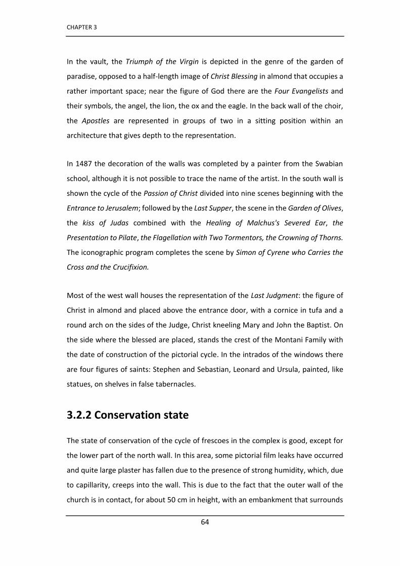
CHAPTER 3
64
In the vault, the Triumph of the Virgin is depicted in the genre of the garden of
paradise, opposed to a half-length image of Christ Blessing in almond that occupies a
rather important space; near the figure of God there are the Four Evangelists and
their symbols, the angel, the lion, the ox and the eagle. In the back wall of the choir,
the Apostles are represented in groups of two in a sitting position within an
architecture that gives depth to the representation.
In 1487 the decoration of the walls was completed by a painter from the Swabian
school, although it is not possible to trace the name of the artist. In the south wall is
shown the cycle of the Passion of Christ divided into nine scenes beginning with the
Entrance to Jerusalem; followed by the Last Supper, the scene in the Garden of Olives,
the kiss of Judas combined with the Healing of Malchus's Severed Ear, the
Presentation to Pilate, the Flagellation with Two Tormentors, the Crowning of Thorns.
The iconographic program completes the scene by Simon of Cyrene who Carries the
Cross and the Crucifixion.
Most of the west wall houses the representation of the Last Judgment: the figure of
Christ in almond and placed above the entrance door, with a cornice in tufa and a
round arch on the sides of the Judge, Christ kneeling Mary and John the Baptist. On
the side where the blessed are placed, stands the crest of the Montani Family with
the date of construction of the pictorial cycle. In the intrados of the windows there
are four figures of saints: Stephen and Sebastian, Leonard and Ursula, painted, like
statues, on shelves in false tabernacles.
3.2.2 Conservation state
The state of conservation of the cycle of frescoes in the complex is good, except for
the lower part of the north wall. In this area, some pictorial film leaks have occurred
and quite large plaster has fallen due to the presence of strong humidity, which, due
to capillarity, creeps into the wall. This is due to the fact that the outer wall of the
church is in contact, for about 50 cm in height, with an embankment that surrounds

Emplacement and samples
65
it. This deterioration process has irreparably damaged the lower part of the narration
of Saint Stephen's life and the representation of the Epiphany in the presbyter.
The state of conservation of the cycle of murals from the Swabian period is not
particularly critical and it is, therefore, still possible to admire the minuteness of
details and the plasticity in the rendering of the bodies; the colours also seem to have
retained their original vivacity. Even in the south wall the loss of colour concerns the
masonry areas in correspondence of the floor slab.
Regarding the murals located in the presbyter and in the triumphal arch, painting
surfaces show a phenomenon of blackening probably caused by the degradation of a
lead-based pigment used originally (lead white according to restorers). This
phenomenon is sometimes circumscribed in small areas where the pigment was used
in order to create chromatic contrasts, such as chiaroscuro effects. In other areas,
instead, its layout concerns larger coloured areas. The chromatic alteration strongly
influences the correct usability of the work because it drastically distorts the true
effect sought by the artists (Figure 3.11).
Some delimited areas in the east wall were treated with preliminary cleaning tests
carried out with the traditional method of reconverting the lead, using a solution of
acetic acid and hydrogen peroxide in cellulose pulp [3]. Since the results obtained
were not considered satisfactory, we made a complete analysis to understand this
blackening phenomenon as well as the suitability of the used treatment. Indeed,
although there is not much evidence of the use of this treatment in the scientific
literature, it is nevertheless used by restorers as confirmed by this case study [4, 5].
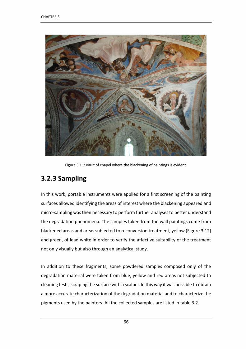
CHAPTER 3
66
Figure 3.11: Vault of chapel where the blackening of paintings is evident.
3.2.3 Sampling
In this work, portable instruments were applied for a first screening of the painting
surfaces allowed identifying the areas of interest where the blackening appeared and
micro-sampling was then necessary to perform further analyses to better understand
the degradation phenomena. The samples taken from the wall paintings come from
blackened areas and areas subjected to reconversion treatment, yellow (Figure 3.12)
and green, of lead white in order to verify the affective suitability of the treatment
not only visually but also through an analytical study.
In addition to these fragments, some powdered samples composed only of the
degradation material were taken from blue, yellow and red areas not subjected to
cleaning tests, scraping the surface with a scalpel. In this way it was possible to obtain
a more accurate characterization of the degradation material and to characterize the
pigments used by the painters. All the collected samples are listed in table 3.2.
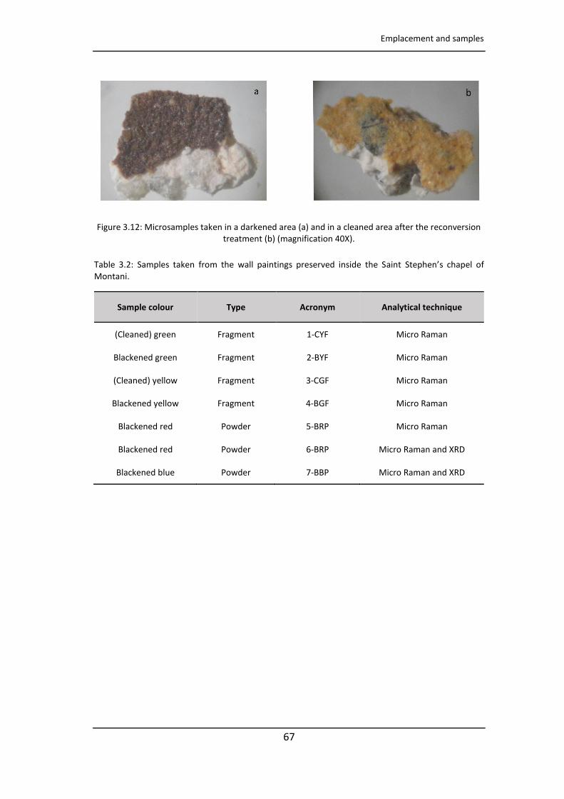
Emplacement and samples
67
Figure 3.12: Microsamples taken in a darkened area (a) and in a cleaned area after the reconversion treatment (b) (magnification 40X).
Table 3.2: Samples taken from the wall paintings preserved inside the Saint Stephen’s chapel of Montani.
Sample colour Type Acronym Analytical technique
(Cleaned) green Fragment 1-CYF Micro Raman
Blackened green Fragment 2-BYF Micro Raman
(Cleaned) yellow Fragment 3-CGF Micro Raman
Blackened yellow Fragment 4-BGF Micro Raman
Blackened red Powder 5-BRP Micro Raman
Blackened red Powder 6-BRP Micro Raman and XRD
Blackened blue Powder 7-BBP Micro Raman and XRD
CHAPTER 3
68
References
[1] R. Saenz Pascual, La pintura gotica en Álava. Una contribución a su estudio, ed: Vitoria-Gasteiz: Diputación Foral de Álava, Departamento de Cultura y Euskera,1997.
[2] L. Andergassen, Montani, la cappella di Santo Stefano. Montani di Sopra, Montani di Sotto, Schnell & Steiner GmbH (2001), Leibnizstraße13, D-93055 Regensburg.
[3] M. Matteini, Ossidazione della biacca in pitture murali- Metodi proposti per la riconversione del pigmento ossidato nelle pitture di A. Baldovinetti della chiesa di S. Miniato (Firenze), In Atti del Convegno sul Restauro delle Opere d'Arte, Florence, 2-6 November, 1976, 257-269.
[4] M. Matteini, A. Moles, The reconversion of oxidized white lead in mural paintings: a control after a five year period, ICOM Committee for Conservation, 6th Triennial Meeting, Ottawa, 1981 81/15/1.
[5] M. Koller, H. Leitner, H. Paschinger (1990) Reconversion of altered lead pigments in alpine mural paintings, Studies of Conservation, 35, 15-20.

69
CHAPTER 4
Experimental techniques
As documented in the chapter 1, different diagnostic techniques have been used
since the 1990s for the characterization of wall paintings. Undoubtedly, portable non-
destructive techniques are the most used ones for works of art that represent the
immovable cultural heritage, thanks to which it was possible to obtain in situ
information without affecting the pictorial surfaces and without sampling. Moreover,
as demonstrated in the following chapters, portable techniques are extremely useful
for selecting areas of particular interest from a diagnostic point of view and identify
an area where take samples in order to complete the investigations using benchtop
instruments.
This chapter summarizes the diagnostic techniques used both in situ and in the
laboratory, the laboratory instruments used for treatment of samples and the
commercial products employed to develop this PhD project.
4.1 General purpose instruments and tools
Loading analytical balance
To know the exact weight of the samples, powder samples were weighed using an AS
200 analytical balance (Mettler Toledo, USA) with a precision of ± 0.0001 g.
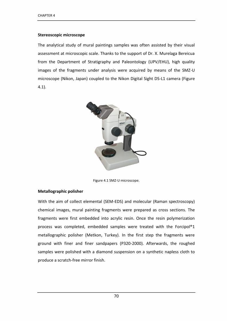
CHAPTER 4
70
Stereoscopic microscope
The analytical study of mural paintings samples was often assisted by their visual
assessment at microscopic scale. Thanks to the support of Dr. X. Murelaga Bereicua
from the Department of Stratigraphy and Paleontology (UPV/EHU), high quality
images of the fragments under analysis were acquired by means of the SMZ-U
microscope (Nikon, Japan) coupled to the Nikon Digital Sight DS-L1 camera (Figure
4.1).
Figure 4.1 SMZ-U microscope.
Metallographic polisher
With the aim of collect elemental (SEM-EDS) and molecular (Raman spectroscopy)
chemical images, mural painting fragments were prepared as cross sections. The
fragments were first embedded into acrylic resin. Once the resin polymerization
process was completed, embedded samples were treated with the Forcipol®1
metallographic polisher (Metkon, Turkey). In the first step the fragments were
ground with finer and finer sandpapers (P320-2000). Afterwards, the roughed
samples were polished with a diamond suspension on a synthetic napless cloth to
produce a scratch-free mirror finish.
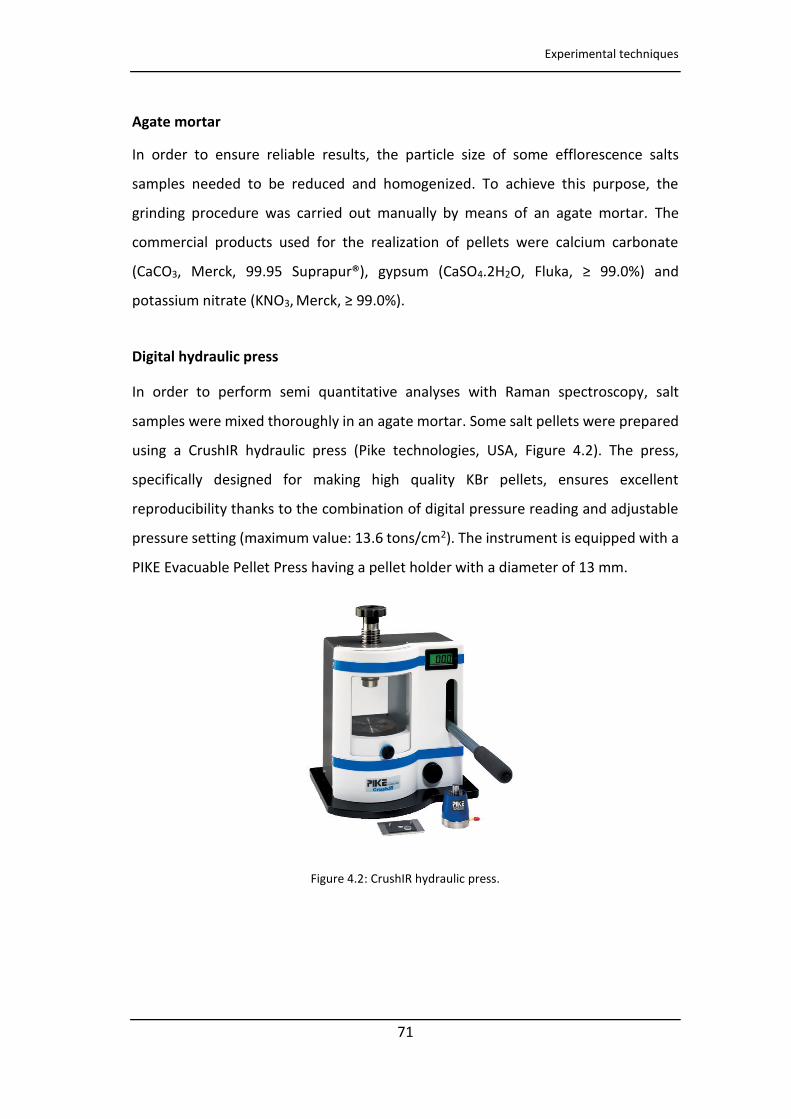
Experimental techniques
71
Agate mortar
In order to ensure reliable results, the particle size of some efflorescence salts
samples needed to be reduced and homogenized. To achieve this purpose, the
grinding procedure was carried out manually by means of an agate mortar. The
commercial products used for the realization of pellets were calcium carbonate
(CaCO3, Merck, 99.95 Suprapur®), gypsum (CaSO4.2H2O, Fluka, ≥ 99.0%) and
potassium nitrate (KNO3, Merck, ≥ 99.0%).
Digital hydraulic press
In order to perform semi quantitative analyses with Raman spectroscopy, salt
samples were mixed thoroughly in an agate mortar. Some salt pellets were prepared
using a CrushIR hydraulic press (Pike technologies, USA, Figure 4.2). The press,
specifically designed for making high quality KBr pellets, ensures excellent
reproducibility thanks to the combination of digital pressure reading and adjustable
pressure setting (maximum value: 13.6 tons/cm2). The instrument is equipped with a
PIKE Evacuable Pellet Press having a pellet holder with a diameter of 13 mm.
Figure 4.2: CrushIR hydraulic press.

CHAPTER 4
72
Cutting plotter, laser ablation system-excimer/CO2 laser, and Thermal roller
laminator
After AutoCAD design of the 3D structure the COP and PSA polymer layers were cut
using a cutter plotter (Graphtec CE5000-40 Craft Robo Pro, Tokyo, Japan). On the
other hand, PMMA layers were cut using a CO2 laser ablation system (Optec Laser
Micromachining Systems, Belgium). A thermal roller laminator (Titan-110, GBC Films,
USA, Figure 4.3) was employed to assemble the microfluidic devices (see chapter 9).
Zeo o COP sheets of μ thi k ess e e pu hased Zeo e Düsseldo f,
Germany). ArCare®8939 PSA of 127 μm double side roll was provided by Adhesive
Research (Limerick, Ireland). Poly(methyl- etha late PMMA μ slides were
purchased from Goodfellow, UK.
Figure 4.3: Cutter plotter Graphtec CE5000-40 Craft Robo Pro.
For the realization of microfluidic sensors, described in the chapter 9, two types of
ionogel were prepared, for the ILs: (IO-1) 1-ethyl-3-methylimidazolium ethyl sulfate
(Sigma-Aldrich, ≥ 9 %, opaque) and (IO-2) trihexyltetradecyl-phosphonium
dicyanamide (Sigma-Aldrich, ≥ 9 %, transparent). The ionogel was obtained by
mixing two monomers, one linear (N-isopropylacrylamide, Sigma-Aldrich, 97%), and
the other for giving the three-dimensional structure N,N’-methylene-bis acrylamide,
Sigma-Aldrich, 99%). In addition, a photoinitiator was added (2,2- Dimethoxy-2-
phenylacetophenone, Sigma-Aldrich, 99%) to induce photopolymerization.
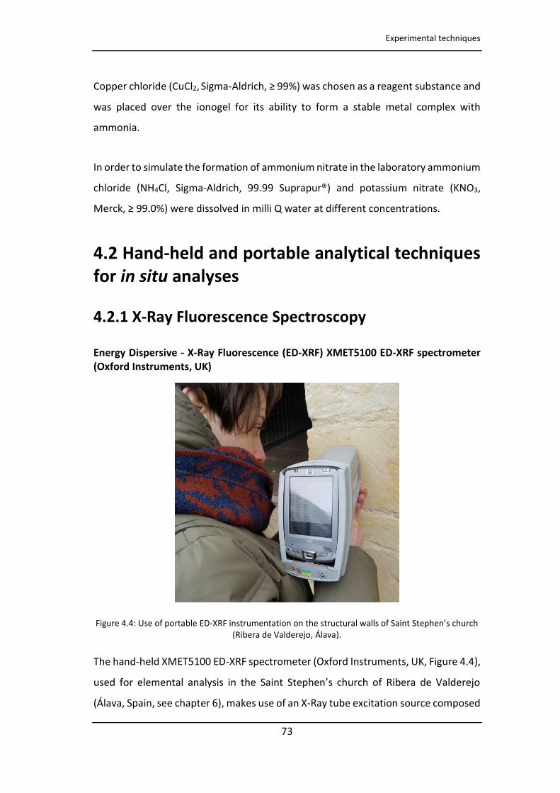
Experimental techniques
73
Copper chloride (CuCl2, Sigma-Aldrich, ≥ 99%) was chosen as a reagent substance and
was placed over the ionogel for its ability to form a stable metal complex with
ammonia.
In order to simulate the formation of ammonium nitrate in the laboratory ammonium
chloride (NH4Cl, Sigma-Aldrich, 99.99 Suprapur®) and potassium nitrate (KNO3,
Merck, ≥ 99.0%) were dissolved in milli Q water at different concentrations.
4.2 Hand-held and portable analytical techniques
for in situ analyses
4.2.1 X-Ray Fluorescence Spectroscopy
Energy Dispersive - X-Ray Fluorescence (ED-XRF) XMET5100 ED-XRF spectrometer
(Oxford Instruments, UK)
Figure 4.4: Use of portable ED-XRF i st u e tatio o the st u tu al alls of Sai t Stephe ’s hu h (Ribera de Valderejo, Álava).
The hand-held XMET5100 ED-XRF spectrometer (Oxford Instruments, UK, Figure 4.4),
used fo ele e tal a al sis i the Sai t Stephe ’s hu h of Ri e a de Valde ejo
(Álava, Spain, see chapter 6), makes use of an X-Ray tube excitation source composed
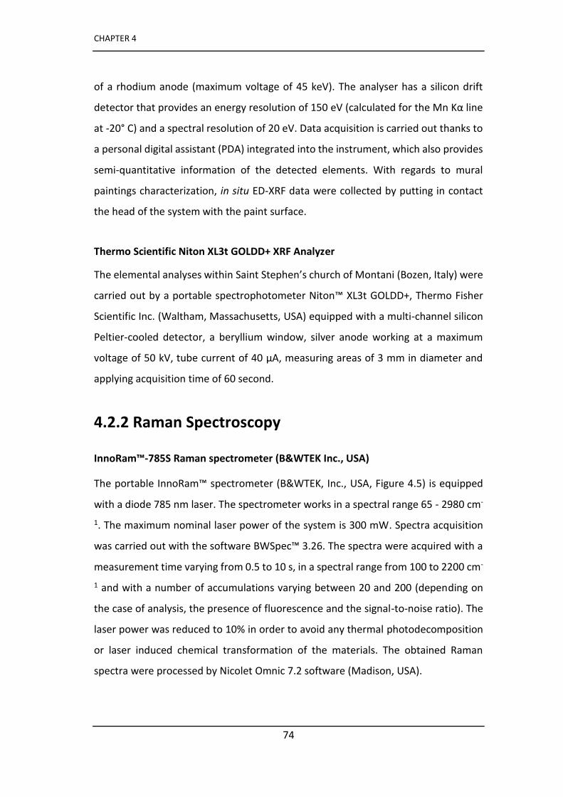
CHAPTER 4
74
of a rhodium anode (maximum voltage of 45 keV). The analyser has a silicon drift
detecto that p o ides a e e g esolutio of eV al ulated fo the M Kα li e
at -20° C) and a spectral resolution of 20 eV. Data acquisition is carried out thanks to
a personal digital assistant (PDA) integrated into the instrument, which also provides
semi-quantitative information of the detected elements. With regards to mural
paintings characterization, in situ ED-XRF data were collected by putting in contact
the head of the system with the paint surface.
Thermo Scientific Niton XL3t GOLDD+ XRF Analyzer
The elemental analyses within Sai t Stephe ’s hurch of Montani (Bozen, Italy) were
carried out by a portable spectrophotometer Niton™ XL3t GOLDD+, Thermo Fisher
Scientific Inc. (Waltham, Massachusetts, USA) equipped with a multi-channel silicon
Peltier-cooled detector, a beryllium window, silver anode working at a maximum
oltage of kV, tu e u e t of μA, easu i g a eas of i dia ete a d
applying acquisition time of 60 second.
4.2.2 Raman Spectroscopy
InnoRam™-785S Raman spectrometer (B&WTEK Inc., USA)
The portable InnoRam™ spectrometer (B&WTEK, Inc., USA, Figure 4.5) is equipped
with a diode 785 nm laser. The spectrometer works in a spectral range 65 - 2980 cm-
1. The maximum nominal laser power of the system is 300 mW. Spectra acquisition
as a ied out ith the soft a e BWSpe ™ . . The spe t a e e a ui ed ith a
measurement time varying from 0.5 to 10 s, in a spectral range from 100 to 2200 cm-
1 and with a number of accumulations varying between 20 and 200 (depending on
the case of analysis, the presence of fluorescence and the signal-to-noise ratio). The
laser power was reduced to 10% in order to avoid any thermal photodecomposition
or laser induced chemical transformation of the materials. The obtained Raman
spectra were processed by Nicolet Omnic 7.2 software (Madison, USA).
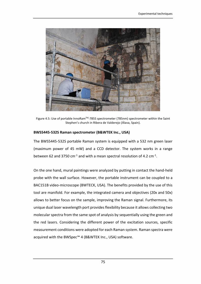
Experimental techniques
75
Figure 4.5: Use of portable InnoRamTM-785S spectrometer (785nm) spectrometer within the Saint Stephe ’s hu h i Ri e a de Valde ejo Álava, Spain).
BWS5445-532S Raman spectrometer (B&WTEK Inc., USA)
The BWS5445-532S portable Raman system is equipped with a 532 nm green laser
(maximum power of 45 mW) and a CCD detector. The system works in a range
between 62 and 3750 cm-1 and with a mean spectral resolution of 4.2 cm-1.
On the one hand, mural paintings were analyzed by putting in contact the hand-held
probe with the wall surface. However, the portable instrument can be coupled to a
BAC151B video-microscope (BWTECK, USA). The benefits provided by the use of this
tool are manifold. For example, the integrated camera and objectives (20x and 50x)
allows to better focus on the sample, improving the Raman signal. Furthermore, its
unique dual laser wavelength port provides flexibility because it allows collecting two
molecular spectra from the same spot of analysis by sequentially using the green and
the red lasers. Considering the different power of the excitation sources, specific
measurement conditions were adopted for each Raman system. Raman spectra were
a ui ed ith the BWSpe ™ B&WTEK I c., USA) software.
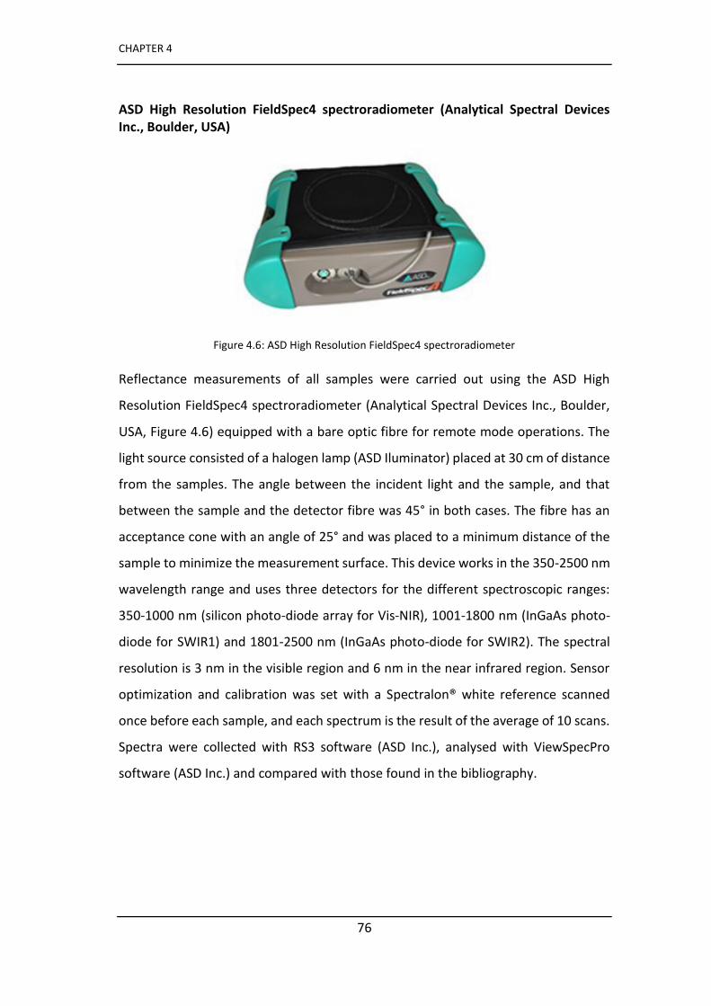
CHAPTER 4
76
ASD High Resolution FieldSpec4 spectroradiometer (Analytical Spectral Devices
Inc., Boulder, USA)
Figure 4.6: ASD High Resolution FieldSpec4 spectroradiometer
Reflectance measurements of all samples were carried out using the ASD High
Resolution FieldSpec4 spectroradiometer (Analytical Spectral Devices Inc., Boulder,
USA, Figure 4.6) equipped with a bare optic fibre for remote mode operations. The
light source consisted of a halogen lamp (ASD Iluminator) placed at 30 cm of distance
from the samples. The angle between the incident light and the sample, and that
between the sample and the detector fibre was 45° in both cases. The fibre has an
acceptance cone with an angle of 25° and was placed to a minimum distance of the
sample to minimize the measurement surface. This device works in the 350-2500 nm
wavelength range and uses three detectors for the different spectroscopic ranges:
350-1000 nm (silicon photo-diode array for Vis-NIR), 1001-1800 nm (InGaAs photo-
diode for SWIR1) and 1801-2500 nm (InGaAs photo-diode for SWIR2). The spectral
resolution is 3 nm in the visible region and 6 nm in the near infrared region. Sensor
optimization and calibration was set with a Spectralon® white reference scanned
once before each sample, and each spectrum is the result of the average of 10 scans.
Spectra were collected with RS3 software (ASD Inc.), analysed with ViewSpecPro
software (ASD Inc.) and compared with those found in the bibliography.
Experimental techniques
77
4.3 Bench top analytical techniques for non-
destructive laboratory analyses
4.3.1 Scanning Electron Microscopy (SEM) Energy
Dispersive X-Ray Spectrometer (EDS)
EVO40 Scanning Electron Microscope (Carl Zeiss STS, Germany)
Elemental images of selected samples were performed using an EVO40 scanning
electron microscope (Carl Zeiss STS, Germany) coupled to an X-Max Energy-
Dispersive X-Ray Spectrometer (Oxford Instruments, UK). Previous to SEM-EDS
analyses, the selected samples were fixed in specific aluminium sample holders (pin)
with carbon type. Furthermore, with the aim of improving the quality of SEM images,
a thin gold fil < μ as applied o the surface of the sample using a Emitech
K550X sputter coater vacuum chamber (Quorum Technologies, UK). EDS analyses
were carried out using a working distance of 8–10 mm, an I Probe of 180 pA, a 35º
take-off angle, an acceleration potential of 30 kV, an integration time of 50 s and a
number of scans between 6 and 10. The INCA Microanalysis Suite 4.3 (Oxford
Instruments, UK) software was used to collect and manage the collected data.
4.3.2 Raman Spectroscopy
InVia Confocal Raman spectrometer (Renishaw, UK)
The inVia Confocal Raman microscope (Figure 4.7) is equipped with a 514 nm
(maximum power of 50 mW) and a 785 nm (maximum power of 350 mW) excitation
laser, a DMLM Leica microscope (Bradford, UK) and a CCD detector. The
spectrometer works in a spectral range between 65 and 3800 cm-1 with a mean
spectral resolution of 1.2 cm-1. On the one side, StreamLine technology was employed
to generate high definition molecular images of specific samples. Briefly, the
motorized stage of the microscope coupled to the Raman spectrometer moves the
sample beneath the lens so that the line of laser-light is rastered across the area
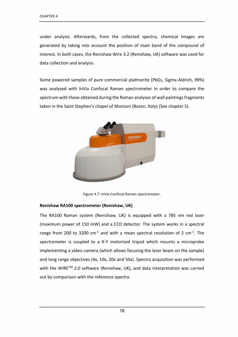
CHAPTER 4
78
under analysis. Afterwards, from the collected spectra, chemical images are
generated by taking into account the position of main band of the compound of
interest. In both cases, the Renishaw Wire 3.2 (Renishaw, UK) software was used for
data collection and analysis.
Some powered samples of pure commercial plattnerite (PbO2, Sigma Aldrich, 99%)
was analysed with InVia Confocal Raman spectrometer in order to compare the
spectrum with those obtained during the Raman analyses of wall paintings fragments
take i the Sai t Stephe ’s hapel of Mo ta i Boze , Ital (See chapter 5).
Figure 4.7: InVia Confocal Raman spectrometer.
Renishaw RA100 spectrometer (Renishaw, UK)
The RA100 Raman system (Renishaw, UK) is equipped with a 785 nm red laser
(maximum power of 150 mW) and a CCD detector. The system works in a spectral
range from 200 to 3200 cm-1 and with a mean spectral resolution of 2 cm-1. The
spectrometer is coupled to a X-Y motorized tripod which mounts a microprobe
implementing a video-camera (which allows focusing the laser beam on the sample)
and long range objectives (4x, 10x, 20x and 50x). Spectra acquisition was performed
with the WIRETM 2.0 software (Renishaw, UK), and data interpretation was carried
out by comparison with the reference spectra.
Experimental techniques
79
Horiba - Jobin Yvon LabRam micro-spectrometer
The micro-Raman measurements were carried out with a 632.8 nm laser in a nearly
backscattered geometry by using a Horiba - Jobin Yvon LabRam micro-spectrometer
(300 mm focal length) equipped with an integrated Olympus BX40 microscope. The
spectral resolution was about 1.5 cm-1. The Rayleigh radiation was blocked by a
holographic edge filter and the backscattered Raman light was dispersed by an 1800
grooves/mm holographic grating on a Peltier cooled CCD, consisting of an array of
/ pi els. The e t a e slit idth as fi ed at μ . The lase po e o the
samples was adjusted by means of density filters. Raman spectra were collected at
very low laser power (< 0.1 mW) in order to avoid photo-thermal effects on the
samples induced by the laser excitation as well as at varying power to check the
thermal behaviour of the pigments. The spectra were collected using a 50x ultra long
working distance (ULWD) microscope objective. Typical exposures were 10-20 s, with
3-5 accumulations. The system was regularly calibrated using the 520.6 cm-1 Raman
band of silicon. The data analysis was performed by LABSPEC 5.78.24, Horiba - Jobin
Yvon LabRam, built-in software package. All collected spectra were compared with
reference spectra.
4.3.4 X-Ray Diffraction (XRD)
PRO PANalytical Xpert X-Ray Diffractometer (PANalytical, Netherlands)
The used X-Ray Diffractometer is equipped with a copper tube, a vertical goniometer
(Bragg-Brentano geometry), a programmable divergence slit, a secondary graphite
monochromator and a Pixcel detector. The condition of all measurements were set
at 40 KV, 40 mA and a scan ranging between 5 and 70º 2theta.
Bruker D2 PHASER diffractometer
Powder XRD analysis of mural paintings samples were carried out with a Bruker D2
PHASER diffractometer operating in Bragg-Brentano geometry, equipped with a
LYNXEYE detector (based on silicon strip te h olog a d oppe a ode CuKα . The
CHAPTER 4
80
powdered samples (few milligrams) were analysed on a zero-background sample
holder (ZBH) and measurements were taken in steps of 0.02° o e θ a ge f o
to 60°.
4.4 Isolation and characterization of
biodeteriogens genomic DNA sequences via PCR
amplification
To genomically characterize the main biodeteriogens the biological strains, collected
in Ribera de Valderejo, sterile swabs and PDA petri dishes were sent to the BAT
laboratory (CIAL institute, Madrid) for their characterization.
Starting from the genomic characterisation of green patinas, the biological strains
contained in the PDA Petri dishes were cultured for seven days at room temperature
(25 °C). Then, metagenomic DNA was isolated in a biological safety cabinet (BIO II
Advance, Telstar, Spain) using the soil DNA isolation commercial kit. All the steps in
the isolation procedure were carried out following the manufacturer's instructions.
In order to amplify the region of internal transcribed spacer (ITS), a polymerase chain
reaction (PCR) and secondary PCR procedures were performed as described by Leake
et al. [1]. To verify the success of the extraction, the metagenomic DNA was checked
by using 0.8% w/v agarose gel electrophoresis. A Inc PTC-100 (MJ Research, USA)
thermal cycler was used to amplify segments of DNA via the PCR. For the
characterisation of the fungi and bacteria species, the 18S and 16S ribosomal RNA
gene region was amplified, respectively. An output file containing ITS sequences with
a partial 18S and 16S ribosomal RNA genes' sequence were then analysed by using
Bioedit software. Finally, ITS sequences' identification was carried out by comparison
with the National Centre for Biotechnology Information (NCBI) database [2].
Experimental techniques
81
References
[1] J. L. Leake, S. E. Dowd, R. D. Wolcott, A. M. Zischkau and Y. Sun (2009) Identification of yeast in chronic wounds using new pathogen – detection technologies, Journal of Wound Care, 18, 103-108.
[2] D. L. Wheeler, T. Barrett, D. A. Benson, S. H. Bryant, K. Canese, V. Chetvernin, D. M. Church, M. DiCuccio, R. Edgar, S. Federhen, L. Y. Geer, Y. Kapustin, O. Khovayko, D. Landsman, D. J. Lipman, T. L. Madden, D. R. Maglott, J. Ostell, V. Miller, K. D. Pruitt, G. D. Schuler, E. Sequeira, S. T. Sherry, K. Sirotkin, A. Souvorov, G. Starchenko, R. L. Tatusov, T. A. Tatusova, L. Wagner, E. Yaschenko, Database resources of the National Center for Biotechnology Information, Nucleic Acids Research, 2007, 35, D5–D12.

83
CHAPTER 5
Investigation of mural paintings
blackening in the chapel of Saint
Stephen’s in Montani (Italy)
As reported in chapter 3, a part of the doctoral project was carried out through a
collaboration with the Department of Mathematical, Physical and Computer Sciences
of the University of Parma (Italy) and involved the study of wall paintings preserved
inside the Saint Stephen s chapel in Montani (Val Venosta, Bolzano, Italy) in order to
characterize the alteration products that cause the blackening of paintings.
Chemical alterations of raw materials, like pigments and organic binders, can cause
chromatic changes with a negative aesthetic effect, preventing a full appreciation of
the artwork. Blackening phenomena or alteration of pigments are widely described
in the literature [1]: the most known examples are the darkening of vermilion [2],
haematite [3], azurite [4] and of lead-based pigments, in particular white and red lead
[5-9]. Since these processes endanger the preservation of the wall paintings, it is
essential to understand the chemical alteration processes that occur during the
ageing of the pigments.
The lead-based pigments (the most used are white lead, 2PbCO3·Pb(OH)2, and red
lead, Pb3O4) have been widely employed in the production of artworks since ancient
CHAPTER 5
84
times [10]. The formation of their secondary products are well known to conservators
and scientists as well as their instability and their tendency to blacken, especially
when using the fresco technique [11]. However recent research has shown the
presence of blackening of lead white even in secco mural paintings [7].
The natural ageing of lead-based compounds can manifest in the form of different
chromatic alterations from light pink to dark brown depending on the type of decay
products. The factors that cause the degradation of lead-based pigments are
different and interrelated, and over the past years several studies have been
conducted to clarify which conditions may give rise to the different decay compounds
[12].
The blackening of white lead and red lead can occur in the form of lead sulphide PbS
(galena mineral) in the presence of atmospheric hydrogen sulphide (H2S). This
degradation path has been observed especially on manuscripts [13-15]. The
formation of lead sulphide could be caused also by the interaction of the lead
pigments with sulphur-based pigments, even if in the literature there is, however, no
clear evidence of this effect [16, 17].
The fo atio of la k lead dio ide β-PbO2 (plattnerite mineral) as a degradation
product is a rarer phenomenon than the development of lead sulphide. Plattnerite
has been identified in mural paintings as a decay product of both white lead and red
lead [18]. It is worth mentioning that recent studies claimed that plattnerite was
intentionally used as pigment in some mural paintings in Iran, after the discovery of
this mineral in a nearby mine [19], but this is a very particular case.
The darkening of lead white in the fresco technique has been attributed to oxidation
processes favoured by the alkaline environment before the lime is transformed into
calcium carbonate and in the presence of moisture [11]. However, the use of a
hydrophobic binder such as egg or oil can inhibit the process of degradation despite
the alkaline environment [20]. Chemical tests conducted on white lead in a NaOH
Investigation of mural paintings blackening in the chapel of Saint Stephen s in Montani (Italy)
85
alkaline solution have shown the transformation of the white pigment in a mixture of
assi ot β-PbO), as the main product of reaction, together with red lead and
plattnerite [20].
Other studies have suggested that the presence of microorganisms such as bacteria
(Arthobacter genus [22] or Flavobacterium species [23]) can favour the degradation
of white and red lead into plattnerite. As stated by Matteini and Moles [11], the
blackening of red lead can occur in presence of hydrogen peroxide (H2O2) produced
by the metabolic activity of microorganisms. Furthermore, some bacteria can
transform lead white, red lead and massicot into PbS. This phenomenon is caused by
the interaction of hydrogen sulphide, produced by bacteria utilizing sulphur-
containing amino acids, with lead-based pigments [15].
Tests performed in laboratory on reference samples of lead based pigments have
highlighted the influence on the degradation of natural inorganic salts and synthetic
salts that may be present on wall paintings after cleaning interventions [24]. The
results showed that blackening of white lead occurred only in the presence of a
strong oxidizing agent, such as NaClO, used as fungicide agent, with formation of
platt e ite, s uti ite α-PbO2) and cerussite (PbCO3). On the contrary, the
blackening did not occur in the presence of hydrogen peroxide. However, red lead
tends to darken in all salt solutions containing dissolved atmospheric CO2 in the form
of hydrogen carbonate anions, forming a mixture of plattnerite and cerussite [24].
Thus, relatively drastic conditions seem to be needed for the darkening of lead white
in wall paintings.
Moreover, studies on naturally aged wall paintings and physical-chemical tests on
raw materials in sulphuric acid solution, proved the presence of a mixture of
platt e ite β-PbO2) and anglesite (PbSO4) as decay product of red lead pigment [12].
This degradation of the red lead, proposed by Aze et al. [12], has been explained by
a process of sulphation or carbonation of β-PbO, a highly reactive specie, which can
e fou d as i pu it i the ed pig e t. β-PbO is transformed into anglesite, and
CHAPTER 5
86
subsequently a solvolytic disproportionation process of Pb3O4 takes place with the
formation of plattnerite.
The whitening of the red lead, converted into lead carbonates cerussite and
hydrocerussite (Pb3(CO3)2(OH)2), or in lead sulphate anglesite, is a less known
phenomenon although it has been observed over pictorial surfaces. It occurs when
the pigment reacts with atmospheric pollutants such as SO2, CO and CO2 in specific
temperature and moisture conditions [12, 25]. Other authors suggest that the
alteration of minium gives initially a dark colour, with formation of plattnerite, which
then turns into a more stable anglesite, causing the total loss of the colour [26].
Othe lead o pou ds su h as lead o o ides, assi ot β-PbO-orthorhombic) and
litha ge α-PbO-tetragonal), have been frequently identified in wall paintings
together with red lead. They may be present either as pigments, or as impurities of
the same red lead. I fa t, α-P O a d β-PbO are intermediate products generating
from the calcination of the white lead at high temperatures during the manufacturing
process of red lead [27]. Lead monoxides are instable in the presence of moisture and
in an alkaline environment. In fact, the carbonation processes cause the formation of
lead carbonates cerussite, hydrocerussite and plumbonacrite (Pb5O(OH)2(CO3)3) [28].
The present work concerns the study of wall paintings preserved in the Saint
Stephe s chapel located in Montani (Bolzano) in Val Venosta (Italy). They are
affected by a degradation phenomenon, which is clearly visible as an extended
darkening, especially on the apse and on the vault.
Preliminary cleaning tests on yellow and green areas were carried out, according to
the method of white-lead reconversion, using hydrogen peroxide in a solution of
acetic acid, at very low concentration, and distilled water [29]. The darkening,
supposed to be due to transformation of white lead into lead dioxide (plattnerite),
has been explained by Matteini et al. [30] as an oxidation reaction where the lead
changes its oxidation number from +2 to + 4. The proposed treatment consists in the
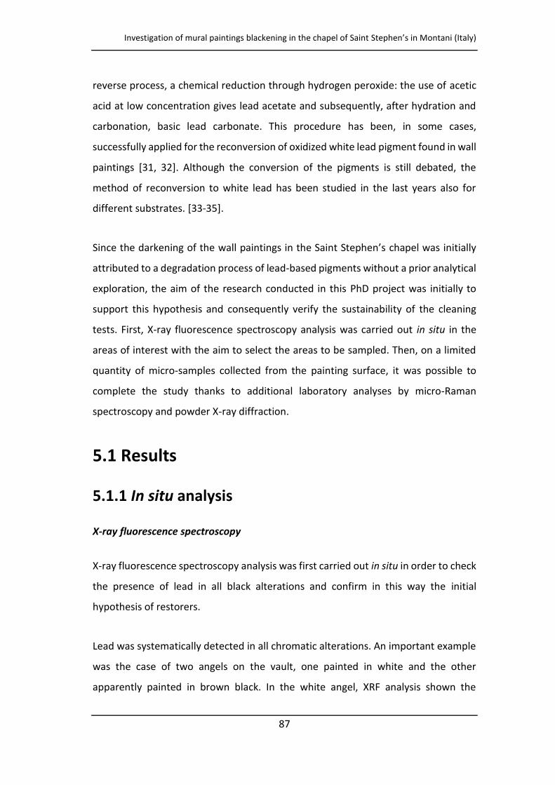
Investigation of mural paintings blackening in the chapel of Saint Stephen s in Montani (Italy)
87
reverse process, a chemical reduction through hydrogen peroxide: the use of acetic
acid at low concentration gives lead acetate and subsequently, after hydration and
carbonation, basic lead carbonate. This procedure has been, in some cases,
successfully applied for the reconversion of oxidized white lead pigment found in wall
paintings [31, 32]. Although the conversion of the pigments is still debated, the
method of reconversion to white lead has been studied in the last years also for
different substrates. [33-35].
Since the darkening of the all pai ti gs i the Sai t Stephe s chapel was initially
attributed to a degradation process of lead-based pigments without a prior analytical
exploration, the aim of the research conducted in this PhD project was initially to
support this hypothesis and consequently verify the sustainability of the cleaning
tests. First, X-ray fluorescence spectroscopy analysis was carried out in situ in the
areas of interest with the aim to select the areas to be sampled. Then, on a limited
quantity of micro-samples collected from the painting surface, it was possible to
complete the study thanks to additional laboratory analyses by micro-Raman
spectroscopy and powder X-ray diffraction.
5.1 Results
5.1.1 In situ analysis
X-ray fluorescence spectroscopy
X-ray fluorescence spectroscopy analysis was first carried out in situ in order to check
the presence of lead in all black alterations and confirm in this way the initial
hypothesis of restorers.
Lead was systematically detected in all chromatic alterations. An important example
was the case of two angels on the vault, one painted in white and the other
apparently painted in brown black. In the white angel, XRF analysis shown the
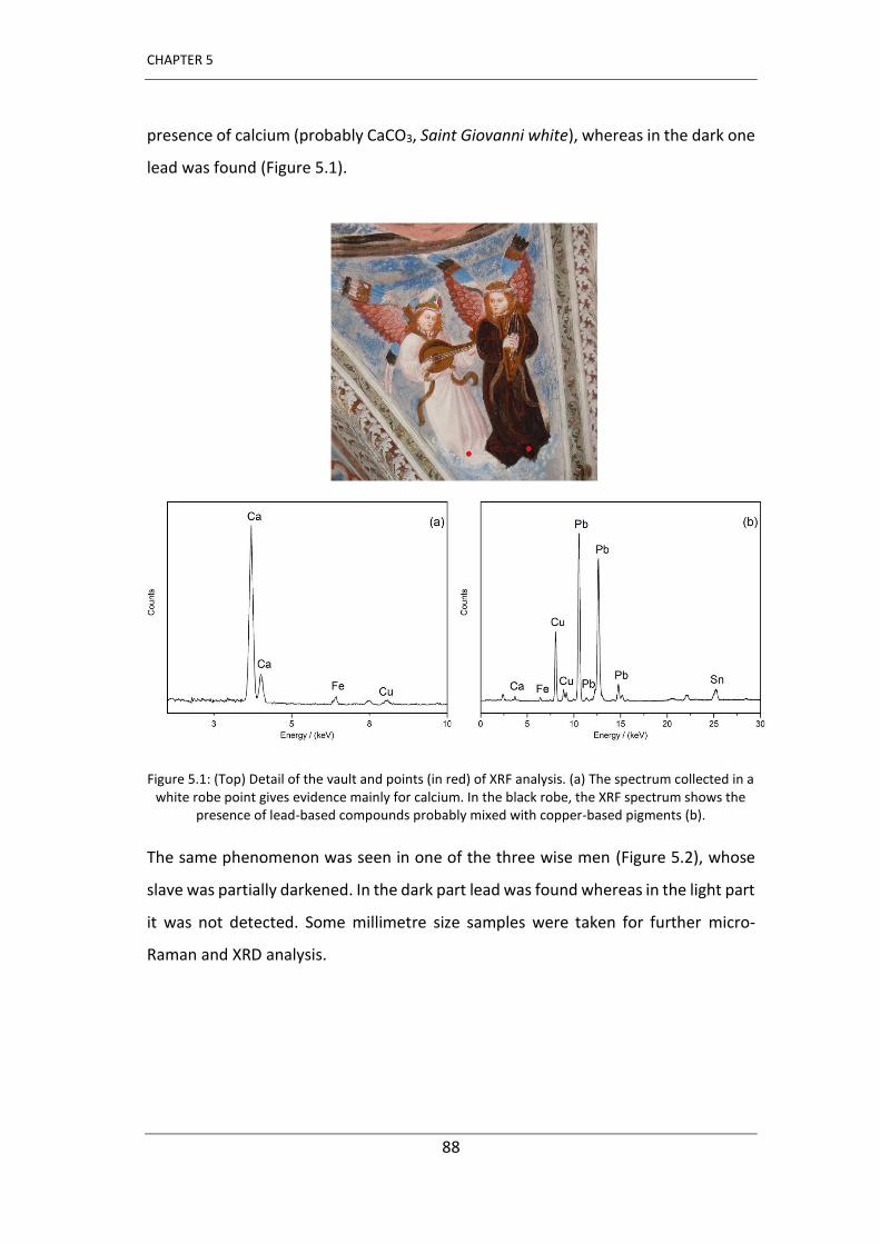
CHAPTER 5
88
presence of calcium (probably CaCO3, Saint Giovanni white), whereas in the dark one
lead was found (Figure 5.1).
Figure 5.1: (Top) Detail of the vault and points (in red) of XRF analysis. (a) The spectrum collected in a white robe point gives evidence mainly for calcium. In the black robe, the XRF spectrum shows the
presence of lead-based compounds probably mixed with copper-based pigments (b).
The same phenomenon was seen in one of the three wise men (Figure 5.2), whose
slave was partially darkened. In the dark part lead was found whereas in the light part
it was not detected. Some millimetre size samples were taken for further micro-
Raman and XRD analysis.
Investigation of mural paintings blackening in the chapel of Saint Stephen s in Montani (Italy)
89
Figure 5.2: Detail of the sleeve of one of the three Wise Men, where different blue colours are observed.
5.1.2 Laboratory analyses
Micro-Raman spectroscopy
Analysis of plattnerite standards
Since the hypothesis formulated by the restorers was that the blackening of paintings
was caused by the presence of plattnerite (PbO2), Raman analysis were carried out in
order to confirm this assumption.
As attested by previous studies, plattnerite has a very weak Raman scattering, and it
can degrade if the excitation laser power is not kept sufficiently low [36]. The problem
of laser induced degradation of lead pigments is indeed a well-known phenomenon,
even if some aspects have still to be better understood [37,38]. It has been repeatedly
shown that if the laser power is higher than some threshold, depending on the laser
wavelength and the nature of the analysed sample, degradation phenomena may be
triggered during Raman analyses and secondary products, as red lead, massicot and
litharge, are formed. [39-41]. Thus, we decided to perform Raman measurements
first on plattnerite standards, both on mineral plattnerite and on powdered synthetic
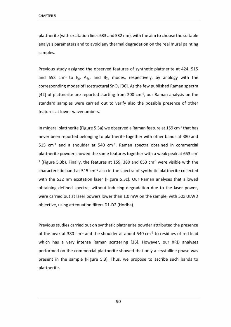
CHAPTER 5
90
plattnerite (with excitation lines 633 and 532 nm), with the aim to choose the suitable
analysis parameters and to avoid any thermal degradation on the real mural painting
samples.
Previous study assigned the observed features of synthetic plattnerite at 424, 515
and 653 cm-1 to Eg, A1g, and B2g modes, respectively, by analogy with the
corresponding modes of isostructural SnO2 [36]. As the few published Raman spectra
[42] of plattnerite are reported starting from 200 cm-1, our Raman analysis on the
standard samples were carried out to verify also the possible presence of other
features at lower wavenumbers.
In mineral plattnerite (Figure 5.3a) we observed a Raman feature at 159 cm-1 that has
never been reported belonging to plattnerite together with other bands at 380 and
515 cm-1 and a shoulder at 540 cm-1. Raman spectra obtained in commercial
plattnerite powder showed the same features together with a weak peak at 653 cm-
1 (Figure 5.3b). Finally, the features at 159, 380 and 653 cm-1 were visible with the
characteristic band at 515 cm-1 also in the spectra of synthetic plattnerite collected
with the 532 nm excitation laser (Figure 5.3c). Our Raman analyses that allowed
obtaining defined spectra, without inducing degradation due to the laser power,
were carried out at laser powers lower than 1.0 mW on the sample, with 50x ULWD
objective, using attenuation filters D1-D2 (Horiba).
Previous studies carried out on synthetic plattnerite powder attributed the presence
of the peak at 380 cm-1 and the shoulder at about 540 cm-1 to residues of red lead
which has a very intense Raman scattering [36]. However, our XRD analyses
performed on the commercial plattnerite showed that only a crystalline phase was
present in the sample (Figure 5.3). Thus, we propose to ascribe such bands to
plattnerite.
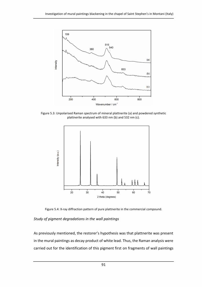
Investigation of mural paintings blackening in the chapel of Saint Stephen s in Montani (Italy)
91
Figure 5.3: Unpolarised Raman spectrum of mineral plattnerite (a) and powdered synthetic plattnerite analysed with 633 nm (b) and 532 nm (c).
Figure 5.4: X-ray diffraction pattern of pure plattnerite in the commercial compound.
Study of pigment degradations in the wall paintings
As p e iousl e tio ed, the esto e s h pothesis as that platt e ite as p ese t
in the mural paintings as decay product of white lead. Thus, the Raman analysis were
carried out for the identification of this pigment first on fragments of wall paintings
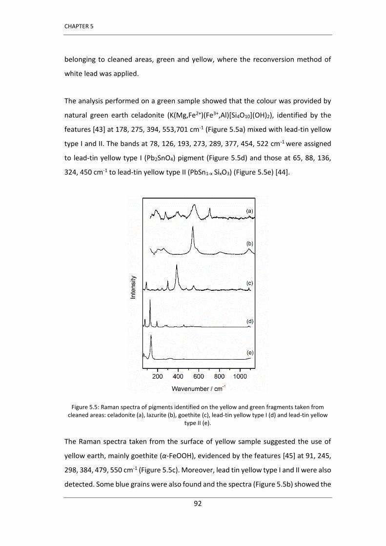
CHAPTER 5
92
belonging to cleaned areas, green and yellow, where the reconversion method of
white lead was applied.
The analysis performed on a green sample showed that the colour was provided by
natural green earth celadonite (K(Mg,Fe2+)(Fe3+,Al)[Si4O10](OH)2), identified by the
features [43] at 178, 275, 394, 553,701 cm-1 (Figure 5.5a) mixed with lead-tin yellow
type I and II. The bands at 78, 126, 193, 273, 289, 377, 454, 522 cm-1 were assigned
to lead-tin yellow type I (Pb2SnO4) pigment (Figure 5.5d) and those at 65, 88, 136,
324, 450 cm-1 to lead-tin yellow type II (PbSn1-x SixO3) (Figure 5.5e) [44].
Figure 5.5: Raman spectra of pigments identified on the yellow and green fragments taken from cleaned areas: celadonite (a), lazurite (b), goethite (c), lead-tin yellow type I (d) and lead-tin yellow
type II (e).
The Raman spectra taken from the surface of yellow sample suggested the use of
yellow earth, mainly goethite (α-FeOOH), evidenced by the features [45] at 91, 245,
298, 384, 479, 550 cm-1 (Figure 5.5c). Moreover, lead tin yellow type I and II were also
detected. Some blue grains were also found and the spectra (Figure 5.5b) showed the
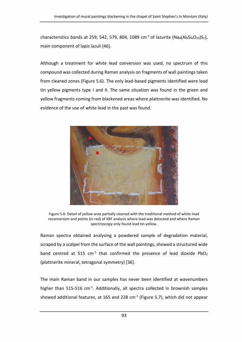
Investigation of mural paintings blackening in the chapel of Saint Stephen s in Montani (Italy)
93
characteristics bands at 259, 542, 579, 804, 1089 cm-1 of lazurite (Na8(Al6Si6O24)Sn),
main component of lapis lazuli [46].
Although a treatment for white lead conversion was used, no spectrum of this
compound was collected during Raman analysis on fragments of wall paintings taken
from cleaned zones (Figure 5.6). The only lead-based pigments identified were lead
tin yellow pigments type I and II. The same situation was found in the green and
yellow fragments coming from blackened areas where plattnerite was identified. No
evidence of the use of white lead in the past was found.
Figure 5.6: Detail of yellow area partially cleaned with the traditional method of white-lead reconversion and points (in red) of XRF analysis where lead was detected and where Raman
spectroscopy only found lead tin yellow.
Raman spectra obtained analysing a powdered sample of degradation material,
scraped by a scalpel from the surface of the wall paintings, showed a structured wide
band centred at 515 cm-1 that confirmed the presence of lead dioxide PbO2
(plattnerite mineral, tetragonal symmetry) [36].
The main Raman band in our samples has never been identified at wavenumbers
higher than 515-516 cm-1. Additionally, all spectra collected in brownish samples
showed additional features, at 165 and 228 cm-1 (Figure 5.7), which did not appear
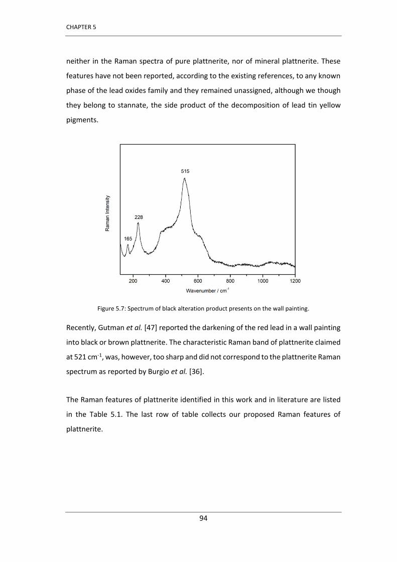
CHAPTER 5
94
neither in the Raman spectra of pure plattnerite, nor of mineral plattnerite. These
features have not been reported, according to the existing references, to any known
phase of the lead oxides family and they remained unassigned, although we though
they belong to stannate, the side product of the decomposition of lead tin yellow
pigments.
Figure 5.7: Spectrum of black alteration product presents on the wall painting.
Recently, Gutman et al. [47] reported the darkening of the red lead in a wall painting
into black or brown plattnerite. The characteristic Raman band of plattnerite claimed
at 521 cm-1, was, however, too sharp and did not correspond to the plattnerite Raman
spectrum as reported by Burgio et al. [36].
The Raman features of plattnerite identified in this work and in literature are listed
in the Table 5.1. The last row of table collects our proposed Raman features of
plattnerite.
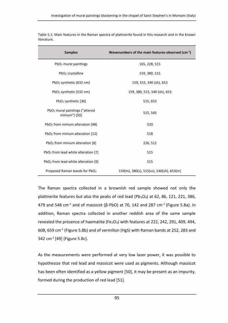
Investigation of mural paintings blackening in the chapel of Saint Stephen s in Montani (Italy)
95
Table 5.1: Main features in the Raman spectra of plattnerite found in this research and in the known literature.
Samples Wavenumbers of the main features observed (cm-1)
PbO2 mural paintings 165, 228, 515
PbO2 crystalline 159, 380, 515
PbO2 synthetic (632 nm) 159, 515, 540 (sh), 653
PbO2 synthetic (532 nm) 159, 380, 515, 540 (sh), 653
PbO2 synthetic [36] 515, 653
PbO2 u al pai ti gs alte ed i iu [ ] 515, 545
PbO2 from minium alteration [48] 520
PbO2 from minium alteration [12] 518
PbO2 from minium alteration [8] 226, 512
PbO2 from lead white alteration [7] 515
PbO2 from lead white alteration [9] 515
Proposed Raman bands for PbO2 159(m), 380(s), 515(vs), 540(sh), 653(m)
The Raman spectra collected in a brownish red sample showed not only the
plattnerite features but also the peaks of red lead (Pb3O4) at 62, 86, 121, 221, 386,
479 and 548 cm-1 and of assi ot β-PbO) at 70, 142 and 287 cm-1 (Figure 5.8a). In
addition, Raman spectra collected in another reddish area of the same sample
revealed the presence of haematite (Fe2O3) with features at 222, 242, 291, 409, 494,
608, 659 cm-1 (Figure 5.8b) and of vermilion (HgS) with Raman bands at 252, 283 and
342 cm-1 [49] (Figure 5.8c).
As the measurements were performed at very low laser power, it was possible to
hypothesize that red lead and massicot were used as pigments. Although massicot
has been often identified as a yellow pigment [50], it may be present as an impurity,
formed during the production of red lead [51].
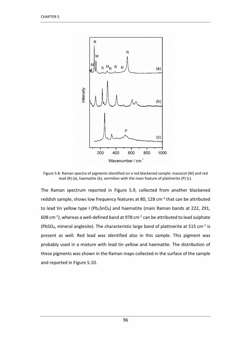
CHAPTER 5
96
Figure 5.8: Raman spectra of pigments identified on a red blackened sample: massicot (M) and red lead (R) (a), haematite (b), vermilion with the main feature of plattnerite (P) (c).
The Raman spectrum reported in Figure 5.9, collected from another blackened
reddish sample, shows low frequency features at 80, 128 cm-1 that can be attributed
to lead tin yellow type I (Pb2SnO4) and haematite (main Raman bands at 222, 291,
608 cm-1), whereas a well-defined band at 978 cm-1 can be attributed to lead sulphate
(PbSO4, mineral anglesite). The characteristic large band of plattnerite at 515 cm-1 is
present as well. Red lead was identified also in this sample. This pigment was
probably used in a mixture with lead tin yellow and haematite. The distribution of
these pigments was shown in the Raman maps collected in the surface of the sample
and reported in Figure 5.10.
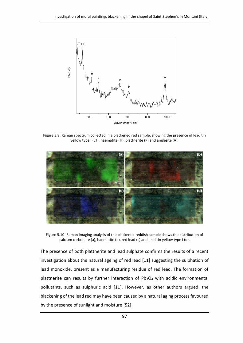
Investigation of mural paintings blackening in the chapel of Saint Stephen s in Montani (Italy)
97
Figure 5.9: Raman spectrum collected in a blackened red sample, showing the presence of lead tin yellow type I (LT), haematite (H), plattnerite (P) and anglesite (A).
Figure 5.10: Raman imaging analysis of the blackened reddish sample shows the distribution of calcium carbonate (a), haematite (b), red lead (c) and lead tin yellow type I (d).
The presence of both plattnerite and lead sulphate confirms the results of a recent
investigation about the natural ageing of red lead [11] suggesting the sulphation of
lead monoxide, present as a manufacturing residue of red lead. The formation of
plattnerite can results by further interaction of Pb3O4 with acidic environmental
pollutants, such as sulphuric acid [11]. However, as other authors argued, the
blackening of the lead red may have been caused by a natural aging process favoured
by the presence of sunlight and moisture [52].
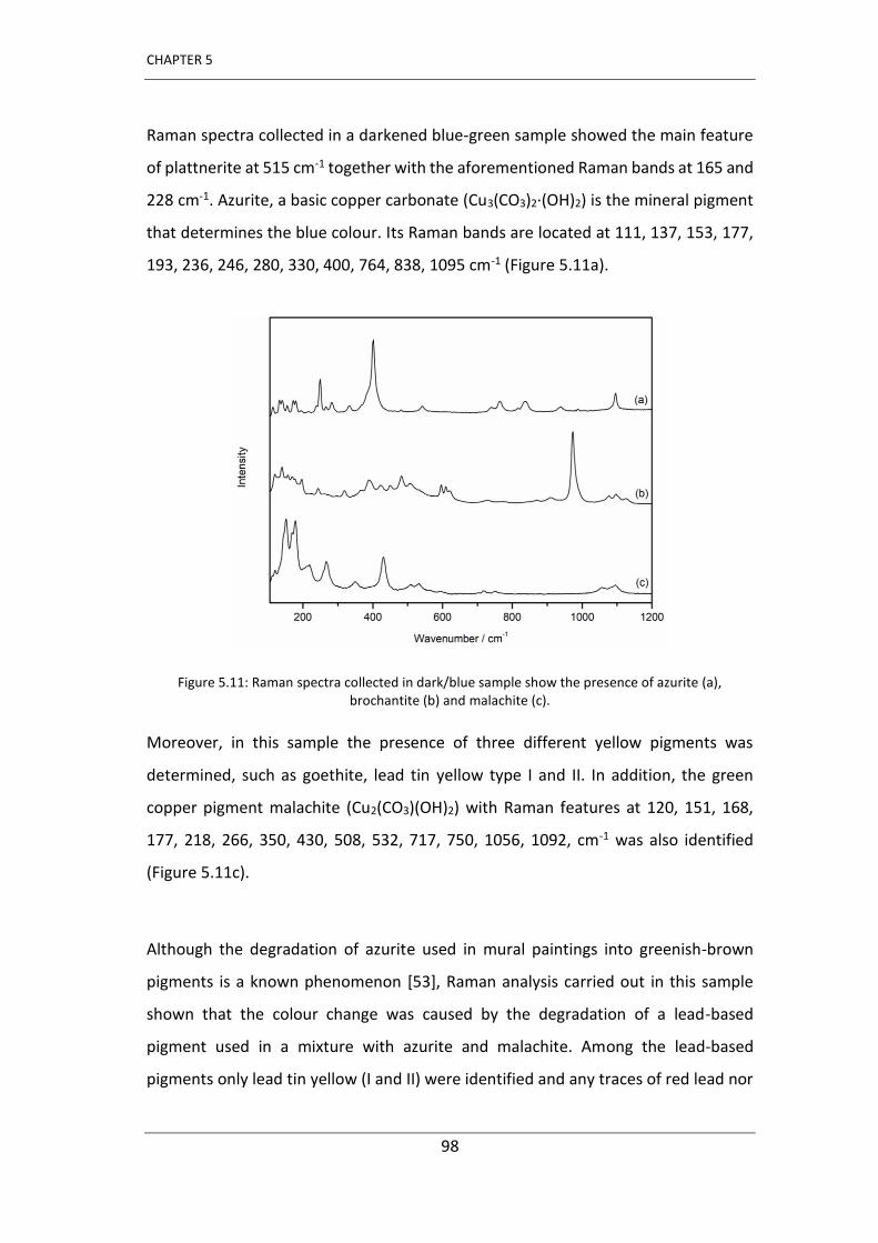
CHAPTER 5
98
Raman spectra collected in a darkened blue-green sample showed the main feature
of plattnerite at 515 cm-1 together with the aforementioned Raman bands at 165 and
228 cm-1. Azurite, a basic copper carbonate (Cu3(CO3)2·(OH)2) is the mineral pigment
that determines the blue colour. Its Raman bands are located at 111, 137, 153, 177,
193, 236, 246, 280, 330, 400, 764, 838, 1095 cm-1 (Figure 5.11a).
Figure 5.11: Raman spectra collected in dark/blue sample show the presence of azurite (a), brochantite (b) and malachite (c).
Moreover, in this sample the presence of three different yellow pigments was
determined, such as goethite, lead tin yellow type I and II. In addition, the green
copper pigment malachite (Cu2(CO3)(OH)2) with Raman features at 120, 151, 168,
177, 218, 266, 350, 430, 508, 532, 717, 750, 1056, 1092, cm-1 was also identified
(Figure 5.11c).
Although the degradation of azurite used in mural paintings into greenish-brown
pigments is a known phenomenon [53], Raman analysis carried out in this sample
shown that the colour change was caused by the degradation of a lead-based
pigment used in a mixture with azurite and malachite. Among the lead-based
pigments only lead tin yellow (I and II) were identified and any traces of red lead nor
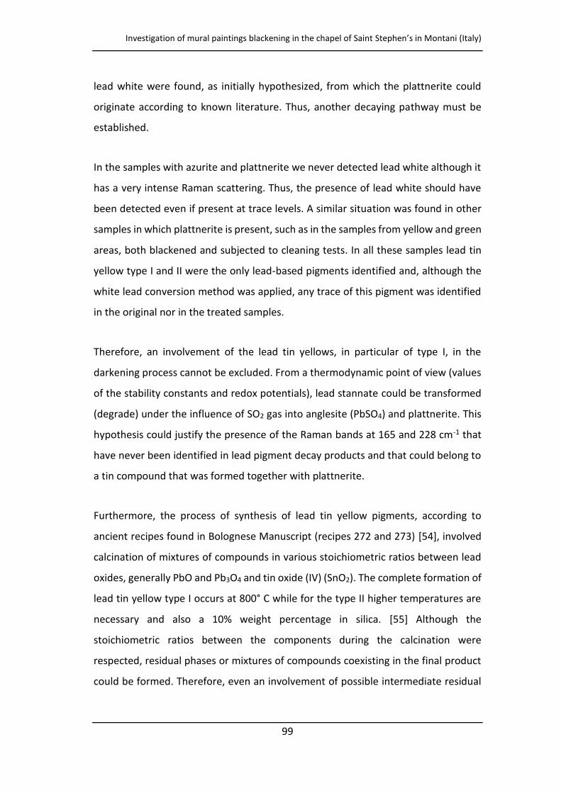
Investigation of mural paintings blackening in the chapel of Saint Stephen s in Montani (Italy)
99
lead white were found, as initially hypothesized, from which the plattnerite could
originate according to known literature. Thus, another decaying pathway must be
established.
In the samples with azurite and plattnerite we never detected lead white although it
has a very intense Raman scattering. Thus, the presence of lead white should have
been detected even if present at trace levels. A similar situation was found in other
samples in which plattnerite is present, such as in the samples from yellow and green
areas, both blackened and subjected to cleaning tests. In all these samples lead tin
yellow type I and II were the only lead-based pigments identified and, although the
white lead conversion method was applied, any trace of this pigment was identified
in the original nor in the treated samples.
Therefore, an involvement of the lead tin yellows, in particular of type I, in the
darkening process cannot be excluded. From a thermodynamic point of view (values
of the stability constants and redox potentials), lead stannate could be transformed
(degrade) under the influence of SO2 gas into anglesite (PbSO4) and plattnerite. This
hypothesis could justify the presence of the Raman bands at 165 and 228 cm-1 that
have never been identified in lead pigment decay products and that could belong to
a tin compound that was formed together with plattnerite.
Furthermore, the process of synthesis of lead tin yellow pigments, according to
ancient recipes found in Bolognese Manuscript (recipes 272 and 273) [54], involved
calcination of mixtures of compounds in various stoichiometric ratios between lead
oxides, generally PbO and Pb3O4 and tin oxide (IV) (SnO2). The complete formation of
lead tin yellow type I occurs at 800° C while for the type II higher temperatures are
necessary and also a 10% weight percentage in silica. [55] Although the
stoichiometric ratios between the components during the calcination were
respected, residual phases or mixtures of compounds coexisting in the final product
could be formed. Therefore, even an involvement of possible intermediate residual
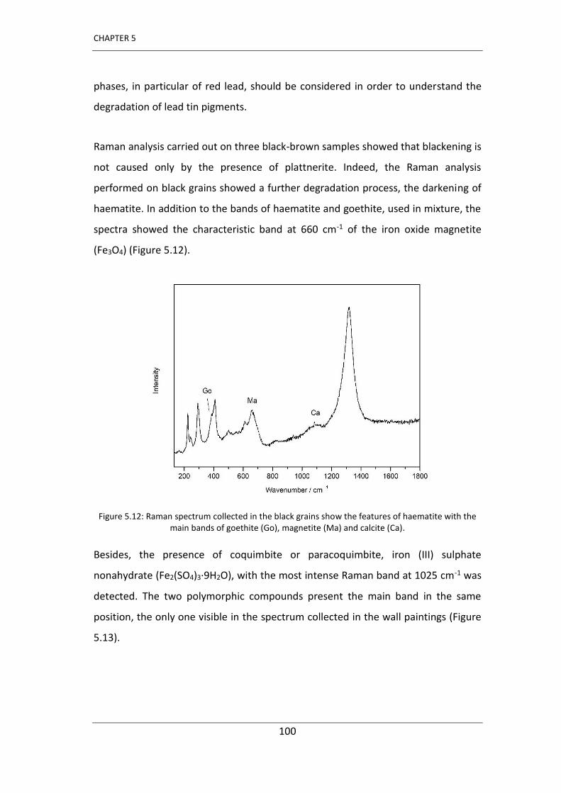
CHAPTER 5
100
phases, in particular of red lead, should be considered in order to understand the
degradation of lead tin pigments.
Raman analysis carried out on three black-brown samples showed that blackening is
not caused only by the presence of plattnerite. Indeed, the Raman analysis
performed on black grains showed a further degradation process, the darkening of
haematite. In addition to the bands of haematite and goethite, used in mixture, the
spectra showed the characteristic band at 660 cm-1 of the iron oxide magnetite
(Fe3O4) (Figure 5.12).
Figure 5.12: Raman spectrum collected in the black grains show the features of haematite with the main bands of goethite (Go), magnetite (Ma) and calcite (Ca).
Besides, the presence of coquimbite or paracoquimbite, iron (III) sulphate
nonahydrate (Fe2(SO4)3·9H2O), with the most intense Raman band at 1025 cm-1 was
detected. The two polymorphic compounds present the main band in the same
position, the only one visible in the spectrum collected in the wall paintings (Figure
5.13).
Investigation of mural paintings blackening in the chapel of Saint Stephen s in Montani (Italy)
101
The degradation process of haematite was studied for the first time by Maguregui et
al. [56] following the identification of magnetite and coquimbite on wall paintings in
the archaeological site of Pompeii (Italy). These decay products were caused by a
phenomenon of sulphation due to the presence of SO2 gas released by the eruption
of Vesuvius, but also present in the modern atmospheres around Pompeii. The study
of the chemical reactions was carried out by laboratory experiments exposing
haematite to SO2 vapours [57]. At first, haematite pigment, in the presence of sulphur
dioxide, is reduced to magnetite whose formation causes the blackening of the
paints. On the other hand, the SO2 is oxidized to SO3, which is mixed with the water
present as atmospheric humidity giving H2SO4. In the Pompeii mural paintings, the
formation of coquimbite on the surface is generated by the interaction of iron (III)
oxide with carbonate and sulphate anions of the plaster generated by cycles of wet
deposition of acid aerosol (SO3 or H2SO4) of the environment.
Figure 5.13: Raman spectrum collected in a red blackening sample show the features of lead tin yellow type I, plattnerite (P), red lead (R), coquimbite (Co) and calcite (Ca) and the features at 165
cm-1 and 228 cm-1 (⁎).
As attested by Gómez et al. [58] the formation of iron sulphate in the pore stones can
occur at pH values lower than 4.5, as occurs in the presence of acid pollutants.
Subsequently coquimbite is produced by hydration processes.
CHAPTER 5
102
Thus, the presence of magnetite in the chapel of Saint Stephen could indicate an
impact of SOx gases, degrading the haematite. The presence of coquimbite could also
be a consequence of such degradation favoured by the presence of gypsum in the
mortar and the rain water infiltration.
Another issue that would support the role of SOx in the darkening phenomena inside
the chapel could be the presence of basic copper sulphates. Together with malachite,
the copper sulphate brochantite (Cu4(SO4)(OH)6) was found in a sample. The
identification was possible, thanks to literature [59], taking into account the position
of minor bands that are different from other compounds with different hydration
state such as posnjakite (Cu4(SO4)(OH)6·H2O) or langite (Cu4(SO4)(OH)6·2H2O). The
Raman features were located at 118, 140, 156, 169, 195, 243, 298, 319, 365, 390,
420, 448, 482, 506, 596, 608, 620, 769, 870, 909, 972, 1076, 1096, 1125 cm-1 (Figure
5.11b). Previous studies showed brochantite as an alteration product from the
sulphation of malachite by SO2 in wall paintings [60], but also in nature mixed with
malachite [61]. However, an intentional use of brochantite as a green pigment has
been also demonstrated [62].
The state of conservation of the wall pai ti gs of the Sai t Stephe s hapel is good,
except for the wall exposed to the north that shows a particularly severe degradation
phenomenon, being covered for about half of its height of the ground in the outer
part and this favours the infiltration of water into the walls. This process and
continuous rainwater infiltration may have created favourable conditions for
pigment degradation.
In addition, although the area in which the chapel is located seems an
uncontaminated mountain area, the contribution of anthropological factors must be
considered in the study of the degradation phenomena affecting the wall paintings.
Indeed, studies on air quality in the district of Bolzano showed that the collected data
in the detection station of Laces, adjacent to the town of Montani, represented an
Investigation of mural paintings blackening in the chapel of Saint Stephen s in Montani (Italy)
103
anomaly compared with other stations due to the high values of PM10. The
atmospheric pollution in Val Venosta is increased by the orographic layout of the
valley and by the cold currents which, due to thermal inversion phenomena, drain
the air masses full in contaminants into the plains zones where then they tend to be
stagnant. The use of wood heating in rural areas have contributed significantly over
the years to the increase in concentrations of pollutants in this area, especially in
winter.
With regards to sulphur dioxide, values above the norm were not recorded in the
studies carried out over the past ten years. However, the values of SO2 have
decreased significantly in Europe thanks to new environmental policies that have
promoted the use of biomass fuels, which have a lower environmental impact instead
of coal fuels widely used in past decades. This means that SO2 values in the past were
much higher than present.
X-ray diffraction
X-ray powder diffraction analyses were also carried out in order to characterize the
decay products of lead-based pig e ts o pai ti g s su fa e. The follo i g esults
were obtained by powdered samples taken from red and blue areas, analysed also
by Raman spectroscopy. The diffraction pattern representative of a red darkened
area (Figure 5.14 o fi ed the p ese e of platt e ite at θ = . °, . °, . °,
49°, 52.1°, 58.8°, massicot (29.1°) and red lead (26.3°, 30.8°). The most intense peak
at 26.7° is likely due to quartz of the mortar, corresponding to its most intense
reflection.
Figure 5.15 reports the diffraction pattern collected in a darkened blue sample.
Azurite was the main component in the powder and it was identified by the features
at θ = . °, . °, . °, . °, . °, . °, . °, . °, . °, . °, . °, . °,
40.4°, 41.5°, 47.7°. Malachite (CuCO3·Cu(OH)2), was identified by peaks at 14.7°,
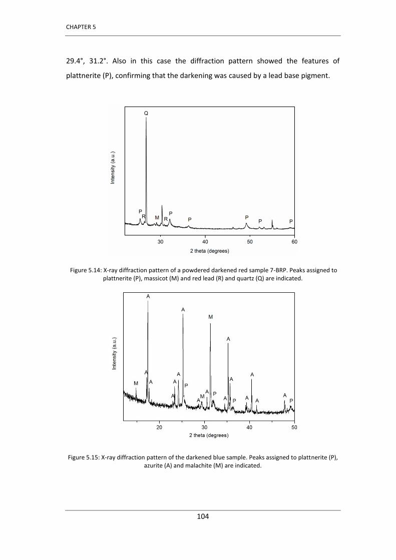
CHAPTER 5
104
29.4°, 31.2°. Also in this case the diffraction pattern showed the features of
plattnerite (P), confirming that the darkening was caused by a lead base pigment.
Figure 5.14: X-ray diffraction pattern of a powdered darkened red sample 7-BRP. Peaks assigned to plattnerite (P), massicot (M) and red lead (R) and quartz (Q) are indicated.
Figure 5.15: X-ray diffraction pattern of the darkened blue sample. Peaks assigned to plattnerite (P), azurite (A) and malachite (M) are indicated.
Investigation of mural paintings blackening in the chapel of Saint Stephen s in Montani (Italy)
105
Final remarks
This research concerning the study of late - Gothic wall paintings preserved inside the
Saint Stephen s hapel in Montani (Val Venosta, Bolzano, Italy) confirms the presence
of lead, either in the darkened area, or in cleaned areas treated according to the
method of reconversion of lead white.
Thanks to the use of laboratory techniques, especially of Raman instrumentation, it
was possible to identify the nature of mortar and pigments (Table 5.2 summarizes
the different compounds detected during this research) belonging to the Gothic
period.
Samples of commercial pure plattnerite and in mineral form were analysed and all
the Raman features at 159 (m), 380 (s), 515(vs), 540 (sh) and 653 (m) cm-1 were
reported for the first time belonging to the form of lead dioxide.
Ra a spe t a o tai ed at lo lase e itatio po e ≈ . W a d po de XRD
evidenced the presence of plattnerite (PbO2) on all blackened samples and, in minor
amount, of anglesite as alteration products of lead-based pigments. The aging
process occurs in the presence of a basic substrate, humidity and sunlight (dry and
wet cycles) and was favoured by the presence of acidic pollutants (SOx) in the
atmosphere, which stagnate in the area where the chapel is located, due to the
particular orographic characteristic of the valley.
Red lead was clearly identified on the samples belonging to red painted areas
originally. Actually, we have no evidence that the darkening of wall paintings could
be attributed to the use of white lead because its Raman spectrum has never been
collected on the samples.
A cleaning test using the method of reconversion of white lead was applied in a very
limited area of the paints, unfortunately before verifying the composition of the black
CHAPTER 5
106
patina to prove the actual presence of lead white. The Raman spectra collected on
the cleaned areas gave no evidence of plattnerite, confirming apparently the
effectiveness of the conversion treatment but no traces of lead white were identified.
However, it was decided not to proceed with the cleaning of larger areas because the
method was considered too aggressive and the treated areas showed a pulverization
of the pigments. This work represents another example of the importance of the use
of non-destructive diagnostic techniques with the aim to identify the secondary
products and to give indications to restorers and conservators on the suitable
method to be used.
All the Raman spectra collected in brownish samples taken from the mural paintings
showed unassigned features at 165 and 228 cm-1 that cannot be attributed to known
lead oxide phases. The constant presence of lead tin yellow pigments on all the
analysed blackened samples suggested that these bands, always visible together with
the main plattnerite band, could prove the presence of tin and lead based
compounds as secondary products not yet mentioned in the literature. To verify this
new decaying process, laboratory tests in which lead tin pigments are subjected to
aging with SO2 vapours, in the presence of moisture, should be conducted in order to
verify the blackening of the pigments, the possible formation of plattnerite, and the
formation of side tin and/or lead based compounds.
The spectroscopic results obtained after applying all these techniques allowed us to
identify another degradation process other than the initially expected one. Indeed,
the blackening process of haematite was also demonstrated by using Raman
spectroscopy, and the presence of magnetite caused, together with the plattnerite,
the chromatic change of the painted surfaces. Among the sulphate compounds
identified in the samples, coquimbite was also identified as a haematite decay
product, which confirmed that a strong sulphation process has been produced.
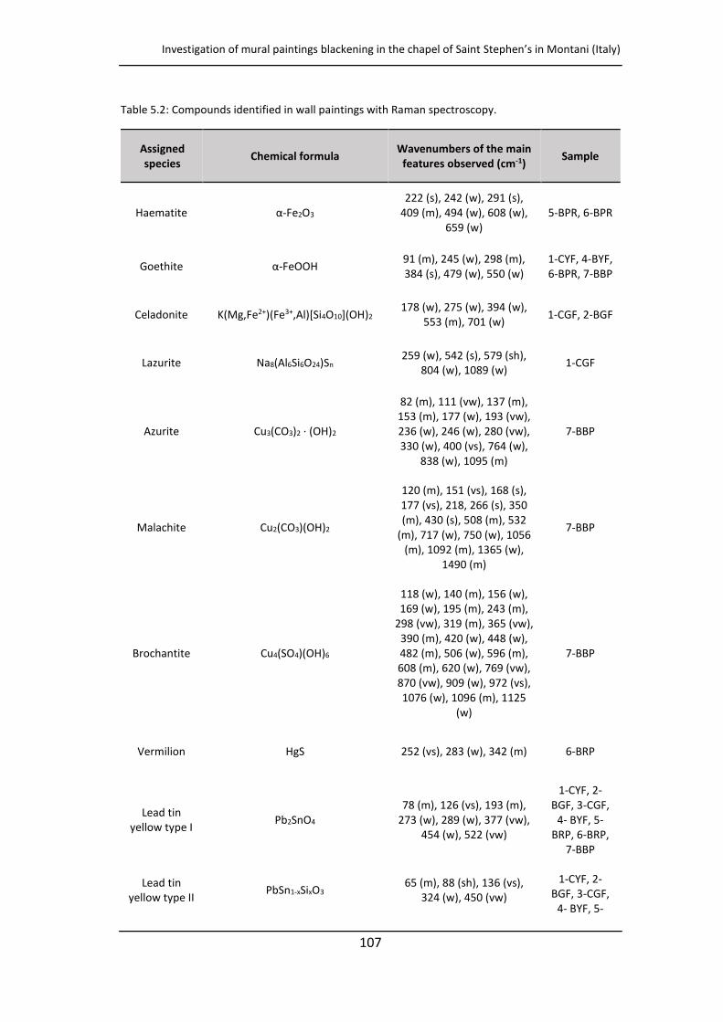
Investigation of mural paintings blackening in the chapel of Saint Stephen s in Montani (Italy)
107
Table 5.2: Compounds identified in wall paintings with Raman spectroscopy.
Assigned
species Chemical formula
Wavenumbers of the main
features observed (cm-1) Sample
Haematite α-Fe2O3 222 (s), 242 (w), 291 (s),
409 (m), 494 (w), 608 (w), 659 (w)
5-BPR, 6-BPR
Goethite α-FeOOH 91 (m), 245 (w), 298 (m), 384 (s), 479 (w), 550 (w)
1-CYF, 4-BYF, 6-BPR, 7-BBP
Celadonite K(Mg,Fe2+)(Fe3+,Al)[Si4O10](OH)2 178 (w), 275 (w), 394 (w),
553 (m), 701 (w) 1-CGF, 2-BGF
Lazurite Na8(Al6Si6O24)Sn 259 (w), 542 (s), 579 (sh),
804 (w), 1089 (w) 1-CGF
Azurite Cu3(CO3)2 · (OH)2
82 (m), 111 (vw), 137 (m), 153 (m), 177 (w), 193 (vw), 236 (w), 246 (w), 280 (vw), 330 (w), 400 (vs), 764 (w),
838 (w), 1095 (m)
7-BBP
Malachite Cu2(CO3)(OH)2
120 (m), 151 (vs), 168 (s), 177 (vs), 218, 266 (s), 350 (m), 430 (s), 508 (m), 532
(m), 717 (w), 750 (w), 1056 (m), 1092 (m), 1365 (w),
1490 (m)
7-BBP
Brochantite Cu4(SO4)(OH)6
118 (w), 140 (m), 156 (w), 169 (w), 195 (m), 243 (m),
298 (vw), 319 (m), 365 (vw), 390 (m), 420 (w), 448 (w), 482 (m), 506 (w), 596 (m), 608 (m), 620 (w), 769 (vw), 870 (vw), 909 (w), 972 (vs), 1076 (w), 1096 (m), 1125
(w)
7-BBP
Vermilion HgS 252 (vs), 283 (w), 342 (m) 6-BRP
Lead tin yellow type I
Pb2SnO4 78 (m), 126 (vs), 193 (m),
273 (w), 289 (w), 377 (vw), 454 (w), 522 (vw)
1-CYF, 2- BGF, 3-CGF,
4- BYF, 5-BRP, 6-BRP,
7-BBP
Lead tin yellow type II
PbSn1-xSixO3 65 (m), 88 (sh), 136 (vs),
324 (w), 450 (vw)
1-CYF, 2- BGF, 3-CGF,
4- BYF, 5-
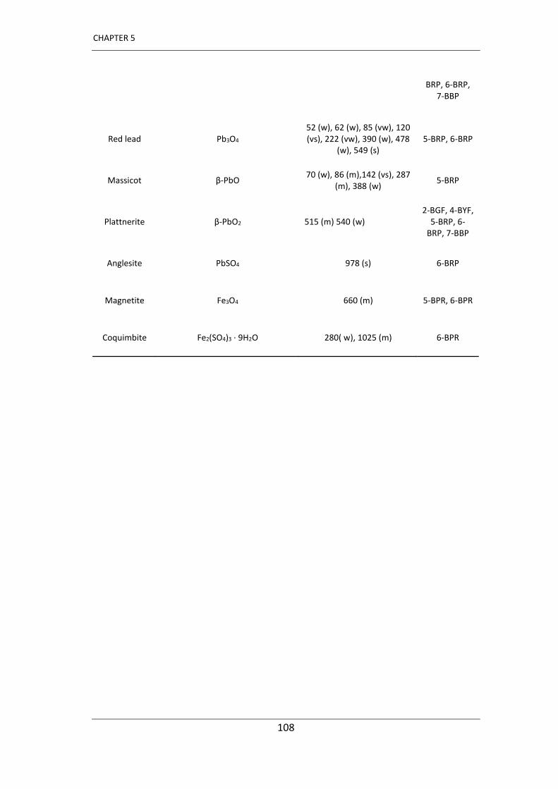
CHAPTER 5
108
BRP, 6-BRP, 7-BBP
Red lead Pb3O4 52 (w), 62 (w), 85 (vw), 120 (vs), 222 (vw), 390 (w), 478
(w), 549 (s) 5-BRP, 6-BRP
Massicot β-PbO 70 (w), 86 (m),142 (vs), 287
(m), 388 (w) 5-BRP
Plattnerite β-PbO2 515 (m) 540 (w) 2-BGF, 4-BYF,
5-BRP, 6-BRP, 7-BBP
Anglesite PbSO4 978 (s) 6-BRP
Magnetite Fe3O4 660 (m) 5-BPR, 6-BPR
Coquimbite Fe2(SO4)3 · 9H2O 280( w), 1025 (m) 6-BPR

Investigation of mural paintings blackening in the chapel of Saint Stephen s in Montani (Italy)
109
References
[1] K. Castro, A. Sarmiento, I. Martínez-Arkarazo, J. M. Madariaga, L. A. Fernández (2008) Green Copper Pigments Biodegradation in Cultural Heritage: From Malachite to Moolooite, Thermodynamic Modeling, X-ray Fluorescence, and Raman Evidence, Analytical Chemistry, 80, 4103-4110.
[2] K. Keune, J. J. Boon (2005) Analytical Imaging Studies Clarifying the Process of the Darkening of Vermilion in Paintings, Analytical Chemistry, 77, 4742-4750.
[3] M. Maguregui, U. Knuutinen, I. Martínez-Arkarazo, K. Castro, J. M. Madariaga (2011) Thermodynamic and Spectroscopic Speciation to Explain the Blackening Process of Hematite Formed by Atmospheric SO2 Impact: The Case of Marcus Lucretius House (Pompeii), Analytical Chemistry, 83, 3319-3326.
[4] E. Mattei, G. de Vivo, A. De Santis, C. Gaetani, C. Pelosi, U. Santamaria (2008) Raman spectroscopic analysis of azurite blackening, Journal of Raman Spectroscopy, 39, 302-306.
[5] V. S. F. Muralha, C. Miguel, M. João Melo (2012) Mi o‐Ra a stud of Medie al Cistercian 12–13th century manuscripts: Santa Maria de Alcobaça, Portugal, Journal
of Raman Spectroscopy, 43, 1737-1746.
[6] D. Hradil, J. Hradilo , E. Koč, S. Š a o , P. Bezdička, J. Mař Íková-Kubková (2013) U i ue P e‐Ro a es ue u als i Kostoľa pod T í ečo , Slo akia: the pai ti g technique and causes of damage, Archaeometry, 55, 691-706.
[7] M. Vagnini, R. Vivani, E. Viscuso, M. Favazza, B.G. Brunetti, A. Sgamellotti, C. Miliani (2018) Investigation on the process of lead white blackening by Raman spe t os op , XRD a d othe ethods: Stud of Ci a ue s pai ti gs i Assisi, Vibrational Spectroscopy, 98, 41-49.
[8] S. Daniilia, S. Sotiropoulou, D. Bikiaris, C. Salpistis, G. Karagiannis, Y. Chryssoulakis, B. A. Priced, J. H. Ca lso e Pa seli os B za ti e all pai ti gs i the P otato Church, Mount Athos, Greece: a technical examination, Journal of Cultural Heritage, 1, 91–110.
[9] L. de Ferri, F. Mazzini, D, Vallotto, G. Pojana (2017) In situ non-invasive characterization of pigments and alteration products on the masonry altar of S. Maria ad Undas (Idro, Italy), Archaeological and Anthropological Sciences, 1-17.
[10] C. Cennini, Il libro dell’arte, F. Frezzato, Neri Pozza Editore, 2009.
[11] S. Giovannoni, M. Matteini, A. Moles (1990) Studies and developments concerning the problem of altered lead pigments in wall painting, Studies in
Conservation, 35, 21-25.
[12] S. Aze, J. M. Vallet, M. Pomey, A. Baronnet, O. Grauby (2007) Red lead darkening in wall paintings: natural ageing of experimental wall paintings versus artificial ageing tests, European Journal of Mineralogy, 19, 883-890.
CHAPTER 5
110
[13] G. D. Smith, A. Derbyshire, R.J. H. Clark (2002) In Situ Spectroscopic Detection of Lead Sulphide on a Blackened Manuscript Illumination by Raman Microscopy, Studies
in Conservation, 47, 250-256.
[14] C. L. Hoevel (1985) A Study of the Discoloration Products Found in White Lead Paint Films, The American Institute for Conservation: Book and Paper Group Annual, Vol. 4.
[15] G. D. Smith, R. J.H. Clark (2002) The role of H2S in pigment blackening, Journal of
Cultural Heritage, 3, 101-105.
[16] H. G. M. Edwards, D. W. Farwell, E. M. Newton, F. Rull (1999) Minium; FT-Raman non-destructive analysis applied to an historical controversy, Analyst, 124, 1323-1326.
[17] S. Bruni, F. Cariati, F. Casadio, V. Guglielmi (2001) Micro-Raman identification of the palette of a precious XVI century illuminated Persian codex, Journal of Cultural
Heritage, 4, 291-296.
[18] C. Pelosi, G. Agresti, M. Andaloro, P. Baraldi, P. Pogliani, U. Santamaria (2013) The rock hewn wall paintings in Cappadocia (Turkey). characterization of the constituent materials and a chronological overview, E-Preservation Science, 10, 99-108.
[19] P. Holakooei, A.H. Karimy (2015) Early Islamic pigments used at the Masjid-i Jame of Fahraj, Iran: a possible use of black plattnerite, Journal of Archaeological Science, 54, 217-227.
[20] M. Matteini, A. Moles (1979) A preliminary investigation of the unusual te h i ue of Leo a do s u al The last supper , Studies in Conservation, 24, 125-133.
[21] N. N. Brandt, N. L. Rebrikova, A. Yu. Chikishev (2009) Raman spectroscopy of the components of 18th-century icon painting, Moscow University Physics Bulletin, 64,
600-604.
[22] J.P. Petushkova, N. Lyalinova (1986) Microbiological degradation of lead-containing pigments in mural paintings, Studies in Conservation, 31, 65-69.
[23] F. Qingping, Z. Xiaojun, M. Xiaojun (1999) Effects of microbes on color changes of red lead in murals, The Journal of General and Applied Microbiology, 45, 85-88.
[24] E. Kotula o , P. Bezdička, D. Hradil, J. Hradilo , S. Š a o , T. G ga Degradation of lead-based pigments by salt solutions, Journal of Cultural Heritage, 10, 367-378.
[25] S. Sotiropoulou, S. Daniilia, C. Miliani, F. Rosi, L. Cartechini, D. Papanikola-Bakirtzis (2008) Microanalytical investigation of degradation issues in Byzantine wall paintings, Applied Physics A, 92, 143-150
[26] A. Domínguez-Vidal, M. J. de la Torre-López, R. Rubio-Domenec, M. J. Ayora-Cañada (2012) In situ noninvasive Raman microspectroscopic investigation of polychrome plasterworks in the Alhambra, Analyst, 137, 5763-5769.
Investigation of mural paintings blackening in the chapel of Saint Stephen s in Montani (Italy)
111
[27] M. San Andrés, J.M. De la Roja, S.D. Dornheim, V.G. Baonza, 2008, Litharge and massicot: thermal decomposition synthetic route for basic lead(II) carbonate and Raman microscopy analysis, In Castillejo et al. (eds) Lasers in the Conservation of
Artworks Proceedings of the International Conference Lacona VII, Madrid, Spain, 17-21 september 2007.
[28] S. Aze, J.M. Vallet, V. Detalle, O. Grauby, A. Baronnet (2008) Chromatic alterations of red lead pigments in artworks: a review, Phase Transitions, 81, 145-154.
[29] M. Matteini, A. Moles, S. Giovannoni, Scientific Methodologies Applied to Works of Art, Florence 2-5 May, 1984, 113.
[30] M. Matteini, Ossidazione della biacca in pitture murali- Metodi proposti per la riconversione del pigmento ossidato nelle pitture di A. Baldovinetti della chiesa di S. Miniato (Firenze), In Atti del Convegno sul Restauro delle Opere d'Arte, Florence, 2-6 November, 1976, 257.
[31] M. Matteini, A. Moles, The reconversion of oxidized white lead in mural paintings: a control after a five-year period, ICOM Committee for Conservation, 6th Triennial Meeting, Ottawa, 1981 81/15/1.
[32] M. Koller, H. Leitner, H. Paschinger (1990) Reconversion of altered lead pigments in alpine mural paintings, Studies in Conservation, 35, 15-20.
[33] M. R. McFarland (1997) The whitening effects of peroxide gels on darkened lead white paint, The book and Paper Group Annual, Vol. 16.
[34] S. M. Lussier (2006) An Examination of Lead White Discoloration and the Impact of Treatment on Paper Artifacts: A Summary of Experimental Testing, The Book and
Paper Group Annual, Vol. 25, 9-12.
[35] S. M. Lussier and G. D. Smith (2007) A review of the phenomenon of lead white darkening and its conversion treatment, Studies in Conservation, 8, 41-53.
[36] L. Burgio, R. J. H. Clark, S. Firth (2001) Raman spectroscopy as a means for the identification of plattnerite (PbO2), of lead pigments and of their degradation products, Analyst, 126, 222-227.
[37] P. Pouli, D. C. Emmony, C. E. Madden, I. Sutherland (2003) Studies towards a thorough understanding of the laser-induced discoloration mechanisms of medieval pigments, Journal of Cultural Heritage, 4, 271-275.
[38] R. Bordalo, P. J. Morais, H. Gouveia, C. Young (2006) Laser Cleaning of Easle Paintings: an overview, Laser Chemistry, Article ID 90279, 9 pages http://dx.doi.org/10.1155/2006/90279.
[39] C. Andalò, M. Bicchieri, P. Bocchini, G. Casu, G.C. Galletti, P. A. Mandò, M. Na do e, A. Sodo, M. Plossi Zappal The eautiful T io fo d A o e attributed to Botticelli: a chemical characterisation by proton-induced X-ray emission and micro-Raman spectroscopy Analytica Chimica Acta, 429, 279-286.

CHAPTER 5
112
[40] G. D. Smith, L. Burgio, S. Firth, R. J. H. Clark (2001) Laser-induced degradation of lead pig e ts ith efe e e to Botti elli s T io fo d A o e, Analytica Chimica Acta, 440, 185-188.
[41] A. De Santis, E. Mattei, C. Pelosi (2007) Mi o‐Ra a a d st atig aphi studies of the pai ti gs o the Ce alo odel usi al i st u e t A.D. a d lase ‐induced degradation of the detected pigments, Journal of Raman Spectroscopy, 38, 1368-1378.
[42] S. Daniilia, E. Minopoulou, Fr. D. Demosthenous, G. Karagiannis (2008) A comparative study of wall paintings at the Cypriot monastery of Christ Antiphonitis: one artist or two? Journal of Archeological Science, 35, 1695 1707.
[43] F. Ospitali, D. Bersani, G. Di Lonardo, P. P. Lottici (2008) G ee ea ths : vibrational and elemental characterization of glauconites, celadonites and historical pigments Journal of Raman Spectroscopy, 39, 1066-1073.
[44] J. Bagdze ičie ė, G. Niau a, E. Ga škaitė, J. Se aitie ė, J. Lukšė ie ė, S. Tautkus (2011) Spectroscopic analysis of lead tin yellow pigment in medieval necklace beads f o Ke a ė-Kri eikiškės e ete i Lithua ia, Chemija, 22, 216-222.
[45] D.L.A. de Faria, F.N. Lopes (2007) Heated goethite and natural hematite: Can Raman spectroscopy be used to differentiate them?, Vibrational Spectroscopy, 45, 117-121.
[46] M. Bicchieri, M. Nardone, P.A. Russo, A. Sodo, M. Corsi, G. Cristoforetti, V. Palleschi, A. Salvetti, E. Tognoni (2001) Characterization of azurite and lazurite based pigments by laser induced breakdown spectroscopy and micro-Raman spectroscopy, Spectrochimica Acta B, 56, 915-922.
[47] M. Gutman, M. Lesar-Kikelj, A. Mlade o ič, V. Čo al-Sed ak, A. K iž a , S. Kramard (2014) Raman microspectroscopic analysis of pigments of the Gothic wall painting from the Dominican Monastery in Ptuj (Slovenia), Journal of Raman
Spectroscopy, 45, 1103-1109.
[48] M. Aceto, G. Gatti, A. Agostino, G. Fenoglio, V. Giordano, M. Varetto, G. Castagner (2012) The mural paintings of Ala di Stura (Piedmont, Italy): a hidden treasure investigated, Journal of Raman Spectroscopy, 43, 1754-1760.
[49] H. G. M. Edwards, D. W. Farwell, S. Rozenberg (1999) Raman spectroscopic study of red pigment and fresco fragments from King Herod's Palace at Jericho, Journal of
Raman Spectroscopy, 30, 361-366.
[50] I. Aliatis, D. Bersani, E. Campani, A. Casoli, P. P. Lottici, S. Mantovana, I.G. Marino (2010) Pigments used in Roman wall paintings in the Vesuvian area, Journal of Raman
Spectroscopy, 41, 1537-1542.
[51] P. Vandenabeele, B. Wehling, L. Moens, B. Dekeyzer, B. Cardon, A. von Bohlen, R. Klockenhämper (1999) Pigment investigation of a late-medieval manuscript with total reflection X-ray fluorescence and micro-Raman spectroscopy, Analyst, 124, 169-172.
Investigation of mural paintings blackening in the chapel of Saint Stephen s in Montani (Italy)
113
[52] S. Daniilia, E. Minopoulou (2009) A study of smalt and red lead discolouration in Antiphonitis wall paintings in Cyprus, Applied Physics A, 96, 701-711.
[53] A. Lluveras, S. Boularand, A. Andreotti,·M. Vendrell-Saz (2010) Degradation of azurite in mural paintings: distribution of copper carbonate, chlorides and oxalates by SRFTIR, Applied Physics A, 99, 363-375.
[54] M. P. Merrifield, Original Treatises on the Arts of Painting, II, London 1849.
[55] C. Pelosi, G. Agresti, U. Santamaria, E. Mattei (2010) Artificial yellow pigments: production and characterization through spectroscopic methods of analysis, E-
Preservation Science, 7, 108-115.
[56] M. Maguregui, U. Knuutinen, K. Castro, J. M. Madariaga (2010) Raman spectroscopy as a tool to diagnose the impact and conservation state of Pompeian second and fourth style wall paintings exposed to diverse environments (House of Marcus Lucretius), Journal of Raman Spectroscopy, 41, 1400-1409.
[57] M. Maguregui, K. Castro, H. Morillas, J. Trebolazabala, U. Knuutinen, R. Wiesinger, M. Schreinerd, J.M. Madariaga (2014) Multianalytical approach to explain the darkening process of hematite pigment in paintings from ancient Pompeii after accelerated weathering experiments, Analytical Methods, 6, 372-378.
[58] O. Gómez-Laserna, I. Arrizabalaga, N. Prieto-Taboada, M A. Olazabal, G. Arana, J. M. Madariaga (2015) In situ DRIFT, Raman, and XRF implementation in a multianalytical methodology to diagnose the impact suffered by built heritage in urban atmospheres, Analytical and Bioanalytical Chemistry, 407, 5635-5647.
[59] V. Hayez, J. Guillaume, A. Hubin, H. Terryn (2004) Mi o‐Ra a spe t os op fo the study of corrosion products on copper alloys: setting up of a reference database and studying works of art, Journal of Raman Spectroscopy, 35, 732-738.
[60] R. Mazzeo, P. Baraldi, R. Lujan C. Fagnano (2004) Characterization of mural painting pigments from the Thubchen Lakhang temple in Lo Manthang, Nepal, Journal of Raman Spectroscopy, 35, 678-685.
[61] D. Bersani, P. P. Lottici, G. Antonioli, E. Campani, A. Casoli, C. Violante (2004) Pigments and binders in the wall paintings of Santa Maria della Steccata in Parma (Italy): the ultimate technique of Parmigianino, Journal of Raman Spectroscopy, 35, 694-703.
[62] S. Valadas, R. V. Freire, A. Cardoso, J. Mirão, C. B. Dias, P. Vandenabeele, A. Candeias (2015) On the Use of the Unusual Green Pigment Brochantite (Cu4(SO4)(OH)6) in the 16th-Century Portuguese-Flemish Paintings Attributed to The Master Frei Carlos Workshop, Microscopy and Microanalysis, 21, 518-525.

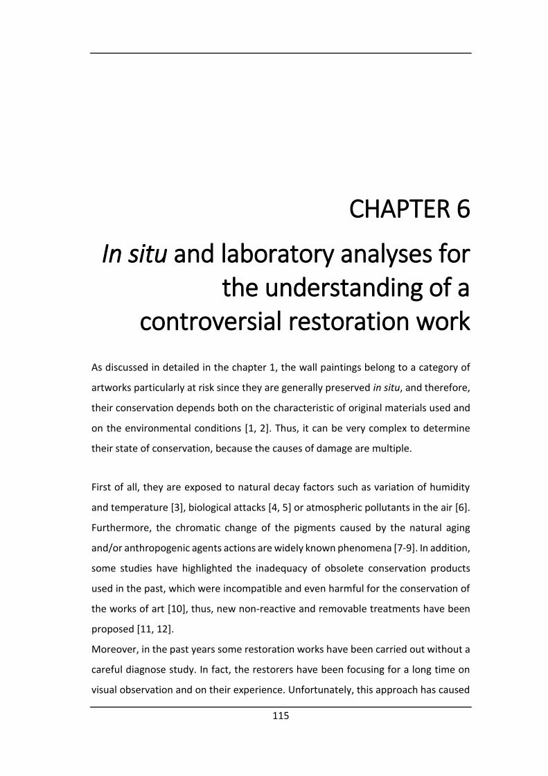
115
CHAPTER 6
In situ and laboratory analyses for
the understanding of a
controversial restoration work
As discussed in detailed in the chapter 1, the wall paintings belong to a category of
artworks particularly at risk since they are generally preserved in situ, and therefore,
their conservation depends both on the characteristic of original materials used and
on the environmental conditions [1, 2]. Thus, it can be very complex to determine
their state of conservation, because the causes of damage are multiple.
First of all, they are exposed to natural decay factors such as variation of humidity
and temperature [3], biological attacks [4, 5] or atmospheric pollutants in the air [6].
Furthermore, the chromatic change of the pigments caused by the natural aging
and/or anthropogenic agents actions are widely known phenomena [7-9]. In addition,
some studies have highlighted the inadequacy of obsolete conservation products
used in the past, which were incompatible and even harmful for the conservation of
the works of art [10], thus, new non-reactive and removable treatments have been
proposed [11, 12].
Moreover, in the past years some restoration works have been carried out without a
careful diagnose study. In fact, the restorers have been focusing for a long time on
visual observation and on their experience. Unfortunately, this approach has caused

CHAPTER 6
116
incorrect diagnoses and, accordingly, the application of unsuitable conservation
procedures to the artworks [13]. This is the reason why due to the complexity in the
conservation of wall paintings, integrated analytical approaches have become an
esse tial step for the ide tifi atio of the degradatio ’s auses and, especially, for
the election of the conservation treatments required [14].
This chapter presents the results of a multianalytical methodology for the study of
wall paintings, based on the use of Raman and X-ray fluorescence spectroscopy in
situ, SEM-EDS microscopy and Raman spectroscopy laboratory measurements. The
paintings belong to the late Gothic period and they are preserved in the Saint
Stephen’s hur h in Ribera de Valderejo (Álava, Basque Country). This research was
focused on characterizing the plaster composition, the colour palette used by the
artists and the materials employed during the recent restoration works.
6.1 In situ analyses
Portable Raman and X-ray fluorescence spectroscopy were selected to perform the
in situ study of the chromatic palette and to identify the original and new pigments
employed in the past restoration works. As summarise in the section 3.1.1, since the
paintings located in the apse wall are characterized by a grey colour, the analyses
were conducted mainly in those areas, in order to verify any degradation process (see
Figure 6.1).
The Raman analysis carried out in deteriorated areas, in which the paint layer is lost,
showed that the original preparation mortar (arriccio, used to make the wall flat) is
composed of a mixture of calcite and siliceous material. The acquired spectra showed
the features of calcium carbonate (CaCO3, Raman bands: 1086, 712, 282 and 156 cm-
1) (Figure 6.2a) and quartz (SiO2, Raman bands: 206, 262, 356, 465 cm-1) (Figure 6.2b).
Unlike other studied churches in the Basque territory, gypsum was not found in this
mortar [15, 16].

In situ and laboratory analyses for the understanding of a controversial restoration works
117
Figure 6.1: Representation of Original Sin in the apse area in which are visible grey areas (pointed by arrows) and green areas (pointed by circles).
Figure 6.2: Raman spectra acquired in the preparation mortar give evidence of calcite (a) and quartz with the contribution of calcite (b).
Raman analyses were also carried out in order to identify the composition of the
white layer of plaster that still covers the majority of the nave and the west wall. This
layer was applied to cover the deteriorated paintings in the past. It is composed of
two different sub-layers. The Raman spectra acquired in the exterior part showed the
well-defined peaks of gypsum (CaSO4·2H2O, 1008 and 415 cm-1). The part underneath
has a heterogeneous granulometry and a reddish colour. The Raman analyses
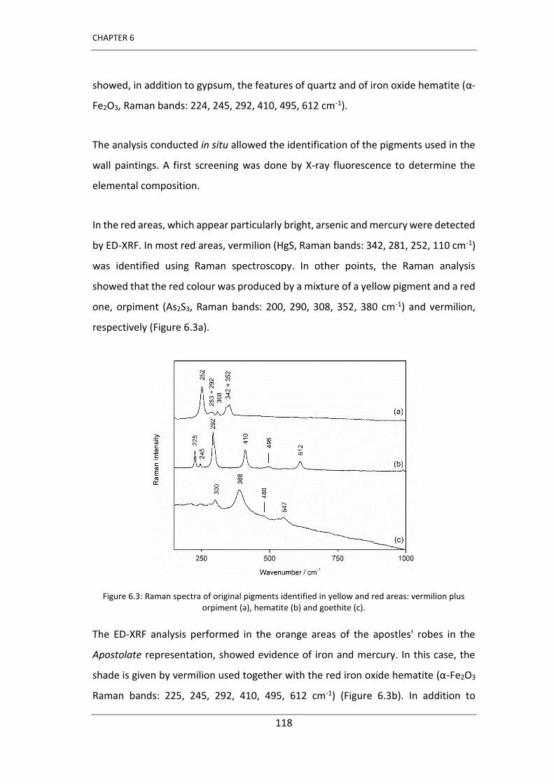
CHAPTER 6
118
showed, in addition to gypsum, the features of quartz and of iron oxide hematite (α-
Fe2O3, Raman bands: 224, 245, 292, 410, 495, 612 cm-1).
The analysis conducted in situ allowed the identification of the pigments used in the
wall paintings. A first screening was done by X-ray fluorescence to determine the
elemental composition.
In the red areas, which appear particularly bright, arsenic and mercury were detected
by ED-XRF. In most red areas, vermilion (HgS, Raman bands: 342, 281, 252, 110 cm-1)
was identified using Raman spectroscopy. In other points, the Raman analysis
showed that the red colour was produced by a mixture of a yellow pigment and a red
one, orpiment (As2S3, Raman bands: 200, 290, 308, 352, 380 cm-1) and vermilion,
respectively (Figure 6.3a).
Figure 6.3: Raman spectra of original pigments identified in yellow and red areas: vermilion plus orpiment (a), hematite (b) and goethite (c).
The ED-XRF analysis performed in the orange areas of the apostles' robes in the
Apostolate representation, showed evidence of iron and mercury. In this case, the
shade is gi e er ilio used together ith the red iro o ide he atite α-Fe2O3
Raman bands: 225, 245, 292, 410, 495, 612 cm-1) (Figure 6.3b). In addition to
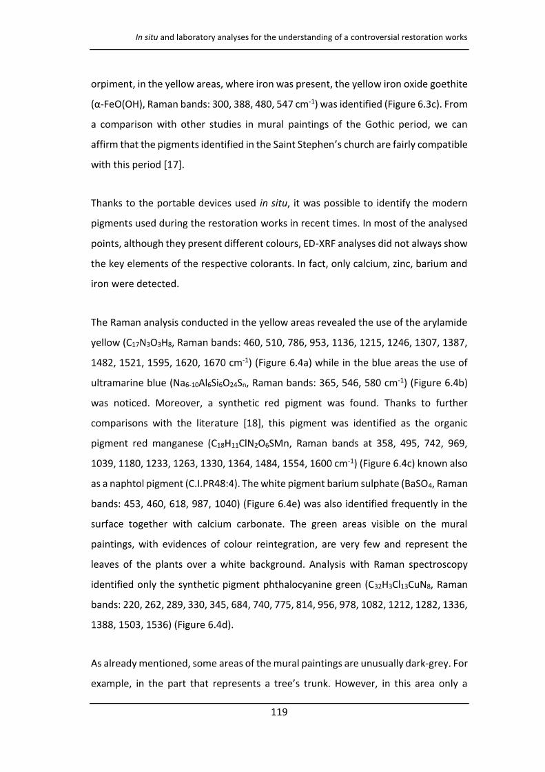
In situ and laboratory analyses for the understanding of a controversial restoration works
119
orpiment, in the yellow areas, where iron was present, the yellow iron oxide goethite
α-FeO(OH), Raman bands: 300, 388, 480, 547 cm-1) was identified (Figure 6.3c). From
a comparison with other studies in mural paintings of the Gothic period, we can
affirm that the pigments identified in the Saint Stephen’s hurch are fairly compatible
with this period [17].
Thanks to the portable devices used in situ, it was possible to identify the modern
pigments used during the restoration works in recent times. In most of the analysed
points, although they present different colours, ED-XRF analyses did not always show
the key elements of the respective colorants. In fact, only calcium, zinc, barium and
iron were detected.
The Raman analysis conducted in the yellow areas revealed the use of the arylamide
yellow (C17N3O3H8, Raman bands: 460, 510, 786, 953, 1136, 1215, 1246, 1307, 1387,
1482, 1521, 1595, 1620, 1670 cm-1) (Figure 6.4a) while in the blue areas the use of
ultramarine blue (Na6-10Al6Si6O24Sn, Raman bands: 365, 546, 580 cm-1) (Figure 6.4b)
was noticed. Moreover, a synthetic red pigment was found. Thanks to further
comparisons with the literature [18], this pigment was identified as the organic
pigment red manganese (C18H11ClN2O6SMn, Raman bands at 358, 495, 742, 969,
1039, 1180, 1233, 1263, 1330, 1364, 1484, 1554, 1600 cm-1) (Figure 6.4c) known also
as a naphtol pigment (C.I.PR48:4). The white pigment barium sulphate (BaSO4, Raman
bands: 453, 460, 618, 987, 1040) (Figure 6.4e) was also identified frequently in the
surface together with calcium carbonate. The green areas visible on the mural
paintings, with evidences of colour reintegration, are very few and represent the
leaves of the plants over a white background. Analysis with Raman spectroscopy
identified only the synthetic pigment phthalocyanine green (C32H3Cl13CuN8, Raman
bands: 220, 262, 289, 330, 345, 684, 740, 775, 814, 956, 978, 1082, 1212, 1282, 1336,
1388, 1503, 1536) (Figure 6.4d).
As already mentioned, some areas of the mural paintings are unusually dark-grey. For
e a ple, i the part that represe ts a tree’s tru k. Ho e er, i this area o l a
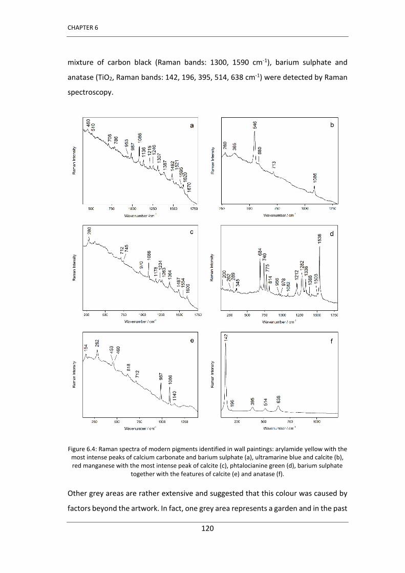
CHAPTER 6
120
mixture of carbon black (Raman bands: 1300, 1590 cm-1), barium sulphate and
anatase (TiO2, Raman bands: 142, 196, 395, 514, 638 cm-1) were detected by Raman
spectroscopy.
Figure 6.4: Raman spectra of modern pigments identified in wall paintings: arylamide yellow with the most intense peaks of calcium carbonate and barium sulphate (a), ultramarine blue and calcite (b), red manganese with the most intense peak of calcite (c), phtalocianine green (d), barium sulphate
together with the features of calcite (e) and anatase (f).
Other grey areas are rather extensive and suggested that this colour was caused by
factors beyond the artwork. In fact, one grey area represents a garden and in the past
In situ and laboratory analyses for the understanding of a controversial restoration works
121
it could have been green in colour. Therefore, special attention was given to the study
of these areas in order to understand if the tone was an intentional act or if it was
caused by a process of degradation of the raw materials used in the wall paintings.
Raman analyses carried out in this grey area, representing grass, in addition to the
features of calcite, provided evidence of a mixture of carbon black and barium
sulphate. Moreover, the modern white pigment anatase was also identified (Figure
6.4f). There were not recorded spectra of any antique green pigment or blue or
yellow pigments that may have been mixed to obtain a green colour [19]. During the
analysis in situ some green areas painted over this grey background were discovered.
However, these areas, representing leaves, were painted with the modern pigment
phtalocyanine green; thus, we must assume that this green area is a reintegration.
Unfortunately, there were still doubts about the origin of the possible blackening,
and analyses in the laboratory were fundamental. Some samples were taken and
submitted to the laboratory. Besides, large amounts of arsenic were detected by X-
ray fluorescence in different points of the grey areas (Figure 6.5), even with arsenic
signals higher than in the areas where orpiment was found by Raman spectroscopy.
Figure 6.5: ED-XRF spectrum collected in grey areas.
CHAPTER 6
122
Thus, despite the evident arsenic signals in these areas are evident, as pointed out by
the result obtained in situ, it seemed to be scarcely plausible that the grey colour was
caused by a blackening of orpiment, the only arsenic containing compound found in
the wall paintings, since this pigment was detected only in red-orange areas, where
the Raman analysis allowed to identify the orpiment in a perfect state of
conservation. Moreover, it seemed that the grey colour was not due to any
blackening process.
6.2 Laboratory analyses
According to the results obtained with the portable equipment, only the areas of
greatest interest were sampled. In order to know the origin of the detected arsenic,
to investigate the cause of grey coloration of the wall paintings and the absence of
original green pigments, some micro samples were collected in the grey areas. They
were embedded in resin and then they were analysed as cross sections using SEM-
EDS microscopes and Raman spectroscopy in the laboratory. Moreover, another
sample was taken in the green areas to verify the presence of original green pigments
and it was analysed also as a cross section. In this way it was possible to study the
stratigraphy to better distinguish the original green paint layer from chromatic
retouching belonging to the modern times.
First of all, the grey samples were observed at the lowest magnification (200 x) using
the scanning electron microscope. The image obtained allowed us to distinguish a
complex stratigraph . The ele e ts’ distri utio i Figure 6.6 shows the composition
of a grey sample in cross section.
The elemental maps show 3 layers (Figure 6.6). The mortar is characterised by the
presence of calcium, carbon and silicon (not shown). The original paint layer presents
a heterogeneous composition with evident grains of carbon in a matrix of sulphur
and calcium. Over this, there is another layer with heterogeneous composition in
which some grains of various sizes can be observed. The elemental maps show that

In situ and laboratory analyses for the understanding of a controversial restoration works
123
the grains are composed by magnesium and arsenic. Together with Mg and As, there
is a very thin layer containing Zn, S, Ba and Ti.
In addition, the analyses with SEM-EDS confirmed the presence of zinc in the outer
layer together whit titanium and barium, which was also detected by ED-XRF during
in situ analysis. Although the Raman spectrum of zinc sulphide was not collected (it
has a very weak Raman signal), based on the SEM-EDS cross-sections results, it is
possible to assume that the lithopone (ZnS · BaSO4), was used in a mixture with
anatase since they coexist in the same layer. There are examples in the literature of
the use of mixtures of lithopone and anatase in the modern age [20]. This thin
painting layer applied in modern times is visible in all the analysed samples collected
from the grey areas. Since the documentation regarding the past restoration works
is incomplete, we cannot report the reason for this choice. However, we can assume
that probably a white painting layer was applied by the restorers to attenuate the
original and real black colour of the paintings.
Figure 6.6: Optical microscope image (a) and secondary electron SEM image (b) obtained in a grey sample in cross-section. The elemental distribution maps show evidence of calcium, carbon and
silicon (not shown) in the mortar, of sulphur and calcium in the original painting layer together with large amounts of carbon, of the grains composed by arsenic and magnesium, and of zinc, barium and
titanium on the surface.

CHAPTER 6
124
To complete the results obtained by means of SEM-EDS analysis in cross-section,
further studies were carried out by Raman spectroscopy for the molecular
characterization of this area.
The Raman spectra collected in this layer gave evidence of the massive presence of
carbon black (Figure 6.7a) mixed with gypsum (CaSO4 · 2H2O, Raman bands: 180, 415,
492, 618, 670, 1008, 1135 cm-1) (Figure 6.7b). The observation of samples in the cross
section allowed us to distinguish in the original painting, characterized by large grains
in a sulphur matrix, small orange grains. The Raman spectra showed very defined
features of cinnabar in scarce orange grains (Figure 6.7c).
Figure 6.7: Raman spectra of pigments detected in the original paint from samples collected in grey areas: carbon black with the contribution of gypsum (a), gypsum alone (b) and cinnabar (c).
In addition, the samples observed as cross-sections under the microscope did not
show evidence of the use of a green pigment in the original painting layer, where no
signals of green pigments (or mixtures of yellow and blue pigments) were recorded
by Raman spectroscopy.
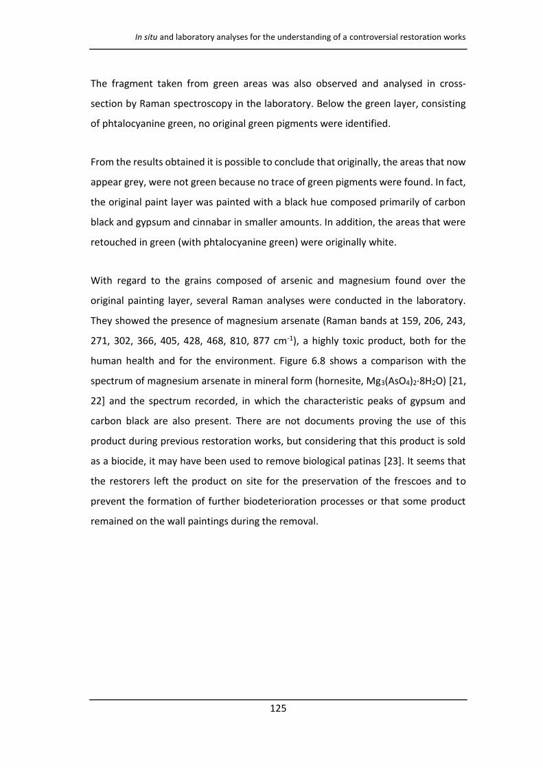
In situ and laboratory analyses for the understanding of a controversial restoration works
125
The fragment taken from green areas was also observed and analysed in cross-
section by Raman spectroscopy in the laboratory. Below the green layer, consisting
of phtalocyanine green, no original green pigments were identified.
From the results obtained it is possible to conclude that originally, the areas that now
appear grey, were not green because no trace of green pigments were found. In fact,
the original paint layer was painted with a black hue composed primarily of carbon
black and gypsum and cinnabar in smaller amounts. In addition, the areas that were
retouched in green (with phtalocyanine green) were originally white.
With regard to the grains composed of arsenic and magnesium found over the
original painting layer, several Raman analyses were conducted in the laboratory.
They showed the presence of magnesium arsenate (Raman bands at 159, 206, 243,
271, 302, 366, 405, 428, 468, 810, 877 cm-1), a highly toxic product, both for the
human health and for the environment. Figure 6.8 shows a comparison with the
spectrum of magnesium arsenate in mineral form (hornesite, Mg3(AsO4)2·8H2O) [21,
22] and the spectrum recorded, in which the characteristic peaks of gypsum and
carbon black are also present. There are not documents proving the use of this
product during previous restoration works, but considering that this product is sold
as a biocide, it may have been used to remove biological patinas [23]. It seems that
the restorers left the product on site for the preservation of the frescoes and to
prevent the formation of further biodeterioration processes or that some product
remained on the wall paintings during the removal.
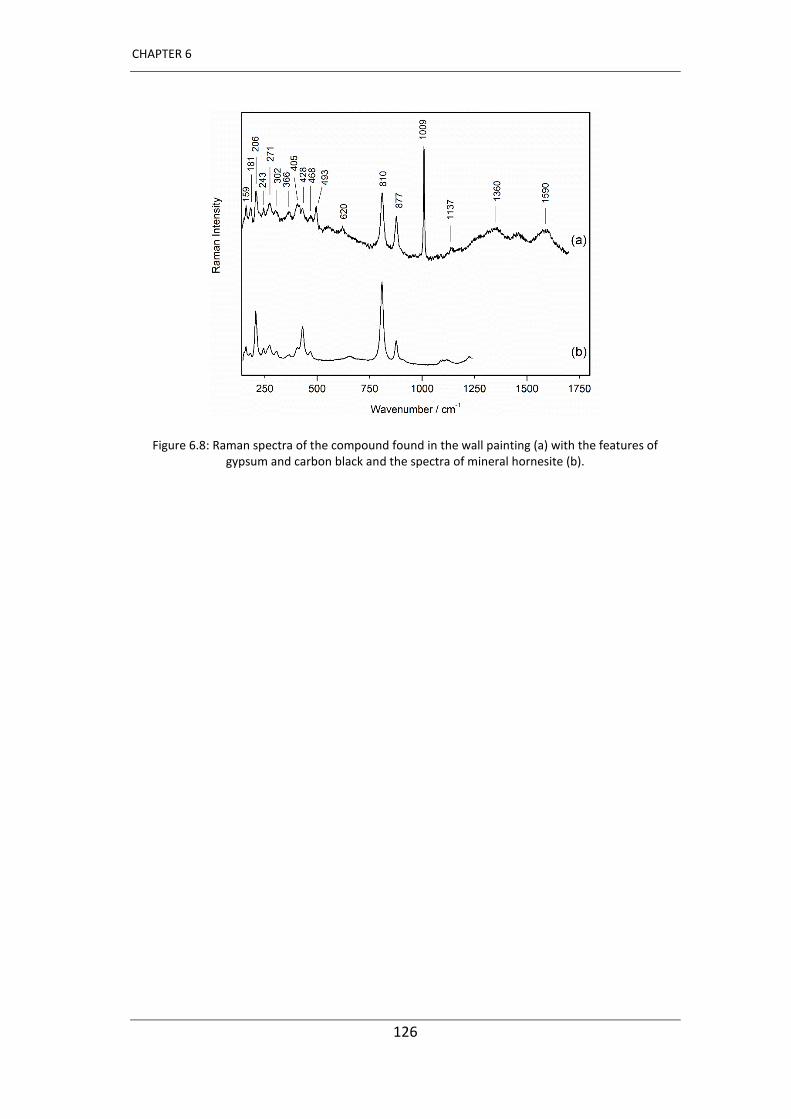
CHAPTER 6
126
Figure 6.8: Raman spectra of the compound found in the wall painting (a) with the features of gypsum and carbon black and the spectra of mineral hornesite (b).
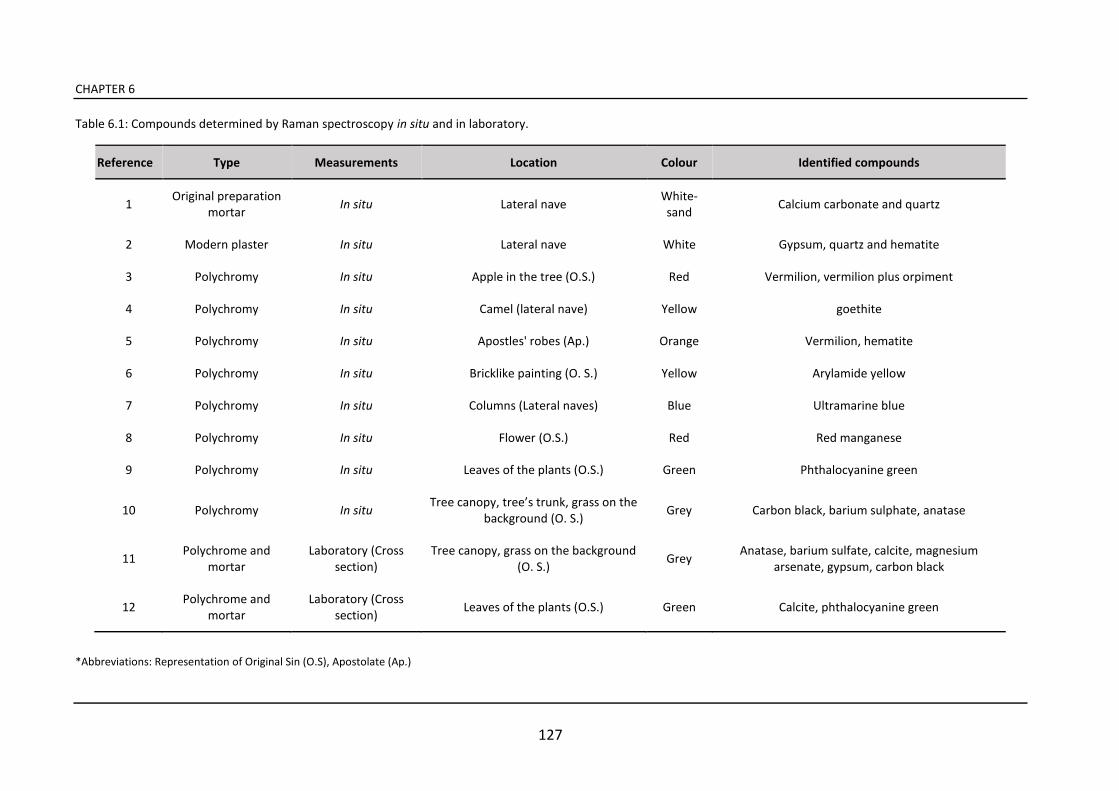
CHAPTER 6
127
Table 6.1: Compounds determined by Raman spectroscopy in situ and in laboratory.
Reference Type Measurements Location Colour Identified compounds
1 Original preparation
mortar In situ Lateral nave
White-
sand Calcium carbonate and quartz
2 Modern plaster In situ Lateral nave White Gypsum, quartz and hematite
3 Polychromy In situ Apple in the tree (O.S.) Red Vermilion, vermilion plus orpiment
4 Polychromy In situ Camel (lateral nave) Yellow goethite
5 Polychromy In situ Apostles' robes (Ap.) Orange Vermilion, hematite
6 Polychromy In situ Bricklike painting (O. S.) Yellow Arylamide yellow
7 Polychromy In situ Columns (Lateral naves) Blue Ultramarine blue
8 Polychromy In situ Flower (O.S.) Red Red manganese
9 Polychromy In situ Leaves of the plants (O.S.) Green Phthalocyanine green
10 Polychromy In situ Tree canopy, tree’s trunk, grass on the
background (O. S.) Grey Carbon black, barium sulphate, anatase
11 Polychrome and
mortar
Laboratory (Cross
section)
Tree canopy, grass on the background
(O. S.) Grey
Anatase, barium sulfate, calcite, magnesium
arsenate, gypsum, carbon black
12 Polychrome and
mortar
Laboratory (Cross
section) Leaves of the plants (O.S.) Green Calcite, phthalocyanine green
*Abbreviations: Representation of Original Sin (O.S), Apostolate (Ap.)
In situ and laboratory analyses for the understanding of a controversial restoration works
128
6.3 Final remarks
The results obtained confirm the reliability of the scientific approach for the
evaluation of the state of wall paintings preservation. However, it should be noted
that the simultaneous use of different elemental and molecular techniques allows a
complete study of the materials (see Table 6.1).
By means of spectroscopic techniques, X-ray fluorescence and Raman spectroscopy,
used in situ, it was possible to get a fairly complete evaluation of the original pigments
used by the artists. The pictorial palette is homogeneous in the different parts of the
church; hematite, goethite, vermilion, orpiment, gypsum and carbon black. Besides,
all the pigments used in restorations works carried out in the modern era, such as
arylamide yellow, phtalocyanine green, ultramarine blue, red manganese, barium
sulphate, carbon black, and anatase were identified.
In addition, the in situ analyses permitted the identification of a very interesting area,
from a diagnostic point of view, and a selective sampling was done to complete the
study in the laboratory. This allowed us to avoid collecting other useless samples from
the wall paintings. In this way, only some micro samples were collected and analysed
as cross-section.
The laboratory techniques showed that the presence of a grey-dark colour in the wall
paintings was caused neither by an external factor nor by a process of degradation of
raw materials but by a voluntary act of the painters. By means of Raman spectroscopy
measurements carried out on cross sections, we could verify that the original layer
was painted mainly with gypsum and carbon black and with a lesser amount of
vermilion. Thus, these parts of the wall paintings would have been originally dark-
grey in colour because no green pigment or mixture (yellow and blue pigments) was
ever identified. Probably, the lack of green pigments is due to an insufficient
availability of the raw materials such as the green earths, malachite, celadonite or
glauconite [24].
In situ and laboratory analyses for the understanding of a controversial restoration works
129
The only green colour found was phtalocyanine green, which was applied on the
surface during a past intervention. In this case, a scientific approach with diagnostic
instruments would have allowed to define the original appearance of mural paintings.
In fact, the restorers conducted the restoration work starting from a visual
observation and from a personal point of view assuming that the grass and the foliage
must have been painted in green. They probably thought about a blackening
phenomenon of the pigments, in particular about the degradation of the azurite into
black tenorite as reported in literature [25]. It may have been mixed with a yellow
pigment to obtain a green colour and probably for that reason the restorers used the
phtalocyanine green for colour reintegration. Furthermore, the choice of painting
some areas that originally were white with phtalocyanine green, as shown in the
laboratory analysis, is a further evidence of an arbitrary choice of the restorers which
is not compatible with the original appearance of the paintings.
In addition, the analyses in cross-section clarified an important aspect that was not
possible to solve in situ with the spectroscopic techniques. In fact, it was possible to
explain the presence of arsenic detected by the X-ray fluorescence analysis
conducted in situ. Thanks to the elemental maps obtained by SEM-EDS, and to the
molecular maps obtained by Raman spectroscopy, the use of the highly toxic
substance magnesium arsenate, that was not documented, was identified.
This seems particularly serious because it is a very hazardous material for the
environment and especially for human health. Thus, it was very important to
document the presence of such product, especially to make sure that further
restorations can be carried out safely and with appropriate precautions for the
restorers in the future.
Moreover, magnesium arsenate is a rather invasive product and the restorers did not
consider the possible damages that it could induct to the painted surfaces. Without
an analytical approach we could not have information since the restoration
documents at our disposal did not specify the substance used. For further restoration
CHAPTER 6
130
works, it is not only necessary to know the original materials but, above all, the
materials used in the modern era and, as we have verified, a careful study of the
materials can only be made with the use of complementary diagnostic techniques.
This work shows the importance of using scientific diagnostic techniques for the
study of cultural heritage, especially to provide guidelines to the restorers for an
appropriate intervention. In fact, the study of the original materials should be
indispensable before making chromatic reintegration to ensure that the choices are
compatible and respect the original appearance of the paintings. What we could
verify is that, certainly, in the church of Ribera de Valderejo no physico-chemical
analyses were carried out and, for this reason, inappropriate interventions were
conducted in the past years.
In situ and laboratory analyses for the understanding of a controversial restoration works
131
References
[1] M. Maguregui, U. Knuutinen, I. Martínez-Arkarazo, A. Giakoumaki, K. Castro, J. M. Madariaga (2012) Field Raman analysis to diagnose the conservation state of excavated walls and wall paintings in the archaeological site of Pompeii (Italy), Journal
of Raman Spectroscopy, 43, 1747-1753.
[2] S. R. Amato, D. Bersani, P. P. Lottici, P. Pogliani, C. Pelosi (2017) A Multi-Analytical Approach to the Study of the Mural Paintings in the Presbytery of Santa Maria Antiqua Al Foro Romano in Rome, Archeometry, 59, 1-15.
[3] M. Zarzo, A. Fernández-Navajas, F. J. García-Diego (2011) Long-Term Monitoring of Fresco Paintings in the Cathedral of Valencia (Spain) Through Humidity and Temperature Sensors in Various Locations for Preventive Conservation, Sensors 11, 8685-8710.
[4] L. Zucconi, M. Gagliardi, D. Isola, S. Onofri, M. C. Andaloro, C. Pelosi, P. Pogliani, L. Selbmann (2012) Biodeterioration agents dwelling in or on the wall paintings of the Hol Sa iour’s a e Vallera o, Ital , International Biodeterioration and
Biodegradation, 70, 40-46.
[5] M. Maguregui, U. Knuutinen, J. Trebolazabala, H. Morillas, K. Castro, I. Martínez-Arkarazo, J. M. Madariaga (2012) Use of in situ and confocal Raman spectroscopy to study the nature and distribution of carotenoids in brown patinas from a deteriorated wall painting in Marcus Lucretius House (Pompeii), Analytical and Bioanalytical
Chemistry, 402, 1529-1539.
[5] J. Watt, J. Tidblad, V. Kucera, R. Hamilton ed., The Effects of Air Pollution on Cultural Heritage, Springer US, 2009.
[7] M. Maguregui, U. Knuutinen, K. Castro, J. M. Madariaga (2010) Raman spectroscopy as a tool to diagnose the impact and conservation state of Pompeian second and fourth style wall paintings exposed to diverse environments (House of Marcus Lucretius), Journal of Raman Spectroscopy, 41, 1400-1409.
[8] L. Cartechini, C. Miliani, B. G. Brunetti, A. Sgamellotti, C. Altavilla, E. Ciliberto, F. D’acapito (2008) X-ray absorption investigations of copper resinate blackening in a XV century Italian painting, Applied Physics, 92, 243-250.
[9] S. Aze, J. M. Vallet, A. Baronnet, O. Grauby (2006) The fading of red lead pigment in wall paintings: tracking the physico-chemical transformations by means of complementary micro-analysis techniques, European Journal of Mineralogy, 18, 835-843.
[10] C. Milanesi, F. Baldi, S. Borin, L. Brusetti, F. Ciampolini, F. Iacopini, M. Cresti (2009) Deterioration of medieval painting in the chapel of the Holy Nail, Siena (Italy) partially treated with Paraloid B72, International Biodeterioration and
Biodegradation, 63, 844-850.
CHAPTER 6
132
[11] R. Giorgi, M. Baglioni, D. Berti, P. Baglioni (2010) New Methodologies for the Conservation of Cultural Heritage: Micellar Solutions, Microemulsions, and Hydroxide Nanoparticles, Accounts of Chemical Researc, 43, 695-704.
[12] N. Prieto-Taboada, C. Isca, I. Martínez-Arkarazo, A. Casoli, M. A. Olazabal, G. Arana, J. M. Madariaga (2014) The problem of sampling on built heritage: a preliminary study of a new non-invasive method, Environmental Science and
Pollution Research, 21, 12518-12529.
[13] M. Pérez-Alonso, K. Castro, M. Álvarez, J.M. Madariaga (2004) Scientific analysis ersus restorer’s e pertise for diag osis prior to a restoratio pro ess: the ase of
Santa Maria Church (Hermo, Asturias, North of Spain), Analytica Chimica Acta, 524, 379-389.
[14] H. G. M. Edwards, M. T. Domenech-Carbó, M. D. Hargreaves, A. Domenech-Carbó (2008) A Raman spectroscopic and combined analytical approach to the restoration of severely damaged frescoes: the Palomino project, Journal of Raman Spectroscopy, 39, 444-452.
[15] M. Irazola, M. Olivares, K. Castro, M. Maguregui, I. Martínez-Arkarazo, J. M. Madariaga (2012) In situ Raman spectroscopy analysis combined with Raman and SEM-EDS imaging to assess the conservation state of 16th century wall paintings, Journal of Raman Spectroscopy, 43, 1676-1684.
[16] M. Pérez-Alonso, K. Castro, J. M. Madariaga (2006) Investigation of degradation mechanisms by portable Raman spectroscopy and thermodynamic speciation: The wall painting of Santa María de Lemoniz (Basque Country, North of Spain), Analytica
Chimica Acta, 571, 121-128.
[17] M.L. Franquelo, A. Duran, L.K. Herrera, M.C. Jimenez de Haro, J.L. Perez-Rodriguez (2009) Comparison between micro-Raman and micro-FTIR spectroscopy techniques for the characterization of pigments from Southern Spain Cultural Heritage, Journal of Molecular Structure, 924-926, 404-412.
[18] W. Fremout, S. Saverwyns (2012) Identification of synthetic organic pigments: the role of a comprehensive digital Raman spectral library, Journal of Raman
Spectroscopy, 43, 1536-1544.
[19] M.C. Edreira, M.J. Feliu, C. Fernández-Lorenzo, J. Martín (2003) Spectroscopic analysis of roman wall paintings from Casa del Mitreo in Emerita Augusta, Mérida, Spain, Talanta, 59, 1117-1139.
[20] L. Boselli, S. Ciattini, M. Galeotti, M. R. Lanfranchi, C. Lofrumento, M. Picollo, A. Zoppi (2009) An unusual white pigment in La Verna Sanctuary frescoes: an analysis with micro-Raman, FTIR, XRD and UV-VIS-NIR FORS, E Preservation. Science, 6, 38-42.
[21] B Lafuente, R.T. Downs, H. Yang, N. Stone (2015) The power of databases: the RRUFF project. In: Highlights in Mineralogical Crystallography, T Armbruster and R M Danisi, eds. Berlin, Germany, W. De Gruyter, 1-30.
[22] R. L. Frost, W. Martens, P. A. Williams, J. T. Kloprogge (2003) Raman spectroscopic study of the vivianite arsenate minerals, Journal of Raman
Spectroscopy, 34, 751-759.
In situ and laboratory analyses for the understanding of a controversial restoration works
133
[23] C.T. Duval (1970) Chemical Relationships and Nomenclature of Pesticides–Acaricides, Molluscicides, Rodenticides, etc, PANS Pest Articles & News Summaries, 16, 451-469.
[24] I. Aliatis, D. Bersani, E. Campani, A. Casoli, P. P. Lottici, S. Mantovani, I.G. Marino, F. Ospitali (2009) Gree pig e ts of the Po peia artists’ palette, Spectrochimica.
Acta Part A, 73, 532-538.
[25] E. Mattei, G. de Vivo, A. De Santis, C. Gaetani, C. Pelosi, U. Santamaria (2008) Raman spectroscopic analysis of azurite blackening, Journal of Raman Spectroscopy, 39, 302-306.

135
CHAPTER 7
Study of natural impact on wall
paintings and building materials of
the abandoned church of Ribera
de Valderejo
The present chapter concerns the study of the conservation state of Saint Stephen s
church in the village of Ribera de Valderejo (Álava, Basque Country) and of the wall
paintings preserved inside of it. The town is located in a rural area and it is in a state
of abandonment, therefore, in these environmental conditions the preservation of
artworks is complex.
The interest for the presence of degradation processes in the church of Ribera de
Valderejo was born during the study of murals paintings, discussed in the chapter 6,
when the presence of accumulation of soluble salts on the surface of wall paintings
was noticed together with an extensive colonization of microorganism. Thus, in order
to identify the causes that provoked them, considering primarily its natural
environment, a deepen research only on the state of preservation of the building was
carried out.
CHAPTER 7
136
The diagnostic of the conservation state of the building heritage is a current topic
that has been discussed in several previous works with the aim to identify the
degradation processes and the mechanisms that provoked them [1, 2]. Certainly,
nowadays, a special attention is paid on damage caused by anthropogenic factors
due to the increase of contaminants in the atmosphere [3, 4]. In contrast, the
degradation of cultural heritage assets in rural areas does not depend primarily on
anthropogenic agents but also on their natural surrounding environment.
Independent of the area, rural or industrial, the artworks located in the open air are
difficult to preserve and the impact of environmental agents such as the variation of
the soil moisture, the presence of macro and microorganism, the variations of
temperatures and humidity, the presence of salt weathering and the biological
attacks can be harmful without a conservative strategy [5]. Thus, the identification of
the degradation processes described above is an essential step for the conservation
of cultural heritage in particular in order to suggest the guidelines for an appropriate
restoration work.
Over the last years, several diagnostic techniques have been successfully employed
for the evaluation of conservation state of building materials and pictorial surfaces,
especially thanks to the use of portable instruments due to their non-destructive and
non-invasive characteristics [6]. In particular Raman spectroscopy has demonstrated
its reliability for the characterization of secondary products found on historical
building materials thanks to its high spectral resolution [7].
For this aim, in this chapter, building materials and decay products were
characterized first using Raman spectroscopy and X-ray fluorescence portable
devices and subsequently, some collected samples were analysed by laboratory
techniques.

Study of natural impact on building materials in the abandoned church of Ribera de Valderejo
137
7.1 Results
The in situ analyses with portable devices (Raman spectroscopy, X-ray fluorescence)
allowed us the study of the secondary products found in wall paintings and in building
materials. On the other hand, Raman spectroscopy and X-ray diffraction were
employed in the laboratory for a complete characterization of materials, in particular
for secondary products like efflorescence salts, which were complex to analyse in the
form of efflorescence. The analysis was focus on the determination of their
composition and the factors that have caused them. Moreover, in order to make it
possible to distinguish the composition of the original materials from the degradation
products, the structural stone of the church and of the portal were also sampled.
7.1.1 Study of soluble and insoluble efflorescence salts
By using in situ Raman spectroscopy on mural painting surface it was possible to
identify different degradation products. Hydrated calcium oxalate weddellite
(CaC2O4·2H2O) was detected in some points of the original mortar. The most intense
peaks were visible at 869, 910 and 1474 cm-1 (Figure 7.1).
The presence of weddellite and the most stable form whewellite (CaC2O4·H2O), may
result from the concomitant biological activity of microorganisms, such as algae or
bacteria, and environmental conditions. The microorganism can excrete oxalic acid
(H2C2O4) that reacts with the carbonaceous materials in the wall paintings, such as
calcium carbonate, the main component of the plaster of the wall paintings [8].
In addition, the presence of nitrates was determined, both in the inner layers of the
plaster and in the pictorial surfaces. In this case, the presence of the single feature at
1050 cm-1 can be attributed to nitrocalcite (Ca(NO3)2·6H2O) and niter (KNO3) since
both have the main band in this position (the intense fluorescence did not allow us
to detect the bands of lower frequencies). However, both salts can be differentiated
due to their different FWHM (Full Wide at Half Maximum) values, FWHM=8 cm-1 for
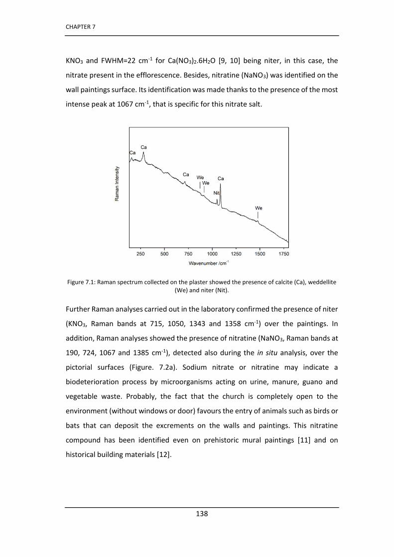
CHAPTER 7
138
KNO3 and FWHM=22 cm-1 for Ca(NO3)2.6H2O [9, 10] being niter, in this case, the
nitrate present in the efflorescence. Besides, nitratine (NaNO3) was identified on the
wall paintings surface. Its identification was made thanks to the presence of the most
intense peak at 1067 cm-1, that is specific for this nitrate salt.
Figure 7.1: Raman spectrum collected on the plaster showed the presence of calcite (Ca), weddellite (We) and niter (Nit).
Further Raman analyses carried out in the laboratory confirmed the presence of niter
(KNO3, Raman bands at 715, 1050, 1343 and 1358 cm-1) over the paintings. In
addition, Raman analyses showed the presence of nitratine (NaNO3, Raman bands at
190, 724, 1067 and 1385 cm-1), detected also during the in situ analysis, over the
pictorial surfaces (Figure. 7.2a). Sodium nitrate or nitratine may indicate a
biodeterioration process by microorganisms acting on urine, manure, guano and
vegetable waste. Probably, the fact that the church is completely open to the
environment (without windows or door) favours the entry of animals such as birds or
bats that can deposit the excrements on the walls and paintings. This nitratine
compound has been identified even on prehistoric mural paintings [11] and on
historical building materials [12].
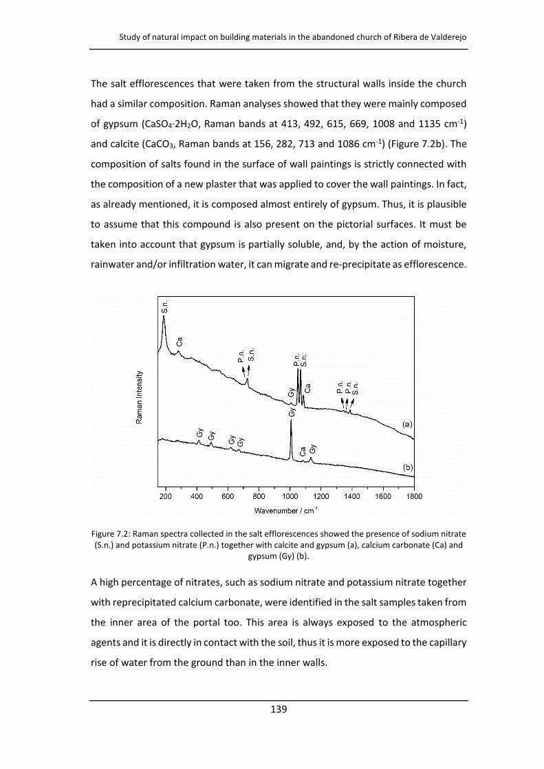
Study of natural impact on building materials in the abandoned church of Ribera de Valderejo
139
The salt efflorescences that were taken from the structural walls inside the church
had a similar composition. Raman analyses showed that they were mainly composed
of gypsum (CaSO4·2H2O, Raman bands at 413, 492, 615, 669, 1008 and 1135 cm-1)
and calcite (CaCO3, Raman bands at 156, 282, 713 and 1086 cm-1) (Figure 7.2b). The
composition of salts found in the surface of wall paintings is strictly connected with
the composition of a new plaster that was applied to cover the wall paintings. In fact,
as already mentioned, it is composed almost entirely of gypsum. Thus, it is plausible
to assume that this compound is also present on the pictorial surfaces. It must be
taken into account that gypsum is partially soluble, and, by the action of moisture,
rainwater and/or infiltration water, it can migrate and re-precipitate as efflorescence.
Figure 7.2: Raman spectra collected in the salt efflorescences showed the presence of sodium nitrate (S.n.) and potassium nitrate (P.n.) together with calcite and gypsum (a), calcium carbonate (Ca) and
gypsum (Gy) (b).
A high percentage of nitrates, such as sodium nitrate and potassium nitrate together
with reprecipitated calcium carbonate, were identified in the salt samples taken from
the inner area of the portal too. This area is always exposed to the atmospheric
agents and it is directly in contact with the soil, thus it is more exposed to the capillary
rise of water from the ground than in the inner walls.
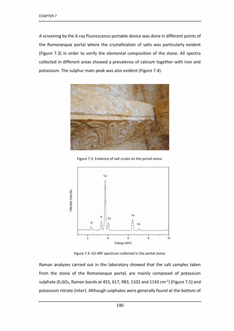
CHAPTER 7
140
A screening by the X-ray fluorescence portable device was done in different points of
the Romanesque portal where the crystallization of salts was particularly evident
(Figure 7.3) in order to verify the elemental composition of the stone. All spectra
collected in different areas showed a prevalence of calcium together with iron and
potassium. The sulphur main peak was also evident (Figure 7.4).
Figure 7.3: Evidence of salt crusts on the portal stone.
Figure 7.4: ED-XRF spectrum collected in the portal stone.
Raman analyses carried out in the laboratory showed that the salt samples taken
from the stone of the Romanesque portal, are mainly composed of potassium
sulphate (K2SO4, Raman bands at 455, 617, 983, 1102 and 1143 cm-1) (Figure 7.5) and
potassium nitrate (niter). Although sulphates were generally found at the bottom of
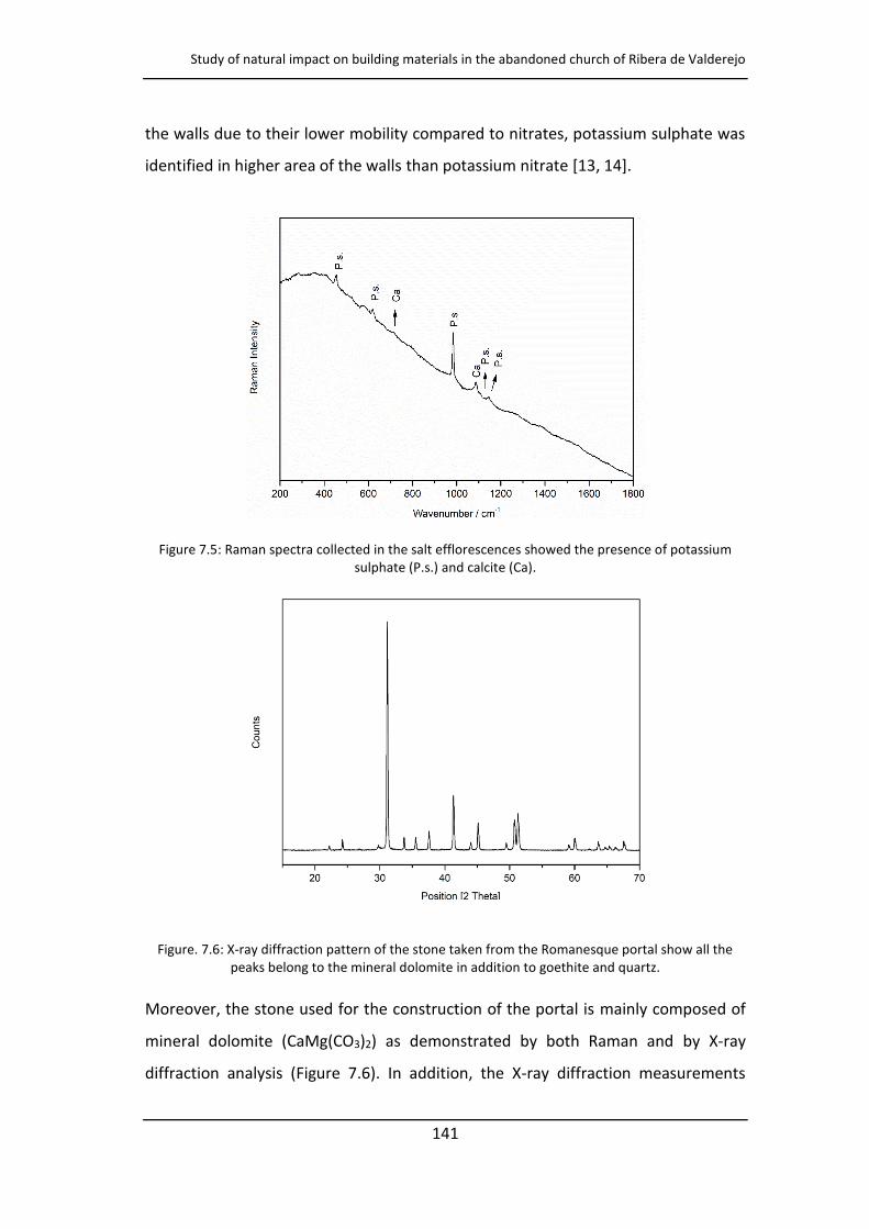
Study of natural impact on building materials in the abandoned church of Ribera de Valderejo
141
the walls due to their lower mobility compared to nitrates, potassium sulphate was
identified in higher area of the walls than potassium nitrate [13, 14].
Figure 7.5: Raman spectra collected in the salt efflorescences showed the presence of potassium sulphate (P.s.) and calcite (Ca).
Figure. 7.6: X-ray diffraction pattern of the stone taken from the Romanesque portal show all the peaks belong to the mineral dolomite in addition to goethite and quartz.
Moreover, the stone used for the construction of the portal is mainly composed of
mineral dolomite (CaMg(CO3)2) as demonstrated by both Raman and by X-ray
diffraction analysis (Figure 7.6). In addition, the X-ray diffraction measurements
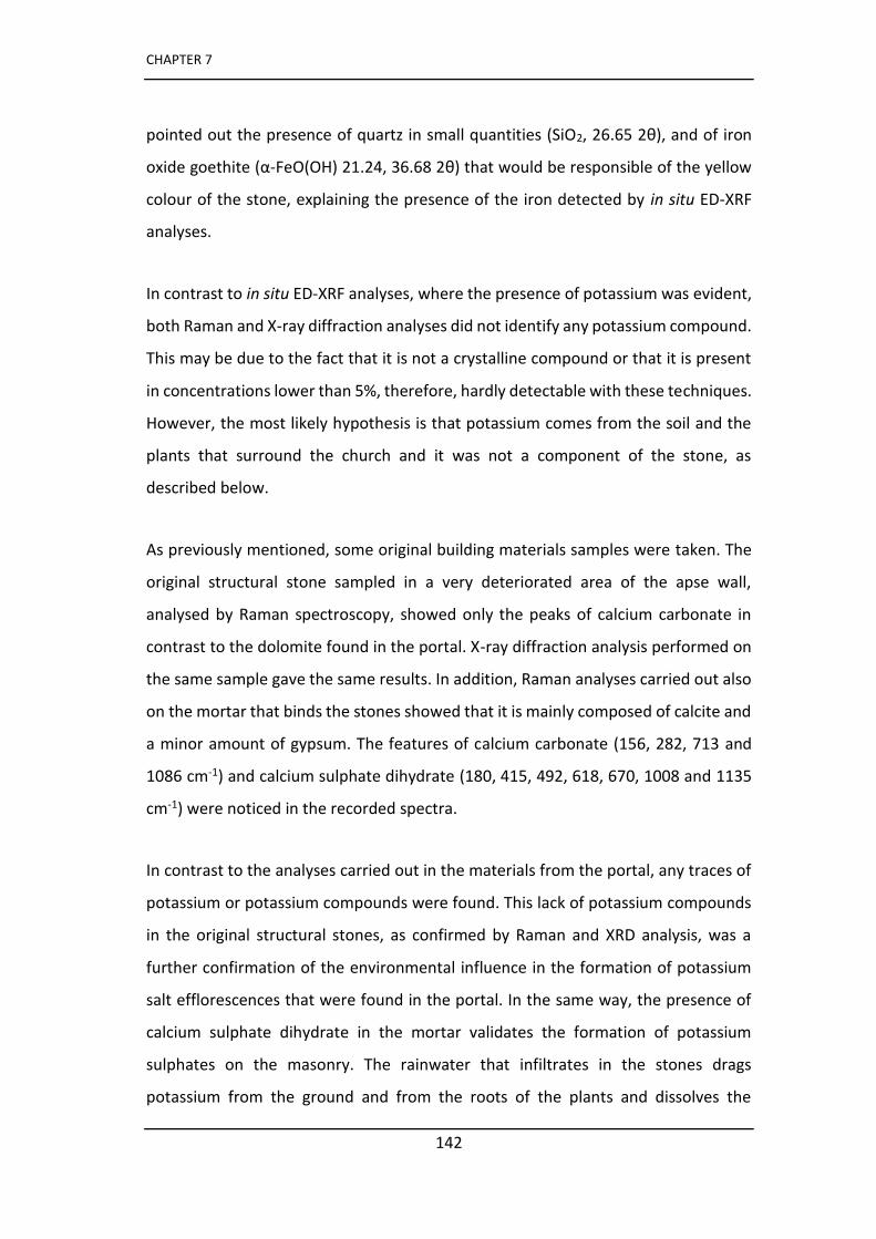
CHAPTER 7
142
pointed out the presence of quartz in small quantities (SiO2, 26.65 2θ), and of iron
o ide goethite α-FeO(OH) 21.24, 36.68 2θ) that would be responsible of the yellow
colour of the stone, explaining the presence of the iron detected by in situ ED-XRF
analyses.
In contrast to in situ ED-XRF analyses, where the presence of potassium was evident,
both Raman and X-ray diffraction analyses did not identify any potassium compound.
This may be due to the fact that it is not a crystalline compound or that it is present
in concentrations lower than 5%, therefore, hardly detectable with these techniques.
However, the most likely hypothesis is that potassium comes from the soil and the
plants that surround the church and it was not a component of the stone, as
described below.
As previously mentioned, some original building materials samples were taken. The
original structural stone sampled in a very deteriorated area of the apse wall,
analysed by Raman spectroscopy, showed only the peaks of calcium carbonate in
contrast to the dolomite found in the portal. X-ray diffraction analysis performed on
the same sample gave the same results. In addition, Raman analyses carried out also
on the mortar that binds the stones showed that it is mainly composed of calcite and
a minor amount of gypsum. The features of calcium carbonate (156, 282, 713 and
1086 cm-1) and calcium sulphate dihydrate (180, 415, 492, 618, 670, 1008 and 1135
cm-1) were noticed in the recorded spectra.
In contrast to the analyses carried out in the materials from the portal, any traces of
potassium or potassium compounds were found. This lack of potassium compounds
in the original structural stones, as confirmed by Raman and XRD analysis, was a
further confirmation of the environmental influence in the formation of potassium
salt efflorescences that were found in the portal. In the same way, the presence of
calcium sulphate dihydrate in the mortar validates the formation of potassium
sulphates on the masonry. The rainwater that infiltrates in the stones drags
potassium from the ground and from the roots of the plants and dissolves the
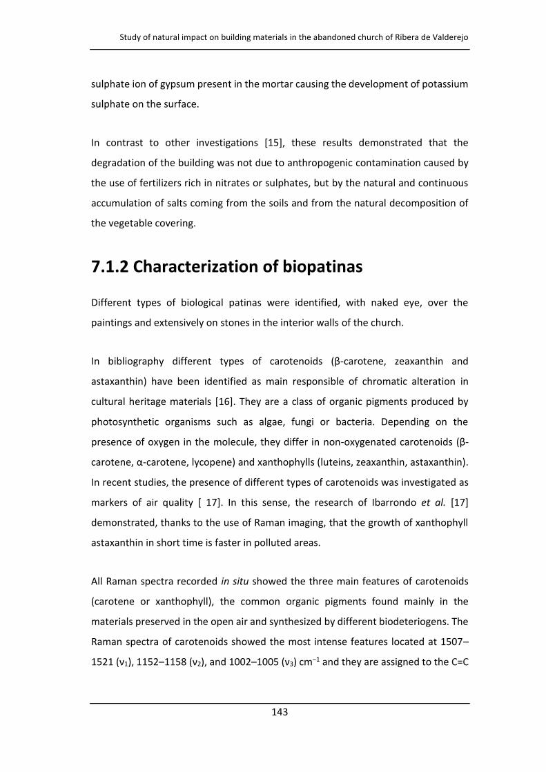
Study of natural impact on building materials in the abandoned church of Ribera de Valderejo
143
sulphate ion of gypsum present in the mortar causing the development of potassium
sulphate on the surface.
In contrast to other investigations [15], these results demonstrated that the
degradation of the building was not due to anthropogenic contamination caused by
the use of fertilizers rich in nitrates or sulphates, but by the natural and continuous
accumulation of salts coming from the soils and from the natural decomposition of
the vegetable covering.
7.1.2 Characterization of biopatinas
Different types of biological patinas were identified, with naked eye, over the
paintings and extensively on stones in the interior walls of the church.
In bibliography diffe e t t pes of a ote oids β-carotene, zeaxanthin and
astaxanthin) have been identified as main responsible of chromatic alteration in
cultural heritage materials [16]. They are a class of organic pigments produced by
photosynthetic organisms such as algae, fungi or bacteria. Depending on the
presence of oxygen in the molecule, they differ in non-oxygenated carotenoids β-
a ote e, α-carotene, lycopene) and xanthophylls (luteins, zeaxanthin, astaxanthin).
In recent studies, the presence of different types of carotenoids was investigated as
markers of air quality [ 17]. In this sense, the research of Ibarrondo et al. [17]
demonstrated, thanks to the use of Raman imaging, that the growth of xanthophyll
astaxanthin in short time is faster in polluted areas.
All Raman spectra recorded in situ showed the three main features of carotenoids
(carotene or xanthophyll), the common organic pigments found mainly in the
materials preserved in the open air and synthesized by different biodeteriogens. The
Raman spectra of carotenoids showed the most intense features located at 1507–
ν1), 1152– 8 ν2), and 1002– ν3) cm−1 and they are assigned to the C=C
CHAPTER 7
144
stretching mode, to the C–C stretching mode and to the C–H in plane bending
vibrational mode of carotenoids respectively [16].
Unfortunately, the in situ analyses did not allowed to identify the specific type of
compound because the main Raman bands of carotenoids are in similar positions,
and the bands of lower frequencies and the overtones must be considered for the
accurate identification. Thus, some biological patinas were sampled and further
analysis was carried out in the laboratory by Raman microspectroscopy.
Raman spectra gave evidence of zeaxanthin, a xantophyll carotenoid, and one of the
most common carotenoid found in the nature. (Figure. 7.7 a). The Raman spectrum
sho ed the ost i te se peaks at ν1 C=C , ν2 C–C) and ν3 C–H) the
most affected ones by the Raman resonance effect. The other Raman bands were
located at 960, 1187, 1207, 1267, 1285, 1347, 1386, 1443, 2153, 2345 and 2666 cm-
1.
Astaxanthin, another carotenoid of the xanthophylls class containing alcohol and
keto groups with 12 conjugated C=C double bonds, was also identified in a green area
of a patina. (Figure 7.7 b) The main Raman bands were located at 1507 (ν1 C=C), 1152
ν2 C–C) and 1002 (ν3 C–H), cm-1. In addition, other peaks of lower Raman intensity
that allowed to identify the compound were detectable at 883, 958, 1193, 1212,
1358, 1448, 1568, 2015, 2146, 2294, 2339, 2435, 2461, 2502, 2647, 2785, 2982 and
3015 cm-1.
Raman analyses carried out in some samples in which some orange spots were
e ide t usi g opti al i os ope, sho ed the p ese e of β-carotene (Figure 7.7 c)
This type of non-oxidized carotenoid is the main pigment present in non-polluted
areas. The most intense Raman bands are located at 1518 (ν1 C=C) 1158 (ν2 C–C) and
1005 (ν3 C–H) cm-1. The overtones at 2316 cm-1 (2ν2) and combination-tones at 2518
cm-1 (ν1ν3) and 2668 cm-1 (ν1ν2) were evident. In addition, the features at 874, 966,
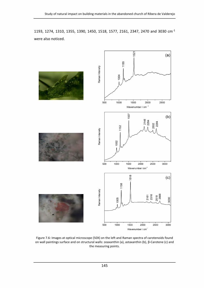
Study of natural impact on building materials in the abandoned church of Ribera de Valderejo
145
1193, 1274, 1310, 1355, 1390, 1450, 1518, 1577, 2161, 2347, 2470 and 3030 cm-1
were also noticed.
Figure 7.6: Images at optical microscope (50X) on the left and Raman spectra of carotenoids found on wall paintings surface and on structural walls: zeaxanthin (a), astaxanthin (b), β-Carotene (c) and
the measuring points.
CHAPTER 7
146
Photosynthetic organisms such as cyanobacteria, fungi and algae can synthesize
carotenoids as a self-protective mechanism to defend against damage caused by air
pollution or direct solar radiation. Moreover, the presence of carotenoids as auxiliary
DNA repair agents has been recognised also in extreme environmental conditions
[18].
The same biological patinas sampled for the Raman analyses were rubbed with
cotton swabs and an in vitro growth assay in Petri dishes with Potato Destrose Agar
was performed. Then, in order to identify the biological strains, a genomic DNA
extraction was carried out. A high similarity (99%) was found with database entries
of all biodeteriogens identified.
In two samples of black patinas taken from the wall paintings, fungi of the genus
Cladosporium cladorporioides were identified (Figure 7.8a). It is a rather common
genus of Ascomycetes fungi found on the mural paintings often together with
Aspergillum or Penicillum genus and its spores can be found in the atmosphere, in
the soil or in the water. In three green colour patinas, the fungi of the genus
Mortierella alpina were found (Figure 7.8b). Both identified genes live as saprotrophs
on dead organic materials in the soil.
Moreover, two bacteria genus were identified. The first belongs to the genus
Streptomyces sp., a genus of filamentous bacteria of the family Streptomycetaceae
(order Actinomycetales) (Figure 7.8c). The second kind of bacteria was the Bacillus
amyloliquefaciens, a microorganism of the Bacillus genus (Figure 7.8d). Both bacterial
strains are characterized as gram positive bacteria [19] commonly found in the soil
and vegetation.
Isolated strains, both fungal and bacterial, belong to spore formers, more properly
called an endospore for the bacterial spore. They are certainly the most popular
especially for their high rate of spore dissemination, and consequently, for the high
colonization capability [20]. The spores are also produced to protect the cell and they
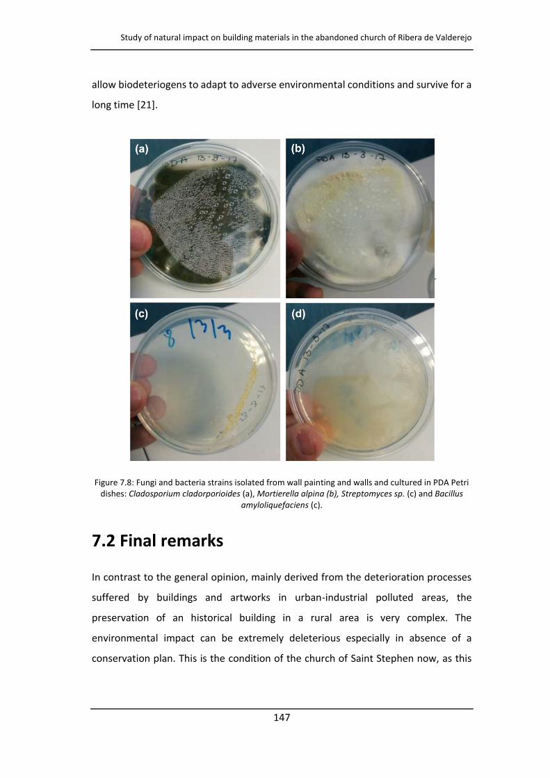
Study of natural impact on building materials in the abandoned church of Ribera de Valderejo
147
allow biodeteriogens to adapt to adverse environmental conditions and survive for a
long time [21].
Figure 7.8: Fungi and bacteria strains isolated from wall painting and walls and cultured in PDA Petri dishes: Cladosporium cladorporioides (a), Mortierella alpina (b), Streptomyces sp. (c) and Bacillus
amyloliquefaciens (c).
7.2 Final remarks
In contrast to the general opinion, mainly derived from the deterioration processes
suffered by buildings and artworks in urban-industrial polluted areas, the
preservation of an historical building in a rural area is very complex. The
environmental impact can be extremely deleterious especially in absence of a
conservation plan. This is the condition of the church of Saint Stephen now, as this
CHAPTER 7
148
multianalytical study using several diagnostic techniques has demonstrated, pointing
out the causes of the degradation processes.
With regard to the study of the conservation state of the church, in situ analyses
identified different degradation products, in particular salt efflorescences and
organic patinas, that later were sampled and analysed in-depth in the laboratory. For
example, an extensive presence of nitrate salts, potassium and sodium nitrate, was
identified by Raman spectroscopy and, as evidenced by the obtained results, it seems
to be a consequence of natural environmental impact rather than of the
a th opoge i polluta ts. The efo e, the ai it ate salts sou es, ide tified o the
surface of the mural paintings and building materials, are nitrogen compounds,
ammonium salts and nitrates, present in the soil from the decomposition of organic
matter and from plants that absorb the ions through the roots.
The church is constantly exposed to conditions of the surrounding environment and,
in addition, it is set directly on the ground in an agricultural area, thus, the ions
coming from the soil and from the plants penetrate easily in the walls thanks to the
phenomena of rain water infiltration and capillary rise. The water acts as a medium
and transports the ions from the ground into the masonry. Besides, the temperature
changes trigger dissolution-crystallization cycles and cause the development of white
salts surfacing. In the case of the Saint Stephen church these phenomena are
favoured by the presence of a masonry that has lost cohesion, and therefore, is
characterized by cracks through which water can easily trace.
Moreover, the building is almost covered by vegetation, herbaceous and higher
plants that contribute through the roots to transfer the substances. The prolongation
of these phenomena over time has caused a massive loss of materials and the
collapse of a part of the building.
The stud of uildi g ate ials as a fu the step to u de sta d the auses that led
to the fo atio of salts. I fa t, as de o st ated usi g Ra a a d XRD a al ses,
Study of natural impact on building materials in the abandoned church of Ribera de Valderejo
149
the environmental impact is so strong that the potassium sulphate, generated by the
interaction of potassium from the soil and the gypsum of the mortar, generally
located in lower parts of the masonry than nitrates, was found in the portal stone at
about 2.50 meters high.
The research performed shown that the degradation of walls and mural paintings
cannot be attributed only to the presence of saline efflorescence but also to an
intense biological activity as documented by the molecular and microbiological
techniques. For example, the presence of calcium oxalate weddellite identified in situ
with portable devices appeared strictly connected to biodeterioration processes.
Raman spectroscopy has proven to be extremely efficient for the recognition of three
different carotenoids, β-carotene, zeaxanthin and astaxanthin, identified in the
biological patina samples. The identification of astaxanthin, the most oxidized of the
carotenoid species, is particularly interesting. Until now, it was documented that it
was formed, in a short time, in the presence of acidic environmental stress, such as a
high SO2 concentration in the atmosphere. We are not able to know how long the
church has been in abandonment but, as documented in this study, the astaxanthin
can develop in a long time in an uncontaminated countryside as well.
The detection of fungi and bacteria using analyses of PCR-amplified 18S and 16S rDNA
fragments, contributed to better understand the genus of biodeterioration markers
that grow within the church. These microorganisms may have a negative effect on
the conservation of the mural paintings especially in a place where the atmospheric
conditions cannot be controlled. They are causing not only a chromatic damage but
also a structural damage caused by the production of mycelium, which can grow in
the paintings and in the mortar.
CHAPTER 7
150
References
[1] M. A. Rogerio-Candelera, M. Lazzari, E. Cano (eds.), Science and Technology for the Conservation of Cultural Heritage, CRC Press, Boca Raton (USA), 2013.
[2] I. Martínez-Arkarazo, M. Angulo, L. Bartolome, N. Etxebarria, M. A. Olazabal, J. M. Madariaga (2007) An integrated analytical approach to diagnose the conservation state of building materials of a palace house in the metropolitan Bilbao (Basque Country, North of Spain), Analytica Chimica Acta, 584, 350-359.
[3]. J. Aramendia, L. Gómez-Nubla, K. Castro, I. Martínez-Arkarazo, D. Vega, A. Sanz López de Heredia, A. García Ibáñez de Opakua, J. M. Madariaga (2012) Portable Raman study on the conservation state of four Co Te steel‐ ased s ulptu es Eduardo Chillida impacted by urban atmospheres, Journal of Raman Spectroscopy, 43, 1111-1117.
[4]. M. Sablier, P. Garrigue (2014) Cultural heritage and its environment: an issue of interest for Environmental Science and Pollution Research, Environmental Science
and Pollution Research, 21, 5769-5773.
[5] L. K. Herrera, H. A. Videla, (2004) The importance of atmospheric effects on biodeterioration of cultural heritage constructional materials, International
Biodeterioration & Biodegradation, 54, 125-134.
[6] J. M. Madariaga (2015) Analytical chemistry in the field of cultural heritage, Analytical
Methods, 7, 4848-4876.
[7] M. Ma szałek (2016), Identification of secondary salts and their sources in deteriorated stone monuments using micro-Raman spectroscopy, SEM-EDS and XRD, Journal of Raman Spectroscopy, 47, 1473–1485.
[8] R. M. Io , S. Teodo es u, R. M. Şti es u, I. A. Bu u i ă, I. D. Dula ă, M. L. Io (2017) Calcium Oxalate on Limestone Surface of Heritage Buildings, Key Engineering
Materials, 750, 129-134.
[9 H. Morillas, M. Maguregui, O. Gómez-Laserna, J. Trebolazabal, J. M. Madariaga (2012) Characterisation and diagnosis of the conservation state of cementitious materials exposed to the open air in XIX century lighthouses located on the coast of the Bas ue Cou t : The ase of Igueldo lighthouse, San Sebastian, North of Spain, Journal of Raman Spectroscopy, 43, 1630-1636.
[10] N. Prieto-Taboada, O. Gómez-Laserna, I. Martínez-Arkarazo, M. A. Olazabal, J. M. Madariaga (2013) Relevance of Cross-Section Analysis in Correct Diagnosis of the State of Conservation of Building Materials As Evidenced by Spectroscopic Imaging, Analytical Chemistry, 85, 9501-9507.
[11] A. Hernanz, M. Mas, B. Gavilán, B. Hernández (2006) Raman microscopy and IR spectroscopy of prehistoric paintings from Los Murciélagos cave (Zuheros, Córdoba, Spain), Journal of Raman Spectroscopy, 37, 492-497.
Study of natural impact on building materials in the abandoned church of Ribera de Valderejo
151
[12] A. Hernanz, I. Bratu, O. F. Marutoiu, C. Marutoiu, J. M. Gavira-Vallejo, H. G. M. Edwards (2008) Micro-Raman spectroscopic investigation of external wall paintings f o St. Du it u s Chu h, Su ea a, Ro a ia, Analytical and Bioanalytical Chemistry,
392, 263-268.
[13] A. Arnold, K. Zehnder, Monitoring Wall Paintings Affected by Soluble Salts, The Conservation of Wall Paintings, Proceedings of a symposium organized by the Courtauld Institute of Art and the Getty Conservation Institute, London, July 13-16, 1987, Sharon Cather, Editor.
[14] M. A. Vázquez , E. Galán , P. Ortiz , R. Ortiz (2013) Digital image analysis and EDX SEM as combined techniques to evaluate salt damp on walls, Construction and
Building Materials, 45, 95-105.
[15] M. Veneranda, M. Irazola, M. Díez, A. Iturregui, J. Aramendia, K. Castro, J. M. Madariaga (2014) Raman spectroscopic study of the degradation of a middle age mural painting: the role of agricultural activities, Journal of Raman Spectroscopy, 45, 1110–1118.
[16] M. Maguregui, U. Knuutinen, J. Trebolazabala, H. Morillas, K. Castro, I. Martínez-Arkarazo, J. M. Madariaga (2012) Use of in situ and confocal Raman spectroscopy to study the nature and distribution of carotenoids in brown patinas from a deteriorated wall painting in Marcus Lucretius House (Pompeii), Analytical and Bioanalytical
Chemistry, 402, 1529-1539.
[17] I. Ibarrondo, N. Prieto-Taboada, I. Martínez-Arkarazo, J. M. Madariaga (2016) Resonance Raman imaging as a tool to assess the atmospheric pollution level: carotenoids in Lecanoraceae lichens as bioindicators, Environmental Science and
Pollution Research, 23, 6390-6399.
[18] H. G. M. Edwards (2007) A novel extremophile strategy studied by Raman spectroscopy, Spectrochimica Acta Part A, 68, 1126-1132.
[19] L. Laiz, D. Recio, B. Hermosin, C. Saiz-Jiménez (2000) Microbial Communities in Salt Efflorescences. In: Ciferri O., Tiano P., Mastromei G. (eds) Of Microbes and Art. Springer, Boston, MA.
[20] W. Wanfu, M. Xu, M Yantian, M. Lin, W. Fasi, M. Xiaojun, A. Lizhe, F. Huyuan (2010) Seasonal dynamics of airborne fungi in different caves of the Mogao Grottoes, Dunhuang, China, International Biodeterioration & Biodegradation, 64, 461-466.
[21] A. A. Gorbushina, J. Heyrman, T. Dornieden, M. González-Delvall, W. E. Krumbei, L. Laiz, Karin Petersen, C. Saiz-Jímenez (2004) Bacterial and fungal diversity and biodeterioration problems in mural painting environments of St. Martins church (Greene–Kreiensen, Germany) J. Swings, International Biodeterioration &
Biodegradation, 53, 13-24.

153
CHAPTER 8
PALME software as an alternative
tool for semi-quantification of salt
efflorescence
As demonstrated in the previous chapter, the identification of salt deposits is a crucial
first step in understanding the causes that led to the degradation process and even
which chemical reactions have taken place between the environment and the
artwork. Since in most cases, the causes of the presence of products of degradation
are multiple, currently it is also important to quantify the precipitated salts to better
understand the contribution of each factor on the phenomenon of deterioration that
can be anthropogenic or natural, intrinsic or extrinsic to the material [1-3].
Many studies show the reliability of the vibrational spectroscopic techniques, FT-IR
and Raman, for the study of salt efflorescences [4, 5]. Compared to FT-IR
spectroscopy, the Raman technique has a number of advantages; the interpretation
of spectra is easier and faster because there are not overlapping bands or overtones.
Besides, it can be applied for analysing solid and liquid samples without
pretreatment. Although the Raman technique can be considered the most one used
for the study of saline deposits, performed in situ and in the laboratory, less
researches use Raman spectroscopy for the quantitative analysis of these
compounds. As the bibliography's demonstrates, the most widespread method used
CHAPTER 8
154
for semi-quantification with Raman spectroscopy is based on the so-called external
calibration curves, where the intensity of the Raman signal is proportional to the
concentration of the compound within the sample [6]. An example for this is the work
of Veneranda et al., where a quantitative study of salt deposits, present as
degradation product on mural paintings, has been successfully performed with
Raman spectroscopy using linear external calibration plots [7].
Quantitative Raman analysis has been performed always more frequently with
samples in solution [8] and less in solid state [9]. In fact, to get good results
homogeneous samples are needed and these can be hardly to obtain when you work
with samples in solid state. However, the problem of the homogeneity of the solid
phase samples can be overcome by using different procedures during Raman
analysis. The use of a rotating sample holder, for example, allows to cover a wider
area that represents the whole composition of the sample. Moreover, it is possible
to record some spectra for each sample in different areas chosen randomly averaging
the results obtained [10].
In this chapter Raman spectroscopy has been used to identify the different
compounds present in such salts, but can also be used for the semi-quantification of
such salt deposits based on the use of external calibration curves. Normally, this
method takes time for the preparation of pellets composed both by mixtures of
standard and or by real samples. In the present work we propose an innovative
approach for semi-quantification of salts in the solid state by portable micro-Raman
spectroscopy. It is based on the use of the PALME software (MONARIS-UPMC, Paris)
that has been developed in order to obtain the quantitative information from the
vibrational spectrum using a Multivariate Linear Regression approach (MLR). PALME
software considers a mixture spectrum as a linear combination of the suspected pure
materials spectra and in the field of Cultural Heritage studies it has been applied to
various contexts [11-14]. In this investigation both calibration curves and PALME
software have been applied to the quantitative analysis on samples of pure salts at
different concentrations of calcium carbonate (CaCO3), gypsum (CaSO4·2H2O) and
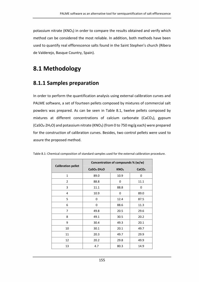
PALME software as an alternative tool for semiquantification of salt efflorescence
155
potassium nitrate (KNO3) in order to compare the results obtained and verify which
method can be considered the most reliable. In addition, both methods have been
used to quantify real efflorescence salts found in the Saint Stephe ’s church (Ribera
de Valderejo, Basque Country, Spain).
8.1 Methodology
8.1.1 Samples preparation
In order to perform the quantification analysis using external calibration curves and
PALME software, a set of fourteen pellets composed by mixtures of commercial salt
powders was prepared. As can be seen in Table 8.1, twelve pellets composed by
mixtures at different concentrations of calcium carbonate (CaCO3), gypsum
(CaSO4·2H2O) and potassium nitrate (KNO3) (from 0 to 750 mg/g each) were prepared
for the construction of calibration curves. Besides, two control pellets were used to
assure the proposed method.
Table 8.1: Chemical composition of standard samples used for the external calibration procedure.
Calibration pellet Concentration of compounds % (w/w)
CaSO4·2H2O KNO3 CaCO3
1 89.0 10.9 0
2 88.8 0 11.1
3 11.1 88.8 0
4 10.9 0 89.0
5 0 12.4 87.5
6 0 88.6 11.3
7 49.8 20.5 29.6
8 49.1 30.5 20.2
9 30.4 49.3 20.1
10 30.1 20.1 49.7
11 20.3 49.7 29.9
12 20.2 29.8 49.9
13 4.7 80.3 14.9
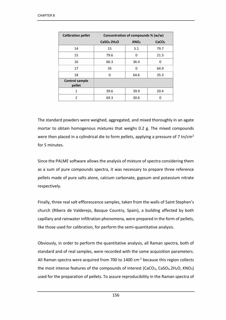
CHAPTER 8
156
Calibration pellet Concentration of compounds % (w/w)
CaSO4·2H2O KNO3 CaCO3
14 15 5.1 79.7
15 79.6 0 21.3
16 66.3 36.4 0
17 35 0 64.9
18 0 64.6 35.3
Control sample
pellet
1 39.6 39.9 20.4
2 69.3 30.6 0
The standard powders were weighed, aggregated, and mixed thoroughly in an agate
mortar to obtain homogenous mixtures that weighs 0.2 g. The mixed compounds
were then placed in a cylindrical die to form pellets, applying a pressure of 7 tn/cm2
for 5 minutes.
Since the PALME software allows the analysis of mixture of spectra considering them
as a sum of pure compounds spectra, it was necessary to prepare three reference
pellets made of pure salts alone, calcium carbonate, gypsum and potassium nitrate
respectively.
Finally, three real salt efflorescence samples, taken from the walls of Sai t Stephe ’s
church (Ribera de Valderejo, Basque Country, Spain), a building affected by both
capillary and rainwater infiltration phenomena, were prepared in the form of pellets,
like those used for calibration, for perform the semi-quantitative analysis.
Obviously, in order to perform the quantitative analysis, all Raman spectra, both of
standard and of real samples, were recorded with the same acquisition parameters.
All Raman spectra were acquired from 700 to 1400 cm-1 because this region collects
the most intense features of the compounds of interest (CaCO3, CaSO4.2H2O, KNO3)
used for the preparation of pellets. To assure reproducibility in the Raman spectra of
PALME software as an alternative tool for semiquantification of salt efflorescence
157
the pellets, the analyses were performed coupling the Raman probe with a long-
distance objective lens (4x) allowing to analyse a eas of ea μ 2 where the
required homogeneity is attained. Each spectrum was collected with 30 seconds of
exposure time and 5 accumulations using the 10% of the nominal laser power. In
order to obtain a good level of accuracy and to verify the repeatability of analysis,
eighteen spectra were recorded in different points of each pellet.
8.2 PALME software
Software PALME was developed in order to obtain quantitative information from the
vibrational spectrum of the mixture compound using a Multivariate Linear Regression
approach (MLR) [15, 16]. As a first step, each reference spectrum is decomposed into
elementary bands (the user can select the Gaussian or Lorentzian band profiles) and
adjusted on the experimental spectrum. It defines the reference spectra profiles
which are stored to be used for decomposition.
Then, the mixed spectrum is loaded and its adjustment procedure is performed by a
linear combination of pure materials spectra using a least-squares criterion and the
Levenberg-Marquardt algorithm. The software shows graphically (Figure 8.1) the
decomposition of the spectrum.
The contribution of each pure component that is required to compose the
experimental spectrum is expressed as two different coefficients, the weighting
coefficient (á) and the area proportion depending on the employed spectroscopic
technique. In the case of Raman spectroscopy, the contribution of each component
pure on the mixture is expressed as a weighting coefficient (á) expressed as
percentage. In order to achieve a good level of accuracy in the semi-quantitative
analysis a series of spectra are collected in the same sample and the amount of
analyte is given by the average of the coefficients.

CHAPTER 8
158
Figure 8.1: Spectral adjustment of Raman spectrum recorded on a pellet of real salt composed by a
mixture of calcium carbonate, gypsum and potassium nitrate.
8.3 Results and discussion
In order to perform the quantification of each compound in the samples using
calibration curves, 18 pellets were analysed using micro Raman spectroscopy. Six
calibration curves were constructed, three evaluating the height (Figure 8.2) and
three evaluating the integration area (Figure 8.3) of the main Raman band of calcium
carbonate (1086 cm-1, CO32- ν1), gypsum (1008 cm-1, SO4
2-ν1) and potassium nitrate
(1050 cm-1, NO3- ν0), for each Raman spectrum recorded.
Due to the experimental uncertainty, the uncertainty of the intercept was always
higher than 50% of % relative standard deviation (RSD); thus, the intercept of all
calibration curves was fitted to 0 value. The changes in the slopes were within the
estimated uncertainty of the results.
As demonstrated by previous studies carried out on salt efflorescence, it occurs a
direct proportionality between the intensity of the main peaks of salts with their
PALME software as an alternative tool for semiquantification of salt efflorescence
159
concentration [17]. Considering the height of the main Raman bands, the R2 values
for calcium carbonate, gypsum and potassium nitrate were 0.9544, 0.9788 and
0.9525 respectively.
Because the height of the Raman bands is influenced by the degree of crystallinity of
the compounds, three calibration curves were built considering also the area of the
peaks. As it can be seen, the values have a slightly improvement using the integration
area of the main peaks. The R2 were 0.9531 for CaCO3, 0.9795 for CaSO4.2H2O and
0.9606 for KNO3. However, the general distribution of dots around the regression line
is about the same for both extracted parameters (i.e. band intensities or areas),
underlining that the measurement dispersion is mo e elated to sa ple’s
characteristics than spectra treatment.
The obtained calibration curves showed that the linear correlation between both,
peak integration area and height, is reasonably good. Moreover, taking into account
that the work is based on the study of solid samples, the results obtained were quite
good. In fact, it is much more difficult to achieve a good level of homogeneity of the
samples with powders in the solid state compared to samples in solution. However,
with regard to the homogeneity, the relative standard deviation of the signals of the
analytes of interest in each pellet varied between 4%-10%, which can be acceptable
for solid samples.
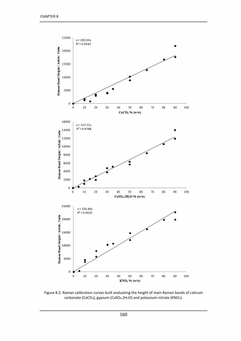
CHAPTER 8
160
Figure 8.2: Raman calibration curves built evaluating the height of main Raman bands of calcium
carbonate (CaCO3), gypsum (CaSO4.2H2O) and potassium nitrate (KNO3).
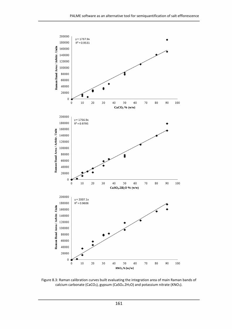
PALME software as an alternative tool for semiquantification of salt efflorescence
161
Figure 8.3: Raman calibration curves built evaluating the integration area of main Raman bands of
calcium carbonate (CaCO3), gypsum (CaSO4.2H2O) and potassium nitrate (KNO3).
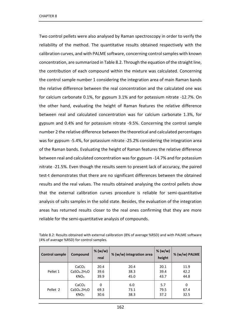
CHAPTER 8
162
Two control pellets were also analysed by Raman spectroscopy in order to verify the
reliability of the method. The quantitative results obtained respectively with the
calibration curves, and with PALME software, concerning control samples with known
concentration, are summarized in Table 8.2. Through the equation of the straight line,
the contribution of each compound within the mixture was calculated. Concerning
the control sample number 1 considering the integration area of main Raman bands
the relative difference between the real concentration and the calculated one was
for calcium carbonate 0.1%, for gypsum 3.1% and for potassium nitrate -12.7%. On
the other hand, evaluating the height of Raman features the relative difference
between real and calculated concentration was for calcium carbonate 1.3%, for
gypsum and 0.4% and for potassium nitrate -9.5%. Concerning the control sample
number 2 the relative difference between the theoretical and calculated percentages
was for gypsum -5.4%, for potassium nitrate -25.2% considering the integration area
of the Raman bands. Evaluating the height of Raman features the relative difference
between real and calculated concentration was for gypsum -14.7% and for potassium
nitrate -21.5%. Even though the results seem to present lack of accuracy, the paired
test-t demonstrates that there are no significant differences between the obtained
results and the real values. The results obtained analysing the control pellets show
that the external calibration curves procedure is reliable for semi-quantitative
analysis of salts samples in the solid state. Besides, the evaluation of the integration
areas has returned results closer to the real ones confirming that they are more
reliable for the semi-quantitative analysis of compounds.
Table 8.2: Results obtained with external calibration (8% of average %RSD) and with PALME software
(4% of average %RSD) for control samples.
Control sample Compound % (w/w)
real % (w/w) integration area
% (w/w)
height % (w/w) PALME
Pellet 1
CaCO3
CaSO4.2H2O
KNO3
20.4
39.6
39.9
20.4
38.3
45.0
20.1
39.4
43.7
11.9
42.2
44.8
Pellet 2
CaCO3
CaSO4.2H2O
KNO3
0
69.3
30.6
6.0
73.1
38.3
5.7
79.5
37.2
0
67.4
32.5
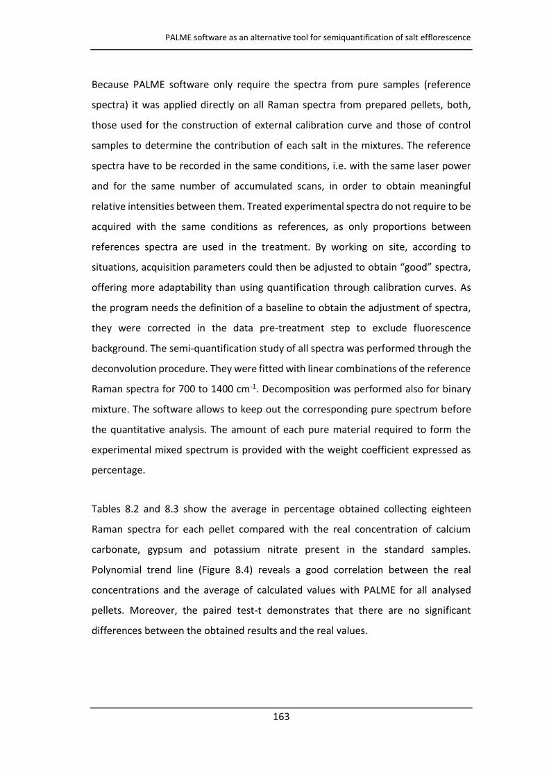
PALME software as an alternative tool for semiquantification of salt efflorescence
163
Because PALME software only require the spectra from pure samples (reference
spectra) it was applied directly on all Raman spectra from prepared pellets, both,
those used for the construction of external calibration curve and those of control
samples to determine the contribution of each salt in the mixtures. The reference
spectra have to be recorded in the same conditions, i.e. with the same laser power
and for the same number of accumulated scans, in order to obtain meaningful
relative intensities between them. Treated experimental spectra do not require to be
acquired with the same conditions as references, as only proportions between
references spectra are used in the treatment. By working on site, according to
situatio s, a uisitio pa a ete s ould the e adjusted to o tai good spe t a,
offering more adaptability than using quantification through calibration curves. As
the program needs the definition of a baseline to obtain the adjustment of spectra,
they were corrected in the data pre-treatment step to exclude fluorescence
background. The semi-quantification study of all spectra was performed through the
deconvolution procedure. They were fitted with linear combinations of the reference
Raman spectra for 700 to 1400 cm-1. Decomposition was performed also for binary
mixture. The software allows to keep out the corresponding pure spectrum before
the quantitative analysis. The amount of each pure material required to form the
experimental mixed spectrum is provided with the weight coefficient expressed as
percentage.
Tables 8.2 and 8.3 show the average in percentage obtained collecting eighteen
Raman spectra for each pellet compared with the real concentration of calcium
carbonate, gypsum and potassium nitrate present in the standard samples.
Polynomial trend line (Figure 8.4) reveals a good correlation between the real
concentrations and the average of calculated values with PALME for all analysed
pellets. Moreover, the paired test-t demonstrates that there are no significant
differences between the obtained results and the real values.
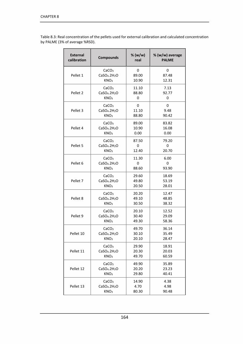
CHAPTER 8
164
Table 8.3: Real concentration of the pellets used for external calibration and calculated concentration
by PALME (3% of average %RSD).
External
calibration Compounds
% (w/w)
real
% (w/w) average
PALME
Pellet 1
CaCO3
CaSO4.2H2O
KNO3
0
89.00
10.90
0
87.48
12.31
Pellet 2
CaCO3
CaSO4.2H2O
KNO3
11.10
88.80
0
7.13
92.77
0
Pellet 3
CaCO3
CaSO4.2H2O
KNO3
0
11.10
88.80
0
9.48
90.42
Pellet 4
CaCO3
CaSO4.2H2O
KNO3
89.00
10.90
0.00
83.82
16.08
0.00
Pellet 5
CaCO3
CaSO4.2H2O
KNO3
87.50
0
12.40
79.20
0
20.70
Pellet 6
CaCO3
CaSO4.2H2O
KNO3
11.30
0
88.60
6.00
0
93.90
Pellet 7
CaCO3
CaSO4.2H2O
KNO3
29.60
49.80
20.50
18.69
53.19
28.01
Pellet 8
CaCO3
CaSO4.2H2O
KNO3
20.20
49.10
30.50
12.47
48.85
38.32
Pellet 9
CaCO3
CaSO4.2H2O
KNO3
20.10
30.40
49.30
12.52
29.09
58.36
Pellet 10
CaCO3
CaSO4.2H2O
KNO3
49.70
30.10
20.10
36.14
35.49
28.47
Pellet 11
CaCO3
CaSO4.2H2O
KNO3
29.90
20.30
49.70
18.91
20.03
60.59
Pellet 12
CaCO3
CaSO4.2H2O
KNO3
49.90
20.20
29.80
35.89
23.23
40.41
Pellet 13
CaCO3
CaSO4.2H2O
KNO3
14.90
4.70
80.30
4.38
4.98
90.48
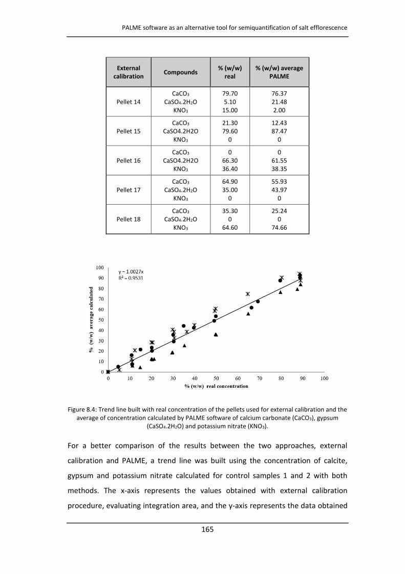
PALME software as an alternative tool for semiquantification of salt efflorescence
165
External
calibration Compounds
% (w/w)
real
% (w/w) average
PALME
Pellet 14
CaCO3
CaSO4.2H2O
KNO3
79.70
5.10
15.00
76.37
21.48
2.00
Pellet 15
CaCO3
CaSO4.2H2O
KNO3
21.30
79.60
0
12.43
87.47
0
Pellet 16
CaCO3
CaSO4.2H2O
KNO3
0
66.30
36.40
0
61.55
38.35
Pellet 17
CaCO3
CaSO4.2H2O
KNO3
64.90
35.00
0
55.93
43.97
0
Pellet 18
CaCO3
CaSO4.2H2O
KNO3
35.30
0
64.60
25.24
0
74.66
Figure 8.4: Trend line built with real concentration of the pellets used for external calibration and the
average of concentration calculated by PALME software of calcium carbonate (CaCO3), gypsum
(CaSO4.2H2O) and potassium nitrate (KNO3).
For a better comparison of the results between the two approaches, external
calibration and PALME, a trend line was built using the concentration of calcite,
gypsum and potassium nitrate calculated for control samples 1 and 2 with both
methods. The x-axis represents the values obtained with external calibration
procedure, evaluating integration area, and the y-axis represents the data obtained
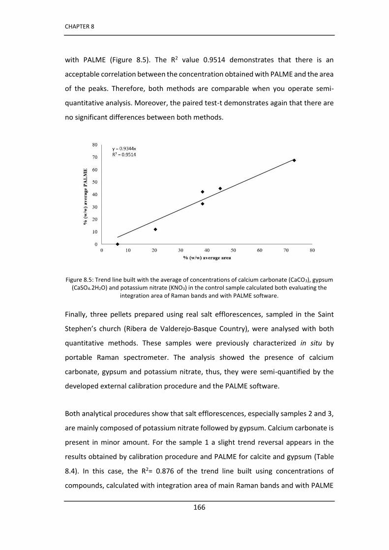
CHAPTER 8
166
with PALME (Figure 8.5). The R2 value 0.9514 demonstrates that there is an
acceptable correlation between the concentration obtained with PALME and the area
of the peaks. Therefore, both methods are comparable when you operate semi-
quantitative analysis. Moreover, the paired test-t demonstrates again that there are
no significant differences between both methods.
Figure 8.5: Trend line built with the average of concentrations of calcium carbonate (CaCO3), gypsum
(CaSO4.2H2O) and potassium nitrate (KNO3) in the control sample calculated both evaluating the
integration area of Raman bands and with PALME software.
Finally, three pellets prepared using real salt efflorescences, sampled in the Saint
Stephe ’s church (Ribera de Valderejo-Basque Country), were analysed with both
quantitative methods. These samples were previously characterized in situ by
portable Raman spectrometer. The analysis showed the presence of calcium
carbonate, gypsum and potassium nitrate, thus, they were semi-quantified by the
developed external calibration procedure and the PALME software.
Both analytical procedures show that salt efflorescences, especially samples 2 and 3,
are mainly composed of potassium nitrate followed by gypsum. Calcium carbonate is
present in minor amount. For the sample 1 a slight trend reversal appears in the
results obtained by calibration procedure and PALME for calcite and gypsum (Table
8.4). In this case, the R2= 0.876 of the trend line built using concentrations of
compounds, calculated with integration area of main Raman bands and with PALME

PALME software as an alternative tool for semiquantification of salt efflorescence
167
software, confirmed a good correspondence between the data obtained from both
methods (Figure 8.6). Once again, the paired test-t confirmed that there were no
significant differences among the obtained results.
Although the research seems to show that the best results were obtained using
calibration curves considering integration area of main Raman bands, paired test-t
did not see significant differences, and the program PALME has significant
advantages. In fact, it allowed to perform quantification analysis without preparing a
set of reference pellets (with a calibration curve for each measurement conditions)
with a considerable saving of time. Even if the variation between real and calculated
data is higher with PALME it can be accepted when you work in situ for semi-
quantitative analysis of mixtures of salts. However, the PALME software has proved
to be an effective tool for identifying the main compounds in a mixture and this
aspect is very important if PALME software is applied in the field of Cultural Heritage.
Characterizing the concentrations of each mixture salts make it possible to classify
the different causes that have caused the salt formation. The identification of each
concentration therefore, allows to diagnose the most damaging cause of a porous
material's deterioration. In this way, it can be possible to suggest the most
appropriate method of intervention and in case to prevent the damage.
Table 8.4: Result obtained with external calibration (20% of average %RSD) and with PALME (10% of
average %RSD) for real salt efflorescences from the sampled church.
Real samples Compounds % (w/w) integration area % (w/w)
height % (w/w) PALME
Pellet 1
CaCO3
CaSO4.2H2O
KNO3
22.9
11.7
53.3
21.8
15.3
48.4
17.7
21.4
59.5
Pellet 2
CaCO3
CaSO4.2H2O
KNO3
18.0
25.3
59.7
17.2
35.3
58.6
10.2
33.6
56.9
Pellet 3
CaCO3
CaSO4.2H2O
KNO3
12.6
43.0
47.5
14.0
59.5
45.2
7.1
49.8
42.4
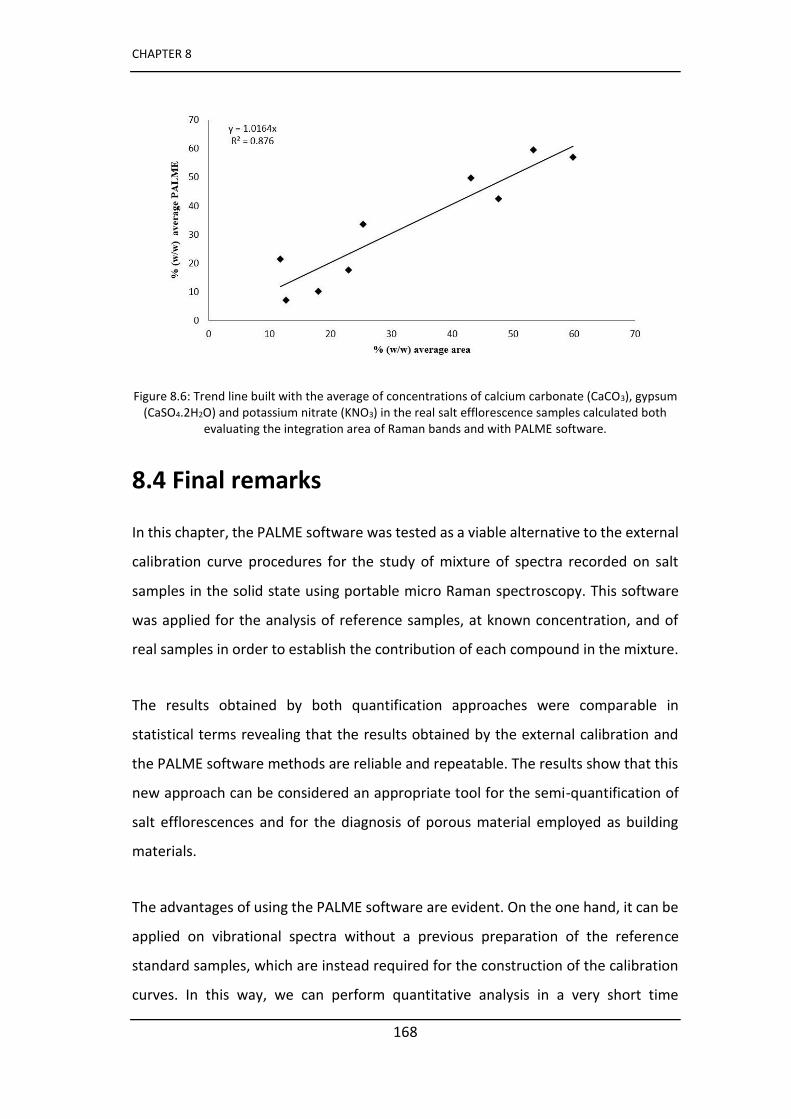
CHAPTER 8
168
Figure 8.6: Trend line built with the average of concentrations of calcium carbonate (CaCO3), gypsum
(CaSO4.2H2O) and potassium nitrate (KNO3) in the real salt efflorescence samples calculated both
evaluating the integration area of Raman bands and with PALME software.
8.4 Final remarks
In this chapter, the PALME software was tested as a viable alternative to the external
calibration curve procedures for the study of mixture of spectra recorded on salt
samples in the solid state using portable micro Raman spectroscopy. This software
was applied for the analysis of reference samples, at known concentration, and of
real samples in order to establish the contribution of each compound in the mixture.
The results obtained by both quantification approaches were comparable in
statistical terms revealing that the results obtained by the external calibration and
the PALME software methods are reliable and repeatable. The results show that this
new approach can be considered an appropriate tool for the semi-quantification of
salt efflorescences and for the diagnosis of porous material employed as building
materials.
The advantages of using the PALME software are evident. On the one hand, it can be
applied on vibrational spectra without a previous preparation of the reference
standard samples, which are instead required for the construction of the calibration
curves. In this way, we can perform quantitative analysis in a very short time

PALME software as an alternative tool for semiquantification of salt efflorescence
169
compared to other chemometric techniques. In fact, PALME does not need the
preparation of the calibration curve, thus, it is a less time consuming approach.
On the other hand, one of the most important aspects highlighted in this research is
that the use of portable Raman instrumentation coupled to PALME software could
allow to perform semi-quantitative estimation of compounds, not only in the
laboratory but also in situ. This feature is particularly useful in the field of the Cultural
Heritage as it allows to carry out a quantitative study of salt deposits without needing
a sampling and without using a laboratory analysis. Requiring only the reference
spectra of quantified compounds, this reference database could be acquired prior to
the field campaign. On site, it only requires for spectrum treatment the choice of the
suspected compounds to obtain their quantification, without prerequisite on
measurement conditions. Thanks to its non-invasiveness, speed in processing data
and reliability features, the combination of Raman spectroscopy and PALME software
for semi-quantification purposes is suggested for all fields of study, including Cultural
Heritage, in which the sampling procedure is precluded or not recommended.
Furthermore, compared to the reference technique of X-ray diffraction (XRD) and
Rietveld analysis, such approach offers an alternative for the analytical quantification
of mineral phases much more portable and without heavy data treatment.
CHAPTER 8
170
References
[1] P. López-Arce, J. García-Guinea, D. Benavente, L. Tormo, E. Doehne (2009)
Deterioration of dolostone by magnesium sulphate salt: An example of incompatible
building materials at Bonaval Monastery, Spain, Construction and Building Materials,
23, 846-855.
[2] N. Prieto-Taboada, M. Maguregui, I. Martínez-Arkarazo, M. A. Olazabal, G. Arana,
J.M. Madariaga (2011) Spectroscopic evaluation of the environmental impact on
black crusted modern mortars in urban–industrial areas, Analytical and Bioanalytical
Chemistry, 399, 2949-2959.
[3] H. Morillas, M. Maguregui, O. Gómez-Laserna, J. Trebolazabala, J. M. Madariaga
(2012) Characterisation and diagnosis of the conservation state of cementitious
materials exposed to the open air in XIX century lighthouses located on the coast of
the Basque Country: The case of Igueldo lighthouse, San Sebastian, North of Spain,
Journal of Raman Spectroscopy, 43, 1630-1636.
[4] M. Irazola, M. Olivares, K. Castro, M. Maguregui, I. Martinez-Arkarazo, J. M.
Madariaga (2012) In situ Raman spectroscopy analysis combined with Raman and
SEM‐EDS i agi g to assess the o se atio state of th e tu y all paintings,
Journal of Raman Spectroscopy, 11, 1676-1684.
[5] R. Mazzeo, E. Joseph, V. Minguzzi, G. Grillini, P. Baraldi D. Prandstraller (2006)
S ie tifi i estigatio s of the Tokhu g‐Ri to u al pai ti gs 8 A.D. of the Koguryo era, Democratic People's Republic of Korea, Journal of Raman Spectroscopy,
37, 1086-1097.
[6] J. H. Giles, D. A. Gilmore, M. B. Denton (1999) Quantitative analysis using Raman
spectroscopy without spectral standardization, Journal of Raman Spectroscopy, 30,
767-771.
[7] M. Veneranda, M. Irazola, M. Díez, A. Iturregui, J. Aramendia, K. Castro, J. M.
Madariaga, (2014) Raman spectroscopic study of the degradation of a middle age
mural painting: the role of agricultural activities, Journal of Raman Spectroscopy, 45,
1110-1118.
[8] M. Kim, H. Chung, Y. Woo, M. S. Kemper (2007) A new non-invasive, quantitative
Raman technique for the determination of an active ingredient in pharmaceutical
liquids by direct measurement through a plastic bottle, Analytica Chimica Acta, 587,
200-207.
[9] C.G. Kontoyannis, N.Ch. Bouropoulos, P.G. Koutsoukos (1997) Use of Raman
Spectroscopy for the Quantitative Analysis of Calcium Oxalate Hydrates: Application
for the Analysis of Urinary Stones, Applied Spectroscopy, 51, 64-67.
[10] R. Szostak, S. Mazurek (2002) Quantitative determination of acetylsalicylic acid
and acetaminophen in tablets by FT-Raman spectroscopy, Analyst, 127, 144-148.

PALME software as an alternative tool for semiquantification of salt efflorescence
171
[11] F. Salpin, F. Trivier, S. Lecomte, C. Coupry (2006) A new quantitative method:
o ‐dest u ti e study y Ra an spectroscopy of dyes fixed on wool fibres, Journal of
Raman Spectroscopy, 37, 1403-1410.
[12] C. Daher, V. Pimenta, L. Bellot-Gurlet (2014) Towards a non-invasive quantitative
analysis of the organic components in museum objects varnishes by vibrational
spectroscopies: Methodological approach, Talanta, 129, 336-345.
[13] J. Aramendia, L. Gómez-Nubla, L. Bellot-Gurlet, K. Castro, C. Paris, P. Colomban,
J. M. Madariaga (2014) Protective ability index measurement through Raman
quantification imaging to diagnose the conservation state of weathering steel
structures, Journal of Raman Spectroscopy, 45, 1076-1084.
[14] J. Monnier, L. Bellot-Gurlet, D. Baron, D. Neff, I. Guillota, P. Dillmann (2011) A
methodology for Raman structural quantification imaging and its application to iron
indoor atmospheric corrosion products, Journal of Raman Spectroscopy, 42, 773-781.
[15] S. Döpner, P. Hildebrandt, A. Grant Mauk, H. Lenk, W. Stempfle (1996) Analysis
of vibrational spectra of multicomponent systems. Application to pH-dependent
resonance Raman spectra of ferricytochrome c, Spectrochimica Acta Part A, 51, 573-
584.
[16] S. Bernard, T. Soulimane, S. Lecomte (2004) Redox and conformational equilibria
of cytochrome c552 from Thermus thermophilus adsorbed on a chemically modified
sil e ele t ode p o ed y su fa e‐e ha ed eso a e Ra a spe t os opy, Journal
of Raman Spectroscopy, 35, 47-52.
[17] A. Broggi, E. Petrucci, M. P. Bracciale, M. L. Santarelli FT‐Ra a spectroscopy for quantitative analysis of salt efflorescences, Journal of Raman
Spectroscopy, 43, 1560-1566.

199
CHAPTER 10
Final conclusions
As stated in chapter 2, the main objective of this thesis consisted in the study of
some wall paintings from the Gothic period. In particular, two different scenarios
were studied, one concerning the wall paintings preserved inside the Saint
Stephen’s hur h of Ri era de Valderejo Álava, Spain) and the other one the wall
paintings lo ated inside the Saint Stephen’s hapel of Montani Bolzano, Italy . In
both cases, an assessment of the state of conservation was carried out because the
paintings presented some critical issues that were understood only thanks to a
thorough scientific diagnostic study, together with the expertise of curators.
The wall paintings were studied trying to obtain the maximum information through
portable non-destructive techniques including the identification of the critical areas
of study to take as few samples as possible and to avoid unnecessarily damage to
the painted surfaces, and therefore, safeguarding their integrity. The best in situ
results were obtained through the simultaneous use of elemental and molecular
spectroscopic techniques such as XRF and Raman spectroscopy. However, some
important information could only be obtained through the use of laboratory
techniques that are able to obtain information using a very limited amount of
sample. For this purpose, the techniques applied in situ were extremely useful for
the identification of particularly important areas, from the diagnostic point of view,
where micro samples were required to collect.
CHAPTER 10
200
Furthermore, the identification of the materials such as pigments, their choice and
the way they were used in the figurative scenes, represents a tool to understand
the social, economic and cultural aspects when the work of art was realized as well
as the condition of the client or the cultural environment of the past. For this
reason, the study of materials does not only concern technicians, but must be
complementary to the studies of the historical and artistic surroundings in which a
work of art is born. Therefore, an interdisciplinary approach between professionals
who share different fields of knowledge seems essential in order to carry out a
critical diagnostic study and establish the best conservation protocol. Fortunately,
this PhD project has been developed with the collaboration of such professionals.
From a socio-cultural point of view two completely different cases have been
studied and this is reflected in the current appearance of the paintings.
In the case of Saint Stephen’s hapel of Montani, the great wealth of the client is
demonstrated by the richness of the decorations and by the variety and quality of
the pigments used. The study of wall paintings confirmed the presence of lead-
based pigments which were identified by the restorers as responsible for an
extensive phenomenon of blackening without having made any diagnostic analysis.
Restorers even applied a lead white cleaning procedure without confirming the
presence of lead white as an original pigment.
Fortunately, the application of the reconversion method of lead dioxide to lead
white was applied only in small areas during the cleaning tests. Although this
method was proposed in the 90s, it has not been widely used according to the
existing bibliography. As demonstrated by this case study, probably at the local
level, this treatment continues to be used, without carrying out any previous
analyzes, and going against the minimum intervention concept, one of the
restoration fundamentals. Thus, the decision of not continue with the treatment
seems the most appropriate from a conservation point of view as the physical

Final conclusions
201
integrity of the surfaces was not guaranteed either because this blackened aspect is
part of the history of the pictorial cycle and should be preserved despite strongly
influencing the appreciation of the work of art.
In addition, the analysis carried out in the laboratory confirmed the presence of
different lead based pigments but without detecting any trace of lead white.
Furthermore, the results showed that blackening was caused not only by the
presence of lead pigments (red lead) but also by the degradation process of iron
pigments that had not been considered by restorers.
In the ase of the Saint Stephen’s hur h of Ri era de Valderejo the presence of
both original and modern pigments was identified in situ as evidence of the fact
that the paintings have undergone restoration works in the modern times. The
current appearance of the paintings, characterized by a dark shade and due to the
large use of carbon black, reflected a humble social context and the lack of raw
materials in that period or region.
A further proof of the importance of applying diagnostic techniques for the study of
original materials before carrying out restoration work, was confirmed by the fact
that the pictorial retouching intervention did not respect the original appearance of
the paintings. The analyses showed that no green pigment was originally used and
that the restoration intervention with modern green pigments was not appropriate.
Furthermore, XRF analysis identified the presence of arsenic in different parts of
wall paintings and this was not compatible with the presence of arsenic pigments
since only traces of them were identified. Through the use of chemical analysis
imaging in the laboratory it was possible to identify the presence of an
undocumented product based on arsenic, a biocide product, a substance potentially
dangerous for the environment and for humans. In particular, in view of future
restorations, restorers should take precautions in this regard.
CHAPTER 10
202
The two scenarios investigated highlighted the importance of the application of
non-destructive and micro-destructive diagnostic techniques for the study of
original materials and degradation products, but above all, with the aim of guiding
the restorers in the choice of the most appropriate restoration methods. This type
of approach was not very common in the past years and the evaluations were
carried out according to the experience of the restorer, causing, in some cases,
inappropriate choices of procedures not compatible with the state of conservation
of the works of art or with the work of art itself.
Nowadays the scientific diagnostic approach is an essential step in the planning of a
conservation intervention and it will be necessary to insist in this direction with the
aim to educate all those who work for the preservation of cultural heritage towards
greater sensitivity by encouraging and promoting the use of any analytical
technique, better if it is non-destructive. Is the labor of scientist to teach and spread
the necessity of the analytical studies of any artwork before any intervention.
Another important aspect underlined in the thesis is that a greater attention must
be directed at the surrounding environment without focusing only on wall paintings
but extending the observation to phenomena that could affect them and to the
degradation products that are located even in adjacent areas. Indeed, the
investigations documented in this PhD thesis, demonstrate the importance of the
study of secondary products in relation to the characteristics of the surrounding
environment in order to evaluate its influence in the deterioration processes.
Unlike the church of Montani, the work on the church of St. Stephen required a
thorough assessment of the state of conservation because it presents more critical
problems caused by the state of abandonment of the entire building. The
characterization of efflorescence is an important step because they are one of the
major causes of deterioration of building materials, and because it is important to
understand which are the factors that have caused their formation and try to limit
them when it is possible. The lack of cohesion of the building materials was caused
Final conclusions
203
by continuous phenomena of both capillary rising from the ground and water
infiltration phenomena favored by cracks opened by the presence of macro
organisms (higher plants) that surround the church.
In particular, the study conducted in the church of Ribera de Valderejo would like to
draw attention to a situation now over the edge. The massive presence of saline
crystallization, of macro and microorganisms has caused irreversible damage over
the years and in the absence of a recovery plan there is a real risk of collapse of the
structure and that the wall paintings will be lost forever. It is important to highlight
that all these problems have arisen in absence of direct or near pollution sources
(such as greenhouse gases producers) in the middle of a natural park, which means
that even in clean surroundings decay phenomena occur because soils, grass,
microorganisms, rain are acting every day.
The studies carried out have shown phenomena of indirect degradation caused by
the environmental and by the morphological conditions of the territory in which the
buildings are located. This suggests that it would be necessary to study in a more
detailed way also scenarios that do not show clear sources of contamination or that
apparently do not appear to be damaged because they are placed in environments
with a low level of anthropogenic pollutants.
Another important aspect of this PhD work is related with the new methodologies
that have been tested (see the last two chapters of the thesis). The PALME program,
used for the first time for the semiquantification of salt efflorescences, has proved
to be an excellent tool thanks to its ease and speed of use. Furthermore, the main
advantage is that it could be used in study campaigns in which the greater presence
of a given salt can already suggest in situ the predominant factor of deterioration of
the material under study.
The realization and the application of the microfluidic device for the detection of
ammonium can represent a new research line even if the work presented in this
CHAPTER 10
204
PhD thesis is a first step towards the experimentation of new non-invasive devices
able to be applied directly to the surfaces both for the detection of contaminants
and for their monitoring. The results are quite interesting, although, as will be
explained below, further research is necessary. Indeed, thanks to the use of the
device it was possible to determine the presence of ammonium, a substance very
difficult to detect in mural paintings, generated by the environmental impact and
highly dangerous for the integration of carbonated building materials.
Considering the results obtained, I can state that the established objectives have
been successfully achieved. In addition, thanks to the experience gained in these
years I think that have acquired the skills necessary to complete a critical scientific
study starting from the analysis to be carried out in situ and the modalities of an
effective sampling to conclude the analysis in the laboratory, completing the whole
analytical process.
Future work
Currently, scientific research applied to the study of cultural heritage is focused,
among other directions, on the development of new technologies and the
experimentation of new materials in particular for the study and monitoring of the
effects of contaminants in the atmosphere. This has been done in this PhD on two
low polluted scenarios. But, at the end of a research that has developed the
characterization of original and alteration materials and the evaluation of the state
of conservation of wall paintings, the last chapter of the thesis has been dedicated
to the development of a new methodology for the monitoring of ammonium.
Indeed, a microfluidic device was designed, and tested in the laboratory on
mockups exposed to ammonium nitrate solutions for 7 months. It proved to be
suitable for the detection of ammonium already from the first month of application.
Final conclusions
205
However, further tests in the laboratory using mockups should be carried out by
varying the ammonium nitrate solution concentration ranges, as well as the
humidity, monitoring the sensor colour change through a photometric analysis, etc.
To simplify the monitoring, once applied in a real case, the sensor should be
monitored remotely through a micro-camera similar to those of which smartphones
are equipped, with a considerable time saving.
As demonstrated by the results shown in chapter 9, the device also detected the
ammonium when the mortar is in contact with vapors, thus, it is not clear if the
detection comes from the outside of the device or through the mortar, by
absorption. This drawback can be solved by modifying the experimental protocols
and by improving the sensor design or by using different materials. In fact, the
PMMA layer is rigid and does not adhere perfectly to the mockup so it could be
replaced by a more flexible polymer such as PSA or COP used in the lamination
process. Furthermore, it may be useful in the future to equip the device with an
external sensor in direct contact with the environment so that the behavior of the
two sensors can be compared.
The reduced realization time together with the easy use and remote control are
characteristics for a device that could be placed on the market and it could be
considered in the future its applicability also for the determination of other
contaminants that affect cultural heritage.


207
CHAPTER 11
Scientific publications
1 I. Costantini, M. Veneranda, N. Prieto-Taboada, L. Bellot-Gurlet, K. Castro, J.M.
Madariaga, (2016) Comparison of semiquantification experimental methodologies
using micro-Raman spectroscopy: PALME software as an alternative tool for the study
of salt efflorescence, Journal of Raman Spectroscopy, 47, 1415-1421.
2 I. Costantini, M. Veneranda, M. Irazola, J. Aramendia, K. Castro, J. M. Madariaga,
(2018) The green grass was never green: How spectroscopic techniques should have
assisted restoration works, Microchemical Journal 138, 154-161.
3 I. Costantini, K. Castro, J. M. Madariaga, Portable and laboratory analytical
instruments for the study of materials, techniques and environmental impacts in
mediaeval mural paintings (2018) Analytical Methods, 10, 4854-4870
4 J. M. Madariaga, I. Costantini, K. Castro (2019) Analysis of natural degradation of
medieval mural paintings in the open air abandoned church of Ribera, North of Spain,
Raman Spectroscopy in Art and Archaeology, Volume 2, 213-233, Willy, Chichester,
UK.

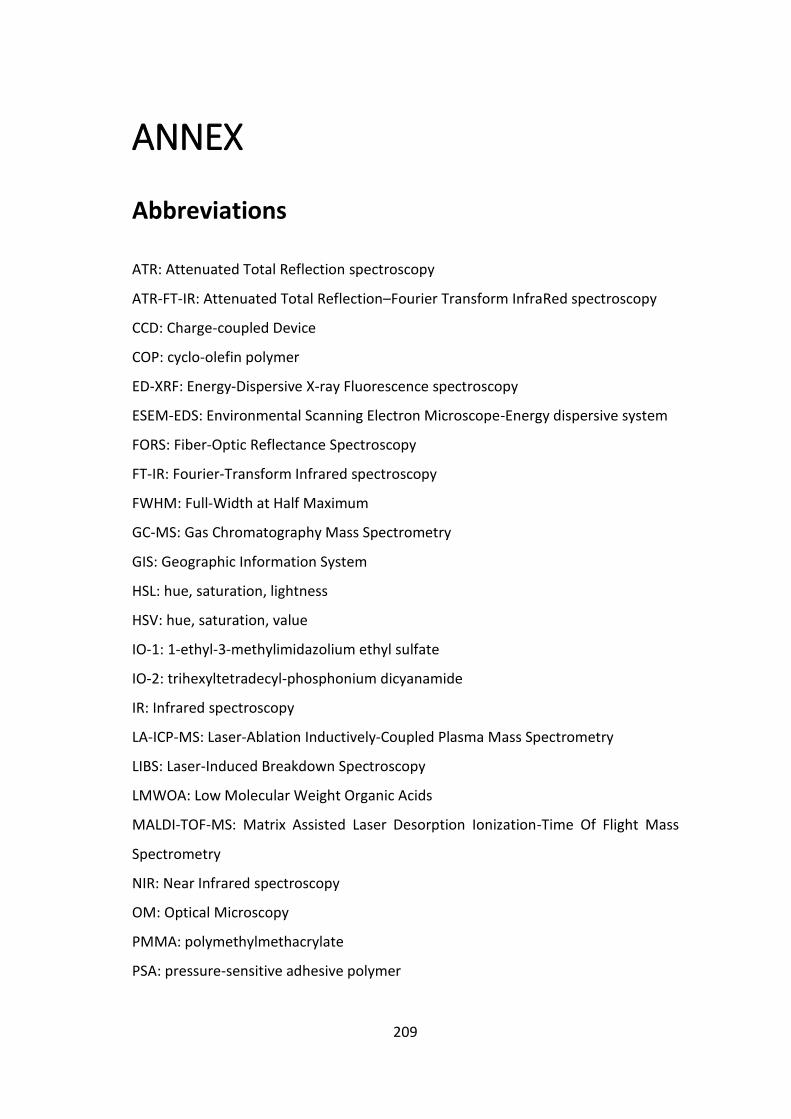
209
ANNEX
Abbreviations
ATR: Attenuated Total Reflection spectroscopy
ATR-FT-IR: Attenuated Total Reflection–Fourier Transform InfraRed spectroscopy
CCD: Charge-coupled Device
COP: cyclo-olefin polymer
ED-XRF: Energy-Dispersive X-ray Fluorescence spectroscopy
ESEM-EDS: Environmental Scanning Electron Microscope-Energy dispersive system
FORS: Fiber-Optic Reflectance Spectroscopy
FT-IR: Fourier-Transform Infrared spectroscopy
FWHM: Full-Width at Half Maximum
GC-MS: Gas Chromatography Mass Spectrometry
GIS: Geographic Information System
HSL: hue, saturation, lightness
HSV: hue, saturation, value
IO-1: 1-ethyl-3-methylimidazolium ethyl sulfate
IO-2: trihexyltetradecyl-phosphonium dicyanamide
IR: Infrared spectroscopy
LA-ICP-MS: Laser-Ablation Inductively-Coupled Plasma Mass Spectrometry
LIBS: Laser-Induced Breakdown Spectroscopy
LMWOA: Low Molecular Weight Organic Acids
MALDI-TOF-MS: Matrix Assisted Laser Desorption Ionization-Time Of Flight Mass
Spectrometry
NIR: Near Infrared spectroscopy
OM: Optical Microscopy
PMMA: polymethylmethacrylate
PSA: pressure-sensitive adhesive polymer
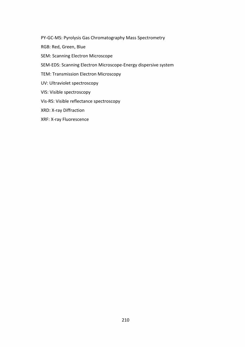
210
PY-GC-MS: Pyrolysis Gas Chromatography Mass Spectrometry
RGB: Red, Green, Blue
SEM: Scanning Electron Microscope
SEM-EDS: Scanning Electron Microscope-Energy dispersive system
TEM: Transmission Electron Microscopy
UV: Ultraviolet spectroscopy
VIS: Visible spectroscopy
Vis-RS: Visible reflectance spectroscopy
XRD: X-ray Diffraction
XRF: X-ray Fluorescence