Application of enzymes in Nitrogen-13 radiochemistry - ADDI
222
-
Upload
khangminh22 -
Category
Documents
-
view
0 -
download
0
Transcript of Application of enzymes in Nitrogen-13 radiochemistry - ADDI


Application of enzymes in Nitrogen-13
radiochemistry; biocatalytic synthesis of
PET radionuclides with biomedical interest
Eunice da Silva
2017

Cover calligraphy Laura Saa
Cover design Eunice S. Da Silva
©2017, Eunice Da Silva. All rights reserved.
No part of this publication may be reproduced, stored in a retrieval system,
or transmitted in any form or by any means, without permission of the
author

Application of enzymes in Nitrogen-13
radiochemistry; biocatalytic synthesis of
PET radionuclides with biomedical interest
PhD Thesis
to obtain the degree of PhD in
Synthetic and Industrial Chemistry
at the University of Basque Country
by
Eunice da Silva
2017
(c)2017 EUNICE DA SILVA FERNANDES

Thesis Supervisors:
Jordi Llop, PhD (Radiochemistry & Nuclear Imaging Lab, CIC
biomaGUNE)
Fernando Lopez-Gallego, PhD (Heterogeneous Biocatalysis Lab,
CIC biomaGUNE)
University tutor:
Esther Lete, PhD, Professor (Department of Organic Chemistry
Faculty of Science and Technology University of the Basque
Country)

Contents
SUMMARY .................................................................................................................. 1
RESUMEN .................................................................................................................... 5
CHAPTER 1 ................................................................................................................ 15
GENERAL INTRODUCTION ................................................................................................ 15
CHAPTER 2 ................................................................................................................ 45
STATE-OF-ART: BIOCATALYSIS IN RADIOCHEMISTRY ............................................................... 45
CHAPTER 3 ................................................................................................................ 81
BACKGROUND AND OBJECTIVES ........................................................................................ 81
CHAPTER 4 ................................................................................................................ 89
EFFICIENT [13N]NITRATE REDUCTION CATALYZED BY A HIGHLY STABLE IMMOBILIZED BIOCATALYST .. 89
CHAPTER 5 .............................................................................................................. 127
ENZYMATIC PREPARATION OF 13N-LABELED AMINO ACIDS AND THEIR BIOLOGICAL ASSESSMENT IN
PROSTATE TUMOR MODELS ............................................................................................ 127
CHAPTER 6 .............................................................................................................. 169
DESIGN AND FULL CHARACTERIZATION OF L-ALANINE IMMOBILIZED ON SOLID AND POROUS
MATERIALS ................................................................................................................. 169
CHAPTER 7 .............................................................................................................. 199
GENERAL CONCLUSIONS ................................................................................................ 199
LIST OF ABBREVIATIONS ......................................................................................... 203
ACKNOWLEDGMENTS ............................................................................................. 207

1
Summary With the widespread installation of biomedical cyclotrons and positron
emission tomography (PET) scanners around the world, scientists are
increasingly recognizing PET as an accessible and valuable tool for the
investigation of physiological or biological challenges in the pre-clinical
and clinical setups. The continuous progress of nuclear imaging techniques
demands for the development and implementation of efficient, clean and
sustainable synthetic routes to manufacture novel radiotracers. This task is
especially challenging for the synthesis of radiotracers labeled with short-
lived β+ emitters, which are extremely valuable since they substantially
reduce the radiation burden on the subject under investigation. Hence,
extremely rapid and selective synthetic schemes must be designed to
access radiotracers radiolabeled with short half-lived positron emitters.
Biocatalysis, although surprisingly underused in radiochemistry,
constitutes an attractive alternative to conventional chemistry. Enzymes
present an exquisite chemical selectivity and high turnover numbers; they
also evolved to work under physiological conditions, where the
concentration of the metabolites is tightly regulated and are rarely in
excess. Furthermore, enzymes are also the most sophisticated chiral
catalysts yielding enantiopure products in most of the cases. Hence, the
use of enzymes can now be seen as an innovative approach to radiolabel
biologically active molecules with short half-lived positron emitters, for
their use in positron emission tomography. Although years 70´s and 80´s
are considered the golden era of biocatalysis in radiochemistry, advances

2
in enzyme engineering during the last decade have significantly enhanced
the toolbox of enzymes available for chemical reactions, which may find
application in the context of radiochemistry. The biocatalytic approaches
for the synthesis of PET radiotracers using Nitrogen-13, Carbon-11 and
Fluorine-18 that have been reported during the last four decades are
reviewed in Chapter 2.
The main target of this PhD thesis was the development of new bio-
strategies in the context of nitrogen-13 radiochemistry, and mainly the
preparation of 13N-labeled precursors and their incorporation in biological
molecules with interest in preclinical application.
In Chapter 4, the reduction of [13N]NO3- to [13N]NO2
- using a
heterogeneous biocatalyst is described. For the design and fabrication of a
suitable heterogeneous biocatalyst, a eukaryotic nitrate reductase from
Aspergillus niger was immobilized on different carriers. Optimal
immobilization and recovered activities were obtained when this enzyme
was immobilized on porous agarose beads activated with a positively
charged tertiary amino group. This immobilized preparation was 12-fold
more thermostable than the soluble enzyme and could be re-used in up to
7 reaction cycles, preserving its initial activity. A pure preparation of
[13N]NO2- was obtained after 4 min reaction at room temperature and it
was successfully applied in a two-step chemo-enzymatic radiosynthesis of
S-[13N]nitrosoglutathione.
A one-pot, enzymatic and non-carrier-added synthesis of a variety of 13N-
labeled amino acids (L-[13N]alanine, [13N]glycine, and L-[13N]serine) is
described in Chapter 5. To that aim, an L-alanine dehydrogenase from
Bacillus subtilis was used, together with nicotinamide adenine
dinucleotide (NADH) as the redox cofactor, and labeled ammonia as the

Summary
3
amine source. Moreover, the addition of formate dehydrogenase from
Candida boidinii in the same reaction vessel allowed the in situ
regeneration of NADH during the radiochemical synthesis of the amino
acids. The enzymatically labeled amino acids have been used to analyze
their in vivo biodistribution in healthy mice. Likewise, the capacity to
selectively accumulate in the tumor has been assessed in a prostate cancer
mouse model, by using dynamic PET-CT imaging.
Lastly, Chapter 6 describes the immobilization of L-Alanine
dehydrogenase from Bacillus subtillis on agarose microbeads, activated
with glyoxyl groups (aliphatic aldehydes) and two other carriers. These
immobilized enzyme preparations were extensively characterized towards
temperature, pH and kinetic parameters. Finally, the optimal
heterogeneous biocatalyst was applied in the synthesis of L-[13N]alanine
using pyruvate and labeled ammonia as the substrates.


5
Resumen La instalación generalizada de ciclotrones biomédicos a nivel mundial ha
suscitado un gran interés entre la comunidad científica, que ve la
tomografía por emisión de positrones (PET, del inglés Positron Emission
Tomography) como una herramienta accesible para abordar de manera no
invasiva, longitudinal y traslacional determinados problemas biológicos,
fisiológicos y/o médicos inabordables mediante otras técnicas. Sin
embargo, la utilización de la PET exige el desarrollo e implementación de
rutas sintéticas que permitan la preparación eficiente y robusta de
radiotrazadores marcados con isótopos emisores de positrones. Esto
resulta especialmente complicado en el caso de la síntesis de
radiotrazadores marcados con emisores de positrones de vida media corta.
Dichos emisores de positrones resultan extremadamente valiosos ya que
permiten reducir de manera significativa la dosis de radiación efectiva
recibida por el sujeto investigado.
La biocatálisis, aunque ha sido sorprendentemente infrautilizada en el
contexto de la radioquímica, constituye una alternativa atractiva a la
química convencional. Las enzimas presentan una excelente selectividad
química y una gran capacidad catalítica. Además, han evolucionado de
manera natural para trabajar en condiciones fisiológicas, donde la
concentración de los metabolitos está fuertemente regulada y raramente en
exceso. Las enzimas son también los catalizadores quirales más
sofisticados que se conocen ya que son capaces de producir productos

6
enantioméricamente puros incluso en esquemas de reacción muy
complejos.
En este contexto, la biocatálisis ha emergido como una estrategia
altamente innovadora para marcar moléculas biológicamente activas con
emisores de positrones de vida media corta, con el fin último de utilizar
dichas moléculas marcadas (radiotrazadores) en estudios de imagen
mediante PET.
La edad de oro de la aplicación de la biocatálisis en radioquímica fueron
las décadas de los 70 y 80. Sin embargo, los avances en ingeniería
enzimática llevados a cabo durante la última década han mejorado
significativamente la cantidad, calidad y versatilidad de las enzimas
disponibles. Este hecho ha abierto nuevas perspectivas en la utilización de
la catálisis enzimática en el contexto de la radioquímica.
El primer objetivo de la presente tesis doctoral ha sido efectuar una
revisión exhaustiva de los diferentes enfoques biocatalíticos utilizados
para la preparación de radiotrazadores marcados con isótopos emisores de
positrones. Todos los trabajos reportados en las últimas cuatro décadas en
lo que se refiere a la síntesis de radiotrazadores PET utilizando Nitrógeno-
13, Carbono-11 y Flúor-18, así como las perspectivas de futuro que ofrece
esta tecnología se describen y discuten en el Capítulo 2 del presente
documento.
Una vez superada la fase de documentación, se definió el principal
objetivo de esta tesis doctoral, que consistía en el desarrollo de nuevas
estrategias basadas en biocatálisis en el contexto de la radioquímica del
nitrógeno-13, con el objetivo último de preparar precursores marcados con 13N y su incorporación en moléculas biológicas con interés en la aplicación

Resumen
7
preclínica, esto es, en la realización de estudios de imagen en animales de
experimentación.
El primer objetivo experimental concreto de la tesis doctoral fue abordar
el desarrollo de un proceso enzimático para la reducción de [13N]NO3- a
[13N]NO2-. Cabe destacar que la irradiación directa de agua ultrapura con
protones acelerados permite la formación eficiente de nitrógeno-13
mediante la reacción nuclear 16O(p,α)13N, siendo la especie química
formada mayoritariamente el anión [13N]NO3-. Dicha especie no tiene gran
aplicación como sintón radiactivo; sin embargo, su reducción a [13N]NO2-
permite abordar la preparación de una gran variedad de moléculas
marcadas, incluyendo nitrosaminas, nitrosotioles y azoderivados, entre
otros. Hasta la fecha, la reducción mencionada arriba se llevaba a cabo de
manera eficiente en una columna de cadmio. Sin embargo, la utilización
de este metal podría conllevar problemas asociados a su toxicidad en el
caso de pretenderse la aplicación en el entorno clínico, y se estimó muy
oportuno desarrollar un proceso enzimático que permitiera evitar la
utilización de cadmio. La consecución de este objetivo se planteó mediante
la utilización de un biocatalizador heterogéneo. Para el diseño del
biocatalizador heterogéneo se inmovilizó una nitrato reductasa eucariota
de Aspergillus niger en diferentes soportes porosos. El rendimiento óptimo
de inmovilización y actividades enzimáticas recuperadas se obtuvieron
cuando esta enzima se inmovilizó en unidades porosas de agarosa,
activadas con un grupo amino terciario cargado positivamente. Este
soporte presentó los grupos funcionales óptimos en términos de densidad
y reactividad para lograr una mayor actividad enzimática después del
proceso de inmovilización. Con el estudio de los parámetros cinéticos
aparentes de la nitrato reductasa inmovilizada en este tipo de soportes,
pudimos observar que la inmovilización daba lugar a un aumento de la Km

8
aparente para el NADPH, este aumento parece ser debido a una serie de
problemas difusionales que presenta el cofactor a la hora de alcanzar los
centros activos de las enzimas inmovilizadas en el microambiente poroso.
Además, esta preparación inmovilizada fue 12 veces más termoestable que
la enzima soluble y pudo reutilizarse hasta 7 ciclos de reacción,
preservando su actividad inicial. En condiciones óptimas (4 minutos,
temperatura ambiente; estas condiciones son comparables a las utilizadas
anteriormente en el proceso de reducción en presencia de cadmio), pudo
obtenerse [13N]NO2- puro que posteriormente pudo utilizarse para abordar
la síntesis de S-[13N]nitrosoglutationa (Figura R.1). De la realización de
este trabajo resultó la primera de las publicaciones asociadas a la presente
tesis doctoral (da Silva, E. S. et al., Catalysis Science & Technology, 2015,
5, 2705-2713).
Figura R.1. Síntesis de S-[13N]nitrosoglutathiona ([13N]GSNO) a partir de
[13N]NO3- producido en el ciclotrón.
Una vez desarrollado el método de reducción descrito en el capítulo 4, se
planteó la posibilidad de emplear métodos biocatalíticos para la

Resumen
9
preparación de compuestos biológicamente activos, concretamente
aminoácidos. Para la selección de los aminoácidos a marcar se contó con
la colaboración del profesor Arkaitz Carracedo, de CIC bioGUNE, que
dispone de un modelo animal de cáncer de próstata y dedica parte de su
investigación al estudio de aberraciones metabólicas en estos tumores. En
este contexto, en el Capítulo 5 de la presente tesis doctoral se presenta la
síntesis enzimática, en un solo paso y sin carrier añadido de diferentes
aminoácidos marcados con nitrógeno-13, concretamente L-[13N]alanina,
[13N]glicina y L-[13N]serina (Figura R.2a). Para ello, se usó una L-alanina
deshidrogenase de Bacillus subtilis NADH–dependiente. Esta enzima es
capaz de llevar a cabo la aminación reductiva de -cetoácidos mediante el
uso de [13N]amoníaco como fuente de grupos amino. Sin embargo, esta
enzima no fue capaz de sintetizar L-aminoácidos muy voluminosos como
la L-fenilalanina y al L-norvalina, debido a que presenta unos valores de
Km para el amonio demasiado altos en la síntesis de los aminoácidos
voluminosos anteriormente descritos. Tras optimización de las
condiciones experimentales (cantidad de catalizador, pH, etc) pudo
conseguirse la preparación de los tres aminoácidos con rendimiento
radioquímico suficiente para abordar estudios en los modelos animales
proporcionados por el Prof. Carracedo. Además, el acoplamiento de este
enzima con una formiato deshidrogenasa de Candida boidinii en el mismo
medio de reacción permitió la regeneración in situ de NADH durante la
radiosíntesis de los aminoácidos (Figura R.2b), de modo que pudo
disminuirse hasta 50 veces la concentración del cofactor sin comprometer
los rendimientos radioquímicos de la reacción.

10
Figura R.2. (a) preparación enzimática de L-[13N]alanina, [13N]glycina, y L-
[13N]serina utilizando L-alanina dehidrogenasa (L-AlaDH) obtenida de Bacillus
subtilis; (b) Preparación de L-[13N]alanina con regeneración del cofactor NADH.
Los aminoácidos marcados enzimáticamente se utilizaron en primer lugar
para estudiar su biodistribución in vivo en ratones sanos (Figura R.3).
Todos los aminoácidos mostraron acumulación en la zona abdominal,
hecho que ya había sido observado para otros aminoácidos en estudios
anteriores reportados en la literatura. Dos de los aminoácidos (L-
[13N]alanina y [13N]glycina) mostraron poca eliminación por orina,
convirtiéndose en potenciales trazadores para la visualización in vivo de
los tumores de próstata.

Resumen
11
Figura R.3. Imágenes PET-CT representativas obtenidas a diferentes tiempos
tras administración intravenosa de L-[13N]alanina en ratón. Se muestran
proyecciones coronales corregistradas con cortes representativos de imágenes
CT; 0-30 s (a); 31-70 s (b); 71-230 s (c); 231-740 s (d); 741-3020 s (e).
Los estudios efectuados en animales con tumor mostraron que los
aminoácidos se acumulaban selectivamente en la próstata, y que dicha
acumulación aumentaba en presencia de tumor (Figura R4). Sin embargo,
es necesario efectuar estudios más exhaustivos y a diferentes tiempos tras
aparición de los tumores para obtener resultados más concluyentes. Los
resultados derivados del capítulo 5 se recogen en la segunda publicación
asociada a esta tesis doctoral (da Silva, E. S. et al., Chemistry – A
European Journal, 2016, 22, 13619-13626).

12
Figure R.4. Imágenes PET-CT (proyecciones sagitales) de animales control y
animales con tumor, obtenidas tras administración de los aminoácidos marcados
con nitrógeno-13. La posición de la vejiga y de la próstata se indican con una
fleche blanca y roja, respectivamente. Como puede observarse, tan solo el
aminoácido L-[13N]serina muestra eliminación significativa por orina.
El trabajo desarrollado en el capítulo 5 implicaba la utilización de enzimas
en disolución. Sin embargo, con el fin de evitar la presencia de enzima en
la disolución final, así como para posibilitar la realización de reacciones
en flujo, se consideró conveniente explorar la posibilidad de inmovilizar
las enzimas. Así pues, en el Capítulo 6 se describe la inmovilización de la
enzima L-alanina deshidrogenase de Bacillus subtillis en microesferas de
agarosa porosa, activadas con grupos de glioxal (aldehídos alifáticos),
además de otros dos soportes. Estas preparaciones enzimáticas
inmovilizadas se caracterizaron en términos de temperatura, pH y

Resumen
13
parámetros cinéticos. En base a estudios físico-químicos de la superficie
de esta enzima, se propouso una orientación óptima a través de la cuál la
enzima era inmovilizada en soportes activados con grupos aldehídos,
dando lugar a enzimas inmovilizadas con una gran estabilidad y una
actividad suficiente para su aplicación en procesos biotecnológicos.
Finalmente, se aplicó el biocatalizador heterogéneo óptimo para abordar
la síntesis de L-[13N]alanina, usando piruvato y amoniaco marcado como
sustratos. Los resultados demostraron que con las enzimas inmovilizadas
podían obtenerse buenos rendimientos radioquímicos, equivalentes a los
obtenidos utilizando las enzimas en disolución. Sin embargo, pudo
reciclarse el catalizador hasta en 5 ciclos consecutivos, sin que esto
afectara de manera significativa al rendimiento obtenido (Figura R5). De
este trabajo, que abre pues las puertas al desarrollo de reacciones en flujo,
resultó la tercera de las publicaciones relacionadas con la presente tesis
docotral (da Silva, E. S. et al., Process Biochemistry, in press).
Figura R.5. Rendimiento cromatográfico obtenido al efectuar síntesis
consecutivas de L-[13N]-alanina utilizando la enzima inmovilizada. La reacción
se llevó a cabo con 45 g x mL-1 de enzima inmovilizada a 25 ºC, pH 8 y t= 20
minutos.
0 1 2 3 4 50
20
40
60
80
100
Cho
rmat
ogr
aphi
c Y
ield
[%
]
Cycles

14
En resumen, en esta tesis doctoral, hemos demostrado que la aplicación de
métodos biosínteticos permite la radiosíntesis selectiva y eficiente de
compuesto marcadas que serían muy díficiles de obtener mediante
métodos químicos. Los excelentes resultados que hemos obtenido aquí han
sido gracias a capacidad que tienen los enzimas para catalizar un amplia
diversidad de reacciones químicas con una gran quimio- regio- y
enantoselectividad. En esta tesis, hemos mostrado que las enzimas se
pueden aplicacar en radioquímica tanto en su forma solúble como en su
forma inmovilzada. Y por eso en este contexto, hemos sido capaces de
desarrollar diferentes procesos sínteticos para la generación de
radiotrazadores basados en Nitrogeno 13.

Chapter 1
General introduction


Chapter 1
17
BIOCATALYSIS
One of the principal functions of enzymes as natural biocatalysts is to
enhance the rate of virtually all chemical reactions within a cell. They
increase the rates of chemical reactions without being permanently altered
or consumed. They also increase the reaction rates without changing the
equilibrium between the reactants and the products.
These unique catalytic properties shown by enzymes make them desirable
in many chemical processes. Enzymes work under mild reaction
conditions (physiological pH and temperature), and constitute
biodegradable and environment-friendly catalysts with high activity and
chemo-, regio- and stereoselectivity.1 Additionally, the use of enzymes
does not require the time-consuming protection/deprotection/activation
steps frequently used in synthetic chemistry. These advantages enable
shorter synthetic routes with less generation of waste products, thus being
both environmentally and economically more attractive than traditional
organic syntheses. Furthermore, enzymes can catalyze a wide spectrum of
chemical and biochemical reactions that can be readily assembled in
sophisticated pathways to synthesize complex molecules. These complex
pathways formed by multi-enzyme systems have naturally evolved to
work within the cellular milieu; however, in the last decades chemists have
isolated enzymes and make them work ex-vivo under non-natural
conditions. Moreover, advances in protein engineering have allowed the
application of enzymes to chemical processes different from the natural
ones. Therefore, intrinsic features of enzymes together with the recent
technologies have paved the way for the implementation of biocatalysis in
industrial processes1,2 as well as in the medical and pharmaceutical fields,
where enzymes have been successfully exploited for the chemical

18
synthesis of drugs and building blocks3-5 and for the treatment of a variety
of diseases.1
Enzymes also present some inherent drawbacks associated with their
natural origin: (i) they are poorly stable either in the presence of non-polar
solvents, under high temperatures or under extreme pH values; (ii) they
show limited reactivity towards artificial substrates (non-natural
reactions); and (iii) they are highly soluble in water, limiting thus their
reusability in batch reactions or their applicability in continuous-flow
processes.6 Fortunately, the significant advances in genomics and
computational biology have resulted in novel experimental and
computational protein engineering tools, which have significantly
enhanced the toolbox of available enzymes.7 On the other hand, enzyme
immobilization has addressed the solubility issue by heterogenization of
the biocatalysts, which enables both reutilization in batch reactions and
integration into flow reactors. In addition, immobilized enzymes show
often increased stability, facilitate the product separation and avoid time-
consuming purification steps.8 All these facts open new avenues for the
application of enzymatic chemistry to the preparation of radiolabeled
compounds in multiple biomedical applications.1,9-12
ENZYME IMMOBILIZATION: THE WORKHORSE OF
HETEROGENEOUS BIOCATALYSIS
Enzyme immobilization involves the fixation of an enzyme to an insoluble
matrix (carrier); however, optimizing the immobilization of enzymes is
challenging. In spite of the difficulties, this methodology has been
accepted as one of the most successful methods to overcome the

Chapter 1
19
limitations of free enzymes.13,14 There are several reasons to immobilize
enzymes (Table 1.1): first, immobilized enzymes act as heterogeneous
biocatalysts that can be easily manipulated and separated from the
products once the reaction is completed, preventing the contamination of
the product and facilitating the downstream processing; second, by using
an optimal immobilization methodology, the immobilization may enhance
both operational and storage stability, while denaturation by exposure to
heat or organic solvents can be prevented; third, when the immobilized
enzymes are highly stable, the resulting heterogeneous biocatalyst can be
recycled for several operational cycles and directly integrated into
continuous chemical processes.14
The immobilization of enzymes has been intensively exploited since the
70’s. However, no universal immobilization protocols to improve both
activity and stability of the immobilized enzymes have been described. For
this reason, enzyme immobilization is still an empirical approach where
trial-and-error approximations are more common than rational approaches.
However, the results obtained during more than 40 years of research in
enzyme immobilization have demonstrated that the physic-chemical
properties of the support material and the immobilization chemistry (the
type of bonds between the enzyme and the solid surface) are essential to
achieve active and stable heterogeneous biocatalysts. In theoretical terms,
structural information about the enzyme can guide the selection of the best
immobilization protocol; however, examples of rational enzyme
immobilization are scarce because of the chemical and geometrical
complexity of protein structures and the heterogeneity of the solid material
surfaces. Mass transfer limitations need often to be addressed by selecting
the proper microstructure of the solid carrier to favor the access of the
substrates from the reaction media to the immobilized catalytic centers.15

20
The immobilization chemistry and the nature of the solid carrier are
fundamental to control the protein orientation and the substrate/product
diffusion, and both factors will be crucial to ensure high activity and
stability of the final heterogeneous biocatalysts.16
Table 1.1. Properties of immobilized enzymes: advantages and drawbacks.
Adapted from the reference.17
Advantages Drawbacks
Reuse of the biocatalyst Loss in enzyme activity
Easier reactor operation Mass transfer limitation
Easier product separation Additional cost
Stability of the enzyme
Selection of a Suitable Carrier Material
The properties of the selected carrier are essential for the performance of
the heterogeneous biocatalyst. Ideal carrier properties include physical
strength, hydrophilicity, inertness, ease of derivatization, biocompatibility,
resistance to microbial attack, and availability at low cost.18 However,
even though immobilization on solid carriers is an established technology,
there are still no general rules for selecting the best carrier for a given
application. Both the immobilization chemistry and the selection of the
optimal carrier are usually based on trial-and-error.
In general, the carriers can be classified as inorganic and organic,
according to their chemical composition. In turn, the organic carriers can
be subdivided into natural and synthetic polymers (Figure 1.1). Natural
polymers such as chitin, chitosan and cellulose have many advantages over

Chapter 1
21
many synthetic polymers in terms of biodegradability, non-toxicity,
physiological inertness, hydrophilicity and remarkable affinity to proteins.
The green properties of those materials make them attractive, especially in
specific applications including food, pharmaceutical, medical and
agricultural processing.14,16 However, membranes prepared from synthetic
polymers have been largely preferred as enzyme carriers because of their
low cost, easy surface modification, resistance to biodegradation and
thermal and chemical stabilities.
Inorganic materials can also be classified into natural and synthetic
materials. Naturally occurring silica-based materials are the most suitable
and versatile matrices for enzyme immobilization and have been widely
used in industrial manufacturing of enzyme-processed products,19,20 as
well as for research21 and medical purposes.22,23 Silica can be easily
fabricated to provide desirable morphology, pore structures, and micro-
channels.24,25 Additionally, the surface of silica gel can be easily modified
by chemical methods to provide different types of functional groups at the
surface.19,26,27 Furthermore, silica is cheap, mechanically stable,
chemically inert, and environmentally friendly. In the last decade, other
synthetic inorganic materials such as metal oxides, metal phosphates,
zeolites and metal organic framework (MOFs) are gaining relevance in the
protein immobilization field.28

22
*Metal-organic-frameworks (MOFs) because they are kind of hybrid materials.
Figure 1.1. General classification of the carriers used for enzyme immobilization.
Adapted from the reference16.
In addition to the nature of the carrier, physical characteristics such as
particle diameter, swelling behavior, mechanical strength and compression
behavior also affect the performance of the immobilized systems. Pore and
particle size of the carrier determine the total surface area and critically
affect the maximum amount of enzyme that can be bound. Here, we can
also categorize the solid carriers as non-porous and porous. Non-porous
carriers show less diffusional limitations, but microscopic particles
(suitable for re-utilization) have low loading capacities. Nanoparticles
have a much larger surface area per mass unit but they are laboriously
manipulated in batch reactors and cause filter clogging during their
reutilization. Hence, porous, micrometric particles are generally preferred
because their high surface area enables higher enzyme loads, their
manipulation during the operational cycles is much easier and finally the
General classification of the carriers
Organic
Natural materials
Polysaccharides: cellulose, dextrans, agar, agarose, chitin, alginate
Proteins: collagen, albumin
Carbon
Synthetic materials
Polystyrene
Other polymers: polyacrylate, polymethacrylates, polyacrylamide, ...
Inorganic
Natural materials
Bentonite
Silica
Synthetic materials
Glass (non-porous and controlled pore)
Metals oxidesZeolites and metal hydroxides*

Chapter 1
23
vast majority of the immobilized enzymes are physically protected by the
porous microenvironment. In the context of this PhD thesis. As example,
cross-linked agarose porous beads (agarose BCL) have been used. The
material is hydrophilic and biocompatible, it can be manufactured in the
micrometric size within a range of porosity and is commercially available
at large scale.29,30 Its main limitation, the high cost, can be overcome by
employing reversible methods that allow regeneration and re-use of the
carrier once the immobilized enzymes become inactive.
Selection of the optimal immobilization chemistry. The pathway to the
enzyme stabilization
In the last decades, a plethora of immobilization protocols has been
reported in the literature.2,13,14,16,31 Depending on the enzyme and
immobilization strategy, the final stability and activity of the enzyme may
be significantly affected.
The chemistries to immobilize enzymes onto solid carriers can be divided
into two main groups based on the nature of the enzyme-carrier bonds: (i)
reversible chemistries, where the enzyme-carrier interaction can be easily
reverted by changing the media conditions, thus enabling the enzyme
elution or lixiviation; and (ii) irreversible chemistries, where the enzymes
are irreversibly attached to the carrier (Figure 1.2).6,14 Reversible
immobilization can be achieved by physical adsorption of the enzyme
based on ionic, van der Waals or hydrophobic interactions, on affinity
interactions and on reversible covalent bonds. Irreversible immobilization
mainly involves irreversible covalent bonds between the protein and the
surface.

24
Figure 1.2. Schematic representation of the main different methods of enzyme
immobilization. Adapted from the reference16.
Methods of Reversible Immobilization
Reversibly immobilized enzymes can be detached from the carrier under
gentle conditions. The use of these enzymatic systems is highly attractive
because when the enzymatic activity decays, the carrier can be regenerated
and reloaded with the fresh enzyme. This methodology is particularly
interesting when the cost of the carrier is high. Indeed, the cost of the
carrier is often a primary factor in the overall cost of immobilized
biocatalysts. In addition, the reversibility between the enzyme and the
carrier surface has proven useful for processes where the enzymes must
work at the boundary of insoluble substrates and then need to be recovered
for subsequent operational cycles.32
In 1916, one of the first immobilized enzymes prepared by adsorption was
reported by Nelson and Griffin, who showed that invertase physically
adsorbed on charcoal (solid powder) kept its catalytic activity.33 One
hundred years later, physical adsorption still remains the oldest, simplest,

Chapter 1
25
and most widely used method for protein immobilization.34 The
phenomenon of physical adsorption can be driven by weak and non-
specific forces such as hydrogen bonding, by Van der Waals forces (alone
or combined), by hydrophobic interactions or by stronger salt bridges
(combination of hydrogen bonding and charge-charge interactions).16,35
Immobilization by ionic adsorption (commonly named as ionic exchange)
is vastly exploited in reversible protein immobilization because protein
surfaces are rich in positively (basic residues) and negatively charged
amino acids that can easily interact with negative and positive groups on
the carrier surface, respectively.34 Protein-carrier interactions are strong
enough to retain the protein attached to the solid phase during the operation
but they can be mainly disrupted by ionic strength and high or low pH.
This immobilization chemistry has revealed highly useful to stabilize the
quaternary structure of multimeric proteins.
Another classical method to reversibly immobilize protein is the affinity
immobilization. Affinity binding is also included which relies on the
creation of different (bio)affinity bonds between a functionalized
(activated) carrier and a specific group present on the protein surface.14 In
most cases, proteins are engineered with affinity tails that selectively react
with affinity groups on the carrier surface.6,16,36 The most well-known
genetically encoded affinity tag is the polyhistidine tag (Hist-tag) that is
selectively bound to carriers activated with metal-chelates (Cu(II), Co(II),
Zn(II), or Ni(II)); the metal determines the selectivity and the strength of
the interaction.37-41 This immobilization is easily and selectively reverted
by adding imidazole groups that compete with the histidines for the
chelates. This chemistry provides a remarkable selectivity and an
extraordinary control of protein orientation, and minimizes the

26
conformational changes suffered by the proteins, yielding highly active but
poorly stabilized proteins upon the immobilization protocol.36
Finally, the reversible immobilization based on disulfide bridges is the
most representative method of reversible covalent immobilization. A thiol
from the protein surface (from the cysteine) selectivity reacts with a
disulfide group on the carrier surface. This immobilization is easily
reverted by reducing agents such as dithiothreitol.16,42,43 The major
advantage of this immobilization chemistry is the high selectivity for
cysteine as unique natural amino acid able to participate in such thiol-
disulfide exchange. Since cysteine residues are rarely found in the protein
surfaces, engineering proteins with unique cysteines is a powerful strategy
to immobilize proteins on disulfide activated materials through any desired
orientation.44,45
Methods of Irreversible Immobilization
The concept of irreversible immobilization means that once the enzyme is
attached to the carrier, the enzyme cannot be physically separated from the
solid material. Unlike reversible immobilization, when the enzymes are
irreversibly bound to the carriers, the solid material cannot be regenerated
once the enzyme becomes inactive. This is an important issue when using
expensive materials. However, irreversible attachment avoids the enzyme
lixiviation during the operational process and frequently improves the
enzyme stability, resulting in increased operational life of the
heterogeneous biocatalysts. Stabilization of proteins through
immobilization mainly relies on multivalent attachments that rigidify
protein structures. In other words, the greater the number of covalent
linkages between the enzyme and the carrier, the more rigid the enzyme
Chapter 1
27
structure becomes. This multivalent covalent attachment has been
demonstrated as one of the main reasons to explain the higher stability of
the immobilized proteins.18 Paradoxically, the rigidification of the protein
structure may noticeably decrease the protein activity. This fact is
particularly important for enzymes, where rigidification might reduce
some fundamental flexibility required for the catalysis. For this reason, it
is essential to reach a compromise between the stability (rigidification) and
activity.
Figure 1.2 shows the three main methodologies for irreversible
immobilization: irreversible attachment to a pre-existing carrier,
irreversible entrapment/microencapsulation of the enzyme into synthetic
materials, and irreversible cross-linking of enzyme aggregates.
Irreversible immobilization on the pre-existing carrier will be the only
technique described in detail in this chapter because it was fundamental to
the experimental work of this thesis.
The vast majority of the irreversible immobilization chemistries are based
on a nucleophilic attack of protein surface residues (Lys, Cys, His and even
Tyr) to a battery of reactive groups (cyanogen bromide, vinyl groups,
aldehyde and epoxide groups) present on the surface of the carrier.
Glutaraldehyde has been exploited to activate aminated carriers to
immobilize primary amine groups, i.e., the N-terminus and the -amino
group of the lysines.16,46,47 This immobilization is highly efficient but its
action mechanism is not fully understood.
CNBr-activated carriers are highly versatile. A plethora of materials have
been activated with this groups (membranes, surfaces, nanoparticles, etc.),
and some of these materials are commercially available.48 Dozens of
proteins have been immobilized through this immobilization chemistry

28
that requires very mild conditions and where the primary amines (mainly
from the N-terminus) of proteins react with the cyanogen bromides to form
cyanate esters or imidocarbonates.49-52 The major disadvantage of this
immobilization chemistry is the low stability of cyanogen bromide groups
under aqueous conditions that often limit the multivalency of the
attachment and the protein load of the final immobilized preparation.
Epoxide and glyoxyl groups are alternative groups for irreversible
immobilization that present a much higher stability than the cyanogen
bromides and whose immobilization mechanism is better understood and
reliable that for the glutaraldehyde groups. Glyoxyl groups are small
aliphatic aldehydes that form reversible imino bonds with the primary
amino groups (-NH2 from Lys and N-terminus) in the enzyme forming
reversible Schiff's bases. Finally, those imine bonds must be reduced to
achieve irreversible secondary amine bonds between the protein and the
carrier.53,54 Under alkaline conditions, glyoxyl groups establish a
multivalent covalent attachment with those protein regions that display the
highest amount of most reactive lysine residues, rigidifying such protein
regions.55 The main drawbacks of this chemistry are the mandatory
alkaline pH (around 10) needed to achieved the multivalent
immobilization and the reduction step required to irreversibly immobilize
the protein.56 In spite of such disadvantages, carriers activated with
glyoxyl groups promote an intense multivalent protein immobilization that
has stabilized a plethora of proteins by a factor of 100-1000.53,57
Epoxide groups, unlike glyoxyl groups, can be attacked by a larger battery
of protein nucleophiles such as Cys, His, Asp, Glu, Tyr and of course Lys.
This nucleophilic attack yields irreversible secondary amine bonds without
requiring any reduction step. Nevertheless, after the immobilization, the

Chapter 1
29
remaining epoxy groups must be blocked by aminated or thiolated
compounds to deactivate the surface in order to avoid undesired post-
immobilization interactions. Remarkably, the vast majority of
economically viable carriers based on polymetacrylate are activated with
epoxide groups which have allowed industrial exploitation of this
chemistry.55,58 The biggest limitation of this immobilization chemistry is
the low reactivity of the epoxy groups. Consequently, this chemistry
promotes lower stabilization factors than the glyoxyl under comparable
conditions (carrier activation degree and nature, enzyme orientation, etc.).
Table 1.2. Characteristics, advantages and drawbacks of most common enzyme
immobilization methods.
Method Bonding type Advantages Drawbacks
Adsorption/
Ionic
bonding
Hydrophobic, Van
der Waals or ionic
interactions
Simple and economical
Soft conformational
change of the enzyme
Can be recycled,
regenerated and reused
Desorption
Non-specific
adsorption
Covalent
binding
Chemical binding
between functional
groups of the
enzyme and carrier
No enzyme release
Potential for enzyme
stabilization
Matrix and enzyme
are not regenerable
Major loss of activity
Entrapment Occlusion of an
enzyme within a
polymeric network
No chemical modification
Wide applicability for
multimeric enzymes
Easy handling and reusage
Mass transfer
limitations
Enzyme release
Cross-
linking
Covalent bonds
between enzymes
by a functional
reactant
Strong linkage – low
enzyme release
Biocatalyst stabilization
Mass transfer
limitations
Loss of activity

30
MOLECULAR IMAGING
Molecular imaging can be defined as a set of non-invasive techniques that
allow the visualization of cell function and the monitoring of molecular
processes in living organisms. Since the 90’s, molecular imaging has
opened a wide range of possibilities for the investigation of biological,
physiological and medical processes which cannot be studied using other
techniques.59-61 Positron emission tomography (PET), single photon
emission computerized tomography (SPECT) and optical/fluoresce
imaging are considered molecular imaging techniques.
The anatomical information provided by molecular imaging techniques is
usually scarce. Thus, both in the clinical and in the preclinical settings,
such techniques are combined with other modalities able to provide
accurate anatomical information. The combination of the resulting images
(multimodality) offers information at the anatomical and molecular levels.
Anatomical modalities include magnetic resonance imaging (MRI) and
computed tomography (CT). Under the administration of a contrast agent,
these modalities can be also considered molecular imaging modalities.
It is worth mentioning that each imaging modality has strengths and
limitations. Thus, while nuclear imaging techniques (PET and SPECT)
offer unparalleled sensitivity, they rarely reach a resolution below 1 mm
in the preclinical setting and a few mm in the clinical systems. On the
contrary, MRI can offer high resolution (up to a few micrometers) while
its sensitivity is low (Table 1.3). Consequently, the application of one
modality or another will depend on the problem under investigation and
often the combination of two or more modalities will provide the most
accurate data.

Chapter 1
31
Table 1.3. Properties of most common clinical and pre-clinical imaging
modalities.59,60,62-66
Technique Energy used Common contrast
agents
Imaging
agent
mass
Spatial
resolution
Temporal
resolution
Sensitivity
of detection
Penetration
depth
Quantification
efficiency
CT X rays I or Ba agents mg range 0.03-1 mm Minutes 0.1 mol.l-1 >300 mm ---
SPECT ɣ rays ɣ emitters <1 ng 0.5-15 mm Minutes 10-11 mol.l-1 >300 mm High
PET Annihilation
photons β+ emitters <1 ng 1-10 mm
Seconds-
minutes 10-12 mol.l-1 >300 mm High
MRI Radio frequency
waves
Gd3+ agents or
Fe3O4 nanoparticles 0.01-100mg 0.03-1mm Minutes-hours10-5 mol.l-1 >300 mm Medium
Optical Visible to
infrared light
Fluorophores or
lanthanides <1 ng-10mg 2-10 mm
Seconds-
minutes 10-11 mol.l-1 1-20 mm Medium
Nuclear Imaging techniques
Nuclear imaging techniques rely on the administration of a radiotracer, i.e.,
a molecule labeled with a radioactive isotope to enable external and non-
invasive detection. Gamma ray-emitters are appropriate radioisotopes
because high-energy gamma rays can travel through biological tissues
without suffering significant scatter or attenuation. These gamma rays can
be detected using specific instrumentation, and the original concentration
of radiotracer can be accurately quantified using tomographic
reconstruction algorithms. As a result, a series of tomographic images
conveying information on absolute radioisotope concentration can be
obtained over time, in vivo and in a non- invasive way. Positron emission
tomography (PET), single photon emission computed tomography
(SPECT) and scintigraphy are nuclear imaging techniques. Nowadays,
these techniques are available in combination with anatomical imaging
such as CT or MRI (Figure 1.3).

32
Positron emission tomography will be the only technique described in
detail in this chapter because it was the only modality used in the context
of this PhD thesis.
Figure 1.3. Fusion modalities of medicine nuclear techniques.
Positron emission tomography (PET)
Positron emission tomography relies on positrons (β+) emitted from
radioisotopes. The positron, after travelling short distances (few
millimeters) within the body annihilates with an electron in the
surrounding matter. The combined mass of the positron and the electron is
converted into two collinear ɣ photons with 511 keV each travelling in
opposite directions (annihilation). These gamma rays can be detected by
especial detectors displayed in a ring surrounding the object of study. By
simultaneous detection of both 511 keV rays, lines of response (LORs)
are identified and used to locate the position in which the annihilation

Chapter 1
33
event occurred. The detection of hundreds of thousands of photons enables
the generation of an image (Figure 1.4).
Figure 1.4. Key steps involved in the development of a molecular imaging study.
PET allows whole body 3D scanning and is commonly associated with
CT, or more recently with MRI. This improves the limitations of its poor
anatomic correlation and allows the generation of an attenuation map for
attenuation correction during image reconstruction. In the clinical field,
PET is widely used to image cancer and inflammation states through a
fluorinated analogue of glucose, 2-[18F]fluoro-2-deoxy-glucose
([18F]FDG), that can identify changes in cellular glucose metabolism
during inflammatory processes or related with the hyperactivity of cancer
cells. PET can also be used to investigate more specific physiological and

34
molecular mechanisms of human disease through the use of appropriate
radiolabeled imaging probes and to study the biodistribution of new drugs
and the effectiveness of new therapies.60
Figure 1.5. A simplified diagram illustrating the principles of PET scan.
Fluorine-18 (18F) and carbon-11 (11C) are frequently used radionuclides in
PET. Nitrogen-13 (13N) and oxygen-15 (15O) are also used to a lesser
extent (see Table 1.4). This is, in part, due to the fact that they can be
produced efficiently in biomedical cyclotrons and have a radioactive decay
almost exclusively by β+ emission, but especially because they are isotopes
of elements with low atomic mass that are highly prevalent in all organic
molecules. Hence, any molecule of interest can a priori be radiolabeled
with one of these positron emitters without modifying its biological
activity. In the context of this PhD thesis, 13N was the PET isotope
exploited.

Chapter 1
35
Table 1.4. Features of most common radionuclides used in PET. Adapted from
references 67 and 68.
Radionuclide T1/2 Nuclear reaction
Target Primary
precursors Decay
product 15O 2.04 min 14N(d,n)15O N2(+O2) [15O]O2 15N
13N 9,97 min 16O(p, α)13N H2O
H2O+EtOH [13N]NO2/3 [13N]NH3
13C
11C 20.4 min 14N(p,α)11C N2(+O2) N2(+H2)
[11C]CO2 [11C]CH4
11B
18F 109.8 min
20Ne(d,α)18F 18O(p,n)18F
Ne(+F2) [18O]H2O
[18F]F2 [18F]F-
18O
Nitrogen-13
Nitrogen-13 is the longest-lived radioisotope of nitrogen and has been used
mostly under the chemical form [13N]NH3 or [13N]amino acids. Nitrogen-
13 with high specific activity can be generated by proton irradiation of
pure water via the 16O(p,α)13N nuclear reaction. It decays by positron
emission to its stable isotope carbon-13. The nitrogen-13 generated by
irradiation of pure water is a mixture of radiolabeled nitrate ([13N]NO3-),
nitrite ([13N]NO2-) and ammonia ([13N]NH3), being [13N]NO3
- the major
species (approx. 85% of total radioactivity).69 The [13N]NO3- can be
reduced to [13N]NO2-, which can be used as labeling agent for the synthesis
of a wide variety of labeled species, including nitrosothiols, nitrosamines,
azo derivatives and triazoles.70-75
Cyclotron produced [13N]NO3- can be also reduced to [13N]NH3 by
classical or in-target reduction methods.76 Classical reduction methods
include the treatment with titanium hydroxide,77 Raney-nickel catalyst,78
or Devarda's alloy.79 More commonly, [13N]NH3 can be generated directly
in the irradiated target by the addition of ethanol,80,81 hydrogen gas80 or
methane82 to the pure water, which act as scavengers and prevent radiolytic
oxidation of the ammonia. Ammonia can be also produced in-target using

36
cryogenic targets.83 Besides direct application as flow and perfusion
marker,84-86 [13N]NH3 is the main precursor for the synthesis of natural
amino acids.57,87-96
REFERENCES
(1) Illanes, A.: Enzyme Biocatalysis: Principles and Applications; Springer Netherlands, 2008.
(2) DiCosimo, R.; McAuliffe, J.; Poulose, A. J.; Bohlmann, G. Industrial use of immobilized enzymes. Chemical Society Reviews 2013, 42, 6437-6474.
(3) Tao, J.; Lin, G. Q.; Liese, A.: Biocatalysis for the Pharmaceutical Industry: Discovery, Development, and Manufacturing; Wiley, 2009.
(4) Pollard, D. J.; Woodley, J. M. Biocatalysis for pharmaceutical intermediates: the future is now. Trends in biotechnology 2007, 25, 66-73.
(5) Patel, R. N.: Biocatalysis in the Pharmaceutical and Biotechnology Industries; CRC Press, 2006.
(6) Homaei, A. A.; Sariri, R.; Vianello, F.; Stevanato, R. Enzyme immobilization: an update. Journal of Chemical Biology 2013, 6, 185-205.
(7) Bommarius, A. S. Biocatalysis: A Status Report. Annual review of chemical and biomolecular engineering 2015, 6, 319-345.
(8) da Silva, E. S.; Gomez-Vallejo, V.; Llop, J.; Lopez-Gallego, F. Efficient nitrogen-13 radiochemistry catalyzed by a highly stable immobilized biocatalyst. Catalysis Science & Technology 2015.
(9) Bezborodov, A. M.; Zagustina, N. A. Enzymatic biocatalysis in chemical synthesis of pharmaceuticals (Review). Applied Biochemistry and Microbiology 2016, 52, 237-249.
(10) Bolivar, J. M.; Eisl, I.; Nidetzky, B. Advanced characterization of immobilized enzymes as heterogeneous biocatalysts. Catalysis Today 2016, 259, Part 1, 66-80.
(11) Sheldon, R. A.; van Pelt, S. Enzyme immobilisation in biocatalysis: why, what and how. Chem Soc Rev 2013, 42, 6223-6235.
(12) Reetz, M. T. What are the Limitations of Enzymes in Synthetic Organic Chemistry? Chemical record (New York, N.Y.) 2016, 16, 2449-2459.
(13) Bickerstaff, G.: Immobilization of Enzymes and Cells; Humana Press, 1996.

Chapter 1
37
(14) Datta, S.; Christena, L. R.; Rajaram, Y. R. S. Enzyme immobilization: an overview on techniques and support materials. 3 Biotech 2013, 3, 1-9.
(15) Fessner, W. D.: Biocatalysis: From Discovery to Application; Springer Berlin Heidelberg, 2000.
(16) Guisan, J. M.: Immobilization of Enzymes and Cells; Humana Press, 2006.
(17) Katchalski-Katzir, E. Immobilized enzymes--learning from past successes and failures. Trends in biotechnology 1993, 11, 471-478.
(18) Mohamad, N. R.; Marzuki, N. H. C.; Buang, N. A.; Huyop, F.; Wahab, R. A. An overview of technologies for immobilization of enzymes and surface analysis techniques for immobilized enzymes. Biotechnology, Biotechnological Equipment 2015, 29, 205-220.
(19) Blanco, R. M.; Terreros, P.; Fernández-Pérez, M.; Otero, C.; Dı́az-González, G. Functionalization of mesoporous silica for lipase immobilization: Characterization of the support and the catalysts. Journal of Molecular Catalysis B: Enzymatic 2004, 30, 83-93.
(20) Carlsson, N.; Gustafsson, H.; Thörn, C.; Olsson, L.; Holmberg, K.; Åkerman, B. Enzymes immobilized in mesoporous silica: A physical–chemical perspective. Advances in Colloid and Interface Science 2014, 205, 339-360.
(21) Qhobosheane, M.; Santra, S.; Zhang, P.; Tan, W. Biochemically functionalized silica nanoparticles. Analyst 2001, 126, 1274-1278.
(22) Bharti, C.; Nagaich, U.; Pal, A. K.; Gulati, N. Mesoporous silica nanoparticles in target drug delivery system: A review. International Journal of Pharmaceutical Investigation 2015, 5, 124-133.
(23) Liberman, A.; Mendez, N.; Trogler, W. C.; Kummel, A. C. Synthesis and surface functionalization of silica nanoparticles for nanomedicine. Surface science reports 2014, 69, 132-158.
(24) Wang, Y.; Caruso, F. Mesoporous Silica Spheres as Supports for Enzyme Immobilization and Encapsulation. Chemistry of Materials 2005, 17, 953-961.
(25) Takahashi, H.; Li, B.; Sasaki, T.; Miyazaki, C.; Kajino, T.; Inagaki, S. Immobilized enzymes in ordered mesoporous silica materials and improvement of their stability and catalytic activity in an organic solvent. Microporous and Mesoporous Materials 2001, 44–45, 755-762.
(26) Luechinger, M.; Prins, R.; Pirngruber, G. D. Functionalization of silica surfaces with mixtures of 3-aminopropyl and methyl groups. Microporous and Mesoporous Materials 2005, 85, 111-118.
(27) Maria Chong, A. S.; Zhao, X. S.; Kustedjo, A. T.; Qiao, S. Z. Functionalization of large-pore mesoporous silicas with organosilanes by direct synthesis. Microporous and Mesoporous Materials 2004, 72, 33-42.

38
(28) Lian, X.; Fang, Y.; Joseph, E.; Wang, Q.; Li, J.; Banerjee, S.; Lollar, C.; Wang, X.; Zhou, H. C. Enzyme-MOF (metal-organic framework) composites. Chem Soc Rev 2017.
(29) Zucca, P.; Fernandez-Lafuente, R.; Sanjust, E. Agarose and Its Derivatives as Supports for Enzyme Immobilization. Molecules (Basel, Switzerland) 2016, 21.
(30) Porath, J.; Axén, R. Immobilization of enzymes to agar, agarose, and sephadex support. Methods in Enzymology 1976, 44, 19-45.
(31) da Silva, E. S.; Gómez-Vallejo, V.; Llop, J.; López-Gallego, F. Structural, kinetic and operational characterization of an immobilized l-aminoacid dehydrogenase. Process Biochemistry.
(32) Kudina, O.; Zakharchenko, A.; Trotsenko, O.; Tokarev, A.; Ionov, L.; Stoychev, G.; Puretskiy, N.; Pryor, S. W.; Voronov, A.; Minko, S. Highly efficient phase boundary biocatalysis with enzymogel nanoparticles. Angewandte Chemie (International ed. in English) 2014, 53, 483-487.
(33) Nelson, J. M.; Griffin, E. G. Adsorption of invertase. Journal of the American Chemical Society 1916, 38, 1109-1115.
(34) Jesionowski, T.; Zdarta, J.; Krajewska, B. Enzyme immobilization by adsorption: a review. Adsorption 2014, 20, 801-821.
(35) Cao, L.; Schmid, R. D.: Carrier-bound Immobilized Enzymes: Principles, Application and Design; Wiley, 2006.
(36) Kimple, M. E.; Brill, A. L.; Pasker, R. L. Overview of Affinity Tags for Protein Purification. Current protocols in protein science / editorial board, John E. Coligan ... [et al.] 2013, 73, Unit-9.9.
(37) Agarwal, G.; Naik, R. R.; Stone, M. O. Immobilization of histidine-tagged proteins on nickel by electrochemical dip pen nanolithography. J Am Chem Soc 2003, 125, 7408-7412.
(38) Fischer, N. O.; Blanchette, C. D.; Chromy, B. A.; Kuhn, E. A.; Segelke, B. W.; Corzett, M.; Bench, G.; Mason, P. W.; Hoeprich, P. D. Immobilization of His-tagged proteins on nickel-chelating nanolipoprotein particles. Bioconjugate chemistry 2009, 20, 460-465.
(39) Hefti, M. H.; Milder, F. J.; Boeren, S.; Vervoort, J.; van Berkel, W. J. A His-tag based immobilization method for the preparation and reconstitution of apoflavoproteins. Biochimica et biophysica acta 2003, 1619, 139-143.
(40) Miyazaki, M.; Kaneno, J.; Yamaori, S.; Honda, T.; Briones, M. P.; Uehara, M.; Arima, K.; Kanno, K.; Yamashita, K.; Yamaguchi, Y.; Nakamura, H.; Yonezawa, H.; Fujii, M.; Maeda, H. Efficient immobilization of enzymes on microchannel surface through His-tag and application for microreactor. Protein and peptide letters 2005, 12, 207-210.

Chapter 1
39
(41) Gupta, M. N.; Jain, S.; Roy, I. Immobilized metal affinity chromatography without chelating ligands: purification of soybean trypsin inhibitor on zinc alginate beads. Biotechnology progress 2002, 18, 78-81.
(42) Ovsejevi, K.; Manta, C.; Batista-Viera, F. Reversible covalent immobilization of enzymes via disulfide bonds. Methods in molecular biology (Clifton, N.J.) 2013, 1051, 89-116.
(43) Carlsson, J.; Axen, R.; Unge, T. Reversible, covalent immobilization of enzymes by thiol-disulphide interchange. European journal of biochemistry 1975, 59, 567-572.
(44) Gulla, K. C.; Gouda, M. D.; Thakur, M. S.; Karanth, N. G. Enhancement of stability of immobilized glucose oxidase by modification of free thiols generated by reducing disulfide bonds and using additives. Biosensors and Bioelectronics 2004, 19, 621-625.
(45) Godoy, C. A.; de las Rivas, B.; Grazu, V.; Montes, T.; Guisan, J. M.; Lopez-Gallego, F. Glyoxyl-disulfide agarose: a tailor-made support for site-directed rigidification of proteins. Biomacromolecules 2011, 12, 1800-1809.
(46) Walt, D. R.; Agayn, V. I. The chemistry of enzyme and protein immobilization with glutaraldehyde. TrAC Trends in Analytical Chemistry 1994, 13, 425-430.
(47) Lopez-Gallego, F.; Guisan, J. M.; Betancor, L. Glutaraldehyde-mediated protein immobilization. Methods in molecular biology (Clifton, N.J.) 2013, 1051, 33-41.
(48) Singh, B. D.: Biotechnology: Expanding Horizons; Kalyani Publishers, 2012.
(49) Aehle, W.: Enzymes in Industry; Wiley, 2008.
(50) Axen, R.; Porath, J.; Ernback, S. Chemical Coupling of Peptides and Proteins to Polysaccharides by Means of Cyanogen Halides. Nature 1967, 214, 1302-1304.
(51) Schnapp, J.; Shalitin, Y. Immobilization of enzymes by covalent binding to amine supports via cyanogen bromide activation. Biochemical and Biophysical Research Communications 1976, 70, 8-14.
(52) Kavran, J. M.; Leahy, D. J. Coupling Antibody to Cyanogen Bromide-Activated Sepharose. Methods in enzymology 2014, 541, 27-34.
(53) Lopez-Gallego, F.; Fernandez-Lorente, G.; Rocha-Martin, J.; Bolivar, J. M.; Mateo, C.; Guisan, J. M. Stabilization of enzymes by multipoint covalent immobilization on supports activated with glyoxyl groups. Methods in molecular biology (Clifton, N.J.) 2013, 1051, 59-71.
(54) Mateo, C.; Palomo, J. M.; Fuentes, M.; Betancor, L.; Grazu, V.; López-Gallego, F.; Pessela, B. C. C.; Hidalgo, A.; Fernández-Lorente, G.;

40
Fernández-Lafuente, R.; Guisán, J. M. Glyoxyl agarose: A fully inert and hydrophilic support for immobilization and high stabilization of proteins. Enzyme and Microbial Technology 2006, 39, 274-280.
(55) Santos, J. C. S. d.; Barbosa, O.; Ortiz, C.; Berenguer-Murcia, A.; Rodrigues, R. C.; Fernandez-Lafuente, R. Importance of the Support Properties for Immobilization or Purification of Enzymes. ChemCatChem 2015, 7, 2413-2432.
(56) Grazu, V.; Betancor, L.; Montes, T.; Lopez-Gallego, F.; Guisan, J. M.; Fernandez-Lafuente, R. Glyoxyl agarose as a new chromatographic matrix. Enzyme and Microbial Technology 2006, 38, 960-966.
(57) da Silva, E. S.; Gómez-Vallejo, V.; Llop, J.; López-Gallego, F. Structural, kinetic and operational characterization of an immobilized l-aminoacid dehydrogenase. Process Biochemistry 2017.
(58) Barbosa, O.; Torres, R.; Ortiz, C.; Berenguer-Murcia, A.; Rodrigues, R. C.; Fernandez-Lafuente, R. Heterofunctional supports in enzyme immobilization: from traditional immobilization protocols to opportunities in tuning enzyme properties. Biomacromolecules 2013, 14, 2433-2462.
(59) Tian, J.: Molecular Imaging: Fundamentals and Applications; Zhejiang University Press, 2013.
(60) James, M. L.; Gambhir, S. S. A molecular imaging primer: modalities, imaging agents, and applications. Physiol. Rev. 2012, 92, 897-965.
(61) Townsend, D. W.; Cheng, Z.; Georg, D.; Drexler, W.; Moser, E. Grand challenges in biomedical physics. Front. Phys. 2013, 1, 1-6.
(62) Levin, C. S. New imaging technologies to enhance the molecular sensitivity of positron emission tomography. Proceedings of the IEEE 2008, 96, 439-467.
(63) Schmitz, G. Ultrasonic imaging of molecular targets. Basic Res. Cardiol. 2008, 103, 174-181.
(64) Fass, L. Imaging and cancer: a review. Mol. Oncol. 2008, 2, 115-152.
(65) Liu, X.; Jia, L. The Conduct of Drug Metabolism Studies Considered Good Practice (I): Analytical Systems and In Vivo Studies. Curr. Drug Metab. 2007, 8, 815-821.
(66) Sigel, H.: Metal Ions in Biological Systems: Volume 40: The Lanthanides and Their Interrelations with Biosystems; Taylor & Francis: Switzerland, 2003.
(67) Miller, P. W.; Long, N. J.; Vilar, R.; Gee, A. D. Synthesis of 11C, 18F, 15O, and 13N radiolabels for positron emission tomography. Angewandte Chemie (International ed. in English) 2008, 47, 8998-9033.
(68) De Lima, J. J.: Nuclear Medicine Physics; CRC Press, 2016.

Chapter 1
41
(69) Miller, P. W.; Long, N. J.; Vilar, R.; Gee, A. D. Synthesis of 11C, 18F, 15O, and 13N Radiolabels for Positron Emission Tomography. Angewandte Chemie International Edition 2008, 47, 8998-9033.
(70) Llop, J.; Gómez-Vallejo, V.; Bosque, M.; Quincoces, G.; Peñuelas, I. Synthesis of S-[13N]nitrosoglutathione (13N-GSNO) as a new potential PET imaging agent. Applied Radiation and Isotopes 2009, 67, 95-99.
(71) Gómez-Vallejo, V.; Kato, K.; Oliden, I.; Calvo, J.; Baz, Z.; Borrell, J. I.; Llop, J. Fully automated synthesis of 13N-labeled nitrosothiols. Tetrahedron Letters 2010, 51, 2990-2993.
(72) Gómez-Vallejo, V.; Kato, K.; Hanyu, M.; Minegishi, K.; Borrell, J. I.; Llop, J. Efficient system for the preparation of [13N]labeled nitrosamines. Bioorganic & Medicinal Chemistry Letters 2009, 19, 1913-1915.
(73) Gaja, V.; Gomez-Vallejo, V.; Puigivila, M.; Perez-Campana, C.; Martin, A.; Garcia-Osta, A.; Calvo-Fernandez, T.; Cuadrado-Tejedor, M.; Franco, R.; Llop, J. Synthesis and evaluation of 13N-labelled azo compounds for beta-amyloid imaging in mice. Molecular imaging and biology : MIB : the official publication of the Academy of Molecular Imaging 2014, 16, 538-549.
(74) Joshi, S. M.; de Cozar, A.; Gomez-Vallejo, V.; Koziorowski, J.; Llop, J.; Cossio, F. P. Synthesis of radiolabelled aryl azides from diazonium salts: experimental and computational results permit the identification of the preferred mechanism. Chemical communications (Cambridge, England) 2015, 51, 8954-8957.
(75) Joshi, S. M.; Gomez-Vallejo, V.; Salinas, V.; Llop, J. Synthesis of 13N-labelled polysubstituted triazoles via Huisgen cycloaddition. RSC Advances 2016, 6, 109633-109638.
(76) Suzuki, K.; Yoshida, Y. Production of [13N]NH3 with ultra-high specific activity. Applied Radiation and Isotopes 1999, 50, 497-503.
(77) Krizek, H.; Lembares, N.; Dinwoodie, R.; Gloria, I.; Lathrop, K. A.; Harper, P. V. Production of radiochemically pure 13NH3 for biomedical studies using the 16O(p,a)13N reaction. J. Nucl. Med. 1973, 629.
(78) Dence, C. S.; Welch, M. J.; Hughey, B. J.; Shefer, R. E.; Klinkowstein, R. E. Production of [13N]ammonia applicable to low energy accelerators. Nucl Med Biol 1994, 21, 987-996.
(79) Sobczyk, D. P.; van Grondelle, J.; de Jong, A. M.; de Voigt, M. J. A.; van Santen, R. A. Production of chemically pure gaseous [13N]NH3 pulses for PEP studies using a modified DeVarda reduction. Applied Radiation and Isotopes 2002, 57, 201-207.

42
(80) Berridge, M. S.; Landmeier, B. J. In-target production of [13N]ammonia: Target design, products, and operating parameters. Applied Radiation and Isotopes 1993, 44, 1433-1441.
(81) Kumar, R.; Singh, H.; Jacob, M.; Anand, S. P.; Bandopadhyaya, G. P. Production of nitrogen-13-labeled ammonia by using 11MeV medical cyclotron: our experience. Hellenic journal of nuclear medicine 2009, 12, 248-250.
(82) Krasikova, R. N.; Fedorova, O. S.; Korsakov, M. V.; Landmeier Bennington, B.; Berridge, M. S. Improved [13N]ammonia yield from the proton irradiation of water using methane gas. Applied Radiation and Isotopes 1999, 51, 395-401.
(83) Firouzbakht, M. L.; Schlyer, D. J.; Wolf, A. P.; Fowler, J. S. Mechanism of nitrogen-13-labeled ammonia formation in a cryogenic water target. Nucl Med Biol 1999, 26, 437-441.
(84) Kitsiou, A. N.; Bacharach, S. L.; Bartlett, M. L.; Srinivasan, G.; Summers, R. M.; Quyyumi, A. A.; Dilsizian, V. 13N-Ammonia myocardial blood flow and uptake. Relation to functional outcome of asynergic regions after revascularization 1999, 33, 678-686.
(85) Xiangsong, Z.; Xinjian, W.; Yong, Z.; Weian, C. 13N-NH3: a selective contrast-enhancing tracer for brain tumor. Nuclear medicine communications 2008, 29, 1052-1058.
(86) Di Carli, M. F.; Lipton, M. J.: Cardiac PET and PET/CT Imaging; Springer New York, 2007.
(87) Gelbard, A. S.; Benua, R. S.; Reiman, R. E.; McDonald, J. M.; Vomero, J. J.; Laughlin, J. S. Imaging of the Human Heart after Administration of L-(N-13)Glutamate. Journal of Nuclear Medicine 1980, 21, 988-991.
(88) Barrio, J. R.; Baumgartner, F. J.; Henze, E.; Stauber, M. S.; Egbert, J. E.; MacDonald, N. S.; Schelbert, H. R.; Phelps, M. E.; Liu, F.-T. Synthesis and Myocardial Kinetics of N-13 and C-11 Labeled Branched-Chain l-Amino Acids. Journal of Nuclear Medicine 1983, 24, 937-944.
(89) da Silva, E. S.; Gómez-Vallejo, V.; Baz, Z.; Llop, J.; López-Gallego, F. Efficient Enzymatic Preparation of 13N-Labelled Amino Acids: Towards Multipurpose Synthetic Systems. Chemistry – A European Journal 2016, 22, 13619-13626.
(90) Baumgartner, F. J.; Barrio, J. R.; Henze, E.; Schelbert, H. R.; MacDonald, N. S.; Phelps, M. E.; Kuhl, D. E. 13N-labeled L-amino acids for in vivo assessment of local myocardial metabolism. Journal of medicinal chemistry 1981, 24, 764-766.

Chapter 1
43
(91) Lambrecht, R. H. D.; Slegers, G.; Claeys, A.; Gillis, E.; Vandecasteele, C. Enzymatic synthesis of radiopharmaceutically pure 13N-labelled L-glutamate. Radiochemical and Radioanalytical Letters 1983, 58, 39-48.
(92) Helus, F.; Weber, K.; Zeisler, S.; Maier-Borst, W. An automatic system for the enzymatic synthesis of13N-glutamate. Journal of Radioanalytical and Nuclear Chemistry 1991, 155, 9-13.
(93) Gelbard, A. S.; Cooper, A. J. L.; Asano, Y.; Nieves, E.; Filc-Dericco, S.; Rosenspire, K. C. Methods for the enzymatic synthesis of tyrosine and phenylalanine labeled with nitrogen-13. International Journal of Radiation Applications and Instrumentation. Part A. Applied Radiation and Isotopes 1990, 41, 229-233.
(94) Gelbard, A. S.; Kaseman, D. S.; Rosenspire, K. C.; Meister, A. Enzymatic syntheses of phosphate, l-citrulline, and N-carbamyl l-aspartate labeled with either 13N or 11C. International Journal of Nuclear Medicine and Biology 1985, 12, 235-242.
(95) Cooper, A. J.; Gelbard, A. S. The use of immobilized glutamate dehydrogenase to synthesize 13N-labeled L-amino acids. Anal Biochem 1981, 111, 42-48.
(96) Elmaleh, D.; Hnatowitch, D.; Kulprathipanja, S. A novel synthesis of 13N-L-aspargine. Journal of Labelled Compounds and Radiopharmaceuticals 1979, 16, 92-93.


Chapter 2
State-of-art: biocatalysis in radiochemistry


47
INTRODUCTION
Biocatalysis, although surprisingly underutilized in radiochemistry,
constitutes an attractive alternative to conventional chemistry. First,
enzymes present an exquisite chemical selectivity and high turnover
numbers; second, enzymes were evolved to work under physiological
conditions, where the concentration of the metabolites is tightly regulated
and are rarely in excess. This scenario resembles the conditions for
radiochemical reactions where the concentration of the radioactive
precursors is extremely low; third, enzymes are the most sophisticated
chiral catalysts yielding enantiopure products in most of the cases.
Therefore, by harnessing the enzyme properties in PET radiochemistry,
chemists have the opportunity to synthesize radiotracers through fast and
selective biosynthetic schemes, while minimizing the formation of side
products and preventing the formation of racemic mixtures.
Hitherto, enzymes have been employed to radiosynthesize [13N/11C]amino
acids, [18F]nucleoside derivatives and [11C/18F]proteins. Additionally,
enzymatic reactions have been employed to regio- and stereospecifically
introduce radioactive precursors into a plethora of biological molecules.1,2
Enzyme immobilization has also been utilized to achieve high
radiochemical yields during repeated batch cycles.3-6 Most of the advances
in the application of enzymatic reactions in radiochemistry occurred in the
70´s-80´s, and were summarized by Gelbard in 1981.7 Since then, to the
best of our knowledge, the role of biocatalysis in radiochemistry has never
been reviewed again. The aim of this chapter is to provide an overview of
the biocatalytic approaches that have been reported during the last four
decades for the synthesis of PET radiotracers (see Figure 2.1) using
nitrogen-13, carbon-11, and fluorine-18.

48
Figure 2.1. Number of scientific publications reporting the application of
biocatalysis to radiochemistry since 1980. Data obtained using PubMed, 2015
NLM and Web of Science, 2013 Thomson Reuters.
Nitrogen-13
Nitrogen-13 is the longest-lived radioisotope of nitrogen and has been used
mostly under the chemical form [13N]NH3 or [13N]amino acids. Nitrogen-
13 with high specific activity can be generated by proton irradiation of
pure water via the 16O(p,α)13N nuclear reaction. The nitrogen-13 generated
is a mixture of radiolabeled nitrate ([13N]NO3-), nitrite ([13N]NO2
-) and
ammonia ([13N]NH3), being [13N]NO3- the major species (approx. 85% of
total radioactivity). The [13N]NO3- can be reduced to [13N]NO2
-, which can
be used as labeling agent for the synthesis of a wide variety of labeled
Chapter 2
49
species, including nitrosothiols, nitrosamines, azo derivatives and
triazoles.8-13 This reduction can be easily performed by chemical methods
using cadmium-copper activated columns.8,14
As mentioned in the previous chapter, cyclotron produced [13N]NO3- can
be also reduced to [13N]NH3 by classical or in-target reduction methods.15
Besides direct application as flow and perfusion marker,16-18 [13N]NH3 is
the main precursor for the synthesis of natural amino acids. In general,
[13N]NH3 can be incorporated into amino acids through three main
enzymatic routes (Figure 2.2): a) using amino acid dehydrogenases that
catalyze the reductive amination of -ketoacids using nicotinamide
adenine dinucleotide (NADH) as redox cofactor and [13N]NH3 as the
amine source;1,2,19-26 b) using amino acid synthetases that catalyze the
insertion of [13N]NH3 into the -carboxylic group of acidic amino acids
by using adenosine triphosphate (ATP);2,22,23,27 and c) using a bi-
enzymatic system integrated by an amino acid dehydrogenase (usually
glutamate dehydrogenase) that catalyzes the reductive amination of -
ketoglutarate to produce L-[13N]glutamate, that is concurrently used by a
transaminase that transfers the radiolabeled amine group to the target -
ketoacid to form the desired L-[13N]amino acid.2,22

50
Figure 2.2. Examples of the main enzymatic routes to incorporate [13N]NH3 into
amino acids. (A) Using amino acid dehydrogenases; (B) using amino acid
synthetases and (C) using a bi-enzymatic system formed by an amino acid
dehydrogenase and a transaminase.
These synthetic strategies have been often applied by using soluble
enzymes (Table 2.1). This, in turn, leads to the potential contamination of
the final preparation with pyrogenic enzymes and poses difficulty in the
purification process.7 This problem was smartly solved by immobilizing
the enzymes on solid carriers through covalent and irreversible bonds.
Several immobilized enzymes, including multi-enzyme systems, have
been applied to the heterogeneous synthesis of 13N-labeled amino acids
(e.g. glutamine22, glutamate1,19,20,22-24, asparagine27, citrulline2,

Chapter 2
51
aspartate2,22, alanine1,20,22). These amino acids have been obtained with
different radiochemical yields, ranging from 10% for L-[13N]asparagine
using an asparagine synthetase,27 to 95% for L-[13N]glutamate using a
glutamate dehydrogenase.23 The glutamate dehydrogenase proved
versatile to synthesize a plethora of L-[13N]amino acids beyond glutamate
(see Figure 2.2A).1,20 Some of the above-mentioned amino acids were
investigated in vivo both in animals and humans in order to assess their
biodistribution and metabolism.19,20 The preparation of L-[13N]glutamate
was semi-automated in 1983 by Lambrecht et al.,23 who could synthesize
this labeled amino acid with high purity and high specific activity (1.11
GBq.µmol-1) using a glutamate dehydrogenase immobilized on porous
glass beads in short times (10 minutes). Later on, the same group adapted
this methodology to the fabrication of β-[13N]-nicotinamide adenine
dinucleotide ([13N]NAD) and -[13N]aminobutyric acid ([13N]GABA).28,29
Finally, this technology was optimized to manufacture pure L-
[13N]glutamate through a full automated system.24
Besides amino acids, other biological molecules have been labeled with
nitrogen-13 by using [13N]NH3 as the amine source and carbamyl
phosphate synthase and carbamoyl transferases as the catalysts. Gelbard et
al. synthesized [13N]carbamyl phosphate, L-[-13N]citrulline and
[carbamyl-13N]-L-aspartate using carbamyl phosphate synthetase co-
immobilized with either aspartate transcarbamylase or ornithine
transcarbamylase. These amino acid derivatives were obtained with
radiochemical yields of around 15%. Labeled carbamyl phosphate could
be obtained using carbamyl phosphate synthetase as the catalyst, [13N]NH3
as the ammonia source, carbamic acid as substrate and ATP as the
cofactor; in a concurrent reaction, the corresponding transcarbamylase

52
transferred the carbamyl group to the ornithine or the aspartate to achieve
the corresponding amino acid derivative.2
Table 2.1. Literature published on enzymatic reactions (functional group transfer
and transformations) applied to nitrogen-13 chemistry since 1980. Entries are
ordered chronologically.
Radionuclide Biosynthetic route Enzyme family
Reaction Compound Ref.
[13N]Ammonia Asparagine synthetase EC 6.3.5.4 Acid amide hydrolysis
L-[13N]Asparagine 27
[13N]Ammonia Glutamate
Dehydrogenase (Immobilized)
EC 1.4.1.2 Reductive amination
L-[13N]Glutamate 19,22
[13N]Ammonia
Glutamate Dehydrogenase
EC 1.4.1.2 Reductive amination
L-[13N]Alanine 22 Glutamate-Pyruvate Transaminase (Immobilized)
EC 2.6.1.2
[13N]Ammonia Glutamine synthethase EC 6.3.1.2 Acid amide hydrolysis
L-[13N]Glutamine 22
[13N]Ammonia Aspartate ammonia-lyase EC 4.3.1.1 Addition L-[13N]Aspartate 22
[13N]Ammonia
Glutamate Dehydrogenase
(immobilized/ semi-automated production
method)
EC 1.4.1.2 Reductive amination
L-[13N]Glutamate 23
[13N]Ammonia Glutamate
Dehydrogenase (immobilized)
EC 1.4.1.3 Reductive amination
L-[13N]Glutamate L-α-
[13N]Aminobutyrate L-[13N]Valine
L-[13N]Methionine L-[13N]Leucine L-[13N]Alanine
1,20
[13N]Ammonia Carbamyl phosphate
synthetase (Co-immobilized)
EC 6.3.5.5
Phosphorylat
ion/ Amination
[13N]Carbamyl phosphate
2
[13N]Ammonia
Ornithine transcarbamylase
EC 2.1.3.3 Carbamoylati
on L-[-13N]Citrulline 2 Carbamyl phosphate
synthetase (Co-immobilized)
EC 6.3.5.5 Phosphorylat
ion/ Amination
[13N]Ammonia
Aspartate transcarbamylase
EC 2.1.3.2 Carbamoylati
on [Carbamyl-13N] Carbamyl-L-
aspartate
2 Carbamyl phosphate synthetase
(Co-immobilized) EC 6.3.5.5
Phosphorylation/
Amination
[13N]Ammonia
Glutamate dehydrogenase (Immobilized)
EC 1.4.1.3 Reductive amination
Carbamyl-L-[13N]Aspartate
2
Aspartate transaminase (Immobilized)
EC 2.6.1.1
Transamination
Aspartate transcarbamylase
(Immobilized) EC 2.1.3.2
Carbamoylation
[13N]Ammonia NAD synthetase (Immobilized)
EC 6.3.1.5 Amide group
transfer [13N]-NAD 28

Chapter 2
53
Radionuclide Biosynthetic route Enzyme family
Reaction Compound Ref.
[13N]Ammonia
N.D.
(two subsequent enzymatic Reaction)
N.D.
N.D.
[13N]-GABA
29
[13N]Ammonia (via L-
[13N]Glutamate)
Aspartate aminotransferase
(Immobilized) EC 2.6.1.1
Transamination
L-[13N]Tyrosine 25
[13N]Ammonia Phenylalanine dehydrogenase (Immobilized)
EC 1.4.1.20 Reductive amination
L-[13N]Phenylalanine
L-[13N]Tyrosine
25
[13N]Ammonia
Glutamate Dehydrogenase
(automated production method)
EC 1.4.1.2 Reductive amination
L-[13N]Glutamate 24
[13N]Nitrate Nitrate reductase
(Immobilized) EC 1.7.1.1 Reduction [13N]Nitrite 30
Carbon-11
Over the last two decades of the 20th century, biocatalysis played an
important role in the developments of carbon-11 radiochemistry. Carbon-
11 is an attractive and important β+ emitting radionuclide for the
radiolabeling of molecules with biological activity due to the ubiquitous
presence of carbon in natural products and drug-like compounds. The
cyclotron production of carbon-11 is achieved by proton irradiation of
nitrogen-14 via the 14N(p,α)11C nuclear reaction. By adding small amounts
of O2 or H2 to the target, [11C]CO2 and [11C]CH4 are produced,
respectively. One major drawback in [11C]CO2 production is the inevitable
contamination of the cyclotron target and the gas lines with “cold” CO2,
which leads to a contamination of up to 1000 atoms of stable carbon per
one carbon-11 atom produced, and results in low specific activities. This
effect is less significant in the case of [11C]CH4.31,32 Both [11C]CO2 and
[11C]CH4 can be chemically derivatized to other 11C-labeled precursors
such as [11C]hydrogen cyanide, [11C]acetate, [11C]methanol, [11C]methyl
iodide, [11C]propionate and [11C]carbonate. In this context, biocatalysis
has exploited those precursors to incorporate carbon-11 into more than 50
biological targets (Table 2.2). In carbon-11 radiochemistry, as well as for

54
nitrogen-13, the use of immobilized enzymes is more popular than the use
of their soluble counterparts, because the immobilized enzymes facilitate
product purification and enzyme reutilization, as well as allow the enzyme
integration into continuous flow reactions.
In a pioneering work in the enzymatic synthesis of 11C-labeled
radiotracers, a phosphoenolpyruvate carboxylase (EC4.1.1.31) and aspartate
transaminase (EC2.6.1.1) were separately immobilized on sepharose
activated with cyanogen bromide groups and applied for the synthesis of
L-[4-11C]aspartic acid using [11C]CO2 as the 11C source.33 In brief,
cyclotron-produced [11C]CO2 and the appropriate substrate
(phosphoenolpyruvate) were flushed through a column packed with
immobilized carboxylase, generating [4-11C]oxaloacetate that was
subsequently contacted with L-glutamate and passed through a second
packed column containing an immobilized aspartate transaminase, which
enabled the amine transfer from L-glutamate to the [4-11C]oxaloacetic acid
to yield L-[4-11C]aspartic acid (Figure 2.3). [11C]CO2 was also used as a
radioactive precursor for the synthesis of several amino acid derivatives
based on the coordinate action of carbamoyl phosphate synthetase and
amino acid transaminases. This synthetic strategy has been very valuable
for studying the L-glutamate metabolism in a variety of tissues.2,33,34

Chapter 2
55
Figure 2.3. Schematic representation of the radiosynthesis of L-[4-11C]aspartic
acid. This radiosynthesis was performed using a phosphoenolpyruvate
carboxylase (EC4.1.1.31) and aspartate transaminase (EC2.6.1.1) separately
immobilized on sepharose activated with cyanogen bromide groups and [11C]CO2
as the 11C source.
Enzymatic carbon-11 radiochemistry has also been applied to other
carbon-11 precursors beyond [11C]CO2. For example, [11C]cyanide was
used as a radioactive precursor to synthesize L-[11C]amino acids and other
biological amines.35 In this reaction scheme, O-acetyl-L-serine
sulfhydrylase and -cyano-α-aminobutyric acid synthase (EC2.5.1.49)
catalyze the nitrilation of O-acetyl-L-serine and O-acetyl-L-homoserine to
yield β-[11C]cyano-L-alanine and γ-[11C]cyano-α-amino-L-butyric acid,
respectively. These intermediate nitriles were further converted into L-[4-11C]aspartic acid and L-[5-11C]glutamate by alkaline hydrolysis with 50-
70% decay corrected radiochemical yields and radiochemical purities
higher than 95% (Figure 2.4).36,37 The same chemo-enzymatic route was
applied to the synthesis of L-[methyl-11C]methionine and its analogues
[11C]ethionine and [11C]propionine using [11C]cyanide as radioactive
precursor and their corresponding O-acetyl-protected amino acids.38

56
Figure 2.4. Schematic representation of the chemoenzymatic radiosynthesis of
L-[4-11C]aspartic acid (1) and L-[5-11C]glutamate (2). The synthesis of these
compounds was carried out using O-acetyl-L-serine sulfhydrylase and -cyano-
α-aminobutyric acid synthase to catalyze the nitrilation of O-acetyl-L-serine and
O-acetyl-L-homoserine to yield their intermediates β-[11C]cyano-L-alanine and
γ-[11C]cyano-α-amino-L-butyric acid, respectively.
Another carbon-11 precursor that can be directly used by enzymes is
[11C]acetate which can be labeled in two different positions (carboxylic
and 2-position).39 [11C]acetate is the main precursor to synthesize
[11C]acetyl-CoA,40,41 one of the main hubs in the central metabolism of
cells. [11C]acetyl-CoA can be synthesized through two different enzymatic
pathways; (i) a two-step cascade catalyzed by an acetate kinase (EC2.7.2.1)
that phosphorylates [11C]acetate yielding [11C]acetyl-phosphate, and a
phosphotransacetylase (EC2.3.1.8) that incorporates the coenzyme A into the
phosphorylated intermediate yielding [11C]acetyl-CoA; and (ii) a single-
step reaction catalyzed by coenzyme A synthetase (EC6.2.1.1) that directly
incorporates coenzyme A into [11C]acetate producing [11C]acetyl-CoA.
Interestingly, [11C]acetyl-CoA has been used as a general acetylating agent
to synthesize a variety of carbon-11 labeled molecules such as [11C]acetyl-

Chapter 2
57
L-carnitine,41 L-[11C]carnitine,41 [11C]acetyl-L-serotonin,42 and [11C]N-
acetyl-D-glucosamine43.
[11C]methanol and [11C]propionate have also been employed sporadically
for the enzymatic preparation of 11C-labeled radiotracers. For example, L-
[11C]serine can be synthesized by an alcohol oxidase co-immobilized with
a catalase that oxidizes [11C]methanol to [11C]formaldehyde.44,45 This
radioactive aldehyde is then chemically condensed to tetrahydrofolate
forming a methyl-transfer cofactor that is sequentially used by a serine
hydroxyl-methyltransferase (EC2.1.2.1) to transfer the radiolabeled methyl
group to glycine yielding L-[11C]serine.44 Likewise, [11C]formaldehyde
has been applied to the synthesis of nucleosides, by using an enzyme
system integrated by thymidylate synthase and alkaline phosphatase co-
immobilized on membranes fabricated with hollow fibers to yield
[11C]thymidine and 2´-Fluoro-5-[11C]-methyl-β-L-arabinofuranosyluracyl
([11C]-FMAU).45,46 Also, [11C]propionate can be enzymatically
incorporated into acyl-L-carnitine to form [carbomyl-11C]propionyl-L-
carnitine through two different multi-enzyme systems.47
Unlike the above-mentioned precursors, [11C]methyl iodide cannot be
directly utilized by enzymes but can be efficiently incorporated into L-
[11C]alanine48,49 and L-[11C]methionine38,50 through chemical methods.
These amino acids can be subsequently utilized by different enzymes to
synthesize radiotracers with biomedical interest. For example, L-
[11C]alanine can be enzymatically converted into [11C]pyruvic acid by the
action of alanine racemase and D-amino acid oxidase, leading to
quantitative deamination of the amino acid. The dynamic racemization of
the amino acid mediated by the racemase allows the D-amino acid oxidase
to access 100% of the amino acid in spite of its strict enantioselectivity
58
towards the D-isomer. The catalase is required in this reaction to in situ
eliminate hydrogen peroxide, an undesired by-product generated by the
oxidase. Alternatively, alanine racemase can be coupled with an alanine
dehydrogenase to quantitatively transform L-[11C]alanine into
[11C]pyruvic acid via a NAD+-dependent oxidative deamination catalyzed
by an alanine dehydrogenase.51 The resulting [11C]pyruvic acid can be
easily reduced to [11C]lactic acid by adding a NADH-dependent lactate
dehydrogenase.60,62,64,65 From [11C]pyruvate, several aromatic L-
[11C]amino acids can also be synthesized. For example, by using a
transaminase/tyrosinase enzymatic tandem, L-tyrosine derivatives such as
L-[11C]tyrosine and 3,4-dihydroxy-L-[11C]phenylalanine (L-[11C]DOPA)
can be synthesized. Alternatively, the labeled pyruvate can be converted
into L-[11C]tryptophan or 5-hydroxy-L-[11C]tryptophan by using a
tryptophanase.49,52-54 Interestingly, accurate selection of the enzymatic
route and the 11C-labeled precursor ([11C]cyanide or [11C]methyl iodide)
enables the preparation of L-[11C]tryptophan or L-[11C]tyrosine labeled in
two different positions (carboxylic or β-position; see Figure 2.5).49,52,55-57
The synthesis of these aromatic [11C]amino acids was refined by co-
immobilizing the multi-enzyme systems51 and by automating the
procedure58,59. Using an automated system, L-[11C]DOPA and 5-hydroxy-
L-[11C]tryptophan could be synthesized with >99% radiochemical purity
and 5.3 and 22.0 GBq.μmol-1 specific activities, respectively.58 Advances
on the enzymatic routes have allowed the production of these aromatic
[11C]amino acids in sufficient amounts to evaluate in vivo their
biodistribution.53,60-62
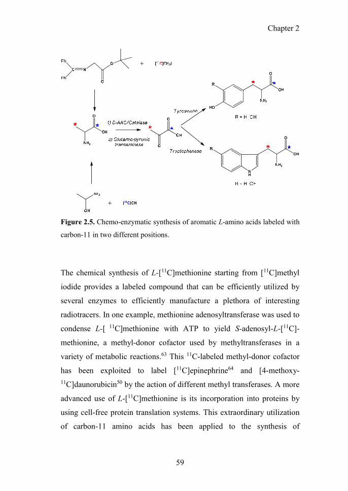
Chapter 2
59
Figure 2.5. Chemo-enzymatic synthesis of aromatic L-amino acids labeled with
carbon-11 in two different positions.
The chemical synthesis of L-[11C]methionine starting from [11C]methyl
iodide provides a labeled compound that can be efficiently utilized by
several enzymes to efficiently manufacture a plethora of interesting
radiotracers. In one example, methionine adenosyltransferase was used to
condense L-[ 11C]methionine with ATP to yield S-adenosyl-L-[11C]-
methionine, a methyl-donor cofactor used by methyltransferases in a
variety of metabolic reactions.63 This 11C-labeled methyl-donor cofactor
has been exploited to label [11C]epinephrine64 and [4-methoxy-11C]daunorubicin50 by the action of different methyl transferases. A more
advanced use of L-[11C]methionine is its incorporation into proteins by
using cell-free protein translation systems. This extraordinary utilization
of carbon-11 amino acids has been applied to the synthesis of

60
[11C]interleukin 8, a chemoattractant cytokine with promising application
in chemo- and immunotherapy.65 The in vitro synthesis of labeled proteins
is based on cell-free pure preparations of protein synthesis machinery from
E.coli that contain all the elements required for the sequential transcription
and translation of genes into proteins (Figure 2.6).66 With this system,
[11C]interleukin 8 was synthesized with a 63% radiochemical yield in only
20 minutes. The [11C]cytokine was further purified by cation exchange and
ultrafiltration methods, with a resulting radiochemical purity >95%. This
pioneering work opens a promising path to the synthesis of radiotracers
based on peptides and biotechnology-derived antibodies, which are
becoming increasingly important as a major source of bio-based drugs.67
Figure 2.6. Schematic illustration of the proposed labeled protein preparation
system using a cell-free translation system.

Chapter 2
61
Table 2.2. Literature published on enzymatic reactions (carbon-carbon bond
formation, functional group transfer and transformations) applied in the carbon-
11 chemistry since 1980.
Radionuclide Biosynthetic route Enzyme family Reaction Compound Ref.
[11C]Carbon dioxide
Phosphoenolpyruvate carboxylase
(Immobilized) EC 4.1.1.31 Decarboxylation
L-[4-11C]Aspartic acid 33 Aspartate transaminase
(Immobilized) EC 2.6.1.1 Transamination
[11C]Carbon dioxide
Carbamyl phosphate synthetase
(Immobilized) EC 6.3.5.5
Phosphorylation/ Amination
[11C]Carbamyl phosphate
2
[11C]Carbon dioxide
Ornithine transcarbamylase
EC 2.1.3.3 Carbamoylation L-[ureido-
11C]Citrulline 2 Carbamyl phosphate
synthetase (Co-immobilized)
EC 6.3.5.5 Phosphorylation/
Amination
[11C]Carbon dioxide
Aspartate transcarbamylase
EC 2.1.3.2 Carbamoylation [Carbamyl-
11C]Carbamyl-L-aspartate
2 Carbamyl phosphate synthetase
(Co-immobilized) EC 6.3.5.5
Phosphorylation/ Amination
[11C]Carbon dioxide
1) Phosphoenol pyruvate carboxylase
1) EC 4.1.1.31 1) Carboxylation 1) [11C]Oxaloacetate
34
2) Citrate synthase 2) EC 2.3.3.1 2) Addition 2) [11C]Citrate 3) Aconitase 3) EC 4.2.1.3 3) Elimination 3) [11C]Isocitrate 4) Isocitrate
dehydrogenase 4) EC 1.1.1.42
4) Oxidative decarboxylation
4) [α-11C]Ketoglutarate
5) Glutamate-pyruvate Transaminase
5) EC 2.6.1.2 5) Transamination 5) L-[11C]Glutamate
[11C]Acetate
1) Acetate kinase 1) EC 2.7.2.1 1)
Phosphorylation 1)
[11C]Acetylphosphate
34
2) Phosphotransacetylase
2) EC 2.3.1.8 2) Acylation 2) [11C]Acetyl-CoA
3) Citrate synthase 3) EC 2.3.3.1 3) Addition 3) [11C]Citrate 4) Aconitase 4) EC 4.2.1.3 4) Elimination 4) [11C]Isocitrate 5) Isocitrate
dehydrogenase 5) EC 1.1.1.42
5) Oxidative decarboxylation 5) [-11C]Ketoglutarate
6) Glutamate-pyruvate Transaminase
6) EC 2.6.1.2 6) Transamination 6) L-[11C]Glutamate
Hydrogen 11C]cyanide
Mandelonitrile lyase EC 4.1.2.10 Cyanohydrin
formation
[11C]Cyanohydrin intermediate for [11C]Octopamine
35
Hydrogen 11C]cyanide
L-3-cyanoalanine synthase
EC 4.4.1.9 Elimination (β –
replacement)
Intermediate compound for L-2,4-Dianino4-
11C]Butyric acid
36
Hydrogen 11C]cyanide
O-acetyl- L-serine sulfhydrylase
EC 2.5.1.47 Elimination (β –
replacement) Intermediate compound for 4-11C]Aspartic acid
37
Hydrogen 11C]cyanide
-cyano-α-aminobutyric acid
synthase EC 2.5.1.49
Elimination (β – replacement)
Intermediate compound for L-5-11C]Glutamate
37
[11C]Methyl iodide
-cyano-α-aminobutyric acid
synthase (Immobilized)
EC 2.5.1.49 Elimination L-[methyl-11C]
Methionine 38
[11C]Carbon dioxide
Acetate kinase EC 2.7.2.1 Quantification by phosphorylation
[1−11C]acetate 39
[1-11C]Acetate Acetyl CoA synthetase
(Immobilized) EC 6.2.1.1 Acid-thiol ligation
[11C]Acetyl coenzyme A
40
[11C]Acetate Acetyl CoA synthetase EC 6.2.1.1 Acid-thiol ligation O-[11C]Acetyl CoA 41
[11C]Acetate
Acetyl CoA synthetase EC 6.2.1.1 Acid-thiol ligation O-[11C]Acetyl-L-
Carnitine 41
Carnitine
acetyltransferase EC 2.3.1.7 Acylation

62
Radionuclide Biosynthetic route Enzyme family Reaction Compound Ref.
[11C]Acetate Carnitine
acetyltransferase EC 2.3.1.7 Acylation L-[11C]Carnitine 41
[1-11C] acetate
(via [11C]acetylCoA)
Acetyl CoA synthetase
EC 6.2.1.1
Acid-thiol ligation
[11C]N-acetylserotonin 42 Arylamine N-
acetyltransferase
EC 2.3.1.5
Acylation
[1-11C]acetate
(via
[11C]acetylCoA)
Acetyl CoA synthetase
EC 6.2.1.1
Acid-thiol ligation
[11C]N-acetyl-D-glucosamine
43 Glucosamine N-acetyltransferase
EC 2.3.1.3 Acylation
[11C]Methanol
Alcohol oxidase EC 1.1.3.13 Oxidation
L-[3-11C]Serine 44
Catalase (co-immobilized)
EC 1.11.1.6
Serine Hydroxyl-
methyltransferase EC 2.1.2.1
Hydroxymethylation
[11C]Carbon dioxide
Alcohol oxidase EC 1.1.3.13 Oxidation 11C]Formaldehyde 45 Catalase
(co-immobilized) EC 1.11.1.6
[11C]Carbon dioxide (via
11C]Formaldehyde)
Thymidylate synthase EC 2.1.1.45 Methylation 11C]Thymidine 46 Alkaline phosphatase
(immobilized) EC 3.1.3.1
Dephosphorylation
11C]2'-Arabino-2'-Fluoro-β-5-MethyI-
Urid (FMAU)
[11C]Propionate
Acetate kinase EC 2.7.2.1 Phosphorylation NCA [carbonyl-
11C]Propionyl L-Carnitine
47 Phosphotransacetylase EC 2.3.1.8 Acylation
Carnitine acetyltransferase
EC 2.3.1.7 Acylation
[11C]Propionate
Acetyl CoA synthetase EC 6.2.1.1 Acid-thiol
ligation
Acylation
NCA [carbonyl- 11C]Propionyl L-Carnitine
47 Carnitine acetyltransferase
EC 2.3.1.7
11C]Methyl iodide
D-amino acid oxidase / Catalase
EC 1.4.3.3 / E.C. 1.11.1.6
Resolution / Redox reaction
[3-11C]Pyruvic acid 48
Glutamic-pyruvic Transaminase
EC 2.6.1.2 Transamination L-[11C]Alanine
Hydrogen 11C]cyanide
D-amino acid oxidase / Catalase
EC 1.4.3.3 / E.C. 1.11.1.6
Resolution / Redox reaction L- [carboxy-
11C]Tyrosine 49,53 Glutamic-pyruvic
transaminase EC 2.6.1.2 Transamination
β-Tyrosinase EC 4.1.99.2 β – replacement L- [carboxy -
11C]DOPA
Hydrogen 11C]cyanide
D-amino acid oxidase / Catalase
EC 1.4.3.3 / E.C. 1.11.1.6
Resolution / Redox reaction
L- [3-11C]Tryptophan 49,54 Glutamic-pyruvic
transaminase EC 2.6.1.2 Transamination 5-Hydroxy-L-
[3-11C]tryptophan Tryptophanase EC 4.1.99.1 β – replacement
Hydrogen 11C]cyanide
D-amino acid oxidase / Catalase
EC 1.4.3.3 / E.C. 1.11.1.6
Resolution / Redox reaction
Transamination
L-[1-11C]Alanine 49 Glutamic-pyruvic
Transaminase EC 2.6.1.2
[methyL-11C] Methionine
L-methionine-S-adenosine transferase
EC 2.1.1.12 Methylation S-methyl-L-[methyl-
11C] Methionine 50
Carminomycin-4-O-methyltransferase
EC 2.1.1.292 Methylation [4-methoxy-
11C]Daunorobicin
11C]Methyl iodide
(via [11C]Carbon dioxide)
Alanine racemase/Alanine Dehydrogenase
EC 5.1.1.1/ EC 1.4.1.1
Isomerization/ Oxidative
deamination L-[3-11C]DOPA 51 β-tyrosinase
(Co-immobilized) EC 4.1.99.2 β – replacement

Chapter 2
63
Radionuclide Biosynthetic route Enzyme family Reaction Compound Ref.
11C]Methyl iodide
(via [11C]Carbon dioxide)
Alanine racemase/ Alanine
Dehydrogenase
Tryptophanase (Co-immobilized)
EC 5.1.1.1/ EC 1.4.1.1
EC 4.1.99.1
Isomerization/ Oxidative
deamination
β – replacement
5-Hydroxy-L- [3-11C]tryptophan
51
11C]Methyl iodide
(via [11C]Carbon dioxide)
Alanine racemase/D-amino acid oxidase
EC 5.1.1.1/ EC
1.4.3.3
Isomerization/
Resolution L-[3-11C]DOPA 51,58,59 β-tyrosinase
(Co-immobilized) EC 4.1.99.2 β – replacement
11C]Methyl iodide
(via [11C]Carbon dioxide)
Alanine racemase/D-amino acid oxidase
EC 5.1.1.1/ EC 1.4.3.3
Isomerization/ Resolution 5-Hydroxy-L-
[3-11C]tryptophan 51,58,59
Tryptophanase (Co-immobilized)
EC 4.1.99.1 β – replacement
[11C]Carbon dioxide
D-amino acid oxidase / Catalase
EC 1.4.3.3 / E.C. 1.11.1.6
Resolution / Redox reaction
L- [3-11C]Tyrosine 52,53 Glutamic-pyruvic transaminase
EC 2.6.1.2 Transamination
β-Tyrosinase EC 4.1.99.2 β – replacement L- [3-11C]DOPA
11C]Methyl iodide
D-amino acid oxidase / Catalase
EC 1.4.3.3 / E.C. 1.11.1.6
Resolution / Redox reaction
L- [β-11C]Tryptophan
55 Glutamic-pyruvic
Transaminase
EC 2.6.1.2 Transamination 5-Hydroxy-L- [β-11C]tryptophan
Tryptophanase EC 4.1.99.1 β – replacement
[11C]Carbonate
Mutant strain of the cyanobacterium
Synechocystis PCC 6803
-- Photosynthesis L-[11C]Phenylalanine 56
[11C]Carbon
dioxide Aspartate transaminase
(Immobilized) EC 2.6.1.1 Transamination [3-11C]Phenylalanine 57
[methyl-11C] Methionine
Methionine adenosyltransferase
EC 2.5.1.6 Adenosyl group
transfer [methyl-11C]Adenosyl-
methionine 63
[methyl-11C] Methionine
L-methionine-S-adenosine transferase
EC 2.5.1.6 Adenosyl group
transfer [11C]-(-)-Epinephrine 64
Phenylethanolamine-N-methyl transferase
EC 2.1.1.28 Methylation
L-[11C]
Methionine Cell-free system PURESYSTEM Protein synthesis [11C]interleukin 8 65
[11C]Carbon dioxide
Pyruvate: ferredoxin 2-oxidoreductase
L-Lactate
dehydrogenase
EC 1.2.7.1
EC 1.1.1.27
Oxidative decarboxylation
Deamination
[11C]Pyruvate acid
L-[3-11C]Lactic acid
68
11C]Methyl iodide
Glutamate-pyruvate transaminase
EC 2.6.1.2 Transamination
L-[3-11C]Lactic acid
69
L-Lactate dehydrogenase
EC 1.1.1.27 Deamination
[11C] Hydrogen Cyanide (NCA)
D-amino acid oxidase (Immobilized)
EC 1.4.3.3 Resolution L-[11C]Leucine 70,71
[11C]Carbon dioxide
Green algae Scenedesmus obtuslusculus Chod.
-- Photosynthesis [11C]Glucose 72
[11C] Hydrogen Cyanide (NCA)
D-amino acid oxidase / Catalase
(Co-immobilized)
EC 1.4.3.3 / E.C. 1.11.1.6
Resolution / Redox reaction
L-[11C]Alanine 73

64
Radionuclide Biosynthetic route Enzyme family Reaction Compound Ref.
[11C] Hydrogen Cyanide (NCA)
D-amino acid oxidase /
Catalase (Co-immobilized)
EC 1.4.3.3 / E.C. 1.11.1.6
Resolution /
Redox reaction
[1-11C]Pyruvic acid
73
L-Alanine dehydrogenase
EC 1.4.1.1 Oxidative
deamination
[11C] Hydrogen Cyanide (NCA)
L-Lactate dehydrogenase
EC 1.1.1.27 Reduction L-[1-11C]Lactic Acid 73
Hydrogen
11C]cyanide
D-amino acid oxidase / Catalase
EC 1.4.3.3 / E.C. 1.11.1.6
Resolution / Redox reaction
[1-11C]pyruvic acid 74
Hydrogen 11C]cyanide
D-amino acid oxidase /
Catalase
EC 1.4.3.3 / E.C. 1.11.1.6
Resolution /
Redox reaction L-[1-11C]Lactic Acid 75 Glutamic-pyruvic
transaminase EC 2.6.1.2 Transamination
L-Lactic dehydrogenase
EC 1.1.1.27 Reduction
11C]Methyl iodide
D-amino acid oxidase / Catalase
EC 1.4.3.3 / E.C. 1.11.1.6
Resolution / Redox reaction
L-[3-11C]Lactic Acid 75 Glutamic-pyruvic
transaminase EC 2.6.1.2 Transamination
L-Lactic dehydrogenase
EC 1.1.1.27 Reduction
[11C]Methyl iodide
D-mannitol dehydrogenase
EC 1.1.1.67 Oxidation D-[16-11C]Fructose 76
11C]Methyl iodide
(via [11C]Carbon dioxide)
Alanine racemase/ Alanine
Dehydrogenase
EC 5.1.1.1/ EC 1.4.1.1
Isomerization/ Oxidative
deamination [3-11C]Pyruvic acid 77
Alanine racemase/D-amino acid oxidase (Co-immobilized)
EC 5.1.1.1/ EC 1.4.3.3
Fluorine-18
The number of enzymatic applications in fluorine-18 radiochemistry is
much lower than in carbon-11 and nitrogen-13 radiochemistry (Table 2.3)
because the repertoire of enzymatic reactions that use fluorine in nature is
very limited. However, incorporation of fluorine-18 into biological
molecules is priceless for PET radiochemistry since fluorine-18 has a very
convenient half-life for in vivo studies, especially in the context of small
molecules.78,79 [18F]fluoride ion ([18F]F-) is cyclotron-generated by
irradiation of 18O-enriched water ([18O]H2O) with high-energy protons via
the 18O(p, n)18F nuclear reaction. The presence of water molecules into the
final radioactive solution affects [18F]F- reactivity, which limits direct use
in nucleophilic substitution reactions. To overcome this drawback,
[18F]Fluoride ions are captured in an anion-exchange resin and further

Chapter 2
65
formulated using a phase transfer catalyst (e.g. Kryptofix [2.2.2]) a polar,
non-protic solvents to favor the nucleophilic attack.
Until XXst century, no enzymes capable to directly incorporate cyclotron-
produced [18F]F- had been reported. Hence, the first 18F-labeling
enzymatic reactions reported in the 1980’s and 1990’s were based on the
use of 18F-labeled precursors. For example, 2-deoxy-2-[18F]fluoro-D-
glucose ([18F]FDG) was purified from the chemical synthesis by using an
hexokinase that selectively phosphorylates D-glucose, facilitating the
purification of the radiolabeled sugar.80 In a more synthetic approach, 6-
[18F]fluorodopamine, 4-[18F]catechol and [18F]FDG-1-phosphate were
used as substrates of dopamine β-hydroxylase81, β-tyrosinase82 and UDP-
glucose pyrophosphorylase83 to synthesize (−)-6-
[18F]fluoronorepinephrine, 6-[18F]fluoro-L-DOPA and uridine diphospho-
2-deoxy-2-[18F]fluoro-α-D-glucose (UDP-[18F]FDG), respectively
(Figure 2.7).
In 2002, the isolation of a fluorinase (adenosyl-fluoride synthase:
EC2.5.1.63) from the soil bacterium Streptomyces cattleya meant a
breakthrough in enzymatic synthesis of fluorinated compounds. The
structural and biochemical characterization of this enzyme opened new
prospects for the direct 18F-radiofluorination of biological molecules.84-87
Nature has specialized fluorinases to convert S-adenosyl-L-methionine
(SAM) to 5’-fluoro-5’-deoxyadenosine (FDA)86 through a nucleophilic
attack of the fluorine ions on the methionine derivative. Since enzymes
work by default under aqueous conditions, it was anticipated that
cyclotron-produced [18F]F- could be used to directly form 18F−C bonds.
Indeed, fluorinases have been later exploited to tackle the [18F]fluorination
of several organic molecules.88,89

66
Figure 2.7. Schematic representation of the enzymatic reaction to synthesize
(−)-6-[18F]fluoronorepinephrine (A), 6-[18F]fluoro-L-DOPA (B) and UDP-
[18F]FDG (C). The radiosynthesis of these molecules was achieved starting from
the respective 18F-labeled substrates using dopamine β-hydroxylase, β-tyrosinase
and UDP-glucose pyrophosphorylase, respectively.
In a first attempt, the fluorinase purified from Streptomyces cattleya was
applied to the preparation of [18F]FDA from SAM and [18F]F-, although
poor yields could be achieved (approx. 1%).90 However, when this enzyme
was recombinantly overexpressed in E.coli and coupled with a L-amino
acid oxidase (L-AAO), the radiochemical yield was greatly increased.91 In
this multi-enzyme system, the fluorinase catalyzes a nucleophilic attack of
[18F]F- ion to SAM, with the release of one molecule of L-methionine. This
molecule of L-methionine is concurrently deaminated by L-AAO,
preventing the reversible reaction. By using this combination of enzymes,
up to 95% radiochemical yield was achieved for the synthesis of [18F]FDA.
Harnessing the benefits of the enzyme immobilization, fluorinase was

Chapter 2
67
immobilized to perform the synthesis of [18F]FDA more efficiently.92
Furthermore, Sergeev et al. have broadened the substrate scope of the
fluorinase, which is able to accept SAM analogues to synthesize [18F]FDA
with 32% radiochemical yield in 3h.93
The high-yield synthesis of [18F]FDA accomplished by this two-enzyme
system has paved the way for the enzymatic incorporation of fluorine-18
to a plethora of molecules. For example, the deamination of [18F]FDA
catalyzed by a 5′-adenylic acid deaminase results in the formation of 5'-
[18F]fluoro-5'-deoxyinosine (5’-[18F]FDI). By using bi-enzymatic systems
comprising an immobilized purine nucleotide phosphorylase and phytase,
the transfer of 18F-fluorinated adenine from [18F]FDA to the 5-deoxy-1-
phosphoribose and the concurrent dephosphorylation of the carbon-1
yielding 5´-deoxy-5’-[18F]fluoro-D-ribose (5’-[18F]FDR) could be
achieved. After 4 hours reaction, the different enzymatic routes resulted in
75% and 45% radiochemical yields for 5’-[18F]FDI and 5’-[18F]FDR,
respectively.91 Recently, the synthesis of 5’-[18F]FDR from [18F]F- through
a four-enzyme/one-pot system (fluorinase, L-AAO, purine nucleotide
phosphorylase and phytase) was significantly improved, resulting in 80%
radiochemical conversion in 2 hours (see Figure 2.8). The amount of the
radiolabeled monosaccharide was sufficient to be tested in PET imaging.93-
95 The multi-enzyme cascade was varied to synthesize 5’-deoxy-5’-
[18F]fluorouridines ([18F]FDU) from [18F]fluoride using [18F]FDA as
intermediate. In the cascade, the phytase was replaced by a thymidine
phosphorylase that incorporates one uracil nitrogen base into the carbon 1
of the fluorinated sugar to yield fluorinated uridines. Using different uracil
derivatives, the multi-enzyme system was used to synthesize the
corresponding uridines with decent radiochemical conversions (13-33%)
in 1-2h time.

68
Figure 2.8. Schematic representation of the reported fluorinase multi-coupled
enzyme systems for the synthesis of 18F-labeled compounds.
As in the case of carbon-11, enzymatic reactions have also been employed
to the synthesis of 18F-labeled proteins using a cell-free approach.65
Considering the slow pharmacokinetics of macromolecules, the 18F-
labeled proteins are more interesting than the 11C-labeled analogues since
the longer half-life of 18F enables longer scanning times. By using pure
transcription and translation machinery of Escherichia coli and 4-
[18F]fluoro-L-proline, Harada et al. have reported the synthesis of
[18F]interleukin-8 in overall production time of 120 minutes with 1.5%
radiochemical conversion with respect to the fluorinated amino acid.
Radiochemical conversion is lower than that achieved with L-
[11C]methionine because the aminoacyl-tRNAs synthases are dramatically

Chapter 2
69
less efficient using non-natural amino acids, limiting the pool of tRNAs
loaded with 4-[18F]fluoro-L-proline.
Table 2.3. Enzymatic radiofluorination and biotechnological developments of
the fluorinase and other enzymatic transformations using in the fluorine-18
chemistry reported since the 80’s.
Radionuclide Biosynthetic
route Enzyme family
Reaction Compound Ref.
18F]Fluoride ion Hexokinase EC 2.7.1.1 Purification
2-Deoxy-2-
[18F]fluoroglucose
80
18F]Fluoride ion
(via 6-[18F]Fluorodopa
mine)
Dopamine β-hydroxylase
EC 1.14.17.1 Reduction (−)-6-
[18F]Fluoronorepinephrine
81
18F]Fluoride ion UDP-glucose
pyrophosphorylase
EC 2.7.7.9 Nucleotidyl
group transfer
Uridine diphospho-2-
deoxy-2-[18F]Fluoro-α-D-
glucose
83,96
18F]Fluoride ion (via 4-
[18F]Fuorocatechol)
β-Tyrosinase EC 4.1.99.2 β – replacement 6-[18F]Fuoro-L-
dopa 82
18F]Fluoride ion Fluorinase EC 2.5.1.63 Halogenation
[18F]-5’-fluoro-5’- Deoxyadenosine
90
18F]Fluoride ion
Fluorinase / L-amino acid
oxidase
EC 2.5.1.63/ EC 1.4.3.2
Halogenation / Oxidation
[18F]-5’-fluoro-5’- Deoxyadenosine
(Intermediate for
[18F]-fluoroacetate)
91,97
18F]Fluoride ion
(via [18F]-5’-fluoro-5’-
Deoxyadenosine)
5′-Adenylic Acid Deaminase
(coupled with Fluorinase / L-
amino acid oxidase)
EC 3.5.4.6 Deamination [18F]-5'-Fluoro-5'-
deoxyinosine 91
18F]Fluoride ion
(via [18F]-5’-fluoro-5’-
Deoxyadenosine)
Fluorinase / L-amino acid
oxidase
EC 2.5.1.63/ EC 1.4.3.2
Halogenation / Oxidation
5´-deoxy-5’-[18F]Fluoro-D-
ribose
91 Purine nucleoside
phosphorylase (Immobilized)
EC 2.4.2.1 Pentosylation
Phytase EC 3.1.3.1 Hydrolysis of phosphoric
ester

70
Radionuclide Biosynthetic
route Enzyme family
Reaction Compound Ref.
18F]Fluoride ion
Fluorinase / L-amino acid
oxidase
EC 2.5.1.63 / EC 1.4.3.2
Halogenation / Oxidation 5’-deoxy-5’-
18F]Fluorouridines
98 Purine nucleoside phosphorylase
EC 2.4.2.1 Pentosylation
Thymidine phosphorylase
EC 2.4.2.4 Pentosylation
18F]Fluoride ion
Fluorinase EC 2.5.1.63 Halogenation 5´-deoxy-5’-
[18F]Fluoro-D-ribose
94,95 Nucleoside Hydrolase (TvNH)
EC 3.2.2.1 Hydrolysis of
N-glycosyl bond
18F]Fluoride ion Fluorinase EC 2.5.1.63 Halogenation [18F]-5’-fluoro-5’- deoxyadenosine
93
18F]Fluoride ion
Fluorinase (Immobilized)
EC 2.5.1.63 Halogenation 18F]-5’-fluoro-5’-
deoxyadenosine 92
L-amino acid oxidase
EC 1.4.3.2
4-18F]fluoro-L-proline
Cell-free system PURExpress Protein
synthesis 18F]interleukin 8 99
SUMMARY AND FUTURE PERSPECTIVES
The application of biosynthetic methods in radiosynthesis has proven
efficient for the preparation of labeled compounds that would be difficult
to label using chemical methods. Enzymes exhibit high regio-, chemo- and
stereo-selectivity and high catalytic versatility, and can be used either in
their free form (in solution) or immobilized on a solid carrier.
The understanding of the precise mechanisms underlying biocatalytic
reactions in radiochemistry opens new avenues for the preparation of
radiolabeled compounds in multiple biomedical applications. Thanks to
the advances in bioengineering, old approaches are finding new
applications. Moreover, new approaches are currently being developed, as
exemplified by the most recent work from O’Hagan et al. with enzymatic
fluorination of sugar and nucleoside derivatives88,89 or the elegant strategy
from Harada et al. for the preparation of 11C/18F-labeled proteins using
cell-free translation systems.74,108

Chapter 2
71
A recurrent apprehension regarding the use of biological catalysts for the
preparation of injectable solutions is often mentioned as a drawback for
the incorporation of PET radionuclides into molecules by enzymatic
methods. However, several factors may open a second era in the enzymatic
preparation of radiopharmaceuticals: first, a legislative evolution has
occurred in the last decades; second, protein engineering and improvement
of industrialized and commercial methods for the preparation of enzymes
have brought a new safety paradigm compatible with GMP practices.100,101
Unlike in the past, where these biomolecules were often synthesized and
isolated in situ for further use, nowadays they can be purchased through
certified companies that ensure biological safety and absence of pyrogens
or cell-derived residues; third, improvements in immobilization techniques
and the availability of a wide range of spin or membrane filters facilitate
purification steps. In this context, it can be expected that enzymatic
synthesis will progressively find more and more applications both in the
pre-clinical and clinical settings. The execution of the experimental work
reported in this PhD thesis (see chapters 4-6) has resulted in a significant
contribution to the development of novel strategies for the radiosynthesis
of 13N-labeled compounds using enzymatic catalysis.
REFERENCES (1) Cooper, A. J.; Gelbard, A. S. The use of immobilized glutamate
dehydrogenase to synthesize 13N-labeled L-amino acids. Anal Biochem 1981, 111, 42-48.
(2) Gelbard, A. S.; Kaseman, D. S.; Rosenspire, K. C.; Meister, A. Enzymatic syntheses of phosphate, l-citrulline, and N-carbamyl l-aspartate labeled with either 13N or 11C. International Journal of Nuclear Medicine and Biology 1985, 12, 235-242.
(3) Datta, S.; Christena, L. R.; Rajaram, Y. R. S. Enzyme immobilization: an overview on techniques and support materials. 3 Biotech 2013, 3, 1-9.

72
(4) Sheldon, R. A.; van Pelt, S. Enzyme immobilisation in biocatalysis: why, what and how. Chem Soc Rev 2013, 42, 6223-6235.
(5) Homaei, A. A.; Sariri, R.; Vianello, F.; Stevanato, R. Enzyme immobilization: an update. Journal of Chemical Biology 2013, 6, 185-205.
(6) Hartmann, M.; Kostrov, X. Immobilization of enzymes on porous silicas - benefits and challenges. Chemical Society Reviews 2013, 42, 6277-6289.
(7) Gelbard, A. S. Biosynthetic methods for incorporating positron-emitting radionuclides into compounds of biomedical interest. Journal of Labelled Compounds and Radiopharmaceuticals 1981, 18, 933-945.
(8) Llop, J.; Gómez-Vallejo, V.; Bosque, M.; Quincoces, G.; Peñuelas, I. Synthesis of S-[13N]nitrosoglutathione (13N-GSNO) as a new potential PET imaging agent. Applied Radiation and Isotopes 2009, 67, 95-99.
(9) Gómez-Vallejo, V.; Kato, K.; Oliden, I.; Calvo, J.; Baz, Z.; Borrell, J. I.; Llop, J. Fully automated synthesis of 13N-labeled nitrosothiols. Tetrahedron Letters 2010, 51, 2990-2993.
(10) Gómez-Vallejo, V.; Kato, K.; Hanyu, M.; Minegishi, K.; Borrell, J. I.; Llop, J. Efficient system for the preparation of [13N]labeled nitrosamines. Bioorganic & Medicinal Chemistry Letters 2009, 19, 1913-1915.
(11) Gaja, V.; Gomez-Vallejo, V.; Puigivila, M.; Perez-Campana, C.; Martin, A.; Garcia-Osta, A.; Calvo-Fernandez, T.; Cuadrado-Tejedor, M.; Franco, R.; Llop, J. Synthesis and evaluation of 13N-labelled azo compounds for beta-amyloid imaging in mice. Molecular imaging and biology : MIB : the official publication of the Academy of Molecular Imaging 2014, 16, 538-549.
(12) Joshi, S. M.; de Cozar, A.; Gomez-Vallejo, V.; Koziorowski, J.; Llop, J.; Cossio, F. P. Synthesis of radiolabelled aryl azides from diazonium salts: experimental and computational results permit the identification of the preferred mechanism. Chemical communications (Cambridge, England) 2015, 51, 8954-8957.
(13) Joshi, S. M.; Gomez-Vallejo, V.; Salinas, V.; Llop, J. Synthesis of 13N-labelled polysubstituted triazoles via Huisgen cycloaddition. RSC Advances 2016, 6, 109633-109638.
(14) Long, N.; Wong, W. T.: The Chemistry of Molecular Imaging; Wiley, 2014.
(15) Suzuki, K.; Yoshida, Y. Production of [13N]NH3 with ultra-high specific activity. Applied Radiation and Isotopes 1999, 50, 497-503.
(16) Kitsiou, A. N.; Bacharach, S. L.; Bartlett, M. L.; Srinivasan, G.; Summers, R. M.; Quyyumi, A. A.; Dilsizian, V. 13N-Ammonia myocardial blood flow and uptake. Relation to functional outcome of asynergic regions after revascularization 1999, 33, 678-686.

Chapter 2
73
(17) Xiangsong, Z.; Xinjian, W.; Yong, Z.; Weian, C. 13N-NH3: a selective contrast-enhancing tracer for brain tumor. Nuclear medicine communications 2008, 29, 1052-1058.
(18) Di Carli, M. F.; Lipton, M. J.: Cardiac PET and PET/CT Imaging; Springer New York, 2007.
(19) Gelbard, A. S.; Benua, R. S.; Reiman, R. E.; McDonald, J. M.; Vomero, J. J.; Laughlin, J. S. Imaging of the Human Heart after Administration of L-(N-13)Glutamate. Journal of Nuclear Medicine 1980, 21, 988-991.
(20) Barrio, J. R.; Baumgartner, F. J.; Henze, E.; Stauber, M. S.; Egbert, J. E.; MacDonald, N. S.; Schelbert, H. R.; Phelps, M. E.; Liu, F.-T. Synthesis and Myocardial Kinetics of N-13 and C-11 Labeled Branched-Chain l-Amino Acids. Journal of Nuclear Medicine 1983, 24, 937-944.
(21) da Silva, E. S.; Gómez-Vallejo, V.; Baz, Z.; Llop, J.; López-Gallego, F. Efficient Enzymatic Preparation of 13N-Labelled Amino Acids: Towards Multipurpose Synthetic Systems. Chemistry – A European Journal 2016, 22, 13619-13626.
(22) Baumgartner, F. J.; Barrio, J. R.; Henze, E.; Schelbert, H. R.; MacDonald, N. S.; Phelps, M. E.; Kuhl, D. E. 13N-labeled L-amino acids for in vivo assessment of local myocardial metabolism. Journal of medicinal chemistry 1981, 24, 764-766.
(23) Lambrecht, R. H. D.; Slegers, G.; Claeys, A.; Gillis, E.; Vandecasteele, C. Enzymatic synthesis of radiopharmaceutically pure 13N-labelled L-glutamate. Radiochemical and Radioanalytical Letters 1983, 58, 39-48.
(24) Helus, F.; Weber, K.; Zeisler, S.; Maier-Borst, W. An automatic system for the enzymatic synthesis of13N-glutamate. Journal of Radioanalytical and Nuclear Chemistry 1991, 155, 9-13.
(25) Gelbard, A. S.; Cooper, A. J. L.; Asano, Y.; Nieves, E.; Filc-Dericco, S.; Rosenspire, K. C. Methods for the enzymatic synthesis of tyrosine and phenylalanine labeled with nitrogen-13. International Journal of Radiation Applications and Instrumentation. Part A. Applied Radiation and Isotopes 1990, 41, 229-233.
(26) da Silva, E. S.; Gómez-Vallejo, V.; Llop, J.; López-Gallego, F. Structural, kinetic and operational characterization of an immobilized l-aminoacid dehydrogenase. Process Biochemistry 2017.
(27) Elmaleh, D.; Hnatowitch, D.; Kulprathipanja, S. A novel synthesis of 13N-L-aspargine. Journal of Labelled Compounds and Radiopharmaceuticals 1979, 16, 92-93.
(28) Lambrecht, R. H. D.; Slegers, G.; Claeys, A.; Vandecasteele, C. Enzymatic synthesis of 13N-β-nicotinamide adenine dinucleotide. International

74
Atomic Energy Agency, Vienna (Austria); Proceedings series 1985, 321-330.
(29) Lambrecht, R. H. D.; Slegers, G.; Mannens, G.; Claeys, A. Enzymatic synthesis of 13N-labeled γ-amino-butyric acid Journal of Labelled Compounds and Radiopharmaceuticals 1986, 23, 1114-1115.
(30) da Silva, E. S.; Gomez-Vallejo, V.; Llop, J.; Lopez-Gallego, F. Efficient nitrogen-13 radiochemistry catalyzed by a highly stable immobilized biocatalyst. Catalysis Science & Technology 2015.
(31) Vallabhajosula, S.: Molecular Imaging: Radiopharmaceuticals for PET and SPECT; Springer Berlin Heidelberg, 2009.
(32) Gómez-Vallejo, V.; Vijay, G.; Jacek, K.; Jordi, L.: Specific Activity of 11C-Labelled Radiotracers: A Big Challenge for PET Chemists. In Positron Emission Tomography - Current Clinical and Research Aspects; InTech, 2012; pp 183-210.
(33) Barrio, J. R.; Egbert, J. E.; Henze, E.; Schelbert, H. R.; Baumgartner, F. J. L-[4-11C]aspartic acid: enzymatic synthesis, myocardial uptake, and metabolism. J. Med. Chem. 1982, 25, 93-96.
(34) Cohen, M. B.; Spolter, L.; Chia, C. C.; Behrendt, D.; Cook, J.; MacDonald, N. S. The varying tissue distribution of L-glutamic acid labelled at three different sites. The International Journal of Applied Radiation and Isotopes 1982, 33, 613-617.
(35) Maeda, M.; Koga, Y.; Fukumura, T.; Kojima, M. [11C]octopamine synthesis using [11C]cyanide: Chemical and enzymatic approaches for the [11C]cyanohydrin synthesis. International Journal of Radiation Applications and Instrumentation. Part A. Applied Radiation and Isotopes 1990, 41, 463-469.
(36) Antoni, G.; Omura, H.; Bergström, M.; Furuya, Y.; Moulder, R.; Roberto, A.; Sundin, A.; Watanabe, Y.; Långström, B. Synthesis of L-2,4-diamino[4-11C]butyric acid and its use in some in vitro and in vivo tumour models. Nuclear medicine and biology 1997, 24, 595-601.
(37) Antoni, G.; Omura, H.; Ikemoto, M.; Moulder, R.; Watanabe, Y.; Långström, B. Enzyme catalysed synthesis of L-[4-11C]aspartate and L-[5-11C]glutamate. Journal of Labelled Compounds and Radiopharmaceuticals 2001, 44, 287-294.
(38) Kaneko, S.; Ishiwata, K.; Ishii, S.-I.; Omura, H.; Senda, M. Enzymatic synthesis of carbon-11 labeled methionine and its derivatives with immobilized γ-cyano-α-aminobutyric acid synthase. Appl. Radiat. Isot. 1999, 51, 285-291.
(39) Pike, V. W.; Eakins, M. N.; Allan, R. M.; Selwyn, A. P. Preparation of [1−11C]acetate—An agent for the study of myocardial metabolism by

Chapter 2
75
positron emission tomography. The International Journal of Applied Radiation and Isotopes 1982, 33, 505-512.
(40) Mannens, G.; Slegers, G.; Lambrecht, R.; Goethals, P. Enzymatic synthesis of carbon-11 acetyl coenzyme A. J. Labelled Compd. Radiopharmaceut. 1988, 25, 695-705.
(41) Jacobson, G. B.; Watanabe, Y.; Valind, S.; Kuratsune, H.; Langstrom, B. Synthesis of O-[11C]Acetyl CoA, O-[11C]Acetyl-L-carnitine, and L-[11C]carnitine labelled in specific positions, applied in PET studies on rhesus monkey. Nucl Med Biol 1997, 24, 471-478.
(42) Mannens, G.; Slegers, G.; Goethals, P. Enzymatic synthesis of [11C]N-acetylserotonin. Journal of Labelled Compounds and Radiopharmaceuticals 1990, 28, 311-320.
(43) Mannens, G.; Slegers, G.; Goethals, P.; Claeys, A. Enzymatic synthesis of carbon-11 N-acetyl-D-glucosamine. J. Labelled Compd. Radiopharmaceut. 1990, 28, 53-64.
(44) Svärd, H.; Jigerius, S.; Långström, B. The Enzymatic synthesis of L-3-11C-serine. Appl. Radiat. Isotop. 1990, 41, 587-591.
(45) Hughes, J. A.; Jay, M. Preparation of [11C]formaldehyde using a hollow fiber membrane bioreactor. Nuclear medicine and biology 1995, 22, 105-109.
(46) Hughes, J. A.; Hartman, N. G.; Jay, M. Preparation of [11C]-thymidine and [11C]-2′-arabino-2′-fluoro-β-5-methyl-uridine (FMAU) using a hollow fiber membrane bioreactor system. Journal of Labelled Compounds and Radiopharmaceuticals 1995, 36, 1133-1145.
(47) Davenport, R. J.; Pike, V. W.; Dowsett, K.; Turton, D. R.; Poole, K. Automated chemoenzymatic synthesis of no-carrier-added [carbonyl-11C]propionyl l-carnitine for pharmacokinetic studies. Appl. Radiat. Isot. 1997, 48, 917-924.
(48) Bjurling, P.; Watanabe, Y.; Långström, B. The synthesis of [3-11C]pyruvic acid, a useful synthon, via an enzymatic route. International Journal of Radiation Applications and Instrumentation. Part A. Applied Radiation and Isotopes 1988, 39, 627-630.
(49) Bjurling, P.; Antoni, G.; Watanabe, Y.; Langstrom, B. Enzymatic synthesis of carboxy-11C-labelled L-tyrosine, L-DOPA, L-tryptophan and 5-hydroxy-L-tryptophan. Acta Chem. Scand. 1990, 44, 178-182.
(50) Eriks-Fluks, E.; Elsinga, P. H.; Hendrikse, N. H.; Franssen, E. J. F.; Vaalburg, W. Enzymatic synthesis of [4-methoxy-11C]daunorubicin for functional imaging of P-glycoprotein with PET. Appl. Radiat. Isot. 1998, 49, 811-813.

76
(51) Ikemoto, M.; Sasaki, M.; Haradahira, T.; Yada, T.; Omura, H.; Furuya, Y.; Watanabe, Y.; Suzuki, K. Synthesis of L-[β-11C]amino acids using immobilized enzymes. Appl. Radiat. Isot. 1999, 50, 715-721.
(52) Bjurling, P.; Watanabe, Y.; Oka, S.; Nagasawa, T.; Yamada, H.; Långström, B. Multi-enzymatic synthesis of 11 C-labelled L-tyrosine and l-DOPA. Acta Chem. Scand. 1990, 44, 183-188.
(53) Hartvig, P.; Agren, H.; Reibring, L.; Tedroff, J.; Bjurling, P.; Kihlberg, T.; Langstrom, B. Brain kinetics of L-[beta-11C]dopa in humans studied by positron emission tomography. J Neural Transm Gen Sect 1991, 86, 25-41.
(54) Hartvig, P.; Lindner, K. J.; Tedroff, J.; Andersson, Y.; Bjurling, P.; Langstrom, B. Brain kinetics of 11 C-labelled L-tryptophan and 5-hydroxy-L-tryptophan in the rhesus monkey. A study using positron emission tomography. J. Neural Transm. Gen. Sect. 1992, 88, 1-10.
(55) Bjurling, P.; Watanabe, Y.; Tokushige, M.; Oda, T.; Langstrom, B. Syntheses of [small beta]-11C-labelled L-tryptophan and 5-hydroxy-L-tryptophan using a multi-enzymatic reaction route. J. Chem. Soc., Perkin Trans. 1 1989, 1331-1334.
(56) Labarre, J.; Donie, P.; Crouzel, C. The biosynthesis of L-[11C]phenylalanine using a mutant strain of a cyanobacterium (Synechocystis PCC 6803). Applied Radiation and Isotopes 1991, 42, 659-663.
(57) Halldin, C.; Längström, B. Synthesis of [3-11C]phenylpyruvic acid and its use in an enzymatic transamination to [3-11C]phenylalanine. J. Labelled Compd. Radiopharmaceut. 1986, 23, 715-722.
(58) Harada, N.; Nishiyama, S.; Sato, K.; Tsukada, H. Development of an automated synthesis apparatus for l-[3-11C] labeled aromatic amino acids. Applied Radiation and Isotopes 2000, 52, 845-850.
(59) Sasaki, M.; Ikemoto, M.; Mutoh, M.; Haradahira, T.; Tanaka, A.; Watanabe, Y.; Suzuki, K. Automatic synthesis of l-[β-11C]amino acids using an immobilized enzyme column. Applied Radiation and Isotopes 2000, 52, 199-204.
(60) Tedroff, J.; Aquilonius, S. M.; Hartvig, P.; Bredberg, E.; Bjurling, P.; Langstrom, B. Cerebral uptake and utilization of therapeutic [beta-11C]-L-DOPA in Parkinson's disease measured by positron emission tomography. Relations to motor response. Acta neurologica Scandinavica 1992, 85, 95-102.
(61) Tedroff, J.; Aquilonius, S. M.; Hartvig, P.; Lundqvist, H.; Bjurling, P.; Langstrom, B. Estimation of regional cerebral utilization of [11C]-L-3,4-dihydroxy-phenylalanine (DOPA) in the primate by positron emission tomography. Acta neurologica Scandinavica 1992, 85, 166-173.

Chapter 2
77
(62) Bergström, M.; Lu, L.; Marquez, M.; Johan Fasth, K.; Bjurling, P.; Watanabe, Y.; Eriksson, B.; Långström, B. Modulation of organ uptake of 11C-labelled L-DOPA. Nuclear Medicine and Biology 1997, 24, 15-19.
(63) Gueguen, P.; Morgat, J.-L.; Maziere, M.; Berger, G.; Comar, D.; Maman, M. Enzymatic synthesis of 11C-labelled S-adenosylmethionine. Journal of Labelled Compounds and Radiopharmaceuticals 1982, 19, 157-170.
(64) Soussain, R.; Gueguen, P.; Morgat, J.-L.; Maziere, M.; Berger, G.; Comar, D. Enzymatic synthesis of 11C-labelled (−)-epinephrine. Journal of Labelled Compounds and Radiopharmaceuticals 1984, 21, 203-222.
(65) Harada, R.; Furumoto, S.; Yoshikawa, T.; Ishikawa, Y.; Shibuya, K.; Okamura, N.; Iwata, R.; Yanai, K. Synthesis of [11C]interleukin 8 using a cell-free translation system and L-[11C]methionine. Nucl Med Biol 2012, 39, 155-160.
(66) Spirin, A. S.: Cell-Free Translation Systems; Springer Berlin Heidelberg, 2012.
(67) Carlson, E. D.; Gan, R.; Hodgman, C. E.; Jewett, M. C. Cell-Free Protein Synthesis: Applications Come of Age. Biotechnology advances 2012, 30, 1185-1194.
(68) Cohen, M. B.; Spolter, L.; Chang, C. C.; Cook, J. S.; Macdonald, N. S. Enzymatic synthesis of 11C-pyruvic acid and 11C-L-lactic acid. The International Journal of Applied Radiation and Isotopes 1980, 31, 45-49.
(69) Kloster, G.; Laufer, P. Enzymatic synthesis and chromatographic purification of L-3-[11C]-lactic acid via D, L-3-[11C]-alanine. J. Labelled Compd. Radiopharmaceut. 1980, 17, 889-894.
(70) Barrio, J. R.; Phelps, M. E.; Huang, S.-C.; Keen, R. E.; MacDonald, N. S. 1-(C-11)L-leucine and the principle of metabolic trapping for the tomographic measurement of cerebral protein synthesis in man. J. Labelled Compd. Radiopharmaceut. 1982, 19, 1271-1272.
(71) Barrio, J. R.; Keen, R. E.; Ropchan, J. R.; MacDonald, N. S.; Baumgartner, F. J.; Padgett, H. C.; Phelps, M. E. L-[1-11C]Leucine: Routine Synthesis by Enzymatic Resolution. J. Nucl. Med. 1983, 24, 515-521.
(72) Ehrin, E.; Stone-Elander, S.; Nilsson, J. L. G.; Bergström, M.; Blomqvist, G.; Brismar, T.; Eriksson, L.; Greitz, T.; Jansson, P. E.; Litton, J.-E.; Malmborg, P.; af Ugglas, M.; Widén, L. C-11-Labeled Glucose and its Utilization in Positron-Emission Tomography. J. Nucl. Med. 1983, 24, 326-331.
(73) Ropchan, J. R.; Barrio, J. R. Enzymatic synthesis of [1-11C]pyruvic acid, L-[1-11C]lactic acid and L-[1-11C]alanine via DL-[1-11C]alanine. J. Nucl. Med. 1984, 25, 887-892.

78
(74) Takahashi, T.; Någren, K.; Aho, K. An alternative synthesis of DL-[1-11C]Alanine from [11C]HCN. International Journal of Radiation Applications and Instrumentation. Part A. Applied Radiation and Isotopes 1990, 41, 1187-1191.
(75) Bjurling, P.; Långström, B. Synthesis of 1- and 3-11C-labelled L-lactic acid using multi-enzyme catalysis. J. Labelled Compd. Radiopharmaceut. 1990, 28, 427-432.
(76) Ögren, M.; Långström, B. ChemInform Abstract: Synthesis of D-[1-11C]Mannitol and Its Enzymatic Oxidation to D-[1/6-11C]Fructose. Acta Chem. Scand. 1998, 52, 1137-1140.
(77) Ikemoto, M.; Sasaki, M.; Haradahira, T.; Okamoto, E.; Omura, H.; Furuya, Y.; Watanabe, Y.; Suzuki, K. A new synthesis of [3-11C]pyruvic acid using alanine racemase. Applied Radiation and Isotopes 1998, 49, 1557-1562.
(78) Wangler, C.; Kostikov, A.; Zhu, J.; Chin, J.; Wangler, B.; Schirrmacher, R. Silicon-[18F]Fluorine Radiochemistry: Basics, Applications and Challenges. Applied Sciences 2012.
(79) Schirrmacher, R.; Wangler, C.; Schirrmacher, E. Recent Developments and Trends in 18F-Radiochemistry: Syntheses and Applications. Mini Rev. Org. Chem. 2007, 4, 317-329.
(80) Gatley, S. J.; Brown, S. G.; Thompson, C. M. Rapid, Inexpensive Quality Control of fluorine-18 2-Deoxy-2-Fluoro-D-Glucose Preparations Using the Hexokinase Reaction In Vitro. J. Nucl. Med. 1988, 29, 1443-1447.
(81) Lui, E.; Chirakal, R.; Firnau, G. Enzymatic synthesis of (−)-6-[18F]-fluoronorepinephrine from 6-[18F]-fluorodopamine by dopamine β-hydroxylase. J Label Compd Radiopharm 1998, 41, 503–521.
(82) Kaneko, S.; Ishiwata, K.; Hatano, K.; Omura, H.; Ito, K.; Senda, M. Enzymatic synthesis of no-carrier-added 6-[18F]fluoro-l-dopa with β-tyrosinase. Applied Radiation and Isotopes 1999, 50, 1025-1032.
(83) Prante, O.; Hamacher, K.; Coenen, H. H. Chemo-enzymatic n.c.a. synthesis of the coenzyme uridine diphospho-2-deoxy-2-[18F]fluoro-α-D-glucose (Abstract). J Label Compd Radiopharm 1999, 42, S111-S112.
(84) Deng, H.; O'Hagan, D.; Schaffrath, C. Fluorometabolite biosynthesis and the fluorinase from Streptomyces cattleya. Natural Product Reports 2004, 21, 773-784.
(85) Zhu, X.; Robinson, D. A.; McEwan, A. R.; O'Hagan, D.; Naismith, J. H. Mechanism of Enzymatic Fluorination in Streptomyces cattleya. Journal of the American Chemical Society 2007, 129, 14597-14604.

Chapter 2
79
(86) O'Hagan, D.; Schaffrath, C.; Cobb, S. L.; Hamilton, J. T. G.; Murphy, C. D. Biochemistry: Biosynthesis of an organofluorine molecule. Nature 2002, 416, 279-279.
(87) Dong, C.; Huang, F.; Deng, H.; Schaffrath, C.; Spencer, J. B.; O'Hagan, D.; Naismith, J. H. Crystal structure and mechanism of a bacterial fluorinating enzyme. Nature 2004, 427, 561-565.
(88) O’Hagan, D.; Deng, H. Enzymatic Fluorination and Biotechnological Developments of the Fluorinase. Chemical Reviews 2015, 115, 634-649.
(89) Onega, M.; Winkler, M.; O’Hagan, D. Fluorinase: a tool for the synthesis of 18F-labeled sugars and nucleosides for PET. Future Medicinal Chemistry 2009, 1, 865-873.
(90) Martarello, L.; Schaffrath, C.; Deng, H.; Gee, A. D.; Lockhart, A.; O'Hagan, D. The first enzymatic method for C–18F bond formation: the synthesis of 5′-[18F]-fluoro-5′-deoxyadenosine for imaging with PET. Journal of Labelled Compounds and Radiopharmaceuticals 2003, 46, 1181-1189.
(91) Deng, H.; Cobb, S. L.; Gee, A. D.; Lockhart, A.; Martarello, L.; McGlinchey, R. P.; O'Hagan, D.; Onega, M. Fluorinase mediated C-18F bond formation, an enzymatic tool for PET labelling. Chem. Commun. 2006, 652-654.
(92) Sergeev, M. E.; Morgia, F.; Javed, M. R.; Doi, M.; Keng, P. Y. Polymer-immobilized fluorinase: Recyclable catalyst for fluorination reactions. Journal of Molecular Catalysis B: Enzymatic 2013b, 92, 51-56.
(93) Sergeev, M. E.; Morgia, F.; Javed, M. R.; Doi, M.; Keng, P. Y. Enzymatic radiofluorination: Fluorinase accepts methylaza-analog of SAM as substrate for FDA synthesis. J. Mol. Catal. B: Enzym. 2013a, 97, 74-79.
(94) Dall'Angelo, S.; Bandaranayaka, N.; Windhorst, A. D.; Vugts, D. J.; van der Born, D.; Onega, M.; Schweiger, L. F.; Zanda, M.; O'Hagan, D. Tumour imaging by Positron Emission Tomography using fluorinase generated 5-[18F]fluoro-5-deoxyribose as a novel tracer. Nuclear Medicine and Biology, 40, 464-470.
(95) Onega, M.; Domarkas, J.; Deng, H.; Schweiger, L. F.; Smith, T. A. D.; Welch, A. E.; Plisson, C.; Gee, A. D.; O'Hagan, D. An enzymatic route to 5-deoxy-5-[18F]fluoro-d-ribose, a [18F]-fluorinated sugar for PET imaging. Chemical Communications 2010, 46, 139-141.
(96) Prante, O.; Hamacher, K.; Coenen, H. H. Chemoenzymatic n.c.a synthesis of the coenzyme UDP-2-deoxy-2-[18F]fluoro-α-D-glucopyranose as substrate of glycosyltransferases. Journal of Labelled Compounds and Radiopharmaceuticals 2007, 50, 55-63.

80
(97) Li, X.-G.; Domarkas, J.; O'Hagan, D. Fluorinase mediated chemoenzymatic synthesis of [18F]-fluoroacetate. Chem. Commun. 2010, 46, 7819-7821.
(98) Winkler, M.; Domarkas, J.; Schweiger, L. F.; O'Hagan, D. Fluorinase-Coupled Base Swaps: Synthesis of [18F]-5′-Deoxy-5′-fluorouridines. Angew. Chem. Int. Ed. 2008, 47, 10141-10143.
(99) Harada, R.; Furumoto, S.; Yoshikawa, T.; Ishikawa, Y.; Shibuya, K.; Okamura, N.; Ishiwata, K.; Iwata, R.; Yanai, K. Synthesis and Characterization of [18F]-Interleukin-8 Using a Cell-Free Translation System and 4-[18F]-Fluoro-L-Proline. Journal of nuclear medicine : official publication, Society of Nuclear Medicine 2016, 57, 634-639.
(100) Gouveia, B. G.; Rijo, P.; Gonçalo, T. S.; Reis, C. P. Good manufacturing practices for medicinal products for human use. Journal of Pharmacy & Bioallied Sciences 2015, 7, 87-96.
(101) EudraLex - Good Manufacturing Practice (GMP) guidelines. DIRECTORATE-GENERAL, E. C. H. A. C., Ed., 2012; Vol. Vol. 4.

Chapter 3
Background and objectives


Chapter 3
83
BACKGROUND
Emerging PET applications, together with the need for better, more
efficient and even personalized diagnostic tools, have resulted in a boost
in the demand for new molecular probes for biological targets currently
inaccessible to this technique. The development of such molecular probes
(radiotracers) requires a multidisciplinary vision and approach, which
should start at target identification, structure design and
development/implementation of radiolabeling strategies, followed by
exhaustive in vitro and in vivo testing, ultimately leading to radiotracer
validation.
The radiochemistry associated with the synthesis of radiotracers is far from
trivial, especially for short half-lived radionuclides. Because of the limited
availability of training programs in this field, PET centres and
(radio)pharmaceutical companies compete for a limited pool of talent. This
gap was identified by Prof. V. Gouverneur (University of Oxford),
coordinator of the Marie-Curie FP7-PEOPLE-ITN action RADIOMI
(2012-2016). In this initiative, six academic and two industrial partners,
all of them leading experts in the areas of chemistry and radiochemistry,
pooled their expertises to implement a coordinated, first-class training
program to produce new talent in PET radiochemistry, focused on the
development of innovative radiolabeling strategies for the preparation of
small molecules labeled with short-lived positron emitters, namely
fluorine-18, carbon-11 and nitrogen-13, while spanning over a wide range
of molecular formats. Besides establishing a cutting-edge, scientifically
sound research program, this successful initiative aimed to provide
transnational and cross-sectorial training to 15 ESRs and 3 ERs, covering
different aspects of radiolabeling using positron emitters.

84
CIC biomaGUNE, a research institution located in San Sebastian and
where the experimental part of the current PhD thesis was conducted,
participated in such initiative. Due to the previous work and tracked
experience of Dr. Llop’s group in the field of 13N-radiochemistry, the
specific role of CIC biomaGUNE team within the project was to develop
innovative labeling strategies using nitrogen-13. With that aim, two PhD
students were selected. One of them, Sameer M. Joshi, was focused on the
development of novel strategies for the preparation of 13N-labeled
compounds using the labeling agent [13N]NO2-. The project for the second
PhD student, myself, Eunice S. Da Silva, author of the work presented
here, should focus on developing innovative enzymatic strategies for the
preparation of radiolabeled amino acids. In the experimental work, I was
able to merge radiochemistry and biocatalysis to access valuable
radiotracers in a more efficient manner thanks to the close collaboration
established with the group of Heterogeneous biocatalysis from CIC
biomaGUNE and led by Dr. Fernando López Gallego, who is the co-
supervisor of this thesis together with Dr. Jordi Llop. This fruitful
collaboration, which started at the beginning of the thesis and is still active
nowadays, and aims to apply biocatalytic solutions in radiochemistry. This
thesis is a clear example of how to succeed bringing together two different
areas to develop a multi-disciplinary project.
Besides conducting the experimental work and getting involved in a
cutting-edge research program at the local level, participation in the
RADIOMI initiative provided me with the opportunity to collaborate with
other different fields such as cancer research in collaboration with Prof.
Arkaitz Carracedo (Animal studies), and to attend periodic meetings and
different workshops, schools and international conferences, which have
contributed to my overall education.

Chapter 3
85
In this context, the first goal of the current PhD was the investigation of
the work published in the field of enzymatic catalysis applied to
radiochemistry. This investigation was focused on the work published
since 1980 when the last review describing the applications of enzymatic
methods in radiochemistry for the synthesis of PET radionuclides was
published. All the literature found in this area was reviewed and the
manuscript has been submitted to a scientific peer-reviewed journal
(Journal of Labelled Compounds and Radiopharmaceuticals). Hence, the
state-of-art of the enzymatic methods in radiochemistry was the basis of
the work presented in the previous Chapter 2.
Considering the potential of the enzymes observed within this literature
revision, the first experimental goal of the present PhD was to develop
biocatalytic strategies for the preparation of pure 13N-nitrite, which can be
used as suitable labeling precursor for the subsequent preparation of 13N-
labeled molecules of interest. The results obtained were published in
Catalysis Science & Technology (Catal. Sci. Technol., 2015,5, 2705-2713
DOI: 10.1039/C5CY00179J) and are the basis of Chapter 4 of this PhD
thesis.
The successful application of enzymes in nitrogen-13 radiochemistry
obtained in the first experimental work aroused our interest in the
development of other enzymatic strategies to label small biomolecules. For
example, amino acids could be very interesting tracers for the detection of
some types of cancer. Enantioselective chemical synthesis of amino acids
is also problematic and very hard to achieve. The use of enzymes could
help, thanks to their fast reaction rates and unique enantio- and chemo-
selectivity. L-alanine dehydrogenase from Bacillus subtilis was the
selected enzyme for the synthesis of amino acids which use ammonia as

86
substrate. This biocatalyst was firstly tested and characterized under non-
radioactive conditions. After optimization of the experimental conditions,
the experimental setup was then applied to the synthesis of a variety of 13N-labeled amino acids (L-[13N]alanine, [13N]glycine, and L-[13N]serine).
The enzymatically radiolabeled amino acids were further analyzed
regarding their in vivo biodistribution in healthy (wild-type) mice. The
capacity to selectively accumulate in the tumor was assessed in a prostate
cancer mouse model kindly provided by Prof. Carracedo. This work,
which is the basis of Chapter 5 of the current PhD thesis, was published
in Chemistry - A European Journal (Chem. Eur. J. 2016, 22, 13619. DOI:
10.1002/chem.201602471).
The third experimental part of the PhD thesis consisted in the stabilization
of L-Alanine dehydrogenase from Bacillus subtillis into a solid carrier.
This enzyme has been intensively exploited in biocatalysis but its
immobilized form is rarely used. The immobilization of L-Alanine
dehydrogenase from Bacillus subtillis was essayed on three different
agaroses. After optimization of the experimental conditions and selection
of the best catalyst, the experimental set up was then applied to the
synthesis of labeled amino acids. This work, which is described in
Chapter 6 of the current PhD thesis, was published in Process
Biochemistry (Proc. Biochemistry. 2017. DOI:
10.1016/j.procbio.2017.03.005).

Chapter 3
87
OBJECTIVES
The general objective of this PhD thesis was the development of enzymatic
strategies for the preparation of 13N-labeled of radiotracers. To achieve this
goal, the following specific objectives were defined:
1- To acquire a solid knowledge about the potential of the biocatalysis in
radiochemistry in order to design enzymatic strategies for efficient
synthetic routes of relevant radiotracers.
2- To develop an immobilized biocatalyst-based on a NADPH-dependent
nitrate reductase for the one-pot preparation and one-step purification
of [13N]NO2-, to be used as a radioactive precursor for the subsequent
chemical synthesis of S-[13N]nitrosoglutathione.
3- To select and produce a suitable NADH-dependent L-amino acid
dehydrogenase to synthesize enantiopure 13N-labeled amino acids
using [13N]NH3 as the primary labeling agent and -ketoacids as
starting materials.
4- To integrate a recycling enzyme (formate dehydrogenase) to the
synthetic process developed in objective 3, in order to in situ replenish
the pool of NADH.
5- To assess the biodistribution of the 13N-labeled amino acids
synthesized in objective 3 in healthy mice; to evaluate the potential of
these 13N-labeled amino acids as radiotracers for the diagnostic of
prostate cancer in animals with Pten deletion.
6- To stabilize the amino acid dehydrogenase selected in objective 3 by
immobilizing this enzyme on solid carriers using different
immobilization chemistries; to fabricate and characterise a robust

88
heterogenous biocatalysts in order to run consecutive cycles of
radiochemical synthesis of 13N-labeled amino acids.

Chapter 4
In the present chapter, an unprecedented strategy for the reduction of [13N]NO3- to
[13N]NO2- using a heterogeneous biocatalyst will be presented. A eukaryotic nitrate
reductase from Aspergillus niger was immobilized on different carriers. Optimal
immobilization yield and recovered activities were obtained when this enzyme was
immobilized on porous agarose beads activated with positively charged tertiary amino
group (Ag-DEAE). The optimal immobilized preparation was 12-fold more thermostable
than the soluble enzyme. Biochemical characterization of the immobilized enzyme
showed an interesting thermal-induced hyperactivation driven by the interaction between
the enzyme and the carrier. The best heterogeneous biocatalyst could be re-used up to 7
reaction cycles preserving its initial activity. Finally, to demonstrate the potential of this
heterogeneous biocatalyst in the context of radiochemistry, a chemo-enzymatic
radiosynthesis of S-13N]nitrosoglutathione was approached using enzymatically
produced [13N]NO2- as the labeling precursor.
Efficient [13N]nitrate reduction catalyzed by a
highly stable immobilized biocatalyst


Chapter 4
91
INTRODUCTION
As recently shown by our research group, the labeling agent
[13N]NO2- is a useful precursor for the preparation of a wide range of
labeled compounds such as S-[13N]nitrosothiols1,2, S-
[13N]nitrosamines3, 13N-labeled azo derivatives4,5, 13N-labeled
azides6 and 13N-labeled triazoles7, and further applications are
currently being investigated8. Unfortunately, direct production of
[13N]NO2- in the cyclotron is not feasible and it is usually obtained
by reduction of [13N]NO3- to [13N]NO2
- using a typical column
containing activated cadmium.9 Besides the inherent toxicity of
cadmium, which may hamper the translation of the technology to the
clinical field, this experimental setting presents several technological
pitfalls.
Inspired by the biological cycle of nitrogen10, we reasoned that
eukaryotic nitrate reductase (eNR)11 may efficiently catalyze the
reduction of [13N]NO3- to the more convenient labeling agent
[13N]NO2-. The enzyme eNR is a homodimer which contains three
different domains per monomer: one N-terminus domain that binds
NADPH and FAD+ as redox cofactors (NADPH-domain), a second
cytochrome b domain containing a Fe-heme cofactor (heme-domain)
and a third C-terminus domain that binds a molybdopterin cofactor
(MOCO-domain). All these cofactors are required during the
catalytic cycle; while FAD+, heme and MOCO cofactors are stably
bound to the enzymes, NADPH must be exogenously added to the
reaction media. The enzyme mechanism relies on the large
conformational change that approaches the NADPH/FAD+ and heme
domains to the MOCO domain, allowing the electron transfer from

92
NADPH to molybdenum via the heme group to reduce nitrate to
nitrite (Figure 4.1).12
Figure 4.1. Hypothetical reaction cycle of nitrate reduction by NR. The
reaction starts with the reduced MoIV form (1). Then, nitrate binds to the
active site (2) and replaces the equatorial hydroxo/water ligand, forming an
intermediate (3). The bond between the nitrate oxygen and nitrogen is
broken and nitrite is released (4); consequently, the Mo center is oxidized
to MoVI (5). After completion of the reductive half-reaction, the Mo is
regenerated [Mo(IV)] for the next cycle. Adapted from the reference12.
Nitrate reductase has been mainly applied for the detection and
determination of nitrate in different sources. Nitrate is a typical inorganic
pollutant in environmental, food, industrial and physiological systems13,14,
therefore, various methods for detection and determination of nitrate have
been reported, involving spectroscopic, chromatographic, electrochemical
detection methods, etc.15,16 Although, spectroscopic and chromatographic
methods are accurate and reliable, they require bulky, delicate and

Chapter 4
93
expensive equipment which is not suitable for in situ applications. Over
the past decades, the immobilization of nitrate reductase has been quite
exploited in biosensors based on immobilization methods, consisting of
another option for optical17 and electrochemical18-21 biosensing
applications. However, the immobilization of this enzyme is rather
challenging since restrictions in the conformational flexibility
required for the catalysis may compromise the reducing capacity.
This structural complexity may explain the low recovered activity of
immobilized eNRs reported elsewhere.22 In our work, we pretend to
stabilize and make use of the nitrate reductase not for nitrate
detection but to incorporate it in radiochemical synthetic routes. To
the extent of our knowledge, the nitrate reductase has been not tested
in any synthetic approaches.
In the present chapter, we will present the unprecedented use of eNR
as an efficient heterogeneous biocatalyst for the reduction of
[13N]NO3- to [13N]NO2
-. The immobilization chemistry was
optimized to yield active and stable preparations of immobilized eNR
on porous agarose beads. As a proof of concept of the applicability
of the experimental set up to the preparation of PET tracers, the 13N-
labeled nitrosothiol S-[13N]nitrosoglutathione ([13N]GSNO) was
prepared with an excellent radiochemical conversion.

94
EXPERIMENTAL SECTION
Materials
Agarose beads activated with a primary amine group (Ag-MANAE)23,
coated with 25 KDa polyethyleneimine (Ag-PEI)24 and coated with
dextran-sulphate (Ag-DS)25 were prepared as described elsewhere.
Cyanogen bromide-activated-Sepharose® 4B (Ag-CB) was purchased
from Sigma-Aldrich Química S.A. (Madrid, Spain) and diethyl-
aminoethyl Sepharose (Ag-DEAE) was purchased from GE healthcare
(Pittsburgh, USA). Nitrate reductase from Aspergillus niger and reduced
β-Nicotinamide adenine dinucleotide 2′-phosphate tetrasodium salt (β-
NADPH; purity >97%) were purchased from Roche Life Science
(Barcelona, Spain). Sodium nitrite (NaNO2, ACS reagent, Ph. Eur.),
sodium nitrate (NaNO3, ACS reagent), sodium phosphate dibasic
(Na2HPO4, for molecular biology, purity ≥98.5%), sodium phosphate
monobasic (NaH2PO4, purity ≥99.0%), glutathione (GSH, purity ≥99%),
S-nitrosoglutathione (GSNO, purity ≥97%), hydrochloric acid (HCl, 1 M
standard solution), sodium hydroxide (NaOH, ACS reagent),
trifluoroacetic acid (CF3COOH, HPLC grade) and acetonitrile (CH3CN,
HPLC grade) were purchased from Sigma-Aldrich Química S.A. (Madrid,
Spain) and used without further purification. Bio-Rad Protein Assay Dye
Reagent Concentrate was purchased from BIO-RAD (Madrid, Spain).
Additive for ionic chromatography mobile phase (P/N 5062-2480) was
purchased from Agilent Technologies (Madrid, Spain). All others reagents
were of analytical grade unless otherwise specified.
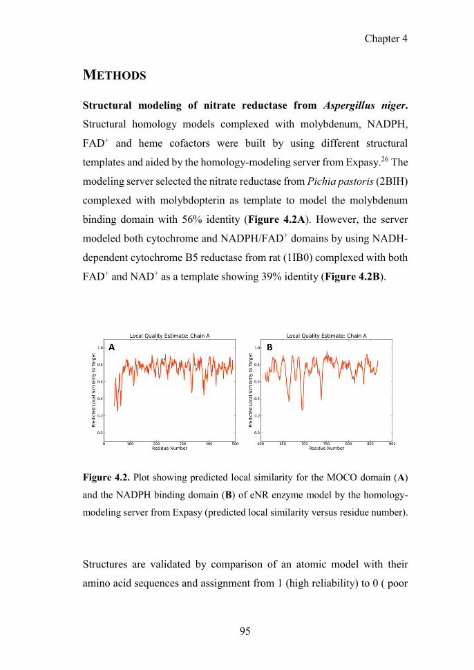
Chapter 4
95
METHODS
Structural modeling of nitrate reductase from Aspergillus niger.
Structural homology models complexed with molybdenum, NADPH,
FAD+ and heme cofactors were built by using different structural
templates and aided by the homology-modeling server from Expasy.26 The
modeling server selected the nitrate reductase from Pichia pastoris (2BIH)
complexed with molybdopterin as template to model the molybdenum
binding domain with 56% identity (Figure 4.2A). However, the server
modeled both cytochrome and NADPH/FAD+ domains by using NADH-
dependent cytochrome B5 reductase from rat (1IB0) complexed with both
FAD+ and NAD+ as a template showing 39% identity (Figure 4.2B).
Figure 4.2. Plot showing predicted local similarity for the MOCO domain (A)
and the NADPH binding domain (B) of eNR enzyme model by the homology-
modeling server from Expasy (predicted local similarity versus residue number).
Structures are validated by comparison of an atomic model with their
amino acid sequences and assignment from 1 (high reliability) to 0 ( poor

96
reliability) for each amino acid positions. The model for the domain that
binds NADPH, FAD+ and heme has a very good compatibility score, while
the model for the molybdenum binding domain presented very low score
mainly due to the low compatibility of its N-terminus that was highly
flexible and consequently hard to model. Protein models were visualized
and aligned with their template structure by using PyMol 0.99 developed
by DeLano Scientific LLC (San Francisco, CA). The electrostatic potential
was calculated by using Bluues server.27
Determination of the enzyme activity and protein concentration. The
activities of both free and immobilized nitrate reductase from Aspergillus
niger were determined by measuring the decrease in absorbance at 340
nm, resulting from NADPH oxidation during the enzymatic conversion of
nitrate to nitrite. A sample of enzyme solution of 10 L was incubated in
a multi-well plate with 190 L reaction mixture with a final concentration
of 10 mM sodium phosphate buffer, 0.21 mM NADPH and 4.25 mM
NaNO3 at 25°C and pH 7.5 under mild shaking. When indicated, different
temperatures and pH values were used. One activity unit was defined as
the amount of enzyme required to oxidize 1 μmol of NADPH under the
above-described conditions. The protein concentration of all soluble eNR
samples was measured by using Bradford’s method28 and bovine serum
albumin (BSA) as a protein standard.
Kinetic parameters. The kinetic parameters were calculated from the
initial rate values determined with different concentrations of NADPH (0-
2 mM) and nitrate (0-20 mM). Results were fitted to Michaelis-Menten
equation by non-linear regression and Km and kcat were calculated using
OriginPro 8.0 software.

Chapter 4
97
Enzyme Immobilization. The eNR was immobilized on agarose beads
activated with cyanogen bromide groups (Ag-CB), on agarose beads
activated with either positively charged mono-aminoethyl groups (Ag-
MANAE) or diethyl-aminoethyl groups (Ag-DEAE) and coated with
positively charged poly-ethylenimine (Ag-PEI), and on agarose beads
coated with negatively charged dextran-sulphate (Ag-DS).
Generally, all the immobilizations were carried out by adding 200mg of
the corresponding carrier (Ag-CB, Ag-DEAE, Ag-MANAE, Ag-PEI and
Ag-DS) to 1mL of soluble eNR (1-5U/mL) in 10 mM sodium phosphate
at pH 7.5. The suspension was kept under mild stirring at 25°C.
Periodically, samples of the supernatants and the carrier-enzyme
suspensions were withdrawn and analyzed to evaluate the progress of the
immobilization process. In parallel, a blank of the soluble enzyme (without
a carrier) was also analyzed. After immobilization, enzyme preparations
were washed with an excess of distilled water and immobilization buffer
to eliminate any non-bound protein molecule.
The immobilization on Ag-CB requires a pre-activation of the carrier;
thus, 0.2 g of the lyophilized carrier was washed and swell with 10mL HCl
1 mM during 30 min, followed by gentle filtration between successive
additions of water. Then, the washed carrier was equilibrated with 100 mM
NaHCO3 in 0.5 M NaCl, pH 8.3. eNR was incubated with the support
under mild stirring at room temperature. The support containing the
immobilized eNR was washed with Tris–HCl using 0.1 M HCl, pH 8.0,
followed by 0.1 M acetic acid/sodium acetate containing 0.5 M NaCl, pH
4.0 (3 cycles of alternating pH buffers) to block any remaining active
groups.

98
Thermal stability of soluble and immobilized eNR. All enzyme
preparations (both soluble and immobilized), were incubated in 10mM
sodium phosphate buffer (pH 7.5) at different temperatures (25-55°C) for
15 min. As previously described, the eNR activities were assayed at 25 ºC.
In a different experiment, soluble and immobilized preparations were
incubated at 40ºC, and samples were withdrawn at different times and
assayed at 25ºC to determine the residual activity. The inactivation
constant is calculated by fitting the thermal inactivation curve to first-order
kinetics equation (A/Ao=e-k*t) considering that the enzyme becomes
completely inactivated after a certain time. The fitting was carried out by
using OriginPro 8.
Loading capacity. For the best immobilization chemistry, Ag-DEAE,
loading capacity studies were carried out. Different amounts of Aspergillus
niger eNR preparation (0.0075; 0.0107; 0.0143; 0.0255; 0.0349 mg
protein) were offered to 200 mg of Ag-DEAE suspended in 1 mL of 10
mM sodium phosphate buffer at pH 7.5. The amount of protein bound to
the carrier was determined as the difference between the initial and the
residual protein concentration in the supernatants. This loading capacity
was also monitored by analyzing both initial and residual enzyme activity
in the supernatant. For each enzyme load, recovered activity of the
immobilized eNR was measured upon the immobilization process.
Fluorescence Spectroscopy studies. Fluorescence measurements were
carried out in a Varioskan Flash fluorescence spectrophotometer (Thermo
Scientific), monitoring the intrinsic fluorescence of soluble and
immobilized Aspergillus niger eNR on Ag-DEAE, using an excitation
wavelength of 280nm with excitation and emission bandwidths of 5 nm
and recording fluorescence emission spectrum between 300 and 600nm.

Chapter 4
99
All spectroscopic measurements were made in 10 mM sodium phosphate
at pH 7.5 and 25ºC.
Activity-pH profile of eNR soluble and immobilized on Ag-DEAE. The
effect of pH on the activity of the enzyme preparations was determined by
measuring the residual activities of eNR at the different pH values
(5<pH<10) and 25°C. The expressed activity was measured as described
before. Different 10 mM buffer solutions were used to set the right pH
value; acetate buffer was used for pH 5-6; sodium phosphate buffer for pH
7-8 and sodium bicarbonate buffer for pH 9-10.
Production of the radioactive precursor [13N]NO2- by enzyme
catalysis: general procedure
Nitrogen-13 was produced in an IBA Cyclone 18/9 cyclotron via the 16O(p,α)13N nuclear reaction. The target system consisted of an aluminum
insert (2 mL) covered with havar foil (thickness 25 μm, ∅ 29 mm) and with
an aluminum vacuum foil (thickness 25 μm, ∅ 23 mm). The target
(containing 1.75 mL of ultrapure water, Type I water, ISO 3696) was
irradiated with 18MeV protons. The beam current was maintained at 20
μA (pressure in the range 5–10 bar into the target during bombardment) to
reach the desired integrated currents (0.1-0.5 μAh). The resulting solution
was transferred to a 10mL vial and the activity was measured in a dose
calibrator (Capintec CRC®-25 PET, New Jersey, USA). The final
activities (corrected to the end of irradiation) were in the range 333-1110
MBq (9-30 mCi).
Enzymatic reduction of [13N]NO3- to [13N]NO2
- was carried out with 6.72
mU of immobilized eNR packed into a plastic column. This insoluble
preparation was incubated with 90 L of cyclotron produced 13N solution

100
(which contained [13N]NO3-) in the presence of 10L of 2.1 mM NADPH
(0.21 mM final concentration) in 500 mM sodium phosphate buffer (50
mM final concentration; pH 7.5) under mild conditions (25°C and pH 7.5)
for 4 min. Relative amounts of [13N]NH4+, [13N]NO2
- and [13N]NO3- were
measured by radio-HPLC. An Agilent 1200 series HPLC equipped with a
quaternary pump, a multiple wavelength detector and a radiometric
detector (Gabi, Raytest) was used. An HP Asahipak ODP-50 (5 µm, 125x4
mm, Teknokroma, Spain) was used as stationary phase, and a solution
containing additive for ionic chromatography (15 mL) in 1L of a mixture
water/acetonitrile (86:14) basified to pH 8.6 with 1M sodium hydroxide
solution was used as the mobile phase at a flow rate of 1 mL/min.
Simultaneous UV (λ = 254 nm) and isotopic detection were used.
Recycling of eNR immobilized on Ag-DEAE. Reusability of eNR
immobilized on Ag-DEAE was evaluated by performing consecutive
catalytic cycles at 25°C using nitrate (both non-radioactive and
radioactive) and NADPH as substrates. In some experiments, the
immobilized enzyme was incubated with a solution 1.0 mM NADPH and
the nitrate reduction was carried out without adding exogeneous NADPH.
After each cycle, the immobilized preparation was washed and the
enzymatic activity towards non-radioactive nitrate was determined by
spectrophotometry, while the final yield towards radioactive nitrate was
determined by radio-HPLC.
Two-step chemo-enzymatic synthesis of S-[13N]nitrosoglutathione
([13N]GSNO)
The preparation of [13N]GSNO followed the synthetic protocol previously
reported,2 but the [13N]NO2- used as the labeling agent was enzymatically
synthesized. A radioactive solution (300 µL) containing cyclotron

Chapter 4
101
produced [13N]NO3- was incubated in a packed column equilibrated with
1 mM NADPH loaded with 175µg eNR per g of carrier (22mU of
immobilized eNR determined as previously described) for 4 min; the
resulting supernatant containing [13N]NO2- was diluted with 1 mL of 10
mM sodium phosphate buffer (pH=7.5) and then flushed through an anion
exchange cartridge (Sep-Pak® Accell Plus QMA, Waters) to selectively
retain [13N]NO2-. The column was further washed with distilled water (2
mL) and the QMA cartridge was dried with nitrogen gas for 15s. An acidic
solution of the precursor glutathione (0.5 mL; 1 mM) was loaded into the
cartridge and the nitrosation reaction was allowed to occur; the reaction
mixture was finally eluted directly into a collection vial. The identification
of [13N]GSNO was performed by co-elution with reference standard using
the same HPLC system described above; in this case, a Mediterranean
SeaRP-18 column (5µm, 150 x 4.6mm, Teknokroma, Spain) was used as
stationary phase and aqueous TFA solution/acetonitrile (95:5) was used as
the mobile phase at a flow rate of 1 mL/min. Simultaneous UV (λ = 220
nm) and isotopic detection were used.
RESULTS AND DISCUSSION
Immobilization of eNR on functionalized porous agarose beads
Eukaryotic nitrate reductase from Aspergillus niger was immobilized
through different immobilization chemistries on a survey of agarose-
based matrixes: i) irreversibly immobilized through its N-terminus on
agarose beads activated with cyanogen bromide groups (Ag-CB); ii)
reversibly immobilized through its acid amino acid richest regions on
agarose beads activated with either positively charged mono aminoethyl

102
groups (Ag-MANAE) or diethyl aminoethyl groups (Ag-DEAE) and
coated with positively charged polyethylenimine (Ag-PEI); and iii)
through its basic amino acid-richest regions on agarose beads coated with
negatively charged dextran sulphate (Ag-DS), see Table 3.1.
Table 4.1. Parameters of Aspergillus niger nitrate reductase immobilization onto
agarose carrier via different chemistries.
CARRIER Reactive
group
μmol of amines
per gram of carrier
Immobilization chemistry
Immobilized Activity
Ai (mU/g)a
Immobilization yield (%)b
Expressed Activity
Ae (mU/g)c/ (%)d
Ag-CB Cyanogen Bromide
-
Covalent bond through the
eNR Nt 964±32 96±3 26±4 / (3)
Ag-DEAE
Diethyl-aminoethyl
150 Ionic
adsorption through the
most acid eNR regions
995±10 99±1 210±60 / (21)
Ag-PEI
Poly-ethylenimine
3448 982±12 98±1 155±25 / (16)
Ag-MANAE
Mono-
aminoethyl 5 820±24 82±2 192±41 / (23)
Ag-DS Dextran-sulphate
-
Ionic adsorption through the most basic
eNR regions
750 75 58 / (8)
The initial offered activity was always 1000mU per gram. aAi=The activity immobilized on 1 gram of carrier after the immobilization process. This activity was calculated as the difference between the offered activity and the
activity in the supernatant after incubation with the carrier for 1h. bImmobilization yield () = (immobilized activity/offered activity) x 100. cThe expressed activity (Ae) is defined as the measured activity of the immobilized enzyme after washing. dRelative expressed activity (Ae) = (expressed activity/immobilized activity) x 100.
As it can be seen at Table 4.1, all carriers were able to efficiently
immobilize eNR (immobilization yields >75% in all cases), although low
values of expressed activity were achieved after immobilization when
compared to the free enzyme. Irreversible immobilization and cationic
exchange on Ag-CB and Ag-DS led to a dramatic decrease in the
expressed activity (26±4 and 58 mU/g, respectively). On the other hand,
ionic immobilization of eNR on positively charged carriers yielded values
of expressed activity in the range 155-210 mU/g, resulting in up to 23% of

Chapter 4
103
relative expressed activity. Noteworthy, although the chemistry that drove
the immobilization on Ag-MANAE, Ag-DEAE and Ag-PEI was the same,
Ag-MANAE offered lower immobilization yields (82±2%) than Ag-
DEAE (99±1%) and Ag-PEI (98±1%). This fact may be explained because
Ag-MANAE surface presents a lower density of positively charged groups
than Ag-DEAE and Ag-PEI surfaces, see Table 4.1.
In order to explain the functional discrepancies obtained with the different
immobilized preparations, we further studied the eNR surface to
understand how the protein orientation on the carrier surface may affect its
functionality. Unfortunately, X-ray structure of eNR from A. niger has not
been solved yet, and hence its 3D-structural homology model containing
the NADPH-domain, the heme-domain and the MOCO-domain was built
(Figure 4.3A) and the electrostatic surface potential was calculated in
order to determine the spatial charge distribution across the enzyme
surface (Figure 4.3B and 4.3C). The analysis of this structural model
reveals an acidic belt on the MOCO-domain located at the opposite face
regarding the active site. The model also shows that heme, FAD+, NAD+
and molybdenum binding sites are surrounded by basic amino acids
(Figure 4.3C).

104
Figure 4.3. Structural model of a monomer of eNR from Aspergillus niger. (A)
Cartoon representation of 3D-structural homology model by using the X-ray
structures of eNR from Pichia pastoris and cytochrome B5 from rat complexed
with both FAD+ and NAD+ as templates for molybdenum domain (N-terminus)
(orange) and cytochrome domain (C-terminus) (purple) respectively. The sphere
representation colored in cyan depicts the dimerization domain of the eNR. Green
sticks represent the cofactors NAD+, FAD+ and molydopterin. (B) Surface
electrostatic potential of eNR calculated by Bluues server (back face of active
site) and (C) proposed orientation of eNR monomer on porous Ag-DEAE
surface. The blue circles represent the tertiary amine groups located on the carrier
surface. The surface electrostatic potential is depicted with a red-blue gradient
that corresponds to the acid-basic gradient. All the images were prepared by using
Pymol 0.99v.

Chapter 4
105
These structural observations suggest that eNR immobilized on Ag-DS is
oriented through its active site (basic region), hampering the accessibility
of both NADPH and nitrate to the catalytic pocket with a consequent
decrease in catalytic efficiency. Nevertheless, the eNR orientation onto
positively charged carriers seems to occur through one acidic region
located far away from the binding pockets. Therefore, active centres
remain fairly accessible for the substrates, resulting in significantly higher
relative expressed activity values for such immobilized preparations
(Figure 4.3C). Moreover, such orientation seems to negligibly affect
dimmer stability because the dimerization domain is not involved in the
protein-carrier interaction. In fact, when eNR immobilized on Ag-DEAE
was incubated under high ionic strength conditions, the enzyme was
quantitatively eluted to the supernatant, demonstrating the ionic character
and the reversibility of this immobilization chemistry.
The values of expressed activity shown in Table 4.1 have been determined
by using NADPH as a cofactor; such values are relatively high when
compared with previous results obtained using immobilization protocols
based on porous hydrogels. For example, eNR from Aspergillus niger
immobilized on a porous vinyl polymeric matrix expressed less than 2%
of its expressed activity by using NADPH as cofactor.22 The high
structural complexity of eNR might be the main reason for the low
expressed activity achieved in this previous work. This enzyme requires
enough structural flexibility for efficient electron transfer between both the
cytochrome and the molybdenum domains. Hence, immobilization
chemistries that highly rigidify the 3D structure of eNR (covalent
immobilization), may hinder the catalytic conformational changes or
promote wrong protein orientations that limit either the electron transfer

106
or the substrate accessibility, resulting in a low expressed activity of the
immobilized preparation.
Effects of the temperature on both activity and stability of soluble and
immobilized eNR
The thermal stabilities of immobilized and soluble eNR were tested at
different temperatures. Figure 4.4A shows the inactivation courses of the
three different immobilized preparations incubated at pH 7.5 and 25°C.
The half-life time of eNR immobilized on Ag-DEAE was estimated 30-
fold and 6-fold higher than eNR immobilized on Ag-MANAE and Ag-
PEI, respectively, suggesting that the thermal stability of the immobilized
enzymes relies on the chemical nature and density of the amine groups
located on the surface of the carrier. The surface of Ag-DEAE containing
150 μmol/g of tertiary amine groups seems to establish more favourable
interactions with the acidic protein regions than the Ag-MANAE surface,
which presents low density of primary amine groups (5 μmol/g), and the
Ag-PEI surface coated by a polymeric bed with a high density of primary,
secondary and tertiary amine groups (3448 μmol/g). Ag-PEI has been
suggested as an excellent carrier to stabilize multimeric enzymes, because
the basic polymeric bed establishes 3D-protein-polymer interactions24 that
attach all the subunits to the carrier, resulting in a stabilization of the
quaternary structure of the protein. Unexpectedly, in this study, eNR
immobilized on Ag-PEI was less stable than eNR immobilized on Ag-
DEAE. This unexpected result may be explained because the flexibility of
the polymer may generate additional interactions with the protein over
time. Such enhancement of the spatial reactivity may lead to undesired
interactions between both cytochrome and molybdenum domains and the

Chapter 4
107
polymeric brushes, reducing the enzyme flexibility required for the
electron transfer, and consequently decreasing the nitrate reduction
activity over time. Those negative interactions would not be possible to
the much less flexible Ag-DEAE surface formed by a 2D-monolayer of
tertiary amine groups. Hence, our results suggest that the interaction
between eNR and the positively charged carriers must be strong enough to
guarantee the stabilizing immobilization effect and flexible enough to
enable the conformation changes required for the catalysis.
Figure 4.4. Stability of eNR. (A) Inactivation course of different eNR
immobilized preparations at 25°C and pH 7.5. eNR was immobilized on Ag-
DEAE (●); Ag-PEI (▲); Ag-MANAE (■). Values are normalized to starting the
activity. (B) Inactivation of eNR soluble (white bars) and immobilized on Ag-
DEAE (black bars) at different temperatures. Soluble and immobilized
preparations were incubated 15 minutes at different temperatures and then
activities were assayed at 25 ºC.
As it can be seen in the Figure 4.4B, the enzyme immobilized on Ag-
DEAE was much more stable than its soluble counterpart under a broad

108
range of temperatures, between 30-55°C. Surprisingly, when such
immobilized biocatalyst was incubated for only 15 minutes in the range of
40-45°C, a 2-fold activity enhancement (hyperactivation) was observed
with respect to the non-thermally treated enzyme.
Figure 4.5. Activity/temperature profile of both soluble and immobilized eNR.
Reductase activity of eNR immobilized on Ag-DEAE (white bars) and soluble
eNR (black bars) was assayed at different temperatures at pH 7.5.
Such hyperactivation effect was not observed with the soluble enzyme,
which decreased its initial activity after incubation at temperatures higher
than 25°C due to thermal inactivation. Interestingly, both the soluble and
immobilized enzymes showed similar activity/temperature profiles.
Figure 4.5 displays that the activity of soluble and immobilized enzymes
at different temperatures is very similar: both preparations showed a
maximum of activity in the range of 25-50. These results point out that the
20 25 30 35 40 45 500.0
0.2
0.4
0.6
0.8
1.0
Re
lati
ve e
nzy
me
Ac
tiv
ity
Temperature (oC)

Chapter 4
109
temperature does have a differential effect on the conformational stability
of the immobilized enzyme compared to the soluble one, but it does
similarly affect their catalytic properties under different reaction
temperatures. The optimal temperature of eNR, both for its soluble and
immobilized forms, ranges 25-30ºC, which agrees with the optimal
temperature of other mesophilic enzymes. Hence, these results suggest that
the aforementioned hyperactivation effect can only be attributed to the
carrier and that is not related to the effect of the temperature on the
catalysis itself.
Inactivation kinetics studies of the hyperactivated immobilized enzyme at
conditions of pH 7.5 and 40°C proved an enhanced stability with respect
to its soluble counterpart under the same conditions (Figure 4.6). The
inactivation courses for both soluble and immobilized enzyme follow first-
order kinetics where the inactivation rate depends on the inactivation
constant. The immobilized enzyme showed a thermal inactivation constant
(ki) of (1.31±0.63)x10-5 (s-1) at 40°C, while the ki value of the soluble
enzyme was (15.3±2.3)x10-5 (s-1). These findings confirm that the
immobilization process results in an increased half-life time (t1/2) of eNR
up to 883 minutes; 12 times longer than the t1/2 of the soluble enzyme.
The thermal-hyperactivation and thermal-stabilization effects observed for
eNR immobilized on Ag-DEAE may be explained by some beneficial
enzyme-carrier interactions triggered by the temperature and that drive to
some local structural re-organization in the enzyme, resulting in a more
efficient electron transfer between the NADPH and the MOCO domains.
Those interactions may stabilize a more beneficial conformation for eNR,
which explains the 2-fold more active and 12-fold more stable enzyme
compared to its soluble counterpart. These carrier- and temperature-

110
assisted conformational changes were further investigated by fluorescence
studies.
Figure 4.6. Thermal inactivation courses of Aspergillus niger nitrate reductase
immobilized onto Ag-DEAE at 40°C. The preparation of A. niger eNR insoluble
derivative was carried out as described in Section Material and Methods. Each
insoluble and soluble preparations were incubated in 10mM sodium phosphate at
pH 7.5 and 40ºC. The A. niger eNR preparations used to perform these studies
were: immobilized on Ag-DEAE (●) and soluble (♦).
Fluorescence spectrum analyzed at 25 ºC was obtained for soluble eNR
and immobilized eNR on Ag-DEAE with or without incubation at 40ºC
during 15 minutes.
As can be observed in Figure 4.7, the fluorescence spectrum of the soluble
enzyme remained unaltered after the thermal incubation at 40ºC in spite of
0 500 1000 1500
0,0
0,5
1,0
Rel
ati
ve
acti
vit
y
Time (minutes)
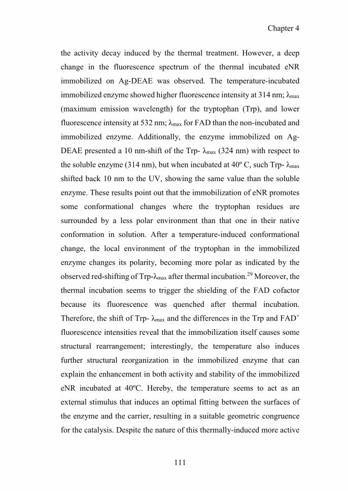
Chapter 4
111
the activity decay induced by the thermal treatment. However, a deep
change in the fluorescence spectrum of the thermal incubated eNR
immobilized on Ag-DEAE was observed. The temperature-incubated
immobilized enzyme showed higher fluorescence intensity at 314 nm; λmax
(maximum emission wavelength) for the tryptophan (Trp), and lower
fluorescence intensity at 532 nm; λmax for FAD than the non-incubated and
immobilized enzyme. Additionally, the enzyme immobilized on Ag-
DEAE presented a 10 nm-shift of the Trp- λmax (324 nm) with respect to
the soluble enzyme (314 nm), but when incubated at 40º C, such Trp- λmax
shifted back 10 nm to the UV, showing the same value than the soluble
enzyme. These results point out that the immobilization of eNR promotes
some conformational changes where the tryptophan residues are
surrounded by a less polar environment than that one in their native
conformation in solution. After a temperature-induced conformational
change, the local environment of the tryptophan in the immobilized
enzyme changes its polarity, becoming more polar as indicated by the
observed red-shifting of Trp-λmax after thermal incubation.29 Moreover, the
thermal incubation seems to trigger the shielding of the FAD cofactor
because its fluorescence was quenched after thermal incubation.
Therefore, the shift of Trp- λmax and the differences in the Trp and FAD+
fluorescence intensities reveal that the immobilization itself causes some
structural rearrangement; interestingly, the temperature also induces
further structural reorganization in the immobilized enzyme that can
explain the enhancement in both activity and stability of the immobilized
eNR incubated at 40ºC. Hereby, the temperature seems to act as an
external stimulus that induces an optimal fitting between the surfaces of
the enzyme and the carrier, resulting in a suitable geometric congruence
for the catalysis. Despite the nature of this thermally-induced more active
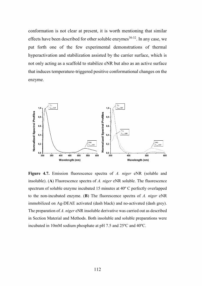
112
conformation is not clear at present, it is worth mentioning that similar
effects have been described for other soluble enzymes30-32. In any case, we
put forth one of the few experimental demonstrations of thermal
hyperactivation and stabilization assisted by the carrier surface, which is
not only acting as a scaffold to stabilize eNR but also as an active surface
that induces temperature-triggered positive conformational changes on the
enzyme.
Figure 4.7. Emission fluorescence spectra of A. niger eNR (soluble and
insoluble). (A) Fluorescence spectra of A. niger eNR soluble. The fluorescence
spectrum of soluble enzyme incubated 15 minutes at 40º C perfectly overlapped
to the non-incubated enzyme. (B) The fluorescence spectra of A. niger eNR
immobilized on Ag-DEAE activated (dash black) and no-activated (dash grey).
The preparation of A. niger eNR insoluble derivative was carried out as described
in Section Material and Methods. Both insoluble and soluble preparations were
incubated in 10mM sodium phosphate at pH 7.5 and 25ºC and 40ºC.

Chapter 4
113
Kinetic parameters and loading capacity of eNR immobilized on Ag-
DEAE
Considering the high stability and satisfactory catalytic efficiency of eNR
immobilized on Ag-DEAE, we selected this heterogeneous biocatalyst to
carry out the sustainable reduction of [13N]NO3- to [13N]NO2
- in aqueous
media under mild conditions. Nevertheless, before applying this
biocatalyst to radiochemistry, we carried out its kinetic characterization.
To this aim, Michaelis-Menten parameters were determined for soluble
and the immobilized eNRs towards the substrates: nitrate and NADPH.
Table 4.2. Kinetic characterization of free and immobilized Aspergillus niger
eNRs.
Km (mM) kcat (s-1) Kcat/Km (s-1 • mM-1)
NaNO3
NR soluble 0.27±0.07 30.33 111.23
NR Ag-DEAE 0.06±0.03 11.97 199.44
NADPH NR free 0.07±0.03 27.93 399.05
NR Ag-DEAE 1.86±0.82 N.D. N.D.
The steady-state kinetics parameters were measured at pH 7.5 and 25°C (see Section Material and Methods).
As it can be seen in Table 4.2, the kinetic studies provided interesting data
that helped us to understand the activity decrease resulting from the
immobilization process. In a first glance, the immobilized preparation
showed a 26-fold Km[NADPH] higher than the soluble enzyme. Contrarily,
Km[NO3] was 4.5 times smaller for the immobilized eNR than for the soluble
preparation. These results confirm that immobilization of eNR on Ag-
DEAE negatively affected the enzyme binding towards NADPH, which
explains the low expressed activity after the immobilization process. The
high Km[NADPH] values for the immobilized preparation may due to two
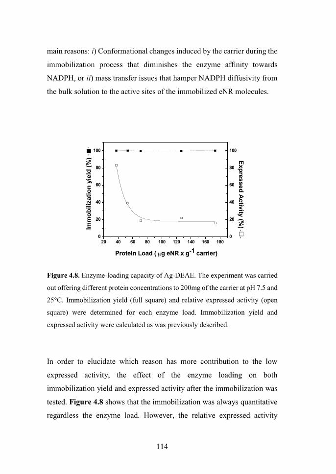
114
main reasons: i) Conformational changes induced by the carrier during the
immobilization process that diminishes the enzyme affinity towards
NADPH, or ii) mass transfer issues that hamper NADPH diffusivity from
the bulk solution to the active sites of the immobilized eNR molecules.
Figure 4.8. Enzyme-loading capacity of Ag-DEAE. The experiment was carried
out offering different protein concentrations to 200mg of the carrier at pH 7.5 and
25°C. Immobilization yield (full square) and relative expressed activity (open
square) were determined for each enzyme load. Immobilization yield and
expressed activity were calculated as was previously described.
In order to elucidate which reason has more contribution to the low
expressed activity, the effect of the enzyme loading on both
immobilization yield and expressed activity after the immobilization was
tested. Figure 4.8 shows that the immobilization was always quantitative
regardless the enzyme load. However, the relative expressed activity
20 40 60 80 100 120 140 160 1800
20
40
60
80
100
Protein Load ( g eNR x g-1 carrier)
Imm
ob
iliza
tio
n y
ield
(%
)
0
20
40
60
80
100
Ex
pres
sed
Ac
tivity
(%)
--
--

Chapter 4
115
presented a strong negative logarithmic correlation with the enzyme load.
Under very low enzyme loads (37.5 μgeNR/g carrier) the relative expressed
activity was 85%, while the value decreased to approximately 20% at loads
higher than 75 μgeNR/gcarrier.
This experiment demonstrates that immobilization chemistry does not
promote inactive enzyme conformations that intrinsically diminish its
specific activity after the immobilization process. In such scenario, the
expressed activity should be always low regardless of the enzyme load.
So, it is plausible to think that mass transfer limitations of substrate and
cofactors are the main reason to explain the low expressed activity of the
immobilized eNR. Contextualizing these results together with the high
Km[NADPH] values for the immobilized preparation, we suggest that the
cofactor undergoes severe mass transfer limitations to diffuse across the
Ag-DEAE microstructure to reach the enzyme active sites. Under high
enzyme loads, NADPH does not efficiently diffuse into the porous
microstructure of the carrier to saturate all active sites immobilized on
such carrier. On the contrary, under very low enzyme loads, the vast
majority of the eNR molecules are saturated with the NADPH. These
results are in good agreement with previous data18, which showed that eNR
from A. niger covalently immobilized on non-porous particles expresses
more than 90% of its specific activity after immobilization. Moreover,
these data strongly suggest that low specific activity of eNR immobilized
on positively charged agarose beads is due to limited NADPH diffusion
rather than inaccurate protein orientation, because positively charged
porous carrier seem to immobilize eNR through an optimal orientation
according to the protein surface analysis (see Figure 4.3C).

116
pH-profile of soluble and immobilized eNR on Ag-DEAE
It is well-known that the immobilization can improve the pH tolerance of
enzymes. Here, the activities of soluble and immobilized eNR on Ag-
DEAE under different pH conditions were also evaluated (Figure 4.9).
4 5 6 7 8 9 100
20
40
60
80
100
120
pH
Rel
ativ
e a
ctiv
ity
(%)
Figure 4.9. Effects of pH on NR immobilized onto Ag-DEAE activity. The NR
preparations used to perform these studies were: immobilized on Ag-DEAE
(open symbols) and soluble (filled symbols). The 10mM buffer solutions used in
this essay were: sodium acetate pH 5 (circles); sodium phosphate pH 6, 7 and 8
(squares) and bicarbonate pH 9 and 10 (triangles).
The soluble and immobilized preparation showed optimal activity at pH
value of 7 (Figure 4.9); such activity significantly decreased at pH lower
than 7 for the immobilized enzyme. However, the effect of the acidic pH
on the activity of the soluble enzyme was more dramatic reducing close to

Chapter 4
117
50% of its initial activity. Under basic conditions, the soluble enzyme
undergoes a dramatic inactivation as reflected in its activity-pH profile;
such inactivation can be moderated by immobilizing eNR on Ag-DEAE.
This data demonstrated that immobilized eNR performs more efficiently
in a wider pH range (5 to 10) than the soluble enzyme.
Reduction of radiolabeled nitrate catalyzed by eNR immobilized on
Ag-DEAE. Reusability of the immobilized biocatalyst
eNR immobilized on Ag-DEAE was used to reduce [13N]NO3- to
[13N]NO2-. The reaction was carried out in batch mode and the
immobilized biocatalyst was re-used for several reaction cycles. As it can
be seen in Figure 4.10, the immobilized enzyme only yielded 60% of
[13N]NO2- in 4 minutes in the first cycle, while the soluble enzyme reduced
96% of [13N]NO3- to [13N]NO2
-. Surprisingly, in the second cycle, the
immobilized enzyme reduced up to 93% of [13N]NO3- to[13N]NO2
-. We
suggest that differences in NADPH effective concentration inside the
carrier pores might cause that intriguing effect.
We hypothesize that during the first reaction cycle some negatively
charged NADPH molecules are ionically absorbed to the positively
charged carrier surface. However, such molecules would present an
association/dissociation equilibrium that would simultaneously provide
one fraction of soluble NADPH available for the enzyme catalysis and the
second fraction of NADPH trapped into the intra-pore space of the carrier
surface that would be available for consecutive cycles. In this scenario, the
effective concentration of NADPH in the second and successive cycles
will be higher, explaining the higher yield observed in the second cycle
compared to the first one. In order to demonstrate this ionic interaction

118
between NADPH and Ag-DEAE, we equilibrated the eNR immobilized
on Ag-DEAE with NADPH (same concentration and time as a typical
reaction) in solution before triggering the first reaction cycle.
Figure 4. 10. Reusability of eNR immobilized on Ag-DEAE. Soluble eNR (white
bar), eNR immobilized on Ag-DEAE without NADPH equilibration (black bars)
and eNR immobilized on Ag-DEAE equilibrated with NADPH (stripped bars)
were used to catalyze the reduction from [13N]NO3- to [13N]NO2
- at 25º C, pH 7.5.
Reactions catalyzed by soluble and non-equilibrated immobilized eNR were
carried out by adding exogenous NADPH, while the reactions catalyzed by
equilibrated immobilized eNR were carried out without exogenous NADPH.
The equilibrated heterogeneous biocatalyst was able to reduce 96% of
[13N]NO3- to [13N]NO2
- from the first cycle (Figure 4.10). This finding
confirms that a fraction of NADPH is ionically trapped into the carrier
surface, increasing the internal concentration of NADPH that results in
higher reduction yields. In fact, we also confirmed that when the
radiochemical reduction was carried out with the NADPH-equilibrated
0 1 2 3 4 5 6 7 80
10
20
30
40
50
60
70
80
90
100
Ra
dio
ch
em
ica
l Y
ield
(%
)
Cycle
Chapter 4
119
immobilized enzyme, the heterogeneous biocatalyst was able to catalyze
the radiolabeled nitrate reduction for 5 cycles with more than 30% yield
without exogenous addition of NADPH (Figure 4.10). This experiment
provides evidence that NADPH molecules are reversibly bound to the
carrier surface providing an internal cofactor concentration that was
sufficient for the immobilized eNR to catalyze the radiochemical reduction
with the maximum yield during the first cycle, and proportionally lower
yields with the consecutive cycles. The lower reduction yield along the
cycles agrees with the fact that NADPH is oxidized to NADP+ decreasing
the pool of NADPH trapped into the carrier porous structure. Therefore,
along the consecutive cycles, immobilized eNR suffers from a lower
availability of the reduced NADPH, explaining the lower yields along the
operational cycles. Contrarily, when reduction was carried out by adding
exogenous NADPH in each reaction cycle, the maximum yield was
maintained during 7 cycles, demonstrating the excellent operational
stability of the immobilized biocatalyst (Figure 4.10). To the best of our
knowledge, this is the first evidence of a heterogeneous biocatalyst where
the carrier plays an important role in supplying the corresponding cofactor
to the enzyme. Furthermore, it was confirmed by SDS-page with silver
staining (Figure 4.11) that this optimal heterogeneous biocatalyst, offers
a clean final product without any significant protein contamination, even
during the recycling process, confirming once again that ionic interactions
between eNR and Ag-DEAE are quite strong in spite of their reversible
nature.

120
Figure 4.11. SDS-PAGE/silver staining of Aspergillus niger nitrate reductase.
Lane M: molecular weight marker; Lane 1: eNR soluble; lane 2: eNR
immobilized before any reaction; lane 3: non-bound protein after immobilization;
lane 4: cycle 1; lane 5: cycle 2; lane 6: cycle 3; lane 7: cycle 4; lane 8: cycle 5;
and lane 9: eNR immobilized after 5 reaction cycles.
Two step chemo-enzymatic solid-phase synthesis of S-[13N]GSNO
To proof the concept that the enzymatically synthesized [13N]NO2- can be
directly integrated into a radiochemical synthetic cascade, we coupled the
optimal eNR immobilized on Ag-DEAE to the two-step chemo-enzymatic
synthesis of S-[13N]GSNO (Figure 4.12).
[13N]NO3- was enzymatically reduced to [13N]NO2
- in the presence of
NADPH. The supernatant of such reaction was passed through an anion
exchange cartridge in order to trap the enzymatically produced [13N]NO2-
. Then, an acidic solution of S-glutathione (1 mM) was also passed through
the same anion exchange cartridge enabling the solid-phase S-nitrosation
of glutathione. The resulting product was finally desorbed from the resin

Chapter 4
121
by elution with distilled water. 95% of the enzymatically yielded
[13N]NO2- was converted to S-[13N]GSNO, similar to the radiochemical
conversion obtained in previous works conducted in our research group
using Cd-reduced [13N]NO2-.1 This result confirms that the redox cofactor
needed for the enzymatic reactions does not interfere with the next
chemical step. Therefore, the radioactive nitrite produced by this
heterogeneous biocatalyst is suitable to be used in radiochemical reactions.
Figure 4.12. [13N]GSNO synthesis using [13 produced by enzymatic
catalysis.
CONCLUSION
In this chapter, an unprecedented strategy for the reduction of [13N]NO3-
to [13N]NO2- using an efficient heterogeneous biocatalyst was successfully
applied. The use of one eukaryotic nitrate reductase immobilized on a solid
carrier to a radiochemical process is for the first time published. This
immobilized enzyme has been able to selectively reduce [13N]NO3- to
[13N]NO2- aided by NADPH as redox cofactor. Likewise, by controlling

122
the immobilization chemistry and physicochemical properties of the
carrier, optimal heterogeneous biocatalysts with high potential in synthetic
chemistry were achieved. Noteworthy, this work is one of the few
examples where an immobilized enzyme has been applied for a
radiochemical process and the first report so far of a two-step chemo-
enzymatic route to synthesize model radiotracers starting from a precursor
([13N]NO3-) directly produced in the cyclotron.
From this experimental work, we can also conclude that immobilized
enzymes are highly active and stable, and simplify the process work-up,
and have great potential to be applied in radiochemistry to yield pure
radiotracers that can be directly used for biomedical purposes. Hence, they
can be anticipated as ideal tools for the preparation of radiotracers labeled
with short-lived radionuclides. Moreover, we have faced the challenge to
immobilize an enzyme whose catalytic mechanism depends on several
cofactors and conformational changes recovering enough activity and
increasing its stability to catalyze several reaction cycles.
The success of this work will so far open the doors to a new application of
enzymes in radiochemistry and will potentiate the use of other chemo-
enzymatic designs for the synthesis of novel radiotracers, even using short-
lived isotopes. Successful enzyme immobilization will contribute to
develop in flow radiochemical processes as well as to integrate
radiochemical synthesis on-chip.33-36
REFERENCES
(1) Llop, J.; Gómez-Vallejo, V.; Bosque, M.; Quincoces, G.; Peñuelas, I. Synthesis of S-[13N]nitrosoglutathione (13N-GSNO) as a new potential PET imaging agent. Applied Radiation and Isotopes 2009, 67, 95-99.

Chapter 4
123
(2) Gómez-Vallejo, V.; Kato, K.; Oliden, I.; Calvo, J.; Baz, Z.; Borrell, J. I.; Llop, J. Fully automated synthesis of 13N-labeled nitrosothiols. Tetrahedron Letters 2010, 51, 2990-2993.
(3) Gómez-Vallejo, V.; Kato, K.; Hanyu, M.; Minegishi, K.; Borrell, J. I.; Llop, J. Efficient system for the preparation of [13N]labeled nitrosamines. Bioorganic & Medicinal Chemistry Letters 2009, 19, 1913-1915.
(4) Gaja, V.; Gomez-Vallejo, V.; Puigivila, M.; Perez-Campana, C.; Martin, A.; Garcia-Osta, A.; Calvo-Fernandez, T.; Cuadrado-Tejedor, M.; Franco, R.; Llop, J. Synthesis and evaluation of (13)N-labelled azo compounds for beta-amyloid imaging in mice. Mol. Imaging Biol. 2014, 16, 538-549.
(5) Gomez-Vallejo, V.; Borrell, J. I.; Llop, J. A convenient synthesis of 13N-labelled azo compounds: a new route for the preparation of amyloid imaging PET probes. Eur. J. Med. Chem. 2010, 45, 5318-5323.
(6) Joshi, S. M.; de Cozar, A.; Gomez-Vallejo, V.; Koziorowski, J.; Llop, J.; Cossio, F. P. Synthesis of radiolabelled aryl azides from diazonium salts: experimental and computational results permit the identification of the preferred mechanism. Chemical Communications 2015, 51, 8954-8957.
(7) Joshi, S. M.; Gomez-Vallejo, V.; Salinas, V.; Llop, J. Synthesis of 13N-labelled polysubstituted triazoles via Huisgen cycloaddition. RSC Advances 2016, 6, 109633-109638.
(8) Joshi, S. M.; Mane, R. B.; Gomez-Vallejo, V.; Llop, J.; Rode, C. Microwave assisted synthesis of 5-substituted 1H-tetrazoles via [3+2] cycloaddition over heterogeneous Cu based catalyst: Application to the preparation of 13N-labelled tetrazoles. submitted 2017.
(9) Margeson, J. H.; Suggs, J. C.; Midgett, M. R. Reduction of nitrate to nitrite with cadmium. Analytical Chemistry 1980, 52, 1955-1957.
(10) Bernhard, A. The Nitrogen Cycle: Processes, Players, and Human Impact. . Nature Education Knowledge 2010, 3, 25.
(11) Krappmann, S.; Braus, G. H. Nitrogen metabolism of Aspergillus and its role in pathogenicity. Medical mycology 2005, 43 Suppl 1, S31-40.
(12) Fischer, K.; Barbier, G. G.; Hecht, H.-J.; Mendel, R. R.; Campbell, W. H.; Schwarz, G. Structural Basis of Eukaryotic Nitrate Reduction: Crystal Structures of the Nitrate Reductase Active Site. The Plant Cell 2005, 17, 1167-1179.
(13) Arauzo, M. Vulnerability of groundwater resources to nitrate pollution: A simple and effective procedure for delimiting Nitrate Vulnerable Zones. Science of The Total Environment 2017, 575, 799-812.
(14) Zhai, Y.; Zhao, X.; Teng, Y.; Li, X.; Zhang, J.; Wu, J.; Zuo, R. Groundwater nitrate pollution and human health risk assessment by using

124
HHRA model in an agricultural area, NE China. Ecotoxicology and Environmental Safety 2017, 137, 130-142.
(15) Moorcroft, M. J.; Davis, J.; Compton, R. G. Detection and determination of nitrate and nitrite: a review. Talanta 2001, 54, 785-803.
(16) Wang, Q.-H.; Yu, L.-J.; Liu, Y.; Lin, L.; Lu, R.-g.; Zhu, J.-p.; He, L.; Lu, Z.-L. Methods for the detection and determination of nitrite and nitrate: A review. Talanta 2017, 165, 709-720.
(17) W. Aylott, J.; J. Richardson, D.; A. Russell, D. Optical Biosensing of Nitrate Ions Using a Sol-Gel Immobilized Nitrate Reductase. Analyst 1997, 122, 77-80.
(18) Sachdeva, V.; Hooda, V. A new immobilization and sensing platform for nitrate quantification. Talanta 2014, 124, 52-59.
(19) Cosnier, S.; Da Silva, S.; Shan, D.; Gorgy, K. Electrochemical nitrate biosensor based on poly(pyrrole-viologen) film-nitrate reductase-clay composite. Bioelectrochemistry (Amsterdam, Netherlands) 2008, 74, 47-51.
(20) Quan, D.; Shim, J. H.; Kim, J. D.; Park, H. S.; Cha, G. S.; Nam, H. Electrochemical Determination of Nitrate with Nitrate Reductase-Immobilized Electrodes under Ambient Air. Analytical Chemistry 2005, 77, 4467-4473.
(21) Madasamy, T.; Pandiaraj, M.; Balamurugan, M.; Bhargava, K.; Sethy, N. K.; Karunakaran, C. Copper, zinc superoxide dismutase and nitrate reductase coimmobilized bienzymatic biosensor for the simultaneous determination of nitrite and nitrate. Biosensors & bioelectronics 2014, 52, 209-215.
(22) Mellor, R. B.; Ronnenberg, J.; Campbell, W. H.; Diekmann, S. Reduction of nitrate and nitrite in water by immobilized enzymes. Nature 1992, 355, 717-719.
(23) Fernandez-Lafuente, R.; Rosell, C. M.; Rodriguez, V.; Santana, C.; Soler, G.; Bastida, A.; Guisan, J. M. Preparation of activated supports containing low pK amino groups. A new tool for protein immobilization via the carboxyl coupling method. Enzyme Microb. Technol. 1993, 15, 546-550.
(24) Mateo, C.; Abian, O.; Fernandez-Lafuente, R.; Guisan, J. M. Reversible enzyme immobilization via a very strong and nondistorting ionic adsorption on support–polyethylenimine composites. Biotechnol. Bioeng. 2000, 68, 98-105.
(25) Fuentes, M.; Pessela, B. C. C.; Maquiese, J. V.; Ortiz, C.; Segura, R. L.; Palomo, J. M.; Abian, O.; Torres, R.; Mateo, C.; Fernández-Lafuente, R.; Guisán, J. M. Reversible and Strong Immobilization of Proteins by Ionic

Chapter 4
125
Exchange on Supports Coated with Sulfate-Dextran. Biotechnol. Prog. 2004, 20, 1134-1139.
(26) Arnold, K.; Bordoli, L.; Kopp, J.; Schwede, T. The SWISS-MODEL workspace: a web-based environment for protein structure homology modelling. Bioinformatics 2006, 22, 195-201.
(27) Walsh, I.; Minervini, G.; Corazza, A.; Esposito, G.; Tosatto, S. C. E.; Fogolari, F. Bluues server: electrostatic properties of wild-type and mutated protein structures. Bioinformatics 2012, 28, 2189-2190.
(28) Bradford, M. M. A rapid and sensitive method for the quantitation of microgram quantities of protein utilizing the principle of protein-dye binding. Analytical Biochemistry 1976, 72, 248-254.
(29) Ghisaidoobe, A. B.; Chung, S. J. Intrinsic tryptophan fluorescence in the detection and analysis of proteins: a focus on Forster resonance energy transfer techniques. International journal of molecular sciences 2014, 15, 22518-22538.
(30) Wedler, F. C.; Hoffmann, F. M. Glutamine synthetase of Bacillus stearothermophilus. I. Purification and basic properties. Biochemistry 1974, 13, 3207-3214.
(31) Facchiano, F.; Ragone, R.; Porcelli, M.; Cacciapuoti, G.; Colonna, G. Effect of temperature on the propylamine transferase from Sulfolobus solfataricus, an extreme thermophilic archaebacterium. Eur. J. Biochem. 1992, 204, 473-482.
(32) Rocha-Martin, J.; Vega, D.; Bolivar, J.; Godoy, C.; Hidalgo, A.; Berenguer, J.; Guisan, J.; Lopez-Gallego, F. New biotechnological perspectives of a NADH oxidase variant from Thermus thermophilus HB27 as NAD+-recycling enzyme. BMC Biotechnol. 2011, 11, 101.
(33) Rensch, C.; Lindner, S.; Salvamoser, R.; Leidner, S.; Bold, C.; Samper, V.; Taylor, D.; Baller, M.; Riese, S.; Bartenstein, P.; Wangler, C.; Wangler, B. A solvent resistant lab-on-chip platform for radiochemistry applications. Lab on a Chip 2014, 14, 2556-2564.
(34) Zhang, H.; Cantorias, M. V.; Pillarsetty, N.; Burnazi, E. M.; Cai, S.; Lewis, J. S. An improved strategy for the synthesis of [(18)F]-labeled arabinofuranosyl nuclosides. Nucl. Med. Biol. 2012, 39, 1182-1188.
(35) Lu, S.; Chun, J.-H.; Pike, V. W. Fluorine-18 chemistry in micro-reactors. J. Labelled Comp. Radiopharm. 2010, 53, 234-238.
(36) Keng, P. Y.; Chen, S.; Ding, H.; Sadeghi, S.; Shah, G. J.; Dooraghi, A.; Phelps, M. E.; Satyamurthy, N.; Chatziioannou, A. F.; Kim, C.-J. C.; van Dam, R. M. Micro-chemical synthesis of molecular probes on an electronic microfluidic device. Proc. Natl. Acad. Sci. U. S. A. 2012, 109, 690-695.

126

Chapter 5
In the present chapter, the one-pot, enzymatic and non-carrier-added synthesis of a variety
of 13N-labeled amino acids (L-[13N]alanine, [13N]glycine, and L-[13N]serine) is presented.
To this aim, L-alanine dehydrogenase from Bacillus subtilis, an enzyme that catalyzes the
reductive amination of α-keto acids has been assayed, using nicotinamide adenine
dinucleotide (NADH) as the redox cofactor and ammonia as the amine source. In
addition, the integration of both L-alanine dehydrogenase and formate dehydrogenase
from Candida boidinii in the same reaction vessel allows the in situ regeneration of
NADH during the radiochemical synthesis of the amino acids. The enzymatically labeled
amino acids have been used to analyze their in vivo biodistribution in healthy (wild-type)
mice. The capacity to selectively accumulate in the tumor has been assessed in a prostate
cancer mouse model (prostate-specific Pten deletion) by using dynamic PET-CT
imaging.
Enzymatic preparation of 13N-labeled amino acids
and their Biological assessment in prostate tumor
models


Chapter 5
129
INTRODUCTION
Amino acids labeled with nitrogen-13 are attractive because these are very
difficult to label using other isotopes. Additionally, amino acids could be
very interesting tracers for the detection of some types of cancer when
[18F]FDG does not work. Enantioselective chemical synthesis of amino
acids is also very hard. The use of enzymes can help, thanks to their fast
reaction rates and unique enantio- and chemo-selectivity.
As already mentioned before (Chapter 2), the utilization of enzymes for
the preparation of 13N-labeled amino acids was first described in the 70’s.1
For example, L-[13N]glutamate was synthesized by incubation of α-
ketoglutarate (α-KG) with [13N]NH3 in the presence of the enzyme L-
glutamate dehydrogenase and NADH as the redox cofactor.2,3 Likewise,
L-[13N]glutamine was synthesized by glutamine synthethase using
glutamic acid as the substrate and adenosine triphosphate (ATP) as the
cofactor.3,4 These two amino acids were synthesized with incorporation
values close to 90% in 15 minutes of incubation. Other amino acids such
as L-[13N]valine, L-[13N]methionine, L-[13N]leucine and L-[13N]alanine
have been produced by a one-pot/two-step or two-pot/two-step processes
by coupling glutamate dehydrogenase and transaminases. These
methodologies have allowed 16-90% chromatographic yields of the
corresponding L-[13N]amino acid.5,6
Amino acid dehydrogenases (AADH) are a class of enzymes which have
been extensively used in biocatalysis as main enzymes for the production
of enantiomerically pure -amino acids,7,8 as cofactor recycling enzymes
to replenish the NADH or NAD+ pool utilized by alcohol dehydrogenases9
and as donor enzymes to provide ω-transaminases with the suitable amine
donors using ammonia as the amine source.10 Interestingly, L-alanine

130
amine dehydrogenases (L-AlaDHs) are a special type of AADH that have
low sequence similarities with other AADH (glutamate dehydrogenase,
leucine dehydrogenases, phenylalanine dehydrogenases…) and some
important mechanistic differences.11,12 L-Alanine dehydrogenase, L-
AlaDH, is an oxidoreductase (EC1.4.1.1) which catalyzes the reversible
deamination of L-alanine yielding pyruvate and ammonia. In this reaction,
the alanine is oxidized at the enzyme active site with concomitant
reduction of NAD+ to NADH by hydride transfer.11,13 The kinetic
properties of L-AlaDHs from different microbial sources have been
extensively investigated, and some of these enzymes have been cloned in
heterologous hosts.14 Although the physiological role of L-AlaDH remains
controversial, it has been reported that this enzyme is involved in the
utilization of alanine as an energy source during the germination process
of Bacillus species.15,16
In continuation of our previous work related to the application of enzyme-
mediated reactions to 13N-radiochemistry,17 we describe in this chapter a
one-pot/one-step radiosynthesis of L-[13N]alanine, [13N]glycine, and L-
[13N]serine catalyzed by a versatile L-alanine dehydrogenase (L-AlaDH)
from Bacillus subtilis (Figure 5.1a). In contrast with the well reported
glutamate dehydrogenase that needs to be coupled to transaminase, L-
AlaDH is self-sufficient to incorporate [13N]NH3 into a broad scope of
organic precursors to yield the corresponding L-[13N]amino acids with an
exquisite selectivity.
One of the major drawbacks of utilizing amino acid dehydrogenase is the
necessity of adding an exogenous redox cofactor to the reaction mixture.
This may require the product purification before administration in order to
avoid toxicological or side effects. In biocatalysis, the recycling of the

Chapter 5
131
redox cofactor has been elegantly solved by using secondary enzymes,
whose mission is to replenish the pool of the cofactors during the reaction.
In this scenario, a lower amount of cofactor can be used, with the
consequent positive impact in the process-costs. There are different
enzymes that have been exploited to regenerate the cofactor.18,19 Glucose
dehydrogenase,20,21 sulfite dehydrogenase,22,23 and formate dehydrogenase
(FDH)24 are some examples.
Figure 5.1. Schematic representation of: (a) Enzyme-mediated preparation of L-
[13N]alanine, [13N]glycine, and L-[13N]serine using L-alanine dehydrogenase (L-
AlaDH) from Bacillus subtilis as the enzyme; (b) one-pot, multi-enzyme reaction
for the preparation of L-[13N]alanine with regeneration of NADH.
In the current work, coupling L-AlaDH with FDH from Candida boidinii
has allowed us in situ recycling of NADH by using formate buffer as the
substrate without compromising reaction rates (Figure 5.1b). Since FDH
can be recombinantly produced in the laboratory obtaining high yields, and

132
it uses a soluble and low-cost substrate - formic acid - which is oxidized
to carbon dioxide as product (which is easily evaporated shifting the
equilibrium towards the oxidation reaction), this enzyme was designated
for the regeneration of NADH in our experiment.24
The present synthetic strategy resulted in ready-to-inject 13N-labeled
amino acids in sufficient amount to approach in vivo studies in small
healthy and diseased rodents, and paves the way towards future solid-
supported, multi-purpose, in flow synthetic processes.
EXPERIMENTAL SECTION
Materials
The genes of L-alanine dehydrogenase (L-AlaDH) from Bacillus subtilis
(L-alanine: NAD oxidoreductase, EC1.4.1.1) and formate dehydrogenase
(FDH) from Candida boidinii (Formate: NAD+ oxidoreductase, EC1.2.1.2)
were synthesized and cloned into pET28b by GenScript company
(Piscataway, USA). β-Nicotinamide adenine dinucleotide, reduced
disodium salt hydrate (β-NADH; purity >97%), pyruvic acid
(CH3COCOOH, purity 98%), ammonium chloride (NH4Cl, for
molecular biology, purity ≥99.5%), sodium phosphate dibasic (Na2HPO4,
for molecular biology, purity ≥98.5%), sodium phosphate monobasic
(NaH2PO4, purity ≥99.0%), L-alanine (purity ≥98%), D-alanine (purity
≥98%), glycine (purity ≥99%), L-serine (purity ≥99.5%), D-serine (purity
≥98%), L-phenylalanine (purity ≥98%), D-phenylalanine (purity ≥98%),
L-norvaline, and D-norvaline (purity ≥99%) were purchased from Sigma
Chemical Co. (St. Louis, IL, USA) and used without further purification.
TALON resin was purchased from Clontech (Saint-Germain-en-Laye,

Chapter 5
133
France). Bio-Rad Protein Assay Dye Reagent Concentrate was purchased
from Bio-Rad Laboratories (Madrid, Spain). All other reagents were of
analytical grade.
Production of L-AlaDH and FDH in E. Coli. Expression. A total of 1ml
of an overnight culture of E. coli BL21 transformed with the corresponding
plasmid (pET28b-BsL-AlaDH or pET28b-CbFDH) was inoculated in a
50ml of Luria-Bertani (LB) broth containing kanamycin (final
concentration of 30 μg mL-1). To overexpress L-AlaDH, the resulting
culture was incubated at 37°C with energetic shaking until the OD600nm
reached 0.6. Then, 1-thio-β-d-galactorpyranoside (IPTG) was added to the
culture to a final concentration of 1 mM to induce protein synthesis. Cells
were grown at 37 °C for 3h and then harvested by centrifugation at 1290g
for 30 min. To overexpress FDH, the culture was incubated at 21°C with
energetic shaking until the OD600nm reached 0.6. Then, 1-thio-β-d-
galactorpyranoside (IPTG) was added to the culture to a final
concentration of 1 mM to induce protein synthesis. Cells were grown at
21°C for 18h and then harvested by centrifugation at 1290g for 30 min.
Purification. Both FDH and L-AlaDH were purified as follows: the
resulting pellet from 50 mL culture was resuspended in 5 mL of binding
buffer (25 mM sodium phosphate buffer solution, pH 7). Cells were
broken by sonication (LABSONIC P, Sartorius Stedim biotech) at
amplitude 30% for 5min. The resulting suspensions were centrifuged at
10528g for 30min. The supernatant containing the enzyme was collected
and passed through a TALON resin equilibrated with binding buffer.
Following the protein binding to the column, the flow through was
discarded and the resin was washed three times with binding buffer prior
to the protein elution with elution buffer (binding buffer supplemented
with 300 mM imidazole). The eluted protein was gel-filtered by using a

134
centrifugal filter of 10KDa (Millipore, Merck) to remove the imidazole
and exchange the enzyme buffer to 25 mM sodium phosphate 7, and the
enzyme was stored under such conditions at 4 ºC. Samples were analyzed
by 12 % SDS-PAGE (Sodium dodecyl sulphate-Polyacrylamide gel
electrophoresis) (Coomassie Blue staining) after each protein production
to determine the purity of the enzymes. The studies of the oligomeric state
of this protein were carried out through a gel filtration process performed
by Fast protein liquid chromatography (FLPC) using a Superdex TM 200,
10/300 GL.
Protein and enzyme colorimetric assays. Determination of the protein
concentration. The protein concentration of soluble L-AlaDH and FDH
were measured as described in Chapter 4 for eNR. Determination of
enzyme activity. The enzymatic activities were spectrophotometrically
measured in 96-well plates by monitoring the absorbance at 340 nm, which
varied depending on the concomitant production or consumption of
NADH. L-AlaDH: The reductive amination was assayed using 10µL of L-
AlaDH, 190 µL of 0.5mM NADH, and 75 mM pyruvic acid (or other α-
keto acids) which were incubated in 450 mM ammonium chloride at pH
5-10 and 25ºC. On the other hand, for the oxidative deamination (inverse
reaction) 10 µL of L-AlaDH, 200 µL of 1mM NAD+ and 10 mM L/D
amino acids (alanine, glycine, serine, norvaline and phenylalanine) were
incubated in 300 mM sodium phosphate buffer at pH 8 and 25ºC. FDH:
To measure the enzyme activity of FDH, 200 μL of 1 mM NAD+ and 100
mM formic acid in 25 mM sodium phosphate buffer at pH 7 were
incubated with 5 μL of the enzymatic solution at 25ºC. One unit of activity
was defined as the amount of enzyme that was needed to either reduce or
oxidize 1 μmol of the corresponding nicotinamide cofactor at 25ºC and the
corresponding pH.
Chapter 5
135
Enzyme kinetic parameters. The kinetic parameters, Michaelis constant
value (Km) and maximum rate (Vmax), were determined by activity
colorimetric assay at pH 8 (pH 9 for serine) and 25ºC following the method
described above for L-AlaDH, using 0-1.8M ammonium chloride as amine
source.
Production of the radiolabeling agent [13N]NH3. Nitrogen-13 (13N) was
produced in an IBA Cyclone 18/9 cyclotron via the 16O(p,α)13N nuclear
reaction as explained in chapter 4, but 5 mM EtOH solution in pure water
was irradiated. Integrated currents in the range 0.1-1 μAh were used. The
resulting solution was transferred to a 10 mL vial and the activity was
measured in a dose calibrator (Capintec CRC®-25 PET, New Jersey,
USA).
Synthesis of L-[13N]amino acids. General procedure. Radioactive
enzymatic reactions of the amino acids were carried out by adding 100µL
of a mixture containing 0.5 mM NADH, 75mM α-keto acid and 300 mM
sodium phosphate buffer solution at pH 8 to a solution of 20 μL containing
L-AlaDH (15-300 µg/mL in reaction) and 100µL of [13N]NH3 (20-450
MBq). Reactions were conducted in tubes with 50 KDa cut-off
membranes. For the preparation of L-[13N]serine, sodium bicarbonate
buffer (300 mM, pH 9) was used instead. The mixture was incubated at
25ºC under mild stirring for 2.5-20 min, and subsequently filtered under
centrifugation to separate the enzyme from the radiolabeled products.
Enzymatic cascade reactions for regeneration of the cofactor. The
reactions were conducted as above, but FDH at 5:1 (FDH:L-AlaDH) molar
ratio and formic acid (100 mM) were added. 100µL of this soluble
preparation were incubated with cyclotron produced [13N]NH3 solution
(100 μL). All the reactions were carried out in tubes with 50 KDa cut-off

136
membranes to easily separate the enzymes from the final products by fast
centrifugation.
HPLC and radio-HPLC analysis. The determination of all
radioactive/non-radioactive compounds was carried out by HPLC using an
Agilent 1100 Series system equipped with a quaternary pump and a
variable wavelength UV detector connected in series with a radiometric
detector (Gabi, Raytest, Straubenhardt, Germany). A chiral column,
(Phenomenex Chirex®3126 column, 50 x 4.6mm; Micron Phenomenex,
Madrid, Spain) was used as stationary phase. As the mobile phase, 1 mM
CuSO4 solution at 1 ml min-1 was used for alanine, glycine and norvaline;
2mM CuSO4 solution at 0.2 ml min-1 was used for serine; and 2mM
CuSO4/methanol solution (70:30) at 1 ml min-1 was used for
phenylalanine. Pure commercial enantiomers were used as standards.
For the determination of the specific activity of L-[13N]alanine, the
concentration of amino acid in the purified fraction was carried out by
HPLC-MS, using an AQUITY UPLC separation module coupled to a LCT
TOF Premier XE mass spectrometer (Waters, Manchester, UK). An
Acquity UPLC Glycan BEH amide column (1.7 µm, 50 mm, 2.1 mm) was
used as stationary phase. The elution buffers were A (acetonitrile) and B
(0.1 M ammonium formate). The column was eluted with a linear gradient
of B: t=0 min, 95%; t=3 min, 50%; t=3.5 min, 95%; t=5 min, 95%. Total
run was 5 min, injection volume was 10 µL and the flow rate 0.5 mL/min.
The detection was carried out in positive ion mode, monitoring the most
abundant isotope peaks from the mass spectra. Alanine was detected as
protonated molecule (m/z = 90) with a retention time of 1.81 min.

Chapter 5
137
Animal experimentation
General considerations. Animals were maintained in a temperature and
humidity-controlled room with a 12 h light-dark cycle. Food and water
were available ad libitum. Animals were cared for and handled in
accordance with the Guidelines for Accommodation and Care of Animals
(European Convention for the Protection of Vertebrate Animals Used for
Experimental and Other Scientific Purposes), and ethical protocols were
approved by the internal Ethical Committee and by regional authorities.
In vivo studies. PET studies were performed using an eXploreVista-CT
small animal PET-CT system (GE Healthcare). Healthy mice (20-22 g,
C57BL/6JRJ, Janvier, France; n=2 per amino acid) were used to assess the
biodistribution of the labeled amino acids. Prostate cancer mice (22-26 g,
prostate-specific Pten deletion; n=2 per amino acid) kindly provided by
Prof. Arkaitz Carracedo (CIC bioGUNE, Bilbao, Spain) were used to
evaluate tumor uptake. In all cases, mice were anesthetized with a mixture
of 3-4% isoflurane in O2 for induction, and reduced to 1-1.5% for
maintenance. A nose cone was used to facilitate regular breathing,
monitored by a SA Instrument Inc. (NY, USA) pressure sensor.
Respiration and body temperature were monitored throughout the scan.
The temperature, measured rectally, was maintained at 37±1ºC using a
water heating blanket (Homeothermic Blanket Control Unit; Bruker,
Germany). For administration of 13N-labeled amino acids, the tail vein was
catheterized with a C10SS-MTV1301 29-gauge catheter (Instech,
Laboratories Inc., Plymouth Meeting, PA, USA) and 16.5±3.1 MBq
(approximately 100 μL) were injected in tandem with the start of a PET
dynamic acquisition. The cannula was then rinsed with a physiologic
saline solution (50µL) and dynamic images (33 frames: 8x5 s, 6x15 s, 6x30
138
s, 6x60 s, 7x120 s) were acquired in 2-bed positions in the 400-700 keV
energy window, with a total acquisition time of 50min and 20s. After each
PET scan, CT acquisitions were also performed, providing anatomical
information as well as the attenuation map for later image reconstruction.
Dynamic acquisitions were reconstructed (decay, random and CT-based
attenuation corrected) with OSEM-2D (2 iterations, 16 subsets). PET
images were visually analyzed using PMOD image analysis software
(PMOD Technologies Ltd, Zürich, Switzerland).
RESULTS AND DISCUSSION
The aim of this chapter was to develop an efficient synthetic procedure for
the preparation of 13N-labeled amino acids for the in vivo assessment of
tumor uptake in a mouse model of prostate cancer. The natural amino acids
L-alanine, L-serine and glycine are biosynthetically linked and together
provide the essential precursors for the synthesis of proteins, nucleic acids,
and lipids that are crucial to cancer cell growth.26,27 Hence, these three
amino acids were identified as the priority target compounds.
Selection, expression and purification of an active L-AlaDH in E. coli
The inspection of different data bases (BRENDA, KEEGG, MetaCyc)
suggests that L-AlaDH from Bacillus subtilis and Bacillus sphaericus may
enable the preparation of different 13N-labeled amino acids since they
present a broad substrates scope for the reductive amination of α-keto
acids. Among L-AlaDHs, the L-AlaDH from Bacillus subtilis was selected
here due to its better kinetic properties for a wider variety of amino acids.

Chapter 5
139
The enzyme was expressed in E. coli BL21 and purified by affinity
chromatography based on the histidine tag inserted at its N-terminus
(Figure 5.2). According to the literature,14,28 the pure L-AlaDH is a
hexamer with a subunit mass of 44 KDa which results in a total mass of
264 KDa (Figure 5.2). This result was confirmed by SDS-PAGE gel and
gel filtration analysis. The pure L-AlaDH showed a specific enzyme
activity of 106±5 U/mg towards pyruvate.
Synthesis of amino acids under non-radioactive conditions
The amination activity towards α-keto acids with different side chains was
first tested in non-radioactive conditions. With that aim, the synthesis of
L-alanine, glycine, and L-serine was approached by incubating the
appropriate substrates with ammonium chloride in the presence of NADH
as cofactor and L-AlaDH from Bacillus subtilis as a biocatalyst. Despite
this work was focused in these three amino acids, two more amino acids
(L-phenylalanine and L-norvaline) were tested just to evaluate the scope
of the application of our method.
Expectedly, the highest activity was observed towards pyruvic acid, but
the enzyme also catalyzed the reductive amination of other α-keto acids
such as glycolic acid, 3-hydroxypyruvic acid, 2-oxopentanoic acid and 2-
phenylpyruvic acid to yield glycine, serine, norvaline and phenylalanine,
respectively (Table 5.1). The specific enzyme activity towards these α-
keto acids ranged from 17 to 32% of the maximum specific enzyme
activity measured with pyruvic acid. Generally, bulky α-keto acids were
worse substrates than small ones, as previously observed for the vast
majority of the previously studied L-AlaDHs.29-31

140
Figure 5.2. (Left) SDS-PAGE analysis of expression and purification of L-
AlaDH from Bacillus subtillis in BL21 E.coli. Lane M shows molecular weight
references. Lane 1 represents a crude extract of after sonication. Lane 2 represents
soluble protein fraction after sonication and removal of cell debris and membrane
fraction. Lane 3 represents the flow-through fraction of the purification step. Lane
4 represents pure L-AlaDH after elution with 300 mM imidazole. The different
protein fractions were analyzed by 12% SDS-PAGE and stained with Coomassie
Blue. The purified enzyme resulted in a band corresponding to 44 kDa. (Right)
Gel filtration chromatogram of the pure enzyme L-AlaDH. The gel filtration was
performed by flow protein liquid chromatography (FPLC) using a Superdex TM
200, 10/300 GL. Reference markers (dotted line): 440 kDa, ferritin; 158 kDa,
aldolase; 75 kDa, conalbumin and 43 kDa, ovalbumin. The purified enzyme
obtained (black line) eluted as a major peak around 264 kDa corresponding to the
total molecular weight of L-AlaDH indicating that the multimer is formed by 6
subunits (44 KDa/subunit).
As just mentioned, L-AlaDH from Bacillus subtilis presented a broad
substrates scope for the reductive amination towards different α-keto

Chapter 5
141
acids. On the contrary, such substrate versatility was not observed in the
oxidative deamination of L-amino acids (Table 5.1). The enzyme did not
oxidize glycine and phenylalanine and poorly oxidized serine and
norvaline. These findings are in accordance with those previously reported
for the recombinant and native enzymes purified from E. coli and B.
subtilis, respectively.14,28
Table 5.1. Specific activity (SA) values of L-Alanine Dehydrogenase for
amination and deamination reactions under non-radioactive conditions.
Amino acid SAdeamination
(µmol x min-1 x mg-1)
SAamination
(µmol x min-1 x mg-1)
Alanine 5.4±0.1 (L) 106±5
Glycine N.D. 34.3±1.6
Serine[b]
0.039±0.001 (L)
22.5±3.1
0.003±0.001 (D)
Norvaline 0.002±0.001 (L) 18.6±1.2
Phenylalanine N.D. 21.4±2.1
pH = 8; N.D. = non-detected
The effect of the pH on the reductive amination activity of L-AlaDH
towards different α-keto acids was subsequently investigated (Figure 5.3).
Optimal specific enzyme activity values for the biosynthesis of all tested
amino acids, except L-serine, were observed at pH 8. For L-serine, optimal
values were achieved at pH 9. Noteworthy, the specific enzyme activity of
L-AlaDH towards 3-hydroxypyruvic acid at pH 9 was only 1.24 times
lower than the activity towards pyruvic acid at pH 8. The activity-pH

142
profile of L-AlaDH for the reductive amination of pyruvic acid was wider
than for other amino acid dehydrogenases.32
Figure 5.3. Activity-pH profile of L-AlaDH for the biosynthesis of L-alanine,
glycine, L-serine, L-phenylalaline and L-norvaline. The reactions were carried
out in the presence of 0.5 mM NADH, 450 mM ammonium chloride and 10 mM
of the corresponding α-keto acid in 300 mM buffer solution at 25ºC. Acetate
buffer was used for pH 5-6; sodium phosphate buffer for pH 7-8 and sodium
bicarbonate buffer for pH 9-10. Enzyme activity measurements were carried out
for 2 minutes and blank experiments (without L-AlaDH) were performed to
discard any stability issues of NADH that may lead to underestimation of the
enzyme activity. Data are expressed as the mean ± standard deviation (n=3); (*)
means the optimal pH value for the synthesis of the corresponding amino acid.
Synthesis of L-[13N]amino acids
A complete translation of the reaction parameters to radioactivity
conditions is often not straightforward. If the carrier-free radioactive

Chapter 5
143
precursor is used (in our case [13N]NH3), the concentration of this species
is extremely low (around 15 µM in typical experiments conducted in our
set up). Hence, the kinetics of the reaction might be significantly altered
and careful refinement of the experimental conditions is usually required.
In this work, we first attempted the synthesis of L-[13N]alanine. With this
purpose, 75 mM of pyruvic acid was incubated with cyclotron-produced
[13N]NH3 (aqueous solution, 20-25 MBq, 100 µL) and L-AlaDH in the
presence of NADH as the cofactor (0.5 mM) in a buffered solution at pH
8.0±0.1. The reactions were carried out in tubes with 50 KDa cut-off
membranes to easily separate the enzymes from the final products by fast
centrifugation. Chromatographic yields were monitored over time (Figure
5.4, solid line) and values were then corrected by radioactive decay at
different reaction times (Figure 5.4, dotted line).
Figure 5.4. Chromatographic yields (solid line) and chromatographic yields
corrected by taking into consideration the radioactive decay of the radionuclide
(dotted line) at different reaction times obtained for the production of L-
[13N]alanine. The reactions were carried out in the presence of 20-25 MBq of
[13N]NH4OH, 15 ug/mL of L-AlaDH, 0.5 mM of NADH, and 75 mM of pyruvic
acid in 300 mM sodium phosphate buffer solution pH 8 at 25ºC.

144
Despite the low concentration of [13N]NH3, chromatographic yields of
68±5% were obtained after 5 minutes reaction; this value increased to
95±1% when the reaction was carried out for 20 min. Taking into account
the short half-life of 13N, incubation times of 10, 15 and 20 minutes proved
to be less desirable than the 5 min period, because the gain in yield was
negligible compared to the decay of nitrogen-13 (Figure 5.4, dotted line).
Noteworthy, when the reaction time was set to 20 min, the amount of
radiochemical impurities (mainly unreacted [13N]NH3) was <5%, enabling
putative in vivo studies without the need of a purification step to remove
unreacted [13N]NH3.
0,0 7,5 15,0 22,5 30,0 37,5 45,0 52,5 60,040
50
60
70
80
90
100
Ch
rom
ato
gra
fic
yie
lds
(%
)
L-AlaDH (g/mL)
Figure 5.5. Effect of the amount of enzyme in the radiochemical synthesis of L-
13N]alanine. The reactions were performed using a range of concentrations of L-
AlaDH between (7.5-60 µg/mL) in the presence of 45-50 MBq of [13N]NH3,
0.5mM NADH, 75 mM pyruvic acid in 300 mM sodium phosphate buffer pH 8
at 25ºC. In all cases, the reaction time was 5 minutes. Results are expressed as a
mean ± standard deviation, n3. Chromatographic yields were calculated from
chromatographic profiles.

Chapter 5
145
In order to gain further knowledge on the reaction kinetics, the
radiochemical synthesis of L-[13N]alanine was performed at different
enzyme concentrations (Figure 5.5). As expected, fixing the optimal
reaction time of 5 min, higher enzyme concentrations led to higher yields.
Values of 68±5%, 78±4%, 85±9% and 98±1% were achieved when
concentrations of enzyme of 15, 30, 45 and 60 µg/mL were used,
respectively.
0,0 7,5 15,0 22,5 30,0 37,5 45,0 52,5 60,040
50
60
70
80
90
100
Ch
rom
ato
gra
fic
yie
lds
(%)
L-AlaDH (g/mL)
Figure 5.6. Effect of the amount of enzyme in the radiochemical synthesis of L-
13N]alanine. The reactions were performed using a range of concentrations of L-
AlaDH between (7.5-60 µg/mL) in the presence of 45-50 MBq of [13N]NH3,
0.5mM NADH, 75 mM pyruvic acid in 300 mM sodium phosphate buffer pH 8
at 25ºC. In all cases, the reaction time was 5 minutes. Results are expressed as a
mean ± standard deviation, n3. Chromatographic yields were calculated from
chromatographic profiles.

146
One of the major goals of the current work was to develop a versatile
synthetic process, suitable for the efficient production of different amino
acids. Additionally to L-[13N]alanine, the preparation of [13N]glycine and
L-[13N]serine was a priority for future use in animal studies. Hence, and in
order to investigate the versatility of our approach, the synthesis of other
two amino acids L-[13N]phenylalaline and L-[13N]norvaline were assayed
using the same experimental set up as for L-[13N]alanine. In all cases, the
reactions were carried out for 5 minutes (optimal reaction time, Figure
5.4), using 60-300 µg/mL of L-AlaDH and under saturating concentration
of the corresponding α-keto acid at the optimal pH values. Reactions of
20min were also investigated for the synthesis of [13N]glycine and L-
[13N]serine. Table 5.2 summarizes all the chromatographic yields
obtained from the chromatographic profiles of the radiosynthesis of the
above-mentioned amino acids.
Table 5.2. Chromatographic yields (CY) for the synthesis of different 13N-labeled
amino acids.
Amino acid pH CY (%)
Alanine 8 98±1[a]
Glycine 8 49±3[a] / 98±1[b]
Serine 8 7[a]
9 42±5[a] / 99±1[c]
Norvaline 8 N.D. [a]
Phenylalanine 8 N.D. [a]
[a] 60 µg/mL of protein; [b] 180 µg/mL of protein; [c] 300 µg/mL of protein; N.D. = non-detected

Chapter 5
147
Chromatographic yields of 49±3% and 42±5% were obtained for
[13N]glycine and L-[13N]serine at short reaction times (5 min),
respectively. These values increased up to >95% when the enzyme
concentration was raised up to 180 and 300 µg/mL for the synthesis of
[13N]glycine and L-[13N]serine, respectively (Table 5.2). The
radiochemical synthesis of glycine and serine required higher enzyme
concentrations because the specific enzyme activity of L-AlaDH was
significantly lower towards glycolic acid and 3-hydroxypyruvic acid than
towards pyruvic acid (Table 5.1). These findings are in accordance with
our experiments under non-radioactive conditions, and confirm that L-
AlaDH from Bacillus subtilis can catalyze the reductive amination of
glycolic acid and 3-hydroxypyruvic acid, although reaction rates were
lower than those observed for pyruvic acid. Interestingly, the synthesis of
L-[13N]serine at pH 8 resulted in chromatographic yields of 7% (Table
5.2). The results corroborate that the synthesis of L-serine is both
kinetically and thermodynamically favored under alkaline conditions, as
observed under non-radioactive conditions.
Unexpected results were obtained for the preparation of L-
[13N]phenylalaline and L-[13N]norvaline. Although the enzyme was active
towards the corresponding α-keto acids under non-radioactive conditions,
no conversion was observed under radioactive conditions, regardless the
reaction time. The absence of formation of L-[13N]phenylalaline and L-
[13N]norvaline and the relatively low reaction rates obtained for
[13N]glycine and L-[13N]serine could be initially attributed to an
equilibrium between amination and deamination reactions. Nevertheless,
the low specific activity of this enzyme for the deamination reactions using
the different aminoacids as substrates discarded this hypothesis and

148
confirmed that the reductive amination is thermodynamically favoured at
pH 8, as previously reported (Table 5.1).31
In an attempt to completely understand these low reaction rates, further
investigation of the enzyme kinetics was performed. Steady-state kinetic
parameters for ammonia were determined for the reductive amination of
the different α-keto acids under non-radioactive conditions (see Table
5.3).
Table 5.3. Kinetic parameters by Michaelis-Menten model for ammonia, for the
different amino acids.
Amino acid[a] Vmax (U/mg) Km (mM)
Alanine 100.46±4.30 47.0±11.4[a]
Glycine 51.96±6.05 527±149 [a]
Serine 74.63±1.26 2810±613[a] 150±21[b]
Norvaline 114.29±9.86 2231±544
Phenylalanine ND ND
[a] pH 8; [b] pH 9; N.D. = non-detected
All Vmax values fell in the range 50-120 U/mg; however, Km values for
ammonia varied significantly from one amino acid to the next. Km values
for the synthesis of L-alanine and glycine at pH 8 were 47.0±11.4 and
527±149, respectively, while the Km value for L-serine at pH 9 was 150±21
mM. However, the Km[ammonia] value for the synthesis of L-norvaline was
2231±544 mM, while the same parameter could not be determined for the
synthesis of L-phenylalanine because the steady-state was never reached,
regardless the concentration of ammonia. These values suggest that the
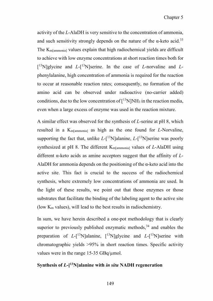
Chapter 5
149
activity of the L-AlaDH is very sensitive to the concentration of ammonia,
and such sensitivity strongly depends on the nature of the α-keto acid.33
The Km[ammonia] values explain that high radiochemical yields are difficult
to achieve with low enzyme concentrations at short reaction times both for
[13N]glycine and L-[13N]serine. In the case of L-norvaline and L-
phenylalanine, high concentration of ammonia is required for the reaction
to occur at reasonable reaction rates; consequently, no formation of the
amino acid can be observed under radioactive (no-carrier added)
conditions, due to the low concentration of [13N]NH3 in the reaction media,
even when a large excess of enzyme was used in the reaction mixture.
A similar effect was observed for the synthesis of L-serine at pH 8, which
resulted in a Km[ammonia] as high as the one found for L-Norvaline,
supporting the fact that, unlike L-[13N]alanine, L-[13N]serine was poorly
synthesized at pH 8. The different Km[ammonia] values of L-AlaDH using
different α-keto acids as amine acceptors suggest that the affinity of L-
AlaDH for ammonia depends on the positioning of the α-keto acid into the
active site. This fact is crucial to the success of the radiochemical
synthesis, where extremely low concentrations of ammonia are used. In
the light of these results, we point out that those enzymes or those
substrates that facilitate the binding of the labeling agent to the active site
(low Km values), will lead to the best results in radiochemistry.
In sum, we have herein described a one-pot methodology that is clearly
superior to previously published enzymatic methods,34 and enables the
preparation of L-[13N]alanine, [13N]glycine and L-[13N]serine with
chromatographic yields >95% in short reaction times. Specific activity
values were in the range 15-35 GBq/µmol.
Synthesis of L-[13N]alanine with in situ NADH regeneration
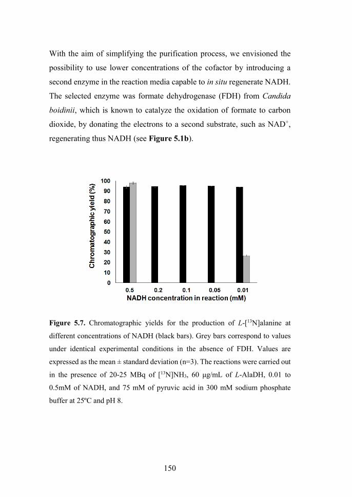
150
With the aim of simplifying the purification process, we envisioned the
possibility to use lower concentrations of the cofactor by introducing a
second enzyme in the reaction media capable to in situ regenerate NADH.
The selected enzyme was formate dehydrogenase (FDH) from Candida
boidinii, which is known to catalyze the oxidation of formate to carbon
dioxide, by donating the electrons to a second substrate, such as NAD+,
regenerating thus NADH (see Figure 5.1b).
Figure 5.7. Chromatographic yields for the production of L-[13N]alanine at
different concentrations of NADH (black bars). Grey bars correspond to values
under identical experimental conditions in the absence of FDH. Values are
expressed as the mean ± standard deviation (n=3). The reactions were carried out
in the presence of 20-25 MBq of [13N]NH3, 60 μg/mL of L-AlaDH, 0.01 to
0.5mM of NADH, and 75 mM of pyruvic acid in 300 mM sodium phosphate
buffer at 25ºC and pH 8.

Chapter 5
151
The one-pot, bi-enzymatic reaction offered exceptional results. The
concentration of NADH in the starting solution could be decreased by 50-
fold without a significant impact on chromatographic yields (Figure 5.6).
The simultaneous addition of L-AlaDH and FDH led to the formation of
L-[13N]alanine with chromatographic yields of 95±2% by using NADH
concentrations as low as 10 μM. These values are three times higher than
those observed without FDH under identical experimental conditions. The
FDH is fundamental to replenish the pool of NADH by consuming formate
as sacrificing substrate; in this manner, the reduced form remains available
for the L-AlaDH to catalyze consecutive cycles of the pyruvate reductive
amination. These results substantially improve the synthetic scheme
described by Baumgartner et al.34 With our configuration, similar yields
can be obtained but the concentration of cofactor is 100-fold lower.
In vivo experiments
Prostate cancer is the fourth cause of death by cancer worldwide, the
second in the male population according to data published by the World
Health Organization (WHO).35,36 Notably, prostate cancer incidence is
dramatically increasing in western societies in the past decades, suggesting
that this type of tumor is intrinsically sensitive to lifestyle changes.37
Despite the good response to standard therapies, a high number of patients
exhibit recurrence and develop metastatic disease after the failure of
subsequent therapeutic regimens. Hence, the molecular aspects related to
disease progression and therapy response remain unclear. Phosphatase and
tensin homolog (Pten) is one of the most commonly deleted/mutated tumor
suppressor genes in human prostate cancer.38,39 As a lipid phosphatase and
a negative regulator of the PI3K/AKT/mTOR pathway, Pten controls

152
several cellular processes, including survival, growth, proliferation,
metabolism, migration, and cellular architecture.40 On the other hand,
cancer cells have altered metabolism and have well-known metabolic
abnormalities. Cancer cells need a continuous supply of nutrients that
maintain abnormal growth and rapid division during cancer progression
and depend on a high rate of aerobic glycolysis for continued growth and
survival. Recent advances in cancer biology and cellular immunity reveal
that not only glucose but also amino acids are essential to support the high
metabolic demands of the tumor.41,42 Because amino acids are the most
highly consumed nutrients by cancer cells and biosynthetic pathways, they
are always in demand for different cancer subtypes.43,44 These processes
of differentiation, proliferation and metabolism of cancer cells can be
understood by using PET imaging technique and metabolic radiotracers,
specifically radiolabeled glucose and amino acids.45-48 The information
obtained about metabolic alterations may aid the development of new
therapies and tools for the early detection of cancer lesions.
In this study, and taking advantage of a collaboration with Prof. Carracedo,
we decided to explore the in vivo behavior of three enzymatically labeled
amino acids (L-[13N]alanine, L-[13N]serine, [13N]glycine) in a mouse
model of prostate cancer, created by depletion of the Pten which resembles
the behavior of human prostate cancer. Before that, we explored the
pharmacokinetics (PK) properties (mainly biodistribution) in healthy
animals. [18F]FDG and [11C]Choline, typically used in the clinical setting
for the detection of prostate cancer, were also used in the study with
diseased animals for comparison.
Starting from 225±25 MBq of cyclotron produced [13N]NH4OH (Volume
of 100µL), 85±6 MBq of pure L-[13N]alanine, [13N]glycine and L-

Chapter 5
153
[13N]serine could be obtained using the enzymatic approach previously
described. The labeled amino acids were obtained with >98% purity, with
decay-corrected radiochemical yields close to 70% in overall production
times of 10 min. Specific activity values ranging 15-35 GBq/µmol were
achieved.
PET studies were conducted also with [18F]FDG, a glucose analogue in
which the hydroxyl group at the 2-position is substituted with an 18F atom;
and [11C]choline, an 11C-labeled amino acid methyl typically used in the
clinical setting for the detection of prostate cancer.49 These tracers are
widely used in the clinical diagnostic field for the early diagnose of
prostate tumors, as well as for the evaluation of the response to
treatment.50-52 The basis behind accumulation of [18F]FDG in tumor tissues
is well known (Figure 5.7). [18F]FDG is taken up and phosphorylated by
cells through the same pathways as glucose, and finally trapped within the
cells since the fluorinated and phosphorylated glucose cannot be
metabolized in the glycolysis. As a result, [18F]FDG concentration
increases proportionally with the utilization rate of glucose and
consequently with the cell growth, and therefore it can be used as an
indirect proliferation marker. On the other hand, [11C]choline incorporates
into tumor cells through an active, carrier-mediated transport mechanism
for choline and then is phosphorylated intracellularly by choline kinase, an
enzyme frequently upregulated in human tumors, yielding phosphoryl
[11C]choline (Figure 5.7).53 In turn, this phosphoryl [11C]choline is
integrated into phospholipids in the cell membrane as part of
phosphatidyl[11C]choline. As the proliferation of cancer cells is much
higher than normal cells, tumor cells exhibit an increased rate of
[11C]choline uptake and incorporation, allowing tumor imaging with PET.

154
Figure 5.8. Intracellular metabolism of [11C]choline and [18F]FDG. Both are
taken up by cells by means of membrane transporters. Once into the cell, they are
phosphorylated to phosphoryl[11C]choline and [18F]glucose-6-phosphate,
respectively. Then, phosphoryl[11C]choline is integrated into phospholipids in the
cell membrane as part of phosphatidyl[11C]choline; [18F]glucose-6-phosphate
does not undergo further metabolism and is trapped in the cell.
In vivo biodistribution studies in wild-type mice
Biodistribution of L-[13N]alanine, [13N]glycine and L-[13N]serine was first
investigated in healthy animals. After image acquisition, volumes of
interest (VOIs) were manually defined in the main organs on CT images
and translated to the PET images for quantification, which resulted in time-
activity curves (TACs) as represented by L-[13N]alanine in Figure 5.8. In
this case, only those organs clearly visualized on the CT images (lung,
heart, kidney, liver, brain and bladder) were delineated. Very similar

Chapter 5
155
biodistribution patterns were obtained for all the labeled amino acids. The
presence of radioactivity was detected in the heart at short times after
administration, confirming the presence of radioactivity in the blood. The
values rapidly decreased from ̴30 %ID/cm3 immediately after injection to
approximately 2%ID/cm3, value that was maintained for a long period of
time. The presence of radioactivity in heart suggests the long residence
time of the labeled species in the blood-pool. At longer time points, a
progressive accumulation of radioactivity in the liver was observed,
consistent with the anabolic function of this organ, with values below 5%
ID/cm3 for the case of L-[13N]alanine. The highest accumulation of
radioactivity was found in the abdominal region, especially in the intestine,
as already reported in previous biodistribution studies for other amino
acids.45 Low accumulation of radioactivity was found in the brain, which
may facilitate the potential localization of tumors in this organ. Normally,
only the aromatic amino acids are presumed to influence brain function.
The aromatic amino acids (e.g. tryptophan, tyrosine, phenylalanine) are
the biosynthetic precursors for the neurotransmitters serotonin, dopamine,
and norepinephrine. However, small neutral amino acids like alanine and
glycine appear to be actively pumped out of the brain.54
Elimination via urine was negligible in the time window of the study for
[13N]alanine, with average accumulation around 1.5 ID%/cm3 (Figure 5.8
and 5.9). A slow progressive elimination by urine was found to
[13N]glycine. However, for L-[13N]serine, elimination via urine was
preferential showing a high uptake in the bladder. This fact may limit
application of this tracer to investigate the uptake in the prostate or prostate
cancer.

156
Figure 5.9. Accumulation of [13N]alanine into the different organs after
intravenous administration. Time-activity-curves are expressed as the percentage
of the injected dose (%ID) per cm3 of tissue. [13N]alanine biodistribution in
organs indicated above each panel. The Y axis scale is different in every graph.
In vivo studies: Prostate-specific Pten deletion cancer model
It is very well known that the amino acid transport is generally increased
in malignant tumor cells.43 This enhanced transport may be associated with
specific cell surface changes in cancer cells.44,55 Generally, the process of
malignant transformation requires that cells acquire and use nutrients
(glucose and amino acids) efficiently for energy, protein synthesis, and
cell division. In this context, the amino acids transport is likely enhanced

Chapter 5
157
in tumor cells to supply the upregulated amino acid metabolism and
protein biosynthesis.
Figure 5.10. Representative PET-CT images obtained after intravenous
administration of L-[13N]alanine in mice. Coronal projections of PET images
have been co-registered with representative CT slices for co-localization of the
radioactive signal. Images correspond to data averaged in the following time
frames: 0-30 s (a); 31-70 s (b); 71-230 s (c); 231-740 s (d); and 741-3020 s (e).
The three amino acids (L-[13N]alanine, [13N]glycine and L-[13N]serine)
labeled with nitrogen-13 by the action of L-AlaDH were tested in a
prostate cancer mouse model, as potential imaging PET agents. Likewise
[11C]choline and [18F]FDG, typically used in the clinical setting for the
detection of prostate cancer, were also used in the study with comparison
purposes.
Upon intravenous administration, accumulation of radioactivity in the
tumor could be observed for L-[13N]alanine and [13N]glycine (see Figure
5.10). Visualization of the prostate after administration of L-[13N]serine

158
was not possible due to the abovementioned accumulation of radioactivity
in the bladder.
Precise quantification of the amount of radioactivity in the prostate was
extremely challenging because the prostate could not be clearly visualized
on the CT images. Hence, the images were only evaluated from a
qualitative point of view. Visual inspection of PET images acquired after
administration of L-[13N]alanine and [13N]glycine suggests a progressive
accumulation of radioactivity in the prostate over time, regardless the
presence of the tumour. However, such accumulation seems to be more
significant in animals with tumour. Despite these promising preliminary
results, further investigation is required in the future to assess the potential
of the 13N-labeled amino acids as metabolic markers in this tumour type.
The involvement of these two amino acids in the metabolism and
proliferation of cancer cells is already known and studied by several
investigators. Alanine has been already identified as an important non-
invasive biomarker for prostate cancer detection and characterization in
magnetic resonance imaging and 1H high-resolution magic angle spinning
(HR-MAS) spectroscopy.56,57 Likewise, glycine metabolism in cancer
cells has been associated with cancer cell proliferation. It has been recently
shown that glycine uptake and catabolism can promote tumorigenesis and
malignancy, suggesting that glycine metabolism could in principle be a
target for the design of innovative therapeutic and diagnostic
strategies.26,58

Chapter 5
159
Figure 5.11. Sagittal PET projections of control and cancer animals resulting
from averaged images (frames 1-33) obtained after administration of [13N]-
labeled amino acids, [18F]FDG and [11C]choline. The position of the bladder and
the tumour are indicated with white and red arrows, respectively. Co-registration
with CT images of the same animal is shown for localization of the radioactivity.

160
Regrettably, high elimination via urine is consistently found for L-
[13N]serine, hampering the visualization of the prostate for the intense
uptake of the labeled amino acid in the region. This is a non-essential
amino acid that is biosynthetically linked to glycine, and both together
provide the essential precursors for the synthesis of both folate and
methionine. These biosynthetic routes are responsible for the synthesis of
proteins, nucleic acids, and lipids, which are essential biological processes
for the uncontrolled growth of cancer cells.26,59-61 Unfortunately, it was not
possible to make use of L-[13N]serine to assess the metabolism of serine in
cancer prostate of mice. However, low uptake of L-[13N]serine observed
in the brain, neck, chest, pelvis, and extremities may open new
opportunities to explore tumor localization in those areas. Many cancer
cells are highly dependent on serine62 which can provide novel
translational opportunities for imaging, drug development, dietary
intervention, and biomarker identification of human cancers.
Standard biodistribution of [11C]choline was observed: relatively high
accumulation in the pancreas, liver and kidneys, and variable uptake in the
bowel, with insignificant urinary excretion (Figure 5.10). Radiolabeled
choline PET has been found to be useful in imaging prostate
cancer.49,51,52,63,64 The biological basis of the radiolabeled choline uptake
in tumors is the malignancy-induced upregulation of choline kinase, which
leads to the incorporation and trapping of choline as phosphatidylcholine
in the tumor cell membranes (Figure 5.10). Unpredictably, tumor uptake
of [11C]choline in our prostate cancer model was almost inexistent. Hence,
the low uptake of [11C]choline can be hypothetically explained by the
hypoxia stage of the cancer cells in our animal model. According to Hara
et al., hypoxic cells significantly incorporate less [11C]Choline, reflecting
diverse biochemical responses to hypoxia.65

Chapter 5
161
For [18F]FDG, the typical biodistribution pattern for this tracer was
observed in our studies: high accumulation was detected in the kidneys
and the bladder due to elimination mainly via urine, while significant
uptake was also observed in the heart and the brain, organs which are
known to consume a significant amount of glucose (Figure 5.10). Due to
its high elimination via urine, [18F]FDG was mainly accumulated in the
bladder, hampering thus visualizing proper visualization of the prostate
and eventually the tumor, similarly to what we observed with L-
[13N]serine.
CONCLUSIONS
In conclusion, we have achieved a fast, efficient, and one-pot synthesis of
L-[13N]alanine, [13N]glycine, and L-[13N]serine catalyzed by L-AlaDH and
using NADH as redox cofactor and FDH as recycling enzymatic partner.
The synthesis of L-[13N]alanine was significantly more efficient than the
synthesis of [13N]glycine and L-[13N]serine. However, the synthesis of
[13N]glycine and L-[13N]serine was successfully optimized by increasing
the biocatalyst load in the reaction mixture. Unfortunately, the synthesis
of artificial amino acids such as L-[13N]norvaline and L-
[13N]phenylalanine was not achieved under no-carrier-added conditions,
due to the high Km values for ammonia.
The synthesis of L-[13N]alanine was optimized by adding FDH to the
reaction media in order to in situ regenerate NADH using formate as
scarifying substrates for hydride transfer. This bi-enzyme system enabled
the 50-fold reduction of NADH concentration in the reaction mixture
without compromising radiochemical yields.

162
The utilization of the enzyme L-AlaDH shows an innovative synthetic
strategy to manufacture ready-to-inject 13N-labeled amino acids (L-
[13N]alanine, [13N]glycine, and L-[13N]serine) in sufficient amount and
purity to approach in vivo studies. Upon intravenous administration, L-
[13N]alanine shows an interesting selective accumulation in the prostate
which seems to increase in the presence of tumors. On the other hand,
[13N]glycine shows uptake in the healthy and cancer prostate.
Unfortunately, L-[13N]serine is eliminated via urine hampering the
visualization of the tumor progression, similarly to what we observed with
[18F]FDG.
REFERENCES
(1) Gelbard, A. S. Biosynthetic methods for incorporating positron-emitting radionuclides into compounds of biomedical interest. Journal of Labelled Compounds and Radiopharmaceuticals 1981, 18, 933-945.
(2) N. Lembares; R. Dinwoodie; I. Gloria; Harper, P.; Lathrop, K. A rapid enzymatic synthesis of 10-min 13N-glutamate and Its pancreatic localization. J. Nucl. Med. 1972, 786-786.
(3) Straatmann, M. G.; Welch, M. J. Enzymatic Synthesis of Nitrogen-13 Labeled Amino Acids. Radiat. Res. 1973, 56, 48-56.
(4) M.B. Cohen; L. Spolter; N.S. McDonald; Cassen, B. Enzymatic synthesis of 13N L-glutamine. J. Nucl. Med., 422.
(5) Cooper, A. J.; Gelbard, A. S. The use of immobilized glutamate dehydrogenase to synthesize 13N-labeled L-amino acids. Anal. Biochem. 1981, 111, 42-48.
(6) Barrio, J. R.; Baumgartner, F. J.; Henze, E.; Stauber, M. S.; Egbert, J. E.; MacDonald, N. S.; Schelbert, H. R.; Phelps, M. E.; Liu, F.-T. Synthesis and Myocardial Kinetics of N-13 and C-11 Labeled Branched-Chain l-Amino Acids. J. Nucl. Med. 1983, 24, 937-944.
(7) Galkin, A.; Kulakova, L.; Yoshimura, T.; Soda, K.; Esaki, N. Synthesis of optically active amino acids from alpha-keto acids with Escherichia coli

Chapter 5
163
cells expressing heterologous genes. Applied and Environmental Microbiology 1997, 63, 4651-4656.
(8) Lu, J.; Zhang, Y.; Sun, D.; Jiang, W.; Wang, S.; Fang, B. The Development of Leucine Dehydrogenase and Formate Dehydrogenase Bifunctional Enzyme Cascade Improves the Biosynthsis of L-tert-Leucine. Applied Biochemistry and Biotechnology 2016, 180, 1180-1195.
(9) Lerchner, A.; Jarasch, A.; Skerra, A. Engineering of alanine dehydrogenase from Bacillus subtilis for novel cofactor specificity. Biotechnology and Applied Biochemistry 2016, 63, 616-624.
(10) Mutti, F. G.; Fuchs, C. S.; Pressnitz, D.; Sattler, J. H.; Kroutil, W. Stereoselectivity of Four (R)-Selective Transaminases for the Asymmetric Amination of Ketones. Advanced Synthesis & Catalysis 2011, 353, 3227-3233.
(11) Baker, P. J.; Sawa, Y.; Shibata, H.; Sedelnikova, S. E.; Rice, D. W. Analysis of the structure and substrate binding of Phormidium lapideum alanine dehydrogenase. Nature structural biology 1998, 5, 561-567.
(12) Abrahamson, M. J.; Wong, J. W.; Bommarius, A. S. The Evolution of an Amine Dehydrogenase Biocatalyst for the Asymmetric Production of Chiral Amines. Advanced Synthesis & Catalysis 2013, 355, 1780-1786.
(13) Grimshaw, C. E.; Cleland, W. W. Kinetic mechanism of Bacillus subtilis L-alanine dehydrogenase. Biochemistry 1981, 20, 5650-5655.
(14) Ye, W.; Huo, G.; Chen, J.; Liu, F.; Yin, J.; Yang, L.; Ma, X. Heterologous expression of the Bacillus subtilis (natto) alanine dehydrogenase in Escherichia coli and Lactococcus lactis. Microbiol. Res. 2010, 165, 268-275.
(15) Siranosian, K. J.; Ireton, K.; Grossman, A. D. Alanine dehydrogenase (ald) is required for normal sporulation in Bacillus subtilis. J. Bacteriol. 1993, 175, 6789-6796.
(16) Freese, E.; Park, S. W.; Cashel, M. The developmental significance of alanine dehydrogenase in bacillus subtilis. Proc. Natl. Acad. Sci. U. S. A. 1964, 51, 1164-1172.
(17) da Silva, E. S.; Gomez-Vallejo, V.; Llop, J.; Lopez-Gallego, F. Efficient nitrogen-13 radiochemistry catalyzed by a highly stable immobilized biocatalyst. Catalysis Science & Technology 2015.
(18) Chenault, H. K.; Simon, E. S.; Whitesides, G. M. Cofactor regeneration for enzyme-catalysed synthesis. Biotechnology & genetic engineering reviews 1988, 6, 221-270.
(19) Weckbecker, A.; Groger, H.; Hummel, W. Regeneration of nicotinamide coenzymes: principles and applications for the synthesis of chiral

164
compounds. Advances in biochemical engineering/biotechnology 2010, 120, 195-242.
(20) Barredo, J. L.: Microbial Enzymes and Biotransformations; Humana Press, 2005.
(21) Pongtharangkul, T.; Chuekitkumchorn, P.; Suwanampa, N.; Payongsri, P.; Honda, K.; Panbangred, W. Kinetic properties and stability of glucose dehydrogenase from Bacillus amyloliquefaciens SB5 and its potential for cofactor regeneration. AMB Express 2015, 5, 68.
(22) Woodyer, R.; Zhao, H.; van der Donk, W. A. Mechanistic investigation of a highly active phosphite dehydrogenase mutant and its application for NADPH regeneration. The FEBS journal 2005, 272, 3816-3827.
(23) Johannes, T. W.; Woodyer, R. D.; Zhao, H. Efficient regeneration of NADPH using an engineered phosphite dehydrogenase. Biotechnol Bioeng 2007, 96, 18-26.
(24) Schirwitz, K.; Schmidt, A.; Lamzin, V. S. High-resolution structures of formate dehydrogenase from Candida boidinii. Protein Science : A Publication of the Protein Society 2007, 16, 1146-1156.
(25) Bradford, M. M. A rapid and sensitive method for the quantitation of microgram quantities of protein utilizing the principle of protein-dye binding. Analytical Biochemistry 1976, 72, 248-254.
(26) Amelio, I.; Cutruzzola, F.; Antonov, A.; Agostini, M.; Melino, G. Serine and glycine metabolism in cancer. Trends Biochem Sci 2014, 39, 191-198.
(27) Albers, M. J.; Bok, R.; Chen, A. P.; Cunningham, C. H.; Zierhut, M. L.; Zhang, V. Y.; Kohler, S. J.; Tropp, J.; Hurd, R. E.; Yen, Y.-F.; Nelson, S. J.; Vigneron, D. B.; Kurhanewicz, J. Hyperpolarized 13C Lactate, Pyruvate, and Alanine: Noninvasive Biomarkers for Prostate Cancer Detection and Grading. Cancer Res. 2008, 68, 8607-8615.
(28) Yoshida, A.; Freese, E. Enzymatic properties of Alanine dehydrogenase of Bacillus subtilis. Biochimica et biophysica acta 1965, 96, 248-262.
(29) Baker, P. J.; Sawa, Y.; Shibata, H.; Sedelnikova, S. E.; Rice, D. W. Analysis of the structure and substrate binding of Phormidium lapideum alanine dehydrogenase. Nat. Struct. Mol. Biol. 1998, 5, 561-567.
(30) Fernandes, P.; Aldeborgh, H.; Carlucci, L.; Walsh, L.; Wasserman, J.; Zhou, E.; Lefurgy, S. T.; Mundorff, E. C. Alteration of substrate specificity of alanine dehydrogenase. Protein Eng. Des. Sel. 2015, 28, 29-35.
(31) Ohashima, T.; Soda, K. Purification and properties of alanine dehydrogenase from Bacillus sphaericus. Eur. J. Biochem. 1979, 100, 29-30.

Chapter 5
165
(32) Brunhuber, N. M.; Blanchard, J. S. The biochemistry and enzymology of amino acid dehydrogenases. Crit. Rev. Biochem. Mol. Biol. 1994, 29, 415-467.
(33) Ohshima, T.; Soda, K.: Biochemistry and biotechnology of amino acid dehydrogenases. In Bioprocesses and Applied Enzymology; Springer Berlin Heidelberg: Berlin, Heidelberg, 1990; pp 187-209.
(34) Baumgartner, F. J.; Barrio, J. R.; Henze, E.; Schelbert, H. R.; MacDonald, N. S.; Phelps, M. E.; Kuhl, D. E. 13N-labeled L-amino acids for in vivo assessment of local myocardial metabolism. Journal of medicinal chemistry 1981, 24, 764-766.
(35) Cancer Research Uk. Worldwide cancer incidence statistics. http://www.cancerresearchuk.org/health-professional/cancer-statistics/worldwide-cancer/incidence.
(36) Ferlay, J.; Soerjomataram, I.; Dikshit, R.; Eser, S.; Mathers, C.; Rebelo, M.; Parkin, D. M.; Forman, D.; Bray, F. Cancer incidence and mortality worldwide: Sources, methods and major patterns in GLOBOCAN 2012. International Journal of Cancer 2015, 136, E359-E386.
(37) Moul, J. W.: Chapter 1 - Population Screening for Prostate Cancer and Early Detection A2 - Mydlo, Jack H. In Prostate Cancer (Second Edition); Godec, C. J., Ed.; Academic Press: San Diego, 2016; pp 3-12.
(38) Ugalde-Olano, A.; Egia, A.; Fernández-Ruiz, S.; Loizaga-Iriarte, A.; Zuñiga-García, P.; Garcia, S.; Royo, F.; Lacasa-Viscasillas, I.; Castro, E.; Cortazar, A. R.; Zabala-Letona, A.; Martín-Martín, N.; Arruabarrena-Aristorena, A.; Torrano-Moya, V.; Valcárcel-Jiménez, L.; Sánchez-Mosquera, P.; Caro-Maldonado, A.; González-Tampan, J.; Cachi-Fuentes, G.; Bilbao, E.; Montero, R.; Fernández, S.; Arrieta, E.; Zorroza, K.; Castillo-Martín, M.; Serra, V.; Salazar, E.; Macías-Cámara, N.; Tabernero, J.; Baselga, J.; Cordón-Cardo, C.; Aransay, A. M.; Villar, A. D.; Iovanna, J. L.; Falcón-Pérez, J. M.; Unda, M.; Bilbao, R.; Carracedo, A. Methodological aspects of the molecular and histological study of prostate cancer: Focus on PTEN. Methods 2015, 77–78, 25-30.
(39) Nardella, C.; Carracedo, A.; Salmena, L.; Pandolfi, P. P. Faithfull modeling of PTEN loss driven diseases in the mouse. Current topics in microbiology and immunology 2010, 347, 135-168.
(40) Jiang, B. H.; Liu, L. Z. PI3K/PTEN signaling in angiogenesis and tumorigenesis. Advances in cancer research 2009, 102, 19-65.
(41) Ananieva, E. Targeting amino acid metabolism in cancer growth and anti-tumor immune response. World Journal of Biological Chemistry 2015, 6, 281-289.
(42) Bhutia, Y. D.; Babu, E.; Ramachandran, S.; Ganapathy, V. Amino Acid transporters in cancer and their relevance to "glutamine addiction": novel

166
targets for the design of a new class of anticancer drugs. Cancer research 2015, 75, 1782-1788.
(43) Busch, H.; Davis, J. R.; Honig, G. R.; Anderson, D. C.; Nair, P. V.; Nyhan, W. L. The Uptake of a Variety of Amino Acids into Nuclear Proteins of Tumors and Other Tissues. Cancer research 1959, 19, 1030-1039.
(44) Isselbacher, K. J. Increased Uptake of Amino Acids and 2-Deoxy-D-Glucose by Virus-Transformed Cells in Culture. Proceedings of the National Academy of Sciences 1972, 69, 585-589.
(45) Jager, P. L.; Vaalburg, W.; Pruim, J.; de Vries, E. G.; Langen, K. J.; Piers, D. A. Radiolabeled amino acids: basic aspects and clinical applications in oncology. Journal of nuclear medicine : official publication, Society of Nuclear Medicine 2001, 42, 432-445.
(46) Crippa, F.; Alessi, A.; Serafini, G. L. PET with radiolabeled aminoacid. The quarterly journal of nuclear medicine and molecular imaging : official publication of the Italian Association of Nuclear Medicine (AIMN) [and] the International Association of Radiopharmacology (IAR), [and] Section of the So 2012, 56, 151-162.
(47) Hosios, A. M.; Hecht, V. C.; Danai, L. V.; Johnson, M. O.; Rathmell, J. C.; Steinhauser, M. L.; Manalis, S. R.; Vander Heiden, M. G. Amino Acids Rather than Glucose Account for the Majority of Cell Mass in Proliferating Mammalian Cells. Developmental cell 2016, 36, 540-549.
(48) Haberkorn, U.; Markert, A.; Mier, W.; Askoxylakis, V.; Altmann, A. Molecular imaging of tumor metabolism and apoptosis. Oncogene 2011, 30, 4141-4151.
(49) Jadvar, H. Prostate Cancer: PET with 18F-FDG, 18F- or 11C-Acetate, and 18F- or 11C-Choline. Journal of Nuclear Medicine 2011, 52, 81-89.
(50) Picchio, M.; Messa, C.; Landoni, C.; Gianolli, L.; Sironi, S.; Brioschi, M.; Matarrese, M.; Matei, D. V.; De Cobelli, F.; Del Maschio, A.; Rocco, F.; Rigatti, P.; Fazio, F. Value of [11C]choline-positron emission tomography for re-staging prostate cancer: a comparison with [18F]fluorodeoxyglucose-positron emission tomography. The Journal of urology 2003, 169, 1337-1340.
(51) Reske, S. N.; Blumstein, N. M.; Neumaier, B.; Gottfried, H. W.; Finsterbusch, F.; Kocot, D.; Moller, P.; Glatting, G.; Perner, S. Imaging prostate cancer with 11C-choline PET/CT. Journal of nuclear medicine : official publication, Society of Nuclear Medicine 2006, 47, 1249-1254.
(52) Murphy, R. C.; Kawashima, A.; Peller, P. J. The Utility of 11C-Choline PET/CT for Imaging Prostate Cancer: A Pictorial Guide. American Journal of Roentgenology 2011, 196, 1390-1398.

Chapter 5
167
(53) Lockman, P. R.; Allen, D. D. The transport of choline. Drug development and industrial pharmacy 2002, 28, 749-771.
(54) Laterra J; Keep R; Betz LA; al., e.: Blood—Brain Barrier. In Basic Neurochemistry: Molecular, Cellular and Medical Aspects.; 6th ed.; Siegel GJ, A. B., Albers RW, et al., editors., Ed.: Philadelphia: Lippincott-Raven, 1999.
(55) Haase, C.; Bergmann, R.; Fuechtner, F.; Hoepping, A.; Pietzsch, J. L-Type Amino Acid Transporters LAT1 and LAT4 in Cancer: Uptake of 3-O-Methyl-6- 18F-Fluoro-l-Dopa in Human Adenocarcinoma and Squamous Cell Carcinoma In Vitro and In Vivo. Journal of Nuclear Medicine 2007, 48, 2063-2071.
(56) Tessem, M.-B.; Swanson, M. G.; Keshari, K. R.; Albers, M. J.; Joun, D.; Tabatabai, Z. L.; Simko, J. P.; Shinohara, K.; Nelson, S. J.; Vigneron, D. B.; Gribbestad, I. S.; Kurhanewicz, J. Evaluation of Lactate and Alanine as Metabolic Biomarkers of Prostate Cancer Using (1)H HR-MAS Spectroscopy of Biopsy Tissues. Magnetic resonance in medicine : official journal of the Society of Magnetic Resonance in Medicine / Society of Magnetic Resonance in Medicine 2008, 60, 510-516.
(57) Albers, M. J.; Bok, R.; Chen, A. P.; Cunningham, C. H.; Zierhut, M. L.; Zhang, V. Y.; Kohler, S. J.; Tropp, J.; Hurd, R. E.; Yen, Y. F.; Nelson, S. J.; Vigneron, D. B.; Kurhanewicz, J. Hyperpolarized 13C lactate, pyruvate, and alanine: noninvasive biomarkers for prostate cancer detection and grading. Cancer research 2008, 68, 8607-8615.
(58) Jain, M.; Nilsson, R.; Sharma, S.; Madhusudhan, N.; Kitami, T.; Souza, A. L.; Kafri, R.; Kirschner, M. W.; Clish, C. B.; Mootha, V. K. Metabolite profiling identifies a key role for glycine in rapid cancer cell proliferation. Science (New York, N.Y.) 2012, 336, 1040-1044.
(59) Yang, M.; Vousden, K. H. Serine and one-carbon metabolism in cancer. Nat Rev Cancer 2016, 16, 650-662.
(60) Mattaini, K. R.; Sullivan, M. R.; Vander Heiden, M. G. The importance of serine metabolism in cancer. The Journal of Cell Biology 2016, 214, 249-257.
(61) Tsun, Z. Y.; Possemato, R. Amino acid management in cancer. Seminars in cell & developmental biology 2015, 43, 22-32.
(62) Frezza, C. Cancer metabolism: Addicted to serine. Nat Chem Biol 2016, 12, 389-390.
(63) Krause, B. J.; Souvatzoglou, M.; Treiber, U. Imaging of prostate cancer with PET/CT and radioactively labeled choline derivates. Urologic oncology 2013, 31, 427-435.

168
(64) Vali, R.; Loidl, W.; Pirich, C.; Langesteger, W.; Beheshti, M. Imaging of prostate cancer with PET/CT using (18)F-Fluorocholine. American Journal of Nuclear Medicine and Molecular Imaging 2015, 5, 96-108.
(65) Hara, T.; Bansal, A.; DeGrado, T. R. Effect of hypoxia on the uptake of [methyl-3H]choline, [1-14C] acetate and [18F]FDG in cultured prostate cancer cells. Nucl Med Biol 2006, 33, 977-984.

Chapter 6
L-Alanine dehydrogenase from Bacillus subtillis has been intensively exploited in
biocatalysis but its immobilized form is rarely used. In the present work, the
immobilization of L-Alanine dehydrogenase from Bacillus subtillis on agarose
microbeads activated with glyoxyl groups (aliphatic aldehydes) and other two carriers is
presented. These immobilized enzyme preparations were extensively characterized
towards temperature, pH and kinetic parameters. Finally, we have applied the optimal
heterogeneous biocatalyst in the synthesis of L-[13N]alanine using pyruvate and
[13N]NH4OH, as substrates. Herein, we have fabricated a highly stable and active
heterogeneous biocatalyst for reductive aminations of -ketoacids that has an enormous
potential in biocatalysis.
Design and full characterization of L-Alanine
immobilized on solid and porous materials


Chapter 6
171
INTRODUCTION
As already described in Chapter 5, L-alanine dehydrogenase from Bacillus
subtilis (L-AlaDH) was used for the radiochemical synthesis of a variety
of L-[13N]amino acids with excellent radiochemical yields and purities.1
Moreover, L-AlaDH from Bacillus subtilis has been also exploited as an
auxiliary enzyme to replenish the amine donor (L-alanine) in
transamination reactions catalyzed by ω-transaminases.2 These examples
show the usefulness of this enzyme in synthetic chemistry. However, the
use of enzymes in synthetic applications is frequently limited by their
solubility and their low stability under operational conditions.
These limitations underlying the vast majority of the native enzymes have
been addressed at once by using immobilization techniques.3 Any
immobilization technique overcomes the solubility problem; nevertheless,
the immobilization itself does not guarantee the enzyme stabilization and
sometimes reduces the catalytic activity of the enzymes.3,4 Enzyme
immobilization is still considered an intuitive technique where the optimal
immobilization protocol is often found using the trial-and-error approach.
However, a deep understanding of the molecular structure of enzymes
combined with spectroscopic tools enable appropriate characterization of
the interactions between proteins and solid materials, as well as the
evaluation of the effects that the immobilization causes on both protein
structure and functionality.5 Such characterization can guide the
optimization of the immobilization protocols to fabricate the most efficient
heterogeneous biocatalysts.
To the best of our knowledge, L-AlaDH has been only successfully
immobilized on amine surfaces activated with glutaraldehyde6 and on
sepharose microbeads activated with cyanogen bromide7. In the first

172
system, the immobilized enzyme retained more than 90% of its initial
activity for weeks under storage conditions at 4°C, while in the second
case, the enzyme kept 70% of its initial activity after one month at 37°C.
In this chapter, L-Alanine dehydrogenase from Bacillus subtillis has been
immobilized through different immobilization chemistries. Structural
modeling of L-AlaDH supported by kinetic and fluorescence studies was
also performed to understand how the immobilization reduces the enzyme
activity but greatly increases the enzyme stability. Finally, to demonstrate
the potential of this heterogeneous biocatalyst in the context of
radiochemistry, the radiosynthesis of L-[13N]alanine was carried out.
EXPERIMENTAL SECTION
Reagents. β-Nicotinamide adenine dinucleotide, reduced disodium salt
hydrate (β-NADH; purity >97%), pyruvic acid (CH3COCOOH, purity
≥98%), ammonium chloride (NH4Cl, for molecular biology, purity
≥99.5%), sodium phosphate dibasic (Na2HPO4, for molecular biology,
purity ≥98.5%), sodium phosphate monobasic (NaH2PO4, purity ≥99.0%),
L-alanine (purity ≥98%), SYPRO® Orange Protein Gel Stain and
cyanogen bromide-activated sepharose 4B (Ag-CB) were purchased from
Sigma Chemical Co. (St. Louis, IL, USA) and used without further
purification. TALON resin was purchased from Clontech (Saint-Germain-
en-Laye, France). Bio-Rad Protein Assay Dye Reagent Concentrate was
purchased from Bio-Rad Laboratories (Madrid, Spain). Agarose 4BCL
microbeads (ABT, Madrid) activated with aldehyde groups were prepared
as described elsewhere.8 All other reagents were of analytical grade.

Chapter 6
173
Structural modelling of L-Alanine Dehydrogenase from Bacillus
subtilis. Three different monomeric structural models of L-AlaDH based
on sequence homology were built using as templates the alanine
dehydrogenases from Thermus thermophilus, Mycobacterium
tubercolosis, and Phormidium lapideum available in protein data bank.9
The most accurate model was obtained by using the L-AlaDH from
Phormidium lapideum (PDB: 1SAY) despite having lower sequence
identity (58.86 %) with L-AlaDH from Bacillus subtilis primary sequence
than the L-AlaDH from Thermus thermophilus (62.84 %). Finally, the
hexamer of our L-AlaDH was constructed by using the biological
assembly of Phormidium lapideum as a template. Protein models were
visualized and aligned with their template structure by using PyMol 0.99
developed by DeLano Scientific LLC (San Francisco, CA). The
electrostatic potential and the local flexibility of the most accurate model
were calculated by using the Bluues10,11 and COREX/BEST12 servers,
respectively.
Figure 6.1. Plot showing predicted local similarity of L-AlaDH enzyme model
by the homology-modeling server from Expasy (predicted local similarity versus
residue number).

174
Protein and enzyme colorimetric assays. Determination of the protein
concentration. The protein concentration of soluble L-AlaDH and FDH
were measured as described in Chapter 4 for eNR. Determination of
enzyme activity. The enzymatic activities of soluble and immobilized
preparations were carried as described in Chapter 7.
Immobilization of L-Alanine Dehydrogenase from Bacillus subtilis.
The immobilization of L-Alanine Dehydrogenase from Bacillus subtilis
was performed on three different carriers: 1) porous 4BCL agarose
microbeads activated with cyanogen bromide groups (Ag-CB); porous
4BCL agarose microbeads activated with aldehyde (glyoxyl) groups (Ag-
G) and porous 6BCL agarose microbeads activated with cobalt-chelates.
Ten milliliters of 16g per mL L-AlaDH solution was added to 1 g of the
carrier (Ag-CB, Ag-G and Ag-Co2+). In the case of Ag-CB and Ag-Co2+,
the enzyme solution was prepared in 25 mM sodium phosphate buffer at
pH 8. For Ag-G, the enzyme solution was prepared in 100 mM sodium
carbonate buffer at pH 10. The mixtures were incubated under gentle
stirring for 2h at 25°C. Periodically, samples of the supernatants and the
enzyme-carrier suspensions were withdrawn and analyzed to evaluate the
progress of the immobilization reaction. After completion of the
immobilization, the samples with Ag-G were reduced with 1 mg x mL-1
NaBH4 during 30 minutes at 4°C, whereas the samples with Ag-CB were
incubated with 100 mM of hydroxylamine for 1 hour at 25°C. All the
immobilized preparations were filtered, washed with distilled water and
finally equilibrated with 25 mM phosphate buffer solution at pH 8.
Thermal Stability. Thermal stability was determined by incubating
enzyme solutions or suspensions in 10 mM phosphate buffer at pH 8 and
60°C for 24 h in a thermo-shaker. Samples were taken at 0.5, 1, 2, 4, 6,

Chapter 6
175
and 24 h of incubation, and their residual activity was measured as
described above. The experimental data were fitted to first-kinetic
equation (A/Ao=e-k*t) using Origin 8.0. In this equation, A is the residual
activity at different incubation times (t), Ao is the initial activity before
incubations and k is the inactivation constant.
Activity-pH profile. The effect of pH on the activity of different enzyme
preparations was determined by measuring the reductive amination of
pyruvic acid in the presence of NADH at the different pH values (5-10) at
25°C. The expressed activity was measured as described before. Different
10 mM buffer solutions were used to set the right pH value; acetate buffer
was used for pH 5 and 6; sodium phosphate buffer for pH 7 and 8 and
sodium bicarbonate buffer for pH 9 and 10.
Fluorescence assays. Fluorescence Spectra were carried out with a
Varioskan Flash fluorescence spectrophotometer (Thermo Scientific),
using an excitation wavelength of 280 nm with excitation and emission
bandwidths of 5 nm and recording fluorescence emission spectrum
between 300 and 550 nm. All spectroscopic measurements were
performed with a solution of 3g x mL-1 of the enzyme (soluble or
immobilized) in 10 mM sodium phosphate at pH 8 and 25°C.
Thermal-shift assay. This assay was performed using 3g x mL-1 of the
enzyme (soluble or immobilized) in 25 mM sodium phosphate buffer and
0.3% of SYPRO® Orange at pH 7. Then, this mixture was incubated in a
StepOne-plus RT-PCR (Applied biosystems-ThermoFisher) by using a
temperature ramp program from 25 to 95°C with a ramp rate of 0.5°C per
minute.

176
Enzyme kinetic parameters. Michaelis constant value (Km) and
maximum rate (Vmax) were determined by activity colorimetric assay at
pH 8 and 25°C as previously described but using 0-1.8M ammonium
chloride, 0-1mM NADH and 0-0.15M pyruvic acid. The experimental data
were fitted to the Michaelis-Menten equation using Origin 8.0.
Production of the radiolabeling agent [13N]NH3. Nitrogen-13 (13N) was
produced in an IBA Cyclone 18/9 cyclotron via the 16O(p,α)13N nuclear
reaction as described in chapter 5. Integrated currents around 0.2 μAh were
used. The resulting solution was transferred to a 10mL vial and the activity
was measured in a dose calibrator (Capintec CRC®-25 PET, New Jersey,
USA).
Synthesis of L-[13N]alanine. The radiosynthesis of L-[13N]alanine was
carried out by adding 100µL of a mixture containing 0.5mM NADH,
75mM pyruvic acid and 300mM sodium phosphate buffer solution at pH
8 (final concentrations in reaction) to a solution or suspension containing
L-AlaDH (concentration in the range 15-45 µg/mL) and 100µL of
[13N]NH4OH (35-45 MBq). Reactions were conducted in test-tubes with
50 KDa cut-off membranes in the case of the soluble enzymes and with
micrometric filters in the case of immobilized. The corresponding mixture
was incubated at 25°C under mild stirring for 2.5 to 20 min and
subsequently filtered under centrifugation during one minute to remove
the enzyme.

Chapter 6
177
RESULTS AND DISCUSSION
Immobilization of L-AlaDH on porous agarose microbeads activated
with different reactive groups
L-Alanine dehydrogenase from Bacillus subtillis was immobilized on
porous 4BCL agarose microbeads activated with either cyanogen bromide
groups (Ag-CB) or with aldehyde (glyoxyl) groups (Ag-G) and porous
6BCL agarose microbeads charged with a tetradentate chelator charged
with cobalt (AG-Co2+). At pH 8, the cyanogen bromide groups of Ag-CB
react with the amine from the N-terminus of each subunit forming
irreversible bonds,13 and the cobalt chelates on the agarose surface of Ag-
Co2+ preferentially interact with the imidazole rings of histidine tags
located at the N-terminus of each subunit of L-AlaDH, creating reversible
coordination bonds between the enzyme and the solid surface. On the other
hand, the immobilization on Ag-G requires alkaline pH to enable the
protein-carrier interactions. The aldehydes of the carrier form reversible
imine groups with the primary amine groups of the enzyme (N-terminus
and -NH2 from the most exposed lysines) that are finally reduced to
irreversible secondary amines by using a mild reducing agent such as
sodium borohydride.13
As it can be seen at Table 6.1, 85-99% of the offered L-AlaDH was
immobilized on the three carriers herein investigated, but the enzyme
activity was reduced upon immobilization in all cases. Nevertheless,
substantial differences were observed between the expressed activities of
the enzymes immobilized on the different carriers; while the specific
activity of L-AlaDH immobilized on Ag-CB was 67% of the soluble
enzyme, L-AlaDH immobilized on Ag-Co2+ was 58% of the soluble

178
enzyme and L-AlaDH immobilized on Ag-G expressed 45% of the activity
of the soluble one.
Table 6.1. Immobilization parameters of L-Alanine Dehydrogenase from
Bacillus Subtilis immobilized onto agarose porous microbeads activated with
different reactive groups.
CARRIER Reactive
group Immobilization chemistry
Immobilized Activity Ai (U/g)a
Immobilization yield (%)b
Expressed Activity
Ae (U/g)c/ (%)d
Ag-CB Cyanogen Bromide
Covalent bond through the primary amine group of the N-terminus
55317 973
38587 (67)
Ag-G Glyoxyl Multipoint covalent bond through the -NH2
of the lysines 56679 8512 295 4
(45)
Ag-Co2+ Cobalt (Co2+)
Affinity binding through the His-tags 56411 992 33920
(58)
aAi=The activity immobilized on 1 gram of carrier after the immobilization process. This activity was calculated as the difference between the offered activity and the activity in the supernatant after incubation with the carrier for 2h. bImmobilization yield () = (immobilized activity/offered activity) x 100. cThe expressed activity (Ae) is defined as the measured activity of the immobilized enzyme after washing. dRelative expressed activity (Ae) = (expressed activity/immobilized activity) x 100.
Similarly, previous works have demonstrated that the immobilization of
two homolog L-AlaDHs from Bacillus stearothermophilus6 and Bacillus
cereus7 on Ag-CB and aminated glass beads activated with glutaraldehyde,
respectively, recovered 50-60% of the specific activity. Remarkably, in the
present work, the L-AlaDH retained 50% of its initial activity upon the
immobilization on Ag-G under alkaline conditions. Such high activity
recovery after irreversible covalent attachment using Ag-G carrier has also
been observed with the glutamate dehydrogenase from Thermus
thermophilus.14 The high stability of these two enzymes (L-alanine
dehydrogenase and glutamate dehydrogenase) under alkaline conditions
avoids their dramatic inactivation during the immobilization process at pH
10.14,15 In contrast, lactate dehydrogenase from rabbit muscle loses 92%

Chapter 6
179
of its initial activity upon immobilization on Ag-G without adding any
enzyme stabilizers.16
Prediction of L-AlaDH orientation and attachment on the surface of
different carriers
To better understand the molecular interaction between the L-AlaDH and
the carrier, we built a structural model of the hexameric L-AlaDH. The
model was built by using Swiss-Model Homology modelling server which
utilizes structure-known proteins deposited in the protein data bank.17,18
Moreover, we calculated the surface electrostatics10,11 and the residue
exposure of this model in order to know which are the most exposed
lysines with the lowest pKa values (-NH2) (Table 6.2).
Table 6.2. Surface exposure and apparent pKa of the lysine residues found in L-
AlaDH from Bacillus subtilis.
Position Calculated pKa Total Surface area
32 11.686 188
35 12.244 166
87 11.19 226
97 12.925 3
99 12.226 50
111 10.727 234
132 11.027 123
134 11.892 7
174 10.922 347
176 11.112 173
179 12.564 124
193 12.308 109
208 10.814 155
239 11.089 261
268 12.502 214

180
Position Calculated pKa Total Surface area
279 11.679 210
282 10.683 230
314 11.715 178
351 11.774 142
355 10.941 291
The values presented in this table were calculated by Bluues server using the pdb file generated in Swiss-Model
server. pKa value corresponds to the -NH2 of each lysine. Lysine residues with pKa lower than 11 are
highlighted in red because they are predicted to be the most reactive ones under the immobilization conditions
(pH 10) on Ag-G. These lysines are identified in the enzymatic model represented in Figure 5.1.
The hexamer contains 120 lysine residues (20 per subunit) distributed
along its 3D surface; 5 lysine residues per subunit (K111, K174, K208,
K282 and K355) present apparent pKa values lower than 11. This means
that under the alkaline immobilization conditions, those 5 lysines and the
N-terminus will be the only primary amino groups majorly unprotonated
and thus ready to react with aldehyde groups on the Ag-G surface.
Moreover, the N-terminus and the lysines K111, K174, K282 and K355
are fairly exposed to the solvent, which assures the geometrical
congruence required for the immobilization reaction to take place.
After visualizing the 3D-model of L-AlaDH, it was suggested that L-
AlaDH may be immobilized on Ag-G, Ag-CB and Ag-Co2+ through
mainly one region, Orientation 1, that fairly displays 3 N-terminus and 6
out of 30 most reactive lysines (3 x K111 and 3 x K351). Ag-CB and Ag-
Co2+ selectively attach the N-terminus, whereas Ag-G establishes bonds
with the most reactive amine groups including N-terminus, K111 and
K351. In addition, such region presents a suitable geometrical congruence
(large flat surface) to attach at least 3 subunits to the solid surface (Figure
6.2).

Chapter 6
181
Figure 6.2. Different planes (orientations) of the L-AlaDH surface. The
quaternary structure of L-AlaDH is formed by six subunits organized in three
pairs of symmetrical faces giving rise to three different planar surfaces. None of
the three faces displays more than three subunits with suitable geometrical and
chemical congruence to react with the agarose surface. The 6 different subunits
are colored in red, purple, violet, green, yellow and green, while all the active
sites are colored in white. The N-terminus (Nt) and the -NH2 from the most
exposed lysines with the lowest pKa values (calculated by Bluues server) are
represented as blue spheres. The images were created with Pymol 0.99v.
Kinetics parameters of L-AlaDH immobilized on different carriers
The kinetics parameters of immobilized enzymes often differ significantly
from those of the soluble enzymes. In our case, the immobilization of L-
AlaDH on different carriers affected the kinetics parameters of the
reductive amination of pyruvate in different manners (Table 6.3).
The catalytic efficiency towards NADH was significantly higher for the
soluble enzyme than for the immobilized forms, mainly because the latter
suffered a dramatic affinity loss for the redox cofactor (high Km). The
higher Km values of the immobilized enzymes for NADH (up to 12.5-fold
higher than the soluble enzyme) resulted in lower Vmax values for all
substrates. This effect was less dramatic when the enzyme was

182
immobilized on Ag-CB; in this case, the immobilized enzyme presented a
3.6 and 2.9-fold lower Km for NADH than the enzyme immobilized on Ag-
G and Ag-Co2+, respectively. Noteworthy, L-AlaDH immobilized on Ag-
G showed 8.5 and 3.4-fold lower Km values for pyruvate and ammonia,
respectively, than the soluble enzyme. Therefore, the aldehyde chemistry
seems to enhance the affinity of the immobilized L-AlaDH for both
ammonia and pyruvate which improves the catalytic efficiency 110 and
285%, respectively, with respect to the free enzyme.
Table 6.3. Kinetic characterization of free and immobilized L-AlaDH from
Bacillus Subtilis.
Vmax
Km mM
Kcat s-1
Kcat/Km s-1 x M-1
NADH
AlaDH soluble 31.05±2.29 0.02±0.008 1.37E+02 6.83E+06
AlaDH Ag-CB 22.00±1.52 0.07±0.017 9.68E+01 1.38E+06
AlaDH Ag-Co2+ 17.79±2.69 0.20±0.08 7.82E+01 3.89E+05
AlaDH Ag-G 17.92±2.5 0.25±0.12 7.88E+01 3.15E+05
NH4Cl
AlaDH soluble 44.08±1.77 100.35±15.32 1.94E+02 1.93E+03
AlaDH Ag-CB 39.45±2.52 120.86±28.02 1.73E+02 1.44E+03
AlaDH Ag-Co2+ 14.90±0.62 29.91±6.80 6.55E+01 2.19E+03
AlaDH Ag-G 14.02±1.20 29.16±6.53 6.24E+01 2.14E+03
Pyruvic acid
AlaDH soluble 55.39±2.20 0.17±0.04 2.44E+02 1.41E+06
AlaDH CNBr 27.37±1.27 0.17±0.05 1.20E+02 7.10E+05
AlaDH Ag-Co2+ 19.43±1.47 0.15±0.07 8.55E+01 5.84E+05
AlaDH Glyoxyl 12.67±1.31 0.02±0.006 6.85E+01 4.03E+06
In all the cases, the immobilization of L-AlaDH driven by either the N-
terminus, the His-tags or the primary amine groups with the lowest pKa
seems to orient the protein through a region where none of the active sites
directly contact with the carrier surface (Orientation 1, see Figure 6.2).

Chapter 6
183
Contrarily, the cofactor binding regions of at least three subunits seem to
interact with the carrier surface, which may hamper the interaction of the
immobilized enzyme with NADH. This fact may explain the high Km
values observed for NADH, which ultimately compromise the catalytic
efficiency of the immobilized enzyme. Furthermore, the valence and the
nature of the interaction between these protein regions and the carrier
surface may differently distort tertiary and quaternary structures, thus
explaining the changes in affinity observed for the other two substrates
(ammonia and pyruvate).
Protein thermal stability of immobilized L-AlaDH preparations
The integrity of the protein structure under reaction conditions is
fundamental to guarantee the protein stability during the operational
process. L-AlaDH immobilized on different agarose-based microbeads
was thermally inactivated at 60°C and compared with the soluble enzyme
(Figure 6.3 and Table 6.4).
Figure 6.3 shows that L-AlaDH immobilized on Ag-G, Ag-CB and Ag-
Co2+ were 23.8, 3.3 and 1.2-fold more stable than the soluble enzyme,
respectively. Moreover, at higher temperatures L-AlaDH immobilized on
Ag-G presented 53% of its initial activity after 30 minutes incubation at
64°C, while the L-AlaDH immobilized on Ag-Co2+ and the soluble
enzyme was almost completely inactivated (residual activity < 10%).

184
Figure 6.3. Thermal inactivation of soluble and immobilized L-AlaDH. The
different enzymatic preparations were incubated at 60°C and pH 8: Soluble L-
AlaDH (squares); L-AlaDH immobilized on Ag-CB (circles); L-AlaDH
immobilized on Ag-Co2+ (diamonds); and L-AlaDH immobilized on Ag-G
(triangles).
Table 6.4. Kinetics parameters of the thermal inactivation of different L-AlaDH
preparations. The thermal inactivation was carried out at 60°C and pH 8 during
24h.
0 4 8 12 16 20 240
20
40
60
80
100
120
140
Time [hour]
Re
sid
ua
l Ac
tivi
ty [
%]
Soluble Ag-CB Ag-G Ag-Co
Carrier a Inactivation constant
(h-1)
Half-life time
(h)b Stabilization factor c
Soluble 0.38±0.04 1.82 -
Ag-CB 0.11±0.02 6.07 3.3
Ag-Co2+ 0.31±0.02 2.22 1.2
Ag-G 0.016±0.01 43.32 23.8
a) Experimental data from Figure 5.2 was fitted with a First-order kinetics equation considering total
inactivation (A/Ao=e-k*t). b) The half-life time was calculated introducing to the corresponding equation
A/Ao=0.5. c) Stabilization factor means the half-life time of the corresponding immobilization preparation
divided by the half-life time of the soluble enzyme.

Chapter 6
185
To evaluate the effect of the thermal inactivation on the protein structure
of L-AlaDH immobilized on Ag-G, the intrinsic fluorescence of soluble
and immobilized enzyme on Ag-G were measured before and after
incubation at 60°C for 4 hours (Figure 6.4A).
Figure 6.4. (A) Intrinsic fluorescence of soluble and immobilized L-AlaDH. The
intrinsic fluorescence of tryptophan was measured for soluble (black lines) and
immobilized (grey line) enzymes before (solid line) and after (dash line) sample
heating at 60°C and pH 8 during 4 hours. (B) The inset figure shows the
normalized fluorescence of immobilized L-AlaDH before and after the
inactivation. All fluorescence values were normalized regarding the fluorescence
at λmax for each sample.
In one hand, the immobilization of L-AlaDH promoted a 20nm blue-shift
of λmax and 2-fold enhancement of the fluorescence intensity compared to

186
its soluble counterpart before thermal inactivation. These results suggest
that L-AlaDH has suffered some conformational changes upon the
immobilization that may explain the lower specific activity of the
immobilized enzyme. The soluble enzyme presented 6% of its maximum
fluorescence intensity and its λmax was 45 nm red-shifted after thermal
incubation, suggesting large structural changes driving the protein to
unfolded states. On the other hand, the maximum fluorescence intensity of
L-AlaDH immobilized on Ag-G was reduced to 32% and the λmax red-
shifted 15 nm after the thermal incubation (inset Figure 6.4B).
Figure 6.5. In silico representation of L-AlaDH local flexibility using a
COREX/BEST server. (A) zoomed-in view of the interaction region of one
subunit showing the highest density of low pKa amine groups. W and F residues
are coloured in green; (B) rigid and flexible residues are coloured in red and
yellow, respectively. Gradering between red and yellow (orange) represent
moderately stable regions. The N-terminus (Nt) and the -NH2 from the most
exposed lysines with the lowest pKa values (calculated by Bluues server) are
represented as blue spheres. The images were created with Pymol 0.99v.

Chapter 6
187
With the molecular model of L-AlaDH in hand, it was pointed that the
fluorescence changes induced by thermal incubation may rely on some
structural rearrangements that affect the protein environment of the unique
tryptophan residue W90 (Figure 6.4). In the soluble enzyme, the loss of
fluorescence intensity and the large red-shift of the λmax suggest that W90
becomes dramatically exposed to the solvent. Contrarily, the
immobilization of L-AlaDH on Ag-G seems to protect that region,
alleviating the conformational changes in the vicinity of the W90 and
resulting in a lower reduction in the fluorescence intensity and a smaller
red-shift of the λmax.
Finally, to rationalize why the immobilization on Ag-G through the
proposed region increases the thermal stability of L-AlaDH, the residue
stability constant of the L-AlaDH model was calculated by using
COREX/BEST server.12,19 Figure 6.5B shows that the interaction region
for Orientation 1 is composed of high, medium and low flexible local
domains. Although, the most reactive groups (low pKa lysines and N-
terminus) are located in the most rigid domains, additional bonds can
involve lysines from other domains with medium and high flexibility
which promote an artificial rigidification of that region. However, since
the immobilization on Ag-CB is restricted to the interactions with the N-
terminus of the enzyme (located at the rigid domains), further
rigidification of the most flexible domains is more difficult. In fact, these
results are supported by the SDS-PAGE analyses (Figure 6.6) that show
how Ag-G irreversibly attaches the 6 subunits, while Ag-CB can only
attach 2 out of the 6 subunits. SDS-PAGE studies showed that the 6
subunits of L-AlaDH were irreversibly attached to Ag-G likely due the
flexibility of the agarose fibers that may enable a 3D interaction between

188
less accessible and reactive lysines and the aldehyde groups of the carrier
surface (Figure 6.6).
Figure 6.6. (A) SDS-PAGE (14%) of L-AlaDH immobilized on Ag-CB and Ag-
G and their soluble references (Ref) for quantification. (B) Quantification of the
number of subunits released under denaturing conditions (5 minutes at 99°C in
presence of SDS-PAGE loading buffer). The quantification was carried out by
image analysis quantifying the pixel intensity of the regions of interest shown in
the gel picture. The raw data of each band was subtracted from the raw data of
the background in order to only quantify the pixel intensity due to the protein
stain. The reference was considered as the amount of protein corresponding to 6
subunits.
These results suggest that the most reactive amine groups can react with
the carrier when the protein is in the bulk (inter-molecular reaction)
driving the immobilization, but the less reactive lysines can only react with

Chapter 6
189
the aldehydes when the protein surface has landed on the carrier surface
(intra-molecular reaction). This fact was not observed when the enzyme
was immobilized on Ag-CB, where only 2 out of 6 subunits were
irreversibly attached to the surface according to electrophoresis results
(Figure 6.6).
Therefore, despite both aldehyde and cyanogen bromide chemistries seem
to orient the protein through the same region, the immobilization on Ag-G
may stabilize the quaternary structure and rigidify the most flexible protein
domains resulting in increased protein stability. In this scenario, it was
suggested that the valence of the attachment is making the difference in
terms of protein stability.
The higher thermal stability of the L-AlaDH immobilized on Ag-G
compared to the soluble enzyme is also supported by thermodynamic
denaturalization studies using a thermo-shift assay (Figure 6.7). The
midpoint temperature where the protein suffers the hydrophobic exposure
increased 1°C in the immobilized preparation, suggesting that the
multivalent and irreversible immobilization on Ag-G stabilizes both
tertiary and quaternary structures of L-AlaDH.
In previous studies, the stability of L-AlaDH from Bacillus
stearothermophilus immobilized on aminated glass beads activated with
glutaraldehyde and the stability of L-AlaDH from Bacillus cereus
immobilized on Ag-CB have been investigated under moderate
temperatures (37°C) and storage conditions.6,7 Here, the stability of L-
AlaDH immobilized on Ag-G was stressed under high temperatures, 60°C.
Our positive results suggest that the valence of the attachment promoted
by Ag-G is making the difference in terms of protein stability compared to
the carriers activated with cyanogen bromide and with glutaraldehyde

190
groups. These results agree with different studies using fluorescence and
force spectroscopy that demonstrate that aldehyde chemistry achieves
higher protein stabilization than the cyanogen bromide chemistry, as the
first promotes multivalent interactions through short spacer arms.20,21
Figure 6.7. Thermal denaturation of soluble L-AlaDH (black line) and L-AlaDH
immobilized on Ag-G. The negative first derivative of fluorescence regarding the
temperature was determined by measuring the fluorescence of the different
samples in a thermal shift assay using SYPRO Orange dye and a Real-Time PCR
system. The L-AlaDH preparations were incubated in 10 mM sodium phosphate
at pH 8 and submitted a gradient of temperatures.

Chapter 6
191
The immobilization chemistry affects the activity-pH profile of L-
AlaDH
The activities of soluble and immobilized enzyme preparations under
different pH conditions were also measured. It has been reported that the
immobilization can improve the pH tolerance of enzymes.22 The soluble
enzyme showed optimal reductive amination activity at pH values in the
range 8-10 (Figure 6.8); such activity significantly decreased at pH lower
than 7. The effect of the acidic pH was even more dramatic when L-AlaDH
was immobilized on Ag-CB; this preparation showed optimal activity at
pH 9, but its normalized activity considerably decreased at acidic pH
values. Noteworthy, the immobilization of L-AlaDH on Ag-G yielded a
highly robust enzyme that presents >75% of its optimal activity from pH
6 to 10, with a broader optimal pH range (8-10) than the soluble enzyme.
These data demonstrate that the reductive amination is favoured at alkaline
pH values likely because of the high stability of the enzyme at high pH
values. Contrarily, under acidic conditions, the soluble enzyme undergoes
a dramatic inactivation as reflected in its activity-pH profile. Such
inactivation can be mitigated by immobilizing L-AlaDH on Ag-G. Overall,
our results clearly show that the immobilization on Ag-G is able to protect
L-AlaDH against acidic pH, broadening the pH spectrum where the
enzyme can be utilized.

192
Figure 6.8. Effect of pH on L-AlaDH activity. The activity of soluble L-AlaDH
in solution (squares) and immobilized L-AlaDH on Ag-CB (circles) and Ag-G
(triangles) were assayed at different pH by using the appropriate buffers at 25°C.
Acetate buffer was used for pH 5 and 6; sodium phosphate buffer for pH 7 and 8
and sodium bicarbonate buffer for pH 9 and 10.
Biotechnological application of highly stable L-AlaDH immobilized on
Ag-G in radiochemistry
The immobilized L-AlaDH on Ag-G has shown an excellent thermal
stability and acceptable activity for biotechnological purposes. Moreover,
the 3-fold lower Km value towards ammonia makes this heterogeneous
biocatalyst highly interesting to be used in the asymmetric reduction of -
ketoacids to synthesize L-[13N]amino acids using [13N]NH4OH as the
radioactive precursor. In the radiochemical environment, the concentration
of the radioactive precursor is rather limiting regarding the other
5 6 7 8 9 100,0
0,2
0,4
0,6
0,8
1,0
Soluble Ag-CB Ag-GN
orm
aliz
ed
Ac
tiv
ity
pH

Chapter 6
193
substrates, and hence biocatalysts with low Km value for ammonia are
promising for this application. As a proof of concept, we radiosynthesized
L-[13N]alanine with different biocatalyst loads in the presence of 75 mM
pyruvic acid, 0.5 mM NADH and [13N]NH4OH (as a radioactive
precursor) at pH 8. After 5 min, 45 μg of enzyme immobilized on Ag-G
per mL of reaction achieved a 65% incorporation of the radionuclide; the
yield could be increased up to 95% after 20 minutes of reaction.
Figure 6.9. Reusing of L-AlaDH immobilized on Ag-G in the radiochemical
synthesis of L-[13N]-alanine. The reaction was carried out with 45 g x mL-1 of
immobilized biocatalyst at 25 ºC and pH 8 during 20 minutes.
This immobilized enzyme showed unaltered efficiency in 5 consecutive
operational batches (Figure 6.9). To the best of our knowledge, this is the
first time that an immobilized L-alanine dehydrogenase is employed for
0 1 2 3 4 50
20
40
60
80
100
Cho
rmat
ogr
aphi
c Y
ield
[%
]
Cycles
194
the one-pot/one-step reductive amination of pyruvate using [13N]NH4OH
as amine donor and producing L-[13N]alanine. Another L-AlaDH from
Bacillus species was used in its immobilized form for the selective
deamination of L-[11C]alanine to synthesize [11C]-pyruvic acid6 and for the
two-step synthesis of L-[15N]alanine from lactic acid7. Unlike L-AlaDH, a
plethora of glutamate dehydrogenases from different sources have been
immobilized on different carriers and applied to a variety of
biotechnological applications.23,24 In particular, glutamate dehydrogenase
covalently immobilized on Ag-CB and on silica beads have also been
utilized for the synthesis L-[13N]amino acids. Depending on the systems
and the amino acids produced, the radiochemical yield ranged between 35-
70% in short times (<5minutes).25 Our system achieves similar
radiochemical yields after 5 minutes although performs worse than the
soluble enzyme that can reach >95% of radiochemical yield in the same
incubation time and with the same protein cocentration.1
CONCLUSION
Soluble L-alanine dehydrogenase from Bacillus subtilis has been
intensively exploited in biocatalysis but rarely used as a heterogeneous
biocatalyst. The immobilization of L-AlaDH on porous agarose
microbeads activated with different reactive groups was performed. The
immobilization of L-AlaDH on Ag-G greatly stabilizes the enzyme at the
cost of activity reduction. Structural and conformational studies based on
intrinsic fluorescence of both soluble and immobilized enzyme have
demonstrated that protein inactivation is due to negative conformational
changes induced by the temperature. The thermally-driven structural
distortions are alleviated by the enzyme immobilization through a covalent

Chapter 6
195
and multivalent attachment. Based on molecular modeling and physic-
chemical calculations, the region that is more prone to react with Ag-G
was identified. The physic-chemical properties and local flexibility of such
identified region suggest an excellent chemical and geometrical
congruence that may rigidify some of the most flexible domains of the
protein, thus increasing its thermal stability. Finally, a proof of the utility
and reusability of this robust heterogeneous biocatalyst for the
radiosynthesis of L-[13N]alanine was carried out showing excellent yields.
REFERENCES
(1) da Silva, E. S.; Gómez-Vallejo, V.; Baz, Z.; Llop, J.; López-Gallego, F. Efficient Enzymatic Preparation of 13N-Labelled Amino Acids: Towards Multipurpose Synthetic Systems. Chemistry – A European Journal 2016, 22, 13619-13626.
(2) Mutti, F. G.; Fuchs, C. S.; Pressnitz, D.; Sattler, J. H.; Kroutil, W. Stereoselectivity of Four (R)-Selective Transaminases for the Asymmetric Amination of Ketones. Advanced Synthesis & Catalysis 2011, 353, 3227-3233.
(3) DiCosimo, R.; McAuliffe, J.; Poulose, A. J.; Bohlmann, G. Industrial use of immobilized enzymes. Chemical Society Reviews 2013, 42, 6437-6474.
(4) Cantone, S.; Ferrario, V.; Corici, L.; Ebert, C.; Fattor, D.; Spizzo, P.; Gardossi, L. Efficient immobilisation of industrial biocatalysts: criteria and constraints for the selection of organic polymeric carriers and immobilisation methods. Chemical Society Reviews 2013, 42, 6262-6276.
(5) Bolivar, J. M.; Eisl, I.; Nidetzky, B. Advanced characterization of immobilized enzymes as heterogeneous biocatalysts. Catalysis Today 2016, 259, Part 1, 66-80.
(6) Ikemoto, M.; Sasaki, M.; Haradahira, T.; Okamoto, E.; Omura, H.; Furuya, Y.; Watanabe, Y.; Suzuki, K. A new synthesis of [3-11C]pyruvic acid using alanine racemase. Applied Radiation and Isotopes 1998, 49, 1557-1562.
(7) Presecan, E.; Ivanof, A.; Mocanu, A.; Palibroda, N.; Bologa, M.; Gorun, V.; Oargǎ, M.; Bârzu, O. Preparation of 15N-labeled l-alanine by

196
immobilized l-alanine dehydrogenase: Differential incorporation of 15N in bacterial proteins. Enzyme and Microbial Technology 1987, 9, 663-667.
(8) Guisán, J. Aldehyde-agarose gels as activated supports for immobilization-stabilization of enzymes. Enzyme and Microbial Technology 1988, 10, 375-382.
(9) Berman, H. M.; Westbrook, J.; Feng, Z.; Gilliland, G.; Bhat, T. N.; Weissig, H.; Shindyalov, I. N.; Bourne, P. E. The Protein Data Bank. Nucleic Acids Research 2000, 28, 235-242.
(10) Walsh, I.; Minervini, G.; Corazza, A.; Esposito, G.; Tosatto, S. C.; Fogolari, F. Bluues server: electrostatic properties of wild-type and mutated protein structures. Bioinformatics (Oxford, England) 2012, 28, 2189-2190.
(11) Fogolari, F.; Corazza, A.; Yarra, V.; Jalaru, A.; Viglino, P.; Esposito, G. Bluues: a program for the analysis of the electrostatic properties of proteins based on generalized Born radii. BMC bioinformatics 2012, 13 Suppl 4, S18.
(12) Vertrees, J.; Barritt, P.; Whitten, S.; Hilser, V. J. COREX/BEST server: a web browser-based program that calculates regional stability variations within protein structures. Bioinformatics (Oxford, England) 2005, 21, 3318-3319.
(13) Mateo, C.; Abian, O.; Bernedo, M.; Cuenca, E.; Fuentes, M.; Fernandez-Lorente, G.; Palomo, J. M.; Grazu, V.; Pessela, B. C. C.; Giacomini, C.; Irazoqui, G.; Villarino, A.; Ovsejevi, K.; Batista-Viera, F.; Fernandez-Lafuente, R.; Guisán, J. M. Some special features of glyoxyl supports to immobilize proteins. Enzyme and Microbial Technology 2005, 37, 456-462.
(14) Bolivar, J. M.; Cava, F.; Mateo, C.; Rocha-Martin, J.; Guisan, J. M.; Berenguer, J.; Fernandez-Lafuente, R. Immobilization-stabilization of a new recombinant glutamate dehydrogenase from Thermus thermophilus. Applied microbiology and biotechnology 2008, 80, 49-58.
(15) Yoshida, A.; Freese, E. Enzymatic properties of Alanine dehydrogenase of Bacillus subtilis. Biochimica et biophysica acta 1965, 96, 248-262.
(16) Jackson, E.; López-Gallego, F.; Guisan, J. M.; Betancor, L. Enhanced stability of l-lactate dehydrogenase through immobilization engineering. Process Biochemistry 2016, 51, 1248-1255.
(17) Biasini, M.; Bienert, S.; Waterhouse, A.; Arnold, K.; Studer, G.; Schmidt, T.; Kiefer, F.; Gallo Cassarino, T.; Bertoni, M.; Bordoli, L.; Schwede, T. SWISS-MODEL: modelling protein tertiary and quaternary structure using evolutionary information. Nucleic Acids Res 2014, 42, W252-258.

Chapter 6
197
(18) Arnold, K.; Bordoli, L.; Kopp, J.; Schwede, T. The SWISS-MODEL workspace: a web-based environment for protein structure homology modelling. Bioinformatics (Oxford, England) 2006, 22, 195-201.
(19) Pan, H.; Lee, J. C.; Hilser, V. J. Binding Sites in Escherichia coli Dihydrofolate Reductase Communicate by Modulating the Conformational Ensemble. Proceedings of the National Academy of Sciences of the United States of America 2000, 97, 12020-12025.
(20) Orrego, A. H.; García, C.; Mancheño, J. M.; Guisán, J. M.; Lillo, M. P.; López-Gallego, F. Two-Photon Fluorescence Anisotropy Imaging to Elucidate the Dynamics and the Stability of Immobilized Proteins. The Journal of Physical Chemistry B 2016, 120, 485-491.
(21) Gregurec, D.; Velasco-Lozano, S.; Moya, S. E.; Vazquez, L.; Lopez-Gallego, F. Force spectroscopy predicts thermal stability of immobilized proteins by measuring microbead mechanics. Soft Matter 2016, 12, 8718-8725.
(22) Bolivar, J. M.; Wilson, L.; Ferrarotti, S. A.; Fernandez-Lafuente, R.; Guisan, J. M.; Mateo, C. Stabilization of a Formate Dehydrogenase by Covalent Immobilization on Highly Activated Glyoxyl-Agarose Supports. Biomacromolecules 2006, 7, 669-673.
(23) Zhao, H.; van der Donk, W. A. Regeneration of cofactors for use in biocatalysis. Current opinion in biotechnology 2003, 14, 583-589.
(24) Doong, R.-a.; Shih, H.-m. Glutamate optical biosensor based on the immobilization of glutamate dehydrogenase in titanium dioxide sol–gel matrix. Biosensors and Bioelectronics 2006, 22, 185-191.
(25) Cohen, M. B.; Spolter, L.; Chang, C. C.; MacDonald, N. S.; Takahashi, J.; Bobinet, D. D. Immobilized enzymes in the production of radiopharmaceutically pure amino acids labeled with 13N. Journal of Nuclear Medicine 1974, 15, 1192-1195


Chapter 7
General conclusions


Chapter 7
201
In this thesis, we have demonstrated that the application of biosynthetic
methods enables the efficient and selective radiosynthesis of labeled
compounds that would be hard to address by using chemical methods. The
excellent results obtained herein rely on the capability of enzymes to
catalyze a wide variety of chemical reactions and on their high regio-,
chemo- and stereo-selectivity in the chemical processes. We have
demonstrated that enzymes can be used either in their free form (in
solution) or immobilized on a solid carrier. In the context of this PhD
thesis, the enzymatic processes have been applied in the context of 13N
radiochemistry.
The main conclusions of the work conducted are:
1- The selective radiochemical reduction of [13N]NO3- to [13N]NO2
-
can be achieved by using a NADPH-dependent eukaryotic nitrate
reductase. Under optimal conditions, such reduction could be
achieved in >90% radiochemical yield.
2- By controlling the immobilization chemistry and physicochemical
properties of the carrier, an optimal heterogeneous biocatalyst
(Nitrate reductase immobilized in Ag-DEAE) with high potential
in synthetic chemistry could be fabricated. The immobilized
enzyme was 12-fold more stable than the soluble enzyme and could
be re-used for the radiochemical reduction of [13N]NO3- up to 7
cycles without compromising the efficiency of the reduction.
3- The 13N-labeled radiotracer [13N]GSNO could be synthesized in
95% chromatographic yield using the abovementioned
immobilized enzyme, following a two-step chemo-enzymatic
process and starting from cyclotron-produced [13N]NO3-.

202
4- A fast, efficient, and one-pot enzymatic synthesis of L-[13N]alanine,
[13N]glycine, and L-[13N]serine was achieved using a NADH-
dependent L-AlaDH in 5 min reaction. This strategy is not suitable for
the preparation of L-[13N]norvaline and L-[13N]phenylalanine under
no-carrier-added conditions, due to the high Km values for ammonia.
5- For the enzymatic synthesis of L-[13N]alanine, the addition of FDH to
the reaction media to regenerate NADH enabled a 50-fold decrease in
the concentration of NADH without compromising radiochemical
yields
6- Upon intravenous administration, L-[13N]alanine, [13N]glycine and L-
[13N]serine accumulate in the abdominal region. Contrary to L-
[13N]serine, L-[13N]alanine and [13N]glycine are not eliminated via
urine and enable proper visualization of the accumulation of the tracer
in the prostate. Visual inspection of the images suggests increased
uptake of these amino acids in the prostate of animals with specific
Pten deletion.
7- The immobilization of L-AlaDH from Bacillus subtillis on Ag-G
greatly stabilizes (approx. 24-fold more than soluble enzyme) the
enzyme at the cost of activity reduction.
8- Structural and conformational studies based on intrinsic fluorescence
of both the soluble and immobilized forms of L-AlaDH have
demonstrated that protein inactivation is due to negative
conformational changes induced by the temperature.
9- The thermally-driven structural distortions are alleviated by the
enzyme immobilization through a covalent and multivalent
attachment on Ag-G.
10- This robust heterogeneous biocatalyst enables the radiosynthesis of
[13N]amino acids with excellent yields (>95%).

203
List of abbreviations
λmax Emission wavelength
2D Two dimension
3D Three dimension
AADH Amino acid dehydrogenase
Ag-CB Cyanogen bromide-activated agarose
Ag-Co2+ Cobalt agarose
Ag-DEAE Diethyl-aminoethyl agarose
Ag-DS Dextran-sulphate agarose
Ag-G Glyoxyl agarose
Ag-MANAE Monoaminoethyl-N-aminoethyl agarose
Ag-PEI Polyethyleneimine agarose
ATP Adenosine triphosphate
BSA Bovine serum albumin
CoA Coenzyme A
CT Computerized tomography
CY Chromatographic yield
Cys Cystine

204
eNR Eukaryotic nitrate reductase
FAD+ Flavin adenine dinucleotide
FDA 5’-fluoro-5’-deoxyadenosine
FDG Fluorodeoxyglucose
FDH Formate dehydrogenase
FDI 5'-fluoro-5'-deoxyinosine
FDR 5´-deoxy-5’-fluoro-D-ribose
FLPC Fast protein liquid chromatography
GABA Aminobutyric acid
Glu Glutamate
GSH Glutathione
GSNO S-nitrosoglutathione
Hist Histidine
HPLC High performed liquid chromatography
HPLC-MS High-performance liquid chromatography-mass
spectrometry
HR-MAS 1H high-resolution magic angle spinning
ID Injected dose
IPTG Isopropyl β-D-1-thiogalactopyranoside
Km Michaelis constant
KG Ketoglutarate

List of Abbreviations
205
L-AAO L-amino acid oxidase
L-AlaDH L-Alanine dehydrogenase
LB Luria-Bertani media
L-DOPA L-3,4-dihydroxyphenylalanine
L-FMAU 2´-Fluoro-5-methyl-β-L-arabinofuranosyluracyl
LOR Lines of response
Lys Lysine
MOCO Molybdenum cofactor
MOF Metal organic framework
MRI Magnetic resonance imaging
NAD+ / NADH Nicotinamide adenine dinucleotide
NADPH β-Nicotinamide adenine dinucleotide 2′-phosphate
Nt N-terminus
OD600nm Optical density of 600nm
PDB Protein data bank
PET Positron emission tomography
PK Pharmacokinetics
Pten Phosphatase and tensin homolog
RNA Ribonucleic acid
RT-PCR Reverse transcription polymerase chain reaction

206
SAM S-adenosyl-L-methionine
SDS-PAGE Sodium dodecyl sulphate-Polyacrylamide gel
electrophoresis
SA Specific activity
SPECT Single-photon emission computed tomography
TFA Trifluoroacetic acid
TOF Time of flight
Trp Tryptophan
UDP Uridine diphosphate
UPLC Ultra-Performance Liquid Chromatography
UV Ultraviolet
Vmax Maximum initial velocity
WHO World Health Organization

207
Acknowledgments After more than 3 years in Donosti, now I am in the last stage of my thesis,
and probably most difficult pages. So, last but not least, I would like to
express my gratitude to all of you that directly or indirectly made this thesis
a reality.
First and foremost, I would like to express my gratitude to Prof. Manuel
Martín Lomas and Prof. Luis M. Liz Marzán, former and current scientific
directors of CIC biomaGUNE, respectively, for giving me the opportunity
to develop the experimental work of this PhD in the outstanding facilities
of the centre.
As for you Dr. Jordi Llop and Dra. Vanessa Gomez-Vallejo, for giving me
the opportunity to conduct this superb international PhD project. I am
especially grateful to be given the chance to participate in high quality
international congresses and meetings which opens my mind and sight to
the world of Radiochemistry and Nuclear Medicine. Also to you, Jordi, as
supervisor, I would like to thank you for the kindly suggestion of the
subject of my thesis. Also, I thank you for your priceless support,
observations, inspiration and valuable suggestions throughout the years of
the thesis. Without your supervision, guidance, the achievement of this
thesis would not have been possible. I thank you for providing me the
opportunity to work with talented researchers.
Furthermore, I would like to thank Dr. Fernando Lopez, who has been my
teacher and supervisor in scientific and technical work with enzymes, for
accepting this challenging collaboration. Fernando, thank you for your
perseverance, tolerance and daily supervision. I also thank you for your
excellent and tireless guidance and encouragement during this almost 3

208
years of work together. All your exigent observations, comments and
suggestions helped me to establish the overall direction of the research.
You always were there when I need and you always found time for me in
your busy agenda.
I take this moment to shower my words of gratitude to all my past and
present colleagues in the Radiochemistry & Nuclear Imaging Lab, Izaro,
Ana, Carlos, Maria, Beatriz, Mikel Errasti, Mikel Gonzalez, Zuriñe, Aitor,
Larraitz, Kiran, Jaya, Sameer, Unai, Xabi, Angel and Luis, it was a
pleasure for me meeting you all here. Thank you all!
To Zuriñe, I thank you for your help in all bureaucratic steps of my all my
PhD, since the beginning to the end.
Very warm and big thanks for the entire Fernando’s and Valery’s Lab
colleagues, Susana (Chili girl), Anabel (Transformer), Laura, Ruta, Javi
and Bea. Thank you for the warm welcome in your lab team! I am very
grateful that I made this adventurous journey with your company. I am
very thankful for all pleasant and FUNtastic days we had in the lab.
Couldn’t imagine a better place and better colleagues to work with.
To my office mates, Maria, Dorleta, Unai, Manu, Andrea, Ana, Juan and
Silvia for all the fun moments, support and exchanged knowledge. Thank
you for sharing your space, food, brainstorms and thoughts!
To RADIOMI family, thank you all for the amazing moments that we
share over theses years during the meetings and schools!
A special thanks to Ainhoa and Ander for your support during animal
studies and Bogdan for imaging. Also, thank you all for the amazing
moments and conversations!

Acknowledgments
209
To the incredible people that I have the opportunity to meet in CIC
biomaGUNE and made my days happier and fun, Ane, Macarena, Nina,
Ana Percebom, Guocheng, Danijela, Richard, Maria Genua, Ana Sanchez,
Marek, Luca, Eneko, Marga, Clara, Susana, Sindy, Dani, David, thank
you!
Personally, special thanks to Ane, Laura, Sónia, Susana, Anabel,
Fernando, Maria Sanroman, Ana Sanchez, Aitor, for all the support in my
down times.
I would also like to thank all the people of administration (Sheyla, Paula,
Charo), maintenance and other supporting departments.
Of course, for you Laura, I have to thank you separately for finding time
to draw all the calligraphy of this cover. Thank you for your astounding
work! “Calligraphy is an art form that uses ink and a brush to express the
very souls of words on paper.” K.A.
I cannot find words to express my thanks to my right hand, my friend and
my beloved boyfriend, Gonçalo. Thank you, a lot, for pushing myself to
apply to this project. I cannot thank you enough for everything that you
have support me during this walk. All your continuous encouragement,
understanding, temperance, emotional support and, also, your professional
appreciations and suggestions, made it possible!
À minha fabulosa Mamy, gostaria de agradecer todo o incondicional apoio
e amor ao longo desta aventura. Por todas as inúmeras vezes que foste o
meu pilar nesta árdua batalha. Sem ti e sem o teu suporte e amor não teria
conseguido chegar até aqui. És a minha fonte de admiração e motivação!
A ti Papy, sei que estás e estarás sempre lá onde quer que seja!

210
O mesmo posso dizer a ti mana, cunhado e sobrinhas quero-vos agradecer
a vossa amizade e o vosso carinhoso e sempre disponível apoio nesta
minha etapa.
A ti mano, futura cunhada e sobrinhos obrigado por toda a motivação e
admiração.
A todos os meus amigos que me acompanharam nesta batalha, obrigado
pelo apoio e amizade, especialmente a ti Tininha, Susana, Rute e Tânia.
Mila esker!
Thank you all!
Muito obrigado!
Muchas gracias!
Há coisas que nunca se poderão explicar
por palavras…
José Saramago

Eunice Sofia da Silva Fernandes E-mail: [email protected]
ACADEMICS
CIC biomaGUNE/University of the Basque Country, UPV-EHU PhD student in Synthetic and Industrial Chemistry; Marie-Curie Fellow within
the Marie Skłodowska-Curie Innovative Training Network RADIOMI project research- Innovative Radiochemistry to Advance Molecular
Imaging, FP7-PEOPLE-2012-ITN (www.radiomi.chem.ox.ac.uk) Supervisor: Jordi Llop Roig, PhD and Fernando Lopez-Gallego, PhD
San Sebastian, Spain,
2013-2017
University of Coimbra, Faculty of Pharmacy MSc in Industrial Pharmaceutical Chemistry (grade: 18/20) Supervisor: Prof. Carlos Cavaleiro, PhD Collaboration with Chimie et Biomasse Group - CNRS 6134 of University of
Corsica Pascal Paoli
Coimbra, Portugal,
2011-2013
University of Coimbra, Faculty of Pharmacy Post-graduation in Industrial Pharmaceutical Chemistry (grade: 16/20)
Coimbra, Portugal,
2010-2011
Health Science Technology School BSc in Pharmacy (grade: 17/20)
Coimbra, Portugal
2006-2010
PROFESSIONAL SKILLS
Pharmacy:
o Laboratory (Techniques incl. analytical, for drug development and identification);
o Industry (GLP, GMP, synthesis, fractioning, quality control, dispensing and packing ofradiopharmaceuticals);
o Hospital (General: Traditional distribution; Replacement for stock levels; Unit Dose;Pharmacotechnics, Visualization and support the preparation of cytostatics. Paediatric: Reception andstorage of orders; Traditional Distribution; Replacement for stock levels; Unitary Doses; Clinic;Visualization and support in the preparation of cytostatics and support, and preparation of parenteralnutrition bags.
o Community (Orders and receipt request; Checking stocks; Dispensing and counselling; Conductinggalenic formulations (Handled); Conducting Blood sugar, Cholesterol, Triglycerides and BloodPressure screenings; Prescription conference)
Radiochemistry/Radiopharmacy: GLP/GMP synthesis, fractioning, quality control and dispensing of [18F]FDGand others 18F and 11C radiopharmaceuticals; synthesis of radiotracers with PET isotopes; enzymatic catalysis inthe preparation of PET radiotracers; Quality Control (Development of analytical methods and labelling of newmolecules); Cyclotron operation.
Chemistry: Chemical synthesis; Purification and structural analysis techniques (including but not exclusively,Analytical and Preparative HPLC, NMR, mass and UV Spectroscopy, Confocal microscopy and Cell observer);Design and development of new drugs: Scale of synthesis transposition.
Biochemistry/biotechnology: Analytical techniques involved in natural, biotechnological compounds andenzymes (Transformation and Expression of proteins; Fluorescence spectroscopy, SDS-page; Protein ThermalShift); Functionalization of solid materials for enzyme immobilisation.
Physiotherapy: Thermotherapy (Paraffin, Parafango, Infrared); Electrotherapy (Ultrasound, Microwave,Electrophoresis); Mobilization and Massage.
Software: Microsoft Office, ChemDraw, Origin8, Graphpad, Photoshop, Illustrator CC adobe, SystemsLaboratory Techniques associated with the analysis GC, GC-MS, HPLC, NMR.
TEACHING EXPERIENCE
Health Science Technology School, Coimbra Invited Auxiliary Professor Lecturer of Pharmacognosy Practice
Coimbra, Portugal
2012-2013

PROFISSIONAL EXPERIENCE Syncom BV.
Chemical and pharmaceutical industry Analytical Scientist
Groningen, The Netherlands
Since Jan2017
Farmácia Luz Marques, Lda. Community Pharmacy Pharmacy Technician
Penacova, Portugal 2010-2013
Drug Discovery group, Centre for Pharmaceutical Studies, Faculty of Pharmacy, University of Coimbra
Research Student Researcher
Coimbra, Portugal 2011-2013
Chimie et Biomasse Group - CNRS 6134 of University of Corsica Pascal Paoli Research Student Researcher
Corsica, France 2012
Hospital Pediátrico de Coimbra Hospital Pharmacy
Curricular training
Coimbra, Portugal Nov2009-Jan2010
Instituto de Ciências Nucleares Aplicadas à Saúde Industry- Radiopharmacy Laboratory
Curricular training
Coimbra, Portugal Sept2009-Nov2009
Laboratório Militar e Produtos Químicos Farmacêuticos, Sucursal de Coimbra Community Pharmacy Curricular training
Coimbra, Portugal Feb2009-April2009
PUBLICATIONS
As first author:
Publication Biocatalysis in Radiochemistry; Enzymatic Incorporation of PET radionuclides into molecules of biomedical interest. Eunice S. da Silva, Vanessa Gómez-Vallejo, Fernando López-Gallego, Jordi Llop. Submitted
Poster Communication: Efficient enzymatic synthesis of 13N-labelled amino acids and evaluation as diagnostic tools in a mouse model of prostate cancer. Eunice S. da Silva, Vanessa Gómez-Vallejo, Zuriñe Baz, Amaia Zabala, Arkaitz Carracedo, Fernando López-Gallego and Jordi Llop. ISRS 2017. Dresden, Germany
Publication Structural, kinetic and operational characterization of an immobilized L-aminoacid dehydrogenase. Eunice S. da Silva, Vanessa Gómez-Vallejo, Jordi Llop, Fernando López-Gallego. Process Biochemistry, Available online 8 March 2017, In press
Poster Communication: Efficient biosynthetic method for incorporating nitrogen-13 into aminoacids of biomedical interest in prostate cancer. Eunice S. da Silva, Vanessa Gómez-Vallejo, Zuriñe Baz, Amaia Zabala, Arkaitz Carracedo, Fernando López-Gallego and Jordi Llop. International Symposium: Molecular Imging Agents in Medicine, UMCG, The Netherlands. February 2017
Publication: Efficient Enzymatic Preparation of 13N-labelled amino acids: Towards Multi-Purpose Synthetic Systems. Eunice S. da Silva, Vanessa Gómez-Vallejo, Jordi Llop and Fernando López-Gallego. Chemistry - A European Journal , 2016, vol.22, 38. 13619–13626
Publication: Efficient nitrogen-13 radiochemistry catalyzed by a highly stable immobilized biocatalyst. Eunice S. da Silva, Vanessa Gómez-Vallejo, Jordi Llop and Fernando López-Gallego. Catal. Sci. Technol., 2015,5, 2705-2713.
Oral Communication: Efficient and sustainable synthesis of L-13N-Alanine catalyzed by heterogeneous biocatalyst. International Molecular Imaging Workshop 2015. CIC biomaGUNE, San Sebastian-Donostia.
Oral Communication: Application of biocatalysis in sustainable Nitrogen-13 chemistry. 2nd Workshop UFI-QOSYC, Pharmacy Faculty, UPV, Vitória-Gasteiz, Spain;
Poster Communication: Biocatalyzed synthesis of L-13N-Alanine by L-Alanine dehydrogenase from bacillus subtilis. November 2015. bioMAPP15, CIC biomaGUNE, San Sebastian-Donostia

Poster Communication: New advances in Nitrogen-13 chemistry: Preparation of 13N-labelled polysubstituted triazoles via Huisgen cycloaddition and biocatalyzed synthesis of L-13N-Alanine by L-Alanine dehydrogenase from bacillus subtilis. November 2015. ISAB, CIC biomaGUNE, San Sebastian-Donostia
Poster Communication: Efficient [13N]nitrate reduction catalyzed by Aspergillus niger nitrate reductase immobilized on positively charged carrier. May 2015, University of Missouri, USA, ISRS2015
Oral Communication: Combination of capillary GC, GC/MS and 13C-NMR for the analysis of the volatile extracts of E. dilatatum and E.pandanifolium. CEF2012. Eunice Fernandes, Lígia Salgueiro, Carlos Cavaleiro. Coimbra, September 2012.
Poster Communication: Composition and variability of the volatile oils of Eryngium campestre. L. CEF2012. Eunice Fernandes, Lígia Salgueiro, Carlos Cavaleiro. Coimbra, September 2012.
Poster Communication: Composition of the volatile oils of E. dilatatum Lam. and E. Pandanifolium. Cham. & Schltdl. 43rd International Symposium on Essential Oils (ISEO2012), Lisbon, 5-8 September 2012, Abstract Book, p. 155
Poster Communication: Composition of the volatile oil of Eryngium dilatatum Lam. 3PYCHEM no Porto. Eunice Fernandes, Lígia Salgueiro, Carlos Cavaleiro. Porto, May 2012.
Publication: Current Radiopharmaceuticals. Positron Emitting Tracers in Pre-Clinical Drug Development. E. Fernandes, Z. Barbosa, G. Clemente, F. Alves and A. J. Abrunhosa, Volume 5, Number 2, April 2012. Pp.90-98
Poster: PET and SPECT in Drug development and discovery of new drugs CEF2010, Faculty of Pharmacy, Coimbra. E. Fernandes, Z. Barbosa, G. Clemente.
Poster: Synthesis of 18F-FDG on Synthera module XII National Congress of Nuclear Medicine, Portuguese Society of Nuclear Medicine. E.Fernandes, R.Vitório, J. P Oterelo, G. S. Clemente, M. C. Rodrigues, C. Guerreiro, A. Abrunhosa, A. Rodrigues. Mealhada, 2009
Collaborations: Poster Communication: Automated synthesis box based on microfluidics for solid phase supported synthesis: application to 18F and 13N. 17th European Symposium on Radiopharmacy and Radiopharmaceuticals-ESRR'14. Jorge Elizalde, Javier Berganzo, Jesús M. Ruano-López, Vanessa Gómez-Vallejo, Kiran B. Gona, Beatriz Rodríguez, Ana Lima, Eunice Sofia da Silva, Jordi Llop. Pamplona, April 2014.
AWARD
MARIE CURIE Fellowship. September 2013
Best Presentation Award Panel in "Drug Discovery", presented at the Conference CEF2012. September 2012
LANGUAGES Portuguese: Native. English: Pre-advanced level (oral and write communication). Spanish: Advanced level (oral and write communication). French: Low Intermediate level (oral and write communication).
ADDITIONAL INFORMATION
Courses & Certificates: - RADIOMI School 4, CIC biomaGUNE, Donostia, Spain (2015) - RADIOMI School 3, University of Turku, Turku, Finland (2015) - RADIOMI School 2, VU University Medical Center, Amsterdam (2014) - Positron Emission Tomography: Technology and Application Course by Division of Imaging Sciences and
Biomedical Engineering, King’s College London, London, UK.(2014) - Training in Technology Transfer Project Management by ISIS Innovation, University of Oxford, Oxford, UK
(2014) - RADIOMI School 1, Department of Chemistry, University of Oxford, Oxford, UK (2014) - Radioprotection and Security in radioactive facilities by CIC biomaGUNE, San Sebastian, Spain (2014) - Photoshop Course. (2013) - Injectable Administration Course by the Associação Portuguesa de Licenciados em Farmácia. (2012) - European Basic Life Support Course by Portuguese Red Cross School. (2011) - Magisterial Dermatology Formulation Course by Cofanor. (2011) - Sifarma 2000 Patients Tracking Course by Post Graduation Scholl of Health and Management. (2011) - European Course of First Aids by Portuguese Red Cross School. (2010) - Inhalation Therapy Course by Cofanor. (2010) - Initial Trainers Teaching Course by Conclusão - Estudos e Formação, Lda. (2010)

- Veterinary Pharmacy Course by Health Science Technology School, Coimbra. (2010) - Homeopathy Course by Health Science Technology School, Porto. (2010) - Professional Formation on Recovery Massages by Sindicato Nacional de Massagistas de Recuperação e
Cinesioterapeutas. (2005-2006)
Other collaborations: - Collaboration in Pharmacy Introduction signature (Pharmacy graduation) with the lecture: “New job
opportunities in Pharmacy”, November 2014 - Member of the evaluation panel for the subject “Estágio II”, on ESTeS de Coimbra. 2011-2013 - Organizing Committee of I Jornadas do Departamento de Farmácia da ESTeSC (Theme: "Community
Pharmacy - Counselling and Health Care Provision"), position of secretary. 2009-2010 - Organising Committee of Semana das Ciências Aplicadas na Saúde 2009 (SCAS 2009).