Anti-inflammatory and analgesic effects of the sesquiterpene lactone budlein A in mice: Inhibition...
9
Anti-inflammatory and analgesic effects of the sesquiterpene lactone budlein A in mice: Inhibition of cytokine production-dependent mechanism Daniel A.R. Valério a,1 , Thiago M. Cunha a,1 , Nilton S. Arakawa b,1 , Henrique P. Lemos a , Fernando B. Da Costa b , Carlos A. Parada a , Sergio H. Ferreira a , Fernando Q. Cunha a , Waldiceu A. Verri Jr. a, ⁎ a Department of Pharmacology, School of Medicine of Ribeirão Preto, University of São Paulo, São Paulo, Avenida Bandeirantes, 3900, 14049-900-Ribeirão Preto, São Paulo, Brazil b Department of Pharmaceutical Sciences, Faculty of Pharmaceutical Sciences of Ribeirão Preto, University of São Paulo, São Paulo, Avenida do Café, s/n, 14040-903-Ribeirão Preto, São Paulo, Brazil Received 4 September 2006; received in revised form 14 December 2006; accepted 16 January 2007 Available online 1 February 2007 Abstract The anti-inflammatory activities of some medicinal plants are attributed to their contents of sesquiterpene lactones. In the present study, the anti-inflammatory and anti-nociceptive activity of a sesquiterpene lactone isolated from Viguiera robusta, budlein A in mice was investigated. The treatment with budlein A dose—(1.0–10.0 mg/kg, p.o., respectively) dependently inhibited the carrageenan-induced: i. neutrophil migration to the peritoneal cavity (2–52%), ii. neutrophil migration to the paw skin tissue (32–74%), iii. paw oedema (13–74%) and iv. mechanical hypernociception (2–58%) as well as the acetic acid-induced writhings (0–66%). Additionally, budlein A (10.0 mg/kg) treatment inhibited the mechanical hypernociception-induced by tumour necrosis factor (TNF-α, 36%), Keratinocyte-derived chemokine (KC, 37%) and Interleukin-1β (IL-1β, 28%), but not of prostaglandin E 2 or dopamine. Budlein A also inhibited the carrageenan-induced release of TNF-α (52%), KC (70%) and IL-1β (59%). Furthermore, an 8 days treatment with budlein A inhibited Complete Freund's adjuvant (10 μl/paw)-induced hypernociception, paw oedema and paw skin myeloperoxidase activity increase while not affecting the motor performance or myeloperoxidase activity in the stomach. Concluding, the present data suggest that budlein A presents anti-inflammatory and antinociceptive property in mice by a mechanism dependent on inhibition of cytokines production. It supports the potential beneficial effect of orally administered budlein A in inflammatory diseases involving cytokine-mediated nociception, oedema and neutrophil migration. © 2007 Elsevier B.V. All rights reserved. Keywords: Budlein A; Viguiera robusta; Asteraceae; Sesquiterpene lactones; Inflammation; Oedema; Nociception; Hypernociception; Hyperalgesia; Neutrophil migration; Pain; Cytokine; Chemokine 1. Introduction The pharmacological activities of some medicinal plants, specially those from the sunflower family Asteraceae, are attributed to their contents of sesquiterpene lactones such as mikanolide, helenalin, parthenolide, artemisinin, bis(isoalanto- diol-B)glutarate. In fact, sesquiterpene lactones may present a wide variety of activities including in vitro antimicrobial (Pickman, 1984), antiviral (Meshnick, 2002), and antitumor activities (Chen et al., 1994). Sesquiterpene lactones also seem promising anti-inflammatory drugs. They inhibit inflammatory oedema induced by cotton pellet granuloma, complete Freund's adjuvant, 4-beta-phorbol 12-myristate 13-acetate, formalin, and carrageenan (Damre et al., 2003; Guardia et al., 2003; Abil'daeva et al., 2004; Silvan et al., 1996; Feltenstein et al., 2004). Additionally, using the acetic acid-induced writhings model it was demonstrated the antinociceptive effect of ses- quiterpene lactones (e.g. parthenolide, costunolide, dehydro- costus lactone) (Jain and Kulkarni, 1999; Okugawa et al., 2000; Ahmed et al., 2001). Furthermore, Recio et al. (2000), demon- strated the concomitant inhibition by different sesquiterpene European Journal of Pharmacology 562 (2007) 155 – 163 www.elsevier.com/locate/ejphar ⁎ Corresponding author. Tel.: +55 16 3602 3227; fax: +55 16 3633 0021. E-mail addresses: [email protected] , [email protected] (W.A. Verri). 1 These authors contributed equally. 0014-2999/$ - see front matter © 2007 Elsevier B.V. All rights reserved. doi:10.1016/j.ejphar.2007.01.029
-
Upload
independent -
Category
Documents
-
view
3 -
download
0
Transcript of Anti-inflammatory and analgesic effects of the sesquiterpene lactone budlein A in mice: Inhibition...

gy 562 (2007) 155–163www.elsevier.com/locate/ejphar
European Journal of Pharmacolo
Anti-inflammatory and analgesic effects of the sesquiterpene lactone budleinA in mice: Inhibition of cytokine production-dependent mechanism
Daniel A.R. Valério a,1, Thiago M. Cunha a,1, Nilton S. Arakawa b,1, Henrique P. Lemos a,Fernando B. Da Costa b, Carlos A. Parada a, Sergio H. Ferreira a,
Fernando Q. Cunha a, Waldiceu A. Verri Jr. a,⁎
a Department of Pharmacology, School of Medicine of Ribeirão Preto, University of São Paulo, São Paulo, Avenida Bandeirantes,3900, 14049-900-Ribeirão Preto, São Paulo, Brazil
b Department of Pharmaceutical Sciences, Faculty of Pharmaceutical Sciences of Ribeirão Preto, University of São Paulo, São Paulo,Avenida do Café, s/n, 14040-903-Ribeirão Preto, São Paulo, Brazil
Received 4 September 2006; received in revised form 14 December 2006; accepted 16 January 2007Available online 1 February 2007
Abstract
The anti-inflammatory activities of some medicinal plants are attributed to their contents of sesquiterpene lactones. In the present study, theanti-inflammatory and anti-nociceptive activity of a sesquiterpene lactone isolated from Viguiera robusta, budlein A in mice was investigated. Thetreatment with budlein A dose—(1.0–10.0 mg/kg, p.o., respectively) dependently inhibited the carrageenan-induced: i. neutrophil migration to theperitoneal cavity (2–52%), ii. neutrophil migration to the paw skin tissue (32–74%), iii. paw oedema (13–74%) and iv. mechanicalhypernociception (2–58%) as well as the acetic acid-induced writhings (0–66%). Additionally, budlein A (10.0 mg/kg) treatment inhibited themechanical hypernociception-induced by tumour necrosis factor (TNF-α, 36%), Keratinocyte-derived chemokine (KC, 37%) and Interleukin-1β(IL-1β, 28%), but not of prostaglandin E2 or dopamine. Budlein A also inhibited the carrageenan-induced release of TNF-α (52%), KC (70%) andIL-1β (59%). Furthermore, an 8 days treatment with budlein A inhibited Complete Freund's adjuvant (10 μl/paw)-induced hypernociception, pawoedema and paw skin myeloperoxidase activity increase while not affecting the motor performance or myeloperoxidase activity in the stomach.Concluding, the present data suggest that budlein A presents anti-inflammatory and antinociceptive property in mice by a mechanism dependenton inhibition of cytokines production. It supports the potential beneficial effect of orally administered budlein A in inflammatory diseasesinvolving cytokine-mediated nociception, oedema and neutrophil migration.© 2007 Elsevier B.V. All rights reserved.
Keywords: Budlein A; Viguiera robusta; Asteraceae; Sesquiterpene lactones; Inflammation; Oedema; Nociception; Hypernociception; Hyperalgesia; Neutrophilmigration; Pain; Cytokine; Chemokine
1. Introduction
The pharmacological activities of some medicinal plants,specially those from the sunflower family Asteraceae, areattributed to their contents of sesquiterpene lactones such asmikanolide, helenalin, parthenolide, artemisinin, bis(isoalanto-diol-B)glutarate. In fact, sesquiterpene lactones may present awide variety of activities including in vitro antimicrobial
⁎ Corresponding author. Tel.: +55 16 3602 3227; fax: +55 16 3633 0021.E-mail addresses: [email protected], [email protected] (W.A. Verri).
1 These authors contributed equally.
0014-2999/$ - see front matter © 2007 Elsevier B.V. All rights reserved.doi:10.1016/j.ejphar.2007.01.029
(Pickman, 1984), antiviral (Meshnick, 2002), and antitumoractivities (Chen et al., 1994). Sesquiterpene lactones also seempromising anti-inflammatory drugs. They inhibit inflammatoryoedema induced by cotton pellet granuloma, complete Freund'sadjuvant, 4-beta-phorbol 12-myristate 13-acetate, formalin, andcarrageenan (Damre et al., 2003; Guardia et al., 2003;Abil'daeva et al., 2004; Silvan et al., 1996; Feltenstein et al.,2004). Additionally, using the acetic acid-induced writhingsmodel it was demonstrated the antinociceptive effect of ses-quiterpene lactones (e.g. parthenolide, costunolide, dehydro-costus lactone) (Jain and Kulkarni, 1999; Okugawa et al., 2000;Ahmed et al., 2001). Furthermore, Recio et al. (2000), demon-strated the concomitant inhibition by different sesquiterpene

Fig. 1. Chemical structure of budlein A.
156 D.A.R. Valério et al. / European Journal of Pharmacology 562 (2007) 155–163
lactones (e.g. confertdiolide) of inflammatory oedema andleukocyte migration to ear skin challenged with 12-O-tetradecanoylphorbol 13-acetate.
Budlein A (Fig. 1) is a sesquiterpene lactone that has beenpreviously isolated from Viguiera buddleiaeformis (De Vivaret al., 1976). However, despite its first isolation in the seven-ties, there are only two reports regarding the budlein A effecton mammalian cells, which demonstrate in vitro inhibition ofsperm motility (Huacuja et al., 1993) and NF–kB activation(Siedle et al., 2004). Inhibition of NF–kB activity by prevent-ing I–kB degradation has been described for other sesquiter-pene lactones including isogoyazensolide, centratherin,atripliciolide tiglate, among others (Hehner et al., 1998;Siedle et al., 2004). The activation of this transcription factoris involved in the production of many inflammatorymediators. After its activation, NF–kB migrates to the cellnucleus and induces the expression of cytokines, such astumour necrosis factor (TNF-α), interleukin-1β (IL-1β),interleukin-6 (IL-6), cyclooxigenase-2 and adhesion molecules(L-selectins, ICAM-1) (Baeuerle and Henkel, 1994; Baeuerleand Baltimore, 1996; May and Ghosh, 1998; for review seeBarnes, 2006), which are important for the genesis ofinflammatory signals.
Thus, in the present study we isolated budlein A from thedicloromethanic extract of Viguiera robusta, and demonstratedthe anti-inflammatory and anti-nociceptive effects of budlein Ain models of carrageenan-induced oedema, leukocyte migrationand nociception. Furthermore, the mechanisms involved inbudlein A effect were also addressed.
2. Materials and methods
2.1. Isolation of budlein A
2.1.1. Plant materialLeaves of V. robusta were collected by F.B.C. in April 2001
in Batatais (35 km, Batatais–Altinopolis highway), state of SãoPaulo, Brazil. E. E. Schilling (Department of Botany, Universityof Tennessee, Knoxville, TN, USA) and J. N. Nakajima(Biology Institute, University of Uberlândia, Uberlândia, MG,Brazil) identified the material. Avoucher specimen (FBC # 105)is deposited at the Herbarium SPFR of the Department ofBiology, FFCLRP, University of São Paulo, Ribeirão Preto, SP,Brazil, with the code SPFR 07155.
2.1.2. Extraction and isolationAir-dried and entire leaves (2.5 kg) ofV. robustawere placed in
an Erlenmeyer and extracted with dichloromethane in sonicator, atroom temperature (28 °C), for 10 min. The residue was filteredthrough common filter paper and the solvent was removed undervacuum, affording 14 g of dried crude extract, whichwas analyzedby infrared (IR) spectroscopy. A strong band at 1.760 cm−1 in thespectrum corresponded to the carbonyl stretching ofγ-lactones, anindication of sesquiterpene lactones in the extract. In order toremove pigments and fats, the extract was dissolved in methanol-water (4:1) and successive partition was made with n-hexane,dichloromethane and methanol, affording, respectively 3.1, 4.0,and 6.5 g of organic soluble residues after solvent evaporationunder vacuum. After IR spectral analysis, a strong band of γ-lactones was observed in the spectrum of the dichloromethaneresidue. This residue was fractioned through vacuum liquidchromatography (silica gel, Merck, n-hexane: ethyl acetate,increasing polarity) to give nine fractions after thin-layerchromatography (TLC) analysis. Fraction 6 (1042 mg) and 7(725 mg) were found to contain γ-lactones monitored via IRspectral analysis. Due to the formation of a solid mass, fraction 6was exhaustively washed with cold ethanol until pure budlein A(500mg)was obtained aswhite crystals. Its chemical structurewasdetermined by means of spectrometric analysis, i.e. IR and nuclearmagnetic resonance (NMR) spectrometry (1H and 13C), as well ascomparison with authentic sample and data reported in theliterature (Da Costa et al., 2001). The purity of budlein A wasdetermined by chromatographic and spectrometric methods. TLCwas carried out using several eluent systems and two sprayreagents (1% vanillin–sulphuric acid or concentrated sulphuricacid). A high performance liquid chromatography (HPLC) runwasmade using methanol–water 55:45 or acetonitrile–water 65:35 asmobile phase, a reversed phase (ODS) analytical column, flow rate1.0 or 1.3 mL/min, and UV detection at λmax 225 and 265 nm, asdescribed elsewhere (Da Costa et al., 2001). Only one compoundwas detected in the chromatographic analyses. The 13C NMRspectrum of budlein A showed 20 carbon atoms corresponding toits structure. By means of chromatographic and spectrometricmethods, we estimated that the purity of budlein A is between 95–98%, therefore suitable for these biological assays. HPLCchromatograms andNMR spectral data are available upon request.
2.2. Animals
Adult male Swiss mice (22–28 g) obtained from theUniversity of Sao Paulo, campus of Ribeirao Preto, werehoused in a temperature-controlled room, with access to waterand food ad libitum until use. All experiments were doubleblind and conducted in accordance with the National Instituteof Health guidelines on the welfare of experimental animalsand with the approval of Ethics Committee of the Faculty ofMedicine of Ribeirao Preto (University of Sao Paulo).
2.3. Paw oedema test
The volume of the mice paw was measured with a plesthis-mometer (Ugo Basil, Italy) before (Vo) the intraplantar stimulus

157D.A.R. Valério et al. / European Journal of Pharmacology 562 (2007) 155–163
with carrageenan and 3 h after (VT), as described previously(Winter et al., 1962). The amount of paw swelling wasdetermined for each mouse and the difference between VT andVo was taken as the oedema value (oedema mm3/paw). Pawoedema was also evaluated after complete Freund's adjuvantstimulus injection as described above.
2.4. Leukocyte migration tests
2.4.1. Leukocyte migration to the paw skin tissueThe myeloperoxidase (MPO) kinetic–colorimetric assay was
used to evaluate the leukocyte migration to the subcutaneousplantar tissue ofmice hind paw (Bradley et al., 1982; Casagrandeet al., 2006). This method was also used to evaluate possiblegastric damage (Souza et al., 2004). Samples of subcutaneousplantar tissue (Cunha et al., 2005) or stomach (Souza et al., 2004)were collected in 50 mM K2HPO4 buffer (pH 6.0) containing0.5% hexadecyl trimethylammonium bromide (HTAB) and keptat −80 °C until use. Samples were homogenized using aPolytron (PT3100), centrifuged at 16,100 ×g for 4 min and theresulting supernatant assayed spectrophotometrically for MPOactivity determination at 450 nm (Spectra max), with 3 readingsin 1 min. The MPO activity of samples was compared to astandard curve of neutrophils. Briefly, 10 μl of sample weremixed with 200 μl of 50 mM phosphate buffer pH 6.0,containing 0.167 mg/ml O-dianisidine dihydrochloride and0.0005% hydrogen peroxide. The results were presented as theMPO activity (number of neutrophils 104/paw).
2.4.2. Neutrophil migration to the peritoneal cavityNeutrophil migration was assessed 4 h after carrageenan
intraperitoneal stimulus. The animals were killed, and the cellspresent in the peritoneal cavity (cav)were harvested by introducing3.0 ml of phosphate-buffered saline (PBS) containing 1 mM ofEDTA. Total counts were performed with a cell counter (CoulterACTseries analyzer;Coulter Corp.,Miami, USA), and differentialcell counts were carried out on cytocentrifuge slides (Cytospin 3;Shandon Southern Products, Astmoore, UK) stained by the May–Grümwald–Giemsa (Rosenfeld) method. The results wereexpressed as the number of neutrophils/cavity (Secco et al., 2003).
2.5. Nociceptive tests
2.5.1. Writhing testThe antinociceptive activity was evaluated in mice using the
writhing test (Collier et al., 1968). Acetic acid (0.6%v/v, 10ml/kg)was injected into the peritoneal cavities ofmice, whichwere placedin a large glass cylinder and the intensity of nociceptive behaviourwas quantified by counting the total number of writhes occurringbetween 0 and 20 min after stimulus injection. The writhingresponse consists of a contraction of the abdominal muscletogether with a stretching of the hind limbs. The antinociceptiveactivity was expressed as the writhing scores over 20 min.
2.5.2. Electronic pressure-meter testWe use the term hypernociception rather than hyperalgesia or
allodynia to define the decrease in the nociceptive withdrawal
threshold (Parada et al., 2003; Verri et al., 2006a). Mechanicalhypernociception was tested in mice as previously reported(Cunha et al., 2004). In a quiet room, mice were placed inacrylic cages (12×10×17 cm) with wire grid floors, 15–30 minbefore the start of testing. The test consisted of evoking a hindpaw flexion reflex with a hand-held force transducer (electronicanesthesiometer; IITC Life Science, Woodland Hills, CA)adapted with a 0.5 mm2 polypropylene tip. The investigator wastrained to apply the tip perpendicularly to the central area of thehind paw with a gradual increase in pressure. The end point wascharacterized by the removal of the paw followed by clearflinching movements. After the paw withdrawal, the intensity ofthe pressure was recorded automatically. The value for theresponse was an averaging of three measurements. The animalswere tested before and after treatments. The results areexpressed by delta (Δ) withdrawal threshold (in g) calculatedby subtracting the zero-time mean measurements from themean measurements 3 h after stimulus. Withdrawal thresholdwas 9.0±0.5 g (mean±SEM.; n=30) before injection of thehypernociceptive agents (e.g. cytokines, carrageenan or com-plete Freund's adjuvant).
2.6. Measurement of motor performance
In order to discard possible non-specific muscle relaxant orsedative effects of budlein A, mice motor performance wereevaluated on the rota-rod test (Rosland et al., 1990). The ap-paratus consisted of a bar with a diameter of 2.5 cm, subdividedinto six compartments by disks 25 cm in diameter (Ugo Basile,Model 7600). The bar rotated at a constant speed of 22 rotationsper minute. The animals were selected 24 h previously byeliminating those mice that did not remain on the bar for twoconsecutive periods of 180 s. Animals were treated with vehicle(tween 80 20% in saline) or budlein A (10 mg/kg, p.o) 30 minbefore testing, or were treated with this same dose for 8 days.The cut-off time used was 180 s.
2.7. Cytokine measurement
Three hours after the injection of carrageenan (100 μg/paw),animals were terminally anaesthetized, the skin tissues wereremoved from the injected and control paws (saline and naive).The samples were homogenized in 500 μl of the appropriatebuffer containing protease inhibitors, and TNF-α, IL-1β andKeratinocyte-derived chemokine (KC) levels were determinedas described previously (Cunha et al., 2005) by enzyme-linkedimmunosorbent assay (ELISA). The results are expressed aspicograms (pg) of each cytokine per paw. As a control, theconcentrations of these cytokines were determined in naivemice and animals injected with saline.
2.8. Experimental protocols
In all tests the animals were per orally (p.o.) treated withvehicle (tween 80 20% in water) or budlein A (1.0, 3.0 and10.0 mg/kg) 30 min before stimuli. In only one series ofexperiments, mice received budlein A (10.0 mg/kg) at different
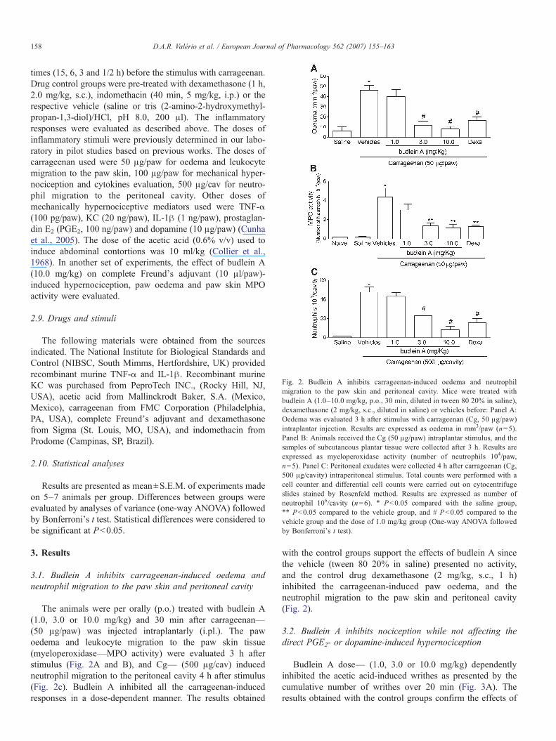
Fig. 2. Budlein A inhibits carrageenan-induced oedema and neutrophilmigration to the paw skin and peritoneal cavity. Mice were treated withbudlein A (1.0–10.0 mg/kg, p.o., 30 min, diluted in tween 80 20% in saline),dexamethasone (2 mg/kg, s.c., diluted in saline) or vehicles before: Panel A:Oedema was evaluated 3 h after stimulus with carrageenan (Cg, 50 μg/paw)intraplantar injection. Results are expressed as oedema in mm3/paw (n=5).Panel B: Animals received the Cg (50 μg/paw) intraplantar stimulus, and thesamples of subcutaneous plantar tissue were collected after 3 h. Results areexpressed as myeloperoxidase activity (number of neutrophils 104/paw,n=5). Panel C: Peritoneal exudates were collected 4 h after carrageenan (Cg,500 μg/cavity) intraperitoneal stimulus. Total counts were performed with acell counter and differential cell counts were carried out on cytocentrifugeslides stained by Rosenfeld method. Results are expressed as number ofneutrophil 106/cavity (n=6). ⁎ Pb0.05 compared with the saline group,⁎⁎ Pb0.05 compared to the vehicle group, and # Pb0.05 compared to thevehicle group and the dose of 1.0 mg/kg group (One-way ANOVA followedby Bonferroni's t test).
158 D.A.R. Valério et al. / European Journal of Pharmacology 562 (2007) 155–163
times (15, 6, 3 and 1/2 h) before the stimulus with carrageenan.Drug control groups were pre-treated with dexamethasone (1 h,2.0 mg/kg, s.c.), indomethacin (40 min, 5 mg/kg, i.p.) or therespective vehicle (saline or tris (2-amino-2-hydroxymethyl-propan-1,3-diol)/HCl, pH 8.0, 200 μl). The inflammatoryresponses were evaluated as described above. The doses ofinflammatory stimuli were previously determined in our labo-ratory in pilot studies based on previous works. The doses ofcarrageenan used were 50 μg/paw for oedema and leukocytemigration to the paw skin, 100 μg/paw for mechanical hyper-nociception and cytokines evaluation, 500 μg/cav for neutro-phil migration to the peritoneal cavity. Other doses ofmechanically hypernociceptive mediators used were TNF-α(100 pg/paw), KC (20 ng/paw), IL-1β (1 ng/paw), prostaglan-din E2 (PGE2, 100 ng/paw) and dopamine (10 μg/paw) (Cunhaet al., 2005). The dose of the acetic acid (0.6% v/v) used toinduce abdominal contortions was 10 ml/kg (Collier et al.,1968). In another set of experiments, the effect of budlein A(10.0 mg/kg) on complete Freund's adjuvant (10 μl/paw)-induced hypernociception, paw oedema and paw skin MPOactivity were evaluated.
2.9. Drugs and stimuli
The following materials were obtained from the sourcesindicated. The National Institute for Biological Standards andControl (NIBSC, South Mimms, Hertfordshire, UK) providedrecombinant murine TNF-α and IL-1β. Recombinant murineKC was purchased from PeproTech INC., (Rocky Hill, NJ,USA), acetic acid from Mallinckrodt Baker, S.A. (Mexico,Mexico), carrageenan from FMC Corporation (Philadelphia,PA, USA), complete Freund's adjuvant and dexamethasonefrom Sigma (St. Louis, MO, USA), and indomethacin fromProdome (Campinas, SP, Brazil).
2.10. Statistical analyses
Results are presented as mean±S.E.M. of experiments madeon 5–7 animals per group. Differences between groups wereevaluated by analyses of variance (one-way ANOVA) followedby Bonferroni's t test. Statistical differences were considered tobe significant at Pb0.05.
3. Results
3.1. Budlein A inhibits carrageenan-induced oedema andneutrophil migration to the paw skin and peritoneal cavity
The animals were per orally (p.o.) treated with budlein A(1.0, 3.0 or 10.0 mg/kg) and 30 min after carrageenan—(50 μg/paw) was injected intraplantarly (i.pl.). The pawoedema and leukocyte migration to the paw skin tissue(myeloperoxidase—MPO activity) were evaluated 3 h afterstimulus (Fig. 2A and B), and Cg— (500 μg/cav) inducedneutrophil migration to the peritoneal cavity 4 h after stimulus(Fig. 2c). Budlein A inhibited all the carrageenan-inducedresponses in a dose-dependent manner. The results obtained
with the control groups support the effects of budlein A sincethe vehicle (tween 80 20% in saline) presented no activity,and the control drug dexamethasone (2 mg/kg, s.c., 1 h)inhibited the carrageenan-induced paw oedema, and theneutrophil migration to the paw skin and peritoneal cavity(Fig. 2).
3.2. Budlein A inhibits nociception while not affecting thedirect PGE2- or dopamine-induced hypernociception
Budlein A dose— (1.0, 3.0 or 10.0 mg/kg) dependentlyinhibited the acetic acid-induced writhes as presented by thecumulative number of writhes over 20 min (Fig. 3A). Theresults obtained with the control groups confirm the effects of
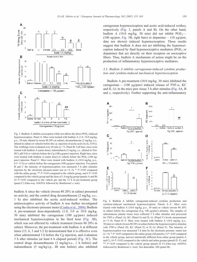
Fig. 3. Budlein A inhibits nociception while not affects the direct PGE2-inducedhypernociception. Panel A: Mice were treated with budlein A (1.0–10.0 mg/kg,p.o., 30 min, diluted in tween 80 20% in saline), dexamethasone (2 mg/kg, s.c.,diluted in saline) or vehicles before the i.p. injection of acetic acid (AcAc, 0.6%).The writhings were evaluated over 20 min (n=7). Panel B: Left bars, mice weretreated with budlein A (same dose), indomethacin (5 mg/kg, s.c., diluted in Tris/HCl, pH 8.0) or vehicles before the Cg (100 μg/paw) injection. Right bars, micewere treated with budlein A (same dose) or vehicle before the PGE2 (100 ng/paw) injection. Panel C: Mice were treated with budlein A (10.0 mg/kg, p.o.,0.5–15 h) or vehicle before the carrageenan (100 μg/paw) injection. For panelsB and C the intensity of hypernociception was measured 3 h after stimulusinjection by the electronic pressure-meter test (n=4–5). ⁎ Pb0.05 comparedwith the saline group, ⁎⁎ Pb0.05 compared to the vehicle group, and # Pb0.05compared to the vehicle group and the dose of 1.0 mg/kg group (panels A and B)## Pb0.05 compared to the vehicle gro and the 15 h of pre-treatment group(panel C) (One-way ANOVA followed by Bonferroni's t test).
Fig. 4. Budlein A inhibits carrageenan-induced cytokine production andcytokine-induced mechanical hypernociception. Panels A–C: Mice weretreated with budlein A (10.0 mg/kg, p.o., 30 min) or vehicle (tween 80 20%in saline) before the carrageenan (Cg, 100 μg/paw) stimulus. The samples ofsubcutaneous plantar tissue were collected 3 h after stimulus and processedfor TNF-α (Panel A), KC (Panel b) and IL-1β (Panel C) levels measurement(n=3–4). Panel D–F: Mice were treated with budlein A (10.0 mg/kg, p.o.,30min) or vehicle (tween 80 20% in saline) before the hypernociceptive stimuluswith TNF-α (Panel D), KC (Panel E) or IL-1β (Panel F). The intensity ofhypernociception was measured 3 h later by the electronic pressure–meter test(n=5). ⁎ Pb0.05 compared to the saline group (All panels), # Pb0.05 comparedto the vehicle group, and not statistically different from saline group (panels Aand C), ## Pb0.05 compared to the vehicle and saline groups (panels D–F), and⁎⁎ Pb0.05 compared to the vehicle group (panels D–F) (One-way ANOVAfolloweed by Bonferroni's t test). Not detectable, ND (panel B).
159D.A.R. Valério et al. / European Journal of Pharmacology 562 (2007) 155–163
budlein A since the vehicle (tween 80 20% in saline) presentedno activity, and the control drug dexamethasone (2 mg/kg, s.c.,1 h) also inhibited the acetic acid-induced writhes. Theantinociceptive activity of budlein A was further investigatedusing the electronic-pressure meter (Cunha et al., 2004). BudleinA pre-treatment dose-dependently (1.0, 3.0 or 10.0 mg/kg,30 min) inhibited the carrageenan (100 μg/paw) inducedmechanical hypernociception in the third hour (Fig. 3B),which was not affected by vehicle treatment (tween 80 20% insaline). Moreover, the pre-treatment with budlein A at differenttimes (15, 6, 3 and 1/2 h) demonstrated that it is effective evenwhen administrated 3 h before the Cg injection, and evaluation3 h after stimulus (at least 6 h elapsed period) (Fig. 3C). Thecontrol drugs dexamethasone (2 mg/kg/s.c., 1 h before) andindomethacin (5 mg/kg/i.p., 40 min before) also inhibited
carrageenan hypernociception and acetic acid-induced writhes,respectively (Fig. 3, panels A and B). On the other hand,budlein A (10.0 mg/kg, 30 min) did not inhibit PGE2—(100 ng/paw, Fig. 3B, right bars) or dopamine— (10 μg/paw,data not shown) induced hypernociception. These resultssuggest that budlein A does not act inhibiting the hypernoci-ception induced by final hypernociceptive mediators (PGE2 ordopamine) that act directly on their receptors on nociceptivefibers. Thus, budlein A mechanism of action might be on theproduction of inflammatory hypernociceptive mediators.
3.3. Budlein A inhibits carrageenan-induced cytokine produc-tion and cytokine-induced mechanical hypernociception
Budlein A pre-treatment (10.0 mg/kg, 30 min) inhibited thecarrageenan— (100 μg/paw) induced release of TNF-α, KCand IL-1β in the mice paw tissue 3 h after stimulus (Fig. 4A, Band c, respectively). Further supporting the anti-inflammatory

160 D.A.R. Valério et al. / European Journal of Pharmacology 562 (2007) 155–163
activity of budlein A, using this same treatment schedule, itinhibited the TNF-α- (100 ng/paw) and KC- (20 ng/paw), andIL-1β- (1 ng/paw) induced mechanical hypernociception in thethird hour (Fig. 4D, E and F, respectively).
Fig. 5. Budlein A treatment inhibits Complete Freund's Adjuvant-inducedinflammation. Panels A–D: Mice were treated with budlein A (10.0 mg/kg, p.o.,diluted in tween 80 20% in saline.), indomethacin (5 mg/kg, p.o., diluted in Tris/HCl, pH 8.0), dexamethasone (2 mg/kg, s.c., diluted in saline) or vehicles during7–8 days. The treatment started 30 min before Complete Freund's Adjuvant(CFA, 10 μL/paw) stimulus. Thereafter, mice (n=5) were treated with one of theabove mentioned drugs once a day, 3 h before measurements. Saline groupreceived 10 μL/paw of saline instead of Complete Freund's Adjuvant. Themechanical hypernociception (Electronic pressure meter, Panel A) and oedema(Panel B) were evaluated until the 7th day. In the eighth day, samples of thesubcutaneous plantar tissue (Panel C) and stomach (Panel D were collected 3 hafter the last treatment for MPO activity evaluation. ⁎ Pb0.05 compared to thesaline group, ⁎⁎ Pb0.05 compared to the vehicles group, and # Pb0.05compared to the vehicles group and not statistically different of the saline group(One-way ANOVA followed by Bonferroni's t test).
3.4. Budlein A treatment inhibits complete Freund's adjuvant-induced inflammation
In general, anti-inflammatory drugs are used chronically orduring short periods such as 7 days. However, even during thoseshort periods conventional non-steroidal anti-inflammatorydrugs (cyclooxigenase-1 inhibitors) may cause gastric damage,which restrict their use (Gudis and Sakamoto, 2005). Therefore,the budlein A anti-inflammatory activity was tested in thecomplete Freund's adjuvant (10 μl/paw)-induced inflammationsince the inflammation in this model persists for more than8 days. This period of treatment allows evaluating the effect onstomach inflammation. Mice were treated once a day (10 mg/kg) during 8 days with budlein A (10 mg/kg), indomethacin(indo, 5 mg/kg/p.o.) or dexamethasone (2 mg/kg/s.c.). Mechan-ical hypernociception (Fig. 5A) and oedema (Fig. 5B) wereevaluated between the first and seventh days. In the eighth day,the motor performance of the mice was evaluated in the rota-rod. After, those mice were sacrificed and samples of thestomach (Fig. 5C) and paw skin (Fig. 5D) were colleted forMPO activity assay. Budlein A inhibited the complete Freund'sadjuvant-induced mechanical hypernociception, paw oedemaand increase of MPO activity in the paw skin. The budlein Atreatment did not alter the MPO activity in the stomach sample,which is consistent with no inflammatory response in the tissue(Souza et al., 2004). The motor performance was not alteredeither few hours (30, 90 and 210 min) after single treatment orafter 8 consecutive days of budlein A administration (data notshown). Dexamethasone treatment reduced all inflammatoryresponses. Indomethacin treatment reduced the mechanicalhypernociception, paw oedema and paw MPO activity.However, consistent with the inhibition of cyclooxigenase-1and inflammation, the indomethacin treatment increased theMPO activity in the stomach. None of the treatments inducedmacroscopic gastric lesions (data not shown), which would beespecially intriguing concerning indomethacin treatment. Nev-ertheless, there is evidence that the per oral treatment withindomethacin or other cycloxigenase inhibitors (e.g. acetylsalicylic acid) induce gastric mucosa tolerance described as nodetectable gastric mucosal lesions concomitantly with anincrease in polymorphonuclear cell infiltrate as determined bythe MPO activity (Wallace et al., 1995).
4. Discussion
V. robusta is a perennial herb found widespread in Brazil.The Mexican folk medicine uses other plants of this species andrelated genera (Heinrich et al., 1998). In the present study, weisolated the sesquiterpene lactone budlein A from the dichloro-methane extract of V. robusta, and demonstrated its in vivo anti-inflammatory activity, suggesting the inhibition of cytokinesrelease and action as its mechanism.
Budlein A dose-dependently inhibited the carrageenan-induced oedema, neutrophil migration to the paw skin andperitoneal cavity, and acetic acid-induced writhes. These wereseminal methods for the in vivo evaluation of the activity ofdrugs such as steroidal and non-steroidal anti-inflammatory,

161D.A.R. Valério et al. / European Journal of Pharmacology 562 (2007) 155–163
inhibitors of leukotriene synthesis and cytokine directedantibodies (French and Galicich, 1964; Limet and Lecomte,1968; Goldman et al., 1993; Canetti et al., 2001).
Focusing in nociception, using the electronic pressure-metertest (Cunha et al., 2004; Vivancos et al., 2004) we have recentlydemonstrated that carrageenan initiates a mechanical hyperno-ciceptive cascade in which prostanoids and sympathetic aminesare the final mediators that induce nociceptor sensitization inmice. Moreover, cytokines mediate the release of those finalmediators induced by carrageenan. Thus, after carrageenanstimulus TNF-α and KC have pivotal roles in a cytokinescascade: i.) TNF-α→ IL-1β that activates the synthesis ofprostanoids, ii.) KC→ sympathetic amines release, and iii.)KC→ IL-1β→prostanoids production (Cunha et al., 2005).
Budlein A dose-dependently inhibited carrageenan-inducedmechanical hypernociception. However, it did not affect thehypernociception induced by the final mediators PGE2 anddopamine. In addition, budlein A treatment inhibited thecarrageenan-induced release of TNF-α, KC and IL-1β as well asthe hypernociception induced by these cytokines. The inhibition ofcytokines release might ultimately lead to the inhibition ofprostanoids and sympathetic amines production/release. Substan-tiating this hypothesis, the treatment with indomethacin (standardcyclooxigenase inhibitor) inhibits the hypernociceptive effect ofTNF-α, KC and IL-1β, and guanethidine (sympathetic blocker)inhibits KC hypernociception in mice (Cunha et al., 2005).Additionally, considering the inhibition of NFκB activation bysesquiterpene lactones (Hehner et al., 1998; Siedle et al., 2004) it isalso possible that budlein A inhibits inflammation-inducedcyclooxigenase-2 mRNA expression, and therefore, PGE2 pro-duction. This mechanism may account for the inhibition of IL-1β-induced mechanical hypernociception since it depends on PGE2production. Thus, the results presented above suggest that budleinA acts preventing the nociceptor sensitisation by inhibiting theproduction of mediators that sensitise the nociceptor. Furthermore,these same mediators (e.g. TNF-α, IL-1β, KC, prostanoids,sympathetic amines) are also involved in the acetic-acid-inducedwrithes (Ribeiro et al., 2000; Cunha TM, unpublished data).Therefore, the inhibition of cytokine productionmight also accountfor the antinociceptive effect of budlein A in the writhing test.
Further supporting the applicability of budlein A as an anti-inflammatory drug, it inhibited the complete Freund's adjuvant-induced hypernociception, paw oedema and increase of pawskin MPO activity during a 7–8 days treatment schedule. Theseeffects were not accompanied by altered liver appearance orweight (data not shown), or gastric damage as determined byMPO activity. Furthermore, disproving relaxing or anaestheticeffects, budlein A (10.0 mg/kg) treatment did not affect themotor performance of the animals as tested in the rota-rodneither acutely (one treatment) or chronically (8 days treatment)(data not shown). Reinforcing this issue, the treatment withbudlein A did not affect the mechanical hypernociceptiveresponses to PGE2 and dopamine. Thus, budlein A may beuseful at least for short-term therapies without the side effects ofcyclooxigenase-1 inhibitors such as gastric damage.
The molecular mechanisms of action of sesquiterpenelactones other than budlein A, involve the alkylation of the
p65 subunit cystein residue of the NF–kB complex, whichinhibits its interaction with DNA, and thus, disabling itstranscription (Rüngeler et al., 1999). Additionally, othersesquiterpene lactones also specifically inhibit the activationof NF–kB by preventing the degradation of I–kB. Moreover,other sesquiterpene lactones (santonin, isophoronoxide andsclareolide) prevent the activation of NF–kB and the degrada-tion of I–kB induced by different stimuli such as phorbol esters,TNF-α and hydrogen peroxide in cultured Jurkat leukemia(Hehner et al., 1998). The most clinically used inhibitors ofNF–kB are the glucocorticosteroids such as dexamethasone(control drug used in the present study). The inhibition of NF–kB or stimulation of I–kB production, and the interaction ofglucocorticoids with the glucocorticoid responsive genesultimately lead to the inhibition of cytokine production andaction as well as genes encoding pro-inflammatory moleculesand enzymes (Ferreira et al., 1997; Baeuerle and Henkel, 1994;Baeuerle and Baltimore, 1996; May and Ghosh, 1998, forreview see Barnes, 2006). This is an important anti-inflamma-tory mechanism considering that cytokines induce hypernoci-ception (Ferreira et al., 1988; Cunha et al., 1991, 1992; Verriet al., 2004, 2005, 2006b, in press) and neutrophil migration(Canetti et al., 2001; Sayers et al., 1988). In agreement with ourresults, budlein A inhibits NF–kB activation as determinedusing the electrophoretic mobility shift assay (Siedle et al.,2004). It is important to point out that glucocorticosterois bindto glucocorticoid cytoplasmatic receptors, and then the complexinduce the known effects and side effects of steroids. Therefore,budlein A probably does not present the common side effectsassociated with glucocorticosteroids therapy since sesquiter-pene lactones bind on NF–kB or I–kB.
Concluding, the present study demonstrated that budlein Ainhibits the inflammation signs by a mechanism related to theinhibition of cytokines release and action. Thus, budlein Amight be a useful orally active drug to control inflammatoryconditions involving hypernociception, oedema and neutrophilmigration.
Acknowledgements
The authors gratefully acknowledge the technical assistanceof Ieda R.S. Schivo, Sérgio R. Rosa, Giuliana B. Francisco andWalter Lopes. This work was supported by grants fromFundação de Amparo à Pesquisa do Estado de São Paulo(FAPESP, Brazil), Conselho Nacional de Pesquisa (CNPq,Brazil), Coordenadoria de Aperfeiçoamento de Pessoal de NívelSuperior (CAPES, Brazil). D.A.R.V. is a recipient of scientificinitiation fellowship from CNPq, H.P.L (MS student), N.S.A.(PhD student) T.M.C (PhD student) are recipients of fellowshipsfrom FAPESP, and W.A.V.J. is a recipient of a Post-Docfellowship from FAPESP.
References
Abil'daeva, A.Z., Pak, R.N., Kulyiasov, A.T., Adekenov, S.M., 2004. Anti-inflammatory effect of arglabin and 11,13-dihydro-13-dimethylaminoargla-bin hydrochloride. Eksp. Klin. Farmakol 67, 37–39.

162 D.A.R. Valério et al. / European Journal of Pharmacology 562 (2007) 155–163
Ahmed, M., Rahman, M.T., Alimuzzaman, M., Shilpi, J.A., 2001. Analgesicsesquiterpene dilactone from Mikania cordata. Fitoterapia 72, 919–921.
Baeuerle, P.A., Baltimore, D., 1996. NF–kappa B: ten years after. Cell 87,13–20.
Baeuerle, P.A., Henkel, T., 1994. Function and activation of NF–kappa B in theimmune system. Annu. Rev. Immunol. 12, 141–179.
Barnes, P.J., 2006. How corticosteroids control inflammation: Quintiles PrizeLecture 2005. Br. J. Pharmacol. 148, 245–254.
Bradley, P.P., Priebat, D.A., Christensen, R.D., Rothstein, G., 1982. Measure-ment of cutaneous inflammation: estimation of neutrophil content with anenzyme marker. J. Invest. Dermatol. 78, 206–209.
Canetti, C., Silva, J.S., Ferreira, S.H., Cunha, F.Q., 2001. Tumour necrosisfactor-alpha and leukotriene B(4) mediate the neutrophil migration inimmune inflammation. Br. J. Pharmacol. 134, 1619–1628.
Casagrande, R., Georgetti, S.R., Verri Jr., W.A., Dorta, D.J., dos Santos, A.C.,Fonseca, M.J., 2006. Protective effect of topical formulations containingquercetin against UVB-induced oxidative stress in hairless mice.J. Photochem. Photobiol., B Biol. 84, 21–27.
Chen, C.H., Yang, L.M., Lee, T.T., Shen, Y.C., Zhang, D.C., Pan, D.J., McPhail,A.T., McPhail, D.R., Liu, S.Y., Li, D.H., et al., 1994. Antitumor agents-CLI.Bis(helenalinyl)glutarate and bis(isoalantodiol-B)glutarate, potent inhibitorsof human DNA topoisomerase II. Bioorg. Med. Chem. 2, 137–145.
Collier, H.O., Dinneen, L.C., Johnson, C.A., Schneider, C., 1968. Theabdominal constriction response and its suppression by analgesic drugs inthe mouse. Br. J. Pharmacol. Chemother. 32, 295–310.
Cunha, F.Q., Lorenzetti, B.B., Poole, S., Ferreira, S.H., 1991. Interleukin-8 as amediator of sympathetic pain. Br. J. Pharmacol. 104, 765–767.
Cunha, F.Q., Poole, S., Lorenzetti, B.B., Ferreira, S.H., 1992. The pivotal role oftumour necrosis factor alpha in the development of inflammatoryhyperalgesia. Br. J. Pharmacol. 107, 660–664.
Cunha, T.M., Verri Jr., W.A., Vivancos, G.G., Moreira, I.F., Reis, S., Parada, C.A.,Cunha, F.Q., Ferreira, S.H., 2004. An electronic pressure-meter nociceptionpaw test for mice. Braz. J. Med. Biol. Res. 37, 401–407.
Cunha, T.M., Verri Jr., W.A., Silva, J.S., Poole, S., Cunha, F.Q., Ferreira, S.H.,2005. A cascade of cytokines mediates mechanical inflammatory hyperno-ciception in mice. Proc. Natl. Acad. Sci. U. S. A. 102, 1755–1760.
Da Costa, F.B., Schorr, K., Arakawa, N.S., Schilling, E.E., Spring, O., 2001.Infraspecific variation in the chemistry of glandular trichomes of twoBrazilian Viguiera populations. J. Braz. Chem. Soc. 12, 403–407.
Damre, A.A., Damre, A.S., Saraf, M.N., 2003. Evaluation of sesquiterpenelactone fraction of Saussurea lappa on transudative, exudative andproliferative phases of inflammation. Phytother. Res. 17, 722–725.
De Vivar, A.R., Diaz, C.G.E., Bratoeff, E.A., Jimenez, L., 1976. Thegermacranolides of Viguiera buddleiaeformis structures of budlein-A and-B. Phytochemistry 15, 525–529.
Feltenstein, M.W., Schuhly, W., Warnick, J.E., Fischer, N.H., Sufka, K.J., 2004.Anti-inflammatory and anti-hyperalgesic effects of sesquiterpene lactonesfrom Magnolia and Bear's foot. Pharmacol. Biochem. Behav. 79, 299–302.
Ferreira, S.H., Lorenzetti, B.B., Bristow, A.F., Poole, S., 1988. Interleukin-1beta as a potent hyperalgesic agent antagonized by a tripeptide analogue.Nature 334, 698–700.
Ferreira, S.H., Cunha, F.Q., Lorenzetti, B.B., Michelin, M.A., Perretti, M.,Flower, R.J., Poole, S., 1997. Role of lipocortin-1 in the anti-hyperalgesicactions of dexamethasone. Br. J. Pharmacol. 121, 883–888.
French, L.A., Galicich, J.H., 1964. The use of steroids for control of cerebraledema. Clin. Neurosurg. 10, 212–223.
Goldman, G.,Welbourn, R., Kobzik, L., Valeri, C.R., Shepro, D., Hechtman, H.B.,1993. Lavage with leukotriene B4 induces lung generation of tumor necrosisfactor-alpha that in turnmediates neutrophil diapedesis. Surgery 113, 297–303.
Guardia, T., Juarez, A.O., Guerreiro, E., Guzman, J.A., Pelzer, L., 2003. Anti-inflammatory activity and effect on gastric acid secretion of dehydroleuco-dine isolated from Artemisia douglasiana. J. Ethnopharmacol. 88, 195–198.
Gudis, K., Sakamoto, C., 2005. The role of cyclooxygenase in gastric mucosalprotection. Dig. Dis. Sci. 50 (Suppl. 1), S16–S23.
Hehner, S.P., Heinrich,M., Bork, P.M., Vogt, M., Ratter, F., Lehmann, V., Schulze-Osthoff, K., Droge, W., Schmitz, M.L., 1998. Sesquiterpene lactonesspecifically inhibit activation of NF-kappa B by preventing the degradationof I kappa B-alpha and I kappa B-beta. J. Biol. Chem. 273, 1288–1297.
Heinrich, M., Robles, M., West, J.E., Ortiz de Montellano, B.R., Rodriguez, E.,1998. Ethnopharmacology of Mexican asteraceae (Compositae). Annu. Rev.Pharmacol. Toxicol. 38, 539–565.
Huacuja, R.L., Carranco, A., Guzman, S.A., Guerrero, C., 1993. Inactivation ofSH groups with sesquiterpene lactones: effects on nuclear decondensationpattern/motility induced by heparin in human spermatozoa. Adv. Contracept.Deliv. Syst. 9, 97–106.
Jain, N.K., Kulkarni, S.K., 1999. Antinociceptive and anti-inflammatory effectsof Tanacetum parthenium L. extract in mice and rats. J. Ethnopharmacol. 68,251–259.
Limet, R., Lecomte, J., 1968. Inhibitory action of indomethacin on generalizededema induced by dextran in rats. Arch. Int. Pharmacodyn. Ther. 171,109–115.
May, M.J., Ghosh, S., 1998. Signal transduction through NF-kappa B. Immunol.Today 19, 80–88.
Meshnick, S.R., 2002. Artemisinin: mechanisms of action, resistance andtoxicity. Int. J. Parasitol. 32, 1655–1660.
Okugawa, H., Ueda, R., Matsumoto, K., Kawanishi, K., Kato, K., 2000. Effectsof sesquiterpenoids from “Oriental incenses” on acetic acid-induced writhingand D2 and 5-HT2A receptors in rat brain. Phytomedicine 7, 417–422.
Parada, C.A., Vivancos, G.G., Tambeli, C.H., Cunha, F.Q., Ferreira, S.H., 2003.Activation of presynaptic NMDA receptors coupled to NaV1.8-resistantsodium channel C-fibers causes retrograde mechanical nociceptor sensiti-zation. Proc. Natl. Acad. Sci. U. S. A. 100, 2923–2928.
Pickman, A.K., 1984. Antifungal activity of sesquiterpene lactones. Biochem.Syst. Ecol. 12, 13–18.
Recio, M.C., Giner, R.M., Uriburu, L., Manez, S., Cerda, M., De la Fuente, J.R.,Rios, J.L., 2000. In vivo activity of pseudoguaianolide sesquiterpenelactones in acute and chronic inflammation. Life Sci. 66, 2509–2518.
Ribeiro, R.A., Vale, M.L., Thomazzi, S.M., Paschoalato, A.B., Poole, S.,Ferreira, S.H., Cunha, F.Q., 2000. Involvement of resident macrophages andmast cells in the writhing nociceptive response induced by zymosan andacetic acid in mice. Eur. J. Pharmacol. 387, 111–118.
Rosland, J.H., Hunskaar, S., Hole, K., 1990. Diazepam attenuates morphineantinociception test-dependently in mice. Pharmacol. Toxicol. 66, 382–386.
Rüngeler, P., Castro, V., Mora, G., Gören, N., Vichnewski, W., Pahl, H.L.,Merfort, I., Schmidt, T.J., 1999. Inhibition of transcription factor NF-_B bysesquiterpene lactones: a proposed molecular mechanism of action. Bioorg.Med. Chem. 7, 2343–2352.
Sayers, T.J., Wiltrout, T.A., Bull, C.A., Denn, A.C., Pilaro, A.M., Lokesh, B.,1988. Effect of cytokines on polymorphonuclear neutrophil infiltration inthe mouse. Prostaglandin- and leukotriene-independent induction ofinfiltration by IL-1 and tumor necrosis factor. J. Immunol. 141, 1670–1677.
Secco, D.D., Paron, J.A., de Oliveira, S.H., Ferreira, S.H., Silva, J.S., Cunha, F.Q.,2003. Neutrophil migration in inflammation: nitric oxide inhibits rolling,adhesion and induces apoptosis. Nitric Oxide 9, 153–164.
Siedle, B., Garcia-Pineres, A.J., Murillo, R., Schulte-Monting, J., Castro, V.,Rungeler, P., Klaas, C.A., Da Costa, F.B., Kisiel, W., Merfort, I., 2004.Quantitative structure-activity relationship of sesquiterpene lactones as inhib-itors of the transcription factor NF-kappaB. J. Med. Chem. 47, 6042–6054.
Silvan, A.M., Abad, M.J., Bermejo, P., Villar, A., 1996. Inhibition byhydroxyachillin, sesquiterpene lactone from Tanacetum microphyllum, ofPMA-induced mouse ear oedema. Inflamm. Res. 45, 289–292.
Souza, M.H., Lemos, H.P., Oliveira, R.B., Cunha, F.Q., 2004. Gastric damageand granulocyte infiltration induced by indomethacin in tumour necrosisfactor receptor 1 (TNF-R1) or inducible nitric oxide synthase (iNOS)deficient mice. Gut 53, 791–796.
Verri Jr.,W.A., Schivo, I.R., Cunha, T.M., Liew, F.Y., Ferreira, S.H., Cunha, F.Q.,2004. Interleukin-18 induces mechanical hypernociception in rats viaendothelin acting on ETB receptors in a morphine-sensitive manner. J.Pharmacol. Exp. Ther. 310, 710–717.
Verri Jr., W.A., Molina, R.O., Schivo, I.R., Cunha, T.M., Parada, C.A., Poole, S.,Ferreira, S.H., Cunha, F.Q., 2005. Nociceptive effect of subcutaneouslyinjected interleukin-12 is mediated by endothelin (ET) acting on ETBreceptors in rats. J. Pharmacol. Exp. Ther. 315, 609–615.
Verri Jr., W.A., Cunha, T.M., Parada, C.A., Poole, S., Cunha, F.Q., Ferreira, S.H.,2006a. Hypernociceptive role of cytokines and chemokines: targets foranalgesic drug development? Pharmacol. Ther. 112, 116–138.

163D.A.R. Valério et al. / European Journal of Pharmacology 562 (2007) 155–163
Verri Jr., W.A., Cunha, T.M., Parada, C.A., Wei, X.Q., Ferreira, S.H., Liew, F.Y.,Cunha, F.Q., 2006b. IL-15 mediates immune inflammatory hypernocicep-tion by triggering a sequential release of IFN-{gamma}, endothelin, andprostaglandin. Proc. Natl. Acad. Sci. U. S. A. 103, 9721–9725.
Verri, W.A., Jr., Cunha, T.M., Parada, C.A., Poole, S., Liew, F.Y., Ferreira, S.H.,Cunha F.Q., in press. Antigen-induced inflammatory mechanical hyperno-ciception in mice is mediated by IL-18. Brain Behav. Immun. doi:10.1016/j.bbi.2006.11.005.
Vivancos, G.G., Verri Jr., W.A., Cunha, T.M., Schivo, I.R., Parada, C.A., Cunha,F.Q., Ferreira, S.H., 2004. An electronic pressure-meter nociception paw testfor rats. Braz. J. Med. Biol. Res. 37, 391–399.
Wallace, J.L.,McKnight, G.W., Bell, C.J., 1995. Adaptation of rat gastricmucosato aspirin requires mucosal contact. Am. J. Physiol. 268, G134–G138.
Winter, C.A., Risley, E.A., Nuss, G.W., 1962. Carrageenan-induced edema inhind paw of the rat as an assay for antiinflammatory drugs. Proc. Soc. Exp.Biol. Med. 111, 544–547.