Anatomy, Art, and the Desire for Divine Beauty
179
University of Windsor University of Windsor Scholarship at UWindsor Scholarship at UWindsor Electronic Theses and Dissertations Theses, Dissertations, and Major Papers 1-1-2022 Anatomy, Art, and the Desire for Divine Beauty: An Exploration into Anatomy, Art, and the Desire for Divine Beauty: An Exploration into the Creation and Perpetuation of the Standardized Body Types of the Creation and Perpetuation of the Standardized Body Types of Early Modern Anatomical Illustrations Early Modern Anatomical Illustrations Sarah D. Shaver Follow this and additional works at: https://scholar.uwindsor.ca/etd Part of the History of Art, Architecture, and Archaeology Commons, and the Sociology Commons Recommended Citation Recommended Citation Shaver, Sarah D., "Anatomy, Art, and the Desire for Divine Beauty: An Exploration into the Creation and Perpetuation of the Standardized Body Types of Early Modern Anatomical Illustrations" (2022). Electronic Theses and Dissertations. 8716. https://scholar.uwindsor.ca/etd/8716 This online database contains the full-text of PhD dissertations and Masters’ theses of University of Windsor students from 1954 forward. These documents are made available for personal study and research purposes only, in accordance with the Canadian Copyright Act and the Creative Commons license—CC BY-NC-ND (Attribution, Non-Commercial, No Derivative Works). Under this license, works must always be attributed to the copyright holder (original author), cannot be used for any commercial purposes, and may not be altered. Any other use would require the permission of the copyright holder. Students may inquire about withdrawing their dissertation and/or thesis from this database. For additional inquiries, please contact the repository administrator via email ([email protected]) or by telephone at 519-253-3000ext. 3208.
-
Upload
khangminh22 -
Category
Documents
-
view
2 -
download
0
Transcript of Anatomy, Art, and the Desire for Divine Beauty
University of Windsor University of Windsor
Scholarship at UWindsor Scholarship at UWindsor
Electronic Theses and Dissertations Theses, Dissertations, and Major Papers
1-1-2022
Anatomy, Art, and the Desire for Divine Beauty: An Exploration into Anatomy, Art, and the Desire for Divine Beauty: An Exploration into
the Creation and Perpetuation of the Standardized Body Types of the Creation and Perpetuation of the Standardized Body Types of
Early Modern Anatomical Illustrations Early Modern Anatomical Illustrations
Sarah D. Shaver
Follow this and additional works at: https://scholar.uwindsor.ca/etd
Part of the History of Art, Architecture, and Archaeology Commons, and the Sociology Commons
Recommended Citation Recommended Citation Shaver, Sarah D., "Anatomy, Art, and the Desire for Divine Beauty: An Exploration into the Creation and Perpetuation of the Standardized Body Types of Early Modern Anatomical Illustrations" (2022). Electronic Theses and Dissertations. 8716. https://scholar.uwindsor.ca/etd/8716
This online database contains the full-text of PhD dissertations and Masters’ theses of University of Windsor students from 1954 forward. These documents are made available for personal study and research purposes only, in accordance with the Canadian Copyright Act and the Creative Commons license—CC BY-NC-ND (Attribution, Non-Commercial, No Derivative Works). Under this license, works must always be attributed to the copyright holder (original author), cannot be used for any commercial purposes, and may not be altered. Any other use would require the permission of the copyright holder. Students may inquire about withdrawing their dissertation and/or thesis from this database. For additional inquiries, please contact the repository administrator via email ([email protected]) or by telephone at 519-253-3000ext. 3208.

ANATOMY, ART, AND THE DESIRE FOR DIVINE BEAUTY
An Exploration into the Creation and Perpetuation of the Standardized Body Types of
Early Modern Anatomical Illustrations
By
Sarah D. Shaver
A Thesis
Submitted to the Faculty of Graduate Studies
through the Department of Sociology, Anthropology and Criminology
in Partial Fulfillment of the Requirements for
the Degree of Master of Arts
at the University of Windsor
Windsor, Ontario, Canada
2021
© 2021 Sarah D. Shaver

ANATOMY, ART, AND THE DESIRE FOR DIVINE BEAUTY
An Exploration into the Creation and Perpetuation of the Standardized Body Types of Early
Modern Anatomical Illustrations
By
Sarah D. Shaver
APPROVED BY:
______________________________________________
C. E. Hundleby
Department of Philosophy
______________________________________________
A. Fitzgerald
Department of Sociology, Anthropology, and Criminology
______________________________________________
J. Albanese, Advisor
Department of Integrated Biology
December 13th, 2021

iii
DECLARATION OF ORIGINALITY
I hereby certify that I am the sole author of this thesis and that no part of this thesis
has been published or submitted for publication.
I certify that, to the best of my knowledge, my thesis does not infringe upon anyone’s
copyright nor violate any proprietary rights and that any ideas, techniques, quotations, or any
other material from the work of other people included in my thesis, published or otherwise,
are fully acknowledged in accordance with the standard referencing practices. Furthermore,
to the extent that I have included copyrighted material that surpasses the bounds of fair
dealing within the meaning of the Canada Copyright Act, I certify that I have obtained a
written permission from the copyright owner(s) to include such material(s) in my thesis and
have included copies of such copyright clearances to my appendix.
I declare that this is a true copy of my thesis, including any final revisions, as
approved by my thesis committee and the Graduate Studies office, and that this thesis has not
been submitted for a higher degree to any other University or Institution.

iv
ABSTRACT
Anatomical illustrations have been integral to modern anatomy’s practice and instruction
since its conception as a discipline in the sixteenth century. While this practice lends itself to
modern anatomy’s highly visual nature, the bodies featured in anatomical illustrations have
been exceptionally homogeneous in appearance for as long as the discipline has existed -- a
fact which has lasting consequences in the contemporary sociomedical world of today. To
examine the construction of the standardized body in modern anatomy this project utilizes a
feminist constructionist framework in conducting a thematic visual analysis of 215 Early
Modern anatomical illustrations. In the first theme, Western Canon, the analysed illustrations
are contextualized within the greater Western European stylistic canon. This theme is used to
discuss how background composition styles of anatomical illustrations all throughout the
Early Modern period worked to communicate “realness” and authenticity to their audiences,
which further normalized an unrealistic adherence to specific body types seen in said
illustrations. Also demonstrated is the way in which the prolific use of contrapposto across
all time periods featured in my study is symbolic of both the desire to create beauty in the
images and the ongoing importance of antiquity to the formation of the Western Canon. The
second theme, Ideal Body Types, is used to explore how the adoption of a “two-sex” model
of understanding human sexual differences combined with prescribed gendered roles created
the two standard human body types, the “man” and the “woman”. Also discussed is the
overarching Western European ideals of “whiteness” which permeate anatomical illustrations
across the Early Modern period. The conclusion of this thesis is a call for a more inclusive,
variation-focused anatomy, and practical first steps are suggested.
Keywords: Anatomy, anatomical illustrations, beauty, medical history, visual thematic
analysis, feminist constructionism

v
DEDICATION
This thesis is dedicated to my wonderful network of support people, without whom I
would have never fulfilled my dream of pursuing post-graduate education. To all my dear
friends and family, my thoughtful thesis committee, my patient and enthusiastic advisor, my
incredible fiancé, even my new baby cousins – thank you.

vi
TABLE OF CONTENTS
DECLARATION OF ORIGINALITY .............................................................................................. iii
ABSTRACT .......................................................................................................................................... iv
DEDICATION....................................................................................................................................... v
LIST OF TABLES ............................................................................................................................. viii
LIST OF APPENDICES ..................................................................................................................... ix
CHAPTER 1 .......................................................................................................................................... 1
INTRODUCTION ................................................................................................................................. 1
1.1 Positioning of Author ..................................................................................................... 4
1.2 Thesis Overview ............................................................................................................. 6
CHAPTER 2 .......................................................................................................................................... 9
HISTORICAL CONTEXT .................................................................................................................. 9
2.1 Classicism in the Renaissance ....................................................................................... 10
2.2 Reconceptualizing the Human Form ............................................................................. 14
2.3 Vesalius’ “New Anatomy” and Beyond .......................................................................... 19
2.4 Cadavers: The Criminal and the Destitute ...................................................................... 27
CHATPER 3 ........................................................................................................................................ 34
METHODOLOGY ............................................................................................................................. 34
3.1 Theoretical Framework: Feminist Constructionism ........................................................ 34
3.2 Thematic Visual Analysis ............................................................................................. 36
3.3 Preparation for Data Collection .................................................................................... 37
3.4 Primary Data Collection ............................................................................................... 40
3.5 Coding and Generation of Themes ................................................................................ 42
CHAPTER 4 ........................................................................................................................................ 48
FINDINGS ........................................................................................................................................... 48
4.1 Theme 1: Western Canon ............................................................................................. 49
4.1.1 Background Composition Styles .............................................................................. 50
4.1.2 Positioning of Bodies ............................................................................................. 58
4.2 Theme 2: Ideal Body Types ........................................................................................... 63
4.2.1 Dual-Gender Features ........................................................................................... 64
4.2.2 Male-Specific Features........................................................................................... 65
4.2.3 Female-Specific Features ....................................................................................... 69
4.2.4 Deviant Bodies ...................................................................................................... 74
CHAPTER 5 ........................................................................................................................................ 78

vii
SYNTHESIS & DISCUSSION .......................................................................................................... 78
5.1 Theme 1: Western Canon ............................................................................................. 80
5.1.1 Background Composition Styles: Communicating “Realness”/Authenticity ................ 80
5.1.2 Positioning of Bodies: Antiquity and Beauty ............................................................ 85
5.2 Theme 2: Ideal Body Types ........................................................................................... 88
5.2.1 Sexual Dimorphism, Gender Roles, and the Creation of Body Types .......................... 89
5.3 Lasting Consequences of Standardization in Anatomy .................................................... 98
5.3.1 Idealized Bodies & Notions of Normalcy .................................................................. 98
5.3.2 Towards a Variation-Focused Anatomy ................................................................. 102
CHAPTER 6 ...................................................................................................................................... 108
CONCLUSION ................................................................................................................................. 108
6.1 Barriers and Future Research ..................................................................................... 111
6.2 Closing Remarks ........................................................................................................ 113
REFERENCES .................................................................................................................................. 115
APPENDIX A .................................................................................................................................... 130
APPENDIX B .................................................................................................................................... 134
APPENDIX C .................................................................................................................................... 152
VITA AUCTORIS ............................................................................................................................ 169

viii
LIST OF TABLES
TABLE 1…………………………………………………………………………………..63
TABLE 2……………………………………………………………………...………..69-70

ix
LIST OF APPENDICES
APPENDIX A…………………………………………………………………………….130
APPENDIX B…………………………………………………………………………….134
APPENDIX C…………………………………………………………………………….152

CHAPTER 1
INTRODUCTION
There is a particular sort of morbid beauty to the anatomical illustrations found in
dusty tomes from centuries past. The earliest prints are incredibly detailed for having been
hand-carved into wooden blocks and depict far-stretching landscapes behind dramatically
posed nude cadavers. Three-hundred-year-old coloured illustrations use rich pigments to
show enchantingly gruesome features juxtaposed by a young female cadaver’s
uncharacteristically blushing face. Even 19th century diagrams, though devoid of their
predecessors’ more fantastical properties, present the human form in exquisite albeit sterile
detail. As it turns out, the beauty featured in early anatomical illustrations exists through no
mere coincidence. Rather, the creation and depiction of beauty was at the forefront of the
minds of anatomists and artists responsible for these illustrations and has since become so
normalized in our concept of anatomy that it has become invisible upon first glance.
While innumerably varied in practice and application, the many disciplines of
Western science have at least one important feature in common: they are often presented as
the keys to unlocking the immutable, objective truths of our universe. As appealing and
straightforward as that belief may seem, in reality a profusion of cultural and sociopolitical
aspects influence science via the humans who conduct it (Gergen, 2001; Martin, 1992;
Sawday, 1995). Rather than operating “outside of” culture, science and medicine operate as
hegemonic systems which pervade society until they are considered “common sense”
(Martin, 1992, p.23).
Anatomy is no different from other scientific disciplines when it comes to its
subjective and complicated history, from its dramatic beginnings in 16th-century Padua, Italy,
to its semination across Western Europe and eventually the rest of the world. One’s
conceptualization of anatomy would not matter in the slightest if it was not for the fact that a

2
belief in anatomy’s outright objectivity leaves no room for substantial critique of the
discipline despite calls for such action: Various issues have been identified in contemporary
medicine when it comes to bias on structural and personal levels, including instances of and
practices which promote racism, sexism, and ableism, among other forms of prejudice
(Homan, 2019; Janz, 2019; Neilson, 2020; Parker, Larkin, & Cockburn, 2017; Williams,
Lawrence, & Davis, 2019). As has been previously identified, a fundamental issue at the
heart of discrimination within the medical system lies in how medicine, specifically anatomy,
is taught, in that a distressing lack of human bodily variation is typical of anatomy books and
manuals (Parker, Larkin, & Cockburn, 2017; Martin, 1992).
The lack of human variation in anatomical teaching and reference materials is
obvious, even to the layperson. Upon a cursory glance at any major anatomy or medical text,
one would likely notice the near absence of female bodies (Parker et al., 2017; Martin, 1992).
Congruently with this discovery, one might also recognize the severe lack of bodies with
non-Western European features; to put it plainly, anatomical illustrations are overwhelmingly
“white” (Parker et al., 2017). Considering these illustrations and what they do and do not
depict, it follows that individuals seeking medical assistance who do not match the “generic”
bodies of anatomical instruction are treated at best with indifference and at worst with life-
threatening disrespect and negligence (Dryden & Nnorom, 2021; Obermeyer, Powers,
Vogeli, & Mullainathan, 2019; Parker et al., 2017; Rondini, Kowalsky, & Waggoner, 2020;
Williams et al., 2019). For individuals whose gender intersects with other dimensions of
difference (such as race, class, sexuality, even age) the risk of facing neglect and malpractice
from medical care providers rockets even higher (Parker et al., 2017; St. Clair, 2020).
The barriers faced by those who embody various dimensions of difference when
trying to access healthcare are the results of systemic issues within the sociomedical sphere,
meaning that these forms of prejudice are thoroughly embedded within policies and practices

3
of society and its medical institutions (Dryden & Nnorom, 2021). The ingrained, “common
sense” nature of systemic beliefs or issues is possible due to them having been gradually
constructed alongside the creation of the very systems they are a part of; when the systems
are as authoritative as those of the science and medical fields, these issues become even
harder to find and fix (Gergen, 2001; Martin, 1992). Due to the constructed nature of
systemic prejudice, the analysis of modern or very recent anatomical texts and manuals alone
will only reveal the “what” of the issue, not the “how” or “why” (see Parker et al., 2017;
Martin, 1992). To get to the deeper questions behind how anatomy has come to primarily use
a generic, standardized body in a world full of variation a historical approach to analysing
medical representations of the human form is needed. As explained by Solomon-Godeau
(1993, p.287), “the image of the human body should be understood as itself a historical
matrix, marked and moulded in a crucible of social, cultural, and psychosexual
circumstance”.
The main goal of conducting this research is to explore how standardized, ideal body
types were physically and socially constructed through their anatomized depictions since the
beginning of modern anatomy. Illustrations have been integral to the practice and instruction
of modern anatomy since its conception; indeed, what made modern anatomy so
revolutionary compared to traditional methods of medicine was that this new anatomy is
observation-based rather than philosophical (Duffin & Wolfe, 2014; Saunders & O’Malley,
1973). Illustrations, diagrams, models, and the like are, understandably, needed for
communicating features and concepts within such a visual discipline. With anatomical
illustrations being so integral to the field, I am able to glean a plethora of information about
the discipline and its cultural context from analysing the images themselves.
To examine the construction of the standardized body in modern anatomy I utilized a
feminist constructionist framework in conducting a thematic visual analysis of 215

4
anatomical illustrations, the earliest from 1543 and the latest in 1867. These years are mostly
encompassed by the era known as the Early Modern period (c.1500-c.1815) in Western
Europe, which is where the analysed illustrations were created and first published. Euro-
American anatomy and medicine as we know it, not only in the Northwestern hemisphere but
globally, hail directly from Western Europe, largely because of colonialist expansion efforts
which were happening concurrently with the dawn of modern anatomy near the heart of the
Christian world in Italy (Petitjean, Jami, & Mouin, 1992; Sawday, 2005). For this reason, in
order to examine and critique the history of modern anatomy and medicine one must look
specifically to Western Europe, the work which was done there, and the sociocultural
contexts within which said work was done.
1.1 Positioning of Author
A visual analysis of anatomical illustrations is suitable for me to conduct due to my
academic and artistic backgrounds. My years of intensive artistic instruction have made me
well practiced in identifying how an image’s composition may reflect aspects such as the
artist’s perceptions of the subject being depicted, which is an important aspect of conducting
visual analyses (Moernaut, Mast, & Pauwels, 2020). I also have significant experience in and
understanding of various art-making processes, having even experimented in some of the
printing techniques originally used by early anatomical artists.
Along with my artistic knowledge, I hold a bachelor’s degree in archaeology and a
certificate in biological anthropology. In pursuing these qualifications, I have attained an
extensive amount of experience with the handling and analysis of human remains, and my
familiarity with the human form allows me to speak insightfully to the contents of anatomical
illustrations. As a result of my artistic and anthropological backgrounds working in tandem
with each other, I also have experience creating anatomical illustrations in a laboratory
setting. As it is difficult to conduct meaningful analysis upon something that is entirely

5
unfamiliar to the researcher (Gibson & Brown, 2011), my experience with the topics involved
in this project enabled me to produce useful, relevant analysis.
In a similar fashion to my artistic and academic backgrounds my position as a
researcher is one of diverse sources. For the exploration of this thesis’ research question I
have brought together a multitude of different source materials, including Western medical
and art history and critical feminist constructionism. Articles or books concerning anatomy
from a history of medicine perspective tend to discuss uncritical overviews of the field at
large as well as specific anatomists who have historically contributed to the discipline (for
example, Brevitt & F.S.G., 1799; Persaud, Tubbs, & Lukas, 2014). Constructionist and
feminist literature on anatomy, on the other hand, is often concerned with contemporary
critiques of anatomy and medicine without taking a decidedly historical approach (for
example, Wylie, 1997; Parker, Larkin, & Cockburn, 2017). With some important exceptions
(see Kemp, 2010; Ghosh, 2015; Smith, 2006), historical anatomical illustrations themselves
are rarely the main focus of published works that are otherwise concerned with anatomy. I
have yet to come across another study which uses historical anatomical illustrations to
conduct exploratory, critical analysis of the body norms perpetuated by modern anatomy and
medicine as is the goal of this thesis.
As my topic is concerned with socially constructed concepts such as gender and
“race”, it is also important to position myself socially. My perspective of this topic is
undoubtedly influenced by my existence as a Western Euro-Canadian, cisgendered & able-
bodied young woman. Critique of aspects of Western (or “Euro-American”) society are
potentially made more difficult for me considering that I have been raised within Western
society myself, and thus many Western-specific concepts have been naturalized and
normalized to me. However, considering my position of privilege compared to others who are
not “white”, cisgendered, and/or able-bodied, it is vitally important that people like myself do
6
the legwork of problematizing and critiquing aspects of our society which result in the
marginalization of minority groups.
1.2 Thesis Overview
This thesis is segmented into chapters, the first of which is titled “Historical
Background” and provides some historical context for my research. This chapter moves
chronologically through time, beginning with the influence of classical antiquity on
Renaissance Europe and subsequently on concepts of beauty, the resurgence of art, and what
became modern science. Next discussed is the major reconceptualization of the human form
which took place as a result of Renaissance thinking, specifically the new view of physical
beauty as being a sign of intellectual beauty and power as well as the shift from
understanding humans to exist on a “one-sex” model to the “two-sex” model of modernity
(Stolberg, 2003). The reader is then introduced to what is now considered modern anatomy,
followed by a very brief overview of the development of modern science and anatomy in
Western Europe, chronologically explained until the late 19th century. The final section of
the Historical Background chapter is concerned with which people’s bodies were dissected
for the benefit of modern anatomy throughout the Early Modern period and discusses major
legislations such as the Murder Act of 1752 and the Anatomy Act of 1832.
The chapter titled “Methodology” begins with an introduction to the theoretical
framework through which I conducted my research, which is the critical framework of
feminist constructionism. Next explained is the concept of thematic visual analysis and how it
is utilized for the analysis of anatomical illustrations in this project. The reader is then walked
through the steps I took to conduct the research itself, from preparation for data collection, to
primary data collection, followed by how I coded the data and generated the themes.

7
The subsequent chapter, “Findings”, describes and provides visuals for the
formulation of the two major themes within the analysis of the anatomical illustrations,
namely “Western Canon” and “Ideal Body Types”. The first theme, Western Canon, situates
the anatomical illustrations within the greater Western European stylistic tradition, allowing
me to conduct a culturally informed analysis of the formation of the ideal body in said
illustrations. A discussion of the Western Canon, specifically that of visual art, is crucial to
understanding these ideal body types in modern anatomy, as these bodies were created to fit
the stylistic rules as defined by this canon. The second theme, Ideal Body Types, is where I
describe and provide examples for the ideal, standardized body types that are found in Early
Modern anatomical illustrations. The standardized, ideal body types as seen in the
illustrations display features from one or more of three categories: male-specific features,
female-specific features, and dual-gender features. While the male- and female-specific
features generally exist in opposition to one another, bodies of both genders almost always
display some or all of the dual-gender features.
In the following chapter, “Synthesis and Discussion”, the two major themes of
Western Canon and Ideal Body Types are revisited, yet this time rather than focusing on
identifying them within the dataset the concentration is on exploring why the themes exist as
they do. The first theme allows for examination of how the background composition styles of
anatomical illustrations throughout the Early Modern period communicated “realness” and
authenticity to their viewers, and how this worked to further normalize the unrealistic
adherence to ideal body types. Next discussed is the positioning of the anatomized bodies
themselves in the illustrations, specifically how the routine use of contrapposto across all
time periods of this study symbolizes both the desire to create beauty in the images and the
importance of antiquity to the Western Canon.

8
Analysis of the second theme reveals how the importance placed upon creating beauty
in Early Modern anatomical illustrations facilitated the creation and the perpetuation of ideal
body types within the discipline since its conception. These ideal body types were (and are)
closely tied to the notion of there being two standard human body types, the “man” and the
“woman”, a perspective which was heavily influenced by the adoption of the “two-sex
model” of human variation as well as the prescribed social roles assigned to each gender.
Next discussed is how, on top of gendered norms, overarching Western European ideals of
“whiteness” permeate the dual-gender features seen in the illustrations in all time periods.
Lastly explored are the enduring consequences of the standardized, idealized forms in
anatomy, and a connection is drawn between the periods analysed in this project and our
contemporary sociomedical reality. To combat these lasting consequences, I call for the
creation of a more inclusive, variation-focused anatomy, and offer a small selection of first
steps which might be taken towards this goal. First, however, a brief explanation of the
historical context of what we now call modern anatomy is in order.

9
CHAPTER 2
HISTORICAL CONTEXT
The time period pertinent to this study is mostly encompassed by the era known as the
Early Modern period (c.1500-c.1815). The Early Modern period overlaps with approximately
the last 60 years of the Italian Renaissance, during a period which may be suitably referred to
as the Late Renaissance (Bartlett, 2014). These broad categorizations of time are not without
their discrepancies, and, as argued by Monfasani (2016), how we choose to categorize the
past says more about the present than any other time period. This being said, Monfasani
(2016, p.1) also expresses that “as a practical matter” we cannot escape periodizing history,
and for the purpose of this project somewhat-rough categorizations of time are sufficient.
Understanding the general sociocultural climate of these time periods in Europe allows for a
greater understanding of the illustrations analysed in this project and the contexts within
which they were created and consumed.
This chapter on the historical context for my research moves forwards roughly in
chronological order, beginning with the influence of classical antiquity on Renaissance
Europe and subsequently on concepts of beauty, the resurgence of art, and what became
modern science. As discussed later in the chapter, the Renaissance man’s (with very few
exceptions all learned Renaissance folk were men) need to parse everything open and learn
the “truth” in the humanistic fashion of the time is what Sawday (1995) calls the “culture of
dissection”. The humanistic culture of dissection had a major part to play in the creation and
practice of what is now seen as modern human anatomy. Next is discussed the major
reconceptualization of the human form which took place as a result of Renaissance thinking.
Specifically discussed is the new view of physical beauty as being a sign of intellectual
beauty and power as well as the shift from understanding humans to exist on a “one-sex”
model to the “two-sex” model of modernity (Stolberg, 2003).

10
The chapter moves on to discuss the early beginnings of modern anatomy, notably
introducing Andreas Vesalius, the “Father of Modern Anatomy” whose 1543 publication of
De Humani Corporis Fabrica was the first significant challenge to the work of Galan in 12
centuries (Persaud et al., 2014). Within this same section, a very brief overview of the
development of modern science and anatomy in Western Europe is given, chronologically
explained until the late 19th century. The final section of this chapter is concerned with which
people’s bodies were dissected for the benefit of modern anatomy throughout the Early
Modern period and discusses major legislations such as the Murder Act of 1752 and the
Anatomy Act of 1832. For the purposes of analysing anatomical illustrations from the Early
Modern period, it is vital to remember the paradoxical nature of upper-class ideas of man
(and I do mean specifically males) having been created in God’s perfect image being used to
create illustrations largely based off extremely socially marginalized bodies of the criminal
and, more generally, the poor (Charlier et al., 2014; Persaud et al., 2014; Sawday, 1995). The
information gained from even this brief historical background of the Late Renaissance and
Early Modern periods is invaluable to conducting meaningful analysis of anatomical
illustrations from these eras.
2.1 Classicism in the Renaissance
While often the focus of early body-science is devoted to the Age of Enlightenment in
Western Europe, some researchers such as Sawday (1995) claim that the very foundations of
the study of anatomy were laid in the Renaissance. Indeed, the academic institutions which
housed and developed the study of anatomy often conceptualized themselves as being
“‘Renaissance’ foundations”, even once outside of the Renaissance period (Sawday, 1995,
p.4). The lasting impact of the Renaissance on Europe and beyond results in it being

11
important for our purposes to understand the philosophic changes taking place during this
period, particularly regarding beauty, art, and the human body (Hauser, 1999; Sawday, 1995).
The dates that bracket the Renaissance are not exactly defined, even among experts,
and might be better understood as a gradual process of societal change which began at some
point in the 15th century and ended in the 16th century (Persaud et al., 2014). While the
earliest date included in my sample (1543) is within a few decades of the end of what is
sometimes referred to as the High Renaissance (c.1480-c.1520), the influence of High
Renaissance on the development of modern anatomy is significant (Rowland, 1998; Sawday,
1995). The turn of the 16th century, at the beginning of a period of Italian history and art
known as the cinquecento (1500-1599), saw the Pontifical State holding the political power
of Rome, which was at this point a place central to the whole of Western civilization (Hauser,
1999). The Popes believed they were the “heirs of the Caesars”, which made it profitable for
them to exploit the grand expectations for a revival of the Roman Empire and its associated
glory which were at the time felt by many across Italy and beyond (Hall, 2005; Hauser, 1999,
p.29). While the quattrocento (1400-1499) may be described as being constituted by mostly
secular art, the cinquecento is a period identifiable by a new form of ecclesiastical art
(Hauser, 1999). The emphasis of this art was focused on might and glory rather than on
spirituality and the supramundane, with the immense power of the Popes being at the
forefront of every church and chapel (Hauser, 1999).
Upon the Popes’ return to Rome in 1376 from their decades-long stay in France, the
city became not only an important centre of diplomatic confluence for the Christian world,
but also a highly important and extremely lucrative money market (Bartlett, 2014; Hauser,
1999). In the early 1500s, the Roman Curia, an administrative group which helped the Pope
govern the Catholic Church, was a greater financial power than any prince, tyrant, banker, or
merchant of North Italy (Hauser, 1999). With its enormous financial power, the Roman

12
Catholic Church could easily afford to patronize the arts more than any other single aristocrat
or organization, and Rome thus quickly replaced Florence as the centre of the art world
(Hauser, 1999). As result of art holding new importance to the power of the Papal State, the
Popes began to collect artists and bring them to Rome to work (Hauser, 1999). This
collection process was quick and drastic; when Pope Julius II began his reign in 1503 there
were only eight to ten painters resident in Rome, however a mere 25 years later there were as
many as 124 (Hall, 2005). Artists whose names have lived on throughout history were called
upon by Julius II for the reconstruction of his grand new Rome, such as Bramante, who
rebuilt Saint Peter’s Basilica, Michelangelo, who painted the Sistine Chapel ceiling, and
Raphael, who designed the Stanze (the Pope’s apartments in the Vatican) (Hall, 2005).
An intensification of the naturalism popular in the quattrocento was occurring within
the art realm of Rome as the stylistic unity which greatly defines the Renaissance began to
form (Hauser, 1999). While this stylization process was heavily built upon and solidified
during the 16th century, its foundations were laid in the mid-15th century with Alberti’s
definition of beauty as “the harmony of all the parts” (Hauser, 1999, p.30; Westfall, 1969).
Under this definition, which was found by Alberti in works of Roman architect Vitruvius and
has its source in Aristotle, a work of art should be created with such harmony between its
parts that the act of removing or adding any aspect would spoil its overall holistic beauty
(Hauser, 1999; Westfall, 1969). The conceptualization of beauty as harmony of parts is a
concept paramount to classicism and remains a fundamental proposition of classical art
theory to this day (Hauser, 1999).
As previously established, the role of classicism in Rome during this time was to exalt
the power and glory of the Popes (and, by extension, the rest of high society) by creating
visual and philosophical ties between the current period and Ancient Rome (Hauser, 1999).
With the ability to influence and largely dictate what was viewed as acceptable and beautiful

13
in art, the Church and rest of the upper class instilled their own self-imposed systems of
morality and decorum onto the art world (Hauser, 1999). As stated by Hauser (1999), a
society which is ordered around the idea of authority and submission will consequently
favour expressions of authority and order in art. In the regular commissioning of art which
subscribed to a very particular kind of beauty, one which was thought to be drawn directly
from the authority of God (Westfall, 1969), the Catholic Church was able to “prove” that
there are universal, unwavering standards and principles by which the world is governed
(Hauser, 1999). Rather than positioning themselves at odds with nature, nature was now
touted as being made up of “perfect forms” created by the very hands of God, and thus should
be incorporated into the human world in the forms of architecture and the nude (Hall, 2005;
Westfall, 1969, p.63).
This perfection, described as being found in nature, was much more of a reflection of
the society itself than of the forms as they exist without human interpretation. Citing nature,
and thus God, as the source for the development of the very art style which exalted the
Catholic Church to the perceived glory of the old Roman Empire resulted in a robust, cyclical
explanation for the power and might of the papacy. To question this new form of beauty as
defined by symmetry and the correspondence of separate but interconnected parts was to
question God’s very design. This authoritative stance on beauty worked to “force reality into
the pattern of a triangle or circle” (Hauser, 1999, p.31), which can be very literally seen in
Leonardo da Vinci’s Vitruvian Man, which sees the male form geometrically figured into a
perfect circle and square (Creed, 1986; Murtinho, 2015). A reconceptualization of the human
form was underway, one which encompassed both the outer and inner body, and this
reconceptualization was derived from and reinforced the power relations in the papal state.

14
2.2 Reconceptualizing the Human Form
In the context of the 21st century, it is extremely difficult to conceptualize the human
body from outside of our medical-scientific perspective (Sawday, 1995). This medicalized
understanding of the human body has not always been the case (Sawday, 1995). Former
understanding of the human form viewed the body as being bound by both theology and
cosmology, and impossible to conceptualize as a discrete entity (Sawday, 1995). Before the
dawn of the New Anatomy in the 16th century, one could not have understood the interior of
the human body without also discussing “that of which gave its materiality significance”
(Sawday, 1995, p.16). In other words, the human essence of being, or the soul, was contained
inside the body and thus gave the body a reason for existence (Johansen, 2017; Sawday,
1995).
Under this view, the soul and body were seen as always at odds with one another,
with either the body as the unwilling host of the parasite that was the soul, or the soul as a
captive in the prison that was the physical body (Hall, 2005; Johansen, 2017; Sawday, 1995).
Plato’s Phaedo describes the soul as perpetually fighting to free itself from its bodily
existence, as doing so would release it from the clutches of all mundane, human evils
(Sawday, 1995). Souls that were too attached to the physical body were considered “impure”,
and upon death of the individual would be “weighed down” and drawn back to Earth rather
than ascending towards God in Heaven (Sawday, 1995, p.17). Plato’s description of the
constant, dualistic battle between the body and the soul dominated popular understanding of
the human body and its relation to the human soul in Western culture prior to the 15th
century (Sawday, 1995).
The 1400s saw the beginning of the disappearance of tension between spiritual and
physical qualities of the human experience, although the shift in perspective was gradual
(Hauser, 1999). The 1500s brought with it a new perspective of the human experience which

15
posited physical beauty and power as direct expressions of intellectual beauty and power
(Hauser, 1999). The motivation behind this shift can be directly traced back to the previously
discussed importance of visual art in the cinquecento for the promotion of the Pope and the
Catholic Church’s, and thus the upper class’, Godly power (Hall, 2005; Hauser, 1999;
Westfall, 1969). As it was important for this narrative to tie the Church and the papacy to the
grandeur of Ancient Rome, the pinnacle of beauty began to resemble that of classical
antiquity (Hauser, 1999; Westfall, 1969). For Alberti, the secondary source beyond nature
when looking for so-called perfect forms was the art and architecture of antiquity, as he
deemed the ancients as having also based their constructions upon nature (Westfall, 1969).
Like Alberti and others such as Brunelleschi and Bramante, Leonardo da Vinci was
similarly influenced by the ancient work of Vitruvius (Creed, 1986; Murtinho, 2015).
Leonardo was one of the many artists and philosophers who took up residence at the court of
Lorenzo de’ Medici in the mid-1400s, which was at the heart of the Early Renaissance in
Florence (Creed, 1986). A series of complicated aesthetic concepts evolved among the
philosophers at the Medici court, which were based on the writings of Vitruvius (Creed,
1986). Two passages from Vitruvius’ works especially influenced Renaissance aesthetics;
The introduction to a tome on the construction of temples describes the “inherent harmony”
of human body proportions before stating that the design of temples should similarly embody
the mathematical harmonies of the human form (Creed, 1986, p.1541; Murtinho, 2015). In
the second passage of the same book, Vitruvius offers his own proof of man’s (specifically
man’s) proportional harmony and symmetry by describing how, when extending the arms and
legs, a man’s body will perfectly span the “most divine, perfect geometric figures of a circle
and a square” (Creed, 1986, p.1541).
It is these passages which are illustrated and quoted (in small text around the image)
by Leonardo in his drawing, Vitruvian Man (Creed, 1986). Although the exact date of the

16
drawing’s production is unknown, expert estimates place its creation around or during 1490
(Creed, 1986; Murtinho, 2015; Sinisgalli, 2010). The image of man’s body creating the
perfect circle and square became regarded as an icon which epitomizes man’s creation in the
image of God (Creed, 1986). The Vitruvian Man (see Image 1A in Appendix A) was and is
the illustration of the concept of man as an emblematic microcosm, symbolic of his place at
the centre of the universe (Murtinho, 2015). Both the Vitruvian and Leonardian takes on the
divine proportions of man establish the navel as a point of convergence for their geometric
calculations, elongating the limbs and torso so that the body stands eight heads tall, the face
(chin to hairline) and hands both corresponding to 1/10th of the total height (Gilson &
DePoy, 2007; Murtinho, 2015).
The idea of man being the proportionally perfect example of God’s creative might
was catapulted to the forefront of Renaissance thought (Creed, 1986; Gilson & DePoy, 2007;
Murtinho, 2015). The concept that the Vitruvian Man symbolizes, that man was created in
God’s perfect image, cooperates wonderfully with the High Renaissance’s subscription to the
new perspective of physical beauty being representational of intellectual power (Creed, 1986;
Hauser, 1999). During this period Michelangelo created his most beautiful and idealized nude
sculptures, as they represented for him the biblical notion of the imago dei, or the “image of
God” (Hall, 2005, p.116). It is this type of artwork which the Popes and the Catholic Church
went to great lengths to promote, to the point of completely redesigning and rebuilding Saint
Peter’s Basilica, and its roots in classical antiquity aided their assertion of their Caesar-like,
God-appointed power (Hall, 2005; Hauser, 1999).
Such a dramatic shift in the conceptualization of the human body and its role in the
universe gradually brought with it a reconceptualization of human categories such as sex
(Stolberg, 2003). In 21st century society, humans are generally seen as being sexually
dimorphic creatures, meaning that there are explicit biological differences between males and

17
females of our species (Frayer & Wolpoff, 1985; Wells, 2012). The differences between male
and female stature, musculature, skeletal robusticity, and the like have been described as
“obvious” to anyone who practices “simple participation in society” (Frayer & Wolpoff,
1985, p.429). Although there have been significant critiques to this perhaps oversimplified
view of human variation (see Ásta, 2018; Butler, 1993; Fausto-Sterling, 2000; Davis &
Preves, 2017), the notion of sexual dimorphism permeates most aspects of modern life, both
within academia and beyond. Sexual dimorphism may be described as embodying a “two-
sex” model, as it is contingent on the existence of two mutually exclusive sexes which all
members of a species may be categorized by (Stolberg, 2003).
Prior to the two-sex model of sexual dimorphism, the predominant views of human
biology worked on what may be generally called a “one-sex” model (Laqueur, 1990;
Schiebinger, 1986; Stolberg, 2003). The basis of a one-sex model of humans rests on the
view of women as “imperfect” men - specifically, “men in whom a lack of vital heat - of
perfection - had resulted in the retention, inside, of structures that in the male are visible
without” (Laqueur, 1990, p.4; Stolberg, 2003). The implication of the one-sex model is that if
it were not for their lack of “heat”, females would have external genitals similar to or the
same as the testicles and penis and overall have more “male-like” features. While some point
to the eighteenth century as having the earliest examples of human sexual dimorphism in
anatomy (see Laqueur, 1990; Schiebinger, 1986), others like Stolberg (2003) provide
evidence for scholarly discussion of sexual differences having taken place in the late 1500s.
Stolberg (2003) draws on a range of German, Swiss, French, Italian, and Dutch sixteenth- and
seventeenth-century medical writing to demonstrate that critiques of a one-sex model and a
movement towards strict sexual dimorphism in both skeletal and sexual anatomy was present
at least two hundred years prior to the Enlightenment. Stolberg (2003, p.277) describes
medical writing concerned with the female skeleton as having entered a “new stage” in 1583
18
with the publication of “the first comprehensive and systematic account of the peculiar
features of the female skeleton” in a book by Swiss physician Felix Platter. Along with the
written description, Platter also included an illustration of an adult female skeleton (see
Image 2A, Appendix A), its features which differ from males labelled A to M, the first in the
history of Western medicine (Stolberg, 2003).
As Stolberg (2003) explains, even after the idea of sexual dimorphism had saturated
academic Europe, the history of anatomy regarding the female body is not clear-cut and has
seen multiple conflicting ideas at once. Sixteenth-century followers of Aristotle remained
inclined towards hierarchical ideas of woman as the imperfect man, while Galenists stressed
complementarity between the sexes, with males and females having similar sexual organs and
structures merely presented in two different-looking ways (Stolberg, 2003). In contrast,
Hippocratic writings denied both of these views, instead insisting upon the “fundamental,
material otherness” of the female body (Stolberg, 2003, p.285). Stolberg (2003) also makes
sure to note that there is a difference between true homology, or a proper one-sex model, and
varying degrees of comparison, both of which coexisted with one another at various times
throughout history.
In order to understand how such ground-breaking ideas about the human form were
being disseminated across western Europe, one must have some knowledge of the scholarly
climate during this period. In the mid-1500s, the Western world was rocked by a new way of
understanding our bodies, one which involved directly observing the inner workings of the
human body rather than pure philosophizing on the matter. This new approach to anatomy
began in Italy with a man named Andreas Vesalius (1514-1564) and spread across Western
Europe to gradually become what is now understood to be modern anatomy in the 21st
century.

19
2.3 Vesalius’ “New Anatomy” and Beyond
The earliest physical evidence for human dissection in western Europe is a
mummified head and neck of a man, dating to sometime between 1200 and 1280, with
arteries filled with mercury sulphide (cinnabar) mixed with vegetable oil (Charlier et al.,
2014). Sometime later, in the 1300s, Italian physician Modino de Luzzi (c.1270-1326)
performed what may have been the first public human dissection in over a century, earning
himself the title of the “Restorer of Anatomy” for his detailed documentation of his dissection
process (Persaud et al., 2014, p.55). Modino’s goal in pioneering methods for human
dissection was primarily to confirm the widely accepted teachings of Galen, resulting his
work being important for the practice of dissection itself, but not ground-breaking in the
overall field of medicine (Persaud et al., 2014). The man who would go on to be considered
the “Father of Modern Anatomy” was not born until late 1514, into a family with a long
history of educated, medical men (Persaud et al., 2014; Saunders & O'Malley, 1973). This
man was Andreas Vesalius, and his publication De Humani Corporis Fabrica in 1543 marks
the beginning of modern anatomy (Persaud et al., 2014; Saunders & O'Malley, 1973;
Sawday, 1995).
Born in Brussels, Belgium, Vesalius obtained a preliminary education through other
educated members of his family and the resources at their disposal before entering the
University of Louvain in 1528, at the age of fourteen (Persaud et al., 2014; Saunders &
O'Malley, 1973). In 1533 Vesalius began his formal medical education at the University of
Paris (Persaud et al., 2014; Saunders & O'Malley, 1973). Although Vesalius was taught
anatomy by the likes of the esteemed Jacques du Bois of Amiens (Latinized as Jacobus
Sylvius, 1478-1555), very few human dissections were carried out, with the teaching of
anatomy relying on the “traditional enclave of Galenism” (Persaud et al., 2014, p.72;
Saunders & O'Malley, 1973). The Galenic approach to medicine and anatomy was primarily

20
founded upon the theory of humourism, which used the four humours (blood, phlegm, black
bile, and yellow bile) to explain the inner workings of the human body (Jouanna, 2012). The
concept of the humours was advanced upon by Galen and other philosophers from
Hippocrates, and Galen’s work had been recognized as the medical authority for over twelve
hundred years by the 1500s (Jouanna, 2012; Persaud et al., 2012). Young Vesalius, like his
peers, accepted Galenic anatomy as truth (Saunders & O'Malley, 1973).
Once completed his baccalaureate, Vesalius travelled to Italy in 1537 in pursuit of
further opportunities to study and practice medicine, his sights set on the University of Padua
(Saunders & O'Malley, 1973). At this time, the University of Padua was not only an epicentre
of growth for the arts, literature, and philosophy, but also a renaissance of the sciences, and
students from all across Europe travelled by the thousands to study at Padua (Persaud et al.,
2014; Saunders & O'Malley, 1973). Here is where, at age 23, Vesalius was nominated by the
Venetian Senate as Professor of Surgery, a position which at this point in time included the
task of teaching anatomy (Saunders & O'Malley, 1973). The nature in which Vesalius taught
his anatomy classes was unusual for the time, as the young man would descend from his
lecturer’s podium to personally dissect and demonstrate on the cadaver (Saunders &
O'Malley, 1973). Students and other affluent men flocked to his lectures, some with the intent
to argue against Vesalius’ claims only to be convinced of them upon watching his practical
demonstration (Saunders & O'Malley, 1973). Vesalius also used large charts to better explain
his discussion, a practice which led him to publish works with both written and illustrated
portions, which had not been customary up to this time (Saunders & O'Malley, 1973).
Although Vesalius has multiple publications to his name, his largest and most ground-
breaking piece of work is undoubtedly his De Humani Corporis Fabrica of 1543 (Persaud et
al., 2014; Saunders & O'Malley, 1973; Sawday, 1995). Fabrica consisted of 659 numbered
pages with 277 illustrated plates, 22 of which were full-page woodcuts (Persaud et al., 2014,

21
p.73). Not only is Fabrica the physical manifestation of the beginning of a new scientific era,
but it is also described as being “one of the most noble and magnificent volumes in the
history of printing” (Saunders & O’Malley, 1973, p.9). The tome is divided into seven
sections which is congruent with Galen’s approach to dissection (Persaud et al., 2014),
however it is the ways in which Fabrica differs from the works of Galen that makes it so
revolutionary to the field of medicine. Moving away from a reliance on the four humours to
explain the functioning of the human body, Vesalius was the first to illustrate and attempt to
explain our inner workings based primarily on visual observation (Persaud et al., 2014;
Sawday, 1995). What is more, the extensive use of illustration in Fabrica is innovative in
itself, and especially so considering the detailed indexing and cross-referencing between the
written text and the images (Saunders & O'Malley, 1973).
Although Vesalius and his new teachings were supported in Italy, he faced significant
backlash from many of his European contemporaries (Persaud et al., 2014; Saunders &
O'Malley, 1973). So bad did the criticism get that Vesalius burned all of his unpublished
works and notes sometime in 1544, the year following Fabrica’s publication (Persaud et al.,
2014; Saunders & O'Malley, 1973). As eloquently put by Vesalian experts Saunders and
O'Malley (1950, p.31), “new truths rend the old with violence”. Despite the continent’s initial
resistance, Vesalius’ approach to anatomy and his arguments in favour of observational
science quickly disseminated across western Europe, and only a few years after Fabrica’s
publication the books (and individual plates) were plagiarized by many prominent European
anatomists (Persaud et al., 2014). The plagiarization of Fabrica and its translation into
multiple different languages helped the process of spreading Vesalian anatomy across
countries; for example, Thomas Geminus’ 1545 English plagiarization of Fabrica, titled
Compendiosa totius anatomie delineatio, was instrumental in introducing the New Anatomy

22
to England, especially to the working-class barber-surgeons, most of whom did not know
Latin (Anatomia, 2017).
During the century or so following the publication of Fabrica, the interior of the
human body began to be viewed and described in ways which are familiar to a 21st century
perspective (Sawday, 1995). Sawday (1995, p.23) equates the individuals who began
“mapping” human anatomy to the European explorers who at that time were colonizing what
is now considered North and South America, dotting their names across the body as the
colonialists did on a map. On the anatomists’ journeys into the human body, they eagerly
equated internal anatomy to the outside world, always promoting similarities between the
body and the complexity of God’s design (Sawday, 1995). If outwards physical beauty was
now taken to represent intellectual power granted by God, the inner body was believed to
express “in miniature the divine workmanship of God” (Hauser, 1999; Sawday, 1995, p.23).
The body was shifting from something beyond human comprehension to something that
could now be studied and even directly influenced by the hands of man (Sawday, 1995).
With the dawn of the 17th century, observational, experiment-based investigation was
beginning to take place across Europe, although radical deviation from Galenism was still
met with significant pushback (Persaud et al., 2014; Stolberg, 2003). Physician William
Harvey (1578-1657), an Englishman who also attended Padua to study medicine after the
completion of his baccalaureate (and likely attended Vesalius’ dissections), made his mark on
the world of medicine in 1628 by being the first to correctly describe and demonstrate the
blood as circulating throughout the body (Persaud et al., 2014; Taylor, 2016). He also
correctly identified the heart as being what moves blood around the body, which directly
contradicted the Galenic idea that blood was made and controlled by the liver (Taylor, 2016).
Classic descriptions of diseases began to appear in the medical record during this century,
such as those by Thomas Sydenham (1624-1689), referred to as the English Hippocrates for
23
building on the legacies and approaches of Vesalius and Harvey (Taylor, 2016). The first
description of microorganisms was published in 1673 by Dutch businessman and scientist
Anton van Leeuwenhoek (1632-1723), giving the Western world their first glimpse at the
microscopic lifeforms whose potential impact on human life scientists were only beginning to
consider (Taylor, 2016).
The 18th century brought with it times of political turmoil for western Europe,
particularly with the monarchy’s absolute power in France stirring civil unrest and the
ascension of George III to the English throne, both of which had a large impact on the
subsequent revolt of the colonists in the Americas (Taylor, 2016; Thomas, 1985).
Revolutionary political thought and radical societal changes facilitated new approaches to
medicine, with disease-specific knowledge beginning to be gathered and treatments
attempted (Taylor, 2016). Scottish doctor James Lind’s (1716-1794) work on the prevention
of scurvy with citrus, formally published in 1753, resulted in the increased strength of the
British navy as its sailors no longer had to be relieved every ten weeks due to their bodies
being wrecked by the disease (Taylor, 2016). Lind is often credited as one of the first
organizers of a clinical trial in medicine (Dunn, 1997; Porter, 1997; Taylor, 2016). Small-
town English physician Edward Jenner (1749-1823) was the first to use inoculation as a
means to prevent smallpox, and his approach was so effective that his work is now seen as
the start of a new age in the prevention of infectious disease (Taylor, 2016).
By the beginning of the 18th century, much of the gross anatomy of the human body
had been named, illustrated, and described, and there was a shift in focus to understanding
how tissues and systems functioned, aided in part by major improvements in the microscope
(Persaud et al., 2014). One such researcher was surgeon and anatomist John Hunter (1728-
1793), who was one of the most famous men in the field of medicine in the 18th century
(Taylor, 2016; Persaud et al., 2014). John Hunter was taught techniques of human dissection

24
and the foundations of surgical anatomy from his brother, surgeon and obstetrician William
Hunter (1718-1783), who was greatly influenced by the anatomical works of Leonardo da
Vinci and is credited by some as being the re-discoverer of Leonardo’s anatomical legacy for
the 18th century (Ghosh, 2015). John Hunter himself is attributed as having pioneered
scientific surgery for his experimental approach to questions of human anatomy (Taylor,
2016; Moore, 2005; Persaud et al., 2014). Indeed, Hunter is credited as having “found
surgery a mechanical art and left it an experimental science” (Garrison, 1929, p.347). John
Hunter’s interest in nature and anatomy drew him to study a large variety of different
phenomena, from human mummies to geological formations and fossils, in attempts to fully
understand what such things might teach us about how living things function (Dobson, 1967).
With the teaching of anatomy becoming widespread in Europe in the 18th and early
19th centuries, great emphasis was being placed upon human dissections as well as the use of
museum specimens for instructional and research purposes (Persaud et al., 2014). Some
anatomical lectures would even take place in museums amidst displays so that the lecturer
might point to preserved specimens to better illustrate his teachings (Berkowitz, 2011). An
important example of such a museum is the Hunterian Museum at the Royal College of
Surgeons in London, which to this day includes an extensive collection of (human and non-
human) specimens utilized by John Hunter and others for the study of comparative anatomy
and physiology (Persaud et al., 2014). The Hunterian Museum has long been an important
place of surgical and anatomical teaching and research, and for centuries influenced how the
general public viewed anatomy, physiology, and surgery (Anders, 2014).
The use of illustration was as important to the 19th century anatomist as it was to
Vesalius in the 1500s, if not even more so, with visual displays being vital components of the
teaching and publicization of anatomy (Berkowitz, 2011). Prominent Scottish artist and
anatomist Sir Charles Bell (1774-1842) considered anatomy and art to be intrinsically related,

25
as did many of his contemporaries (Berkowitz, 2011). Bell taught anatomy to artists and
anatomists alike as a means to not only give both groups an understanding of anatomy itself,
but also to train their observational skills (Berkowitz, 2011). For Bell, the visible world was
readable, akin to how the written word is readable, and that anatomy, particularly that of the
human face, was integral in providing said language (Berkowitz, 2011). This belief of Bell’s
was closely tied to his belief of natural theology, which sees the design of God as being
visible to us through our natural powers of observation (Berkowitz, 2011; Beaven, 1850).
Despite Bell’s distaste for a mathematical approach to human beauty (Berkowitz,
2011), one might draw a connection between the approach of natural theology to human
anatomy and the spirit with which Leonardo da Vinci and his contemporaries adhered to the
divine proportions of man as a means to appreciate God’s divine creation (Creed, 1986;
Murtinho, 2015). For Bell and others of a similar mind, the study of human and comparative
anatomy had everything to do with the development of a particular theory of beauty in art and
life (Berkowitz, 2011). This theory sees that beauty can only be defined by the use of
anatomical formulae and that an understanding of comparative anatomy allows for the
identification of what separates humans from non-human animals, which in itself is what
constitutes beauty along with adherence to rules of simplicity and symmetry (Berkowitz,
2011).
It is within the 19th century that the study of anatomy, as well as the composition of
popular anatomical illustrations, takes a form which in many ways appears decidedly
“modern” from a 21st century perspective. Western research and education of anatomy had
moved from small-scale, private institutions to large, multi-disciplinary medical schools, with
the role of surgeon now being seen as a professional title within the greater medical field
rather than a trade akin to that of a barber (Berkowitz, 2011; Michaleas et al., 2020). The
1858 publication of Henry Gray’s (1827-1861) Anatomy: Descriptive and Surgical set a new

26
tone for the style of anatomical illustrations, one which no longer contained the “graceful
poses” or “whimsical backgrounds” commonly seen in the illustrations of the previous three
centuries (Ghosh, 2015, p.182). The illustrations, prepared by Henry Vandyke Carter (1831-
1897), were aimed strictly at the description of human anatomy and their simplistic,
diagrammatic style helped the book reach its world-renowned status (Ghosh, 2015). Gray’s
book is still published today, under the title of Gray’s Anatomy, and is considered by many to
be the “Bible of anatomy” (Ghosh, 2015, p.182).
The dynamic, overarching story of the New Anatomy allows for glimpses into the
minds of those who participated in the creation and practice of the discipline, as well as the
broader, cultural contexts within which anatomy has existed. Modern anatomy began as a
radical divergence from the status quo of Galenism before its gradual acceptance and spread
across the continent and beyond, branching off into various subdisciplines and even entirely
separate fields. In combination with information presented in previous sections of this
chapter, a brief review of anatomy’s past also allows one to begin to appreciate the
interconnected nature of art and anatomy.
The information provided in this section is more or less the contents of most books
and articles that wish to explore the history of anatomy and medicine. Considering their
pivotal roles in creating anatomy and what has become modern medicine, it makes sense that
the lives and contributions of anatomists are focused upon when recanting the discipline’s
history. But what about the people who, as cadavers, have made the entire discipline of
modern anatomy possible in the first place? Those who lay upon dissection tables were as
much human as the men who dissected them, and every anatomist who has ever lived has
owed their livelihood to the cadavers they study. The neglect of the story of the human-
turned-cadaver when discussing the history of anatomy is likely partially due to their almost

27
complete anonymity, but also because of these individuals’ socially marginalized, often
criminalized existence during life.
2.4 Cadavers: The Criminal and the Destitute
The shift towards a more practical, observational approach to anatomy over the course
of several centuries resulted in an ever-increasing demand for human cadavers. The practice
of human dissection, within formal educational circumstances and beyond, has a long history
of association with the criminal corpse. As previously mentioned, the late 13th and early 14th
centuries saw the beginnings of human dissections in Europe, and it was around this time
when multiple European countries began legalizing the use of bodies of executed criminals
for anatomical dissection (Charlier et al., 2014; Persaud et al., 2014; Sawday, 1995). The
culture of dissection in Renaissance Europe as described by Sawday (1995) saw the
dissection of executed criminals and other marginalized members of society as a necessary
means to useful and even noble ends. Many of us in the 21st century would agree with
(portions of) this perspective. However, for the majority of anatomy’s history, a main factor
of using executed, criminalized bodies for anatomical dissection was to grant upon them a
post-mortem punishment which would prevent them from reaching Heaven (Richardson,
2000; Tarlow & Lowman, 2018). As Sawday (1995) points out, the culture of dissection also
marks the beginning of what Michel Foucault describes as the judicial and penal surveillance
of the human body on a societal level. Even in cases where the bodies of criminals were not
officially granted for anatomic use, the targeting of the executed was tolerated enough for
anatomists to still abscond with cadavers. For example, Vesalius articulated his first skeleton
with bones he stole from the local gallows (Persaud et al., 2014).
In the mid-1700s, the use of dissection as punishment reached a whole new level in
the United Kingdom (Sawday, 1995). The Act for Better Preventing the Horrid Crime of

28
Murder (or the “Murder Act”) was passed in 1752, with its intended purpose being to stem
the perceived rising number of murders in the UK, particularly in London (Kennedy,
McLeod, & McDonald, 2001; Tarlow & Lowman, 2018). The Murder Act stipulated that
those convicted of murder would be executed by hanging (as was customary of the time)
before being doled out a further, post-mortem punishment in the form of either public
anatomisation and dissection or gibbetting (which saw the body suspended 30 feet in the air
in an iron cage, where it remained for years as it “decayed on display”) (Kennedy et al., 2001;
Tarlow & Lowman, 2018, p.87). Whether the corpse was dissected or gibbeted, the
punishment was highly visible and designed to humiliate the condemned and bring shame
upon their families (Park, 2010; Tarlow & Lowman, 2018). According to Tarlow and
Lowman (2018), who wrote an authoritative text on the subject, 1166 individuals were
convicted of murder and sentenced under the Murder Act, with 80 percent of these
individuals having been sentenced to anatomisation and dissection, 12 percent gibbeted, and
the remaining eight percent having received a pardon and not executed.
Although the Murder Act was not primarily intended for the development of medicine
– indeed, the act was, above all else, a “tool for social control designed to create and harness
terror to punish and deter” (Tarlow & Lowman, 2018, p.90) – during this time, the bodies of
hanged murderers provided the only legal source of cadavers for anatomical dissection
(Kennedy et al., 2001). This source was not enough to meet the increasing demand for
cadavers from the growing number of medical schools, and alternative ways of obtaining
bodies for dissections began making their appearance – namely, grave robbing, which was
colloquially known as body snatching (Humphries, 2014; Moore, 2005; Tarlow & Lowman,
2018). Body snatching involved the secret digging-up of freshly interred bodies, which
allowed for as little decomposition as possible as well as less suspicion at the churned ground
(Humphries, 2014). The practice was started by medical students, who were eager to learn

29
hands-on anatomy and were likely encouraged by their instructors to source cadavers by
unsavoury means (Humphries, 2014). Upon realizing there was money to be made by the
selling of corpses to medical schools, individuals outside of the anatomy world also began
taking part in body snatching and selling the bodies to medical schools and anatomists, some
earning the name “resurrectionists” for their work (Humphries, 2014; Mitchell et al., 2011).
The practice of body snatching was something that essentially all anatomists took part
in in one way or another, lest be left in the dust behind those in their field who were willing
to go to greater lengths for their cadavers. Famed John and William Hunter themselves
bought and personally stole the bodies of men, women, and children from both graves and
gallows, and John is credited as having “dissected more human bodies than anyone else of his
time” due to their body snatching practices (Moore, 2005, p.3; Persaud et al., 2014). As body
snatching became more commonplace, public awareness and concern also began to grow.
Certain graveyards where body snatching was known to have occurred were avoided for
interment out of fear of one’s loved one suffering the same fate, which in turn prompted
graveyards to increase security measures, including tall walls, watchtowers, and lockable iron
gates (Humphries, 2014). In Scotland, mortsafes were manufactured and sold to families of
the deceased. These iron cage-like structures were placed over recent graves until the body
was too decomposed to be of any use for dissection (Humphries, 2014; Richardson, 2000).
However, in response to these, body snatchers simply began digging at graves from the side,
below the iron bars (Humphries, 2014). Solid-iron coffins were a much more secure option
for the protection of the dead, but such measures were only available to the wealthy who
could afford them (Frank, 1976; Humphries, 2014; Richardson, 2000). In even more affluent
areas, tombs would be built around burials and traps such as trip-wire-triggered spring guns
were set (Humphries, 2014). The means that the wealthy had to protect their dead meant that
the poor were far more vulnerable to body snatching than those of higher class, as they were

30
unable to afford adequate protection for their dead against determined resurrectionists
(Richardson, 2000).
Beyond the economic factors which offered the wealthy protection against body
snatching, various social protections must also be considered. There was considerably more
at stake for body snatchers when trying to steal the body of a wealthy person than if their
targets were poor. Grave robbing and other forms of gathering bodies (such as bribing
executioners and undertakers) was largely tolerated by society, unless the body belonged to
anyone who was not a pauper and/or a racialized or otherwise ostracized individual
(Richardson, 2000; Wilf, 1989). In the case of American law, which was directly impacted by
similar legislature and customs in the U.K., those found guilty of grave robbing faced
different severities of punishment at depending on whose body had been robbed (Wilf, 1989).
These laws were purposefully left vague in order to leave the sentencing to the discretion of
the judge, who could decide a suitable fine or jail time based on the social and economic
status of the victim (Wilf, 1989). Even in England, where there were no anti-grave robbing
laws enacted, the pattern of who you could and could not snatch without significant public
outcry appears to have been the same (Richardson, 2000). Only once body snatchers began
turning to opportunistic murder did the public start to raise considerable concern.
The expansion of anatomical training continued into the 19th century, especially due
to the large number of surgeons required by the British army for the Napoleonic Wars during
the first two decades of the 1800s (Mitchell et al., 2011). The demand for cadavers was
higher than ever, leading some body snatchers to turn to murder when the frequency of new
burials slowed (Frank, 1976; Humphries, 2014). Famous murdering-body snatching duo
William Burke and William Hare killed 16 people in Edinburgh between November 1827 and
October 1828 and sold their bodies to private anatomy lecturer Dr. Robert Knox (Frank,
1976; Humphries, 2014; Mitchell et al., 2011). Only at this point did local anatomists and

31
more influential members of the public begin to campaign with significant strength for
alternative sources of cadavers (Frank, 1976; Tarlow & Lowman, 2018). After Burke and
Hare were caught in 1828, with Hare given immunity for his testimony and Burke found
guilty and executed before, poetically, being publicly dissected, public outrage at the
frequency and danger of body snatching and other related activities was at an all-time high
(Frank, 1976; Tarlow & Lowman, 2018). The year after the murder of a 14-year-old boy in
London by well-known resurrectionists, the government finally gave into public rage and the
lobbying of anatomists and took action, passing the Anatomy Act in 1832 (Mitchell et al.,
2011; Persaud et al., 2014).
The adoption of the Anatomy Act essentially put an end to the era of rampant grave
robbing, as it made it legal for the unclaimed bodies of the dead to be used for medical
dissection (Humphries, 2014; Mitchell et al., 2011; Persaud et al., 2014). These unclaimed
bodies came from institutions such as workhouses (or poorhouses, which functioned as prison
systems for the poorest of society) and charitable hospitals (Mitchell et al., 2001; Richardson,
2000). Those who ran workhouses or charitable hospitals could sell the corpses of their
inmates to teaching hospitals to “recoup the expenses of poor relief”, should the deceased
have no one to claim them and their funeral expenses (Mitchell et al., 2001, p.92; Richardson,
2000). In other situations, bodies were sold directly to corpse dealers by grieving family
members themselves, who were too severely poor to not do so (Mitchell et al., 2001).
Besides halting the practice of body snatching, the main argument in favour of the
Anatomy Act is similar to the perspective of many people in the 21st century, which is that it
is more favourable to practice dissection on corpses “than for the living to suffer the
consequences of ignorant doctors” (Mitchell., 2011, p.92). That being said, there were
significantly varied attitudes towards the practice of dissection under the Anatomy Act
between the rich and the poor (Mitchell, 2011). Harkening back to Sawday’s (1995) “culture

32
of dissection” of the Renaissance, the wealthy (who determined the law) were perfectly in
favour of dissection continuing and growing, as in their mind it was for a noble and worthy
cause (Mitchell et al., 2011). The poor, on the other hand, did not create the law, and yet were
the ones facing the reality of having to sell the corpses of their loved ones to survive, or
having their own corpses sold for dissection should they die at a workhouse or charitable
hospital (Mitchell et al., 2011; Richardson, 2000). The Anatomy Act went on to inspire
similar legislation in multiple other countries, including Canada and the United States
(Persaud et al., 2014).
The bodies used for anatomical dissections, be it under the Anatomy Act, the Murder
Act, or beforehand, were almost exclusively those of the “other” from the perspective of
those creating laws and conducting dissections. As articulated by Jonathan Sawday (1995,
p.3), a cultural historian of the Early Modern period, Europeans wishing to explore the
human body “looked to the marginal members of their own societies - the criminal, the poor,
the insane, suicides, orphans, even, simply, ‘strangers’ - as potential ‘material’ upon which
they could legitimately practice their own researches and investigations”. In contrast, what
anatomists and their associated artists then went on to “say” about humanity through their
illustrations was not in reference to the actual bodies of the outcasted people upon their
workbenches. Rather, they were concerned with creating beauty in their work. Early modern
upper- and middle-class ideas of “beauty”, as is further discussed in the “Synthesis and
Discussion” chapter, was closely tied to various classist, racist, sexist, and ableist ideals
which deeply permeated society. In other words, anatomists used the bodies of outliers in
their societies to learn, explore, and illustrate everything these people, in their eyes, were not:
beautiful, holy, moral, or at all good examples of God’s creation.
The history of modern anatomy is an intricate, ongoing story, one with a myriad
influencing factors and perspectives, many of which have likely been lost to time. While
33
anatomy in the 21st century looks, in many ways, drastically different from its practice in the
Early Modern period, contemporary anatomy and medicine is connected to its past due to the
generative process of acquiring knowledge. The practice of observational anatomy has
undoubtedly benefitted countless people. For the sake of both the medical field as a science
and those who have been excluded from anatomy thus far, even a glimpse into anatomy’s
history can be utilized to identify and irradicate any vestigial, harmful, and unscientific
components of the discipline.

34
CHATPER 3
METHODOLOGY
3.1 Theoretical Framework: Feminist Constructionism
In recent decades, constructionism has risen to become a strong presence and popular
conceptual framework within the social sciences (Cigdem, Esin, & Squires, 2013).
Constructionist writings emerged around the 1960s as a critical response to the “longstanding
tradition of positivist/empiricist science”, with the hopes of including the impacts of culture,
history, and ideology in discussions of scientific claims to knowledge (Gergen, 2001, p.8).
Originating in the history of science and the sociology of knowledge, constructionism went
on to gain “breadth and depth” via contributions from feminism, critical theory, rhetoric, and
more (Gergen, 2001, p.8). While there remains an ongoing “war” between constructionists
and realists across the sciences, particularly when discussing the human body, I agree with
researchers such as Gergen (2001) who maintain that constructionism can work to create
important space for “reflection, reconsideration and possible reconstruction” of previously-
untouchable “sure knowledge” while still acknowledging the validity of the material body
(p.10). The term “construction” within my research goal, which is to explore the construction
of the ideal human form in early modern anatomy, directly leads into the constructionist
theoretical framework I have utilized for my research.
As I am curious as to how ideal body types were physically and socially constructed
through their anatomized depictions, I do not subscribe to a naturalist approach to this
research specifically, nor to naturalist approaches to notions of sex and gender for several
reasons. Constructionism’s critical stance on naturalism (Holstein & Gubrium, 2008; Cigdem
et al., 2013) makes it possible to operationalize my analysis. A notable example of the use of
the past to problematize naturalist notions of sex is Michel Foucault’s distinguished History
of Sexuality, particularly Volume I (1976), in which he critiques modernity’s repression of

35
“deviant” sexualities and sex in general. While some researchers criticize Foucault’s use of
relativism and “anti-realism” in his arguments (see Morrow & Morrow, 1995), the notion that
truth has layers of meaning, positions, and perceptions is one of the strengths of
constructionism (Marshall & Rossman, 2010), and provides a robust theoretical framework
for my research.
As mentioned by Gergen (2001), feminist theory has long had an association with
constructionism, and as such there is a pool of existing research which utilizes both feminist
and constructionist perspectives (see Martin, 1992; Money, 1995; Stoppard, 2000;
Weingarten, 1991). For example, Martin (1992) takes a constructionist and feminist stance on
sex and gender in her book The Woman in the Body in order to explore the various ways that
woman’s reproduction is conceptualized in American culture. In a somewhat similar fashion
to my research, in her book’s third chapter Martin (1992) analyses text and illustrations from
medical school textbooks as well as handbooks used by medical professionals in hospitals.
Combined with her detailed exploration into the history of medicine, Martin (1992) is able to
use this analysis to create a meaningful critique about how both the male and female form
have and still are conceptualized in the medical field and American society as a whole.
Martin also (1992) argues that although there is a tendency to conceptualize science
as outside of or removed from culture, in reality science and medicine operate as a hegemonic
system that permeates throughout society and integrates itself into “common sense” (p.23).
Medical culture in fact contains a powerful socialisation system which demands conformity
in exchange for participation (Martin, 1992). It is this understanding of the hegemonic reality
of science that makes my research worth conducting, as it explains how the contents and
contexts of historic anatomical illustrations have meaningful influence on the ways gender is
conceptualized in the minds of historic and contemporary medical practitioners and
researchers such as physical anthropologists. Including a critical, feminist perspective in my
36
methodology provides me with the appropriate tools needed for teasing apart how bodies
have been held to strictly sexed/gendered ideals and thus standardized over time. My research
does not exclusively focus on the female form; however, I do critique specific patriarchal
norms that have been grafted onto the human body, which have great impact on men, women,
and others alike.
3.2 Thematic Visual Analysis
A strength of visual analysis is that visual analytical techniques allow for the analysis
of not only the depicted (the actual subject matter), but also the depiction itself (Moernaut,
Mast, & Pauwels, 2020, p.11). While I am interested in what is literally depicted in the
anatomical illustrations I analysed, the deeper latent meaning behind the construction of the
idealized human form is only accessible through the analysis of the visual representation
itself. A highly adaptable form of analysis, visual analysis may be utilized through a broad
range of potential theories and frameworks (Moernaut et al., 2020).
As explained by Moernaut and colleagues (2020), very few single theories offer
“concrete frameworks” for the analysis of both the depicted and the depiction, and, therefore,
the visual researcher is often left to “incorporate their views” into a “more or less systematic
qualitative and/or quantitative type of content analysis” (2020, p.14). This means that I had a
rather wide range of choices as to which theoretical framework to use to conduct my visual
analysis. An example of existing literature which utilizes the theoretic freedom of visual
analysis is an article by Parker, Larkin, and Cockburn (2017). Parker and colleagues’ (2017)
visual analysis of contemporary anatomical illustrations is based on a content analysis coding
schema designed to ensure consistent data collection. The data is then run through a chi-
square test to determine statistical significance between gender and other variables such as
ethnicity and body type (Parker et al., 2017). While I too created a coding schema for my

37
research, my visual analysis is thematic in nature rather than purely content-based and does
not include extensive statistical analysis.
The strength of thematic analysis for my research is that it is a method for
“identifying, analysing, and reporting patterns (themes) within the data”, due to its detailed
descriptive and interpretive properties (Braun & Clarke, 2006, p.79). Visual analysis is also
primarily concerned with discovering significant patterns in the depicted and the depiction in
order to develop “plausible interpretations that link observations to past or current social
processes and normative structures” (Moernaut et al., 2020, p.4). In using a thematic visual
approach to conduct my research I was able to assess various sociocultural contexts and
structural conditions behind the construction of the human form in historical anatomical
illustrations (Braun & Clarke, 2006, p.85). It is with these thematic and visual research
techniques that I analysed anatomical illustrations available to me through the Anatomia
(2017) database. As typical of a “theoretical” approach to thematic analysis, which tends to
be primarily driven by the researcher’s theoretical or analytical interests, my research
provides a more “detailed and nuanced account” of “one particular theme, or group of
themes, within the data” rather than of the entire dataset (Braun & Clarke, 2006, p.83-83).
3.3 Preparation for Data Collection
As explained by Watt (2007), the intrinsic uniqueness of qualitative research projects
requires each individual researcher to determine for themselves how best to proceed in their
research. The case-by-case circumstances of qualitative research makes the inclusion of
reflexivity in one's research essential, as it facilitates an understanding of both the phenomena
under study and the research process itself (Watt, 2007). In order to include aspects of
reflexivity in my proposed research, along with positive previous experience, I kept a
research journal, a practice also tried and recommended by Watt (2007). In this journal I

38
made notes of what progress I made each day as well as any and all l thoughts I had
pertaining to my research. The habit of keeping a regularly updated journal resulted in the
ability to keep detailed track of my research process as well as make it possible to
retrospectively examine my own research and analysis, allowing for forgotten insights to be
remembered as well as improved quality control (Watt, 2007).
To explore the construction of human ideal types in early modern Europe I utilized
the Anatomia collection of anatomical illustrations available through the University of
Toronto Libraries (Anatomia Collection, 2017). This open-access site stores a digital
collection of 4,435 human anatomical illustrations, all of which were originally published
within 98 different books between 1522 and 1867 (Anatomia Collection, 2017). The
anatomical illustrations featured in Anatomia represent the work of 79 authors and 80 artists
who lived and worked within the sixteenth to nineteenth century (Anatomia Collection,
2017). While the texts in which the illustrations were published are not themselves available,
extensive information concerning the books’ content, authorship, significance, and publishing
is provided by Anatomia (Anatomia Collection, 2017). Anatomia also offers contextual
information for the illustrations by supplying a brief history of anatomical illustration as well
as a timeline and description of illustrative techniques (Anatomia Collection, 2017).
Upon initial data collecting, something I knew I was curious about exploring was the
varying, gendered depictions of bodies classified as either “male” or “female” in the
anatomical illustrations. I figured that highlighting how explicitly sexed (or “sex-obvious”)
illustrations are gendered does not determine anything not already known and accepted. I
reasoned that what would be most useful for my research question is the analysis of subject
matter that is not explicitly sexed, such as the abdominal and thoracic segments of the torso.
In such illustrations, physical sexual “identifiers” such as gonads or breasts are pictured as
somewhat of an “afterthought” and are not the main focus on the illustration. As the

39
Anatomia (2017) categorizes illustrations by topic, I purposefully chose to analyse
illustrations from the categories of “thorax” and “abdomen”. Narrowing my search within
Anatomia (2017) to illustrations featuring the abdominal region also guaranteed that the
majority of the data I gathered consisted of images which depicted mostly whole if not entire
bodies, rather than having to sift through hundreds of illustrations depicting isolated features
such as internal organs which are not useful to this project.
Before I began collecting my data, I realized that I needed to explicitly define how I
was to categorize male and female representation in the images in my data set - not only for
my future readers, but for myself as well. I was not attempting to utilize my own personal
ideas of gender in my study, rather I was trying to understand how the anatomists of these
time periods conceptualized gender in the physical form. In order to give myself a reference
guide, I gathered images of archetypal male representation from a classical art standpoint,
then did the same for archetypal female representation in classical art. Working off of these
guides, I created a five-point scale of sexual categorization, which included “certainly male”
(“CM”), “likely male” (“LM”), “undetermined” (“UN”), “likely female” (“LF”), and
“certainly female” (“CF”). This scale is not dissimilar from the kinds of scales used my
physical anthropologists when estimating sex in skeletal remains (for example, see White,
Black, & Folkens, 2012, p.408).
I created the “bookends” of my five-point scale with images and information gathered
from Gombrich (1995) (see Images 3A – 8A in Appendix A). The “certainly male” and
“certainly female” categories are tethered to the human forms as conceptualized by Greek
sculptors around the 4th century BCE – Conceptualizations which have persisted in Western
art as the “most perfect” human forms throughout modern history (Gombrich, 1995, p,105).
After determining my reference points and my scale of sexual categorization, I created the
spreadsheet in which I could collect my data. Each entry was assigned a unique identification

40
number before a plethora of contextual information was gathered about said entry, with much
more information originally gathered than what resulted in being used.
3.4 Primary Data Collection
Due to previous success following Braun and Clarke’s (2006, p.87-93) “phases of
thematic analysis” while conducting visual analysis, I similarly utilized these phases as a
step-by-step guide to conducting my analysis. To satisfy the first phase, “familiarizing
yourself with your data” (Braun & Clarke, 2006, p.87), I familiarized myself with the dataset
available on Anatomia (2017) simply by taking time to explore the website. I discovered
certain illustrations that I knew I could disregard completely, such as those of anatomized
animals, neonates, and isolated “floating” organs, as none of these are pertinent to the
discussion of sex and gender. I also learned how to further refine my search of the collection
beyond the categories of “abdominal” and “thorax”, as I could remove the sub-categories I
considered as likely “sex-obvious”, such as bladder, pelvis, and uterus. This phase also got
me interested in the information held in the backgrounds of the illustrations, and I made note
in my research journal to always make notes of them moving forwards.
Ideally, when working within Braun and Clarke’s (2006, p.87) second phase, one
would generate their “initial codes” right onto the documents themselves. Braun and Clarke’s
(2006) phases of thematic analysis are not specific to visual analysis, however, and therefore
I followed Moernaut et al.’s (2020) advice and adapted the thematic framework in order to
have it be compatible with visual analysis. This consisted of taking detailed notes of the
visual surface-level content of the anatomical illustrations as well as practical and contextual
information about their origins and publication. This information was collected in the
aforementioned Google Sheets document, alongside their copied web addresses which
allowed for me to return to any specific image later on in the analysis process as needed.

41
While collecting data I quickly noticed that by refining my search by removing every
“sex-obvious” category, I had essentially removed all female subjects from my search results.
For example, I had initially thought to remove the category of “urinary tract” from my search,
reasoning that such a category would centre around an obviously-sexed part of the human
body, which I was trying to avoid. The issue with this approach, as I will expand upon in my
analysis, is that female cadavers are almost exclusively featured in an illustration if the focus
is on features that are sexed/gendered into male and female -- such as the urogenital system.
Since the gross morphology of such regions were, and still are, deemed as belonging to two
discrete groups, it appears that anatomists found it worthwhile to include both male and
female depictions in their texts, while other, non-explicitly gendered body parts were not
given the same treatment. If I hoped to analyse any female forms, I had to include some “sex-
obvious” illustrations in my data sample.
As I wrote down my observations of each illustration’s surface-level, literal content, I
made note in my research journal that I was actively trying to not focus on the “analysis” part
of my work as I gathered initial data. While some big-picture ideas came to me during data
collection, which I jotted down in my research journal to be re-visited later, I primarily aimed
to keep my mind open to seeing new patterns each time I sat down to work. With this mindset
I was able to take in as much information as possible from the illustrations without
superimposing my own preconceived ideas onto my data before even reaching the analytical
phase.
This being said, certain patterns did begin to catch my attention as I progressed in the
data collection process. For example, certain forms of symbolism, such as specific body
poses, ruined and overgrown buildings, and forms of memento mori are present across a wide
number of the years I examined. I also began to notice similarities between images originally
created by Andreas Vesalius in the first half of the 16th century and illustrations published in

42
later volumes by several different anatomists. After coming across essentially the exact same
illustration three or more times I was shown the value in my recording of the web links to
each illustration as I could easily find and compare images in such instances. Perhaps the
most ubiquitous feature in the illustrations, as well as one that I was often taken aback by, is
the relatively consistent level of physical fitness in male figures across all time periods
included in this study. Even before analysis took place, I found myself making notes in my
research journal about my confusion regarding the apparent standardization of body types in
the illustrations. At this point I came to the realization that if I were to talk about the
“idealized” form compared to the “actual” form, I had to be sure to research who exactly it
was who was being dissected during these periods, both legally and illegally.
Following advice by Braun and Clarke (2019), rather than remain rigidly tied to a
predetermined sample size, I allowed my final sample size to be directly influenced by my
level of confidence in my ability to create a meaningful analysis from the data. As reflexive
theoretical analysis requires “concurrent practices of data collection and analysis”, Braun and
Clarke (2019, p.2-4) state that decisions about a project’s final sample size should be made
in-situ during the data collection process itself. This project’s final sample size is 215, as this
was the number of entries when I reached saturation; in other words, with 215 entries I knew
I had an adequate and holistic basis for understanding my topic while still giving myself time
for the analysis of said data within the time frame of a master’s degree.
3.5 Coding and Generation of Themes
In phase three of Braun and Clarke’s (2006, p.87) guide to thematic analysis, the
initial descriptive notes created in phase two were coded in a “systematic fashion” by use of
columned charts. I copied my initial notes from my “primary data collection” spreadsheet and
transferred them to a different sheet labelled “coding” (see Appendix B). Along with the

43
initial notes I also copied their corresponding unique ID numbers, their sexual categorization,
and their year of publication. In order to better notice chronological patterns in my data, I
sorted my datasheet by the year of publication, with each entry’s unique ID making it easy to
go back to the “primary data collection” sheet to gather previously gathered information
when needed and for the sake of quality control.
Along with the ID numbers, sexual category, and year of publication, the chart used
for the coding of my data was split into four columns. The first, “initial/observational notes”,
is where the surface-level, observational notes based off of the illustrations were pasted. The
second column, “primary coding”, includes the identification of grounded and descriptive
categories present within the analysed work. In this column I began plucking out points of
interest from the initial, observational notes, including but not limited to things like sex of the
figure, well-defined and strong looking musculature, the presence of different sorts of
symbolism, and the use of various Western artistic tropes such as contrapposto. The third
column, “secondary coding”, includes the start of analysis and the grouping of pre-existing
patterns into themes. The fourth and final column is labelled “qualifications and notes”,
where I added my own comments and notes for the aid of my analysis.
A theme, as explained by Braun and Clarke (2006, p.82), “captures something
important about the data in relation to the research question and represents some level of
patterned response or meaning within the data set”. As my research goal is concerned with
the construction of the ideal human form in sixteenth- to nineteenth-century anatomical
illustrations, I knew my themes were going to be at least somewhat related to the physical
human form depicted in said illustrations. As I worked, however, it became clear that I would
not be able to discuss these depictions of the human form without also analysing the greater
artistic contexts they were depicted in and through. For these reasons, I began organizing my
initial codes into two broad themes.

44
The first theme I created from my dataset has to do with the use of Western European
art tropes in the depiction of bodies for anatomical instruction. Drawing the connection
between anatomical art and the wider art world allows me to utilize more traditional forms of
art critique in my discussion of these illustrations. In the realm of visual art, it is understood
that depictions of the human form do not exist in a vacuum away from the rest of the human
experience - Rather, images of the body must be understood as “historical matrix[es]” which
are created within the confines of “social, cultural, and psychosexual circumstance”
(Solomon-Godeau, 1993, p.287). Said tropes which informed this theme include the
expansive use of contrapposto, which is the Classical “twist” of the human body claimed to
be the most visually pleasing pose to behold and is credited to the ancient sculptors of Greece
and Rome (Gombrich, 1995; Summers, 1977), symbolic representations of the passage of
time in the form of overgrown ruins, examples of memento mori, certain hand gestures, and a
propensity for featuring anatomized human bodies in natural or semi-natural outdoor settings.
While not every trope mentioned is featured at all time periods within my study, they remain
important to discuss as a way of understanding the development of both Western art and
anatomical art over time.
Another significant aspect of my first theme is the application of the Vitruvian theory
to the human form. Around the year 1490, approximately fifty years before Vesalius
published De Humani Corporis Fabrica (1543), Leonardo da Vinci created the illustration
known as the Vitruvian Man, which used ratios and formulae first conceived by Vitruvius in
the 1st century BCE to render the most “correct” and visually pleasing (male) human form
(Gilson & DePoy, 2007). As stated by Gilson and DePoy (2007), the Vitruvian Man went on
to be the “prototypic representation that created the formulaic foundation” for the notion of
the “ideal body” (p.145). The influence of this new elongated bodily design can be clearly
seen in many of the “New Anatomy’s” illustrations from the early-mid 1500s onwards, as I

45
discuss through my first theme and expand upon in my second theme.
The second theme pulled from my data has specifically to do with the “idealized”
bodies themselves, which I was repeatedly faced with in my sample. While I now understand
these features to be a part of a specific “idealized” form, the reason why they originally
caught my attention was the consistency with which they appeared in my data. Very few
bodies featured in anatomical illustrations from the time period in question, even a period
consisting of over 400 years, deviate from the archetype that I have identified (see Table 1 in
subsequent chapter).
I then use this second theme as a grounding point for a broader discussion of the
conceptualization of sex and gender in both the early-modern and modern Western world. As
previously mentioned in the Historical Context portion of this document, the period under
scrutiny in my research saw numerous different definitions of sex and how the male and
female existences corresponded with one another (Stolberg, 2003). The notion of a binary
“two-sex” model shows earliest signs of existence in the sixteenth and seventeenth centuries,
when various Western European medical writers began critiquing the popular “one-sex”
model and moving towards a strict sexual dimorphism (Stolberg, 2003). While the “two-sex”
model slowly began to gain traction throughout Europe, there was never a clear-cut, sudden
shift from one model to another, and indeed a variety of different conceptualizations of sex
existed at once, between different theoretical perspectives (Stolberg, 2003). Through my
analysis of anatomical illustrations from early modern Europe, I am able to propose my
argument as to how the New Anatomy both reflected and solidified the “two-sex” model and
create gendered/sexed, ideal, standardized human types which have persisted to this day.
Phases four and five of Braun and Clarke’s (2006) checklist for thematic analysis
involves reviewing one’s recently created themes before defining and naming said themes.
For use in my coding spreadsheet, I initially gave my two themes the titles of “art tropes” and

46
“body ideal”, however, I knew these names were apt to change. Upon review of my first
theme, I realized that the word “trope” did not quite encompass what I was trying to tease out
of my sample. After some more research into the vast world of art history, I decided to call
this theme “Western Art Canon”. Framing this theme as such allows me to speak directly to
the link between art of classical antiquity and the Renaissance and beyond as described by
Giorgio Vasari (1511-1574), whose account of Western art history established an Italian
Renaissance-based canon, or standard, against which Western visual art is held to this day
(Gregory, 2012; Rubin, 1995). As I argue, the artful side of anatomical illustrations from my
period of study cannot be separated from their scientific side, and this is made clear in my
discussion of this theme as I pull out specific aspects of the Western art canon from the
illustrations in my sample.
The second theme, initially titled “body ideal”, also underwent some change upon
review of what exactly it is I am trying to say in my analysis. I decided to name my second
theme “Ideal Body Types”, as what I am critiquing is the creation of an idealized human
typology in the Western study of anatomy. Although there are really two ideal types (male
and female) I decided to stick to a singular theme rather than attempting to split them up, as
they are intensely intertwined and share many characteristics with one another. What is more,
as they are intended to define two sides of a supposed strict binary, many aspects about the
ideal male type are the antithesis of the ideal female type, making it impossible for one to
exist without contemplation of the other. After these changes to the naming and framing of
my themes, I could then create meaningful analysis with what I had both created and pulled
from the data in my sample.
Finally, phase six is concerned with “producing the report” itself (Braun & Clarke,
2006, p.86). Braun and Clarke (2006, p.86) make note that, unlike within statistical analysis,
qualitative writing is an “integral part of analysis” rather than an afterthought that is removed
47
from the analysis process itself. Indeed, the writing of this report and the analysis of the data
were somewhat intertwined, for as I wrote my interpretations of my data more ideas
regarding to analysis occurred to me. This is likely due to the intersectional nature of the
topic at hand; anatomy as an authoritative, scientific discipline both impacts and is impacted
by countless other areas of human existence. Through the two themes of Western Canon and
Ideal Body Types, this thesis identifies and discusses the normative repercussions of modern
anatomy’s longstanding use of standardized, ideal body types.

48
CHAPTER 4
FINDINGS
The analysis of 215 illustrations from between the years of 1545 and 1867 suggests
the formulation of two major themes that are titled “Western Canon” and “Ideal Body
Types”. The first theme, Western Canon, serves to situate anatomical illustration within the
greater Western European stylistic tradition. In relation to visual art, the term “canon” can be
understood as works of a “given artist, period, or school” which have been accepted as
“genuine” by “connoisseurs” (Locher, 2012, p.29). The term canon can also refer to a
guideline or a set of rules which serve as reference points for subsequent work to be judged
against, making the work which is accepted into the canon as genuine extremely important
culturally as well as strongly influential on other works of art (Locher, 2012). A discussion of
the Western Canon specific to visual art as established during the European Renaissance is
vital to understanding the formulation of the ideal, standardized body in modern anatomy, as
these bodies were created to meet the guidelines of this canon. How human bodies are
perceived and illustrated comes through not only in their depictions themselves, but also the
contexts they were created in. This is especially true in the Early Modern era when the
physical form began to represent an expression of the inward self and intellectual power.
My second theme, Ideal Body Types, exists within the realm of the Western Canon,
however for the sake of this study it is useful to split up the stylistic components and bodily
composition into two themes. Not only does this help with the task of tackling these vast and
complicated topics, but this is also how the patterns appeared to me during my work. While I
now understand the standardized body which I was seeing in the illustrations to be ingrained
within Western ideals, I was at first simply faced with the existence of a heavily patterned
representation of the human form. The theme of Ideal Body Types allows me to describe and

49
provide examples for the ideal, standard body types commonly found in anatomical
illustrations during the period of my study. I have identified and categorized the standard
features seen in these ideal bodies as male-specific and female-specific, as well as there being
a third dual-gender category of features which are seen in both male and female bodies. Aside
from the dual-gender features, most features seen in males and females are directly at odds
with each other, some examples being muscular males versus non-muscular females, little
body fat in males versus more body fat in females, and short-haired males versus long-haired
females. Within both this and the following chapter I heavily draw upon visual examples of
anatomical illustrations from my dataset. These images can be found in Appendix C and add
greatly to the reader’s understanding of the content of this chapter. Additional images from
outside of my dataset are located in Appendix A.
4.1 Theme 1: Western Canon
My first theme, Western Canon, represents the ways in which the analysed anatomical
illustrations are situated within the Western European stylistic tradition. In the following
section I will use examples from my data to demonstrate the presence of specific aspects of
the Western Canon in anatomical illustration. First, I will chronologically describe the pattern
of change in background composition styles throughout early modern Western history and
provide examples to identify them in my data. Such examples will also provide visuals of
symbolism such as memento mori and visual representations of temporality which are
significant aspects of the Western art tradition. I will then pull examples from my data to
show how the positioning of bodies is tied to and influenced by other works and ideas that
make up the foundation of the Western Canon, especially as the use of contrapposto as well
as other iconic imagery.

50
4.1.1 Background Composition Styles
My findings show evidence that there was a strong desire among anatomists and the
artists they worked with to depict dramatic settings along with the cadaver in early modern
anatomy. The settings the cadavers are placed within are not the workrooms one might
picture they were dissected in, but rather they often featured cadavers outdoors, with
considerable effort spent on depicting “realistic” landscapes around them.
Images 1C and 2C are of the 11th and 12th “Table[s] of the Muscles” in Vesalius’ De
humani corporis fabrica (1543). Drawn by Jan Stephen van Calcar (c.1499-c.1546), a pupil
of the renowned Venetian painter Titian, these illustrations are two of sixteen musculorum
tabulae, or tables of the muscles, which make up the beginning of the second book of
Fabrica (Kemp, 2010; Saunders & O’Malley, 1973). If the first fourteen of the “musclemen”
tables are lined up in the correct order, the landscape in the background of the illustrations is
continuous and depicts the countryside of Padua, Italy, where Vesalius attended university
and, subsequently, a place central to early modern anatomy (Persaud et al., 2014; Saunders &
O'Malley, 1973, p.29). As seen in images 1C and 2C, the musclemen stand in the foreground
of the images, often at the edge of some kind of hill or cliff edge, below which the landscape
stretches out into the distance. The landscapes take up around the bottom third of the
composition, and feature rocky outcrops, groves of trees, rolling hills, and buildings with
arches and towers which, presumably, made up the city of Padua in the early-mid 1500s.
Image 3C is an example of another variety of outdoor setting which is seen very early
in modern anatomy, featuring the cadaver as being propped up by the trunk and branches of a
leafless tree and a sparse landscape stretching into the background. The dissected section of
these cadavers, in this case the mouth and thoracic region, is very small in context of the
images’ composition, making it hard to see in much detail of the anatomy

51
depicted/illustrated/presented. Though more often than not outdoors, some illustrations
placed cadavers in indoor settings, such as image 4C. Originally of the same book as image 3,
namely De dissectione partium corporis humani libri tres (1545) by Charles Estienne (1504-
c.1564), image 4C features the deep dissection of the abdomen of a pregnant woman who is
seated, legs spread apart, on what appears to be a trunk or bench at the base of a bed. Detailed
linework and shading can be seen in the drapery about the room, and the dissected abdomen
itself is very small in the centre of the illustration.
We can also glean information from specific aspects depicted in the
landscapes/settings during this time. Not only are buildings and cities regularly seen in the
landscapes of anatomical illustrations, but often these buildings are depicted as ruined and
overgrown. Image 5C is a particularly useful example of this, as the ruins are in the
midground, allowing for their detail to be appreciated. To the left of the cadaver, another one
of Vesalius’ muscle men, is the beginning of what appears to be an arched hallway which
leads back into darkness. The walls of the building are crumbled, the top of what is left
covered in wild cacti. The angle of the image allows for some of the ceiling of the beginning
of the arched hallway to be seen, which bears resemblance to the coffered ceiling of buildings
such as the Pantheon in Rome, which was built sometime after 117 and dedicated about 126-
8 CE (MacDonald, 1976).
Other illustrations bring the ruined buildings right up to the foreground to interact
with the cadaver. Image 6C is one such image, and shows the cadaver seated upon the stone
blocks of a ruined building, leaning back against a crumbled partial wall. An archway can be
seen leading out of frame, and a variety of different shrubs and plants are growing up
between cracks in the stone and from the dirt ground. The inclusion of ruined buildings in
anatomical illustration is common throughout my dataset and represents notions of human

52
temporality and a dedication to antiquity which were popular among philosophers and other
learned men of this period.
Other representations of temporality come in the form of memento mori, which
cyclically owes part of its popularity in Western Europe during this period to the increasing
practice of human anatomy. Memento mori is a Latin phrase meaning “remember to die”, or,
less literally, “remember that you will die”, and the term describes a wide variety of symbolic
artistic traditions which serve to remind humans of our inevitable mortality (Noonan, Little,
& Kerridge, 2013). The practice of creating memento mori art dates back to the Middle Ages
in Europe, and its popularity peaked between the 16th and 18th centuries (Noonan et al.,
2013). Image 7C titled “Humani corporis ossium simul compactorum anterior” (roughly
“Anterior view of entire skeleton”), is from Vesalius’ Fabrica and is an example of memento
mori. The skeleton, which stands of its own volition at the edge of a cliff overlooking a valley
before steep mountains, rests its right arm on the top of a flat-blade, straight-edge spade. In
this context, the spade is a tool used for digging a grave, and in the hands of an animated
skeleton it is a significant symbol of memento mori, as noted by the Anatomia (2017)
database.
An even more explicit form of memento mori can be seen in Image 8C, which also
depicts the anterior view of an articulated (and animated) skeleton. The image was drawn by
an unknown artist, likely around 1544, and is featured in my dataset as part of a 1626
publication of Guido Guidi’s (c.1500-1569) Opera omnio; siue, Ars medicinalis.
The skeleton stands with its right arm wrapped around the snath of a huge scythe, its blade
arching behind the skull and raised left arm. The scythe is easily identifiable as part of the
iconic imagery of the Grim Reaper, whose origins likely lie sometime around the 14th
century during Europe’s battle with the Bubonic Plague (Noyes, 2008).

53
The use of “natural” landscapes in anatomical illustrations continues into the 1600s,
two examples of which are Images 9C and 10C. Both images were published in Adriaan van
de Spiegel’s (1578-1625) De humani corporis fabrica libri X tabulis aere icisis exornati
(1627), featured in my dataset from a republication in 1631. Image 9C depicts a male cadaver
sitting on either a tree stump or a boulder, a swath of fabric between the cadaver and the
surface he sits upon and obscuring his genitals. The foreground of this image is illustrated
with small tufts of shrubbery, the background left blank. Image 10C is centred on a figure
who is standing in the foreground hilled landscape, a hilltop town visible in the background.
To the left of the cadaver is a very small oak tree, which is a popular tree for use in
anatomical illustrations based on my dataset. The figures in both Images 9C and 10C are
closer to the observer than commonly seen in images from the previous century, with the
frame cutting off the bodies right below their knees. It is difficult to describe the figures
depicted in illustrations such as Image 9C and Image 10C as “cadavers” as, despite both
being partially dissected, both figures are drawn as if very much alive and conscious. This,
however, is in no way unique to the 17th century.
A new style was introduced to the world of anatomical illustrations in the late 1600s
and can be seen in Images 11C and 12C. This style involves what I am calling “lab-based”
settings where, rather than showing bodies in landscapes or grand rooms, the cadavers are
drawn from perspectives and in contexts which suggest they are mid-dissection in some sort
of workroom or anatomy theatre. Image 11C shows the torso and head of a male cadaver, the
abdomen flayed from navel to neck. The perspective of the image is as if the observer is
standing to the left of the table or workbench which the cadaver is lying upon and looking
down at the dissection. Image 12C depicts a posterior view of a female cadaver, who is sitting
with her shoulders and head slumped forwards and is held upright by a rope attached at the
nape of her neck. Both images are from Dutch anatomist Govard (or Govert) Bidloo’s (1649-

54
1713) Anatomia humani corporis (1685) and drawn by Amsterdam artist Gerard de Lairesse
(1641-1711) in the style of Dutch still-life painting, which was so popular and influential at
the time as to be known as the Dutch Golden Age (Grootenboer, 2006).
As evidenced by Image 13C, anatomical illustrations featuring extensive landscapes
were still seen alongside “lab-based” images in the late 1600s and into the 1700s. This
particular plate from William Cowper’s (1666-1709) Myotomia reformata (1694) features
two superficially dissected male figures standing at the base of a large hill. The figures appear
to be in silent conversation with each other and are standing of their own volition. In the
distance behind the figures, a large crowd of people can be seen walking uphill and gathering
at the top around a gallows. As will be further discussed in the following chapter, the
presence of a gallows in this illustration is in part perhaps another example of memento mori,
but more specifically it speaks to the fact that during this period hanged criminals were a
major source of cadavers for anatomical investigation and instruction (Richardson, 2000).
“Lab-based” illustrations continued on in the 18th century, with the addition of mid-
surgery contexts like that of Image 14C. Image 14C depicts a view of a man, seemingly alive,
lying on his back on a fabric-draped table, his lower legs dangling off the edge. The patient is
afflicted with an inguinal hernia and appears to be in the middle of surgical treatment, with a
large, curved surgical needle resting on his thigh. While we see a debut of this mid-surgery
style of illustration to anatomy at this time, it was over 120 years before anaesthesia was
introduced to European surgical practice (Robinson & Toledo, 2012). The intense pain of
unmedicated surgery meant that surgeons had to be extremely quick of hand, with procedures
only lasting a number of minutes in an attempt to minimize the suffering of the patient
(Robinson & Toledo, 2012). This fact makes it unlikely that the illustration was actually
drawn from extended observation during surgery and was likely drawn after the fact, from
memory, although perhaps based upon quick sketches done during the procedure.

55
The 1700s brought with it the introduction of colour printing techniques to Europe,
with Jacques Christophe Le Blon (1670-1741), the inventor of the chromolithographic
printing process, having been granted a patent on three-colour printing in 1737 (Wilson,
1995). Le Blon’s assistant and successor was Jacques Gautier d’Agoty (1717-1785), who
published various colour-print anatomical works including Anatomie des parties de la
génération de l'homme et de la femme in 1773 (Wilson, 1995). Image 15C hails from
d’Agoty’s Anatomie and is an example of his style of anatomical plates, many of which
include deeply dissected but “conscious” figures with serene features. This particular image
is of the upper body of a standing female figure, with one arm lifted over her head. Only her
head and neck area areas are undissected, and this region features very pale, cream-coloured
skin and brown hair, which is coiffed in the style of the day, as is typical of this book’s
illustrations. The background of the image is dark and simple, featuring what appears to be
the floor and walls of an indoor room, but no further details are included, and the cadaver is
very much the focus of the illustration.
Although chromolithography added a new element of information and detail to
anatomical illustrations, it was laborious practice that involved extensive trial and error
(Shamey & Kuehni, 2020). Many anatomical illustrations towards the end of the 18th century
remained in shades of grey, although the technical prowess behind the creation of these
illustrations is substantial. Illustrations at this point in time had the potential to be
increasingly detailed, with much more sophisticated tonal shading than the woodcut prints of
Vesalius and his contemporaries over 200 years prior. Image 16C is an excellent example of
the highly tonal, detailed work possible at this time through the use of copperplate printing
techniques. Created by Ciro Santi for publication in Italian anatomist Paulo Mascagni’s
(1755-1815) Vasorum lymphaticorum corporis humani historia et ichnographia (1787),
Image 16C is one of 27 plates in the book which show and describe the human lymphatic

56
system. The isolated head and torso of a male cadaver is the obvious focal point of the image,
with the small amount of background in the illustration left blank. This plain, simplistic style
of background remains the primary trend in anatomical illustrations throughout the following
centuries.
The overall style of anatomical illustrations in the late 18th and 19th centuries
consisted of relatively bare backgrounds, however this does not mean that the images at this
point were not at times dramatic, with dynamic compositions. Images 17C and 18C both
examples of such dynamic compositions. Drawn by Scottish brothers John (Image 17C) and
Charles (Image 18C) Bell around the turn of the century, the illustrations demonstrate a
grizzlier side of anatomical demonstrations, especially in the case of Image 17C. Published in
1794 in J. Bell’s (1763-1820) Engravings, explaining the anatomy of the bones, muscles, and
joints, Image 17C depicts an inferior view of a male cadaver lying supine with his legs spread
wide apart and his wrists bound to his ankles. The perineum of the cadaver is dissected to
better display the genitalia, hence the somewhat odd pose of the figure. To the right of the
cadaver is an articulated rib cage, spinal column, and pelvic girdle, which hangs from a rope
tied to one of the clavicles. An inferior view of a diaphragm floats in isolation above the
supine cadaver and scattered on the floor below appears to be a humerus and femur. This
rather chaotic composition treads the line between the fantastical and the realistic, albeit in a
very different way than seen in the 16th century illustrations of animated, landscape-dwelling
cadavers.
Image 18C is certainly not as busy as Image 17C; however, it is still rather dynamic
as it involves a hand of someone living reaching in from out of frame to hold up a portion of
the dissected flesh from the cadaver’s abdomen. This image was published between 1799 and
1805 in C. Bell’s (1774-1842) A system of dissection: explaining the human body, the manner
of displaying the parts, and their varieties in disease, and features a male cadaver lying on his

57
back across a table. The perspective of this image suggests the observer is standing in front
and to the left of the dissection table, looking down at the cadaver, in a similar fashion to
Image 11C. The head of this cadaver is tilted back, the mouth hanging open in a manner
common to the dead yet unseen in most of the analysed illustrations. The decision to include
the helping hand in the image was likely a very conscious one of Bell’s, who was both
anatomist and artist and had very specific ideas about representing “authenticity” in his
illustrations (Berkowitz, 2011).
As Europe moved further into the 19th century, anatomical illustrations began to look
more “modern” and similar to those of the 20th and 21st centuries. Illustrations of Henry
Grey’s (1827-1861) Anatomy: Descriptive and Surgical (1858) set a new standard for the
style of anatomical illustrations, which primarily consisted of isolated body parts floating
against blank backgrounds (Ghosh, 2015). Even before Grey’s influential publication, many
illustrations already embodied this style, carrying on the trend of plain backgrounds from the
previous century. This is not to say, however, that there was not some degree of imagination
still involved with the creation of anatomical illustrations in the 19th century, as evidenced by
Images 19C and 20C. These images are of plates 92 and 93 from Manuel d'anatomie
descriptive du corps humain, représentée en planches lithographiées (1825) by Jules Cloquet
(1790-1883), and both illustrations appear to depict two lateral views of the same male
cadavers at different ages. In Image 19C, the isolated head (including an undissected face),
heavily dissected neck, and partial ribcage of a young man is labelled “Fig. 1”, and “Fig. 2”
depicts a nearly identical image with the face this time of a very old man. Image 20C is
largely the same as Image 19C, with the old man as “Fig. 1” and his younger self as “Fig. 2”.
As is the case with illustrations throughout the years encompassed by my dataset, when
viewing Images 19C and 20C the observer is faced with paradoxical images that are both
realistic and fantastical. Our 21st century perspectives result in us identifying the detailed,

58
lifelike illustrative techniques of Images 19C and 20C as signifying things that are (or were)
“real”, even while it is impossible for the young and old versions of the same men to have
existed temporality or physically at the same time. As we will explore and discuss in the
following chapter, this phenomenon is similar to how the inclusion of natural landscapes and
classical architecture in 16th- and 17th-century anatomical illustrations signified to their
audiences that what they depicted was “real”. Signifiers of realism and authenticity take a
great number of forms in anatomical illustrations and are not limited to the contexts within
which cadavers are located but also extends to the positioning of bodies themselves.
4.1.2 Positioning of Bodies
When contextualizing anatomical illustrations within the greater scope of the Western
Canon, we must not only analyse the spaces or contexts that bodies are placed in but also how
the bodies themselves are posed. Just like how the style of overall composition is tied to
popular philosophies of the time (and place) they were created, human positions in art can
also give clues as to how the human body was conceptualized in the society the art was
created within. One of the most easily identifiable and significant artistic traditions within the
Western Canon is the use of contrapposto. Contrapposto is an Italian term which means
“opposite” or “opposed” and denotes a pose where the human body is positioned
asymmetrically, but balanced (Langdon, 2001). This is usually achieved by twisting the upper
body along the vertical axis in a different direction from the lower body, the weight shifted
onto one leg, to create a dynamic position and the suggestion of movement in the form
(Langdon, 2001; Pazhoohi, Arantes, Kingston, & Pinal, 2020). Prior to the first appearance of
contrapposto in Greek sculpture around the 5th century BCE, earlier Greek and Egyptian
figures were always depicted with their weight distributed evenly between both feet and their
pelvis horizontally positioned, creating a symmetrical image when viewed from the front or

59
back (Langdon, 2001). The new scheme of contrapposto is considered “one of the great
achievements of Greek art”, and the practice was continued by Roman sculptures throughout
the following centuries (Langdon, 2001).
The use of contrapposto was resumed in the Renaissance, specifically within the
cinquecento, when there was a significant reversion to classical forms and ideals within the
world of Italian art, architecture, and literature (Summers, 1977). Ancient sculptures
depicting contrapposto, such as the Torso Belvedere and Discobolus (see Images 9A, 10A in
Appendix A), were rediscovered during this time, and entered into the new maneira (manner,
style) of the High Renaissance (Summers, 1997). Sculptors such as Michelangelo took the
concept of contrapposto and pushed its use of balance and opposing masses to a new level to
create more drama and complexity in his work, the most famous example of this being his
David (Langdon, 2001).
To begin, Images 21C and 22C are both from Vesalius’ Fabrica and demonstrate two
examples of contrapposto in male (Image 21C) and female (Image 22C) torsos. Both images
are embedded within text, rather than taking up an entire page, and consist of isolated torsos
with only the tops of the limbs and the base of the necks shown. Image 21C, the male torso,
bears striking resemblance to Michelangelo’s David, which was completed around 40 years
prior to the publication of Fabrica (see Image 10A in Appendix A). The female torso
depicted in Image 22C is an image which was reproduced and modified by multiple
anatomists in the years following Fabrica’s publication and success (see Image 49C). From a
21st century perspective one might find similarities between the torso in Image 22C and
famous statues such as the Ancient Greek Venus de Milo from around 150-125 BCE (see
Images 2A, 3A in Appendix A), however said sculpture was not rediscovered until 1820,
nearly 300 years after Calcar drew illustrations for Fabrica (Touchette, 2001). The
similarities between the Venus and Image 22C despite neither of their creators knowing

60
anything about the other provides an example of the stylistic consistency with which
contrapposto appears in the human form across time and space.
Image 23C is also from Fabrica (1543), with Images 24C being a near-exact reprint
in a 1725 edition of Vesalius’ Opera omnia anatomica & chirurgica edited by Bernhard
Siegfried Albinus (1697-1770). Making up the first plate of Vesalius’ series of musclemen,
these images depict an anterior view of a male cadaver in front of Padua’s landscape. The
cadaver is drawn with his weight shifted into his left leg, causing his hips to tilt to one side
and creating that sought-after imbalance between his pelvis and his shoulders. Images 25C is
another reprint of Vesalius’ work, this time by Thomas Geminus (d. 1562) in 1559. This
image depicts a standing but heavily dissected figure, and one may see the twist so often
identified in contrapposto in how the figure’s feet are angled nearly sideways to the viewer
while the upper body is turned to nearly face the observer straight on.
Moving onto the 17th century reveals a continuation of the use of contrapposto.
Images 26C depicts an illustration by an unknown artist for the posthumous publication of
Guidi’s Opera omnia in 1626. This image is of a posterior view of a male, undissected figure
standing on a natural looking hilltop or cliff, his weight in his right leg and his shoulders
tilted off-axis. Image 26C is arguably another example of ponderation, as it appears that the
left (non-weight-bearing) foot is mostly off the ground and slightly behind the figure.
Image 27C was engraved by Léonard Gaultier (c.1561-1641) and featured in an
anatomy text by Andrâe Du Laurens (1558-1609) published the year after that of Image 26, in
1627. This image depicts an anterior view of a pregnant female figure whose abdominal
region is dissected to show the distended uterus. The figure stands on grass-covered ground
and floating in the space around her legs are numbered, isolated illustrations of the uterus and
foetus. While the figure is posed with slight contrapposto, with her weight on her right leg
and her left slightly bent, the most striking thing about this illustration is its blatant
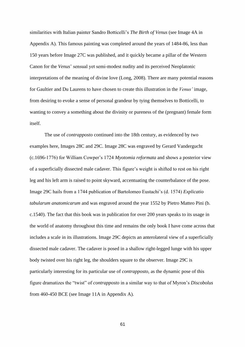
61
similarities with Italian painter Sandro Botticelli’s The Birth of Venus (see Image 4A in
Appendix A). This famous painting was completed around the years of 1484-86, less than
150 years before Image 27C was published, and it quickly became a pillar of the Western
Canon for the Venus’ sensual yet semi-modest nudity and its perceived Neoplatonic
interpretations of the meaning of divine love (Long, 2008). There are many potential reasons
for Gaultier and Du Laurens to have chosen to create this illustration in the Venus’ image,
from desiring to evoke a sense of personal grandeur by tying themselves to Botticelli, to
wanting to convey a something about the divinity or pureness of the (pregnant) female form
itself.
The use of contrapposto continued into the 18th century, as evidenced by two
examples here, Images 28C and 29C. Image 28C was engraved by Gerard Vandergucht
(c.1696-1776) for William Cowper’s 1724 Myotomia reformata and shows a posterior view
of a superficially dissected male cadaver. This figure’s weight is shifted to rest on his right
leg and his left arm is raised to point skyward, accentuating the counterbalance of the pose.
Image 29C hails from a 1744 publication of Bartolomeo Eustachi’s (d. 1574) Explicatio
tabularum anatomicarum and was engraved around the year 1552 by Pietro Matteo Pini (b.
c.1540). The fact that this book was in publication for over 200 years speaks to its usage in
the world of anatomy throughout this time and remains the only book I have come across that
includes a scale in its illustrations. Image 29C depicts an anterolateral view of a superficially
dissected male cadaver. The cadaver is posed in a shallow right-legged lunge with his upper
body twisted over his right leg, the shoulders square to the observer. Image 29C is
particularly interesting for its particular use of contrapposto, as the dynamic pose of this
figure dramatizes the “twist” of contrapposto in a similar way to that of Myron’s Discobolus
from 460-450 BCE (see Image 11A in Appendix A).

62
As shown in Image 30C contrapposto was continued to be used into the mid to late
1700s. While there is an increase in close-up, isolated anatomical drawings (such as Image
31C by Charles Bell) during this period, full-body images remained loyal to the common use
of contrapposto. Image 30C depicts a plain-background version of an illustration from
Albinus’ Tabulae sceleti (1747), published in a 1777-1778 English version of the text. In
Image 30C we see a posterior view of a superficially dissected male cadaver, his weight in
his right hip and his left arm held out slightly aloft, emphasizing the counterbalance in a
similar but more understated fashion to Image 28C.
By the early-mid 1800s, close-up isolated views of cadavers with minimal, if any,
background elements had largely become the norm for anatomical illustrations. Images 32C
and 33C demonstrate the kind of detail possible with this composition style, as well as the
clever use of partial colour to highlight specific features of the dissected cadaver. However,
as in the previous century, any full-body illustrations during this time period often made use
of contrapposto. Image 34C was published in Cloquet’s 1825 Manuel d’anatomie and depicts
a lateral view of a superficially dissected male cadaver. Although the observer cannot see the
angles of the hips and shoulders as one would from an anterior or posterior view of the figure,
the contrapposto of this image is identifiable from the placement of the legs. The left leg of
the cadaver featured in Image 34C is straight and bears the weight of the individual, while the
right leg is relaxed and slightly bent at the knee. Should this same figure be viewed anteriorly
or posteriorly, it would be seen that the hips would be tilted off-axis, especially compared to
the angle of the shoulders, signifying that even in illustrations where it is not immediately
obvious contrapposto is often still taken into consideration by the artist.
Finally, Image 35C demonstrates that it is possible to utilize contrapposto even
without a view of the entire body. Drawn by Joseph Maclise (1815-1880) for his book
Surgical Anatomy (2nd ed, 1856), Image 35C depicts an anterior view of an undissected male

63
figure. The image is centred around the torso of the figure and includes the head as well as
the tops of all four limbs. Even without viewing the entirety of this figure’s form, the use of
contrapposto is seen in the significant tilt of the hips and the twist between the lower and
upper body. It is clear that the use of contrapposto is present in anatomical illustrations from
the beginning of modern anatomy onwards, further solidifying anatomical illustrations
themselves in the wider realm of the Western Canon.
4.2 Theme 2: Ideal Body Types
The theme of Ideal Body Types speaks to the presence of the creation and repetition
of an idealized, standardized human body in the analysed anatomical illustrations. In the
section devoted to this second theme I will describe and provide examples from my data of
the bodily design which is very rarely deviated from in the analysed illustrations (see Table
1). First, I will describe the “dual-gender features” commonly seen in illustrations of both
males and females, accompanied by examples from my data. Next, I will describe and give
examples for the “male-specific features” which are seen in nearly every analysed anatomical
illustration of a male figure, before doing the same with the “female-specific features”.
Finally, I will share examples of the few bodies which differed from these norms in my
dataset.
TABLE 1: Male-specific, female-specific, and dual-gender features as seen in early modern
anatomical illustrations.
Male-Specific Features Female-Specific Features Dual-Gender Features
- Broad shoulders,
tapered waist
- Angular jaw/facial
structure
- Minimal body fat
- Muscular build
- Narrow shoulders,
tapered waist,
slightly widened hips
- “Soft” arms, legs
- Slightly rounded
stomach
- Small, very round
breasts
- minimal muscle
- Light skin
- Long limbs, torso
- Small (pink) mouth
- light/blond/brown
hair
- Straight, narrow,
pointed nose, high
nose bridge

64
4.2.1 Dual-Gender Features
In accordance with the changing ideas around the existence of two sexes versus one,
male and female body types in Western anatomical illustrations slowly diverged from one
another during the Early Modern period. That being said, there are certain features which are
seen in both male and female bodies across time, which I have termed here “Dual-Gender
Features”. As laid out in the Methodology chapter of this thesis, very few bodies, both male
and female, deviate from the following features: light hair and skin, long limbs and torsos,
small lips and mouths, and noses which are narrow and often pointed, with a high nose
bridge.
Image 36C is an image by an artist known only by their initials, “R.S”, and depicts
anterior views of one male and one female figure. The figures are drawn seated on a bench
inside a stone bricked building, with a small window between the two of them showing a
glimpse of a hilly landscape and the simple outline of some buildings. In the right hand of the
female figure is a small sign which reads Nosce te ipsum, with the English translation “knowe
thyself” written below the Latin. This illustration is titled Interiorum corporis humani
partium viva delineatio, or “Quick sketch of the interior parts of the human body”, and the
original/tangible copy had liftable illustrated layers to show the interior of the thorax and
abdomen of both figures, much like a modern day “pop-up” book (Anatomia, 2017).
Published circa 1559, the female and male anatomy in Image 36C do display some
differences from one another, such as the presence of small round breasts on the female and
more defined musculature on the male. However, the overall body shapes of both figures are
more alike than what is seen in later anatomical illustrations, which is in line with the slowly
changing views of sexual dimorphism during this period. Due to the less-pronounced sexual
differences between the male and female figures, Image 36C provides a suitable example of a

65
selection of traits which commonly appear in both males and females in anatomical
illustrations throughout the Early Modern period. Both figures have very light skin and hair,
the same tone as the blank surface the image was printed upon. Both figures also have long,
straight noses, with high bridges. While the male’s mouth is mostly obscured by his facial
hair, the female figure’s mouth sits very small on her face, with small, dainty lips.
The standing nature of the figures in Images 37C and 38C allows us to observe the
long limbs and torsos typical of anatomical figures throughout modern history. Both images
were drawn by an unknown artist and published in Guidi’s Opera omnia in 1626. Image 37C
depicts an anterior view of an undissected male figure who stands on an uneven ground with
his weight in his right leg. Image 38C shows an anterior view of a female figure in a similar
pose to that of Image 37C, the only dissected part of her body being her thoracic region,
specifically her breasts. Both figures once again demonstrate the suite of features and traits
which are typical of all anatomical illustrations across the time period of my study; the male
and female figures both have light skin and hair, small mouths, and narrow and/or pointed
noses with high bridges. Their long limbs and torsos will be referenced in the Synthesis and
Discussion chapter to discuss the influence of da Vinci’s Vitruvian Man and the notion of
divine proportions on modern anatomical understandings of the human form.
4.2.2 Male-Specific Features
In conjunction with the dual-gender features present in nearly every illustration I have
analysed, there are traits that are exclusive to one of two gender groups. In this section I will
first describe and provide examples for what I have termed “male-specific features” before
doing the same with “female-specific features”. These features will then be used in the
Synthesis and Discussion chapter to explore the creation and perpetuation of two mutually
exclusive sexes and how these sexes interact with modern Western gender ideals/roles. The

66
male-specific features I identified in my data include a broad chest and shoulders with a
tapered waist, angular jaw (and often angular overall facial structure), minimal body fat, and
a decidedly muscular build.
Images 39C and 40C are examples of the male body in early modern anatomy, with
both illustrations being modified versions of images from Vesalius’ Fabrica which were
republished in 1559 by Thomas Geminus. Image 39C features two isolated images of the
male torso, both with deeply dissected abdominal regions. Even these partial bodies
demonstrate the typical broad shoulders and tapered waist of male anatomical illustrations, as
well as little body fat. Image 40C shows an anterior view of a male figure, originally one of
Vesalius’ musclemen and which is superficially dissected to show the surface muscles.
Within Image 40C one can easily identify the previously mentioned male-specific features,
with the figure has broad shoulders and a tapered waist, an angular jaw, and well-defined,
large muscles, particularly in his arms and legs.
Coming from Du Laurens’ 1627 Opera omnia anatomica et medica, Image 41C
provides an example of the over-exaggerated, bulging musculature sometimes seen in
anatomical illustrations during the 17th century. This illustration served the purpose of
illustrating human surface anatomy, and depicts two male figures, one seen anteriorly and
one posteriorly. Both of these figures are drawn with a significantly small amount of body
fat, and the skin appears to be almost suctioned to their muscles. Published four years later in
Spiegel’s De humani corporis fabrica libri decem, Images 42C and 43C are examples of both
instances of little body fat and exaggerated muscles from this time period, albeit the muscles
are less extravagant than in Image 41C.
Moving into the 1700s there is a continuation of the use of my identified male-
specific features, however, there appears to have been in part a shift away from some of the
more exaggerated musculature of the previous century. Image 44C is a good example of the

67
less-dramatically-muscled body which is featured in multiple plates throughout the 1777-
1778 English translation of Albinus’ Tables of the Skeleton and Muscles of the Human Body.
Depicting an anterior view of a superficially dissected male cadaver, Image 44 shows broad
shoulders with a tapered waist, an angular face, and a muscular build despite being a lean
frame. It is important to remember that these male-specific features exist alongside the dual-
gender features present in both sexes. Image 44C provides an example of some dual-gender
features, such as the elongated limbs, straight, pointed, and high-bridged nose, and small
mouth.
The early 1800s brought with it some of the more dynamic compositions and poses
seen throughout this period, as shown in Images 17C and 18C. Within these images the
standard male-specific features are still identifiable, as evidenced by Image 45C. Drawn by
Charles Bell for his A System of Dissections at the turn of the century, this illustration depicts
an anterolateral view of a partially superficially dissected male cadaver whose wrists are
bound by twine and lifted up over his head. The figure’s upper body is arched backwards as if
up and over a table or bench. Even in this unconventional position, the male-specific traits of
having a tapered waist compared to broader shoulders and chest, minimal body fat, and
strong looking muscles.
The broad chest and shoulders and tapered waist of the figure in Image 46C are very
obvious in due to the figure’s slightly twisted pose and its posterolateral perspective. Image
46C is from Cloquet’s 1825 publication of his Manuel d’anatomie, and depicts the torso,
upper right arm, and most of the head of a young adult male cadaver. The face is the only
area left undissected in this figure, and displays a sharp, pointed chin, along with dual-gender
features such as a very straight, pointed nose, pale skin, light hair, and a small mouth. The
visible muscle mass of the figure in Image 46C is comparable to that of figures featured in
Images 47C and 48C, as well as Image 35C, all of which were published in books around the

68
mid-1800s. Used earlier as an example of contrapposto, Image 35C depicts an undissected
male figure with large, well-defined muscles, broad shoulders and chest, tapered waist, and
very little body fat.
The anterior perspective of Image 35C makes it very easy to compare this figure to
that in Image 47C. Image 47C depicts an anterior view of a male torso, and while the
abdomen and thorax have been heavily dissected to show the aorta the outline of the figure’s
form as well as the tops of the limbs, the neck, and a sketch of the lower face are visible.
Very similar to the figure in Image 35C, the cadaver in Image 47C displays the typical broad
chest and shoulders compared to a tapered waist, creating a sort of upside-down triangle
shape which is typical of these illustrations. If anything, this “triangle” seems to have gotten
more pronounced over time, with it being most striking in illustrations from the 19th century.
The cross-section of the thorax and abdomen in Image 47C make it possible to see just how
little body fat is on the body, as well as the thickness of some of the muscles such as the
deltoids and obliques. The outline of the lower half of the face includes the tip of a narrow
nose, a small mouth, and an angular jawline with a squared chin. Image 47C is also printed
entirely in coloured ink, allowing the colour of the skin to be observed, which is a light,
creamy beige.
Finally, Image 48C provides an example of how even in the more “realistically”
posed and prepared cadavers, both male-specific and dual-gender features are identifiable.
Image 48C depicts the thoracic region as well as the neck and head of a male cadaver, the
right side of the face, neck, and shoulder deeply dissected. The face of this individual is
angular, with a prominent chin and very straight, pointed nose. The skin and hair of this
cadaver is light, as is the case in every illustration in my dataset but one, which will be
discussed shortly. While much of the body is out of frame, it is still possible to discern that
the figure in Image 48C does not have much body fat and has at least somewhat defined

69
pectoral and deltoid muscles. Although the styles of anatomical illustrations very much
underwent change over the approximately 400 years encompassed by my study, the male
body itself saw decidedly little change in how it was presented and varied very little from the
general form I have described and provided examples for here.
4.2.3 Female-Specific Features
Compared to the typical male form seen in anatomical illustrations from the 1500s to
the 1800s, the typical female form has been subject to more change over time, as displayed in
Table 2. As will be later discussed, these changes in what constitutes the “ideal” female form
are likely tied to the changing ideas around the sexes during this period of time, specifically
the shift from the “one-sex” model to a “two-sex” model. This being said, there are still
certain features which can be seen in most female bodies from the beginning of modern
anatomy to the mid-1800s. These features are narrow shoulders with a tapered waist and
slightly widened hips, “soft” arms and legs due to minimal muscle, a slightly rounded
stomach when not pictured pregnant, long and/or coiffed hair, and small, very round breasts.
While there are 215 illustrations in my dataset, only 28 of these are of females, resulting in a
much smaller selection of examples for female-specific features than there is for male-
specific features.
TABLE 2: Change in sex-specific traits commonly seen in anatomical illustrations over time.
Time Period Male Traits Female Traits
Mid – Late 1500s - Broad shoulders,
tapered waist
- Angular jaw/facial
structure
- Minimal body fat
- Muscular build
- Small, very round
breasts
- Narrow shoulders,
tapered waist,
widened hips, “soft”
limbs
OR
- Some images feature
broad shoulders,

70
tapered waist,
large/shapely muscles
Early – Mid 1600s - Broad shoulders,
tapered waist
- Angular jaw/facial
structure
- Significantly little
body fat
- Extreme, bulging
musculature
- Small, very round
breasts
- Narrow shoulders,
tapered waist,
widened hips, “soft”
limbs
OR
- Some images feature
broad shoulders,
tapered waist,
large/shapely muscles
Late 1600s – 1700s - Broad shoulders,
tapered waist
- Angular jaw/facial
structure
- Minimal body fat
- Lean, though still
muscular
- Narrow shoulders,
tapered waist, slightly
widened hips
- “Soft” arms, legs,
minimal muscle
- Slightly rounded
stomach
- Small, very round
breasts
1800s - Very broad shoulders,
dramatically tapered
waist
- Angular jaw/facial
structure
- Minimal body fat
- Muscular build
- Narrow shoulders,
tapered waist, slightly
widened hips
- “Soft” arms, legs,
minimal muscle
- Slightly rounded
stomach
- Larger breasts than
previous centuries
As with the male form, the earliest example of the anatomized female from within the
realm of what is considered modern anatomy comes from Vesalius’ Fabrica in 1543. Image
49C is composed of various illustrations originally from Fabrica and published in Geminus’
1559 Compendiosa totius anatomie delineatio. Alongside isolated images of the uterus,
placenta, foetus, and vagina are two inset illustrations of female torsos, one of which can be

71
seen in its original form in Image 21C. From these images we can see some “idealized”
female-specific features such as narrow shoulders, tapered waist, and widened hips and small
round breasts. There is no female equivalent to the musclemen in Fabrica, and we are
without full-bodied views of the female form as according to Vesalius.
Moving on to the 17th century, Image 50C is similar to Image 49C in that it features
an isolated female torso amidst some inset illustrations of the female reproductive system,
specifically the uterus. Originating from Guidi’s Opera omnia (1626), this image provides a
good example of the “small and very round” nature of the breasts found in many of the
analysed anatomical illustrations, and they are shown to sit rather far apart from each other
high on the chest of the cadaver. During this century we do see some full-body illustrations of
female figures, such as the previously discussed Image 27C as well as Image 51C. Image 27C
displays likeness to Botticelli’s The Birth of Venus (see Image 3A) not only in the pose of the
figure, but also in terms of proportionality of her limbs and overall body shape. The bodies in
both Image 27C and The Birth of Venus feature some of the female-specific traits identified
in my study, including “soft” limbs and small, very round breasts. While not as long or
flowing as that of Botticelli’s Venus, the hair of the figure in Image 27C falls similarly about
her shoulders, the strands at the top of her head held back with a band or ribbon.
Image 51C depicts an anterior view of a female figure in an outdoor setting, her right
leg resting at the knee on a fallen tree branch or stump. The abdomen of the figure is deeply
dissected, the layers of skin, muscle, and uterus reflected back to show a foetus still attached
to the placenta by its umbilical cord. Published in Spiegel’s 1631 De formato foetu liber
singularis, this figure has broader shoulders and larger arms than some of the more “typical”
female bodies from this time, however she is far less muscular than men depicted within the
same book (see Image 41C). This figure also has very small breasts (though less round and

72
even smaller than what is typical) as well as long hair, which is partially done up in braids at
the top of her head.
For examples of the female form in the late 1600s, we have Images 52C and 53C.
Both illustrations come from Bidloo’s Anatomia humani corporis, published in 1685. Image
52C was the first full-coloured illustration of a female form I came across when gathering my
data and allows us to observe the colour of the individual’s skin which is a light creamy-
beige. This image depicts only the torso of a female cadaver, the abdominal region
superficially dissected to show the surface muscles. While it is not explicitly known from the
original text, this individual appears to be pregnant based on her distended stomach. Image
53C is also fully coloured and depicts the torso of a female cadaver as well as the top of the
left arm and the bottom portion of her face. This individual is definitely pregnant, as the
abdomen has been deeply dissected to see the placenta inside the reflected uterus. As can be
seen in the one visible arm, this figure has no clearly defined muscles. Two curled tendrils of
blonde hair can be seen on either side of her neck, and the dual-gender features of light skin
and a pointed, narrow nose are also identifiable.
Moving on to the 1700s, Image 54C offers a rare example of a female cadaver being
used to depict anatomy that is not associated with reproduction or considered female-specific,
such as breasts. Published in Myologie complette en couleur et grandeur naturelle by Gautier
d’Agoty in 1746, Image 54C depicts a posterior view of a seated female cadaver, her legs out
of frame. The back of this figure has been flayed to the ribs, the muscle reflected and out to
her left side as if a wing. The head of the figure is turned to the right so that the side of her
face can be seen by the observer, showing a straight, pointed nose and a small mouth. Gautier
d’Agoty’s works were printed in full colour with rich pigments, making it possible to see the
blush added to the image of this cadaver’s cheek and the blue of the ribbon which ties back
her light brown hair. The majority of d’Agoty’s female figures are not featured in his text on

73
myology, rather they can be found in his 1773 book Anatomie des parties de la génération de
l'homme et de la femme, which is where Images 55C and 56C originate (also see Image 15C).
Image 55C shows two female figures. The figure in the foreground appears
undissected, however only the upper half of her torso and her head is visible, the rest of her
body out of frame. This figure has dark brown hair which is pulled back from her flushed
face in a simple bun and is holding her right breast between the splayed fingers of her left
hand. Her breasts are both small and extremely round, as if they were pasted onto her chest.
Behind this figure is a second female cadaver who is sitting up on her knees, her head tilted
back and to her right. This cadaver’s entire torso as well as most of her neck and head have
been superficially dissected, her face held onto the skull by some twine. Like the first figure,
this cadaver’s face is coloured as if she is flushed or wearing blush. The vaginal opening is
visible on this cadaver and is dramatically dilated. Both figures in Image 55C display the
typical “soft”, non-muscular female body, along with narrow shoulders.
Image 56C appears to feature the lower body of the figure in the foreground of Image
55. In the original Anatomie des parties de la génération de l'homme et de la femme these
illustrations were sequential, with Image 55C being the seventh plate and Image 56C the
eighth. This illustration depicts a seated female figure from just below her breasts to her toes,
her pregnant stomach deeply dissected. The foetus of this cadaver is drawn seated in her lap
and is also deeply dissected. Around the cadaver’s feet are several isolated illustrations,
including that of the placenta, uterus, and the foetal circulatory system. Image 56 provides an
example of the long, sender, and non-muscular qualities of the typical female legs. The
provocative nature of Gautier d’Agoty’s female cadavers has been noted by scholars (see
Harvey, 2010; Holmstrom, 2017) and will be further discussed in the Synthesis and
Discussion chapter.

74
As discussed in Theme 1, by the 1800s many anatomical illustrations were of closed-
up, isolated body parts. Image 57C provides an example of such an illustration in regard to
the female body and depicts the pelvic region of a deeply dissected female cadaver to show
the female reproductive organs as well as the external genitalia. Other illustrations from this
time period, such as Image 58C, feature slightly more of the body, although still not the entire
form. Image 58 depicts a lateral view of a pregnant female figure from the top of the right leg
to just above the breasts. The abdomen of this figure has been sagittally bisected to show a
cross-section of the vaginal canal and gravid uterus, however the foetus has been removed. In
Image 58C we can see the non-muscular quality of the body as well as round breasts,
however this is perhaps one of the most “natural” looking examples of breasts in my dataset.
4.2.4 Deviant Bodies
While the majority of the male and female bodies in my dataset fit into the described
types, what follows here are some examples of bodies which deviate from the features which
make up my second theme. Rather than ignore these illustrations as outliers, they can be used
to further understand the formation of the standard, idealized male and female bodies, as well
as the standard dual-gender traits seen in most anatomized figures. It is important to keep in
mind that the female body is in itself a deviant body, as it differs from the norm that is the
male form. Be that as it may, we can also pick out female bodies that do not display the
typical female-specific features seen in the majority of other female forms in anatomical
illustrations.
As previously mentioned, the female form in anatomical illustrations undergoes more
change over time than the male form. This is likely due to the change in the understanding of
sexual differences during the beginning of the time period featured in this study. This change
consisted of moving from viewing humans as embodying a “one-sex” model, where females

75
were simply “imperfect” males, to a “two-sex” model with strict and purposeful sexual
dimorphism between males and females. Images 59C, 4C, and 60C provide examples of
female forms which display more of the male-specific features than they do female-specific
ones, such as little body fat, large muscles, and broad shoulders. Both Image 59C and Image
4C were published in 1545 in Estienne’s De dissectione and depict bodies which, if not for
them being pregnant, would be assumed to be male by the standards set in this study. The
figures in these illustrations both have well-defined muscles, broad shoulders, little body fat,
and practically non-existent breasts. Image 60C was published in 1631 by Spiegel in his De
humani corporis fabrica libri decem and features a broad-shouldered and very muscular
female figure. The female assignment of this cadaver is clear in the deep dissection of her
abdomen, which features the female urogenital system, as well as her external gonads.
For all illustrations but one, the female bodies in my dataset are depicted as young
adults, with more age-related variation seen among male cadavers. As will be discussed in the
following chapter, this is likely because most female bodies are featured in birth or
reproductive related contexts, rendering post-menopausal female bodies useless in the
opinion of most anatomists from this period. Image 61C, however, depicts an elderly female
individual, who appears to be living rather than a cadaver. The usefulness of this elderly
female body comes from the pathological context of the image, as this individual was
suffering from an intestinal prolapse.
An important aspect of the standard, ideal male body seems to be large, defined
muscles. Image 62C is an example of a male body which does not feature large muscles, and
in fact appears very emaciated, as if this individual were malnourished in life or perhaps sick
for a long period before their death. This illustration was created by Maclise for Sir Quain’s
1844 book titled The Anatomy of the Arteries of the Human Body with its Applications to
Pathology and Operative Surgery, and bears resemblance to Image 63, which was also drawn

76
by Maclise for his own book, Surgical Anatomy, published in 1851. The cadaver in Image
63C, though not as emaciated as the figure in Image 62C, also caught my attention as being
less muscular than male bodies in other anatomical illustrations. It is possible, though likely
not confirmable, that these two illustrations were based upon the same cadaver.
Lastly, almost all illustrations within my dataset have very pale skin, as well as
generally western European facial features and hair. The one exception to this can be seen in
Image 64C, which depicts an anterior view of a deeply dissected Black male figure.
Published in Maclise’s Surgical Anatomy, Image 64C, or “Pl. XXIV”, appears to have been
created as a comparison illustration to Image 65C, “Pl. XXIII”. Image 65C depicts a male
figure who displays both the typical dual-gender and male-specific features I have identified.
Positioned in exactly the same way as the figure in Image 65C, the figure in Image 64C
deviates from the typical dual-gender features of light skin and hair, small mouth and lips,
and a long, narrow, pointed nose. This figure also does not display the male-specific feature
of having an angular jawline, with his mandible drawn as a rather irregular shape. The
difference in face-shape between Images 64C and 65C is marked, with the figure in Image
64C being drawn with a much more prognathic face compared to that of Image 65C. As will
be later discussed, facial prognathism along with other craniometric measurements have long
been used as a way to racialize non-White individuals and claim that they are evolutionarily
more similar to apes than humans and therefore “less evolved” than White individuals
(Blakey, 1987; Diogo, 2018).
Within this chapter I have proposed my two main themes, Western Canon and Ideal
Body Types, and provided examples from my dataset in order to identify said themes within
my research. The theme of Western Canon situates anatomical illustrations within the greater
Western European stylistic tradition, which allows for culturally informed analysis of the
formation of the ideal, standardized body in modern anatomy through the contexts within

77
which these bodies were created. The second theme, Ideal Body Types, speaks directly to the
ideal bodies themselves, and allows for the description of such types in the presence of
various male-specific, female-specific, and dual-gender features. Having now established
both of these themes and tied them to my data, I now move on to present my analyses and
propose discussion topics for further exploration.

78
CHAPTER 5
SYNTHESIS & DISCUSSION
Images can have a great deal of political and social power. In 19th-century
Netherlands, for example, illustrations were used to create and seminate the concept of a
Dutch national identity (Dellmann, 2015). In this case, carefully chosen images created a new
“truth” which stated that you could tell by looking at someone if they were part of the
national identity or a foreigner, despite the reality of dramatically varying regional dress,
language, and other traditions, all of which were equally Dutch (Dellmann, 2015). This
fabricated homogeneity is mirrored in early modern anatomical illustrations, in which almost
all bodies depicted fit within a strict set of physical parameters, which I identified and
explored in the previous chapter.
Part of the reason behind the limited variation depicted in these illustrations may be
explained by existing evidence which supports the idea that many anatomical illustrations
were composite drawings rather than necessarily drawn from real life (Saunders & O’Malley,
1973). Rather, various features were drawn “from life” but then placed together to create a
human who never actually existed. Also worth noting is that while some anatomists were
artists in their own right, many had other, non-medically trained artists illustrate their works
for them. For example, Vesalius contracted Calcar along with other artists from the school of
Titian for illustrations in Fabrica, and merely outlined and edited the images after they were
created by others before their publication (Smith, 2006). Practices such as these could help to
explain how, if not why, there appears to be only two body types (with considerable overlap)
present in anatomical illustrations.
To get to the “why” behind the creation of anatomical illustrations’ standardized body
types, the themes introduced in the previous chapter must be further explored and analysed.
In this chapter I discuss my two themes in a fashion which mimics the layout of the Findings

79
section, beginning with Theme One, Western Canon, before moving onto Theme 2, Ideal
Body Types. Within the first theme I explore, in chronological order, how background
composition styles of anatomical illustrations all throughout the early modern period worked
to communicate “realness” and authenticity to their audiences. This communication of
authenticity helped to normalize the unrealistic adherence to specific body types seen in said
illustrations. I then move on to discuss the positioning of bodies in anatomical illustrations,
specifically how the prolific use of contrapposto across all time periods featured in my study
is symbolic of both the desire to create beauty in the images and the ongoing importance of
antiquity to the formation of the Western Canon.
My second theme is concerned with the ideal body types seen in anatomical
illustrations, which I categorized into classes of features in the previous chapter, namely
“male-specific”, “female-specific”, and “dual-gender”. With the goal of creating beauty in
anatomical illustrations being of high importance to early modern anatomists, it follows that
ideal body types were created and perpetuated within the discipline from its conception. In
this section I explore how the adoption of a two-sex model of understanding human sexual
differences created the two standard human body types, the “man” and the “woman”. On top
of this, I explore how societal roles prescribed to each gender influence the creation of
physical ideal body types, both of the male and the female. I then discuss the overarching
Western European ideals of “whiteness” which permeate the dual-gender features seen in the
illustrations in all time periods. Lastly, I explore the lasting consequences of standardized,
idealized forms in anatomy, drawing a connection from the periods analysed in this project
with our contemporary sociomedical reality. The conclusion of this chapter is a call for a
more inclusive, variation-focused anatomy, and I offer a small discussion of first steps which
might be taken towards such a goal.

80
5.1 Theme 1: Western Canon
5.1.1 Background Composition Styles: Communicating “Realness”/Authenticity
Through my first theme I was able to pinpoint how the anatomical illustrations in my
study belonged as part of the Western Canon. This knowledge then made it possible for me to
gain contextual information about the illustrations that is culturally informed, specific to the
time period each was created in. Something I have been able to surmise from my study of
illustrations and their historical contexts is that the desire to communicate a sense of
“realness” and authenticity in medical drawings, identified by Kemp (2010, pp.198-99) as
“the rhetoric of the ‘real thing’”, has existed at least since the beginning of modern,
observational anatomy. This insight is not immediately obvious when looking at some
illustrations, particularly the older ones, which look dramatically different from the images
we would recognize as medically legitimate by 21st century standards.
What I wish to highlight by including this section of my analysis is the juxtaposition
between anatomists' desire to portray “realness” in their illustrations while at the same time
almost exclusively showing one to two types of human forms, which is decidedly unrealistic.
The result of this conflict is that, in every time period, those viewing anatomical illustrations
were shown images which, firstly, signified to them realness and authenticity, and secondly,
showed only extremely limited types of human bodies as “real”. Considering realness and
authenticity signify unquestionable “truths”, this is a dangerous combination, one that very
much extends into the contemporary world. If we are ever to overcome ingrained biases in
fields such as anatomy, biology, anthropology, and so on, we must recognize not only where
said biases came from historically, but also how they were normalized at different points
throughout time.
The changing styles seen in my dataset, from the landscape-bound animated figures of
the 16th and 17th centuries to the isolated specimens of the mid-late 1800s, are the result of
81
their creators all trying to communicate the same thing to their viewers: a sense of realness
and authenticity. Naturalistic ideals of the Renaissance led early anatomists like Vesalius to
place his cadavers in natural, outdoor settings. As explained in the Historical Context chapter,
during this period “nature” was being touted by the likes of Alberti as the best source of
“perfect forms”, with the works of the ancients a close second due to the belief that they often
worked directly from nature themselves (Westfall, 1969). This is theoretically congruent with
broader notions of naturalism, which affirms that all beings and events in existence are
“naturally” as they appear, just as they are “meant” to be (Summers, 1990). By placing
cadavers in landscapes, the anatomist made a visual connection with the Renaissance/Early
Modern viewer, communicating to them that what he was depicting about the human form
was real and natural. This artistic decision would have been especially important for
Vesalius, and Calcar and any other artist he may have worked with, as he was the first to
promote a naturalistic, observational approach to understanding the human body rather than a
philosophical one. As the University of Padua was a longstanding intellectual hub for
philosophy, the arts, and science (Saunders & O’Malley, 1973), Vesalius’ choice to make the
countryside of Padua the backdrop to his musclemen further served to communicate said
illustrations' authenticity and significance.
A difference between the backgrounds seen in male illustrations and those in female
illustrations exists within the earlier centuries of modern anatomy and can be seen in my
dataset. While male cadavers were almost exclusively depicted in a landscape of some sort
(see Image 3C), female cadavers were sometimes placed indoors in what appears to be
heavily draped bedchambers (see Image 4C). Although female cadavers were sometimes
depicted outdoors during this time (see Image 51C), there is no example of a male cadaver
placed in a similar indoor setting within my dataset. This phenomenon is likely tied to
communicating “realness” to the viewers, the majority of whom would have been upper-class

82
men, as were essentially all students during this time. Women were confined to the realm of
the house and the role of reproduction, and therefore female cadavers were drawn pregnant
and indoors. The male form, on the other hand, was created in God’s image, and thus
belonged to the outside, natural world along with the other “perfect forms”. The choice to
depict nude female cadavers inside may also represent a need or desire to show a certain level
of modesty in the illustration on behalf of the anatomist, with the naked female body not
being fit for the outside world, outside of the bedroom.
The concept of temporality in the form of ruined buildings in Western art during the
1500s and early 1600s served two purposes. The first purpose is that overgrown ruins
symbolized nature’s (and thus God’s, depending on who you ask) might and persistence
against things built by humans (Hansen, 2016), and almost verged on a kind of memento
mori, a reminder of human mortality. Similar to other Western artforms from this period,
ruined buildings are common in anatomical illustrations (see Images 5C, 6C). The second
purpose of temporality was that it provided a visual tie to the ruins of antiquity and the
idealized Roman Empire they represented to the people of Renaissance Italy and beyond
(Hall, 2005, Hauser, 1999). As established in the Historical Context chapter, notions of
“nature” and its perfection were deeply intertwined with a revival of antiquity in the
Renaissance mind (Hauser, 1999; Westfall, 1969).
The change in background styles from landscapes to lab-based settings in the late
1600s is, conceptually, a continuation of the anatomists’ desire to communicate authenticity
and “realness” through their illustrations. Throughout the 17th century observational,
experiment-based science had been taking place across Europe, and modern anatomy was
becoming an established discipline (Persaud et al., 2014). With human dissections being held
in indoor workshops and anatomy theatres across the continent, anatomical illustrations
began to reflect these real-world settings. Rather than relying on the cadaver’s placement in

83
nature to convey “realness”, the “realest” place to depict the cadaver was now on a worktable
or a wooden floor, where the viewer might expect to see them dissected in real life.
As always, the wider world of Western art also heavily influenced the style of
anatomical illustrations during this period (Anatomia, 2017). As all of Europe was praising
Dutch artists during the “Dutch Golden Age” for their realistic still-life’s, anatomical
illustrations began to similarly take on still-life qualities to signify “realness”. Given that
some anatomical illustrations were composite drawings made up from multiple sketches and
multiple dissections, as well as often copied and edited by others over the years (Markatos et
al., 2017; Saunders & O’Malley, 1973; Sanders, 2005), it follows that it should not
necessarily be assumed that the rooms cadavers were drawn in were “real” rooms, even if
depicted in a style which is similar to still-life paintings. Of course, it mattered not if the lab-
based setting depicted in the illustration was indeed somewhere the cadaver ever was in real
life, only that it was a lab-based setting at all. The presence of some lingering landscape-
based illustrations in the late 1600s actually supports the idea that there was not some sort of
large conceptual change in society that suddenly saw placing animated cadavers outdoors as
silly. What is seen in the illustrations is a gradual change in popular philosophy and societal
values in Europe over a few hundred years.
The 1700s saw the continued use of lab-based illustrations, with the new addition of
mid-surgery contexts (see Image 14C). As mentioned within the Findings chapter, before the
use of anaesthesia in Western medicine, surgeries were short and brutal, leaving little time for
the sketch of any one part of a procedure (Robinson & Toledo, 2012). Mid-surgery
illustrations provided another way to communicate realness to the images’ observers, as it
detailed a very specific and practical event in a period where the surgical possibilities being
brought forth by anatomists were unprecedented in Western Europe (Taylor, 2016; Persaud et
al., 2014). The sense of authenticity communicated by mid-surgery images was evidently not

84
diminished by the fact that it is unlikely that such illustrations were created (at least fully)
during the actual surgery. It is possible that the individual died on the table during surgery,
and they were drawn, or finished being drawn, posthumously, but clearly if that was the case
in the example of Image 14C then some artistic license was still taken in the form of the
conscious appearance of the cadaver. Either way, the illusion of being drawn in real time
from real life is strong in mid-surgery images.
By the late 1700s and into the 1800s anatomical illustrations no longer featured
outdoor scenes of rolling hills and shrubby vegetation. The focal point of most anatomical
illustrations at this point in time was the cadavers themselves, the backgrounds mostly or
entirely blank, although there remains some substantial variation in composition styles (see
Images 17C and 18C for dynamic but lab-based compositions). What can be seen in the
chronological study of anatomical illustrations is a gradual continuation away from the use of
symbolism and complex backgrounds and towards an almost streamlined, minimalist, rather
utilitarian art style.
Although perhaps seemingly arbitrary at first glance, this shift makes sense
considering a similar shift which was taking place over the same period across Europe.
Similar to how we know that the symbolism-heavy style popularized in the early 1500s was
largely due to the monetary power and political interests of the Catholic Church, it is not
unlikely that the style popularized during the late 1700s and 1800s were influenced by the
concurrent Industrial Revolution, with its championing of utility and technological
advancement (Clark, 2012). Images like those of 19C and 20C, which show the same men at
two different ages, are examples of how artists may still incorporate fantastical components
in their art while signifying authenticity to the observer through their overall style. The
composition of the images, which include isolated heads and upper thoracic regions against a
blank background, coheres to the popular style seen at the time they were created. Thanks to
85
their context as well as the prestige of their creator (Jules Cloquet), the inclusion of what
appears to be the same men at different ages does not destroy the validity of these
illustrations.
The composition styles anatomical illustrations embody over time, much like the
broader art world, correspond to the popular ideas of the time they are created within and
what it means to be “real” and valid. The simplistic, clinical nature of anatomical illustrations
like those published in Gray’s Anatomy and used to this day correspond with the
contemporary Western view of the medical world as sterile and purely utilitarian (Kemp,
2010; Robbins, 2018). Understanding this about style allows us to do two things: the ability
to not pass flippant and perhaps negative judgements on anatomical illustrations from
hundreds of years ago so that we might comprehend how they impact our present, and the
ability to not pass the reverse judgements on more modern illustrations which are not
necessarily more “real” than their historical predecessors but are much more familiar to us in
the 21st century. Anatomical illustrations have always been real to their audience, and
therefore have always communicated authoritative ideas about what the human body should
or should not look like. If we are able to tease apart and critique anatomical illustrations from
1543, then the same can be done with those from 2021.
5.1.2 Positioning of Bodies: Antiquity and Beauty
Despite the changes in composition styles previously discussed, the use of
contrapposto is ubiquitous across the time period in my study. Prolific use of contrapposto
has continued into the present day, with it now being an almost subconsciously known
phenomenon regularly seen in visual art and photography (Pazhoohi et al., 2020). The
importance of contrapposto to this study comes from the fact that it is readily seen in
anatomical illustrations from all centuries represented in my dataset. This shows us that,
86
firstly, the artists of anatomical illustrations were at least as focused on achieving “the
beautiful” in their works as they were with representing anatomical features (Summers, 1997,
p.361).
Secondly, the continuous use of contrapposto draws attention to the fact that while
overall art styles can be seen to change with time, certain aspects remain throughout history
and retain their central importance to Western art and, thus, anatomical illustrations. The
importance of antiquity, specifically the ancients’ ideas of beauty (or at least modern
interpretations of them) is such an aspect, as they remained important throughout my period
of study and were instrumental in the development of what we now term the Western canon.
My analysis suggests that the anatomists’ focus on creating beauty in their work coupled with
the powerful, enduring iconography of the ancients contributed to the creation and
perpetuation of the standardized, idealized human forms seen in anatomical illustrations.
With Western beauty being so closely tied to the idea of “balance”, as according to
ancient philosophers, it makes sense that contrapposto is important to Western beauty and
how it was and is visually communicated. Along with contrapposto creating shapes out of the
human body which may be pleasing to some eyes, such as accentuating the waist to hip ratio
(Pazhoohi et al., 2020), the dynamic quality and juxtaposition it brings to an otherwise static
image was and is seen as representing something psychological about the figure, in that their
mind is in motion in a similar way to their body (Summers, 1977). Through this movement,
along with expressions in the face, it was claimed by ancient historians and philosophers such
as Xenophon (430-354 BCE) that it is possible to portray the invisible soul in a work of art
(Summers, 1977). Xenophon concludes this argument that it is therefore better to depict
“noble men” than it is to paint “villains” (Summers, 1977, p.355). If nobility of the soul is
depicted via the use of dynamic positioning, it may be assumed that depicting the morally or
mentally corrupt would make for a rather uninteresting composition.

87
What should an artist do, then, if a living, breathing example of such a noble and
beautiful person cannot be found? As Xenophon writes, in order to show “perfect beauty” a
painter must copy the most beautiful features from a variety of different models in order to
make the whole beautiful (Summers, 1977, p.355). One may be reminded by Xenophon’s
instructions of how early anatomists and artists might pick and choose features from various
cadavers and patients, combining them to create their final illustrations (Saunders &
O’Malley, 1973).
The goal of creating and portraying beauty as described by Xenophon fully supports
the foundational tenets of Renaissance art theory (Summers, 1977). As spoken of in the
Historical Context chapter, a philosophical shift took place in the Renaissance which dictated
that physical beauty was indicative of intellectual power and beauty, and although this is in
opposition to Plato’s views on the soul and body, other notable ancients like Xenophon could
be cited to give this new belief further validity. Rather than this view being secluded to the
Renaissance, the continuous use of contrapposto in works belonging to the Western canon
indicates that a desire to create beauty persists throughout the following centuries.
Considering the great degree which Renaissance ideas and innovations influenced the
development of the modern Western world, the endurance of certain significant philosophies
and practices such as the creation of perfect beauty is easily comprehensible.
Due to the cultural carry-through of specific Renaissance interests and ideals despite
style changes over time, it is now possible to identify the last 500 years or so of art, literature,
music, and philosophy in Western Europe as belonging to a cohesive canon. This
phenomenon is by no means unique to Western European civilization and is an extremely
important aspect of human life by which we create meaning and sustain our identities. The
existence of a Western Canon is not a negative thing in itself. Specific negative outcomes

88
from long-term, uncritical adherence to some portions of the Western Canon, however, are
possible.
Due to the intention of creating beauty in anatomical illustrations a very specific body
type began to represent the peak of human beauty and intellectual power, with those who did
not fit this mould being imperfect and less human. The body which best fit the mould of
perfect beauty was that of an able-bodied Western European man. Secondary to this ideal
came Western European women, as the characteristics and features they shared with
European men made them acceptable, but them being female demoted them to less than
perfect. This holds true under both one-sex and two-sex models, as we are about to discuss.
While these views are unlikely to be actively or consciously held by many individuals in the
21st century, contemporary disciplines such as anatomy and medicine or physical
anthropology have been built upon these very beliefs for at least the last five centuries. Our
modern notions of what’s natural or “normal” are inextricably intertwined with Western
philosophies and beliefs about what it means to be beautiful.
5.2 Theme 2: Ideal Body Types
The goal of creating beauty in art led to the creation of ideal body types in anatomical
illustrations, one for men and one for women, with certain overlapping features between the
two groups. As seen in my data, these types were used again and again throughout history in
anatomical illustrations, further normalizing them. Rather than being pulling from thin air or
some kind of objective truth, these ideal bodies were created based on a desire to create
beauty in art, the adoption of a two-sex model of understanding sexual differences in humans,
the physical representation of gender roles, and the importance of “whiteness” in early
modern Western Europe.

89
5.2.1 Sexual Dimorphism, Gender Roles, and the Creation of Body Types
As discussed in the Historical Context chapter, the importance of naturalism during
the Renaissance along with a new interest in observational science led to significant changes
in how the human body was conceptualized (Hauser, 1999; Stolberg, 2003). As naturalism
affirms that all beings and events in the universe, along with their inherent character, are
“natural” (by the hands of God or otherwise), the view that physical differences between men
and women were the result of female imperfection could not stand (Stolberg, 2003). The
Western world slowly shifted from one-sex model to a two-sex model of viewing human
variation, one which believes that it is natural for women’s bodies to be wholly different
from those of men.
As demonstrated in the Findings chapter, the shift from a one-sex model to a two-sex
model of understanding human variation is visible in how bodies were depicted in early
anatomical illustrations. When the general consensus was that females were “imperfect”
males, female forms were drawn with features that a 21st perspective would likely identify as
masculine. This includes features such as broad shoulders, large and defined muscles
throughout the body, little body fat, and either very small or non-existent breasts (see Images
4, 36, 59, 60). In many of the earliest modern anatomical illustrations, the male and female
bodies looked more alike than they did dissimilar.
Only when the idea of sexual dimorphism became more popular and widely agreed
upon did male and female bodies begin to look drastically different from one another. Female
bodies in anatomical illustrations were now depicted with a new set of female-specific
features (see Tables 1 & 2), most of which existed in direct opposition to many male-specific
features. The reason for this strict opposition is the ideal of sexual dimorphism itself, which
sees males and females as belonging to two discrete groups, with the characteristics of one
dictating the opposing characteristics in the other. Due to the importance given to the creation

90
of beauty in anatomical illustrations, anatomists and artists overwhelmingly chose to depict
the idealized forms of males and females as what they “should” look like. When women
“should” have looked more like men to have been considered as perfect as possible, that is
how they were drawn. When that ideal changed, so did the illustrations.
The bodies which were featured in anatomical illustrations as well as the contexts
they were depicted within informs us about the various gendered roles the cadavers would
have (or should have) embodied in life. In the years encompassed by this study, female
bodies depicted in anatomical contexts were almost exclusively used to demonstrate the ways
they should differ from male bodies. Not only are female bodies shown dramatically less
frequently in anatomical illustrations compared to male ones, but they are also nearly always
pregnant and/or the image is specific to features which are considered female-only, such as
breasts or the female urogenital system. These limited uses for the female cadaver are likely
why illustrations almost solely depict young, plump women, as older, post-menopausal
women are no longer reproductively useful, and therefore anything they have to show
anatomically can be shown by a male cadaver instead. The only example of an elderly
woman in my sample is pathological in context (Image 61), her pathology (a hernia) once
again making her useful for anatomical study under this prevailing perspective.
Other “uses” of the female body include its perceived “inherent” sexuality.
Holmstrom’s 2017 thesis explores the 18th and 19th century emergence of a sexual
fascination with the female corpse, specifically focusing on the relationship between the
alluring female autopsy subject and the conflicted male anatomist. While there was an
increase in female cadavers depicted in anatomical illustrations in the mid-1700s, the world
of anatomy remained very much a male domain, with anatomists and their artists alike being
exclusively men (Holmstrom, 2017). The nature in which female bodies were depicted during
this time in fact emphasized the notion that anatomy as a discipline belonged solely within

91
the male societal sphere, as they were consistently shown as representing masculine
constructions of a feminine sexuality that is “compliant and unresisting” (Holmstrom, 2017,
p.2).
Holmstrom (2017) notices, as I have, that the female cadavers in these illustrations are
always young and beautiful by Western standards, as well as being depicted in ways which
draws attention to their feminine attractions and obscures the fact that they are dead. The
work of Gautier d’Agoty provides particularly striking examples of this use of the female
cadaver. As can be seen in Images 15, 54, 55, and 56, Gautier d’Agoty’s vividly coloured
pieces feature lovely young women who, despite their gored bodies, remain serene and coy
with their downward turned eyes, their cheeks flushed pink. Image 54 is specifically
discussed by Holmstrom (2017, p.37), who describes the figure within it as glancing over her
shoulder at the observer in a “coquettish” way in direct juxtaposition to her flayed and deeply
dissected back. Image 55 shows the upper portion of a blushing woman who is fondling her
right breast with her left hand, while Image 56 reveals the rest of said woman’s body, her
pregnant belly cut open and emptied, her dissected foetus resting between her legs. Even the
hair of Gautier d’Agoty’s female cadavers is coiffed in the way suitable for ladies of his time,
blue ribbons adorning some of them (see Images 15 and 54). Gautier d’Agoty’s images are
considered to have essentially invented the trope of aestheticizing female death in anatomical
illustrations, a phenomenon which corresponds well with the popular motif of the femme
fatale in the following century (Holmstrom, 2017).
The result of showing female cadavers as lifelike, beautiful, and demure while at the
same time having been exposed and mutilated at the hands of a male anatomist is a sense of
vulnerability, one that suggests the woman is both helpless to and welcoming of the intrusion
of the men around her. It is worth noting that the sexualization of female cadavers extended
beyond their depiction in anatomical illustrations, so much so that the public was aware of it.

92
One writer made note that even those who fully support the practice of dissection “shudder at
the mere contemplation” of the body of a “parent, wife, sister, or daughter” being “exposed to
the rude gaze and perhaps to the indecent jests of unfeeling men” before being dismembered
before a room of spectators (Richardson, 2000, p.95). A notable case of sexual exploitation of
the female corpse is that of Mary Paterson, an 18-year-old sex worker who was murdered by
Burke and Hare and sold to the anatomist Dr. Robert Knox (Holmstrom, 2017; Richardson,
2000). So beautiful and erotic did Knox find Patterson that he found himself unwilling to
dissect her, even in the face of severe cadaver shortages, and had her sketched by artist John
Oliphant before preserving her body in a tub of whiskey for three months, inviting students
and other learned men in to observe her corpse (Holmstrom, 2017; Richardson, 2000).
Oliphant’s depiction of Patterson shows her arranged in a near identical position to the figure
in Lorenzo Bernini’s 1620 sculpture Borghese Hermaphroditus, a copy of a Classical
sculpture (Holmstrom, 2017). Once again, the modern Western reverence of Classical
Antiquity takes material form, though it is likely the intersex nature of Hermaphroditus was
lost on or otherwise ignored by the 19th century perspectives of Knox and Oliphant,
especially due to the highly feminised features of this sculpture.
In his work, Holmstrom (2017, p.37) identifies that these sexualized images illustrate
constructions of the female body which are “founded in male fantasy rather than reality”. Not
only is this true for the female forms in anatomical illustrations, but the strict set of
characteristics that the vast majority of male cadavers are depicted as embodying suggests a
similar aversion to the reality of male bodies. The male bodies drawn in anatomical
illustrations are lean and muscular, tall, broad-shouldered, and typically young, although
more variation in age is seen among male subjects than female ones. Even then, the older
males depicted in these illustrations are typically still very lean and muscular, with the only
hint towards their advanced age being their white facial hair and perhaps some wrinkles (see

93
Image 36). In a somewhat similar fashion to the female cadavers, male cadavers in these
illustrations are rarely drawn to look dead, or even remotely unwell. Images 62 and 63 are the
only examples I have come across of emaciated, deceased looking bodies. As mentioned in
the Findings chapter, these images were created by the same artist, and potentially based on
the same cadaver, although the body in Image 63 appears slightly more muscled than that in
Image 62.
A significant reason for depicting idealized male bodies is their use as a
representation of God’s divine creation, which was especially prominent in the Renaissance
and greatly influenced the Western art world and beyond for the following centuries. As
explained in the Historical Context chapter, the concept of the male form as symbolizing the
image of God was popularized in the late 15th and early 16th centuries, notably helped along
by Leonardo da Vinci’s Vitruvian Man (Creed, 1986; Murtinho, 2015). The long-limbed and
torsoed, broad-shouldered, lean body of the Vitruvian Man, so reminiscent of Classical
sculpture, was an intensely powerful image due to it illustrating the idea of man as
symbolizing a microcosm, emblematic of his place at the centre of the universe (Murtinho,
2015). Artistic works which featured similarly idealized bodies, such as Michelangelo’s
David and other sculptures, were supported by the Catholic Church, as they had vested
interest in promoting works which tied them and their society back to the Ancients Romans,
as well as a desire to exalt God’s creative might (Hall, 2005; Hauser, 1999).
As previously established, the field of anatomy has been closely associated with the
visual arts since its conception as a discipline (Saunders & O’Malley, 1973). As modern
anatomy was founded only a matter of decades after the Vitruvian Man made its mark upon
Western Europe, anatomical illustrations embodied the idea of man as the image of God from
Vesalius onwards. The importance placed upon representing an idealized, antiquity-inspired
male form did not remain isolated to the Renaissance. Rather, the body of the Vitruvian Man
94
and the philosophies of moderns like Alberti and ancients such as Vitruvius and Xenophon
served as a foundation for modern Western notions of the “ideal” body (Gilson & DePoy,
2007; Summers, 1977; Westfall, 1969).
The idealized male body has since been considered the standard human form in
anatomical illustrations, with male bodies being seen as neutral in ways that the sexualized
female form is not. While female bodies have historically been used for demonstrating their
reproductive organs and other sexual characteristics, the idealized male body is used for
showing everything else that is not viewed as an exclusively female trait. While early
anatomical illustrations confined (typically pregnant) female bodies to indoor bedroom
settings, male cadavers were depicted in outdoor landscapes, representing their place as a
natural part of God’s creation.
The male body was comfortably considered the standard for anatomical exploration
with the female form an allowable deviation from the male norm, so long as both groups
adhered to a strict set of physical traits. These traits are what I have termed “dual-gender
features” for the purpose of this study. The dual-gender features shown in anatomical
illustrations are decidedly Western European and consist of features such as light hair and
skin, long limbs and torsos, and straight, narrow, high-bridged noses (see Table 1). These
traits, many of which were initially adopted from Ancient Greek and Roman art, have long
been held as the standard of beauty in Western Europe (Gombrich, 1995), which explains
their popularity within anatomical illustrations. While some individuals may have possessed
some or perhaps all of these features, the ubiquity of these specific traits in anatomical
illustrations results in an inaccurate representation of actual human variation.
One of the most strikingly consistent traits seen in the illustrations is light skin.
Notions of otherness and race had begun to be closely tied to skin colour in early modern
western Europe, with the terms “race” and “complexion” taking on new meanings during this

95
period (Poitevin, 2011). “Race” went from meaning one’s lineage or roots to signifying a
number of other categorical concepts, including class, nation, gender, religion, colour, or a
combination of these (Poitevin, 2011). “Complexion”, while once referring to the
composition of the humours in one’s body and the person’s consequential temperament, was
used to refer to the skin’s “natural” colour and texture by at least the late 1560s (Poitevin,
2011).
As continental exploration and colonialism increased during the early modern period,
“travel writers” would record what they saw on their journeys, paying significant attention to
the vast array of skin tones and colours of the people they came across (Poitevin, 2011).
Within their work, these writers recorded their own interpretations and theories about what
caused variation of skin colour, which was then expanded upon by other learned men back
home. Some, such as Thomas Elyot (1539) “pathologized non-European complexions” in his
claims that white and red skin (as in blushed fair skin) was due to well-balanced humours,
while “black, sallowe, pale or white onely” (no blush) was due to unbalanced humours
(Poitevin, 2011, p.68). Others, such as John Bulwer wrote in his work
Anthropometamorphosis (1652), believed that all people with black skin had originally had
white skin, and that their early use of dark pigmented cosmetics had seeped through their skin
to the sperm, which then passed down the dark colour to subsequent generations (Poitevin,
2011).
Theories about cosmetics having caused dark skin colours in humans abetted the
establishment of a “mythology of white naturalness”, which works to construct Western
European whiteness as the “default” complexions of humanity (Poitevin, 2011, p.66). The
role played by cosmetics in exalting the importance of skin colour to national identity and
class politics has been explored by scholars such as Vaughan (2005) and Poitevin (2011).
Vaughan (2005) argues that the blackface donned by male actors on early modern stages,

96
particularly in England, resulted in skin colour gradually becoming the dominant trait of
signifying racial difference within society at large. As explored by Poitevin (2011), in both
domestic pamphlets and manufacturer books for everyday cosmetics, the desired red and
white complexions of English women were typically contrasted with the undesirable
complexions of darker-skinned people. The darker-skinned people referenced in such
publications were both sun-tanned, low-class English/European women and foreign women
from various parts of Africa and the rest of the non-Western world whom travellers were
writing home about (Poitevin, 2011). To understand just how visual notions of class and race
were becoming in the early modern Western world, racial purity or “blue-bloodedness” was
sometimes signified by literally painting blue veins on women’s skin, meant to exemplify
their noble breeding and the paleness of their complexion (Poitevin, 2011, p.71).
There is a connection between whiteness as being the default human “race”, and the
male body being the default or standard sex and anatomical form. Both of these phenomena
are very literally observable in the illustrations on which this study is centred, as they
overwhelmingly feature idealized white male bodies. The intersection of class and “race”
when it comes to what was (and is) considered beautiful, and therefore worth depicting, is an
important one, as we know that the majority of bodies which were used for anatomical
dissection came from the lower classes throughout this period. As established in the
Historical Context chapter, it was often the poor of Europe who were criminalized and
hanged, their bodies then used for dissection due to carefully placed bribes by anatomists or
by decree of the Church or king. It was the poor whose graves were robbed by anatomists
and/or body snatchers, as they could not afford post-mortem protection from such acts nor
did the general public care much if their graves were desecrated. It was the poor who worked
and died in poor houses and passed away unclaimed in charitable hospitals, whose bodies
were then up for the taking by anatomists under laws like the Anatomy Act. It was the poor

97
who had to sell their loved ones’ corpses to the cadaver trade in desperate attempts to feed
those still living.
Within the lowest classes of society was often where people of colour, particularly
“Black Africans”, were relegated in countries like England, where they were often originally
brought against their will for use as slave labourers (Bärwald, Köstlbauer, & von
Mallinckrodt, 2020). The reality of what the poor looked like demographically combined
with the fact that they were overwhelmingly the ones being dissected would suggest that non-
white individuals, or at very least Westerners tanned from working and living in harsh
outdoor conditions, would make up a majority of the bodies featured in anatomical
illustrations. Along with their skin colour, these bodies would also likely bare evidence of
their laborious lives as well as their lack of access to proper nutrition. As we know from
modern disciplines such as physical anthropology, poverty and its circumstances can make
itself shown in the human body in a number of ways, including stunted growth, emaciation,
premature or particularly advanced osteoarthritis or other degenerative conditions, and even
improperly healed injuries (Cardoso, Spake, Wasterlain, & Ferreira, 2019; Waldron, 2009).
As shown in the Findings chapter, however, these are not the bodies featured in
anatomical illustrations. As anatomists were interested in creating beauty in their illustrations,
they evidently turned to the societal beauty standards of Western Europe. During the early
modern period, as discussed in the Historical Context chapter, outer beauty and inner
morality were more intertwined than ever. The practice of equating “goodness” and
“evilness” to skin colour is evidenced in early modern text from at least as early as the 1520s.
Jacopo Berengario da Carpi (1460-1530), an influential Italian physician who is considered to
have been one of the most important anatomists before Vesalius, wrote that blushed cheeks
were common “in those of good complexions”, with English physician and travel writer

98
Andrew Boorde (1490-1549) stating that good colour is often seen in those of good
complexions, while “evyl colour doth come of an evyl complexion” (Poitevin, 2011, p.68).
Through thought processes such as these, a pallet of “red and white” came to be
exalted as the most desirable complexion among Western Europeans, especially among
English women (Poitevin, 2011). The colour white, particularly when discussing
complexions, is suggested by Gian Paolo Lomazzo (1598), a 16th century Italian painter and
art theorist, to signify “simplicity, puritie, and elation of the minde”. Almost as important as
pale white skin was flushed pink/red cheeks, as blushing was seen as signifying modesty,
shame, and sexual chastity (Poitevin, 2011). With this understood, Gautier d’Agoty’s
famously blushing, sexualized cadavers come to mind, the juxtaposition between their erotic
nature and their vulnerable modesty an instrumental aspect of not only their femininity but
also their whiteness. These factors of race and skin colour mixed with a new sexual binary
contributed to the creation and perpetuation of the idealized, standardized body types seen in
early modern anatomical illustrations. Due to the ideal, standardized body’s regular
normalization over the course of the Early Modern period and beyond, it continues to have
lasting consequences within the sociomedical sphere of the 21st century.
5.3 Lasting Consequences of Standardization in Anatomy
5.3.1 Idealized Bodies & Notions of Normalcy
In contemporary Western society, there is a tendency to assume that the past is (and
always was) full of mystery and myth, in contrast to the objective nature of today. This
perspective makes us likely to assume that we have made some kind of intellectual jump
from depicting falsehoods to depicting the truth, and that therefore any concerns we might
have of the past are inconsequential. As I have demonstrated and discussed in this project,
anatomists of the past were as concerned with their works’ authenticity and “realness” as
99
contemporary anatomists. What we recognize as authentic or real is due to various stylistic
cues more than any actual “proof” of realness. Once this is understood, it becomes easier to
both understand and critique how the creation and perpetuation of the standardized human
form in early modern anatomy continues to impact anatomy and medicine of the 21st century.
According to a constructionist standpoint, a discipline’s history has direct bearing on
its subsequent existence, and the study of human anatomy is not exempt from this
phenomenon (Gergen, 2001; Martin, 1992). The standardized sets of features I have
identified in both male and female bodies exist throughout the period included in my dataset
and have had lasting impacts on our current understanding of human variation. For example,
overarching design standardization of both physical and virtual environments in today’s
world are fundamentally tied to notions of divine proportions, the very same which were
illustrated in da Vinci’s Vitruvian Man over five hundred years ago (Gilson & DePoy, 2007).
This means that much of our tangible world as well as the growing virtual realm is designed
to fit the needs of a very specific, standardized body -- the same body which has long been
represented in anatomical illustrations, such as the ones analysed in this project. As explained
by the authors, design standards based upon the formulaic foundations laid by da Vinci and
others do not only demand homogeneity from humanity but also set rules as to how bodies
“should appear, act, and interact” within society and with each other (Gilson & DePoy, 2007,
p.145).
The ways in which “objective” attractiveness is determined within contemporary
society are also based upon the Neoclassical idealized human body. Adding to the issue of
societal beauty standards, Vitruvian Man inspired metrics are used in cosmetic and
reconstructive surgery in order to adhere to both an idea of the “average person” and to
notions of general attractiveness (see Naini, Cobourne, McDonald, & Donaldson, 2008). Of
course, it is understandable why someone would desire an “average” face rather than spend

100
their life being pathologized. However, the metrics themselves used to determine what
“average” looks like have a long cultural history which includes the belief that only a very
specific version of the human body is truly beautiful, or even acceptable (Gilson & DePoy.
2007; Naini et al., 2008).
As established, anatomists were greatly concerned with creating “beauty” in their
anatomical publications, and thus took great care to depict bodies which were considered
beautiful by Western European, Neoclassical standards. As a result of modern anatomy’s
growing popularity throughout Europe and the rest of the world, these idealized bodies
gradually became normalized as what the human body “should” look like (Gilson & DePoy,
2007). With our contemporary ideas of the “average” body directly built upon this
sociomedical tradition, there is a danger for us as a society to consider surgery to make
someone more beautiful and/or more “normal” would mean making them look more
Caucasian.
As discussed by Gilson and DePoy (2007), the issue here is our strict adherence to the
use of population (and even species) averages as a means to design and create our world. In a
2008 study published in the International Journal of Oral & Maxillofacial Surgery Naini and
colleagues write that modern anthropometric data for the average young men and women
(age 19-25 years) roughly corresponds with Classical beauty ideals as described by Vitruvius
and expanded upon by the likes of da Vinci and Albrecht Durer (German Renaissance artist,
1471-1528). The anthropomorphic data used by Naini and colleagues is from a study by
Farkas and colleagues (2005), which utilizes measurements from a subsample that consists of
53.1% Caucasian Europeans. What is more, all 1470 subjects were determined as “healthy”
(i.e., not pathological, “normal”) prior to measurement (Farkas, 2005). Of course, the high
proportion of data from those deemed Caucasian European as well as their “healthy” nature
bodes well for Vitruvian, Neoclassic beauty ideals.

101
Naini and colleagues (2008, p.145) also provided an overall ranking of which
craniofacial to standing height proportions were considered the most attractive, with a ratio of
1/7.5 the most appealing, and claimed that the results were “not influenced by the age,
gender, ethnicity or professional status of the observers” according to their data. While the
authors (Naini et al., 2008) use this statement as a sign of craniofacial proportion
attractiveness being more or less objective, this interpretation of the data does not consider
the globalized nature of Western beauty standards. While Naini and colleagues (2008) do not
provide clear insight into how ethnically diverse their observers were, even if it was a
significantly diverse group it is likely that their perceptions of beauty and normalcy are
influenced by the same idealized, standardized body identified and discussed in this study.
Notions of normalcy in the human body as portrayed by both medicine and popular
media are so impactful that they have individuals of the 21st century questioning if their
genitals are “normal” enough (McDougall, 2013). As explained by anthropologist Lindy Joan
McDougall (2013, p.775-776), the popular female genital ideal of the early 21st century is
that of a minimalist “clean slit”, where “the labia are symmetrical and do not protrude”.
Individuals with vulvas are increasingly altering their genitals through hair removal and
cosmetic surgeries such as labiaplasties in order to try and meet the “clean slit” standard,
which McDougall (2013) links to long-standing Western ideals of feminine cleanliness,
modesty, sexual utility. On top of the procedures discussed by McDougall (2013) are the
surgeries performed on intersex individuals, often when said individual is an infant or young
child (Davis & Preves, 2017; Fausto-Sterling, 2000). The term “intersex” generally refers to
the presence of both male and female sex traits in a person, which may be genital, gonadal,
and/or chromosomal (Davis, Dewey, & Murphy, 2016; Davis & Preves, 2017). Typically,
babies born intersex quickly undergo surgery to “correct” their bodies so that they physically
“fit in” to one normative sexual category or another, usually depending on the gender their

102
parents had been expecting (Davis, Dewey, & Murphy, 2016; Davis & Preves, 2017; Fausto-
Sterling, 2000).
Such drastic measures to ensure normalcy may sound surprising, until one realizes
that the maintaining of an ideal, standardized norm for the human body has been a primary
feature of modern (Western) anatomy and medicine for nearly the last five centuries. As I
have demonstrated in this paper, the standardized human form, which can be understood by
the features listed in Table 1, has been around for over 470 years and regularly normalized
within Western European culture and beyond. Of course, there are indeed real bodies that
look more or less like the ideal, standardized bodies seen in anatomical illustrations. There
are women alive today who would have fit very well into the pre-16th century one-sex model
of human variation, and there are individuals of all genders that might appear to neatly fall
into either the male-specific or female-specific categories as defined in this paper. The issue
is not that no one ever possesses the standard features identified in this paper, but that such
individuals are the only people to be recognized as “normal” from the perspective of said
standard. Greater variation in the bodies represented in anatomy and medicine is needed in
order to address the actual amount of observable (and otherwise) variation in our species.
5.3.2 Towards a Variation-Focused Anatomy
Thus, we are led to the predominant issue at hand: a lack of variation of all types in
modern anatomy. This standardization of body types at the expense of variation results in
malpractice and mystery in modern medicine where there is no reason for any. An obvious
shortfall of modern anatomy is how the near-exclusive use of the male body in illustrations
since its conception in the 1540s has resulted in a contemporary lack of medical knowledge
about bodies that are female or otherwise non-male (Martin, 1992; Parker, Larkin, &
Cockburn, 2017). In turn, the subdisciplines of medicine that are considered decidedly

103
“female”, such as midwifery and childbirth, are then both closed off to bodies which do not
fit into the female ideals as defined by anatomy, as well as regularly treated as less important
than the more “neutral” areas of medicine (Martin, 1992; Walsh, 2016).
The adherence to a strict dichotomy between the sexes in anatomy over the past
several centuries has left no room in contemporary medicine for people who physically do
not fit neatly into a category of “male” or “female”, which can include both intersex and
transgender individuals (Davis & Preves, 2017; Parker et al., 2017). While “intersex” is a
medical term to describe the presence of “dichotomous” sex traits, transgender individuals
are those “whose gender identity or expression, or both, does not normatively align with their
assigned sex” (Davis et al., 2016, p.491). Both intersex and transgender individuals disrupt
the binary understanding of sexual dimorphism as purported by the sociomedical community,
however the two groups typically have very different medical experiences from each other,
particularly in regard to surgery (Davis et al., 2016). Many healthcare providers are quick to
perform “corrective” surgery on an intersex baby or child, however when a fully consenting
transgender individual requests gender affirming surgery they are faced with hesitancy and
negligence in the form of drawn-out diagnostic processes (Davis et al., 2016). Whether an
individual is intersex or transgender, the goal of maintaining the “normalcy” that is the sexual
binary is given more importance than their potential wellbeing.
The issue that is lack of variation in anatomy goes beyond the realm of exclusively
sex and gender. Bodies which vary from any of modern anatomy’s standard dual-gender traits
(see Table 1) are at risk of not being sufficiently understood or respected within the medical
system, including any racialized body, disabled body, or otherwise socially marginalized
body (Homan, 2019; Janz, 2019; Neilson, 2020; Parker et al., 2017; St. Clair, 2020; Williams,
Lawrence, & Davis, 2019). As discussed in this chapter, the standardized bodies of anatomy
are closely linked to Western gender roles, which means that even individuals who simply

104
don’t adhere to Western gender roles face the possibility of being misunderstood and/or
mistreated due to their lack of representation in medical training and what we assume it
means to be male or female in our society (Parker et al., 2017).
This is not to suggest that specific individuals within the medical field are themselves
personally prejudiced, although of course this is sometimes the case. Rather, it is due to the
normalized nature of the standardized body in modern society as a whole that leads to the
misunderstanding of individuals who vary from Western ideals and perpetuates harm against
them. Some aspects of human biology which are often considered fundamental to what makes
us human at all are largely products of (or greatly influenced by) the myth that is anatomy’s
ideal, standardized human form, rather than objective, immutable truths. Examples of such
aspects include ideas around discrete human “races” as well as, perhaps, strictly dimorphic
sex (see Ásta, 2018; Butler, 1993; Fausto-Sterling, 2000).
Perhaps it is time for a new anatomy, or at least a significant shift in the
conceptualization of the human form at a societal level. This refreshed anatomy would be
based upon a desire to showcase and understand all forms of human variation, without
pathologizing said variances, rather than focusing on averages and notions of beauty.
Something that is important to emphasize is that while a severe lack of female illustrations
exist historically (and contemporarily: see Parker et al., 2017), “refreshing” anatomy to better
represent real human variation involves more than simply including more female images.
Based upon anatomy’s long, strict adherence to the standard dual-gender features (see Table
1), an increase in numbers of female illustrations without a wider critique of standardization
in anatomy would likely mean that said illustrations would only depict female bodies which
have the “correct” features.
One way to avoid these issues as well as begin shifting towards variation-focused
anatomy is by changing how sex is viewed from a rigid binary to something more fluid and

105
varied. Researchers such as Butler (1993), Fausto-Sterling (2000), and Ásta (2018) critique
how a binary version of sexual difference is often viewed as “natural” or “inherent” and
therefore off limits to discussion (see Frayer & Wolpoff, 1985), and instead propose that
these body types are created through gendered ideas of what it means to be a man or a woman
in the context of our modern society. The features and traits given responsibility of dictating
sex in the human body have varied over time and space (Butler, 1993; Fausto-Sterling, 2000;
Davis & Preves, 2017). When one recalls how only 500-odd years ago Western Europe
predominately operated on a one-sex model, as well as how female bodies in anatomical
illustrations changed dramatically after the adoption of a two-sex model of human variation,
it indeed seems as though our ideas of what sexes “should” look like anatomically is very
impressionable and changeable. Once it is understood that how we define sexual categories is
culturally subjective rather than some kind of objective truth of nature, it becomes possible to
accept more anatomical variation as “normal”, which in fact better reflects humans’ actual
sexual variation (Ásta, 2018; Fausto-Sterling, 2000; Davis & Preves, 2017).
Another crucial step in creating a more inclusive anatomy is to completely move
away from outdated notions of “race” as a way to feign addressing human variation. First,
discrete human races do not biologically exist. Second, due to historical use of racial
hierarchies to approach human variation, Western anatomical/medical usage of any vague
racial categories or even terms such as “ancestry” in an attempt to showcase variation often
leads to the exoticization of racialized groups as compared to “neutral” Western bodies (see
Images 64C, 65C, Appendix C). Instead of depending on “race” or nationality to attempt to
demonstrate ranges of human variation, the goal of anatomy and other relation fields should
be to show population-based, regional variation. Such a change of focus is important, as
neither “racial” categories nor continental origins actually reflect homogenous clusters of
human variation, contrary to still-popular belief (Albanese, 2021; Albanese & Saunders,

106
2006; Armelagos & Van Gerven, 2003; Blakey, 1987; Blakey, 1998; Tishkoff & Kidd, 2004).
Understanding the actual ranges of visible (and microscopic) variation that exists within our
species requires fully breaking free from antiquated notions of human “races” and their
associated features, and vice versa.
In order to normalize more human variation in our general societal consciousness, an
important first step would be to make sure our medical care providers are introduced to as
much variation as possible in their training. As most medical illustrations feature the lack of
variation discussed in the bulk of this paper, access to dissections and prosections (where one
person dissects a specimen for the instruction of a group of people, i.e., students) (Davis,
Bates, Ellis, & Roberts, 2014) gives students opportunities to witness real-world human
variation for themselves. On top of this benefit, hands-on experience with the dissection of
cadavers fosters professional development and teamwork and offers the chance for medical
students to confront death and dead bodies (Flack & Nicholson, 2018; Habbal 2009).
Despite the assets to be gained through “traditional” forms of anatomical education,
cadaveric dissections are being increasingly phased out of modern medical curriculums, often
supplemented with models and various digital learning tools (Chein, Chen, & Jeng, 2012;
David et al., 2014; Habbal, 2009; McKechnie, Springer, Doumouras, Schroeder, Eskicioglu,
& Reid, 2021; McMenamin, McLachlan, Wilson, McBride, Pickering, Evans, &
Winkelmann, 2018). While three-dimensional digital anatomical learning has been made
possible by innovations such as virtual and augmented reality (See Chein, Chen, & Jeng,
2012), the issue of body standardization as seen in other human-made, visual media is also a
potential issue for the digital realm. Thankfully, many medical students and instructors agree
that human dissection is an important educational tool that is imperative to the study of
medicine (Davis et al., 2014; Flack & Nicholson, 2018), so perhaps there will be a resurgence
in the practice.

107
The transformation from an anatomy concerned with standardization and averages to
one which celebrates human variation will not happen overnight, nor without significant
effort. The few steps I have outlined in the previous pages are preliminary, and only scratch
the surface of the kind of work that must be done in order to see real change in the discipline
and beyond. For all that such an immense change sounds daunting, perhaps impossible, one
must remember two things. The first is that such a change would be a gradual one, pushed
slowly into action by each and every instance where the choice is made to showcase human
variation rather than subscribe to the myth of the standardized, ideal body. The second is that
change on this scale has in fact taken place before: while we will be challenging the last few
hundred years of anatomical practice, Vesalius was able to begin the reconfiguration of
medicine and to essentially create a whole new discipline after 12 centuries of Galenic status
quo. If Vesalius can do it, surely so can we. Such a task will be met with pushback, for after
all, “new truths rend the old with violence” (Saunders & O'Malley, 1950, p.31), but this is
alright. New truths and ways of understanding ourselves and our world will always
significantly disrupt the old, and this is alright, for even by the standards of Western science
itself it is only through regular change and disruption does science stay healthy, alive, and
worth doing.

108
CHAPTER 6
CONCLUSION
Rather than existing and operating detached from the rest of culture, disciplines of
science and medicine are embedded practices within larger, intertwined social systems
(Gergen, 2001; Martin, 1992; Sawday, 1995). In the case of human anatomy, the societal
contexts within which the field was created and honed continuously influenced which bodies
were considered as worthy of depicting for use in teaching and practicing anatomy. Within
this project I have identified the standardized ideal body types used in modern anatomy and
explored their physical and social construction within the contexts of their creation, over the
course of the last five centuries. By identifying and defining the ideal body types
overwhelmingly used in Western European anatomical illustrations (see Table 1), I have also
shown exactly who is not seen as “normal” or acceptable to modern anatomy and medicine.
The use of standardized ideal bodies in Western anatomy has transpired for hundreds
of years, so gracefully and continuously that the practice was normalized as a part of Western
European society without much notice. As discussed in the “Background Composition
Styles” (within Theme 1: Western Canon) section of the fourth chapter, “Synthesis and
Discussion”, anatomists and their associated artists would use stylistic cues to signify the
“realness” and authenticity of their illustrations, as they had been doing since at least the
beginning of modern anatomy. Within even the earliest images, the bodies being depicted
were already being normalized to their audiences as the standard of “normal” set by the
hegemonic power of anatomy and medicine.
Anatomy’s overrepresentation of one (sometimes two, if the female body is involved)
possible human body type is, as I have demonstrated in the “Positioning of Bodies” (Theme
1) section of the previous chapter, largely due to the importance given in Western
Christendom to “divine beauty”. The devotion to depicting divine human beauty is a

109
significant element of the whole Western Canon, in accordance with the views of revered
ancients such as Xenophon and Vitruvius in regard to their desire for “perfect” and balanced
beauty (Hauser, 1999; Summers, 1977; Westfall, 1969). The intention of creating beauty in
anatomical illustrations within the context of early modern Western European society
resulted in a very specific body representing the peak of human beauty and intellectual power
as well as the norm for anatomical illustrations. The embodiment of perfect beauty was that
of an able-bodied Western European man, as according to Western European men. Secondary
to the male ideal came the Western European woman, who is touted for her shared
characteristics with the optimal European man but imperfect due to her being female.
As explored in “Theme 2: Ideal Body Types” within the “Synthesis and Discussion”
chapter, the two distinct yet overlapping ideal male and female body types in early modern
anatomical illustration exist due several culminating factors: a desire to create beauty in all
art, the adoption of a two-sex model of understanding sexual differences in humans, the
physical representation of gender roles, and the importance of “whiteness” in early modern
Western Europe. As the Western world slowly shifted towards primarily operating on a two-
sex model of viewing human variation, anatomical illustrations mirrored the change in
society which began to consider males and females as opposites of each other (see Tables 1
and 2), with a clear hierarchy. As shown and addressed in this thesis, the female body was
typically only ever illustrated if the anatomist wished to demonstrate female-specific features,
especially to do with reproduction, in plain reflection of the era’s gender roles for women.
The one thing binding the male and female ideals together is their “whiteness”, with
notions of otherness and race beginning to be closely tied to skin colour in early modern
western Europe (Poitevin, 2011). Nationalistic intent and xenophobia resulted in the
pathologizing of “non-European” (darker) complexions, and a “mythology of white
naturalness” was established, which constructs Western European whiteness as the “default”

110
complexion of humanity (Poitevin, 2011, p.66, 68). What is more, classism in the context of
most early modern labourers working outdoors in sun and wind further cemented tanned
complexions as low-class and undesirable (Poitevin, 2011). The depiction of skin that is pale
white yet blushed with pink or red became a crucial part of “creating beauty” in anatomical
illustrations and other forms of art, as well as communicating national identity and class
politics (Poitevin, 2011; Vaughan, 2005).
Thus, we are left with the remaining “ideal” human forms stretching themselves
across the pages of nearly five hundred years’ worth of anatomy textbooks and lab manuals,
recreated in stunning relief for the reference of physicians and the reverence of museumgoers.
The relevance of this research project is that, as discussed in the previous chapter, the
practical consequences of an anatomy which operates off of standardized human forms are
felt by people of various dimensions of difference seeking medical care to this day, as their
bodies have been neglected by anatomy for centuries (Homan, 2019; Janz, 2019; Martin,
1992; Neilson, 2020; Parker et al., 2017; Williams et al., 2019).
The reason so many people have been left behind by modern anatomy is that,
although they appear constantly within anatomical illustrations, the ideal bodies used for
instruction of the discipline hardly actually exist in real life. While various characteristics of
the ideal bodies defined in this project are seen in individuals from around the world, finding
every characteristic featured in one person is likely significantly more rare. Even if one was
to find such a person, someone who perfectly embodied either the male or female traits as
described in Table 1, they still would not look quite as lithe and graceful as those drawn in
most anatomical illustrations. While most humans stand at seven to seven-and-a-half head
lengths tall, these proportions often don’t look “right” when depicted in two dimensions
(Gilson & DePoy, 2007, p.149). Although this statement is not completely supported by the
previously discussed work of Naini and colleagues (2008), I have already explained the

111
potential biasing factors of their study, specifically their failure to consider globalized beauty
standards which have been perpetuated by work of the very same nature as theirs.
The standard set by da Vinci’s Vitruvian Man in the late 15th century actually saw the
human body elongated to stand closer to eight heads tall, suggesting that even average human
proportions are not entirely “visually desirable” (Gilson & DePoy, 2007, p.148). Although
the change in human stature from seven-something to eight heads tall might not sound too
dramatic, it remains the case that human proportions were changed in order to better create
beauty and represent the divine quality of men. Even considering the complex past of the
Vitruvian Man as discussed in this thesis, it remains an iconic image commonly associated
with the study of anatomy and other related fields, regularly used on textbook and lab manual
covers (see Images 12A-14A in Appendix A).
This thesis has demonstrated how cultural perspectives and beliefs have influenced
and continue to shape the field of anatomy. Upholding the idea that anatomy as a whole is a
hard and fast science, constructed entirely separate from social influence, blinds us to the
ways in which anatomy has been socially constructed. The practice of feigning perfect
objectivity is a disservice to ourselves under the disguise of “good science”. Rather than
threaten the validity and rigour of a project, acknowledgement of the ways in which historical
prejudices and biases may have impacted a discipline results in the ability to discern outdated
perspectives more readily from the healthy parts of said discipline (Gergen, 2001; Wylie,
1997).
6.1 Barriers and Future Research
The predominant limitation of this study is the time constraint of a master’s degree.
While the analysis of 215 anatomical illustrations and dozens of anatomists and artists (see
Appendix B) supplied ample information for the purposes of this project, there was not
enough time in a two-year degree to analyse and discuss the work of every European

112
anatomist who published material in the Early Modern period. Similarly, this project only
scratched the surface of art history and critique and spoke about artistic trends and practices
in terms of broad swaths of time and geographical location. If time was no object, one could
write whole theses devoted to the many nuances of the artistic practice of specific years and
locales, however the information covered in this project provides sufficient context for the
understanding of the analysed anatomical illustrations and the cultural influences on their
creation.
The other significant limitation of this project is my lack of access to original
documents and texts. Due to the interruption of the ongoing COVID-19 pandemic throughout
my research period, I was unable to travel as I might in other years to see even a few
anatomical texts in person; thus, the contents of my collected data are limited to what is
freely available on The University of Toronto’s Anatomia Collection (2017) of anatomical
plates. It is also worth noting that even if I were to have access to the original printed tomes, I
would be unable to easily and accurately interpret the Latin text which fills most of them.
Ideally, one would study anatomical texts as a whole, composed of cross-referential images
and written words, as this was how early anatomists intended their works to be consumed and
understood (Kemp, 2010; Saunders & O’Malley, 1973). As stated by Saunders and O’Malley
(1973, p.21), “to Vesalius picture and text were one”. Unfortunately, as fluency in Latin
becomes increasingly rarer, the comprehension of anatomical (and otherwise) texts in this
way is more and more uncommon (Saunders & O’Malley, 1973). The future researcher
interested in this topic might either expand upon this project as-is, with more time and a
wider array of resources at their disposal. Otherwise, this thesis may be used as a vantage
point from which to conduct further historically informed, critical analysis of our current
sociomedical system and the norms it perpetuates.

113
6.2 Closing Remarks
The most important aspect of this thesis is its call for the creation of an anatomy
which is focused upon showcasing and normalizing drastic human variation. This new
anatomy appears in contrast to the current reality of the field, which operates on human
averages and, as I have demonstrated, has a long history of operation upon Western European
beauty ideals. To identify the kinds of variation most needed to be actively included in the
new anatomy, one only has to observe what the bodies of anatomy have looked like for the
last few hundred years (Tables 1 and 2). What is needed for the new anatomy is the inclusion
of, essentially, anything and everything that differs from these ideal, standardized traits. Of
course, there is still need for the incorporation of bodies which look similar to those depicted
in historical anatomical illustrations; the main idea of the new anatomy would be that not one
type of body is more concentrated upon at the expense of others.
To increase the diversity of anatomy in a meaningful way, it is likely that the current
ways we categorize and define human variation will have to be addressed and changed. This
will include the actual abandonment of “race” as a means of classifying human variation as
well as the alteration of our categorizations of sex to include more of the actual possible
sexual variation in our species. Without this kind of fundamental reconceptualization of
human variation there remains significant risk of pathologizing any variation brought into the
discipline, as has been done previously (see Images 64C, Appendix C).
It is also important to note that a significant and essential way of increasing the level
of diversity in medicine and anatomy is to increase diversity of who is learning and practicing
medicine and anatomy. As explained by Wylie (1997), bias flourishes when science is
practiced by a homogenous group of individuals, whose shared values and interests go largely
unquestioned. An increase in diversity of those who practice science, medical and otherwise,
subsequently results in diminished bias within the group (Wylie, 1997). A variation-focused

114
anatomy means variation in both those depicted in anatomical illustrations and other
materials as well as in those who learn and practice anatomy.
Any fundamental change made to the study and practice of anatomy and medicine
does not remain isolated to these fields; rather, ripples of change reverberate outwards into
the rest of society from the authoritative epicentre of science, only to reflect yet again as
society influences the course of science. If anatomy was altered or made anew to focus upon
variation rather than standardization, be it tomorrow or another hundred years from now,
change would take place throughout anatomy-related fields, such as physical anthropology
and its many applications, as well as beyond, to more general areas of society and daily life.
Afterall, there is no separating science from the culture it is practiced within (Martin, 1992).
The feedback loop between science and the rest of society might be a useful, healthy one, if
not used for the perpetuation of standardization at the expense of our fellow humans’
wellbeing. Perhaps, led by this new anatomy, we could shift away from admiring only
specific features as beautiful to understanding and appreciating the intrinsic beauty of the
variation possible within our dynamic species.

115
REFERENCES
Abbott, A. (2008). Hidden treasures: Padua's anatomy theatre: Alison Abbott finds that
human dissections during the Renaissance were rather respectable after all.
Nature, 454(7205), 699.
Albanese, J. (2018). The Grant Human Skeletal Collection and Other Contributions of
J.C.B. Grant to Anatomy, Osteology, and Forensic Anthropology. In C. Y.
Henderson & F. A. Cardoso (Eds.), Identified skeletal collections: the testing
ground of anthropology? (pp.35-57). Archaeopress Publishing Ltd.
Albanese, J. (2021). Approaches for constructing and using reference samples to
maximize the utility of new forensic methods. European Journal of Anatomy,
25(S2), pp.63-82.
Albanese, J., and Saunders, S. R. (2006). Is It Possible to Escape Racial Typology in
Forensic Identification? In A. Schmitt, E. Cunha, & J. Pinheiro (Eds.), Forensic
Anthropology and Medicine: Complementary Sciences From Recovery to Cause
of Death (pp. 281-315). Humana Press Incorporated.
Anatomia Collection: anatomical plates 1522-1967. (2017). Retrieved April 10, 2020,
from https://anatomia.library.utoronto.ca/
Anders, E. (2014). The Hunterian Museum at the Royal College of Surgeons, London,
England. Medical History, 58(1), p.142-143. doi:10.1017/mdh.2013.84
Armelagos, G.J., and Van Gerven, D.P. (2003). A Century of Skeletal Biology and
Paleopathology: Contrasts, Contradictions, and Conflicts. American
Anthropologist, 105(1), pp.53-64.
Ásta. (2018). Categories We Live By: The Construction of Sex, Gender, Race, and Other
Social Categories. Oxford Scholarship Online.
10.1093/oso/9780190256791.001.0001
116
Bartlett, K. R. (2014). The Civilization of the Italian Renaissance: A Sourcebook.
University of Toronto Press.
Bärwald, A., Köstlbauer, J., and von Mallinckrodt, R. (2020). People of African Descent
in Early Modern Europe. obo in Atlantic History. doi:
10.1093/obo/9780199730414-0326
https://www.oxfordbibliographies.com/view/document/obo-9780199730414/obo-
9780199730414-0326.xml#obo-9780199730414-0326-div1-0001
Bauer, N. (2001). Simone de Beauvoir, Philosophy & and Feminism. Columbia University
Press.
Beaven, J. (1850). Elements of Natural Theology. F. & J. Rivington.
Berkowitz, C. (2011). The Beauty of Anatomy: Visual Displays and Surgical Education
in Early-Nineteenth-Century London. Bulletin of the History of Medicine, 85(2),
pp.248-278. Retrieved April 20, 2021, from http://www.jstor.org/stable/44451985
Bird, J. (2007) “Curiosity and the ideal: Anatomical investigation and the gendered
imagination in sixteenth-century Florentine art” (Publication No. 3338925)
[Doctoral dissertation, Bryn Mawr College]. ProQuest Dissertations and Theses
Global.
Blakey, M. (1987). Skull Doctors: Intrinsic Social and Political Bias in the History of
American Physical Anthropology. With Special Reference to the Work of Aleš
Hrdlička. Critique of Anthropology, 7(2), pp.7-35.
Blakey, M.L. (1998) Beyond European Enlightenment: Towards a Critical and
Humanistic Human Biology. In A. H. Goodman & T. L. Leatherman (Eds.),
Building a New Biocultural Synthesis: Political-economic Perspectives on
Human Biology (pp.379-406). University of Michigan Press.
Braun, V., and Clarke, V. (2006). Using thematic analysis in psychology. Qualitative

117
Research in Psychology, 3(2), p.77-101.
Brevitt, J., and F.S.G. (1799). The History of Anatomy, from Hippocrates, who lived four
hundred years before Christ. Together with the discoveries and improvements of
succeeding anatomists, in the regular succession of times in which they lived and
flourished to the present period. Early American Imprints, Series 1, no. 35232.
Retrieved April 10, 2020, from https://infoweb-newsbank-
com.ledproxy2.uwindsor.ca/iw-search/we/Evans?p_theme=eai
&p_product=EAIX&d_collections=EVAN&d_collectionName=EVAN&p_actio
n=doc&
p_topdoc=1&p_docnum=1&d_searchform=customized&p_text_custbase-
0=35232&p_field_custbase0=docnum&p_sort=YMD_date:D&p_nbid=D57U4B
FGMTU4NzA1NDU3N
i40NDYyMjoxOjEzOjEzNy4yMDcuNzEuMTI&p_docref=
Butler, J. (1993). Bodies That Matter: On the Discursive Limits of “Sex”. New York:
Routledge.
Cardoso, H., Spake, L., Wasterlain, S., & Ferreira, M. (2019). The impact of social
experiences of physical and structural violence on the growth of African enslaved
children recovered from Lagos, Portugal (15th–17th centuries). American Journal
of Physical Anthropology, 168(1), pp.209–221. https://doi.org/10.1002/ajpa.23741
Charlier, P., Huynh-Charlier, I., Poupon, J., Lancelot, E., Campos, P., Favier, D., Jeannel,
G., Bonati, M., de la Grandmaison, G., & Hervé, C. (2014). A glimpse into the
early origins of medieval anatomy through the oldest conserved human dissection
(Western Europe, 13th c. A.D.). Archives of Medical Science, 10(2), pp.366–373.

118
Chein, C.-H., Chen, C.-H., and Jeng, T.-S. (2012). An Interactive Augmented Reality
System for Learning Anatomy Structure. International MultiConference of
Engineers and Computer Scientists. Hong Kong, March 17-19.
Cigdem, E., Esin, M., and Squires, C. (2013). Chapter 12 Narrative Analysis: The
Constructionist Approach, in the Sage Handbook of Data Analysis, eds. Uwi Flick.
Clark, G. (2012). A Review Essay on “The Enlightened Economy: An Economic History
of Britain 1700–1850” by Joel Mokyr. Journal of Economic Literature, 50(1),
85–95. http://www.jstor.org/stable/23269970
Creed, J. C. (1986). Leonardo da Vinci, Vitruvian Man. JAMA, 256(12):p.1541.
doi:10.1001/jama.1986.03380120011002
Davis, C. R., Bates, A. S., Ellis, H., & Roberts, A. M. (2014). Human Anatomy: Let the
students tell us how to teach. Anatomical Sciences Education, 7(4), pp.262–272.
https://doi.org/10.1002/ase.1424
Davis, G., Dewey, J. M., & Murphy, E. L. (2016). GIVING SEX: Deconstructing
Intersex and Trans Medicalization Practices. Gender & Society, 30(3), 490–514.
https://doi.org/10.1177/0891243215602102
Davis, G., & Preves, S. (2017). Intersex and the Social Construction of Sex. American
Sociological Association, 16(1), p.80.
Dayal, M. R., Kegley, A. D. T., Strkalj, G., Bidmos, M. A., and Kuykendall, K. L.
(2009). The history and composition of the Raymond A. Dart Collection of
Human Skeletons at the University of Witwatersrand, Johannesburg, South
Africa. American Journal of Physical Anthropology, 140(2), pp.324-335.
Dellmann, S. (2015). The Realness of the Dutch: Performing Visual Evidence for
National Differences. Objectivity and the Effects of Truth (Quintana, Angel, Pons,
Jordi, Eds.). Pp.163-172

119
Dickinson, B. D., Genel, M., Robinowitz, C. B., Turner, P. L., Woods, G. L. (2002).
Gender Verification of Female Olympic Athletes. Medicine & Science in Sports
& Exercise, 34(10), p.1539-1542.
Diogo, R. (2018). Links between the discovery of primates and anatomical comparisons
with humans, the chain of being, our place in nature, and racism. Journal of
Morphology, 279(4), pp.472-493. https://doi.org/10.1002/jmor.20783
Dobson, J. (1967). John Hunter’s Anatomy. Annals of the Royal College of Surgeons of
England, 41:6, p.493–501.
Dryden, O., & Nnorom, O. (2021). Time to dismantle systemic anti-Black racism in
medicine in Canada. Canadian Medical Association Journal 193:E55-7. doi:
10.1503/cmaj.201579
Dunn, P. M. (1997). James Lind (1716-94) of Edinburgh and the Treatment of Scurvy.
Archives of Disease in Childhood - Fetal and Neonatal Edition, 76, p.64-65.
Farkas, L.G., Katic, M.J., Forrest, C.R., Alt, K.W,, Bagic, I., Baltadjiev, G., Cunha, E.,
Cvicelová, M., Davies, S., Erasmus, I., Gillett-Netting, R., Hajnis, K., Kemkes-
Grottenthaler, A., Khomyakova, I., Kumi, A., Kgamphe, J.S., Kayo-daigo, N., Le,
T., Malinowski, A., Negasheva, M., Manolis, S., Ogetürk, M., Parvizrad, R.,
Rösing, F., Sahu, P., Sforza, C., Sivkov, S., Sultanova, N., Tomazo-Ravnik, T.,
Tóth, G., Uzun, A., and Yahia, E. (2005). International anthropometric study of
facial morphology in various ethnic groups/races. Journal of Craniofacial
Surgery, 16(4), pp.615-46. doi:10.1097/01.scs.0000171847.58031.9e. PMID:
16077306.
Fausto-Sterling, A. (2000). Sexing the Body: Gender politics and the construction of
sexuality (1st ed.). New York: BasicBooks.

120
Flack, N.A. and Nicholson, H.D. (2018), What do medical students learn from
dissection? American Association of Anatomists, 11: pp.325-335.
https://doi.org/10.1002/ase.1758
Foucault, M. (1979). The History of Sexuality Volume 1: An Introduction. London: Allen
Lane.
Frank, J. B. (1976). Body Snatching: A Grave Medical Problem. The Yale Journal of
Biology and Medicine, 49(4), pp.399-410.
Frayer, D.W., and Wolpoff, M.H. (1985). Sexual Dimorphism. Annual Review of
Anthropology 14:1, p.429-473.
https://doi.org/10.1146/annurev.an.14.100185.002241
Garrison, F. H. (1929). History of Medicine (4th ed.). Saunders.
Geller, P. L. (2005). Skeletal Analysis and Theoretical Complications. World
Archaeology, 37(4), p.597-609.
Gergen, K. J. (2001). Constructionism and realism: a necessary collision?. In Social
construction in context (pp. 8-24). SAGE Publications Ltd, https://www-doi
org.ledproxy2.uwindsor.ca/10.4135/9781446219645.n2
Ghosh, S. K. (2015). Evolution of Illustrations in Anatomy: A Study from the Classical
Period in Europe to Modern Times. Anatomical Sciences Education 8, 175-188.
Gibson, W. J., and Brown, A. (2011). Images and Texts. Working with Qualitative Data.
145-159. http://dx.doi.org/10.4135/9780857029041.
Gilson, S., and DePoy, E. (2007). Da Vinci’s Ill Fated Design Legacy: Homogenization
and Standardization. The International Journal of the Humanities 5, 145-153.
Gombrich, E. H. (1995). The Story of Art (16th edition). Phaidon Press Limited.
Gregory, S. (2012). Vasari and the Renaissance Print. Ashgate Publishing Limited.

121
Grootenboer, H. (2006). The Rhetoric of Perspective: Realism and Illusionism in
Seventeenth-Century Dutch Still-Life Painting. University of Chicago Press.
Habbal, O. (2009). The State of Human Anatomy Teaching in the Medical Schools of
Gulf Cooperation Council Countries; Present and Future Perspectives. Sultan
Qaboos University Medical Journal, 9(1), pp.24-31.
Hall, M. B. (Ed.). (2005). Rome: Artistic Centers of the Italian Renaissance. Cambridge
University Press.
Hansen, M. F. (2016). Telling time: Representations of ruins in the grotesques of
sixteenth-century Italy. Journal of Aesthetics & Culture, 8:1, 30402, DOI:
10.3402/jac.v8/30402.
Harvey, K. (2010). Visualizing Reproduction: a Cultural History of Early-Modern and
Modern Medical Illustrations. The Journal of Medical Humanities, 31(1), pp.37–
51. https://doi.org/10.1007/s10912-009-9100-x
Hauser, A. (1999). Social History of Art, Volume 2: Renaissance, Mannerism, Baroque.
Routledge.
Holmstrom, N. G. (2017). Beautiful, dead, dissected: The dismembered female body in
artistic representation. [Doctoral dissertation, University of Tasmania].
https://eprints.utas.edu.au/23834/
Homan, P. (2019). Structural Sexism and Health in the United States: A New Perspective
on Health Inequality and the Gender System. American Sociological Review,
84(3), 486–516. https://doi.org/10.1177/0003122419848723
Humphries, E. (2014). Murder, Mortsafes and Moir: A Merdial Student Looks at
Anatomy Teaching in Aberdeen. The Journal of the Royal College of Physicians
of Edinburgh, 44, pp.67-71. http://dx.doi.org/10.4997/JRCPE.2014.116
Hunt, D. R, and Albanese, J. (2005). History and demographic composition of the Robert

122
J. Terry anatomical collection. American Journal of Physical Anthropology,
127(4), pp.406-417.
Hurren, E. T. (2013). “The dangerous dead: dissecting the criminal corpse”. The Lancet
382, pp.302-303.
Janz, H. L. (2019). Ableism: the undiagnosed malady afflicting medicine. Canadian
Medical Association Journal, 191, E478-9. doi: 10.1503/cmaj.180903
Johansen, T. K. (2017). The Separation of Soul from Body in Plato’s Phaedo.
Philosophical Inquiry 41, pp.17-28.
Jouanna, J. (2012). Greek Medicine from Hippocrates to Galen: Selected Papers. (P. van
der Eijk, Ed.). (N. Allies, Trans.). Brill.
Kemp, M. (2010). Style and Non-Style in Anatomical Illustration: From Renaissance
Humanism to Henry Gray. The Journal of Anatomy, 216, pp.192-208. DOI:
10.1111/j.1469-7580.2009.01181.x
Kennedy, S. S., McLeod, K. J., and McDonald, S. W. (2001). “And Afterwards Your
Body to be Given for Public Dissection: A History of the Murderers Dissected in
Glasgow and the West of Scotland.” Scottish Medical Journal 46, pp.20-24.
Langdon, A. (2001). Contrapposto. The Oxford Companion to Western Art (H.
Brigstocke, Ed.). Oxford University Press. Retrieved 2 May. 2021, from
https://www-oxfordreference-
com.ledproxy2.uwindsor.ca/view/10.1093/acref/9780198662037.001.0001/acref-
9780198662037-e-611.
Laqueur, T. (2003). Making Sex: Body and Gender from the Greeks to Freud. Harvard
University Press.
Ljungqvist, A., Martinez-Patino, M. J., Martinez-Vidal, A., Zagalaz, L., Diaz, P., and
Mateos, C. (2006). The History of Current Policies on Gender Testing in Elite

123
Athletes. International SportMed Journal, 7(3), pp.225-230.
Lomazzo, G. P. (1598). A Tracte Containing the Artes of Curious Paintinge, Carving,
and Buildinge. Trans. R. Haydocke. Oxford.
Long, J. C. (2008). Botticelli's Birth of Venus as wedding painting. Aurora, The Journal
of the History of Art, 9, pp.1+.
https://link.gale.com/apps/doc/A191476253/LitRC?u=wind05901&sid=bookmar
k-LitRC&xid=65f01152
MacDonald, W. L. (1976). The Pantheon: Design, Meaning, and Progeny. Harvard
University Press.
Marshall, C., and Rossman, G. B. (2010). Designing Qualitative Research (5th ed.). Sage
Publications.
Martin, E. (1992). The Woman in the Body: A Cultural Analysis of Reproduction. Beacon
Press.
McKechnie, T., Springer, J. E., Doumouras, A. G., Schroeder, T., Eskicioglu, C., & Reid,
S. (2021). Influence of undergraduate medical education exposure to cadaveric
dissection on choice of surgical specialty: a national survey of Canadian surgical
residents. Canadian Journal of Surgery, 64(2), E183–E190.
https://doi.org/10.1503/cjs.018019
McMenamin, P. G., McLachlan, J., Wilson, A., McBride, J. M., Pickering, J., Evans, D.
J. R., and Winkelmann, A. (2018). Do we really need cadavers anymore to learn
anatomy in undergraduate medicine? Medical Teacher, 40(10), pp.1020–1029.
https://doi.org/10.1080/0142159X.2018.1485884
Michaleas, S. N., Tsoucalas, G., Tekiner, H., & Karamanou, M. (2020). William
Cheselden (1688-1752): 18th-Century Pioneer of Lateral Lithotomy and
Iridectomy. Surgical Innovation, 27(5), pp.543–548.

124
https://doi.org/10.1177/1553350620940460
Mitchell, P. D., Boston, C., Chamberlain, A. T., Chaplin, S., Chauhan, V., Evans, J.,
Fowler, L., Powers, N., Walker, D., Webb, H., and Witkin, A. (2011). The Study
of Anatomy in England from 1700 to the Early 20th Century. The Journal of
Anatomy, 219, pp.91-99. doi: 10.1111/j.1469-7580.2011.01381.x
Monfasani, J. (2016). Renaissance Humanism, from the Middle Ages to Modern Times.
Taylor & Francis. ISBN 978-1-351-90439-1.
Moernaut, R., Mast, J., and Pauwels, L. (2020). Visual and Multimodal Framing
Analysis. The SAGE Handbook of Visual Research Methods. 484-499.
https://dx.doi.org/10.4135/9781526417015
Moi, T. (1999). What is a Woman? And Other Essays. Oxford University Press.
Money, J. (1995). Gendermaps: Social Constructionism, Feminism, and Sexophical
History. Continuum.
Moore, W. (2005). John Hunter—Surgeon and Resuscitator. Resuscitation, 66:1, p.3-6.
https://doi.org/10.1016/j.resuscitation.2005.02.002.
Moore, S. E. H. (2010). Is the Healthy Body Gendered? Toward a Feminist Critique of
the New Paradigm of Health. Body & Society, 16(2), 95–118.
https://doi.org/10.1177/1357034X10364765
Morrow, R., & Morrow, R. (1995). Sexuality as Discourse -- Beyond Foucault’s
Constructionism. The Australian and New Zealand Journal of Sociology, 31(1),
15–31. Retrieved from http://search.proquest.com/docview/61665373/
Murtinho, V. (2015). Leonardo’s Vitruvian Man Drawing: A New Interpretation Looking
at Leonardo’s Geometric Constructions. Nexus Netw J 17, 507–524.
https://doi.org/10.1007/s00004-015-0247-7

125
Naini, F. B., Cobourne, M.T., McDonald, F., and Donaldson, A.N.A. (2008). The
influence of craniofacial to standing height proportion on perceived
attractiveness. International Journal of Oral and Maxillofacial Surgery, 37(10),
pp.877-885.
Neilson, S. (2020). Ableism in the medical profession. Canadian Medical Association
Journal, 192, E411-2. doi: 10.1503/cmaj.191597
Noonan, E., Little, M., and Kerridge, I. (2013). Return of the memento mori: Imaging
death in public health. Journal of the Royal Society of Medicine, 106(12), pp.475-
477. DOI: 10.1177/0141076813495828
Noyes, D. (2008). Encyclopedia of The End: Mysterious Death in Fact, Fancy, Folklore,
and More. Houghton Mifflin Company.
Obermeyer, Z., Powers, B., Vogeli, C., and Mullainathan, S., (2019). Dissecting racial
bias in an algorithm used to manage the health of populations. Science, 366:6464,
pp.447-453. DOI: 10.1126/science.aax2342
Pai, S. (2006). Human Anatomy: Depicting the Body from the Renaissance to Today;
Human Remains: Dissection and its Histories. BMJ, 333(7559), 153.
Park, K. (2010). Secrets of women gender, generation, and the origins of human
dissection (1st pbk. ed.). Zone Books.
Parker, R., Larkin, T., and Cockburn, J. (2017). A Visual Analysis of Gender Bias in
Contemporary Anatomy Textbooks. Social Science & Medicine, 180, 106-113.
Persaud, T. V. N., Tubbs, R. S., and Loukas, M. (2014). A History of Human Anatomy.
2nd ed. Charles C Thomas Publisher, Ltd.
Petitjean, P., Jami, C., and Moulin, A.M. (Eds.). (1992). Science and Empires: Historical
Studies about Scientific Development and European Expansion. Kluwer
Academic Publishers.

126
Poitevin, K. (2011). Inventing Whiteness: Cosmetics, Race, and Women in Early Modern
England. Journal for Early Modern Cultural Studies 11(1), pp.59-89.
Porter, R. (1997). The Greatest Benefit to Mankind: A Medical History of Humanity. W.
W. Norton.
Richardson, R. (2000). Death, Dissection, and the Destitute (2nd edition). New York:
Routledge & Kegan Paul.
Robinson, D. H., and Toledo, A. H. (2012). Historical Development of Modern
Anesthesia. Journal of Investigative Surgery, 25(3), pp.141-149.
https://doi.org/10.3109/08941939.2012.690328
Rondini, A. C., Kowalsky, R. H., & Waggoner, M. R. (2021). Addressing Meso-Level
Mechanisms of Racism in Medicine. American Journal of Bioethics, 21(2), 66–
69. https://doi.org/10.1080/15265161.2020.1861372
Rowland, I. D. (1998). The Culture of the High Renaissance: Ancients and Moderns in
Sixteenth-Century Rome. Cambridge University Press.
Rubin, P. L. (1995). Giorgio Vasari: Art and History. Yale University Press.
Saunders, J. B., & O’Malley, C. D. (1973). The Illustrations from the Works of Andreas
Vesalius of Brussels: With Annotations and Translations, a Discussion of the
Plates and Their Background, Authorship and Influence, and a Biographical
Sketch of Vesalius. Dover Publications.
Sawday, J. (1995). The Body Emblazoned: Dissection and the human body in
Renaissance Culture. Routledge.
Schiebinger, L. (1986). Skeletons in the Closet: The First Illustrations of the Female
Skeleton in Eighteenth-Century Anatomy. Representations 14, p.42-82.
Sinisgalli, R. (2010). Playing With Leonardo: The Vitruvian Man. Federighi Editori.
127
Shamey, R., and Kuehni, R. G. (2020). Le Blon, Jacob Christoph 1667-1741. Pioneers of
Color Science, pp.111-113. Springer.
Smith, S. B. (2006). From Ars to Scientia: The Revolution of Anatomic Illustration.
Clinical Anatomy, 19, p.382-388.
Solomon-Godeau, A. (1993). Male Trouble: A Crisis in Representation. Art History,
16(2), pp.286-312.
St. Clair, M. (2020). “Disbelieving Black Women to Death”; the “Double Jeopardy”:
Racism and Sexism Affects Black Women’s Access to and Quality of Care During
Pregnancy, Birth, and Postpartum. Thesis/Capstones/Creative Projects. 80.
https://digitalcommons.unomaha.edu/university_honors_program/80
Stanford, C., Allen, J. S., and Anton, S. C. (2015). Exploring Biological Anthropology.
Custom edition for Simon Fraser University. Pearson Education, Inc.
Stolberg, M. (2003). A Woman Down to Her Bones: The Anatomy of Sexual Difference
in the Sixteenth and Early Seventeenth Centuries. Isis, 94(2), p.274-299.
Stoppard, J. (2000). Understanding Depression: Feminist Social Constructionist
Approaches. Routledge London.
Summers, D. (1977). Contrapposto: Style and Meaning in Renaissance Art. The Art
Bulletin, 59(3), 336-361. doi:10.2307/3049668
Summers, D. (1990). The Judgement of Sense: Renaissance Naturalism and the Rise of
Aesthetics. Cambridge University Press.
Tarlow, S., and Lowman, E. B. (2018). Harnessing the Power of the Criminal Corpse.
Springer International Publishing.
Taylor, R. B. (2016). White Coat Tales: Medicine’s Heroes, Heritage, and
Misadventures (2nd ed.). Springer International Publishing.
https://doi.org/10.1007/978-3-319-29055-3_1

128
Thomas, P.D.G. (1985). George III and The American Revolution. History, 70(228),
pp.16-31. Retrieved April 29, 2021, from http://www.jstor.org/stable/24414920
Tishkoff, S. A., and Kidd, K. K. (2004). Implication of Biogeography of Human
Populations for “Race” and Medicine. Nature Genetics, 36, S21-S27.
Touchette, L. (2001). Venus de Milo. The Oxford Companion to Western Art (H.
Brigstocke, Ed.). Oxford University Press. Retrieved 9 Jun. 2021, from
https://www-oxfordreference-
com.ledproxy2.uwindsor.ca/view/10.1093/acref/9780198662037.001.0001/acref-
9780198662037-e-2685.
Vaughan, V. M. (2005). Performing Blackness on English Stages, 1500-1800.
Cambridge: Cambridge.
Walsh, D. (2016). Midwives, gender equality and feminism. The practising midwife,
19(3), 24–26.
Watt, Diane. (2007). On Becoming a Qualitative Researcher: The Value of Reflexivity.
The Qualitative Report, 12(1), p.82-101.
Weingarten, K. (1991). The Discourses of Intimacy: Adding A Social Constructionist
and Feminist View. Family Process, 30(3), pp.285-305.
https://doi.org/10.1111/j.1545-5300.1991.00285.x
Wells, J. (2012). Sexual dimorphism in body composition across human populations:
Associations with climate and proxies for short‐ and long‐term energy supply.
American Journal of Human Biology, 24(4), p.411–419.
https://doi.org/10.1002/ajhb.22223
Westfall, C. (1969). Society, Beauty, and the Humanist Architect in Alberti's de re
aedificatoria. Studies in the Renaissance, 16, 61-79. doi:10.2307/2857173

129
White, T. D., Black, M. T., and Folkens, P. A. (2012). Human Osteology (3rd ed.).
Elsevier Academic Press.
Wilf, S. R. (1989). Anatomy and punishment in late eighteenth-century New York.
Journal of Social History 22, pp.507-530.
Williams, D. R., Lawrence, J. A., and Davis, B. A. (2019). Racism and Health: Evidence
and Needed Research. Annual review of public health, 40, pp.105–125.
https://doi.org/10.1146/annurev-publhealth-040218-043750
Wilson, W. E. (1995). Fabien Gautier d’Agoty and his ‘Histoire Naturelle Regne
Minéral' (1781). The Mineralogical Record, 26(4).
Wylie, A. (1997). Good Science, Bad Science, or Science as Usual? Feminist Critiques
of Science. In Lord D. Hanger (ed.), Women in Human Evolution (pp. 29-55).
Routledge.

130
APPENDIX A
Supplementary Images
Image 1A: Leonardo da Vinci’s Vitruvian Man, c.1490.
Note: Retrieved from:
https://en.wikipedia.org/wiki/Vitruvian_Man
Image 2A: Skeleton of a grown woman. From: Felix
Platter, De corporis humani stuctura (1583), Book 3, Table
II.
Note: From Stolberg, M. (2003). A Woman Down to Her
Bones: The Anatomy of Sexual Difference in the Sixteenth
and Early Seventeenth Centuries. Isis, 94(2), p.278.
Image 3A: Example of “certainly female” body. Venus de
Milo, c.150-125 BCE, by Alexandros of Antioch; Musée
du Louvre, Paris.
Note: Retrieved from
https://en.wikipedia.org/wiki/Venus_de_Milo
Image 4A: Example of “certainly female” body. The Birth
of Venus, c.1484-1486, by Sandro Botticelli; Galleria degli
Uffizi, Florence.
Note: Retrieved from
https://en.wikipedia.org/wiki/The_Birth_of_Venus

131
Image 5A: Examples of “certainly male” and “certainly
female” bodies. Adam and Eve, 1504, by Albrecht Dürer.
Note: From Gombrich, E. H. (1995). The Story of Art (16th
edition), p.348. Phaidon Press Limited.
Image 6A: Example of “certainly male” body. Apollo
Belvedere, c. 350 BCE, Roman copy after Greek original;
Museo Pio Clementino, Vatican.
Note: From Gombrich, E. H. (1995). The Story of Art (16th
edition), p.104. Phaidon Press Limited.
Image 7A: Example of certainly male” body. Hermes with
the young Dionysus, c. 340 BCE, by Praxiteles;
Archaeological Museum, Olympia.
Note: From Gombrich, E. H. (1995). The Story of Art (16th
edition), p.102. Phaidon Press Limited.
Image 8A: Example of “certainly male” bodies. Laocoön
and His Sons, c.175-50 BCE, by Hagesandros,
Athenodoros, and Polydoros of Rhodes; Museo Pio
Clementino, Vatican.
Note: From Gombrich, E. H. (1995). The Story of Art (16th
edition), p.111. Phaidon Press Limited.

132
Image 9A: Torso Belvedere, c.1 BCE-CE, signed
"Apollonios, son of Nestor, Athenian". Believed to be a 1st
century BCE or CE copy of an older, like 2nd century BCE
statue; Museo Pio Clementino, Vatican.
Note: Retrieved from:
https://en.wikipedia.org/wiki/Belvedere_Torso
Image 10A: Discobolus, c.450 BCE, Roman copy after
bronze original by Myron; Museo Nazionale, Rome.
Note: From Gombrich, E. H. (1995). The Story of Art (16th
edition), p.91. Phaidon Press Limited.
Image 11A: David, 1501-1504, by Michelangelo; Galleria
dell’Accademia, Florence.
Note: Retrieved from
https://en.wikipedia.org/wiki/David_(Michelangelo)
Image 12A: 21st century anatomy text featuring the stylize
iconography of da Vinci’s Vitruvian Man.
Note: Cover of: Roebuck, J. (2018). Anatomy 360: The
Ultimate Visual Guide to the Human Body. Thunder Bay
Press. Retrieved from:
https://www.simonandschuster.ca/books/Anatomy-
360/Jamie-Roebuck/9781684122806

133
Image 13A: 21st century anatomy/physiology text featuring
the stylize iconography of da Vinci’s Vitruvian Man.
Note: Cover of: Kellar, R. (2015). Anatomy and Physiology
Laboratory Manual: Understanding How Structure
Enables Physiologic Function. Great River Learning.
Image 14A: 21st century physical anthropology lab manual
featuring the stylize iconography of da Vinci’s Vitruvian
Man.
Note: Cover of: France, D.L. (2018). Lab Manual and
Workbook for Physical Anthropology (8th ed.). Cengage.
Retrieved from:
https://www.cengage.co.uk/books/9781305259041/

134
APPENDIX B
Coding of Data for Thematic Analysis
ID
Year
Published Patterns Coding Notes
AC0008 1543
nude, male, abdomen/torso, pale skin, in isolation (no background),
well-defined musculature, contrapposto, similar image to another
entry, deeply dissected, line drawn, hatchmarks
art tropes, body
ideal
AC0024 1543
nude, male, abdomen/torso, in isolation, well-defined musculature,
external gonads visible, superficial dissection, crosshatching body ideal
AC0183 1543
nude, male, abdomen/torso, gonads visible, contrapposto, deep
dissection, well-defined musculature, very little bodyfat, pale skin,
tonally shaded with hatchmarks, in isolation
art tropes, body
ideal
AC0184 1543
female, nude, abdomen/torso, wavy light hair, deep dissection,
contrapposto, pale skin, in isolation, tonally shaded with small
hatchmarks
art tropes, body
ideal
female, not
explicitly about
pregnancy
AC0185 1543
skeleton but posed/standing, full body, contrapposto, memento mori
symbolism, natural outdoor setting, long-limbed, tonally shaded with
small hatchmarks art tropes memento mori
AC0186 1543
male, nude, full body, broad shouldered, large bulging muscles, deep
dissection natural outdoor setting, buildings with archways in
background, tonally shaded with small hatchmarks
art tropes, body
ideal city in background
AC0187 1543
male, nude, full body, contrapposto, features isolated body part, deep
dissection natural outdoor setting, buildings in distance, dead tree
growing new life, tonally shaded with small hatchmarks
art tropes, body
ideal
outside, city in
background,
leafless tree
AC0188 1543
male, nude, full body, contrapposto, superficial dissection, strong
large muscles, broad shoulders, long limbs, natural outdoor setting,
buildings in distance, tonally shaded with small hatchmarks
art tropes, body
ideal
outside, city in
background
AC0189 1543
male, nude, full body, contrapposto (more specifically ponderation),
superficially dissected, angular jaw, pointed nose, pale skin,
strong/large muscles, broad shoulders, tapered waist, long limbs, long
neck, gonads visible, natural outdoor setting, buildings of a town in
background, tonally shaded with small hatchmarks
art tropes, body
ideal
outside, city in
background
AC0190 1543
male, nude, full body, superficially dissected, contrapposto, angular
jaw, straight pointed nose, strong/large muscles, strange proportions,
gonads visible, broad shoulders, tapered waist, long limbs, natural
outdoor setting, city in background, tonally shaded with small
hatchmarks
art tropes, body
ideal
outside, city in
background
AC0191 1543
male, nude, full body, deep dissection, contrapposto, high nose
bridge, pointed nose, large/strong muscles, penis and scrotum
removed but testees visible, long limbs, natural outdoor setting, city
in background, tonally shaded with small hatchmarks
art tropes, body
ideal
outside, city in
background
AC0192 1543
male, nude, full body, deep dissection, contrapposto, pointed chin,
muscle/flesh hanging off limbs, strong/large muscles, long limbs,
natural setting, house and town/city in background, tonally shaded
with small hatchmarks
art tropes, body
ideal
outside, city in
background
AC0193 1543
male, full body, nude, superficial dissection, contrapposto
(ponderation), eyes open, high nose bridge, pointed nose, strong/large
muscles, limbs and torso long, gonads blocked from view, natural
setting, archways of ruined building in semi-distance (detailed roof
visible, similar to ceiling of pantheon in Rome), clifftop town/palace
is distance, line drawn and tonally shaded with small hatchmarks
art tropes, body
ideal ruins, outside
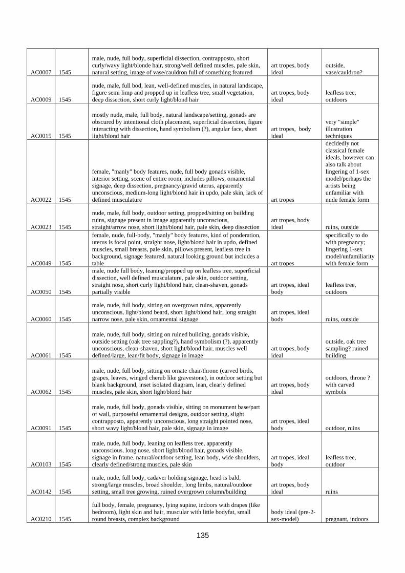
135
AC0007 1545
male, nude, full body, superficial dissection, contrapposto, short
curly/wavy light/blonde hair, strong/well defined muscles, pale skin,
natural setting, image of vase/cauldron full of something featured
art tropes, body
ideal
outside,
vase/cauldron?
AC0009 1545
nude, male, full bod, lean, well-defined muscles, in natural landscape,
figure semi limp and propped up in leafless tree, small vegetation,
deep dissection, short curly light/blond hair
art tropes, body
ideal
leafless tree,
outdoors
AC0015 1545
mostly nude, male, full body, natural landscape/setting, gonads are
obscured by intentional cloth placement, superficial dissection, figure
interacting with dissection, hand symbolism (?), angular face, short
light/blond hair
art tropes, body
ideal
very "simple"
illustration
techniques
AC0022 1545
female, "manly" body features, nude, full body gonads visible,
interior setting, scene of entire room, includes pillows, ornamental
signage, deep dissection, pregnancy/gravid uterus, apparently
unconscious, medium-long light/blond hair in updo, pale skin, lack of
defined musculature art tropes
decidedly not
classical female
ideals, however can
also talk about
lingering of 1-sex
model/perhaps the
artists being
unfamiliar with
nude female form
AC0023 1545
nude, male, full body, outdoor setting, propped/sitting on building
ruins, signage present in image apparently unconscious,
straight/arrow nose, short light/blond hair, pale skin, deep dissection
art tropes, body
ideal ruins, outside
AC0049 1545
female, nude, full-body, "manly" body features, kind of ponderation,
uterus is focal point, straight nose, light/blond hair in updo, defined
muscles, small breasts, pale skin, pillows present, leafless tree in
background, signage featured, natural looking ground but includes a
table art tropes
specifically to do
with pregnancy;
lingering 1-sex
model/unfamiliarity
with female form
AC0050 1545
male, nude full body, leaning/propped up on leafless tree, superficial
dissection, well defined musculature, pale skin, outdoor setting,
straight nose, short curly light/blond hair, clean-shaven, gonads
partially visible
art tropes, ideal
body
leafless tree,
outdoors
AC0060 1545
male, nude, full body, sitting on overgrown ruins, apparently
unconscious, light/blond beard, short light/blond hair, long straight
narrow nose, pale skin, ornamental signage
art tropes, ideal
body ruins, outside
AC0061 1545
male, nude, full body, sitting on ruined building, gonads visible,
outside setting (oak tree sappling?), hand symbolism (?), apparently
unconscious, clean-shaven, short light/blond hair, muscles well
defined/large, lean/fit body, signage in image
art tropes, body
ideal
outside, oak tree
sampling? ruined
building
AC0062 1545
male, nude, full body, sitting on ornate chair/throne (carved birds,
grapes, leaves, winged cherub like gravestone), in outdoor setting but
blank background, inset isolated diagram, lean, clearly defined
muscles, pale skin, short light/blond hair
art tropes, body
ideal
outdoors, throne ?
with carved
symbols
AC0091 1545
male, nude, full body, gonads visible, sitting on monument base/part
of wall, purposeful ornamental designs, outdoor setting, slight
contrapposto, apparently unconscious, long straight pointed nose,
short wavy light/blond hair, pale skin, signage in image
art tropes, ideal
body outdoor, ruins
AC0103 1545
male, nude, full body, leaning on leafless tree, apparently
unconscious, long nose, short light/blond hair, gonads visible,
signage in frame. natural/outdoor setting, lean body, wide shoulders,
clearly defined/strong muscles, pale skin
art tropes, ideal
body
leafless tree,
outdoor
AC0142 1545
male, nude, full body, cadaver holding signage, head is bald,
strong/large muscles, broad shoulder, long limbs, natural/outdoor
setting, small tree growing, ruined overgrown column/building
art tropes, body
ideal ruins
AC0210 1545
full body, female, pregnancy, lying supine, indoors with drapes (like
bedroom), light skin and hair, muscular with little bodyfat, small
round breasts, complex background
body ideal (pre-2-
sex-model) pregnant, indoors
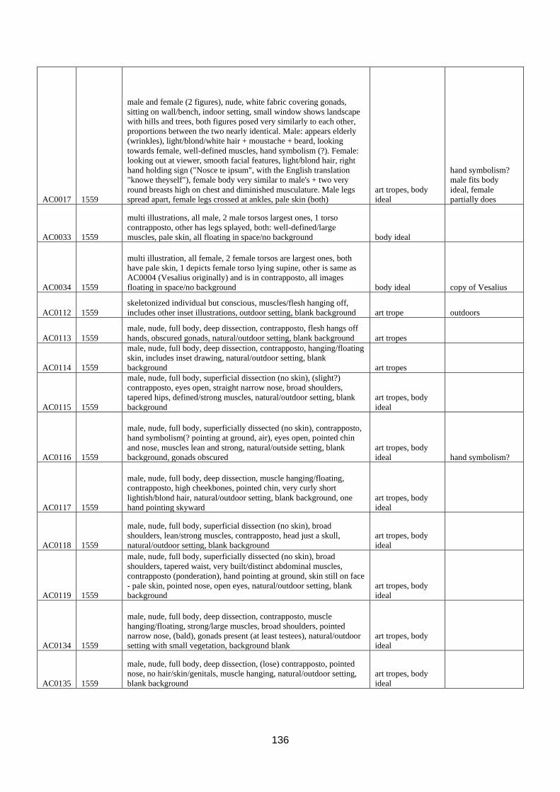
136
AC0017 1559
male and female (2 figures), nude, white fabric covering gonads,
sitting on wall/bench, indoor setting, small window shows landscape
with hills and trees, both figures posed very similarly to each other,
proportions between the two nearly identical. Male: appears elderly
(wrinkles), light/blond/white hair + moustache + beard, looking
towards female, well-defined muscles, hand symbolism (?). Female:
looking out at viewer, smooth facial features, light/blond hair, right
hand holding sign ("Nosce te ipsum", with the English translation
"knowe theyself"), female body very similar to male's + two very
round breasts high on chest and diminished musculature. Male legs
spread apart, female legs crossed at ankles, pale skin (both)
art tropes, body
ideal
hand symbolism?
male fits body
ideal, female
partially does
AC0033 1559
multi illustrations, all male, 2 male torsos largest ones, 1 torso
contrapposto, other has legs splayed, both: well-defined/large
muscles, pale skin, all floating in space/no background body ideal
AC0034 1559
multi illustration, all female, 2 female torsos are largest ones, both
have pale skin, 1 depicts female torso lying supine, other is same as
AC0004 (Vesalius originally) and is in contrapposto, all images
floating in space/no background body ideal copy of Vesalius
AC0112 1559
skeletonized individual but conscious, muscles/flesh hanging off,
includes other inset illustrations, outdoor setting, blank background art trope outdoors
AC0113 1559
male, nude, full body, deep dissection, contrapposto, flesh hangs off
hands, obscured gonads, natural/outdoor setting, blank background art tropes
AC0114 1559
male, nude, full body, deep dissection, contrapposto, hanging/floating
skin, includes inset drawing, natural/outdoor setting, blank
background art tropes
AC0115 1559
male, nude, full body, superficial dissection (no skin), (slight?)
contrapposto, eyes open, straight narrow nose, broad shoulders,
tapered hips, defined/strong muscles, natural/outdoor setting, blank
background
art tropes, body
ideal
AC0116 1559
male, nude, full body, superficially dissected (no skin), contrapposto,
hand symbolism(? pointing at ground, air), eyes open, pointed chin
and nose, muscles lean and strong, natural/outside setting, blank
background, gonads obscured
art tropes, body
ideal hand symbolism?
AC0117 1559
male, nude, full body, deep dissection, muscle hanging/floating,
contrapposto, high cheekbones, pointed chin, very curly short
lightish/blond hair, natural/outdoor setting, blank background, one
hand pointing skyward
art tropes, body
ideal
AC0118 1559
male, nude, full body, superficial dissection (no skin), broad
shoulders, lean/strong muscles, contrapposto, head just a skull,
natural/outdoor setting, blank background
art tropes, body
ideal
AC0119 1559
male, nude, full body, superficially dissected (no skin), broad
shoulders, tapered waist, very built/distinct abdominal muscles,
contrapposto (ponderation), hand pointing at ground, skin still on face
- pale skin, pointed nose, open eyes, natural/outdoor setting, blank
background
art tropes, body
ideal
AC0134 1559
male, nude, full body, deep dissection, contrapposto, muscle
hanging/floating, strong/large muscles, broad shoulders, pointed
narrow nose, (bald), gonads present (at least testees), natural/outdoor
setting with small vegetation, background blank
art tropes, body
ideal
AC0135 1559
male, nude, full body, deep dissection, (lose) contrapposto, pointed
nose, no hair/skin/genitals, muscle hanging, natural/outdoor setting,
blank background
art tropes, body
ideal

137
AC0136 1559
male, nude, full body, hanging from rope through face bone, gonads
not present, flesh/muscle hanging, natural/outdoor setting (hillside?),
stone/brick wall behind cadaver, isolated drawing on wall,
background left blank, same but mostly mirrored image of another
plate (AC0124) art tropes
AC0137 1559
male, full body, very deeply dissected (no gonads), leaning against
overgrown stone wall, natural/outdoor setting(? very uneven ground),
isolated/inset illustrations of diff body parts, pointed nose, eyes
closed and brows pulled together as if in pain, no hair/skin
art tropes, body
ideal overgrown ruins
AC0051 1560
multi illustrations, all male, 2 male torsos largest ones, 1 torso
contrapposto, other has legs splayed, both: well-defined/large
muscles, pale skin, all floating in space/no background, similar and
very particular pose to another plate
art tropes, body
ideal
AC0053 1560
multi illustrations, includes several isolated diagrams of the female
urogenital system, 2 female torsos in the middle of page, both have
pale skin, 1 depicts female torso lying supine, other is similar to multi
other plate (Vesalius originally) but with a face, contrapposto,
genitals and breasts visible, not very well defined musculature, open
eyes, light/blond hair partially in up-do. all images floating in
space/no background
art tropes, body
ideal
female not
explicitly
pregnancy
AC0120 1560
male, full body, superficially dissected (no skin), gonads visible,
strong/large muscles, holding in right hand his own flayed skin, in
left hand holds a knife (symbolism), nose and chin slightly rounded,
natural/outdoor setting, blank background art tropes
symbolism - cutting
off own skin,
holding the knife
AC0121 1560
male, entire body, contrapposto (ponderation), reaching skyward,
looking skyward, eyes open, straight pointed nose, prominent nose,
long limbs, well-muscled, natural/outdoor setting, background blank
art tropes, body
ideal
AC0122 1560
male, full body, superficial dissection, muscles strong/well-defined,
contrapposto, holding open flap of muscle (interacting with the
dissection), nose isn't particularly pointed, eyes closed,
natural/outdoor setting, blank background
art tropes, body
ideal
AC0123 1560
male, nude, full body, hanging from rope through face bone, gonads
not present, flesh/muscle hanging, natural/outdoor setting (hillside?),
stone/brick wall behind cadaver, isolated drawing on wall,
background left blank, same but mostly mirrored image of another
plate (AC0137) art tropes
copy/mirror of
earlier illustration
AC0124 1560
male, nude, deep dissection, contrapposto, muscle hanging off body
(obscures gonads), skeletonized head, natural/outdoor setting, blank
background art tropes
AC0125 1560
male, full body, superficial dissection, contrapposto, hand
symbolism(?), hanging/floating muscles, pointed chin, pointed nose,
long limbs, natural/outside setting, blank background
art tropes, body
ideal hand symbolism?
AC0128 1560
male, full body, deep dissection, (lose) contrapposto, muscles
hanging/floating, testis visible, long legs, broad shoulders, pointed
nose, open eyes, natural/outdoor setting, small shrub in corner, blank
background
art tropes, body
ideal
AC0129 1560
male, full body, deep dissection, contrapposto, foot resting on
log/uprooted tree, muscle hanging, face still has skin which is pale,
open eyes, pointed nose, gonads removed (can see base of where
penis was removed), natural/outdoor setting, small vegetation,
background blank
art tropes, body
ideal
AC0131 1560
male, full body, superficial dissection, strong/lean muscles,
contrapposto, no hair/skin, blank background
art tropes, body
ideal

138
AC0132 1560
male, full body, deep dissection (largely skeletonized), thorax
partially removed and put onto the bottom of a broom handle/pole,
which props up the cadaver, cadaver holding onto pole (interacting
with setting), muscle hanging, pointed nose, open but dark eyes, no
hair/skin, natural/outdoor setting, background blank (shaded but
blank) art tropes
very dynamic
composition
AC0004 1605
female, nude, full-body, superficial and deep dissection, urogenital
system + external and internal gonads visible, contrapposto, pale
skin, light/blond wavy hair (even tho no head), "smooth" looking
skin, no prominent muscles, isolated and floating in space, blank
background, image also used in AC0034
art tropes, body
ideal
copy/version of
earlier Vesalius
AC0011 1605
likely male (broad shoulders), partial body (torso), deep dissection,
no hair/skin left on head, hanging flayed skin, pointed chin, long
torso, large leg muscles, blank background body ideal
AC0029 1605
male, nude, partial body (torso), multi illustration (one of inset
isolated body part), testees visible but no penis, deep dissection, well-
defined/strong muscles, pale skin, blank background body ideal
AC0030 1605
male, nude, partial body (torso, no head), testees shown but no penis,
deep dissection, lean/slightly emaciated body but with well-
defined/strong muscles, pale skin, floating in space, blank
background body ideal
AC0035 1605
male, partial body (torso), deep dissection, gastrointestinal organs
spilling out of body, gonads not visible, sunken chest, well-
defined/strong muscles, pale skin, inset isolated illustration of body
part below torso (technically multi illustration) body ideal
AC0047 1605
male, partial body (torso, no head), testicles visible but no penis,
superficial dissection, muscles well-defined/strong and "lumpy",
unsteady lines, image floating in space, blank background body ideal
AC0048 1605
multi-illustration, likely male (shape of abdomen) partial body
(torso), gonads not visible, superficial dissection, blank background
(too heavily
dissected)
could maybe argue
for body ideal, but
eh
AC0057 1605
male, partial body (torso no head), deep dissection, very little body
fat, significantly (strongly) muscled legs, gonads removed, floating in
space, blank background body ideal
AC0066 1605
male/likely male (proportions), partial body (torso, no head), deep
dissection, no gonads visible, image floating in space, blank
background body ideal
AC0126 1605
(likely) male, partial body (torso), deep dissection, head skeletonized,
muscles hanging/floating, image floating in space, blank background
(too heavily
dissected)
could maybe argue
for body ideal, but
weak
AC0127 1605
(likely) male, partial body (torso), deep dissection, muscles
hanging/floating/specifically "flared out", face undissected, pale skin,
eyes looking upwards, nose narrow and pointed, mouth open and
grimacing, blank background
art trope(?), body
ideal
AC0068 1613
male, full body, sitting on white sheet/cloth, on top of a rock or tree
stump, small trees/other vegetation growing from rock/tree stump,
natural/outdoor setting, (loose) contrapposto (seated ponderation?),
hand covering genitals with sheet (interacting with setting),
superficial dissection, short light/blond hair, eyes are open looking at
the ground, long pointed nose, pale facial fair, lean and fit,
defined/strong muscles, pale skin, blank background
art tropes, body
ideal
AC0014 1626
female, multi illustration (in isolation form each other), urogenital
system, partial body (no head), deep dissection, breasts and gonads
visible, small round breasts, smooth looking pale skin, tapered waist,
blank background body ideal

139
AC0130 1626
male, nude, full body, superficial dissection, contrapposto
(ponderation), hand pointed at the sky (hand symbolism?), other hand
pointed at ground (hand symbolism?). no hair on head, eyes open and
looking at sky past his hand, extremely pointed nose, pointed chin,
plump lips, strong and lean looking body, natural/outdoor setting, no
skin on face? blank background
art tropes, body
ideal
AC0138 1626
male, nude, full body, superficial dissection, one hand pointed at
ground (hand symbolism?), contrapposto, face undissected, eyes
open, squat nose, very sharp cupid's bow on top lip, short wavy
light/white/blond hair on head, clean-shaven face, pale skin,
strong/very pronounced muscles, gonads visible, outdoor/natural
setting, blank background
art tropes, body
ideal
AC0194 1626
multi image, top centre of likely male, partial body (torso, no head),
deep dissection, pale skin, not extremely broad shoulders but waist
still tapers, gentle-ish musculature, isolated inset images of body
parts, all images floating in space, blank background body ideal
although only
"LM" still fits into
the theme with
what features it
does have, such as
pale skin, tapered
waist, gentle
muscles
AC0195 1626
multi illustration, all isolated, 2 illustrations of male partial bodies
(torsos), hand pointing towards ground (hand symbolism?),
contrapposto, gonads visible, very broad shoulders, thick neck, fit
physique/strong muscles, "bulgy" muscles, pale skin, blank
background
art tropes, body
ideal
AC0196 1626
male, nude, entire body, undissected (surface anatomy), contrapposto,
hand pointed towards ground (hand symbolism?), light/blond/grey
wavy hair slightly past chin in length, light/white facial hair, pale
skin, closed eyes, thin eyebrows, nose with high bridge and slopes up
at end, closed and small mouth. defined/strong/bulgy muscles, very
long limbs especially legs, gonads visible, natural/outdoor setting,
blank background
art tropes, body
ideal
AC0197 1626
male, entire body, undissected (surface anatomy), contrapposto, had
pointed at ground (hand symbolism?), eyes and mouth open, high
nose bridge, straight long pointed nose, short wavy light/blond hair,
clean-shaven face, physically fit/strong/defined muscles, broad
shoulders, pale skin, natural/outdoor setting, blank background
art tropes, body
ideal
AC0198 1626
female, full body, superficial dissection of breasts/thorax,
contrapposto, round face, full cheeks, small forehead, open eyes,
think eyebrows, small mouth, light/blond wavy long hair mostly in
updo with tendrils hanging down, pale skin, slightly rounded
stomach, small very round breasts, long limbs especially legs,
natural/outdoor setting, blank background
art tropes, body
ideal
AC0199 1626
skeleton standing as if alive, full body, skeleton holds huge scythe
(memento mori symbolism), many inaccuracies in the skeleton, blank
background art tropes
heavy memento
mori symbolism
AC0056 1627
female, nude, contrapposto, stark similarity to famous renaissance
painting, light/blond long hair falling about her shoulders, deep
dissection to show pregnant uterus, eyes open and looking at viewer,
straight narrow nose, small lips, pale skin, multiple inset illustration
of reproductive organs, natural/outdoor setting, blank background
art tropes, body
ideal
AC0096 1627
multi illustration, all isolated, 2 illustrations of male cadavers, full
body, both deeply dissected (thorax), one lying supine, other stands
beside him with his hands in the chest of the other hand (interacting
with setting/dissection), both faces look similar to each other, pointed
noses, pointed chins, closed eyes and lips, short curly light/blond
hair, gonads of supine figure visible, pale skin on both, background
left blank
art tropes, body
ideal
two figures
interacting, one
dissecting the other
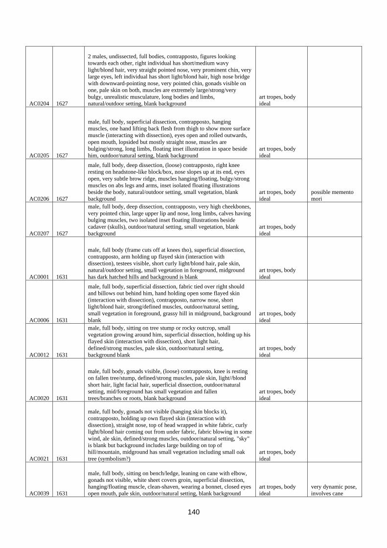
140
AC0204 1627
2 males, undissected, full bodies, contrapposto, figures looking
towards each other, right individual has short/medium wavy
light/blond hair, very straight pointed nose, very prominent chin, very
large eyes, left individual has short light/blond hair, high nose bridge
with downward-pointing nose, very pointed chin, gonads visible on
one, pale skin on both, muscles are extremely large/strong/very
bulgy, unrealistic musculature, long bodies and limbs,
natural/outdoor setting, blank background
art tropes, body
ideal
AC0205 1627
male, full body, superficial dissection, contrapposto, hanging
muscles, one hand lifting back flesh from thigh to show more surface
muscle (interacting with dissection), eyes open and rolled outwards,
open mouth, lopsided but mostly straight nose, muscles are
bulging/strong, long limbs, floating inset illustration in space beside
him, outdoor/natural setting, blank background
art tropes, body
ideal
AC0206 1627
male, full body, deep dissection, (loose) contrapposto, right knee
resting on headstone-like block/box, nose slopes up at its end, eyes
open, very subtle brow ridge, muscles hanging/floating, bulgy/strong
muscles on abs legs and arms, inset isolated floating illustrations
beside the body, natural/outdoor setting, small vegetation, blank
background
art tropes, body
ideal
possible memento
mori
AC0207 1627
male, full body, deep dissection, contrapposto, very high cheekbones,
very pointed chin, large upper lip and nose, long limbs, calves having
bulging muscles, two isolated inset floating illustrations beside
cadaver (skulls), outdoor/natural setting, small vegetation, blank
background
art tropes, body
ideal
AC0001 1631
male, full body (frame cuts off at knees tho), superficial dissection,
contrapposto, arm holding up flayed skin (interaction with
dissection), testees visible, short curly light/blond hair, pale skin,
natural/outdoor setting, small vegetation in foreground, midground
has dark hatched hills and background is blank
art tropes, body
ideal
AC0006 1631
male, full body, superficial dissection, fabric tied over right should
and billows out behind him, hand holding open some flayed skin
(interaction with dissection), contrapposto, narrow nose, short
light/blond hair, strong/defined muscles, outdoor/natural setting,
small vegetation in foreground, grassy hill in midground, background
blank
art tropes, body
ideal
AC0012 1631
male, full body, sitting on tree stump or rocky outcrop, small
vegetation growing around him, superficial dissection, holding up his
flayed skin (interaction with dissection), short light hair,
defined/strong muscles, pale skin, outdoor/natural setting,
background blank
art tropes, body
ideal
AC0020 1631
male, full body, gonads visible, (loose) contrapposto, knee is resting
on fallen tree/stump, defined/strong muscles, pale skin, light//blond
short hair, light facial hair, superficial dissection, outdoor/natural
setting, mid/foreground has small vegetation and fallen
trees/branches or roots, blank background
art tropes, body
ideal
AC0021 1631
male, full body, gonads not visible (hanging skin blocks it),
contrapposto, holding up own flayed skin (interaction with
dissection), straight nose, top of head wrapped in white fabric, curly
light/blond hair coming out from under fabric, fabric blowing in some
wind, ale skin, defined/strong muscles, outdoor/natural setting, "sky"
is blank but background includes large building on top of
hill/mountain, midground has small vegetation including small oak
tree (symbolism?)
art tropes, body
ideal
AC0039 1631
male, full body, sitting on bench/ledge, leaning on cane with elbow,
gonads not visible, white sheet covers groin, superficial dissection,
hanging/floating muscle, clean-shaven, wearing a bonnet, closed eyes
open mouth, pale skin, outdoor/natural setting, blank background
art tropes, body
ideal
very dynamic pose,
involves cane

141
AC0058 1631
male, full body, sitting on wall/ledge, hand resting on cane/pole that
is slightly taller than him while sitting, eyes open looking upwards,
short light/blond hair, superficial dissection, tapered waist,
strong/defined muscles, pale skin, outdoor/natural setting, small
vegetation growing from wall/ledge, blank background
art tropes, body
ideal
very dynamic pose,
involves cane
AC0067 1631
multi illustration, one is certainly male: partial body (bust), chest
covered in white fabric, head tilted upwards, eyes open mouth closed,
narrow nose, pale skin, short curled rightish/maybe brown hair on
head, facial hair (moustache curls at end), other cadaver is likely
male: partial body (bust), head tilted upwards, eyes open, mouth
close, clean-shaven, very pointed nose, short light/blond hair, pale
skin, both illustrations floating in space, blank background body ideal
AC0211 1631
full body, female, pregnancy, natural landscape, small mouth, straight
nose, pale skin, thin brows, round face with small chin, very small
breasts, light long partially up hair, "soft" (less muscley) limbs
body ideal,
western canon pregnant
AC0212 1631
full body, female, contrapposto, round face pointed sloped nose, pale
skin, curly light hair in updo, muscular, broad shoulders, very small
breasts, blank background
body ideal (pre-2-
sex-model)
AC0095 1651
multi illustration, top left illustration: bald, likely male, partial body,
deeply dissected, head tilted back, eyes ad mouth closed, narrow
pointed nose. top right: likely same individual as 1st?, bald, likely
male, narrow pointed nose, 2 other illustrations inset diagrams of the
ribcage/diaphragm, etc, all floating in space, blank background body ideal
AC0005 1685
male, sheets covering groin, supine, deep dissection, cadaver holding
open flayed skin (interaction with dissection), wearing a shirt?,
perspective of viewer looking down at cadaver, body resting on a
surface (shading), blank background art tropes
holding own skin
even when lying
supine. looks like
actual lab setting
(first example of
this in dataset?)
AC0013 1685
unknown sex/likely female, sitting, white cloth-covered surface (lab
setting?), held upright by cord around neck (almost hanging), head
obscured by white fabric wrapped around it, perhaps braided
light/blond hair falling out form underneath, arms limp at sides, one
hand resting on a block, back superficially dissected, blank
background body ideal practical lab setting
AC0025 1685
coloured illustration, female, partial body (torso), supine, perspective
of looking down at cadaver, red fabric partially covered external
gonads, dark pubic hair, abdomen dissected, abdomen slightly
distended (pregnancy?) , pale light beige/creamy coloured skin,
indoor setting?, body + fabric floating in space, blank background body ideal
AC0031 1685
coloured illustration, female, pregnant, lying on a surface, supine,
perspective looking down at cadaver, breasts visible, dark red fabric
covering genitals, thin slightly parted pink lips, curls of blond hair
either side of neck, skin is light peachy beige colour, light blue
coloured gathered sleeve on one arm, background left blank body ideal

142
AC0059 1685
likely male, supine, partial body (torso), lying on a fabric surface,
perspective of looking down at cadaver, white fabric covering top of
the head and the genitals, narrow pointed nose, closed mouth,
superficial dissection, wooden block seen on surface, skin/muscle
laid flat and held out from body on wooden block, pale skin, indoor
setting body ideal
very "realistic" lab
setting
AC0133 1685
female, partial body, superficial dissection, flayed skin hanging from
cadaver, little body fat except the bum, narrow ribcage and thin waist,
seated on a surface, white fabric covering genitals, smooth looking
skin, pale skin, curly light/blond hair, indoor setting?, blank
background body ideal
AC0143 1694
multi illustration - male, full body, superficially dissected (full body),
contrapposto, open eyes and mouth, very long pointed nose, no hair
or skin, strong/defined muscles, wide shoulders, tapered waist,
standing on natural/uneven ground (outdoor setting). sitting on
ground is lateral view of a male human face, superficially dissected,
eyes open, thin closed lips, straight and pointed nose with high nose
bridge. Inset isolated illustration in top corner of eyeball and oculus
muscles, blank background
art tropes, body
ideal
outdoor settings
into the 1700s
AC0156 1694
male, full body, standing, contrapposto, hand pointing at sky, muscle
hanging, face has no skin but pointed nose still visible, mouth and
eyes open, no hair, gonads gone, natural/outdoor setting, blank
background
art tropes, body
ideal hand symbolism?
AC0157 1694 ((NOT USEFUL FOR THIS PROJECT)) n/a
AC0158 1694 N/A (decorative number/letter) n/a
AC0159 1694
two figures, both male, entire body, contrapposto, individual 1: parted
lips, open eyes, long narrow straight nose, high nose bridge.
individual 2: arm resting on cane/sword, prominent brow ridge, very
long pointed nose, high cheekbones, sharp chin, gonads visible. Both
have long limbs, strong/defined muscles, natural/outdoor landscape,
sloping hill in background, hundreds of people walking up the hill to
a ruined? building and gallows (memento mori), sky shaded + some
white clouds
art topes, body
ideal
two figures
seeming to interact
with each other,
memento mori in
form of gallows in
background,
outdoor setting
AC0170 1694
two figures, both male, entire body, one individual lying supine with
a deeply dissected abdomen and apparently in distress, other
individual kneeling over top of first individual and is only
superficially dissected, looking down worriedly at the first individual.
Both have: open eyes, slightly open mouths, pointed nose, sharp
jawline, high cheekbones, prominent brow ridge, gonads visible in
less dissected one, both have long times, large/strong muscles,
natural/outdoor setting, why shaded as if cloudy
art tropes, body
ideal
two figures
interacting, both
dissected but more
dissected one
apparently in
pain/distress, other
one looks very
worried, very
dynamic poses
AC0171 1694
male, entire body, lying supine, hands flexed, limbs in the air, head
tilted back, appears to be yelling, very pointed nose, long limbs, bald,
landscape orientation, uneven borders, cloudy background
art tropes, body
ideal
very dynamic pose,
appears in distress,
outdoor setting
AC0172 1694
male, entire body, contrapposto, hands appear bound, sharp chin,
high cheekbones, bald, strong/lumpy muscles, limbs and body long,
natural/outdoor setting, cliff in background
art tropes, body
ideal
AC0173 1694
male, full body, contrapposto, holding out own flayed skin,
large/strong muscles, broad shoulders, bald, long limbs, black
background
art tropes, body
ideal
AC0174 1694
male, lying on left side, body slumped over left arm, right arm
grazing the ground, rocky ground that turns into white sheets under
cadaver, bulgy/lumpy/strong muscles, gonads visible, bald head,
prominent brow ridge, narrow pointed nose, high cheekbones,
natural/outdoor setting, small vegetation, crutch?, blank background
art tropes, body
ideal
crutch in
background

143
AC0175 1694
two figures, both male, contrapposto, long narrow pointed nose, thin
lips, long limbs, strong/defined muscles, gonads visible,
natural/outdoor setting, second figure standing in semi-background
skin hanging off upper arm, pale skin sky blank
art tropes, body
ideal
AC0176 1694
three figures, all male, contrapposto, one figure stands in the centre
and has a pointed chin, pointed nose, prominent pointed brow ridge.
Two other figures sitting on edge of a cliff, also have pointed nose,
high cheekbones, appear to be interacting with each other, all have
very strong/defined muscles, broad shoulders, natural/outdoor setting,
background/sky shaded as if cloudy
art tropes, body
ideal
AC0177 1694
two figures, male, full body, standing in exact same position,
contrapposto, hand (of each) pointed upwards/in front of them,
pointed noses, flesh hanging, gonads visible on one cadaver,
bulgy/strong muscles, natural/outdoor setting, blank background
art tropes, body
ideal
AC0147 1713
male, human body, superficially dissected, unsure if lying supine or
suspended upright, many ropes connected to his body run out of
frame, limbs splayed, eyebolt in top of head, closed very small
mouth, sharply pointed nose, eyes are small and close, no hair,
genitals removed, well muscled/strong, figure floats in space, blank
background body ideal
AC0178 1723
male, supine, head on wooden block, lying on hard surface,
perspective of looking down at cadaver, legs partially covered by
white fabric, gonads obscured by flayed skin, superficial dissection,
bladder distended (on purpose), pale skin, very broad shoulders, very
tapered waist, very large/bulgy/lumpy muscles, prominent Adams
apple, sharp jawline, eyes and mouth closed, nose cut off by frame,
short wavy light/blond hair and sideburns, background is blank (but
shaded), image drawn in landscape body ideal
AC0179 1723
male, partial body, lying on left side, image in landscape orientation,
white sheet over top of body, one leg is propped up and bent at the
knee with the foot behind the body, pale skin, very strong/muscled
legs, background blank but tonally shaded body ideal dynamic pose
AC0180 1723
male. partial body, lying on right side, sheet covering genitals, some
pubic hair showing, very strong/lumpy ab muscles, essentially no
bodyfat on body (shown in cross-section), pale skin, background is
shaded but blank of detail body ideal
AC0181 1723
female, living patient, sits at base of wide cylindrical column, holding
up her tunic/gown to see undissected stomach with an intestinal
prolapse, gonads and legs obscured by more fabric, she wears more
fabric around her hair/head, tunic/gown is high collared and long
sleeve, face appears wrinkled (elderly), narrow pointed nose, chin
small and pointed, eyebrows drawn together, pale skin, curtain drapes
across top left corner, background shaded but blank art tropes
living, elderly
female patient -
female not to do
with pregnancy,
featured because
she has an intestinal
prolapse
AC0182 1723
male, living patient, full body, lying supine, on a table with legs
hanging off, mid-surgery (hernia), shirt pulled up and otherwise
naked, gonads showing, wide shoulders, large/bulgy muscles, eyes
open and looking upwards, small closed mouth, very narrow pointed
nose, very angular jaw, high cheekbones, should length wavy
light/blond hair, pale skin, background uniformly shaded behind
patient and table body ideal
living patient, in
surgical setting
AC0166 1725
male, full body, contrapposto, one hand points towards ground
(where a label is), eyes open and looking upwards, slightly open
mouth, thin lips, very pointed nose, long limbs, tapered waist, broad
shoulders, natural/outdoor setting, background shows waterfront
town with lots of arches and obliques (in ruins?), blank "sky"
art tropes, body
ideal ruins

144
AC0167 1725
male, full body, contrapposto, gonads visible, eyes open, lips parted,
high cheekbones, long pointed narrow nose, strong/defined muscles,
long limbs, broad shoulders, squat tombstone on ground (with
image's label), natural/outdoor setting, rolling hills and grove of trees,
rocky cliff, behind is some buildings and a large tower, boat next to
tower, blank "sky"
art tropes, body
ideal city in background
AC0139 1744
male, full body, abdominal cavity emptied, contrapposto, hand
pointed towards the sky, bald head, closed small mouth, long narrow
nose, eyes open looking past viewer, pale skin, very large head,
stands on small square board, border is a scale, blank background
art tropes, body
ideal scale as a border
AC0140 1744
male, entire body, contrapposto, gonads visible, defined/strong
muscles, lean body, pale skin, thin closed lips, narrow pointed nose,
no hair, standing on a small platform, blank background, border is
also a scale
art tropes, body
ideal
AC0141 1744
male, full body, gonads visible, contrapposto, stands on small
platform, pointed up/at the corner, shoulders look narrow bc dissected
heavily, pale skin, mouth closed, pale thin lips, narrow pointed nose,
eyes open pointing in 2 different directions, blank background, border
is scale
art tropes, body
ideal
AC0144 1744
male, full body, blood vessels drawn in detail all over body, standing
on flat platform, anus visible, lean, well muscled limbs, blank
background, border is a scale body ideal
AC0145 1744
male, full body, stands on flat platform, contrapposto, hand pointed
up, bald, shoulders have been heavily dissected, blank background
border is a scale art tropes
AC0146 1744
male, full body, loose contrapposto, stands on flat platform, two
fingers pointed (kind of down), lips parted, straight pointed nose,
open eyes, blank background, border is a scale
art tropes (body
ideals)
AC0148 1744
male, full body, stands on flat platform, contrapposto, back of hand
brought to forehead, bald, no gonads visible, strong muscles, lean
form, broad shoulders, tapered waist, blank background, border is
scale
art tropes, body
ideal
AC0149 1744
male, full body, partially skeletonized, stands on flat platform,
contrapposto, blank background, border is a scale art tropes
AC0150 1744
male, full body, partially skeletonized, stands on flat platform,
contrapposto, eyes open and looking up, mouth closed, (nose
basically gone), blank background, border is a scale art tropes
AC0151 1744
male, full body, stands on flat platform, contrapposto (ponderation),
back of hand to forehead, eyes open, small closed mouth, straight
narrow pointed nose, penis partially visible, lean, defined/strong
muscles, blank background, border is also a scale
art tropes, body
ideal
AC0163 1746
coloured, female, partial body, flap of skin/muscle floating out to the
side, muscle is deep red, skin very light cream colour, cheeks flushed
pink, nose is straight and pointed, eyes open, hair is brown and in an
updo held with a blue ribbon, background is dark mossy green body ideal
AC0164 1746
coloured, male, partial body, hanging peeled back skin is very light
pinkish cream colour, little bodyfat, defined/strong muscles,
background dark muddy brown body ideal
AC0168 1747
male, entire body, contrapposto, hand pointing downwards, closed
eyes, straight narrow nose, small mouth. high cheekbones, sharp
gonial angle, broad shoulders, tapered waist, long slender neck arms
and legs, defined/strong muscles, gonads visible, blank background
(but in final its detailed)
art tropes, body
ideal

145
AC0169 1747
male, full body, contrapposto (ponderation), protruding forehead
(compared to chin), pointed nose, gonads visible, defined/strong
muscles, long limbs and torso, blank background
art tropes, body
ideal
AC0016 1773
coloured, female, partial body, dissected breasts and gonads visible,
light cream skin, flushed pink cheeks, slight smile, straight nose, dark
curly brunette shoulder-length hair with a blue ribbon/headband, dark
mossy green background with a beam in the background
body ideal,
setting/background
AC0032 1773
coloured, partial body, skinless, skeletonized head, inset illustration
of a blue/green coloured human arm, background mottled blue/green body ideal
AC0160 1773
coloured, two figures, both female, one living and one deeply
dissected. Living one is partially in frame, hand fondling her breast,
very light beige/cream skin, flushed pink cheeks, straight narrow
nose, gracile brow ridge, dark brown hair pulled back from face in a
bun. Dissected figure kneels on her knees, on top of deep red fabric,
face held on to head with some twine, eyes open gazing up, light
cream skin and flushed cheeks, pink lips, very pointed narrow nose,
distended vulva facing more towards the front of body than real life.
Both have small very round breasts. Background of image is dark
green body ideals dynamic posing
AC0161 1773
coloured, multi-illustration, one female sitting on ground with her
knees up and legs spread - light cream, pink/rosy cheeks, pink knees,
pink genitals, plump face, straight pointed nose, brown hair pulled
back from face, very round breasts that sit far apart on her chest, a
figure stands between first lady's legs, contrapposto, three other
isolated illustrations on the ground in front of the figures,
reproductive/urogenital systems main feature, background is dark
green
art tropes, body
ideal
AC0162 1773
coloured, female, partial body, pregnant belly deeply dissected, fetus
is visible, breast visible but higher than anatomically expected, skin is
light cream coloured, pink cheeks, small reddish lips, narrow and
pointed nose, large eyes, high forehead, light brown hair up in curls
on back of her head, mossy dark green background body ideal female, pregnancy
AC0165 1773
coloured, multi-illustration - centre dissected legs cut off by frame
right below groin, sex unknown, pale cream coloured hand visible,
contrapposto, on either side are isolated illustrations of male and
female urogenital systems, background dark green colour
art tropes (body
ideals)
AC0215 1773
partial body (can't see head), female, pregnant, fetus pictured, light
cream beige, pink elbows and knees, slender limbs (not very
muscular) body ideal
"connects" with
AC0160
AC0152 1777
male, full body, contrapposto, lean, defined/strong muscles, tapered
waist, broad shoulders, long limbs and torso, blank background
art topes, body
ideal
AC0072 1787
likely male, partial body, hanging skin, eyes and mouth closed,
straight pointed nose, blank background body ideal
LM but VERY
likely male
AC0101 1794
multi-illustration, male, isolated illustration of diaphragm,
skeletonized torso hanging slightly off the floor by a rope, male
cadaver lying supine, knees bent, legs spread apart, wrists tied to
ankles, gonads visible, lying on white sheet on the floor, lean, pale
skin, shaded but blank background body ideal
very dynamic
composition
AC0064 1799
male, partial body, lying supine, sternum and some ribs cut out and
held out of the way by rope, slightly open mouth, eyes closed,
straight narrow nose, light/blonde short hair, pale skin, clean-shaven,
perspective of looking down at cadaver, two inset isolated
illustrations below the main figure body ideal lab setting

146
AC0065 1799
likely male, partial body (neck and part of head), straight pointed
nose, curly lightish/maybe brown hair on back of head, pale skin,
background blank body ideal
AC0104 1799
male, partial body, eyes closed, mouth open, nose straight and
narrow, pale skin, light/blonde short curly hair, clean-shaven,
background blank body ideal
AC0026 1800
male, partial body, lying supine, on table/work bench, hand reaching
from out of frame to hold up skin/tissue, head lolled back, eyes
closed, mouth open, straight narrow nose, short curly
light/blond/white hair, no facial hair, lean body, inset image floating
in corner, blank background body ideal
someone from out
of frame interacting
with
cadaver/dissection,
clearly lab setting
AC0036 1806
male, lying supine, white-cloth covered surface, perspective looking
down at cadaver, lean body, defined/strong muscles, "fuzzy" short
pale/blond hair, pale skin, blank background body ideal lab setting
AC0037 1813
male, lying supine, eyes and mouth closed, narrow nose, floating in
space, blank background
(too heavily
dissected)
AC0038 1814
male, partial body, arms tied above head, body bent backwards to
chest is lifted towards ceiling, head tilted back, stubble/slight facial
hair, mouth and eyes wide open, gonads visible, pale skin, narrow
prominent nose, defined/strong muscles, lean figure, background
blank body ideal
AC0002 1825 NOT USEFUL FOR THIS ANALYSIS n/a
AC0027 1825 likely male, partial body, isolated, blank background
(too heavily
dissected)
AC0063 1825
male, eyes and mouth closed, high nose bridge, pointed nose, thin
lips, pale skin, short curly lightish/maybe brown hair, muttonchop, no
other facial hair, blank background, floating in space body ideal
facial hair "mutton
chops" of the time
AC0069 1825
male, partial body, eyes mouth closed, very straight pointed nose,
pointed chin, inset image in top corner, "floating" in space, blank
background body ideal
AC0070 1825
male, partial body, little bodyfat, lean/strong, pale skin, long torso,
isolated, blank background body ideal
AC0071 1825
male, partial body, little bodyfat, lean/strong, pale skin, long torso,
isolated, blank background body ideal
AC0073 1825
male, eyes and mouth closed, straight pointed nose, pale skin, clean
shaven face, blank background body ideal
AC0074 1825
male, partial body (no head), long torso, minimal bodyfat, partially
covered by white cloth, isolated, gonads partially seen, blank
background body ideal
AC0099 1825
male, partial body, little bodyfat, lean/strong, pale skin, long torso,
very lean/fit body, isolated, blank background body ideal
AC0107 1825
male, partial body (no head), defined abdominal muscles, gonads
partially visible, isolated, blank background body ideal
AC0153 1825
two figures, both male, partial body, appear to be same individual at
different age, old man has white facial hair while young man has
mutton chops, old man has short white hair while young man has
short wavy lightish/brown hair, thin lips, long narrow high-bridged
pointed nose, sharp brow ridge, both dissected similarly with the
younger having more muscle mass, isolated images, blank
background
art tropes?, body
ideal
same face used on 2
images to show
differences between
20-30yo and elderly
man - obviously
couldn't actually be
the same person
and yet was drawn
to be
AC0154 1825
two figures, both male, partial body, appear to be same individual at
different age, both have no facial hair, younger man darker hair than
the older one, very long pointed nose, very high nose bridge, shallow
brow ridge, younger man has more muscle mass but otherwise
dissections are the same, isolated images, blank background
art tropes?, body
ideal
same face used on 2
images to show
differences between
20-30yo and elderly
man - obviously
couldn't actually be
the same person

147
and yet was drawn
to be
AC0155 1825
male, full body, contrapposto, one hand resting on hip-high pillar,
eyes open looking at ground, thin small closed lips, slightly receded
lower lip, long pointed nose with high bridge, no hair, gonads
partially visible, thick/strong/large muscles, blank background
art tropes, body
ideal
AC0214 1825
partial body, female, pregnant, pale skin, not very muscular, blank
background body ideal
larger and more
weighty? Breasts
than most earlier
images - most
illustrations feature
breasts which kind
of defy gravity
AC0028 1829
coloured, multi image of same body, partial body (no head), likely
female, hernia in diaphragm, light cream-coloured skin, "smooth"
skin with minimal muscle, one breast partially visible, isolated
images, blank background
art tropes, body
ideal
likely female not to
do with pregnancy
but individual has
very large hernia
AC0102 1829
multi figures, 2 feature only deeply dissected chest with aneurysms, 1
(top) figure male partial body (no head), pale skin, lean/fit, isolated
images, blank background body ideal
AC0010 1831
coloured, male, partial body, light pinkish skin, broad shoulders,
defined/strong muscles, gonads visible, very little bodyfat, tapered
waist, thin lips, narrow nose, isolated image, blank background body ideal
AC0040 1831
coloured, male, partial body, defined/strong muscles, light cream
coloured skin, smooth facial skin, ginger/brown sideburns, thin pink
lips, narrow nose, isolated image, blank background body ideal sideburns
AC0041 1831
coloured, male, partial body (no head), gonads removed,
strong/defined muscles, light cream coloured skin, isolated image,
blank background body ideal
AC0042 1831
coloured, likely male, partial body skeletonized, outline of fleshed
body around the skeleton shows broad shoulders (due to muscles),
tapered waist (not much fat between surface and skeleton), isolated
image, blank background body ideal
even skeletonized
individuals are used
to show idealized
male body
proportions - this
outline suggests
male while you
cant tell from
skeleton
AC0043 1831
coloured, male, partial body, gonads visible, light cream coloured
skin, minimal bodyfat, long torso, isolated image, blank background body ideal
AC0044 1831
coloured, male, partial body, defined/strong muscles, light cream
coloured skin, gonads visible, isolated image, blank background body ideal
AC0046 1831
partially coloured, sex unknown, deeply dissected, isolated image,
blank background
(too heavily
dissected)
beautiful
colouring/drawing
job
AC0076 1831
coloured, male, partial body (no head), minimal bodyfat,
strong/defined muscles, light beige coloured skin, isolated image,
blank background body ideal
AC0077 1831
coloured, partial body, male, gonads visible, lean/fit, broad shoulders,
light pinkish cream coloured skin, brown hair/mutton shops, isolated
image, blank background body ideal Mutton chops

148
AC0078 1831
coloured, male, partial body (half of face), gonads visible, minimal
bodyfat, broad chest/shoulders, large/strong muscles, light cream
coloured skin, angular chin, thin pink lips, narrow pointed nose,
brown mutton chops, isolated image, blank background body ideal
AC0079 1831
coloured, male, partial body (part of the face), angular jawline, thin
lips, lean, broad shouldered, muscular, light cream coloured skin,
brown pubic hair, gonads partially visible, isolated image, blank
background body ideal
AC0080 1831
coloured, very deeply dissected, sex unknown, light cream coloured
skin seen on ears, isolated image, blank background
(too heavily
dissected)
strong looking
shoulder muscles
and can see they
had light skin, but
nothing else
determinable
AC0081 1831
coloured, male, partial body (part of face), partial body (partial face),
gonads visible, lean, fit, broad shoulders, muscular, minimal bodyfat,
light cream coloured skin, brown pubic hair, isolated image, blank
background body ideal
AC0082 1831
coloured, male, partial body (part of face), thin lips, angular jaw
gonads visible, lean/fit, minimal bodyfat, broad shoulders, light
beige/cream coloured skin, isolated image, blank background body ideal
AC0083 1831
coloured, male, partial body (part of head), lean/fit, broad shoulders,
muscular, minimal bodyfat, light cream coloured skin, dark brown
hair and possible mutton chops, isolated image, blank background body ideal
AC0084 1831
coloured, male, partial body (part of face), light cream coloured skin,
broad shoulders, muscular, minimal bodyfat, brown mutton chops,
narrow nose, thin pink lips, isolated image, blank background body ideal Mutton chops
AC0085 1831
coloured, male, partial body (part of head), light cream coloured skin,
broad shoulders, muscular, minimal bodyfat, brown facial hair can be
seen from behind, short brown hair, isolated image, blank
background body ideal
AC0086 1831
coloured, male, partial body (only chin), muscular back/shoulders and
butt, lean, minimal bodyfat, light beige coloured skin, dark brown
pubic hair, isolated image, blank background body ideal
AC0087 1831
coloured, male, partial body (part of head), brown hair, brown facial
hair can be seen from behind, lean, muscular, minimal bodyfat, light
beige coloured skin, dark brown pubic hair, isolated image, blank
background body ideal
AC0088 1831
coloured, male, partial body, large/strong/defined muscles, broad
chest/shoulders, pointed chin, thin lips, narrow pointed nose, isolated
image, blank background body ideal
AC0092 1831
coloured, multi-illustration, female, partial body, three different view
of same individual, two are views of shoulder and one is of figure
lying supine being operated on, each illustration has white
gown/sheet covering midsection, left breast exposed in supine
illustration and lying on white sheet, light cream coloured skin, curly
brown hair partially visible, hands operating on woman also light
cream coloured, all images isolated with no background body ideal
lab setting, person
out of frame
interacting with
dissection/operation
AC0097 1831
coloured, male, partial body, lean, muscular, minimal bodyfat, light
cream coloured skin, short brown hair, pink lips, clean shaven face
(partially visible face), isolated image, blank background body ideal
AC0105 1831
coloured, living individual, male, demonstrating use of anatomical
instruments, lean/fit, light cream coloured skin, long straight pointed
nose, high brow ridge, wavy short dark brown hair, mutton chops, 6
isolated inset illustrations, isolated images, blank background body ideal
mutton chops,
demonstrating
anatomical
instruments - looks
a lot like the
dissected bodies in
the body ideal way

149
AC0108 1831
coloured, male, partial body, strong/defined muscles, light cream
pinkish coloured skin, isolated image, blank background body ideal
AC0109 1831
coloured, male partial body, light beige coloured skin, lean/fit,
minimal bodyfat, pointed nose, pointed chin, small [ink lips, short
dark brown hair, long torso, isolated image, blank background body ideal
AC0110 1831
coloured, male, partial body, very lean/fit, large/strong muscles, very
broad shoulders, tapered waist, light beige coloured skin, angular jaw,
straight narrow nose, small pink lips, large dark brown mutton chops,
isolated image, blank background body ideal mutton chops
AC0111 1831
coloured, male, partial body, light beige coloured skin, lean/fit, strong
back muscles, minimal bodyfat, isolated image, blank background body ideal
AC0213 1831
coloured, reproductive system, light cream coloured skin, pale pubic
hair, blank background body ideal
AC0075 1834
unknown sex, partial body (thorax), little bodyfat, pale skin, isolated
image, blank background body ideal
unknown sex but
little bodyfat and
pale skin
AC0055 1840
partially coloured, male, partial body, undissected, pale skin, pink
groin and nipples, gonads visible, lean/fit, isolated image, blank
background body ideal
AC0045 1844
male, partial body, gonads shown, arms held up by ribbons from out
of frame, skin of dissected abdomen attached to arms and held out of
the way, pale skin, isolated image, blank background body ideal
intestines falling
out from under
omentum - could
argue this shows
increased realism
AC0098 1844
partially coloured, male, partial body (incl head), bald head, very
narrow pointed nose, closed mouth, high cheekbones, pale skin, thin
(perhaps elderly), little body fat, isolated image, blank background body ideal
strikes me as what
many actual
cadavers of the
time would have
actually looked like
body wise (thin,
maybe elderly,
maybe emaciated) -
still narrow pointed
nose, high
cheekbones, pale
skin, male
AC0100 1844
male, partial body (incl head), appears emaciated, eyes partly open,
sunken eye sockets, narrow pointed nose, wavy light/blond hair,
receding hairline (elderly-ish?), pale skin, isolated image, blank
background body ideal
definitely
emaciated/been
dead for at least a
bit of time already -
still narrow pointed
nose, blond hair,
pale skin, male
AC0200 1844
male, partial body, gonads visible, uncircumcised penis,
darkish/brown kind of long pubic hair, tapered waist, arms bent at
elbows with wrists tied together and being held up by a rope out of
frame, minimal bodyfat, pale skin, isolated image, blank background body ideal
AC0201 1844
male, partial body (incl head), pale skin, tapered waist, lean/fit, short
wavy light/blond hair, sideburns/mutton chops, high cheekbones,
pointed chin, very straight narrow pointed nose, eyes and mouth
closed, slight stubble, isolated image, blank background body ideal
AC0202 1844
male, partial body (incl head), slumped shoulders, pale skin, short
wavy light/blond hair, eyes and mouth closed, long straight pointed
nose, lean/fit, isolated image, blank background body ideal

150
AC0203 1844
multi illustration, each of head and neck, male, different but very
similar looking individuals, three inset illustrations (all have high
bows, high nose bridges, pointed straight narrow nose, pale skin, fair
hair) above one larger more detailed image, sunken cheeks,
somewhat elderly, ring of sarge short wavy hair on head, closed eyes,
very long pointed nose, high nose bridge, mouth slightly open, some
stubble on chin, pale skin, isolated images, blank background body ideal
different but very
similar looking
people, all with
essentially same
features within
body ideal theme
AC0003 1856
male, partial body, undissected, gonads visible, uncircumcised penis,
chin-length darkish/brown hair and long mutton chops, open eyes,
very little body hair, straight narrow nose, thin lips, tapered waist,
broad chest and shoulders, strong/large muscles, minimal bodyfat,
contrapposto, isolated image, blank background
body ideal, art
tropes mutton chops
AC0018 1856
male, partial body, sitting upright, gonads visible, head lolling, eyes
and mouth closed, short darkish/brown hair and mutton chops, high
nose bridge, narrow pointed nose, small closed mouth, closed eyes,
broad shoulders, strong/defined muscles, pale skin, isolated image,
blank background body ideal
AC0019 1856
male, partial body, lying supine, darkish/brown hair, possible small
moustache and stubble, closed eyes, thin lips, narrow nose, pale skin,
isolated image, blank background (with some overlaid writing) body ideal lab setting ?
AC0052 1856
male, partial body, standing, wrists tied above head and hanging by
rope out of frame, head lolls to side, lightish/dark blond or brown hair
and mutton chops, eyes and mouth closed, narrow pointed nose, high
nose bridge, gonads visible, defined/strong muscles, pale skin,
isolated image, blank background body ideal
mutton chops, lab
setting ?
AC0089 1856
male, partial body, gonads visible, pale skin, eyes open and looking
out of frame towards what it looks like he is reaching for with left
hand, some stubble on face, straight pointed nose, high nose bridge,
wavy darkish/brown hair, lean/fit, long torso, isolated image, blank
background body ideal
is the comparison
plate for AC0090 -
the white
counterpart to show
comparative
anatomy between
"white man" and
"black man"
AC0090 1856
male, partial body, head turned towards left side/towards what he
seems to be reaching for with left arm, very dark coloured skin,
significant facial prognathism, very large lips, short flat nose,
slopping forehead, receded jaw with small chin, uneven jawline, very
short black hair drawn in small circles, isolated image, blank
background --
only non-white
individual found in
my study - is plate
XXIV to AC0089's
plate XXIII,
appears to be
feature for the
purpose of
"comparative
anatomy" between
the "white man"
and the "black
man"
AC0094 1856
male, partial body, lean/fit, strong/defined muscles, broad shoulders,
minimal bodyfat, closed eyes, slightly open mouth, long straight
narrow pointed nose, straight lightish/brown hair, isolated image,
blank background body ideal
AC0106 1856
male, partial body, arms tied together in front of body, head lolled
back, gaunt looking face, eyes closed and sunken, mouth hangs open
slightly, very pointed narrow nose, short light/blond hair, very thin
(perhaps emaciated), pale skin, isolated image, blank background body ideal perhaps emaciated

151
AC0054 1867
male, partial body (pelvic region), lower body propped up on plank of
wood, figure lying supine, minimal bodyfat, gonads partially visible,
pale skin, isolated image, blank background body ideal lab setting
AC0093 1867
male, partial body, sitting but leaning back on block, high
cheekbones, pointed narrow nose, pointed chin, stubble, short wavy
light/blond hair, lean, minimal bodyfat, pale skin, isolated image,
blank background body ideal
AC0208 1867
partially coloured, partial body, male, lying supine, medium-length
very light/blond hair, angular chin, high cheekbones, pointed narrow
nose, stubble, pale skin, isolated image, blank background body ideal
AC0209 1867
partially coloured, partial body, male, prominent chin, high
cheekbones, very angled pointed nose, high nose bridge, short/wispy
lightish/blond hair with receding hairline, possible sideburns, pale
skin, isolated image, blank background body ideal

152
APPENDIX C
Example Images from Data Sample
Image 1C: “Undecima musculorum tabula” (“11th Table of
the Muscles”) by Calcar. From: Vesalius, Andreas, 1514-
1564. De humani corporis fabrica libri septem. Basileæ
[Basel]: Ex officina I. Oporini, 1543.
Note: From Anatomia Collection: anatomical plates 1522-
1967. (2017). Retrieved April 10, 2020, from
https://anatomia.library.utoronto.ca/
Image 2C: “Duodecima musculorum tabula” (“12th Table
of the Muscles”), by Calcar. From: Vesalius, Andreas,
1514-1564. De humani corporis fabrica libri septem.
Basileæ [Basel]: Ex officina I. Oporini,1543.
Note: From Anatomia Collection: anatomical plates 1522-
1967. (2017). Retrieved April 10, 2020, from
https://anatomia.library.utoronto.ca/
Image 3C: Untitled, by La Riviere. From: Estienne,
Charles, 1504-ca. 1564. De dissectione partium corporis
humani libri tres, a Carolo Stephano, doctore medico,
editi. Vna cum figuris, & incisionum declarationibus, a
Stephano Riuerio chirurgo co[m]positis. Parisiis: Apud S.
Colinæum, 1545.
Note: From Anatomia Collection: anatomical plates 1522-
1967. (2017). Retrieved April 10, 2020, from
https://anatomia.library.utoronto.ca/
Image 4C: Untitled, by La Riviere. From: Estienne,
Charles, 1504-ca. 1564. De dissectione partium corporis
humani libri tres, a Carolo Stephano, doctore medico,
editi. Vna cum figuris, & incisionum declarationibus, a
Stephano Riuerio chirurgo co[m]positis. Parisiis: Apud S.
Colinæum, 1545.
Note: From Anatomia Collection: anatomical plates 1522-
1967. (2017). Retrieved April 10, 2020, from
https://anatomia.library.utoronto.ca/

153
Image 5C: “Secunda musculorum tabula” (“2nd Table of
the Muscles) By Calcar. From: Vesalius, Andreas, 1514-
1564. De humani corporis fabrica libri septem. Basileæ
[Basel]: Ex officina I. Oporini,1543.
Note: From Anatomia Collection: anatomical plates 1522-
1967. (2017). Retrieved April 10, 2020, from
https://anatomia.library.utoronto.ca/
Image 6C: Untitled, by La Riviere. From: Estienne,
Charles, 1504-ca. 1564. De dissectione partium corporis
humani libri tres, a Carolo Stephano, doctore medico,
editi. Vna cum figuris, & incisionum declarationibus, a
Stephano Riuerio chirurgo co[m]positis. Parisiis: Apud S.
Colinæum, 1545.
Note: From Anatomia Collection: anatomical plates 1522-
1867. (2017). Retrieved April 10, 2020, from
https://anatomia.library.utoronto.ca/
Image 7C: “Humani corporis ossium simul compactorum
anterior” (roughly “Anterior view of entire skeleton”), by
Calcar. From: Vesalius, Andreas, 1514-1564. De humani
corporis fabrica libri septem. Basileæ [Basel]: Ex officina
I. Oporini,1543.
Note: From Anatomia Collection: anatomical plates 1522-
1967. (2017). Retrieved April 10, 2020, from
https://anatomia.library.utoronto.ca/
Image 8C: “Tabula I”, by unknown artist. From: Guidi,
Guido, ca. 1500-1569. Opera omnia; siue, Ars medicinalis,
in qua cuncta quæ ad humani corporis valetudinem
præsentem tuendam, & absentem reuocandam pertinent,
methodo exactissima explicantur. Francofurti ad Moenum
[Frankfurt am Main]: Typis & sumptibus Wechelianorum,
apud D. & D. Aubrios & C. Schleichium, 1626. (o.g.
Image likely c.1544)
Note: From Anatomia Collection: anatomical plates 1522-
1967. (2017). Retrieved April 10, 2020, from
https://anatomia.library.utoronto.ca/

154
Image 9C: “Tab. XVIII. lib. IV”, by Fialetti. From:
Spiegel, Adriaan van de, 1578-1625. De humani corporis
fabrica libri decem: tabulis XCIIX æri incisis
elegantissimis, nec antehac visis exornati.1631.
Note: From Anatomia Collection: anatomical plates 1522-
1967. (2017). Retrieved April 10, 2020, from
https://anatomia.library.utoronto.ca/
Image 10C: “Tab. I. lib. V” by Fialetti. From: Spiegel,
Adriaan van de, 1578-1625. De humani corporis fabrica
libri decem: tabulis XCIIX æri incisis elegantissimis, nec
antehac visis exornati.1631.
Note: From Anatomia Collection: anatomical plates 1522-
1967. (2017). Retrieved April 10, 2020, from
https://anatomia.library.utoronto.ca/
Image 11C: “T. 20 [vigesima tabula]” by Lairesse. From:
Bidloo, Govard, 1649-1713. Anatomia humani corporis,
centum & quinque tabulis, per artificiossis. G. de Lairesse
ad vivum delineatis, demonstrata, veterum recentiorumque
inventis explicata plurimisque, hactenus non detectis,
illustrata. Amstelodami [Amsterdam]: Sumptibus viduæ J.
a Someren,1685.
Note: From Anatomia Collection: anatomical plates 1522-
1967. (2017). Retrieved April 10, 2020, from
https://anatomia.library.utoronto.ca/
Image 12C: “T. 27 [vigesimaseptima tabula]” by Lairesse.
From: Bidloo, Govard, 1649-1713. Anatomia humani
corporis, centum & quinque tabulis, per artificiossis. G. de
Lairesse ad vivum delineatis, demonstrata, veterum
recentiorumque inventis explicata plurimisque, hactenus
non detectis, illustrata. Amstelodami [Amsterdam]:
Sumptibus viduæ J. a Someren,1685.
Note: From Anatomia Collection: anatomical plates 1522-
1967. (2017). Retrieved April 10, 2020, from
https://anatomia.library.utoronto.ca/
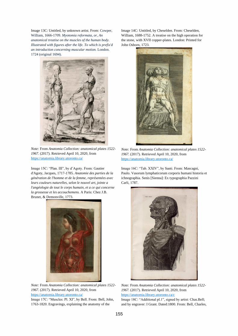
155
Image 13C: Untitled, by unknown artist. From: Cowper,
William, 1666-1709. Myotomia reformata, or, An
anatomical treatise on the muscles of the human body.
Illustrated with figures after the life. To which is prefix'd
an introduction concerning muscular motion. London.
1724 (original 1694).
Note: From Anatomia Collection: anatomical plates 1522-
1967. (2017). Retrieved April 10, 2020, from
https://anatomia.library.utoronto.ca/
Image 14C: Untitled, by Cheselden. From: Cheselden,
William, 1688-1752. A treatise on the high operation for
the stone, with XVII copper-plates. London: Printed for
John Osborn, 1723.
Note: From Anatomia Collection: anatomical plates 1522-
1967. (2017). Retrieved April 10, 2020, from
https://anatomia.library.utoronto.ca/
Image 15C: “Plan. III”, by d’Agoty. From: Gautier
d'Agoty, Jacques, 1717-1785. Anatomie des parties de la
génération de l'homme et de la femme, représentées avec
leurs couleurs naturelles, selon le nouvel art, jointe a
l'angéologie de tout le corps humain, et a ce qui concerne
la grossesse et les accouchemens. A Paris: Chez J.B.
Brunet, & Demonville, 1773.
Note: From Anatomia Collection: anatomical plates 1522-
1967. (2017). Retrieved April 10, 2020, from
https://anatomia.library.utoronto.ca/
Image 17C: “Muscles: Pl. XI”, by Bell. From: Bell, John,
1763-1820. Engravings, explaining the anatomy of the
Image 16C: “Tab. XXIV”, by Santi. From: Mascagni,
Paulo. Vasorum lymphaticorum corporis humani historia et
ichnographia. Senis [Sienna]: Ex typographia Pazzini
Carli, 1787.
Note: From Anatomia Collection: anatomical plates 1522-
1967. (2017). Retrieved April 10, 2020, from
https://anatomia.library.utoronto.ca/c
Image 18C: “Additional pl.1”, signed by artist: Chas.Bell;
and by engraver: I Grant. Dated:1800. From: Bell, Charles,

156
bones, muscles, and joints. Edinburgh: Printed by John
Paterson; for Bell and Bradfute, and T. Duncan; and J.
Johnson, and G.G.G. and J. Robinsons, London, 1794
Note: From Anatomia Collection: anatomical plates 1522-
1967. (2017). Retrieved April 10, 2020, from
https://anatomia.library.utoronto.ca/
Sir, 1774-1842. A system of dissections: explaining the
human body, the manner of displaying the parts, and their
varieties in disease. The second edition. Edinburgh.
London, 1799-1805.
Note: From Anatomia Collection: anatomical plates 1522-
1967. (2017). Retrieved April 10, 2020, from
https://anatomia.library.utoronto.ca/
Image 19C: “Pl. 92”, by Pailloux. From: Cloquet, Jules,
1790-1883. Manuel d'anatomie descriptive du corps
humain, représentée en planches lithographiées. Paris:
Chez Béchet jeune, 1825.
Note: From Anatomia Collection: anatomical plates 1522-
1967. (2017). Retrieved April 10, 2020, from
https://anatomia.library.utoronto.ca/
Image 20C: “Pl. 93”, by Pailloux. From: Cloquet, Jules,
1790-1883. Manuel d'anatomie descriptive du corps
humain, représentée en planches lithographiées. Paris:
Chez Béchet jeune, 1825.
Note: From Anatomia Collection: anatomical plates 1522-
1967. (2017). Retrieved April 10, 2020, from
https://anatomia.library.utoronto.ca/
Image 21C: “Decima quinti libri figura” (“15th book
figure”), by Calcar. From: Vesalius, Andreas, 1514-1564.
Image 22C: “Vigesima quinti libri figura” (“25th Book
FIgure”), by Calcar. From: Vesalius, Andreas, 1514-1564.

157
De humani corporis fabrica libri septem. Basileæ [Basel]:
Ex officina I. Oporini, 1543.
Note: From Anatomia Collection: anatomical plates 1522-
1967. (2017). Retrieved April 10, 2020, from
https://anatomia.library.utoronto.ca/
De humani corporis fabrica libri septem. Basileæ [Basel]:
Ex officina I. Oporini, 1543.
Note: From Anatomia Collection: anatomical plates 1522-
1967. (2017). Retrieved April 10, 2020, from
https://anatomia.library.utoronto.ca/
Image 23C: “Prima musculorum tabula”, by Calcar. From:
Vesalius, Andreas, 1514-1564. De humani corporis fabrica
libri septem. Basileæ [Basel]: Ex officina I. Oporini, 1543.
Note: From Anatomia Collection: anatomical plates 1522-
1967. (2017). Retrieved April 10, 2020, from
https://anatomia.library.utoronto.ca/
Image 24C: “Prima musculorum tabula” originally by
Calcar, this ed.: by Wandelaar. From: Vesalius, Andreas,
edited by Albinus: Opera omnia anatomica & chirurgica.
Cura Hermanni Boerhaave ... & Bernhardi Siegfried Albini
... Lugduni Batavorum [Leiden]: Apud J. du Vivie, et J. &
H. Verbeek, 1725.
Note: From Anatomia Collection: anatomical plates 1522-
1967. (2017). Retrieved April 10, 2020, from
https://anatomia.library.utoronto.ca/
Image 25C: “Sexta musculorum tabula”, by Calcar
(reprinted). From: Geminus, Thomas, d. 1562.
Image 26C: “Tabula LX”, by unknown artist. From: Guidi,
Guido, ca. 1500-1569. Opera omnia; siue, Ars medicinalis,

158
Compendiosa totius anatomie delineatio, ære exarata per
Thomam Geminum. Londini: [Imprinted by T. Geminus],
1559.
Note: From Anatomia Collection: anatomical plates 1522-
1967. (2017). Retrieved April 10, 2020, from
https://anatomia.library.utoronto.ca/
in qua cuncta quæ ad humani corporis valetudinem
præsentem tuendam. Francofurti ad Moenum [Frankfurt
am Main]: Typis & sumptibus Wechelianorum, apud D. &
D. Aubrios & C. Schleichium, 1626.
Note: From Anatomia Collection: anatomical plates 1522-
1967. (2017). Retrieved April 10, 2020, from
https://anatomia.library.utoronto.ca/
Image 27C: “Tabula hæc uterum gravidæ mulieris et
embryi situm ostendit” (“Figure/Map shows location of the
womb of the pregnant woman, and fetus”), engraved by
Gaultier. From: Du Laurens, Andrâe, 1558-1609. Opera
omnia anatomica et medica: ex postrema recognitione,
accessione quorundam librorum, qui lucem antea non
viderant, locupletata ... Francofurti [Frankfurt am Main]:
Typis Caspari Rotelij, Impensis VVilhelmi Fitzeri, 1627.
Note: From Anatomia Collection: anatomical plates 1522-
1967. (2017). Retrieved April 10, 2020, from
https://anatomia.library.utoronto.ca/
Image 28C: “Tab. VII”, engraved by Vandergucht. From:
Cowper, William, 1666-1709. Myotomia reformata, or, An
anatomical treatise on the muscles of the human body.
Illustrated with figures after the life. To which is prefix'd
an introduction concerning muscular motion. London.
1724.
Note: From Anatomia Collection: anatomical plates 1522-
1967. (2017). Retrieved April 10, 2020, from
https://anatomia.library.utoronto.ca/
Image 29C: “Tab. XXX”, by Pini. From: Eustachi,
Bartolomeo, d. 1574. Explicatio tabularum anatomicarum
Image 30C: “Tab. V”, by (A.) Bell. In book: Albinus,
Bernhard Siegfried, 1697-1770. Tables of the skeleton and

159
Bartholomæi Eustachii. Accedit tabularum editio nova.
Leidæ Batavorum [Leiden]: Apud J.A. Langerak et J. & H.
Verbeek, 1744.
Note: From Anatomia Collection: anatomical plates 1522-
1967. (2017). Retrieved April 10, 2020, from
https://anatomia.library.utoronto.ca/
muscles of the human body. Translated from the Latin.
Edinburgh: Printed by Balfour & Smellie, for A. Bell,
1777-1778.
Note: From Anatomia Collection: anatomical plates 1522-
1967. (2017). Retrieved April 10, 2020, from
https://anatomia.library.utoronto.ca/
Image 31C: “Plate 5”, by (C.) Bell. From: Bell, Charles,
Sir, 1774-1842. A system of dissections: explaining the
human body, the manner of displaying the parts, and their
varieties in disease. The second edition. Edinburgh:
Printed [by and] for Mundell and Son ...; and for J.
Johnson ...; and Longman and Rees ..., London, 1799-
1805.
Note: From Anatomia Collection: anatomical plates 1522-
1967. (2017). Retrieved April 10, 2020, from
https://anatomia.library.utoronto.ca/
Image 33C: “17.e livraison, pl. 5”, by Chazal. From:
Cruveilhier, Jean, 1791-1874. Anatomie pathologique du
Image 32C: “Pl.186”, by Haincelain. From: Cloquet, Jules,
1790-1883. Manuel d'anatomie descriptive du corps
humain, représentée en planches lithographiées. Paris:
Chez Béchet jeune, 1825
Note: From Anatomia Collection: anatomical plates 1522-
1967. (2017). Retrieved April 10, 2020, from
https://anatomia.library.utoronto.ca/
Image 34C: “Pl. 127”, by Pailloux, engraved by Feillet.
From: Cloquet, Jules, 1790-1883. Manuel d'anatomie

160
corps humain, descriptions avec figures lithographiées et
colorisées des diverses altérations morbides dont le corps
humain est susceptible. Paris: J.-B. Baillière, 1829-1835.
Note: From Anatomia Collection: anatomical plates 1522-
1967. (2017). Retrieved April 10, 2020, from
https://anatomia.library.utoronto.ca/
descriptive du corps humain, représentée en planches
lithographiées. Paris: Chez Béchet jeune, 1825.
Note: From Anatomia Collection: anatomical plates 1522-
1967. (2017). Retrieved April 10, 2020, from
https://anatomia.library.utoronto.ca/
Image 35C: “Pl. XXV”, by Maclise. From: Maclise, Joseph
(1815 - 1880). Surgical anatomy. 2nd ed. London: J.
Churchill, 1856.
Note: From Anatomia Collection: anatomical plates 1522-
1967. (2017). Retrieved April 10, 2020, from
https://anatomia.library.utoronto.ca/
Image 36C: “Interiorum corporis humani partium viva
delineatio” (“Quick sketch of the interior parts of the
human body “), by R.S. From: Interiorvm corporis hvmani
partivm viva delineatio : pervtilis anatomes interiorvm
mvliebris partivm cognitio ac earundem situs, figura,
numerus, positio, hand [sic] iniucounda cognitu.
[London]: [s.n.], [ca. 1559].
Note: From Anatomia Collection: anatomical plates 1522-
1967. (2017). Retrieved April 10, 2020, from
https://anatomia.library.utoronto.ca/
Image 37C: “Tabula LVIIII”, by unknown artist. From:
Guidi, Guido, ca. 1500-1569. Opera omnia; siue, Ars
Image 38C: “Tabula LXXI”, by unknown artist. From:
Guidi, Guido, ca. 1500-1569. Opera omnia; siue, Ars

161
medicinalis, in qua cuncta quæ ad humani corporis
valetudinem præsentem tuendam. Francofurti ad Moenum
[Frankfurt am Main]: Typis & sumptibus Wechelianorum,
apud D. & D. Aubrios & C. Schleichium, 1626.
Note: From Anatomia Collection: anatomical plates 1522-
1967. (2017). Retrieved April 10, 2020, from
https://anatomia.library.utoronto.ca/
medicinalis, in qua cuncta quæ ad humani corporis
valetudinem præsentem tuendam. Francofurti ad Moenum
[Frankfurt am Main]: Typis & sumptibus Wechelianorum,
apud D. & D. Aubrios & C. Schleichium, 1626
Note: From Anatomia Collection: anatomical plates 1522-
1967. (2017). Retrieved April 10, 2020, from
https://anatomia.library.utoronto.ca/
Image 39C: Untitled, contains copies of original prints by
Calcar. From: Geminus, Thomas, d. 1562. Compendiosa
totius anatomie delineatio, ære exarata per Thomam
Geminum. Londini: [Imprinted by T. Geminus], 1559.
Note: From Anatomia Collection: anatomical plates 1522-
1967. (2017). Retrieved April 10, 2020, from
https://anatomia.library.utoronto.ca/
Image 41C: “Tabula hæc externas omnes et præcipuas
corporis partes exprimit” (“Table/Image of surface
Image 40C: “Tertia musculorum tabula”, reprint, originally
by Calcar. From: Geminus, Thomas, d. 1562.
Compendiosa totius anatomie delineatio, ære exarata per
Thomam Geminum. Londini: [Imprinted by T. Geminus],
1559.
Note: From Anatomia Collection: anatomical plates 1522-
1967. (2017). Retrieved April 10, 2020, from
https://anatomia.library.utoronto.ca/
Image 42C: “Tab. XI. lib. IV”, by Fialetti. From Spiegel,
Adriaan van de, 1578-1625. De humani corporis fabrica

162
anatomy and principle body parts”), unknown artist. From:
Du Laurens, Andrâe, 1558-1609. Opera omnia anatomica
et medica : ex postrema recognitione, accessione
quorundam librorum, qui lucem antea non viderant,
locupletata ... Francofurti [Frankfurt am Main]: Typis
Caspari Rotelij, Impensis VVilhelmi Fitzeri, 1627.
Note: From Anatomia Collection: anatomical plates 1522-
1967. (2017). Retrieved April 10, 2020, from
https://anatomia.library.utoronto.ca/
libri decem : tabulis XCIIX æri incisis elegantissimis, nec
antehac visis exornati. Francofurti [Frankfurt am Main]:
Impensis & cælo Matthæi Meriani ..., 1631.
Note: From Anatomia Collection: anatomical plates 1522-
1967. (2017). Retrieved April 10, 2020, from
https://anatomia.library.utoronto.ca/
Image 43C: “Tab. IX. lib. IV”, by Fialetti. From: Spiegel,
Adriaan van de, 1578-1625. De humani corporis fabrica
libri decem : tabulis XCIIX æri incisis elegantissimis, nec
antehac visis exornati. Francofurti [Frankfurt am Main]:
Impensis & cælo Matthæi Meriani ..., 1631.
Note: From Anatomia Collection: anatomical plates 1522-
1967. (2017). Retrieved April 10, 2020, from
https://anatomia.library.utoronto.ca/
Image 45C: “Plate 1”, by (C.) Bell, engraved by Grant.
From: Bell, Charles, Sir, 1774-1842. A system of
Image 44C: “TAB. I.”, by Albinus and Wandelaar. From:
Albinus, Bernhard Siegfried, 1697-1770. Tables of the
Skeleton and Muscles of the Human Boy. Translated from
the Latin. Edinburgh, Balfour & Smellie, 1777-1778.
Note: From Anatomia Collection: anatomical plates 1522-
1967. (2017). Retrieved April 10, 2020, from
https://anatomia.library.utoronto.ca/
Image 46C: “Pl. 89”, by Pailloux. From: Cloquet, Jules,
1790-1883. Manuel d'anatomie descriptive du corps

163
dissections : explaining the human body, the manner of
displaying the parts, and their varieties in disease. The
second edition. Edinburgh: Printed [by and] for Mundell
and Son ...; and for J. Johnson ...; and Longman and Rees
..., London, 1799-1805.
Note: From Anatomia Collection: anatomical plates 1522-
1967. (2017). Retrieved April 10, 2020, from
https://anatomia.library.utoronto.ca/
humain, représentée en planches lithographiées. Paris:
Chez Béchet jeune, 1825.
Note: From Anatomia Collection: anatomical plates 1522-
1967. (2017). Retrieved April 10, 2020, from
https://anatomia.library.utoronto.ca/
Image 47C: “Tome 4. Pl. 14”, by N.H. Jacob. From:
Bourgery, Marc Jean, 1797-1849. Traité complet de
l'anatomie de l'homme comprenant la medecine operatoire,
par le docteur Bourgery. Avec planches lithographiées
d'après nature par N.H. Jacob. Paris: C. Delaunay, 1831-
1854.
Note: From Anatomia Collection: anatomical plates 1522-
1967. (2017). Retrieved April 10, 2020, from
https://anatomia.library.utoronto.ca/
Image 48C: “Pl. 16”, by Maclise. From: Quain, Richard,
Sir, 1816-1898. The anatomy of the arteries of the human
body with its applications to pathology and operative
surgery. In lithographic drawings with practical
commentaries. London: Taylor and Walton, 1844.
Note: From Anatomia Collection: anatomical plates 1522-
1967. (2017). Retrieved April 10, 2020, from
https://anatomia.library.utoronto.ca/
Image 49C: “Prima[-octava] figurarum muliebrium” (First
feminine figure”), reprint of mulitple original prints by
Image 50C: “Tabula LXVIII”, by unknown artist. From:
Guidi, Guido, ca. 1500-1569. Opera omnia; siue, Ars

164
Calcar. In book: Geminus, Thomas, d. 1562. Compendiosa
totius anatomie delineatio, ære exarata per Thomam
Geminum. Londini: [Imprinted by T. Geminus], 1559.
Note: From Anatomia Collection: anatomical plates 1522-
1967. (2017). Retrieved April 10, 2020, from
https://anatomia.library.utoronto.ca/
medicinalis, in qua cuncta quæ ad humani corporis
valetudinem præsentem tuendam. Francofurti ad Moenum
[Frankfurt am Main]: Typis & sumptibus Wechelianorum,
apud D. & D. Aubrios & C. Schleichium, 1626
Note: From Anatomia Collection: anatomical plates 1522-
1967. (2017). Retrieved April 10, 2020, from
https://anatomia.library.utoronto.ca/
Image 51C: “Tab. IIII”, by unknown artist. From: Spiegel,
Adriaan van de, 1578-1625. De formato foetu liber
singularis, æneis figuris exornatus : epistolæ duæ
anatomicæ : tractatus de arthritide : opera posthuma /
studio Liberalis Cremæ ... edita. Francofurti [Frankfurt am
Main]: Impensis & cælo Matthæi Meriani ..., typis
Casparis Rötelii, 1631
Note: From Anatomia Collection: anatomical plates 1522-
1967. (2017). Retrieved April 10, 2020, from
https://anatomia.library.utoronto.ca/
Image 52C: “T. 31 [trigesimaprima tabula]”, by Lairesse.
From: Bidloo, Govard, 1649-1713. Anatomia humani
corporis, centum & quinque tabulis, per artificiossis. G. de
Lairesse ad vivum delineatis, demonstrata, veterum
recentiorumque inventis explicata plurimisque, hactenus
non detectis, illustrata. Amstelodami [Amsterdam]:
Sumptibus viduæ J. a Someren, 1685.
Note: From Anatomia Collection: anatomical plates 1522-
1967. (2017). Retrieved April 10, 2020, from
https://anatomia.library.utoronto.ca/
Image 53C: “T. 55 [quinquagesimaquinta tabula]”, by
Lairesse. From: Bidloo, Govard, 1649-1713. Anatomia
Image 54C: Untitled, by Gautier d’Agoty. From: Gautier
d’Agoty, Jacques, 1717-1785. Myologie complette en

165
humani corporis, centum & quinque tabulis, per
artificiossis. G. de Lairesse ad vivum delineatis,
demonstrata, veterum recentiorumque inventis explicata
plurimisque, hactenus non detectis, illustrata.
Amstelodami [Amsterdam]: Sumptibus viduæ J. a
Someren, 1685.
Note: From Anatomia Collection: anatomical plates 1522-
1967. (2017). Retrieved April 10, 2020, from
https://anatomia.library.utoronto.ca/
couleur et grandeur naturelle : composée de l'Essai et de
la Suite de l'Essai d'anatomie, en tableaux imprimés ;
ouvrage unique, utile et nécessaire aux etudians &
amateurs de cette science. Paris.1746.
Note: From Anatomia Collection: anatomical plates 1522-
1967. (2017). Retrieved April 10, 2020, from
https://anatomia.library.utoronto.ca/
Image 55C: “Plan. VII”, by Gautier d’Agoty. From:
Gautier d'Agoty, Jacques, 1717-1785. Anatomie des parties
de la génération de l'homme et de la femme, représentées
avec leurs couleurs naturelles, selon le nouvel art, jointe a
l'angéologie de tout le corps humain, et a ce qui concerne
la grossesse et les accouchemens. A Paris: Chez J.B.
Brunet, & Demonville, 1773.
Note: From Anatomia Collection: anatomical plates 1522-
1967. (2017). Retrieved April 10, 2020, from
https://anatomia.library.utoronto.ca/
Image 56C: “Plan. VIII”, by Gautier d’Agoty. From:
Gautier d'Agoty, Jacques, 1717-1785. Anatomie des parties
de la génération de l'homme et de la femme, représentées
avec leurs couleurs naturelles, selon le nouvel art, jointe a
l'angéologie de tout le corps humain, et a ce qui concerne
la grossesse et les accouchemens. A Paris: Chez J.B.
Brunet, & Demonville, 1773.
Note: From Anatomia Collection: anatomical plates 1522-
1967. (2017). Retrieved April 10, 2020, from
https://anatomia.library.utoronto.ca/

166
Image 57C: “Tome 5. Pl. 63”, by N.H.Jacob, printed by
Lemercier. From: Bourgery, Marc Jean, 1797-1849. Traité
complet de l'anatomie de l'homme comprenant la medecine
operatoire, par le docteur Bourgery. Avec planches
lithographiées d'après nature par N.H. Jacob. Paris: C.
Delaunay, 1831-1854.
Note: From Anatomia Collection: anatomical plates 1522-
1967. (2017). Retrieved April 10, 2020, from
https://anatomia.library.utoronto.ca/
Image 58C: “Pl. 322”, by Haincelain, engraved by G. Frey.
From: Cloquet, Jules, 1790-1883. Manuel d'anatomie
descriptive du corps humain, représentée en planches
lithographiées. Paris: Chez Béchet jeune, 1825.
Note: From Anatomia Collection: anatomical plates 1522-
1967. (2017). Retrieved April 10, 2020, from
https://anatomia.library.utoronto.ca/
Image 59C: Untitled, by La Riviere. From: Estienne,
Charles, 1504-ca. 1564. De dissectione partium corporis
humani libri tres, a Carolo Stephano, doctore medico,
editi. Vna cum figuris, & incisionum declarationibus, a
Stephano Riuerio chirurgo co[m]positis. Parisiis: Apud S.
Colinæum, 1545.
Note: From Anatomia Collection: anatomical plates 1522-
1967. (2017). Retrieved April 10, 2020, from
https://anatomia.library.utoronto.ca/
Image 60C: “Tab. XIX. lib. VIII”, by Fialetti. From:
Spiegel, Adriaan van de, 1578-1625. De humani corporis
fabrica libri decem : tabulis XCIIX æri incisis
elegantissimis, nec antehac visis exornati. Francofurti
[Frankfurt am Main]: Impensis & cælo Matthæi Meriani ...,
1631.
Note: From Anatomia Collection: anatomical plates 1522-
1967. (2017). Retrieved April 10, 2020, from
https://anatomia.library.utoronto.ca/

167
Image 61C: “Tab. XVI”, by Cheselden. From: Cheselden,
William, 1688-1752. A treatise on the high operation for
the stone, with XVII copper-plates. London: Printed for
John Osborn, 1723.
Note: From Anatomia Collection: anatomical plates 1522-
1967. (2017). Retrieved April 10, 2020, from
https://anatomia.library.utoronto.ca/
Image 62C: “Pl. III”, by Maclise, printed by M. & N.
Hanhart. From: Maclise, Joseph. Surgical anatomy. 2nd ed.
London: J. Churchill, 1856.
Note: From Anatomia Collection: anatomical plates 1522-
1967. (2017). Retrieved April 10, 2020, from
https://anatomia.library.utoronto.ca/
Image 63C: “Pl. III” by Maclise, printed by M. & N.
Hanhart. From: Maclise, Joseph. Surgical anatomy. 2nd ed.
London: J. Churchill, 1856.
Note: From Anatomia Collection: anatomical plates 1522-
1967. (2017). Retrieved April 10, 2020, from
https://anatomia.library.utoronto.ca/
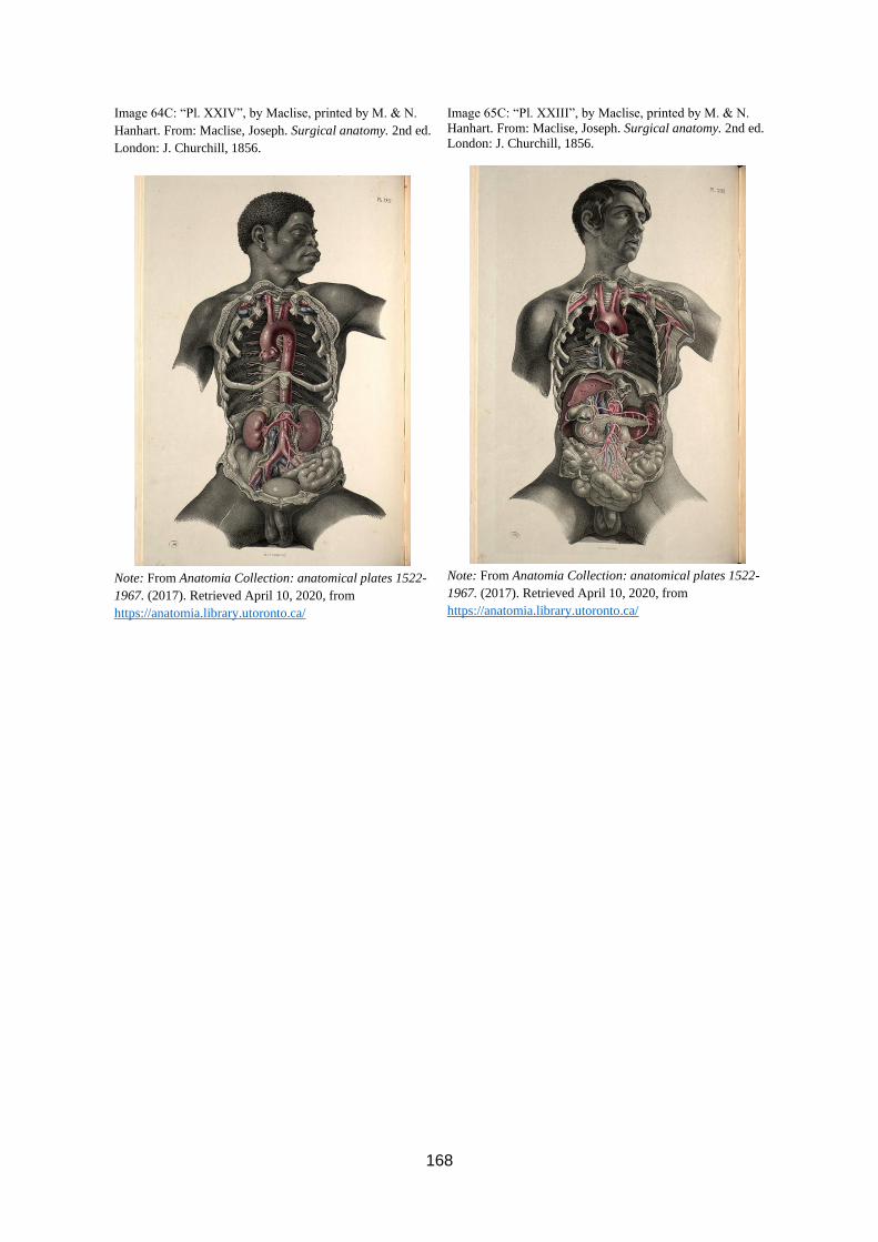
168
Image 64C: “Pl. XXIV”, by Maclise, printed by M. & N.
Hanhart. From: Maclise, Joseph. Surgical anatomy. 2nd ed.
London: J. Churchill, 1856.
Note: From Anatomia Collection: anatomical plates 1522-
1967. (2017). Retrieved April 10, 2020, from
https://anatomia.library.utoronto.ca/
Image 65C: “Pl. XXIII”, by Maclise, printed by M. & N.
Hanhart. From: Maclise, Joseph. Surgical anatomy. 2nd ed.
London: J. Churchill, 1856.
Note: From Anatomia Collection: anatomical plates 1522-
1967. (2017). Retrieved April 10, 2020, from
https://anatomia.library.utoronto.ca/

169
VITA AUCTORIS
NAME: Sarah D. Shaver
PLACE OF BIRTH: Abbotsford, British Columbia
YEAR OF BIRTH: 1997
EDUCATION: Simon Fraser University, Burnaby, B.C.
2015-2019 B.A.
Langley Fine Arts School, Langley, B.C.
2006-2015