Ana_Retameiro2020.pdf - TEDE - Unioeste
122
UNIVERSIDADE ESTADUAL DO OESTE DO PARANÁ – CAMPUS DE CASCAVEL CENTRO DE CIÊNCIAS BIOLÓGICAS E DA SAÚDE PROGRAMA DE PÓS-GRADUAÇÃO EM BIOCIÊNCIAS E SAÚDE – MESTRADO ANA CAROLINE BARBOSA RETAMEIRO EFEITOS DO EXERCÍCIO RESISTIDO E DO LASER DE BAIXA POTÊNCIA NA ARTICULAÇÃO TALOCRURAL EM MODELO EXPERIMENTAL DE ARTRITE REUMATOIDE CASCAVEL-PR (Março/2020)
-
Upload
khangminh22 -
Category
Documents
-
view
1 -
download
0
Transcript of Ana_Retameiro2020.pdf - TEDE - Unioeste
1
UNIVERSIDADE ESTADUAL DO OESTE DO PARANÁ – CAMPUS DE
CASCAVEL
CENTRO DE CIÊNCIAS BIOLÓGICAS E DA SAÚDE
PROGRAMA DE PÓS-GRADUAÇÃO EM BIOCIÊNCIAS E SAÚDE –
MESTRADO
ANA CAROLINE BARBOSA RETAMEIRO
EFEITOS DO EXERCÍCIO RESISTIDO E DO LASER DE BAIXA POTÊNCIA
NA ARTICULAÇÃO TALOCRURAL EM MODELO EXPERIMENTAL DE
ARTRITE REUMATOIDE
CASCAVEL-PR
(Março/2020)

2
ANA CAROLINE BARBOSA RETAMEIRO
EFEITOS DO EXERCÍCIO RESISTIDO E DO LASER DE BAIXA POTÊNCIA
NA ARTICULAÇÃO TALOCRURAL EM MODELO EXPERIMENTAL DE
ARTRITE REUMATOIDE
CASCAVEL-PR
(março/2020)

3
ANA CAROLINE BARBOSA RETAMEIRO
EFEITOS DO EXERCÍCIO RESISTIDO E DO LASER DE BAIXA POTÊNCIA
NA ARTICULAÇÃO TALOCRURAL EM MODELO EXPERIMENTAL DE
ARTRITE REUMATOIDE
Dissertação apresentada ao Programa
De Pós-Graduação em Biociências e
Saúde - Mestrado, do Centro de
Ciências Biológicas e da Saúde, da
Universidade Estadual do Oeste do
Paraná, como requisito parcial para a
obtenção do título de Mestre em
Biociências e Saúde.
Área de Concentração: Fatores que
influenciam a morfofisiologia orgânica.
ORIENTADOR: Gladson Ricardo Flor
Bertolini.
COORIENTADOR: Lucinéia de Fátima
Chasko Ribeiro e Taciane Stein Leal.
CASCAVEL-PR
(março/2020)
4
FICHA CATALOGRÁFICA

5
FOLHA DE APROVAÇÃO
ANA CAROLINE BARBOSA RETAMEIRO
EFEITOS DO EXERCÍCIO RESISTIDO E DO LASER DE BAIXA POTÊNCIA
NA ARTICULAÇÃO TALOCRURAL EM MODELO EXPERIMENTAL DE
ARTRITE REUMATOIDE
Esta dissertação foi julgada adequada para a obtenção do título de Mestre em
Biociências e Saúde e aprovada em sua forma final pelo Orientador e pela
Banca Examinadora.
Orientador: Prof. Dr. (a)________________________________
UNIOESTE
Prof. Dr. (a)__________________________________________
UNIOESTE
Prof. Dr. (a)__________________________________________
UEL
CASCAVEL-PR
(fevereiro/2020)

6
AGRADECIMENTOS
Esse trabalho não é só meu, mas também de todas as pessoas que me deram suporte, não só na parte acadêmica, mas de várias formas ao longo da vida. Por isso, devo agradecer principalmente a meus pais: Joventina e Luiz Carlos e à minha irmã Bruna, por serem meus primeiros orientadores neste mundo e por seguirem dessa forma me dando suporte até hoje mesmo nas situações mais adversas.
Agradeço a todas minhas professoras e professores, que me apontaram o caminho até a graduação, às minhas orientadoras durante a graduação: Professoras Doutoras Márcia Miranda Torrejais, Célia Cristina Leme Beu e Rose Meire Costa Brancalhão; e, especialmente, à Professora Doutora Lucinéia de Fátima Chasko Ribeiro que me orientou durante todo o processo do mestrado: muito obrigada por toda paciência, dedicação, ensinamentos e por acreditarem que eu sou capaz.
Meus agradecimentos a meus coorientadores: Taciane Stein Leal e Gladson Ricardo Flor Bertolini por estarem sempre prontos para me auxiliar e sanar minhas dúvidas. Desejo, novamente, agradecer à Taciane juntamente com Morgana Neves, minhas queridas fisioterapeutas, por terem sido os pilares da projeção e do planejamento deste trabalho; pela companhia e suporte antes, durante e depois do período experimental, sempre me explicando coisas com as quais nunca havia tido contato com muita paciência e bom humor. Obrigada por dividir e ensinar um pouco da profissão de vocês para mim!
Estendo os agradecimentos a meus primeiros colegas de laboratório que se tornaram também grandes amigos: Suellen Scarton, Pâmela Buratti, Caroline Covatti, Matheus Felipe Zazula, Adriana Souza e Bruno Popik. E aos que foram chegando com o tempo e acrescentando ainda mais aprendizado, amizade, amor, companheirismo e felicidade em minha vida: Aldair Casagrande dos Santos, Bárbara Zanardini de Andrade, Ariadne Barbosa, Mylena Campos, Luiz Gustavo Vasconcelos Machado, Alana Ludemila Tavares, Juliana Souza, Ana Luiza Peretti, Maria Luiza Wutzke, Izabela Camilo, Pâmela Silva, Aline Reginato, Estefani Marin, Carol De Toni e Diego. E ainda aos amigos que não são do laboratório, mas estão sempre presentes em momentos bons e ruins me dando apoio, lembrando que tudo vai dar certo e deixando tudo mais leve: Aldair, Hellen Rambo, Fernanda Farfale e Giovanna Piovezan.
Além das pessoas, ainda devo agradecer aos meus animais de estimação, principalmente à Serafine, que sempre me ofereceu amor incondicional desde o início da graduação e sempre se sentou a meu lado

7
quando eu chegava em casa, muitas vezes cansada e desmotivada, e acabava por me fazer lembrar de alguns valores que, às vezes, seres humanos são incapazes de compreender.
Resta agradecer ao Programa de Pós-Graduação em Biociências e Saúde pela oportunidade de desenvolvimento da pesquisa, a CAPES pela bolsa concedida, e a todos que torceram, acreditaram e me ajudaram de alguma forma: MUITO OBRIGADA!
“Inacabado, sei que sou um ser condicionado, mas consciente do
inacabamento, sei que posso ir além dele.”
Paulo Freire.

8
RESUMO
RETAMEIRO, A.C.B. Efeitos do exercício resistido e do laser de baixa
potência na articulação talocrural em modelo experimental de artrite
reumatoide. 118 páginas. Dissertação (Mestrado). Programa de Pós-
Graduação em Biociências e Saúde, Centro de Ciências Biológicas e da
Saúde, Campus de Cascavel, Unioeste, 2020.
A Artrite reumatoide (AR) é uma doença autoimune que afeta as articulações
sinoviais e estruturas periarticulares, principalmente as presentes nos
membros, ocasionando dor e limitações físicas aos portadores, o que repercute
na qualidade de vida e traz reflexos em questões socioeconômicas devido aos
afastamentos das atividades laborais e gastos com o tratamento. Na tentativa
de minimizar seus efeitos, modalidades terapêuticas vêm sendo utilizadas, uma
delas é o exercício físico e laser de baixa potência (LBP). No entanto, ainda
existem lacunas quanto aos protocolos utilizados e quanto aos efeitos da
associação das duas modalidades terapêuticas no tratamento da AR. Assim,
este estudo analisou os efeitos do exercício resistido e do LBP em parâmetros
funcionais e morfológicos da articulação talocrural em modelo de AR,
promovido pelo completo adjuvante de Freund (CFA). Para tanto, nas
avaliações funcionais foram utilizados 128 ratos Wistar e nas análises
histomorfométricas, 80. Os animais foram submetidos ao protocolo de AR de
fase aguda e de fase crônica da doença, para avaliação funcional foram 64 em
cada fase e para histomorfometria foram 40 de cada fase, e cada uma delas
subdividida em grupo controle (GC), grupo artrite (GA), controle laser (CL),
controle exercício (CE), controle laser e exercício (CLE), artrite laser (AL),
artrite exercício (AE), e artrite laser e exercício (ALE), todos com oito animais
para os testes de funcionalidade e cinco para as análises histológicas. Nos
grupos artrite, a indução ocorreu pela aplicação de CFA na base da cauda,
para imunização e, posteriormente, na cavidade articular tíbio-femoral direita
dos animais. Os tratamentos se iniciaram 24 horas após a injeção intra-
articular, realizados em dias alternados com progressão de tempo e séries para
o exercício. As avaliações funcionais de preensão, índice funcional do
isquiático (IFC) e plano inclinado foram realizadas no dia da injeção intra-

9
articular, 24 horas depois e no 3º e 5º dias para o grupo agudo. Para o crônico,
o intervalo entre as avaliações aumentou para sete dias, ocorrendo no 12º, 19º
e 26º dias. Após o período experimental, as articulações talocrurais do membro
pélvico experimental (direito) foram processadas e fotodocumentadas para as
análises morfológicas da morfologia geral do tecido articular e
histomorfométricas da altura da zona profunda e das demais zonas e da
contagem de condrócitos. Todos os parâmetros analisados nas avaliações
funcionais mostraram diferença significativa dos grupos artrite em relação aos
controles nas duas fases da doença, com retorno de valores semelhantes ao
controle nos grupos AL, AE e ALE, porém com maior eficácia na associação
dos dois tratamentos. Na morfologia da articulação talocrural de GA foi possível
observar angiogênese na subíntima da membrana sinovial e cartilagem
articular com pannus e floculações nas duas fases da doença. Na fase crônica
as alterações morfológicas da superfície articular foram mais expressivas do
que na fase aguda, uma vez que 50% dos animais apresentaram tais
características, e no agudo apenas em alguns destes e com maior evidência no
tálus. Na fase aguda ainda foi observado angiogênese na subíntima de todos
os demais grupos, porém com ausência de infiltrado inflamatório e com o grupo
AE se assemelhando mais ao controle nesse aspecto. Nesta mesma fase foi
possível notar maior presença de condrócitos em GA, CE e AE, o que foi
comprovado na análise histomorfométrica, o que pode indicar uma fase de
reparo tecidual mais avançada nestes grupos.
Palavras-Chaves: Angiogênese, Reparo tecidual, Artrite experimental, Adjuvante
de Freund, Exercício, Laser, Articulação do Tornozelo.
10
ABSTRACT
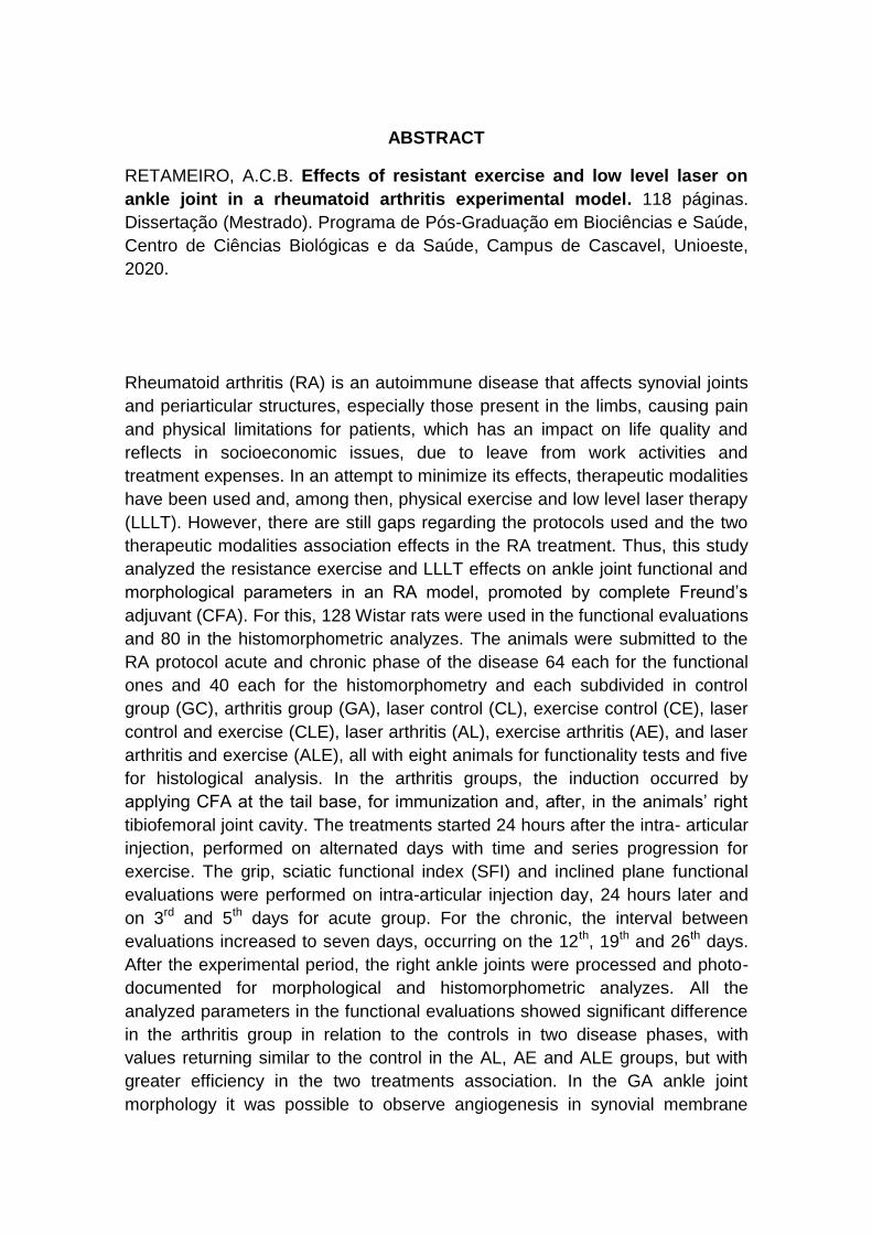
RETAMEIRO, A.C.B. Effects of resistant exercise and low level laser on
ankle joint in a rheumatoid arthritis experimental model. 118 páginas.
Dissertação (Mestrado). Programa de Pós-Graduação em Biociências e Saúde,
Centro de Ciências Biológicas e da Saúde, Campus de Cascavel, Unioeste,
2020.
Rheumatoid arthritis (RA) is an autoimmune disease that affects synovial joints
and periarticular structures, especially those present in the limbs, causing pain
and physical limitations for patients, which has an impact on life quality and
reflects in socioeconomic issues, due to leave from work activities and
treatment expenses. In an attempt to minimize its effects, therapeutic modalities
have been used and, among then, physical exercise and low level laser therapy
(LLLT). However, there are still gaps regarding the protocols used and the two
therapeutic modalities association effects in the RA treatment. Thus, this study
analyzed the resistance exercise and LLLT effects on ankle joint functional and
morphological parameters in an RA model, promoted by complete Freund’s
adjuvant (CFA). For this, 128 Wistar rats were used in the functional evaluations
and 80 in the histomorphometric analyzes. The animals were submitted to the
RA protocol acute and chronic phase of the disease 64 each for the functional
ones and 40 each for the histomorphometry and each subdivided in control
group (GC), arthritis group (GA), laser control (CL), exercise control (CE), laser
control and exercise (CLE), laser arthritis (AL), exercise arthritis (AE), and laser
arthritis and exercise (ALE), all with eight animals for functionality tests and five
for histological analysis. In the arthritis groups, the induction occurred by
applying CFA at the tail base, for immunization and, after, in the animals’ right
tibiofemoral joint cavity. The treatments started 24 hours after the intra- articular
injection, performed on alternated days with time and series progression for
exercise. The grip, sciatic functional index (SFI) and inclined plane functional
evaluations were performed on intra-articular injection day, 24 hours later and
on 3rd and 5th days for acute group. For the chronic, the interval between
evaluations increased to seven days, occurring on the 12th, 19th and 26th days.
After the experimental period, the right ankle joints were processed and photo-
documented for morphological and histomorphometric analyzes. All the
analyzed parameters in the functional evaluations showed significant difference
in the arthritis group in relation to the controls in two disease phases, with
values returning similar to the control in the AL, AE and ALE groups, but with
greater efficiency in the two treatments association. In the GA ankle joint
morphology it was possible to observe angiogenesis in synovial membrane

11
subintima and articular cartilage with pannus and flocculation, in both disease
phases. The articular surface morphological changes were more expressive in
chronic phase, since 50% of animals had such characteristics, and in acute only
in some of them and with greater evidence in talus. In the acute phase,
angiogenesis was also observed in the all other groups subintima, but with
inflammatory infiltrate absence and with AE group being similar to the control in
this aspect. In the same phase it was possible to notice a greater chondrocytes
presence in GA, CE and AE, which was confirmed in the histomorphometric
analysis which may indicate a more advanced tissue repair phase in these
groups.
Key-words: Angiogenesis, Tissue Repair, Experimental arthritis, Freund’s
adjuvant, Exercise, Laser, Ankle Joint.

12
LISTA DE ILUSTRAÇÕES
Figura 1. Representação dos componentes da articulação sinovial... ............. 22
Figura 2: Representação esquemática tridimensional da ultraestrutura da
membrana sinovial e seus diferentes tipos celulares. ...................................... 24
Figura 3: Ultraestrutura dos dois tipos de sinoviócitos na articulação têmporo-
mandibular de rato. .......................................................................................... 26
Figura 4: Zonas da cartilagem articular. .......................................................... 30
Figura 5: Articulação talocrural de rato. ........................................................... 32
Figura 6: Esquema representando a anatomia de uma articulação. ............... 34
Figura 7: Ciclos de feedback operacional envolvendo efeitos diretos do TNF
nos Tregs CD25 aumentando o status de doença da AR ................................ 36

13
LISTA DE ABREVIATURAS
μm – micrômetro
μL- microlitro
AMPc – AMP cíclico
AL - grupo atrite laser
AE - grupo artrite exercício
ALE- grupo artrite laser e exercício
AR - Artrite reumatoide
ATP – adenosina trifosfato
AV- Avaliação
Ca+- Cálcio
Cl- - Cloro
CE – controle exercício
CFA - Adjuvante Completo de Freund
CL – Controle Laser de Baixa potência
CLE – Controle laser de baixa potência mais exercício
cm -centímetros
Cox – Citocromo C oxidase
CRP- Proteína C reativa
DMARDs – Medicamentos modificadores do curso da doença
GAGs- Glicosaminoglicanos
GA- Grupo artrite
GC – grupo controle
K+- Potássio
IFC – Índice Funcional do isquiático

14
IL-6 – Interleucina seis
J/cm2- joules por centímetros quadrados
LBP- Laser de Baixa potência
LE- Lesão exercício
LLBP – Lesão laser de baixa potência
LLBPE – Lesão laser de baixa potência mais exercício
m- metros
Na+- Sódio
PGs – Proteoglicanos
TCA – Ácido tricloroacético
TNFα – Fator de necrose tumoral α
Treg- Células T regulatórias
15
SUMÁRIO
1. INTRODUÇÃO ............................................................................................. 16
2. OBJETIVOS ................................................................................................. 18
2.1 Objetivo Geral ........................................................................................ 18
2.2 Objetivos Específicos ........................................................................... 18
3. REVISÃO DE LITERATURA ....................................................................... 19
3.1 ARTRITE REUMATOIDE ........................................................................... 19
3.2 ARTICULAÇÕES SINOVIAIS .................................................................... 21
3.2.1 Membrana e líquido sinovial ............................................................. 22
3.2.4 CARTILAGEM ARTICULAR ............................................................... 26
3.2.5 Articulação Talocrural ....................................................................... 31
3.2.6 Ação da AR nas articulações ............................................................ 32
3.3 TRATAMENTO PARA ARTRITE REUMATOIDE ...................................... 37
3.3.1. Exercício físico como tratamento .................................................... 38
3.3.2. LBP como tratamento ....................................................................... 41
3.4. ESTUDOS EXPERIMENTAIS COM CFA ................................................. 42
ARTIGO 1 ........................................................................................................ 44
ARTIGO 2 ........................................................................................................ 74
CONSIDERAÇÕES FINAIS ........................................................................... 100
REFERENCIAS GERAIS ............................................................................... 101
ANEXO I......................................................................................................... 113
ANEXO II........................................................................................................ 114
ANEXO III....................................................................................................... 118

16
1. INTRODUÇÃO
Artrite reumatoide (AR) é uma doença inflamatória autoimune,
sistêmica e frequentemente progressiva (CAN et al., 2002; DE SOUZA et al.,
2010; FIRESTEIN, 2003; KHURANA; BERNEY, 2005) que afeta cerca de 1 %
da população em países desenvolvidos. No Brasil, são poucos os dados sobre
a doença, com incidência entre 0,2 e 1% na população (MOTA et al., 2011;
SENNA et al., 2004).
No entanto, dentre as doenças progressivas que acometem a saúde
humana, a AR apresenta incidência crescente, principalmente por ser uma
comorbidade relacionada a outras complicações clínicas e psicossociais
contemporâneas, como o estresse e a obesidade (MARGARETTEN et al.,
2011; NAGASE; KASHIWAGI, 2003). Suas manifestações clínicas incluem:
pouco apetite, febre, anemia, edema (TORPY, 2011) e as que mais se
destacam são as manifestações articulares (TORPY, 2011; TUFTS, 2012),
principalmente nas articulações sinoviais, presentes em locais de grande
amplitude de movimento, o que inclui a articulação talocrural ou tornozelo (DE
CARVALHO et al., 2004).
A articulação talocrural está envolvida em uma série de movimentos
que têm papel crucial na deambulação e na sustentação do corpo na posição
ortostática. Compreende uma sindesmose distal entre a tíbia e a fíbula e um
encaixe formado pelos terços distais dos ossos tíbia e fíbula com o tálus e seu
acometimento, como acontece na AR, leva a alterações biomecânicas e
funcionais que afetam de forma negativa a vida do portador (BOGART; ORT,
2008; BONO; BERBERIAN, 2001).
Normalmente é observado, nos indivíduos afetados pela AR, o
desenvolvimento de sinovite persistente com edema e mobilidade articular
reduzida, resultando no alongamento dos tendões, ligamentos e cápsulas

17
articulares, com subsequente instabilidade, o que leva ao imobilismo (GILES et
al., 2008) e, consequentemente, à diminuição da massa e da força muscular,
comprometendo a integridade da articulação e dos tecidos que a circundam,
resultando em dor e alterações nos padrões de movimentos, o que limita a
atividade diária (TORPY, 2011; TUFTS, 2012), as atividades laborais (ABELL
et al., 2005; VLIELAND, 2003), trazendo consequências socioeconômicas
(MOTA et al., 2011).
Pesquisas relacionadas à AR em humanos são difíceis de ser
controladas, pois testes repetidos tornam-se extenuantes e, no processo
inflamatório, interferem a ingestão de medicamentos e/ou hábitos diários do
paciente. Com a proposta de estudar a etiopatogênese da AR e de buscar por
tratamentos mais eficientes, muitos modelos experimentais foram
desenvolvidos ao longo dos anos, e o modelo utilizando o adjuvante completo
de Freund (CFA) se destaca por ter se mostrado eficaz em mimetizar os
sintomas da AR humana (CAI et al., 2006; COOK; MOORE, 2006; FARIA et al.,
2009; NAGAKURA et al., 2003; ZHANG et al., 2002).
Na prática clínica, a terapia padrão é a medicamentosa, que inclui
glicocorticoides, metotrexato, anticorpos monoclonais, medicamentos
modificadores da doença (DMARDS) e outros agentes farmacológicos
relacionados a mecanismos inflamatórios (TAK et al., 2011). No entanto, estes
medicamentos geralmente não possuem ação duradoura e podem levar à
manifestação de diversos efeitos colaterais (BENUCCI et al., 2017;
PASSOS,2016; PATEL et al., 2017; TORPY, 2011); portanto, outras
abordagens terapêuticas têm sido buscadas.
Neste sentido, estudos demostram que o exercício físico tem
apresentado benefícios na reabilitação de indivíduos com AR, dentre os quais
estão o aumento da amplitude de movimento e da flexibilidade (FENTEM,
1994); diminuição significativa da dor e da atividade inflamatória, melhorando a
marcha e a função geral (SHIH et al., 2006; KAPALE et al., 2017). No entanto,
dados morfofuncionais estão concentrados em modelos anteriores,
principalmente na articulação tibiofemoral, de forma que a sua ação na
articulação talocrural ainda não foi estudada.
18
Além do exercício, o laser de baixa potência (LBP) vem sendo utilizado
no tratamento de várias doenças musculoesqueléticas, incluindo a AR (ASADA
et al., 1989; PALMGREN et al., 1989; WAYLONIS et al., 1988). Há evidências
de que o LBP promove mudanças locais e sistêmicas o que pode levar à
diminuição da dor, do edema e do enrijecimento articular. Também melhora a
resposta inflamatória aguda, por redução de prostaglandinas e inibição de
ciclooxigenase local, o que acarreta estímulo do processo de regeneração
tecidual (BJORDAL et al., 2003; HENRIQUES et al., 2010; MORIYAMA et al.,
2005).
Apesar de a literatura trazer os benefícios do exercício e do LBP,
existem lacunas quanto aos protocolos utilizados e nos efeitos da associação
das duas modalidades terapêuticas no tratamento da AR. E ainda, se o modelo
induzido por CFA causa alterações nos aspectos morfofuncionais da
articulação talocrural, assim como as modalidades de tratamentos. Sendo
assim, o trabalho visou testar esta hipótese, a fim de obter informações sobre
os protocolos de tratamento para a AR que auxilie os profissionais na busca da
reabilitação de seus pacientes melhorando a qualidade de vida.
2. OBJETIVOS
2.1 Objetivo Geral
Analisar os efeitos do exercício resistido e de laser de baixa potência
nos parâmetros morfofuncionais da articulação talocrural em modelo
experimental de artrite reumatoide.
2.2 Objetivos Específicos
Avaliar as alterações nos tecidos da articulação talocrural em modelo
de AR induzido por CFA e a ação dos tratamentos com exercício resistido e
LBP.

19
Analisar os efeitos dos tratamentos nos parâmetros funcionais de estado
funcional do membro e força muscular na articulação do tornozelo em
modelo de AR;
Analisar os efeitos dos tratamentos nos aspectos morfológicos da
membrana sinovial e da cartilagem articular da articulação talocrural.
Foram observados: organização geral da membrana, vascularização e
presença de infiltrado inflamatório nos vasos e na matriz extracelular. Na
cartilagem foram observadas: presença de floculações, organização e
disposição dos condrócitos, fissuras e degradação. Na análise
histomorfométrica foram realizadas as medidas de: espessura total,
espessura da zona superficial, espessura da zona profunda e contagem
de condrócitos.
3. REVISÃO DE LITERATURA
3.1 ARTRITE REUMATOIDE
A AR é uma doença inflamatória sistêmica e frequentemente
progressiva, crônica e autoimune (CAN et al., 2002; DE SOUZA et al., 2016;
FIRESTEIN, 2003; KHURANA; BERNEY, 2005), que afeta cerca de 1 % da
população em países desenvolvidos (MARIANO, 2011). No Brasil, um estudo
realizado em 2004, mostrou incidência de 0,46%, o que representa quase um
milhão de pessoas, entre os quais, os mais afetados eram pessoas entre a
quarta e sexta décadas de vida, sendo uma faixa etária economicamente ativa
(MOTA et al., 2011; SENNA et al., 2004).
A etiologia da doença ainda não é totalmente conhecida, no entanto,
sabe-se que sua susceptibilidade é determinada por uma interação complexa
entre fatores genéticos e ambientais (ADYUKOV et al., 2003; COSTENBADER
et al., 2006; DI GIUSEPPE et al., 2014; SUGIYAMA et al., 2010). Como
predisposições para o desenvolvimento da AR podem ser mencionadas:
hereditariedade, poucos hábitos de higiene, exposição à agentes ambientais,

20
má nutrição, ferimentos, lactação prolongada, gravidez frequente, menopausa,
transtornos psicológicos, ansiedade, tuberculose, ataques de reumatismo
agudo (TUFTS, 2012) e infecção ambiental por bactéria ou vírus (KAPALE et
al., 2017).
Devido à sua característica autoimune, sistêmica (TORPY, 2011) e
simétrica (LLOPIS et al., 2017), diversos órgãos e tecidos do corpo podem ser
afetados, como: coração, pele, pulmões, pleura e, principalmente, as
articulações (TORPY, 2011). Dentre os sintomas que podem ser apresentados
estão: pouco apetite, estado febril, anemia e edema (TORPY, 2011). No
entanto, os que mais se destacam são as manifestações articulares (TORPY,
2011; TUFTS, 2012), principalmente nas sinoviais (DE CARVALHO et al.,
2004), uma vez que os indivíduos afetados normalmente desenvolvem sinovite
persistente com inchaço e mobilidade articular reduzida, o que pode resultar no
alongamento dos tendões, ligamentos e cápsulas articulares, com subsequente
instabilidade e diminuição da massa e força muscular e inibição direta da
contração de grupos musculares adjacentes, comprometendo a integridade
biomecânica da articulação e tecidos que a circundam, resultando em dor e
padrões de movimentos que, geralmente, não oferecem energia suficiente e
limitam as atividades físicas (TORPY, 2011; TUFTS, 2012).
Estas manifestações da AR levam a impactos significantes na
qualidade de vida do indivíduo (GILES et al., 2008), podendo acometer
atividades do dia a dia, bem como as laborais (ABELL et al., 2005; VLIELAND,
2003), o que pode levar a custos diretos e individuais ou indiretos e públicos,
devido ao maior gasto com saúde, o que mostra sua grande importância social
(MOTA et al., 2011; PINCUS et al., 1984; ) e, assim, a necessidade de maiores
investigações científicas relacionadas a maior compreensão e opções de
tratamentos para esta enfermidade.

21
3.2 ARTICULAÇÕES SINOVIAIS
As articulações interligam o conjunto de ossos e cartilagens que
formam o arcabouço do corpo, o que permite a mobilidade corporal. São
divididas em três tipos, de acordo com a natureza do elemento que se interpõe
entre as peças articuladas: tecido conjuntivo fibroso; articulações fibrosas,
tecido cartilaginoso; articulações cartilagíneas, líquido sinovial; articulações
sinoviais (DÂNGELO; FATTINI, 2007).
A articulação talocrural é uma dentre as articulações sinoviais que
diferem dos outros tipos de articulações pelo movimento livre dos ossos, que
estão relacionadas à amplitude de movimento, sendo chamadas de diartroses.
Como característica, estas possuem várias estruturas e sistema biológico
complexo, composto de células altamente especializadas, provenientes de
vários tipos de tecidos, como hematopoiético e mesenquimal (HUI et al., 2012;
IWANAGA et al., 2000).
A articulação sinovial saudável é constituída pela cápsula articular,
membrana sinovial, líquido sinovial, cartilagem articular e elementos anexos
associados, como os ligamentos capsulares (Figura 1) (HUI et al., 2012). A
cápsula articular liga as extremidades ósseas, e delimita a cavidade articular.
Esta cavidade contém o líquido sinovial; uma substância que, em humanos
saudáveis, é caracterizada como: incolor, ou de aspecto claro, pálido-
amarelada, transparente e viscosa que é um dialisado do plasma sanguíneo
contendo elevado teor de ácido hialurônico, sintetizado pelos sinoviócitos, e
liberado para o interior do espaço sinovial, com número relativamente pequeno
de células (HUI et al., 2012; IWANAGA et al., 2000; JUNQUEIRA; CARNEIRO,
2013).

22
Figura 1. Representação dos componentes da articulação sinovial. Em A: Esquema de uma articulação sinovial típica. Em B: corte histológico de joelho de cobaia. Estão indicados: Membrana sinovial (MS), Cartilagem articular (A), Cavidade articular (CA), Cápsula articular (C), Epífise de crescimento (E).
Adaptado de JUNQUEIRA (2009).
3.2.1 Membrana e líquido sinovial
As paredes das capsulas articulares têm estruturas diferentes,
conforme a articulação considerada, sendo em geral constituídas por duas
camadas, uma externa, a fibrosa e uma interna, a camada ou membrana
sinovial (JUNQUEIRA; CARNEIRO, 2013) cuja função primária é a produção
de líquido sinovial, que nutre as cartilagens articulares, faz a remoção dos
restos articulares e do tecido conjuntivo da cavidade articular (HUI et al., 2012;
IWANAGA et al., 2000; OVALLE; NAHIRNEY, 2008).
Ainda, o líquido sinovial facilita o deslizamento das superfícies
articulares que são revestidas por cartilagem hialina, sem pericôndrio, através
de vários lubrificantes, estes que são macromoléculas sintetizadas e
secretadas pelas diferentes células sinoviais dos quais destaca-se: o ácido
Periósteo
MS CA
Ligamento
Cartilagem articular
Cavidade articular
Cápsula
articular
Camada
fibrosa
Membrana
sinovial
Medula óssea
amarela

23
hialurônico, um glicosaminoglicano (GAG) não sulfatado, responsável pela
característica viscosa do líquido sinovial com propriedades lubrificantes tanto
em condições estáticas quanto dinâmicas (HUI et al., 2012; IWANAGA et al.,
2000; JUNQUEIRA; CARNEIRO, 2013). É também é uma via transportadora de
substâncias entre a cartilagem articular (avascular) e o sangue dos capilares da
membrana sinovial; desta forma, nutrientes e oxigênio passam do sangue para
a cartilagem articular e gás carbônico difunde-se no sentido contrário
(JUNQUEIRA; CARNEIRO, 2013).
A membrana sinovial contém, na sua região, superficial uma camada
única que possui de uma a três camadas celulares de profundidade e é
chamada de íntima sinovial na qual encontram-se várias células modificadas do
tecido conjuntivo denominadas sinoviócitos (BARLAND; NOVIKOFF;
HAMERMAN, 1962; IWANAGA et al., 2000; OVALLE; NAHIRNEY, 2008;
SHIKICHI et al., 1999). As células do tipo A têm aspecto e a atividade funcional
semelhante a macrófagos, sua função é a remoção de materiais particulados e
correspondem de 20 a 30% das células de revestimento, as do tipo B são
semelhantes a fibroblastos (Figura 2). São responsáveis pela secreção de
GAGs e glicoproteínas e correspondem de 75 a 80% da contagem total de
células (JUNQUEIRA; CARNEIRO, 2013; IWANAGA et al., 2000; OVALLE;
NAHIRNEY, 2008).
A camada subíntima está localizada subjacente à íntima e, ao contrário
desta, que é relativamente espessa, é composta por tecido conjuntivo frouxo e
muito vascularizado, podendo ser do tipo areolar, adiposa ou fibrosa. Na
adiposa, há uma camada íntima única e achatada enquanto a matriz contém
células adiposas. A camada fibrosa da cápsula articular possui fina camada de
sinoviócitos e matriz formada por tecido conjuntivo denso (LEACH et al., 1988;
SHIVELY; VAN SICKLE, 1977). Numerosos capilares fenestrados são
encontrados em todos os tipos de subíntima sinovial, de forma que o sangue
que extravasa pode, rapidamente, interagir com o líquido sinovial em casos de
lesões articulares fazendo a remoção de artefatos intracelulares e regulação de
eventos imunológicos (IWANAGA et al., 2000; OVALLE; NAHIRNEY, 2008).

24
Figura 2: Representação esquemática tridimensional da ultraestrutura da membrana sinovial e seus diferentes tipos celulares.
Adaptado de JUNQUEIRA; CARNEIRO (2013).
A membrana sinovial de rato observada por MURASHIGE (1971)
contém duas camadas de célula distintas compreendendo uma mais próxima
do lúmen, que consiste em maior parte de células do tipo A e uma camada
mais profunda consistindo em células do tipo B, com uma linha celular maior do
que uma camada de células (CUTLIP; CHEVILLE, 1973; LEACH et al, 1988).
Sinoviócitos do tipo A, células normalmente circulares e localizadas na
parte superior da íntima, são imunorreativas a vários anticorpos monoclonais
contra macrófagos ou substâncias derivadas destes (BURMESTER et al.,
1983; HOGG et al., 1985; IZUMI et., 1995). Eles também expressam antígeno
IA, o qual desempenha ação na apresentação de antígenos nos estágios
iniciais da resposta imunológica (ATHANASOU, 1995; NOZAWA-INOUE et al.,
1988).

25
A superfície das células do tipo A é coberta por microvilosidades,
estrutura única de macrófagos típicos (figura 3 A) que são ativados na
captação de substâncias estranhas injetadas no interior da cavidade articular.
Fisiologicamente, absorvem e degradam constituintes extracelulares, debris
celulares, microrganismos e antígenos no fluido sinovial e matriz íntima, com o
uso de um sistema vesicular e lisossomal bem desenvolvido. Como as células
sinoviais assumem significativa quantidade de proteínas, elas podem ser
capazes de modificar a composição proteica do fluído sinovial (SOUTHWICK;
BENSCH, 1971).
As células do tipo B possuem núcleo relativamente grande, geralmente
profundamente recuado, comparado com a pequena quantidade de citoplasma
circundante (figura 3 B) (JILANI; GHADIALLY, 1986) e é sugerido que sua
função seja secretória. Uma vez que, células do tipo B secretam colágeno,
fibronectina (MAPP; REVEL, 1985), ácido hialurônico (ROY; GHADIALLY,
1976) e outras GAGs no interstício da íntima e na cavidade articular. E por isso
sendo, também, consideradas envolvidas, direta ou indiretamente, no controle
da composição proteica do fluído sinovial.
Outra atividade que mostra o envolvimento de sinoviócitos é a de
barreira, pois estabelecem, parcialmente, permeabilidade preferencial das
substâncias da base da íntima sinovial. Esta barreira, denominada
hematoarticular, oferece resistência à troca passiva e livre de algumas
substâncias (NISHIJIMA, 1981).
Esta seleção de substâncias ocorre para manter a concentração
relativa de proteínas com peso molecular maior diferente entre o plasma e o
fluído sinovial, mantendo-as geralmente com fluidez menor em relação ao
sérum. Já as proteínas plasmáticas de peso molecular menor, são mantidas no
fluido sinovial em concentrações maiores (SHANNON; GRAHAM, 1971).
Estudos anteriores de ultraestrutura dos sinoviócitos de roedores
indicaram a natureza endócrina do tipo B (GRAABAEK, 1982). Em ratos, estes
apresentam várias membranas circundadas de grânulos eletrodensos, os quais
se parecem com células endócrinas pituitárias. A formação de grupos de

26
sinoviócitos com granulações próximas a capilares fenestradas e sua rápida
degranulação em resposta a estímulos nocivos à membrana sinovial (LINCK;
PORTE, 1988) sugerem a sua função como células receptoras e secretórias,
uma característica semelhante a membros paraneurais (FUJITA et al., 1988).
Uma vez que as células do tipo B sempre estão perto de capilares fenestrados,
eles podem também confrontar a possibilidade da função hormonal (LINCK;
PORTE, 1988).
Figura 3: Ultraestrutura dos dois tipos de sinoviócitos na articulação temporomandibular de rato. Células do tipo A (A), ocupando a porção superficial da camada íntima. Células do tipo B (B), distantes da cavidade articular (CA). Em B: Eletromicrografia de sinoviócitos em amostras maceradas de articulação de cavalos. Células do tipo B (B) estendendo radialmente vários processos primários espessos que formam uma ampla rede de processos para cobrir a superfície da íntima sinovial.
Adaptado de: INAWAGA (2000)
3.2.4 CARTILAGEM ARTICULAR
A cartilagem articular é uma forma especializada de tecido conjuntivo
de consistência rígida que cobre a superfície epifisária dos ossos articulados a
ela (MOW et al., 1992; OVALLE; NAHIRNEY, 2008). Desta forma, atua como
suporte para tecidos moles, absorvendo choques e facilitando o deslizamento
dos ossos nas articulações de forma a minimizar a concentração de força ao
transmitir a carga de um osso para o seguinte dentro dessa complexa estrutura
(BRANDT, 2003). Trata-se de um tecido altamente hidratado, aneural e
A B

27
desprovido de suprimento sanguíneo, nervoso e linfático (MOW et al., 1992)
sendo que, os responsáveis por sua nutrição são os capilares localizados no
pericôndrio ou, como já explanado anteriormente, o líquido sinovial (OVALLE;
NAHIRNEY, 2008).
Na diartrose, a cartilagem articular atua juntamente com o osso
subcondral como tecido especializado dinâmico que permite a distribuição de
carga entre os ossos adjacentes (PAN et al., 2009) com elasticidade para
suportar cargas cíclicas durante o tempo de vida do organismo sem falhar. A
anisotropia mecânica descreve o comportamento da cartilagem articular como
um material visco-elástico que, graças à sua composição e organização,
enrijece devido à permeabilidade dependente da tensão (JURVELIN et al.,
2003). É um material bifásico com as propriedades mecânicas da sua fase
líquida interagindo com as propriedades da sua fase sólida (ZHU et al., 1993).
A fase sólida é composta de colágeno, principalmente do tipo II, este
que está presente como uma rede de fibras responsáveis pela forma geral do
tecido. Essa rede é preenchida com GAGs, proteoglicanos e, em menor parte
com glicoproteínas. A fase líquida apresenta composição de água e eletrólitos
(Ca2+, K+, Na+ e Cl-) na qual todos os componentes sólidos estão imersos (LAI
et al., 1991). As duas, em conjunto, constituem a chamada matriz extracelular e
são responsáveis pelas propriedades biomecânicas da cartilagem combinando
rigidez compressiva e resiliência ao cisalhamento, o que mantém a estabilidade
enquanto existem forças atuando sobre a estrutura e provocando seu
deslocamento em planos diferentes (ARMIENTO et al., 2018).
Além da sua função como distribuidora de carga, a cartilagem articular
permite o movimento da articulação sinovial com um coeficiente muito baixo de
atrito (UNSWORTH et al., 1975). O mecanismo de lubrificação na articulação
sinovial é complexo e dependente das condições de carga, sendo o resultado
da combinação da lubrificação do filme fluido em alta carga (efeito newtoniano)
e o limite de lubrificação em cargas baixas (RADIN et al., 1972). O fluido
sinovial que preenche a cavidade articular tem viscosidade diretamente
proporcional ao seu teor de ácido hialurônico (OGSTON; STANIER, 1953).

28
Ao longo da matriz extracelular, os proteoglicanos polianiônicos atraem
o fluido intersticial e sua propensão a se espalhar em grande volume é
reduzida pela rede de colágeno. Quando a cartilagem articular é comprimida, o
líquido intersticial se move proporcionando a rigidez do tecido (MAROUDAS et
al., 1991). Este módulo de compressão muda de acordo com a profundidade e
depende da localização anatômica (ATHANASIOU et al. 1995). A rigidez
compressiva da cartilagem é reforçada pela resistência à tração fornecida pela
rede de colágeno existente (RESPONTE et al., 2007).
A produção da matriz extracelular ocorre por um tipo de célula madura
e altamente especializada: o condrócito (BUCKWALTER; MANKIN, 1997). Nos
tecidos vivos ocupam totalmente as lacunas, distribuídos em grande volume de
matriz que é composta de 70 a 75% por água e 15 a 20% por colágeno,
principalmente. Embora sua superfície pareça regular em microscópio óptico, a
microscopia eletrônica mostra reentrâncias e saliências que aumentam a
superfície de contato facilitando as trocas com o meio extracelular, o que é
importante para sua nutrição, uma vez que as cartilagens são desprovidas de
capilares sanguíneos, a oxigenação dos condrócitos é deficiente, de forma que
estas vivem sobre baixas tensões de oxigênio (JUNQUEIRA; CARNEIRO,
2013).
Apesar da sua classificação como um único tipo de célula, devido à
sua derivação comum a partir de células tronco mesenquimais, os condrócitos
articulares mostram diferenças significativas em morfologia, densidade e
organização em toda a profundidade da cartilagem em que se encontra. Esta
heterogeneidade da cartilagem está relacionada com uma orientação
diferencial das fibras colágenas, distribuição de proteoglicanos (PG) e tensão
nos quatro níveis de organização, desde a superfície articular até a cavidade
onde está presente a medula óssea (figura 4) (ARMIENTO et al., 2018) são
estes:
1) Zona superficial: onde a densidade celular é maior e as células são
dispostas em aglomerados horizontais, estando as lacunas achatadas no
sentido paralelo à porção superior (SCHUMACHER et al., 2002), possui fibras

29
de colágeno tipo II orientadas tangencialmente à superfície (SIMON;
JACKSON, 2006; WATRIN et al., 2001) o que proporciona resistência às
tensões e desempenha papel vital na manutenção da saúde da cartilagem
articular por funcionar como superfície de baixo atrito que resiste à tração e
distribui as cargas de forma ideal (SCHUMACHER et al., 2002).
2) Zona de transição ou intermediária: Preenchida por condrócitos
esféricos no interior de uma rede desorganizada de colágeno, composto por
fibras mais espessas onde ocorrem forças de compressão, com o colágeno
distribuído obliquamente e com grande quantidade de PGs (CLARK, 1991).
3) Zona profunda ou radial: onde os condrócitos encontram-se em
menor densidade e geralmente isolados em lacunas mais arredondadas
(GENESER, 2003; SCHUMACHER et al., 2002), devido à esta mudança de
formato ocorrer de forma correspondente no núcleo, as fibras de colágeno se
distribuem de forma vertical e a carga é distribuída para resistir a compressão.
4) Zona de cartilagem calcificada: Comparada a outras zonas, a
interface entre a cartilagem e o osso subcondral tem uma composição peculiar
em GAGs e glicoproteínas (LYONS et al., 2005) e é conhecida como cartilagem
calcificada devido a presença de uma frente de mineralização (ZHANG et al.,
2012), visível como uma linha fina (tidemark) que é corada fortemente em
hematoxilina (FAWNS; LANDELLS, 1953) e demarca a fronteira entre a
cartilagem calcificada e não calcificada (SIMON; JACKSON, 2006).

30
Figura 4: Zonas da cartilagem articular. Em A: Esquema representando as zonas da cartilagem articular com a disposição dos condrócitos. Em B: Fotomicrografia da tíbia na articulação talocrural de rato Wistar, estão indicados: Zona superficial (1), Zona intermediária (2), Zona profunda (3), Tidemark (TD), osso subcondral (SB) e condrócito (*) (H/E).
Adaptado de HSHD Endocrine / Autora.
A composição bioquímica da cartilagem articular varia entre as
espécies, e sua espessura está relacionada de forma proporcional à massa
corporal, enquanto a densidade celular reduz em relação a esta (MALDA et al.,
2013).
O colágeno do tipo II fornece a força tênsil e os PGs que correspondem
de 2 a 10% e são responsáveis pela resistência a pressões (OVALLE;
NAHIRNEY, 2008; WATRIN et al., 2001). Sendo assim, além da função de
secreção de colágeno, PGs e glicoproteínas, como a condronectina, realizada
pelos condrócitos, a cartilagem hialina degrada glicose, principalmente por
mecanismo anaeróbio com formação de ácido lático como produto final e os
nutrientes transportados pelo sangue atravessam o pericôndrio, penetram na
matriz da cartilagem e alcançam os condrócitos mais profundos. A
movimentação de moléculas ocorre, principalmente, através da água de
solvatação das macromoléculas e o bombeamento promovido pelas forças de
compressão e descompressão exercidas sobre as cartilagens (JUNQUEIRA;
CARNEIRO, 2013).
Tidemark
Zonas
Superfície
Articular
Zona
superficial
Zona
intermediária
Zona profunda
Zona calcificada
Osso
subcondral
A
* B
SB
TD 2
3
4
1

31
O metabolismo da matriz extracelular, a densidade e a organização de
condrócitos sofrem importantes alterações durante o processo de crescimento
e maturação (JADIN et al., 2005). A absorção e redistribuição de forças
compressivas são adaptações frente às demandas funcionais e por isso são
responsáveis pelo desenvolvimento morfológico e manutenção da homeostase
da cartilagem articular (VANWANSEELE; LUCCHINETTI; STUSSI, 2002).
Em suma, as articulações sinoviais possuem diversos componentes
que desempenham as mais variadas funções, estas que permitem a amplitude
adequada de movimentos e alterações nestes componentes podem
comprometer a integridade biomecânica da articulação e tecidos que a
circundam, resultando em padrões de movimentos que, geralmente, não
oferecem energia suficiente e limitam as atividades físicas do indivíduo afetado.
(TORPY, 2011; TUFTS, 2012).
3.2.5 Articulação Talocrural
O membro inferior, como um todo, faz o suporte do peso corporal na
posição vertical onde se destacam as articulações do quadril, joelho e talocrural
ou do tornozelo. A articulação talocrural (Figura 5) é classificada como
gínglimo, compreende uma sindesmose distal entre a tíbia e a fíbula e um
encaixe formado pelos terços distais dos ossos tíbia e fíbula com o tálus e seus
movimentos primários são os de: flexão plantar, extensão (flexão dorsal),
inversão e eversão. (BOGART; ORT, 2008; BONO; BERBERIAN, 2001).
É a articulação que possui a maior área de superfície dentre as que
suportam o peso do corpo, atividades como a deambulação podem produzir
tensões que correspondem ao dobro da força suportada pelo joelho ou quadril
em humanos (MANDI, 2012). A estabilidade desta articulação é mantida pela
anatomia óssea incrementada pelas cargas compressivas provenientes da
manutenção da posição do corpo. Durante a movimentação os ligamentos, que
constituem as partes moles da articulação, passam a desenvolver o papel de
estabilizadores (RENSTRÖM; LYNCH, 1999).

32
Figura 5: Articulação talocrural de rato. Em A, radiografia em perfil lateral da articulação e, em B, fotomicrografia em corte sagital, coloração em Safranina O Fast Green.
Adaptado de TSAI et al. (2007); KUNZ (2014).
3.2.6 Ação da AR nas articulações
Artrite é uma doença autoimune, inflamatória e degenerativa com
desenvolvimento gradual que pode progredir para articulações dos pulsos,
joelhos ou ombros, no entanto, afeta, primeiramente, articulações das mãos e
dos pés (LLOPIS et al, 2017), como é o caso da articulação talocrural. As
principais manifestações clínicas são crônicas e simétricas (LLOPIS et al,
2017), como: dores severas nas articulações, força muscular reduzida,
enrijecimento e fadiga em uma ou mais das pequenas articulações e
movimentações físicas limitadas (MCINNES; O’DELL, 2010) são geralmente
seguidas por inchaço e calor, acompanhados por dores musculares que podem
piorar e persistir por semanas ou meses (MCINNES; O’DELL, 2010).
Embora a causa precisa do desenvolvimento doença continue
desconhecida, acredita-se que as deformações das articulações ocorrem
devido a sua caraterística autoimune, ou seja, células do sistema imunológico
que deveriam atacar bactérias e elementos externos, atacam tecidos saudáveis
do próprio corpo, como se fossem células invasoras, liberando substâncias que
provocam inflamação (KAPALE et al., 2017; PASSOS, 2016), o que ocasiona
sinovites erosivas que podem levar a alto nível de destruição articular (DE
B

33
CARVALHO et al., 2004). Monócitos, neutrófilos e macrófagos são descritos
como os maiores responsáveis por mediar a destruição tecidual (DAVIGNON et
al., 2013; KAPALE et al., 2017; KINNE et al., 2000) e além destes, a presença
de citocinas inflamatórias, como o fator de necrose tumoral alfa (TNFα) (KINNE
et al., 2000; MCINNES; SCHETT, 2011), aumento de concentrações de
exoglicosidases e agentes degenerativos mediados por NF-k beta (SOFAT et
al., 2012).
Essas células do sistema imunológico formam uma camada fibrosa de
tecidos anormais, chamada pannus, que libera substâncias que aceleram a
erosão óssea e destruição da cartilagem, de forma que o seu formato e
alinhamento são perdidos resultando em deformações, e danos aos ligamentos
adjacentes (FIRESTEIN, 2003; KAPALE et al., 2017). Os componentes da
matriz extracelular da cartilagem são enzimaticamente degradados pelas
metaloproteinases, hialuronidases e agrecanases, respectivamente (NAGASE;
KASHIWAGI, 2003). No entanto, o principal alvo é a membrana sinovial, e seu
espessamento causa danos irreversíveis à cápsula e à cartilagem articular
(Figura 6), uma vez que essas estruturas são repostas pelo pannus e, abaixo
deste arranjo, a cartilagem fica corroída e destruída e as articulações se tornam
fixas devido a estrutura espessa e endurecida, o que resulta no edema,
modificação do tecido articular e, devido ao desuso, na atrofia de estruturas
adjacentes, como: pele, ossos, músculos, nervos e, ainda, podem acometer o
tecido conectivo de vasos sanguíneos (FIRESTEIN, 2003).
O maior acometimento desta estrutura ocorre devido a hiperplasia no
processo inflamatório, que configura sinovite (FIRESTEIN, 2003), pois o grande
número de células do sistema imune que invadem esta estrutura, levam a
proliferação celular, neovascularização e formação de folículos linfoides
germinativos. Os mecanismos envolvidos no recrutamento das células
inflamatórias para o interior da membrana sinovial têm sido extensivamente
estudados (BARTOK; FIRESTEIN, 2010) mostrando hiperemia sinovial
(CAROTTI et al., 2002), proliferação de sinoviócitos invasivos semelhantes a
fibroblastos que resistem à apoptose e aumento na propriedade de aderência e
invasão (LEFÈVRE et al., 2009), infiltração de leucócitos, resposta

34
neoangiogênica associada (BOTTINI, 2013; MCINNES; SCHETT, 2011)
alterações na função dos linfócitos T e B, produção anormal de citocinas e
anticorpos (BUGATTI et al., 2007; FEKETE et al., 2007).
Além disso, ocorre a elevação de três a 100 vezes de citocinas pró-
inflamatórias, bem como fator de necrose tumoral α (TNFα), interleucina seis
(IL-6), interleucina beta (IL 1β) e proteína C reativa (CRP) (KAPALE et al.,
2017) que proporcionam a destruição das camadas superficiais da cartilagem
articular (RANNOU et al., 2006).
.
Figura 6: Esquema representando a anatomia de uma articulação. A esquerda, articulação saudável. A direita, articulação afetada pela AR com as diversas células da resposta imune que causam inflamação e danos aos ossos e a cartilagem.
Adaptado de: SMOLEN et al.(2016).
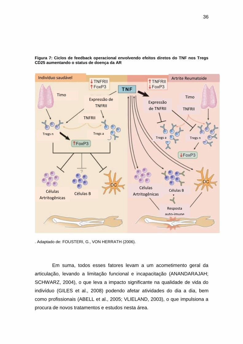
Um estudo publicado por Valencia et al. (2006), mostrou que pacientes
com AR possuem alterações nas células T regulatórias (Treg) CD25+
associados ao aumento da expressão da proteína Fox P23 e TNFα. As Treg
podem ser agrupadas em dois grandes gêneros: um intrínseco, gerado pelo
HIPEREMIA
VASCULAR
MASTÓCITOS
LINHA SINOVIAL
ESPESSA
CÉLULAS
PLASMÁTICAS
NEUTRÓFILO
CÉLULA B
CÉLULA T
CÉLULAS DENDRÍTICAS
MACRÓFAGO
FIBROBLASTO
OSTEOCLASTO
ARTICULAÇÃO COM AR
TÍBIA
SINOVIÓCITOS
MEMBRANA
SINOVIAL
CARTILAGEM
CÁPSULA
FÊMUR
ARTICULAÇÃO
SAUDÁVEL

35
timo (caraterizadas por expressão de CD25+ e Fox P3) (FEHERVAZI et al.,
2004); e Tregs geradas depois de imunização ou exposição a antígenos ou
autoantígenos (HOMANN et al., 1999). In vivo, as funções das Tregs são
baseadas na indução de citocinas como IL-10, Trasforming growth factor beta
(TGF beta) e IL 4 e 5; modulação das células apresentadoras de antígeno; e,
algumas vezes, têm efeitos supressores diretos em células T patogênicas.
Além disso, todas as Tregs podem agir como “supressoras expectadoras” por
suprimir outras células T efetoras de antígenos específicos diferentes e esta
habilidade faz com que sejam consideradas células imunossupressoras
fisiológicas e sítio-específicas de alto interesse terapêutico. Como ilustrado na
figura 7, na AR, ocorre efeito negativo direto do TNFα na regulação das
funções efetivas das Tregs CD4+, CD25+, o que inicia um ciclo vicioso de
aumento do processo patogênico da doença (EHRENSTEIN et al., 2004).
36
Figura 7: Ciclos de feedback operacional envolvendo efeitos diretos do TNF nos Tregs CD25 aumentando o status de doença da AR
. Adaptado de: FOUSTERI, G., VON HERRATH (2006).
Em suma, todos esses fatores levam a um acometimento geral da
articulação, levando a limitação funcional e incapacitação (ANANDARAJAH;
SCHWARZ, 2004), o que leva a impacto significante na qualidade de vida do
indivíduo (GILES et al., 2008) podendo afetar atividades do dia a dia, bem
como profissionais (ABELL et al., 2005; VLIELAND, 2003), o que impulsiona a
procura de novos tratamentos e estudos nesta área.
Resposta
auto-imune
Células B Células
Artritogênicas
Tregs a Tregs n
TNFRII
Expressão
de TNFRII
Timo
Células B Células
Artritogênicas
Tregs a Tregs n
TNFRII
Expressão de
TNFRII
Timo
Artrite Reumatoide Indivíduo saudável

37
3.3 TRATAMENTO PARA ARTRITE REUMATOIDE
O tratamento se inicia com o diagnóstico prematuro e monitoramento
da progressão da doença (BÉRTOLO, 2007). Na prática clínica, a terapia
padrão inclui glicocorticoides, anti-inflamatórios, analgésicos e DMARDs
(FERRAZ-AMARO et al., 2009; TORPY, 2011) que conferem apenas alívio
temporário, não interferindo na progressão da doença. Os analgésicos
disponíveis no Brasil são paracetamol e dipirona (BENUCCI et al., 2017;
PASSOS, 2016; PATEL et al., 2017; TORPY, 2011). Analgésicos opioides,
como codeína e tramadol podem ser administrados, porém com muita cautela,
pois em doenças crônicas trazem risco de sonolência, depressão sensorial e
dependência (BENUCCI et al., 2017; PASSOS, 2016; PATEL et al., 2017;
TORPY, 2011).
Além dos efeitos adversos, que fazem com que estes medicamentos
tenham que ser administrados com muita cautela, sendo evitados sempre que
possível ou empregados em doses mínimas, pelo menor tempo possível, em
forma decrescente e quando realmente necessários (PASSOS, 2016), muitos
pacientes não respondem positivamente, pois alguns indivíduos podem
continuar sofrendo com limitações funcionais (PINCUS et al., 1999). Com a
saúde debilitada, menor expectativa de vida, deficiências na qualidade de vida
que afetam as atividades laborais e relações sociais (CHORUS et al., 2003;
MCINNES; SCHETT, 2011). Desta forma, permanece a grande necessidade de
abordagens terapêuticas alternativas ou associadas ao uso dos medicamentos.

38
3.3.1. Exercício físico como tratamento
Atividade física é definida como movimento corporal produzido por
músculos esqueléticos que requer gasto de energia, o que influencia no
desenvolvimento e na saúde em geral ao longo de toda a vida (BAMMAN et al.,
2014; COLPANI et al., 2013; JUONALA et al., 2013). Dentro do seu mecanismo
de ação, há evidências crescentes de que o treinamento físico ativa
mobilização de células progenitoras endoteliais da medula óssea para facilitar a
regeneração celular (MOEBIUS-WINKLER et al., 2011), células progenitoras
neurais para induzir a hipertrofia fisiológica (XIAO et al., 2014) e de que os
tecidos: muscular, adiposo e ósseo podem funcionar como órgãos endócrinos
autênticos, produzindo e liberando proteínas na circulação que modulam
respostas fisiológicas durante e após o exercício (CATOIRE et al., 2014;
PEDERSEN; FEBBRAIO, 2008; YOU; NICKLAS, 2008).
Estes fatores promovem bem-estar cardiometabólico, melhoram o
desempenho cognitivo e ajudam efetivamente na prevenção e tratamento de
várias doenças, incluindo as cardiovasculares, neurológicas, sarcopenia,
osteoporose e câncer (STRANAHAN; MATTSON, 2012). E ainda, programas
de exercícios orientados de forma correta são essenciais para aperfeiçoar a
saúde de pessoas em uma ampla variedade de deficiências (PETERSON et al.,
2012).
Em seu estudo, Benhamou (2007) sugeriu que a atividade física era
prejudicial em pacientes com AR e que os profissionais da saúde orientassem
os portadores a evitar exercícios e manter o repouso. No entanto, estudos
publicados sugerem que os indivíduos acometidos podem se beneficiar com
segurança de atividades físicas (VLIELAND, 2003) de forma que, além de
trazer benefícios para a população geral, oferece melhor qualidade de vida
entre indivíduos com a doença, podendo ser considerada atividade
fundamental para atenuar sintomas, conforme demonstrado em várias
pesquisas, incluindo achados de estudos randomizados controlados (BAILLET,
et al., 2009; LEMMEY et al., 2009; NEUBERGER et al., 2007)
O exercício físico provoca uma série de respostas fisiológicas,
resultantes de adaptações autonômicas e hemodinâmicas (MONTEIRO;

39
FILHO, 2004) que geram tensão, os tendões transmitem forças do músculo
para o osso, a musculatura é comprimida e, por consequência, os vasos
arteriais periféricos, elevando drasticamente a resistência periférica total e
reduzindo a perfusão muscular. Para restabelecer o fluxo sanguíneo, ocorre
aumento na atividade do sistema nervoso simpático, no débito cardíaco e na
pressão arterial média (MCARDLE et a., 2001).
Em indivíduos com AR, ocorre inflamação sinovial em seus
revestimentos, o que leva à hipertrofia sinovial e algumas vezes à infiltração
nos tecidos tendinosos. O aumento de citocinas inflamatórias circulantes
também afeta o colágeno, levando a danos e desorganização de sua estrutura,
sob cargas mecânicas, estes se tornam mais rígidos, provendo produção
menos eficiente de força (REEVES et al., 2006; ONAMBELE et al., 2006).
A função principal da cartilagem na articulação sinovial é proteger o
osso de danos por meio da redução da fricção entre os ossos adjacentes
durante o movimento (MILNER, 2008). Na AR, as camadas articulares
superficiais são destruídas (RANNOU et al., 2006) e esta degeneração leva à
falha da articulação, limitação funcional e incapacitação (ANANDARAJAH;
SCHWARZ, 2004) e, depois de repousar por longos períodos, o fluido sinovial
é expelido para a cavidade articular resultando em contato entre as diferentes
áreas da cartilagem. Quando o movimento é retomado o mecanismo do fluido
de lubrificação é reativado, promovendo melhora na lubrificação e da saúde
articular (SCHOLES; UNSWORTH, 2004). Durante os períodos de compressão
e descompressão, os quais podem ser alcançados através de forças
mecânicas com exercícios regulares cíclicos em série, a cartilagem responde
de forma sítio-específica, o que previne a fragilidade e disfunção deste tecido
(AROKOSKI et al., 2004)
Todas estas respostas fisiológicas podem levar a benefícios, estes que
já foram demostrados por estudos de reabilitação com exercício físico em
indivíduos com AR, dentre os quais estão: aumento da amplitude de
movimento, flexibilidade, (FENTEM, 1994), diminuição significativa da dor,

40
melhorando a marcha e a função geral (SHIH et al., 2006; KAPALE et al.,
2017), redução da atividade inflamatória e seus sintomas, limitando a
destruição da articulação e incapacitação (KAPALE et al., 2017), melhora
cardiorrespiratória e saúde cardiovascular, aumento da massa muscular,
redução da adiposidade (incluindo gordura atenuada no tronco), melhora a
força muscular e condicionamento físico, bem estar psicológico (LEMMEY et
al., 2009), melhora na fadiga (NEILL et al., 2006) e na qualidade de vida geral
(ABELL et al., 2005).
Dentre as diversas modalidades de exercícios existentes, o
treinamento resistido passou a ganhar visibilidade como sendo uma das
melhores formas de treinamento físico visando saúde e qualidade de vida, é
uma modalidade que visa oferecer resistência contra a ação muscular por meio
da utilização de pesos, que podem ser os do próprio corpo (SANTARÉM, 1999)
e que, se trabalhada de forma responsável e adequada, pode trazer benefícios
como: aumento de força muscular, melhora da coordenação motora,
manutenção e aumento de flexibilidade, até o fortalecimento ósseo e articular,
que leva a uma melhora do desempenho das atividades de vida diária, e assim,
à funcionalidade e à autossuficiência (BERMUDES et al., 2003; POSNER et al.,
1995). Além da melhora dos sintomas anteriormente citados, ocorre também
diminuição da dor. Uma das explicações para a referida analgesia é a liberação
endógena de opioides pelo sistema nervoso central e periférico onde a ativação
de receptores centrais e periféricos resulta nos efeitos antinociceptivos, bem
como na expressão em células imunes para produzir significativa
antinocicepção (HUA; CABOT, 2010; LESNIAK; LIPKOWSKI, 2011)
Em suma, o papel do exercício na promoção da saúde articular na AR
pode ser de grande importância (MAINI; FELDMANN, 2004). Considerando ser
uma intervenção de bom custo-benefício (DE JONG et al., 2003;
STAVROPOULOS et al., 2013; METSIOS et al., 2009) e com poucos efeitos
colaterais. Porém, como as informações dos protocolos utilizados na AR ainda
são contraditórias (BENHAMOU, 2007; SCHOLES; UNSWORTH, 2004),
tornam-se necessárias mais pesquisas que mostrem os efeitos funcionais e
morfofisiológicos desta prática como tratamento desta condição inflamatória.

41
3.3.2. LBP como tratamento
O termo Laser vem do inglês: Light amplification by stimulated emission
of radiation, ou seja, amplificação de luz por emissão estimulada de radiação.
Trata-se de dispositivos que normalmente geram radiação eletromagnética
relativamente uniforme em comprimento de onda, fase e polarização,
originalmente descrita por Theodore Maiman em 1960 (MAIMAN, 1960) no qual
é utilizada uma fonte de luz ou energia de radiação (VERMA et al., 2012).
Uma das modalidades utilizadas é o laser de baixa potência, este afeta
os sistemas biológicos por meios não termais (LIN et al., 2010), por meio da
absorção da radiação vermelha e infravermelha que possui a capacidade de
promover analgesia, regeneração tecidual e reduzir processos inflamatórios
(SILVA et al., 2007).
A absorção de fótons leva a célula a estados eletronicamente excitados
e, consequentemente, à aceleração de reações de transferências de elétrons
(YU et al., 1997), o que ocasiona maior produção de adenosina trifosfato (ATP)
(PASSARELLA, 1989), e ao aumento de atividade de antiportes Na+/H+ e
Ca2+/Na+, e todos os transportadores acionados por ATP para íons, como
Na+/K+ ATPase e bombas de Ca2+.
Como o ATP é um substrato da adenilciclase, sua concentração também
controla a produção de AMP cíclico (AMPc). Ambos Ca2+ e AMPc são
mensageiros secundários muito importantes, uma vez que os íons Ca2+
regulam quase todos os processos do corpo humano (contração muscular,
coagulação sanguínea, transferência de sinais em nervos, expressão de genes,
entre outros) (HAMBLIN, 2009). Além disso, a foto ativação de enzimas
terminais, como Cox, desempenha papel vital na ativação de cascatas
biológicas diversas subsequentes à irradiação do laser, controlando a
proliferação celular, síntese proteica, depósito e organização do colágeno
(KARU et al., 2004).

42
As evidências de que o LBP proporciona ao organismo uma melhor
resposta a quadros inflamatórios, por meio da redução do edema, redução do
quadro álgico e bioestimulação celular (KHOZEIMEH et al., 2015; KARU 2004)
tornam o LBP um dos recursos de escolha no tratamento de inúmeras doenças
musculoesqueléticas, incluindo a AR (HAKER; LUNDEBERG, 1990; KLEIN;
EEK, 1990), para a qual este tratamento tem demonstrado tanto mudanças
locais quanto sistêmicas, incluindo diminuição da dor, edema e enrijecimento
articular, e melhora da resposta inflamatória aguda por redução de
prostaglandinas e inibição de ciclooxigenase local, o que acarreta estímulo do
processo de regeneração tecidual (HENRIQUES et al., 2010; MORIYAMA et
al., 2005).
Além destes benefícios, o LBP é uma terapia relativamente simples e
de baixo custo que tem se mostrado como ideal para dar suporte a tratamentos
convencionais (MORIYAMA et al., 2009), o que torna relevante pesquisas
sobre seus efeitos em indivíduos acometidos pela AR em associação com
outra modalidade terapêutica, que também tem se mostrado eficaz na
reabilitação da doença.
3.4. ESTUDOS EXPERIMENTAIS COM CFA
Pesquisas relacionadas à AR realizadas em humanos são difíceis de
ser controladas, pois testes repetidos tornam-se extenuantes e tanto a ingestão
de medicamentos, quanto hábitos diários de cada indivíduo podem interferir no
processo inflamatório. Estudos in vivo realizados em animais, utilizando
modelos experimentais de indução da artrite, podem gerar maiores
informações sobre essa problemática. Com a proposta de estudar a
etiopatogênese da artrite e na busca de diferentes tratamentos, muitos modelos
experimentais foram desenvolvidos ao longo dos anos para representar a
artrite em seres humanos (CAI et al., 2006; COOK; MOORE, 2006). Dentre os
modelos experimentais de AR desenvolvidos (CAI et al., 2006; COOK;
MOORE, 2006; NAGAKURA et al., 2003; YU et al., 2002), o CFA se destaca
(CAI et al., 2006; COOK; MOORE, 2006; NAGAKURA et al., 2003).

43
Em modelos animais, a manifestação e a gravidade dos sintomas
podem variar de acordo com o gênero do animal usado, forma e local da
administração das espécies de Mycobacterium, e o número e intervalo de
inoculações (CAI et al., 2006; COOK; MOORE, 2006; NAGAKURA et al., 2003).
A artrite induzida por CFA é um modelo adequado, pois mimetiza os sinais e
sintomas da AR humana, incluindo as mudanças histopatológicas, a infiltração
celular, a hipersensibilidade e o edema da articulação afetada (BARTON et al.,
2013).

44
ARTIGO 1
Physical exercise and low-level laser therapy effects on ankle joint in
experimental rheumatoid arthritis model.
Ana Caroline Barbosa Retameiro1, Morgana Neves1, Alana Ludemila Tavares1,
Taciane Stein Leal1, Gladson Ricardo Flor Bertolini1, Lucinéia de Fátima
Chasko Ribeiro.1
1Universidade Estadual do Oeste do Paraná (Unioeste), Universitaria St. 2069,
Cascavel, Paraná 85819-110, Brazil.

45
Physical exercise and low-level laser therapy effects on ankle joint in
experimental rheumatoid arthritis model.

46
ABSTRACT
Background. Rheumatoid Arthritis (RA) is an autoimmune and inflammatory
disease that affects, mainly, body members which includes the ankle joint.
Treatment involves physical exercises and low level laser therapy (LLLT) to
modulate the inflammatory process and maintain physical capacity. In this way,
the propose of this is study was analyze physical exercise and LLLT effects on
functional and morphological parameters of the ankle joint submitted to AR
model promoted by complete Freund’s adjuvant (CFA). Methods. For functional
analysis sample was composed by 64 male Wistar rats were randomly divided
into two major groups: control (n=32) and arthritis (n=32) and each one was
subdivided into four groups: treated and untreated (n=8) and for
histomorphometrical 40 male Wistar rats were used in the same way, but
control and arthritis (n=20) and treated and untreated (n=5). The RA promotion
occurred first by an immunization with CFA injection at the tail base and, after
seven days, by intra-articular injection for arthritis groups. For control groups the
same protocol was used, but with saline injection. Treatments with LLLT and/or
resisted stair climbing exercise, started 24 hours after the intra-articular
injection, performed on alternating days with progressive time and series for
exercise. Joint disability was evaluated by Sciatic Functional Index (SFI), hold
force and incline plane evaluation and ankle joint morphological aspects by
morphological and morphometrical analysis. The generalized mixed model test
was applied for all parameters, but with Fischer (LSD) post test for functional
and Sidak for morphometry (p=0, 05). Results. The association between

47
treatments showed positive effects on the functional measures once the
member functionality recovery the initial values through the evaluations; on
histological analysis the morphology presented differences in the arthritis group
that had no treatment. The synovial membrane presented subintimal with
angiogenesis revealing a large amount of blood vessels, which were turgid of
blood cells and inflammatory cells inside the vessels. The cartilage presented
smaller extracellular matrix and, consequently, higher chondrocyte density.
Therefore, the cartilage was degraded with pannus in some animals.
Conclusions. The arthritis model was effective in mimicking the disease in a
systemic way and the combination of the two treatments was able to help in
peripheral functionality.
Keywords
Experimental arthritis, Freund’s adjuvant, exercise, Laser, ankle joint.

48
INTRODUCTION
Rheumatoid arthritis (RA) is characterized as a systemic and frequently
progressive inflammatory disease [1] that affects about 1% of the population in
developed countries. In Brazil, there are few data about the disease, with an
incidence between 0,2 and 1% over the population [2].
Among progressive diseases that affect human health, RA has
increasing incidence, mainly because it is comorbidity related to other clinical
and psychosocial complications [3]. Its clinical manifestations can include: poor
appetite, fever, anemia and edema [4] and the most prominent are the articular
manifestations [5], especially in synovial joints, present in large movement
amplitude, which includes the ankle joint [6].
The ankle joint is involved in a lot of body movements, which play a key
role in walking activity and supporting the body in the standing position. It
comprises a distal syndesmosis between the tibia and fibula bones and a
socket formed by the tibia and fibula distal thirds bones with talus and its
involvement, as happens in RA, leads to biomechanical and functional changes
that negatively affect the patient’s life [7]. In individuals affected by RA is usually
observed persistent synovitis development with edema and reduced joint
mobility, resulting in tendons stretching and ligaments and joint capsules
instability, leading to immobility [8] and consequently, in muscle mass and

49
strength decrease, compromising the joint and surrounding tissues integrity,
resulting in pain and changes in movement patterns that limit daily activities [5]
as well as work activities [9], what show their great socioeconomic relevance.
Research related to RA in humans is sometimes difficult to control,
once the repeated tests become strenuous and medications ingestion or daily
habits may interfere in the inflammatory process. In this way, many
experimental models has been developed over the years, and the Freund’s
complete adjuvant (CFA) model stands out for being effective in mimicking
human RA symptoms [10].
Standard therapy is made with drugs, using pharmacological agents
related to inflammatory mechanisms [11]. However, these drugs usually have
no lasting action and may lead to several side effects manifestations [12];
Therefore, other therapeutic approaches have been sought.
There are studies showing physical exercise as positive RA individual’s
rehabilitation, showing increase in motion range and flexibility [13]; significant
decrease in pain and inflammatory activity, improving gait and general functions
[14,15] . However, morphofunctional data are generally concentrated in
previous models, mainly in the tibiofemoral joint, so that its action in the ankle
joint hasn’t been studied yet.
In addition to exercise, low level laser therapy (LLLT) has been applied
to treat a lot of musculoskeletal disorders, including RA [16, 17]. There is
evidence that LLLT promotes local and systemic changes which may lead to
decreasing pain, edema and joint stiffness. It also improves inflammatory

50
response reducing prostaglandins and inhibiting local cyclooxygenase, which
leads to tissue regeneration process stimulation [18, 19].
Although the literature brings the exercise and LLLT benefits, there are
gaps regarding the protocols used and the association effects of both
therapeutic modalities in RA treatment. Also, the CFA model causes changes in
the ankle joint morphofunctional aspects, as well as the treatment modalities.
Thus, the objective was test the exercise and LLLT effects on the ankle joint
functionality and morphological aspects in order to obtain information about the
RA treatment protocols that help professionals in their patients rehabilitation,
improving their life quality.
METHODS
Animals and experimental groups
The study is an experimental research and randomized into groups
using Microsoft Excel 2010, composed by 80 Wistar male rats, aged 15 weeks;
kept in plastic polypropylene boxes, with access to water and feed at will; and
controlled temperature at 21ºC ±1ºC, 12 hours light/dark photoperiod. The study
was approved by the Ethics Committee for Animal Use (CEUA) of the State
University of Western Paraná (Unioeste).
The animals were randomly separated into two big groups: arthritis
(n=40) and control (n=40). And subdivided into eight groups (n=5) as follows:
control group (CG), control laser (CL), control exercise (CE), control laser +
exercise (CLE), arthritis group (AG), arthritis laser (AL), arthritis exercise (AE)
and arthritis laser + exercise (ALE) (Figure 1).

51
Figure 1: Experimental groups. Are indicated: CG: Control Group; CL: Control Laser;
CE; Control Exercise; CLE: Control Laser and Exercise; AG: Arthritis Group; AL; Arthritis Laser;
AE: Arthritis Exercise and ALE: Arthritis Laser and Exercise.
CFA-induced disability
The arthritis model was induced by a pre-sensitized intradermal
injection at the tail base with 50 μL of CFA (0.5 mg/mL, Mycobacterium
butirycum) in AG, AE, AL and ALE animals. The CG, CL, CE and CLE groups
received saline solution (sodium chloride 0.9%) instead CFA. Thus, the
administration area where occurred the substances injection was trichotomized
and submitted to asepsis with iodinated alcohol (1%). Seven days after the first
stimulus in the tail, an intra-articular injection with 50 μL (0.5 mg/mL) of CFA
Mycobacterium butirycum or saline solution (according to the subgroups
mentioned above) was performed in the tibiofemoral joint, in the animals right
pelvic limb.
52
Evaluations
Animals
All the animals passed by functional evaluations. In this way, before the
experiment starts they were trained and adapted to the equipment. After that,
they were submitted to seven evaluations. A basal evaluation was made seven
days after the tail injection and before the intra-articular application. After 24
hours, and before the treatment starts, evaluation 1 (EV1) was performed. On
the third, fifth, 12th, 19th and 26th days after intra-articular injection EV2, EV3,
EV4, EV5 and EV6, respectively, were performed. On the 28th day, the animals
were euthanized (Figure 2).
The muscle strength evaluation was performed using a grip strength
meter (Insight®, Ribeirão Preto, São Paulo), described by Bertelli and Mira [20].
The animal was positioned so that it grasped on a grid connected to a force
transducer, with the right pelvic limb, then pulled by the tail with increasing force
until the grip is lost, at which time. The device provides the force exerted by the
animal. The left pelvic limb was immobilized by the evaluator’s hand so as not
to interfere in the evaluation.
Motor function was evaluated using the inclined plane (Insight®,
Ribeirão Preto, São Paulo). The test measured the animal’s ability to remain in
top of a ramp whose inclination increased five degrees every five seconds. The
maximum angle which the animal remained on the ramp without slipping for at

53
least five seconds defined the inclined plane test score. The measurements
were performed with the animal positioned in the transverse plane (vertical
position).
Figure 2: Evaluations schedule.
Treatment Protocol
For CE, CLE, AE and ALE the treatment was made through resisted
climbing exercise in stairs use. For this, a vertical wooden ladder with 67 iron
steps, high of 1.18 m, width 20.5 cm and 60º inclination was used. At the top of
the stairs was located a box, with 20X20 cm in height and width, in which the
animals rested between one series and another with a 60 s interval. The
treatment starts 24 hours after the intra-articular injection. For this, was used a
100g overload coupled to the tail. In the first week the animals performed four
series of five climbs on the stairs and in the second week, with the same
overload, the series changed for four with seven ascents, and in the last week
for four series of 10 ascents completing 14 days of intercalated treatment.
In the CL, CLE, AL and ALE treatment the animals were submitted to
LLLT (Laserpulse Ibramed®). The other groups’ animals received the pen
contact, but without beam emission, in the sensitized pelvic limb in the knee
region. The application points were anterior to the patella, medial face in the
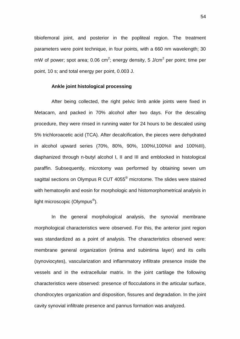
54
tibiofemoral joint, and posterior in the popliteal region. The treatment
parameters were point technique, in four points, with a 660 nm wavelength; 30
mW of power; spot area; 0.06 cm2; energy density, 5 J/cm2 per point; time per
point, 10 s; and total energy per point, 0.003 J.
Ankle joint histological processing
After being collected, the right pelvic limb ankle joints were fixed in
Metacarn, and packed in 70% alcohol after two days. For the descaling
procedure, they were rinsed in running water for 24 hours to be descaled using
5% trichloroacetic acid (TCA). After decalcification, the pieces were dehydrated
in alcohol upward series (70%, 80%, 90%, 100%I,100%II and 100%III),
diaphanized through n-butyl alcohol I, II and III and emblocked in histological
paraffin. Subsequently, microtomy was performed by obtaining seven um
sagittal sections on Olympus R CUT 4055® microtome. The slides were stained
with hematoxylin and eosin for morphologic and histomorphometrical analysis in
light microscopic (Olympus®).
In the general morphological analysis, the synovial membrane
morphological characteristics were observed. For this, the anterior joint region
was standardized as a point of analysis. The characteristics observed were:
membrane general organization (intima and subintima layer) and its cells
(synoviocytes), vascularization and inflammatory infiltrate presence inside the
vessels and in the extracellular matrix. In the joint cartilage the following
characteristics were observed: presence of flocculations in the articular surface,
chondrocytes organization and disposition, fissures and degradation. In the joint
cavity synovial infiltrate presence and pannus formation was analyzed.

55
For histomorphometric analysis, the interest regions were
photomicrographed using Olympus® DP71 microscope. The protocol for
measuring articular cartilage thickness and chondrocyte number was adapted
from Ando et al. and Hagiwara et al. For this, three distinct regions were
defined: P1, anterior end (near the phalanges); P2, joint middle region and P3,
posterior articular end.
The tibia and talus middle or P2 regions were selected as analysis
regions, since they are the site of highest joint weight discharge (Kunz et al.,
2014). Photomicrographs were performed at 400X magnification for the
following measures performing: total cartilage thickness, defined as the distance
between the cartilaginous surface and the osteochondral junction; the surface
zone thickness comprising the space between the cartilage surface and
tidemark; and the deep zone thickness which is bounded superiorly by the
tidemark and inferiorly by the subchondral bone. The chondrocytes number was
counted using an 300um X 300um area where only the chondrocytes that was
fully within the borders limits were considered.
Statistical analysis
Statistical analyzes were performed using the BioStat program through
the generalized mixed models test. For the functional analyzes, the LSD post-
test was used and for the histomorphometric analyzes, the Sidak post-test was
used. The significance adopted was p<0.05.

56
RESULTS
In muscle strength there was significant difference in the member
functionality evaluation between the evaluations (F(4.3)= 9.9, p<0.0002),
between the groups (F=(7.56)=5.8 p< 0.00004), and there was interaction
between groups and evaluations (F(32.3)=3.4, p<0.0001) (Table 1).
Within groups, there was significant difference between control groups
(CG, CL, CE and CLE) and the arthritis groups that were treated (AL, AE and
ALE) (p<0.0002) and values returns close to control values in all the treated
groups. Therefore, the CE group showed a positive difference in relation to all
the other groups (Table 1).
Between groups, the control groups were different from all the arthritis
groups (p< 0.00004), and all the treated groups (AL, AE and ALE) had values
returned close to control values.
The motor function evaluation showed differences between the
evaluations (F(7.4)=23.6, p<0.0001), between the groups (F(7.56)=5.4,
p<0.0001) and there was interaction between groups and evaluations
(F(49,4)=5.9, p<0.0002) (Table 2).
Within groups, there was significant difference between groups with
arthritis that were treated (AL, AE and ALE) and groups without arthritis (CG,
CL, CE and CLE) (p<0.0001). In EV4 the AL group returned to the initial values
and in AV5 AE and AEL restored the initial values to (Table 2).
Between groups the animals of the control groups (CG, CL, CE and
CLE) had significant differences (p<0.0001) when compared with all arthritis

57
groups. In EV4 all the arthritis groups (AG, AE, Al and ALE) restored the initial
values (Table 2)

58
Table 1: Grip strength exercised throughout the evaluations within
the same group and between groups
AV0 AV1 AV2 AV3 AV4 AV5 AV6
GC 64,5±13aA
61,9±9,4 abAB
85,7±22,5acA
77,0±14,2a AC
76,9±19,5a A
83,4±21,2a
AC
78,6±19,9aA
GA 62,1±13,4a A
39,5±19a AB
43,6±15,7aB
46,7±11,6 a B
49,3±9,3a B
51,4±9,8aB
50,1±3, 7
aB
CL 66,5±14,4 a A
73,7±21,7 a A
80,6±14,4 aA
75,4±13,7 a AC
73,0±11,8 a A
77,2±16,2a
AC
78,5±16,8 a A
AL 60,9±16,1 abA
50,8±20,5 abAB
48,9±17,9 abB
48±15,7 a B
58,4±12,0 abAB
60,0±15,5
abABC
63,7±15,3
abAB
CE 71,8±14,1abc A
66,3±12,4abcAB
66,4±24,8abcAB
75,9±13,4abcAC
85,2±12 abA
89,9±15,3a
AC
83,7±14,7abA
AE 62,9±6 abA
48,9±18,4 abAB
46,7±16,6 aB
48,5±14 aB
51,2±17,8 aB
56,9±17,8
abBC
65,8±16,5
abAB
CLE 64,0±17,8aA
73,8±22,3aA
76,9±14,8aA
79,6±16,8a AC
79,5±16,1a A
77,0±19,5a
AC
79,9±10,4aA
ALE 65,6±12,8abA
48,2±23,5aAB
46,6±16,5aB
56,8±16,6abAB
63,8±14,4abAB
61,0±14,6ab
AC
69,2±20,3abAB
Values are expressed in mean ± standard error. Different letters represents different values. In
the lines, the lowercase letters, comparison within the same group and in columns, with capital
letters, comparison between groups throughout the evaluations.

59
Table 2: Maximum angle reached by animals during the evaluations within
the same group and between groups.
EV0 EV1 EV2 EV3 EV4 EV5 EV6
CG 59.8±0.7 aA 58.8±1aA 59,6±1 a ACD 59.8±1 aAC 59.4±1 aA 59.4±0.8 a A 60.2±0.8 a A
AG 59.0±0.7 a A 58.3±1a A 58,1±1aABCD 59±1aAC 57.7±1 a A 58.8±0.8 a A 57.7±0.8 a ABC
CL 58.3±0.7 aA 57.3±1a A 57,7±1 aACD 57.1±1a AC 55.4±1 a A 55.6±0.8 a A 55.2±0.8 aBC
AL 58.8±0.7 aA 48.8±1.0bcB 53,5±1b BD 54.6±1,1b BC 56.9±1aA 58.3±0.8aA 57.9±0.8aAC
CE 58.3±0.7 aA 58.3±1 a A 59,0±1 aACD 58.6±1 aAC 58.8±1 aA 58.5±0.8 a A 57.9±0.8 aAC
AE 58.3±0.7 a A 50.0±1.0bcB 54,4±1bd BCD 55.6±1 adAC 57.3±1 adA 58.1±0.8 a A 58.1±0.8 a AC
CLE 60.8±0.7 a A 59±1 a A 58,5±1 a ACD 58.8±1 a AC 58.6±1 a A 59.2±0.8 a A 59.6±0.8 a A
ALE 57.9±0.7ac A 47.5±1. bB 54,6±1ac BCD 56.2±1ac AC 57.7±1 ac A 58.8±0.8 a A 59.0±0.8 a A
Values are expressed in mean ± standard error. Different letters represents different values. In
the lines, the lowercase letters, comparison within the same group and in columns, with capital
letters, comparison between groups throughout the evaluations.

60
Histomorphometrical analysis
Articular cartilage morphometry
The histomorphometical analysis of P2 articular cartilage of both tibia
and talus showed no significant difference in any of the analyzed parameters
(Table 3 and 4).
Table 3: Tibia histomorphometrical analysis
TIBIA Total Thickness Superficial Zone Deep Zone Chondrocytes
CG 154±11.7 a 70.6±7.4 a 82.7±8.3 a 14.9±1.1 a
AG 142.8±11.7 a 55.7±8 a 85.1±9.3 a 21.5±2 a
CL 162.5±11.5 a 72.5±7 a 89±8.4 a 16.5±1.2 a
AL 156.6±12 a 71±7.4 a 84.7±8.5 a 19±1.5 a
CE 171.2±11.5 a 75.7±6.5 a 94.7±8.4 a 18.3±1.3 a
AE 161.5±11.5 a 75.8±7 a 84.6±8 a 18.4±1.3 a
CLE 164±11.6 a 74±7 a 87.1±8.2 a 18.1±1.3 a
ALE 153.7±12 a 66.3±7 a 87.4±8.2 a 17±1.2 a
Values are expressed in mean ± standard error. Different letters represents different
values.

61
Table 4: Talus histomorphometrical analysis
TALUS Total Thickness Superficial Zone Deep Zone Chondrocytes
CG 140 ±10.6a 44.1±5 a 95.6±8.3 a 17.1±1.1 a
AG 139.3±11.5 a 52.7±6.4 a 84.8±8.9 a 21.7±1.7 a
CL 124.1±10 a 48.1±5.1 a 75.1±7.7 a 17.6±1.1 a
AL 118.3±10.6 a 40±4.4 a 80±8.3 19.4±1.3 a
CE 122.9±9.4 a 43.1±4.3 a 80.3±7.3 a 18.2±1.1 a
AE 118.5±10 a 45±4.7 a 72.7±7.7 a 16±1 a
CLE 126.7±10 a 48±5 a 78.2±7.7 a 19±1.2 a
ALE 131.4±10 a 45.1±4.7 a 87.5±7.7 a 19.7±1.2 a
Values are expressed in mean ± standard error. Different letters represents different
values.
Morphological analysis
Synovial Membrane
The control group presented characteristic morphology with two to three
cell layers, types A and B synoviocytes in the synovial intima, and subintima
with adipose cells predominance (Figure 3A). The arthritis group (AG) showed
subintima with angiogenesis, revealing a large blood vessels number, which
were turgid with red blood cells with inflammatory cells inside the vessels
(Figure 3B). The laser groups (CL and AL), exercise (CE and AE) and groups
where the association of the two treatments was performed (CLE and ALE), had

62
the same characteristics of angiogenesis and red blood cells congestion,
however, was not observed the presence of inflammatory cells.

63
H G
F E
D C
B A
IN SI
BV
BV
IN
SI
BV
IN
BV
SI
IN
* BV
SI IN
AD
AD
AD
AD
SI
BV BV
SI
IN
BV
SI IN
AD IN
SI
AD
AD
64
Figure 3: Wistar rats talocrural joint synovial membrane photomicrographs, 28 days
after induction of knee arthritis by CFA, sagittal section, hematoxylin and eosin staining. A:
Control group; B: Arthritis group; C: Control Laser; D: Arthritis Laser; E: Control exercise; F:
Arthritis exercise; G: Control laser and exercise and H: Arthritis laser and exercise. Are
indicated: Intimate layer (IN), Subintimate layer (SI), Blood vessels (BV) filled by red blood cells;
adipocytes (AD) and inflammatory infiltrate (*).
Articular cartilage
The CG group showed, in tibia and talus, characteristic morphology,
that is, smooth articular surface and chondrocytes organized in four cell layers.
In the superficial zone was identified a higher cell density arranged in horizontal
with flattened clusters. In the intermediate zone the chondrocytes assumed
rounded aspect and were arranged alone or in isogenic groups. The deep zone
showed chondrocytes organized in gaps, and it was separated from the
calcified zone by a basophilic line called tidemark. The subcondral bone also
had a normal appearance (Figure 4A).
In the GA group, the cartilage presented a smaller extracellular matrix
amount and the chondrocytes, consequently, with higher density. In addition,
the superficial zone was totally disorganized with cartilage degradation and
pannus in 50% of the analyzed animals, one animal only in the talus and the
other in both talus and tibia (Figure 4B).
The CL, CE, CLE, AL and AE groups showed no alterations,
morpholologically resembling the CG group.

65
H G
F E
D C
B A
AC
SB
SB
TA
TI
AC
SB
SB
AC
AC
TA
TI
SB
SB
TA
TI
AC
AC
TA
TI
SB
TA
TI

66
Figure 4: Wistar rats talocrural joint cartilage photomicrographs, 28 days after arthritis
induction in the knee by CFA, sagittal section, hematoxylin and eosin staining. A: Control group;
B: Arthritis group; C: Control Laser; D: Arthritis Laser; E: Control exercise; F: Arthritis exercise;
G: Control laser and exercise and H: Arthritis laser and exercise. Are indicated: Tibia (TI), Talus
(TA) Articular cartilage (AC), Subchondral bone (SB), Tidemark (arrow), Articular cavity (star);
Flocculation (FL) and pannus (PA).
DISCUSSION
Both treatments used in this study showed positive effects in functional
evaluations (muscle strength and motor function). Many studies showed that
physical activity promotes pain reduction [21-26]. Exercise is a form of stressful
stimulus capable of promoting changes in homeostasis with responses
reorganizations mainly from the neuroendocrine system, probably generating an
increase of endogenous glucocorticoids serum [27].
All of these physiological responses can lead to benefits, which have
already been demonstrated by rehabilitation studies with physical exercise in
individuals with RA, among which are: increased range of motion, flexibility [13]
significant decrease in pain, improving gait and general function [14, 15],
cardiorespiratory improvement and cardiovascular health, increased muscle
mass, reduced adiposity, improves muscle strength and physical fitness,
psychological well-being [28], improves fatigue [39] and overall life quality [21].
Hurkmans et al. inferred in a systematic review that physical exercises
and behavioral education in patients with RA are recommended for clinical use
and LLLT therapy is among the proposed guidelines, but the recommendation
parameters are not well clarified [30]. In this study, LLLT also showed
improvements in the functional parameters. Brosseau et al. made a systematic

67
review relating the LLLT efficacy in RA treatment and concluded that laser
therapy has a beneficial effect when used at least four weeks, with effects on
pain reduction and morning stiffness [31].
A study made by Khozeimeh et al., 2015 showed that LLLT therapy
provides the body a better response to inflammatory conditions, by reducing
edema, reducing pain and provides cellular biostimulation [32]. In addition to
these benefits, LLLT is relatively simple and low-cost therapy that has been
shown to be ideal for supporting conventional treatments.
Hsieh et al.showed that inflammatory action and functionality recovery
using LLLT therapy is related actin in modulation of inflammatory mediators,
indicating beneficial effects using 4.5 J/cm2 with inflammation reduction [33]
what corroborates with the results of the present study.
The morphometrical analysis in this study didn’t show significant
differences in any tested parameters, but the morphological analyses showed in
AG a subintima with angiogenesis, revealing a large amount of blood vessels,
turgid with red blood cells, with inflammatory cells inside of this on synovial
membrane.
Firestein, 2003 showed on his study that the greater involvement of
synovial membrane structure occurs due to hyperplasia in the inflammatory
process which configures synovitis [34], since the large number of immune
system cells that invade this structure, lead to cell proliferation,
neovascularization and germinal formation of germinal lymphoid follicles. The
mechanisms involved in the inflammatory cells recruitment into synovial

68
membrane have been extensively studied [35] showing synovial hyperemia [36],
fibroblast-like invasive synoviocytes proliferation that resist apoptosis and
increased in the property of adherence and invasion[38], leukocyte infiltration,
associated neoangiogenic response [37, 38](; changes in the function of T and
B lymphocytes, abnormal production of cytokines and antibodies [39, 40].
On this study the treated arthritis groups (AL, AE and ALE) also
presented the same characteristics of angiogenesis and red blood cells
congestion, however inflammatory cells presence was not observed. Scholes et
al. demonstrated that the degeneration caused by RA in joints causes
immobility what leads the synovial fluid to rest for long periods and when the
movement returned, the lubrication fluid mechanism is reactivated, promoting
improvement in lubrication and joint health [41].
Khozeimeh et al. brings evidence that LLLT treatment provides the
body with a better response to inflammatory conditions reducing edema, pain
and making cellular biostimulation [32]. In addition, other studies had point LLLT
as a therapy that has demonstrated both local and systemic changes, including
decrease in pain, edema and joint stiffness, reduction in prostaglandins and
inhibition in local cyclooxygenase, what leads to tissue stimulation and
regeneration process [42, 19, 20].
In the present study, the AG articular cartilage morphology showed a
lower extracellular matrix amount and higher chondrocytes density. In addition,
the superficial zone was totally disorganized with cartilage degradation and
pannus presence in 50% of the analyzed animals. These results corroborate
with the studies of Kapale et al., where the immune system cells are described

69
as forming a fibrous abnormal layer of tissues, called as pannus, which releases
substances that accelerate bone erosion and cartilage destruction, what leads
to loss of its shape and alignment, resulting in deformations [15]. These
deformations leads the joints to become fixed due to the tick and hardened
structure, which results in edema, articular tissue modification and, due to
disuse, in adjacent structures atrophy, such as: skin, bones, muscles, nerves
and even blood vessels connective tissues [34].
In the present study, the CFA Application occurred in the knee joint, we
analyzed the ankle joint and the morphological analysis showed characteristic
arthritis morphology in GA group. These results may show successful
systematic characteristic of the sickness [1, 34, 43] in CFA experimental model.
CONCLUSION
Both treatments (LLLT and exercise) and the combination of treatments
had positive effect on the functional evaluations and the morphology showed
efficacy in systematic arthritis induction.
REFERENCES
1. Khurana, R., Berney, S.M. Clinical aspects of rheumatoid arthritis.
Pathophysiology. 2005; 12:153-165.
2. Mota, L.M., Cruz, B.A., Brenol, C.V., Pereira, I.A., Fronza, L.S., Bertolo,
M.B., Freitas, M.V.C., Silva, N.A., Junior, P.L., Giorgi, R.D.N., Lima,
R.A.C., Pinheiro, G.R.C.Consensus of the Brazilian Society of
Rheumatology for diagnosis and early assessment of rheumatoid
arthritis. Rev. Bras. Reumatol. 2011; 51:199-219. 2011.

70
3. Margaretten, M., Julian, L., Katz, P., Yelin, E. Depression in patients with
rheumatoid arthritis: description, causes and mechanisms. Int. J. Clin.
Rheumatol. 2011; 6: 617-623.
4. Torpy, J.M., Perazza, G.F., Golub, R.M. Rheumatoid Arthritis. JAMA.
2011; 305, 1.
5. Tufts, M.H. Rheumatoid Arthritis. From the AJN Archives. 2012; 112.
6. De Carvalho, M.R., Tebexreni, A.S, Salles, C.A., Barros Neto, T., Natour,
J. Oxygen uptake during walking in patients with rheumatoid arthritis – a
controlled study. J. Rheumatol. 2004; 31, 655-662.
7. Bogart, B.I., Ort, V.H. Anatomia e Embriologia. 2008; 1.ed. Rio de
Janeiro: Elsevier.
8. Giles, J.T., Bartlett, S.J., Andersen, R.E., Fontaine, K.R., Bathon,
J.M.Association of body composition with disability in rheumatoid
arthritis: impact of appendicular fat and lean tissue mass. Arthritis Care
Res.2008; 59, 1407-1415.
9. Abell, J.I., Hootmann, J.M., Zack, M.M., Moriarty, D., Helmick, C.G.
Physical activity and health related quality of life among people arthritis.
J. Epidemiol. Commun. H. 2005; 59, 380-385, 2005.
10. Faria, T.J., Souza-Silva, E., Oliveira, D.T., Senna, E.L., Tonussi,
C.R.Evaluation of the pro-inflammatory potential of nanostructured drug
carriers in knee-joints of rats: effect on nociception, edema, and cell
migration. J Pharm Sci. 2009; 98, 4844-4851.
11. Tak, P.P., Kalden, J.R.. Advances in rheumatology: New targeted
therapeutics. Arthritis Res. Ther. 2011; 13,1-5.
12. Benucci, M. Factors correlated with the improvement of endothelial
dysfunction during Abatacept therapy in patients with rheumatoid
arthritis. J Inflamm Res. 2017; 11, 247-252.
13. Fentem, P.H. Benefits of exercise in health and disease. BMJ.1994; 308,
1291–1295.
14. Shih, M., Hootman, J.M., Kruger, J., Helmick, C.G. Physical Activity in
Men and Woman with Arthritis. Am J Prev Med. 2006; 30, 385-93.

71
15. Kapale, P., Vardharajulu, G., Warude, T.Effect of free exercise and
Rheumathoid arthritis. Indian J Physiother Occup Ther. 2017; 11, 63-67.
16. Asada, K., Yutoni, Y., Shimazu, A.Diode laser therapy for rheumatoid
arthritis. A clinical evaluation of 102 joints treated with low reactive-level
laser therapy (LLLT). Laser Ther. 1989; 1, 147-51.
17. Palmgren, N., Jensen, G.F, Kaae, K., Windelin, M., Colov, H.C. Low-
powered laser therapy in rheumatoid arthritis. Lasers Med. Sci. 1989; 4,
193-6.
18. Henriques, A.C.G., Cazal, C., Castro, J.F.L. Ação da laserterapia no
processo de proliferação e diferenciação celular: revisão de literatura.
Rev. Col. Bras. Cir. 2010; 37, 295- 302.
19. Moriyama, Y., Moriyama, E.H., Blackmore, K., Akens, M.K,, Lilge, L. In
vivo study of the inflammatory modulating effects of lowlevel laser
therapy on iNOS expression using bioluminescence imaging. Photochem
Photobiol. 2005; 81, 1351–1355.
20. Bertelli, J.A., Mira, J.C. The grasping test: a simple behavioral method
assessment of peripheral nerve regeneration in the rat. J. Neurosc.
Methods. 1995; 1.
21. Abell, J.I., Hootman, J.M., Zack, M.M., Moriarty, D., Helmick, C.G.
Physical activity and health related quality of life among people arthritis.
J. Epidemiol. Commun. H. 2005; 59, 380-385.
22. Benhamou, M.A.M. Reconditioning in patients with rheumatoid arthritis.
Ann. Readapt Med Phys. 2007; 50, 382-5.
23. Rall LC, Roubenoff R. Benefits of exercise for patients with rheumatoid
arthritis. Nutr Clin Care. 2000;3:209-15.
24. Shih M, Hootman JM, Kruger J, Helmick CG. Physical activity in men and
woman with arthritis. National Health Interview Survey, 2002. Am J Prev
Med. 2006;30:385-393.

72
25. Ottawa Panel Evidence-Based Clinical Practice Guidelines for
Therapeutic Exercises in the Management of Rheumatoid Arthritis in
Adults. Phys Ther. 2004;84: 934-972.
26. Van Den Ende CHM, Vliet Vlieland TPM, Muneke MW, Hazes MW.
Dynamic exercise therapy in rheumatoid arthritis: A systematic rewiew.
Br J Rheumatol. 1998;37:677-687.
27. Gomes, P.R., Bressan, E., Silva, T.M., Gevaerd, M.S., Tonussi, C.R.,
Domenech, S.C. Efeitos de um minuto e dez minutos de deambulação
em ratos com artrite induzida por adjuvante completo de Freund sobre
os sintomas de dor e edema. Rev. Bras. Reumatol. 2014; 54, 83-89.
28. Lemmey, A.B, Marcora, S.M., Chester, K., Wilson, S., Casanova, F.,
Maddison, P.J. Effects of high-intensity resistance training in patients
with rheumatoid arthritis: a randomized controlled trial. Arthritis Care
Res. 2009; 61, 1726–1734.
29. Feldthusen, C., Mnnerkorpi, K. Factors of importance for reducing fatigue
in persons with rheumatoid arthritis: a qualitative interview study. BMJ
open. 2019; 0, p. 1-8.
30. Hurkmans EJ, Jones A, Li LC, Vlieland TPMV (2011) Quality appraisal of
clinical practice guidelines on the use of physiotherapy in rheumatoid
arthritis: a systematic review. Rheumatology 50: 1879–1888.
31. Brosseau L, Welch V, Ga W et al. Low level laser therapy (Classes I , II
and III) for treating rheumatoid arthritis. Cochrane Database Syst Rev.
2005; 19:CD002049.
32. Khozelmeh, F. Comparative evaluation of low level laser and systemic
steroid therapy in adjuvant-enchanced arthristis of rat
temporomandibular joint: a histological study. Dent. Res. J. 2015;12,
215-223.
33. Hsieh YL, Fan YC, Yang CC. Low-level laser therapy alleviates
mechanical and cold allodynia induced by oxaliplatin administration in
rats. Support Care Cancer. 2016; 24:233–242.
34. Firestein, G.S. Evolving concepts of rheumatoid arthritis. Nature. 2003;
423, 356-361.

73
35. Bartok, B., Firestein, G.S. Fibroblast-like synoviocytes: key effector cells in rheumatoid arthritis. Immunol Rev. 2010; 233, 233-255.
36. Carotti, F., Salaffi, P., Manganelli, D., Salera, D., Simonetti, B., Grassi, W. Power Doppler sonography in the assessment of synovial tissue of the knee joint in rheumatoid arthritis: a preliminary experience. Ann Rheum Dis. 2002; 61, 877–882.
37. Lefévre, S. Synovial fibroblasts spread rheumatoid arthritis to unaffected joints. Nat Med. 2009; 15, 1414–1420.
38. Bottini, N., Firestein, G.S. Duality of fibroblast-like synoviocytes in RA: Passive responders and imprinted aggressors. Nat Rev Rheumatol. 2013; 9, p. 24-33.
39. Mcinnes, I.B., Schett, G.The pathogenesis of rheumatoid arthritis. N Engl J Med. 2011; 365, 2205–2219.
40. Bugatti, S., Codullo, V., Caporali, R., Montecucco, C.B. B cells in rheumatoid arthritis. Autoimmun Rev. 2007; 7,137-142.
41. Fekete, A., Soos, L., Szekanecz, Z., Szabo, Z., Szodoray, P., Barath, S. Disturbances in B- and T-cell homeostasis inrheumatoid arthritis: suggested relationships withantigen-driven immune responses. J Autoimmun.2007; 29, 154-163.
42. Bjordal, J.M. A systematic review of low level laser therapy with location specific doses for pain from chronic joint disorders. Aust J Physiother. 2003; 49, 107–116.
43. Can, C., Cinar, M.G., Kosay, S., Evinc.A. Vascular endothelial dysfunction associated with elevated serum homocysteine levels in rat adjuvant arthritis: effect of vitamin E administration. Life Sci. 2002; 71, 401-410.

74
ARTIGO 2
Physical exercise and low-level laser therapy systemic effects on ankle
joint in acute experimental rheumatoid arthritis model.
Ana Caroline Barbosa Retameiro*1, Morgana Neves1, Alana Ludemila Tavares1,
Taciane Stein Leal1, Gladson Ricardo Flor Bertolini1, Lucinéia de Fátima
Chasko Ribeiro1
1Universidade Estadual do Oeste do Paraná (Unioeste), Universitaria St. 2069,
Cascavel, Paraná 85819-110, Brazil.
*Corresponding Author
Full name: Ana Caroline Barbosa Retameiro
Department: Centro de Ciências Biológicas e da Saúde
Institute/University/Hospital: Universidade Estadual do Oeste do Paraná
(Unioeste)
Street Name & Number: Universitaria St. 2069
City, State, Postal code, Country: Cascavel, Paraná 85819-110, Brazil.
Tel: (45) 99112-0023
E-mail: [email protected]
Keywords: Experimental arthritis, Freund’s adjuvant, exercise, Laser, ankle joint
75
ABSTRACT
Background. Rheumatoid arthritis (RA) is a chronic, autoimmune, and
systemic inflammatory disease characterized by symmetrical synovitis of
peripheral joints, which includes the ankle joint and, if untreated, may lead to
joint destruction and a loss of functional capacity. Treatment may involve
physical exercises and low level laser therapy (LLLT) to reduce pain, disability
and modulation of inflammatory process, what can improve functionality. In this
way, the propose of this is study was analyze physical exercise and LLLT
effects on functional and morphological parameters of the ankle joint submitted
to AR acute model promoted by complete Freund’s adjuvant (CFA). Methods.
For functional analysis 64 male Wistar rats were randomly divided into two
major groups: control (n=32) and arthritis (n=32) and each one was subdivided
into four groups: treated and untreated (n=8) and for histomorphometrical 40
male Wistar rats were used in the same way, but control and arthritis (n=20)
and treated and untreated (n=5). The RA promotion occurred first by an
immunization with CFA injection at the tail base and, after seven days, by intra-
articular injection for arthritis groups. For control groups the same protocol was
used, but with saline injection. The treatments with LLLT and/or resisted stair
climbing exercise, started 24 hours after the intra-articular injection, performed
on alternated days with progressive time and series for exercise. Joint disability
was evaluated by Sciatic Functional Index (SFI), hold force and incline plane
evaluation and ankle joint morphological aspects by morphological and
morphometrical analysis. The mixed generalized model test was applied for all
parameters, but with Fischer (LSD) post test for functional and Sidak for
morphometry (p=0, 05). Results. The association between LLLT and exercises
presented positive changes in functional parameters; in the histological analysis
the arthritis group synovial membrane showed subintimal with angiogenesis
revealing large amount of blood vessels, which were turgid of blood cells and
inflammatory cells inside the vessels. The arthritis articular cartilage presented
flocculation on the joint surface, chondrocytes disorganization and degeneration
of the superficial layers with subchondral bone exposure and pannus. The
arthritis groups treated only with LLLT and both treatments had disorganized
chondrocytes and tidemark absence in some animals. The arthritis group
treated with exercise showed greater amount of chondrocytes, as confirmed in
the histomorphometric analysis. Conclusion. The arthritis model was effective
in mimicking the disease in a systemic way, the combination of the two
treatments was able to help in peripheral functionality and exercise helped in
morphological recovery of the ankle joint.

76
INTRODUCTION
Rheumatoid arthritis (RA) is a chronic, autoimmune, and systemic inflammatory disease that occurs in 0.24% to 1% of the population [1]. Due to its autoimmune, systemic [2] and symmetrical [3] characteristics, many organs and body tissues can be affected. However, the ones that stand out most are articular manifestations [4], mainly in the synovial ones, which includes the ankle joint [5].
Ankle joint comprises a distal syndesmosis between the tibia and fibula bones and a socket formed by the by tibia and fibula distal thirds bones with talus that is involved in a lot of body movements, which play a crucial role in walking activity and supporting the body in the standing position and its commitment, as happen in RA, leads to biomechanical and functional changes that causes limitations that negatively affects the patient’s life [6].
The main clinical manifestations includes: severe joint pain, reduced muscle strength, stiffness and fatigue and limited physical movements that are usually followed by edema and heat, accompanied by muscle pain that can worsen and persist for weeks or months [7] what limits the individual’s daily activities [4] as well as work activities [8] which shows the great socioeconomic relevance in RA study.
The standard therapy to RA is usually made with drugs, with pharmacological agents related to inflammatory mechanisms [9]. However, these drugs usually have no lasting action and may lead to several side effects manifestations [10]; In this way, other therapeutic approaches have been researched. The research is related to RA in humans is difficult to control, once the repeated tests become strenuous and medications ingestion or daily habits may interfere in the inflammatory process. Therefore, many experimental models have been developed over the years, and the Freund’s complete adjuvant (CFA) model stands out for being effective in mimicking human RA symptoms [11].
Although the literature brings the exercise and LLLT benefits, there are gaps regarding the protocols used and the association effects of both therapeutic modalities in RA treatment. Studies show that if worked in a responsible and adequate way, exercises can bring benefits such as: increase in muscular strength, improvement in motor coordination, maintenance and increase of flexibility, bone and joint strengthening, which leads to an improvement in the activities performance of daily living activities, and thus, functionality and self –sufficiency [12]. In addition, there is also a decrease in pain with opioides endogenous release by the central and peripheral nervous system, where the central and peripheral receptors results in antinociceptive effects, as well as in immune cells expression [13].

77
In addition to exercise, there are evidences that low level laser therapy (LLT) provides the body with a better response to inflammatory conditions, by reducing edema, pain and making cellular bio stimulation [14]. This treatment has demonstrated both local and systemic changes including decreased pain, swelling and joint stiffness, and improved acute inflammatory response by reduction in prostaglandins and inhibition of local cyclooxygenase, which leads to stimulating the tissue regeneration process [15].
Thus, the study aimed to test the systemic effects of both and association of the two treatments on ankle joint functional and histomorphometrical aspects in a CFA-induced arthritis model in order to obtain information about the RA treatment protocols that helps professionals in their patients rehabilitation, improving their life quality.
METHODS
The study is an experimental research and animals were randomized into groups using Microsoft Excel 2010, the sample was composed by 80 Wistar male rats, aged 15 weeks; kept in plastic polypropylene boxes, with access to water and feed at will; and controlled temperature at 21ºC ±1ºC, 12 hours light/dark photoperiod. The study was approved by the Ethics Committee for Animal Use (CEUA) of the State University of Western Paraná (Unioeste).
For functional analysis 64 male Wistar rats were randomly separated into two big groups: arthritis (n=32) and control (n=32). And subdivided into eight groups (n=8) as follows: control group (CG), control laser (CL), control exercise (CE), control laser + exercise (CLE), arthritis group (AG), arthritis laser (AL), arthritis exercise (AE) and arthritis laser + exercise (ALE). For histomorphometrical analysis 40 male Wistar rats were used, divided in the same way but with arthritis (n=20) and control (n=20). And subdivided into the same eight groups (n=5).
Systemic RA induction through CFA
The systemic arthritis model were induced by a pre-sensitized intradermal injection at the tail base with 50 μl of CFA (0,5 mg/mL, Mycobacterium butirycum) in AG, AE, AL and ALE animals. The CG, CL, CE and CLE groups received saline solution (sodium chloride 0,9%) instead CFA. Seven days after the first stimulus in the tail an intra-articular injection with 50 μL (0,5 mg/ml) of CFA Mycobacterium butirycum or saline solution (according to the subgroups mentioned above) was performed, to test the systemic response in the ankle, in the tibiofemoral joint, after tricomization and asepsis with iodinated alcohol (1%) of the area, in the animals right pelvic limb.
Functional evaluations
All the animals passed by functional evaluations (Figure 1). In this way, before the experiment starts they were trained and adapted to the equipment. After that, they were submitted to seven evaluations. A basal evaluation was made seven days after the tail injection and before the intra-articular application. After 24 hours, and before the treatment starts, evaluation 1 (EV1)

78
was performed. On the third and fifth days after intra-articular injection EV2, and EV3 respectively, were performed. On the seventh day, the animals were euthanized.
The member functionality was evaluated by the sciatic functional index (SFI), a reproducible quantitative method of the rat sciatic nerve functional condition that uses analyzing data characteristic of animal’s printed footprint (De Medinaceli et al., 1993). Footprints measurements (Figure 2 A) are made and used in a formula (Figure 2 B), by which the SFI is calculated.
This formula uses measurements on the non-injured pelvic limb (N) and the one that passed by experimental procedures (E) by obtained the measurements: footprint length, total toe spread (or distance from first to fifth toes) and intermediate fingers spreading (or distance between the second and fourth fingers) as shown in figure 2A. In this work, to obtain footprint impressions, the Bain et al. [16] method was used. In this way, the animals were placed on a clear acrylic catwalk enclosed at the top to make them walk their full length into a small dark environment that they used for shelter. This catwalk had a mirror attached on the underside, which allowed the filming of the rats’ gait in video for further analysis. The measurements were performed using the Image-Pro Plus® 6.0 software.
Motor function was evaluated using the inclined plane (Insight®, Ribeirão Preto, São Paulo). The test measured the animal’s ability to stay on top of a ramp whose inclination increased five degrees every five seconds. The maximum angle which the animal remained on the ramp without slipping for at least five seconds defined the inclined plane test score. The measurements were made with the animal positioned with in the transverse plane (vertical position).
Figure 1: Functional evaluations schematization.

79
Figure 2: In A: Wistar rat footprint. Are indicated N: non-experimental member; E: experimental
member; PL: Print Length (footprint length); TS: Total Toe Spread (total fingers spread); IT:
Intermediate Toe Spread (middle fingers spread). In B formula used for calculate the Sciatic
Functional Index. Are indicated: Sciatic Functional Index (SFI); N: normal, or not operated; E:
experimental or operated; PL: Print Length (Footprint Length); TS: total toe spread; IT:
Intermediate Toe Spread. [17]
Treatment Protocol
For CE, CLE, AE and ALE the treatment was made through resisted climbing exercise in stairs use. For this, a vertical wooden ladder with 67 iron steps, high of 1,18 m, width 20,5 cm and 60º inclination was used. At the top of the stairs was placed a box, with 20X20 cm in height and width, in which the animals rested between one series and another with a 60 s interval. The treatment starts 24 hours after the intra-articular injection. For this, was used a 100g overload coupled to the tail. The animals performed four series of five climbs on the stairs completing seven days of intercalated treatment.
In the CL, CLE, AL and ALE treatment the animals were submitted to LLLT (Laserpulse Ibramed®). The other groups’ animals received the pen contact, but without beam emission, in the sensitized pelvic limb in the knee region. The application points were anterior to the patella, medial face in the tibiofemoral joint, and posterior in the popliteal region. The treatment parameters were point technique, in four points, with a 660 nm wavelength; 30 mW of power; spot area; 0,06 cm2; energy density, 5 J/cm2 per point; time per point, 10 s; and total energy per point, 0,003 J.
Ankle joint histological processing
After being collected, the right pelvic limb ankle joints were fixed in Metacarn, and packed in 70% alcohol after two days. For the descaling
N
E
TS
IT
A
B
A
80
procedure, they were rinsed in running water for 24 hours to be descaled using 5% trichloroacetic acid (TCA). After decalcification, the pieces were dehydrated in alcohol upward series (70%, 80%, 90%, 100%I,100%II and 100%III), diaphanized through n-butyl alcohol I, II and III and emblocked in histological paraffin. Subsequently, microtomy was performed by obtaining seven um sagittal sections on Olympus R CUT 4055® microtome. The slides were stained with hematoxylin and eosin for morphologic and histomorphometrical analysis in light microscopic (Olympus®).
In the general morphological analysis, the synovial membrane morphological characteristics were observed. For this, the anterior joint region was standardized as a point of analysis. The characteristics observed were: membrane general organization (intima and subintima layer) and its cells (synoviocytes), vascularization and inflammatory infiltrate presence inside the vessels and in the extracellular matrix. In the joint cartilage the following characteristics were observed: presence of flocculations in the articular surface, chondrocytes organization and disposition, fissures and degradation. In the joint cavity synovial infiltrate presence and pannus formation was analyzed.
For histomorphometric analysis, the interest regions were photomicrographed using Olympus® DP71 microscope. The protocol for measuring articular cartilage thickness and chondrocyte number was adapted from Ando et al. and Hagiwara et al. For this, three distinct regions were identified: P1, anterior end (near the phalanges); P2, joint middle region and P3, posterior articular end.
The tibia and talus middle or P2 regions were selected as analysis regions, since they are the site of highest joint weight discharge (Kunz et al., 2014). Photomicrographs were performed at 400X magnification for the following measures performing: total cartilage thickness, defined as the distance between the cartilaginous surface and the osteochondral junction; the surface zone thickness comprising the space between the cartilage surface and tidemark; and the deep zone thickness which is bounded superiorly by the tidemark and inferiorly by the subcondral bone. The chondrocytes number was counted using an 300um X 300 um area where only the chondrocytes that was fully within the borders limits were considered.
Statistical analysis
Statistical analyzes were performed using the BioStat program through the mixed generalized models test. For the functional analyzes, the LSD post-test was used and for the histomorphometric analyzes, the Sidak post-test was used. The significance adopted was p<0,05.

81
RESULTS
There was significant difference in the member functionality evaluation between the evaluations (F (3.96)=11.3; p<0.0001), between the groups (F(7.32)=9.1; p<0.0001), and there was interaction between groups and evaluations (F=(22)=2,3; p=0,003) (Table 1).
When comparing within groups, there was significant difference between groups with and without arthritis (p<0.00001), and in AV3 all the treated groups (AE, AL and ALE) completely restored the initial values (Table 1).
Between groups the animals of the control groups (CG, CL, CE and CLE) had significant differences (p<0,00001) when compared with all the arthritis groups. In AV3 all the arthritis groups (AG, AE, AL and ALE) returned to the initial values (Table 1).
The motor function evaluation showed differences between the evaluations (F(3.4)=36.1, p<0.00001), between the groups (F(7.52)=4.1, p=0.001) and there was interaction between groups and evaluations (F(23.9)=4.7, p<0.0001) (Table 2).
Within groups, there was significant difference between all arthritis groups (AG, AL, AE and ALE) and groups without arthritis (CG, CL, CE and CLE) (p<0,0001). In AV3 the ALE group returned to the initial values (Table 2).
Between groups the animals of the control groups (CG, CL, CE and CLE) had significant differences (p<0,0001) when compared with all arthritis groups. In AV3 all the arthritis groups (AG, AE, AL and ALE) restored the initial values (Table 2).

82
Table 1: SFI throughout evaluations within the same group and between groups
EV0 EV1 EV2 EV3
CG -0.6 ± 7.4a,A -9.2 ± 5.7a,A -0.2 ± 7.3a, AC -8.6 ± 7.1ª,A
AG -7.4 ± 7.4a,A -23.8 ± 5.7a,A -45.6 ± 7.3b,B -34 ± 7.1ª,b,A
CL -8.4 ± 7.4a,A -13.2 ± 5.7a,A -0.4 ± 7.3a,AC -9 ± 7.1ª,A
AL -9.2 ± 7.4a,A -51.6 ± 5.7b,B -40.2 ± 7.3b,c,BC -27.8 ± 7.1a,c,A
CE -11.2 ± 7.4a,A -8.6 ± 5.7a,A -10.4 ± 7.3a, AC -19.4 ± 7.1ªA
AE -15.6 ± 7.4a,A -49.2 ± 5.7b,B -42.6 ± 7.3b,BC -18.8 ± 7.1a,A
CLE -2.6 ± 7.4a,A -6 ± 5.7a,A -13.8±7.3a,ABC -8 ± 7.1a,A
ALE -13.8 ± 7.4a,A -44 ± 5.7b,B -39.2 ± 7.3a,b,BC -24.2 ± 7.1ª,A
Values expressed in mean ± standard error. Different letters represents different values. In the
lines, the lowercase letters, comparison within the same group and in columns, with capital
letters, comparison between groups throughout the evaluations.

83
Table 2: Maximum angle reached by animals during the evaluations within
the same group and between groups.
AV0 AV1 AV2 AV3
CG 58.8±0.9 a, A 57.4±1.0 a,A 58.3±1.4 a, AB 57.9±1.6 a,A
AG 58.5±0.8 a,A 50.2±1.0b,c,B 52.5±1.3 b,c, AC 54.4±1.5 d,A
CL 57.4±0.9 a,A 56.9±1.0 a,A 57.4±1.4 a,AB 57.4±1.6 a,A
AL 58.3±0.8 a,A 49.0±1.0 b,B 53.1±1.3 c,AC 55.0±1.5 c,A
CE 58.8±0.9 a,A 58.6±1.0 a,A 58.3±1.4 a,AB 56.9±1.6 a,A
AE 57.7±0.8 a,A 47.7±1.0 b,B 54.2±1.3 c,AB 55.0±1.5 c,A
CLE 59.3±0.9 a,A 56.9±1.0b,A 59.5±1.4 a c,AB 57.6±1.6 a,b,c,A
ALEA 57.3±0.8 a,A 48.3±1.0 b,B 52.7±1.3 c,AC 56.3±1.5 a,A
Values expressed in mean ± standard error. Different letters represents different values. In the
lines, the lowercase letters, comparison within the same group and in columns, with capital
letters, comparison between groups throughout the evaluations.
Histomorphometrical analysis
Articular cartilage morphometry
The histomorphometric analysis of the articular cartilage P2 region showed no significant difference between the groups both in the total cartilage thickness and in the superficial and deep zones. In the chondrocyte count, there was a significant difference between the groups (Wald (1; 6605.6) = 25.2, p=0.001). AE showed a higher chondrocytes number when compared to the other groups and CL when compared with CG (table 3).

84
Table 3: Tibia histomorphometrical analysis
TIBIA Total Thickness Superficial Zone Deep Zone Chondrocytes
CG 145±9.1 a 76.1±9 a 70±6,3 a 12,7±1,2ª
AG 163±9.1 a 88±10.1 a 72,7±6,5 a 17,7±1,6ªcd
CL 166±10.8 a 79±10.7 a 84,4±9 a 17,4±1,9ªcd
AL 141±10 a 65.5±8.1 a 70±6,8 a 15,3±1,5ªcd
CE 143,4±9.1 a 79±9 a 64,1±5,8 a 15,3±1,4ªcd
AE 138,6±10.8 a 61.2±8.3 a 72,2±7,7 a 25,2±2,8bcd
CLE 137,3±9.1 a 65.7±7.6 a 69±6,2 a 16±1,5ªcd
ALE 145±8.5 a 83±9 a 59±5 a 15,5±1,3ªcd
Values expressed in mean ± standard error. Different letters represents different
values.
In the P2 talus cartilage, the histomorphometric analysis of both cartilage total thickness and the superficial and deep zones, did not show any significant difference between groups. In the chondrocyte count, there was a significant difference between the groups (Wald (1; 5958.6)=19.8, p=0.006). AE showed a higher chondrocytes number when compared to AL, CLE and GC. ALE and CE showed the same characteristic, but ALE only when compared with GC and CE with CLE and GC (table 4).
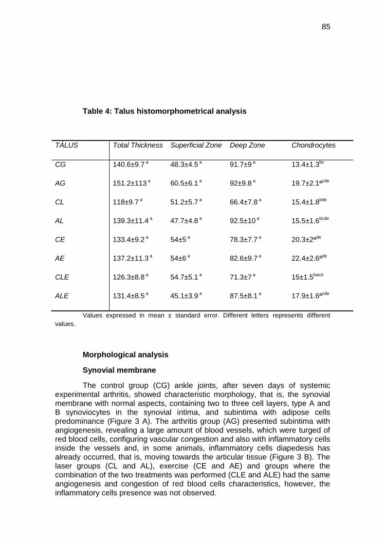
85
Table 4: Talus histomorphometrical analysis
TÁLUS Total Thickness Superficial Zone Deep Zone Chondrocytes
CG 140.6±9.7 a 48.3±4.5 a 91.7±9 a 13.4±1.3bc
AG 151.2±113 a 60.5±6.1 a 92±9.8 a 19.7±2.1ªcde
CL 118±9.7 a 51.2±5.7 a 66.4±7.8 a 15.4±1.8bde
AL 139.3±11.4 a 47.7±4.8 a 92.5±10 a 15.5±1.6bcde
CE 133.4±9.2 a 54±5 a 78.3±7.7 a 20.3±2ªde
AE 137.2±11.3 a 54±6 a 82.6±9.7 a 22.4±2.6ªde
CLE 126.3±8.8 a 54.7±5.1 a 71.3±7 a 15±1.5bacd
ALE 131.4±8.5 a 45.1±3.9 a 87.5±8.1 a 17.9±1.6ªcde
Values expressed in mean ± standard error. Different letters represents different
values.
Morphological analysis
Synovial membrane
The control group (CG) ankle joints, after seven days of systemic experimental arthritis, showed characteristic morphology, that is, the synovial membrane with normal aspects, containing two to three cell layers, type A and B synoviocytes in the synovial intima, and subintima with adipose cells predominance (Figure 3 A). The arthritis group (AG) presented subintima with angiogenesis, revealing a large amount of blood vessels, which were turged of red blood cells, configuring vascular congestion and also with inflammatory cells inside the vessels and, in some animals, inflammatory cells diapedesis has already occurred, that is, moving towards the articular tissue (Figure 3 B). The laser groups (CL and AL), exercise (CE and AE) and groups where the combination of the two treatments was performed (CLE and ALE) had the same angiogenesis and congestion of red blood cells characteristics, however, the inflammatory cells presence was not observed.

86
H G
F E
D C
B A IN
SI BV
BV
SI
IN
SI
BV
IN
BV
SI
IN
*
BV
SI
IN
AD
AD
AD
AD
SI
BV
BV SI
IN
BV
SI
IN
IN
SI
AD

87
Figure 3: Wistar rats talocrural joint synovial membrane photomicrographs, seven
days after induction of systemic arthritis by CFA, sagittal section, hematoxylin and eosin
staining. A: Control group; B: Arthritis group; C: Control Laser; D: Arthritis Laser; E: Control
Exercise; F: Arthritis exercise; G: Control laser and exercise; F: Arthritis laser and exercise. Are
indicated: Intimate layer (IN), Subintimate layer (SI), Blood vessels (BV) filled by red blood cells;
adipocytes (AD) and inflammatory infiltrate (*).
Articular Cartilage
The CG group articular cartilage, of both tibia and talus bones showed a smooth surface and the chondrocytes were organized in the four characteristic cell layers. In the superficial zone, a higher cell density was observed, with the cells arranged in horizontal clusters, with a flattened aspect. In the intermediate zone, the cells assumed a rounded appearance and were arranged isolated or isogenous groups. Soon after, the chondrocytes were organized in gaps, corresponding to the deep zone, separated from the calcified zone, by a basophilic line, the tidemark, with the normal-looking subchondral bone ( Figure 5A).
In AG group, in some animals, there was a predominance of changes in the articular cartilage, especially in talus, where in addition to flocculation on the articular surface and chondrocytes disorganization, it was possible to observe superficial layers degeneration with consequent subchondral bone exposure and pannus. In the tibia, flocculations were found on the joint surface and chondrocyte disorganization (Figure 5B). The CL, CE and CLE articular cartilage did not show any changes, being morphologically similar to the CG. In the AL group, chondrocyte disorganization and tidemark absence were observed in some animals (Figure 5C). The AE group had a higher amount of chondrocytes (Figure 5D), as already showed in the histomorphometric analysis (TABLES). In the cartilage, the CLE group was shown to be similar to CG, while in ALE group, was possible to observe chondrocytes disorganization, flocculation and tidemark absence.

88
Figure 4: Wistar rats talocrural joint cartilage photomicrographs, seven days after arthritis induction in the
knee by CFA, sagittal section, hematoxylin and eosin staining. A: Control group; B: Arthritis group; C: Control Laser; D:
Arthritis Laser; E: Control Exercise; F: Arthritis Exercise; G: Control Laser and Exercise; F: Arthritis Laser and Exercise.
Are indicated: Tibia (TI), Talus (TA) Articular cartilage (AC), Subchondral bone (SB), Tidemark (arrow), Articular cavity
(star); Flocculation (FL) and pannus (PA).
H G
F E
D C
B A
AC SB SB
TA TI
AC
SB
SB
AC
A
C
TA
TI
SB
SB
TA
TI
AC
A
C
TA
TI
SB
TI
TA
TA
TI

89
DISCUSSION
RA is commonly associated with pain and functional disability [18]. In the present study, in the functional parameters tested, it was observed that the limb functionality was affected by RA. In the SFI, both separate and associated treatments showed return to the initial values. Already the motor function evaluation by the inclined plane showed that the association of the two treatments was effective in reversing the disablement caused by the disease.
The AR usually presents symmetrical, persistent and destructive polyarthritis affecting particularly the hand and feet small joints, like the ankle joint. The disease main clinical features include joint pain, stiffness and swelling, joint damage, fatigue, functional loss what leads to disability [19].
As synovitis comprises the main RA clinical feature, its therapy has been focused on inflammation modulation and consequently disease degenerative progression deceleration by anti-inflammatory drugs administration like: corticosteroids, drugs modifying the course of the disease synthetic immunosuppressive drugs, used alone or in combination [20]. However, this treatment displays several important side effects, which sometimes does not show clinical improvement and disease remission [21]. Therefore, other treatment strategy which presents an anti-inflammatory effect, but do not suppress the entire immune system has been investigated [22]. LLLT has shown a promising non pharmacological alternative to RA treatment due to its local healing stimulation, inflammatory process modulation and pain relief effects [23-26] and it may help with the functional aspects.
The LLLT photobiological effects consists in light photons absorption by chomophores related to mitochondrial respiratory chain on cytochrome c, which produces a photosignal, subsequently transduced into the cell. This photosignaling involves transient free radical production, increases adenosine triphosphate (ATP), and modulates cellular redox potential, which could induces redox-sensitive transcription factor. LLLT could also generate singlet oxygen stimulating processes, such as RNA and DNA synthesis. Therewith, photobiological effects of LLLT are observed, as increase of cell migration, cytokine levels, growth factors, inflammatory mediator modulation, and increase of tissue oxygenation [27,28].
Hurkmans et al. inferred in a systematic review that physical exercises and behavioral education in patients with RA are recommended for clinical use and LLLT therapy is among the proposed guidelines, but the recommendation parameters are not well clarified [29]. In this study, LLLT also showed improvements in the functional parameters. Brosseau et al. made a systematic review relating the LLLT efficacy in RA treatment and concluded that laser therapy has a beneficial effect when used ate least four weeks, with effects on pain reduction and morning stiffness [30].
A study made by Khozeimeh et al. showed that LLLT therapy provides the body a better response to inflammatory conditions, by reducing edema, reducing pain and provides cellular bio stimulation [31]. In addition to these

90
benefits, LLLT is relatively simple and low-cost therapy that has been shown to be ideal for supporting conventional treatments.
Hsieh et al., showed that inflammatory action and functionality recovery using LLLT therapy is related actin in modulation of inflammatory mediators, indicating beneficial effects using 4,5 J/cm2 with inflammation reduction [32] what corroborates with the results of the present study.
Physical inactivity is common in patients with RA and has been associated with muscle and bone loss. In this way, physical activity and exercises are also important components of no pharmacology therapy in RA. Metsios et al. showed that exercise has been shown to reduce inflammation [33].
A study made by Cooney et al. recommended that RA patients should be taught and supported in an effective exercise program of moderate exercise 3-5 days/week for 30-60 minutes/session, combined with moderate strengthening) [34]. Forestier et al. brings evidences that when the disease is very active or there is severe involvement of the joints of the lower limbs, activities with low impact on the joints or with load alleviation are recommended [35].
On this study, the synovial membrane morphological analysis the AG group presented subintima with angiogenesis, revealing a large amount of blood vessels, which were turged of red blood cells, configuring vascular congestion and also with inflammatory cells inside the vessels and, in some animals, inflammatory cells diapedesis has already occurred, that is, moving towards the articular tissue. As already stated above, synovitis comprises the main RA clinical feature and this is due to the fact that it is an inflammatory autoimmune disease characterized by chronic and symmetrical degeneration of synovial joints [3]. Tending to worsen over time as joint architecture is modified by synovits, that is, synovial membrane inflammation [18].
Although the disease development precise cause remains unknown, it is believed that joints deformities occur due to their autoimmune characteristic, that is, immune system cells that should attack bacteria and external elements, attack healthy body tissues, as if they were invasive cells, releasing substances that cause inflammation [36]. Choy, described that the triggering event seems to be innate immune response activation, which includes arthritis-associated antigen presentation, by exogenous material and autologous antigens, through major histocompatibility complex and costimulatory proteins by dendritic cells, macrophages and activated B cells to T cells, which promote its differentiation mainly into T helper [22]. McIness inferred that T and B cell activation mediate effector function RA through cytokines and chemokines release, leukocyte activation, macrophages, fibroblast and endothelial cells, as well as B cell produced autoantibodies [7].
Additionally, Elshabrawy et al. showed that polymorphonuclear cells, mobilized by chemokines and cytokines, infiltrate the synovial compartment and produce a wide range of pro-inflammatory cytokines, leading to increase in cell proliferation, vasodilatation, vascular permeability, and proteolytic enzyme

91
secretion, from synovium stromal cells and from chondrocytes [37]. Pap at al. described that the metalloproteinases promotes collagen type II degradation and alter glycosaminoglycan composition and water retention capacity of joint cartilage, which result in joint biochemical and mechanical dysfunctions [38].
The present study synovial membrane morphological analysis also showed that the laser groups (CL and AL), exercise (CE and AE) and groups where the combination of the two treatments was performed (CLE and ALE) had the same angiogenesis and congestion of red blood cells characteristics, however, the inflammatory cells presence were not observed.
Angiogenesis is important for normal physiological growth [39]. Abnormal vessels growth can lead to chronic diseases and angiogenesis plays an important role in the healing process after conditions such as inflammatory disorders [40]. Physical activity has shown as a cardiac and skeletal muscle adaptation important stimulus for adaptation and physiological remodeling process to increase capillary density [41] and mitochondrial enzyme activity [42].
Hudlicka et al. showed some possible stimuli for exercise-induced angiogenesis, including increase in blood flow, also described as hyperemia, movement stress, muscle stretch, hypoxia and metabolic disturbance [43].
The LLLT anti-inflammatory effect has been widely described in literature [30, 44-45] and has been associated to prostaglandin levels alteration and vascular permeability [46]. Dos Anjos et al. showed LLLT decreased inflammatory area, as well as the number of infiltrate cells [25].
Despite well-known LLLT anti-inflammatory effect is extensive clinical use in treatment on inflammatory conditions, the impact of this treatment on immune response, especially on immune cells behavior, is still poorly exploited [47].
On this study, the articular cartilage morphological analysis showed that in AG group, in some animals, there was a predominance of changes in the articular cartilage, especially in talus, where in addition to flocculation on the articular surface and chondrocytes disorganization, it was possible to observe superficial layers degeneration with consequent subchondral bone exposure and pannus. In the tibia, flocculation was found on the joint surface and chondrocyte disorganization.
RA is an inflammatory autoimmune disease characterized by synovial joints chronic degeneration. Rosa et al. says that joints space inflammation can lead to the cartilage destruction and ultimately the bone [48]. Due to its autoimmune characteristic immune system cells attack healthy body tissues, as if they were invasive cells, releasing substances that cause inflammation [36], which causes erosive synovitis that can lead to a high level of joint destruction [5]. Monocytes, neutrophils and macrophages are described as the most responsible for mediating tissue destruction [36] and, in addition, McIness and Schett, showed inflammatory cytokines presence, such as tumor necrosis factor

92
alpha (TNF α) [49] and Sofat et al. showed increased oxoglycosidases concentrations and degenerative agents mediated by NF-k beta [50].
The immune system cells form a fibrous and abnormal tissue, called pannus, which releases substances that accelerate bone erosion and cartilage destruction, losing their shape and alignment resulting in deformations and damage to adjacent ligaments [36]. The extracellular matrix cartilage components are enzymatically degraded by metalloproteinases, hyaluronidases and agrecanases, respectively [51]. In addition, as showed by Firestein, the synovial membrane thickening causes irreversible damage to the capsule and articular cartilage, since the structures are replaced by the pannus and, bellow this arrangement, the cartilage became corroded and destroyed and the joints become fixed due to the thick and hardened structure, which results in edema [52].
The present study morphological analysis also showed that CL, CE and CLE articular cartilage did not show any changes, being morphologically similar to the CG. But, in the AL group, chondrocyte disorganization and tidemark absence were observed in some animals and the AE group had a higher amount of chondrocytes, what was confirmed by the histomorphometric analysis. In the cartilage, the CLE group was shown to be similar to CG, while in ALE group, was possible to observe chondrocytes disorganization, flocculation and tidemark absence.
Gonçalves et al. described these reactions as a possible reflect of the articular cartilage damaged restoration, due to the chondrocytes proliferative capacity and subchondral bone repair, with increased nutritional support in the region [53].
In this study was possible to observe that the groups treated with exercise presented morphology closest to the control group. As already said above, the joint affected by RA become fixed and, as showed by Simas et al., Immobilization leads to articular cartilage degeneration, decreased thickness, decreased cartilage matrix synthesis, irregular cartilage surface and lower total cartilage mass and volume [54].
The histological changes that occur after immobilization of the synovial joint are progressive resulting in the matrix structural macromolecules loss, but, when remobilized, the chondrocytes starts to synthesize the macromolecules quickly enough again, and the cartilage can be successfully synthesized, making it reversible changes [55].
Moore et al. showed that because of cartilage’s biphasic nature, purely biomechanical benefits of exercise on tissue health are possible as well [56]. Cartilage is highly-hydrated (70-80% water), thus mechanical loading drives pressurization of its interstitial fluid. This pressurization allows for the fluid-born support of compressive stresses, stiffening of the cartilage, and shielding of the extra cellular matrix from stresses, together, these outcomes, through the mechanisms of interstitial lubrication and nutrition [56] Nonetheless, under sustained loads, cartilage thins/strains due to pressure-induced fluid exudation,

93
driving a loss of interstitial fluid pressure and the defeat of numerous biomechanical functions, including lubricity [57].
In his study, Narmoneva et al. showed that articular cartilage requires some load and movement regime to maintain its physical nature and biochemical properties. Joint load reduction and immobilization in experimental in vivo models induce joint degeneration, including reduced hydration, altered structure and reduced proteoglycan synthesis and decrease in cartilage thickness [58].
Sandell and Aigner, brings on their study that chondrocyte proliferation, as happened in the present study exercise groups, may result from the articular instability possibly modifying the intensity and distribution of forces in the articular cartilage, leading to the proteoglycans and collagens breakdown and, consequently, to the loss of matrix structure and to an increase in water content and these injuries to the structure of the cartilage produced chondrocytes death and initiated a repair response, resulting in chondrocyte proliferation [59]. In this way, the tissue can be passing through a repair phase, characterized by the proliferation of the chondrocytes and the increase of chondrocyte metabolic activity [59]. Thus, the cartilage lesions in the treated groups were less severe and less frequent suggesting the predominance of articular cartilage in the repair phase.
CONCLUSION
The CFA model showed to be effective in reproduce the RA systemic characteristic, since even with the intra-articular CFA application occurring in the tibio femoral joint, classic RA manifestations occurred in the ankle joint synovial membrane and cartilage. Both treatments (LLLT and exercise) and the combination of treatments had positive effect on the functional evaluations, and the morphology and morphometry showed that the synovial membrane inflammatory aspects and the cartilage lesions in the treated groups were less severe, and the groups treated with exercise presented histology more similar to the control group. Thus, the higher chondrocyte number on this exercise groups, may indicate a more advanced repair phase.

94
REFERENCES
1- Cheung PP, Dougados M, Andre V, Balandraud N, Chales G,
CharyValckenaere I, et al. Improving agreement in assessment of
synovitis in rheumatoid arthritis. Jt Bone Spine. 2013; 80: 155–9.
2- Torpy J.M., Perazza, G.F., Golub, R.M. Rheumatoid Arthritis. JAMA.
2011; .305: 1.
3- Llopis E., Kroon H.M., Acosta J., Bloem, J.L. Conventional Radiology in
Rheumatoid Arthritis. Radiol. Clin. North Am. 2017; 55: 917–941.
4- Tufts M.H. Rheumatoid Arthritis. From the AJN Archives. 2012; 112.
5- De Carvalho M.R, Tebexreni, A.S., Salles, C.A., Barros Neto, T., Natour,
J. Oxygen uptake during walking in patients with rheumatoid arthritis – a
controlled study. J. Rheumatol. 2004; 31: 655-662.
6- Bogart B.I., Ort V.H. Anatomia e Embriologia. 1.ed. Rio de Janeiro:
Elsevier, 2008.
7- Mciness I.B., O’Dell J.R. State-of-the-art: rheumatoid arthritis. Ann
Rheum Dis. v. 2010; 69: 1898-1906.
8- Abell J.I., Hootmann J.M., Zack M.M., Moriarty D., Helmick C.G.Physical
activity and health related quality of life among people arthritis. J.
Epidemiol. Commun. 2005; 59: 380-385.
9- Tak P.P., Kalden J.R. Advances in rheumatology: New targeted
therapeutics. Arthritis Res. Ther. 2011; 13: 1-5.
10- Benucci M. Factors correlated with the improvement of endothelial
dysfunction during Abatacept therapy in patients with rheumatoid
arthritis. J Inflamm Res. 2017; 11: 247-252.
11- Faria T.J., Souza-Silva E., Oliveira D.T, Senna E.L., Tonussi C.R.
Evaluation of the pro-inflammatory potential of nanostructured drug
carriers in knee-joints of rats: effect on nociception, edema, and cell
migration. J Pharm Sci. 2009; 98: 4844-4851.

95
12- Bermudes A.M.L.M., Vassalo D.V., Vasquez E.C., Lima E.G.
Monitorização Ambulatorial da Pressão Arterial em Indivíduos
Normotensos Submetidos a Duas Sessões Únicas de Exercícios:
Resistido e Aeróbio. ABC cardiol. 2003; 82: 57-64.
13- Lesniak A., Lipkowski A.W. Opioid peptides in peripheral pain control.
Acta Neurobiol. 2011; 71: 129-138.
14- Khozelmeh F. Comparative evaluation of low level laser and systemic
steroid therapy in adjuvant-enchanced arthristis of rat
temporomandibular joint: a histological study. Dent. Res. J. 2015; 12:
215-223.
15- Henriques A.C.G., Cazal C., Castro J.F.L. Ação da laserterapia no
processo de proliferação e diferenciação celular: revisão de literatura.
Rev. Col. Bras. Cir. 2010; 37: 295- 302.
16- Bain J.R., Mackinson S.E., Hunter D.A. Functional evaluation of
complete sciatic, peroneal, and posterior tibial nerve lesions in the rat.
Plast. Reconstr. Surg. v. 1989; 83: 129-138.
17- Gasparini A.L.P., Barbieri C.H., Mazzer N. Correlação entre diferentes
métodos de avaliação funcional da marcha de ratos com lesão por
esmagamento do nervo isquiático. Acta Otop. Bras. 2007; 15, p. 285-
299.
18- Cross M., Smith E., Hoy D., Carmona L., Wolfe F., Vos T., Williams B.,
Gabriel S., Lassere M., Johns N., Buchbinder R., Woolf A., March L. The
global burden of rheumatoid arthritis. Ann. Rheum. Dis. 2014; 73: 1316.
19- Olofsson T, Petersson IF, Eriksson JK, Englund M, Nilsson JA, Geborek
P, et al. Predictors of work disability after start of anti TNF therapy in a
national cohort of Swedish patients with rheumatoid arthritis: does early
anti-TNF therapy bring patients back to work? Ann Rheum Dis
2017;76:1245.
20- J. S. Smolen, R. Landewé, J. Bijlsma, G. Burmester, K. Chatzidionysiou,
M. Dougados, J. Nam, S. Ramiro, M. Voshaar, R. van Vollenhoven, D.
Aletaha, M. Aringer, M. Boers, C. D. Buckley, F. Buttgereit, V. Bykerk, M.
Cardiel, B. Combe, M. Cutolo, Y. van Eijk-Hustings, P. Emery, A. Finckh,
C. Gabay, J. Gomez-Reino, L. Gossec, J. E. Gottenberg, J. M. W.
Hazes, T. Huizinga, M. Jani, D. Karateev, M. Kouloumas, T. Kvien, Z. Li,
X. Mariette, I. McInnes, E. Mysler, P. Nash, K. Pavelka, G. Poór, C.

96
Richez, P. van Riel, A. Rubbert-Roth, K. Saag, J. da Silva, T. Stamm, T.
Takeuchi, R. Westhovens, M. de Wit, D. van der Heijde EULAR
recommendations for the management of rheumatoid arthritis with
synthetic and biological disease-modifying antirheumatic drugs: 2016
update. Ann. Rheum. Dis. 2017; 76: 960.
21- P. D. W. Kiely, A. K. Brown, C. J. Edwards, D. T. O'Reilly, A. J. K. Östör,
M. Quinn, A. Taggart, P. C. Taylor, R. J. Wakefield, P. G. Conaghan,
Rheumatology. 2009; 48: 765.
22- E. Choy, Rheumatology 2012; 3 (suppl 5): 51.
23- Neves M., Retameiro A.C.B., Tavares A.L.F., Reginato A., Menolli R.A.,
Stein T.S., Ribeiro L.F.C., Bertolini G.R.F. Physical exercise and low
level laser therapy in the nociception and leukocite migration of Wistar
rats submitted to a model of rheumatoid arthritis. Lasers in medical
science. 2019; 1-12.
24- S. Kazem Shakouri, J. Soleimanpour, Y. Salekzamani, M. R. Oskuie,
Lasers Med. Sci. 2010; 25: 73.
25- L. M. J. dos Anjos, A. S. de da Fonseca, J. Gameiro, F. de Paoli, Lasers
Med. Sci. 2017; 32: 975.
26- C. R. Tim, P. S. Bossini, H. W. Kido, I. Malavazi, M. R. von Zeska Kress,
M. F. Carazzolle, N. A. Parizotto, A. C. Rennó, J. Photochem. Photobiol.
B Biol. 2016; 154: 8.
27- Chen ACH, Arany PR, Huang YY, Tomkinson EM, Saleem T, Yull FE,
Blackwell TS, Hamblin MR. Low-level laser therapy activates NF-kB via
generation of reactive oxygen species in mouse embryonic fibroblasts. PLoS
One. 2011; 6: 22453.
28- Karu TI. Mitochondrial signaling in mammalian cells activated by red and
near-IR radiation. Photochem Photobiol. 2008; 84: 1091–1099.
29- Hurkmans EJ, Jones A, Li LC, Vlieland TPMV. Quality appraisal of
clinical practice guidelines on the use of physiotherapy in rheumatoid
arthritis: a systematic review. Rheumatology. 2011; 50: 1879–1888.
30- Brosseau L, Robinson V, Wells G, Debie R, Gam A, Harman K, Morin
M, Shea B, Tugwell P. Low level laser therapy (classes I, II and III) for

97
treating rheumatoid arthritis. Cochrane Database Syst Rev. 2005;
19:CD002049.
31- Khozelmeh F. Comparative evaluation of low level laser and systemic
steroid therapy in adjuvant-enchanced arthristis of rat
temporomandibular joint: a histological study. Dent. Res. J. 2015; 12:
215-223.
32- Hsieh YL, Fan YC, Yang CC (2016) Low-level laser therapy alleviates
mechanical and cold allodynia induced by oxaliplatin administration in
rats. Support Care Cancer. 2016; 24:233–242.
33- Metsios GS, Stavropoulos-Kalinoglou A, Veldhuijzen van Zanten JJ,
Nightingale P, Sandoo A, Dimitroulas T, et al. Individualised exercise
improves endothelial function in patients with rheumatoid arthritis. Ann
Rheum Dis. 2014; 73: 748e51.
34- Cooney JK, Law RJ, Matschke V, Lemmey AB, Moore JP, Ahmad Y, et
al. Benefits of exercise in rheumatoid arthritis. J Aging Res. 2011;
681640.
35- Forestier R, Andre-Vert J, Guillez P, Coudeyre E, Lefevre-Colau MM,
Combe B, et al. Non-drug treatment (excluding surgery) in rheumatoid
arthritis: clinical practice guidelines. Jt Bone Spine. 2009;76:691e8.
36- Kapale P. Vardharajulu G., Warude T. Effect of free exercise and
Rheumathoid arthritis. Indian J Physiother Occup Ther. 2017; 11: 63-67.
37- H. A. Elshabrawy, Z. Chen, M. V. Volin, S. Ravella, S. Virupannavar, S.
Shahrara, Angiogenesis. 2015; 18: 433.
38- T. Pap, A. Korb-Pap, Nat. Rev. Rheumatol. 2015; 11: 606.
39- Findlay JK. Angiogenesis in reproductive tissues. J Endocrinol. 1986;
111:357–366.
40- Folkman J. Angiogenesis in cancer, vascular, rheumatoid and other
disease. Nat Med. 1995; 1:27–31.
41- Laughlin MH, Bowles DK, Duncker DJ (2012) The coronary circulation in
exercise training. Am J Physiol Heart Circ Physiol. 2012; 302:H10–H23.

98
42- Savard R, Smith LJ, Palmer JE et al. Site specific effects of acute
exercise on muscle and adipose tissue metabolism in sedentary female
rats. Physiol Behav. 1988; 43:65–71.
43- Hudlicka O, Brown M, Egginton S. Angiogenesis in skeletal and cardiac
muscle. Physiol Rev 72:369–417 23. Sundberg CJ, Kaijser L (1992)
Effects of graded restriction of perfusion on circulation and metabolism in
the working leg; quantifi cation of a human ischaemia-model. Acta
Physiol Scand. 1993; 146:1–9.
44- Kingsley JD, Demchak T, Mathis R. Low-level laser therapy as a
treatment for chronic pain. Front Physiol. 2014; 5:306.
45- Bjordal JM, Johnson MI, Lopes-Martins RAB, Bogen B, Chow R,
Ljunggren AE. Short-term efficacy of physical interventions in
osteoarthritic knee pain. A systematic review and meta-analysis of
randomised placebo-controlled trials. BMC Musculoskelet Disord. 2007;
8:51.
46- de Jesus JF, Spadacci-Morena DD, dos Anjos Rabelo ND, Pinfildi CE,
Fukuda TY, Plapler H. Low-level laser therapy in IL-1β, COX-2, and
PGE2 modulation in partially injured Achilles tendon. Lasers Med Sci.
2015; 30:153–158.
47- D. L. Asquith, A. M. Miller, I. B. McInnes, F. Y. Liew, Eur. J. Immunol.
Animal models of rheumatoid arthritis. 2009; 39, 2040.
48- D.J.d.F. Rosa, E.A.d. Paula, H.L.M. Bonfante, H.d.L. Bonfante, C.E.d.C.
Areal, G.S. Baião, M.A. Espíndula, Nodulose acelerada na artrite
reumatóide durante terapia com Leflunomida, Rev. Bras. Reumatol.
2007: 228–231.
49- McIness I.B., Schett G. The pathogenesis of rheumatoid arthritis. N Engl
J Med. 2011; 365: 2205–2219.
50- Sofat N., Robertson S.D., Wait R. Fibronectin III 13-14 domains induce
joint damage via Toll-like receptor 4 activation and synergize with
interleukin-1 and tumour necrosis factor. J. Innate Immun. 2012; 4: 69-
79.
51- Nagase H., Kashiwagi M. Aggrecanases and cartilagem matrix
degradation. Arthritis Res. Ther. 2003; 94: 94-103.

99
52- Firestein G.S. Evolving concepts of rheumatoid arthritis. Nature. 2003;
423:356-361.
53- Gonçalves G, Melo EG, Gomes MG, Nunes VA, Rezende CM. Effects of
chondroitin sulfate and sodium hyaluronate on chondrocytes and
extracellular matrix of articular cartilage in dogs with degenerative joint
disease. Arq Bras Med Vet Zootec. 2008;60: 93-102.
54- Simas J.M.M, Kunz R.I, Brancalhão R.M.C, Ribeiro L.F.C., Bertolini
G.R.F. Efeitos do exercício físico sobre a cartilagem de ratas
ooforectomizadas submetidas à imobilização. Einstein. 2015; 13: 574-9.
55- Brandt K.D. Response of joint structures to inactivity and to reloading
after immobilization. Arth. e Rheum. 2003; 49: 267-271.
56- Moore AC, Burris DL. An analytical model to predict interstitial lubrication
of cartilage in migrating contact areas. J Biomech. 2014;47(1):148–153.
57- Ateshian GA. The role of interstitial fluid pressurization in articular
cartilage lubrication. J Biomech. 2009;42(9):1163–1176.
58- Narmoneva D.A., Cheung H.S., Wang J.Y. Altered swelling behavior of
femoral cartilage following joint immobilization in a canine model. J.
Orthop. Res. 2002; 20: 83-91.
59- Sandell L.J., Aigner T. Articular cartilage and changes in arthritis. An
introduction: cell biology of osteoarthritis. Arth. Res. 2001; 3: 107-113.

100
CONSIDERAÇÕES FINAIS
O modelo de indução da artrite por CFA foi eficaz em induzir a doença
de forma sistêmica, uma vez que, mesmo com a injeção intra-articular do
adjuvante ocorrendo na articulação tíbio-femoral, foi possível observar
manifestações características da artrite na articulação talocrural em ambas as
fases da doença. O tratamento com exercício resistido e LBP se mostrou eficaz
quando usado separadamente e em associação nos aspectos funcionais
testados. No período agudo os parâmetros histomorfométricos avaliados
mostraram que os aspectos inflamatórios da membrana sinovial e as lesões da
cartilagem articular dos grupos tratados se apresentaram de forma menos
severa. Além disso, o maior número de condrócitos nos grupos exercício pode
indicar uma fase de reparo tecidual mais avançada.
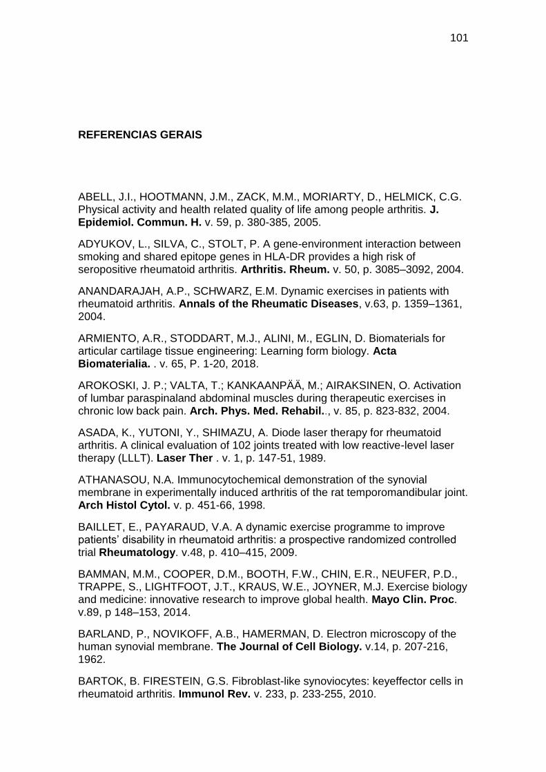
101
REFERENCIAS GERAIS
ABELL, J.I., HOOTMANN, J.M., ZACK, M.M., MORIARTY, D., HELMICK, C.G. Physical activity and health related quality of life among people arthritis. J. Epidemiol. Commun. H. v. 59, p. 380-385, 2005.
ADYUKOV, L., SILVA, C., STOLT, P. A gene-environment interaction between smoking and shared epitope genes in HLA-DR provides a high risk of seropositive rheumatoid arthritis. Arthritis. Rheum. v. 50, p. 3085–3092, 2004.
ANANDARAJAH, A.P., SCHWARZ, E.M. Dynamic exercises in patients with rheumatoid arthritis. Annals of the Rheumatic Diseases, v.63, p. 1359–1361, 2004.
ARMIENTO, A.R., STODDART, M.J., ALINI, M., EGLIN, D. Biomaterials for articular cartilage tissue engineering: Learning form biology. Acta Biomaterialia. . v. 65, P. 1-20, 2018.
AROKOSKI, J. P.; VALTA, T.; KANKAANPÄÄ, M.; AIRAKSINEN, O. Activation of lumbar paraspinaland abdominal muscles during therapeutic exercises in chronic low back pain. Arch. Phys. Med. Rehabil.., v. 85, p. 823-832, 2004.
ASADA, K., YUTONI, Y., SHIMAZU, A. Diode laser therapy for rheumatoid arthritis. A clinical evaluation of 102 joints treated with low reactive-level laser therapy (LLLT). Laser Ther . v. 1, p. 147-51, 1989.
ATHANASOU, N.A. Immunocytochemical demonstration of the synovial membrane in experimentally induced arthritis of the rat temporomandibular joint. Arch Histol Cytol. v. p. 451-66, 1998.
BAILLET, E., PAYARAUD, V.A. A dynamic exercise programme to improve patients’ disability in rheumatoid arthritis: a prospective randomized controlled trial Rheumatology. v.48, p. 410–415, 2009.
BAMMAN, M.M., COOPER, D.M., BOOTH, F.W., CHIN, E.R., NEUFER, P.D., TRAPPE, S., LIGHTFOOT, J.T., KRAUS, W.E., JOYNER, M.J. Exercise biology and medicine: innovative research to improve global health. Mayo Clin. Proc. v.89, p 148–153, 2014.
BARLAND, P., NOVIKOFF, A.B., HAMERMAN, D. Electron microscopy of the human synovial membrane. The Journal of Cell Biology. v.14, p. 207-216, 1962.
BARTOK, B. FIRESTEIN, G.S. Fibroblast-like synoviocytes: keyeffector cells in rheumatoid arthritis. Immunol Rev. v. 233, p. 233-255, 2010.

102
BARTON, C.J., LACKS, S., MALLIARAS, P., MORRISSEY, D. Gluteal muscle activity and patellofemoral pain syndrome: a systematic review. Br.J. Sports Med. v. 47, p. 207-214, 2013.
BENHAMOU, M-AM. Reconditioning in patients with rheumatoid arthritis. Ann. Readapt Med Phys. v. 50, p. 382-5, 2007.
BENUCCI, M. Factors correlated with the improvement of endothelial dysfunction during Abatacept therapy in patients with rheumatoid arthritis. J Inflamm Res. v. 11, p. 247-252, 2017.
BERMUDES, A.M.L.M.; VASSALLO, D.V.; VASQUEZ, E.C.; LIMA, E.G. Monitorização Ambulatorial da Pressão Arterial em Indivíduos Normotensos Submetidos a Duas Sessões Únicas de Exercícios: Resistido e Aeróbio. ABC cardiol. v. 82. p. 57-64, 2003.
BÉRTOLO, M.B. Consenso brasileiro no diagnóstico e tratamento da artrite reumatoide. Rev. Bras. Reumatol. v. 47, 150, 2007.
BOGART, B.I., ORT, V.H. Anatomia e Embriologia. 1.ed. Rio de Janeiro: Elsevier, 2008.
BONO, C.M., BERBERIAN, W.S. Orthotic devices: Degenerative disorders of the foot and ankle. Foot ankle Clin. v. 6, p. 329-340, 2001.
BOTTINI, N., FIRESTEIN, Duality of fibroblast-like synoviocytes in RA: Passive responders and imprinted aggressors. Nat Rev Rheumatol. v. 9, p. 24-33, 2013.
BRANDT, K.D. Response of joint structures to inactivity and to reloading after immobilization. Arthritis Rheumatol. v. 49, p. 267-271, 2003.
BUCKWALTER, H., MAKIN, H. Instructional Course Lectures, The American
Academy of Orthopaedic Surgeons-articular cartilage. Part I: Tissue design
and chondrocyte-matrix interactions. J. Bone Joint Surg. Am. v.79, p. 600–611, 1977.
BUGATTI, S., CODULLO, V., CAPORALI, R., MONTECUCCO, C. B cells in rheumatoid arthritis. Autoimmun Rev. v. 7, p. 137-142, 2007.
BURMESTER, G.R., DIMITRIU, B.A., WATERS, S.J., WINCHESTER, R.J. Mononuclear phagocytes of normal and rheumatoid synovial membrane indentified by monoclonal antibodies. Immunology. v. 56, p. 673- 681, 1985.
CAI, X. WONG, Y.F., ZHOU, H., LIU, Z.Q., XIE, Y., JIANG, Z.H Manipulation of the indution of adjuvant arthritis in Sprague-Dawley rats. Inflamm. Res. v. 55, p. 368-377, 2006.
CAN, C., CINAR, M.G., KOSAY, S., EVINC, A. Vascular endothelial dysfunction associated with elevated serum homocysteine levels in rat adjuvant arthritis: effect of vitamin E administration. Life Sci. 71, 401-410, 2002.

103
CAROTTI, F., SALAFFI, P., MANGANELLI, D., SALERA, D., SIMONETTI, B., GRASSI, W. Power Doppler sonography in the assessment of synovial tissue of the knee joint in rheumatoid arthritis: a preliminary experience. Ann Rheum Dis . v. 61, p. 877–882, 2002.
CATOIRE, M., MENSINK, M., KALKHOVEN, E., SCHRAUWEN, P., KERSTEN, S. Identification of human exercise-induced myokines using secretome analysis. Physiol. Genomics. v. 46, p. 256–267, 2014.
CHORUS, A.M., MIEDEMA, H.S., BOONEN, A., VAN DER LINDEN, S. Quality of life and work in patients with rheumatoid arthritis and ankylosing spondylitis of working age. Ann Rheum Dis. v. 62, p. 1178–1184, 2003.
CLARK, J.M. Variation of collagen fiber alignment in a joint surface: a scanning
electron microscope study of the tibial plateau in dog, rabbit, and man. J. Orthop. Res. v. 9, p.246–257, 1991.
COLPANI, V., OPPERMANN, K., SPRITZER, P.M. Association between habitual physical activity and lower cardiovascular risk in premenopausal, perimenopausal, and postmenopausal women: a population-based study. Menopause. v. 20, p 525–531, 2013.
COOK, C.D., MOORE, K.I. Effects of sex, hindpaw injection site and stimulus modality on nociceptive sensitivity in arthritic rats. Physiol Behav. v. 87, p., 552-62, 2006.
CUTLIP, R.C. CHEVILLE, N.F. Normal structure and morphological effects of a corticosteroid on the peripatellar synovial membrane of the rat. Kaibogaku Zasshi. v.2, p.183-9, 1999.
DÂNGELO, J.G., FATTINI, C.A. Anatomia Humana Sistêmica e Segmentar: para o estudante de medicina. 3 ed. Rio de Janeiro: Atheneu, 2007.
DAVIGNON, J-L., HAYDER, M., BARON, M., BOYER, J-F., CONSTANTIN, A., APPARAILLY, F. Targeting monocytes/macrophages in the treatment of rheumatoid arthritis. Rheumatology. v. 52, p. 590- 598, 2013.
DE CARVALHO, MR., TEBEXRENI, A.S., SALLES, C.A., BARROS NETO, T., NATOUR, J. Oxygen uptake during walking in patients with rheumatoid arthritis – a controlled study. J. Rheumatol. v. 31, p. 655-662, 2004.
DE JONG, Z., MUNNEKE, M., ZWINDERMAN, A.H. Is a long-term high-intensity exercise program effective and safe in patients with rheumatoid arthritis? Results of a randomized controlled trial. Arthristis Rheum. v.48, p. 2415-2424, 2003.
DE MEDINACELI, L. Interpreting nerve morphometry data after experimental traumatic lesions. J. Neurosci. Methods. v. 58, p. 29-37, 1993.
DE SOUZA, S., BANSAL, R., GALLOWAY, J. Rheumathoid arthritis – an update for general dental practitioners. Br. Dent. J. v. 221, p.667-673, 2016.
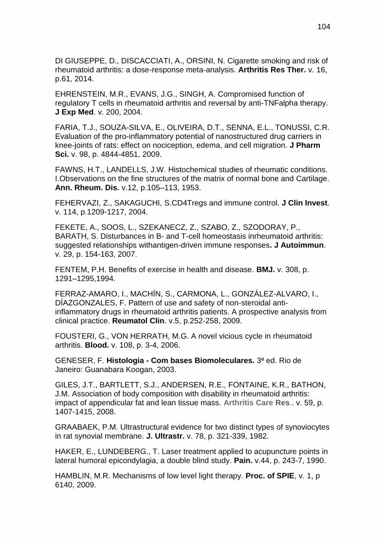
104
DI GIUSEPPE, D., DISCACCIATI, A., ORSINI, N. Cigarette smoking and risk of rheumatoid arthritis: a dose-response meta-analysis. Arthritis Res Ther. v. 16, p.61, 2014.
EHRENSTEIN, M.R., EVANS, J.G., SINGH, A. Compromised function of regulatory T cells in rheumatoid arthritis and reversal by anti-TNFalpha therapy. J Exp Med. v. 200, 2004.
FARIA, T.J., SOUZA-SILVA, E., OLIVEIRA, D.T., SENNA, E.L., TONUSSI, C.R. Evaluation of the pro-inflammatory potential of nanostructured drug carriers in knee-joints of rats: effect on nociception, edema, and cell migration. J Pharm Sci. v. 98, p. 4844-4851, 2009.
FAWNS, H.T., LANDELLS, J.W. Histochemical studies of rheumatic conditions. I.Observations on the fine structures of the matrix of normal bone and Cartilage. Ann. Rheum. Dis. v.12, p.105–113, 1953.
FEHERVAZI, Z., SAKAGUCHI, S.CD4Tregs and immune control. J Clin Invest. v. 114, p.1209-1217, 2004.
FEKETE, A., SOOS, L., SZEKANECZ, Z., SZABO, Z., SZODORAY, P., BARATH, S. Disturbances in B- and T-cell homeostasis inrheumatoid arthritis: suggested relationships withantigen-driven immune responses. J Autoimmun. v. 29, p. 154-163, 2007.
FENTEM, P.H. Benefits of exercise in health and disease. BMJ. v. 308, p. 1291–1295,1994.
FERRAZ-AMARO, I., MACHÍN, S., CARMONA, L., GONZÁLEZ-ALVARO, I., DÍAZGONZALES, F. Pattern of use and safety of non-steroidal anti-inflammatory drugs in rheumatoid arthritis patients. A prospective analysis from clinical practice. Reumatol Clin. v.5, p.252-258, 2009.
FOUSTERI, G., VON HERRATH, M.G. A novel vicious cycle in rheumatoid arthritis. Blood. v. 108, p. 3-4, 2006.
GENESER, F. Histologia - Com bases Biomoleculares. 3ª ed. Rio de Janeiro: Guanabara Koogan, 2003.
GILES, J.T., BARTLETT, S.J., ANDERSEN, R.E., FONTAINE, K.R., BATHON, J.M. Association of body composition with disability in rheumatoid arthritis: impact of appendicular fat and lean tissue mass. Arthritis Care Res.. v. 59, p. 1407-1415, 2008.
GRAABAEK, P.M. Ultrastructural evidence for two distinct types of synoviocytes in rat synovial membrane. J. Ultrastr. v. 78, p. 321-339, 1982.
HAKER, E., LUNDEBERG., T. Laser treatment applied to acupuncture points in lateral humoral epicondylagia, a double blind study. Pain. v.44, p. 243-7, 1990.
HAMBLIN, M.R. Mechanisms of low level light therapy. Proc. of SPIE, v. 1, p 6140, 2009.

105
HENRIQUES, A.C.G., CAZAL, C., CASTRO, J.F.L. Ação da laserterapia no processo de proliferação e diferenciação celular: revisão de literatura. Rev. Col. Bras. Cir. v. 37, p. 295- 302, 2010.
HOGG, N., PALMER, D.G., REVELL, P.A. Ontogenic development of rat synovial lining cells: immunohistochemical ande immunoelectron microscopical analysis. Cell and Tissue Research. v. 262, p. 463-472, 1999.
HOMANN, D., HOLZ, A., BOT, A. Autoreactive CD4T cells protect from autoimmune diabetes via bystander suppression using the IL-4/Stat6 pathway. Immunity. v.11, p. 463-472, 1999.
HUA, S., CABOT, P.J. Mechanisms of peripheral immunecell-mediated analgesia in inflammation: clinical and therapeutic implications. Trends Pharmacol Sci. v.31, p. 427-433, 2010.
HUI, A.Y., MCCARTY, W.J., MASUDA, K., FIRESTEIN, G.S., SAH, R.L. A systems biology approach to synovial joint lubrification in health, injuru, and disease. WIREs Systems Biology and Medicine. v. 4, p. 15-37, 2012.
IWANAGA, T., SHIKICHI, M., KITAMURA, H., YANASE, H., NAZAWA-INOUE, K. Morphology and functional roles of synoviocytes in the joint. ARCH HISTOL CYTOL. v. 63, p. 17-31, 2000.
IZUMI, S., MOTOHIRO, T., KATSUMASA, T. KIYOSHI, T. Synovial macrophages Ann Rheum Dis. v. 54, p. 392–394, 1995.
JILANI, M., GHADIALLY, F.N. Pathogenesis of rheumatoid arthritis: the role of synoviocytes. Rheumatoid Arthritis, v. 27, p.355, 2001
JUNQUEIRA, L.C.U, CARNEIRO, J. Histologia Básica. 13ª ed. Rio de Janeiro: Guanabara, Koogan, 2013.
JUONALA, M., VIIKARI, J.S., RAITAKARI, O.T. Main findings from the prospective Cardiovascular Risk in Young Finns Study. Curr. Opin. Lipidol. v. 24, p.57–64, 2013.
JUVERLIN, J.S., BUSCHMANN, M.D., HUNZIKER, E.B. Mechanical anisotropy of the human knee articular cartilage in compression. Proc. Inst. Mech. Eng. H. v. 217, p.215–219, 2003.
KAPALE, P., VARDHARAJULU, G., WARUDE, T. Effect of free exercise and Rheumathoid arthritis. Indian J Physiother Occup Ther. v. 11, p. 63-67, 2017.
KARU, T.I., KOLYAKOV, S.F. Exact action spectra for cellular responses relevant to phototherapy. Photomed Laser Surg. v. 23, p. 355-61, 2005.
KHURANA, R., BERNEY, S.M. Clinical aspects of rheumatoid arthritis. Pathophysiology. v. 12, p. 153-165, 2005.
KINNE, R.W., BREAUER, R., STUHLMÜLLER, B., PALOMBO-KINNE, E., BURMESTER, G-R. Macrophages in rheumatoid arthritis. Arthritis Res. Ther. v.2, p.189-202, 2000.

106
KLEIN, R., EEK, B. Low energy laser treatment and exercise for low back pain: double-blind controlled trial. Arch Phys Med Rehabil. v.71, p. 34-7, 1990.
KHOZELMEH, F. Comparative evaluation of low level laser and systemic steroid therapy in adjuvant-enchanced arthristis of rat temporomandibular joint: a histological study. Dent. Res. J. v. 12, p. 215-223, 2015.
KUNZ, R.I., CORADINI, J.G., SILVA, L.I., BERTOLINI, G.R.F., BRANCALHÃO, R.M.C., RIBEIRO, L.F.C. Effects of immobilization and remobilization on the ankle joint in wistar rats. Brazilian Journal of Medical and Biological Research. v.47, p. 824-849, 2014.
LAI, W. M., HOU, V.C. A triphasic theory for the swelling and deformation behaviors of articular cartilage, J. Biomech. Eng. v.113, p. 245–258, 1991.
LEACH, D.H., CALDWELL, S.J., FERGUSON, J.G. Eletron Microscopy of the human synovial membrane. J. Cell Biol. v. 14, p. 207-220.
LEFÈVRE, S. Synovial fibroblasts spread rheumatoid arthritis to unaffected joints. Nat Med. v. 15, p. 1414–1420, 2009.
LEMMEY, A.B., MARCORA, S.M., CHESTER, K, WILSON, S., CASANOVA, F., MADDISON, P.J. Effects of high-intensity resistance training in patients with rheumatoid arthritis: a randomized controlled trial. Arthritis Care Res. v. 61, p. 1726–1734, 2009.
LESNIAK, A., LIPKOWSKI, A.W. Opioid peptides in peripheral pain control. Acta Neurobiol Exp. v.71, p. 129-138, 2011.
LIN, F. JOSEPHS, S.F., ALEXANDRESCU, D.T., RAMOS, F., BOGIN, V., GAMMIL, V. Lasers, stem cells, and COPD. J Trans Med. v. 8, p.16, 2010.
LINCK, G., PORTE, A. The Paraneuron, 1988. Ed. Fujita
LLOPIS, E., KROON, H.M., ACOSTA, J., BLOEM, J.L. Conventional Radiology in Rheumatoid Arthritis. Radiol. Clin. North Am. V. 55, p. 917–941, 2017.
LYONS, T.J., STODDART, R.W., MCCLURE, S.F., MCCLURE, J. The tidemark of thechondro-osseous junction of the normal human knee joint, J. Mol. Histol. v.36 p. 207–215, 2005.
MAIMAN, T.H. Stimulated optical radiation in Ruby. Nature.v.187, p. 493-94, 1960.
MAINI, R., FELDMANN, M. Rheumatoid arthritis; immunopathgenesis of rheumatoid arthritis. in Oxford Textbook of Rheumatology, Press, Oxford, UK, 2004.
MALDA, J.C., DE GRAUW, K.E, BENDERS, M.J., KIK, C.H., VAN DE LEST, L.B., CREEMERS, W.J., VAN WEEREN, P.R. Of mice, men and elephants: the

107
relation between articular cartilage thickness and body mass. PLoS One. v.8, p. 57683.
MANDI, D.M. Ankle fractures. Clinics in Podiatric Medicine and Surgery. v. 29, p. 155-186, 2012.
MAPP, P.I., REVELL, P.A. Synthesis of hyaluronic acid by human synovial tissue slices.Science. v.125 p., 1300, 1985.
MARGARETTEN, M.,JULIAN,, L., KATZ, P., YELIN, E. Depression in patients with rheumatoid arthritis: description, causes and mechanisms. Int. J. Clin. Rheumatol. v. 6, p. 617-623, 2011.
MARIANO, R.N. Cartilha Artrite Reumatoide. Comissão de Artrite Reumatoide da Sociedade Brasileira de Reumatologia, 2011.
MAROUDAS, A., WACHTEL, E., GRUSHKO, G., KARTZ, E.P. WEINBERG, P.The effect of osmotic and mechanical pressures on water partitioning in articular cartilage. Biochim. Biophys. Acta. v.1073, p. 285–294, 1991.
MCARDLE, W.D.; KATCH, F.; KATCH, V.L. Fisiologia do Exercício: Energia, Nutrição e Desempenho Humano. 5. ed. Rio de Janeiro: Guanabara Koogan. 2001.
MCINNES, I.B., O’DELL, J.R. State-of-the-art: rheumatoid arthritis. Ann Rheum Dis. v. 69., p. 1898-1906, 2010.
MCINNES, I.B., SCHETT, G. The pathogenesis of rheumatoid arthritis. N Engl J Med. v. 365, p. 2205–2219, 2011.
METSIOS, G.S., STAVROPULOS-KALINOGLOU, A., PANOULAS, V.F. Association of physical inactivity with increased cardiovascular risk in patients with rheumatoid arthritis. Eur J Cardiovasc Prev Rehabil. v. 16, p. 188–94, 2009.
MILNER, C.E. Funtional Anatomy for Sport and Exercise. Quick Reference, Oxon, Routledge, UK, 2008.
MOEBIUS-WINKLER, S., SCHULER, G., ADAMS, V. Endothelial progenitor cells and exercise-induced redox regulation. Antioxid. Redox Signal. v. 15, p. 997–1011, 2011.
MONTEIRO, M.F., FILHO, D.C. Exercício físico e o controle da pressão arterial. Rev Bras Med Esporte. v.10, p.513-516, 2004.
MORIYAMA, Y., MORIAYAMA, E.H., BLACKMORE, K., AKENS, M.K., LILGE, L. In vivo study of the inflammatory modulating effects of lowlevel laser therapy on iNOS expression using bioluminescence imaging. Photochem Photobiol. v. 81, p.1351–1355, 2005.
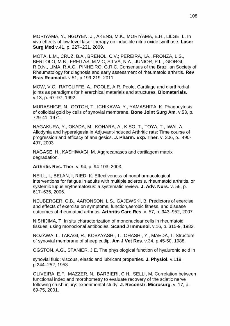
108
MORIYAMA, Y., NGUYEN, J., AKENS, M.K., MORIYAMA, E.H., LILGE, L. In vivo effects of low-level laser therapy on inducible nitric oxide synthase. Laser Surg Med v.41, p. 227–231, 2009.
MOTA, L.M., CRUZ, B.A., BRENOL, C.V.; PEREIRA, I.A., FRONZA, L.S., BERTOLO, M.B., FREITAS, M.V.C, SILVA, N.A., JUNIOR, P.L., GIORGI, R.D.N., LIMA, R.A.C., PINHEIRO, G.R.C. Consensus of the Brazilian Society of Rheumatology for diagnosis and early assessment of rheumatoid arthritis. Rev Bras Reumatol. v.51, p.199-219. 2011.
MOW, V.C., RATCLIFFE, A., POOLE, A.R. Poole, Cartilage and diarthrodial joints as paradigms for hierarchical materials and structures. Biomaterials. v.13, p. 67–97, 1992.
MURASHIGE, N., GOTOH, T., ICHIKAWA, Y., YAMASHITA, K. Phagocytosis of colloidal gold by cells of synovial membrane. Bone Joint Surg Am. v.53, p. 729-41, 1971.
NAGAKURA, Y., OKADA, M., KOHARA, A., KISO, T., TOYA, T., IWAI, A. Allodynia and hyperalgesia in Adjuvant-Induced Arthritic rats: Time course of progression and efficacy of analgesics. J. Pharm. Exp. Ther. v. 306, p., 490-497, 2003
NAGASE, H., KASHIWAGI, M. Aggrecanases and cartilagem matrix degradation.
Arthritis Res. Ther. v. 94, p. 94-103, 2003.
NEILL, I., BELAN, I, RIED, K. Effectiveness of nonpharmacological interventions for fatigue in adults with multiple sclerosis, rheumatoid arthritis, or systemic lupus erythematosus: a systematic review. J. Adv. Nurs. v. 56, p. 617–635, 2006.
NEUBERGER, G.B., AARONSON, L.S., GAJEWSKI, B. Predictors of exercise and effects of exercise on symptoms, function,aerobic fitness, and disease outcomes of rheumatoid arthritis. Arthritis Care Res. v. 57, p. 943–952, 2007.
NISHIJIMA, T. In situ characterization of mononuclear cells in rheumatoid tissues, using monoclonal antibodies. Scand J Immunol. v.16, p. 315-9, 1982.
NOZAWA, I., TAKAGI, R., KOBAYASHI, T., OHASHI, Y., MAEDA, T. Structure of synovial membrane of sheep cutlip. Am J Vet Res. v.34, p.45-50, 1988.
OGSTON, A.G., STANIER, J.E. The physiological function of hyaluronic acid in
synovial fluid; viscous, elastic and lubricant properties. J. Physiol. v.119, p.244–252, 1953.
OLIVEIRA, E.F., MAZZER, N., BARBIERI, C.H., SELLI, M. Correlation between functional index and morphometry to evaluate recovery of the sciatic nerve following crush injury: experimental study. J. Reconstr. Microsurg. v. 17, p. 69-75, 2001.

109
ONAMBELE, G.L., NARICI, M.V., MAGANARIS, C.N. Calf muscle-tendon properties and postural balance in old age. Journal of Applied Physiology. v. 100, p. 2048–2056.
OVALLE, W.K., NAHIRNEY, P.C. Netter – Bases da Histologia. 1ª ed. Rio de Janeiro: Elsiever, 2008.
PALMGREN, N. JENSEN, G.F., KAAE, K., WINDELIN, M., COLOV, H.C. Low-powered laser therapy in rheumatoid arthritis. Lasers Med Sci. v. 4, p.193-6, 1989.
PAN, J., ZHOU, X., LI, W., NOVOTNY, S.B., DOTY, S.B., WANG, L. In situ measurement of transport between subchondral bone and articular cartilage, J. Orthop. Res. v.10, p. 1347–1352, 2009.
PASSARELA, S. He-Ne laser irradiation of isolated mitochondria. J Photochem Photobiol. v.3, p. 642-3, 1989.
PASSOS, L.F.S. Artrite reumatoide: novas opções terapêuticas. OPAS/OMS – Representação Brasil. v. 1, p.1-7, 2016.
PATEL, R., MAGOMEDOVA, L., TSAI, R., ANGERS, S., ORELLANA, A., CUMMINS, C.L. Separating the anti-inflammatory and diabetogenic effects of
glucocorticoids through LXR𝛽 antagonism. Endocrinology. v. 158, p. 1034–1047, 2017.
PEDERSEN, B.K., FEBBRAIO, M.A. Muscle as an endocrine organ: focus on muscle-derived interleukin-6. Physiol. Rev. v. 88, p. 1379–1406, 2008.
PINCUS, T., CALLAHAN, L.F., SALE, W.G., BROOKS, A.L., PAYNE, L.E., VAUGHN, W.K. Severe functional declines, work disability, and increased mortality in seventy-five rheumatoid arthritis patients studied over nine years. Arthritis Rheum. v. 27, p. 864-872, 1984.
POSNER J. D. Physical determinants of independence in mature women. Arch Phys Med Rehabil. v. 76, p. 373−380, 1995.
RADIN, E.L., PAUL, A.L. A consolidated concept of joint lubrication. J. Bone Joint Surg. Am. v.54, p.607–613, 1972.
RANNOU, F., FRANÇOIS, M., CORVOL, M.T., BERENBAUM, F. Cartilage breakdown in rheumatoid arthritis. Joint Bone Spine. v. 73, p. 29–36, 2006.
REEVES, N.D., NARICI, M.V., MAGANARIS, C.N. Myotendinous plasticity to ageing and resistance exercise in humans. Experimental Physiology. v. 91, p. 483–498, 2006.
RESPONTE, D.J., NATOLI, R.M., ATHANASIOU, K.A. Collagens of articular cartilage: structure, function, and importance in tissue engineering. Crit. Rev. Biomed. Eng. v.35, p.363–411, 2007.
RESTRÖM, P.A.F.H., LYNCH, S.A. Lesões ligamentares do tornozelo. RevBras MedEsporte. v.5, p. 13-23, 1999.

110
ROY, S., GHADIALLY, F.N. Light and electron microscopic investigation of equine synovial membrane. Acta Vet Scand. v.17, p.153-68, 1976.
SANTARÉM, J. M. Treinamento de Força e Potência. O Exercício, ed Gorayeb,N. & Barros Neto,T.L. Atheneu,1ª ed. 1999.
SCHOLES, S., BULL, A., UNSWORTH, A., AMIS, A. Biomechanics of articulations and derangments in disease. in OxfordTextbook of Rheumatology.
SCHUMACHER, B.L., SU, J.L., LINDLEY, K.M., KUETTNER, K.E., COLE, A.A. Horizontally Oriented Clusted Clusters of Multiple Chondrons in the Superficial Zone of Ankle, but Not Knee Articular Cartilage.AnatRec. v. 266, p. 241-248, 2002.
SENNA, E.R., BARROS, A.L., SILVA., E.O., COSTA. I.F., PEREIRA, L.V., CICONELLI, R.M., FERRAZ, M.B. Prevalence of rheumatic diseases in Brazil: a study using the COPCORD approach. J. Rheumatol. v.31, p.594, 2004.
SHANNON, S.L., GRAHAM, R.C. Protein uptake by synovial cells. I. Ultrastructural study of the fate of intraarticulary injected peroxidases. J. Histochem. Cytochem. v.19, p. 29-42, 1971.
SHIH, M., HOOTMAN, J.M., KRUGER, J., HELMICK, C.G. Physical Activity in Men and Woman with Arthritis. Am J Prev Med. v. 30, p. 385-93, 2006.
SHIKICHI, M., KITAMURA, H.P., YANASE, H., KONNO, A., TAKAHASHI-IWANAGA, H., IWANAGA, T. Three-dimensional ultrastructure of synoviocytes in the horse joint as revealed by the scanning electron microscope. ARCH HISTOL CYTOL. v.62, p. 219-229, 1999.
SHIVELY, J.A., VAN SICKLE, D.C. Three-dimensional ultrastructure of synoviocytes in the horse joint as revealed by the scanning electron microscope. ARCH HISTOL CYTOL. v.62, p. 219-229, 1999.
SMOLEN, J.S., ALETAHA, D., MCINNES, I.B. Rheumatoid arthritis. Lancet. v. 388, p. 2023-2038, 2016.
SOFAT, N., ROBERTSON, S.D. WAIT., R. Fibronectin III 13-14 domains induce joint damage via Toll-like receptor 4 activation and synergize with interleukin-1 and tumour necrosis factor. J. Innate Immun. v. 4, p. 69-79, 2012.
SOUTHWICK, WAYNE O., BENSCH, KLAUS G. Phagocytosis of Colloidal Gold by Cells of Synovial Membrane. JBJS. v.53, p. 729-741, 1971.
STAVROPOLOS-KALINOGLOU, A., METSIOS, G.S., VELDHUIJZEN VAN ZANTEN, J.J.C.S. Individualised aerobic and resistance exercise training improves cardiorespiratory fitness and reduces cardiovascular risk in patients with rheumatoid arthritis. Ann. Rheum. Dis. 72, 1819-1825, 2013.
SIMON, T.M., JACKSON, D.W. Articular cartilage: injury pathways and treatment options. Sports Med. Arthosc. Rev. v. 14, p.146-154, 2006.
111
STEIN, T; SOUZA-SILVA, E., MASCARIN, L., ETO, C., FIN, F.E., TONUSSI, C.R. Histaminergic Pharmacology Modulates the Analgesic and Antiedematogenic Effects of Spinally Injected Morphine. Anesth. Analg. v. 123, p. 238–243, 2016.
STRANAHAN, A.M., MATTSON, M.P. Recruiting adaptive cellular stress responses for successful brain ageing. Nat. Rev. Neurosci. v.13, p.209–216, 2012.
SUGIYAMA, D., NISHIMURA, K., TAMAKI, K. Impact of smoking as a risk factor for
TAK, P.P., KALDEN, J.R. Advances in rheumatology: New targeted therapeutics. Arthritis Res. Ther. v. 13, p. 1-5, 2011.
TORPY, J.M., PERAZZA, G.F., GOLUB, R.M. Rheumatoid Arthritis. JAMA. v. 305, p. 1, 2011.
TSAI, Y.C., YAO, J.G., CHEN, P.H., POSAKONY, J.W., BAROLO, S., KIM, J., SUN, Y.H. Upd/Jak/STAT signaling represses wg transcription to allow initiation of morphogenetic furrow in Drosophila eye development. Dev. Biol. v.306 p. 760-771, 2007.
TUFTS, M. H. Rheumatoid Arthritis. From the AJN Archives. v. 112, 2012.
UNSWORTH, A., DOWSON, D., WRIGHT, V. The frictional behavior of human synovial joints- Part I: natural joints. J. Lubrication Tech. v. 97, p. 369–376, 1975
VALENCIA, X. STEPHENS, G., GOLDBACH-MANSKY, R., WILSON, M., SHEVACH, E.M., LIPSKY, P.E. TNF downmodulates the function of human CD4+CD25hi T-regulatory cells. Blood. v. 108, p.253-261,2006.
VANWANSEELE, B., LUCCHINETTI, E., STUSSI, E. The effects of immobilization on the characteristics of articular cartilage: current concepts and fucture directions. Osteoarthritis Cartilage. v. 10, p. 408-419, 2002.
VERMA, S.K., MAHESHWARI, S., SINGH, R.K., CHAUDHARI, P.K. Laser in dentistry: An innovative tool in modern dental practice. Natl J Maxillofac Surg. v.3, p.124-32, 2012.
VLIELAND, T.P.M. Rehabilitation of people with rheumatoid arthritis. Best Pract Res. v. 17, p.847-861, 2003.
WATRIN, A., RUAUD, J.P.B., OLIVIER, P.T.A., GUINGAMP, N.C., GORNORD, P. D., NETTER, P.A., BLUM, A.G., GUILLOT, G.M., GILLET, P.M., LOEUILLE, D.H.J. T2 Mapping of Rat Patellar Cartilage. Radiology. v. 219, p. 395-402, 2001.
WAYLONIS, G., WILKE, S., O’TOOLE, D., WAYLONIS, D.A., WAYLONIS, D.B. Chronic myofascial pain: management by low-output helium-neon laser therapy. Arch Phys Med Rehabil. v.69, p.1017-20, 1988.

112
XIAO, J., XU, T., LI, J., LY, D., CHEN, P., ZHOU, Q., XU, J. Exercise induced physiological hypertrophy initiates activation of cardiac progenitor cells. Int. J. Clin. Exp. Pathol. v. 7, p. 663–669, 2014.
YOU, T., NICKLAS, B.J. Effects of exercise on adipokines and the metabolic syndrome. Curr. Diab. Rep. v. 8, p. 7–11, 2008.
YU, W., NAIM, J.O., MCGOWAN, M., IPPOLITO, K., LANZAFAME, R.J. Photomodulation of oxidative metabolism and electron chain enzymes in rat liver mitochondria, Photochem Photobiol. v.66, p. 866-71, 1997.
ZHANG, L., MIA, M.Y., ZHENG, C.L., HOSSAIN, M.A., YAMASAKI, F., TOKUNAGA, O. The preventive effects of incomplete Freund’s adjuvant and other vehicles on the development of adjuvant-induced arthritis in Lewis rats. Immunology. v.98, p. 267-272, 2002.
ZHU, W., MOW, V.C., KOOB, D.R. Viscoelastic shear properties of
articular cartilage and the effects of glycosidase treatments. J. Orthop. Res. v. 11, p.771–781, 1993.

113
ANEXO I

114
ANEXO II
Advances in Rheumatology – Preparing your manuscript
Research
Criteria
Research articles should report on original primary research.
Advances in Rheumatology strongly encourages that all datasets on which the
conclusions of the paper rely should be available to readers. We encourage
authors to ensure that their datasets are either deposited in publicly available
repositories (where available and appropriate) or presented in the main
manuscript or additional supporting files whenever possible. Please see
Springer Nature’s information on recommended repositories. Where a widely
established research community expectation for data archiving in public
repositories exists, submission to a community-endorsed, public repository is
mandatory. A list of data where deposition is required, with the appropriate
repositories, can be found on the Editorial Policies Page.
Double-blind peer review
Please note: Advances in Rheumatology operates double-blind peer review.
The following information should not be included in the main manuscript file, but
should instead be uploaded as part of the covering letter:
Title page
Competing interests

115
Authors’ contributions
Acknowledgements
Authors’ information
Preparing your manuscript
The information below details the section headings that you should include in
your manuscript and what information should be within each section.
Please note that your manuscript must include a 'Declarations' section including
all of the subheadings (please see below for more information).
Title page
The title page should:
present a title that includes, if appropriate, the study design e.g.:
"A versus B in the treatment of C: a randomized controlled trial", "X is a risk
factor for Y: a case control study", "What is the impact of factor X on subject Y:
A systematic review"
or for non-clinical or non-research studies a description of what the article
reports
list the full names and institutional addresses for all authors
if a collaboration group should be listed as an author, please list the Group
name as an author. If you would like the names of the individual members of the
Group to be searchable through their individual PubMed records, please include
this information in the “Acknowledgements” section in accordance with the
instructions below
116
indicate the corresponding author
Abstract
The Abstract should not exceed 350 words. Please minimize the use of
abbreviations and do not cite references in the abstract. Reports of randomized
controlled trials should follow the CONSORT extension for abstracts. The
abstract must include the following separate sections:
Background: the context and purpose of the study
Methods: how the study was performed and statistical tests used
Results: the main findings
Conclusions: brief summary and potential implications
Trial registration: If your article reports the results of a health care intervention
on human participants, it must be registered in an appropriate registry and the
registration number and date of registration should be in stated in this section. If
it was not registered prospectively (before enrollment of the first participant),
you should include the words 'retrospectively registered'. See our editorial
policies for more information on trial registration
Keywords
Three to ten keywords representing the main content of the article.
Background
The Background section should explain the background to the study, its aims, a
summary of the existing literature and why this study was necessary or its
contribution to the field.
Methods
The methods section should include:

117
The aim, design and setting of the study
The characteristics of participants or description of materials
a clear description of all processes, interventions and comparisons. Generic
drug names should generally be used. When proprietary brands are used in
research, include the brand names in parentheses
the type of statistical analysis used, including a power calculation if appropriate
Results
This should include the findings of the study including, if appropriate, results of
statistical analysis which must be included either in the text or as tables and
figures.
Discussion
This section should discuss the implications of the findings in context of existing
research and highlight limitations of the study.
Conclusions
This should state clearly the main conclusions and provide an explanation of the
importance and relevance of the study reported.
List of abbreviations
If abbreviations are used in the text they should be defined in the text at first
use, and a list of abbreviations should be provided.

118
ANEXO III
Cells Tissues Organs Guidelines
Research Article
Title
First Name(s) Surname1, First Name(s) Surname1, First Name(s) Surname2*,
First Name(s) Surname3, First Name(s) Surname1
1 Department, Institute/University/Hospital, City, (State,) Country
2 Department, Institute/University/Hospital, City, (State,) Country
3Department, Institute/University/Hospital, City, (State,) Country
Short Title: to be used as running head
*Corresponding Author
Full name
Department
Institute/University/Hospital
Street Name & Number

119
City, State, Postal code, Country
Tel:
Fax:
E-mail:
Keywords: Please provide 3–5 keywords highlighting the most important points
of your paper.
Abstract
A short Abstract should summarize the main points and reflect the content of
the article. It should be written in a clear and concise way and be unstructured,
set in 1 paragraph. Abbreviations used in the main text may be introduced and
used. Use neither bibliographic references nor references to figures or tables in
the Abstract.
Refer to the Author Guidelines for more information about the maximum
accepted length (word count) of an Abstract in your chosen journal.
Introduction
The Introduction should provide a summary of the background to the relevant
field of research and the specific problems addressed and should state the
hypotheses being explored as well as the main goal(s) of the study.
Conclusions or findings should not appear in the Introduction.
Materials and Methods
The Materials and Methods section should clearly list all inclusion and exclusion
criteria, methods of research, and variables evaluated and should state how
120
outcomes were assessed. All terms should be adequately defined and statistical
information should be sufficiently detailed so that a study can be repeated.
Results
The Results section should describe the most important findings of the study,
analysis, or experiment. The most important results should be indicated, and
relevant trends and patterns should be described.
Discussion/Conclusion
The Discussion/Conclusion should provide an evaluation of the results. There
should be a clear discussion of the implications, significance, and novelty of the
results presented and whether the data support or contradict previous studies.
Statements
All papers must contain the following statements after the main body of the text
and before the reference list:
Acknowledgement
In the Acknowledgement section, authors must include individuals and
organizations that have made substantive contributions to the research or the
manuscript. An exception is where funding was provided, which should be
included in Funding Sources. Please refer to the Guidelines issued by the
ICMJE to determine non-author contributors that should be included in the
Acknowledgement section.
Statement of Ethics
Published research must comply with the guidelines for human studies and
should include evidence that the research was conducted ethically in
accordance with the World Medical Association Declaration of Helsinki. In the
manuscript, authors should state that subjects (or their parents or guardians)
have given their written informed consent and that the study protocol was
approved by the institute’s committee on human research. If ethical approval
was not required or obtained, please state why.

121
Disclosure Statement
Authors are required to disclose any possible conflicts of interest. All forms of
support and financial involvement (e.g. employment, consultancies, honoraria,
stock ownership and options, expert testimony, grants or patents received or
pending, royalties) which took place in the previous three years should be listed,
regardless of their potential relevance to the paper. Also the nonfinancial
relationships (personal, political, or professional) that may potentially influence
the writing of the manuscript should be declared. If there is no conflict of
interest, please state: “The authors have no conflicts of interest to declare.”
Funding Sources
Authors must give full details about the funding of any research relevant to their
study, including sponsor names and explanations of the roles of these sources
in the preparation of data or the manuscript.
Author Contributions
In the Author Contributions section, a short statement detailing the contributions
of each person named as an author should be included. Contributors to the
paper who do not fulfil the ICMJE Criteria for Authorship should be credited in
the Acknowledgement section.

122