Analytical study of traditional decorative materials and techniques used in Ming Dynasty wooden...
11
Original article Analytical study of traditional decorative materials and techniques used in Ming Dynasty wooden architecture. The case of the Drum Tower in Xi’an, P.R. of China Rocco Mazzeo a, *, Darinn Cam b , Giuseppe Chiavari a , Daniele Fabbri c , He Ling d , Silvia Prati c a Department of Chemistry “G. Ciamician”, University of Bologna, Via Selmi 2, 40126, Bologna Italy b Environmental Research Centre, Via Ciro Menotti, 48, 48023, Marina di Ravenna (RA), Italy c Laboratory of Chemistry, C.I.R.S.A. University of Bologna, via S.Alberto 163, Ravenna, Italy d Chemistry Department, Jaotong University, Xi’an (Shaanxi), P.R. China Received 20 January 2004; accepted 7 June 2004 Abstract Only few published information are available in the conservation literature on materials and techniques used by ancient Chinese artists to decorate wooden architectural buildings. This paper presents the results of a joint research aimed at collecting technical information through an historical survey and studying the results of the scientific examinations carried out on the paint samples collected from the decorated surfaces of the Drum Tower in Xi’an, a Ming Dynasty monument built up in 1380AC. Optical microscopy of the cross-sections, scanning electron microscopy coupled with energy-dispersive X-ray analysis (SEM-EDX), X-ray diffraction as well as pyrolysis-gas chromatography– mass spectrometry have been used to both characterise the inorganic pigments composition and the binding media used. The analytical results showed that the materials composition and technique used to plaster the wooden surface are in good agreement with the information gathered through the historical survey. In fact, clay, lime, siccative oil, probably tung oil and fabrics’strips are the main plaster components. At the same time the plaster represents the priming material for the painted decorations whose pigments composition, indicates that they are both original and applied on the occasion of a past restoration procedure carried out in the XVIII century even though the binding medium used follows the ancient tradition. © 2004 Elsevier SAS. All rights reserved. 1. Research aims Very few publications are available on materials and techniques used by ancient Chinese artists to decorate wooden architectural buildings [1]. On the contrary, the nowadays restoration of decorated Chinese wooden architec- tural structures makes extensive use of modern materials whose performance, when exposed to outdoor environments, has not been yet fully evaluated. This research is aimed at contributing to a better understanding of the original materials used in the past so that the implementation of further research studies make take advantage of this knowledge when studying the most appropriate restora- tion materials and methods to be adopted. In this research the results of scientific examinations carried out on the paint samples collected from the decorated surfaces of the Drum Tower in Xi’an have been compared with information collected through historical survey on an- cient decoration techniques. 2. Introduction The Drum Tower (Fig. 1), so called because of the pres- ence of a huge drum used to tell the time at dusk, is a magnificent wood monument built in 1380 AC (thirteenth * Corresponding author E-mail addresses: [email protected] (R. Mazzeo), [email protected] (D. Cam), [email protected] (G. Chiavari), [email protected] (D. Fabbri), [email protected] (H. Ling), [email protected] (S. Prati). Journal of Cultural Heritage 5 (2004) 273–283 www.elsevier.com/locate/culher 1296-2074/$ - see front matter © 2004 Elsevier SAS. All rights reserved. doi:10.1016/j.culher.2004.06.001
-
Upload
independent -
Category
Documents
-
view
5 -
download
0
Transcript of Analytical study of traditional decorative materials and techniques used in Ming Dynasty wooden...

Original article
Analytical study of traditional decorative materials and techniques usedin Ming Dynasty wooden architecture. The case of the Drum Tower
in Xi’an, P.R. of China
Rocco Mazzeo a,*, Darinn Cam b, Giuseppe Chiavari a, Daniele Fabbri c,He Ling d, Silvia Prati c
a Department of Chemistry “G. Ciamician”, University of Bologna, Via Selmi 2, 40126, Bologna Italyb Environmental Research Centre, Via Ciro Menotti, 48, 48023, Marina di Ravenna (RA), Italyc Laboratory of Chemistry, C.I.R.S.A. University of Bologna, via S.Alberto 163, Ravenna, Italy
d Chemistry Department, Jaotong University, Xi’an (Shaanxi), P.R. China
Received 20 January 2004; accepted 7 June 2004
Abstract
Only few published information are available in the conservation literature on materials and techniques used by ancient Chinese artists todecorate wooden architectural buildings. This paper presents the results of a joint research aimed at collecting technical information throughan historical survey and studying the results of the scientific examinations carried out on the paint samples collected from the decoratedsurfaces of the Drum Tower in Xi’an, a Ming Dynasty monument built up in 1380 AC. Optical microscopy of the cross-sections, scanningelectron microscopy coupled with energy-dispersive X-ray analysis (SEM-EDX), X-ray diffraction as well as pyrolysis-gas chromatography–mass spectrometry have been used to both characterise the inorganic pigments composition and the binding media used. The analytical resultsshowed that the materials composition and technique used to plaster the wooden surface are in good agreement with the information gatheredthrough the historical survey. In fact, clay, lime, siccative oil, probably tung oil and fabrics’ strips are the main plaster components. At the sametime the plaster represents the priming material for the painted decorations whose pigments composition, indicates that they are both originaland applied on the occasion of a past restoration procedure carried out in the XVIII century even though the binding medium used follows theancient tradition.© 2004 Elsevier SAS. All rights reserved.
1. Research aims
Very few publications are available on materials andtechniques used by ancient Chinese artists to decoratewooden architectural buildings [1]. On the contrary, thenowadays restoration of decorated Chinese wooden architec-tural structures makes extensive use of modern materialswhose performance, when exposed to outdoor environments,has not been yet fully evaluated. This research is aimed atcontributing to a better understanding of the original
materials used in the past so that the implementation offurther research studies make take advantage of thisknowledge when studying the most appropriate restora-tion materials and methods to be adopted. In this researchthe results of scientific examinations carried out on thepaint samples collected from the decorated surfaces ofthe Drum Tower in Xi’an have been compared withinformation collected through historical survey on an-cient decoration techniques.
2. Introduction
The Drum Tower (Fig. 1), so called because of the pres-ence of a huge drum used to tell the time at dusk, is amagnificent wood monument built in 1380 AC (thirteenth
* Corresponding authorE-mail addresses: [email protected] (R. Mazzeo),
[email protected] (D. Cam), [email protected](G. Chiavari), [email protected] (D. Fabbri), [email protected](H. Ling), [email protected] (S. Prati).
Journal of Cultural Heritage 5 (2004) 273–283
www.elsevier.com/locate/culher
1296-2074/$ - see front matter © 2004 Elsevier SAS. All rights reserved.doi:10.1016/j.culher.2004.06.001
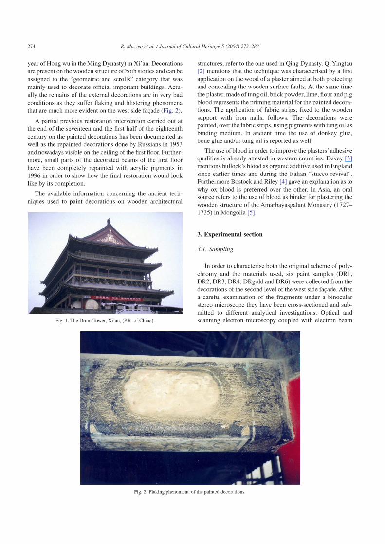
year of Hong wu in the Ming Dynasty) in Xi’an. Decorationsare present on the wooden structure of both stories and can beassigned to the “geometric and scrolls” category that wasmainly used to decorate official important buildings. Actu-ally the remains of the external decorations are in very badconditions as they suffer flaking and blistering phenomenathat are much more evident on the west side façade (Fig. 2).
A partial previous restoration intervention carried out atthe end of the seventeen and the first half of the eighteenthcentury on the painted decorations has been documented aswell as the repainted decorations done by Russians in 1953and nowadays visible on the ceiling of the first floor. Further-more, small parts of the decorated beams of the first floorhave been completely repainted with acrylic pigments in1996 in order to show how the final restoration would looklike by its completion.
The available information concerning the ancient tech-niques used to paint decorations on wooden architectural
structures, refer to the one used in Qing Dynasty. Qi Yingtau[2] mentions that the technique was characterised by a firstapplication on the wood of a plaster aimed at both protectingand concealing the wooden surface faults. At the same timethe plaster, made of tung oil, brick powder, lime, flour and pigblood represents the priming material for the painted decora-tions. The application of fabric strips, fixed to the woodensupport with iron nails, follows. The decorations werepainted, over the fabric strips, using pigments with tung oil asbinding medium. In ancient time the use of donkey glue,bone glue and/or tung oil is reported as well.
The use of blood in order to improve the plasters’adhesivequalities is already attested in western countries. Davey [3]mentions bullock’s blood as organic additive used in Englandsince earlier times and during the Italian “stucco revival”.Furthermore Bostock and Riley [4] gave an explanation as towhy ox blood is preferred over the other. In Asia, an oralsource refers to the use of blood as binder for plastering thewooden structure of the Amarbayasgalant Monastry (1727–1735) in Mongolia [5].
3. Experimental section
3.1. Sampling
In order to characterise both the original scheme of poly-chromy and the materials used, six paint samples (DR1,DR2, DR3, DR4, DRgold and DR6) were collected from thedecorations of the second level of the west side façade. Aftera careful examination of the fragments under a binocularstereo microscope they have been cross-sectioned and sub-mitted to different analytical investigations. Optical andscanning electron microscopy coupled with electron beamFig. 1. The Drum Tower, Xi’an, (P.R. of China).
Fig. 2. Flaking phenomena of the painted decorations.
274 R. Mazzeo et al. / Journal of Cultural Heritage 5 (2004) 273–283
microprobe analysis using an energy-dispersive X-ray detec-tor (SEM-EDX) were employed in order to characterise thestratigraphic morphology and determine the elemental com-position of each paint layer.
X-Ray diffraction was used to confirm the composition ofsome pigments and pyrolysis-gas-chromatography massspectrometry was performed in order to characterise thenature of the organic binding media used. The latter was also
used to characterise the chemical nature of a referencesample of tung oil, which was spread over a glass support andleft curing for three years (1995–1998) in China under nor-mal laboratory environmental conditions. To this regard ithas to be pointed out that no air conditioning system wasavailable at that time in the laboratory of the Xi’an Centre forconservation, so that both temperature and relative humidityfluctuations were very high.
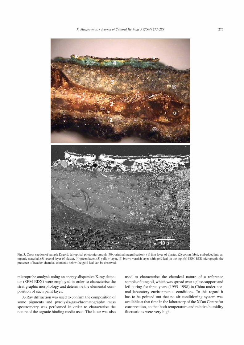
Fig. 3. Cross-section of sample Drgold: (a) optical photomicrograph (50× original magnification): (1) first layer of plaster, (2) cotton fabric embedded into anorganic material, (3) second layer of plaster, (4) green layer, (5) yellow layer, (6) brown varnish layer with gold leaf on the top; (b) SEM-BSE micrograph: thepresence of heavier chemical elements below the gold leaf can be observed.
275R. Mazzeo et al. / Journal of Cultural Heritage 5 (2004) 273–283
3.2. Methodologies
3.2.1. Pyrolysis–Gas chromatography–Mass Spectrometry
In this study, analytical pyrolysis [6–12] experimentswere performed using an integrated system consisting of aCDS Pyroprobe 1000 heated filament pyrolyser (ChemicalData System, Oxford, PA, USA) and a Varian 3400 gaschromatograph coupled to a Saturn II ion-trap mass spec-trometer (Varian Analytical Instruments, Walnut Creek, CA,USA). A DB-5MS J&W capillary column (30 m × 0.25 mm;i.d.: 0.25 µm film thickness) was programmed from 50 to300 °C at 5 °C/min, holding the initial temperature for 2 min.The samples, less than 1 mg, were pyrolysed without treat-ment in duplicate through a quartz sample holder at 700 °Cfor 10 s. The pyrolysis experiments were carried out in themethylating conditions adding 5 µl of an aqueous solution of25% of tetramethyl ammonium hydroxide (TMAH) to thesample before pyrolysis; in this way it is possible to obtainthe methylation of carboxylic and hydroxyl groups. Thisprocedure is called thermally assisted hydrolysis and methy-lation (THM), TMAH thermochemolysis or more simplypyrolysis-methylation.
PY-GC interface was kept at 250 °C and the injection portat 250 °C. Injection mode was split (1:50 split ratio). Thecarrier gas was Helium at a flow rate of 1.5 ml/min. Massspectra (1 scan/s) were recorded under electron impact at70 eV from 40 to 450 m/z.
3.2.2. Optical microscopy (OM)
Samples were embedded in a resin support, then cross-sectioned and polished according to conventional methods.
Dark field observation of cross-sectioned samples hasbeen performed using a LEITZ LABORLUX S microscopeequipped with fixed ocular of 10× and objective of 10×.
3.2.3. Scanning Electron Microscopy–Energy DispersiveX-Ray spectroscopy (SEM-EDX)
In this study, a Scanning Electron Microscope, Philips XL20 model SEM-EDX equipped with an Energy DispersiveX-ray Analyser was used on the same cross-sectionedsamples already prepared for the optical microscopy obser-vations. The elemental composition was carried out at anacceleration voltage of 25–30 KeV, lifetime > 50 s, CPS ≈2000 and working distance 34 mm.
3.2.4. X-ray diffraction
A Philips 1015 with a CuK radiation, 40 KV and 40 mA,Ni filter radiation was used to characterize the pigment com-position. Diffraction patterns were interpreted by compari-son with JCPDS data.
3.3. Analytical results
3.3.1. Preparation layers
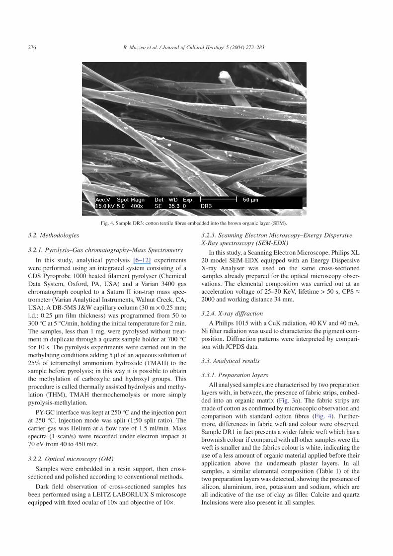
All analysed samples are characterised by two preparationlayers with, in between, the presence of fabric strips, embed-ded into an organic matrix (Fig. 3a). The fabric strips aremade of cotton as confirmed by microscopic observation andcomparison with standard cotton fibres (Fig. 4). Further-more, differences in fabric weft and colour were observed.Sample DR1 in fact presents a wider fabric weft which has abrownish colour if compared with all other samples were theweft is smaller and the fabrics colour is white, indicating theuse of a less amount of organic material applied before theirapplication above the underneath plaster layers. In allsamples, a similar elemental composition (Table 1) of thetwo preparation layers was detected, showing the presence ofsilicon, aluminium, iron, potassium and sodium, which areall indicative of the use of clay as filler. Calcite and quartzInclusions were also present in all samples.
Fig. 4. Sample DR3: cotton textile fibres embedded into the brown organic layer (SEM).
276 R. Mazzeo et al. / Journal of Cultural Heritage 5 (2004) 273–283

3.3.2. Paint layers: pigments
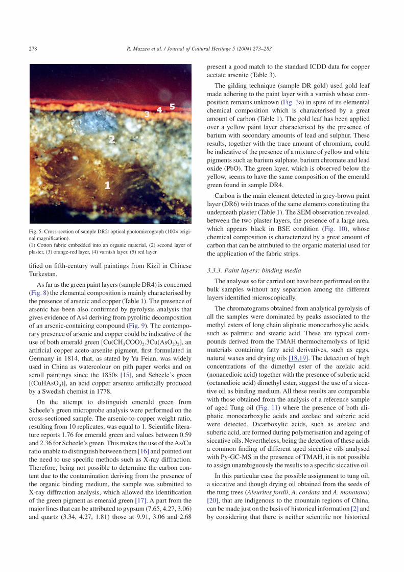
Electron beam microprobe analyses showed that twokinds of red pigments have been used (Table 1). Minium(Pb3O4) is present in both the red layers of sample DR2 eventhough the inner one shows an orange tonality if comparedwith the deep red characteristic of the surface. It was notpossible to characterise the thin black layer that can beobserved in cross-section (Fig. 5) between the two paintlayers. In spite of this, the large amount of carbon detectedwith microprobe analysis can be indicative of an organicmaterial applied as a varnish. The red layer in sample DR1does not contain any chemical element that could be relatedwith the presence of an inorganic red pigment. In fact thepresence of barium, calcium and sulphur may be attributed tobarium and calcium sulphate that have been used as mordantin connection with lake paints since about the beginning ofthe XIX century [13]. These elements are much more con-centrated at the bottom of the red layer as showed in theback-scattered photomicrography (Fig. 6a). The presence ofa red lake is clearly visible under microscopic examination ofthe cross-section that shows also how it penetrates into the
ground layer (Fig. 6b). In addition, a thin layer that fluorescesyellow under the excitation of UV light is clearly visible onthe top of the red layer (Fig. 6c). It shows an elementalcomposition similar to the underneath red layer and corre-spond to the external whitish thin layer visible in the Scan-ning Electron Microscope–Back Scattered Electron micro-graph (Fig. 6a). This layer may represent a further superficialthin application of red lake.
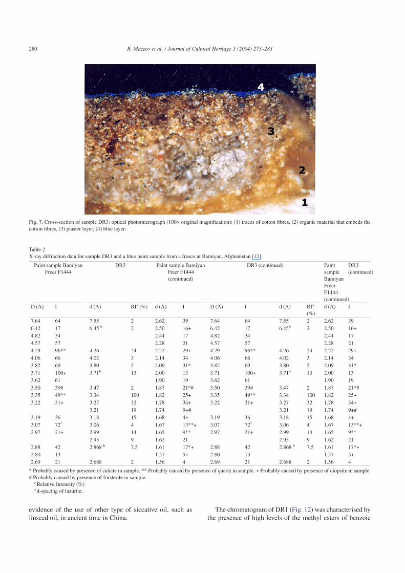
The blue pigment (sample DR3) resulted to be constitutedof lapis lazuli (Fig. 7), the main component of the semi-precious stone lazurite, a sodium aluminosilicate mineralwith sulphur radical anions residing in the aluminosilicatecrystal lattice (Table 1). Its presence has been confirmed byX-ray diffraction analysis. In fact, a part from a large amountof quartz, the X-ray diffraction data (Table 2) present a goodmatch to those obtained from a blue paint from a fresco atBamiyan, Afghanistan that was identified as natural ultrama-rine [14]. Lazurite has been ascribed to various sources inPersia, China and Tibet. The best quality lazurite comes fromBadakhshan in northeastern Afghanistan and was first iden-
Table 1Results of the Py-GC-MS and SEM-EDX analyses (in brackets elements detected in trace amount)
Py-GC-MS OM SEM-EDXSample Main fragments Material
identificationStratigraphy Thickness (µm) Elemental composition identification
DR1 Azelaic acid Siccative oil Superficial layer a 1 Ba, S, Si, Ca (Al, K, Fe)Palmitic acid Anthraqui-
none dye oralkoid resin
Red 18 C, Ba, Ca, S (Si, Al)Stearic acid Brown plaster 30 Si, Ca, Al, (Ba, S, Mg, Fe, K, Na)Benzoic acid Brown organic layer with traces of
brown plasterC, Si, Ca, Al, (Fe, K, Na)
Phtalic acidDimethyl ester
DR2 Azelaic acid Siccative oil Red 1 PbPalmitic acid Blackish organic layer 1 C, OStearic acid Orange/Red 2 Pb
Brown plaster 5 Si, Al, Mg (Fe, K, Na)Brown organic layer with tracesbrown plaster
15 C, Si, Al, (Mg, Fe, K, Na)
DR3 Azelaic acid Siccative oiland Animalglue
Blue 5 Si, Al, S, Na, (Ca, Fe, K)Palmitic acid Brown plaster 15–20 Si, Al, Mg (Fe, K, Na)Stearic acid Brown organic layer 10 C, OPirrole White cotton fibres 30–40 C, O
DR4 Azelaic acid Siccative oil Green 2–5 As, CuPalmitic acid Brown plaster 40 Si, Al, Ca, (Mg,Fe, K, Na)Stearic acid Brown organic layer 20–25 C (S, Ca, Si, Al, Na, Fe)As4 Brown plaster 40 Si, Al, Ca (Mg, Fe, K, Na)
DRgold Azelaic acid Siccative oil Gold leaf Au (Cu, As, Fe)Palmitic acid Varnish C, OStearic acid Yellow 2 Ba, S, Pb (Cr, As, Cu)As4 Green 3–4 As, Cu
Brown plaster 15–30 Si, Al, Ca, (Mg,Fe, Cl, As)Brown organic layer 20–30 C (Ca, Si, Al, Na, Fe)Brown plaster 20 Si, Al, Ca,Mg,(Fe)
DR6 Azelaic acid Siccative oil Grey 2 C,O (Si, Al, Ca, Fe)Palmitic acid Brown plaster 15 Si, Al, Ca, (Mg,Fe, K, Na)Stearic acid Brown organic layer 20 C (Ca, Si, Fe)As4 Brown plaster 35-40 Si, Al, Ca, (Mg,Fe, K, Na)
a Visible in cross-section under UV illumination and in SEM-BSE micrograph (Fig. 6c).
277R. Mazzeo et al. / Journal of Cultural Heritage 5 (2004) 273–283
tified on fifth-century wall paintings from Kizil in ChineseTurkestan.
As far as the green paint layers (sample DR4) is concerned(Fig. 8) the elemental composition is mainly characterised bythe presence of arsenic and copper (Table 1). The presence ofarsenic has been also confirmed by pyrolysis analysis thatgives evidence of As4 deriving from pyrolitic decompositionof an arsenic-containing compound (Fig. 9). The contempo-rary presence of arsenic and copper could be indicative of theuse of both emerald green [Cu(CH3COO)2.3Cu(AsO2)2], anartificial copper aceto-arsenite pigment, first formulated inGermany in 1814, that, as stated by Yu Feian, was widelyused in China as watercolour on pith paper works and onscroll paintings since the 1850s [15], and Scheele’s green[(CuHAsO3)], an acid copper arsenite artificially producedby a Swedish chemist in 1778.
On the attempt to distinguish emerald green fromScheele’s green microprobe analysis were performed on thecross-sectioned sample. The arsenic-to-copper weight ratio,resulting from 10 replicates, was equal to 1. Scientific litera-ture reports 1.76 for emerald green and values between 0.59and 2.36 for Scheele’s green. This makes the use of the As/Curatio unable to distinguish between them [16] and pointed outthe need to use specific methods such as X-ray diffraction.Therefore, being not possible to determine the carbon con-tent due to the contamination deriving from the presence ofthe organic binding medium, the sample was submitted toX-ray diffraction analysis, which allowed the identificationof the green pigment as emerald green [17]. A part from themajor lines that can be attributed to gypsum (7.65, 4.27, 3.06)and quartz (3.34, 4.27, 1.81) those at 9.91, 3.06 and 2.68
present a good match to the standard ICDD data for copperacetate arsenite (Table 3).
The gilding technique (sample DR gold) used gold leafmade adhering to the paint layer with a varnish whose com-position remains unknown (Fig. 3a) in spite of its elementalchemical composition which is characterised by a greatamount of carbon (Table 1). The gold leaf has been appliedover a yellow paint layer characterised by the presence ofbarium with secondary amounts of lead and sulphur. Theseresults, together with the trace amount of chromium, couldbe indicative of the presence of a mixture of yellow and whitepigments such as barium sulphate, barium chromate and leadoxide (PbO). The green layer, which is observed below theyellow, seems to have the same composition of the emeraldgreen found in sample DR4.
Carbon is the main element detected in grey-brown paintlayer (DR6) with traces of the same elements constituting theunderneath plaster (Table 1). The SEM observation revealed,between the two plaster layers, the presence of a large area,which appears black in BSE condition (Fig. 10), whosechemical composition is characterized by a great amount ofcarbon that can be attributed to the organic material used forthe application of the fabric strips.
3.3.3. Paint layers: binding media
The analyses so far carried out have been performed on thebulk samples without any separation among the differentlayers identified microscopically.
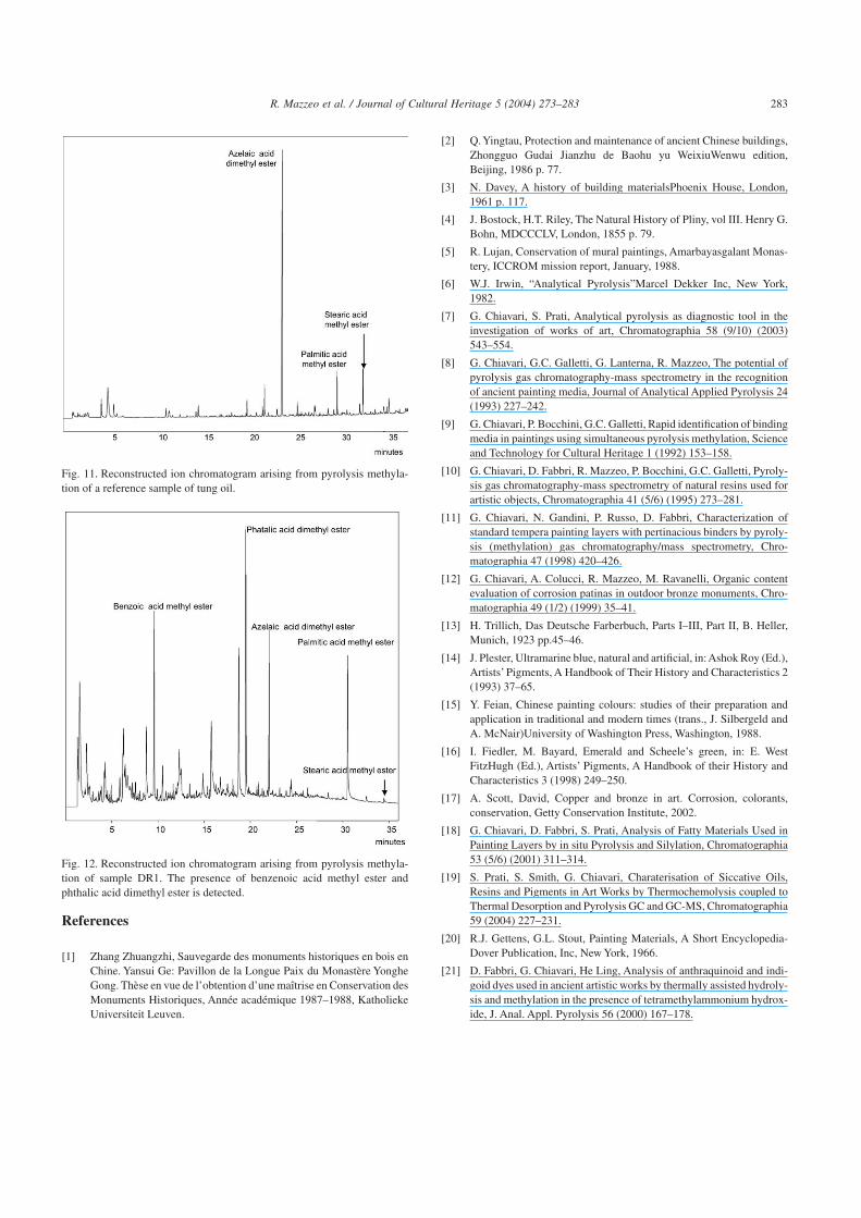
The chromatograms obtained from analytical pyrolysis ofall the samples were dominated by peaks associated to themethyl esters of long chain aliphatic monocarboxylic acids,such as palmitic and stearic acid. These are typical com-pounds derived from the TMAH thermochemolysis of lipidmaterials containing fatty acid derivatives, such as eggs,natural waxes and drying oils [18,19]. The detection of highconcentrations of the dimethyl ester of the azelaic acid(nonanedioic acid) together with the presence of suberic acid(octanedioic acid) dimethyl ester, suggest the use of a sicca-tive oil as binding medium. All these results are comparablewith those obtained from the analysis of a reference sampleof aged Tung oil (Fig. 11) where the presence of both ali-phatic monocarboxylic acids and azelaic and suberic acidwere detected. Dicarboxylic acids, such as azelaic andsuberic acid, are formed during polymerisation and ageing ofsiccative oils. Nevertheless, being the detection of these acidsa common finding of different aged siccative oils analysedwith Py-GC-MS in the presence of TMAH, it is not possibleto assign unambiguously the results to a specific siccative oil.
In this particular case the possible assignment to tung oil,a siccative and though drying oil obtained from the seeds ofthe tung trees (Aleurites fordii, A. cordata and A. monatana)[20], that are indigenous to the mountain regions of China,can be made just on the basis of historical information [2] andby considering that there is neither scientific nor historical
Fig. 5. Cross-section of sample DR2: optical photomicrograph (100× origi-nal magnification).(1) Cotton fabric embedded into an organic material, (2) second layer ofplaster, (3) orange-red layer, (4) varnish layer, (5) red layer.
278 R. Mazzeo et al. / Journal of Cultural Heritage 5 (2004) 273–283

Fig. 6. Cross-section of sample DR1. (a) SEM-BSE micrograph (82× original magnification). The BSE observation highlight the presence of heavier elementboth on the surface and on the bottom of the red layer, (b) optical photomicrograph (100× original magnification): (1) plaster layer, (2) red layer, (c) opticalphotomicrograph under UV fluorescence (200× original magnification): a thin layer that fluoresces yellow is visible on the top of the red.
279R. Mazzeo et al. / Journal of Cultural Heritage 5 (2004) 273–283
evidence of the use of other type of siccative oil, such aslinseed oil, in ancient time in China.
The chromatogram of DR1 (Fig. 12) was characterised bythe presence of high levels of the methyl esters of benzoic
Fig. 7. Cross-section of sample DR3: optical photomicrograph (100× original magnification): (1) traces of cotton fibres, (2) organic material that embeds thecotton fibres, (3) plaster layer, (4) blue layer.
Table 2X-ray diffraction data for sample DR3 and a blue paint sample from a fresco at Bamiyan, Afghanistan [12]
Paint sample BamiyanFreer F1444
DR3 Paint sample BamiyanFreer F1444(continued)
DR3 (continued) PaintsampleBamiyanFreerF1444(continued)
DR3(continued)
D (A) I d (A) RIa (%) d (A) I D (A) I d (A) RIa
(%)d (A) I
7.64 64 7.55 2 2.62 39 7.64 64 7.55 2 2.62 396.42 17 6.45 b 2 2.50 16+ 6.42 17 6.45b 2 2.50 16+4.82 34 2.44 17 4.82 34 2.44 174.57 57 2.28 21 4.57 57 2.28 214.29 96** 4.26 24 2.22 29+ 4.29 96** 4.26 24 2.22 29+4.06 66 4.02 3 2.14 34 4.06 66 4.02 3 2.14 343.82 69 3.80 5 2.09 31* 3.82 69 3.80 5 2.09 31*3.71 100+ 3.71b 13 2.00 13 3.71 100+ 3.71b 13 2.00 133.62 61 1.90 19 3.62 61 1.90 193.50 39# 3.47 2 1.87 21*# 3.50 39# 3.47 2 1.87 21*#3.35 49** 3.34 100 1.82 25+ 3.35 49** 3.34 100 1.82 25+3.22 31+ 3.27 32 1.78 34+ 3.22 31+ 3.27 32 1.78 34+
3.21 19 1.74 9+# 3.21 19 1.74 9+#3.19 36 3.18 15 1.68 4+ 3.19 36 3.18 15 1.68 4+3.07 72* 3.06 4 1.67 13**+ 3.07 72* 3.06 4 1.67 13**+2.97 21+ 2.99 14 1.65 9** 2.97 21+ 2.99 14 1.65 9**
2.95 9 1.62 21 2.95 9 1.62 212.88 42 2.868 b 7.5 1.61 17*+ 2.88 42 2.868 b 7.5 1.61 17*+2.80 13 1.57 5+ 2.80 13 1.57 5+2.69 21 2.688 2 1.56 4 2.69 21 2.688 2 1.56 4
* Probably caused by presence of calcite in sample. ** Probably caused by presence of quartz in sample. + Probably caused by presence of diopsite in sample.# Probably caused by presence of forsterite in sample.
a Relative Intensity (%)b d-spacing of lazurite.
280 R. Mazzeo et al. / Journal of Cultural Heritage 5 (2004) 273–283

acid and 1,2-benzenedicarboxylic acid (o-phthalate) as wellas phthalic anhydride, not detected in the chromatograms ofthe other samples. The very source of phthalic acid is stillunder investigation. Phthalic acid derivatives are componentsof alkyd resins, which have been used as varnishes for wood-works, and occasionally found by restorers on paintings.Antraquinones occurring in madder dyes are also known toproduce dimethyl phthalate in the pyrolysates when sub-jected to pyrolysis/methylation [21].
4. Conclusions
In this study, the analyses are not only meant to assist inthe assessment of the authenticity of the materials used butalso aimed at guiding conservators and conservation scien-tists in taking into account materials and methods used in thepast by Chinese artists.
The coupling of reflected light microscopy, SEM-EDX,XRD and Py-GC-MS applied on cross-sectioned paintsamples taken from the Ming Dynasty monument of theDrum Tower provides a useful tool for the contemporarycharacterisation of the original scheme of polychromy andthe identification of the nature of the pigments and the or-ganic binding media used.
The scientific results of this study are quite in agreementwith the few historical information available.
In fact the presence between clay based plaster layers offabric strips treated with a siccative oil, probably tung oil wasconfirmed. In addition both the brownish colour and thewider weft of the cotton fabric observed in sample DR1seems to be original, while the red lake, which penetratesonto the underneath plaster layer, was applied on the occa-sion of past restoration interventions. The emerald green andthe composition of the yellow layer found in samples DR4and DR gold can certainly be ascribed to a later restorationprocedure carried out in the late XVIII century which corre-sponds to the time of introduction in China of the abovementioned pigments. To the same period of time can be datedthe external red layer found in sample DR2. In this case thepresence of an underneath varnish, which appears blackunder cross-section, can be a further confirmation.
A siccative oil, probably tung oil, seems to have been usednot only as a waterproof agent but also as binder for theplaster layersand pigments, even though the blue decorationshows the possible use of animal glue.
It was not possible to confirm the hypothesis about thepresence of blood binder even though it can be sustainedthanks to the available bibliographic data [3,4].
Fig. 8. Cross-section of sample DR4: optical photomicrograph (100× original magnification).
Fig. 9. Reconstructed ion chromatogram arising from pyrolysis methylationof sample DR4; the presence of As4 deriving from pyrolitic decompositionof an arsenic-containing compound is detected.
281R. Mazzeo et al. / Journal of Cultural Heritage 5 (2004) 273–283

Acknowledgements
The authors would like to express their gratitude to theShaanxi Bureau of Cultural Relics and the Xi’an Center for
the Conservation and Restoration of Cultural Relics in Xi’anfor having provided the samples for analysis. A specialthanks has to be paid to Dr. Giuseppe Falini (University ofBologna, Chemistry Department) for having performed theX-ray diffraction analyses.
Table 3X-ray diffraction data for sample DR4 and Copper acetate arsenites (Emerald green): ICDD 1-51 and 31-448 (15)
Copper acetate arseniteC4H6As6Cu4O16 ICDD 1-51
Copper acetate arsenite ICDD31-448
DR4 Copper acetate arseniteICDD 31-448 (continued)
DR4 (continued)
D (A) I D (A) I D (A) RIa(%) D (A) I D (A) RIa(%)10.0 100 9.71 100 9.91b 45 2.67 20
6.76 25 7.65 22 2.63 95.36 5 2.62 404.86 9 4.91 13 2.58 20
4.55 12 4.70 11 4.27 57 2.55 353.99 12 4.53 100 4.04 9 2.49 25 2.49 173.48 8 3.93 95 3.34 100 2.48 2
3.71 5 3.19 68 2.46 9 2.45 153.30 4 4.48 80 2.43 53.06 20 3.38 55 3.06b 38 2.41 602.68 24 3.28 70 2.38 122.40 4 3.18 8 2.36 6
3.09 45 2.31 7 2.28 53.07 60 2.26 14 2.13 19
1.69 8 3.06 80 2.25 65 2.08 41.62 4 2.98 10 2.98 22 2.18 17 1.97 41.55 8 2.73 55 2.87 20 2.15 5 1.87 61.45 4 2.71 80 2.12 10 1.81 12
2.69 19 2.68b 17 2.11 16 1.77 102.68 100 2.07 12 1.66 5
2.00 16a Relative Intensity (%).b d-spacing of emerald green.
Fig. 10. Cross-section of sample DR6:SEM-BSE micrograph (39× original magnification), a large black area due to the presence of an organic material is visibleon the upper left side.
282 R. Mazzeo et al. / Journal of Cultural Heritage 5 (2004) 273–283
References
[1] Zhang Zhuangzhi, Sauvegarde des monuments historiques en bois enChine. Yansui Ge: Pavillon de la Longue Paix du Monastère YongheGong. Thèse en vue de l’obtention d’une maîtrise en Conservation desMonuments Historiques, Année académique 1987–1988, KatholiekeUniversiteit Leuven.
[2] Q.Yingtau, Protection and maintenance of ancient Chinese buildings,Zhongguo Gudai Jianzhu de Baohu yu WeixiuWenwu edition,Beijing, 1986 p. 77.
[3] N. Davey, A history of building materialsPhoenix House, London,1961 p. 117.
[4] J. Bostock, H.T. Riley, The Natural History of Pliny, vol III. Henry G.Bohn, MDCCCLV, London, 1855 p. 79.
[5] R. Lujan, Conservation of mural paintings, Amarbayasgalant Monas-tery, ICCROM mission report, January, 1988.
[6] W.J. Irwin, “Analytical Pyrolysis”Marcel Dekker Inc, New York,1982.
[7] G. Chiavari, S. Prati, Analytical pyrolysis as diagnostic tool in theinvestigation of works of art, Chromatographia 58 (9/10) (2003)543–554.
[8] G. Chiavari, G.C. Galletti, G. Lanterna, R. Mazzeo, The potential ofpyrolysis gas chromatography-mass spectrometry in the recognitionof ancient painting media, Journal of Analytical Applied Pyrolysis 24(1993) 227–242.
[9] G. Chiavari, P. Bocchini, G.C. Galletti, Rapid identification of bindingmedia in paintings using simultaneous pyrolysis methylation, Scienceand Technology for Cultural Heritage 1 (1992) 153–158.
[10] G. Chiavari, D. Fabbri, R. Mazzeo, P. Bocchini, G.C. Galletti, Pyroly-sis gas chromatography-mass spectrometry of natural resins used forartistic objects, Chromatographia 41 (5/6) (1995) 273–281.
[11] G. Chiavari, N. Gandini, P. Russo, D. Fabbri, Characterization ofstandard tempera painting layers with pertinacious binders by pyroly-sis (methylation) gas chromatography/mass spectrometry, Chro-matographia 47 (1998) 420–426.
[12] G. Chiavari, A. Colucci, R. Mazzeo, M. Ravanelli, Organic contentevaluation of corrosion patinas in outdoor bronze monuments, Chro-matographia 49 (1/2) (1999) 35–41.
[13] H. Trillich, Das Deutsche Farberbuch, Parts I–III, Part II, B. Heller,Munich, 1923 pp.45–46.
[14] J. Plester, Ultramarine blue, natural and artificial, in:Ashok Roy (Ed.),Artists’ Pigments, A Handbook of Their History and Characteristics 2(1993) 37–65.
[15] Y. Feian, Chinese painting colours: studies of their preparation andapplication in traditional and modern times (trans., J. Silbergeld andA. McNair)University of Washington Press, Washington, 1988.
[16] I. Fiedler, M. Bayard, Emerald and Scheele’s green, in: E. WestFitzHugh (Ed.), Artists’ Pigments, A Handbook of their History andCharacteristics 3 (1998) 249–250.
[17] A. Scott, David, Copper and bronze in art. Corrosion, colorants,conservation, Getty Conservation Institute, 2002.
[18] G. Chiavari, D. Fabbri, S. Prati, Analysis of Fatty Materials Used inPainting Layers by in situ Pyrolysis and Silylation, Chromatographia53 (5/6) (2001) 311–314.
[19] S. Prati, S. Smith, G. Chiavari, Charaterisation of Siccative Oils,Resins and Pigments in Art Works by Thermochemolysis coupled toThermal Desorption and Pyrolysis GC and GC-MS, Chromatographia59 (2004) 227–231.
[20] R.J. Gettens, G.L. Stout, Painting Materials, A Short Encyclopedia-Dover Publication, Inc, New York, 1966.
[21] D. Fabbri, G. Chiavari, He Ling, Analysis of anthraquinoid and indi-goid dyes used in ancient artistic works by thermally assisted hydroly-sis and methylation in the presence of tetramethylammonium hydrox-ide, J. Anal. Appl. Pyrolysis 56 (2000) 167–178.
Fig. 11. Reconstructed ion chromatogram arising from pyrolysis methyla-tion of a reference sample of tung oil.
Fig. 12. Reconstructed ion chromatogram arising from pyrolysis methyla-tion of sample DR1. The presence of benzenoic acid methyl ester andphthalic acid dimethyl ester is detected.
283R. Mazzeo et al. / Journal of Cultural Heritage 5 (2004) 273–283