Naturally Occurring Radioactive Materials (NORM) in Class II ...

ANALYSIS OF NATURALLY OCCURRING DELAYEDTYPEHYPERSENSITIVITY REACTIONS IN LEPROSY
BY IN SITU HYBRIDIZATION
BY CATHLEEN L. COOPER,* CHRISTOPH MUELLER,$TIP-AKSORN SINCHAISRI,1, CLAUDE PIRMEZ,T JOHN CHAN,II
GILLA KAPLAN,1 SUMMER M. M. YOUNG, : IRVING L . WEISSMAN,§BARRY R. BLOOM,II THOMAS H. REA,T AND ROBERT L . MODLIN*t
From the *Department of Pathology and TSection of Dermatology, University of Southern CaliforniaSchool ofMedicine, Los Angeles, California 90033; the Laboratory of Experimental Oncology,
Department of Pathology, Stanford University Medical Center, Stanford, California; theIIDepartment of Microbiology and Immunology, Albert Einstein College ofMedicine,Bronx, New York 10461; and ILaboratory for Cellular Physiology and Immunology,
The Rockefeller University, New York, New York 10021
Leprosy provides a useful model for understanding immunoregulatory mecha-nisms in man. The disease forms a spectrum in which the immunologic responseof the patient correlates with the clinical and histopathologic classification (1-3). Atone end of the spectrum, patients with tuberculoid leprosy have one or several skinlesions in which bacilli can rarely be identified . CD4+ T lymphocytes predominatein these lesions (4-7) and respond to Mycobacterium leßrae in vitro (8) . At the otherend of the spectrum, patients with lepromatous leprosy have diffuse infiltration ofskin and nerves with bacilli-laden macrophages. CD8+ cells predominate inlepromatous lesions and function as antigen-specific Ts cells in vitro (9, 10). TheCD4+ lymphocytes derived from these lesions are unresponsive to M. leßrae . Im-posed upon this spectrum are the so-called "reactional states" of reversal reactionand erythema nodosum leprosum (ENL)' that are of immunologic interest in un-derstanding mechanisms of immunoregulation in man.The reversal reaction syndrome is associated with a marked rise in lymphocyte
transformation toM. leßrae antigens in vitro, and is therefore thought to be a delayed-type hypersensitivity (DTH) reaction againstM. leßrae antigens (11-15) . Clinically,the condition is often associated with reduction or elimination of bacilli in lesionsand sometimes followed by upgrading of the clinical and histological classificationtoward the tuberculoid pole . The pathogenesis of ENL reactions is somewhat contro-versial, thought to be due to immune complex deposition in the lesions (16-18) and/orincreases in cell-mediated immunity (19, 20).
This work was supported by grants from the National Institutes of Health (AI-22187, AI-22553), theUNDP/World Bank/World Health Organization Special Programme for Research and Training in TropicalDiseases (IMMLEP), National Hansen's Disease Center, Knights of St . Lazarus ofJerusalem, HeiserTrust, Drown Foundation, and the Hartford Foundation . Address correspondence to Robert L. Modlin,Dermatology, HMR 904, U. S. C . School of Medicine, 2025 Zonal Avenue, Los Angeles, CA 90033 .
1 Abbreviations used in this paper: AEC, aminoethyl carbazole; DTH, delayed-type hypersensitivity ; ENL,erythema nodosum leprosum .
J . Exp. MED . 0 The Rockefeller University Press - 0022-1007/89/05/1565/17 $2 .00
1565Volume 169 May 1989 1565-1581
on January 31, 2016jem
.rupress.orgD
ownloaded from
Published May 1, 1989

1566
IN SITU ANALYSIS OF LEPROSY LESIONS
Immunohistologic study of skin lesions has thus far not distinguished betweenthe immune processes underlying the pathogenesis of reversal reaction and ENL(21-25) . The inflammatory infiltrates in both reactional states are characterized bya predominance of CD4' lymphocytes . IL-2-containing cells are present in the le-sions of both conditions . Furthermore, keratinocyte la expression, a putative markerofIFN-y production, and Langerhans cell hyperplasia are apparent in the epidermisoverlying both reactional states .The present study was undertaken to investigate the immunoregulatory processes
that underlie these reactional states of leprosy, utilizing newly developed reagentsand technology to analyze the inflammatory infiltrates . We performed in situ hy-bridization to cellular mRNA using probes for IFN-y and human serine esterase(huHF), a marker for cytotoxic cells, in order to define functional T cell subpopula-tions in these reactions . The findings of these studies were then analyzed in the con-text of immunostaining of lesions with mAbs that define subpopulations of CD4'and CD8' lymphocytes . The results suggest that reversal reactions are more char-acteristic of classical DTH reactions than ENL, and represent a window into thecomplex immunoregulatory mechanisms underlying the pathogenesis ofDTH in man.
Materials and Methods
Patients .
Patients with leprosy were classified according to the clinicopathological criteriaof Ridley and Jopling (1) . Clinical criteria for the diagnosis of reversal reactions includedtorpid lesions becoming erythematous and tumid, the abrupt onset of new erythematous,tumid lesions, and the sudden onset of neuritis . Histological changes were not uniform butincluded edema in the granulomas, increased numbers of lymphocytes, giant cells, and des-moplasia of the connective tissue . The diagnosis of ENL was made using clinical and histo-pathologic criteria . Patients presented with the distinctive picture ofcrops ofpainful and tendererythematous nodules in association with fever, malaise and arthralgias . Histopathologic ex-amination of lesions revealed the presence of neutrophils and lymphocytes superimposed overa lepromatous infiltrate. In addition, lepromin skin tests (3-wk Mitsudareactions) were studiedfrom tuberculoid patients (26) . All specimens were obtained with informed consent fromtuberculoid patients (26) . All specimens were obtained with informed consent from patientsat the Outpatient Clinic of Los Angeles County/University of Southern California MedicalCenter, San Francisco Medical Center (with gratitude to Dr. Robert Gelber), and McKeanReliability Institute, Chiang Mai, Thailand . The patients were distributed among the differentdiagnostic groups showing no segregation according to sex, race, or age .
Tissues.
Skin biopsy specimens, obtained by punch or scalpel technique, were dividedfor histological diagnosis on conventional paraffin sections and for immunoperoxidase andin situ hybridization study by snap freezing in liquid nitrogen tissue embedded in OCT medium(Ames Co ., Elkhart, .IN) . The tissues were stored at -70'C until sectioning.
In Situ HybridizationThe in situ hybridization technique described below was performed as previously described
(27) .Tissue Preparation .
Biopsy specimens were collected and frozen as described above . 3-Amcryostat sections were placed on poly-L-lysine hydrobromide-coated slides (Sigma ChemicalCo., St . Louis, MO) and air dried for 2 h . Dried sections were fixed for,20 min in 4%paraformaldehyde-PBS (pH 7 .4), rinsed through several changes ofPBS (Flow Laboratories,Inc ., McLean, VA), and dehydrated through graded alcohols . Sections were stored dessicatedat 4°C until use .
Probe Preparation.
Two probes were utilized in the present study : IFN-y cDNA, which wasprovided by Hoffmann-La Roche, Inc . (Nutley, NJ), and huHF gene coding forhuman serineesterase, as described previously (28) . The cDNA was recloned into an expression vector system
on January 31, 2016jem
.rupress.orgD
ownloaded from
Published May 1, 1989
COOPER ET AL .
1567
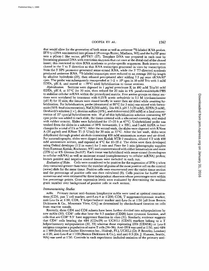
that would allow for the generation of both sense as well as antisense "S-labeled RNA probes .IFN-,y cDNA was inserted into pGem-1 (Promega Biotec, Madison, WI) and the huHF geneinto a pGem-1 like vector, pSPT671 (27) . Template DNA was prepared in each case bylinearizing plasmid DNA with restriction enzymes that cut once at the distal end ofthe clonedinsert ; this restricted in vitro RNA synthesis to probe-specific sequences . Both inserts werecloned in the 5' to 3' direction so that RNA transcripts generated in vitro by transcriptionfrom the 5' SP6 promotor-generated sense-strand RNA, while the 3' T7-directed synthesisproduced antisense RNA. 35S-labeled transcripts were reduced to an average 200-bp lengthby alkaline hydrolysis (29), then ethanol precipitated after adding 7 .5 Ftg yeast tRNA/106cpm . The probe was subsequently resuspended at 1-2 x 10 6 cpm in 10 mM Tris with 1 mMEDTA, pH 8, and stored at -70°C until hybridization to tissue sections .
Hybridization.
Sections were digested in 1 ug/ml proteinase K in 100 mM Tris/50 mMEDTA, pH 8, at 37'C for 30 min, then refixed for 20 min in 4% paraformaldehyde/PBSto stabilize cellular mRNA within the proteolyzed matrix . Free amino groups on tissue sec-tions were acetylated by treatment with 0.25% acetic anhydride in 0 .1 M triethanolamine(pH 8) for 10 min ; the tissues were rinsed briefly in water then air dried while awaiting hy-bridization . For hybridization, probe (denatured at 80'C for 3 min) was mixed with forma-mide (50% final concentration), NaCl (300 mM), Tris-HCl, pH 7.5 (20 mM), EDTA (5 mM),Denhardt's solution (1 x), dextran sulfate (10%), and dithiotreitol (100 mM) at a final concen-tration of 10 5 cpm/p.l hybridization mix . 10,ul of this hybridization solution containing 106cpm probe was added to each slide, the tissue covered with a siliconized coverslip, and sealedwith rubber cement . Slides were hybridized for 17-20 h at 47°C . Unhybridized probe waswashed from the slides in a solution of 50% formamide, 2 x SSC, and 5 mM EDTA in fourchanges for a total of2 h at 56°C . After the second wash, the slides were digested with RNaseA (20 jig/ml) and RNase TI (1 U/ml) for 30 min at 37°C. After the last wash, slides weredehydrated through graded alcohols containing 400 mM ammonium acetate and air dried .For autoradiography, slides were dipped into Kodak NTB-2 emulsion, diluted 1 :2 with 400mM ammonium acetate, and exposed at 4'C for 10-28 d . The slides were then developedusing Dektol developer (1 :2 in water) for 2 min and Fixer for 5 min (photography suppliesfrom Eastman Kodak; Rochester, NY) and counterstained with eitherhematoxlyin and eosin(IFN-y) or 4% Giemsa (huHF) . Each tissue was hybridized with sense-strand (homologousto cellular mRNA) as well as antisense strand (complementary to cellular mRNA) probes ;known positive and negative control tissues were included in each run .
Evaluation ofSlides.
Cells were considered to be positive for the expression of IFN- ,y whenthey contained greater than twice the number of grains ofthe most positive cell on the control(sense) slide for the same tissue . Positive cells were enumerated over the entire tissue sectionand the percentage of positive cells was then calculated (8) . Cells positive for huHF werenumerous and were estimated by three independent observers whose percentages were withinfive percentage points . Gene expression levels were evaluated by determining the mediangrain number over background of positive cells in each section.
Immunostaining StudiesmAbs .
Primary mouse anti-human lymphocyte mAbs were used at optimal concentra-tions (CD3, pan T cell marker, anti-Leu-4 at 1 :200; CD8, T suppressor/cytotoxic marker,anti-Leu-2a at 1 :50 ; CD4, T helper/inducer marker anti-Leu-3a at 1 :50 [all from BectonDickinson & Co., Mountain View, CA]) as determined by checkerboard titration on cellsfrom reactive tonsils .
Recently, these CD4 and CD8 subsets have been further divided into subpopulations bynew mAbs (30) . CD8' cells that bear the 9.3 marker (CD28) have cytotoxic function, andcells that are CD8' 9.3 - have suppressor function in vitro (31) . Similarly, evidence suggeststhat CD4' cells bearing the 4114 (CDw29) or UCHLI (CD45) markers belong to a Thelper/memory subpopulation (32, 33) whereas those expressing 2114 (CD45R) or Leu-8antigens comprise a population ofnaive T cells (34-36) . Anti-2114 was used at 1:50, anti-4134at 1 :400 (both from Coulter Electronics Inc., Hialeah, FL), UCHL1 (Dr. P Beverley, London)at 1 :20, anti-Leu-8 at 1 :100 (Becton Dickinson & Co.), and anti-9 .3 (Dr. J . Hansen, Seattle,WA) was used at 1 :50 . Controls in each experiment included omission of the primary anti-
on January 31, 2016jem
.rupress.orgD
ownloaded from
Published May 1, 1989

1568
IN SITU ANALYSIS OF LEPROSY LESIONS
body and the use of irrelevant antibodies of the same IgG subclass on sections for the sameperiod of incubation . Blocking was used for some mAbs that gave significant levels of back-ground staining, (e .g ., anti-4B4, -9 .3, and -Leu-3a) . Where appropriate, slides were prein-cubated in normal serum (1 :20 in PBS) derived from the species that generated the secondaryantibody ; i .e ., normal goat serum was used when the linking antibody was biotinylated goatantimouse .
Single Immunoperoxidase Staining.
All sections were acetone fixed and incubated sequen-tially as described (21) : first with the mAbs, then peroxidase-conjugated goat anti-mouse IgG1 :20 (Tago Inc ., Burlingame, CA) for 30 min with 10-min PBS washes between each incuba-tion . Slides were then incubated with the chromogen aminoethyl carbazole (AEC) in the pres-ence of hydrogen peroxide for 15 min, counterstained with hematoxylin, and then mountedin glycerine-gelatin . The IFN--y-induced peptide, IP-10, was visualized in tissue sections usinga rabbit polyclonal antibody as previously reported (37) .
Double Immunoperoxidase Staining.
This procedure allows for the visualization of two an-tigens on the same section . For the monoclonals that gave significant levels of backgroundstaining, the sections were first blocked with normal goat or horse serum (diluted 1 :20 inPBS), as specified above . Each section was stained first with primary mAbs followed by goatanti-mouse IgG-peroxidase conjugate . Throughout both single and double immunostainingprocedures, each antiserum (or Avidin-Biotin complex) was applied to the tissue section for30 min, followed by a 10-min PBS wash . The first reaction was developed by a short (8-min)incubation in AEC. The development period was shorter than that used in single stainingso that the strong AEC precipitate would not obscure the weaker blue precipitate depositedin the second stage of the procedure. The sections were washed with PBS for 10 min . Beforethe second stain, all sections were blocked with normal horse serum at 1 :20 (Vector Labora-tories, Inc ., Burlingame, CA) . Subsequently, the slides were incubated with the second pri-mary antibody, washed and then incubated with the secondary biotinylated horse anti-mouseIgG 1 :50 (Vector Laboratories, Inc .), followed by an incubation with the ABC-GO kit (VectorLaboratories, Inc .) . The blue color was developed with a glucose oxidase substrate kit (con-taining nitroblue tetrazolium as chromogen) or alternatively using /3-galactosidase kit (VectorLaboratories, Inc .) using X-gal as the chromogenic substrate . Slides were then rinsed in tapwater for 10 min and mounted with glycerine-gelatin . Control slides, on which either of theprimary antisera were omitted, demonstrated that the second labeling system did not cross-react with the antibodies already added in the first reaction .
Quantitation ofStained Cells.
Single staining cells were numerous, such that the percentageof positive cells was determined by averaging the estimates of two independent observers ;their reading invariably agreed within five percentage points . For double immunostaining,cells were counted to be double positive when both red and blue colors could be seen onthe same cells or a purple color distinct from either red- or blue-colored cells in the samesection was identified . Percentages of double-stained cells were determined as described pre-viously (38) .
ResultsIFN-y Gene Expression in Lesions .
To assess functional activity of T lymphocytesin lesions, we initially performed in situ hybridization to detect IFN-1' mRNA. 16 pa-tients with reversal reaction were studied, all ofwhom were classified as having border-line lepromatous leprosy. 10 patients with ENL were classified as having leproma-tous disease . Findings were compared with specimens from patients with borderlinelepromatous leprosy not in reversal reaction (n = 7), tuberculoid patients (n = 15),and lepromatous patients (n = 10) . Since the "Mitsuda" reaction to M. leprae is anestablished measure ofDTH, skin test reactions in three responsive individuals werestudied . Cells showing positive hybridization were most numerous in reversal reac-tions, followed by lepromin skin tests and tuberculoid lesions (Figs . 1 and 2). Incomparison, lesions from nonreactional lepromatous patients and patients with ENL
on January 31, 2016jem
.rupress.orgD
ownloaded from
Published May 1, 1989

COOPER ET AL .
1569
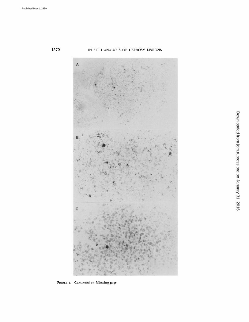
contained few positive cells, generally an order ofmagnitude lower than in reversalreactions . The positive cells were centrally located within the granuloma in reversalreactions and tuberculoid lesions, suggesting that they are ofthe Th phenotype (Fig . 1) .The relative level ofmRNA wasestimated by determination ofthe number ofgrainsper average positive cell . IFN-y mRNA expression was five times higher in reversalreactions than tuberculoid lesions or lepromin skin tests (Fig . 2) . Interestingly, thefew positive cells in ENL lesions contained levels of IFN-y mRNA that were fivetimes greater than found in lepromatous cases.To obtain evidence that IFN-y mRNA was translated into functional protein, im-
munostaining for IP-10 was undertaken . This peptide has been reported to bespecifically induced by IFN-y in cells, including keratinocytes (37) . The majorityof kerotinocytes overlying reversal reactions and tuberculoid leprosy stained positivefor IP-10 (Fig . 1), indicating the presence of IFN-y in these lesions . On the otherhand, few keratinocytes in lepromatous epidermis were positive for IP-10 . ENL le-sions gave an intermediate staining pattern.huHF Gene Expression in Lesions.
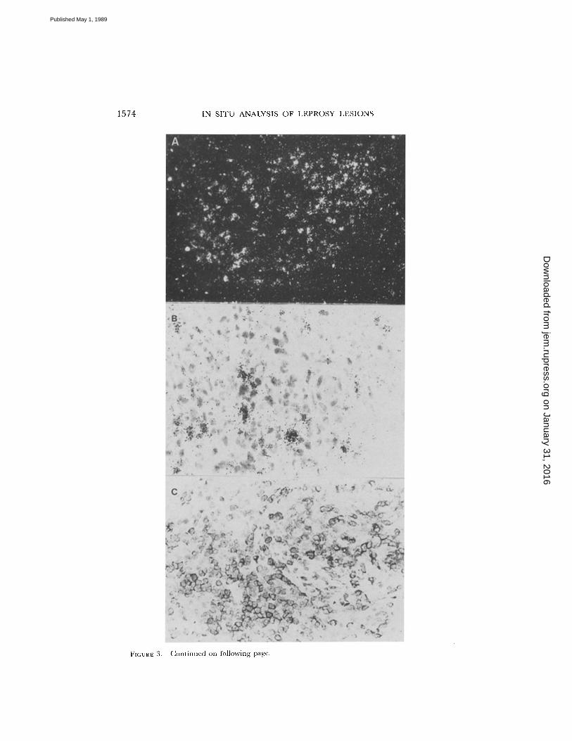
We investigated lesions for the presence ofTcellsof cytotoxic function using a riboprobe for the enzymatic marker associated withcytotoxic cells, serine esterase (huHF) . Cells positive for huHF were more numerousin tuberculoid, lepromin skin tests, and reversal reaction lesions, as compared withENL and nonreactional lepromatous lesions (Figs. 3 and 4) . The microanatomiclocation of huHF+ cells within granulomas of tuberculoid leprosy and reversal reac-tion was similar to that of the CD4+UCHL1' subpopulation (see Fig. 3) . The rela-tive gene expression was higher in reversal reaction than in tuberculoid lesions (Fig. 4) .T Cell Subsets in Lesions.
The nature of the T lymphocyte infiltrates in lesionsof leprosy reactional states was established by using mAbs in conjunction with im-munohistochemical techniques . These studies were undertaken to provide ex-perimental support for the in situ hybridization studies.The distribution and location of the CD4 and CD8 T lymphocytes subsets were
found to be similar to that described in earlier studies (21, 22). CD4+ cells predomi-nated in reversal reaction and ENL lesions, lepromin skin tests, and tuberculoidlesions, but not in nonreactional borderline lepromatous or lepromatous lesions.The pattern of cell distribution in reversal reaction specimens was similar to thatfound in tuberculoid granulomas : CD4+ cells were distributed throughout thegranulomas with CD8+ cells confined to the surrounding mantle zone. In contrast,in ENL and nonreactional lepromatous or borderline lepromatous lesions, bothCD4' and CD8+ cells were admixed throughout the biopsy sections .CD4+ Subpopulations .
To analyze in more detail the nature of the immunologicprocesses underlying the reactional states, lesions were analyzed with mAbs usedto define subpopulations of CD4 and CD8 cells . In reversal reactions, as in tuber-culoid granulomas and Mitsuda reactions, 2H4' and Leu-8' cells were invariablyrestricted to the mantle area surrounding the granuloma, whereas the cells associatedwith macrophages in the granuloma were ofthe UCHL1 and 4134 phenotype (Fig . 3) .These markers have been associated with differentiated or memory T cells and Thcells. In contrast, in ENL and nonreactional lepromatous and borderline leproma-tous, all phenotypes were admixed with macrophages.Double immunostaining was undertaken to quantify the various CD4 subpopula-
tions since some markers, such as UCHL1, are present on different cell types (Fig . 5) .
on January 31, 2016jem
.rupress.orgD
ownloaded from
Published May 1, 1989
1570
IN SITU ANALYSIS OF LEPROSY LESIONS
FIGURE 1 .
Continued on following page .
on January 31, 2016jem
.rupress.orgD
ownloaded from
Published May 1, 1989
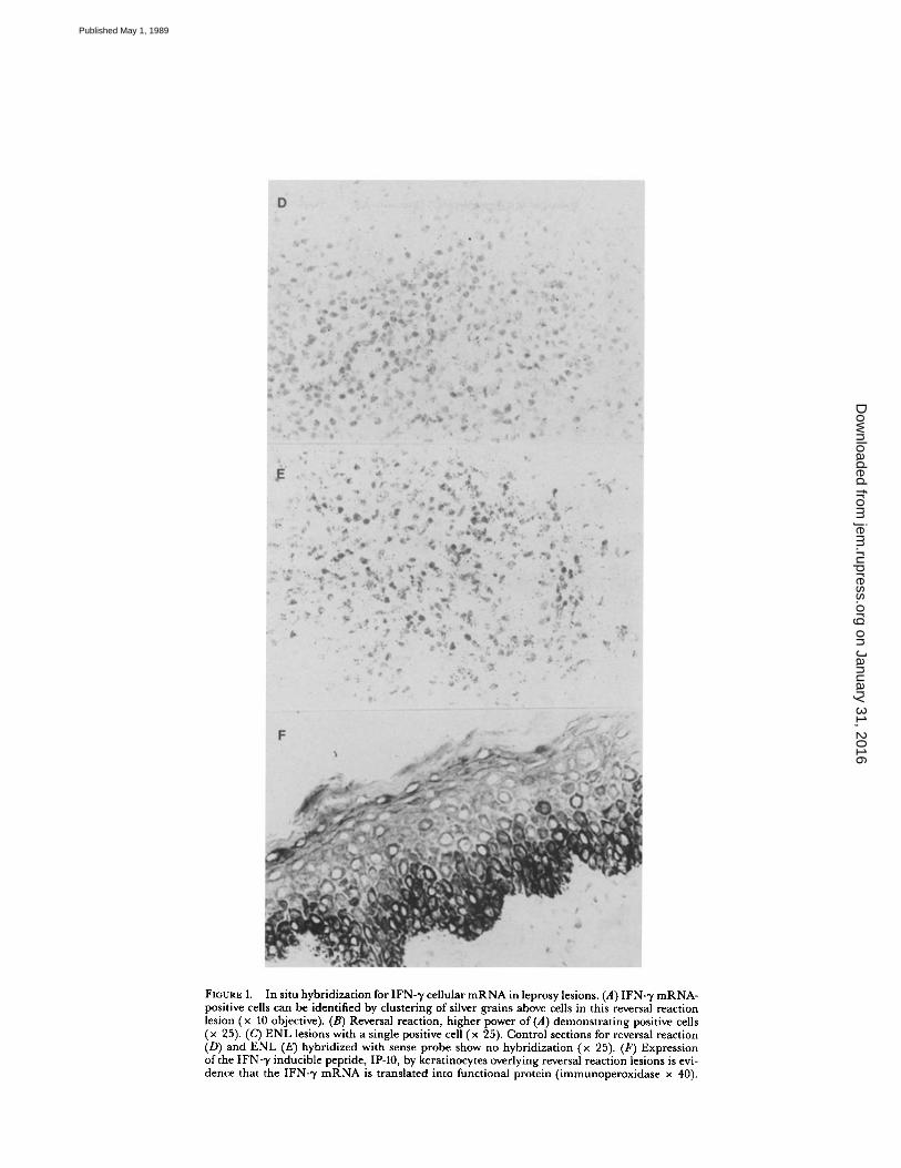
FIGURE 1.
In situ hybridization for IFN--y cellular mRNA in leprosylesions. (A) IFN- ,y mRNA-positive cells can be identified by clustering of silver grains above cells in this reversal reactionlesion (x 10 objective) . (B) Reversal reaction, higher power of(A) demonstrating positive cells(x 25). (C) ENL lesions with a single positive cell (x 25) . Control sections for reversal reaction(D) and ENL (E) hybridized with sense probe show no hybridization (x 25). (F) Expressionof the IFN-,y inducible peptide, IP-10, by keratinocytes overlying reversal reaction lesions is evi-dence that the IFN-,y mRNA is translated into functional protein (immunoperoxidase x 40) .
on January 31, 2016jem
.rupress.orgD
ownloaded from
Published May 1, 1989

1572
IN SITU ANALYSIS OF LEPROSY LESIONS
A
NZOrnWJZ
(AJ
WWUu_OFZWUWa
BJJWUW
Oa
ZOOZawc7aW>a
60
50
40
30
20
10
FIGURE 2 .
IFN-.y mRNA in leprosy lesionsby in situ hybridization. (A) Percent of posi-tive cells in lesions. (B) Gene expression asmeasured by median grain count over back-ground in positive cells .
In the more immunologically active forms ofleprosy, the ratio of T helper/memory(CD4+2H4-UCHLI') to T naive (CD4+2H4+) cells were high ; however, the con-verse was true for immunologically unresponsive forms of leprosy. The memory tonaive ratio was 9:1 in reversal reaction, 6:1 for ENL, 13:1 for lepromin skin tests,14 :1 for tuberculoid lesions, and 1 :1 for nonreactional patients with borderlinelepromatous or lepromatous disease . Double immunostaining with UCHL1 and Leu-8gave results that were consistent with 2H4 results. CD4+UCHL1+ (T helper ormemory) cells were numerous in reversal reactions, ENL, and tuberculoid leprosy;whereas, CD4+ Leu-8+ (putative Ts-inducer) cells were a significant component ofthe CD4 response in lepromatous individuals.
on January 31, 2016jem
.rupress.orgD
ownloaded from
Published May 1, 1989
COOPER ET AL.
1573
CD8+ Subpopulations.
Antibody 9.3 was used in conjunction with Leu-2a (anti-CD8) to divide CD8+ cells into two subphenotypes, Ts (9 .3 -) and T cytotoxic (9.3+)cells (31) . Double immunostaining revealed that cells of the T cytotoxic phenotypewere the predominant CD8+ subset in reversal reactions, lepromin skin tests, andin tuberculoid lesions, while cells of the Ts phenotype were the predominant CD8+subset in nonreactional lepromatous and ENL lesions (Fig . 5). However, since thenumbers of CD8+ cells in ENL lesions are few, the actual percentage of Ts pheno-types in ENL lesions are far fewer than in nonreactional lepromatous lesions, onthe order of that in tuberculoid lesions .
DiscussionAnalysis of tissue lesions of the major reactional states of leprosy was undertaken
to study the immune mechanisms underlying regulation of cell-mediated immunityand DTH in man. Study of the reactional states of leprosy provides new insightsinto these immune processes . Reversal reactions in leprosy are characterized by areduction, or occasionally the elimination, oforganisms from lesions with upgradingof the clinical classification, associated with a sudden gain in cell-mediated immu-nity againstM. leprae in vitro. Cells infiltrating the reversal reaction lesions containedmRNA coding for IFN-y, as well as human serine esterase, and expressed cell sur-face antigenic determinants of theTh andT cytotoxic subpopulations . These resultsprovide in vivo evidence that reversal reactions represent a naturally occurring DTHreaction in leprosy. In contrast, the study of ENL lesions provides evidence sug-gesting that this reactional state represents a transient cell-mediated immune pro-cess clearly differentiated from the classical DTH response occurring in reversalreaction .
In situ analysis of tissue lesions revealed greater numbers of cells expressing IFN-ymRNA, as well as cells of the T helper/memory phenotype (CD4+UCHLI +), inreversal reactions, tuberculoid leprosy, and lepromin skin tests in comparison withlepromatous leprosy. Such Th cells derived from leprosy lesions may contribute toDTH by proliferating and producing IFN-y in response to M. leprae in vitro (38) .IFN-y is likely to be involved in the reduced numbers of bacilli observed in reversalreactions since this lymphokine has been shown to facilitate the intracellular killingof mycobacteria in vitro and in vivo (39, 40).
Cells expressing serine esterase mRNA (huHF), a marker for cytotoxic cells, weremore abundant in reversal reactions, lepromin skin tests, and tuberculoid lesionsthan in nonreactional lepromatous leprosy, and correlated in number with theCD8+9.3+ T cytotoxic population . However, the microanatomic location of thesehuHF+ cells throughout the granuloma indicate that some are likely to be eithermacrophages or CD4+ cells . Since macrophages do not appear to express this gene(41) and CD4 killers can release this protease (42), it appears that CD4+ cells inreversal reaction lesions contain this cytotoxic marker. Although NK cells have beenshown to produce serine esterase, immunohistologic analysis of leprosy lesions hasfailed to demonstrate the presence of cells bearing NK phenotypes . We have notyet obtained functional evidence that cells in lesions have cytotoxic activity, and infact, Th cells have been shown to express the huHF marker (43) ; however, EM hasshown that cytolysis of parasitized macrophages occurs within leprosy lesions (44) .
on January 31, 2016jem
.rupress.orgD
ownloaded from
Published May 1, 1989
1574
FIGURE 3 .
Continued on following page .
IN SI'T'U ANALYSIS OF LEPROSY LESIONS
on January 31, 2016jem
.rupress.orgD
ownloaded from
Published May 1, 1989

FIGURE: 3 .
In situ hybridization to detect serine esterase (huHF), a marker for T cytotoxic cells .(A) Positive cells in this darkfield micrograph of a reversal reaction granuloma are identified by -the clustering of white dots into spots and are distributed throughout the granuloma in the samedistribution of CD4' cells (x 25). (B)A clustering of positive cells is observed in the ENL granu-loma (x 40). The sense control hybridizations are negative in both reversal reaction (E) andENL (F) (x 25). Immunoperoxidase staining of reversal reaction granulomas revealed thatUCHLI' cells (C) are distributed throughout the granuloma; whereas, 2H4' cells (D) are inthe lymphocytic mantle surrounding the granuloma (x 40).
on January 31, 2016jem
.rupress.orgD
ownloaded from
Published May 1, 1989

1576
IN SITU ANALYSIS OF LEPROSY LESIONS
A 30
zOWWJ
NJWWUwO
wUWa
BJJWUwFNOai
2Q
C7wc7QWQ
20
10
60
50
40
30
20
10
FIGURE 4.
Serine esterase (huHF) mRNA inleprosy lesions by in situ hybridization. (A)Percent of positive cells in lesions . (B) Geneexpression as measured by median grain countover background in positive cells .
In addition, CD4+ antigen-specific killers have been isolated from the peripheralblood of leprosy patients (45) . These T cytotoxic cells are thought to contribute tohost defenses against mycobacterial infection by lysing infected targets in vitro (46) . Thelysis of these infected targets in tuberculoid lesions may then eventuate in the elimi-nation of bacilli, allowing the dilution of organisms from heavily parasitized macro-phages into fresh IFN-y activated phagocytes, where destruction can then take place.Thedata presented demonstrate that reversal reactions are a naturally occurring
DTH reaction within leprosy. Although elimination of reduction of bacilli is the desiredresult, concomitant nerve damage is often a serious consequence. Of interest, thelevel of IFN-y and serine esterase gene expression in reversal reaction lesions wasmarkedly elevated compared with lepromin skin tests and tuberculoid leprosy le-
on January 31, 2016jem
.rupress.orgD
ownloaded from
Published May 1, 1989

z0m 40Z_W
WVWOI.-ZW
A
20
0e
e~'ePtd ~ee~
ldJe
fct~'P gf
COOPER ET AL .
1577
OWJ
JWVu.OF2Ud
B
30
20
10
0
- CD8+CD28+ T Cytotoxic
0 CD8+CD28 - T Suppressor
sa~ ~ytee~ ,`opp td~e
Po et
beR~3vr<~et°
,e0
6~
FIGURE 5 .
(A) Percentages of CD4' subpopulations in leprosy lesions . The Thelper/memorysubpopulations was identified as CD4'UCHLl' or CD4'2H4-; whereas, the Tnaive subpopu-lation was identified as CD4'2H4' . (B) Percentages ofCD8' subpopulations in leprosy lesions .TheTcyotoxic subpopulations was identified as CD8'9.3' ; whereas, the Ts subpopulation wasidentified as CD8'9.3 - .
sions. Since Kaufmann (47) has shown in the mousethat induction ofclass II MHCon M. leprae antigen-containing Schwann cells by IFN--y permits them to be killedin vitro, it is intriguing to speculate that reversal reactions representahyperimmuneDTH response in man in which T cell cytotoxicity mediates severe nerve damage.
Analysis of ENL lesions revealed clear differences from that of reversal reaction(Fig . 6) . Although phenotypic Th cells predominated in ENL lesions, the high levelsof IFN--y gene expression, characteristic of reversal reactions, were not observed .ENL granulomas lacked the characteristic microanatomic separation of T cell sub-populations found in tuberculoid and reversal reaction granulomas . In addition,T cytotoxic cells as measured by 9.3 antigen expression and huHF serine esterasegene expression were relatively low in these ENL specimens. The relative absenceof a DTH-like response in ENL correlates with the inability of the immune cellsin these lesions to clear bacilli .The differences observed between reversal reactions and ENL reactions in leprosy
can be conceptualized as representing fundamentally different dynamic events oc-curring in the lesions of the patients . Reversal reactions would appear to reflect a
FIGURE 6 .
Differential increases over background ofvarious T cell subpopulations in reversalreaction compared with ENL lesions.
on January 31, 2016jem
.rupress.orgD
ownloaded from
Published May 1, 1989

1578
IN SITU ANALYSIS OF LEPROSY LESIONS
selective increase of CD4+ IFN-y producing and CD4+ and/or CD8+ CTL activityin the lesions, with thenumber of CD8+ Ts cells remaining almost constant . In con-trast, ENL reactions may be visualized as a transient diminution of Ts activity, leadingto partial augmentation of Th activity. We have reported a transient diminutionof Ts activity in vitro during ENL episodes (24) . The diminution of Ts regulationcould account for the augmentation of both cell-mediated immunity and antibodyformation characteristic of ENL reactions. However, ENL reactions would appearto lack significant numbers of lymphokine-producing cells and fail to exhibit asignificant increase in CTL cells. This picture could lead to the immune complexdisease symptoms associated with reversal reactions. The changes in T cell subsetsand function in these reactional states of leprosy continue to offer new insights intoregulation of cell-mediated immunity in man.
SummaryAnalysis of tissue lesions of the major reactional states ofleprosy was undertaken
to study the immune mechanisms underlying regulation of cell-mediated immunityand delayed-type hypersensitivity (DTH) in man. In situ hybridization hybridiza-tion of reversal reaction biopsy specimens for INFy mRNA expression revealed a10-fold increase in specific mRNA-containing cells over that observed in unrespon-sive lepromatous patients . Expression of huHF serine esterase, amarker for Tcyto-toxic cells, were fourfold increased in reversal reaction and tuberculoid lesions abovethat detected in unresponsive lepromatous individuals . Immunohistology ofreversalreactions confirmed a selective increase of Th and T cytotoxic cells in the cellularimmune response . Of interest, the microanatomic location of these serine esterasemRNA-containing cells was identical to the distribution of CD4+ cells .Analysis of erythema nodosum leprosum (ENL) lesions revealed differences in
the underlying immune processes in comparison with reversal reaction lesions. Al-though phenotypic Th cells predominated in ENLlesions, IFN-y and serine esterasegene expression were markedly reduced. We suggest that reversal reactions repre-sent a hyperimmune DTH response characterized by a selective increase of CD4+IFN-y producing cells and T cytotoxic cells, which result in the clearing of bacilliand concomitant tissue damage . In contrast, ENL reactions may be viewed as atransient diminution of Ts cells and activity leading to a partial and transient aug-mentation in cell-mediated immunity, perhaps sufficient to result in antibody andimmune complex formation, but insufficient to clear bacilli from lesions.
Receivedfor publication 30 November 1988 and in revisedform 12 January 1989 .
References1 . Ridley, D. S., and W. H. Jopling. 1966 . Classification of leprosy according to immunity.A five-group system . Int . J. Lepr 34:255 .
2 . Ridley, D. S. 1974 . Histological classification and the immunological spectrum of leprosy.Bull. W. H. 0. 51 :451 .
3 . Bloom, B. R., and V. Mehra. 1984 . Immunological unresponsiveness in leprosy. Immunol.Rev. 80:5 .
4. van Voorhis, W. C., G. Kaplan, E. N. Sarno, M. A. Horwitz, R. M. Steinman, W. R.Levis, N. Nogueira, L. S. Hair, C. R. Gattass, B. A. Arrick, and -Z . A. Cohn. 1982 .
on January 31, 2016jem
.rupress.orgD
ownloaded from
Published May 1, 1989

COOPER ET AL .
1579
The cutaneous infiltrates ofleprosy : cellular characteristics and the predominant T-cellphenotypes . N. Engl. J. Med. 307:1593.
5. Modlin, R. L ., F. M . Hofman, C. R. Taylor, and T H. Rea . 1983 . T lymphocytes subsetsin the skin lesions of patients with leprosy. J. Am . Acad. Dermatol. 8:182 .
6. Narayanan, R. B ., L . K . Bhutani, A . K . Sharma, and I . Nath . 1983 . T cell subsets inleprosy lesions : in situ characterization using monoclonal antibodies . Clin . Exp. Immunol.51 :421 .
7. Modlin, R . L ., F. M . Hoffman, P R. Meyer, O. P Sharma, C . R . Taylor, and T. H.Rea . 1983 . In situ demonstration of T lymphocyte subsets in granulomatous inflamma-tion : leprosy, rhinoscleroma and sarcoidosis . Clin. Exp. Immunol. 51 :430 .
8. Modlin, R . L ., M. B . Brenner, M. S . Krangel, A . D. Duby, and B . R. Bloom . 1987 .Tcell receptors of human suppressor cells. Nature (Lond). 329:541 .
9. Modlin, R. L ., V. Mehra, L . Wong, Y. Fujimiya, W-C. Chang, D. A. Horwitz, B. R.Bloom, T. H . Rea, and P K. Pattengale. 1986 . Suppressor T lymphocytes from leproma-tous leprosy skin lesions . J. Immunol. 137 :2831.
10 . Modlin, R. L ., H . Kato, V. Mehra, E . E. Nelson, F. Xue-dong, T. H . Rea, P. K . Patten-gale, and B . R . Bloom. 1986. Genetically restricted suppressor T-cell clones derived fromlepromatous leprosy lesions . Nature (Loud.) . 322 :459 .
11 . Waters, M. F. R ., J . L . Turk, and S . N . C . Wemambu . 1971 . Mechanisms of reactionsin leprosy. Int. J Lepr. 39:417 .
12 . Godal, T., B. Myrvang, D. R . Samuel, W. F. Ross, and M. Lofgren . 1973 . Mechanismof reactions in borderline tuberculoid (BT) leprosy. Acta Pathol. Microbiol. Scand.236(Suppl):45.
13 . Barnetson, R . StC ., G . Bjune, J . M . H. Pearson, and G. Kronvall. 1976 . Cell mediatedand humoral immunity in "reversal reactions ." Int. J. Lepr. 44:267 .
14 . Bjune, G., R . StC . Barnetson, D. S . Ridley, and G. Kronvall . 1976 . Lymphocyte trans-formation test in leprosy: correlation of the response with inflammation of lesions. Clin .Exp. Immunol. 25:85.
15 . Rea, T. H., and C . R . Taylor. 1977 . Serum and tissue lysozyme in leprosy. Infect. Immun.18:847 .
16 . Wemambu, S . N . C ., J . L . Turk, M. F. R . Waters, and R . J . W. Rees . 1969 . Erythemanodosum leprosum : a clinical manifestation of the Arthus phenomenon . Lancet. ii :933 .
17 . Drutz, D. J ., and R . A . Gutman . 1973 . Renal manifestations of leprosy: glomerulone-phritis, a complication of erythema nodosum leprosum . Am. J Trop. Med. Hyg. 22:496 .
18 . Bjorvatn, B., R. StC . Barnetson, G. Kronvall, R . H . Zubler, and P. H . Lambert . 1976 .Immune complexes and complement hypercatabolism in patients with leprosy. Clin. Exp.Immunol. 26:388 .
19 . Rea, T. H., and N. E . Levan . 1980 . Variations in dinitrobenzene responsivity in un-treated leprosy : evidence for a beneficial role for anergy. Int. J Lepr. 48:120 .
20 . Stach, J . L., M. Strobel, F. Fumoux, and J . F Bach . 1982 . Defect in the generation ofcytotoxic T cells in lepromatous leprosy. Clin. Exp. Immunol. 48:633 .
21 . Modlin, R . L ., J . F. Gebhard, C . R. Taylor, and T. H . Rea . 1983 . In situ characteriza-tion ofT lymphocyte subsets in the reactional states of leprosy. Clin. Exp. . Immunol. 53 :17 .
22 . Narayanan, R. B ., S . Laal, A . K . Sharma, L . K . Bhutani, and I . Nath . 1984 . Differencesin predominant T cell phenotypes and distribution pattern in reactional lesions oftuber-culoid and lepromatous leprosy. Clin. Exp. Immunol. 55:623 .
23 . Modlin, R. L ., A . C . Blake, S . A . Vaccaro, D. A . Horwitz, C . R . Taylor, and T H.Rea . 1985 . Tissue and blood Tlymphocyte subpopulations in erythema nodosumleprosum . Arch . Dermatol. 121:216 .
24 . Modlin, R. L ., V. Mehra, R. Jordan, B . R . Bloom, and T. H . Rea. 1986 . In situ andin vitro characterization of the cellular immune response in erythema nodosum leprosum .J Immunol. 136:883 .
on January 31, 2016jem
.rupress.orgD
ownloaded from
Published May 1, 1989

1580
IN SITU ANALYSIS OF LEPROSY LESIONS
25 . Rea, T. H., J . Y. Shen, and R . L . Modlin . 1986 . Epidermal keratinocyte la expression,Langerhans cell hyperplasia and lymphocytic infiltration in skin lesions of leprosy. Clin .Exp. Immunol. 65:253 .
26 . Dugan, E., R . L . Modlin, and T. H. Rea . 1985 . An in situ immunohistological studyof Mitsuda reactions . Int. J. Lepr. 53:404 .
27 . Mueller, C., H . K. Gershenfeld, C . G . Lobe, C . Y. Okada, R. C . Bleackley, and I . L .Weissman . 1988 . A high proportion of T lymphocytes that infiltrate H-2-incompatibleheart allografts in vivo express genes encoding cytotoxic cell-specific serine proteases,but do not express the MEL-13-defined lymph node homing receptor.j Exp. Med. 167:1124 .
28 . Gershenfeld, H . K., R . J . Hershberger, T. B . Shows, and I . L . Weissman . 1988 . Cloningand chromosomal assignment of a human cDNA encoding a T cell- and natural killercell-specific trypsin-like serine protease . Proc. Natl. Acad. Sci. USA. 85 :1184 .
29 . Angerer, L . M., M. E . Stoler, and R. C . Angerer. 1987 . In situ hybridization with RNAprobes . In In Situ Hybridization : Applications to the CNS. K . Valentino, J . Eberwine,and E . J . Barchas, editors . Oxford University Press, New York . 42-75 .
30 . Beverley, P. C . L . 1986 . Human T cell subsets . Immunol. Lett. 14:263 .31 . Yamada, H., I? J . Martin, M . A . Bean, M. P. Braun, I? G . Beatty, K . Sadamoto, and
J . A . Hansen. 1985 . Monoclonal antibody 9.3 and anti-CDIl antibodies define reciprocalsubsets of lymphocytes . Eur. J. Immunol. 15:1164 .
32 . Morimoto, C., N . L . Letvin, A . W. Boyd, M. Hagan, H. M . Brown, M. M. Kornacki,and S . F. Schlossman . 1985 . The isolation and characterization of the human helper in-ducer T cell subset . J. Immunol. 134:3762 .
33 . Akbar, A . N ., L. Terry, A . Timms, P. C . L. Beverley, and G. Janossy. 1988 . Loss ofCD45Rand gain of UCHLI reactivity is a feature of primed T cells . J. Immunol. 140:2171 .
34 . Morimoto, C., N . L . Letvin, J . A . Distaso, W. R . Aldrich, and S . F. Schlossman . 1985 .The isolation and characterization of the human suppressor inducer T cell subset . .J.Immunol. 134:1508 .
35 . Damle, N. K., A . L . Childs, and L . V. Doyle . 1987 . Immunoregulatory T lymphocytesin man . Soluble antigen-specific suppressor-inducer T lymphcoytes are derived from theCD4'CD45R-p80' subpopulations . J Immunol. 139:1501 .
36 . Sanders, M. E ., M . W. Makgoba, and S . Shaw . 1988 . Human naive and memory Tcells : reinterpretation ofhelper-inducer and suppressor-inducer subsets . Immunol. Today.9:195 .
37 . Kaplan, G., A . D. Luster, G. Hancock, and Z . A . Cohn . 1987 . The expression of a 'yinterferon-induced protein (IP-10) in delayed immune responses in human skin . j Exp.Med. 166:1098 .
38 . Modlin, R. L ., J . Melancon-Kaplan, S. M. M . Young, C . Pirmez, H . Kino, J . Convit,T H. Rea, and B . R . Bloom . 1988 . Learning from lesions : patterns of tissue inflamma-tion in leprosy. Proc . Natl. Acad. Sci. USA. 85:1213 .
39 . Rook, G . A . W., J . Steele, L . Fraher, S . Barker, R. Karmali, and J . O'Riordan . 1986 .Vitamin Ds, gamma interferon, and control of proliferation ofMycobacterium tuberculosisby human monocytes . Immunology. 57:159 .
40 . Nathan, C . F., G . Kaplan, W. R. Levis, A . Nusrat, M . D. Witmer, S . A . Sherwin, C . K.Job, C. R . Horowitz, R . M . Steinman, and Z . A . Cohn. 1986 . Local and systemic effectsof intradermal recombinant interferon-gamma in patients with lepromatous leprosy. N.Engl. J. Med. 315 :6 .
41 . Brunet, J.-F., F. Denizot, M. Suzan, W. Haas, J .-M . Mencia-Huerta, G. Berke, M.-F.Luciani, and P. Golstein . 1987 . CTLA-1 and CTLA-3 serine esterase transcripts are de-tected mostly in cytotoxic T cells, but not only and not always . J. Immunol. 138:4102 .
42 . Velotti, F., H . R . MacDonald, and M. Nabholz . 1987 . Granzyme A sectrion by normalactivated Lyt-2' and L3T4' T cells in response to antigenic stimulation. Eur. J Immunol.17:1095 .
on January 31, 2016jem
.rupress.orgD
ownloaded from
Published May 1, 1989

COOPER ET AL .
158 1
43 . Taplits, M. S ., P. A . Henkart, and R. J . Hodes . 1988 . T helper cell cytoplasmic granules .Exocytosis in response to activation via the T cell receptor. J. Immunol. 141 :1 .
44 . Kaplan, G., G . Sheftel, C . K. Job, N . K . Mathur, I. Nath, and Z . A . Cohn . 1988 . Efficacyof a cell-mediated reaction to the purified protein derivative oftuberculin in the disposalof Mycobacterium leprae from human skin . Proc. Nall. Acad. Sci. USA. 85:5210 .
45 . Hancock, G. E ., Z . A . Cohn, and G. Kaplan . 1989 . The generation of antigen-specificmajor histocompatibility complex-restricted cytotoxic T lymphocytes of the CD4'phenotype . Enhancement by the cutaneous administration of Interleukin 2.J. Exp. Med.169:909 .
46 . Mustafa, A. S., and T. Godal . 1987 . BCG induced CD4' cytotoxic T cells from BCGvaccinated healthy subjects : relation between cytotoxicity and suppression in vitro. Clin.Exp. Immunol. 69:255 .
47 . Kaufmann, S . H . E . 1988 . CD8' T lymphocytes in intracellular microbial infections .Immunol. Today. 9:168 .
on January 31, 2016jem
.rupress.orgD
ownloaded from
Published May 1, 1989
Copyright © 2022 FDOKUMEN