(Anacardiaceae) - Natuurtijdschriften
11
Ada Bot. Neerl. 35(4), November 1986, p. 425-435. Distribution and development of secretory ducts in Trichoscypha (Anacardiaceae) R.W. den Outer and W.L.H. van Veenendaal Vakgroep Plantencytologie en -morfologie. Landbouwuniversiteit,Arboretumlaan 4,6703 BD Wageningen SUMMARY Distribution and development of three different secretory ducts systems were studied in leaves, stems and roots of several Trichoscypha species (Anacardiaceae) from west tropical Africa. One primary three-dimensional network occurs in the pith and the cortex of the stem and mesophyll of the leaves. A second primary system of mainly longitudinal orientated, hardly ramifying, secretory ducts is found in the metaphloem of leaves, stem and roots while a secondary three-dimensional system is detected in the secondary phloem of leaves, stem and roots. In the stem and roots, the last system is continuous with the secondary xylem via horizontal secretory ducts in both secondary phloem- and xylem rays. The three systems are separated from oneanother. Ducts initiate schizogenously and develop further lysigenously in the primary phloem of the root, in the secondary phloem of the stem and sometimes also in the wood rays. In other parts of the plant the ducts develop entirely lysigenously. 1. INTRODUCTION In the Anacardiaceae more or less longitudinal secretory ducts are particularly common in the phloem, cortex and pith of the stem, but they also occur in both phloem- and wood rays in numerous genera (Metcalfe 1983). Whether this is also the case in the genus Trichoscypha Hook. f. that comprises about 75 species, all trees and shrubs, is not known or only partly described. The genus occurs in tropical and south Africa, however it is absent on Madagascar (Per- rier de la Bathie 1946). The present paper describes the duct system in six Trichoscypha species from Secretory ducts have considerable taxonomic importance and are characteristic of a small number of dicotyledon families (Metcalfe 1983). Their products are sometimes of economic significance. When present in Anacardiaceae, secre- tory ducts will always be found in the stem. In several species secretory ducts are not only confined to the stem, but are also present in leaf, root, flower or fruit. There is much confusion in literature concerning the mode of origin of the canals; do they originate schizogenously, lysigenously or schizo-lysigenously? This may be dependent on the species or even within a species on the considered tissue (Gibson 1981; Venkaiah & Shah 1984). Also the chemical nature of the deposits in secretory canals caused much discussion (Fahn 1979; Fahn & Evert 1974).
-
Upload
khangminh22 -
Category
Documents
-
view
0 -
download
0
Transcript of (Anacardiaceae) - Natuurtijdschriften
Ada Bot. Neerl. 35(4), November 1986, p. 425-435.
Distribution and development
of secretory ducts in
Trichoscypha (Anacardiaceae)
R.W. den Outer and W.L.H. van Veenendaal
Vakgroep Plantencytologieen -morfologie. Landbouwuniversiteit,Arboretumlaan 4,6703BD
Wageningen
SUMMARY
Distribution and developmentofthree different secretory ducts systems were studied in leaves, stems
and roots of several Trichoscypha species (Anacardiaceae) from west tropical Africa. One primary
three-dimensional network occurs in the pith and the cortex ofthe stem and mesophyll of the leaves.
A second primary system of mainly longitudinal orientated, hardly ramifying, secretory ducts is
found in the metaphloem of leaves, stem and roots while a secondary three-dimensional systemis detected in the secondary phloem of leaves, stem and roots. In the stem and roots, the last system
is continuous with the secondary xylem via horizontal secretory ducts in both secondary phloem-
and xylem rays. The three systems are separated from oneanother.
Ducts initiate schizogenously and developfurther lysigenously in the primary phloem ofthe root,
in the secondary phloem of the stem and sometimes also in the wood rays. In other parts ofthe
plant the ducts developentirely lysigenously.
1. INTRODUCTION
In the Anacardiaceaemore or less longitudinal secretory ducts are particularly
common in the phloem, cortex and pith of the stem, but they also occur in both
phloem- and wood rays in numerous genera (Metcalfe 1983). Whether this
is also the case in the genus Trichoscypha Hook. f. that comprises about 75
species, all trees and shrubs, is not known or only partly described. The genus
occurs in tropical and south Africa, however it is absent on Madagascar (Per-
rier de laBathie 1946).The present paper describes the duct system in six Trichoscypha species from
Secretory ducts have considerable taxonomic importance and are characteristic
of a small number of dicotyledon families (Metcalfe 1983). Their products
are sometimes ofeconomic significance. When present in Anacardiaceae, secre-
tory ducts will always be found in the stem. In several species secretory ducts
are not only confined to the stem, but are also present in leaf, root, flower or
fruit.
There is much confusion in literature concerning the mode of origin of the
canals; do they originate schizogenously, lysigenously or schizo-lysigenously?This may be dependent on the species or even within a species on the considered
tissue (Gibson 1981; Venkaiah & Shah 1984). Also the chemical nature of the
deposits in secretory canals caused much discussion (Fahn 1979; Fahn & Evert
1974).
426 R. W. DEN OUTER AND W. L. H. VAN VEENENDAAL
the Ivory Coast and Liberiawhich are treatedin the floraof west tropical Africa
(Hutchinson & Dalziel 1954).
2. MATERIALS AND METHODS
Samples used comprise the following Trichoscypha species:- T. arborea (A. Chev.) A. Chev. (Versteegh & Den Outer 659, Ivory Coast,
1969, large tree, dbh 30 cm, stem and leaf; Versteegh & Jansen 778, Liberia,
1969, large tree, dbh 40 cm, root, stem and leaf; De Koning 3571, Ivory Coast,
1974, seedlings);- T. beguei Aubrev. et Pellegr. (Versteegh & Den Outer 648, Ivory Coast, 1969,
small tree, dbh 15 cm, stem and leaf; Versteegh & Jansen 794, Liberia, 1969,
small tree, dbh20 cm, stem and leaf);- T. chevalieri Aubrev. et Pellegr. (Versteegh & Den Outer 571, Ivory Coast,
1969, shrub, dbh 6 cm, stem and leaf);- T. mannii Hook. f. (Versteegh & Den Outer 739, Ivory Coast, 1969, shrub,dbh 6 cm, stem and leaf);- T. oba Aubrev. et Pellegr. (Oldeman 443, Ivory Coast, 1963, small tree, dbh
10 cm, root, stem and leaf);
- T. yapoensis Aubrev. et Pellegr. (Versteegh & Jansen 758, Liberia, 1969, small
tree, dbh 10 cm, stem and leaf).
Samples of T. arborea, T. manniiand especially those of T. oba were used most
frequently in the present study. Stem samples containing bark attached to wood,
were collected at breast hight and immediately fixed in FAA. All the material
of T. oba was collected from a living tree raised from seed (Oldeman 443) in
the greenhouse at Wageningen and cut freshly. The collection is housed at the
Department of PlantCytology and Morphology, the accompanying herbarium
vouchers at the Department of Plant Taxonomy, Agricultural University, Wa-
geningen. Anatomical features were studied in transverse, radial and tangentialserial sections, which were unstained or stained with sudan III (staining fatty
substances, latex), iodine (starch stain) or toluidineblue (contrast, cytoplasmic
stain), butalso with sudan IV (fatty substances, latexstain), phloroglucin-hydro-
chloric acid (staining lignin), eosine (cytoplasmic stain), zinc chloride-iodine
(cellulose stain), aceto-carmine (according to Heitz; staining the nucleus), nitric
acid (Xanthoprotein reaction; protein stain) and sulfuric acid (reaction of Ras-
pail; protein stain, but also staining glucosids, alkaloids) (Johansen 1940).
Some material was impregnated with technovit 7100 before sectioning with
a rotary microtome. All sections were embeddedin Kaisers gelatin-glycerin (Jo-
hansen 1940).
427SECRETORY DUCTS IN TRICHOSCYPHA
3. RESULTS
3.1. Distribution of the secretory ducts
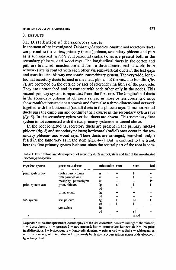
In the stem of the investigated Trichoscypha species longitudinal secretory ducts
are present in the cortex, primary (meta-)phloem, secondary phloem and pith
as is summarized in table 1. Horizontal (radial) ones are present both in the
secondary phloem- and wood rays. The longitudinal ducts in the cortex and
pith are branched, anastomose and form a three-dimensional network; both
networks are in contact with each other via semi-vertical ducts in the leafgaps
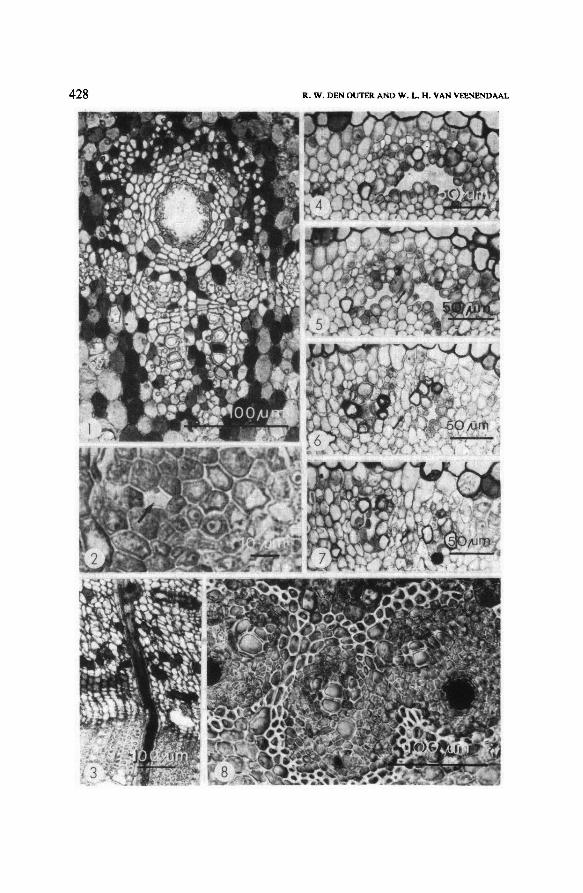
and constitute in this way one continuousprimary system. The very wide, longi-tudinal secretory ducts formedin the meta-phloem of the vascular bundles (fig.
1), are protected on the outside by arcs of sclerenchyma fibres of the pericycle.
They are unbranched and in contact with each other only in the nodes. This
second primary system is separated from the first one. The longitudinal ducts
in the secondary phloem which are arranged in more or less concentric ringsshow ramifactions and anastomosis and form also a three-dimensional network
together with the horizontal (radial) ducts in the phloem rays. These horizontal
ducts pass the cambium and continue their course in the secondary xylem rays
(fig. 3). In the secondary xylem vertical ducts are absent. This secundary duct
system is not connected with the two primary systems mentionedabove.
In the root longitudinal secretory ducts are present in the primary (meta-)
phloem (fig. 2) and secondary phloem; horizontal(radial) ones occur in the sec-
ondary phloem- and wood rays. These ducts are arranged, branched and/or
fused in the same way as in the stem (figs. 4-7). But in contrast to the trunk
here the first primary system is absent, since the central part of the root is com-
Legends; *= noducts present in the mesophyllof the leaflet outside the surroundingsofthe mid vein;
- = ducts absent; 4- = present; ? = not reported; hor = more or less horizontal; ir = irregular,in all directions;1 = lysigenous; Ig = longitudinal;prim. = primary;rd = radial; s = schizogenous;sec. = secondary; s-1 = initiation schizogenously but lysigeny occursin later stages ofdevelopment;
tg = tangential.
Table 1. Distribution and developmentof secretory ducts in root, stem and leaf of the investigated
Trichoscypha species.
type duct system presence in tissue orientation root stem leaf
prim, system one cortex parenchyma ir - 1 —
pith parenchyma ir - 1 -
mesophyll parenchyma ir - - I*
prim, system two prim, phloem >g s-1 1 I
rd - -
prim, xylem ig - - -
rd - - -
sec. system sec. phloem ig i S-1 1
rd i I -
sec. xylem ig - - -
rd - s-1 -
also 1
428\I R. W. DEN OUTER AND W. L. H. VAN VEENENDAAL
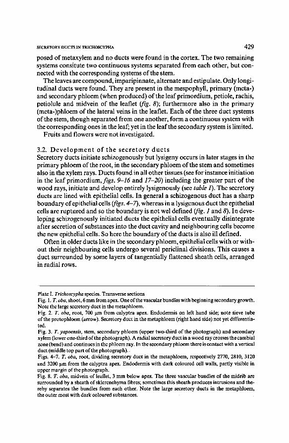
429SECRETORY DUCTSIN TRICHOSCYPHA
posed of metaxylem and no ducts were found in the cortex. The two remaining
systems consitute two continuous systems separated from each other, but con-
nected with the corresponding systems ofthe stem.
The leaves are compound, imparipinnate, alternate and estipulate. Only longi-
tudinal ducts were found. They are present in the mespophyll, primary (meta-)
and secondary phloem (when produced) ofthe leafprimordium, petiole, rachis,
petiolule and midvein of the leaflet (fig. 5); furthermore also in the primary
(meta-)phloem of the lateral veins in the leaflet. Each of the three duct systems
of the stem, though separated from one another, form a continuous system with
the corresponding ones in the leaf; yet in the leafthe secondary system is limited.
Fruits and flowers were not investigated.
3.2. Development of the secretory ducts
Secretory ducts initiate schizogenously but lysigeny occurs in later stages in the
primary phloem ofthe root, in the secondary phloem of the stem and sometimes
also in the xylem rays. Ducts found inall other tissues (see for instance initiation
in the leaf primordium, figs. 9-16 and 17-20) including the greater part of the
wood rays, initiateand develop entirely lysigenously (see table 1). The secretory
ducts are linedwith epithelial cells. In general a schizogenous duct has a sharp
boundary of epithelial cells (figs. 4-7), whereas in alysigenous duct the epithelial
cells are ruptured and so the boundary is not wel defined(fig. 1 and 8). In deve-
loping schizogenously initiated ducts the epithelial cells eventually disintegrate
after secretionof substances into the duct cavity and neighbouring cells become
the new epithelial cells. So here the boundary of the ducts is also ill defined.
Often in older ducts like in the secondary phloem, epithelial cells with or with-
out their neighbouring cells undergo several periclinal divisions. This causes a
duct surrounded by some layers of tangentially flattened sheath cells, arranged
in radial rows.
TrichoscyphaPlate I.
Fig. 1.
root, 700 pm from calyptra apex. Endodermis on left hand side; note sieve tube
ofthe protophloem(arrow). Secretory duct in the metaphloem(righthand side) not yet differentia-
ted.
T. oba,
Fig. 3.
root, dividing secretory duct in the metaphloem, respectively 2770, 2810, 3120
and 3200 pm from the calyptra apex. Endodermis with dark coloured cell walls, partly visible in
upper margin of thephotograph.
Figs. 4-7.
Fig. 8, T. oba,
T. oba,
species. Transverse sections
shoot, 6 mm from apex. One ofthe vascular bundles with beginning secondary growth.
Note the large secretory duct in the metaphloem.
T. yapoensis, stem, secondary phloem (upper two-third ofthe photograph) and secondary
xylem (lower one-third ofthe photograph).Aradial secretory duct in awood ray crosses the cambial
zone(bend) and continues in the phloemray. In the secondary phloemthere is contact with a vertical
duct (middle top part ofthe photograph).
Fig. 2.
midvein of leaflet, 3 mm below apex. The three vascular bundles of the midrib are
surrounded by a sheath of sklerenchyma fibres; sometimes this sheath produces intrusions and the-
reby separates the bundles from each other. Note the large secretory ducts in the metaphloem,
the outermost with dark coloured substances.
T. oba,
430\II R. W. DEN OUTER AND W. L. H. VANVEENENDAAL
431SECRETORY DUCTS IN TRICHOSCYPHA
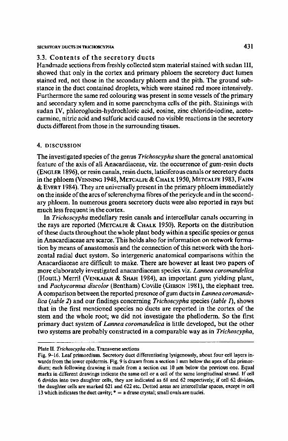
3.3. Contents of the secretory ducts
Handmade sections fromfreshly collectedstem material stained with sudan III,showed that only in the cortex and primary phloem the secretory duct lumen
stained red, not those in the secondary phloem and the pith. The ground sub-
stance in the duct contained droplets, which were stained red more intensively.
Furthermore the same red colouring was present in some vessels of the primary
and secondary xylem and in some parenchyma cells of the pith. Stainings with
sudan IV, phloroglucin-hydrochloric acid, eosine, zinc chloride-iodine, aceto-
carmine, nitric acid and sulfuric acid caused no visible reactions in the secretory
ducts differentfrom those in the surrounding tissues.
4. DISCUSSION
The investigated species of the genus Trichoscypha share the general anatomical
feature of the axis of all Anacardiaceae, viz. the occurrence of gum-resin ducts
(Engler 1896), or resin canals, resin ducts, laticiferous canals or secretory ducts
in the phloem (Venning 1948, Metcalfe& Chalk 1950, Metcalfe 1983, Fahn
& Evert 1984). They are universally present in the primary phloem immediately
on the inside ofthe arcs of sclerenchyma fibresof the pericycle and in the second-
ary phloem. In numerous genera secretory ducts were also reported in rays but
much less frequent in the cortex.
In Trichoscypha medullary resin canals and intercellular canals occurring in
the rays are reported (Metcalfe & Chalk 1950). Reports on the distribution
of these ducts throughout the whole plant body within a specific species or genus
in Anacardiaceae are scarce. This holds also for informationon network forma-
tion by means of anastomosis and the connectionof this network with the hori-
zontal radial duct system. So intergeneric anatomical comparisons within the
Anacardiaceaeare difficult to make. There are however at least two papers of
more elaborately investigated anacardiacean species viz. Lannea coromandelica
(Houtt.) Merril (Venkaiah & Shah 1984), an important gum yielding plant,
and Pachycormus discolor (Bentham) Coville (Gibson 1981), the elephant tree.
A comparison between the reported presence ofgumducts inLannea coromande-
lica (table 2) and our findings concerning Trichoscypha species ( table 1), shows
that in the first mentioned species no ducts are reported in the cortex of the
stem and the whole root; we did not investigate the phelloderm. So the first
primary duct system of Lannea coromandelica is little developed, but the other
two systems are probably constructed in a comparable way as in Trichoscypha,
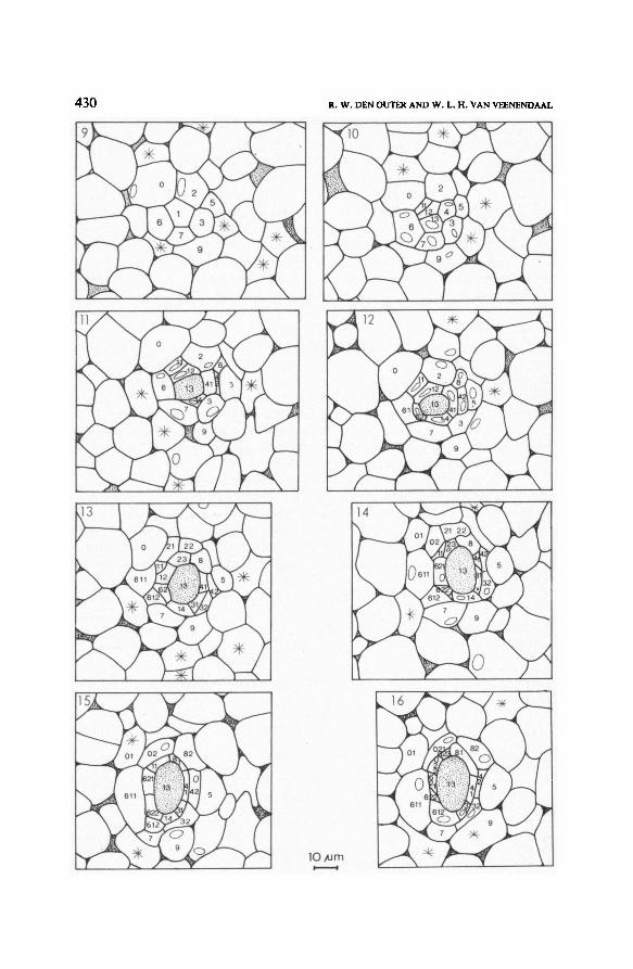
Trichoscypha oba.Plate II. Transverse sections
Fig. 9-16. Leaf primordium. Secretory duct differentiating lysigenously, about four cell layers in-
wards from the lower epidermis. Fig. 9 is drawn from a section 1 mm below the apex of the primor-
dium; each following drawing is made from a section cut 10 pm below the previous one. Equal
marks in different drawings indicate the same cell or a cell of the same longitudinal strand. Ifcell
6 divides into two daughter cells, they are indicated as 61 and 62 respectively; if cell 62 divides,
the daughtercells are marked 621 and 622 etc. Dotted areas are intercellular spaces, except in cell
13 which indicates the duct cavity;*
= a druse crystal; small ovals are nuclei.
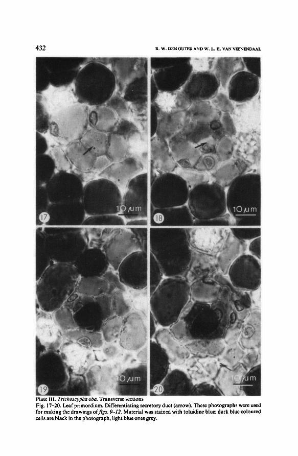
432\III R. W, DEN OUTER AND W. L. H,VAN VEENENDAAL
Material was stained with toluidine blue; dark blue coloured
cells are black in the photograph, light blue onesgrey.
Fig. 17-20. Leaf primordium. Differentiating secretory duct (arrow). These photographswere used
for making the drawings offigs. 9-12.
Transverse sectionsPlate III. Trichoscypha oba.
433SECRETORY DUCTS IN TRICHOSCYPHA
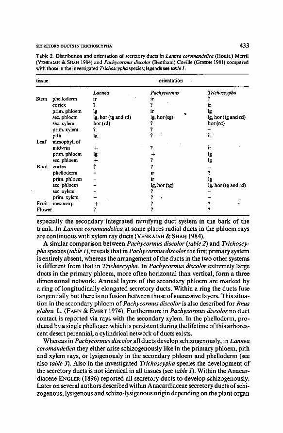
especially the secondary integrated ramifying duct system in the bark of the
trunk. In Lannea coromandelica at some places radial ducts in the phloem rays
are continuous with xylem ray ducts (Venkaiah & Shah 1984).
tissue orientation
Lannea Pachycormus Trichoscypha
Stem phelloderm ir ir ?
cortex ? ? ir
prim,phloem Ig ir Ig
sec. phloem Ig, hor (tg and rd) Ig,hor (tg) Ig, hor (tg and rd)
sec. xylem hor (rd) ? hor (rd)
prim, xylem ? ?
pith Ig ? ir
Leaf mesophyll of
midvein + ? ir
prim, phloem Ig + Igsec. phloem + ? Ig
Root cortex ? ?
phelloderm -ir ?
prim, phloem - ir Ig
sec. phloem - Ig, hor (tg) Ig, hor (tg and rd)
sec. xylem - ? -
prim, xylem - ? -
Fruit mesocarp + ? ?
Flower ? ? ?
A similar comparison between Pachycormus discolor (table 2) and Trichoscy-
pha species {table 1), reveals that inPachycormus discolor the first primary system
is entirely absent, whereas the arrangement of the ducts in the two other systems
is different from that in Trichoscypha. In Pachycormus discolor extremely large
ducts in the primary phloem, more often horizontal than vertical, form a three
dimensional network. Annual layers of the secondary phloem are marked by
a ring of longitudinally elongated secretory ducts. Within a ring the ducts fuse
tangentially but there is no fusion between thoseof successive layers. This situa-
tion in the secondary phloem of Pachycormus discolor is also described for Rhus
glabra L. (Fahn & Evert 1974). Furthermorein Pachycormus discolorno duct
contact is reported via rays with the secondary xylem. In the phelloderm, pro-
ducedby a single phellogen which is persistent during the lifetimeof thisarbores-
cent desert perennial, a cylindrical network of ducts exists.
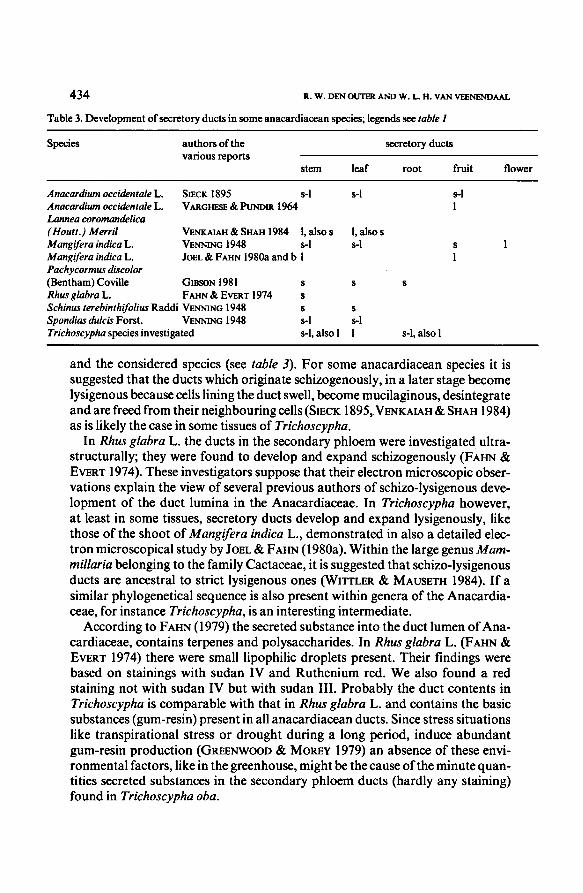
Whereas inPachycormus discolor all ducts develop schizogenously, in Lannea
coromandelicathey eitherarise schizogenously like in the primary phloem, pithand xylem rays, or lysigenously in the secondary phloem and phelloderm (seealso table 3). Also in the investigated Trichoscypha species the development of
the secretory ducts is not identical in all tissues (see table 1). Within the Anacar-
diaceaeEngler (1896) reported all secretory ducts to develop schizogenously.
Later on several authors described withinAnacardiaceaesecretory ductsofschi-
zogenous, lysigenous and schizo-lysigenous origin depending on the plant organ
Table 2. Distribution and orientation of secretory ducts in (Houtt.) Merril
(Venkaiah & Shah 1984) and
Lannea coromandelica
(Bentham) Coville (Gibson 1981) comparedwith those in the investigated
Pachycormus discolor
species; legends see table I.Trichoscypha
tissue orientation
Lannea Pachycormus Trichoscypha
Stem phelloderm IT ir ?
cortex 7 7 ir
prim,phloem ig ir ig
sec. phloem lg, hor (tg and rd) lg, hor (tg) lg, hor (tg and rd)
sec. xylem hor (rd) ? hor (rd)
prim, xylem ? 7 -
pith ig ? ir
Leaf mesophyll of
midvein + 7 ir
prim,phloem ig + igsec.phloem + 7 ig
Root cortex ? ? -
phelloderm - ir 7
prim, phloem - ir igsec. phloem - lg, hor (tg) lg, hor (tg and rd)
sec. xylem - ? -
prim, xylem - ? -
Fruit mesocarp + ? ?
Flower ? ? ?
434 R. W. DEN OUTER AND W. L, H. VAN VEENENDAAL
and the considered species (see table i). For some anacardiacean species it is
suggested that the ducts which originate schizogenously, in a later stage become
lysigenous because cells lining the duct swell, become mucilaginous, desintegrateand are freed from their neighbouring cells (Sieck 1895,Venkaiah& Shah 1984)
as is likely the case in some tissues of Trichoscypha.
Species authors ofthe
various reportsstem leaf root
Anacardium occidentale L.
Anacardium occidentale L.
Lannea coromandelica
(Houtt.) Merril
Mangifera indica L.
Mangiferaindica L.
Pachycormus discolor
(Bentham)Coville
Rhus glabra L.
Schinus terebinthifoliusRaddi
Spondias dulcis Forst.
fruit flower
Sieck 1895 s-1 s-1 s-1
Varghese & Pundir 1964 1
Venkaiah & Shah 1984 1, also s 1, also s
Venning 1948 s-1 s-1 s 1
Joel & Fahn 1980a and b 1 1
Gibson 1981 s s s
Fahn & Evert 1974 s
Venning 1948 s s
Venning 1948 s-1 s-1
Trichoscypha species investigated s-1, also 11 s-1, also 1
secretory ducts
In Rhus glabra L. the ducts in the secondary phloem were investigated ultra-
structurally; they were found to develop and expand schizogenously (Fahn &
Evert 1974). These investigators suppose that their electron microscopic obser-
vations explain the view of several previous authors of schizo-lysigenous deve-
lopment of the duct lumina in the Anacardiaceae. In Trichoscypha however,
at least in some tissues, secretory ducts develop and expand lysigenously, like
those of the shoot of Mangifera indica L., demonstratedin also a detailedelec-
tron microscopical study by Joel& Fahn (1980a). Within the large genusMam-
miliariabelonging to the family Cactaceae, it is suggested that schizo-lysigenousducts are ancestral to strict lysigenous ones (Wittler & Mauseth 1984). If a
similar phylogenetical sequence is also present within genera of the Anacardia-
ceae, for instance Trichoscypha, is an interesting intermediate.
According to Fahn (1979) the secreted substance into the duct lumenof Ana-
cardiaceae, contains terpenes and polysaccharides. In Rhus glabra L. (Fahn &
Evert 1974) there were small lipophilic droplets present. Their findings were
based on stainings with sudan IV and Ruthenium red. We also found a red
staining not with sudan IV but with sudan III. Probably the duct contents in
Trichoscypha is comparable with that in Rhus glabra L. and contains the basic
substances (gum-resin) present in all anacardiacean ducts. Since stress situations
like transpirational stress or drought during a long period, induce abundant
gum-resin production (Greenwood & Morey 1979) an absence of these envi-
ronmental factors, like in the greenhouse, might be the cause of the minutequan-
tities secreted substances in the secondary phloem ducts (hardly any staining)found in Trichoscypha oba.
Table 3. Developmentof secretory ducts in some anacardiacean species; legends see table I
Species authors ofthe
various reports
secretory ducts
stem leaf root fruit flower
Anacardium occidentale L.
Anacardium occidentale L.
Lannea coromandelica
Sieck 1895
Varghese & Pundir 1964
s-1 s-1 s-1
1
(Houtt.) Merril Venkaiah & Shah 1984 1, also s 1, also s
Mangifera indica L. Venning 1948 S-1 s-1 s 1
Mangiferaindica L.
Pachycormus discolor
Joel & Fahn 1980a and b 1 1
( Bentham)Coville
Rhus glabra L.
Gibson 1981
Fahn & Evert 1974
s
s
s s
Schinus terebinthifoliusRaddi Venning 1948 s s
Spondias dulcis Forst. Venning 1948 s-1 s-1
Trichoscypha species investigated s-1, also 1 1 s-1, also 1
435SECRETORY DUCTS IN TRICHOSCYPHA
REFERENCES
Engler,A. (1896); Anacardiaceae. In; A. Engler & K. Prantl: Naliirliche Pflanzenfamilien Bd,
III, 5. W, Engelmann, Leipzig; 138-178.
Fahn, A. (1979): Secretory tissues in plants. Academic Press, London, 302 p.
—& R. F. Evert (1974): Ultrastructure of the secretory ducts of Rhus glabra L. Amer. J. Bot.
61(1); 1-14.
Gibson, A. C. (1981): Vegetative anatomy of Pachycormus (Anacardiaceae). Bot. J. Linnean Soc.
83: 273-284.
Greenwood, C. & P. Morey (1979):Gummosis in honey mesquite. Bot. Gaz. 140(1): 32-38.
Hutchinson, J, & J. M, Dalziel (1954): Flora ofwest tropicalAfrica. Vol. I, part 1.Crown Agence,London: 733-737.
Joel,D. M. & A. Fahn (1980a); Ultrastructure of the resin ducts ofMangifera indica L, (Anacar-
diaceae). 1. Differentiation and senescenceof the shoot ducts. Ann. Bot. 46: 225-233.
— & — (1980b); 3. Secretion of the protein-polysaccharide mucilage in the fruit. Ann. Bot. 46:
785-790
Johansen, D. A. (1940): Plant microtechnique. McGraw-Hill Book Co. Inc., New York, London,
523 p.
Metcalfe, C. R. & L. Chalk (1950): Anatomy of the dicotyledons II. Clarendon Press, Oxford:
452-459.
— (1983): Secretory structures: cells, cavities, and canals in leaves and stems. In: C. R. Metcalfe
& L. Chalk: Anatomyofthe dicotyledons. Second edition. Vol. II. Wood structure and conclusions
of the generalintroduction. Clarendon Press, Oxford; 64-67.
Perrier de la Bathie, H. (1946): Anacardiacées. In: H. Humbert: Flore de Madagascar et des
Comores, Fam. 114. Mus. Nat. Hist. Nat., Paris.
Sieck, W. (1895): Die schizolysigenen Secretbehalter. Jahrh. Wiss. Bot. 27: 197-242.
Varghese, J. M. & Y. P. S. Pundir (1964): Anatomy ofthe pseudocarpin Anacardium occidentale
L. Proc. Indian Acad. Sci. (Plant Sciences) 59: 252-258,
Venkaiah,K. & J. J. Shah (1984): Distribution, developmentand structure ofgum ducts in Lannea
coromandelica (Houtt.) Merril, Ann. Bot. 54: 175-186.
Venning, F. D. (1948): The ontogeny ofthe laticiferous canals in the Anacardiaceae. Am. J. Bot.
35:637-644.
Wittler, G. H. & J. D, Mauseth (1984): Schizogeny and ultrastructure of developinglatex ducts
in Mammillaria guerreronis(Cactaceae). Am. J. Bot. 71(8): 1128-1138.