An in vitro approach to the study of target organ toxicity of drugs and chemicals
10
IN VITRO CELLULAR & DEVEIA)PMENTAL BIOLOGY Volume 21, Number 9, September 1985 1985 Tissue Culture Association, Inc. AN IN VITRO APPROACH TO THE STUDY OF TARGET ORGAN TOXICITY OF DRUGS AND CHEMICALS DANIEL ACOSTA, ELSIE M. B. SORENSEN, DAVID C. ANUFORO. DAVID B. MITCHELL, KENNETH RAMOS, KENNETH S. SANTONE, AND MARY ANN SMITH Department of Pharmacology and Toxicology, Collegeof Pharmacy, The Universityof Texas, Austin. Texas 78712 SUMMARY A major goal of our laboratory has been the development of primary culture systems that retain differentiated functions and responses characteristic of intact tissues in vivo. Specifically, we have developed cellular models of primary cultures of rat heart, liver, and kidney cells to explore the mechanisms by which drugs or chemicals may be toxic to key organs of the body and to develop new techniques by which xenobiotics may be evaluated or identified as potential toxicants to living systems. The purpose of this paper is to describe our rationale and approach to the study of target organ toxicology with in vitro cellular systems. Key words: in vitro; target organ; toxicity. INTRODUCTION As more drugs and chemicals are developed, the need for establishing the safety or toxicological potential of these xenobiotics becomes more critical. To understand better the biochemical effects of these xenobiotics and their metabolites, experimental models have been developed to elucidate the mechanisms of these biochem- ical modifications and to explain the mechanisms by which these chemicals cause injury to the host tissue. Whole animal studies have traditionally been used to investigate the chemical changes that these foreign compounds undergo, as well as to investigate their ability to cause injury to the host organism. Such studies have provided very useful data that are thought to indicate the actual in vivo mechanisms of biotransformatiou and toxicity in the living organism. However, whole animal studies encounter several problems that limit their usefulness. These problems include: {a~ the cost of maintaining experimental animals in a society of inflationary economics, a problem that is further exacerbated by increasing demands for ideal animal maintenance conditions by society and by changing government regulations that increase the cost of housing experimental animals; (b~ the difficulty of isolating and separating pure metabolites and nonmetabolized parent compounds from highly complex biological fluids; (c~ the quantities of metabolites isolated in vivo may be too small to permit physiochemical or biochemical analysis; and Idj in toxicity studies, several parameters may be used to assess the effects of the offending agents, including behavioral, neurological, endocrine, and other functional changes. Thus, the results represent the interactions of several phenomena occurring in the animal at various times after treatment with the selected compound. In this 495 respect, most indices of toxicity from whole animal studies provide data on late or final injury. The investigator must then postulate early cellular, subcellu- lar, or molecular mechanisms by conjectural hypotheses and deductions. A cell culture system would be an attractive model for elucidating the mode of interaction between a drug or chemical and a tissue at the cellular, subcellular, or molecular levels. The use of cultured cells enables one to examine the species- and organ-specific toxicity of a particular compound (33,74,76). The mechanism of action of the toxic chemical can be studied under rigidly controlled conditions in the target organ of toxicity. The nature of the cell culture system and the ease of its manipulation may provide insight into the chemical mechanism that underlies the toxicity of the xenobiotic by allowing one to study a modification of a subcellular macromolecule, an enzymatic reaction, or an organelle function as sensitive and specific indicators of toxicity. Cell culture systems are generally devoid of many of the variables inherent in whole animal studies, such as blood flow, hormonal factors, and nervous system controls. The use of cultured cells allows for the maintenance of a uniform chemical and physical environment. Cells in culture have an increased accessibility to chemicals placed in the culture medium and allow for studies with more accurate xenobiotic dosing, concentrations, and durations of exposure. It is generally easier to monitor the biological effects of toxic chemicals in cultured cells by microscopic inspection or by biochemical methods. One may also perform simultaneous and repeated determinations of intraceUular and extracellular concen- trations of drugs and chemicals or their metabolites and cellular constituents, or both.
Transcript of An in vitro approach to the study of target organ toxicity of drugs and chemicals
IN VITRO CELLULAR & DEVEIA)PMENTAL BIOLOGY Volume 21, Number 9, September 1985 �9 1985 Tissue Culture Association, Inc.
AN IN V I T R O A P P R O A C H T O T H E S T U D Y O F T A R G E T O R G A N T O X I C I T Y O F D R U G S AND C H E M I C A L S
DANIEL ACOSTA, ELSIE M. B. SORENSEN, DAVID C. ANUFORO. DAVID B. MITCHELL, KENNETH RAMOS, KENNETH S. SANTONE, AND MARY ANN SMITH
Department of Pharmacology and Toxicology, College of Pharmacy, The University of Texas, Austin. Texas 78712
SUMMARY
A major goal of our laboratory has been the development of primary culture systems that retain differentiated functions and responses characteristic of intact tissues in vivo. Specifically, we have developed cellular models of primary cultures of rat heart, liver, and kidney cells to explore the mechanisms by which drugs or chemicals may be toxic to key organs of the body and to develop new techniques by which xenobiotics may be evaluated or identified as potential toxicants to living systems. The purpose of this paper is to describe our rationale and approach to the study of target organ toxicology with in vitro cellular systems.
Key words: in vitro; target organ; toxicity.
INTRODUCTION
As more drugs and chemicals are developed, the need for establishing the safety or toxicological potential of these xenobiotics becomes more critical. To understand better the biochemical effects of these xenobiotics and their metabolites, experimental models have been developed to elucidate the mechanisms of these biochem- ical modifications and to explain the mechanisms by which these chemicals cause injury to the host tissue.
Whole animal studies have traditionally been used to investigate the chemical changes that these foreign compounds undergo, as well as to investigate their ability to cause injury to the host organism. Such studies have provided very useful data that are thought to indicate the actual in vivo mechanisms of biotransformatiou and toxicity in the living organism. However, whole animal studies encounter several problems that limit their usefulness. These problems include: {a~ the cost of maintaining experimental animals in a society of inflationary economics, a problem that is further exacerbated by increasing demands for ideal animal maintenance conditions by society and by changing government regulations that increase the cost of housing experimental animals; (b~ the difficulty of isolating and separating pure metabolites and nonmetabolized parent compounds from highly complex biological fluids; (c~ the quantities of metabolites isolated in vivo may be too small to permit physiochemical or biochemical analysis; and Idj in toxicity studies, several parameters may be used to assess the effects of the offending agents, including behavioral, neurological, endocrine, and other functional changes. Thus, the results represent the interactions of several phenomena occurring in the animal at various times after treatment with the selected compound. In this
495
respect, most indices of toxicity from whole animal studies provide data on late or final injury. The investigator must then postulate early cellular, subcellu- lar, or molecular mechanisms by conjectural hypotheses and deductions.
A cell culture system would be an attractive model for elucidating the mode of interaction between a drug or chemical and a tissue at the cellular, subcellular, or molecular levels. The use of cultured cells enables one to examine the species- and organ-specific toxicity of a particular compound (33,74,76). The mechanism of action of the toxic chemical can be studied under rigidly controlled conditions in the target organ of toxicity. The nature of the cell culture system and the ease of its manipulation may provide insight into the chemical mechanism that underlies the toxicity of the xenobiotic by allowing one to study a modification of a subcellular macromolecule, an enzymatic reaction, or an organelle function as sensitive and specific indicators of toxicity. Cell culture systems are generally devoid of many of the variables inherent in whole animal studies, such as blood flow, hormonal factors, and nervous system controls. The use of cultured cells allows for the maintenance of a uniform chemical and physical environment. Cells in culture have an increased accessibility to chemicals placed in the culture medium and allow for studies with more accurate xenobiotic dosing, concentrations, and durations of exposure. It is generally easier to monitor the biological effects of toxic chemicals in cultured cells by microscopic inspection or by biochemical methods. One may also perform simultaneous and repeated determinations of intraceUular and extracellular concen- trations of drugs and chemicals or their metabolites and cellular constituents, or both.

496 ACOSTA ET AL.
This paper describes the in vitro models of target organ toxicity that our laboratory has developed to study the hepatotoxicity, cardiotoxicity, and nephrotoxicity of selected drugs and chemicals. We will describe the rationale for the use of primary culture systems of postnatal or neonatal rat tissues, the selection of the target organs for in vitro toxicity studies, and the value of these in vitro studies for evaluating the direct cytotoxicity of drugs and chemicals to key tissues of the body.
Selection of Primary Cell Culture Systems Over Other In Vitro Systems.
A major objective of our laboratory has been to establish primary culture systems of the heart, liver, and kidneys which retain differentiated functions and responses that are characteristic of the intact tissue in vivo. Of the several in vitro systems available for studying the toxicity of xenobiotics, the following preparations have been used the most extensively: perfused organ systems; tissue slices; established cell lines; isolated ceils in suspension; and primary cell cultures.
Although the perfused organ system has been used widely for biochemical studies, it has several drawbacks when utilized as a model for evaluating toxicity of xenobiotics ( l lL It has a limited viability of a few hours and thus the agent under investigation can be examined only for short-term toxic effects. The apparatus necessary to perform perfusion experiments is complex and costly. In addition, histologic evaluation of the functional integrity of the tissue is tedious and time-consuming. Finally, there are problems of reproducibility among laboratories, and statistical problems may arise because of the small number of organ preparations that can be simultaneously treated in a given study. However, the perfused organ system does retain structural integrity and maintains cell-to-cell interrelationships, as found in vivo.
Experimental systems of tissue slices have similar disadvantages as the perfused organ preparations (69L There is a sharp decrease in viability with time after preparation of the slices. Expensive cofactors usually have to be added to the incubation medium and leakage of nucleotides and coenzymes may occur. Problems with adequate oxygen diffusion and substrate penetration to all parts of the tissue may prevail with concomitant injury and destruction of cells. A major advantage of tissue slices is the maintenance of cell-to-cell interrelationships.
Freshly isolated cells in suspension have received much attention as experimental systems for biochemical and toxicity studies. There is the ease of isolation and the simplicity of the system. With isolated hepatocytes or renal cells, the investigator is able to study the metabolism and toxicity of the selected xenobiotic in the same system. However, as with the previously mentioned methods, there are potential problems associated with these preparations ~11,69). Because the cells are in suspension, there is loss of cell-to-cell contact, and viability is limited to a few hours. During the isolation procedure there is damage to the membranes with leakage of enzymes and cofactors, and intermediary
metabolism is impaired because the cells are used immediately after isolation and are not allowed to recover from the trauma of isolation.
Although established cell lines have an increased viability period when compared to the other in vitro systems and are easier to maintain than primary culture systems, cell lines lose many of their differentiated functions associated with the intact tissue in vivo and they exhibit characteristics of cancer or transformed cells ~ 11 L
To overcome some of the above-mentioned shortcom- ings, we developed primary cell culture systems that are closer in their biochemical and physiologic responses to cells of the same origin in the intact animal. Findings obtained with primary cultures should be regarded as more reliable for predicting the anticipated effects under in vivo conditions {llL Principal advantages of primary cultures include increased longevity over perfused organs, tissue slices, and freshly isolated cells; recovery from trauma and damage of isolation procedure; retention of several differentiated functions over several days in culture; and the capability to conduct acute and chronic toxicity studies of xenobiotics. In summary, the use of primary cell cultures as toxicity-testing systems should be more reliable in predicting the likelihood of cytotoxicity in vivo than the other models described previously.
Rationale for Selection of Target Organs for In Vitro Toxicity Studies.
Liver. The liver has a multitude of important physiologic functions such as urea synthesis, plasma protein synthesis, fat and glucose metabolism, and acts as part of the reticuloen- dothelial system. Another important function of the liver is to metabolize endogenous and exogenous substrates, such as drugs and chemicals. It is the function of detoxication and processing of xenobiotics that makes the liver particularly susceptible to toxicity {80L The endoplasmic reticulum of liver cells contains enzymes of the cytochrome P-450 monooxygenase system responsible for the conversion of chemicals to more polar metabolites that can be further conjugated to make urinary and biliary excretion attainable. Therefore, there is little doubt that drug metabolism usually serves as a protective mechanism of physiologic significance against the toxic effects of xenobiotics on vital cellular func- tions. However, the toxicity of many chemicals results from their metabolic conversion to derivatives that can alter tissue macromolecules, a process that has come to be known as metabolic activation (49L In recent years, a significant body of information has accumulated on the role of metabolism in the activation of xenobiotics to toxic metabolites that may be carcinogenic, mutagenic, or teratogenic, or that may be responsible for producing tissue injury and necrosis {28,46).
Because of the importance of the liver in the metabolism and toxicity of xenobiotics, our laboratory developed a primary culture system of hepatocytes derived from 10-d-old neonatal rats to study hepatotoxic agents. A modification of the procedure of Leffert and Paul (421 was used to prepare cultures of differentiated neonatal rat hepatocytes. This techniques permits the
IN VITRO TARGET ORGAN TOXICOLOGY 497
growth of functional, parenchymal hepatocytes, but suppresses the growth of nonparenchymal liver ceils, such as fibroblasts and endothelial cells. The selection method is based on the ability of parenchymal liver cells to survive in arginine-deficient medium by synthesizing arginine from ornithine, whereas nonarginine- synthesizing cells lack an essential amino acid for survival in culture.
Fetal liver was not selected as the source of tissue for the preparation of primary cultures of rat hepatocytes because fetal liver cells grow and divide rapidly, which is not characteristic of young or adult liver cells. Furthermore, fetal hepatocytes lack some of the drug-metabolizing enzyme systems found in adult liver cells (38}. For example, in fetal hepatocytes NADPH cytochrome C reductase resides almost completely in the rough endoplasmic reticulum but after birth is found in equal amounts in both the rough and smooth membranes (29L By the end of 3 d after birth, the smooth endoplasmic reticulum is better developed and cytochrome P-450 content of neonatal rat liver reaches adult levels between 3 to 8 d postnatum {29,38L
Although primary cultures of adult rat hepatocytes seem to be suitable models for in vitro toxicity and metabolism studies, a major drawback of these cells is that they have a limited life span of 7 to 10 d (24,40,41) and enter a period of degeneration and most of the measured hepatic cell functions begin to decline rapidly. Thus, long-term studies with these cells are difficult to conduct and interpretation of data is obscured with cultures that are in a state of metabolic decline. With primary cultures of neonatal rat hepatocytes, we have maintained the cells in culture for as long as 80 d with no apparent changes in morphology ~3~ and have conducted biochemical studies with these cells for up to 25 d ~3}.
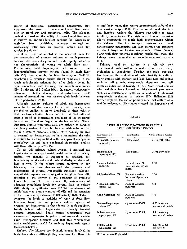
To use this primary culture system of neonatal rat hepatocytes as an experimental model for in vitro toxicity studies, we thought it important to establish the functionality of the cells and their similarity to the adult liver in vivo. In the culture system employed in our laboratory, we have demonstrated the presence and maintenance of several liver-specific functions: suifobro- mophthalein uptake and conjugation to glutathione (3); retention of the activity of the L-isozyme of pyruvate kinase for over 20 d in culture {3}; maintenance of adequate glutathione levels for several days in culture 148}; ability to synthesize urea {62,63}; maintenance of stable lactate to pyruvate ratios {62,63~; and the retention of high levels of cytochrome P-450 activity 153}. Table 1 compares the levels or activities of some of these liver functions found in our primary culture system of neonatal rat hepatocytes to those found in adult rat liver, freshly isolated adult hepatocytes, or freshly isolated neonatal hepatocytes. These results demonstrate that neonatal rat hepatoytes in primary culture retain certain adult liver-specific functions and that this experimental model could prove useful for studying hepatotoxic agents (see section below ~.
Kidney. The kidneys are dynamic organs involved in body homeostasis. Although they comprise less than 1%
of total body mass, they receive approximately 20% of the total cardiac output t21). The nature of renal structure and function renders the kidneys susceptible to toxic insult by xenobiotics. The high rate of renal perfusion allows compounds to reach high concentrations within the kidneys. The ion transport systems and renal concentrating mechanisms can also increase the exposure of the kidneys to foreign compounds. These factors, along with their inherent metabolic capabilities, make the kidneys more vulnerable to xenobiotic-induced toxicity {51}.
Primary renal cell culture is a relatively new experimental model within the realm of in vitro toxicity evaluation of xenobiotics. Thusfar, the main emphasis has been on the evaluation of metal toxicity in culture. Early studies with mercury and lead have used end-points such as cell growth, morphologic alterations, and cell death as indicators of toxicity {77,79}. More recent studies with cadmium have focused on biochemical parameters such as metallothionein synthesis, in addition to standard morphologic evaluation of toxicity {27L Belleman {22~ further explored the use of primary renal cell culture as a tool in toxicology. His studies stressed the importance of
TABLE 1
LIVER-SPECIFIC FUNCTIONS IN VARIOUS RAT LIVER PREPARATIONS
Liver Preparation ~ Liver Function Activity or Level of Function
Neonatal hepatocyte BSP uptake" 27.4 ~g/107 cells culture (3t
Isolated adult BSP uptake 24.0 pg/107 cells hepatocytes (73
Neonatal hepatocyte Ratio of L- and M- 1.70 culture ~3 } isozymes of pyruvate
kinase
Adult whole liver ~75) Ratio of l- and M- 1.56 isozymes of pyruvate kinase
Neonatal hepatocyte Ratio of lactate to 6.2 cutlure ~ 62) pyruvate
Adult whole liver ~76) Ratio of lactate to 7.0 pyruvate
Neonatal hepatocyte Cytochrome P-450 0.56 nmol/mg culture (53) microsomal protein
Isolated neonatal Cytochrome P-450 0.49 nmol/mg hepatocytes (53} microsomal protein
Isolated adult Cytochrome P-450 0.17 nmol/mg hepatocytes (54} cel| protein
~ = bromosulfophthalein
498 ACOSTA ET AL.
biochemical markers of cell function, such as brush border enzyme activity. Although it is well known that kidney cells in culture can form characteristic multicellu- lar domes, a phenomenon associated with transepithelial salt transport, no definitive studies on the toxic effects of xenobiotics on these domes have been conducted.
Before cells in culture can be used for toxicity studies, it must be determined that the cells have retained a certain degree of differentiated functions. Because it is thought that renal metabolism may be involved in the toxicities of various compounds, it is important that the metabolic integrity of the kidney cells be maintained in culture. Renal cytochrome P-450 has been implicated in the formation of reactive intermediates that cause tissue damage {35L The presence of marker enzymes, alkaline phosphatase and maltase, indicate proximal tubular brush border function (36). The kidney tubules are responsible for the transport of many organic anions and cations. Inasmuch as some nephrotoxic agents can have an effect on renal transport systems, it is necessary that cultured kidney cells maintain this function (34). If cultured kidney cells maintain their drug metabolizing capacity, brush border functions, and transport proper- ties they are more valid experimental models of nephrotoxicity. Alterations in these parameters can then be used to monitor xenobiotic-induced renal cell injury.
Because of the important role of the kidney in metabolizing and excreting xenobiotics from the body, we developed a primary culture system of renal cortical epithelial cells derived from 12 to 14 d-old rats for use in the in vitro evaluation of nephrotoxic agents. As described above, it is important to establish the functionality of the cells and their similarity to the kidney in vivo. The integrity of our primary culture system of renal cortical cells was determined on the basis of cell morphology and function. Arginine-deficient and D- valine supplemented culture medium was used to enhance epithelial cell growth and to suppress fibroblast overgrowth {31,42,66L These cell cultures were observed through an inverted phase contrast microscope to ensure that they maintained a characteristic epithelial-like cell morphology throughout the duration of the culture period {67). Furthermore, in our primary culture system of renal epithelial cells, we were able to demonstrate the presence and retention of several kidney-specific func- tions: adequate levels of brush border enzymes, alkaline phosphatase and maltase {67~; maintenance of cytochrome P-450 levels over 3 d in culture; and retention of glutathione levels over 3 d in culture (66). By establishing the functional integrity of our primary renal culture system of rat cortical epithelial cells, we then explored its use as an in vitro model to evaluate selected nephrotoxic agents for their effects at the cellular and subcellular level (see section belowL
Heart. The cardiotoxicity of xenobiotics has become an increasingly important topic of research because clinical use of certain drugs has revealed latent cardiotoxicity, not originally detected by preclinical animal studies {19L In addition, important anthracyeline antibiotics (doxorubi- cin} used in the treatment of several types of cancer have
had their clinical use limited because repeated use produces myocardial abnormalities. Alterations in myo- cardial energetics, disturbances in calcium metabolism, and peroxidation of cardiac lipids have been proposed as mechanisms by which doxorubiein damages the heart t33L Other important therapeutic agents as catechola- mines lepinephrine and isoproterenolL if administered in high doses, are highly cardiotoxic and produce extensive cardiac necrosis {19L Finally, the increased exposure of humans to environmental chemicals has resulted in the identification of several chemicals with high cardiotoxic potential 119).
Because the effects of cardiotoxic chemicals may vary from subtle functional changes to severe abnormalities that result in heart failure, several experimental models are needed to better understand chemical-induced cardiomyopathy. For this reason, our laboratory has developed a primary culture system of neonatal myocar- dial cells derived from 3- to 5-d-old rats to investigate the cardiotoxicity of selected agents at the cellular and subeeilular level. It is important to note that most of the in vitro myocardial studies reported in the areas of pharmacology and toxicology have used neonatal rats or chick embryos as the source for the heart cell cultures. There are only a few reported studies that have used adult rat hearts for the preparation of myocardial cell cultures {45,52L but these culture systems are limited in their use because of the difficulty of isolating large numbers of viable myocardial cells and because of their reduced life span in culture.
Our laboratory has used the method of Wenzel et al. {83~ to isolate primary myocardial cell cultures that are relatively free of contaminating nonmuscle cells as endothelial cells and fibroblasts. This procedure provides a uniform population of myocardial cells that form a confluent monolayer of synchronously beating cells. They are sensitive to drugs and metabolites in a manner similar to the whole heart {70L These myocardial cell cultures are also known to have intact beta-receptors coupled to adenylate cyclase (79L and the responsiveness of these receptors to cateeholamines in the newborn heart is similar to that of the adult heart (55). In the section below we describe the use of this primary culture system of rat myocardial cells in exploring the cardiotoxicity of selected drugs and chemicals.
Hepatotoxicity Studies With Primary Cultures of Rat Hepatocytes
The hepatotoxici ty of therapeutic agents and environmental chemicals has become an area of intense research interest. For instance, benoxaprofen was removed from use as a prescription drug when clinical evidence of its hepatotoxieity was reported in the literature (30,72L Little evidence of their hepatotoxicity was found in preclinical animal studies. Other well-known therapeutic agents such as acetaminopben, papaverine, tetracycline, and nitrofurantoin are hepatotoxie when taken in overdose or in eases of poisoning ~84}. Heavy metals such as cadmium and other
IN VITRO TARGET ORGAN TOXICOLOGY 499
environmental chemicals also have high hepatotoxic potential (84).
A major objective of our laboratory was to establish an in vitro model of hepatotoxicity with cultured hepatocytes so that one could potentially detect hepatotoxic chemi- cals, elucidate the cellular pathways of metabolism of the chemicals, and determine the mechanisms by which hepatotoxic agents produce toxic cellular damage. To study the in vitro toxicity of xenobiotics, we have developed a battery of sensitive cytotoxic indicators to gain the maximum amount of information after toxic injury to cultured cells. Our laboratory (47,63) as well as other investigators (23,62) have shown that the commonly used tests to measure in vitro toxicity (e.g. viability dye-exclusion methods, protein content, cell counts) are relatively insensitive measures of cytotoxicity. We have utilized cytotoxic indicators that can be correlated more easily to in vivo parameters of toxicity. As indicators of membrane integrity we have used leakage of potassium and cytosolic enzymes from injured cells into the culture medium, rather than the insensitive dye-exclusion tests. The measurement of serum enzymes in clinical toxicolo- gy is a well-accepted technique for evaluating specific organ damage by diseases or toxic chemicals.
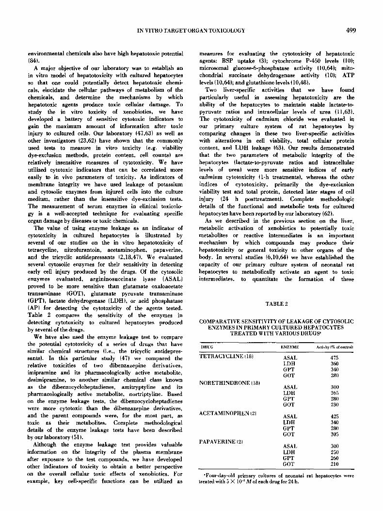
The value of using enzyme leakage as an indicator of cytotoxicity in cultured hepatocytes is illustrated by several of our studies on the in vitro hepatotoxicity of tetracycline, nitrofurantoin, acetaminophen, papaverine, and the tricyclic antidepressants (2,18,47). We evaluated several cytosolic enzymes for their sensitivity in detecting early ceil injury produced by the drugs. Of the cytosolic enzymes evaluated, argininosuccinate lyase (ASAL) proved to be more sensitive than glutamate oxaloacetate transaminase (GOTL glutamate pyruvate transaminase (GPT), lactate dehydrogenase (LDH), or acid phosphatase lAP) for detecting the cytotoxicity of the agents tested. Table 2 compares the sensitivity of the enzymes in detecting cytotoxicity to cultured hepatocytes produced by several of the drugs.
We have also used the enzyme leakage test to compare the potential cytotoxicity of a series of drugs that have similar chemical structures (i.e., the tricyclic antidepres- santsL In this particular study (47) we compared the relative toxicities of two dibenzazepine derivatives, imipramine and its pharmacologically active metabolite, desimipramine, to another similar chemical class known as the dibenzocycloheptadienes, amitryptyline and its pharmacologically active mctabolite, nortriptyline. Based on the enzyme leakage tests, the dibenzocycloheptadienes were more cytotoxic than the dibenzazepine derivatives, and the parent compounds were, for the most part, as toxic as their metabolites. Complete methodological details of the enzyme leakage tests have been described by our laboratory (51).
Although the enzyme leakage test provides valuable information on the integrity of the plasma membrane after exposure to the test compounds, we have developed other indicators of toxicity to obtain a better perspective on the overall cellular toxic effects of xenobiotics. For example, key cell-specific functions can be utilized as
measures for evaluating the cytotoxicity of hepatotoxic agents: BSP uptake (3); cytochrome P-450 levels (10); microsomal glucose-6-phosphatase activity (10,64); mito- chondrial succinate dehydrogenase activity (10); ATP levels (10,64); and glutathione levels (10,48).
Two liver-specific activities that we have found particularly useful in assessing hepatotoxicity are the ability of the hepatocytes to maintain stable lactate-to- pyruvate ratios and intracellular levels of urea (11,63L The cytotoxicity of cadmium chloride was evaluated in our primary culture system of rat hepatocytes by comparing changes in these two liver-specific activities with alterations in cell viability, total cellular protein content, and LDH leakage (63). Our results demonstrated that the two parameters of metabolic integrity of the hepatocytes (lactate-to-pyruvate ratios and intracellular levels of urea) were more sensitive indices of early cadmium cytotoxicity (1-h treatmentsL whereas the other indices of cytotoxicity, primarily the dye-exclusion viability test and total protein, detected later stages of cell injury (24 h posttreatmenfl. Complete methodologic details of the functional and metabolic tests for cultured hepatocytes have been reported by our laboratory (62).
As we described in the previous section on the liver, metabolic activation of xenobiotics to potentially toxic metabolites or reactive intermediates is an important mechanism by which compounds may produce their hepatotoxicity or general toxicity to other organs of the body. In several studies (6,10,64) we have established the capacity of our primary culture system of neonatal rat hepatocytes to metabolically activate an agent to toxic intermediates, to quantitate the formation of these
TABLE 2
COMPARATIVE SENSITIVITY OF LEAKAGE OF CYTOSOLIC ENZYMES IN PRIMARY CULTURED HEPATOCYTES
TREATED WITH VARIOUS DRUGS"
DRUG ENZYME Activity {% of controll
TETRACYCLINE (18) ASAL 475 LDH 360 G v r 340 GOT 280
NORETHINDRONE (18)
ACETAMINOPItEN (2)
PAPAVERINE (2)
ASAL 300 LDH 265 GPT 280 GOT 230
ASAL 425 LDH 340 GPT 280 GOT 305
ASAL 300 LDH 250 GPT 260 GOT 210
"Four-day-old primary cultures of neonatal rat hepatocytes were treated with 5 )< 10 -~ M o1 each drug for 24 h.

500 ACOSTA ET AL.
metabolites, and to assess the cytotoxicity of the formed metabolites to the cells in culture. One such study investigated the cytochrome P-450-mediated biotransfor- marion of cyclophosphamide to cytotoxic metabolites that nonspecifically alkylate DNA and cellular proteins 16). By utilizing a coculture system of nonreplicating hepatocytes and rapidly dividing fibroblasts, we were able to demonstrate the cytotoxicity of cyclophosphamide to the replicating fibroblasts with their enhanced DNA synthe- sis by the ability of the parenchymal hepatocytes to metabolize cyclophosphamide to alkylating metabolites which interacted in a lethal fashion with the fibroblasts. In essence, the hepatocytes served as the metabolizing units and the fibroblasts as the targets of the toxic metabolites. When metabolism of cyclophosphamide was blocked by SKF 525-A, an inhibitor of the cytochrome P-450 monooxygenase system, toxicity to the fibroblasts was eliminated. This study provides further evidence that postnatal rat hepatocytes in culture possess similar drug-metabolizing enzymes as adult hepatocytes.
In another study we investigated the metabolic activation of acetaminophen to toxic intermediate(s} and their cytotoxicity to our primary culture system of neonatal rat hepatocytes il0D. By measuring the distribu- tion of covalent binding of the reactive intermediate~s) to macromolecules in various subcellular fractions, we were able to identify the potential sites of cellular damage produced by acetaminophen. As shown in Table 3, the microsomal fraction had the greatest amount of radiola- bel binding, followed by the cytosolic, mitochondrial, and nuclear fractions, respectively. This distribution of covalent binding suggests that the reactive intermedi- ate~s) generated from the metabolism of acetaminophen are sufficiently stable to migrate throughout the cell and interact with potentially key cellular components. The extent of covalent binding of the reactive intermediate(sj to the subcellular fractions was correlated to alterations of key marker enzymes or functions associated with each fraction. Because the activity of glucose-6-phosphatase and the levels of cytochrome P-450, two indicators of microsomal integrity, were dramatically decreased by acetaminophen exposure of the hepatocytes as early as 4 h after treatment, the endoplasmic reticulum seems to be a primary target of acetaminophen toxicity. The other
TABLE 3
COVALENT BINDING OF [3H]ACETAMINOPHEN TO SUBCELLULAR FRACTIONS OF PRIMARY CULTURES
OF RAT HEPATOCYTES ~
Percent o| Tota| Subcellular Fraction Radiolabel Bound
Microsomai 37 Cytosolic 23 Mitochondfial 21 Nuclear 19
~The hepatocytes were treated with a mixture of 1.0 #Ci of [3H]acetaminophen per culture dish diluted in culture medium con- taining 5 mM unlabeled acetaminophen for an exposure period of 4 h.
subcellular sites, as manifested by changes in marker enzymes or functions, showed less damage at longer periods of exposure. In summary, this primary culture system of neonatal rat hepatocytes has proven to be an invaluable tool in the investigation of metabolism- mediated cytotoxicity of xenobiotics.
Cardiotoxicity Studies With Primary Cultures of Rat Myocardial Cells.
The use of primary cultures of rat myocardial cells in toxicologic studies began with the pioneering work of Wenzel and colleagues 125,82,83~, which demonstrated that toxic components of cigarette smoke (nicotine and carbon monoxide) could be evaluated in a culture system of rat heart cells. These myocardial cell culture systems have also proven to be useful experimental models in the evaluation of factors associated with heart disease and myocardial cell injury. Our laboratory has explored the use of primary cultures of rat myocardial cells as experimental models to study ischemic injury to the myocardium. Because it is not possible to reproduce true ischemia in cultured heart cells, we attempted to simulate conditions associated with ischemia. We demonstrated that hypoxia, glucose deprivation, and the production of extraceUular acidosis in cultured myocardial cells resulted in time-related inhibition of beating activity, marked alteration in cellular morphology, leakage of cytoplasmic enzymes, and enhanced lysosomal membrane fragility (9,12-14). Our studies showed that an in vitro model of myocardial ischemia may simulate qualitatively some of the responses observed in vivo, especially loss of contractility, enzyme leakage, and organelle membrane alterations. We have also used the myocardial cell culture system to study cell injury associated with hypoxia/reoxygenation phenomena and with depletion and repletion of calcium, the so-called calcium paradox {7,8). These studies showed that the absence of calcium ions during a hypoxic episode potentiated the cellular injury produced upon reoxygenation of hypoxic heart cells.
In other investigations we have used the primary culture system of rat myocardial cells to study drug and chemical toxicity at the cellular and subcellular levels. To utilize cultured cells for toxicity studies, it was necessary to develop techniques for evaluating cell iinjury or cytotoxicity produced by xenobiotics. Some methods that have been used routinely in our laboratory include measures of organelle (lysosomal and mitochondrial~ membrane integrity, cytosolic enzyme leakage, cell viability, alterations in contractile activity, and morpho- logic changes in cellular shape, size, and appearance. Such drugs and chemicals as chlorpromazine, clofibrate, amphotericin B, diazepam, caffeine, tricyclic antidepres- sants, food additives, and free fatty acids have been examined for their toxic effects to primary cultures of neonatal rat myocardial cells (1,4,15-17,81). An important consideration in conducting in vitro toxicity studies is to utilize similar concentrations that may be found in plasma or tissues of humans or animals administered the particular agent. We have been able to demonstrate dose-response and time-course relationships for each of the chemicals studied.
IN VITRO TARGET ORGAN TOXICOLOGY 501
A study in which we utilized several of the parameters of cytotoxicity cited above was the evaluation of the cardiotoxic potential of several tricyclic antidepressants (15). Lactate dehydrogenase leakage, cell viability, and contractile activity were measured to compare the cardiotoxicity of amitriptyline, nortriptyline, imipra- mine, and desipramine. On the basis of increased LDH leakage, decreased cell viability, and the production of arrhythmic contractile activity in the cultured myocardial cells, we concluded that amitriptyline had greater cardiotoxic potential than the other compounds. Our in vitro findings closely paralleled the in vivo cardiotoxicity of amitriptyline, which was shown to be more toxic than other trieyelic antidepressants (45).
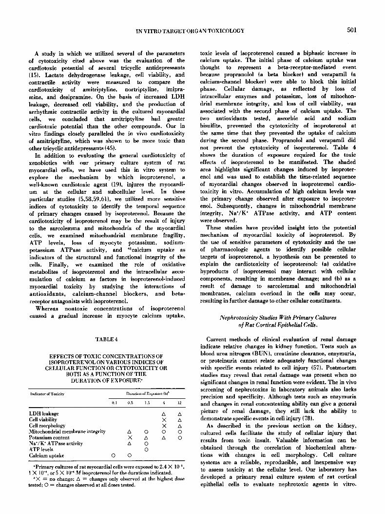
In addition to evaluating the general cardiotoxicity of xenobiotics with our primary culture system of rat myocardial cells, we have used this in vitro system to explore the mechanism by which isoproterenol, a well-known cardiotoxic agent (19), injures the myocardi- um at the cellular and subcellular level. In these particular studies (5,58,59,61), we utilized more sensitive indices of cytotoxicity to identify the temporal sequence of primary changes caused by isoproterenol. Because the cardiotoxicity of isoproterenol may be the result of injury to the sarcolemma and mitocbondria of the myocardial cells, we examined mitochondrial membrane fragility, ATP levels, loss of myocyte potassium, sodium- potassium ATPase activity, and 4Scalcium uptake as indicators of the structural and functional integrity of the cells. Finally, we examined the role of oxidative metabolites of isoproterenol and the intracellular accu- mulation of calcium as factors in isoproterenol-induced myocardial toxicity by studying the interactions of antioxidants, calcium-channel blockers, and beta- receptor antagonists with isoproterenol.
Whereas nontoxic concentrations of isoproterenol caused a gradual increase in myocyte calcium uptake,
TABLE 4
EFFECTS OF TOXIC CONCENTRATIONS OF ISOPROTERENOL ON VARIOUS INDICES OF
CELLULAR FUNCTION OR CYTOTOXICITY OR BOTH AS A FUNCTION OF THE
DURATION OF EXPOSURE ~
Indicator of Toxicity Duration of Exposure (h
0.1 0.5 1.5 4 12
LDH leakage Cell viability Cell morphology Mitochondrial membrane integrity A Potassium content X Na+/K + ATPase activity A ATP levels Calcium uptake 0 0
0 A 0 0
A A X X A 0 0 A 0
~ cultures of rat myocardial cells were exposed to 2.4 X 10 -s, 1 X 10 -4, or 5 >( 10 -4 M isoproterenol for the durations indicated.
~X = no change; A = changes only observed at the highest dose tested; O = changes observed at all doses tested.
toxic levels of isoproterenol caused a biphasic increase in calcium uptake. The initial phase of calcium uptake was thought to represent a beta-receptor-mediated event because propranolol (a beta blocker) and verapamil (a calcium-channel blocker) were able to block this initial phase. Cellular damage, as reflected by loss of intracellular enzymes and potassium, loss of mitochon- drial membrane integrity, and loss of cell viability, was associated with the second phase of calcium uptake. The two antioxidants tested, aseorbic acid and sodium bisulfite, prevented the cytotoxicity of isoproterenol at the same time that they prevented the uptake of calcium during the second phase. Propranolol and verapamil did not prevent the cytotoxicity of isoproterenol. Table 4 shows the duration of exposure required for the toxic effects of isoproterenol to be manifested. The shaded area highlights significant changes induced by isoproter- enol and was used to establish the time-related sequence of myocardial changes observed in isoproterenol cardio- toxicity in vitro. Accumulation of high calcium levels was the primary change observed after exposure to isoproter- enol. Subsequently, changes in mitocbondrial membrane integrity, Na§ § ATPase activity, and ATP content were observed.
These studies have provided insight into the potential mechanism of myocardial toxicity of isoproterenol. By the use of sensitive parameters of cytotoxicity and the use of pharmacologic agents to identify possible cellular targets of isoproterenol, a hypothesis can be presented to explain the cardiotoxicity of isoproterenoh (a) oxidative byproducts of isoproterenol may interact with cellular components, resulting in membrane damage; and (b) as a result of damage to sarcolemmal and mitochondrial membranes, calcium overload in the cells may occur, resulting in further damage to other cellular constituents.
Nephrotoxicity Studies With Primary Cultures o/Rat Cortical Epithelial Cells.
Current methods of clinical evaluation of renal damage indicate relative changes in kidney function. Tests such as blood urea nitrogen (BUN), creatinine clearance, enzymuria, or proteinuria cannot relate adequately functional changes with specific events related to cell injury (57). Postmortem studies may reveal that renal damage was present when no significant changes in renal function were evident. The in vivo screening of nephrotoxins in laboratory animals also lacks precision and specificity. Although tests such as enzymuria and changes in renal concentrating ability can give a general picture of renal damage, they still lack the ability to demonstrate specific events in cell injury (78).
As described in the previous section on the kidney, cultured cells facilitate the study of cellular injury that results from toxic insult. Valuable information can be obtained through the correlation of biochemical altera- tions with changes in cell morphology. Cell culture systems are a reliable, reproducible, and inexpensive way to assess toxicity at the cellular level. Our laboratory has developed a primary renal culture system of rat cortical epithelial cells to evaluate nephrotoxic agents in vitro.
502 ACOSTA ET AL.
Through the use of sensitive indicators of cytotoxicity and biochemical alterations, the sequence of changes that occurs during xenobiotic-induced renal damage was determined. Unlike most investigations, our evaluations of nephrotoxicity in vitro were based on xenobiotic- induced changes in morphology, membrane integrity, normal cellular biochemistry, and renal-specific func- tions (65-68).
In our first series of studies (65,66) we evaluated several well-known nephrotoxic agents (mercuric chloride, cadmium chloride, and acetaminophen) to help establish guidelines for the assessment of toxicity in culture and to determine the sensitivity of the in vitro indices of cytotoxicity developed by our laboratory. Whereas Inamoto et al. (37) and Vickery and McCann (77) observed mercuric chloride toxicity in cultured cells over several days in culture (alterations in RNA, DNA, and protein synthesis and cell growth), we were able to detect inhibition of LDH activity as early as 1 h after treatment, and a decline in alkaline phosphatase activity was seen after 4 h. Cadmium chloride caused an increased leakage of LDH and a decrease in akaline phosphatase activity after 2 h of exposure in our culture system of renal cortical cells. Our results correlate well with the in vivo effects of cadmium in animals (26,56L The cytotoxicity of acetaminophen was reflected by increased LDH leakage after 24 h of treatment and by fluctuations in glutathione levels. These studies with the nephrotoxic agents helped to establish the validity of our primary renal cell culture system as a tool for toxicity evaluation.
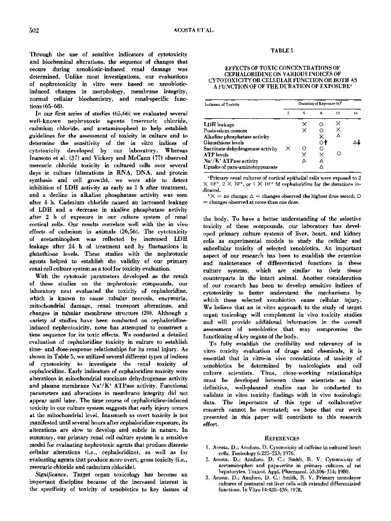
With the cytotoxic parameters developed as the result of these studies on the nephrotoxic compounds, our laboratory next evaluated the toxicity of cephaloridine, which is known to cause tubular necrosis, enzymuria, mitochondrial damage, renal transport alterations, and changes in tubular membrane structure (20). Although a variety of studies have been conducted on cephaloridine- induced nephrotoxicity, none has attempted to construct a time sequence for its toxic effects. We conducted a detailed evaluation of cephaloridine toxicity in culture to establish time- and dose-response relationships for its renal injury. As shown in Table 5, we utilized several different types of indices of cytotoxicity to investigate the renal toxicity of cephaloridine. Early indicators of cephaloridine toxicity were alterations in mitochondrial succinate dehydrogenase activity and plasma membrane Na+/K § ATPase activity. Functional parameters and alterations in membrane integrity did not appear until later. The time course of cephaloridine-induced toxicity in our culture system suggests that early injury occurs at the mitochondrial level. Inasmuch as overt toxicity is not manifested until several hours after cephaloridine exposure, its alterations are slow to develop and subtle in nature. In summary, our primary renal cell culture system is a sensitive model for evaluating nephrotoxic agents that produce discrete cellular alterations (i.e., cephaloridineL as well as for evaluating agents that produce more overt, gross toxicity (i.e., mercuric chloride and cadmium chloride).
Significance. Target organ toxicology has become an important discipline because of the increased interest in the specificity of toxicity of xenobiotics to key tissues of
TABLE 5
EFFECTS OF TOXIC CONCENTRATIONS OF CEPHALORIDINE ON VARIOUS INDICES OF
CYTOTOXICITY OR CELLULAR FUNCTION OR BOTH AS A FUNCTION OF OF THE DURATION OF EXPOSURE"
Indicator of Toxicity Duration of Exposure (h) b
2 4 8 12 16
LDH leakage X O X Postassium content X O X Alkaline phosphatase activity X A Giutathione levels O Succinate dehydrogenase activity X O O ATP levels X X O Na+/K § ATPase activity A A Uptake of para-aminohippuarate A
~ renal cultures of cortical epithelial cells were exposed to 2 X 10 -5, 2 X 10 -4, or 1 X 10 -3 M cephaloridine for the durations in- dicated.
X = no change; A =- changes observed the highest dose tested; �9 = changes observed at more than one dose.
the body. To have a better understanding of the selective toxicity of these compounds, our laboratory has devel- oped primary culture systems of liver, heart, and kidney cells as experimental models to study the cellular and subcelhlar toxicity of selected xenobiotics. An important aspect of our research has been to establish the retention and maintenance of differentiated functions in these culture systems, which are similar to their tissue counterparts in the intact animal. Another consideration of our research has been to develop sensitive indices of cytotoxicity to better understand the mechanisms by which these selected xenobiotics cause cellular injury. We believe that an in vitro approach to the study of target organ toxicology will complement in vivo toxicity studies and will provide additional information in the overall assessment of xenobiotics that may compromise the functioning of key organs of the body.
To fully establish the credibility and relevancy of in vitro toxicity evaluation of drugs and chemicals, it is essential that in vitro-in vivo correlations of toxicity of xenobiotics be determined by toxicologists and cell culture scientists. Thus, close-working relat ionships must be developed between these scientists so that definitive, well-planned studies can be conducted to validate in vitro toxicity findings with in vivo toxicologic data. The importance of this type of collaborative research cannot be overstated; we hope that our work presented in this paper will contribute to this research effort.
REFERENCES
1. Acosta, D.; Anuforo, D. Cytotoxicity of caffeine in cultured heart cells. Toxicology 6:225-233; 1976.
2. Acosta, D.; Anuforo, D. C.; Smith, R. V. Cytotoxlcity of acetaminophen and papaverine in primary cultures of rat hepatocytes. Toxicol. Appl. Pharmacol. 53:306-314; 1980.
3. Acosta, D.; Anuforo, D. C.; Smith, R. V. Primary monolayer cultures of postnatal rat liver cells with extended differentiated functions. In Vitro 14:428-436; 1978.

IN VITRO TARGET ORGAN TOXICOLOGY 503
4. Acosta, D.; Chappell, R. Cardiotoxicity of diazepam in cultured heart ceils. Toxicology 8:311-317; 1977.
5. Acosta, D.; Combs, A. B.; Ramos, K. Attenuation by an- tioxidants of Na+/K ~ ATPase inhibition by toxic concentrations of isoproterenol in cultured rat myocardial cells. J. Mol. Cell. Cardiol. 16:281-284; 1984.
6. Acosta, D.; Mitchell, D. B. Metabolic activation and cytotoxicity of cyclophosphamide in primary cultures of postnatal rat hepatocytes. Biochem. Pharmacol. 30:3225-3230; 1981.
7. Acosta, D.; Ramos, K. Li-Goldman, C. P. Cellular injury of primary cultures of rat myocytes incubated in calcium-free medium followed by recovery in calcium. In Vitro 19:141-144; 1983.
8. Aeosta, D.; Ramos, K.; Li-Goldman, C. P. Cell injury of cultured rat myocardial cells after reoxygenation of hypoxic cultures in the presence and absence of calcium. In Vitro 20:642-646; 1984.
9. Aeosta, D.; Li, C. P. Actions of cxtracellular acidosis on primary cultures of rat myocardial cells deprived of oxygen and glucose. J. Mol. Cell. Cardiol. 12:1459-1463; 1980.
10. Aeosta, D.; Mitchell, D. B. Subcellular localization of hepatocyte injury due to metabolic activation of acetaminophen. Toxicologist 3:113; 1983.
11. Acosta, D.; Sorensen, E. M. B. Role nf calcium in cytotoxie in- jury of cultured hepatocytes. Ann. NY Acad. Sei. 407:78-92; 1983.
12. Acosta, D.; Puckett, M. Ischemic myocardial injury in cultured heart cells: preliminary observations on morphology and beating activity. In Vitro 13:818-823; 1977.
13. Acosta, D.; Puckett, M.; MeMiilin, R. lschemic myocardial injury in cultured heart cells: leakage of cytoplasmic enzymes from injured cells. In Vitro 14:728-732; 1978.
14. Acosta, D.; Puekett, M.; McMillin, R. Ischemic myocardial injury in cultured heart cells: in situ lysosomal damage. Ex- perientia 34:1388-1389; 1978.
15. Acosta, D.; Ramos, K. Cardiotoxieity of tricyclic antidepressants in primary cultures of rat myocardial cells. J. Toxicol. Environ. Health 14:137-143; 1984.
16. Acosta, D.; Wenzel, D. G. A permeability test for the study of mitochondrial injury in in vitro cultured heart muscle and endothelioid cells. Histochem. J. 7:45-56; 1975.
17. Aeosta, D.; Wcnzel, D. G. Injury produced by free fatty acids to lysosomes and mitochrondria in cultured heart muscle and endothelial ceils. Atherosclerosis 20:417-426; 1974.
18. Anuforo, D. C.; Aeosta, D.; Smith, R. V. Hepatotoxicity studies with primary cultures of rat liver cells. In Vitro 14:981-988; 1978.
19. Balazs, T.; Fcrrans, V. J. Cardiac lesions induced by chemicals. Environ. Hlth. Perspect. 26:181-191; 1978.
20. Barza, M. Thenephrotoxieity of cephalosporins: an overview. J. Infect. Dis. 137:,560-573; 1978.
21. Beeuwkes, R.; Ichikawa, I.; Brenner, B. M. The renal cir- culations. In: Brenner, B. M.; Rector, F. C., eds. The kidney, wfl. 1. Philadelphia: W. B. Saunders; 1981:267.
22. Belleman, P. Primary monolayer culture of liver parenchymal cells and kidney cortical tubules as a useful new model for biochemical pharmacology and experimental toxicology. Arch. Toxicol. 44:63-84; 1980.
23. Bhuyan, B. K.; Loughman, B. E.; Fraser, T. J. et al. Com- parison of different methods of determining cell viability after exposure to cytotoxic compounds. Exp. Cell Res. 97:275-280; 1976.
24. Bonney, R. J. Adult liver parenchymal cells in primary culture: characteristics and cell recognition standards. In Vitro 10:130-142; 1974.
25. Brenner, G. M.; Wenzel, D. G. Carbon monoxide and cultured rat heart cells. I. Inhibition of cell growth and maintenance of beating rate. Toxicol. Appl. Pharmacol. 23:251-262; 1972.
26. Chart, W. Y.; Rennert, O. M. Cadmium nephropathy. Ann. Clin. Lab. Sci. 11:229-238; 1981.
27. Cherian, G. M. The synthesis of metallothionein and cellular adaptation to metal toxicity in primary rat kidney epithelial cell cultures. Toxicology 17:225-231; 1980.
28. Cohen, Y. Reactive metabolites and their implications for toxicology. Adv. Pharmacol. Ther. 9:1-25; 1980.
29. Dallner, G.; Siekevitz, P.; Palade, G. E. Biogenesis of en- doplasmic reticulum membranes. J. Cell Biol. 30:73-95; 1966.
30. FDA Drug Bulletin. Treatment IND for benoxaprofen. FDA Drug Bull. 13:4--5; 1983.
31. Gilbert, S. F.; Migeon, B. R. D-Valine as a selective agent for normal human and rodent epithelial ceils in culture. Cell 5:11- 17; 1975.
32. Goodman, J.; Hochstein, P. Generation of free radicals and lipid peroxidation by redox cycling of adriamycin and daunomycin. Biochem. Biophys. Res. Commun. 77:797-803; 1977.
33. Grisham, J. W.; Smith, G. J. Predictive and mechanistic evaluation of toxic responses in mammalian cell culture systems. Pharmacol. Rev. 36:151s-171s; 1984.
34. Hirsch, G. H. Differential effects of nephrotoxic agents on renal transport and metabolism by use of in vitro techniques. En- viron. Health Perspect. 15:89-99; 1976.
35. Hook, J. B.; McCormack, K. M.; Kluwe, W. M. Biochemical mechanisms of nephrotoxicity. In: Hodgson, E.; Bend, J. R.; Philpott, R. M., eds. Reviews in biochemical toxicology, vol. 1. New York: Elsevier; 1979:53-78.
36. ttsu, B. Y.; McNamara, P. D.; Schlesinger, H.; et al. Ease of solubilization of liver marker enzymes in three preparations of rat renal brush border membranes. Enzyme 25:170-181; 1981.
37. lnamoto, H.; Ino, Y.; Inamoto, N.; et al. Effect of HgCI~ on rat kidney cells in primary culture. Lab. Invest. 34:489-494; 1976.
38. Jones, A. L.; Mills, E. S. Ultrastructural concepts of drug metabolism. Am. J. Drug Alcohol Abuse 1:111-122; 1974.
39. Kluwe. W. M. Renal function tests as indicators of kidney injury in subacute toxicity studies. Toxicol. Appl. Pharmacol. 57:414-424; 1981.
40. Laishes, B. A.; Williams, G. M. Conditions affecting primary cell cultures of functional adult rat hepatocytes. I. The effect of insulin. In Vitro 12:521-532; 1976.
41. Laishes, B. A.; Williams. G. M. Conditions affecting primary cell cultures of functional adult rat hepatocytes II. Dexamethasone enhanced longevity and maintenance of morphology. In Vitro 12:821-832; 1976.
42. Leffert, H.; Paul, D. Serum dependent growth of primary cultured differentiated fetal rat hepatocytes in arginine- deficient medium. J. Cell. Physiol. 81:113-124; 1973.
43. Leslie, S. W.; Gad, S. C.; Acosta, D. Cytotoxicity of butylated hydroxytoluene and butylated hydroxyanisole in cultured heart cells. Toxicology 10:281-289; 1978.
44. Lieberman, M.; Adam, W. J.; Bullock, P. N. The cultured heart cell: problems and prospects. Methods Cell Biol. 21A:187-203; 1980.
45. Mattila, M. J.; Saarnivara, L. Amitriptyline toxicity. Lancet 2:1138; 1970.
46. Mazel, P.; Pessayre, D. Significance of metabolite-mediated toxicities in the safety evaluation of drugs and chemicals. Adv. Med. Toxicol. 1:207-343; 1976.
47. Mitchell, D. B.; Acosta, D. Evaluation of the cytotoxicity of tricyclic antidepressants in primary cultures of rat bepatocytes. J. Toxicol. Environ. Health, 7:83-92; 1981.
48. Mitchell, D. B.; Acosta. D. The effect of culture medium sup- plements on glutathione levels in primary cultures of postnatal rat hepatocytes. In Vitro 17:243; 1981.
49. Mitchell, J. R.; Jollow, D. J. Metabolic activation of drugs to toxic substances. Gastroenterology 68:392-410; 1975.
50. Mitchell, J. R.; McMurtry, R. J.; Statham, C. N.; et al. Molecular basis for several drug-induced nephropathies. Am. J. Med. 62:518-526; 1977.
51. Mitchell, D. B.; Santone, K. S.; Acosta, D. Evaluation of cytotoxicity in cultured cells by enzyme leakage. J. Tiss. Cult. Methods 6:113-116; 1980.
52: Moses, R. L.; Kasten, F. H. Ultrastructure of dissociated adult mammalian myocytes. J. Mol. Cell. Cardiol. 11:161-172; 1979.
53. Nelson, K. F.; Acosta, D.; Bruckner, J. V. Long-term main- tenance and induction of cytochrome P-450 in primary cultures of rat hepatocytes. Biochem. Pharmacol. 31:2211-2214; 1982.
504 ACOSTAETAL.
54. Paine, A. J.; Legg, R. F. Apparent lack of correlation between the loss of cytochrome P-450 in hepatic parenchymal cell culture and the stimulation of haem oxygenase activity. Biochem. Biophys. Res. Commun. 81:672-679; 1978.
55. Park, M. K.; Sheridan, P. H.; Morgan, W. F. etal. Comparative inotropic responses of newborn and adult rabbit papillary muscles to isoproterenol and calcium. Dev. Pharmacol. Ther. 1:70-82; 1980.
56, Peereboom-Stegeman, J. H. J.; Melet, J.; Peereboom, J. W. C.; et al. Influence of chronic cadmium intoxication on the alkaline phosphatase activity in liver and kidney. Toxicology 14:67-80; I979.
57, Prescott, L. F. Assessment of nephrotoxicity. Br. J. Clin. Pharmacol. 13:303-311; 1982.
58. Ramos, K.; Acosta, D. Prevention by L-ascorbic acid of isoproterenol-induced cardiotoxicity in primary cultures of rat myocytes. Toxicology 26:81-90; 1983.
59. Ramos, K.; Combs, A. B.; Acosta, D. Cytotoxicity of isoproterenol to cultured heart cells: effects of antioxidants on modifying membrane damage. Toxicol. Appl. Pharmacol. 70:317-323; 1983.
60. Ramos, K.; Combs, A. B.; Acosta, D. Role of calcium in isoproterenol cytotoxicity to cultured myocardial cells. Biochem. Pharmacol. 33:1989-1992; 1984.
61. Salocks, C. B.; Hsieh, D. P.; Byard, J. L. Butylated hydroxytoluene pretreatment protects against cytotoxicity and reduces covalent binding of aflatoxin B, in primary hepatocyte cultures. Toxicol. Appl. Pharmacol. 59:331-345; 1981.
62. Santone, K. S.; Acosta, D. Measurement of functional metabolic activity as a sensitive parameter of cytotoxicity in cultured hepatocytes. J. Tissue Cult. Methods 7:137-142; 1982.
63. Santone, K. S.; Acosta, D.; Bruckner, J. V. Cadmium toxicity in primary cultures of rat hepatocytes. J. Toxicol. Environ. Health 10:169-177; 1982.
64. Santone, K. S.; Acosta, D.; The role of extracellular calcium in CCL injury of cultured rat hepatocytes. Toxicologist 4:133; 1984.
65. Smith, M. A.; Acosta, D. Cephaloridine toxicity in primary cultures of rat kidney epithelial cells. Pharmacologist 25:272; 1983.
66. Smith, M. A.; Acosta, D. Effects of acetaminophen on primary cultures of rat kidney epithelial cells. Toxicologist 3:112; 1983.
67. Smith, M. A.; Acosta, D. Effects of HgCI~ on alkaline phosphatase activity, lactate dehydrogenase activity, and cell morphology in primary renal epithelial cell cultures. Phar- macologist 24:146; 1982.
68. Smith, M. A.; Aeosta, D. Effect of cephaloridine on cellular ATP content and mitochondrial succinate dehydrogenase activity in primary cuhures of rat kidney cortical epithelial cells. Toxicologist 4:31; 1984.
69. Smith, R. V.; Acosta, D.; Rosazza, J. P. Cellular and microbial models in the investigation of mammalian metabolism of xenobiotics. Adv. Biochem. Eng. 5:69-100; 1977.
70. Sperelakis, N. Cultured heart cell reaggregate model for studying cardiac toxicology. Environ. Health Perspect. 26:243-267; 1978.
71. Stammati, A. P.; Silano, V.; Zucco, F. Toxicology investigations with cell culture systems. Toxicology 20:90-153; 1981.
72. Taggart, H. M.; Alderice, J. M. Fatal cholestatic jaundice in elderly patients taking benoxaprofen. Br. Med. J. 284:1372; 1982.
73. Tardiff, R. G. In vitro methods of toxicity evaluation. Ann. Rev. Pharmacol. Toxicol. 18:357-369; 1978.
74. Van Beezooijen, C. F. A.; Grell, T.; Knock, D. L. Bromosulfophthalein uptake by isolated liver parenchymal cells. Biochem. Biophys. Res. Commun. 69:354-361; 1976.
75. Van Berkel, T. J. C.; Koster, J. F.; Hulsman, W. C. Distribution of L- and M-type pyruvate kinase between parenchymai and Kupffer cells of rat liver. Biochim. Biophys. Acta 276:425-429; 1972.
76. Veech, R. L.; Raijman, L.; Krebs, H. A. Equilibrium relations between the cytoplasmic adenine nucleotide system and nicotinamide-adenine nucleotide system in rat liver. Biochem. J. 117:499-503; 1970.
77. Vickery, H. M.; McCann, D. S. Temperature and species dif- ferences in susceptibility of kidney cell cultures to mercury toxicity. In Vitro 14:312-316; 1978.
78. Walker, M. J. A. Initial investigation into adrenaline ac- cumulation and adrenergic responsiveness in cultured neonatal rat heart cells. Br. J. Pharmacol. 62:185-193; 1978.
79. Walton, J.; Buckley, I. K. The lead poisoned cell: a fine struc- tural study using culture kidney cells. Exp. Mol. Pathol. 27:167-182; 1977.
80. Welch, R. M. Toxicological implications of drug metabolism. Pharmacol. Rev. 30:457-467, 1979.
81. Wenzel, D. G.; Acosta, D. Permeability of lysosomes and mitochondria in cultured rat heart muscle and endothelial cells as affected by vitamin A, chlorpromazine, amphotericin B, and clofibrate. Res, Commun. Chem. Pathol. Pharmacol. 6:689-700; 1973.
82. Wenzel, D. G.; Brenner, G. M. Carbon monoxide and cultured rat heart cells. II. Interaction of carbon monoxide and hypoxia on growth and contractile activity. Toxicol. Appl. Pharmacol. 24:256-265; 1973.
83. Wenzel, D. G.; Wheatley, J. W.; Byrd, G. D. Effects of nicotine on cultured heart cells. Toxicol. Appl. Pharmacol. 17:774- 785; 1970.
84. Zimmerman, H. J. Hepatotoxicity. The adverse effects of drugs and other chemicals on the liver. New York: Appleton-Century- Crofts; 1978.