Aminolevulinate synthase: Lysine 313 is not essential for binding the pyridoxal phosphate cofactor...
6
Protein Science (1995), 4:1001-1006. Cambridge University Press. Printed in the USA. Copyright 0 1995 The Protein Society Aminolevulinate synthase: Lysine 3 13 is not essential for binding the pyridoxal phosphate cofactor but is essential for catalysis GLORIA C. FERREIRA,’*2,3 USHA VAJAPEY,’ OSAMA HAFEZ,‘ GREGORY A. HUNTER,’ AND MICHAEL J. BARBER’.’ ’ Department of Biochemistry and Molecular Biology, College of Medicine, Institute for Biomolecular Science, and H. Lee Moffitt Cancer Center and Research Institute, University of South Florida, Tampa, Florida 33612 (RECEIVED September 22, 1994; ACCEPTED February 14, 1995) Abstract 5-Aminolevulinate synthase is the first enzyme of the heme biosynthetic pathway in animals and some bacteria. Lysine-3 13 of the mouse erythroid aminolevulinate synthase was recently identified to be linked covalently to the pyridoxal 5”phosphate cofactor (Ferreira GC, Neame PJ, Dailey HA, 1993, Protein Sci 2: 1959-1965). Here we report on the effect of replacement of aminolevulinate synthase lysine-313 by alanine, histidine, and glycine, using site-directed mutagenesis. Mutant enzymes were purified to homogeneity, and the purification yields were simi- lar to those of the wild-type enzyme. Although their absorption spectra indicate that the mutant enzymes bind pyridoxal 5’-phosphate, they bind noncovalently. However, addition of glycine to the mutant enzymes led to the formation of external aldimines. The formation of an external aldimine between the pyridoxal 5”phosphate co- factor and the glycine substrate is the first step in the mechanism of the aminolevulinate synthase-catalyzed reac- tion. In contrast, lysine-313 is an essential catalytic residue, because the K313-directed mutant enzymes have no measurable activity. In summary, site-directed mutagenesis of the aminolevulinate synthase active-site lysine-313, to alanine (K313A), histidine (K313H), or glycine (K313G) yields enzymes that bind the pyridoxal 5”phosphate cofactor and the glycine substrate to produce external aldimines, but which are inactive. This suggests that lysine- 313 has a functional role in catalysis. Keywords: 5-aminolevulinate synthase; heme biosynthesis; pyridoxal 5”phosphate 5-Aminolevulinate synthase (EC 2.3.1.37), found in animals and some bacteria, is a pyridoxal 5”phosphate-dependent enzyme that catalyzes the condensation of glycine and succinyl-CoA to yield ALA, CoA, and carbon dioxide (Kikuchi et al., 1958; Jor- dan, 1991). This reaction represents the first committed step in the heme biosynthetic pathway of these organisms (Jordan, 1991). ALAS has been isolated and purified from different sources, ranging from bacteria to man (Warnick & Burnham, 1971; Nakakuki et al.,1980; Borthwick et al.,1986; Munakata et al., 1993), and in all instances the enzyme is functional as a homodimer (Jordan, 1991; Munakata et al., 1993). In animals, Reprint requests to: Gloria C. Ferreira, Department of Biochemis- try and Molecular Biology, College of Medicine, University of South Florida, 12901 Bruce B. Downs Boulevard, Tampa, Florida 33612; e-mail: [email protected]. A bbreviarions: ALA, 5-aminolevulinate; ALAS, S-aminolevulinate synthase; PLP, pyridoxal 5’-phosphate; MES, 2-[N-morpholinolethane- sulfonic acid; CHES, 2-[N-cyclohexylamino]-ethanesulfonic acid; BSA, bovine serum albumin. there are two ALAS isoforms, which are encoded by distinct genes and are differentially expressed: the erythroid form of ALAS (ALAS-E) only in erythroid cells and the housekeeping form (ALAS-H) nonspecifically in all tissues (Riddle etal., 1989; Bishop et al., 1990; Cox et al., 1990). Two ALAS isozymes, en- coded by hemA and hemT, have also been identified in Rhodo- bacter sphaeroides (Neidle & Kaplan, 1993). Although ALAS has been known to be a PLP-dependent en- zyme for several decades, only recently, with the overproduc- tion of murine ALAS-E in Escherichia coli (Ferreira & Dailey, 1993), has it become possible to obtain enough purified enzyme for the determination of the PLP-binding lysine residue. The t-amino group of lysine-313 was identified to form the Schiff base linkage with PLP in murine ALAS-E(Ferreira et al., 1993). Sequence alignment analysis of ALAS sequences from different species indicates that the pyridoxyllysine peptide and the ALAS active site are conserved among all organisms and are present in the C-terminal domain of the enzyme (Cox et al., 1990; Fer- reira et al., 1993). The recent sequencing of the purified papain- resistant core domain of the rat ALAS-E has confirmed that 1001
-
Upload
independent -
Category
Documents
-
view
0 -
download
0
Transcript of Aminolevulinate synthase: Lysine 313 is not essential for binding the pyridoxal phosphate cofactor...
Protein Science (1995), 4:1001-1006. Cambridge University Press. Printed in the USA. Copyright 0 1995 The Protein Society
Aminolevulinate synthase: Lysine 3 13 is not essential for binding the pyridoxal phosphate cofactor but is essential for catalysis
GLORIA C. FERREIRA,’*2,3 USHA VAJAPEY,’ OSAMA HAFEZ,‘ GREGORY A . HUNTER,’ AND MICHAEL J. BARBER’.’ ’ Department of Biochemistry and Molecular Biology, College of Medicine, Institute for Biomolecular Science, and
H. Lee Moffitt Cancer Center and Research Institute, University of South Florida, Tampa, Florida 33612
(RECEIVED September 22, 1994; ACCEPTED February 14, 1995)
Abstract
5-Aminolevulinate synthase is the first enzyme of the heme biosynthetic pathway in animals and some bacteria. Lysine-3 13 of the mouse erythroid aminolevulinate synthase was recently identified to be linked covalently to the pyridoxal 5”phosphate cofactor (Ferreira GC, Neame PJ, Dailey HA, 1993, Protein Sci 2: 1959-1965). Here we report on the effect of replacement of aminolevulinate synthase lysine-313 by alanine, histidine, and glycine, using site-directed mutagenesis. Mutant enzymes were purified to homogeneity, and the purification yields were simi- lar to those of the wild-type enzyme. Although their absorption spectra indicate that the mutant enzymes bind pyridoxal 5’-phosphate, they bind noncovalently. However, addition of glycine to the mutant enzymes led to the formation of external aldimines. The formation of an external aldimine between the pyridoxal 5”phosphate co- factor and the glycine substrate is the first step in the mechanism of the aminolevulinate synthase-catalyzed reac- tion. In contrast, lysine-313 is an essential catalytic residue, because the K313-directed mutant enzymes have no measurable activity. In summary, site-directed mutagenesis of the aminolevulinate synthase active-site lysine-313, to alanine (K313A), histidine (K313H), or glycine (K313G) yields enzymes that bind the pyridoxal 5”phosphate cofactor and the glycine substrate to produce external aldimines, but which are inactive. This suggests that lysine- 313 has a functional role in catalysis.
Keywords: 5-aminolevulinate synthase; heme biosynthesis; pyridoxal 5”phosphate
5-Aminolevulinate synthase (EC 2.3.1.37), found in animals and some bacteria, is a pyridoxal 5”phosphate-dependent enzyme that catalyzes the condensation of glycine and succinyl-CoA to yield ALA, CoA, and carbon dioxide (Kikuchi et al., 1958; Jor- dan, 1991). This reaction represents the first committed step in the heme biosynthetic pathway of these organisms (Jordan, 1991). ALAS has been isolated and purified from different sources, ranging from bacteria to man (Warnick & Burnham, 1971; Nakakuki et al., 1980; Borthwick et al., 1986; Munakata et al., 1993), and in all instances the enzyme is functional as a homodimer (Jordan, 1991; Munakata et al., 1993). In animals,
Reprint requests to: Gloria C. Ferreira, Department of Biochemis- try and Molecular Biology, College of Medicine, University of South Florida, 12901 Bruce B. Downs Boulevard, Tampa, Florida 33612; e-mail: [email protected].
A bbreviarions: ALA, 5-aminolevulinate; ALAS, S-aminolevulinate synthase; PLP, pyridoxal 5’-phosphate; MES, 2-[N-morpholinolethane- sulfonic acid; CHES, 2-[N-cyclohexylamino]-ethanesulfonic acid; BSA, bovine serum albumin.
there are two ALAS isoforms, which are encoded by distinct genes and are differentially expressed: the erythroid form of ALAS (ALAS-E) only in erythroid cells and the housekeeping form (ALAS-H) nonspecifically in all tissues (Riddle et al., 1989; Bishop et al., 1990; Cox et al., 1990). Two ALAS isozymes, en- coded by hemA and hemT, have also been identified in Rhodo- bacter sphaeroides (Neidle & Kaplan, 1993).
Although ALAS has been known to be a PLP-dependent en- zyme for several decades, only recently, with the overproduc- tion of murine ALAS-E in Escherichia coli (Ferreira & Dailey, 1993), has it become possible to obtain enough purified enzyme for the determination of the PLP-binding lysine residue. The t-amino group of lysine-313 was identified to form the Schiff base linkage with PLP in murine ALAS-E (Ferreira et al., 1993). Sequence alignment analysis of ALAS sequences from different species indicates that the pyridoxyllysine peptide and the ALAS active site are conserved among all organisms and are present in the C-terminal domain of the enzyme (Cox et al., 1990; Fer- reira et al., 1993). The recent sequencing of the purified papain- resistant core domain of the rat ALAS-E has confirmed that
1001
1002
Table 1. Site-directed mutagenesis of the ALAS-encoding plasmida
ALAS-WT L G K * A F G
ALAS-WT 5' . . . C T T GGC AAG GCC TTT GGT . . . 3' K313A 3' . . . GAA CCG CCC CGG AAA AC. . . . 5' K313H 3' . . . GAA CCG C T A CGC AAA AC. . . . 5' K3 13G 3' . . . GAA CCG CCC CGG AAA AC. . . . 5'
a K313-directed mutants were constructed as described in the Mate- rials and methods using the oligonucleotides shown above. The PLP- binding lysine in the wild type (K313) is marked with an asterisk. Nucleotide substitutions introduced by mutagenesis are in bold.
the catalytic domain is located in the ALAS C-terminal region (Munakata et al., 1993).
The availability of a large amount of purified recombinant ALAS has allowed us to study the relationship between enzyme function and the structure of its active site. In this paper we re- port the effects of site-directed mutagenesis of the active-site ly- sine residue (Lys-313) to which the PLP cofactor is covalently bound. The results provide evidence that, although Lys-313- directed mutants (e.g., K313A, K313H, and K313G) bind non- covalently the PLP cofactor and the glycine substrate to produce external aldimines, Lys-3 13 covalently bound to PLP is required for catalytic activity.
Results
Purification and enzymatic activity of ALAS-E wild-type and K313 mutated variants
To define the functional role of the lysine residue that forms an internal aldimine with P L P in the active site of ALAS, we com- pared some critical catalytic and spectroscopic properties of
A K l k 1 2 3 4 97.4 - 66.2 -
45.0 -
31 .O -
21.5 -
G. C. Ferreira et al.
the wild-type enzyme (ALAS-WT) and ALAS mutant variants, in which Lys-313 was replaced by alanine (K313A). histidine (K313H), and glycine (K313G) (Table l ) . The DNA sequences of the different mutants were verified by dideoxyoligonucleo- tide sequencing, using a primer complementary to a region of ALAS approximately 150 nucleotides downstream from the re- gion of mutation. The site-directed, mutated ALAS-encoding fragments were subcloned in the ALAS expression plasmid, pGF23 (Ferreira et al., 1993). The expression in E. coli and pu- rification of the K313 mutant enzymes were performed as de- scribed for the wild-type enzyme (Ferreira & Dailey, 1993). The purified enzymes, both wild-type and K313 mutants, were yel- lowish in color, which is typical of PLP-dependent enzymes. The yields and the degree of purification for the K3 13-directed ALAS mutants were similar to those for the wild-type ALAS (Fig. IA). The amount of enzyme from 2 L of overproducing bacterial cells (approximately 8 g of cell paste) was usually about 45 mg. How- ever, in contrast to the wild-type ALAS, the K313 mutants exhibited no detectable activity under the standard assay con- ditions (Fig. 1B).
pH dependence of ALAS- WT activity and PLP content of ALAS- WT and K313 mutant enzymes
Purified ALAS-WT was examined for the effect of pH on en- zyme activity. ALAS-WT has a pH optimum of 8.5 (Fig. 2). The stoichiometric ratios of P L P in the ALAS-WT and K3 13 mutants were established by determining the amount of PLP present in the Amicon cell-concentrated enzymes (see the Ma- terials and methods) using the method of Adams (1979). Val- ues ranging from 1.2 to 1.7 mol of PLP/mol of enzyme subunit were found for both the wild-type and mutant enzymes. (Val- ues of 1.2 mol of PLP/mol of enzyme subunit were obtained for ALAS-WT and K313G, 1 .S mol of PLP/mol of enzyme sub- unit for K313A, and 1.7 mol of PLP/mol of enzyme subunit for K313H.)
0 50 200 250
Fig. 1. A: Recombinant ALAS proteins (purified from overproducing bacterial strains). Proteins were separated by SDS-PAGE and detected by staining with fast stain (see the Materials and methods). Lane I , ALAS-WT (i.e., wild-type ALAS); lane 2, K313A (i.e., ALAS with lysine-313 mutated into alanine); lane 3, K313H (i.e., ALAS with lysine-313 mutated into histidine); lane 4, K313G (Le., ALAS with lysine-313 mutated into glycine). Molecular masses are indicated on the left. B: ALAS activity of wild- type and ALAS K313-directed mutant forms. 0, ALAS-WT; A, K313A; A, K313H; 0, K313G.
Lys-313 mutants of aminolevulinate synthase 1003
20
- 15 L : 0
E 4 10 d x > V
c .- .- c
Q 5
4 6 8 10 12 14
Fig. 2. pH dependence of ALAS activity. ALAS assays were done in the following buffers at a concentration of 60 mM: MES, pH 5.5-6.5; Tris-HC1, pH 7.0-8.5; CHES, pH 9.0-12.5 with 100 mM glycine and 100 pM succinyl-CoA.
CD spectra
To verify that no gross changes in secondary structure were introduced in the K313-directed mutants, CD spectra (200- 270 nm) of the wild-type and mutant enzymes were compared. The spectra of ALAS-WT, K3 13A, K3 13H, and K3 13G did not differ significantly in the far UV region (Fig. 3A). These results suggest that no significant change in the overall conformation of the enzyme protein was introduced by mutation of Lys-3 13 to Ala, His, or Gly. It is likely, however, as has been shown with the K258A mutant of E. coli aspartate aminotransferase (Smith et al. 1989), that small conformational changes would be con- fined to the region immediately surrounding the site of the mu- tation. In the visible region (300-500 nm), ALAS-WT shows a strong positive CD maximum, typical of many PLP-dependent enzymes, with the maximum rotation at 412 nm, correspond- ing to the absorption maximum of aldimine-bound PLP. In con- trast, K313A, K313H, and K313G show no measurable CD at 412 nm (Fig. 3B).
U V-visible spectra
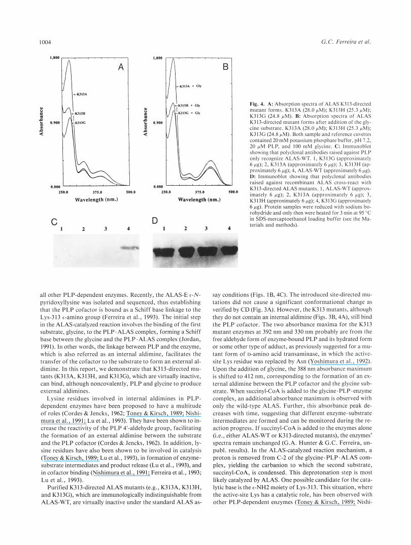
The spectra of the K-3 13 ALAS mutant enzymes indicate that they contain PLP, although the 428-nm peak, characteristic of the ALAS-WT (Ferreira & Dailey, 1993), was shifted to a shorter wavelength (-392 nm) (Fig. 4A). The absorbance maxima at 428 and 392 nm for the wild-type and mutant enzymes, respec- tively, correspond to the internal aldimine in the wild-type en- zyme (Ferreira & Dailey, 1993) and the free aldehyde form in the mutant enzymes (Fig. 4A). Of interest, there is an absorp- tion maximum at 412 nm upon the addition of the glycine sub- strate as with the wild-type enzyme (Fig. 4B). This suggests that an external aldimine can be formed between the substrate and the PLP cofactor both in the wild-type ALAS and K313- ALAS mutant enzymes. In contrast, however, the addition of succinyl- CoA to the PLP-glycine .enzyme complex leads to the appear-
4 ~-
t
AE
' l
/ i -1 L L " ~2- L
300 Wavelength[nm] 500
Fig. 3. A: CD spectra of ALAS enzymes in the far UV region. ALAS- WT (curve 1); K313A (curve 2); K313H (curve 3); K313G (curve 4). Spec- tra were recorded at 10 pM protein (for ALAS-WT and K313H), at 19 pM protein (for K313A), and at 6 pM protein (for K313G) in 0.02 M K2HP0,, 20 pM PLP, pH 7.2, as described in the Materials and meth- ods. (Differences in e reflect differences in protein concentration.) B: CD spectra of PLP at the active site of ALAS enzymes. ALAS-WT (curve 1); K313A (curve 2); K313H (curve 3); K313G (curve 4). Spec- tra were recorded at 46.5 pM protein in 0.02 M K2HP04, 20 pM PLP, pH 7.2, as described in the Materials and methods.
ance of a second absorbance maximum (at 510 nm) with the wild-type but not with the K313 mutant enzymes. Significantly, the 5 10-nm absorbance maximum decreases with the progress of the reaction (G.A. Hunter & G.C. Ferreira, unpubl. results). The purified K313 mutants were immunochemically indistin- guishable from ALAS-WT when examined by immunoblot anal- ysis (Fig. 4D). However, polyclonal antibodies raised against PLP recognized ALAS-WT but not the K313-directed ALAS mutants. Upon reduction of the Schiff base linkage, the PLP cofactor remained covalently bound to ALAS-WT, even dur- ing SDS-PAGE (Kittler et al., 1986). Because the K313 ALAS mutants did not form a Schiff base linkage with PLP, the co- factor was not covalently bound to the enzyme and was disso- ciated from the protein during SDS-PAGE (Fig. 4C).
Discussion
In ALAS, the PLP cofactor is bound to an active-site lysyl res- idue through a Schiff base linkage, as has been reported for
G. C. Ferreira et al.
A
250.0 375.0 500.0
W a v e l e n g t h (nm.)
C 1 2 3 4
210.0 375.0 5w.o
W a v e l e n g t h (nm.)
D l 2 3 4
Fig. 4. A: Absorption spectra of ALAS K313-directed mutant forms. K313A (28.0 FM); K313H (25.3 pM); K313G (24.8 PM). R: Absorption spectra of ALAS K313-directed mutant forms after addition of the gly- cine substrate. K313A (28.0 PM); K313H (25.3 PM); K313G (24.8 PM). Both sample and reference cuvettes contained 20mM potassium phosphate buffer, pH 7.2, 20 pM PLP, and 1 0 0 mM glycine. C: lmmunoblot showing that polyclonal antibodies raised against PLP only recognize ALAS-WT. I , K313C (approximately 6 pg); 2, K313A (approximately 6 pg); 3, K313H (ap- proximately 6 pp); 4, ALAS-WT (approximately 6 pg). D: lmmunoblot showing that polyclonal antibodies raised against recombinant ALAS cross-react with K313-directed ALAS mutants. 1, ALAS-WT (approx- imately 6 pp); 2, K313A (approximately 6 pg); 3, K313H (approximately 6 pg): 4, K313G (approximately 6 pp). Protein samples were reduced with sodium bo- rohydride and only then were heated for 3 min at 95 “C in SDS-mercaptoethanol loading buffer (see the Ma- terials and methods).
all other PLP-dependent enzymes. Recently, the ALAS-E E-N- pyridoxyllysine was isolated and sequenced, thus establishing that the PLP cofactor is bound as a Schiff base linkage to the Lys-313 +amino group (Ferreira et al., 1993). The initial step in the ALAS-catalyzed reaction involves the binding of the first substrate, glycine, to the PLP.ALAS complex, forming a Schiff base between the glycine and the PLP.ALAS complex (Jordan, 1991). In other words, the linkage between PLP and the enzyme, which is also referred as an internal aldimine, facilitates the transfer of the cofactor to the substrate to form an external al- dimine. In this report, we demonstrate that K313-directed mu- tants (K313A, K313H, and K313G), which are virtually inactive, can bind, although noncovalently, PLP and glycine to produce external aldimines.
Lysine residues involved in internal aldimines in PLP- dependent enzymes have been proposed to have a multitude of roles (Cordes & Jencks, 1962; Toney & Kirsch, 1989; Nishi- mura et al., 1991; Lu et al., 1993). They have been shown to in- crease the reactivity of the P L P 4”aldehyde group, facilitating the formation of an external aldimine between the substrate and the PLP cofactor (Cordes & Jencks, 1962). In addition, ly- sine residues have also been shown to be involved in catalysis (Toney & Kirsch, 1989; Lu et al., 1993), in formation of enzyme- substrate intermediates and product release (Lu et al., 1993), and in cofactor binding (Nishimura et al., 1991; Ferreira et al., 1993; Lu et al., 1993).
Purified K313-directed ALAS mutants (e.g., K313A, K313H, and K3 13G), which are immunologically indistinguishable from ALAS-WT, are virtually inactive under the standard ALAS as-
say conditions (Figs. IB, 4C). The introduced site-directed mu- tations did not cause a significant conformational change as verified by CD (Fig. 3A). However, the K313 mutants, although they do not contain an internal aldimine (Figs. 3B, 4A), still bind the P L P cofactor. The two absorbance maxima for the K313 mutant enzymes at 392 nm and 330 nm probably are from the free aldehyde form of enzyme-bound PLP and its hydrated form or some other type of adduct, as previously suggested for a mu- tant form of D-amino acid transaminase, in which the active- site Lys residue was replaced by Asn (Yoshimura et al., 1992). Upon the addition of glycine, the 388 nm absorbance maximum is shifted to 412 nm, corresponding to the formation of an ex- ternal aldimine between the PLP cofactor and the glycine sub- strate. When succinyl-CoA is added to the glycine.PLP.enzyme complex, an additional absorbance maximum is observed with only the wild-type ALAS. Further, this absorbance peak de- creases with time, suggesting that different enzyme-substrate intermediates are formed and can be monitored during the re- action progress. If succinyl-CoA is added to the enzymes alone (i.e., either ALAS-WT or K313-directed mutants), the enzymes’ spectra remain unchanged (G.A. Hunter & G.C. Ferreira, un- publ. results). In the ALAS-catalyzed reaction mechanism, a proton is removed from C-2 of the glycine.PLP.ALAS com- plex, yielding the carbanion to which the second substrate, succinyl-CoA, is condensed. This deprotonation step is most likely catalyzed by ALAS. One possible candidate for the cata- lytic base is the e-NH2 moiety of Lys-313. This situation, where the active-site Lys has a catalytic role, has been observed with other PLP-dependent enzymes (Toney & Kirsch, 1989; Nishi-

Lys-313 mutants of aminolevulinate synthase 1005
mura et al., 1991; Lu et al., 1993). For example, the €-amino group of Lys-258 of L-aspartate aminotransferase (i.e., the active-site Lys) catalyzes the proton transfer in the tautomeriza- tion of the external aldimine to ketimine (Kirsch et al., 1984; Toney & Kirsch, 1989). Kirsch and coworkers (1990) demon- strated that primary amines functionally replace the PLP- binding Lys by catalyzing both the 1,3 prototropic shift and external aldimine hydrolysis reactions with inactive L-aspartate aminotransferase K258A. Interestingly, the substitution of ALAS K313 with histidine (K313H) yields an inactive enzyme, although at optimum pH the histidine residue is probably un- protonated. This suggests that the mutation affected the reac- tivity of the enzyme by either altering the conformation of the active site or altering the positioning of the PLP cofactor. How- ever, the actual protonation state of the histidine residue in the active-site microenvironment is presently unknown.
In summary, although substitution of Lys-313 by Ala, His, and Gly did not prevent PLP binding or the formation of an ex- ternal aldimine with glycine, the mutant enzymes were essentially inactive. ALAS has the P L P cofactor tightly bound to the pro- tein through both noncovalent interactions and a covalent link- age with the c-amino group of a lysine residue at the active site. Because K313A, K313H, and K313G, which are virtually inac- tive, can produce external aldimines, this suggests a catalytic role for Lys-313. Future experiments on ALAS-E will be directed at determining the functional role(s) of Lys-313.
Materials and methods
Restriction enzymes were obtained from New England Biolabs and Boehringer Mannheim and were used according to the suppliers' instructions. Sequenase was from United States Bio- chemicals; T4 DNA ligase was purchased from New England Biolabs. Deoxy- and dideoxynucleotide triphosphates were from United States Biochemicals. [35S]daATP was from ICN. The oligonucleotide-directed in vitro mutagenesis kit was purchased from Amersham. Geneclean 11 kit was a product of Bio 101 Inc. Acrylamide and gel reagents were purchased from Bio-Rad. Fast stain was from Zoion Research. The ProtoBlotR Western Blot A P system was from Promega. Nitrocellulose (0.45 pm) and Affi-Gel Protein A agarose were from Bio-Rad. The bicincho- ninic acid protein assay reagents were obtained from Pierce Chemical Co. DEAE-Sephacel was obtained from Sigma Chem- ical Co. and Ultrogel Aca 44 was from IBF Biotechnics Inc. All other chemicals were of the highest purity available.
Oligonucleotide-directed mutagenesis
Lysine-313, the amino acid involved in the Schiff base link- age of the PLP cofactor, was mutated to alanine, histidine, and glycine as previously described (Ferreira et al., 1993). Oligonucleotide-directed mutagenesis, using M13mp18 as the cloning vector, was carried out essentially as described in the Amersham oligonucleotide-directed in vitro mutagenesis kit directions. The Lys-3 13 mutations were verified by sequencing according to the dideoxy chain termination method (Sanger et al., 1977; Sambrook et al., 1989). The mutated ALAS DNA- encoding fragments were then subcloned in the ALAS expres- sion plasmid, pGF23, replacing the wild-type region (Ferreira & Dailey, 1993; Ferreira et al., 1993).
Purification of K313A, K313H, and K313G
Wild-type and mutant forms of ALAS were purified from ex- tracts of a host E. coli overproducing strain as described for the wild-type enzyme in Ferreira and Dailey (1993). Briefly, DH5a cells harboring the different expression plasmids (i.e., for K313A, K313H, K313G, and wild-type ALAS) were grown in low phosphate medium (MOPS) containing 100 pg/mL ampi- cillin at 37 "C for 20 h. The alkaline phosphatase promoter, which controls the expression of ALAS and the ALAS K313- mutants, is induced by starvation in phosphate. Cells were then harvested by centrifugation. The purification procedure involves lysis of cells in a French press cell, precipitation of ALAS as well as of its mutant variants with ammonium sul- fate, gel-filtration (Ultrogel AcA U), and ion-exchange (DEAE- Sephacel) chromatographies.
Spectroscopic methods: CD and UV-visible spectra
Wild-typeALAS, K313A, K313H, andK313G wereconcentrated in an Amicon stirred cell with a YMlO membrane. Denatured protein was removed by centrifugation and the absorption spec- tra were determined on the supernatant solutions. CD spectra were recorded on a Jasco model 710 spectropolarimeter at 25 "C using a cuvette of a pathlength of 1 cm for 500-300 nm or 0.1 cm for 270-200 nm spectra. The observed rotation in degrees (eobs) was converted to molar ellipticity. Absorption spectra and difference absorption spectra were recorded using a Shimadzu UV 2100U spectrophotometer.
Preparation of antibodies and purification of IgG fractions
Polyclonal antibodies were raised against ALAS and against PLP. Two rabbits were immunized with purified ALAS apoen- zyme and two with the pyridoxyl-BSA complex. Additional in- jections of the ALAS antigen were made 2 and 7 weeks after the first injection. The pyridoxyl-BSA complex was prepared by adding P L P (10 mM) to BSA (3 mg/mL) in a final volume re- action of 3 mL. After stirring for 15 min at 4 "C, small amounts of sodium borohydride were added and stirring was continued for 15-30 min. To determine the extension of the derivatization, absorbance at 330 nm was measured. The pyridoxyl-BSA com- plex was dialyzed extensively against 20 mM potassium phos- phate, pH 7.2. After 1 month of pyridoxyl-lysine complex injections, the two rabbits were injected with a PLP-Po~Y-L- lysine complex antigen. The PLP-poly-lysine antigen was pre- pared by adding PLP (3.5 mg/mL) to poly-lysine (0.85 mg/mL) in a final volume reaction of 30 mL. After stirring for 1 h at 4 "C, sodium borohydride (100 mg) was added very slowly, with constant stirring, until the P L P yellow color disappeared. The pyridoxyl-lysine complex was then extensively dialyzed against 4 L of distilled water. Preimmune sera were drawn before the first injection. The immunization and sera drawing procedures were carried out at Spring Valley Laboratories, Maryland. Anti- sera titers were performed every 3-4 weeks for 6 months, using standard enzyme-linked immunosorbent assay (Ausubel et al., 1992). To purify the IgC fractions of the ALAS and PLP poly- clonal antibodies, Affi-gel protein A agarose (Bio-Rad) chro- matography was performed as described in the manufacturer's instructions.

1006 G. C. Ferreira et al.
SDS-PAGE, immunoblot analysis, and protein determination
SDS-PAGE was performed as described by Laemmli (1970); 15% acrylamide and 1.5-mm-thick gels were used. Aliquots of the protein samples (10-15 pL) were heated for 3 min at 95 “C in SDS-mercaptoethanol loading buffer (Laemmli, 1970) and the proteins were visualized upon fast-staining. For immunoblot analysis, SDS-PAGE was carried out as described above and the electrophoretic transfer to nitrocellulose filters according to Towbin et al. (1979) in a Bio-Rad mini Trans-Blot cell filled with 20% (v/v) methanol, 192 mM glycine, and 25 mm Tris. Anti- bodies were used at a dilution of 1500. The blots were incubated with the antibodies for 1 h at room temperature. The binding of the antibodies to ALAS or to PLP on the nitrocellulose fil- ters was detected using the ProtoBlotR AP (from Promega), as recommended by the manufacturer. Briefly, the incubation with the anti-IgG second antibody conjugated with alkaline phospha- tase (15,OOO dilution) was for 30 min and the “Western blue sta- bilized substrate for alkaline phosphatase” was used as provided by Promega. Protein concentrations were determined by the bi- cinchoninic acid assay, using BSA as the standard.
Enzyme assay and PLP determination
ALAS activity was determined according to the method of Lien and Beattie (1982). One unit of activity in any reaction corre- sponds to the formation of 1 nmol of ALA product in 60 min at 37 “C. PLP was determined by the fluorimetric method of Adams (1979).
Acknowledgments
This work was supported by the National Science Foundation (MCB- 9206574 to G.C.F.) and the National Institutes of Health (GM 32696 to M.J.B.). G.C.F. is an ACS Junior Faculty Scholar (JFRA-401) and a recipient of an NSF Young Investigator Award (MCB-9257656). We thank Dr. A. Iriarte (University of Missouri, Kansas City, Missouri) for the procedure and advice on the preparation of polyclonal antibodies against phosphopyridoxyl derivatives.
References
Adams E. 1979. Fluorimetric determination of pyridoxal phosphate in en- zymes. Methods Enzymol62:407-410.
Ausubel FM, Brent R, Kingston RE, Moore DD, Seidman JG, Smith JA, Struhl K. 1992. Currentprotocols in molecular biology. New York: John
Bishop DF, Henderson AS, Astrin KH. 1990. Human 5-aminolevulinate syn- Wiley & Sons, Inc. pp 11.2.1-1 1.2.2.
thase: Assignment of the housekeeping gene to 3p21 and the erythroid- specific gene to the X chromosome. Genomics 7:207-214.
Borthwick IA, Srivastava G, Pirola BA, May BK, Elliott WH. 1986. Puri- fication of hepatic mitochondrial 5-aminolevulinate synthase. Methods
Cordes EH, Jencks WP. 1962. Semicarbazone formation from pyridoxal, Enzymol123:395-401.
Cox TC, Bawden MJ, Abraham NG, Bottomley SS, May BK, Baker E, Chen pyridoxal phosphate, and their Schiff bases. Biochemisrry 1:773-778.
LZ, Sutherland CR. 1990. Erythroid 5-aminolevulinate synthase is lo- cated on the X chromosome. A m J Hum Genet 46:107-111.
Ferreira GC, Dailey HA. 1993. Expression of mammalian 5-aminolevulinate synthase in Escherichia coli. Overproduction, purification and charac- terization. J Biol Chem 268:584-590.
Ferreira GC, Neame PJ, Dailey HA. 1993. Heme biosynthesis in mamma- lian systems: Evidence of a Schiff base linkage between the pyridoxal 5”phosphate cofactor and a lysine residue in 5-aminolevulinate synthase. Protein Sci 2:1959-1965.
Jordan PM. 1991. Biosynthesis of 5-aminolevulinic acid and its transforma- tion into uroporphyrinogen I11 in animals and bacteria. In: Jordan PM, ed. Biosynthesis of tetrapyrroles. Amsterdam: Elsevier. pp 1-66.
Kikuchi G, Kumar A, Talmage P, Shemin D. 1958. The enzymatic synthe- sis of 6-aminolevulinic acid. J Biol Chem 233:1214-1219.
Kirsch JF, Eichele G, Ford GC, Vincent MC, Jansonius JN. 1984. Mecha- nism of aspartate aminotransferase proposed on the basis of its spatial structure. J Mol Biol 174:497-525.
Kirsch JF, Toney MD, Goldberg JM. 1990. Chemical rescue and change in rate-determining steps elicited by site-directed mutagenesis probes of as- partate aminotransferase. In: Craik CS, Fletterick RF, Matthews CR. Wells V, eds. Protein andpharmaceuticalengineering. New York. Wiley- Liss. pp 105-118.
Kittler JM, Viceps-Madore D, Cidlowski JA, Meisler NT, and Thanassi JW. 1986. Monoclonal antibodies to vitamin B6. Methods Enzymol 122: 120-127.
Laemmli UK. 1970. Cleavage of structural proteins during the assembly of the head of bacteriophage T4. Nature 227:680-685.
Lien LF, Beattie DS. 1982. Comparisons and modifications of the colori-
Lu 2, Nagata S, McPhie P, Miles EW. 1993. Lysine 87 in the subunit of tryp- metric assay for delta-aminolevulinic acid synthase. Entyme28:120-132.
tophan synthase that forms an internal aldimine with pyridoxal phos- phate serves critical roles in transamination, catalysis, and product release. J Biol Chem 268:8727-8734.
Munakata H, YamagamiT, NagaiT, Yamamoto M, Hayashi N. 1993. Pu- rification and structure of rat erythroid-specific 6-aminolevulinate syn- thase. JBiochem 114:103-111.
Nakakuki M, Amici K, Hayashi N, Kikuchi G. 1980. Purification and some properties of 6-aminolevulinate synthase from the rat liver cytosol frac-
tochondrial enzyme. J Biol Chem 255:1738-1745. tion and immunochemical identity of the cytosolic enzyme and the mi-
Neidle EL, Kaplan S. 1993. Expression of the Rhodobactersphaeroides hemA and hemT genes, encoding two 5-aminolevulinic acid synthase isozymes. J Bacteriol I75:2292-2303.
Nishimura K, Tanizawa K , Yoshimura T, Esaki N, Futaki S, Manning JM, Soda K. 1991. Effect of substitution of a lysyl residue that binds pyri- doxal phosphate in thermostable D-amino acid aminotransferase by ar-
Riddle RD. Yamamoto M, Engel JD. 1989. Expression of 6-aminolevulinate ginine and alanine. Biochemistry 30:4072-4077.
synthase in avian cells: Separate genes encode erythroid-specific and non- specific isoenzymes. Proc Nut1 Acad Sci USA 8:792-796.
Sambrook J, Fritsch EF, Maniatis T. 1989. Molecular cloning: A laboratory manual. Cold Spring Harbor, New York: Cold Spring Harbor Labora-
Sanger F, Nicklen S, Coulson AR. 1977. DNA sequencing with chain- tory Press.
terminating inhibitors. Proc Nut1 Acad Sci USA 74:5463-5467. Smith DL, Almo SC, Toney MD, Ringe D. 1989. 2.8-A resolution crystal
structure of an active-site mutant of aspartate aminotransferase from Escherichia coli. Biochemistry 28:8161-8167.
Toney MD, Kirsch JF. 1989. Direct Bransted analysis of the restoration of activity to a mutant enzyme by exogenous amines. Science 243:1485- 1488.
Towbin H, Staehelin T, Gordon J. 1979. Electrophoretic transfer of proteins
applications. Proc Nut1 Acad Sri USA 76:4350-4354. from polyacrylamide gels to nitrocellulose sheets: Procedure and some
Warnick GR, Burnham BF. 1971. Regulation of porphyrin biosynthesis. Pu- rification and characterization of 6-aminolevulinic acid synthase. J Biol Chem 246:6880-6885.
Yoshimura T, Bhatia MB, Manning JM, Ringe D, Soda K . 1992. Partial
stituted for the lysine that binds coenzyme pyridoxal 5’-phosphate. Bio- reactions of bacterial D-amino acid transaminase with asparagine sub-
chemistry 31:11748-11754.