Altered resting brain function and structure in professional badminton players
9
Altered Resting Brain Function and Structure in Professional Badminton Players Xin Di, 1,2 Senhua Zhu, 2,3 Hua Jin, 1 Pin Wang, 1 Zhuoer Ye, 1 Ke Zhou, 4 Yan Zhuo, 4 and Hengyi Rao 1–3 Abstract Neuroimaging studies of professional athletic or musical training have demonstrated considerable practice- dependent plasticity in various brain structures, which may reflect distinct training demands. In the present study, structural and functional brain alterations were examined in professional badminton players and com- pared with healthy controls using magnetic resonance imaging (MRI) and resting-state functional MRI. Gray mat- ter concentration (GMC) was assessed using voxel-based morphometry (VBM), and resting-brain functions were measured by amplitude of low-frequency fluctuation (ALFF) and seed-based functional connectivity. Results showed that the athlete group had greater GMC and ALFF in the right and medial cerebellar regions, respectively. The athlete group also demonstrated smaller ALFF in the left superior parietal lobule and altered functional con- nectivity between the left superior parietal and frontal regions. These findings indicate that badminton expertise is associated with not only plastic structural changes in terms of enlarged gray matter density in the cerebellum, but also functional alterations in fronto-parietal connectivity. Such structural and functional alterations may reflect specific experiences of badminton training and practice, including high-capacity visuo-spatial processing and hand-eye coordination in addition to refined motor skills. Key words: amplitude of low frequency fluctuation; badminton athlete; cerebellum, fronto-parietal network; func- tional connectivity; voxel-based morphometry Introduction T he human brain shows remarkable plasticity in re- sponse to learning and experience, even after a relatively short time of training. For example, after 7 days of juggling training, significant changes in gray matter (GM) intensity could be detected in multiple brain regions, including the fronto-parietal attention network, the middle temporal (MT) area, and the hippocampus (Boyke et al., 2008; Dragan- ski et al., 2004; Driemeyer et al., 2008). Skilled professionals with multiple years of training and practice, such as athletes and musicians, have also demonstrated significant changes in multiple brain regions (for reviews, see Nakata et al., 2010; Schlaug, 2001; Yarrow et al., 2009). For example, the cerebel- lum has been shown to be larger in musicians (Gaser and Schlaug, 2003; Han et al., 2009; Hutchinson et al., 2003) and basketball players (Park et al., 2009) compared with normal controls, which may reflect refined motor skills in these pro- fessionals. Additional changes in multiple cortical areas in- volving motor, auditory, and visual-spatial processes have been observed in keyboard musicians (Gaser and Schlaug, 2003; Han et al., 2009), which may be associated with more subtle and complex patterns of finger dexterity in music prac- tice. GM alterations in sensorimotor and premotor regions have also been revealed in professional divers (Wei et al., 2009) and ballet dancers (Ha ¨ nggi et al., 2010), while addi- tional parietal changes have been found in skilled golfers, which may reflect more attention and motor control support- ing golf practice ( Ja ¨ ncke et al., 2009). These diverse findings from athletes and musicians suggest that brain structure changes observed in professionals may depend on the spe- cific motor and cognitive processes involved in skill training over time. Compared with structural changes in skilled professionals, evidence supporting functional brain alterations in elite ath- letes or musicians is relatively limited. Using functional mag- netic resonance imaging (fMRI), two recent studies examined neural activations during preshot routines of expert golfers 1 Center for Studies of Psychological Application, South China Normal University, Guangzhou, China. 2 Department of Psychology, Sun Yat-Sen University, Guangzhou, China. 3 Department of Neurology, Center for Functional Neuroimaging, University of Pennsylvania, Philadelphia, Pennsylvania. 4 State Key Laboratory of Brain and Cognitive Science, Institute of biophysics, Chinese Academy of Sciences, Beijing, China. BRAIN CONNECTIVITY Volume 2, Number 4, 2012 ª Mary Ann Liebert, Inc. DOI: 10.1089/brain.2011.0050 225
Transcript of Altered resting brain function and structure in professional badminton players
Altered Resting Brain Function and Structurein Professional Badminton Players
Xin Di,1,2 Senhua Zhu,2,3 Hua Jin,1 Pin Wang,1 Zhuoer Ye,1 Ke Zhou,4 Yan Zhuo,4 and Hengyi Rao1–3
Abstract
Neuroimaging studies of professional athletic or musical training have demonstrated considerable practice-dependent plasticity in various brain structures, which may reflect distinct training demands. In the presentstudy, structural and functional brain alterations were examined in professional badminton players and com-pared with healthy controls using magnetic resonance imaging (MRI) and resting-state functional MRI. Gray mat-ter concentration (GMC) was assessed using voxel-based morphometry (VBM), and resting-brain functions weremeasured by amplitude of low-frequency fluctuation (ALFF) and seed-based functional connectivity. Resultsshowed that the athlete group had greater GMC and ALFF in the right and medial cerebellar regions, respectively.The athlete group also demonstrated smaller ALFF in the left superior parietal lobule and altered functional con-nectivity between the left superior parietal and frontal regions. These findings indicate that badminton expertise isassociated with not only plastic structural changes in terms of enlarged gray matter density in the cerebellum, butalso functional alterations in fronto-parietal connectivity. Such structural and functional alterations may reflectspecific experiences of badminton training and practice, including high-capacity visuo-spatial processing andhand-eye coordination in addition to refined motor skills.
Key words: amplitude of low frequency fluctuation; badminton athlete; cerebellum, fronto-parietal network; func-tional connectivity; voxel-based morphometry
Introduction
The human brain shows remarkable plasticity in re-sponse to learning and experience, even after a relatively
short time of training. For example, after 7 days of jugglingtraining, significant changes in gray matter (GM) intensitycould be detected in multiple brain regions, including thefronto-parietal attention network, the middle temporal(MT) area, and the hippocampus (Boyke et al., 2008; Dragan-ski et al., 2004; Driemeyer et al., 2008). Skilled professionalswith multiple years of training and practice, such as athletesand musicians, have also demonstrated significant changes inmultiple brain regions (for reviews, see Nakata et al., 2010;Schlaug, 2001; Yarrow et al., 2009). For example, the cerebel-lum has been shown to be larger in musicians (Gaser andSchlaug, 2003; Han et al., 2009; Hutchinson et al., 2003) andbasketball players (Park et al., 2009) compared with normalcontrols, which may reflect refined motor skills in these pro-fessionals. Additional changes in multiple cortical areas in-
volving motor, auditory, and visual-spatial processes havebeen observed in keyboard musicians (Gaser and Schlaug,2003; Han et al., 2009), which may be associated with moresubtle and complex patterns of finger dexterity in music prac-tice. GM alterations in sensorimotor and premotor regionshave also been revealed in professional divers (Wei et al.,2009) and ballet dancers (Hanggi et al., 2010), while addi-tional parietal changes have been found in skilled golfers,which may reflect more attention and motor control support-ing golf practice ( Jancke et al., 2009). These diverse findingsfrom athletes and musicians suggest that brain structurechanges observed in professionals may depend on the spe-cific motor and cognitive processes involved in skill trainingover time.
Compared with structural changes in skilled professionals,evidence supporting functional brain alterations in elite ath-letes or musicians is relatively limited. Using functional mag-netic resonance imaging (fMRI), two recent studies examinedneural activations during preshot routines of expert golfers
1Center for Studies of Psychological Application, South China Normal University, Guangzhou, China.2Department of Psychology, Sun Yat-Sen University, Guangzhou, China.3Department of Neurology, Center for Functional Neuroimaging, University of Pennsylvania, Philadelphia, Pennsylvania.4State Key Laboratory of Brain and Cognitive Science, Institute of biophysics, Chinese Academy of Sciences, Beijing, China.
BRAIN CONNECTIVITYVolume 2, Number 4, 2012ª Mary Ann Liebert, Inc.DOI: 10.1089/brain.2011.0050
225
(Milton et al., 2007) and archers (Kim et al., 2008). The resultsdemonstrated that professionals tended to recruit a more fo-cused and efficient organization of visual and motor net-works directly related to task requirements, whereasnovices tended to engage more brain regions related tohigh-level cognitive control functions. However, findingsfrom these task-related fMRI studies depend heavily on thetask demands and specific cognitive or motor paradigmsused in the studies, which makes the comparison of resultsacross studies difficult.
The use of resting-state functional connectivity fMRI (fc-fMRI) that examines functional brain changes have beenemerging since the first report of functional connectivity be-tween bilateral motor cortices (Biswal et al., 1995). Resting-state fc-fMRI measures the synchronization of low-frequencyblood oxygen level dependent (BOLD) oscillations betweenspatially distinct brain regions in the absence of tasks. There-fore, it provides a complimentary method to the conventionaltask-related fMRI for detecting functional brain changes asso-ciated with training and experience. Since resting-state fc-fMRIdoes not involve any particular motor or cognitive task re-quirements, it may reflect a cumulative effect of experienceover time (Lewis et al., 2009). Recent fc-fMRI studies have sug-gested that motor training altered resting-state brain activity(Xiong et al., 2009; Albert et al., 2009). For example, Albertet al. (2009) examined resting-state functional brain changesduring motor training using independent component analysis(ICA) and found that motor learning, but not motor perfor-mance, significantly modulated resting-state functional con-nectivity in the fronto-parietal and cerebellar networks.However, to our knowledge, the effects of long-term trainingon resting-state brain function remain largely unknown.
In this study, we used MRI to examine both structural andresting-state functional brain alterations in professional rac-quet players, specifically badminton players. To the best ofour knowledge, structural and functional characteristics ofthe badminton players’ brain have not been explored usingneuroimaging methods. Similar to other racquet sports, bad-minton requires refined hand-eye coordination and visuo-spatial ability (Ward et al., 2002; Shim et al., 2005), whichmay result in structural and functional brain changes thatare different from other sports that have been studied, for ex-ample, golf, diving, or basketball. We predict that profes-sional badminton players will show significant structuraland functional alterations in brain regions related to visuo-spatial processing and hand-eye coordination. A group ofprofessional badminton players and matched nonathlete con-trols were recruited in the present study. Standard voxel-based morphometry (VBM) (Ashburner and Friston, 2000)was first conducted to examine the structural differences be-tween the two groups. The amplitudes of low-frequency fluc-tuations (ALFF; Zang et al., 2007) of resting-state fMRI werethen used to measure regional properties of the brain’s intrin-sic neural activity and compared between the two groups.Finally, the seed-based functional connectivity analyses (Foxet al., 2005) of resting-state fMRI were conducted to character-ize functional integration, using regions that have been shownto be different between professional players and controls fromthe VBM and ALFF analyses. We chose to use the hypothesis-driven, seed-based functional connectivity analysis instead ofdata-driven methods such as ICA in order to determine thespecific brain regions and networks that are related to culti-
vated visuo-spatial processing and hand-eye coordinationskills after long-term badminton training and practice.
Methods
Subjects
Twenty badminton players (10 males) were recruited forthe study. All subjects were members of professional teamsor professional university teams in Beijing and had completedat least 3 years of badminton training (mean = 8.9 years, rangefrom 3 to 16 years). Subjects had either completed college-level education or were current college students during thestudy. The control group consisted of 18 age- and gender-matched college students (9 men). No control subjects hadformal training or experience in badminton or other racquetsports (Table 1). All athletes and controls were right handed.Written informed consent was obtained from all subjects atthe beginning of the study.
MRI scan
All subjects were scanned using a Siemens 3T Trio scannerlocated in the Beijing Center for Magnetic Resonance Imaging,Beijing, China. Subjects first completed a task-independent,resting-state scan, during which time they were asked toopen their eyes to look at a blank screen. No additional taskwas required. The resting scan lasted 6 min, with a TR of2 sec. As a result, 180 images were obtained for each subject.BOLD images were obtained using an echo-planar imaging se-quence with a 12-channel coil. Acquisition parameters were asfollows: TE = 30 ms; TR = 2000 ms; flip angle = 90o; 32 inter-leaved axial slices; slice thickness = 4 mm, no gap; image ma-trix = 64 · 64; FOV = 200 · 200 mm; bandwidth = 2232. At theend of the scan, a high spatial resolution, T1-weighted magne-tization prepared rapid gradient-echo (MP-RAGE) structuralimage was obtained. Scan parameters were as follows: TR =2530 ms; TE = 1.64; flip angle = 7.0; 176 slices; slice thick-ness = 1 mm; image matrix = 256 · 256; FOV = 256 · 256 mm.
VBM analysis
Standard VBM analysis was conducted using SPM8 soft-ware (www.fil.ion.ucl.ac.uk/spm/, Wellcome Departmentof Cognitive Neurology, UK, implemented in Matlab7.7, Math Works, Natick, MA). For each subject, the high-resolution structural image was first segmented into differentbrain tissue types using a standard template provided in theSPM software. The unified segmentation model implementedin SPM8 (Ashburner and Friston, 2005) was used to segmentthe structural image into GM, white matter (WM), and cere-brospinal fluid (CSF) in standard Montreal NeurologicalInstitute (MNI) space. The tissue probability template wasthe modified version of the International Consortiumfor Brain Mapping tissue probabilistic atlases, which was
Table 1. Information for the Two Groups of Subjects
Athletes Controls Statistics
No. of subjects (male) 20 (10) 18 (9)Year of age (STD) 22.5 (4.57) 20.7 (4.25) p = 0.234Years of badminton
training8.85 (3.25) 0
226 DI ET AL.

provided by the SPM8 new segment routine. We used thenormalized GM images that were not modulated and repre-sented gray matter concentration (GMC). For computationalsimplicity, the image was re-sampled at a resolution of1.5 · 1.5 · 1.5 mm3 in the new segmentation step. The result-ing images were visually checked to make sure there wereno misclassifications. Finally, the GMC images weresmoothed using a Gaussian filter with a 6-mm full width athalf maximum (FWHM) kernal.
Preprocessed GMC images were then entered into a sec-ond-level, two-sample t-test model to compare group leveldifferences. Total Gray matter volume (GMV), gender, andage were added to the model as covariates. Since the smooth-ness of GMC images is nonstationary (Ashburner and Friston,2000), nonstationary cluster extent correction was adopted tocorrect multiple comparisons (Worsley et al., 1999). The am-plitude threshold was first set at p < 0.001, and then, the clus-ter extent was corrected at p < 0.05 using the nonstationaritytoolbox (Hayasaka et al., 2004). The clusters identified inthe group analysis were labeled using the Talairach Daemon(Talairach and Tournoux, 1988; Lancaster et al., 2000) after ac-counting for the discrepancy between MNI space and Talair-ach space (Lancaster et al., 2007). This analysis revealed twoclusters in the right cerebellum that were defined as theseed regions for further functional connectivity analysis.
Resting-state fMRI data preprocessing
Preprocessing of resting-state functional images was com-pleted using SPM8. First, the two initial resting-state imageswere discarded for each subject, leaving a total of 178 imagesper subject. All functional images were motion correctedusing the realign function. Functional images from two athletesand two controls were discarded, because at least one of themotion parameter estimates was larger than 3 mm or 3 de-
grees. As a result, images from 18 athletes and 16 controlswere included in the subsequent analysis. For each subject,the functional images were first registered to the subject’sown high-resolution structural image. The structural imagewas then normalized to the T1 template. Next, all functionalimages were normalized to the standard MNI space usingthe parameters obtained from normalizing the subject’s struc-tural images. Finally, all normalized functional images weresmoothed using a Gaussian filter with an 8 mm FWHM kernal.
ALFF analysis
We used the ALFF to index regional properties of thebrain’s intrinsic neural activity. ALFF analysis was completedusing the resting-state fMRI data analysis toolkit, (REST) v1.4(www.restfmri.net/forum/REST_V1.4, State Key Laboratoryof Cognitive Neuroscience and Learning, Beijing, China; Songet al., 2011). For each subject, linear trends were removedfrom all the functional images. An ideal band-pass filter be-tween 0.01 and 0.08 Hz was used to filter the detrended func-tional images. An ALFF map was then calculated for eachsubject. A normalized ALFF map (mALFF) was also obtainedby dividing the whole-brain mean from the original ALFFmap. Finally, the mALFF maps were entered into a second-level, two-sample t-test model to compare group-level differ-ences using SPM8. Subject gender and age were added intothe group model as covariates. An amplitude threshold wasfirst set at p < 0.001, and the cluster extent was corrected atthe false discovery rate, p < 0.05. The clusters identified inthe group level analysis were also labeled using the TalairachDaemon (Lancaster et al., 2000; Talairach and Tournoux,1988) after accounting for the discrepancy between MNIspace and Talairach space (Lancaster et al., 2007). This analy-sis revealed clusters in the medial cerebellum and left parietallobe, which were also defined as the seed regions for furtherfunctional connectivity analysis.
Functional connectivity analysis
Functional connectivity analysis was conducted using thefunctional connectivity toolbox, v.12.p (Chai et al., 2012;web.mit.edu/swg/software.htm). For each subject, fourseed regions were defined, including the posterior cerebellum(Fig. 3A) and the anterior cerebellum area (Fig. 3C) from theVBM analysis, as well as the medial cerebellum (Fig. 3E) andthe left superior parietal lobule (Fig. 3G) from the ALFF anal-ysis. Before the modeling, the fifth-order eigenvector ofWM and the first fifth-order eigenvector of CSF were re-moved from the preprocessed resting-state fMRI imagesto minimize physiological noise that was probably due to
FIG. 1. Larger gray matter concentration in the athletegroup than the control group. Threshold was set as clusterlevel false discovery rate (FDR) corrected p < 0.05. L: left; R:right.
Table 2. Clusters Showed Larger Gray Matter Concentration in the Athlete Group than in the Control Group
MNI coordinates
Regions Cluster size Corrected p Peak t x y z
R. Cerebellum, Posterior Lobe, Cerebellar Tonsil 597 0.001 5.394 24 �67 �45R. Cerebellum, Posterior Lobe, Cerebellar Tonsil 4.764 17 �63 �44R. Cerebellum, Anterior Lobe 141 0.022 4.928 24 �54 �30R. Cerebellum, Anterior Lobe, Culmen 3.621 32 �49 �26
Threshold was p < 0.001 with a cluster-level nonstationary correction of p < 0.05.R, right; L, left; MNI, Montreal Neurological Institute.
BADMINTON PLAYER’S BRAIN 227
respiratory and cardiac factors. Six rigid-body head-motionparameters were also regressed out using linear regressionanalyses. The imaging data were also temporally filteredusing a 0.01–0.08 Hz band-pass filter. Voxel-wise correla-tion analysis was then conducted, and a beta contrastmap was generated for each seed region and each subject.These individual seed correlation maps were then enteredinto two types of group-level analyses. The first group-level analysis used one-sample t-tests to determine the spe-cific intrinsic network connected to each of the seed regions.This is an important step, because subdivisions of a brainstructure might be connected to different networks (e.g.,in cerebellum, Habas et al., 2009). The second group-levelanalysis used two-sample t-tests to compare differences be-tween the two groups. Since anti-correlation between thetask-positive network and the default mode network re-mains controversial (Fox et al., 2009; Murphy et al., 2009),we restricted our results within the task-positive networkby applying a positive correlation mask of the same seed re-gions. The masks were defined by using an ‘‘or’’ operationon the two groups’ correlation maps using a threshold ofp < 0.001.
Results
VBM results
The mean total GMV of the athlete group was 699.1 mL(SD = 60.2), while the mean total GMV of the control groupwas 717.7 mL (SD = 59.3). There was no difference in totalGMV observed between the two groups (t =�0.96, p = 0.35).
A voxel-wise comparison revealed greater GMC in tworight cerebellum clusters for the athlete group than the con-trol group (Fig. 1 and Table 2). One cluster was located inthe posterior right cerebellar tonsil (lobule 8), and the otherregion was located in the anterior right cerebellar hemisphere(lobule 6). However, no region showed greater GMC for thecontrol group than the athlete group.
The voxel-wise comparison of GMV showed no differencesbetween the two groups using a threshold of p < 0.05 withnonstationary correction. However, greater GMV in similarcerebellum clusters was observed using a more liberal thresh-old of p < 0.001 and cluster size > 141 voxels, (Table 3).
ALFF results
Larger ALFFs were also found in the cerebellum for theathlete group than the control group (Fig. 2 and Table 4), in-cluding the anterior part of the culmen, and the medial por-tion of the anterior lobe (vermis lobule 6). In contrast, therewas a cluster in the left superior parietal lube (BA 7/19)that revealed larger ALFFs in the control group than the ath-lete group.
Functional connectivity analysis
Functional connectivity analysis for all subjects revealedthat seeding regions belonged to distinct functional networks(Fig. 3). The right posterior cerebellum seed showed positivefunctional connectivity to the left dorsal-lateral prefrontal cor-tex, left parietal cortex, and left temporal lobe (Fig. 3B); whilethe right anterior cerebellum and the medial part of the
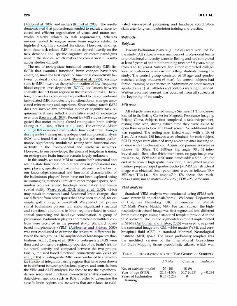
Table 3. Clusters Showed Larger Gray Matter Volume in the Athlete Group
than in the Control Group (R: right; L: left)
MNI coordinates
Area # voxels corrected p Peak t x y z
R. Cerebellum, Posterior Lobe 274 0.008 4.71 24 �64 �44R. Cerebellum, Anterior Lobe 149 0.031 4.68 23 �52 �32R. Cerebellum, Anterior Lobe 3.91 24 �42 �32L. Cerebellum, Posterior Lobe 212 0.016 4.31 �27 �78 �36L. Cerebellum, Posterior Lobe 3.74 �30 �81 �27R. Superior Temporal Gyrus, BA 41 182 0.021 4.19 50 �37 4R. Superior Temporal Gyrus, BA 13 4.09 42 �28 3
Threshold was p < 0.001 with a cluster extent of 141 voxels. The clusters in bold corresponded to the clusters reported in Gray matter con-centration analysis (Table 2).
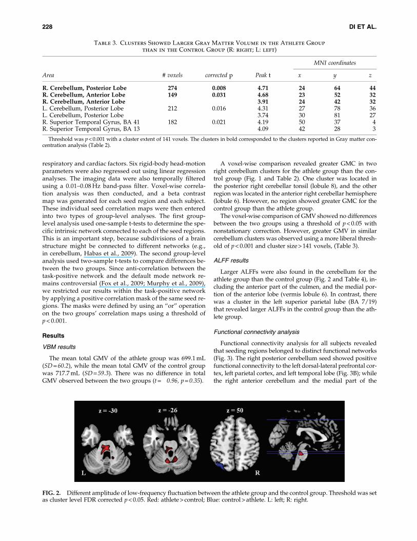
FIG. 2. Different amplitude of low-frequency fluctuation between the athlete group and the control group. Threshold was setas cluster level FDR corrected p < 0.05. Red: athlete > control; Blue: control > athlete. L: left; R: right.
228 DI ET AL.
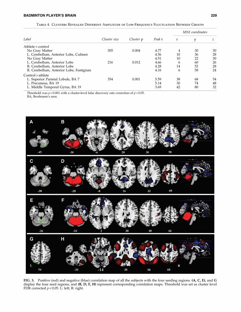
FIG. 3. Positive (red) and negative (blue) correlation map of all the subjects with the four seeding regions. (A, C, E), and Gdisplay the four seed regions, and (B, D, F, H) represent corresponding correlation maps. Threshold was set as cluster levelFDR corrected p < 0.05. L: left; R: right.
Table 4. Clusters Revealed Different Amplitude of Low-Frequency Fluctuation Between Groups
MNI coordinates
Label Cluster size Cluster p Peak t x y z
Athlete > controlNo Gray Matter 305 0.004 4.77 �4 �30 �30L. Cerebellum, Anterior Lobe, Culmen 4.56 �10 �36 �28No Gray Matter 4.51 �10 �22 �30L. Cerebellum, Anterior Lobe 216 0.012 4.66 �6 �60 �26R. Cerebellum, Anterior Lobe 4.28 14 �52 �28R. Cerebellum, Anterior Lobe, Fastigium 4.16 6 �58 �24
Control > athleteL. Superior Parietal Lobule, BA 7 354 0.001 5.59 �38 �68 54L. Precuneus, BA 19 5.14 �30 �74 48L. Middle Temporal Gyrus, BA 19 3.69 �42 �80 32
Threshold was p < 0.001 with a cluster-level false discovery rate correction of p < 0.05.BA, Brodmann’s area.
BADMINTON PLAYER’S BRAIN 229

cerebellum seeds showed positive connectivity with bilateralsensorimotor areas (Fig. 3D, F). The left superior parietal lob-ule seed showed connectivity with typical task-positive net-works, including the bilateral dorsal lateral prefrontalcortex, the bilateral parietal cortex, the bilateral MT lobe,and the bilateral posterior cerebellum (Fig. 3H).
No differences were found in functional connectivity mapsof both groups when the right posterior and the right anteriorcerebellum regions were used as seeds, respectively. The me-dial cerebellum seed region revealed less functional connec-tivity with the right anterior cingulate cortex in the athletegroup than the control group (Fig. 4). However, this clusterwas mainly located in the WM; therefore, it was not furtherdiscussed.
The left parietal seed region showed greater functional con-nectivity with the right anterior cingulate cortex (BA 32/24)and the left middle frontal cortex (BA 6), and weaker func-tional connectivity with the left inferior frontal cortex (BA9) and the bilateral middle frontal cortex (BA 9) in the athletegroup than the control group (Fig. 5 and Table 5). Using apositive correlation map of all subjects as an inclusivemask, we further confirmed that the two left middle frontal
cortex clusters, which showed different functional connectiv-ity with the left superior parietal lobule between the twogroups, belonged to the positive correlation network (Fig. 6).
Discussion
The two main findings of the present study are as follows:First, badminton athletes demonstrated greater GMC in theright anterior and posterior lobes of the cerebellum andgreater ALFFs in the medial portion of the cerebellum. Sec-ond, badminton athletes showed smaller ALFFs in the left su-perior parietal lobule and altered functional connectivitybetween the left superior parietal lobule and frontal regions.
The role of the cerebellum
The greater cerebellum GMC in badminton players is con-sistent with other cross-sectional studies of athletes, such asbasketball players (Park et al., 2009) and keyboard musicians(Hutchinson et al., 2003). Structural expansion may resultfrom microstructural changes at the neuronal level (Andersonet al., 1994; Kleim et al., 1997; Kleim et al., 1998). Changes inthe cerebellum were also observed in the ALFF analysis,which is consistent with the recent finding that short-termmotor training enhances resting-state independent cerebellarcomponent activity (Albert et al., 2009). There is also evidencethat task-related neural responses, as measured by fMRI, maybe shaped by motor training (Doyon et al., 2002; Jenkins et al.,1994; Jueptner et al., 1997). These findings suggest that thecerebellum plays a key role in long-term professional bad-minton training and practice. However, the current cross-sectional study cannot rule out the alternative explanationthat individuals with larger cerebellar density, and altered in-trinsic activity might be more likely to master the fine motordexterity that is necessary for elite badminton skills. Futurestudies using a longitudinal design are needed to answerthese questions.
It is noteworthy that Hutchinson et al. (2003) and Park et al.(2009) only measured the global volume of the cerebellum orcerebellar lobules. The voxel-wise method adopted in thepresent study enabled the determination of region-specificanatomical differences within the cerebellar lobes. Usingfunctional connectivity analysis, we further demonstratedthat regions of the cerebellum which showed structural differ-ences with VBM analysis were not restricted solely to the
FIG. 4. Cluster showed reduced functional connectivitywith the medial cerebellum seed region in the athlete groupthan in the control group. Threshold was set as cluster levelFDR corrected p < 0.05. L: left; R: right.
FIG. 5. Cluster showed different functional connectivity with the left parietal seed region between groups. Threshold was setas cluster level FDR corrected p < 0.05. Red: athlete > control; Blue: control > athlete. L: left; R: right.
230 DI ET AL.

motor network. The right posterior cerebellum region is func-tionally connected to the left dorsal-lateral prefrontal cortex,the left parietal cortex, and the left temporal lobe, suggestingthat the posterior cerebellum region belongs to the left fronto-parietal executive network (Habas et al., 2009). Therefore, theexpansion of the cerebellum resulting from motor trainingmay not only support motor control functions, but also in-volve other nonmotor functions such as visuo-motor coordi-nation and executive control.
It is also worthy to point out that the nonmodulated GMCfrom VBM analysis did not compensate for the effect of spa-tial normalization; therefore, it cannot reflect absolute volume
changes (Mechelli et al., 2005). Instead, the GMC findingsmight reflect the differences of relative concentration or thedensity of cerebellum in the two groups. However, theGMV analysis after modulation showed very similar results,supporting the modulation of badminton training on the cere-bellum structure.
The role of the fronto-parietal network
The present study found, for the first time, that the functionof the fronto-parietal network is altered in athletes. First, re-gional ALFFs in the left superior parietal lobule were smallerin the athlete group. The superior parietal lobule is thought toact as a relay that passes information from visual-processingareas to motor-processing areas, regions which supportvisuomotor coordination (Caminiti et al., 1996; Wise et al.,1997). Enhanced neural activity was observed in the superiorparietal lobule after short-term motor training ( Jenkins et al.,1994; Jueptner et al., 1997; Sakai et al., 1998). However, lowerALFFs appear to be inconsistent with a recent finding that thestrength of the fronto-parietal network increased after 11 minof visuomotor training (Albert et al., 2009). We argue that thisfinding may reflect different consequences of training phasesbetween short-term training in Albert et al. (2009) and long-term athletic training in the present study. This notion isconsistent with Xiong et al. (2009), who showed that region-specific brain activation increases first, then decreases, duringa longer period of motor training.
In addition to the amplitude of fluctuation, our results re-veal altered connectivity within the fronto-parietal network.The fronto-parietal network, which is supported by the un-derlying superior longitudinal fasciculus (Makris et al.,2005), plays a key role in hand-eye coordination (Battaglia-Mayer et al., 2001; Marconi et al., 2001), visual-guided reach-ing (Battaglia-Mayer et al., 2003; Burnod et al., 1999), andgrasping (Grol et al., 2007). This is consistent with the notionthat badminton athletes have higher visuomotor skills thanindividuals who do not play racquet sports. In addition,this network is reported in key regions to support the antici-pation of badminton serving (Wright et al., 2010; Wright andJackson 2007).
Our results suggest that functional connectivity hasbeen rewired within the fronto-parietal network (Fig. 6) in
Table 5. Clusters Revealed Different Functional Connectivity with the Parietal Seed Region Between Groups
MNI coordinates
Label BA Cluster size Cluster p Peak t x y z
Athlete > controlR. Anterior Cingulate 32 355 < 0.001 5.87 10 38 14R. Anterior Cingulate 32 4.81 14 42 6R. Anterior Cingulate 24 4.64 12 30 20R. Medial Frontal Gyrus 10 202 0.004 5.7 26 44 �12L. Middle Frontal Gyrus 6 114 0.025 4.47 �36 22 44R. Cingulate Gyrus 31 120 0.025 4.2 8 �26 36
Control > athleteL. Inferior Frontal Gyrus 9 296 0.001 5.25 �46 10 30L. Middle Frontal Gyrus 9 4.41 �54 14 32L. Middle Frontal Gyrus 9 3.78 �46 20 24R. Middle Frontal Gyrus 9 103 0.048 4.96 60 14 34
Threshold was p < 0.001 with a cluster-level false discovery rate correction of p < 0.05.
FIG. 6. Illustration of different functional connectivity be-tween the athlete group and controls within the left fronto-parietal network. Arrow in cyan represents enhanced functionalconnectivity between the left superior parietal lobule (green)and the left middle frontal gyrus (BA6) (red) in the athletegroup than in the control group. Arrow in purple representslower functional connectivity between the left superior parie-tal lobule (green) and the left middle frontal gyrus (BA9)(blue) in the athlete group than in the control group.
BADMINTON PLAYER’S BRAIN 231

badminton athletes but not controls. In the athlete group, theleft superior parietal lobule revealed enhanced functionalconnectivity with the premotor cortex (BA6); whereas in thecontrol group, the left superior parietal lobule revealed stron-ger functional connectivity with the middle frontal gyrus(BA9). Since the premotor cortex supports the sensory guid-ance of movement (Passingham, 1985), this result indicatesthat the fronto-parietal network in the athlete group may in-volve superior motor-related processing in comparison withthe control group, thus reflecting a cumulative effect of themotor training or the prerequisition of superior visual-spatialcapability in professional badminton athletes.
Conclusion
In conclusion, the present study revealed significant struc-tural and functional alterations in professional badmintonplayers compared with normal controls. Specifically, badmin-ton players showed altered intrinsic neural activity and func-tional integration in the fronto-parietal network in addition tostructural changes in the cerebellum. These alterations mayreflect specific task demands and cognitive processes duringbadminton training, including refined visuo-spatial process-ing and hand-eye coordination in addition to motor skills.
Acknowledgments
This research was supported in part by the NationalNature Science Foundation of China Grants 31070984,30770730, 30921064, 31171082, 90820307, and 91124004; theNational Basic Research Program of China 2012CB720701;and NIH Grants R01 HL102119, R21 DA032022, and R03DA027098. Tha authors thank Marc Korczykowski for hishelpful comments.
Author Disclosure Statement
No competing financial interests exist.
References
Albert NB, Robertson EM, Miall RC. 2009. The resting humanbrain and motor learning. Curr Biol 19:1023–1027.
Anderson BJ, Li X, Alcantara AA, Isaacs KR, Black JE, GreenoughWT. 1994. Glial hypertrophy is associated with synaptogene-sis following motor-skill learning, but not with angiogenesisfollowing exercise. Glia 11:73–80.
Ashburner J, Friston KJ. 2000. Voxel-based morphometry—themethods. Neuroimage 11:805–821.
Ashburner J, Friston KJ. 2005. Unified segmentation. Neuro-image 26:839–851.
Battaglia-Mayer A, Caminiti R, Lacquaniti F, Zago M. 2003. Mul-tiple levels of representation of reaching in the fronto-parietalnetwork. Cereb Cortex 13:1009–1022.
Battaglia-Mayer A, Ferraina S, Genovesio A, Marconi B, SquatritoS, Molinari M, Lacquaniti F, Caminiti R. 2001. Eye-hand coordi-nation during reaching. II. An analysis of the relationships be-tween visuomanual signals in parietal cortex and fronto-parietal association projections. Cereb Cortex 11:528–544.
Biswal B, Yetkin FZ, Haughton VM, Hyde JS. 1995. Functionalconnectivity in the motor cortex of resting human brainusing echo-planar MRI. Magn Reson Med 34:537–541.
Boyke J, Driemeyer J, Gaser C, Buchel C, May A. 2008. Training-induced brain structure changes in the elderly. J Neurosci28:7031–7035.
Burnod Y, Baraduc P, Battaglia-Mayer A, Guigon E, Koechlin E,Ferraina S, Lacquaniti F, Caminiti R. 1999. Fronto-parietalcoding of reaching: an integrated framework. Exp Brain Res129:325–346.
Caminiti R, Ferraina S, Johnson PB. 1996. The sources of visualinformation to the primate frontal lobe: a novel role for the su-perior parietal lobule. Cereb Cortex 6:319–328.
Chai XJ, Castanon AN, Ongur D, Whitfield-Gabrieli S. 2012.Anticorrelations in resting state networks without global sig-nal regression. Neuroimage 59:1420–1428.
Draganski B, Gaser C, Busch V, Schuierer G, Bogdahn U, May A.2004. Neuroplasticity: changes in grey matter induced bytraining. Nature 427:311–312.
Driemeyer J, Boyke J, Gaser C, Buchel C, May A. 2008. Changes ingray matter induced by learning—Revisited. PLoS One 3:e2669.
Doyon J, Song AW, Karni A, Lalonde F, Adams MM, UngerleiderLG. 2002. Experience-dependent changes in cerebellar contri-butions to motor sequence learning. Proc Natl Acad Sci U S A99:1017–1022.
Fox MD, Snyder AZ, Vincent JL, Corbetta M, Van Essen DC,Raichle ME. 2005. The human brain is intrinsically organizedinto dynamic, anticorrelated functional networks. Proc NatlAcad Sci U S A 102:9673–9678.
Fox MD, Zhang D, Snyder AZ, Raichle ME. 2009. The global sig-nal and observed anticorrelated resting state brain networks.J Neurophysiol 101:3270–3283.
Gaser C, Schlaug G. 2003. Brain structures differ between musi-cians and non-musicians. J Neurosci 23:9240–9245.
Grol MJ, Majdandzic J, Stephan KE, Verhagen L, Dijkerman HC,Bekkering H, Verstraten FA, Toni I. 2007. Fronto-parietalconnectivity during visually guided grasping. J Neurosci 27:11877–11887.
Habas C, Kamdar N, Nguyen D, Prater K, Beckmann CF, MenonV, Greicius MD. 2009. Distinct cerebellar contributions to in-trinsic connectivity networks. J Neurosci 29:8586–8594.
Han Y, Yang H, Lv YT, Zhu CZ, He Y, Tang HH, Gong QY, LuoYJ, Zang YF, Dong Q. 2009. Gray matter density and whitematter integrity in pianists’ brain: a combined structural anddiffusion tensor MRI study. Neurosci Lett 459:3–6.
Hanggi J, Koeneke S, Bezzola L, Jancke L. 2010. Structural neuro-plasticity in the sensorimotor network of professional femaleballet dancers. Hum Brain Mapp 31:1196–1206.
Hayasaka S, Phan KL, Liberzon I, Worsley KJ, Nichols TE. 2004.Nonstationary cluster-size inference with random field andpermutation methods. Neuroimage 22:676–687.
Hutchinson S, Lee LH, Gaab N, Schlaug G. 2003. Cerebellar vol-ume of musicians. Cereb Cortex 13:943–949.
Jancke L, Koeneke S, Hoppe A, Rominger C, Hanggi J. 2009. TheArchitecture of the Golfer’s Brain. PLoS One 4:e4785.
Jenkins IH, Brooks DJ, Nixon PD, Frackowiak RS, PassinghamRE. 1994. Motor sequence learning: a study with positronemission tomography. J Neurosci 14:3775–3790.
Jueptner M, Stephan KM, Frith CD, Brooks DJ, Frackowiak RS,Passingham RE. 1997. Anatomy of motor learning. I. Frontalcortex and attention to action. J Neurophysiol 77:1313–1324.
Kim J, Lee HM, Kim WJ, Park HJ, Kim SW, Moon DH, Woo M,Tennant LK. 2008. Neural correlates of pre-performance rou-tines in expert and novice archers. Neurosci Lett 445:236–241.
Kleim JA, Swain RA, Armstrong KA, Napper RM, Jones TA,Greenough WT. 1998. Selective synaptic plasticity withinthe cerebellar cortex following complex motor skill learning.Neurobiol Learn Mem 69:274–289.
Kleim JA, Swain RA, Czerlanis CM, Kelly JL, Pipitone MA, Green-ough WT. 1997. Learning-dependent dendritic hypertrophy of
232 DI ET AL.
cerebellar stellate cells: plasticity of local circuit neurons. Neu-robiol Learn Mem 67:29–33.
Lancaster JL, Tordesillas-Gutierrez D, Martinez M, Salinas F,Evans A, Zilles K, Mazziotta JC, Fox PT. 2007. Bias betweenMNI and Talairach coordinates analyzed using the ICBM-152 brain template. Hum Brain Mapp 28:1194–1205.
Lancaster JL, Woldorff MG, Parsons LM, Liotti M, Freitas CS,Rainey L, Kochunov PV, Nickerson D, Mikiten SA, Fox PT.2000. Automated Talairach atlas labels for functional brainmapping. Hum Brain Mapp 10:120–131.
Lewis CM, Baldassarre A, Committeri G, Romani GL, CorbettaM. 2009. Learning sculpts the spontaneous activity of the rest-ing human brain. Proc Natl Acad Sci U S A. 106:17558–17563.
Makris N, Kennedy DN, McInerney S, Sorensen AG, Wang R,Caviness VS Jr, Pandya DN. 2005. Segmentation of subcom-ponents within the superior longitudinal fascicle in humans:a quantitative, in vivo, DT-MRI study. Cereb Cortex 15:854–869.
Marconi B, Genovesio A, Battaglia-Mayer A, Ferraina S, SquatritoS, Molinari M, Lacquaniti F, Caminiti R. 2001. Eye-hand coordi-nation during reaching. I. Anatomical relationships betweenparietal and frontal cortex. Cereb Cortex 11:513–527.
Mechelli A, Price CJ, Friston KJ, Ashburner J. 2005. Voxel-basedmorphometry of the human brain: methods and applications.Curr Med Imaging Rev 1:105–113.
Milton J, Solodkin A, Hlustık P, Small SL. 2007. The mind of ex-pert motor performance is cool and focused. Neuroimage35:804–813.
Murphy K, Birn RM, Handwerker DA, Jones TB, Bandettini PA.2009. The impact of global signal regression on resting statecorrelations: are anti-correlated networks introduced? Neuro-image 44:893–905.
Nakata H, Yoshie M, Miura A, Kudo K. 2010. Characteristics ofthe athletes’ brain: evidence from neurophysiology and neu-roimaging. Brain Res Rev 62:197–211.
Park IS, Lee KJ, Han JW, Lee NJ, Lee WT, Park KA, Rhyu IJ. 2009.Experience-dependent plasticity of cerebellar vermis in bas-ketball players. Cerebellum 8:334–339.
Passingham RE. 1985. Premotor cortex: sensory cues and move-ment. Behav Brain Res 18:175–185.
Sakai K, Hikosaka O, Miyauchi S, Takino R, Sasaki Y, Putz B. 1998.Transition of brain activation from frontal to parietal areas invisuomotor sequence learning. J Neurosci 18:1827–1840.
Schlaug G. 2001. The brain of musicians. A model for functionaland structural adaptation. Ann N Y Acad Sci 930:281–299.
Shim J, Carlton LG, Chow JW, Chae WS. 2005. The use of anti-cipatory visual cues by highly skilled tennis players. J MotBehav 37:164–175.
Song XW, Dong ZY, Long XY, Li SF, Zuo XN, Zhu CZ, He Y, YanCG, Zang YF. 2011. REST: a toolkit for resting-state functionalmagnetic resonance imaging data processing. PLoS One 6:e25031.
Talairach J,Tournoux P. 1988: Co-planar Stereotaxic Atlas of theHuman Brain: 3-Dimensional Proportional System - An Approachto Cerebral Imaging. NY: Thieme.
Ward P, Williams AM, Bennett SJ. 2002. Visual search and biolog-ical motion perception in tennis. Res Q Exerc Sport 73:107–112.
Wei G, Luo J, Li Y. 2009. Brain structure in diving players on MRimaging studied with voxel-based morphometry. Prog NatSci 19:1397–1402.
Wise SP, Boussaoud D, Johnson PB, Caminiti R. 1997. Premotorand parietal cortex: corticocortical connectivity and combina-torial computations. Annu Rev Neurosci 20:25–42.
Worsley KJ, Andermann M, Koulis T, MacDonald D, Evans AC.1999. Detecting changes in nonisotropic images. Hum BrainMapp 8:98–101.
Wright MJ, Bishop DT, Jackson RC, Abernethy B. 2010. Func-tional MRI reveals expert-novice differences during sport-related anticipation. Neuroreport 21:94–98.
Wright MJ, Jackson RC. 2007. Brain regions concerned with per-ceptual skills in tennis: an fMRI study. Int J Psychophysiol63:214–220.
Xiong J, Ma L, Wang B, Narayana S, Duff EP, Egan GF, Fox PT.2009. Long-term motor training induced changes in regionalcerebral blood flow in both task and resting states. Neuro-image 45:75–82.
Yarrow K, Brown P, Krakauer JW. 2009. Inside the brain of anelite athlete: the neural processes that support high achieve-ment in sports. Nat Rev Neurosci 10:585–596.
Zang YF, He Y, Zhu CZ, Cao QJ, Sui MQ, Liang M, Tian LX, JiangTZ, Wang YF. 2007. Altered baseline brain activity in childrenwith ADHD revealed by resting-state functional MRI. BrainDev 29:83–91.
Address correspondence to:Hengyi Rao
Department of PsychologySun Yat-Sen University
Guangzhou 510275China
E-mail: [email protected]
Hua JinCenter for Studies of Psychological Application
South China Normal UniversityGuangzhou 510631
China
E-mail: [email protected]
BADMINTON PLAYER’S BRAIN 233