Ultraviolet Radiation Inside Interstellar Grain Aggregates. III. Fluffy Grains
Upload
independentCategory
view
0download
0
ORIGINAL ARTICLE
Alterations in architecture and metabolism inducedby ultraviolet radiation-B in the carragenophyteChondracanthus teedei (Rhodophyta, Gigartinales)
Éder C. Schmidt & Beatriz Pereira & Carime L. Mansur Pontes & Rodrigo dos Santos &
Fernando Scherner & Paulo A. Horta & Roberta de Paula Martins & Alexandra Latini &Marcelo Maraschin & Zenilda L. Bouzon
Received: 28 February 2011 /Accepted: 10 May 2011 /Published online: 8 June 2011# Springer-Verlag 2011
Abstract The in vivo effect of ultraviolet radiation-B(UVBR) in apical segments of Chondracanthus teedeiwas examined. Over a period of 7 days, the segments werecultivated and exposed to photosynthetically active radia-tion (PAR) at 80 μmol photons m−2 s−1 and PAR+UVBR at1.6 Wm−2 for 3 h per day. The samples were processed forelectron microscopy and histochemistry; also was analyzedgrowth rates, mitochondrial activity, protein levels, contentof photosynthetic pigments and photosynthetic perfor-mance. UVBR elicited increased cell wall thickness andaccumulation of plastoglobuli, changes in mitochondrialorganization and destruction of chloroplast internal organi-zation. Compared to controls, algae exposed to PAR+UVBR showed a growth rate reduction of 55%. Thecontent of photosynthetic pigments, including chlorophylla and phycobiliproteins, decreased after exposure to PAR+UVBR. This result agrees with the decreased photosyntheticperformance observed after exposing algae to PAR+UVBR.
Irradiation also elicited increased activity of the antioxidantenzyme glutathione peroxidase and decreased mitochondrialNADH dehydrogenase activity, which correlated with thedecreased protein content in plants exposed to PAR+UVBR.Taken together, these findings strongly indicate that UVBRnegatively affects the architecture and metabolism of thecarragenophyte C. teedei.
Keywords Chondracanthus teedei . Ultraviolet radiation-B .
Ultrastructure . Photosynthetic performance . Photosyntheticpigments .Mitochondrial activity
Abbreviationsα Photosynthetic efficiencyAPC AllophycocyaninANOVA Analysis of varianceβ PhotoinhibitionCFC Chlorofluorocarbons
Handling Editor: Klaus Harter
Eder C. Schmidt and Beatriz Pereira should be considered as firstauthors.
É. C. Schmidt (*)Post-Graduate Program in Cell Biology and Development,Department of Cell Biology, Embryology and Genetics,Federal University of Santa Catarina,88049-900( CP 476, Florianópolis, SC, Brazile-mail: [email protected]
B. Pereira :C. L. M. Pontes :R. dos SantosScientific Initiation-PIBIC-CNPq, Department of Cell Biology,Embryology and Genetics, Federal University of Santa Catarina,88049-900( CP 476, Florianópolis, SC, Brazil
F. Scherner : P. A. HortaDepartment of Botany, Federal University of Santa Catarina,88010-970( CP 476, Florianópolis, SC, Brazil
R. de Paula Martins :A. LatiniLaboratório de Bioenergética e Estresse Oxidativo,Departamento de Bioquímica, Centro de Ciências Biológicas,Universidade Federal de Santa Catarina,Florianópolis, Santa Catarina, Brazil
M. MaraschinPlant Morphogenesis and Biochemistry Laboratory,Federal University of Santa Catarina,88049-900( CP 476, Florianópolis, SC, Brazil
Z. L. BouzonCentral Laboratory of Electron Microscopy,Federal University of Santa Catarina,88049-900( CP 476, Florianópolis, SC, Brazil
Protoplasma (2012) 249:353–367DOI 10.1007/s00709-011-0286-1

Chl a Chlorophyll arETR Relative electron transport rateFW Fresh weightGPx Glutathione peroxidase assayGRs Growth ratesLM Light microscopyMCF Methyl chloroformMAAs Mycosporine-like amino acidsPAM Pulse amplitude-modulatedPmax Maximum photosynthetic ratePAR Photosynthetically active radiationPC PhycocyaninPE PhycoerythrinRLC Rapid light curverETR Relative electron transport rateSEM Scanning electron microscopeTEM Transmission electron microscopeUVR Ultraviolet radiationUVBR Ultraviolet radiation-B
Introduction
The stratospheric ozone layer provides natural protectionagainst ultraviolet radiation (UVR) exposure for all biolog-ical organisms (Madronich 1992). It has been nearly threedecades since the first reports about man-made changes inthe stratospheric ozone layer, which resulted from atmo-spheric pollutants, such as chlorofluorocarbons (CFCs),halocarbons, carbon dioxide (CO2), and methyl chloroform(MCF) (Kerr and Mc Elroy 1993). Increasingly, ultravioletB radiation (UVBR) (280–320 nm) reaches the earth'ssurface as a result of this ozone layer depletion (Mitchell etal. 1992; Lubin and Jensen 1995). UV energy inducesphotodamage in proteins, nucleic acids, and other com-pounds in biological tissues (Mitchell et al. 1992), as wellas damage to physiological processes and ultrastructure(Bischof et al. 2006).
Ultraviolet radiation affects all biological organisms,especially those in the aquatic ecosystem, in manyimportant ways. Several studies have shown a decreasedcarragenophyte macroalgae growth rate (Schmidt et al.2009, 2010a, b; Navarro et al. 2010). The photosyntheticprocess is also potentially affected by inhibiting the activityof the 1, 5 di-phosphate carboxylase/oxygenase (Rubisco)D1 protein of the photosystem II reaction center (Lesserand Shick 1994) and by altering the thylakoid membranecomposition of chloroplasts (Grossman et al. 1993). Inaddition, accumulated DNA damage has been studied indiverse carragenophytes, including Mastocarpus stellatus(Stackhouse) Guiry and Chondrus crispus Stackhouse(Roleda et al. 2004b), as well as spores of Iridaea cordata(Turner) Bory de Saint-Vincent (Zacher et al. 2009). One of
the strategies used by macroalgae to survive exposure tohigh levels of UVR is the synthesis and accumulation ofphotoprotective compounds, such as mycosporine-likeamino acids (MAAs) that absorbing UV radiation, andcarotenoids, which directly or indirectly absorb UVRenergy (Karsten and Wiencke 1999; Karsten et al. 1999,2000; Sommaruga 2001; Sonntag et al. 2007; Zacher et al.2009). In spite of this adaptation, one particular photosyn-thetic pigment remains a key target of UVBR. Severalstudies suggest that changes have occurred in the concen-trations of chlorophyll a in M. stellatus and C. crispus(Roleda et al. 2004b), and Kappaphycus alvarezii (Doty)Doty ex P. Silva (Schmidt et al. 2010a, b). Phycobiliproteincontent has also been altered, as demonstrated in studies byEswaran et al. (2001) and Schmidt et al. (2010a), reportingon K. alvarezii.
Changes in the ultrastructure of macroalgae exposed toUVBR have been reported in many studies (Poppe et al.2002, 2003; Garbary et al. 2004; Holzinger et al. 2004,2006; Holzinger and Lütz 2006). Some papers reportedchanges in carragenophytes subjected to UVBR, such as K.alvarezii, as reported by Schmidt et al. (2009, 2010a, b),and I. cordata, as reported by Navarro et al. (2010). Thesechanges mainly occur in the chloroplasts, modifying thequantity, size, organization, as well as the number ofthylakoids (Talarico and Maranzana 2000; Schmidt et al.2009). At the same time, however, other studies have notshown ultrastructural damage in such green algae asZygnema C. Agardh when exposed to PAR+UVA+UVBduring 24 h (Holzinger et al. 2009) or Urospora penicilli-formis (Roth) J.E. Areschoug (Roleda et al. 2009a).
The genus Chondracanthus Kützing includes a largenumber of species and is distributed worldwide in mostoceans (Guiry 1984). As a source of kappa carrageenanextraction throughout the world, it has achieved significanteconomic importance. These sulphated polysaccharidespresent in the cell walls of members of the Gigartinaleshave been used extensively as gels and thickeners in foodand industrial preparations (Pereira and Mesquita 2004).
Chondracanthus teedei (Mertens ex Roth) Kützing is alsodistributed along the Brazilian coastline from MaranhãoState to Rio Grande do Sul State, Brazil. It occursinfrequently between rocks and more commonly on shel-tered, silty shores in the lower intertidal zone. In Ponta dasCanas Beach (Florianopolis-SC, Brazil), C. teedei oftengrows with the red macroalgae Gracilaria domingensis(Kützing) Sonder ex Dickie. The thallus of C. teedei iscartilaginous and cylindrical, with purple-red or blackishfronds, sometimes with greenish or whitish spots.
Considering the significant economic impact of C. teedei,we investigated the in vivo effect of UVBR on this species,focusing on changes in cellular architecture, ultrastruc-ture, and histochemistry. Additionally, we investigated
354 E.C. Schmidt et al.
the following metabolic parameters: growth rates, mito-chondrial function, antioxidant activity, protein levels,content of photosynthetic pigments, and photosyntheticperformance.
Materials and methods
Algal material
C. teedei samples were collected from Ponta das CanasBeach (27° 23 34 S and 48° 26 11 W) in February 2009during the summer season. The algal samples werecollected from the rocks and transported at ambienttemperature in dark containers to LAMAR-UFSC (Macro-algae Laboratory, Federal University of Santa Catarina,Florianopolis, Santa Catarina, Brazil). At noon on sunnydays during the summertime, this region receives naturalsolar irradiation varying from 2.2 to 3.5 Wm−2 based on adaily UVB index that varies from 9 to 14 during a typicalsummer season.
Unialgal culture was established as described byOliveira et al. (1995). To avoid contamination by thepresence of epiphytes, the collected algae were meticu-lously cleaned with a brush and filtered seawater. Somesamples were fixed in the field for transmission electronmicroscopy (TEM), and these samples were considered“freshly harvested”. The apical portions were maintainedby immersing in seawater enriched with von Stoschmedium. These segments were cultivated under the sameconditions during 14 days (experimental acclimationperiod) before their utilization in the UVBR experiments.
Culture conditions
The apical thalli portions were selected (±50 mg) from theC. teedei samples and cultivated for 7 days in 250 mLbeakers containing 200 mL natural sterilized seawater, ±34practical salinity units (p.s.u.), enriched with 1.6 mL vonStosch medium (Edwards 1970).
Culture room conditions were 24°C, continuousaeration, illumination from above with fluorescent lights(Philips C-5 Super 84 16 W/840, Brazil), photosynthet-ically active radiation (PAR) at 80 μmol photons m−2 s−1
(Li-cor light meter 250, USA) and 12 h photocycle(starting at 8 h).
The UVBR was provided through a Vilber Lourmatlamp (VL-6LM, Marne La Vallée, France) with peakoutput at 312 nm. The intensity of UVB radiation was1.6 Wm−2 (Radiometer Model IL 1400A (InternationalLight, Newburyport, MA, USA)). To avoid exposure toUVC radiation, a cellulose diacetate foil having athickness of 0.075 mm was utilized.
Apical thalli controls were evaluated using PAR-only,while exposed apical thalli were cultivated under PAR+UVBR. Samples for light microscope and transmissionelectron microscope were fixed directly on day 7, the lastday of experimentation, after the final exposure to UVB at15:30 h. Medium was changed weekly. Four replicates weremade for each experimental group.
Growth rates
Growth rates (GRs) for treatment groups and control werecalculated using the following equation: GR [% day−1]=[(Wt/Wi)−1]×100/t, where Wi=initial wet mass, Wt=wetmass after 28 days, and t=internal time in days (Pennimanet al. 1986).
Pigments analysis
The content of photosynthetic pigments (chlorophyll a andphycobiliproteins) of C. acicularis was analyzed betweentreatment group and control. Samples (fresh weight) werefrozen by immersion in liquid nitrogen and kept at −40°Cuntil the analyses. All pigments were extracted in quadru-plicate samples.
Chlorophyll a
Chlorophyll a (Chl a)was extracted from approximately1 g of tissue in 3 ml of dimethylsulfoxide (DMSO, Merck,Darmstadt, FRG) at 40°C, during 30 min, using a glasstissue homogenizer (Hiscox and Israelstam 1979). Pig-ments were quantified spectrophotometrically according toWellburn (1994).
Phycobiliproteins
About 1 g algae material was ground to a powder with liquidnitrogen and extracted at 4°C in darkness in 0.1 M phosphatebuffer, pH 6.4. The homogenates were centrifuged at 2,000×gfor 20 min. Phycobiliprotein levels [allophycocyanin (APC),phycocyanin (PC), and phycoerythrin (PE)] were determinedby UV–vis spectrophotometry, and calculations were per-formed using the equations of Kursar et al. (1983).
Photosynthetic performance
Experiments were followed by measurements of chloro-phyll fluorescence using a pulse amplitude-modulated(PAM) fluorometer (Diving-PAM underwater fluorome-ter; Walz, Effeltrich, Germany). The measurements wereobtained through the application of a series of eightexposures to gradually increasing actinic irradiance levelsusing the “Rapid Light Curve” (RLC) option of the
Ultraviolet radiation-B in Chondracanthus teedei 355

Diving-PAM. The RLC technique is a useful applicationfor the rapid investigation of the photosynthetic appara-tus and provides information on the overall photosyn-thetic performance of seaweeds (White and Critchley1999). PAM optimal configurations were previouslyevaluated for C. teedei under in situ conditions, and oncedefined, they were kept constant (gain=4; measuringintensity=6; saturating pulses length=0.8 s). The sea-weeds were dark-adapted for 30 min before the measure-ments, and after dark adaptation, PAM readings weretaken immediately under ambient light.
From each sample, a relative electron transport rate(rETR) was determined for each exposure resulting in anrETR curve for every replicate. Since electrons leading toCO2 reduction in dark reactions of photosynthesis arederived from the splitting of water in photosystem II, ETRmay be estimated from the effective quantum yield. Thus,ETR=ΔF/Fm ×PAR×0.5×0.84, where PAR is the actinicirradiance in micromole photons per meter per meter persecond, making the assumptions that photosystem IIabsorbs half (0.5) of the quanta of available light (Joneset al. 1999) and that 0.84 is an ETR-factor based on theaverage of light which is actually absorbed by seaweeds(Diving-PAM Underwater Fluorometer Handbook of Oper-ation, Heins Walz GmbH 1998). To compare RLCs usingparametric statistics, three descriptive parameters wereused: photosynthetic efficiency (α), maximum photosyn-thetic rate (Pmax) and photoinhibition (β). These parame-ters were calculated by the equation of Platt et al. (1980)with the Microcal Origin 5.0 program, using rETR valuesobtained for each replicate. Pmax and β were calculated bycurve fitting using all the RLC values, while α wasobtained by linear fitting using the first three points of therETR vs. irradiance curve (Yokoya et al. 2007).
Light microscope
Samples approximately 5 mm in length were fixed in 2.5 %paraformaldehyde in 0.2 M (pH 7.2) phosphate bufferovernight. Subsequently, the samples were dehydrated inincreasing series of ethanol aqueous solutions. Afterdehydration, the samples were infiltrated with Historesin(Leica Historesin, Heidelberg, Germany). Sections 5 μm inlength were stained with different histochemical techniquesand investigated with an Epifluorescent (Olympus BX 41)microscope equipped with Image Q Capture Pro 5.1Software (Qimaging Corporation, Austin, TX, USA).
Histochemical staining
Light microscope (LM) sections were stained as follows:Periodic Acid-Schiff (PAS) used to identify neutral poly-saccharides (Gahan 1984), Toluidine Blue (TB-O) 0.5 %,
pH 3.0 (Merck Darmstadt, Germany) used for acidpolysaccharides through a metachromatic reaction (Gordonand McCandless 1973), and Coomassie Brilliant Blue(CBB) 0.02% in Clarke's solution (Serva, Heidelberg,Germany) used for proteins (Gahan 1984). The AnilineBlue was utilized for morphological study. Controlsconsisted of applying solutions to sections without thestaining component (e.g., omission of periodic acid appli-cation in the PAS reaction).
Transmission electron microscope
For observation under the transmission electron microscope(TEM), samples approximately 5 mm in length were fixedwith 2.5% glutaraldehyde in 0.1 M sodium cacodylatebuffer (pH 7.2) plus 0.2 M sucrose overnight. The materialwas post-fixed with 1% osmium tetroxide for 4 h, dehy-drated in a graded acetone series, and embedded in Spurr'sresin. Thin sections were stained with aqueous uranylacetate followed by lead citrate, according to Reynolds(1963). Four replicates were made for each experimentalgroup; two samples per replication were then examinedunder TEM JEM 1011 (JEOL Ltd., Tokyo, Japan, at80 kV). Similarities based on the comparison of individualtreatments with replicates suggested that the ultrastructuralanalyses were reliable.
Scanning electron microscope
The samples were fixed for SEM observations usingprocedures identical to those used for TEM. The sampleswere dehydrated with ethanolic series, dried on Criticalpoint EM-CPD-030 (Leica, Heidelberg, Germany), thensputter-coated with gold prior to exanimation. The sampleswere examined under SEM JSM 6390 LV (JEOL Ltd.,Tokyo, Japan, at 10 kV).
Biochemical analyses
Samples from the PAR-only and PAR+UVBR of C. teedeigroups were homogenized in 20 mM phosphate buffer,pH 7.4, and centrifuged at 1,000×g for 10 min at 4°C. Thelow-speed supernatants (S1) were separated and used forassessing glutathione peroxidase activity and proteincontent.
Glutathione peroxidase assay
Glutathione peroxidase (GPX) activity was measuredaccording to Wendel (1981) using tert-butyl-hydroperoxideas substrate. The enzyme activity was determined bymonitoring the NADPH disappearance at 340 nm in50 mM potassium phosphate buffer, pH 7.0, containing
356 E.C. Schmidt et al.

1 mM EDTA, 1 mM glutathione, 0.2 U/mL glutathionereductase, 1 mM azide, 0.2 mM tert-butyl-hydroperoxide,0.2 mM NADPH, and the supernatant containing 0.2–0.3 mgprotein/mL. GPx activity was expressed as nanomole ofNADPH oxidized/minute/mg of protein, using the NADPHextinction coefficient at 340 nm of 6.22×10³ M−1cm−1.
Sample preparations for measuring the respiratory chaincomplex activities
Samples from the PAR-only and PAR+UVBR of C. teedeigroups were homogenized in ten volumes of 50 mMphosphate buffer, pH 7.4, containing 0.3 M sucrose,5 mM MOPS, 1 mM EGTA, and 0.1% bovine serumalbumin. The homogenates were centrifuged at 1,000×g for10 min at 4°C; the pellet was then discarded, and thesupernatants were used for measuring NADH dehydroge-nase activity.
Determination of NADH dehydrogenase activity
NADH dehydrogenase activity was assessed in super-natants by the rate of NADH-dependent ferricyanidereduction at 420 nm (1mm−1 cm−1) as previously describedin Cassina and Radi (1996). The method described todetermine this activity was slightly modified, as detailed ina previous report by Latini et al. (2005). The enzymeactivity was calculated as nanomole per minute permilligram protein.
Protein determination
The amount of protein in the samples was determined usingthe method of Lowry et al. (1951).
Data analysis
Data were analyzed by unifactorial analysis of variance(ANOVA) and the Tukey a posteriori test. Unifactorial
statistical analyses were performed using the Statisticasoftware package (Release 6.0), considering p≤0.05.Analyses were performed in order to evaluate the effecton the growth rates, concentration of photosynthetic pig-ments, photosynthetic parameters (α, Pmax, and β), andbiochemical analyses between PAR-only and PAR+UVBRtreatments.
Multivariate statistical analyses were used to describedifferences in electron transport rate obtained with theapplication of RLCs, using as descriptors the series of rETRvalues (eight values) obtained from the application of PARseries. Analyses were performed with untransformed data.A dissimilarity matrix using Euclidean distance wascalculated, and analysis of similarity (ANOSIM), a permu-tation test, was used to test the null hypothesis of nodifferences in rETR between treatments (Clarke andWarwick 1994). Analyses were made using the PRIMER6.0 program (software package from Plymouth MarineLaboratory, UK).
Results
Growth rates
After 7 days in culture, the C. teedei showed statisticaldifferences (p≤0.05) in GRs between thalli cultured underPAR-only (control condition) and thalli cultured under acombination of PAR+UVBR. During 7 days, the exposedalgae showed a bleaching of the apical segments (Fig. 1).The control treatment showed the highest GRs at 6.1%day−1,compared to exposed algae which grew only 2.7%day−1
from the average 7-day cultivation.
Pigments
UVB radiation affected the content of photosyntheticpigments in C. teedei. The amount of photosyntheticpigments is shown in Table 1. UVB radiation decreased
Fig. 1 Morphological responseof C. teedei after 7 days ofexposure to PAR-only andPAR+UVBR. a Detail of con-trol apical segments. Note thered colors and large quantity ofdichotomy. b Observe thebleaching that occurs in apicalsegments (arrows) exposed toPAR+UVBR
Ultraviolet radiation-B in Chondracanthus teedei 357

chlorophyll a level compared with control algae. Theamounts of phycobiliprotein contents (APC, PC, and PE)were reduced in C. teedei exposed to PAR+UVBR. PEshowed the largest reduction after exposure to PAR+UVBRat 56%, followed by 26% of PC, 23% of APC, and 28%Chl a, compared with control. The values of concentrationof all photosynthetic pigments were significantly differentbetween control and exposed algae (ANOVA, p<0.0002).
Photosynthetic performance
According to ANOSIM, the relative electron transport rate(rETR) presented significant differences (p<0.05) betweenPAR-only and PAR+UVBR for C. teedei. A decrease inrETR values was observed after exposure to UVBR (Fig. 2)compared to control. Conversely, C. teedei exposed toUVBR presented lower values of the photosyntheticparameters: maximum photosynthetic rate (Pmax), photo-synthetic efficiency (α), and photoinhibition (β) (Table 2).
Morphological description
The cortical region of C. teedei control plants, whenobserved in transversal sections, showed two to three layersof elongated cells. The cortical cells were small, measuringbetween 4 and 8 μm in diameter and 2 to 9 μm in length,and surrounded by a thick cell wall (Fig. 3a, c–e). Toward
the subcortical region, the cells become spherical and morevacuolated when compared with the cortical cells. Theyhave different sizes (15 to 21 μm in diameter and 14 to30 μm in length) and gradually increase in size toward themedullar region (Fig. 3b). From the third or fourth celllayers, an increase in the intercellular space towardsmedullar cells was detected. The medullar cells are largerthan the cortical and subcortical cells. In longitudinalsection, the medullar cells are elongated with filamentousaspect (Fig. 3b). These cells have different sizes from 40 to100 μm in diameter and 45 to 150 μm in length (Fig. 3c).
Observations under LM and histochemistry
The control samples stained with Toluidine Blue (TB-O)showed a metachromatic reaction in the cell wall of C.teedei, indicating the presence of acidic polysaccharidessuch as carrageenan (Fig. 4a). When stained with TB-O, theUVBR-exposed sample of C. teedei showed a moreintensive metachromasia in the cell wall as compared tothe control plant (Fig. 4b).
For the control samples stained with Coomassie BrilliantBlue (CBB), the outermost layer of the cortical regionreacted more intensely than other cells, indicating thepresence of proteins (Fig. 4c). Pit connections betweencortical and subcortical cells can be observed with this stain(Fig. 4c). The pit plug filled the pit connection with slightlygranular, electron-dense material. This plug is covered bytwo membranes and composed of protein which fills thechannel between the daughter cells, resulting in partialcytokinesis.
The plants exposed to UVBR, when stained with CBB,also showed positive reaction in the cytoplasm of corticaland subcortical cells, and the reaction was more intensethan that observed in control samples, indicating a largequantity of organelles or structures rich in protein (Fig. 4d).
Control samples stained with Periodic Acid-Schiff (PAS)exhibited a strong reaction in the cell wall, suggesting thepresence of cellulosic compounds (Fig. 5a, b). In thecytoplasm, a positive reaction with neutral polysaccharideswas observed, especially with many floridean starch grains,the main substance of red algae reserve. The cortical,
Fig. 2 Relative electron transport rate of C. teedei exposed to UVBRfor 3 h/day over a period of 7 days. Means±S.D., n=4
Table 1 Changes of photosynthetic pigments (μg/g (FW)) of the C.teedei exposed to UVBR for 3 h per day over a period of 7 days
Treatment Chl a APC PC PE
PAR-only 340±3.3a 690±4.0a 270±2.5a 700±2.3a
PAR+UVBR 245±4.3b 530±3.5b 200±2.8b 305±2.8b
Data are means of quadruplicates. Means±SD, n=4. Letters indicatesignificant differences according to the Tukey test (p≤0.05)
Table 2 Parameters of the photosynthesis of the C. teedei exposed toUVBR for 3 h/day over a period of 7 days
Parameters Control UVBR p
Pmax 26.6±1.8a 18.8±0.1b <0.001
α 0.1±0.0a 0.1±0.0b <0.001
β 0.1±0.1a −0.2±0.0b <0.005
Maximum photosynthetic rate (Pmax ), photosynthetic efficiency (α),and photoinhibition (β). Means±S.D, n=4. Significant differences (p<0.05) are according to the Tukey test
358 E.C. Schmidt et al.

subcortical, and medullar cells all presented abundant starchgrains which increased in number toward the subcorticalcells (Fig. 5a, b). Finally, PAS reaction of the UVBR-exposed C. teedei revealed a reduction in the density ofstarch grains in the cortical, subcortical, and medullar cellswhen compared to control (Fig. 5c, d). However, the cellwall of cortical cells was thicker, and the PAS-positivereaction was more intense (Fig. 5c). It was also possible todetect a reduction in mucilaginous compounds (Fig. 5c).
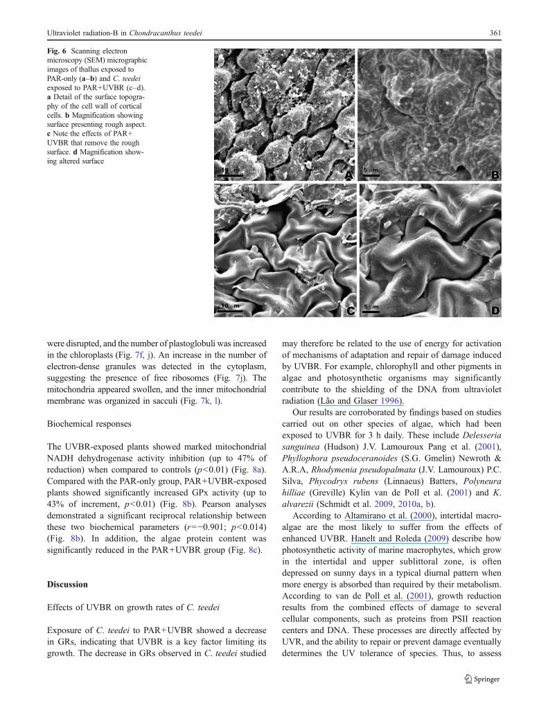
Observations under SEM
Under SEM, the control surface topography of C. teedeishowed bigger undulations of the cell wall of cortical cells,indicating a rough surface aspect (Fig. 6a, b) whencompared with UVBR-exposed plants whose surfaceappears to have been influenced by UVBR (Fig. 6c, d).This result indicates that plants exposed to PAR+UVBR
probably undergo a change in the mucilage that coats thethallus.
Observations under TEM
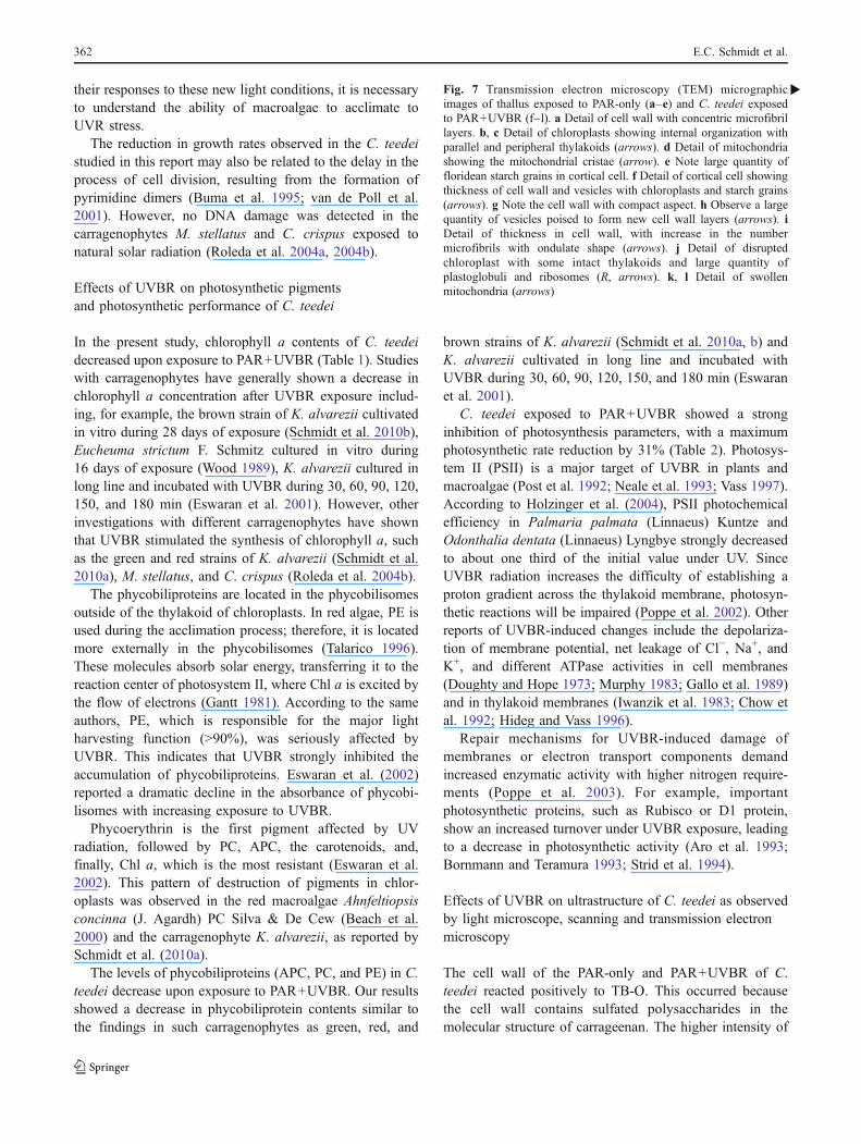
When observed under transmission electron microscopythat control (PAR-only) samples of C. teedei showed a cellwall formed by two layers (Fig.7a–e). The outermost layerpresented concentric microfibrils densely packed, while theinnermost layer was electron-transparent. Both regionswere embedded in an amorphous matrix which consistedof sulfated polysaccharides such as carrageenans (Fig. 7a).The chloroplasts assumed the typical internal organizationof the red algae with unstacked, evenly spaced thylakoids.The chloroplasts apparently consisted of an individual andflat thylakoid surrounded by a single peripheral thylakoid(Fig. 7b, c). Small mitochondria were observed; they werespherical to elongate, with tubuli-type structure (Fig. 7d).
Fig. 3 Light microscopy and scanning electron microscopy of thallusexposed to PAR-only of C. teedei. a Transversal section stained withaniline blue. Observe the elongated cortical cells (CC) and the morevacuolated subcortical cells (SC). (b). Longitudinal section stainedwith aniline blue. Note the medullar cells with filamentous aspects
(arrows). c, d SEM observation of C. teedei in free-hand section.Observe the distribution of cortical, subcortical, and medullar cells. eSEM observation of C. teedei in cryofracture section. Note the detailof cell wall of cortical cells
Ultraviolet radiation-B in Chondracanthus teedei 359

The control cells of C. teedei had a large quantity offloridean starch grains (Fig. 7e).
After exposure to PAR+UVBR for 3 h per day during a7-day period, C. teedei were observed to undergo someultrastructural changes, involving an increase in thethickness of the cell wall, chloroplasts, and mitochondria(Fig. 7f–l). The cell wall of plants cultivated with PAR+UVBR showed more compact aspect compared to control
plants, a fact thought to be associated with the increase inthe number of concentric microfibrils (Fig. 7f, g). Thesemicrofibrils sometimes showed an ondulate shape (Fig. 7i).In many cells, a large quantity of vesicles, with fibrilcontents poised to form new cell wall layers, could beobserved (Fig. 7h).
Chloroplasts showed visible changes in ultrastructuralorganization with irregular morphology. The thylakoids
Fig. 4 Light microscopy of thetransversal sections of thallusexposed to PAR-only control(a and c) and exposure of C.teedei to PAR+UVBR (b andd). Section stained with TB-O.a The cell walls of cortical cells(CC) and subcortical cells (SC)show metachromatic reaction.b Section stained with TB-O.Note the intense metachromaticreactions in the cell wall ofcortical and subcortical cells.c Section stained with CBB.Positive reaction with the nuclei(arrow) and all cellular proteins.The pit connections with posi-tive reaction (arrowheads).d Section stained with CBB.The cytoplasm shows the dens-est cellular reaction. The nucleihave become more evident(arrow), and the pit connectionsare visible (arrowheads)
Fig. 5 Light microscopy of thetransversal sections of thallusexposed to PAR-only (a and b)and exposure of C. teedei toPAR+UVBR (c and d). Sectionsstained with PAS. a Observe thePAS-positive floridean starchgrains (S) in the cells and posi-tive reaction with the cell wall(CW). b Note the medullar cells(MC) with a large quantity offloridean starch grains and pos-itive reaction in cell wall. cDetail of thickness (arrows) thatoccurs in cell wall of corticalcell. Note the reduction in thedensity of floridean starch grainsin the cortical and subcorticalcells. d Observe a reduction inthe quantity of starch grains inthe medullar cells
360 E.C. Schmidt et al.
were disrupted, and the number of plastoglobuli was increasedin the chloroplasts (Fig. 7f, j). An increase in the number ofelectron-dense granules was detected in the cytoplasm,suggesting the presence of free ribosomes (Fig. 7j). Themitochondria appeared swollen, and the inner mitochondrialmembrane was organized in sacculi (Fig. 7k, l).
Biochemical responses
The UVBR-exposed plants showed marked mitochondrialNADH dehydrogenase activity inhibition (up to 47% ofreduction) when compared to controls (p<0.01) (Fig. 8a).Compared with the PAR-only group, PAR+UVBR-exposedplants showed significantly increased GPx activity (up to43% of increment, p<0.01) (Fig. 8b). Pearson analysesdemonstrated a significant reciprocal relationship betweenthese two biochemical parameters (r=−0.901; p<0.014)(Fig. 8b). In addition, the algae protein content wassignificantly reduced in the PAR+UVBR group (Fig. 8c).
Discussion
Effects of UVBR on growth rates of C. teedei
Exposure of C. teedei to PAR+UVBR showed a decreasein GRs, indicating that UVBR is a key factor limiting itsgrowth. The decrease in GRs observed in C. teedei studied
may therefore be related to the use of energy for activationof mechanisms of adaptation and repair of damage inducedby UVBR. For example, chlorophyll and other pigments inalgae and photosynthetic organisms may significantlycontribute to the shielding of the DNA from ultravioletradiation (Lão and Glaser 1996).
Our results are corroborated by findings based on studiescarried out on other species of algae, which had beenexposed to UVBR for 3 h daily. These include Delesseriasanguinea (Hudson) J.V. Lamouroux Pang et al. (2001),Phyllophora pseudoceranoides (S.G. Gmelin) Newroth &A.R.A, Rhodymenia pseudopalmata (J.V. Lamouroux) P.C.Silva, Phycodrys rubens (Linnaeus) Batters, Polyneurahilliae (Greville) Kylin van de Poll et al. (2001) and K.alvarezii (Schmidt et al. 2009, 2010a, b).
According to Altamirano et al. (2000), intertidal macro-algae are the most likely to suffer from the effects ofenhanced UVBR. Hanelt and Roleda (2009) describe howphotosynthetic activity of marine macrophytes, which growin the intertidal and upper sublittoral zone, is oftendepressed on sunny days in a typical diurnal pattern whenmore energy is absorbed than required by their metabolism.According to van de Poll et al. (2001), growth reductionresults from the combined effects of damage to severalcellular components, such as proteins from PSII reactioncenters and DNA. These processes are directly affected byUVR, and the ability to repair or prevent damage eventuallydetermines the UV tolerance of species. Thus, to assess
Fig. 6 Scanning electronmicroscopy (SEM) micrographicimages of thallus exposed toPAR-only (a–b) and C. teedeiexposed to PAR+UVBR (c–d).a Detail of the surface topogra-phy of the cell wall of corticalcells. b Magnification showingsurface presenting rough aspect.c Note the effects of PAR+UVBR that remove the roughsurface. d Magnification show-ing altered surface
Ultraviolet radiation-B in Chondracanthus teedei 361
their responses to these new light conditions, it is necessaryto understand the ability of macroalgae to acclimate toUVR stress.
The reduction in growth rates observed in the C. teedeistudied in this report may also be related to the delay in theprocess of cell division, resulting from the formation ofpyrimidine dimers (Buma et al. 1995; van de Poll et al.2001). However, no DNA damage was detected in thecarragenophytes M. stellatus and C. crispus exposed tonatural solar radiation (Roleda et al. 2004a, 2004b).
Effects of UVBR on photosynthetic pigmentsand photosynthetic performance of C. teedei
In the present study, chlorophyll a contents of C. teedeidecreased upon exposure to PAR+UVBR (Table 1). Studieswith carragenophytes have generally shown a decrease inchlorophyll a concentration after UVBR exposure includ-ing, for example, the brown strain of K. alvarezii cultivatedin vitro during 28 days of exposure (Schmidt et al. 2010b),Eucheuma strictum F. Schmitz cultured in vitro during16 days of exposure (Wood 1989), K. alvarezii cultured inlong line and incubated with UVBR during 30, 60, 90, 120,150, and 180 min (Eswaran et al. 2001). However, otherinvestigations with different carragenophytes have shownthat UVBR stimulated the synthesis of chlorophyll a, suchas the green and red strains of K. alvarezii (Schmidt et al.2010a), M. stellatus, and C. crispus (Roleda et al. 2004b).
The phycobiliproteins are located in the phycobilisomesoutside of the thylakoid of chloroplasts. In red algae, PE isused during the acclimation process; therefore, it is locatedmore externally in the phycobilisomes (Talarico 1996).These molecules absorb solar energy, transferring it to thereaction center of photosystem II, where Chl a is excited bythe flow of electrons (Gantt 1981). According to the sameauthors, PE, which is responsible for the major lightharvesting function (>90%), was seriously affected byUVBR. This indicates that UVBR strongly inhibited theaccumulation of phycobiliproteins. Eswaran et al. (2002)reported a dramatic decline in the absorbance of phycobi-lisomes with increasing exposure to UVBR.
Phycoerythrin is the first pigment affected by UVradiation, followed by PC, APC, the carotenoids, and,finally, Chl a, which is the most resistant (Eswaran et al.2002). This pattern of destruction of pigments in chlor-oplasts was observed in the red macroalgae Ahnfeltiopsisconcinna (J. Agardh) PC Silva & De Cew (Beach et al.2000) and the carragenophyte K. alvarezii, as reported bySchmidt et al. (2010a).
The levels of phycobiliproteins (APC, PC, and PE) in C.teedei decrease upon exposure to PAR+UVBR. Our resultsshowed a decrease in phycobiliprotein contents similar tothe findings in such carragenophytes as green, red, and
brown strains of K. alvarezii (Schmidt et al. 2010a, b) andK. alvarezii cultivated in long line and incubated withUVBR during 30, 60, 90, 120, 150, and 180 min (Eswaranet al. 2001).
C. teedei exposed to PAR+UVBR showed a stronginhibition of photosynthesis parameters, with a maximumphotosynthetic rate reduction by 31% (Table 2). Photosys-tem II (PSII) is a major target of UVBR in plants andmacroalgae (Post et al. 1992; Neale et al. 1993; Vass 1997).According to Holzinger et al. (2004), PSII photochemicalefficiency in Palmaria palmata (Linnaeus) Kuntze andOdonthalia dentata (Linnaeus) Lyngbye strongly decreasedto about one third of the initial value under UV. SinceUVBR radiation increases the difficulty of establishing aproton gradient across the thylakoid membrane, photosyn-thetic reactions will be impaired (Poppe et al. 2002). Otherreports of UVBR-induced changes include the depolariza-tion of membrane potential, net leakage of Cl−, Na+, andK+, and different ATPase activities in cell membranes(Doughty and Hope 1973; Murphy 1983; Gallo et al. 1989)and in thylakoid membranes (Iwanzik et al. 1983; Chow etal. 1992; Hideg and Vass 1996).
Repair mechanisms for UVBR-induced damage ofmembranes or electron transport components demandincreased enzymatic activity with higher nitrogen require-ments (Poppe et al. 2003). For example, importantphotosynthetic proteins, such as Rubisco or D1 protein,show an increased turnover under UVBR exposure, leadingto a decrease in photosynthetic activity (Aro et al. 1993;Bornmann and Teramura 1993; Strid et al. 1994).
Effects of UVBR on ultrastructure of C. teedei as observedby light microscope, scanning and transmission electronmicroscopy
The cell wall of the PAR-only and PAR+UVBR of C.teedei reacted positively to TB-O. This occurred becausethe cell wall contains sulfated polysaccharides in themolecular structure of carrageenan. The higher intensity of
Fig. 7 Transmission electron microscopy (TEM) micrographicimages of thallus exposed to PAR-only (a–e) and C. teedei exposedto PAR+UVBR (f–l). a Detail of cell wall with concentric microfibrillayers. b, c Detail of chloroplasts showing internal organization withparallel and peripheral thylakoids (arrows). d Detail of mitochondriashowing the mitochondrial cristae (arrow). e Note large quantity offloridean starch grains in cortical cell. f Detail of cortical cell showingthickness of cell wall and vesicles with chloroplasts and starch grains(arrows). g Note the cell wall with compact aspect. h Observe a largequantity of vesicles poised to form new cell wall layers (arrows). iDetail of thickness in cell wall, with increase in the numbermicrofibrils with ondulate shape (arrows). j Detail of disruptedchloroplast with some intact thylakoids and large quantity ofplastoglobuli and ribosomes (R, arrows). k, l Detail of swollenmitochondria (arrows)
�
362 E.C. Schmidt et al.

reaction to TB-O in plants exposed to PAR+UVBR couldbe related to an increase in the production of vesicles, asobserved under TEM (see Fig. 7h), which were subse-quently incorporated into cell wall.
When analyzed under TEM, the PAR-only cell wall ofC. teedei showed a microfibrillar texture with microfibrilsstructured in concentric layers with different degrees ofcompression. Observations of the cell wall of plants
Ultraviolet radiation-B in Chondracanthus teedei 363

exposed to PAR+UVBR showed an increase in thatstructural component. The increase in the thickness of thecell wall of C. teedei exposed to PAR+UVBR can beinterpreted as a defense mechanism against exposure toultraviolet radiation. It is probable that the activity of Golgibodies is more intense in plants exposed to PAR+UVBRand that this results in the large production of vesicleswhich then format the matrix content of cell wall. PASreaction, as observed under LM (Fig. 5c), indicates greaterdeposition of neutral polysaccharides, such as cellulose.
Cellular metabolism of C. teedei exposed to PAR+UVBR is modified such that vesicles with fibril contentsresponsible for cell wall thickening are produced (Fig. 7h),
and this, in turn, reduces the synthesis of mucilagemolecules that coat the thallus (Fig. 6d).
The decrease in floridean starch grains observed throughTEM and LM in plants exposed to PAR+UVBR may berelated to a change in the route of biosynthesis of starchenzymes of the Calvin cycle, possibly by activating thedegradation pathway. The degradation pathways may beutilized to activate the biosynthesis of defense compounds.According to Sinha et al. (2000), some organisms have acapacity to produce mechanisms to counteract the damag-ing effects of UVBR, such as accumulation of carotenoids,mycosporine-like amino acids and detoxifying enzymes orradical quenchers. Inhibition of the synthesis of florideanstarch grains, by means of UDP-glucose, diverts synthesisfor the production of cell wall components, such ascarrageenan that also uses via UDP-glucose as a precursorto biosynthesis of this polysaccharide. This phenomenon, asobserved with TEM and LM, also results in the increase ofthe cell wall.
In red algae, the thylakoids that are not associated witheach other are free in chloroplasts. The chloroplasts ofPAR-only C. teedei showed a structure very similar to thatof red algae, having one peripheral thylakoid surrounded byparallel thylakoids. The number of parallel thylakoids isvariable, and this number mainly depends on the spatiallocation of the cell in the algae.
In contrast, the chloroplasts of C. teedei exposed to PAR+UVBR showed significant structural changes, includingmodification in the quantity, size, and organization ofthylakoids. Some reports with carregenophytes exposed toPAR+UVBR, including K. alvarezii by Schmidt et al.(2009) and I. cordata by Navarro et al. (2010), showedultrastructural changes in the chloroplasts.
When analyzed by TEM, the algae exposed to PAR+UVBR of C. teedei revealed an increase in the number ofthe plastoglobuli in the chloroplast. According to Holzingeret al. (2009), when the algae are subjected to stress,nitrogen limitation and the synthesis of lipids areobserved. These phenomena occur because the pathwaysto form protein-containing cell structures are suppressed.Similar results were reported with formation of plasto-globuli in K. alvarezii by Schmidt et al. (2009) afterexposure to UV radiation. This increase in the number oflipids can be considered as a change in metabolism,which, in turn, results in reduction of cell proliferationand decrease in GRs.
The mitochondria of the PAR+UVBR of C. teedei alsoshowed ultrastructural changes. The mitochondria appearedswollen, and the inner mitochondrial membrane wasorganized in sacculi. Other studies report ultrastructuralchanges in mitochondria of P. palmata and Palmariadecipiens. In P. palmata, these changes were manifestedby an apparent swelling and by changes in the inner
Fig. 8 Biochemical responses ofC. teedei exposed to UVBR for 3 h/dayover a period of 7 days. Means±S.D, n=4. a Relative changes ofmitochondrial NADH dehydrogenase activity of C. teedei. b Glutathioneperoxidase assay (GPx) activity of C. teedei. c Protein content ofC. teedei
364 E.C. Schmidt et al.

mitochondrial membrane from a tubuli- to sacculi-typestructure when exposed to UVA+UVB radiation (Poppe etal. 2003). In the agarophyte G. domingensis exposed during21 days to PAR+UVBR, mitochondria appeared swollen,and the presence of electron-dense granules in the mito-chondrial matrix was detected Schmidt et al (2010c). Suchultrastructural changes can result in a disordered metabo-lism of ATP (Vosjan et al. 1990).
Effects of UVBR on plant metabolism
The significant reduction in protein content observed inPAR+UVBR-treated C. teedei agrees with previous resultsshowing reduction in protein concentrations in the carra-genophyte K. alvarezii when exposed to UVBR (30 to180 min) (Eswaran et al. 2001). These data indicate that thecombination of PAR+UVBR inhibits protein metabolism,probably by altering biosynthesis pathways, or proteinmobilization for repair processes, including the activationof antioxidant defense. In this context, and as previouslyreported by Poppe et al. (2003), UVBR-induced repairmechanisms demand increased enzymatic activity withhigher nitrogen requirements.
The increased glutathione peroxidase activity observedin irradiated plants could be related to increasedhydrogen peroxide production, since this treatmentblocked the mitochondrial NADH dehydrogenase activi-ty, including that of complex I. In this scenario, complexI of the respiratory chain has been reported as one of themajor sites of reactive oxygen species (ROS) generationin mitochondria under physiological conditions (Boveriset al. 1976; Chen et al. 2003), and an overt ROSproduction is observed when this redox center is blocked(Turrens 1997; Turrens and Boveris 1980). Furthermore,ROS formed at the NADH dehydrogenase site of complexI are released into the mitochondrial matrix (Chen et al.2003), thereby eliciting oxidative damage of mitochondri-al enzymes, including the complexes of the respiratorychain, enzymes of the Krebs cycle, and several othersensitive proteins, as well as mtDNA (Zhang et al. 1990;Hausladen and Fridovich 1994; Bandy and Davison 1990).This is in line with Costa et al. (2002), who demonstratedthat UVBR exposure stimulates the generation ROS. Inaddition, Shiu and Lee (2005) showed that ROS-inducedtoxicity provokes an increased antioxidant enzymaticresponse.
Conclusion
In summary, the present study demonstrates that UVBRnegatively affects the intertidal macroalgae. This becameobvious after 3 h of daily exposure to UVBR over 7 days of
experimental treatment, resulting in alteration in thallusmorphology, changes in cellular organization (cell wallthickness, disrupted chloroplasts, and swollen mitochon-dria). Moreover, this exposure caused significant metabolicalterations that led to decreases in photosynthetic efficiencyand growth rates. Additionally, the UVBR-induced mito-chondrial dysfunction induced a marked cellular antioxi-dant response.
The relevance of these findings is highlighted by botheconomic consequences and the current index of strato-spheric ozone depletion, especially in the southernhemisphere.
Acknowledgements The authors would like to acknowledge thestaff of the Central Laboratory of Electron Microscopy (LCME),Federal University of Santa Catarina, Florianopolis, Santa Catarina,Brazil, for the use of their transmission electron microscope. Thisstudy was supported in part by the Conselho Nacional deDesenvolvimento Científico e Tecnológico (CAPES, Brazil), Con-selho Nacional de Desenvolvimento Científico e Tecnológico(CNPq, Brazil) and Fundação de Apoio à Pesquisa Cientifica eTecnológica do Estado de Santa Catarina (FAPESC). The authorsare grateful to CAPES for providing a scholarship to Éder C. Schmidtand CNPq-PIBIC for providing a scholarship to Beatriz Pereira.Bouzon ZL, Maraschin M, Horta P.A and Latini A are CNPq fellows.This study is part of the Ph.D. thesis of the first author.
Conflict of interest The authors declare that they have no conflict ofinterest.
References
Altamirano M, Flores-Moya A, Figueroa FL (2000) Long-term effects ofnatural sunlight under various ultraviolet radiation conditions ongrowth and photosynthesis of intertidalUlva rigida (Chlorophyceae)cultivated in situ. Bot Mar 43:119–126
Aro EM, Virgen I, Andersson B (1993) Photoinhibition of photosystemII. Inactivation, protein damage and turnover. Biochim BiophysActa 1143:113–134
Bandy B, Davison AJ (1990) Mitochondrial mutations may increaseoxidative stress: implications for carcinogenesis and aging? FreeRadic Biol Med 8:523–539
Beach KS, Smith CM, Okano R (2000) Experimental analysis ofrhodophyte photoacclimation to PAR and UV-radiation using invivo absorbance spectroscopy. Bot Mar 43:525–536
Bischof K, Gómez I, Molis M, Hanelt D, Karsten U, Lüder U,Roleda MY, Zacher K, Wiencke C (2006) Ultraviolet radiationshapes seaweed communities. Rev Environ Sci Biotechnol5:141–166
Bornmann JF, Teramura AH (1993) Effects of ultraviolet-Bradiation on terrestrial plants. In: Young AR, Björn LO, MoanJ, Nultsch W (eds) Environmental UV photobiology. Plenum,New York, pp 427–471
Boveris A, Cadenas E, Stoppani AO (1976) Role of ubiquinone in themitochondrial generation of hydrogen peroxide. Biochem J156:435–444
Buma AGJ, van Hannen EJ, Roza L, Veldhuis MJW, Gieskes WWC(1995) Monitoring ultraviolet-B-induced DNA damage in indi-vidual diatom cells by immunofluorescent thymine dimerdetection. J Phycol 31:314–321
Ultraviolet radiation-B in Chondracanthus teedei 365

Cassina A, Radi R (1996) Differential inhibitory action of nitric oxideand peroxynitrite on mitochondrial electron transport. ArchBiochem Biophys 328:309–316
Chen Q, Vazquez EJ, Moghaddas S, Hoppel CL, Lesnefsky EJ (2003)Production of reactive oxygen species by mitochondria: centralrole of complex III. J Biol Chem 278:36027–36031
Chow WS, Strid A, Anderson JM (1992) Short-term treatment of peaplants with supplementary ultraviolet-B radiation: recovery time-courses of some photosynthetic functions and components. In:Murata N (ed) Research in photosynthesis. Kluwer Academic,Dordrecht, pp 361–364
Clarke KR, Warwick RM (1994) Change in marine communities: anapproach to statistical analysis and interpretation. NaturalEnvironmental Research Council, Plymouth
Costa H, Gallego SM, Tomaro ML (2002) Effect of UV-B radiation onantioxidant defense system in sunflower cotyledons. Plant Sci162:939–945
Doughty CJ, Hope AB (1973) Effects of ultraviolet radiation on themembranes of Chara coralline. Membr Biol 13:185–198
Edwards P (1970) Illustrated guide to the seaweeds and sea grassesin the vicinity of Porto Arkansas, Texas. Contr Mar Sci 15:1–228
Eswaran K, Subba Rao PV, Mairh OP (2001) Impact of ultraviolet-Bradiation on Kappaphycus alvarezii (Solieraceae, Rhodophyta).Indian J Mar Sci 30:105–107
Eswaran K, Mairh OP, Subba Rao PV (2002) Inhibition of pigmentsand phycocolloid in a marine red algae Gracilaria edulis byultraviolet-B radiation. Biol Plant 45:157–159
Gahan PB (1984) Plant histochemistry and cytochemistry: anintroduction. Academic, London
Gallo RL, Kochevar IE, Granstein RD (1989) Ultraviolet radiationinduces a change in cell membrane potential in vitro: a possiblesignal for ultraviolet radiation induced alteration in cell activity. JPhotochem Photobiol 49:655–662
Gantt E (1981) Phycobilisomes. Annu Rev Plant Physiol 32:327–347Garbary DJ, Kim KY, Hoffman J (2004) Cytological damage to the
red alga Griffithsia pacifica from ultraviolet radiation. Hydro-biologia 512:165–170
Gordon EM, McCandless EL (1973) Ultrastructure and histochemistryof Chondrus crispus Stackhouse. In: Harvey MJ, McLachlan J(eds) Chondrus crispus. T. N. Scot. Inst. Sci, Halifax, pp 111–133
Grossman AR, Schaefer MR, Chiang GG, Collier JL (1993) Thephycobilisome a light harvesting complex responsive to environ-mental conditions. Microbiol Rev 57:725–749
Guiry MD (1984) Structure, life history and hybridization of AtlanticGigartina teedii (Rhodophyta) in culture. Br Phycol J 19:37–55
Hanelt D, Roleda MY (2009) UVB radiation may ameliorate photo-inhibition in specific shallow-water tropical marine macrophytes.Aquat Bot 91:6–12
Hausladen A, Fridovich I (1994) Superoxide and peroxynitriteinactivate aconitases, but nitric oxide does not. J Biol Chem269:29405–29408
Hideg E, Vass I (1996) UV-B induced free radical production in plantleaves and isolated thylakoid membranes. Plant Sci 115:251–260
Hiscox JD, Israelstam GF (1979) A method for the extraction ofchlorophyll from leaf tissue without maceration. Can J Bot57:1332–1334
Holzinger A, Lütz C (2006) Algae and UV irradiation: effects onultrastructure and related metabolic functions. Micron 37:190–207
Holzinger A, Lütz C, Karsten U, Wiencke C (2004) The effect ofultraviolet radiation on ultrastructure and photosynthesis in thered macroalgae Palmaria palmata and Odonthalia dentata fromArtic waters. Plant Biol 6:568–577
Holzinger A, Karsten U, Lütz C, Wiencke C (2006) Ultrastructure andphotosynthesis in the supralittoral green macroalga Prasiola
crispa from Spitsbergen (Norway) under UV exposure. Phycolo-gia 45:168–177
Holzinger A, Roleda MY, Lütz C (2009) The vegetative arcticfreshwater green alga Zygnema is insensitive to experimentalUV exposure. Micron 40:831–838
Iwanzik W, Tevini M, Dohnt G, Voss M, Weiss W, Gräber P, RengerG (1983) Action of UV-B radiation on photosynthetic primaryreactions in spinach chloroplasts. Physiol Plant 58:401–407
Jones RJ, Kildea T, Hoegh-Guldberg O (1999) PAM chlorophyllfluorometry: a new in situ technique for stress assessment inscleractinian corals, used to examine the effects of cyanide fromcyanide fishing. Mar Pollut Bull 38:864–874
Karsten U, Wiencke C (1999) Factors controlling the formation ofUV-absorbing mycosporine-like amino acids in the marine redalga Palmaria palmata from Spitsbergen (Norway). J PlantPhysiol 155:407–415
Karsten U, Bischof K, Hanelt D, Tüg H, Wiencke C (1999) Theeffect of ultraviolet radiation on photosynthesis andultraviolet-absorbing substances in the endemic Arctic macro-alga Devaleraea ramentacea (Rhodophyta). Plant Physiol105:58–66
Karsten U, Sawall T, West J, Wiencke C (2000) Ultraviolet sunscreencompounds in epiphytic red algae from mangroves. Hydro-biologia 432:159–171
Kerr JB, Mc Elroy CT (1993) Evidence for large upward trends ofultraviolet-B radiation linked to ozone depletion. Science262:1032–1034
Kursar TA, van Der Meer J, Alberte RS (1983) Light-harvestingsystem of the red alga Gracilaria tikvahiae. I. Biochemicalanalyses of pigment mutations. Plant Physiol 73:353–360
Lão K, Glaser AN (1996) Ultraviolet-B photodestruction of light-harvesting complex. PNAS 93:5258–5263
Latini A, Rodriguez M, Borba Rosa R, Scussiato K, Leipnitz G, Reisde Assis D, da Costa FG, Funchal C, Jacques-Silva MC, Buzin L,Giugliani R, Cassina A, Radi R, Wajner M (2005) 3-Hydroxyglutaric acid moderately impairs energy metabolism inbrain of young rats. Neuroscience 135:111–120
Lesser MP, Shick JM (1994) Effects of irradiance and ultravioletradiation on photoadaptation in the zooxanthellae of Aiptasiapallida: primary production, photoinhibition, and enzymicdefenses against oxygen toxicity. Mar Biol 102:243–255
Lowry OH, Rosebough NG, Farr AL (1951) Protein measurementwith the folin phenol reagent. J Biol Chem 193:265–275
Lubin D, Jensen EH (1995) Effects of clouds and stratospheric ozonedepletion on ultraviolet-radiation trends. Nature 377:710–713
Madronich S (1992) Implications of recent total atmospheric ozonemeasurements for biological active ultraviolet radiation reachingthe earth's surface. Geophys Res Lett 19:37–40
Mitchell DL, Jen J, Cleaver JE (1992) Sequence specificity ofcyclobutane pyrimidine dimers in DNA treated with solar(ultraviolet B) radiation. Nucleic Acids Res 20:225–229
Murphy TM (1983) Membranes as targets of ultraviolet radiation.Physiol Plant 58:381–388
Navarro NP, Mansilla A, Plastino EM (2010) Iridaea cordata(Gigartinales, Rhodophyta): responses to artificial UVB radia-tion. J Appl Phycol 22:385–394
Neale PJ, Cullen JC, Lesser MP, Melis A (1993) Physiological basesfor detecting and predicting photoinhibition of aquatic photosyn-thesis by PAR and UV radiation. In: Yamamoto HY, Smith CM(eds) Photosynthetic responses to the environment. Amer SocPlant Physiol, Rockville, pp 61–77
Oliveira EC, Paula EJ, Plastino EM, Petti R (1995) Metodologías parael cultivo no axenico de macroalgas marinas in vitro. In: AlvealK, Ferrario M, Oliveira E, SAR E (eds) Manual de métodosficológicos. Universidad de Concepción, Concepción-Chile, pp429–447
366 E.C. Schmidt et al.

Pang S, Gomez I, Lüning K (2001) The red macroalga Delesseriasanguinea as a UVB-sensitive model organism: selective growthreduction by UVB in outdoor experiments and rapid recording ofgrowth rate during and after UV pulses. Eur J Phycol 36:207–216
Penniman CA, Mathieson AC, Penniman CE (1986) Reproductivephenology and growth of Gracilaria tikvahiae McLachlan(Gigartinales, Rhodophyta) in the Great Bay Estuary, NewHampshire. Bot Mar 29:147–154
Pereira L, Mesquita JF (2004) Population studies and carrageenanproperties of Chondracanthus teedei var. lusitanicus (Giga-rtinaceae, Rhodophyta). J Appl Phycol 16:369–383
Platt T, Gallegos CL, Harrison WG (1980) Photoinhibition ofphotosynthesis in natural assemblages of marine phytoplankton.J Mar Res 38:687–701
Poppe F, Hanelt D, Wiencke C (2002) Changes in ultrastructure,photosynthetic activity and pigments in the Antarctic Red AlgaPalmaria decipiens during acclimation to UV radiation. Bot Mar45:253–261
Poppe F, Schmidt RAM, Hanelt D, Wiencke C (2003) Effects of UVradiation on the ultrastructure of several red algae. Phycol Res51:11–19
Post A, Gentle S, Larkum AWS (1992) Algal photosynthesis:inhibition by UV-B radiation, recovery and UV-absorbing pig-ments. In: Murata N (ed) Research in photosynthesis. KluwerAcademic, Dordrecht, pp 847–850
Reynolds ES (1963) The use of lead citrate at light pH as an electronopaque stain in electron microscopy. J Cell Biol 17:208–212
Roleda MY, Hanelt D, Kräbs G, Wiencke C (2004a) Morphology,growth, photosynthesis and pigments in Laminaria ochroleuca(Laminariales, Phaeophyta) under ultraviolet radiation. Phycolo-gia 43:603–613
Roleda MY, van de Poll WH, Wiencke C, Hanelt D (2004b) PAR andUVBR effects on photosynthesis, viability, growth and DNA indifferent life stages of two coexisting Gigartinales: implicationsfor recruitment and zonation pattern. Mar Ecol Progr Ser281:37–50
Roleda MY, Lütz-Meindl U, Wiencke C, Lütz C (2009) Physiological,biochemical, and ultrastructural responses of the green macroalgaUrospora penicilliformis from Arctic Spitsbergen to UV radia-tion. Protoplasma 243:105–116
Schmidt EC, Scariot LA, Rover T, Bouzon ZL (2009) Changes inultrastructure and histochemistry of two red macroalgae strainsof Kappaphycus alvarezii (Rhodophyta, Gigartinales), as aconsequence of ultraviolet B radiation exposure. Micron40:860–869
Schmidt EC,MaraschinM, Bouzon ZL (2010a) Effects of UVB radiationon the carragenophyte Kappaphycus alvarezii (Rhodophyta,Gigartinales): changes in ultrastructure, growth, and photosyn-thetic pigments. Hydrobiologia 649:171–182
Schmidt EC, Nunes BG, Maraschin M, Bouzon ZL (2010b) Effectof ultraviolet-B radiation on growth, photosynthetic pigments,and cell biology of Kappaphycus alvarezii (Rhodophyta,Gigartinales) macroalgae brown strain. Photosynthetica48:161–172
Schmidt EC, Santos R, Horta P, Maraschin M, Bouzon ZL (2010c)Effects of UVB radiation on the agarophyteGracilaria domingensis
(Rhodophyta, Gracilariales): changes in cell organization, growthand photosynthetic performance. Micron 41:919–930
Shiu C-T, Lee T-M (2005) Ultraviolet-B-induced oxidative stress andresponses of the ascorbate-glutathione cycle in a marine macro-alga Ulva fasciata. J Exp Bot 56:2851–2865
Sinha RP, Klisch M, Gröniger A, Hader DP (2000) Mycosporine-likeamino acids in the marine red alga Gracilaria cornea: effects ofUV and heat. Environ Exp Bot 43:33–43
Sommaruga R (2001) The role of solar UV radiation in the ecology ofalpine lakes. J Photochem Photobiol B Biol 62:35–42
Sonntag B, Summerer M, Sommaruga R (2007) Sources ofmycosporine-like amino acids in planktonic Chlorella-bearingciliates (Ciliophora). Freshwater Biol 52:1476–1485
Strid A, Chow WS, Anderson JM (1994) UV-B damage andprotection at the molecular level in plants. Photosynth Res39:475–489
Talarico L (1996) Phycobiliproteins and phycobilisomes in red algae:adaptive responses to light. Sci Mar 60:205–222
Talarico L, Maranzana G (2000) Light and adaptative responses inred macroalgae: an overview. J Photochem Photobiol B Biol56:1–11
Turrens JF (1997) Superoxide production by the mitochondrialrespiratory chain. Biosci Rep 17:3–8
Turrens JF, Boveris A (1980) Generation of superoxide anion by theNADH dehydrogenase of bovine heart mitochondria. Biochem J191:421–427
van de Poll WH, Eggert A, Buma GJ, Breeman AM (2001) Effects ofUV-B induced DNA damage and photoinhibition on growth oftemperate marine red macrophytes: habitat-related differences inUV-B tolerance. J Phycol 37:30–38
Vass I (1997) Adverse effects of UV-B light on the structure andfunction of the photosynthetic apparatus. In: Pessarakali M (ed)Handbook of photosynthesis. Marcel Dekker, New York
Vosjan J, Döhler H, Nieuwland G (1990) Effect of UV-B irradiance onthe ATP content of microorganisms of the Weddell Sea(Antarctica). Neth J Sea Res 25:391–394
Wellburn AR (1994) The spectral determination of chlorophyll a andchlorophyll b, as well as total carotenoids, using various solventswith spectrophotometers of different resolution. J Plant Physiol144:307–313
Wendel A (1981) Glutathione peroxidase. Meth Enzymol 77:325–333White AK, Critchley C (1999) Rapid light curves: a new fluorescence
method to assess the state of the photosynthetic apparatus. PhotosRes 59:63–72
Wood WF (1989) Photoadaptive responses of the tropical red algaEucheuma striatum Schmitz (Gigartinales) to ultra-violet radia-tion. Aquat Bot 33:41–51
Yokoya NS, Necchi O Jr, Martins AP, Gonzalez SF, Plastino EM(2007) Growth responses and photosynthetic characteristics ofwild and phycoerythrin-deficient strains of Hypnea musciformis(Rhodophyta). J Appl Phycol 19:197–205
Zacher K, Roleda MY, Wulff A, Hanelt D, Wiencke C (2009)Responses of Antarctic Iridaea cordata (Rhodophyta) tetrasporesexposed to ultraviolet radiation. Phycol Res 57:186–193
Zhang Y, Marcillat O, Giulivi C, Ernster L, Davies KJ (1990) Theoxidative inactivation of mitochondrial electron transport chaincomponents and ATPase. J Biol Chem 265:16330–16336
Ultraviolet radiation-B in Chondracanthus teedei 367
Copyright © 2022 FDOKUMEN