Adulteration of Mustard Cooking Oil With Argemone Oil: Do Indian Food Regulatory Policies and...
12
515 Adulteration of Mustard Cooking Oil with Argemone Oil: Do Indian Food Regulatory Policies and Antioxidant Therapy Both Need Revisitation? CHALLAGUNDLA KISHORE BABU, SUBHASH KUMAR KHANNA, and MUKUL DAS ABSTRACT Consumption of adulterated mustard oil (Brassica nigra) with argemone oil (Argemone mexicana) even for a short duration leads to a clinical condition referred as epidemic dropsy. In humans, argemone oil contained in adulterated mustard oil causes oxidative stress and death of red blood cells via met-hemoglobin formation by altering pyridine nucleotide(s) and glutathione redox potential. Argemone oil contamination poses a serious threat to human health and should be checked by appropriate regulatory measures. Antioxidant therapy pro- vides symptomatic relief and should be seriously considered for therapeutic interventions against argemone oil toxicity. Antioxid. Redox Signal. 9, 515–525. News & Views ANTIOXIDANTS & REDOX SIGNALING Volume 9, Number 4, 2007 © Mary Ann Liebert, Inc. DOI: 10.1089/ars.2006.1492 CONSUMPTION OFADULTERATED EDIBLE MUSTARD OIL WITH ARGEMONE OIL CAUSES EPIDEMIC DROPSY IN HUMANS C ONSUMPTION OF ADULTERATED MUSTARD OIL with arge- mone oil even for a short duration leads to a clinical condition referred as epidemic dropsy (8). The main symp- toms of dropsy include nausea, vomiting, diarrhea, anorexia, dyspnea, palpitation, hyperpigmentation of body parts, burn- ing sensation of eyes, bilateral pitting edema of lower limbs, erythema, breathlessness, tachycardia, hepatomegaly, crepita- tions in the lungs, and gallop rhythm (7). In severe cases, glaucoma and even death due to cardiac and respiratory fail- ure have been reported (7, 8). PREVALENCE OF EPIDEMIC DROPSY A number of epidemic dropsy outbreaks have been re- ported in different countries, including Burma (6), Fiji Island (12), Nepal (21), and South Africa (43). From time to time a number of sporadic cases have been reported in different states of India, including Andhra Pradesh (24), Bihar (27), Delhi (41), Maharashtra (47), Madhya Pradesh (37), Ra- jasthan (14), Uttar Pradesh (11), and West Bengal (35). But the epidemic at New Delhi, India, in 1998 was the largest so far, affecting >3,000 people while >60 lost their lives (11). ARGEMONE OIL/SANGUINARINE CAUSES GENOTOXICITY AND CARCINOGENICITY Sanguinarine and dihydrosanguinarine, the interconvert- ible alkaloids, are the toxic etiological agents of argemone oil (38). Sanguinarine has been used in toothpastes and oral rinse products in Europe and the United States (18, 25). However, use of sanguinarine for human clinical applications is contro- versial, as long-term use of sanguinarine in toothpastes has been linked with the development of a premalignant condi- tion, oral leukoplakia (17). The electrophilic nature of iminium group in sanguinarine is responsible for binding to DNA by interaction with GC rich regions (29, 44). Our recent studies suggest that argemone oil and isolated sanguinarine Food Toxicology Laboratory, Industrial Toxicology Research Centre, Lucknow, India. A part of this work was presented as an abstract at the 3rd Meeting of the International Redox Network (IRN) held in Kyoto, Japan, November 9–11, 2005.
Transcript of Adulteration of Mustard Cooking Oil With Argemone Oil: Do Indian Food Regulatory Policies and...

515
Adulteration of Mustard Cooking Oil with Argemone Oil: Do Indian Food Regulatory Policies and Antioxidant Therapy
Both Need Revisitation?
CHALLAGUNDLA KISHORE BABU, SUBHASH KUMAR KHANNA, and MUKUL DAS
ABSTRACT
Consumption of adulterated mustard oil (Brassica nigra) with argemone oil (Argemone mexicana) even for ashort duration leads to a clinical condition referred as epidemic dropsy. In humans, argemone oil contained inadulterated mustard oil causes oxidative stress and death of red blood cells via met-hemoglobin formation byaltering pyridine nucleotide(s) and glutathione redox potential. Argemone oil contamination poses a seriousthreat to human health and should be checked by appropriate regulatory measures. Antioxidant therapy pro-vides symptomatic relief and should be seriously considered for therapeutic interventions against argemoneoil toxicity. Antioxid. Redox Signal. 9, 515–525.
News & Views
ANTIOXIDANTS & REDOX SIGNALINGVolume 9, Number 4, 2007© Mary Ann Liebert, Inc.DOI: 10.1089/ars.2006.1492
CONSUMPTION OF ADULTERATEDEDIBLE MUSTARD OIL WITH
ARGEMONE OIL CAUSES EPIDEMICDROPSY IN HUMANS
CONSUMPTION OF ADULTERATED MUSTARD OIL with arge-mone oil even for a short duration leads to a clinical
condition referred as epidemic dropsy (8). The main symp-toms of dropsy include nausea, vomiting, diarrhea, anorexia,dyspnea, palpitation, hyperpigmentation of body parts, burn-ing sensation of eyes, bilateral pitting edema of lower limbs,erythema, breathlessness, tachycardia, hepatomegaly, crepita-tions in the lungs, and gallop rhythm (7). In severe cases,glaucoma and even death due to cardiac and respiratory fail-ure have been reported (7, 8).
PREVALENCE OF EPIDEMIC DROPSY
A number of epidemic dropsy outbreaks have been re-ported in different countries, including Burma (6), Fiji Island(12), Nepal (21), and South Africa (43). From time to time a
number of sporadic cases have been reported in differentstates of India, including Andhra Pradesh (24), Bihar (27),Delhi (41), Maharashtra (47), Madhya Pradesh (37), Ra-jasthan (14), Uttar Pradesh (11), and West Bengal (35). Butthe epidemic at New Delhi, India, in 1998 was the largest sofar, affecting >3,000 people while >60 lost their lives (11).
ARGEMONE OIL/SANGUINARINE CAUSESGENOTOXICITY AND CARCINOGENICITY
Sanguinarine and dihydrosanguinarine, the interconvert-ible alkaloids, are the toxic etiological agents of argemone oil(38). Sanguinarine has been used in toothpastes and oral rinseproducts in Europe and the United States (18, 25). However,use of sanguinarine for human clinical applications is contro-versial, as long-term use of sanguinarine in toothpastes hasbeen linked with the development of a premalignant condi-tion, oral leukoplakia (17). The electrophilic nature ofiminium group in sanguinarine is responsible for binding toDNA by interaction with GC rich regions (29, 44). Our recentstudies suggest that argemone oil and isolated sanguinarine
Food Toxicology Laboratory, Industrial Toxicology Research Centre, Lucknow, India.A part of this work was presented as an abstract at the 3rd Meeting of the International Redox Network (IRN) held in Kyoto, Japan, November
9–11, 2005.
14386Babu.pgs 1/31/07 3:48 PM Page 515

516 BABU ET AL.
alkaloid cause DNA damage in liver, bone marrow, and bloodcells in mice (1, 2). Further, studies have shown that arge-mone oil and sanguinarine alkaloid have carcinogenic poten-tial in a two-stage mouse skin tumor protocol and has beenimplicated to cause DNA damage in the blood of dropsy pa-tients (10).
ARGEMONE OIL/SANGUINARINE CAUSES OXIDATIVE STRESS
Normal cells contain a specific antioxidant system to pro-tect against oxidative damage caused by reactive oxygenspecies (ROS). If the antioxidant system may not be able toscavenge the production of ROS, a shift is formed betweenthe pro- and antioxidant balance, normally referred to as ox-idative stress (40). Sanguinarine has been shown to possesspro-oxidant properties in vitro towards the production of sin-glet oxygen and hydrogen peroxide (28, 46). Recent studieshave shown decreased levels of antioxidants, including glu-tathione (GSH), �-tocopherol, and total antioxidant capacity(TAC), which in turn may cause oxidative damage of lipids,proteins, and DNA in dropsy patients (10, 11).
Glutathione system (GSH, GSSG), pyridine nucleotides(NAD, NADP, NADH, NADPH), and thiol groups are in-volved in a variety of free radical scavenging reactions, andtheir redox potential plays an important role in protecting he-moglobin and erythrocyte membranes (23). Hence, there is aneed to investigate GSH and pyridine nucleotide redox poten-tials in the red blood cells (RBC) of dropsy patients of an out-break at Lucknow and Patna cities of India.
STATUS OF EPIDEMIC DROPSY PATIENTSOF THE PRESENT OUTBREAK
The dropsy patients in Lucknow and Patna had pedaledema, erythema, breathlessness, pain in lower limbs, diar-rhea, fever along with enhanced jugular venous pressure, andhepatomegaly. The patients in Lucknow were diagnosed earlyfor epidemic dropsy, while diagnosis of Patna patients wasconfirmed only after 2 months of development of the symp-toms, after being admitted to King George’s Medical Univer-sity (KGMU), Lucknow. The mustard oil consumed by thesepatients was found to be contaminated with argemone oil.
RED BLOOD CELLS ARE HIGHLYVULNERABLE IN EPIDEMIC
DROPSY SYNDROME
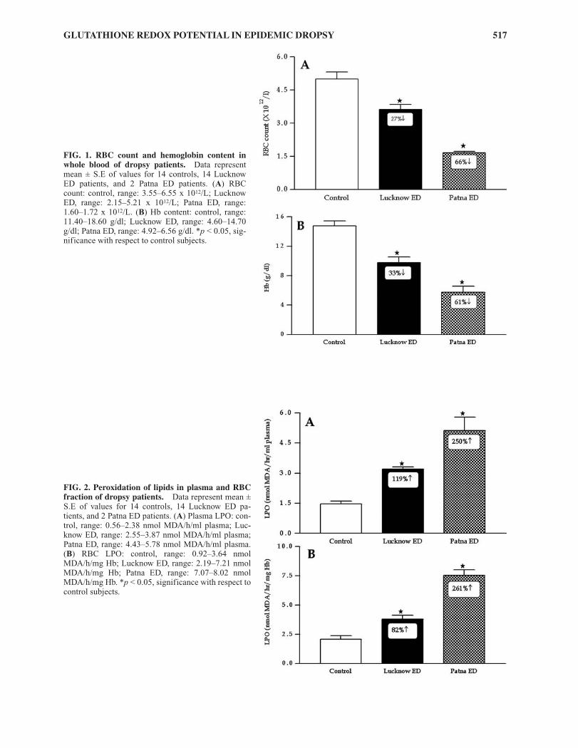
Erythrocytes are highly vulnerable to free radical-inducedlipid peroxidation (LPO) reactions due to their rich content ofpolyunsaturated fatty acids (PUFAs) and continuous exposureto high concentrations of molecular oxygen (5). A significant(p < 0.05) decrease in RBC count (27%) and Hb content(33%) was found in Lucknow dropsy patients (Fig. 1). Thedecrease in RBC count (66%) and Hb content (61%) was rel-atively more in Patna patients with respect to Lucknow pa-
tients when compared to controls (Fig. 2). Formation of lipidperoxides in plasma and RBC of Lucknow dropsy patientswas found to be significantly elevated (82%–119%) whencompared to controls. These levels were enhanced signifi-cantly (p < 0.05) in Patna patients (250–261%) and the valueswere even significantly higher than Lucknow patients (Fig.2). The decrease in RBC count, hemoglobin content, and in-crease in plasma and RBC LPO in the present study may beattributed to the production of ROS by argemone oil and/orsanguinarine (11, 28, 46).
ARE REDOX PAIRS SUCH AS PYRIDINENUCLEOTIDES(S), GLUTATHIONE, AND
THIOL GROUPS TARGET FOR ARGEMONEOIL POISONING IN HUMANS?
Erythrocytes have a variety of redox systems includingGSH, GSSG, NAD, NADH and NADP, and NADPH to pro-vide electrons in ROS detoxification reactions (23). The in-tracellular GSH redox system, with a high GSH level and farlower concentration of oxidized GSSG, is predominantly re-sponsible for the protection of both Hb and RBC membranesagainst oxidation and hemolysis (31). A significant (p < 0.05)decrease in GSH content (42%) with concomitant enhance-ment of GSSG content (107%) was observed in RBC of Luc-know dropsy patients when compared to control subjects.Lower levels of GSH (77%) and higher levels of GSSG(223%) were noticed in Patna patients with respect to Luc-know patients when compared to control (Fig. 3). The ratio ofGSSG/GSH in erythrocytes of Lucknow dropsy patients(threefold) and Patna dropsy patients (14-fold) was found tobe significantly (p < 0.05) increased when compared to con-trols, which may be due to the direct utilization of GSH byGSH peroxidase to detoxify sanguinarine-induced formationof peroxides, or reduced regeneration of GSH from GSSGcatalyzed by GSH reductase (GR) (11). It has been suggestedthat GSH also participates in transhydrogenation reactionsthat are involved in the formation and maintenance of thiolmoieties of proteins, which in turn are essential for maintain-ing flexibility of the RBC membranes, Hb, and other mole-cules such as coenzyme A (20). In this regard, a significantdecrease in thiol content (47%) was observed in RBC of Luc-know dropsy patients, which was relatively more in Patnadropsy patients (69%) when compared to controls (Fig. 4).
Glycolysis is the only source for NADH in the RBC, whichis involved not only for the production of energy but alsoserves as reducing equivalents to maintain Hb in its func-tional [Fe (II)] state (13). Sanguinarine inhibits pyruvate oxi-dase, which results in an increase of pyruvic acid levels in theblood of dropsy patients (38). Our study showed that NADtcontent was increased (44%) significantly (p < 0.05) whereasdecreased levels of NADH (32%) were observed in Lucknowdropsy patients as compared to controls. Increase in NADt(77%) and decrease in NADH content (59%) was relativelymore in Patna dropsy patients with respect to Lucknowdropsy patients when compared to controls. The ratio ofNADH/NADt was found to be decreased significantly (p <0.05) in Lucknow dropsy (53%) and Patna dropsy (78%) pa-
14386Babu.pgs 1/31/07 3:48 PM Page 516
GLUTATHIONE REDOX POTENTIAL IN EPIDEMIC DROPSY 517
FIG. 1. RBC count and hemoglobin content inwhole blood of dropsy patients. Data representmean ± S.E of values for 14 controls, 14 LucknowED patients, and 2 Patna ED patients. (A) RBCcount: control, range: 3.55–6.55 x 10¹²/L; LucknowED, range: 2.15–5.21 x 10¹²/L; Patna ED, range:1.60–1.72 x 10¹²/L. (B) Hb content: control, range:11.40–18.60 g/dl; Lucknow ED, range: 4.60–14.70g/dl; Patna ED, range: 4.92–6.56 g/dl. *p < 0.05, sig-nificance with respect to control subjects.
FIG. 2. Peroxidation of lipids in plasma and RBCfraction of dropsy patients. Data represent mean ±S.E of values for 14 controls, 14 Lucknow ED pa-tients, and 2 Patna ED patients. (A) Plasma LPO: con-trol, range: 0.56–2.38 nmol MDA/h/ml plasma; Luc-know ED, range: 2.55–3.87 nmol MDA/h/ml plasma;Patna ED, range: 4.43–5.78 nmol MDA/h/ml plasma.(B) RBC LPO: control, range: 0.92–3.64 nmolMDA/h/mg Hb; Lucknow ED, range: 2.19–7.21 nmolMDA/h/mg Hb; Patna ED, range: 7.07–8.02 nmolMDA/h/mg Hb. *p < 0.05, significance with respect tocontrol subjects.
14386Babu.pgs 1/31/07 3:48 PM Page 517

tients, when compared to controls (Fig. 5), which suggeststhat the impairment in glycolysis pathway leads to a depletedsupply of reducing equivalents. The NAD redox potential(NADH/NADt) plays an important role in the reduction ofmet-Hb catalyzed by met-Hb reductase, maintaining low lev-els of met-Hb and high concentrations of normal Hb to re-store RBCs oxygen carrying capacity (13). Met-Hb forma-tion in RBC of Lucknow dropsy and Patna dropsy patientswas found to be increased by 155% and 418%, respectively,when compared to controls (Fig. 6). Hence, decrease in
518 BABU ET AL.
NADH/NADt redox potential of RBCs indicates the impair-ment of met-Hb reduction rate and inhibition of glycolysis inthe RBC, which leads to breathlessness in dropsy syndrome.
The hexose monophosphate (HMP) shunt in erythrocytesis critical to supply the reducing equivalents, NADPH, toprotect against oxidative insult through the recycling of GSHfrom GSSG catalyzed by GR (33). RBCs of Lucknow dropsypatients showed significant (p < 0.05) increase (23%) inNADPt along with decrease (33%) in NADPH concentra-tions as compared to control subjects (Fig. 7). The ratio of
FIG. 3. GSH, GSSG, and GSSG/GSH values in RBC fraction of dropsy patients. Data represent mean ± S.E of values for14 controls, 14 Lucknow ED patients, and 2 Patna ED patients. (A) GSH; control, range: 7.61–15.28 µmol/g Hb; Lucknow ED,range: 2.36–9.13 µmol/g Hb; Patna ED, range: 1.91–3.12 µmol/g Hb. (B) GSSG: Control, range: 0.47–1.68 µmol/g Hb; LucknowED, range: 0.85–2.92 µmol/g Hb; Patna ED, range: 2.81–3.35 µmol/g Hb. (C) GSSG/GSH: control, range: 0.031–0.131; LucknowED, range: 0.150–0.880; Patna ED, range: 0.90–1.75. *p < 0.05, significance with respect to control subjects.
14386Babu.pgs 1/31/07 3:48 PM Page 518

GLUTATHIONE REDOX POTENTIAL IN EPIDEMIC DROPSY 519
FIG. 4. Thiol content in RBC fraction of dropsy patients.Data represent mean ± S.E of values for 14 controls, 14 Luc-know ED patients, and 2 Patna ED patients. Control, range:12.69–24.96 µmol/g Hb; Lucknow ED, range: 5.64–11.12µmol/g Hb; Patna ED, range: 4.30–6.11 µmol/g Hb. *p < 0.05,significance with respect to control subjects.
FIG. 5. NADt, NADH, and NADH/NADt values in RBC fraction ofdropsy patients. Data represent mean± S.E of values for 14 controls, 14 Luc-know ED patients, and 2 Patna ED pa-tients. (A) NADt: control, range:69.08–97.61 nmol/ml RBC; LucknowED, range: 93.80–136.58 nmol/mlRBC; Patna ED, range: 138.7–143.7nmol/ml RBC. (B) NADH: control,range: 37.24–64.33 nmol/ml RBC; Luc-know ED, range: 26.30–46.58 nmol/mlRBC; Patna ED, range: 19.67–22.34nmol/ml RBC. (C) NADH/NADt: con-trol, range: 0.492– 0.890; Lucknow ED,range: 0.193–0.387; Patna ED, range:0.13–0.16. *p < 0.05, significance withrespect to control subjects.
FIG. 6. Met-Hb formation in RBC fraction of dropsy pa-tients. Data represent mean ± S.E of 14 controls, 14 Luc-know ED patients, and 2 Patna ED patients. Control, range:0.31–0.87% of total Hb; Lucknow ED, range: 0.81–2.68% oftotal Hb; Patna ED, range: 2.98–3.25% of total Hb. *p < 0.05,significance with respect to control subjects.
14386Babu.pgs 1/31/07 3:48 PM Page 519

NADPH/NADPt was decreased (45%) significantly (p <0.05) in RBC of Lucknow dropsy patients as compared tocontrol subjects. The depletion of NADPH content (60%)and the ratio of NADPH/NADPt (76%) were more severe inPatna patients with respect to Lucknow patients when com-pared to controls (Fig. 7). The decrease in NADPH contentclearly shows the unavailability of NADPH to recycle GSHfrom GSSG, leading to accumulation of GSSG in dropsy pa-
520 BABU ET AL.
tients, which may be due to inhibition of G6PDH, a key en-zyme of HMP shunt to produce NADPH from G6P (3). Fur-ther, high levels of GSSG could inhibit the activity ofG6PDH, which in turn may decrease the NADPH content(36). In addition, depletion of NADPH concentration maydecrease the formation of Hb from met-Hb, as it also plays aminor role in the reduction of met-Hb by NADPH dependentmet-Hb reductase (20).
FIG. 7. NADPt, NADPH, and NADPH/NADPt values in RBC fraction of dropsy patients. Data represent mean ± S.E of 14controls, 14 Lucknow ED patients, and 2 Patna ED patients. (A) NADPt: control, range: 39.30–59.62 nmol/ml RBC; LucknowED, range: 40.56–76.69 nmol/ml RBC; Patna ED, range: 82.12–87.25 nmol/ml RBC. (B) NADPH: control, range: 32.26–58.34nmol/ml RBC; Lucknow ED, range: 21.23– 43.42 nmol/ml RBC; Patna ED, range: 16.34–18.64 nmol/ml RBC. (C)NADPH/NADPt: control, range: 0.66–0.97; Lucknow ED, range: 0.33–0.76; Patna ED, range: 0.19–0.23. *p < 0.05, significancewith respect to control subjects.
14386Babu.pgs 1/31/07 3:48 PM Page 520

GLUTATHIONE REDOX POTENTIAL IN EPIDEMIC DROPSY 521
GLUTATHIONE REDOX POTENTIAL IS APRIME SOURCE FOR THE EXISTING PRO-
OXIDANT ENVIRONMENT
Redox potential (Eh) of GSH/GSSG pool in RBC repre-sent different components of oxidative stress and provides aparameter for pro- and antioxidant events (22). The Eh valuesfor RBCs are more positive in Lucknow and Patna dropsy pa-tients (�193 and �167 mV; pro-oxidant environment) thancontrols subjects (�221 mV; pro-reducing environment) andsupports the concept that erythrocytes of patients are more inoxidative environment. A significant (p < 0.05) reduction(50%) in total antioxidant capacity (TAC) was found in theplasma of Lucknow dropsy patients as compared to controlsubjects. The reduction in TAC was found to be more in Patnadropsy patients (66%) with respect to Lucknow dropsy pa-tients (50%) when compared to controls (Fig. 8), which maybe due to the antioxidant action to scavenge sanguinarine-induced production of free radicals or due to nonavailabilityof reducing equivalents to their oxidized forms.
Sanguinarine alkaloid was found to be present in Patnadropsy patients ranging from 7.15 to 25.7 µg/ml plasma(lanes 2 and 3 of Fig. 9). The spot of dihydrosanguinarine wasalso observed in plasma of Patna dropsy patients. Interest-ingly, the plasma of a patient where sanguinarine content washigher (25.7 µg/ml), showed a metabolite spot at Rf 0.69,However, Lucknow dropsy patients showed neither the pres-
FIG. 8. Redox potential values for glutathionepool in RBC and total antioxidant capacity inplasma of dropsy patients. Data represent mean± S.E of 14 controls, 14 Lucknow ED patients, and2 Patna ED patients. (A) Redox potential values:control [�221 ± �2.45 (range: �207 to �240)];Lucknow ED [�194 ± �3.36 (range: �166 to�207)] + 28 mV; Patna ED [�167 (range: �167 to�167)] + 54 mV. (B) TAC: control [1.28 ± 0.11(range: 0.73–1.74)]; Lucknow ED [0.63 ± 0.04(range: 0.41–0.87)] 50% ↓; Patna ED [0.43 ± 0.04(range: 0.39–0.47)] 66% ↓.
A
B
FIG. 9. HPTLC profile of sanguinarine extracted fromplasma of dropsy patients. Lane 1, control; lanes 2 and 3,Patna ED patients; lane 4, standard sanguinarine; lanes 5–8,Lucknow ED patients. Sanguinarine was extracted fromplasma of dropsy patients as described in Materials and Meth-ods. The extracted samples were analyzed by HPTLC using bu-tanol:acetic acid:water (4:1:2) as mobile phase. The plate wasviewed under dark room at 365 nm.
14386Babu.pgs 1/31/07 3:48 PM Page 521

ence of sanguinarine/dihydrosanguinarine spots nor the meta-bolite spot (Fig. 9, lanes 5–8).
CONCLUSIONS AND DIRECTIONS
The ROS-dependent damage of erythrocyte membranes andHb along with decreases in GSH, NADH, NADPH, and thiolcontents is responsible to enhance pro-oxidant environment
522 BABU ET AL.
and alterations in plasma TAC leading to the development ofnormocytic anemia in dropsy patients having breathlessness(Fig. 10). Enhancement of the pro-oxidant environment inPatna patients as compared to Lucknow patients may be due tothe fact that antioxidant therapy including �-tocopherol wasnot given to Patna patients. Hence, antioxidant therapy may bebeneficial to ED patients and needs detailed clinical trials infuture. Although Indian legislation clearly suggests that mus-tard oil should be absolutely free from argemone oil contami-nation (34), however, unscrupulous traders adopt unethical
Plasma
Plasma TAC (↓)
Degradation of erythrocytes & oxidation of hemo globin (↑)
Anemia and breathlessness
Alterations in redox potential [Pro-oxidant environment, ( ↑)]
Argemone oil/ sanguinarine
RBC
ROS
O2
GSSG (↑)
GSH (↓)
Glutathione reductase
Protein-SSG
Protein-SH (↓)
Thioltransferase
NAD +
NADH ( ↓)
NADPH ( ↓)
NADP +
Transhydrogenation
FIG. 10. Mechanism(s) of producing pro-oxidant environment due to alterations in glutathione and pyridine nucleotidesleading to decrease of total antioxidant capacity, which in turn causes damage of erythrocytes and hemoglobin in epi-demic dropsy patients.
14386Babu.pgs 1/31/07 3:48 PM Page 522

practices for economic gains in loose mustard oil (Fig. 11),which is consumed by lower socioeconomic groups. Hence, theregulatory authorities should enforce the banning of the sale ofloose mustard oil and at the same time suggest the oil industryto prepare tamper-proof small packing (~100–200 g), so thatthe commodity is within the purchase capacity of lower strata.
ACKNOWLEDGMENTS
The authors are grateful to Dr. C.M. Gupta, Director, Indus-trial Toxicology Research Center (ITRC) for his keen interest inthe study. C.K.B and S.K.K are thankful to Indian Council ofMedical Research (ICMR), New Delhi for the award of SeniorResearch Fellowship and Emeritus Scientist position, respec-tively. Our appreciations are due to KGMU, Lucknow for provid-ing the blood samples of dropsy patients and Technology Missionon Oil Seed, Pulses & Maize (TMOP&M), New Delhi for finan-cial support. The manuscript is ITRC communication #2536.
ABBREVIATIONS
ED, epidemic dropsy; GSH, glutathione reduced; GSSG,glutathione oxidized; Hb, hemoglobin; LPO, lipid peroxida-tion; MDA, malondialdehyde; NADt, nicotinamide adeninedinucleotide total; NADPt, nicotinamide adenine dinu-cleotide phosphate total; Met-Hb, met-hemoglobin; ROS, re-active oxygen species; TAC, total antioxidant capacity.
APPENDIX
1. �-Nicotinamide adenine dinucleotide phosphate reduced(NADPH), nicotinamide adenine dinucleotide phosphate oxidized(NADP), nicotinamide adenine dinucleotide reduced (NADH), nicoti-
GLUTATHIONE REDOX POTENTIAL IN EPIDEMIC DROPSY 523
namide adenine dinucleotide oxidized (NAD), nicotinamide, glu-tathione reduced (GSH), glutathione oxidized (GSSG), 5,5�-dithio-bis-2-nitrobenzoic acid (DTNB), thiobarbaturic acid (TBA), malondialde-hyde (MDA), O-pthalaldehyde (OPT), N-ethyl maleimide (NEM),disodium ethylene diamine tetraacetic acid (Na2 EDTA), tris (hydrox-ymethyl) aminomethane, glucose-6-phosphate (G6P), 3-[4,5-di-methylthiazol-2-yl]-2,5-diphenyl tetrazolium bromide (MTT),phenazine ethosulfate (PES), and sanguinarine (>98%) were purchasedfrom Sigma Chemical Co, St. Louis, MO. Alcohol dehydrogenase(ALDH) from baker’s yeast was procured from SRL Pvt Ltd, Mumbai,India. Glucose-6-phosphate dehydrogenase (G6PDH) from yeast wasobtained from Boehringer, Mannheim, Germany. Absolute alcohol wasobtained from Bengal Chemicals and Pharmaceuticals, Calcutta, India.Hemoglobin and total antioxidant capacity assay kits were procuredfrom Accurex Biomedical Pvt Ltd. Mumbai, India and Randox Labora-tories Ltd, Ireland, UK, respectively. Vacutainer tubes were obtainedfrom Becton Dickinson Co, NJ. Water was purified by a Millipore sys-tem (Milli-Q Corp, Billerica, MA) and had a resistivity >18 M W cm�l.All the other chemicals used were of the highest purity available fromcommercial sources.
2. During February 2005, an outbreak of epidemic dropsy was re-ported in Rajajipuram, Lucknow, Uttar Pradesh, India. Fourteen pa-tients from different families of same locality were admitted to KingGeorge’s Medical University (KGMU), Lucknow, and were givensymptomatic treatment along with antioxidant therapy including �-to-copherol, based on our earlier clinical findings (26). Two patients fromPatna (Bihar, India) were admitted to KGMU during December 2004with symptoms of epidemic dropsy for the past 2 months and had undergone symptomatic treatment only. Lucknow patients were of differentage groups including children, adolescents, and adults of both sexes,while Patna patients were husband and wife (37 and 47 years). By seek-ing the history of the patients and the records at KGMU, it was con-firmed that all patients had consumed mustard oil adulterated with AOas the samples of edible oil consumed by these patients were found tocontain argemone alkaloids using ITRC argemone oil detection kit (9).
3. Fourteen dropsy patients from Lucknow outbreak and 2 patientsfrom Patna outbreak and 14 healthy volunteers were involved in this in-vestigation. Blood was collected from Lucknow dropsy patients atKGMU after 1 week of antioxidant therapy and from 2 Patna patientsafter 2 months of appearing symptoms, in vacutainer tubes containingsodium citrate as anticoagulant and brought to the lab within an hour inice-cold condition. A portion of blood was used for erythrocyte countand hemoglobin concentration and the other portion of blood was used
AGMARK
BIS, PFA,PCR
Genuine raw material
OrganizedOil Industry
Cheaper admixed raw material
Small Scale Expellers
Adulteration in mustard seeds ?
Mustard seeds Argemone seeds
Bulk Auction Sites (Mandi)
Unscrupulous collectors
? Adulteration in
mustard oil
Distributors
Adulteration in mustard oilSWM , PFA
CPA
Retail outlets
Packed Oil (1-15 kg)
Retail outlets
Loose oil
Consumers
FIG. 11. Various steps from oil seeds to retail sellingof mustard oil in Indian markets. Steps that are regu-lated by government acts are marked with dotted arrowsin asterisks. Possible adulteration of mustard oil is men-tioned at various steps before reaching consumers. Apartfrom different government acts regulating the genuinemustard oil, two additional steps (marked with questionmark) may be introduced as amendments in the legisla-tion so as to curtail this menace, which are mentioned inthe Conclusion. AGAMRK, Agricultural Produce Mark-ing and Grading Act; BIS, Bureau of Indian Standards;CPA, Consumer Protection Act; PCR, Packaged Com-modities Rules; PFA, Prevention of Food AdulterationAct; SWM, Standards of Weights and Measures Act.
14386Babu.pgs 1/31/07 3:48 PM Page 523

for preparation of RBC fraction (4). Briefly, blood was centrifuged at2,500 g for 15 min at 4°C and erythrocytes were separated from plasmaand buffy coat. The erythrocyte-rich precipitate was washed threetimes with saline (3:1, vol/vol) and directly used for the assay of lipidperoxidation (LPO), hemoglobin oxidation, oxidized and reducedGSH, oxidized and reduced pyridine nucleotide content, and thiolgroups. Plasma was used for the assay of LPO, total antioxidant capac-ity, and sanguinarine content.
4. RBC count and hemoglobin content in whole blood sampleswere carried out on Automatic Full Digital Cell Counter (MS9, MeletSchloesing Laboratories, Cergy-Pointoise, France) and expressed asnumber of cells � 1012/l and g/dl, respectively (45). Hemoglobin con-tent of the RBC fraction was estimated by using an Accurex kit follow-ing the instructions given by the manufacturer, and these values wereused for the calculation of LPO, GSH, GSSG, and free sulfhydryls inerythrocytes.
5. The extent of LPO in plasma and RBC fraction was assayed bymeasuring the formation of malondialdehyde (MDA) and expressed asnmol MDA/hr/ml (plasma) or nmol MDA/hr/mg Hb (RBC) using amolar extinction coefficient of 1.56 � 105 M�1 cm�1 (32). Thiol con-tent was assayed in the RBC fraction using Ellman’s reagent and ex-pressed as µmol/g Hb (15).
6. The content of GSH and GSSG in RBC was assayed accordingto the modified method of Hissin and Hiff (19) using OPT as fluores-cent probe, which allows binding of GSH to form highly fluorescentderivative. Briefly, 0.2 ml RBC pellet was diluted with 0.8 ml distilledwater and precipitated with 10% TCA (1 ml). Supernatant was used forGSH and GSSG determination. For GSH assay, a 4 ml reaction mixtureconsisted of 3.25 ml phosphate-EDTA buffer (0.1 M, pH 8.0), 0.25 mlOPT (1 mg/ml methanol), and 0.5 ml supernatant. Fluorescence ofGSH-OPT adduct was determined after 15 min, using excitation andemission wavelengths of 350 and 450 nm (slit widths 5 and 10 nm), re-spectively. For GSSG assay, NEM was used to prevent oxidation ofGSH to GSSG during the assay, which allows the formation of stablecomplex with GSH. After incubating 0.5 ml of supernatant with NEM(0.04 M) for 30 min, GSSG was measured using the procedure outlinedabove (for GSH assay), except that 0.1 N NaOH was employed as dilu-ant rather than phosphate-EDTA buffer. The quantification of GSH andGSSG was performed by comparing the standard GSH and GSSG fluo-rescence and expressed as µmol/g Hb. The ratio of GSSG and GSH wascalculated by using specific values in the individual samples.
7. Redox potential of GSH/GSSG pool (2GSH → GSSG + 2e� +2H+) in erythrocytes was calculated using Nernst equation: Eh (mV) =E0�[(RT/nF) log (thiol2)/(disulfide)], where E0 is the respective stan-dard potential (�264 mV) for the redox couple at pH 7.4, R is the gasconstant (8.314 Jk�1mol�1), T is the absolute temperature (310 K), n isnumber of electrons transferred, (2) and F is the Faraday constant(96485 J/V) (39).
8. The content of pyridine nucleotides were determined by spec-trophotometric cycling assay with some minor modifications (48). Inbrief, 20 µl of RBC suspension was mixed with 1.98 ml of alkaline ex-traction solution containing nicotinamide (10 µM), NaHCO3 (20 µM),and Na2CO3 (100 µM). The reaction mixture was frozen immediatelyand thawed quickly in a room temperature water bath. The mixture wasagain chilled to 0°C followed by centrifugation at 14,000 g for 1 min at4°C, and the supernatant was separated. For measurement of reducedform of nucleotides, the oxidized forms of NAD and NADP were de-stroyed by incubating 700 µl supernatant at 60°C for 30 min and againchilled to 0°C. Unheated supernatant was used for the assay of totalNAD (NADt) and NADP (NADPt). In brief, a volume of 100 µl ofheated (NADH) or unheated (NADt) supernatant was added to 800 µlof recycling buffer, containing 100 µmol Tris-Cl (pH 8.0), 2 µmol PES,0.5 µmol MTT, and 0.2 mg ALDH. The mixture was incubated for 5min at 37°C, and 100 µl of ethanol (6 M) was added as substrate. Therate of increase of absorbance was measured at 570 nm over a 5-minperiod. For the assay of NADPH and NADPt, 1.3 IU/ml of G6PDH andG6P (1 mmol) was used as enzyme and substrate instead of ALDH andethanol. The rate of increase in absorbance was measured at 570 nmover a 90-sec period and expressed as nmol/ml RBC.
9. The oxidation of hemoglobin in erythrocytes was evaluated bymeasuring the formation of met-hemoglobin (Met-Hb) (16). In brief,RBC fractions (20 µl) were lysed with distilled water (1.98 ml). A por-tion of this lysate (600 µl) was mixed with 400 µl phosphate buffer (0.5
524 BABU ET AL.
M, pH 6.1) and centrifuged at 15,000 g for 5 min to sediment debris. Theabsorbance of supernatant fraction was measured at 630 nm before (S1)and after (S2) conversion to cyanomethemoglobin by the addition of 50µl of 10% potassium cyanide (KCN). The difference between S1 and S2represents the absorbance due to Met-Hb. Another portion of supernatantwas used to measure total hemoglobin levels, after converting to Met-Hbby potassium ferricyanide before (T1) and after (T2) addition of KCN.The Met-Hb formation was calculated by using the formula[100(S1�S2)]/[10(T1�T2)] and expressed as percentage of total Hb.
10. TAC was assayed in plasma by commercially available kit ac-cording to the method of Miller et al. (30). In brief, 1 ml chromogensolution containing 610 µM 2,2�-azino-di-[3-ethylbenzthiazolinesulphonate] (ABTS) and 6.1 µM peroxidase (metmyoglobin) was incu-bated with 0.2 ml H2O2 (250 µmol/l) to produce the cation radicalABTS+. This radical has a relatively stable blue-green color and has theabsorption maxima at 600 nm. Antioxidants in the sample (20 µl) causesuppression of this color production to a degree, which is proportionalto their concentration. The results were calibrated using a standardcurve of Trolox (6-hydroxy-2, 5, 7, 8-tetramethylchro-man-2-car-boxylic acid) a water-soluble vitamin E analogue and expressed asmmole equivalents of Trolox concentration per liter plasma (mM).
11. Sanguinarine was extracted from plasma and analyzed usinghigh performance thin layer chromatography (HPTLC). In brief,plasma (1 ml) was digested with 2 N HCl (1 ml) at 90°C for 30 min,followed by centrifugation at 3,000 rpm for 20 min. Supernatant (~2ml) fraction was separated and extracted with 2 ml chloroform: aceticacid (3:1.5, vol/vol). The mixture was allowed to separate aqueous andorganic phase at room temperature for 30 min. Upper acid layer wasseparated and filtered through filter (0.22 µ, Millipore) and dried onsand bath. The residue was dissolved in methanol and spotted onto aprecoated silica gel plate (E. Merck, Mumbai, India). The plate was runusing butanol, acetic acid, water (4:1:2, vol/vol/vol) as a mobile phaseand the content of sanguinarine in plasma was calculated using puresanguinarine as standard and expressed as µg/ml plasma.
12. All results were expressed as the mean + standard error (SE), asindicated in the tables. Statistical comparisons were made by analysisof variance with rank ordering (42) employed to calculate the signifi-cance of difference between the means of healthy controls and the epi-demic dropsy patients. A value of p < 0.05 was used as the level ofsignificance.
REFERENCES
1. Ansari KM, Chauhan LK, Dhawan A, Khanna SK, and Das M.Unequivocal evidence of genotoxic potential of argemone oil inmice. Int J Cancer 112: 890–895, 2004.
2. Ansari KM, Dhawan A, Khanna SK, and Das M. In vivo DNAdamaging potential of sanguinarine alkaloid, isolated from arge-mone oil, using alkaline Comet assay in mice. Food & Chem Toxi-col 43: 147–153, 2005.
3. Babu CK, Khanna SK, and Das M. Glutathione redox paradigm inblood, a biomarker of oxidative stress in Epidemic Dropsy pa-tients. Presented in “International Conference on Antioxidants &Free Radicals in Health-Nutrition & Radio Protectors and VI An-nual Conference of Society of Free Radical Research in India” atSt. John’s National Academy of Health Sciences, Bangalore,India, January 10–12, 2005.
4. Beutler E. Red blood cell metabolism. In: Rossi’s Principles ofTransfusion Medicine, 3rd ed, edited by Simon TL, Dzik WH, andSnyder EL. Philadelphia: Lippincott Williams & Wilkins, 2002,pp. 43–49.
5. Chiu D, Lubin B, and Shohet SB. Peroxidative reactions in red cellbiology. In: Free Radicals in Biology, Vol. 5, edited by Pryor WA,New York, NY: Academic Press, 1982, pp. 115–160.
6. Dalal KR. Report on the investigation of an outbreak in Rangoonin the year 1924. Indian Med Gaz 64: 19–21, 1929.
7. Das M and Khanna SK. Clinico-epidemiological, toxicological,and safety evaluation studies on argemone oil. Crit Rev Toxicol27: 273–297, 1997.
8. Das M and Khanna SK. Epidemic dropsy. Natl Medical J India11: 207–208, 1998.
14386Babu.pgs 1/31/07 3:48 PM Page 524

9. Das M and Khanna SK. Management of epidemic dropsy causedby argemone oil. In: Health and Dietary Aspects of Mustard Oil,edited by Bhatnagar AK, Singh HB, and Prasad S. MRPC, NewDelhi, 2001, pp. 139–149.
10. Das M, Ansari KM, Dhawan A, Shukla Y, and Khanna SK. Corre-lation of DNA damage in epidemic dropsy patients to carcino-genic potential of argemone oil and isolated sanguinarine alkaloidin mice. Int J Cancer 117: 709–717, 2005.
11. Das M, Babu K, Reddy NP, and Srivastava LM. Oxidative damageof plasma proteins and lipids in epidemic dropsy patients: alter-ations in antioxidant status. Biochim Biophys Acta 1722: 209–217,2005.
12. Deva Sangayam AD. An epidemic of dropsy among Indians inFiji. Indian Med Gaz 62: 506–508, 1927.
13. Duncan JR, Prasse KW, and Mahaffey EA. Erythrocytes. In: Vet-erinary Laboratory Medicine: Clinical Pathology, edited by Dun-can JR, Prasse KW, and Mahaffey EA. Ames, Iowa: Iowa StateUniversity Press, 1994, pp. 3–35.
14. Dutta AK, Joshi CK, and Dutta P. Epidemic dropsy outbreak inRajasthan. J Indian Med Assoc 83: 268–271, 1985.
15. Ellman GL. Tissue sulfhydryl groups. Arch Biochem Biophys 82:70–77, 1959.
16. Evelyn K and Malloy H. Micro determination of oxyhemoglobin,methemoglobin, and sulfhemoglobin in a single sample of blood.J Biol Chem 126: 655–662, 1938.
17. Eversole LR, Eversole GM, and Kopcik J. Sanguinaria-associatedoral leukoplakia: comparison with other benign and dysplasticleukoplakic lesions. Oral Surg Oral Med Oral Pathol Oral RadiolEndod 89: 455–464, 2000.
18. Frankos VH, Brusick DJ, Johnson EM, Maibach HI, Munro I,Squire RA, and Weil CS. Safety of sanguinaria extract as used incommercial toothpaste and oral rinse products. J Can Dent Assoc56S: 41–47, 1990.
19. Hissin PJ and Hilf R. A fluorometric method for determination ofoxidized and reduced glutathione in tissues. Anal Biochem 74:214–226, 1976.
20. Jaffe ER. The formation and reduction of methemoglobin inhuman erythrocytes. In: Cellular and Molecular Biology of Eryth-rocytes, edited by Yoshikawa H and Rapoport SM. Baltimore,MD: University Park Press, 1974, p. 345.
21. Jha N, Chaturvedi S, Yadav BK, Karki DB, and Rai BK. Field in-vestigation of an outbreak of epidemic dropsy in eastern Nepal.Indian J Public Health 43: 87–88, 1999.
22. Jones DP. Redox potential of GSH/GSSG couple: assay and bio-logical significance. Methods Enzymol 348: 93–112, 2002.
23. Kehrer JP and Lund LG. Cellular reducing equivalents and oxida-tive stress. Free Radic Biol Med 17: 65–75, 1994.
24. Krishnamachari KA and Satyanarayana K. Epidemic dropsy inAndhra Pradesh. Indian J Med Res 60: 741–746, 1972.
25. Kuftinec MM, Mueller–Joseph LJ, and Kopczyk RA. Sanguinariatoothpaste and oral rinse regimen clinical efficacy in short- andlong-term trials. J Can Dent Assoc 56S: 31–33, 1990.
26. Kumar A, Hussain F, Das M, and Khanna SK. An outbreak of epi-demic dropsy in the Barabanki District of Uttar Pradesh, India: alimited trial for the scope of antioxidants in the management ofsymptoms. Biomed Environ Sci 5: 251–256, 1992.
27. Lal SB. Epidemic dropsy in Bihar. Indian Med Gaz 86: 64–71,1951.
28. Maiti M and Chatterjee A. Production of singlet oxygen by san-guinarine and beriberine. Curr Sci 68: 734–736, 1995.
29. Maiti M. Molecular aspects on the interaction of sanguinarinewith B-form, Z-form and HL-form of DNA structures. Indian JBiochem Biophys 38: 20–26, 2001.
30. Miller NJ, Rice–Evans C, Davies MJ, Gopinathan V, and Milner A.Method for measuring antioxidant capacity and its application tomonitoring the antioxidant status in premature neonates. Clin Sci84: 407–412, 1993.
31. Nemeth I, Orvos H, and Boda D. Blood glutathione redox status ingestational hypertension. Free Radic Biol Med 30: 715–721, 2001.
GLUTATHIONE REDOX POTENTIAL IN EPIDEMIC DROPSY 525
32. Ohkawa H, Ohishi N, and Yagi K. Assay for lipid peroxides in ani-mal tissues by thiobarbituric acid reaction. Anal Biochem 95:351–358, 1979.
33. Pandolfi PP, Sonati F, Rivi R, Mason P, Grosveld F, and LuzzattoL. Targeted disruption of the housekeeping gene encoding glucose6-phosphate dehydrogenase (G6PD): G6PD is dispensable forpentose synthesis but essential for defense against oxidativestress. EMBO J 14: 5209–5215, 1995.
34. The Prevention of Food Adulteration (PFA) Act of India, 1954:Rules and Notifications, 26th edition, Eastern Book Co., Lucknow,India, 2005.
35. Rai Chaudhuri MN and Chauduri RN. Epidemic dropsy in WestDinajpur. Bull Calcutta Sch Trop Med 13: 37–38, 1965.
36. Rodriquez–Segade S, Ramos–Martinez JL, and Freire M. Reversaleffect of oxidized glutathione on the inhibition of glucose-6-phos-phate dehydrogenase by NADPH. Biochem J 231: 805–806, 1985.
37. Sandhu HS, Tiwari SC, Naik G, and Bose C. An epidemiologicalstudy of an outbreak of epidemic dropsy in Itarsi (MP). Indian JPublic Health 26: 10–15, 1982.
38. Sarkar SN. Isolation from argemone oil of dihydrosanguinarinesanguinarine: toxicity of sanguinarine. Nature (London) 162:265–266, 1948.
39. Schafer FQ and Buettner GR. Redox state of the cell as viewedthough the glutathione disulfide/glutathione couple. Free RadicBiol Med 30: 1191–1212, 2001.
40. Sies H. Oxidative stress: oxidants and antioxidants. Exp Physiol82: 291–295, 1997.
41. Singh NP, Anuradha S, Dhanwal DK, Singh K, Parkash A, MadanK, and Agarwal SK. Epidemic dropsy-a clinical study of the Delhioutbreak. J Assoc Phys India 48: 877–880, 2000.
42. Snedecor GW and Cochran WG. One way classification: analysisof variance. In: Statistical Methods, 8th ed, edited by Snedecor GWand Cochran WG. Ames, Iowa: Iowa State University Press, 1989,pp. 217–236.
43. Steyn DG. Poisoning with the seed of Argemone mexicana (Mexi-can poppy) in human beings; Indian epidemic dropsy in SouthAfrica. S Afr Med J 24: 333–339, 1950.
44. Stiborova M, Simerek V, Frei E, Hobza P, and Ulrichova J. DNAadduct formation from quaternary benzo[c]phenanthridine alka-loids sanguinarine and chelerythrine as revealed by the 32P- post-labeling technique. Chem Biol Int 140: 231–242, 2002.
45. Tietz NW. (Ed). Clinical Guide to Laboratory Tests. 2nd ed. Phila-delphia: WB Saunders, 1990, pp. 166–167.
46. Tuveson RW, Larson RA, Marley KA, Wang GR, and BerenbaumMR. Sanguinarine, a phototoxic H2O2 producing alkaloid. Pho-tochem Photobiol 50: 733–738, 1989.
47. Wadia RS, Relwani GS, Batra RK, Ichaporia RN, and Grant KB.Epidemic dropsy in Poona 1969 (clinical features and 1 year fol-low up). J Assoc Physicians India 19: 641–646, 1971.
48. Wagner TC and Scott MD. Single extraction method for thespectrophotometric quantification of oxidized and reduced pyri-dine nucleotides in erythrocytes. Anal Biochem 222: 417–426,1994.
Address reprint requests to:Dr. Mukul Das
Food Toxicology LaboratoryIndustrial Toxicology Research Centre
PO Box #80M.G.Marg
Lucknow-226 001, India
E-mail: [email protected]
Date of submission to ARS Central, October 21, 2006; date ofacceptance, October 22, 2006.
14386Babu.pgs 1/31/07 3:48 PM Page 525

14386Babu.pgs 1/31/07 3:48 PM Page 526