Adolpho Lutz and tropical medicine - SciELO Livros
214
Historical introduction Insects, people, and disease: Adolpho Lutz and tropical medicine Jaime L. Benchimol Magali Romero Sá SciELO Books / SciELO Livros / SciELO Libros BENCHIMOL, JL., and SÁ, MR., eds. and orgs. Adolpho Lutz: Febre amarela, malária e protozoologia = Yellow fever, malaria and protozoology [online]. Rio de Janeiro: Editora FIOCRUZ, 2005. 956 p. Adolpho Lutz Obra Completa, v.2, book 1. ISBN: 85-7541-064-4. Available from SciELO Books <http://books.scielo.org >. All the contents of this work, except where otherwise noted, is licensed under a Creative Commons Attribution-Non Commercial-ShareAlike 3.0 Unported. Todo o conteúdo deste trabalho, exceto quando houver ressalva, é publicado sob a licença Creative Commons Atribuição - Uso Não Comercial - Partilha nos Mesmos Termos 3.0 Não adaptada. Todo el contenido de esta obra, excepto donde se indique lo contrario, está bajo licencia de la licencia Creative Commons Reconocimento-NoComercial-CompartirIgual 3.0 Unported.
-
Upload
khangminh22 -
Category
Documents
-
view
3 -
download
0
Transcript of Adolpho Lutz and tropical medicine - SciELO Livros
Historical introduction Insects, people, and disease: Adolpho Lutz and tropical medicine
Jaime L. Benchimol Magali Romero Sá
SciELO Books / SciELO Livros / SciELO Libros BENCHIMOL, JL., and SÁ, MR., eds. and orgs. Adolpho Lutz: Febre amarela, malária e protozoologia = Yellow fever, malaria and protozoology [online]. Rio de Janeiro: Editora FIOCRUZ, 2005. 956 p. Adolpho Lutz Obra Completa, v.2, book 1. ISBN: 85-7541-064-4. Available from SciELO Books <http://books.scielo.org>.
All the contents of this work, except where otherwise noted, is licensed under a Creative Commons Attribution-Non Commercial-ShareAlike 3.0 Unported.
Todo o conteúdo deste trabalho, exceto quando houver ressalva, é publicado sob a licença Creative Commons Atribuição - Uso Não Comercial - Partilha nos Mesmos Termos 3.0 Não adaptada.
Todo el contenido de esta obra, excepto donde se indique lo contrario, está bajo licencia de la licencia Creative Commons Reconocimento-NoComercial-CompartirIgual 3.0 Unported.
245 TRABALHOS SOBRE FEBRE AMARELA, MALÁRIA, PROTOZOOLOGIA
Jaime L. Benchimol
Magali Romero Sá
A dolpho Lutz spent seventeen years far from his family. Born in the
Brazilian capital on 18 December 1855, the second of Gustav and
Mathilde Oberteuffer Lutz’s ten children, Adolpho was two when his parents
returned to Switzerland in 1857, seven years after emigrating to Brazil.
In 1864, they once again relocated to Rio, leaving their three eldest children
to study in Basilea. Adolpho would only be reunited with his parents in
1881, when he went back to Rio with his medical degree in hand and a
solid background in medicine and biology, acquired at German-speaking
universities. While he was away, the city that had welcomed these Swiss
immigrants had undergone remarkable changes.
When Adolpho Lutz’s parents disembarked in Rio de Janeiro, in late
1849/early 1850, the forces that had in the previous decades resisted the
Empire’s centralizing policies and Southeastern Brazil’s escalating economic
hegemony had been tamed.
Divided essentially into masters and slaves, the port city was thriving
as the point of contact between an expanding coffee crop – tended by slaves
in the Paraíba River Valley – and the world market. Its streets bustled
with slaves hired out by their masters on a daily basis for work in a wide
gamut of trades. The slaves received only a small portion of the money
earned and applied it towards their own sustenance: food, drink, perhaps
Insects, people, anddisease: Adolpho Lutzand tropical medicine
246 ADOLPHO LUTZ — OBRA COMPLETA Vol. 2 — Livro 1
even a room in a tenement. But most of the earnings went to fatten the
coffers of the richest slave holders or to guarantee the survival of the poorest
among them, sometimes almost as poor as the working slaves themselves.
Household slaves did the multiple tasks demanded by the natural economy
buttressing residences based on slave labor. This meant they supplied their
masters’ homes with water and removed sewage, duties soon transformed
into profitable ‘public’ services in the hands of private companies.
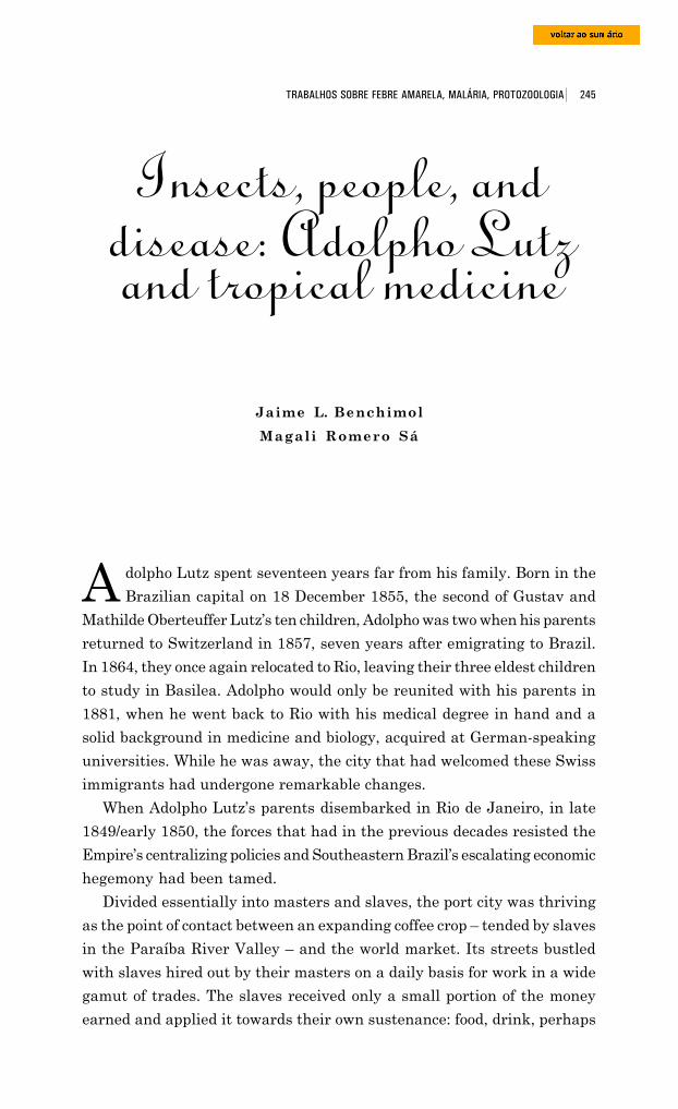
Largo da Carioca, 1844. Watercolor on paper, by Eduard Hildebrand, entitled “Brunnen in Rio de Janeiro”[fountains in Rio de Janeiro], showing the Carioca Fountain, the Terceira da Penitência Hospital, SaintAnthony’s Church and Convent. Staatliche Museen zu Berlin collection, Alemanha (Belluzo, 1994, v.3, p.106,fig. 481).
In the ensuing decades, the second industrial revolution – the
revolution of iron and steel manufacturing, of capital goods, and of the
construction of railways and steamboats – would consolidate England’s
world power, albeit other countries likewise revolutionized by big industry
were emerging as serious competitors. Capital exports, in the form of public
loans and direct investments, lent impetus to the modernization of
peripheral economies like Brazil’s, equipping them to respond to the new
inflow of raw materials and industrialized goods.
In the time between Gustav and Mathilde Lutz’s immigration to Brazil
and young doctor Adolpho’s return voyage, other processes helped to change

247 TRABALHOS SOBRE FEBRE AMARELA, MALÁRIA, PROTOZOOLOGIA
the face of the Brazilian capital: abolition of the slave trade in 1850, the
Paraguayan War (1864-70), demographic growth, and the gradual
expansion of freed labor. In the 1870s, the Empire of Dom Pedro II and of
the coffee barons seemed to be at the heyday of its grandeur and stability,
and Brazil was living out its apparent destiny as a primarily agricultural
nation. Rio de Janeiro was its most prosperous commercial and financial
center. Matching the pace at which Paraíba Valley plantations absorbed
the last contingent of Brazilian slaves through interprovincial trade, Rio
made ample room for wage labor. The circulation of merchandise – basis of
the urban economy – improved qualitatively thanks to these new labor
relations. The production of important manufactures notwithstanding, the
productive sector remained an appendix of import and export activities.
Rio de Janeiro’s Providência Hill in 1866, then known as Gamboa Hill. Photograph by Augusto Malta(Museu Histórico Nacional, Rio de Janeiro).
Replacing river and pack-mule transportation, the tracks of the Dom
Pedro II and Leopoldina railways brought Rio de Janeiro closer to its rural
rear guard. Likewise revolutionized by steam power, maritime transport
increased alongside a complex of commercial and financial enterprises
formed chiefly of British capital. The port itself saw its first reforms, like
metal warehouses and steam-driven cranes on the Customs House pier,
where cargo handling could now be done without slave labor.
In the 1860s through 1870s, foreign companies, along with some
Brazilian ones, began setting up public services: gas lighting, household
248 ADOLPHO LUTZ — OBRA COMPLETA Vol. 2 — Livro 1
water and sewer, garbage disposal, urban transit, and so on. This helped
eliminate the system under which slaves provided such services and also
undermined the household economy responsible for the self-sufficiency of
slave-run residences. Tramway companies spearheaded extension of the
urban network beyond the former perimeter of the Old City and of Rio’s
latest development, the New City. Although new neighborhoods were
springing up, critical problems became concentrated in downtown Rio, born
of a growing incompatibility between the former material structure and
the new capitalist economic relations taking root within this framework.
Between rail station, docks, and the labyrinth of commerce, the center’s
narrow, winding streets became crowded with a greater inflow of men and
merchandise, including large iron manufactures. The populous downtown
was home to a wide assortment of edifices: offices and banks, stores, repair
shops, waterfront warehouses, public buildings, private one- and two-story
residences, grocery stores oft times servicing tenements and the like, old
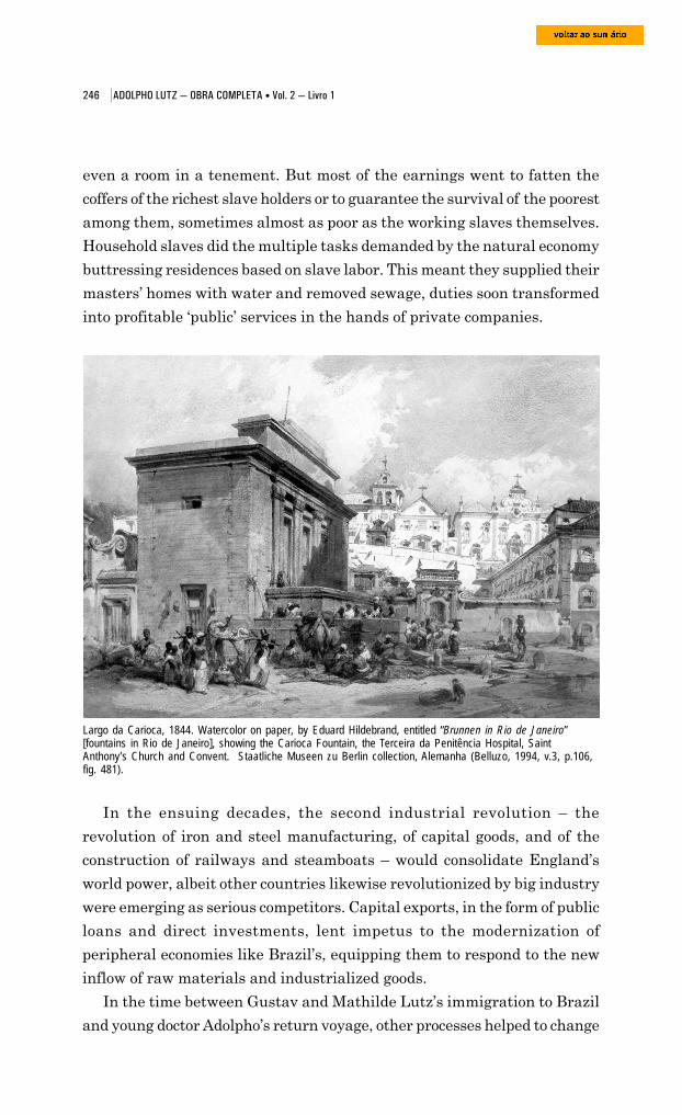
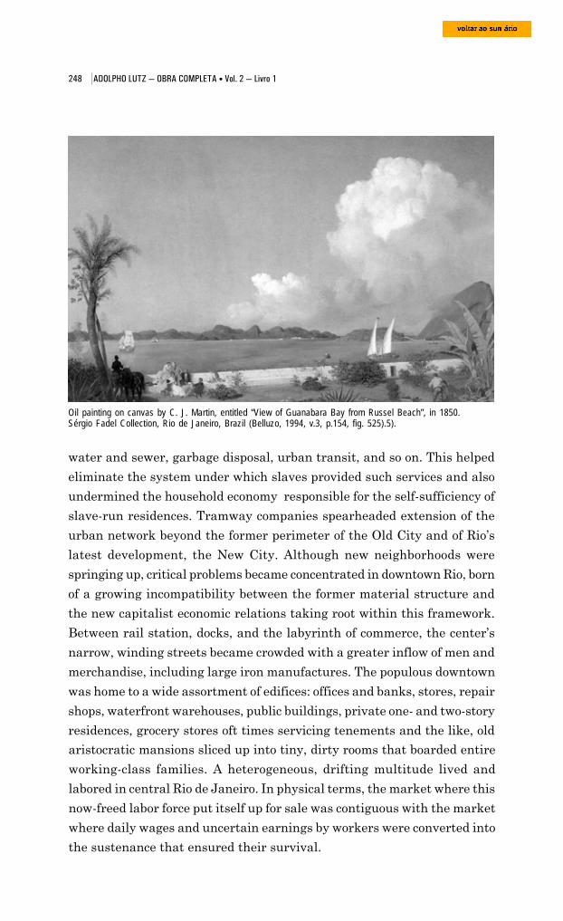
Oil painting on canvas by C. J. Martin, entitled “View of Guanabara Bay from Russel Beach”, in 1850.Sérgio Fadel Collection, Rio de Janeiro, Brazil (Belluzo, 1994, v.3, p.154, fig. 525).5).
aristocratic mansions sliced up into tiny, dirty rooms that boarded entire
working-class families. A heterogeneous, drifting multitude lived and
labored in central Rio de Janeiro. In physical terms, the market where this
now-freed labor force put itself up for sale was contiguous with the market
where daily wages and uncertain earnings by workers were converted into
the sustenance that ensured their survival.
249 TRABALHOS SOBRE FEBRE AMARELA, MALÁRIA, PROTOZOOLOGIA
There, every year, more or less deadly epidemics broke out. Morbidity
and mortality rates varied with the biological and social synergy of those
who came into contact with each other during the course of each disease.
Smallpox epidemics generally occurred in the winter. Cholera struck Rio
de Janeiro in 1855-56, at the close of the 19th century’s third major pandemic.
In the nation’s capital and in its provinces, tuberculosis, dysenteries,
malaria, and fevers known by dozens of names raged like chronic scourges.
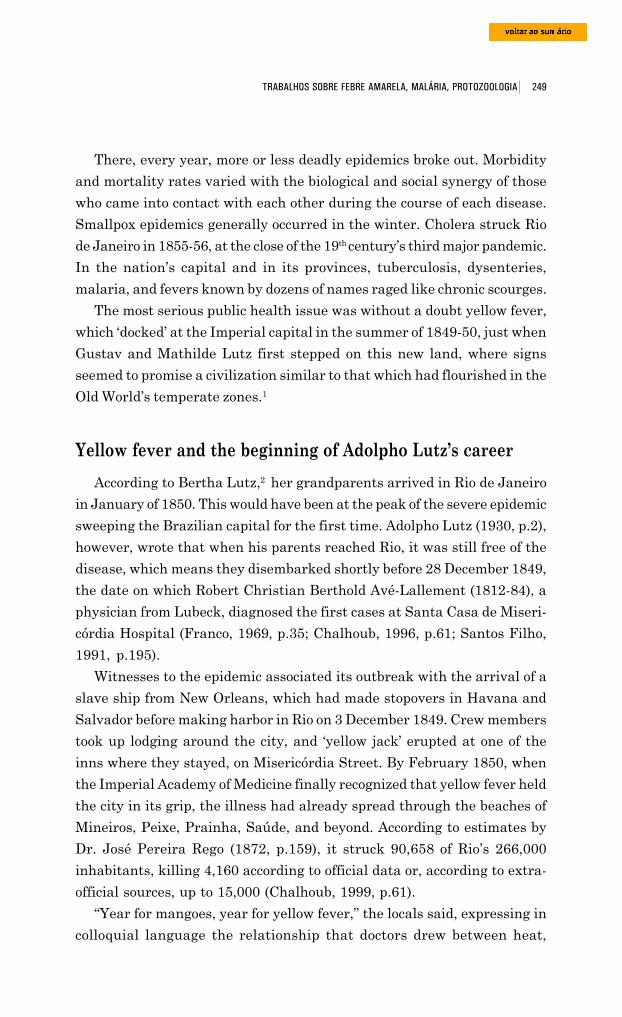
The most serious public health issue was without a doubt yellow fever,
which ‘docked’ at the Imperial capital in the summer of 1849-50, just when
Gustav and Mathilde Lutz first stepped on this new land, where signs
seemed to promise a civilization similar to that which had flourished in the
Old World’s temperate zones.1
Yellow fever and the beginning of Adolpho Lutz’s career
According to Bertha Lutz,2 her grandparents arrived in Rio de Janeiro
in January of 1850. This would have been at the peak of the severe epidemic
sweeping the Brazilian capital for the first time. Adolpho Lutz (1930, p.2),
however, wrote that when his parents reached Rio, it was still free of the
disease, which means they disembarked shortly before 28 December 1849,
the date on which Robert Christian Berthold Avé-Lallement (1812-84), a
physician from Lubeck, diagnosed the first cases at Santa Casa de Miseri-
córdia Hospital (Franco, 1969, p.35; Chalhoub, 1996, p.61; Santos Filho,
1991, p.195).
Witnesses to the epidemic associated its outbreak with the arrival of a
slave ship from New Orleans, which had made stopovers in Havana and
Salvador before making harbor in Rio on 3 December 1849. Crew members
took up lodging around the city, and ‘yellow jack’ erupted at one of the
inns where they stayed, on Misericórdia Street. By February 1850, when
the Imperial Academy of Medicine finally recognized that yellow fever held
the city in its grip, the illness had already spread through the beaches of
Mineiros, Peixe, Prainha, Saúde, and beyond. According to estimates by
Dr. José Pereira Rego (1872, p.159), it struck 90,658 of Rio’s 266,000
inhabitants, killing 4,160 according to official data or, according to extra-
official sources, up to 15,000 (Chalhoub, 1999, p.61).
“Year for mangoes, year for yellow fever,” the locals said, expressing in
colloquial language the relationship that doctors drew between heat,
250 ADOLPHO LUTZ — OBRA COMPLETA Vol. 2 — Livro 1
humidity, and epidemics. Except for the period between 1862 and 1869,
the disease ‘grew’ as regularly as other seasonal fruit, always during the
so-called muggy season, that long period of heat and rain running from
about November till March or April. Analogies with the plant world didn’t
end there. It was assumed that yellow fever, like plants, took perfectly to
coastal lowlands, especially port cities, where putrefying plant and animal
matter provided them with ideal humus.
Writing about his parents and Rio de Janeiro’s first epidemic, Adolpho
Lutz said (1930):
In the subsequent period, they had many children, all of whom ran therisk of yellow fever for varying but generally quite lengthy periods of time.My mother, who lived in Rio for over thirty years, never caught it, but myfather and a brother of mine each came down with it twice and another gotsick during the first Santos epidemic, that is, in 1879, meaning that familyimmunity can be discarded. However, many more people in the familyremained free and today can be considered [to have been] protected by thisimperceptible process of immunization, whose existence is as evident asits establishment is hard to accompany. Taking the third generation intoaccount, one can say that in my family the morbidity rate of those exposeddid not reach one-third, a remarkable fact when one compares it with thegeneral morbidity rates during the first epidemics in different points aroundthe state of São Paulo.
When he came ashore in Rio de Janeiro in 1881 with his medical degree,
at the age of 26, Adolpho Lutz took up residence at his parents’ home at 33
Princeza Imperial Street, in the neighborhood of Catete, a house large
enough to accommodate his nine siblings and the Girls’ School founded by
his mother.3 His father’s business was located at 44A Sabão Street.
According to advertisements published in Almanak Laemmert (p.497, 512,
549), Lutz & C. was a company of “foreign businessmen” dealing in import
and export, in the latter case in collaboration with J.R. Dietiker. They were
publicized as “Consignees and Commission Houses for Import and Export
Goods” and “Wholesale Stores of Imported Dry Goods.” Taking advantage
of the process of urban modernization then underway, Lutz & C. acquired
(4 Apr. 1872) control of the assets of an industry that produced
“instantaneous tubular wells,” owned by Gustavo Adolpho Wierffbain, “ci-
vil engineer, born in Germany, and resident of the Empire of Brazil.”4
As we saw in the first book of The Complete Works of Adolpho Lutz, one
of the first things he did was to have his diploma recognized by the Rio de
Janeiro School of Medicine. In an article published in Correspondenz-Blatt
251 TRABALHOS SOBRE FEBRE AMARELA, MALÁRIA, PROTOZOOLOGIA
für Schweizer Aerzte in April 1882, Lutz described the bureaucracy entailed
in this process and drew a portrait of medicine in the Brazilian Empire. He
explained that the country’s larger cities had their advantages, but that
life was thrice as expensive as in Switzerland and, in the case of Rio de
Janeiro, one could add the risk of yellow fever to the city’s bothersome
heat. This was one reason the young doctor decided to find a rural town
where he could practice medicine. Besides enjoying the rewards of the
“beauty of nature,” whoever set about building a reputable practice in such
places could expect to make a small fortune, as in Europe. Income varied
with the region, with less money to be earned in some places and substantial
sums in others, like certain coffee-growing areas or some German
settlements in southern Brazil.
Lutz remained in the capital of the Empire for eight months, waiting
for his diploma to be recognized. In the first half of 1882, he tried opening
an office in the nearby mountain town of Petrópolis but ended up settling
in Limeira, site of a sizeable Swiss-German settlement where his sister
Helena had just moved, shortly after her marriage to the German
businessman Gottfried Wilhelm Luce.
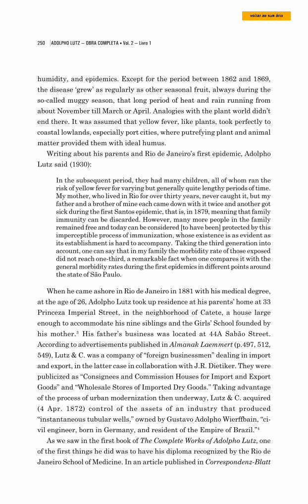
Adolpho Lutz’s results in general medicine, surgery and obstetrics, after exams taken at the Rio de JaneiroFaculty of Medicine to validate his degree in medicine, dated 21 December 1881 (BR. MN. Fundo Adolpho Lutz).
252 ADOLPHO LUTZ — OBRA COMPLETA Vol. 2 — Livro 1
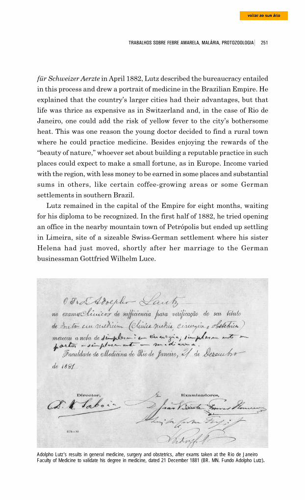
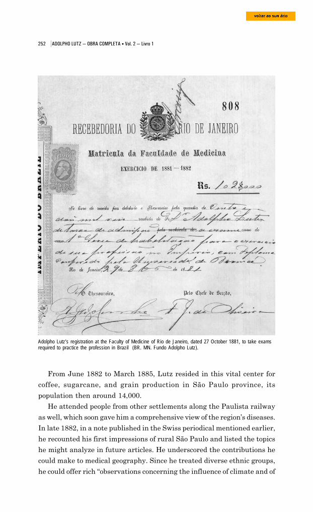
Adolpho Lutz’s registration at the Faculty of Medicine of Rio de Janeiro, dated 27 October 1881, to take examsrequired to practice the profession in Brazil (BR. MN. Fundo Adolpho Lutz).
From June 1882 to March 1885, Lutz resided in this vital center for
coffee, sugarcane, and grain production in São Paulo province, its
population then around 14,000.
He attended people from other settlements along the Paulista railway
as well, which soon gave him a comprehensive view of the region’s diseases.
In late 1882, in a note published in the Swiss periodical mentioned earlier,
he recounted his first impressions of rural São Paulo and listed the topics
he might analyze in future articles. He underscored the contributions he
could make to medical geography. Since he treated diverse ethnic groups,
he could offer rich “observations concerning the influence of climate and of
253 TRABALHOS SOBRE FEBRE AMARELA, MALÁRIA, PROTOZOOLOGIA
the human race on different diseases;
in [his] work, [he] attended blacks,
Brazilians, and German, Portuguese,
and Italian immigrants, who thus
provided most interesting comparative
material” (Lutz, 1883, p.30).
In Limeira, Adolpho Lutz did
important investigations into both
clinical practice and the helminthology
of domestic animals and man. It was
then that he undertook his research on
worms, broadening the repertoire of
pathologies under study by the Bahian
tropicalist school and likewise opening
the door to the investigation of animal
diseases in Brazil. As we saw in the
second book of this collection (Benchimol
and Sá, 2004), Adolpho Lutz’s interest
in leprosy led him to Hamburg in March
1885, where he worked about a year at
the clinic founded by Paul Gerson Unna.
Under the latter’s guidance, he ventured
into the territory of bacteriology, focusing
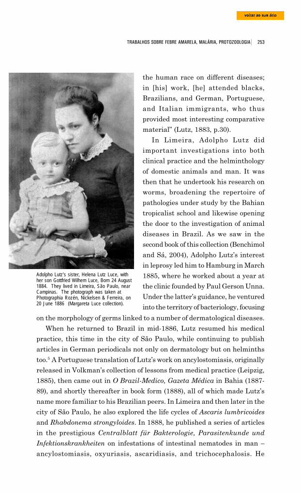
Adolpho Lutz’s sister, Helena Lutz Luce, withher son Gottfried Wilhem Luce, Born 24 August1884. They lived in Limeira, São Paulo, nearCampinas. The photograph was taken atPhotographia Rozén, Nickelsen & Ferreira, on20 June 1886 (Margareta Luce collection).
on the morphology of germs linked to a number of dermatological diseases.
When he returned to Brazil in mid-1886, Lutz resumed his medical
practice, this time in the city of São Paulo, while continuing to publish
articles in German periodicals not only on dermatology but on helminths
too.5 A Portuguese translation of Lutz’s work on ancylostomiasis, originally
released in Volkman’s collection of lessons from medical practice (Leipzig,
1885), then came out in O Brazil-Medico, Gazeta Médica in Bahia (1887-
89), and shortly thereafter in book form (1888), all of which made Lutz’s
name more familiar to his Brazilian peers. In Limeira and then later in the
city of São Paulo, he also explored the life cycles of Ascaris lumbricoides
and Rhabdonema strongyloides. In 1888, he published a series of articles
in the prestigious Centralblatt für Bakterologie, Parasitenkunde und
Infektionskrankheiten on infestations of intestinal nematodes in man –
ancylostomiasis, oxyuriasis, ascaridiasis, and trichocephalosis. He
254 ADOLPHO LUTZ — OBRA COMPLETA Vol. 2 — Livro 1
emphasized the role of the soil and feces in spreading these diseases and
drew correlations between them and the immigrant population’s living and
eating habits; he further called his peers’ attention to how often domiciliary
infestations and family epidemics occurred.6
Amoebae, bacilli, and dysenteries
Intestinal diseases were a preponderant component of public health
problems in Brazilian cities, which were to greater or lesser degrees then
experiencing population booms and witnessing the decline of living
conditions as a counterpart to the development of capitalism. Lutz started
studying these diseases while practicing medicine in Limeira, and in 1891
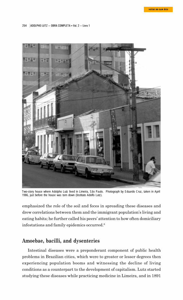
Two-story house where Adolpho Lutz lived in Limeira, São Paulo. Photograph by Eduardo Cruz, taken in April1986, just before the house was torn down (Instituto Adolfo Lutz).
255 TRABALHOS SOBRE FEBRE AMARELA, MALÁRIA, PROTOZOOLOGIA
he published a vital study on the subject, likewise in Centralblatt für
Bakterologie, Parasitenkunde und Infektionskrankheiten (1891, p.241-8).
The etiology of dysenteries was quite unclear back then. Amoebae had
been located in the corpses of people who had succumbed to a variety of
intestinal syndromes, but no one had been able to demonstrate a cause-
and-effect relation with these protozoans.
When Lutz began exploring the question, existing notions about
dysentery were quite ill-defined and the topic one of great controversy.
Physicians searching for the cause of this malady found various
microorganisms in patients’ feces and body organs, and each one believed
that whatever they found played the role of specific etiological agent.
Dopter (1909, p.1-2) pinpoints 1859 as the year these studies began,
when Vilem Dusan Lambl (1824-95), a Bohemian physician who had
obtained his medical degree in Prague and who worked at a children’s
hospital there (Franz-Josefs-Kinder-Spitale),7 observed the presence of
amoebae in the feces and, more importantly, in the intestines of a child
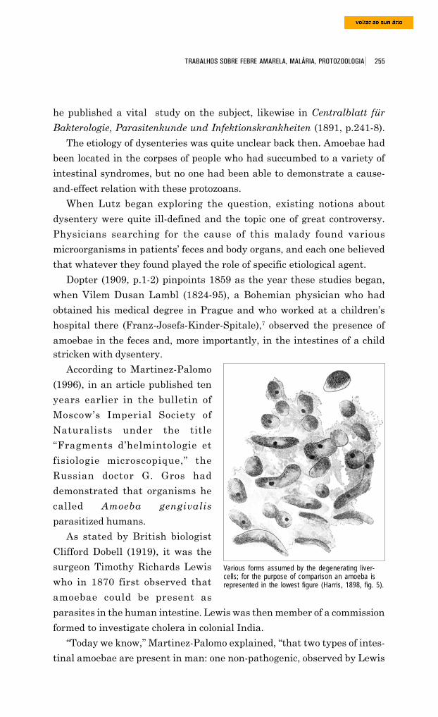
Various forms assumed by the degenerating liver-cells; for the purpose of comparison an amoeba isrepresented in the lowest figure (Harris, 1898, fig. 5).
stricken with dysentery.
According to Martinez-Palomo
(1996), in an article published ten
years earlier in the bulletin of
Moscow’s Imperial Society of
Naturalists under the title
“Fragments d’helmintologie et
fisiologie microscopique,” the
Russian doctor G. Gros had
demonstrated that organisms he
called Amoeba gengivalis
parasitized humans.
As stated by British biologist
Clifford Dobell (1919), it was the
surgeon Timothy Richards Lewis
who in 1870 first observed that
amoebae could be present as
parasites in the human intestine. Lewis was then member of a commission
formed to investigate cholera in colonial India.
“Today we know,” Martinez-Palomo explained, “that two types of intes-
tinal amoebae are present in man: one non-pathogenic, observed by Lewis
256 ADOLPHO LUTZ — OBRA COMPLETA Vol. 2 — Livro 1
(Entamoeba coli), and one pathogenic (Entamoeba histolytica), defined by
Lösch. It took decades to establish this differentiation.”
This author credits Fedor Aleksandrovich Lösch, a physician and
microscopist from Saint Petersburg, with discovery of the causative agent
of amoebiasis.
In 1873, a young peasant named J. Markow, who was suffering from heavydiarrhea and a rectal malady, began undergoing treatment (initiallysuccessful) with quinine sulfate (and other drugs) ... Lösch provided aprecise description of the amoebae found in the patient’s feces, which hechristened Amoeba coli (most of the ulcerations were in the colon), butsince ... the inoculation of dogs with these amoebae had no effect, he darednot categorically affirm that they were responsible for the infection.8
The Russian doctor presumed the amoebae would merely exacerbate
intestinal inflammation through direct mechanical irritation (Dopter, 1909,
p.2). Perhaps the physician’s doubts also derived from his reading of a
paper published by Basch shortly before (1869), in which the latter had
observed non-characterized spherical elements and filaments suggestive
of a bacterium of the group Leptothrix in slices of dysenteric intestines. A
short time later (1875), Rajewsky was to describe colonies of cocci and
bacteria in the lymphatic vessels of the submucous membrane.
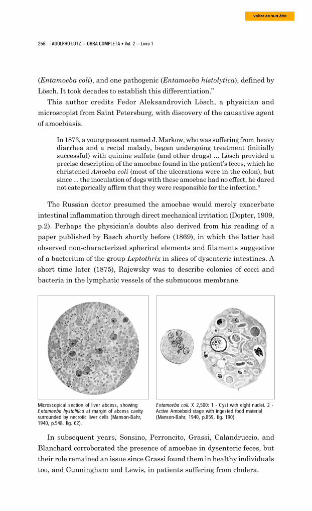
Microscopical section of liver abcess, showingEntamoeba hystolitica at margin of abcess cavitysurrounded by necrotic liver cells (Manson-Bahr,1940, p.548, fig. 62).
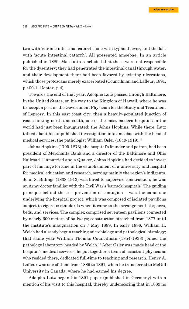
In subsequent years, Sonsino, Perroncito, Grassi, Calandruccio, and
Blanchard corroborated the presence of amoebae in dysenteric feces, but
their role remained an issue since Grassi found them in healthy individuals
too, and Cunningham and Lewis, in patients suffering from cholera.
Entamoeba coli. X 2,500: 1 - Cyst with eight nuclei. 2 -Active Amoeboid stage with ingested food material(Manson-Bahr, 1940, p.859, fig. 190).
257 TRABALHOS SOBRE FEBRE AMARELA, MALÁRIA, PROTOZOOLOGIA
In 1883, while studying the disease in Egypt, Koch recognized that
amoebae could play a specific pathogenic role, after having found them
not just in human stool but also in slices of dysenteric intestines, deep
within the intestinal walls.9 Kartulis, his disciple, confirmed these
observations after examining many sick people in Alexandria and in Greece.
He had found amoebae in their stools and intestines and in abscesses of
the liver as well (Dopter, 1909, p.2-3), but he could not erase doubts about
their pathogenic role, since experimental inoculation in laboratory animals
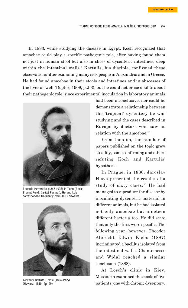
Giovanni Battista Grassi (1854-1925)(Howard, 1930, fig. 49).
Edoardo Perroncito (1847-1936) in Turin (EmileBrumpt Fund, Institut Pasteur). He and Lutzcorresponded frequently from 1883 onwards.
had been inconclusive; nor could he
demonstrate a relationship between
the ‘tropical’ dysentery he was
studying and the cases described in
Europe by doctors who saw no
relation with the amoebae.10
From then on, the number of
papers published on the topic grew
steadily, some confirming and others
refuting Koch and Kartulis’
hypothesis.
In Prague, in 1886, Jaroslav
Hlava presented the results of a
study of sixty cases.11 He had
managed to reproduce the disease by
inoculating dysenteric material in
different animals, but he had isolated
not only amoebae but nineteen
different bacteria too. He did state
that only the first were specific. The
following year, however, Theodor
Albrecht Edwin Klebs (1887)
incriminated a bacillus isolated from
the intestinal walls. Chantemesse
and Widal reached a similar
conclusion (1888).
At Lösch’s clinic in Kiev,
Massiutin examined the stools of five
patients: one with chronic dysentery,
258 ADOLPHO LUTZ — OBRA COMPLETA Vol. 2 — Livro 1
two with ‘chronic intestinal catarrh’, one with typhoid fever, and the last
with ‘acute intestinal catarrh’. All presented amoebae. In an article
published in 1889, Massiutin concluded that these were not responsible
for the dysentery; they had penetrated the intestinal canal through water,
and their development there had been favored by existing ulcerations,
which those protozoans merely exacerbated (Councilman and Lafleur, 1891,
p.400-1; Dopter, p.4).
Towards the end of that year, Adolpho Lutz passed through Baltimore,
in the United States, on his way to the Kingdom of Hawaii, where he was
to accept a post as the Government Physician for the Study and Treatment
of Leprosy. In this east coast city, then a heavily-populated junction of
roads linking north and south, one of the most modern hospitals in the
world had just been inaugurated: the Johns Hopkins. While there, Lutz
talked about his unpublished investigation into amoebae with the head of
medical services, the pathologist William Osler (1849-1919).12
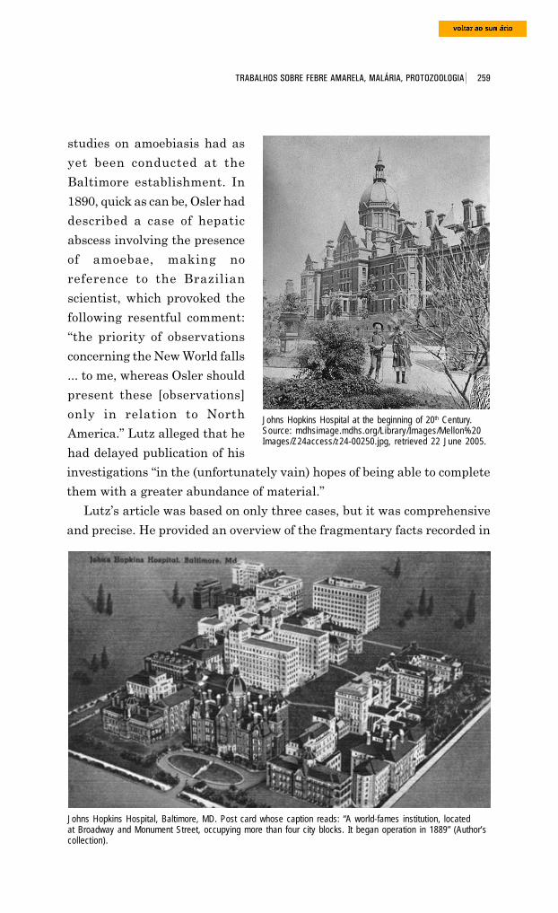
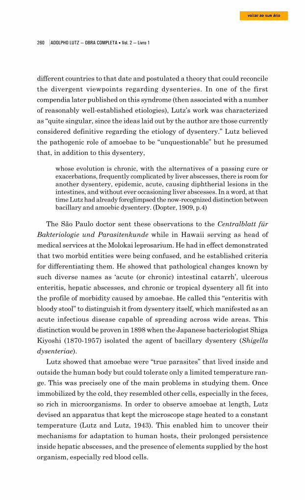
Johns Hopkins (1795-1873), the hospital’s founder and patron, had been
president of Merchants Bank and a director of the Baltimore and Ohio
Railroad. Unmarried and a Quaker, Johns Hopkins had decided to invest
part of his huge fortune in the establishment of a university and hospital
for medical education and research, serving mainly the region’s indigents.
John S. Billings (1838-1913) was hired to supervise construction; he was
an Army doctor familiar with the Civil War’s ‘barrack hospitals’. The guiding
principle behind these – prevention of contagion – was the same one
underlying the hospital project, which was composed of isolated pavilions
subject to rigorous standards when it came to the arrangement of spaces,
beds, and services. The complex comprised seventeen pavilions connected
by nearly 600 meters of hallways; construction stretched from 1877 until
the institute’s inauguration on 7 May 1889. In early 1886, William H.
Welch had already begun teaching microbiology and pathological histology;
that same year William Thomas Councilman (1854-1933) joined the
pathology laboratory headed by Welch.13 After Osler was made head of the
hospital’s medical services, he put together a team of assistant physicians
who resided there, dedicated full-time to teaching and research. Henry A.
Lafleur was one of them from 1889 to 1891, when he transferred to McGill
University in Canada, where he had earned his degree.
Adolpho Lutz began his 1891 paper (published in Germany) with a
mention of his visit to this hospital, thereby underscoring that in 1889 no
259 TRABALHOS SOBRE FEBRE AMARELA, MALÁRIA, PROTOZOOLOGIA
studies on amoebiasis had as
yet been conducted at the
Baltimore establishment. In
1890, quick as can be, Osler had
described a case of hepatic
abscess involving the presence
of amoebae, making no
reference to the Brazilian
scientist, which provoked the
following resentful comment:
“the priority of observations
concerning the New World falls
... to me, whereas Osler should
present these [observations]
only in relation to North
America.” Lutz alleged that he
had delayed publication of his
Johns Hopkins Hospital at the beginning of 20th Century.Source: mdhsimage.mdhs.org/Library/Images/Mellon%20Images/Z24access/z24-00250.jpg, retrieved 22 June 2005.
investigations “in the (unfortunately vain) hopes of being able to complete
them with a greater abundance of material.”
Lutz’s article was based on only three cases, but it was comprehensive
and precise. He provided an overview of the fragmentary facts recorded in
Johns Hopkins Hospital, Baltimore, MD. Post card whose caption reads: “A world-fames institution, locatedat Broadway and Monument Street, occupying more than four city blocks. It began operation in 1889” (Author’scollection).
260 ADOLPHO LUTZ — OBRA COMPLETA Vol. 2 — Livro 1
different countries to that date and postulated a theory that could reconcile
the divergent viewpoints regarding dysenteries. In one of the first
compendia later published on this syndrome (then associated with a number
of reasonably well-established etiologies), Lutz’s work was characterized
as “quite singular, since the ideas laid out by the author are those currently
considered definitive regarding the etiology of dysentery.” Lutz believed
the pathogenic role of amoebae to be “unquestionable” but he presumed
that, in addition to this dysentery,
whose evolution is chronic, with the alternatives of a passing cure orexacerbations, frequently complicated by liver abscesses, there is room foranother dysentery, epidemic, acute, causing diphtherial lesions in theintestines, and without ever occasioning liver abscesses. In a word, at thattime Lutz had already foreglimpsed the now-recognized distinction betweenbacillary and amoebic dysentery. (Dopter, 1909, p.4)
The São Paulo doctor sent these observations to the Centralblatt für
Bakteriologie und Parasitenkunde while in Hawaii serving as head of
medical services at the Molokai leprosarium. He had in effect demonstrated
that two morbid entities were being confused, and he established criteria
for differentiating them. He showed that pathological changes known by
such diverse names as ‘acute (or chronic) intestinal catarrh’, ulcerous
enteritis, hepatic abscesses, and chronic or tropical dysentery all fit into
the profile of morbidity caused by amoebae. He called this “enteritis with
bloody stool” to distinguish it from dysentery itself, which manifested as an
acute infectious disease capable of spreading across wide areas. This
distinction would be proven in 1898 when the Japanese bacteriologist Shiga
Kiyoshi (1870-1957) isolated the agent of bacillary dysentery (Shigella
dysenteriae).
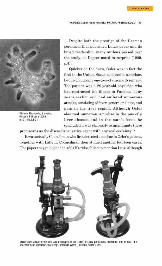
Lutz showed that amoebae were “true parasites” that lived inside and
outside the human body but could tolerate only a limited temperature ran-
ge. This was precisely one of the main problems in studying them. Once
immobilized by the cold, they resembled other cells, especially in the feces,
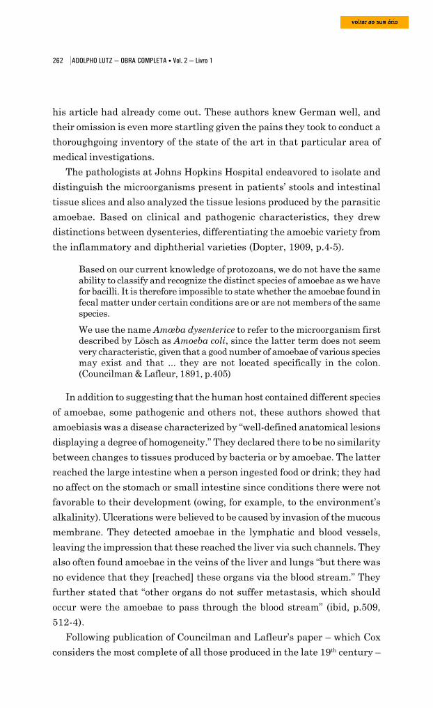
so rich in microorganisms. In order to observe amoebae at length, Lutz
devised an apparatus that kept the microscope stage heated to a constant
temperature (Lutz and Lutz, 1943). This enabled him to uncover their
mechanisms for adaptation to human hosts, their prolonged persistence
inside hepatic abscesses, and the presence of elements supplied by the host
organism, especially red blood cells.
261 TRABALHOS SOBRE FEBRE AMARELA, MALÁRIA, PROTOZOOLOGIA
Phylum Rhizopoda, Amoeba.(Brusca & Brusca, 2003,p.121, fig.5.1-C).
Despite both the prestige of the German
periodical that published Lutz’s paper and its
broad readership, many authors passed over
the study, as Dopter noted in surprise (1909,
p.4).
Quicker on the draw, Osler was in fact the
first in the United States to describe amoebae,
but involving only one case of chronic dysentery.
The patient was a 29-year-old physician who
had contracted the illness in Panama many
years earlier and had suffered numerous
attacks, consisting of fever, general malaise, and
pain in the liver region. Although Osler
observed numerous amoebae in the pus of a
liver abscess and in the man’s feces, he
concluded it was still early to incriminate these
protozoans as the disease’s causative agent with any real certainty.14
It was actually Councilman who first detected amoebae in Osler’s patient.
Together with Lafleur, Councilman then studied another fourteen cases.
The paper they published in 1891 likewise failed to mention Lutz, although
Microscope similar to the one Lutz developed in the 1880s to study protozoans, helminths and insects. It isattached to an apparatus that keeps amoebas warm (Instituto Adolfo Lutz).
262 ADOLPHO LUTZ — OBRA COMPLETA Vol. 2 — Livro 1
his article had already come out. These authors knew German well, and
their omission is even more startling given the pains they took to conduct a
thoroughgoing inventory of the state of the art in that particular area of
medical investigations.
The pathologists at Johns Hopkins Hospital endeavored to isolate and
distinguish the microorganisms present in patients’ stools and intestinal
tissue slices and also analyzed the tissue lesions produced by the parasitic
amoebae. Based on clinical and pathogenic characteristics, they drew
distinctions between dysenteries, differentiating the amoebic variety from
the inflammatory and diphtherial varieties (Dopter, 1909, p.4-5).
Based on our current knowledge of protozoans, we do not have the sameability to classify and recognize the distinct species of amoebae as we havefor bacilli. It is therefore impossible to state whether the amoebae found infecal matter under certain conditions are or are not members of the samespecies.
We use the name Amœba dysenterice to refer to the microorganism firstdescribed by Lösch as Amoeba coli, since the latter term does not seemvery characteristic, given that a good number of amoebae of various speciesmay exist and that ... they are not located specifically in the colon.(Councilman & Lafleur, 1891, p.405)
In addition to suggesting that the human host contained different species
of amoebae, some pathogenic and others not, these authors showed that
amoebiasis was a disease characterized by “well-defined anatomical lesions
displaying a degree of homogeneity.” They declared there to be no similarity
between changes to tissues produced by bacteria or by amoebae. The latter
reached the large intestine when a person ingested food or drink; they had
no affect on the stomach or small intestine since conditions there were not
favorable to their development (owing, for example, to the environment’s
alkalinity). Ulcerations were believed to be caused by invasion of the mucous
membrane. They detected amoebae in the lymphatic and blood vessels,
leaving the impression that these reached the liver via such channels. They
also often found amoebae in the veins of the liver and lungs “but there was
no evidence that they [reached] these organs via the blood stream.” They
further stated that “other organs do not suffer metastasis, which should
occur were the amoebae to pass through the blood stream” (ibid, p.509,
512-4).
Following publication of Councilman and Lafleur’s paper – which Cox
considers the most complete of all those produced in the late 19th century –
263 TRABALHOS SOBRE FEBRE AMARELA, MALÁRIA, PROTOZOOLOGIA
dysentery became a topic of note in the United States, and many doctors
published reports on cases found in the areas where they worked. One of
these was H. Harris,15 professor of pathology at Jefferson Medical College.
In a paper dated 1898 (p.385-6), he cited Adolpho Lutz, recognizing that
the latter had lodged “fair criticisms against our imprecise classifications of
intestinal diseases.”
Other authors assumed a stance similar to Councilman and Lafleur’s.
Dopter (1903, p.4-8) cites a long list of names,16 including one Brazilian,
Francisco Fajardo, who – as we will see – had a close relationship with
Adolpho Lutz in other realms of protozoology. The papers published by
these authors would make a vital contribution to our understanding of the
life cycle of the amoeba and its parasitism, but it would take some years for
the differentiation between pathogenic and non-offensive forms to firmly
establish itself.
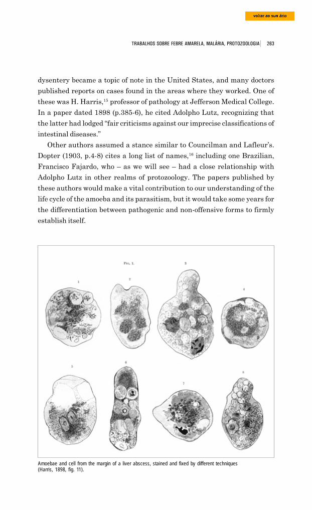
Amoebae and cell from the margin of a liver abscess, stained and fixed by different techniques(Harris, 1898, fig. 11).
264 ADOLPHO LUTZ — OBRA COMPLETA Vol. 2 — Livro 1
caused by a pathogenic species, Amoeba dysenteriae, which differed from
other varieties found in the intestines of healthy individuals (Dopter, 1909,
p.6-7). The German scientists Heinrich Iranaus Quincke and Ernst Roos
(1893) reached the same conclusion.17 Although unable to correctly
differentiate the amoebae (Martinez-Palomo, 1996), in a 1903 paper the
protozoologist Fritz Schaudinn established the name of the pathogenic
species that is still used today: Entamoeba histolytica. But it would be
another ten years before Walker and Sellards distinguished it unequivocally
from free-living amoebae in water, which did not produce dysentery
(Entamoeba coli) (Faust, et al., 1975, p.85). Schaudinn conducted his
experiments on prisoners at the Bilibid penitentiary in Manila, in the
Philippines. Of the twenty men who ingested eggs of that species, seventeen
became infected but none developed the disease. On the other hand, of the
twenty volunteers who received capsules containing Entamoeba histolytica
eggs, seventeen were infected by the first dose whereas it took one of them
three inoculations. Only four of the eighteen who had the parasite got
sick. The experiment showed that the organism could be pathogenic in
some people and not produce symptoms in others. It further showed that
asymptomatic carriers could transmit the pathogenic parasite to healthy
people (Martinez-Palomo, 1996).
Following publication of Adolpho Lutz’s papers, other authors
endeavored to draw a relationship between dysentery and bacteria,
assigning amoebae a secondary, meaningless, or sometimes even beneficial
role. One who took this line was Maggiora; he attributed the disease to the
pyocyanic bacillus and to a colibacillus displaying an abnormal level of
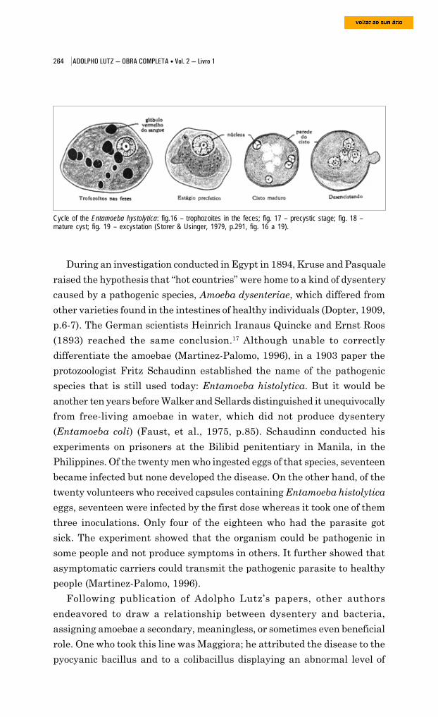
Cycle of the Entamoeba hystolytica: fig.16 – trophozoites in the feces; fig. 17 – precystic stage; fig. 18 –mature cyst; fig. 19 – excystation (Storer & Usinger, 1979, p.291, fig. 16 a 19).
During an investigation conducted in Egypt in 1894, Kruse and Pasquale
raised the hypothesis that “hot countries” were home to a kind of dysentery
265 TRABALHOS SOBRE FEBRE AMARELA, MALÁRIA, PROTOZOOLOGIA
virulence. Laveran also incriminated colibacilli. In 1895, based on case
studies from Rome, Tivoli, Sienna, and even Alexandria, Celli and Fiocca
concluded that a variety of this microorganism, which they called Bacterium
coli dysenteriae, was responsible for dysentery. According to Bertrand and
Baucher, dysentery was a polymicrobial infection associated with a number
of different germs (septic Vibrio, pyocyanic bacilli, staphylococci, and
colibacillus). In Saigon, Calmete incriminated the pyocyanic bacillus
(Dopter, 1909, p.5-9).18
The absence of amoebae in typical cases of dysentery, combined with
their presence in healthy individuals or people with other diseases – facts
pointed out by a number of authors – simply added to the confusion in this
area of pathology.
As stated earlier, unification of these two lines of interpretation as
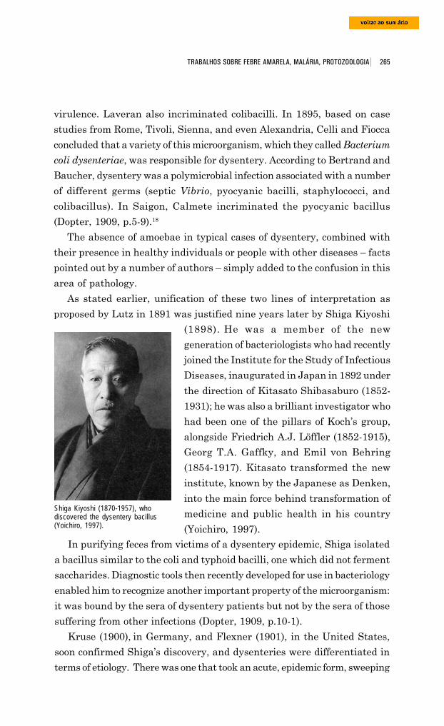
proposed by Lutz in 1891 was justified nine years later by Shiga Kiyoshi
Shiga Kiyoshi (1870-1957), whodiscovered the dysentery bacillus(Yoichiro, 1997).
(1898). He was a member of the new
generation of bacteriologists who had recently
joined the Institute for the Study of Infectious
Diseases, inaugurated in Japan in 1892 under
the direction of Kitasato Shibasaburo (1852-
1931); he was also a brilliant investigator who
had been one of the pillars of Koch’s group,
alongside Friedrich A.J. Löffler (1852-1915),
Georg T.A. Gaffky, and Emil von Behring
(1854-1917). Kitasato transformed the new
institute, known by the Japanese as Denken,
into the main force behind transformation of
medicine and public health in his country
(Yoichiro, 1997).
In purifying feces from victims of a dysentery epidemic, Shiga isolated
a bacillus similar to the coli and typhoid bacilli, one which did not ferment
saccharides. Diagnostic tools then recently developed for use in bacteriology
enabled him to recognize another important property of the microorganism:
it was bound by the sera of dysentery patients but not by the sera of those
suffering from other infections (Dopter, 1909, p.10-1).
Kruse (1900), in Germany, and Flexner (1901), in the United States,
soon confirmed Shiga’s discovery, and dysenteries were differentiated in
terms of etiology. There was one that took an acute, epidemic form, sweeping
266 ADOLPHO LUTZ — OBRA COMPLETA Vol. 2 — Livro 1
especially through temperate countries, and another, an amoebic form,
chronic in nature and endemic to the tropics. New differentiations soon
followed; the form of dysentery caused by Balantidium coli as well as the
one caused by spirilli were described (Le Dantec). The term ‘dysentery’
came to refer to a syndrome with various etiologies. “Thus the opinion
formulated by Lutz, Councilman, and Lafleur in 1891-92 has now been
proven,” wrote Dopter in 1909 (p.10-2), in one of the first treatises on this
subject.
Adolpho Lutz and yellow fever
It is possible that Lutz had come into contact with yellow fever patients
or individuals suspected of having the disease while practicing medicine
in Limeira and in the city of São Paulo, but no such traces can be found
in the documentation with which we have worked. His first documented
contact with the disease took place in early 1889, in Campinas, when the
city fell victim to a major epidemic that had serious impact not only on its
citizens but also on public opinion around the country.
Campinas was one of the principal urban centers of Southeastern Brazil.
“It competed and in many regards was actually tied with São Paulo, the
capital,” in the words of Santos Filho and Novaes (1996, p.9), authors of
the most thoroughgoing study on this yellow fever epidemic, which
“destroyed [the city’s] vigor, paralyzed development, and crushed the city.”
Over the previous three decades, coffee had replaced the region’s
sugarcane plantations, and Campinas had prospered as a dynamic
commercial and financial center. Its plantation owners and businessmen
held a good share of the wealth in the province of São Paulo, and they
invested it in railway lines that encouraged the expansion of crops and of
new urban centers in western São Paulo state, an expanding frontier for
the country’s principal economic activity. Armed with a capitalist mentality
that contrasted with the mentality of plantation owners in the decaying
areas of the Paraíba Valley, Campinas coffee growers and businessmen
invested capital in companies that provided public services and outfitted the
city with the same improvements that had been transforming urban life in
the capitals of the Empire and the provinces: gas lighting (1875), animal-
powered tramlines (1879), and telephone lines (1884). Water and sewer
services came only later, in 1891-92, largely in response to yellow fever.
267 TRABALHOS SOBRE FEBRE AMARELA, MALÁRIA, PROTOZOOLOGIA
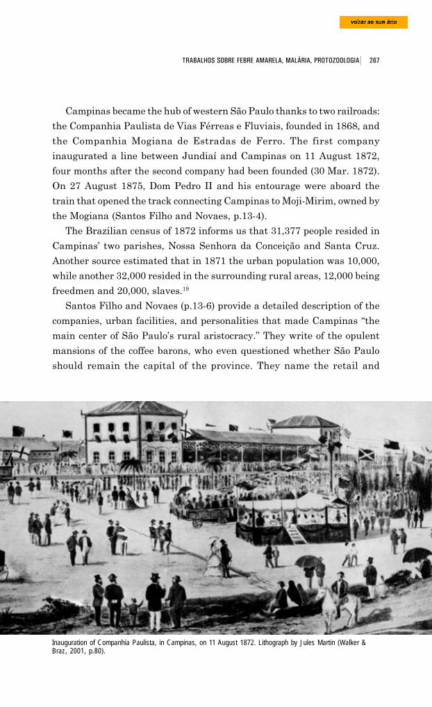
Campinas became the hub of western São Paulo thanks to two railroads:
the Companhia Paulista de Vias Férreas e Fluviais, founded in 1868, and
the Companhia Mogiana de Estradas de Ferro. The first company
inaugurated a line between Jundiaí and Campinas on 11 August 1872,
four months after the second company had been founded (30 Mar. 1872).
On 27 August 1875, Dom Pedro II and his entourage were aboard the
train that opened the track connecting Campinas to Moji-Mirim, owned by
the Mogiana (Santos Filho and Novaes, p.13-4).
The Brazilian census of 1872 informs us that 31,377 people resided in
Campinas’ two parishes, Nossa Senhora da Conceição and Santa Cruz.
Another source estimated that in 1871 the urban population was 10,000,
while another 32,000 resided in the surrounding rural areas, 12,000 being
freedmen and 20,000, slaves.19
Santos Filho and Novaes (p.13-6) provide a detailed description of the
companies, urban facilities, and personalities that made Campinas “the
main center of São Paulo’s rural aristocracy.” They write of the opulent
mansions of the coffee barons, who even questioned whether São Paulo
should remain the capital of the province. They name the retail and
Inauguration of Companhia Paulista, in Campinas, on 11 August 1872. Lithograph by Jules Martin (Walker &Braz, 2001, p.80).
268 ADOLPHO LUTZ — OBRA COMPLETA Vol. 2 — Livro 1
wholesale businesses that supplied the entire western part of the province,
with such products as iron and bronze castings, agricultural machinery,
ornamental goods, and locally made construction materials.
Campinas had a cathedral, theaters, a skating rink, a hippodrome, clubs,
and three large hotels: the Europa, the França, and the Grande Hotel
Campineiro. It also boasted six schools (including one called Culto à Ciên-
cia, or Homage to Science) that drew students from a broad region of
influence and where some important proponents of the Republic taught.20
The Gazeta de Campinas (1869) was the major newspaper but others in
circulation included Sensitiva (1873), Mocidade (1874), Diário de Campi-
nas (1875), and Correio de Campinas (1885).
Santa Casa de Misericórdia Hospital was inaugurated in 1876; two years
later, Beneficiência Portuguesa Hospital was founded. Santos Filho and
Novaes (p.21) also write of hospitals for smallpox victims and lepers, both
funded by the Municipal Chamber. During the crisis of 1889, special
Arms of the city of Campinas.Founded in the first half of 18thcentury it was called MatoGrosso until 1842, when it wasgiven its current name. Source:www.ngw.nl/int/bra/images/campinas, retrieved 22 June2005.
infirmaries or lazarettos would be hastily created
to isolate those stricken with yellow fever.
The conviction that this disease “could not move
into the mountains” (Lutz, 1930) and that its sole
habitat was the overcrowded urban centers of the
hot and humid coastal plains had been shot down
in 1876, when Dr. Valentin José da Silveira Lopes,
future Viscount of São Valentin, had diagnosed
two Portuguese with yellow fever, both having
just arrived from Rio de Janeiro. They had
checked into a small hospital of which Lopes was
one of the owners. The event had no major
repercussions because these were imported cases,
but two months later (30 Apr. 1876), Lopes stated to the Gazeta de Campi-
nas that another eight people had come down with yellow fever. Since all
of them lived near the Companhia Paulista de Vias Férreas e Fluviais
railroad station, in houses contiguous with land where train cars filled
with coal and merchandise from the port of Santos stood parked, Lopes
presumed these wagons had brought the germs from infected ships. His
diagnosis alarmed the population and incited much controversy not only
in Campinas but likewise among physicians, politicians, and journalists
from the capitals of the province and of the Empire, since very few believed
269 TRABALHOS SOBRE FEBRE AMARELA, MALÁRIA, PROTOZOOLOGIA
yellow fever could manifest itself so far from the coast (Santos Filho and
Novaes, p.23-4).
Lopes submitted a special communication about this to the Imperial
Academy of Medicine; in its 5 June 1876 session, the note was read and
commented on by Antônio Correia de Sousa Costa (1834-89), professor of
hygiene at Rio’s School of Medicine and president of the Central Board of
Public Hygiene. His expert opinion was that the cases verified in Campinas
were indeed yellow fever, and this was published in the Anais Brasilienses
de Medicina (“Treatise on yellow fever in Campinas” [in Port.]). Lopes was
elected a corresponding member of the Academy on 28 August 1876.21
Dr. Valentin José da Silveira Lopes’ viewpoint was endorsed by Antô-
nio Felício dos Santos (1843-1931), Júlio Rodrigues de Moura (1839-92),
João Vicente Torres Homem (1837-87) – one of the most renowned clinicians
in the city of Rio de Janeiro and professor of medical practice at its School
of Medicine – and Carlos Ferreira de Souza Fernandes (1829-88), author
of “Yellow fever in Campinas” [in Port.] (Anais Brasilienses de Medicina,
v.28, p.1876-7). Another ally was Augusto César de Miranda Azevedo (1851-
1907), future deputy for São Paulo to the constitutional assembly (1891)
and then a resident of Rio de Janeiro.
But the hypothesis that yellow fever was present in Campinas was
contested by a number of professors from the Rio de Janeiro School of
Medicine. Among these was José Martins da Cruz Jobim (1802-78), for
many years the school’s director, professor of forensic medicine, one of the
founders of the Academy of Medicine, and also a senator of the Empire.
His ideas concerning the etiology of the disease underpinned the position
taken by Antônio de Souza Campos (1845-1918), the first native of Cam-
pinas to earn his medical degree (1872): the latter argued that it would be
impossible to transport the element which produced the disease (that is,
the “infected” air of Santos, which impregnated train cargos), since the trip
across the highlands would be through the mountains’ cold, healthier air
(Santos Filho and Novaes, p.26).
The controversy did not dissuade rural inhabitants from their illusion
that they were safe from yellow fever, “until the facts came to prove the
contrary,” as Adolpho Lutz would later state (1930), having believed Lopes’
diagnosis to be correct in 1876.
The 1889 epidemic, the first to sweep through Campinas, hit the
population hard and had national repercussions.
270 ADOLPHO LUTZ — OBRA COMPLETA Vol. 2 — Livro 1
When it broke out, the population was terrified and whoever could,
fled.
The plantation owners ... moved to their rural landholdings or to São Paulo... Entire families abandoned their homes and their belongings. Those whocouldn’t get their hands on a carriage or a horse went by foot, seekingrefuge at nearby farms or cities. Houses, shops, grocery stores, workshops,hotels were closed ... Pharmacies could not keep up with the dispatch ofprescriptions” (ibid., p.36-7).
The physician José Maria Teixeira (1854-95), about whom we will speak
shortly, author of A epidemia de Campinas em 1889 (Rio de Janeiro, 1889),
was to register these words: “The city was abandoned and almost deserted!
Long, straight streets with hundreds of houses closed up and not a single
passerby” (cited in Simões, 1897, p.23). The remaining residents rushed to
the church every day to “pray publicly ad petendam pluviam,” according
to another witness to the crisis (ibid.), in hopes that God’s tears would
wash away the sinister miasma hanging over the city.
According to a report published in the Freie Presse: Zeitung für Deutsche
in Brasilien (Free press: a newspaper for Germans in Brazil), the “business
of thieves” became quite profitable in this ghost town. One of those who
fled, Mr. Felipe José, discovered upon his return that neither gold, nor silk,
nor watches remained in his shop. Another businessman who had sought
refuge in São Paulo came back to find his store stripped clean. “Many
robberies of this type will undoubtedly come to light as the fugitives of the
epidemic return to Campinas.”
In Reminiscências (1930), Lutz makes mention of the general opinion
that three-quarters of the city’s 20,000 inhabitants had abandoned it,
“leaving behind mostly men who held jobs; however, many people returned
too soon and ended up contracting the infection. Of those who were not
immune and lived inside the city, almost all were infected.”
According to Santos Filho and Novaes (1996) and Simões (1897), the
person who carried the disease to Campinas was the Swiss woman Rosa
Beck, unmarried, 24 years old, having recently arrived in Brazil with the
intention of finding employment as a French teacher. It is not clear where
she disembarked nor where she caught the disease – Santos or, more likely,
Rio de Janeiro. She died at 2:00 a.m. on 10 February 1889. She had taken
up lodging with compatriots of hers, in the same building where they ran a
bakery called Padaria Suíça. From there yellow fever spread through the
271 TRABALHOS SOBRE FEBRE AMARELA, MALÁRIA, PROTOZOOLOGIA
rest of the town. The second death was a nine-year-old boy who shopped at
the bakery. Two days prior to the boy’s death, the attending physician, Eduar-
do Guimarães, “for the good of the public health,” published the news that
he was caring for a patient with a critical case of yellow fever. Such was the
incredulity that he summoned “eleven distinguished colleagues” to corroborate
his diagnosis (Correio de Campinas, 23 Feb. 1889, cited in Santos Filho and
Novaes, p.41). The fact that the victim was from Campinas and had never
left the city was frightening: some as yet unknown local cause was responsible
for his yellow fever. “Like olive oil spilled on a blotter” (Simões, 1897, p.21),
the disease spread during March; it then let up for a few days, worsened in
April (the “month of horror”), subsided again, and in early May intensified
once more, from then on waning until dying out in late June.22
According to Adolpho Lutz (1930), overall mortality was estimated at
about 2,000, “including those who were infected and passed away in other
places.” The physician Ângelo Simões (1897) says that yellow fever attacked
“over 2,000 people out of a population of 3,000 (since that is how many
remained in the city, at most), resulting in 1,200 deaths, a number which
I have most carefully verified.”
The wealthier people, who could afford to leave the city, were less
affected. Most of the victims were Brazilians with no immunity to the
disease, followed by Italians, Portuguese, and immigrants of other
nationalities.
Adolpho Lutz in Campinas
In March 1889, the Municipal Chamber converted into lazarettos some
residences in the neighborhood of Guanabara, then located a distance away
from downtown. Special infirmaries for yellow fever patients were likewise
set up in the Circolo Italiani Uniti’s building, at the Correia de Melo muni-
cipal school, at the headquarters of the Sociedade Portuguesa de
Beneficiência, and also at Santa Casa de Misericórdia Hospital. In 1890,
the Lazareto do Fundão would be inaugurated in the vicinity of the
cemetery; it was later transformed into an isolation hospital, with two
wooden pavilions for patients, lodgings for the physician and caretaker,
and facilities for a pharmacy, sterilizer, and so on (Lapa, 1996, p.261).
Santos Filho and Novaes (p.44-6) described the hygiene measures
adopted by the Chamber:23
272 ADOLPHO LUTZ — OBRA COMPLETA Vol. 2 — Livro 1
Public thoroughfares were coated with pitch and watered down almostevery evening, while barrels of tar burned day and night on the corners ofthe main streets. Bonfires of fragrant herbs were lit. It was believed thatthe smoke ... would clean the air of harmful miasmas ... All furniture andobjects found in the rooms of those who perished from the fever weredestroyed and burned ... To keep the city’s inhabitants safe from contagion,the corpses were buried at night.24
Since it was mostly the poor who had remained in town, the Chamber
enacted measures to help this population, vulnerable not just to the disease
but also to hunger and neglect. It paid for public vehicles to transport
doctors who made house calls for free, distributed food and clothing, and
authorized pharmacies to provide medicine at the expense of the Munici-
pal Chamber, whose president, José Paulino Nogueira (1853-1915), fell
sick with yellow fever too.
In early April, when the situation worsened, Nogueira telegraphed the
president of the province, Pedro Vicente de Azevedo (1844-1902),25
requesting that doctors be sent urgently since most of them who lived in
the town – about twenty – had left with their families, and the few who
remained were stretched thin.26 In Santos on assignment to the government
of São Paulo, Dr. Francisco Marques de Araújo Góis headed to Campinas.
He arrived on 5 April. Astonished by the extent of the crisis and the
precarious aid provided to its victims, he endorsed the request for medical
reinforcements. Towards the end of that month, he returned to Campinas
as head of the Provincial Aid Commission, which comprised some thirty-
five people, including physicians, fifth-and sixth-year students from the
Rio de Janeiro School of Medicine, pharmacists, disinfectors, and staff who
would provide other services.27
Adolpho Lutz was a member of the commission, together with Drs. Claro
Marcondes Homem de Melo (1866-1924), Irineu de Sousa Brito Junior,
Aristides Franco de Meireles, Bráulio Gomes, and Luis Felipe Jardim (the
last two came down with yellow fever but survived). In Reminiscências
(p.128), Lutz recalls the weeks he spent in that city:
In 1889, when I was urgently called from São Paulo to Campinas, wherethere were no more doctors, I encountered a pandemic rightfully said to beyellow fever ... After four to five weeks, colleagues had arrived from Rio deJaneiro, and the epidemic was waning in Campinas.
Lutz makes mention of two other medical commissions, one sent by the
Ministry of the Empire and the other by the Comissão de Imprensa
273 TRABALHOS SOBRE FEBRE AMARELA, MALÁRIA, PROTOZOOLOGIA
Fluminense (Rio de Janeiro State Press Commission), the latter comprising
several Rio de Janeiro newspapers (Gazeta de Noticias, O Paiz, and Jor-
nal do Commercio, among others).
The commission sent by the central government, then under the control
of the Conservative Party, was headed by Dr. José Maria Teixeira (1854-
95), mentioned earlier, who was professor at the Rio de Janeiro School of
Medicine in medical subjects and pharmacy. It seems the first members of
the group reached Campinas on 7 April, bearing 100 beds and an
“ambulance” for which the pharmacist Joaquim T. Soares da Câmara was
responsible. Besides Teixeira, other members included the physicians
Eufrásio José da Cunha, Francisco Custório Pereira de Barros,28 Francisco
Corrêa Dutra, João de Deus da Cunha Pinto, Fernando de Barros, and
Luís Manuel Pinto Neto (Santos Filho and Novaes, p.54-5). The Imprensa
Fluminense sent the physicians Clemente Miguel da Cunha Ferreira (1857-
1947), as head, and João Batista da Mota de Azevedo Correia (1854-?).
They arrived in Campinas on 20 April, bringing with them a substantial
quantity of medicine, clothing, and foodstuffs that had been donated to
the victims of the Campinas epidemic at events held in Rio de Janeiro,
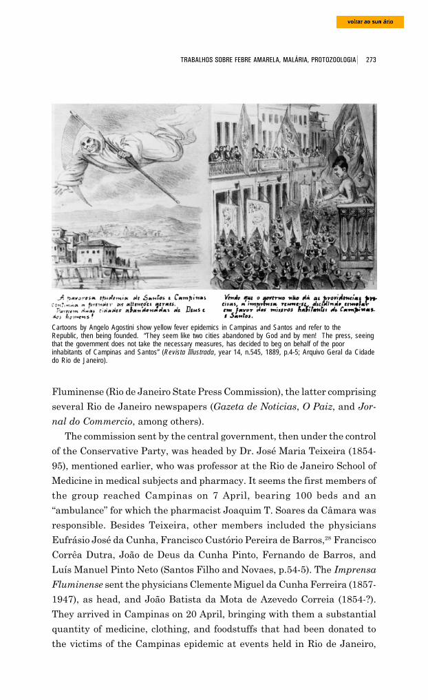
Cartoons by Angelo Agostini show yellow fever epidemics in Campinas and Santos and refer to theRepublic, then being founded. “They seem like two cities abandoned by God and by men! The press, seeingthat the government does not take the necessary measures, has decided to beg on behalf of the poorinhabitants of Campinas and Santos” (Revista Illustrada, year 14, n.545, 1889, p.4-5; Arquivo Geral da Cidadedo Rio de Janeiro).
274 ADOLPHO LUTZ — OBRA COMPLETA Vol. 2 — Livro 1
including a charity party, a horse race, and a concert. They stayed in town
until 28 May, when the epidemic was already on the ebb. In addition to
the physicians on assignment, Drs. Baltasar Vieira de Melo and Domingos
José Freire Júnior came to Campinas of their own accord, the first having
signed two death certificates (Santos Filho and Novaes, p.58-9, 61, 63).
These physicians, especially those from the São Paulo commission, made
daily house calls to the sick in the city’s four districts. They inspected
swamplands, latrines, wells, and deposits of used water, closing some down
and spreading large amounts of antiseptic agents (mainly ferrous
preparations, sulphurous gas,29 and carbolic acid) over all possible sources
of yellow fever, including victims’ caskets. The Municipal Chamber even
threatened to break into the houses of those who had fled in order to disinfect
them (Santos Filho and Novaes, p.45).
“A great deal of money was spent (not only in Campinas but in other
towns as well) on disinfectants that were poured into latrines, and drinking
water was brought in from other places, while mosquitoes were allowed to
breed as they liked,” Lutz would later write, when views on transmission
of the disease had already changed radically (Lutz, 1930, p.142).
At that time, water was incriminated as a possible etiological agent. As
water lines had yet to be laid in Campinas, starting on 17 April, the Com-
panhia Paulista de Estradas de Ferro shipped 24,000 liters per day from
Valinhos to the tanks that fed the city’s public water fountains, and the
Municipal Chamber provided water trucks to transport it to outlying areas.
According to Santos Filho and Novaes, the São Paulo commission signed
twenty-seven death certificates in April and May, a very small number in
comparison with the overall number of lives claimed by the epidemic. This
total displayed the signatures of only four physicians, most (i.e., fifteen)
signed by Adolpho Lutz, “who attended the greatest number of patients.”30
The cited authors argue that the first number was so low because those
doctors’ job was “essentially prevention and disinfection.” They also note
that the number of patients admitted to hospitals was low as compared to
those treated at home; people were terrified of hospitals and, as in many
other towns, regarded them as “death chambers.” Consequently, some doctors
made “about ninety house calls a day” (Santos Filho and Novaes, p.10).
As the epidemic wound down, only Góis, Homem de Melo, and two
disinfectors remained in Campinas. Of the group sent by the central
government, those who stayed behind were Correia Dutra (who had taken
275 TRABALHOS SOBRE FEBRE AMARELA, MALÁRIA, PROTOZOOLOGIA
over as head of the commission following the return of José Maria Teixeira)
and Eufrásio José da Cunha, as well as the academics Vito Pacheco Leão
and Alberto de Castro Meneses (Santos Filho and Novaes, p.57). They were
the last to leave the town, in late June or early July. The district judge
reopened the court that month, and on 15 July classes resumed. Campinas
“arose from the ashes, and the risen phoenix was purposely chosen as the
city’s symbol”.31
Yellow fever spreads through Southeastern Brazil
In the modern history of yellow fever in Brazil [wrote Adolpho Lutz (1930)]the most ill-fated year was 1889, when it hardly ever rained for threemonths during the hot season and the temperature reached highs not seenother years. In Rio and in Santos, severe and rapidly fatal pyrexias appearedin epidemic form. Never seen before, these fevers were generally classifiedas “pernicious fits” or, more rarely, as “fulminating yellow fever.” Onlyafter much discussion was it recognized that the fevers were caused solelyby the high temperatures – called heat stroke in the vernacular.
That same summer ... in Santos, where there had been no yellow fever forten years, the epidemic was very severe. In fifteen days, the German Clublost one-fourth of its members.
In the month of March alone, the city saw 580 deaths due to yellow
fever, according to Santos Filho and Novaes (p.38), or 650 according to
Freire (1890a), 500 of whom were recent immigrants. In Rio de Janeiro,
2,408 people perished in 1889 (1,926 immigrants), according to this same
source. The first authors put the number of deaths at 2,156. The figure
rises if we include deaths in Niterói, across the bay from Rio: 177, of whom
63 were immigrants.
Lutz (1930) makes mention of the appearance of yellow fever in Rio
Claro and Belém do Descalvado. Other cities invaded by the disease were
Cataguases (MG); Barra Mansa, Vassouras, and Resende (Rio de Janei-
ro); Desengano (Espírito Santo); and Serraria (Rio de Janeiro), once again,
part of the population in these places fled, including physicians.32
The outbreaks in these cities did away once and for all with the notion
that yellow fever was specific to intertropical coastal regions. It was not
without reason that physicians searched so persistently for its chemical or
biological agent in the holds of ships, comparing these to the unhealthy
housing of port cities.
276 ADOLPHO LUTZ — OBRA COMPLETA Vol. 2 — Livro 1
Accompanying the tide of immigration that swept into Brazil following
the collapse of slavery and the upsurge in economic prosperity during the
passage from monarchy to Republic, the spread of yellow fever placed on
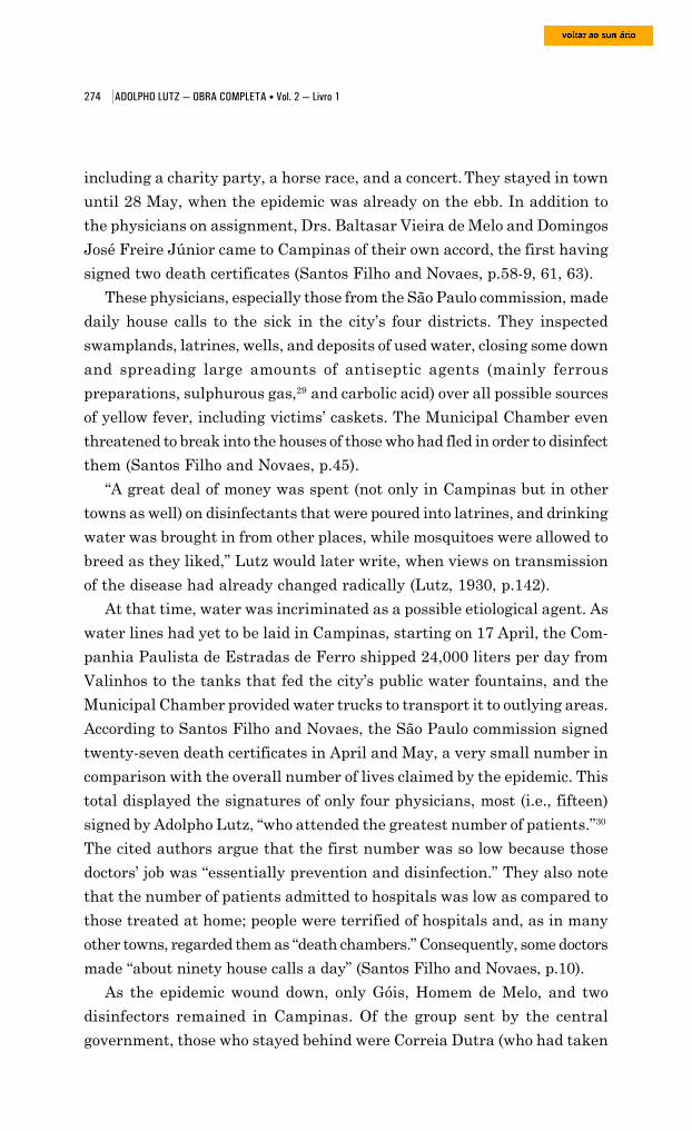
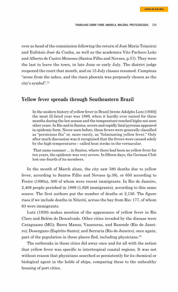
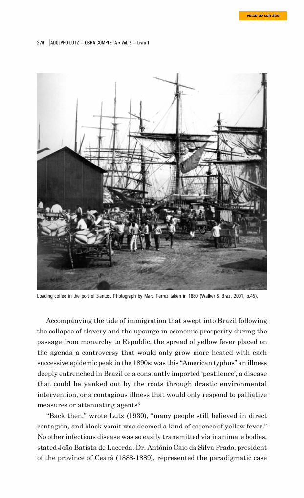
Loading coffee in the port of Santos. Photograph by Marc Ferrez taken in 1880 (Walker & Braz, 2001, p.45).
the agenda a controversy that would only grow more heated with each
successive epidemic peak in the 1890s: was this “American typhus” an illness
deeply entrenched in Brazil or a constantly imported ‘pestilence’, a disease
that could be yanked out by the roots through drastic environmental
intervention, or a contagious illness that would only respond to palliative
measures or attenuating agents?
“Back then,” wrote Lutz (1930), “many people still believed in direct
contagion, and black vomit was deemed a kind of essence of yellow fever.”
No other infectious disease was so easily transmitted via inanimate bodies,
stated João Batista de Lacerda. Dr. Antônio Caio da Silva Prado, president
of the province of Ceará (1888-1889), represented the paradigmatic case
277 TRABALHOS SOBRE FEBRE AMARELA, MALÁRIA, PROTOZOOLOGIA
of this viewpoint: he died on 25 May 1889, after receiving letters and
newspapers from infected Campinas. This and similar cases would often
be posited as unquestionable proof that the yellow fever germ was
transported by a wide gamut of objects: the infective matter clung to clothing,
to coal, to fruit peels, to stationery; entering the holds of ships and train
cars, it could travel great distances (Lacerda, op. cit., p.16-30; Brazil-Me-
dico, 8 Jun. 1899, p.212-4).
Proponents of the idea that yellow fever was transmitted by mosquitoes
would later have to work hard to refute the “monolith that is constantly
hurled against the new doctrine” – the case of Caio Prado and similar others
(Quinto Congresso, p.36, 150).
Adolpho Lutz’s Reminiscências was written when mosquito transmission
had already become established. For him, propagation of the disease after
being ‘exported’ from Rio de Janeiro to Campinas could be explained by
the following set of factors: Stegomyia fasciata had been disseminated,
and infected people were moving into or through regions inhabited by people
lacking immunity to the disease.
Campinas saw outbreaks in 1890, 1892, 1896, and 1897. The worst
took place in 1896, but “it no longer elicited the same manifestation of
solidarity. Everyone accepted things. There was no longer a stampede of
physicians or any charity drives” (Santos Filho and Novaes, p.35, 10).
For Adolpho Lutz (1930), infected mosquitoes carried by rail lines
accounted for the 1889 outbreaks in Rio Claro and Belém do Descalvado,
and also for a number of isolated cases among mail carriers and railroad
workers, including “a lady in Valinhos, who was married to the stationmaster
and lived in the stationhouse. She had never been to Campinas while yellow
fever was there.” In subsequent years, in the words of Lutz, the disease:
invaded the state of São Paulo, primarily following the Paulista railway. Itravaged Limeira, where I had maintained a private practice for five yearswithout ever seeing Stegomyia but it spared Araras and Jundiaí, wherethe villages are located quite far from the train stations. After the Paulista[railway], the Mogiana was invaded, and only much later came theSorocabana’s turn. What unquestionably kept it from spreading fasterwas that the numerous cases among those who fled the centers of epidemicscould not spread the yellow fever, without the prior or simultaneous importof Stegomyia.
Back when these outbreaks were erupting, the explanations that were
viewed as the most innovative no longer associated the disease with miasmas
278 ADOLPHO LUTZ — OBRA COMPLETA Vol. 2 — Livro 1
inseparably tied to certain environments; instead, it was believed to be
associated with microorganisms that could travel through different
environments and from person to person, airborne or by other means. The
overriding belief was that the germ underwent some kind of transformation
in the external environment before it became infective, yet only in some
geographical zones and during a certain season of the year.
In search of the yellow fever microbe
When Adolpho Lutz began his career in the early 1880s, the issue of
microbes was kindling heated discussions over the disease that was then
Brazil’s number-one public health problem. December was the month when
the merciless sun of the ‘muggy season’ beat down on the streets and houses
of Rio de Janeiro, or when these were submerged under torrential rains. It
was in that month in 1879, that Dr. Domingos José Freire, who held the
chair in organic chemistry at Rio’s School of Medicine,33 announced in the
papers his discovery of a microbe which he believed to be the cause of
yellow fever. In the “humors” of sick people he had found granules and
Vibrio that took the form of black corpuscles as they developed. He
contended that the detritus and countless spores released by them lent the
patient’s vomit its characteristic black color (Gazeta de Noticias, 23 Feb.
and 29 Feb. 1880).
In the first half of 1883, using techniques recently discovered by Louis
Pasteur, Freire developed a vaccine against the disease by attenuating the
microscopic algae that he called Cryptococcus xanthogenicus. The only other
prophylactic measure of this kind then applicable in fighting human diseases
was the smallpox vaccine. Pasteur’s achievements in this area were still
limited to vaccines against chicken cholera (1880) and anthrax or haematic
carbuncle (1881). Pasteur’s debut in the realm of human pathologies, with
the anti-rabies vaccine, would involve complex social and technical
constraints overcome only in 1886, as Geison (1995), Debré (1995),
Salomon-Bayet (1986), and others have shown.
A crisis involving the smallpox vaccine in Bahia catapulted Freire to
the presidency of the Central Board of Public Hygiene in late 1883,
facilitating dissemination of his vaccine against yellow fever among Rio de
Janeiro’s tenement dwellers.34 He enjoyed a surprisingly warm welcome
among immigrants and then among native-born Brazilians, not only
279 TRABALHOS SOBRE FEBRE AMARELA, MALÁRIA, PROTOZOOLOGIA
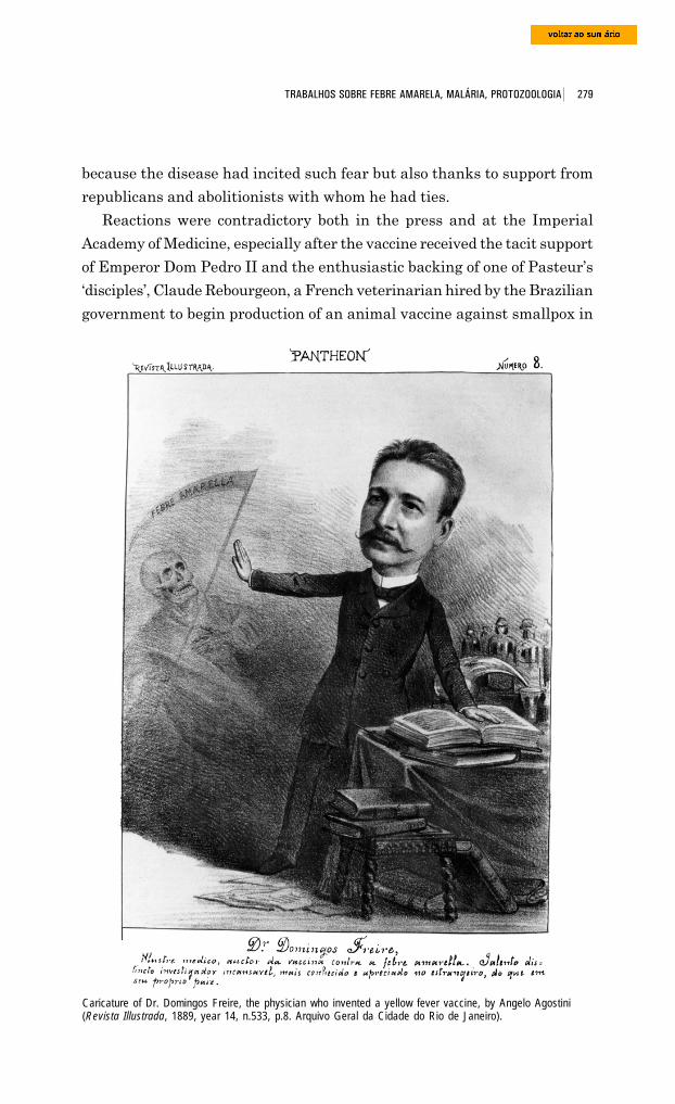
Caricature of Dr. Domingos Freire, the physician who invented a yellow fever vaccine, by Angelo Agostini(Revista Illustrada, 1889, year 14, n.533, p.8. Arquivo Geral da Cidade do Rio de Janeiro).
because the disease had incited such fear but also thanks to support from
republicans and abolitionists with whom he had ties.
Reactions were contradictory both in the press and at the Imperial
Academy of Medicine, especially after the vaccine received the tacit support
of Emperor Dom Pedro II and the enthusiastic backing of one of Pasteur’s
‘disciples’, Claude Rebourgeon, a French veterinarian hired by the Brazilian
government to begin production of an animal vaccine against smallpox in

280 ADOLPHO LUTZ — OBRA COMPLETA Vol. 2 — Livro 1
Rio Grande do Sul. Rebourgeon presented Freire’s discovery before the
academies of medicine and of science in Paris (Freire and Rebourgeon,
1884). Important figures in French medicine responded favorably, including
the pathologist Alfred Vulpian and the veterinarian Henry Bouley.
It should be mentioned in passing that both Pasteur’s 1881 efforts to
identify the yellow fever microbe as well as Emperor Dom Pedro II’s efforts
to entice him to Brazil to decipher the disease met with failure (Vallery-
Radot, 1951).35
Between 1883 and 1894, at least 12,329 immigrants and native-born
Brazilians in Rio and other cities around the country were inoculated with
Freire’s vaccine, which reached Puerto Rico, Jamaica, the Guianas, and
other French colonies too (Benchimol, 1999, p.119-68). The dissemination
of this invention was partly due to the increasingly complex web of relations
entwining the Brazilian bacteriologist with a number of other actors: colo-
nial and business interests, other microbe hunters, medical and scientific
associations, and the authors of treatises systematizing achievements in
microbiology.
During his stay in Europe between December 1886 and July 1887, Freire
submitted two special communications to the Academy of Sciences in Paris,
co-authored with Rebourgeon and with Paul Gibier, a researcher from the
Natural History Museum in Paris (Freire, Gibier, and Rebourgeon, 1887a
and b). This and other events in the French capital made a strong impact
in the Brazilian capital, and upon his return Freire was received as a hero
of Brazilian science by students and professors at technical schools and
universities in Rio de Janeiro, Minas Gerais, and São Paulo, by journalists
from a variety of periodicals, and by militant members of republican clubs
and abolitionist societies. Weeks later he traveled to Washington to
participate in the 9th International Medical Congress, which approved a
resolution recommending his vaccine for all countries afflicted with yellow
fever.36
A measure of the vaccine’s social and geographic reach is apparent in
the quantity and quality of the names that certified Freire’s statistical data
or that reported on post-vaccine symptoms, endorsing the immunizing
action of his prophylactic liquid; it is further reflected in the number of
physicians and authorities involved in its dissemination.
During 1883-84, a handful of people from Vassouras were vaccinated.37
On 6 February 1889, when the city’s Municipal Chamber met to decide
281 TRABALHOS SOBRE FEBRE AMARELA, MALÁRIA, PROTOZOOLOGIA
what means should be used to keep the disease from invading once again
(the city had already been hit twice), the decision to employ the vaccine
was unanimous. The terms of the letter to Freire show how far his reputation
had come. They entreated him to give the frightened population access to
“individual means of prevention, unquestionably more reliable than gene-
ral means, and whose discovery and popularization are due to ... your
immeasurable talent, [as you knew how] to earn not only the blessings of
the fatherland ... but likewise an eminent place among humanity’s
benefactors.”38
Joaquim Caminhoá (son), Freire’s assistant, vaccinated 199 people there.
The appeals sent by the Chambers of Pomba, Macaé, and Niterói were
no different. The first city requested a shipment of the vaccine liquid in
June, after the disease had struck neighboring Cataguases (MG), where
vaccination was performed by Dr. Araújo Lima. In Macaé, Dr. Carneiro
Mattoso, physician for the Chamber and public hygiene delegate, undertook
the task. In Niterói, 163 vaccinations were administered, a good share by
Freire himself, right inside the Chambers in the presence of its president,
Prosper David, and of the physician Victor David and the city aldermen.
The hygiene inspector for the province of Rio de Janeiro, Dr. Henrique
Baptista, was responsible for going from residence to residence. At the end
of the campaign, the Chamber offered use of one of its rooms so the scientist
could continue his “humanitarian work” throughout 1889. In Juiz de Fora
and Serraria (MG), a local physician, Dr. Avelar Andrade, was responsible
for vaccinating 30 and 50 people, respectively. The 54 inoculations in
Resende (Rio de Janeiro), all involving native-born Brazilians, were also
performed at the initiative of two local physicians, Drs. Gustavo Gomes
Jardim and Carlos Augusto de Oliveira e Silva. In Desengano (Espírito
Santo), the epidemic was combated by Dr. Henrique Baptista, with two
other physicians assisting: Rodrigues Guião, public hygiene delegate, and
Edmundo Lacerda, local clinician. Of the city’s 425 inhabitants, 102
received what was allegedly an ‘attenuated yellow fever virus’ (24%).
The disease reached Santos. At the Municipal Chambers, under the
eyes of the town’s most prominent citizens, 133 people were inoculated in
February 1889 by Dr. Barata Ribeiro, professor from Rio’s School of
Medicine.39
On 9 March, José Paulino Nogueira (mentioned earlier), president of
the Campinas Municipal Chamber, asked that the Inspector of Public
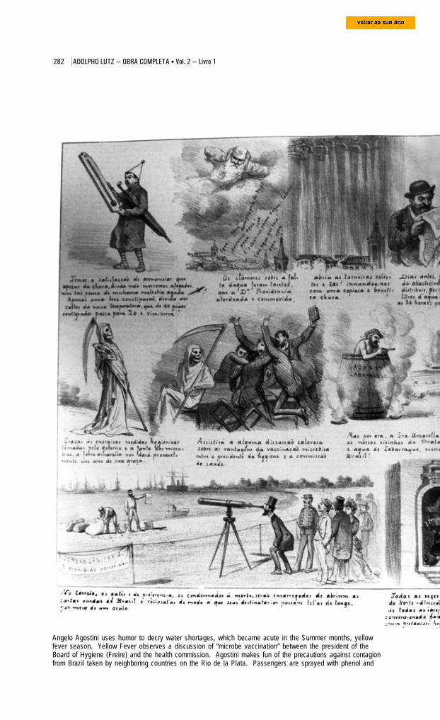
282 ADOLPHO LUTZ — OBRA COMPLETA Vol. 2 — Livro 1
Angelo Agostini uses humor to decry water shortages, which became acute in the Summer months, yellowfever season. Yellow Fever observes a discussion of “microbe vaccination” between the president of theBoard of Hygiene (Freire) and the health commission. Agostini makes fun of the precautions against contagionfrom Brazil taken by neighboring countries on the Rio de la Plata. Passengers are sprayed with phenol and
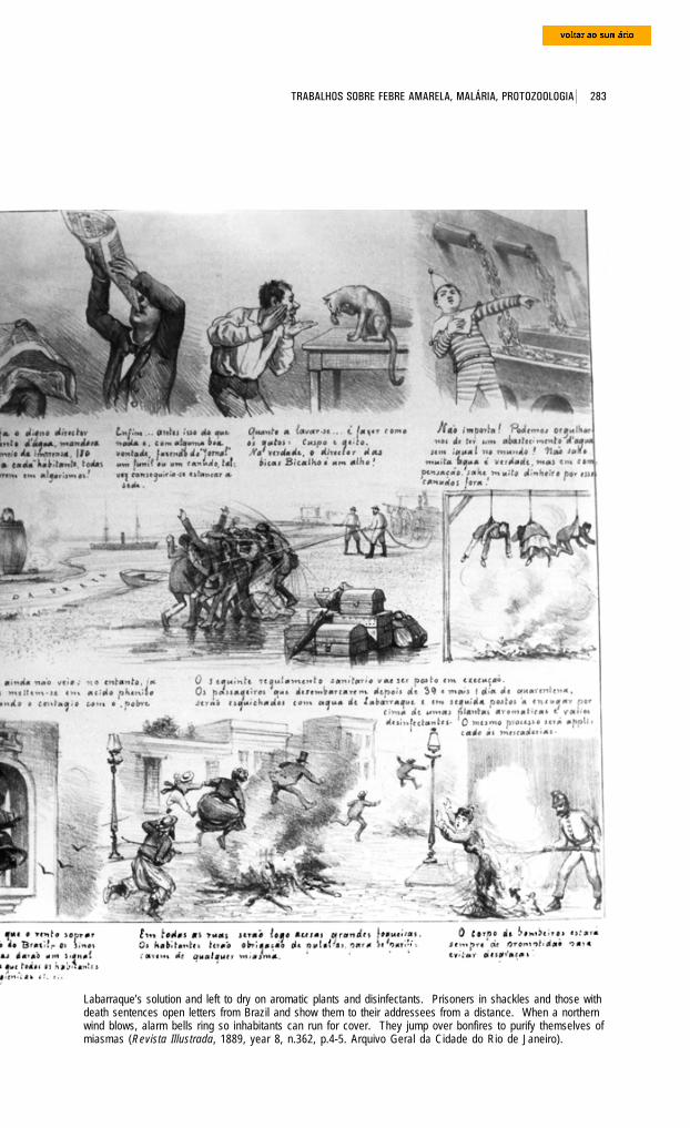
283 TRABALHOS SOBRE FEBRE AMARELA, MALÁRIA, PROTOZOOLOGIA
Labarraque’s solution and left to dry on aromatic plants and disinfectants. Prisoners in shackles and those withdeath sentences open letters from Brazil and show them to their addressees from a distance. When a northernwind blows, alarm bells ring so inhabitants can run for cover. They jump over bonfires to purify themselves ofmiasmas (Revista Illustrada, 1889, year 8, n.362, p.4-5. Arquivo Geral da Cidade do Rio de Janeiro).
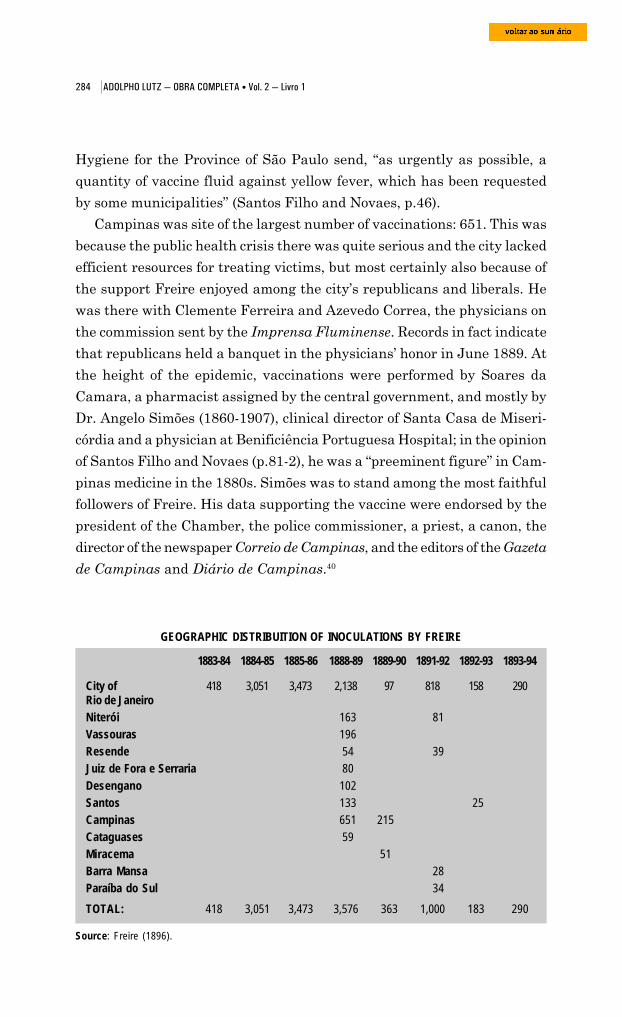
284 ADOLPHO LUTZ — OBRA COMPLETA Vol. 2 — Livro 1
Source: Freire (1896).
GEOGRAPHIC DISTRIBUITION OF INOCULATIONS BY FREIRE
Hygiene for the Province of São Paulo send, “as urgently as possible, a
quantity of vaccine fluid against yellow fever, which has been requested
by some municipalities” (Santos Filho and Novaes, p.46).
Campinas was site of the largest number of vaccinations: 651. This was
because the public health crisis there was quite serious and the city lacked
efficient resources for treating victims, but most certainly also because of
the support Freire enjoyed among the city’s republicans and liberals. He
was there with Clemente Ferreira and Azevedo Correa, the physicians on
the commission sent by the Imprensa Fluminense. Records in fact indicate
that republicans held a banquet in the physicians’ honor in June 1889. At
the height of the epidemic, vaccinations were performed by Soares da
Camara, a pharmacist assigned by the central government, and mostly by
Dr. Angelo Simões (1860-1907), clinical director of Santa Casa de Miseri-
córdia and a physician at Benificiência Portuguesa Hospital; in the opinion
of Santos Filho and Novaes (p.81-2), he was a “preeminent figure” in Cam-
pinas medicine in the 1880s. Simões was to stand among the most faithful
followers of Freire. His data supporting the vaccine were endorsed by the
president of the Chamber, the police commissioner, a priest, a canon, the
director of the newspaper Correio de Campinas, and the editors of the Gazeta
de Campinas and Diário de Campinas.40
1883-84 1884-85 1885-86 1888-89 1889-90 1891-92 1892-93 1893-94
City of 418 3,051 3,473 2,138 97 818 158 290Rio de JaneiroNiterói 163 81Vassouras 196Resende 54 39Juiz de Fora e Serraria 80Desengano 102Santos 133 25Campinas 651 215Cataguases 59Miracema 51Barra Mansa 28Paraíba do Sul 34TOTAL: 418 3,051 3,473 3,576 363 1,000 183 290

285 TRABALHOS SOBRE FEBRE AMARELA, MALÁRIA, PROTOZOOLOGIA
From December 1888 through June 1889, when yellow fever spread
throughout these rural parts – many affected for the first time – a total of
3,576 people were vaccinated, 988 of whom were immigrants and 2,537,
Brazilian (70.94%). Average mortality among those vaccinated was 0.79%,
a figure that fell to zero in Santos, Resende, Serraria, and Cataguases. “Given
these clear successes ... who would dare to doubt the efficaciousness of our
prophylactic measure and the truth of the fruitful doctrines formulated by
our eminent master, Pasteur?” asked Freire (op. cit., p.22, 25).
The more the urban middle layers accepted his new invention, the more
he was vulnerable to criticism from his adversaries, since the statistical
gap was widening between the vaccinated population – native-born, blacks,
and immigrants considered ‘acclimated’ and thus immune to yellow fever –
and the population still susceptible to the disease, made up mostly of recent
immigrants. Changes in the social composition of those vaccinated reflect
changes in how the vaccine was distributed. During an era of heightening
skepticism not only about yellow fever remedies but also about the feasibility
of sanitizing the Brazilian capital, this biological product became a most
welcome component of the doctor/patient relationship and of relations
between philanthropical institutions and the needy.
There is no way of knowing Adolpho Lutz’s opinion of Freire’s vaccine.
It is certain that he witnessed its use in Campinas, and all indications are
that he began analyzing the question, with his typical painstaking rigor,
in an unsigned article attributed to him by the organizers of his archive,
his children Bertha and Gualter Adolpho Lutz. “On the question of the
efficaciousness of vaccination against yellow fever” [in Port.] would appear
to be the first article in a series, but we were unable to locate any subsequent
editions of the periodical in which it was published and therefore unable to
ascertain whether Lutz continued the analysis initiated in São Paulo on
11 May 1889.
The article came out in the Freie Presse: Zeitung für Deutsche in
Brasilien, a periodical founded that same year and put out three times a
week. It had representatives not only in Campinas (Guilherme Trippe,
resident and/or with a business at 99 Lusitana Street) but in nine other
cities in São Paulo too (Araras, Botucatu, Piracicaba, Pirassununga, Ri-
beirão Preto, Rio Claro, Santos, São Carlos do Pinhal, and Sorocaba), as
well as Curitiba, Joinville, and Leipzig, home to Germany’s major
publishing houses.41
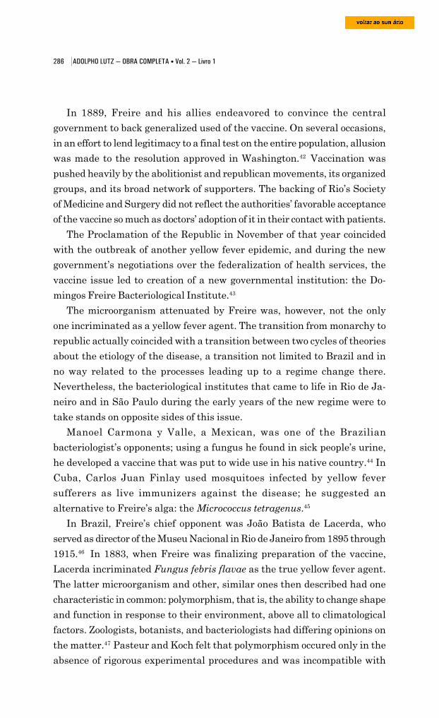
286 ADOLPHO LUTZ — OBRA COMPLETA Vol. 2 — Livro 1
In 1889, Freire and his allies endeavored to convince the central
government to back generalized used of the vaccine. On several occasions,
in an effort to lend legitimacy to a final test on the entire population, allusion
was made to the resolution approved in Washington.42 Vaccination was
pushed heavily by the abolitionist and republican movements, its organized
groups, and its broad network of supporters. The backing of Rio’s Society
of Medicine and Surgery did not reflect the authorities’ favorable acceptance
of the vaccine so much as doctors’ adoption of it in their contact with patients.
The Proclamation of the Republic in November of that year coincided
with the outbreak of another yellow fever epidemic, and during the new
government’s negotiations over the federalization of health services, the
vaccine issue led to creation of a new governmental institution: the Do-
mingos Freire Bacteriological Institute.43
The microorganism attenuated by Freire was, however, not the only
one incriminated as a yellow fever agent. The transition from monarchy to
republic actually coincided with a transition between two cycles of theories
about the etiology of the disease, a transition not limited to Brazil and in
no way related to the processes leading up to a regime change there.
Nevertheless, the bacteriological institutes that came to life in Rio de Ja-
neiro and in São Paulo during the early years of the new regime were to
take stands on opposite sides of this issue.
Manoel Carmona y Valle, a Mexican, was one of the Brazilian
bacteriologist’s opponents; using a fungus he found in sick people’s urine,
he developed a vaccine that was put to wide use in his native country.44 In
Cuba, Carlos Juan Finlay used mosquitoes infected by yellow fever
sufferers as live immunizers against the disease; he suggested an
alternative to Freire’s alga: the Micrococcus tetragenus.45
In Brazil, Freire’s chief opponent was João Batista de Lacerda, who
served as director of the Museu Nacional in Rio de Janeiro from 1895 through
1915.46 In 1883, when Freire was finalizing preparation of the vaccine,
Lacerda incriminated Fungus febris flavae as the true yellow fever agent.
The latter microorganism and other, similar ones then described had one
characteristic in common: polymorphism, that is, the ability to change shape
and function in response to their environment, above all to climatological
factors. Zoologists, botanists, and bacteriologists had differing opinions on
the matter.47 Pasteur and Koch felt that polymorphism occured only in the
absence of rigorous experimental procedures and was incompatible with
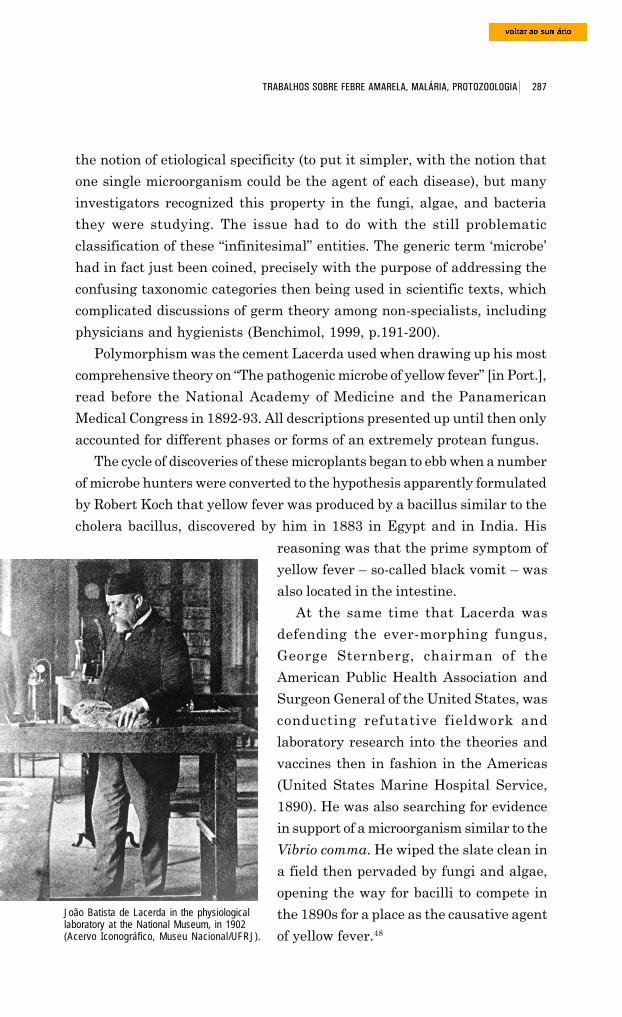
287 TRABALHOS SOBRE FEBRE AMARELA, MALÁRIA, PROTOZOOLOGIA
the notion of etiological specificity (to put it simpler, with the notion that
one single microorganism could be the agent of each disease), but many
investigators recognized this property in the fungi, algae, and bacteria
they were studying. The issue had to do with the still problematic
classification of these “infinitesimal” entities. The generic term ‘microbe’
had in fact just been coined, precisely with the purpose of addressing the
confusing taxonomic categories then being used in scientific texts, which
complicated discussions of germ theory among non-specialists, including
physicians and hygienists (Benchimol, 1999, p.191-200).
Polymorphism was the cement Lacerda used when drawing up his most
comprehensive theory on “The pathogenic microbe of yellow fever” [in Port.],
read before the National Academy of Medicine and the Panamerican
Medical Congress in 1892-93. All descriptions presented up until then only
accounted for different phases or forms of an extremely protean fungus.
The cycle of discoveries of these microplants began to ebb when a number
of microbe hunters were converted to the hypothesis apparently formulated
by Robert Koch that yellow fever was produced by a bacillus similar to the
cholera bacillus, discovered by him in 1883 in Egypt and in India. His
João Batista de Lacerda in the physiologicallaboratory at the National Museum, in 1902(Acervo Iconográfico, Museu Nacional/UFRJ).
reasoning was that the prime symptom of
yellow fever – so-called black vomit – was
also located in the intestine.
At the same time that Lacerda was
defending the ever-morphing fungus,
George Sternberg, chairman of the
American Public Health Association and
Surgeon General of the United States, was
conducting refutative fieldwork and
laboratory research into the theories and
vaccines then in fashion in the Americas
(United States Marine Hospital Service,
1890). He was also searching for evidence
in support of a microorganism similar to the
Vibrio comma. He wiped the slate clean in
a field then pervaded by fungi and algae,
opening the way for bacilli to compete in
the 1890s for a place as the causative agent
of yellow fever.48
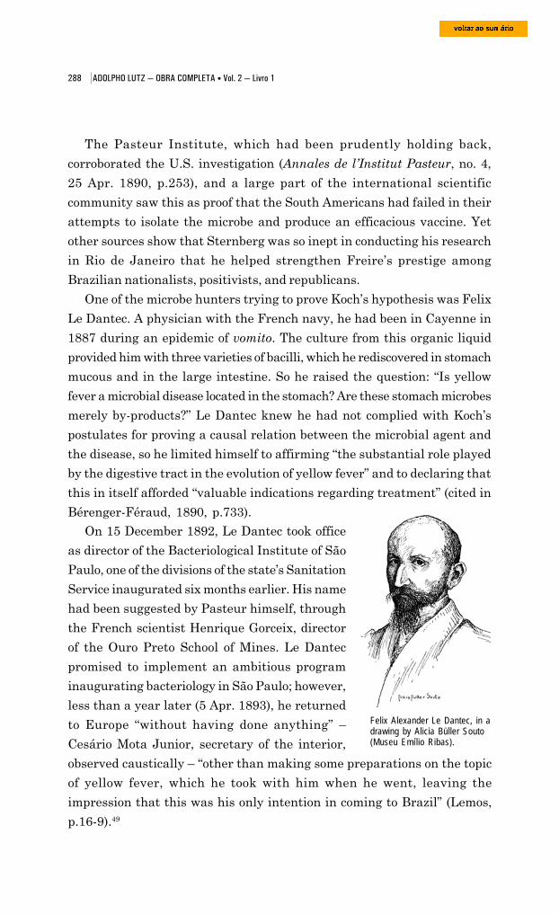
288 ADOLPHO LUTZ — OBRA COMPLETA Vol. 2 — Livro 1
On 15 December 1892, Le Dantec took office
as director of the Bacteriological Institute of São
Paulo, one of the divisions of the state’s Sanitation
Service inaugurated six months earlier. His name
had been suggested by Pasteur himself, through
the French scientist Henrique Gorceix, director
of the Ouro Preto School of Mines. Le Dantec
promised to implement an ambitious program
inaugurating bacteriology in São Paulo; however,
less than a year later (5 Apr. 1893), he returned
to Europe “without having done anything” –
Cesário Mota Junior, secretary of the interior,
Felix Alexander Le Dantec, in adrawing by Alicia Büller Souto(Museu Emílio Ribas).
observed caustically – “other than making some preparations on the topic
of yellow fever, which he took with him when he went, leaving the
impression that this was his only intention in coming to Brazil” (Lemos,
p.16-9).49
The Pasteur Institute, which had been prudently holding back,
corroborated the U.S. investigation (Annales de l’Institut Pasteur, no. 4,
25 Apr. 1890, p.253), and a large part of the international scientific
community saw this as proof that the South Americans had failed in their
attempts to isolate the microbe and produce an efficacious vaccine. Yet
other sources show that Sternberg was so inept in conducting his research
in Rio de Janeiro that he helped strengthen Freire’s prestige among
Brazilian nationalists, positivists, and republicans.
One of the microbe hunters trying to prove Koch’s hypothesis was Felix
Le Dantec. A physician with the French navy, he had been in Cayenne in
1887 during an epidemic of vomito. The culture from this organic liquid
provided him with three varieties of bacilli, which he rediscovered in stomach
mucous and in the large intestine. So he raised the question: “Is yellow
fever a microbial disease located in the stomach? Are these stomach microbes
merely by-products?” Le Dantec knew he had not complied with Koch’s
postulates for proving a causal relation between the microbial agent and
the disease, so he limited himself to affirming “the substantial role played
by the digestive tract in the evolution of yellow fever” and to declaring that
this in itself afforded “valuable indications regarding treatment” (cited in
Bérenger-Féraud, 1890, p.733).
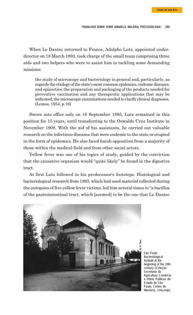
289 TRABALHOS SOBRE FEBRE AMARELA, MALÁRIA, PROTOZOOLOGIA
São PauloBacteriologicalInstitute at thebeginning of the 20thcentury (ColeçãoSecretaria daAgricultura Comércioe Obras Públicas doEstado de SãoPaulo, Centro deMemória, Unicamp).
When Le Dantec returned to France, Adolpho Lutz, appointed under-
director on 18 March 1893, took charge of the small team comprising three
aids and two helpers who were to assist him in tackling some demanding
missions:
the study of microscopy and bacteriology in general and, particularly, asregards the etiology of the state’s most common epidemics, endemic diseases,and epizooties; the preparation and packaging of the products needed forpreventive vaccination and any therapeutic applications that may beindicated; the microscopic examinations needed to clarify clinical diagnoses.(Lemos, 1954, p.16)
Sworn into office only on 18 September 1895, Lutz remained in this
position for 15 years, until transferring to the Oswaldo Cruz Institute in
November 1908. With the aid of his assistants, he carried out valuable
research on the infectious diseases that were endemic to the state or erupted
in the form of epidemics. He also faced harsh opposition from a majority of
those within the medical field and from other social actors.
Yellow fever was one of his topics of study, guided by the conviction
that the causative organism would “quite likely” be found in the digestive
tract.
At first Lutz followed in his predecessor’s footsteps. Histological and
bacteriological research from 1893, which had used material collected during
the autopsies of five yellow fever victims, led him several times to “a bacillus
of the gastrointestinal tract, which [seemed] to be the one that Le Dantec
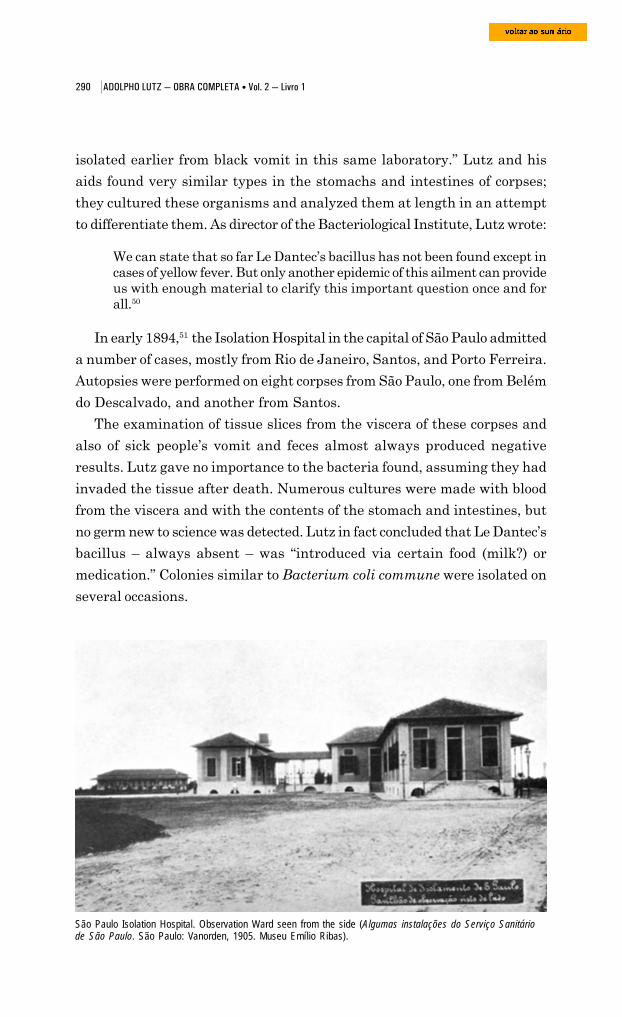
290 ADOLPHO LUTZ — OBRA COMPLETA Vol. 2 — Livro 1
isolated earlier from black vomit in this same laboratory.” Lutz and his
aids found very similar types in the stomachs and intestines of corpses;
they cultured these organisms and analyzed them at length in an attempt
to differentiate them. As director of the Bacteriological Institute, Lutz wrote:
We can state that so far Le Dantec’s bacillus has not been found except incases of yellow fever. But only another epidemic of this ailment can provideus with enough material to clarify this important question once and forall.50
In early 1894,51 the Isolation Hospital in the capital of São Paulo admitted
a number of cases, mostly from Rio de Janeiro, Santos, and Porto Ferreira.
Autopsies were performed on eight corpses from São Paulo, one from Belém
do Descalvado, and another from Santos.
The examination of tissue slices from the viscera of these corpses and
also of sick people’s vomit and feces almost always produced negative
results. Lutz gave no importance to the bacteria found, assuming they had
invaded the tissue after death. Numerous cultures were made with blood
from the viscera and with the contents of the stomach and intestines, but
no germ new to science was detected. Lutz in fact concluded that Le Dantec’s
bacillus – always absent – was “introduced via certain food (milk?) or
medication.” Colonies similar to Bacterium coli commune were isolated on
several occasions.
São Paulo Isolation Hospital. Observation Ward seen from the side (Algumas instalações do Serviço Sanitáriode São Paulo. São Paulo: Vanorden, 1905. Museu Emílio Ribas).
291 TRABALHOS SOBRE FEBRE AMARELA, MALÁRIA, PROTOZOOLOGIA
Because there were not enough sick people in São Paulo, studies only
proceeded in February 1895, at the Isolation Hospital in Santos, where
Arthur Vieira de Mendonça stayed for ten days and Lutz, five.52 There
they could observe people just coming down with the disease and could
culture material from the organs of those who perished.
They once again failed to find any “constantly present” parasite either
through direct microscopic examination or after subjecting preparations
and histological slices to known staining processes, as well as to some recently
discovered ones, “used for the first time to study yellow fever.” Inoculated
into a number of culture media, the blood most often would remain sterile
for over fifteen days. “On rare occasions, varieties of white or yellow sarcines
would develop, which can be considered accidental contaminations.”
Cultures made from the blood of yellow fever sufferers in the final stage of
the disease would at times produce microorganisms resembling Bacillus
coli communis and also streptococci and other varieties of cocci “not yet
determined.”
In tissue slices from the stomach and intestinal walls and also in the
mucous membrane of the duodenum and the ileus, “a large number of
bacilli [were often verified to be] invading the epithelium (which displayed
a greater or lesser degree of necrosis) and not rarely the submucous
membrane as well.” These were mainly the Bacillus mesentericus vulgatus,
Bacillus subtilis, and Bacillus albuminis.” The scientists concluded that
this constituted a
post mortem invasion by common species whose resistant spores are alwayspresent in the stomach and that develop chiefly where the mucousmembrane suffers peptic softening ... We have encountered similar processesin illnesses that differ entirely from yellow fever.
In his February 1895 report, Adolpho Lutz stated that he still thought
it likely that the yellow fever agent resided in the digestive tract, like the
cholera agent, but so far he had failed to isolate any organism different
from those found in other diseases.
We paid the price of much thankless work to obtain these results, mostnegative, in perfect agreement with Sternberg’s. It is our opinion that theyellow fever problem not only still awaits solution but will prove quitechallenging ... Either its germ is so poorly characterized that it blends inwith common germs, or it can only be found in significant abundanceduring a very limited time period, or, lastly, we as yet lack any method forrecognizing and culturing it.
292 ADOLPHO LUTZ — OBRA COMPLETA Vol. 2 — Livro 1
In 1897, the incidence of yellow fever dropped off in the state of São
Paulo, with only forty-one patients admitted to the Isolation Hospital during
the first five months of the year. Lutz and his aids autopsied seven of the
twenty-eight people who died there; they came from Rio Claro (three), Dois
Córregos (two), Araraquara (one), and Leme (one). Another five cases were
cured and provided material for bacteriological examinations of the contents
of the stomach and intestines, and also of the bile, urine, spleen, blood,
and other organic liquids (Lutz, 1898).
The results were as frustrating as before.
Sanarelli’s bacillus on the agenda
On the façade of the Domingos Freire Bacteriological Institute in Rio de
Janeiro waved a tri-color flag, black symbolizing vomit; yellow, jaundice;
and red, hemorrhaging. This emblem brought home the image of a bulwark
erected to defend a discovery coveted by so many. The flag was hoisted at
the moment when the press was stirring up expectations that yellow fever’s
secrets were about to be revealed by Giuseppe Sanarelli, a renowned
bacteriologist hired by the Uruguayan government to establish and direct
the Institute of Experimental Hygiene, inaugurated in Montevideo on 16
March 1896. Sanarelli, 31, had previously worked in Camilo Golgi’s
laboratory at the University of Pavia, at Max von Pettenkofer’s lab in
Munich, and at Elie Metchnikoff’s lab at the Pasteur Institute, in Paris.
There he had conducted studies on immunity in typhoid fever and cholera,
earning himself a respected name within the international scientific
community (Brazil-Medico, no. 24, Jun. 1896, p.218).
Three months after inauguration of the Montevideo institute, the Italian
bacteriologist came to Rio de Janeiro in quest of the organic matter he
needed in order to study yellow fever. In January 1897, his caldron was
bubbling. In the salons and cafes of the Brazilian capital, the topic of the
day was the news that Sanarelli had completed his research and had sent
a pli-cacheté (sealed communication) to the Academy of Medicine in Rome
to guarantee his place as the first discoverer. From the time he took this
initiative until the discovery was finally disclosed (postponed more than
once), a number of other postulants to the title of discoverer of the cause
and/or cure of yellow fever made their way onto the public stage.
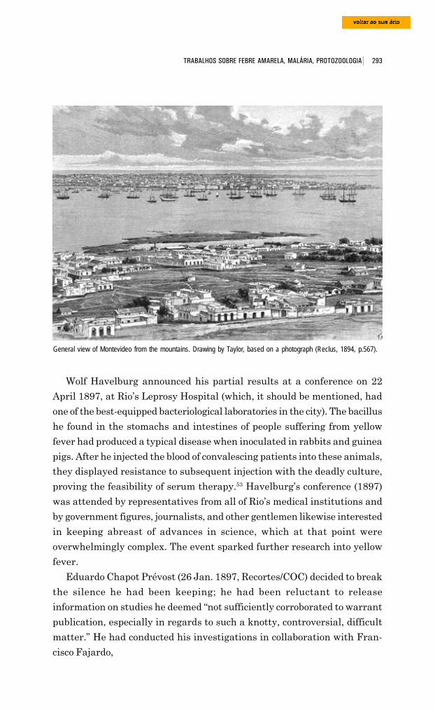
293 TRABALHOS SOBRE FEBRE AMARELA, MALÁRIA, PROTOZOOLOGIA
Wolf Havelburg announced his partial results at a conference on 22
April 1897, at Rio’s Leprosy Hospital (which, it should be mentioned, had
one of the best-equipped bacteriological laboratories in the city). The bacillus
he found in the stomachs and intestines of people suffering from yellow
fever had produced a typical disease when inoculated in rabbits and guinea
pigs. After he injected the blood of convalescing patients into these animals,
General view of Montevideo from the mountains. Drawing by Taylor, based on a photograph (Reclus, 1894, p.567).
they displayed resistance to subsequent injection with the deadly culture,
proving the feasibility of serum therapy.53 Havelburg’s conference (1897)
was attended by representatives from all of Rio’s medical institutions and
by government figures, journalists, and other gentlemen likewise interested
in keeping abreast of advances in science, which at that point were
overwhelmingly complex. The event sparked further research into yellow
fever.
Eduardo Chapot Prévost (26 Jan. 1897, Recortes/COC) decided to break
the silence he had been keeping; he had been reluctant to release
information on studies he deemed “not sufficiently corroborated to warrant
publication, especially in regards to such a knotty, controversial, difficult
matter.” He had conducted his investigations in collaboration with Fran-
cisco Fajardo,
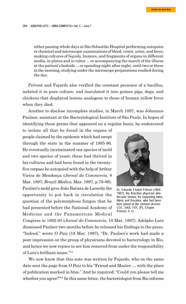
294 ADOLPHO LUTZ — OBRA COMPLETA Vol. 2 — Livro 1
either passing whole days at São Sebastião Hospital performing autopsiesor chemical and microscopic examinations of blood, vomit, urine, and feces;making cultures of liquids, humors, and fragments of organs in differentmedia, in plates and in tubes ... or accompanying the march of the illnessat the patient’s bedside ... or spending night after night, until two or threein the morning, studying under the microscope preparations readied duringthe day.
Prévost and Fajardo also verified the constant presence of a bacillus,
isolated it in pure culture, and inoculated it into guinea pigs, dogs, and
chickens that displayed lesions analogous to those of human yellow fever
when they died.
Another to disclose incomplete studies, in March 1897, was Johannes
Paulser, assistant at the Bacteriological Institute of São Paulo. In hopes of
identifying those germs that appeared on a regular basis, he endeavored
Dr. Eduardo Chabot Prévost (1864-1907), the Brazilian physician whobecame famous for separating twins,Maria and Rosalina, who had beenborn joined at the sternum (AcervoCOC, DAD, FOC [P], Chapot-Prévost, E-1).
to isolate all that he found in the organs of
people claimed by the epidemic which had swept
through the state in the summer of 1895-96.
He eventually incriminated one species of mold
and two species of yeast; these had thrived in
his cultures and had been found in the twenty-
five corpses he autopsied with the help of Arthur
Vieira de Mendonça (Jornal do Commercio, 8
Mar. 1897; Brazil-Medico, Mar. 1897, p.78-86).
Paulser’s mold gave João Batista de Lacerda the
opportunity to put back in circulation the
question of the polymorphous fungus that he
had presented before the National Academy of
Medicine and the Panamerican Medical
Congress in 1892-93 (Jornal do Commercio, 10 Mar. 1897). Adolpho Lutz
dismissed Paulser two months before he released his findings to the press.
“Indeed,” wrote O Paiz (18 Mar. 1897), “Dr. Paulser’s work had made a
poor impression on the group of physicians devoted to bacteriology in Rio,
and hence we now rejoice to see him removed from under the responsibility
of Lutz’s brilliant name.”54
We now know that this note was written by Fajardo, who on the same
date sent the page from O Paiz to his “Friend and Master … with the place
of publication marked in blue.” And he inquired: “Could you please tell me
whether you agree?”55 In this same letter, the bacteriologist from Rio informs
295 TRABALHOS SOBRE FEBRE AMARELA, MALÁRIA, PROTOZOOLOGIA
Lutz that he had received news from Sanarelli: “He is going to make his
announcement next May; he is decisive about the microbe. If I had time,
I’d go to Montevideo before then.”
In January 1897, the press in Rio de Janeiro started raising the issue.
One journalist expressed his “serious anxiousness” to learn the results
already achieved by the “wise man from the state of Rio” (11 Jan. 1897,
Recortes/COC). But, the journalist went on to state, Lutz was a most cautious
investigator and was certainly the most averse to publicity.
One indicator of how seriously yellow fever was taken at the institute
headed by Lutz are the data on post-mortem examinations: from 1893 to
1908, the lab’s bacteriologists cut open 121 corpses of yellow fever victims
and 92 of typhoid fever victims (cholera morbus ranked third, at 62). The
figures are even more impressive when we focus only on the 1894-97 period:
86 autopsies performed on victims of yellow fever and 67 on those who
died of typhoid fever. In 1896, 43 victims of the first disease were autopsied
and only 18 of the second.56
In a mid-1897 communiqué addressed solely to the director of the São
Paulo Sanitation Service, Lutz took stock of his studies on the etiology of
yellow fever. He had not found any germ that could be incriminated either
in the organs of those who had perished from well-characterized yellow
fever or in blood drawn during life.
The few positive results can be perfectly well accounted for by the possibleinvasion of secondary-infection germs or by the commencement of corpsedecomposition ... The cultures that have been made independently by ourlaboratory staff from many autopsies of yellow fever victims have revealedforms that correspond to species known to produce secondary infections inother illnesses (as, for example, typhoid fever), mainly microbes similar oridentical to Bacillus coli commune and to pyogenic cocci. (Lutz, 1897)
Lutz steered clear of any discussion of “certain work undertaken using
defective methods and also in the absence of information considered
indispensable.” Instead, he limited himself to observing that “the supposed
positive results alleged in these works have not been verified by other
investigators.” In an article published in the Revista Médica de São Paulo,
he expressed the opinion that the bacillus described by Havelburg appeared
to be merely “one of the countless varieties of Bacillus coli communis, which,
as we have verified, is often quite virulent for animals, when isolated from
cases of yellow fever” (Lutz, 1898).
296 ADOLPHO LUTZ — OBRA COMPLETA Vol. 2 — Livro 1
In a report written earlier,57 Lutz had pointed out that the anatomical
lesions observed in the viscera by advocates of the etiological theories then
in circulation had been “quite exaggerated, since the authors had examined
poorly conserved specimens or ones removed from the corpse long after
death. Thus, changes owing to decomposition, which occurs very rapidly
in yellow fever, were grouped together with changes caused by the illness.”
In kidneys suffering such conditions, Lutz always saw “coagula of albumin
and cylinders in the lumen of the renal tracts, but in well-preserved
specimens the epithelia [were] all in their proper place, and the nuclei
[were] easily stained.” The same thing was the case with the liver: “the
cells always contain many yellow pigment granules; using certain staining
methods, these hold the color of the dye, but only a careless observer could
confuse them with microorganisms.” Clinical observation showed that the
viscera quickly resumed their functions once the critical phase of the disease
had passed, which seemed to demonstrate that the lesions weren’t very
deep: “everything indicates that this is caused by intoxication by toxins
rather than the localization of the infectious process inside the parenchyma
of the viscera.”
This line of anatomical-pathological investigation led Adolpho Lutz to
provide guidance to one of his assistants, Dr. José Martins Bonilha de
Toledo, who was studying urine in yellow fever. Bonilha de Toledo, who
had specialized in biological chemistry in Belgium, conducted in-depth
chemical and microscopic analyses of the urine of thirteen victims of the
disease. He concluded that renal lesions were a constant, and directly
proportional to the seriousness of the case, confirming the suspicion that
they were caused by a toxin secreted by the unknown microbe responsible
for producing yellow fever. In January 1897, Bonilha de Toledo published
his observations on renal changes and on variations in the chlorides,
phosphates, and urea of yellow fever sufferers’ urine. He left only a few
clues when it came to solving the etiological question, but he clarified some
still obscure points concerning diagnosis and prognosis of the disease.58
Adolpho Lutz, the “wise man from the state of Rio,” boasted credentials
as solid as those of Giuseppe Sanarelli, the “wise Italian.” Thanks to Lutz’s
circumspection and prudence, and also to the capacity of the laboratory he
headed – superior to those operating in Rio – he distinguished himself as
the foremost “verifier” of the microbes, sera, and vaccines proposed by these
zealously competing bacteriologists. Above all, was someone who sought
297 TRABALHOS SOBRE FEBRE AMARELA, MALÁRIA, PROTOZOOLOGIA
victory by following the most unusual pathways, going farther than all
others in his handling of the press, in his pursuit of economic advantages,
and in mobilizing remarkably strong allies throughout his period in the
spotlight.
On 11 November 1896, Felipe Pereira Caldas, a physician from the state
of Rio Grande do Sul, spoke before the National Academy of Medicine about
the smallpox serum he had developed (Brazil-Medico, Nov. 1896). The
newspapers that day made much ado about the news (denied) that Sanarelli
had just unveiled the secret of yellow fever. Days earlier, imitating the
Italian’s action, Caldas had presented the Academy with a sealed
communiqué in order to guarantee priority in the discovery of a serum
against yellow fever. In January 1897, when Havelburg added fuel to the
race to discover the yellow fever microbe, Carlos Seidl tested the first doses
of Caldas’ serum at São Sebastião Hospital in Caju, which he directed. The
Rio Grande do Sul lobby, led by Dr. Severo Macedo, beseeched Manuel
Victorino Pereira, vice-president of the Republic and a physician from Bahia,
to sponsor Caldas on a new trip to Rio de Janeiro, arguing that it was
essential for a Brazilian scientist to assert priority for the discovery.
Felipe Caldas and his prophylactic reached the height of fame in mid-
1897, precisely when Giuseppe Sanarelli, in Montevideo, announced
discovery of the icteroid bacillus and the continuation of his efforts to obtain
a curative serum. In a message read on 26 June, Brazilian President Pru-
dente de Morais informed the National Congress that the Academy of
Medicine, by a unanimous vote, had requested that the government submit
Caldas’ serum to large-scale testing in the capital of the Republic and in
other places during the next yellow fever epidemic (Diário Oficial, 1 Jul.
1897; Brazil-Medico, 1 May 1898, p.145). The message was accompanied
by a statement from Nuno de Andrade, director of Public Health, in which
he declared that
Dr. Caldas’ yellow fever serum is not the fantasy of a visionary discoverer,but a medicine that can easily become part of yellow fever therapy, andwhose full benefits can only be definitively measured once its usefulnesshas been verified in a considerable number of cases.
In August 1897, Adolpho Lutz, Arthur Mendonça, and Bonilha de Toledo
signed a scathing report on Caldas’ findings. Conducted solely in the
laboratory, the verification contrasted sharply with the results of clinical
studies by physicians from Rio. Lutz placed quotation marks around phrases
298 ADOLPHO LUTZ — OBRA COMPLETA Vol. 2 — Livro 1
used by Caldas, to underscore questionable aspects of both method and
product: the serum’s supposed dual-purpose nature (preventive and
curative); the possible contamination of material collected in Rio de Janei-
ro and transported to the city of Rio Grande; and the non-determination of
the microbe correlated to yellow fever.
Asked if he used one of the known methods for isolating the germ, heanswered in the negative, since he ‘only had liquid serum available, inwhich – in his opinion – the only germs born are yellow fever ones.’ Askedif the inoculated animals displayed yellow fever symptoms, he said no.(Lutz and Toledo, 1897)
Although this report by the Adolpho Lutz team did not eliminate the
discovery made in Rio Grande do Sul (it would find a place in newspaper
headlines on and off until 1902), it did clear the way for a more enthusiastic
acceptance of the discovery of the icteroid bacillus announced by Giuseppe
Sanarelli on 10 June 1897, at Montevideo’s Teatro Solis.
Adolpho Lutz and Arthur Vieira de Mendonça traveled to Uruguay’s
capital in June as delegates of the São Paulo government. They were soon
joined by João Batista de Lacerda, Brazilian government representative,
along with Francisco Fajardo, Chapot Prévost, and Virgílio Ottoni, delegates
for Rio’s Society of Medicine and Surgery and for the Union of Hospital
Interns.59
A parenthetical comment: On his way back to Brazil, Lutz stopped in
Buenos Aires to meet the pathologist Robert Johann Wernicke (1854-1922)
and his disciple Alejandro Posadas (1870-1902). There he closely examined
the microscopic preparations and clinical cases that had prompted Wernicke
to publish a special communication in the Centralblatt für Bakteriologie
und Parasitenkunde (v.12, p.859-61, 1892) under the title “Ueber einen
Protozoenbefund bei mycosis fungoides,” and Posadas to publish “Un nuevo
caso de micose fungoidea con psorospermia” in the Circulo Médico Argen-
tino (v.15, p.585-97, 1892). Lutz’s visit was spurred by his studies then
underway in São Paulo, which were to culminate in his 1908 publication
of a paper that would eventually be recognized as his chief contribution to
Brazilian dermatology: the discovery in Brazil, for the first time, of a disease
characterized by serious mouth lesions, today known by a variety of names,
including Lutz disease or Lutz mycosis.60
In Montevideo, Lutz attended a conference that Sanarelli held for “a mixed
public of professionals and the lay.” The audience included the president of
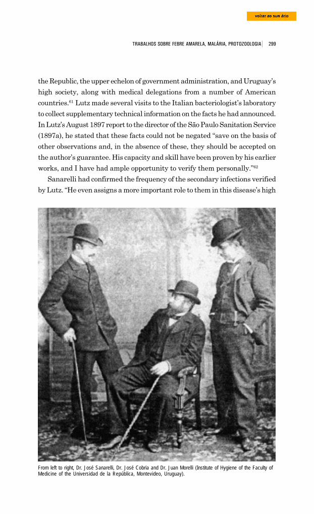
299 TRABALHOS SOBRE FEBRE AMARELA, MALÁRIA, PROTOZOOLOGIA
the Republic, the upper echelon of government administration, and Uruguay’s
high society, along with medical delegations from a number of American
countries.61 Lutz made several visits to the Italian bacteriologist’s laboratory
to collect supplementary technical information on the facts he had announced.
In Lutz’s August 1897 report to the director of the São Paulo Sanitation Service
(1897a), he stated that these facts could not be negated “save on the basis of
other observations and, in the absence of these, they should be accepted on
the author’s guarantee. His capacity and skill have been proven by his earlier
works, and I have had ample opportunity to verify them personally.”62
Sanarelli had confirmed the frequency of the secondary infections verified
by Lutz. “He even assigns a more important role to them in this disease’s high
From left to right, Dr. José Sanarelli, Dr. José Cobria and Dr. Juan Morelli (Institute of Hygiene of the Faculty ofMedicine of the Universidad de la República, Montevideo, Uruguay).
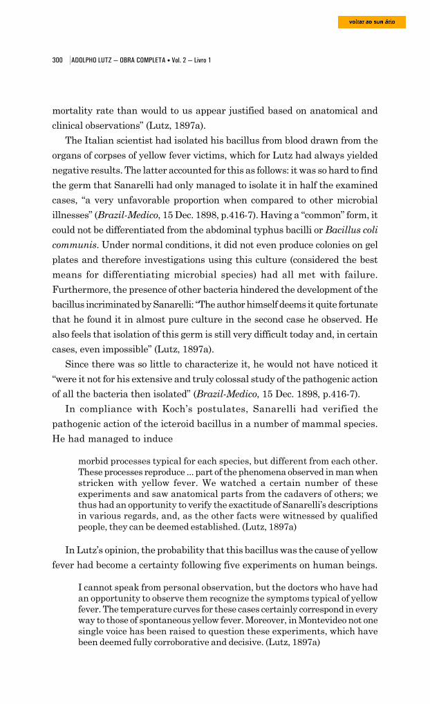
300 ADOLPHO LUTZ — OBRA COMPLETA Vol. 2 — Livro 1
mortality rate than would to us appear justified based on anatomical and
clinical observations” (Lutz, 1897a).
The Italian scientist had isolated his bacillus from blood drawn from the
organs of corpses of yellow fever victims, which for Lutz had always yielded
negative results. The latter accounted for this as follows: it was so hard to find
the germ that Sanarelli had only managed to isolate it in half the examined
cases, “a very unfavorable proportion when compared to other microbial
illnesses” (Brazil-Medico, 15 Dec. 1898, p.416-7). Having a “common” form, it
could not be differentiated from the abdominal typhus bacilli or Bacillus coli
communis. Under normal conditions, it did not even produce colonies on gel
plates and therefore investigations using this culture (considered the best
means for differentiating microbial species) had all met with failure.
Furthermore, the presence of other bacteria hindered the development of the
bacillus incriminated by Sanarelli: “The author himself deems it quite fortunate
that he found it in almost pure culture in the second case he observed. He
also feels that isolation of this germ is still very difficult today and, in certain
cases, even impossible” (Lutz, 1897a).
Since there was so little to characterize it, he would not have noticed it
“were it not for his extensive and truly colossal study of the pathogenic action
of all the bacteria then isolated” (Brazil-Medico, 15 Dec. 1898, p.416-7).
In compliance with Koch’s postulates, Sanarelli had verified the
pathogenic action of the icteroid bacillus in a number of mammal species.
He had managed to induce
morbid processes typical for each species, but different from each other.These processes reproduce ... part of the phenomena observed in man whenstricken with yellow fever. We watched a certain number of theseexperiments and saw anatomical parts from the cadavers of others; wethus had an opportunity to verify the exactitude of Sanarelli’s descriptionsin various regards, and, as the other facts were witnessed by qualifiedpeople, they can be deemed established. (Lutz, 1897a)
In Lutz’s opinion, the probability that this bacillus was the cause of yellow
fever had become a certainty following five experiments on human beings.
I cannot speak from personal observation, but the doctors who have hadan opportunity to observe them recognize the symptoms typical of yellowfever. The temperature curves for these cases certainly correspond in everyway to those of spontaneous yellow fever. Moreover, in Montevideo not onesingle voice has been raised to question these experiments, which havebeen deemed fully corroborative and decisive. (Lutz, 1897a)
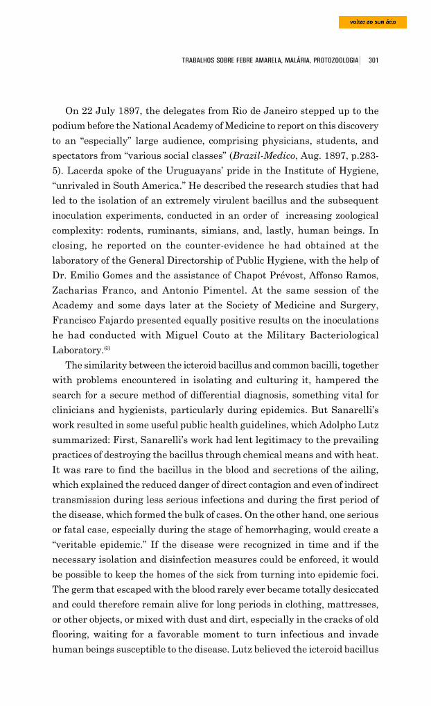
301 TRABALHOS SOBRE FEBRE AMARELA, MALÁRIA, PROTOZOOLOGIA
On 22 July 1897, the delegates from Rio de Janeiro stepped up to the
podium before the National Academy of Medicine to report on this discovery
to an “especially” large audience, comprising physicians, students, and
spectators from “various social classes” (Brazil-Medico, Aug. 1897, p.283-
5). Lacerda spoke of the Uruguayans’ pride in the Institute of Hygiene,
“unrivaled in South America.” He described the research studies that had
led to the isolation of an extremely virulent bacillus and the subsequent
inoculation experiments, conducted in an order of increasing zoological
complexity: rodents, ruminants, simians, and, lastly, human beings. In
closing, he reported on the counter-evidence he had obtained at the
laboratory of the General Directorship of Public Hygiene, with the help of
Dr. Emilio Gomes and the assistance of Chapot Prévost, Affonso Ramos,
Zacharias Franco, and Antonio Pimentel. At the same session of the
Academy and some days later at the Society of Medicine and Surgery,
Francisco Fajardo presented equally positive results on the inoculations
he had conducted with Miguel Couto at the Military Bacteriological
Laboratory.63
The similarity between the icteroid bacillus and common bacilli, together
with problems encountered in isolating and culturing it, hampered the
search for a secure method of differential diagnosis, something vital for
clinicians and hygienists, particularly during epidemics. But Sanarelli’s
work resulted in some useful public health guidelines, which Adolpho Lutz
summarized: First, Sanarelli’s work had lent legitimacy to the prevailing
practices of destroying the bacillus through chemical means and with heat.
It was rare to find the bacillus in the blood and secretions of the ailing,
which explained the reduced danger of direct contagion and even of indirect
transmission during less serious infections and during the first period of
the disease, which formed the bulk of cases. On the other hand, one serious
or fatal case, especially during the stage of hemorrhaging, would create a
“veritable epidemic.” If the disease were recognized in time and if the
necessary isolation and disinfection measures could be enforced, it would
be possible to keep the homes of the sick from turning into epidemic foci.
The germ that escaped with the blood rarely ever became totally desiccated
and could therefore remain alive for long periods in clothing, mattresses,
or other objects, or mixed with dust and dirt, especially in the cracks of old
flooring, waiting for a favorable moment to turn infectious and invade
human beings susceptible to the disease. Lutz believed the icteroid bacillus
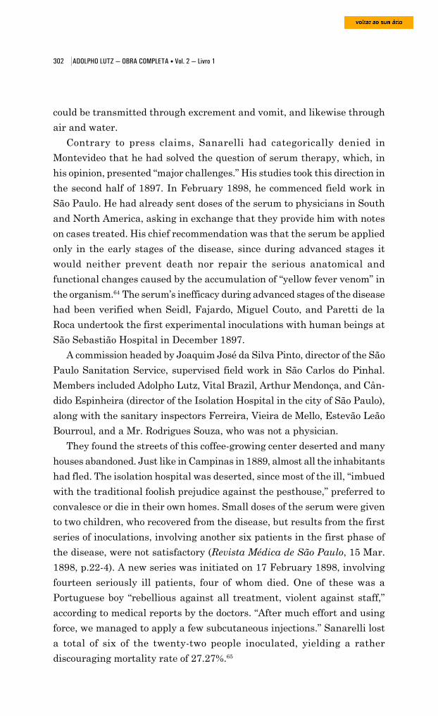
302 ADOLPHO LUTZ — OBRA COMPLETA Vol. 2 — Livro 1
could be transmitted through excrement and vomit, and likewise through
air and water.
Contrary to press claims, Sanarelli had categorically denied in
Montevideo that he had solved the question of serum therapy, which, in
his opinion, presented “major challenges.” His studies took this direction in
the second half of 1897. In February 1898, he commenced field work in
São Paulo. He had already sent doses of the serum to physicians in South
and North America, asking in exchange that they provide him with notes
on cases treated. His chief recommendation was that the serum be applied
only in the early stages of the disease, since during advanced stages it
would neither prevent death nor repair the serious anatomical and
functional changes caused by the accumulation of “yellow fever venom” in
the organism.64 The serum’s inefficacy during advanced stages of the disease
had been verified when Seidl, Fajardo, Miguel Couto, and Paretti de la
Roca undertook the first experimental inoculations with human beings at
São Sebastião Hospital in December 1897.
A commission headed by Joaquim José da Silva Pinto, director of the São
Paulo Sanitation Service, supervised field work in São Carlos do Pinhal.
Members included Adolpho Lutz, Vital Brazil, Arthur Mendonça, and Cân-
dido Espinheira (director of the Isolation Hospital in the city of São Paulo),
along with the sanitary inspectors Ferreira, Vieira de Mello, Estevão Leão
Bourroul, and a Mr. Rodrigues Souza, who was not a physician.
They found the streets of this coffee-growing center deserted and many
houses abandoned. Just like in Campinas in 1889, almost all the inhabitants
had fled. The isolation hospital was deserted, since most of the ill, “imbued
with the traditional foolish prejudice against the pesthouse,” preferred to
convalesce or die in their own homes. Small doses of the serum were given
to two children, who recovered from the disease, but results from the first
series of inoculations, involving another six patients in the first phase of
the disease, were not satisfactory (Revista Médica de São Paulo, 15 Mar.
1898, p.22-4). A new series was initiated on 17 February 1898, involving
fourteen seriously ill patients, four of whom died. One of these was a
Portuguese boy “rebellious against all treatment, violent against staff,”
according to medical reports by the doctors. “After much effort and using
force, we managed to apply a few subcutaneous injections.” Sanarelli lost
a total of six of the twenty-two people inoculated, yielding a rather
discouraging mortality rate of 27.27%.65
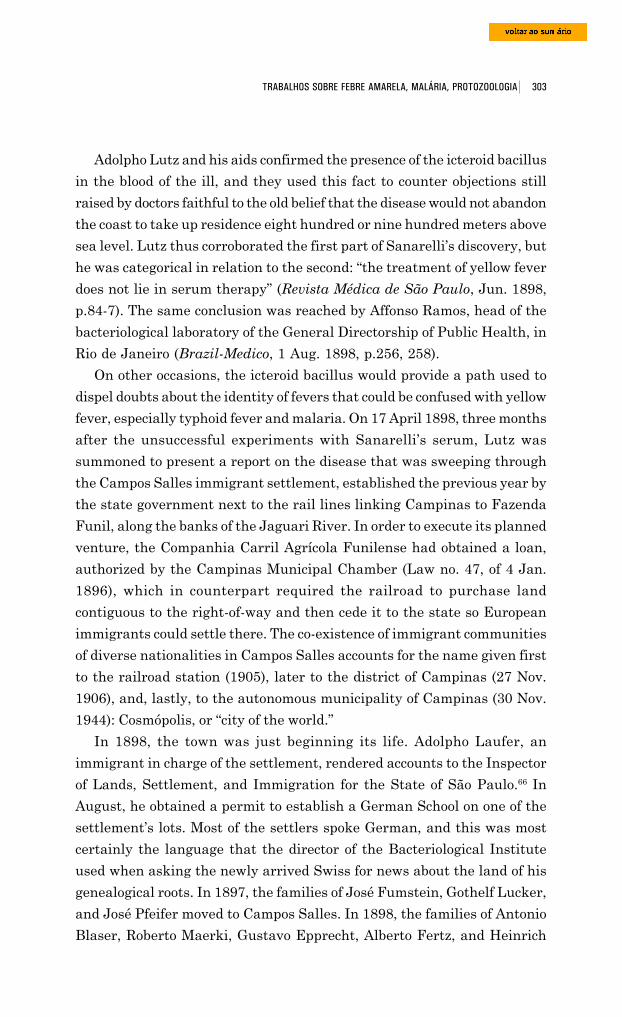
303 TRABALHOS SOBRE FEBRE AMARELA, MALÁRIA, PROTOZOOLOGIA
Adolpho Lutz and his aids confirmed the presence of the icteroid bacillus
in the blood of the ill, and they used this fact to counter objections still
raised by doctors faithful to the old belief that the disease would not abandon
the coast to take up residence eight hundred or nine hundred meters above
sea level. Lutz thus corroborated the first part of Sanarelli’s discovery, but
he was categorical in relation to the second: “the treatment of yellow fever
does not lie in serum therapy” (Revista Médica de São Paulo, Jun. 1898,
p.84-7). The same conclusion was reached by Affonso Ramos, head of the
bacteriological laboratory of the General Directorship of Public Health, in
Rio de Janeiro (Brazil-Medico, 1 Aug. 1898, p.256, 258).
On other occasions, the icteroid bacillus would provide a path used to
dispel doubts about the identity of fevers that could be confused with yellow
fever, especially typhoid fever and malaria. On 17 April 1898, three months
after the unsuccessful experiments with Sanarelli’s serum, Lutz was
summoned to present a report on the disease that was sweeping through
the Campos Salles immigrant settlement, established the previous year by
the state government next to the rail lines linking Campinas to Fazenda
Funil, along the banks of the Jaguari River. In order to execute its planned
venture, the Companhia Carril Agrícola Funilense had obtained a loan,
authorized by the Campinas Municipal Chamber (Law no. 47, of 4 Jan.
1896), which in counterpart required the railroad to purchase land
contiguous to the right-of-way and then cede it to the state so European
immigrants could settle there. The co-existence of immigrant communities
of diverse nationalities in Campos Salles accounts for the name given first
to the railroad station (1905), later to the district of Campinas (27 Nov.
1906), and, lastly, to the autonomous municipality of Campinas (30 Nov.
1944): Cosmópolis, or “city of the world.”
In 1898, the town was just beginning its life. Adolpho Laufer, an
immigrant in charge of the settlement, rendered accounts to the Inspector
of Lands, Settlement, and Immigration for the State of São Paulo.66 In
August, he obtained a permit to establish a German School on one of the
settlement’s lots. Most of the settlers spoke German, and this was most
certainly the language that the director of the Bacteriological Institute
used when asking the newly arrived Swiss for news about the land of his
genealogical roots. In 1897, the families of José Fumstein, Gothelf Lucker,
and José Pfeifer moved to Campos Salles. In 1898, the families of Antonio
Blaser, Roberto Maerki, Gustavo Epprecht, Alberto Fertz, and Heinrich
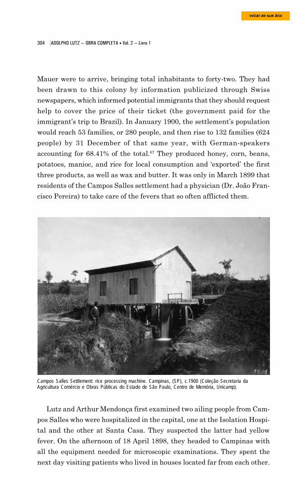
304 ADOLPHO LUTZ — OBRA COMPLETA Vol. 2 — Livro 1
Mauer were to arrive, bringing total inhabitants to forty-two. They had
been drawn to this colony by information publicized through Swiss
newspapers, which informed potential immigrants that they should request
help to cover the price of their ticket (the government paid for the
immigrant’s trip to Brazil). In January 1900, the settlement’s population
would reach 53 families, or 280 people, and then rise to 132 families (624
people) by 31 December of that same year, with German-speakers
accounting for 68.41% of the total.67 They produced honey, corn, beans,
potatoes, manioc, and rice for local consumption and ‘exported’ the first
three products, as well as wax and butter. It was only in March 1899 that
residents of the Campos Salles settlement had a physician (Dr. João Fran-
cisco Pereira) to take care of the fevers that so often afflicted them.
Campos Salles Settlement: rice processing machine. Campinas, (SP), c.1900 (Coleção Secretaria daAgricultura Comércio e Obras Públicas do Estado de São Paulo, Centro de Memória, Unicamp).
Lutz and Arthur Mendonça first examined two ailing people from Cam-
pos Salles who were hospitalized in the capital, one at the Isolation Hospi-
tal and the other at Santa Casa. They suspected the latter had yellow
fever. On the afternoon of 18 April 1898, they headed to Campinas with
all the equipment needed for microscopic examinations. They spent the
next day visiting patients who lived in houses located far from each other.
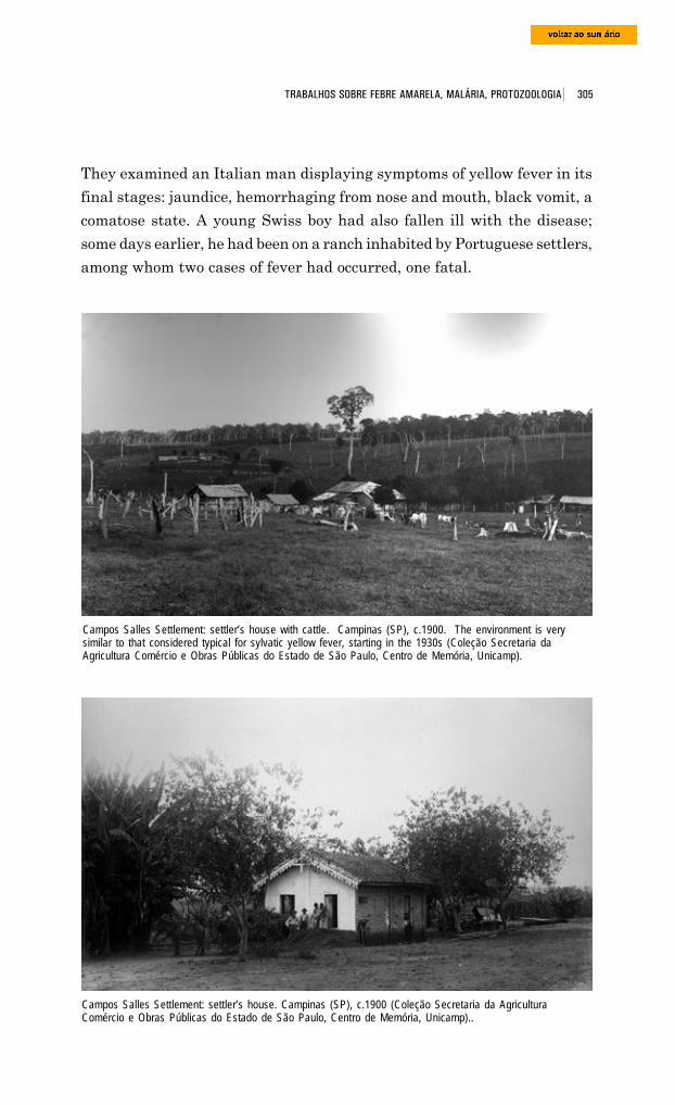
305 TRABALHOS SOBRE FEBRE AMARELA, MALÁRIA, PROTOZOOLOGIA
They examined an Italian man displaying symptoms of yellow fever in its
final stages: jaundice, hemorrhaging from nose and mouth, black vomit, a
comatose state. A young Swiss boy had also fallen ill with the disease;
some days earlier, he had been on a ranch inhabited by Portuguese settlers,
among whom two cases of fever had occurred, one fatal.
Campos Salles Settlement: settler’s house with cattle. Campinas (SP), c.1900. The environment is verysimilar to that considered typical for sylvatic yellow fever, starting in the 1930s (Coleção Secretaria daAgricultura Comércio e Obras Públicas do Estado de São Paulo, Centro de Memória, Unicamp).
Campos Salles Settlement: settler’s house. Campinas (SP), c.1900 (Coleção Secretaria da AgriculturaComércio e Obras Públicas do Estado de São Paulo, Centro de Memória, Unicamp)..
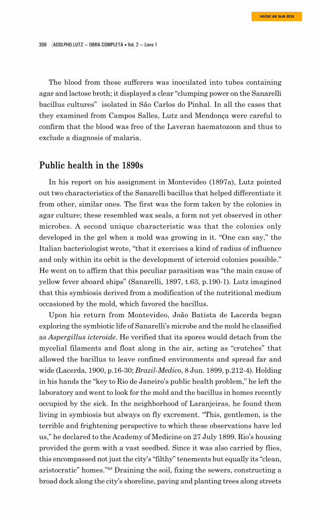
306 ADOLPHO LUTZ — OBRA COMPLETA Vol. 2 — Livro 1
The blood from these sufferers was inoculated into tubes containing
agar and lactose broth; it displayed a clear “clumping power on the Sanarelli
bacillus cultures” isolated in São Carlos do Pinhal. In all the cases that
they examined from Campos Salles, Lutz and Mendonça were careful to
confirm that the blood was free of the Laveran haematozoon and thus to
exclude a diagnosis of malaria.
Public health in the 1890s
In his report on his assignment in Montevideo (1897a), Lutz pointed
out two characteristics of the Sanarelli bacillus that helped differentiate it
from other, similar ones. The first was the form taken by the colonies in
agar culture; these resembled wax seals, a form not yet observed in other
microbes. A second unique characteristic was that the colonies only
developed in the gel when a mold was growing in it. “One can say,” the
Italian bacteriologist wrote, “that it exercises a kind of radius of influence
and only within its orbit is the development of icteroid colonies possible.”
He went on to affirm that this peculiar parasitism was “the main cause of
yellow fever aboard ships” (Sanarelli, 1897, t.63, p.190-1). Lutz imagined
that this symbiosis derived from a modification of the nutritional medium
occasioned by the mold, which favored the bacillus.
Upon his return from Montevideo, João Batista de Lacerda began
exploring the symbiotic life of Sanarelli’s microbe and the mold he classified
as Aspergillus icteroide. He verified that its spores would detach from the
mycelial filaments and float along in the air, acting as “crutches” that
allowed the bacillus to leave confined environments and spread far and
wide (Lacerda, 1900, p.16-30; Brazil-Medico, 8 Jun. 1899, p.212-4). Holding
in his hands the “key to Rio de Janeiro’s public health problem,” he left the
laboratory and went to look for the mold and the bacillus in homes recently
occupied by the sick. In the neighborhood of Laranjeiras, he found them
living in symbiosis but always on fly excrement. “This, gentlemen, is the
terrible and frightening perspective to which these observations have led
us,” he declared to the Academy of Medicine on 27 July 1899. Rio’s housing
provided the germ with a vast seedbed. Since it was also carried by flies,
this encompassed not just the city’s “filthy” tenements but equally its “clean,
aristocratic” homes.”68 Draining the soil, fixing the sewers, constructing a
broad dock along the city’s shoreline, paving and planting trees along streets
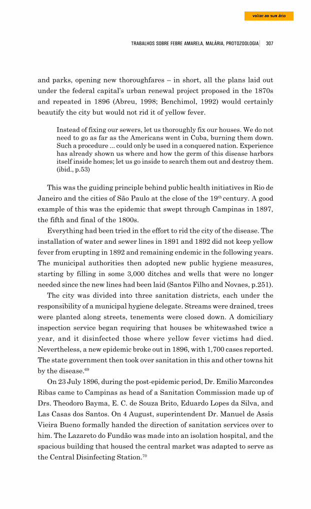
307 TRABALHOS SOBRE FEBRE AMARELA, MALÁRIA, PROTOZOOLOGIA
and parks, opening new thoroughfares – in short, all the plans laid out
under the federal capital’s urban renewal project proposed in the 1870s
and repeated in 1896 (Abreu, 1998; Benchimol, 1992) would certainly
beautify the city but would not rid it of yellow fever.
Instead of fixing our sewers, let us thoroughly fix our houses. We do notneed to go as far as the Americans went in Cuba, burning them down.Such a procedure ... could only be used in a conquered nation. Experiencehas already shown us where and how the germ of this disease harborsitself inside homes; let us go inside to search them out and destroy them.(ibid., p.53)
This was the guiding principle behind public health initiatives in Rio de
Janeiro and the cities of São Paulo at the close of the 19th century. A good
example of this was the epidemic that swept through Campinas in 1897,
the fifth and final of the 1800s.
Everything had been tried in the effort to rid the city of the disease. The
installation of water and sewer lines in 1891 and 1892 did not keep yellow
fever from erupting in 1892 and remaining endemic in the following years.
The municipal authorities then adopted new public hygiene measures,
starting by filling in some 3,000 ditches and wells that were no longer
needed since the new lines had been laid (Santos Filho and Novaes, p.251).
The city was divided into three sanitation districts, each under the
responsibility of a municipal hygiene delegate. Streams were drained, trees
were planted along streets, tenements were closed down. A domiciliary
inspection service began requiring that houses be whitewashed twice a
year, and it disinfected those where yellow fever victims had died.
Nevertheless, a new epidemic broke out in 1896, with 1,700 cases reported.
The state government then took over sanitation in this and other towns hit
by the disease.69
On 23 July 1896, during the post-epidemic period, Dr. Emilio Marcondes
Ribas came to Campinas as head of a Sanitation Commission made up of
Drs. Theodoro Bayma, E. C. de Souza Brito, Eduardo Lopes da Silva, and
Las Casas dos Santos. On 4 August, superintendent Dr. Manuel de Assis
Vieira Bueno formally handed the direction of sanitation services over to
him. The Lazareto do Fundão was made into an isolation hospital, and the
spacious building that housed the central market was adapted to serve as
the Central Disinfecting Station.70
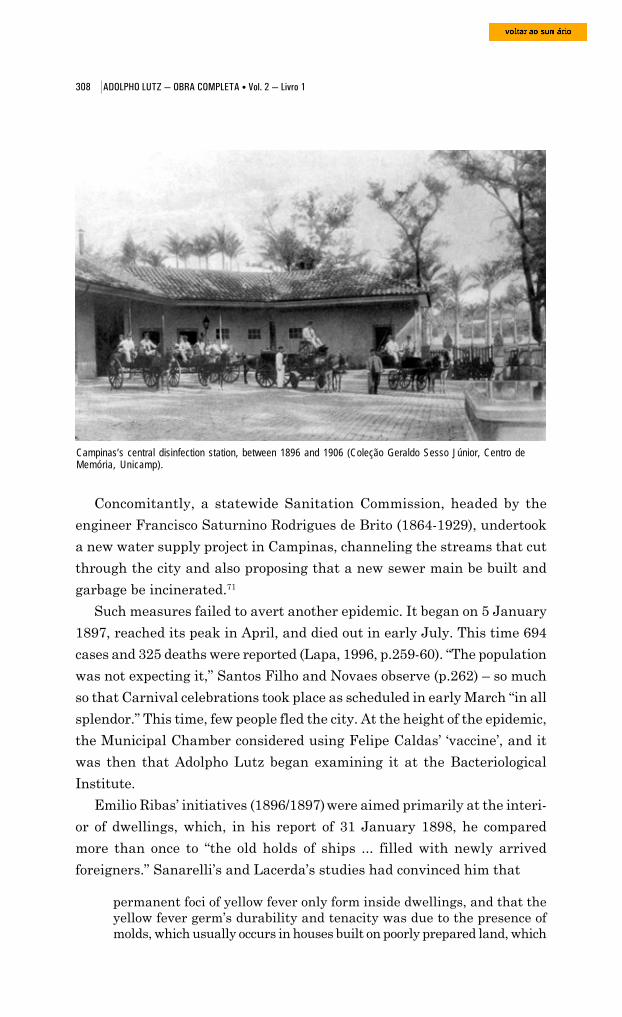
308 ADOLPHO LUTZ — OBRA COMPLETA Vol. 2 — Livro 1
Concomitantly, a statewide Sanitation Commission, headed by the
engineer Francisco Saturnino Rodrigues de Brito (1864-1929), undertook
a new water supply project in Campinas, channeling the streams that cut
through the city and also proposing that a new sewer main be built and
garbage be incinerated.71
Such measures failed to avert another epidemic. It began on 5 January
1897, reached its peak in April, and died out in early July. This time 694
cases and 325 deaths were reported (Lapa, 1996, p.259-60). “The population
was not expecting it,” Santos Filho and Novaes observe (p.262) – so much
so that Carnival celebrations took place as scheduled in early March “in all
splendor.” This time, few people fled the city. At the height of the epidemic,
the Municipal Chamber considered using Felipe Caldas’ ‘vaccine’, and it
was then that Adolpho Lutz began examining it at the Bacteriological
Institute.
Emilio Ribas’ initiatives (1896/1897) were aimed primarily at the interi-
or of dwellings, which, in his report of 31 January 1898, he compared
more than once to “the old holds of ships ... filled with newly arrived
foreigners.” Sanarelli’s and Lacerda’s studies had convinced him that
permanent foci of yellow fever only form inside dwellings, and that theyellow fever germ’s durability and tenacity was due to the presence ofmolds, which usually occurs in houses built on poorly prepared land, which
Campinas’s central disinfection station, between 1896 and 1906 (Coleção Geraldo Sesso Júnior, Centro deMemória, Unicamp).
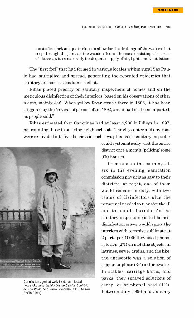
309 TRABALHOS SOBRE FEBRE AMARELA, MALÁRIA, PROTOZOOLOGIA
most often lack adequate slope to allow for the drainage of the waters thatseep through the joints of the wooden floors – houses consisting of a seriesof alcoves, with a naturally inadequate supply of air, light, and ventilation.
The “first foci” that had formed in various locales within rural São Pau-
lo had multiplied and spread, generating the repeated epidemics that
sanitary authorities could not defeat.
Ribas placed priority on sanitary inspections of homes and on the
meticulous disinfection of their interiors, based on his observations of other
places, mainly Jaú. When yellow fever struck there in 1896, it had been
triggered by the “revival of germs left in 1892, and it had not been imported,
as people said.”
Ribas estimated that Campinas had at least 4,200 buildings in 1897,
not counting those in outlying neighborhoods. The city center and environs
were re-divided into five districts in such a way that each sanitary inspector
Desinfection agent at work inside an infectedhouse (Algumas instalações do Serviço Sanitáriode São Paulo. São Paulo: Vanorden, 1905. MuseuEmílio Ribas).
could systematically visit the entire
district once a month, ‘policing’ some
900 houses.
From nine in the morning till
six in the evening, sanitation
commission physicians saw to their
districts; at night, one of them
would remain on duty, with two
teams of disinfectors plus the
personnel needed to transfer the ill
and to handle burials. As the
sanitary inspectors visited homes,
disinfection crews would spray the
interiors with corrosive sublimate at
2 parts per 1000; they used phenol
solution (2%) on metallic objects; in
latrines, sewer drains, and the like,
the antiseptic was a solution of
copper sulphate (3%) or limewater.
In stables, carriage barns, and
parks, they sprayed solutions of
cresyl or of phenol acid (4%).
Between July 1896 and January
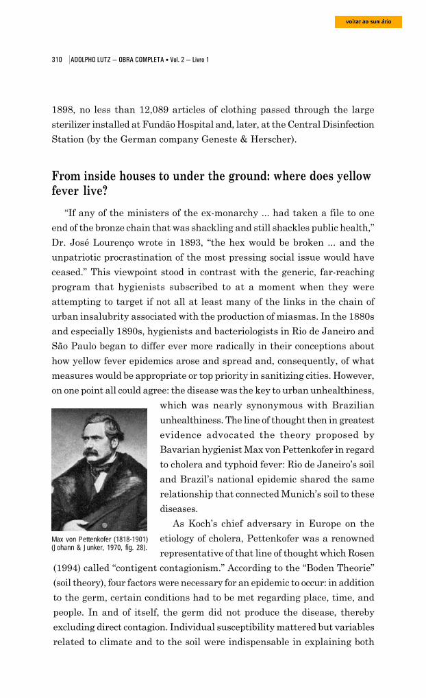
310 ADOLPHO LUTZ — OBRA COMPLETA Vol. 2 — Livro 1
1898, no less than 12,089 articles of clothing passed through the large
sterilizer installed at Fundão Hospital and, later, at the Central Disinfection
Station (by the German company Geneste & Herscher).
From inside houses to under the ground: where does yellowfever live?
“If any of the ministers of the ex-monarchy ... had taken a file to one
end of the bronze chain that was shackling and still shackles public health,”
Dr. José Lourenço wrote in 1893, “the hex would be broken ... and the
unpatriotic procrastination of the most pressing social issue would have
ceased.” This viewpoint stood in contrast with the generic, far-reaching
program that hygienists subscribed to at a moment when they were
attempting to target if not all at least many of the links in the chain of
urban insalubrity associated with the production of miasmas. In the 1880s
and especially 1890s, hygienists and bacteriologists in Rio de Janeiro and
São Paulo began to differ ever more radically in their conceptions about
how yellow fever epidemics arose and spread and, consequently, of what
measures would be appropriate or top priority in sanitizing cities. However,
on one point all could agree: the disease was the key to urban unhealthiness,
which was nearly synonymous with Brazilian
unhealthiness. The line of thought then in greatest
evidence advocated the theory proposed by
Bavarian hygienist Max von Pettenkofer in regard
to cholera and typhoid fever: Rio de Janeiro’s soil
and Brazil’s national epidemic shared the same
relationship that connected Munich’s soil to these
diseases.
As Koch’s chief adversary in Europe on the
etiology of cholera, Pettenkofer was a renowned
representative of that line of thought which Rosen
Max von Pettenkofer (1818-1901)(Johann & Junker, 1970, fig. 28).
(1994) called “contigent contagionism.” According to the “Boden Theorie”
(soil theory), four factors were necessary for an epidemic to occur: in addition
to the germ, certain conditions had to be met regarding place, time, and
people. In and of itself, the germ did not produce the disease, thereby
excluding direct contagion. Individual susceptibility mattered but variables
related to climate and to the soil were indispensable in explaining both
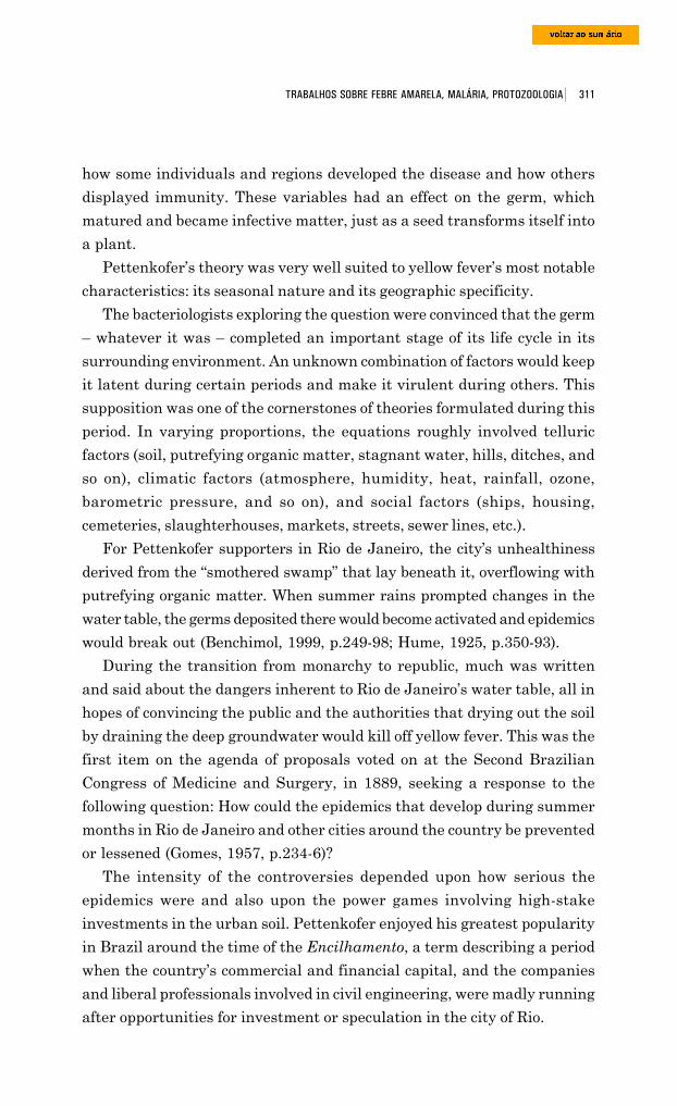
311 TRABALHOS SOBRE FEBRE AMARELA, MALÁRIA, PROTOZOOLOGIA
how some individuals and regions developed the disease and how others
displayed immunity. These variables had an effect on the germ, which
matured and became infective matter, just as a seed transforms itself into
a plant.
Pettenkofer’s theory was very well suited to yellow fever’s most notable
characteristics: its seasonal nature and its geographic specificity.
The bacteriologists exploring the question were convinced that the germ
– whatever it was – completed an important stage of its life cycle in its
surrounding environment. An unknown combination of factors would keep
it latent during certain periods and make it virulent during others. This
supposition was one of the cornerstones of theories formulated during this
period. In varying proportions, the equations roughly involved telluric
factors (soil, putrefying organic matter, stagnant water, hills, ditches, and
so on), climatic factors (atmosphere, humidity, heat, rainfall, ozone,
barometric pressure, and so on), and social factors (ships, housing,
cemeteries, slaughterhouses, markets, streets, sewer lines, etc.).
For Pettenkofer supporters in Rio de Janeiro, the city’s unhealthiness
derived from the “smothered swamp” that lay beneath it, overflowing with
putrefying organic matter. When summer rains prompted changes in the
water table, the germs deposited there would become activated and epidemics
would break out (Benchimol, 1999, p.249-98; Hume, 1925, p.350-93).
During the transition from monarchy to republic, much was written
and said about the dangers inherent to Rio de Janeiro’s water table, all in
hopes of convincing the public and the authorities that drying out the soil
by draining the deep groundwater would kill off yellow fever. This was the
first item on the agenda of proposals voted on at the Second Brazilian
Congress of Medicine and Surgery, in 1889, seeking a response to the
following question: How could the epidemics that develop during summer
months in Rio de Janeiro and other cities around the country be prevented
or lessened (Gomes, 1957, p.234-6)?
The intensity of the controversies depended upon how serious the
epidemics were and also upon the power games involving high-stake
investments in the urban soil. Pettenkofer enjoyed his greatest popularity
in Brazil around the time of the Encilhamento, a term describing a period
when the country’s commercial and financial capital, and the companies
and liberal professionals involved in civil engineering, were madly running
after opportunities for investment or speculation in the city of Rio.
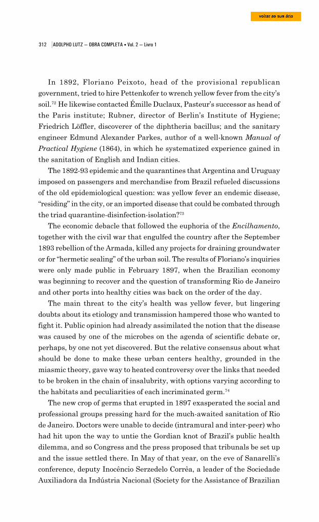
312 ADOLPHO LUTZ — OBRA COMPLETA Vol. 2 — Livro 1
In 1892, Floriano Peixoto, head of the provisional republican
government, tried to hire Pettenkofer to wrench yellow fever from the city’s
soil.72 He likewise contacted Émille Duclaux, Pasteur’s successor as head of
the Paris institute; Rubner, director of Berlin’s Institute of Hygiene;
Friedrich Löffler, discoverer of the diphtheria bacillus; and the sanitary
engineer Edmund Alexander Parkes, author of a well-known Manual of
Practical Hygiene (1864), in which he systematized experience gained in
the sanitation of English and Indian cities.
The 1892-93 epidemic and the quarantines that Argentina and Uruguay
imposed on passengers and merchandise from Brazil refueled discussions
of the old epidemiological question: was yellow fever an endemic disease,
“residing” in the city, or an imported disease that could be combated through
the triad quarantine-disinfection-isolation?73
The economic debacle that followed the euphoria of the Encilhamento,
together with the civil war that engulfed the country after the September
1893 rebellion of the Armada, killed any projects for draining groundwater
or for “hermetic sealing” of the urban soil. The results of Floriano’s inquiries
were only made public in February 1897, when the Brazilian economy
was beginning to recover and the question of transforming Rio de Janeiro
and other ports into healthy cities was back on the order of the day.
The main threat to the city’s health was yellow fever, but lingering
doubts about its etiology and transmission hampered those who wanted to
fight it. Public opinion had already assimilated the notion that the disease
was caused by one of the microbes on the agenda of scientific debate or,
perhaps, by one not yet discovered. But the relative consensus about what
should be done to make these urban centers healthy, grounded in the
miasmic theory, gave way to heated controversy over the links that needed
to be broken in the chain of insalubrity, with options varying according to
the habitats and peculiarities of each incriminated germ.74
The new crop of germs that erupted in 1897 exasperated the social and
professional groups pressing hard for the much-awaited sanitation of Rio
de Janeiro. Doctors were unable to decide (intramural and inter-peer) who
had hit upon the way to untie the Gordian knot of Brazil’s public health
dilemma, and so Congress and the press proposed that tribunals be set up
and the issue settled there. In May of that year, on the eve of Sanarelli’s
conference, deputy Inocêncio Serzedelo Corrêa, a leader of the Sociedade
Auxiliadora da Indústria Nacional (Society for the Assistance of Brazilian
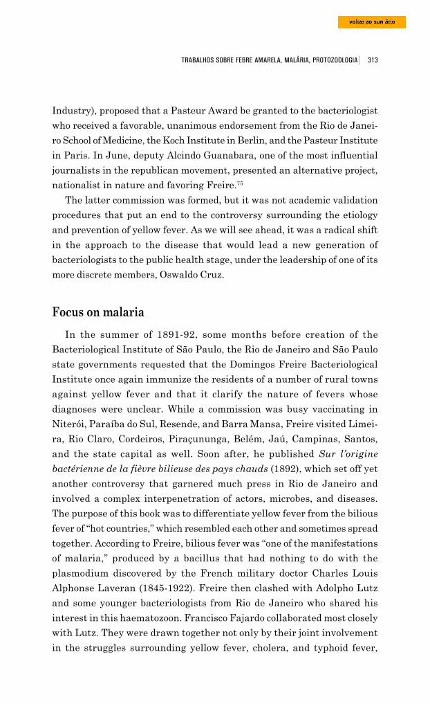
313 TRABALHOS SOBRE FEBRE AMARELA, MALÁRIA, PROTOZOOLOGIA
Industry), proposed that a Pasteur Award be granted to the bacteriologist
who received a favorable, unanimous endorsement from the Rio de Janei-
ro School of Medicine, the Koch Institute in Berlin, and the Pasteur Institute
in Paris. In June, deputy Alcindo Guanabara, one of the most influential
journalists in the republican movement, presented an alternative project,
nationalist in nature and favoring Freire.75
The latter commission was formed, but it was not academic validation
procedures that put an end to the controversy surrounding the etiology
and prevention of yellow fever. As we will see ahead, it was a radical shift
in the approach to the disease that would lead a new generation of
bacteriologists to the public health stage, under the leadership of one of its
more discrete members, Oswaldo Cruz.
Focus on malaria
In the summer of 1891-92, some months before creation of the
Bacteriological Institute of São Paulo, the Rio de Janeiro and São Paulo
state governments requested that the Domingos Freire Bacteriological
Institute once again immunize the residents of a number of rural towns
against yellow fever and that it clarify the nature of fevers whose
diagnoses were unclear. While a commission was busy vaccinating in
Niterói, Paraíba do Sul, Resende, and Barra Mansa, Freire visited Limei-
ra, Rio Claro, Cordeiros, Piraçununga, Belém, Jaú, Campinas, Santos,
and the state capital as well. Soon after, he published Sur l’origine
bactérienne de la fièvre bilieuse des pays chauds (1892), which set off yet
another controversy that garnered much press in Rio de Janeiro and
involved a complex interpenetration of actors, microbes, and diseases.
The purpose of this book was to differentiate yellow fever from the bilious
fever of “hot countries,” which resembled each other and sometimes spread
together. According to Freire, bilious fever was “one of the manifestations
of malaria,” produced by a bacillus that had nothing to do with the
plasmodium discovered by the French military doctor Charles Louis
Alphonse Laveran (1845-1922). Freire then clashed with Adolpho Lutz
and some younger bacteriologists from Rio de Janeiro who shared his
interest in this haematozoon. Francisco Fajardo collaborated most closely
with Lutz. They were drawn together not only by their joint involvement
in the struggles surrounding yellow fever, cholera, and typhoid fever,
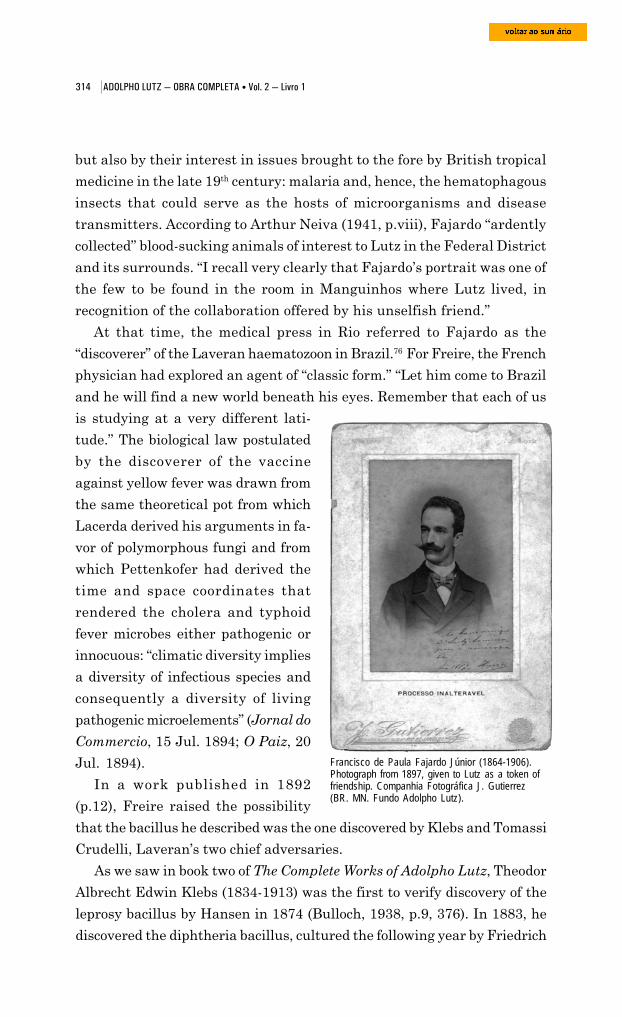
314 ADOLPHO LUTZ — OBRA COMPLETA Vol. 2 — Livro 1
but also by their interest in issues brought to the fore by British tropical
medicine in the late 19th century: malaria and, hence, the hematophagous
insects that could serve as the hosts of microorganisms and disease
transmitters. According to Arthur Neiva (1941, p.viii), Fajardo “ardently
collected” blood-sucking animals of interest to Lutz in the Federal District
and its surrounds. “I recall very clearly that Fajardo’s portrait was one of
the few to be found in the room in Manguinhos where Lutz lived, in
recognition of the collaboration offered by his unselfish friend.”
At that time, the medical press in Rio referred to Fajardo as the
“discoverer” of the Laveran haematozoon in Brazil.76 For Freire, the French
physician had explored an agent of “classic form.” “Let him come to Brazil
and he will find a new world beneath his eyes. Remember that each of us
is studying at a very different lati-
tude.” The biological law postulated
by the discoverer of the vaccine
against yellow fever was drawn from
the same theoretical pot from which
Lacerda derived his arguments in fa-
vor of polymorphous fungi and from
which Pettenkofer had derived the
time and space coordinates that
rendered the cholera and typhoid
fever microbes either pathogenic or
innocuous: “climatic diversity implies
a diversity of infectious species and
consequently a diversity of living
pathogenic microelements” (Jornal do
Commercio, 15 Jul. 1894; O Paiz, 20
Jul. 1894).
In a work published in 1892
(p.12), Freire raised the possibility
Francisco de Paula Fajardo Júnior (1864-1906).Photograph from 1897, given to Lutz as a token offriendship. Companhia Fotográfica J. Gutierrez(BR. MN. Fundo Adolpho Lutz).
that the bacillus he described was the one discovered by Klebs and Tomassi
Crudelli, Laveran’s two chief adversaries.
As we saw in book two of The Complete Works of Adolpho Lutz, Theodor
Albrecht Edwin Klebs (1834-1913) was the first to verify discovery of the
leprosy bacillus by Hansen in 1874 (Bulloch, 1938, p.9, 376). In 1883, he
discovered the diphtheria bacillus, cultured the following year by Friedrich
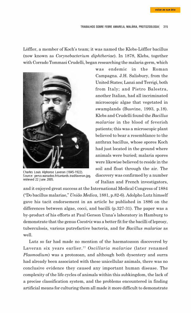
315 TRABALHOS SOBRE FEBRE AMARELA, MALÁRIA, PROTOZOOLOGIA
Löffler, a member of Koch’s team; it was named the Klebs-Löffler bacillus
(now known as Corynebacterium diphtheriae). In 1878, Klebs, together
with Corrado Tommasi Crudelli, began researching the malaria germ, which
Charles Louis Alphonse Laveran (1845-1922).Source: perso.wanadoo.fr/santards.trad/laveran.jpg,retrieved 22 June 2005.
was endemic in the Roman
Campagna. J.H. Salisbury, from the
United States; Lanzi and Terrigi, both
from Italy; and Pietro Balestra,
another Italian, had all incriminated
microscopic algae that vegetated in
swamplands (Busvine, 1993, p.18).
Klebs and Crudelli found the Bacillus
malariae in the blood of feverish
patients; this was a microscopic plant
believed to bear a resemblance to the
anthrax bacillus, whose spores Koch
had just located in the ground where
animals were buried; malaria spores
were likewise believed to reside in the
soil and float through the air. The
discovery was confirmed by a number
of Italian and French investigators,
and it enjoyed great success at the International Medical Congress of 1884
(“Do bacillus malariae,” União Medica, 1881, p.82-6). Adolpho Lutz himself
gave his tacit endorsement in an article he published in 1886 on the
differences between algae, cocci, and bacilli (p.327-31). The paper was a
by-product of his efforts at Paul Gerson Unna’s laboratory in Hamburg to
demonstrate that the genus Cocotrix was a better fit for the bacilli of leprosy,
tuberculosis, various putrefactive bacteria, and for Bacillus malariae as
well.
Lutz so far had made no mention of the haematozoon discovered by
Laveran six years earlier.77 Oscillaria malariae (later renamed
Plasmodium) was a protozoan, and although both dysentery and surra
had already been associated with these unicellular animals, there was no
conclusive evidence they caused any important human disease. The
complexity of the life cycles of animals within this subkingdom, the lack of
a precise classification system, and the problems encountered in finding
artificial means for culturing them all made it more difficult to demonstrate
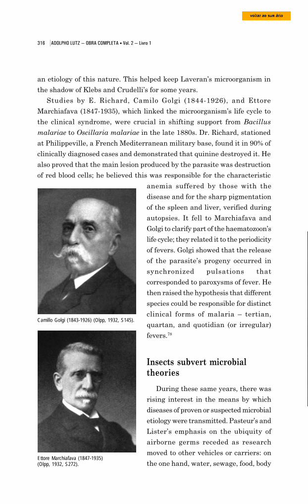
316 ADOLPHO LUTZ — OBRA COMPLETA Vol. 2 — Livro 1
an etiology of this nature. This helped keep Laveran’s microorganism in
the shadow of Klebs and Crudelli’s for some years.
Studies by E. Richard, Camilo Golgi (1844-1926), and Ettore
Marchiafava (1847-1935), which linked the microorganism’s life cycle to
the clinical syndrome, were crucial in shifting support from Bacillus
malariae to Oscillaria malariae in the late 1880s. Dr. Richard, stationed
at Philippeville, a French Mediterranean military base, found it in 90% of
clinically diagnosed cases and demonstrated that quinine destroyed it. He
also proved that the main lesion produced by the parasite was destruction
of red blood cells; he believed this was responsible for the characteristic
Ettore Marchiafava (1847-1935)(Olpp, 1932, S272).
Camillo Golgi (1843-1926) (Olpp, 1932, S145).
anemia suffered by those with the
disease and for the sharp pigmentation
of the spleen and liver, verified during
autopsies. It fell to Marchiafava and
Golgi to clarify part of the haematozoon’s
life cycle; they related it to the periodicity
of fevers. Golgi showed that the release
of the parasite’s progeny occurred in
synchronized pulsations that
corresponded to paroxysms of fever. He
then raised the hypothesis that different
species could be responsible for distinct
clinical forms of malaria – tertian,
quartan, and quotidian (or irregular)
fevers.78
Insects subvert microbialtheories
During these same years, there was
rising interest in the means by which
diseases of proven or suspected microbial
etiology were transmitted. Pasteur’s and
Lister’s emphasis on the ubiquity of
airborne germs receded as research
moved to other vehicles or carriers: on
the one hand, water, sewage, food, body
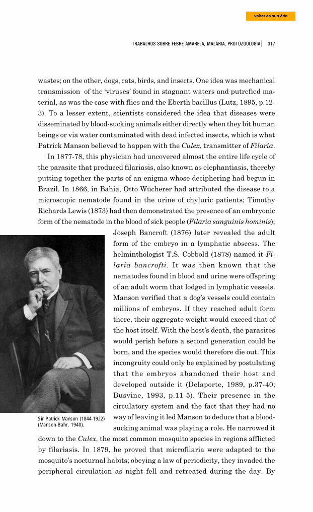
317 TRABALHOS SOBRE FEBRE AMARELA, MALÁRIA, PROTOZOOLOGIA
wastes; on the other, dogs, cats, birds, and insects. One idea was mechanical
transmission of the ‘viruses’ found in stagnant waters and putrefied ma-
terial, as was the case with flies and the Eberth bacillus (Lutz, 1895, p.12-
3). To a lesser extent, scientists considered the idea that diseases were
disseminated by blood-sucking animals either directly when they bit human
beings or via water contaminated with dead infected insects, which is what
Patrick Manson believed to happen with the Culex, transmitter of Filaria.
In 1877-78, this physician had uncovered almost the entire life cycle of
the parasite that produced filariasis, also known as elephantiasis, thereby
putting together the parts of an enigma whose deciphering had begun in
Brazil. In 1866, in Bahia, Otto Wücherer had attributed the disease to a
microscopic nematode found in the urine of chyluric patients; Timothy
Richards Lewis (1873) had then demonstrated the presence of an embryonic
form of the nematode in the blood of sick people (Filaria sanguinis hominis);
Sir Patrick Manson (1844-1922)(Manson-Bahr, 1940).
Joseph Bancroft (1876) later revealed the adult
form of the embryo in a lymphatic abscess. The
helminthologist T.S. Cobbold (1878) named it Fi-
laria bancrofti. It was then known that the
nematodes found in blood and urine were offspring
of an adult worm that lodged in lymphatic vessels.
Manson verified that a dog’s vessels could contain
millions of embryos. If they reached adult form
there, their aggregate weight would exceed that of
the host itself. With the host’s death, the parasites
would perish before a second generation could be
born, and the species would therefore die out. This
incongruity could only be explained by postulating
that the embryos abandoned their host and
developed outside it (Delaporte, 1989, p.37-40;
Busvine, 1993, p.11-5). Their presence in the
circulatory system and the fact that they had no
way of leaving it led Manson to deduce that a blood-
sucking animal was playing a role. He narrowed it
down to the Culex, the most common mosquito species in regions afflicted
by filariasis. In 1879, he proved that microfilaria were adapted to the
mosquito’s nocturnal habits; obeying a law of periodicity, they invaded the
peripheral circulation as night fell and retreated during the day. By
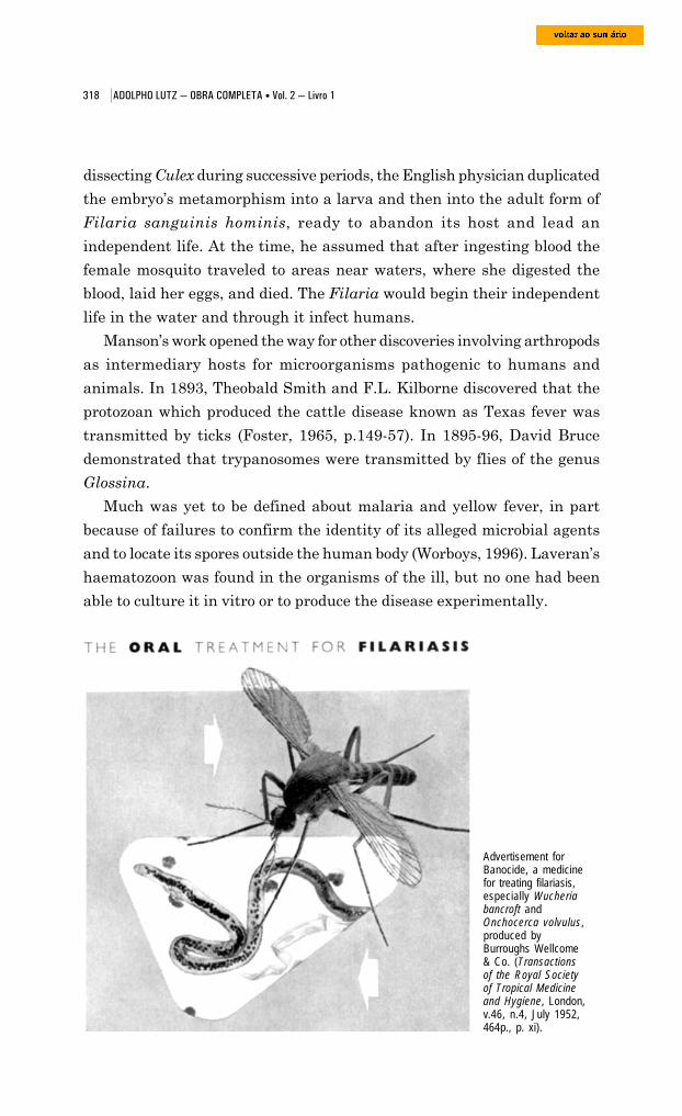
318 ADOLPHO LUTZ — OBRA COMPLETA Vol. 2 — Livro 1
dissecting Culex during successive periods, the English physician duplicated
the embryo’s metamorphism into a larva and then into the adult form of
Filaria sanguinis hominis, ready to abandon its host and lead an
independent life. At the time, he assumed that after ingesting blood the
female mosquito traveled to areas near waters, where she digested the
blood, laid her eggs, and died. The Filaria would begin their independent
life in the water and through it infect humans.
Manson’s work opened the way for other discoveries involving arthropods
as intermediary hosts for microorganisms pathogenic to humans and
animals. In 1893, Theobald Smith and F.L. Kilborne discovered that the
protozoan which produced the cattle disease known as Texas fever was
transmitted by ticks (Foster, 1965, p.149-57). In 1895-96, David Bruce
demonstrated that trypanosomes were transmitted by flies of the genus
Glossina.
Much was yet to be defined about malaria and yellow fever, in part
because of failures to confirm the identity of its alleged microbial agents
and to locate its spores outside the human body (Worboys, 1996). Laveran’s
haematozoon was found in the organisms of the ill, but no one had been
able to culture it in vitro or to produce the disease experimentally.
Advertisement forBanocide, a medicinefor treating filariasis,especially Wucheriabancroft andOnchocerca volvulus,produced byBurroughs Wellcome& Co. (Transactionsof the Royal Societyof Tropical Medicineand Hygiene, London,v.46, n.4, July 1952,464p., p. xi).
319 TRABALHOS SOBRE FEBRE AMARELA, MALÁRIA, PROTOZOOLOGIA
The polemic between Freire (spokesperson for Klebs and Crudelli’s
bacillus in Brazil) and Francisco Fajardo’s and Adolpho Lutz’s teams
(advocates of Laveran’s haematozoon) transpired halfway between the time
that Manson formulated his hypothesis that a mosquito played host to this
haematozoon before it infected humans (as with filariasis) and the time
that this hypothesis was substantiated. Confirmation came thanks both to
Ronald Ross, who uncovered the cycle of the bird malaria parasite in the
Culex in 1898, and also to Giovanni Grassi, Amico Bignami, and Giuseppe
Bastianelli, who the following year clarified the cycle of the human malaria
parasite in mosquitoes of the genus Anopheles.
During this interval, the medical press in Brazil and abroad was filled
with information and speculation about the role of insects in the
transmission of other diseases. The spotlight was on flies, who entered the
collective imagination of urban populations as an omnipresent source of
danger in the midst – or in substitution – of intangible miasmas. In 1898,
it was declared that flies disseminated the microbes of carbuncles, Egyptian
ophthalmia, Biskara boils, pian (yaws), and glanders. In his laboratory,
Yersin had verified that dead flies contained the plague bacillus and could
infect drinking water. Joly (1899, 1898) confirmed that they deposited the
bacilli of tuberculosis on foodstuffs and beverages, and carried them even
when desiccated.
As we saw earlier, the final theory regarding the etiology of yellow fever
was quite unpredictably lit upon right before Finlay’s theory was enthroned
by the Brazilian public health field. When João Batista de Lacerda went
into homes recently occupied by sick people in order to search for Sanarelli’s
bacillus and its associated mold, he always found them on fly excrement.
Many of the articles written back then leave us with this impression: it
was as if the component parts of Pasteurian-based theories on malaria,
yellow fever, and other diseases were ‘magnetized’ by the force field of
another type of medicine, one which soon would be called “tropical.” New
living pieces were fit into constructs built under the aegis of bacteriology,
re-arranging them. Soil, water, air, food, houses, and people had been
woven into webs traversed by alleged pathogenic microbes (mainly algae,
fungi, and bacilli), and it was hard to accommodate the new players inside
these nets. Connections were rebuilt, new components added, but the insects
very often remained strangers in the nest. In the case of malaria and yellow
fever, the logic governing state-of-the-art research in tropical medicine
320 ADOLPHO LUTZ — OBRA COMPLETA Vol. 2 — Livro 1
seemed incompatible with the microbial theories then in the process of
perishing.
Malaria and typhoid fever
Adolpho Lutz’s research on malaria tied into another polemic question
that kept the press busy in the 1890s, especially in São Paulo. This was
typhoid fever, a disease with intestinal manifestations that Lutz himself
had contracted while on vacation in 1878, when he was studying in Leipzig,
Germany. In Reminiscencias da febre typhoide, published in 1936, Lutz
would classify it as “one of the Bacteriological Institute’s prime concerns.”
The disease had long remained undifferentiated, assigned to the realm
of septic diseases, fevers, and diseases associated with the generic name
“typhus.”79 The Englishman William Budd (1811-80) was the first to
correlate typhoid fever with a living “virus” (which at the time meant living
“poison”) – transmitted by indirect or direct contagion. In 1880, the
pathologist Carl Joseph Eberth (1835-1926) described a microorganism
first known as “Eberth’s bacillus” and later called Salmonella typhi; it was
present in the mesenteric ganglia and spleen of bodies he had autopsied.
The discovery was confirmed by Koch, who presented a more precise
description of the bacillus. In 1884, Georg Gaffky (1850-1918), Koch’s
assistant and successor, managed to isolate that microorganism and obtain
pure cultures.
At that time, physicians in São Paulo were mistakenly diagnosing as
“typhoid-malaria fever,” “remittent fever,” or “São Paulo fever” certain
clinical cases that Adolpho Lutz would soon recognize as typhoid fever.
The expression ‘São Paulo fever’ had been coined by “masters of indigenous
medicine” to designate a disease that during the last decade of the Empire
had laid waste to the capital of São Paulo, rivaling the havoc wreaked by
yellow fever in Rio de Janeiro and Santos (Pestana, 1915, p.11).
In his first report as interim director of the Bacteriological Institute (2
Jan. 1894), Lutz argued against the malarial nature of fevers in the city of
São Paulo, sustaining that, were this the case, Laveran’s Plasmodium
malarie should be found in the victims’ blood.80 It was not yet clear to him
what these fevers represented. Symptoms resembled those of undulant
fever, which spread through the Island of Malta (a malady likewise known
as brucellosis in honor of David Bruce, who in 1887 had discovered its
321 TRABALHOS SOBRE FEBRE AMARELA, MALÁRIA, PROTOZOOLOGIA
agent, Micrococcus melitensis). Lutz endeavored without success to locate
this microorganism in the blood of São Paulo’s ailing. The hypothesis that
the disease was “abdominal typhoid” – as the Germans called typhoid fever
(Landouzy and Jayle, 1902) – was strengthened by an autopsy performed
in 1894 on someone reported to have died of yellow fever.81
In the war against adversaries of this diagnosis, Lutz called to action
Eberth himself, then director of the University of Halle’s Anatomical
Institute. In a letter dated 1 May 1895, Eberth attested that the Brazilian
bacteriologist’s cultures were “legitimate cultures of typhoid fever bacilli.”82
Most São Paulo physicians insisted that these “São Paulo fevers” were
nothing more than a native or local type of malaria. In his 1894-95 report,
Lutz argued against this supposition.
As always, when it is a matter of verifying mistakes, we expect protests.These were not long in coming, but were limited to articles in politicalnewspapers, some of them anonymous, others by authors with no authority.There is no attempt to prove the facts, that is, by thermometric curves,the presentation of sick people, autopsies, or microscopic preparations. Wealways have our observations verified by the most qualified people we canfind, and the Laboratory is open to adversaries, as much as to friends.83
During the turbulent 1890s, São Paulo fevers were diagnosed either as
malaria or typhoid fever, eventually coming to rest on the side of typhoid
fever, thanks to Lutz and his aids. Something similar happened with yellow
fever. Some physicians reduced it to a unique manifestation of malaria, of
American characteristics.84
To demonstrate that legitimate malaria did not exist in the city of São
Paulo, Lutz had to produce evidence that combined clinical descriptions,
autopsy reports, and, mainly, laboratory verification of the absence of
Plasmodium in the blood. The other side of this venture was the
reconnaissance of places where malaria indeed occurred under Lutz’s
jurisdiction, which meant it was necessary to find sufferers who were
carrying Laveran’s parasite.
In search of protozoans in birds, reptiles, and amphibians
Adolpho Lutz, his aids at the Bacteriological Institute, and his partners
in Rio de Janeiro – Francisco Fajardo first and foremost – were not only in
tune with studies being developed by British and Italian scientists in an
322 ADOLPHO LUTZ — OBRA COMPLETA Vol. 2 — Livro 1
effort to firmly establish the clinical manifestations and etiology of the
disease and discover how it is transmitted. They also offered their
collaboration in these studies and pursued a research program that
preserved a relative autonomy from the more immediate, controversial issues
in public health. The mode of transmission of Plasmodium malariae was
the principal enigma then challenging them. Linked to it were other enig-
mas of not only medical but also zoological interest: what species of this
genus, and what other genera of this phylum of the animal kingdom, might
be involved with diseases of invertebrates and vertebrates, including
humans?
Taxonomic categories were different from today’s, and knowledge about
protozoans still quite inexact. In his 1895 report, Lutz succinctly described
the state-of-the-art in this realm of medical zoology.
Today, Laveran’s plasmodia are considered sporozoans, forming thesubdivision Haemosporidia together with the cytozoa of birds, reptiles,and amphibians. Sporozoans are a group of protozoans that all lead aparasitic existence and cause a number of diseases in man and animals,some of which we have had the opportunity to observe in the laboratory.We shall first mention rabbit coccidiosis, produced by the invasion ofCoccidium oviforme in the rabbit’s liver. This disease has killed many ofour experimental animals and must be largely responsible for the hardshipsencountered by those among us who raise rabbits.
Lutz made mention of some species of myxosporidia that he had found
in other animals. He cited a pebrine called Glugea bombycis, which he had
observed in a species of Lepidoptera common in São Paulo (Brassolis antyra).
He had sometimes found unclassified spores of Myxomycetes in fish.
Adolpho Lutz’s first paper on this topic, republished in this volume under
the title “On a myxosporidium of the gall bladder of Brazilian Batrachia”
[in Port.], was conducted when he still lived and practiced medicine in
Limeira. It was first released in 1889, in the Centralblatt für Bakterologie
und Parasitenkunde (Bulletin on bacteriology and parasitology), a periodical
founded two years earlier in Jena by Friedrich Löffler, Oscar Uhlworm,
and Karl Georg Friedrich Rudolf Leuckart (1822-98). The latter
parasitologist, who held the chair in zoology and zootomy at the University
of Leipzig, was still Lutz’s main point of reference. As we saw in volume I
of Lutz’s Complete Works, Leuckart was spokesperson for the young Swiss-
Brazilian’s work on the region’s cladocera, presented before the Leipzig’s
Society of Natural Sciences (Lutz, 1878). Among the authors Lutz consulted
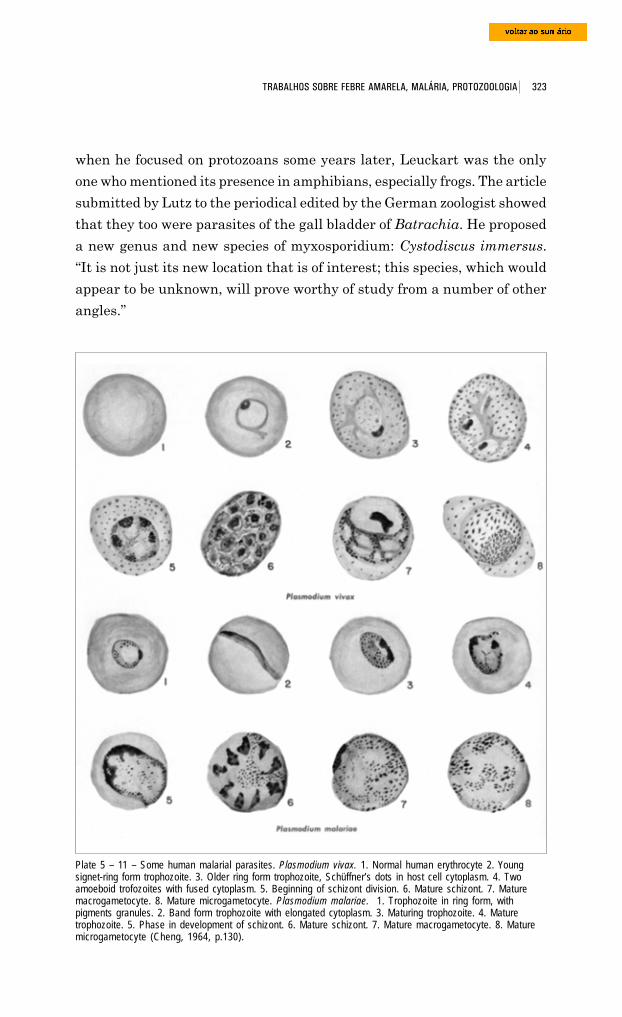
323 TRABALHOS SOBRE FEBRE AMARELA, MALÁRIA, PROTOZOOLOGIA
when he focused on protozoans some years later, Leuckart was the only
one who mentioned its presence in amphibians, especially frogs. The article
submitted by Lutz to the periodical edited by the German zoologist showed
that they too were parasites of the gall bladder of Batrachia. He proposed
a new genus and new species of myxosporidium: Cystodiscus immersus.
“It is not just its new location that is of interest; this species, which would
appear to be unknown, will prove worthy of study from a number of other
angles.”
Plate 5 – 11 – Some human malarial parasites. Plasmodium vivax. 1. Normal human erythrocyte 2. Youngsignet-ring form trophozoite. 3. Older ring form trophozoite, Schüffner’s dots in host cell cytoplasm. 4. Twoamoeboid trofozoites with fused cytoplasm. 5. Beginning of schizont division. 6. Mature schizont. 7. Maturemacrogametocyte. 8. Mature microgametocyte. Plasmodium malariae. 1. Trophozoite in ring form, withpigments granules. 2. Band form trophozoite with elongated cytoplasm. 3. Maturing trophozoite. 4. Maturetrophozoite. 5. Phase in development of schizont. 6. Mature schizont. 7. Mature macrogametocyte. 8. Maturemicrogametocyte (Cheng, 1964, p.130).
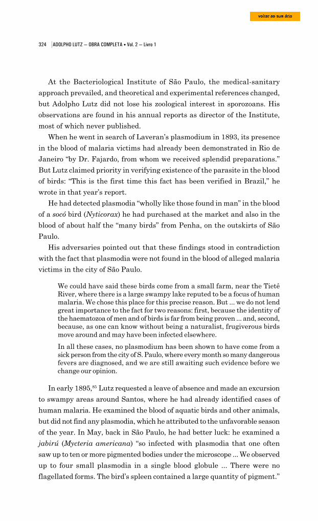
324 ADOLPHO LUTZ — OBRA COMPLETA Vol. 2 — Livro 1
At the Bacteriological Institute of São Paulo, the medical-sanitary
approach prevailed, and theoretical and experimental references changed,
but Adolpho Lutz did not lose his zoological interest in sporozoans. His
observations are found in his annual reports as director of the Institute,
most of which never published.
When he went in search of Laveran’s plasmodium in 1893, its presence
in the blood of malaria victims had already been demonstrated in Rio de
Janeiro “by Dr. Fajardo, from whom we received splendid preparations.”
But Lutz claimed priority in verifying existence of the parasite in the blood
of birds: “This is the first time this fact has been verified in Brazil,” he
wrote in that year’s report.
He had detected plasmodia “wholly like those found in man” in the blood
of a socó bird (Nyticorax) he had purchased at the market and also in the
blood of about half the “many birds” from Penha, on the outskirts of São
Paulo.
His adversaries pointed out that these findings stood in contradiction
with the fact that plasmodia were not found in the blood of alleged malaria
victims in the city of São Paulo.
We could have said these birds come from a small farm, near the TietêRiver, where there is a large swampy lake reputed to be a focus of humanmalaria. We chose this place for this precise reason. But ... we do not lendgreat importance to the fact for two reasons: first, because the identity ofthe haematozoa of men and of birds is far from being proven ... and, second,because, as one can know without being a naturalist, frugiverous birdsmove around and may have been infected elsewhere.
In all these cases, no plasmodium has been shown to have come from asick person from the city of S. Paulo, where every month so many dangerousfevers are diagnosed, and we are still awaiting such evidence before wechange our opinion.
In early 1895,85 Lutz requested a leave of absence and made an excursion
to swampy areas around Santos, where he had already identified cases of
human malaria. He examined the blood of aquatic birds and other animals,
but did not find any plasmodia, which he attributed to the unfavorable season
of the year. In May, back in São Paulo, he had better luck: he examined a
jabirú (Mycteria americana) “so infected with plasmodia that one often
saw up to ten or more pigmented bodies under the microscope ... We observed
up to four small plasmodia in a single blood globule ... There were no
flagellated forms. The bird’s spleen contained a large quantity of pigment.”
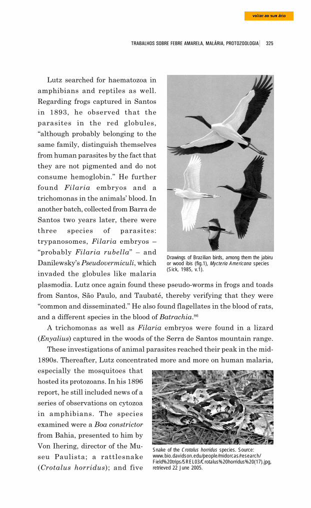
325 TRABALHOS SOBRE FEBRE AMARELA, MALÁRIA, PROTOZOOLOGIA
Lutz searched for haematozoa in
amphibians and reptiles as well.
Regarding frogs captured in Santos
in 1893, he observed that the
parasites in the red globules,
“although probably belonging to the
same family, distinguish themselves
from human parasites by the fact that
they are not pigmented and do not
consume hemoglobin.” He further
found Filaria embryos and a
trichomonas in the animals’ blood. In
another batch, collected from Barra de
Santos two years later, there were
three species of parasites:
trypanosomes, Filaria embryos –
“probably Filaria rubella” – and
Danilewsky’s Pseudovermiculi, which
invaded the globules like malaria
plasmodia. Lutz once again found these pseudo-worms in frogs and toads
from Santos, São Paulo, and Taubaté, thereby verifying that they were
“common and disseminated.” He also found flagellates in the blood of rats,
and a different species in the blood of Batrachia.86
A trichomonas as well as Filaria embryos were found in a lizard
(Enyalius) captured in the woods of the Serra de Santos mountain range.
These investigations of animal parasites reached their peak in the mid-
1890s. Thereafter, Lutz concentrated more and more on human malaria,
especially the mosquitoes that
hosted its protozoans. In his 1896
report, he still included news of a
series of observations on cytozoa
in amphibians. The species
examined were a Boa constrictor
from Bahia, presented to him by
Von Ihering, director of the Mu-
seu Paulista; a rattlesnake
(Crotalus horridus); and five
Drawings of Brazilian birds, among them the jabiruor wood ibis (fig.1), Mycteria Americana species(Sick, 1985, v.1).
Snake of the Crotalus horridus species. Source:www.bio.davidson.edu/people/midorcas/research/Field%20trips/SREL03/Crotalus%20horridus%20(17).jpg,retrieved 22 June 2005.
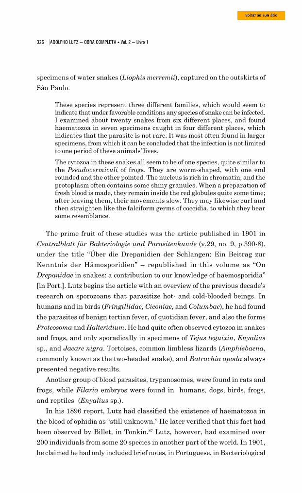
326 ADOLPHO LUTZ — OBRA COMPLETA Vol. 2 — Livro 1
specimens of water snakes (Liophis merremii), captured on the outskirts of
São Paulo.
These species represent three different families, which would seem toindicate that under favorable conditions any species of snake can be infected.I examined about twenty snakes from six different places, and foundhaematozoa in seven specimens caught in four different places, whichindicates that the parasite is not rare. It was most often found in largerspecimens, from which it can be concluded that the infection is not limitedto one period of these animals’ lives.
The cytozoa in these snakes all seem to be of one species, quite similar tothe Pseudovermiculi of frogs. They are worm-shaped, with one endrounded and the other pointed. The nucleus is rich in chromatin, and theprotoplasm often contains some shiny granules. When a preparation offresh blood is made, they remain inside the red globules quite some time;after leaving them, their movements slow. They may likewise curl andthen straighten like the falciform germs of coccidia, to which they bearsome resemblance.
The prime fruit of these studies was the article published in 1901 in
Centralblatt für Bakteriologie und Parasitenkunde (v.29, no. 9, p.390-8),
under the title “Über die Drepanidien der Schlangen: Ein Beitrag zur
Kenntnis der Hämosporidien” – republished in this volume as “On
Drepanidae in snakes: a contribution to our knowledge of haemosporidia”
[in Port.]. Lutz begins the article with an overview of the previous decade’s
research on sporozoans that parasitize hot- and cold-blooded beings. In
humans and in birds (Fringillidae, Ciconiae, and Columbae), he had found
the parasites of benign tertian fever, of quotidian fever, and also the forms
Proteosoma and Halteridium. He had quite often observed cytozoa in snakes
and frogs, and only sporadically in specimens of Tejus teguixin, Enyalius
sp., and Jacare nigra. Tortoises, common limbless lizards (Amphisbaena,
commonly known as the two-headed snake), and Batrachia apoda always
presented negative results.
Another group of blood parasites, trypanosomes, were found in rats and
frogs, while Filaria embryos were found in humans, dogs, birds, frogs,
and reptiles (Enyalius sp.).
In his 1896 report, Lutz had classified the existence of haematozoa in
the blood of ophidia as “still unknown.” He later verified that this fact had
been observed by Billet, in Tonkin.87 Lutz, however, had examined over
200 individuals from some 20 species in another part of the world. In 1901,
he claimed he had only included brief notes, in Portuguese, in Bacteriological
327 TRABALHOS SOBRE FEBRE AMARELA, MALÁRIA, PROTOZOOLOGIA
Institute reports because he wanted to further explore the study of these
parasites which were of such interest to him.
The parasites of snakes ... that appear temporarily as cytozoa areundoubtedly sporozoans closely related to Drepanidae, which are generallyincluded in a specific group of haemosporidia. In any case, they are quitedistinct from the parasites of malaria, whereas in many aspects theyresemble coccidia, that is, gregarins (Monocystis) and even sarcosporidia.The future will tell whether we are justified in organizing haemosporidiain one specific group and separating the genera Danilewskya andKaryolysus, based on our current, quite incomplete knowledge. I will keepDrepanidium as the name of the genus, since it is the oldest, and call theparasites in the blood of snakes Drepanidium serpentium, since I believethat I must consider the various forms I have observed as belonging to one[same] species.
The distribution of human malaria in São Paulo
Lutz’s research into human malaria focused at first on Barra de Santos
and the swampy lowlands near that port. In 1893, he twice verified
Plasmodium malariae in people who had come down with the disease there.
In his 1895 report, he described three more cases: one from the port of
Taboado, along the Paraná River; the other two, a mother and daughter
who lived in Mogy-Guassú. There is no mention of human malaria in the
1896 report,88 even though this report contains the most information on
the topic of haematozoans in animals. The following year,89 Lutz diagnosed
fourteen cases in the acute stage of the disease. He attributed this increase
to meteorological influences and also to his new access to patients
hospitalized at Santa Casa in the city of São Paulo. But all of these came
from outside the city. “We are still waiting,” he wrote, “[to find a] case of
malarial fever displaying plasmodia in the blood contracted in São Paulo,
[and this] should prove that the existence of paludism in the capital is no
more than an old myth.”
The victims he examined in 1897 had picked up malaria along the banks
of the Mogy-Guassú River (three cases, two of which in Porto Ferreira)
and in Guarujá (one), Paranaguá (one), Serra Azul (one), and Motuca,
near the Guaríba River (one). One of the cases came from Rio de Janeiro.
The largest number (six) occurred in the Serra de Santos highlands. The
next year, Lutz encountered another eleven cases,90 distributed as follows:
Porto Ferreira (one), Salto de Itú (one), Inhaim (two), and Serra de San-
328 ADOLPHO LUTZ — OBRA COMPLETA Vol. 2 — Livro 1
tos (two, plus an unspecified number “with pigmented leukocytes”); at Santa
Casa, he also examined an ailing individual from a farm near Araraquara,
a “chronic case, probably Italian in origin,” plus four cases from unspecified
areas of rural São Paulo.
The thirty malaria victims he had examined until then and the
information provided by other physicians allowed Lutz to draw a preliminary
sketch of the geography of malaria in the state of São Paulo and its
surrounds. The foci were concentrated in three regions: first, along the
coast (Barra de Santos, Guarujá, and, outside the state boundary, Rio de
Janeiro and Paranaguá); second, in coastal highlands, mainly around
Santos; and last, along the banks of rural São Paulo’s big rivers: Mogy-
Guassú, Tietê, Paraná, and Piracicaba. Cases elsewhere were rare and not
of great import. The malaria occurring near rural rivers only appeared
when these reached a certain size; on the Tietê, for example, the malarial
stretch was near the Itu falls. “Thus it is that between the coastal highlands
and the banks of the big rivers there is a nearly unaffected zone, wherein
lies the capital.”
Most of the cases were not serious: “They usually start as the common
type and manifest in this form again during relapses. Tertian fever is more
unusual and generally only observed in long-lasting cases, apparently the
result of a gradual modification of the quotidian type.”91
Lutz’s reports indicate that he had been putting into practice the experi-
mental program then predominant in so-called exotic pathology, whose
introduction Delaporte (1989) attributes to Manson. As the French historian
has shown, Laveran’s studies had left two questions pending. The first had
to do with the nature and function of certain forms displaying long motile
filaments, found in extravasated blood; some believed these were bodies
undergoing disintegration while others thought they represented a new stage
in the parasite’s development. The second problem concerned the way malaria
spread. Although it was possible to produce the disease by inoculating healthy
individuals with a sick person’s blood, it did not appear to be contagious.
Several hypotheses were suggested. Some believed that parasites penetrated
the organism when they ingested stagnant water or inhaled the dust carried
from swamplands; others thought the parasites that lived in these swampy
areas infected mosquitoes, which in turn infected humans.
In 1894, Manson linked the two questions when he applied the
hypothesis of the cycle of Filaria to motile filaments. He conjectured that
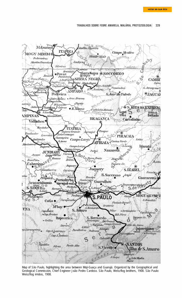
329 TRABALHOS SOBRE FEBRE AMARELA, MALÁRIA, PROTOZOOLOGIA
Map of São Paulo, highlighting the area between Moji-Guaçu and Guarujá. Organized by the Geographical andGeological Commission, Chief Engineer João Pedro Cardoso. São Paulo, Weiszflog brothers, 1908. São Paulo:Weiszflog irmãos, 1908.
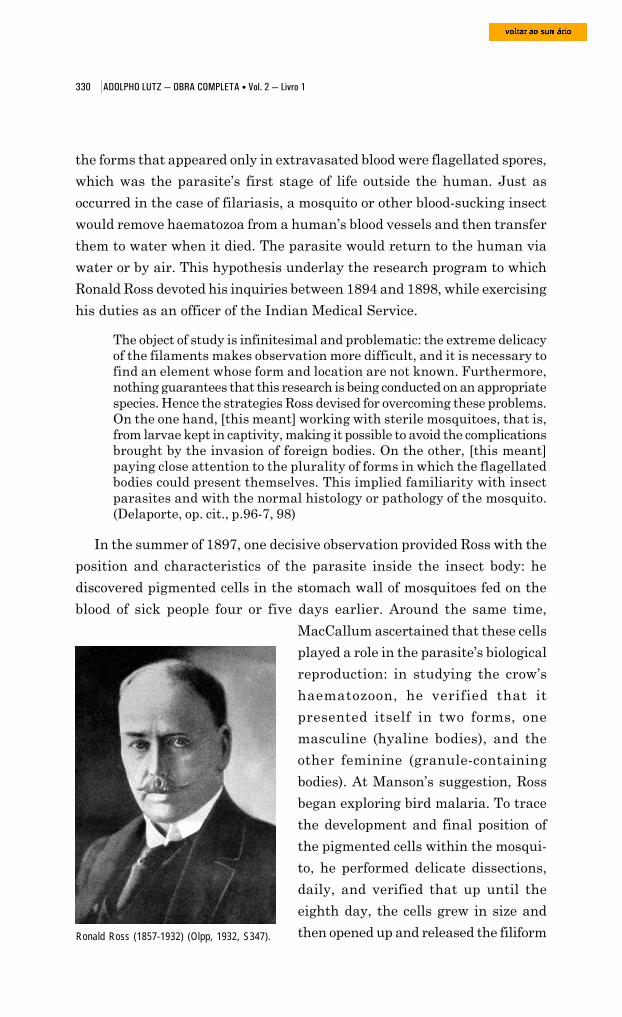
330 ADOLPHO LUTZ — OBRA COMPLETA Vol. 2 — Livro 1
MacCallum ascertained that these cells
played a role in the parasite’s biological
reproduction: in studying the crow’s
haematozoon, he verified that it
presented itself in two forms, one
masculine (hyaline bodies), and the
other feminine (granule-containing
bodies). At Manson’s suggestion, Ross
began exploring bird malaria. To trace
the development and final position of
the pigmented cells within the mosqui-
to, he performed delicate dissections,
daily, and verified that up until the
eighth day, the cells grew in size and
then opened up and released the filiformRonald Ross (1857-1932) (Olpp, 1932, S347).
the forms that appeared only in extravasated blood were flagellated spores,
which was the parasite’s first stage of life outside the human. Just as
occurred in the case of filariasis, a mosquito or other blood-sucking insect
would remove haematozoa from a human’s blood vessels and then transfer
them to water when it died. The parasite would return to the human via
water or by air. This hypothesis underlay the research program to which
Ronald Ross devoted his inquiries between 1894 and 1898, while exercising
his duties as an officer of the Indian Medical Service.
The object of study is infinitesimal and problematic: the extreme delicacyof the filaments makes observation more difficult, and it is necessary tofind an element whose form and location are not known. Furthermore,nothing guarantees that this research is being conducted on an appropriatespecies. Hence the strategies Ross devised for overcoming these problems.On the one hand, [this meant] working with sterile mosquitoes, that is,from larvae kept in captivity, making it possible to avoid the complicationsbrought by the invasion of foreign bodies. On the other, [this meant]paying close attention to the plurality of forms in which the flagellatedbodies could present themselves. This implied familiarity with insectparasites and with the normal histology or pathology of the mosquito.(Delaporte, op. cit., p.96-7, 98)
In the summer of 1897, one decisive observation provided Ross with the
position and characteristics of the parasite inside the insect body: he
discovered pigmented cells in the stomach wall of mosquitoes fed on the
blood of sick people four or five days earlier. Around the same time,
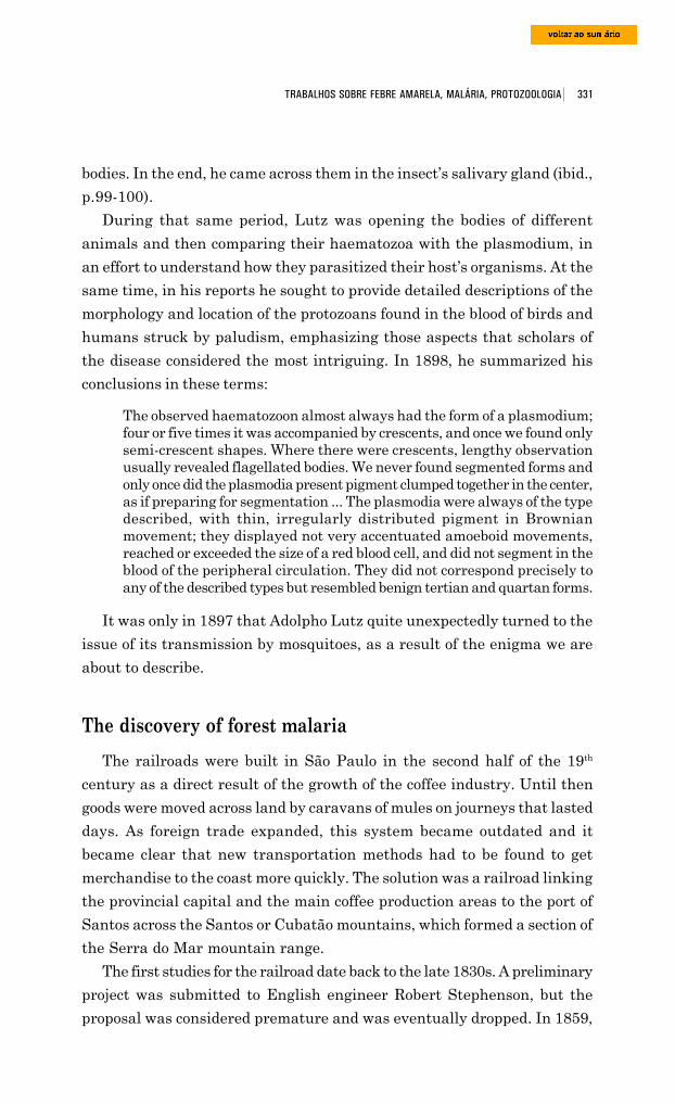
331 TRABALHOS SOBRE FEBRE AMARELA, MALÁRIA, PROTOZOOLOGIA
bodies. In the end, he came across them in the insect’s salivary gland (ibid.,
p.99-100).
During that same period, Lutz was opening the bodies of different
animals and then comparing their haematozoa with the plasmodium, in
an effort to understand how they parasitized their host’s organisms. At the
same time, in his reports he sought to provide detailed descriptions of the
morphology and location of the protozoans found in the blood of birds and
humans struck by paludism, emphasizing those aspects that scholars of
the disease considered the most intriguing. In 1898, he summarized his
conclusions in these terms:
The observed haematozoon almost always had the form of a plasmodium;four or five times it was accompanied by crescents, and once we found onlysemi-crescent shapes. Where there were crescents, lengthy observationusually revealed flagellated bodies. We never found segmented forms andonly once did the plasmodia present pigment clumped together in the center,as if preparing for segmentation ... The plasmodia were always of the typedescribed, with thin, irregularly distributed pigment in Brownianmovement; they displayed not very accentuated amoeboid movements,reached or exceeded the size of a red blood cell, and did not segment in theblood of the peripheral circulation. They did not correspond precisely toany of the described types but resembled benign tertian and quartan forms.
It was only in 1897 that Adolpho Lutz quite unexpectedly turned to the
issue of its transmission by mosquitoes, as a result of the enigma we are
about to describe.
The discovery of forest malaria
The railroads were built in São Paulo in the second half of the 19th
century as a direct result of the growth of the coffee industry. Until then
goods were moved across land by caravans of mules on journeys that lasted
days. As foreign trade expanded, this system became outdated and it
became clear that new transportation methods had to be found to get
merchandise to the coast more quickly. The solution was a railroad linking
the provincial capital and the main coffee production areas to the port of
Santos across the Santos or Cubatão mountains, which formed a section of
the Serra do Mar mountain range.
The first studies for the railroad date back to the late 1830s. A preliminary
project was submitted to English engineer Robert Stephenson, but the
proposal was considered premature and was eventually dropped. In 1859,
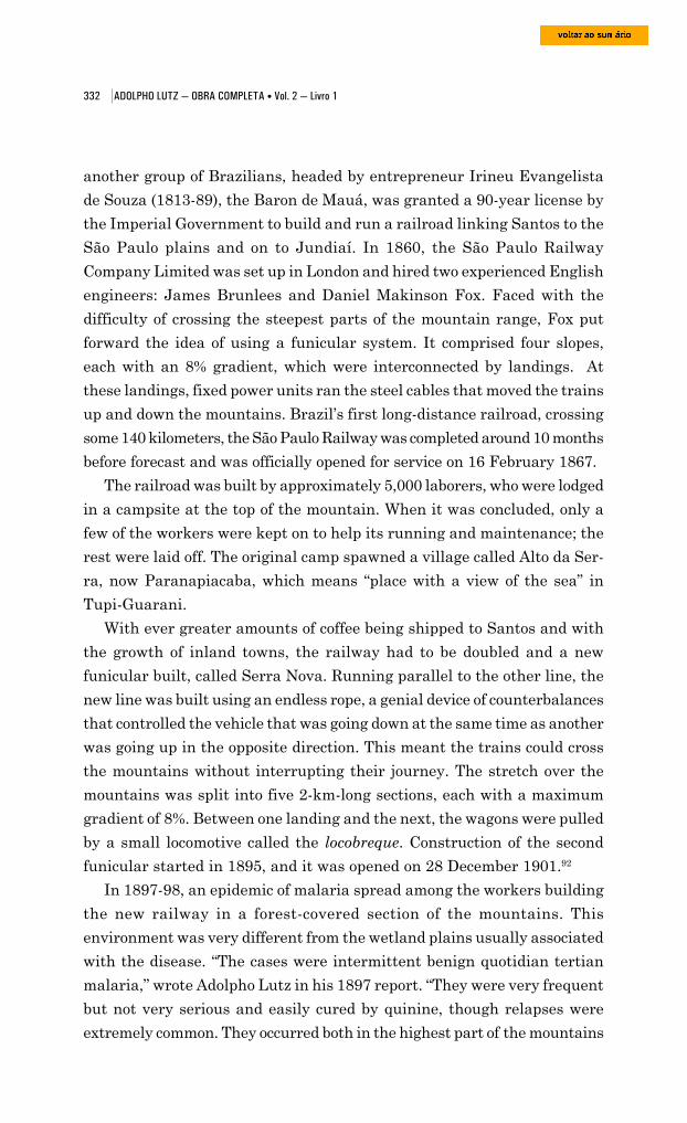
332 ADOLPHO LUTZ — OBRA COMPLETA Vol. 2 — Livro 1
another group of Brazilians, headed by entrepreneur Irineu Evangelista
de Souza (1813-89), the Baron de Mauá, was granted a 90-year license by
the Imperial Government to build and run a railroad linking Santos to the
São Paulo plains and on to Jundiaí. In 1860, the São Paulo Railway
Company Limited was set up in London and hired two experienced English
engineers: James Brunlees and Daniel Makinson Fox. Faced with the
difficulty of crossing the steepest parts of the mountain range, Fox put
forward the idea of using a funicular system. It comprised four slopes,
each with an 8% gradient, which were interconnected by landings. At
these landings, fixed power units ran the steel cables that moved the trains
up and down the mountains. Brazil’s first long-distance railroad, crossing
some 140 kilometers, the São Paulo Railway was completed around 10 months
before forecast and was officially opened for service on 16 February 1867.
The railroad was built by approximately 5,000 laborers, who were lodged
in a campsite at the top of the mountain. When it was concluded, only a
few of the workers were kept on to help its running and maintenance; the
rest were laid off. The original camp spawned a village called Alto da Ser-
ra, now Paranapiacaba, which means “place with a view of the sea” in
Tupi-Guarani.
With ever greater amounts of coffee being shipped to Santos and with
the growth of inland towns, the railway had to be doubled and a new
funicular built, called Serra Nova. Running parallel to the other line, the
new line was built using an endless rope, a genial device of counterbalances
that controlled the vehicle that was going down at the same time as another
was going up in the opposite direction. This meant the trains could cross
the mountains without interrupting their journey. The stretch over the
mountains was split into five 2-km-long sections, each with a maximum
gradient of 8%. Between one landing and the next, the wagons were pulled
by a small locomotive called the locobreque. Construction of the second
funicular started in 1895, and it was opened on 28 December 1901.92
In 1897-98, an epidemic of malaria spread among the workers building
the new railway in a forest-covered section of the mountains. This
environment was very different from the wetland plains usually associated
with the disease. “The cases were intermittent benign quotidian tertian
malaria,” wrote Adolpho Lutz in his 1897 report. “They were very frequent
but not very serious and easily cured by quinine, though relapses were
extremely common. They occurred both in the highest part of the mountains
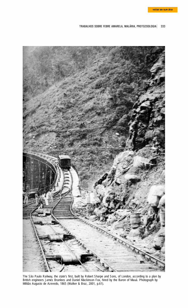
333 TRABALHOS SOBRE FEBRE AMARELA, MALÁRIA, PROTOZOOLOGIA
The São Paulo Railway, the state’s first, built by Robert Sharpe and Sons, of London, according to a plan byBritish engineers James Brunlees and Daniel Mackinson Fox, hired by the Baron of Mauá. Photograph byMilitão Augusto de Azevedo, 1865 (Walker & Braz, 2001, p.61).
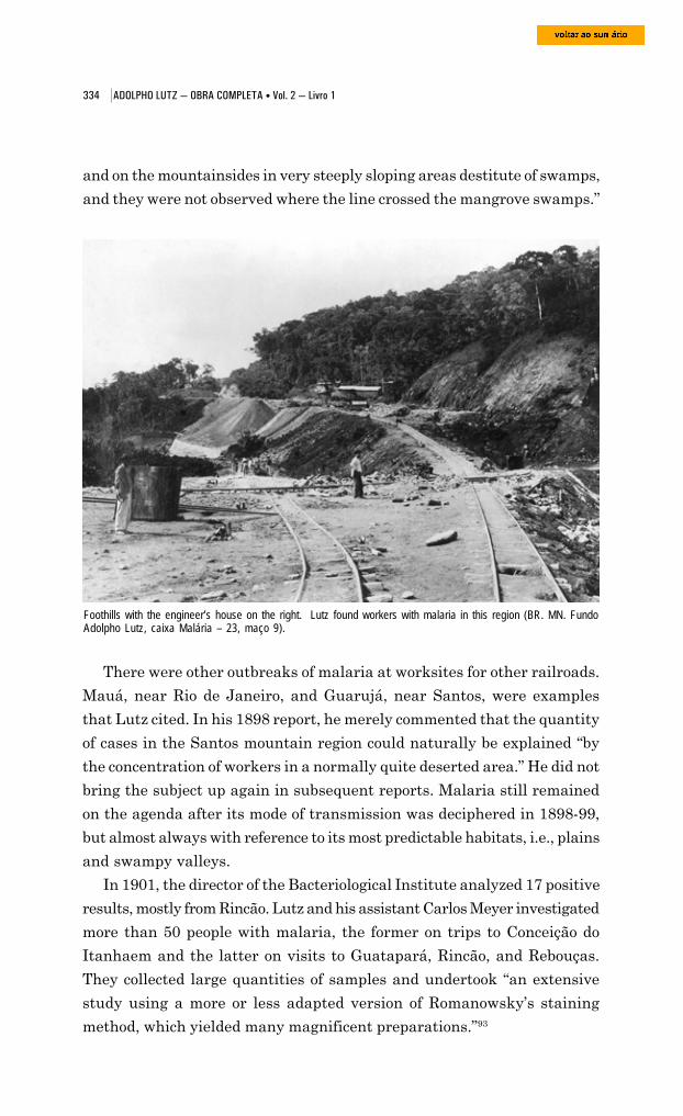
334 ADOLPHO LUTZ — OBRA COMPLETA Vol. 2 — Livro 1
and on the mountainsides in very steeply sloping areas destitute of swamps,
and they were not observed where the line crossed the mangrove swamps.”
Foothills with the engineer’s house on the right. Lutz found workers with malaria in this region (BR. MN. FundoAdolpho Lutz, caixa Malária – 23, maço 9).
There were other outbreaks of malaria at worksites for other railroads.
Mauá, near Rio de Janeiro, and Guarujá, near Santos, were examples
that Lutz cited. In his 1898 report, he merely commented that the quantity
of cases in the Santos mountain region could naturally be explained “by
the concentration of workers in a normally quite deserted area.” He did not
bring the subject up again in subsequent reports. Malaria still remained
on the agenda after its mode of transmission was deciphered in 1898-99,
but almost always with reference to its most predictable habitats, i.e., plains
and swampy valleys.
In 1901, the director of the Bacteriological Institute analyzed 17 positive
results, mostly from Rincão. Lutz and his assistant Carlos Meyer investigated
more than 50 people with malaria, the former on trips to Conceição do
Itanhaem and the latter on visits to Guatapará, Rincão, and Rebouças.
They collected large quantities of samples and undertook “an extensive
study using a more or less adapted version of Romanowsky’s staining
method, which yielded many magnificent preparations.”93
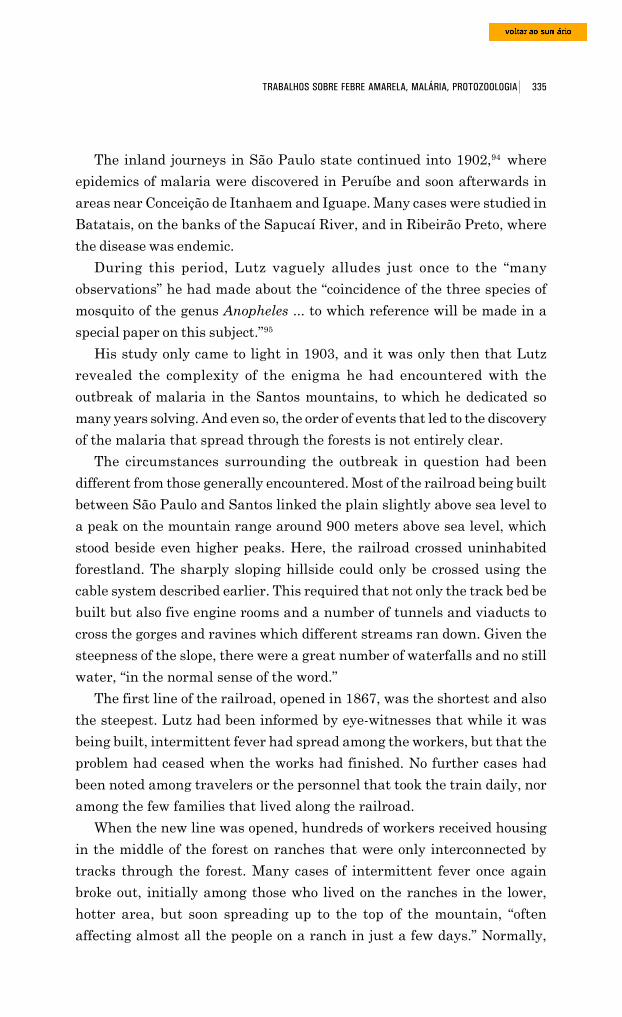
335 TRABALHOS SOBRE FEBRE AMARELA, MALÁRIA, PROTOZOOLOGIA
The inland journeys in São Paulo state continued into 1902,94 where
epidemics of malaria were discovered in Peruíbe and soon afterwards in
areas near Conceição de Itanhaem and Iguape. Many cases were studied in
Batatais, on the banks of the Sapucaí River, and in Ribeirão Preto, where
the disease was endemic.
During this period, Lutz vaguely alludes just once to the “many
observations” he had made about the “coincidence of the three species of
mosquito of the genus Anopheles ... to which reference will be made in a
special paper on this subject.”95
His study only came to light in 1903, and it was only then that Lutz
revealed the complexity of the enigma he had encountered with the
outbreak of malaria in the Santos mountains, to which he dedicated so
many years solving. And even so, the order of events that led to the discovery
of the malaria that spread through the forests is not entirely clear.
The circumstances surrounding the outbreak in question had been
different from those generally encountered. Most of the railroad being built
between São Paulo and Santos linked the plain slightly above sea level to
a peak on the mountain range around 900 meters above sea level, which
stood beside even higher peaks. Here, the railroad crossed uninhabited
forestland. The sharply sloping hillside could only be crossed using the
cable system described earlier. This required that not only the track bed be
built but also five engine rooms and a number of tunnels and viaducts to
cross the gorges and ravines which different streams ran down. Given the
steepness of the slope, there were a great number of waterfalls and no still
water, “in the normal sense of the word.”
The first line of the railroad, opened in 1867, was the shortest and also
the steepest. Lutz had been informed by eye-witnesses that while it was
being built, intermittent fever had spread among the workers, but that the
problem had ceased when the works had finished. No further cases had
been noted among travelers or the personnel that took the train daily, nor
among the few families that lived along the railroad.
When the new line was opened, hundreds of workers received housing
in the middle of the forest on ranches that were only interconnected by
tracks through the forest. Many cases of intermittent fever once again
broke out, initially among those who lived on the ranches in the lower,
hotter area, but soon spreading up to the top of the mountain, “often
affecting almost all the people on a ranch in just a few days.” Normally,
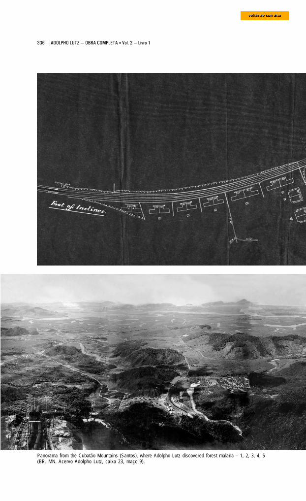
336 ADOLPHO LUTZ — OBRA COMPLETA Vol. 2 — Livro 1
Panorama from the Cubatão Mountains (Santos), where Adolpho Lutz discovered forest malaria – 1, 2, 3, 4, 5(BR. MN. Acervo Adolpho Lutz, caixa 23, maço 9).
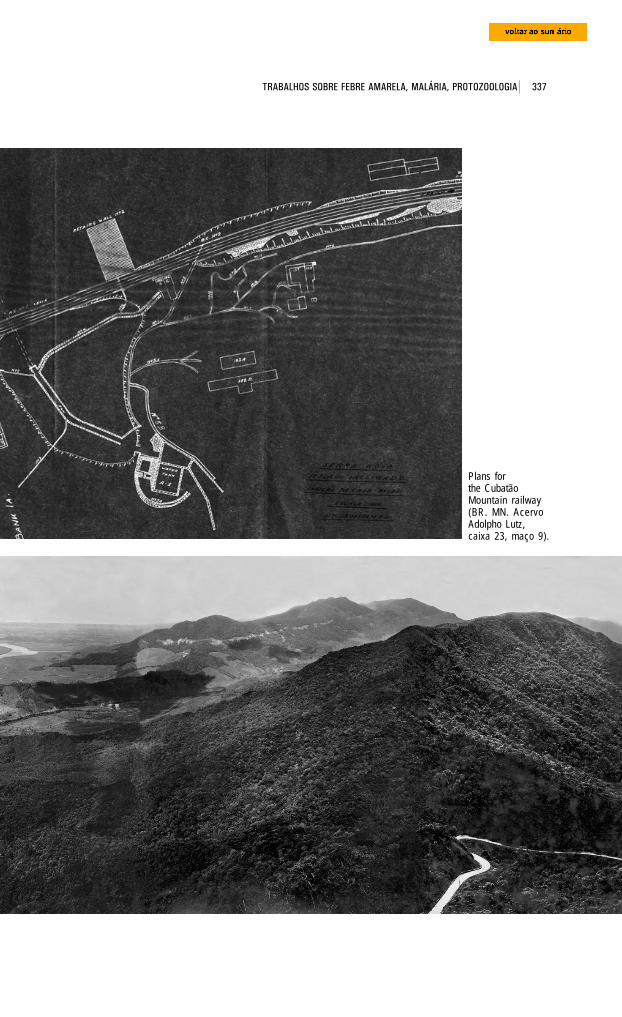
337 TRABALHOS SOBRE FEBRE AMARELA, MALÁRIA, PROTOZOOLOGIA
Plans forthe CubatãoMountain railway(BR. MN. AcervoAdolpho Lutz,caixa 23, maço 9).
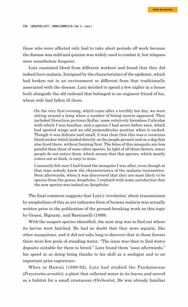
338 ADOLPHO LUTZ — OBRA COMPLETA Vol. 2 — Livro 1
those who were affected only had to take short periods off work because
the disease was mild and quinine was widely used to combat it, but relapses
were nonetheless frequent.
Lutz examined blood from different workers and found that they did
indeed have malaria. Intrigued by the characteristics of the epidemic, which
had broken out in an environment so different from that traditionally
associated with the disease, Lutz decided to spend a few nights in a house
built alongside the old railroad that belonged to an engineer friend of his,
whose wife had fallen ill there.
On the very first evening, which came after a terribly hot day, we weresitting around a lamp when a number of biting insects appeared. Theyincluded Simulium pertinax Kollar, some relatively harmless Culicidaewith which I was familiar, and a species I had never before seen, whichhad spotted wings and an odd perpendicular position when it sucked.Though it was delicate and small, it was clear that this was a voraciousblood-sucker which landed directly on the people present and on a dog thatalso lived there, without buzzing first. The bites of this mosquito are lesspainful than those of some other species. In light of all these factors, manypeople do not notice them, which means that this species, which mostlycomes out at dusk, is easy to miss.
I instantly felt sure I had found the mosquito I was after, even though atthat time nobody knew the characteristics of the malaria transmitter.Soon afterwards, when it was discovered that they are most likely to bespecies from the genus Anopheles, I realized with some satisfaction thatthe new species was indeed an Anopheles.
The final comment suggests that Lutz’s ‘revelation’ about transmission
by anophelines of this as yet unknown form of human malaria was actually
written prior to the publication of the ground-breaking work on this topic
by Grassi, Bignami, and Bastianelli (1899).
With the suspect species identified, the next step was to find out where
its larvae were hatched. He had no doubt that they were aquatic, like
other mosquitoes, and it did not take long to discover that in those forests
there were few pools of standing water. “The issue was then to find water
deposits suitable for them to breed.” Lutz found them “soon afterwards,”
his speed in so doing being thanks to his skill as a zoologist and to an
important prior experience.
When in Hawaii (1889-92), Lutz had studied the Pandanaceae
(Freycinetia arnottii), a plant that collected water in its leaves and served
as a habitat for a small crustacean (Orchestia). He was already familiar
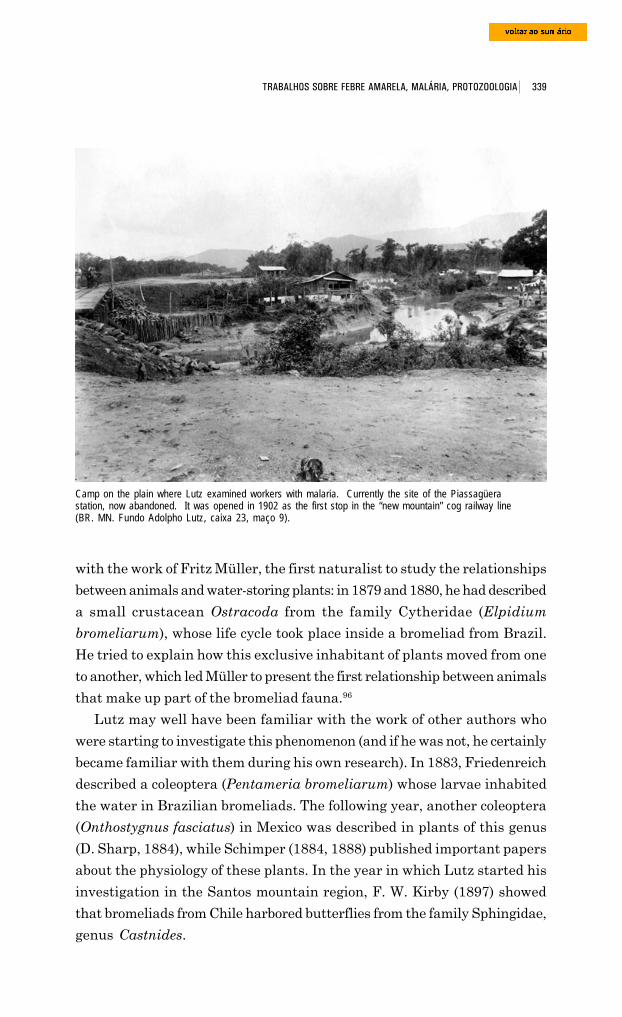
339 TRABALHOS SOBRE FEBRE AMARELA, MALÁRIA, PROTOZOOLOGIA
Camp on the plain where Lutz examined workers with malaria. Currently the site of the Piassagüerastation, now abandoned. It was opened in 1902 as the first stop in the “new mountain” cog railway line(BR. MN. Fundo Adolpho Lutz, caixa 23, maço 9).
with the work of Fritz Müller, the first naturalist to study the relationships
between animals and water-storing plants: in 1879 and 1880, he had described
a small crustacean Ostracoda from the family Cytheridae (Elpidium
bromeliarum), whose life cycle took place inside a bromeliad from Brazil.
He tried to explain how this exclusive inhabitant of plants moved from one
to another, which led Müller to present the first relationship between animals
that make up part of the bromeliad fauna.96
Lutz may well have been familiar with the work of other authors who
were starting to investigate this phenomenon (and if he was not, he certainly
became familiar with them during his own research). In 1883, Friedenreich
described a coleoptera (Pentameria bromeliarum) whose larvae inhabited
the water in Brazilian bromeliads. The following year, another coleoptera
(Onthostygnus fasciatus) in Mexico was described in plants of this genus
(D. Sharp, 1884), while Schimper (1884, 1888) published important papers
about the physiology of these plants. In the year in which Lutz started his
investigation in the Santos mountain region, F. W. Kirby (1897) showed
that bromeliads from Chile harbored butterflies from the family Sphingidae,
genus Castnides.
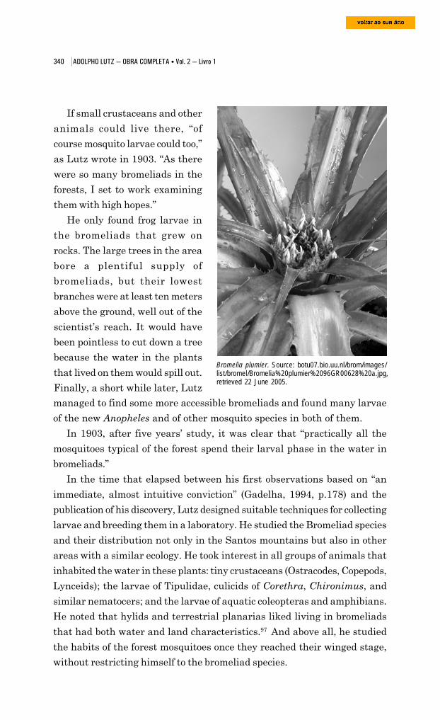
340 ADOLPHO LUTZ — OBRA COMPLETA Vol. 2 — Livro 1
If small crustaceans and other
animals could live there, “of
course mosquito larvae could too,”
as Lutz wrote in 1903. “As there
were so many bromeliads in the
forests, I set to work examining
them with high hopes.”
He only found frog larvae in
the bromeliads that grew on
rocks. The large trees in the area
bore a plentiful supply of
bromeliads, but their lowest
branches were at least ten meters
above the ground, well out of the
scientist’s reach. It would have
been pointless to cut down a tree
because the water in the plants
that lived on them would spill out.
Finally, a short while later, Lutz
managed to find some more accessible bromeliads and found many larvae
of the new Anopheles and of other mosquito species in both of them.
In 1903, after five years’ study, it was clear that “practically all the
mosquitoes typical of the forest spend their larval phase in the water in
bromeliads.”
In the time that elapsed between his first observations based on “an
immediate, almost intuitive conviction” (Gadelha, 1994, p.178) and the
publication of his discovery, Lutz designed suitable techniques for collecting
larvae and breeding them in a laboratory. He studied the Bromeliad species
and their distribution not only in the Santos mountains but also in other
areas with a similar ecology. He took interest in all groups of animals that
inhabited the water in these plants: tiny crustaceans (Ostracodes, Copepods,
Lynceids); the larvae of Tipulidae, culicids of Corethra, Chironimus, and
similar nematocers; and the larvae of aquatic coleopteras and amphibians.
He noted that hylids and terrestrial planarias liked living in bromeliads
that had both water and land characteristics.97 And above all, he studied
the habits of the forest mosquitoes once they reached their winged stage,
without restricting himself to the bromeliad species.
Bromelia plumier. Source: botu07.bio.uu.nl/brom/images/list/bromel/Bromelia%20plumier%2096GR00628%20a.jpg,retrieved 22 June 2005.
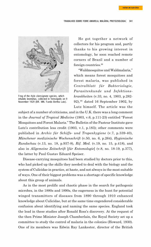
341 TRABALHOS SOBRE FEBRE AMARELA, MALÁRIA, PROTOZOOLOGIA
He got together a network of
collectors for his program and, partly
thanks to his growing interest in
entomology, he soon reached remote
corners of Brazil and a number of
foreign countries.98
“Waldmosquitos und Waldmalaria,”
which means forest mosquitoes and
forest malaria, was published in
Centralblatt für Bakteriologie,
Parasitenkunde und Infektions-
krankheiten (v.33, no. 4, 1903, p.282-
92),99 dated 16 September 1902, by
Lutz himself. The article was the
subject of a number of criticisms, and in the U.K. there was a long comment
in the Journal of Tropical Medicine (1903, v.6, p.111-23) entitled “Forest
Mosquitoes and Forest Malaria.” The Bulletin of the Pasteur Institute gave
Lutz’s contribution less credit (1903, v.1, p.183); other comments were
published in Archiv für Schiffs- und Tropenhygiene (v.7, p.339-40),
Münchener medizinische Wochenschrift (v.50, no. 6, p.264), Hygienische
Rundschau (v.13, no. 18, p.937-8), Rif. Med. (v.19, no. 15, p.418), and
also in Allgemeine Zeitschrift [für Entomologie] (v.8, no. 18-19, p.377),
the latter by Paul Gustav Eduard Speiser.
Disease-carrying mosquitoes had been studied by doctors prior to this,
who had picked up the skills they needed to deal with the biology and the
system of Culicidae in practice, at haste, and not always in the most suitable
of ways. One of their biggest problems was a shortage of specific knowledge
about this group of animals.
As in the most prolific and chaotic phase in the search for pathogenic
microbes, in the 1880s and 1890s, the eagerness in the hunt for potential
winged transmitters of diseases from 1890 through 1910 enhanced
knowledge about Culicidae, but at the same time engendered considerable
confusion about identifying and naming the same species. England took
the lead in these studies after Ronald Ross’s discovery. At the request of
the then Prime Minister Joseph Chamberlain, the Royal Society set up a
committee to study the control of malaria in the colonies (Howard, 1930).
One of its members was Edwin Ray Lankester, director of the British
Frog of the Hyla claresignata species, whichinhabits bromelias, collected in Teresópolis on 9November 1929 (BR. MN. Fundo Bertha Lutz).
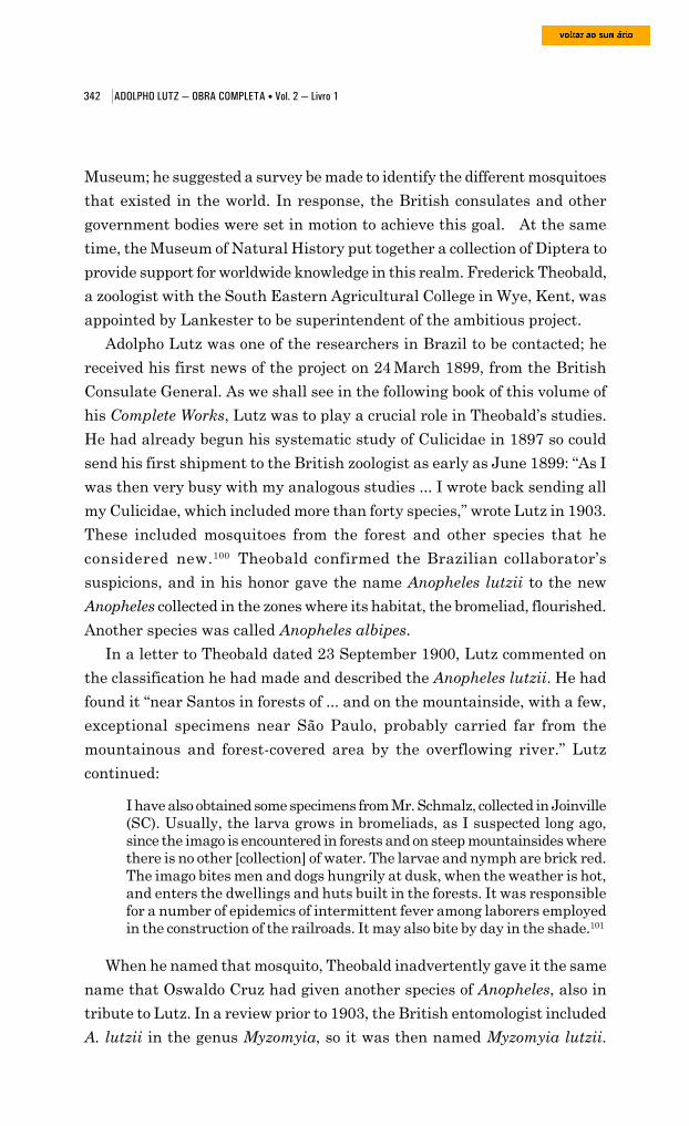
342 ADOLPHO LUTZ — OBRA COMPLETA Vol. 2 — Livro 1
Museum; he suggested a survey be made to identify the different mosquitoes
that existed in the world. In response, the British consulates and other
government bodies were set in motion to achieve this goal. At the same
time, the Museum of Natural History put together a collection of Diptera to
provide support for worldwide knowledge in this realm. Frederick Theobald,
a zoologist with the South Eastern Agricultural College in Wye, Kent, was
appointed by Lankester to be superintendent of the ambitious project.
Adolpho Lutz was one of the researchers in Brazil to be contacted; he
received his first news of the project on 24 March 1899, from the British
Consulate General. As we shall see in the following book of this volume of
his Complete Works, Lutz was to play a crucial role in Theobald’s studies.
He had already begun his systematic study of Culicidae in 1897 so could
send his first shipment to the British zoologist as early as June 1899: “As I
was then very busy with my analogous studies ... I wrote back sending all
my Culicidae, which included more than forty species,” wrote Lutz in 1903.
These included mosquitoes from the forest and other species that he
considered new.100 Theobald confirmed the Brazilian collaborator’s
suspicions, and in his honor gave the name Anopheles lutzii to the new
Anopheles collected in the zones where its habitat, the bromeliad, flourished.
Another species was called Anopheles albipes.
In a letter to Theobald dated 23 September 1900, Lutz commented on
the classification he had made and described the Anopheles lutzii. He had
found it “near Santos in forests of ... and on the mountainside, with a few,
exceptional specimens near São Paulo, probably carried far from the
mountainous and forest-covered area by the overflowing river.” Lutz
continued:
I have also obtained some specimens from Mr. Schmalz, collected in Joinville(SC). Usually, the larva grows in bromeliads, as I suspected long ago,since the imago is encountered in forests and on steep mountainsides wherethere is no other [collection] of water. The larvae and nymph are brick red.The imago bites men and dogs hungrily at dusk, when the weather is hot,and enters the dwellings and huts built in the forests. It was responsiblefor a number of epidemics of intermittent fever among laborers employedin the construction of the railroads. It may also bite by day in the shade.101
When he named that mosquito, Theobald inadvertently gave it the same
name that Oswaldo Cruz had given another species of Anopheles, also in
tribute to Lutz. In a review prior to 1903, the British entomologist included
A. lutzii in the genus Myzomyia, so it was then named Myzomyia lutzii.
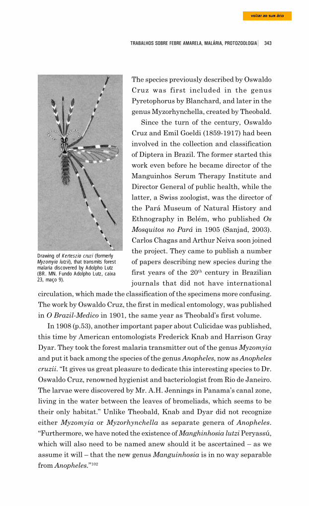
343 TRABALHOS SOBRE FEBRE AMARELA, MALÁRIA, PROTOZOOLOGIA
The species previously described by Oswaldo
Cruz was first included in the genus
Pyretophorus by Blanchard, and later in the
genus Myzorhynchella, created by Theobald.
Since the turn of the century, Oswaldo
Cruz and Emil Goeldi (1859-1917) had been
involved in the collection and classification
of Diptera in Brazil. The former started this
work even before he became director of the
Manguinhos Serum Therapy Institute and
Director General of public health, while the
latter, a Swiss zoologist, was the director of
the Pará Museum of Natural History and
Ethnography in Belém, who published Os
Mosquitos no Pará in 1905 (Sanjad, 2003).
Carlos Chagas and Arthur Neiva soon joined
the project. They came to publish a number
of papers describing new species during the
first years of the 20th century in Brazilian
journals that did not have international
circulation, which made the classification of the specimens more confusing.
The work by Oswaldo Cruz, the first in medical entomology, was published
in O Brazil-Medico in 1901, the same year as Theobald’s first volume.
In 1908 (p.53), another important paper about Culicidae was published,
this time by American entomologists Frederick Knab and Harrison Gray
Dyar. They took the forest malaria transmitter out of the genus Myzomyia
and put it back among the species of the genus Anopheles, now as Anopheles
cruzii. “It gives us great pleasure to dedicate this interesting species to Dr.
Oswaldo Cruz, renowned hygienist and bacteriologist from Rio de Janeiro.
The larvae were discovered by Mr. A.H. Jennings in Panama’s canal zone,
living in the water between the leaves of bromeliads, which seems to be
their only habitat.” Unlike Theobald, Knab and Dyar did not recognize
either Myzomyia or Myzorhynchella as separate genera of Anopheles.
“Furthermore, we have noted the existence of Manghinhosia lutzi Peryassú,
which will also need to be named anew should it be ascertained – as we
assume it will – that the new genus Manguinhosia is in no way separable
from Anopheles.”102
Drawing of Kerteszia cruzi (formerlyMyzomyia lutzii), that transmits forestmalaria discovered by Adolpho Lutz(BR. MN. Fundo Adolpho Lutz, caixa23, maço 9).
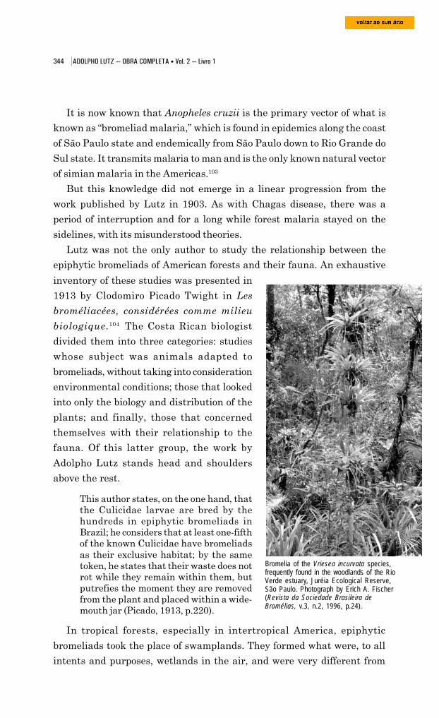
344 ADOLPHO LUTZ — OBRA COMPLETA Vol. 2 — Livro 1
Bromelia of the Vriesea incurvata species,frequently found in the woodlands of the RioVerde estuary, Juréia Ecological Reserve,São Paulo. Photograph by Erich A. Fischer(Revista da Sociedade Brasileira deBromélias, v.3, n.2, 1996, p.24).
It is now known that Anopheles cruzii is the primary vector of what is
known as “bromeliad malaria,” which is found in epidemics along the coast
of São Paulo state and endemically from São Paulo down to Rio Grande do
Sul state. It transmits malaria to man and is the only known natural vector
of simian malaria in the Americas.103
But this knowledge did not emerge in a linear progression from the
work published by Lutz in 1903. As with Chagas disease, there was a
period of interruption and for a long while forest malaria stayed on the
sidelines, with its misunderstood theories.
Lutz was not the only author to study the relationship between the
epiphytic bromeliads of American forests and their fauna. An exhaustive
inventory of these studies was presented in
1913 by Clodomiro Picado Twight in Les
broméliacées, considérées comme milieu
biologique.104 The Costa Rican biologist
divided them into three categories: studies
whose subject was animals adapted to
bromeliads, without taking into consideration
environmental conditions; those that looked
into only the biology and distribution of the
plants; and finally, those that concerned
themselves with their relationship to the
fauna. Of this latter group, the work by
Adolpho Lutz stands head and shoulders
above the rest.
This author states, on the one hand, thatthe Culicidae larvae are bred by thehundreds in epiphytic bromeliads inBrazil; he considers that at least one-fifthof the known Culicidae have bromeliadsas their exclusive habitat; by the sametoken, he states that their waste does notrot while they remain within them, butputrefies the moment they are removedfrom the plant and placed within a wide-mouth jar (Picado, 1913, p.220).
In tropical forests, especially in intertropical America, epiphytic
bromeliads took the place of swamplands. They formed what were, to all
intents and purposes, wetlands in the air, and were very different from
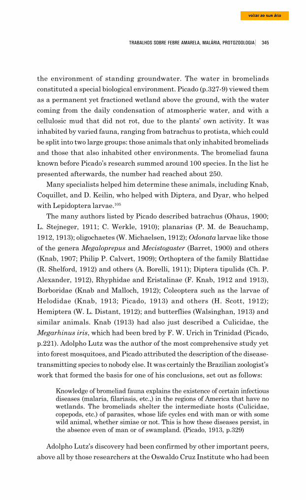
345 TRABALHOS SOBRE FEBRE AMARELA, MALÁRIA, PROTOZOOLOGIA
the environment of standing groundwater. The water in bromeliads
constituted a special biological environment. Picado (p.327-9) viewed them
as a permanent yet fractioned wetland above the ground, with the water
coming from the daily condensation of atmospheric water, and with a
cellulosic mud that did not rot, due to the plants’ own activity. It was
inhabited by varied fauna, ranging from batrachus to protista, which could
be split into two large groups: those animals that only inhabited bromeliads
and those that also inhabited other environments. The bromeliad fauna
known before Picado’s research summed around 100 species. In the list he
presented afterwards, the number had reached about 250.
Many specialists helped him determine these animals, including Knab,
Coquillet, and D. Keilin, who helped with Diptera, and Dyar, who helped
with Lepidoptera larvae.105
The many authors listed by Picado described batrachus (Ohaus, 1900;
L. Stejneger, 1911; C. Werkle, 1910); planarias (P. M. de Beauchamp,
1912, 1913); oligochaetes (W. Michaelsen, 1912); Odonata larvae like those
of the genera Megaloprepus and Mecistogaster (Barret, 1900) and others
(Knab, 1907; Philip P. Calvert, 1909); Orthoptera of the family Blattidae
(R. Shelford, 1912) and others (A. Borelli, 1911); Diptera tipulids (Ch. P.
Alexander, 1912), Rhyphidae and Eristalinae (F. Knab, 1912 and 1913),
Borboridae (Knab and Malloch, 1912); Coleoptera such as the larvae of
Helodidae (Knab, 1913; Picado, 1913) and others (H. Scott, 1912);
Hemiptera (W. L. Distant, 1912); and butterflies (Walsinghan, 1913) and
similar animals. Knab (1913) had also just described a Culicidae, the
Megarhinus iris, which had been bred by F. W. Urich in Trinidad (Picado,
p.221). Adolpho Lutz was the author of the most comprehensive study yet
into forest mosquitoes, and Picado attributed the description of the disease-
transmitting species to nobody else. It was certainly the Brazilian zoologist’s
work that formed the basis for one of his conclusions, set out as follows:
Knowledge of bromeliad fauna explains the existence of certain infectiousdiseases (malaria, filariasis, etc.,) in the regions of America that have nowetlands. The bromeliads shelter the intermediate hosts (Culicidae,copepods, etc.) of parasites, whose life cycles end with man or with somewild animal, whether simiae or not. This is how these diseases persist, inthe absence even of man or of swampland. (Picado, 1913, p.329)
Adolpho Lutz’s discovery had been confirmed by other important peers,
above all by those researchers at the Oswaldo Cruz Institute who had been
346 ADOLPHO LUTZ — OBRA COMPLETA Vol. 2 — Livro 1
asked for help by businessmen who were investing in railroad construction
and hydroelectric power generation in Brazil’s malaria zones. In 1906,
Carlos Chagas wrote about an outbreak of malaria at some 700 meters
above sea level in a place where it seemed impossible for the Culicidae to
live: “The fact caused us some surprise, which then evaporated when we
discovered the Myzomyia lutzi in the region. This was on one of the ridges
of the Serra do Mar mountain range, where the Companhia Docas de
Santos set up an important electricity facility.”106 At the same time, in
Trinidad, F.W. Ulrich, and later Lassale and De Verteuil, also highlighted
the role of forest mosquitoes [Anopheles (k) bellator] in transmitting
malaria (Gadelha, 1994).
In the same year that Lutz published his work, Galli-Valério (1903)
encountered oocysts in the stomach of specimens from Paranaguá (PR);
five years later, Stephens and Christopheres (1908) included Myzomyia
lutzi among the proven malaria vectors, since they found what they called
ookinetes in it, which are malaria parasites at the stage prior to the zygote,
when they take the form of tiny wriggling worms.
Even though this species was considered “dangerous,” Arthur Neiva (1909,
p.76) believed the evidence that it transmitted the disease was inconclusive.
In his view, the poor conditions in which the species were found in Italy
after being preserved in alcohol and then sent to Galli-Valério made it
impossible to know whether the oocysts he had encountered were of human
or bird origin, and so the finding only increased the “likelihood that this
anopheles transmits malaria.” However, the ookinetes seen by Stephens and
Christophers could easily be detected in “any anopheles that has fed on
gametocytes under transmission conditions; it should not be concluded from
this discovery that the ookinete can always become a sporozoite.”
Such provisos signaled the need for a more comprehensive research
program, but the growing acceptance of Anopheles lutzi as the protagonist
of a special type of malaria transmission came to a sudden halt in the early
years of the 1910s, and it was only in the 1940s that forest malaria reemerged
as a subject of study and was targeted for sanitation control.
The controversy with Knab and Dyar and its upshot
Gadelha (1994) and authors he quotes (Downs and Pittendrigh, 1946,
1949; Rachou, 1946) attribute the prolonged ostracism to criticisms made in
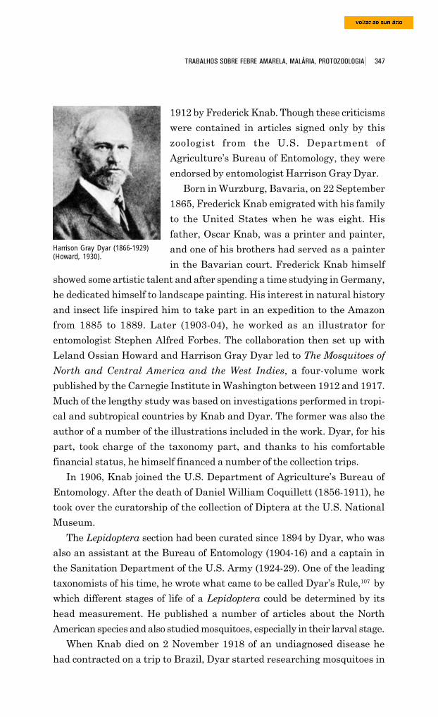
347 TRABALHOS SOBRE FEBRE AMARELA, MALÁRIA, PROTOZOOLOGIA
1912 by Frederick Knab. Though these criticisms
were contained in articles signed only by this
zoologist from the U.S. Department of
Agriculture’s Bureau of Entomology, they were
endorsed by entomologist Harrison Gray Dyar.
Born in Wurzburg, Bavaria, on 22 September
1865, Frederick Knab emigrated with his family
to the United States when he was eight. His
father, Oscar Knab, was a printer and painter,
and one of his brothers had served as a painter
in the Bavarian court. Frederick Knab himself
Harrison Gray Dyar (1866-1929)(Howard, 1930).
showed some artistic talent and after spending a time studying in Germany,
he dedicated himself to landscape painting. His interest in natural history
and insect life inspired him to take part in an expedition to the Amazon
from 1885 to 1889. Later (1903-04), he worked as an illustrator for
entomologist Stephen Alfred Forbes. The collaboration then set up with
Leland Ossian Howard and Harrison Gray Dyar led to The Mosquitoes of
North and Central America and the West Indies, a four-volume work
published by the Carnegie Institute in Washington between 1912 and 1917.
Much of the lengthy study was based on investigations performed in tropi-
cal and subtropical countries by Knab and Dyar. The former was also the
author of a number of the illustrations included in the work. Dyar, for his
part, took charge of the taxonomy part, and thanks to his comfortable
financial status, he himself financed a number of the collection trips.
In 1906, Knab joined the U.S. Department of Agriculture’s Bureau of
Entomology. After the death of Daniel William Coquillett (1856-1911), he
took over the curatorship of the collection of Diptera at the U.S. National
Museum.
The Lepidoptera section had been curated since 1894 by Dyar, who was
also an assistant at the Bureau of Entomology (1904-16) and a captain in
the Sanitation Department of the U.S. Army (1924-29). One of the leading
taxonomists of his time, he wrote what came to be called Dyar’s Rule,107 by
which different stages of life of a Lepidoptera could be determined by its
head measurement. He published a number of articles about the North
American species and also studied mosquitoes, especially in their larval stage.
When Knab died on 2 November 1918 of an undiagnosed disease he
had contracted on a trip to Brazil, Dyar started researching mosquitoes in

348 ADOLPHO LUTZ — OBRA COMPLETA Vol. 2 — Livro 1
their adult phase. His studies into the masculine genitalia of the Culicidae
were very important in classifying the group. He also studied the families
Simuliidae, Psychodidae, and Chaoboridae.
Not only did Dyar often publish papers in his country’s leading
entomology journals, but he also set up lnsecutor lnscitiae Menstruus, which
brought out fourteen volumes between 1913 and 1927. He died on 21
January 1929, having engaged in heated controversies with some of the
leading names in American entomology, including Coquillet, J. B. Smith,
and Henry Skinner.
The clash with Lutz started with the publication of an article by Frederick
Knab (1912a) in Journal of Economic Entomology, in which he analyzed
the transmission of diseases by blood-sucking insects. The same theme was
also the subject of a written communiqué at the same time at the
Entomological Society of Washington.108 At this session, when Howard set
Lutz’s theory about forest malaria against Knab’s argument, the latter
declared together with Dyar that the Brazilian zoologist had wrongly
interpreted the facts that had led him to formulate his theory.
In the article in the American Association of Economic Entomologists’
publication, Knab stated that the studies into the role of blood-sucking
insects in disease transmission that had recently been started were
overwhelmingly vague, chaotic, and supported by questionable data
collected by investigators with little biological training. “Since the discovery
that certain blood-sucking insects are secondary hosts for pathogenic
parasites, practically every insect that sucks blood either habitually or
occasionally has been viewed as a potential disease transmitter or suspected
of being such.”
He went on to say that only insects that were closely associated to man
and that regularly sucked blood repeatedly could host and transmit a
parasite from human blood. It would not be enough to do this from time to
time, like the forest-dwelling mosquitoes studied by Lutz. Other prerequisites
were their relative longevity, and continuous feeding on blood and
reproduction. Only thus would there be enough individuals for the life
cycle of the parasites they hosted not to be interrupted, including the
definitive host – man – who would then be struck by an endemic disease.
These criteria were equally applicable to mosquitoes and any other blood-
sucking insect. As Tabanidae and Simuliidae did not meet these criteria,
they could be disregarded as disease transmitters. Actually, few insects
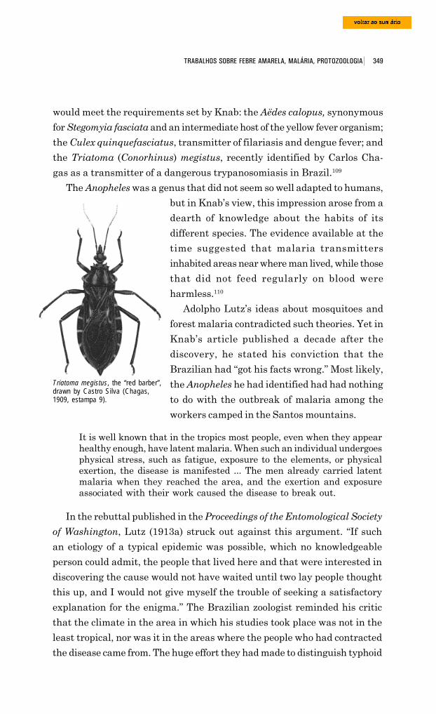
349 TRABALHOS SOBRE FEBRE AMARELA, MALÁRIA, PROTOZOOLOGIA
Triatoma megistus, the “red barber”,drawn by Castro Silva (Chagas,1909, estampa 9).
would meet the requirements set by Knab: the Aëdes calopus, synonymous
for Stegomyia fasciata and an intermediate host of the yellow fever organism;
the Culex quinquefasciatus, transmitter of filariasis and dengue fever; and
the Triatoma (Conorhinus) megistus, recently identified by Carlos Cha-
gas as a transmitter of a dangerous trypanosomiasis in Brazil.109
The Anopheles was a genus that did not seem so well adapted to humans,
but in Knab’s view, this impression arose from a
dearth of knowledge about the habits of its
different species. The evidence available at the
time suggested that malaria transmitters
inhabited areas near where man lived, while those
that did not feed regularly on blood were
harmless.110
Adolpho Lutz’s ideas about mosquitoes and
forest malaria contradicted such theories. Yet in
Knab’s article published a decade after the
discovery, he stated his conviction that the
Brazilian had “got his facts wrong.” Most likely,
the Anopheles he had identified had had nothing
to do with the outbreak of malaria among the
workers camped in the Santos mountains.
It is well known that in the tropics most people, even when they appearhealthy enough, have latent malaria. When such an individual undergoesphysical stress, such as fatigue, exposure to the elements, or physicalexertion, the disease is manifested ... The men already carried latentmalaria when they reached the area, and the exertion and exposureassociated with their work caused the disease to break out.
In the rebuttal published in the Proceedings of the Entomological Society
of Washington, Lutz (1913a) struck out against this argument. “If such
an etiology of a typical epidemic was possible, which no knowledgeable
person could admit, the people that lived here and that were interested in
discovering the cause would not have waited until two lay people thought
this up, and I would not give myself the trouble of seeking a satisfactory
explanation for the enigma.” The Brazilian zoologist reminded his critic
that the climate in the area in which his studies took place was not in the
least tropical, nor was it in the areas where the people who had contracted
the disease came from. The huge effort they had made to distinguish typhoid
350 ADOLPHO LUTZ — OBRA COMPLETA Vol. 2 — Livro 1
fever and other fevers from malaria so as to gainsay the incorrect concept
of “São Paulo fevers” had shown that the disease caused by the plasmodium
discovered by Laveran was highly localized, “even in tropical countries.
Actually, it is not to be found in many places where Anophelines dwell,
which themselves are far from being ubiquitous.”
Lutz further argued that his findings had been confirmed by a number
of scholars, in particular Carlos Chagas, “which is extremely important,
since he observed a number of malaria epidemics in different places and
studied the Anophelidae present there.” There had been records of other
epidemics of malaria in places without wetlands but with a profusion of
epiphytic bromeliads.
Observations made in different forested areas of the Serra do Mar
mountain range that had characteristics similar to those described in 1903
by Lutz had led to the generally accepted belief in Brazil that large
engineering projects in such environments would inevitably be accompanied
by outbreaks of malaria. However, no sign of the illness had been found in
worksites in dry areas in Campos and in inland forests that had no
bromeliad anophelines.
It cannot be denied that some of the workers hired for these works were
individuals that had chronic malaria, but they did not have a healthy
bearing, and as Knab supposed, they could easily be identified and
discounted.
Going further with his argument against his rival’s theoretical
assumption, Lutz stated that two malaria transmitters in Brazil – Cellia
albimana and, most importantly, Cellia argyrotarsis – were common in
uninhabited areas, moving closer to human habitations only in swampy
areas. “The fact that they neither want nor prefer human blood is
demonstrated by the well-known fact that they prefer the horse to the
horseman ... The same applies to all other species of Anopheles.”
People who entered areas where large animals were rare naturally
attracted mosquitoes, and if they stayed there long enough,
the epidemic [would] accompany the growth of the infection among themosquitoes, and they themselves [would] grow in numbers thanks to theeasy feeding. It is a well-established fact that a species can become anexcellent intermediate or definitive host of a parasite new to a region becausethe host for the following stage was only recently introduced.
351 TRABALHOS SOBRE FEBRE AMARELA, MALÁRIA, PROTOZOOLOGIA
It is in this paragraph that the crux of the matter lies. Knab held that
malaria, yellow fever, and other “parasitic” diseases were transmitted only
by blood-suckers that were already accustomed to human blood. Knowledge
about the different species capable of performing this role grew considerably
at the time, but the system Knab adhered to was conservative, almost static.
Lutz had perceived the chance of humans being involved in existing or
emerging cycles in the wild, and not only for malaria. In a later communiqué
– the counter-rebuttal to Knab’s second article – the Brazilian scientist
stated:
Messrs Dyar and Knab believe that mosquitoes that have never been incontact with man cannot transmit disease. One must place men in totallyuninhabited areas to test his thesis. Overall, this is rather difficult, but itso happens that in Brazil roads and railroads have been built under suchconditions, and there is almost always a malaria epidemic. Epidemics ofLeishmaniasis skin sores have also been noted in absolutely deserted areas,correctly attributed to transmission by Phlebotomus. I have also witnesseda small epidemic of yellow fever among people who lived in a place whereone might expect there to be forest mosquitoes. All this shows that thetheoretical considerations have not been respected by the facts, and allthat is needed is for the transmitter, whatever its past may be, to belong toa category in which the parasite can develop; then it must have repeatedaccess to human beings, some infected and others without immunity. Asthe development process takes time, its life cannot be very short. For thisreason, it is a favorable condition for it to be egg-laying (Lutz, 1913b).
In his response to Lutz’s first rebuttal read by Dyar at a session of the
Entomological Society of Washington, Knab (1913a) had offered his mea
culpa for the dogmatic tone he adopted in his first paper, and for his
arrogance in putting forward explanations at such a distance in geographic
and temporal terms from the problem studied by the Brazilian zoologist.
“Personally, Dr. Lutz’s writings have inspired me greatly, and it was in no
way my intention to discredit him.”
After rereading his paper and its rebuttal, he had reached the conclusion
that they were in agreement on all the points except one:
I am not inclined ... to admit that a species of Anopheles which is onlyfound in uninhabited forests, and which normally could not have access tohuman blood, should all of a sudden become the host for a parasite ofhuman blood. Certainly this could occur, and maybe it is just this that isin question, but in my view it would be so unusual as to require extremelyconclusive proof.
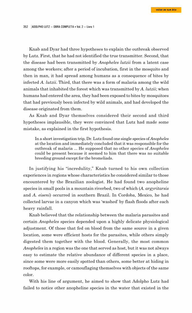
352 ADOLPHO LUTZ — OBRA COMPLETA Vol. 2 — Livro 1
Knab and Dyar had three hypotheses to explain the outbreak observed
by Lutz. First, that he had not identified the true transmitter. Second, that
the disease had been transmitted by Anopheles lutzii from a latent case
among the workers; after a period of incubation, first in the mosquito and
then in man, it had spread among humans as a consequence of bites by
infected A. lutzii. Third, that there was a form of malaria among the wild
animals that inhabited the forest which was transmitted by A. lutzii; when
humans had entered the area, they had been exposed to bites by mosquitoes
that had previously been infected by wild animals, and had developed the
disease originated from them.
As Knab and Dyar themselves considered their second and third
hypotheses implausible, they were convinced that Lutz had made some
mistake, as explained in the first hypothesis.
In a short investigation trip, Dr. Lutz found one single species of Anophelesat the location and immediately concluded that it was responsible for theoutbreak of malaria ... He supposed that no other species of Anophelescould be present because it seemed to him that there was no suitablebreeding ground except for the bromeliads.
In justifying his “incredulity,” Knab turned to his own collection
experiences in regions whose characteristics he considered similar to those
encountered by the Brazilian zoologist. He had found two anopheline
species in small pools in a mountain riverbed, two of which (A. argyritarsis
and A. eiseni) occurred in southern Brazil. In Cordoba, Mexico, he had
collected larvae in a canyon which was ‘washed’ by flash floods after each
heavy rainfall.
Knab believed that the relationship between the malaria parasites and
certain Anopheles species depended upon a highly delicate physiological
adjustment. Of those that fed on blood from the same source in a given
location, some were efficient hosts for the parasites, while others simply
digested them together with the blood. Generally, the most common
Anopheles in a region was the one that served as host, but it was not always
easy to estimate the relative abundance of different species in a place,
since some were more easily spotted than others, some better at hiding in
rooftops, for example, or camouflaging themselves with objects of the same
color.
With his line of argument, he aimed to show that Adolpho Lutz had
failed to notice other anopheline species in the water that existed in the
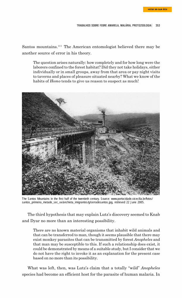
353 TRABALHOS SOBRE FEBRE AMARELA, MALÁRIA, PROTOZOOLOGIA
Santos mountains.111 The American entomologist believed there may be
another source of error in his theory.
The question arises naturally: how completely and for how long were thelaborers confined to the forest habitat? Did they not take holidays, eitherindividually or in small groups, away from that area or pay night visitsto taverns and places of pleasure situated nearby? What we know of thehabits of Homo tends to give us reason to suspect as much!
The Santos Mountains in the first half of the twentieth century. Source: www.portocidade.stcecilia.br/fotos/santos_primeira_metade_sec_xx/anchieta_imigrantes/g/serradesantos.jpg, retrieved 22 June 2005.
The third hypothesis that may explain Lutz’s discovery seemed to Knab
and Dyar no more than an interesting possibility.
There are no known material organisms that inhabit wild animals andthat can be transferred to man, though it seems plausible that there mayexist monkey parasites that can be transmitted by forest Anopheles andthat man may be susceptible to this. If such a relationship does exist, itcould be demonstrated by means of a suitable study, but I consider that wedo not have the right to invoke it as an explanation for the present casebased on no more than its possibility.
What was left, then, was Lutz’s claim that a totally “wild” Anopheles
species had become an efficient host for the parasite of human malaria. In
354 ADOLPHO LUTZ — OBRA COMPLETA Vol. 2 — Livro 1
Knab’s view, this seemed “so unlikely that no other evidence except for a
demonstration of the presence of the parasites in the mosquito’s salivary
glands would make [him] accept it.”
Like Neiva, in 1909, he considered Galli-Valério’s (1904) notification
about the presence of oocysts of the malaria parasite in the walls of the
stomach of specimens of Anopheles cited by Lutz. “Even a versed student
of American mosquitoes would hesitate to positively identify such specimens,
and I do not recall Galli-Valério before having given the slightest attention
to American mosquitoes!”
Knab and Dyar issued Lutz a challenge: that whatever the true
explanation was, “the burden of truth lies with the investigator ... we have
the right to wait for the proof to be complete or to reject the explanation
provided.”
In Lutz’s counter-rebuttal read by Howard in a session of the
Entomological Society of Washington, Lutz (1913b), in a paragraph quoted
above, refuted the assumption that mosquitoes that had never before been
in contact with man could not transmit malaria, leaving open the chance
that the infection of bromeliad mosquitoes could initially occur through
contact with a non-human host that inhabited the forest.
Lutz then attacked the weakest links of the arguments against him
made by the American entomologists.
I am accused of ignoring the fact that men are men, and it is said thatthey must have slipped away at night, getting infected elsewhere. I havealready said that they lived in the forest some many miles away and hadno means of getting around except by foot. Even if they had gotten out ofcontrol, they could not have gone far enough away in one night for themto reach anywhere where they could have found that which is suggestedthey may have found, nor would this have helped, because there was andthere still is no malaria in those areas. If one were to take such anargument further, one could also say that the Italian sailors whocontracted yellow fever on board the war vessel anchored off Rio de Janeirosome distance from the coast were attacked because they swam inland atnight.
Lutz scoffed at the statement that Galli-Valério was not equipped to
identify the Myzomyia lutzi, even though “it is an extremely characteristic
species that can be differed from all others by a quick look at its scutum.” It
was with justified disdain that he dealt with the accusation that he – as
well as Chagas – had failed to note the presence of other anophelines in
the areas where he made his studies, “even though Messrs Knab and Dyar
355 TRABALHOS SOBRE FEBRE AMARELA, MALÁRIA, PROTOZOOLOGIA
can see them from Washington. Yet it was to be expected that Lutz and
Chagas knew their anophelines, since they had worked with them for many
years and had together identified most of the Brazilian species.”
But Adolpho Lutz made no comment about his adversaries’ demand
that he provide proof that the mosquito from the forest in fact hosted the
malaria parasite. He had not followed the protocol established by Manson
in the study of filariasis transmission and replicated in the studies by Ross
and the Italians about the transmission of bird malaria by Culex, and
human malaria by Anopheles.
The study program that Lutz used was a combination of exhaustive
study of habitats and the distribution of Myzomyia lutzi and the vegetation
that hosted it with field studies into the cases of malaria in humans that
occurred in the environments where forest and bromeliad mosquitoes lived
in great numbers. It was a program that required the use of entomology,
botany, epidemiology, and medicine, but which totally bypassed the wisdom
of Ross, Grassi, and other investigators who had spent their time observing
the evolution of the parasite in the organisms of its intermediate hosts.
Though he had demonstrated his skills in this type of experimental
procedure in the parasitology studies he had published in the 1880s, Lutz
would not take the route indicated by his opponents. He concluded the
matter, maintaining “entirely” the exactness of his observations and
emphasizing the “practical interest” inherent to them.
This is why I oppose them ... and reject the comment that most likely thegentleman made a mistake, which seems to imply that it is a habit ofmine to commit errors when making scientific observations. Being as Iam in less of a hurry to communicate my observations than many thesedays, I do not believe my quota of errors to be unusually high.
These and other equally cutting remarks by Lutz, reintroduced into the
present edition of his text, were suppressed by the editor of the Proceedings
of the Entomological Society of Washington, which triggered a sharp protest
by Frederick Knab. The letter he sent to W. D. Hunter on 24 January
1914 shows Adolpho Lutz’s authority in that new field of knowledge –
medical entomology – as well as the leading position he held among his
peers in Brazil, especially the talented group of investigators at the Oswaldo
Cruz Institute.
If we leave to one side the ethical question, the change in the tone of theletter adds a completely different nature to the controversy ... placing Dr.
356 ADOLPHO LUTZ — OBRA COMPLETA Vol. 2 — Livro 1
Dyar and I in a very unpleasant position. The researchers in Rio deJaneiro ... will naturally draw the conclusion that ... we have made useof untoward means to sidestep their forceful criticisms. I fear thisprocedure, however trifling it may seem to you ... may cause irreparabledamage. It may well have caused a rupture between us and the Rioresearchers that may never be repaired. I am most sorry about this, as itwill seriously affect my work. For some years it has been my aim tocultivate friendly relations with these researchers so that we may workin harmony and facilitate each other’s investigations, and I had foundtheir response encouraging. As you undoubtedly know, by far the mostimportant medical entomological work being done this side of the Atlanticis by Lutz and his associates, and from a dipterology standpoint, aboveall, it would be most desirable to maintain contact with them.
In his reply dated 27 January, Hunter seemed surprised at their
reaction. “The only alterations I made were to omit two sentences that in
my view were too scathing to appear ... In view of this, it seems to me that
the publication of the letter ... has served more to maintain an entente
cordiale than to destroy it.”
The tone of the letter that Knab sent to Lutz two days later clearly
shows his desire to restore good feelings to their relationship,112 which, to
all intents and purposes, was sustained by a substantial, important scientific
exchange for both parties. In 1911, Knab had published a detailed,
complimentary criticism of two papers by Lutz (1909, 1910) about the
Simuliidae of Brazil. In the American’s view, it was
by far the most comprehensive study yet dedicated to this so interesting,so economically important group of Diptera ... it is gratifying to see thatDr. Lutz does not adhere to the systems of the old school, but ratherapproaches his subject from all angles. He grants due importance to dataobtained at initial stages from biology, matches them with the characteristicsof the imagoes, while at the same time carefully considering any potentialsources of error.
In a letter in January 1914, Knab respectfully consulted Adolpho Lutz
about the habits and classification of two species of Ceratopogoninae he
had been studying, and alluded to a comment previously made by his
Brazilian colleague that he still had a lot of unpublished material about
mosquitoes. “Why not publish it? It is hardly likely that your biological
data at least will conflict with our book ... Most of your observations will
undoubtedly be original, and the rest, more comprehensive, thereby
improving on what we have been able to present.”
357 TRABALHOS SOBRE FEBRE AMARELA, MALÁRIA, PROTOZOOLOGIA
Lutz’s theory in limbo
Gadelha (1994) states that the decline of Adolpho Lutz’s theory became
more marked in the 1920s, when well-known malaria specialists produced
work that seemed to invalidate it. He specifically refers to Darling and to
Nelson Davis. The former had shown that no malaria vector came from
bromeliads in Panama, thereby prompting cancellation of a costly project
to eradicate these plants that had been launched in that country.
Davis investigated an epidemic in a mountain area of Angra dos Reis
(Rio de Janeiro), where more than one railroad was built. Though he
attributed most of the cases to relapses of chronic malaria, as Knab had
about the cases examined by Lutz in the Santos mountains, Davis was
forced to admit the existence of local transmission. This led him to study
the infection capacity of forest malaria mosquitoes, which were now included
in the genus Kerteszia.113 He obtained a very low frequency, and concluded
that they were capable neither of setting off nor of maintaining an epidemic.
Only when an epidemic was at its height, when there were numerous
carriers of highly infected gametocytes, could the species perhaps contribute
with some cases of transmission.
That very same year, Arthur Neiva (1925, p.4) produced a report for
The Light and Power Company about the sanitary risks involved in
introducing one of the Canadian company’s projects in the Serra de Cubatão
mountain range, the same area where Lutz had discovered forest malaria.
The entomologist from the Oswaldo Cruz Institute, who had considerable
experience working in campaigns to fight the disease, did not hesitate in
stating that the main problem was the Myzomyia lutzi, which accounted
for 40% of all the mosquitoes that lived in the bromeliads in that region. In
his report, Neiva referred to the campaign he had led against that mosqui-
to in Iguape, São Paulo, which had mainly consisted of deforesting the
area that they planned to protect.
Gadelha (1994, p.136) finds it strange that the opposing viewpoints of
Neiva and Davis should not have triggered a controversy. In his view, this
was not only due to the limited circulation of the report written in
Portuguese, but more importantly to the fact that “Davis’s conclusions were
based on lab work, while Neiva’s were founded on generic entomologic and
epidemiological observations.” Based on Deane (1986, p.7), Gadelha shows
that the dissection of salivary glands to check for the presence of oocysts
358 ADOLPHO LUTZ — OBRA COMPLETA Vol. 2 — Livro 1
was a laborious, delicate technique, in which a small number of individuals
were used to check infection rates in anopheles. With the introduction of a
new technique at the end of the 1930s, it became possible for entomologists
to examine hundreds or even thousands of salivary glands. It was then
clear that Davis had been mistaken. In Gadelha’s article, the reader will
find a reliable analysis of the chain of events that granted forest malaria
greater visibility as of the 1940s.
Lutz did not live to see this resurgence or the campaigns introduced by
the National Malaria Service (1941) that caused the destruction of millions
of bromeliads in southern Brazil.
In a paper published in 1903, he had considered it impracticable to
deal with these plants. The use of chemicals to destroy the larvae sheltered
in them had seemed to him “more theory than practice ... Normally, the
only thing that gets results against malaria in the wild is making
clearings.” Malaria in wetland plains was more easily combated using a
set of preventive measures. In a report written at the turn of the 20th
century,114 Lutz referred to studies performed in different parts of the
world by English, German, and Italian committees, and stated that clear
guidelines could now be established for fighting the disease that were
very similar – as we shall see – to those that would soon be adopted to
fight yellow fever. The director of the Bacteriological Institute stated:
1 – due to the habits of the parasites in the plasmodium discovered byLaveran, of which there are many species, malaria is only transmittedby mosquitoes of the genus Anopheles, which serve as hosts during anessential phase of their evolution; malaria only occurs where there is anabundance of Anopheles; 2 – if it is to transmit the malady, the Anophelesmust bite people with malaria at an appropriate stage in the developmentof the disease. Only after a certain period of time, which varies betweeneight and fifteen days, will these insects be capable of transmitting themalady to other individuals by biting them.
Lutz believed malaria could be combated in any of three ways: by
treating people with the disease to stop anophelines from infecting them;
by destroying the mosquitoes, especially their larvae; by stopping infected
mosquitoes from biting people by using mosquito nets, houses protected
with screens, fumigation, and other such measures. “It is seen that these
last means may stop groups of individuals who spend the hot months in
areas where malaria is commonplace from being infected.”
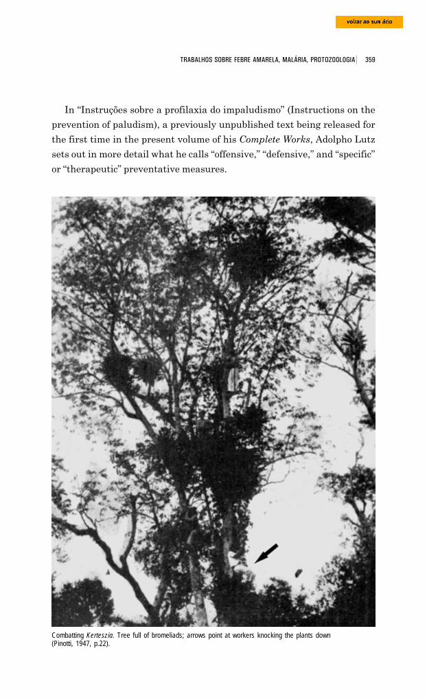
359 TRABALHOS SOBRE FEBRE AMARELA, MALÁRIA, PROTOZOOLOGIA
In “Instruções sobre a profilaxia do impaludismo” (Instructions on the
prevention of paludism), a previously unpublished text being released for
the first time in the present volume of his Complete Works, Adolpho Lutz
sets out in more detail what he calls “offensive,” “defensive,” and “specific”
or “therapeutic” preventative measures.
Combatting Kerteszia. Tree full of bromeliads; arrows point at workers knocking the plants down(Pinotti, 1947, p.22).
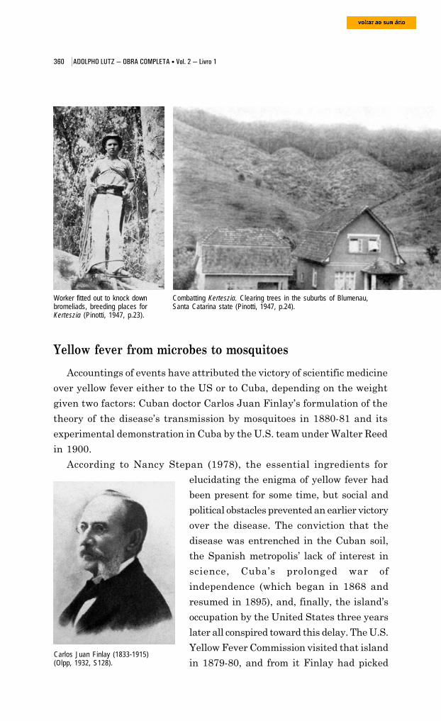
360 ADOLPHO LUTZ — OBRA COMPLETA Vol. 2 — Livro 1
Carlos Juan Finlay (1833-1915)(Olpp, 1932, S128).
Yellow fever from microbes to mosquitoes
Accountings of events have attributed the victory of scientific medicine
over yellow fever either to the US or to Cuba, depending on the weight
given two factors: Cuban doctor Carlos Juan Finlay’s formulation of the
theory of the disease’s transmission by mosquitoes in 1880-81 and its
experimental demonstration in Cuba by the U.S. team under Walter Reed
in 1900.
According to Nancy Stepan (1978), the essential ingredients for
Worker fitted out to knock downbromeliads, breeding places forKerteszia (Pinotti, 1947, p.23).
Combatting Kerteszia. Clearing trees in the suburbs of Blumenau,Santa Catarina state (Pinotti, 1947, p.24).
elucidating the enigma of yellow fever had
been present for some time, but social and
political obstacles prevented an earlier victory
over the disease. The conviction that the
disease was entrenched in the Cuban soil,
the Spanish metropolis’ lack of interest in
science, Cuba’s prolonged war of
independence (which began in 1868 and
resumed in 1895), and, finally, the island’s
occupation by the United States three years
later all conspired toward this delay. The U.S.
Yellow Fever Commission visited that island
in 1879-80, and from it Finlay had picked
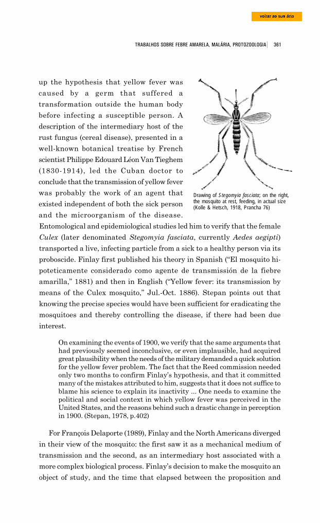
361 TRABALHOS SOBRE FEBRE AMARELA, MALÁRIA, PROTOZOOLOGIA
up the hypothesis that yellow fever was
caused by a germ that suffered a
transformation outside the human body
before infecting a susceptible person. A
description of the intermediary host of the
rust fungus (cereal disease), presented in a
well-known botanical treatise by French
scientist Philippe Edouard Léon Van Tieghem
(1830-1914), led the Cuban doctor to
conclude that the transmission of yellow fever
was probably the work of an agent that
existed independent of both the sick person
and the microorganism of the disease.
Entomological and epidemiological studies led him to verify that the female
Culex (later denominated Stegomyia fasciata, currently Aedes aegipti)
transported a live, infecting particle from a sick to a healthy person via its
proboscide. Finlay first published his theory in Spanish (“El mosquito hi-
poteticamente considerado como agente de transmissión de la fiebre
amarilla,” 1881) and then in English (“Yellow fever: its transmission by
means of the Culex mosquito,” Jul.-Oct. 1886). Stepan points out that
knowing the precise species would have been sufficient for eradicating the
mosquitoes and thereby controlling the disease, if there had been due
interest.
On examining the events of 1900, we verify that the same arguments thathad previously seemed inconclusive, or even implausible, had acquiredgreat plausibility when the needs of the military demanded a quick solutionfor the yellow fever problem. The fact that the Reed commission neededonly two months to confirm Finlay’s hypothesis, and that it committedmany of the mistakes attributed to him, suggests that it does not suffice toblame his science to explain its inactivity ... One needs to examine thepolitical and social context in which yellow fever was perceived in theUnited States, and the reasons behind such a drastic change in perceptionin 1900. (Stepan, 1978, p.402)
For François Delaporte (1989), Finlay and the North Americans diverged
in their view of the mosquito: the first saw it as a mechanical medium of
transmission and the second, as an intermediary host associated with a
more complex biological process. Finlay’s decision to make the mosquito an
object of study, and the time that elapsed between the proposition and
Drawing of Stegomyia fasciata; on the right,the mosquito at rest, feeding, in actual size(Kolle & Hetsch, 1918, Prancha 76)
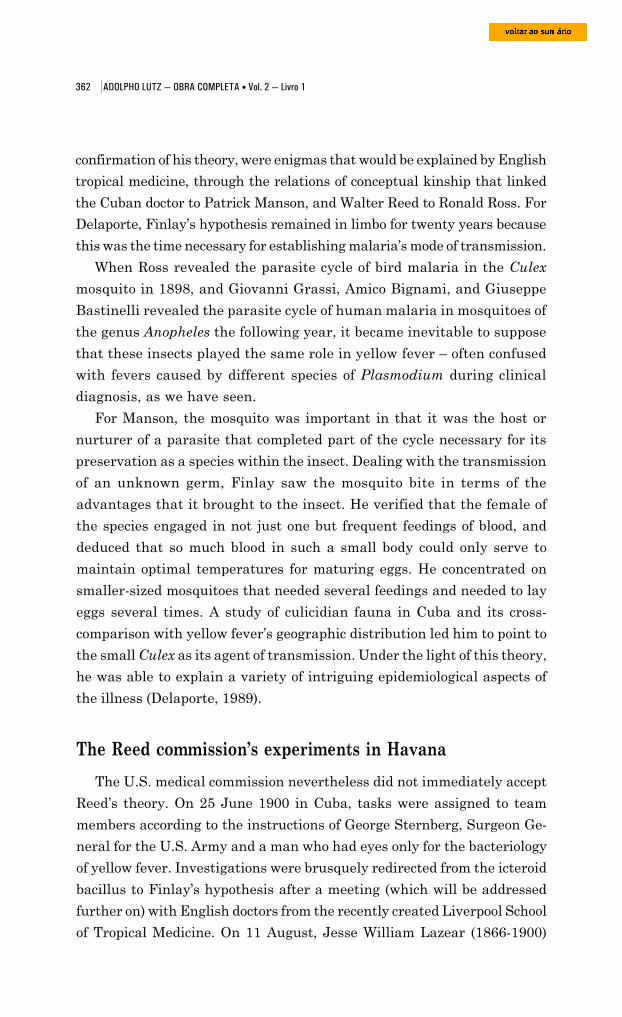
362 ADOLPHO LUTZ — OBRA COMPLETA Vol. 2 — Livro 1
confirmation of his theory, were enigmas that would be explained by English
tropical medicine, through the relations of conceptual kinship that linked
the Cuban doctor to Patrick Manson, and Walter Reed to Ronald Ross. For
Delaporte, Finlay’s hypothesis remained in limbo for twenty years because
this was the time necessary for establishing malaria’s mode of transmission.
When Ross revealed the parasite cycle of bird malaria in the Culex
mosquito in 1898, and Giovanni Grassi, Amico Bignami, and Giuseppe
Bastinelli revealed the parasite cycle of human malaria in mosquitoes of
the genus Anopheles the following year, it became inevitable to suppose
that these insects played the same role in yellow fever – often confused
with fevers caused by different species of Plasmodium during clinical
diagnosis, as we have seen.
For Manson, the mosquito was important in that it was the host or
nurturer of a parasite that completed part of the cycle necessary for its
preservation as a species within the insect. Dealing with the transmission
of an unknown germ, Finlay saw the mosquito bite in terms of the
advantages that it brought to the insect. He verified that the female of
the species engaged in not just one but frequent feedings of blood, and
deduced that so much blood in such a small body could only serve to
maintain optimal temperatures for maturing eggs. He concentrated on
smaller-sized mosquitoes that needed several feedings and needed to lay
eggs several times. A study of culicidian fauna in Cuba and its cross-
comparison with yellow fever’s geographic distribution led him to point to
the small Culex as its agent of transmission. Under the light of this theory,
he was able to explain a variety of intriguing epidemiological aspects of
the illness (Delaporte, 1989).
The Reed commission’s experiments in Havana
The U.S. medical commission nevertheless did not immediately accept
Reed’s theory. On 25 June 1900 in Cuba, tasks were assigned to team
members according to the instructions of George Sternberg, Surgeon Ge-
neral for the U.S. Army and a man who had eyes only for the bacteriology
of yellow fever. Investigations were brusquely redirected from the icteroid
bacillus to Finlay’s hypothesis after a meeting (which will be addressed
further on) with English doctors from the recently created Liverpool School
of Tropical Medicine. On 11 August, Jesse William Lazear (1866-1900)
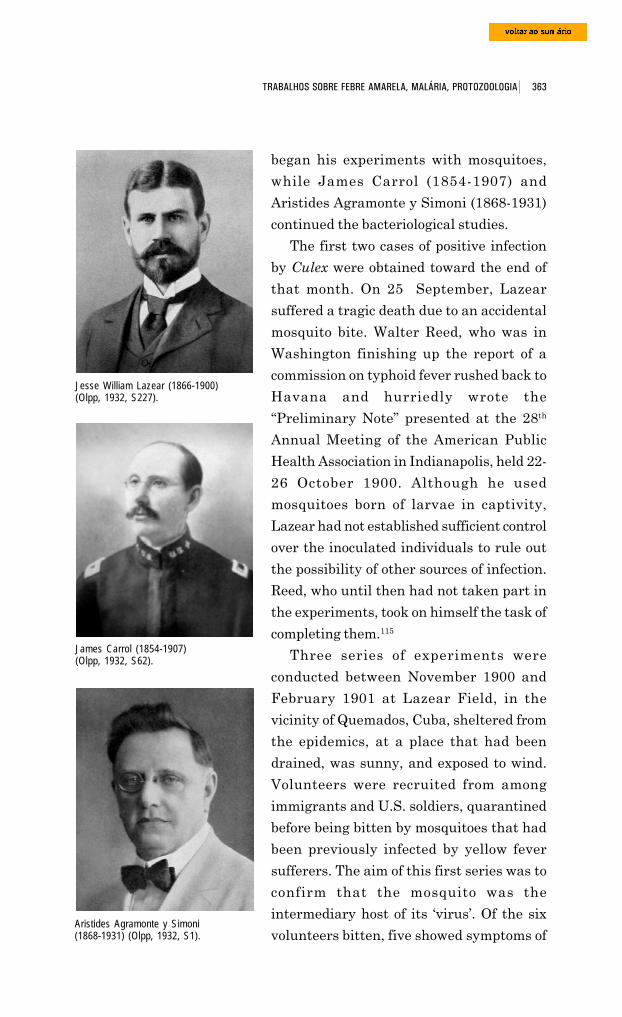
363 TRABALHOS SOBRE FEBRE AMARELA, MALÁRIA, PROTOZOOLOGIA
began his experiments with mosquitoes,
while James Carrol (1854-1907) and
Aristides Agramonte y Simoni (1868-1931)
continued the bacteriological studies.
The first two cases of positive infection
by Culex were obtained toward the end of
that month. On 25 September, Lazear
suffered a tragic death due to an accidental
mosquito bite. Walter Reed, who was in
Washington finishing up the report of a
commission on typhoid fever rushed back to
Havana and hurriedly wrote the
“Preliminary Note” presented at the 28th
Annual Meeting of the American Public
Health Association in Indianapolis, held 22-
26 October 1900. Although he used
mosquitoes born of larvae in captivity,
Lazear had not established sufficient control
over the inoculated individuals to rule out
the possibility of other sources of infection.
Reed, who until then had not taken part in
the experiments, took on himself the task of
completing them.115
Three series of experiments were
conducted between November 1900 and
February 1901 at Lazear Field, in the
vicinity of Quemados, Cuba, sheltered from
the epidemics, at a place that had been
drained, was sunny, and exposed to wind.
Volunteers were recruited from among
immigrants and U.S. soldiers, quarantined
before being bitten by mosquitoes that had
been previously infected by yellow fever
sufferers. The aim of this first series was to
confirm that the mosquito was the
intermediary host of its ‘virus’. Of the six
volunteers bitten, five showed symptoms of
Jesse William Lazear (1866-1900)(Olpp, 1932, S227).
James Carrol (1854-1907)(Olpp, 1932, S62).
Aristides Agramonte y Simoni(1868-1931) (Olpp, 1932, S1).
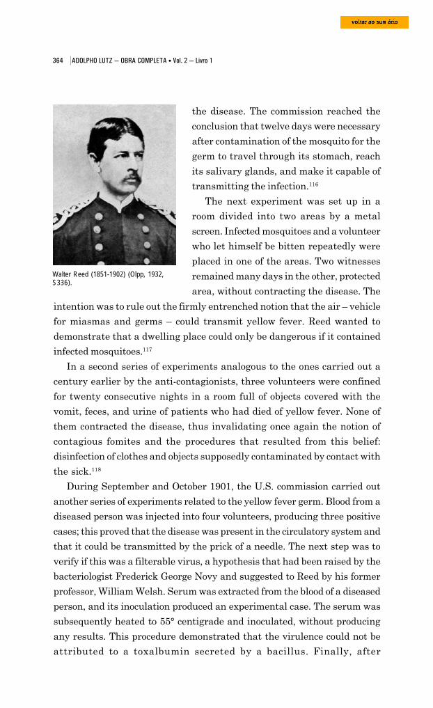
364 ADOLPHO LUTZ — OBRA COMPLETA Vol. 2 — Livro 1
Walter Reed (1851-1902) (Olpp, 1932,S336).
the disease. The commission reached the
conclusion that twelve days were necessary
after contamination of the mosquito for the
germ to travel through its stomach, reach
its salivary glands, and make it capable of
transmitting the infection.116
The next experiment was set up in a
room divided into two areas by a metal
screen. Infected mosquitoes and a volunteer
who let himself be bitten repeatedly were
placed in one of the areas. Two witnesses
remained many days in the other, protected
area, without contracting the disease. The
intention was to rule out the firmly entrenched notion that the air – vehicle
for miasmas and germs – could transmit yellow fever. Reed wanted to
demonstrate that a dwelling place could only be dangerous if it contained
infected mosquitoes.117
In a second series of experiments analogous to the ones carried out a
century earlier by the anti-contagionists, three volunteers were confined
for twenty consecutive nights in a room full of objects covered with the
vomit, feces, and urine of patients who had died of yellow fever. None of
them contracted the disease, thus invalidating once again the notion of
contagious fomites and the procedures that resulted from this belief:
disinfection of clothes and objects supposedly contaminated by contact with
the sick.118
During September and October 1901, the U.S. commission carried out
another series of experiments related to the yellow fever germ. Blood from a
diseased person was injected into four volunteers, producing three positive
cases; this proved that the disease was present in the circulatory system and
that it could be transmitted by the prick of a needle. The next step was to
verify if this was a filterable virus, a hypothesis that had been raised by the
bacteriologist Frederick George Novy and suggested to Reed by his former
professor, William Welsh. Serum was extracted from the blood of a diseased
person, and its inoculation produced an experimental case. The serum was
subsequently heated to 55° centigrade and inoculated, without producing
any results. This procedure demonstrated that the virulence could not be
attributed to a toxalbumin secreted by a bacillus. Finally, after
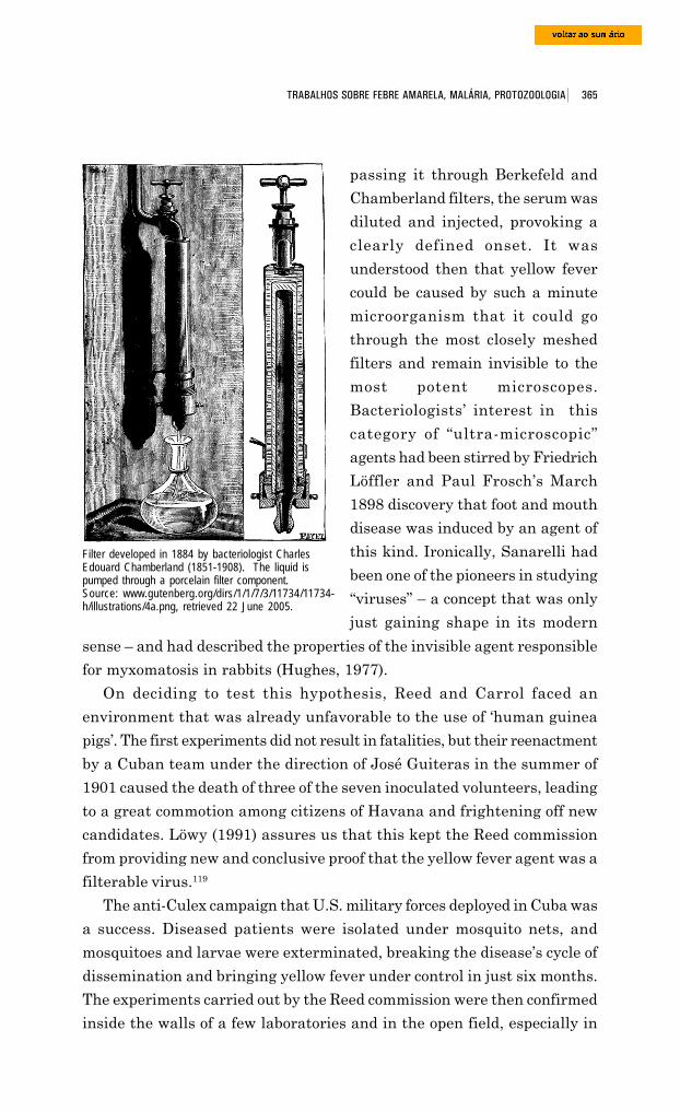
365 TRABALHOS SOBRE FEBRE AMARELA, MALÁRIA, PROTOZOOLOGIA
passing it through Berkefeld and
Chamberland filters, the serum was
diluted and injected, provoking a
clearly defined onset. It was
understood then that yellow fever
could be caused by such a minute
microorganism that it could go
through the most closely meshed
filters and remain invisible to the
most potent microscopes.
Bacteriologists’ interest in this
category of “ultra-microscopic”
agents had been stirred by Friedrich
Löffler and Paul Frosch’s March
1898 discovery that foot and mouth
disease was induced by an agent of
this kind. Ironically, Sanarelli had
been one of the pioneers in studying
“viruses” – a concept that was only
just gaining shape in its modern
sense – and had described the properties of the invisible agent responsible
for myxomatosis in rabbits (Hughes, 1977).
On deciding to test this hypothesis, Reed and Carrol faced an
environment that was already unfavorable to the use of ‘human guinea
pigs’. The first experiments did not result in fatalities, but their reenactment
by a Cuban team under the direction of José Guiteras in the summer of
1901 caused the death of three of the seven inoculated volunteers, leading
to a great commotion among citizens of Havana and frightening off new
candidates. Löwy (1991) assures us that this kept the Reed commission
from providing new and conclusive proof that the yellow fever agent was a
filterable virus.119
The anti-Culex campaign that U.S. military forces deployed in Cuba was
a success. Diseased patients were isolated under mosquito nets, and
mosquitoes and larvae were exterminated, breaking the disease’s cycle of
dissemination and bringing yellow fever under control in just six months.
The experiments carried out by the Reed commission were then confirmed
inside the walls of a few laboratories and in the open field, especially in
Filter developed in 1884 by bacteriologist CharlesEdouard Chamberland (1851-1908). The liquid ispumped through a porcelain filter component.Source: www.gutenberg.org/dirs/1/1/7/3/11734/11734-h/illustrations/4a.png, retrieved 22 June 2005.
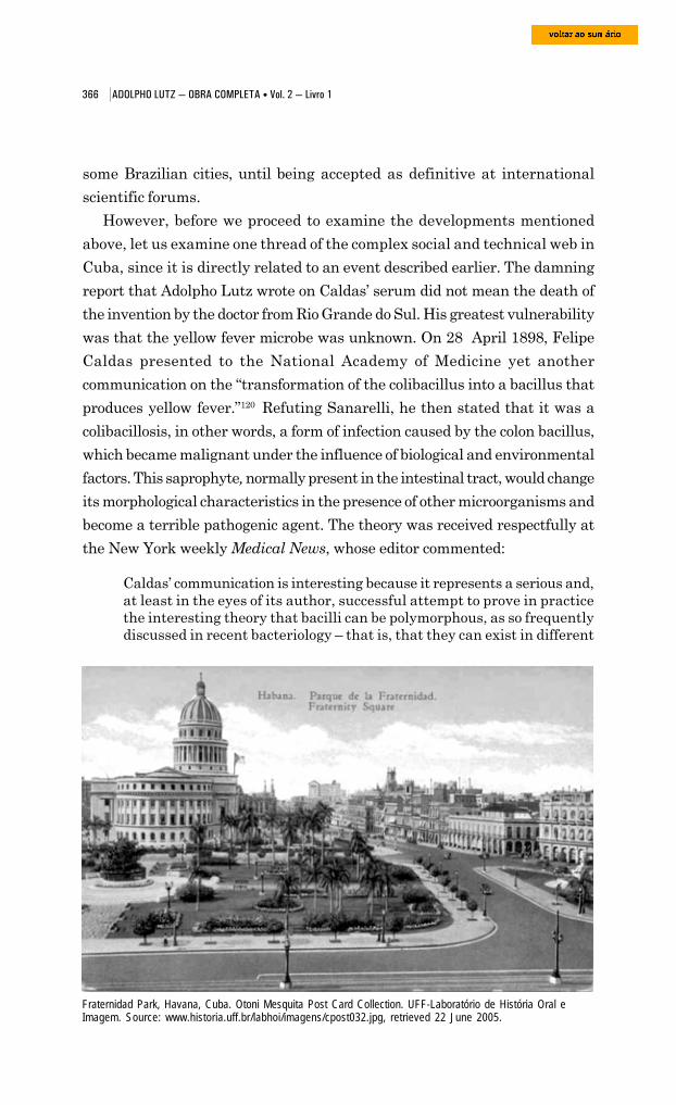
366 ADOLPHO LUTZ — OBRA COMPLETA Vol. 2 — Livro 1
Fraternidad Park, Havana, Cuba. Otoni Mesquita Post Card Collection. UFF-Laboratório de História Oral eImagem. Source: www.historia.uff.br/labhoi/imagens/cpost032.jpg, retrieved 22 June 2005.
some Brazilian cities, until being accepted as definitive at international
scientific forums.
However, before we proceed to examine the developments mentioned
above, let us examine one thread of the complex social and technical web in
Cuba, since it is directly related to an event described earlier. The damning
report that Adolpho Lutz wrote on Caldas’ serum did not mean the death of
the invention by the doctor from Rio Grande do Sul. His greatest vulnerability
was that the yellow fever microbe was unknown. On 28 April 1898, Felipe
Caldas presented to the National Academy of Medicine yet another
communication on the “transformation of the colibacillus into a bacillus that
produces yellow fever.”120 Refuting Sanarelli, he then stated that it was a
colibacillosis, in other words, a form of infection caused by the colon bacillus,
which became malignant under the influence of biological and environmental
factors. This saprophyte, normally present in the intestinal tract, would change
its morphological characteristics in the presence of other microorganisms and
become a terrible pathogenic agent. The theory was received respectfully at
the New York weekly Medical News, whose editor commented:
Caldas’ communication is interesting because it represents a serious and,at least in the eyes of its author, successful attempt to prove in practicethe interesting theory that bacilli can be polymorphous, as so frequentlydiscussed in recent bacteriology – that is, that they can exist in different
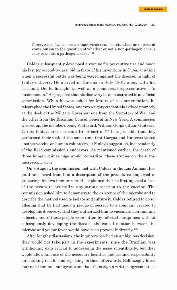
367 TRABALHOS SOBRE FEBRE AMARELA, MALÁRIA, PROTOZOOLOGIA
forms, each of which has a unique virulence. This stands as an importantcontribution to the question of whether or not a non-pathogenic virusmay turn into a pathogenic virus.121
Caldas subsequently developed a vaccine for preventive use and made
his last (or second-to-last) bid in favor of his inventions in Cuba, at a time
when a successful battle was being waged against the disease, in light of
Finlay’s theory. He arrived in Havana in July 1901, along with his
assistant, Dr. Bellinzaghi, as well as a commercial representative – “a
businessman.” He proposed that his discovery be demonstrated to an official
commission. When he was asked for letters of recommendation, he
telegraphed the United States, and two weighty credentials arrived promptly
at the desk of the Military Governor: one from the Secretary of War and
the other from the Brazilian Consul General in New York. A commission
was set up, the members being V. Havard, William Gorgas, Juan Guiteras,
Carlos Finlay, and a certain Dr. Albertini.122 It is probable that they
performed their task at the same time that Gorgas and Guiteras tested
another vaccine on human volunteers, at Finlay’s suggestion, independently
of the Reed commission’s endeavors. As mentioned earlier, the death of
three human guinea pigs would jeopardize these studies on the ultra-
microscopic virus.
On 9 August, the commission met with Caldas in the Las Animas Hos-
pital and heard from him a description of the procedures employed in
preparing his two immunizers. He explained that he first injected a dose
of the serum to neutralize any strong reaction to the vaccine. The
commission asked him to demonstrate the existence of the microbe and to
describe the method used to isolate and culture it. Caldas refused to do so,
alleging that he had made a pledge of secrecy to a company created to
develop his discovery. Had they authorized him to vaccinate non-immune
subjects, and if these people were bitten by infected mosquitoes without
subsequently developing the disease, the causal relation between the
microbe and yellow fever would have been proven, indirectly.123
After lengthy discussions, the inquirers reached an ambiguous decision:
they would not take part in the experiments, since the Brazilian was
withholding data crucial to addressing the issue scientifically, but they
would allow him use of the necessary facilities and assume responsibility
for checking results and reporting on them afterwards. Bellinzaghy hired
four non-immune immigrants and had them sign a written agreement, as
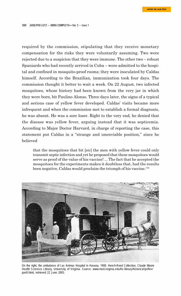
368 ADOLPHO LUTZ — OBRA COMPLETA Vol. 2 — Livro 1
required by the commission, stipulating that they receive monetary
compensation for the risks they were voluntarily assuming. Two were
rejected due to a suspicion that they were immune. The other two – robust
Spaniards who had recently arrived in Cuba – were admitted to the hospi-
tal and confined in mosquito-proof rooms; they were inoculated by Caldas
himself. According to the Brazilian, immunization took four days. The
commission thought it better to wait a week. On 22 August, two infected
mosquitoes, whose history had been known from the very jar in which
they were born, bit Paulino Alonso. Three days later, the signs of a typical
and serious case of yellow fever developed. Caldas’ visits became more
infrequent and when the commission met to establish a formal diagnosis,
he was absent. He was a sore loser. Right to the very end, he denied that
the disease was yellow fever, arguing instead that it was septicemia.
According to Major Doctor Harvard, in charge of reporting the case, this
statement put Caldas in a “strange and unenviable position,” since he
believed
that the mosquitoes that bit [sic] the men with yellow fever could onlytransmit septic infection and yet he proposed that these mosquitoes wouldserve as proof of the value of his vaccine! ... The fact that he accepted themosquitoes for the experiments makes it doubtless that, had the resultsbeen negative, Caldas would proclaim the triumph of his vaccine.124
On the right, the ambulance of Las Animas Hospital in Havana, 1900. Hench-Reed Collection, Claude MooreHealth Sciences Library, University of Virgínia. Source: www.med.virginia.edu/hs-library/historical/yelfev/pan9.html, retrieved 22 June 2005.
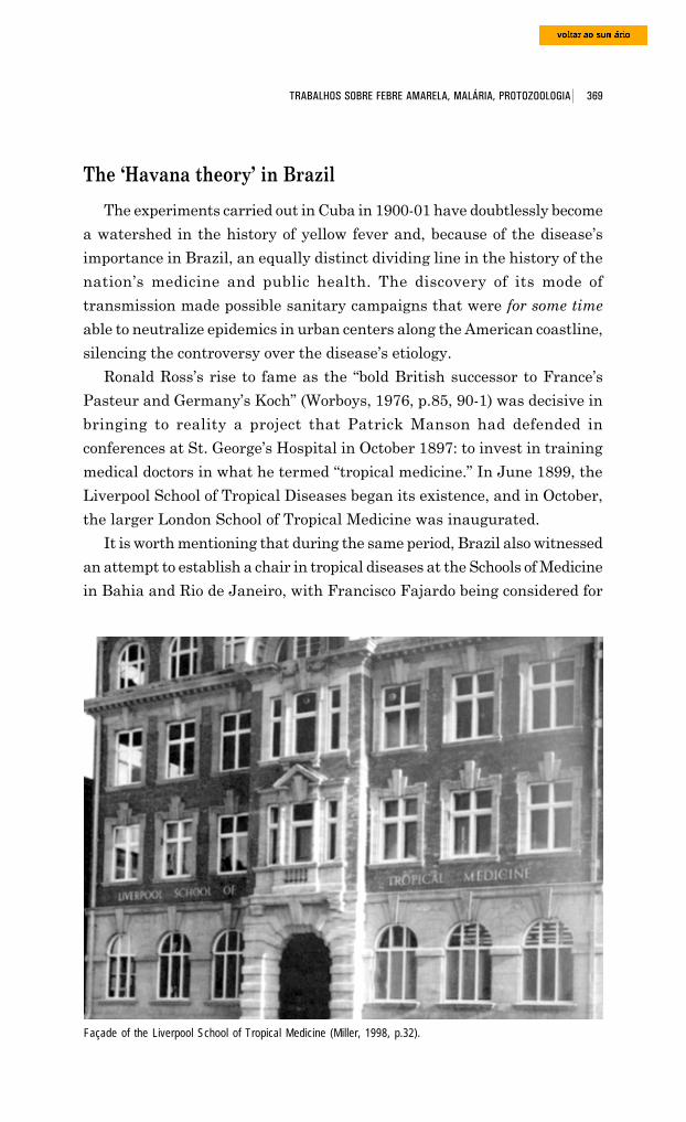
369 TRABALHOS SOBRE FEBRE AMARELA, MALÁRIA, PROTOZOOLOGIA
The ‘Havana theory’ in Brazil
The experiments carried out in Cuba in 1900-01 have doubtlessly become
a watershed in the history of yellow fever and, because of the disease’s
importance in Brazil, an equally distinct dividing line in the history of the
nation’s medicine and public health. The discovery of its mode of
transmission made possible sanitary campaigns that were for some time
able to neutralize epidemics in urban centers along the American coastline,
silencing the controversy over the disease’s etiology.
Ronald Ross’s rise to fame as the “bold British successor to France’s
Pasteur and Germany’s Koch” (Worboys, 1976, p.85, 90-1) was decisive in
bringing to reality a project that Patrick Manson had defended in
conferences at St. George’s Hospital in October 1897: to invest in training
medical doctors in what he termed “tropical medicine.” In June 1899, the
Liverpool School of Tropical Diseases began its existence, and in October,
the larger London School of Tropical Medicine was inaugurated.
It is worth mentioning that during the same period, Brazil also witnessed
an attempt to establish a chair in tropical diseases at the Schools of Medicine
in Bahia and Rio de Janeiro, with Francisco Fajardo being considered for
Façade of the Liverpool School of Tropical Medicine (Miller, 1998, p.32).
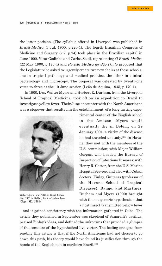
370 ADOLPHO LUTZ — OBRA COMPLETA Vol. 2 — Livro 1
Walter Myers, born 1872 in Great Britain,died 1901 in Belém, Pará, of yellow fever(Olpp, 1932, S288).
the latter position. (The syllabus offered in Liverpool was published in
Brazil-Medico, 1 Jul. 1900, p.220-1). The fourth Brazilian Congress of
Medicine and Surgery (v.2, p.74) took place in the Brazilian capital in
June 1900. Vitor Godinho and Carlos Seidl, representing O Brazil-Medico
(22 May 1900, p.173-4) and Revista Médica de São Paulo proposed that
the Legislature be asked to urgently create two new chairs at those schools,
one in tropical pathology and medical practice, the other in clinical
bacteriology and microscopy. The proposal was defeated by twenty-one
votes to three at the 19 June session (Leão de Aquino, 1945, p.170-1).
In 1900, Drs. Walter Myers and Herbert E. Durham, from the Liverpool
School of Tropical Medicine, took off on an expedition to Brazil to
investigate yellow fever. Their June encounter with the North Americans
was a stopover that resulted in the establishment of a long-lasting expe-
rimental center of the English school
in the Amazon. Myers would
eventually die in Belém, on 29
January 1901, a victim of the disease
he had traveled to study.125 In Hava-
na, they met with the members of the
U.S. commission; with Major William
Gorgas, who headed the Bureau of
Inspection of Infectious Diseases; with
Henry R. Carter, from the U.S. Marine
Hospital Service; and also with Cuban
doctors: Finlay, Guiteras (professor of
the Havana School of Tropical
Diseases), Bango, and Martinez.
Durham and Myers (1900) brought
with them a generic hypothesis – that
a host insect transmitted yellow fever
– and it gained consistency with the information gathered in Cuba. The
article they published in September was skeptical of Sanarelli’s bacillus,
praised Finlay’s ideas, and defined the unknowns that provided a glimpse
of the contours of the hypothetical live vector. The feeling one gets from
reading this article is that if the North Americans had not chosen to go
down this path, his theory would have found its justification through the
hands of the Englishmen in northern Brazil.126
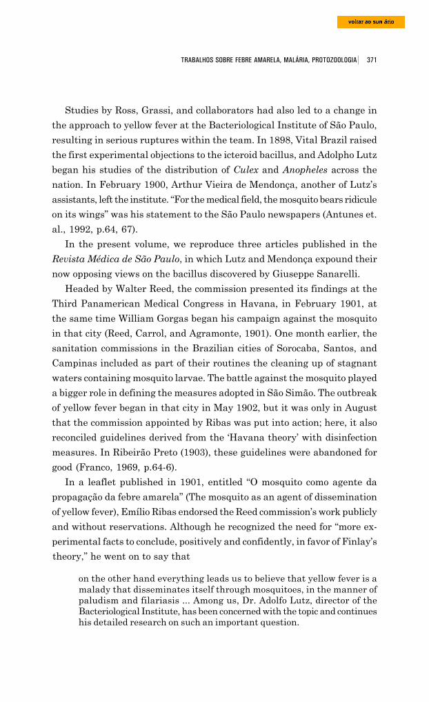
371 TRABALHOS SOBRE FEBRE AMARELA, MALÁRIA, PROTOZOOLOGIA
Studies by Ross, Grassi, and collaborators had also led to a change in
the approach to yellow fever at the Bacteriological Institute of São Paulo,
resulting in serious ruptures within the team. In 1898, Vital Brazil raised
the first experimental objections to the icteroid bacillus, and Adolpho Lutz
began his studies of the distribution of Culex and Anopheles across the
nation. In February 1900, Arthur Vieira de Mendonça, another of Lutz’s
assistants, left the institute. “For the medical field, the mosquito bears ridicule
on its wings” was his statement to the São Paulo newspapers (Antunes et.
al., 1992, p.64, 67).
In the present volume, we reproduce three articles published in the
Revista Médica de São Paulo, in which Lutz and Mendonça expound their
now opposing views on the bacillus discovered by Giuseppe Sanarelli.
Headed by Walter Reed, the commission presented its findings at the
Third Panamerican Medical Congress in Havana, in February 1901, at
the same time William Gorgas began his campaign against the mosquito
in that city (Reed, Carrol, and Agramonte, 1901). One month earlier, the
sanitation commissions in the Brazilian cities of Sorocaba, Santos, and
Campinas included as part of their routines the cleaning up of stagnant
waters containing mosquito larvae. The battle against the mosquito played
a bigger role in defining the measures adopted in São Simão. The outbreak
of yellow fever began in that city in May 1902, but it was only in August
that the commission appointed by Ribas was put into action; here, it also
reconciled guidelines derived from the ‘Havana theory’ with disinfection
measures. In Ribeirão Preto (1903), these guidelines were abandoned for
good (Franco, 1969, p.64-6).
In a leaflet published in 1901, entitled “O mosquito como agente da
propagação da febre amarela” (The mosquito as an agent of dissemination
of yellow fever), Emílio Ribas endorsed the Reed commission’s work publicly
and without reservations. Although he recognized the need for “more ex-
perimental facts to conclude, positively and confidently, in favor of Finlay’s
theory,” he went on to say that
on the other hand everything leads us to believe that yellow fever is amalady that disseminates itself through mosquitoes, in the manner ofpaludism and filariasis ... Among us, Dr. Adolfo Lutz, director of theBacteriological Institute, has been concerned with the topic and continueshis detailed research on such an important question.

372 ADOLPHO LUTZ — OBRA COMPLETA Vol. 2 — Livro 1
As a matter of fact, Lutz had been correlating the presence of
mosquitoes to the yellow fever epidemics in the territory under his
jurisdiction for quite some time, and therefore verification of the Cuban
doctor’s theory had not surprised him. São Paulo’s Sanitation Service
publication brought already, in the form of an inset, his first systematic
observations on mosquitoes as agents of disease transmission.
Lutz’s note concerned two species of mosquitoes of wide geographic
distribution usually found in homes, only one of which, however, had
been positively linked to the transmission of yellow fever. It is interesting
to note that both Lutz and Theobald at first had trouble correctly
identifying the species. In the first letter to his Brazilian colleague, dated
28 April 1900,127 the entomologist from the British Museum commented
that their wide-ranging distribution and variety of local names raised
significant obstacles to the identification of certain species, especially Culex
taeniatus, even in older descriptions. Ficalbi’s and Arribalzaga’s works
were cited as valuable resources in this endeavor. Lutz’s answer to
Theobald included a request for a sample of that Culex to compare with
his own. A few months later, after receiving updated literature, he declared
that he was already able to determine his species. In observations sent to
Theobald on the material identified, he noted that C. taeniatus was
commonly found in houses in coastal regions or inland, but not in the
capital city of São Paulo. He had not yet postulated its relationship with
yellow fever. It was only in a letter dated January 1901 that he informed
Theobald that he had been paying particular attention to this species
because its distribution coincided with that of the disease. His observations
were published that same month by Ribas.
Terms such as ‘mosquito rajado’ (literally, striped mosquito) or
‘pernilongo’ (long-legged mosquito) – both colloquial names for Stegomyia
fasciata – were commonly used to designate blood-sucking dipterous
insects. Lutz had previously studied “a couple dozen species” in Brazil,
most of which were only present in the wilds or in swamplands, the setting
of the investigation he was finishing on the transmission of forest malaria.
Only two species (Culex taeniatus and Culex fatigans) were frequent
‘tenants’ in human households. Although found in almost all regions
offering the right climatic conditions, they were “extremely rare” species in
uninhabited or uncultivated areas.
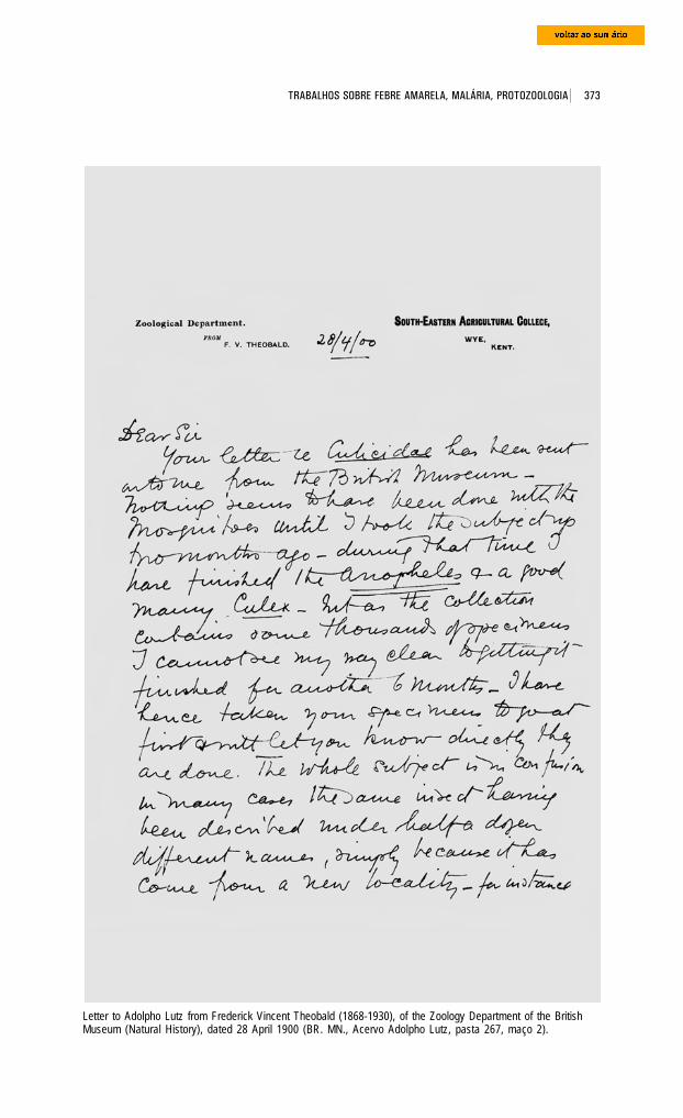
373 TRABALHOS SOBRE FEBRE AMARELA, MALÁRIA, PROTOZOOLOGIA
Letter to Adolpho Lutz from Frederick Vincent Theobald (1868-1930), of the Zoology Department of the BritishMuseum (Natural History), dated 28 April 1900 (BR. MN., Acervo Adolpho Lutz, pasta 267, maço 2).
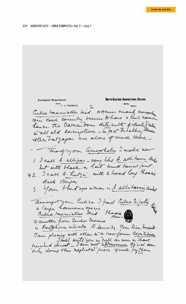
374 ADOLPHO LUTZ — OBRA COMPLETA Vol. 2 — Livro 1
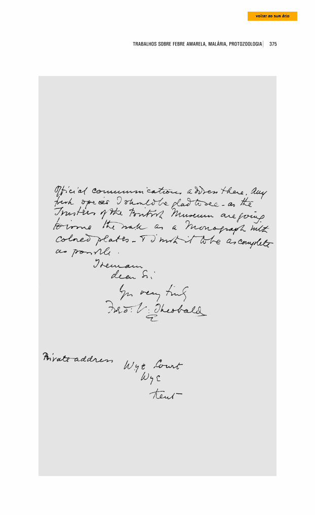
375 TRABALHOS SOBRE FEBRE AMARELA, MALÁRIA, PROTOZOOLOGIA
376 ADOLPHO LUTZ — OBRA COMPLETA Vol. 2 — Livro 1
The most widely scattered is Culex fatigans, our common night-timemosquito, which is found almost everywhere since it is less sensitive to thecold. It transmits filariasis and certain bird haematozoa; I do not considerit a suspect in the case of yellow fever. Culex taeniatus, on the other hand,is the only one that can account for the distribution of yellow fever, becausewe know that it has been described, under various names, in almost allthe places where yellow fever has reigned.
Lutz pointed out its occurrence in the United States, Cuba, Buenos Aires,
on the south and west coasts of Africa, in Hawaii – in short, in all places
where yellow fever had spread. He did not, however, associate it with Culex
fasciatus, a species used in the experiments carried out by the Reed
commission in Cuba. The Brazilian zoologist followed the classification
proposed by Giles (1900) and the one Theobald sent him in August 1900.128
Upon creating the genus Stegomyia in 1901, Theobald placed in it
the Culex species related to yellow fever transmission, including C.
taeniatus and many of the synonyms pointed out by Lutz.129 The species
was then named Stegomyia fasciata. It was a mosquito that “had had
36 synonyms from 1762, when Linnaeus classified it as Culex aegipty,
until 1926, when Silver renamed it Aedes aegypti, as it is currently
known” (Franco, 1969, p.64).
Albuquerque (1950, p.11-2) most certainly falls into an anachronism
when she states that already in 1889 in Campinas, Lutz would have had
a “clear intuition of the truth” on the correlation between mosquitoes and
yellow fever. Lutz himself is to blame since, in an article published in
1903 (“Yellow fever and the mosquito,” in Port.), and later in his Remi-
niscências (1930), he alluded to observations made in 1889; these,
however, had not been published, most probably because they amounted
to little more than suspicions as yet irreconcilable with the etiological
theories of the day.
“At that time, all the elements for explaining this mode of transmission
were lacking,” reads an excerpt from the first article. Even so, when that
coffee-growing center was devastated by yellow fever, Lutz had noted “an
extraordinary abundance of the ‘striped mosquitoes’ that [he] knew so well
from Rio de Janeiro, but that [he] had never observed in the interior during
five years of practice in a region not too far from Campinas.” Lutz at that
time correlated the “plague of the Stegomyias with tanks of still waters in
the gardens of houses that had been abandoned by their owners. They
were such a nuisance that he used a mosquito net every night and sometimes

377 TRABALHOS SOBRE FEBRE AMARELA, MALÁRIA, PROTOZOOLOGIA
even in the daytime “so that [he] could read in peace; naturally that did
not stop [him] from being bitten on several occasions” (Lutz, 1930).
It is, however, true that his first speculations on the role bloodsuckers
played in spreading disease predate this event. They are found in his
“Studies on leprosy”, written in 1885-86 and published in Monatshefte für
Praktische Dermatologie (1887). On describing primary lesions of the
nervous form, which could act as “the entryway for infection,” he considered
it “impressive that its first locus of manifestation ... is almost always in the
parts of the body that are left uncovered and exposed to insect bites and
other trauma” (1887, p.549). Lutz had already observed that each new
case of infection required a pre-existing case within a defined area, but
that the necessary conditions were of such complexity and peculiarity (much
like yellow fever, we might add), that one could exclude direct person-to-
person contagion. To explain transmission of Hansen’s disease, Lutz
admitted, albeit hypothetically, that blood or excretions from the patient’s
mucus that contained the infectious agent might require “maturation” in
an external medium, or “direct inoculation that generates vulnerability
(for example, through insect bites).”
This documented speculation leads us to give credit to the statement
made in 1903 that the notion of mosquito transmission of yellow fever had
been “always attractive” to Lutz after Campinas. “As a doctor and naturalist,
I have always paid attention to these mosquitoes,” Lutz wrote, “and I knew
that they were very common in Rio de Janeiro and in Santos, although
much scarcer in rural São Paulo, where on rare occasions they did become
a nuisance to me in the places I knew and lived in.”
In the 1903 article, he traced doubts about this hypothesis to the realm
of entomology itself; his main objection concerned the epidemics of yellow
fever along the Mediterranean and African coastlines, since it was believed
that the Stegomyia fasciata existed only on the American continent. “It
was with great surprise that, a few months later, I observed the same
mosquito in the Sandwich Islands, and not only in the capital but even in
far-removed plantations ... I then understood that [S. fasciata] was scattered
about by seafaring vessels and that it must be found in other warm countries,
although I was unaware of its presence in southern Europe. Theobald was
to demonstrate this in the early 20th century.
As we have shown in the previous volume of the Collected Works of
Adolpho Lutz, it was in Hawaii, where he lived from 1889 to mid-1892,
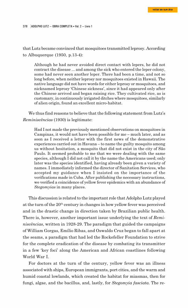
378 ADOLPHO LUTZ — OBRA COMPLETA Vol. 2 — Livro 1
that Lutz became convinced that mosquitoes transmitted leprosy. According
to Albuquerque (1950, p.13-4):
Although he had never avoided direct contact with lepers, he did notcontract the disease ... and among the sick who entered the leper colony,some had never seen another leper. There had been a time, and not solong before, when neither leprosy nor mosquitoes existed in Hawaii. Thenative language did not have words for either leprosy or mosquitoes, andnicknamed leprosy ‘Chinese sickness’, since it had appeared only afterthe Chinese arrived and began raising rice. They cultivated rice, as iscustomary, in continuously irrigated ditches where mosquitoes, similarlyof alien origin, found an excellent micro-habitat.
We thus find reasons to believe that the following statement from Lutz’s
Reminiscências (1930) is legitimate:
Had I not made the previously mentioned observations on mosquitoes inCampinas, it would not have been possible for me – much later, and assoon as I received a letter with the first news of the demonstrativeexperiences carried out in Havana – to name the guilty mosquito amongus without hesitation, a mosquito that did not exist in the city of SãoPaulo. It seemed probable to me that we were dealing with the samespecies, although I did not call it by the name the Americans used; onlylater was the species identified, having already been given a variety ofnames. I immediately informed the director of Sanitation Services, whoaccepted my guidance when I insisted on the importance of theverifications made in Cuba. After publishing the necessary instructions,we verified a coincidence of yellow fever epidemics with an abundance ofStegomyias in many places.
This discussion is related to the important role that Adolpho Lutz played
at the turn of the 20th century in changes in how yellow fever was perceived
and in the drastic change in direction taken by Brazilian public health.
There is, however, another important issue underlying the text of Remi-
niscências, written in 1928-29. The paradigm that guided the campaigns
of William Gorgas, Emílio Ribas, and Oswaldo Cruz began to fall apart at
the seams, a paradigm that had led the Rockefeller Foundation to strive
for the complete eradication of the disease by combating its transmitter
in a few ‘key foci’ along the American and African coastlines following
World War I.
For doctors at the turn of the century, yellow fever was an illness
associated with ships, European immigrants, port cities, and the warm and
humid coastal lowlands, which created the habitat for miasmas, then for
fungi, algae, and the bacillus, and, lastly, for Stegomyia fasciata. The re-
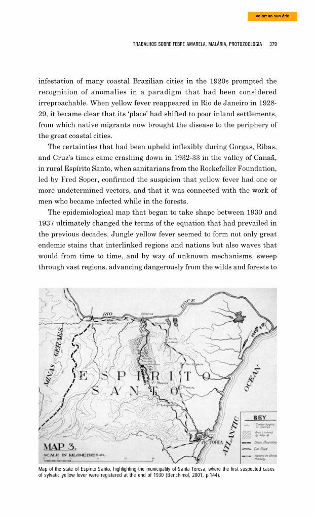
379 TRABALHOS SOBRE FEBRE AMARELA, MALÁRIA, PROTOZOOLOGIA
infestation of many coastal Brazilian cities in the 1920s prompted the
recognition of anomalies in a paradigm that had been considered
irreproachable. When yellow fever reappeared in Rio de Janeiro in 1928-
29, it became clear that its ‘place’ had shifted to poor inland settlements,
from which native migrants now brought the disease to the periphery of
the great coastal cities.
The certainties that had been upheld inflexibly during Gorgas, Ribas,
and Cruz’s times came crashing down in 1932-33 in the valley of Canaã,
in rural Espírito Santo, when sanitarians from the Rockefeller Foundation,
led by Fred Soper, confirmed the suspicion that yellow fever had one or
more undetermined vectors, and that it was connected with the work of
men who became infected while in the forests.
The epidemiological map that began to take shape between 1930 and
1937 ultimately changed the terms of the equation that had prevailed in
the previous decades. Jungle yellow fever seemed to form not only great
endemic stains that interlinked regions and nations but also waves that
would from time to time, and by way of unknown mechanisms, sweep
through vast regions, advancing dangerously from the wilds and forests to
Map of the state of Espírito Santo, highlighting the municipality of Santa Teresa, where the first suspected casesof sylvatic yellow fever were registered at the end of 1930 (Benchimol, 2001, p.144).
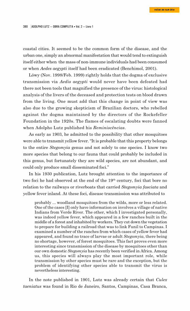
380 ADOLPHO LUTZ — OBRA COMPLETA Vol. 2 — Livro 1
coastal cities. It seemed to be the common form of the disease, and the
urban one, simply an abnormal manifestation that would tend to extinguish
itself either when the mass of non-immune individuals had been consumed
or when Aedes aegypti itself had been eradicated (Benchimol, 2001).
Löwy (Nov. 1998/Feb. 1999) rightly holds that the dogma of exclusive
transmission via Aedis aegypti would never have been defeated had
there not been tools that magnified the presence of the virus: histological
analysis of the livers of the deceased and protection tests on blood drawn
from the living. One must add that this change in point of view was
also due to the growing skepticism of Brazilian doctors, who rebelled
against the dogma maintained by the directors of the Rockefeller
Foundation in the 1920s. The flames of escalating doubts were fanned
when Adolpho Lutz published his Reminiscências.
As early as 1903, he admitted to the possibility that other mosquitoes
were able to transmit yellow fever. “It is probable that this property belongs
to the entire Stegomyia genus and not solely to one species. I know two
more species that belong to our fauna that could probably be included in
this genus, but fortunately they are wild species, are not abundant, and
could only produce small disseminated foci.”
In his 1930 publication, Lutz brought attention to the importance of
two foci he had observed at the end of the 19th century, foci that bore no
relation to the railways or riverboats that carried Stegomyia fasciata and
yellow fever inland. At these foci, disease transmission was attributed to
probably ... woodland mosquitoes from the wilds, more or less related.One of the cases [I] only have information on involves a village of nativeIndians from Verde River. The other, which I investigated personally,was indeed yellow fever, which appeared in a few ranches built in themiddle of a forest and inhabited by workers. They cut down the vegetationto prepare for building a railroad that was to link Funil to Campinas. Iexamined a number of the ranches from which cases of yellow fever hadappeared, and found no trace of larvae or adult Stegomyia, there beingno shortage, however, of forest mosquitoes. This fact proves even moreinteresting since transmission of the disease by mosquitoes other thanour own domestic Stegomyia has recently been verified in Africa. Amongus, this species will always play the most important role, whiletransmission by other species must be rare and the exception, but theproblem of identifying other species able to transmit the virus isnevertheless interesting.
In the note published in 1901, Lutz was already certain that Culex
taeniatus was found in Rio de Janeiro, Santos, Campinas, Casa Branca,
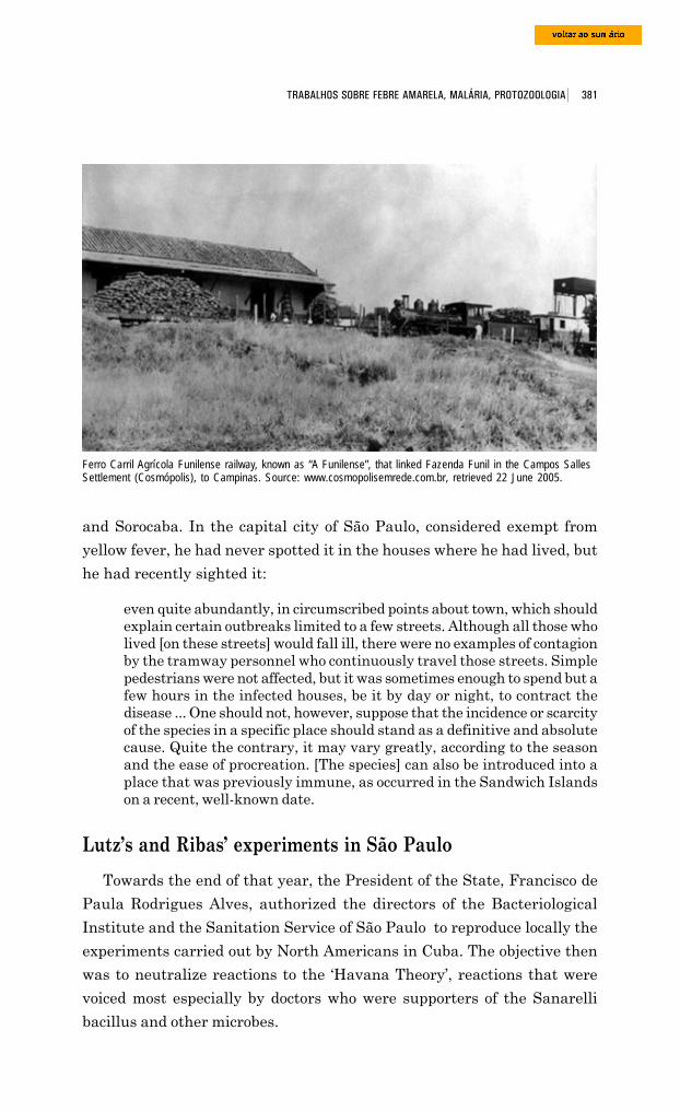
381 TRABALHOS SOBRE FEBRE AMARELA, MALÁRIA, PROTOZOOLOGIA
and Sorocaba. In the capital city of São Paulo, considered exempt from
yellow fever, he had never spotted it in the houses where he had lived, but
he had recently sighted it:
even quite abundantly, in circumscribed points about town, which shouldexplain certain outbreaks limited to a few streets. Although all those wholived [on these streets] would fall ill, there were no examples of contagionby the tramway personnel who continuously travel those streets. Simplepedestrians were not affected, but it was sometimes enough to spend but afew hours in the infected houses, be it by day or night, to contract thedisease ... One should not, however, suppose that the incidence or scarcityof the species in a specific place should stand as a definitive and absolutecause. Quite the contrary, it may vary greatly, according to the seasonand the ease of procreation. [The species] can also be introduced into aplace that was previously immune, as occurred in the Sandwich Islandson a recent, well-known date.
Lutz’s and Ribas’ experiments in São Paulo
Towards the end of that year, the President of the State, Francisco de
Paula Rodrigues Alves, authorized the directors of the Bacteriological
Institute and the Sanitation Service of São Paulo to reproduce locally the
experiments carried out by North Americans in Cuba. The objective then
was to neutralize reactions to the ‘Havana Theory’, reactions that were
voiced most especially by doctors who were supporters of the Sanarelli
bacillus and other microbes.
Ferro Carril Agrícola Funilense railway, known as “A Funilense”, that linked Fazenda Funil in the Campos SallesSettlement (Cosmópolis), to Campinas. Source: www.cosmopolisemrede.com.br, retrieved 22 June 2005.
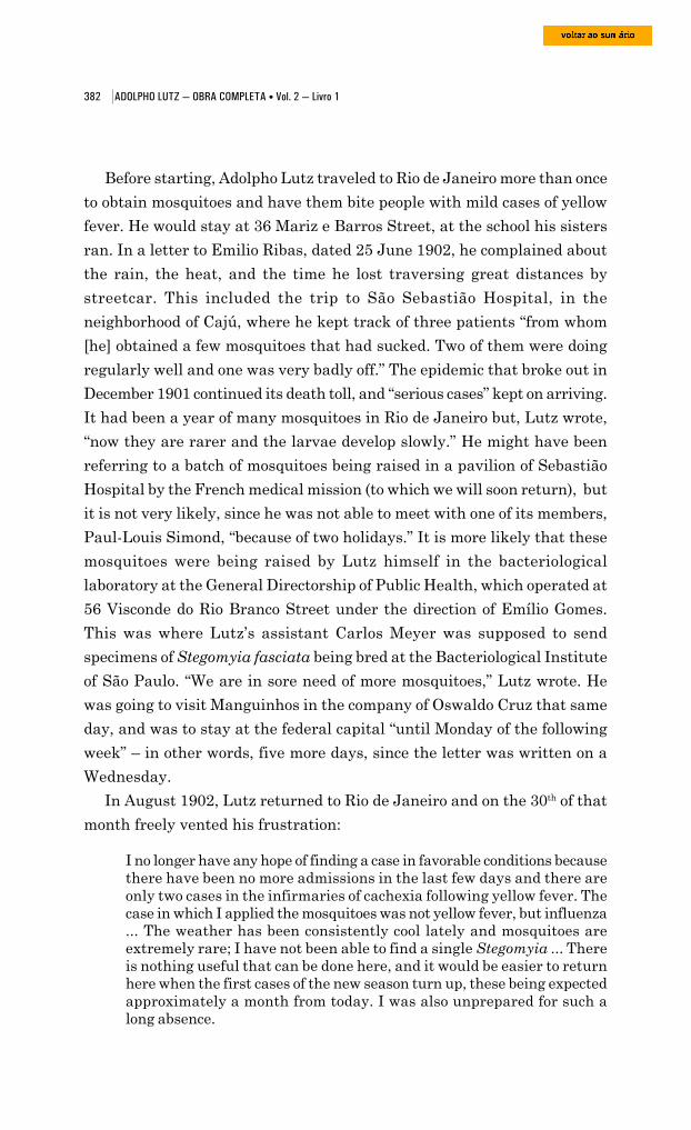
382 ADOLPHO LUTZ — OBRA COMPLETA Vol. 2 — Livro 1
Before starting, Adolpho Lutz traveled to Rio de Janeiro more than once
to obtain mosquitoes and have them bite people with mild cases of yellow
fever. He would stay at 36 Mariz e Barros Street, at the school his sisters
ran. In a letter to Emilio Ribas, dated 25 June 1902, he complained about
the rain, the heat, and the time he lost traversing great distances by
streetcar. This included the trip to São Sebastião Hospital, in the
neighborhood of Cajú, where he kept track of three patients “from whom
[he] obtained a few mosquitoes that had sucked. Two of them were doing
regularly well and one was very badly off.” The epidemic that broke out in
December 1901 continued its death toll, and “serious cases” kept on arriving.
It had been a year of many mosquitoes in Rio de Janeiro but, Lutz wrote,
“now they are rarer and the larvae develop slowly.” He might have been
referring to a batch of mosquitoes being raised in a pavilion of Sebastião
Hospital by the French medical mission (to which we will soon return), but
it is not very likely, since he was not able to meet with one of its members,
Paul-Louis Simond, “because of two holidays.” It is more likely that these
mosquitoes were being raised by Lutz himself in the bacteriological
laboratory at the General Directorship of Public Health, which operated at
56 Visconde do Rio Branco Street under the direction of Emílio Gomes.
This was where Lutz’s assistant Carlos Meyer was supposed to send
specimens of Stegomyia fasciata being bred at the Bacteriological Institute
of São Paulo. “We are in sore need of more mosquitoes,” Lutz wrote. He
was going to visit Manguinhos in the company of Oswaldo Cruz that same
day, and was to stay at the federal capital “until Monday of the following
week” – in other words, five more days, since the letter was written on a
Wednesday.
In August 1902, Lutz returned to Rio de Janeiro and on the 30th of that
month freely vented his frustration:
I no longer have any hope of finding a case in favorable conditions becausethere have been no more admissions in the last few days and there areonly two cases in the infirmaries of cachexia following yellow fever. Thecase in which I applied the mosquitoes was not yellow fever, but influenza... The weather has been consistently cool lately and mosquitoes areextremely rare; I have not been able to find a single Stegomyia ... Thereis nothing useful that can be done here, and it would be easier to returnhere when the first cases of the new season turn up, these being expectedapproximately a month from today. I was also unprepared for such along absence.
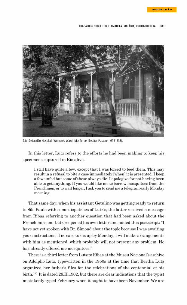
383 TRABALHOS SOBRE FEBRE AMARELA, MALÁRIA, PROTOZOOLOGIA
In this letter, Lutz refers to the efforts he had been making to keep his
specimens captured in Rio alive.
I still have quite a few, except that I was forced to feed them. This mayresult in a refusal to bite a case immediately [when] it is presented. I keepa few unfed but some of these always die. I apologize for not having beenable to get anything. If you would like me to borrow mosquitoes from theFrenchmen, or to wait longer, I ask you to send me a telegram early Mondaymorning.
That same day, when his assistant Getulino was getting ready to return
to São Paulo with some dispatches of Lutz’s, the latter received a message
from Ribas referring to another question that had been asked about the
French mission. Lutz reopened his own letter and added this postscript: “I
have not yet spoken with Dr. Simond about the topic because I was awaiting
your instructions; if no case turns up by Monday, I will make arrangements
with him as mentioned, which probably will not present any problem. He
has already offered me mosquitoes.”
There is a third letter from Lutz to Ribas at the Museu Nacional’s archive
on Adolpho Lutz, typewritten in the 1950s at the time that Bertha Lutz
organized her father’s files for the celebrations of the centennial of his
birth.130 It is dated 28.II.1902, but there are clear indications that the typist
mistakenly typed February when it ought to have been November. We are
São Sebastião Hospital, Women’s Ward (Musée de l’Institut Pasteur, MP31335).
384 ADOLPHO LUTZ — OBRA COMPLETA Vol. 2 — Livro 1
led to this conclusion by one of Lutz’s comments: “The epidemic is declining
but there are still cases.” In February 1902, it was reaching its apex, with
a daily average of ten hospitalizations at São Sebastião Hospital.
On the other hand, if the letter was written on 28 November, it means
that Lutz last stayed in Rio de Janeiro on the eve of beginning the
experiments in São Paulo. It was a Friday, and he promised “to return on
the Monday night train, arriving in São Paulo Tuesday morning.”
A disinfector with the Sanitation Service of São Paulo had just brought
him mosquitoes that “had arrived alive but were mostly male, which do not
produce any results.” Lutz hoped that Ivo Bandi would bring him more.
The ones he had been able to infect were alive and well, and the bacteriologist
from São Paulo referred to them in terms that show he expected to use
them soon: “The four mosquitoes from the first case will be good in a few
days; the patient has already been cured, having suffered an attack
characteristic in all aspects, but not critical. We have some mosquitoes
from two other cases, of which one mild, and the other fatal, and I have
kept them separately.”
It was raining heavily, and the scientist once more complained that in
Rio “one spends half the time traveling to and fro, or waiting for others.”
During his previous visit he had tried to meet with Nuno de Andrade, the
Director General of Public Health, and with the surgeon Pedro Affonso,
Director of the Municipal Vaccinogen Institute and of the Manguinhos
Serum Therapy Institute. He missed them again but was able to meet with
Simond, from the French mission, “who seemed to me the most
knowledgeable of the three” (the other two were Marchoux and Salimbeni).
“They are very committed to the issue of the mosquitoes, which they award
great importance.”
At the Bacteriological Institute, Lutz had begun controlled reproduction
from the larvae stage of the Stegomyia fasciata captured in Rio de Janeiro,
feeding them substances that were not blood, so as to exclude any other
unwanted infections.
On 28 November, sample mosquitoes were sent to Meyer, who was at
São Simão at the time, in the middle of a new outbreak of yellow fever. His
mission there was to infect mosquitoes by having them draw blood from
recent arrivals at the town’s isolation hospital, 730 kilometers away from
the capital of São Paulo by rail. In a letter to Lutz dated 1 December,
Meyer mentioned the clinical conditions of the patients submitted to the
385 TRABALHOS SOBRE FEBRE AMARELA, MALÁRIA, PROTOZOOLOGIA
procedure, the difficulties involved in having the mosquitoes bite them
appropriately, and the problems in keeping the insects alive until the trip
back to the capital. Received at the Bacteriological Institute on 2 December,
they were kept on a diet of honey and dates for over twelve days, the
minimum period of time considered necessary for making them agents of
infection. Three more were added to compensate for the temperature, which
was lower than that in Havana during Reed’s stay.
Experiments carried out in the Isolation Hospital in São Paulo employed
mosquitoes infected on Alexandrina, Joaquim Farquinio, and Nicola Razzoti
(the latter classified as a case of “long duration and serious in nature”); the
records for each session always specify the day of the contamination
procedure, the day the patients fell ill, and their clinical conditions expressed
in two numbers: pulse and temperature. Details of these experiments were
compiled in records signed by Ribas, Lutz, Carlos H. Meyer (an aid at the
Bacteriological Institute), Candido Espinheira (director of the Isolation
Hospital), Victor Godinho (a doctor at the same hospital), and a “commission
of clinical doctors” made up of Antonio Gomes Silva Rodrigues, Adriano
Julio de Barros, and Luiz Pereira Barreto.
A militant of the Republican Movement, Barreto had presided over the
State Constitutional Assembly in 1891 and had been the first president of
the Society of Medicine and Surgery of São Paulo. He had been a passionate
supporter of the transmission of yellow fever by water. Telarolli Junior
(1993, p.153-4) analyzed the heated controversy in the São Paulo
newspapers in 1896, which largely mirrored the one that had taken place
in Rio de Janeiro: “It was common at the time, for example, for well-to-do
families traveling by rail to the interior of the state to take a stock of mine-
ral water from Minas Gerais, in order to avoid catching yellow fever.”
Luiz Pereira Barreto was at that point the president of the State Senate,
honorary professor at the Polytechnic School of São Paulo, and author of
well-known articles on medicine, philosophy, politics, and religion. He
exerted all the authority he had to legitimize the experiments on mosquito
transmission.
The first series of experiments aimed at proving that Stegomyia were
the agents of transmission of yellow fever; it involved five sessions between
15 December 1902 and 20 January 1903.
The preferences to use volunteers who had already been ‘acclimated’, to
infect mosquitoes with mild cases of the disease, and to rely on an extended
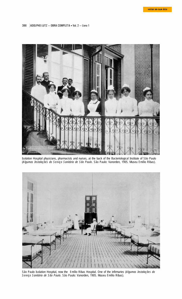
386 ADOLPHO LUTZ — OBRA COMPLETA Vol. 2 — Livro 1
São Paulo Isolation Hospital, now the Emílio Ribas Hospital. One of the infirmaries (Algumas Instalações doServiço Sanitário de São Paulo. São Paulo: Vanorden, 1905. Museu Emílio Ribas).
Isolation Hospital physicians, pharmacists and nurses, at the back of the Bacteriological Institute of São Paulo(Algumas Instalações do Serviço Sanitário de São Paulo. São Paulo: Vanorden, 1905. Museu Emílio Ribas).
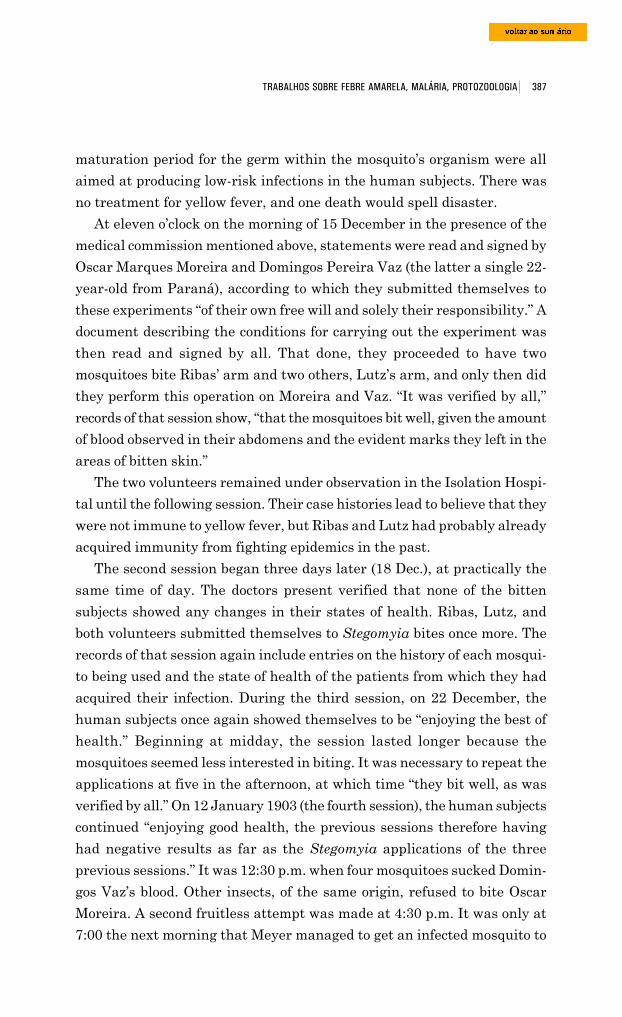
387 TRABALHOS SOBRE FEBRE AMARELA, MALÁRIA, PROTOZOOLOGIA
maturation period for the germ within the mosquito’s organism were all
aimed at producing low-risk infections in the human subjects. There was
no treatment for yellow fever, and one death would spell disaster.
At eleven o’clock on the morning of 15 December in the presence of the
medical commission mentioned above, statements were read and signed by
Oscar Marques Moreira and Domingos Pereira Vaz (the latter a single 22-
year-old from Paraná), according to which they submitted themselves to
these experiments “of their own free will and solely their responsibility.” A
document describing the conditions for carrying out the experiment was
then read and signed by all. That done, they proceeded to have two
mosquitoes bite Ribas’ arm and two others, Lutz’s arm, and only then did
they perform this operation on Moreira and Vaz. “It was verified by all,”
records of that session show, “that the mosquitoes bit well, given the amount
of blood observed in their abdomens and the evident marks they left in the
areas of bitten skin.”
The two volunteers remained under observation in the Isolation Hospi-
tal until the following session. Their case histories lead to believe that they
were not immune to yellow fever, but Ribas and Lutz had probably already
acquired immunity from fighting epidemics in the past.
The second session began three days later (18 Dec.), at practically the
same time of day. The doctors present verified that none of the bitten
subjects showed any changes in their states of health. Ribas, Lutz, and
both volunteers submitted themselves to Stegomyia bites once more. The
records of that session again include entries on the history of each mosqui-
to being used and the state of health of the patients from which they had
acquired their infection. During the third session, on 22 December, the
human subjects once again showed themselves to be “enjoying the best of
health.” Beginning at midday, the session lasted longer because the
mosquitoes seemed less interested in biting. It was necessary to repeat the
applications at five in the afternoon, at which time “they bit well, as was
verified by all.” On 12 January 1903 (the fourth session), the human subjects
continued “enjoying good health, the previous sessions therefore having
had negative results as far as the Stegomyia applications of the three
previous sessions.” It was 12:30 p.m. when four mosquitoes sucked Domin-
gos Vaz’s blood. Other insects, of the same origin, refused to bite Oscar
Moreira. A second fruitless attempt was made at 4:30 p.m. It was only at
7:00 the next morning that Meyer managed to get an infected mosquito to

388 ADOLPHO LUTZ — OBRA COMPLETA Vol. 2 — Livro 1
draw blood from Moreira. Half an hour later, another bit him twice but
did not draw blood. The operation was repeated at midday, unsuccessfully,
even though the mosquito had not been fed during the previous fifty-
three hours. The records were duly registered and certified by all present,
who were surely exhausted. They decided to repeat the procedure with
infected Stegomyia on the morning of 20 January 1903. This fifth session
saw the inclusion of two new volunteers, Januário Fiori (Italian,
unmarried, 23 years old, residing in Brazil for the previous eleven years)
and André Ramos (“mulatto, Brazilian, 40 years old, married”). They were
exposed to mosquitoes whose larvae had been brought to São Paulo from
Itú, and the grown mosquitoes had then been infected in São Simão. All
of them bit well, to judge from the signs left on arms and the blood
contained in their abdomens.
According to Lemos (1954), three of the four volunteers contracted yellow
fever, but there were no signs in the fourth one or in the two extra subjects,
Lutz and Ribas.
Truth be told, the materials assembled in the current volume reveal a
few discrepancies not entirely in keeping with such a ‘conclusive’ version
of the facts. According to one of the manuscripts attached to the records of
the experiments, concerning Domingos Pereira Vas, he was bitten on 12
January (fourth session) by four mosquitoes that had been infected by
Benjamim Rosanini on 24 December. On 15 December (therefore between
the fourth and fifth session), he was “bitten again by three of the same
mosquitoes at 1:15 in the afternoon.” On 15 December, at 2:45 p.m., he
began to feel ill, complaining of: “lack of appetite, headache, photophobia,
especially in the frontal region, pain all over his body, and a strong
backache. The symptoms increased progressively, matching his rise in
temperature.”
The three members of the physicians commission presented this case
in a report to Emilio Ribas,131 without, however, making any reference to
the mosquito procedure of 15 December. It in fact reads that the “brave
young man” had begun to feel “slightly unwell” the previous night, when
he “threw up three times until 10 p.m.” Although they classified it as “a
typically benign case of yellow fever,” they admitted it did leave room for
doubt: “The absence of albumins in his urine could, to some more rigorous
people, present a serious motive for doubting the precision of the
diagnosis.”
389 TRABALHOS SOBRE FEBRE AMARELA, MALÁRIA, PROTOZOOLOGIA
Another equally questionable benign case was that of André Ramos.
Bitten on the 20th, his legs began hurting four days later; he experienced
stomach and back pains, burning eyes, and intense hyperemia in the
conjunctiva and thorax. He was sick the night of the 25th, with strong
headaches and stomach aches, and precordial anxiety. Despite these
symptoms, urine tests “never showed the presence of albumins.”
If we read between the lines, the tone in which the members of the
medical commission report on the third case reveals questionable aspects
that they attempted to hide in the other two: Januário Fiori’s case “does
not allow for hesitation in the diagnosis. Absolutely nothing was missing
to complete the morbid syndrome of yellow fever.” Bitten the same day as
Ramos, he began to feel sick on the 23rd. “He still had his 7 o’clock tea but
he did not have an appetite. At 7:30, he felt a headache. He then complained
of strong shivers, a headache above the eyes, and pain in his legs. At nine
o’clock, hyperemia of the conjunctiva, face, and thorax was already quite
visible.”
The irony of the story is that the most dramatic experimental evidence
developed behind the backs of the medical commission. The reports on
patients’ urine (like the one we see attached to the records, in this volume)
were written by Bonilha de Toledo, who died 24 April 1903 of yellow fever,
probably infected in his own laboratory (Lemos, p.74).
The second series of experiments – eleven sessions that extended from
April to 10 May 1903 – intended to demonstrate “the contagion or non-
contagion of yellow fever through clothing used by victims of the disease.”
It involved three Italians who were kept secluded in Pavilion II of the São
Paulo Isolation Hospital, in a room protected against mosquitoes with a
metal screen and filled with clothes and objects soiled with the urine, vomit,
and excrement of yellow fever patients. They submitted to this torment
without showing any signs of the disease, which in this case was the desired
outcome.
The first volunteer, Giuseppi Malagutti, son of Antonio Malagutti, was
a native of the province of Emilia and a 31-year-old widowed mechanic.
He had arrived in Brazil on 30 or 31 March 1902, and lived at 5 Américo
Brasiliense Street. Angelo Paroletti came from Milan; son of João Paroletti,
he was 43 years old, unmarried, and worked as a mason. He had been in
Brazil since 15 or 20 June 1902, residing at 117 Libero Badaró Street. The
third volunteer was Giovanni Siniscalchi, son of Fu Pascholi, born in
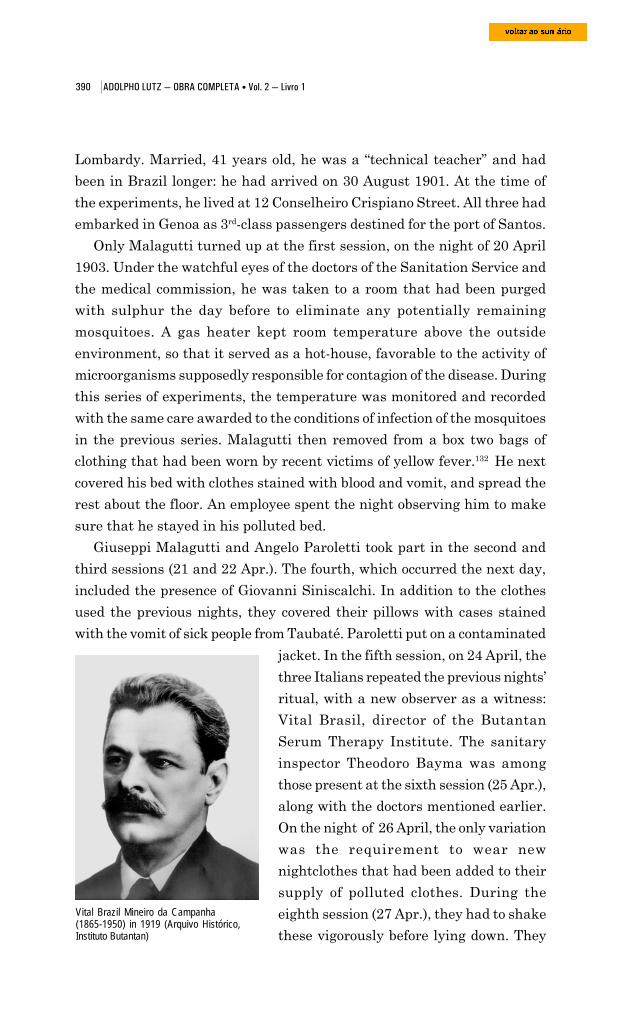
390 ADOLPHO LUTZ — OBRA COMPLETA Vol. 2 — Livro 1
Lombardy. Married, 41 years old, he was a “technical teacher” and had
been in Brazil longer: he had arrived on 30 August 1901. At the time of
the experiments, he lived at 12 Conselheiro Crispiano Street. All three had
embarked in Genoa as 3rd-class passengers destined for the port of Santos.
Only Malagutti turned up at the first session, on the night of 20 April
1903. Under the watchful eyes of the doctors of the Sanitation Service and
the medical commission, he was taken to a room that had been purged
with sulphur the day before to eliminate any potentially remaining
mosquitoes. A gas heater kept room temperature above the outside
environment, so that it served as a hot-house, favorable to the activity of
microorganisms supposedly responsible for contagion of the disease. During
this series of experiments, the temperature was monitored and recorded
with the same care awarded to the conditions of infection of the mosquitoes
in the previous series. Malagutti then removed from a box two bags of
clothing that had been worn by recent victims of yellow fever.132 He next
covered his bed with clothes stained with blood and vomit, and spread the
rest about the floor. An employee spent the night observing him to make
sure that he stayed in his polluted bed.
Giuseppi Malagutti and Angelo Paroletti took part in the second and
third sessions (21 and 22 Apr.). The fourth, which occurred the next day,
included the presence of Giovanni Siniscalchi. In addition to the clothes
used the previous nights, they covered their pillows with cases stained
with the vomit of sick people from Taubaté. Paroletti put on a contaminated
Vital Brazil Mineiro da Campanha(1865-1950) in 1919 (Arquivo Histórico,Instituto Butantan)
jacket. In the fifth session, on 24 April, the
three Italians repeated the previous nights’
ritual, with a new observer as a witness:
Vital Brasil, director of the Butantan
Serum Therapy Institute. The sanitary
inspector Theodoro Bayma was among
those present at the sixth session (25 Apr.),
along with the doctors mentioned earlier.
On the night of 26 April, the only variation
was the requirement to wear new
nightclothes that had been added to their
supply of polluted clothes. During the
eighth session (27 Apr.), they had to shake
these vigorously before lying down. They
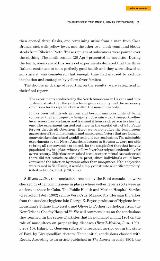
391 TRABALHOS SOBRE FEBRE AMARELA, MALÁRIA, PROTOZOOLOGIA
then opened three flasks, one containing urine from a man from Casa
Branca, sick with yellow fever, and the other two, black vomit and bloody
stools from Ribeirão Preto. These repugnant substances were poured over
the clothing. The ninth session (28 Apr.) presented no novelties. During
the tenth, observers of this series of experiments declared that the three
Italians continued to be in perfectly good health and they were allowed to
go, since it was considered that enough time had elapsed to exclude
incubation and contagion by yellow fever fomites.
The doctors in charge of reporting on the results were categorical in
their final report:
The experiments conducted by the North Americans in Havana and ours... demonstrate that the yellow fever germ can only find the necessaryconditions for its reproduction within the mosquito’s body.
It has been definitively proven and beyond any possibility of beingcontested that a mosquito – Stegomyia fasciata – can transport yellowfever across great distances and transmit it from a sick person to a healthyone. The experiment carried out here in the capital city of São Pauloforever dispels all objections. Here, we do not suffer the tumultuousaggression of the climatological and mesological factors that are found inmany stricken places [and would] confound our conclusions. The admirableexperiments by the North American doctors in Havana ... were not ableto bring all controversies to an end, for the simple fact that that heavilypopulated city is a place where yellow fever has reigned endemically forover a century. Objections were raised because experimental cases observedthere did not constitute absolute proof, since individuals could havecontracted the infection by means other than mosquitoes. If this objectionwere raised in São Paulo, it would simply constitute scientific improbity.(cited in Lemos, 1954, p.73, 75-7)
Still sub judice, the conclusions reached by the Reed commission were
checked by other commissions in places where yellow fever’s roots were as
ancient as those in Cuba. The Public Health and Marine Hospital Service
(created on 1 July 1902) sent to Vera Cruz, Mexico, Drs. Herman B. Parker
from the service’s hygiene lab; George E. Beyer, professor of Hygiene from
Louisiana’s Tulane University; and Oliver L. Pothier, pathologist from the
New Orleans Charity Hospital.133 We will comment later on the conclusions
they reached. In the series of articles that he published in mid 1901 on the
role of mosquitoes on propagating diseases (Brazil-Medico, Jun. 1901,
p.208-10), Hilário de Gouveia referred to research carried out in the state
of Pará by Liverpoodlian doctors. Their initial conclusions clashed with
Reed’s. According to an article published in The Lancet in early 1901, the
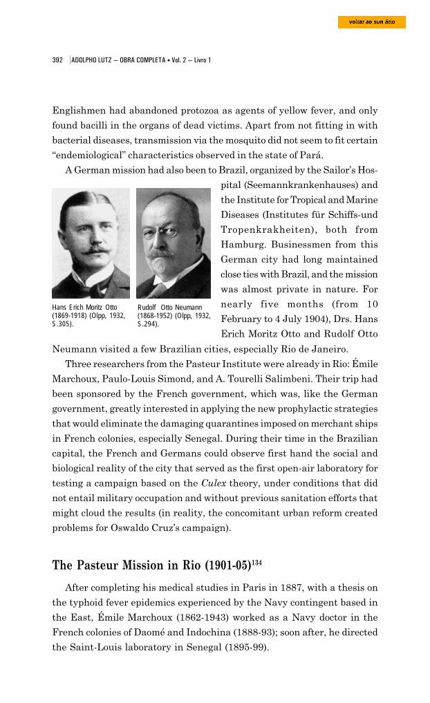
392 ADOLPHO LUTZ — OBRA COMPLETA Vol. 2 — Livro 1
Englishmen had abandoned protozoa as agents of yellow fever, and only
found bacilli in the organs of dead victims. Apart from not fitting in with
bacterial diseases, transmission via the mosquito did not seem to fit certain
“endemiological” characteristics observed in the state of Pará.
A German mission had also been to Brazil, organized by the Sailor’s Hos-
Rudolf Otto Neumann(1868-1952) (Olpp, 1932,S.294).
Hans Erich Moritz Otto(1869-1918) (Olpp, 1932,S.305).
pital (Seemannkrankenhauses) and
the Institute for Tropical and Marine
Diseases (Institutes für Schiffs-und
Tropenkrakheiten), both from
Hamburg. Businessmen from this
German city had long maintained
close ties with Brazil, and the mission
was almost private in nature. For
nearly five months (from 10
February to 4 July 1904), Drs. Hans
Erich Moritz Otto and Rudolf Otto
Neumann visited a few Brazilian cities, especially Rio de Janeiro.
Three researchers from the Pasteur Institute were already in Rio: Émile
Marchoux, Paulo-Louis Simond, and A. Tourelli Salimbeni. Their trip had
been sponsored by the French government, which was, like the German
government, greatly interested in applying the new prophylactic strategies
that would eliminate the damaging quarantines imposed on merchant ships
in French colonies, especially Senegal. During their time in the Brazilian
capital, the French and Germans could observe first hand the social and
biological reality of the city that served as the first open-air laboratory for
testing a campaign based on the Culex theory, under conditions that did
not entail military occupation and without previous sanitation efforts that
might cloud the results (in reality, the concomitant urban reform created
problems for Oswaldo Cruz’s campaign).
The Pasteur Mission in Rio (1901-05)134
After completing his medical studies in Paris in 1887, with a thesis on
the typhoid fever epidemics experienced by the Navy contingent based in
the East, Émile Marchoux (1862-1943) worked as a Navy doctor in the
French colonies of Daomé and Indochina (1888-93); soon after, he directed
the Saint-Louis laboratory in Senegal (1895-99).
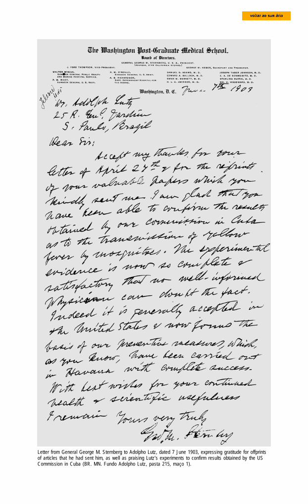
393 TRABALHOS SOBRE FEBRE AMARELA, MALÁRIA, PROTOZOOLOGIA
Letter from General George M. Sternberg to Adolpho Lutz, dated 7 June 1903, expressing gratitude for offprintsof articles that he had sent him, as well as praising Lutz’s experiments to confirm results obtained by the USCommission in Cuba (BR. MN. Fundo Adolpho Lutz, pasta 215, maço 1).
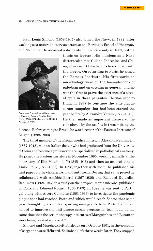
394 ADOLPHO LUTZ — OBRA COMPLETA Vol. 2 — Livro 1
Paul Louis Simond (1858-1947) also joined the Navy, in 1882, after
working as a natural history assistant at the Bordeaux School of Pharmacy
and Medicine. He obtained a doctorate in medicine only in 1887, with a
thesis on leprosy. His missions as a Navy
doctor took him to Guiana, Indochina, and Chi-
na, where in 1893 he had his first contact with
the plague. On returning to Paris, he joined
the Pasteur Institute. His first works in
microbiology were on the haematozoons of
paludism and on coccidia in general, and he
was the first to prove the existence of a sexu-
al cycle in these parasites. He was sent to
India in 1897 to continue the anti-plague
serum campaign that had been started the
year before by Alexandre Yersin (1863-1943).
He then made an important discovery: the
role played by the rat flea in transmitting the
Paul-Louis Simond in military dressin Valence, France. Studio ‘BlainFrère’, 1905-1910 (Musée de l’InstitutPasteur, D2089).
disease. Before coming to Brazil, he was director of the Pasteur Institute of
Saigon (1898-1900).
The third member of the French medical mission, Alexandre Salimbeni
(1867-1942), was an Italian doctor who had graduated from the University
of Siena and become a professor there, specialized in pathological anatomy.
He joined the Pasteur Institute in November 1895, working initially at the
laboratory of Elie Metchnikoff (1845-1916) and then as an assistant to
Émile Roux (1853-1933). In 1898, together with them, he published his
first paper on the cholera toxin and anti-toxin. During that same period he
collaborated with Amédée Borrel (1867-1936) and Edouard Dujardin-
Beaumetz (1868-1947) in a study on the peripneumonia microbe, published
by Roux and Edmond Nocard (1850-1903). In 1899 he was sent to Portu-
gal along with Alvert Calmette (1863-1933) to investigate the pandemic
plague that had reached Porto and which would reach Santos that same
year, brought by a ship transporting immigrants from Porto. Salimbeni
helped to improve the anti-plague serum preparation technique, at the
same time that the serum therapy institutes of Manguinhos and Butantan
were being created in Brazil.135
Simond and Marchoux left Bordeaux on 4 October 1901, in the company
of sergeant nurse Hébrard. Salimbeni left three weeks later. They stopped
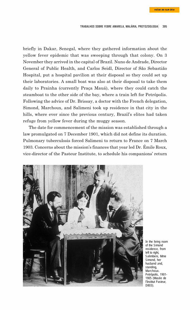
395 TRABALHOS SOBRE FEBRE AMARELA, MALÁRIA, PROTOZOOLOGIA
briefly in Dakar, Senegal, where they gathered information about the
yellow fever epidemic that was sweeping through that colony. On 3
November they arrived in the capital of Brazil. Nuno de Andrade, Director
General of Public Health, and Carlos Seidl, Director of São Sebastião
Hospital, put a hospital pavilion at their disposal so they could set up
their laboratories. A small boat was also at their disposal to take them
daily to Prainha (currently Praça Mauá), where they could catch the
steamboat to the other side of the bay, where a train left for Petrópolis.
Following the advice of Dr. Brissay, a doctor with the French delegation,
Simond, Marchoux, and Salimeni took up residence in that city in the
hills, where ever since the previous century, Brazil’s elites had taken
refuge from yellow fever during the muggy season.
The date for commencement of the mission was established through a
law promulgated on 7 December 1901, which did not define its duration.
Pulmonary tuberculosis forced Salimeni to return to France on 7 March
1903. Concerns about the mission’s finances that year led Dr. Émile Roux,
vice-director of the Pasteur Institute, to schedule his companions’ return
In the living roomof the Simondresidence, fromleft to right,Salimbeni, MmeSimond, herhusband and,standing,Marchoux.Petrópolis, 1901-1905 (Musée del’Institut Pasteur,D803).
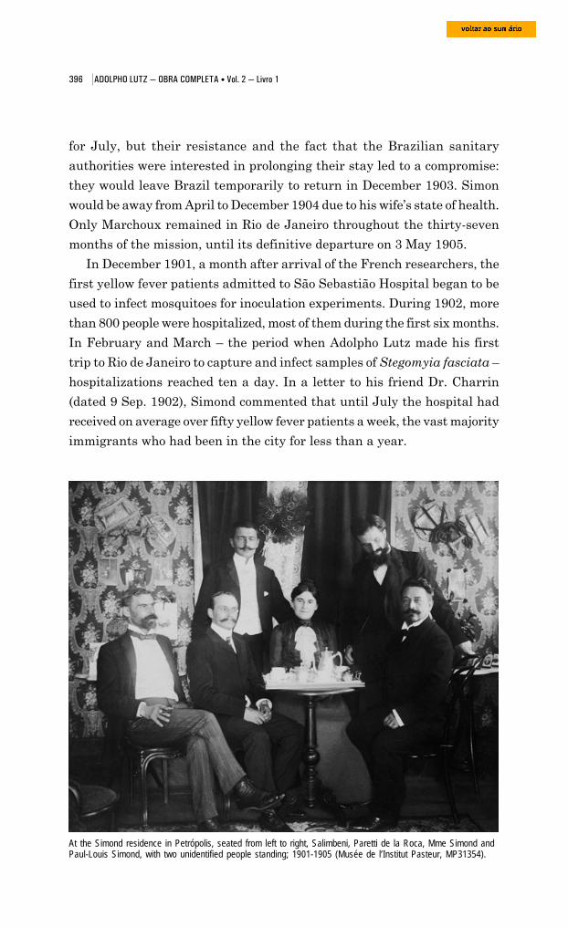
396 ADOLPHO LUTZ — OBRA COMPLETA Vol. 2 — Livro 1
for July, but their resistance and the fact that the Brazilian sanitary
authorities were interested in prolonging their stay led to a compromise:
they would leave Brazil temporarily to return in December 1903. Simon
would be away from April to December 1904 due to his wife’s state of health.
Only Marchoux remained in Rio de Janeiro throughout the thirty-seven
months of the mission, until its definitive departure on 3 May 1905.
In December 1901, a month after arrival of the French researchers, the
first yellow fever patients admitted to São Sebastião Hospital began to be
used to infect mosquitoes for inoculation experiments. During 1902, more
than 800 people were hospitalized, most of them during the first six months.
In February and March – the period when Adolpho Lutz made his first
trip to Rio de Janeiro to capture and infect samples of Stegomyia fasciata –
hospitalizations reached ten a day. In a letter to his friend Dr. Charrin
(dated 9 Sep. 1902), Simond commented that until July the hospital had
received on average over fifty yellow fever patients a week, the vast majority
immigrants who had been in the city for less than a year.
At the Simond residence in Petrópolis, seated from left to right, Salimbeni, Paretti de la Roca, Mme Simond andPaul-Louis Simond, with two unidentified people standing; 1901-1905 (Musée de l’Institut Pasteur, MP31354).
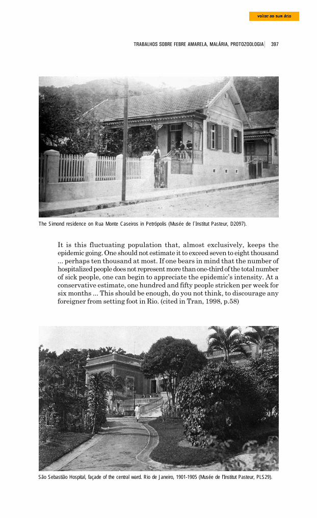
397 TRABALHOS SOBRE FEBRE AMARELA, MALÁRIA, PROTOZOOLOGIA
It is this fluctuating population that, almost exclusively, keeps theepidemic going. One should not estimate it to exceed seven to eight thousand... perhaps ten thousand at most. If one bears in mind that the number ofhospitalized people does not represent more than one-third of the total numberof sick people, one can begin to appreciate the epidemic’s intensity. At aconservative estimate, one hundred and fifty people stricken per week forsix months ... This should be enough, do you not think, to discourage anyforeigner from setting foot in Rio. (cited in Tran, 1998, p.58)
The Simond residence on Rua Monte Caseiros in Petrópolis (Musée de l´Institut Pasteur, D2097).
São Sebastião Hospital, façade of the central ward. Rio de Janeiro, 1901-1905 (Musée de l’Institut Pasteur, PLS29).
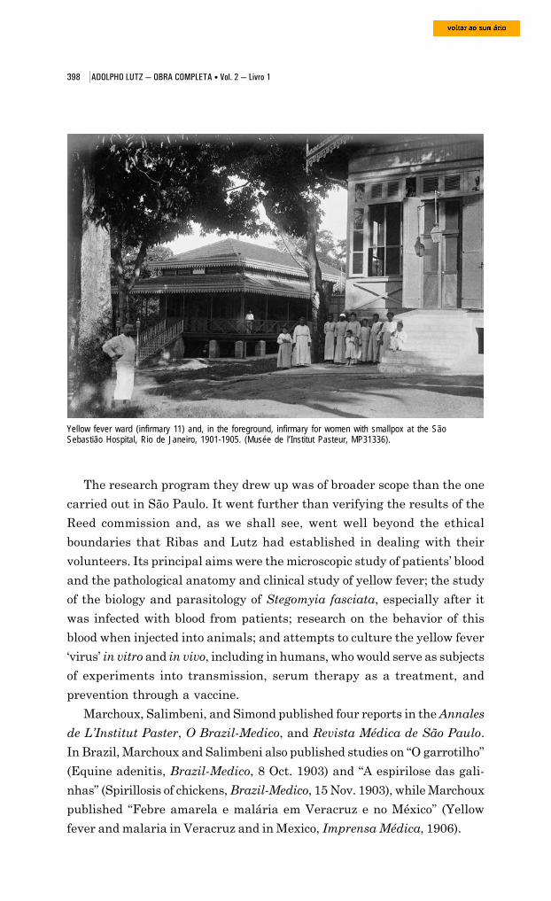
398 ADOLPHO LUTZ — OBRA COMPLETA Vol. 2 — Livro 1
The research program they drew up was of broader scope than the one
carried out in São Paulo. It went further than verifying the results of the
Reed commission and, as we shall see, went well beyond the ethical
boundaries that Ribas and Lutz had established in dealing with their
volunteers. Its principal aims were the microscopic study of patients’ blood
and the pathological anatomy and clinical study of yellow fever; the study
of the biology and parasitology of Stegomyia fasciata, especially after it
was infected with blood from patients; research on the behavior of this
blood when injected into animals; and attempts to culture the yellow fever
‘virus’ in vitro and in vivo, including in humans, who would serve as subjects
of experiments into transmission, serum therapy as a treatment, and
prevention through a vaccine.
Marchoux, Salimbeni, and Simond published four reports in the Annales
de L’Institut Paster, O Brazil-Medico, and Revista Médica de São Paulo.
In Brazil, Marchoux and Salimbeni also published studies on “O garrotilho”
(Equine adenitis, Brazil-Medico, 8 Oct. 1903) and “A espirilose das gali-
nhas” (Spirillosis of chickens, Brazil-Medico, 15 Nov. 1903), while Marchoux
published “Febre amarela e malária em Veracruz e no México” (Yellow
fever and malaria in Veracruz and in Mexico, Imprensa Médica, 1906).
Yellow fever ward (infirmary 11) and, in the foreground, infirmary for women with smallpox at the SãoSebastião Hospital, Rio de Janeiro, 1901-1905. (Musée de l’Institut Pasteur, MP31336).
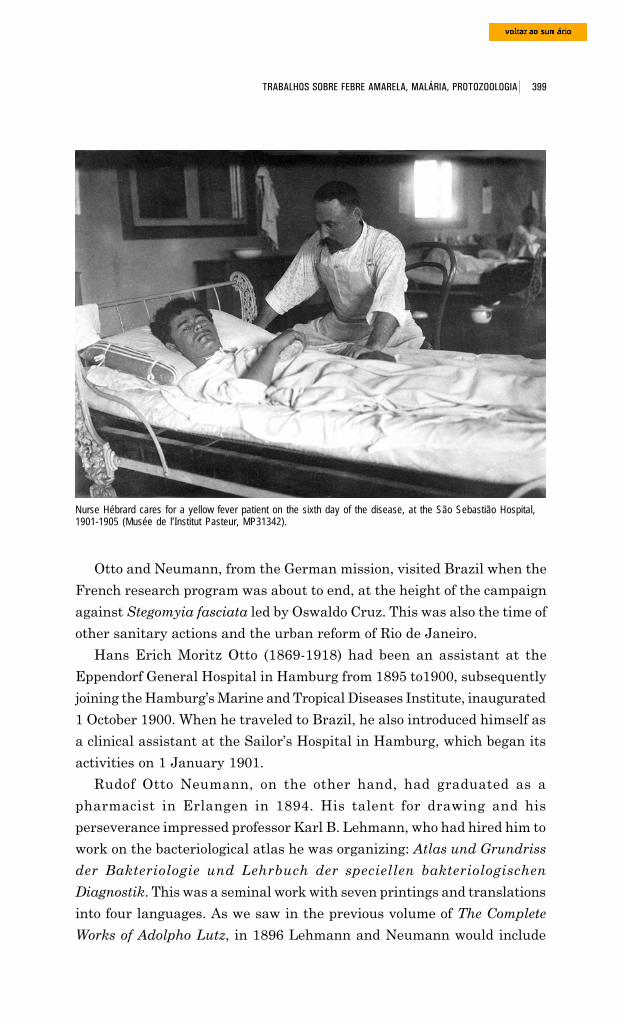
399 TRABALHOS SOBRE FEBRE AMARELA, MALÁRIA, PROTOZOOLOGIA
Otto and Neumann, from the German mission, visited Brazil when the
French research program was about to end, at the height of the campaign
against Stegomyia fasciata led by Oswaldo Cruz. This was also the time of
other sanitary actions and the urban reform of Rio de Janeiro.
Hans Erich Moritz Otto (1869-1918) had been an assistant at the
Eppendorf General Hospital in Hamburg from 1895 to1900, subsequently
Nurse Hébrard cares for a yellow fever patient on the sixth day of the disease, at the São Sebastião Hospital,1901-1905 (Musée de l’Institut Pasteur, MP31342).
joining the Hamburg’s Marine and Tropical Diseases Institute, inaugurated
1 October 1900. When he traveled to Brazil, he also introduced himself as
a clinical assistant at the Sailor’s Hospital in Hamburg, which began its
activities on 1 January 1901.
Rudof Otto Neumann, on the other hand, had graduated as a
pharmacist in Erlangen in 1894. His talent for drawing and his
perseverance impressed professor Karl B. Lehmann, who had hired him to
work on the bacteriological atlas he was organizing: Atlas und Grundriss
der Bakteriologie und Lehrbuch der speciellen bakteriologischen
Diagnostik. This was a seminal work with seven printings and translations
into four languages. As we saw in the previous volume of The Complete
Works of Adolpho Lutz, in 1896 Lehmann and Neumann would include
400 ADOLPHO LUTZ — OBRA COMPLETA Vol. 2 — Livro 1
the leprosy and tuberculosis bacilli in the genus Mycobacterium. Neumann
had begun his medical studies late in life, graduating from Würzburg only
in 1899. He had then obtained a doctorate in Kiel. His autobiography
mentions various important institutes at which he worked: Heidelberg,
Paris (Pasteur Institute), Liverpool, Cairo (Kasr El-Aini School of Medicine),
and Hamburg (Hygiene Institute and Institute for Marine and Tropical
Diseases). He investigated not only yellow fever but also schistosomiasis,
ancylostomiasis, and malaria. When he arrived in Brazil, he was serving
as a professor at Heidelberg University and an associate at the same hos-
pital and institute where Otto worked.136
German authorities were careful to inform Simond and Marchoux in
advance of the arrival of the two German doctors, hoping to prevent a
possible resurgence in that distant port city of the ceaseless strife that had
engaged scientists of both nations since the Franco-Prussian war and the
famous animosity between Louis Pasteur and Robert Koch. According to
Tran (1998), the missions collaborated cordially, to the point that Otto and
Neumann’s work was praised in a commentary published in the Bulletin
de L’Institut Pasteur.
They first visited Pernambuco, where they did not find any yellow fever.
After their stay in Rio de Janeiro (their foremost aim in the trip), they also
visited the capital of São Paulo, as well as two other cities – Campinas and
Santos – that they considered well known because of the epidemics of
‘American typhus’. Before returning to Germany, they also stopped in
Bahia, a state that had remained free of yellow fever in recent years. They
published preliminary versions of their results in a few German journals
and, in 1906, a full report, over 150 pages long, including 55 maps, sketches,
photographs, layouts, and seven tables printed in color.137
In Rio de Janeiro, where they stayed almost three months (from 6 Mar.
to end May), Otto and Neumann’s research was also conducted at São
Sebastião Hospital, a good part devoted to verifying the results already
obtained by the researchers from the Pasteur Institute. Both Germans were
very fearful of contracting the disease since they were “highly susceptible”
to yellow fever, and they even injured themselves during autopsies. They
of course slept under mosquito nets every night.
The sick they examined were isolated in screen-lined compartments called
“Marchoux Rooms,” which housed two beds each and were erected inside
the oldest pavilions. The German researchers in fact referred to them as

401 TRABALHOS SOBRE FEBRE AMARELA, MALÁRIA, PROTOZOOLOGIA
“barracks” since the underlying concept used by the hospital to isolate
diseases deemed contagious had been borrowed from military campaigns.
They consisted of moveable units that could be easily discarded if they
became contaminated. At one point, São Sebastião Hospital, inaugurated
in 1889 on the eve of the fall of the monarchy, had paper machê pavilions
imported from Germany.
All but three of the twenty-four sick patients examined by Otto and
Neumann were past the third day of the illness, increasing the difficulty of
investigating the pathogenic agent, which disappeared from human blood
by the fourth day. But the high mortality rate (60%) favored autopsy
studies.
They verified that yellow fever was an acute infectious disease displaying
the characteristics of quickly evolving hemorrhagic septicemia. Anatomical-
pathological examination did not reveal any specific characteristic, except
for the absence of splenomegalia, which helped distinguish yellow fever
from malaria. The prognosis was always serious, and could only be made
with any degree of certainty in the second period. Diagnosis was not difficult
in serious cases; differential diagnosis was important because of similarities
to the hemoglobinuria form of malaria, to bilious typhoid fever, and
phosphorous intoxication. The main yellow fever symptoms recorded by
The interior of a yellow fever infirmary, equipped with booths lined with metal screens, São Sebastião Hospital,1901-1905 (Musée de l’Institut Pasteur, MP31340).
402 ADOLPHO LUTZ — OBRA COMPLETA Vol. 2 — Livro 1
Otto and Neumann were redness of the face; body odor (“similar to that
of flesh from a recently slaughtered animal”); severe epigastric distress;
and the appearance of albumin in the urine from the first days of the
disease. In yellow fever (as with typhus), the pulse rate did not adjust to
the temperature of the patient. Black vomit, nose bleeds, and bloody stools
sometimes did not occur in cases that were less hemorrhagic or might
retreat given signs of a kidney dysfunction. The patient’s clinical status
would then become less precise, and an inexperienced doctor might take
it for uremia when hemorrhaging was imperceptible and only the jaundice
was intense.
Stegomyia fasciata was Otto and Neumann’s main object of study. They
left Brazil convinced that it was the transmitter of yellow fever, but not
that it was the only form of transmission possible. In Hamburg they would
continue their experiments. “The initial material came from Santos, Rio,
and Bahia, altogether from 30 to 40 mosquitoes, and a good quantity of
larvae.” They were transported by ship in a heated compartment, and
transferred to the “mosquito room” at the Institute for Tropical and Marine
Diseases, a spacious room kept at a constant temperature of 27°C; it
contained cages and a large aquarium with a mosquito-proof metal mesh.
There they were able to follow twelve generations that were raised in the
space of six months. “Regrettably, the Culex fatigans and Anopheles
argyrotarsus, which had been brought together, did not reproduce.”
They had used “catching glasses” developed by Nocht to collect the
mosquitoes in Rio de Janeiro, but on walls and harder-to-reach places they
had used a device suggested by Adolpho Lutz. “It is a simple glass tube,
somewhat curved, with a rubber tube and some cotton at one end. One
places the tube easily over the mosquito and then draws it through the
rubber tube until it reaches the cotton.”
A few problems had been encountered in growing Stegomyia in the
Brazilian capital, especially the tiny ants that proved to be avid mosquito
eaters.
The glasses where we raised our mosquitoes always had to be carefullyplaced in a dish of water. Even so, from time to time, the ants wouldsomehow find their way into the glasses at night, passing through thethin piece of gauze and eating our Stegomyia. We finally greased thesides of the dishes with a sticky substance that trapped the ants.
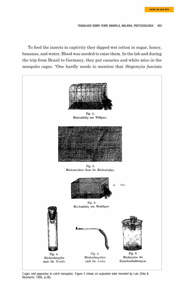
403 TRABALHOS SOBRE FEBRE AMARELA, MALÁRIA, PROTOZOOLOGIA
To feed the insects in captivity they dipped wet cotton in sugar, honey,
bananas, and water. Blood was needed to raise them. In the lab and during
the trip from Brazil to Germany, they put canaries and white mice in the
mosquito cages. “One hardly needs to mention that Stegomyia fasciata
Cages and apparatus to catch mosquitos. Figure 5 shows an aspiration tube invented by Lutz (Otto &Neumann, 1906, p.28).
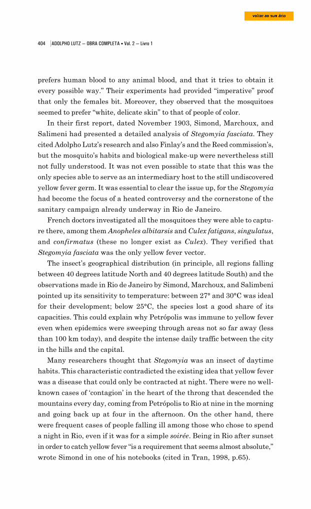
404 ADOLPHO LUTZ — OBRA COMPLETA Vol. 2 — Livro 1
prefers human blood to any animal blood, and that it tries to obtain it
every possible way.” Their experiments had provided “imperative” proof
that only the females bit. Moreover, they observed that the mosquitoes
seemed to prefer “white, delicate skin” to that of people of color.
In their first report, dated November 1903, Simond, Marchoux, and
Salimeni had presented a detailed analysis of Stegomyia fasciata. They
cited Adolpho Lutz’s research and also Finlay’s and the Reed commission’s,
but the mosquito’s habits and biological make-up were nevertheless still
not fully understood. It was not even possible to state that this was the
only species able to serve as an intermediary host to the still undiscovered
yellow fever germ. It was essential to clear the issue up, for the Stegomyia
had become the focus of a heated controversy and the cornerstone of the
sanitary campaign already underway in Rio de Janeiro.
French doctors investigated all the mosquitoes they were able to captu-
re there, among them Anopheles albitarsis and Culex fatigans, singulatus,
and confirmatus (these no longer exist as Culex). They verified that
Stegomyia fasciata was the only yellow fever vector.
The insect’s geographical distribution (in principle, all regions falling
between 40 degrees latitude North and 40 degrees latitude South) and the
observations made in Rio de Janeiro by Simond, Marchoux, and Salimbeni
pointed up its sensitivity to temperature: between 27° and 30°C was ideal
for their development; below 25°C, the species lost a good share of its
capacities. This could explain why Petrópolis was immune to yellow fever
even when epidemics were sweeping through areas not so far away (less
than 100 km today), and despite the intense daily traffic between the city
in the hills and the capital.
Many researchers thought that Stegomyia was an insect of daytime
habits. This characteristic contradicted the existing idea that yellow fever
was a disease that could only be contracted at night. There were no well-
known cases of ‘contagion’ in the heart of the throng that descended the
mountains every day, coming from Petrópolis to Rio at nine in the morning
and going back up at four in the afternoon. On the other hand, there
were frequent cases of people falling ill among those who chose to spend
a night in Rio, even if it was for a simple soirée. Being in Rio after sunset
in order to catch yellow fever “is a requirement that seems almost absolute,”
wrote Simond in one of his notebooks (cited in Tran, 1998, p.65).
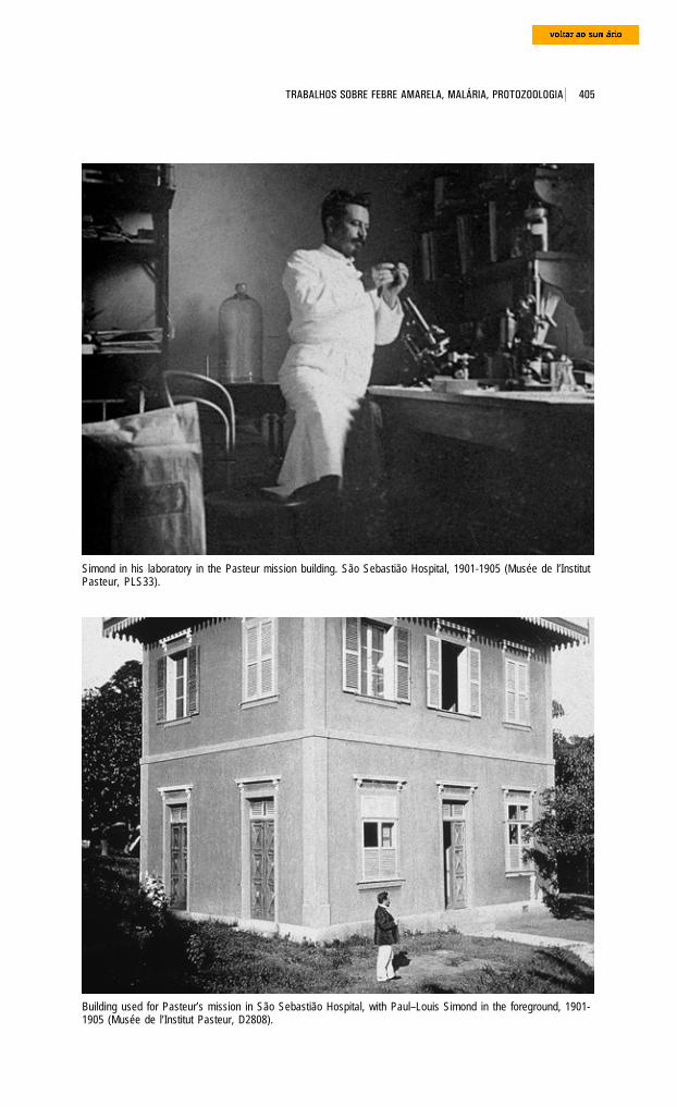
405 TRABALHOS SOBRE FEBRE AMARELA, MALÁRIA, PROTOZOOLOGIA
Simond in his laboratory in the Pasteur mission building. São Sebastião Hospital, 1901-1905 (Musée de l’InstitutPasteur, PLS33).
Building used for Pasteur’s mission in São Sebastião Hospital, with Paul–Louis Simond in the foreground, 1901-1905 (Musée de l’Institut Pasteur, D2808).
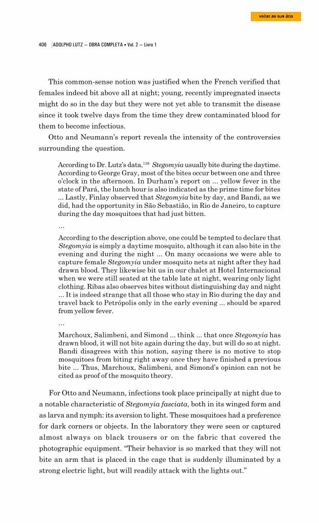
406 ADOLPHO LUTZ — OBRA COMPLETA Vol. 2 — Livro 1
...
According to the description above, one could be tempted to declare thatStegomyia is simply a daytime mosquito, although it can also bite in theevening and during the night ... On many occasions we were able tocapture female Stegomyia under mosquito nets at night after they haddrawn blood. They likewise bit us in our chalet at Hotel Internacionalwhen we were still seated at the table late at night, wearing only lightclothing. Ribas also observes bites without distinguishing day and night... It is indeed strange that all those who stay in Rio during the day andtravel back to Petrópolis only in the early evening ... should be sparedfrom yellow fever.
...
Marchoux, Salimbeni, and Simond ... think ... that once Stegomyia hasdrawn blood, it will not bite again during the day, but will do so at night.Bandi disagrees with this notion, saying there is no motive to stopmosquitoes from biting right away once they have finished a previousbite ... Thus, Marchoux, Salimbeni, and Simond’s opinion can not becited as proof of the mosquito theory.
For Otto and Neumann, infections took place principally at night due to
a notable characteristic of Stegomyia fasciata, both in its winged form and
as larva and nymph: its aversion to light. These mosquitoes had a preference
for dark corners or objects. In the laboratory they were seen or captured
almost always on black trousers or on the fabric that covered the
photographic equipment. “Their behavior is so marked that they will not
bite an arm that is placed in the cage that is suddenly illuminated by a
strong electric light, but will readily attack with the lights out.”
This common-sense notion was justified when the French verified that
females indeed bit above all at night; young, recently impregnated insects
might do so in the day but they were not yet able to transmit the disease
since it took twelve days from the time they drew contaminated blood for
them to become infectious.
Otto and Neumann’s report reveals the intensity of the controversies
surrounding the question.
According to Dr. Lutz’s data,138 Stegomyia usually bite during the daytime.According to George Gray, most of the bites occur between one and threeo’clock in the afternoon. In Durham’s report on ... yellow fever in thestate of Pará, the lunch hour is also indicated as the prime time for bites... Lastly, Finlay observed that Stegomyia bite by day, and Bandi, as wedid, had the opportunity in São Sebastião, in Rio de Janeiro, to captureduring the day mosquitoes that had just bitten.
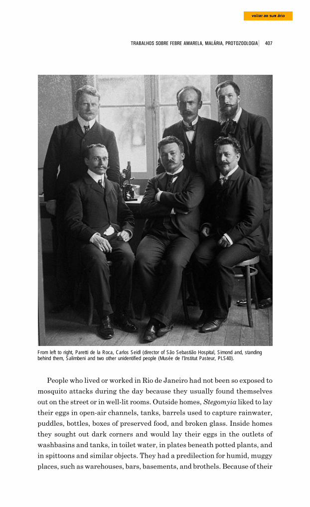
407 TRABALHOS SOBRE FEBRE AMARELA, MALÁRIA, PROTOZOOLOGIA
People who lived or worked in Rio de Janeiro had not been so exposed to
mosquito attacks during the day because they usually found themselves
out on the street or in well-lit rooms. Outside homes, Stegomyia liked to lay
their eggs in open-air channels, tanks, barrels used to capture rainwater,
puddles, bottles, boxes of preserved food, and broken glass. Inside homes
they sought out dark corners and would lay their eggs in the outlets of
washbasins and tanks, in toilet water, in plates beneath potted plants, and
in spittoons and similar objects. They had a predilection for humid, muggy
places, such as warehouses, bars, basements, and brothels. Because of their
From left to right, Paretti de la Roca, Carlos Seidl (director of São Sebastião Hospital, Simond and, standingbehind them, Salimbeni and two other unidentified people (Musée de l’Institut Pasteur, PLS40).
408 ADOLPHO LUTZ — OBRA COMPLETA Vol. 2 — Livro 1
attraction to sweet foods, they could be found in great concentrations in
beer halls, bakeries, sugar refineries, and restaurants.
They did not like drafts but could rise to heights of hundreds of meters.
They did not undertake major migrations but did travel great distances in
trains, streetcars, automobiles, and ships, especially in cargoes of sugar,
molasses, and fruit and in damp packages. Otto and Neumann put
Stegomyia inside suitcases of dry and damp clothing, and saw that the
sturdiest females would survive up to fifteen days.
According to Tran (1998), in a retrospective view, the French mission’s
most original feat was to demonstrate that these mosquitoes could transmit
the yellow fever ‘virus’ to descendents through heredity (vertical or
transovarian). This line of investigation was prompted by the puzzling
appearance of sporadic cases, sometimes spaced out, between epidemics.
The theories held back then explained that the disease was ‘imported’ to
stricken areas by travelers or objects that had been infected by microbes,
and these sporadic resurgences were attributed to the transformation that
the germ underwent in the ground, responding to climate-related or telluric
factors. The ‘Havana theory’ could not explain this adequately, since the
mosquito only had a limited lifetime.
Writing a paper for the general public (1903), Lutz stated that Stegomyia
fasciata could live “for two or three summer months as long as they were
fed.” He went on:
it is well known that not only among us but even in colder nations, theycan last through an entire winter. At lower temperatures, Stegomyia donot bite, which halts the appearance of new infections until the heatreturns. If the temperature does rise significantly in the winter, theycan leave their hiding places and bite again, explaining the cases wehave observed in this season.
Both sporadic cases and outbreaks that occurred independently of any
outside imports were given a new explanation in 1932 when Soper’s team
discovered jungle yellow fever and then demonstrated the existence of a
sylvatic cycle dependent on non-human receptacles of the yellow fever virus,
more especially forest-dwelling primates. At the beginning of the 20th
century, in the absence of any proven animal models, suspicion could not
fall on any vertebrate hosts other than humans. Walter Reed’s team had
tried to verify the hypothesis of an autonomous mosquito-based circuit of
transmission, but their results had been negative.
409 TRABALHOS SOBRE FEBRE AMARELA, MALÁRIA, PROTOZOOLOGIA
French researchers in Rio de Janeiro conducted experiments in which
eggs laid by females who had bitten sick patients were raised from the
larvae stage to the adult stage, but to prove that they and their descendents
were infectious meant resorting to human volunteers. Attempts in 1903
were unsuccessful, but in 1905 the researchers managed to reproduce
symptoms of the disease in a volunteer (Tran, 1998, p.66). Simond and
Marchoux (1905) hurried to publish these observations, even before the
release of the third report the next year.
The newspaper article “Mystère de la Fièvre Jaune. Les découvertes
des docteurs Marchoux et Simond expliquent l’ épidemia inexplicable de
Panama,” published in Le Matin (1 Aug. 1905, p.1-2), raised doubts about
the anti-Culex measures put into practice in Cuba and Brazil. This was
because in Panama these measures did not prevent the disease from
proliferating at twice its previous intensity from May to June of 1905,
despite the previous year’s mosquito-fighting measures. An outbreak of
the epidemic in New Orleans also led the newspaper to ask Marchoux
and Simond whether yellow fever might not have a way of propagating
in those places which was unlike the means used in Havana and Rio de
Janeiro.
Authorized by the Minister of the Colonies, the two French doctors
went public with information that would be included in their reports.
They vehemently rejected the suspicions that had been raised. Their
studies had ratified Finlay’s theory, but in the Brazilian capital they
verified that the incriminated mosquito had two new sources of infection.
One consisted of the mild cases of yellow fever that would pass unnoticed
by doctors yet would still provide mosquitoes with what they needed to
keep the epidemic going. The main fuel, however, was provided by babies
still being breast-fed: these recent arrivals to infected regions were as
susceptible as any immigrant, but they were remarkably resistant to the
yellow fever ‘poison’. The other source of invisible infection was the
hereditary transmission of the yellow fever ‘virus’ to the descendents of
the female Stegomyia fasciata that had drawn blood from sick patients.
“This fact will certainly cause great sensation in the scientific field,” the
paper noted. “Its verification was based on direct experience ... It was
impossible for the French mission to find another volunteer in Rio to con-
tinue the experiment, but one hopes that it will be taken up again in
Panama by the American doctors.”
410 ADOLPHO LUTZ — OBRA COMPLETA Vol. 2 — Livro 1
A newspaper article published at about the same time in Le Journal (2
Jul. 1905) by Fernand Hauser, also drew attention to that “frightening
[thing], the microbial infection of yellow fever, which extends to the eggs
of the infected mosquito.”139
The etiological enigma
In spite of the Reed commission’s efforts to demonstrate that the yellow
fever agent was a “filterable virus,” recent papers on paludism had
disseminated the supposition that it was perhaps a parasite whose
characteristics were analogous to the Laveran plasmodium.
In an article published in the Revista de Medicina Tropical (Havana,
Apr. 1903), Finlay himself defended the hypothesis: this parasite would be
an ultra-microscopic protozoan whose stages of development were more or
less similar to those of the malaria parasite. He supposed that its sexual
reproduction took place inside humans (its definitive host), while schizogony
occurred in the mosquito. This inference was based on the observation
that the infection of Stegomyia lasted indefinitely, while in humans it was
short. On the other hand, in the case of malaria, infection in man was of
indefinite duration while in Anopheles it was apparently limited.
It was during this period that the first report by Parker, Beyer, and
Pothier was released (1903; also Parker, 1903). The doctors from the U.S.
Navy commission did not find any specific organism in the blood of yellow
fever patients or in their tissues after death. Observation of Stegomyia
fasciata, however, proved promising. After feeding on a patient, the insect’s
ovaries at first underwent hypertrophy, as would normally occur after a
feeding of normal blood; they would then undergo complete atrophy. The
salivary glands also displayed excessive enlargement. Three or four days
after the absorption of contaminated blood a “small fusiform protozoan”
(whose development was analogous to that of coccidia) would be found in
the salivary glands and stomach. In future experiments, the authors
expected to better determine the life cycle and etiological role of the organism
they named Myxococcidium stegomyiae.
Parker, Beyer, and Pothier considered it a definitive scientific fact that
the disease’s medium of transference was Stegomyia fasciata. But they
could not yet tell if it was “its sole medium of transference.” They believed
that “to prove this negative affirmation, appropriate conditions must be
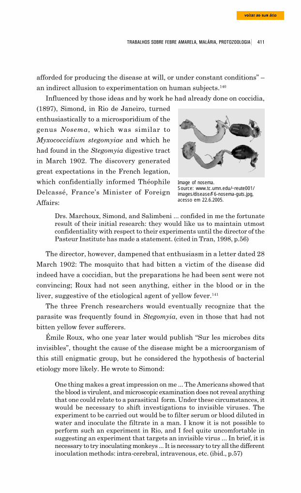
411 TRABALHOS SOBRE FEBRE AMARELA, MALÁRIA, PROTOZOOLOGIA
afforded for producing the disease at will, or under constant conditions” –
an indirect allusion to experimentation on human subjects.140
Influenced by those ideas and by work he had already done on coccidia,
Image of nosema.Source: www.tc.umn.edu/~reute001/images/disease/F6-nosema-guts.jpg,acesso em 22.6.2005.
(1897), Simond, in Rio de Janeiro, turned
enthusiastically to a microsporidium of the
genus Nosema, which was similar to
Myxococcidium stegomyiae and which he
had found in the Stegomyia digestive tract
in March 1902. The discovery generated
great expectations in the French legation,
which confidentially informed Théophile
Delcassé, France’s Minister of Foreign
Affairs:
Drs. Marchoux, Simond, and Salimbeni ... confided in me the fortunateresult of their initial research: they would like us to maintain utmostconfidentiality with respect to their experiments until the director of thePasteur Institute has made a statement. (cited in Tran, 1998, p.56)
The director, however, dampened that enthusiasm in a letter dated 28
March 1902: The mosquito that had bitten a victim of the disease did
indeed have a coccidian, but the preparations he had been sent were not
convincing; Roux had not seen anything, either in the blood or in the
liver, suggestive of the etiological agent of yellow fever.141
The three French researchers would eventually recognize that the
parasite was frequently found in Stegomyia, even in those that had not
bitten yellow fever sufferers.
Émile Roux, who one year later would publish “Sur les microbes dits
invisibles”, thought the cause of the disease might be a microorganism of
this still enigmatic group, but he considered the hypothesis of bacterial
etiology more likely. He wrote to Simond:
One thing makes a great impression on me ... The Americans showed thatthe blood is virulent, and microscopic examination does not reveal anythingthat one could relate to a parasitical form. Under these circumstances, itwould be necessary to shift investigations to invisible viruses. Theexperiment to be carried out would be to filter serum or blood diluted inwater and inoculate the filtrate in a man. I know it is not possible toperform such an experiment in Rio, and I feel quite uncomfortable insuggesting an experiment that targets an invisible virus ... In brief, it isnecessary to try inoculating monkeys ... It is necessary to try all the differentinoculation methods: intra-cerebral, intravenous, etc. (ibid., p.57)
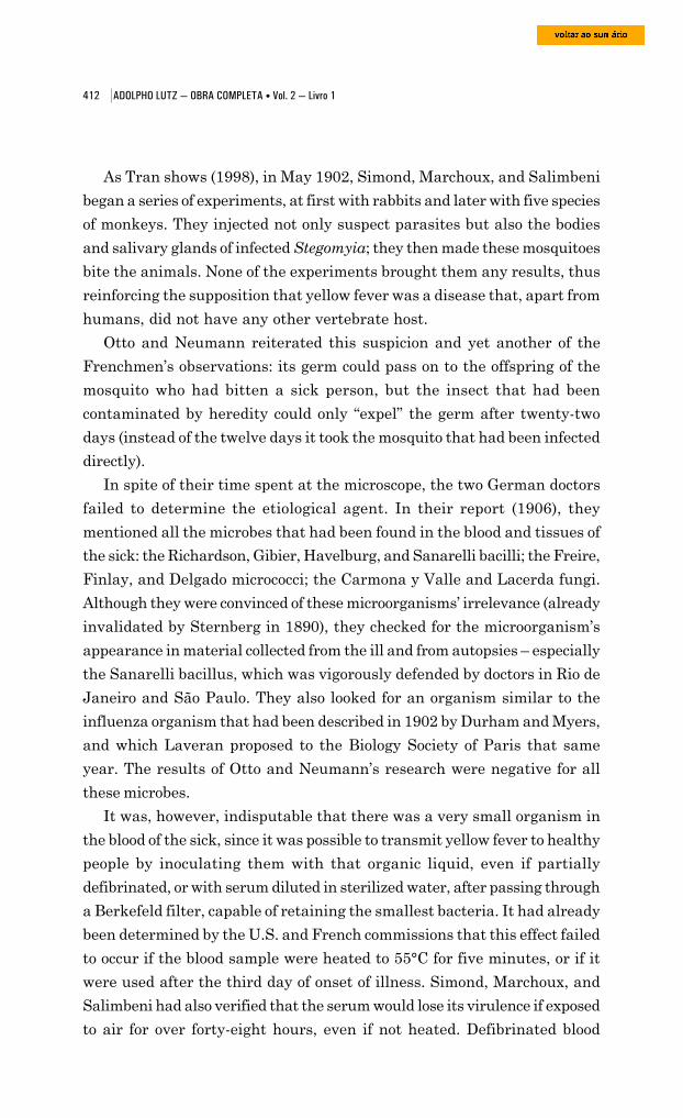
412 ADOLPHO LUTZ — OBRA COMPLETA Vol. 2 — Livro 1
As Tran shows (1998), in May 1902, Simond, Marchoux, and Salimbeni
began a series of experiments, at first with rabbits and later with five species
of monkeys. They injected not only suspect parasites but also the bodies
and salivary glands of infected Stegomyia; they then made these mosquitoes
bite the animals. None of the experiments brought them any results, thus
reinforcing the supposition that yellow fever was a disease that, apart from
humans, did not have any other vertebrate host.
Otto and Neumann reiterated this suspicion and yet another of the
Frenchmen’s observations: its germ could pass on to the offspring of the
mosquito who had bitten a sick person, but the insect that had been
contaminated by heredity could only “expel” the germ after twenty-two
days (instead of the twelve days it took the mosquito that had been infected
directly).
In spite of their time spent at the microscope, the two German doctors
failed to determine the etiological agent. In their report (1906), they
mentioned all the microbes that had been found in the blood and tissues of
the sick: the Richardson, Gibier, Havelburg, and Sanarelli bacilli; the Freire,
Finlay, and Delgado micrococci; the Carmona y Valle and Lacerda fungi.
Although they were convinced of these microorganisms’ irrelevance (already
invalidated by Sternberg in 1890), they checked for the microorganism’s
appearance in material collected from the ill and from autopsies – especially
the Sanarelli bacillus, which was vigorously defended by doctors in Rio de
Janeiro and São Paulo. They also looked for an organism similar to the
influenza organism that had been described in 1902 by Durham and Myers,
and which Laveran proposed to the Biology Society of Paris that same
year. The results of Otto and Neumann’s research were negative for all
these microbes.
It was, however, indisputable that there was a very small organism in
the blood of the sick, since it was possible to transmit yellow fever to healthy
people by inoculating them with that organic liquid, even if partially
defibrinated, or with serum diluted in sterilized water, after passing through
a Berkefeld filter, capable of retaining the smallest bacteria. It had already
been determined by the U.S. and French commissions that this effect failed
to occur if the blood sample were heated to 55°C for five minutes, or if it
were used after the third day of onset of illness. Simond, Marchoux, and
Salimbeni had also verified that the serum would lose its virulence if exposed
to air for over forty-eight hours, even if not heated. Defibrinated blood
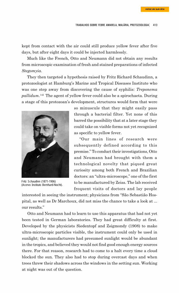
413 TRABALHOS SOBRE FEBRE AMARELA, MALÁRIA, PROTOZOOLOGIA
kept from contact with the air could still produce yellow fever after five
days, but after eight days it could be injected harmlessly.
Much like the French, Otto and Neumann did not obtain any results
from microscopic examination of fresh and stained preparations of infected
Stegomyia.
They then targeted a hypothesis raised by Fritz Richard Schaudinn, a
protozoologist at Hamburg’s Marine and Tropical Diseases Institute who
was one step away from discovering the cause of syphilis: Treponema
pallidum.142 The agent of yellow fever could also be a spirochaeta. During
a stage of this protozoan’s development, structures would form that were
Fritz Schaudinn (1871-1906)(Acervo Instituto Bernhard-Nocht).
so minuscule that they might easily pass
through a bacterial filter. Yet none of this
barred the possibility that at a later stage they
could take on visible forms not yet recognized
as specific to yellow fever.
“Our main lines of research were
subsequently defined according to this
premise.” To conduct their investigations, Otto
and Neumann had brought with them a
technological novelty that piqued great
curiosity among both French and Brazilian
doctors: an “ultra-microscope,” one of the first
to be manufactured by Zeiss. The lab received
frequent visits of doctors and lay people
interested in seeing the instrument; physicians from “São Sebastião Hos-
pital, as well as Dr Marchoux, did not miss the chance to take a look at ...
our results.”
Otto and Neumann had to learn to use this apparatus that had not yet
been tested in German laboratories. They had great difficulty at first.
Developed by the physicists Siedentopf and Zsigmondy (1908) to make
ultra-microscopic particles visible, the instrument could only be used in
sunlight; the manufacturers had presumed sunlight would be abundant
in the tropics, and believed they would not find good enough energy sources
there. For that reason, research had to come to a halt every time a cloud
blocked the sun. They also had to stop during overcast days and when
trees threw their shadows across the windows in the setting sun. Working
at night was out of the question.
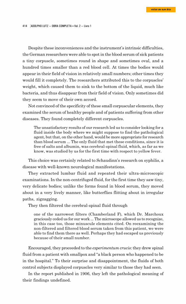
414 ADOLPHO LUTZ — OBRA COMPLETA Vol. 2 — Livro 1
Despite these inconveniences and the instrument’s intrinsic difficulties,
the German researchers were able to spot in the blood serum of sick patients
a tiny corpuscle, sometimes round in shape and sometimes oval, and a
hundred times smaller than a red blood cell. At times the bodies would
appear in their field of vision in relatively small numbers; other times they
would fill it completely. The researchers attributed this to the corpuscles’
weight, which caused them to sink to the bottom of the liquid, much like
bacteria, and thus disappear from their field of vision. Only sometimes did
they seem to move of their own accord.
Not convinced of the specificity of these small corpuscular elements, they
examined the serum of healthy people and of patients suffering from other
diseases. They found completely different corpuscles.
The unsatisfactory results of our research led us to consider looking for afluid inside the body where we might suppose to find the pathologicalagent, but that, on the other hand, would be more appropriate for researchthan blood serum ... The only fluid that met these conditions, since it isfree of salts and albumin, was cerebral-spinal fluid, which, as far as weknow, was studied by us for the first time with respect to yellow fever.
This choice was certainly related to Schaudinn’s research on syphilis, a
disease with well-known neurological manifestations.
They extracted lumbar fluid and repeated their ultra-microscopic
examinations. In the non-centrifuged fluid, for the first time they saw tiny,
very delicate bodies; unlike the forms found in blood serum, they moved
about in a very lively manner, like butterflies flitting about in irregular
paths, zigzagging.
They then filtered the cerebral-spinal fluid through
one of the narrowest filters (Chamberland F), which Dr. Marchouxgraciously ceded us for our work ... The microscope allowed us to recognize,in this case too, those minuscule elements cited. On reexamining thenon-filtered and filtered blood serum taken from this patient, we wereable to find them there as well. Perhaps they had escaped us previouslybecause of their small number.
Encouraged, they proceeded to the experimentum crucis: they drew spinal
fluid from a patient with smallpox and “a black person who happened to be
in the hospital.” To their surprise and disappointment, the fluids of both
control subjects displayed corpuscles very similar to those they had seen.
In the report published in 1906, they left the pathological meaning of
their findings undefined.
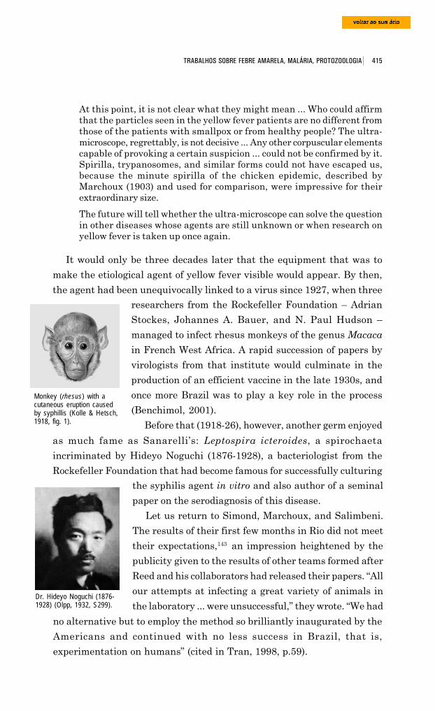
415 TRABALHOS SOBRE FEBRE AMARELA, MALÁRIA, PROTOZOOLOGIA
At this point, it is not clear what they might mean ... Who could affirmthat the particles seen in the yellow fever patients are no different fromthose of the patients with smallpox or from healthy people? The ultra-microscope, regrettably, is not decisive ... Any other corpuscular elementscapable of provoking a certain suspicion ... could not be confirmed by it.Spirilla, trypanosomes, and similar forms could not have escaped us,because the minute spirilla of the chicken epidemic, described byMarchoux (1903) and used for comparison, were impressive for theirextraordinary size.
The future will tell whether the ultra-microscope can solve the questionin other diseases whose agents are still unknown or when research onyellow fever is taken up once again.
It would only be three decades later that the equipment that was to
make the etiological agent of yellow fever visible would appear. By then,
the agent had been unequivocally linked to a virus since 1927, when three
Monkey (rhesus) with acutaneous eruption causedby syphillis (Kolle & Hetsch,1918, fig. 1).
researchers from the Rockefeller Foundation – Adrian
Stockes, Johannes A. Bauer, and N. Paul Hudson –
managed to infect rhesus monkeys of the genus Macaca
in French West Africa. A rapid succession of papers by
virologists from that institute would culminate in the
production of an efficient vaccine in the late 1930s, and
once more Brazil was to play a key role in the process
(Benchimol, 2001).
Before that (1918-26), however, another germ enjoyed
as much fame as Sanarelli’s: Leptospira icteroides, a spirochaeta
incriminated by Hideyo Noguchi (1876-1928), a bacteriologist from the
Rockefeller Foundation that had become famous for successfully culturing
Dr. Hideyo Noguchi (1876-1928) (Olpp, 1932, S299).
the syphilis agent in vitro and also author of a seminal
paper on the serodiagnosis of this disease.
Let us return to Simond, Marchoux, and Salimbeni.
The results of their first few months in Rio did not meet
their expectations,143 an impression heightened by the
publicity given to the results of other teams formed after
Reed and his collaborators had released their papers. “All
our attempts at infecting a great variety of animals in
the laboratory ... were unsuccessful,” they wrote. “We had
no alternative but to employ the method so brilliantly inaugurated by the
Americans and continued with no less success in Brazil, that is,
experimentation on humans” (cited in Tran, 1998, p.59).
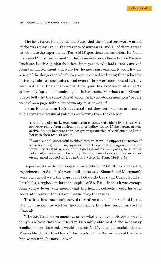
416 ADOLPHO LUTZ — OBRA COMPLETA Vol. 2 — Livro 1
The first report they published states that the volunteers were warned
of the risks they ran, in the presence of witnesses, and all of them agreed
to submit to the experiments. Tran (1998) questions this assertion. He found
no trace of “informed consent” in the documentation collected at the Pasteur
Institute. It is his opinion that these immigrants, who had recently arrived
from the old continent and were for the most part extremely poor, had no
sense of the dangers to which they were exposed by letting themselves be
bitten by infected mosquitoes, and even if they were conscious of it, they
accepted it for financial reasons. Reed paid his experimental subjects
generously (up to one hundred gold dollars each). Marchoux and Simond
purportedly did the same. One of Simond’s lab notebooks mentions “people
to pay” on a page with a list of twenty-four names.144
It was Roux who in 1902 suggested that they perform serum therapy
trials using the serum of patients recovering from the disease.
You should also make experiments on patients with blood from those whoare recovering from serious bouts of yellow fever. If the serum provesactive, do not hesitate to inject great quantities of virulent blood in ahorse to then test its serum.
If you are at all successful in this direction, it would support the notion ofa bacterial agent. In my opinion, and I repeat it yet again, the solidimmunity created by a bout of the disease seems, in my eyes, to favor thenotion of a bacteria ... It is a pity that you cannot carry out experienceson m. [men] of good will, as in Cuba. (cited in Tran, 1998, p.58)
Experiments with men began around March 1903. Ribas and Lutz’s
experiments in São Paulo were still underway. Simond and Marchoux’s
were conducted with the approval of Oswaldo Cruz and Carlos Seidl in
Petrópolis, a region similar to the capital of São Paulo in that it was exempt
from yellow fever; this meant that the human subjects would have no
accidental contact that risked invalidating the results.
The first three cases only served to confirm conclusions reached by the
U.S. commission, as well as the conclusions Lutz had communicated to
Simond:
“The São Paulo experiments ... prove what you have probably observed
for yourselves: that the infection is readily obtained if the necessary
conditions are observed. I would be grateful if you would explain this to
Messrs Metchnikoff and Roux,” the director of the Bacteriological Institute
had written in January 1903.145
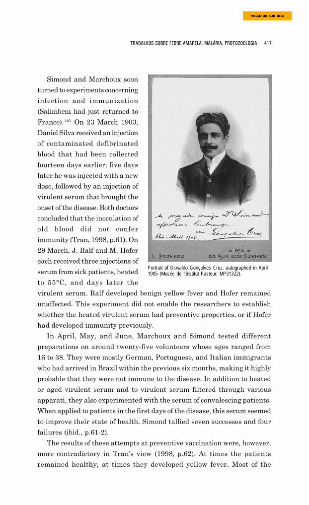
417 TRABALHOS SOBRE FEBRE AMARELA, MALÁRIA, PROTOZOOLOGIA
Simond and Marchoux soon
turned to experiments concerning
infection and immunization
(Salimbeni had just returned to
France).146 On 23 March 1903,
Daniel Silva received an injection
of contaminated defibrinated
blood that had been collected
fourteen days earlier; five days
later he was injected with a new
dose, followed by an injection of
virulent serum that brought the
onset of the disease. Both doctors
concluded that the inoculation of
old blood did not confer
immunity (Tran, 1998, p.61). On
29 March, J. Ralf and M. Hofer
each received three injections of
serum from sick patients, heated
to 55°C, and days later the
virulent serum. Ralf developed benign yellow fever and Hofer remained
unaffected. This experiment did not enable the researchers to establish
whether the heated virulent serum had preventive properties, or if Hofer
had developed immunity previously.
In April, May, and June, Marchoux and Simond tested different
preparations on around twenty-five volunteers whose ages ranged from
16 to 38. They were mostly German, Portuguese, and Italian immigrants
who had arrived in Brazil within the previous six months, making it highly
probable that they were not immune to the disease. In addition to heated
or aged virulent serum and to virulent serum filtered through various
apparati, they also experimented with the serum of convalescing patients.
When applied to patients in the first days of the disease, this serum seemed
to improve their state of health. Simond tallied seven successes and four
failures (ibid., p.61-2).
The results of these attempts at preventive vaccination were, however,
more contradictory in Tran’s view (1998, p.62). At times the patients
remained healthy, at times they developed yellow fever. Most of the
Portrait of Oswaldo Gonçalves Cruz, autographed in April1905 (Musée de l’Institut Pasteur, MP31322).
418 ADOLPHO LUTZ — OBRA COMPLETA Vol. 2 — Livro 1
experiments produced simple bouts of fever that were difficult to diagnose
unequivocally. If the symptoms were tenuous, should the researchers
conclude that the vaccination had failed or that there was relative immunity?
Interpretation of the results was made even more uncertain by the absence
of control groups. Simond and Marchoux alleged that they could not allow
themselves this kind of luxury with human subjects.
On the morning of 18 June 1903, Raymondo Geronimo, a 38-year-old
Italian, was making his way down from Petrópolis to Rio de Janeiro on the
7:30 a.m. train. He felt very ill, and upon arriving at the foot of the
mountains, he left the train with his suitcase in hand and began to walk
the line in the opposite direction, as if he intended to return to Petrópolis.
He walked for a few hundred meters, stopped, and passed out. Railroad
employees carried him to the station, where he passed away after a few
hours (ibid., p.63).
Geronimo was one of Simond and Marchoux’s ‘human guinea pigs’. Six
days earlier, he had been given an injection of virulent serum heated for
15 minutes to 48°C. On the 18th, at six in the morning, S. Bordach also
died; he was a 23-year-old man from Hamburg who had arrived in Brazil
a month earlier. On 10 June, he had been bitten by two infected Stegomyia
and had taken seriously ill four days later. The third fatal victim was
Heinrich Falk, a young Bavarian, also 23 years old; he had arrived in Rio
on 1 June. On 10 June, in Petrópolis, he received one cc of virulent serum
that had been passed through a Chamberland filter without being diluted.
He fell ill six days later and died on 20 or 21 June. There seems to have
been a fourth victim, on the same date: Lippe, a 34-year-old man from
Westphalia; his name appears next to that of the other three in one of
Simond’s notebooks (ibid., p.62).
Neither Simond nor Marchoux made any mention of these accidents in
the mission’s official reports published in the Annales de l’Institut Pasteur.
The experiments were not completely discontinued because in 1905, as we
saw, they still used a human volunteer to prove the hereditary transmission
of the yellow fever virus by mosquitoes.
The complicity of the Brazilian sanitary authorities is proven beyond
any doubt by a letter that Oswaldo Cruz sent to Emílio Ribas on 20 July
1903. Amid assessments of the French results, he commented:
419 TRABALHOS SOBRE FEBRE AMARELA, MALÁRIA, PROTOZOOLOGIA
Regarding human experimentation, they verified that:
Stegomyia transmitted the typical form of the disease. Unfortunately, therewere three deaths caused by classic experimental yellow fever, with anuria,black vomit, jaundice, albuminuria, etc.; the cases were verified by autopsy,and all the characteristic lesions were found ... One of the sick menvictimized by yellow fever had been bitten by only two mosquitoes, one ofwhich practically did not draw blood. I ask of you complete confidentialityregarding these cases that ended in death and which were carried outunder my sole responsibility.
You will understand, my dear friend, how our adversarial press wouldexploit this fact if they were informed of it. These experiments werecategorical, just as the others in which the accompanying symptomswere as categorical as they could possibly be.147
In the rough seas of sanitary practices
Nuno de Andrade, Oswaldo Cruz’s predecessor at the General
Directorship of Public Health, had voiced many doubts on this topic in
the Jornal do Commercio (24 Aug. 1902) and in Revista Médica de São
Paulo (1902). The discovery made by the North Americans added a new
element to yellow fever prophylaxis, but it had failed to gather enough
convincing elements to oust environmental disinfection and sanitation
practices. The Havana doctrine was based on the doctrine of paludism.
Neither of these accepted a microbe free in the external environment,
and both restricted all the threads of the problem to humans and the
mosquito. “I admit that the hypothesis that the yellow fever germ does
not exist in the external environment bothers me seriously,” stated
Andrade, “because scientific documents and our own observations have
accumulated a world of facts that would be completely inexplicable if the
deductions of American prophylaxis were accepted without restrictions.”
Andrade pointed to decisive experiments that had not been conducted
and which would exclude alternative pathways of the germ. The North
Americans had demonstrated that contaminated objects did not transmit
the disease directly, but they had not proven that these objects did not
carry the germ. No experiment had cancelled out the possibility that
healthy mosquitoes could infect themselves on soiled objects and then
inoculate the disease in humans. No one had investigated Stegomyia’s
excretions for possible infective properties. In the absence of such proof,
the prevention formula continued to employ the term “contaminating
object” as part of the equation.
420 ADOLPHO LUTZ — OBRA COMPLETA Vol. 2 — Livro 1
The fact that the microbe remained undetermined also left the theory of
its transmission exposed to other disturbing questions. “The most impressive
thing about the shadowy etiology of yellow fever is that the blood injected
by the syringe transmits the disease immediately ... but if drawn by the
mosquito, it only becomes infectious after about 12 days!” The North
Americans provided an explanation that worked by analogy and did not
rest on experimental proof. They supposed that the unknown microbe
underwent transformations in the body of Stegomyia similar to those
undergone by the malaria plasmodium inside Anopheles. The parallelism
seemed arbitrary. The circumstantial fact that the mosquitoes were hosts
of both germs did not imply identical life cycles. The microbe’s invisibility,
allied to a lack of knowledge over what went on during the interval between
the mosquito’s contamination and the appearance of its capacity for
infection, tainted the Havana doctrine with fuzzy areas. These areas did
not concern Stegomyia as an agent of transmission – Nuno de Andrade
considered this a proven fact – but rather what preventive deductions could
be derived from this fact.
Nor did he take the war on the mosquito in Cuba as evidence of an
exclusive form of propagation, as postulated by the Americans. He
considered this battle a kind of “touch-up work” that supplemented the
material improvements made by the military authorities earlier. With dis-
cipline, severity, and implacable rigor against those who hid the sick, they
had rectified the Havana coastline, dried up marshlands, built sewage
systems, distributed great quantities of drinking water, installed electric
lighting, cleaned prisons, built hospitals, intervened in residences, cleaned
up the markets, and paved all of the city streets. It was not out of the
question to suppose that those interventions had contributed in no small
amount to the success of an initiative that was usually ascribed only to the
war on mosquitoes.
The Havana doctrine, therefore, insofar as it excludes the yellow fevergerm from the environment, is a postulate or a question mark; it doesnot authorize the exclusive prophylaxis that the Americans recommend,nor does it at this point have the power to impose disregard for the currentprocesses of defensive hygiene ... The prophylactic formula must be acomplex one, that is, it must embrace all the processes of prophylaxiscurrently in use, in addition to all those derived from the transmissibilityof yellow fever by Stegomyia. Addition and not subtraction.
421 TRABALHOS SOBRE FEBRE AMARELA, MALÁRIA, PROTOZOOLOGIA
In São Paulo, too, this theory found passionate opponents. Arthur Vieira
de Mendonça continued to defend the icteroid bacillus and his arguments
had a marked impact on public opinion since they were expressed precisely
by a bacteriologist who had, until recently, been close to Adolfo Lutz. He
had worked at the Bacteriological Institute since its foundation, and had
served as under-director after Lutz became head of the Institute upon Le
Dantec’s departure. Moreover, Mendonça was one of the founders of the
Revista Medica de São Paulo, along with Victor Godinho, and had presided
the Society of Medicine and Surgery of São Paulo during the most critical
period of the controversy (1903-04).148 Another of Lutz’s assistants, Ivo
Bandi, would join the fray in favor of the Sanarelli bacillus.149 In 1903,
when the results of the experiments conducted at the Isolation Hospital
were released, Mendonça published a book (1903) containing the letter,
articles, and reports written by him and by other opponents of the theory
of transmission of yellow fever by mosquitoes.
If we add Finlay’s cases to those of the U.S. commission and of SãoPaulo, we have a great number of sick people in which the disease isnot clearly defined and the symptoms are always hazy and inexpressive,and yet they want medical doctors as a class to accept the diagnosis ofyellow fever ... Never has Rio de Janeiro seen such favorable conditionsas now to rid itself of yellow fever. Having the engineer Passos as Mayorprovides us with a sure guarantee that residences can be improved andgeneral advances made in the city, and if this were accompanied bysystematic hospital isolation and disinfections, in very little time yellowfever would be eradicated, as it was in Santos and Campinas.
Preference, however, has been given to a war on the mosquito, a warthat was not efficacious in a small town like São Simão; what can oneexpect of it in a great city like Rio de Janeiro! (Mendonça, 1903, p.136,cited in Almeida, 2003, p.252-3)
Controversies on the Havana theory found a concentrated forum at the
5th Brazilian Congress on Medicine and Surgery, which took place in Rio
de Janeiro from 16 June to 2 July 1903, right when Oswaldo Cruz was
silencing facts on the deaths resulting from the French mission’s
experiments. Partisans of Finlay’s theory did as much as they could to
make the Congress into a tribunal that would sanction it.
The reality was, however, that no one took the stand to claim that the
mosquito did not transmit yellow fever. Adversaries defined themselves as
“not convinced” or as “non-exclusivists.” They were intransigent in the
defense of disinfection and ground sanitation, which the exclusivists wanted

422 ADOLPHO LUTZ — OBRA COMPLETA Vol. 2 — Livro 1
to abolish. Prominent in the first group, besides Ivo Bandi, were Jorge
Pinto, head of the Sanitation Service for the State of Rio de Janeiro, and
Pacífico Pereira, a leading figure in Bahia’s Tropicalist School, who was
absent but whose paper was read during the Meeting.150 The most
noteworthy “orthodox exclusivists,” or unitaristas (defenders of one sole
explanation), were Felicio dos Santos, an “experienced old” general
practitioner from Bahia who owned the Casa de Saúde São Sebastião hos-
pital in Rio de Janeiro, and Drs. Plácido Barbosa and Carneiro de Men-
donça, from Rio de Janeiro’s Public Health.
Doctors from São Paulo had decisive participations. Carlos Meyer and
Arthur Palmeira Ripper read reports on the experiments conducted at that
state’s Isolation Hospital, along with a communication from director of the
state’s Sanitation Service; its conclusions guided the final vote on
deliberations.151 At the closing session, it was decided to schedule the 6th
meeting in São Paulo, chaired by Emílio Ribas. A delegation from the 5th
congress accompanied the São Paulo contingent to the train station, where
they were acclaimed during their departure.
The result of these negotiations of a political nature concerning an issue of
a scientific nature – the truth or error of the transmission and prevention
of yellow fever – turned Oswaldo Cruz’s strategy into the officially
sanctioned guidelines of the medical corporation. It is clear, however, that
the verdict fell short of what the commanders of the anti-Culex campaign
had hoped for, and the campaign had to be negotiated in many other forums,
inside and outside the nation: before the Congress, the press, professional
associations, international scientific institutions, various (and clashing)
levels and bodies within the government administration, and the very people
of the city who were targeted with successive pieces of “advice” drawn up
with the intent of disseminating the new beliefs and producing a new
conventional wisdom regarding the capital issues of public and individual
health. The Congress of Medicine and Surgery was therefore only a
cogwheel in the political machinery underlying the campaign against yellow
fever, which was already in the streets. Measures derived from the Hava-
na theory were put into practice at the same time as negotiations were
going on over legal instruments, institutional arrangements, funding,
consents, and symbolic endorsements. These negotiations took up the entire
year of 1903 and part of 1904 and were made in the wake of events of
much greater consequence than the issue of yellow fever, even though the
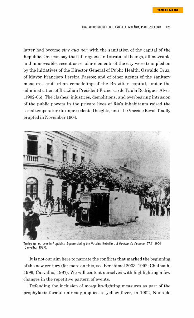
423 TRABALHOS SOBRE FEBRE AMARELA, MALÁRIA, PROTOZOOLOGIA
latter had become sine qua non with the sanitation of the capital of the
Republic. One can say that all regions and strata, all beings, all moveable
and immoveable, recent or secular elements of the city were trampled on
by the initiatives of the Director General of Public Health, Oswaldo Cruz;
of Mayor Francisco Pereira Passos; and of other agents of the sanitary
measures and urban remodeling of the Brazilian capital, under the
administration of Brazilian President Francisco de Paula Rodrigues Alves
(1902-06). The clashes, injustices, demolitions, and overbearing intrusion
of the public powers in the private lives of Rio’s inhabitants raised the
social temperature to unprecedented heights, until the Vaccine Revolt finally
erupted in November 1904.
Trolley turned over in República Square during the Vaccine Rebellion. A Revista da Semana, 27.11.1904(Carvalho, 1987).
It is not our aim here to narrate the conflicts that marked the beginning
of the new century (for more on this, see Benchimol 2003, 1992; Chalhoub,
1996; Carvalho, 1987). We will content ourselves with highlighting a few
changes in the repetitive pattern of events.
Defending the inclusion of mosquito-fighting measures as part of the
prophylaxis formula already applied to yellow fever, in 1902, Nuno de
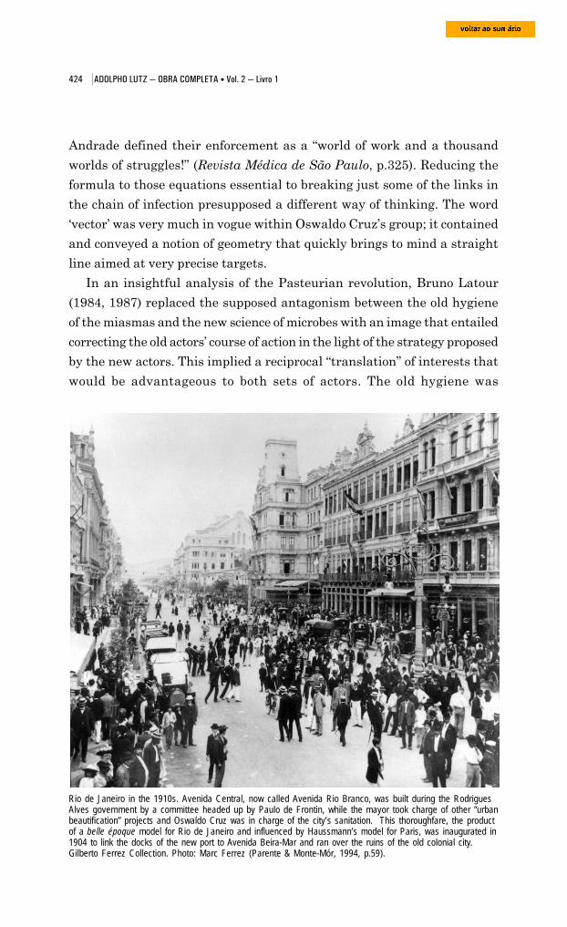
424 ADOLPHO LUTZ — OBRA COMPLETA Vol. 2 — Livro 1
Andrade defined their enforcement as a “world of work and a thousand
worlds of struggles!” (Revista Médica de São Paulo, p.325). Reducing the
formula to those equations essential to breaking just some of the links in
the chain of infection presupposed a different way of thinking. The word
‘vector’ was very much in vogue within Oswaldo Cruz’s group; it contained
and conveyed a notion of geometry that quickly brings to mind a straight
line aimed at very precise targets.
In an insightful analysis of the Pasteurian revolution, Bruno Latour
(1984, 1987) replaced the supposed antagonism between the old hygiene
of the miasmas and the new science of microbes with an image that entailed
correcting the old actors’ course of action in the light of the strategy proposed
by the new actors. This implied a reciprocal “translation” of interests that
would be advantageous to both sets of actors. The old hygiene was
Rio de Janeiro in the 1910s. Avenida Central, now called Avenida Rio Branco, was built during the RodriguesAlves government by a committee headed up by Paulo de Frontin, while the mayor took charge of other “urbanbeautification” projects and Oswaldo Cruz was in charge of the city’s sanitation. This thoroughfare, the productof a belle époque model for Rio de Janeiro and influenced by Haussmann’s model for Paris, was inaugurated in1904 to link the docks of the new port to Avenida Beira-Mar and ran over the ruins of the old colonial city.Gilberto Ferrez Collection. Photo: Marc Ferrez (Parente & Monte-Mór, 1994, p.59).
425 TRABALHOS SOBRE FEBRE AMARELA, MALÁRIA, PROTOZOOLOGIA
characterized by unlimited ambition. For every targeted disease, battles
had to be waged on an extremely wide array of fronts: against the forces of
nature, against the topography of cities, against the most varied components
of urban life. With the microbes specific to each disease in hand, the
Pasteurians pinpointed their key battles – “the mandatory points of
passage” – which would carry the hygienist contingents to the victories
they so ardently desired. Although this seductive image can help us to
think through the issues, it is the product of a distillation of accidents,
mistakes, and contradictions that make the actual battles much more
confusing and unruly – battles whose outcomes were not always favorable.
The efforts by Pasteur’s followers in Brazil to address yellow fever from
the perspective of specific microbes did not eliminate the plural nature of
strategies against the disease, even for Domingos Freire, who aimed at
yellow fever a syringe armed with his prophylaxis. One could argue that
these Brazilian Pasteurians were wrong, but that would only invalidate
the points of passage they established. The logic behind construction and
validation of microbial theories resulted in the reiteration of most of the
certainties produced by physicians and hygienists. Latour’s description of
the needed rectification of both direction and mentality only appeared when
Oswaldo Cruz became head of Public Health. The differences can be seen
in the definition of a limited number of diseases to be targeted, in the focus
on the vectors of yellow fever and of the bubonic plague, and in the
emphasis on vaccination – which does not escape the image of an arrow
pointed at the specific flank of smallpox.
These arrows guided the action of Oswaldo Cruz’s sanitary brigades
and lent clarity to their initiatives in the chaotic, tumultuous context framing
the urban reform and sanitation of Rio de Janeiro. We can discern its unique
trajectories amidst the offensive attack that engineers were concomitantly
leading against many of the targets pinpointed by public health during
the previous century (and which the sanitarians now judged irrelevant).
The ties that Oswaldo Lutz’s sanitary campaigns were tearing asunder or
patching together within a fraying social and urban fabric pushed into
action, relation, or conflict a multitude of actors unforeseen when the new
strategy was presented, with the simplicity of its experimental correlations,
at the 5th Brazilian Congress of Medicine and Surgery. Turmoil swallowed
the vaccine, the plague was subjugated, and yellow fever disappeared only
momentarily in Rio de Janeiro.
426 ADOLPHO LUTZ — OBRA COMPLETA Vol. 2 — Livro 1
Adolfo Lutz and microsporidia
Having intended from the very beginning to study animal
haematozoons, Paul-Louis Simond, communicating with Oswaldo Cruz,
had made “the most well-deserved references” to Lutz’s latest paper on
ophidian haematozoa. He and Marchoux reportedly said they would
inevitably go to São Paulo and, “with great pleasure” meet with “the friend
[they] had already come to know well through his published papers.” This
information was passed to Lutz on 20 November 1901, about two weeks
after the Pasteur Institute mission’s arrival in Rio de Janeiro. Oswaldo
Cruz was still a simple technician at the Manguinhos Serum Therapy
Laboratory, where he worked in the preparation of serum and vaccine
against the bubonic plague. It was only in December 1902 that he would
take Pedro Affonso’s place as head of this establishment. The Frenchmen
never did travel to São Paulo and Lutz’s opportunity to meet Simond in
person came only in November 1902, when the latter came a second time
to Rio, to obtain infected Stegomyia. In a letter written 9 March 1903 (two
weeks before Cruz became Director General of Public Health), Simond
stated:
It was with keenest interest that I read the report of the experimentsyou conducted with the aid of some doctors from São Paulo. They are ofthe utmost importance since this is the first time that the mosquito’srole in yellow fever has been confirmed outside of Havana ... I am noless interested in your other papers on pebrines and on the wild plantsthat host mosquito larvae. Mr. Foetterle gave me a copy of this latestpublication, for which I thank you warmly. If I decide to travel to SãoPaulo, I will make certain to let you know in advance so that I will besure to find you in your laboratory. It is hardly necessary to say that Iwould like to go strictly incognito and visit only yourself. (BR. MN.,Fundo Adolpho Lutz, pasta 174, maço 1)
This letter does not add much to what we already know about its author’s
investigations into the possible role of a microsporidian in the etiology of
yellow fever. It is particularly enlightening with respect to the direction
being taken by its recipient. Lutz had just written “Waldmosquitos und
Waldmalaria” on 16 September 1902. In June, he found himself in Rio de
Janeiro to collect Stegomyia for the experiments that would commence in
December. That same year, in partnership with Alfonso Splendore, Adolpho
Lutz published the first in a series of three papers on pebrine and
microsporidia. He released these in a German periodical (CentralBlatt für
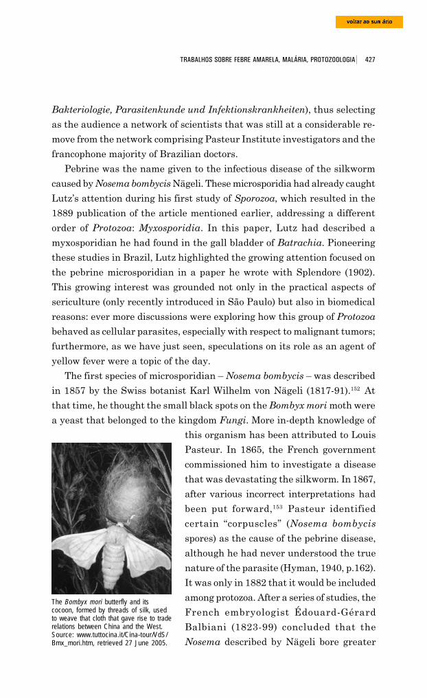
427 TRABALHOS SOBRE FEBRE AMARELA, MALÁRIA, PROTOZOOLOGIA
Bakteriologie, Parasitenkunde und Infektionskrankheiten), thus selecting
as the audience a network of scientists that was still at a considerable re-
move from the network comprising Pasteur Institute investigators and the
francophone majority of Brazilian doctors.
Pebrine was the name given to the infectious disease of the silkworm
caused by Nosema bombycis Nägeli. These microsporidia had already caught
Lutz’s attention during his first study of Sporozoa, which resulted in the
1889 publication of the article mentioned earlier, addressing a different
order of Protozoa: Myxosporidia. In this paper, Lutz had described a
myxosporidian he had found in the gall bladder of Batrachia. Pioneering
these studies in Brazil, Lutz highlighted the growing attention focused on
the pebrine microsporidian in a paper he wrote with Splendore (1902).
This growing interest was grounded not only in the practical aspects of
sericulture (only recently introduced in São Paulo) but also in biomedical
reasons: ever more discussions were exploring how this group of Protozoa
behaved as cellular parasites, especially with respect to malignant tumors;
furthermore, as we have just seen, speculations on its role as an agent of
yellow fever were a topic of the day.
The first species of microsporidian – Nosema bombycis – was described
in 1857 by the Swiss botanist Karl Wilhelm von Nägeli (1817-91).152 At
that time, he thought the small black spots on the Bombyx mori moth were
a yeast that belonged to the kingdom Fungi. More in-depth knowledge of
The Bombyx mori butterfly and itscocoon, formed by threads of silk, usedto weave that cloth that gave rise to traderelations between China and the West.Source: www.tuttocina.it/Cina-tour/VdS/Bmx_mori.htm, retrieved 27 June 2005.
this organism has been attributed to Louis
Pasteur. In 1865, the French government
commissioned him to investigate a disease
that was devastating the silkworm. In 1867,
after various incorrect interpretations had
been put forward,153 Pasteur identified
certain “corpuscles” (Nosema bombycis
spores) as the cause of the pebrine disease,
although he had never understood the true
nature of the parasite (Hyman, 1940, p.162).
It was only in 1882 that it would be included
among protozoa. After a series of studies, the
French embryologist Édouard-Gérard
Balbiani (1823-99) concluded that the
Nosema described by Nägeli bore greater
428 ADOLPHO LUTZ — OBRA COMPLETA Vol. 2 — Livro 1
affinity to sporozoa than to fungi; he then transferred it to the class
Sporozoa, created by Rudolf Leuckart in 1879, and defined the taxon
Microsporidia to include Nosema bombycis and the other species of the
group.154
Until a few years ago, microsporidia had been classified in the phylum
Protozoa as an order within the subclass Cnidosporidia and the class
Sporozoa, along with myxosporidia (Hyman, 1940, p.47; Store and Usinger,
1979, p.311).
Phylum Protozoa Class Sporozoa
Subclass Cnidosporidia Order Mysoxporidia Order Microsporidia
These microorganisms caused a great stir in 1988 when they were
discovered in HIV patients. After many studies into their taxonomic
classification, it was established that myxosporidia are more closely
related to Metazoa than Protozoa, and microsporidia, to fungi (Cox, 2002,
p.595-612).
At the time that Lutz and Splendore published their paper, few species
had been described and very little was known about these sporozoans.
There was a consensus that they were intra-cellular parasites of a few
vertebrates (fish) and, mainly, invertebrates. It was also known that they
engaged in a form of hereditary transmission, by which the female host
passed the agent of infection to the next generation through its eggs. Lutz
found in Splendore the ideal partner for investigating these minute
parasites, whose size demanded extreme patience and a great expertise in
microscopy and also in fixing and staining techniques, knowledge that
Splendore had acquired in Italy during his first few years in the profession.
Lutz often calls attention to the patient work of his co-author: “Since there
were few specifications on fixing and dying techniques, or on use of reagents,
we paid these issues meticulous attention, with Dr. Splendore undertaking
numerous lengthy experiments.” In another excerpt on silkworm
reproduction, Lutz praised Splendore’s skill in managing to obtain “a good
number of healthy Bombyx mori eggs” (Lutz and Splendore, 1902, p.151).
Alfonso Splendore (1871-1953) associated himself to the director of
the Bacteriological Institute of São Paulo as soon as he arrived from Italy
in 1899. A graduate of Rome’s Medical and Surgery Faculty two years

429 TRABALHOS SOBRE FEBRE AMARELA, MALÁRIA, PROTOZOOLOGIA
earlier, he had worked as an assistant at Rome’s Hygiene Institute,
alongside such masters as Angelo Celli (1857-1914), Claudio Fermi
(1862-?), and Giovanni Battista Grassi (1854-1925). When he moved to
Brazil, he probably brought with him recommendations addressed to Lutz,
who kept close ties with Italians who were studying malaria. In addition to
working with Lutz at the São Paulo institute, Splendore was responsible
for founding the laboratory at Rome’s Umberto I Hospital. He also headed
the laboratory at the Beneficência Portuguesa Hospital in São Paulo.155
Apart from the three papers on microsporidia, Splendore worked on a
number of other papers with Lutz.156
In the first, they established new species that parasitized another group
of insects, besides moths, and a species of fish. At that time it was not
known whether moths were infected by other species of Nosema, besides
Nosema bombycis, which Lutz and Splendore also called “pebrine.” They
verified this with a butterfly quite common in the region of São Paulo
(Brassolis astyra Godt), which Lutz had first examined when he began his
studies on sporozoans in the 1890s. With Splendore, Lutz now demonstrated
the spontaneous infectious process and its artificial transmission in a
laboratory of other species of Nosema; they further verified the
hereditary transmission of the infection – an issue, as we have seen,
that greatly interested Paul-Louis Simond.
They carried out a series of experiments using healthy eggs from
silkworm caterpillars obtained locally. With the help of Alfonse’s brother,
Dr. Achille Splendore,157 they managed to bring in from Italy animals
infected with pebrine, thus making possible comparisons and transmission
experiments. They faced many difficulties, especially concerning
classification of the species of host butterflies. Since there was no systematic
classification of the Brazilian species, Lutz and Splendore were forced to
rely on Adolfo P. Mabilde’s work on butterflies of the state of Rio Grande
do Sul and on the general treatise published by W. J. Holland in 1898,
The Butterfly Book. They based themselves solely on the form of the spores
in identifying the species of microsporidia. In his studies on myxosporidia,
the Belgian scientist P. Thélohan (1892, 1895) had identified a polar capsule
with a spiral filament in this group of microorganisms, but Lutz and
Splendore did not consider it because they could not discern such a minute
morphologic characteristic. They relied on another criterion to distinguish
the species: their association with hosts. This was not an acceptable criterion
430 ADOLPHO LUTZ — OBRA COMPLETA Vol. 2 — Livro 1
according to the day’s classification rules, and the authors themselves admit
it: “It seems impossible to name species based solely on spore characteristics
that appear in varying quantities; that is why we have adopted a method
that is largely disapproved of today, which is to designate the species by
the name of the first host in which it was discovered” (Lutz & Splendore,
1902, p.153).
In this article they identified nine new species of Nosema, most of which
were Lepidoptera parasites. They verified that naturally occurring infections
only had lethal consequences in silkworm pebrine. In the case of Brassolis
astyra, São Paulo’s common butterfly, an uncommon atrophy of the
caterpillar was a sign of a very serious infection, but no visible symptoms
were observed in other species. The authors observed that another butterfly,
Dione juno, would go through normal metamorphosis despite extensive
infection. It became clear that hosts who spent lengthier periods in the
larvae stage on the one hand favored rapid propagation of the species
corresponding to microsporidia and, on the other, favored the harmful
aspects of this generally benign infectious process.
The work of these two scientists from the Bacteriological Institute of
São Paulo attracted the attention of other researchers interested in the
group. F. Mesnil wrote a review on it for the Bulletin do Institut Pasteur
(1903, v.I, p.62). That same year, Paul-Louis Simond published a note in
the Comptes Rendus de la Société de Biologie de Paris on Myxococcidium
stegomyia, the microsporidium of the genus Nosema discovered in Stegomyia
fasciata, initially in specimens that had drawn blood from a yellow-fever
patient, leading the French bacteriologist to the supposition that it was the
causative agent of the disease.158
Lutz and Splendore also focused on the microsporidia that parasitized
blood-eating hematophagous diptera and, in the paper published in 1904,
they referred to “some forms” that had been recently observed, especially
“parasites that one of us [Lutz] found in Simulium larvae.” They found
different species of Nosema, which they described without, however,
defining which genus they belonged to, for lack of specialized literature.
This article was published in CentralBlatt für Bakteriologie,
Parasitenkunde und Infektionskrankheiten as an addendum to the earlier
one: in addition to describing new species, they included life-size drawings
of the various hosts of microsporidia. The 1904 article benefited from an
intense exchange of information between Adolpho Lutz and the Austrian
431 TRABALHOS SOBRE FEBRE AMARELA, MALÁRIA, PROTOZOOLOGIA
lepidopterologist Joseph Foetterle, who lived in Petrópolis, then a spot
favored by German and Austrian immigrants. Although he worked as a
violin teacher at Colégio Sion in Rio, his passion was collecting butterflies.
He had a good knowledge of insects and maintained close ties with physicians
that were investigating transmitters, especially those who lived in or
regularly paid visits to that pleasant mountain city. In the collection of
Paul-Louis Simond, who became Foetterle’s good friend, we find
photographs taken by the Frenchman, showing the Austrian proudly
displaying his collection. Foetterle also corresponded with Austrians and
other Europeans and mediated relations between researchers who came to
Brazil and the “natives.” In his letters to Adolpho Lutz, who was a close
friend, he always kept him abreast of news on members of the French mission.
Foetterle was also involved in the search for new microsporidia. His
correspondence with Lutz contains much important information on pebrine,
dyes and butterfly classification. The director of the Bacteriological Institute
would send Foetterle material to classify, while the Austrian would send
him insects contaminated with pebrine. “I received your friendly card on
Saturday and, on Sunday, the moth you sent, which belongs to the genus
Caeculia (Fam. Lasiocampidae),” the Austrian naturalist wrote, for example,
on 1 March 1903.
I am afraid that I can not tell what species it is. I have not yet raisedthe species you sent me, but I am familiar with the caterpillars ofspecies that are very close to it ... Regrettably, I have but few duplicatesamples of these rascals, which, however, I could cede to you. Theother day I finally found pebrine in a species close to yours ... It tookme a while to find these subjects that made such mockery of me, and itwas a fair amount of time and work that I spent on it. I do not have thetime to set them into permanent preparations today, but I will certainlydo so tomorrow and send them to you, along with the moths. I havealso experimented with pyoctanin as a dye, and had quite good results.Have you also used this dye?159
In another letter, dated 19 October, which was sent along with more
material, he states: “The species in which I found pebrine is in the paper
tube ... and I hope that this time you can confirm my findings and can also
discover pebrine in the three preparations I am sending with it. If, once
more, there are not any, then it is better that I stop meddling in this.”160
Foetterle’s contributions were incorporated into the work of Lutz and
Splendore, who did not fail to mention his valuable help.
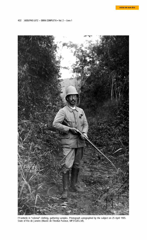
432 ADOLPHO LUTZ — OBRA COMPLETA Vol. 2 — Livro 1
FFoetterle in “colonial” clothing, gathering samples. Photograph autographed by the subject on 25 April 1905.State of Rio de Janeiro (Musée de l’Institut Pasteur, MP31328-2.tif).
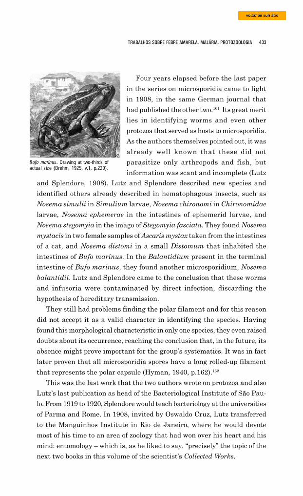
433 TRABALHOS SOBRE FEBRE AMARELA, MALÁRIA, PROTOZOOLOGIA
Bufo marinus. Drawing at two-thirds ofactual size (Brehm, 1925, v.1, p.220).
Four years elapsed before the last paper
in the series on microsporidia came to light
in 1908, in the same German journal that
had published the other two.161 Its great merit
lies in identifying worms and even other
protozoa that served as hosts to microsporidia.
As the authors themselves pointed out, it was
already well known that these did not
parasitize only arthropods and fish, but
information was scant and incomplete (Lutz
and Splendore, 1908). Lutz and Splendore described new species and
identified others already described in hematophagous insects, such as
Nosema simulii in Simulium larvae, Nosema chironomi in Chironomidae
larvae, Nosema ephemerae in the intestines of ephemerid larvae, and
Nosema stegomyia in the imago of Stegomyia fasciata. They found Nosema
mystacis in two female samples of Ascaris mystax taken from the intestines
of a cat, and Nosema distomi in a small Distomum that inhabited the
intestines of Bufo marinus. In the Balantidium present in the terminal
intestine of Bufo marinus, they found another microsporidium, Nosema
balantidii. Lutz and Splendore came to the conclusion that these worms
and infusoria were contaminated by direct infection, discarding the
hypothesis of hereditary transmission.
They still had problems finding the polar filament and for this reason
did not accept it as a valid character in identifying the species. Having
found this morphological characteristic in only one species, they even raised
doubts about its occurrence, reaching the conclusion that, in the future, its
absence might prove important for the group’s systematics. It was in fact
later proven that all microsporidia spores have a long rolled-up filament
that represents the polar capsule (Hyman, 1940, p.162).162
This was the last work that the two authors wrote on protozoa and also
Lutz’s last publication as head of the Bacteriological Institute of São Pau-
lo. From 1919 to 1920, Splendore would teach bacteriology at the universities
of Parma and Rome. In 1908, invited by Oswaldo Cruz, Lutz transferred
to the Manguinhos Institute in Rio de Janeiro, where he would devote
most of his time to an area of zoology that had won over his heart and his
mind: entomology – which is, as he liked to say, “precisely” the topic of the
next two books in this volume of the scientist’s Collected Works.
434 ADOLPHO LUTZ — OBRA COMPLETA Vol. 2 — Livro 1
Notes1 Although evidence suggests that yellow fever had been present in Brazil since 1694 (Ministério da Saúde,1971), it was only as of the mid-19th century that it became the country’s major public health issue.2 On magnetic tape, Bertha Lutz, daughter of Adolpho Lutz, recorded interesting facts about the family historyand the life of her father. Entitled Lutziana, the tape registers in her own voice the outline of a biography shenever came to write (BR. MN. [Brasil, Museu Nacional ] – Fundo Adolpho Lutz).3 On this point, see the interview with José de Barros Ramalho Ortigão Junior, published under the title“Recordações da infância: as primeiras letras com a família Lutz,” História, Ciências, Saúde – Manguinhos,v.10, no. 1, Jan.-Apr. 2003, p.420-4; in the same issue (p.13-83), see Benchimol, “Adolpho Lutz: um estudobiográfico.” See further Sá and Benchimol, “Adolpho Lutz: formação e primeiros trabalhos” / “Adolpho Lutz:Education and First Works,” by Benchimol and Sá (2004, p.118-84; 185-250).4 Arquivo Nacional. Documents referring to Rio de Janeiro’s Junta Comercial: 1872 – Liv. 648, Reg. 11424 G6,firma Lutz & Cia.5 The bibliography of Adolpho Lutz compiled by Herman Lent (Neiva, 1941) was reprinted, with corrections andadditions, in História, Ciências, Saúde – Manguinhos, v.10, no. 1, p.362-409.6 Deane (1955, p.77-80). As this author shows, Lutz advocated the use of feces examinations to diagnosethese helminthoses; he also pointed out that this practice was not given due credit even in the three German-speaking universities where he had studied. Lutz’s biographers underscore his pioneer role in veterinaryresearch. His first work in the area was the description of a species of Rhabdonema found in the domestic pig(1885). He wrote too about the role of fleas as hosts of Dipylidium caninum; about stephanurosis, cysticercosis,and other helminthiases common in animals; about Fasciola hepatica; and also about the wild hosts ofDioctophime renale, a parasite of the kidneys of several domestic animals.7 See also Councilman and Lafleur (1891, p.396) and www.whonamedit.com/doctor.cfm/2929.html (consultedon 13 Apr. 2005).8 “Lösch was the first author to provide a more accurate description of the amoeba species found in the stool ofa patient with dysentery, accompanied by a thorough clinical record and an autopsy report. This was truly apioneer study, containing excellent observations and descriptions of the organism called Amœba coli, to whichvery little was added subsequently” (Councilman and Lafleur, 1891, p.397).9 “All his reports merely make mention of the existence of these microorganisms, without attributing any greatimport to them” (Councilman and Lafleur, 1891, p.400).10 According to Councilman and Lafleur (1891, p.400-1), after Lösch’s, the most important study is the one thatprompted Kartulis to publish a series of articles in Virchow’s Archiv.11 “His results were published in Czech, a language unfamiliar to most researchers ... Kartulis mistook ‘Ouplavici’, which in Czech means ‘About dysentery’, as the name of the paper’s author ... And thus thephantasmagoric professor ‘Uplavici, O.’ made his way into a number of specialized bibliographies, until Dobelluntangled the confusion in 1938” (Martinez-Palomo, 1996).12 Born in Canada, Osler had begun his clinical practice in Dundas, Ontario, but was soon appointed lecturerand then professor of physiology, pathology, and medicine at McGill University. In 1884, he took the chair inclinical medicine at the University of Pennsylvania, in Philadelphia. There he became one of the foundingmembers of the Association of American Physicians. In 1888, Osler accepted an invitation to serve as the firstprofessor of medicine at Johns Hopkins University Medical School, in Baltimore. Together with William HenryWelch (1850-1934), head of pathology; Howard Atwood Kelly (1858-1943), head of gynecology and obstetrics;and William Steward Halsted (1852-1922), responsible for surgery, they were to turn Johns Hopkins into one ofthe world’s most prestigious medical schools (Risse, p.407; see also www.whonamedit.com/doctor.cfm/1627.html; consulted on 13 Apr. 2005).13 A graduate of the University of Maryland, Councilman had traveled to Europe in 1880 to specialize inpathology. He was in the same cities as Lutz – Vienna, Strasbourg, Leipzig, and Prague – and there spent timewith people who were also connected with the Brazilian researcher’s education. In Vienna, he trained with KarlFreiherr von Rokitansky (1804-78); in Strasbourg, he worked with Friedrich Daniel von Recklinghausen (1833-1910); in Leipzig, with Julius Friedrich Cohnheim (1839-84) and Carl Weigert (1845-1904); and, lastly, inPrague, with Hans Chiari (1851-1916) (www.whonamedit.com/doctor.cfm/2860.html). In 1886, he joined theteam of William Henry Welch (1850-1934) as well as the group of researchers at the newly inauguratedpathology laboratory where Osler also worked.14 His paper was published in the Bulletin of the Johns Hopkins Hospital in 1890. The article on the patient’sdeath, on 5 April 1890, appeared the following month in the same periodical (Martinez-Palomo, 1996).15 Among others, he cited Stengel (Medical News, 15 Nov. 1890); Musser (University Medical Magazine, Dec.1890); and Dock (Texas Medical Journal, Apr. 1891).
435 TRABALHOS SOBRE FEBRE AMARELA, MALÁRIA, PROTOZOOLOGIA
16 He cites Osler, Nasse, Harold, Stengel, Eichberg, Kowacz, Fenaglio, Boas, Vivaldi, Babès and Zigura,Sorgo, Manner, Júrgens, and Roemer, in North and South America, Italy, Germany, and other countries.17 For these authors, there was such a wide diversity of opinions on the protozoans’ role because all amoebaehad been confused. Only A. dysenteriae caused dysentery. They conducted experiments on cats, andobserved that they were only infected by amoebae that had ingested red blood cells, whereas amoebae thathad not ingested such cells did not cause the disease (Cox, F.E.G., 2002). Martinez-Palomo (ibid.) points outthat these authors discovered the resistant form of the amoeba, the cyst (we now know that two forms of E.histolytica exist: trophozoites and cysts).18 Dopter (ibid.) also makes reference to Gregorieff’s work incriminating a bacillus identical to the one describedearlier by Chantemesse and Widal. Ogata likewise attributed cases observed in Japan to a bacillus. ForZancarol, it was a streptococcus that he had found in stools and liver abscesses. Casagrandi and Barbagalloactually affirmed that these protozoans were saprophytes that were useful to the intestine because theycontributed to the destruction of virulent bacteria. Schuberg (1895) gave a purgative to a number of healthyindividuals and verified that half of these were hosting inoffensive amoebae in their intestines.19 Maciel, Discriminações raciais, p.57; and Lisboa, Almanaque de Campinas para 1871, both cited by SantosFilho and Novaes (p.35).20 Francisco Rangel Pestana (1839-1903) and Américo Brasiliense de Almeida Melo (1833-96). Two republicanleaders lived in the city: Manual Ferraz de Campos Sales (1841-1913) and Francisco Glicério de CerqueiraLeite (1846-1916), in addition to Américo Brasílio de Campos (1838-1900) and Francisco Quirino dos Santos(1841-86) (Santos Filho and Novaes, p.14).21 In 1889, he was to present another treatise: “Yellow fever in Campinas: two words about this epidemic, readbefore the Imperial Academy of Medicine of Rio de Janeiro in the session of 27 April 1889” [in Port.] (Anais daAcad. de Med. do Rio de Janeiro, 1889, p.331-49). According to Santos Filho and Novaes (p.29), these casesof yellow fever had not set off an epidemic, as in 1889, because its transmitter, Aedes aegyti, was not yetpresent in the city.22 Santos Filho and Novaes (p.175-6). Doctors believed the epidemic resumed in May because of the return ofinfected inhabitants who had fled. They then appealed to all those who had left to delay their return until thecrisis had passed.23 The last Chamber of the monarchy was elected for the 1887-90 legislature and was dissolved by republicandecree on 21 January 1890. It was first chaired by the pharmacist Otto Langgaard, who lost his children to theepidemic. At the height of the crisis, the nine aldermen did not meet but in May a session took place at one oftheir homes (Santos Filho and Novaes, p.47).24 Lapa (1996, p.260-1) also mentions the elimination of ‘black ditches’, the filling in of swampy areas, and thechanneling of streams. He notes that the measures adopted to eliminate the bad odors and miasmas “managedto have some effect, ignored, however, by physicians and authorities, that is, they scared off the mosquitoAedes aegypti.”25 On 11 April, he was replaced by the Baron de Jaguara (1837-95).26 According to Ângelo Simões (1897, p.23-4), “of the twenty-three general practitioners that we were, onlythree stayed, taking on an insane job.” The three were João Guilherme da Costa Aguiar (1856-89), GermanoMelcghert (1844-1921), and Simões himself, “the second having fallen ill and the first having paid with his ownlife for the heroism that kept him in the struggle against such a dreadful enemy.”27 Diario de Campinas, 29 May 1889, cited in Santos Filho and Novaes (p.49).28 Author of Estudos sobre a febre amarela (1880), upon his return to Portugal he published A epidemia deCampinas em 1889 (Rio de Janeiro, 1889).29 A compound of ferrous sulphate (400 grs.), crystallized ferric acid (100 grs.), and boiling water (2 liters).30 He is said to have signed eleven certificates in April and one on 5 May, in Santa Cruz, and another three inApril, in Conceição (Santos Filho and Novaes, p.53).31 Santos Filho and Novaes (p.11). Drs. Francisco Marques de Araújo Góis, Claro Marcondes Homem deMelo, and Bráulio Gomes were awarded medals commissioned by the Chamber. “Adolfo Lutz was not paid thishonor, which is impossible to understand,” write Santos Filho and Novaes (p.53). They explain this “because[the three who received the medals] could easily travel to São Paulo – something they had always done –thereby not failing to provide steady, more effective assistance.” Lutz’s work there notwithstanding (vouched bythe number of certificates he signed), the authors state that as a “wise researcher already, [he] was moreconcerned about observing the disease and its contagion than in actually practicing medicine” (ibid., p.50, 53). Itis possible that Lutz left earlier owing to his upcoming travels to Hamburg and Hawaii. On this point, seeBenchimol and Sá (2004).
436 ADOLPHO LUTZ — OBRA COMPLETA Vol. 2 — Livro 1
32 According to Freire (1890a), fifteen lives were claimed in Vassouras (three immigrants), eleven in Resende(three immigrants), twenty-two in Desengano (seven immigrants), twenty-one in Serraria, and twenty inCataguases, bringing deaths to a total of 4,137 (2,802 immigrants), with Rio de Janeiro, Campinas, and Santosincluded. Santos Filho and Novaes (p.38-9) point to the contrast between the assistance given these cities andthat given Campinas in 1889. The central, provincial, and municipal governments spared no efforts in aiding thevictims of the first epidemic. On the other hand, the imperial government appointed only one physician (AugustoDaniel de Araújo Lima) to attend to the indigent ill in Cataguases, while three doctors residing in the townvolunteered to attend the poor free of charge. For the authors, this contrast is explained by the fact thatCampinas was then “a very prestigious socioeconomic and political center ... one of the most thriving cities,with a solid economic situation and among the most culturally advanced, if not in all Brazil at least in the entireprovince of São Paulo and in Southern Brazil.”33 On Freire and other hunters of the yellow fever microbe, see Benchimol (1999).34 The provincial president dismissed the vaccination agents at work in Bahia’s municipalities after the locallegislature cut off funding, alleging that this service should fall to the central government. Francisco AntunesMaciel, minister of the Empire, did not concur with protests lodged by the Central Board of Public Health. On 4October 1883, the members of the Board resigned. On 8 October, Domingos José Freire took over as presidentof a new Board.35 The letters reproduced by Vallery-Radot (1930, p.397-411) and other letters can be found at the MuseuImperial, Setor de Documentação e Referência, Arquivos da Casa Imperial (Petrópolis). On the history of thesmallpox vaccine in Brazil, see Fernandes (1999).36 The special communication read on 7 September was entitled “Vaccination avec la culture atténuée dumicrobe de la fièvre jaune.” It was summarized in Medical News (17 Sep. 1887, v.51, p.330-4), in thenewspapers Jornal do Commercio, O Paiz, and Gazeta de Noticias (22-23 Aug. 1899) and in Brazil-Medico(no. 33, 1 Sep. 1899, p.319). Freire wrote over one hundred works on chemistry, medicine, and public health,in the form of reports, compendia, books, monographs, and communications.37 The majority of those vaccinated were Brazilian. This shows that Freire’s prestige itself played a greater rolethan foreign immigration in spreading use of the vaccine. In Rio de Janeiro, his data were authenticated byCounselor Caminhoá and two attorneys, Julio Ottoni and Oliveira Coelho. Joaquim Caminhoá, the counselor’sson, and Virgílio Ottoni – both of whom were assistants at the School of Medicine’s organic chemistry lab – tookpart in the vaccinations; others included the physicians Silva Santos, hygiene delegate; Guilherme Affonso;Alfredo Barcellos; and Campos da Paz. In a letter dated 19 October 1889, Dr. Pinto Netto, director of the SantaIzabel Maritime Hospital, in Jurujuba, informed Freire that a commission from the Society of Medicine andSurgery was investigating his preventive method. Many of these vaccinations were observed by members ofthe commission, which included two of Freire’s ex-assistants and Drs. Chapot Prévost and Benicio de Abreu,then professors at the School of Medicine. We read in one newspaper, for instance, that “on Sunday, from eightuntil nine in the morning, Dr. Virgílio will be offering vaccinations in Botafogo, at 66 Voluntários da Pátria Street,assisted by Dr. Carlos Costa, member of the commission of the Society of Medicine and Surgery” (Gazeta deNoticias, 7 Feb. 1889).38 Fifteen of those not vaccinated died (three immigrants and twelve Brazilians, three of whom black). A largenumber of people fell ill, although records only specify that 115 of them were black. Freire’s published resultswere endorsed by Dr. Joaquim Francisco Moreira, president of the Chamber, and Luis E. de Lemos, secretary;by the jurist Sebastião Eurico Gonçalves de Lacerda; and by the hygiene delegate Dr. Augusto de PaivaMagalhães Calvet (Freire, op. cit., p.23, 25-6).39 None of those vaccinated died. Among the non-vaccinated, yellow fever claimed 650 lives. Present wereDrs. Julio Alves de Moraes, hygiene delegate; municipal physician Joaquim da Motta Silva; Giovanni Eboli, ofSanta Casa de Misericórdia Hospital; Carl Hertschel, local practitioner; the public prosecutor, Dr. JoãoNepomuceno Freire; the Baron de São Domingos, a state judge; the president of the Chamber, Julio Corrêa;Alfred Esquivel; Lucas Fortunato; and Americo Martins, among others.40 Only three failures were reported, i.e., 0.46% of all those vaccinated. According to Simões’ report, mortalityamong the unvaccinated was estimated at 810 inhabitants, but for another 300 of those buried, the deathcertificates indicated such questionable causes of death as “icterohemorrhagic fever” and “reigning fever”,among others. Simões published the evaluation that he had sent Freire in the Correio de Campinas (10 Jun.1889). On 11 May 1889, he transcribed a report and forwarded it to the president of the province of São Paulo,the Baron de Jaguara: he planned to continue vaccinating, especially people who were returning to Campinasdespite instructions published in newspapers advising that they wait until the city had been wholly disinfected.Freire published this letter in the Gazeta de Noticias (12 May 1889). In Tratamento da febre amarella pela aguachlorada, published in 1897, Simões still wrote: “Save later proof to the contrary, the discovery of the yellowfever microbe inarguably falls to Freire” (p.40 ff.)41 The newspaper’s representatives were: in Araras, professor J. Voss; in Botucatu, professor Carlos Knüppel;in Piracicaba, J.J. Huffenbaecher; in Pirassununga, Gustav Beck; in Ribeirão Preto, Carl C. Petersen; in RioClaro, Luiz F. Barthmann; in Santos, Paul Wilkens; in São Carlos do Pinhal, C. Priester; and in Sorocaba, Th.

437 TRABALHOS SOBRE FEBRE AMARELA, MALÁRIA, PROTOZOOLOGIA
Kaysel, hat manufacturer. The masthead also listed Gottlieb Müller and S. Lauer, from Curitiba and Joinville,respectively. W. Diebener’s bookstore represented the paper in Leipzig. Carlos Constantin Knüppel, first clerk ofcourt in Joinville, was founder and editor of Beobachter am Mathiastrom, the first newspaper (handwritten)launched in that town. He moved to São Paulo where he was a teacher at the first Escola Alemã (GermanSchool). He later moved to Botucatu, where he also taught.42 Freire won the support of municipal authorities in Rio and other cities and also of many doctors residing onthe coast and inland, including delegates from the central hygiene department. He likewise had the backing ofvarious institutes from civil society. But the vaccinations were met with hostility by the Ministry of the Empire,by the leadership of the General Inspectorship of Public Hygiene, and by the Imperial Academy of Medicine (thelatter theoretically the realm of Brazil’s medical elite, where the interests of the profession and of the governmentintertwined).43 On this topic, see Benchimol (1999).44 On this topic, see Soriano (2002) and Benchimol (1999).45 Finlay (1965, p.317-48). The Cuban physician located the disease’s source of morbidity in the trajectory ofthe bite, through the inner walls of blood vessels, assuming that the insect innoculated the disease into thecorresponding tissues of healthy people. According to Delaporte (1989, p.69, 73), this theory identified yellowfever with smallpox: “If one needed to summarize Finlay’s outlook, one could easily say that he saw themosquito through Jenner’s eyes.”46 Lacerda recounts part of his history at the institute in Fastos do Museu Nacional (1905). The bio-bibliographypublished by the Museu Nacional in 1951 entirely omits works on yellow fever and bacteriology, which areanalyzed by Benchimol (1999).47 On this topic, see Benchimol (2004).48 Yellow fever is caused by an arbovirus of the genus Flavivirus (Veronesi, 1991, p.174-82). It presents anumber of clinical manifestations in human beings: high fever, chronic headache, muscle aches – in short, allthe signs of an acute infection, which soon affects the digestive tract. After two or three days, if the diseasedoes not recede, the patient’s vomit and feces will turn bloody and jaundice will worsen, as will abdominalpain; the patient hemorrhages from the fossa canina, gums, or skin. Decreased urination triggers the toxemicstage, which develops into coma and leads to death. These symptoms are caused by virus replication insidethe organism after inoculation by a mosquito of the genus Aedes or Haemagogus. The disease’s viral etiologywas only established in 1927; in March 1937, on the premises of the Oswaldo Cruz Institute, the RockefellerFoundation began to produce a vaccine made from virus cultured in chicken embryo, a vaccine which is stillused today (Benchimol, 2001).49 See also Stepan, 1976; Benchimol, 1990.50 “Relatório do Instituto Bacteriológico. Ano de 1893.” Excerpts of Reports on yellow fever by Prof. Lutz whendirector of the Bacteriological Institute (1893-94. Pasta 212, f maço 4, BR. MN. Fundo Adolpho Lutz).51 “Relatório do Instituto Bacteriológico. Ano de 1894,” 1st semester, São Paulo (BR. MN. Fundo Adolpho Lutz).52 “Relatório do Instituto Bacteriológico. Ano de 1894,” Jan./Feb. 1895, São Paulo (BR. MN. Fundo AdolphoLutz).53 Havelburg’s paper, published in Annales de L’Institut Pasteur (Jun. 1897), was a condensed version of thearticle that had come out in Berliner Klinische Wochensschrift (1897). It was released together with Sanarelli’sfirst special communication. Both sent cultures of their bacilli. Sanarelli’s supplementary evidence waspublished in September and October. Havelburg’s conference was published by Brazil-Medico and, in bookform, by the Jornal do Commercio. He had just been appointed head of the hospital’s anatomical-pathologicallaboratory (Brazil-Medico, 8 Apr. 1897, p.119). Lutz commented on Sanarelli’s sealed letter in Brazil-Medico (15Dec. 1898, p.416-7).54 In July 1897, he was replaced by Vital Brazil Mineira da Campanha (caixa 25, pasta 16, maço 3: “Auxiliaresdo Dr. Lutz”; BR. MN. Fundo Adolpho Lutz).55 Letter from Francisco Fajardo to Adolpho Lutz, dated 18 March 1897 (BR. MN. Fundo Adolpho Lutz).56 “Resumo dos trabalhos do Instituto Bacteriológico de São Paulo,” 1892-1908, p.41 (BR. MN. Fundo AdolphoLutz).57 “Relatório do Instituto Bacteriológico. Ano de 1894,” Jan./Feb. 1895, São Paulo (BR. MN. Fundo Adolpho Lutz).58 Born in Capivari (São Paulo) on 6 December 1871, he graduated from the Brussels School of Medicine in1895, and the following year joined the Bacteriological Institute of São Paulo as an “adjunct physician.” BetweenMay and November 1901, he traveled to Europe to study wine yeast cultures using Pasteur’s system. In 1901,he took the chair in bacteriology at the Escola Livre de Farmácia de São Paulo (São Paulo Free School ofPharmacy), now the University of São Paulo’s School of Dentistry. He died on 24 April 1903, a victim of yellowfever.
438 ADOLPHO LUTZ — OBRA COMPLETA Vol. 2 — Livro 1
59 In an official letter to Dr. J.J. da Silva Pinto, director general of the Sanitation Service (dated 25 Mar. 1897),Lutz advised that he had received reliable information that Sanarelli was planning to hold a conference in Maywhere he would reveal his discovery: “I would like to suggest that it would be appropriate for our institute to berepresented upon this occasion, so that we may be capable, as early as possible, of assessing the value of thepractical usefulness of the aforementioned discovery.” According to an official letter to Silva Pinto from the StateDepartment of Internal Affairs, signed by A. Dino Bueno and dated 4 June 1897, Lutz and Mendonça hadtraveled to Montevideo the previous day on assignment to the Government of the State. Source: excerpts fromProf. Lutz’s reports on yellow fever as director of the Bacteriological Institute, 1893-94 (pasta 212 f maço 4, BR.MN. Fundo Adolpho Lutz).60 On this topic, see Benchimol (2004).61 The conference was published in O Paiz (10 Jun. 1897) and condensed in Brazil-Medico (22 Jun. 1897,p.209-20). While in the capital of Uruguay, a telegram arrived, appointing Lacerda representative of the NationalAcademy of Medicine (Brazil-Medico, 15 Jun. 1897, p.199; 1 Jul. 1897, p.227; 22 Jul. 1897, Aug. 1897, p.283-5; 22 Dec. 1898, p.429-31).62 Lutz said he would postpone discussion of the conference until Sanarelli’s treatise had been published in theAnnales de l’Institut Pasteur (it would appear in nos. 6, 8, and 9, in Jun., Sep., and Oct. 1897). In this firstreport, Lutz voiced a preliminary opinion of Sanarelli’s ideas and promised to return to the topic in the nextBacteriological Institute report.63 In a letter dated 28 July, Fajardo asked Lutz for a “copy of the notes [he] had taken on the yellow feverexperiments conducted by Sanarelli in our presence” (pasta 212 f maço 4; BR. MN. Fundo Adolpho Lutz). The Military Bacteriological Laboratory had been inaugurated on 2 July 1896, at a private residence located at24-A Senador Furtado Street. In late 1897, the director, Ismael da Rocha, and Drs. Antonio Ferreira do Amaral,Raymundo Firmino de Assis, and Manuel Saraiva de Campos worked there (Brazil-Medico, 22 Oct. 1897,p.355). The lab was later transferred to one of the pavilions of the Central Army Hospital, located at 40 Duque deSaxe Street (Brazil-Medico, 1 Jan. 1899).64 He released his first results in the annals of the University of Montevideo, in the Jornal do Commercio (22Oct. 1897), and in Brazil-Medico (Nov. 1897, p.379-81). Sanarelli guaranteed that the serum was notdangerous. He suggested an initial dose of 20cc; were no improvement observed, a second dose should beinjected, then a third, and so on, taking into consideration the patient’s resistance and condition, the stage of thedisease, etc. The injections should be made subcutaneously, in the flank or buttocks, and, in urgent cases,directly into the veins (Révue Physician-Chirurgicale du Brésil, Feb. 1898, p.39-42; see also The AmericanMedical Association Journal, 26 Mar. 1898, p.745).65 Nevertheless, in July he asserted the priority of his invention (Brazil-Medico, 15 Jul. 1898, p.237). Thereaction in the United States was favorable (The American Medical Association Journal, 28 May 1898, p.1304-5).66 Starting in 1900, correspondence was sent directly to the Secretary of Agriculture, Commerce, and PublicWorks; Laufer’s aid, Otto Herbst, would later take over the responsibility. Information on this settlement wassourced from Livro de Correspondências 1899 – 1903 (E 01834), and Livro Relatório 1900 (E 01838), ArquivoPermanente / Arquivo do Estado de São Paulo (AP/AESP).67 The proportions were as follows: Germans, 45.51%; Swiss, 9.93%; Austrians, 9.13%; and German-Brazilians, 3,84%. Other nationalities were: Hungarian, 8.65%; Swedes, 9.29%; Brazilian, 7.21%; Italians,5.28%; and Portuguese, 1.12%.68 Lacerda (1900, p.44-5). First published in Annaes da Academia de Medicina do Rio de Janeiro (1899) andBrazil-Medico (1899).69 The municipality was responsible for sanitation and hygiene services until promulgation of a law dated 3August 1896 and of a regulation dated 7 October 1896, whereby these duties were transferred to the state. TheMunicipal Intendancy retained responsibility for garbage collection and street cleaning (Lapa, 1996, p.261).70 The Commission set up offices in a building that the Municipal Chamber had originally constructed to house amarket inaugurated in 1861, located on what is now known as Anchieta Avenue. It was adapted toaccommodate “carriage barns, sterilizers, clothes closets, bathrooms, sulphurous gas chambers, incinerators,etc.” Like the São Paulo Central Disinfecting Station, it was called the Campinas Central Disinfecting Station. Itserved this purpose from 1896 to 1918, when it was torn down and a Normal School built in its place, nowcalled the Instituto de Educação Carlos Gomes. Around the same time, a disinfecting station was set up at theterminal where passengers arrived in town (Lapa, 1996, p.263).71 Santos Filho and Novaes, p.258-60. In 1896, Brito laid out a design for “sanitation works that includeddrainage, better water impounding, creation of new reservoirs, installation of a sedimentation box, and a wholeset of improvements, the most important of which would be drainage ditches, abutted by tree-lined avenues,which would actually come into being when Anchieta and Orozimbo Maia avenues were later laid” (Lapa,1996, p.262).
439 TRABALHOS SOBRE FEBRE AMARELA, MALÁRIA, PROTOZOOLOGIA
72 “Consultas feitas pelo governo, em 1892, a profissionais estrangeiros, por intermédio dos respectivosrepresentantes do Brasil na Europa, sobre o saneamento da cidade do Rio de Janeiro” (Diário Oficial, yearXXXVI, no. 41, 11 Feb. 1897, p.690-1). Also published in the Jornal do Commercio (12 Feb. 1897, p.2).73 The Academy of Medicine formed a committee to analyze the matter. Gurgel do Amaral (future mayor of thecity) endorsed the position defended by the Public Health demographer, Aureliano Portugal, who believed thesoil should be drained (Gazeta de Noticias, 8 Dec. 1892, p.1). Arguing with Rocha Faria, Aureliano Portugalreiterated his faith in Pettenkofer: “Unless the soil is sanitized through drainage, this dreadful malady will notabandon these parts.” José Lourenço and Clemente Ferreira presented their own opinion, contending thatyellow fever was imported and contagious. After Lourenço’s treatise was read before the Academy (citedearlier), Portugal replied in the Gazeta de Noticias (28 Dec. 1892, p.2).74 This topic is addressed in Benchimol (2003).75 Congresso Nacional, Annaes da Camara dos Deputados, 1897, v.1, p.354-7; Congresso Nacional. Annaesda Camara dos Deputados, 1897, v.1, p.400-1.76 In an article in which he disagreed with Freire, Fajardo (15 Jun. 1894, p.180) transcribed statements byLaveran himself and by the Italian pathologist Camilo Golgi. Both had very favorable things to say about thepreparations that the histologist from Rio had made in his effort to detect the forms of malaria plasmodium foundin the blood of Rio de Janeiro residents. Laveran wrote from Paris (Mar. 1893): “At the next session of theBiology Society, I shall speak of your preparations and I shall say that you have managed in Brazil to find thehaematozoon of paludism; if you would send me a paper on this, it would be my pleasure to submit it to one ofour scientific societies. You are on the right path and Mr. Domingos Freire, on the wrong one. I would like tothank you for having sent me Dr. Freire’s latest paper.” In a letter dated 18 August 1893, Golgi expressed histhanks for two “very finely obtained” preparations of malarial blood, which he had received from Fajardo.77 The discovery took place almost by chance, when he was in Algeria. Busvine (1993, p.18, 20) explains thatmalaria parasites contain a black pigment, which is the partially digested hemoglobin of blood cells. The blackclusters had already been described by other physicians as degenerated blood corpuscles. When he examineda soldier’s fresh blood, Laveran witnessed a phenomenon that his predecessors hadn’t, according to Busvine,because they had only observed dead cells from the blood: some of the pigmented bodies became rounded andthen extruded many thin, rapidly waving filaments. This process would become known as exflagellation; thefilaments are spermatozoids that are released to fertilize gametes in the mosquito’s stomach. In 1880, Laveranpublished the discovery of Oscillaria malariae in the Annales de Dermatologie (1:173) and in the Bulletin del’Academie de Medecine de Paris (44, 2nd ser., v.9, p.1346).78 Tertian malaria, the most common form, is caused by Plasmodium vivax; fever occurs every other day.Although the tertian form is milder, it can lead to a state of chronic illness. People afflicted with tertian malariasuffer relapses. In the case of quartan malaria, caused by Plasmodium malariae, fever recurs every 72 hours,while in the case of quotidian (or irregular) malaria, caused by Plasmodium falciparum, fever and chills appearat irregular intervals. The latter is the most pernicious form of the disease. (www.noolhar.com/opovo/ceara/160963.html, consulted on 16 May 2005).79 It became more clearly defined in the first half of the 19th century, thanks to clinical and anatomical-pathological studies by French physicians like Pierre Charles Louis (1787-1872), Armand Trousseau (1801-67),and, above all, Pierre-Fidèle Bretonneau (1778-1862) (Kolle and Hetsch, 1918, p.265; Bulloch, 1938, p.314). InBrazil, one of the rare diagnoses of typhoid fever came in 1851, during an epidemic in the village of Itapetininga.It was made by the Danish physician Teodoro Joannis Henrique Langgaard (1813-83), author of numerousbooks on popular medicine that enjoyed a large audience in the 1860s through 1880s.80 “Relatório de 1893: Febres Remittentes e Typhoideas,” Arquivo Adolpho Lutz (caixa 4, pasta 21, maço 2,p.1; BR. MN. Fundo Adolpho Lutz).81 “Relatório do Instituto Bacteriológico. Ano de 1894. Obras de Lutz 1891-1899” (caixa 2; BR. MN. FundoAdolpho Lutz).82 Eberth’s expert opinion was included in the 1895 report: “Febre Typhoide” (caixa 4, pasta 21, maço 2, p.4-5), and in “Contribuições à história da medicina brasileira. Febre typhoide em S. Paulo durante os annos de1893-1907 segundo os relatórios do Dr. Adolpho Lutz como director do Instituto Bacteriológico do estado,” 1943,p.4-5. (BR. MN. Fundo Adolpho Lutz).83 “Relatório do Instituto Bacteriológico. Ano de 1894. Obras de Lutz 1891-1899” (caixa 2, p.38; BR. MN. FundoAdolpho Lutz).84 Mello (1885), for example, believed that yellow fever was simply a more serious form of acute paludism. Atthe same time, many bacteriologists – Lutz himself included at one point – drew analogies between yellowfever and cholera based on both diseases’ intestinal manifestations. This association between cholera andicteroid and abdominal typhus can also be seen in the discussions concerning means of transmission and,consequently, of prevention. Around the same time that Adolpho Lutz, Clemente Ferreira, and Desidério Stapler,for instance, was each defending in his own way the notion that yellow fever was transmitted by water, Luiz
440 ADOLPHO LUTZ — OBRA COMPLETA Vol. 2 — Livro 1
Pereira Barreto, one of the political bosses of the Republican Party, held the same view in regard to yellow fever.Although his was a minority opinion within São Paulo’s Society of Medicine and Surgery, his ideas influenced thesanitation works executed in 1896, under Manuel Ferraz de Campos Salles’ administration, in the capital of São Paulo,Santos, Campinas, and other places hit by the ‘black vomit’ (Teixeira, 2001, p.121-3; Tellaroli Jr., 1996).85 “Relatório de 1895” (BR. MN. Fundo Adolpho Lutz).86 “Relatório de 1896” (BR. MN. Fundo Adolpho Lutz).87 In the species Python molurus, Tropidonetus stolatus, and Bungarus fasciatus. This knowledge came fromLutz’s reading of Wasielewsky’s “Sporozoenkunde” (Report on Wasielewsky’s sporozoans) and from“Baumgarten´s Jahresbericht” (Jahresbericht über die Fortschritte in der Lehre von den pathogenenMikroorganismen [1885-1911], an annual publication created by Dr. Paul Clemens von Baumgarten [1848-1928], professor of pathological anatomy at Königsberg). An addendum to the 1901 article presents the followingobservation: “Only after this work was finished did I obtain access to a copy of Labbé’s monograph, sold out inthe bookshops ... My results harmonize with his data. Our divergences only appear when it comes tointerpreting what was observed. Nevertheless, I see no reason to alter my conceptions ... A revised andexpanded edition of this fine monograph would certainly be opportune and welcome.”88 “Relatório de 1896” (BR. MN. Fundo Adolpho Lutz).89 “Relatório de 1897” (BR. MN. Fundo Adolpho Lutz).90 “Relatório de 1898” (BR. MN. Fundo Adolpho Lutz).91 Lutz thought absurd the theory that all quotidian fevers were the result of “two attacks of intermittent tertianfever, or three attacks of quartan fever.”92 On 13 September 1946, a few years before the end of the English license period, the São Paulo Railwaywas taken back by the Brazilian government. Two years later, it was renamed Estrada de Ferro Santos-Jundiaíand was later joined into the national railroad network. A cog railway – a new traction system for crossing themountains – was opened in 1974, making the funicular system built in the 1860s obsolete. Its replacement wasin use until 1982, when it also stopped operating commercially. See “Endless rope” and “São Paulo Railway”in Wikipedia, the free encyclopedia s.l.: 27 Jan. 2005. Available at: pt.wikipedia.org/wiki/Endless_rope(accessed on 5 May 2005); “Paranapiacaba,” in Vésper Estudo Orientado (São Paulo: Escola Vésper, n.d.).Available at www.escolavesper.com.br/paranapiacaba/paranapiacaba.htm (accessed on 5 May 2005);MINAMI (n.d.); “Empreendimentos que honram o Estado de São Paulo,” O Diário, 26 Jan. 1939; Santos andLichti, 1996.93 “As a matter of fact,” comments Lutz, “this staining method is better than any other for the clarity with which itshows both the chromatin and the protoplasm of the plasmodia and their respective evolution phenomena.However, it is very delicate to use ... often causing great difficulty. Thanks to our extensive practice weeventually managed to successfully avoid the frequent failures that occurred at the beginning of these studies”(“Relatório de 1901: Malaria,” BR. MN. Fundo Adolpho Lutz).94 “Relatório de 1902” (BR. MN. Fundo Adolpho Lutz).95 “Relatório de 1901” (BR. MN. Fundo Adolpho Lutz).96 Picado (1913, p.219) mentions three papers by F. Müller: “Waserthiere in den Wipfeln des Waldes” (Kosmos,Leipzig, 1879, v.IV, p.390-2); “Descrição do Elpidium bromeliarum. Crustaceo da familia dos Cytherideos”(Arquivos do Museu Nacional, Rio de Janeiro, 1879, v.IV, p.27-34); “Wasserthiere in Baumwipfeln. Elpidiumbromeliarum” (Kosmos, 1880, v.XVI, p.386-8.97 Some of the species in the plants fed on humus enriched by the presence of microscopic organisms (rotifers,infusórios, diatoms, etc.); others, like Coleoptera larvae, hunted tiny creatures. Megarhinus larvae did a “goodcleanup” among the small culicids (Lutz, 1903).98 In an article published in 1903, he thanked the collaboration of a number of friends, citing Mr. Aehringsmannand Professor von Wettstein, who had provided him with water from bromeliads containing mosquito larvaefrom a variety of locations; Mr. Schmalz of Joinville, who had, at his request, bred a large number of thesemosquitoes; and Messrs Loefgren and Edwall of the São Paulo Geography Commission, who had suppliedinvaluable bibliographic information about botany. An amateur collector, João Paulo Schmalz started corresponding withLutz in 1899, when he was searching for Diptera. In the first letter, dated 30 June 1899, he said that his collectionbasically comprised Coleoptera and Lepidoptera, but few Diptera, but that he would collect specimens of this group forLutz. From then on, he sent him a number of packages (BR. MN. Fundo Adolpho Lutz, Pasta 216, maço 12)99 It was republished in Portuguese on 30 April 1950 by Revista Brasileira de Malariologia, v.II, no. 2, (“Mosquitos dafloresta e malária silvestre,” p.91-100) and in English (“Forest Mosquitoes and Forest Malaria,” p.101-10). In thepresent edition, apart from updating the spelling, we have added footnotes with the corrections made by Adolpho Lutz’schildren and ourselves, based on a comparison with “Waldmosquitos und Waldmalaria.”
441 TRABALHOS SOBRE FEBRE AMARELA, MALÁRIA, PROTOZOOLOGIA
100 Letter from Lutz to Theobald dated ... The first reference to the mosquitoes is in the 1898 Bacteriological Institutereport. There it states that “the director was mostly occupied with the necessary histology tests and with the study ofparasitic maladies that are not caused by bacteria, but depend on amoebae, flagellates, sporozoans, worms, andparasitic insects.”101 In the first volume of his article on Culicidae, Theobald (refª.) makes the following observation as he describes thehabitat of Anopheles lutzii: “Received from Dr. Lutz, who writes that this [species] is Anopheles of Brazil. It breeds in stillwaters, and the adults live in the forests in mountainous regions, transmitting malaria in areas where there is noswampland.”102 The Anopheles lutzii was captured in 1901 in Rio de Janeiro by Oswaldo Cruz on the banks of Rodrigo deFreitas lagoon. In 1907 Oswaldo Cruz also identified Manguinosia lutzii, which was renamed Anophelesperyassui the following year, when it was discovered that it was actually an anopheline.103 In the view of Gadelha (1994, p.175-95), author of the best study on the subject, the expression ‘bromeliadmalaria’ was first used by Downs and Pittendrigh (1946). The issue of Parassitologia in which Paulo Gadelha’sarticle was published was edited by W.F. Bynun and B. Fantini, and is entirely dedicated to “Malaria andEcosystems: Historical Aspects.”104 Picado states that nobody had yet studied the set of bromeliad fauna and the variety of environmentalconditions these plants provided. It was these aims that he intended to reach in the work that won him hisdegree in botany from the Sorbonne (1912) and his doctorate from the University of Paris (18 Nov. 1913). Thelibrary at the Museu Nacional has a copy of his thesis, dedicated to Adolpho Lutz. Born in San Marcos deNicaragua on 17 April 1887, Clodomiro Picado Twight graduated from Liceo de Costa Rica in 1906. From 1907to 1908, he taught natural sciences at Colégio San Luis Gonzaga and published a number of articles about thewildlife of Costa Rica. After winning a scholarship from Congress at the request of his teachers, he traveled toEurope in October 1908. He returned to his country in 1910, when an earthquake hit Cartago. During his stay inCosta Rica, he collected material for his doctoral thesis. In March 1911, he returned to France with drawings,photographs, and biological material. In 1912, the Comptes Rendus de l’Academia des Sciences published threearticles about bromeliads.105 As well as giving Bouvier, Calvert, and Champion special mention, Peyerimhoff and Scott also mentionKnab for the information given to Picado about bromeliad fauna. They list seven articles written by Knab (1904,1905, 1912a, 1912b, 1913a, 1913b, 1913c).106 The previous year, Cândido Gaffrée had asked Oswaldo Cruz, Director General of public health and directorof the Manguinhos Serum Therapy Institute, to recommend a doctor capable of preventing malaria in Itatinga,where the Companhia Docas de Santos was building a hydroelectric dam whose construction was almost at astandstill because of malaria. Oswaldo Cruz appointed Chagas, who established procedures that came to beadopted in other campaigns against malaria. Most importantly, he found out that the only way to stop thedisease from spreading in regions where sanitation actions were unfeasible was to focus preventive measuresin the housing where the infected people and mosquitoes lived. For more on this, see Chagas Filho, 1993;Albuquerque et. al., 1991; Benchimol, 1990.107 This rule states that the width of the head capsule of Lepidoptera larvae follows a geometrically regularprogression in its successive instars.108 Lutz was not mentioned in this communiqué.109 Knab had already published an article about this (Proceedings of the Entomological Society of Washington,v.XIII, p.71, 1911), showing that triatoma was notable among American members of the genus, due to its closeadaptation to man.110 In his following article (Knab, 1913a), he states what very similar line of thought it was that had led Grassi tohis great discovery, “which was not, as is generally thought, that Anopheles transmits malaria, but that specificspecies do so.” The Italian parasitologist had started from the theory that the blood-sucking insect responsible forthe transmission must be distributed along the same lines as the disease. Gadelha (1994) states that Grassi’sideas about the specificity of the relationship between anopheles and human plasmodium were taken by Knab,who then extrapolated the same relationship for the different anopheles and human hosts of the parasites.111 Dyar’s words: “At the time of this investigation, it was not known to what degree of specialization therelationship with malaria had reached. It was thought that human malaria was simply transmitted by Anophelesas opposed to other mosquitoes. Lutz’s conclusion at the time was therefore natural and plausible. But now weknow that the malaria relationship is highly specialized. Each type of malaria is generally carried by one or twospecies of Anopheles in a location. We often find different species of Anopheles in a given place, but only one iscapable of transmitting the form of malaria prevalent there. Mr. Knab has shown that for such a delicaterelationship to have been established, one would need for it to have been preceded by a habitual association tothe vertebrate host and the mosquito – in other words, only a domestic or semi-domestic Anopheles is capableof operating as a malaria transmitter (Knab, 1913a).” In Knab, “The contentions regarding ‘forest malaria’,”Proceedings of the Entomological Society of Washington (v.XV, part 2, p.110-8, 1913).
442 ADOLPHO LUTZ — OBRA COMPLETA Vol. 2 — Livro 1
112 He attached the letter to Hunter and clarified his and Dyar’s position regarding the mutilation of his communication.Knab responded to Hunter on 29 January 1914, saying that the suppressed sections “entirely changed the tone andspirit” of Lutz’s counter-rebuttal. “As it was, the letter very appropriately put an end to a controversy in which thearguments had been exhausted on both sides; now we only have a weak statement. To sum up, Dr. Lutz’s commentswere emasculated.”113 Genus created by Theobald in 1905 and considered by Dyar to be a subgenus of Anopheles in the 1920s. Thisentomologist classified all the bromeliad species identified as a separate group by Knab in 1913 as Kerteszia (Zavortink,1973, p.4; Knab, 1913b).114 “Relatório de 1900” (BR. MN. Fundo Adolpho Lutz).115 The preliminary note was published in the Philadelphia Medical Journal (27 Oct. 1900). Delaporte (op. cit.,p.106-9) shows that it was written based on combining Durham and Myers’ article with Lazear’s field notes.Stepan (op. cit., p.421) contrasts these errors with judgments of Finlay’s incompetence made by NorthAmerican historians.116 According to Peller (op. cit., p.200), between 5 Dec. 1900 and 7 Feb. 1901, two series of experiments wereconducted. In the first, ten of the twelve inoculated volunteers fell ill; in the second, four out of five. In both,Finlay made the clinical diagnosis. When the dispute over priority emerged, the main argument challenging hismerit came from these experiments. His error consisted of adjusting the experimental inoculations to the time ittook the mosquito to have a new meal of blood, and not to the period of maturation of the ‘virus’. He had usedmosquitoes that had only been contaminated two to six days earlier, relying on the observation that it took twoto three days to digest the blood and prompt the mosquito to search for more food. Eight volunteers bitten bymosquitoes that had been contaminated less than twelve days earlier stayed healthy. The twelfth day (i.e., thesame period Ross had established for malaria) would be the dividing line, distinguishing successfulexperiments from failures.117 According to Peller, this experiment, carried out between 30 November and 19 December 1900, wasrepeated twice, the first time with three volunteers and then with two, for twenty-one and eighteen nights,respectively.118 This is, in fact, the central topic of Peller’s article: to redress the injustice committed by U.S. historians andby Reed himself, who omitted the work of S. Firth, to whom full credit should be given for discovering thatyellow fever was not transmitted by fomites; the commission had employed the same methodology as itsprecursor (see also Harvay, 1981).119 Although a healthy volunteer had been contaminated with filtered serum from a sick man, according tocriteria established by Löffler and Frosch, only serial transmission would prove that the etiological agent was anultramicroscopic virus. An isolated case of transmission did not exclude the action of toxins secreted by abacterium (Löwy, 1991).120 On 14 April 1898, João Batista de Lacerda criticized the communication that Caldas had submitted to theNational Academy of Medicine in May of the previous year and to the Pasteur Institute. Carlos Seidl, who hadconducted clinical testing, had to justify the serum’s hasty adoption at São Sebastião Hospital. Rocha Faria,president of the commission formed to verify it, proposed that the Academy issue a clear verdict in order toprotect its scientific reputation, shaken by the report of the bacteriologists from São Paulo. In an article publishedin O Brazil-Medico (1 May 1898, p.145), Seidl once more stated that empiricism was not an obstacle for clinicalexperimentation involving diseases of dubious or obscure etiology. “Did not Richet and Hericourt, the foundersof serum therapy, dedicate themselves to exploring this new system of treatment on diseases such as cancerand syphilis? So too did Emmerich, Stoll, Roger, Blecere, Thomaselli, Istamanoff, Fournier, Gilbert, Auché,Laudmann, Pellizari, Carrasquilla, etc., etc., with cancer, smallpox, scarlet fever, syphilis, acute jointrheumatism, whooping cough, leprosy, etc. Even without being specific, Caldas’ serum could have “the virtuesinherent to the simple horse serum.” Seidl was criticized by Abel Parente (Brazil-Medico, 15 May 1898, p.169-70). He wanted to know the number of patients treated at São Sebastião to “prove that the experiments with theCaldas serum ... were fatal to the patient.” Seidl’s irate response can be found in Brazil-Medico (1 Jun. 1898,p.187-8).121 “Correspondence. Yellow Fever, an infection produced by malignant colon bacilli,” Medical News (26 Aug.1899, v.75, p.279).122 Brazil-Medico (8 Dec. 1901, p.456-7). Transcript of a letter sent from Cuba, on 2 September, by V. Harvard;also published in Medical News (14 Sep. 1901). The commission’s report was originally published in Havana,in Revista de Medicina Tropical (no. 9, Sep. 1901).123 They also asked him if he had proof of the vaccine’s efficacy. Caldas said that in the last two years he hadvaccinated 23 students in Rio de Janeiro; as far as the serum was concerned, he claimed to have cured a ratioof 85% of his subjects in Brazil and Mexico. A little earlier, Matienzo had presented results to the MexicanHealth Council regarding experiments carried out in Veracruz with a healing and preventive serum prepared atthe New York City Department of Health, in collaboration with Dr. Baker, of whom we will speak later (RevistaMédica de São Paulo, 1900, p.33).
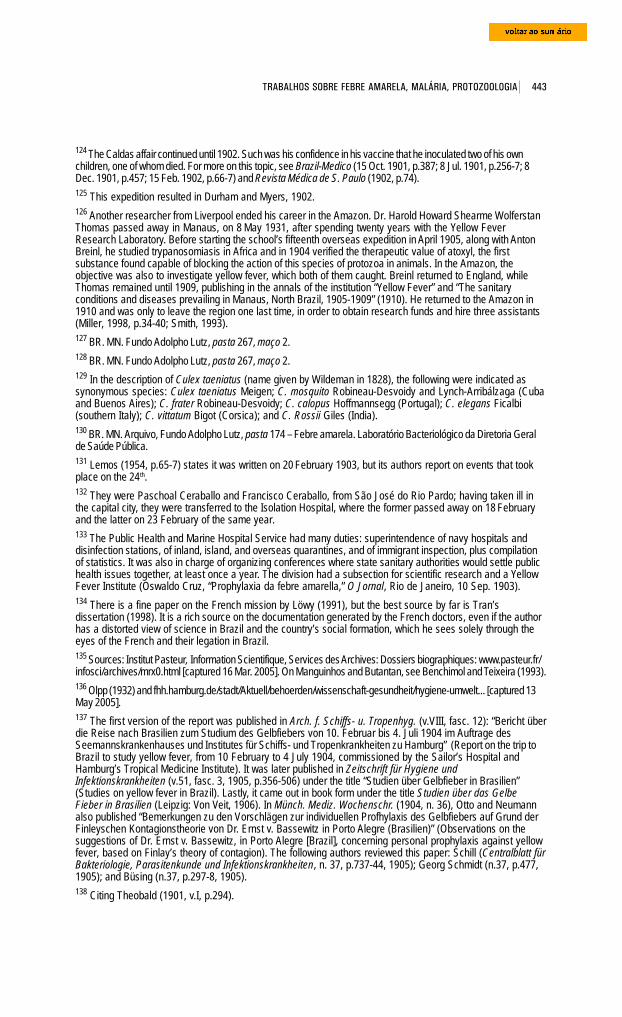
443 TRABALHOS SOBRE FEBRE AMARELA, MALÁRIA, PROTOZOOLOGIA
124 The Caldas affair continued until 1902. Such was his confidence in his vaccine that he inoculated two of his ownchildren, one of whom died. For more on this topic, see Brazil-Medico (15 Oct. 1901, p.387; 8 Jul. 1901, p.256-7; 8Dec. 1901, p.457; 15 Feb. 1902, p.66-7) and Revista Médica de S. Paulo (1902, p.74).125 This expedition resulted in Durham and Myers, 1902.126 Another researcher from Liverpool ended his career in the Amazon. Dr. Harold Howard Shearme WolferstanThomas passed away in Manaus, on 8 May 1931, after spending twenty years with the Yellow FeverResearch Laboratory. Before starting the school’s fifteenth overseas expedition in April 1905, along with AntonBreinl, he studied trypanosomiasis in Africa and in 1904 verified the therapeutic value of atoxyl, the firstsubstance found capable of blocking the action of this species of protozoa in animals. In the Amazon, theobjective was also to investigate yellow fever, which both of them caught. Breinl returned to England, whileThomas remained until 1909, publishing in the annals of the institution “Yellow Fever” and “The sanitaryconditions and diseases prevailing in Manaus, North Brazil, 1905-1909” (1910). He returned to the Amazon in1910 and was only to leave the region one last time, in order to obtain research funds and hire three assistants(Miller, 1998, p.34-40; Smith, 1993).127 BR. MN. Fundo Adolpho Lutz, pasta 267, maço 2.128 BR. MN. Fundo Adolpho Lutz, pasta 267, maço 2.129 In the description of Culex taeniatus (name given by Wildeman in 1828), the following were indicated assynonymous species: Culex taeniatus Meigen; C. mosquito Robineau-Desvoidy and Lynch-Arribálzaga (Cubaand Buenos Aires); C. frater Robineau-Desvoidy; C. calopus Hoffmannsegg (Portugal); C. elegans Ficalbi(southern Italy); C. vittatum Bigot (Corsica); and C. Rossii Giles (India).130 BR. MN. Arquivo, Fundo Adolpho Lutz, pasta 174 – Febre amarela. Laboratório Bacteriológico da Diretoria Geralde Saúde Pública.131 Lemos (1954, p.65-7) states it was written on 20 February 1903, but its authors report on events that tookplace on the 24th.132 They were Paschoal Ceraballo and Francisco Ceraballo, from São José do Rio Pardo; having taken ill inthe capital city, they were transferred to the Isolation Hospital, where the former passed away on 18 Februaryand the latter on 23 February of the same year.133 The Public Health and Marine Hospital Service had many duties: superintendence of navy hospitals anddisinfection stations, of inland, island, and overseas quarantines, and of immigrant inspection, plus compilationof statistics. It was also in charge of organizing conferences where state sanitary authorities would settle publichealth issues together, at least once a year. The division had a subsection for scientific research and a YellowFever Institute (Oswaldo Cruz, “Prophylaxia da febre amarella,” O Jornal, Rio de Janeiro, 10 Sep. 1903).134 There is a fine paper on the French mission by Löwy (1991), but the best source by far is Tran’sdissertation (1998). It is a rich source on the documentation generated by the French doctors, even if the authorhas a distorted view of science in Brazil and the country’s social formation, which he sees solely through theeyes of the French and their legation in Brazil.135 Sources: Institut Pasteur, Information Scientifique, Services des Archives: Dossiers biographiques: www.pasteur.fr/infosci/archives/mrx0.html [captured 16 Mar. 2005]. On Manguinhos and Butantan, see Benchimol and Teixeira (1993).136 Olpp (1932) and fhh.hamburg.de/stadt/Aktuell/behoerden/wissenschaft-gesundheit/hygiene-umwelt... [captured 13May 2005].137 The first version of the report was published in Arch. f. Schiffs- u. Tropenhyg. (v.VIII, fasc. 12): “Bericht überdie Reise nach Brasilien zum Studium des Gelbfiebers von 10. Februar bis 4. Juli 1904 im Auftrage desSeemannskrankenhauses und Institutes für Schiffs- und Tropenkrankheiten zu Hamburg” (Report on the trip toBrazil to study yellow fever, from 10 February to 4 July 1904, commissioned by the Sailor’s Hospital andHamburg’s Tropical Medicine Institute). It was later published in Zeitschrift für Hygiene undInfektionskrankheiten (v.51, fasc. 3, 1905, p.356-506) under the title “Studien über Gelbfieber in Brasilien”(Studies on yellow fever in Brazil). Lastly, it came out in book form under the title Studien über das GelbeFieber in Brasilien (Leipzig: Von Veit, 1906). In Münch. Mediz. Wochenschr. (1904, n. 36), Otto and Neumannalso published “Bemerkungen zu den Vorschlägen zur individuellen Profhylaxis des Gelbfiebers auf Grund derFinleyschen Kontagionstheorie von Dr. Ernst v. Bassewitz in Porto Alegre (Brasilien)” (Observations on thesuggestions of Dr. Ernst v. Bassewitz, in Porto Alegre [Brazil], concerning personal prophylaxis against yellowfever, based on Finlay’s theory of contagion). The following authors reviewed this paper: Schill (Centralblatt fürBakteriologie, Parasitenkunde und Infektionskrankheiten, n. 37, p.737-44, 1905); Georg Schmidt (n.37, p.477,1905); and Büsing (n.37, p.297-8, 1905).138 Citing Theobald (1901, v.I, p.294).
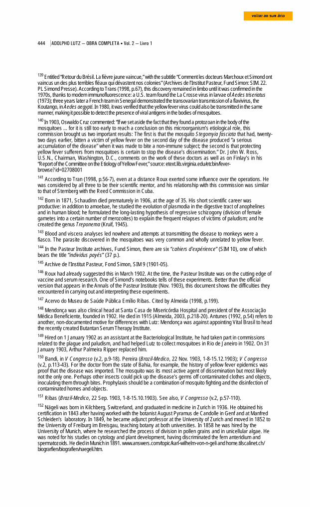
444 ADOLPHO LUTZ — OBRA COMPLETA Vol. 2 — Livro 1
139 Entitled “Retour du Brésil. La fièvre jaune vaincue,” with the subtitle “Comment les docteurs Marchoux et Simond ontvaincus un des plus terribles fléaux qui dévastent nos colonies” (Archives de l’Institut Pasteur, Fund Simon: SIM. 22.PL Simond Presse). According to Trans (1998, p.67), this discovery remained in limbo until it was confirmed in the1970s, thanks to modern immunofluorescence: a U.S. team found the La Crosse virus in larvae of Aedes triseriatus(1973); three years later a French team in Senegal demonstrated the transovarian transmission of a flavivirus, theKoutango, in Aedes aegypti. In 1980, it was verified that the yellow fever virus could also be transmitted in the samemanner, making it possible to detect the presence of viral antigens in the bodies of mosquitoes.140 In 1903, Oswaldo Cruz commented: “If we set aside the fact that they found a protozoan in the body of themosquitoes ... for it is still too early to reach a conclusion on this microorganism’s etiological role, thiscommission brought us two important results: The first is that the mosquito Stegomyia fasciata that had, twenty-two days earlier, bitten a victim of yellow fever on the second day of the disease produced “a seriousaccumulation of the disease” when it was made to bite a non-immune subject; the second is that protectingyellow fever sufferers from mosquitoes is certain to stop the disease’s dissemination.” Dr. John W. Ross,U.S.N., Chairman, Washington, D.C., comments on the work of these doctors as well as on Finlay’s in his“Report of the Committee on the Etiology of Yellow Fever,” source: etext.lib.virginia.edu/etcbin/fever-browse?id=02708001141 According to Tran (1998, p.56-7), even at a distance Roux exerted some influence over the operations. Hewas considered by all three to be their scientific mentor, and his relationship with this commission was similarto that of Sternberg with the Reed Commission in Cuba.142 Born in 1871, Schaudinn died prematurely in 1906, at the age of 35. His short scientific career wasproductive: in addition to amoebae, he studied the evolution of plasmodia in the digestive tract of anophelinesand in human blood; he formulated the long-lasting hypothesis of regressive schizogony (division of femalegametes into a certain number of merozoites) to explain the frequent relapses of victims of paludism; and hecreated the genus Treponema (Kruif, 1945).143 Blood and viscera analyses led nowhere and attempts at transmitting the disease to monkeys were afiasco. The parasite discovered in the mosquitoes was very common and wholly unrelated to yellow fever.144 In the Pasteur Institute archives, Fund Simon, there are six “cahiers d’expérience” (SIM 10), one of whichbears the title “individus payés” (37 p.).145 Archive de l’Institut Pasteur, Fond Simon, SIM 9 (1901-05).146 Roux had already suggested this in March 1902. At the time, the Pasteur Institute was on the cutting edge ofvaccine and serum research. One of Simond’s notebooks tells of these experiments. Better than the officialversion that appears in the Annals of the Pasteur Institute (Nov. 1903), this document shows the difficulties theyencountered in carrying out and interpreting these experiments.147 Acervo do Museu de Saúde Pública Emílio Ribas. Cited by Almeida (1998, p.199).148 Mendonça was also clinical head at Santa Casa de Misericórdia Hospital and president of the AssociaçãoMédica Beneficiente, founded in 1902. He died in 1915 (Almeida, 2003, p.218-20). Antunes (1992, p.54) refers toanother, non-documented motive for differences with Lutz: Mendonça was against appointing Vital Brasil to headthe recently created Butantan Serum Therapy Institute.149 Hired on 1 January 1902 as an assistant at the Bacteriological Institute, he had taken part in commissionsrelated to the plague and paludism, and had helped Lutz to collect mosquitoes in Rio de Janeiro in 1902. On 31January 1903, Arthur Palmeira Ripper replaced him.150 Bandi, in V Congresso (v.2, p.9-18). Pereira (Brazil-Medico, 22 Nov. 1903, 1-8-15.12.1903); V Congresso(v.2, p.113-43). For the doctor from the state of Bahia, for example, the history of yellow fever epidemics wasproof that the disease was imported. The mosquito was its most active agent of dissemination but most likelynot the only one. Perhaps other insects could pick up the disease’s germs off contaminated clothes and objects,inoculating them through bites. Prophylaxis should be a combination of mosquito fighting and the disinfection ofcontaminated homes and objects.151 Ribas (Brazil-Medico, 22 Sep. 1903, 1-8-15.10.1903). See also, V Congresso (v.2, p.57-110).152 Nägeli was born in Kilchberg, Switzerland, and graduated in medicine in Zurich in 1936. He obtained hiscertification in 1843 after having worked with the botanist August Pyramus de Candolle in Genf and at ManfredSchleiden’s laboratory. In 1849, he became adjunct professor at the University of Zurich and moved in 1852 tothe University of Freiburg im Breisgau, teaching botany at both universities. In 1858 he was hired by theUniversity of Munich, where he researched the process of division in pollen grains and in unicellular algae. Hewas noted for his studies on cytology and plant development, having discriminated the fern anteridium andspermatozoids. He died in Munich in 1891. www.answers.com/topic/karl-wilhelm-von-n-geli and home.tilscalinet.ch/biograrfien/biografien/naegeli.htm.
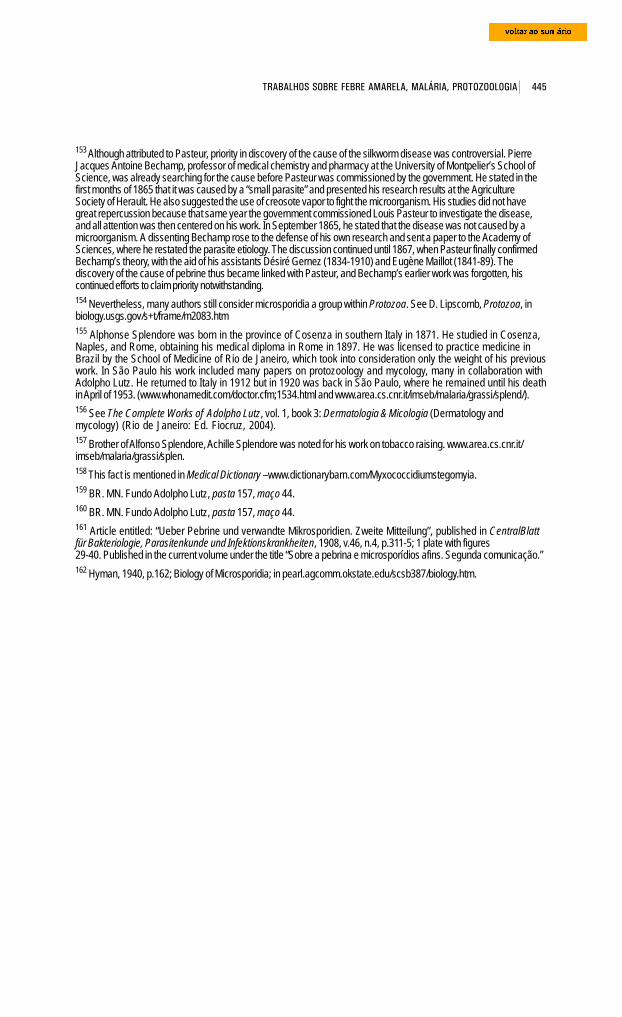
445 TRABALHOS SOBRE FEBRE AMARELA, MALÁRIA, PROTOZOOLOGIA
153 Although attributed to Pasteur, priority in discovery of the cause of the silkworm disease was controversial. PierreJacques Antoine Bechamp, professor of medical chemistry and pharmacy at the University of Montpelier’s School ofScience, was already searching for the cause before Pasteur was commissioned by the government. He stated in thefirst months of 1865 that it was caused by a “small parasite” and presented his research results at the AgricultureSociety of Herault. He also suggested the use of creosote vapor to fight the microorganism. His studies did not havegreat repercussion because that same year the government commissioned Louis Pasteur to investigate the disease,and all attention was then centered on his work. In September 1865, he stated that the disease was not caused by amicroorganism. A dissenting Bechamp rose to the defense of his own research and sent a paper to the Academy ofSciences, where he restated the parasite etiology. The discussion continued until 1867, when Pasteur finally confirmedBechamp’s theory, with the aid of his assistants Désiré Gernez (1834-1910) and Eugène Maillot (1841-89). Thediscovery of the cause of pebrine thus became linked with Pasteur, and Bechamp’s earlier work was forgotten, hiscontinued efforts to claim priority notwithstanding.154 Nevertheless, many authors still consider microsporidia a group within Protozoa. See D. Lipscomb, Protozoa, inbiology.usgs.gov/s+t/frame/m2083.htm155 Alphonse Splendore was born in the province of Cosenza in southern Italy in 1871. He studied in Cosenza,Naples, and Rome, obtaining his medical diploma in Rome in 1897. He was licensed to practice medicine inBrazil by the School of Medicine of Rio de Janeiro, which took into consideration only the weight of his previouswork. In São Paulo his work included many papers on protozoology and mycology, many in collaboration withAdolpho Lutz. He returned to Italy in 1912 but in 1920 was back in São Paulo, where he remained until his deathin April of 1953. (www.whonamedit.com/doctor.cfm;1534.html and www.area.cs.cnr.it/imseb/malaria/grassi/splend/).156 See The Complete Works of Adolpho Lutz, vol. 1, book 3: Dermatologia & Micologia (Dermatology andmycology) (Rio de Janeiro: Ed. Fiocruz, 2004).157 Brother of Alfonso Splendore, Achille Splendore was noted for his work on tobacco raising. www.area.cs.cnr.it/imseb/malaria/grassi/splen.158 This fact is mentioned in Medical Dictionary –www.dictionarybarn.com/Myxococcidiumstegomyia.159 BR. MN. Fundo Adolpho Lutz, pasta 157, maço 44.160 BR. MN. Fundo Adolpho Lutz, pasta 157, maço 44.161 Article entitled: “Ueber Pebrine und verwandte Mikrosporidien. Zweite Mitteilung”, published in CentralBlattfür Bakteriologie, Parasitenkunde und Infektionskrankheiten, 1908, v.46, n.4, p.311-5; 1 plate with figures29-40. Published in the current volume under the title “Sobre a pebrina e microsporídios afins. Segunda comunicação.”162 Hyman, 1940, p.162; Biology of Microsporidia; in pearl.agcomm.okstate.edu/scsb387/biology.htm.
446 ADOLPHO LUTZ — OBRA COMPLETA Vol. 2 — Livro 1
Bibliografia/BibliographyABREU, M. de A. Evolução urbana do Rio de Janeiro. Rio de Janeiro: IplanRio/Jorge Zahar, 1998.ALBUQUERQUE, M. B., BENCHIMOL, J. L., PIRES, F. A., SANTOS, R. A. dos, THIELEN, E. V.,
WELTMAN, W. L. A ciência a caminho da roça: imagens das expediçðes científicas do InstitutoOswaldo Cruz ao interior do Brasil entre 1911 e 1913. Rio de Janeiro: Casa de Oswaldo Cruz(COC)/Fiocruz, 1991.
ALEXOPOULOS, C. J., MIMS, C. W., BLACKWELL, M. Introductory Mycology. 4.ed. New York: JohnWiley & Sons, 1996.
ALGUMAS instalações do Serviço Sanitário de São Paulo. São Paulo: Vanorden, 1905.ALMANAK LAEMMERT (Almanak Administrativo Mercantil e Industrial da Corte e Província do Rio de
Janeiro). Rio de Janeiro: Ed. Laemmert, 1844-1889, 497, 512, 549p., 46 edições.ALMEIDA, F. P. Mycologia Medica. Estudo das mycoses humanas e de seus cogumelos. São Paulo:
Melhoramentos, 1939.ALMEIDA, M. de. República dos invisíveis: Emílio Ribas, microbiologia e saúde pública em São Paulo
(1898-1917). São Paulo, 1998. Dissertação (Mestrado) – Departamento de História, FFLCH,Universidade de São Paulo (sob orientação da professora Dra. Maria Amélia Dantes).
ALMEIDA, M. de. República dos invisíveis: Emílio Ribas, microbiologia e saúde pública em São Paulo(1898-1917). Bragança Paulista: Edusf, 2003.
ANTUNES, J. L.; NASCIMENTO, C. B. do; NASSI, L. C.; PREGNOLATTO, N. P. Instituto Adolfo Lutz –100 anos de laboratório de saúde pública. São Paulo: Secretaria de Estado de Saúde/InstitutoAdolfo Lutz/Ed. Letras & Letras, 1992.
ARQUIVO NACIONAL. Documentos referentes à Junta Comercial do Rio de Janeiro: 1872 – liv. 648,Reg. 11424 G6, firma Lutz e Cia.
BACELLAR, R. C. Brazil’s Contribution to Tropical Medicine and Malaria. Personalities and Institutions.Rio de Janeiro: Gráfica Olímpica, 1963.
BASCH. Anatomisch und klinische Untersuchungen über Dysenterie. Virchow’s Archiv, Bd. XLV, 1869,p.204.
BELLUZO, A. M. de M. O Brasil dos viajantes. A construção da paisagem. São Paulo: Metalivros/Odebrecht, 1994. 3v.
BENCHIMOL, J. L.; SÁ, M. R. Adolpho Lutz e as controvérsias sobre a lepra; Adolpho Lutz andcontroversies over leprosy. In: BENCHIMOL & SÁ (Org.) Adolpho Lutz, Obra Completa, v. I, livro 2:Hanseníase. Leprosy. Rio de Janeiro: Ed. Fiocruz, 2004, p.25-116, 117-205.
BENCHIMOL, J. L.; SÁ, M. R. (Org.) Adolpho Lutz, Obra Completa, v. I, livro 1: Primeiros trabalhos:Alemanha, Suíça e Brasil (1878-1885). First Works: Germany, Switzerland and Brazil (1878-1885);livro 2: Hanseníase. Leprosy; livro 3: Dermatologia & Micologia. Dermatology & Micology; livro 4:Suplemento. Sumário, Glossário e Índices. Supplement. Summary, Glossary and Indexes. Rio deJaneiro: Ed. Fiocruz, 2004.
BENCHIMOL, J. L., SÁ, M. R., BECKER, J. et al. Trabalhos científicos de Adolpho Lutz, publicados ouinéditos. História, Ciências, Saúde – Manguinhos, v.10, n.1, jan.-abr. 2003, p.362-409.
BENCHIMOL, J. L. Adolpho Lutz e a dermatologia em perspectiva histórica; Adolpho Lutz andDermatology in Historical Perspective. In: BENCHIMOL & SÁ (Org.) Adolpho Lutz, Obra Completa,v. I, livro 3: Dermatologia & Micologia. Dermatology & Micology. Rio de Janeiro: Ed. Fiocruz, 2004,p.40-152, 153-262.
_______. Adolpho Lutz: um estudo biográfico. História, Ciências, Saúde – Manguinhos, v.10, n.1, jan.-abr. 2003, p.13-83.
_______. Dos micróbios aos mosquitos. Febre amarela e Revolução pasteuriana no Brasil. Rio deJaneiro: Ed. UFF/Ed. Fiocruz, 1999.
_______. Entrevista com José de Barros Ramalho Ortigão Junior publicada sob o título ‘Recordações dainfância: as primeiras letras com a família Lutz’. História, Ciências, Saúde – Manguinhos, v.10, n.1,jan.-abr. 2003, p.420-4.
447 TRABALHOS SOBRE FEBRE AMARELA, MALÁRIA, PROTOZOOLOGIA
_______. Manguinhos do sonho à vida: a ciência na Belle Époque. Rio de Janeiro: COC/Fiocruz, 1990._______. Pereira Pasos – um Haussmann tropical: a renovação urbana do Rio de Janeiro no início do
século XX. Rio de Janeiro: Secretaria Municipal de Cultura, Turismo e Esportes, DepartamentoGeral de Documentação e Informação Cultural, Divisão de Editoração, 1992, 2.ed. (BibliotecaCarioca, v.11).
BÉRENGER-FÉRAUD, L.-J.-B. Traité théorique & clinique de la fièvre jaune par L.-J.-B. BérengerFéraud, Directeur du Service de Santé, Président du Conseil Supérieur de Santé de la Marine,Membre correspondant de l’Academie de Médicine etc. Paris: Octave Doin, 1890.
BERTRAND & BAUCHER. Nouvelle étude bactériologique des selles dans la dysenterie nostrasépidémique. Gazette hebdomadaire de médecine et de chirurgie, 1893, p.474; 1894, p.176.
BREHM, A. E. Die Lurche und Kriechtierei. Ed. revista por Franz Werner. Leipzig: BibliographischesInstitut in Leipzig, 1925. 2v.
BROMÉLIA. Revista da Sociedade Brasileira de Bromélias, v.3, n.2, 1996. 39p.BRUSCA, R. C., BRUSCA, G. J. Invertebrates. 2.ed. Sunderland, MA: Sinauer Associates, 2003.BULLOCH, W. The history of bacteriology. London/New York/Toronto: Oxford University Press, 1938.BUSVINE, J. R. Disease transmission by insects. Its 90 years of effort to prevent it. Berlin: Springer-
Verlag, 1993.CARTA de Francisco Fajardo a Adolpho Lutz, em 18.3.97. BR. MN. Fundo Adolpho Lutz.CARVALHO, J. M. Os bestializados: o Rio de Janeiro e a República que não foi. São Paulo: Companhia
das Letras, 1987.CASSABOIS, J. Monsieur Pasteur. Paris: Messidor/La Farandole, 1986.CELLI & FIOCIA. Ueber Aetiologie der Dysenteries. Centralblatt für Bakteriologie, Parasitenkunde und
Infektionskrankheiten, 1895, v.XVII, n.9/10, p.309; Riforma medica, 1894, n.37/68, p.806;Centralblatt für Bakteriologie, Parasitenkunde und Infektionskrankheiten, 1894, v.XV-XVI, p.329-470.
CHAGAS, C. A profilaxia do impaludismo. Rio de Janeiro: Typ. Besnard Frères, 1905. Disponíveltambém em O Brazil-Medico, v.20, n.31, 1906, p.351-7; n.33, p.337-40; n.41, p.419-22; v.21, n.16,p.151-4.
_______. Estudos hematológicos no impaludismo. Rio de Janeiro: Typ. da Papelaria União, 1903,220p. (Tese inaugural).
CHAGAS, C. Nova tripanozomiaze humana. Memórias do Instituto Oswaldo Cruz, v.1, n.2, p.159-218,1909.
CHAGAS FILHO, C. Meu pai. Rio de Janeiro: COC/Fiocruz, 1993.CHALHOUB, S. Cidade febril: cortiços e epidemias na corte imperial. São Paulo: Companhia das Letras,
1996.CHANTEMESSE, A.; WIDAL, F. Sur le microbe de la dysenterie épidémique. Bulletin de l’Academie de
Médecine, 1888/Semaine médicale, 18 avr. 1888, p.22.CHAPOT PRÉVOST, E. Etiologia da febre amarela. 26 jan. 1897. Recortes/Casa de Oswaldo Cruz.CHENG, Th. C. The biology of animal parasites. Philadelphia & London: s. n., 1964.COCHRANE, R. G., DAVEY, T. F., McROBERT, G. Leprosy in theory and practice. London: John
Wrigth & Sons, 1964. 659p.CONFÉRENCE INTERNATIONALE DE LA LÈPRE, 3, 1923. Strasbourg. Annales... Paris, 1924. 527p.COUNCILMAN, W. T., LAFLEUR, H. A. Johns Hopkins Hospital Reports: Reports in Pathology, I.
Amœbic Dysenterie. Baltimore, Maryland: The Johns Hopkins Press, 1891.COX, F. E. G. History of Human Parasitology. Clinical Microbiology Reviews, v.15, n.4, Oct. 2002, p.595-
612. Também disponível em: cmr.asm.org/cgi/content/full/15/4/595, acesso em 5.5.2005.CRUZ, O. G. Opera Omnia. Rio de Janeiro: Instituto Oswaldo Cruz, 1972.
448 ADOLPHO LUTZ — OBRA COMPLETA Vol. 2 — Livro 1
DARLING, S. T. Studies in Relation Malaria. Isthmian Canal Commission Public, ICC Press, 1910.DAVIS, N. C. A Field Study of Mountain Malaria in Brazil. American Journal of Epidemiology, v.6, p.119-38.DEANE, M. P. Adolfo Lutz, helmintologista. Revista do Instituto Adolfo Lutz, v.15, 1955, número
comemorativo do centenário de Adolfo Lutz, p.73-85._______. Malaria Vectors in Brazil. Memórias do Instituto Oswaldo Cruz, v.81, Supplement II, 1986, p.5-14.DEBRÉ, P. Pasteur. São Paulo: Scritta, 1995.DELAPORTE, F. Histoire de la fièvre jaune. Paris: Payot, 1989.DOBBELL, C. The Amoebæ Living in Man. London: John Bale and Sons, 1919.DOPTER, Ch. Les dysenteries: étude bactériologique. Paris: Octave Doin et fils, 1909.DOWNS, W. G., PITTENDRIGH, C. S. Bromeliad Malaria in Trinidad and West Indies. American Journal
of Tropical Medicine and Hygiene, v.26, n.1, jan. 1946, 47-66.DURHAM, H. E.; MYERS, W. Memoir VII – Report of the Yellow Fever Expedition to Pará. Liverpool
School of Tropical Medicine, Miscelanea v.1-10, 1902, 75p.DYAR, H. G.; KNAB, F. Descriptions of Some New Mosquitoes from Tropical America. Proceedings U. S.
National Museum, v.XXXV, n.1632, 1908, p.53-70.EMPREENDIMENTOS que honram o Estado de São Paulo. In: O Diário, 26 jan. 1939.FAJARDO, F. Ueber amœbische Hepatitis und Enteritis in den Tropen (Bräsilien). Centralblatt für
Bakteriologie, Parasitenkunde und Infektionskrankheiten, 1896, v.XIX, n.20, p.753._______. Resposta ao professor dr. Domingos Freire. O Brazil-Medico. 15.6.1894, ano VIII, n.23,
p.177-80.FAUST, B.C; BEAVER, P. C.; JUNG, R. C. Animal agents and vectors of human disease. Philadelphia,
Lea and Febiger, 1975, 4th ed.FERNANDES, C. F. de S. A febre amarela em Campinas. Anais Brasilienses de Medicina, v.28, 1876-
1877.FERNANDES, T. M. D. Vacina antivariólica. Ciência, técnica e o poder dos homens, 1808-1920. Rio de
Janeiro: Ed. Fiocruz, 1999.FIDALGO, O. Introdução à história da micologia brasileira. São Paulo: Rickia, 1968. v.3, p.1-44.FINLAY, C. J. Fiebre amarilla experimental comparada con la natural en sus formas benignas. Trabajo
leído en la Sociedad de Estudios Clínicos de la Habana. Sessiones de 31 enero y 29 de febrero de1884. In: _______. Obras completas, 1965, tomo I, p.317-48.
FLEXNER. A Comparative Study of Dysenterie Bacille. Centralblatt für Bakteriologie, Parasitenkundeund Infektionskrankheiten, 1901, n.12, Bd. XXX.
FONSECA FILHO, O. da. A Escola de Manguinhos: contribuição para o estudo do desenvolvimento damedicina experimental no Brasil. (Oswaldo Cruz – Monumenta Histórica). Adendo ao v.2. SãoPaulo: s.n., 1974.
FOSTER, W. D. History of parasitology. London: E & S. Livingstone, 1965.FRANCO, O. História da febre amarela no Brasil. Rio de Janeiro: DNER, Div. de Cooperação e
Divulgação, 1969.FREIRE, D. J. Conférence sur la fièvre jaune, prononcée devant la Société de Thérapeutique
Dosimétrique de Paris. Repertoire Universel de Médicine Dosimétrique, Paris, Mai 1887._______. Conferência sobre a febre amarella realizada pelo dr. Domingos Freire no dia 19 de junho de
1897 no pavilhão central da Faculdade de Medicina analisando a conferência feita em Montevideu nodia 10 do mesmo mês pelo dr. Sanarelli. Rio de Janeiro: Typo-Lithographia de Pinheiro & Cia.,1898.
_______. Vaccination avec la culture atténuée du microbe de la fièvre jaune. Medical News, 17 set.1887, v.51, p.330-4; Jornal do Commercio/O Paiz/Gazeta de Noticias, 22-23 ago. 1899); Brazil-Medico, n.33, 1 set. 1899, p.319.
449 TRABALHOS SOBRE FEBRE AMARELA, MALÁRIA, PROTOZOOLOGIA
_______. “Aos meus amados discípulos drs. Chapot Prevost, Fajardo e V. Ottoni”, Jornal doCommercio, 15.7.1894.
_______. Thérapeutique. Résultats obtenus par l’inoculation préventive du virus atténué de la fièvrejaune, à Rio de Janeiro. Comptes Rendus des Séances de l’Academie des Sciences, Avr. 1887a,t.104, p.1020-2.
_______. Tratamento da febre amarela pelas injeções hipodérmicas de salicilato de sódio. Gazeta deNoticias, Rio de Janeiro, 29-30.3.1880, p.2.
FREIRE, D. J., GIBIER, P., REBOURGEON, C. Médecine expérimentale. Du microbe de la fièvre jauneet de son atténuation. Deuxième note. Comptes Rendus Hebdomanaires des Séances del’Academie des Sciences, 21.3.1887b, t.104, p.858-60.
FREIRE, D.; REBOURGEON, C. Pathologie expérimentale – Le microbe de la fièvre jaune. Inoculationpréventive. Note de MM. D. Freire et Rebourgeon, présenté para M. Bouley. Comptes Rendus desSéances de l’Academie des Sciences, t.XCIX, séance du Lundi 10/11/1884, n.19, p.806.
GADELHA, P. From ‘Forest Malária’ to ‘Bromeliad Malaria’: a Case-study of Scientific Controversy andMalaria Control. Parassitologia. Rivista quadrimestrale dell’Università di Roma “La Sapienza”.Organo Ufficiale della Società della Italiana di Parassitologia, v.36, n.1-2, ago. 1994.
GALLI-VALERIO, B. Sur la présence d’oocystes chez Anopheles lutzi Théobald. Centralblatt fürBakteriologie, Parasitenkunde und Infektionskrankheiten, n.35, p.85, 1904.
GEISON, G. L. The Private Science of Louis Pasteur. Princeton: Princeton University Press, 1995.GOMES, O. C. Manuel Vitorino Pereira, médico e cirurgião. Rio de Janeiro: Agir, 1957.HANSEN, G. A.; LOOFT, C.; NORMAN, W. Leprosy in its clinical & pathological aspects. London: Bristol,
1985. 59p.HARRIS, H. F. Amœbic disentery. The American Journal of the Medical Sciences. Philadelphia & New
York: Lea Brothers & Co, 1898, v.CXV, n.4, p.384-413.HARRISON, G. Mosquitoes, malaria & man: a history of the hostilities since 1880. New York: E. P.
Dutton, 1978.HARVEY, A. McG. Johns Hopkins and Yellow Fever: a Story of Tragedy and Triumph. The Johns
Hopkins Medical Journal, v.19, n.1, July 1981, p.25-39.HASTINGS, R. C. Leprosy. London: Churchill, 1994. 470p.HAVELBURG, W. Recherches expérimentales et anatomiques sur la fièvre jaune. Annales de l’Institut
Pasteur, Paris, année XI, n.6, Juin, 1897, p.515-22._______. Estudos experimentaes, anatomicos e bacteriologicos sobre as propriedades e sobre a
etiologia da febre amarela. Conferencia feita no dia 22 de abril de 1897 no hospital dos Lazaros. Riode Janeiro: Jornal do Commercio, 1897, 40p.
HOWARD, L. O. Smithsonian Miscellaneous Collections (v.84). A History of Applied Entomology.Washington: Smithsonian Institution, 1930.
HUME, E. E. Max von Pettenkofer’s theory of the etiology of cholera, typhoid fever and other intestinaldiseases. A review of his arguments and evidence. Annals of Medical History, v.II, n.4, 1925, p.390-453.
_______. Resumo dos trabalhos feitos no mês de maio pelo director e seus ajudantes, dr. A. deMendonça e dr. João Teixeira. São Paulo, maio 1895, 3p. Folheto.
JOHANN, E., JUNKER, J. Historia de la cultura alemana de los ultimos 100 años. Munique:Nymphenburger Verlagshandlung, 1970.
JOLY, P. R. Os insetos na transmissão das moléstias contagiosas. Pelo dr. P. R. Joly (These deBordeaux). O Brazil-Medico, 22 abr. 1898, v.XX, n.16, p.140.
_______. Papel dos insetos na transmissão das moléstias de origem telúrica. O Brazil-Medico, 1 jul.1899, n.25, p.245.
KARTULIS. Zur Aetiologie der Dysenterie in Aegypten. In: VIRCHOW, R. Archiv für pathologischeAnatomie und Physiologie. Berlin: Druck und Verlag von Georg Reimer, 1886, 101 Band, p.521-32.
KNAB, F. A Chironomid Inhabitant of Sarracenia purpurea. Journal New York Entomological Society,v.XIII, 1905, p.69-73.
450 ADOLPHO LUTZ — OBRA COMPLETA Vol. 2 — Livro 1
_______. A New Bromelicolous Megarhinus (Diptera: Culicidæ). Insecutor Inscitae Menstruus, v.I, n.3,1913a, p.35-6.
_______. Anopheles boliviensis. Notes de parasitologie. Sur la présence d’oocystes chez Anopheleslutzii Theobald. CentralBlatt für Bakteriologie, Parasitenkunde und Infektionskrankheiten, 1 Abt.Orig., v.35, p.85, 1904.
_______. Blood-Sucking Insects as Transmitters of Human Disease. Proceedings of the EntomologicalSociety of Washington, v.XIV, n.4, p.219-21, Oct.-Dec. 1912.
_______. Dr. A. Lutz’s Studies of Brazilian Simuliidæ. Proceedings of the Biological Society ofWashington, v.XIII, n.3, Jul.-Sep., 1911, p.172-9.
_______. The Food Habits of Megarhinus. Psyche, v. 18, p.80-2, 1911._______. Chrysomela staphylea in North America (Col.). Entomological News, n.22, p. 306-9, 1911._______. New Species of Anisopidæ (Rhyphidæ) from Tropical America (Diptera: Nematocera).
Proceedings of the Biological Society of Washington, v.XXV, 1912, p.111-2._______. Some Neotropical Syrphidæ (Diptera). Insecutor Inscitæ Menstruus, v.I, n.2, 1913b, p.14-5._______. Dr. Lutz´s Contentions Regarding ‘Forest Malaria’. Proceedings of the Entomological Society
of Washington, v.XV, 1913c, p.110-8._______. The dependence of disease transmission by blood-sucking insects. Proceedings of the
Entomological Society of Washington, 1913._______. The Larvæ of Megarhinus rutilus Coq. and M. portoricensis Roed. Proceedings of the
Biological Society of Washington, 1904._______. Unconsidered Factors in Disease Transmission by Blood-Sucking Insects. Journal of
Economic Entomology, v.5, Apr. 1912, n.2, p.196-200.KNAB, F., MALLOCH, J. R. A Borborid from an Epiphytic Bromeliad (Diptera: Borboridæ). Entomological
News, v.XXIII, p.413-5.KOLLE, W.; HETSCH, H. La bactériologie expérimentale appliquée a l’étude des maladies infectieuses.
Paris: Atar, 1918, 2v. (3.éd. française revue, augmentée et annotée, d’après la 4.éd. allemande parH. Carrière).
KRUIF, P. de. Microbe Hunters. New York: Pocket Books, 1945.KRUSE. Ueber die Ruhr als Volkskrankheit und ihren Erreger. Deutsche medizinische Wochenschrift,
1900, n.40._______. Die Ruhrgefahr in Deutschland. Centralblatt für Allgemeine Gesundheitspflege, 1900, Bd. XIX,
p.189.KRUSE & PASQUALE. Untersuchungen über Dysenterie um Leberabcess. Zeitschrift für Hygiene, Bd.
XVI, 1894, n.1._______. Eine Expedition nach Aegypten zur Studium der Dysenterie und des Leberabscess. Deutsche
medizinische Wochenschrift, 1893, n.15/16.LACAZ, C. da S. História da micologia médica no Brasil. Ciência e Cultura, v.35, n.11, p.1599-1607,
nov. 1983.LACAZ, C. da S.; PORTO, E.; MARTINS, J. E. C.; HEINS-VACCARI, E. M.; MELO, N. T. de. Tratado
de micologia médica. 9.ed. São Paulo: Sarvier, 2002.LACERDA, J. B. de. O micróbio patogênico da febre amarela. Trabalho lido perante a Academia
Nacional de Medicina e apresentado ao Congresso Médico Pan-Americano de Washington pelo dr.João Batista de Lacerda. Annaes da Academia de Medicina do Rio de Janeiro, 1891-1892, t. LVII.Rio de Janeiro: Cia. Typographica do Brazil, 1891, p.269-375.
_______. A simbiose do bacilo icteróide com um bolor. Focalização da febre amarella no interior dosdomicilios. Communicações lidas perante a Academia Nacional de Medicina do Rio de Janeiro pelodr. J. B. de Lacerda presidente da seção médica. Rio de Janeiro: Typ. Leuzinger, 1900.
_______. Provas e documentos em favor da localização intradomiciliar da febre amarela. (Comunicaçãoà Academia Nacional de Medicina, em sessão de 14 de agosto, pelo dr. J. B. de Lacerda.) Annaesda Academia de Medicina, 1899a, t. LXV. Rio de Janeiro: Typ. Besnard Frères, p.459-73.
_______. “Provas e documentos em favor da localização intradomiciliar da febre amarela”.(Comunicação à Academia Nacional de Medicina, em sessão de 14 de agosto, pelo dr. J. Batista. deLacerda.). O Brazil-Medico, 22.9. 1899b, n.35, p.339-42.
_______. O micróbio da febre amarela. A simbiose do bacilo icteroide com os bolores pelo dr. J.- B. deLacerda. (Comunicação feita à Academia Nacional de Medicina, na sessão de 12 de maio de 1899.)Revue Médico-Chirurgicale du Bresil, jun. 1899c, n.6, p.166-77.
451 TRABALHOS SOBRE FEBRE AMARELA, MALÁRIA, PROTOZOOLOGIA
_______. A simbiose do bacillo icteroide com os bolôres pelo dr. João Baptista de Lacerda.(Comunicação feita à Academia Nacional de Medicina, em sessão de 12 de maio do corrente ano).O Brazil-Medico, 22.5. 1899, n.20, p.192-5.
_______. Provas e documentos em favor da localisação intradomiciliar da febre amarela. (Comunicaçãoà Academia Nacional de Medicina, em sessão de 14 de agosto, pelo dr. J. B. de Lacerda.) Annaesda Academia de Medicina, 1899, t. LXV. Rio de Janeiro: Typ. Besnard Frères, p.459-73.
_______. Fatos concordantes e provas indiretas da interferência dos bolôres na propagação da febreamarela. (Comunicação à Academia Nacional de Medicina, na sessão de 25 de maio de 1899, pelodr. Lacerda). O Brazil-Medico, 8.6.1899, n.22, p.212-4.
LANDOUZY, L., JAYLE, F. Glossaire médical. Paris: Masson et Cie., s.d. [c.1902].LAPA, J. R. do A. A cidade: os cantos e os antros. Campinas 1850-1900. São Paulo: Edusp, 1996.LAVERAN, Ch. L. A. Annales de Dermatologie, v.1, n.173; Bulletin de l’Academie de Medecine de Paris,
1880, v.44, 2.sér., n.9, p.1346._______. Sur la nature de l’agent de la fièvre jaune. Comptes Rendus de la Société de Biologie, t. LIV,
p.391, 1902.LEMOS, F. C. Contribuição à história do Instituto Bacteriológico 1892-1940. Revista do Instituto Adolpho
Lutz, n. especial, 14 nov. 1954.LIVRO de Correspondências 1899–1903, E 01834. Arquivo Permanente/Arquivo do Estado de São
Paulo AP/AESP.LIVRO Relatório 1900, E 01838. Arquivo Permanente/Arquivo do Estado de São Paulo AP/AESP.LOPES, V. J. da S. A febre amarela em Campinas: duas palavras a respeito desta epidemia lidas na
Academia Imperial de Medicina do Rio de Janeiro em sessão de 27 de abril de 1889. Anais daAcademia de Medicina do Rio de Janeiro, 1889, p.331-49.
LÖSCH, F. A. Massenhafte Entwickelung von Amœben im Dickdarm. Virchow’s Archive, 1875, Bd. LXV,p.196.
LUTZ, A. A febre amarella e o mosquito. Gazeta de Noticias (n.299), 20 out. 1903, Rio de Janeiro._______. Ankylostoma duodenal e Ankylostomiase. Gazeta Medica da Bahia, ano XVIII, série 3, v.5,
n.11, p.487-96; ano XIX, n.2, ago. 1888, p.60-5; n.3, set. 1888, p.113-24; n.4, out. 1888, p.157-66;n.6, dez. 1888, p.254-64; ano XX, n.7, jan. 1889, p.315-22; n.9, mar. 1889, p.410-4; n.10,abr.1889, p.451-6.
_______. Ausland. Brasilien. Correspondenz-Blatt für Schweizer Aerzte, Basel, v.13, n.1, 1883, p.30.[Correspondência Cantonal. Boletim de Correspondência para Médicos Suíços, ano XIII, n.1, 1ºjan. 1883.]
_______. Batraciens du Brésil. Diagnoses d’espèces nouvelles. Comptes Rendus des séances de laSociété de Biologie, Paris, v.93, n.22, p.137-9, 211-4. Publicado também como Espécies Novas deBatrachios Brasileiros. New Species of Brazilian Batrachians. Preliminary Note. Reimpresso dasMemórias do Instituto Oswaldo Cruz, Rio de Janeiro, t. XVIII, fasc. 1, 1925.
_______. Contribuição à história da medicina no Brasil segundo os relatórios do dr. Adolpho Lutz comodiretor do Instituto Bacteriológico de S. Paulo (1893-1908). Apresentação de Henrique Aragão,diretor interino do IOC e Introdução de Bertha e Gualter A. Lutz. Cólera Asiático em São Paulo –Disenteria. Memórias do Instituto Oswaldo Cruz, v.39, n.2, 1943, p.177-241. Rio de Janeiro:Imprensa Nacional.
_______. Contribuição para o conhecimento das espécies brasileiras do gênero Simulium. Beitrag zurKentniss der Brasilianischen Simuliumarten. Memórias do Instituto Oswaldo Cruz, t.1, n.2, 1909,p.124-46.
_______. Contribuições à História da Medicina no Brasil. Reminiscências Dermatológicas. A FolhaMédica, v.2, n.19, p.145-6, 1921.
_______. Contribution to the history of medicine in Brazil. The early work and reports of dr. Adolpho Lutz,director of the Bacteriological Institute of the State of São Paulo from 1893 to 1908. Foreword __
Cholera __ Disentery. Reprint from Memórias do Instituto Oswaldo Cruz, v.39, n.2, 1943, p.191-252.Rio de Janeiro: Imprensa Nacional.
_______. Cópia do relatório de 1902, enviado em 26 de janeiro de 1903 à diretoria do ServiçoSanitário. Dr. Carlos Meyer. (Em ‘Lista dos trabalhos e estudos feitos no Instituto Bacteriológico e nãopublicados’ consta: LUTZ, A. Relatório dos trabalhos do Instituto Bacteriológico em 1902. /BR. MN.Fundo Adolpho Lutz, caixa 25, pasta 227, maço 1.)
452 ADOLPHO LUTZ — OBRA COMPLETA Vol. 2 — Livro 1
_______. Ein Fall von Lichen ruber, obtusus et planus. Monatshefte für Praktische Dermatologie, n.6,p.592-6, 1887.
_______. Febre amarella. Revista Médica de São Paulo, 1901, p.2-5._______. Febre amarella. Resposta. Revista Médica de São Paulo. Jornal Pratico de Medicina,
Cirurgia e Hygiene, ano 4, n.4, 1901, p.65-7._______. Forest Malaria. Proceedings of the Entomological Society of Washington, 1913, v.15, n.4,
p.169-70._______. Helminthologisches aus Hawaii von dr. A. Lutz in San Francisco. CentralBlatt für Bakteriologie
und Parasitenkunde, Jena, v.13, n.4, 1892, p.126-8. (Trabalho realizado em San Francisco,Califórnia, em 24.11.1892.)
_______. Instituto Bacteriológico de São Paulo. Relatório de 1900. BR. MN. Fundo Adolpho Lutz, caixa25, pasta 227, maço 1.
_______. Instituto Bacteriológico. Relatório de 1901. BR. MN. Fundo Adolpho Lutz, caixa 25, pasta 227,maço 1.
_______. Klinisches über Parasiten des Menschen und der Hausthiere. Nach Erfahrungen aus einerärztlichen Praxis in Brazilien mitgetheilt von dr. Adolf Lutz in São Paulo – I. Ascaris lumbricoides.Centralblatt für Bakterologie und Parasitenkunde, Jena, ano II, v.3, n.18, 1888, p.553-7; n.19,p.585-8; n.20, p.617-20.
_______. Klinisches über Parasiten des Menschen und der Hausthiere Nach Erfahrungen aus einerärztlichen Praxis in Brazilien mitgetheilt von dr. Adolf Lutz in São Paulo – II. Oxyuris vermicularis.Centralblatt für Bakterologie und Parasitenkunde, Jena, ano II, n.22, 1888, p.681-4; n.23, p.713-6;n.24, p.745-6 (subtítulo: Rhabdonema srongyloides); n.24, p.746-7 (subtítulo: Dochmiusankylostoma und verwandte Arten); n.24, p.747-8. Klinisches über Parasiten des Menschen und derHausthiere Nach Erfahrungen aus einer ärztlichen Praxis in Brazilien mitgetheilt von dr. Adolf Lutz inSão Paulo (Schluss). Trichocephalus dispar, Jena, ano II, n.25, 1888, p.777-8 (subtítulo: Taenien);n.25, p.778-81.
_______. Mosquitos da floresta e malária silvestre. Revista Brasileira de Malariologia, v.II, n.2, 30 abr.1950, p.91-100. (Versão em inglês: Forest mosquitoes and Forest Malaria, p.101-10.)
_______. Nachtrag zu menier lezten Mitteilung über die Invasion von Taenia elliptica und Ascaris.Centralblatt für Bakterologie und Parasitenkunde, ano II, v.3, n.10, 1888, p.299-300. (Publicadonesta edição como “Aditamento à minha última comunicação sobre a invasão de Taenia elliptica eAscaris”.)
_______. Relatório apresentado ao dr. director do Serviço Sanitário do Estado de São Paulo pelo dr.Adolpho Lutz Director do Instituto Bacteriológico de São Paulo – 1896”. Relatório datado de fevereirode 1897. BR. MN. Fundo Adolpho Lutz. Caixa 25, pasta 16, maço 1.
_______. Relatorio apresentado ao dr. director geral do Serviço Sanitario pelo dr. Adolpho Lutz em 26de agosto de 1897 sobre os institutos e trabalhos de hygiene no Rio da Prata. São Paulo:Typographia do Diario Official, 1897, 12p.
_______. Relatório apresentado pelo dr. Adolpho Lutz, director do Instituto Bacteriológico do Estado,acerca de sua commissão em Montevidéo por occasião da conferencia do dr. Sanarelli, relativa àdescoberta do microbio da febre amarella. São Paulo: Typographia do Diario Official, 10p. Trabalhoredigido em São Paulo em 27.7.1897. (Reimpresso: Sobre o bacilo icteroide de Sanarelli. O Brazil-Medico, Revista Semanal de Medicina e Cirurgia, Rio de Janeiro, n.11, p.424-7, 1897.)
_______. Relatório de 1893: febres remittentes e typhoideas. BR. MN. Fundo Adolpho Lutz, caixa 4,pasta 21, maço 2, p.1.
_______. Relatório do Instituto Bacteriológico. São Paulo: Imprensa Official do Estado, 1895. (Reúnerelatórios apresentados em 2 jan. 1894 [p.202-10] e 3 mar. 1895 [p.29-40] à Diretoria do ServiçoSanitário pelo dr. Adolpho Lutz, ainda diretor interino do Instituto.)
_______. Relatório do Instituto Bacteriológico concernente ao ano de 1893. Trechos dos Relatórios doProf. Lutz quando diretor do Instituto Bacteriológico sobre Febre Amarela. BR. MN. Fundo AdolphoLutz, 1893-1894. Pasta 212f, maço 4.
_______. Relatório do Instituto Bacteriológico; ano de 1894. 1º semestre – São Paulo. BR. MN. FundoAdolpho Lutz.
_______. Relatório do Instituto Bacteriológico; ano de 1894. jan./fev. 1895 – São Paulo. BR. MN.Fundo Adolpho Lutz.
_______. Relatório do Instituto Bacteriológico: 1894. BR. MN. Fundo Adolpho Lutz, caixa 2._______. Relatório do Instituto Bacteriológico. São Paulo: Imprensa Official do Estado, 1895.
(Referenciado como: BR. MN. Fundo Adolpho Lutz, caixa 25, pasta 19.)_______. Relatório do Instituto Bacteriológico. São Paulo: Imprensa Official do Estado, 1896.
(Referenciado como: BR. MN. Fundo Adolpho Lutz.)
453 TRABALHOS SOBRE FEBRE AMARELA, MALÁRIA, PROTOZOOLOGIA
_______. Relatório dos trabalhos do Instituto Bacteriológico durante o ano de 1897. Revista Médica deSão Paulo. Jornal Pratico de Medicina, Cirurgia e Hygiene, 15.11.1898, v.1, n.10, p.175-87.
_______. Relatório do Instituto Bacteriológico. São Paulo: Imprensa Official do Estado, 1898.(Referenciado como: BR. MN. Fundo Adolpho Lutz.)
_______. Relatório do Instituto Bacteriológico: impaludismo. São Paulo: Imprensa Official do Estado,1900. (Referenciado como: BR. MN. Fundo Adolpho Lutz.)
_______. Relatório do Instituto Bacteriológico: impaludismo. São Paulo: Imprensa Official do Estado,1901. (Referenciado como: BR. MN. Fundo Adolpho Lutz.)
_______. Relatório do Instituto Bacteriológico. São Paulo: Imprensa Official do Estado, 1902.(Referenciado como: BR. MN. Fundo Adolpho Lutz.)
_______. Reminiscencias sobre a febre amarella no estado de São Paulo. / Erinnerungen aus derGelbfieberzeit im Staate San Paulo. Memórias do Instituto Oswaldo Cruz, t.XXIV, fasc.3, out. 1930,p.127-42 (português), p.143-60 (alemão).
_______. Resumo dos trabalhos do Instituto Bacteriológico de São Paulo, 1892-1908, p.41. BR. MN.Fundo Adolpho Lutz.
_______. Segunda contribuição para o conhecimento das espécies brasileiras do genero Simulium.Zweiter Beitrag zur Kenntnis der Brasilianischen Simuliumarten. Memórias do Instituto OswaldoCruz, t.2, n.2, 1910, p.213-67.
_______. Sul modo di trasporto dell’Ascaris lumbricoides. Giornale della R. Accademia di Medicina diTorino, ano 51, v.36, n.2-3, p.84-6. Torino: Stamperia dell’Unione, 1888.
_______. The Contentions Regarding ‘Forest malaria’. Proceedings of the Entomological Society ofWashington, v.XV, part 2, p.110-8, 1913.
_______. The Insect Host of Forest Malaria. Proceedings of the Entomological Society of Washington,v.15, n.3, 1913, p.108-9.
_______. Trabalhos do Instituto Bacteriologico do Estado de São Paulo durante o anno de 1898 pelo dr.Adolpho Lutz. Relatorio apresentado ao dr. director do Serviço Sanitario de São Paulo. São Paulo:Escola Typographica Salesiana, 1898, 16p.
_______. Über Ankylostoma duodenale und Ankylostomiasis. Sammlung Klinischer Vorträge inVerbindung mit deutschen Klinikern, Innere Medicin, 1885, n.62-92. Leipzig, Druck und Verlag vonBreitkopft und Härtel, 1885, p.255-6 (Innere Medicin, n.88), p.2295-350; 265 (Innere Medicin, n.92),p.2467-506, 4 pranchas.
_______. Über die Ätiologie der Pityriasis hat Prof. Primo Ferrari an die Academia Gioenia di ScienzeNaturali in Catania... Monatshefte für Praktische Dermatologie, Bd., Heft 2, Hamburg, p.84, 1886a.
_______. Über eine neue, in Brasilien beobachtete Krankheit. Monatshefte für Praktische Dermatologie,v.5, n.1, p.32-3, 1886b.
_______. Uma micose pseudococcídica localizada na boca e observada no Brasil. Contribuição aoconhecimento das hifoblastomicoses americanas. O Brazil-Medico, v.22, n.13, p.121-4, 1908.
_______. Vorschläge und Betrachtungen zur Frage der Trichinosistherapie und ihrer experimentellenBegründung von dr. Adolf Lutz in São Paulo in Brasilien. Deutsche Medizinal-Zeitung, Berlin, n.13,13.2.1888, p.151-3; n.15, p.175-6, 20.2.1888.
_______. Waldomosquitos und Waldmalaria. Centralblatt für Bakteriologie, Parasitenkunde undInfektionskrankheiten, Bd. XXXIII, n.4, p.282-92, 7 figs. Jena. Trabalho realizado em São Paulo em16.9.1902. Tradução para o inglês e português: Mosquitos da floresta e malária silvestre, RevistaBrasileira de Malariologia, v.2, n.2, p.91-100, figs.1-7, 1950.
_______. Weiteres zur Frage der Uebertragung des menschlichen Spulwurmes von Adolph Lutz.Centralblatt für Bakteriologie und Parasitenkunde, ano II, 1888, 1ª parte, v.3, n.9, p.265-8; 2ª parte(final), n.10, p.297-9. Trabalho realizado em São Paulo, em 7.1.1888. (Publicado nesta ediçãocomo: Acréscimo à questão da transmissão da solitária humana.)
_______. Zur Behandlung der Atherome. Dermatologische Wochenschrift, 1891, p.505-6. (Publicadocomo: Sobre o tratamento dos ateromas.]
_______. Zur Frage der Invasion von Taenia elliptica und Ascaris lumbricoides. Centralblatt fürBakteriologie und Parasitenkunde, Jena, ano I, (I. Jahr.), 1887, Bd. 2, n.24, p.713-8. (Publicadonesta edição como: Sobre a questão da invasão da Taenia elliptica e do Ascaris Lumbricoides.)
_______. Zur Frage der Uebertragung des menschlichen Spulwurmes. Weitere Mittheilungen von dr.Adolpho Lutz in São Paulo. Centralblatt für Bakterologie und Parasitenkunde, Jena, ano II, v.3,n.14, 1888, p.425-8. Rodapé de Leuckart, p.427. Trabalho realizado em São Paulo. (Publicadonesta edição como: Sobre a questão da transmissão da solitária humana.)
_______. Zur Frage der Uebertragung von Taenia elliptica. Centralblatt für Bakteriologie undParasitenkunde, Jena, ano II, v.3, n.16, 1888, p.489-90. (Publicado nesta edição como: Sobre aquestão da transmissão da Taenia elliptica.)
454 ADOLPHO LUTZ — OBRA COMPLETA Vol. 2 — Livro 1
_______. Zur Frage nach der Wirksamkeit der Gelbefieberimpfung. Freie Presse, São Paulo, v.1, n.66,p.11, 1889. (Publicado nesta edição como: Sobre a questão da eficácia da vacinação contra a febreamarela.)
_______. Zur Kenntniss der Amöben-Enteritis und Hepatitis von dr. A. Lutz in Honolulu (Hawai).Centralblatt für Bakteriologie, Parasitenkunde und Infektionskrankheiten, Bd. X, n.8, 5.9.1891,p.241-8. (Publicado nesta edição como: Contribuição ao Conhecimento da Enterite e HepatiteAmebiana.)
LUTZ, A., NEIVA, A. Contribuição para a biologia das megarhininas com descrição de duas espéciesnovas. Memórias do Instituto Oswaldo Cruz, 1913, t.5, n.2, p.129-41. (Em português e alemão.)
LUTZ, A.; NEIVA, A. Contribuição para o estudo das Megarininae. II. Do Megarhinus haemorrhoidalis(Fabricius, 1794). Beitrag zum Studium der Megarhininae II. Ueber Megarhinus haemorrhoidalis(Fabricius, 1794). Memórias do Instituto Oswaldo Cruz, t.6, n.1, 1914, p.50-7, estampas coloridasn.5-6. (Em português e alemão.)
LUTZ, A.; SPLENDORE, A. Über eine bei Menschen und Ratten beobachtete Mykose. Beitrag zurKenntnis der sogennanten Sporotrichosen. Centralblatt Bakteriologie, Parasitenkunde undInfektionskrankheiten, v.45, p.631-7, 1907.
LUTZ, A., TOLEDO, J. M. B. de. Parecer do Instituto Bacteriologico do estado de São Paulo sobre oserum apresentado pelo sr. dr. Phelippe Caldas. São Paulo: Typographia do Diario Official, 24 maio1897, 8p. (Publicado também como: Serum contra a febre amarela proposto pelo dr. PhilippeCaldas. Parecer dos drs. Adolpho Lutz, Arthur Vieira Mendonça e Bonilha de Toledo, diretor eajudantes do Instituto Bacteriológico do Estado de São Paulo. O Brazil-Medico, Revista Semanal deMedicina e Cirurgia, v.11, n.30, ago. 1897, p.268-9.)
MANSON-BAHR, Ph. H. Manson’s Tropical Diseases. A manual of the diseases of warm climates.London, Toronto, Melbourne, Sydney: Cassel & Co., 1940.
MARTÍNEZ PALOMO, A. Crónica de una polémica centenaria. In: Las amibas, enemigos invisibles.6.reimpr. México: Fondo de Cultura Económica, 1996. (1.ed., 1987.) Também disponível em:bibliotecadigital.ilce.edu.mx/sites/ciencia/volumen1/ciencia2/47/htm/amibas.htm, acesso em 5.5.2005.
MELLO, V. de. De l’identité de la fièvre jaune et de l’impaludisme aigu: conséquente contagiosité del’impaludisme et curabilité de la fièvre jaune par Viera de Mello – Membre de l’Academie deMédecine – Note communiqué à l’Académie des Sciences de Paris. União médica, 1885, v.V, p.565-70.
MILANEZ, A. I. Fungos: heróis ou vilões? Paper read at the 14th MEETING OF BIOLOGISTS, Cuiabá,Mato Grosso. Livro de resumos, 2003. p.43.
MILLER, P. J. Malaria, Liverpool. An illustrated history of the Liverpool School of Tropical Medicine, 1898-1998. Liverpool (UK), Liverpool School of Tropical Medicine, 1998.
NEIVA, A. Necrológico do professor Adolpho Lutz 1855-1940. Rio de Janeiro: Imprensa Nacional, 1941.xxiii p. Reimpresso das Memórias do Instituto Oswaldo Cruz, Rio de Janeiro, t. 36, fasc. 1, p.1-xxiii,1 est., 1941.
_______. Contribuição para o estudo dos dípteros. Observações sobre a biologia e a sistemática dasanofelinas brazileiras e suas relações com o impaludismo./Beitrag zur Kenntniss der Dipeta.Beobachtungen über die Biologie und Systematik der brasilianischen Anophelinen und derenBeziehungen mit der Malaria. Memorias do Instituto Oswaldo Cruz, v.1, n.1, p.69-77, abr. 1909.
_______. Necrológio do professor Adolpho Lutz 1855-1940. Rio de Janeiro: Imprensa Nacional, 1941.OLPP, G. Hervorragende Tropenärzte in Wort und Bild. München: Verlag der Ärztlichen Rundschau
Otto Gmelin, 1932.OTTO, M.; NEUMANN, R. O. Bericht über die Reise nach Brasilien zum Studium des Gelbfiebers von
10. Februar bis 4. Juli 1904 im Auftrage des Seemannskrankenhauses und Institutes für Schiffs-und Tropenkrankheiten zu Hamburg, Arch. f. Schiffs- u. Tropenhyg. Bd VIII. Heft 12, p. 297-9.
OTTO, M.; NEUMANN, R. O. Studien über Gelbfieber in Brasilien. Zeitschr. F. Hygiene undInfektionskrankheiten, Bd. 51, 1905. Heft 3., p.356-506.
OTTO, M.; NEUMANN, R. O. Bemerkungen zu den Vorschlägen zur individuellen Prophylaxis desGelbfiebers auf Grund der Finleyschen Kontagionstheorie von Dr. Ernst v. Bassewitz in Porto Alegre(Brasilien). Münch. Mediz. Wochenschr., 1904, n.36.
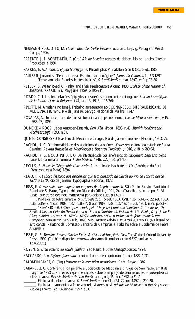
455 TRABALHOS SOBRE FEBRE AMARELA, MALÁRIA, PROTOZOOLOGIA
NEUMANN, R. O., OTTO, M. Studien über das Gelbe Fieber in Brasilien. Leipzig: Verlag Von Veit &Comp., 1906.
PARENTE, J. I.; MONTE-MÓR, P. (Org.) Rio de Janeiro: retratos de cidade. Rio de Janeiro: InteriorProduções, c.1994.
PARKES, E. A. A manual of practical hygiene. Philadelphia: P. Blakston, Son & Co., 6.ed., 1883.PAULSER, Johannes. “Febre amarela. Estudos bacteriológicos”. Jornal do Commercio, 8.3.1897._______. “Febre amarela. Estudos bacteriológicos”. O Brazil-Medico, mar. 1897, no 9, p.78-86.PELLER, S. Walter Reed, C. Finlay, and Their Predecessors Around 1800. Bulletin of the History of
Medicine, v.XXXIII, n.3, May-June 1959, p.195-211.PICADO, C. T. Les broméliacées épiphytes considérées comme milieu biologique. Bulletin Scientifique
de la France et de la Belgique. t.47, fasc. 3, 1913, p.16-360.PINOTTI, M. A malária no Brasil. Trabalho apresentado ao I CONGRESSO INTERAMERICANO DE
MEDICINA, set. 1946. Rio de Janeiro, Serviço Nacional de Malária, 1947.POSADAS, A. Un nuevo caso de micosis fungoidea con psorospermia. Circulo Médico Argentino, v.15,
p.585-97, 1892.QUINCKE & ROOS. Ueber Amœben-Enteritis, Berl. Klin. Woch., 1893, n.45; Munich Medizinische
Wochenschrift, 1893, n.39.QUINTO CONGRESSO Brasileiro de Medicina e Cirurgia. Rio de Janeiro: Imprensa Nacional, 1903, 2v.RACHOU, R. G. Da domesticidade dos anofelinos do subgênero Kerteszia no litoral do estado de Santa
Catarina. Revista Brasileira de Malariologia e Doenças Tropicais, , 1946, v.18, p.589-94.RACHOU, R. G. & COUTINHO, J. O. Da infectibilidade dos anofelinos do subgênero Kerteszia pelos
parasitas da malária humana. Folha Médica, 1946, v.27, n.3, p.1-10.RECLUS, E. Nouvelle Géographie Universelle. Paris: Libraire Hachette, t. XIX (Amérique du Sud,
L’Amazone et la Plata), 1894.REGO, J. P. Esboço histórico das epidemias que têm grassado na cidade do Rio de Janeiro desde
1830 a 1870. Rio de Janeiro: Typographia Nacional, 1872.RIBAS, E. O mosquito como agente da propagação da febre amarela. São Paulo: Serviço Sanitário do
Estado de S. Paulo, Typographia do Diario do Official, 1901, 24p. (Trabalho assinado por E. M.Ribas, que transcreve nota manuscrita por Adolpho Lutz, p.13-21.)
_______. Profilaxia da febre amarela. O Brazil-Medico, 15 set. 1903, XVII, n.35, p.343-7; 22 set. 1903,n.36, p.353-7; 1 out. 1903, n.37, p.363-4; 8 out. 1903, n.38, p.374-6; 15 out. 1903, n.39, p.383-4.
_______. 1896/1898 – Relatório apresentado pelo Chefe da Comissão Sanitária de Campinas, Dr.Emílio Ribas ao Cidadão Diretor Geral do Serviço Sanitário do Estado de São Paulo, Dr. J. J. da S.Pinto, relativo aos anos de 1896 e 1897 e trabalhos sobre a epidemia de febre amarela emCampinas. Manuscrito. São Paulo, 1898. 54p. Instituto Adolfo Lutz, Arquivo, Livro 17. (Na lateral dolivro consta: Relatório da Comissão Sanitária de Campinas e Trabalho sobre a Epidemia de FebreAmarela.)
RISSE, G. B. Mending Bodies, Saving Souls. A History of Hospitals. New York/Oxford: Oxford UniversityPress, 1999. (Também disponível em www.whonamedit.com/doctor.cfm/1627.html; acesso13.4.2005.)
ROSEN, G. Uma história da saúde pública. São Paulo: Hucitec/Unesp/Abrasco, 1994.SACCARDO, P. A. Sylloge fungorum: omnium hucusque cognitorum. Padua, 1882-1931.SALOMON-BAYET, C. (Org.) Pasteur et la revolution pastorienne. Paris: Payot, 1986.SANARELLI, G. Conferência lida perante a Sociedade de Medicina e Cirurgia de São Paulo, em 8 de
março de 1898 ... Primeiras experimentações sobre o emprego de serum curativo e preventivo dafebre amarela. Revista Médica de São Paulo, ano I, n.2, 15 mar. 1898, p.21-7.
_______. Etiologia da febre amarela. O Brazil-Medico, ano XI, n.24, 22 jun. 1897, p.209-20._______. Etiologia e patogenia da febre amarela. Annaes da Academia de Medicina do Rio de Janeiro.
Rio de Janeiro: Typ. Leuzinger, 1897, t.63.
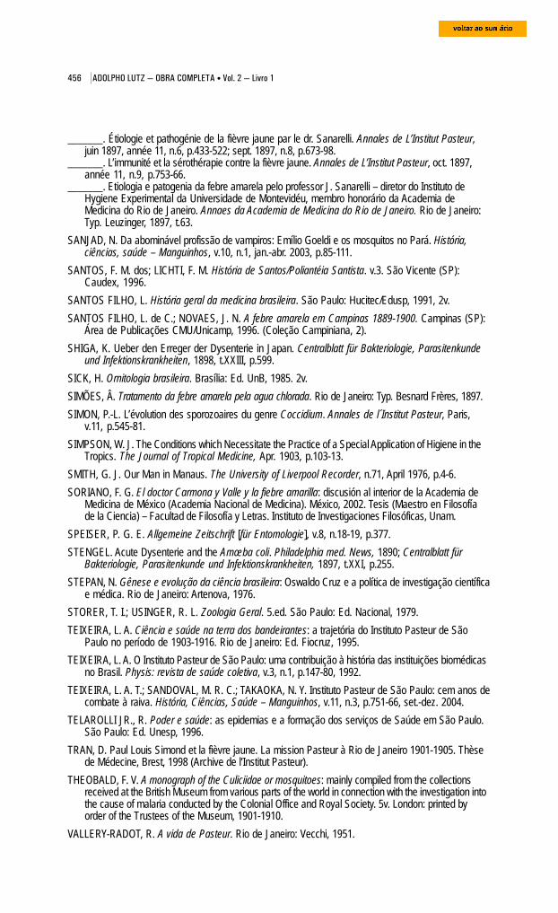
456 ADOLPHO LUTZ — OBRA COMPLETA Vol. 2 — Livro 1
_______. Étiologie et pathogénie de la fièvre jaune par le dr. Sanarelli. Annales de L’Institut Pasteur,juin 1897, année 11, n.6, p.433-522; sept. 1897, n.8, p.673-98.
_______. L’immunité et la sérothérapie contre la fièvre jaune. Annales de L’Institut Pasteur, oct. 1897,année 11, n.9, p.753-66.
_______. Etiologia e patogenia da febre amarela pelo professor J. Sanarelli – diretor do Instituto deHygiene Experimental da Universidade de Montevidéu, membro honorário da Academia deMedicina do Rio de Janeiro. Annaes da Academia de Medicina do Rio de Janeiro. Rio de Janeiro:Typ. Leuzinger, 1897, t.63.
SANJAD, N. Da abominável profissão de vampiros: Emílio Goeldi e os mosquitos no Pará. História,ciências, saúde – Manguinhos, v.10, n.1, jan.-abr. 2003, p.85-111.
SANTOS, F. M. dos; LICHTI, F. M. História de Santos/Poliantéia Santista. v.3. São Vicente (SP):Caudex, 1996.
SANTOS FILHO, L. História geral da medicina brasileira. São Paulo: Hucitec/Edusp, 1991, 2v.SANTOS FILHO, L. de C.; NOVAES, J. N. A febre amarela em Campinas 1889-1900. Campinas (SP):
Área de Publicações CMU/Unicamp, 1996. (Coleção Campiniana, 2).SHIGA, K. Ueber den Erreger der Dysenterie in Japan. Centralblatt für Bakteriologie, Parasitenkunde
und Infektionskrankheiten, 1898, t.XXIII, p.599.SICK, H. Ornitologia brasileira. Brasília: Ed. UnB, 1985. 2v.SIMÕES, Â. Tratamento da febre amarela pela agua chlorada. Rio de Janeiro: Typ. Besnard Frères, 1897.SIMON, P.-L. L’évolution des sporozoaires du genre Coccidium. Annales de l´Institut Pasteur, Paris,
v.11, p.545-81.SIMPSON, W. J. The Conditions which Necessitate the Practice of a Special Application of Higiene in the
Tropics. The Journal of Tropical Medicine, Apr. 1903, p.103-13.SMITH, G. J. Our Man in Manaus. The University of Liverpool Recorder, n.71, April 1976, p.4-6.SORIANO, F. G. El doctor Carmona y Valle y la fiebre amarilla: discusión al interior de la Academia de
Medicina de México (Academia Nacional de Medicina). México, 2002. Tesis (Maestro en Filosofíade la Ciencia) – Facultad de Filosofía y Letras. Instituto de Investigaciones Filosóficas, Unam.
SPEISER, P. G. E. Allgemeine Zeitschrift [für Entomologie], v.8, n.18-19, p.377.STENGEL. Acute Dysenterie and the Amœba coli. Philadelphia med. News, 1890; Centralblatt für
Bakteriologie, Parasitenkunde und Infektionskrankheiten, 1897, t.XXI, p.255.STEPAN, N. Gênese e evolução da ciência brasileira: Oswaldo Cruz e a política de investigação científica
e médica. Rio de Janeiro: Artenova, 1976.STORER, T. I.; USINGER, R. L. Zoologia Geral. 5.ed. São Paulo: Ed. Nacional, 1979.TEIXEIRA, L. A. Ciência e saúde na terra dos bandeirantes: a trajetória do Instituto Pasteur de São
Paulo no período de 1903-1916. Rio de Janeiro: Ed. Fiocruz, 1995.TEIXEIRA, L. A. O Instituto Pasteur de São Paulo: uma contribuição à história das instituições biomédicas
no Brasil. Physis: revista de saúde coletiva, v.3, n.1, p.147-80, 1992.TEIXEIRA, L. A. T.; SANDOVAL, M. R. C.; TAKAOKA, N. Y. Instituto Pasteur de São Paulo: cem anos de
combate à raiva. História, Ciências, Saúde – Manguinhos, v.11, n.3, p.751-66, set.-dez. 2004.TELAROLLI JR., R. Poder e saúde: as epidemias e a formação dos serviços de Saúde em São Paulo.
São Paulo: Ed. Unesp, 1996.TRAN, D. Paul Louis Simond et la fièvre jaune. La mission Pasteur à Rio de Janeiro 1901-1905. Thèse
de Médecine, Brest, 1998 (Archive de l’Institut Pasteur).THEOBALD, F. V. A monograph of the Culiciidae or mosquitoes: mainly compiled from the collections
received at the British Museum from various parts of the world in connection with the investigation intothe cause of malaria conducted by the Colonial Office and Royal Society. 5v. London: printed byorder of the Trustees of the Museum, 1901-1910.
VALLERY-RADOT, R. A vida de Pasteur. Rio de Janeiro: Vecchi, 1951.
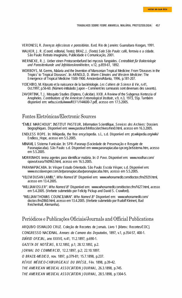
457 TRABALHOS SOBRE FEBRE AMARELA, MALÁRIA, PROTOZOOLOGIA
VERONESI, R. Doenças infecciosas e parasitárias. 8.ed. Rio de Janeiro: Guanabara Koogan, 1991.WALKER, J. R. (Coord. editorial, Texto); BRAZ, J. (Texto) Sala São Paulo: café, ferrovia e a cidade.
São Paulo: Retrato imaginário, Publicidade e Comunicação, 2001.WERNICKE, R. J. Ueber einen Protozoenbefund bei mycosis fungoides. Centralblatt für Bakteriologie
und Parasitenkunde und Infektionskrankheiten, v.12, p.859-61, 1892.WORBOYS, M. Germs, Malaria and the Invention of Mansonian Tropical Medicine: From ‘Diseases in the
Tropics’ to ‘Tropical Diseases’. In: ARNOLD, D. Warm Climates and Western Medicine: TheEmergence of Tropical Medicine 1500-1900. Amsterdam/Atlanta, 1996, p.181-207.
YOICHIRO, M. Kitasato et la naissance de la bactériologie. Les Cahiers de Science & Vie, n.41,Oct.1997, p.56-60. (Número intitulado: Japon – Comment les samouraïs sont devenues des savants).
ZAVORTINK, T. J. Mosquito Studies (Diptera, Culicidæ). XXIX. A Review of the Subgenus Kerteszia ofAnopheles. Contributions of the American Entomological Institute, v.9, n.3, 1973, 55p. Tambémdisponível em: wrbu.si.edu/www/REF1/144600-7.pdf, acesso em 17.5.2005.
Fontes Eletrônicas/Electronic Sources“ÉMILE MARCHOUX”. INSTITUT PASTEUR, Information Scientifique, Services des Archives: Dossiers
biographiques. Disponível em: www.pasteur.fr/infosci/archives/mrx0.html, acesso em 16.3.2005.ENDLESS ROPE. In: Wikipedia, the free encyclopedia. s.l., s.d. Disponível em: pt.wikipedia.org/wiki/
Endless_Hope, acesso em 5.5.2005.MINAMI, I. Sistema Funicular. In: SPR–Paranap (Sociedade de Preservação e Resgate de
Paranapiacaba). São Paulo: s.d. Disponível em www.paranapiacaba-spr.org.br/sistema.htm, acessoem 5.5.2005.
MORRINHOS treina agentes para identificar malária. In: O Povo. Disponível em: www.noolhar.com/opovo/ceara/160963.html, acesso em 16.5.2005.
PARANAPIACABA. In: Vésper Estudo Orientado. São Paulo: Escola Vésper, s.d. Disponível em:www.escolavesper.com.br/paranapiacaba/paranapiacaba.htm, acesso em 5.5.2005.
“VILEM DUSAN LAMBL”. Who Named It? Disponível em: www.whonamedit.com/doctor.cfm/2929.html,acesso em 13.4.2005.
“WILLIAM OSLER”. Who Named It? Disponível em: www.whonamedit.com/doctor.cfm/1627.html, acessoem 5.4.2005. (Verbete submetido por Felicity Pickup and David S. Crawford).
“WILLIAM THOMAS COUNCILMAN”. Who Named It? Disponível em: www.whonamedit.com/doctor.cfm/2860.html, acesso em 13.4.2005. (Verbete submetido por Rudolf Kleinert, BadReichenhall, Alemanha).
Periódicos e Publicações Oficiais/Journals and Official PublicationsARQUIVO OSWALDO CRUZ. Coleção de Recortes de Jornais. Livro 1 [Abrev.: Recortes/COC].CONGRESSO NACIONAL. Annaes da Camara dos Deputados, 1897, v.1, p.354-57, 400-1.DIÁRIO OFICIAL, ano XXXVI, n.41, 11.2.1897, p.690-1.GAZETA DE NOTÍCIAS, 8.12.1892, p.1; 28.12.1892, p.2.JORNAL DO COMMERCIO, 12.2.1897, p.2; 22.10.1897.O BRAZIL-MEDICO, nov. 1897, p.379-81; 15.7.1898, p.237.RÉVUE MÉDICO-CHIRURGICALE DU BRÉSIL, Fév. 1898, p.39-42.THE AMERICAN MEDICAL ASSOCIATION JOURNAL, 26.3.1898, p.745.THE AMERICAN MEDICAL ASSOCIATION JOURNAL, 28.5.1898, p.1304-5.