A stress-responsive RNA switch regulates VEGFA expression
12
A stress-responsive RNA switch regulates VEGF expression Partho Sarothi Ray 1,2 , Jie Jia 1 , Peng Yao 1 , Mithu Majumder 3 , Maria Hatzoglou 3 , and Paul L. Fox 1,* 1 Department of Cell Biology, The Lerner Research Institute, Cleveland Clinic, 9500 Euclid Avenue, Cleveland, Ohio, USA 44195 2 Department of Biology, Indian Institute of Science Education and Research, Kolkata 700106, India 3 Department of Nutrition, Case Western Reserve University, 10900 Euclid Avenue, Cleveland, Ohio, USA 44106 Abstract Ligand binding to structural elements in noncoding regions of mRNA modulates gene expression 1 , 2 . Ligands such as free metabolites or other small molecules directly bind and induce conformational changes in regulatory RNA elements known as riboswitches 1 - 4 . Other types of RNA switches are activated by complexed metabolites, e.g., RNA-ligated metabolites such as aminoacyl-charged tRNA in the T-box system 5 , or protein-bound metabolites in the glucose- or amino acid-stimulated terminator-antiterminator systems 6 , 7 . All of these switch types are found in bacteria, fungi, and plants 8 - 10 . Here, we report an RNA switch in human vascular endothelial growth factor-A (VEGF) mRNA 3’UTR that integrates signals from interferon (IFN)-γ and hypoxia to regulate VEGF translation in myeloid cells. Analogous to riboswitches, the VEGF 3’UTR undergoes a binary conformational change in response to environmental signals. However, the VEGF 3’UTR switch is metabolite-independent, and the conformational change is dictated by mutually exclusive, stimulus-dependent binding of proteins, namely, the IFN-γ-activated inhibitor of translation (GAIT) complex 11 , 12 and heterogenous nuclear ribonucleoprotein (hnRNP) L. We speculate the VEGF switch represents the founding member of a family of signal-mediated, protein-dependent RNA switches that evolved to regulate gene expression in multicellular animals where precise integration of disparate inputs may be more important than rapidity of response. VEGF is induced by hypoxic stress in pathological settings such as tumor cores and atherosclerotic lesions 13 - 15 . These sites are enriched in inflammatory cytokines, including IFN-γ which represses myeloid cell expression of VEGF and other inflammatory proteins by GAIT-mediated translational silencing 16 , 17 . Macrophages simultaneously exposed to opposing inflammatory and hypoxic signals must render a decision to restrict GAIT function to allow VEGF synthesis and risk inflammatory protein accumulation, or permit GAIT- mediated inhibition of VEGF synthesis and risk unrelieved hypoxia. To determine the cell response to this dilemma, U937 monocytic cells were exposed simultaneously to hypoxia * To whom all correspondence should be addressed: Department of Cell Biology The Lerner Research Institute / NC10 Cleveland Clinic 9500 Euclid Avenue Cleveland, OH 44195 Tel: 216-444-8053; Fax: 216-444-9404; [email protected]. E-mail addresses of coauthors: P. S. Ray, [email protected]; J. Jia, [email protected]; P. Yao, [email protected]; M. Majumder, [email protected]; M. Hatzoglou, [email protected] Author Information Reprints and permissions information is available at www.nature.com/reprints. Correspondence and requests for materials should be addressed to P.L.F. ([email protected]). Full Methods and any associated references are available in the online version of the paper at www.nature.com/nature. Supplementary Information is linked to the online version of the paper at www.nature.com/nature. None of the authors have any financial conflict of interest with the information in this manuscript. NIH Public Access Author Manuscript Nature. Author manuscript; available in PMC 2010 April 22. Published in final edited form as: Nature. 2009 February 12; 457(7231): 915–919. doi:10.1038/nature07598. NIH-PA Author Manuscript NIH-PA Author Manuscript NIH-PA Author Manuscript
Transcript of A stress-responsive RNA switch regulates VEGFA expression

A stress-responsive RNA switch regulates VEGF expression
Partho Sarothi Ray1,2, Jie Jia1, Peng Yao1, Mithu Majumder3, Maria Hatzoglou3, and Paul L.Fox1,*
1Department of Cell Biology, The Lerner Research Institute, Cleveland Clinic, 9500 EuclidAvenue, Cleveland, Ohio, USA 441952Department of Biology, Indian Institute of Science Education and Research, Kolkata 700106,India3Department of Nutrition, Case Western Reserve University, 10900 Euclid Avenue, Cleveland,Ohio, USA 44106
AbstractLigand binding to structural elements in noncoding regions of mRNA modulates geneexpression1,2. Ligands such as free metabolites or other small molecules directly bind and induceconformational changes in regulatory RNA elements known as riboswitches1-4. Other types ofRNA switches are activated by complexed metabolites, e.g., RNA-ligated metabolites such asaminoacyl-charged tRNA in the T-box system5, or protein-bound metabolites in the glucose- oramino acid-stimulated terminator-antiterminator systems6,7. All of these switch types are found inbacteria, fungi, and plants8-10. Here, we report an RNA switch in human vascular endothelialgrowth factor-A (VEGF) mRNA 3’UTR that integrates signals from interferon (IFN)-γ andhypoxia to regulate VEGF translation in myeloid cells. Analogous to riboswitches, the VEGF3’UTR undergoes a binary conformational change in response to environmental signals. However,the VEGF 3’UTR switch is metabolite-independent, and the conformational change is dictated bymutually exclusive, stimulus-dependent binding of proteins, namely, the IFN-γ-activated inhibitorof translation (GAIT) complex11,12 and heterogenous nuclear ribonucleoprotein (hnRNP) L. Wespeculate the VEGF switch represents the founding member of a family of signal-mediated,protein-dependent RNA switches that evolved to regulate gene expression in multicellular animalswhere precise integration of disparate inputs may be more important than rapidity of response.
VEGF is induced by hypoxic stress in pathological settings such as tumor cores andatherosclerotic lesions13-15. These sites are enriched in inflammatory cytokines, includingIFN-γ which represses myeloid cell expression of VEGF and other inflammatory proteinsby GAIT-mediated translational silencing16,17. Macrophages simultaneously exposed toopposing inflammatory and hypoxic signals must render a decision to restrict GAIT functionto allow VEGF synthesis and risk inflammatory protein accumulation, or permit GAIT-mediated inhibition of VEGF synthesis and risk unrelieved hypoxia. To determine the cellresponse to this dilemma, U937 monocytic cells were exposed simultaneously to hypoxia
*To whom all correspondence should be addressed: Department of Cell Biology The Lerner Research Institute / NC10 ClevelandClinic 9500 Euclid Avenue Cleveland, OH 44195 Tel: 216-444-8053; Fax: 216-444-9404; [email protected] addresses of coauthors: P. S. Ray, [email protected]; J. Jia, [email protected]; P. Yao, [email protected]; M. Majumder,[email protected]; M. Hatzoglou, [email protected]
Author Information Reprints and permissions information is available at www.nature.com/reprints. Correspondence and requests formaterials should be addressed to P.L.F. ([email protected]).
Full Methods and any associated references are available in the online version of the paper at www.nature.com/nature.
Supplementary Information is linked to the online version of the paper at www.nature.com/nature.
None of the authors have any financial conflict of interest with the information in this manuscript.
NIH Public AccessAuthor ManuscriptNature. Author manuscript; available in PMC 2010 April 22.
Published in final edited form as:Nature. 2009 February 12; 457(7231): 915–919. doi:10.1038/nature07598.
NIH
-PA Author Manuscript
NIH
-PA Author Manuscript
NIH
-PA Author Manuscript
and IFN-γ. The decrease in VEGF protein after normoxic, IFN-γ treatment for 24 h wascompletely abrogated by hypoxia (Fig. 1a, Suppl. Fig. 1). Polysome profiling and RT-PCR(or qRT-PCR) showed that hypoxia restored VEGF translation (Suppl. Figs. 2,3). Consistentwith other reports, mild hypoxia inhibited total protein synthesis nominally (Suppl. Fig. 4)18.Thus, hypoxia overrides GAIT-mediated repression of VEGF translation observed innormoxia16.
To determine whether hypoxia inhibits the GAIT pathway or specifically alters VEGFmRNA response to IFN-γ, we compared in vitro translation of firefly luciferase (Fluc)reporter transcripts bearing ceruloplasmin (Cp) and VEGF mRNA 3’UTRs, both containingfunctional GAIT elements16,19. Lysates from normoxic, but not hypoxic, cells treated withIFN-γ for 24 h repressed translation of the VEGF 3’UTR-bearing reporter (Fig. 1b). Incontrast, both lysates blocked translation of Cp 3’UTR-bearing reporter, indicating afunctional GAIT complex in hypoxia. RNA EMSA/supershift analysis verified binding-competent GAIT complex in hypoxic lysates (Suppl. Fig. 5). Thus, a hypoxic cells factor(s)specifically overcomes silencing of VEGF mRNA translation, implicating a VEGF 3’UTR-related property.
The VEGF 3’UTR contains a 125-nt, hypoxia stability region (HSR) that binds hnRNP L, asplicing factor with cytosolic activities20 including mRNA stabilization21,22 (Fig. 1c). TheVEGF GAIT element resides within the HSR, immediately downstream of the hnRNP Lbinding site. Translation of a reporter transcript bearing the HSR was inhibited by 24-h,IFN-γ-treated normoxic, but not hypoxic, lysate (Fig. 1d). In contrast, translation of atranscript bearing VEGF GAIT element alone was silenced by both lysates. Expression ofHSR-bearing reporter in U937 cells treated with IFN-γ was also restored under hypoxia,whereas a reporter containing only the GAIT element was repressed under both normoxiaand hypoxia (Fig. 1e). Thus, the HSR recapitulates the differential response of VEGFmRNA to IFN-γ and hypoxia in vitro and in vivo. Similar results were observed in primaryhuman peripheral blood monocytes, murine bone marrow-derived macrophages, and otherhuman and murine myeloid cell lines (Suppl. Fig. 6). Glutamyl-prolyl tRNA synthetase(EPRS) is the GAIT protein that binds GAIT element RNA12,23. UV-crosslinking of theHSR with lysates from IFN-γ-treated cells revealed an ~170 kDa protein consistent withEPRS that binds only under normoxia (Fig. 2a). The interaction of a ~60 kDa proteinconsistent with hnRNP L was enhanced by hypoxia22. These bands were identified ashnRNP L (Fig. 2a, left) and EPRS (Fig. 2a, right) by immunodepletion. Notably, depletionof hnRNP L induces EPRS binding to the HSR, even in hypoxia, and the converse was seenafter EPRS depletion, suggesting mutually exclusive binding of hnRNP L and EPRS.
AS3’UTR,332-357, an oligomer antisense to the reported hnRNP L binding site22, markedlydecreased hnRNP L binding to HSR RNA in hypoxic cell lysates (Fig. 2b) and restoredtranslation inhibition of an HSR-bearing reporter RNA in the presence of 24-h, IFN-γ-treated, hypoxic cell lysates, indicating hnRNP L binding is required to overcome translationsilencing (Fig. 2c). Likewise, exogenous expression of myc-tagged hnRNP L in IFN-γ-treated, normoxic cells partially restored expression of VEGF (Suppl. Fig. 7a) and VEGFHSR-containing reporter (Suppl. Fig. 7b). Mutation of the HSR CA-repeats abrogatedbinding of recombinant hnRNP L (Suppl. Fig. 8a) and overcame translation repression bynormoxic IFN-γ treatment (Suppl. Fig. 8b). Similarly, U-to-C mutation in HSR nt position367 (which is essential for GAIT complex binding16) blocked binding of recombinant EPRSlinker (the domain that binds the GAIT element23), and repressed translation even underhypoxia. Lastly, siRNA-mediated knockdown confirmed the requirement for hnRNP L inregulating expression of endogenous VEGF (Fig. 2d) and VEGF HSR-bearing reporter(Suppl. Fig. 9). VEGF expression was not repressed by IFN-γ treatment of normoxic cells
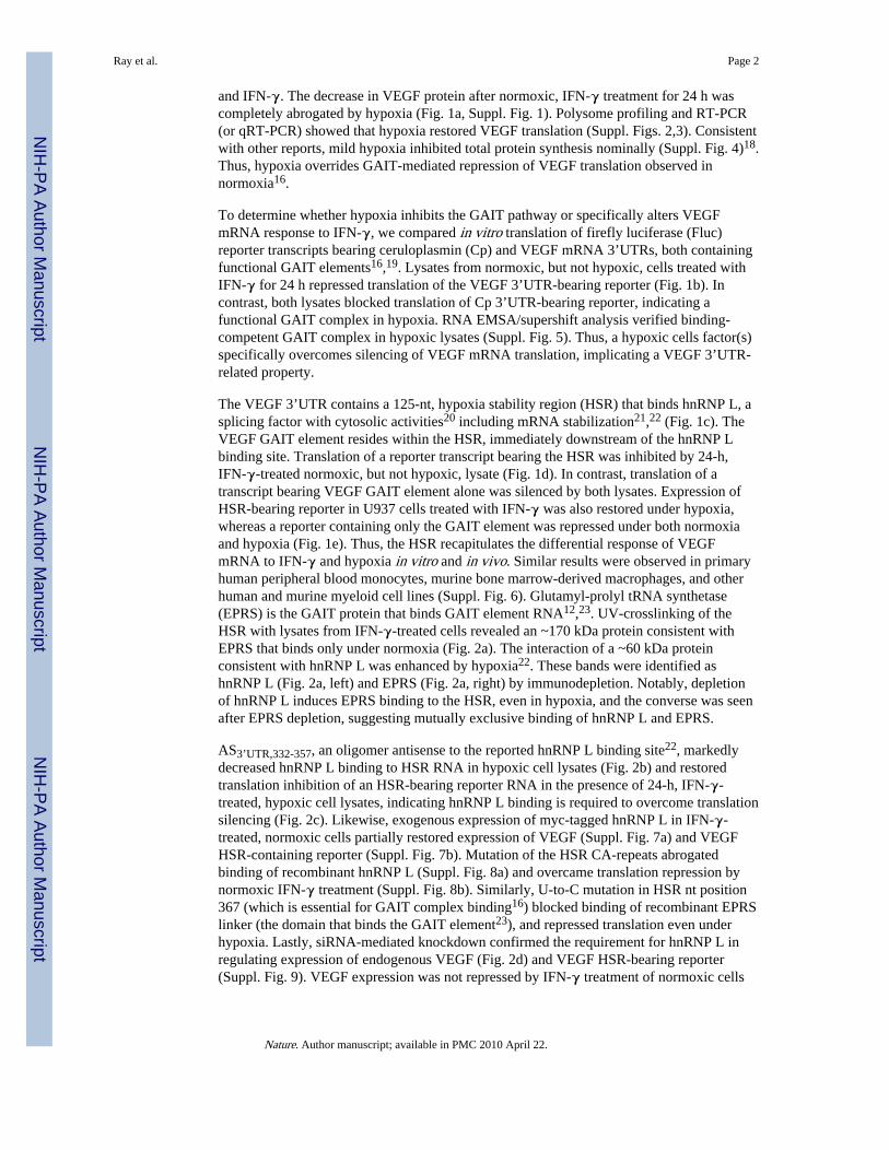
Ray et al. Page 2
Nature. Author manuscript; available in PMC 2010 April 22.
NIH
-PA Author Manuscript
NIH
-PA Author Manuscript
NIH
-PA Author Manuscript

in which GAIT complex formation is prevented by knockdown of the essential GAITcomponent, ribosomal protein L13a (Suppl. Fig 10)11.
Surface plasmon resonance showed hnRNP L (Kd = 61.5 nM) and EPRS linker (Kd = 26.4nM) have comparable affinity for VEGF HSR RNA, suggesting they might compete forbinding, and the equilibrium could be shifted by changes in their relative amounts. Hypoxiadid not increase hnRNP L expression (Fig. 3a). However, IFN-γ treatment in normoxiamarkedly reduced hnRNP L expression, and hypoxia partially blocked this decrease.MG132, a proteasome inhibitor, blocked IFN-γ-mediated reduction of hnRNP L (Fig. 3b).Also, MG132 overcame translational silencing in vitro (Fig. 3c) and in transfected cells(Suppl. Fig. 11). Thus, hypoxia overcomes the IFN-γ-stimulated proteasomal degradation ofhnRNP L and maintains a level of hnRNP L protein that competes with the GAIT complexfor HSR binding.
The proximity of hnRNP L and GAIT complex binding sites suggests a potential interactioncontributing to mutually exclusive binding. The lowest energy (−17.3 kcal/mol) predictedsecondary structure24 of the HSR is stabilized by a 25-bp stem (Fig. 4a, left). Theexperimentally defined GAIT structural element19 is disrupted by base-pairing with adownstream antisense strand (stem stability sequence). The lack of a GAIT elementstructure suggests this predicted conformer is “translation-permissive” (TP). Because GAITcomplex binding to the HSR under normoxia requires GAIT element recognition, we re-folded the HSR under experimentally determined base-pairing constraints essential forGAIT element structure and function19, which predicted a “translation-silencing” (TS)conformer less stable (−4.1 kcal/mol) than the TP conformer (Fig. 4a, right). Wehypothesized that VEGF HSR RNA might exist in both conformations, and that hypoxia andIFN-γ regulate a binary switch between the conformations mediated by mutually exclusivebinding of hnRNP L and GAIT complex.
To validate the RNA switch experimentally, in vitro-transcribed HSR was subjected toRNase A probing25. Nearly all nuclease cleavage sites corresponded to nucleotides predictedto be single-stranded in both conformers (Fig. 4a; 4b, lane 2). A notable exception wascleavage at UTR positions 359 and 361 (GE1), predicted to be a single-stranded bulge in theTP, but base-paired in the TS conformer. Cleavage at positions 371 and 374 (GE2) exhibitedopposite characteristics, i.e., base-paired in the TP but single-stranded in the TS conformer.Robust cleavage at GE1 and GE2 suggested co-existence of both conformers. The presenceof the less energetically favorable TS conformer can be explained by co-transcriptionalfolding, i.e., sequential folding of subdomains in nascent mRNA during transcription26.Heat-denaturation and subsequent renaturation of the HSR eliminated the GE2 signature,establishing the presence of two conformers and a conformational switch (Fig. 4c, lanes1,2). Finally, we investigated the role of proteins in the RNA switch. The HSR wasincubated with lysates from IFN-γ-treated cells, protein was removed, and extracted RNAprobed with RNase A27. Normoxic cell lysate decreased GE1 cleavage, consistent withconversion to the TS conformer (Fig. 4d, lane 2). Conversely, hypoxic lysate inducedconversion to the TP conformer (lane 5). In normoxic lysates, immunodepletion of GAITcomplex switched most RNA to the TP conformer (lane 3), whereas hnRNP L depletioncaused conversion to the TS conformer (lane 4). In hypoxic lysates, hnRNP L depletionpartially restored the TS conformer (lane 7). These data reveal a protein-dependent,hypoxia- and IFN-γ-responsive RNA switch in the VEGF 3’UTR that regulates translation.
The VEGF RNA switch is functionally analogous to bacterial riboswitches, undergoingconformational changes in response to environmental signals to regulate gene expression.However, the sensing mechanisms of the switches are distinct. Riboswitches respond tostimuli by direct binding of the effector molecule. In contrast, the VEGF switch requires
Ray et al. Page 3
Nature. Author manuscript; available in PMC 2010 April 22.
NIH
-PA Author Manuscript
NIH
-PA Author Manuscript
NIH
-PA Author Manuscript

signal “interpretation” by regulatory proteins (Fig. 4e). VEGF mRNA uses an unprecedentedswitching mechanism to integrate the response to two disparate stress stimuli. The switchcomprises a single domain that alternates between two conformers in response to mutuallyexclusive binding of ligands, thereby functioning as an uncommon “AND NOT” Booleanlogic gate (Fig. 4f) distinct from the S-adenosyl methionine and adenosylcobalaminriboswitches in B. clausii metE mRNA which bind their cognate ligands independently toform a NOR logic gate28. Multi-input signal integration may be advantageous for regulatinggene expression in response to the diverse environmental stresses faced by metazoan cells.The switch may be myeloid cell-specific, as EPRS binding and VEGF translationalrepression are not seen in HeLa cells (Suppl. Fig. 12). Transcript specificity permits GAIT-mediated repression of harmful inflammatory proteins while simultaneously allowing high-level VEGF expression to relieve tissue hypoxia15. We anticipate that elements in othereukaryotic transcripts will be discovered as components of RNA switches. A small upstreamORF in arginine transporter cat-1 mRNA internal ribosome entry site is a candidate as itundergoes a conformational change in response to amino acid availability; however,regulatory proteins have not been identified29. The VEGF switch may represent thefounding member of a family of protein-dependent binary RNA switches in whichenvironmental stimuli are transduced by regulatory proteins to alter RNA conformation andcontrol gene expression.
METHODS SUMMARYHuman U937 monocytic cells were incubated with 500 units/ml of human IFN-γ (R & DSystems) for up to 24 h. Hypoxic treatment was in a humidified incubator at 3% pO2.Capped RNAs were transcribed from linearized plasmids using the mMessage mMachinetranscription system (Ambion). [α-32P]UTP-labeled VEGF-HSR RNA was transcribedusing the T7-riboprobe system (Promega). RNAs were in vitro translated in rabbitreticulocyte lysate (Promega) in the presence of methionine-free amino acid mixture andtranslation-grade [35S]methionine (Perkin Elmer) and cytosolic extract (500 ng of protein).Cells were nucleofected (Amaxa) with siRNAs or reporter plasmids containing Flucupstream of wild-type or mutant VEGF HSR expressed from CMV promoter, together withRluc driven by SV40 promoter. For analysis of polysome-associated mRNA, ribosomalfractions were obtained by sucrose gradient fractionation30. RNA was isolated using Trizolreagent (Life Technologies) and subjected to RT using oligo(dT) primers and PCR usinggene-specific primers. The conformational switch in the VEGF HSR RNA was probed byenzymatic cleavage by RNase A (Ambion). To probe protein-dependent RNAconformational shifts, proteins were removed with 5% SDS on ice before RNA extractionwith phenol:chloroform and precipitation by ethanol in presence of 2 mM MgCl227.
METHODSPlasmid construction
The pSP64-FLuc-VEGF-A 3’UTR(11-900)-A30, pSP64 FLuc-Cp 3’UTR-A30, and pSP64-FLuc-VEGF-A GAIT-A30 constructs were generated as described15,31. The VEGF-A3’UTR HSR sequence (nt 332-456) was PCR-amplified and inserted into the pcDNA3vector (Invitrogen) to generate pcDNA3-VEGF-A HSR. The VEGF-A HSR sequence wasalso inserted into pSP64-FLuc-VEGF-A 3’UTR(11-900)-A30 after releasing the VEGF-A3’UTR to generate pSP64-FLuc-VEGF-A HSR-A30. The VEGF HSR was also inserteddownstream of FLuc in pcDNA3 to generate pCD-FLuc-VEGF-A HSR. VEGF-A HSR,with U367 mutated to C, was PCR-amplified by a megaprimer approach and the mutatedsequence was also inserted downstream of FLuc in pcDNA3 to generate pCD-FLuc-VEGF-A HSR-GAITmut. The VEGF-A HSR was also amplified using a forward primer with the Cand A residues in the hnRNP L binding site mutated to G and T respectively, to generate
Ray et al. Page 4
Nature. Author manuscript; available in PMC 2010 April 22.
NIH
-PA Author Manuscript
NIH
-PA Author Manuscript
NIH
-PA Author Manuscript

VEGF-A HSR-hnRNP L sitemut. The sequence was inserted downstream of FLuc inpcDNA3 to generate pCD-FLuc-VEGF-A HSR-hnRNP L site-mut. Both the mutantsequences were also inserted into pcDNA3 to generate the template for in vitro transcription.The pcDNA3-myc-hnRNP L plasmid was a kind gift from Naoyuki Kataoka, University ofPennsylvania School of Medicine. Rat hnRNP L sequence was inserted into a His-taggedprokaryotic expression vector for bacterial expression. The linker domain of human EPRS(aa 753-953) was similarly expressed from a His-tagged prokaryotic expression vector.
Immunodepletion of GAIT Complex and hnRNP LU937 cell lysates were incubated with polyclonal anti-EPRS antibody15 or polyclonal anti-hnRNP L antibody (Santa Cruz) coupled to protein-A Sepharose CL beads (Sigma) in RIPAbuffer. The beads were pelleted and the process was repeated twice with supernatants. Thesupernatants were concentrated and re-dissolved in 10X PBS. The supernatants wereimmunoblotted with anti-EPRS and anti-hnRNP L antibody to verify immunodepletion.
Isolation of Polysome-Associated mRNARibosomal fractions were obtained as described30. Briefly, U937 cells were homogenized inTMK lysis buffer containing cycloheximide (0.1 mg/ml). Cells were lysed and cytosolicextract was obtained by centrifugation at 10,000 × g for 20 min. The extract was overlayedon a 10-50% (w/v) sucrose gradient and centrifuged at 100,000 × g for 4 h at 4 °C. Fractionswere collected using a programmable gradient fractionator (Isco) and absorbance at 254 nmwas measured.
Cell Transfection and Luciferase assayU937 cells were transiently co-transfected with 6 μg of pCD-FLuc-VEGF-A HSR and 8 μgof pCD-myc-hnRNPL DNAs using human dendritic cell nucleofection kit V (AmaxaBiosystems). RLuc-expressing vector pRL-SV40 (1 μg) was co-transfected fornormalization of transfection efficiency. After 12 h, transfected cells were incubated withIFN-γ, lysed, and either immunoblotted or luciferase activity of the lysate was measuredusing dual luciferase assay kit (Promega). Cells were similarly nucleofected with 400 nMsiRNA against hnRNP L (target sequence: 1066-1084, UAUGGCUUGGAUCAAUCUA) orwith a control siRNA which is not similar to any human genomic sequence(AAGCGCTACT-ACAGCAGTC).
UV-crosslinking of RNA-protein complexes32P-UTP labeled RNAs were incubated with cell lysates or bacterially-expressed andpurified EPRS linker or rat hnRNP L proteins and then UV-crosslinked. The RNA-proteincomplexes were extensively digested by RNase A digestion and resolved by SDS-PAGE
Computational prediction of RNA structureRNA secondary structure predictions of the VEGF HSR RNA were performed using theMfold program (http://mfold.bioinfo.rpi.edu), and the structure with the lowest free energyselected. The same sequence was refolded by incorporating base-pairing constraints in theGAIT element stems that have been experimentally determined to be required for GAITcomplex binding and activity18.
Enzymatic probing of RNA StructureVEGF-A HSR RNA was generated by in vitro transcription, and then 5′-end-labeled with[γ-32P]ATP using the KinaseMax kit (Ambion). The RNA was purified using Micro Bio-Spin chromatography columns (Biorad) and the purity tested on 8 M urea-5%polyacrylamide gel. The RNA was probed with RNase A (Ambion) as described24 with
Ray et al. Page 5
Nature. Author manuscript; available in PMC 2010 April 22.
NIH
-PA Author Manuscript
NIH
-PA Author Manuscript
NIH
-PA Author Manuscript

modifications. Briefly, about 10,000 cpm of the end-labeled RNA, together with 3 μg ofyeast tRNA, was incubated with 10 pg of RNase A in 1X RNA structure buffer for 10 min atroom temperature. Amounts of RNase used in the reactions were initially titrated to ensure“single-hit” cleavage. For RNA sequencing reactions, similar amounts of end-labeled RNAand tRNA was incubated with 20 pg of RNase A in 1X sequencing buffer at 50 °C for 5min. The single nucleotide ladder was generated by incubating similar amounts of end-labeled RNA and tRNA in hydroxide hydrolysis buffer (50 mM NaOH, 1 mM EDTA) at 90°C for 3 min. The reactions were resolved on 8 M urea-10% polyacrylamide sequencinggels.
For probing the protein-directed RNA conformational change, extraction of end-labeledRNA after protein-binding was done as described previously26 with modifications. Briefly,the RNA was incubated with 100 ng of cell lysates, and immundepleted lysates, in RNAbinding buffer at room temperature for 15 min. Proteins were removed by incubation with 2μl of 5% SDS on ice. The RNA was extracted twice with phenol:chloroform andprecipitated by ethanol in presence of 2 mM MgCl2. The RNA was further subjected toRNase digestion as described above.
Supplementary MaterialRefer to Web version on PubMed Central for supplementary material.
AcknowledgmentsWe are grateful to D. Driscoll and T.M. Henkin for helpful discussions. This work was supported by NIH grantsP01 HL29582, R01 HL67725, and P01 HL76491 (to P.L.F.), and R01 DK60596 (to M.H.).
REFERENCES1. Mandal M, Breaker RR. Gene regulation by riboswitches. Nat Rev Mol Cell Biol. 2004; 5:451–463.
[PubMed: 15173824]
2. Grundy FJ, Henkin TM. Regulation of gene expression by effectors that bind to RNA. Curr OpinMicrobiol. 2004; 7:126–131. [PubMed: 15063848]
3. Winkler W, Nahvi A, Breaker RR. Thiamine derivatives bind messenger RNAs directly to regulatebacterial gene expression. Nature. 2002; 419:952–956. [PubMed: 12410317]
4. Cromie MJ, Shi Y, Latifi T, Groisman EA. An RNA sensor for intracellular Mg2+ Cell. 2006;125:71–84. [PubMed: 16615891]
5. Grundy FJ, Henkin TM. tRNA as a positive regulator of transcription antitermination in B. subtilis.Cell. 1993; 74:475–482. [PubMed: 8348614]
6. Merino E, Babitzke P, Yanofsky C. trp RNA-binding attenuation protein (TRAP)-trp leader RNAinteractions mediate translational as well as transcriptional regulation of the Bacillus subtilis trpoperon. J Bacteriol. 1995; 177:6362–6370. [PubMed: 7592410]
7. Schilling O, Langbein I, Muller M, Schmalisch MH, Stulke J. A protein-dependent riboswitchcontrolling ptsGHI operon expression in Bacillus subtilis: RNA structure rather than sequenceprovides interaction specificity. Nucleic Acids Res. 2004; 32:2853–2864. [PubMed: 15155854]
8. Batey RT. Structures of regulatory elements in mRNAs. Curr Opin Struct Biol. 2006; 16:299–306.[PubMed: 16707260]
9. Cheah MT, Wachter A, Sudarsan N, Breaker RR. Control of alternative RNA splicing and geneexpression by eukaryotic riboswitches. Nature. 2007; 447:497–500. [PubMed: 17468745]
10. Wachter A, Tunc-Ozdemir M, Grove BC, Green PJ, Shintani DK, Breaker RR. Riboswitch controlof gene expression in plants by splicing and alternative 3′ end processing of mRNAs. Plant Cell.2007; 19:3437–3450. [PubMed: 17993623]
Ray et al. Page 6
Nature. Author manuscript; available in PMC 2010 April 22.
NIH
-PA Author Manuscript
NIH
-PA Author Manuscript
NIH
-PA Author Manuscript

11. Mazumder B, Sampath P, Seshadri V, Maitra RK, DiCorleto P, Fox PL. Regulated release of L13afrom the 60S ribosomal subunit as a mechanism of transcript-specific translational control. Cell.2003; 115:187–198. [PubMed: 14567916]
12. Sampath P, Mazumder B, Seshadri V, Gerber CA, Chavatte L, Kinter M, Ting SM, Dignam JD,Kim S, Driscoll DM, Fox PL. Noncanonical function of glutamyl-prolyl-tRNA synthetase: gene-specific silencing of translation. Cell. 2004; 119:195–208. [PubMed: 15479637]
13. Braunstein S, Karpisheva K, Pola C, Goldberg J, Hochman T, Yee H, Cangiarella J, Arju R,Formenti SC, Schneider RJ. A hypoxia-controlled cap-dependent to cap-independent translationswitch in breast cancer. Mol Cell. 2007; 28:501–512. [PubMed: 17996713]
14. Bastide A, Karaa Z, Bornes S, Hieblot C, Lacazette E, Prats H, Touriol C. An upstream openreading frame within an IRES controls expression of a specific VEGF-A isoform. Nucleic AcidsRes. 2008; 36:2434–2445. [PubMed: 18304943]
15. Ferrara N, Davis-Smyth T. The biology of vascular endothelial growth factor. Endocr Rev. 1997;18:4–25. [PubMed: 9034784]
16. Ray PS, Fox PL. A post-transcriptional pathway represses monocyte VEGF-A expression andangiogenic activity. EMBO J. 2007; 26:3360–3372. [PubMed: 17611605]
17. Mukhopadhyay R, Ray PS, Arif A, Brady AK, Kinter M, Fox PL. DAPK-ZIPK-L13a axisconstitutes a negative-feedback module regulating inflammatory gene expression. Mol Cell. 2008In press.
18. Liu L, Cash TP, Jones RG, Keith B, Thompson CB, Simon MC. Hypoxia-induced energy stressregulates mRNA translation and cell growth. Mol Cell. 2006; 21:521–531. [PubMed: 16483933]
19. Sampath P, Mazumder B, Seshadri V, Fox PL. Transcript-selective translational silencing bygamma interferon is directed by a novel structural element in the ceruloplasmin mRNA 3′untranslated region. Mol Cell Biol. 2003; 23:1509–1519. [PubMed: 12588972]
20. Piñol-Roma S, Swanson MS, Gall JG, Dreyfuss G. A novel heterogeneous nuclear RNP proteinwith a unique distribution on nascent transcripts. J Cell Biol. 1989; 109:2575–2587. [PubMed:2687284]
21. Claffey KP, Shih SC, Mullen A, Dziennis S, Cusick JL, Abrams KR, Lee SW, Detmar M.Identification of a human VPF/VEGF 3′ untranslated region mediating hypoxia-induced mRNAstability. Mol Biol Cell. 1998; 9:469–481. [PubMed: 9450968]
22. Shih SC, Claffey KP. Regulation of human vascular endothelial growth factor mRNA stability inhypoxia by heterogeneous nuclear ribonucleoprotein L. J Biol Chem. 1999; 274:1359–1365.[PubMed: 9880507]
23. Jia J, Arif A, Ray PS, Fox PL. WHEP domains direct noncanonical function of glutamyl-prolyltRNA synthetase in translational control of gene expression. Mol Cell. 2008; 29:679–690.[PubMed: 18374644]
24. Jaeger JA, Turner DH, Zuker M. Improved predictions of secondary structures for RNA. Proc NatlAcad Sci U S A. 1989; 86:7706–7710. [PubMed: 2479010]
25. Knapp G. Enzymatic approaches to probing RNA secondary and tertiary structure. MethodsEnzymol. 1989; 180:192–212. [PubMed: 2482414]
26. Brion P, Westhof E. Hierarchy and dynamics of RNA folding. Annu Rev Biophys Biomol Struct.1997; 26:113–137. [PubMed: 9241415]
27. Huthoff H, Berkhout B. Two alternating structures of the HIV-1 leader RNA. RNA. 2001; 7:143–157. [PubMed: 11214176]
28. Sudarsan N, Hammond MC, Block KF, Welz R, Barrick JE, Roth A, Breaker RR. Tandemriboswitch architectures exhibit complex gene control functions. Science. 2006; 314:300–304.[PubMed: 17038623]
29. Yaman I, Fernandez J, Liu H, Caprara M, Komar AA, Koromilas AE, Zhou L, Snider MD,Scheuner D, Kaufman RJ, Hatzoglou M. The zipper model of translational control: a smallupstream ORF is the switch that controls structural remodeling of an mRNA leader. Cell. 2003;113:519–531. [PubMed: 12757712]
30. Merrick WC, Hensold JO. Analysis of eukaryotic translation in purified and semipurified systems.Curr Protocols Cell Biol. 2001 Chapter 11.
Ray et al. Page 7
Nature. Author manuscript; available in PMC 2010 April 22.
NIH
-PA Author Manuscript
NIH
-PA Author Manuscript
NIH
-PA Author Manuscript

31. Mazumder B, Seshadri V, Imataka H, Sonenberg N, Fox PL. Translational silencing ofceruloplasmin requires the essential elements of mRNA circularization: Poly(A) tail, poly(A)-binding protein, and eukaryotic translation initiation factor 4G. Mol Cell Biol. 2001; 21:6440–6449. [PubMed: 11533233]
Ray et al. Page 8
Nature. Author manuscript; available in PMC 2010 April 22.
NIH
-PA Author Manuscript
NIH
-PA Author Manuscript
NIH
-PA Author Manuscript

Figure 1. Suppression of GAIT-mediated translation silencing of VEGF by hypoxiaa, VEGF in lysates from U937 cells treated with IFN-γ in normoxia (Nmx.) or hypoxia(Hpx.) was determined by immunoblot (IB) and RT-PCR; GAPDH was probed as control.b, In vitro translation of Fluc reporter RNAs bearing VEGF 3’UTR11-900 (left) or Cp 3’UTR(right) in presence of cytosolic lysates and control Rluc RNA lacking a 3’UTR. c, Schematicof RNA elements in VEGF 3’UTR and HSR. d, In vitro translation of reporter RNAsbearing VEGF HSR (left) or GAIT element (right) in presence of cytosolic lysates. e, U937cells were nucleofected with pcDNA3-Fluc reporters bearing VEGF HSR (top) or GAITelement (bottom). Cells were co-transfected with a plasmid expressing Rluc under SV40promoter. Relative luciferase activity (Fluc/Rluc) was expressed as mean ± s.d. (3experiments).
Ray et al. Page 9
Nature. Author manuscript; available in PMC 2010 April 22.
NIH
-PA Author Manuscript
NIH
-PA Author Manuscript
NIH
-PA Author Manuscript

Figure 2. hnRNP L binding to HSR restores VEGF translation in hypoxiaa, Lysates from IFN-γ-treated cells were subjected to UV-crosslinking with [32P]UTP-labeled VEGF HSR RNA before and after immunodepletion with anti-hnRNP L (left) oranti-EPRS (right) antibodies. Effective depletion was shown by IB. b, Excess DNAoligomer antisense to hnRNP L binding site (AS3’UTR,332-357) blocks binding of hnRNP Lto HSR RNA. c, Inhibition of hnRNP L binding by AS3’UTR,332-357 restores translationalsilencing of reporter RNA in hypoxia. d, siRNA-mediated knockdown of hnRNP L inducestranslational repression of VEGF in hypoxic cells. Lysates from cells transfected withhnRNP L (left) and control (right) siRNAs were immunoblotted with anti-VEGF, -hnRNP Land -GAPDH antibodies.
Ray et al. Page 10
Nature. Author manuscript; available in PMC 2010 April 22.
NIH
-PA Author Manuscript
NIH
-PA Author Manuscript
NIH
-PA Author Manuscript

Figure 3. hnRNP L is regulated by stimulus-dependent proteasomal degradationa, Lysates from U937 cells incubated in the absence or presence of IFN-γ wereimmunoblotted with anti-hnRNP L and -GAPDH antibodies. b, Immunoblot of lysates fromcells treated with MG132 (200 nM). c, In vitro translation of VEGF HSR reporter RNA inpresence of cell lysates.
Ray et al. Page 11
Nature. Author manuscript; available in PMC 2010 April 22.
NIH
-PA Author Manuscript
NIH
-PA Author Manuscript
NIH
-PA Author Manuscript

Figure 4. Protein-dependent switching of the VEGF 3’UTR HSRa, Secondary structure of VEGF HSR predicted by Mfold shows GAIT element (green),hnRNP L binding site (red), and stem stability sequence (blue). TP is lowest free energyconformer predicted by Mfold (left). TS conformer was generated by introducingexperimentally-determined base-pairing constraints in GAIT element stem (right). Strongand weak RNase cleavage sites are marked by red and blue circles, respectively. Keysignature cleavage sites are indicated (*, **). b, 32P-endlabeled VEGF HSR RNA wasprobed with RNase A under non-denaturing (lane 2) and denaturing (lane 3) conditions.Cleavages corresponding to predicted signature sites are indicated (*, **). c, RNase Aprobing of VEGF HSR RNA under non-denaturing conditions, or after the RNA wasdenatured and renatured. d, VEGF HSR RNA was incubated with cell lysates treated withIFN-γ for 24 h under normoxia or hypoxia, or with lysates immunodepleted with anti-hnRNP L and anti-EPRS antibodies, and subjected to RNase A-mediated cleavage undernon-denaturing conditions after protein removal. e, Proposed pathway that switches theVEGF HSR to the TP conformer in the presence of IFN-γ and hypoxia (left), or to the TSconformer in the presence of IFN-γ and normoxia (right). f, Truth table showing AND NOTBoolean logic function of the VEGF RNA switch integrating signals from IFN-γ andhypoxia.
Ray et al. Page 12
Nature. Author manuscript; available in PMC 2010 April 22.
NIH
-PA Author Manuscript
NIH
-PA Author Manuscript
NIH
-PA Author Manuscript