A review of Spegazzini taxa of Periconia and Sporocybe after over 115 years
10
Fungal Diversity 67 A review of Spegazzini taxa of Periconia and Sporocybe after over 115 years Cecilia C. Carmarán and M. Victoria Novas * Departamento de Biodiversidad y Biología Experimental, Facultad de Cs. Exactas y Naturales (UBA), Ciudad Universitaria, Pabellón II, 4to Piso, C1428EHA, Buenos Aires, Argentina Carmarán, C.C. and Novas, M.V. (2003). A review of Spegazzini taxa of Periconia and Sporocybe after over 115 years. Fungal Diversity 14: 67-76. Some groups or genera described by Spegazzini have never been re-examined. In this paper the type material of Sporocybe and Periconia species described by Spegazzini are studied. The genus Periconia is represented in Argentina by seven species: P. bromeliicola, P. byssoides, P. circinata, P. lateralis, P. minutissima, P. spegazzinii and P. tirupatiensis. Among Spegazzini’s holotypes of Sporocybe, S. bromeliicola is considered a nomen dubium, S. antarctica is a lichen, S. chlorocephala is a synonym of Stromatographium stromaticum, Sporocybe penicillata is a synonym of Melanographium spinulosum and S. sacchari is a synonym of Doratomyces purpureofuscus. Doratomyces is represented by four species: D. asperulus, D. microsporus D. purpureofuscus and D. stemonitis, three of which are reported under this genus for the first time. Key words: Argentina, Dematiaceae, Doratomyces, hyphomycetes Introduction During early botanic description in South America, between 1879 and 1926, Carlos Spegazzini, described and identified ca. 4000 species of fungi. This was an enormous contribution to Mycology in Argentina, considering that only 39 species were recognised from Argentina before his exhaustive work. His collections are deposited in the Herbarium LPS at the “Instituto de Botánica Carlos Spegazzini”, La Plata, Buenos Aires Province. Some groups or genera described by Spegazzini have been re-examined, eg. Heinemann (1962) studied the types of the genus Agaricus; Carmarán (2002) the Diatrypaceae, Hladki and Romero (2001) studied Kretzschmaria species from Argentina, Lindquist and Wright (1959) studied the identity of Poroniopsis and Hypocreodendron. and Rajchenberg and Wright (1987) studied Spegazzini's types of Corticiaceae and Polyporaceae. We present the * Corresponding author: C.C. Carmarán; email: [email protected]
-
Upload
independent -
Category
Documents
-
view
1 -
download
0
Transcript of A review of Spegazzini taxa of Periconia and Sporocybe after over 115 years

Fungal Diversity
67
A review of Spegazzini taxa of Periconia and Sporocybe afterover 115 years
Cecilia C. Carmarán and M. Victoria Novas*
Departamento de Biodiversidad y Biología Experimental, Facultad de Cs. Exactas y Naturales(UBA), Ciudad Universitaria, Pabellón II, 4to Piso, C1428EHA, Buenos Aires, Argentina
Carmarán, C.C. and Novas, M.V. (2003). A review of Spegazzini taxa of Periconia andSporocybe after over 115 years. Fungal Diversity 14: 67-76.
Some groups or genera described by Spegazzini have never been re-examined. In this paper thetype material of Sporocybe and Periconia species described by Spegazzini are studied. Thegenus Periconia is represented in Argentina by seven species: P. bromeliicola, P. byssoides, P.circinata, P. lateralis, P. minutissima, P. spegazzinii and P. tirupatiensis. Among Spegazzini’sholotypes of Sporocybe, S. bromeliicola is considered a nomen dubium, S. antarctica is alichen, S. chlorocephala is a synonym of Stromatographium stromaticum, Sporocybepenicillata is a synonym of Melanographium spinulosum and S. sacchari is a synonym ofDoratomyces purpureofuscus. Doratomyces is represented by four species: D. asperulus, D.microsporus D. purpureofuscus and D. stemonitis, three of which are reported under this genusfor the first time.
Key words: Argentina, Dematiaceae, Doratomyces, hyphomycetes
Introduction
During early botanic description in South America, between 1879 and1926, Carlos Spegazzini, described and identified ca. 4000 species of fungi.This was an enormous contribution to Mycology in Argentina, considering thatonly 39 species were recognised from Argentina before his exhaustive work.His collections are deposited in the Herbarium LPS at the “Instituto deBotánica Carlos Spegazzini”, La Plata, Buenos Aires Province.
Some groups or genera described by Spegazzini have been re-examined,eg. Heinemann (1962) studied the types of the genus Agaricus; Carmarán(2002) the Diatrypaceae, Hladki and Romero (2001) studied Kretzschmariaspecies from Argentina, Lindquist and Wright (1959) studied the identity ofPoroniopsis and Hypocreodendron. and Rajchenberg and Wright (1987)studied Spegazzini's types of Corticiaceae and Polyporaceae. We present the *Corresponding author: C.C. Carmarán; email: [email protected]
68
results of a study on Spegazzini material of Periconia and Sporocybe in thelight of modern classification systems.
In 1791 H.J. Tode proposed the genus Periconia in his FungiMecklenburgenses Selecti, fasc. 2, p. 2. The genus has been studied by variousauthors (Mason and Ellis, 1953; Ellis, 1971, 1976; Saikia and Sarbhoy, 1982;Muntañola,-Cvetković et al., 1998, 1999) and currently comprises 39 species.Periconia species are temperate and tropical in their distribution and occur onrotting vegetation in terrestrial (Romero, 1983, 1994; Photita et al., 2001;Yanna et al., 2002), freshwater (Cai et al., 2002), mangrove (Alias and Jones,2000; Sarma and Vittal, 2000) and marine (Morrison-Gardiner, 2002) habitats.They are also plant pathogens and endophytes (Romero et al., 2001).
According to Ellis (1971), Periconia includes species withmacronematous conidiophores mostly with a stipe and spherical head, whichbranches are present or absent. Conidiogenous cells are monoblastic orpolyblastic, discrete on stipe and branched. Conidia are catenate, usuallyspherical or subspherical, pale to dark brown, verruculose or echinulate,without septa. The type species is Periconia lichenoides Tode ex Mérat. Theyare anamorphic ascomycetes.
In Argentina, Spegazzini (1910) described P. laevispora as a newspecies, and later, Romero (1983, 1994), contributed new reports for the genus.Fries (1925) proposed Sporocybe as a new genus and described it as follows:"Receptaculum subulatum, stipitiforme terminatum capitulo farinaceosporidiifero, floccis destituto." The name Sporocybe byssoides Fr. was formallypublished by Fries (1832). Spegazzini (1882, 1887, 1896, 1922, 1926) laterdescribed five species within this genus.
Following a detailed analysis of the description of S. byssoides, Masonand Ellis (1953) considered Sporocybe to be a synonym of Periconia, becauseit had been based on P. byssoides.
This paper provides information on the Spegazzini material and anupdate on the taxonomic position of his Periconia and Sporocybe species. Therepresentation of the genus Periconia and a key of the known species inArgentina is given. Some of the Spegazzini material studied in the presentpaper belongs to the genus Doratomyces, so its representation in Argentina wasalso studied. Corda (1829) proposed Doratomyces, while Morton and Smith(1963) provided an amended and complete diagnosis of the genus.
Materials and methods
Specimens from BAFC and LPS were studied (herbarium abbreviationsfollow Holmgren et al., 1990). Observations and measurements were taken
Fungal Diversity
69
from material, which was re-hydrated using tap water and/or KOH 5%, andthen squash-mounted in floxine. The drawings were made using a cameralucida.
Results and discussion
Examination of the Spegazzini collections
Periconia laevispora Speg.This fungus was collected by Spegazzini (1910) in the Buenos Aires
Province. Unfortunately it was impossible to observe the upper section of theconidiophores and the conidiogenous cells. From Spegazzini's drawings (Figs1, 2), and our observation of the conidiophores (Fig. 3) and the conidia (Figs 3,6), it is possible to relate this species to P. minutissima Corda. However, due tothe presence of a basal stromata, a character not found in P. minutissima, andthe minute verruculose conidia we consider it to be a good species. Taking intoaccount the poor condition of this unique specimen, an epitypification of thespecies is required.
Periconia spegazzinii Sacc., Syll. Fung. 22: 1350. 1913.≡ P. laevispora Speg., An. Mus. Nac. Buenos Aires 20 (ser. 3, v. 13): 431, 1910. non
Lindau, 1906.Stroma 30-40 µm diam., formed by pseudoparenchymatic, small, dark
brown, to brown, stromatic cells, each 5-9 µm wide. Conidiophoresmononematous, macronematous, erect, straight, septate, smooth-walled, brownto dark brown, 500-2000 µm long × 8-10 µm wide; micronematousconidiophores not observed. Each conidiophore ends in a single spherical headof conidia (50-90 µm). Conidiogenous cells discrete, terminal, situated on palebrown, apical branches. Conidia in acropetal branched chains, dry, spherical,verruculose, aseptate, pale brown to brown, 4-6 µm diam.
Material examined: ARGENTINA, Buenos Aires, San Miguel, on decaying petioles ofZizyphus vulgaris, March 1903, C. Spegazzini (LPS 2136 – holotype of P. laevispora).
Saccardo (1913) proposed P. spegazzinii Sacc. for P. laevispora Speg.,because this name was a later homonym of P. laevispora Lindau.
Sporocybe antarctica Speg.Spegazzini collected this organism in Tierra del Fuego. Following
examination of type material we consider that this specimen is a lichen. Forthis reason, Sporocybe antarctica should be regarded as a nomen dubium.
Material examined: ARGENTINA, Tierra del Fuego, Cabo Negro, on Fagus antarctica,1882, C. Spegazzini (LPS 33139 – holotype of S. antarctica).

70
Figs 1-2. Spegazzini’s drawing of Periconia spegazzinii. Note the conidiophore ending in aspherical head, the conidiogenous cells arising in terminal branches and the spherical, pale andaseptate conidia. Fig. 3. Conidiophore, basal stromata and verruculose conidia of Periconiaspegazzinii. Figs 4-5. Spegazzini’s drawing of Periconia bromeliicola. Note the conidiophore,the conidiogenous cells and the elliptic-globose conidia.
Sporocybe bromeliicola Speg.Unfortunately, the specimen on which this species was based consists of
only few pieces of bark without any conidiophores. From Spegazzini’sdrawings (Figs 4, 5), it seems to be a synnemata. It is impossible to ascertainthe morphology of this taxon and Sporocybe bromeliicola should be regardedas a nomen dubium.
Sporocybe bromeliicola Speg., Bol. Acad. Nac. Cienc. Córdoba 29 (2/3): 183.1926.
Spegazzini description transcribed textually: “Maculae arescentes sordide fusco-cinereae, epiphyllae, determinatae; stipites errumpenti-superficiales, cylindrici coriacelli,breviusculi, sordide fusci, capitulo subgloboso duplo latiore terminati; hyphae denseconstipatae tenues fumosae, apice rotundatae monosporae; conidia elliptico-globosa minutalaevia fumosa.”
Material examined: ARGENTINA, Córdoba, Alta Gracia, on leaves of indet.Bromeliaceous host, 4 February 1925, C. Brunch (LPS 33140 – holotype of S. bromeliicola).
Fungal Diversity
71
Sporocybe chlorocephala Speg.Seifert (1987) reviewed the type material of S. chlorocephala and
considered this species to be a synonym of Stromatographium stromaticum(Berk.) Höhn. This fungus is classified as:
Stromatographium stromaticum (Berk.) Höhn., Denk. Akad. Wien 83: 37.1907.
≡ Stilbum stromaticum Berk., London J. Bot. 10: 642. 1843.= Botryonipha stromatica (Berk.) O. Kuntze, Rev. Gen. Plant. 2: 845. 1891.= Sphaerostilbe wrightii Berk. and Curt., J. Linn. Soc. Bot. 10: 377. 1869. (1868)
(nomen anamorphosum).= Sporocybe chloroceohala Speg., An. Soc. Cient. Argent. 13: 31. 1882. fide Seifert,
1987.Material examined: ARGENTINA, Buenos Aires, Recoleta, on decaying trunk of Salix
humboldtiana Willd., 8 May 1881, C. Spegazzini (LPS 33141 – holotype of S. chlorocephala).
Sporocybe penicillata Speg.Unfortunately this specimen is in poor condition. Conidia are
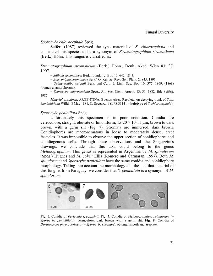
verruculose, straight, obovate or limoniform, 15-20 × 10-11 µm, brown to darkbrown, with a germ slit (Fig. 7). Stromata are immersed, dark brown.Conidiophores are macronematous in loose to moderately dense, erectfascicles. It was impossible to observe the upper section of conidiophores andconidiogenous cells. Through these observations and the Spegazzini'sdrawings, we conclude that this taxa could belong to the genusMelanographium. This genus is represented in Argentina by M. spinulosum(Speg.) Hughes and M. cokeii Ellis (Romero and Carmaran, 1997). Both M.spinulosum and Sporocybe penicillata have the same conidia and conidiophoremorphology. Taking into account the morphology and the fact that material ofthis fungi is from Paraguay, we consider that S. penicillata is a synonym of M.spinulosum.
Fig. 6. Conidia of Periconia spegazzinii. Fig. 7. Conidia of Melanographium spinulosum (=Sporocybe penicilliata), verruculose, dark brown with a germ slit. Fig. 8. Conidia ofDoratomyces purpureofuscus (= Sporocybe sacchari), oblong, smooth and aseptate.

72
Melanographium spinulosum (Speg.) Hughes, Canad. J. Bot. 36: 783. 1958.= Sporocybe penicillata Speg., An. Mus. Nac. Hist. Nat. Buenos Aires 31: 444; pl. 23,
fig. 259. 1922.For other synonyms see Hughes (1958).Material examined: PARAGUAY, Asunción, on a branch of Seguiera paraguayensis,
July 1919, C. Spegazzini (LPS 33124 – holotype of S. penicillata); ibid., Guarapí, 1881, B.Balansa (no. 2796), (LPS 1001 – holotype of Cordella spinulosa Speg.); ibid., Calle-poi yYaguaron, on rot leaves of Laurinea fide packets (Lauraceae?), 20 September 1887, B.Balansa (no. 4002), (LPS 12926 – holotype of Cordella tomentosa Speg.).
Sporocybe sacchari Speg.Conidiophores macronematous, synnematous, dark brown, mostly
smooth, branched to the apex forming sphaerical heads, 70-80 µm diam.Conidia ovoid to oblong, smooth, aseptate, brown, 3.5-4.5 × 5.5-7.5 µm (Fig.8). The material is very poor and unfortunately it was not possible to recognizepercurrence in the conidiogenous cells.
We conclude that S. sacchari is not a Periconia, but is very similar toGraphium pistillarioides Speg. (1896). Seifet studied this material andconsidered that it was a synonym of Doratomyces microsporus (Sacc.) Morton& Smith, but he never formally published it. However, the conidia shape andsize differ between these two taxa. In our opinion S. sacchari conidia are moresimilar to those of Doratomyces purpureofuscus (Fries) Morton & Smith. Forthis reason a new synonym is proposed.
Doratomyces purpureofuscus (Fr.) Morton & Smith, Mycol. Pap. 86: 74-77.1963.
≡ Sporocybe sacchari Speg., Rev. Fac. Agron. Vet. La Plata 2 (18): 253. 1896.Material examined: ARGENTINA, Buenos Aires, La Plata, on leaves, sheaths, and
culms of Saccharum officinarum, 1894, C. Spegazzini (LPS 33143 – holotype of S. sacchari);ibid., Tucumán, on leaves of S. officinarum, April 1894, C. Spegazzini (LPS 33137 – holotypeof Graphium sacchari); ibid., 23 Sept. 1894, C. Spegazzini (LPS 33136 – holotype of G.pistillarioides).
Periconia pycnospora Speg.This taxon was reported by Spegazzini from Argentina (Spegazzini,
1898, 1913) and from Chile (Spegazzini, 1910b) on various substrates.Periconia pycnospora is a synonym of P. byssoides (Mason and Ellis, 1953).Recently it was reported on Geoffroea decorticans (Bianchinotti, 1998) and onTabebuia avellanedae (López,. pers. com.). This fungus is classified as:
Periconia byssoides Pers. ex MératDescription, synonyms and illustrations in Mason and Ellis (1953)
Fungal Diversity
73
Material examined: ARGENTINA, Buenos Aires, Santa Catalina, on Ailanthusglandulosa, 11 March 1890, C. Spegazzini (LPS 12976); ibid., Buenos Aires, La Plata, onBohemeriae candidissimae, August 1911, C. Spegazzini (LPS 12977); ibid., on Vitis vinifera,25 November 1906, C. Spegazzini (LPS 12978); CHILE, Valdivia, on Digitalis? sp., January1909, C. Spegazzini (LPS 12979); ibid., Victoria, on Lauralia sp. fide packets (Laureceae?),November 1922, C. Spegazzini (LPS 12975).
Other Periconia species reported from Argentina
Romero (1983, 1994) contributed to the genus Periconia in Argentina.During a study of xylophilous micromycetes on Eucalyptus viminalis Labill.she described and illustrated Periconia tirupatiensis Subram., Periconiacircinata (Mangin) Sacc., Periconia minutissima Corda and Periconia lateralisEllis and Everh., and also isolated P. circinata (Romero, 1983).
It is also worthwhile to mention the description of Palaeopericoniafritzschei Ibáñez & Zamuner. The studied material represents the first fossilrecord related to Periconia and the oldest record, up to that date, of thefossilized Dematiaceae (Ibáñez and Zamuner, 1996).
Doratomyces reported from Argentina
In Argentina, Doratomyces is represented by four species. Three arereported from Argentina for the first time here.
Doratomyces purpureofuscus (Fr.) Morton & Smith, Mycol. Pap. 86: 74.1963.
≡ Sporocybe sacchari Speg., Rev. Fac. Agron. Vet. La Plata 2 (18): 253. 1896.
Doratomyces microsporus (Sacc.) Morton & Smith, Mycol. Pap., 86: 77. 1963.≡ Graphium pistillarioides Speg., Rev. Fac. Agron. Vet. La Plata, 2 (18): 254. 1896.
Doratomyces asperulus Wright & Marchand, Bol. Soc. Bot. Arg. 24: 307.(1972).
Described by Wright and Marchand from soil from Buenos Aires.
Doratomyces stemonitis (Pers. & Fr.) Morton & Smith, Mycol. Pap. 86: 70.1963.
Identified by J.E. Wright on roots of Vitis vinifera but never published. Itis interesting to mention that we have not found any previous report of thesespecies neither on Saccharum officinarum nor Vitis vinifera.
74
Additional material examined: ARGENTINA, Mendoza, Capital, on roots of Vitisvinifera, 14 March 1939, Ruiz Leal, det. J.E. Wright (BAFC- 51.230).
Key to species of Periconia from Argentina
1. Conidiophores in large groups with the stipes closely adpressed, conidia 5-7.5 µm diam...............................................................................................................................P. tirupatiensis1. Conidiophores widely scattered .............................................................................................. 2
2. Conidia formed unilaterally below the sterile, setiform apex ..................................P. lateralis2. Conidia in a well-defined, fairly compact head at the apex of the stipe.................................. 3
3. Conidiophores circinate at the apex ....................................................................... P. circinata3. Conidiophores not circinate at the apex .................................................................................. 4
4. Conidia 10-15 µm diam. ........................................................................................P. byssoides4. Conidia 4-6 µm diam. ............................................................................................................ 5
5. No stromata formed, conidiophores up to 550 µm long often swollen at the base, conidiastraw-coloured to pale brown, verruculose ...........................................................P. minutissima5. Stromata formed, conidiophores 500-2000 µm long, conidia pale brown to brown,verruculose.............................................................................................................. P. spegazzinii
Key to species of Doratomyces from Argentina modified from Ellis (1971)
1. Conidia slightly rough...........................................................................................D. asperulus1. Conidia smooth ....................................................................................................................... 2
2. Conidia 3-5 × 2-3 µm....................................................................................... D. microsporus2. Conidia mostly larger thant 5 × 3.5 µm .................................................................................. 3
3. Heads ellipsoidal or cylindrical, conidia often pointed at apex; usually with anEchinobotryum state................................................................................................. D. stemonitis3. Heads spherical or sub spherical, conidia mostly rounded at apex; no Echinobotryum state........................................................................................................................D. purpureofuscus
Acknowledgements
We thank the LPS Herbarium for the material in loan. We are very grateful to K.A.Seifert for letting us publish his comments on Graphium pistillarioides and to A.I. Romero forher critical review. This is publication no. 154 of the PRHIDEB financed by CONICET.
References
Alias, S.A. and Jones, E.B.G. (2000). Colonization of mangrove wood by marine fungi atKuala Selangor Mangrove Stand, Malaysia. Fungal Diversity 5: 9-21.
Fungal Diversity
75
Bianchinotti, M.V. (1998). Contribución al conocimiento de la micobiota Argentina.Micromicetes sobre Geoffroea decorticans (Leguminosae) III. Boletín de la SociedadArgentina de Botánica 33: 149-155.
Cai, L., Tsui, C.K.M., Zhang, K. and Hyde, K.D. (2002). Aquatic fungi from Lake Fuxian,Yunnan, China. Fungal Diversity 9: 57-70.
Carmarán, C.C. (2002). Contribución al estudio del orden Diatrypales en las zonassubtropicales de la República Argentina. Boletín de la Sociedad Micológica de Madrid26: 43-56.
Corda, A.C.J. (1829). In Sturn, J. Deutschlands Flora iii (Pilze), Bd. 2, Heft 7.Ellis, M.B. (1971). Dematiaceous Hyphomycetes. Commonwealth Mycological Institute, Kew.Ellis, M.B. (1976). More Dematiaceous Hyphomycetes. Commonwealth Mycological Institute,
Kew.Fries, E.M. (1925). Systema Orbis Vegetabilis. 1-134.Fries, E.M. (1832). Systema Mycologicum, 3, Index: 1- 202. Greifswald.Heinemann, P. (1962). Agarici Austroamericani V. Etude des types de C. Spegazzini. Extrait
du Bulletin de l' Institut. agronomique et des Stations de Recherches de Gembloux. 30:273-282.
Hladki, A.I. and Romero, A.I. (2001). The genus Kretzschmaria from Tucuman, Argentina.Mycotaxon 79: 481-496.
Holmgren, K.P., Holmgren, N.H. and Barnett, L.C. (1990). Index Herbariorum. Part I: TheHerbaria of the World. New York Botanical Garden. New York, USA.
Hughes, S.J. (1958). Revisiones Hyphomycetum aliquot cum appendice de nominibusrejiciendis. Canadian Journal of Botany 36: 727-836.
Ibáñez, C.G. and Zamuner, A.B. (1996). Hyphomycetes (Deuteromycetes) in cones ofAraucaria mirabilis (Spegazzini) Windhausen, Middle Jurassic of Patagonia, Argentina.Mycotaxon 59: 137-143.
Lindquist, J.C. and Wright, J.E. (1959). Sobre la identidad de Poroniopsis Spegazzini eHypocreodendron P. Hennings. Darwiniana 11: 598-605.
Mason, E.W. and Ellis, M.B. (1953). British species of Periconia. Mycological Papers 56: 1-127.
Morrison-Gardiner, S. (2002). Dominant fungi from Australian Coral reefs. Fungal Diversity 9:105-121.
Morton, F.J. and Smith, G. (1963). The genera Scopulariopsis Bainier, Microascus Zukal, andDoratomyces Corda. Mycological Papers 86: 32-96.
Muntañola-Cvetković, M., Hoyo, P. and Gómez-Bolea, A. (1998). Periconia fusiformis anam.sp. nov. Mycotaxon 68: 131-136.
Muntañola-Cvetković, M., Hoyo, P. and Gómez-Bolea, A. (1999). Periconia flabelliformisanam. sp. nov. Mycotaxon 71: 259-265.
Photita, W., Lumyong, S., Lumyong, P., Ho, W.H., Mckenzie, E.H.C. and Hyde, K.D. (2001).Fungi on Musa acuminata in Hong Kong. Fungal Diversity 6: 99-106.
Rajchenberg, M. and Wright, J.E. (1987) Type studies of Corticiaceae and Polyporaceae(Aphyllophorales) described by C. Spegazzini. Mycologia 79: 246-264.
Romero, A.I. (1983). Contribución al estudio de los hongos xilófilos de la Argentina. I.Deuteromycotina en Eucalyptus viminalis (Myrtaceae). Boletín de la SociedadArgentina de Botánica 22: 57-79.
Romero, A.I. (1994). Estudio florístico y ecológico de micromicetes xilófilos sobre tocones deEucalyptus viminalis en el NE de la Pcia. de Buenos Aires. Ph.D. Thesis. FCEy N.Universidad de Buenos Aires, Argentina.
76
Romero, A.I. and Carmaran C. C. (1997). Algunos micromicetes xilófilos de la regiónsubtropical argentina. I. Misiones. Boletín de la Sociedad Argentina de Botánica 33: 59-67.
Romero, A.I., Carrión, G. and Rico-Gray, V. (2001). Fungal latent pathogens and endophytesfrom leaves of Parthenium hysterophorus (Asteraceae). Fungal Diversity 7: 81-87.
Saccardo, P.A. (1913). Sylloge Fungorum Volume 22.Sarma, V.V. and Vittal, B.P.R. (2000). Biodiversity of mangrove fungi on different substrata of
Rhizophora apiculata and Avicennia spp. from Godavari and Krishna deltas, east coastof India. Fungal Diversity 5: 23-41.
Spegazzini, C. (1882). Fungi argentini pug. 4. Anales de la Sociedad Científica Argentina 13:11-35.
Spegazzini, C. (1887). Fungi patagonici. Boletín de la Academia Nacional de Ciencias deCórdoba 11: 5-64.
Spegazzini, C. (1896). Hongos de la caña de azúcar. Revista de la Facultad de Agronomía yVeterinaria de La Plata 2: 227-258.
Spegazzini, C. (1898). Fungi argentini novi v. critici. Anales del Museo Nacional de BuenosAires 6: 81-365.
Spegazzini, C. (1910a). Mycetes argentinenses. Anales del Museo Nacional de Buenos Aires20: 329-467.
Spegazzini, C. (1910b). Fungi chilensis. Revista de la Facultad de Agronomía y Veterinaria 6:1-205.
Spegazzini, C. (1913). Mycetes argentinenses. Anales del Museo Nacional de Historia Naturalde Buenos Aires. 24: 167-185.
Spegazzini, C. (1922). Fungi paraguayenses. Anales del Museo Nacional de Historia Naturalde Buenos Aires 31: 355-450.
Spegazzini, C. (1926). Contribuciónal conocimiento de la flora micológica de las Sierras deCórdoba.. Boletín de la Academia Nacional de Ciencias de Córdoba 29. 113-190.
Saikia, U.N. and Sarbhoy, A.K. (1982). Hyphomycetes of India-V*. The genus Periconia.Journal of Indian Phytopathology 35: 277-281.
Seifert, K.A. (1987). Stromatographium and Acrostoma gen. nov.: two tropical hyphomycetesgenera with distinctive synnema anatomies. Canadian Journal of Botany 65: 2196-2201.
Wright, J.E. and Marchand, S. (1972). Microflora del suelo de la Argentina III. Dosinteresantes géneros sistemáticos: Trichurus y Doratomyces. Boletín de la SociedadArgentina de Botánica 24: 305-310.
Yanna, Ho, W.H. and Hyde, K.D. (2002). Fungal succesion on fronds of Phoenix hanceana inHong Kong. Fungal Diversity 10: 185-211.
(Received 23 December 2002; accepted 4 May 2003)