A randomised controlled trial to evaluate the clinical and cost ...
126
Methodology A randomised controlled trial to evaluate the clinical and cost- effectiveness of Hickman line insertions in adult cancer patients by nurses A Boland A Haycox A Bagust L Fitzsimmons Health Technology Assessment 2003; Vol. 7: No. 36 HTA Health Technology Assessment NHS R&D HTA Programme
-
Upload
khangminh22 -
Category
Documents
-
view
1 -
download
0
Transcript of A randomised controlled trial to evaluate the clinical and cost ...

Methodology
A randomised controlled trial to evaluate the clinical and cost-effectiveness of Hickman line insertions in adult cancer patients by nurses
A BolandA HaycoxA BagustL Fitzsimmons
Health Technology Assessment 2003; Vol. 7: No. 36
HTAHealth Technology AssessmentNHS R&D HTA Programme
Copyright notice
© Queen's Printer and Controller of HMSO 2003 HTA reports may be freely reproduced for the purposes of private research and study and may be included in professional journals provided that suitable acknowledgement is made and the reproduction is not associated with any form of advertising Violations should be reported to [email protected] Applications for commercial reproduction should be addressed to HMSO, The Copyright Unit, St Clements House, 2–16 Colegate, Norwich NR3 1BQ

How to obtain copies of this and other HTA Programme reports.An electronic version of this publication, in Adobe Acrobat format, is available for downloading free ofcharge for personal use from the HTA website (http://www.ncchta.org). A fully searchable CD-ROM isalso available (see below).
Printed copies of HTA monographs cost £20 each (post and packing free in the UK) to both public andprivate sector purchasers from our Despatch Agents, York Publishing Services.
Non-UK purchasers will have to pay a small fee for post and packing. For European countries the cost is£2 per monograph and for the rest of the world £3 per monograph.
You can order HTA monographs from our Despatch Agents, York Publishing Services by:
– fax (with credit card or official purchase order) – post (with credit card or official purchase order or cheque)– phone during office hours (credit card only).
Additionally the HTA website allows you either to pay securely by credit card or to print out yourorder and then post or fax it.
Contact details are as follows:York Publishing Services Email: [email protected] Box 642 Tel: 0870 1616662YORK YO31 7WX Fax: 0870 1616663UK Fax from outside the UK: +44 1904 430868
NHS and public libraries can subscribe at a very reduced cost of £100 for each volume (normallycomprising 30–40 titles). The commercial subscription rate is £300 per volume. Please contact YorkPublishing Services at the address above. Subscriptions can only be purchased for the current orforthcoming volume.
Payment methods
Paying by chequeIf you pay by cheque, the cheque must be in pounds sterling, made payable to York PublishingDistribution and drawn on a bank with a UK address.
Paying by credit cardThe following cards are accepted by phone, fax, post or via the website ordering pages: Delta, Eurocard,Mastercard, Solo, Switch and Visa. We advise against sending credit card details in a plain email.
Paying by official purchase orderYou can post or fax these, but they must be from public bodies (i.e. NHS or universities) within the UK.We cannot at present accept purchase orders from commercial companies or from outside the UK.
How do I get a copy of HTA on CD?
Please use the form on the HTA website (www.ncchta.org/htacd.htm). Or contact York PublishingServices (see contact details above) by email, post, fax or phone. HTA on CD is currently free of chargeworldwide.
The website also provides information about the HTA Programme and lists the membership of the variouscommittees.
HTA

A randomised controlled trial toevaluate the clinical and cost-effectiveness of Hickman lineinsertions in adult cancer patients by nurses
A Boland1*
A Haycox1
A Bagust1
L Fitzsimmons2
1 Prescribing Research Group, Department of Pharmacology andTherapeutics, University of Liverpool, UK
2 Christie Hospital NHS Trust, Manchester, UK
* Corresponding author
Declared competing interests of authors: none
Published November 2003
This report should be referenced as follows:
Boland A, Haycox A, Bagust A, Fitzsimmons L. A randomised controlled trial to evaluatethe clinical and cost-effectiveness of Hickman line insertions in adult cancer patients bynurses. Health Technol Assess 2003;7(36).
Health Technology Assessment is indexed in Index Medicus/MEDLINE and Excerpta Medica/EMBASE.

NHS R&D HTA Programme
The NHS R&D Health Technology Assessment (HTA) Programme was set up in 1993 to ensure that high-quality research information on the costs, effectiveness and broader impact of health
technologies is produced in the most efficient way for those who use, manage and provide care in the NHS.
Initially, six HTA panels (pharmaceuticals, acute sector, primary and community care, diagnostics and imaging, population screening, methodology) helped to set the research priorities for the HTAProgramme. However, during the past few years there have been a number of changes in and aroundNHS R&D, such as the establishment of the National Institute for Clinical Excellence (NICE) and the creation of three new research programmes: Service Delivery and Organisation (SDO); New and Emerging Applications of Technology (NEAT); and the Methodology Programme.
This has meant that the HTA panels can now focus more explicitly on health technologies (‘health technologies’ are broadly defined to include all interventions used to promote health, prevent and treat disease, and improve rehabilitation and long-term care) rather than settings of care. Therefore the panel structure was replaced in 2000 by three new panels: Pharmaceuticals;Therapeutic Procedures (including devices and operations); and Diagnostic Technologies andScreening.
The HTA Programme will continue to commission both primary and secondary research. The HTACommissioning Board, supported by the National Coordinating Centre for Health TechnologyAssessment (NCCHTA), will consider and advise the Programme Director on the best research projects to pursue in order to address the research priorities identified by the three HTA panels.
The research reported in this monograph was funded as project number 95/16/06.
The views expressed in this publication are those of the authors and not necessarily those of the HTA Programme or the Department of Health. The editors wish to emphasise that funding andpublication of this research by the NHS should not be taken as implicit support for any recommendations made by the authors.
HTA Programme Director: Professor Kent WoodsSeries Editors: Professor Andrew Stevens, Dr Ken Stein, Professor John Gabbay,
Dr Ruairidh Milne, Dr Chris Hyde and Dr Rob RiemsmaManaging Editors: Sally Bailey and Sarah Llewellyn Lloyd
The editors and publisher have tried to ensure the accuracy of this report but do not accept liabilityfor damages or losses arising from material published in this report. They would like to thank thereferees for their constructive comments on the draft document.
ISSN 1366-5278
© Queen’s Printer and Controller of HMSO 2003
This monograph may be freely reproduced for the purposes of private research and study and may be included in professional journals provided that suitable acknowledgement is made and the reproduction is not associated with any form of advertising.
Applications for commercial reproduction should be addressed to HMSO,The Copyright Unit, St Clements House, 2–16 Colegate, Norwich, NR3 1BQ.
Published by Gray Publishing, Tunbridge Wells, Kent, on behalf of NCCHTA.Printed on acid-free paper in the UK by St Edmundsbury Press Ltd, Bury St Edmunds, Suffolk.
Criteria for inclusion in the HTA monograph seriesReports are published in the HTA monograph series if (1) they have resulted from work commissioned for the HTA Programme, and (2) they are of a sufficiently high scientific quality as assessed by the referees and editors.
Reviews in Health Technology Assessment are termed ‘systematic’ when the account of the search,appraisal and synthesis methods (to minimise biases and random errors) would, in theory, permitthe replication of the review by others.
G

Objectives: To examine the clinical and cost-effectiveness of image-guided Hickman line insertionsversus blind Hickman line insertions undertaken bynurses in adult cancer patients.Design: A cost-effectiveness analysis was carried outalongside a randomised controlled trial.Setting: A large acute cancer centre in Manchester,UK. Participants: Cancer patients due to have a Hickmanline insertion who were over 18 years of age and wereclinically and physically compliant with specifiedprotocols. Interventions: In order to obtain central venousaccess for the patient, two interventions wereinvestigated: (i) blind insertion of a Hickman line and (ii) image-guided insertion of a Hickman line. Bothinterventions involved blind venipuncture of thesubclavian vein. In the blind arm, the Hickman line wasroutinely inserted without the use of image guidance atany point in the procedure. Transfer to theinterventional X-ray suite and use of image guidancewere options immediately available to the operatorduring the procedure if required. In the image-guidedarm, the position of the guidewire was checked beforethe Hickman line was introduced and later theHickman line was positioned with the use of X-rayfluoroscopy. Main outcome measures: The primary clinicaloutcome measure was catheter-tip misplacement andthis was expected to be higher in the blind arm. When
comparing the skill level of the trainer and the trainees,pneumothorax was the primary clinical outcomemeasure. Other outcomes measures included arterialpuncture, haematoma, infection, failed insertion andassistance from other healthcare professionals. Results: No statistically significant difference was foundbetween the mean cost per patient in the two arms ofthe trial. The only statistically significant difference inclinical outcomes was the frequency of catheter-tipmisplacement, which was higher in the blind arm of thetrial. At very low costs, the image-guided approachdominates the blind approach as fewer costs andgreater benefits are incurred. It is evident that nursespreviously inexperienced in the procedure can betrained to insert Hickman lines successfully both at thebedside and under image guidance within a 3-monthperiod. Conclusions: This report indicates that nurse insertionof Hickman lines in the majority of adult cancerpatients is both safe and effective. However, there area select group of patients for whom image-guidedinsertion may be preferred. The results reveal thatskills and expertise can be transferred from trainer totrainee through a relatively short, but intensive, trainingcourse. It is also evident that patients support nurseinsertion. Further research is suggested to compare thesafety and efficacy of nurse versus doctor insertions inparticular subgroups of patients and also to assess thequantity and quality of current service provision inorder to inform NHS decision-making in this area.
Health Technology Assessment 2003; Vol. 7: No. 36
iii
© Queen’s Printer and Controller of HMSO 2003. All rights reserved.
Abstract
A randomised controlled trial to evaluate the clinical and cost-effectiveness of Hickman line insertions in adult cancer patients by nurses
A Boland,1* A Haycox,1 A Bagust1 and L Fitzsimmons2
1 Prescribing Research Group, Department of Pharmacology and Therapeutics, University of Liverpool, UK2 Christie Hospital NHS Trust, Manchester, UK* Corresponding author


Health Technology Assessment 2003; Vol. 7: No. 36
v
List of abbreviations .................................. vii
Executive summary .................................... ix
1 Introduction ............................................... 1Background to the study ............................ 1Clinical applications of CVCs .................... 1Types of CVCs ............................................ 2Methods of Hickman line insertion ........... 3Choice of access site for the Hickman catheter ....................................................... 4Choice of operator to perform the insertionprocedure ................................................... 4Complications associated with the insertion of Hickman catheters ................................. 5Reasons for catheter removal ..................... 6Analysis of empirical evidence (1980–2000) ............................................... 6Review of health economics literature ....... 10Summary of published literature ............... 11Rationale for the study ............................... 11
2 Methods ..................................................... 15Overview of nurse training programme .... 15RCT ............................................................ 17NHS resource use ....................................... 22Patient satisfaction questionnaire ............... 24Statistical and data analysis of primary data ............................................................. 25Economic evaluation .................................. 25
3 Results ........................................................ 29Evaluation of pre-trial nurse trainingprogramme ................................................. 29Results of the RCT ..................................... 31Comparing nurse skill ................................ 38Patient satisfaction questionnaire ............... 42
Economic evaluation .................................. 43Resource implications of Hickman lineinsertions by nurses across the NHS .......... 51
4 Discussion ................................................... 59Evaluation of nurse training programme ................................................. 59Costs of the nurse training programme .... 59RCT ............................................................ 60Comparing nurse skill ................................ 62Patient satisfaction questionnaire ............... 63Economic evaluation .................................. 64Conclusions ................................................ 66
Acknowledgements .................................... 69
References .................................................. 71
Appendix 1 Nurse training programme ... 73
Appendix 2 Patient satisfaction questionnaire .............................................. 75
Appendix 3 Patient case report forms ...... 79
Appendix 4 Unit costs and cost estimates ..................................................... 85
Appendix 5 Patient information leaflet .... 93
Appendix 6 National survey responses ..... 97
Health Technology Assessment reportspublished to date ....................................... 101
Health Technology Assessment Programme ................................................ 109
Contents


Health Technology Assessment 2003; Vol. 7: No. 36
vii
© Queen’s Printer and Controller of HMSO 2003. All rights reserved.
AIDS acquired immunodeficiency syndrome
ALD Adult Leukaemia Database
BCSH British Committee in Standards inHaematology
CBA cost–benefit analysis
CEA cost-effectiveness analysis
CHNT Christie Hospital NHS Trust
CLIP central line insertion project
CL confidence limits
CMA cost-minimisation analysis
CNS clinical nurse specialist
CTSU Clinical Trial Support Unit
CUA cost–utility analysis
CVC central venous catheter
df degrees of freedom
HIV human immunodeficiency virus
ICER incremental cost-effectiveness ratio
ICVAD implantable central venous accessdevice
KP Karnofsky performance
PHLS Public Health Laboratory Service
PICC peripherally inserted central catheter
QALY quality-adjusted life year
RCT randomised controlled trial
SD standard deviation
TPN total parenteral nutrition
VAD venous access device
List of abbreviations
All abbreviations that have been used in this report are listed here unless the abbreviation is well known (e.g. NHS), or it has been used only once, or it is a non-standard abbreviation used only in figures/tables/appendices in which case the abbreviation is defined in the figure legend or at the end of the table.

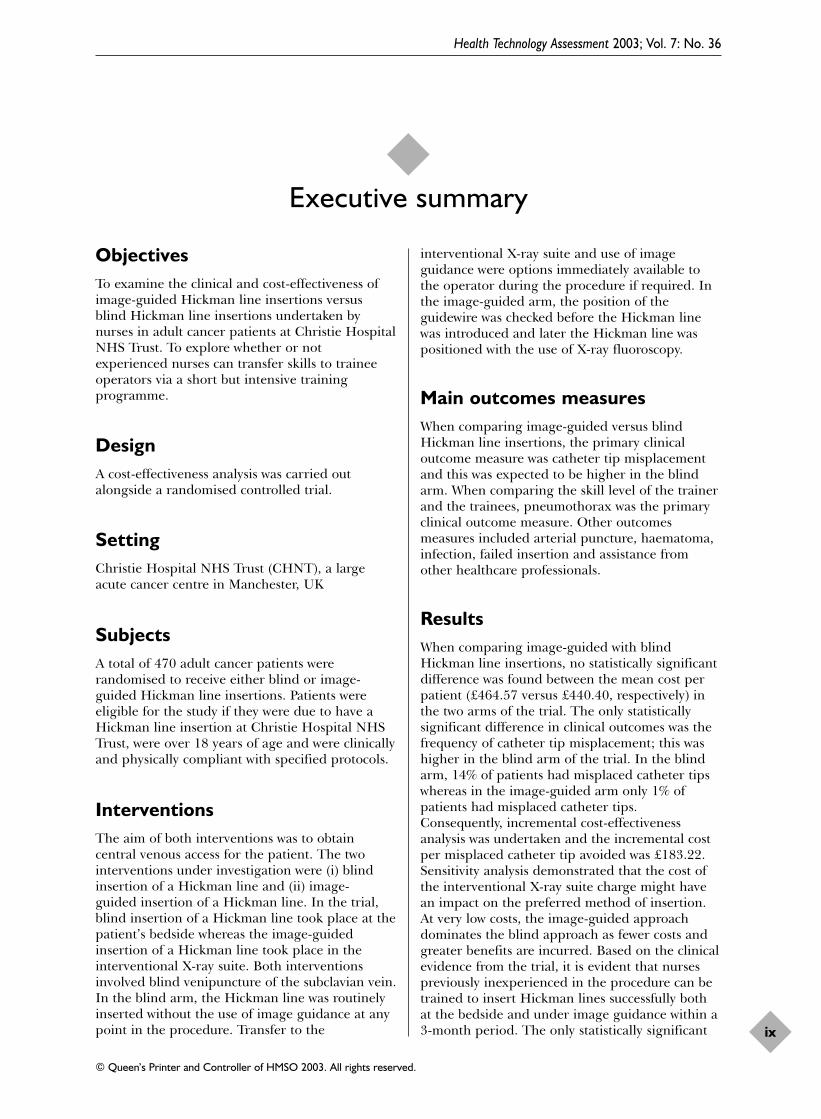
ObjectivesTo examine the clinical and cost-effectiveness ofimage-guided Hickman line insertions versusblind Hickman line insertions undertaken bynurses in adult cancer patients at Christie HospitalNHS Trust. To explore whether or notexperienced nurses can transfer skills to traineeoperators via a short but intensive trainingprogramme.
DesignA cost-effectiveness analysis was carried outalongside a randomised controlled trial.
SettingChristie Hospital NHS Trust (CHNT), a largeacute cancer centre in Manchester, UK
SubjectsA total of 470 adult cancer patients wererandomised to receive either blind or image-guided Hickman line insertions. Patients wereeligible for the study if they were due to have aHickman line insertion at Christie Hospital NHSTrust, were over 18 years of age and were clinicallyand physically compliant with specified protocols.
InterventionsThe aim of both interventions was to obtaincentral venous access for the patient. The twointerventions under investigation were (i) blindinsertion of a Hickman line and (ii) image-guided insertion of a Hickman line. In the trial,blind insertion of a Hickman line took place at thepatient’s bedside whereas the image-guidedinsertion of a Hickman line took place in theinterventional X-ray suite. Both interventionsinvolved blind venipuncture of the subclavian vein.In the blind arm, the Hickman line was routinelyinserted without the use of image guidance at anypoint in the procedure. Transfer to the
interventional X-ray suite and use of imageguidance were options immediately available tothe operator during the procedure if required. Inthe image-guided arm, the position of theguidewire was checked before the Hickman linewas introduced and later the Hickman line waspositioned with the use of X-ray fluoroscopy.
Main outcomes measuresWhen comparing image-guided versus blindHickman line insertions, the primary clinicaloutcome measure was catheter tip misplacementand this was expected to be higher in the blindarm. When comparing the skill level of the trainerand the trainees, pneumothorax was the primaryclinical outcome measure. Other outcomesmeasures included arterial puncture, haematoma,infection, failed insertion and assistance fromother healthcare professionals.
ResultsWhen comparing image-guided with blindHickman line insertions, no statistically significantdifference was found between the mean cost perpatient (£464.57 versus £440.40, respectively) inthe two arms of the trial. The only statisticallysignificant difference in clinical outcomes was thefrequency of catheter tip misplacement; this washigher in the blind arm of the trial. In the blindarm, 14% of patients had misplaced catheter tipswhereas in the image-guided arm only 1% ofpatients had misplaced catheter tips.Consequently, incremental cost-effectivenessanalysis was undertaken and the incremental costper misplaced catheter tip avoided was £183.22.Sensitivity analysis demonstrated that the cost ofthe interventional X-ray suite charge might havean impact on the preferred method of insertion.At very low costs, the image-guided approachdominates the blind approach as fewer costs andgreater benefits are incurred. Based on the clinicalevidence from the trial, it is evident that nursespreviously inexperienced in the procedure can betrained to insert Hickman lines successfully bothat the bedside and under image guidance within a3-month period. The only statistically significant
Health Technology Assessment 2003; Vol. 7: No. 36
ix
© Queen’s Printer and Controller of HMSO 2003. All rights reserved.
Executive summary

difference identified when comparing the skilllevel of the three nurses was that the trainer wasless likely to call for assistance from anotherhealthcare professional during the procedure thanthe trainees.
ConclusionsThis report indicates that nurse insertion ofHickman lines in the majority of adult cancerpatients at CHNT is both safe and effective.However, there are a select group of patients forwhom image-guided insertion may be preferred.The results reveal that skills and expertise can betransferred from trainer to trainee through arelatively short but intensive training course. Fromthe patient satisfaction evidence available, it is
evident that patients support nurse insertion.Nurse insertions can free up clinical resources in asafe and effective manner.
Recommendations for futureresearchReliable estimates of the clinical and cost-effectiveness of Hickman line insertions in adultcancer patients can only be calculated if furtherresearch to compare the safety and efficacy ofnurse versus doctor insertions in particularsubgroups of patients is carried out. It is alsorecommended that future studies be conducted toassess the quantity and quality of current serviceprovision in order to inform NHS decision-making in this area.
Executive summary
x

Background to the studyIn the NHS, approximately 200,000 centralvenous catheters (CVCs) are inserted in adultpatients per year.1 As the average cost of insertionis estimated to be approximately £450,2 theannual direct cost to the NHS of CVC insertionscan be conservatively estimated at around £80million. For many seriously ill patients, theavailability of venous access devices (VADs) notonly improves quality of life but also saves lives.Consequently, the appropriate use of VADs (orCVCs) is recognised as an integral part of totalpatient management across a wide spectrum ofspecialties.3 Central venous access is required for avariety of reasons, the most common being for theadministration of chemotherapy and the deliveryof total parenteral nutrition (TPN). A wide rangeof CVCs are available for hospital and domiciliaryuse in the UK. This research focuses on theHickman line, a tunnelled and cuffed CVC whichis the most frequently used CVC in the NHS.
General surgeons and anaesthetists were the firstclinicians to insert Hickman lines surgically in theoperating theatre. More recently, interventionalradiologists have developed innovative methodsfor the insertion of Hickman lines under imageguidance. Extensive clinical evidence exists tosupport the hypothesis that the image-guidedapproach to insertion is superior to the surgicalapproach in terms of both improved healthoutcomes and patient satisfaction.4 Empiricalstudy in this area is currently focused oncomparing image-guided and blind methods ofinsertion. Recent study results suggest that, for themajority of patients, the blind insertion of aHickman line is no less safe or effective thanimage-guided insertion.5 Also, clinicians are nolonger the sole group of operators; nurse-ledcentral venous access services are beingestablished and CVC insertion training for nursesis being delivered both with and without theroutine use of image guidance. This researchaddresses a range of issues but concentrates on thetwo issues at the forefront of research in this area:first, what are the costs and benefits of image-guided versus blind approaches to Hickman lineinsertions in adult cancer patients undertaken bynurses?; second, how effectively and over what
time frame can the necessary insertion skills betransferred from experienced nurses to traineenurses? The issue of skill transference is crucial togeneralising the results of this research throughoutthe NHS.
This first chapter serves to introduce the reader tothe topic of CVC insertion. Fundamental areas ofinterest are discussed. Where appropriate,published references have been used to supportkey statements. A comprehensive review of thepublished medical literature was undertaken inorder to identify relevant clinical papers. Themain databases searched (1980–2000) includedMEDLINE, EMBASE and CANCERLIT. Onlyreferences published in English were retrieved.
Clinical applications of CVCsIn the past, CVCs have been primarily used forlong-term parenteral nutrition.6 However, over theyears, indications for their use have continued toexpand. Nowadays, common applications for long-term central venous access are prolongedchemotherapy, TPN and haemodialysis. Lesscommon applications include plasmapheresis,antibiotic therapy, iron supplements, vitamins,repeated administration of blood products, fluidreplacement and needle phobia.6–8 Otherindications for catheter insertion include poorperipheral venous access, when intravenoustherapy involves drugs known to be venoussclerosants, resuscitative intravenous therapy andwhen ambulatory chemotherapy is to be given asan outpatient procedure.9
As more and more patients are being treated forleukaemia, solid tumours, infection and AIDS, thedemand for CVCs has risen. Consequently, severaldifferent types of central venous access device areavailable. When deciding which access device isappropriate for the patient, a range of factorsmust be considered. Hagle and colleagues10
suggest that both the doctor and the patient havechoices to make. The doctor must assess the costs,benefits and clinical risks of the device for thepatient. At the same time, the patient mustconsider the daily maintenance requirements ofthe device, any cosmetic implications and whether
Health Technology Assessment 2003; Vol. 7: No. 36
1
© Queen’s Printer and Controller of HMSO 2003. All rights reserved.
Chapter 1
Introduction
or not the device will interfere with his or heractivities of daily living. The choice of access site isvery important. Evidence suggests that patientsprefer the non-dominant side for subclavian orupper extremity access. However, for patients whohave had previous surgery or radiation therapy toone side, it may be necessary to use contralateralaccess. Hamilton8 believes that the choice ofvenous access device should meet the individualpatient’s requirement and be based primarily onpatient preference.
Types of CVCsFor simplicity, the different types of central venousaccess device can be broadly categorised asfollows: (1) non-tunnelled, (2) tunnelled and (3)implanted infusion ports.10,11 First, non-tunnelledcatheters are designed for short-term use of daysto weeks. Typical non-tunnelled catheters includethe standard triple lumen (tapered) and Hohn(non-tapered) catheters, midline catheters andperipherally inserted central catheters (PICCs).PICCs are a relatively new venous access deviceand have lower procedure risk compared withcentrally placed tunnelled devices.7 PICCs areeither single- or double-lumen catheters and areoften inserted if patients fail to meet the criteriafor subclavian line insertion.9 PICCs are insertedinto the basilic or cephalic networks near theantecubital fossae.12
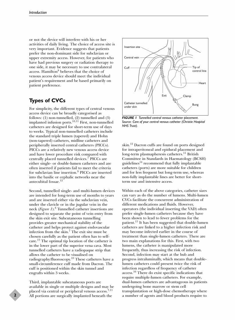
Second, tunnelled single- and multi-lumen devicesare intended for long-term use of months to yearsand are inserted either via the subclavian vein,under the clavicle or in the jugular vein in theneck (Figure 1).8 Tunnelled catheter insertions aredesigned to separate the point of vein entry fromthe skin exit site. Subcutaneous tunnellingprovides greater mechanical stability of thecatheter and helps protect against endovascularinfection from the skin.7 The exit site must bechosen carefully as the patient often has to self-care.13 The optimal tip location of the catheter isin the lower part of the superior vena cava. Mosttunnelled catheters have a radiopaque strip thatallows the catheter to be visualised onradiography/fluoroscopy.10 These catheters have asmall-circumference cuff made from Dacron. Thecuff is positioned within the skin tunnel andengrafts within 3 weeks.
Third, implantable subcutaneous ports areavailable in single or multiple designs and may beplaced via central or peripheral venous access.7,11
All portions are surgically implanted beneath the
skin.12 Dacron cuffs are found on ports designedfor intraperitoneal and epidural placement andlong-term plasmapheresis catheters.11 BritishCommittee in Standards in Haematology (BCSH)guidelines14 recommend that fully implantablecatheters (ports) are more suitable for childrenand for less frequent but long-term use, whereasnon-fully implantable lines are better for short-term use and intensive access.
Within each of the above categories, catheter sizescan vary as do the number of lumens. Multi-lumenCVCs facilitate the concurrent administration ofdifferent medications and fluids. However,operators (the individual inserting the VAD) oftenprefer single-lumen catheters because they havebeen shown to lead to fewer problems for thepatient.15 It has been suggested that double-lumencatheters are linked to a higher infection risk andmay become infected earlier in the course oftreatment than single-lumen catheters. There aretwo main explanations for this. First, with twolumens, the catheter is manipulated morefrequently, thus increasing the risk of infection.Second, infection may start at the hub andprogress intraluminally, which means that double-lumen catheters could present twice the risk ofinfection regardless of frequency of catheteraccess.16 There do exist specific indications thatrequire multiple-lumen catheters. For example,dual-lumen catheters are advantageous in patientsundergoing bone marrow or stem celltransplantation or high-dose chemotherapy wherea number of agents and blood products require to
Introduction
2
Insertion site
Central vein
Cuff
Catheter tunnelledunder skin
Exit
Tip ofcontrol line
Heart
Bung
Clamp
FIGURE 1 Tunnelled central venous catheter placement.Source: Care of your central venous catheter (Christie HospitalNHS Trust).

be infused simultaneously.9 Also, recent guidelinesrecommend that if TPN is being administeredalongside other medications or fluids, then onelumen should be used exclusively for thispurpose.16
Methods of Hickman lineinsertionAlthough there are many different types of centralvenous access device available, this reportconcentrates on the insertion of tunnelledHickman catheters in adult cancer patients via thesubclavian vein. There are three main approachesto the insertion of Hickman catheters and thesecan be broadly categorised as cutdown/surgicalapproach, radiological percutaneous placementand blind percutaneous placement.
Cutdown/surgical approachThe cutdown/surgical approach is the traditionalapproach to central venous access. When catheterswere first used to provide long-term venous access,the procedure was routinely performed in theoperating theatre by surgeons and anaesthetistswhilst the patient was under general anaesthesia.The venous cutdown approach frequently uses thecephalic vein to obtain venous access. However,the external jugular vein and the internal jugularvein have been used as insertion sites.6 Subclavianand femoral veins are also sometimes used. Thecutdown technique requires a surgical incision andmanipulation of the skin and subcutaneous tissue.The catheter is inserted into the subclavian veinand advanced along the superior vena cava.4 Theability to see and thus control possible bleedinghas been used as an argument in favour of thesurgical cutdown technique for catheter insertion.In the mid-1980s, lack of complications associatedwith the cutdown technique made it attractive tooperators.17 The surgical approach had initiallybeen proved to be safe and effective. However,when compared with the percutaneous technique,the cutdown technique was soon considered to beproblematic.18 Other reported disadvantagesassociated with this method include lengthyaverage operating times, a relatively low successrate (approximately 75%), veins being compromisedfor future use as vessels are not ligated andrelatively large entry wounds (5–10 cm). Thecutdown approach is also relatively expensivegiven high operating theatre overheads andsurgeons’ salaries. Finally, it can be argued that thesuccess of the surgical service very much dependson ample operating room availability in order toensure that demand can be met.13
Radiological percutaneous placement Interventional radiologists place lines usingpercutaneous techniques in an interventional X-ray or angiographic suite. The insertion isusually carried out under local anaesthesia andlight sedation. Imaging guidance can be used toidentify the entry site and/or patient anatomy atvarious stages during the insertion procedure.19,20
Image guidance may or may not be used forvenous puncture. Depending on the insertion site,ultrasonic vein-locating devices may or may not beof use. Results of a large randomised controlledtrial (RCT) concluded that there were noadvantages associated with the use of ultrasonicguidance in locating the subclavian vein forcatheter insertion.19 However, the availability ofvenography, fluoroscopic and ultrasound guidancein the radiology suite can facilitate central venousaccess for both the patient and the operator.
The main advantages of a radiological service overa surgical service can be described as convenienceand excellent patient tolerance.21 In addition, theuse of general anaesthesia as part of the cutdownapproach, possible multiple cutdown attempts andpostoperative check radiographs only serve toprolong the operative time within the surgicalgroup as compared with radiological placements.Very few lines are misplaced if the procedure iscarried out in an interventional X-ray suite asimaging is used to confirm the position of thecatheter tip before the procedure ends. Finally, ithas been suggested that, compared with operatingrooms, interventional radiology suites do notusually carry as expensive overheads and canusually accommodate requests for catheterplacement within 24 hours.13 The maindisadvantage associated with this insertion methodis that patients are dependent on the availabilityof skilled interventional radiologists and access toradiological facilities. Adam4 argues that thedemand for interventional radiologists is usuallygreater than their supply. Mauro and Jacques13
suggest that most interventional X-ray suites canaccommodate most patients. However, evidencesupporting this statement is required, especially ifhospitals have a high throughput of patients whorequire central venous access. If waiting times forX-ray suites do exist, then the optimal managementof patients can be delayed. Akin to the otherinsertion techniques available, radiologicalinsertions are associated with morbidity includingnon-infectious and infectious complications.
Blind percutaneous placement Blind insertion of Hickman catheters is routinelycarried out in a variety of locations and these can
Health Technology Assessment 2003; Vol. 7: No. 36
3
© Queen’s Printer and Controller of HMSO 2003. All rights reserved.

include the operating theatre, at the bedside in ahospital ward or in an outpatient clinic. Imageguidance is not used for venous puncture. Instead,the catheter is advanced blindly by the operator tothe lower part of the superior vena cava. Accurateplacement of the Hickman catheter relies oncorrect identification of anatomical landmarks.After the catheter has been inserted, a chest X-rayis performed in order to check both the positionof the catheter and to identify any pneumothorax,hydrothorax or haemothorax.22
The greatest benefit of the blind approach is that,if a suitably qualified healthcare professional isavailable, then there is no need to wait for gaps inoperating theatre or X-ray suite lists. As long asthere is emergency access to image-guidancefacilities, the procedure itself can be performed inmost clean environments. Other purportedadvantages of the blind percutaneous method overthe surgical cutdown approach are similar to theadvantages associated with radiological placement:decreased operative time, less morbidity, betterprimary placement, higher success rates and moreaccurate positioning of the catheter.21 Blindpercutaneous placement of the catheter alsoappears to provoke less anxiety in patients as theyare often more familiar with their hospital wardsurroundings and do not have to fear theoperating theatre or the X-ray suite. The maindisadvantage associated with blind placement of acentral catheter is the risk of catheter-tipmisplacement. Although chest X-rays immediatelyafter the procedure identify any tipmisplacements, it can take time for the line to berepositioned as the patient will have to wait for anavailable slot in the X-ray list and this may causediscomfort and inconvenience to the patient.4
Also, if the insertion procedure is non-routine andthe patient has to be moved from the ward to theX-ray suite during the procedure, this might causesome patients to become anxious. Blindplacement is also associated with significantmorbidity including non-infectious and infectiouscomplications.
Choice of access site for theHickman catheterSansivero22 states that choosing an access siterequires simultaneous consideration of patient,therapy and device characteristics. Options for theaccess site of VADs include the following:ancillary/subclavian vein, superficial and deepveins of the arm, internal jugular vein, inferiorvena cava, cephalic vein and the hepatic veins.4,11
Parker3 advises that VADs inserted via thesubclavian vein are associated with fewermechanical complications and have a lower risk ofinfection than those devices placed in the femoralor jugular veins.
Choice of operator to performthe insertion procedureThere exists a range of possible operators for theinsertion of Hickman catheters in adult cancerpatients and these include surgeons, anaesthetists,interventional radiologists, medical oncologistsand nurses trained in the procedure. As expected,the skills required by the operator have changedin line with advances in the insertion technique.For example, when the cutdown approach was infavour, surgeons and anaesthetists routinely placedHickman catheters. Now that there exists researchto support the insertion of catheters at thebedside, trained nurses are leading some centralvenous insertion services.9,23
The transition from operating theatre to bedsideplacement of catheters is still taking place. Alongthe way, interventional radiologists have begun toinsert Hickman lines in interventional X-raysuites.4,24,25 In the past, radiologists were onlyinvolved in the manipulation of malpositionedcatheters or retrieval of intravascular catheterfragments.7 However, this role has expanded andradiologists are becoming primary operators inthe placement and management of CVCs. Thecase for interventional radiologists to take on therole of operator is directly related to the researchevidence supporting the superiority of theradiological techniques over surgical techniques.In addition, Adam4 argues that the technique iseasy to learn and that most interventionalradiologists are already proficient in the use of theequipment required, for example, fluoroscopic orultrasound guidance.
Nurses are currently being trained to insertHickman lines and there are three main hospitalsacross the UK which offer insertion training[Christie Hospital NHS Trust (CHNT), JohnRadcliffe Hospital and Manchester RoyalInfirmary]. Although nursing staff do not routinelyplace Hickman catheters, there are some cancercentres in the UK whose central venous accessservice is nurse led. Various studies have beenpublished supporting the extension of the nurse’srole in this area.8,9,23 It can be argued that nurseinsertions of CVCs of all types can lead toimproved care for patients as a more holistic
Introduction
4

approach can be adopted as effort is made todeliver unfragmented patient care. Nurseplacements of CVCs at the patient’s bedside meanthat insertions can take place at the optimal timein a patient’s management as waiting lists forconsultants, theatres or X-ray suites are no longerbarriers to insertion. Nurses are being trained touse fluoroscopic guidance to verify the position ofthe catheter and the location of the catheter tipduring the insertion procedure. Nurses are alsousing these skills to reposition Hickman lines ifblind insertions lead to misplaced catheter tips. Itis often more convenient for the patient if a nurseis available to accompany the patient to theinterventional X-ray suite and perform theprocedure; otherwise the patient would bedependent on the availability of the suite and on asuitably trained doctor.
Regardless of who inserts Hickman catheters, it isgenerally accepted that operator experience ispositively correlated with successful patientoutcomes. Indeed, BCSH guidelines14 state thatinsertion should be performed by experiencedoperators, regardless of specialty. McBride andcolleagues21 demonstrated that there exists a steepoperator learning curve and that complicationrates improve notably after the operator hascarried out more than 30 procedures. Indeed,most centres that provide CVC insertion trainingfor nurses demand that at least 30 supervisedinsertions are performed before the trainee ispermitted to work unsupervised. Whether or notthere is a need for a dedicated team to provide acomprehensive insertion service is debated in theliterature. Wisborg and colleagues26 compared 140catheters inserted by three operators and 60catheters inserted by seven trained operators andfound no statistically significant differencesbetween the operators. The authors concludedthat even a large pool of operators could achieveacceptable complication rates as long as they areexperienced in central venous catheterisation.Fitzsimmons and colleagues9 demonstrated howan experienced member of staff performing andoverseeing Hickman catheter insertions by otherscan also lead to improved outcomes for the patientand increased success rates for the operator.
Morton and colleagues27 emphasised the tendencyfor some senior medical staff to consider theinsertion of CVCs to be tedious, leading them todelegate such insertions to more junior membersof staff. Such delegation may lead to an increasednumber of failed insertions.9 In contrast, othersargue that some junior doctors feel that their roleand status may be threatened by nurses who are
trained to insert Hickman lines, leading certaindoctors to insist on performing Hickman lineinsertions to ensure that they do not lose thisvaluable skill. Historically, education and trainingin CVC insertion have been very different forclinicians and nurses. Anecdotal evidence suggeststhat junior doctors are permitted to ‘watch one, doone’ whereas nurses have to undertake a formalperiod of training requiring them to participate inat least 30 supervised insertions.
Complications associated withthe insertion of HickmancathetersCentral venous access devices have undoubtedlyrevolutionised patient cancer care by bothextending and improving patient quality of life.However, it must be remembered that the use ofCVCs is associated with significant morbidity andis therefore not without limitation. Hickman-related complication and infection rates have beenshown to vary depending on the choice of accessdevice, insertion method and site. There exists avariety of procedural complications that canmanifest both during and soon after the insertionof CVCs, including catheter-tip misplacement,pneumothorax, arterial puncture, haematoma andfailed insertion. Post-procedural complicationsinclude line and tunnel infections and thrombosis.
Catheter-tip misplacement Catheter-tip misplacement is primarily associatedwith blind puncture of the subclavian vein. Interventional radiologists and, more recently,trained nurses routinely reposition misplacedcatheter tips under image guidance. Themisplaced catheter tip can usually be repositionedwithout any additional risk to the patient, that is,the device can be manipulated in situ. However,any repositioning may potentially cause thepatient some discomfort and inconvenience.Misplaced catheter tips are usually found to be inthe neck (jugular vein) or across the midline.Lines across the midline are often easy to flushdown into position with saline. However,misplaced lines in the jugular vein are moredifficult to reposition as they can require hookingfrom the femoral vein or reinsertion of aguidewire into the existing line.
Pneumothorax Most insertion-related pneumothoraces are usuallysmall and manifest shortly after the catheter isinserted. A pneumothorax may resolve
Health Technology Assessment 2003; Vol. 7: No. 36
5
© Queen’s Printer and Controller of HMSO 2003. All rights reserved.

spontaneously, especially if a 21-gauge needle isused.13 If not, it can be easily treated by a small-bore catheter that is positioned over the apex ofthe lung; it is then introduced through the secondanterior interspace, and is attached to anunderwater device (intercostal tube/chest drain).The risk of pneumothorax is particularlyassociated with blind puncture of the subclavianvein because the operator is solely reliant onanatomical landmarks for the venipuncture.
Arterial punctureInadvertent or unrecognised arterial cannulation,although rare, may have serious consequences forthe patient. It is therefore important to make surethat the patient is kept under observation bynursing and/or clinical staff for at least 24 hours ifaccidental arterial puncture occurs.28
Haematoma Haematomas can occur if the needle inadvertentlyenters the subclavian or axillary artery13 or if thereis bleeding within the subcutaneous tunnel orpocket.7 If a 21-gauge needle is used there isusually no clinical sequelae to a haematoma.
InfectionEvery year, approximately 6000 patients in the UKare diagnosed with catheter-related bloodstreaminfections.29 Catheter-related infections followinginsertion of a CVC vary in severity. Localisedinfections can occur at the exit site, insertion siteor in the skin tunnel. When CVC insertion-relatedinfection is suspected, it is important to locate theorigin of the infection.30 Catheter-relatedbacteraemia is established if positive blood culturesare obtained from both the catheter and peripheralblood for the same organism, with no other sourceidentified. Most infections can be treated withappropriate antibiotic coverage. However, catheterremoval is inevitable for some patients.31 Evidencesuggests that infection rates are no higher whenthe procedure is performed in the radiology suitethan when performed in the operating room.32,33
Failed insertionFailed insertions can occur in both blind andimage-guided placements. Rates of failure areusually low. Failure rates associated with blindcatheter insertions range from 5 to 9% whereasimage-guided failures are usually <2%.13,20
ThrombusThrombus, like infection, is one of the moreserious complications after insertion of a CVC.Removal of the catheter is usually advocated at thesame time as anticoagulation treatment.
Reasons for catheter removalThere are a variety of reasons for catheterremoval, the most common being that thepatient’s therapy has been completed and thecatheter is no longer required. Other reasonsinclude the following: the patient requests that thecatheter is removed, clinical complicationsnecessitate catheter removal (including infection),thrombus or the catheter has become accidentallymisplaced or removed.
Analysis of empirical evidence(1980–2000)A review of 21 empirical studies identified by thecomprehensive literature search and publishedbetween 1980 and 2000 reveals that in the pastthere have been three principal foci of research inthis evolving field. The first research theme toemerge is the retrospective presentation of case-study results of clinical experience of Hickmanline insertions (n = 10). A second theme is thecomparison of Hickman line catheters with othertypes of CVC (n = 7) for the same clinicalpurpose. The final theme to surface is the head-to-head comparison of a range of differenttechniques for the insertion of Hickman lines (n = 4). These three foci represent the naturalprogression of empirical work describing any newhealthcare technology. First, there is the need tomake sure that the technology works, second, toassess whether it is superior to the othertechnologies available, and finally, to investigatehow the healthcare technology is to be used andby whom.
Case studies of Hickman lineexperiencesSurgical insertionIn 1990, Claessen and colleagues34 performed aretrospective analysis of 120 Hickman catheters inThe Netherlands. They analysed incidence ofcomplications, risk factors for complications andpatient satisfaction; 102 lines were inserted bymeans of a minor operation by direct vision and11 by percutaneous puncture; no details weresupplied for the remainder. Two patients whoselines were inserted in a minor operation sufferedpneumothoraces that required tube drainage.Twenty-eight infections were identified across bothgroups. Males were found to be associated with ahigher rate of infection than females. Patientsatisfaction was obtained by questionnaire and wasfound to be high.
Introduction
6

Newman and colleagues35 retrospectively reviewed690 Hickman insertions by surgeons. The authorsidentified 160 exit site infections, 46 tunnelinfections and 397 bacteraemias. The authorsconcluded that the key to improved Hickmancatheter management was the establishment of adedicated team who were responsible for insertion,routine care and management of catheter-associated complications.
Radiological insertionSeveral options exist for the insertion of Hickmancatheters in an interventional X-ray suite orradiology department. Catheters can be placedpercutaneously with radiological guidance on entryand/or on placement of the guidewire. In the late1980s and 1990s, many studies were carried out inorder to demonstrate the superiority ofradiological procedures over surgical placements.In all of these papers, some form of real-timeimaging, including fluoroscopy, ultrasound,venography or a mix of these techniques, was used.
In 1985, Pessa and Howard36 analysed data on 157Hickman–Broviac catheters in 136 patients. Theirresults demonstrated that although thepercutaneous approach with intraoperativefluoroscopy to guide catheter placement was oftenquicker and simpler to perform, due considerationmust be given to complication rates. The authorsexperienced a 17% intraoperative complicationrate with the percutaneous method and thisincluded a 7% chance of arterial puncture and a2% risk of pneumothorax. Although the authorsdescribed two different insertion methods,percutaneous method and cutdown technique, themajority of the results did not differentiatebetween the two.
Robertson and colleagues25 reported findings on60 Hickman catheters placed in a radiologydepartment in the USA. Fluoroscopy was used todetermine entry site, confirm intravenous locationand check the position of the guidewire. Onepatient out of 51 was found to have apneumothorax that required a chest tube drainand an air embolus to the pulmonary artery. Onecase of arterial puncture was identified whichcontributed to the patient’s death at a later date.Catheter sepsis was confirmed in four patients(2%) and there were four (5%) suspected cases oflocal infection or inflammation. The authorsbelieved their results to be favourable comparedwith those of other studies and they support theview that radiological Hickman catheterplacement offers substantial benefits overtraditional surgical placement.
Page and colleagues37 demonstrated the usefulnessof prior digital subtraction angiography(interventional radiology suite) and video-imagingof the vein (catheterisation laboratory) whenperforming a Hickman line insertion. Analysis of31 Hickman catheters placed in 21 patientssuggested that radiological placement was anexcellent alternative to blind surgical placement.There was only one documented line infection,there were six cases of suspected infection and ninepatients had episodes of septicaemia. Only onepatient suffered a puncture of the subclavian artery.
In 1991, Wisborg and colleagues26 prospectivelyanalysed 200 percutaneous placements of CVCs;181 Hickman catheters and 19 subcutaneousinfusion ports were inserted in 172 patients.Eighteen procedures were performed in 15 patientsbelow the age of 4 years. Sixteen patients sufferedcomplications. There were 12 arterial punctures,two failed attempts, one pleural puncture, oneperson developed transient hoarseness and one linemigrated into the right atrium after 24 hours.Three operators performed 70% of the placements;the rest were inserted by seven other experiencedanaesthesiologists. There was no difference incomplication rates between the two groups ofoperators, or between children and adults.
Ray and colleagues38 conducted a review of 560Hickman catheter insertions in 475 patients.Catheters were inserted by a percutaneoustechnique using fluoroscopic screening. Ananaesthetist of Registrar grade or above insertedHickman lines. There were nine pneumothoraces(2%), one of which required a chest tube drain,and there were 21 (4%) arterial punctures with nosignificant consequences. There were 17 (3%)initial failed venous punctures. The results showedthat 30% of catheters required removal owing toincidence of complications including sepsis,migration, thrombosis and blockage. The authorssupport Pessa and Howard36 as they argue thatdespite huge improvements in cathetermanufacture and care in recent years, it isimportant to remember that catheters are stillassociated with significant morbidity.
Teh and Leong18 described their experiences with20 Hickman line insertions in adult patients. Inthe study, the central position of the guidewire wasconfirmed by fluoroscopy. The authors found thatthe advantages of using Hickman cathetersoutweighed any attendant complications.
In 1997, Nightingale and colleagues39 reportedfindings from a prospective analysis of 949 long-
Health Technology Assessment 2003; Vol. 7: No. 36
7
© Queen’s Printer and Controller of HMSO 2003. All rights reserved.

term central venous access catheters forambulatory chemotherapy in patients withgastrointestinal malignancy. Study results revealedthat more experienced operators had fewercomplications associated with insertion than lesserexperienced operators. They also found thatcatheter insertions in the superior vena cava weremore at risk of removal than those placed in theright atrium.
Bedside insertionIn 1994, Morales and Dorta5 reported theirexperience with 84 single-lumen tunnelledHickman catheters which were insertedpercutaneously at the bedside in a generaloncology ward; 74% of catheters were placedthrough the right subclavian vein. One case ofpneumothorax (1%) and six (7%) arterialpunctures were identified. The authors concludedthat the placement of Hickman catheters at thebedside was a safe procedure that could beperformed by skilled physicians. The authorsstated that the advantages of this approachinclude reduced costs, independence fromsurgeons and the fact that catheters can beinserted at the optimal time in the patient’smanagement.
Comparison of Hickman lines withother types of CVCRaaf 40 compared seven types of CVCs. In total,826 access devices in 681 patients were analysed;135 catheters were Hickman catheters. Whencomparing four types of silastic right atrialcatheters for vascular access in cancer patients, theauthors found that there were no statisticaldifferences in terms of complications. However,the authors concluded that Hickman cathetersperformed well with an overall complication rateof 17% (n = 23) and a relatively small number ofcatheters were lost because of complications (n = 7).
Stanislav and colleagues41 explored the reliabilityof Hickman catheters and implantable centralvenous access devices (ICVADs) in patients withcancer. Forty-four Hickman catheters were placedin 34 patients and were compared with 71 ICVADsin 68 patients. Analysis showed that althoughthere were insertion complications (two arterialpunctures and a pneumothorax), none requiredtreatment. The study results demonstrated thatcomplications necessitated removal of 39% ofHickman catheters and 18% of ICVADs.Complication rates were calculated as one in 501days for the Hickman group and one in 1450 daysfor the ICVAD group. The authors concluded that
the ICVAD should be the preferred type of CVCfor patients with cancer.
In 1991, Gray33 evaluated data on 252 indwellingCVCs that had been placed within a radiologydepartment; 139 catheters were placed forhaemodialysis of renal failure patients and 123Hickman–Broviac catheters were inserted in 99patients for a variety of reasons including TPN,chemotherapy, intravenous antibiotics andplasmapheresis. Hickman–Broviac patientssuffered seven (5%) pneumothoraces, four ofwhich required chest tube drains, whereas therewere no cases of pneumothorax in thehaemodialysis group. There was more bleeding(7/1), failed catheters (28/0) and suspectedinfections (10/6) in the haemodialysis group thanin the Hickman–Broviac group. The authorsconcluded that their figures can be used tosupport the placement of CVCs by interventionalradiologists in a radiology department.
A study comparing Groshong with Hickmancatheters was published in 1992 by Pasquale andcolleagues;42 55 Groshong catheters and 53Hickman catheters were inserted during the studyperiod. There was an overall complication rate of71% for Groshong catheters compared with 42%for Hickman catheters. Catheter infections wererecorded for 13% of the Groshong group and for11% of the Hickman group. On the basis of theirresults, the authors concluded that the Hickmancatheter was superior to the Groshong catheter.
In 1992, Meuller and colleagues43 carried out aprospective RCT to compare infectious and non-infectious complications of Hickman cathetersversus Port-a-Caths. Data were available on 46patients randomised to receive a Hickman catheterand on 46 patients randomised to receive a Port-a-Cath. Nineteen patients in each group did notexperience any complications. Of the Hickmancatheter complications, 42% were infection-related,compared with 21% in the Port-a-Cath group.Eleven complications in each group led to theremoval of the device line. The authors concludedthat there was no difference between the two studygroups in terms of documented infections ormechanical or thrombolitic complications.
Sharpe and Morris44 compared three differenttypes of central venous catheters in terms of septicand non-septic complication rates. Forty-threepatients were included in the study: 17 in group A(Hickman line), 20 in group B (Port-a-Cath) and11 in group C (Pasport). Table 1 provides asummary of the study results.
Introduction
8

The authors concluded that although the threetypes of catheter under investigation improvequality of life, septic complications remain asignificant problem.
Kincaid and colleagues45 published a report of589 blind placements of long-term central venousaccess devices; 278 tunnelled and 280 non-tunnelled catheters were placed percutaneously inan outpatient setting without the use of real-timeimaging. Several different catheter types wereused, including the Hickman catheter. Cathetermisplacement occurred in 16 patients (3%) andthe incidence of pneumothorax was 2%. Dataanalysis showed that late complications, includinginfection and thrombosis, occurred in 9% ofpatients. The authors also estimated the costs ofplacing a single-lumen Port-a-Cath at the bedside,in the operating room and in a radiologydepartment. They concluded that routineplacement of central venous access devices in anoutpatient setting yields favourable results andshould be subjected to further investigation.
Comparing different settings,operators and techniques for Hickman insertionsLameris and colleagues20 compared 40sonographically guided and fluoroscopy-controlledHickman procedures with 40 blind percutaneouspunctures and fluoroscopy-controlled
catheterisations. Key results from the study arepresented in Table 2.
The authors concluded that sonographicallyguided insertion was the preferred method ofinsertion as it appeared to lead to improvedoutcomes including increased success rates andreduced puncture-related complications.
In 1994, Mansfield19 addressed a similar questionto that of Lameris and colleagues.20 Theyconducted an RCT trial to compare ultrasound-guided location of the subclavian vein (n = 411)compared with standard insertion procedures (n =410). They found that the use of ultrasoundtechniques did not influence the rate ofcomplications or failures of subclavian veincatheterisations. The authors reported a 12% (n = 51) failure rate in the ultrasound group and12% (n = 49) in the control group. As the use ofultrasound guidance was demonstrated to have noeffect, all patients were considered together in theevaluation of other risk factors for adverseoutcomes. In total, 10% of patients (n = 80) hadcomplications and these included misplacement (n = 49), arterial puncture (n = 30), pneumothorax(n = 12) and mediastinal haematoma (n = 5).Sixteen patients were identified as having morethan one complication. The strongest predictor ofa complication was a failed insertion attempt. Thetrial was designed to recruit 1100 patients;
Health Technology Assessment 2003; Vol. 7: No. 36
9
© Queen’s Printer and Controller of HMSO 2003. All rights reserved.
TABLE 1 Comparing outcomes from three different types of CVC
Group A Group B Group C
Cumulative total days 2757 6857 3120
Complication free 10 (59%) 15 (75%) 8 (73%)Sepsis complication 5 (30%) 3 (15%) 1 (9%)Non-sepsis complication 2 (12%) 2 (10%) 2 (18%)
TABLE 2 Sonographic versus blind insertion outcomes
With sonographic guidance Without sonographic guidance on entry (n = 40) on entry (n = 40)
Unsuccessful catheterisation 0 2 (5%)Pneumothorax 0 3 (7.5%)Haematoma 0 1 (2.5%)Bleeding at entry site 2 (5%) 2 (5%)Local infection 1 (2.5%) 0Thrombosis 1 (2.5%) 0Catheter sepsis 10 (25%) 14 (35%)Occlusion 0 2 (5%)Migration 2 (5%) 2 (5%)
however, the trial was stopped after the interimanalysis (n = 824) showed that ultrasoundguidance had no effect on complications.
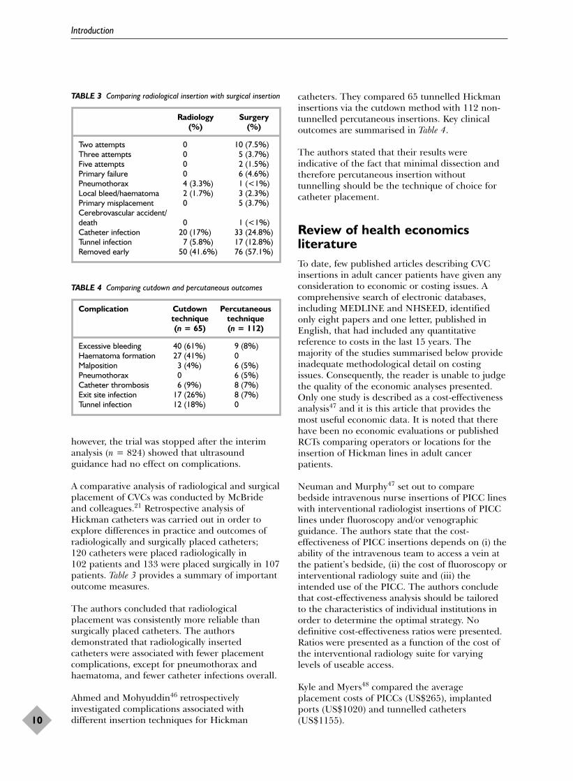
A comparative analysis of radiological and surgicalplacement of CVCs was conducted by McBrideand colleagues.21 Retrospective analysis ofHickman catheters was carried out in order toexplore differences in practice and outcomes ofradiologically and surgically placed catheters; 120 catheters were placed radiologically in 102 patients and 133 were placed surgically in 107patients. Table 3 provides a summary of importantoutcome measures.
The authors concluded that radiologicalplacement was consistently more reliable thansurgically placed catheters. The authorsdemonstrated that radiologically insertedcatheters were associated with fewer placementcomplications, except for pneumothorax andhaematoma, and fewer catheter infections overall.
Ahmed and Mohyuddin46 retrospectivelyinvestigated complications associated withdifferent insertion techniques for Hickman
catheters. They compared 65 tunnelled Hickmaninsertions via the cutdown method with 112 non-tunnelled percutaneous insertions. Key clinicaloutcomes are summarised in Table 4.
The authors stated that their results wereindicative of the fact that minimal dissection andtherefore percutaneous insertion withouttunnelling should be the technique of choice forcatheter placement.
Review of health economicsliteratureTo date, few published articles describing CVCinsertions in adult cancer patients have given anyconsideration to economic or costing issues. Acomprehensive search of electronic databases,including MEDLINE and NHSEED, identifiedonly eight papers and one letter, published inEnglish, that had included any quantitativereference to costs in the last 15 years. Themajority of the studies summarised below provideinadequate methodological detail on costingissues. Consequently, the reader is unable to judgethe quality of the economic analyses presented.Only one study is described as a cost-effectivenessanalysis47 and it is this article that provides themost useful economic data. It is noted that therehave been no economic evaluations or publishedRCTs comparing operators or locations for theinsertion of Hickman lines in adult cancerpatients.
Neuman and Murphy47 set out to comparebedside intravenous nurse insertions of PICC lineswith interventional radiologist insertions of PICClines under fluoroscopy and/or venographicguidance. The authors state that the cost-effectiveness of PICC insertions depends on (i) theability of the intravenous team to access a vein atthe patient’s bedside, (ii) the cost of fluoroscopy orinterventional radiology suite and (iii) theintended use of the PICC. The authors concludethat cost-effectiveness analysis should be tailoredto the characteristics of individual institutions inorder to determine the optimal strategy. Nodefinitive cost-effectiveness ratios were presented.Ratios were presented as a function of the cost ofthe interventional radiology suite for varyinglevels of useable access.
Kyle and Myers48 compared the averageplacement costs of PICCs (US$265), implantedports (US$1020) and tunnelled catheters(US$1155).
Introduction
10
TABLE 3 Comparing radiological insertion with surgical insertion
Radiology Surgery (%) (%)
Two attempts 0 10 (7.5%)Three attempts 0 5 (3.7%)Five attempts 0 2 (1.5%)Primary failure 0 6 (4.6%)Pneumothorax 4 (3.3%) 1 (<1%)Local bleed/haematoma 2 (1.7%) 3 (2.3%)Primary misplacement 0 5 (3.7%)Cerebrovascular accident/death 0 1 (<1%)Catheter infection 20 (17%) 33 (24.8%)Tunnel infection 7 (5.8%) 17 (12.8%)Removed early 50 (41.6%) 76 (57.1%)
TABLE 4 Comparing cutdown and percutaneous outcomes
Complication Cutdown Percutaneous technique technique(n = 65) (n = 112)
Excessive bleeding 40 (61%) 9 (8%)Haematoma formation 27 (41%) 0Malposition 3 (4%) 6 (5%)Pneumothorax 0 6 (5%)Catheter thrombosis 6 (9%) 8 (7%)Exit site infection 17 (26%) 8 (7%)Tunnel infection 12 (18%) 0

Hamilton2 suggested that nurse-led Hickman lineinsertions were cost-effective compared withHickman line insertions by medical staff. It wasestimated that inserting a Hickman line in theoperating theatre by a surgeon would cost £450whereas a line inserted by a nurse on the wardwould cost £150. However, it is not clear whetheror not the cost of treating complications wasincluded in these figures.
Raad and colleagues49 compared tunnelled CVCswith non-tunnelled silastic CVCs. The authorsfound that, given the low infection rate and longdurability of non-tunnelled silicone CVCs, thesecatheters could offer a cost-effective and safealternative to surgically implantable tunnelledcatheters. When compared with the tunnelledHickman catheter, there was an estimatedinsertion cost saving of US$2322 per CVC.Scarpinato50 suggested that Raad and colleagues49
had underestimated these figures as they had notincluded the cost of catheter removal. Scarpinato50
proposed that the true cost saving per CVC wouldbe US$4600.
Thomson51 published a financial feasibility studyto compare peripheral catheters with midlinecatheters. A retrospective audit was performed toaddress the question ‘does the midline catheterreally provide significant cost savings overall?’.Thomson concluded that use of the midlinecatheter could lead to substantial cost savings(US$11,844 in 23 patients) when compared withthe cost of multiple peripheral venipunctures.
Foley52 advocated the placement of lines and ports by interventional radiologists instead ofsurgeons. He argued that more accurate catheterplacement, improved patient safety, acceptablecomplication rates and reduced costs constitutesupport for the placement of lines by radiologists.
Kincaid and colleagues45 stated that the averageprocedure-related fee for insertion of a single-lumen central venous Port-a-Cath in an outpatientsetting was US$1691 versus US$4559 in theoperating theatre and US$3890 in the radiologydepartment. These prices do not include anysubsequent interventions that were related to theprocedure but which took place post insertion.
Finally, in a comparison of hospital with homeCVC survival, Melville and colleagues53 suggestedthat if home sepsis rates could be achieved in thehospital setting, there would be considerable costsavings to the NHS.
Summary of published literatureMuch of the published literature in this importantarea is descriptive in nature. This review of theliterature reveals that relatively few comparativestudies have been conducted. Not only is there apaucity of published economic evaluation results,but also there are few published RCTs whichaddress the following key issues: (i) choice ofinsertion method, (ii) location of insertionprocedure and (iii) choice of operator. These threeissues are central to the current debate about themost clinical and cost-effective method ofHickman line insertion in adult cancer patients.Reporting standards for central venous access haverecently been published.54 These guidelines havebeen designed to facilitate consistent reporting ofclinical trial results so that true comparisonsamong studies can be made. It is clear from thereview of the literature undertaken in this reportthat these guidelines are both timely andappropriate. There is currently much debate inthe NHS about who should be responsible for theinsertion of CVCs, yet this issue is not reflected inthe literature. To date there are no comparativepublished studies involving nurses, yet it is nurseswho are the most recent group of healthcareprofessionals to be trained to insert CVCs.Although the trial in this report does not comparethe performances of different types of operator, itdoes compare two different methods of Hickmanline insertion by nurses and should therefore be auseful addition to the evidence base.
Rationale for the studyAs the range of indications for the use of CVCsexpands, the number of eligible patients requiringCVCs will rise. Consequently, there will be aparallel increase in the demand for healthcareresources to fund central venous access services, bethey hospital- or community-based schemes.Evaluation of both the costs and benefits of suchservices is essential if resources are to be targetedin a manner that generates maximum clinicalbenefits to patients. A key component of anycentral line service is the efficient organisation anddelivery of the CVC insertion service. Every effortmust be made to ensure that Hickman lineinsertions are carried out at the optimal time inthe patients’ management in order to minimisethe risk of adverse health outcomes for thepatient. If scarce healthcare resources are to beinvested in the timely delivery of cost-effectiveinsertion services, then the followingorganisational issues must be addressed:
Health Technology Assessment 2003; Vol. 7: No. 36
11
© Queen’s Printer and Controller of HMSO 2003. All rights reserved.

1. Which is the safest and most cost-effectivemethod/setting for the insertion of Hickmanlines in adult cancer patients?
2. Which operators provide the safest and mostreliable source of expertise in the insertion ofHickman lines in adult cancer patients?
Clearly, the answers to such questions are importantfrom both economic and clinical perspectives. First,from an economic perspective it is clear that as therange of indications for Hickman lines widens, thedemand for Hickman lines will grow. For example,recent evidence has shown that new treatments forcancer and HIV/AIDS have already led to adramatic increase in the demand for Hickman lineinsertions. It is vital, therefore, to determine whichof a range of options for Hickman line insertion isthe most cost-effective.
Second, it is imperative that NHS hospitals provideadequate training for their staff in this procedure.Inadequate training may mean that Hickman-related complication and infection rates are higherthan necessary and this translates into a poorquality service for the patient. In addition, the costsof treating complications and infections areconsiderable. Although very few patients die as adirect result of a Hickman line insertion, the clinicalcomplications for the patient can be very serious,especially in the field of cancer, where it mightmean the delay of chemotherapy treatment. Also, asnew nursing posts are being created (e.g. clinicalnurse specialists and nurse consultants), thenumber of nurses with CVC insertion skills is rising.Performance monitoring and evaluation of bothmedical and nursing operators are then required ifclinical governance issues are to be addressed. Thedevelopment of a training programme whichincludes core competencies and expected standardsof practice to improve the quality of Hickman lineinsertions could yield important benefits to bothpatients and healthcare professionals in the NHS.This report therefore evaluates the potentialcontribution of nursing staff to the improvement ofpatient care in the NHS as their role expands toinclude the insertion of Hickman lines at thebedside and under image guidance.
Study setting: CHNTThe CHNT in Manchester provides an idealsetting from which to address both the clinical andeconomic questions outlined above. At CHNT, thepreferred method of Hickman line insertion isbedside placement for routine procedures withaccess to image-guidance facilities if required. Pre-1995, junior doctors were primarilyresponsible for the insertion of Hickman lines in
the Haematological Oncology Department atCHNT. However, as a result of inadequate trainingand sporadic supervision by more senior staff, theorganisation and delivery of the service were poorand waiting times for the procedure were long.9
Consequently, patients suffered as maximumhealth benefits were not being realised.
In an effort to address clinical concerns, a clinicalnurse specialist (CNS) was employed and trained toinsert Hickman lines. Once experienced in theprocedure, the CNS was responsible for the trainingand supervision of junior medical staff. The CNSwas also responsible for the coordination of thecentral venous access service across the Departmentsof Haematology Oncology and Medical Oncology.
Since 1996, the profile of the CNS has grownsignificantly and in 1998 she was responsible forcarrying out approximately 90% of all Hickmanline insertions, approximately 670 per year, atCHNT. However, as the service continued toexpand, the demand for Hickman line insertionswas greater than supply. Supply was constrainedby the lack of trained staff at CHNT available tocarry out the procedure on a regular basis. Beforethe trial, all Hickman lines inserted by the CNSwere performed at the patient’s bedside. If animage-guided insertion was required, then thepatient was referred to an appropriately trainedconsultant. At the same time, awareness ofevidence-based medicine and clinical governanceissues meant that staff at CHNT were keen toexplore the costs and benefits of image-guidedversus blind insertion Hickman line insertions.
Key nursing and medical staff at CHNT, incollaboration with health economists at theUniversity of Liverpool, decided to apply for HTAfunding. A proposal was submitted with twin aims:(i) to carry out an economic evaluation alongsidean RCT to compare image-guided and blindHickman line insertions and (ii) to evaluate anurse training programme for the insertion ofHickman lines both at the bedside and underimage guidance. The HTA application wassuccessful and the primary research carried out asa result forms the basis of this final report.
Hypotheses and clinical objectives ofprimary clinical researchAs outlined in the grant application submitted, theaim of the RCT was to examine the clinical andcost-effectiveness of image-guided Hickman lineinsertions versus bedside Hickman line insertionsperformed by nurses in adult cancer patients. Theprimary hypothesis was that, other than catheter-
Introduction
12

tip misplacement, there were no real differencesbetween the two insertion approaches. Thesecondary hypothesis was that, once trained,trainees could insert Hickman lines with the samelevel of competency as the trainer. The clinicalobjectives of the trial were to identify success ratesand also frequency and severity of clinicalcomplications in each of the trial arms.Comprehensive data analysis on image-guidedversus blind insertions was planned together withsubgroup analysis focused on comparisons betweenthe three nurses (two trainees and trainer).
Aims and objectives of the studyThe three stated principal objectives of the studywere as follows:
1. to compare the performance of the CNS andjunior doctors in the insertion of Hickman lines
2. to compare the marginal costs and benefitsarising from image-guided versus blindinsertion of Hickman lines
3. to identify the training requirements that willenable the benefits (if any) of the nursetraining programme to be reproducedthroughout the NHS.
Within these three broad objectives, a range ofspecific research objectives were also identified:
1. to evaluate the net incremental resourceimplications of routine use of image guidancefor Hickman line insertion in comparison withblind insertion
2. to evaluate the safety and efficacy of the twotreatments under investigation
3. to evaluate variations in other treatmentoutcomes, for example, patient satisfaction
4. to document the frequency and implications ofcomplications and other significant clinicalfactors related to Hickman line insertions bynurses
5. to assess the incremental cost-effectiveness ratio underlying Hickman line insertion inspecific subgroups of patients in both treatment arms
6. to assess the generalisability of patient benefits and resource savings arising fromnurse-inserted Hickman lines throughout the NHS.
The null hypotheses of the study were that therewould be no difference in:
1. outcomes (except for frequency of catheter-tipmisplacement) between the two arms of thetrial
2. the costs associated with the interventions inthe two groups.
The majority of the research aims and objectiveswere achieved. However, a comparison of theinsertion skills of the CNS and junior doctors wasnot undertaken. This was primarily because adirect evaluation of CNS and junior doctorinsertion performances had previously beencarried out at the CHNT and a summary of theresearch report has been published.9 In brief, this paper demonstrated that rates of failedinsertions, complications, infections and waitingtimes at CHNT were lower for the CNS than forthe junior doctors. Patient satisfaction was alsoshown to be higher for the CNS than the juniordoctors. As the result of these findings, the CNShas been responsible for the training andsupervision of all junior doctors who are interestedin CVC insertions. Consequently, the performanceof junior doctors has improved substantially with both complications and failure rates falling.Based on this information, the research teambelieved the continued funding of the CNS post to be evidence enough to support thesuperior skills of the CNS as compared with JDs at the CHNT.
Health Technology Assessment 2003; Vol. 7: No. 36
13
© Queen’s Printer and Controller of HMSO 2003. All rights reserved.


This chapter is divided into two main parts.The first part describes the methodologies
employed to collect primary data and is dividedinto four separate sections: overview of nursetraining programme, evaluation of the RCT,collection of NHS resource use data and survey ofpatients’ views. The statistical approach planned isalso outlined. The second part describes theeconomic evaluation method used to analyse theprimary data collected. An outline of the statisticalmethods employed is also presented. Finally, asimple arithmetic exercise is described in order toestimate the effect of increasing the number ofnurse insertions on the total annual costs ofHickman line insertions in cancer centres andunits in England and Wales.
Overview of nurse trainingprogrammeRecruitment of traineesTwo qualified nurses were recruited as trainees tothe CHNT. Two nurses were recruited in order toensure the viability of the RCT if one trainee wereto pull out of the training programmeunexpectedly. There were 12 applicants for theposts, six of whom were short-listed and called forinterview. Contracts were initiated on 1 January1999. The trainee nurses were contracted tocomplete the 3-month training programme andalso to participate in the RCT. The trainee nurseswere also expected to provide hands-on support tothe CNS in related clinical areas. Neither of thenurses had previous working experience of CVCinsertion.
Hospital organisational requirementsHickman line insertions are not routinely carriedout by nursing staff, but are usually carried out bymedical personnel. This expansion of thetraditional role of the nurse means that there existcertain documented requirements to whichemployer and employee must adhere. Thecontract signed by the trainees explicitly statedthat they were permitted to insert Hickman linesin adult cancer patients as required by the CHNT.
Supervisors to the traineesThroughout the training period, a range of keypersonnel were identified to give advice andguidance to the trainees if support was required(Nurse Executive Director, Senior Nurse Manager,Consultant Medical Oncologist, Director ofRadiology, Consultant Radiologist and ClinicalNurse Specialist). However, it was the CNS,mentor and teacher, who had day-to-day contactwith the trainees and was responsible fordesigning, coordinating and delivering thetraining programme.
TimetableThe timetable of the training programme was veryflexible. From the onset it was agreed that theinitial estimate of a 3-month training programmeneed not be rigid – that more or less time couldbe spent on the nurse training programme asrequired. The CNS felt that the number ofobserved, supervised and unsupervised centralline insertions was the most important factor inthe education of the trainees, and not the numberof weeks spent in training. However, as it turnedout, the trainees were trained for the estimated12-week period. The trainees spent the initialstages of the training programme observing theCNS insert lines, they then progressed to insertlines under the direct supervision of the CNS andthen finally they inserted lines withoutsupervision.
Training programme for blind insertionof Hickman lines The nurse training programme was designed bythe CNS in conjunction with key medical andclinical oncology personnel at the CHNT. Thetraining programme was based on the trainingprogramme that the CNS had herself undertakena few years previously. The training programmewas divided into four key phases. The aims andobjectives for each phase are outlined in Appendix 1. In summary, the trainee nurses spentthe first 2 weeks familiarising themselves withtheir new work environment and studying relevantcentral line insertion literature. The CNS gave thetrainees seminars on central line insertion-relatedtopics. From week three onwards, both trainees
Health Technology Assessment 2003; Vol. 7: No. 36
15
© Queen’s Printer and Controller of HMSO 2003. All rights reserved.
Chapter 2
Methods

became actively involved in insertion proceduresas they observed, participated in and completedHickman line insertions. Although more than 100lines were inserted at the CHNT during this 12-week period, it was inappropriate for thetrainees to be involved in all of the insertion-related procedures. This was due to a variety ofreasons, for example, CNS and patient preference,patient health status and trainee nurse workload.
Training for image-guided insertion ofHickman linesBoth trainer and trainees undertook externaltraining before they were permitted to insertHickman lines in patients with the use of imageguidance in the X-ray department. All threenurses attended a training day in order tocomplete the 1-day course ‘Training in RadiationProtection related to Diagnostic Radiology’. Thiscourse is approved by the Royal College ofRadiologists and covers ‘core of knowledge’ asdetailed in the regulations. During the 3-monthtraining period, staff in the Department ofDiagnostic Radiology at the CHNT briefed nurseson procedures in the X-ray department.Guidelines regarding image-guided Hickman lineinsertions in the form of a working protocol weredrawn up by diagnostic radiology staff in order toensure patient and professional health and safetyat all times. Nurses were also given a copy ofDiagnostic Radiology Local Rules.
Type of CVCs insertedNurses were trained by the CNS to insert a varietyof central venous lines, including single-, double-and triple-lumen Hickman lines and also PICCs.It was appropriate for nurses to be trained toinsert PICCs because if a patient is not eligible fora Hickman line, a PICC is often inserted in orderto gain temporary venous access. Or, if theoperator attempts to insert the Hickman line butfails, then a PICC is often inserted if venous accessis required immediately. The CNS also trained thenurses to remove Hickman lines and PICCs.
ComplicationsEvery Hickman line insertion procedure carriesthe risk of complication(s) occurring. It wasanticipated that some complications would ariseduring the course of the training period. Mostfrequently occurring complications are catheter-tipmisplacement, pneumothorax, arterial puncture,haematoma, infection and failed insertion. As theCNS was present during most of the proceduresand especially in the early stages of the trainingprogramme, all appropriate action was taken toensure patient safety.
In-house assessmentIn-house assessment of the competency of the twotrainee nurses was carried out at the end of the 3-month period. A consultant oncologistexperienced in Hickman line insertion assessedeach nurse as she inserted a Hickman line blind atthe bedside. A consultant interventionalradiologist assessed each nurse as he or sheinserted a Hickman line under image guidance inthe interventional X-ray suite. Each consultantasked each nurse pertinent questions before,during and after insertions. The CNS was notpresent for any of the procedures that wereassessed. Following the successful completion ofthe assessment, both internal assessors signedstatements that the two trainees were competent toperform blind and image-guided insertions ofHickman lines.
External assessmentA consultant oncologist from St James’ Hospital inLeeds, who is experienced in blind Hickman lineinsertions, conducted the external assessment. Theexternal assessor was present at the blindinsertions and undertook rigorous evaluation ofthe practical abilities and theoretical knowledge ofboth nurses. Following successful completion ofthe assessment, the external assessor signed astatement that the two trainees were competent toperform blind insertions of Hickman lines. Therewas no need for the external assessor to assess theimage-guided insertions as they were performedin exactly the same manner as the blind insertions.
Evaluation of nurse trainingprogrammeA diary was kept of all (blind and image-guided)trainee Hickman line insertions during the 3-month training period. This information wasdocumented in order to ensure that an accuratedescription of all trainee Hickman line insertions(number and type) was available. Any clinicalcomplications that occurred and their outcomeswere also recorded and analysed. The informationwas collected as part of a quality assuranceexercise to ensure that the training programmewas able to equip the nurses with the necessaryinsertion skills and that patients did not suffer anymajor adverse effects during the training period.
Cost analysisA cost analysis was undertaken to estimate theresources incurred during the 3-month trainingperiod. The cost analysis was carried out from theperspective of the NHS. Costs of the nursetraining programme were categorised as follows:
Methods
16

salaries, training courses, use of X-ray suite,clinical complications and travel expenses.
SalariesThree months’ salary costs of the two traineenurses were included in the cost of the trainingprogramme. In addition, 1 month’s salary of theCNS was also included as an estimate of thereplacement cost of time used. If the CNS had notparticipated in the nurse training programme, shewould have spent one-third of her timeperforming bone marrow procedures and acting asa resource for training and supervising juniordoctors. In her absence, another member of staffcarried out bone marrow procedures. Thisreplacement cost is therefore an estimate of theopportunity cost of the CNS’s time.
As part of the training programme, two consultantoncologists and a consultant radiologist formallyassessed the skills of the trainee nurses. Salarycosts for half a day for each of the assessors wastherefore included in the total costs of the trainingprogramme. During the training programme, staffin the Department of Diagnostic Radiology atCHNT briefed nurses on procedures to be carriedout in the X-ray suite. Therefore, half a day’sconsultant radiologist salary was also included inthe total costs of the nurse training programme.
Training coursesAll three nurses (trainer and trainees) attended atraining day in order to complete the 1-day course‘Training in Radiation Protection Related toDiagnostic Radiology’, the fees for which wereincluded in the cost analysis.
Use of X-ray suite As the nurses would have to insert Hickman linesunder image guidance in the interventional X-raysuite as part of the RCT, they observed andparticipated in 15 image-guided Hickman lineinsertions during the period of the trainingprogramme. In normal circumstances, these 15 lines would have been inserted at the bedside.Therefore, the X-ray suite charges for these lineswere included in the total costs of the nursetraining programme.
Clinical complicationsAs some clinical complications were expectedduring the training programme, the cost oftreating these complications was considered.However, as a proportion of these complicationswould have occurred under normal circumstances,a cost estimate based on the CNS’s actualperformance within the RCT was calculated. It was
estimated that the cost of treating twomisplacements and three pneumothoraces wouldbe over and above what was expected in the trialand these were therefore included as part of thecosting exercise.
ExpensesThe expenses of the external assessor were alsopaid, as were travel and subsistence costs to thethree nurses on their radiation protection trainingcourse day.
RCTThe study was a prospective RCT that aimed tomeasure any difference in effectiveness betweenthe image-guided approach and bedside approachto Hickman line insertions by nurses in adultcancer patients.
Ethical approval and support for theprojectEthical approval was sought and obtained fromSouth Manchester Medical Research EthicsCommittee. An advisory committee to the projectwas also set up before the trial commenced. Keystakeholders from the CHNT and the Universityof Liverpool made up this small group andincluded nursing and medical staff, healtheconomists and statisticians.
Trial populationAll of the patients who were recruited for the studyduring the period April 1999 to May 2000 werecancer patients attending the CHNT. At theCHNT, patients can be admitted for Hickman lineinsertions as either inpatients or day-case patientsdepending on a variety of factors such as patientpreference, patient health state, patient residenceand consultant recommendation. The recruitedpatients were a mix of both inpatients and day-case patients. However, the procedure itself isclassified as a day-case procedure. Patients wereboth male and female and were diagnosed witheither haematological or solid tumour cancers.Patient inclusion and exclusion criteria to the RCTare listed in Box 1. All patients who were noteligible for the trial were labelled as off-studypatients. The same information was collected foroff-study patients as was collected for thosepatients who were recruited to the trial.
InterventionsThe aim of both interventions was to obtain central venous access for the patient. The twointerventions under investigation were (i) blind
Health Technology Assessment 2003; Vol. 7: No. 36
17
© Queen’s Printer and Controller of HMSO 2003. All rights reserved.

insertion of a Hickman line and (ii) image-guided insertion of a Hickman line. In the trial,blind insertion of a Hickman line took place at thepatient’s bedside whereas the image-guidedinsertion of a Hickman line took place in theinterventional X-ray suite. Both interventionsinvolved blind venipuncture of the subclavian vein.In the blind arm, the Hickman line was routinelyinserted without the use of image guidance at anypoint in the procedure. However, transfer to theinterventional X-ray suite and use of imageguidance were options immediately available tothe operator during the procedure if required. Inthe image-guided arm, the position of theguidewire was checked before the Hickman linewas introduced and the Hickman line waspositioned with the use of X-ray fluoroscopy (i.e.the location of the Hickman line was checkedusing image guidance during the procedure andany misplacement of the tip was then rectifiedbefore the procedure was completed). Patients inboth arms of the trial had chest X-rays after theprocedure in order to identify any possiblepneumothoraces. All activities in the X-ray suitewere recorded in a log book after the procedurewas performed. The information recordedincluded the amount of time spent using thefluoroscopic facilities. In the interests of safety, apre-set alarm was activated if the screening timewas over 5 min.
Informed consentPatients were given an information leafletdescribing the Hickman line insertion procedureand a description of what was required frompatients participating in the trial. The nurse readthrough the information leaflet with the patientsand gave the patients an opportunity to askquestions or raise concerns. Patients were then leftalone to consider whether or not they wanted to
participate in the trial. Patients were made awarethat they could withdraw from the trial at any timewithout having to give a reason. In order to checkthat the information given to patients wascomplete and that consent was fully informed, theissue of informed consent was addressed withinthe patient satisfaction questionnaire (Appendix 2).As patient consent was obtained beforerandomisation, the nurse taking consent may ormay not have been the nurse to whom theprocedure was randomised.
Randomisation Patients who were eligible for the study wererandomly assigned to either Hickman lineinsertion at the bedside or Hickman line insertionunder image guidance and to one of the threenurses. Patients were recruited to the study by thenurses involved in the RCT. Randomisation was toboth procedure mode (blind or image-guidedinsertion) and to nurse (nurse1, nurse2 or nurse3).
To clarify further, if all three nurses were availablethen the randomisation was to a substudy labelledcentral line insertion project 3 CLIP3 – this studyhad six trial arms:
Nurse Procedure mode1 Blind2 Blind3 Blind1 Image-guided2 Image-guided3 Image guided
If, however, only two nurses were available thenthe randomisation was to a substudy labelledCLIP2 – once the available nurses were re-labelledA and B, this study had four trial arms:
Nurse Procedure modeA BlindB BlindA Image-guidedB Image-guided
Finally, if only a single nurse was available, therandomisation was to a substudy labelled CLIP1,which only had two arms:
Procedure modeBlindImage-guided
In the instances where entry was to either CLIP1or CLIP2, the names of the unavailable nurse(s)and also the reason for their absence were
Methods
18
BOX 1 Patient inclusion and exclusion criteria
Inclusion criteria
>18 years of ageReferred for Hickman line insertionPatient able to lie flat and be compliantPlatelets ideally >100 × 109/1, not less than <75 × 109/1
Exclusion criteria
Patient too ill – nurse assessmentPatient refused consentX-ray facilities were unavailable on requestPatient had a failed previous insertion attempt within
24 hoursPatient had poor haematological profileOther

recorded at the time of randomisation. Theprocedure was a valid one provided that:
1. Nurse unavailability was not manipulated.2. Selection bias was guarded against, that is, all
eligible cases continued to be entered into thetrial when only one or two nurses were available.
The reasons for nurse unavailability were recordedand were consistent with the practicalities of workinglives. The second aspect is difficult to ensure.However, comparison between the case series inCLIP1 and CLIP2 did not reveal any markeddifferences in the case mix of the two substudies.
After the nurse taking consent had checked thatthe patient was eligible for the trial, that the X-raysuite was available and that the patient had giveninformed consent, one of the nurses involved inthe RCT telephoned the Clinical Trial SupportUnit (CTSU) to find out how the procedure was tobe performed and by whom. Computerisedrandomisation was carried out centrally by theCTSU at the CHNT. There was completeseparation of the people involved in thegeneration and implementation of allocations. Aminimisation algorithm with an additionalrandom element was employed to ensure that thenumbers of participants in each of the trial armswas closely balanced within each of the followingstrata: diagnosis, single- or double-lumen line andpre-booked or emergency patients.
Information communicated over the telephone tothe randomisation centre by the nurse included:patient characteristics (name, date of birth,hospital number); patient’s consultant; plannednumber of lumens; emergency or planned insertionand which of the three nurses were available. Thepatient was then randomised to either blind orimage guidance and to a specific nurse. If therewas only one nurse available for randomisation,then this group of patients was labelled as CLIP1;if there were two nurses available for randomisation,CLIP2; and if there were three nurses available forrandomisation, CLIP3. There was no significanttime delay between allocation and randomisation;the procedure was started within 10 minutes ofallocation. The nursing staff kept a diary andrecorded which patients were allocated to whicharm of the trial on which date. Post-randomisation,this diary was cross-checked with the randomisationdatabase allocations.
Protocol violationsBefore the RCT commenced, a list of possibleprotocol violations was compiled and included the
following: patient being randomised twice,insertion procedure being interrupted andcompleted by a different nurse, X-ray suite in use,image-guided facilities out of order, only onenurse being available for randomisation. Allprotocol violations that occurred during the RCTwere recorded.
BlindingIt was impossible to conceal the outcome ofrandomisation from the patient or the operator asboth parties had to be in the same room at thetime of the insertion procedure. The analyst wasnot blinded to the outcome of randomisation atthe time of analysis.
Sample size and powerTwo separate sample size calculations wereperformed in order to ensure that the trial had asufficient number of subjects to address the twoprincipal research questions of interest using theconventional levels of statistical significance (95%)and power (80%).
1. To assess the procedure mode, that is, blindversus image-guided insertion, the primaryoutcome measure was chosen to be themisplacement rate. With 200 cases in each arm,a one-tailed test at the 5% level of significancewould have approximately 80% power if thetrue malposition rate were 0.5% under imageguidance and 5% under blind insertion.
2. To assess nurse skill, the primary outcomemeasure was chosen to be the pneumothoraxrate. The main comparisons would beperformed separately for each trainee againstthe nurse specialist. With 150 cases for eachnurse, a one-tailed test at the 5% level ofsignificance would have approximately 80%power if the true pneumothorax rate were 2.5%for the nurse specialist and 10% for a trainee(the power rising to 95% if the true trainee ratewas as high as 13.5%). Additionally, with 150insertions, if the rate were approximately 10%then the precision of the estimator would beapproximately ±5% to facilitate comparisonwith national rates.
To accommodate both of these requirements, itwas planned to allocate 75 patients to eachnurse/procedure mode combination (i.e. 450 casesin total).
Analysis of dataThe analysis of the data was conducted on both anintention-to-treat and a per protocol basis. Theresults of the intention-to-treat analysis are
Health Technology Assessment 2003; Vol. 7: No. 36
19
© Queen’s Printer and Controller of HMSO 2003. All rights reserved.

presented in the main body of this report. Twodifferent intention-to-treat analyses areundertaken in order to answer two differentquestions. All of the randomisations were includedin the comparison of blind and image-guidedinsertions. Only the randomisations in CLIP2 andCLIP3 were included in the comparison of nurseskill as CLIP1 patients were not truly randomisedto a nurse as there was only one nurse available atthe time of randomisation.
Subgroup analysisThe study has the two main modes of comparison:
1. procedure mode (blind versus image-guidedinsertion) – primary analysis
2. nurse skill (trainer versus trainees) – a priorisubgroup analysis.
The assessment of procedure mode can be validlymade using all of the randomisations. However,the assessment of nurse skill can strictly only bemade using data from CLIP3. Owing to a 3-monthperiod of sickness of the CNS during the period ofthe trial, the number of patients recruited toCLIP3 was not as high as was previouslyanticipated. After statistical advice was sought, itwas believed that no bias would result from alsoincluding the CLIP2 randomisations in theanalysis of nurse skill.
Exploratory analysisAlthough not stated beforehand, furtherexploratory analysis was undertaken after the datawere collected. Ex ante exploratory analysis wascarried out to compare outcomes among differentgroups of patients and included the following:type of Hickman line inserted (single versusdouble) and booking (emergency versus pre-booked). Multiple analyses of the same data arelikely to create a considerable risk of false-positivefindings55 and so the patient and studycharacteristics used for subgroup analysis werecarefully chosen. The subgroup analysesconducted were carried out because it was thoughtthat these characteristics might influenceoutcomes and because they were used in thestratification process.
General data collection – patient casereport formsThe information recorded by the nursing staff onthe patient case report forms provided themajority of the patient data used in the study. Thenursing staff completed the specially designedpatient case report forms because of the clinicalnature of the information solicited and also for
convenience as the nursing staff were in contactwith the patients throughout the duration of theirstay at CHNT. The researcher later completed,where possible, information that was missing fromthe forms. Every completed patient case reportform was checked against the data entered ontothe computerised database at regular intervals.The few errors that were identified were noted andrectified by the researcher. A copy of the patientcase report form is presented in Appendix 3.
Key outcome measures Clinical outcomes of interest included thefollowing: catheter-tip misplacement,pneumothorax, arterial puncture, haematoma,line and tunnel infection. When comparing blindversus image guidance, the primary clinicaloutcome measure was catheter-tip misplacement.When comparing the performance of the threenurses, the primary clinical outcome measure waspneumothorax. Process outcomes included thefollowing: number of successful procedures, failedinsertions, assistance by nurse, assistance byradiologist, assistance by oncologist and patienttransfer from bedside to interventional X-ray suite.Time taken to perform procedure, length of timespent in interventional X-ray suite to repositionmisplaced catheter tip and waiting time betweenbasic insertion and repositioning of Hickman linewere also explored.
Catheter-tip misplacementAll catheter-tip misplacements in the RCT wereidentified from post-procedural chest X-rays andwere recorded by the nursing staff on theindividual patient case report forms. Initially, thenurses recorded only whether or not catheter-tipmisplacement had occurred. However, it soonbecame clear that differences between patients interms of the clinical and psychologicalconsequences suffered could not be identified.Clearly, a patient whose Hickman line onlyrequires a saline flush to manipulate the tip into abetter position does not suffer the sameconsequences as a patient whose Hickman line hasto be removed by a nurse and then reinserted.Where tip misplacement occurred, patient medicalrecords were always reviewed in order to obtainmore detailed clinical information including tipposition and how and when the line wasrepositioned. All repositionings were successfullyconducted under image guidance in theinterventional X-ray suite.
PneumothoraxIn the RCT, all pneumothoraces were identifiedfrom post-procedural chest X-rays and were
Methods
20

recorded by the nursing staff on individual patientcase report forms. Patient medical records werethen reviewed in order to obtain more detailedclinical data including size of pneumothorax,whether or not a chest drain was required andpatient length of stay.
Arterial punctures and haematomasIn the RCT, all arterial punctures and haematomaswere recorded by the nursing staff on the patientcase record forms and were written up in themedical record notes.
InfectionThe nurses recorded all episodes of infectionwithin 14 days of the procedure on the patientcase report forms. It was assumed that allinfections that manifested during this period werelinked to the insertion procedure. Confirmed lineinfections were those infections identified bypositive blood cultures. Confirmed tunnelinfections were identified by positive woundspecimens. If the culture results and specimenswere negative, before assuming that the infectionwas suspected, the researcher reviewed thepatient’s medical records in order to determinewhether or not the tests were carried out becauseinfection was truly suspected or for some othervalid reason. If there was a valid reason for takingthe cultures and specimens which was unrelated toinfection, then a suspected infection episode wasnot recorded. Although patient case report formswere designed to capture all incidences ofinfection, the delay (up to 14 days) beforeinfections were recorded meant that patientmedical records were not always easily available tonursing staff who were completing these forms.Patient databases the CHNT [Adult LeukaemiaDatabase (ALD)] and Withington Hospital [PublicHealth Laboratory Service (PHLS)] were thenaccessed by the researcher in order to completethe patient information required. In this report,all line and tunnel infections reported are acombination of suspected and confirmed infections.
Summary of clinical outcomes evidenceEvidence from a variety of sources was used toconfirm and cross-check that all clinical andprocess outcomes had occurred and these arepresented in Table 5.
Successful insertionSuccessful insertions were defined as those whichdid not require any assistance by anotherhealthcare professional and did not result in anyclinical complications or infection.
Failed insertionsIn the RCT, a failed insertion was recorded if theinsertion procedure was performed as plannedbut, given unforeseen circumstances, the operatorwas unable to insert a Hickman line. Theprocedure was defined as a failed insertion only ifthe operator had been unable to insert a Hickmanline in the patient; in all other circumstances, forexample, if the Hickman tip was misplaced, afailed insertion was not recorded as the outcome.Failed insertions usually resulted in one of thefollowing three scenarios:
1. The operator inserted a PICC instead of aHickman line.
2. The operator referred the patient to anoncologist or radiologist for a femoral or ajugular vein insertion.
3. The operator rebooked the patient for aHickman line insertion by one of the nurses ata later date.
The nursing staff recorded all failed insertions onthe patient case record forms. Medical recordswere reviewed in order to determine how or ifvenous access was achieved at a later date. In time,central venous access was obtained for all patientsin the trial.
Assistance from other healthcareprofessionalsAll insertions that required assistance from any of
Health Technology Assessment 2003; Vol. 7: No. 36
21
© Queen’s Printer and Controller of HMSO 2003. All rights reserved.
TABLE 5 Sources of clinical evidence
Source of evidence of clinical outcome
Clinical outcome Case record Medical records Chest X-ray/ ALD PHLSforms radiology report
Misplacement � � �Pneumothorax � � �Arterial puncture �Infection rates � � � �Successful/failed insertion � �

the nurses or a radiologist or an oncologist, forwhatever reason, were recorded on the patientcase report forms.
Patient transferThe nurses identified all of the patients who weretransferred from the bedside to the interventionalX-ray suite on the patient case report forms.
Time taken to perform proceduresIn all cases, key times taken to perform differentprocedures were recorded. Start and finish timeswere noted on the patient case report forms.
GeneralisabilityAs there were no significant differences betweenthe trial protocol and real-world clinical practiceand owing to the 100% patient compliance rate,the generalisability of the clinical trialeffectiveness data was anticipated to be fairly high.
The focus of the research was not only to assessthe optimal form of Hickman line insertion forthe CHNT; it was also to identify generalisablelessons for the NHS as a whole. Current provisionof Hickman line insertions by a small number ofnurses in England is widely recognised. Thisresearch attempted to identify the extent to whichexpertise in Hickman line insertion byexperienced nurses can be transferred to othersvia a short-term training programme. One aimwas to assess whether or not nurses previouslyunskilled in the procedure could rapidly assimilatethe necessary skills to perform unsupervisedHickman line insertions.
NHS resource usePerspectiveAll resources consumed as part of the RCT wereidentified and measured throughout the durationof the trial from the perspective of the NHS. Theresources associated with infection episodes within14 days of the insertion procedure were alsorecorded. Resource-use data were collected inorder to estimate the impact of the two trialinterventions on NHS budgets. A decision wastaken not to adopt a societal perspective based on evidence that the difficulties of estimatingpatient borne direct and indirect resourceconsumption are well documented.56 Given thatvery ill adult cancer patients made up the studypopulation and would not have been able tocomplete detailed resource consumption forms ortake part in interviews, an NHS perspectiveseemed appropriate.
Resource groupsFor ease of exposition, resources were crudelydivided into resource groups as detailed in Box 2.Within each resource group a range of NHSresources were consumed. Wherever possible,physical quantities of resources consumed innatural units have been reported. Descriptions ofactual resource consumption in each of theresource groups are presented in Appendix 4.
Group A: basic insertion procedureIf the basic insertion procedure was performed atthe bedside, then the use of the followingresources was considered: Hickman line insertionpack, wide range of consumables, nursing time,time spent on the ward, routine tests and chest X-ray. Even if a Hickman line was not inserted anda failed procedure was recorded, a chest X-ray wasalways performed in order to identify anypneumothorax present. If the basic insertionprocedure was performed in the interventional X-ray suite, then use of the following resources wasconsidered: Hickman line insertion pack, widerange of consumables, nursing time, time spent onthe ward and in the interventional X-ray suite,routine tests and chest X-ray. Even if a Hickmanline was not inserted and a failed procedure wasrecorded, a chest X-ray was always performed inorder to identify any pneumothorax present. Atrolley equipped with the required items wasprepared for each patient before the procedurebegan. The same trolley was prepared for blindand image-guided insertions.
Given the confidential nature of the serviceprovided by the nurses, it was impossible for theresearcher to observe how long each nurse spentwith each patient during the trial over and abovetime spent during the insertion procedure. It wasalso very difficult for the nurses to record howlong they spent with each patient on a daily basisbecause they tended to treat more than one
Methods
22
BOX 2 Identifying resource groups
Resource groups
(A) Basic insertion procedure(B) Failed insertions(C) Clinical complications subdivided into: misplacement,
pneumothorax, arterial puncture and haematoma(D) Infections subdivided into: suspected or confirmed
line and suspected and confirmed tunnel infection(E) Assistance subdivided into: nurse, oncologist or
radiologist assistance(F) Patient transfer from bedside to interventional X-ray
suite(G) Additional line inserted

patient at a time. Based on their experienceduring the trial, the nurses were asked to estimatehow much time, in general, they spent with eachpatient. It was estimated that one nurse wouldspend 3 hours with a patient if the procedure hadbeen straightforward. If the patient had amisplaced catheter tip or a pneumothorax or if aPICC was inserted during the procedure, thenursing time spent with each patient was estimatedto be 4 hours. This extra hour of nursing resourcewas itemised under catheter-tip misplacement,pneumothorax and failed insertion as appropriate.
The nurses recorded actual times spent duringblind and image-guided insertion procedures onthe patient case report forms. These times differedacross patients and were usually less than 1 hour.However, it was agreed that the approximation of1 hour be used to calculate the use of X-ray suitecharges as it was impossible for the nurses to knowin advance how long the procedure would takeand as a consequence the X-ray suite was typicallybooked for 1 hour.
Group B: failed insertionsThe resources consumed within the failedinsertion resource group varied considerablybetween patients. The additional resourcesconsumed by each patient who had a PICCinserted, or were referred to an oncologist orradiologist, or who were rebooked for insertion,were identified and measured. The resourceconsequences of failed insertions were estimatedfrom reviews of individual patient medical records.The resources consumed were mainly made up ofconsumables used to insert PICC lines, nursingtime, salaries and consumables associated withHickman line insertion at a later date.
Group C: clinical complicationsClinical complications were subdivided into the
following groups: catheter-tip misplacement,pneumothorax, arterial puncture, haematoma andinfection. The resource consumption associatedwith misplaced catheter tips was measured byreviewing individual patient medical records. Thiswas performed to determine how the tips wererepositioned and to identify whether or not anovernight stay was required. Resources consumedduring repositioning included consumables andhealthcare professional time and occupancy of X-ray suite (based on actual times). Two nurseswere usually involved in the repositioning of theline. As explained before, 1 hour of extra nursingtime was also included in the estimate of resourceconsumption associated with misplaced lines. Thisextra hour was over and above the time taken toreposition the catheter tip. Box 3 summarises themost common manipulation techniques used bynursing and medical staff at the CHNT to rectifycatheter-tip misplacement.
The medical records of all patients who suffered apneumothorax during the trial period werereviewed. Individual patient resource use datawere collected for these patients. Resourceestimates of additional days in hospital, number ofchest X-rays performed and whether or not a chestdrain was required were noted. Other resourcesincluded in the treatment of a pneumothorax were1 hour of extra nursing time from the nurses and,where appropriate, the consumables and specialistregistrar and nursing time involved in insertingand removing chest drains.
The length of stay of all patients who suffered anarterial puncture was checked on the CHNTdatabase in order to determine whether or not anovernight stay was required. No other resourceswere assumed for these patients. No additionalresources were assumed for those patients whosuffered a haematoma.
Health Technology Assessment 2003; Vol. 7: No. 36
23
© Queen’s Printer and Controller of HMSO 2003. All rights reserved.
BOX 3 Consequence of rectifying catheter-tip misplacement
Manipulation technique Clinical consequences for patient
Flushing the line into position by nurse Extra 15 minutes in X-ray suite, no additional risk of pneumothorax, arterialpuncture or haematoma
Line in situ is repositioned via line by nurse Extra 30 minutes in X-ray suite, no additional risk of pneumothorax, arterialpuncture or haematoma
Line is repositioned with pigtail catheter Extra 80 minutes in X-ray suite, no additional risk of pneumothorax, arterial through the femoral vein by radiologist puncture or haematoma
Line is rewired using guidewire and new line Extra 30 minutes in X-ray suite, no additional risk of pneumothorax, arterial by nurse puncture or haematoma
Line is removed and reinserted by nurse Extra 80 minutes in X-ray suite, additional risk of pneumothorax, arterialpuncture or haematoma

Group D: infection episodesIn order to determine how each patient’s infectionepisode was treated, the researcher reviewed,where appropriate, each patient’s medical records.Cancer patients are often prescribed antibiotics fora variety of reasons and it was impossible for theresearcher to be confident that accurate linksbetween indication and prescription were beingmade. As an alternative, one of the trial nursesalso reviewed patient medical records for thispurpose. However, after much time was spentemployed in the task, it was agreed that onlyverification of the actual status (suspected orconfirmed) of the infection could be made andthat actual resource use could not be measured inthis way. After much discussion between medicaland nursing staff, it was agreed that patients withsymptoms of infections at the CHNT weretypically divided into two groups as described inBox 4.
As far as resource use is concerned, whether or nota patient had a suspected or a confirmed infectionwas of no consequence, as patients were treated inexactly the same way until the results of thecultures and swabs were returned from thelaboratories. Given the problems associated withindication and prescription of antibiotics, for thepurposes of this report it was assumed that nochanges to the antibiotic regimens were initiatedafter the laboratory results were known.
Group E: assistance from nurse orradiologist or oncologistThe only resource consumed when assistance wasrequired was nursing or medical staff time. It wasestimated that if assistance was required it was forapproximately 15 minutes. This figure is based ondiscussions with nursing and medical staff.
Group F: patient transfer from bedsideto interventional X-ray suite Resources consumed in this group were mainly X-ray suite charges. It was estimated that if a
patient was transferred from the bedside to theinterventional X-ray suite then the extra timespent in the X-ray suite was approximately 30 minutes and this was charged appropriately.This figure is based on discussions with nursingand medical staff. No additional nursing time wasrequired.
Group G: additional Hickman lineAn additional Hickman line was only required if itwas decided, during the procedure, to insert asingle-lumen line instead of a double-lumen line.Resource use in this group was therefore made upof single Hickman lines. This group did notinclude the insertion of PICCs, as these havealready been included in the failed insertionresource group.
Patient satisfaction questionnaireAfter the patient’s Hickman line had beeninserted and the patient was able to leave thehospital, the patient was given a patientsatisfaction questionnaire to complete. The aim ofthe patient satisfaction questionnaire was to elicitpatient attitudes towards their Hickman lineinsertion experience. The self-administeredquestionnaire was designed by nursing staff andepidemiologists working at the CHNT. Thequestionnaire was short (two pages) and consistedof mainly closed questions. A space for generalcomments was provided. The key topics coveredby the questionnaire included waiting time,information provided and patient experience ofprocedure. The questionnaire was bothanonymous and confidential in nature. A copy ofthe questionnaire is provided in Appendix 2.
In order to obtain a greater number of responses,questionnaires were distributed to both on- andoff-study patients. Patients were asked to completethe questionnaire and hand it in to the ward clerkon leaving the hospital or post it to the Quality
Methods
24
BOX 4 CHNT infection policy
Group 1: Patient is pyrexial with a normal white cell count and there is no other obvious reason for a high temperatureother than line infection.
Regime 1: Patients are given 400 mg of intravenous teicoplanin, followed by 200 mg (intravenous) per day for 5 days.Patients are also given 500 mg per day of oral levofloxacin for a 6-day period. Teicoplanin can be administeredas an outpatient treatment if the patient lives within travelling distance of the CHNT and is able to attendevery day for 6 days.
Group 2: Patient has a high temperature and a low white cell count and does not have a chest infection.
Regime 2: Patients are given 420 mg of intravenous netilmicin per day for 6 days (estimate is made on the assumption ofa 70 kg patient and 4.5 mg of intravenous tazocin three times per day for 6 days).
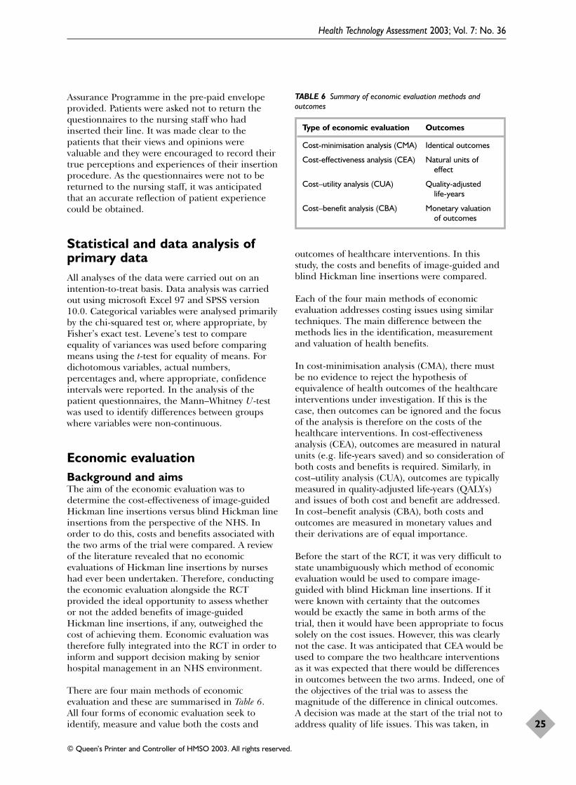
Assurance Programme in the pre-paid envelopeprovided. Patients were asked not to return thequestionnaires to the nursing staff who hadinserted their line. It was made clear to thepatients that their views and opinions werevaluable and they were encouraged to record theirtrue perceptions and experiences of their insertionprocedure. As the questionnaires were not to bereturned to the nursing staff, it was anticipatedthat an accurate reflection of patient experiencecould be obtained.
Statistical and data analysis ofprimary dataAll analyses of the data were carried out on anintention-to-treat basis. Data analysis was carriedout using microsoft Excel 97 and SPSS version10.0. Categorical variables were analysed primarilyby the chi-squared test or, where appropriate, byFisher’s exact test. Levene’s test to compareequality of variances was used before comparingmeans using the t-test for equality of means. Fordichotomous variables, actual numbers,percentages and, where appropriate, confidenceintervals were reported. In the analysis of thepatient questionnaires, the Mann–Whitney U-testwas used to identify differences between groupswhere variables were non-continuous.
Economic evaluationBackground and aimsThe aim of the economic evaluation was todetermine the cost-effectiveness of image-guidedHickman line insertions versus blind Hickman lineinsertions from the perspective of the NHS. Inorder to do this, costs and benefits associated withthe two arms of the trial were compared. A reviewof the literature revealed that no economicevaluations of Hickman line insertions by nurseshad ever been undertaken. Therefore, conductingthe economic evaluation alongside the RCTprovided the ideal opportunity to assess whetheror not the added benefits of image-guidedHickman line insertions, if any, outweighed thecost of achieving them. Economic evaluation wastherefore fully integrated into the RCT in order toinform and support decision making by seniorhospital management in an NHS environment.
There are four main methods of economicevaluation and these are summarised in Table 6.All four forms of economic evaluation seek toidentify, measure and value both the costs and
outcomes of healthcare interventions. In thisstudy, the costs and benefits of image-guided andblind Hickman line insertions were compared.
Each of the four main methods of economicevaluation addresses costing issues using similartechniques. The main difference between themethods lies in the identification, measurementand valuation of health benefits.
In cost-minimisation analysis (CMA), there mustbe no evidence to reject the hypothesis ofequivalence of health outcomes of the healthcareinterventions under investigation. If this is thecase, then outcomes can be ignored and the focusof the analysis is therefore on the costs of thehealthcare interventions. In cost-effectivenessanalysis (CEA), outcomes are measured in naturalunits (e.g. life-years saved) and so consideration ofboth costs and benefits is required. Similarly, incost–utility analysis (CUA), outcomes are typicallymeasured in quality-adjusted life-years (QALYs)and issues of both cost and benefit are addressed.In cost–benefit analysis (CBA), both costs andoutcomes are measured in monetary values andtheir derivations are of equal importance.
Before the start of the RCT, it was very difficult tostate unambiguously which method of economicevaluation would be used to compare image-guided with blind Hickman line insertions. If itwere known with certainty that the outcomeswould be exactly the same in both arms of thetrial, then it would have been appropriate to focussolely on the cost issues. However, this was clearlynot the case. It was anticipated that CEA would beused to compare the two healthcare interventionsas it was expected that there would be differencesin outcomes between the two arms. Indeed, one ofthe objectives of the trial was to assess themagnitude of the difference in clinical outcomes.A decision was made at the start of the trial not toaddress quality of life issues. This was taken, in
Health Technology Assessment 2003; Vol. 7: No. 36
25
© Queen’s Printer and Controller of HMSO 2003. All rights reserved.
TABLE 6 Summary of economic evaluation methods andoutcomes
Type of economic evaluation Outcomes
Cost-minimisation analysis (CMA) Identical outcomes
Cost-effectiveness analysis (CEA) Natural units of effect
Cost–utility analysis (CUA) Quality-adjusted life-years
Cost–benefit analysis (CBA) Monetary valuation of outcomes

large part, because of the nature of the healthcareintervention being evaluated. Insertion of aHickman line is a procedure that, in isolation,does not lead to improved health outcomes forpatients. Only through its use, with chemotherapydrugs or blood products, does it directly affectpatient quality of life. It was therefore impossibleto use CUA, as no estimates of quality of life werecollected. CBA was not undertaken because of thedifficulty of attaching monetary values to theintangible costs associated with Hickman lineinsertions.
Incremental cost-effectiveness ratios are presentedin terms of the incremental cost per natural unitof effect. In this trial, given the stated objectives,the most important ratio to be calculated was theincremental cost per misplaced catheter tipavoided. Although other clinical outcomes were ofinterest, it was this outcome which was expected todiffer between the blind and image-guided armsof the trial. The actual insertion puncture in thetwo arms of the trial was identical. Consequently,rates of failure, pneumothorax, arterial punctureand haematoma were expected to be similar in theblind arm and in the image-guided arm. Theincremental cost of a successful procedure was alsocalculated as this outcome was also expected todiffer between the two arms. Finally, forcompleteness, the likelihood of having a clinicaloutcome excluding a misplaced catheter tip wasexplored within incremental CEA.
Identification of costsThe choice of perspective influences the range ofcosts to be identified, measured and valued in anyeconomic evaluation. Given the NHS perspectiveadopted in this study, only direct healthcare costswere identified and estimated and includedconsumables, salaries, tests and investigations,pharmacy supplies and hospital resources. Indirectcosts and intangible costs were not estimated inthis economic evaluation. All CHNT salariesincluded ‘on costs’ (National Insurance andsuperannuation) and overhead costs (indirect anddirect). Hourly rates for nursing costs were basedon a 37.5-hour week and hourly rates for medicalstaff were based on a 35-hour week. A mix ofpatient-specific and non-patient-specific resourceuse data sources was used to collect importantclinical and cost information. Average costs, notmarginal costs, were used in the economicevaluation for two reasons. First, the use ofmarginal costs requires access to large andcomplex cost datasets that are both expensive anddifficult to acquire; given the time and budgetconstraints of the project, marginal costs would
have been impossible to obtain. Second, as theresults of the economic evaluation were intendedto be of use to a mix of NHS hospital decision-makers and the marginal costs of each hospital aredifferent, average costs were used to improve thegeneralisability of the results. None of the costsincurred in the RCT were associated with the trialper se rather than the costs of providinghealthcare. No protocol-driven costs were incurredduring the RCT.
Measurement of costsThe costs associated with insertion of the Hickman line were based on the resource groupcategories as described previously and aspresented in Box 5.
The costs reported in this study were obtainedfrom a variety of sources and are summarised inTable 7 below. All costs were based on the most up-to-date market prices available. Wheneverpossible, local sources of cost data were used. Allcosts used in the economic evaluation are listed inAppendix 4.
Basis for cost calculationsThe methods used for calculating costs in theeconomic evaluation were very simple. A bottom-up approach was adopted in an attempt to presentas much detailed costing information as wasavailable. As far as possible, all relevant costinformation was included in the economic analysis.Patient cost distributions were summarised bytheir mean, 95% confidence intervals andstandard deviation, and any cost variationsidentified were explored. Evidence suggests that itis the arithmetic mean cost that is most useful toNHS decision-makers.57 The discounting of costswas not appropriate in the comparison of image-guided and blind Hickman line insertions giventhe time horizon of the clinical and economicanalysis.
Methods
26
BOX 5 Resource groups
(A) Basic insertion procedure(B) Failed insertions(C) Clinical complications subdivided into misplacement,
pneumothorax and arterial puncture (D) Infections subdivided into line and suspected and
tunnel infection(E) Assistance subdivided into nurse, oncologist or
radiologist assistance(F) Patient transfer from bedside to interventional X-ray
suite(G) Additional Hickman line

Allowing for uncertainty in theeconomic evaluation: sensitivityanalysis Sensitivity analysis was undertaken in order to testthe robustness of the economic evaluation results,that is, in order to determine whether or notchanges in key assumptions or costs would have animpact on the results of the economic evaluation.For each of the assumptions that were varied thefollowing information was presented: type ofsensitivity analysis conducted, reason for varyingthe assumption, plausible range of values for thevariation and the source of the range of values.Given that the CHNT is a specialist cancer centrewith a relatively unique cost structure, sensitivityanalysis was conducted around unit X-ray costs inan effort to improve external validity to ensurethat the economic results of the study can be usedin different settings. The main limitation of theeconomic evaluation was that CHNT X-ray suitecharges were used to estimate the cost ofoccupancy of the X-ray suite for image-guidedHickman line insertions. However, by varying theradiology charges in the sensitivity analysis toexplore whether or not the economic evaluationresults were affected, it was anticipated that thislimitation would be somewhat overcome.
Summary of assumptions used in theeconomic evaluation1. All patients (inpatients and outpatients) were
treated as day-case patients for the purposesof the costing study.
2. The cost of an overnight stay resulting from aclinical complication for patients who werebooked in as inpatients was not included inthe economic evaluation as these patients
would have been hospitalised anyway.Therefore, the cost of an overnight stay wasonly included, where appropriate, for thosewho were booked in as outpatients or day-casepatients.
3. On average, each nurse spent 3 hours withevery patient who participated in the trialwhether a day-case patient or an inpatient.
4. If a patient suffered a catheter-tipmisplacement, pneumothorax or had a PICCinserted during the procedure, then the nurse was deemed to have spent an extrahour with the patient during the course oftheir stay.
5. All X-ray suite charges were as calculated bythe Radiology Department at the CHNT.
6. When assistance was required, the nurse,radiologist and/or oncologist were eachreckoned to have assisted for 15 minutes.
7. Each patient was considered to have spent 30 minutes in the X-ray suite after beingtransferred from the bedside to theinterventional X-ray suite.
8. Before antibiotics are prescribed, a patientsuspected of having a Hickman line infectionreceived either (i) three blood cultures andone swab (single-lumen line) or (ii) six bloodcultures and one swab (double-lumen line).
9. A patient with an infection either receivedantibiotic regime 1 or 2 as previouslydescribed.
10. Most of the lines placed during the trial weresingle- or double-lumen lines. However, twopatients had a triple-lumen line inserted.Where appropriate, for the purposes of theeconomic evaluation, these triple-lumen lineswere considered as double-lumen lines.
Health Technology Assessment 2003; Vol. 7: No. 36
27
© Queen’s Printer and Controller of HMSO 2003. All rights reserved.
TABLE 7 Cost data
Cost categories Source Year
Consumables Logistics Catalogue 2000British National Formulary 2000Bard 2000
Pharmacy items Pharmacy Department, CHNT 2000British National Formulary 2000
Radiology charges Radiology Department, CHNT 1999/2000
Salaries Finance Department, CHNT 1999/2000
Tests Department of Haematology, CHNT 200Finance Department, CHNT


Evaluation of pre-trial nursetraining programmeBoth nurses completed the training programmeand each nurse was assessed favourably by theCNS and both the in-house and external assessors.Both nurses went on to participate fully in theRCT.
Table 8 presents summary data on the frequencyand type of CVCs inserted by the trainee nursesduring the training programme. These lines donot include the number of Hickman lineinsertions that were observed. The majority oflines inserted during the training programmewere single- and double-lumen Hickman lines.
Table 9 provides a brief description of the clinicalcomplications experienced by patients during thetraining period whilst a trainee was participatingin the procedure. Seventeen (18%) complicationepisodes were recorded, none of which had seriousclinical consequences for patients. Catheter-tip
misplacement was the most frequent complicationassociated with Hickman line insertion during thetraining period.
Table 10 shows the resources that were identified toestimate the total costs of the training programme.
The total cost of the nurse training programmewas estimated to be £19,127.27, as shown in Table 11.
Of the total cost of training the two trainees, 83%was made up of salary costs. Radiology charges foruse of the X-ray suite and the costs of treatingpatients with pneumothorax, which included anovernight stay, were relatively small in comparisonwith salary costs.
A variety of insertion training programme optionscould be designed. Table 12 shows the effect ontotal costs of reducing the training programmefrom 12 to 8 weeks assuming that the samenumber of patients are treated. This would be
Health Technology Assessment 2003; Vol. 7: No. 36
29
© Queen’s Printer and Controller of HMSO 2003. All rights reserved.
Chapter 3
Results
TABLE 8 Frequency and type of CVC inserted during training programme
Type of line insertion participated in, or completed by, trainee nurse Nurse1 Nurse2 Pearson �2
Single 28 23Double 14 18Triple 0 1Single PICC 6 4 �2 = 1.606Total 48 46 p = 0.448, df = 2
df, degrees of freedom.
TABLE 9 Clinical complications during training period
Complications during insertions by No. of Other informationnurse trainees events
Tip misplacement – positioned across midline 5 3 tips were repositioned in X-ray at a later time; 2 tips werecorrectly placed using flushing and vigorous respiration
Tip misplacement – positioned in jugular vein 4 2 tips were repositioned during the procedure in X-ray; 1 tipwas repositioned in X-ray at a later time; 1 tip was referred toa radiologist for repositioning
Arterial puncture 5 No bleeding
Pneumothorax 3 No chest drain required

possible if the trainees were able to participate inmore insertions during a shorter training period.Table 12 also shows the effect on costs of trainingone trainee instead of two trainees over a 12-weekperiod assuming that the same number of patientsare treated. This would be the case if moreintensive training were provided. Further analysiscan be carried out to identify the costs of trainingmore than two trainees at a time and/or over adifferent time period. At the CHNT it was decidedthat each of the trainees should participate in atleast 50 Hickman line insertions. If this numberwere higher or lower then this would affect theduration of the training programme and wouldalso lead to an increase or decrease in costs.
This type of training course is clearly a one-offcost for any institution and one which is timelimited by technological advance. The costs andbenefits of expanding this training programmethroughout the NHS are explored in the section‘Resource implications of Hickman line insertionsby nurses across the NHS’ (p. 51).
In-house feedback from trainingprogrammeFeedback was sought from the CNS and thetrainee central lines nurses throughout the periodof the training programme. All parties concernedagreed that the training programme was a success.At the end of the training programme, trainee
Results
30
TABLE 10 Sources of costs
Description of cost items Unit cost (£) Source Year
G grade salary – annual cost 24983 Finance Department 1999/2000I grade salary – annual cost 30671 Finance Department 1999/2000Consultant salary – annual cost 86326 Finance Department 1999/2000X-ray training day – course fee 50.00 Invoice 1999Radiology charges – hourly rate 124.33 Radiology Department 1999/2000External assessor expenses – travel and subsistence 50.00 CHNT invoice 1999X-ray training day expenses 10.00 CHNT invoice 1999Average cost of misplaced line 109.14 See Appendix 4 1999/2000Average cost of pneumothorax 355.53 See Appendix 4 1999/2000
TABLE 11 Resources and costs
Training programme resources Quantities of resources Cost (£)
Salaries 2 × G grade nurse salary for 3 months 12491.501 × I grade nurse salary for 1 month 2555.924 × Consultant hourly rate for 4 hours 804.96
Courses 3 × Training day course fees 150.00
Radiology charges 15 × Hourly radiology charge 1864.95
Travel expenses 1 × External assessor expenses 50.003 × Travel expenses to training day course 30.00
Complications – misplacement 2 × Mean misplacement cost 231.88
Complications – pneumothorax 3 × Mean pneumothorax cost 948.06
Total 19127.27
Mean cost per nurse trained 9563.63
Mean cost per patient 203.48
TABLE 12 Costs of alternative training programmes
Training programme Cost (£) Cost per nurse trained (£) Cost per patient (£)
8 weeks 14111.46 7055.73 150.121 trainee 12821.52 12821.52 136.40

nurses felt confident in their ability to insert linesand the CNS was more than satisfied with thetrainees’ performance. With hindsight, the nursesand the CNS agreed that a few changes to thetraining programme could be made. It wasthought that a more intensive training programmecould be undertaken. This could be achieved if theCNS trained only one trainee at a time. Onetrainer to one trainee means that the length of theprogramme can be reduced as the target numberof insertions can be achieved in a shorter time
period. Or, if one trainee were working under thesupervision of more than one trainer, this wouldalso mean that the target number of insertionscould be achieved in a shorter time period.
Results of the RCTRecruitment During the 13-month period of the RCT, 667central venous lines were inserted in cancerpatients at the CHNT. Of the 667 lines requestedfor patients, 619 were for patients who weredeemed suitable for entry into the clinical trial bythe nursing staff. Box 6 lists the reasons why 48patients were excluded from the RCT.
Of the 619 patients who were eligible to participatein the RCT, 17 refused to give written and oralconsent to the nursing staff and were therefore notincluded in the trial; 132 patients were alsoexcluded from the trial because the X-ray suite wasunavailable on request before randomisation tookplace. The remaining 470 were randomised toeither blind or image-guided insertion.
A total of 470 patients were randomised as part ofthe RCT; 235 patients were allocated to blindinsertion and 235 to image-guided insertion(Figure 2). Each of the patients was alsorandomised to nurse1, nurse2 or nurse3.
Health Technology Assessment 2003; Vol. 7: No. 36
31
© Queen’s Printer and Controller of HMSO 2003. All rights reserved.
BOX 6 Patient exclusion
Reason for patient exclusion No. of patientsexcluded
Patient was too ill 9Patient had a failed previous attempt 2Patient had poor haematology 15Patient was under 18 years of age 9Doctor was performing insertion as 5
part of his or her trainingOthera 8
a Other reasons for exclusion included the following:patient did not speak English and did not want to beparted from her husband if she was randomised to X-raysuite; patient was taking regular aspirin; patient wanted ageneral anaesthetic in theatre; patient had a clot in theneck; patient was neutropenic and could not leave theward; patient had an anatomical abnormality, reason notstated.
Assessed for eligibility(n = 667)
Allocated to insertion under image guidance (n = 235)
Allocated to blind insertion(n = 235)
CLIP1(n = 9)
CLIP2 + 3(n = 226)
CLIP1(n = 12)
CLIP2 + 3(n = 223)
Follow-up and analysisof blind and image
completed(n = 9)
Follow-up and analysisof blind and image
completed(n = 226)
Follow-up and analysisof blind and image
completed(n = 12)
Follow-up and analysisof blind and image
completed(n = 223)
Excluded from analysisof nurse skill
(n = 9)
Follow-up and analysisof nurse skill completed
(n = 226)
Excluded from analysisof nurse skill
(n = 12)
Follow-up and analysisof nurse skill completed
(n = 223)
Assessed for eligibility (n = 197)Not meeting inclusion criteria (n = 48)Refused to participate (n = 17)X-ray unavailable (n = 132)Randomised (n = 470)
FIGURE 2 Progress of patients through RCT

In the blind arm, 88 patients were randomised tonurse1, 77 to nurse2 and 70 to nurse3. Ninepatients were randomised to CLIP1, 85 to CLIP2and 141 to CLIP3.
In the image arm, 90 patients were randomised tonurse1, 71 to nurse2 and 74 to nurse3. Twelvepatients were randomised to CLIP1, 81 to CLIP2and 142 to CLIP3.
Protocol violationsThere were two protocol violations in the blindarm. In one instance, the nurse who was allocatedto one patient was called away during theprocedure and so another nurse took over theprocedure. On another occasion during theinsertion procedure, it was clear that the patienthad a hidden anatomical abnormality and was notsuitable for a Hickman line; a PICC line was theninserted instead. There were four protocolviolations in the image-guided arm. Threepatients were taken to the X-ray suite for theirprocedure only to be told that the suite wasoccupied and so their Hickman lines were insertedat the bedside. On one occasion, a patient wastaken to the X-ray suite only to be told that thefluoroscopy machine was out of order. Again, thepatient was taken back to the ward and a Hickmanline was inserted at the bedside. All of theinsertions that violated the protocol were includedin the intention-to-treat analysis.
Patient characteristicsPatient randomisation ensured that there were noreal differences between the groups of patients inthe blind and image-guided arms of the trial.There were slightly more men than women in thetrial (Table 13) and the majority of both sexes wereover 40 years of age (Table 14). Just over half ofthe patients weighed between 60 and 79 kg (Table 15). About 80% of patients were of normalphysical state, that is, they were neither over- norunder-weight (Table 16). Table 17 shows that three-quarters of patients had a Karnofsky performance(KP) score between 80 and 100% (a KP score of80% means that the patient is able to carry outnormal activities with effort and a score of 100%means that the patient is normal). Some 54% ofpatients had been diagnosed with gastrointestinalcancer. Patients were more likely to be sufferingfrom a solid tumour cancer than a haematologicalcancer (Table 18). There were 76% of patients whowere treated on the day ward, with the remainderspread across the Adult Leukaemia Unit, NathanHouse (private ward) and CHNT generaloncology wards (Table 19). Some 66% of patientswere treated as inpatients (Table 20) and 91% ofHickman line insertions were pre-booked (Table21). There were 8% of patients who required ablood and/or platelet transfusion before theirHickman line insertion (Table 22). Table 23 showshow many lines were inserted blind and underimage guidance by each of the three nurses.
Results
32
TABLE 13 Gender
Gender Blind (n = 235) Image-guided (n = 235) Total (n = 470) Fisher’s exact test
Men 133 (57%) 125 (53%) 258 (55%) �2 = 0.550Women 102 (44%) 110 (47%) 212 (45%) p = 0.516
TABLE 14 Age
Age (years) Blind (n = 235) Image-guided (n = 235) Total (n = 470) Pearson �2
<25 13 (5%) 12 (5%) 25 (5%)25–29 5 (2%) 9 (4%) 14 (3%)30–34 12 (5%) 10 (4%) 22 (5%)35–39 6 (2%) 15 (6%) 21 (5%)40–44 22 (9%) 9 (4%) 31 (6%)45–49 20 (8%) 32 (14%) 52 (11%)50–54 29 (12%) 31 (13%) 60 (13%)55–59 44 (19%) 42 (18%) 86 (18%)60–64 31 (13%) 25 (11%) 56 (12%)64–69 31 (13%) 32 (14%) 63 (13%)70–74 16 (7%) 11 (5%) 27 (6%) �2 = 15.217≥ 75 6 (2%) 7 (3%) 13 (3%) p = 0.173, df = 11

Health Technology Assessment 2003; Vol. 7: No. 36
33
© Queen’s Printer and Controller of HMSO 2003. All rights reserved.
TABLE 15 Weight
Weight (kg) Blind (n = 235) Image-guided (n = 235) Total (n = 470) Pearson �2
<50 13 (6%) 7 (3%) 20 (4%) 50–59 30 (13%) 36 (16%) 66 (14%)60–69 54 (25%) 56 (25%) 110 (23%)70–79 56 (26%) 64 (29%) 120 (26%)80–89 36 (16%) 27 (12%) 63 (13%)90–99 23 (11%) 24 (11%) 47 (10%)≥ 100 7 (3%) 11 (5%) 18 (4%) �2 = 5.031Missing 16 (7%) 10 (4%) 26 (6%) p = 0.540, df = 6
TABLE 16 Physical state of patients
Physical state Blind (n = 235) Image-guided (n = 235) Total (n = 470) Pearson �2
Cachexic 29 (12%) 27 (11%) 56 (12%)Normal 189 (80%) 187 (80%) 376 (80%) �2 = 0.503Obese 17 (7%) 21 (9%) 38 (8%) p = 0.778, df = 2
TABLE 17 KP score of patients
KP score Blind (n = 235) Image-guided (n = 235) Total (n = 470) Pearson �2
≤ 60 22 (10%) 21 (9%) 43 (9%)70 33 (14%) 35 (15%) 68 (15%)80 85 (36%) 74 (32%) 159 (34%)≥ 90 93 (40%) 104 (44%) 197 (42%) �2 = 1.455Missing 2 (1%) 1 (1%) 3 (1%) p = 0.693, df = 3
TABLE 18 Primary diagnosis of patients
Primary diagnosis Blind (n = 235) Image-guided (n = 235) Total (n = 470) Pearson �2
Leukaemia + other 19 + 0 (8%) 18 + 1 (8%) 38 (8%)haematological
Lymphoma 24 (10%) 21 (9%) 45 (10%)Myeloma 20 (8%) 25 (11%) 45 (10%)Gastrointestinal 127 (54%) 124 (54%) 251 (53%)Breast 19 (8%) 17 (7%) 36 (8%) �2 = 1.066Other solid tumour 26 (11%) 29 (12%) 55 (12%) p = 0.957, df = 5
TABLE 19 Location of patients
Location Blind (n = 235) Image-guided (n = 235) Total (n = 470) Pearson �2
Day ward 181 (77%) 176 (75%) 357 (76%)Adult Leukaemia Unit 10 (4%) 13 (6%) 23 (5%)General wards 38 (16%) 39 (17%) 77 (16%)Nathan House 6 (3%) 6 (3%) 12 (3%) �2 = 0.472Missing 0 1 (1%) 1 (1%) p = 0.925, df = 3
TABLE 20 Inpatient or outpatient
Blind (n = 235) Image-guided (n = 235) Total (n = 470) Fisher’s exact test
Inpatient 155 (66%) 156 (66%) 311 (66%) �2 = 0.100Outpatient 80 (34%) 79 (34%) 159 (34%) p = 0.500

Characteristics of RCTsAt the CHNT, the patient’s health status andcorresponding need for treatment determinewhether or not a single-, double- or triple-lumenHickman line is planned for each patient. In theRCT, the majority (97%) of Hickman linesplanned for the patients was subsequently inserted(Table 24). The most frequently inserted line wasthe single-lumen Hickman line. Only 3% (n = 13)of Hickman lines were not inserted as planned. Innine instances, Hickman lines were not inserted inpatients at all; in five of these patients PICCs wereinserted and no lines were inserted in the
remaining four patients (Table 25). Two plannedsingle-lumen Hickman lines were inserted asdouble-lumen lines and two planned double-lumen lines were inserted as single-lumen lines.Of patients who participated in the RCT, 81%were having their first Hickman line inserted(Table 26). Of Hickman lines inserted, 83% wereon the patients’ right side (Tables 27 and 28).
Clinical outcomes from RCTsThe rate of misplaced catheter tips was statisticallydifferent between the blind and the image-guidedarms (p < 0.001). Of Hickman lines inserted at
Results
34
TABLE 21 Pre-booked or emergency patients
Blind (n = 235) Image-guided (n = 235) Total (n = 470) Fisher’s exact test
Pre-booked 214 (91%) 212 (90%) 426 (91%) �2 = 0.100Emergency 21 (9%) 23 (10%) 44 (9%) p = 0.874
TABLE 22 Platelet or blood transfusion
Blind (n = 235) Image-guided (n = 235) Total (n = 470) Fisher’s exact test
Blood transfusion 14 (6%) 6 (3%) 20 (4%) �2 = 3.342p = 0.108
Platelet transfusion 16 (7%) 12 (5%) 28 (6%) �2 = 0.608p = 0.560
TABLE 23 Insertions by nurse
Trial nurse Blind (n = 235) Image-guided (n = 235) Total (n = 470) Pearson �2
Nurse1 88 (37%) 90 (38%) 178 (38%)Nurse2 77 (33%) 71 (30%) 148 (30%) �2 = 0.377Nurse3 70 (30%) 74 (31%) 144 (31%) p = 0.828, df = 2
TABLE 24 Planned Hickman line insertions
Planned Blind (n = 235) Image-guided (n = 235) Total (n = 470) Fisher’s exact test
Single 177 (75%) 177 (75%) 354 (75%)Double 57 (24%) 57 (24%) 114 (24%) �2 = 0.000Triple 1 (1%) 1 (1%) 2 (1%) p = 1.000
TABLE 25 Actual Hickman lines inserted during the trial
Inserted Blind (n = 235) Image-guided (n = 235) Total (n = 470) Pearson �2
Single 170 (72%) 178 (76%) 348 (74%)Double and triple 57 +1 (25%) 54 + 1 (23%) 113 (24%)PICC 4 (1%) 1 (1%) 5 (1%) �2 = 3.064None 3 (1%) 1 (1%) 4 (1%) p = 0.382, df = 3

the bedside, 14% had misplaced catheter tips.Only one catheter tip was misplaced under imageguidance (Table 29). The majority (78%) of thelines with misplaced tips were either flushed ormanipulated into position by the nurses on the
same day (Table 30). There were no statisticallysignificant differences in frequency of arterialpuncture, pneumothorax or haematoma betweenthe two arms. Infections related to the insertionwere defined as all infections taking place within
Health Technology Assessment 2003; Vol. 7: No. 36
35
© Queen’s Printer and Controller of HMSO 2003. All rights reserved.
TABLE 26 Insertion number
Insertion no Blind (n = 235) Image-guided (n = 235) Total (n = 470) Pearson �2
1 192 (81%) 190 (82%) 382 (81%)2 32 (14%) 34 (14%) 66 (14%)3 7 (3%) 10 (4%) 17 (4%)>4 4 (2%) 0 4 (2%) �2 = 4.598Missing 0 1 (1%) 1 (1%) p = 0.204, df = 3
TABLE 27 Left- and right-sided insertions
Insertion side Blind (n = 235) Image-guided (n = 235) Total (n = 470) Fisher’s exact test
Left 40 (17%) 36 (15%) 76 (17%) �2 = 0.251Right 195 (83%) 199 (85%) 394 (83%) p = 0.707
TABLE 28 Reason for choice of insertion side
Reason for choice Blind (n = 235) Image-guided (n = 235) Total (n = 470) Pearson �2
of side
Operator preference 174 (74%) 184 (79%) 358 (76%)Venous compromise 6 (2%) 5 (2%) 11 (2%)Breast disease 22 (9%) 17 (7%) 39 (8%)Other anatomical 20 (9%) 21 (9%) 41 (9%)Compromised lung function 8 (3%) 5 (2%) 13 (3%) �2 = 2.228Other 5 (2%) 3 (1%) 8 (2%) p = 0.817, df = 5
TABLE 29 Clinical complications
Complication Blind Image-guided Estimated difference Total Fisher’s exact (n = 235) (n = 235) (lower 95% CL to (n = 470) test
upper 95% CL)
Misplacement 32 (14%) 1 (1%) –31 (–13%) 32 (7%) �2 = 33.199(–8 to 18%) p = <0.001
Pneumothorax 7 (3%) 2 (1%) –5 (–2%) 9 (2%) �2 = 2.832(–4 to 0.3%) p = 0.175
Arterial puncture 15 (6%) 13 (5%) –2 (–1%) 28 (6%) �2 = 0.152(–5 to 3%) p = 0.846
Haematoma 2 (1%) 4 (2%) 2 (5%) 6 (1%) �2 = 0.675(2 to 8%) p = 0.685
Line infection 10 (4%) 14 (6%) 4 (2%) 24 (5%) �2 = 0.598(–2 to 5%) p = 0.531
Tunnel infection 11 (5%) 4 (2%) –3 (–3%) 15 (3%) �2 = 3.569(–6 to 2%) p = 0.069
CL, confidence limit.

14 days of the insertion procedure. There wereseven suspected infection episodes and 14confirmed infection episodes in the blind arm.There were six suspected infection episodes and12 confirmed infections in the image-guided arm.In total, there were 24 line infection episodes and15 tunnel infection episodes recorded during thetrial period (Table 29).
Process outcomesIn the blind arm, 67% of Hickman lines weresuccessfully inserted without any clinicalcomplications or assistance from other staff. Therewere seven (0.02%) failed insertions in the blindarm. In the image-guided arm, 81% of Hickmanlines were successfully inserted without anycomplications or assistance from other staff. Therewere two (0.008%) failed insertions in the image-guided arm (Table 31).
Of all insertions performed in the clinical trial,11% (n = 51) required assistance from a nurse,oncologist and/or radiologist (Table 31). Nurses were called upon for assistance much more frequently than radiologists or oncologists.The CNS was the most frequently called upon
member of staff in both the blind and image-guided arms.
Only three patients were transferred from thebedside to the interventional X-ray suite duringthe course of the insertion procedure.
TimeThe average time taken to complete the procedurein the blind and image-guided arms of the trialwas 38 minutes (95% CL: 36 to 39) and 40 minutes(95% CL: 38 to 42), respectively. Minimum andmaximum times taken to complete the procedurein the blind arm were 15 and 90 minutes,respectively. Minimum and maximum times takento complete the procedure in the image arm were20 and 150 minutes, respectively. In the economicanalysis it is assumed that the interventional X-raysuite is occupied for a 60-minute period. Clearly,the average actual time spent by each of thenurses performing image-guided procedures issometimes more and sometimes less than 60 minutes.
If a patient has a misplaced catheter tip, then thepatient is taken to the interventional X-ray suite in
Results
36
TABLE 30 Catheter-tip misplacement remedial technique
Technique Blind (n = 32) Image-guided (n = 1) Total (n = 33)
Flush 9 (28%) 0 9 (27%)Reposition (nurse) 16 (50%) 0 16 (48%)Rewire 4 (12%) 0 4 (12%)Reposition (interventional radiologist) 1 (3%) 1 (100%) 2 (6%)Removal and reinsertion 2 (6%) 0 2 (6%)
TABLE 31 Process outcomesa
Outcome Blind Image-guided Estimated difference Total Fisher’s exact (n = 235) (n = 235) (lower 95% CL to (n = 470) test
upper 95% CL)
Successful insertion 157 (67%) 191 (81%) 34 (14%) 348 (74%) �2 = 12.797(7 to 22%) p = <0.001
Failed insertion 7 (3%) 2 (1%) –5 (–2%) 9 (2%) �2 = 2.832(–5 to 0.3%) p = 0.175
Nurse assistance 28 (12%) 18 (8%) –10 (–4%) 46 (10%) �2 = 2.410(–9 to 1%) p = 0.162
Radiologist assistance 3 (1%) 4 (2%) 1 (0.4%) 7 (1%) �2 = 0.145(–2 to 3%) p = 1.000
Oncologist assistance 3 (1%) 0 –3 (–1%)(–3 to 0.2%) 3 (1%) �2 = 3.019
p = 0.248
a Note that this table is explanatory only and is not meant to include all patients.

order to have the tip repositioned. Table 32describes the average time taken for nurses toreposition misplaced catheter tips during the trialperiod.
The average waiting time between the initialprocedure and the patient’s visit to theinterventional X-ray suite for repositioning of theline was 105 minutes (95% CL: 72 to 138 minutes).The minimum waiting time was 15 minutes andthe maximum waiting time was 330 minutes. The waiting time between procedures depends on the demand for the interventional radiologysuite.
Exploratory analysis Single- versus double-lumen linesSingle- versus double-lumen Hickman lines werealso compared for the clinical and processoutcomes of interest (catheter-tip misplacement,pneumothorax, arterial puncture, haematoma,
line infection, tunnel infection, successful insertion,failed insertion, nurse assistance, oncologistassistance and radiologist assistance). A range ofstatistically significant differences were foundbetween these two groups. Double-lumen lineswere associated with significantly more assistancefrom the radiologist, more frequent catheter-tipmisplacement and more episodes of line andtunnel infections. Consequently, single-lumen lineswere significantly more likely to be successful thandouble-lumen lines.
Pre-booked versus emergencyWhen comparing pre-booked lines and emergencylines, three statistically significant differences werefound (Table 33). Pre-booked patients weresignificantly more likely to have successfuloutcomes than emergency patients. Patients withemergency lines were significantly more likely tohave Hickman lines with catheter-tips misplacedand suffer from line infections.
Health Technology Assessment 2003; Vol. 7: No. 36
37
© Queen’s Printer and Controller of HMSO 2003. All rights reserved.
TABLE 32 Average time taken to reposition misplaced catheter tips
Technique used to reposition misplaced Average time SD Assumption used in economic catheter tip (minutes) (95% CLs) analysis (minutes)
Flushing 17 (24 to 29) 10 15 Reposition by nurses 27 (19 to 34) 14 30Reposition by radiologist 80 80Rewiring 30 (10 to 50) 12 30Removal and reinsertion by nurse Unavailablea 80
SD, standard deviation.a Time taken was assumed to be the same as for the reposition by the interventional radiologist for the purposes of
economic evaluation.
TABLE 33 Exploratory analysis using blind versus image-guided dataset
Single vs double lumen Pre-booked vs emergency
Outcome Single Doublea Fisher’s exact Pre-booked Emergency Fisher’s exact (n = 354) (n = 116) test, p-value (n = 426) (n = 44) Test, p-value
Pneumothorax 8 (2%) 1 (1%) 0.463 9 (2%) 0 0.410Misplacement 17 (5%) 16 (4%) 0.003 25 (6%) 8 (18%) 0.007Arterial puncture 22 (6%) 6 (5%) 0.823 25 (6%) 3 (7%) 0.738Haematoma 4 (1%) 2 (2%) 0.640 4 (1%) 2 (5%) 0.101Successful insertion 279 (79%) 69 (59%) 0.000 325 (76%) 23 (52%) 0.003Failed insertion 6 (2%) 3 (3%) 0.013 7 (2%) 2 (5%) 0.638Line infection 9 (3%) 15 (3%) 0.000 17 (4%) 7 (16%) 0.001Tunnel infection 7 (2%) 8 (7%) 0.696 13 (3%) 2 (5%) 0.202Nurse assistance 32 (9%) 14 (12%) 0.369 39 (9%) 7 (16%) 0.177Oncologist assistance 3 (1%) 0 1.000 3 (1%) 0 1.000Radiologist assistance 3 (1%) 4 (3%) 0.066 7 (2%) 0 1.000
a Includes two triple-lumen lines.

Comparing nurse skillAll of the outcome data that were collected duringthe period of the RCT can be linked to the nursewho carried out the insertion procedure.
Three-way comparison of nurse skill Tables 34–36 show the clinical and processoutcomes associated with each individual nurse inboth arms of the RCT. The total number ofprocedures performed by all three nurses was 449.Nurse1 inserted 173 Hickman lines and nurse2and nurse3 carried out 139 and 137 procedures,respectively.
Comparing nurse trainees (nurse1 andnurse2) with trainerTo explore whether or not the trainees and thetrainer were operating at a similar skill levelduring the trial, the outcomes of the trainees werecombined and then compared with those of thetrainer (Table 37). Rate of pneumothorax was theprimary clinical outcome of interest. Whencomparing the two nurse trainees together versusthe trainer for a range of clinical and processoutcomes (pneumothorax, catheter-tipmisplacement, arterial puncture, haematoma, lineinfection, tunnel infection, successful insertion,failed insertion, nurse assistance, oncologist
Results
38
TABLE 34 Clinical outcomes
Nurse1 Nurse2 Nurse3
Clinical outcome Blind Image- Blind Image- Blind Image-(n = 86) guided (n = 72) guided (n = 68) guided
(n = 87) (n = 67) (n = 69)
Pneumothorax 3 (3%) 0 4 (6%) 2 (3%) 0 0Misplacement 14 (16%) 0 7 (10%) 1 (2%) 10 (15%) 0Arterial puncture 7 (8%) 3 (3%) 6 (8%) 7 (11%) 2 (3%) 3 (4%)Haematoma 1 (1%) 2 (2%) 0 2 (3%) 1 (1%) 0Line infection 1 (1%) 6 (7%) 7 (10%) 1 (2%) 2 (3%) 7 (10%)Tunnel infection 0 1 (1%) 6 (8%) 2 (3%) 5 (7%) 1 (1%)
TABLE 35 Process outcomes
Nurse1 Nurse2 Nurse3
Clinical outcome Blind Image- Blind Image- Blind Image-(n = 86) guided (n = 72) guided (n = 68) guided
(n = 87) (n = 67) (n = 69)
Failed insertion 4 (5%) 1 (1%) 2 (3%) 0 0 1 (1%)Nurse assistance 12 (14%) 8 (9%) 12(17%) 9 (13%) 4 (6%) 1 (1%)Oncologist assistance 0 0 1 (1%) 0 0 0Radiologist assistance 0 2 (2%) 1a (1%) 1 (1%) 2b (3%) 1 (1%)
a Patient was transferred from bedside to X-ray suite during procedure.b Patient was transferred from bedside to X-ray suite during procedure.
TABLE 36 Assistance required from other nurse
Assistance Nurse1 (called) Nurse2 (called) Nurse3 (called)
Nurse Blind Image- Blind Image- Blind Image- Blind Image-guided guided guided guided
Nurse1 n = 86 n = 87 n/a n/a 4 (5%) 1 (1%) 8 (9%) 7 (8%)Nurse2 n = 72 n = 67 4 (5%) 4 (6%) n/a n/a 8 (11%) 5 (7%)Nurse3 n = 68 n = 69 4 (6%) 1 (1%) 0 0 n/a n/a

assistance and radiologist assistance), there wasonly one statistically significant difference.Procedures carried out by the trainees weresignificantly more likely to require the assistance ofanother nurse than procedures carried out by thetrainer (p = 0.002).
Comparing nurse1 with trainer In order to ensure that one trainee was notcompensating for the other trainee in terms ofskill level, each of the trainees was compared withthe trainer. Table 38 shows that no statisticallysignificant differences were identified betweennurse1 and the trainer for a range of clinical andprocess outcomes (pneumothorax, catheter-tipmisplacement, arterial puncture, haematoma, lineinfection, tunnel infection, successful insertion,failed insertion, oncologist assistance andradiologist assistance). The only statisticallysignificant difference was found between nurse1and the trainer in terms of the number of timesassistance from another nurse was required.Nurse1 was more likely to ask for assistance fromanother nurse than the trainer (p = 0.012).
Comparing nurse2 with trainer Table 39 shows that no statistically significantdifferences were identified between nurse2 and thetrainer for a range of clinical and processoutcomes (catheter-tip misplacement, arterialpuncture, haematoma, line infection, tunnelinfection, successful insertion, failed insertion,oncologist assistance and radiologist assistance).Two statistically significant differences were foundbetween nurse2 and the trainer. Nurse2 was morelikely to ask for assistance from another nurse thanthe trainer (p = 0.002) and was also more likely tocause a pneumothorax (p = 0.03) during theprocedure.
Comparing nurse1 with nurse2In order to check that there were no realdifferences between the skill levels between thetrainees, the outcomes of nurse1 and nurse2 werecompared. Table 40 shows that no statisticallysignificant differences were identified betweennurse1and nurse2 for a range of clinical andprocess outcomes (pneumothorax, catheter-tipmisplacement, arterial puncture, haematoma, line
Health Technology Assessment 2003; Vol. 7: No. 36
39
© Queen’s Printer and Controller of HMSO 2003. All rights reserved.
TABLE 37 Comparing nurse trainees with trainer
Outcome Trainees (n = 312) Trainer (n = 137) p-Value Test
Pneumothorax 9 (3%) 0 0.063 Fisher’s exact Misplacement 22 (7%) 10 (7%) 1.000 Fisher’s exact Arterial puncture 23 (7%) 5 (4%) 0.202 Fisher’s exact Haematoma 5 (2%) 1 (1%) 0.672 Fisher’s exact Line infection 15 (5%) 9 (6%) 0.499 Fisher’s exact Tunnel infection 9 (3%) 6 (4%) 0.411 Fisher’s exact Successful insertion 223 (71%) 105 (77%) 0.299 Fisher’s exact Failed insertion 8 (2%) 1 (1%) 0.287 Fisher’s exact Nurse assistance 41 (13%) 5 (4%) 0.002 Fisher’s exact Oncologist assistance 3 (1%) 0 0.556 Fisher’s exact Radiologist assistance 4 (1%) 3 (2%) 0.441 Fisher’s exact
TABLE 38 Comparing nurse1 with trainer
Outcome Nurse1 (n = 173) Trainer (n = 137) p-Value Test
Pneumothorax 3 (2%) 0 0.258 Fisher’s exact Misplacement 14 (8%) 10 (7%) 0.834 Fisher’s exact Arterial puncture 10 (6%) 5 (3%) 0.436 Fisher’s exact Haematoma 3 (2%) 1 (1%) 0.633 Fisher’s exact Line infection 7 (4%) 9 (6%) 0.440 Fisher’s exact Tunnel infection 1 (1%) 6 (4%) 0.048 Fisher’s exact Successful insertion 131(76%) 105 (77%) 0.894 Fisher’s exact Failed insertion 5 (3%) 1 (1%) 0.233 Fisher’s exact Nurse assistance 20 (12%) 5 (3%) 0.012 Fisher’s exact Oncologist assistance 1 (1%) 0 1.000 Fisher’s exact Radiologist assistance 2 (1%) 3 (2%) 0.658 Fisher’s exact

infection, successful insertion, failed insertion,nurse assistance, oncologist assistance andradiologist assistance). The only statisticallysignificant difference was found between nurse1and nurse2 in terms of the number of tunnelinfections. Nurse2 was more likely to insert aHickman line in those patients who go on to havea confirmed tunnel infection than nurse1 (p = 0.012).
Comparing all three nursesAs a final check, the skill levels of the three nurseswere compared simultaneously. The results in Table 41 show that there were no real differencesand that the skill levels of the three nurses are infact comparable.
Exploratory analysis Analysis was also carried out in order to explore
Results
40
TABLE 39 Comparing nurse2 with trainer
Outcome Nurse2 (n = 139) Trainer (n = 137) p-Value Test
Pneumothorax 6 (4%) 0 0.030 Fisher’s exact Misplacement 8 (6%) 10 (7%) 0.808 Fisher’s exact Arterial puncture 13 (9%) 5 (4%) 0.086 Fisher’s exact Haematoma 2 (1%) 1 (1%) 1.000 Fisher’s exact Successful insertion 92 (66%) 105 (77%) 0.063 Fisher’s exact Failed insertion 3 (2%) 1 (1%) 0.622 Fisher’s exact Line infection 8 (6%) 9 (6%) 1.000 Fisher’s exact Tunnel infection 8 (6%) 6 (4%) 0.785 Fisher’s exact Nurse assistance 21 (15%) 5 (4%) 0.002 Fisher’s exact Oncologist assistance 2 (1%) 0 0.498 Fisher’s exact Radiologist assistance 2 (1%) 3 (2%) 0.683 Fisher’s exact
TABLE 40 Comparing nurse1 with nurse2
Outcome Nurse1 (n = 173) Nurse2 (n = 139) p-Value Test
Pneumothorax 3 (2%) 6 (4%) 0.194 Fisher’s exact Misplacement 14 (8%) 8 (6%) 0.508 Fisher’s exact Arterial puncture 10 (6%) 13 (9%) 0.278 Fisher’s exact Haematoma 3 (2%) 2 (1%) 1.000 Fisher’s exact Successful insertion 131 (76%) 92 (66%) 0.100 Fisher’s exact Failed insertion 5 (3%) 3 (2%) 0.736 Fisher’s exact Line infection 7 (4%) 8 (6%) 0.597 Fisher’s exact Tunnel infection 1 (1%) 8 (6%) 0.012 Fisher’s exact Nurse assistance 20 (12%) 21 (15%) 0.401 Fisher’s exact Oncologist assistance 1 (1%) 2 (1%) 0.587 Fisher’s exact Radiologist assistance 2 (1%) 2 (1%) 1.000 Fisher’s exact
TABLE 41 Comparing all three nurses
Outcome Nurse1 (n = 173) Nurse2 (n = 139) Trainer (n = 137) Fisher’s exact test, p-value
Pneumothorax 3 (2%) 6 (4%) 0 0.036Misplacement 14 (8%) 8 (6%) 10 (7%) 0.724Arterial puncture 10 (6%) 13 (9%) 5 (4%) 0.140Haematoma 3 (2%) 2 (1%) 1 (1%) 0.741Successful insertion 131 (76%) 92 (66%) 105 (77%) 0.088Failed insertion 5 (3%) 3 (2%) 1 (1%) 0.398Line infection 7 (4%) 8 (6%) 9 (6%) 0.623Tunnel infection 1 (1%) 8 (6%) 6 (4%) 0.03Nurse assistance 20 (12%) 21 (15%) 5 (4%) 0.006Oncologist assistance 1 (1%) 2 (1%) 0 0.335Radiologist assistance 2 (1%) 2 (1%) 3 (2%) 0.759

whether or not those insertions that had requiredassistance from another nurse were more likely toresult in poorer clinical and process outcomesthan those insertions which did not requireassistance. Haematoma and line and tunnelinfections were the only outcomes that did notdiffer significantly depending on whether nurseassistance was required (Table 42).
TimeTable 43 presents a range of mean insertion timesfor each of the nurses. Nurse3 is the mostexperienced of the three and this is evident by thefact that, in general, she inserted Hickman linesmore quickly than the trainees did. For all of thenurses, image-guided insertions took slightlylonger than blind insertions and double- and
triple-lumen lines took slightly longer than single-lumen lines.
Levene’s test for equality of variances was used toidentify whether or not equal variances could beassumed. The t-test for equality of means was thenused to generate p-values to compare the meantimes of the three nurses. When comparing thetrainees, statistically significant differences werefound in mean times for the insertion of single-lumen lines and those lines inserted under imageguidance. Mean times for nurse1 were notsignificantly different from the trainer’s times forsingle- or double-lumen lines or for lines insertedunder image guidance. Mean times for nurse2were significantly different from the trainer’s timesfor single-lumen lines and lines inserted blind and
Health Technology Assessment 2003; Vol. 7: No. 36
41
© Queen’s Printer and Controller of HMSO 2003. All rights reserved.
TABLE 42 Nurse assistance and outcomes
Outcome Total no. of events Nurse assistance required Fisher’s exact test, p-value during event
Pneumothorax 9 4 (44%) 0.008Misplacement 32 8 (25%) 0.010Arterial puncture 28 14 (50%) 0.000Haematoma 6 2 (33%) 0.118Line infection 24 1 (0.04%) 0.711Tunnel infection 15 0 0.382Successful insertion 328 0 0.000Failed insertion 9 7 (78%) 0.000Oncologist assistance 3 2 (67%) 0.029Radiologist assistance 7 3 (43%) 0.026
TABLE 43 Mean insertion times for the three nurses
Nurse1 (n = 173) Nurse2 (n = 139) Nurse3 (n = 137)
Blind, mean (minutes) (SD) 38 (15) 41 (11) 34 (12) Image-guided, mean (minutes) (SD) 39 (18) 46 (14) 38 (15)Single-lumen, mean (minutes) (SD) 37 (14) 43 (11) 34 (13)Double-lumen, mean (minutes) (SD) 44 (22) 45 (11) 40 (17) Triple-lumen, mean (minutes) 90 50
TABLE 44 Time taken for failed insertions
Nurse Arm Type of line inserted Duration of failed insertion (minutes)
Nurse1 Blind Single PICC 75Nurse1 Blind Single PICC 75Nurse2 Blind 0 70Nurse2 Blind 0 90Nurse1 Blind Single PICC 90Nurse1 Blind 0 75Nurse2 Blind Single PICC 60Nurse3 Image-guided 0 60Nurse1 Image-guided Single PICC 120

under image guidance. Simple analysis of meaninsertion times appears to show that nurse1 andthe trainer have similar insertion times whereasinsertions performed by nurse2 take slightlylonger. There were no differences in the length oftime taken to insert double-lumen lines among thethree nurses.
Table 44 presents the length of time that eachfailed insertion procedure took to complete. All ofthe failed insertion times were at least one hourlong. Minimum time taken for a failed insertionwas 60 minutes and the maximum time taken wastwo hours. Mean time for a failed insertion was 79minutes (95% CLs: 65 to 93; SD = 18).
Patient satisfaction questionnaireInformation generated from the patientsatisfaction questionnaire (Appendix 2) providesvaluable information about the impact ofextending the nurse’s traditional role to includethe insertion of Hickman lines.
Questionnaires were given to patients who tookpart in the CLIP trial (n = 470) and also to thosepatients who did not participate in the CLIP trialbut who had a Hickman line inserted during thesame study period (n = 197). A total of 150 (22%)questionnaires were completed and returned bypatients.
Patient location and waiting timesFifty-nine patients (41%) who completed thequestionnaire were inpatients and 86 (59%) wereday-case patients; 49% (n = 73) of patients hadtheir line inserted on the ward and 51% (n = 75)had their line inserted in the X-ray department.
Patients whose lines were inserted on the wardwere asked how long they had to wait for a bed. Of the 49 patients who responded, 10% wereinpatients so they did not wait at all; 59% waitedless than half an hour, 22% waited between half anhour and 1 hour and 8% waited longer than 1 hour.
Patients whose lines were inserted in the X-raydepartment were asked how long they had to waitfor a bed. Of the 60 patients who responded, 58%waited less than half an hour, 17% waited betweenhalf an hour and 1 hour, 10% waited longer than1 hour and 15% did not know how long they hadwaited. These same patients were also asked howlong they waited for a vacant slot in the X-raydepartment. Of the 57 patients who responded,
63% waited less than half an hour, 21% waitedbetween half an hour and 1 hour, 12% waitedlonger than 1 hour and 4% did not know howlong they had waited.
Patients were asked how they felt about theirexperience of waiting times on the day of theirHickman line insertion. Of the 116 patients whogave a qualitative response, 91% were positiveabout the waiting time experienced. Only 10patients (9%) were negative and felt that theirwaiting time was too long or their waiting timeadded to the anxiety felt before the procedure.Approximately 54% of patients stated that theirwaiting time was ‘not a problem’ or that it was‘fine’ or ‘no bother’. About 15% said that theirwaiting time was ‘satisfactory’ or ‘as expected’.About 24% of patients recorded that they werepleased with their waiting time or that the waitingtime was better than expected and 20% of patientssaid that they felt that they ‘didn’t have to wait’ orthat the waiting times were ‘very good’ or that theywere ‘very pleased’ with the waiting times.
Patient appraisal of informationprovidedPatients were asked about the amount of writteninformation provided to them regarding theircentral line insertion. The patient informationleaflet used during the trial period is presented inAppendix 5. Almost 97% (n = 143) of patientsstated that they had been given the right amountof written information and 3% (n = 5) felt thatthey had been given more information than theywanted. Patients were also asked about the amountof verbal information provided to them regardingtheir central line insertion. Again, 97% (n = 145)felt that they had been given the right amount ofverbal information and only 3% (n = 4) felt thatthey had been given more information than theywanted.
Patient experience of anxietyPatients were asked about their level ofapprehension prior to the insertion of theircentral line: 22% (n = 33) of patients said theywere not at all apprehensive, 49% (n = 73) saidthey were a little apprehensive and 29% (n = 44)said they were very apprehensive. Patients wereasked to state what caused them to feel anxiousbefore having their central line inserted. Theywere given a range of options to choose from: fearof pain, a complication occurring, unsuccessfulprocedure, other reason or not applicable as thepatient did not feel anxious. Patients were invitedto choose as many reasons as was appropriate.About 34% of all patients (n = 141) stated they
Results
42

were anxious due to fear of pain, 46% stated theywere anxious because of the possibility of acomplication occurring, 21% were anxious aboutan unsuccessful procedure and 19% gave anotherresponse as the source of their anxiety. Otherresponses included lack of information given atsatellite hospital, general anxiety about havingcancer treatment and fear of the unknown. About20% of patients claimed not to be anxious aboutthe procedure.
Of the 80% (n = 113) of patients who stated areason(s) for their apprehension, 21 said that theirapprehension was due to fear alone, 33 said thattheir apprehension was totally due to thepossibility of a complication occurring, eight solelyfeared an unsuccessful procedure and 11 listed asingle other reason for their apprehension. Fortypatients therefore listed more than one reason fortheir apprehension before the procedure.
SedationSome 98% (n = 147) of patients stated that theywere offered sedation to make them feel drowsyand relaxed during the procedure. One patientresponded that they did not know if it had beenoffered and one patient said that sedation had notbeen offered. About 83% (n = 124) stated thatthey accepted the sedation and 17% of patientsrefused sedation.
Patient experience of the procedurePatients were asked if they found the procedureuncomfortable: 117 patients (78%) stated that theprocedure was not uncomfortable at all, 31 (21%)said the procedure was a little uncomfortable andtwo (1%) felt that it was very uncomfortable.Patients were also asked if they found theprocedure painful: 120 patients (80%) did not findthe procedure painful at all whereas 30 (20%)found the procedure a little painful.
When asked if the level of pain or discomfortexperienced was what was expected, 63% (n = 86)reported that it was not as bad as expected, 11% (n = 15) said that it was the same as expected, 1%(n = 1) said it was worse than expected and 25%(n = 34) said that they had not known what toexpect.
Further comments from patientsAbout 58% (n = 63) of respondents had furthercomments to make about the insertion of theirHickman line. Only 1% could be interpreted asnegative comment. Almost all patient commentswere favourable and there was great support forthe nursing staff (n = 35). In general, patients
praised the nursing staff for their reassurance (n = 19), helpfulness (n = 11) and professionalism(n = 8). Although the survey responses suggest that5% (4/73) of patients had misplaced lines and onepatient suffered a pneumothorax, these patientswere generally positive about their experience. Toillustrate this, one respondent made the comment,‘had to go to X-ray to readjust line from neck tocorrect position, would have been good if this hadnot happened, but the procedure went well’.
Statistical analysisIn order to identify any statistically significantdifferences between the patients who had theirline inserted on the ward and the patients whohad their line inserted in the X-ray department, achi-squared analysis or Fisher’s exact test wasperformed where appropriate. There was only oneidentified statistically significant differencebetween these two groups. Some 13% (8/60) ofpatients who had their line inserted on the wardstated that the possibility of an unsuccessfulprocedure caused them to feel anxious; 42%(21/50) of patients who had their line inserted inthe X-ray department reported that the possibilityof an unsuccessful procedure caused them to feelanxious. This difference was statistically significantat the 95% level as p = 0.012.
The Mann–Whitney U-test was performed in orderto identify any differences between patients whoselines were inserted on the ward and in theinterventional X-ray suite in terms ofapprehension, pain and discomfort levels. Nostatistically significant results were found
The Mann–Whitney U-test was also performed toidentify any differences in discomfort and painbetween patients who were given a sedative andpatients who were not given a sedative. Nostatistically significant results were identified.
Economic evaluationIn order to conduct the economic evaluation, totalcosts and total benefits were calculated for eacharm of the trial. Table 45 presents the total costfigures for the blind and the image-guided armsof the RCT. It also shows mean per patient costs ineach of the trial arms.
Minimum and maximum costs per patient in theblind arm were £309.07 and £2102.97,respectively. The mean cost per patient was£440.40 (95% CL: 397.00 to 483.81; SD = 337).Minimum and maximum costs in the image arm
Health Technology Assessment 2003; Vol. 7: No. 36
43
© Queen’s Printer and Controller of HMSO 2003. All rights reserved.
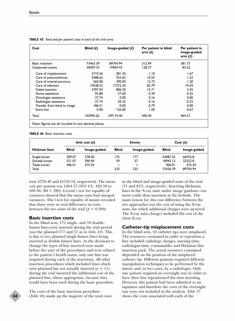
were £378.40 and £1530.54, respectively. The meancost per patient was £464.57 (95% CL: 428.58 to500.56; SD = 280). Levene’s test for equality ofvariances showed that the mean costs had unequalvariances. The t-test for equality of means revealedthat there were no real differences in costsbetween the two arms of the trial (p = 0.399).
Basic insertion costsIn the blind arm, 175 single- and 59 double-lumen lines were inserted during the trial period(not the planned 177 and 57 as in Table 24). Thisis due to two planned single-lumen lines beinginserted as double-lumen lines. As the decisions tochange the types of line inserted were madebefore the start of the procedures and were relatedto the patient’s health status, only one line wasrequired during each of the insertions. All otherinsertion procedures which included lines whichwere planned but not actually inserted (n = 11)during the trial incurred the additional cost of theplanned line, where appropriate, because theywould have been used during the basic procedure.
The costs of the basic insertion procedure (Table 46) made up the majority of the total costs
in the blind and image-guided arms of the trial(71 and 81%, respectively). Inserting Hickmanlines in the X-ray suite under image guidance wasmore costly than insertion at the bedside. Themain reason for this cost difference between thetwo approaches was the cost of using the X-raysuite, for which additional charges were incurred.The X-ray suite charges included the cost of thechest X-ray.
Catheter-tip misplacement costsIn the blind arm, 32 catheter tips were misplaced.The resources consumed in order to reposition aline included radiology charges, nursing time,radiologist time, consumables and Hickman lineinsertion pack. The actual resources consumeddepended on the position of the misplacedcatheter tip. Different patients required differentmanipulation techniques to be performed by thenurses and, in two cases, by a radiologist. Onlyone patient required an overnight stay in order tohave their line repositioned the next morning.However, this patient had been admitted as aninpatient and therefore the costs of the overnightstay were not included in the analysis. Table 47shows the costs associated with each of the
Results
44
TABLE 45 Total and per patient costs in each of the trial arms
Cost Blind (£) Image-guided (£) Per patient in blind Per patient in arm (£) image-guided
arm (£)
Basic insertion 73463.39 89704.94 312.49 381.72Unplanned events 30059.33 19469.43 128.37 83.22
Care of misplacement 3710.36 281.35 1.10 1.67Care of pneumothorax 2488.62 355.62 10.55 1.52Care of arterial puncture 260.00 390.00 15.72 1.20Care of infection 19538.52 17372.24 82.79 74.24Failed insertion 3707.04 806.30 15.71 3.45Nurse assistance 92.80 57.60 0.39 0.25Oncologist assistance 37.74 0.00 0.16 0.00Radiologist assistance 37.74 50.32 0.16 0.22Transfer from blind to image 186.51 0.00 0.79 0.00Extra line 0.00 156.00 1.00 0.67
Total 103495.62 109174.40 440.40 464.57
Note: figures are all rounded to two decimal places.
TABLE 46 Basic insertion costs
Unit cost (£) Events Cost (£)
Hickman lines Blind Image-guided Blind Image-guided Blind Image-guided
Single-lumen 309.07 378.40 175 177 54087.25 66976.8Double-lumen 321.07 390.40 59 57 18943.13 22252.8Triple-lumen 406.01 475.34 1 1 406.01 475.34Total 235 235 73436.39 89704.94

techniques used. The costs of repositioning amisplaced catheter tip ranged from £50.98 to£318.07.
Pneumothorax costsThe clinical and resource consequences of patientswith pneumothoraces were different and thereforethe resource implications were analysed at anindividual patient level. For example, only onepatient had a large pneumothorax and had a chestdrain inserted by a specialist registrar andremoved by a nurse. Patients with pneumothoracesconsumed the following resources: overnight stay,consumables, nursing time, specialist registrartime and chest X-rays. Table 48 summarises thecosts associated with seven pneumothoraces in theblind arm and two pneumothoraces in the image-guided arm of the trial. The mean cost of apneumothorax in the blind arm was £316.02 (95%CL: £23.80 to 608.25; SD = 380).
Arterial puncture costsIn the blind arm, 15 patients had arterial puncturesduring the insertion procedure. Eleven out of these15 patients were admitted as inpatients. Therefore,only four patients in the blind arm had overnightstays that were unplanned. Two of the four patientswith arterial punctures also suffered
pneumothoraces. To avoid double counting forthese patients, only one overnight stay per patientwas accounted for and this was included under thepneumothorax resource group heading. Two of thefour had failed procedures without a pneumothoraxand therefore the costs of two unplanned overnightstays were included in the analysis at a total cost of£260.00. In the image-guided arm, 13 patients hadarterial punctures. Ten out of 13 patients werebooked in as inpatients. Therefore, only the cost ofthree unplanned overnight stays were included inthe analysis at a total cost of £390.00.
Infection costsThe unit costs of treating infection (Table 49) werethe same for patients who had their Hickman linesinserted in either arm of the trial. Resourcesassociated with the treatment of both line andtunnel infections included inpatient stay, drugs,nursing time, medical staff time, blood cultures,swabs and consumables. Patients with infecteddouble-lumen lines were more expensive to treatthan patients with infected single-lumen lines asswabs and blood cultures had to be taken frommore than one lumen. The cost of treatingpatients (n = 5) with a line and a tunnel infectionat the same time was assumed to be the same as apatient with a line infection.
Health Technology Assessment 2003; Vol. 7: No. 36
45
© Queen’s Printer and Controller of HMSO 2003. All rights reserved.
TABLE 47 Costs of rectifying catheter-tip misplacement
Misplacement technique Unit Blind Image-guided Blind arm Image-guided cost (£) cost (£)a arm cost (£)
Flush single 50.98 8 0 407.84 0Flush double 55.23 1 0 55.23 0Reposition single (n) 98.98 7 0 692.86 0Reposition double (n) 99.53 9 0 895.77 0Reposition single (r) 280.80 1 0 280.80 0Reposition double (r) 281.35 0 1 0 281.35Rewire double 188.43 4 0 753.72 0Single removed and reinserted by nurse 306.07 1 0 306.07 0Double removed and reinserted by nurse 318.07 1 0 318.07 0Total 3710.36 281.35
a Mean cost of catheter tip misplacement in blind arm is £115.94 (95% CL: £89.47 to 142.41; SD = 73).n, Nurse; r, interventional radiologist.
TABLE 48 Costs of treating pneumothorax
Costs of pneumothorax Unit cost (£) Event Blind (£) Events Image-guided (£) Total (£)
Consumables 25.09 1 25.09 0 0 25.09Overnight stay 130.00 11 1430.00 0 0 1430.00Chest X-ray 55.00 17 935.00 6 330.00 1265.00Nurse 1.28 1 1.28 0 0 1.28Specialist registrar 7.58 1 7.58 0 0 7.58Extra nursing time 12.81 7 89.67 2 25.62 115.29Total 2488.62 355.62 2844.24

Failed insertion costsEach of the procedures which were defined asfailed insertions (n = 9) had different clinical andresource consequences. It was necessary tocalculate the costs of the procedure for eachindividual patient in the RCT (Table 50). If a PICCline was inserted during the procedure, then thecost of the PICC line was included in this categorywith the cost of the planned line included in thebasic insertion cost category as insertion of theHickman line would have been attempted. In theblind arm, costs ranged from £66.33 to £1778.69.
In this group, resources consumed included PICCinsertion pack, referral to radiologist or oncologistfor Hickman line insertion and treatment ofinfection.
Assistance from other healthcareprofessionalsThe costs of assistance from other healthcareprofessionals during the RCT were relatively smallcompared with the other cost generating eventsthat took place (Table 51). The costs of assistancefrom a nurse were less expensive than the costs of
Results
46
TABLE 49 Costs of infection
Infection Unit cost (£) Cost of events (£)a
(excluding doubles)Single Double Blind Image-guided
Single Double Single Double
Line infection 1062.14 1104.14 4248.56 6624.84 5310.7 9937.26Tunnel infection 1062.14 1104.14 4248.56 4416.56 2124.28 0
a Total costs in blind arm = £19,538.52. Total cost in image-guided arm = £17,372.24.
TABLE 50 Failed insertion costs
Failed insertion type Blind arm (£)a Image-guided arm (£)a
PICC insertion during procedure 66.33PICC insertion during procedure 66.33PICC insertion during procedure/insertion in jugular by radiologist at a later date 368.95PICC insertion during procedure/insertion in femoral by oncologist at a later date/
infection/PICC insertion 1778.69Referred for femoral insertion by oncologist at a later date 402.28Referred for femoral insertion by oncologist at a later date 415.90PICC inserted during procedure/Hickman inserted at a later date 444.73PICC insertion 2 days later/Hickman inserted at a later date 579.73Rebooked for Hickman line insertion by nurse 390.40Total cost 3707.04 806.30
a Mean cost of failed insertion in blind arm = £529.60 ( 95% CL: £–9.82 to 1069.04; SD = 583). Mean cost of failedinsertion in image arm = £403.15 (95% CL: £241.17 to 565.15; SD = 18).
TABLE 51 Costs of assistance by healthcare professionals
Events Cost (£)
Unit cost (£) Blind Image-guided Blind Image-guided
Nurse assistance 3.20 30a 17 92.80 57.60Oncologist assistance 12.58 3 0 37.74 0Radiologist assistance 12.58 3 4 37.74 50.32Total 168.28 107.92
a During one procedure, assistance was provided by two nurses, hence this figure is one higher than reported previously (Tables 35 and 37).

assistance from an oncologist or a radiologist asthe cost of healthcare professional assistance wasmeasured in minutes and estimated using CHNTsalary scales. It was assumed that the duration ofassistance by any of the healthcare professionalswas 15 minutes. The total cost of nurse assistancewas greater than oncologist or radiologistassistance in both arms because the assistance ofanother nurse was called for more frequently thanassistance from a doctor.
Transfer costsOnly three patients were transferred from thebedside to the interventional X-ray suite duringthe procedure. Radiology charges were used tocalculate the total transfer costs based on a 30-minute stay in the interventional X-ray suite. Thetotal cost of transfer was £186.51.
Additional line costsOnly two Hickman lines were included in theadditional line costs category and these lines weredouble-lumen lines that were changed to single-lumen lines during the basic insertion procedure.Both of these procedures took place in imageguidance. The total additional line cost was£156.00 for the image arm of the trial.
Planned single- versus planned double-lumen Hickman linesTable 52 presents cost data on single- and double-lumen Hickman lines. It shows that the meaninsertion cost of a double-lumen Hickman line inthe trial was more expensive than the meaninsertion cost of a single-lumen Hickman line.The mean cost of a double-lumen line was themost expensive of all three lines because double-lumen lines were associated with more expensivecomplications than the others.
Table 53 shows the mean cost of inserting single-,double- and triple-lumen Hickman lines duringwhich complications occurred. Figure 3 shows thecost of uncomplicated insertions versus complicatedinsertions.
Incremental cost-effectiveness analysisIn the economic analysis of blind and image-guidedHickman line insertions by nurses, the primaryclinical outcome of interest was the rate of catheter-tip misplacement. However, in any economicevaluation, the choice of effectiveness measure canimpact on the incremental cost-effectiveness ratio(ICER) calculated. In order to explore the potentialeffect of different effectiveness measures in thiseconomic analysis, a range of outcome measureswere used. The three effectiveness measures usedin the economic evaluation were as follows:
1. catheter-tip misplacement avoided2. successful insertion3. all other clinical complications avoided.
Catheter-tip misplacement avoidedFigure 4 shows both the costs and proportion ofmisplaced catheter tips avoided associated witheach of the trial arms. Proportion of misplacedcatheter tips can also be expressed as proportionof correctly placed tips during the basic insertionprocedure. The 95% CIs around the pointestimates are also shown.
For meaningful comparison, it is important tolook at the additional costs and benefits that theimage-guided approach would impose over theblind approach. In order to do this, incrementalcost-effectiveness per misplaced catheter tipavoided was calculated.
Health Technology Assessment 2003; Vol. 7: No. 36
47
© Queen’s Printer and Controller of HMSO 2003. All rights reserved.
TABLE 52 Mean cost of single-, double- and triple-lumen lines
Type of line No. of lines Average cost of insertion (£) SD 95% CI
Single, blind 177 395.27 263 355.89 to 433.69Double, blind 57 581.14 321 453.84 to 708.81Triple, blind 1 406.01Single, image-guided 177 414.48 180 387.80 to 441.16Double, image-guided 57 619.86 439 503.13 to 736.59Triple, image-guided 1 478.54
TABLE 53 Mean cost of single-, double- and triple-lumen lineswith complications
Type of line Mean (£) SD
Single, blind, complicated 625.51 431Double, blind, complicated 957.81 556Triple, complicated 478.54Single, image-guided, complicated 733.22 463Double, image-guided, complicated 1044.37 527

Results
48
Single blind Double blind Triple blind Single image
Type of Hickman line
Double image Triple image0
200
400
600
800
1000
1200
Mea
n co
s (£
)Complicated
Uncomplicated
FIGURE 3 Costs of complicated and uncomplicated Hickman line insertions
00
100
200
300
Cos
t per
pat
ient
(£)
400
500
600
0.1 0.2 0.3 0.4 0.5
Proportion of correctly placed catheter tips during basic insertion
0.6 0.7 0.8 0.9 1
10.99
429
501
0.90.82
484
397
Blind armImage arm
FIGURE 4 Cost-effectiveness plane for correct placement of catheter tips during basic insertion

Incremental cost per misplaced catheter tipavoided= (Mean cost per patient in the image-guided
arm – mean cost per patient in the blindarm)/(rate of misplacement in the blind arm –rate of misplacement in the image-guided arm)
= (£464.57 – 440.40)/(0.13617 – 0.00425)= £24.17/0.13192= £183.22
This means that if there is a switch from blindinsertion to image-guided insertion, then a cost of£183.22 is incurred for every additional misplacedcatheter tip that is avoided.
Successful line insertionA successful insertion was defined as an insertionthat did not require any assistance by anotherhealthcare professional, nor did it result in anyclinical complications or infection. Clearly, thenumber of successful lines associated with blindand image-guided insertion is affected by the rateof misplaced catheter tips. This measure ofeffectiveness also incorporates the processoutcomes described in this report as the numberof successful lines is also affected by the rate ofassistance required during the procedure. Figure 5shows both the costs and proportion of successfulline insertions associated with each of the trial
arms. Again, 95% CIs around the point estimatesare also shown.
Incremental cost per successful line insertion= (Mean cost per patient in the image-guided
arm – mean cost per patient in the blindarm)/(rate of successful insertions in the image-guided arm – rate of successful insertion in theblind arm)
= (£464.57 – 440.4)/(0.812766 – 0.668085)= £24.17/0.144681= £167.05
This means that that if there is a switch from blindinsertion to image-guided insertion, then a cost of£167.05 is incurred for every additional successfulHickman line inserted.
All other clinical complications avoidedThe term ‘all other clinical complications avoided’refers to the risk of any other clinical complicationoccurring excluding catheter-tip misplacement(i.e. risk of pneumothorax, arterial puncture,haematoma or infection). During the trial, allcatheter-tip misplacements were routinelyrepositioned on the same day as the basicinsertion procedure. In the blind arm there were39/235 (17%) other complications excludingmisplacement. In the image-guided arm there
Health Technology Assessment 2003; Vol. 7: No. 36
49
© Queen’s Printer and Controller of HMSO 2003. All rights reserved.
00
100
200
300
400
500
600C
ost p
er p
atie
nt (£
)
0.1 0.2 0.3 0.4 0.5
Proportion of successful line insertions
0.6 0.7 0.8 0.9 1
0.850.77
429
501
0.730.61
484
397
Blind armImage arm
FIGURE 5 Cost-effectiveness plane for successful line insertion

were 28/235 (11%) other complications excludingmisplacement. Figure 6 shows both the costs andproportion of all other clinical complicationsavoided with each of the trial arms. Again, 95%CIs around the point estimates are also shown.
Incremental cost per clinical complicationavoided (excluding misplaced catheter tip)= (Mean cost per patient in the image-guided
arm – mean cost per patient in the blindarm)/(rate of misplacement in the blind arm –rate of misplacement in the image-guided arm)
= (£464.57 – 440.40)/(0.165 – 0.119)= £24.17/0.046= £525.43
This means that if there is a switch from blindinsertion to image-guided insertion, then a cost of£525.43 is incurred for every additional clinicalcomplication avoided excluding catheter-tipmisplacement.
Sensitivity analysisInterventional X-ray suite chargesOne-way sensitivity analysis was undertaken on thedata in order to estimate the effect of changingthe costs of using the interventional X-ray suite ontotal costs and on the ICERs. The primary reason
for varying the radiology charges was that theradiology charges were the main difference incosts between routine blind and image-guidedHickman line insertions. Also, X-ray costs were acost component of catheter-tip misplacement andfailed insertions. In addition, the radiology costsare tariff-based unit costs and therefore may notreflect the true cost of using the X-ray suite forthis particular group of oncology patients. Afterdiscussions with staff in the Finance Department atthe CHNT, the plausible range of values for thevariation was estimated to be 10% above and 10%below the unit costs already used in the analysis.However, to illustrate the importance of the effectof changing these costs, the range for thesensitivity analysis is –50 to +50%.
Figure 7 shows the relationship between theinterventional X-ray suite charges and the meancost per patient in each of the trial arms. As isanticipated, the mean cost per patient in theimage-guided arm is positively correlated with therise in X-ray suite charges.
If there had been large differences in the skilllevels among the nurses, then the effectivenessoutcomes would have been varied to see whetheror not there would have been any effect on costs.
Results
50
00
100
200
300
400
500
600
Cos
t per
pat
ient
(£)
0.1 0.2 0.3 0.4 0.5
Proportion of insertions with no clinical complications excludingcatheter-tip misplacement
0.6 0.7 0.8 0.9 1
0.920.84
429
501
0.920.74
484
397
Blind armImage arm
FIGURE 6 Cost-effectiveness plane for all other clinical complications avoided

However, there were no real differences in theperformance of the three nurses and so thisexercise was not performed.
No sensitivity analysis was carried out on theacquisition costs of the Hickman lines as both armsin the trial used approximately the same numberof lines. Even though the CHNT purchases thelines at a discounted price, varying this figure inthe analysis would have no effect on the preferredmode of insertion, only on total costs.
Figure 8 shows how changing the interventional X-ray suite charges affects the ICERs. It is clearthat two of the ICERs are very similar. This is tobe expected because they both include an estimateof catheter-tip misplacement. If X-ray suite costswere 30% less than the costs stated in the trial (i.e.£87.03), then the ICERs become negative, that is,savings can be made by adopting the dominantstrategy, which in this example would be theimage-guided approach.
Resource implications of Hickmanline insertions by nurses acrossthe NHSThe three main aims of this section are as follows:
1. to estimate the costs of expanding the nursetraining programme for the insertion ofHickman lines to meet the needs of cancercentres and units across the NHS
2. to estimate the total annual costs and benefitsof Hickman line insertions by nurses anddoctors in cancer centres and units in Englandand Wales
3. to describe the cost of moving towards 100%image-guided Hickman line insertions and100% bedside insertions in cancer centres andunits in England and Wales.
As there are no readily available published data toachieve the above aims, much use is made ofunpublished results from a national survey carriedout by a member of the current research team. Asthe conduct of this survey was not included in theoriginal grant application, it has not been includedin the main body of this report. Inclusion of thesurvey results in this section is for illustrativepurposes only. Full details of the survey can befound in Appendix 6.
Expanding the nurse trainingprogramme to meet the needs of theNHSAny estimate of the cost of a national nursetraining programme for the insertion of Hickmanlines depends on an accurate estimate of demand
Health Technology Assessment 2003; Vol. 7: No. 36
51
© Queen’s Printer and Controller of HMSO 2003. All rights reserved.
–50% –40% –30% –20% –10% 0%
% Reduction in radiology cost per hour
10% 20% 30% 40% 50%
£300
£350
£400
£450
£500
£550C
ost p
er p
atie
nt (£
)
Per patient cost in blind armPer patient cost in image arm
FIGURE 7 Effect of varying radiology charges on per patient costs

for this service. Not every cancer centre or cancerunit in the UK will require that staff be trained inthe insertion of Hickman lines. Some cancercentres and cancer units may not want to changethe delivery of their current insertion services orthe organisation may have insufficient demand forHickman line insertions to merit the training ofnew staff. As shown in Table 54, estimates from anational survey, as described in Appendix 6,revealed that approximately 70% of cancer centresand units perform between one and five Hickmanline insertions per week. One member of staffcould be expected to manage this level of serviceprovision. About 21% of respondents stated thatthey insert less than one Hickman line per week;
additional information from this latter group ofrespondents suggested that a more accurateestimate would be two Hickman line insertions permonth for these centres and units. Only 5% of thecancer centres or units who responded to thesurvey insert more than five Hickman lines perweek.
When asked, survey respondents revealed that, inprinciple, they were eager for nursing staff to betrained to insert Hickman lines. Approximately40% of organisations that responded stated thatthey would like at least one nurse to be trained toinsert Hickman lines. Not all organisations thatwere surveyed responded to the questionnaire.
Results
52
–50% –40% –30% –20% –10% 0%
% Reduction in radiology cost per hour
10% 20% 30% 40% 50%
£0
£500
–£500
–£1000
£1000
£1500
£2000In
crem
enta
l cos
t per
uni
t of b
enef
it (£
)
Misplaced catheter tips avoidedSuccessful insertionRisk of other clinical complications avoided
FIGURE 8 Incremental cost-effectiveness ratios
TABLE 54 Number of Hickman line insertions
Hickman lines inserted n = 107 % Minimum implied Estimated implied Maximum implied per week total annual total volume total annual
volume volume
0 23 21 0 01–5 76 71 3952 11856 197606–15 3 3 936 1638 2340
16–30 2 2 1664 2392 312031–45 0 0 0 0 0>45 0 0 0 0 0Don’t know 3 3 189 (est.) 455 728 (est.)
Total 107 100 6741 16341 25948

Had they responded in a similar way to those whodid reply, it is estimated that 73 cancer centresand cancer units across England and Wales woulddemand at least one nurse to be trained to insertHickman lines (42% of 172). As shown in Figure 9,at an average cost of £9564 per nurse trained, theestimated total resource cost would beapproximately £700,000.
If training were demanded for 73 nurses, thenclearly the design of the nurse training programmeas described in this report would have to berevisited. Given the hands-on clinical traininginvolved, no single organisation could takeresponsibility for training this number of nursingstaff. A network of regional training centres wouldhave to be set up to coordinate the provision andorganisation of this level of training. However, if anational programme were developed, theneconomies of scale could be achieved and thetraining cost per nurse would be reduced. Theestimated nurse training cost of £9564, ascalculated in this report, is based on only twonurses being trained simultaneously. It isacknowledged that the training cost per nursecalculated is therefore likely to be an upperestimate of the true cost of training.
Annual costs of Hickman lineinsertions in cancer centres and unitsBefore the annual total costs of inserting Hickmanlines in cancer centres and units in England and
Wales can be estimated, the following informationis required:
1. How many Hickman lines are currentlyinserted in cancer centres and units?
2. Who inserts Hickman lines?3. Where are Hickman line insertions performed?4. How are Hickman lines inserted?
Answers to the above questions are not available.There are no published estimates of the annualnumber of Hickman lines inserted in adult cancerpatients in the NHS in England and Wales. Thereare no reports of how many lines are inserted bydoctors and nurses. Although it is well known thatthe majority of Hickman line insertions areperformed in operating theatres, interventional X-ray suites and at the patient’s bedside, there areno reliable sources of evidence detailing who doeswhat, where and how often.
Based on national survey responses and expertopinion in the field, we estimate that about 16,000Hickman line insertions (minimum = 6500 andmaximum = 25,000) are currently performedeach year in cancer centres and units across theUK NHS (see Table 54). We also assume that of allthe Hickman lines inserted by doctors, 30% areinserted in X-ray suites, 55% in operating theatresand 15% at the patients’ bedside. If we furtherassume that nurses perform approximately 10% ofall Hickman line insertions in cancer centres and
Health Technology Assessment 2003; Vol. 7: No. 36
53
© Queen’s Printer and Controller of HMSO 2003. All rights reserved.
00
100
200
300
400
500
600
700
800
10
Tota
l cos
t of t
rain
ing
(£, t
hous
ands
)
20 30 40
Number of trained nurses
50 60 70 80
FIGURE 9 Total nurse training costs

Results
54
BOX 7 Assumptions
Estimated total number of Hickman line insertions per year = 16,000Mean cost of tip misplacement by nurse at the bedside = £505.49Mean cost of insertion with tip OK by nurse at the bedside = £430.148Mean cost of tip misplacement by nurse in X-ray =£684.33Mean cost of insertion with tip OK by nurse in X-ray = £468.633Mean cost of tip misplacement by doctor at the bedside = £542.99Mean cost of insertion with tip OK by doctor at the bedside = £467.65Mean cost of tip misplacement by doctor in operating theatre = £721.83Mean cost of insertion with tip OK by doctor in operating theatre = 501.13Mean cost of tip misplacement by doctor in X-ray =£721.83Mean cost of insertion with tip OK by doctor in X-ray = £501.13Costs of using X-ray suite = costs of using operating theatreBenefits of using operating theatre = benefits of blind insertion 30% of Hickman lines inserted under image guidance by doctors55% of Hickman lines inserted blind in the operating theatre by doctors15% of Hickman lines inserted blind at the bedside by doctors60% of Hickman lines inserted under image guidance by nurses40% of Hickman lines inserted blind at the bedside by nursesCatheter-tip misplacement avoided rate if inserted blind = 0.863Catheter-tip misplacement rate avoided if inserted under image guidance = 0.995
Annual number of Hickman line insertions Benefit Cost (£) Tips OK
Tip misplaced
Tip OK
0.004255319 684.33
0.995744681 468.633
X-ray insertion (image)
Blind insertion (blind)
Tip misplaced
Tip OK
0.136170213 505.49
0.863829787 430.148
Tip misplaced
Tip OK
0.004255319 721.83
0.995744681 501.13
X-ray (image)
Operating theatre (blind)
Tip misplaced
Tip OK
0.136170213 721.83
0.863829787 501.13
Bedside (blind)
Tip misplaced
Tip OK
0.136170213 542.99
0.863829787 467.65
Nurse insertion
Doctor insertion
16,000
FIGURE 10 Total annual costs of Hickman line insertions in cancer patients

units, then it can be deduced that the breakdownfor nurses is 60% inserted in X-ray suites with atleast 40% inserted blind at the bedside. Thiscalculation is based on the fact that nurses at theCHNT insert 650 Hickman lines per year at thebedside (650/1586 = 40%). Using the assumptionslisted in Box 7, the annual costs and associatedoutcomes of Hickman line insertions across cancercentres and cancer units in England and Wales canbe estimated. Costs of insertions with and withouttip misplacement by nurses are based on the costestimates calculated in this report. Costs of similarinsertions by doctors are also based on the costestimates calculated in this report, with 1 hour ofdoctor time substituted for 1 hour of nursing time.Benefit estimates are measured in terms of thenumber of correctly placed catheter tips duringthe basic insertion as this is the primary clinicaloutcome measure used in the report. Therefore,blind and image-guided benefit data are based onthe estimates calculated in this report. The aim ofthe analysis is to explore the effect of increasingthe proportion of Hickman line insertions bynurses on total annual costs. Given that theanalysis of total costs is based primarily onestimated figures, sensitivity analysis is carried outaround the proportions of nurses performingHickman line insertions (0–1.0).
The decision tree in Figure 10 illustrates how thetotal annual cost of Hickman line insertions usingthe assumptions in Box 7 can be calculated.
By varying the proportion of doctor and nursinginsertions from zero to one, and holding the X-raysuite, operating theatre and bedside proportionsconstant, the graphs of total annual costs andbenefits shown in Figures 11 and 12 can be drawn.
Estimates of the total annual cost and benefits ofHickman line insertions in adult cancer patientsare presented in Table 55 for different proportionsof nurse- and doctor-performed insertions.Clearly, the training costs of nurses have not beenincluded in the cost of nurse insertions as thedesign of the training programme is uncertain.
There are many combinations of doctor and nurseinsertions, X-ray suite, operating theatre andbedside insertions. Tables 56 and 57 present a fewcombinations based on an estimated 16,000Hickman line insertions per year for the purposesof illustration only.
Limitations to estimating total annualcosts of Hickman line insertionsThere are three main limitations to this analysis as
Health Technology Assessment 2003; Vol. 7: No. 36
55
© Queen’s Printer and Controller of HMSO 2003. All rights reserved.
00
1
2
3
4
5
6
7
8
9
10 20
Tota
l cos
ts (£
mill
ions
)
30 40 50
Proportion of Hickman lines inserted by nurses (%)
60 70 80 90 100
Cost of nurse insertionsCost of doctor insertionsTotal costs
FIGURE 11 Total annual cost of Hickman line insertions in adult cancer patients

Results
56
00
2,000
4,000
6,000
8,000
10,000
12,000
14,000
16,000
10 20
Cor
rect
ly p
lace
d ca
thet
er ti
ps d
urin
g ba
sic in
sert
ion
30 40 50
Proportion of Hickman lines inserted by nurses (%)
60 70 80 90 100
Benefits of nurse insertionsBenefits of doctor insertionsTotal Benefits
FIGURE 12 Total annual benefits of Hickman line insertions in adult cancer patients
TABLE 55 Increasing the proportion of nurse Hickman line insertions
Proportion of nurse Cost of nurse Benefits of Cost of doctor Benefits of Total costs Total insertions insertions (£) nurse insertions insertions doctor (£) benefits
(£) insertions
0.0 0 0 8,231,322 14454 8,231,322 14,4540.1 732,630 1509 7,408,190 13009 8,140,819 14,5180.2 1465,259 3018 6,585,058 11564 8,050,317 14,5810.3 2197,889 4526 5,761,925 10118 7,959,814 14,6440.4 2930,518 6035 4,938,793 8673 7,869,311 14,7080.5 3663,148 7544 4,115,661 7227 7,778,809 14,7710.6 4395,777 9053 3,292,529 5782 7,688,306 14,8340.7 5128,407 10561 2,469,397 4336 7,597,803 14,8980.8 5861,036 12070 1646,264 2891 7,507,301 14,9610.9 6593,666 13579 823,132 1445 7,416,798 15,0241.0 7326,295 15088 0 0 7,326,295 15,088
TABLE 56 100% blind insertions at the bedside
Proportion of nurse Cost of nurse insertions Cost of doctor insertions Total costs insertions (£) (£) (£)
0 0 7,646,545 7,646,5450.5 3,523,258 3,823,272 7,346,5311 7,046,517 0 7,046,517

a direct result of the paucity of data available. Thefirst is that estimates of total numbers of annualHickman line insertions are based on the nationalsurvey responses only. As only responses from 71%of cancer centres and units were available, thefigures used in the calculations may be anunderestimate of the total number of annualinsertions. However, given that the reason for thelack of responses is unknown (cancer centre orunit may or may not insert Hickman lines, cancercentre or unit may insert only very few Hickman
lines), these figures were not grossed up to 100%.Second, catheter-tip misplacement rates may ormay not be the same for doctors and nurses. Asthere are no published head-to-head comparisonsof nurse versus doctor Hickman line insertions,the results obtained from the clinical trial asdescribed in this report were used. Finally, giventhat there is a range of medical salary scales, onlyconsultant salary scales were used in the analysis.This means that the analysis is focused on themaximum costs of doctor insertions.
Health Technology Assessment 2003; Vol. 7: No. 36
57
© Queen’s Printer and Controller of HMSO 2003. All rights reserved.
TABLE 57 100% image-guided insertions
Proportion of nurse Cost of nurse insertions Cost of doctor insertions Total costs insertions (£) (£) (£)
0 0 8,033,106 8,033,1060.5 3,756,406 4,016,553 7,772,9601 7,512,813 0 7,512,813


Evaluation of nurse trainingprogrammeBoth the trainee nurses and the trainer felt thatthe nurse training programme as carried out atthe CHNT had been a success. All concerned feltthat the right balance between theory and practicehad been achieved in the early weeks and thisenabled the trainees to participate fully in thelatter weeks of the training programme. Thetrainees experienced very few problems during the12 weeks and no complaints were taken to theappointed supervisors. The value added by thetraining programme is emphasised by the fact thatboth nurses are now permanent full-time membersof staff at the CHNT. At the end of the RCT, thereferring consultants showed how much theyvalued the Hickman line service provided by thenursing staff by supporting a request to securefunding to ensure their continued employment.
Irrespective of the structure of the service, high-quality training is the key to a successful insertionservice. There appears to be a generalacknowledgement that both nursing and medicalstaff in the NHS could benefit from improvedtraining in this area. As the national survey resultsemphasise (Appendix 6), cancer centres and unitsperceive training for both doctors and nurses to beimportant. This perhaps represents an implicitawareness by service providers that the currenttraining of doctors in this area could be improved.
As the largest single-site cancer hospital inEurope, the CHNT is ideally suited to theevaluation of a nurse training programme inHickman line insertion. Staff at the CHNTcurrently perform approximately 670 Hickmanline insertions per year, of which a significantproportion are inserted by a trained nurse. Thismeans that an adequate number of Hickman linescan be observed and participated in for trainingpurposes during a relatively short time period.Other hospitals, where the demand for Hickmanline insertions is relatively small, might find thatthe length of their training programme wouldhave to be extended and the staff to be trainedmight have to be involved on a part-time basis.Alternatively, selected large-scale regional trainingcentres could be established for this purpose.
It is important to remember that cancer patientsare not the only patients who require Hickmanline insertions. Central venous access is indicatedfor a variety of reasons and it might also bepossible to conduct training in large non-cancerNHS hospitals where the demand for Hickmaninsertions is high.
Costs of the nurse trainingprogrammeThe cost of implementing and running the nursetraining programme at the CHNT wasapproximately £9500 for each nurse. This trainingprogramme is based on the trainee experiencingalmost 50 insertion procedures within a 3-monthperiod. The nurse training programme coversboth blind and image-guided insertions, so thatshould a catheter tip be misplaced, the nurse isequipped with the skills to undertake repositioningin the interventional X-ray suite. Previously at theCHNT, repositioning had been carried outexclusively by interventional radiologists. Hence itis envisaged that the initial cost of the trainingprogramme is likely to be recouped as aconsequence of reduced input from medical staff.
The most significant cost driver appears to be thelength of the training programme. The durationof the training programme is largely determinedby the time required to observe and undertakesufficient numbers of supervised and non-supervised Hickman line insertions in order toensure nurse competency in the procedure.
In organisations where trainee nurses couldundertake approximately 50 insertions in less thanthe 12-week period, then training costs would bereduced. If the training period were to be reducedto 8 weeks, then training costs would be about£7000 per nurse. This figure represents an extra£150.12 per patient during the 12-week period ofthe training programme.
The scale of the training programme undertakenis also an important cost driver and care must betaken to ensure that increasing the number oftrainees does not adversely affect the quality of thetraining programme that can be provided.
Health Technology Assessment 2003; Vol. 7: No. 36
59
© Queen’s Printer and Controller of HMSO 2003. All rights reserved.
Chapter 4
Discussion
There is a variety of insertion training programmeoptions available, such as part-time, full-time,small-group training, large-group training, cancerpatient-specific insertion, non-cancer patient-specific insertion, local programme, nationalprogramme. Each of the options has advantages,disadvantages and a different impact on costs. Giventhe lack of published annual reports on numbersof Hickman line insertions for what purpose, usingwhat method and by whom, it is very difficult toestimate accurately the demand for insertiontraining across the NHS in England and Wales.However, the costs of setting up and coordinatinga national programme are likely to be substantial.Nevertheless, there are potential resource savingsassociated with this type of training programmeand in the future the savings arising from thetraining programme could perhaps outweigh theinitial investment costs of training, especially inNHS organisations where insertions are currentlypredominantly carried out by senior medical staff.
The section ‘Resource implications of Hickmanline insertions by nurses across the NHS’ (p. 51)provides estimates based on survey responses andexpert opinion to demonstrate the impact on costsof increasing the number of nurse insertions andreducing the number of doctor insertions. Theresults of a simple arithmetic exercise reveal thatsubstantial cost savings could be made available tothe NHS. However, the actual benefits to the NHSare more likely to be in a reduced workload fordoctors, senior and junior, rather than in financialterms. In particular, given the difficulties inimplementing the new deal for junior doctors inthe NHS, substituting nurse insertions for doctorinsertions might be one way of reducing timepressures on more junior medical staff.
Clearly, there any many financial, organisation andprofessional issues that need to be addressed if theviability of a widespread NHS training programmeis to be seriously considered to support CVCinsertion by nurses. Many challenges would haveto be faced, including a lack of critical mass ofCNSs to support trainees and the development ofnational standards and protocols. The setting upof a CVC insertion training programme for nursesat the CHNT has been successful in recent yearsand it is hoped that this success can be replicatedin other institutions throughout the NHS.
RCTPatient characteristicsPatients in both arms of the trial were notsignificantly different in terms of gender, age,
weight, primary diagnosis and KP score. Althoughthe insertion of a Hickman line can be performedas an outpatient procedure, most patients wereadmitted as inpatients. For most patients,Hickman lines were inserted for theadministration of chemotherapy. In suchcircumstances, patients at the CHNT are usuallyadmitted overnight for a Hickman line insertion iftheir chemotherapy is due to start early the nextday. Very few patients had their lines inserted asan emergency procedure. The availability of a coreteam of insertion nurses means that insertions canbe pre-booked wherever possible so that insertionscan take place at the optimal time in the patient’smanagement. Three-quarters of all patients in thetrial were initially seen in the day ward where theinsertion team is based and where there are 15beds. The location of the insertion staff in the dayward means that they are in close contact withpatients at all times.
Study characteristicsAll procedures were carried out by the nurse andin the setting to which it was randomised. Nurse1carried out more insertions in both arms than theother nurses. Despite being off sick for 3 months,nurse3 carried at almost the same number ofinsertions as nurse2. Nurse2 performed moreinsertions off-study than the other two nurses did.A small number of Hickman lines were notinserted as planned. This reflects the fact thatsome of the operators faced unexpected problemsduring the procedure itself. Most Hickman lineswere, however, inserted as planned and thisdemonstrates the accuracy of nurse assessment ofthe patient and procedure before insertion takesplace. The majority of the insertions wereperformed on the patient’s right side. This isbecause all three nurses are right-handed and it iseasier to perform the insertion on the patient’sright. However, it is not always possible to choosethe right side as the patient’s anatomy and healthstatus can dictate otherwise.
Catheter-tip misplacementThe evidence suggests that Hickman insertionscarried out at the bedside were significantly morelikely to result in misplaced catheter tips thaninsertions carried out in the interventional X-raysuite.
A difference in the number of misplaced cathetertips across the two arms was expected prior to thetrial. Operator previous experience, review of themedical literature and awareness of benefits offluoroscopy all indicate that use of fluoroscopytechniques is likely to lead to a reduced incidence
Discussion
60

of tip misplacement. One of the aims of the trialwas to determine the magnitude of this differenceand to investigate whether or not this differencewas acceptable to both staff and patients.
Blind insertion of Hickman lines in the RCT wasassociated with a 14% catheter-tip misplacementrate. This figure is slightly higher than thosereported in other studies.4 However, it ismisleading to present absolute numbers ofmisplaced tips without a correspondingdescription of the clinical implications forpatients. There exists a range of manipulationtechniques available to the operator who isresponsible for repositioning the Hickman line.Choice of technique depends on the actualposition of the line on the post-insertion chest X-ray. Each of the techniques has different clinicalimplications for the patient.
The majority of misplaced catheter tips in the trialwere manipulated by either flushing orrepositioning. Neither of these techniques imposedany major clinical implications upon the patient asno additional venipuncture was required. Thetechnique of flushing combined with heavybreathing would appear to be unique to the CHNTand takes approximately 15 minutes to perform.Repositioning of the line takes approximately 30 minutes and again is a routine procedure withlimited clinical implications for the patient. Use offlushing and repositioning techniques means thatpatients do not have to undergo a further invasiveprocedure with the associated possibility ofadditional clinical complications. Rewiring orremoval and reinsertion may have substantialclinical implications for the patients, as does areposition by a radiologist. Rewiring, althoughthere is no additional risk of puncture-relatedcomplications, may be uncomfortable for thepatient. With removal and reinsertion or repositionby a radiologist, the risk of pneumothorax,haematoma or arterial puncture is again present.However, only one-fifth of patients in the trial withmisplaced tips had to undergo either a rewire or aremoval and reinsertion, which means that 78% ofrepositioning procedures were simple and routine.
The framework for evaluating clinical outcomes istherefore not straightforward. If patients whoreceived either a flush or whose lines wererepositioned by a nurse were removed from theanalysis (n = 25), there would be no statisticallysignificant results between the two arms (p = 0.136).
A more detailed analysis of catheter-tipmisplacement identified that double-lumen lines
were also significantly more likely to be misplacedthan single-lumen lines. Equally, emergency lineswere more likely to be misplaced than pre-bookedlines. Double-lumen lines are more rigid thansingle-lumen lines. This means that where asingle-lumen line might manoeuvre itself into anideal position as the patient moves, this does notreadily happen with double-lumen lines and sothere are more misplaced double-lumen lines thansingle-lumen lines. Emergency patients usuallyhave acute leukaemia with mediastinal diseaselymphadanopathy. This means that anatomicallandmarks are more difficult to view both at thebedside and under image guidance and thereforethere is a higher risk of a catheter-tip misplacementoccurring.
Clearly, this analysis of catheter-tip misplacementwould benefit from detailed commentary ofpatient experience within the trial. However, thisinformation was not collected during the trialperiod. Professional clinical observation of patientswho return to the X-ray suite to have their linerepositioned is consistent with the view that unlessan additional venipuncture is required, thencatheter-tip misplacement is rarely more thaninconvenient for the patient. However, it isrecognised that different patients may havedifferent concerns and anxieties regardingcatheter-tip misplacement and that minimisationof patient clinical risk is a priority for medical staffat the CHNT.
Pneumothorax, arterial puncture andhaematomaThe results of the RCT demonstrate that there areno statistically significant differences in numbersof pneumothorax, arterial puncture or haematomabetween blind or image-guided insertions ofHickman lines. No real differences were expectedfor these clinical outcomes because they are onlyassociated with the actual physical puncture of thesubclavian vein and the same puncture techniquewas used in the blind and image-guided arms ofthe trial. The most serious of these clinicaloutcomes is pneumothorax. Very fewpneumothoraces were caused during the trial withonly one chest drain being inserted in a patient.Pneumothorax rates of 3% in the blind arm and1% in the image-guided arm are indicative of thehigh quality of the insertions performed by all ofthe nurses in both arms of the trial.
InfectionThere were no real differences between the groupsin terms of line or tunnel infections between blindand image-guided insertions. This result is
Health Technology Assessment 2003; Vol. 7: No. 36
61
© Queen’s Printer and Controller of HMSO 2003. All rights reserved.

reassuring given possible anxiety that lines notcarried out in an operating theatre under thestrictest of conditions might be associated with asignificant number of infection episodes. This wasclearly not the case during the trial at the CHNT.However, when comparing double-lumen andsingle-lumen lines, both line and tunnel infectionswere significantly more likely to occur with doublelumens than single lumens. Published evidencesuggests that this is because lines with dual lumensare manipulated more than single lumens.16
Assistance by healthcare professionalsAs expected, the nurses were more likely torequest assistance when faced with non-routineinsertions. Oncologists and radiologists were rarelycalled to assist the nurses with their procedures.This demonstrates the nurses’ confidence in theirown abilities and those of their nursing colleaguesto deal with non-routine cases. It also suggests thatthe X-ray training undertaken by the nurses wassufficient to enable them to work effectively in theX-ray suite. One in nine Hickman line insertionsrequired the assistance of a nurse colleague.Unfortunately, the reason for assistance was notrecorded and so the exact nature of the assistancesought or provided is unknown. In any case, somemight argue that this figure is too high and thatthis evidence supports the delivery of insertionservices by a team of operators rather than by anindividual. This finding has implications for thefuture organisation of nurse-led insertion services.Where sole nurses are employed to deliver theinsertion service, consideration must be given tohow immediate medical backup can be guaranteedif required.
Comparing nurse skillThe evaluation of the nurse training programmeat the CHNT emphasises that through the use of ashort but intensive training course, nurses withlongstanding experience can effectively transferkey Hickman line insertion skills to fellow nursetrainees by following the training programmewhich is outlined in Appendix 1. A more detaileddiscussion of the clinical outcome data ispresented in Chapter 3. Rate of pneumothoraxwas the primary clinical outcome of interest whencomparing across the three nurses. No statisticallysignificant differences in pneumothorax rates wereidentified between the trainer and the trainees.The sample size was based on an expectedpneumothorax rate of 5% for the trainer and thepower calculation stated that an acceptable ratefor the trainees was not greater than 10%. As the
overall pneumothorax rate was 2%, both thetrainer and the trainees performed better thananticipated. The results appear to indicate that thetraining course achieved its objective of skilltransference from trainer to trainee.
When comparing the performance of the threenurses, the only statistically significant differenceidentified was associated with the frequency withwhich the trainee nurses called for assistance. Thisis perhaps to be expected for two reasons. First,despite having successfully completed theirtraining course, the trainees may have felt itprudent to call on the trainer when a non-routinesituation arose. Second, the very fact that therewas help available might have encouraged thenurses to err on the side of caution and call forassistance when perhaps it was not strictlyessential. In fact, during the training programme,the trainer went to great lengths to ensure that the trainees’ requests for assistance in appropriatecircumstances were perceived as praiseworthyrather than blameworthy. Consequently, the nurses were equipped with the clinical skills which ensured that the number of unnecessarycomplications was minimal during the RCT.
Based on the available clinical evidence, it isevident that nurses previously inexperienced inthe procedure can be trained to insert Hickmanlines successfully both at the bedside and underimage guidance within a 3-month period.Evidence from the RCT suggests that the majorityof Hickman lines can be undertaken blind at thebedside without clinical complications by bothtrainees and trainer. It is acknowledged, however,that the skill level of the trainer was greater thanthat of the trainees, especially in outcomes relatingto misplacement and also in identifying whenassistance was required. Nevertheless, pre-trialassessment by internal and external examinersand outcome evidence from the trial both showthat the skill level of the trainees is more thanadequate.
There may be circumstances under which image-guided insertion would be preferable to blindinsertion. Based on their experiences during thetrial period, the nursing staff have since developeda working protocol which outlines those patientswho might be more suited to image-guidedinsertion methods. Such patients include veryyoung patients, very anxious patients, patientswho have abnormal anatomy or patients who havehad a previously failed or complicated procedure.Procedures being undertaken when there is known
Discussion
62

mediastinal disease are also undertaken in theinterventional X-ray suite. Finally, high-riskpatients who require double-lumen lines couldalso be taken to the X-ray suite for the insertion ofa Hickman line. These are all patients who, in theopinion of the trained nurses at the CHNT, wouldbenefit from avoiding the potential complicationof a misplaced catheter tip.
As outlined above, evidence from the RCTsuggests that it is possible to train previouslyinexperienced nurses in CVC insertion techniquesin a relatively short time. Neither of the traineeshad any experience in CVC insertion before thetrial. However, as only two nurses were trained inthis way for the purposes of the trial, it isrecognised that not all nurses may replicate theirsuccessful learning and that different nurses mayrequire different approaches to CVC insertiontraining.
Patient satisfaction questionnaireThe response rate to the questionnaire was lowerthan anticipated (22%). However, despite thisresponse rate, the views of 150 patients wereobtained. After deliberation by the research team,it was agreed that it was appropriate to include thelimited information collected using the survey inthis report. Responses from 150 cancer patientsprovide the reader with useful insights into theorganisation and delivery of the insertion serviceas perceived by the user. Given that therespondents were ill cancer patients, the return of150 completed responses was not viewed asinsignificant by the research team.
As the questionnaires were completedanonymously, reminder questionnaires could notbe sent out in an attempt to improve them, norcould the patients be split into trial participantsand non-trial participants. With hindsight, it isevident that the design of the survey could havebeen improved. In particular, it should have beenpossible to analyse the results by a range ofdifferent subgroups, for example, on-studypatients, patients with misplaced lines.
The inpatient/day-case split and the wardinsertion/X-ray department insertion split are verysimilar to splits of patients within the RCT.Therefore patient responses may be consideredrepresentative of the patient group as a whole.
Analysis of the patient satisfaction questionnairesrevealed that patients were very supportive of both
the organisation and delivery of the Hickman lineinsertion service at the CHNT. Patient waitingtimes for insertion on the ward or in the X-raydepartment were generally acceptable, with somepatients experiencing no waiting time at all.Survey responses suggested that the organisationof Hickman line insertion procedures at theCHNT was currently being managed successfullyby nurses.
Responses suggested that the amount of bothwritten and verbal information provided topatients appeared to be about right. Even though some patients did not know what toexpect, when asked to judge their experience ofpain or discomfort almost 100% of patients feltthat the right amount of information had beenprovided. If too much information is provided,patients may feel over-burdened and this can leadto increased levels of patient stress and anxiety;conversely, if too little information is provided,this can lead to patients feeling disorientated andfearful of the procedure to be performed. Whenbalance is achieved and the right amount ofpatient information is provided, patients feel morerelaxed and at ease and the procedure is thereforemore likely to be successful. No changes will bemade to the information package given to patientsas it is appears that the majority of patients aresatisfied with both the structure and the content ofthe information currently provided.
Given the invasive nature of the Hickman lineinsertion, a proportion of patients will inevitablyexperience some form of apprehension about theprocedure. Questionnaire responses revealed,however, that almost three-quarters of patientshaving a Hickman line inserted either did not feelany apprehension at all or felt only a littleapprehension. Just over one-quarter of patientsfelt very apprehensive. These figures suggest that,for the majority of patients, the delivery of theHickman line insertion service was not associatedwith high levels of anxiety. The single reason mostlikely to cause apprehension in patients was thepossibility of a complication occurring, whichindicates that patients were aware of the nature ofthe procedure and the associated risk ofcomplications.
Patients whose lines were inserted in the X-raydepartment were more likely to be anxious aboutthe possibility of the procedure being unsuccessfulthan those patients whose lines were inserted onthe ward. This might suggest that patients aremore likely to think about the consequences of theprocedure simply because it is being carried out in
Health Technology Assessment 2003; Vol. 7: No. 36
63
© Queen’s Printer and Controller of HMSO 2003. All rights reserved.

the X-ray department and not on the ward.Further research is merited to identify whether ornot insertions carried out in the X-ray departmentlead to higher levels of anxiety than for thosepatients whose lines are inserted on the ward. Ifso, this could have implications for the futureorganisation and delivery of the Hickman lineinsertion service.
One explanation for higher levels of anxiety in thegroup of patients having their line inserted in theX-ray department is that some of these patientsmight have been managed off-study and thereforethey would have had their line inserted in the X-ray department for a valid medical reason.Unlike those patients who were randomised to X-ray as part of the RCT, these patients wereprobably more aware of the likelihood of anunsuccessful procedure as a result of their pre-insertion consultation with nursing staff.
The majority of patients accepted the offer ofsedation to make them feel drowsy and relaxedduring the procedure. Unsurprisingly, use of asedative appears to have led to less patientdiscomfort. There was no real difference in painbetween the groups who took sedation and thosewho did not take sedation. Very few patientsexperienced either discomfort or pain during theprocedure, which suggests that the technique usedto insert Hickman lines by the nursing staff isappropriate and that the trained nurses haveacquired excellent insertion skills.
At the end of the questionnaire, patients wereasked for additional comments. As more than halfof the patients completed this section, it is clearthat the patients surveyed were keen to show theirappreciation of this service. Favourable responsesincluded the following comments: nursing staffwere excellent and really put me at ease;procedure made more pleasant and bearable bythe professional way the staff carried out theirduties and explained all details; sister whoperformed the insertion was excellent; I am veryhappy with the care I have received andreassurance given; nursing staff very professional,courteous, put me at ease – no complaints.Patients appear to be very satisfied with thestandard of care received during their experienceof this procedure; this may be in part due to thefact that their care was unfragmented and that thesame nurses were present throughout theprocedure. It is recognised that patient relief atleaving hospital after a successful procedure mayexplain, to some extent, such strong patientsupport for the service.
The limited survey evidence available suggeststhat the patients at the CHNT are at least satisfiedwith the organisation and delivery of the Hickmanline insertion service by nursing staff. Clinicalanalysis and economic evaluation of this servicehave been carried out as part of the RCT.However, patient verification and support ofnurse-led Hickman line insertion can only addweight to the argument that nurses can be trainedto insert Hickman lines successfully. The mainconclusion from this survey evidence must be thatpatients are very satisfied with the nurse-ledHickman line insertion service currently operatingat the CHNT.
Economic evaluationEconomic resultsThe results of the economic evaluation appear todemonstrate that the difference in per patientcosts between the two arms of the trial is notsignificant. By implication, this suggests that thepreferred option would be the mode of insertionwith the best clinical outcomes as the cost ofachieving these outcomes is the same no matterwhich method is adopted. This study shows thatthe clinical outcomes in both arms of the trial werenot significantly different, except for theoccurrence of catheter-tip misplacement. Giventhat blind insertion of Hickman lines is associatedwith an increased number of catheter-tipmisplacements and that frequency of catheter-tipmisplacement is the primary outcome of interestin this trial, the evidence suggests that image-guided insertion of Hickman lines would be thepreferred option. However, it must be recognisedthat this is only true if all catheter-tipmisplacements are judged to be of equal severityby the patient, a topic that has not beenadequately addressed by the clinical literature orthis trial.
Common costs versus other costsThe total costs of the resources consumed in theRCT can be divided into two categories. First,there are those costs that are common to all basicinsertions no matter what the outcome. Secondthere are those costs that are associated withcertain types of outcomes, that is, complications,infections, failed insertions and assistance. Most ofthe costs that were incurred fall into the firstcategory. Within this first category, the maindifference in costs between the image-guided andthe blind arms was the costs of occupying theinterventional radiology suite and using thefluoroscopy equipment. Clearly, it is less expensive
Discussion
64

to insert Hickman lines at the bedside without theaid of image guidance. However, without the aidof image guidance, the incidence of misplacedlines increases. A proportion of the savings gainedby inserting lines at the bedside is offset by thecosts of repositioning lines at a later time underimage guidance. The debate regarding whichinsertion mode is the most cost-effective raises thequestion of whether or not it is more important toinvest resources early in the patient’smanagement, that is, at the time of insertion, orlater in the patient’s management, that is, afterthe insertion. Clearly, a balance must be struckbetween avoiding clinical complications andoptimising the use of scarce healthcare resources.
Costs of catheter-tip misplacementThe costs of catheter-tip misplacement primarilyrelate to the blind arm of the trial. The costs oftreating this complication were greater than thecosts associated with treating pneumothorax,arterial puncture and haematoma. There are ninedifferent costs described in this report associatedwith catheter-tip misplacement. This reflects thevariety of different methods available for therepositioning of a misplaced catheter tip. About80% of all repositionings undertaken in the trialcost between £50.98 and £99.53.
Costs of pneumothorax, arterialpuncture and haematomaThe costs associated with pneumothorax weremuch larger than those associated with arterialpuncture and haematoma. This is largely becausea patient with pneumothorax undergoes a series ofchest X-rays while hospitalised for observation.Although these costs are mainly associated withpneumothoraces conducted in the blind arm,there is no reason to suggest that they could nothave been incurred in the image-guided arm ofthe trial as the initial venipuncture was the samein both arms of the trial.
Costs of infectionsThe costs associated with line and tunnel infectionswere high-cost events. When comparing theimage-guided arm with the blind arm thesefigures were similar, as the number of events ineach arm was simply multiplied by unit costs andthere were no statistically significant differences.However, when comparing patients with single-lumen lines versus patients with double-lumenlines, there is a stark contrast, primarily becausedouble-lumen lines were more likely to beassociated with infections than single-lumen lines.The frequency of both line and tunnel infections ishigher in double-lumen lines than single-lumen
lines and this has expensive cost implications forthe NHS. It is likely that the antibiotics costsassociated with the treatment of Hickman lineinfection are underestimated in this report, giventhe fact that no patient-specific resourceconsumption was recorded. As the numbers ofpatients who had a Hickman line infection weresimilar in each arm of the trial, this underestimateis not likely to bias the results of the economicevaluation.
Cost of failed insertionsThe cost differential associated with failedinsertions between the two trial arms was found tobe high. The cost of failed insertions was fourtimes higher in the blind arm than in the image-guided arm. Would those failed insertions in theblind arm have been successfully carried outunder image guidance? Only one patient wastransferred from the bedside to the interventionalX-ray suite during the procedure in order toattempt insertion under image guidance. Thissuggests that if the failed insertions in the blindarm had been carried out under image guidancethen this would not have made any difference tothe clinical outcome for the patient and the failedinsertions would still have occurred. Imageguidance helps to ensure the correct positioningof the line; however, it does not guaranteeinsertion of the Hickman line. The fact that thisdifferential exists is largely due to chance or someother factor that the research team may not haveconsidered. Differences in the numbers and costsof pneumothoraces and arterial punctures werealso higher in the blind arm and again there is noreason for this; analysis shows that the results werenot statistically significant and therefore may haveoccurred by chance.
Incremental cost-effectiveness ratioAs the mean cost per patient in the image-guidedarm was greater than the mean cost per patient inthe blind arm, and the benefits appear to begreater in the image-guided arm than in the blindarm, incremental cost-effectiveness analyses wereundertaken. The results of the key economicevaluation demonstrated that the incremental costper misplaced catheter tip avoided was £183.22.The decision rule here is whether NHS decision-makers believe that the benefit of avoiding oneadditional catheter-tip misplacement is greater orless than this monetary value. However, ICERsshould only ever be used to guide decision-makers; they should not be used in isolation. Data,expertise and knowledge from a variety of sourcesmust be taken into consideration as decisionsabout health policy and practice are being
Health Technology Assessment 2003; Vol. 7: No. 36
65
© Queen’s Printer and Controller of HMSO 2003. All rights reserved.

discussed. In summary, the results of the economicevaluation do not offer clear guidance on whichmode of Hickman line insertion is the most cost-effective. Evidence from other sources must beused alongside economic data if informeddecisions are to be made regarding the preferredmethod of insertion.
Sensitivity analysisSensitivity analysis was conducted in order toexplore the impact of varying the cost of theinterventional X-ray suite charges. It isdemonstrated that the results of the sensitivityanalysis can have an impact on the preferredmethod of insertion. At very low costs, the image-guided approach dominates the blind approach asfewer costs and greater benefits are incurred.
Organisational considerationsIf the economic evaluation had clearlydemonstrated that the most cost-effective optionwas image-guided insertion of Hickman lines,reorganisation of the current use of X-ray spaceavailable in the CHNT would have to beundertaken as it is unlikely that sufficient sparecapacity in the X-ray suite would be available toallow extra procedures to be performed. Giventhat 132 patients were excluded from the RCTbecause the X-ray suite was unavailable beforerandomisation, due consideration must be givenas to whether or not the current X-ray spaceavailable could meet the extra demand forservices. The results of the economic evaluationare only a guide to decision-making and must beviewed alongside other key considerations.
Limitations of the studyIt could be argued that there might be some biasor imprecision in the costing approach adoptedbecause unit costs were sometimes used instead ofpatient-specific costs. In addition, the cost of somesterile consumables was estimated in the studybecause real costs could not be identified.However, the costs of consumables and use of unitcosts were equally accounted for in both arms ofthe trial and so no bias was present. Also, the factthat pre-calculated Radiology Department chargeswere used in the cost-effectiveness analysis mightmean that total costs are under- or overestimated.The representativeness of the patient populationin the trial might come under scrutiny. Theproportion of emergency patients whoparticipated in the trial was 10%. In the analysis ofoff-study patients during the same period, 23% ofpatients were emergency patients. This is notreally surprising as the trial exclusion criteria weredesigned to exclude the most critically ill patients.
However, the trial population did account foralmost 80% of the patients who had a Hickmanline inserted at the CHNT during the trial period.
ConclusionsImplications for healthcareThis report has indicated that blind and image-guided nurse insertions of Hickman lines in adultcancer patients at the CHNT are both safe andeffective. The study results appear to demonstratethat the skills and expertise required by nurseoperators are not confined to a few exceptionalnurses but can be transferred to trainees through arelatively brief but intensive training course.
At the CHNT, for the majority of adult cancerpatients, blind insertion of Hickman lines at thebedside represents a safe and effective procedureby nurses. When a blind insertion procedure isbeing performed, nursing staff have immediateaccess to radiological facilities if required. Indeed,the study recognises that immediate access toradiological facilities is vital to support the smallminority of patients in whom complications occurduring the insertion procedure.
There are no significant differences in mean costper patient between blind and image-guidedinsertions. However, a significant difference infrequency of catheter-tip misplacement betweenthe two arms was identified. A balance must bestruck between avoiding clinical complications andoptimising the use of scarce healthcare resources.
Implementing trained nurse insertion on anational basis could provide a number ofsignificant benefits to the NHS. First, it couldsupply a stable and reliable source of expertise inthis procedure. Currently, at the CHNT asignificant proportion of insertions are undertakenby junior doctors on rotation who do not haveadequate training or expertise in the procedure.Second, junior doctors and radiological facilitiesare amongst the most pressurised resources withinthe NHS. Expanding the role of the nurse toinclude Hickman line insertions could help to easethis pressure and free up scarce resources. WhereCVCs are inserted by more senior medical staff,increased savings could occur.
Recommendations for researchNurses are currently being trained to insertHickman lines in a small minority of hospitalsacross the NHS and the expansion of their role is
Discussion
66

supported by clinical evidence from this RCT. Incancer units and centres across the NHS, furtherresearch to compare the safety and efficacy ofnurse versus doctor insertions in particularsubgroups of patients is required to enhance therigour of our principle conclusion. Given thepaucity of published information describing howmany insertions are performed per year, in
what setting, by whom and with what outcome, itis recommended that research be carried out toassess the quantity and quality of current serviceprovision. Only then can reliable estimates of theclinical and cost-effectiveness of Hickman lineinsertions in adult cancer patients be calculatedand the extent of potential improvements inresource use and outcome be accurately estimated.
Health Technology Assessment 2003; Vol. 7: No. 36
67
© Queen’s Printer and Controller of HMSO 2003. All rights reserved.


This study was supported by the NHS R&DExecutive’s Health Technology
Assessment Programme, project number 95/16/06. We are indebted to the referees forreading the report and the quality of theircomments.
Contributions of the authorsAngela Boland conducted the literature review,collected clinical data, analysed the clinical andeconomic evidence, carried out the economicevaluation and prepared the final report forpublication.
Alan Haycox supervised the economic evaluationand helped to prepare the final report for
publication. He had the original idea for theeconomic evaluation.
Adrian Bagust contributed to the economicevaluation and the modelling sections, advised onthe appropriate use of statistical techniques andhelped to prepare the final report for publication.
Lesley Fitzsimmons and her team carried out theclinical research and collected the clinical data.Lesley Fitzsimmons also helped to prepare thefinal report for publication by editing all of thedraft reports. She also had the original idea forthe clinical research.
Health Technology Assessment 2003; Vol. 7: No. 36
69
© Queen’s Printer and Controller of HMSO 2003. All rights reserved.
Acknowledgements


1. Elliott TSJ, Faroqui MH, Armstrong RF, Hanson GC. Guidelines for good practice incentral venous catheterization. J Hosp Infect1994;28:163–76.
2. Hamilton H. Care improves while costs reduce.The clinical nurse specialist in total parenteralnutrition. Professional Nurse 1993;8:592–6.
3. Parker L. IV devices and related infections: causesand complications. Br J Nurs 1999;8:1491–8.
4. Adam A. Insertion of long term central venouscatheters: time for a new look. BMJ 1995;311:341–2.
5. Morales M, Dorta J. Percutaneous insertion ofHickman catheters while the patient is in bed: asimplification of the technique. Support Cancer Care1994;2:270–1.
6. Davis SJ, Thompson JS, Edney JA. Insertion ofHickman catheters. A comparison of cutdown andpercutaneous techniques. Am Surg 1984:50:673–6.
7. Campbell WE, Mauro MA, Jacques PF.Radiological insertion of long-term venous accessdevices. Semin Interv Radiol 1994;11: 366–76.
8. Hamilton HC. Selecting the correct intravenousdevice: nursing assessment. Br J Nurs 2000;9:968–78.
9. Fitzsimmons CL, Gilleece MH, Ranson MR,Wardley A, Morris C, Scarffe JH. Central venouscatheter placement: extending the role of thenurse. J R Coll Physicians Lond 1997;31:533–4.
10. Hagle ME, McDonagh JM, Rapp CJ. Patients withlong-term vascular access devices: care andcomplications. Orthop Nurs 1994;13:41–52.
11. Hadaway LC. Comparison of vascular accessdevices. Semin Oncol Nurs 1995;11:154–66.
12. Fulton SJ. Long-term vascular access devices. AnnuRev Nurs Res 1997;15:237–62.
13. Mauro MA, Jacques PF. Radiologic placement oflong-term central venous catheters: a review. JnlVasc Intern Radiol 1993;4:127–37.
14. BCSH Working Party. BCSH guidelines on theinsertion and management of central venous lines.Br J Haematol 1997;98:1041–7.
15. Yeung C, May J, Hughes R. Infection rate forsingle lumen vs triple lumen subclavian catheters.Infect Control Hosp Epidemiol 1988;84:667–72.
16. Department of Health. Guidelines for preventinginfections associated with the insertion andmaintenance of central venous catheters. J HospInfect 2001;47 Suppl:S47–67.
17. Starker PM, LaSala PA, Askanazi J. Placement ofBroviac catheters for total parenteral nutrition.Surg Gynecol Obstet 1983;156:229–30.
18. Teh A, Leong KW. Insertion of Hickman cathetersby the percutaneous technique. Med J Malaysia1995;50:353–8.
19. Mansfield PF. Complications and failures ofsubclavian vein catheterisation. N Engl J Med1994;331:1735–8.
20. Lameris JS, Post PJM, Zonderland HM, GerritsenPG, Kappers-Klunne MC, Schutte HE.Percutaneous placement of Hickman catheters:comparison of sonographically guided and blindtechniques. Am J Radiol 1990;155:1097–9.
21. McBride KD, Fisher R, Warnock N, Winfield DA,Reed MW, Gaines PA. A comparative analysis ofradiological and surgical placement of centralvenous catheters. Cardiovasc Intervent Radiol 1997;20:17–22.
22. Sansivero GE. Venous anatomy and physiology.Considerations for vascular access device placementand function. J Intravasc Nurs 1998;21(55):S107–14.
23. Hamilton H, O’Byrne M, Nicholai L. Central linesinserted by clinical nurse specialists. Nurs Times1995;91(17):38–9.
24. Harrison M. Central venous catheters: a review ofthe literature. Nurs Stand 1997;11(27):43–5.
25. Robertson LJ, Mauro MA, Jacques PF. Radiologicplacement of Hickman catheters. Radiology 1989;170:1007–9.
26. Wisborg T, Flaatten H, Koller ME. Percutaneousplacement of permanent central venous catheters:experience with 200 catheters. Acta AnaesthesiolScand 1991;35:49–51.
27. Morton JE, Jan-Mohammed RMI, Barker HF,Milligan DW. Percutaneous insertion of subclavianHickman catheters. Bone Marrow Transplant 1991;7:39–41.
28. Singleton RJ, Webb RK, Ludbrook GL, Fox MAL.Problems associated with vascular access: ananalysis of 2000 incident reports. Anaesth IntensiveCare 1993;21:664–9.
Health Technology Assessment 2003; Vol. 7: No. 36
71
© Queen’s Printer and Controller of HMSO 2003. All rights reserved.
References

29. Waghorn DJ. Intravascular device associated withsystemic infections: a 2-year analysis of cases in adistrict general hospital. J Hosp Infect 1994;28:91–101.
30. Milne C. Hickman catheters. Nurs Stand 1988;8(3):34–5.
31. Oppenheim BA. Optimal management of centralvenous catheter-related infections – what is theevidence? J Infect 2000;40:26–30.
32. Morris SL, Jacques PF, Mauro MA. Radiology-assisted placement of implantable subcutaneousinfusion ports for long-term venous access.Cardiovasc Radiol 1992;182:149–51.
33. Gray RR. Radiologic placement of indwellingcentral venous lines for dialysis, TPN andchemotherapy. J Interv Radiol 1991;6:133–44.
34. Claessen KA, de Vries JTH, Huisman SJ,Dubbelman R, Van Rheenen CMF, Van DamFSAM, et al. Long-term venous access with aHickman catheter: complications and patientsatisfaction. Neth J Surg 1990;42(2):47–9.
35. Newman KA, Reed WP, Schimpff SC, BustamanteCI, Wade JC. Hickman catheters in associationwith intensive cancer chemotherapy. Support CareCancer 1993;1:92–7.
36. Pessa ME, Howard RJ. Complications ofHickman–Broviac catheters. Surg Gynecol Obstet1985;161:257–60.
37. Page AC, Evans RA, Kaczmarski R, Mufti GJ,Gishen P. The insertion of chronic indwellingcentral venous catheters (Hickman lines) ininterventional radiology suites. Clin Radiol1990;42:105–9.
38. Ray S, Stacey R, Imrie M, Filshie J. A review of560 Hickman catheter insertions. Anaesthesia1996;51:981–5.
39. Nightingale CE, Norman A, Cunningham D,Young J, Webb A, Fishie J. A prospective analysisof 949 long-term central venous access cathetersfor ambulatory chemotherapy in patients withgastrointestinal malignancy. Eur J Cancer 1997;33:398–403.
40. Raaf J. Results from use of 826 vascular accessdevices in cancer patients. Cancer1985;55:1312–21.
41. Stanislav GV, Fitzgibbons RJ, Bailey RT, MailliardJA, Johnson PS, Feole JB. Reliability ofimplantable central venous access devices inpatients with cancer. Arch Surg 1987;122:1280–3.
42. Pasquale MD, Campbell JM, Magnant CM.Groshong® versus Hickman® catheters. SurgGynecol Obstet 1992;174:408–10.
43. Mueller BU, Skelton J, Callender DPE, MarshallD, Gress J, Longo D, et al. A prospective
randomized trial comparing the infectious andnoninfectious complications of an externalizedcatheter versus a subcutaneously implanted devicein cancer patients. J Clin Oncol 1992;10:1943–8.
44. Sharpe PC, Morris TCM. Complications associatedwith central venous catheters in a haematologyunit. Ulster Med J 1994;63:144–50.
45. Kincaid EH, Davis PW, Chang MC, FenstermakerJM, Pennell TC. Blind placement of long termcentral venous access devices: report of 589consecutive procedures. Am Surg 1999;65:520–4.
46. Ahmed Z, Mohyuddin Z. Complications associatedwith different insertion techniques for Hickmancatheters. Postgrad Med J 1998;74:104–7.
47. Neuman ML, Murphy BD. Bedside placement ofperipherally inserted central catheters: a cost-effectiveness analysis. Radiology 1998;206:423–8.
48. Kyle KS, Myers JS. Peripherally inserted centralcatheters. Development of a hospital basedprogram. J Intravenous Nurs 1990;13:287–90.
49. Raad I, Davis S, Becker M, Hohn D, Houston D,Umphrey J, Bodey GP. Low infection rate andlong durability of nontunneled silastic catheters. Asafe and cost-effective alternative for long-termvenous access. Arch Intern Med 1993;153:1791–6.
50. Scarpinato L. Underestimation of the total costand other possible used of the nontunneled silasticcatheters. Arch Intern Med 1994;154:1038.
51. Thomson S. IV therapy: a financial feasibilitystudy. Nurs Manage 1993;24:68A–68G.
52. Foley MJ. Venous access devices: low costconvenience. Diagn Imaging 1993;7:16–25.
53. Melville CAS, Bisset WM, Long S, Milla PJ.Counting the cost: hospital versus home centralvenous catheter survival. J Hosp Infect 1997;35:197–205.
54. Silberzweig JE, Sacks D, Khorsandi AS, Bakal CW.Reporting standards for central venous access.Technology assessment committee. J Vasc IntervRadiol 2000;11:391–400.
55. Altman DG, Schulz KF, Moher D, Egger M,Davidoff F, Elbourne D, et al.; CONSORT GROUP(Consolidated Standards of Reporting Trials). Therevised CONSORT statement for reportingrandomized trials: explanation and elaboration.Ann Intern Med 2001;134:663–94.
56. Drummond MF, O’Brien B, Stoddart GL, Torrance GW. Methods for the economicevaluation of health care programmes. Oxford:Oxford University Press; 1988.
57. Thompson GS, Barber JA. How should cost datain pragmatic randomised trials be analysed? BMJ2000;320:1197–200.
References
72

Phase 1: week 1Aims of the programme1. to assess the personal need of the appointee
related to the post2. to begin study of theory related to central line
insertion. Topics to include:(a) the Christie Hospital procedural policy,
guidelines and criteria for line insertion(b) ethical/legal issues of informed patient
consent, professional accountability andnature of ‘extended role’
(c) anatomy of the head, neck, thorax andcardiovascular system
(d) pharmacology related to the administrationof intravenous sedation (midazolam) andlocal anaesthesia (lignocaine 2%)
3. to observe insertion of central lines by CNS.
(Trainee) objectives1. to outline personal needs of the appointee
from the training period2. to discuss any queries related to the training
process from the trainee’s perspective3. to understand the requirements of central
venous access and the need for tunnelledcentral lines
4. be familiar with Christie Hospital policy andcriteria for central line insertion
5. understand theory related to central venousaccess and potential complications
6. know the anatomy and vascular structures ofthe head, neck and thorax
7. be familiar with the administration andpotential side-effects of midazolam andlignocaine
8. observe the insertion of tunnelled subclaviancentral venous lines.
Phase 2: week 2Aims of the programme1. to practice and demonstrate, using a
mannequin and models, practical skillsrequired for subclavian vein cannulation andtunnelled line insertion.
2. to assist the mentor in the insertion of centralvenous catheters.
(Trainee) objectives1. to practice on a mannequin and demonstrate
skills of: subclavian vein cannulation;administration of sedation and local anaesthetic; tunnelling technique; skin incision; suturing
2. be able to assist the mentor in the insertion ofa central line
3. be able to prepare the equipment for centralline insertion
4. be able to prepare and position the patient forcentral line insertion
5. be able to identify the correct use and side-effects of local anaesthetic and sedativesrelevant to the insertion of a central line, andassist the mentor in their administration
6. understand the potential complications of theprocedure and be able to recognise theirsymptoms
7. be able to identify on X-ray the correct tiplocation of a central line.
Phase 3: weeks 3–9Aims of the programme1. to insert central lines using perfect technique,
operating under direct supervision and withthe assistance of the mentor.
(Trainee) objectives1. be able to adapt the practice of phase 2 to real
patients2. be able to give a clear explanation of the
procedure to patients and obtain writteninformed consent.
Phase 4: weeks 10–12Aims of the programme1. to insert central lines under image guidance,
operating under direct supervision and withthe assistance of the mentor and consultantradiologist.
(Trainee) objectives1. to understand the theory of image-guided
technique for line placement
Health Technology Assessment 2003; Vol. 7: No. 36
73
© Queen’s Printer and Controller of HMSO 2003. All rights reserved.
Appendix 1
Nurse training programme

2. to undertake the relevant training to enable thetrainee to operate in the X-ray department
3. to consolidate and maintain the fundamentalsof practice acquired in phases 2 and 3 of thetraining programme
4. be able to adapt the procedure to the localdemands of the X-ray departmentenvironment.
Appendix 1
74

Health Technology Assessment 2003; Vol. 7: No. 36
75
© Queen’s Printer and Controller of HMSO 2003. All rights reserved.
Appendix 2
Patient satisfaction questionnaire

Christie Hospital NHS Trust
CENTRAL LINE INSERTION PROJECT TRIALPatient Satisfaction Questionnaire
Introduction
Thank you for agreeing to take part in the central line insertion project trial. The aim of the trial is tocompare whether it is more advantageous to have the central line inserted in the x-ray department or onthe ward.
We would be grateful if you could answer the following questions so we can include your experiences andpreferences about the different methods of putting the central line in. The information you give willremain confidential and you do not have to give your name.
1. Were you …. □ an in-patient □ a day case?
2. Where did you have your central line inserted?
a) □ on the ward (please specify which ward) _______________________b) □ x-ray department
3. If your line was inserted on the ward: How long did you wait for a bed?
□ 0–30 minutes □ 31–60 minutes □ more than an hour□ not applicable – I was an in-patient and already in bed
If your line was inserted in x-ray: How long did you wait for a bed?
□ 0–30 minutes □ 31–60 minutes □ more than an hour□ not applicable – I was an in-patient and already in bed
How long did you wait for a vacant slot in x-ray?
□ 0–30 minutes □ 31–60 minutes □ more than an hour □ don’t know
4. How did you feel about the waiting times?
__________________________________________________________
5. How did you feel about the amount of written information you were given about central lineinsertion?
□ not enough information □ the right amount of information□ more than I wanted to know
6. How did you feel about the verbal information/explanation you were given about central lineinsertion?
□ not enough information □ the right amount of information□ more than I wanted to know
7. If you had too much information, what would you have liked less of?
__________________________________________________________
Appendix 2
76

If you did not have enough information, what would you have liked more of?
__________________________________________________________
8. How apprehensive did you feel about having a central line inserted?
□ not at all apprehensive□ yes, a little apprehensive□ yes, very apprehensive
9. What caused you to feel anxious before having the central line inserted?(tick as many boxes as you like)
□ fear of pain□ something going wrong (a complication occurring)□ not being able to have a central line inserted (procedure unsuccessful)□ another reason (please specify) ___________________________________□ not applicable – I did not feel anxious
10. Were you offered sedation to make you drowsy and relaxed during the procedure?
□ Yes □ No □ Don’t know
11. Did you have sedation?
□ Yes □ No □ Don’t know
12. Did you find the procedure uncomfortable?
□ Not at all □ Yes, a little □ Yes, very
13. Did you find the procedure painful?
□ Not at all □ Yes, a little □ Yes, very
14. Was the level of pain or discomfort what you expected?
□ Not as bad as I expected □ Same as I expected□ Worse than I expected □ I didn’t know what to expect
15. Do you have any further comments to make regarding the insertion of your central line?
__________________________________________________________
__________________________________________________________
__________________________________________________________
Health Technology Assessment 2003; Vol. 7: No. 36
77
© Queen’s Printer and Controller of HMSO 2003. All rights reserved.


Health Technology Assessment 2003; Vol. 7: No. 36
79
© Queen’s Printer and Controller of HMSO 2003. All rights reserved.
Appendix 3
Patient case report forms

Appendix 3
80
CLIP STUDYPatients Details
__ __ __ __Trial or Off Study Number | | | | | |__|__|__|__|
__ __ __ __ __ __ __Hospital Number | | | | | | | | |__|__|__|__|__|__|__|
__ __ __ __ __ __ __ __ __ __ __ __ __ __ __ __ __ __ __ __Surname | | | | | | | | | | | | | | | | | | | | | |__|__|__|__|__|__|__|__|__|__|__|__|__|__|__|__|__|__|__|__| __ __ __ __ __ __ __ __ __ __ __ __ __ __ __ __ __ __ __ __First name(s) | | | | | | | | | | | | | | | | | | | | | |__|__|__|__|__|__|__|__|__|__|__|__|__|__|__|__|__|__|__|__|
__ __ __ __ __ __ __ __Date of Birth (ddmmyyyy) | | | | | | | | | |__|__|__|__|__|__|__|__|
__Gender 1=Male | | 2=Female |__|
__Primary Diagnosis 1=Leukaemia | |(as trial factor) 2=Lymphoma |__| 3=Myeloma 4=Other Haematological Cancer 5=GI 6=Breast 7=Other Solid Tumour __ __ __ __ __ __ __ __Date of Procedure (ddmmyyyy) | | | | | | | | | |__|__|__|__|__|__|__|__|
__ __ __ _ __Weight (Kg) | | | |.| | |__|__|__|_|__|
__Physical State 1=Cachexic | | 2=Normal |__| 3=Obese __ __ __KP Score 0(10)100 | | | | |__|__|__|
__In/Out Patient 1=In Patient | | 2=Out Patient/Day Case |__|
__ __ __ __Where is the patient? See Appendix 1 | | | | | (e.g. W11, XRAY, DAYW) |__|__|__|__|
__ __ __ __Proposed Treatment See Appendix 2 | | | | | (e.g. VAD, 5FU, FEC) |__|__|__|__|

Health Technology Assessment 2003; Vol. 7: No. 36
81
© Queen’s Printer and Controller of HMSO 2003. All rights reserved.
CLIP STUDY NUMBERReason for Central Line use 1=No, 2=Yes
First Adjuvant BloodTreatment Treatment Relapse Antibiotics Products __ __ __ __ __ | | | | | | | | | | |__| |__| |__| |__| |__|
Poor Peripheral Needle Access Phobia TPN Other __ __ __ __ | | | | | | | | |__| |__| |__| _________________________________ |__|
Pre-Line Bloods
Hb g/dl WBC x 109/l Platelets x 109/l PT APTT __ __ _ __ __ __ __ _ __ __ __ __ __ __ __ __| | |.| | | | | |.| | | | | | | | | | | ||__|__|_|__| |__|__|__|_|__| |__|__|__| |__|__| |__|__|
__Has the patient had a Hb transfusion? 1=No | | 2=Yes |__|
__Has the patient had a Platelet transfusion? 1=No | | 2=Yes |__|
Trial/Off Study Central Line Details __Was this a pre-booked insertion? 1=No (emergency) | | 2=Yes(planned) |__|
__Does the patient fit the criteria for the CLIP trial? 1=No | | 2=Yes |__|
__Which Study/Trial is this procedure in? 0=Off Study | | 1=Single |__| 2=Two Nurses 3=Three Nurses
First Second Nurse Nurse __ __If in the Single or 1=Holiday | | | |Two Nurse Trial 2=Sick |__| |__|give reasons for 3=Busymissing nurse(s) 4=On a course 5=Other: ________________________________________ 8=Not applicable
__Trial Arm Randomised to 0=Not entered (off study) | | 1=Drew Blind insertion |__| 2=Drew Image Guided insertion __Trial Nurse Randomised to 0=Off Study | | 1=Andrea |__| 2=Jude 3=Lesley

Appendix 3
82
CLIP STUDY NUMBER __If not in the Trial, 1=Patient too ill | |give the main reason 2=Refused consent |__| 3=Xray not available 4=Failed previous procedure (Study No.___________) 5=Outside of the hours 9am-5pm 6=Other: _________________________________________ 8=Not Applicable
__If not in the Trial, 1=Line inserted off study | |what was done? 2=PICC inserted |__| 3=Referred on to a Surgeon 4=Referred on to a Radiologist 5=Referred on to an Oncologist
__ __Which Line Insertion is this? (Use 1,2,3,etc.) | | | |__|__|
__If not First line insertion 1=Start of new treatment | |give reason for this line 2=Previous pulled/fell out |__| 3=Previous removed for medical reason 4=Other: ________________________________________
__On which side was the 1=Left | |insertion/attempt? 2=Right |__|
__Give main reason for 1=Operator preference | |Choice of side 2=Venous compromise |__| 3=Breast Disease 4=Other anatomical abnormality 5=Compromised lung function 6=Other: ________________________________________
__What kind of line was inserted? 0=None inserted | | 1=Single Lumen |__| 2=Double Lumen 3=Triple Lumen 4=Single PICC 5=Double PICC __Operator 1=Andrea | | 2=Jude |__| 3=Lesley 4=Doctor (for training) __If Doctor for training 1=Andrea | |which nurse did the training? 2=Jude |__| 3=Lesley 8=Not applicable

Health Technology Assessment 2003; Vol. 7: No. 36
83
© Queen’s Printer and Controller of HMSO 2003. All rights reserved.
CLIP STUDY NUMBERTrial Outcome Information in the first 14 days __How did the procedure go? 1=Routine | | 2=Non-routine |__| 3=Failed, put in PICC 4=Failed, referred on to Surgeon 5=Failed, referred on to Radiologist 6=Failed, referred on to Oncologist 7=Failed, insertionWas any assistance required? use 1=No, 2=Yes
Nurse RadiologicalDoctor Specialist Who? Intervention __ __ __ 1=Andrea __| | | | | | 2=Jude | ||__| |__| |__| 3=Lesley |__|
If there were problems what were they?
use 1=No, 2=Yes
Arterial Pneumothorax Haematoma Line OtherPuncture Puncture Misplaced __ __ __ __ __| | | | | | | | | ||__| |__| |__| |__| _________________ |__|
_________________
h h m m h h m m __ __ _ __ __ __ __ _ __ __How long did it take? Start | | |:| | | Finish | | |:| | |(from scrubbing up to |__|__|_|__|__| |__|__|_|__|__| scrubbing down or abandonment) __ __ _ __ __ __ __ _ __ __If any Image Guidance was used Start | | |:| | | Finish | | |:| | |record time in XRAY Theatre |__|__|_|__|__| |__|__|_|__|__|
Post Line Insertion Information (for first 14 days) Date of Event d d m m y y y y __ __ __ __ __ __ __ __ __Was there a line infection | | 1=No | | | | | | | | |within 14 days? |__| 2=Suspected |__|__|__|__|__|__|__|__| 3=Confirmed (by positive blood culture) __ __ __ __ __ __ __ __ __Was there a Tunnel infection? | | 1=No | | | | | | | | | |__| 2=Suspected |__|__|__|__|__|__|__|__| 3=Confirmed (by positive swab culture) __ __ __ __ __ __ __ __ __If an infection event occurred | | 1=Nothing done | | | | | | | | |what was done with the line? |__| (no add AB) |__|__|__|__|__|__|__|__|(AB=antibiotics) 2=Treated by AB 3=Line removed (no add AB) 4=Line removed and treated by AB __ __ __ __ __ __ __ __ __Was there a Post line Thrombus | | 1=No | | | | | | | | |within 14 days? |__| 2=Suspected |__|__|__|__|__|__|__|__| 3=Confirmed __ __ __ __ __ __ __ __ __Was anticoagulant therapy | | 1=No | | | | | | | | |started within 14 days? |__| 2=Yes |__|__|__|__|__|__|__|__| 8=Not Applicable __ __ __ __ __ __ __ __ __If Post line Thrombus, was | | 1=No | | | | | | | | |the line removed ? |__| 2=Yes |__|__|__|__|__|__|__|__| 8=Not Applicable


Code Description of cost items Unit cost (£) Source
A1 Incontinence sheet 0.11 LAC 2000A2 Sterile green drapes 0.25 EstimatedA3 Sterile gown 0.50 EstimatedA4 Sterile i.v. cut down set 1.00 EstimatedA5 Sterile gloves 0.24 EstimatedA6 Sterile wood pulp receiver 0.20 EstimatedA7 Dressing pack with gallipot 0.46 LAC 2000A8 Non-woven swabs 0.005 LAC 2000A9 I.v. 3000 dressings 1.19 BNF 2000A10 20 ml syringe 0.09 LAC 2000A11 Orange needle 0.07 LAC 2000A12 Green needle 0.02 LAC 2000A13 Surgical scalpel 15 blade 0.05 LAC 2000A14 Skin suture and 2.0 straight needle 0.25 LAC 2000A15 10 ml syringe 0.05 LAC 2000A16 Cotton-wool ball 0.01 LAC 2000A17 Alchowipe 0.02 LAC 2000A18 Green plastic forceps 0.07 LAC 2000A19 Butterfly cannula 0.25 LAC 2000A20 Labels 0.10 LAC 2000A21 Chest drain bottle 8.66 LAC 2000A22 Chest drain tubing set 4.97 LAC 2000A23 Trochar introducer 7.04 LAC 2000A24 50 ml syringe 0.31 LAC 2000A25 Sterile obdurator 0.18 LAC 2000A26 Sodium chloride/sterile waters (2 × 10 ml) 0.25 BNF 2000A27 Midazolam (10 mg–2 ml) 0.81 BNF 2000A28 Lignocaine (20 ml–2%) 0.71 BNF 2000A29 Chlorhexadine in spirit (50 ml) 0.07 BNF 2000A30 Hepsal ampoules (5 ml) 0.28 BNF 2000A31 50 ml saline 0.24 PharmacyA32 Single-lumen Hickman line – discounted 51.00 BardA33 Double-lumen Hickman line – discounted 63.00 BardA34 Single-lumen Hickman line – undiscounted 59.95 BardA35 Double-lumen Hickman line – undiscounted 74.50 BardA36 Peel-apart percutaneous introducer kit 27.00 BardA37 Tunnelling device 5.00 BardA38 Single PICC–discounted 49.48 BardA39 Double PICC–discounted 71.82 BardA40 G grade nurse per hour 12.81 FinanceA41 Consultant radiologist per hour 50.31 FinanceA42 Consultant oncologist per hour 50.31 FinanceA43 Specialist registrar per hour 15.16 FinanceA44 Day ward cost 80.00 FinanceA45 Overnight/inpatient day 130.00 FinanceA46 Full blood count 10.50 FinanceA47 Chest X-ray 55.00 Finance/RadiologyA48 Coagulation screen 21.00 Finance
Health Technology Assessment 2003; Vol. 7: No. 36
85
© Queen’s Printer and Controller of HMSO 2003. All rights reserved.
Appendix 4
Unit costs and cost estimates

Code Description of cost items Unit cost (£) Source
A49 Group and save 10.50 FinanceA50 Radiology group 1 hourly charge 52.61 Radiology DeptA51 Radiology group 2 hourly charge 85.47 Radiology DeptA52 Radiology group 3 hourly charge 124.33 Radiology DeptA53 Radiology group 4 hourly charge 247.62 Radiology DeptA54 Radiology group 5 hourly charge 290.59 Radiology DeptA55 Blood culture from the Line 14.00 FinanceA56 Exit site swab 8.00 FinanceA57 I.v. teicoplanin (400 mg) 37.80 BNF 2000A58 I.v. teicoplanin (200 mg) 18.90 BNF 2000A59 I.v. levofloxacin (500 mg) 2.78 BNF 2000A60 Netilmicin (150 mg) 11.76 BNF 2000A61 Tazocin (4.5 mg) 39.48 BNF 2000A62 Giving set 0.92 LAC 2000A63 100 ml saline 0.48 PharmacyA64 Radiographer 23.31 Radiology DeptA65 Oncall 1.14 Radiology DeptA66 A&C staff 2.84 Radiology DeptA67 X-ray films (weighted) R3 6.72 Radiology DeptA68 X-ray equipment (weighted) R3 0.61 Radiology DeptA69 X-ray maintenance R3 10.78 Radiology DeptA70 X-ray chemistry R3 0.76 Radiology DeptA71 Pharmacy R3 0.39 Radiology DeptA72 Print and Stat 0.24 Radiology DeptA73 Capital charges R3 16.50 Radiology DeptA74 Other 14.60 Radiology DeptA75 X-ray films (weighted) R4 8.96 Radiology DeptA76 X-ray equipment (weighted) R4 0.82 Radiology DeptA77 X-ray maintenance R4 14.38 Radiology DeptA78 X-ray chemistry R4 1.01 Radiology DeptA79 Pharmacy R4 0.53 Radiology DeptA80 Capital charges R4 21.99 Radiology DeptA81 Dressing (sleek) 1.58 LAC 2000A82 5 ml syringe 0.04 LAC 2000A83 Blood transfusion 80.00 Finance DeptA84 Platelet transfusion 150.00 Finance DeptA85 Single-blind insertion 309.07 ReportA86 Double-blind insertion 321.07 ReportA87 Single-image insertion 378.40 ReportA88 Double-image insertion 390.40 Radiology DeptA89 Femoral ventricular pigtail catheter 17.00 Radiology DeptA90 Needle 3.50 Radiology DeptA91 Guidewire for pigtail catheter 3.50 Radiology DeptA92 Triple lumen and kit (undiscounted) 179.94 BardA93 Additional single lumen line and introducer 78.00 Report
Appendix 4
86

Cost estimates (£)Basic insertion costs Single-blind Hickman line insertion Cost Ref.Hickman line insertion pack 83.00 A37 + A36 + A32Consumables 10.64 A1 + (A2 × 4) + A3 + A7 + (A8 × 4) + (A9 × 2) +
(A10 × 3) + A11 + (A12 × 3) + A5 × 2 + A13 +A14 + (A15 × 2) + (A16 × 2) + A17 + (A26 × 2) +A19 + (A20 × 2) + A27 + A28 + (A6 × 2) + A29 +(A30 × 3) + A18 + A4
Nursing time 38.43 3 × A40Day ward costs 80.00 A44Tests 42.00 A46 + A48 + A49Chest X-ray 55.00 A47Total 309.07
Double-blind Hickman line insertion Cost Ref.Hickman line insertion pack 95.00 A33 + A36 + A37Consumables 10.64 A1 + (A2 × 4) + A3 + A7 + (A8 × 4) + (A9 × 2) +
(A10 × 3) + A11 + (A12 × 3) + A5 × 2 + A13 +A14 + (A15 × 2) + (A16 × 2) + A17 + (A26 × 2) +A19 + (A20 × 2) + A27 + A28 + (A6 × 2) + A29 +(A30 × 3) + A18 + A4
Nursing time 38.43 3 × A40Day ward costs 80.00 A44Tests 42.00 A46 + A48 + A49Chest X-ray 55.00 A47Total 321.07
Single-image Hickman line insertion Cost Ref.Hickman line insertion pack 83.00 A37 + A36 + A32Consumables 10.64 A1 + (A2 × 4) + A3 + A7 + (A8 × 4) + (A9 × 2) +
(A10 × 3) + A11 + (A12 × 3) + A5 × 2 + A13 +A14 + (A15 × 2) + (A16 × 2) + A17 + (A26 × 2) +A19 + (A20 × 2) + A27 + A28 + (A6 × 2) + A29 +(A30 × 3) + A18 + A4
Nursing time 38.43 3 × A40Day ward costs 80.00 A44Tests 42.00 A46 + A48 + A49Radiology charge 124.33 A52Total 378.40
Double-image Hickman line insertion Cost Ref.Hickman line insertion pack 95.00 A33 + A36 + A37Consumables 10.64 A1 + (A2 × 4) + A3 + A7 + (A8 × 4) + (A9 × 2) +
(A10 × 3) + A11 + (A12 × 3) + A5 × 2 + A13 +A14 + (A15 × 2) + (A16 × 2) + A17 + (A26 × 2) +A19 + (A20 × 2) + A27 + A28 + (A6 × 2) + A29 +(A30 × 3) + A18 + A4
Nursing time 38.43 3 × A40Day ward costs 80.00 A44Tests 42.00 A46 + A48 + A49Radiology charge 124.33 A52Total 390.40
Health Technology Assessment 2003; Vol. 7: No. 36
87
© Queen’s Printer and Controller of HMSO 2003. All rights reserved.

Costs of misplaced lines: flush/single Cost Ref.Consumables 3.88 A1 + A31 + A24 + A7 + A25 + A3 + A2 +
(A5 × 2) + A29 + A9 + A11 + A12Nurse in X-ray 3.20 A40 × 0.25Radiology charge 31.08 A52 × 0.25Extra nursing time 12.81 A40Total 50.98
Costs of misplaced lines: flush/double Cost Ref.Consumables 4.93 A1 + (A24 × 2) + (A31 × 2) + A7 + A12 + A11 +
(A3 × 2) + A2 + (A5 × 2) + A29 + A9 + A25Nurses in X-ray 6.41 A40 × 0.25 × 2Radiology charge 31.08 A52 × 0.25Extra nursing time 12.81 A40Total 55.23
Costs of misplaced lines: Cost Ref.reposition/singleConsumables 11.19 A1 + (A2 × 3) + A3 + (A8 × 4) + (A9 × 2) +
(A10 × 3) + A11 + (A12 × 3) + (A5 × 2) + A13 +A14 + (A15 × 2) + (A16 × 2) + A17 + (A26 × 2) +A19 + (A20 × 2) + A27 + A28 + (A6 × 2) + A29 +(A30 × 3) + A18 + A4 + A24 + A31
Nurses in X-ray 12.81 A40 × 0.5 × 2Radiology charge 62.17 A52 × 0.5Extra nursing time 12.81 A40Total 98.98
Costs of misplaced lines: Cost Ref.reposition/doubleConsumables 11.74 A1 + (A2 × 3) + A3 + (A8 × 4) + (A9 × 2) +
(A10 × 3) + A11 + (A12 × 3) + (A5 × 2) + A13 +A14 + (A15 × 2) + (A16 × 2) + A17 + (A26 × 2) +A19 + (A20 × 2) + A27 + A28 + (A6 × 2) + A29 +(A30 × 3) + A18 + A4 + (2 × A31) + (2 × A24)
Nurses in X-ray 12.81 A40 × 0.5 × 2Radiology charge 62.17 A52 × 0.5Extra nursing time 12.81 A40Total 99.53
Costs of misplaced lines: Cost Ref.reposition/single/radiologistConsumables 11.19 A1 + (A2 × 3) + A3 + (A8 × 4) + (A9 × 2) +
(A10 × 3) + A11 + (A12 × 3) + (A5 × 2) + A13 +A14 + (A15 × 2) + (A16 × 2) + A17 + (A26 × 2) +A19 + (A20 × 2) + A27 + A28 + (A6 × 2) + A29 +(A30 × 3) + A18 + A4 + A24 + A31
Radiology charge 165.73 A52 × 1.333Extra nursing time 12.81 A40Radiologist’s time 67.06 A41 × 1.333Reposition pack 24.00 A89 + A90 + A91Total 280.80
Costs of misplaced lines: Cost Ref.reposition/double/radiologistConsumables 11.74 A1 + (A2 × 3) + A3 + (A8 × 4) + (A9 × 2) +
(A10 × 3) + A11 + (A12 × 3) + A5 × 2) + A13 +
Appendix 4
88

A14 + (A15 × 2) + (A16 × 2) + A1 + (A26 × 2) +A19 + (A20 × 2) + A27 + A28 + (A6 × 2) + A29 +(A30 × 3) + A18 + A4 + (2 × A31) + (2 × A24)
Radiology charge 165.73 A52 × 1.333Extra nursing time 12.81 A40Radiologist’s time 67.06 A41 × 1.333Reposition pack 24.00 A89 + A90 + A91Total 281.35
Costs of misplaced lines: rewire/single Cost Ref.Hickman line insertion pack 78.00 A32 + A36Consumables 10.64 A1 + (A2 × 4) + A3 + A7 + (A8 × 4) + (A9 × 2) +
(A10 × 3) + A11 + (A12 × 3) + A5 × 2 + A13 +A14 + (A15 × 2) + (A16 × 2) + A17 + (A26 × 2) +A19 + (A20 × 2) + A27 + A28 + (A6 × 2) + A29 +(A30 × 3) + A18 + A4
Nurses in X-ray 12.81 A40 × 0.5 × 2Radiology charge 62.17 A52 × 0.5Extra nursing time 12.81 A40Total 176.43
Costs of misplaced lines: rewire/double Cost Ref.Hickman line insertion pack 90.00 A33 + A36Consumables 10.64 A1 + (A2 × 4) + A3 + A7 + (A8 × 4) + (A9 × 2) +
(A10 × 3) + A11 + (A12 × 3) + A5 × 2 + A13 +A14 + (A15 × 2) + (A16 × 2) + A17 + (A26 × 2) +A19 + (A20 × 2) + A27 + A28 + (A6 × 2) + A29 +(A30 × 3) + A18 + A4
Nurses in X-ray 12.81 A40 × 0.5 × 2Radiology charge 62.17 A52 × 0.5Extra nursing time 12.81 A40Total 188.43
Costs of misplaced lines: Cost Ref.remove/reinsert/nurse/singleHickman line insertion pack 78.00 A36 + A32Nurses in X-ray 34.15 A40 × 1.333Consumables 15.38 A1 + (A2 × 3) + A3 + (A8 × 4) + (A9 × 2) +
(A10 × 3) + A11 + (A12 × 3) + (A5 × 2) + A13 +A14 + (A15 × 2) + (A16 × 2) + A1 + (A26 × 2) +A19 + (A20 × 2) + A27 + A28 + (A6 × 2) + A29 +(A30 × 3) + A18 + A4 + (2 × A9) + A29 + (2 × A15) + (3 × A12) + A11 + (4 × A8) + A4 +A28 + A13 + A14 + A16 + A17
Radiology charge 165.73 A52 × 1.333Extra nursing time 12.81 A40 × 1Total 306.07
Costs of misplaced lines: Cost Ref.remove/reinsert/nurse/singleHickman line insertion pack 90.00 A36 + A32Nurses in X-ray 34.15 A40 × 1.333 × 2Consumables 15.38 A1 + (A2 × 3) + A3 + (A8 × 4) + (A9 × 2) +
(A10 × 3) + A11 + (A12 × 3) + \(A5 × 2) + A13 +A14 + (A15 × 2) + (A16 × 2) + A1 + (A26 × 2) +A19 + (A20 × 2) + A27 + A28 + (A6 × 2) + A29 +(A30 × 3) + A18 + A4 + (2 × A9) + A29 +
Health Technology Assessment 2003; Vol. 7: No. 36
89
© Queen’s Printer and Controller of HMSO 2003. All rights reserved.

(2 × A15) + (3 × A12) + A11 + (4 × A8) + A4 +A28 + A13 + A14 + A16 + A17
Radiology charge 165.73 A52 × 1.333Extra nursing time 12.81 A40Total 318.07
Costs of pneumothorax Cost Ref. Event Blind Events Image CostConsumables 25.09 A21 + A22 + A23 + 1 25.09 0 0 25.09
A4 + (A5 × 2) + A28 + A12 + A11 + A15 + (A8 × 2) + A26 + A14 + A81
Overnight stay 130.00 A45 11 1430.00 0 0 1430.00Chest X-ray 55.00 A47 17 935.00 6 330 1265.00Nurse 1.28 A40 × 0.1 1 1.28 0 0 1.28Specialist Registrar 7.58 A43 × 0.5 1 7.58 0 0 7.58Extra nursing time 12.81 A40 7 89.67 2 25.62 115.29Total 2488.62 355.62 2844.24
Costs of arterial puncture Cost Ref.Overnight stay 130.00 A45
Cost of infection – single lumen Cost Ref.Inpatient costs 780.00 6 × A45Blood culture 42.00 3 × A55Swab 8.00 A56Consumables 3.93 A62 + (2 × A7) + (3 × A15) + A25 + (2 × A17) +
(3 × 63) + A30Antibiotics 228.21 ((A57 + (5 × A58) + (6 × A59) + (6 × A60)) +
((6 × A61) + (6 × A62))/2Total 1062.14
Cost of infection – double lumen Cost Ref.Inpatient costs 780.00 6 × A45Blood culture 84.00 6 × A55Swab 8.00 A56Consumables 3.93 A62 + (2 × A7) + (3 × A15) + A25 + (2 × A17) +
(3 × 63) + A30Antibiotics 228.21 ((A57 + (5 × A58) + (6 × A59) + (6 × A60)) +
((6 × A61) + (6 × A62))/2Total 1104.14
PICC insertion during basic procedure Cost Ref.PICC insertion pack 49.48 A38Extra nurse time 12.81 A40PICC extra consumables 4.04 (2 × A2) + (2 × A8) + (3 × A15) + (2 × A31) +
(2 × A5) + (2 × A9) + A82Total 66.33
PICC insertion during procedure/ Cost Ref.insertion in jugular by radiologist PICC insertion pack 71.82 A39PICC extra consumables 4.04 (2 × A2) + (2 × A8) + (3 × A15) + (2 × A31) +
(2 × A5) + (2 × A9) + A82Extra nurse time 12.81 A40Jugular insertion – radiology charge 124.33 A52Jugular line insertion pack 95.00 A33 + A36 + A37
Appendix 4
90

Consumables 10.64 A1 + (A2 × 3) + A3 + (A8 × 4) + (A9 × 2) + (A10 × 3) + A11 + (A12 × 3) + A5 × 2) + A13 +A14 + (A15 × 2) + (A16 × 2) + A17 + (A26 × 2) +A19 + (A20 × 2) + A27 + A28 + (A6 × 2) + A29 +(A30 × 3) + A18 + A4
Radiologist 50.31 A41Total 368.95
PICC insertion during procedure/ Cost Ref.insertion in femoral by oncologist at a later date/infection/PICC insertionFemoral insertion – radiology charge 124.33 A52Femoral line insertion pack 95.00 A33 + A36 + A37Femoral consumables 10.64 A1 + (A2 × 4) + A3 + A7 + (A8 × 4) + (A9 × 2) +
(A10 × 3) + A11 + (A12 × 3) + A5 × 2 + A13 +A14 + (A15 × 2) + (A16 × 2) + A17 + (A26 × 2) +A19 + (A20 × 2) + A27 + A28 + (A6 × 2) + A29 +(A30 × 3) + A18 + A4
Oncologist 50.31 A41Day ward costs 80.00 A44Tests 42.00 A46 + A48 + A49Double-lumen infection 1104.14 (((A57 + (5 × A58) + (6 × A59) + (6 × A60)) +
((6 × A61) + (6 × A62))/2) + (6 × A45) + (6 × A55)+ A56 + A62 + (2 × A7) + (3 × A15) + A25 + (2 × A17) + (3 × 63) + A30
PICC insertion pack 71.82 A39PICC consumables 10.64 A1 + (A2 × 3) + A3 + (A8 × 4) + (A9 × 2) +
(A10 × 3) + A11 + (A12 × 3) + A5 × 2) + A13 +A14 + (A15 × 2) + (A16 × 2) + A1 + (A26 × 2) +A19 + (A20 × 2) + A27 + A28 + (A6 × 2) + A29 +(A30 × 3) + A18 + A4
Nursing time 12.81 A40Day ward costs 80.00 A44Tests 42.00 A46 + A48 + A49Chest X-ray 55.00 A47Total 1778.69
Referred for femoral insertion by Cost Ref.oncologist at a later dateFemoral insertion – radiology charge 124.33 A52Femoral insertion pack 95.00 A33 + A36 + A37Consumables 10.64 A1 + (A2 × 4) + A3 + A7 + (A8 × 4) + (A9 × 2) +
(A10 × 3) + A11 + (A12 × 3) + A5 × 2 + A13 +A14 + (A15 × 2) + (A16 × 2) + A17 + (A26 × 2) +A19 + (A20 × 2) + A27 + A28 + (A6 × 2) + A29 +(A30 × 3) + A18 + A4
Oncologist 50.31 A42Day ward costs 80.00 A44Tests 42.00 A46 + A48 + A49Total 402.28
Referred for femoral insertion by Cost Ref.oncologist at a later dateFemoral insertion – radiology charge 124.33 A52Femoral insertion pack 83.00 A32 + A36 + A37Consumables 10.64 A1 + (A2 × 4) + A3 + A7 + (A8 × 4) + (A9 × 2) +
(A10 × 3) + A11 + (A12 × 3) + A5 × 2 + A13 +A14 + (A15 × 2) + (A16 × 2) + A17 + (A26 × 2) +
Health Technology Assessment 2003; Vol. 7: No. 36
91
© Queen’s Printer and Controller of HMSO 2003. All rights reserved.

A19 + (A20 × 2) + A27 + A28 + (A6 × 2) + A29 +(A30 × 3) + A18 + A4
Oncologist 50.31 A42Day ward costs 80.00 A44Tests 42.00 A46 + A48 + A49Extra nursing cost 25.62 A40 × 2Total 415.90
PICC inserted during procedure/ Cost Ref.Hickman inserted at a later datePICC insertion pack 49.48 A38PICC extra consumables 4.04 (2 × A2) + (2 × A8) + (3 × A15) + (2 × A31) +
(2 × A5) + (2 × A9) + A82Extra nurse time 12.81 A40Image single Hickman line insertion 378.40 A1 + (A2 × 3) + A3 + (A8 × 4) + (A9 × 2) +
(A10 × 3) + A11 + (A12 × 3) + A5 × 2) + A13 +A14 + (A15 × 2) + (A16 × 2) + A1 + + (A26 × 2)+ A19 + (A20 × 2) + A27 + A28 + (A6 × 2) + A29+ (A30 × 3) + A18 + A4 + A37 + A38 + A32 + (3 × A44) + A46 + A46 + A49 + A52
Total 444.73
PICC insertion 2 days later/Hickman Cost Ref.inserted at a later datePICC insertion pack 49.48 A38PICC extra consumables 4.04 (2 × A2) + (2 × A8) + (3 × A15) + (2 × A31) +
(2 × A5) + (2 × A9) + A82Chest X-ray 55.00 A47Day ward cost 80.00 A44Nurse time 12.81 A40Image single Hickman line insertion 378.40 A1 + (A2 × 3) + A3 + (A8 × 4) + (A9 × 2) +
(A10 × 3) + A11 + (A12 × 3) + A5 × 2) + A13 +A14 + (A15 × 2) + (A16 × 2) + A1 + + (A26 × 2)+ A19 + (A20 × 2) + A27 + A28 + (A6 × 2) + A29+ (A30 × 3) + A18 + A4 + A37 + A38 + A32 + (3 × A44) + A46 + A46 + A49 + A52
Total 579.73
Rebooked for Hickman line insertion Cost Ref.by nurseImage double Hickman line insertion 390.40 A1 + (A2 × 3) + A3 + (A8 × 4) + (A9 × 2) +
(A10 × 3) + A11 + (A12 × 3) + A5 × 2) + A13 +A14 + (A15 × 2) + (A16 × 2) + A17 + (A26 × 2) +A19 + (A20 × 2) + A27 + A28 + (A6 × 2) + A29 +(A30 × 3) + A18 + A4 + A33 + A36 + A37 + (3 × A40) + A44 + A46 + A48 + A49 + A52
Total 390.40
Assistance by health care professional Cost Ref.Assistance by nurse 3.20 A40 × 0.25Assistance by radiologist 12.58 A41 × 0.25Assistance by oncologist 12.58 A42 × 0.25
Transfer from blind to image-guided Cost Ref.Radiology charge 62.17 A52 × 0.5
Additional single-lumen pack Cost Ref.Pack 78.00 A32 + A36
Appendix 4
92

Health Technology Assessment 2003; Vol. 7: No. 36
93
© Queen’s Printer and Controller of HMSO 2003. All rights reserved.
Appendix 5
Patient information leaflet

Wilmslow Road, Withington,Manchester M20 4BXTelephone: Direct: 0161 446
Hospital: 0161 446 3000
Central Line Insertion Project Trial
PATIENT INFORMATION
Your Doctor has referred you for the insertion of a central venous line, sometimes called a Hickman line,long line or central venous catheter.
What is a central line?A central line is a long narrow hollow tube made of soft plastic with an opening at each end. One endremains outside the body and the other end lies in a large vein in the chest. It can remain in position forseveral months.
Why do you need a central line?Central lines are usually inserted for one or more of the following reasons;
(a) Some chemotherapy drugs are not suitable to be given into small veins in the hand or arm, thesedrugs must be given into a larger vein.
(b) To allow some chemotherapy treatments to be continued at home, therefore these patients do notneed to stay in hospital for all their treatment.
(c) When it is envisaged that patients will need prolonged chemotherapy treatment and frequent needlesticks to take blood samples.
(d) To provide venous access for patients when the veins in their hands or arms are not suitable, eitherbecause they are too small, or have been used too frequently.
(e) Central lines are sometimes inserted for patients who have a phobia of needles.
How are central lines inserted?A local anaesthetic is given to numb the area where the line is to be inserted, a sedative may also be givento make you drowsy and relaxed throughout the procedure.
The central line is inserted by puncturing the vein just under the collar bone (the insertion site) and issecured by feeding it under the skin (the skin tunnel) so it exits above the nipple on the chest wall (theexit site). The line has a small dacron cuff around it which beds into the tissue in the skin tunnel toprevent it from falling out.
A small cut is made at both the insertion and exit sites requiring one or two stitches in each, these areremoved after about 3 weeks when the cuff is secure and the skin has healed.
Appendix 5
94
CHRISTIE HOSPITALNATIONAL HEALTH SERVICE TRUST

What are the risks of central line insertion?As with most procedures there is a small risk of complications occurring, these include:
� Accidental puncture of the lung which allows air to leak into the chest, this occasionally requires theplacement of a tube to drain off the air.
� The catheter not being in the correct position, approximately 5% of patients need to have the centralline moved to a better position.
� Before the procedure blood samples will be taken to make sure your blood count is satisfactory andyour blood is able to clot normally as occasionally excessive bleeding can occur.
The central line insertion project trialYou are invited to participate in a trial we are currently undertaking to compare inserting central venouslines either on the ward, or in the x-ray department by nurses who have been specially trained toperform this small operation.
Here at the Christie Hospital central lines are usually inserted while patients are on the ward and theposition checked by taking an x-ray of the chest afterwards. In the central line insertion project trial 50%of the procedures are being undertaken on the ward and 50% in the x-ray department, with the positionof the line being checked at the time it is put in.
The Department of Medical Statistics will be responsible for allocating where the procedure will takeplace in order to ensure an equal number of patients with various types of cancer are included in eachgroup.
Your participation is voluntaryYou should not feel under any pressure to enter the trial, your treatment will not be jeopardised in anyway should you refuse. Similarly, you may withdraw from the study at any time prior to commencement ofthe procedure without having to give a reason.
Patients not entering the trial will have their central line placed by a specialist nurse on the ward and theposition of the line checked by a chest x-ray which we routinely do at present.
ConfidentialityAll information regarding your care will remain confidential and any published reference to this studywill not identify individual patients. Should you agree to take part in the study you will be asked tocomplete a patient satisfaction questionnaire.
Please ensure you have received answers to all your questions before consenting to participate in the trial.If you have any problems or queries please contact;
Sister Lesley Fitzsimmons. Tel; 0161-446-3000 Bleep 12014(Clinical Nurse Specialist)
Health Technology Assessment 2003; Vol. 7: No. 36
95
© Queen’s Printer and Controller of HMSO 2003. All rights reserved.


Health Technology Assessment 2003; Vol. 7: No. 36
97
© Queen’s Printer and Controller of HMSO 2003. All rights reserved.
National survey responsesIntroduction There is a paucity of published information regarding the insertion of Hickman lines in adult cancerpatients across England and Wales. This questionnaire was designed to elicit information from a mix ofboth medical and nursing staff across cancer centres and units that could add to the current body ofknowledge in this area.
MethodsQuestionnaires were sent to cancer centres and cancer units in England and Wales (n = 172) in early2000. Addresses and contact details for the units and centres were requested from relevant regionalhealth authorities. Two questionnaires were sent to each organisation; one questionnaire was addressed tothe medical cancer lead and the other was addressed to the Director of Nursing.
ResultsSurvey response rateOf 344 questionnaires sent out to cancer centres and cancer units across the UK NHS, there were 160replies. Twenty-one organisations sent replies from both doctors and nurses. A decision was made toexclude the questionnaires returned by the doctors in order to avoid double counting. This means thatfrom the 172 individual organisations targeted, there were 139 replies. This converts to a modifiedresponse rate of 81%. However, not all of the replies could be used in the analysis for a variety of reasons.For example, seven replies stated that only Groshong or Broviac central lines were used in theirorganisation and not Hickman lines, one questionnaire was returned blank and 11 questionnaires couldnot be included because the organisation identification code was removed. All of these questionnaireswere re-introduced for analysis when individual responses to questions were sought, not organisationalresponses.
Organisational responses of interestDoes your hospital have a central line policy or written guidelines for central lines required by adultcancer patients?Fifty out of 110 (45%) respondents replied that they have a central line policy or written guidelines forcentral lines required by adult cancer patients; 52% replied that they did not have a policy or writtenguidelines; three respondents stated that they did not know whether a policy or written guidelinesexisted.
Approximately how many Hickman lines in adult cancer patients are inserted within your hospitaleach week?Of 107 respondents, 75 (70%) stated that between one and five Hickman lines per week were insertedwithin their hospital. A full breakdown of figures is presented in the following table.
Lines inserted per week n = 107
0 23 (21%)1–5 76 (71%)6–15 3 (3%)
16–30 2 (2%)31–45 0>45 0Don’t know 3 (3%)
Appendix 6
National survey responses

How often does your hospital insert Hickman lines in adult cancer patients for the following reasons?
In your hospital, where and how often are Hickman lines for adult cancer patients inserted?
In your hospital, how often do consultants insert Hickman lines in adult cancer patients?
In your hospital, how often do specialist registrars insert Hickman lines in adult cancer patients?
In your hospital, how often do nurses insert Hickman lines in adult cancer patients?
In your hospital, how often do junior house officers insert Hickman lines in adult cancer patients?Only one respondent out of 107 stated that junior house officers occasionally insert Hickman lines inadult cancer patients. The remainder (99%) stated that junior house officers never insert Hickman linesin adult cancer patients.
In your hospital, how often do senior house officers insert Hickman lines in adult cancer patients?Thirteen respondents out of 109 stated that senior house officers occasionally insert Hickman lines inadult cancer patients. The remainder (88%) stated that senior house officers never insert Hickman linesin adult cancer patients.
Nurse Never Occasionally Regularly Always n
Oncologist 65 0 3 0 68Surgeon 33 0 0 0 33Anaesthetist 66 0 0 0 66Radiologist 66 0 0 0 66
Specialist registrar Never Occasionally Regularly Always n
Oncologist 44 3 2 4 53Surgeon 19 15 11 17 62Anaesthetist 34 16 4 5 59Radiologist 40 2 3 5 50
Consultant Never Occasionally Regularly Always n
Oncologist 42 3 1 5 51Surgeon 13 18 9 38 78Anaesthetist 26 8 6 20 60Radiologist 28 6 12 21 67
Never Occasionally Regularly Always n
Theatre 8 25 15 50 98X-ray 36 11 17 18 82Bedside 59 7 3 0 69
Frequently Infrequently Never n
Chemotherapy 76 21 8 105TPN 17 59 15 91Blood products 24 50 19 93Needle phobia 17 45 25 87Poor venous access 38 48 10 96Antibiotics 17 45 25 87
Appendix 6
98

In your hospital, do nurses insert Hickman lines in adult cancer patients?Three (3%) respondents out of 110 stated that nurses do insert Hickman lines in adult cancer patients.
On average, how long does a cancer patient in your hospital have to wait, after treatment is agreedbefore a Hickman line is inserted?
In your hospital, over the past year, what is the longest time an adult cancer patient has had to waitfor a Hickman line?
If a formal training programme was available for medical and nursing staff to gain/refresh technicalskills on Hickman line insertions, how many staff in your hospital do you think might be trained inthe first year?
Individual responses of interestDo you support nurse insertion of Hickman lines?Eighty-four out of 160 (53%) respondents replied that they were in favour of Hickman line insertions bynurses, 35 (22%) said that they were not in favour of Hickman line insertions by nurses, 21 (13%) statedthat they did not know and 20 respondents did not complete this question. There was no statisticallysignificant difference in the results between nursing staff and medical staff.
Training programme Medical staff Nurses
0 11 (12%) 25 (25%)1–10 59 (64%) 59 (59%)
11–20 0 2 (2%)>20 0 0Don’t know 22 (24%) 14 (14%)Total 92 100
Longest wait for treatment n = 94
<8 days 52 (55%)8–14 days 24 (26%)
15–21 days 9 (10%)22–28 days 2 (2%)<1 month 7 (7%)
Average wait for insertion n = 102
<1 day 8 (8%)1–3 days 46 (45%)4–7 days 35 (34%)>7 days 13 (13%)
Health Technology Assessment 2003; Vol. 7: No. 36
99
© Queen’s Printer and Controller of HMSO 2003. All rights reserved.


Volume 1, 1997
No. 1Home parenteral nutrition: a systematicreview.
By Richards DM, Deeks JJ, SheldonTA, Shaffer JL.
No. 2Diagnosis, management and screeningof early localised prostate cancer.
A review by Selley S, Donovan J,Faulkner A, Coast J, Gillatt D.
No. 3The diagnosis, management, treatmentand costs of prostate cancer in Englandand Wales.
A review by Chamberlain J, Melia J,Moss S, Brown J.
No. 4Screening for fragile X syndrome.
A review by Murray J, Cuckle H,Taylor G, Hewison J.
No. 5A review of near patient testing inprimary care.
By Hobbs FDR, Delaney BC,Fitzmaurice DA, Wilson S, Hyde CJ,Thorpe GH, et al.
No. 6Systematic review of outpatient servicesfor chronic pain control.
By McQuay HJ, Moore RA, EcclestonC, Morley S, de C Williams AC.
No. 7Neonatal screening for inborn errors ofmetabolism: cost, yield and outcome.
A review by Pollitt RJ, Green A,McCabe CJ, Booth A, Cooper NJ,Leonard JV, et al.
No. 8Preschool vision screening.
A review by Snowdon SK, Stewart-Brown SL.
No. 9Implications of socio-cultural contextsfor the ethics of clinical trials.
A review by Ashcroft RE, ChadwickDW, Clark SRL, Edwards RHT, Frith L,Hutton JL.
No. 10A critical review of the role of neonatalhearing screening in the detection ofcongenital hearing impairment.
By Davis A, Bamford J, Wilson I,Ramkalawan T, Forshaw M, Wright S.
No. 11Newborn screening for inborn errors of metabolism: a systematicreview.
By Seymour CA, Thomason MJ,Chalmers RA, Addison GM, Bain MD,Cockburn F, et al.
No. 12Routine preoperative testing: a systematic review of the evidence.
By Munro J, Booth A, Nicholl J.
No. 13Systematic review of the effectiveness of laxatives in the elderly.
By Petticrew M, Watt I, Sheldon T.
No. 14When and how to assess fast-changingtechnologies: a comparative study ofmedical applications of four generictechnologies.
A review by Mowatt G, Bower DJ,Brebner JA, Cairns JA, Grant AM,McKee L.
Volume 2, 1998
No. 1Antenatal screening for Down’ssyndrome.
A review by Wald NJ, Kennard A,Hackshaw A, McGuire A.
No. 2Screening for ovarian cancer: a systematic review.
By Bell R, Petticrew M, Luengo S,Sheldon TA.
No. 3Consensus development methods, andtheir use in clinical guidelinedevelopment.
A review by Murphy MK, Black NA,Lamping DL, McKee CM, SandersonCFB, Askham J, et al.
No. 4A cost–utility analysis of interferon beta for multiple sclerosis.
By Parkin D, McNamee P, Jacoby A,Miller P, Thomas S, Bates D.
No. 5Effectiveness and efficiency of methodsof dialysis therapy for end-stage renaldisease: systematic reviews.
By MacLeod A, Grant A, Donaldson C, Khan I, Campbell M,Daly C, et al.
No. 6Effectiveness of hip prostheses inprimary total hip replacement: a criticalreview of evidence and an economicmodel.
By Faulkner A, Kennedy LG, Baxter K, Donovan J, Wilkinson M,Bevan G.
No. 7Antimicrobial prophylaxis in colorectalsurgery: a systematic review ofrandomised controlled trials.
By Song F, Glenny AM.
No. 8Bone marrow and peripheral bloodstem cell transplantation for malignancy.
A review by Johnson PWM, Simnett SJ,Sweetenham JW, Morgan GJ, Stewart LA.
No. 9Screening for speech and languagedelay: a systematic review of theliterature.
By Law J, Boyle J, Harris F, HarknessA, Nye C.
No. 10Resource allocation for chronic stableangina: a systematic review ofeffectiveness, costs and cost-effectiveness of alternativeinterventions.
By Sculpher MJ, Petticrew M, Kelland JL, Elliott RA, Holdright DR,Buxton MJ.
No. 11Detection, adherence and control ofhypertension for the prevention ofstroke: a systematic review.
By Ebrahim S.
No. 12Postoperative analgesia and vomiting,with special reference to day-casesurgery: a systematic review.
By McQuay HJ, Moore RA.
No. 13Choosing between randomised andnonrandomised studies: a systematicreview.
By Britton A, McKee M, Black N,McPherson K, Sanderson C, Bain C.
No. 14Evaluating patient-based outcomemeasures for use in clinical trials.
A review by Fitzpatrick R, Davey C,Buxton MJ, Jones DR.
Health Technology Assessment 2003; Vol. 7: No. 36
101
© Queen’s Printer and Controller of HMSO 2003. All rights reserved.
Health Technology Assessment reports published to date

No. 15Ethical issues in the design and conductof randomised controlled trials.
A review by Edwards SJL, Lilford RJ,Braunholtz DA, Jackson JC, Hewison J,Thornton J.
No. 16Qualitative research methods in healthtechnology assessment: a review of theliterature.
By Murphy E, Dingwall R, GreatbatchD, Parker S, Watson P.
No. 17The costs and benefits of paramedicskills in pre-hospital trauma care.
By Nicholl J, Hughes S, Dixon S,Turner J, Yates D.
No. 18Systematic review of endoscopicultrasound in gastro-oesophagealcancer.
By Harris KM, Kelly S, Berry E,Hutton J, Roderick P, Cullingworth J, et al.
No. 19Systematic reviews of trials and otherstudies.
By Sutton AJ, Abrams KR, Jones DR,Sheldon TA, Song F.
No. 20Primary total hip replacement surgery: a systematic review of outcomes andmodelling of cost-effectivenessassociated with different prostheses.
A review by Fitzpatrick R, Shortall E,Sculpher M, Murray D, Morris R, LodgeM, et al.
Volume 3, 1999
No. 1Informed decision making: anannotated bibliography and systematicreview.
By Bekker H, Thornton JG, Airey CM, Connelly JB, Hewison J,Robinson MB, et al.
No. 2Handling uncertainty when performingeconomic evaluation of healthcareinterventions.
A review by Briggs AH, Gray AM.
No. 3The role of expectancies in the placeboeffect and their use in the delivery ofhealth care: a systematic review.
By Crow R, Gage H, Hampson S,Hart J, Kimber A, Thomas H.
No. 4A randomised controlled trial ofdifferent approaches to universalantenatal HIV testing: uptake andacceptability. Annex: Antenatal HIVtesting – assessment of a routinevoluntary approach.
By Simpson WM, Johnstone FD, BoydFM, Goldberg DJ, Hart GJ, GormleySM, et al.
No. 5Methods for evaluating area-wide andorganisation-based interventions inhealth and health care: a systematicreview.
By Ukoumunne OC, Gulliford MC,Chinn S, Sterne JAC, Burney PGJ.
No. 6Assessing the costs of healthcaretechnologies in clinical trials.
A review by Johnston K, Buxton MJ,Jones DR, Fitzpatrick R.
No. 7Cooperatives and their primary careemergency centres: organisation andimpact.
By Hallam L, Henthorne K.
No. 8Screening for cystic fibrosis.
A review by Murray J, Cuckle H,Taylor G, Littlewood J, Hewison J.
No. 9A review of the use of health status measures in economic evaluation.
By Brazier J, Deverill M, Green C,Harper R, Booth A.
No. 10Methods for the analysis of quality-of-life and survival data in healthtechnology assessment.
A review by Billingham LJ, AbramsKR, Jones DR.
No. 11Antenatal and neonatalhaemoglobinopathy screening in the UK: review and economic analysis.
By Zeuner D, Ades AE, Karnon J,Brown J, Dezateux C, Anionwu EN.
No. 12Assessing the quality of reports ofrandomised trials: implications for theconduct of meta-analyses.
A review by Moher D, Cook DJ, JadadAR, Tugwell P, Moher M, Jones A, et al.
No. 13‘Early warning systems’ for identifyingnew healthcare technologies.
By Robert G, Stevens A, Gabbay J.
No. 14A systematic review of the role of humanpapillomavirus testing within a cervicalscreening programme.
By Cuzick J, Sasieni P, Davies P,Adams J, Normand C, Frater A, et al.
No. 15Near patient testing in diabetes clinics:appraising the costs and outcomes.
By Grieve R, Beech R, Vincent J,Mazurkiewicz J.
No. 16Positron emission tomography:establishing priorities for healthtechnology assessment.
A review by Robert G, Milne R.
No. 17 (Pt 1)The debridement of chronic wounds: a systematic review.
By Bradley M, Cullum N, Sheldon T.
No. 17 (Pt 2)Systematic reviews of wound caremanagement: (2) Dressings and topicalagents used in the healing of chronicwounds.
By Bradley M, Cullum N, Nelson EA,Petticrew M, Sheldon T, Torgerson D.
No. 18A systematic literature review of spiraland electron beam computedtomography: with particular reference toclinical applications in hepatic lesions,pulmonary embolus and coronary arterydisease.
By Berry E, Kelly S, Hutton J, Harris KM, Roderick P, Boyce JC, et al.
No. 19What role for statins? A review andeconomic model.
By Ebrahim S, Davey Smith G,McCabe C, Payne N, Pickin M, SheldonTA, et al.
No. 20Factors that limit the quality, numberand progress of randomised controlledtrials.
A review by Prescott RJ, Counsell CE,Gillespie WJ, Grant AM, Russell IT,Kiauka S, et al.
No. 21Antimicrobial prophylaxis in total hipreplacement: a systematic review.
By Glenny AM, Song F.
No. 22Health promoting schools and healthpromotion in schools: two systematicreviews.
By Lister-Sharp D, Chapman S,Stewart-Brown S, Sowden A.
No. 23Economic evaluation of a primary care-based education programme for patientswith osteoarthritis of the knee.
A review by Lord J, Victor C,Littlejohns P, Ross FM, Axford JS.
Volume 4, 2000
No. 1The estimation of marginal timepreference in a UK-wide sample(TEMPUS) project.
A review by Cairns JA, van der Pol MM.
No. 2Geriatric rehabilitation followingfractures in older people: a systematicreview.
By Cameron I, Crotty M, Currie C,Finnegan T, Gillespie L, Gillespie W, et al.
Health Technology Assessment reports published to date
102

No. 3Screening for sickle cell disease andthalassaemia: a systematic review withsupplementary research.
By Davies SC, Cronin E, Gill M,Greengross P, Hickman M, Normand C.
No. 4Community provision of hearing aidsand related audiology services.
A review by Reeves DJ, Alborz A,Hickson FS, Bamford JM.
No. 5False-negative results in screeningprogrammes: systematic review ofimpact and implications.
By Petticrew MP, Sowden AJ, Lister-Sharp D, Wright K.
No. 6Costs and benefits of communitypostnatal support workers: arandomised controlled trial.
By Morrell CJ, Spiby H, Stewart P,Walters S, Morgan A.
No. 7Implantable contraceptives (subdermalimplants and hormonally impregnatedintrauterine systems) versus other formsof reversible contraceptives: twosystematic reviews to assess relativeeffectiveness, acceptability, tolerabilityand cost-effectiveness.
By French RS, Cowan FM, MansourDJA, Morris S, Procter T, Hughes D, et al.
No. 8An introduction to statistical methodsfor health technology assessment.
A review by White SJ, Ashby D, Brown PJ.
No. 9Disease-modifying drugs for multiplesclerosis: a rapid and systematic review.
By Clegg A, Bryant J, Milne R.
No. 10Publication and related biases.
A review by Song F, Eastwood AJ,Gilbody S, Duley L, Sutton AJ.
No. 11Cost and outcome implications of theorganisation of vascular services.
By Michaels J, Brazier J, PalfreymanS, Shackley P, Slack R.
No. 12Monitoring blood glucose control indiabetes mellitus: a systematic review.
By Coster S, Gulliford MC, Seed PT,Powrie JK, Swaminathan R.
No. 13The effectiveness of domiciliary health visiting: a systematic review of international studies and a selective review of the Britishliterature.
By Elkan R, Kendrick D, Hewitt M,Robinson JJA, Tolley K, Blair M, et al.
No. 14The determinants of screening uptakeand interventions for increasing uptake:a systematic review.
By Jepson R, Clegg A, Forbes C,Lewis R, Sowden A, Kleijnen J.
No. 15The effectiveness and cost-effectivenessof prophylactic removal of wisdomteeth.
A rapid review by Song F, O’Meara S,Wilson P, Golder S, Kleijnen J.
No. 16Ultrasound screening in pregnancy: asystematic review of the clinicaleffectiveness, cost-effectiveness andwomen’s views.
By Bricker L, Garcia J, Henderson J,Mugford M, Neilson J, Roberts T, et al.
No. 17A rapid and systematic review of theeffectiveness and cost-effectiveness ofthe taxanes used in the treatment ofadvanced breast and ovarian cancer.
By Lister-Sharp D, McDonagh MS,Khan KS, Kleijnen J.
No. 18Liquid-based cytology in cervicalscreening: a rapid and systematic review.
By Payne N, Chilcott J, McGoogan E.
No. 19Randomised controlled trial of non-directive counselling,cognitive–behaviour therapy and usualgeneral practitioner care in themanagement of depression as well asmixed anxiety and depression inprimary care.
By King M, Sibbald B, Ward E, BowerP, Lloyd M, Gabbay M, et al.
No. 20Routine referral for radiography ofpatients presenting with low back pain:is patients’ outcome influenced by GPs’referral for plain radiography?
By Kerry S, Hilton S, Patel S, DundasD, Rink E, Lord J.
No. 21Systematic reviews of wound caremanagement: (3) antimicrobial agentsfor chronic wounds; (4) diabetic footulceration.
By O’Meara S, Cullum N, Majid M,Sheldon T.
No. 22Using routine data to complement andenhance the results of randomisedcontrolled trials.
By Lewsey JD, Leyland AH, Murray GD, Boddy FA.
No. 23Coronary artery stents in the treatmentof ischaemic heart disease: a rapid andsystematic review.
By Meads C, Cummins C, Jolly K,Stevens A, Burls A, Hyde C.
No. 24Outcome measures for adult criticalcare: a systematic review.
By Hayes JA, Black NA, Jenkinson C, Young JD, Rowan KM,Daly K, et al.
No. 25A systematic review to evaluate theeffectiveness of interventions to promote the initiation of breastfeeding.
By Fairbank L, O’Meara S, RenfrewMJ, Woolridge M, Sowden AJ, Lister-Sharp D.
No. 26Implantable cardioverter defibrillators:arrhythmias. A rapid and systematicreview.
By Parkes J, Bryant J, Milne R.
No. 27Treatments for fatigue in multiplesclerosis: a rapid and systematic review.
By Brañas P, Jordan R, Fry-Smith A,Burls A, Hyde C.
No. 28Early asthma prophylaxis, naturalhistory, skeletal development andeconomy (EASE): a pilot randomisedcontrolled trial.
By Baxter-Jones ADG, Helms PJ,Russell G, Grant A, Ross S, Cairns JA, et al.
No. 29Screening for hypercholesterolaemiaversus case finding for familialhypercholesterolaemia: a systematicreview and cost-effectiveness analysis.
By Marks D, Wonderling D,Thorogood M, Lambert H, HumphriesSE, Neil HAW.
No. 30A rapid and systematic review of theclinical effectiveness and cost-effectiveness of glycoprotein IIb/IIIaantagonists in the medical managementof unstable angina.
By McDonagh MS, Bachmann LM,Golder S, Kleijnen J, ter Riet G.
No. 31A randomised controlled trial ofprehospital intravenous fluidreplacement therapy in serious trauma.
By Turner J, Nicholl J, Webber L,Cox H, Dixon S, Yates D.
No. 32Intrathecal pumps for giving opioids inchronic pain: a systematic review.
By Williams JE, Louw G, Towlerton G.
No. 33Combination therapy (interferon alfaand ribavirin) in the treatment ofchronic hepatitis C: a rapid andsystematic review.
By Shepherd J, Waugh N, Hewitson P.
Health Technology Assessment 2003; Vol. 7: No. 36
103
© Queen’s Printer and Controller of HMSO 2003. All rights reserved.

No. 34A systematic review of comparisons ofeffect sizes derived from randomisedand non-randomised studies.
By MacLehose RR, Reeves BC,Harvey IM, Sheldon TA, Russell IT,Black AMS.
No. 35Intravascular ultrasound-guidedinterventions in coronary artery disease:a systematic literature review, withdecision-analytic modelling, of outcomesand cost-effectiveness.
By Berry E, Kelly S, Hutton J,Lindsay HSJ, Blaxill JM, Evans JA, et al.
No. 36A randomised controlled trial toevaluate the effectiveness and cost-effectiveness of counselling patients withchronic depression.
By Simpson S, Corney R, Fitzgerald P,Beecham J.
No. 37Systematic review of treatments foratopic eczema.
By Hoare C, Li Wan Po A, Williams H.
No. 38Bayesian methods in health technologyassessment: a review.
By Spiegelhalter DJ, Myles JP, Jones DR, Abrams KR.
No. 39The management of dyspepsia: asystematic review.
By Delaney B, Moayyedi P, Deeks J,Innes M, Soo S, Barton P, et al.
No. 40A systematic review of treatments forsevere psoriasis.
By Griffiths CEM, Clark CM,Chalmers RJG, Li Wan Po A, Williams HC.
Volume 5, 2001
No. 1Clinical and cost-effectiveness ofdonepezil, rivastigmine andgalantamine for Alzheimer’s disease: arapid and systematic review.
By Clegg A, Bryant J, Nicholson T,McIntyre L, De Broe S, Gerard K, et al.
No. 2The clinical effectiveness and cost-effectiveness of riluzole for motorneurone disease: a rapid and systematicreview.
By Stewart A, Sandercock J, Bryan S,Hyde C, Barton PM, Fry-Smith A, et al.
No. 3Equity and the economic evaluation ofhealthcare.
By Sassi F, Archard L, Le Grand J.
No. 4Quality-of-life measures in chronicdiseases of childhood.
By Eiser C, Morse R.
No. 5Eliciting public preferences forhealthcare: a systematic review oftechniques.
By Ryan M, Scott DA, Reeves C, BateA, van Teijlingen ER, Russell EM, et al.
No. 6General health status measures forpeople with cognitive impairment:learning disability and acquired braininjury.
By Riemsma RP, Forbes CA, Glanville JM, Eastwood AJ, Kleijnen J.
No. 7An assessment of screening strategies forfragile X syndrome in the UK.
By Pembrey ME, Barnicoat AJ,Carmichael B, Bobrow M, Turner G.
No. 8Issues in methodological research:perspectives from researchers andcommissioners.
By Lilford RJ, Richardson A, StevensA, Fitzpatrick R, Edwards S, Rock F, et al.
No. 9Systematic reviews of wound caremanagement: (5) beds; (6) compression;(7) laser therapy, therapeuticultrasound, electrotherapy andelectromagnetic therapy.
By Cullum N, Nelson EA, FlemmingK, Sheldon T.
No. 10Effects of educational and psychosocialinterventions for adolescents withdiabetes mellitus: a systematic review.
By Hampson SE, Skinner TC, Hart J, Storey L, Gage H, Foxcroft D,et al.
No. 11Effectiveness of autologous chondrocytetransplantation for hyaline cartilagedefects in knees: a rapid and systematicreview.
By Jobanputra P, Parry D, Fry-SmithA, Burls A.
No. 12Statistical assessment of the learningcurves of health technologies.
By Ramsay CR, Grant AM, Wallace SA, Garthwaite PH, Monk AF,Russell IT.
No. 13The effectiveness and cost-effectivenessof temozolomide for the treatment ofrecurrent malignant glioma: a rapid andsystematic review.
By Dinnes J, Cave C, Huang S, Major K, Milne R.
No. 14A rapid and systematic review of theclinical effectiveness and cost-effectiveness of debriding agents intreating surgical wounds healing bysecondary intention.
By Lewis R, Whiting P, ter Riet G,O’Meara S, Glanville J.
No. 15Home treatment for mental healthproblems: a systematic review.
By Burns T, Knapp M, Catty J, Healey A, Henderson J, Watt H, et al.
No. 16How to develop cost-consciousguidelines.
By Eccles M, Mason J.
No. 17The role of specialist nurses in multiplesclerosis: a rapid and systematic review.
By De Broe S, Christopher F, Waugh N.
No. 18A rapid and systematic review of theclinical effectiveness and cost-effectiveness of orlistat in themanagement of obesity.
By O’Meara S, Riemsma R, Shirran L, Mather L, ter Riet G.
No. 19The clinical effectiveness and cost-effectiveness of pioglitazone for type 2diabetes mellitus: a rapid and systematicreview.
By Chilcott J, Wight J, Lloyd JonesM, Tappenden P.
No. 20Extended scope of nursing practice: amulticentre randomised controlled trialof appropriately trained nurses andpreregistration house officers in pre-operative assessment in elective generalsurgery.
By Kinley H, Czoski-Murray C,George S, McCabe C, Primrose J, Reilly C, et al.
No. 21Systematic reviews of the effectiveness ofday care for people with severe mentaldisorders: (1) Acute day hospital versusadmission; (2) Vocational rehabilitation;(3) Day hospital versus outpatient care.
By Marshall M, Crowther R, Almaraz-Serrano A, Creed F, Sledge W, Kluiter H, et al.
No. 22The measurement and monitoring ofsurgical adverse events.
By Bruce J, Russell EM, Mollison J,Krukowski ZH.
No. 23Action research: a systematic review andguidance for assessment.
By Waterman H, Tillen D, Dickson R,de Koning K.
No. 24A rapid and systematic review of theclinical effectiveness and cost-effectiveness of gemcitabine for thetreatment of pancreatic cancer.
By Ward S, Morris E, Bansback N,Calvert N, Crellin A, Forman D,et al.
Health Technology Assessment reports published to date
104

No. 25A rapid and systematic review of theevidence for the clinical effectivenessand cost-effectiveness of irinotecan,oxaliplatin and raltitrexed for thetreatment of advanced colorectal cancer.
By Lloyd Jones M, Hummel S,Bansback N, Orr B, Seymour M.
No. 26Comparison of the effectiveness ofinhaler devices in asthma and chronicobstructive airways disease: a systematicreview of the literature.
By Brocklebank D, Ram F, Wright J,Barry P, Cates C, Davies L, et al.
No. 27The cost-effectiveness of magneticresonance imaging for investigation ofthe knee joint.
By Bryan S, Weatherburn G, BungayH, Hatrick C, Salas C, Parry D, et al.
No. 28A rapid and systematic review of theclinical effectiveness and cost-effectiveness of topotecan for ovariancancer.
By Forbes C, Shirran L, Bagnall A-M,Duffy S, ter Riet G.
No. 29Superseded by a report published in alater volume.
No. 30The role of radiography in primary carepatients with low back pain of at least 6weeks duration: a randomised(unblinded) controlled trial.
By Kendrick D, Fielding K, Bentley E,Miller P, Kerslake R, Pringle M.
No. 31Design and use of questionnaires: areview of best practice applicable tosurveys of health service staff andpatients.
By McColl E, Jacoby A, Thomas L,Soutter J, Bamford C, Steen N, et al.
No. 32A rapid and systematic review of theclinical effectiveness and cost-effectiveness of paclitaxel, docetaxel,gemcitabine and vinorelbine in non-small-cell lung cancer.
By Clegg A, Scott DA, Sidhu M,Hewitson P, Waugh N.
No. 33Subgroup analyses in randomisedcontrolled trials: quantifying the risks offalse-positives and false-negatives.
By Brookes ST, Whitley E, Peters TJ, Mulheran PA, Egger M,Davey Smith G.
No. 34Depot antipsychotic medication in thetreatment of patients with schizophrenia:(1) Meta-review; (2) Patient and nurseattitudes.
By David AS, Adams C.
No. 35A systematic review of controlled trialsof the effectiveness and cost-effectiveness of brief psychologicaltreatments for depression.
By Churchill R, Hunot V, Corney R,Knapp M, McGuire H, Tylee A, et al.
No. 36Cost analysis of child healthsurveillance.
By Sanderson D, Wright D, Acton C,Duree D.
Volume 6, 2002
No. 1A study of the methods used to selectreview criteria for clinical audit.
By Hearnshaw H, Harker R, CheaterF, Baker R, Grimshaw G.
No. 2Fludarabine as second-line therapy forB cell chronic lymphocytic leukaemia: a technology assessment.
By Hyde C, Wake B, Bryan S, BartonP, Fry-Smith A, Davenport C, et al.
No. 3Rituximab as third-line treatment forrefractory or recurrent Stage III or IVfollicular non-Hodgkin’s lymphoma: asystematic review and economicevaluation.
By Wake B, Hyde C, Bryan S, BartonP, Song F, Fry-Smith A, et al.
No. 4A systematic review of dischargearrangements for older people.
By Parker SG, Peet SM, McPherson A,Cannaby AM, Baker R, Wilson A, et al.
No. 5The clinical effectiveness and cost-effectiveness of inhaler devices used inthe routine management of chronicasthma in older children: a systematicreview and economic evaluation.
By Peters J, Stevenson M, Beverley C,Lim J, Smith S.
No. 6The clinical effectiveness and cost-effectiveness of sibutramine in themanagement of obesity: a technologyassessment.
By O’Meara S, Riemsma R, ShirranL, Mather L, ter Riet G.
No. 7The cost-effectiveness of magneticresonance angiography for carotidartery stenosis and peripheral vasculardisease: a systematic review.
By Berry E, Kelly S, Westwood ME,Davies LM, Gough MJ, Bamford JM,et al.
No. 8Promoting physical activity in SouthAsian Muslim women through ‘exerciseon prescription’.
By Carroll B, Ali N, Azam N.
No. 9Zanamivir for the treatment of influenzain adults: a systematic review andeconomic evaluation.
By Burls A, Clark W, Stewart T,Preston C, Bryan S, Jefferson T, et al.
No. 10A review of the natural history andepidemiology of multiple sclerosis:implications for resource allocation andhealth economic models.
By Richards RG, Sampson FC, Beard SM, Tappenden P.
No. 11Screening for gestational diabetes: asystematic review and economicevaluation.
By Scott DA, Loveman E, McIntyre L,Waugh N.
No. 12The clinical effectiveness and cost-effectiveness of surgery for people withmorbid obesity: a systematic review andeconomic evaluation.
By Clegg AJ, Colquitt J, Sidhu MK,Royle P, Loveman E, Walker A.
No. 13The clinical effectiveness of trastuzumabfor breast cancer: a systematic review.
By Lewis R, Bagnall A-M, Forbes C,Shirran E, Duffy S, Kleijnen J, et al.
No. 14The clinical effectiveness and cost-effectiveness of vinorelbine for breastcancer: a systematic review andeconomic evaluation.
By Lewis R, Bagnall A-M, King S,Woolacott N, Forbes C, Shirran L, et al.
No. 15A systematic review of the effectivenessand cost-effectiveness of metal-on-metalhip resurfacing arthroplasty fortreatment of hip disease.
By Vale L, Wyness L, McCormack K,McKenzie L, Brazzelli M, Stearns SC.
No. 16The clinical effectiveness and cost-effectiveness of bupropion and nicotinereplacement therapy for smokingcessation: a systematic review andeconomic evaluation.
By Woolacott NF, Jones L, Forbes CA,Mather LC, Sowden AJ, Song FJ, et al.
No. 17A systematic review of effectiveness andeconomic evaluation of new drugtreatments for juvenile idiopathicarthritis: etanercept.
By Cummins C, Connock M, Fry-Smith A, Burls A.
No. 18Clinical effectiveness and cost-effectiveness of growth hormone inchildren: a systematic review andeconomic evaluation.
By Bryant J, Cave C, Mihaylova B,Chase D, McIntyre L, Gerard K, et al.
Health Technology Assessment 2003; Vol. 7: No. 36
105
© Queen’s Printer and Controller of HMSO 2003. All rights reserved.

No. 19Clinical effectiveness and cost-effectiveness of growth hormone inadults in relation to impact on quality oflife: a systematic review and economicevaluation.
By Bryant J, Loveman E, Chase D,Mihaylova B, Cave C, Gerard K, et al.
No. 20Clinical medication review by apharmacist of patients on repeatprescriptions in general practice: arandomised controlled trial.
By Zermansky AG, Petty DR, RaynorDK, Lowe CJ, Freementle N, Vail A.
No. 21The effectiveness of infliximab andetanercept for the treatment ofrheumatoid arthritis: a systematic reviewand economic evaluation.
By Jobanputra P, Barton P, Bryan S,Burls A.
No. 22A systematic review and economicevaluation of computerised cognitivebehaviour therapy for depression andanxiety.
By Kaltenthaler E, Shackley P, StevensK, Beverley C, Parry G, Chilcott J.
No. 23A systematic review and economicevaluation of pegylated liposomaldoxorubicin hydrochloride for ovariancancer.
By Forbes C, Wilby J, Richardson G,Sculpher M, Mather L, Reimsma R.
No. 24A systematic review of the effectivenessof interventions based on a stages-of-change approach to promote individualbehaviour change.
By Riemsma RP, Pattenden J, Bridle C, Sowden AJ, Mather L, Watt IS, et al.
No. 25A systematic review update of theclinical effectiveness and cost-effectiveness of glycoprotein IIb/IIIaantagonists.
By Robinson M, Ginnelly L, SculpherM, Jones L, Riemsma R, Palmer S, et al.
No. 26A systematic review of the effectiveness,cost-effectiveness and barriers toimplementation of thrombolytic andneuroprotective therapy for acuteischaemic stroke in the NHS.
By Sandercock P, Berge E, Dennis M,Forbes J, Hand P, Kwan J, et al.
No. 27A randomised controlled crossover trial of nurse practitioner versus doctor-led outpatient care in abronchiectasis clinic.
By Caine N, Sharples LD,Hollingworth W, French J, Keogan M,Exley A, et al.
No. 28Clinical effectiveness and cost –consequences of selective serotoninreuptake inhibitors in the treatment ofsex offenders.
By Adi Y, Ashcroft D, Browne K,Beech A, Fry-Smith A, Hyde C.
No. 29Treatment of established osteoporosis: a systematic review and cost–utilityanalysis.
By Kanis JA, Brazier JE, Stevenson M,Calvert NW, Lloyd Jones M.
No. 30Which anaesthetic agents are cost-effective in day surgery? Literature review, national survey ofpractice and randomised controlledtrial.
By Elliott RA Payne K, Moore JK,Davies LM, Harper NJN, St Leger AS, etal.
No. 31Screening for hepatitis C amonginjecting drug users and ingenitourinary medicine clinics:systematic reviews of effectiveness,modelling study and national survey ofcurrent practice.
By Stein K, Dalziel K, Walker A,McIntyre L, Jenkins B, Horne J, et al.
No. 32The measurement of satisfaction withhealthcare: implications for practicefrom a systematic review of theliterature.
By Crow R, Gage H, Hampson S,Hart J, Kimber A, Storey L, et al.
No. 33The effectiveness and cost-effectivenessof imatinib in chronic myeloidleukaemia: a systematic review.
By Garside R, Round A, Dalziel K,Stein K, Royle R.
No. 34A comparative study of hypertonicsaline, daily and alternate-day rhDNase in children with cystic fibrosis.
By Suri R, Wallis C, Bush A,Thompson S, Normand C, Flather M, et al.
No. 35A systematic review of the costs andeffectiveness of different models ofpaediatric home care.
By Parker G, Bhakta P, Lovett CA,Paisley S, Olsen R, Turner D, et al.
Volume 7, 2003
No. 1How important are comprehensiveliterature searches and the assessment oftrial quality in systematic reviews?Empirical study.
By Egger M, Jüni P, Bartlett C,Holenstein F, Sterne J.
No. 2Systematic review of the effectivenessand cost-effectiveness, and economicevaluation, of home versus hospital orsatellite unit haemodialysis for peoplewith end-stage renal failure.
By Mowatt G, Vale L, Perez J, WynessL, Fraser C, MacLeod A, et al.
No. 3Systematic review and economicevaluation of the effectiveness ofinfliximab for the treatment of Crohn’sdisease.
By Clark W, Raftery J, Barton P, Song F, Fry-Smith A, Burls A.
No. 4A review of the clinical effectiveness andcost-effectiveness of routine anti-Dprophylaxis for pregnant women whoare rhesus negative.
By Chilcott J, Lloyd Jones M, WightJ, Forman K, Wray J, Beverley C, et al.
No. 5Systematic review and evaluation of theuse of tumour markers in paediatriconcology: Ewing’s sarcoma andneuroblastoma.
By Riley RD, Burchill SA, Abrams KR,Heney D, Lambert PC, Jones DR, et al.
No. 6The cost-effectiveness of screening forHelicobacter pylori to reduce mortalityand morbidity from gastric cancer andpeptic ulcer disease: a discrete-eventsimulation model.
By Roderick P, Davies R, Raftery J,Crabbe D, Pearce R, Bhandari P, et al.
No. 7The clinical effectiveness and cost-effectiveness of routine dental checks: asystematic review and economicevaluation.
By Davenport C, Elley K, Salas C,Taylor-Weetman CL, Fry-Smith A, Bryan S, et al.
No. 8A multicentre randomised controlledtrial assessing the costs and benefits ofusing structured information andanalysis of women’s preferences in themanagement of menorrhagia.
By Kennedy ADM, Sculpher MJ,Coulter A, Dwyer N, Rees M, Horsley S, et al.
No. 9Clinical effectiveness and cost–utility ofphotodynamic therapy for wet age-relatedmacular degeneration: a systematic reviewand economic evaluation.
By Meads C, Salas C, Roberts T,Moore D, Fry-Smith A, Hyde C.
No. 10Evaluation of molecular tests forprenatal diagnosis of chromosomeabnormalities.
By Grimshaw GM, Szczepura A,Hultén M, MacDonald F, Nevin NC,Sutton F, et al.
Health Technology Assessment reports published to date
106

No. 11First and second trimester antenatalscreening for Down’s syndrome: theresults of the Serum, Urine andUltrasound Screening Study (SURUSS).
By Wald NJ, Rodeck C, Hackshaw AK,Walters J, Chitty L, Mackinson AM.
No. 12The effectiveness and cost-effectivenessof ultrasound locating devices forcentral venous access: a systematicreview and economic evaluation.
By Calvert N, Hind D, McWilliamsRG, Thomas SM, Beverley C, Davidson A.
No. 13A systematic review of atypicalantipsychotics in schizophrenia.
By Bagnall A-M, Jones L, Lewis R,Ginnelly L, Glanville J, Torgerson D, et al.
No. 14Prostate Testing for Cancer andTreatment (ProtecT) feasibility study.
By Donovan J, Hamdy F, Neal D,Peters T, Oliver S, Brindle L, et al.
No. 15Early thrombolysis for the treatment ofacute myocardial infarction: a systematicreview and economic evaluation.
By Boland A, Dundar Y, Bagust A,Haycox A, Hill R, Mujica Mota R, et al.
No. 16Screening for fragile X syndrome: a literature review and modelling.
By Song FJ, Barton P, Sleightholme V,Yao GL, Fry-Smith A.
No. 17Systematic review of endoscopic sinussurgery for nasal polyps.
By Dalziel K, Stein K, Round A,Garside R, Royle P.
No. 18Towards efficient guidelines: how tomonitor guideline use in primary care.
By Hutchinson A, McIntosh A, Cox S,Gilbert C.
No. 19Effectiveness and cost-effectiveness ofacute hospital-based spinal cord injuriesservices: systematic review.
By Bagnall A-M, Jones L, Richardson G, Duffy S, Riemsma R.
No. 20Prioritisation of health technologyassessment. The PATHS model:methods and case studies.
By Townsend J, Buxton M, Harper G.
No. 21Systematic review of the clinicaleffectiveness and cost-effectiveness oftension-free vaginal tape for treatmentof urinary stress incontinence.
By Cody J, Wyness L, Wallace S,Glazener C, Kilonzo M, Stearns S, et al.
No. 22The clinical and cost-effectiveness ofpatient education models for diabetes: a systematic review and economicevaluation.
By Loveman E, Cave C, Green C,Royle P, Dunn N, Waugh N.
No. 23The role of modelling in prioritisingand planning clinical trials.
By Chilcott J, Brennan A, Booth A,Karnon J, Tappenden P.
No. 24Cost–benefit evaluation of routineinfluenza immunisation in people 65–74years of age.
By Allsup S, Gosney M, Haycox A,Regan M.
No. 25The clinical and cost-effectiveness ofpulsatile machine perfusion versus coldstorage of kidneys for transplantationretrieved from heart-beating and non-heart-beating donors.
By Wight J, Chilcott J, Holmes M,Brewer N.
No. 26Can randomised trials rely on existingelectronic data? A feasibility study toexplore the value of routine data inhealth technology assessment.
By Williams JG, Cheung WY, Cohen DR, Hutchings HA, Longo MF,Russell IT.
No. 27Evaluating non-randomised interventionstudies.
By Deeks JJ, Dinnes J, D’Amico R,Sowden AJ, Sakarovitch C, Song F, et al.
No. 28A randomised controlled trial to assessthe impact of a package comprising apatient-orientated, evidence-based self-help guidebook and patient-centredconsultations on disease managementand satisfaction in inflammatory boweldisease.
By Kennedy A, Nelson E, Reeves D,Richardson G, Roberts C, Robinson A,et al.
No. 29The effectiveness of diagnostic tests forthe assessment of shoulder pain due tosoft tissue disorders: a systematicreview.
By Dinnes J, Loveman E, McIntyre L,Waugh N.
No. 30The value of digital imaging in diabeticretinopathy.
By Sharp PF, Olson J, Strachan F,Hipwell J, Ludbrook A, O’Donnell M, et al.
No. 31Lowering blood pressure to preventmyocardial infarction and stroke: a new preventive strategy.
By Law M, Wald N, Morris J.
No. 32Clinical and cost-effectiveness ofcapecitabine and tegafur with uracil forthe treatment of metastatic colorectalcancer: systematic review and economicevaluation.
By Ward S, Kaltenthaler E, Cowan J,Brewer N.
No. 33Clinical and cost-effectiveness of newand emerging technologies for earlylocalised prostate cancer: a systematicreview.
By Hummel S, Paisley S, Morgan A,Currie E, Brewer N.
No. 34Literature searching for clinical andcost-effectiveness studies used in healthtechnology assessment reports carriedout for the National Institute forClinical Excellence appraisal system.
By Royle P, Waugh N.
No. 35Systematic review and economicdecision modelling for the preventionand treatment of influenza A and B.
By Turner D, Wailoo A, Nicholson K,Cooper N, Sutton A, Abrams K.
No. 36A randomised controlled trial toevaluate the clinical and cost-effectiveness of Hickman line insertionsin adult cancer patients by nurses.
By Boland A, Haycox A, Bagust A,Fitzsimmons L.
Health Technology Assessment 2003; Vol. 7: No. 36
107
© Queen’s Printer and Controller of HMSO 2003. All rights reserved.


Health Technology Assessment 2003; Vol. 7: No. 36
109
Health Technology AssessmentProgramme
Prioritisation Strategy GroupMembers
Chair,Professor Kent Woods,Director, NHS HTA Programme& Professor of Therapeutics,University of Leicester
Professor Bruce Campbell,Consultant Vascular & GeneralSurgeon, Royal Devon & ExeterHospital
Professor Shah Ebrahim,Professor in Epidemiology of Ageing, University of Bristol
Dr John Reynolds, ClinicalDirector, Acute GeneralMedicine SDU, RadcliffeHospital, Oxford
Dr Ron Zimmern, Director,Public Health Genetics Unit,Strangeways ResearchLaboratories, Cambridge
HTA Commissioning BoardMembers
Programme Director, Professor Kent Woods, Director,NHS HTA Programme,Department of Medicine andTherapeutics, Leicester RoyalInfirmary, Robert KilpatrickClinical Sciences Building,Leicester
Chair,Professor Shah Ebrahim,Professor in Epidemiology ofAgeing, Department of SocialMedicine, University of Bristol,Canynge Hall, WhiteladiesRoad, Bristol
Deputy Chair, Professor Jenny Hewison,Professor of Health CarePsychology, Academic Unit ofPsychiatry and BehaviouralSciences, University of LeedsSchool of Medicine, Leeds
Professor Douglas Altman,Professor of Statistics inMedicine, Centre for Statisticsin Medicine, Oxford University,Institute of Health Sciences,Cancer Research UK MedicalStatistics Group, Headington,Oxford
Professor John Bond, Professorof Health Services Research,Centre for Health ServicesResearch, University ofNewcastle, School of HealthSciences, Newcastle upon Tyne
Professor John Brazier, Directorof Health Economics, SheffieldHealth Economics Group,School of Health & RelatedResearch, University ofSheffield, ScHARR RegentCourt, Sheffield
Dr Andrew Briggs, PublicHealth Career Scientist, HealthEconomics Research Centre,University of Oxford, Instituteof Health Sciences, Oxford
Dr Christine Clark, MedicalWriter & Consultant Pharmacist,Cloudside, Rossendale, LancsandPrincipal Research Fellow,Clinical Therapeutics in theSchool of Pharmacy, BradfordUniversity, Bradford
Professor Nicky Cullum,Director of Centre for EvidenceBased Nursing, Department ofHealth Sciences, University ofYork, Research Section,Seebohm Rowntree Building,Heslington, York
Dr Andrew Farmer, SeniorLecturer in General Practice,Department of Primary HealthCare, University of Oxford,Institute of Health Sciences,Headington, Oxford
Professor Fiona J Gilbert,Professor of Radiology,Department of Radiology,University of Aberdeen, LilianSutton Building, Foresterhill,Aberdeen
Professor Adrian Grant,Director, Health ServicesResearch Unit, University ofAberdeen, Drew Kay Wing,Polwarth Building, Foresterhill,Aberdeen
Professor Alastair Gray, Director,Health Economics ResearchCentre, University of Oxford,Institute of Health Sciences,Headington, Oxford
Professor Mark Haggard,Director, MRC ESS Team, CBUElsworth House, Addenbrooke’sHospital, Cambridge
Professor F D Richard Hobbs,Professor of Primary Care &General Practice, Department ofPrimary Care & GeneralPractice, University ofBirmingham, Primary Care andClinical Sciences Building,Edgbaston, Birmingham
Professor Peter Jones, Head ofDepartment, UniversityDepartment of Psychiatry,University of Cambridge,Addenbrooke's Hospital,Cambridge
Professor Sallie Lamb, ResearchProfessor in Physiotherapy/Co-Director, InterdisciplinaryResearch Centre in Health,Coventry University, Coventry
Dr Donna Lamping, SeniorLecturer, Health ServicesResearch Unit, Public Healthand Policy, London School ofHygiene and Tropical Medicine,London
Professor David Neal, Professorof Surgical Oncology, OncologyCentre, Addenbrooke's Hospital,Cambridge
Professor Tim Peters, Professorof Primary Care Health ServicesResearch, Division of PrimaryHealth Care, University ofBristol, Cotham House, CothamHill, Bristol
Professor Ian Roberts, Professorof Epidemiology & PublicHealth, Intervention ResearchUnit, London School ofHygiene and Tropical Medicine,London
Professor Peter Sandercock,Professor of Medical Neurology,Department of ClinicalNeurosciences, University ofEdinburgh, Western GeneralHospital NHS Trust, BramwellDott Building, Edinburgh
Professor Martin Severs,Professor in Elderly HealthCare, Portsmouth Institute ofMedicine, Health & Social Care,St George’s Building,Portsmouth
Dr Jonathan Shapiro, SeniorFellow, Health ServicesManagement Centre, ParkHouse, Birmingham
Current and past membership details of all HTA ‘committees’ are available from the HTA website (www.ncchta.org)
© Queen’s Printer and Controller of HMSO 2003. All rights reserved.

Health Technology Assessment Programme
110
Diagnostic Technologies & Screening PanelMembers
Chair,Dr Ron Zimmern, Director ofthe Public Health Genetics Unit,Strangeways ResearchLaboratories, Cambridge
Dr Paul Cockcroft, ConsultantMedical Microbiologist/Laboratory Director, PublicHealth Laboratory, St Mary’s Hospital, Portsmouth
Professor Adrian K Dixon,Professor of Radiology,Addenbrooke’s Hospital,Cambridge
Dr David Elliman, Consultant inCommunity Child Health,London
Dr Andrew Farmer, SeniorLecturer in General Practice,Institute of Health Sciences,University of Oxford
Dr Karen N Foster, ClinicalLecturer, Dept of GeneralPractice & Primary Care,University of Aberdeen
Professor Jane Franklyn,Professor of Medicine,University of Birmingham
Professor Antony J Franks,Deputy Medical Director, TheLeeds Teaching Hospitals NHSTrust
Mr Tam Fry, HonoraryChairman, Child GrowthFoundation, London
Dr Susanne M Ludgate, MedicalDirector, Medical DevicesAgency, London
Dr William Rosenberg, SeniorLecturer and Consultant inMedicine, University ofSouthampton
Dr Susan Schonfield, CPHMSpecialised ServicesCommissioning, CroydonPrimary Care Trust
Dr Margaret Somerville,Director of Public Health,Teignbridge Primary Care Trust,Devon
Mr Tony Tester, Chief Officer,South Bedfordshire CommunityHealth Council, Luton
Dr Andrew Walker, SeniorLecturer in Health Economics,University of Glasgow
Professor Martin J Whittle,Head of Division ofReproductive & Child Health,University of Birmingham
Dr Dennis Wright, ConsultantBiochemist & Clinical Director,Pathology & The KennedyGalton Centre, Northwick Park& St Mark’s Hospitals, Harrow
Pharmaceuticals PanelMembers
Chair,Dr John Reynolds, ClinicalDirector, Acute GeneralMedicine SDU, OxfordRadcliffe Hospital
Professor Tony Avery, Professorof Primary Health Care,University of Nottingham
Professor Iain T Cameron,Professor of Obstetrics &Gynaecology, University ofSouthampton
Mr Peter Cardy, ChiefExecutive, Macmillan CancerRelief, London
Dr Christopher Cates, GP andCochrane Editor, Bushey HealthCentre, Bushey, Herts.
Mr Charles Dobson, SpecialProjects Adviser, Department ofHealth
Dr Robin Ferner, ConsultantPhysician and Director, WestMidlands Centre for AdverseDrug Reactions, City HospitalNHS Trust, Birmingham
Dr Karen A Fitzgerald,Pharmaceutical Adviser, Bro TafHealth Authority, Cardiff
Professor Alastair Gray,Professor of Health Economics,Institute of Health Sciences,University of Oxford
Mrs Sharon Hart, ManagingEditor, Drug & TherapeuticsBulletin, London
Dr Christine Hine, Consultant inPublic Health Medicine, Bristol South & West PrimaryCare Trust
Professor Robert Peveler,Professor of Liaison Psychiatry,Royal South Hants Hospital,Southampton
Dr Frances Rotblat, CPMPDelegate, Medicines ControlAgency, London
Mrs Katrina Simister, NewProducts Manager, NationalPrescribing Centre, Liverpool
Dr Ken Stein, Senior Lecturer inPublic Health, University ofExeter
Professor Terence Stephenson,Professor of Child Health,University of Nottingham
Dr Richard Tiner, MedicalDirector, Association of theBritish Pharmaceutical Industry,London
Professor Dame Jenifer Wilson-Barnett, Head of FlorenceNightingale School of Nursing& Midwifery, King’s College,London
Current and past membership details of all HTA ‘committees’ are available from the HTA website (www.ncchta.org)
Health Technology Assessment 2003; Vol. 7: No. 36
111
Therapeutic Procedures PanelMembers
Chair, Professor Bruce Campbell,Consultant Vascular andGeneral Surgeon, Royal Devon& Exeter Hospital
Dr Mahmood Adil, Head ofClinical Support & HealthProtection, Directorate ofHealth and Social Care (North),Department of Health,Manchester
Professor John Bond, Head ofCentre for Health ServicesResearch, University ofNewcastle upon Tyne
Mr Michael Clancy, Consultantin A & E Medicine,Southampton General Hospital
Dr Carl E Counsell, SeniorLecturer in Neurology,University of Aberdeen
Dr Keith Dodd, ConsultantPaediatrician, DerbyshireChildren’s Hospital, Derby
Professor Gene Feder, Professorof Primary Care R&D, Barts &the London, Queen Mary’sSchool of Medicine andDentistry, University of London
Ms Bec Hanley, FreelanceConsumer Advocate,Hurstpierpoint, West Sussex
Professor Alan Horwich,Director of Clinical R&D, TheInstitute of Cancer Research,London
Dr Phillip Leech, PrincipalMedical Officer for PrimaryCare, Department of Health,London
Mr George Levvy, ChiefExecutive, Motor NeuroneDisease Association,Northampton
Professor James Lindesay,Professor of Psychiatry for theElderly, University of Leicester
Dr Mike McGovern, SeniorMedical Officer, Heart Team,Department of Health, London
Dr John C Pounsford,Consultant Physician, NorthBristol NHS Trust
Professor Mark Sculpher,Professor of Health Economics,Institute for Research in theSocial Services, University ofYork
Dr L David Smith, ConsultantCardiologist, Royal Devon &Exeter Hospital
Professor Norman Waugh,Professor of Public Health,University of Aberdeen
Current and past membership details of all HTA ‘committees’ are available from the HTA website (www.ncchta.org)

Health Technology Assessment Programme
112Current and past membership details of all HTA ‘committees’ are available from the HTA website (www.ncchta.org)
Expert Advisory NetworkMembers
Mr Gordon Aylward, Chief Executive, Association of British Health-Care Industries, London
Ms Judith Brodie, Head of Cancer SupportService, Cancer BACUP, London
Mr Shaun Brogan, Chief Executive, RidgewayPrimary Care Group, Aylesbury,Bucks
Ms Tracy Bury, Project Manager, WorldConfederation for PhysicalTherapy, London
Mr John A Cairns, Professor of Health Economics,Health Economics ResearchUnit, University of Aberdeen
Professor Howard Stephen Cuckle, Professor of ReproductiveEpidemiology, Department ofPaediatrics, Obstetrics &Gynaecology, University ofLeeds
Professor Nicky Cullum, Director of Centre for EvidenceBased Nursing, University of York
Dr Katherine Darton, Information Unit, MIND – TheMental Health Charity, London
Professor Carol Dezateux, Professor of PaediatricEpidemiology, London
Professor Martin Eccles, Professor of ClinicalEffectiveness, Centre for HealthServices Research, University ofNewcastle upon Tyne
Professor Pam Enderby,Professor of CommunityRehabilitation, Institute ofGeneral Practice and PrimaryCare, University of Sheffield
Mr Leonard R Fenwick, Chief Executive, Newcastleupon Tyne Hospitals NHS Trust
Professor David Field, Professor of Neonatal Medicine,Child Health, The LeicesterRoyal Infirmary NHS Trust
Mrs Gillian Fletcher, Antenatal Teacher & Tutor andPresident, National ChildbirthTrust, Henfield, West Sussex
Ms Grace Gibbs, Deputy Chief Executive,Director for Nursing, Midwifery& Clinical Support Servs., WestMiddlesex University Hospital,Isleworth, Middlesex
Dr Neville Goodman, Consultant Anaesthetist,Southmead Hospital, Bristol
Professor Robert E Hawkins, CRC Professor and Director ofMedical Oncology, Christie CRCResearch Centre, ChristieHospital NHS Trust, Manchester
Professor F D Richard Hobbs, Professor of Primary Care &General Practice, Department ofPrimary Care & GeneralPractice, University ofBirmingham
Professor Allen Hutchinson, Director of Public Health &Deputy Dean of ScHARR,Department of Public Health,University of Sheffield
Professor Rajan Madhok, Medical Director & Director ofPublic Health, Directorate ofClinical Strategy & PublicHealth, North & East Yorkshire& Northern Lincolnshire HealthAuthority, York
Professor David Mant, Professor of General Practice,Department of Primary Care,University of Oxford
Professor Alexander Markham, Director, Molecular MedicineUnit, St James’s UniversityHospital, Leeds
Dr Chris McCall, General Practitioner, TheHadleigh Practice, CastleMullen, Dorset
Professor Alistair McGuire, Professor of Health Economics,London School of Economics
Dr Peter Moore, Freelance Science Writer,Ashtead, Surrey
Dr Andrew Mortimore, Consultant in Public HealthMedicine, Southampton CityPrimary Care Trust
Dr Sue Moss, Associate Director, CancerScreening Evaluation Unit,Institute of Cancer Research,Sutton, Surrey
Professor Jon Nicholl, Director of Medical CareResearch Unit, School of Healthand Related Research,University of Sheffield
Mrs Julietta Patnick, National Co-ordinator, NHSCancer Screening Programmes,Sheffield
Professor Chris Price, Visiting Chair – Oxford, ClinicalResearch, Bayer DiagnosticsEurope, Cirencester
Ms Marianne Rigge, Director, College of Health,London
Professor Sarah Stewart-Brown, Director HSRU/HonoraryConsultant in PH Medicine,Department of Public Health,University of Oxford
Professor Ala Szczepura, Professor of Health ServiceResearch, Centre for HealthServices Studies, University ofWarwick
Dr Ross Taylor, Senior Lecturer, Department ofGeneral Practice and PrimaryCare, University of Aberdeen
Mrs Joan Webster, Consumer member, HTA –Expert Advisory Network


The National Coordinating Centre for Health Technology Assessment,Mailpoint 728, Boldrewood,University of Southampton,Southampton, SO16 7PX, UK.Fax: +44 (0) 23 8059 5639 Email: [email protected]://www.ncchta.org ISSN 1366-5278
FeedbackThe HTA Programme and the authors would like to know
your views about this report.
The Correspondence Page on the HTA website(http://www.ncchta.org) is a convenient way to publish
your comments. If you prefer, you can send your comments to the address below, telling us whether you would like
us to transfer them to the website.
We look forward to hearing from you.