A novel genus Ammassolinea gen. nov. (Cyanobacteria) isolated from sub-tropical epipelic habitats
8
Fottea, Olomouc, 14(2): 241–248, 2014 241 A novel genus Ammassolinea gen. nov. (Cyanobacteria) isolated from sub– tropical epipelic habitats Petr HAšLER 1* , Petr Dvořák 1 , Aloisie Poulíčková 1 & Dale A. CASAMATTA 2 1 Department of Botany, Faculty of Science, Palacký University Olomouc, Šlechtelů 11, CZ–783 71 Olomouc, Czech Republic; *Corresponding author e–mail: [email protected] 2 University of North Florida, Department of Biology, University of North Florida, Jacksonville, Florida, USA; e–mail: [email protected] Abstract: A new monospecific genus of an epipelic, filamentous oscillatorean Phormidium–like cyanobacte- rium is established. The genus was first discovered from a pond at Fiddlers Ridge Drive, Jacksonville, Florida, USA and subsequently isolated into uni–algal strains. During the process of cultivation, a combination of morphological, ultrastructural and molecular evidence supports the erection of a novel genus. Based on apo- morphies which differ from related taxa such as heap–like colony formation, ultrastructural properties and molecular uniqueness, we propose the generic epithet Ammassolinea (sand–dwelling filaments). Key words: cyanobacteria, Florida, new genus, 16S rDNA, ITS INTRODUCTION Cyanobacteria are a group of photo–oxygenic pro- karyotes present in nearly all ecosystems (WHITTON & POTTS 2000). Amongst the most ancient lineages of life, cyanobacteria are often studied due to their ubiq- uity in aquatic habitats and prevalence in freshwater harmful algal blooms. In these cases, cyanobacteria have been shown to produce numerous secondary me- tabolites which can have negative impacts on associ- ated aquatic flora and fauna (QUIBLIER et al. 2013). The vast majority of cyanobacteria studied are from planktonic or aerophytic habitats such as soils (Poulíčková et al. 2008, 2014). While the bio- diversity of cyanobacteria from these habitats is well established, even if the systematic relationships have not been resolved, there are aquatic habitats from which cyanobacterial diversity is poorly known. For example, recent investigations into epipelic habitats (e.g. Hašler & Poulíčková 2010; HAšLER et al. 2012; HAšLER et al. 2014) have revealed a potential wealth of novel taxa. Furthermore, tropical and subtropical regions have been woefully understudied to date. Cyanobacteria have traditionally been identi- fied using morphological features, although this prac- tice has been shown to be potentially problematic due to phenotypic or culture-induced plasticity, such as commonly seen with sheaths (CASAMATTA et al. 2003; CASAMATTA et al. 2005; Perkerson et al. 2011; Dvořák et al. 2012; HAšLER et al. 2012). The systematic rela- tionships among the cyanobacteria are being revised by the use of molecular markers, most notable the 16S rDNA gene sequence and Internal Transcribed Spacer regions (JOHANSEN & CASAMATTA 2005; Perkerson et al. 2011; CASAMATTA et al. 2012; HAšLER et al. 2012, Dvořák et al. 2014; HAšLER et al. 2014). These mo- lecular approaches have facilitated an explosion of new taxa descriptions by using markers including the secondary folding structures of the ITS regions to pro- vide the much needed phylogenetic signals to character poor or phenotypic plastic taxa. For example, many filamentous cyanobacteria lacking specialized cells (e.g. the Oscillatoriales sensu stricto), cannot neces- sarily be differentiated based solely on morphology. A polyphasic approach employing sequence data, ITS secondary structures, ecology, morphology and che- motaxonomy have allowed a much more robust assess- ment of phylogenetic relationships over the last decade (CASAMATTA et al. 2012; ENGENE et al. 2013). The purpose of this paper is to present a novel lineage of cyanobacteria recovered from a seldom sam- pled epipelic habitat from a seldom sampled subtropi- cal region. This novel lineage superficially resembles the polyphyletic genus Phormidium, and yet is geneti- cally quite distinct. Thus, we propose the novel gener- ic epithet Ammassolinea to represent this new clade. MATERIALS AND METHODS Strain isolation. Strains were isolated from a pond at Fid- dlers Ridge Drive, Jacksonville, Florida. Samples were
Transcript of A novel genus Ammassolinea gen. nov. (Cyanobacteria) isolated from sub-tropical epipelic habitats

Fottea, Olomouc, 14(2): 241–248, 2014 241
A novel genus Ammassolinea gen. nov. (Cyanobacteria) isolated from sub–tropical epipelic habitats
Petr Hašler1*, Petr Dvořák1, Aloisie Poulíčková1 & Dale A. Casamatta2
1 Department of Botany, Faculty of Science, Palacký University Olomouc, Šlechtelů 11, CZ–783 71 Olomouc, Czech Republic; *Corresponding author e–mail: [email protected] University of North Florida, Department of Biology, University of North Florida, Jacksonville, Florida, USA; e–mail: [email protected]
Abstract: A new monospecific genus of an epipelic, filamentous oscillatorean Phormidium–like cyanobacte-rium is established. The genus was first discovered from a pond at Fiddlers Ridge Drive, Jacksonville, Florida, USA and subsequently isolated into uni–algal strains. During the process of cultivation, a combination of morphological, ultrastructural and molecular evidence supports the erection of a novel genus. Based on apo-morphies which differ from related taxa such as heap–like colony formation, ultrastructural properties and molecular uniqueness, we propose the generic epithet Ammassolinea (sand–dwelling filaments).
Key words: cyanobacteria, Florida, new genus, 16S rDNA, ITS
IntroductIon
Cyanobacteria are a group of photo–oxygenic pro-karyotes present in nearly all ecosystems (WHitton & Potts 2000). Amongst the most ancient lineages of life, cyanobacteria are often studied due to their ubiq-uity in aquatic habitats and prevalence in freshwater harmful algal blooms. In these cases, cyanobacteria have been shown to produce numerous secondary me-tabolites which can have negative impacts on associ-ated aquatic flora and fauna (Quiblier et al. 2013).
The vast majority of cyanobacteria studied are from planktonic or aerophytic habitats such as soils (Poulíčková et al. 2008, 2014). While the bio-diversity of cyanobacteria from these habitats is well established, even if the systematic relationships have not been resolved, there are aquatic habitats from which cyanobacterial diversity is poorly known. For example, recent investigations into epipelic habitats (e.g. Hašler & Poulíčková 2010; Hašler et al. 2012; Hašler et al. 2014) have revealed a potential wealth of novel taxa. Furthermore, tropical and subtropical regions have been woefully understudied to date.
Cyanobacteria have traditionally been identi-fied using morphological features, although this prac-tice has been shown to be potentially problematic due to phenotypic or culture-induced plasticity, such as commonly seen with sheaths (Casamatta et al. 2003; Casamatta et al. 2005; Perkerson et al. 2011; Dvořák et al. 2012; Hašler et al. 2012). The systematic rela-tionships among the cyanobacteria are being revised
by the use of molecular markers, most notable the 16S rDNA gene sequence and Internal Transcribed Spacer regions (JoHansen & Casamatta 2005; Perkerson et al. 2011; Casamatta et al. 2012; Hašler et al. 2012, Dvořák et al. 2014; Hašler et al. 2014). These mo-lecular approaches have facilitated an explosion of new taxa descriptions by using markers including the secondary folding structures of the ITS regions to pro-vide the much needed phylogenetic signals to character poor or phenotypic plastic taxa. For example, many filamentous cyanobacteria lacking specialized cells (e.g. the Oscillatoriales sensu stricto), cannot neces-sarily be differentiated based solely on morphology. A polyphasic approach employing sequence data, ITS secondary structures, ecology, morphology and che-motaxonomy have allowed a much more robust assess-ment of phylogenetic relationships over the last decade (Casamatta et al. 2012; engene et al. 2013).
The purpose of this paper is to present a novel lineage of cyanobacteria recovered from a seldom sam-pled epipelic habitat from a seldom sampled subtropi-cal region. This novel lineage superficially resembles the polyphyletic genus Phormidium, and yet is geneti-cally quite distinct. Thus, we propose the novel gener-ic epithet Ammassolinea to represent this new clade.
MaterIals and Methods
Strain isolation. Strains were isolated from a pond at Fid-dlers Ridge Drive, Jacksonville, Florida. Samples were

242 Hašler et al.: A novel genus Ammassolinea gen. nov.
epipelic on mixed sediment of sand and organic detritus (30°06’31.0”N, 81°42’51.7”W).
The cultures were maintained in 100 ml Erlenmeyer flasks under the following conditions: temperature 22±1 °C, illumination 20 µmol.m–2.s–1, light regime 12h light/12h dark, and liquid Zehnder medium (staub 1961).
Morphological assessment. Morphology of the strain and natural samples were analyzed using a light microscope Zeiss AxioImager (objectives EC Plan–Neofluar 40×/1.3 N.A., oil immersion, DIC; Plan–Apochromat 100×/1.4 N.A., oil immersion, DIC). Images were taken with a high resolu-tion camera (AxioCam HRc 13MPx). During morphologi-cal evaluation of natural samples and strains, the following characters were assessed: cell shape, terminal cell shape, cell dimensions, reproduction, sheaths, and granulation of cells. Measurements were performed on 100 cells of both natural and culture materials.
TEM sample preparation. The sample for TEM was pre-pared as follows: heap–like colonies of filamentous cyano-bacterium were fixed with 2.5% glutaraldehyde in 0.05 M phosphate buffer (pH 7.2). Sample was washed in 0.05 M phosphate buffer (pH 7.2). Afterwards, the sample was post-fixed in 1% osmium tetraoxide diluted in the same phos-phate buffer for 2 hours at room temperature. Subsequently, filaments were dehydrated in a gradient of isopropanol (25–100%), and embedded in Spurr’s resin. Ultrathin cross–sec-tions (90 nm) were stained with 1% uranyl acetate and Pb–citrate and observed in Jeol JEN 1010 transmission electron microscope (electron emitter: Lanthanum Hexaboride Cath-ode LaB6, emission intensity: 100 kV).
PCR amplification and sequencing. Genomic DNA was extracted using an UltraClean Microbial DNA Isolation Kit (MOBIO, Carlsbad, CA, USA) from approximately 50 mg of fresh biomass and following the manufacturer’s manual. DNA quality and consistence was checked on ethidium bro-mide stained 1.5% agarose gel. DNA was quantified using the NanoDrop 1000 (Thermo Fisher Scientific, Wilmington, DE, USA).
PCR amplification of the partial 16S rDNA and the whole 16S–23S ITS was performed using primers forward P2 (5‘–GGGGAATTTTCCGCAATGGG–3‘), and reverse P1 (5‘– CTCTGTGTGCCTAGGTATCC–3‘) previously de-scribed in Boyer et al. (2002). The PCR reaction, with a total volume of 40 µl, contained: 17 µl of sterile water, 1 µl of each primer (0.01 mM concentration), 20 µl FastStart PCR master (Roche Diagnostics GmbH, Mannheim, Germany), and 1 µl of template DNA (50 ng.μl–1). PCR amplification was performed with the conditions used before in Dvořák et al. (2012). The PCR products were cloned using a Strata-Clone PCR Cloning Kit (Agilent Technologies, Stratagene Product Division, La Jolla, CA, USA) following the manu-facturer’s instructions. After the white–blue selection on am-picillin 1.5% agarose plates with Luria Bertani medium, at least 4 positive colonies were transferred into fresh liquid LB medium, and cultured overnight at 37 °C. The plasmid was isolated using a GenElute™ Five–Minute Plasmid Miniprep Kit (Sigma–Aldrich, Co., Saint Louis, MO, USA).
Plasmids were commercially sequenced with prim-ers M13F and M13R. Moreover, two additional internal sequencing primers were added P5 (5‘–TGTACACACC-GCCCGTC–3‘), and P8 (5’–AAGGAGGTGATCCAGC-CACA–3‘) (boyer et al. 2001, 2002). Sequences were as-
sembled and proofread in Sequencher 5.1 (Gene Codes Corporation, Ann Arbor, MI, USA). Sequences were depos-ited in GenBank (http://www.ncbi.nlm.nih.gov/), accession numbers: KM491920–KM491928.
Phylogenetic analyses. The most similar sequences of 16S rDNA were identified using nucleotide BLAST (http://blast.ncbi.nlm.nih.gov/Blast.cgi). Multiple sequence alignment was performed in MEGA 6 (tamura et al. 2013) using Mus-cle algorithm (edgar 2004). All other non–identical refer-ence sequences of Phromidium were added. The tree was rooted to a bacterial outgroup. Representative sequences of the heterocyst forming cyanobacteria Nostocales were also added. The most appropriate model for maximum likelihood analyses was determined in jModelTest 0.1.1 (Posada 2008) based on both the Bayesian and the Akaike Information Cri-terion as follows: General Time Reversible model with gam-ma distributed variation across sites. Fifty percent majority consensus tree was constructed in MrBayes 3.2.1 (ronQuist & Huelsenbeck 2003). Two separate runs were performed, each with 3 heated and 1 cold chains for 15000000 genera-tions. The sampling frequency was each 5000th generation. Twenty five percent of trees were discarded as burn–in. Maximum likelihood analysis was performed in RaxML 8.0.2 (stamatakis 2014) with a model predicted by model test. Rapid bootstrapping analysis was used to test the tree topology. Maximum parsimony analyses were performed in PAUP* 4.0b10 (sWofford 2001), gaps were treated as miss-ing data. All analyses were tested using bootstrapping with 1000 replicates. Similarity matrix was computed using Clust-alW 2.0 (larkin et al. 2007). The secondary structures of D1–D1´ helix and Box–B helix ITS regions were predicted with the Mfold web server version 3.5 (Zucker 2003) with temperature set to default (37 °C).
results
Morphology and new genus establishment
Ammassolinea gen. nov.Description: trichomes solitary or aggregated into macroscopic heap–like colonies, straight or slightly bent, more or less attenuated at the apical cells; tri-chomes without mucilaginous sheath, not or slightly constricted at the cross–walls, motile (bending, glid-ing), cells shorter than wide to longer than wide and grow into original size before next division, apical cells rounded, broadly conical to conical, trichomes divide into hormogonia via the formation of necridic cells. Cells possess radial arrangement of thylakoids on transversal plane. Thylakoids on longitudinal plane form skew–like structures Type species: Ammassolinea attenuataEtymology: The generic epithet is based on ammos, Greek for sand, and linea, Latin for line
Ammassolinea attenuata sp. nov. (Figs 1–2)Description: trichomes solitary, later aggregated into macroscopic heap–like colonies, straight or slightly bent, more or less attenuated at the apical cells, tri-

chome without mucilaginous sheath, motile, not or slightly constricted at the cross–walls, cells rectangu-lar or barrel–like, 5.25±0.79 μm wide, 4.32±1.04 μm long, cell content green, blue–green or pale blue–green with visible chromatoplasma (especially under labora-tory conditions), without granules at the cross–walls, trichomes divide into hormogonia via the formation of necridic cells. Species possesses radial arrangement of thylakoids on transversal plane. Thylakoids on longitu-dinal plane form skew–like structures. Etymology: the species epithet is derived from attenu-ated ends of trichomes.Type locality: Pond at Fiddlers Ridge Drive, Jackson-
ville, Florida; 30°06’31.0”N, 81°42’51.7”W.Habitat: epipelic on mixed sediment of sand and or-ganic detritus.Iconotype: Fig. 1.Holotype: OLM Botany 24: Lichenes and others no. 9218, dried sample is deposited in Regional Museum in Olomouc, Czech Republic. Type strain: UPOC 61/2013, deposited at the culture collection of De-partment of Botany, Palacký University in Olomouc, Czech Republic. Isotype: UPOC 64/2013, deposited at the culture col-lection of Department of Botany, Palacký University in Olomouc, Czech Republic, Fig. 2.
Fig. 1. Ammasolinea attenuata, holotype, illustrating the variability of trichomes (A–R); (S) hormogonium; (T–U) formation of necridic cells; * different shapes of trichome ends of the same trichome. Type strain UPOC 61/2013 originating from a Pond at Fiddlers Ridge Drive. Scale bar 10 μm.
Fottea, Olomouc, 14(2): 241–248, 2014 243

PhylogeneticsPhylogenetic trees constructed using 16S rDNA gene sequence data yielded a tree typology with an uncer-tain placement of Ammassolinea (Fig. 4). The clade containing the isolates and their clones was highly supported (100%), but the placement of this clade was
not well supported. All sequenced clones formed a co-herent cluster with minimal differences among them. A similarity matrix constructed from all 16S rDNA clones showed 99.59–99.9% similarity. Our new iso-lates shared only 93.15% similarity with the closest re-lated Blast hit (Phormidium DVL 1003c, JQ771628),
Fig. 2. Ammasolinea attenuata, isotype, illustrating the variability of trichomes. Strain UPOC 64/2013 originating from a pond at Fiddlers Ridge Drive. Scale bar 10 μm.
244 Hašler et al.: A novel genus Ammassolinea gen. nov.

Fig. 3. Ammassolinea attenuata cross–sections: (A) transversal section, (t) radial arrangement of thylakoids, (*) membranous stack; (B) longi-tudinal section, (t) skew–like oriented thylakoids.
which belongs to a different clade. There are another five clades that contain taxa putatively identified as “Phormidium”, further illustrating the polyphyletic na-ture of this genus and revealing additional unrevised, polyphyletic lineages.
ITS analysisSecondary structures of the ITS region revealed that both strains have two different operons. One operon contains both tRNAs for isoleucine and alanine and the other lacks both tRNAs. The D1–D1’ helices are identical, but significant differences were found in Box B helices (Fig. 5). The basal sequence was identical (5’–CAGCA–UGCUG–3’). Clones lacking tRNAs had only small terminal loop and now basal loop in Box B. On the other hand, clones having both tRNAs had larger terminal loop than clones lacking tRNAs. Moreover, they contained large basal loop (Fig. 5).
dIscussIon
Cyanobacteria are exceedingly common components of freshwater ecosystems, yet certain habitats, such as epipelon or epiphyton, are rarely sampled for their cya-nobacterial components. This may be due to the dif-ficulties in collecting, identifying, and culturing these organisms, but recent investigations into epipelic sam-ples have revealed numerous new taxa (Poulíčková et al. 2008, 2014; Hašler et al. 2012, 2014). Further, most aquatic ecologists have been slow to appreciate the potential role that microbes in these habitats may play in the larger aquatic ecology. While we did not examine the putative ecological role that Ammasso-linea may have in epipelic habitats, we do note that the morphologically similar genus Phormidium is commonly grazed upon, and thus may be of ecological importance for the invertebrate community. Moreover,
these epipelic assemblages may also play a role in sub-strate stabilisation and create microhabitats for bacte-rial communities involved in nutrient transformation.Morphologically, trichomes of our new genus resem-ble those of Phormidium. Phormidium is the most commonly encountered stream macroalgae in North America (sHeatH & Cole 1992), but is highly poly-phyletic and character poor (Casamatta et al. 2003). Further, there are numerous cryptic taxa within this genus (Casamatta et al. 2003). Thus, it is not surpris-ing that Amassolinea is nearly indistinguishable mor-phologically from Phormidium. However, based on a range of evidence we conclude that Ammassolinea is a novel genus. The ecology (epipelic habitats from a subtropical region) is very different from other simi-larly described Phormidium taxa. The colony morphol-ogy of Ammassolinea (heap–like) is also different than other currently described taxa of Phormidium sensu lato. The ultrastructures of members of Phormidium are heterogeneous and do not exhibit a uniform pat-tern (marquarDt & Palinska 2007) among which both parietal and radial arrangements of thylakoids prevail. Thylakoids of Ammassolinea attanuata exhibit radial orientation (Fig. 3) but form wavy rather than straight strips as found in Phormidum or Microcoleus (e.g. in Phormidium cf. irriguum, Microcoleus autumnalis; lokmer 2007; strunecký et al. 2013). Longitudinal sections of Ammassolinea do not show a fasciculate thylakoid arrangement, rather they are arranged slight-ly diagonally throughout the whole cell. So in general, thylakoids form skew–like structures along the longi-tudinal cell axis. Other members of Phormidium (Ph. inundatum or Ph. tergestinum) form parietal thylakoids which are different from the orientation observed in Ammassolinea. We identified membranous stacks in the structure of Ammassolinea but we do not have any information whether this structure also occurs in Phor-midium. These patterns were previously reported in Cylindrospermum sp., several chroococcalean endo-
Fottea, Olomouc, 14(2): 241–248, 2014 245
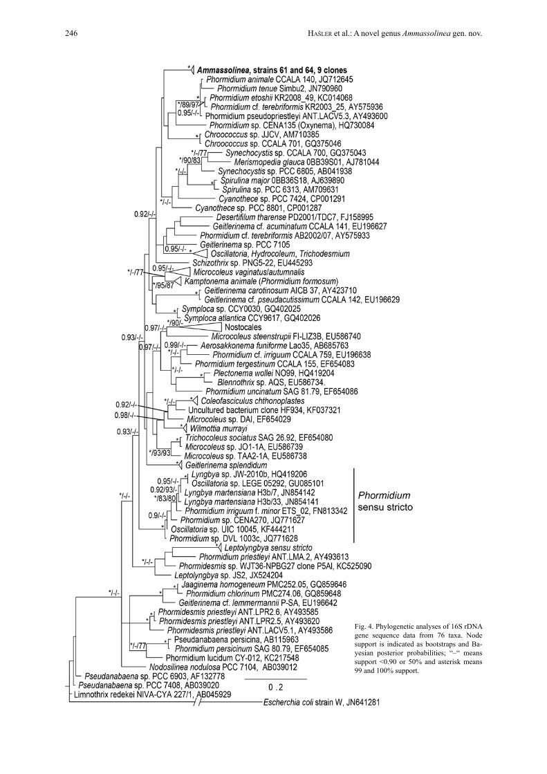
Fig. 4. Phylogenetic analyses of 16S rDNA gene sequence data from 76 taxa. Node support is indicated as bootstraps and Ba-yesian posterior probabilities; “–“ means support <0.90 or 50% and asterisk means 99 and 100% support.
246 Hašler et al.: A novel genus Ammassolinea gen. nov.

lithic species and in the genus Synechococcus (Jen-sen 1993). The importance of membranous stacks for cyanobacteria is not clear. Jensen (1993) suggests that these structures are important for cellular syntheses, but their role in Ammassolinea is unclear at this time.
The 16S sequence data clearly distinguishes Ammassolinea from sister taxa, sharing only 93.15% sequence similarity with its closest neighbour Phor-midium DVL 1003c which clusters with Phormidium sensu stricto (strunecký et al. 2014). Thus, it is clear that Ammassolinea is new genus clustering aside the type of Phormidium. The secondary folding patterns of the ITS are also very different from closely related taxa. The secondary folding pattern of the D1–D1’ he-lix resembles that of other Phormidium–like taxa, such as Kamptonema (Hašler et al. 2012; strunecký et al 2014), Microcoleus vaginatus, and Microcoleus au-tumnalis (Hašler et al. 2012). It possesses a larger ter-minal loop and two rather small loops close to the basis of the helix. This character in particular has increas-ingly been employed as a stable marker for distinguish-ing cryptic genera (JoHansen et al. 2011; Casamatta et al. 2012). Finally, the heap–like colony morphology of Ammassolinea is distinct and different from typi-cal Phormidium which is characteristically expanded, widely attached to the substrate, gelatinous, and mu-cilaginous according to komárek & anagnostiDis (2005). Morphologically, Ammassolinea trichomes resemble Phormidium taylorii, which is also recorded from the United States, but the latter do not taper to-ward the apices (komárek & anagnostiDis 2005).
Cyanobacterial species concepts are subject to much debate. Recently, a proliferation of new cyano-bacterial taxa have been precipitated by the use of mo-lecular markers coupled with an apomorphic species concept more conducive to the erection of new taxa (e.g., JoHansen & Casamatta 2005). Our ecological, morphological and molecular data indicates that our isolates are sufficiently removed from any other sister taxa to warrant the erection of a new genus. The sub-tropical habitat of isolation represents a depauperate area in terms of its ecological and information.
While some may be reluctant to erect a new ge-nus without a clear morphological distinction (at the trichome level), there are several reasons that this is acceptable. Firstly, recent works dealing with tropi-cal marine cyanobacteria have revealed numerous new genera which share morphological similarity with “Lyngbya”, but posesswidely divergent chemotaxo-nomic and genetic (16S rDNA) markers (engene et al. 2012, 2013). Secondly, geno–genera (those units erect-ed based on genetic signatures without a corresponding morphological character) may harbor environmentally or culturally induced morphological differentiation currently not realized. For example, the recently erected genus Nodosilinea was originally described as a “Leptolyngbya” but with inducible formation of nod-ules on the trichomes at low light levels (li & brand
2007). This genus was at first a geno–genus, with a fortuitous morphological apomorphy subsequently dis-covered (Perkerson et al. 2011). Lastly, morphologi-cal similarity may mask ecological divergence. As a whole, cyanobacteria are intimately tied to convergent evolution constrained by simple bacterial morphology. For example, the ubiquitous Synechococcus has been described in marine and freshwaters throughout the world. However, recent inversions have shown that this genus contains numerous lineages (at least seven new genera) masked by lack of morphological evi-dence (Dvořák et al. 2014).
AcknowledgementsMinor gratefulness belong to the grants ESF Post–UP II CZ.1.07/2.3.00/30.0041 and IGA PrF 2014001.
references
boyer, s.l.; fletCHner, V. & JoHansen, J.r. (2001): Is the 16S–23S rRNA internal transcribed spacer (ITS) re-gion a good tool for use in molecular systematics and population genetics? A case study in cyanobacteria. – Molecular Biology and Evolution 18: 1057–1069.
boyer, s.l.; JoHansen, J.r. & HoWard, g.l. (2002): Phylo-geny and genetic variance in terrestrial Microcoleus (Cyanophyceae) species based on sequence analysis of the 16S rRNA gene and associated 16S–23S ITS region. – Journal of Phycology 38: 1222–1225.
casamatta, D.a.; D. stanić, m.; gantar & ricHarDson, l.l. (2012): Characterization of Roseonema reptotaenium (Oscillatoriales, Cyanobacteria) gen. et sp. nov. isola-ted from Caribbean black band disease. – Phycologia 51:489–499.
Fig. 5. ITS secondary structures found for the most important helices as follows: D1–D1´, Box B1, Box B2.
Fottea, Olomouc, 14(2): 241–248, 2014 247

Casamatta, d.a.; Vis, m.l. & r.g. sHeatH. (2003): Cryptic species in cyanobacterial systematics: a case study of Phormidium retzii (Oscillatoriales) using 16S rDNA and RAPD analyses. – Aquatic Botany 77: 295–309.
Casamatta, d.a.; JoHansen, J.r.; Vis, m.l. & broadWater, s.t. (2005): Molecular and morphological characte-rization of ten polar and near–polar strains within the Oscillatoriales (Cyanobacteria). – Journal of Phycolo-gy 41:421–438.
Dvořák, P.; Hašler, P. & Poulíčková, a. (2012): Phylogeogra-phy of the Microcoleus vaginatus (cyanobacteria) from three continents – a spatial and temporal characteriza-tion. – PLoS ONE 7: e40153. doi:10.1371/journal.pone.0040153
Dvořák, P.; HinDák, F.; Hašler, P.; HinDáková, a. & Poulíčko-vá, a. (2014): Morphological and molecular studies of Neosynechococcus sphagnicola, gen. et sp. nov. (Cya-nobacteria, Chroococcales). – Phytotaxa 170: 24–34.
engene, n.; gunasekera, s. P.; gerwick, w. H. & Paul, v. J. (2013): Phylogenetic Inferences Reveal Large Extent of Novel Biodiversity in Chemically Rich Tropical Marine Cyanobacteria. –Appl. Env. Microbiol. 79: 1882–1888.
engene, n.; rottacker, e. c.; cHoi, H.; byrum, t.; kaštov-ský, J. H.; komárek, J.; ellisman, m. H. & gerwick W. H. (2012): Moorea producta gen. nov., sp. nov. and Moorea bouillonii comb. nov., tropical marine cyano-bacteria rich in bioactive secondary metabolites. – Int. J. Syst. Evol. Microbiol. 62: 1172–1179
Hašler, P.; Dvořák, P.; JoHansen, J.r.; kitner. m.; onDřeJ, v. & Poulíčková, a. (2012): Morphological and molecu-lar study of epipelic filamentous genera Phormidium, Microcoleus and Geitlerinema (Oscillatoriales, Cyano-phyta/Cyanobacteria). – Fottea 12: 341–356.
Hašler, P.; Dvořák, P. & Poulíčková, P. (2014): A new genus of filamentous epipelic cyanobacteria, Johansenia. – Preslia 86: 81–94.
Jensen, t. e. (1993): Cyanobacterial Ultrastructure. – In: ber-ner, t. (ed.): Ultrastructure of Microalgae. – pp. 7–51, CRC Press, Boca Raton, Florida.
JoHansen, J.r. & Casamatta, d.a. (2005): Recognizing cya-nobacterial diversity through adoption of a new species paradigm. – Algological Studies 117:71–93.
JoHansen, J.r.; kováčik, l.; casamatta, D.a. & kaštovský, J. (2011): Leptolyngbya corticola sp. nov. (Pseudana-baenaceae, Cyanobacteria), an aerophytic, heteropolar taxon from the Czech Republic. – Nova Hedwigia 92: 283–302.
komárek, J. & anagnostiDis, k. (2005): Cyanoprokaryota. 2. Teil: Oscillatoriales. – In: büDel, b.; gärDner, g.; kri-enitz, l. & sCHagerl, m. (eds): Süsswasserflora von Mitteleuropa, Vol. 19/2. – 759 pp., Elsevier, München.
larkin, m.a.; blacksHielDs, g.; brown, n.P.; Duenna, r.; mCgettigan, P.a.; mCWilliam, H.; Valentin, f.; WallaCe, i.m.; Wilm, a.; loPez, r.; tHomPson, J.d.; gibbon, t.J. & Higgins, d.g. (2007): Clustal W and Clustal X version 2.0. – Bioinformatics 23: 2947–2948.
li, z. & brand, J. (2007): Leptolyngbya nodulosa sp. nov. (Os-cillatoriaceae), a subtropical marine cyanobacterium that produces a unique multicellular structure. – Phy-cologia 46: 396–401.
lokmer, a. (2007): Polyphasic approach to the taxonomy of the selected oscillatorian strains (Cyanobacteria) [Ms. thesis]. – 61 pp., University of South Bohemia, České Budějovice, Czech Republic.
marquarDt, J. & Palinska, ka. (2007): Genotypic and phe-
notypic variability of Phormidium–like cyanobacteria from different habitats and geographical sites. – Archi-ves of Microbiology 187: 397–413.
Perkerson, r.b.; JoHansen, J.r.; kovacik, l.; branD, J.; kaš-tovský, J. & casamatta, D.a. (2011): A unique Pseu-danabaenalean (Cyanobacteria) genus Nodosilinea gen. nov. based on morphological and molecular data. – Journal of Phycology 47: 1397–1412.
Posada, D. (2008): jModelTest: phylogenetic model averaging. – Molecular Biology and Evolution 25: 1253–1256.
Poulíčková, a.; Hašler, P.; lysaková, m. & sPears, b. (2008): The ecology of freshwater epipelic algae: an update. – Phycologia 47: 437–450.
Poulíčková, a.; Dvořák, P.; maZalová, P. & Hašler, P. (2014): Epipelic microphototrophs: an overlooked assemblage in lake ecosystems. – Freshwater Science 33: 513–523.
Quiblier, C.; Wood, S.; Echenique–Subiabre, I.; Heath, M.; Villeneuve, A. & Humbert, J.F. (2013): A review of current knowledge on toxic bentic freshwater cyano-bacteria – Ecology, toxin production and risk manage-ment. – Water Research 47: 5464–5479.
ronquist, F. & Huelsenbeck, J. P. (2003): MRBAYES 3: Ba-yesian phylogenetic inference under mixed models. – Bioinformatics 19: 1572–1574.
sHeatH, r.g. & cole, k.m. (1992): Biogeography of stream macroalgae in North America. – J. Phycol. 28: 448–460.
stamatakis a. (2014): RAxML Version 8: A tool for Phylo-genetic Analysis and Post–Analysis of Large Phyloge-nies. – Bioinformatics, DOI: 10.1093/bioinformatics/btu033
staub, r. (1961): Research on physiology of nutrients of the planktonic cyanobacterium Oscillatoria rubescens. – Schweizerische Zeitschrift Fur Hydrologie 23: 83–198.
strunecký, o.; komárek, J.; JoHansen, J.; lukešová, a. & el-ster, J. (2013): Molecular and morphological criteria for revision of the genus Microcoleus (Oscillatoriales, Cyanobacteria). – J. Phycol. 49: 1167–1180.
strunecký, o.; komárek, J. & šmarDa, J. (2014): Kampto-nema (Microcoleaceae, Cyanobacteria), a new genus derived from the polyphyletic Phormidium on the basis of combined molecular and cytomorphological mar-kers. – Preslia 86: 193–207.
sWofford, d. l. (2001): PAUP*. Phylogenetic analysis usány parsimony (*and other methods). – Version4. Sinauer Associates, Sunderland, Massachusetts.
Tamura, K.; Stecher, G.; Peterson, D.; Filipski, A. & Kumar, S. (2013): MEGA6: Molecular Evolutionary Genetics Analysis version 6.0. – Molecular Biology and Evolu-tion: 30 2725–2729.
WHitton, b.a. & Potts, m. (2000): The ecology of cyano-bacteria. Their diversity in time and space. – 669 pp., Springer, Berlin.
Zucker, m. (2003): Mfold web server for nucle-ic acid folding and hybridization prediction. – Nucleic. Acids. Res. 31: 3406–3415.
© Czech Phycological Society (2014)Received May 30, 2014Accepted July 3, 2014
248 Hašler et al.: A novel genus Ammassolinea gen. nov.