A new xinjiangchelyid turtle from the Middle Jurassic of Xinjiang, China and the evolution of the...
29
A new xinjiangchelyid turtle from the Middle Jurassic of Xinjiang, China and the evolution of the basipterygoid process in Mesozoic turtles Rabi et al. Rabi et al. BMC Evolutionary Biology 2013, 13:203 http://www.biomedcentral.com/1471-2148/13/203
-
Upload
uni-tzuebingen -
Category
Documents
-
view
2 -
download
0
Transcript of A new xinjiangchelyid turtle from the Middle Jurassic of Xinjiang, China and the evolution of the...

A new xinjiangchelyid turtle from the MiddleJurassic of Xinjiang, China and the evolution ofthe basipterygoid process in Mesozoic turtlesRabi et al.
Rabi et al. BMC Evolutionary Biology 2013, 13:203http://www.biomedcentral.com/1471-2148/13/203
Rabi et al. BMC Evolutionary Biology 2013, 13:203http://www.biomedcentral.com/1471-2148/13/203
RESEARCH ARTICLE Open Access
A new xinjiangchelyid turtle from the MiddleJurassic of Xinjiang, China and the evolution ofthe basipterygoid process in Mesozoic turtlesMárton Rabi1,2*, Chang-Fu Zhou3, Oliver Wings4, Sun Ge3 and Walter G Joyce1,5
Abstract
Background: Most turtles from the Middle and Late Jurassic of Asia are referred to the newly defined cladeXinjiangchelyidae, a group of mostly shell-based, generalized, small to mid-sized aquatic froms that are widelyconsidered to represent the stem lineage of Cryptodira. Xinjiangchelyids provide us with great insights into theplesiomorphic anatomy of crown-cryptodires, the most diverse group of living turtles, and they are particularlyrelevant for understanding the origin and early divergence of the primary clades of extant turtles.
Results: Exceptionally complete new xinjiangchelyid material from the ?Qigu Formation of the Turpan Basin(Xinjiang Autonomous Province, China) provides new insights into the anatomy of this group and is assigned toXinjiangchelys wusu n. sp. A phylogenetic analysis places Xinjiangchelys wusu n. sp. in a monophyletic polytomy withother xinjiangchelyids, including Xinjiangchelys junggarensis, X. radiplicatoides, X. levensis and X. latiens. However, theanalysis supports the unorthodox, though tentative placement of xinjiangchelyids and sinemydids outside ofcrown-group Testudines. A particularly interesting new observation is that the skull of this xinjiangchelyid retainssuch primitive features as a reduced interpterygoid vacuity and basipterygoid processes.
Conclusions: The homology of basipterygoid processes is confidently demonstrated based on a comprehensivereview of the basicranial anatomy of Mesozoic turtles and a new nomenclatural system is introduced for the carotidcanal system of turtles. The loss of the basipterygoid process and the bony enclosure of the carotid circulationsystem occurred a number of times independently during turtle evolution suggesting that the reinforcement of thebasicranial region was essential for developing a rigid skull, thus paralleling the evolution of other amniote groupswith massive skulls.
BackgroundMost recent, morphology-based, phylogenetic studies offossil and extant turtles agree that the Middle to LateJurassic was a particularly important phase in the earlydiversification of crown group Testudines [1-6].Xinjiangchelyidae is a clade of turtles that includes someof the most common taxa known from this time periodin Asia and that is widely considered to represent theprimitive morphology of the cryptodiran stem lineage[2-4,7-16]. The exact content of this clade is still an open
* Correspondence: [email protected] für Geowissenschaften, University of Tübingen, Hölderlinstraße 12,72074, Tübingen, Germany2Department of Paleontology & MTA–ELTE Lendület Dinosaur ResearchGroup, Eötvös Loránd University, Budapest, HungaryFull list of author information is available at the end of the article
© 2013 Rabi et al.; licensee BioMed Central LtdCommons Attribution License (http://creativecreproduction in any medium, provided the or
question, however, as the anatomy and phylogenetic rela-tionships of many candidate taxa are still poorly known.A new species of xinjiangchelyid, Xinjiangchelys wusu
n. sp., is described here on the basis of exceptionally wellpreserved skeletons that were found and recovered bythe 2009 and 2011 Field Teams of the Sino-GermanCooperation Project in the Upper Jurassic ?QiguFormation of the Turpan Basin, Xinjiang AutonomousProvince, China and that provide new insights into themorphology of xinjiangchelyids.One anatomical region of special interest for turtle
evolution is the basicranium. The basisphenoid ofsome paracryptodires and xinjiangchelyids, includingXinjiangchelys wusu n. sp., has previously been shownto exhibit a pair of lateral processes that were homolo-gized with the basipterygoid process of basal amniotes
. This is an Open Access article distributed under the terms of the Creativeommons.org/licenses/by/2.0), which permits unrestricted use, distribution, andiginal work is properly cited.

Rabi et al. BMC Evolutionary Biology 2013, 13:203 Page 2 of 28http://www.biomedcentral.com/1471-2148/13/203
[15,17]. However, the homology of these structures iscontroversial in the literature [18-20] and a compre-hensive assessment of this issue is still outstanding.We here identify similar basisphenoid processes in abroad range of extinct turtles and conclude that theirpresence has been overlooked in the Mesozoic turtleliterature during the last forty years. We here further-more provide compelling morphological evidence for thehomology of the basisphenoid processes of xinjiangchelyidswith the basipterygoid processes of basal turtles and basalamniotes and review the evolution of this structure inMesozoic turtles. We finally present an internally consistentnomenclatural system that reflects recent insights into themorphology of the carotid canal system. To test the phylo-genetic implications of our new insights, we analyzed an ex-tensive sample of xinjiangchelyids in a global, cladisticframework of turtles. We obtained the unorthodox place-ment of this clade outside crown group Testudines, whichmay hint at a surprisingly extensive evolutionary history ofthe turtle stem lineage.
Figure 1 The geographic location of the “Turtle Cliff” site in the Turpphotograph of the cliff where the turtles were found and cut out wit
MethodsGeological settingsThe “Turtle Cliff Fossil Site” yielded the new materialdescribed herein and is located within the FlamingMountains about 26 km ENE of the city of Shanshan inthe Turpan Basin, Xinjiang Autonomous Province, China(Figure 1). The Flaming Mountains consist of Triassicto Paleogene sediments that were uplifted during theNeogene [21-23]. Published reports on the geology andstratigraphy of the Flaming Mountains in particularand the Turpan Basin in general are rare (e.g., [24] andreferences therein) and many uncertainties thereforeexist regarding the absolute age of formations and theircorrelation with similar units in other Central Asian basins.Jurassic clastic strata in the Flaming Mountains were pre-liminarily divided into the Early Jurassic Sangonghe For-mation, the Middle Jurassic Xishanyao, Sanjianfang,Qiketai, and Qigu Formations (the latter was recentlydated in the Junggar Basin with 164.6 Ma ± 1.4 Ma, [25]),and the Late Jurassic Karaza Formation [26]. Future
an Basin of Xinjiang Autonomous Province, China (above) and ah the help of a rock saw (below).

Figure 2 Type series of Xinjiangchelys wusu n. sp. from “TurtleCliff”, Turpan Basin of Xinjiang Autonomous Province, China,?Qigu Formation, Middle Jurassic.
Rabi et al. BMC Evolutionary Biology 2013, 13:203 Page 3 of 28http://www.biomedcentral.com/1471-2148/13/203
stratigraphic research needs to clarify whether Late Jurassicstrata are indeed mostly absent in the area.Piedmont-fluvial deposits dominate the upper parts of
the Jurassic sequence [27,28]. Red-colored sediments,especially prominent in the Qigu Formation, indicate areduction in the monsoonal circulation in Asia resultingin a paleoclimatic change from humid to seasonally dryduring the late Middle and early Late Jurassic [24,25,28-31].The total thickness of the supposed Qigu Formation isabout 850 m in the area of the Turtle Cliff Fossil Site [31].The formation is rich in vertebrate fossils, dominated bydinosaurs and turtles [28]. Finds of the latter include thespectacular turtle taphocoenosis at Mesa Chelonia [28]near the lower border of the formation and the hereinintroduced Turtle Cliff Fossil Site near the base of theupper third of the formation.The Turtle Cliff Fossil Site is situated geographically
1 km to the ENE and stratigraphically 500 m above theMesa Chelonia site [28]. Since no explicit justificationhas been given for the correlation of the strata sup-posedly belonging to the Qigu Formation in the TurpanBasin, the assignment of rocks units exposed in theFlaming Mountains to this formation is not transparent[28], but our preliminary classification places both siteswithin the Qigu Formation. The deposits that allegedlyrepresent the Qigu Formation in the Turpan Basin arecharacterized by alternating coarse and fine-grainedsediments that often contain unionid freshwater bivalves,reflecting changing depositional conditions typical ofriver systems [24,31]. Temporary subaerial exposure isindicated by paleosols [28].The turtle skeletons at the Turtle Cliff Fossil Site were
found on the top of a low hill in a steeply inclined (65°),fine-grained and strongly cemented sandstone layer richin lithoclasts. Above and below the turtle-bearing sand-stone horizon follows a succession of predominately redsilt-and mudstones.
Material studied in this paperOur description of Xinjiangchelys wusu n. sp. is basedon a sandstone slab with at least 3 individuals (Figure 2)that were excavated during the 2011 joint field season ofthe University of Tübingen, Shenyang Normal University,and Jilin University, that was lead and carried out by all co-authors at the Turtle Cliff Fossil Site (see Geological Set-tings). The quarried fossils are currently housed at thePaleontology Museum of Liaoning (PMOL) at ShenyangNormal University, Shenyang, Liaoning but will eventuallybe integrated into the municipal museum of Shanshan,Xinjiang Autonomous Province that is currently under con-struction. All specimens have been assigned a combinedPMOL-Sino-German Cooperation Project (SGP) number,which will be deposited with the specimens once the mu-seum in Shanshan is operational. The detailed coordinates
of the locality are archived at PMOL and will be disclosedto qualified researchers interested in studying the site.Specimen PMOL-SGP A0100-1 was discovered with
the carapace exposed in dorsal view in 2009 below asmall cliff and was cut out of the hard sandstone ledgein a block with an ICS diamond chain rock saw in 2011.Subsequent preparation revealed that the slab containedtwo more individuals with PMOL-SGP A0100-2 cut inhalf through the long axis during excavation. The slab intotal includes PMOL-SGP A0100-3: shell with carapacepartially exposed, femora, skull and lower jaw; PMOL-SGP A0100-2: shell (plastron not exposed), partial neck,left foot and left hand and PMOL-SGP A0100-1: poste-riorly incomplete carapace, neck, crushed skull with ar-ticulated mandible, left and right hand and incompleteleft posterior limb.The anatomy of fossil taxa was reviewed mostly
based on personal observations of published materialand with the help of photographs. The following fossiltaxa were studied first hand: Allopleuron hoffmanni(Gray, 1831) [32] (NHMUK R42913); Chubutemys copelloiGaffney et al., 2007 [10] (MPEF-PV1236); Dracochelysbicuspis Gaffney and Ye, 1992 [33] (IVPP V4075);Hangaiemys hoburensis Sukhanov and Narmandakh,1974 [34] (PIN 3334-4, PIN 3334-34, PIN 3334-35,PIN 3334-36, PIN 3334-37); Heckerochelys romaniSukhanov, 2006 [35] (PIN 4561-2 and PIN 4719-34);Hoyasemys jimenezi Pérez-García et al., 2012 [36](MCCM-LH-84); Helochelydra nopcsai Lapparent deBroin and Murelaga, 1999 [37] (IWCMS 1998.21);Judithemys sukhanovi Parham and Hutchison, 2003 [9]

Figure 3 Proposed internally consistent nomenclature for theosseous portion of the carotid circulation system of turtles asexemplified on the skull of Dracochelys bicuspis (IVPP V4075).Abbreviations: bo: basioccipital, bs: basisphenoid, fpp: foramenpalatinum posterius, mx: maxilla, pal: palatine, pmx: premaxilla,pt: pterygoid, qu: quadrate, vo: vomer.
Rabi et al. BMC Evolutionary Biology 2013, 13:203 Page 4 of 28http://www.biomedcentral.com/1471-2148/13/203
(TMP 87.2.1); Kallokibotion bajazidi Nopcsa, 1923 [38](NHMUK R4921 and NHMUK R4925); Kayentachelysaprix Gaffney et al., 1987 [39] (MNA V1558, MCZ 8917);Macrobaena mongolica Tatarinov, 1959 [40] (PIN 533-4);Manchurochelys manchoukuoensis Endo and Shikama,1942 [41] (PMOL AR00008); Meiolania platyceps Owen,1886 [42] (NHMUK R682); Mongolemys elegans Khosatzkyand Mlynarski, 1971 [43] (five uncatalogued skulls at thecollections of PIN); Mongolochelys efremovi Khozatsky,1997 [44] (PIN 552-459 and two uncatalogued skulls);Naomichelys speciosa Hay, 1908 [45] (FMNH PR 273);Niolamia argentina Ameghino 1899 [46] Notoemyslaticentralis Cattoi and Freiberg, 1961 [47] (cast ofMOZP 2487); Odontochelys semitestacea Li et al., 2008[48] (IVPP V13240); Ordosemys leios Brinkman andPeng, 1993 [49] (IVPP V9534-1); Peligrochelys walshaeSterli and de la Fuente, In press [16] (MACN PV CH2017, MACN PV CH 2017); Portlandemys mcdowelliGaffney, 1975 [50] (NHMUK R2914, NHMUK R3163,NHMUK R3164); Proganochelys quenstedti Baur, 1887[51] (SMNS 16980); Rhinochelys elegans Lydekker,1889 [52] (NHMUK R27); Sandownia harrisi Meylanet al., 2000 [53] (MIWG 3480); Sinemys gamera Brinkmanand Peng, 1993 [54] (IVPP V9532-11); Sinemys brevispinusTong and Brinkman, In press [55] (IVPP V9538-1);Solnhofia parsonsi Gaffney, 1975 [56] (TM 4023);Toxochelys latiremis Cope, 1873 [57] (NHMUK R4530and NHMUK R3902); Xinjiangchelys (Annemys) levensisSukhanov and Narmandakh, 2006 [58] (PIN 4636-4-2,[59]); Xinjiangchelys (Annemys) latiens Sukhanov andNarmandakh, 2006 (PIN 4636-6-2, [59]); and Xinjiangchelysradiplicatoides Brinkman et al., 2013 [15] (IVPP V18104).The following taxa were studied on the basis of photo-
graphs: Adocus lineolatus Cope, 1874 [60] (CCM 60-15);Basilochelys macrobios Tong et al., 2009 [61] (MD 8-2);Bouliachelys suteri Kear and Lee, 2006 [62] (SAMP41106); Meiolania platyceps AM F: 18671; Plesiochelysetalloni Pictet and Humbert, 1857 [63] (MH 435);Pleurosternon bullockii Owen 1842 [64] (UMZC T1041).
Osteological terminologyThe cranial nomenclature presented by Gaffney [65,66]has been highly influential, because all anatomicalsystems of the cranium were clearly described andillustrated in these publications and because a broadaudience was thereby enabled to apply these names con-sistently to the skulls of fossil and recent turtles. Only inthe last few years have some shortcomings become ap-parent, however, particularly in regards to the nomencla-ture of the carotid system and we herein seek to rectifythis situation by providing an internally consistent no-menclatural system for this anatomical region (Figure 3).The internal carotid artery of most turtles, like most
amniotes, splits into a cerebral and a palatine (lateral)
branch. Although these structures are interrelated, theycan be thought of as three different vessels, which areherein terms the internal carotid artery, the cerebral ar-tery, and the palatine artery. New insights into the cra-nial anatomy of basal turtles [15,20,67] has revealed thatthese three blood vessels can enter the skull throughthree non-homologous foramina and that they can alsoexit the skull through three non-homologous foramina,for a total of six non-homologous foramina. The nomen-clatural system of Gaffney [65,66] proved to be confus-ing, because it only provides three names for these sixforamina (i.e., foramen anterior [italics added for em-phasis] canalis carotici interni, foramen posterior canaliscarotici interni, and foramen caroticum laterale) and be-cause these names were defined as applying to inappro-priate portions of the carotid system. For instance, theforamen anterior canalis carotici interni was defined asapplying to the exit of the cerebral artery, not to the exitof the internal carotid artery, whereas the foramen pos-terior canalis carotici interni could either be the entry ofthe internal carotid artery or of the cerebral artery[65,66]. An addition oddity of this nomenclatural systemthat makes it difficult for neophytes to learn that thepalatine artery is situated in the “lateral canal,” not thepalatine canal. Incidentally, the use of the “-ior” suffix

Rabi et al. BMC Evolutionary Biology 2013, 13:203 Page 5 of 28http://www.biomedcentral.com/1471-2148/13/203
(as in anterior and posterior) is not appropriate, giventhat the “-ius” suffix is the proper neuter singular endingin Latin.Sterli et al. [20] were the first to realize these deficien-
cies in the nomenclatural system of Gaffney [65,66] andproposed new terms, but these new terms are not suffi-cient to name all six potential foramina and they breakwith tradition set by Gaffney [65,66] in their grammat-ical construction. These inconsistencies were partiallyaddressed recently [15] but some parts of the system stillremain unnamed and the palatine artery is still definedas sitting in the lateral canal.We herein propose a new nomenclatural system that
attempts to follow the grammatical precedence set forthby Gaffney [65,66], but that breaks tradition by providingnames for all potential foramina and by renaming thelateral canal the palatine canal. This nomenclatural sys-tem consists of a total of 10 new terms (Figure 3):
Canalis caroticus internusThe bony canal that holds any portion of the internal ca-rotid artery, absent, among others, in basal turtles andparacryptodires.
Foramen posterius canalis carotici interni (fpcci)The posterior entry of the internal carotid artery, absent,among others, in basal turtles and paracryptodires.
Foramen anterius canalis carotici interni (facci)The anterior exit of the internal carotid artery, onlypresent in turtles with a fenestra caroticus.
Canalis caroticus cerebralisThe bony canal that holds any portion of the cerebralartery, present in all turtles.
Foramen posterius canalis carotici cerebralis (fpccc)The posterior entry of the cerebral artery, not developedin turtles where the split of the internal carotid arteryinto the cerebral and palatine branches is covered bybone.
Foramen anterius canalis carotici cerebralis (faccc)The anterior exit of the cerebral artery, present in allturtles, typically located near the dorsum sellae.
Canalis caroticus palatinumThe bony canal that holds any portion of the palatine ar-tery, generally absent in turtles with an open interpterygoidvacuity.
Foramen posterius canalis carotici palatinum (fpccp)The posterior entry of the palatine artery, generally de-veloped in turtles with a closed interpterygoid vacuity,
but not in those where the split of the internal carotidartery into the cerebral and palatine branches is coveredby bone.
Foramen anterius canalis carotici palatinum (faccp)The anterior exit of the palatine artery, generally presentin turtles with a close interpterygoid vacuity.
Fenestra caroticus (fca)A figurative bony window into the otherwise closedcarotid system, which exposes the split of the internalcarotid artery into the cerebral and palatine branches.The window is posteriorly defined by the foramenanterius canalis carotici interni and anteriorly defined bythe foramen posterius canalis carotici cerebralis andthe foramen posterius canalis carotici palatinum or theinterpterygoid vacuity.
Phylogenetic analysisA phylogenetic analysis was performed using TNT[68,69] using a modified version of a previous character/taxon matrix [16], which in return is based on earlierstudies [3,5,59,70] [Additional file 1]. Part of the changesare reported in an in press paper by Rabi et al. [59] andthese are repeated below for the sake of clarity. Five taxawere added to the matrix [16], including Xinjiangchelysradiplicatoides, X. junggarensis (sensu Brinkman et al.2008 [71]), X. (Annemys) levensis, X. (Annemys) latiens, andBasilochelys macrobios. The scorings of X. radiplicatoidesare primarily based on the literature [15], those of X.junggarensis (=X. latimarginalis [72]) on personal observa-tion of IVPP material from Pingfengshan [72], those of X.(Annemys) levensis, and X. (Annemys) latiens based on per-sonal observation of PIN material, and those of B.macrobios based on the literature [60] and photographsobtained from H. Tong. The following scorings werechanged relative to the original matrix [16] (the earlierscorings are in parenthesis): Epiplastron B: Hangaiemyshoburensis: 1 (?), Sinemys lens Wiman, 1930 [73] 1 (?);Pterygoid B: H. hoburensis 1 (2), Dracochelys bicuspis 1(2), Pleurosternon bullockii 1 (2), Kallokibotion bajazidi1 (2), Mongolochelys efremovi 1 (2), Chubutemys copelloi1 (2), Eileanchelys waldmani Anquetin, 2009 [74] ? (2);Carapace D: H. hoburensis 0 (?), Chengyuchelys baenoidesYoung and Chow, 1953 [75] (IVPP-V6507) 0 (1); CarapaceE: H. hoburensis-(?); Vertebral A: Siamochelys peninsularisTong et al., 2002 [76] ? (1); Vertebral C: S. peninsularis ?(1); Anal A: S. peninsularis ? (0), Ch. baenoides ? (0);Entoplastron B: Ch. baenoides ? (1); Mesoplastron A: S.peninsularis 2 (0); Hypoplastron A: Ch. baenoides ? (0);Xiphiplastron A-B: Ch. baenoides ? (0); Dorsal Rib A: S.peninsularis ? (2); Plastral Scute B: S. peninsularis 1 (0).Further modifications relative to Rabi et al. in press
[59] include the addition of Xinjiangchelys wusu to the

Rabi et al. BMC Evolutionary Biology 2013, 13:203 Page 6 of 28http://www.biomedcentral.com/1471-2148/13/203
matrix and changing of the following scorings:Supraoccipital A: X. (Annemys) levensis 1 (0); X.radiplicatoides ? (0); X. (Annemys) latiens ? (0); Ptery-goid B: Sphenodon punctatus 0 (2), Anthodon serrarius1 (2), Peligrochelys walshae 2 (1), Niolamia argentina2 (?); Dentary A: X. (Annemys) levensis 0 (1); X.junggarensis ? (1).The character Cervical Vertebrae A was omitted from
the analysis because we found it difficult to replicate thischaracter objectively and perceived a number of inconsist-encies in the matrix [59]. The character Diploid Number Awas also omitted following previous studies [3,59,77].The following characters were treated as ordered: 7
(Nasal A), 19 (Parietal H), 27 (Squamosal C), 40 (MaxillaD), 42 (Vomer A), 50 (Quadrate B +C), 52 (AntrumPostoticum A), 59 (Pterygoid B), 81 (Opisthotic C), 82(Opisthotic D), 89 (Stapedial Artery B), 98 (CanalisCaroticum F), 120 (Carapace A), 121 (Carapace B), 130(Peripheral A), 133 (Costal B), 138 (SupramarginalA), 158 (Hyoplastron B), 159 (Mesoplastron A), 161(Hyoplastron B), 176 (Abdominal A), 213 (Cleithrum A),214 (Scapula A), 232 (Manus B), 233 (Manus C). Sphen-odon punctatus, Owenetta kitchingorum, Simosaurusgaillardoti and Anthodon serrarius were designated asoutgroups [16,59]. Although, there is growing evidence fora turtle-archosaur clade among molecular studies, morpho-logical analyses still suggest lepidosaurian or parareptilianaffinities for turtles at the moment. As it turns out, how-ever, the choice of outgroup is irrelevant, as all outgroupsreveal that the presence of teeth and the lack of a completeshell should be considered primitive for turtles and that thepartially shelled, toothed taxon Odontochelys semitestaceais therefore sister to all turtles. The fusion of thebasicranium discussed in our paper occurs far deeperwithin the turtle tree and is therefore not influenced by thechoice of outgroups, but rather by the arrangement of basalturtles.Given that this analysis is focused on the phylogenetic
relationships and placement of xinjiangchelyid turtles,we decided to crop taxa not pertinent to these questions(e.g., most derived baenids, most meiolaniforms) and abroad spectrum of taxa known from fragmentary mater-ial only (see Appendix A for a complete list) in order toreduce the size of the matrix [59]. The resulting matrixconsists of 237 characters for a total of 84 terminal taxa.The character-taxon matrix and the tnt. file are foundunder [Additional files 1, and 2], respectively.The relationships of living cryptodiran taxa were manu-
ally constrained according to recent results of molecularphylogenetic studies (following previous studies [1,2,59]),without assuming a priori, however, that Trionychianests within Cryptodira [78,79]. The internal relationshipsof durocryptodires were constrained using a moleculartopology [79] (i.e., (Emydidae (Geoemydidae + Testu-
dinidae)) + (Chelonioidea (Chelydridae + Kinosternoidea))).The complete list of taxa designated as floaters can befound in Appendix B. A first run of heuristic searchtree-bisection-reconnection, using thousands of ran-dom addition sequence replicates and 10 trees savedper replicate, failed to find all the most parsimonioustrees (MPT) and therefore the heuristic search was re-peated until the MPTs were found 30 times duringeach replicate (using the command “xmult = hits 30;”).The trees retained in the memory were exposed to asecond round of tree-bisection-reconnection.
Systematic paleontologyTESTUDINATA Klein, 1760 [80]TESTUDINES Batsch, 1788 [81]XINJIANGCHELYIDAE Nessov in Kaznyshkin et al.,1990 [7] (sensu Rabi et al., In press [59])
RemarkWe follow the phylogenetic definition of Xinjiangchelyidaeused in Rabi et al. (In press [59]) where Xinjiangchelyidaeis defined as the most inclusive clade containingXinjiangchelys junggarensis Ye, 1986 [82], but notSinemys lens, Macrobaena mongolica, or any species ofRecent turtle.
Xinjiangchelys Ye, 1986 [82]
Remark: A number of genera other than Xinjiangchelyshave been referred to Xinjiangchelyidae in recent years,including Chengyuchelys Young and Chow, 1953 [75];Tienfuchelys Young and Chow, 1953 [75]; AnnemysSukhanov and Narmandakh, 2006 [58]; Shartegemys,Sukhanov and Narmandakh, 2006 [58]; YanduchelysPeng et al. 2005 [83]; Protoxinjiangchelys Tong et al.2012 [13] ([8,13-15,58,71]). The majority of these generaare sufficiently diagnosed relative to Xinjiangchelys,but there is no up-dated diagnosis available forXinjiangchelys. This taxon has therefore been ren-dered a waste-backed taxon defined by what it isnot. To avoid further complications we suggest usinga more inclusive definition for Xinjiangchelys thatincludes all species of Xinjiangchelyidae (sensu Rabiet al., In press [59]) until the phylogenetic relation-ships of the included taxa can be determined moreconfidently.
Xinjiangchelys wusu sp. nov.
(Figure 2, Figures 4, 5, 6, 7, and 8)urn:lsid:zoobank.org:pub:2BCCC095-7622-4 F27-8 F80-6199F24690B5

Figure 4 Skulls and partial neck of Xinjiangchelys wusu, Middle Jurassic, ?Qigu Formation, “Turtle Cliff”, Shanshan area, Turpan Basin,Xinjiang Autonomous Province, China. A, PMOL-SGP A0100-1 (holotype), photograph and line drawing of skull and anterior cervicalvertebrae in dorsal view; B, PMOL-SGP A0100-3, photograph and line drawing of skull, mandible and hyoid apparatus in ventral view.Abbreviations: ang: angular, bo: basioccipital, bpt: basipterygoid process, bs: basisphenoid, cb I.: cornu branchiale I, cor: coronoid,cv: cervical vertebra, de: dentary, ex: exoccipital, fcl: foramen caroticum laterale, fpccc: foramen posterius canalis carotici cerebralis,fpcci: foramen posterius canalis carotici interni, fpo: fenestra postotica, fpp: foramen palatinum posterius, fr: frontal, fst: foramen stapedio-temporale, ica: incisura columella auris, ipv: interpterygoid vacuity, ju: jugal, mx: maxilla, na: nasal, op: opisthotic, pa: parietal, pal: palatine,pfr: prefrontal, pmx: premaxilla, po: postorbital, pt: pterygoid, qj: quadratojugal, qu: quadrate, rs: rugose surface of processus trochlearisoticum, so: supraoccipital, sq: squamosal, sur: surangular, vo: vomer, * refers to fossa pterygoidea. A, X, G, H, Y refer to scales after Sterli andde la Fuente [16].
Rabi et al. BMC Evolutionary Biology 2013, 13:203 Page 7 of 28http://www.biomedcentral.com/1471-2148/13/203

Rabi et al. BMC Evolutionary Biology 2013, 13:203 Page 8 of 28http://www.biomedcentral.com/1471-2148/13/203
HolotypePMOL-SGP A0100-1, a partial skeleton, including theskull exposed in dorsal view (Figures 2, 4, 6 and 8B).
Referred materialPMOL-SGP A0100-3, partial skeleton (Figures 2, 4,5 and 7,); PMOL-SGP A0100-2, partial skeleton withoutskull, plastron not exposed (Figures 2, 6 and 7A, C-D).
Locality and horizonTurtle Cliff Fossil Locality (see Geological Settings),Shanshan, Turpan Basin, Xinjiang Autonomous Province,People’s Republic of China (Figure 1); ?Qigu Formation,Upper Jurassic.
Etymologywusu refers to a small town in Xinjiang AutonomousProvince.
DiagnosisA species of Xinjiangchelys; skull differing from X.(Annemys) levensis in the prefrontals being fully sepa-rated by the frontals; from X. (Annemys) latiens by thebroader skull and the extensive jugal and frontal contri-bution to the orbit, from X. radiplicatoides by the flat-tened skull and the presence of a remnant of theinterpterygoid vacuity. Shell differing from X. chowiMatzke et al. [84] X. qiguensis Matzke et al., [85] X.tianshanensis Kaznyshkin et al. [7] and X. junggarensis(sensu Brinkman et al. 2008 [70]) by the narrow verte-bral scales.
DescriptionSkull
Preservation The skull of PMOL-SGP A0100-1 is ex-posed only in dorsal and lateral views, whereas its palatalside is covered by the carapace of PMOL-SGP A0100-2(Figure 2). It is dorsoventrally crushed and the preorbitalregion is slightly shifted from its original position. Theskull of PMOL-SGP A0100-3, on the other hand, isexposed in ventral view and in articulation with thehyoids and the mandible (Figures 4B and 5).
Scales Some of the cranial scales are traceable inPMOL-SGP A0100-1, but most of them are not apparent(Figure 4A). Using a recently suggested nomenclaturalsystem [16] we identify the unpaired scale Y on the pos-terior half of the frontal posteriorly bordered by thepaired scale F. Scale G is bordered by the unpaired par-ietal scale X posteriorly. Scale A is another unpairedscale of the parietal found posteriorly to scale X. Scale Hmay have also been present laterally to scale X. The skull
roof is otherwise decorated with fine grooves and veryshallow pits that do not show a clear pattern.
Nasals The nasals are very poorly preserved but theirsutures with the frontal and the prefrontal are partiallytraceable on the right side of PMOL-SGP A0100-1(Figure 4A). They are reduced, posteriorly tapering ele-ments that are partially separated by the anterior frontalprocess. The nasals contribute to the formation of theexternal nares.
Prefrontals The dorsal plate of the prefrontals is elong-ate and medially separated from its counterpart by theanterior frontal process (Figure 4A). The prefrontal con-tacts the nasal anteriorly and the maxilla ventrally. Thedescending process of the prefrontal has a wide contactwith the palatines within the fossa orbitalis. Its contactwith the vomer is not visible, but given the large size ofthe prefrontal pillars it was very likely present. Thefrontal forms the anterior half of the dorsal margin ofthe orbit.
Frontals The frontals form an anterior process that iswedged between the prefrontals (Figure 4A). The poster-ior half of the dorsal margin of the orbit is formed bythe frontals. The orbit has a subcircular outline andfaces dorsolaterally.
Parietals The dorsal plate of the parietals exhibits arelatively deep temporal emargination that reachesbeyond the level of the anterior border of the cavumtympani (Figure 4A). The parietal meets the frontalanteriorly and has a long contact with the postorbital.Even though the parietals are slightly shifted from theiroriginal position in PMOL-SGP A0100-1, their postero-lateral tips also touched the squamosal, as seen on theright side. Dorsoventral crushing obscures the structuresof the processus inferior parietalis.
Jugal The jugal area is compressed and its entire lateralsurface is exposed in dorsal view in PMOL-SGP A0100-1 and it is also partially visible in PMOL-SGP A0100-3(Figures 4A and 5B). It sends a long posterior processalong the postorbital but it is unclear whether it meetsthe quadratojugal. The skull exhibits a moderate cheekemargination that exposes the coronoid process of themandible. Anteriorly, the jugal forms the posterolateralmargin of the orbit and contacts the maxilla. It is un-clear whether the ventral plate of the jugal contacts theposterior end of the triturating surface and/or the an-terolateral tip of the external pterygoid process.
Quadratojugal The quadratojugal is a reduced, flatelement that is best preserved in PMOL-SGP A0100-3,

Figure 5 PMOL-SGP A0100-3, skull, mandible and hyoid apparatus of Xinjiangchelys wusu, Middle Jurassic, ?Qigu Formation, “Turtle Cliff”, Shanshanarea, Turpan Basin, Xinjiang Autonomous Province, China. A, occipital view of skull; B, left ventrolateral view of skull and mandible; C, medial view of rightramus of mandible. Abbreviations: ang: angular, art: articular, bo: basioccipital, bs: basisphenoid, cb I.: cornu branchiale I., co: condylus occipitalis, cor: coronoid,ct: cavum tympani, de: dentary, ex: exoccipital, fm: foramen magnum; fnh: foramen nervi hypoglossi, fpcci: foramen posterius canalis carotici interni, fpo: fenestrapostotica, ica: incisura columella auris, ju: jugal,mx: maxilla, op: opisthotic, or: orbit, pmx: premaxilla, ppe: processus pterygoideus externus, pra: prearticular,pt: pterygoid, qj: quadratojugal, qu: quadrate, scm: sulcus cartilaginis Meckelii, so: supraoccipital, spe: splenial, sur: surangular.
Rabi et al. BMC Evolutionary Biology 2013, 13:203 Page 9 of 28http://www.biomedcentral.com/1471-2148/13/203
though not fully exposed (Figure 5B). It has no clearcontact with the jugal anteriorly but this is all butcertain. Dorsally, it meets the postorbital and posteriorlyit borders the cavum tympani. Its lower rim of theskull is emarginated, which gives the quadratojugal asubtriangular outline. The quadratojugal sends a pair ofnarrow and tapering processes along the dorsal andthe ventral margins of the cavum tympani, respectively.The dorsal one of these processes is wedged between thepostorbital and the quadrate and appears not to reachthe squamosal. The ventral one terminates slightly be-fore the level of the condylus mandibularis.
Squamosal The squamosal is better preserved on theright side of PMOL-SGP A0100-1, the left one beingcompressed and the lateral plate being exposed whenthe skull is viewed dorsally (Figure 4). The lateral surface
of the squamosal is smooth and there is no squamosalhorn. The squamosal has a very short point-like contactwith the parietal along the anterior margin of the uppertemporal emargination. There is no contribution tothe formation of the anterior opening of the antrumpostoticum as seen in PMOL-SGP A0100-3. Medially,the squamosal contacts the quadrate and may even havea short contact with the opisthotic within the uppertemporal fossa. The contact of the ventral portion withthe opisthotic and the quadrate is not exposed in eitherspecimen.
Postorbital The postorbitals are long elements; theyform the posterodorsal margin of the orbit and also con-tribute to the rim of the upper temporal emargination(Figure 4A). The postorbital has an anteroventral contactwith the jugal, a posteroventral contact with the

Rabi et al. BMC Evolutionary Biology 2013, 13:203 Page 10 of 28http://www.biomedcentral.com/1471-2148/13/203
quadratojugal, a long lateral contact with the parietal,and also meets the squamosal posteriorly.
Premaxilla The premaxillary region is shifted anteriorlyfrom the original position and damaged in PMOL-SGPA0100-1 (Figure 4). The premaxilla forms the ventralmargin of the external nares and contacts the other pre-maxilla medially and the maxilla posterolaterally. Theexternal nares are undivided. Only little of the ventralaspect of the premaxillary region is exposed but it is ap-parent that there is no premaxillary hook.
Maxilla The maxilla forms the ventral margin of theorbit, sends a dorsal process to contact the descendingpillar of the prefrontal, and contacts the premaxilla an-teriorly and the jugal posteriorly (Figures 4 and 5B). Thetriturating surface is only partially exposed but it is ap-parently narrow and straight with a sharp and low labialridge.
Vomer A single, slightly damaged and displaced, elegantvomer is present in PMOL-SGP A0100-3 exposed indorsal view (Figure 4B). Its outline is very similar to thatof Xinjiangchelys levensis.
Palatine The right palatine is preserved incompletelyand shifted from the original position in PMOL-SGPA0100-3 (Figure 4B). It shows an extensive free lateralmargin that is indicative of a large foramen palatinumposterius.
Quadrate Apart from the region of the cavum tympani,the right quadrate of PMOL-SGP A0100-1 is in goodcondition whereas the left otic region is badlyfragmented and compressed (Figures 4 and 5B). InPMOL-SGP A0100-3 the region of the cavum tympani isexposed in lateral view. The cavum tympani is anteriorlybordered by the quadratojugal and by the squamosaldorsally and posteriorly. The incisura columella auris isan open but tight notch and there is no precolumellarfossa. The antrum postoticum is well developed and itsopening is formed entirely by the quadrate, although thesquamosal comes very close to the lateral rim. Thequadrate contacts within the upper temporal fossa thesquamosal posterolaterally and the opisthotic medially,but its medial contact with the prootic is obscured. To-gether with the prootic it forms a large foramenstapedio-temporale. The quadrate forms a poorly devel-oped processus trochlearis oticum that is composed of arugose area.
Epipterygoid The epipterygoids are not exposed in ei-ther specimen.
Pterygoid The pterygoids are almost intact in PMOL-SGPA0100-3 except for their anteriormost edges (Figures 4Band 5A-B). The pterygoid has a long posterior processreaching as far as the back of the skull and terminatingslightly anterior to the basioccipital-basisphenoid suture.The pterygoid covers the cranioquadrate space and con-tacts the posterolateral corner of the basisphenoid but notthe basioccipital. The pterygoid has a short dorsal contactwith the exoccipital, but this contact is not part of the skullsurface. The foramen posterius canalis carotici interniopens at the back of the skull within the ventral surface ofthe pterygoid. The quadrate ramus of the pterygoid bears awell-developed, oval-shaped pterygoid fossa. The processuspterygoideus externus is present and it is characterized by aposteriorly extending horizontal plate and a dorsoventrallythickened vertical plate. A characteristic feature of thepterygoid is a large oval opening just anterior to thebasisphenoid and posterior to the region where the ptery-goids meet one another along the midline. This openinghas intact margins, is clearly not a result of erosion or anyother taphonomic processes, but is distinct from the for-amen posterius canalis carotici palatinum. We interpret thisstructure as the remnant of the interpterygoid vacuity.Anterolaterally, the pterygoid bears a margin that is indica-tive of a large foramen palatinum posterius.
Supraoccipital Much of the crista supraoccipitalis isdisplaced and preserved in fragments in PMOL-SGPA0100-1 (Figures 4 and 5A). The supraoccipital providesonly a small contribution to the skull roof where it contactsthe parietals. The ventral plate of the supraoccipital con-tacts the opisthotic laterally and forms the dorsal margin ofthe foramen magnum. The crista supraoccipitalis extendedapparently only slightly beyond the posterior tip of thesquamosals. In PMOL-SGP A0100-3 the supraoccipitalcrest is intact as exposed in ventral view and does not pro-trude much beyond the level of the occipital condyle.
Exoccipitals The exoccipitals are preserved on bothsides in PMOL-SGP A0100-3 (Figures 4 and 5A). Theyform the ventrolateral wall of the foramen magnum. Apair of foramen nervi hypoglossi pierce each elementsbut the formed foramen jugulare posterius is notdistinct from the fenestra postotica (Figure 5A).Laterally, the exoccipitals contact the opisthotic andhave a ventromedial contact with the basioccipital.Anteroventrally, the exoccipital has a short contactwith the posteriormost tip of the pterygoid, but thiscontact does not contribute to the smooth, palatal sur-face of the skull. A suboval, unossified area excludesthe exoccipital from anteromedially contacting thebasisphenoid.

Figure 6 Carapaces of Xinjiangchelys wusu, Middle Jurassic, ?Qigu Formation, “Turtle Cliff”, Shanshan area, Turpan Basin, XinjiangAutonomous Province, China. A, PMOL-SGP A0100-1 (holotype), photograph and line drawing; B, PMOL-SGP A0100-2, photograph and linedrawing. Abbreviations: CE: cervical scute, co: costal, ne: neural, nu: nuchal, per: peripheral, PL: pleural, VE: vertebral, py: pygal, sp: suprapygal.
Rabi et al. BMC Evolutionary Biology 2013, 13:203 Page 11 of 28http://www.biomedcentral.com/1471-2148/13/203
Basioccipital The basioccipital has a pair of basioccipitaltubera with rounded posterior edges that extends as aroof over the foramen nervi hypoglossi when the skull isviewed ventrally (Figures 4B and 5A-B). The neck of thebasioccipital condyle is short and lacks paired ridges orgrooves. The basioccipital has no contact with the
pterygoid and the processus interfenestralis of theopisthotic is therefore visible in ventral view. Anteriorly,the basioccipital meets the basisphenoid via a transversesuture. A shallow concavity extends on the ventral sur-face of the basioccipital that barely protrudes onto thebasisphenoid.
Rabi et al. BMC Evolutionary Biology 2013, 13:203 Page 12 of 28http://www.biomedcentral.com/1471-2148/13/203
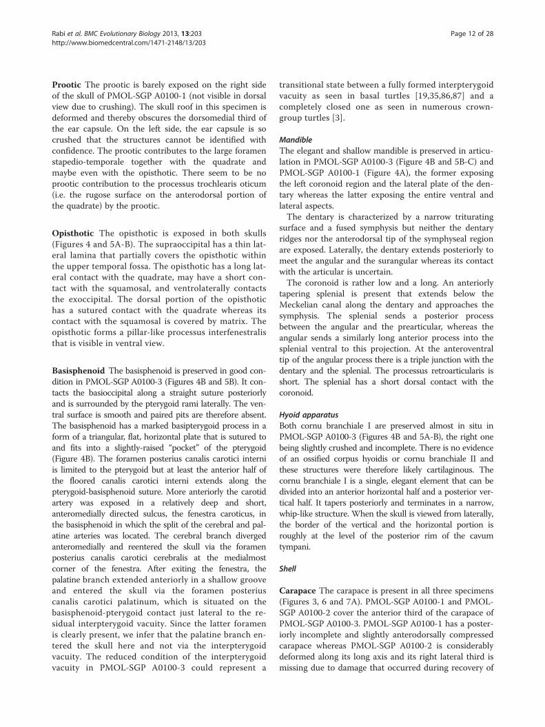
Prootic The prootic is barely exposed on the right sideof the skull of PMOL-SGP A0100-1 (not visible in dorsalview due to crushing). The skull roof in this specimen isdeformed and thereby obscures the dorsomedial third ofthe ear capsule. On the left side, the ear capsule is socrushed that the structures cannot be identified withconfidence. The prootic contributes to the large foramenstapedio-temporale together with the quadrate andmaybe even with the opisthotic. There seem to be noprootic contribution to the processus trochlearis oticum(i.e. the rugose surface on the anterodorsal portion ofthe quadrate) by the prootic.
Opisthotic The opisthotic is exposed in both skulls(Figures 4 and 5A-B). The supraoccipital has a thin lat-eral lamina that partially covers the opisthotic withinthe upper temporal fossa. The opisthotic has a long lat-eral contact with the quadrate, may have a short con-tact with the squamosal, and ventrolaterally contactsthe exoccipital. The dorsal portion of the opisthotichas a sutured contact with the quadrate whereas itscontact with the squamosal is covered by matrix. Theopisthotic forms a pillar-like processus interfenestralisthat is visible in ventral view.
Basisphenoid The basisphenoid is preserved in good con-dition in PMOL-SGP A0100-3 (Figures 4B and 5B). It con-tacts the basioccipital along a straight suture posteriorlyand is surrounded by the pterygoid rami laterally. The ven-tral surface is smooth and paired pits are therefore absent.The basisphenoid has a marked basipterygoid process in aform of a triangular, flat, horizontal plate that is sutured toand fits into a slightly-raised “pocket” of the pterygoid(Figure 4B). The foramen posterius canalis carotici interniis limited to the pterygoid but at least the anterior half ofthe floored canalis carotici interni extends along thepterygoid-basisphenoid suture. More anteriorly the carotidartery was exposed in a relatively deep and short,anteromedially directed sulcus, the fenestra caroticus, inthe basisphenoid in which the split of the cerebral and pal-atine arteries was located. The cerebral branch divergedanteromedially and reentered the skull via the foramenposterius canalis carotici cerebralis at the medialmostcorner of the fenestra. After exiting the fenestra, thepalatine branch extended anteriorly in a shallow grooveand entered the skull via the foramen posteriuscanalis carotici palatinum, which is situated on thebasisphenoid-pterygoid contact just lateral to the re-sidual interpterygoid vacuity. Since the latter foramenis clearly present, we infer that the palatine branch en-tered the skull here and not via the interpterygoidvacuity. The reduced condition of the interpterygoidvacuity in PMOL-SGP A0100-3 could represent a
transitional state between a fully formed interpterygoidvacuity as seen in basal turtles [19,35,86,87] and acompletely closed one as seen in numerous crown-group turtles [3].
MandibleThe elegant and shallow mandible is preserved in articu-lation in PMOL-SGP A0100-3 (Figure 4B and 5B-C) andPMOL-SGP A0100-1 (Figure 4A), the former exposingthe left coronoid region and the lateral plate of the den-tary whereas the latter exposing the entire ventral andlateral aspects.The dentary is characterized by a narrow triturating
surface and a fused symphysis but neither the dentaryridges nor the anterodorsal tip of the symphyseal regionare exposed. Laterally, the dentary extends posteriorly tomeet the angular and the surangular whereas its contactwith the articular is uncertain.The coronoid is rather low and a long. An anteriorly
tapering splenial is present that extends below theMeckelian canal along the dentary and approaches thesymphysis. The splenial sends a posterior processbetween the angular and the prearticular, whereas theangular sends a similarly long anterior process into thesplenial ventral to this projection. At the anteroventraltip of the angular process there is a triple junction with thedentary and the splenial. The processus retroarticularis isshort. The splenial has a short dorsal contact with thecoronoid.
Hyoid apparatusBoth cornu branchiale I are preserved almost in situ inPMOL-SGP A0100-3 (Figures 4B and 5A-B), the right onebeing slightly crushed and incomplete. There is no evidenceof an ossified corpus hyoidis or cornu branchiale II andthese structures were therefore likely cartilaginous. Thecornu branchiale I is a single, elegant element that can bedivided into an anterior horizontal half and a posterior ver-tical half. It tapers posteriorly and terminates in a narrow,whip-like structure. When the skull is viewed from laterally,the border of the vertical and the horizontal portion isroughly at the level of the posterior rim of the cavumtympani.
Shell
Carapace The carapace is present in all three specimens(Figures 3, 6 and 7A). PMOL-SGP A0100-1 and PMOL-SGP A0100-2 cover the anterior third of the carapace ofPMOL-SGP A0100-3. PMOL-SGP A0100-1 has a poster-iorly incomplete and slightly anterodorsally compressedcarapace whereas PMOL-SGP A0100-2 is considerablydeformed along its long axis and its right lateral third ismissing due to damage that occurred during recovery of

Figure 7 PMOL-SGP A0100-3 Xinjiangchelys wusu shell, Middle Jurassic, ?Qigu Formation, “Turtle Cliff”, Shanshan area, Turpan Basin,Xinjiang Autonomous Province, China. A, photograph and line drawing of posterior two third of carapace; B, photograph and line drawing ofplastron. The right forelimb in ‘B’ does not belong to PMOL-SGP A0100-3 but to PMOL-SGP A0100-1. Abbreviations: AB: abdominal, AN: anal,co: costal, EG: extra gular, epi: epiplastron, FE: femoral, GU: gular, HU: humeral, hyo: hyoplastron, hypo: hypoplastron, IM: inframarginal,md: musk duct foramen, ne: neural, per: peripheral, PE: pectoral, PL: pleural, py: pygal, sp: suprapygal, VE: vertebral, xi: xiphiplastron.
Rabi et al. BMC Evolutionary Biology 2013, 13:203 Page 13 of 28http://www.biomedcentral.com/1471-2148/13/203
the block (see Materials and Methods). PMOL-SGPA0100-3 is not deformed; the exposed portion preservesthe original outline of the carapace suggesting a rela-tively wide shell.
Carapacial bones The nuchal is a trapezoidal elementand more than twice as wide than long (Figure 6). The nu-chal emargination is minor in PMOL-SGP A0100-1 but
appears to be slightly deeper in PMOL-SGP A0100-2. Thisemargination extends onto peripheral 1 in both specimens.There are eight pairs of costal bones, all of which have
firm contacts with the peripherals and lack costal fonta-nelles. The reduction of neural 7 in PMOL-SGP A0100-2 allows for a short, medial contact of costals 7, whichcontrasts the morphology of PMOL-SGP A0100-3,where a subdivided neural 7 does not allow for this

Rabi et al. BMC Evolutionary Biology 2013, 13:203 Page 14 of 28http://www.biomedcentral.com/1471-2148/13/203
contact (Figures 6B and 7A). This region is not pre-served in PMOL-SGP A0100-1.Costal 1 tapers laterally and is subequal in anteropos-
terior length with the more posterior costals. The figureof PMOL-SGP A0100-2 (Figure 6B) hints at a seeminglylonger costal 1, but this is an optical illusion resultingfrom distortion. Costal 2 has a slightly concave antero-lateral outline and sends a wide rectangular posterolat-eral process into the posterior half of peripheral 4, as isbest seen on the left side of PMOL-SGP A0100-1 (Fig-ure 6A), but also visible on the right side and in PMOL-SGP A0100-2 (Figure 6B). Costal 3 is the mediolaterallywidest element and has straight and parallel anterior andposterior sides. Costal 4 is slightly concave anteriorlyand convex posteriorly and has strongly concave con-tacts with peripheral 6 and 7. Costal 5 is slightly convexanteriorly and posteriorly. It has a short, oblique contactwith peripheral 7 and a strongly concave contact withperipheral 8. PMOL-SGP A0100-3 is different in thatcostal 5 barely touches peripheral 7 (Figure 7A). Thecontact of costal 6 with peripheral 8 projects more lat-erally relative to its contact with peripheral 9. Costal 7has an oblique and straight anterior border and a con-cave posterior border. Costal 8 is narrow and slightlyconvex anteriorly and posteriorly.The neural series is complete and consists of eight ele-
ments, including a subdivided neural 7 in PMOL-SGPA0100-3 (Figure 7A). In PMOL-SGP A0100-2 the series isinterrupted by the short contact of costals 7 (Figure 6B). InPMOL-SGP A0100-1, this region is incomplete. Most neu-rals are hexagonal, coffin-shaped elements with the shortsides facing anterolaterally. Neural 1 of PMOL-SGP A0100-1 and 3 is quadrangular whereas it is hexagonal with shortsides facing posterolaterally in PMOL-SGP A0100-2 thequadrangular element being neural 2 instead. In PMOL-SGP A0100-3 neural 7 is subdivided into a larger, regular,hexagonal element and a small, square element and neural8 is hexagonal. In PMOL-SGP A0100-2 both neurals 7 and8 are pentagonal and do not contact one another, therebyallowing for a medial contact of costals 7.There are two suprapygals, the anterior one is trape-
zoidal has no contacts with the peripherals and consi-derably wider and shorter in PMOL-SGP A0100-2 than inPMOL-SGP A0100-3 (Figures 6B and 7A). Suprapygal 2 isa wide element that contacts costal 8 and peripheral 11.There are 11 pairs of peripherals (Figures 6 and 7). A
distinct gutter extends from the lateral corner of thenuchal to peripheral 7 along the lateral margin of thecarapace. Peripheral 1 contacts costal 1 and is larger onthe right side than on the left in PMOL-SGP A0100-1(Figure 6A). Peripheral 2-6 are narrow elements whereas7-11 are considerably expanded laterally. Peripheral 8 isthe widest peripheral element and has a strong medialprojection into costal 5 in all specimens.
Carapacial scales There are five vertebrals and a singlewide cervical (Figures 6 and 7B). Vertebral 1 is wider thanlong and barely touches peripheral 1. The proportions ofvertebrals 2-4 vary somewhat among the specimens,but all are narrower than most pleurals. PMOL-SGPA0100-1 has the relatively widest vertebrals of all. Ver-tebral 2 is slightly longer than wide in PMOL-SGPA0100-3 (as reconstructed) and PMOL-SGP A0100-2whereas in PMOL-SGP A0100-1 it is markedly widerthan long. Vertebral 3 is slightly wider than long inPMOL-SGP A0100-3 and PMOL-SGP A0100-3-3 andslightly longer than wide in PMOL-SGP A0100-2. Ver-tebral 4 is considerably wider than long in PMOL-SGPA0100-3 and PMOL-SGP A0100-3-3 and this is lessdistinct in PMOL-SGP A0100-2. The vertebral 3-4 sul-cus has anterior projection at the midline that extendsonto the posterior portion of neural 5 in all specimens.Vertebral 5 is wider than long, contacts peripherals 11laterally, and does not prolong onto the suprapygal (aspreserved in PMOL-SGP A0100-1 and PMOL-SGPA0100-3).The pleurals are all wider than long except for pleural
4 that is longer than wide (Figures 6 and 7A).The marginals are either restricted to the peripherals
or their borders coincide with the costo-peripheral con-tacts or, as in the case of marginals 5 and 7, they slightlylap onto the costals. Marginal 11 indistinctly prolongsonto the costals on the left sides of all specimens (rightside not preserved in PMOL-SGP A0100-1).
Plastron The plastron is only exposed in PMOL-SGPA0100-3 (Figure 7B). In this specimen the plastron ispreserved in perfect condition except for minor damagein the right bridge area. The dorsal aspect of the plas-tron is not visible. The plastron is characterized bycomplete ossification (i.e., no fontanelles) and compact,sutural contacts. Scale sulci are clearly developed. Theanterior lobe is about 40% wider than long, shorter thanthe posterior lobe, and has a slightly rounded anteriormargin. Mesoplastra are absent. The posterior lobe isposteriorly tapering, slightly wider at its base than long,and lacks an anal notch. At least one musk duct foramenis present between peripheral 4 and the hyoplastron.
Plastral bones The epiplastron is trapezoidal, shows aroughly transverse suture with the hyoplastron, ananteromedially directed contact with the entoplastron,and a sagittal contact with the other epiplastron(Figure 7B). The entoplastron is oval-shaped, abouttwice as long as wide, and only partially separates theepiplastra. The buttress of the hyoplastron is relativelylow and it terminates on the anterior half of peripheral2. The contact of the plastron with the carapace is tight

Figure 8 Neck and appendicular elements of Xinjiangchelys wusu, Middle Jurassic, ?Qigu Formation, “Turtle Cliff”, Shanshan area,Turpan Basin, Xinjiang Autonomous Province, China. A, PMOL-SGP A0100-2, photograph and line drawing of articulated cervical vertebraeIII-V in lateral view; B, PMOL-SGP A0100-1, photograph and line drawing of right distal fore limb; C, PMOL-SGP A0100-2, photograph and linedrawing of left fore limb; D, PMOL-SGP A0100-2, photograph and line drawing of left hind limb. Abbreviations: asc: fused astragalocalcaneum,cr: cervical rib, da: diapophysis, dc: distal carpal, int: intermedium, mc: metacarpal, mce: medial central, mt: metatarsal, ns: neural spine,pa: parapophysis, pis: pisiform, ts: transverse process, uln: ulnare.
Rabi et al. BMC Evolutionary Biology 2013, 13:203 Page 15 of 28http://www.biomedcentral.com/1471-2148/13/203
but we interpret it as being ligamentous rather than su-tured owing for the presence of plastral pegs. However,we note that our meaning of ligamentous contact isprobably different from the concept of earlier studies[13,14]. The edges of the bridge peripherals slightlyoverlap the margin of the bridge of the plastron. Pe-ripherals 3, 4, and the anterior third of peripheral 5contact the plastron via well-developed pegs. Furtherposteriorly, the contact between the plastron and thecarapace transfers into a smooth-edged contact untilthe posterior third of peripheral 6. Just medial to thisedge the plastron is notched at the contact of the hyo-and the hypoplastron, but this space is filled up withtwo elements (on one side) that appear to be aberrantextra ossifications that meet the hyo- and thehypoplastron along finely serrated edges. The anterioredge of these elements is partially fused with thehyoplastron. More posteriorly, the hypoplastron
contacts the peripherals via pegs with the inguinal but-tress terminating on the anterior third of peripheral 8. Thexiphiplastra are well developed and they have a fork-likecontact with the posterolateral portion of the hypoplastronin ventral view. Interfingering interplastral sutures are ab-sent.
Plastral scales One pair of gulars and one pair ofextragulars are present. The gulars do not extend ontothe entoplastron and the extragulars have a transversecontact with the humeral scales. The midline sulcus ofthe plastron is straight instead of sinusoidal. The pec-toral scale is shorter than the abdominal. The femoral/anal sulcus is omega-shaped and the anals barely extendnear the midline onto the hypoplastron. Four pairs ofinframarginals are present, of which the third covers thehyo/hypoplastral suture (Figure 7B).

Rabi et al. BMC Evolutionary Biology 2013, 13:203 Page 16 of 28http://www.biomedcentral.com/1471-2148/13/203
Appendicular skeletonThe articulated distal half of the right fore limb ofPMOL-SGP A0100-1 is exposed in antipalmar view nextto the skull of PMOL-SGP A0100-3 (Figures 7B and 8B).The left distal fore limb of this individual is lesscomplete and preserved tucked on the other side of theslab next to the carapace (Figure 6A). The articulatedleft fore and hind limbs of PMOL-SGP A0100-2(Figure 6B) are preserved in palmar and antipalmar view,respectively (Figures 8C-D). Only the left ulna, radius,an associated phalanx, the distal end of the right hu-merus, and both femora are exposed in specimenPMOL-SGP A0100-3 (Figure 7B). An additional, isolatedleft hind limb is present on the slab that likely belongsto a fourth specimen (Figure 3).
Humerus The humerus has a slightly curved shaft witha suboval cross-section (Figure 8C). The lateral processis slightly better developed than the medial process andboth processes are situated at the same level relative toone and another along the proximal part of the hu-merus. The ectepicondylar foramen is closed.
Radius and ulna The radius is elegant and narrow andhas a straight and relatively flat shaft in cross-section(Figures 8B-C). Its proximal epiphysis is subcircular incross-section whereas its distal epiphysis is expandedand more compressed. The articulation surface for themedial centrale extends along the distal margin of theepiphysis as in Podocnemis expansa [19]. The medialedge of the distal epiphysis lacks a medial projection thatis otherwise present in Macrochelys temminckii. The la-teral ridge below the proximal epiphysis, presumably forthe attachment of the radio-ulnar ligament [19], is re-duced. The ulna is flattened and more robust than theradius. The medial margin of the shaft is more curvedthan the lateral one. The ridge for the bicipital tendonattachment is reduced and the olecranon is poorly deve-loped. The medial process of the proximal epiphysis issituated slightly below the level of the olecranon as inM. temminckii but unlike in P. expansa. The relativeproportions of the proximal and distal epiphyses moreresemble M. temminckii in having similar width (thedistal being slightly wider).
Manus The relatively elongate and narrow phalanges ofthe manus suggest intermediate aquatic adaptation [88].The phalangeal formula is 2-3-3-3-3 (Figures 8B-C). Theunguals are clawed, narrow, and pointed, and decreasein size from the digit I to V. The distal articulation sur-faces of the proximal phalanges exhibit posteriorlyprojecting flanges that underlap the proximal epiphysisof the preceding metacarpals. The first metacarpal is theshortest and the most robust. The lateral overlapping of
the metacarpals with one another is present but notmarked. The distal carpals are ovoid and that of the firstdigit is slightly wider than those of the remaining digits.There is a small pisiform and the medial centrale istightly connected with the lateral centrale. The inter-medium is not elongate proximodistally, the ulnare isflat and deep, and the radiale bears little if any articula-tion with the radius.
Femur The femur has a slightly curved shaft (Figures 7Band 8D). The trochanter minor faces anteriorly, the tro-chanter major faces dorsally, and the femoral head onlyslightly extends above the trochanters. The trochantersare moderately developed. The proximal epiphysis has asimilar width as the distal one.
Tibia and fibula The tibia has a wide proximal epiphy-sis (Figure 8D) than Podocnemis expansa or Macrochelystemminckii. The ridge for the patellar tendon attachmentis placed close to the midline of the shaft as in M.temminckii and unlike in P. expansa where it is shiftedlaterally. The fibula is straight and has a more expandedand more compressed distal epiphysis than its proximalone. Proximally, the shaft lacks a medial flange, unlike inPodocnemis expansa.
Pes The hooked fifth metatarsal is a large, blocky ele-ment (Figure 8D). The astragalus is fused with the calca-neum. The pedal formula is 2-3-3-3-? and digits 1-4were clawed, whereas digit 5 is incompletely preserved.The first metatarsal is more robust than the others.
Vertebral columnFour cervicals are preserved in PMOL-SGP A0100-2,three in PMOL-SGP A0100-1, and PMOL-SGP A0100-3exhibits one cervical vertebra and two anterior caudals(Figures 4A, 6 and 8A).In PMOL-SGP A0100-2 three cervicals are well ex-
posed in lateral and dorsal views that could representany series of cervicals between 2 to 6 (Figure 8A). InPMOL-SGP A0100-1 cervicals 2 and 3 are exposed indorsal view (Figure 4A). The centra are amphicoelousand more than twice as long than high (excluding theventral keel and including the dorsal spine). A low ven-tral keel extends along the entire midline of the centra.The transverse processes are compressed, relatively ro-bust, with parallel anterior and posterior sides, and ex-hibit clear diapophyses. The transverse process does notextend much laterally and is slightly longer than wide.The posterior third of the transverse process extends be-yond the middle of the centrum whereas its anteriortwo-thirds extend anteriorly to the middle of the cen-trum, terminating well before the anterior end of thecentrum. The cervicals have well-developed bifurcated

Rabi et al. BMC Evolutionary Biology 2013, 13:203 Page 17 of 28http://www.biomedcentral.com/1471-2148/13/203
ribs. The dorsal articulation of the ribs with the trans-verse processes is not preserved and elements thereforemust have shifted, but the anterior contact with theparapophysis is still preserved. The parapophyses are si-tuated at the anteroventral margin of the centrum and isbest developed in the second cervical preserved inPMOL-SGP A0100-2 (probably cervical 3 or 4). Theneural arch is longer and more than twice as high as thecentrum (centrum including the transverse process butexcluding the ventral keel and the arch including thezygapophyses but excluding the dorsal spine). Thepostzygapophyses are only slightly separated and unitein a common low stem. The anterodorsal surface of thepostzygapophysis is convex whereas the posterodorsal isconcave with a groove extending anteromedially. Theneural spines are damaged and their full height is there-fore unknown, except for the most anterior preservedcervical in PMOL-SGP A0100-2. Cervical 2 has a longneural spine extending all along the dorsal surface of thearch whereas cervical 3 has a shorter spine (PMOL-SGPA0100-1). The anteriormost cervical in PMOL-SGPA0100-2 has a low but long spine, the following ishigher, and the third has a short and high process.The prezygapophyses are a little higher than thepostzygapophyses (except for the third preserved inPMOL-SGP A0100-2) and slightly extend beyond thelevel of the anterior edge of the centrum in lateralview.
Results and discussionTaxonomic commentsFollowing the phylogenetic definition of Rabi et al. [59],Xinjiangchelys wusu is assigned to Xinjiangchelyidae be-cause it is recovered in a monophyletic group togetherwith Xinjiangchelys junggarensis (Figure 9). Other mem-bers of Xinjiangchelyidae include X. radiplicatoides, X.(Annemys) latiens and X. (Annemys) levensis and thisclade is only supported by one unambiguous synapo-morphy (Anal A:1, extension of anal scale ontohypoplastron).Among taxa traditionally referred to Xinjiangchelyidae,
the morphology of X. wusu is most similar to that of X.(Annemys) levensis, Xinjiangchelys (Annemys) latiensand X. radiplicatoides, however, a number of differencesjustify its recognition as a separate taxon. In contrast toX. levensis, the prefrontals do not meet in the midline inX. wusu, the basioccipital tubera are better developed,there are two foramina nervi hypoglossi instead of three,the vertebral 3-4 sulcus extends onto neural 5 not neural6, and the midline plastral sulcus is straight instead of si-nusoidal. Xinjiangchelys (Annemys) latiens has a propor-tionally more elongated skull, reduced frontal and jugalcontribution to the orbit and sinusoidal midline plastral
sulcus, whereas X. radiplicatoides has a more inflatedskull, a slit-like interpterygoid vacuity instead of a roundopening with very indistinct foramen caroticus palatinum, astrongly plicated carapace, and a sinusoidal midline plastralsulcus.Since the interrelationships of xinjiangchelyids are un-
resolved in the consensus tree and pruning the rougetaxon Xinjiangchelys junggarensis reveals that Annemys(i.e., X. levensis and X. latiens) is paraphyletic (levensisforms the sister taxon of a latiens, X. wusu and X.radiplicatoides trichotomy), we suggest referring wusuand all other species to the genus Xinjiangchelys Ye1986 [82] as this taxon has priority over AnnemysSukhanov and Narmandakh 2006 [58].Recently, abundant remains of xinjiangchelyids were
reported from the Mesa Chelonia turtle bone bed, whichis stratigraphically situated 500 m below and spatially lo-cated 1 km away from the Turtle Cliff site [28]. TheseMesa Chelonia turtles are represented by several partialskeletons and were all referred to an indeterminate spe-cies of Annemys [28]. The Mesa Chelonia form is verysimilar to X. wusu but a few differences are present andtherefore we consider it a separate taxon. Xinjiangchelyswusu is about 15% larger, the foramen posterius canaliscarotici interni is located along the posterior surface ofthe pterygoid, not in a notch at the back of the skull, thevertebral 3-4 sulcus extends onto neural 5 (extends ontoneural 6 in eleven specimens out of twelve in the MesaChelonia form) and the plastral pegs are visible evenwhen the plastron is articulated with the carapace,whereas the pegs are mostly covered by the peripheralring in the fully ossified specimens of the Mesa Cheloniaforms. A further difference might be that X. wusu lacksany types of fontanelles in the carapace or the plastronwhereas they are present in more than half of the speci-mens from Mesa Chelonia that appear to be adult-sizedindividuals.Another closely related form, mostly known by the
skull, has been reported from the Junggar Basin [15] andwas referred to Annemys sp. The foramen posteriuscanalis carotici interni of this skull is located in a notchbetween the basisphenoid and the pterygoid (unlike X.wusu) and the lateral plate of the jugal lacks aposterodorsal process extending ventral to the post-orbital [15]. On the other hand, the skull from theJunggar Basin is very similar to the Mesa Chelonia formand we tentatively refer them to the same, yet unnamedtaxon.
The homology of the basipterygoid process inMesozoic turtlesBasal tetrapods and basal amniotes have no sutural rela-tionship between their basicranium and the palatoquadrateregion [89]. Instead, the basicranium articulates anteriorly
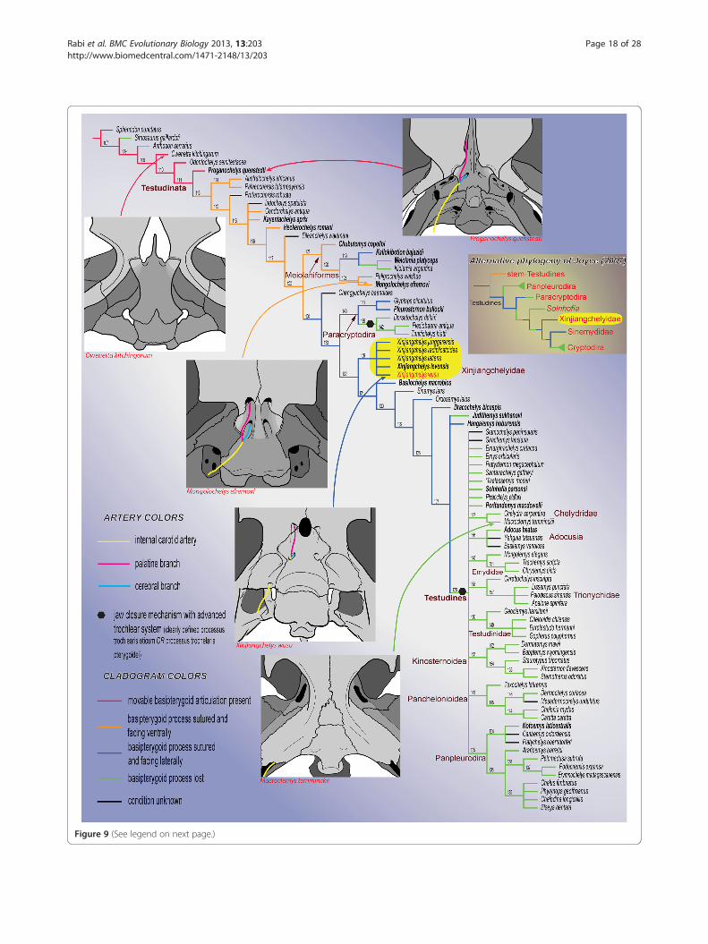
Figure 9 (See legend on next page.)
Rabi et al. BMC Evolutionary Biology 2013, 13:203 Page 18 of 28http://www.biomedcentral.com/1471-2148/13/203
(See figure on previous page.)Figure 9 Hypothetical relationships of the major clades of turtles and the evolution of the basipterygoid process and the carotidartery circulation system. The cladogram is the strict consensus tree of 9261 trees of 870 steps obtained after a parsimony analysis of 237morphological characters and 84 extinct and extant turtle taxa. The relationships of Durocryptodira [1] were constrained after the molecularphylogeny of Barley et al. [79]. Note the unorthodox position of Xinjiangchelyidae outside of Testudines. The more traditional phylogeneticplacement of Xinjiangchelyidae [3] is presented on the right for comparison. Taxa in bold are figured in Figures 10, 11, 12. Numbers correspondto nodes.
Rabi et al. BMC Evolutionary Biology 2013, 13:203 Page 19 of 28http://www.biomedcentral.com/1471-2148/13/203
with the pterygoid via the basipterygoid process of thebasisphenoid (also termed the basitrabecular process)and posteriorly with the quadrate and the squamosalvia the paroccipital process of the opisthotic. Abasipterygoid process has been identified in a numberof basal turtles and proto-turtles (Figures 10A-C), in-cluding Odontochelys semitestacea [48], Proganochelysquenstedti [19], Palaeochersis talampayensis Rougieret al., 1995 [90,91] Australochelys africanus Gaffneyet al., 1994 [92,93], Kayentachelys aprix [86,94],Heckerochelys romani [35], and Condorchelys antiquaSterli, 2008 [87,95]. Among this group of taxa, themore primitive ones, such as O. semitestacea and Pr.quenstedti, retain a movable basipterygoid articulation inthe form of a ventrolaterally directed, blunt basipterygoidprocess that articulates with the corresponding facet in thepterygoid (Figure 10A). All more derived basal turtles withan unambiguous basipterygoid process are interpretedas having a fused articulation [18,20,35,66,86,87,93,94]whereas all more advanced stem-testudine taxa and allcrown turtles are universally considered to have lost theirbasipterygoid process completely (e.g., [66]). Some derivedtaxa have nevertheless been hypothesized to retain a re-duced basipterygoid process, but the homology of thisstructure has been a controversial issue.The presence of a basipterygoid process was first
reported in the Late Jurassic turtle Mesochelys durlstonensisEvans and Kemp, 1975 [17], a taxon that was subsequentlysynonymized with Pleurosternon bullockii [96]. A similarstructure was noticed by Gaffney (1979) [18] in Glyptopsplicatulus Cope 1877 [97] and he concluded that it is nothomologous with the unambiguous basipterygoid processof basal turtles based on topological considerations, a con-cept subsequently confirmed by Sterli et al. [20]. More re-cently, Brinkman et al. [15] identified a paired process ofthe basisphenoid similar to that seen in Pleurosternonbullockii (Figure 11H) in a broad selection of Jurassic andEarly Cretaceous Asian eucryptodires and interpreted it asbeing homologous with the basipterygoid process of theearliest turtles, thereby contradicting the homology assess-ment of Gaffney [18] and Sterli et al. [20].According to the homology concept of Gaffney [18]
and Sterli et al. [20], the paired lateral processes ofthe basisphenoid that fit into corresponding pockets inthe pterygoids in G. plicatulus and Pl. bullockii cannotbe interpreted as the basipterygoid process because: a)
they are placed posterior to the dorsum sellae andtherefore have different topological relationships com-pared to the true basipterygoid processes seen incaptorhinomorphs (e.g., the purported basal amniotecondition) and b) because the processes in question donot ascend, as in basal turtles, but are instead aligned inthe same horizontal plane as the pterygoids. Indeed, thebasipterygoid process of captorhinomorphs is situatedanterior to the dorsum sellae, the foramen posteriuscanalis carotici cerebralis [15], and the foramen nerviabducentis, whereas in G. plicatulus and Pl. bullockii theprocess in question is found posteriorly to these struc-tures ([18], figure 23, note that the foramen posteriuscanalis carotici cerebralis is labeled foramen posteriuscanalis carotici interni). However, as already noted byothers [15], when the condition seen in Pr. quenstedti(Figure 10A; unknown for Gaffney [18]) is compared to thatof captorhinomorphs, it is evident that the dorsum sellae isin a derived position similar to that seen in G. plicatulusand Pl. bullockii (Figure 11H) in that it extends more an-teriorly over the foramen anterius canalis carotici cerebralis([19], figures 42-44). This anterior movement of the dor-sum sellae likely resulted in the anterior migration of theforamen nervi abducentis and the foramen posteriuscaroticus cerebralis (the latter being erroneously named theforamen posterius canalis carotici interni in previous stud-ies [17,18] for G. plicatulus, Pl. bullockii, and Captorhinussp., as recently demonstrated [20,67]). The apparentmorphocline shows that the basipterygoid process of Pr.quenstedti, whose homology relative to captorhinomorphshad never been questioned (e.g., [19]), is derived relative tothe basal amniote condition and that it is in the same rela-tive position as that seen in basal paracryptodires, exceptthat in G. plicatulus and Pl. bullockii the cerebral foramenis positioned slightly more to the anterior. In addition, thereis no reason to consider the foramina of the carotid circula-tion system to be stable landmarks that cannot shift fromtheir position during evolution: in K. aprix the cerebral for-amen is positioned just posteriorly to the basipterygoidprocess (Figure 10B) whereas in H. romani it is placed closeto the anterior termination of the process (Figure 10C).Sterli et al. [20] furthermore argued that the
basisphenoid process of G. plicatulus and Pl. bullockiiis not homologous with the basipterygoid process ofbasal amniotes, because it is directed laterally andfound in the same plane as the pterygoid, unlike in Pr.

Figure 10 Braincase and palatoquadrate of select basal turtles and a pan-pleurodire showing the presence or absence of abasipterygoid process. A, Proganochelys quenstedti (SMNS 16980); B, Kayentachelys aprix (MCZ 8917); C, Heckerochelys romani (PIN 4561–2); D-E,Mongolochelys efremovi (PIN, uncatalogued) in ventral and oblique posterior view; F, Kallokibotion bajazidi (NHMUK R4925); G, Meiolania platyceps(NHMUK R682); H, Chubutemys copelloi (MPEF-PV1236); I, Notoemys laticentralis (cast of MOZP 2487). Abbreviations: bo: basioccipital, bpt:basipterygoid process, bs: basisphenoid, ex: exoccipital, fca: fenestra caroticus, fpccc: foramen posterius canalis carotici cerebralis, fpcci: foramenposterius canalis carotici interni, fpccp: foramen posterius canalis carotici palatinum, ips: intrapterygoid slit, ipv: interpterygoid vacuity, pr: prootic,pt: pterygoid, pte: processus pterygoideus externus.
Rabi et al. BMC Evolutionary Biology 2013, 13:203 Page 20 of 28http://www.biomedcentral.com/1471-2148/13/203
quenstedti, where the basipterygoid process is directedventrolaterally and situated ventral to the pterygoid.However, not all basal turtles have their basipterygoidprocess projecting ventrally. In H. romani the basipterygoidprocess is clearly present [35] but it projects laterally with avery minor ventral component and it is in the same planeas the pterygoid (Figure 10C). Thus, this taxon demon-strates that there was a phase in the evolution of thebasicranium when the basipterygoid articulation wasalready sutured and was in the same level as the rest of thepalate. The morphology of the basipterygoid in H. romaniis close to that of xinjiangchelyids and “sinemydids/macrobaenids” (Figures 11A-E). A flat, triangular process
projects laterally and slightly ventrally in these taxa to fitinto the corresponding pit of the pterygoid in the sameplane. There is no basis for interpreting this process as aneomorphic structure and given the identical topologicalposition and the highly comparable shape the lateralbasisphenoid process in basal paracryptodires (Figure 11H),xinjiangchelyids and “sinemydids/macrobaenids” can beconfidently interpreted as being homologous with thebasipterygoid process of basal turtles and basal amniotes.
The basipterygoid process in Mesozoic turtlesSince the basipterygoid process is generally interpreted tobe a primitive character absent in derived turtles, many

Rabi et al. BMC Evolutionary Biology 2013, 13:203 Page 21 of 28http://www.biomedcentral.com/1471-2148/13/203
published descriptions of Mesozoic turtle skulls fail toreport and illustrate the basipterygoid process. This is espe-cially true for various Jurassic and Early Cretaceous Asianforms (i.e., xinjiangchelyids, sinemydids, and macrobaenids,Figure 11A-E). In addition to the taxa listed in a previousstudy [15] we further identified a laterally facingbasipterygoid process in Kallokibotion bajazidi (Figure10F), Dracochelys bicuspis (Figure 11F), Manchurochelysmanchoukuoensis, Sinemys brevispinus (as also reportedelsewhere [55]), Ordosemys leios, Xinjiangchelys levensis(Figure 11B), and Xinjiangchelys latiens, the alleged stem-adocusian Basilochelys macrobios (Figure 11F) and thebasal eucryptodire Hoyasemys jimenezi (Figure 12A). InSandownia harrisi the basipterygoid process is reduced andonly visible in the floor of an opening formed by thepterygoids (i.e., the fenestra caroticus, Figure 12B). Asimilar morphology may be present in the macrobaenidsJudithemys sukhanovi (Figure 11C) and Macrobaenamongolica and in the adocid Adocus lineolatus(Figure 12C) but the corresponding opening is so tightthat the basipterygoid process (if any) is not visible.Consequently, we suggest scoring these taxa, includingS. harrisi, as lacking the basipterygoid process, sincethe ventral surface of the basicranium lacks thisstructure. Various early marine turtles, including Solnhofiaparsonsi (Figure 12D), Portlandemys mcdowelli (Figure 12E),Plesiochelys etalloni, and the early protostegid Bouliachelyssuteri (Figure 12F) also lack basipterygoid processes. Allother members of Testudines, including Mongolemyselegans lack a basipterygoid process as well.The basipterygoid process is present and ventrolaterally
directed in several representatives of the Meiolaniformes, arecently recognized Mesozoic to Pleistocene clade of basalturtles [16], including Mongolochelys efremovi (Figures10D-E) and Chubutemys copelloi (Figure 10H). Anotherputative member of this clade, Kallokibotion bajazidi(Figure 10F) also retains the downward facing basipterygoidprocess (contrary to a previous report [98]). On the otherhand, in Meiolania platyceps it is not the basisphenoid thatextends ventrally to contact the pterygoid but rather it isthe pterygoid that sends a process dorsally to contactthe basisphenoid and to form the lateral wall of theintrapterygoid-slit ([99], figure 58). This is apparentsince the suture between the basisphenoid and the pter-ygoid extends inside the fenestra caroticus, indicatingthat the basipterygoid process is lost (Figure 10G). Asimilar morphology can be observed in the Eocenemeiolaniid Niolamia argentina as well. In the solemydidHelochelydra nopcsai the basipterygoid process isclearly absent given the complete loss of basisphenoidexposure whereas the condition in Naomichelys speciosais clearly more derived than in more basal turtles (e.g.Kayentachelys aprix, Figure 10B) but a clear interpret-ation is difficult at the moment.
The oldest known panpleurodire skull is that ofNotoemys laticentralis (Figure 10I) from the Late Juras-sic of Argentina. The basisphenoid of this species showsa very reduced lateral protrusion just anterior to the for-amen posterius canalis carotici interni ([100], Figure 2B;[101], pl. 1C). Since the split of the cerebral and palatinebranches of the carotid artery is always situating ventralto the basipterygoid process in turtles known to retainthis structure, we do not consider the protrusion ofNotoemys laticentralis to be homologous with thebasipterygoid process, given that it is situated dorsal tothe split of the arterial branches, not ventral. The samerationale is applied for the interpretation of a lateralprotrusion in the basisphenoid of several chelids andin Araripemys barretoi Price, 1973 [102-104].Given that this structure has been notoriously overlooked
in many Mesozoic taxa, we suggest that future workersshould always explicitly note the presence or absence of thebasipterygoid process while describing and/or scoring ex-tinct turtles and also illustrate the basisphenoid accordingly.We suggest using the term “basipterygoid process” or“processus basipterygoideus” instead of “basitrabecularprocess” since the latter is less widely used in the fossilturtle literature. The term “fused basipterygoid articu-lation” [66] is not very precise since the basipterygoidprocess and the pterygoid are never fused per se, butrather connected by a suture.
The evolution of the basipterygoid process in turtlesIn the basal most known Triassic turtles and proto-turtles,such as Proganochelys quenstedti and Odontochelyssemitestacea, the basipterygoid process is a robust andrelatively thick structure that is directed ventrolaterally toarticulate with a facet in the pterygoid. The pterygoid ofthese turtles is situated ventrally to the plane of thebasisphenoid (Figure 10A). In spite of the presence of akinetic joint in these taxa, their skull was not kinetic inthe sense of others Holliday and Witmer [105]. In morederived turtles, such as Palaeochersis talampayensis andAustralochelys africanus, the basipterygoid process is stillprominent and faces ventrolaterally, but the articulationwith the ventrally positioned pterygoid is transformed intoa sutural contact. The Early and Middle Jurassic turtlesKayentachelys aprix (Figure 10B) and Condorchelysantiqua together with Cretaceous Mongolochelysefremovi, Kallokibotion bajazidi and Chubutemyscopelloi (Figures 10D-F,G) represent a more advancedphase in that the process is more reduced and compressed,but the basisphenoid is still situated dorsal to the pterygoid.The next phase is exemplified by Heckerochelys romani(Figure 10C), and various members of Xinjiangchelyidae,Sinemydidae, and Macrobaenidae (Figures 11A-E) wherethe process is compressed and mainly laterally oriented andthe basisphenoid is aligned with the pterygoid.

Figure 11 Braincase and palatoquadrate of select Asian and North American Mesozoic turtles. A, “Annemys” sp. from Turpan Basin, MesaChelonia, (SGP 2009/18, see [28]); B, Xinjiangchelys levensis (PIN 4636-4-2); C, Sinemys gamera (IVPP V9532-11); D, Dracochelys bicuspis (IVPP V4075);E, Hangaiemys hoburensis (PIN 3334-36); F, Basilochelys macrobios (MD8-2); G, Judithemys sukhanovi (TMP 87.2.1); H, Pleurosternon bullockii (UMZC 1041);I, Eubaena cephalica (MRF 571). Abbreviations: bo: basioccipital, bpt: basipterygoid process, bs: basisphenoid, ex: exoccipital, fca: fenestra caroticus,fpccc: foramen posterius canalis carotici cerebralis, fpcci: foramen posterius canalis carotici interni, fpccp: foramen posterius canalis carotici palatinum,ipv: interpterygoid vacuity, pt: pterygoid. Judithemys sukhanovi (G) has a reduced fenestra caroticus (fca, highlighted in green). The fpccp and the fpcccin this species are situated close to one another inside the fenestra caroticus and are therefore not visible in ventral view.
Rabi et al. BMC Evolutionary Biology 2013, 13:203 Page 22 of 28http://www.biomedcentral.com/1471-2148/13/203
According to our phylogenetic hypothesis, the completereduction and reorientation of the basipterygoid processhappened independently in a number of turtle clades. Thebasipterygoid process was lost once within paracryptodiressince basal members, such as Glyptops plicatulus andPleurosternon bullockii (Figure 11H), still retain a process,whereas it is absent in Compsemys victa and all baenids(Figure 11I) [106-109]. The basipterygoid process is further-more lost in derived members of Meiolaniformes (i.e.,Niolamia argentina, Peligrochelys walshae and Meiolaniaplatyceps, Figure 10G). At least one more independent lossoccurred within crown Testudines (i.e., along the stem ofPleurodira and Cryptodira) as indicated by the presenceof the basipterygoid process in most xinjiangchelyids,sinemydids, and Hangaiemys hoburensis. Furthermore,
the basal position of Judithemys sukhanovi implies anadditional independent loss in this species.Considering the more traditional phylogenetic hypotheses
that place Xinjiangchelyids on the stem of Cryptodira (e.g.[3]), these either infer two additional independent losses ofthe basipterygoid process (in Panpleurodires and earlymarine turtles including Solnhofia parsonsi) or alternatively(and perhaps less likely) the basipterygoid process wasreacquired in basal paracryptodires (pleurosternids),xinjiangchelyids and sinemydids (Figure 9). Since ourresults themselves demonstrate that the loss of thebasipterygoid process is quite homoplastic in turtles,two additional losses do not render considerably lowersupport for the traditional phylogenetic hypothesis [3]relative to the hypothesis presented here.

Rabi et al. BMC Evolutionary Biology 2013, 13:203 Page 23 of 28http://www.biomedcentral.com/1471-2148/13/203
The reduction of the basipterygoid process inparacryptodires and crown-group Testudines was asso-ciated with the expansion of the parasphenoid ventral tothe basisphenoid that eventually resulted in the completeenclosure of the arteries of the carotid circulation systemin bone [20]. In the case of Cryptodires the pterygoid wasinvolved as well [15,66]. In all groups the synchronous lossof the basipterygoid process led to the final reinforcementof the basicranial region [19,87].The multiple parallel losses of the basipterygoid process
suggest that several clades of turtles gained an advantage byreinforcing the contact between the basicranium and thepalatoquadrate. Interestingly, the loss of the basipterygoidprocess is often associated with another derived trait, thepresence of a well-developed trochlear system. Manypancryptodires, including all crown-group members, andall pleurodires have an advanced jaw closure mechanismwhere the jaw adductor muscle is redirected by the otictrochlea in the former and the pterygoid trochlea in the lat-ter, in both cases acting like a pulley system [65]. As alreadypointed out previously [94,110], many basal taxa do notpossess, or do not clearly possess the advanced otic troch-lear process found in most crown-group cryptodires.Our review of taxa that retain a basipterygoidprocess, including basal turtles, most meiolaniforms,xinjiangchelyids, sinemydids, and macrobaenids re-veals that these taxa possess poorly developed otictrochlea (if any) in form of a rugose surface or a lowridge that only barely protrudes anteriorly, unlike intaxa where the basipterygoid process is absent, includ-ing plesiochelyids, eurysternids, baenids, and mostcrown-group cryptodires, where the otic trochlea isrobust and protrudes significantly (Figure 9). The con-dition in pleurodires is also consistent with this correl-ation as they have an advanced trochlear processformed by the pterygoid and the basipterygoid processis absent even in the earliest known extinct species,Notoemys laticentralis (Figures 9 and 10I) [101].The loss of the basipterygoid process and the enclosure
of the carotid circulation system in bone probably resultsin a reinforced connection between the basicranium andthe palatoquadrate and therefore in a more rigid skull.As previous works pointed out [66,87,111] the develop-ment of advanced jaw closure mechanisms during turtleevolution likely required a more rigid skull that is com-pliant with higher bite performance and the loss of thebasipterygoid process in association with the formationof an advanced trochlear system is therefore consistentwith this pattern. In this regard the evolution of turtlesparallels other amniote groups with rigid skulls includ-ing, therapsids, sauropterygians, and crocodyliformeswhich also lost their basipterygoid processes andenclosed the carotid system during the reinforcement ofthe basicranium [89].
Phylogenetic implicationsThe phylogenetic analysis found 9261 most parsimoni-ous trees (length = 870) and most cryptodire clades wereonly partially recovered in the strict consensus relativeto the molecular based topology we used as a constraint.This might be due to character conflict caused by theextinct taxa designated as floaters (see Appendix B for alist of taxa) and urges a thorough review of all scoringsof the matrix in the future.The results of our analysis conflict with previous stud-
ies regarding the position of Xinjiangchelyidae (i.e., theclade of all turtles more closely related to Xinjiangchelysjunggarensis than to any extant turtle), a group that isotherwise commonly hypothesized to be pancryptodiran[1-4,8-11,16,61,70,72], by placing it outside of crowngroup Testudines (Figure 9). On the other hand they areconsistent with the results of the most recent analysis ofturtle phylogeny [59]. Xinjiangchelyids indeed possess anumber of primitive characters, including the presenceof a reduced interpterygoid vacuity and a basipterygoidprocess, the absence of a bony canal for the split of thecerebral and palatine branches, the presence of dorsalprocesses of epiplastron, long first dorsal ribs, andamphicoelous cervicals. We identify two characters that areresponsible for the basal position of Xinjiangchelyidae inour cladogram. In contrast to our current and earlier ana-lysis [59], the presence of a basipterygoid process was previ-ously scored as unknown whereas the first dorsal rib wasscored as short for Xinjiangchelys junggarensis (formerly X.latimarginalis), the only xinjiangchelyid in the originalmatrix [16]. However, as we demonstrated above and in ac-cordance with a recent study [15], a basipterygoid processis present in the basicranium of X. radiplicatoides, X. wusu,X. levensis and X. latiens. The first dorsal rib of X.latimarginalis was previously identified as short [72](reaching about half way to the axillary buttress), butrevision of the specimen in question (IVPP V9537-1)reveals that the rib was long. In fact, the rib is incompletelypreserved, but the corresponding scar extends along the en-tire anterior edge of the second dorsal rib. A long firstdorsal rib is furthermore present in X. levensis (unknownfor X. latiens and X. wusu) and a long scar is described andfigured for X. radiplicatoides [15]. We therefore scored X.junggarensis, X. radiplicatoides and X. levensis as having along first dorsal rib.It was previously unknown that the junction of the
palatine and cerebral branches of the carotid arterywas not floored in xinjiangchelyids (see also [59]), butthis can not be responsible for their basal positionsince sinemydids had been scored with this primitive condi-tion [16], but were placed on the stem of crown Cryptodira.On the other hand, we realized that the original
matrix [16] contains a good number of inconsistentlyscored characters and fixing these errors would likely

Figure 12 Braincase and palatoquadrate of select Mesozoic turtles. A, Hoyasemys jimenezi MCCM-LH-84; B, Sandownia harrisi (MIWG 3480);C, Adocus sp. (CCM 60-15); D, Solnhofia parsonsi (TM 4023); E, Portlandemys mcdowelli (NHMUK R2914); F, Bouliachelys suteri (SAM P41106).Abbreviations: bo: basioccipital, bpt: basipterygoid process, bs: basisphenoid, ex: exoccipital, fca: fenestra caroticus, fpccc: foramen posteriuscanalis carotici cerebralis, fpcci: foramen posterius canalis carotici interni, fpccp: foramen posterius canalis carotici palatinum, pt: pterygoid,sch: secondary choana. Sandownia harrisi (B) appears to retain a small basipterygoid process inside the fenestra caroticus, but given that is notexposed on the palatal surface of the skull and not visible in ventral view we suggest reflecting this difference in the scoring of this taxon in thefuture. Adocus lineolatus (C) has a reduced fenestra caroticus (fca). The fpccp and the fpccc in this species are situated close to one another deepinside the fenestra caroticus and therefore they are not visible in ventral view.
Rabi et al. BMC Evolutionary Biology 2013, 13:203 Page 24 of 28http://www.biomedcentral.com/1471-2148/13/203
alter the current results (see also [59]). A comprehen-sive revision and expansion of this matrix is thereforein progress as part of a larger scale project (includingWGJ, MR and J. Sterli).
Relationships of Cretaceous “Eucryptodires”The Cretaceous “eucryptodires” of Asia and NorthAmerica, including Sinemys lens, Ordosemys leios,Dracochelys bicuspis, Hangaiemys hoburensis, andJudithemys sukhanovi are often collectively referred to as“sinemydids/macrobaenids” in the literature ([12] andreferences therein) reflecting the likely paraphyletic na-ture of the group (see Rabi et al. [59] for a phylogeneticdefinition of these clades). All previous works, however,agree that these taxa are placed somewhere along thestem of crown Cryptodira or that parts nest within it.Our cladogram here preliminarily places all of these tur-tles outside of Testudines in a paraphyletic grade morederived than Xinjiangchelyidae, but more basal thancrown Testudines (Figure 9).Previous analyses also acknowledged the presence of
long first dorsal rib in O. leios and D. bicuspis but ig-nored the presence of a basipterygoid process in these
taxa (see above), a condition that pulled them to a morebasal position, together with Hangaiemys hoburensis, an-other taxon with a basipterygoid process but with shortfirst dorsal rib. Interestingly, Judithemys sukhanovi lacksa basipterygoid process, but is found just outside ofcrown Testudines in a polytomy with H. hoburensis,which possess this process.Some previous published analyses have actually found
that some of these taxa do form a clade [3,4,16] but dueto changes we introduced in their scorings, we cannotrecover such groups rendering further support to theparaphyletic nature of “sinemydids/macrobaenids”.
Relationships of early marine turtlesMost previous analyses that included various Late Jurassicmarine European taxa (e.g., Plesiochelys solodurensis,Portlandemys mcdowelli, “Thalassemys” moseri Bräm1965 [112]) and the Early Cretaceous South AmericanSantanachelys gaffneyi variously united them intoclades and/or paraphyletic grades somewhere along thestem of crown Cryptodira in a more basal positionthan xinjiangchelyids [3,4,10,61,70,113,114]. A recent ex-ception is the analysis we modified in our study [16] and
Rabi et al. BMC Evolutionary Biology 2013, 13:203 Page 25 of 28http://www.biomedcentral.com/1471-2148/13/203
which found a (Ples. solodurensis + Port. mcdowelli) cladesisterto Testudines and a (Sol. parsonsi (Sant. gaffneyi + “T.”moseri)) clade in a polytomy with members of “sinemydids/macrobaenids” on the stem of Cryptodira. We found thesetaxa in an even more derived position as part of the crowngroup Testudines (Figure 9). One of the reasons for themore derived position of these taxa is likely the recognitionof the basipterygoid process in xinjiangchelyids and“sinemydids/macrobaenids” that is clearly lost in all theEuropean taxa (Figures 10D-E) and Santanachelys gaffneyi.Again, these results must be viewed with caution given thenecessity of a comprehensive revision of the current matrix(see above).
Relationships of Basilochelys macrobiosBasilochelys macrobios from the Latest Jurassic/EarliestCretaceous of Thailand has been hypothesized to repre-sent an early crown group Cryptodire closely related toor nesting within Trionychia [61,115]. The position ofBasilochelys macrobios in crown-group Cryptodira is notsupported by our analyses and this taxon is recovered in-stead in the next less inclusive node to Xinjiangchelyidaeoutside of crown Testudines (Figure 9). Basilochelysmacrobios has a sculptured shell surface that is reminiscentof certain nanhsiungchelyids [61] but there has been no at-tempt to homologize it with the sculpturing of trionychiansand therefore we scored the type of sculpturing (CarapaceE) as unknown. However, the scoring of the presence of“trionychian-type” sculpture (Carapace E-2) does not alterthe position of B. macrobios. A trionychian-like sculpturingis also present in Yehguia tatsuensis and Siamochelyspeninsularis ([1], MR, WGJ pers. obs.) but no matter howwe score this character it does not influence the positionof these three taxa. Based on detailed photographs weare confident that B. macrobios possesses a suturedbasipterygoid process (Figure 11F; contrary what has beenpreviously reported [61]) and this is clearly the reason forits relatively basal position in our analysis. Y. tatsuensis isrecovered as part of Testudines in the Adocusia cladewhich is consistent with earlier hypotheses [1,16]. On theother hand, the placement of S. peninsularis in the crowngroup of turtles contrasts previous results [4,16].
ConclusionsThe discovery of Xinjiangchelys wusu from the Late Jur-assic of the Turpan Basin, Xinjiang, China adds to theknown diversity of xinjiangchelyid turtles and providesthe first step in the direction of understanding the bio-geographical relationships of the Turpan Basin tetrapodfaunas with roughly coeval faunas, especially those ofthe adjacent Junggar Basin (e.g. [12]).Xinjiangchelyids have been mostly known on the basis
of shells but these new findings together with recently
described material from the Junggar Basin provides newinsights into the anatomy of the rest of the skeleton.The study of X. wusu made clear the necessity for
reviewing the basicranial morphology of Mesozoic tur-tles, which revealed that the basipterygoid process hasbeen overlooked in a broad range of extinct taxa. Therepeated independent loss of the basipterygoid processtogether with the enclosure of the carotid circulation systemin bone during turtle evolution argues for strong selectivepressures to reinforce the basicranial region and to develop amore rigid skull. Testing the phylogenetic implications ofthese novel anatomical data in a global context resultedin the unorthodox basal placement of xinjiangchelyids,sinemydids, and macrobaenids. This topology needs furthertesting since it would infer unexpected reversals in Pan-Pleurodires, including the reacquisition of a “reduced”mesoplastron and the reorganization of the entry of thecarotid artery into the skull among others and therefore athorough revision of the matrix is of primary importance.Nevertheless, this analysis, together with an earlier study[16], raises the issue that certain widely recognized Pan-Cryptodiran synapomorphies [91], including the completeflooring of the cranioquadrate space by the pterygoid and thepresence of at least a poorly developed otic trochlea, mightbe symplesiomorphies of Testudines.
Appendix AList of omitted taxa from the matrix of Sterli and de laFuente (In press)Ninjemys oweni, Warkalania carinaminor, Patagoniaemysgasparinae, Otwayemys cunicularius, Prochelidella cerro-barcinae, Myuchelys latisternum, Chelodina colliei, Yami-nuechelys maior, Dinochelys whitei, Neurankylus eximius,Boremys pulchra, Baena arenosa, Chisternon undatum,Macrochelys schmidti, Protochelydra zangerli, Chelonoidisgringorum, Stylemys nebraskensis, Echmatemys wyomin-gensis, Xenochelys formosa, Hoplochelys crassa, Plastomenusaff. thomassii, Anosteira ornata.
Appendix BList of taxa designated as floaters after constraining therelationships of Durocryptodira in the phylogenetic analysisSiamochelys peninsularis, Basilochelys macrobios, Ordo-semys leios, Dracochelys bicuspis, Judithemys sukhanovi,Hangaiemys hoburensis, Xinjiangchelys junggarensis,Xinjiangchelys radiplicatoides, Xinjiangchelys wusu, X.(Annemys) latiens, X. (Annemys) levensis, Shachemyslaosiana, Adocus beatus, Yehguia tatsuensis, Basilemysvariolosa, Baptemys wyomingensis, Mongolemys elegans, allmembers of Trionychidae and Panpleurodira, Carettochelysinsculpta,Toxochelys latiremis, Mesodermochelys undulatus,Plesiochelys etalloni, Santanachelys gaffneyi, Solnhofiaparsonsi and Portlandemys mcdowelli.

Rabi et al. BMC Evolutionary Biology 2013, 13:203 Page 26 of 28http://www.biomedcentral.com/1471-2148/13/203
Additional files
Additional file 1: Contains the taxon-character matrix used in thephylogenetic analysis in nexus.
Additional file 2: Corresponds to the character-taxon matrixexported into a tnt. format file that can be analyzed with the TNTphylogenetic software.
AbbreviationsAM: Australian Museum, Sydney, Australia; CCM: Carter County Museum,Montana USA; FMNH: Field Museum of Natural History, Chicago, USA;IVPP: Institute of Vertebrate Paleontology and Paleoanthropology, Beijing,China; IWCMS: Dinosaur Isle Museum, see MIWG; MACN: Museo Argentinode Ciencias Naturales “Bernardino Rivadavia”, Buenos Aires, Argentina;MCCM: Museo de las Ciencias de Castilla–La Mancha, Cuenca, Spain;MCZ: Museum of Comparative Zoology, Harvard University, Cambridge, USA;MD: Sirindhorn Museum, Phu Kum Khao, Sahatsakhan, Kalasin Province,Thailand; MH: Naturhistorisches Museum, Basel, Switzerland; MIWG: Museumof Isle of Wight Geology, Sandown, United Kingdom; MNA: Museum ofNorthern Arizona, Flagstaff, USA; MOZP: Museo “Prof. Dr. Juan A. Olsacher”,Zapala, Argentina; MPEF: Museo Paleontológico Egidio Feruglio, Trelew,Argentina; MRF: Marmarth Research Foundation, Marmarth, North Dakota,USA; NHMUK: Natural History Museum of London, United Kingdom;PIN: Paleontological Institute, Russian Academy of Sciences, Moscow, Russia;PMOL: Paleontological Museum of Liaoning, Shenyang Normal University,Shenyang, China; SAM: South Australian Museum, Adelaide, Australia;SGP: Sino-German Cooperation Project; SMNS: Staatliches Museum fürNaturkunde, Stuttgart, Germany; UMZC: University Museum of Zoology,Cambridge, United Kingdom; TM: Teylers Museum, Haarlem, TheNetherlands; TMP: Royal Tyrrell Museum of Palaeontology, Drumheller,Canada.
Competing interestsThe authors declare that they have no competing interests.
Authors’ contributionsWGJ initiated study and raised funds. SG obtained permits for fieldwork andMR, OW, CFZ, and WGJ participated in fieldwork. MR, CFZ, and WGJ gathereddata, illustrated specimens, and performed analyses. MR wrote primary draftof manuscript. All authors read and approved manuscript.
AcknowledgementsWe would like to thank Leonie Schwermann for discovering this site. Forcollaboration and organization of the fieldwork, we are deeply indebted toHans-Ulrich Pfretzschner (Tübingen) and to personnel from the GeologicalSurvey No. 1 and the Geological Surveying Institution in Urumqi, and theJilin University in Changchun, especially Du Qing. Sincere thanks goes toVerena Régent for preparation. The following people kindly provided accessto fossil specimens under their care: Vladimir Sukhanov (PIN), Xu Xing, LiuJun, Corwin Sullivan (IVPP), Bert Sliggers and Wouter Meyboom (TM), SandraChapman (NHMUK), Eduardo Ruigómez (MPEF), Donald Brinkman and JamesGardner (TMP), Peter Makovicky, Bill Simpson (FMNH), Jesús Madero andAdán Perez García (MCCM), and Rainer Schoch (SMNS). Benjamin P. Kear(University of Uppsala), Juliana Sterli (CONICET), Igor Danilov (ZIN), andHaiyan Tong (IVPP) provided photographs of specimens and usefulcomments. MR would like to thank the MTA-ELTE Lendület DinosaurResearch Group for support and Juliana Sterli for her valuable assistance withthe TNT software. The Willi Hennig Society is thanked for providing freeaccess to the program TNT. The 2009 field season, including OW, werefunded by a grant to Hans-Ulrich Pfretzschner (PF 219/21) of the DeutscheForschungsgemeinschaft (DFG). The 2011 field season, including MR, wasfunded by DFG grant to WGJ (JO 928/2). This project was also funded bySYNTHESYS grant GB-TAF-1882 to M. Rabi. We are grateful for Juliana Sterliand Massimo Delfino for their constructive reviews.
Author details1Institut für Geowissenschaften, University of Tübingen, Hölderlinstraße 12,72074, Tübingen, Germany. 2Department of Paleontology & MTA–ELTELendület Dinosaur Research Group, Eötvös Loránd University, Budapest,Hungary. 3Paleontological Institute, Shenyang Normal University, 253 North
Huanghe Street, Shenyang 110034, China. 4NiedersächsischesLandesmuseum Hannover, 5 Willy-Brandt-Allee, Hannover 530169, Germany.5Yale Peabody Museum of Natural History, 170 Whitney Avenue, New Haven,Connecticut 06511, USA.
Received: 12 June 2013 Accepted: 16 September 2013Published: 22 September 2013
References1. Danilov IG, Parham JF: A redescription of ‘Plesiochelys tatsuensis’ from the
late jurassic of China, with comments on the antiquity of the crownclade Cryptodira. J Vertebr Paleontol 2006, 26:573–580.
2. Danilov IG, Parham JF: A reassessment of some poorly known turtlesfrom the middle jurassic of China, with comments on the antiquity ofextant turtles. J Vertebr Paleontol 2008, 28:306–318.
3. Joyce WG: Phylogenetic relationships of Mesozoic turtles. Bull PeabodyMus Nat Hist 2007, 48:3–102.
4. Anquetin J: Reassessment of the phylogenetic interrelationships of basalturtles (Testudinata). J Syst Palaeontol 2012, 10:3–45.
5. Sterli J, Pol D, Laurin M: Incorporating phylogenetic uncertainty onphylogeny-based palaeontological dating and the timing of turtlediversification. Cladistics. In press, doi: 10.1111/j.1096-0031.2012.00425.x.
6. Joyce WG, Parham JF, Lyson TR, Warnock RCM, Donoghue PCJ: Fossilcalibrations for molecular rate studies of turtle evolution: an example ofbest practice. J Paleontol 2013, 87:612–634.
7. Kaznyshkin MN, Nalbandyan LA, Nessov LA: Middle and late jurassicturtles of Fergana (Kirghiz SSR) [in Russian]. Yezhegodnik VsesoyuznogoPaleontologicheskogo Obshchestva (Annu All-union Palaeontol Soc) 1990,33:185–204.
8. Sukhanov VB: Mesozoic turtles of Middle and Central Asia. In The age ofdinosaurs in Russia and Mongolia. Edited by Benton MJ, Shishkin MA, UnwinDM, Kurochkin EN. Cambridge: Cambridge University Press;2000:309–367.
9. Parham JF, Hutchison JH: A new eucryptodiran turtle from the latecretaceous of North America (Dinosaur Provincial Park, Alberta, Canada).J Vertebr Paleontol 2003, 23:783–798.
10. Gaffney ES, Rich TH, Vickers-Rich P, Constantine A, Vacca R, Kool L: Chubutemys,a new eucryptodiran turtle from the early cretaceous of Argentina, and therelationships of Meiolaniidae. Am Mus Novit 2007, 3599:1–35.
11. Brinkman DB, Wu XC: The skull of Ordosemys, an early cretaceous turtle fromInner Mongolia, People’s Republic of China, and the interrelationships ofEucryptodira (Chelonia, Cryptodira). Paludicola 1999, 2:134–147.
12. Rabi M, Joyce WG, Wings O: A review of the Mesozoic turtles of theJunggar Basin (Xinjiang, Northwest China) and the paleobiogeographyof Jurassic to early cretaceous Asian Testudinates. Paleobiodiv Paleoenv2010, 90:259–273.
13. Tong H, Danilov IG, Ye Y, Ouyang H, Peng G: Middle jurassic turtles fromSichuan Basin, China: a review. Geol Mag 2012, 149:675–695.
14. Tong H, Danilov IG, Ye Y, Ouyang H, Peng G, Li K: A revision ofxinjiangchelyid turtles from the late jurassic of Sichuan Basin, China.Ann Paleontol 2012, 98:73–114.
15. Brinkman DB, Eberth D, Clark J, Xing X, Wu XC: Turtles from the JurassicShishugou formation of the Junggar Basin, People’s Republic of China,and the basicranial region of basal eucryptodires. In Morphology andevolution of turtles. Edited by Brinkman DB, Gardner JD, Holroyd PA.Dordrecht: Springer; 2013:147–172.
16. Sterli J, de la Fuente MS: New evidence from the Palaeocene of Patagonia(Argentina) on the evolution and palaeobiogeography of meiolaniid-liketurtles (Testudinata). J Syst Paleontol. In press.
17. Evans J, Kemp TS: The cranial morphology of a new lower cretaceousturtle from southern England. Palaeontol 1975, 18:25–40.
18. Gaffney ES: The jurassic turtles of North America. Bull Am Mus Nat Hist1979, 162:91–136.
19. Gaffney ES: The comparative osteology of the triassic turtleProganochelys. Bull Am Mus Nat Hist 1990, 194:1–263.
20. Sterli J, Müller J, Anquetin J, Hilger A: The parabasisphenoid complex inMesozoic turtles and the evolution of the testudinate basicranium. Can JEarth Sci 2010, 47:1337–1346.
21. Zhao XJ: Mesozoic vertebrate-bearing beds and stratigraphy of northernXinjiang. Mem Inst Vert Paleontol Paleoanthropol 1980, 16:1–120 [in Chinese].
22. Dong ZM: Dinosaurian faunas of China. Beijing: China Ocean Press; 1992:188.

Rabi et al. BMC Evolutionary Biology 2013, 13:203 Page 27 of 28http://www.biomedcentral.com/1471-2148/13/203
23. Wings O, Schellhorn R, Mallison H, Thuy B, WuW SG: The first dinosaur tracksitefrom Xinjiang, NW China (Middle Jurassic Sanjianfang Formation, TurpanBasin)—a preliminary report. Global Geol 2007, 10:113–129.
24. Shao L, Stattegger K, Li W, Haupt BJ: Depositional style and subsidencehistory of the Turpan Basin (NW China). Sed Geol 1999, 128:155–169.
25. Wang SE, Gao LZ: SHRIMP U-Pb dating of zircons from tuff of jurassic Qiguformation in Junggar Basin, Xinjiang. Geol Bull China 2012, 31:503–509.
26. Dong ZM: Vertebrates of the Turpan Basin, the Xinjiang Uygurautonomous Region, China. In Sino-Japanese silk road dinosaur expedition.Edited by Dong ZM. Beijing: China Ocean Press; 1997:96–101.
27. Liu LY, Di SX: Characteristics of middle jurassic sedimentation andreservoir pore evolution in Turpan depression [in Chinese with Englishabstract]. Oil Gas Geol 1997, 3:247–260.
28. Wings O, Rabi M, Schneider JW, Schwermann L, Sun G, Zhou CF, Joyce WG:An enormous Jurassic turtle bone bed from the Turpan basin ofXinjiang, China. Naturwissenschaften 2012, 99:925–935.
29. Hendrix MS, Graham SA, Carroll AR, Sobel ER, McKnight CL, Schulein BJ,Wang Z: Sedimentary record and climatic implications of recurrentdeformation in the Tian Shan; evidence from Mesozoic strata of thenorth Tarim, south Junggar, and Turpan basins, Northwest China.Geol Soc Am Bull 1992, 104:53–79.
30. Eberth DA, Brinkman DB, Chen P, Yuan F, Wu S, Li G, Cheng X: Sequencestratigraphy, paleoclimate patterns, and vertebrate fossil preservations injurassic-cretaceous strata of the Junggar Basin, Xinjiang autonomousregion, People’s Republic of China. Can J Earth Sci 2001, 38:1627–1644.
31. Schwermann L: Der Mittlere bis Obere Jura östlich von Shanshan im zentralenTurpan-Becken, Uigurisches Autonomes Gebiet Xinjiang (NW China). Universityof Bonn, Bonn: Unpublished Diplom Mapping Thesis; 2010.
32. Gray JE: Synopsis reptilium, Part 1: tortoises. London: Crocodiles andEnaliosaurians; 1831:85.
33. Gaffney ES, Ye X: Dracochelys, a new cryptodiran turtle from the earlycretaceous of China. Am Mus Novit 1992, 3048:1–13.
34. Sukhanov VB, Narmandakh P: A new early cretaceous turtle from thecontinental deposits of the Northern Gobi. Trans Joint Soviet-MongolianPaleontol Exped 1974, 1:192–220 [in Russian].
35. Sukhanov VB: An archaic turtle, Heckerochelys romani gen. et sp. nov.,from the middle jurassic of Moscow region, Russia. In Fossil turtle research.1st edition. Edited by Danilov IG, Parham JF. St. Petersburg: ZoologicalInstitute, Russian Academy of Sciences; 2006:112–118.
36. Pérez-García A, de la Fuente MS, Ortega F: A new freshwater basaleucryptodiran turtle from the early cretaceous of Spain. Acta PaleontolPol 2012, 57:285–298.
37. Lapparent de Broin F, Murelega X: Turtles from the upper cretaceous ofLaño (Iberian peninsula). Estud Mus Cienc Nat Alava 1999, 14:135–211.
38. Nopcsa F: On the geological importance of the primitive reptilian faunaof the uppermost Cretaceous; with a description of a new tortoise(Kallokibotion). J Geol Soc London 1923, 79:100–116.
39. Gaffney ES, Hutchison JH, Jenkins FA Jr, Meeker LJ: Modern turtle origins:the oldest known cryptodire. Science 1987, 237:289–291.
40. Tatarinov LP: A new turtle of the family Baenidae from the lower Eoceneof Mongolia. Paleontologicheskiy Zhurnal 1959, 1:100–113 [in Russian].
41. Endo R, Shikama T: Mesozoic reptilian fauna in the Jehol mountainland,Manchoukuo. Bull Centr Nat Mus Manchoukuo 1942, 3:1–19.
42. Owen R: Description of some fossil remains of two species of aMegalanian genus (Meiolania) from “Lord Howe’s Island”. Philos T Roy SocB 1886, 179:181–191.
43. Khosatzky LI, Młynarski M: Chelonians from the upper cretaceous of Gobidesert, Mongolia. Palaeontol Pol 1971, 25:131–144.
44. Khozatsky LI: Large turtles from the late cretaceous of Mongolia. Russ JHerpetol 1997, 4:148–154.
45. Hay OP: The fossil turtles of North America. Pub Carnegie Inst Washington1908, 75:1–568.
46. Ameghino F: Sinopsis geológica-paleontológica: suplemento (adiciones ycorrecciones). La Plata, Julio de 1899, 1899:1–13.
47. Cattoi N, Freiberg MA: Nuevo hallazgo de Chelonia extinguidos en laRepublica Argentina. Physis 1961, 22:202.
48. Li C, Wu XC, Rieppel O, Wang LT, Zhao J: Ancestral turtle from the latetriassic of southwestern China. Nature 2008, 456:497–501.
49. Brinkman DB, Peng JH: Ordosemys leios, n.gen., n.sp., a new turtle fromthe early cretaceous of the Ordos Basin, Inner Mongolia. Can J Earth Sci1993, 30:2128–2138.
50. Gaffney ES: A taxonomic revision of the jurassic turtles Portlandemys andPlesiochelys. Am Mus Novit 1975, 2574:1–19.
51. Baur G: Osteologische Notizen über Reptilien (Fortsetzung II). Zool Anz1887, 10:241–268.
52. Lydekker RA: On remains of Eocene and Mesozoic Chelonia and a toothof (?) Ornithopsis. J Geol Soc London 1889, 45:227–246.
53. Meylan PA, Moody RTJ, Walker CA, Chapman SD: Sandownia harrisi, ahighly derived trionychoid turtle (Testudines: Cryptodira) from theearly cretaceous of the Isle of Wight, England. J Vert Paleontol 2000,20:522–532.
54. Brinkman DB, Peng JH: New material of Sinemys (Testudines,Sinemydidae) from the early cretaceous of China. Can J EarthSci 1993, 30:2139–2152.
55. Tong H, Brinkman D: A new species of Sinemys (Testudines: Cryptodira:Sinemydidae) from the early cretaceous of Inner Mongolia. China:Palaeobiodiversity and Palaeoenvironments. In press.
56. Gaffney ES: Solnhofia parsoni, a new cryptodiran turtle from the latejurassic of Europe. Am Mus Novit 1975, 2576:1–25.
57. Cope ED: Description of Toxochelys latiremis. Proc Acad Nat SciPhiladelphia, 1873:10.
58. Sukhanov VB, Narmandakh P: New taxa of Mesozoic turtles fromMongolia. In Fossil turtle research. 1st edition. Edited by Danilov IG, ParhamJF. St. Petersburg: Zoological Institute, Russian Academy of Sciences;2006:119–127.
59. Rabi M, Sukhanov VB, Egorova VN, Danilov I, Joyce WG: Osteology,relationships, and ecology of Annemys (Testudines, Eucryptodira) fromthe late jurassic of Shar Teg, Mongolia and phylogenetic definitionsfor Xinjiangchelyidae, Sinemydidae, and Macrobaenidae. J VertebrPaleontol. In press.
60. Cope ED: Description of Adocus lineolatus. Bull US Geol and Geogr SurvTerrs 1874, 1(2):30.
61. Tong H, Claude J, Naksri W, Suteethorn V, Buffetaut E, Khansubha S,Wongko K, Yuangdetkla P: Basilochelys macrobios n. gen. and n. sp., alarge cryptodiran turtle from the Phu Kradung formation (latest Jurassic–earliest Cretaceous) of the Khorat Plateau, NE Thailand. Geol Soc LondonSpec Pub 2009, 315:153–173.
62. Kear BP, Lee MSY: A primitive protostegid from Australia and early seaturtle evolution. Biol Lett 2006, 2:116–119.
63. Pictet FJ, Humbert A: Description d’une emyde nouvelle (Emys etalloni)du terrain jurassique supérieur des environs de Saint-Claude.Mat Paléontol Suisse 1857, 1:1–10.
64. Owen R: Report on British fossil reptiles:Part II. Rep Brit Assoc Adv Sci 1842,11:60–204.
65. Gaffney ES: An illustrated glossary of turtle skull nomenclature. Am MusNovit 1972, 2486:1–33.
66. Gaffney ES: Comparative cranial morphology of recent and fossil turtles.Bull Am Mus Nat Hist 1979, 164:69–376.
67. Müller J, Sterli J, Anquetin J: Carotid circulation in amniotes and itsimplications for turtle relationships. N Jb Geol Paläont (Abh) 2011,261:289–297.
68. Goloboff PA, Mattoni CI, Quinteros AS: TNT, a free program forphylogenetic analysis. Cladistics 2008, 24:774–786.
69. Goloboff PA, Farris J, Nixon K: TNT: tree search using new technology, vers. 1.1.Willy Hennig Society Edition; 2008. Program and documentation available athttp://www.zmuc.dk/public/phylogeny/tnt.
70. Sterli J, de la Fuente MS: A new turtle from the La Colonia formation(Campanian–Maastrichtian), Patagonia, Argentina, with remarks on theevolution of the vertebral column in turtles. Paleontology 2011, 54:63–78.
71. Brinkman DB, Li JL, Ye XK: Order Testudines. In The Chinese fossil reptilesand their kin. Edited by Li JL, Wu XC, Zhang FC. Beijing: Science Press;2008:35–102.
72. Peng JH, Brinkman DB: New material of Xinjiangchelys (Reptilia:Testudines) from the late jurassic Qigu formation (Shishugou Group) ofthe Pingfengshan locality, Junggar Basin, Xinjiang. Can J Earth Sci 1993,30:2013–2026.
73. Wiman C: Fossile Schildkröten aus China. Paleontol Sin C 1930, 6:1–56.74. Anquetin J, Barrett PM, Jones MEH, Moore-Fay S, Evans S: A new stem turtle
from the middle jurassic of Scotland: new insights into the evolution andpalaeoecology of basal turtles. Proc R Soc B 2009, 276:879–886.
75. Young CC, Chow MC: New fossil reptiles from Szechuan, China. Acta SciSin 1953, 2:216–229.
Rabi et al. BMC Evolutionary Biology 2013, 13:203 Page 28 of 28http://www.biomedcentral.com/1471-2148/13/203
76. Tong H, Buffetaut E, Suteethorn V: Middle jurassic turtles from southernThailand. Geol Mag 2002, 139:687–697.
77. Joyce WG, Bell CJ: A review of the comparative morphology of extanttestudinoid turtles (Reptilia: Testudines). Asiat Herpetol Res 2004, 10:53–109.
78. Krenz JG, Naylor GJP, Shaffer HB, Janzen FJ: Molecular phylogenetics andevolution of turtles. Mol Phylogenet Evol 2005, 37:178–191.
79. Barley AJ, Spinks PQ, Thomson RC, Shaffer HB: Fourteen nuclear genesprovide phylogenetic resolution for difficult nodes in the turtle tree oflife. Mol Phylogenet Evol 2010, 55:1189–1194.
80. Klein IT: Klassification und kurze Geschichte der vierfüßigen Thiere (translatedby F. D. Behn). Lübeck: Jonas Schmidt; 1760.
81. Batsch AJGC: Versuch einer Anleitung, zur Kenntniß und Geschichte der Thiereund Mineralien. Jena: Akademische Buchhandlung; 1788:528.
82. Ye X: A jurassic turtle from Junggar, Xinjiang. Vert PalAsiat 1986, 24:171–181.83. Peng GY, Ye Y, Gao CS, Jiang S: Jurassic dinosaur faunas in Zigong. Zigong:
Zigong Dinosaur Museum; 2005:236 [in Chinese with English translation].84. Matzke AT, Maisch MW, Sun G, Pfretzschner HU, Stöhr H: A new
xinjiangchelyid turtle (Testudines, Eucryptodira) from the jurassic Qiguformation of the southern Jungar Basin, Xinjiang, North-West China.Palaeontology 2004, 47:1267–1299.
85. Matzke AT, Maisch MW, Sun G, Pfretzschner HU, Stöhr H: A new middleJurassic xinjiangchelyid turtle (Testudines; Eucryptodira) from China(Xinjiang, Junggar Basin). J Vertebr Paleontol 2005, 25:63–70.
86. Sterli J, Joyce WG: The cranial anatomy of the early jurassic turtleKayentachelys aprix. Acta Palaeontol Pol 2007, 52:675–694.
87. Sterli J, de la Fuente MS: Anatomy of Condorchelys antiqua Sterli, 2008,and the origin of the modern jaw closure mechanism in turtles. J VertebrPaleontol 2010, 30:351–366.
88. Joyce WG, Gauthier JA: Paleoecology of triassic stem turtles sheds newlight on turtle origins. Proc R Soc B 2004, 271:1–5.
89. Romer AS: Osteology of reptiles. Chicago: University of Chicago Press; 1956:772.90. Rougier GW, de la Fuente MS, Arcucci AB: Late triassic turtles from South
America. Science 1995, 268:855–858.91. Sterli J, de la Fuente MS, Rougier GW: Anatomy and relationships of
Palaeochersis talampayensis, a late triassic turtle from Argentina.Palaeontogr Abt A 2007, 281:1–61.
92. Gaffney ES, Kitching JW: The most ancient African turtle. Nature 1994,369:55–58.
93. Gaffney ES, Kitching JW: The morphology and relationships ofAustralochelys, an early jurassic turtle from South Africa. Am Mus Novit1995, 3130:1–29.
94. Gaffney ES, Jenkins FA Jr: The cranial morphology of Kayentachelys,an early jurassic cryptodire, and the early history of turtles. Acta Zool-Stockholm 2010, 91:335–368.
95. Sterli J: A new, nearly complete stem turtle from the jurassic of SouthAmerica with implications for turtle evolution. Biol Lett 2008, 4:286–289.
96. Gaffney ES, Meylan PA: A phylogeny of turtles. In The phylogeny andclassification of the tetrapods, Volume 35A, Amphibians, Reptiles, Birds.Systematics Association Special Volume. 1st edition. Edited by Benton MJ.Oxford: Clarendon Press; 1988:157–219.
97. Cope ED: On reptilian remains from the Dakota beds of Colorado.Proc Amer Phil Soc 1877, 17:193–196.
98. Gaffney ES, Meylan PA: The Transylvanian turtle Kallokibotion, a primitivecryptodire of cretaceous age. Am Mus Novit 1992, 3040:1–37.
99. Gaffney ES: The cranial morphology of the extinct horned turtleMeiolania platyceps, from the Pleistocene of Lord Howe Island, Australia.Bull Am Mus Nat Hist 1983, 175:361–480.
100. Fernandez MS, de la Fuente M: Redescription and phylogenetic positionof Notoemys: the oldest Gondwanian pleurodiran turtle. N Jb Geol undPaläont Abh 1994, 193:81–105.
101. Lapparent de Broin F, de la Fuente MS, Fernandez MS: Notoemyslaticentralis (Chelonii, Pleurodira), late jurassic of Argentina: newexamination of the anatomical structures and comparisons. Rev Paléobiol2007, 26:99–136.
102. Price LI: Quelônio Amphichelydia no Cretáceo Inferior do Nordeste doBrasil. Rev Brasil Geociênc 1973, 3:84–95.
103. Gaffney ES: The side-necked turtle family Chelidae: a theory ofrelationships using shared derived characters. Am Mus Novit 1977,2620:1–28.
104. Gaffney ES, Tong H, Meylan PA: Evolution of the side-necked turtles: thefamilies Bothremydidae, Euraxemydidae, and Araripemydidae. Bull AmMus Nat Hist 2006, 300:1–698.
105. Holliday CM, Witmer LM: Cranial kinesis in dinosaurs: intracranial joints,protractor muscles and their significance for cranial evolution andfunction in diapsids. J Vertebr Paleontol 2008, 28:1073–1088.
106. Lyson TR, Joyce WG: A new species of Palatobaena (Testudines:Baenidae) and a maximum parsimony and Bayesian phylogeneticanalysis of Baenidae. J Paleontol 2009, 83:457–470.
107. Lyson TR, Joyce WG: A revision of Plesiobaena (Testudines: Baenidae) andan assessment of baenid ecology across the K/T boundary. J Paleontol2009, 83:833–853.
108. Lyson TR, Joyce WG: A new baenid turtle from the late cretaceous(Maastrichtian) hell creek formation of North Dakota and a preliminarytaxonomic revision of cretaceous baenidae. J Vert Paleontol 2010, 30:394–402.
109. Lyson TR, Joyce WG: Cranial anatomy and phylogenetic placement of theenigmatic turtle Compsemys victa. J Paleontol 2011, 85:789–801.
110. Joyce WG, Sterli J: Congruence, non-homology, and the phylogeny ofbasal turtles. Acta Zool-Stockholm 2012, 93:149–159.
111. Herrel AJ, O’Reilly C, Richmond AM: Evolution of bite performance inturtles. J Evolution Biol 2002, 15:1083–1094.
112. Bräm H: Die Schildkröten aus dem oberen Jura (Malm) der Gegend vonSolothurn. Schweiz Paläont Abh 1965, 83:1–190.
113. Hirayama R, Brinkman DB, Danilov IG: Distribution and biogeography ofnon-marine Cretaceous turtles. Russ J Herpetol 2000, 7:181–198.
114. Sterli J: Phylogenetic relationships among extinct and extant turtles:the position of Pleurodira and the effects of the fossils on rootingcrown-group turtles. Contrib Zool 2010, 79:93–106.
115. Lourenço JM, Claude J, Galtier N, Chiari Y: Dating cryptodiran nodes: origin anddiversification of the turtle superfamily Testudinoidea. Mol Phylogenet Evol2012, 62:496–07.
doi:10.1186/1471-2148-13-203Cite this article as: Rabi et al.: A new xinjiangchelyid turtle from theMiddle Jurassic of Xinjiang, China and the evolution of thebasipterygoid process in Mesozoic turtles. BMC Evolutionary Biology2013 13:203.
Submit your next manuscript to BioMed Centraland take full advantage of:
• Convenient online submission
• Thorough peer review
• No space constraints or color figure charges
• Immediate publication on acceptance
• Inclusion in PubMed, CAS, Scopus and Google Scholar
• Research which is freely available for redistribution
Submit your manuscript at www.biomedcentral.com/submit