A New Mouse Model for Marfan Syndrome Presents Phenotypic Variability Associated with the Genetic...
9
A New Mouse Model for Marfan Syndrome Presents Phenotypic Variability Associated with the Genetic Background and Overall Levels of Fbn1 Expression Bruno L. Lima 1. , Enrico J. C. Santos 1.¤a , Gustavo R. Fernandes 1 , Christian Merkel 1¤b , Marco R. B. Mello 3¤c , Juliana P. A. Gomes 1 , Marina Soukoyan 1 , Alexandre Kerkis 1¤a , Silvia M. G. Massironi 2 , Jose ´ A. Visintin 3 , Lygia V. Pereira 1 * 1 Laborato ´ rio de Gene ´tica Molecular do Departamento de Gene ´tica e Biologia Evolutiva, Universidade de Sa ˜o Paulo, Sa ˜o Paulo, Brazil, 2 Departamento de Imunologia do Instituto de Cie ˆncias Biome ´dicas, Universidade de Sa ˜o Paulo, Sa ˜o Paulo, Brazil, 3 Departamento de Reproduc ¸a ˜o Animal da Faculdade de Veterina ´ria e Zootecnia, Universidade de Sa ˜o Paulo, Sa ˜o Paulo, Brazil Abstract Marfan syndrome is an autosomal dominant disease of connective tissue caused by mutations in the fibrillin-1 encoding gene FBN1. Patients present cardiovascular, ocular and skeletal manifestations, and although being fully penetrant, MFS is characterized by a wide clinical variability both within and between families. Here we describe a new mouse model of MFS that recapitulates the clinical heterogeneity of the syndrome in humans. Heterozygotes for the mutant Fbn1 allele mgD loxPneo , carrying the same internal deletion of exons 19–24 as the mgD mouse model, present defective microfibrillar deposition, emphysema, deterioration of aortic wall and kyphosis. However, the onset of a clinical phenotypes is earlier in the 129/Sv than in C57BL/6 background, indicating the existence of genetic modifiers of MFS between these two mouse strains. In addition, we characterized a wide clinical variability within the 129/Sv congenic heterozygotes, suggesting involvement of epigenetic factors in disease severity. Finally, we show a strong negative correlation between overall levels of Fbn1 expression and the severity of the phenotypes, corroborating the suggested protective role of normal fibrillin-1 in MFS pathogenesis, and supporting the development of therapies based on increasing Fbn1 expression. Citation: Lima BL, Santos EJC, Fernandes GR, Merkel C, Mello MRB, et al. (2010) A New Mouse Model for Marfan Syndrome Presents Phenotypic Variability Associated with the Genetic Background and Overall Levels of Fbn1 Expression. PLoS ONE 5(11): e14136. doi:10.1371/journal.pone.0014136 Editor: Gisela Nogales-Gadea, University Hospital Vall d’Hebron, Spain Received July 26, 2010; Accepted November 4, 2010; Published November 30, 2010 Copyright: ß 2010 Lima et al. This is an open-access article distributed under the terms of the Creative Commons Attribution License, which permits unrestricted use, distribution, and reproduction in any medium, provided the original author and source are credited. Funding: This work was funded by Fundacao de Amparo Pesquisa do Estado de Sao Paulo (FAPESP: www.fapesp.br) and Conselho Nacional de Desenvolvimento Cientifico e Tecnologico (CNPq: www.cnpq.br). The funders had no role in study design, data collection and analysis, decision to publish, or preparation of the manuscript. Competing Interests: The authors have declared that no competing interests exist. * E-mail: [email protected] ¤a Current address: Celltrovet – Applied Genetics, Veterinary Activities Ltda., Sa ˜o Paulo, Brazil ¤b Current address: Instituto do Corac ¸a ˜o e Fundac ¸a ˜o Faculdade de Medicina da Universidade de Sa ˜o Paulo, Sa ˜o Paulo, Brazil ¤c Current address: Departamento de Reproduc ¸a ˜o e Avaliac ¸a ˜o Animal, Instituto de Zootecnia da Universidade Federal Rural do Rio de Janeiro, Serope ´ dica, Brazil . These authors contributed equally to this work. Introduction Marfan Syndrome (MFS; OMIM#154700) is an autosomal dominant disorder with pleiotropic phenotype variations involving the skeletal, ocular and cardiovascular systems [1,2,3]. The disease incidence is 1 in 10,000 with 25% of cases being sporadic [4]. In 1991, mutations in the FBN1 gene (OMIM 134797), which encodes the fibrillin-1 protein, were genetically linked to the MFS phenotype [5,6,7]. Although MFS is completely penetrant [8], it presents a wide clinical variability [9]. Genotype-phenotype correlations in MFS have been complicated by the large number of unique mutations reported, as well as by clinical heterogeneity among individuals with the same mutation; this extensive clinical variability, even within families, suggests the presence of modifier genes [10]. In 1997, a murine model for MFS - mgD - was created to mimic the dominant-negative effect of fibrillin-1 mutations seen in MFS patients [11]. Approximately 6 kb of the Fbn1 gene encompassing exons 19–24 were replaced by a neomycin-resistance expression cassette (neoR), resulting in a protein monomer missing 272 residues. Surprisingly, heterozygote animals were histologically indistinguishable from wild-type mice, suggesting absence of the dominant-negative effects seen in MFS patients. In contrast, homozygote animals died before the second week of life, due to cardiovascular failure. Expression analysis showed that the mgD allele had a 90% lower transcript level compared to the normal Fbn1 allele. Accordingly, it was postulated that the neoR cassette sequence had interfered with the mutant allele expression, consequently restricting the dominant-negative effect of the mutation. Here we report the generation of a novel variant of the mgD mouse model in which the same mutant Fbn1 allele is present, but with neoR flanked by lox-P sequences (mgD loxPneo ), allowing Cre- recombinase-mediated deletion of the resistance cassette [12]. Unexpectedly, this construct now resulted in heterozygous mgD loxPneo mice presenting some aspects of the MFS phenotype, PLoS ONE | www.plosone.org 1 November 2010 | Volume 5 | Issue 11 | e14136
Transcript of A New Mouse Model for Marfan Syndrome Presents Phenotypic Variability Associated with the Genetic...
A New Mouse Model for Marfan Syndrome PresentsPhenotypic Variability Associated with the GeneticBackground and Overall Levels of Fbn1 ExpressionBruno L. Lima1., Enrico J. C. Santos1.¤a, Gustavo R. Fernandes1, Christian Merkel1¤b, Marco R. B.
Mello3¤c, Juliana P. A. Gomes1, Marina Soukoyan1, Alexandre Kerkis1¤a, Silvia M. G. Massironi2, Jose A.
Visintin3, Lygia V. Pereira1*
1 Laboratorio de Genetica Molecular do Departamento de Genetica e Biologia Evolutiva, Universidade de Sao Paulo, Sao Paulo, Brazil, 2 Departamento de Imunologia do
Instituto de Ciencias Biomedicas, Universidade de Sao Paulo, Sao Paulo, Brazil, 3 Departamento de Reproducao Animal da Faculdade de Veterinaria e Zootecnia,
Universidade de Sao Paulo, Sao Paulo, Brazil
Abstract
Marfan syndrome is an autosomal dominant disease of connective tissue caused by mutations in the fibrillin-1 encodinggene FBN1. Patients present cardiovascular, ocular and skeletal manifestations, and although being fully penetrant, MFS ischaracterized by a wide clinical variability both within and between families. Here we describe a new mouse model of MFSthat recapitulates the clinical heterogeneity of the syndrome in humans. Heterozygotes for the mutant Fbn1 allelemgDloxPneo, carrying the same internal deletion of exons 19–24 as the mgD mouse model, present defective microfibrillardeposition, emphysema, deterioration of aortic wall and kyphosis. However, the onset of a clinical phenotypes is earlier inthe 129/Sv than in C57BL/6 background, indicating the existence of genetic modifiers of MFS between these two mousestrains. In addition, we characterized a wide clinical variability within the 129/Sv congenic heterozygotes, suggestinginvolvement of epigenetic factors in disease severity. Finally, we show a strong negative correlation between overall levelsof Fbn1 expression and the severity of the phenotypes, corroborating the suggested protective role of normal fibrillin-1 inMFS pathogenesis, and supporting the development of therapies based on increasing Fbn1 expression.
Citation: Lima BL, Santos EJC, Fernandes GR, Merkel C, Mello MRB, et al. (2010) A New Mouse Model for Marfan Syndrome Presents Phenotypic VariabilityAssociated with the Genetic Background and Overall Levels of Fbn1 Expression. PLoS ONE 5(11): e14136. doi:10.1371/journal.pone.0014136
Editor: Gisela Nogales-Gadea, University Hospital Vall d’Hebron, Spain
Received July 26, 2010; Accepted November 4, 2010; Published November 30, 2010
Copyright: � 2010 Lima et al. This is an open-access article distributed under the terms of the Creative Commons Attribution License, which permitsunrestricted use, distribution, and reproduction in any medium, provided the original author and source are credited.
Funding: This work was funded by Fundacao de Amparo Pesquisa do Estado de Sao Paulo (FAPESP: www.fapesp.br) and Conselho Nacional de DesenvolvimentoCientifico e Tecnologico (CNPq: www.cnpq.br). The funders had no role in study design, data collection and analysis, decision to publish, or preparation of themanuscript.
Competing Interests: The authors have declared that no competing interests exist.
* E-mail: [email protected]
¤a Current address: Celltrovet – Applied Genetics, Veterinary Activities Ltda., Sao Paulo, Brazil¤b Current address: Instituto do Coracao e Fundacao Faculdade de Medicina da Universidade de Sao Paulo, Sao Paulo, Brazil¤c Current address: Departamento de Reproducao e Avaliacao Animal, Instituto de Zootecnia da Universidade Federal Rural do Rio de Janeiro, Seropedica, Brazil
. These authors contributed equally to this work.
Introduction
Marfan Syndrome (MFS; OMIM#154700) is an autosomal
dominant disorder with pleiotropic phenotype variations involving
the skeletal, ocular and cardiovascular systems [1,2,3]. The disease
incidence is 1 in 10,000 with 25% of cases being sporadic [4]. In
1991, mutations in the FBN1 gene (OMIM 134797), which
encodes the fibrillin-1 protein, were genetically linked to the MFS
phenotype [5,6,7]. Although MFS is completely penetrant [8], it
presents a wide clinical variability [9]. Genotype-phenotype
correlations in MFS have been complicated by the large number
of unique mutations reported, as well as by clinical heterogeneity
among individuals with the same mutation; this extensive clinical
variability, even within families, suggests the presence of modifier
genes [10].
In 1997, a murine model for MFS - mgD - was created to mimic
the dominant-negative effect of fibrillin-1 mutations seen in MFS
patients [11]. Approximately 6 kb of the Fbn1 gene encompassing
exons 19–24 were replaced by a neomycin-resistance expression
cassette (neoR), resulting in a protein monomer missing 272
residues. Surprisingly, heterozygote animals were histologically
indistinguishable from wild-type mice, suggesting absence of the
dominant-negative effects seen in MFS patients. In contrast,
homozygote animals died before the second week of life, due to
cardiovascular failure. Expression analysis showed that the mgDallele had a 90% lower transcript level compared to the normal
Fbn1 allele. Accordingly, it was postulated that the neoR cassette
sequence had interfered with the mutant allele expression,
consequently restricting the dominant-negative effect of the
mutation.
Here we report the generation of a novel variant of the mgDmouse model in which the same mutant Fbn1 allele is present, but
with neoR flanked by lox-P sequences (mgDloxPneo), allowing Cre-
recombinase-mediated deletion of the resistance cassette [12].
Unexpectedly, this construct now resulted in heterozygous
mgDloxPneo mice presenting some aspects of the MFS phenotype,
PLoS ONE | www.plosone.org 1 November 2010 | Volume 5 | Issue 11 | e14136
including aortic, skeletal and respiratory system manifestations,
before the removal of neoR sequence. Moreover, these phenotypes
differ significantly between two different isogenic mouse strains,
C57BL/6 (B6) and 129/Sv, and also vary within the 129/Sv
background. Therefore, in addition to modeling the clinical
manifestations of MFS disease, the mgDloxPneo mouse model is an
experimental system in which both the genetic background and
epigenetic contributions to MFS clinical variability can be
evaluated.
Results
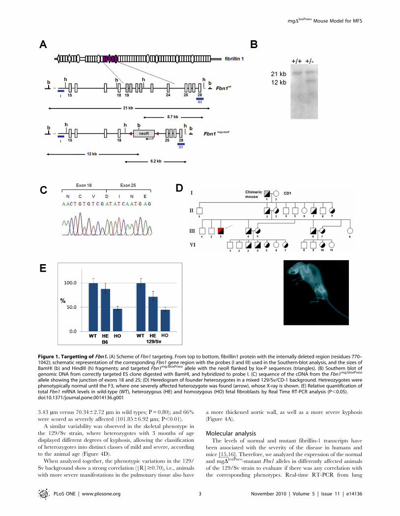
Animal developmentCells from correctly targeted ES cell clones were aggregated to
morulas, and used to produce chimeric male mice that, after mating
with CD1 females, resulted in transmission of the mgDloxPneo Fbn1
allele to a proportion of the F1 generation (Figure 1A). RT-PCR
and sequencing analysis of skin RNA from heterozygous animals
confirmed the in-frame deletion of exons 19–24 in transcripts
derived from the targeted allele (Figure 1B–C). As observed in the
original mgD strain, the heterozygous mgDloxPneo from the F1 did
not present any apparent phenotype. However, in subsequent
crosses between F1 heterozygotes, we obtained some heterozygous
animals with a very severe phenotype, characterized by deformities
of the spine at 2 months of age (Figure 1 D). From a total of 47
heterozygous mice, four presented the mentioned feature, and died
by 3 months of age of unknown causes presenting hemothorax,
suggestive of aortic rupture. We also obtained four homozygous
animals that died between 4 and 8 days of age. All these animals
came from mixed 129/Sv and CD-1 backgrounds.
These results suggested that the phenotype variability could be
associated with the different genetic backgrounds of the animals. To
test this hypothesis, the mgDloxPneo allele was put into two different
isogenic backgrounds, namely the 129/Sv and B6 strains. Haplotype
analysis of a large panel of microsatellite markers confirmed the
congenic status of mice after 14 generations (data not shown).
Real-time RT-PCR analysis of embryonic day 13 fibroblasts
from both strains showed 47%65 (P,0.05) of Fbn1 mRNA levels
in homozygous, and 78%610 (P,0.05) in heterozygous fibro-
blasts when compared to wild-type cells (Figure 1E). Therefore,
the level of expression of the mgDloxPneo allele is significantly
higher than the 10% level observed in the original mgD allele [11],
which may explain the differences in phenotypes seen in
heterozygotes between the two models.
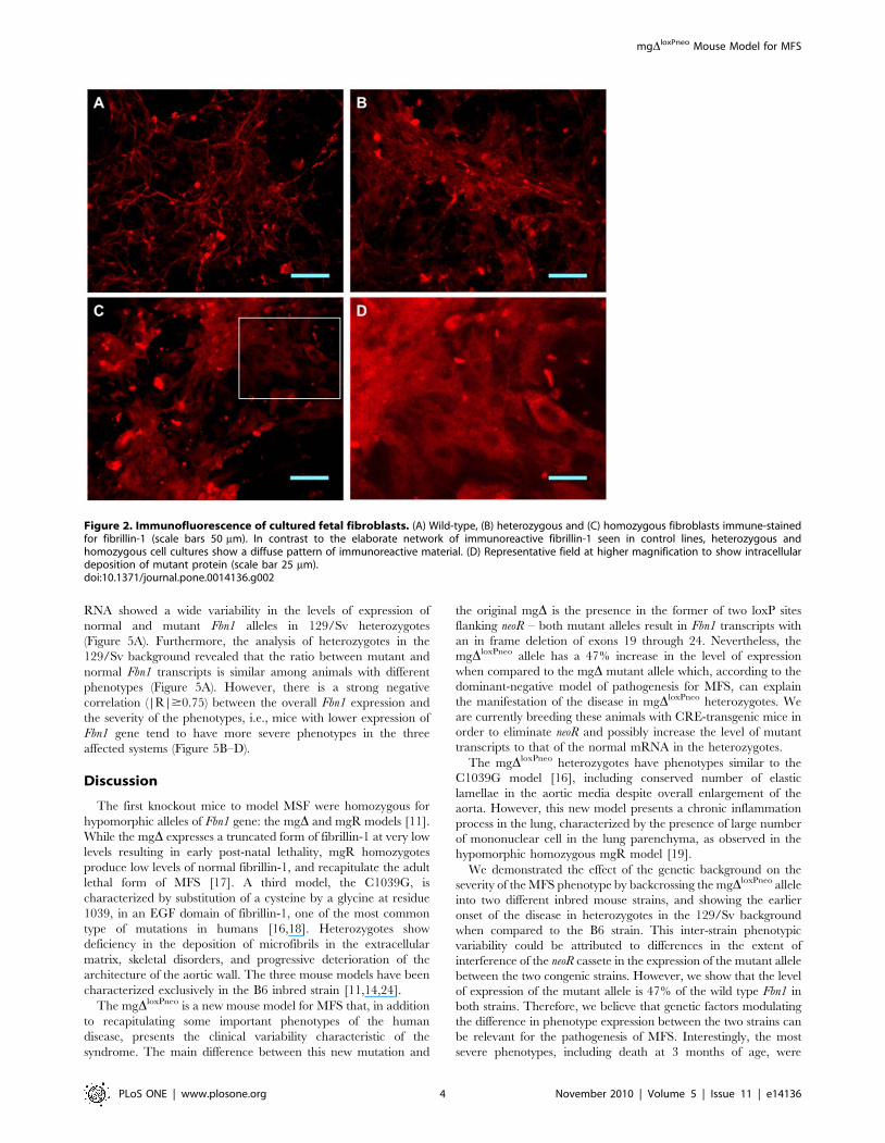
Immunohistochemical comparison of cultured fetal fibroblasts
from B6 animals revealed qualitative differences in fibrils between
wild-type and mutant cells (Figure 2). In contrast to the elaborate
network of immunoreactive fibrillin-1 seen in control lines
(Figure 2A), heterozygous cells present fibers spread over a diffuse
background (Figure 2B), while homozygous cell cultures show a
diffuse pattern of immunoreactive material (Figure 2C) similar to
that reported in cultured fibroblasts from homozygous mgD, and
heterozygous Tight Skin models [11,13]. In the homozygous
mutant fibroblasts we also observed an apparent intracellular
deposition of mutant protein, evidenced by the visualization of
cells with nuclear regions delimitated by marked protein
(Figure 2D). The changes observed in the immunofluorescence
assays indicate that a portion of the mutant molecules may be
retained inside the cell, which in turn, could be a significant factor
contributing to the pathogenesis of MFS [14].
PhenotypesHeterozygous mice from both strains have normal lifespan and
reproductive capacity, but display some of the classic MFS
phenotypes (Figure 3). Pulmonary alterations include enlargement
of peripheral air space (respiratory bronchioles and alveoli), and
destruction of alveolar wall structures, characterizing pulmonary
emphysema (Figure 3A–D). We also detected a large amount of
infiltrating mononuclear cells, indicating a chronic inflammation
process in the lung. The cardiovascular phenotypes include
thickening of the aortic media, disruption/degradation of the
elastic fibers (Figure 3F–I), but no inflammatory cells were
detected. Finally, mutant animals also presented skeletal manifes-
tations, mostly kyphosis (Figure 3K–N).
In order to characterize the phenotypic variability in the two
strains, we quantified the phenotypes in the three affected organ
systems. Analysis of the mean linear intercept (Lm) revealed
changes on the average size of alveoli in 129/Sv heterozygotes
(Figure 3E). By 3 months of age, these animals present an Lm
significantly higher (p,0.02) than wild-type mice, and this
difference became more pronounced with age. In contrast, B6
mice at 3 months of age presented milder (although significant,
p,0.01) pulmonary alterations, and only at 6 months of age did
the alterations become severe.
The vascular phenotype was quantified by measuring the
thickness of the aortic media in heterozygous animals. As observed
in the lung, at 3 months of age 129/Sv heterozygotes exhibited a
significantly enlarged media when compared to wild-type, and this
difference increased with age (Figure 3J). In contrast, in the B6
background heterozygous mgDloxPneo mice were asymptomatic at
3 months of age, whereas by 9 months they presented severe
alterations indistinguishable from those of 129/Sv heterozygotes at
the same age.
Quantification of the skeletal manifestations was performed by
calculating the ratio between the linear distance and the length
from the first cervical vertebrae to the last thoracic vertebrae (KR)
(Figure 3N). As with the other phenotypes, heterozygous animals
from the 129/Sv strain manifested a more severe skeletal
phenotype earlier than those from the B6 background
(Figure 3O). Together, these data show that the main differences
observed in disease manifestation between the two strains is the
age of onset of symptoms, which is delayed in B6 animals.
Finally, when present in homozygocity the mutation is lethal
during gestation in both strains. Crossings between heterozygotes
produced 30 (42%) wild-type animals and 41 (58%) heterozygous
offsprings. Homozygous mutant embryos were identified only
prior to embryonic day 13.
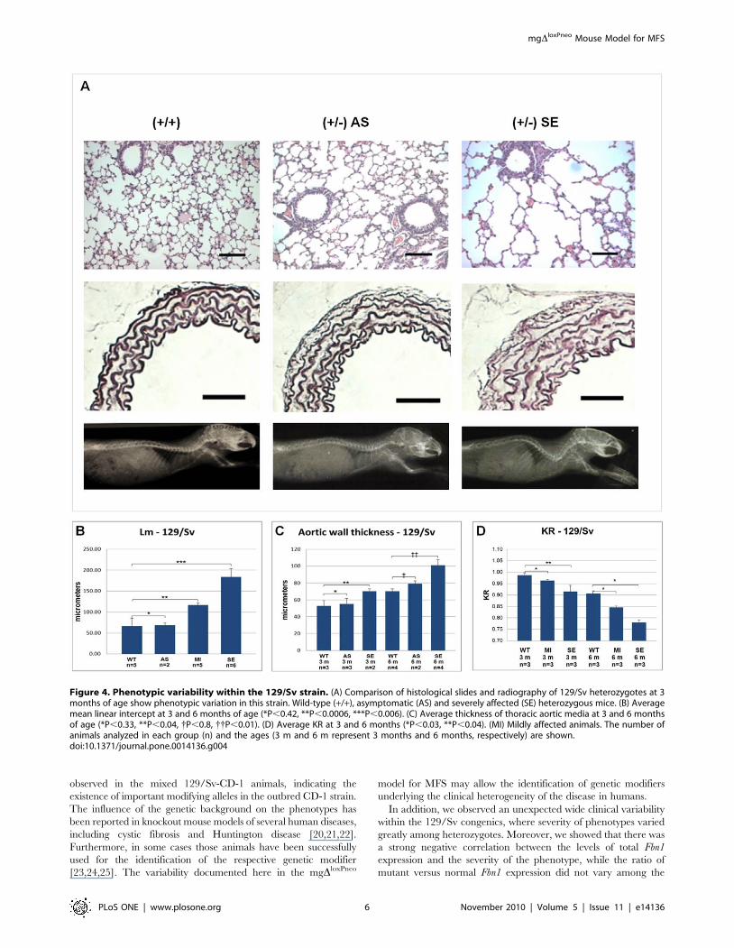
129/Sv phenotypic variabilityInterestingly, while the different phenotypes are homogeneous
among B6 heterozygotes (Figure 3E, J and O), animals from the
129/Sv strain present a wide clinical variability before 6 months of
age, with phenotypes varying from mild to very severe (Figure 4A).
Based on the average Lm in alveoli, we scored thirteen 129/Sv
heterozygotes between 3 to 6 months of age in 3 clinical categories
(Figure 4B): compared with wild-type animals (Lm =
66.75618.26 mm), 15% were classified as asymptomatic
(Lm = 68.4565.2 mm; P,0,42); 38% as carrying moderate
alterations (Lm = 116.4465.46 mm; P,0.0006) and 46% as
presenting severe alterations (Lm = 183.86619.84 mm; P,0.006).
The aortic phenotype also exhibited variability in the 129/Sv
heterozygotes at 3 months of age (Figure 4C), allowing the
classification of phenotypes in 2 categories: 60% were defined as
asymptomatic (55.2166.46 mm versus 61.61611.08 mm in wild
types; P = 0.33), and 40% were classified as presenting moderate
changes (77.7562.59 mm; P,0.04). At 6 months of age, 33% of
the heterozygotes were classified as asymptomatic (79.106
mgDloxPneo Mouse Model for MFS
PLoS ONE | www.plosone.org 2 November 2010 | Volume 5 | Issue 11 | e14136
3.43 mm versus 70.3462.72 mm in wild types; P = 0.80); and 66%
were scored as severely affected (101.8566.92 mm; P,0.01).
A similar variability was observed in the skeletal phenotype in
the 129/Sv strain, where heterozygotes with 3 months of age
displayed different degrees of kyphosis, allowing the classification
of heterozygotes into distinct classes of mild and severe, according
to the animal age (Figure 4D).
When analyzed together, the phenotypic variations in the 129/
Sv background show a strong correlation (|R|$0.70), i.e., animals
with more severe manifestations in the pulmonary tissue also have
a more thickened aortic wall, as well as a more severe kyphosis
(Figure 4A).
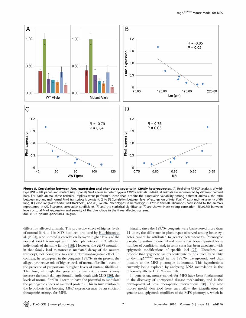
Molecular analysisThe levels of normal and mutant fibrillin-1 transcripts have
been associated with the severity of the disease in humans and
mice [15,16]. Therefore, we analyzed the expression of the normal
and mgDloxPneo-mutant Fbn1 alleles in differently affected animals
of the 129/Sv strain to evaluate if there was any correlation with
the corresponding phenotypes. Real-time RT-PCR from lung
Figure 1. Targetting of Fbn1. (A) Scheme of Fbn1 targeting. From top to bottom, fibrillin1 protein with the internally deleted region (residues 770–1042); schematic representation of the corresponding Fbn1 gene region with the probes (I and III) used in the Southern-blot analysis, and the sizes ofBamHI (b) and HindIII (h) fragments; and targeted Fbn1mgDloxPneo allele with the neoR flanked by lox-P sequences (triangles). (B) Southern blot ofgenomic DNA from correctly targeted ES clone digested with BamHI, and hybridized to probe I. (C) sequence of the cDNA from the Fbn1mgDloxPneo
allele showing the junction of exons 18 and 25; (D) Heredogram of founder heterozygotes in a mixed 129/Sv/CD-1 background. Hetreozygotes werephenotypically normal until the F3, where one severely affected heterozygote was found (arrow), whose X-ray is shown. (E) Relative quantification oftotal Fbn1 mRNA levels in wild-type (WT), heterozygous (HE) and homozygous (HO) fetal fibroblasts by Real Time RT-PCR analysis (P,0.05).doi:10.1371/journal.pone.0014136.g001
mgDloxPneo Mouse Model for MFS
PLoS ONE | www.plosone.org 3 November 2010 | Volume 5 | Issue 11 | e14136
RNA showed a wide variability in the levels of expression of
normal and mutant Fbn1 alleles in 129/Sv heterozygotes
(Figure 5A). Furthermore, the analysis of heterozygotes in the
129/Sv background revealed that the ratio between mutant and
normal Fbn1 transcripts is similar among animals with different
phenotypes (Figure 5A). However, there is a strong negative
correlation (|R|$0.75) between the overall Fbn1 expression and
the severity of the phenotypes, i.e., mice with lower expression of
Fbn1 gene tend to have more severe phenotypes in the three
affected systems (Figure 5B–D).
Discussion
The first knockout mice to model MSF were homozygous for
hypomorphic alleles of Fbn1 gene: the mgD and mgR models [11].
While the mgD expresses a truncated form of fibrillin-1 at very low
levels resulting in early post-natal lethality, mgR homozygotes
produce low levels of normal fibrillin-1, and recapitulate the adult
lethal form of MFS [17]. A third model, the C1039G, is
characterized by substitution of a cysteine by a glycine at residue
1039, in an EGF domain of fibrillin-1, one of the most common
type of mutations in humans [16,18]. Heterozygotes show
deficiency in the deposition of microfibrils in the extracellular
matrix, skeletal disorders, and progressive deterioration of the
architecture of the aortic wall. The three mouse models have been
characterized exclusively in the B6 inbred strain [11,14,24].
The mgDloxPneo is a new mouse model for MFS that, in addition
to recapitulating some important phenotypes of the human
disease, presents the clinical variability characteristic of the
syndrome. The main difference between this new mutation and
the original mgD is the presence in the former of two loxP sites
flanking neoR – both mutant alleles result in Fbn1 transcripts with
an in frame deletion of exons 19 through 24. Nevertheless, the
mgDloxPneo allele has a 47% increase in the level of expression
when compared to the mgD mutant allele which, according to the
dominant-negative model of pathogenesis for MFS, can explain
the manifestation of the disease in mgDloxPneo heterozygotes. We
are currently breeding these animals with CRE-transgenic mice in
order to eliminate neoR and possibly increase the level of mutant
transcripts to that of the normal mRNA in the heterozygotes.
The mgDloxPneo heterozygotes have phenotypes similar to the
C1039G model [16], including conserved number of elastic
lamellae in the aortic media despite overall enlargement of the
aorta. However, this new model presents a chronic inflammation
process in the lung, characterized by the presence of large number
of mononuclear cell in the lung parenchyma, as observed in the
hypomorphic homozygous mgR model [19].
We demonstrated the effect of the genetic background on the
severity of the MFS phenotype by backcrossing the mgDloxPneo allele
into two different inbred mouse strains, and showing the earlier
onset of the disease in heterozygotes in the 129/Sv background
when compared to the B6 strain. This inter-strain phenotypic
variability could be attributed to differences in the extent of
interference of the neoR cassete in the expression of the mutant allele
between the two congenic strains. However, we show that the level
of expression of the mutant allele is 47% of the wild type Fbn1 in
both strains. Therefore, we believe that genetic factors modulating
the difference in phenotype expression between the two strains can
be relevant for the pathogenesis of MFS. Interestingly, the most
severe phenotypes, including death at 3 months of age, were
Figure 2. Immunofluorescence of cultured fetal fibroblasts. (A) Wild-type, (B) heterozygous and (C) homozygous fibroblasts immune-stainedfor fibrillin-1 (scale bars 50 mm). In contrast to the elaborate network of immunoreactive fibrillin-1 seen in control lines, heterozygous andhomozygous cell cultures show a diffuse pattern of immunoreactive material. (D) Representative field at higher magnification to show intracellulardeposition of mutant protein (scale bar 25 mm).doi:10.1371/journal.pone.0014136.g002
mgDloxPneo Mouse Model for MFS
PLoS ONE | www.plosone.org 4 November 2010 | Volume 5 | Issue 11 | e14136
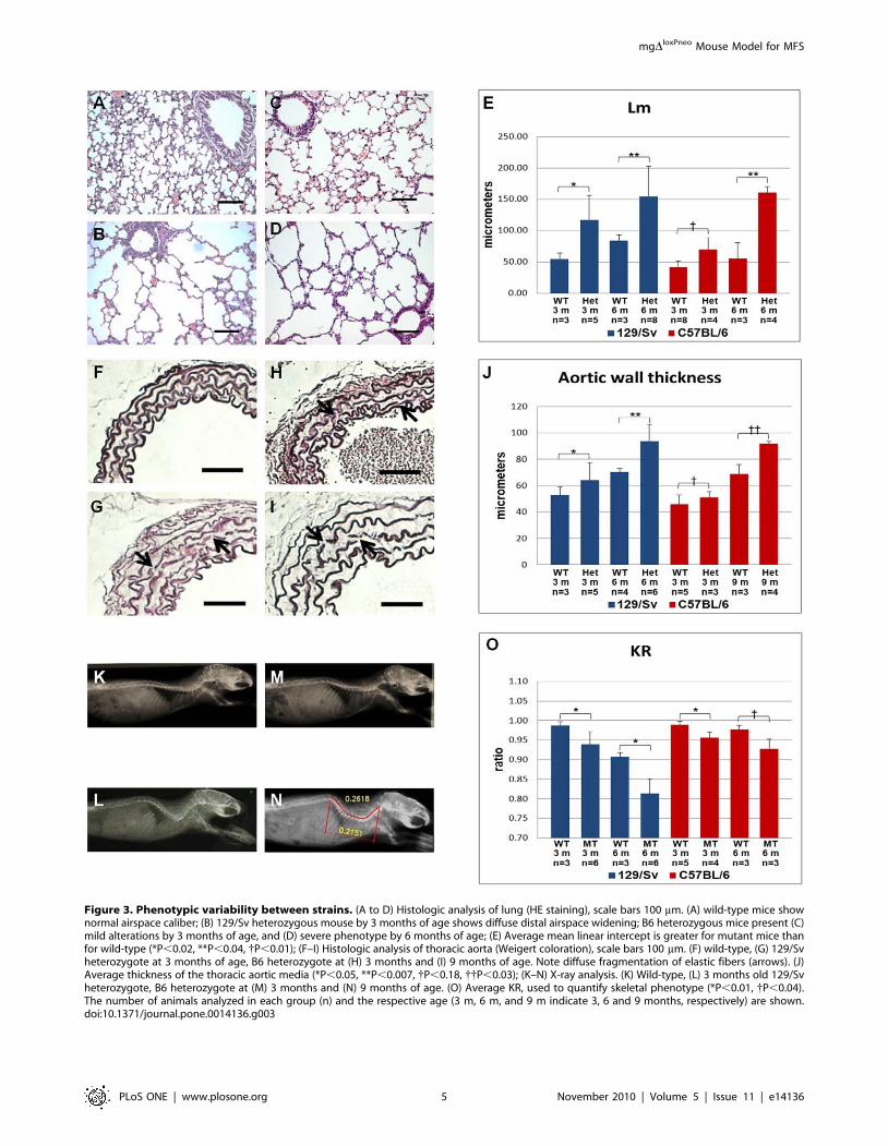
Figure 3. Phenotypic variability between strains. (A to D) Histologic analysis of lung (HE staining), scale bars 100 mm. (A) wild-type mice shownormal airspace caliber; (B) 129/Sv heterozygous mouse by 3 months of age shows diffuse distal airspace widening; B6 heterozygous mice present (C)mild alterations by 3 months of age, and (D) severe phenotype by 6 months of age; (E) Average mean linear intercept is greater for mutant mice thanfor wild-type (*P,0.02, **P,0.04, {P,0.01); (F–I) Histologic analysis of thoracic aorta (Weigert coloration), scale bars 100 mm. (F) wild-type, (G) 129/Svheterozygote at 3 months of age, B6 heterozygote at (H) 3 months and (I) 9 months of age. Note diffuse fragmentation of elastic fibers (arrows). (J)Average thickness of the thoracic aortic media (*P,0.05, **P,0.007, {P,0.18, {{P,0.03); (K–N) X-ray analysis. (K) Wild-type, (L) 3 months old 129/Svheterozygote, B6 heterozygote at (M) 3 months and (N) 9 months of age. (O) Average KR, used to quantify skeletal phenotype (*P,0.01, {P,0.04).The number of animals analyzed in each group (n) and the respective age (3 m, 6 m, and 9 m indicate 3, 6 and 9 months, respectively) are shown.doi:10.1371/journal.pone.0014136.g003
mgDloxPneo Mouse Model for MFS
PLoS ONE | www.plosone.org 5 November 2010 | Volume 5 | Issue 11 | e14136
observed in the mixed 129/Sv-CD-1 animals, indicating the
existence of important modifying alleles in the outbred CD-1 strain.
The influence of the genetic background on the phenotypes has
been reported in knockout mouse models of several human diseases,
including cystic fibrosis and Huntington disease [20,21,22].
Furthermore, in some cases those animals have been successfully
used for the identification of the respective genetic modifier
[23,24,25]. The variability documented here in the mgDloxPneo
model for MFS may allow the identification of genetic modifiers
underlying the clinical heterogeneity of the disease in humans.
In addition, we observed an unexpected wide clinical variability
within the 129/Sv congenics, where severity of phenotypes varied
greatly among heterozygotes. Moreover, we showed that there was
a strong negative correlation between the levels of total Fbn1
expression and the severity of the phenotype, while the ratio of
mutant versus normal Fbn1 expression did not vary among the
Figure 4. Phenotypic variability within the 129/Sv strain. (A) Comparison of histological slides and radiography of 129/Sv heterozygotes at 3months of age show phenotypic variation in this strain. Wild-type (+/+), asymptomatic (AS) and severely affected (SE) heterozygous mice. (B) Averagemean linear intercept at 3 and 6 months of age (*P,0.42, **P,0.0006, ***P,0.006). (C) Average thickness of thoracic aortic media at 3 and 6 monthsof age (*P,0.33, **P,0.04, {P,0.8, {{P,0.01). (D) Average KR at 3 and 6 months (*P,0.03, **P,0.04). (MI) Mildly affected animals. The number ofanimals analyzed in each group (n) and the ages (3 m and 6 m represent 3 months and 6 months, respectively) are shown.doi:10.1371/journal.pone.0014136.g004
mgDloxPneo Mouse Model for MFS
PLoS ONE | www.plosone.org 6 November 2010 | Volume 5 | Issue 11 | e14136
differently affected animals. The protective effect of higher levels
of normal fibrillin-1 in MFS has been proposed by Hutchinson et
al. (2003), who showed a correlation between higher levels of the
normal FBN1 transcript and milder phenotypes in 3 affected
individuals of the same family [10]. However, the FBN1 mutation
in that family lead to nonsense mediated decay of the mutant
transcript, not being able to exert a dominant-negative effect. In
contrast, heterozygotes in the congenic 129/Sv strain present the
alleged protective role of higher levels of normal fibrillin-1 even in
the presence of proportionally higher levels of mutant fibrillin-1.
Therefore, although the presence of mutant monomers may
increase the tissue damage found in individuals with MFS [26], the
levels of normal fibrillin-1 seem to have the potential to modulate
the pathogenic effects of mutated proteins. This in turn reinforces
the hypothesis that boosting FBN1 expression may be an efficient
therapeutic strategy for MFS.
Finally, since the 129/Sv congenic were backcrossed more than
14 times, the difference in phenotypes observed among heterozy-
gotes cannot be attributed to genetic heterogeneity. Phenotypic
variability within mouse inbred strains has been reported for a
number of conditions, and, in some cases has been associated with
epigenetic modifications of specific loci [27]. Therefore, we
propose that epigenetic factors contribute to the clinical variability
of the mgDloxPneo model in the 129/Sv background, and thus
possibly to the MFS phenotype in humans. This hypothesis is
currently being explored by analyzing DNA methylation in the
differently affected 129/Sv animals.
In conclusion, mouse models for MFS have been fundamental
in the discovery of unexpected disease mechanisms, and in the
development of novel therapeutic interventions [28]. The new
mouse model described here may allow the identification of
genetic and epigenetic modifiers of the MFS phenotype, which in
Figure 5. Correlation between Fbn1 expression and phenotype severity in 129/Sv heterozygotes. (A) Real-time RT-PCR analysis of wild-type (WT – left panel) and mutant (right panel) Fbn1 alleles in heterozygous 129/Sv animals. Individual animals are represented by different coloredbars. For each animal three technical replicas were performed. Note that, despite the expression variability among different animals, the ratiobetween mutant and normal Fbn1 transcripts is constant. (B to D) Correlation between level of expression of total Fbn1 (Y axis) and the severity of (B)lung, (C) vascular (AWT: aortic wall thickness), and (D) skeletal phenotypes in heterozygous 129/Sv animals. Diamonds correspond to the animalsrepresented in (A). Pearson’s correlation coefficients (R) and the statistical significance (P) are shown. Note strong correlation (|R|$0.75) betweenlevels of total Fbn1 expression and severity of the phenotype in the three affected systems.doi:10.1371/journal.pone.0014136.g005
mgDloxPneo Mouse Model for MFS
PLoS ONE | www.plosone.org 7 November 2010 | Volume 5 | Issue 11 | e14136
turn will lead to a better understanding of the clinical variability of
the disease, and of the physiology of pulmonary, cardiovascular
and skeletal systems.
Materials and Methods
AnimalsAll animals used were housed under controlled temperature and
light conditions in pathogen-free environment at the Immunology
Department of ICB USP experimentation housing facility. We
analyzed 45 mutant animals and 20 wild-type animals, at three
different ages and from two different mice strains. The C57BL/6
(B6) was chosen because it is the most widely used inbred strain,
and all other extant mouse models for MFS are in that
background. The 129/Sv was chosen because the ES cells used
for gene targeting were derived from this strain. All animal
experiments followed the protocols approved by the Institutional
Animal Care and Use Committee of the Instituto de Biociencias at
USP. Protocol ID: CEA/IBUSP 020/2004.
Development of the mgDloxPneo mouse modelThe murine ES cell line USP-1 was used for gene targeting
experiments [29]. Generation of positively targeted ES cell clones
and production of chimaeric mice were performed as previously
described [11]. The Fbn1mgDloxPneo targeting vector was a
modification of the previously described targeting vector used to
create the mgD and mgR models [11,17]. Two complementary
38-bp oligonucleotides with the loxP consensus sequence [12] were
synthesized, annealed, and cloned flanking the neoR expression
cassette, which was then used to replace the original neoR cassette
of the mgD vector. Correctly targeted ES clones were identified by
southern blot as described [11].
GenotypingDNA was extracted from a 0.5 cm piece of tail using Proteinase
K (Promega) as described [30]. Each sample was submitted to two
PCRs to identify the presence of the Fbn1mgDloxPneo allele and the
normal allele, which served as an internal reaction control.
Fbn1mgDloxPneo allele primers: forward 59 -GAG GCT ATT CGG
CTA TGA CT – 39, reverse 59 – CTC TTC GTC CAG ATC
ATC CT – 39. Cycling conditions were 94uC for 2.5 min, then
94uC, 57uC, 72uC for 1 min for 30 cycles in a 10 ml volume.
Fbn1wt allele primers: forward 59 – AAA CCA TCA AGG GCA
CTT GC – 39, reverse 59 – CAC ATT GCG TGC CTT TAA
TTC – 39. Cycling conditions were 94uC for 2.5 min primary
denaturation, then 94uC, 55uC, 72uC for 1 min for 30 cycles in a
10 ml volume.
ImmunofluorescenceMouse embryonic fibroblasts (MEFs) were prepared from
embryos at 13–14 days of gestation as described [31]. Cells were
fixed in 4% paraformaldehyde in PBS for 20 min at 4uC and
permeabilized in 0.05% Triton X-100 in PBS for 5 min. Non-
specific binding was blocked with 10% FBS in PBS for 1 h at room
temperature. Cells were incubated with pAb9543 (1:1000 dilution)
primary antibody [32] overnight at 4uC and with secondary
antibody coupled to Cy3 for 1 h at room temperature. The
fluorescence signals were examined using an Axiovert 200 (Carl
Zeiss) and an ApoTome imaging system (Carl Zeiss).
Histological analysisAnimals were sacrificed by cervical dislocation. Mouse tissues
were processed as previously described [33]. Five-micron sections
were stained with hematoxylin and eosin, and adjacent sections
were assayed for Weigert coloration, specific for elastic fiber
visualization. Slides were examined and photographed using an
Axiovert 200 (Carl Zeiss).
Phenotype quantificationSkeletal: A full body x-ray of each mouse was digitalized and the
follow measurements were taken using AutoCAD software 2002:
the cervical-thoracic segment length and the straight line distance
of the same segment. With those measures we were able to
establish a kyphosis ratio (straight distance/segment length), and
use the ratio to score the animals according to the severity of the
skeletal manifestation (the smaller ratio, the more severe the
manifestation).
Aortic wall: The histological samples were photographed at 506and 1006 magnification, and the length of the inner and outer
perimeters of the aorta were measured using the imageJ software
[34]. From this we could estimate the inner and outer radius, and
wall thickness of the aorta.
Lung: The size of alveolar airways was determined by
measuring the linear intercept on H&E-stained lungs as previously
described [35].
Real-time RT-PCRTotal RNA was extracted from mouse lung and from mouse
embryonic day 13 fibroblasts using TRIzol (Invitrogen Corp.).
The RNA was treated with DNase according to the manufacture’s
instructions (Invitrogen Corp.). A total of 1 mg of total RNA was
reverse-transcribed with SuperScriptTM III First-Strand Synthesis
System (Invitrogen Corp.). Wild type, mutant and total Fbn1
mRNA levels were determined using real-time RT-PCR sequence
detection (7500 Real Time System; Applied Biosystems). mRNA
levels were normalized to Actb mRNA, and fold expression
determined as previously described [36]. The following primers
and probes sequences were designed using the PrimerExpress
software (Applied Biosystems): WT forward 59 – ACA TAA CTG
GGA AAA ACT GTG TCG ATA – 39, WT reverse 59 – TTC
CAG GTG TGT TTC GAC ATT GT – 39, WT probe 59 –TGT
GCT GAA CAG TCT ACT– 39; KO forward 59 –GGG ATA
TGA AGT AGA CAT AAC TGG GAA A– 39, KO reverse 59 –
GAG GCT GGG TAT CAT CTT GCA – 39, KO probe 59 –
ACT GTG TCG ATA TCA ATG– 39; Fbn1Total forward 59 –
CCT GTG CTA TGA TGG GTT CA – 39, Fbn1Total reverse 59
– AGG TCC CAC TAA GGC AGA TGT – 39; ACTB forward 59
–ACGGCCAGGTCATCACTATTG – 39, ACTB reverse 59 –
CAAGAAGGAAGGCTGGAAAAGA– 39. Three technical rep-
licates of each reaction were performed.
Statistical AnalysesPearson’s Correlation Coefficient (R) was used to determine the
correlation between the Fbn1 gene expression and the severity of
the phenotype, reflecting the degree to which the variables are
related; weak correlation 0#|R|#0.29; moderate correlation
0.30#|R|#0.69; strong correlation |R|$0.70. P,0.05 was
deemed to be significant.
A nonparametric test, Mann Whitney test, was used to
determine statistical significance for all tests other than the
Pearson Correlation Coefficient. All statistical analyses were
performed using MINITAB (R14). P,0.05 was deemed to be
significant.
Acknowledgments
We wish to thank Dr Peter Pearson for his significant help on the
preparation of the manuscript. We also gratefully acknowledge our
mgDloxPneo Mouse Model for MFS
PLoS ONE | www.plosone.org 8 November 2010 | Volume 5 | Issue 11 | e14136
colleagues Joana C.M de Mello, Ana Maria Fraga, Raquel Stabellini, Erica
Sara Araujo, Fabiano T. de Araujo, Natassia M.S Vieira, Cynthia Q.
Cardoso and Dra. Mariz Vainzof for helpful suggestions.
Author Contributions
Conceived and designed the experiments: BLL EJCS SMGM JAV LVP.
Performed the experiments: BLL EJCS GRF CAM MRBM MS AK
SMGM JPAG LVP. Analyzed the data: BLL EJCS GRF CAM MRBM
SMGM JPAG JAV LVP. Wrote the paper: BLL LVP.
References
1. Pyeritz RE (1990) Marfan syndrome. N Engl J Med 323: 987–989.
2. Silverman DI, Gray J, Roman MJ, Bridges A, Burton K, et al. (1995) Familyhistory of severe cardiovascular disease in Marfan syndrome is associated with
increased aortic diameter and decreased survival. J Am Coll Cardiol 26:
1062–1067.3. Pyeritz RE, McKusick VA (1981) Basic defects in Marfan syndrome.
N Engl J Med 305: 1011–1012.4. Gray JR, Bridges AB, Faed MJ, Pringle T, Baines P, et al. (1994) Ascertainment
and severity of Marfan syndrome in a Scottish population. J Med Genet 31:51–54.
5. Kainulainen K, Peltonen L (1991) Marfan gene discovered. Ann Med 23:
395–396.6. Kainulainen K, Steinmann B, Collins F, Dietz HC, Francomano CA, et al.
(1991) Marfan syndrome: no evidence for heterogeneity in different populations,and more precise mapping of the gene. Am J Hum Genet 49: 662–667.
7. Dietz HC, Cutting GR, Pyeritz RE, Maslen CL, Sakai LY, et al. (1991) Marfan
syndrome caused by a recurrent de novo missense mutation in the fibrillin gene.Nature 352: 337–339.
8. Pyeritz RE (1986) The Marfan syndrome. Am Fam Physician 34: 83–94.9. Pyeritz RE, Murphy EA, McKusick VA (1979) Clinical variability in the Marfan
syndrome(s). Birth Defects Orig Artic Ser 15: 155–178.10. Hutchinson S, Furger A, Halliday D, Judge DP, Jefferson A, et al. (2003) Allelic
variation in normal human FBN1 expression in a family with Marfan syndrome:
a potential modifier of phenotype? Hum Mol Genet 12: 2269–2276.11. Pereira L, Andrikopoulos K, Tian J, Lee SY, Keene DR, et al. (1997) Targetting
of the gene encoding fibrillin-1 recapitulates the vascular aspect of Marfansyndrome. Nat Genet 17: 218–222.
12. Sunaga S, Maki K, Komagata Y, Ikuta K, Miyazaki JI (1997) Efficient removal
of loxP-flanked DNA sequences in a gene-targeted locus by transient expressionof Cre recombinase in fertilized eggs. Mol Reprod Dev 46: 109–113.
13. Gayraud B, Keene DR, Sakai LY, Ramirez F (2000) New insights into theassembly of extracellular microfibrils from the analysis of the fibrillin 1 mutation
in the tight skin mouse. J Cell Biol 150: 667–680.14. Whiteman P, Handford PA (2003) Defective secretion of recombinant fragments
of fibrillin-1: implications of protein misfolding for the pathogenesis of Marfan
syndrome and related disorders. Hum Mol Genet 12: 727–737.15. Buoni S, Zannolli R, Macucci F, Ansaldi S, Grasso M, et al. (2004) The FBN1
(R2726W) mutation is not fully penetrant. Ann Hum Genet 68: 633–638.16. Judge DP, Biery NJ, Keene DR, Geubtner J, Myers L, et al. (2004) Evidence for
a critical contribution of haploinsufficiency in the complex pathogenesis of
Marfan syndrome. J Clin Invest 114: 172–181.17. Pereira L, Lee SY, Gayraud B, Andrikopoulos K, Shapiro SD, et al. (1999)
Pathogenetic sequence for aneurysm revealed in mice underexpressing fibrillin-1. Proc Natl Acad Sci U S A 96: 3819–3823.
18. Ng CM, Cheng A, Myers LA, Martinez-Murillo F, Jie C, et al. (2004) TGF-beta-dependent pathogenesis of mitral valve prolapse in a mouse model of Marfan
syndrome. J Clin Invest 114: 1586–1592.
19. Neptune ER, Frischmeyer PA, Arking DE, Myers L, Bunton TE, et al. (2003)Dysregulation of TGF-beta activation contributes to pathogenesis in Marfan
syndrome. Nat Genet 33: 407–411.
20. Sweet A, Erickson RP, Huntington C, Dawson D (1992) A potential animal model
for studying CF heterozygote advantage: genetic variation in theophylline-
inducible colonic chloride currents among inbred strains of mice. Biochem Med
Metab Biol 47: 97–102.
21. Otsuru S, Hofmann TJ, Rasini V, Veronesi E, Dominici M, et al. (2010)
Osteopoietic engraftment after bone marrow transplantation: Effect of inbred
strain of mice. Exp Hematol.
22. Yang T, Huang YG, Ye W, Hansen P, Schnermann JB, et al. (2005) Influence of
genetic background and gender on hypertension and renal failure in COX-2-
deficient mice. Am J Physiol Renal Physiol 288: F1125–1132.
23. Wheeler FC, Fernandez L, Carlson KM, Wolf MJ, Rockman HA, et al. (2005)
QTL mapping in a mouse model of cardiomyopathy reveals an ancestral
modifier allele affecting heart function and survival. Mamm Genome 16:
414–423.
24. Dietrich WF, Lander ES, Smith JS, Moser AR, Gould KA, et al. (1993) Genetic
identification of Mom-1, a major modifier locus affecting Min-induced intestinal
neoplasia in the mouse. Cell 75: 631–639.
25. Pu WT (2009) Identification of a cardiac disease modifier gene using forward
genetics in the mouse. PLoS Genet 5: e1000643.
26. Booms P, Tiecke F, Rosenberg T, Hagemeier C, Robinson PN (2000)
Differential effect of FBN1 mutations on in vitro proteolysis of recombinant
fibrillin-1 fragments. Hum Genet 107: 216–224.
27. Rakyan VK, Blewitt ME, Druker R, Preis JI, Whitelaw E (2002) Metastable
epialleles in mammals. Trends Genet 18: 348–351.
28. Judge DP, Dietz HC (2008) Therapy of Marfan syndrome. Annu Rev Med 59:
43–59.
29. Sukoyan MA, Kerkis AY, Mello MR, Kerkis IE, Visintin JA, et al. (2002)
Establishment of new murine embryonic stem cell lines for the generation of
mouse models of human genetic diseases. Braz J Med Biol Res 35: 535–542.
30. Zangala T (2007) Isolation of genomic DNA from mouse tails. J Vis Exp. 246 p.
31. Hogan A, Beddington R, Constantine F, Lacy E (1994) Manipulating the mouse
Embryo: a laboratory manual. Cold Spring Harbor, NY: Cold Spring Harbor
Laboratory Press.
32. Reinhardt DP, Keene DR, Corson GM, Poschl E, Bachinger HP, et al. (1996)
Fibrillin-1: organization in microfibrils and structural properties. J Mol Biol 258:
104–116.
33. Andrikopoulos K, Liu X, Keene DR, Jaenisch R, Ramirez F (1995) Targeted
mutation in the col5a2 gene reveals a regulatory role for type V collagen during
matrix assembly. Nat Genet 9: 31–36.
34. Muraishi H [Fundamental of medical image processing with personal computer
system—development of Plugins by ImageJ]. Nippon Hoshasen Gijutsu Gakkai
Zasshi 66: 260–264.
35. Dunnill MS (1962) Quantitative Methods in the Study of Pulmonary Pathology.
Thorax 17: 320–328.
36. Livak KJ, Schmittgen TD (2001) Analysis of relative gene expression data using
real-time quantitative PCR and the 2(-Delta Delta C(T)) Method. Methods 25:
402–408.
mgDloxPneo Mouse Model for MFS
PLoS ONE | www.plosone.org 9 November 2010 | Volume 5 | Issue 11 | e14136