a method for preparing a permanent nocht's stain (nocht ...
17
A METHOD FOR PREPARING A PERMANENT NOCHT'S STAIN (NOCHT-JENNER STAIN). BY T. W. HASTINGS, M.D. (From the Cornell University Medical School, New York City.) PLATES XXI. AND XXII. The possibility of preparing a permanent staining solution from the formulas for Nocht's (I) stain was suggested by the constant use of Jenner's (2) stain for routine blood examina- tions and malarial staining during the summer and fall months of ~9ox. In malarial blood the pigmented bodies of tertian fever and the crescent bodies of mstivo-autumnal fever are readily demonstrated with Jenner's stain, and the use of poly- chrome stains was resorted to only in doubtful cases showing no full-grown forms of parasites. Goldhorn's (3) method of staining with the polychrome blue prepared from methylene blue and lithium carbonate proved valuable, but we were never able to obtain the constantly per- feet results obtained by employing the methods of Noeht as described by Lazier (4) and Ewing (5). No other method gives results comparable to these. The only objection to them is the time required (from two to twelve hours) for good staining. During the fall of ~9oI, repeated attempts to prepare a per- manent staining fluid by Nocht's method succeeded in so far that in the later preparations the same method of procedure was followed, but we were unable to obtain the polychrome tint for the nuclear material and the chromatin staining in the malarial parasite until the publication of Leishman (6) suggested the final simple step in staining, that of diluting the staining fluid with distilled water after staining one minute in full strength and allowing this diluted stain to remain in contact with the specimen from three to five minutes. 265 Downloaded from http://rupress.org/jem/article-pdf/7/3/265/1182954/265.pdf by guest on 02 February 2022
-
Upload
khangminh22 -
Category
Documents
-
view
0 -
download
0
Transcript of a method for preparing a permanent nocht's stain (nocht ...
A METHOD FOR PREPARING A PERMANENT NOCHT'S STAIN (NOCHT-JENNER STAIN).
BY T. W. HASTINGS, M.D.
(From the Cornell University Medical School, New York City.)
PLATES XXI. AND XXII.
The possibility of preparing a permanent staining solution from the formulas for Nocht's (I) stain was suggested by the constant use of Jenner's (2) stain for routine blood examina- tions and malarial staining during the summer and fall months of ~9ox. In malarial blood the pigmented bodies of tertian fever and the crescent bodies of mstivo-autumnal fever are readily demonstrated with Jenner's stain, and the use of poly- chrome stains was resorted to only in doubtful cases showing no full-grown forms of parasites.
Goldhorn's (3) method of staining with the polychrome blue prepared from methylene blue and lithium carbonate proved valuable, but we were never able to obtain the constantly per- feet results obtained by employing the methods of Noeht as described by Lazier (4) and Ewing (5). No other method gives results comparable to these. The only objection to them is the time required (from two to twelve hours) for good staining.
During the fall of ~9oI, repeated attempts to prepare a per- manent staining fluid by Nocht's method succeeded in so far that in the later preparations the same method of procedure was followed, but we were unable to obtain the polychrome tint for the nuclear material and the chromatin staining in the malarial parasite until the publication of Leishman (6) suggested the final simple step in staining, that of diluting the staining fluid with distilled water after staining one minute in full strength and allowing this diluted stain to remain in contact with the specimen from three to five minutes.
265
Dow
nloaded from http://rupress.org/jem
/article-pdf/7/3/265/1182954/265.pdf by guest on 02 February 2022
266 Method for PreTaring a -Permanent Nocht's Stain
The staining fluid consists of a powder, prepared by mixing solutions of eosin and polychrome blue and ghrlich's rectified methylene blue, dissolved in pure methylic alcohol, and the methods of preparation and of staining combine, therefore, the principles of Nocht, Jenner, and Leishrnan.
The methods described by May and Grimwald (7), Michaelis (8), Reuter (9), Willebrand (io), Wright ( i i) , and Schegoleff (i2) resemble Jenner's and Leishman's, and none of them gives the intense clear staining obtained by employing Nocht's (i3) principle of mixing three solutions.
Nocht (I4) pointed out that the essential staining element of the Rornanowski (i5) and Ziernann (i6) methods is a new material which he designated as "red from methylene blue" (Michaelis's "rnethylen-azur," or "azurblau" (i7)), which is formed in all alkaline methylene-blue solutions.
Nocht's first (I899) alkaline methylene blue consisted of a i % aqueous solution of methylene blue with o.5% soda, which stood for a few days at a temperature of from 5o°-6o ° C.
A few months later Nocht (~8) reported a method for making the polychrome methylene blue (alkaline), by steaming the i % methylene blue and o. 5 % soda mixture in a stew-pot for a few hours, filtering and neutralizing. Thereafter this solution was added to the original i % aqueous methylene-blue solution, and later (i9o 3) Nocht (i9) described a third method for the prepara- tion of polychrome methylene blue. To ioo c. c. of i % methy- lene blue solution was added the silver oxide precipitated from one gram of silver nitrate in solution by the addition of alkali in sufficient quantity, and this mixture was allowed to digest four or five days at room-temperature. This last method has not furnished us a good chrornatin-dye, nor can it be used for preparing the stain to be described. Nocht's second method is used. The polychrome borax-methylene blue of Ziemann (~o) was found unsatisfactory.
P R E P A R A T I O N OF T H E S T A I N I N G F L U I D .
Two stain powders are required: the water-soluble, yellow eosin (Grflbler) and Ehrlich's rectified methylene blue (Grflbler).
Dow
nloaded from http://rupress.org/jem
/article-pdf/7/3/265/1182954/265.pdf by guest on 02 February 2022

T. W. Hastings 267
The polychrome blue is readily prepared f rom the la t te r af ter the following directions:
Methylene blue (Ehrlich's rectified) . . . . . . . . 2 grin. Sodium carbonate (dry powder) . . . . . . . . . . . 2 g~m. Distilled wate r . . . . . . . . . . . . . . . . . . . . . . . . . . 2oo c. c.
Dissolve the sodium carbonate in hot distilled water and stir in the methylene blue powder in the above proport ions; br ing the mixture to a modera te boil in an evaporat ing dish on a wire gauze over the flame, or hea t over a boiling water-bath, for f rom ten to fifteen minutes ; add f rom 3o to 4o c. c. of dis- tilled water for each Ioo c. c. of solution (i.e., f rom 6o to 80 c. c.) to replace wate r lost by evaporation, and hea t for f rom ten to fifteen minutes longer. The hot solution is poured off f rom the sediment and made up to 2oo c. c. wi th distilled wate r if necessary. This should be part ial ly neutral ized before fur ther mixing, by the addit ion of f rom i2 .5% to 2o% acetic acid. I t is well to add the acetic acid to one-half the polychrome-blue solution unti l a wel l-marked acid react ion to l i tmus is obtained (6 or 7 c. c. of i2 .5% acid or 3 or 4 c. c. of 20% acid to ioo c. c.), and to mix this neutral ized port ion wi th the un-neutral ized half, thus prevent ing over-neutralization. The solution should be alkaline in final react ion since a slight excess of acid destroys the polychrome properties which cannot be restored by addit ion of alkalies.
A guide to successful prepara t ion is the mixing of the solutions according to Ewlng ' s directions:
A. = Distilled wa te r . . . . . . . . . . . . . . . . . . . . . . . . . . . . . . . . . . . xo c. c. B. = x % aqueous eosin . . . . . . . . . . . . . . . . . . . . . . . . . . . . . . . 3-5 drops C. = Fresh ly prepared polychrome-blue solution . . . . . . . . . . 6 drops D. ~ i % aqueous methylene blue . . . . . . . . . . . . . . . . . . . . . . 2 drops
If the specimens s ta ined in the above mix ture give good results in f rom two to twelve hours, t he polychrome-blue solution m a y be used for prepar ing the powder.
F r o m Ewing ' s directions the following propor t ions for xooo c . c. of w a t e r were calculated, talcing forty-five drops equivalent to one fluid d ram and to
Dow
nloaded from http://rupress.org/jem
/article-pdf/7/3/265/1182954/265.pdf by guest on 02 February 2022
2 6 8 Method for Preparing a Permanent Nocht's Stain
3.7 c c. T h u s for io c c. of wa te r , t he equ iva l en t s for t he s ame so lu t ions w o u l d be:
B. 3 d rops = 0.246 c. c., a p p r o x i m a t e l y . . . . . . . . . . . . . . . . . . 0.25 c. c. C. 6 . . . . = o . 4 9 2 C. C . . . . . . . . . . . . . . . . . . . o . S o C. c .
D. 2 " = o.164 c .c . " • . . . . . . . . . . . . . . . . . o.16 c. c.
a n d for lOOO c. c. of w a t e r :
B. 24.6 a p p r o x i m a t e l y . . . . . . . . . . . . . . . . . . . . . . . . . . . . . . . . . 25 c .c .
C . 4 9 . 2 " • . . . . . . . . . . . . . . . . . . . . . . . . . . . . . . . . 5 o c . c .
D. I6.4 " • . . . . . . . . . . . . . . . . . . . . . . . . . . . . . . . . I6. 5 e. c.
These p r o p o r t i o n s give a c o m p a r a t i v e l y m i n u t e q u a n t i t y of prec ip i ta te , a n d the final p r o d u c t does n o t s t a in well.
The fol lowing m i x t u r e s in lO e. c. of w a t e r were made , a lways t a k i n g B. in q u a n t i t y a p p r o x i m a t e l y equa l to hal f of C., a n d D. in q u a n t i t y equa l to a t h i r d of C.
z. A. = Dist i l led w a t e r . . . . . . . . . . . . . . . . . . . . . . . . . . . . . . . . IO B . = i 070 a q u e o u s eosin . . . . . . . . . . . . . . . . . . . . . . . . . . . . . 5 C. = P o l y e h r o m e b lue . . . . . . . . . . . . . . . . . . . . . . . . . . . . . . io D. = 1 % a q u e o u s m e t h y l e n e blue . . . . . . . . . . . . . . . . . . . . 3.5
2 A . . . . . . . . . . . . . . . . . . . . . . . . . . . . . . . . . . . . . . . . . . . . . . . 1 o
B . . . . . . . . . . . . . . . . . . . . . . . . . . . . . . . . . . . . . . . . . . . . . . . 3 C . . . . . . . . . . . . . . . . . . . . . . . . . . . . . . . . . . . . . . . . . . . . . . . 6
D . . . . . . . . . . . . . . . . . . . . . . . . . . . . . . . . . . . . . . . . . . . . . . . 2
3 " h . . . . . . . . . . . . . . . . . . . . . . . . . . . . . . . . . . . . . . . . . . . . . . . 1 o
B . . . . . . . . . . . . . . . . . . . . . . . . . . . . . . . . . . . . . . . . . . . . . . . 1. 5
C . . . . . . . . . . . . . . . . . . . . . . . . . . . . . . . . . . . . . . . . . . . . . . . 3 D . . . . . . . . . . . . . . . . . . . . . . . . . . . . . . . . . . . . . . . . . . . . . . . 2
4. A . . . . . . . . . . . . . . . . . . . . . . . . . . . . . . . . . . . . . . . . . . . . . . . . lO B . . . . . . . . . . . . . . . . . . . . . . . . . . . . . . . . . . . . . . . . . . . . . . . 1
C . . . . . . . . . . . . . . . . . . . . . . . . . . . . . . . . . . . . . . . . . . . . . . . 2
D . . . . . . . . . . . . . . . . . . . . . . . . . . . . . . . . . . . . . . . . . . . . . . . 0.66
e . e .
C . C .
C . C .
C . C .
C . C .
C . C .
c . e .
c . c .
c . c .
c . c .
c . c .
C . C .
C . C .
C . c .
C . C .
C. C:
Mixtu res 3 and 4 gave good resul ts , b u t w h e n m i x e d in p r o p o r t i o n corre- s p o n d i n g to iooo c. c. of wa te r , t h e m a x i m u m a m o u n t of p rec ip i t a t e w i t h good s t a i n i ng w a s ob t a i ned b y us ing f r o m 70 to 80 c . c . of D ( aque ous m e t h y l e n e blue 1 % ) in No. 4 - - t h a t is:
5. A. Dist i l led w a t e r . . . . . . . . . . . . . . . . . . . . . . . . . . . . . . . . . . IOOO c. e. B. i % a q u e o u s so lu t ion eosin . . . . . . . . . . . . . . . . . . . . . . . lOO e . e .
C. Po l ych rom e-b l ue sol . . . . . . . . . . . . . . . . . . . . . . . . . . . . . 2o0 c. c. D. 1 % a q u e o u s m e t h y l e n e - b l u e sol . . . . . . . . . . . . . . . . . . . 70 c. e.
T w e n t y - o n e m i x t u r e s were p r e p a r e d in p r o p o r t i o n s of No. 5, a n d t e s t ed as t o s t a in ing proper t i es , a n d one w i t h B, C, a n d D, in p r o p o r t i o n w i t h o u t dist i l led w a t e r . T h e va r i a t i ons in these m i x t u r e s cons is ted in us ing old p o l y c h r o m e -
Dow
nloaded from http://rupress.org/jem
/article-pdf/7/3/265/1182954/265.pdf by guest on 02 February 2022
T. W. Hastings 269
blue prepared after Unna's formula, commercial polychrome-blue of Unna, old polychrome-blue prepared after Goldhorn's formula, freshly prepared poly- chrome-blue after Unna's formula, commercial "Goldhorn's polychrome methy- lene-blue," solutions of medicinal methylene blue, solutions of polychrome-blue in which the methylene blue and sodium carbonate varied from o. i % to i %, solution prepared according to Nocht's method No. 3, with silver oxide (simi- lar to Borrel's blue), and in changing the order of mixing. A polychrome-bhie solution containing only o.5 % of methylene blue and 0.5 % sodium carbonate may be used, but deeper chromatin staining is obtained with mixtures contain- ing from o.75 % to i % of each.
I t was found impossible to obtain a polychrome-blue solution from simple methylene blue (Grfibler) and sodium" carbonate, a dirty brown precipitate being thrown down on heating.
Samples of eosin other than the water-soluble yellow eosin (Grfibler) did not give good results.
Finally, from the above, the following directions for preparing a satisfactory stain were deduced. Three solutions, a I % aqueous solution of water-soluble yellow eosin (Grflbler), freshly prepared polychrome methylene-blue solution, and i % aqueous solution of Ehrlich's rectified methylene blue (Grflbler) are mixed together. The freshly prepared, partially neutralized polychrome-blue never fails to give good results. An un- neutralized polychrome-blue solution may be used. I t is un- necessary to cool the freshly prepared polychrome-blue solutions. The solutions should be mixed together in the following order:
A. Distilled water . iooo c. c. B. i % aqueous sol. of eosin (water soluble, yellow) ioo c. c. C. Polychrome-blue sol. (freshly prepared) . . 200 c. c. D. x% aqueous sol. of methylene blue (Ehrlich's rectified). 7o c. c.
A greenish metallic scum appears on the surface and a fine black precipitate is thrown down. This precipitate may not appear unti l 8o c. c. of solution D have been added.
The mixture is filtered at once or after s tanding for from twenty to th i r ty minutes and the residue allowed to dry in the air (from twenty-four to forty-eight hours), or is dried in a hot air chamber at a temperature not above 6o ° C. The dried residue is scraped from the filter paper, powdered in a mortar, and dis- solved in pure methylic alcohol.
From the above amounts of unmixed solutions about o. 7 to
Dow
nloaded from http://rupress.org/jem
/article-pdf/7/3/265/1182954/265.pdf by guest on 02 February 2022
270 Method for Preparing a Permanent Nocht's Stain
I grm. of powder will be obtained, of which o.2 5 to o.3 grm. will saturate iooc . c. of pure methylic alcohol (Merck's). To obtain complete solution of 0.2 5 grm. of powder in i o o c . c. of methylic alcohol, it is necessary to mix them in a mortar, using con- siderable pressure to break up the powder; intense colors vary- ing from blue to a reddish-purple upon the sides of the mortar and the pestle will show tha t proper solution has been obtained.
Some lots of methylic alcohol show an acidity of ~ to 2 c. c. N alkali to ioo e. c. of alcohol, and such an alcohol should be neutralized with o.o 5 to o. i grm. of dry sodium carbonate before mixing with the powdered precipitate, since this amount of acid is sufficient to give results similar to over-neutralization.
Over-neutralization destroys the proper ty of staining the chromat in granules, the specimen staining intensely red through- out ; and a marked alkaline reaction--i , e., with no neutralization - -p reven ts proper fixation, the red cells and leucocytes appear- ing blurred and frayed out at the edges, a n d gives intense blue staining throughout .
When the solution is of proper s t rength it has a purple-plum color, and after shaking leaves the sides of the bottle quite clear above the surface of the stain.
F I X I N G AND S T A I N I N G .
As wi th Jenner 's stain, previous fixation is unnecessary. Blood films on coveroslips or smears on slides are thoroughly dried in the air. The specimen is flooded with the stain in full s t rength for one minute, then diluted with a few drops of dis- tilled water (five or six drops for a ~ in. cover-slip) unti l the greenish metallic scum appears on the surface, and one can readily see through the diluted solution at the edges of the cover-glass only when the spedmen is held over a white surface (filter paper). The diluted stain is allowed to remain on the specimen for five minutes (Leishman) in order to bring out the eosin-staining of the red cells, the granular staining, and the po]ychrome staining properties; the preparation is then washed in distilled water for two or three seconds and dried immediately by blotting.
Dow
nloaded from http://rupress.org/jem
/article-pdf/7/3/265/1182954/265.pdf by guest on 02 February 2022
T. W. Hastings 271
For malarial specimens this procedure is always sufficient to stain the young forms, but the mature forms of tertian and quartan and the crescents of mstivo-auturnnal fever may require two minutes'flooding with the undiluted stain and ten minutes' flooding with the diluted stain for the bringing out of the chro- matin particles in the parasites.
The negative surface of the specimen should be carefully in- spected and washed, if necessary, since the stain drying on the glass gives rise to a thick greenish coating which obscures the field of examination.
This method does not employ the washing out of the blue spoken of by Wright, but does include his "differentiation," the principle of which we first learned from Lelshman's publication. If the specimens are washed and dried after staining one minute in full strength, the staining of the nuclear material is of a pale blue, and of the red cells and of the granules of the leucocytes faint or none.
The dilution of the stain accomplishes the "differentiation" desired, unless the dried specimens have been kept for several weeks or months; these old specimens always stain a deep blue throughout.
The normal red cells in well-spread specimens vary in color from a dull light red to a deeper eosin red. If after staining for one minute in full strength the water and alcoholic stain be not thoroughly mixed in diluting, particularly in thickly spread areas of the specimen, the red cells may show a bluish tinge or absence of the red stain in streaks.
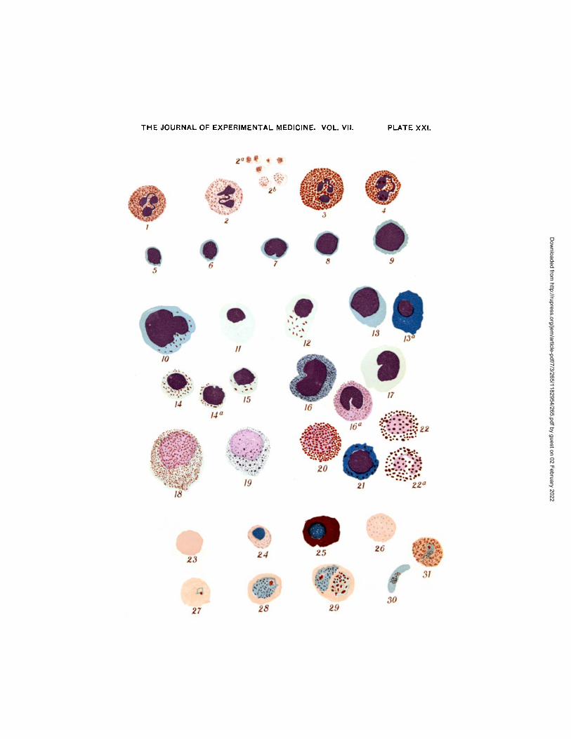
Polychromatophilia and granular basophilia are well shown. The "stippling" (T~pfelung) (Plate XXI, Fig. 31) of Schflffner (21) and Ruge (22) and Gold_horn (~3), appearing in many red cells invaded with tertian parasites, is of a color decidedly dif- ferent from that of the basic granules (Plate XXI, Fig. 26) of the anmmias and lead-poisoning; and the occurrence of both these changes in the red cells, but never together in the same red cell, in cases of malarial infection with well-marked secondary anmmia, differentiates them. The "stippling" varies somewhat with the reaction of the staining solution, increasing in intensity and in
Dow
nloaded from http://rupress.org/jem
/article-pdf/7/3/265/1182954/265.pdf by guest on 02 February 2022

272 Method for Preparing a Permanent Nocht's Stain
extent (i.e., extending to cells containing young as well as cells containing old parasites) with the increase in alkalinity of the stain. The unneutralized stains always bring out marked "stip- pling," the cells containing the parasites showing an irregular spiculated edge somewhat similar to certain crenated corpuscles, a fine cytoplasmic reticulum, and small nodal dots stained a deep crimson-red. The non-invaded cells, however, do not show "stip- pling" even with a stain sufficiently alkaline to interfere with fixation and staining. "Stippling" is evidently a specific cell- degeneration.
The occurrence of azurophile granules in the protoplasm of many of the mononuclear forms (small and large) is found in all specimens.
The staining of eosinophile granules is not so clear and bright as with other stains (eosin and methylene blue, or Jenner's) and may prove puzzling to one not familiar with the blood. The variety of colors cannot well be described, and an idea of them is best obtained from specimens, and from the drawings attached.
N U C L E A R S T A I N I N G .
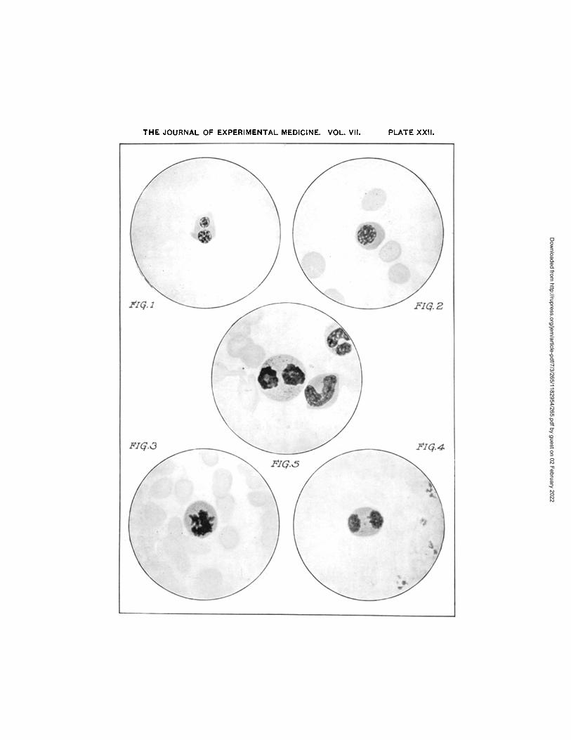
The nuclear material of white cells and of nucleated red cells takes a dark purple-plum color, that of the erythroblasts tending more toward dark-blue tints. More characteristic than the t in t is the clearly defined staining of the chromatin material, resulting in nuclear pictures excelled in clearness by good hema- toxylin staining only. The chromatin-thread staining is not well represented in the colored figures, but is easily seen in the photographs of megaloblasts (Plate XXII , Figs. I-4) and of the mitotic myelocyte (Plate XXII , Fig. 5)- Trachychromatic and amblychromatic nuclei are not well differentiated, and to us this does not seem a characteristic necessary to a good stain, since it is a point of differentiation allowing of extreme varia- tions due to personal equation, and is, therefore, one of small value in classifying cells. The clearness or unclearness of staining is, however, important, and Nocht's dyes bring out definitely clear-cut, deep-staining nuclei in the majority of
Dow
nloaded from http://rupress.org/jem
/article-pdf/7/3/265/1182954/265.pdf by guest on 02 February 2022
T. W. Hastings 273
leucocytes, and poorly stained nuclei not sharply outlined in a small number of cells--the former evidently healthy nuclear material, the latter examples of hydrops of the nucleus. Unless one bears this in mind and at the same time rids one's self of the importance of pale- and deep-staining nuclei in differentiating types of cells, the mononuclear forms will often be confusing and more rarely polymorphonuclear and mononuclear forms will be confused.
Definitely amblychromatic nuclei occur in mast-cells and myelocytes, and yet here the shape of nuclei, relation of nucleus to cell-body, and granulations are the important factors in deciding upon the type of cell.
These remarks in regard to nuclear staining are dwelt upon, for during the last two years while using modified Nocht's dyes and studying nuclei carefully we have not found myelocytes in the peripheral circulation nearly so frequently as with the older dyes (eosin and methylene blue, Ehrlich's triple stain, Jenner's stain).
The granular leucocytes, using the qualification granular in its original sense, exhibit three characteristic staining reactions, viz., of nucleus, of ground-substance of protoplasm, and of the granules. The ground-substance of the neutrophilic and eosino- philic cells takes a decided pink stain, and the granules decided red tints, that of the eosinophile being more red than that of the neutrophile. The mast-cells show an unstained, white ground- substance in which are scattered the densely stained purple granules. The transition forms (Plate XXI, Figs. i6 and x6a) show a blue-staining ground-substance,, deeper in tint than the blue of the large mononuclear forms and lighter than the blue of the lymphocyte protoplasm, with fine granules scattered over the blue background, these granules in some cells suggesting the "azur-granulation" and in other cells the neutrophilic granu- lation.
BASIC STAINING.
Nuclear staining has been briefly considered separately, for in the azur-stains the nuclear tints are probably due either to the metachromatic property of the stain or to a combination of
Dow
nloaded from http://rupress.org/jem
/article-pdf/7/3/265/1182954/265.pdf by guest on 02 February 2022

274 Method for Preparing a Permanent Nocht's Stain
methylen-azur and eosin staining, the eosin succeeding the methylen-azur in its reaction with the nuclear material, while true basic staining is found in the protoplasm of the lympho- cytes and in the mast-cell granules. In normal blood the lymphocyte (Plate XXI, Figs. 5-9) alone shows the deep blue pro- toplasm characteristic of the reaction of a simple basic blue dye; the mast-cell granules (Plate XXI, Figs. 22 and 22a) are meta- chromatic as well as definitely basic, varying in t int from a deep blue to a decided purple or even a deep red tint. The proto- plasm of the large mononuclear cells reacts faintly basic, the tint varying from the faintest blue to a decided light blue.
In pathologic blood, in addition to the normal lymphocyte, two types of cells with deeply staining basic cyanophilJc proto- plasm are found, the "stimulation-form" of Tiirk (Plate XXI , Figs. I3 and i3a ) and the "large lymphocyte" or lymphoid- marrow cell of acute " lymphat ic" leula~mia (Plate XXI, Fig. 2 i). All mydocytes (Plate XXI, Figs i8-2o) show a definitely basic blue ground-substance, and a few of them show basic granules similar to the mast-cell granulation.
N E U T R A L S TA I N I N G.
The granulation of finely granular polynuclears reacts with a less red t int than that seen in the eosinophiles--a red with a shade of blue, not the violet or lilac tint so characteristic of the other good neutral stains (Ehrlich's, Jenner's), so that the size and shape of the individual granule are more characteristic of this cell than the staining reaction. A similar color reaction is evident in some of the transition forms, the "transitional neutro- philes" of Cabot (Plate XXI , Fig. i6), which contain granules having no affmit F for an uncombined "azu r" dye, as "azur b lau" in methyl alcohol. The granules of the finely granular myelocytes react as do the granules of the polynuclears, while the myelocytic granulation is, as a rule, not so abundant as in the polynuclear cells.
AZUR S T A I N I N G .
The most characteristic property of the stain is the staining of certain granules in large mononuclear (Plate XXI, Figs. io, i i, I4,
Dow
nloaded from http://rupress.org/jem
/article-pdf/7/3/265/1182954/265.pdf by guest on 02 February 2022
T. W. Hastings 275
I 5), transitional (Plate XXI, Pig. i 6a), and some lymphocyte-like cells (Plate XXI, Fig. I4a), which with simple acid and basic dyes and neutral dyes (as Ehrlieh's and Jenner's) show no granula- tion in the protoplasm." The same reaction is found in certain granules in the protoplasm of the "large lymphocytes" of acute lymphatic leukmmia. Large mononuclear cells, with and without this "azur-granulation," occur, the former being not numerous. Such granulation may be considered due to the fragmentation and dissemination throughout the protoplasm of nuclear particles, or as due to degenerative changes in the proto- plasm, the latter view being that of Michaelis and Wolff (24), Tflrk (25) , and Ehrlich (25). This degenerative change occurs in large mononuclear forms and in lymphocytes, so that such a cell as I4a, Plate XXI, is an old or aged lymphocyte which stained in its younger hours as a typical lymphocyte (Plate XXI, Fig. 5)-
In many specimens of blood, however, we can readily find cells of the large mononuclear type, the protoplasm of which exhibits a constricting and budding process, resulting in the formation of a cell with a nucleus staining deeply and surrounded by a narrow zone of light-blue protoplasm containing few (from 3 to 6) or many (from 3o to 40) "azur granules." Certainly a large number of such cells are not aged lymphocytes but atypical large mononuclears. Similarly one finds not infrequently con- stricting portions of transition-forms (Plate XXI, Fig. x6a) simu- lating plates (Plate XXI, Fig. 2b) in size, but not typical in staining reaction, the resemblance being sufficiently close, how- ever, to be confusing. These false-plates seem to us to correspond to the " large" plates described by some observers.
STAINING OF PLATES.
Excepting the plate-like bodies derived from large mono- nuclear and transition forms, the plates (Plate XXI, Pig. 2a), particularly in specimens prepared with two per cent. solution of sodium metaphosphate, present a faintly pink outer ring or
Dow
nloaded from http://rupress.org/jem
/article-pdf/7/3/265/1182954/265.pdf by guest on 02 February 2022
276 Method for PreTaring a Permanent Nocht's Stain
zone surrounding a faintly blue inner portion in which there is a deep red rod-like retieulum. In most specimens the plates in masses present only the deep red reticular structure, but in these same specimens a few scattered' individual bodies will show the characteristic staining reaction of the plates in blood prepared with sodium metaphosphate.
CHROMATIN STAINING.
The chromatin material of the malarial parasite (Plate XXI, Figs. ~ 7-30) takes the deep ruby-red tint so characteristic of the original Romanowski stain. With Nocht's methods, however, the chromatin staining is much more certain than with the modifications of Romanowski's method (Wright's, Leish- man's).
In the younger phases of growth the chromatin stains deeply and clearly, while in the older, full-grown, crescentic and ovoid forms (i.e., in the gametocytes) the chromatin rods stain faintly red. The chromatin material in the nuclei of amoeba coil, of trypanosomes, and of the Leishman-Donovan body resembles that of the malarial parasite in staining reaction.
CLASSIFICATION OF W H ITE CELLS.
Study of blood, marrow, and lymphoid-tissue specimens with the older and then the "azu r" dyes impresses one with the correctness of the views of Ehrlich and Tflrk as opposed to the classification elaborated, or simplified, if one so chooses, by the school headed by Pappenheim and Grawitz.
Following Ehrlich and Tflrk, the "azur "-granular lymphocyte- like cell (Plate XXI, Fig. 14a) is to be classed with the lymphocytes, as an aged lymphocyte, although Michaelis and Wolff 'were not able to find this cell in lymph nodes. Many of these cells un- doubtedly are from the larger mononuclear forms, as mentioned under "azur staining"; and partly for this reason, and partly because variations of these cells in the peripheral circulation in
Dow
nloaded from http://rupress.org/jem
/article-pdf/7/3/265/1182954/265.pdf by guest on 02 February 2022

T. W. Hastings 277
disease may throw some light on their proper classification, we have during the last three years included these "azur" cells with large mononuclear forms in differential counting. In the early stages of variola, before the pustular eruption, this is the type of large mononuclear cells relatively and absolutely markedly increased in number; and likewise in chronic malarial infection, between paroxysms, and in the late weeks of typhoid fever it is increased.
The transition forms characterized with the older stains by the indented or lobed nucleus and by non-granular protoplasm are evidently of two types (Plate XXI, Figs. i5, i6a, i7), the non- granular (Fig. i7), and the granular, some of the latter having pale protoplasm and a definite "azur" granulation (Fig. i6a); others having a more deeply s£ained protoplasm with a fine ap- parently neutrophilic granulation ("transitional neutrophiles" of Cabot, "neutrophiles with lobed nuclei" of Tftrk) are similar to transition phases from the myelocyte to the polynuclear cell found in marrow. This last neutrophilic transition form one finds increased in the peripheral circulation in subsiding in- fections which produce a polynucleosis and in myelocytic leu- k~emia. The "azur granule" transitional cell, the supposed end stage of the large mononuclear, is rarely found increased in the peripheral circulation.
The "large lymphocyte" with azur granules (Plate XXI, Fig: i) of acute lymphatic leul~mia is a cell best classed by itself
until we know more of its nature and origin. The presence of definite nucleoli by Nocht's and other methods (methyl-green and pyronin) and the "azur" granules in the protoplasm are char- acteristic. So far we have encountered such a cell in infant's blood and in acute lymphatic lettkmmia. The large-sized lym- phocytes of chronic lymphatic leukmmia and of lymphocytosis contain no azur granules. This cell corresponds to Tfirk's lym- phoid-marrow cell, although Tftrk states he has never found "azur granules" in the protoplasm.
Unfortunately in the three cases of acute lymphatic leul~mia from which we have studied the blood post-mortem examination was not permitted.
Dow
nloaded from http://rupress.org/jem
/article-pdf/7/3/265/1182954/265.pdf by guest on 02 February 2022
278 Method for Preparing a Permanent Nocht's £'tain
The white ceils represented in Plate XXI are probably best classified as follows:
In normal blood: Polynuclear neutrophiles . . . . . . . . . . . . . . . . . . Figs. i , 2.
" eosinophiles . . . . . . . . . . . . . . . . . . Figs. 3, 4. Lymphoeytes . . . . . . . . . . . . . . . . . . . . . . . . . . . Figs. 5-9.
Small . . . . . . . . . . . . . . . . . . . . . . Figs. Large . . . . . . . . . . . . . . . . . . . . . . Figs.
Large mononuclears . . . . . . . . . . . . . . . . . . . . . . Figs. Transit ionals . . . . . . . . . . . . . . . . . . . . . . . . . . . . Figs. Mast-cells . . . . . . . . . . . . . . . . . . . . . . . . . . . . . . Figs.
In pathologic blood: Myelocytes . . . . . . . . . . . . . . . . . . . . . . . . . . . . . . Figs. I8-2o.
Neutrophil ic . . . . . . . . . . . . . . . . . . Fig. i8. Basophilic . . . . . . . . . . . . . . . . . . . . Fig. x 9. Eosinophilic . . . . . . . . . . . . . . . . . . . Fig. 2o.
Lymphoid-marrow cell . . . . . . . . . . . . . . . . . . . Fig. 2 L St imulat ion forms . . . . . . . . . . . . . . . . . . . . . . . Figs. I3, 13a.
5--7"
8- 9 . IO, I2, I 4 , i 4 a , 15.
I6, i r a , x 7. 22~ 2 2 a .
EXPLANATION OF PLATES.
PLATE XXI.
Figs. I-2. Polynuclear neutrophiles. Fig. 2a. Plates, Fig. 2b False-plates. Figs. 3-4. Polynuclear eosinophiles. Figs. 5-9. Lymphocy*es. Figs. io-x2, I4, I4a, 15. Large mononuclears.
Fig. x4a, "old , aged" lymphocyte of Michaelis and Wolff. Figs. x3, i3a. Figs. i6, I6a, i 7. Figs. i8-2o. Fig. 2x.
Figs. 22, 22a. Fig. 23 . Fig. 24. Fig. 25. Fig. 26. Figs. 27-3o. Fig. 3 i .
Figs. x-2. Figs. 3-4. Fig. 5.
St imulat ion form of Tfirk. Transit ion-forms. Myelocytes. " L a r g e l ymphocy te " of acute lymphat ic lettkmmia
(T~rk 's lymphoid-marrow cell). Mast-cells. Normal red cell. Normoblast wi th basic granules in protoplasm. Megaloblast. Granular basophilia in red cell. Malaria parasites. Stippling in red cell invaded by te r t ian parasite.
PLATE XXII .
Megaloblasts showing vesicular nuclei ( X rooo). Mitotic figures in megaloblasts ( X iooo). Mitotic figure in neutrophilic myelocyte ( X Iooo).
Dow
nloaded from http://rupress.org/jem
/article-pdf/7/3/265/1182954/265.pdf by guest on 02 February 2022
THE JOURNAL OF EXPERIMENTAL MEDICINE. VOL. VII. PLATEXXI .
Dow
nloaded from http://rupress.org/jem
/article-pdf/7/3/265/1182954/265.pdf by guest on 02 February 2022
THE JOURNAL OF EXPERIMENTAL MEDICINE. VOL. Vll. PLATE XXII.
Dow
nloaded from http://rupress.org/jem
/article-pdf/7/3/265/1182954/265.pdf by guest on 02 February 2022
T. W . H a s t i n g s 279
B I B L I O G R A P H Y .
x. Nocht---Cent. f. Bakt., i899 ' xxv , Abt. i, 754; Cent. ~. Bakt., x899 , xxv , Abt . i, x 7.
2. Jenner--Lancet, x899 , i, 37 o. 3- Goldhorn- -New York Univ. Bull. o~ Med. Sciences, x9ox , i, 59. 4. Lazier--Johns Hopkins Hospital Reports, i9ox , x, 2. 5. Ewing--Jour. of Exper. Med., x90x, v, 433. 6. Lelshman--Brit. Med. ]our., x9ox, ii, 757- 7- May and Gr~nwald--Cent . f. innere Med., x9o2 , xxiii, 268. 8. Michaelis--Deutsche reed. Woeh., i899 , 49 o. 9. Reu te r - -M~nch . reed. Woch., x9ox, x26x; Cent. ~. Bakt., x9ox, xxx, Abt . i,
254. xo. Willebrand--Deutsche reed. Woch., xgox, xxvSi, 57. xI. Wright--Jour. of Med. Research, i9o2, vii , x38. i2. Schegoleff--Medidnskoie Obozreine, x9o2, lvil; Ref. Philadelphia. Med.
Jour, x9o3 , ix, 897. x 3. Nocht---Cent. f. Bakt., x898, xxiv, Abt. i, 839. x4. Nocht---Cent. f. Bakt., x899 , xxv, Abt . i, 764. x 5. Romanowski - -S t . Petersburger medizinische Wochenschri~t, x89x , viii, 297 ,
307 • x6. Ziemann--Ueber Malaria und andere Btutparasiten, x898, ~46, x64. 17. Michaelis---Cent. f. Bakt., x9ox, xxix, Abt . i, 763; Einf~hrung in dia
Farbsto]~chemie, Berlin, x9o2, 43. x8. Nocht---Cent. f. Bakt., Abt. i, x899, xxvl , I7. x9. Nocht--Enzyklopddie der mikroskopischen Technik, x9o3, par t ii, p. 784. 2o. Ziemann---Cent. f. Bakt., i898, xxiv, Abt. i, 947- 2x. Schfaffner--Deutsches Arch. f. klin. Med., x899, lxiv, 428, 22. R u g e - - Z d t . ~. Hygiene ~nd Infectionkrankheiten, x9oo, xxxiii , z78. 23. Goldhorn- -Loc . cit. 24. Michaeiis and Wolff--Virchow's Archives, x9o2, clxvli, x5I. 25. Tftrk--Vorles~ngen ~ber klinische Hdmatolog~e, i9o4, 39 o. 26. E h r l i c h - - N o t e at end of article by Michaeiis and Wolff (~4).
Dow
nloaded from http://rupress.org/jem
/article-pdf/7/3/265/1182954/265.pdf by guest on 02 February 2022