8th BALKAN CONGRESS OF NUCLEAR MEDICINE
48
8 th BALKAN CONGRESS OF NUCLEAR MEDICINE POSTER PROCEEDINGS
-
Upload
khangminh22 -
Category
Documents
-
view
0 -
download
0
Transcript of 8th BALKAN CONGRESS OF NUCLEAR MEDICINE
8th BALKAN CONGRESS OF NUCLEAR MEDICINE POSTER PROCEEDINGS

31
Nuclear Medicine Seminars 2019;1:(Suppl 1):1-778th BALKAN CONGRESS OF NUCLEAR MEDICINE
[BPP-01]
Comparison of TNM Staging and Histopathological Differentiation with Metabolic Parameters of PET in Laryngeal Cancer
Gül Çekin1, Hale Aslan2, Gülhan Kaya Altuncı1, Emine Acar1, Mustafa Yazır2, Fulya Çakalağaoğlu3
1İzmir Katip Çelebi University Atatürk Training and Research Hospital, Clinic of Nuclear Medicine, İzmir2İzmir Katip Çelebi University Atatürk Training and Research Hospital, Clinic of Head And Neck Surgery, İzmir 3İzmir Katip Çelebi University Atatürk Training and Research Hospital, Clinic of Pathology, İzmir
Aim: The purpose of our retrospective study was to compare TNM staging and degree of histopathological differentiation with metabolic and volumetric parameters of pretreatment F-18 FDG positron emission tomography (PET)/ computerized tomography (CT) in laryngeal cancer.
Method: We reviewed the records of 27 male patients (age range, 47-89 y) who were previously diagnosed histopathologically with laryngeal squamous cell carcinoma, then underwent pretreatment PET/CT imaging. Age, gender, histopathological diagnosis, stage of the patients included in the study were recorded. Standardized uptake values (SUVmax and SUVmean), metabolic tumor volume (MTV) and total lesion glycolysis (TLG) of primary tumor and neck lymph nodes were evaluated for each patient. Then the metabolic parameters of PET imaging were compared with degree of histopathological differentiation and clinical TNM staging. Data were evaluated in SPSS 22.0 program. Mann-Whitney U test was used for pairwise comparisons, Kruskal-Wallis test was used for multiple comparisons, and pairwaise Kruskal-Wallis test was used in post-hoc analysis.
Results: The mean TLG, MTV, SUVmax and SUVmean values for primary tumor were 124.93±112.24 g, 11.96±9.23 cm3, 16.60±6.74 g/mL, 10.24±4.18 g/mL, respectively. The same measurements for lymph nodes were15.33±30.44 g, 2.04±2.02 cm3, 7.24±5.19 g/mL, 4.76±3.31 g/mL, respectively.MTV range and the mean values of TLG, SUVmax and SUVmean for all metastatic lymph nodes were 0.6-9.5 cm3 and 22.05±12.02 g, 9.64±1.79 g/mL, 6.24±1.15 g/mL, respectively. When we compared the T and N stage with the metabolic and volumetric parameters of the primary tumor and lymph node, we found statistically significant differences between the T stage and MTV of primary tumor (p=0.031) and between the N stage and TLG of primary tumor (p=0.042). In post-hoc analyses TLG of primary tumor is higher in N2 patients compared to N0 patients (p=0.026). In T4 stage, MTV values significantly higher than T1 and T2 stage (p=0.037, p=0.017, respectively). When the reactive and metastatic lymph nodes were compared, there was a statistically significant difference between SUVmax and SUVmean values in lymph nodes (p=0.016, p=0.010, respectively)
Conclusion: MTV values were significantly higher in T4 tumors. SUVmax and SUVmean values of metastatic lymph nodes are significantly higher than reactive lymph nodes. As a result, F-18 FDG PET/CT imaging, which helps the clinicians in staging and treatment planning is a valuable tool in detection of laryngeal cancer.
Keywords: TLG, MTV, SUVmax, SUVmean, laryngeal cancer
Figure 1.
Table 1. Patient characteristicsCharacteristic Value
Median age (years) (range) 62.81 (47-89)
Sex, male/female 27/0
Tumor Stage, T1/T2/T3/T4, n (%) 1/7/6/13, (3.7, 25.9, 22.2, 48.1)
Nodal Stage N0/N1/N2, n (%) 13/11/3, (48.1, 40.7, 11.1)
Overall Stage I/II/III/IV n (%) 0/7/5/15 (0, 25.9, 18.5, 55.5)
Histopathological differentiation, well, moderately/poorly, n (%)
5/8/14, (18.5, 29.6, 51.8)
Table 2. Metabolic and volumetric parameters of T stages of primary tumor
T1 stage
T2 stage
T3 stage
T4 stage
MTV (cm3) (median) 1.22 8.17 9.90 22.97
TLG (g) (median) 8.4 58.54 158.32 156.30
SUVmax (g/mL) (median) 11.49 22.97 27.42 16.42
SUVmean (g/mL) (median) 6.95 8.20 15.99 10.12
MTV: Metabolic tumor volüme, TLG: Total lesion glycolysis, SUV: Standardized uptake values

32
Nuclear Medicine Seminars 2019;1:(Suppl 1):1-77 8th BALKAN CONGRESS OF NUCLEAR MEDICINE
[BPP-02]
Relation Between Metabolic Parameters in F-18 FDG PET/CT and Hematological Markers in Non-Small Cell Lung Cancer
Sibel Göksel1, Arzu Cengiz2, Hakan Öztürk3, Yakup Yürekli2
1Recep Tayyip Erdoğan University Faculty of Medicine Training and Research Hospital, Clinic of Nuclear Medicine, Rize 2Adnan Menderes University Faculty of Medicine, Department of Nuclear Medicine, Aydın 3Adnan Menderes University Faculty of Medicine, Department of Biostatistics, Aydın
Aim: Various inflammatory markers such as elevated neutrophil-to-lymphocyte ratio (NLR), platelet-to-lymphocyte ratio (PLR) and neutrophil count and low mean platelet volume (MPV) have been shown to be with poor prognosis in malignancies. Metabolic parameters in F-18 FDG positron emission tomography (PET)/computerized tomography (CT) such as metabolic tumor volume (MTV) and total lesion glycolysis (TLG) have also been reported to be a prognostic marker in various cancers. The aim of this study is to evaluate the correlation between hematological markers and metabolic F-18 FDG PET/CT parameters in patients with non-small cell lung cancer (NSCLC).
Method: A total of 132 patients (122 male, 10 female) (mean age 69) with NSCLC who underwent PET/CT for initial staging were evaluated retrospectively. Patients with hematological disease, infections, chemo/radiotheraphy or surgery history were excluded. Hematological parameters were measured before two weeks of the PET/CT. Correlation between these parameters was evaluated with Spearman correlation test. The prognostic significances of variables for overall survival (OS) were assessed by uni/multivariate analyses using Cox-regression model. P<0.05 was considered statistically significant.
Results: There was statistically significant positive correlation between neutrophil (r=0.578, p<0.001), NLR (r=0.524, p<0.001), PLR (r=0.445, p<0.001) and MTV. While there was found positive correlation between neutrophil (r=0.593, p<0.001), NLR (r=0.540, p<0.001), PLR (r=0.460, p<0.001) and TLG, MPV was negatively correlated with TLG. SUVmax of primary tumor was not correlated with hematological parameters. While TLG, MTV, NLR and PLR were increased in advanced stages, MPV was decreased. Median follow-up time was 9.8 months (0.5-68 months). Univariate Cox-regression analysis demonstrated that greater age (p<0.05), high neutrophil (p<0.001), NLR (p<0.001), PLR (p<0.001), MTV (p<0.05), TLG (p<0.05), low MPV (p<0.05) values and advenced stage (p<0.001) were significant predictors of poor prognosis. Multivariate Cox-regression analysis revealed that NLR (HR: 2.67; 95% CI, 1.74-4.08; p<0.001) and advenced stage disease (HR: 7.77; 95% CI, 2.61-23.07; p<0.001) were significant independent predictors of poor prognosis.
Conclusion: Prognostic hematological markers are significantly correlated with metabolic PET/CT parameters in patients with NSCLC. These parameters can contribute to prognostic evaluation of NSCLC patients in initial staging.
Keywords: MTV, TLG, hematological markers, lung cancer, F-18 FDG PET/CT
[BPP-03]
Can PET/CT Be Used As a First-Line Imaging Modality for Patients with Proven Metastasis to Detect Primary Tumor Origin?
Seyhan Karaçavuş, Güler Silov, İsmail Çiftçi, Özgül Turhal, Mehtap Başbuğ, Zeynep Erdoğan, Ayşegül Özdal, Hümeyra Gençer
University of Health Sciences, Kayseri City Hospital, Clinic of Nuclear Medicine, Kayseri
Aim: To investigate the clinical impact of whole body positron emission tomography positron emission tomography (PET)/ computerized tomography (CT) scan with F-18-fluorodeoxyglucose (FDG) in the detection of the occult primary tumor in patients with proven metastatic cancer from unknown primary origin.
Method: Ninety consecutive patients with occult primary tumor (37 visceral biopsy, 53 lymph nodes proven tumor metastasis) were referred for F-18 FDG PET/CT whole body scan. PET/CT images were interpreted by visual inspection and semi-quantitative analysis (standardized uptake value, SUV). Histopathological and clinical follow-up findings were used to assess the diagnostic value of F-18 FDG PET/CT scan.
Results: F-18 FDG PET/CT scan was able to detect the primary tumor in 64/90 patients (71%). The primary tumors were confirmed by histopathologic and clinical follow-up findings, and located in the head and neck (n=7), the lung (n=15), the breast (n=2), the esophagus (n=3), the stomach (n=6), the bile ducts (n=2), the pancreas (n=8), the colon (n=7), the ovary (n=2), the prostate (n=5), lymphoma (n=7). F-18 FDG PET/CT scan results were proved false positive in 11 patients (12%), which were located in the head and neck (n=2), the lung (n=3), the gastric (n=1), the colon (n=2), the ovary (n=1), the prostate (n=2). During the clinical follow-up of median 4 months, primary tumor was found in only 5 patients of 26 cases detected by other diagnostic methods unidentified by F-18 FDG PET/CT scan (pancreas cancer, gastric cancer, colon cancer, prostate cancer and urinary tumor). In 21 patients (21/90, 23%), the primary tumor site was not localized. However, in 9 of them, F-18 FDG PET/CT scan identified further unexpected metastases, modifying the stage of disease. Overall, the following oncological treatment was influenced by the F-18 FDG PET/CT whole body imaging, in a total of 75 patients (75/90, 83%).
Conclusion: F-18 FDG PET/CT whole body scan is both a quitely sensitive and a non-invasive tomographic whole-body imaging modality, allowing for the detection of a primary tumor and complete tumor staging and selecting appropriate therapeutic methods in single examination. It could be prefered as a first-line imaging modality for patients with proven carcinoma metastases of unknown primary origin rather than using it after other diagnostic procedures have failed to detect a primary tumor.
Keywords: Positron emission tomography, cancer, unknown primary origin

33
Nuclear Medicine Seminars 2019;1:(Suppl 1):1-778th BALKAN CONGRESS OF NUCLEAR MEDICINE
[BPP-04]
Clinical Conditions Where PET/CT is Superior Than Ca 15.3 to Determine Progression in Metastatic Breast Cancer
Emine Ebru Bayar1, Neslihan Çetin Avcı2, Emine Özlem Gür3, Gonca Gül Bural4
1İzmir Katip Çelebi University Atatürk Training and Research Hospital, Clinic of Nuclear Medicine, İzmir 2Ümraniye Training and Research Hospital, Clinic of Nuclear Medicine, İstanbul 3İzmir Katip Çelebi University Atatürk Training and Research Hospital, Clinic of General Surgery, İzmir 4Akdeniz University Faculty of Medicine, Clinic of Nuclear Medicine, Antalya
Aim: Our aim was to determine the subjects in which positron emission tomography (PET)/ computerized tomography (CT) was successful; but Ca 15.3 was inefficient for the detection of progressive metastatic breast cancer.
Method: Progression decision on FDG PET/CT images were based on European Organization for Research and Treatment of Cancer (EORTC) criteria: increase 25% in SUVmax and/or presence of new lesion. Ca 15.3 levels were also noted and compared in all subjects before and after chemotherapy.
Metastatic lesions were confirmed with biopsy and with other radiological imaging modalities and/or clinical follow-up. Metastatic lesions were grouped in to two different groups. First group involved patients with visceral metastases (lung, liver, pleural, peritoneal) and others (bone and lymph node metastases). Increase in Ca 15.3 levels were compared in subject with and without visceral metastases using chi-square test.
Results: There was increase in only 22 subjects in Ca 15.3 levels out of 46, whom PET/CT showed progression. Ca 15.3 was insufficient in showing progressive metastatic disease in 52% (24/46) of patients. Twenty-two of the subjects with increase in Ca 15.3 levels 16 had visceral metastases (73%) and 6 had non-visceral metastases (27%). In 24 subjects who had progression on PET/CT scan; but had normal Ca 15.3 levels; has visceral metastases (37%) and 15 had nonvisceral metastases (63%). In subjects with visceral metastases increased in Ca 15.3 levels were higher than subjects with non-visceral metastases with statistical significance (p=0.02).
Conclusion: PET/CT is more efficient in detecting progressive disease in subjects with metastatic breast cancer. Ca 15.3 levels are especially insufficient for the detection of progression in non-visceral metastases compared to visceral metastases. There for breast cancer in subject with nonvisceral metastases PET/CT is superior than Ca 15.3 levels for the detection of progressive disease.
Keywords: Breast cancer, Ca 15.3, FDG PET/CT
[BPP-05]
The Role of Radiomics: Predicting the Treatment Response to Neoadjuvant Therapy in Patients with LARC
Nazlı Pınar Karahan Şen, Ayşegül Aksu, Gamze Çapa Kaya
Dokuz Eylül University Faculty of Medicine, Department of Nuclear Medicine, İzmir
Aim: The aim of this study was to develop a current model for evaluating the response to neoadjuvant chemo-radiotherapy (CRT) in patients with Locally Advanced Rectal Cancer (LARC).
Method: The medical records of all the patients with initial staging FDG positron emission tomography (PET)/computerized tomography (CT) and who had diagnosed LARC between January 2009 and March 2016 at our institution were retrospectively evaluated. In total, 64 patients who had neoadjuvant chemo-radiotherapy and surgical resection were included
in this study. We divided patients into two groups according to tumor regression grade as responders (RG) and non-responders (NRG). In addition, the status of metastatic lymph nodes in PET/CT images at the time of staging was investigated. The images were evaluated in the LIFEx program. Region of interest (ROI) for the primary tumors was drawn and the texture features were calculated. A significant relationship between these features and TRG was investigated with Mann-Whitney U test. Receiver operating curve (ROC) analysis was performed for features with p<0.05. The features which had the highest Area Under Curve (AUC) was evaluated with the logistic regression method.
Results: Twenty-six patients were RG and 38 were in NRG group. The rate of lymph node detection at baseline PET/CT was 34.6% in RG group and 63.2% in NRG group (p=0.025). Texture analysis results demonstrated highest AUC values in GLRLM_GLNU, GLZLM_LZHGE, GLZLM_LZE, GLZLM_LRE, shape volume, shape compacity in PET and GLRLM_RLNU, GLZLM_GLNU, shape volume in CT. When the 5 features that had the most highest AUC values and the lymph node status in PET images (LN+) were evaluated together in the logistic regression analysis; the model of GLRLM_GLNU and LN+ was the most significant one with AUC 0.740. The sensitivity and specificity were calculated as 65.8 and 84.6, respectively.
Conclusion: Recently, treatment practices for individuals have become increasingly important. In this study, it was thought that the evaluation of PET/CT images before neoadjuvant CRT in LARC patients by using the texture analysis method might be useful in predicting the response to treatment. The role of radiomics in predicting treatment response should be supported by studies involving large patient groups.
Keywords: Texture analysis, FDG PET/CT, rectum cancer
[BPP-06]
Is There Any Improvement in Clinical Staging with FDG PET/CT Compared to Surgical Staging in Cases of Lung Cancer?
Gonca Kara Gedik, Farise Yılmaz
Selçuk University Faculty of Medicine, Department of Nuclear Medicine, Konya
Aim: F-18 fluorine fluorodeoxyglucose positron emission tomography/computed tomography (FDG PET/CT) imaging is considered standard for non-small cell lung carcinoma (NSCLC). The aim of this study was to compare clinical staging (cTNM) performed with FDG PET/CT and surgical staging (sTNM) in patients with NSCLC treated with surgery, using 8th edition of TNM staging system.
Method: We performed a retrospective analysis of 99 surgical patients with NSCLC who underwent FDG PET/CT examination. Semiquantitative measures of SUVmax, SUVpeak, metabolic tumor volume (MTV) and total lesion glycolysis (TLG) were calculated from the primary lesions and mediastinal lymph nodes. Findings of cTNM were compared with final pathologic evaluation. Subjects were divided into two groups as postsurgical cTNM changed and cTNM unchanged. Patients in cTNM changed group were further classified as postsurgical upstaged (US) and downstaged (DS). Semiquantitative parameters of US patients were compared with the results of remaining patients of cTNM unchanged and DS. Cut-off values were obtained from calculated semiquantitative results of FDG uptakes in lymph nodes for determining tumoral involvement. P value <0.05 was considered as statistically significant.
Results: Subjects were aged 40 to 82 years (mean: 64.78±8.70 years). Eighty-three of them were male, 16 were female. Classification agreement was observed in 43 patients (43%), in 57%, stage migration was seen.Diagnostic accuracy of cT staging was higher than cN staging (72% vs 62%).
34
Nuclear Medicine Seminars 2019;1:(Suppl 1):1-77 8th BALKAN CONGRESS OF NUCLEAR MEDICINE
Concurrence of cTNM and sTNM was more pronounced in T1 and N0 subsets which were 84% and 74%, respectively. Change in T staging occurred in 20 of 56 (36%), in N staging 22 of 56 (39%) and change in T and N in 14 patients (25%). Distribution of US and DS patients in cTNM changed group were, 43% (24 of 56) and 57% (32 of 56), respectively. Results of semiquantitative measures were significantly higher in US patients than the results of the remaining patients for all parameters. Most specific cut-off value was found as MTV with an acceptable sensitivity (90% and 67%, respectively) for mediastinal involvement.
Conclusion: The concordance between cTNM and sTNM is better in staging T category compared to N stations. Semiquantitavive measures of primary tumor may play role in predicting postsurgical upstaging. Taken MTV into consideration in mediastinal region may be more valuable than other parameters in the assessment of nodal involvement.
Keywords: Clinical, FDG PET/CT, lung carcinoma, quantification, staging
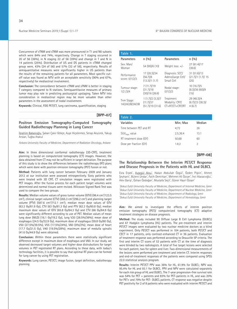
[BPP-07]
Positron Emission Tomography-Computed Tomography Guided Radiotherapy Plannıng in Lung Cancer
İbrahim Babalıoğlu, Şaban Çakır Gökçe, Ayşe Hiçsönmez, Serap Akyürek, Yakup Arslan, Tuğba Atakul
Ankara University Faculty of Medicine, Department of Radiation Oncology, Ankara
Aim: In three dimensional conformal radiotherapy (3D-CRT), treatment planning is based on computerized tomography (CT) images. However, the data obtained from CT may not be sufficient in target delination. The purpose of this study is to show the differences between the radiotherapy (RT) plans which were done with positron emission tomography (PET) fusion or not.
Method: Patients with lung cancer between February 2009 and January 2012 at our institution were assessed retrospectively. Sixty patients who were treated with 3D CRT, CT simulation images were registrated with PET images. After the fusion process for each patient target volumes were determined and normal tissues were revised. Wilcoxon Signed Rank Test was used to compare the two groups.
Results: Median volume values of gross tumor volume (GTV) (96.4 cm3/123.5 cm3), clinical target volume (CTV) (265.3 cm3/296.2 cm3) and planning target volume (PTV) (587.6 cm3/712.1 cm3); median mean dose values of GTV (63.2 Gy/61.9 Gy), CTV (63 Gy/61.3 Gy) and PTV (62.2 Gy/60.8 Gy); median maximum dose values of GTV (65.8 Gy/64.3 Gy) and CTV (66 Gy/64.9 Gy) were significantly different according to use of PET. Median values of mean lung dose (MLD) (16.1 Gy/19.2 Gy), lung V20 (28.5%/36%); mean dose of esophagus (24.5 Gy/32.9 Gy), maximum dose of esophagus (59.8 Gy/60 Gy), V50 (27.5%/39.5%), V55 (17.5%/24%), V60 (0%/2%); mean dose of heart (17.7 Gy/21.5 Gy), V40 (19.5%/28%); maximum dose of medulla spinalis (41.6 Gy/44.9 Gy) were obtained.
Conclusion: Within these parameters there were statistically significant difference except in maximum dose of esophagus and V60. In our study, we observed decreased target volumes and higher dose distrubutions for target volumes in PET registrated RT plans. According to these data, with today’s technology facilities, it is possible to say that optimal RT plans can be formed for lung cancer by using PET registration.
Keywords: Lung cancer, PET/CT, image fusion, target definition, radiotherapy planning
Table 1.Parameters n (%) Parameters n (%)
Sex: Man/Woman
54 (90)/6 (10) Weight loss: +/-27 (61.4)/17 (38.6)
Performance score: 0/1/2/3
17 (28.3)/34 (56.7)/8 (13.3)/1 (1.7)
Diagnosis: SCC/Adeno/Large Cell/Small Cell
31 (51.6)/13 (21.7)/1 (1.7)/ 15 (25)
Tumour stage: 1/2 /3/4
7 (11.7)/19 (31.7)/18 (30)/16 (26.6)
Nodal stage: 0/1/2/3
10 (16.7)/5 (8.3)/36 (60)/9 (15)
Tnm Stage: 1A/2A/2B/3A/3B
1 (1.7)/2 (3.3)/7 (11.7)/37 (51.7)/19 (31.6)
Treatment Modality: CRT/CT+RT/CT+CRT/RT
29 (48.3)/4 (6.7)/23 (38.3)/ 4 (6.7)
Table 2.Variables Min; Max Median
Time between PET and RT 4;72 26
SUVmax value 2.5;36.4 13.1
RT treatment dose (GY) 50;68 60
Dose per fraction (GY) 1.8;2 2
[BPP-08]
The Relationship Between the Interim PET/CT Response and Disease Prognosis in the Patients with HL and DLBCL
Esra Erpek1, Ayşegül Aksu2, Hakan Abdullah Özgül3, Özden Pişkin4, Ahmet Şeyhanlı4, Bülent Ündar4, Fatih Demirkan4, Mehmet Ali Özcan4, İnci Alacacıoğlu4, Alev Garip4, Özhan Özdoğan2, Mustafa Seçil3, Güner Hayri Özsan4
1Dokuz Eylül University Faculty of Medicine, Department of Internal Medicine, İzmir 2Dokuz Eylül University Faculty of Medicine, Department of Nuclear Medicine, İzmir 3Dokuz Eylül University Faculty of Medicine, Department of Radiology, İzmir 4Dokuz Eylül University Faculty of Medicine, Department of Hematology, İzmir
Aim: We aimed to investigate the effects of interim positron emission tomography (PET)/ computerized tomography (CT) adaptive treatment strategies on disease prognosis.
Method: The study included 90 Diffuse Large B Cell Lymphoma (DLBCL) and 67 Hodgkin Lymphoma (HL) patients. The pre-treatment and interim PET/CT images were evaluated by two nuclear medicine doctors as a blind experiment. Only PET/CT was performed in 104 patients, both PET/CT and CECT in 17 patients, only contrast-enhanced CT in 36 patients. Evaluation of treatment response was performed according to Deauville 5P criteria. The first and interim CT scans of 53 patients with CT at the time of diagnosis were blinded by two radiologists. A total of five target lesions were selected for each patient, two for spleen and liver. Two-dimensional measurements of the lesions were performed pre-treatment and interim CT. Interim responses and end-of-treatment responses of the patients were compared using SPSS 22.0 statistical analysis program.
Results: Interim PET/CT PPV was 36% for HL, 61.9% for DLBCL; NPV was 95.4% for HL and 93.1 for DLBCL. PPV and NPV were calculated separately for each risk group of HL and DLBCL. The 7-year progression-free survival rate was 54% for PET + patients and 83% for PET-patients in HL, and was 20% for PET+ and 79% for PET- DLBCL patients. CT response was negative despite PET positivity for 2 of 8 patients who were evaluated with interim PET/CT and
35
Nuclear Medicine Seminars 2019;1:(Suppl 1):1-778th BALKAN CONGRESS OF NUCLEAR MEDICINE
contrast-enhanced CT in the DLBCL group. CT response was positive despite PET negativity for 3 of 9 patients who were evaluated with interim PET/CT and contrast-enhanced CT in the HL group. In the group of 157 patients, interim Lugano PET/CT-related response and interim Lugano and RECIST 1.1 CT-related responses were found to be negligible in the kappa analysis. PET/CT-related response was more compatible with treatment response than CT-related response.
Conclusion: PET/CT prevented some patients to receive unnecessary and intensive treatments and some patients to have inadequate treatment and improved the evaluation of lymphoma treatment response significantly. The use of the Deauville criteria standardized the comparability between the images. PET/CT has high NPV and low-medium PPV in both HL/DLBCL groups and in individual risk groups. PET negativity is a prognostic indicator with a high specificity for progression-free survival. Treatment intensification based only on interim PET positivity may cause patients to be exposed to unnecessary drug toxicity.
Keywords: Lymphoma, interim evaluation, PET/CT
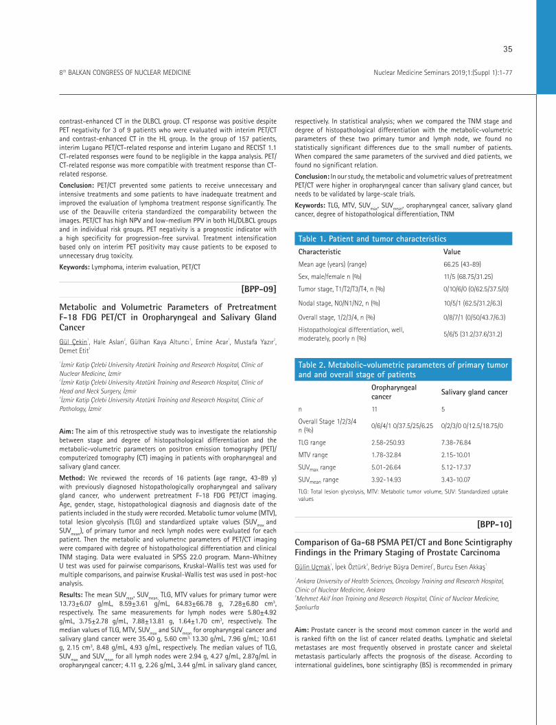
[BPP-09]
Metabolic and Volumetric Parameters of Pretreatment F-18 FDG PET/CT in Oropharyngeal and Salivary Gland Cancer
Gül Çekin1, Hale Aslan2, Gülhan Kaya Altuncı1, Emine Acar1, Mustafa Yazır2, Demet Etit3
1İzmir Katip Çelebi University Atatürk Training and Research Hospital, Clinic of Nuclear Medicine, İzmir 2İzmir Katip Çelebi University Atatürk Training and Research Hospital, Clinic of Head and Neck Surgery, İzmir 3İzmir Katip Çelebi University Atatürk Training and Research Hospital, Clinic of Pathology, İzmir
Aim: The aim of this retrospective study was to investigate the relationship between stage and degree of histopathological differentiation and the metabolic-volumetric parameters on positron emission tomography (PET)/computerized tomography (CT) imaging in patients with oropharyngeal and salivary gland cancer.
Method: We reviewed the records of 16 patients (age range, 43-89 y) with previously diagnosed histopathologically oropharyngeal and salivary gland cancer, who underwent pretreatment F-18 FDG PET/CT imaging. Age, gender, stage, histopathological diagnosis and diagnosis date of the patients included in the study were recorded. Metabolic tumor volume (MTV), total lesion glycolysis (TLG) and standardized uptake values (SUVmax and SUVmean), of primary tumor and neck lymph nodes were evaluated for each patient. Then the metabolic and volumetrıc parameters of PET/CT imaging were compared with degree of histopathological differentiation and clinical TNM staging. Data were evaluated in SPSS 22.0 program. Mann-Whitney U test was used for pairwise comparisons, Kruskal-Wallis test was used for multiple comparisons, and pairwise Kruskal-Wallis test was used in post-hoc analysis.
Results: The mean SUVmax, SUVmean, TLG, MTV values for primary tumor were 13.73±6.07 g/mL, 8.59±3.61 g/mL, 64.83±66.78 g, 7.28±6.80 cm3, respectively. The same measurements for lymph nodes were 5.80±4.92 g/mL, 3.75±2.78 g/mL, 7.88±13.81 g, 1.64±1.70 cm3, respectively. The median values of TLG, MTV, SUVmax and SUVmean for oropharyngeal cancer and salivary gland cancer were 35.40 g, 5.60 cm3, 13.30 g/mL, 7.96 g/mL; 10.61 g, 2.15 cm3, 8.48 g/mL, 4.93 g/mL, respectively. The median values of TLG, SUVmax and SUVmean for all lymph nodes were 2.94 g, 4.27 g/mL, 2.87g/mL in oropharyngeal cancer; 4.11 g, 2.26 g/mL, 3.44 g/mL in salivary gland cancer,
respectively. In statistical analysis; when we compared the TNM stage and degree of histopathological differentiation with the metabolic-volumetric parameters of these two primary tumor and lymph node, we found no statistically significant differences due to the small number of patients. When compared the same parameters of the survived and died patients, we found no significant relation.
Conclusion: In our study, the metabolic and volumetric values of pretreatment PET/CT were higher in oropharyngeal cancer than salivary gland cancer, but needs to be validated by large-scale trials.
Keywords: TLG, MTV, SUVmax, SUVmean, oropharyngeal cancer, salivary gland cancer, degree of histopathological differentiation, TNM
Table 1. Patient and tumor characteristicsCharacteristic Value
Mean age (years) (range) 66.25 (43-89)
Sex, male/female n (%) 11/5 (68.75/31.25)
Tumor stage, T1/T2/T3/T4, n (%) 0/10/6/0 (0/62.5/37.5/0)
Nodal stage, N0/N1/N2, n (%) 10/5/1 (62.5/31.2/6.3)
Overall stage, 1/2/3/4, n (%) 0/8/7/1 (0/50/43.7/6.3)
Histopathological differentiation, well, moderately, poorly n (%)
5/6/5 (31.2/37.6/31.2)
Table 2. Metabolic-volumetric parameters of primary tumor and and overall stage of patients
Oropharyngeal cancer Salivary gland cancer
n 11 5
Overall Stage 1/2/3/4 n (%)
0/6/4/1 0/37.5/25/6.25 0/2/3/0 0/12.5/18.75/0
TLG range 2.58-250.93 7.38-76.84
MTV range 1.78-32.84 2.15-10.01
SUVmax range 5.01-26.64 5.12-17.37
SUVmean range 3.92-14.93 3.43-10.07
TLG: Total lesion glycolysis, MTV: Metabolic tumor volume, SUV: Standardized uptake values
[BPP-10]
Comparison of Ga-68 PSMA PET/CT and Bone Scintigraphy Findings in the Primary Staging of Prostate Carcinoma
Gülin Uçmak1, İpek Öztürk2, Bedriye Büşra Demirel1, Burcu Esen Akkaş1
1Ankara University of Health Sciences, Oncology Training and Research Hospital, Clinic of Nuclear Medicine, Ankara 2Mehmet Akif İnan Training and Research Hospital, Clinic of Nuclear Medicine, Şanlıurfa
Aim: Prostate cancer is the second most common cancer in the world and is ranked fifth on the list of cancer related deaths. Lymphatic and skeletal metastases are most frequently observed in prostate cancer and skeletal metastasis particularly affects the prognosis of the disease. According to international guidelines, bone scintigraphy (BS) is recommended in primary
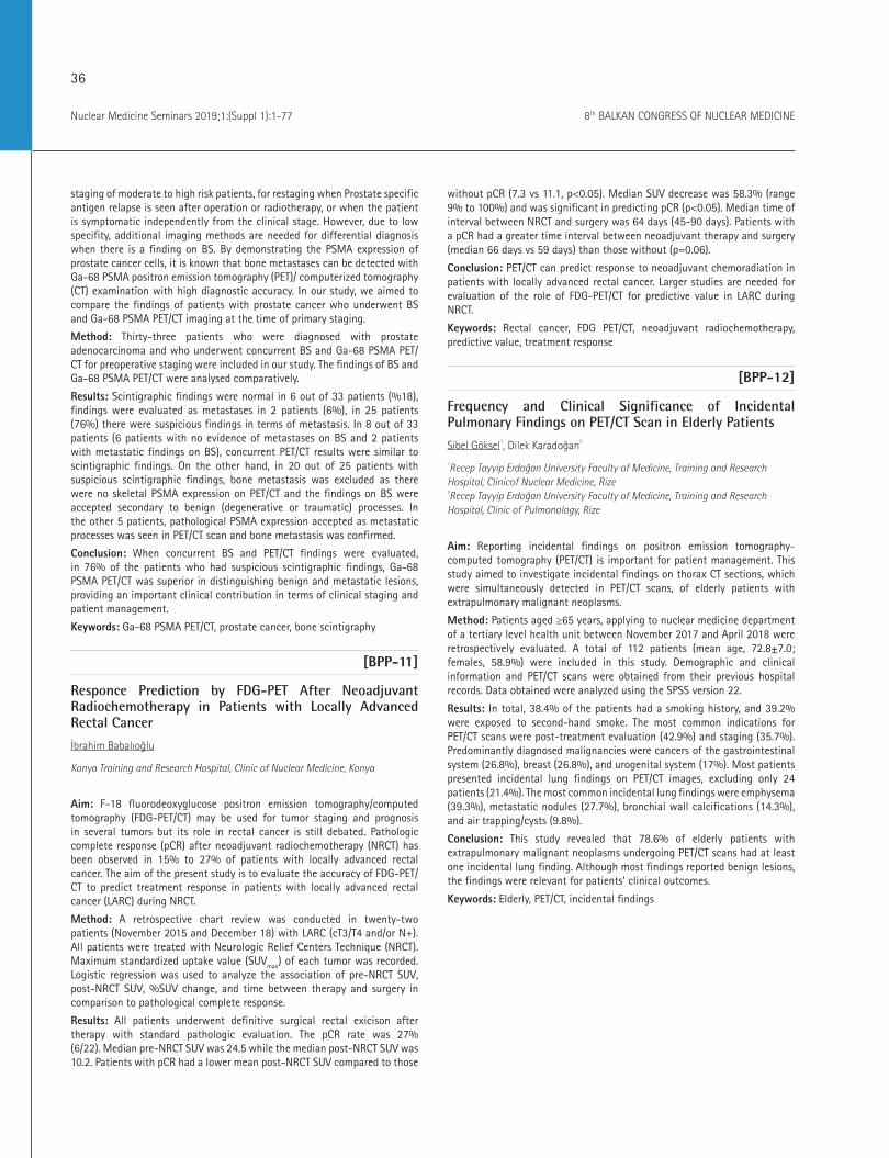
36
Nuclear Medicine Seminars 2019;1:(Suppl 1):1-77 8th BALKAN CONGRESS OF NUCLEAR MEDICINE
staging of moderate to high risk patients, for restaging when Prostate specific antigen relapse is seen after operation or radiotherapy, or when the patient is symptomatic independently from the clinical stage. However, due to low specifity, additional imaging methods are needed for differential diagnosis when there is a finding on BS. By demonstrating the PSMA expression of prostate cancer cells, it is known that bone metastases can be detected with Ga-68 PSMA positron emission tomography (PET)/ computerized tomography (CT) examination with high diagnostic accuracy. In our study, we aimed to compare the findings of patients with prostate cancer who underwent BS and Ga-68 PSMA PET/CT imaging at the time of primary staging.
Method: Thirty-three patients who were diagnosed with prostate adenocarcinoma and who underwent concurrent BS and Ga-68 PSMA PET/CT for preoperative staging were included in our study. The findings of BS and Ga-68 PSMA PET/CT were analysed comparatively.
Results: Scintigraphic findings were normal in 6 out of 33 patients (%18), findings were evaluated as metastases in 2 patients (6%), in 25 patients (76%) there were suspicious findings in terms of metastasis. In 8 out of 33 patients (6 patients with no evidence of metastases on BS and 2 patients with metastatic findings on BS), concurrent PET/CT results were similar to scintigraphic findings. On the other hand, in 20 out of 25 patients with suspicious scintigraphic findings, bone metastasis was excluded as there were no skeletal PSMA expression on PET/CT and the findings on BS were accepted secondary to benign (degenerative or traumatic) processes. In the other 5 patients, pathological PSMA expression accepted as metastatic processes was seen in PET/CT scan and bone metastasis was confirmed.
Conclusion: When concurrent BS and PET/CT findings were evaluated, in 76% of the patients who had suspicious scintigraphic findings, Ga-68 PSMA PET/CT was superior in distinguishing benign and metastatic lesions, providing an important clinical contribution in terms of clinical staging and patient management.
Keywords: Ga-68 PSMA PET/CT, prostate cancer, bone scintigraphy
[BPP-11]
Responce Prediction by FDG-PET After Neoadjuvant Radiochemotherapy in Patients with Locally Advanced Rectal Cancer
İbrahim Babalıoğlu
Konya Training and Research Hospital, Clinic of Nuclear Medicine, Konya
Aim: F-18 fluorodeoxyglucose positron emission tomography/computed tomography (FDG-PET/CT) may be used for tumor staging and prognosis in several tumors but its role in rectal cancer is still debated. Pathologic complete response (pCR) after neoadjuvant radiochemotherapy (NRCT) has been observed in 15% to 27% of patients with locally advanced rectal cancer. The aim of the present study is to evaluate the accuracy of FDG-PET/CT to predict treatment response in patients with locally advanced rectal cancer (LARC) during NRCT.
Method: A retrospective chart review was conducted in twenty-two patients (November 2015 and December 18) with LARC (cT3/T4 and/or N+). All patients were treated with Neurologic Relief Centers Technique (NRCT). Maximum standardized uptake value (SUVmax) of each tumor was recorded. Logistic regression was used to analyze the association of pre-NRCT SUV, post-NRCT SUV, %SUV change, and time between therapy and surgery in comparison to pathological complete response.
Results: All patients underwent definitive surgical rectal exicison after therapy with standard pathologic evaluation. The pCR rate was 27% (6/22). Median pre-NRCT SUV was 24.5 while the median post-NRCT SUV was 10.2. Patients with pCR had a lower mean post-NRCT SUV compared to those
without pCR (7.3 vs 11.1, p<0.05). Median SUV decrease was 58.3% (range 9% to 100%) and was significant in predicting pCR (p<0.05). Median time of interval between NRCT and surgery was 64 days (45-90 days). Patients with a pCR had a greater time interval between neoadjuvant therapy and surgery (median 66 days vs 59 days) than those without (p=0.06).
Conclusion: PET/CT can predict response to neoadjuvant chemoradiation in patients with locally advanced rectal cancer. Larger studies are needed for evaluation of the role of FDG-PET/CT for predictive value in LARC during NRCT.
Keywords: Rectal cancer, FDG PET/CT, neoadjuvant radiochemotherapy, predictive value, treatment response
[BPP-12]
Frequency and Clinical Significance of Incidental Pulmonary Findings on PET/CT Scan in Elderly Patients
Sibel Göksel1, Dilek Karadoğan2
1Recep Tayyip Erdoğan University Faculty of Medicine, Training and Research Hospital, Clinicof Nuclear Medicine, Rize 2Recep Tayyip Erdoğan University Faculty of Medicine, Training and Research Hospital, Clinic of Pulmonology, Rize
Aim: Reporting incidental findings on positron emission tomography-computed tomography (PET/CT) is important for patient management. This study aimed to investigate incidental findings on thorax CT sections, which were simultaneously detected in PET/CT scans, of elderly patients with extrapulmonary malignant neoplasms.
Method: Patients aged ≥65 years, applying to nuclear medicine department of a tertiary level health unit between November 2017 and April 2018 were retrospectively evaluated. A total of 112 patients (mean age, 72.8±7.0; females, 58.9%) were included in this study. Demographic and clinical information and PET/CT scans were obtained from their previous hospital records. Data obtained were analyzed using the SPSS version 22.
Results: In total, 38.4% of the patients had a smoking history, and 39.2% were exposed to second-hand smoke. The most common indications for PET/CT scans were post-treatment evaluation (42.9%) and staging (35.7%). Predominantly diagnosed malignancies were cancers of the gastrointestinal system (26.8%), breast (26.8%), and urogenital system (17%). Most patients presented incidental lung findings on PET/CT images, excluding only 24 patients (21.4%). The most common incidental lung findings were emphysema (39.3%), metastatic nodules (27.7%), bronchial wall calcifications (14.3%), and air trapping/cysts (9.8%).
Conclusion: This study revealed that 78.6% of elderly patients with extrapulmonary malignant neoplasms undergoing PET/CT scans had at least one incidental lung finding. Although most findings reported benign lesions, the findings were relevant for patients’ clinical outcomes.
Keywords: Elderly, PET/CT, incidental findings
37
Nuclear Medicine Seminars 2019;1:(Suppl 1):1-778th BALKAN CONGRESS OF NUCLEAR MEDICINE
[BPP-13]
Utility of F-18 FDG PET/CT in Vulvar Cancer
Özge Öz, Burcu Esen Akkaş, Hüseyin Emre Tosun, Gülin Uçmak
Dr. Abdurrahman Yurtaslan Ankara Oncology Training and Research Hospital, Clinic of Nuclear Medicine, Ankara
Aim: Invasive vulvar carcinoma (VC) is an uncommon malignancy accounting for 4% of all gynecological cancers with an incidence of 2-3/100,000 women. The status of inguino-femoral lymph nodes (IFLN) is the main factor in predicting mortality for VC The preoperative assessment of lymph node status is a relevant issue to plan the surgical strategy and many guidelines including National Comprehensive Cancer Network recommends whole body F-18 fluorodeoxyglucose positron emission tomography/computed tomography (FDG-PET/CT) imaging before establishing a treatment plan for invasive vulvar carcinoma.
Method: In this retrospective study, we included patients with histologically confirmed vulvar cancer T1-T2 <4 cm with a stromal invasion greater than 1 mm, who underwent FDG PET/CT followed by surgery in various Gynecologic Oncology Centers across Ankara/Turkey from 2003 to 2018.
Results: A total of 13 patients were included in the analysis. Median age of the patients were 67 (range: 50-98 years). Eight patients had squamous cell histology, 4 had vulvar Paget Disease and one patient had adenoid cystic carcinoma. Seven of the patients had IFLN dissection, while remaining 6 patients did not receive lymph node dissection. Among 7 patients, 3 of them had IFLN metastasis and all patients with lymph node metastasis had pathologic FDG uptake in PET/CT. However, two patients with pathologic FDG uptake in IFLNs had no metastasis in final histopathology reports. Seven patients had positive surgical margins. There were no relationships between positive surgical margin and primary tumor diameter and also FDG uptake of the primary tumor.
Conclusion: In our series, F-18 FDG PET/CT had 100% sensitivity and 50% specificity for detection of IFLN metastasis. Concerning the flat plate results, it is widely known in the literature that inflammatory cells and activated macrophages represent a common cause of increased F-18 FDG uptake, as occurring in inguinal reactive lymph nodes (LN) after vulvar biopsy or shaving. This study confirms that standard F-18 FDG PET/CT represents an effective preoperative imaging method for LN staging in VC, allowing better planning of groin surgical procedures.
Keywords: F-18 FDG PET/CT, vulvar cancer, lymph node metastasis
[BPP-14]
Clinical Contribution of Dual-Phase Ga-68 PSMA PET/CT Examination in Patients with Prostate Cancer
Gülin Uçmak1, İpek Öztürk2, Bedriye Büşra Demirel1, Burcu Esen Akkaş1
1Ankara University of Health Sciences, Oncology Training and Research Hospital, Clinic of Nuclear Medicine, Ankara 2Şanlıurfa Mehmet Akif İnan Training and Research Hospital, Clinic of Nuclear Medicine, Şanlıurfa
Aim: Dual phase examination is widely used in F-18 positron emission tomography/computed tomography (FDG-PET/CT) studies as it is well known that the FDG uptake of tumor cells increases with time. In current literature, there is a limited number of studies investigating the effect of dual phase examination of Ga-68 PSMA PET/CT. Some in vitro studies have shown that PSMA uptake of prostate cancer cells increases with time so that clinical
use of dual phase examination may contribute to differential diagnosis of benign and malignant lesions. The aim of our study was to investigate the clinical contribution of the dual-phase Ga-68 PSMA PET/CT examination in the staging of prostate cancer patients.
Method: Twenty-seven patients diagnosed with prostate adenocarcinoma who underwent Ga-68 PSMA PET/CT scan for preoperative staging were included in our study. Late pelvic images were taken at 2nd hour in order to re-evaluate the PSMA negative suspicious milimetric pelvic lymph nodes seen in routine images taken at 1st hour. Also the alteration of PSMA expression of the primary tumor was analysed.
Results: Three of the patients were diagnosed as Gleason grade (GG) 1, 7 patients were GG 2, 4 patients were GG 3, 5 patients were GG 4, 8 patients were GG 5. Simultaneous serum PSA levels ranged from 0.7 to 168.1 ng/mL. In 10 of 27 (37%) patients, PSMA positive pelvic lymph nodes were observed in routine early images. No additional PSMA positive lesion was detected in late images, but the lymph nodes were more visually distinguishable. The SUVmax values of the primary tumour decreased in 11 patients (early SUVmax
ranged 6.3-29.5, late SUVmax ranged 3.1-26.6), increased in 9 patients (early SUVmax ranged 3.7-26.9, late SUVmax ranged 6.6-33.3) and no difference was seen in 7 patients. Although there was no statistically significant correlation between serum PSA values, Gleason grades and the change in the SUVmax values observed in late images, there was a correlation close to statistically significant level between PSA levels and detection of increase in SUVmax value in late images (p=0.06).
Conclusion: Although our study did not reveal significant contribution of dual-phase images in 27 prostate cancer patients, it can be said that in the presence of suspicious lesions, late images may contribute clinically and more effective results can be obtained when the serum PSA level is higher. It is concluded that different results could be obtained by prospective studies with high number of patients.
Keywords: Ga-68 PSMA PET/CT, dual phase imaging, prostate cancer
[BPP-15]
Role of F-18 FDG PET/CT Semi-quantitative Metabolic Parameters in Small Cell Lung Cancer
Tamer Aksoy, Esra Arslan, Göksel Alçın, Tevfik Fikret Çermik
İstanbul University İstanbul Faculty of Medicine, Department of Nuclear Medicine, İstanbul
Aim: The aim of this study is to determine the prognostic significance of F-18 fluorodeoxyglucose positron emission tomography/computed tomography (FDG-PET/CT) metabolic parameters in small cell lung carcinoma (SCLC).
Method: Tumor localization, size, distant organ metastasis and metabolic parameters of the 244 (230 M, 14 F) (mean age: 66.0±10.0) SCLC patients who were referred to our clinic for staging with F-18 FDG PET/CT, has been retrospectively analyzed. Mean follow-up time of all patients was 27 months (1-103 months). As a preliminary report, primary tumor’s SUVpeak, SUVmean 40%, MTV 40%, TLG 40, SUVmean 70%, MTV 70%, and TLG 70 values were evaluated in 31 patients for survival analyses.
Results: Mean SUVmax for the primary tumor was 19.7±8.7 (range: 4.4-58.8). During the follow-up period, 182 patients had died and median survival time was found 10.4 months. MTV 40% and MTV 70% parameters were calculated for 31 patients. As a result of the comparison of these 31 patients, OS was statistically higher in MTV 40% >20 group than in MTV 40 %20 group (p=0.04).
Conclusion: F-18 FDG PET/CT might be a predictor for overall survival rates and prognosis of patients with SCLC. Also, F-18 FDG PET/CT parameters
38
Nuclear Medicine Seminars 2019;1:(Suppl 1):1-77 8th BALKAN CONGRESS OF NUCLEAR MEDICINE
may predict the tumor biology and survival expectancy in SCLC. The highest relation has been found in MTV 40% and OS time. Further prospective studies with much more higher population numbers might show new paths to understand the mechanisms of this relationships.
Keywords: Small cell lung carcinoma, F-18 FDG PET/CT, survival
[BPP-16]
The Rate of Coronary Vessel Calcification on PET/CT and Its Clinical Importance in Elderly Patients with Malignancy
Sibel Göksel
Recep Tayyip Erdoğan University Training and Research Hospital, Clinic of Nuclear Medicine, Rize
Aim: Positron emission tomography/computed tomography (PET/CT) scan is a widely used diagnostic method in most malignancies. Recently it also became popular for other diagnoses besides malignancies. In this study, we aimed to evaluate the coronary vessel calcification (CVC) of the elderly patients who underwent PET/CT scan due to malignancies. Therefore, we believe that the results of this study might be important to emphasize the value of PET/CT in coronary artery disease (CAD).
Method: All patients who underwent PET/CT scan due to malignancies between November 2017-August 2018 were retrospectively evaluated. Among them, 100 patients who were over 65 years were included in this study. The thorax CT images of PET/CT scans were reevaluated and presence of coronary vessel /aorta calcification were noted. Patient database was scanned to gather informations of CAD history, previous cardiac symptoms and smoking habits. All patients were investigated for cardiac symptoms by phone call. Obtained data were analyzed using SPSS version 20.
Results: A total of 100 patients were included in this study; 60 of them were female and the mean age was 73.01±7.06 years (min 65-max 93). 57 patients had coronary calcification. Thirteen patients were previously diagnosed with CAD. 61 patients had smoking history. Cardiac symptoms were detected in 70.5 percent of the smokers and 48.7 percent of the non-smokers (p=0.02). 90 percent of male and 41 percent of female patients had a smoking history (p<0.001). 62 patients had cardiac symptoms and CVC was detected in 69.4 percent of them. CVC was detected in 36.8 percent of the patients without cardiac symptoms, as well (p=0.001). Seventy-five percent of the male and 53 percent of the female patients had cardiac symptoms (p=0.02). Cardiac symptoms were determined in 49 patients who had no previous CAD history.
Conclusion: In this study, we found out that 57 percent of our elderly patients had CVC. Presence of CVC might be the only sign of CAD. In PET/CT images, CVC was incidentally observed. Although some of the patients who had CVC were aware of their CAD, a plenty of them had no complaints. The indication of CVC on PET/CT images might help to increase the awareness of CAD in elderly patients. As a result, early detection of CVC might be important in increasing survival rate and life quality of patients. Reporting of CVC in PET/CT images is important in the prognosis of elderly malignancies patients.
Keywords: Coranary vessel calcification, PET/CT, elderly patient
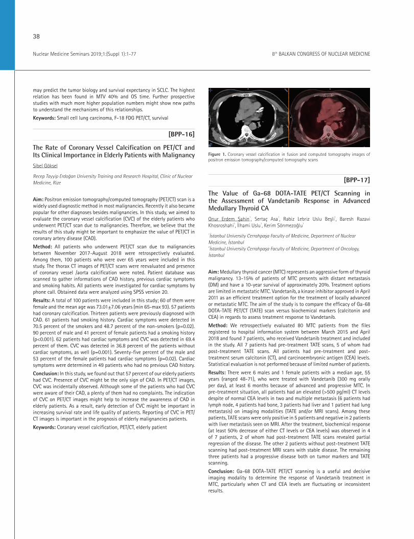
Figure 1. Coronary vessel calcification in fusion and computed tomography images of positron emission tomography/computed tomography scans
[BPP-17]
The Value of Ga-68 DOTA-TATE PET/CT Scanning in the Assessment of Vandetanib Response in Advanced Medullary Thyroid CA
Onur Erdem Şahin1, Sertaç Asa1, Rabiz Lebriz Uslu Beşli1, Baresh Razavi Khosroshahi1, İlhami Uslu1, Kerim Sönmezoğlu1
1İstanbul University Cerrahpaşa Faculty of Medicine, Department of Nuclear Medicine, İstanbul 2İstanbul University Cerrahpaşa Faculty of Medicine, Department of Oncology, İstanbul
Aim: Medullary thyroid cancer (MTC) represents an aggressive form of thyroid malignancy. 13-15% of patients of MTC presents with distant metastasis (DM) and have a 10-year survival of approximately 20%. Treatment options are limited in metastatic MTC. Vandetanib, a kinase inhibitor approved in April 2011 as an efficient treatment option for the treatment of locally advanced or metastatic MTC. The aim of the study is to compare the efficacy of Ga-68 DOTA-TATE PET/CT (TATE) scan versus biochemical markers (calcitonin and CEA) in regards to assess treatment response to Vandetanib.
Method: We retrospectively evaluated 80 MTC patients from the files registered to hospital information system between March 2015 and April 2018 and found 7 patients, who received Vandetanib treatment and included in the study. All 7 patients had pre-treatment TATE scans, 5 of whom had post-treatment TATE scans. All patients had pre-treatment and post-treatment serum calcitonin (CT), and carcinoembryonic antigen (CEA) levels. Statistical evaluation is not performed because of limited number of patients.
Results: There were 6 males and 1 female patients with a median age, 55 years (ranged 48-71), who were treated with Vandetanib (300 mg orally per day), at least 6 months because of advanced and progressive MTC. In pre-treatment situation, all patients had an elevated (>500 pg/ml) CT levels despite of normal CEA levels in two and multiple metastasis (6 patients had lymph node, 4 patients had bone, 3 patients had liver and 1 patient had lung metastasis) on imaging modalities (TATE and/or MRI scans). Among these patients, TATE scans were only positive in 5 patients and negative in 2 patients with liver metastasis seen on MRI. After the treatment, biochemical response (at least 50% decrease of either CT levels or CEA levels) was observed in 4 of 7 patients, 2 of whom had post-treatment TATE scans revealed partial regression of the disease. The other 2 patients without post-treatment TATE scanning had post-treatment MRI scans with stable disease. The remaining three patients had a progressive disease both on tumor markers and TATE scanning.
Conclusion: Ga-68 DOTA-TATE PET/CT scanning is a useful and decisive imaging modality to determine the response of Vandetanib treatment in MTC, particularly when CT and CEA levels are fluctuating or inconsistent results.

39
Nuclear Medicine Seminars 2019;1:(Suppl 1):1-778th BALKAN CONGRESS OF NUCLEAR MEDICINE
Keywords: DOTA-TATE, Vandetanib, medullary thyroid cancer, treatment response
[BPP-18]
The Usefulness of Ga-68 PSMA PET/CT in Staging of Prostate Cancer
Özgul Ekmekcioğlu1, Duygu Has Şimşek2, Sinan Kiremitçi3, Hikmet Özvar5, Tamer Özülker4
1Şişli Hamidiye Etfal Training and Research Hospital, Clinic of Nuclear Medicine, İstanbul2İstanbul University İstanbul Faculty of Medicine, Department of Nuclear Medicine, İstanbul3Şişli Hamidiye Etfal Training and Research Hospital, Clinic of Urology, İstanbul 4Okmeydanı Training and Research Hospital, Clinic of Nuclear Medicine, İstanbul5Şişli Hamidiye Etfal Training and Research Hospital, Clinic of Radiation Oncology, İstanbul
Aim: Prostate cancer (PCa) is the most common type of cancer in men. Early detection of primary lesion and the metastatic disease could change the therapy management and survival. Especially, the patients with oligometastatic disease have recently proven to have better outcome. Ga-68 prostate-specific membrane antigen positron emission tomography computed tomography has demonstrated better image quality than conventional imaging techniques which is more sensivite and specific to detect prostate cancer and the metastases.
Method: We evaluated the patients with prostate cancer retrospectively that were referred to our department for primary staging with Ga-68 PSMA PET/CT between 2017-2018. Patients with pathological confirmation of PCa with high-risk disease were included in this study. Available information on patient demographics, clinical and histopathological findings with Gleason score, initial prostate specific antigen PSA levels, findings for bone scan, and [Ga-68]PSMA PET/CT were retrieved.
Results: Fourty-nine patients with age between 52-82 (mean=65.3) and PSA values were between 4,17-1957 (mean=165,6) were included to this study. After evaluation of the images; 48 patients has shown pathologic uptake, but only a patient with 9.2 PSA value had low PSMA uptake in prostate gland. We could reach the 72% of the biopsy results of the patients. Gleason scores were categorized as 9, 8, 7 and 6 in 26%, 10%-26% and 10% of the patients respectively. 51% of the patients were demonstrated PSMA uptake in lymph nodes and 28% of the patients with either bone, liver or lung that were interpreted as metastases (Figure 1, 2). 14 patient with distant metastases had PSA values between 17.4-1957.
Conclusion: [Ga-68] PSMA PET/CT imaging in the assessment of staging especially high-risk prostate cancer patients with great potential for the detection of lymph node spread and bone metastases that would impact the management plan. Further longitudinal studies with more patients are needed to understand the outcome.
Keywords: PSMA, PET/CT, gallium, prostate cancer
Figure 1. Patient with PSA value of 1555 has demonstrated liver and bone metastases. He had also situs inversus totalis
Figure 1. Patient with PSA value of 117 and GS=9 has demonstrated metastases in lung and lymph nodes
[BPP-19]
Tc-99m MDP SPECT/CT and Multiparametric MRI in the Detection of Pelvic Bone Metastases from Prostate Cancer
Anastas Krassenov Demirev
Acıbadem City Clinic Cancer Center, Clinic of Nucleor Medicine Sofia, Bulgaria
Aim: Initial staging and follow up of prostate cancer involves both radiology and nuclear medicine. One of the most often imaging techniques in many departments are still bone scintigraphy with bisphosphonate combined with computed tomography-Tc-99m MDP-SPECT/CT and multiparametric (including contrast enhanced) magnetic resonance imaging-MRI. We assessed the involvement of pelvic bones in patients with prostate cancer, evaluated equivocal results from both MRI and Tc-99m MDP-SPECT/CT, and consequently investigated the reasons for this discordance, thus suggesting ways of improving the diagnostic approach in initial staging and follow up.
40
Nuclear Medicine Seminars 2019;1:(Suppl 1):1-77 8th BALKAN CONGRESS OF NUCLEAR MEDICINE
Results: The imaging studies were performed between 01/08/2018-09/10/2018 in our hospital and included 55 patients with prostate cancer, who underwent both multiparametric MRI and Tc-99m SPECT/CT for intial staging and/or follow up. We included patients’ age, stage of disease/histology, gleason score, PSA value at time of imaging and initial PSA-iPSA, MRI and SPECT/CT results, type of therapy and follow up.
Conclusion: The study revealed 11 discordant results including 10 (positive for active metastatic bone disease/equivocal) MRI studies, which subsequent Tc-99m MDP SPECT/CT did not confirm and one Tc-99m MDP SPECT/CT study (positive for active metastatic bone disease/equivocal), which MRI didi not confirm. The overall trend was determined by the presence of far more positive/equivocal findings from multiparametric MRI all in all 18% (n=10) compared to 2% (n=1) from Tc-99m MDP SPECT/CT. Various factors proved to be involved in the cause of the observed differences. This resulted in a down-staging of 20% of the patients (n=11), which in turn influenced the therapy regimen.
Keywords: Multiparametric magnetic resonance imaging, MRI, Tc-99m MDP SPECT/CT, single photon emission computed tomography, prostate cancer, bone metsatases
[BPP-20]
PET/CT in the Evaluation of the Effect of Stereotactic Body Radiotherapy in Patients with Lung Metastases
Irena Kostadinova1, Nikolai Nedev2, Gabriela Mateva1, Anastas Krasenov Demirev1, Marina Garcheva1, Rossi Sekalova2
1Acıbadem City Clinic Oncology, Clinic of Nuclear Medicine, Sofia, Bulgaria 2Acıbadem City Clinic Oncology, Clinic of Radiotherapy, Sofia, Bulgaria
Aim: The purpose of this retrospective study is to assess the effect of SBRT in patients with oligometastatic disease of the lung, using PET/CT and to compare the obtained information from the PET and the CT part of the study.
Method: We have reviewed 20 patients with 3 or less lung metastases from different tumours treated with SBRT, of whom- 7 were with breast carcinoma, 6-with colorectal cancer, 4 -with lung carcinoma, 2 -with renal and 1 with gastric cancer. Prior to and after the radiotherapy (3-6 months after completion of treatment) the effect was evaluated with PET/CT, using both PERCIST and RECIST criteria. The patients were treated on Varian TrueBeam STx™ linear accelerator, the respiratory motion was assessed with 4D-CT. The average BED 10 (biologically effective dose 10 Gy) was 80.95 Gy (range 59.5-198.72), delivered in 1 to 5 fractions. The effect of the treatment was evaluated with the change of SUVmax and the sizes of the lesions and the results were compared.
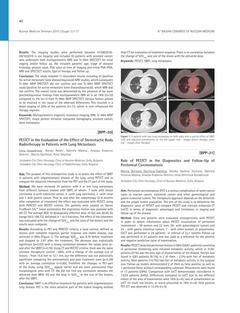
Results: According to PET and PERCIST criteria, a local control, defined as lesions with complete response, partial response and stable disease, was achieved in 86% (Figure 1). The average SUVmax was 6.70 before treatment and dropped to 2.97 after the treatment. The decrease was statistically significant (p=0.03) with a strong correlation between the values prior to- and after the SBRT (r=0.76). Using CT and RECIST criteria, there was the same ultimate therapeutic control - 86%, with a change of the average size of lesions - from 15.6 mm to 12.1 mm, but the difference was not statistically significant comparing the pretreatment and post treatment sizes (p=0.39) with an average correlation (r=0.60). Therefore the changes in PET-part of the study, using SUVmax, appear earlier and are more pronounced than morphological ones with CT. We did not find any correlation between the delivered dose (BED 10 ) and the drop in SUVmax or the size of the lesions, after the SBRT.
Conclusion: SBRT is an effective treatment for patients with oligometastatic lung disease. PET is the more sensitive part of the hybrid imaging method
than CT for evaluation of treatment response. There is no correlation between the change of SUVmax and size of the lesion with the delivered dose.
Keywords: PET/CT, SBRT, lung metastases
Figure 1. A patient with two lung metastases on both sides with a partial effect of SBRT with and radiation pneumonitis on the left (upper row - images before therapy, lower role - images after therapy)
[BPP-21]
Role of PET/CT in the Diagnostics and Follow-Up of Peritoneal Carcinomatosis
Marina Borisova Garcheva-Tsacheva, Veselka Vasileva Stoinova, Gabriela Hristova Mateva, Anastas Krasenov Demirev, Irena Dimitrova Kostadinova
Acıbadem City Clinic Oncology, Clinic of Nuclear Medicine, Sofia, Bulgaria
Aim: Peritoneal carcinomatosis (PC) is a serious complication of some cancer types as ovarian cancer, colorectal cancer and other gynecological and gastro-intestinal tumors. The therapeutic approach depends on the detection and the proper extent evaluation. The aim of the study is to determine the diagnostic value of PET/CT and compare PET/CT and contrast enhanced CT (ceCT) in terms of diagnostic advantages and limitations in staging and follow-up of the disease.
Method: Sixty one patients were evaluated retrospectively with PET/CT in order to obtain information about PET/CT visualization of peritoneal involvement - 39 women and 22 men, 29- with gynecological tumors and 25 - with gastro intestinal tumors, 7 - with other tumors, or polyserositis. CeCT was performed in 24 patients- in interval of 2±1 months. Follow-up was performed in 27 patients and was used as a reference for the positive and negative predictive value of examination.
Results: PET/CT detected peritoneal lesions in 96% (59/61 patients) consisting of peritoneal thickening with elevated metabolic activity, which in 31/61 patients (51%) was the only sign of dissemination of the disease. Ascites was found in 5/61 patients (8.1%) in 2 of them - 3.2%-with foci of metabolic activity. Nine patients (14.7%) had foci of metabolic activity in the surgical scar. Follow-up studies demonstrated 2 of them as false positive, as well as 1 peritoneal focus without corresponding substrate. Recurrences were found in 17 patients (28%). Comparison with ceCT demonstrated: coincidence in 13/24 patients (54%). Differences compared to ceCT due to the different extent of the area of examination were 16% (n=4); lack of enhancement on ceCT (in small size lesions, or scars) amounted to 16% (n=4), false positive PET /CT was observed in 12.5% (n=3).

41
Nuclear Medicine Seminars 2019;1:(Suppl 1):1-778th BALKAN CONGRESS OF NUCLEAR MEDICINE
Conclusion: PET/CT has additional value in the evaluation of PC and in the detection of scar implants as new, or persistent metabolically active lesions. Being a whole body examination PET/CT detects more local recurrences and more areas of dissemination. It can evaluate the activity of lesions without size changes. Eventually it can demonstrate foci in ascite fluid. The limitations of PET/CT are mainly related to false positive results, which reduce its positive predictive value, which is higher for ceCT, after the follow-up.
Keywords: Peritoneal carcinomatosis, PET/CT, contrast enhanced CT
[BPP-22]
F-18 FDG PET/CT Imaging in Diagnosis and Staging of Gallbladder Adenocarcinoma-Case Report
Simon Beshliev, Goran Spirov, Dusica Todorova Stefanovski, Slavko Tasevski, Meri Angjeleska, Ana Ugrinska
University Institute of Positron Emission Tomography, Skopje, R. of Macedonia
Aim: To present a case with suspected gallbladder cancer where 18-FDG PET/CT proved to be useful imaging modality for the diagnosis and staging.
Method: Fifty seven years old female was referred to PET/CT scan due to inconclusive findings from ultrasonography (US), computed tomography (CT), an magnetic resonance imaging (MRI) and core biopsy of the liver. Pathohistological examination of the tumor mass in the liver confirmed hepatic metastasis from unknown origin. PET/CT scan was performed from the vertex of the skull to the toe, 3 minutes per bad on a SIMENS Biograph 40 PET/CT one hour after intravenous administration of 347Mbq of F-18 FDG with low dose CT scan without intravenous or gastrointestinal contrast. Pre-scan level of glucose was 5.1 mmol/L. The maximum standard uptake value (SUVmax) measured 3.9 in the region of the liver (segment VI).
Results: A PET/CT scan demonstrated increased FDG uptake (SUVmax=11.3) in a hypodense unhomogenous mass that involved the gallbladder and the liver in segment IV/VIII. Two metabolically active focuses (SUVmax=4.3) were detected in the liver in the segment III and VI and two enlarged nodules near the pancreatic head (SUVmax=4.3).
Conclusion: The PET/CT confirmed the suspected diagnosis of gallbladder cancer and because of the spread in the liver and lymph nodes surgery was not performed. Gallbladder cancer is a rare malignancy that grows rapidly with local invasion into the liver and with distant spread to lymph nodes. Despite the routine use of ultrasonography, computed tomography and magnetic imaging, in this case report, PET/CT scan proved to be very useful due to its capability of whole body imaging and possibility of showing additional lesions and providing optimal pre-treatment staging in patient that allowed appropriate treatment plan to be tailored.
Keywords: Gallblader, cancer, PET/CT
[BPP-23]
Case Studies of PET/CT Imaging of non-infectious Pneumonitis Induced by mTOR Inhibitors in Patients with Breast Cancer
Gabriela Mateva, Irena Kostadinova, Anastas Krasenov Demirev
Acıbadem City Clinic Oncology, Clinic of Nuclear Medicine, Sofia, Bulgaria
Aim: Non-infectious pneumonitis is a known but not so often (up to 4% of the treated patients) encountered complication in patients treated with mTOR inhibitors. As FDG PET/CT has become an essential clinical tool for
follow up of oncology patients. It is important that the nuclear medicine physicians are aware of the adverse effects of different treatment regimens, and especially with radiological features and metabolic changes that they can induce, having in mind that they could occur even before clinical presentation.
Case: We present two patients with advanced HR-possitive HER2-negative breast cancer treated with everolimus after failure of treatment with letrozole. Both patients were referred for a regular follow up PET/CT. One of them reported recent respiratory troubles - cough and shortness of breath and the other one had no symptoms. The hybrid images revealed bilateral ground-glass opacities and septal thickenings with diffuse moderately increased metabolic activity (with SUVmax up to 7.2 in the first case and 3.1 - in the other) suggestive for pneumonitis. The literature review showed scarce data of PET/CT findings in such cases. After clinical and paraclinical exclusion of infectious origin of the pulmonary findings, with following progression of the symptoms, in both of the patients, the treatment with everolimus was discontinued, as the diagnosis of noninfectious pneumonitis was made. There was a significant clinical improvement and the follow-up PET/CT examinations after 4 months proved the complete resolution of radiological and metabolic changes.
Conclusion: We consider that nuclear medicine physicians have to be aware of noninfectious pneumonitis as a possible side effect of treatment with mTOR inhibitors, which might be lethal if not diagnosed and managed correctly and on time.
Keywords: FDG, breast cancer, everolimus, mTOR inhibitors, pneumonitis, non-infectious
[BPP-24]
The Impact of a Bayesian Penalized-likelihood Reconstruction Algorithm on Detection of Small Liver Lesions
Gabriela Mateva, Irena Kostadinova, Marina Garcheva
Acıbadem City Clinic Oncology, Clinic of Nuclear Medicine, Sofia, Bulgaria
Aim: To demonstrate the benefit of implementing different reconstruction methods for better lesion detection and for increased interpretation confidence.
Method: We present the case of 51 years old female patient diagnosed with sigmoid colon adenocarcinoma two years earlier. As initial treatment a hemicolectomy was performed, followed by 6 courses FOLFOX. One year after the treatment completion, the levels of CA19-9 started to elevate steadily up to 71.29 U/L. Three consecutive contrast enhanced CT scans were performed in the course of the year, and all were interpreted as negative findings for relapse. After the negative diagnostic contrast enhanced CT scans, a FDG scan was performed at our hospital on a Discovery IQ five ring PET/CT scanner. After i.v. administration of 233 MBq of FDG a low dose CT acquisition was followed by an emission scan at two minutes per bed in 3D mode. Two PET reconstructions were performed using ordered subset expectation maximization (OSEM) algorithm and bayesian penalized likelihood reconstruction algorithm (Q.Clear), which improves signal to noise and signal to background ratios.
Results: Q.Clear reconstruction showed prominent uptake within the first liver segment, which was not well defined and visible on the standard OSEM reconstruction. If we apply the PERCIST criteria to this lesion based on the SUVpeak measured on the Q.Clear reconstruction (lesion SUVpeak 3.3, liver SUVmean = 1.73 with SD=0.14), the lesion should be considered as measurable. However if we apply it to the OSEM reconstruction, (SUVpeak=2.41, liver SUVmean=1.78 with SD=0.11) the lesion would be considered as non-
42
Nuclear Medicine Seminars 2019;1:(Suppl 1):1-77 8th BALKAN CONGRESS OF NUCLEAR MEDICINE
measurable. Finally, based on the Q.Clear reconstruction we interpreted the finding as a solitary liver metastases. The patient was referred for liver resection, followed by chemotherapy. The histopathology report confirmed the diagnosis - metastases of moderately differentiated adenocarcinoma. Five months later a follow-up PET/CT scan was performed and it showed complete response.
Conclusion: The use of Q.Clear reconstruction with FDG PET/CT could be used in patients with elevated CA19-9 levels and negative results form other imaging techniques to identify early and reliably small lesions in the liver. In our case Q.Clear increased the confidence and specificity of the final result and helped for the choice of further treatment.
Keywords: FDG, PET/CT, Bayesian penalized-likelihood reconstruction algorithm, liver lesions
[BPP-25]
Primary Pericardial Malign Mesothelioma: A Case Report
Zekiye Hasbek1, Seyit Ahmet Ertürk1, Şule Karadayı2
1Sivas Cumhuriyet University Faculty of Medicine, Department of Nuclear Medicine, Sivas 2Sivas Cumhuriyet University Faculty of Medicine, Department of Thoracic Surgery, Sivas
Aim: In this case, we aimed to present the F-18 FDG PET/CT findings of a patient with primary pericardial malignant mesothelioma which is rarely reported in the literature.
Case: A 56-year-old male patient was admitted with the complaint of shortness of breath. In thorax CT examination, pleural effusion was observed in the left hemithorax with a thickness of 4 cm and a thickness of 1.5 cm on the right side. Pericardial effusion which reached 5 cm in thickness and pericardial thickening was also observed in pericardium. Lymph nodes which to reach ~ 1.5 cm in size were observed in the mediastinum. The cytologic examination from pericardial effusion was reported as malignant cytology, which is thought to be primarily compatible with malignant mesothelioma. F-18-FDG PET/CT examination performed for staging of the patient and it showed that the heart was observed to be bigger than normal, pericardial effusion measured as ~ 5 cm in pericardium and F-18 FDG uptake in the whole pericardium (SUVmax: 12.2). In addition to the large pleural effusion observed in the left hemithorax, increased F-18 FDG uptake was observed in the atelectatic area related with pericardium. F-18 FDG uptake in multiple lymph nodes with continuity of the lesion identified in the pericardium which has conglomerate-like appearance was observed in the prevascular, upper-lower paratracheal, subaortic, aortopulmonary window and bilateral hilar lymphatic stations in mediastinum and these lymph nodes was invading the pulmonary trunk and the pulmonary arteries (highest SUVmax: 10.8). F-F-18DG uptake in conglomerated lymph nodes was observed in the right neck region starting from level 3 and extending to subclavicular and upper mediastinal area and F-18 FDG uptake in lymph nodes was observed in right pectoral and axillary lymphatic stations (Figure 1 and 2). The patient died as a result of sudden respiratory failure and cardiac arrest after completion of PET/CT. He did not respond to resuscitation.
Conclusion: Although the most common malignancy of pericardium is mesothelioma, primary pericardial malignant mesothelioma is a very rare fatal tumor with a incidence of 0.0022%. Since the clinical findings are nonspecific, the diagnosis is extremely difficult. F-18 FDG PET/CT is important in the diagnosis and staging of pericardial malignancies.
Keywords: Malignancy, pericardium, mesothelioma, PET/CT
Figure 1. MIP images of patient in F-18 FDG PET/CT
Figure 2. Axial thoracic fusion image of patient in F-18 FDG PET/CT
[BPP-26]
A Case of Breast Tuberculosis Mimicking Breast Cancer
Ebru İbişoğlu, Mehmet Tarık Tatoğlu, Hatice Uslu
İstanbul Medeniyet University Göztepe Training and Research Hospital, Clinic of Nuclear Medicine, İstanbul
Aim: Tuberculosis can be seen in any organ in the body, with breast tuberculosis a rare from of the disease. Breast tuberculosis affects women of reproductive age and the clinical presentation is often misleading, mimicking breast cancer. Although whole-body F-18 FDG PET/CT scanning is useful in the diagnosis of breast cancer, F-18 FDG uptake is sometimes associated with benign breast disease. A case is reported of F-18 FDG breast uptake caused by breast tuberculosis.
Case: A 40-year-old female presented with left breast enlargement, pain, and redness of three months’ duration. Physical examination revealed lumps in her left breast together with an enlarged axillary node. The right breast and axilla were normal. A chest radiograph and routine hematological and biochemical investigations were within normal limits, apart from a mildly raised ESR. T2 weighted MR imaging showed diffuse hypo and hyperintense areas in the left breast. The abscess was drained and the patient was given antibiotic therapy. However, the patient could not be treated with standard antibiotics. F-18 FDG PET/CT for left breast lesions with a suspicion of
43
Nuclear Medicine Seminars 2019;1:(Suppl 1):1-778th BALKAN CONGRESS OF NUCLEAR MEDICINE
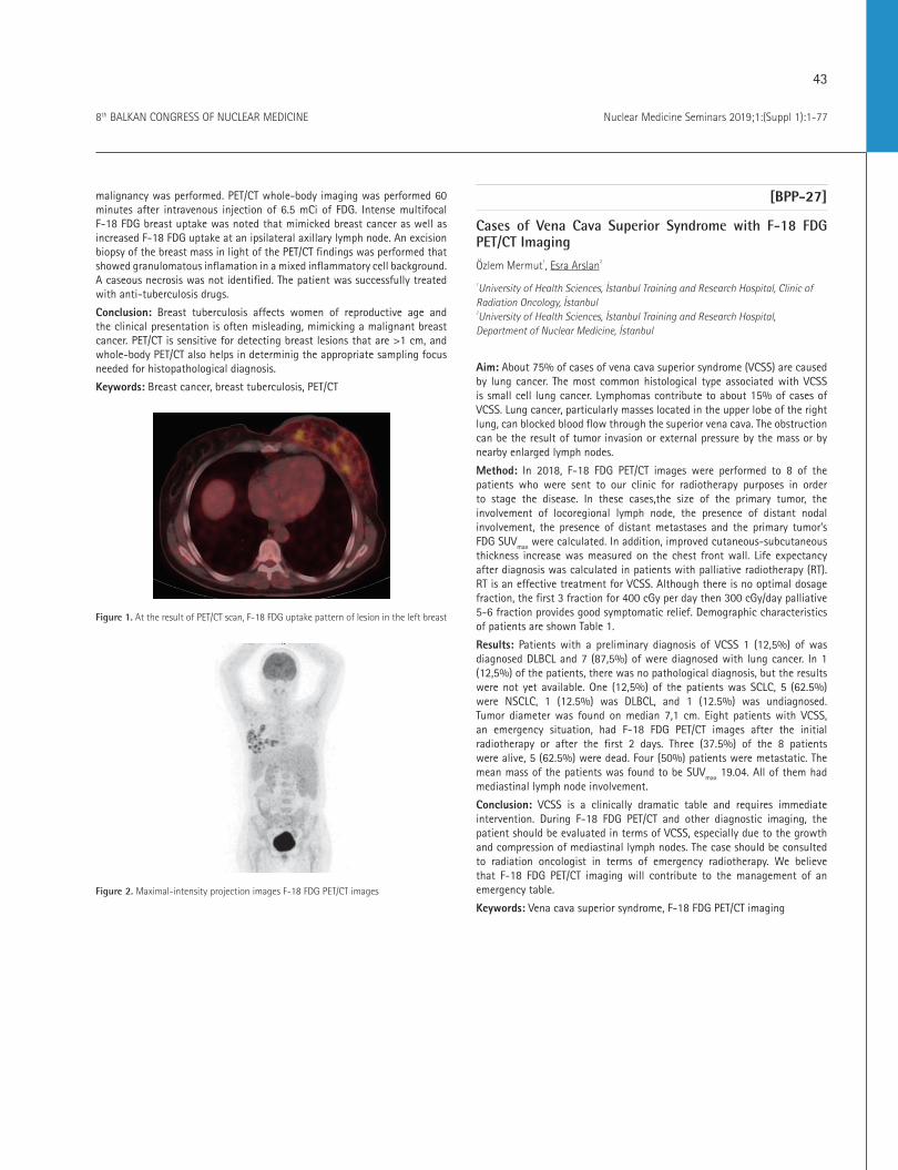
malignancy was performed. PET/CT whole-body imaging was performed 60 minutes after intravenous injection of 6.5 mCi of FDG. Intense multifocal F-18 FDG breast uptake was noted that mimicked breast cancer as well as increased F-18 FDG uptake at an ipsilateral axillary lymph node. An excision biopsy of the breast mass in light of the PET/CT findings was performed that showed granulomatous inflamation in a mixed inflammatory cell background. A caseous necrosis was not identified. The patient was successfully treated with anti-tuberculosis drugs.
Conclusion: Breast tuberculosis affects women of reproductive age and the clinical presentation is often misleading, mimicking a malignant breast cancer. PET/CT is sensitive for detecting breast lesions that are >1 cm, and whole-body PET/CT also helps in determinig the appropriate sampling focus needed for histopathological diagnosis.
Keywords: Breast cancer, breast tuberculosis, PET/CT
Figure 1. At the result of PET/CT scan, F-18 FDG uptake pattern of lesion in the left breast
Figure 2. Maximal-intensity projection images F-18 FDG PET/CT images
[BPP-27]
Cases of Vena Cava Superior Syndrome with F-18 FDG PET/CT Imaging
Özlem Mermut1, Esra Arslan2
1University of Health Sciences, İstanbul Training and Research Hospital, Clinic of Radiation Oncology, İstanbul 2University of Health Sciences, İstanbul Training and Research Hospital, Department of Nuclear Medicine, İstanbul
Aim: About 75% of cases of vena cava superior syndrome (VCSS) are caused by lung cancer. The most common histological type associated with VCSS is small cell lung cancer. Lymphomas contribute to about 15% of cases of VCSS. Lung cancer, particularly masses located in the upper lobe of the right lung, can blocked blood flow through the superior vena cava. The obstruction can be the result of tumor invasion or external pressure by the mass or by nearby enlarged lymph nodes.
Method: In 2018, F-18 FDG PET/CT images were performed to 8 of the patients who were sent to our clinic for radiotherapy purposes in order to stage the disease. In these cases,the size of the primary tumor, the involvement of locoregional lymph node, the presence of distant nodal involvement, the presence of distant metastases and the primary tumor’s FDG SUVmax were calculated. In addition, improved cutaneous-subcutaneous thickness increase was measured on the chest front wall. Life expectancy after diagnosis was calculated in patients with palliative radiotherapy (RT). RT is an effective treatment for VCSS. Although there is no optimal dosage fraction, the first 3 fraction for 400 cGy per day then 300 cGy/day palliative 5-6 fraction provides good symptomatic relief. Demographic characteristics of patients are shown Table 1.
Results: Patients with a preliminary diagnosis of VCSS 1 (12,5%) of was diagnosed DLBCL and 7 (87,5%) of were diagnosed with lung cancer. In 1 (12,5%) of the patients, there was no pathological diagnosis, but the results were not yet available. One (12,5%) of the patients was SCLC, 5 (62.5%) were NSCLC, 1 (12.5%) was DLBCL, and 1 (12.5%) was undiagnosed. Tumor diameter was found on median 7,1 cm. Eight patients with VCSS, an emergency situation, had F-18 FDG PET/CT images after the initial radiotherapy or after the first 2 days. Three (37.5%) of the 8 patients were alive, 5 (62.5%) were dead. Four (50%) patients were metastatic. The mean mass of the patients was found to be SUV
max 19.04. All of them had mediastinal lymph node involvement.
Conclusion: VCSS is a clinically dramatic table and requires immediate intervention. During F-18 FDG PET/CT and other diagnostic imaging, the patient should be evaluated in terms of VCSS, especially due to the growth and compression of mediastinal lymph nodes. The case should be consulted to radiation oncologist in terms of emergency radiotherapy. We believe that F-18 FDG PET/CT imaging will contribute to the management of an emergency table.
Keywords: Vena cava superior syndrome, F-18 FDG PET/CT imaging

44
Nuclear Medicine Seminars 2019;1:(Suppl 1):1-77 8th BALKAN CONGRESS OF NUCLEAR MEDICINE
[BPP-28]
Ga-68 PSMA Uptake in Hepatocellular Carcinoma with Adrenal Metastasis
Ayşegül Aksu1, Emine Acar2,3, Erkan Derebek1
1Dokuz Eylül University Faculty of Medicine, Department of Nuclear Medicine, İzmir 2İzmir Katip Çelebi University, Atatürk Training and Research Hospital, Clinic of Nuclear Medicine, İzmir 3Dokuz Eylül University, Graduate Faculty of Health Sciences, Department of Translational Oncology, İzmir
Aim: Ga-68 prostate specific membrane antigen (Ga-68 PSMA), a positron emission tomography (PET) tracer that was recently introduce for imaging of prostate cancer, may accumulate in other solid tumors including hepatocellular carcinoma (HCC).
Case: A 70-year-old man with diagnose of prostate adenocarcinoma was referred for 68Ga-labeled prostate-specific membrane antigen PET/CT scan for staging. In the liver segment 4, pathological 68Ga-PSMA involvement was noted in the neatly limited lesion. The lesion was diagnosed as HCC with tru-cut biopsy. The patient had suppressed PSA value under antiandrogen treatment.
Conclusion: There was increased 68Ga-PSMA uptake in the nodular lesion in the left adrenal gland. At the last CT, dimensional progression was observed in the nodular lesion in the left adrenal gland and evaluated as metastasis.
Keywords: HCC, PSMA, adrenal, PET/CT
[BPP-29]
A Rare Case: Recurrence of Cecal SCC on PET/CT
Ayşegül Aksu, Nazlı Pınar Karahan Şen, Erkan Derebek
Dokuz Eylül University Faculty of Medicine, Department of Nuclear Medicine, İzmir
Squamous cell carcinoma (SCC) of the colon is a rare tumor that accounts for 0.1%-0.2% of colonic malignancies. We report a 35-year-old man with diagnosis of colon squamous cell carcinoma and referred to us for PET/CT imaging for restaging. PET/CT scan detected high FDG uptake in recurrent
mass at the lower right quadrant of the abdomen and the lesion was histologically proved to be SCC.
Keywords: Colon carcinoma, squamous cell, PET/CT
[BPP-30]
Metabolic Super Scan Finding with FDG PET/CT in a Patient with Parathyroid Cancer
Nilüfer Yıldırım1, Demirhan Eski1, Elif Özdemir2, Şeyda Türkolmez2, Oya Topaloğlu2
1Ankara Atatürk Training and Research Hospital, Clinic of Nuclear Medicine, Ankara 2Ankara Yıldırım Beyazıt University Faculty of Medicine, Department of Nuclear Medicine, Ankara
Aim: Parathyroid cancer (PC) is an uncommon agressive malignancy, characterized by markedly raised serum calcium levels and correlated symptoms. Neck ultrasound and Tc-99m sestamibi scintigraphy are the most common imaging studies used to detect benign parathyroid adenoma but are also of value in malignant cases. The combined whole body imaging modality of F-18 FDG PET/CT provides both anatomic and metabolic information, which may be of incremental value in the detection of metastatic lesions of PC. Here, we describe a metabolic super scan finding with FDG PET/CT in a patient with PC.
Case: A 42-year-old man was admitted to hospital with a mass on the right cervical region, 6 years ago. An excisional biopsy revealed parathyroid cancer and parathroid lesion was resected. After 2 years, the patient underwent parathyroid scintigraphy and whole body scan with MIBI, since persistent hypercalcemia and recurrent kidney stones. Multiple focal uptakes in the low cervical region were considered as metastatic parathyroid carcinoma with MIBI scintigraphy. A confirmatory FDG PET/CT scan was performed and hypermetabolic foci in the bilateral servical region were seen, as well as mild skeletal activity was considered as increased bone metabolism (Figure 1). Patient was subjected to chemotherapy, radiotherapy and serum calcium level decreased. During follow-up; he complained fatigue and renal failure symptoms, his laboratory tests showed raised serum calcium levels to 16.87 mg/dL, and parathyroid hormone (PTH) levels to 2162 pg/mL. Neck ultrasound couldn’t demonstrated the presence of an abnormal finding. Repeat PET/CT scan were done, diffuse metastatic uptake throughout all axiel and upper appendicular skeleton was revealed (SUV
max: 12.59), as well as
Table 1. Demographic characteristics of patients
Gender Age DiagnosisLesion size (PET/CT) (cm)
Primary lesion SUVmax
Lymp node involvement
Distant metastases
Subcutaneos thickening
External pressure vena cava
Posttreatment survival (day)
Dead:D Alive:A
M 63NSCLC (adeno)
7.33 20.04 + Bone + + 37 D
M 62 NSCLC 3.8 10.9 + Brain - + 62 D
M 74 SCLC 8.67 10.25 + - + + 31 D
M 59 Undiagnosis 9 23.2 + Liver - + 55 D
M 74 NSCLC (scc) 11.5 16.4 + - - + 10 D
M 53NSCLC (adeno)
6.9 15.5 + Bone - + 38 A
M 42 NSCLC 7 24.05 + - - + 36 A
M 59 DLBCL 3.3 35.05 + - - + 225 A

45
Nuclear Medicine Seminars 2019;1:(Suppl 1):1-778th BALKAN CONGRESS OF NUCLEAR MEDICINE
bilateral pulmonary metastases (SUVmax: 2.77) (Figure 2). This finding can be interpreted as metabolic ‘super scan’ which is a well described phenomenon on bone scintigraphy.
Conclusion: Diffuse skeletal activity was observed along with very low F-18 FDG uptake in the brain, mediastinum, kidney and bowel in the current scan.
Keywords: Super scan, parathroid cancer, FDG PET/CT
Figure 1. At the result of PET/CT scan, F-18 FDG uptake pattern of lesion in the left breast
Figure 2. Coronary vessel calcification in fusion and CT images of PET/CT scans
[BPP-31]
Peritonitis Carcinomatosa in Lung Cancer: A Case Presentation
Hasan Bozkurt1, Aslı Yıldız1, Gülden Angın2
1Balıkesir Atatürk City Hospital, Clinic of Nuclear Medicine, Balıkesir 2Balıkesir Atatürk City Hospital, Clinic of Radiation Oncology, Balıkesir
Aim: Lung cancer is one of the most common types of cancer worldwide and approximately 40-50% of patients have distant metastasis at the time of
diagnosis. It is not common peritoneal metastases (PM) in lung cancer cases and has been reported in very few cases. We reported on PM in PET/CT, which was performed to evaluate the response to treatment after radiotherapy (RT) in a patient with primary lung cancer.
Case: A 86-year-old woman diagnosed with bronchoscopy had non-small cell lung cancer. The patient had multiple hypermetabolic mediastinal lymph nodes and hypermetabolic bone lesions and there was no pathological FDG uptake in the abdomen in the first PET/CT for staging. The second PET/CT imaging was performed in our clinic to evaluate response to treatment after RT. Significant dimensional and metabolic regression was detected in primary malignant mass in upper lobe anterior of left lung. However, the number, size and metabolic activities of bone lesions showed significant progression. In addition, the patient had metastatic focus in the liver and increased FDG uptake was observed in the areas of peritoneal thickening with density increment which gave the appearance of omental cake at the just behind the anterior wall of the abdomen (Figure 1). The findings were consistent with PM of primary lung tumor.
Conclusion: Detection of peritoneal spread is important suggesting a poor prognosis and considering the palliative treatment options. Median survival of patients with peritonitis carsinomatosa (PC) in lung cancer have been reported as 2.3 months. In our case, the patient had exitus about one month after the diagnosis of PC. Peritoneal spread in lung cancer is quite rare. In a recent study, it was reported that 0.4% of patients diagnosed with lung cancer had PC. Several pathways have been described in the literature as a mechanism of peritoneal spread of tumor cells. As a result of serosa involvement, depending on tumor cells involved in the circulation during tumor removal or in advanced stage disease peritoneal involvement may be seen. One study in the literature found that peritoneal spread was closely associated with malignant pleural disease at the time of diagnosis. As a result, tumor behavior is unpredictable although there are predictable areas in disease progression and spread stages in malignant diseases. Follow-up with imaging modalities is important, especially in the advanced disease group, even if they are asymptomatic.
Keywords: Lung cancer, peritonitis carsinomatosa, PET/CT
Figure 1. Axial sections of abdomen and MIP image of patient
46
Nuclear Medicine Seminars 2019;1:(Suppl 1):1-77 8th BALKAN CONGRESS OF NUCLEAR MEDICINE
[BPP-32]
Penile Metastasis in F-18 FDG PET/CT Scan in a Patient with Bladder Cancer
Aziz Gültekin1, Doğangün Yüksel1, Furkan Ufuk2, Olga Yaylalı1, Tarık Şengöz1
1Pamukkale University Faculty of Medicine, Department of Nuclear Medicine, Denizli 2Pamukkale University Faculty of Medicine, Department of Radiodiagnostics, Denizli
Aim: Metastases to the penis are extremely rare events. Most frequently, penile metastases arises from the urogenital system (bladder, prostate, renal) or the rectum-sigmoid colon. A total of about 500 cases of penile metastases have been reported in the literature so far. About 300 of these cases were bladder cancer metastasis. F-18 fluorodeoxyglucose positron emission tomography/computed tomography (F-18 FDG PET/CT) scan was performed for staging of a patient with bladder cancer. We described penile metastasis by F-18 FDG PET/CT.
Case: A 68-year-old patient who was diagnosed with invasive bladder cancer underwent F-18 FDG PET/CT for staging. In F-18 FDG PET/CT, 10 mm diameter left intracavernosal metastatic lesion was detected in the penis with a SUVmax of 4.53. In order to confirm the metastasis, the patient underwent penile MRI. The MRI revealed a nodular lesion, 8 mm in diameter, with peripheral contrast enhancement in the left cavernous sinus (Figure 1).
Conclusion: There are various mechanisms of penile metastasis reported in the literature. Metastatic spread from primary bladder cancer to penis occurs mainly via retrograde venous route. The other postulated mechanism for penile metastasis include retrograde lymphatic spread, direct invasion, direct arterial spread, and instrumental seeding. The main symptoms of penile metastasis are induration and swelling of penis. It reported that up to 40% patients with penile metastasis may initially present with priapism, due to compromised penile venous drainage. Pain, hematuria, and obstructive voiding symptoms are rarer symptoms of penil metastasis. F-18 FDG PET/CT scan is performed for staging, re-staging and evaluation of response to treatment for bladder cancer. It reported that F-18 FDG PET/CT imaging had excellent diagnostic performance in the detection of distant disease, since it correctly identified 100% distant metastatic lesions (lung, bone, and distant lymph node stations). Recently, it reported that the information provided by PET/CT changed the management in 17% of their patients. Penile metastasis from bladder cancer is an indicator of poor prognosis. The patients with penile metastasis poorly respond to therapy, despite the use of effective systemic chemotherapy. The disease-specific life expectancy is less than one year in these patients. Radical ablative surgery does not contribute to survival; however, it offers an alternative method in symptomatic patients.
Keywords: Penile metastasis, F-18 FDG PET/CT, bladder cancer, penile MRI
Figure 1. F-18 FDG PET/CT scan axiel fusion image was noted lesion in the left corpus cavernosum (a: yellow arrow). Penil MRI T1 enhanced image shows metastatic lesion (b: yellow arrow)
[BPP-33]
F-18 FDG-PET/CT Findings for Staging and Therapy Response Assessment of a Rare Case: Primary Uterine Cervical Lymphoma
Müge Nur Engin1, Duygu Has Şimşek1, Sevgi Kalayoğlu Beşışık2, Işık Adalet1, Ayşe Mudun1, Seher Nilgün Ünal1
1İstanbul University İstanbul Faculty of Medicine, Department of Nuclear Medicine, İstanbul 2İstanbul University İstanbul Faculty of Medicine, Department of Internal Medicine, Division of Hematology, İstanbul
Aim: We present a rare case with primary uterine cervical diffuse large B cell lymphoma who underwent F-18 FDG-PET/CT for staging and therapy response assessment.
Case: A 23-year-old female with vaginal bleeding was examined and underwent vaginal ultrasonography in gynecology clinic. A mass arising from the uterine cervix was detected and biopsy was done to cervical mass. The result of biopsy concluded as high-grade malignant lymphoma. Due to malign pathology, pelvic MRI and F-18 FDG-PET/CT were performed to the patient for further investigation and staging. F-18 FDG-PET/CT showed heterogenous intense FDG uptake in cervical mass (SUVmax: 8.1) extended to the endometrial cavity. No additional FDG avid lesion was found in F-18 FDG-PET/CT. MRI showed a 44x23x30 mm mass in the cervix surrounded by thin fibrous stroma with hyperintensity on T2-weighted imaging. Final biopsies revealed diffuse large B cell lymphoma in cervical mass end endometrium. After 4 cycles of chemotherapy, partial response was detected in cervical mass and endometrium with F-18 FDG-PET/CT.
Conclusion: This case illustrates that F-18 FDG PET/CT has an important role in detecting unexpected localizations, staging and therapy response assessment of non-Hodgkin’s lymphoma.
Keywords: F-18 FDG PET/CT, uterine cervical lymphoma
[BPP-34]
F-18 FDG PET/CT Imaging of HIV Negative Kaposi’s Sarcoma
Özge Öz, Burcu Esen Akkaş, Hüseyin Emre Tosun, Gülin Uçmak
Dr. Abdurrahman Yurtaslan Ankara Oncology Training and Research Hospital, Clinic of Nuclear Medicine, Ankara
Aim: Kaposi’s Sarcoma (KS) is a rare, human herpes virus (HSV) type 8 related indolent vascular tumor with low malignant potential and have several subtypes. The incidence of HIV negative Kaposi Sarcoma is very rare. The most frequent sites for the lesions are mucocutaneous tissues. Skin, lymph nodes, gastrointestinal tract and lungs may be involved. Computerized tomography (CT) and magnetic resonance imaging (MRI) are used to assess distant spread and lymphatic involvement of disease. PET/CT imaging is not routinely recommended for the diagnostic evaluation of KS. As well as, whole-body PET/CT provides additional information and lymph node involvement of the disease. Here we present a HIV-negative Kaposi’s Sarcoma patient with multiple nodal involvement detected by PET/CT.
Case: A 19 year-old male patient presented with swelling in the right ankle and footpad. Biopsy of the lesion revealed Kaposi’s Sarcoma. The lesion showed complete remission after single dose radiotherapy with 8 Gy. In the follow-up of the patient, enlarged lymph nodes presented in bilateral axillary (diameter of largest lymph node 16x7 mm) and submental (diameter

47
Nuclear Medicine Seminars 2019;1:(Suppl 1):1-778th BALKAN CONGRESS OF NUCLEAR MEDICINE
of largest lymph node 10x4 mm) regions. PET/CT showed pathologic FDG uptake in submental (SUVmax: 2.27) and bilateral axillary (SUVmax: 4.81) lymph nodes. Tru-cut biopsy of the pathologic lymph nodes revealed metastatis of the Kaposi’s Sarcoma. The patient had radiotherapy to bilateral axillary lymph nodes (total 32 Gy) with concurrent chemotheraphy (paclitaxel chemotherapy). After 4 months from the initiation of the therapy, a second PET/CT was performed to evaluate the response to the treatment and revealed complete metabolic response.
Conclusion: KS is a multifocal disease typically appearing at mucocutaneous sites, but lymph node and visceral organ involvement may occasionally be seen. Imaging modalities are used not only to stage the disease by determining the extent of spread but also to evaluate the response to therapy at follow-up like in other oncological diseases. As in our case presentation, PET/CT imaging in KS may guide the treatment plan of the disease. KS may display heterogenous FDG avidity and F-18 FDG PET/CT imaging has been reported to have a role both for staging and for the evaluation of response to therapy in this disease. Small metastatic lesions with high FDG avidity which stay under the detection threshold of the CT may be detected with PET/CT imaging and guide the treatment.
Keywords: F-18 FDG/PET, Kaposi sarcoma, HIV negative, nodal involvement
Figure 1.
Figure 2.
[BPP-35]
Recently Diagnosed Breast Carcinoma with Diffuse FDG Uptake of the Bone Marrow
Aysun Sükan1, Fazıl Pierovi1, Hakan Akıncı2
1Medica Medical Center, Clinic of Nuclear Medicine, İstanbul 2Surgimed Medical Center, Clinic of General Surgery, İstanbul
Aim: To show the diffuse infiltration of the bone marrow with the primary malignancy at the early stages of the breast malignancy.
Case: A thirty 38 years old female patient had a tru-cut biopsy from the upper lateral quadrant (tail) of the right breast and also from the right axillary lymph nodes. The results came as invasive ductal carcinoma of the breast as histologic grade 3 (3+3+2=8) (Bloom-Richardson Elston modification); nuclear grade as 3 (Modified Black). Axillary lymph nodes resulted as carcinoma infiltration of the whole specimen.
Method: A PET/CT examination was done in our department with Siemens Biograhp 6 LSO HI-REZ PET/CT with of 13 mCi FDG and 8 beds each 3 minutes were acquired an hour after injection. PET CT is done to determine the stage of the primary malignancy. The result of the PET CT examination revealed a lesion measured as 22x12 mm diameters located at the upper lateral quadrant showing malignant type of hypermetabolism which is consistent with the primary malignancy and multipl subcentimetric lymph nodes with mild to moderate hypermetabolism are also regarded as metastatic. Diffuse heterogeneous hypermetabolism is detected in the whole skeleton within the scanned areas accompanied with some scattered radiolucent appearances of the bones on CT images. A late scan of the pelvis was performed and increase of the FDG uptake was detected at the late images. Diffuse malignant infiltration of the bone marrow was primarly suggested but due to the anemia of the patient noted in medical history, hematological disorders could not be excluded. MR was requested from the pelvis as most heterogenous site on PET/CT examination. It revealed as widespread medullary signal abnormalities sparing the epiphsis of the head of the femur and major trochanteric zones and diffusion restriction on diffusion weighted images and concluded as either malignant infiltration of the bone marrow or systemic hematological disorder.
Conclusion: A diffuse heterogeneous infiltration of the bone marrow from the primary breast cancer is not a common finding especially at the early stages of the disease. PET/CT and MR images could not make a clear distinction between hematological causes or diffuse malignant infiltration of the bone marrow when the patient has anemia. Biopsy of the bone marrow was done and the preliminary results came as malignant infiltration of the bone marrow.
Keywords: Breast carcinoma, F-18 FDG, bone marrow

48
Nuclear Medicine Seminars 2019;1:(Suppl 1):1-77 8th BALKAN CONGRESS OF NUCLEAR MEDICINE
Figure 1. F-18 FDG PET/CT
[BPP-36]
F18-FDG PET/CT Imaging of Invasive Vulvar Paget’s Disease
Özge Öz, Burcu Esen Akkaş, Hüseyin Emre Tosun, Gülin Uçmak
Dr. Abdurrahman Yurtaslan Ankara Oncology Training and Research Hospital, Department of Nuclear Medicine, Ankara
Aim: Extramammary Paget’s disease (EMPD) is a rare malignancy, characterized with malignant Paget’s cells located in the epidermis. The most common location of EMPD in women is vulvar region. The mainstay of the treatment is surgical removal of the lesion with clear surgical margins. Radical excision and inguinofemoral lymph node dissection is not recommended due to non-invasive and intracutaneous nature of the disease. In rare instances, the lesion can arise from Pagetoid spread of adnexal or visceral malignancy to the epidermis.
Case: Fifty-one years old woman presented with recurrent EMPD of the vulva. The patient had two previous wide vulvar excisions due to vulvar Paget’s disease. In the third recurrence, biopsy specimens showed suspicious dermal invasion. A Positron emission tomography/computed tomography (PET/CT) was performed to evaluate possible local spread of the disease. PET/CT revealed enlarged right inguinal lymph nodes with pathologic FDG uptake (SUVmax: 10.1) and pathologic FDG uptake in anterior vulvar region (SUVmax: 5.2) without a visible and non-palpable lesion. Radical vulvectomy with bilateral inguinofemoral lymph node dissection was performed due to PET/CT result. Six out of 14 lymph nodes were positive for Paget’s disease. Detection of primary lesions with PET/CT is limited by lesion thickness, while detection of metastasis appears to be more dependent on size and FDG-avidity. There is evidence that the detection of primary cutaneous EMPD lesions on PET/CT depends more on lesion thickness than size. FDG-avidity can also help detect true nodal metastasis. Fujiwara and colleagues examined the relationship between SUVmax values and lymph node metastasis. PET detected all biopsy-proven lymph node metastasis (27%); there were no false positives (histologically negative and PET-positive) or false negatives (histologically positive and PET negative).
Conclusion: Invasion and metastasis rate of EMPD is extremely low. PET/CT is recommended in the initial evaluation of invazive vulvar carcinoma before initiating the treatment. However, in EMPD evaluation, PET/CT is not routinely recommended. But if there is clinical suspicion for invasive
EMPD, proper imaging studies should be performed to establish an optimal treatment plan.
Keywords: F-18 FDG PET/CT, extra mammarian Paget’s disease, Vulvar Paget’s disease, nodal involvement
Figure 1.
Figure 2.
[BPP-37]
F-18 FDG PET/CT for Pituitary Metastasis in Lung Cancer: A Case Report
N. Belgit Talay1, Fatma Tokgöz Akyıl2, Berna Okudan1
1University of Health Sciences, Ankara Numune Hospital, Clinic of Nuclear Medicine, Ankara 2Çanakkale Government Hospital, Clinic of Chest Disease, Çanakkale
Aim: Lung cancers can spread when cells break off from the tumor, and travel through the bloodstream or the lymphatics to distant regions of the
49
Nuclear Medicine Seminars 2019;1:(Suppl 1):1-778th BALKAN CONGRESS OF NUCLEAR MEDICINE
body. The most common areas for lung cancer to spread are the lymph nodes, liver, bones, brain, and adrenal glands. Pituitary metastasis is very rare and it may be difficult to notice.
Case: A 70-years-old male patient presented to our hospital with complaints of weight loss, fatigue, exhaustion, general condition disturbance and generalized body pain. The patient was referred for computerized tomography (CT) with doubt of malignancy. CT scan showed a mass lesion in the right lung. The patient was then referred to PET/CT for diagnosis and staging. PET/CT scan revealed a malignant mass with dimensions of 8x6 cm in the right lung which narrowed the lower lobe bronchus. FDG uptake was seen in an area of approximately 18x12 mm in the left part of pituitary area (SUVmax: 11.36). Metastatic hypermetabolic lymph nodes in the mediastinum, abdomen besides multiple liver and bone metastases were also observed. MRI imaging was recommended considering that this pituitary lesion may be metastatic. Fiberoptic bronchoscopy (FOB) was performed and the pathology result was reported as squamous cell carcinoma. The pituitary MRI imaging revealed a mass lesion on the left side of the pituitary gland, which induces optic chiasm, and slightly compressing the left optic nerve. The patient’s hormone panel was consistent with hyperprolactinemia and hypogonadotropic hypogonadism. The patient was diagnosed with pituitary metastasis and radiotherapy was planned. In this patient, we aimed to show F18 FDG involvement in pituitary metastasis which is rarely seen in a PET/CT study conducted for primary diagnosis and primary staging in a patient with a preliminary diagnosis of lung cancer.
Keywords: F-18 FDG PET/CT, pituitary metastasis, lung cancer
Figure 1. Pituitary metastasis
Figure 2. Maximum intensity projection
[BPP-38]
Heterogenous Ga-68 PSMA Uptake in Left Upper Abdomen: Mesenchymal Tumor
Ayça Arçay1, Funda Aydın1, Zeynep Gül Kıprak1, Ömer Kutlu2, Ahmet Fırat Güngör3
1Akdeniz University Faculty of Medicine, Department of Nuclear Medicine, Antalya 2Akdeniz University Faculty of Medicine, Department of Urology, Antalya 3Emsey Hospital, Clinic of Nuclear Medicine, İstanbul
Aim: PSMA is spesific for prostate cancer cells but uptake on PSMA PET/CT in uncommon locations for prostate cancer metastasis must be considered for second malignancies. We report a case of abdominal mesenchymal tumor showing heterogenous PSMA uptake in the left upper abdomen.
Case: A 50-year-old man with recently diagnosed prostate adenocarcinoma (Gleason score 3+3; initial PSA, 16.6 ug/L) was referred for Ga-68 PSMA PET/CT to assess for metastatic disease. Ga-68 PSMA PET/CT revealed mildy increased focal tracer uptake (SUVmax: 6.1) in the left part of prostate gland, diffusion MR scan showed no diffusion restriction or contrast enhancement in this region. No prominent pathological metastatic focus was detected. The scan, however, revealed a lesion of 120x92 mm diameters in the left upper abdomen. It had heterogenously increased PSMA uptake (SUVmax: 16.9) and also had cold areas at the posterior region. Histological examination of tissue obtained by core biopsy revealed mesenchymal tumor with spindle cells; no prominent mitosis, necrosis or atypia was seen. The lesion was interpreted as gastrointestinal/extragastrointestinal stromal tumor by pathologists.
Conclusion: Ga-68 PSMA uptake has been described in a large number of non-prostate malignancies-hepatocellular carcinoma, papillary thyroid carcinoma, lung cancer, follicular lymphoma and pancreatic neuroendocrine tumor. PSMA expression is markedly increased in the prostate cancer cells compared to benign tissue, PSMA is also expressed in the endothelial cells of tumor-associated neovasculature. The possible explanation of PSMA expression in nonprostate malignancies is that. Different PSMA uptake patterns of GIST have been reported in several cases like peripheral uptake and central necrosis, rather homogenous intense uptake, focal uptake, moderate heterogenous uptake. Therefore, GIST PSMA uptake pattern is not uniform. PSMA uptake in unusal sites for prostate cancer metastasis must be kept in mind for second malignancies.
Keywords: Ga-68 PSMA PET/CT, mesenchymal tumor, GIST, secondary malignancy
Figure 1. MIP image of PSMA PET/CT, arrow showing the uptake at the left upper abdomen
50
Nuclear Medicine Seminars 2019;1:(Suppl 1):1-77 8th BALKAN CONGRESS OF NUCLEAR MEDICINE
Figure 2. Transaxial images of tumor at CT and PET/CT fusion
[BPP-39]
Take a Second Look: It’s Kikuchi Fujimoto Disease Mimicking Malignant Lymphoma
Esra Çiftçi1, Huri Tilla İlçe1, Fahri Yılmaz2, Tuba Hacıbekiroğlu3
1Sakarya University Faculty of Medicine, Training and Research Hospital, Clinic of Nuclear Medicine, Sakarya 2Sakarya University, Faculty of Medicine, Training and Research Hospital, Clinic of Pathology, Sakarya 3Sakarya University, Faculty of Medicine, Training and Research Hospital, Clinic of Hematology of Internal Medicine, Sakarya
Aim: Kikuchi Fujimoto disease (KFD) is an uncommon benign condition of necrotizing lymphadenitis commonly seen in East Asian and can mimic lymphoma. We aimed to report a case of KFD mimicking malignant lymphoma on PET/CT in older women.
Case: A 57 year-old woman, who presented with fatigue,fever and lymphadenopathy.She had history of romatoid artritis (RA) that is in remission period. Her laboratory data revealed pansitopeni, a mild elevated LDH, CRP levels and high ESR level without any other biochemical, serological and infectious agents abnormalities. She underwent BM aspiration biopsy and could not differenciate “reaktif BM” and “lymphoma infiltration” and offering LN disection. PET/CT was performed and showed intense bone marrow FDG activity at axial and apendicular skeletal system in addition to multiple hypermetabolic LNs localized at supra and infradiafragmatic regions (SUVmax: 9.4). Based on those findings; lymphoproliferatif malignancies were concerned at the first differantial diagnosis and biopsy was suggested.Numerous lymphohistiocytic cells and calcified debris compatible were seen and she was diagnosed with KFD. She underwent PET/CT scan after 5 months and there was no abnormal FDG uptake. Her symptoms and laboratory values improved after 5 months without any treatment based on KFD.
Conclusion: KFD, also known as histiocytic necrotizing lymphadenitis, is a rare,benign,self-limiting disease characterized by cervical lymphadenopathy and fever. It was first described in Asia in 1972 as a lymphadenitis in which histology reveals a focal proliferation of histiocytic cells and abundant nuclear debris. Few cases have been reported in Europe. The pathophysiology remains unknown. Three hypotheses have been raised for this disease: the role of viruses, genetic predisposition and an autoimmune cause because of the correlation with SLE.Our patient also had a history of RA in remission so that she had an autoimmune susceptibility. There are studies concluded that KFD mimicking malignant lymphoma with PET/CT. In our case, since there was stage 4 lymphoma-like PET images at the time of initial scan, met the diagnostic criteria of lymphoma. Treatment of KFD is mainly supportive and resolution occurs within 6 months like occured in our case. This case emphasizes the importance of KFD in the differential diagnosis of lymphoma, especially in women with a history of autoimmune disease like RA or SLE.
Keywords: Kikuchi Fujimoto disease, PET/CT, lymphoma
Figure 1. Calcified histiocytic necrotizing lymphadenitis (H&E X40)
Figure 2. F-18 FDG PET/CT MIP images
[BPP-40]
Whole-Body or Torso imaging with Ga-68 PSMA PET/CT in Prostate Carcinoma?
Ceren Özge Engur, Salih Özgüven, Kevser Öksüzoğlu, Selin Kesim, Nuh Filizoğlu, Sabahat İnanır
Marmara University Pendik Training and Research Hospital, Departmant of Nuclear Medicine, İstanbul
Aim: Ga-68 PSMA is a non-invasive diagnostic modality for staging, re-staging and evaluating the response to treatment in patients with prostate cancer. The imaging procedure of PET/CT is performed from the skull base to mid-thighs for most oncology studies.
Method: Two patients with a diagnosis of prostate carcinoma referred to Ga-68 PSMA PET/CT.
Case 1: A 77 years-old patient with prostate cancer referred to Ga-68 PSMA PET/CT. Ga-68 PSMA PET/CT study was performed from skull base to mid-thighs, and showed Ga-68 PSMA accumulation in metastatic lesions in pelvic bones as well as in prostate gland (Figure 1 A, 1 B). On bone scintigraphy, additional metastatic lesions were detected in the distal part of right femur and right calcaneus (Figure 1C).
Case 2: A 68 year-old patient with a typical metastatic superscan appearance on bone scan admitted to Ga-68 PSMA PET/CT (Figure 2A). Ga-68 PSMA PET/CT demonstrated multiple PSMA avid metastases in axial and appendicular skeleton including both feet extending to the distal phalanx (Figure 2B, 2C).
Conclusion: Since the low probability of metastasis, the cranium and lower extremities were not included in the routine imaging procedure of PET/CT.

51
Nuclear Medicine Seminars 2019;1:(Suppl 1):1-778th BALKAN CONGRESS OF NUCLEAR MEDICINE
Inclusion of the head and lower extremities in the FOV of PET/CT may help in improved staging accuracy for patients with prostate cancer.
Keywords: Prostate Cancer, Bone scintigraphy, Ga-68 PSMA PET/CT
Figure 1. Case 1
Figure 2. Case 2
[BPP-41]
Benign Mimic of Disseminated Sclerotic Bone Metastasis: Osteopoikilosis
Zeynep Aydın, Özlem Şahin, Buğra Kaya, Ahmet Eren Şen
Necmettin Erbakan University Meram Faculty of Medicine, Department of Nuclear Medicine, Konya
Aim: Osteopoikilosis (OP), also referred to as “osteopathia condensans disseminata”, is a rare bone sclerotic dysplasia with a prevalence estimated at 1 per 50,000. It has been described in all age groups and the incidence is similar in both sexes. OP is characterized by defective endochondral bone formation. It is usually asymptomatic, but in some cases (15-20%) it may be associated with articular pain and it often is an incidental discovery on radiographic examinations. The radiological feature is multiple sclerotic, punctate (2-10 mm), oval or round foci, symmetrically distributed in epiphyseal and metaphyseal regions of the axial and appendicular skeleton. These lesions are most frequently located at knees, shoulders, carpal and tarsal bones. OP must be differentiated among others from sclerotic metastases and bone sclerotic dysplasias. Occasionally, it is associated with
abnormalities such as renal or heart malformation, or endocrine disorders.
Case: We present a 48-year old man studied due to gastric adenocarcinoma. CT displayed multiple sclerotic lesions, most of them small, distributed in practically all the skeletal structures. Considering the context of this patient, the first diagnostic choice was blastic metastasis. F-18 FDG PET/CT scan was performed to staging the extent of the disease. Increased metabolic activity gastric tumor and metastatic perigastric lymph nodes were observed. In addition, the patient had an ectopic kidney. Also PET/CT images revealed innumerable small sclerotic lesions in the vertebrae, sternum, ribs, pelvic bones and especially appendicular skeleton including hand fingers. However, lack of FDG uptake was seen on the PET images, in the small round sclerotic lesions. FDG-PET/CT provided metabolic characterization of the lesions and a larger imaging area (including upper extremity) than CT thus led us to move away from the diagnosis of metastasis. The coexistence of renal anomaly also strengthened the diagnosis of OP.
Conclusion: OP is a rare bone disorder incidentally observed on X-rays. This disorder is typically asymptomatic and generally autosomal dominantly inherited. There is a predilection for epiphysis and metaphysis of the long tubular bones, carpus, tarsus, pelvis and scapula. It differs from metastasis in that the lesions are metabolically inactive and include the appendicular skeleton. Therefore it is important to be aware of this rare and benign condition to prevent making a misdiagnosis.
Keywords: Osteopoikilosis, sclerotic bone metastases, FDG-PET/CT
Figure 1. Sclerotic lesions with no increased FDG uptake in bilateral acetabulum, femoral head and phalanges in PET/CT
[BPP-42]
A Rare Case Report of Nasal Metastasis from Esophageal Squamoz Cell Carsinoma Detected by PET/CT
Ayşe Esra Arslan
Erzurum Region Training and Research Hospital, Clinic of Nuclear Medicine, Erzurum
Aim: Esophageal cancer most commonly spreads to regional lymph nodes. Distant metastasis is usually to distant lymph nodes, liver, peritoneum. The metastasis of nasal region is very rare and it have been reported only three cases in the literature. However we think that this case is the first case which has detected by PET/CT.
Case: Our patient is 58 years old male who have diagnosed esophagus squamous cell carcinoma. PET/CT was performed to evaluate the response of chemotherapy. After giving 8.4 mCi F-18 FDG, 60-minute later the way into the area of the cranium to the proximal segment of femur, PET/CT study done. We found increased F-18 FDG uptake in the asymmetric wall thickness of distal part of esophagus. In addition, multiple lymphadenopathies in head-neck, mediastinum, thorax, abdomen and pelvis, which are progressive according to the previus study were noted. Also, multiple metastatic
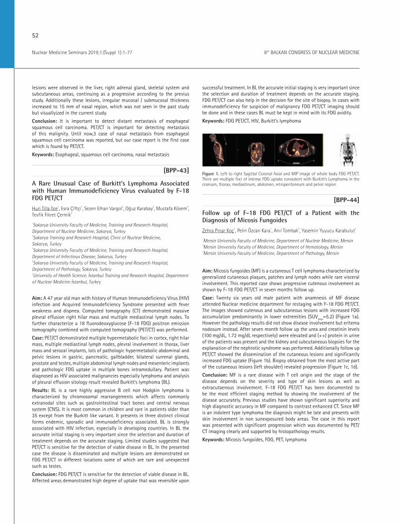
52
Nuclear Medicine Seminars 2019;1:(Suppl 1):1-77 8th BALKAN CONGRESS OF NUCLEAR MEDICINE
lesions were observed in the liver, right adrenal gland, skeletal system and subcutaneous areas, continuing as a progressive according to the previus study. Additionally these lesions, irregular mucosal / submucosal thickness increased to 15 mm of nasal region, which was not seen in the past study but visuallyized in the current study.
Conclusion: It is important to detect distant metastasis of esophageal squamous cell carcinoma. PET/CT is important for detecting metastasis of this malignity. Until now,3 case of nasal metastasis from esophageal squamous cell carcinoma was reported, but our case report is the first case which is found by PET/CT.
Keywords: Esophageal, squamous cell carcinoma, nasal metastasis
[BPP-43]
A Rare Unusual Case of Burkitt’s Lymphoma Associated with Human Immunodeficiency Virus evaluated by F-18 FDG PET/CT
Huri Tilla İlce1, Esra Çiftçi1, Sezen Elhan Vargol2, Oğuz Karabay3, Mustafa Kösem4, Tevfik Fikret Çermik5
1Sakarya University Faculty of Medicine, Training and Research Hospital, Department of Nuclear Medicine, Sakarya, Turkey 2Sakarya Training and Research Hospital, Clinic of Nuclear Medicine, Sakarya, Turkey 3Sakarya University Faculty of Medicine, Training and Research Hospital, Department of Infectious Disease, Sakarya, Turkey 4Sakarya University Faculty of Medicine, Training and Research Hospital, Department of Pathology, Sakarya, Turkey 5University of Health Science, İstanbul Training and Research Hospital, Department of Nuclear Medicine İstanbul, Turkey
Aim: A 47 year old man with history of Human Immunodeficiency Virus (HIV) infection and Acquired Immunodeficiency Syndrome presented with fever weakness and dispnea. Computed tomography (CT) demonstrated massive plevral effusion right hilar mass and multiple mediastinal lymph nodes. To further characterize a 18 fluorodeoxyglucose (F-18 FDG) positron emission tomography combined with computed tomography (PET/CT) was performed.
Case: PET/CT demonstrated multiple hypermetabolic foci in cortex, right hilar mass, multiple mediastinal lymph nodes, plevral involvement in thorax, liver mass and serozal implants, lots of pathologic hypermetabolic abdominal and pelvic lesions in gastric, pancreatic, gallbladder, bilateral surrenal glands, prostate and testes, multiple abdominal lymph nodes and mesenteric implants and pathologic FDG uptake in multiple bones intramedullary. Patient was diagnosed as HIV associated malignancies especially lymphoma and analysis of pleural effusion sitology result revealed Burkitt’s lymphoma (BL).
Results: BL is a rare highly aggressive B cell non Hodgkin lymphoma is characterized by chromosomal rearrangements which affects commonly extranodal sites such as gastrointestinal tract bones and central nervous system (CNS). It is most common in children and rare in patients older than 35 except from the Burkitt like variant. It presents in three distinct clinical forms endemic, sporadic and immunodeficiency associated. BL is strongly associated with HIV infection, especially in developing countries. In BL the accurate initial staging is very important since the selection and duration of treatment depends on the accurate staging. Limited studies suggested that PET/CT is sensitive for the detection of viable disease in BL. In the presented case the disease is disseminated and multiple lesions are demonstrated on FDG PET/CT in different locations some of which are rare and unexpected such as testes.
Conclusion: FDG PET/CT is sensitive for the detection of viable disease in BL. Affected areas demonstrated high degree of uptake that was reversible upon
successful treatment. In BL the accurate initial staging is very important since the selection and duration of treatment depends on the accurate staging. FDG PET/CT can also help in the decision for the site of biopsy. In cases with immunodeficiency for suspicion of malignancy FDG PET/CT imaging should be done and in these cases BL must be kept in mind with its FDG avidity.
Keywords: FDG PET/CT, HIV, Burkitt’s lymphoma
Figure 1. Left to right Sagittal Coronal Axial and MIP image of whole body FDG PET/CT. There are multiple foci of intense FDG uptake consistent with Burkitt’s Lymphoma in the cranium, thorax, mediastinum, abdomen, retroperitoneum and pelvic region
[BPP-44]
Follow up of F-18 FDG PET/CT of a Patient with the Diagnosis of Micosis Fungoides
Zehra Pınar Koç1, Pelin Özcan Kara1, Anıl Tombak2, Yasemin Yuyucu Karabulut3
1Mersin University Faculty of Medicine, Department of Nuclear Medicine, Mersin 2Mersin University Faculty of Medicine, Department of Hematology, Mersin 3Mersin University Faculty of Medicine, Department of Pathology, Mersin
Aim: Micosis fungoides (MF) is a cutaneous T cell lymphoma characterized by generalized cutaneous plaques, patches and lymph nodes while rare visceral involvement. This reported case shows progressive cutenous involvement as shown by F-18 FDG PET/CT in seven months follow up.
Case: Twenty six years old male patient with anamnesis of MF disease attended Nuclear medicine department for restaging with F-18 FDG PET/CT. The images showed cutenous and subcutaneous lesions with increased FDG accumulation predominantly in lower extremities (SUVmax=5.2) (Figure 1a). However the pathology results did not show disease involvement but eritema nodosum instead. After seven month follow up the urea and creatinin levels (100 mg/dL, 1.72 mg/dL respectively) were elevated and (++) protein in urine of the patients was present and the kidney and subcutaneous biopsies for the explanation of the nephrotic syndrome was performed. Additionally follow up PET/CT showed the dissemination of the cutaneous lesions and significantly increased FDG uptake (Figure 1b). Biopsy obtained from the most active part of the cutaneous lesions (left shoulder) revealed progression (Figure 1c, 1d).
Conclusion: MF is a rare disease with T cell origin and the stage of the disease depends on the severity and type of skin lesions as well as extracutaenous involvement. F-18 FDG PET/CT has been documented to be the most efficient staging method by showing the involvement of the disease accurately. Previous studies have shown significant superiority and high diagnostic accuracy in MF compared to contrast enhanced CT. Since MF is an indolent type lymphoma the diagnosis might be late and presents with skin involvement in non sunexposured body areas. The case in this report was presented with significant progression which was documented by PET/CT imaging clearly and supported by histopathology results.
Keywords: Micosis fungoides, FDG, PET, lymphoma

53
Nuclear Medicine Seminars 2019;1:(Suppl 1):1-778th BALKAN CONGRESS OF NUCLEAR MEDICINE
Figure 1. At the result of PET/CT scan, F-18 FDG uptake pattern of lesion in the left breast
Figure 2. Coronary vessel calcification in fusion and CT images of PET/CT scans
[BPP-45]
FDG PET/CT Additional Delayed Pelvic Images with Oral Hydration-Voiding-Refilling for Detecting Bladder Wall Lesions
Seval Erhamamcı1, Nesrin Aslan2
1Başkent University İstanbul Health Practice and Research Center Hospital, Clinic of Nuclear Medicine, İstanbul 2Neolife Medical Center, Clinic of Nuclear Medicine, İstanbul
Aim: Urinary clearance of F-18-fludeoxyglucose (FDG) and variability in bladder wall FDG uptake may hamper the interpretation and limit the use of FDG positron emission tomography (PET)/computerized tomography) CT for imaging bladder tumors. Washing out the excreted F-18-FDG is key to overcoming this limitation of F-18-FDG/PET. Some studies have shown usefulness of delayed PET imaging with forced bladder perfusion, diuretics, and oral hydration with large volumes of water in the diagnosis of bladder cancer. However, these previous methods are impractical for routine examinations due to risks of adverse reactions and infections, patient discomfort, exposure of technicians to radiation. In this case, the minimally invasive method of oral hydration using only water and voiding-refilling was used, and utility of the early and delayed images were presented on F-18-FDG/PET/CT.
Case: A-53 year-old woman with ovarian cancer underwent bilateral oopherectomy and radiotherapy. Angiosarcoma was detected on the right side wall of the patient’s bladder during follow-up. The patient was referred for F-18-FDG/PET/CT. One hour after injection of 9 mCi of FDG, the patient was asked to void; after that, the patient underwent whole-body PET/CT imaging. Axial, sagittal, and coronal images of whole-body F-18-FDG/PET/CT revealed no pathological uptake (Figure 1 and 2). Also, any pathologic FDG uptake was not discriminated at the primary tumor region at the bladder wall. The patient was asked to void again and was instructed to receive oral hydration so that the urinary FDG concentration was diluted. Additional delayed pelvic images were acquired when the patient felt the urge to urinate without voiding. Intense FDG uptake at the right side and posterior wall of the bladder, which was consistent with primary tumor, could be discriminated clearly images of delayed pelvic PET/CT (arrow).
Conclusion: Delayed F-18-FDG/PET images with oral hydration water and voiding-refilling were more useful than early images for detecting bladder wall lesions. This method is an efficient and simple technique, and also less invasive than other previously reported methods, therefore could be incorporated into routine examinations.
Keywords: F-18-FDG, PET/CT, bladder carcinoma
Figure 1.
Figure 2.

54
Nuclear Medicine Seminars 2019;1:(Suppl 1):1-77 8th BALKAN CONGRESS OF NUCLEAR MEDICINE
[BPP-46]
A Rare Case of Sinonasal Melanoma and the Role of F-18 FDG PET/CT
Nazım Coşkun1, Hamza Özer2, Berna Okudan1, N.Belgit Talay1
1University of Health Sciences, Ankara Numune Training and Research Hospital, Clinic of Nuclear Medicine, Ankara 2University Of Health Sciences, Ankara Numune Training and Research Hospital, Clinic of Radiology, Ankara
Aim: Sinonasal melanoma is a rare tumor with poor prognosis, high rates of local and regional recurrence and distant metastasis. The aim of this case report is to emphasize the role of F18 FDG positron emission tomography (PET)/computerized tomography (CT) in staging and evaluating the treatment response of these patients.
Case: A 74-year-old male patient suffering from a mass lesion in the right nasal region was admitted to for a F-18 fludeoxyglucose (FDG) PET/CT scan which revealed an expansile malignant soft tissue mass of 65x40x74 mm, filling and obliterating the right nasal cavity and maxillary sinus, invasing neighbor palatin drum and conchal bone (SUVmax: 20.8) (Figure 1). There was no metastatic hypermetabolic lymph node in the head and neck region. Biopsy result of the the mass was reported as malignant melanoma. In the immunohistochemical study, intensive melanocytic staining was observed with HMB45, melan A, S-100. Tumor cells were negative with SMA, cardanin, CD31, CD34, factor 8. As the patient refused the operation, chemotherapy and radiotherapy treatments including Cisplatin were initiated. In the PET/CT study conducted to evaluate the treatment response, it was found that the size and metabolic activities of the mass decreased and showed significant regression (SUVmax: 2.8) (Figure 2).
Conclusion: A rare diagnosis of sinonasal melanoma should be considered in the evaluation and differential diagnosis of nasopharyngeal masses. F-18 FDG/PET/CT is an important imaging modality for staging of sinonasal malignant melanoma and evaluation of treatment response.
Keywords: Sinonasal Melanoma, F-18 FDG PET/CT
Figure 1. Melanoma before theraphy
Figure 2. Melanoma before theraphy
[BPP-47]
Non-Hodgkın Lymphoma as a Reason for Renal Stasis: Utility of SPECT/CT
Aslı Ayan, Aybüke Uçgun, Sümeyye Alkan
Ankara Gülhane Training and Research Hospital, Clinic of Nuclear Medicine, Ankara
Aim: Our aim is to express the utility of single photon emission computed tomography (SPECT)/computerized tomography (CT) as a useful tool to demonstrate a surprising cause of unilateral renal stasis.
Case: A 20 years old man, with a history of lower back pain for two months, underwent a lumbar magnetic resonance imaging (MRI) and the findings waere reported as signal loss in L4-L5 intervertebral disc contrast enhancement in T1A sequences, which may be consistent with spondylodiscitis. Lumbar MRI also revealed conglomerated left paravertebral lymph nodes at L2-L3 level. The three phase bone scan was performed. Bone scan showed normal skeletal system findings. However, left renal stasis accompanied with left proximal ureteral stasis were detected (Figure 1). Subsequent imaging showed a soft tissue mass, which is approximately 35 mm in diameter and located at L3 vertebra level. The mass borders could not be differentiated from left ureter. The caudal sections of CT images also demonstrated round lymph nodes in left groin, which were also shown by an ultrasound examination. The patient underwent left inguinal lymph node biopsy, which reported as Non-Hodgkin lymphoma. Up to the bone scan findings confirmed with US evaluation, a nephrostomy catheter placed to restore the urinary flow.
Conclusion: Obstructive uropathy is a common finding due to renal stones, vesicouretheral reflux and posterior ureteral valve. The solid tumors and retroperitoneal fibrosis may also are common causes of renal stasis and hydronephrosis, but primary lymphomas are rare. The bone scan finding of renal stasis, and SPECT/CT findings together explained the obstructive etiology and the reason for lower back pain. Early reporting of renal stasis demonstrated by bone scan and SPECT/CT resulted in a nephrostomy in order to preserve renal function in the patient. In cases with no previous history of urinary tract pathology, SPECT/CT may provide additional benefits for bringing the etiology into the open.
Keywords: Lymphoma, bone scan, SPECT/CT, renal stasis
Figure 1. The figure displays left renal and ureteral stasis with normal features of skeletal system. Renal stasis and proximal ureter activity (white arrow)

55
Nuclear Medicine Seminars 2019;1:(Suppl 1):1-778th BALKAN CONGRESS OF NUCLEAR MEDICINE
Figure 2. The ureter activity could not be seen in the computerized tomography (CT) sections of the mass level (white arrows), the finding may be related to the total obstruction. Single photon emission computerized tomography/CT images revealed a soft tissue mass around left ureter, which is approximately 35 mm in diamet
[BPP-48]
Septic Pulmonary Embolism Mimicking Lung Metastasis: FDG/PET/CT Findings in a Patient with Bladder Carcinoma
Özlem Şahin, Ahmet Eren Şen, Zeynep Aydın, Buğra Kaya
Necmettin Erbakan University Faculty of Meram Medicine, Department of Nuclear Medicine, Konya
Aim: Cavitary pulmonary nodules may be seen in many different conditions, from acute and chronic infections to chronic systemic diseases or malignancies. Lung metastases of transitional cell carcinomas usually have multiple nodules, solitary masses or interstitial micronodules. However, metastases in the form of multiple cavitary lesions have also been reported. Most cases of septic pulmonary embolism are seen after tricuspid valve endocarditis (iv drug users), permanent catheter, pacemaker wire, peripheral septic thrombophlebitis or organ transplantation. Septic pulmonary embolism due to necrotizing fasciitis is rare. In a study of 247 patients with septic pulmonary embolism, in only two patients necrotizing fasciitis was identified as primary focus.
Case: A 56 year old male patient diagnosed with bladder Ca with type 2 diabetes mellitus was hospitalized in the endocrinology service for blood glucose regulation. During his follow-up, he developed swelling and movement limitation in his left arm, swelling and pain in the scrotal region. When his anamnesis was deepened, it was learned that he had a blunt trauma to the left shoulder 4 days before his hospitalization. The patient who developed shortness of breath had seen pulmonary nodules in thorax CT and requested positron emission tomography (PET)/computerized tomography (CT) for metastasis. PET/CT images showed increased fludeoxyglucose (FDG) uptake in the lung nodules and some of them were cavitary. (Figure 1a, 1b) In addition, abscess appearances with air densities were observed in the left shoulder region and in the scrotal area showing increased FDG uptake (Figure 1c, 1d). When the patient was evaluated with clinical and laboratory findings; septic pulmonary embolism due to necrotizing fasciitis was considered. The patient’s laboratory findings were as follows: C-reaktif protein: 343.4 mg/L (0.1-5), procalcitonin: 2,26ng/mL (n<0,1), white blood cells: 27,7x10³/µL (4-10), neutrophil %: 91,3 (37-73). On the growth of coagulase negative staphylococcus and staph aureus in blood culture, the patient received an appropriate antibiotherapy for approximately one month, and his clinic and laboratory improved. control thorax CT revealed sequela fibrotic changes.
Conclusion: In our case the cause of pulmonary nodules with increased FDG uptake was septic embolism due to necrotizing fasciitis in the shoulder region. Cavitary pulmonary nodules may be present in many diseases
including metastasis of bladder cancer. Septic pulmonary embolism should also be kept in mind as in our case.
Keywords: Septic pulmonary embolism, necrotizing fasciitis, FDG/PET/CT
region and scrotal area showing increased FDG uptake
Figure 1a, 1b. PET/CT images; pulmonary nodules with increased FDG uptake, some of them cavitary; 1c, 1d Abscess in the left shoulder region and scrotal area showing increased FDG uptake
[BPP-49]
A Rare Case Report of Oral Metastasis from Esophageal and Gastric Adenocarcinoma Detected by PET/CT
Münir Demirci
Erzurum Regional Training and Research Hospital, Clinic of Nuclear Medicine, Erzurum
Aim: Esophageal and gastric cancer most commonly spreads to regional lymph nodes. Distant metastasis is usually to distant lymph nodes, liver, peritoneum. The metastasis of oral region is rare and it have been reported nineteen cases in the literature. However we think that this case is the secound case which has detected by positron emission tomography (PET)/computerized tomography (CT) in the literature.
Case: Our patient is 64 years old male who have diagnosed adenocarcinoma of distal esophagus and stomach. PET/CT was performed for restaging. After giving 9.26 mCi F-18-fludeoxyglucose (FDG), 60-minute later the way into the area of the cranium to the proximal segment of femur, PET/CT study done. We found increased F-18-FDG uptake in the asymmetric wall thickness of distal part of esophagus and covering the gastroesophageal junction and gastric cardia. In addition, multiple lymphadenopathies abdomen and multiple lesions of liver which are progressive according to the previus study were noted. Also, increased F-18-FDG uptake at the right maxillary level in the oral region was noted. The pathology result of this pathology was adenocarcinoma metastasis.
Conclusion: It is important to detect distant metastasis of gastric and esophageal adenocarsinoma. PET/CT is important for detecting metastasis of these malignities. Until now, 19 case of oral metastasis from gastric adenocarsinoma was reported, but our case report is the secound case which is found by PET/CT.
Keywords: Gastric and esophageal adenocarcinoma, PET/CT, oral metastasis

56
Nuclear Medicine Seminars 2019;1:(Suppl 1):1-77 8th BALKAN CONGRESS OF NUCLEAR MEDICINE
[BPP-50]
Uncommon Site of Prostatic Adenocarcinoma Metastasis: Thyroid Cartilage
Ceren Özge Engür, Salih Özgüven, Kevser Öksüzoğlu, Selin Kesim, Nuh Filizoğlu, Sabahat İnanır
Marmara University Pendik Training and Research Hospital, Clinic of Nuclear Medicine, İstanbul
Aim: Prostate cancer is the most common malignancy in men and tends to metastases to bone, lung, liver, pleura and adrenal glands. Herein we presented a case of prostate carcinoma with an atypical site of metastasis.
Method: An 83-years-old male patient with a known diagnosis of prostate cancer and newly developed mass in the left lung was referred to fludeoxyglucose (FDG) positron emission tomography (PET)/computerized tomography (CT) and Ga-68 prostate membrane antigen (PSMA)/PET/CT.
Results: FDG PET/CT demonstrated hypermetabolic mass located in apicoposterior segment of the left lung and multiple hypermetabolic metastatic lesions in skeletal system including the right side of the thyroid cartilage. Then, patient underwent Ga-68 PSMA PET/CT due to an increase in prostate-specific antigen level (12.39 ng/mL). Ga-68 PSMA PET/CT revealed PSMA accumulation in the mass located in left lung, intense PSMA uptake in the prostatic gland and increased uptake in the widespread metastatic lesions of skeletal system involving the right side of the thyroid cartilage. Conclusion: Ga-68 PSMA is a useful tool for evaluating the distant and unexpected metastases of prostate cancer. Cartilaginous tissue is a resistant site for metastasis because its rich structure of protease inhibitors prevents the destruction of extracellular matrix components. In this patient with advanced stage prostate cancer, both FDG and PSMA uptake were observed in the thyroid cartilage metastasis.
Keywords: Thyroid Cartilage, prostate Cancer, FDG PET/CT, Ga-68 PSMA PET/CT
Figure 1. FDG PET/CT imaging
Figure 2. Ga-68 PSMA PET/CT
[BPP-51]
Sensitivity of F-18-FDG PET/CT in Langerhans Cell Histiocytosis and its Clinical Variant Eosinophilic Granuloma
Sarkhan Mahmudov, Alev Çınar, Aybüke Üçgün, Ülkü Nur Göker
Ankara Gülhane Training and Research Hospital, Clinic of Nuclear Medicine, Ankara
Aim: Fludeoxyglucose (FDG) positron emission tomography (PET)/computerized tomography (CT) findings in Langerhans cell histiocytosis and its clinical variant eosinophilic granuloma.
Method: PET/CT image was taken on discovery PET/CT 690 device, 45 minutes after injecting 0.14 mCi/kg F-18-FDG in both cases.
Results: In a 72-year-old female patient with diagnosed thymic langerhans cell histiocytosis, FDG PET/CT images were evaluated in favor of the residual thymus tissue with soft tissue density showing an increased heterogeneous metabolic activity in size of about 19x14 mm (SUVmax: 3.5) in the anterior mediastinal prevascularspace. Increased intense FDG uptake (SUVmax: 7.8) secondary to previous surgery in the sternum was detected. (Figure 1). FDG PET/CT was performed in a 37-year-old female patient. In the right iliac bone (SUVmax: 9.2) and neighboring the sacroiliac joint, lytic-expansive hypermetabolic (SUVmax: 17.2) lesion and an increased metabolic activity in the hypermetabolic (SUVmax: 4.1) lymph node in the right internal iliac region were observed. The diagnosis of eosinophilic granuloma was confirmed in histopathological examination of the right iliac bone in this case. In addition, bio-distribution of FDG in both cases was evaluated in normal physiological limits (Figure 2).
Conclusion: Langerhans cell histiocytosis and its clinical type eosinophilic granuloma belong to a rare tumor group. They are usually seen in children. The objective of this paper is to present FDG PET/CT findings of langerhans cell histiocytosis and eosinophilic granuloma in two adult patients.
Keywords: Langerhans Cell Histiocytosis. eosinophilic granuloma. FDG PET/CT

57
Nuclear Medicine Seminars 2019;1:(Suppl 1):1-778th BALKAN CONGRESS OF NUCLEAR MEDICINE
Figure 1. a) Maximum intensity projection, b) transaxial PET image, c) transaxial CT image, d) transaxial PET/CT fusion image
Figure 2. a) Maximum intensity projection, b) transaxial PET image, c) transaxial CT image, d) transaxial PET/CT fusion image
[BPP-52]
Development of in Vitro Test System for Peptide Receptor Targeting Radionuclide Quality Control and Functional Research
Zane Kalnina1, Karlis Svirksts2, Elza Rubena1, Mara Grube3, Toms Kusins2, Antra Berzina4, Ricards Kovaldins2, Liva Mazkalnina2, Gunta Kizane5, Andrejs Grinbergs2
1Latvian Biomedical Study and Research Centre, Laboratory Animal Facility, Riga, Latvia 2Kodolmedicinas Klinika Ltd, Riga, Latvia 3University Of Latvia Institute, Department Oo Microbiology and Biotechnology, Riga, Latvia 4Riga East University Hospital, Clinic of Nuclear Medicine, Riga, Latvia 5University Of Latvia, Institute Of Chemical Physics, Riga, Latvia
Aim: In the modern radio-oncology, proper diagnostics and control of metastatic prostate cancer (mPC) and neuroendocrine tumour (mNET) cannot be imagined without Ga-68-prostate membrane antigen (PSMA) and Ga-68-DOTATATE radionuclide-guided positron emission tomography (PET)/computerized tomography (CT).
Method: In the world’s leading clinics Lu-177-PSMA and Lu-177-DOTATATE -the theranostic counterparts of the abovementioned radiopharmaceuticals (RP)-are currently used for treating mPC and mNET when other therapeutic approaches fail. Such therapy is capable of substantially improving the patients’ quality of life and overall lifespan. This theranostic approach represents a substantial step towards personalized targeted diagnostics and therapy.
Results: In Latvia, in-house RP synthesis protocols in GMP conditions have been introduced recently following the best European practice standards and are made available for their use in clinical oncology. In this study, we aimed to develop a simple, sensitive and accurate in vitro test system based on cancer cell lines that could be used as a fast internal biological quality control for the synthesized RP binding capacity as well as for the studying the abovementioned RP-induced physiological effects in cancer cells. The test system was based on stable cancer cell lines: PC3 (PSMA+/-), LNCaP (PSMA++), NCI-H69 (SSTR2+), AR42J (SSTR2++), CorL23 (SSTR2-); human dermal fibroblast cell line Hs68 was used as non-malignant control. The cell lines were evaluated for the target receptor PSMA and SSTR2 expression levels by immunocytochemistry and fluorescence microscopy and used for RP binding assays in following experiments. Moreover, the RP treatment-induced biomolecular profile changes in the cell lines were evaluated by FTIR spectroscopy.
Conclusion: As a result, we have approbated an in vitro test system suitable for the express quality control of the synthesized RPs for their binding to PSMA and SSTR2 receptors and the binding assay results are comparable with the receptor expression levels in corresponding cell lines. By using Lu-177-RPs, it is possible to qualitatively and quantitatively assess the binding capacity on live cells. It was demonstrated that RP treatment of cancer cells expressing the target receptors, but not cells with low or no receptor expression, is followed by carbohydrate content increase-a finding to be further researched. Further elaboration of the developed in vitro test system and protocols is planned to enable the prediction of the efficacy of RP binding in individual patient-derived biopsy material.
Keywords: Theranostics, peptide receptor radionuclide therapy, in vitro models, prostate cancer, neuroendocrine tumors, PSMA, SSTR2
[BPP-53]
Effects of I-131 Treatment on DNA Damage Parameters in Patients with Differentiated Thyroid Cancer
Tarık Şengöz1, Emine Kılıç Toprak2, Olga Yaylalı1, Özgen Kılıç Erkek2, Yasin Özdemir2, Burak Oymak2, Hande Şenol3, Doğangün Yüksel1, Vural Küçükatay2, Melek Bor Küçükatay2
1Pamukkale University Faculty of Medicine, Department of Nuclear Medicine, Denizli 2Pamukkale University Faculty of Medicine, Department of Physiology, Denizli 3Pamukkale University Faculty of Medicine, Department of Biostatistics, Denizli
Aim: Thyroidectomy followed by administration of large activities of I-131 is the treatment of choice for differentiated thyroid carcinoma (DTC). Although I-131 was suggested to form chain fractures in DNA of thyroid cells, the effect of RAIT on peripheral blood cell DNA remains controversial. The aim of this study was to determine the effect of RAIT on DNA damage in lymphocytes in patients with DTC.
Method: Totally 21 DTC patients were included in our study (mean age 45.09±10.02 years). Of the 21 patients; 6 (%28.6) were treated with a low (30 mCi) RAIT dose, 11 (%52.4) with ablation (100 mCi) dose and 4 (%19) with higher dose (>100 mCi). Venous blood samples were taken from each patient before treatment, 7 days and 6 months after the treatment. DNA

58
Nuclear Medicine Seminars 2019;1:(Suppl 1):1-77 8th BALKAN CONGRESS OF NUCLEAR MEDICINE
damage was assessed using comet assay following lymphocyte isolation from venous whole blood. Seventy-five cells per slide per sample were scored to evaluate DNA damage. Head length, tail length, head intensity, tail intensity, tail moment were evaluated for quantitative analysis of DNA damage. Data were analyzed with the SPSS 24.0 program. Repeated measures analysis of variance was used when parametric test assumptions were provided; and the Friedman test was used when parametric test assumptions were not provided. P<0.05 was considered statistically significant.
Results: Tail intensity values were significantly lower after treatment (1st week and 6 months) (18.30±6.48 and 18.06±5.58) compared to before treatment (23.83±9.82) (p=0.002). The head intensity values were statistically significantly higher after treatment (1st week and 6 months) (81.69±6.48 and 81.93±5.58) than before treatment (76.16±9.82) (p=0.002). The tail moment was lower at 6 months following treatment compared to before treatment (5.24±2.51 vs. 8.04±3.86) (p=0.005) (Table 1).
Conclusion: The increase in head intensity and decrease in tail intensity and moment followed by treatment have shown that RAIT does not cause significant DNA damage in peripheral blood cells. RAIT may have activated protective effects on lymphocyte DNA by activating repair mechanisms.
Keywords: Differentiated thyroid cancer, I-131, radioactive iodine therapy, DNA damage, comet assay
Table 1. DNA damage assay parameters of the patientsComet assay values
Before treatment (n=21) (mean ± SD)
7th day after treatment (n=21) (mean ± SD)
6th month after treatment (n=21) (mean ± SD)
Tail Intensity (%)
23.83±9.82 18.30±6.48 18.06±5.58*,#
Head Intensity (%)
76.16±9.82 81.69±6.48 81.93±5.58*,#
Tail Moment (μm)
8.04±3.86 6.02±2.15 5.24±2.51*
Values are expressed as means±SD; *p<0.05: the difference from before treatment; #p<0.05: statistically significant from 7th day after treatment
[BPP-54]
Case Report: 3rd Cycles Lu-177 DOTATATE Therapy In Iodine Refractory Thyroid Cancer Patients
Özlem Uluyol1, Tarık Şengöz1, Aziz Gültekin1, Olga Yaylalı1, Doğangün Yüksel1
1Pamukkale University, Faculty of Medicine, Department of Nuclear Medicine
Aim: Iodine refractory thyroid cancer requires alternative treatment methods due to its poor prognosis. The presence of somatostatin receptors in the thyroid cell led to Lu-177-DOTA-peptide treatment applications. We aimed to present three cases of thyroid cancer with Lu-177 DOTA treatment.
Case 1: A 66-years-old male patient with thyroid follicular carcinoma underwent repeated high-dose radioactive iodine therapy (RAIT) at different times (1150 mCi in total). However, the elevation of Tg continued and no uptake was observed in the I-131 whole-body scan (WBC). Ga-68-DOTATATE positron emission tomography (PET)/computerized tomography (CT) showed somatostatin receptor (SSTR) expression in multiple metastatic lesions in both lungs, thoracic and lumbar vertebrae and Lu-177-DOTA-TATE treatment was planned. Ga-68-DOTA-TATE PET/CT performed after the treatment showed that activity involvement in metastatic foci persisted
(Figure 1). The patient was accepted as stable disease with imaging, laboratory and clinical evaluation and the 4th cycle treatment was planned.
Case 2: A 75-year-old woman with metastatic thyroid papillary cancer was treated with RAIT of 110 mCi (30+75 mCi). No uptake was observed in diagnostic I-131 WBC, but Tg was found to be high. Ga-68-DOTATATE PET/CT showed intense SSTR expression in multiple neck lymph nodes and lung metastases. Three cycles of 200 mCi Lu-177-DOTATATE were given to the patient. In post-treatment Ga-68-DOTATATE PET/CT, activity involvement was observed in metastatic foci and there was no significant change in Tg values. The patient had clinical improvement (Figure 2 A,B,C,D). The case was accepted as stable disease with imaging, laboratory and clinical evaluation.
Case 3: A 59-year-old female patient with metastatic follicular thyroid cancer underwent 200 mCi of RAIT. I-131 WBC had no activity uptake, while Tg>300 ng/mL. Extensive SSTR expression was detected in multiple lymph node metastases in the neck, mediastinum and bilateral hilar areas in Ga-68-DOTATATE PET/CT. In our unit, 2 cycles of Lu-177-DOTATATE treatment was not performed and no significant changes were observed in Tg (Figure E,F,G). We planned to continue Lu-177-DOTA-TATE treatment.
Conclusion: In patients with resistant to RAI refractory and can not tolerate tyrosine kinase inhibitor therapy, and SST expression with Ga-68 DOTA-TATE imaging, Lu-177 DOTA-TOC treatment is a good option. Specific studies with high number of patients are needed in this research group.
Keywords: Iodine refractory thyroid cancer, Ga-68-DOTATATE PET/CT, Lu-177
Figure 1.
Figure 2.
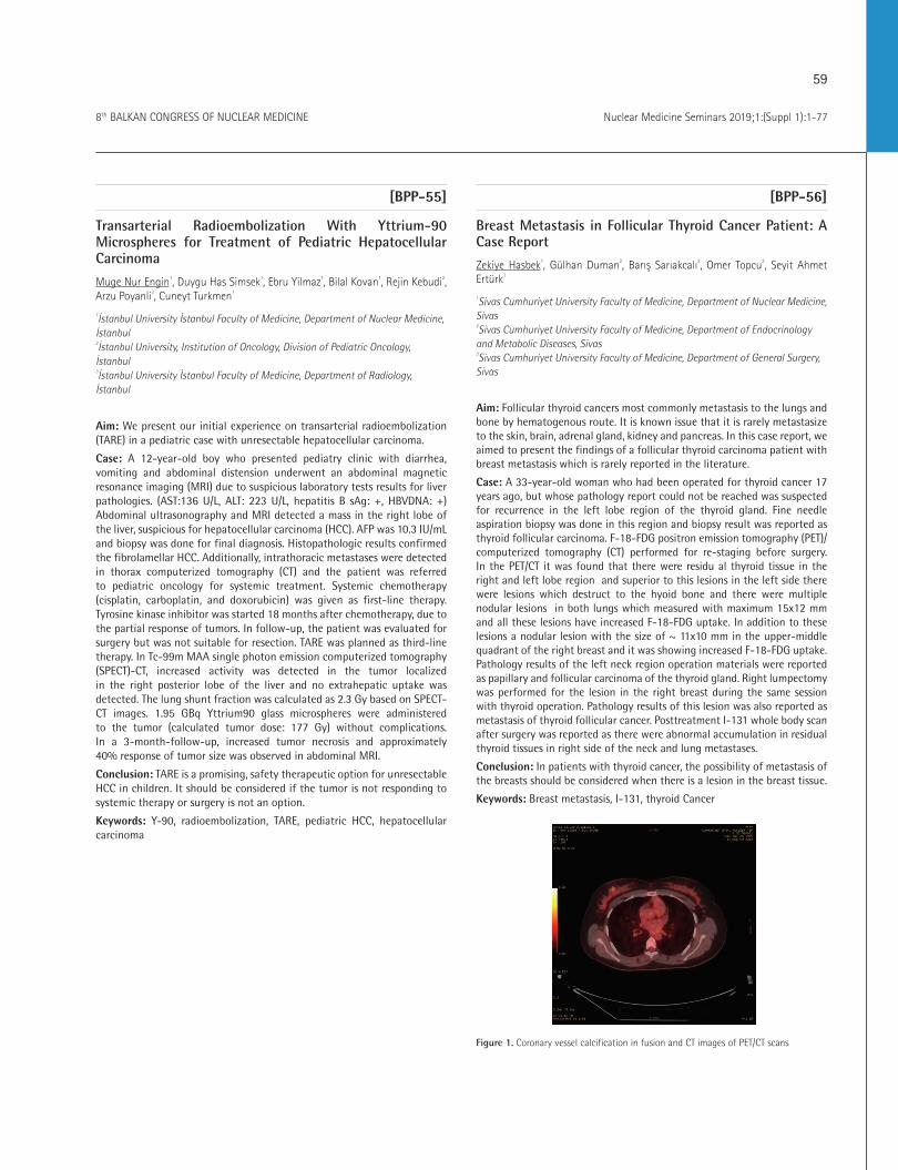
59
Nuclear Medicine Seminars 2019;1:(Suppl 1):1-778th BALKAN CONGRESS OF NUCLEAR MEDICINE
[BPP-55]
Transarterial Radioembolization With Yttrium-90 Microspheres for Treatment of Pediatric Hepatocellular Carcinoma
Muge Nur Engin1, Duygu Has Simsek1, Ebru Yilmaz1, Bilal Kovan1, Rejin Kebudi2, Arzu Poyanli3, Cuneyt Turkmen1
1İstanbul University İstanbul Faculty of Medicine, Department of Nuclear Medicine, İstanbul 2İstanbul University, Institution of Oncology, Division of Pediatric Oncology, İstanbul 3İstanbul University İstanbul Faculty of Medicine, Department of Radiology, İstanbul
Aim: We present our initial experience on transarterial radioembolization (TARE) in a pediatric case with unresectable hepatocellular carcinoma.
Case: A 12-year-old boy who presented pediatry clinic with diarrhea, vomiting and abdominal distension underwent an abdominal magnetic resonance imaging (MRI) due to suspicious laboratory tests results for liver pathologies. (AST:136 U/L, ALT: 223 U/L, hepatitis B sAg: +, HBVDNA: +) Abdominal ultrasonography and MRI detected a mass in the right lobe of the liver, suspicious for hepatocellular carcinoma (HCC). AFP was 10.3 IU/mL and biopsy was done for final diagnosis. Histopathologic results confirmed the fibrolamellar HCC. Additionally, intrathoracic metastases were detected in thorax computerized tomography (CT) and the patient was referred to pediatric oncology for systemic treatment. Systemic chemotherapy (cisplatin, carboplatin, and doxorubicin) was given as first-line therapy. Tyrosine kinase inhibitor was started 18 months after chemotherapy, due to the partial response of tumors. In follow-up, the patient was evaluated for surgery but was not suitable for resection. TARE was planned as third-line therapy. In Tc-99m MAA single photon emission computerized tomography (SPECT)-CT, increased activity was detected in the tumor localized in the right posterior lobe of the liver and no extrahepatic uptake was detected. The lung shunt fraction was calculated as 2.3 Gy based on SPECT-CT images. 1.95 GBq Yttrium90 glass microspheres were administered to the tumor (calculated tumor dose: 177 Gy) without complications. In a 3-month-follow-up, increased tumor necrosis and approximately 40% response of tumor size was observed in abdominal MRI.
Conclusion: TARE is a promising, safety therapeutic option for unresectable HCC in children. It should be considered if the tumor is not responding to systemic therapy or surgery is not an option.
Keywords: Y-90, radioembolization, TARE, pediatric HCC, hepatocellular carcinoma
[BPP-56]
Breast Metastasis in Follicular Thyroid Cancer Patient: A Case Report
Zekiye Hasbek1, Gülhan Duman2, Barış Sarıakcalı2, Omer Topcu3, Seyit Ahmet Ertürk1
1Sivas Cumhuriyet University Faculty of Medicine, Department of Nuclear Medicine, Sivas 2Sivas Cumhuriyet University Faculty of Medicine, Department of Endocrinology and Metabolic Diseases, Sivas 3Sivas Cumhuriyet University Faculty of Medicine, Department of General Surgery, Sivas
Aim: Follicular thyroid cancers most commonly metastasis to the lungs and bone by hematogenous route. It is known issue that it is rarely metastasize to the skin, brain, adrenal gland, kidney and pancreas. In this case report, we aimed to present the findings of a follicular thyroid carcinoma patient with breast metastasis which is rarely reported in the literature.
Case: A 33-year-old woman who had been operated for thyroid cancer 17 years ago, but whose pathology report could not be reached was suspected for recurrence in the left lobe region of the thyroid gland. Fine needle aspiration biopsy was done in this region and biopsy result was reported as thyroid follicular carcinoma. F-18-FDG positron emission tomography (PET)/computerized tomography (CT) performed for re-staging before surgery. In the PET/CT it was found that there were residu al thyroid tissue in the right and left lobe region and superior to this lesions in the left side there were lesions which destruct to the hyoid bone and there were multiple nodular lesions in both lungs which measured with maximum 15x12 mm and all these lesions have increased F-18-FDG uptake. In addition to these lesions a nodular lesion with the size of ~ 11x10 mm in the upper-middle quadrant of the right breast and it was showing increased F-18-FDG uptake. Pathology results of the left neck region operation materials were reported as papillary and follicular carcinoma of the thyroid gland. Right lumpectomy was performed for the lesion in the right breast during the same session with thyroid operation. Pathology results of this lesion was also reported as metastasis of thyroid follicular cancer. Posttreatment I-131 whole body scan after surgery was reported as there were abnormal accumulation in residual thyroid tissues in right side of the neck and lung metastases.
Conclusion: In patients with thyroid cancer, the possibility of metastasis of the breasts should be considered when there is a lesion in the breast tissue.
Keywords: Breast metastasis, I-131, thyroid Cancer
Figure 1. Coronary vessel calcification in fusion and CT images of PET/CT scans
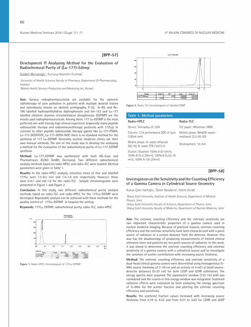
60
Nuclear Medicine Seminars 2019;1:(Suppl 1):1-77 8th BALKAN CONGRESS OF NUCLEAR MEDICINE
[BPP-57]
Development ff Analysing Method for the Evaluatıon of Radiochemıcal Purity of (Lu-177)-Edtmp
Güldem Mercanoğlu1, Kurtuluş Hayrettin Eryılmaz2
1University of Health Sciences Faculty of Pharmacy, Department Of Pharmacology, İstanbul 2Moltek Health Services Production and Marketing Inc, Kocaeli
Aim: Various radiopharmaceuticals are available for the systemic radiotherapy of pain palliation in patients with multiple skeletal lesions and osteoblastic lesions on skeletal scintigraphy. P-32, Sr-89, and Re-186 labelled hydroxyethylidene diphosphonate and Sm-153 and Lu-177 labelled ethylene diamine tetramethylene phosphonate (EDTMP) are the mostly used radiopharmaceuticals. Among them, 177 Lu-EDTMP is the most preferred one with having high clinical experience (especially many peptide radionuclide therapy and radioimmunotherapy protocols with 177Lu). In contrast to other peptide radionuclide therapy agents like Lu-177-PSMA, Lu-177-DOTATATE, Lu-177-DOTA-NOC there is no standard method for the synthesis of 177 Lu-EDTMP. Generally, nuclear medicine clinics use their own manual methods. The aim of this study was to develop the analysing a method for the evaluation of the radiochemical purity of Lu-177-EDTMP synthesis.
Method: Lu-177-EDTMP was synthesized with both ML-Eazy and Pharmatracer, (EZAG GmBH, Germany). Two different radiochemical analysis methods based on radio-HPLC and radio-TLC were studied. Method parameters were given in Table 1.
Results: In the radio-HPLC analysis, retention times of free and labelled 177Lu were 7.5-8.5 min and 1.4-1.6 min respectively. However, these were 0-0.1 and 0.8-1.0 for the radio-TLC. Sample chromatograms were presented in Figure 1 and Figure 2.
Conclusion: In this study, two different radiochemical purity analysis methods based on radio-TLC and radio-HPLC for the 177Lu-EDTMP were developed. Repeatable analysis can be achieved with these methods for the quality control of 177Lu-EDTMP in hospital the setting.
Keywords: 177Lu, EDTMP, radiochemical purity, radio-TLC, radio-HPLC
Figure 1. Radio-HPLC chromatogram of 177Lu-EDTMP
Figure 2. Radio-TLC chromatogram of labelled EDMP
Table 1. Method parametersRadio-HPLC Radio-TLC
Device: Shimadzu LC-20A TLC paper: Whatman 3MM
Column: C18 perfectbond ODS-H 5μm (100x4 mm)
Mobile phase: NH4OH-water-methanol (2.5-50-50)
Mobile phase: A: water:ethanole (90:10), B: water:TFA (100:0.1)
Development: 10 mm
Elution: Gradient 100% A (0-5min), 100% B (5-5.30min), 100% B (5.30-16 min), 100% A (16-20min)
[BPP-58]
Investıgatıon on the Sensitivity and the Counting Efficiency of a Gamma Camera in Cylindrical Source Geometry
Rukiye Çakır Haliloğlu1, Özlem Karadeniz2, Hatice Durak3
1Dokuz Eylül University, Institute of Health Sciences, Department of Medical Physics, İzmir 2Dokuz Eylül University Faculty of Sciences, Department of Physics, İzmir 3Dokuz Eylül University Faculty of Medicine, Department of Nuclear Medicine, İzmir
Aim: The extrinsic counting efficiency and the extrinsic sensitivity are two important characteristic properties of a gamma camera used in nuclear medicine imaging. Because of practical reasons, extrinsic counting efficiency and the extrinsic sensitivity have been characterized with a point source of radiation at a certain distance from the detector. However this way has the disadvantage of producing measurements of limited clinical relevance since real patients are not point sources of radiation. In this work, it was aimed to determine the extrinsic counting efficiency and extrinsic sensitivity of a gamma camera with a cylindrical source and to investigate the variation of scatter contribution with increasing source thickness.
Method: The extrinsic counting efficiency and extrinsic sensitivity of a dual-head clinical gamma camera were determined using homogeneous Tc-99m source thickness of 2-18 cm and an activity of 4 mCi at small source-detector distances (5-25 cm) for both LEGP and LEHR collimators. The energy spectra were acquired. The asymmetric window (133-153 keV) was considered and the counts in this energy window was integrated. Scattered radiation effects were evaluated by both analyzing the energy spectrum of Tc-99m for the scatter fraction and plotting the extrinsic counting efficiency and sensitivity.
Results: The scattered fraction values increased with increasing source thickness from 0.19 to 4.52 and from 0.27 to 4.62 for LEHR and LEGP
61
Nuclear Medicine Seminars 2019;1:(Suppl 1):1-778th BALKAN CONGRESS OF NUCLEAR MEDICINE
collimator. Calculated extrinsic sensitivity decreased with increasing source thickness, from 55 counts/sMBq to 24 counts/sMBq and from 98 counts/sMBq to 41 counts/sMBq for LEHR and LEGP collimator. The extrinsic efficiency decreases with increasing source thickness, from 0.19% to 0.08% and from 0.35% to 0.14% for LEHR and LEGP collimator. Calculated extrinsic efficiency increased with increasing source-to-detector distance, from 0.19% to 0.54% and from 0.35% to 0.97% for LEHR and LEGP collimator.
Conclusion: The scattered fraction values increased with increasing source thickness. Calculated extrinsic sensitivity shows a decreasing trend with increasing source thickness and increasing source-to-detector distance. The extrinsic efficiency decreases with increasing source thickness and efficiency increases with increasing sourceto-detector distance. Increasing source to detector distances results with increasing extrinsic counting efficiency but decreasing extrinsic sensitivity.
Keywords: Sensitivity, counting efficiency, Scattered radiation effects, gamma camera, cylindrical source, rectangular detector.
[BPP-59]
The Role of Thyroıd Scintigraphy in Determining the Residual Tissue after Thyroidectomy for Thyroid Carcinoma
Derya Çayır1, Bahadır Külah2, Mehmet Bozkurt1
1University of Health Sciences, Dışkapı Yıldırım Beyazıt Training and Research Hospital, Clinic of Nuclear Medicine, Ankara2University of Health Sciences, Dışkapı Yıldırım Beyazıt Training and Research Hospital, Clinic of General Surgery, Ankara
Aim: Presence of the residual tissue after thyroidectomy for differentiated thyroid carcinoma (DTC) effects the success of radioiodine (RAI) ablation. The presence of the residual tissue is a risk factor for locoregional recurrence and can be evaluated by thyroid scintigraphy and serum thyroglobuline (Tg) levels. We aimed to investigate the role of the thyroid scintigraphy in determining the presence of the residual thyroid tissue after total thyroidectomy for DTC.
Method: Our study included 259 patients who underwent total thyroidectomy between July 2014 and October 2018, and diagnosed as DTC histopathologically. Presence, localization and size of the residual thyroid tissue were detected by using Tc-99m pertechnenate scintigraphy 3-6 weeks after thyroidectomy. Preooperative ultrasonography findings, postoperative stimulated serum Tg levels, and histopathologic findings were also investigated retrospectively.
Results: The presence of the residual tissue was detected in 187 (165 female and 22 male) patients. Mean age of the patients was 48±0.85 years. Serum Tg levels were high (>2 ng/mL) in 59 (31.6%) patients. There was no statistically significant difference between the patients with thyroid papillary microcarcinoma and patients with thyroid papiller carcinoma in terms of the presence of the residual tissue (p=0.062). The comparison of the preoperative ultrasonography findings with postoperative scintigraphy findings revealed that patients with nodule size larger than 2 cm and patients with number of the nodules more than two had statistically more frequent residual tissue than others (p=0.047 and p=0.036, respectively).
Conclusion: Determining the presence of the residual tissue after total thyroidectomy is necessary and important in terms of postoperative treatment strategy including RAI ablation. The volume of the residual tissue defines the next treatment as surgery or RAI ablation. Ultrasonographic examination for the presence of the residual tissue is ineffective due to postoperatif changes and presence of the granulation tissue in the operation field in the early postoperative period (three months). Postoperative serum Tg levels accompanied with early scintigraphic findings seem to be more
helpful for assessing the presence of the residual tissue and treatments strategy after total thyroidectomy for DTC.
Keywords: Differentiated thyroid carcinoma, thyroid scintigraphy, residual tissue
[BPP-60]
Ultrasound and Tc-99m MIBI Parathyroid SPECT/CT in Value of Secondary Hyperparathyroidism
Pelin Arıcan1, Ali Konukseven1, Duygu Has Şimşek2, Özgür Ekmekçioğlu1, Ayşen Sever1, Mesut Kafi1
1University Of Health Sciences, Şişli Hamidiye Etfal Training and Research Hospital, Clinic of Nuclear Medicine, İstanbul2İstanbul University Faculty İstanbul Medicine, Department of Nuclear Medicine, İstanbul
Aim: Secondary hyperparathyroidism (SHPT) is a common complication of chronic renal failure. SHPT often involves multiple glands and the location of parathyroid lesions is highly variable. Thus, imaging is important for planning and guiding surgical treatment in SHPT. Ultrasonography, computerized tomography (CT), and (magnetic resonance imaging) MRI are used at pre-operation location of parathyroid lesions. Tc-99m MIBI dual phase imaging is commonly used in detecting parathyroid lesions because of its simplicity, when SPECT/CT has further improved diagnosis of SHPT. The purpose of this study is to compare the diagnosis values of Tc99m MIBI planar imaging, SPECT/CT, and ultrasonography in the detection of parathyroid lesion with chronic renal failure.
Method: A retrospective study consisted of 38 patients (19 female, 19 male; mean age: 49±23 years) with chronic renal failure treated by hemodialysis and underwent Tc-99m MIBI single photon emission computerized tomography (SPECT)/CT parathyroid scintigraphy for diagnosis of SHPT in our hospital between 2016 and 2018 years. Parathyroid hormone level was greater than 600 pg/mL, calsium concentration was greater than 10.4 mg/dL in all patients. Ultrasonography was performed in all patients before scintigraphy. After IV injection of 740 MBq of Tc-99m MIBI, early planar scan and SPECT/CT of the neck and chest was obtained at 15 minutes and delayed scan was obtained at 120 minutes. Scintigraphy was considered positive when focal tracer retansion in neck or mediastinium on delayed static and/or SPECT/CT images. All cases were confirmed by pathological examination. The detection rate of lesion using ultrasonography, planar imaging, and SPECT/CT were analyzed.
Results: SPECT/CT identified parathyroid lesion in 31 patients while the planar imaging detected parathyroid lesions in 27 patients. Parathyroid pathology in 28 patients were found by ultrasonography. Scintigraphy demonstrated ectopic parathyroid lesions in 3 patients. Ultrasonography did not identify ectopic lesion. The sensitivities of ultrasonography, planar scan and SPECT/CT were 87%, 84%, and 96%, the specificity was 71%, 100%, and 100%, and the accuracy was 87%, 87%, and 97%, respectively.
Conclusion: Our study demonstrated that SPECT/CT is better in detecting parathyroid lesions than planar scan and ultrasonography in patients with SHPT. Scintigraphy is superior than ultrasonography to determine ectopic parathyroid lesions. SPECT/CT provides precise localization of parathyroid lesions before surgery in SHPT.
Keywords: Secondary hyperparathyroidism, parathyroid scintigraphy, SPECT/CT, ultrasonography

62
Nuclear Medicine Seminars 2019;1:(Suppl 1):1-77 8th BALKAN CONGRESS OF NUCLEAR MEDICINE
[BPP-61]
A Quantitatively Visual Functional Grading Scale for Somatostatin Receptor Scintigraphy Images
Cati Raluca Stolniceanu1, Teodor Marian Ionescu1, Irena Cristina Grıerosu1, Milovan Matovıc2, Cipriana Stefanescu1
1Grigore T. Popa University of Medicine and Pharmacy, Department of Nuclear Medicine, Biophysics and Medical Physics, St. Spiridon Hospital, Clinic of Nuclear Medicine, Iaşi, Romania 2University of Kragujevac Faculty of Medicine, Clinical Center Kragujevac, Center for Nuclear Medicine, Serbia
Aim: Somatostatin receptor scintigraphy (SRS) has proven useful for characterizing malignancy in neuroendocrine tumours (NETs) but evaluation methods of the scintigraphic images can still be improved. Aim of our study was to develop a visual functional grading scale (VFGS) for assessing SRS images: whole body scintigraphy (WBS), single photon emission computerized tomography (SPECT) and spot scintigraphy for patients with primary and metastatic pancreatic NETs (pNETs), pheochromocytoma (Pheo) and medullary thyroid cancer (MTC).
Method: Our prospective study during 12 months included SRS images of 22 patients, with metastatic NETs, from two centers. Based on the location of the primary tumor, we defined: group 1-4 patients with Pheo, group 2-10 pNETs, group 3-8 MTC. We used Tc-99m-tektrotyd and gamma camera Siemens e cam Dual Head, equipped with LEAP collimators and appropriate software for acquisition and processing. Study design: early dynamic scintigraphy (60 images, 1 image/second, 128x128 matrix), spot scintigraphy (10 minutes/image, 256x256 matrix), WBS (matrix 256x256, 6cm/min bed movement) and SPECT (128x128 matrix and angle rotation of 6 degrees). Images were acquired at 10 minutes, 2-4 and 24 hours after i.v. administration of 10.57 MBq/kg bw Tc99m-Tektrotyd, Polatom. We defined and used a VFGS, (3 grades, based on the uptake intensity) to assess quantitatively SRS images for the pathological uptakes (Table 1). Uptake was related to a target standard right thigh background region, using a ROI of 240 pixels, with a maximum of 25 counts.
Results: We quantified a total of 70 pathological radiopharmaceutical uptakes (PRU), variable grades from G
1-3. More than half (58,5%) (n=41) were G3, mostly of them 68,3% (n=28) were liver metastases. 21 PRU (30%) were G2 and 7 (10 %) G1.. In group 1, were 8 PRU (11,4%), G3 abdominal uptake in all 4 patients and in a single case a G3 cervicothoracic uptake. In group 2, were 48 PRU (68,6%), with G1-3 abdominal and G2-3 hepatic PRU in all the patients, except one. In group 3, we found 14 PRU (20%), 7 G1-3 in the cervicothoracic bed, 6 G1-2 hepatic and one G1 abdominal uptake. Was a single G1 lung uptake in whole group of 70 PRU.
Conclusion: This simple, reliable VFGS, based on the PRU characteristics, can be used for research purposes, allowing an enough detailed analysis, for further statistically evaluations. In the future, we intend to validate VFGS on a large cohort of patients with NETs.
Keywords: Neuroendocrine tumours, somatostatin receptor scintigraphy, grading scale, pathological uptake
Table 1. Visual Functional Grading scale for assessing somatostatin receptor scintigraphy images for pathological uptakesUptake classification Grade (intensity)
Small uptake: <70 counts/ROI G1: + (low intensity)
Medium uptake: 70-120 counts/ROI G2: ++ (moderate intensity)
High uptake: >120 counts/ROI G3: +++ (markedly intensity)
[BPP-62]
DTC: Case Report Of False Positive I-131 Uptake on WBS in a Patient With Bilateral Femur Osteonecrosis.
Ioannis PIakovou1, Evanthia I Giannoula1
1Aristotle University of Thessaloniki, Papageorgiou Hospital, Clinic of 3rd Nuclear Medicine, Greece
Aim: Differentiated Thyroid Cancer (DTC) is one of the fastest growing cancers worldwide. Despite the generally good prognosis of thyroid carcinoma, about 5% of patients will develop metastatic disease, exhibiting a more aggressive behavior. Radioiodine whole-body scintigraphy (WBS) has been used in the detection of DTC. Radioiodine is a sensitive marker for detection of thyroid cancer; however, radioiodine uptake is not specific for thyroid tissue. It can also be seen in healthy tissue as well as in inflammation, or in a variety of benign and malignant non-thyroidal entities. Case: The subject of the present case report is a 52 years old man with brain metastatic DTC who received radioiodine therapy and corticosteroids as palliative therapy. WBS revealed bilateral iodine uptake of the femur. Corticosteroid therapy is among the most widely recognized risk factor for osteonecrosis, which at the present case had to be recognized as a false positive I-131 uptake in order to avoid diagnostic error. Conclusion: Radioiodine WBS plays an important role in clinical decision making for the evaluation and the management of patients with DTC. Despite its high range of sensitivity and specificity, a variety of reports of false positive whole body scans has demonstrated a diversity of causes. Comprehension of the physiology of iodine uptake and of the pathophysiology of clinical entities which end up giving false positives scans, provides clinicians a useful tool in order to avoid diagnostic and therapeutic errors as far as DTC is concerned.
Keywords: Differentiated thyroid cancer, radioiodine whole body scintigraphy, false positive, corticosteroids, osteonecrosis
63
Nuclear Medicine Seminars 2019;1:(Suppl 1):1-778th BALKAN CONGRESS OF NUCLEAR MEDICINE
[BPP-63]
Structural and Functional Imaging for the Diagnosis of Neuroendocrine Carcinoma of the Cervix
Wael Jalloul1, Ruxandra Tibu1, Tudor Ciobanu1, Alexandru Tarca1, Aeligton Filho Arcoverde3, Mateus Polvore4, Ana-Maria Statescu1, Cipriana Ștefănescu2
1St. Spiridon Emergency Hospital, Clinic of Nuclear Medicine Laboratory, Iaşi, Romania 2Grigore T. Popa University of Medicine and Pharmacy, Department of Nuclear Medicine, Biophysics and Medical Physics, Iaşi, Romania 3Federal University of Piauí, Brazil 4Federal University of Uberlândia, Brazil
Aim: Neuroendocrine carcinoma of the cervix (NECC) is an aggressive histological variant accounting for about 1-1.5% of all cervical cancers. Small cell neuroendocrine carcinomas (NEC) is the most common type of NECC. It is more likely to invade the lymphovascular space and to spread to the regional lymph node basin at the time of diagnosis. The purpose of our study was to describe the imaging aspects in a case of NECC and to look for a theranostic approach to treat it.
Method: We present the case of 70-year-old woman diagnosed with NECC (small cell NEC). After uterine curettage and electrical conization, followed by chemotherapy, she was sent for a Tc-99m-HDP bone scan in order to obtain further informations about the nature of a lesion described on a follow up magnetic resonance imaging (MRI) on the left ischium and to look for other possible bone metastasis.
Results: a) Anatomopathological exam: a neuroendocrine cervical small cell carcinoma (poorly differentiated). -Immunohistochemical exam: positive chromogranin A, synaptophysin and Ki67 (40-50%). b) Abdominal and pelvic MRI: the exact topography and extension of the neoplasia. Four metastatic sites were discovered in the liver and one nodular lesion at the base of the right lung. c) Thoracic CT scan: a pulmonary nodule on the right lung. d) A follow up MRI (6 months after the first MRI): a possible metastatic lesion on the left ischium. No other aspects of active disease were discovered. e) Tc-99m-HDP bone scan: A high uptake on the left ischium (corresponding to the MRI description) and an other one on the mid shaft of the tibia, which could have a metastatic etiology. The theranostic approach with the use of somatostatin analogues was not possible in this case because of the high value of Ki67 (40-50%).
Conclusion: MRI, CT and Tc-99m-HDP bone scan are essential tools in the pre and post-therapeutic assessment of NECC for local staging and to determine the extent of nodal and metastatic spread. In our case, the patient has simultaneously liver, lung and bone metastasis which is rarely described.
Keywords: Neuroendocrine carcinoma of the cervix, 99mTc-HDP bone scan, metastasis.
[BPP-64]
Ectopic Parathyroid Adenoma After Radioactive Iodine Therapy: is That a Coincidence?
Laura Teodoriu1, Ana-Maria Statescu2, Roxana Iacob2, Irena Grierosu2, Mihai Gutu2, Cristina Preda1, Cipriana Stefanescu2
1St. Spiridon Emergency University Hospital, Clinic of Endocrinology, Iaşi, Romania 2St. Spiridon Emergency University Hospital, Clinic of Nuclear Medicine Laboratory, Iaşi, Romania 3Grigore T. Popa University of Medicine and Pharmacy, Department of Nuclear Medicine, Biophysics and Medical Physics, Iaşi, Romania
Aim: Radioactive iodine treatment (RAI) is the first, still the best, theranostic approach for high risk papillary thyroid carcinoma management. However, I-131 radiation can impair adjacent parathyroid tissue. Primary hyperparathyroidism is a rare complication of RAI, with, typically, long period of latency following radiation exposure. Our aim is to stress on the choice of a personalized Tc-99m MIBI scan acquisition protocol in patients with RAI history and parathyroid pathology association.
Method: A 55 years old female patient was admitted to the endocrinology department for thyroid monitoring and suspicion of parathyroid pathology. Her medical history mentions a papillary thyroid carcinoma (T2N0M0) diagnosed 17 years ago, treated by total thyroidectomy and RAI in two doses (2.96 GBq and 2.59 GBq), due to histopathology and elevated thyroglobulin. At the presentation, the patient had elevated values for: serum calcium (11.45 mg/dL) in three consecutive days, PTH (99.48 pg/mL) and urinary calcium (520 mg/24h). Ultrasound (US), computerized tomography (CT) and functional - Tc-99m MIBI (early, 1h and 2h images) evaluation were assessed.
Results: US images outlines bilateral renal calculi and no remnant thyroid tissue in anterior cervical region. CT evidenced no pathologic formations, but Tc-99m MIBI scan revealed ectopic parathyroid adenoma in the superior mediastinum, with 2h delayed radiotracer accumulation. The patient undergone surgery and the histopathology confirmed the parathyroid adenoma. The association of papillary thyroid carcinoma and primary hyperparathyroidism raises some questions about the etiology of this parathyroid node in this particular case. According to literature,
I-131 could affect parathyroid tissue with long period of latency, like in our case report (16 years). However, ectopic parathyroid adenoma would have no reason being affected from RAI, because there was no direct contact of parathyroid tissue with thyroid treated tissue. Also, literature reports showed a high prevalence of the association of papillary/follicular thyroid carcinoma and primary hyperparathyroidism with no conclusive mechanism, only hypothesis that common genetic and transcription factors are implicated.
Conclusion: Tc-99m MIBI functional personalized evaluation with two delayed points, 1h (for malignant tissue) and 2h (for benign parathyroid tissue), could be essential in differential diagnosis, complementary to structural images, in selected cases.
Keywords: Ectopic parathyroid adenoma, RAI, Tc-99m MIBI, personalized evaluation
64
Nuclear Medicine Seminars 2019;1:(Suppl 1):1-77 8th BALKAN CONGRESS OF NUCLEAR MEDICINE
[BPP-65]
Presentation of Situs Inversus Totalis on I-123 MIBG Scintigraphy
Bedri Seven1
1Amasya University, Sabuncuoğlu Şerefeddin Training And Research Hospital, Clinic of Nuclear Medicine, Amasya
Aim: Situs inversus is a rare congenital condition characterized by transposition of organs to the opposite side of the body. Situs inversus may be complete (situs inversus totalis) or partial (situs inversus partialis) confined to either the thoracic or the abdominal viscera. This condition generally does not cause inconvenience in daily life. It may be discovered in infancy but often remains asymptomatic and discovered incidentally in adult life. A 27-year-old woman with labile hypertension was referred for I-123 metaiodobenzylguanidine (MIBG) scintigraphy due to suspected pheochromocytoma. 4 and 24 hours after intravenous administration of I‐123 MIBG planar and SPECT images were obtained. I-123 MIBG scintigraphy showed no non-physiological uptake, but revealed situs inversus totalis. In the images, the left-sided liver and right-sided spleen was shown. Furthermore, myocardial I-123 MIBG uptake was seen in the right hemithorax. Situs inversus totalis was also revealed by magnetic resonance imaging in the patient.
Keywords: I-123 MIBG, pheochromocytoma, situs Inversus
[BPP-66]
A Rare Case Report: A Parathyroid Adenoma Presenting with Proximal Muscle Weakness in an Adolescent
Ebru Salmanoğlu1, Bülent Güneri2, Fatih Temiz3
1Kahramanmaraş Sütçü İmam University Faculty of Medicine, Department of Nuclear Medicine, Kahramanmaraş 2Kahramanmaraş Sütçü İmam University Faculty of Medicine, Department of Orthopaedics And Traumatology, Kahramanmaraş 3Kahramanmaraş Sütçü İmam University Faculty of Medicine, Department of Pediatric Endocrinology, Kahramanmaraş
Aim: MIBI is lipophilic, cationic agent, it passes through the cell membrane by passive diffusion and MIBI is kept from mitochondria with a high negative transmembrane potential. Tc-99m MIBI Parathyroid Scintigraphy is a sensitive nuclear medicine imaging modality for the diagnosis of parathyroid adenoma. We wanted to present a parathyroid adenoma case report which is seen rarely in adolescents in the literature presenting with proximal muscle weakness.
Method: A fourteen year old woman was applied to the department of orthopaedics and traumatology suffering with lower extremity pain and suffering away limping for approximately two months.
Results: Proximal muscle weakness was present in the patient’s physical examination. Gower’s finding was negative. The patient’s blood calcium level was high, blood phosphor level was low, alkaline phosphatase level was high, parathyroid hormone level was high. Despite bisphosphonates and calcitonin treatment, therere was a continuation of resistance hypercalcemia. Therefore the patient was referred to department of nuclear medicine with preliminary diagnosis of parathyroid adenoma. At the patient’s parathyroid scintigraphy, there was a typical image of parathyroid adenoma was shown at the inferior of the right thyroid lobe. The patient was operated. Parathyroid adenoma was confirmed by histopathology.
Conclusion: When an adolescent patient applys to the clinician with proximal muscle weakness, blood calcium, phosphor and parathyroid hormone levels should be evaluated. Parathyroid adenoma is should be considered in the patients with resistant to treatment and we believe that parathyroid scintigraphy is an important imaging method at the diagnosis.
Keywords: Adolescent, parathyroid adenoma, parathyroid scintigraphy
[BPP-67]
The Diagnostic Ability and Additional Clinical Value of SPECT/CT To Standard Planar Imaging in Tc-99m RBC Scintigraphy
Selin Soyluoglu1, Büşra Özdemir1, Gülay Durmuş Altun1
1Trakya University, Faculty Of Medicine, Department of Nuclear Medicine, Edirne
Aim: Gastrointestinal (GI) tract bleeding is a life-threatening, complex clinical problem that requires a multidisciplinary approach for a successful treatment. The aim of this study was to determine the additional clinical value of single photon emission computerized tomography (SPECT)/computerized tomography (CT) to standard planar imaging in patients referred to Tc-99m labeled red blood cell (RBC) scintigraphy for differential diagnosis of acute GI bleeding.
Method: Fifteen conventional dynamic and planar Tc-99m RBC scintigraphies of thirteen patients who referred to nuclear medicine department between January 2017 and December 2018 with suspicious of acute GI bleeding were evaluated retrospectively. SPECT/CT using a hybrid system was performed in nine patients who had positive findings on planar imaging. The number of patients and matters that SPECT/CT contributed to scintigraphic interpretation of presence or location of GI bleeding were recorded.
Results: The image fusion was easy and successful in demonstrating the exact anatomical localization of the extravasation regions. SPECT/CT could identified the location of bleeding foci in eight studies. Bleeding site of five patients with a history of cancer (bladder, gastric, coloncancers and a gastroenteropancreatic neuroendocrine tumor) which could not be determined by other imaging modalities, could be determined and guided the surgeon. In one patient, fixed activity accumulation in the midline of abdomen on planar images was found to be due to a previously unknown aortic dissection in SPECT/CT. In one patient, the accumulation of activity in right upper quadrant, which could be interpreted as false positive, was found to be gallbladder in SPECT/CT and delayed images at 6 hour showed the focus of bleeding in cecum.
Conclusion: SPECT/CT hybrid imaging is feasible and useful to facilitate image interpretation in patients with acute GI bleeding and increase the accuracy of Tc-99m RBC scintigraphy. SPECT imaging can guide the surgeon through more accurate localization. Therefore, for proper patient management, SPECT/CT should be applied to detect bleeding focus if present.
Keywords: SPECT/CT, Tc-99m RBC scintigraphy, gastrointestinal bleeding
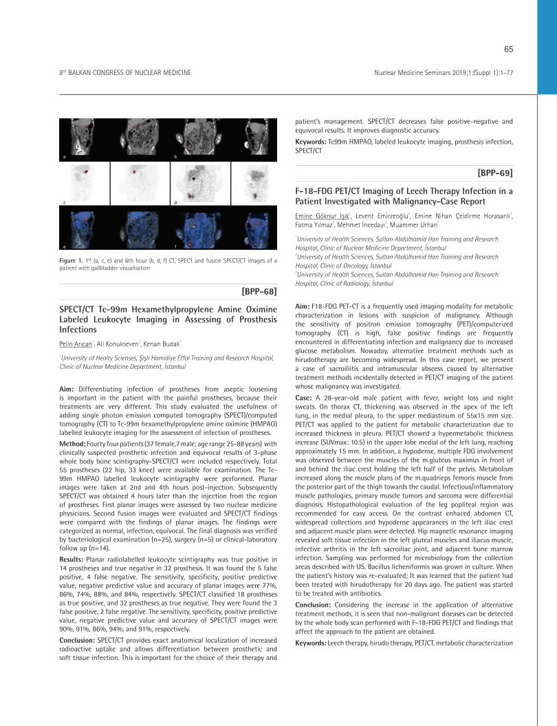
65
Nuclear Medicine Seminars 2019;1:(Suppl 1):1-778th BALKAN CONGRESS OF NUCLEAR MEDICINE
Figure 1. 1st (a, c, e) and 6th hour (b, d, f) CT, SPECT and fusion SPECT/CT images of a patient with gallbladder visualisation
[BPP-68]
SPECT/CT Tc-99m Hexamethylpropylene Amine Oximine Labeled Leukocyte Imaging in Assessing of Prosthesis Infections
Pelin Arıcan1, Ali Konukseven1, Kenan Budak1
1University of Healty Scienses, Şişli Hamidiye Etfal Training and Research Hospital, Clinic of Nuclear Medicine Department, İstanbul
Aim: Differentiating infection of prostheses from aseptic loosening is important in the patient with the painful prostheses, because their treatments are very different. This study evaluated the usefulness of adding single photon emission computed tomography (SPECT)/computed tomography (CT) to Tc-99m hexamethylpropylene amine oximine (HMPAO) labelled leukocyte imaging for the assessment of infection of prostheses.
Method: Fourty four patients (37 female,7 male; age range 25-88 years) with clinically suspected prosthetic infection and equivocal results of 3-phase whole body bone scintigraphy-SPECT/CT were included respectively. Total 55 prostheses (22 hip, 33 knee) were available for examination. The Tc-99m HMPAO labelled leukocyte scintigraphy were performed. Planar images were taken at 2nd and 4th hours post-injection. Subsequently SPECT/CT was obtained 4 hours later than the injection from the region of prostheses. First planar images were assessed by two nuclear medicine physicians. Second fusion images were evaluated and SPECT/CT findings were compared with the findings of planar images. The findings were categorized as normal, infection, equivocal. The final diagnosis was verified by bacteriological examination (n=25), surgery (n=5) or clinical-laboratory follow up (n=14).
Results: Planar radiolabelled leukocyte scintigraphy was true positive in 14 prostheses and true negative in 32 prosthesis. It was found the 5 false positive, 4 false negative. The sensitivity, specificity, positive predictive value, negative predictive value and accuracy of planar images were 77%, 86%, 74%, 88%, and 84%, respectively. SPECT/CT classified 18 prostheses as true positive, and 32 prostheses as true negative. They were found the 3 false positive, 2 false negative. The sensitivity, specificity, positive predictive value, negative predictive value and accuracy of SPECT/CT images were 90%, 91%, 86%, 94%, and 91%, respectively.
Conclusion: SPECT/CT provides exact anatomical localization of increased radioactive uptake and allows differentiation between prosthetic and soft tissue infection. This is important for the choice of their therapy and
patient’s management. SPECT/CT decreases false positive-negative and equivocal results. It improves diagnostic accuracy.
Keywords: Tc99m HMPAO, labeled leukocyte imaging, prosthesis infection, SPECT/CT
[BPP-69]
F-18-FDG PET/CT Imaging of Leech Therapy Infection in a Patient Investigated with Malignancy-Case Report
Emine Göknur Işık1, Levent Emirzeoğlu2, Emine Nihan Çeldirme Horasanlı3, Fatma Yılmaz3, Mehmet İncedayı3, Muammer Urhan1
1University of Health Sciences, Sultan Abdülhamid Han Training and Research Hospital, Clinic of Nuclear Medicine Department, İstanbul 2University of Health Sciences, Sultan Abdülhamid Han Training and Research Hospital, Clinic of Oncology, İstanbul 3University of Health Sciences, Sultan Abdülhamid Han Training and Research Hospital, Clinic of Radiology, İstanbul
Aim: F18-FDG PET-CT is a frequently used imaging modality for metabolic characterization in lesions with suspicion of malignancy. Although the sensitivity of positron emission tomography (PET)/computerized tomography (CT) is high, false positive findings are frequently encountered in differentiating infection and malignancy due to increased glucose metabolism. Nowaday, alternative treatment methods such as hirudotherapy are becoming widespread. In this case report, we present a case of sacroiliitis and intramuscular abscess caused by alternative treatment methods incidentally detected in PET/CT imaging of the patient whose malignancy was investigated.
Case: A 28-year-old male patient with fever, weight loss and night sweats. On thorax CT, thickening was observed in the apex of the left lung, in the medial pleura, to the upper mediastinum of 55x15 mm size. PET/CT was applied to the patient for metabolic characterization due to increased thickness in pleura. PET/CT showed a hypermetabolic thickness increase (SUVmax: 10.5) in the upper lobe medial of the left lung, reaching approximately 15 mm. In addition, a hypodense, multiple FDG involvement was observed between the muscles of the m.gluteus maximus in front of and behind the iliac crest holding the left half of the pelvis. Metabolism increased along the muscle plans of the m.quadrieps femoris muscle from the posterior part of the thigh towards the caudal. Infectious/inflammatory muscle pathologies, primary muscle tumors and sarcoma were differential diagnosis. Histopathological evaluation of the leg popliteal region was recommended for easy access. On the contrast enhaced abdomen CT, widespread collections and hypodense appearances in the left iliac crest and adjacent muscle plans were detected. Hip magnetic resonance imaging revealed soft tissue infection in the left gluteal muscles and iliacus muscle, infective arthritis in the left sacroiliac joint, and adjacent bone marrow infection. Sampling was performed for microbiology from the collection areas described with US. Bacillus licheniformis was grown in culture. When the patient’s history was re-evaluated; It was learned that the patient had been treated with hirudotherapy for 20 days ago. The patient was started to be treated with antibiotics.
Conclusion: Considering the increase in the application of alternative treatment methods, it is seen that non-malignant diseases can be detected by the whole body scan performed with F-18-FDG PET/CT and findings that affect the approach to the patient are obtained.
Keywords: Leech therapy, hirudo therapy, PET/CT, metabolic characterization

66
Nuclear Medicine Seminars 2019;1:(Suppl 1):1-77 8th BALKAN CONGRESS OF NUCLEAR MEDICINE
Figure 1. F-18 FDG PET-CT. a) MIP, hypermetabolic pleural thickness increase (b, c, d), hypermetabolic field in muscle plans in the leg (d, e, f)
[BPP-70]
Unusual Presentation of Extrapulmonary Tuberculosis with Splenic Involvement at PET/CT Mimicking Malignant Lymphoma
Esra Çiftçi1, Huri Tilla İlçe1, Fahri Yılmaz2, Gözde Çakar2, Oğuz Karabay3
1Sakarya University Faculty of Medicine, Training and Research Hospital, Clinic of Nuclear Medicine, Sakarya 2Sakarya University Faculty of Medicine, Training and Research Hospital, Clinic of Pathology, Sakarya 3Sakarya University, Faculty of Medicine, Training and Research Hospital, Clinic of Infectious Disease and Clinical Microbiology, Sakarya
Aim: We aimed to report a case of unusual presentation of extrapulmonary TB with splenic involvement and generalized lymphadenopathy showed on positron emission tomography (PET)/computerized tomography (CT), mimicking lymphoma, which is not shown in literature before.
Case: A 45 year-old man,who presented with fever was referred to our hospital. He had history of ankilozan spondilitis which is in remission without drug use. His laboratory data revealed slightly pansitopeni, an elevated acute-phase reactors (AFR). His blood, urine cultures and PCR assay were negative. BM biopsy showed “mildly hypercelluler without atipic differantiation”. PET/CT showed multiple hypermetabolic LNs localized more at abdominopelvic lymphatic regions (SUVmax:15) in addition to high FDG uptake of spleen with splenomagaly (SUVmax:10.6) and less LN at left supraclavikular and mediastinal regions. Also milier noduler apperance was observed at lung. Lymphoma were concerned and after laparoscopic splenectomy, paraaortic LN and periton biopsy, revealed granulomas consisting of caseous necrosis and epithelioid histiocytes with Langhans type giant cells at all those spesimen,typical of TB. He was symptom-free after standard anti-TB treatment.
Results: Extrapulmonary TB occurs in approximately 1/5 of all immunocompetent patients and in more than half of all immunocompromised patients. It’s also more common in children and women, is extremely rare in non-HIV patients. For abdominal TB, after abdominal lymphadenopathy, the most commonly involved parts are the ileocecal region and ascending colon, then peritoneum, mesentery, liver, pancreas and spleen. Akdoğan et al., presented PET/CT findings of isolated gastric TB mimicking gastric cancer and lymphoma at whom had a newly diagnosed for ankylosing spondylitis as presented in our patient’s history. Hepatosplenic TB is representing less than 1% and generally develops secondary to miliary TB. Our patient had also miliary lung parenchymal pattern. Since splenic TB has no characteristic symptoms or abnormal imaging findings, the misdiagnosis rate with lymphoma, splenic abscess, carcinoma of the spleen and rheumatic fever is high. In a case reported as ITP associated with splenic TB, fever was the only symptom with trombosytopenia, anemia and elevated AFR same with our case.
Conclusion: We suggest that splenic TB must be suspected in patients who have fever, abdominal lymphadenopathy and thrombocytopenia with high splenic FDG uptake and also history of autoimmune disease using immunosuppressive agents at past must arouse suspicion.
Keywords: Splenic Tubercolusis, FDG PET/CT, lymphoma
Figure 1. Normal spleen seen on the left side and Granulomas consisting of caseous necrosis and epithelioid histiocytes with Langhans type giant cells seen on the right side (H&E X100)
[BPP-71]
F-18 FDG-PET/CT Imaging in Takayasu Arteritis
Zekiye Hasbek1, Seyit Ahmet Ertürk1, Ali Cakmakcılar1, Emre Cicekli2
1Sivas Cumhuriyet University Faculty of Medicine, Department of Nuclear Medicine, Sivas 2Medicana Sivas Hospital, Clinic of Internal Disease, Sivas
Aim: Takayasu arteritis (TA) is a rare systemic vasculitic disease of unknown etiology and it is characterized by chronic inflammation of large and mid-size arteries, especially the aorta and branches of pulmonary arteries. TA is most commonly affects young women. It is very insidious and chronic disease. Although the mean age at diagnosis is between 30-32 years, pathological changes starts earlier at age of between 10-24. We decided
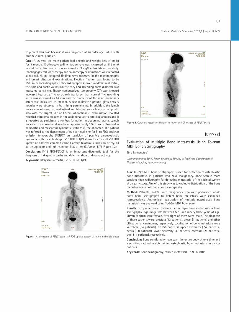
67
Nuclear Medicine Seminars 2019;1:(Suppl 1):1-778th BALKAN CONGRESS OF NUCLEAR MEDICINE
to present this case because it was diagnosed at an older age unlike with routine clinical practice.
Case: A 66-year-old male patient had anemia and weight loss of 20 kg for 3 months. Erythrocyte sedimentation rate was measured as 115 mm/hr and C-reactive protein was measured as 9 mg/L in his laboratory study. Esophagogastroduodenoscopy and colonoscopy examinations were reported as normal. No pathological findings were observed in the mammography and breast ultrasound examinations. Ejection fraction was found to be 55% in echocardiography. Echocardiography showed mild/minimal mitral, tricuspid and aortic valves insufficiency and sscending aorta diameter was measured as 4.1 cm. Thorax computerized tomography (CT) scan showed increased heart size. The aortic arch was larger than normal. The ascending aorta was measured as 44 mm and the diameter of the main pulmonary artery was measured as 30 mm. A few milimetric ground glass density nodules were observed in both lung parenchyme. In addition, the lymph nodes were observed at mediastinal and bilateral supraclavicular lymphatic area with the largest size of 1.5 cm. Abdominal CT examination revealed calcified atheroma plaques in the abdominal aorta and iliac arteries and it is reported as peripheral thrombus formation in abdominal aorta. Lymph nodes with a maximum diameter of approximately 1.5 cm were observed in paraaortic and mesenteric lymphatic stations in the abdomen. The patient was referred to the department of nuclear medicine for F-18 FDG positron emission tomography (PET)/CT on suspicion of possible paraneoplastic syndrome with these findings. F-18 FDG PET/CT showed increased F-18 FDG uptake at bilateral common carotid artery, bilateral subclavian artery, all aortic segments and right common iliac artery (SUVmax: 5.7) (Figure 1,2).
Conclusion: F-18 FDG-PET/CT is an important diagnostic tool for the diagnosis of Takayasu arteritis and determination of disease activity.
Keywords: Takayasu’s arteritis, F-18-FDG-PET/CT,
Figure 1. At the result of PET/CT scan, 18F-FDG uptake pattern of lesion in the left breast
Figure 2. Coronary vessel calcification in fusion and CT images of PET/CT scans
[BPP-72]
Evaluation of Multiple Bone Metastasis Using Tc-99m MDP Bone Scintigraphy
Ebru Salmanoğlu1
1Kahramanmaraş Sütçü İmam University Faculty of Medicine, Department of Nuclear Medicine, Kahramanmaraş
Aim: Tc-99m MDP bone scintigraphy is used for detection of osteoblastic bone metastasis in patients who have malignancy. Bone scan is more sensitive than radiography for detecting metastasis of the skeletal system at an early stage. Aim of this study was to evaluate distribution of the bone metastasis on whole body bone scintigraphy.
Method: Patients (n=422) with malignancy who were performed whole body bone scintigraphy to detect bone metastasis were examined retrospectively. Anatomical localization of multiple osteoblastic bone metastasis was analyzed using Tc-99m MDP bone scan.
Results: Sixty nine cancer patients had multiple bone metastases in bone scintigraphy. Age range was between ten and ninety three years of age. Eleven of them were female, fifty eight of them were male. The diagnosis of those patients were; prostate (43 patients), breast (11 patients) and other (15 patients) carcinomas, respectively. Localization of bone metastasis were vertebrae (64 patients), rib (56 patients), upper extremity ( 52 patients), pelvis ( 50 patients), lower extremity (39 patients), sternum (24 patients), skull (14 patients), respectively.
Conclusion: Bone scintigraphy can scan the entire body at one time and a sensitive method in determining osteoblastic bone metastasis in cancer patients.
Keywords: Bone scintigraphy, cancer, metastasis, Tc-99m MDP
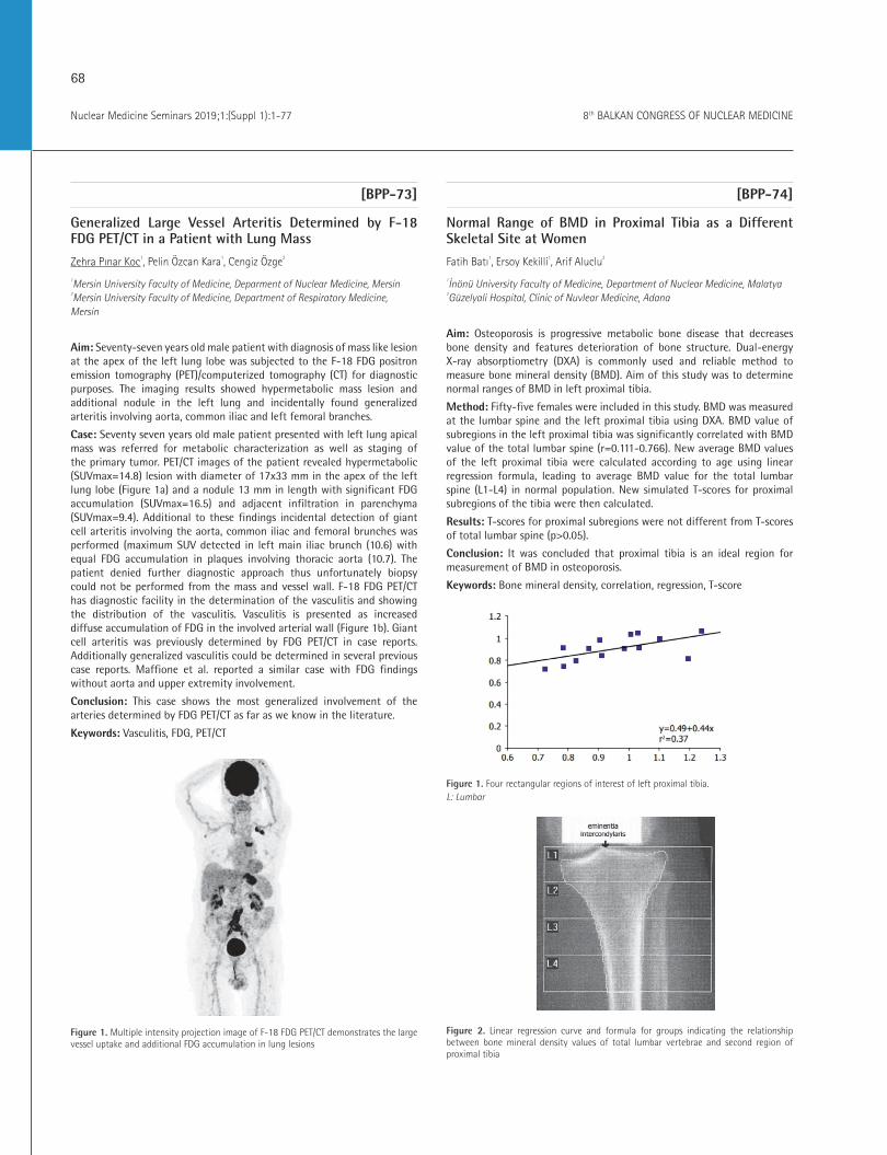
68
Nuclear Medicine Seminars 2019;1:(Suppl 1):1-77 8th BALKAN CONGRESS OF NUCLEAR MEDICINE
[BPP-73]
Generalized Large Vessel Arteritis Determined by F-18 FDG PET/CT in a Patient with Lung Mass
Zehra Pınar Koc1, Pelin Özcan Kara1, Cengiz Özge2
1Mersin University Faculty of Medicine, Deparment of Nuclear Medicine, Mersin 2Mersin University Faculty of Medicine, Department of Respiratory Medicine, Mersin
Aim: Seventy-seven years old male patient with diagnosis of mass like lesion at the apex of the left lung lobe was subjected to the F-18 FDG positron emission tomography (PET)/computerized tomography (CT) for diagnostic purposes. The imaging results showed hypermetabolic mass lesion and additional nodule in the left lung and incidentally found generalized arteritis involving aorta, common iliac and left femoral branches.
Case: Seventy seven years old male patient presented with left lung apical mass was referred for metabolic characterization as well as staging of the primary tumor. PET/CT images of the patient revealed hypermetabolic (SUVmax=14.8) lesion with diameter of 17x33 mm in the apex of the left lung lobe (Figure 1a) and a nodule 13 mm in length with significant FDG accumulation (SUVmax=16.5) and adjacent infiltration in parenchyma (SUVmax=9.4). Additional to these findings incidental detection of giant cell arteritis involving the aorta, common iliac and femoral brunches was performed (maximum SUV detected in left main iliac brunch (10.6) with equal FDG accumulation in plaques involving thoracic aorta (10.7). The patient denied further diagnostic approach thus unfortunately biopsy could not be performed from the mass and vessel wall. F-18 FDG PET/CT has diagnostic facility in the determination of the vasculitis and showing the distribution of the vasculitis. Vasculitis is presented as increased diffuse accumulation of FDG in the involved arterial wall (Figure 1b). Giant cell arteritis was previously determined by FDG PET/CT in case reports. Additionally generalized vasculitis could be determined in several previous case reports. Maffione et al. reported a similar case with FDG findings without aorta and upper extremity involvement.
Conclusion: This case shows the most generalized involvement of the arteries determined by FDG PET/CT as far as we know in the literature.
Keywords: Vasculitis, FDG, PET/CT
Figure 1. Multiple intensity projection image of F-18 FDG PET/CT demonstrates the large vessel uptake and additional FDG accumulation in lung lesions
[BPP-74]
Normal Range of BMD in Proximal Tibia as a Different Skeletal Site at Women
Fatih Batı1, Ersoy Kekilli1, Arif Aluclu2
1İnönü University Faculty of Medicine, Department of Nuclear Medicine, Malatya 2Güzelyali Hospital, Clinic of Nuvlear Medicine, Adana
Aim: Osteoporosis is progressive metabolic bone disease that decreases bone density and features deterioration of bone structure. Dual-energy X-ray absorptiometry (DXA) is commonly used and reliable method to measure bone mineral density (BMD). Aim of this study was to determine normal ranges of BMD in left proximal tibia.
Method: Fifty-five females were included in this study. BMD was measured at the lumbar spine and the left proximal tibia using DXA. BMD value of subregions in the left proximal tibia was significantly correlated with BMD value of the total lumbar spine (r=0.111-0.766). New average BMD values of the left proximal tibia were calculated according to age using linear regression formula, leading to average BMD value for the total lumbar spine (L1-L4) in normal population. New simulated T-scores for proximal subregions of the tibia were then calculated.
Results: T-scores for proximal subregions were not different from T-scores of total lumbar spine (p>0.05).
Conclusion: It was concluded that proximal tibia is an ideal region for measurement of BMD in osteoporosis.
Keywords: Bone mineral density, correlation, regression, T-score
Figure 1. Four rectangular regions of interest of left proximal tibia. L: Lumbar
Figure 2. Linear regression curve and formula for groups indicating the relationship between bone mineral density values of total lumbar vertebrae and second region of proximal tibia

69
Nuclear Medicine Seminars 2019;1:(Suppl 1):1-778th BALKAN CONGRESS OF NUCLEAR MEDICINE
Table 1. Correlation analysis of BMD value of lumbar vertebra (L1: first lumbar vertebrae, L2: second lumbar vertebra, L3: third lumbar vertebrae, L4: fourth lumbar vertebrae, LT: total lumbar vertebrae (L1-4), r: correlation coefficient)
L1 L2 L3 L4 LT
L1 r 1 0.914** 0.855** 0.797** 0.928**
L1 p p<0.001 p<0.001 p<0.001 p<0.001
L2 r 0.914** 1 0.901** 0.813** 0.952**
L2 p p<0.001 p<0.001 p<0.001 p<0.001
L3 r 0.855** 0.901** 1 0.859** 0.962**
L3 p p<0.001 p<0.001 p<0.001 p<0.001
L4 r 0.797** 0.813** 0.859** 1 0.931**
L4 p p<0.001 p<0.001 p<0.001 p<0.001
L5 r 0.928** 0.952** 0.962** 0.931** 1
L5 p p<0.001 p<0.001 p<0.001 p<0.001
Table 2. New simulated T scores of each region in proximal tibia (LTTs: T scores of the total lumbar vertebrae, T1Ts: T scores of the first region of interest in tibia, T2Ts: T scores of the second region of interest in tibia, T3Ts: T scores of the third region of interest in tibia, T4Ts: T scores of the forth region of interest in tibia, TTTs: T scores of the sum of areas in the tibia)
Group I (n=14) Mean ± SD (Range)
Group II (n=14) Mean ± SD (Range)
Group III (n=14) Mean ± SD (Range)
Group IV (n=14) Mean ± SD (Range)
LTTs -1.271±1.028 -1.729±0.100 -1.757±1.516 -2.646±0.907
T1Ts -1.029±0.412 -1.336±0.752 -1.029±0.849 -3.015±0.590
T2Ts -1.050±0.449 -1.379±0.796 -1.021±0.742 -2.092±1.026
T3Ts -1.036±0.573 -1.529±0.833 -1.029±0.698 -2.531±1.027
T4Ts -1.107±0.478 -1.571±0.841 -1.029±0.785 -2.669±1.144
TTTs -1.129±0.484 -1.400±0.857 -0.993±0.862 -2.592±1.074
P value
0.452 0.167 0.743 0.0001
[BPP-75]
Renal Cell Carcinoma with Bilateral Tibia Metastasis on Tc-99m MDP Bone Scintigraphy: Case Report
Ebru Salmanoglu1
1Kahramanmaraş Sütçü Imam University Faculty of Medicine, Department of Nuclear Medicine, Kahramanmaraş
Aim: Tc-99m MDP bone scintigraphy is used for screening of bone metastases in cancer patients. Bone metastasis in the appendicular skeleton is rare and it is seen in the proximal part of the femur. Tibia metastasis is not common. The aim of presenting this case report is that bilateral tibial metastases in a patient with renal cell carcinoma contributes to the literature.
Method: A forty-nine year old male patient diagnosed with renal cell carcinoma with pain in both lower legs was referred to the department of nuclear medicine with a preliminary diagnosis of osteomyelitis. Tc-99m MDP three-phase bone scintigraphy was performed.
Results: Increased perfusion and hyperemia was observed at the bilateral distal part of tibia on perfusion and blood pool phase imaging. Increased Tc-99m MDP uptake was seen at the bilateral distal part of tibia on late static imaging. It was considered suspicious for metastatic renal cell carcinoma. It was confirmed as a metastasis by histopathologically.
Conclusion: Bone scintigraphy is a sensitive method for detecting osteoblastic bone metastases. When symmetrical involvement is seen on bone scintigraphy, metastasis should be considered.
Keywords: Tc-99m MDP, renal cell carcinoma, osteoblastic metastasis
[BPP-76]
Relationship between Obesity and Disability in Adult Individuals Retrospective Screening
Fatih Batı1, Yüksel Ersoy2, Raikan Büyükavcı2, Semra Aktürk2, Osman Celbiş3
1İnönü University Faculty of Medicine, Department of Nuclear Medicine, Malatya 2İnönü University Faculty of Medicine, Department of Physical Medicine And Rehabilitation, Malatya 3İnönü University Faculty of Medicine, Department of Forensic Medicine, Malatya
Aim: The objective is to examine the relationship between the musculoskeletal system (MSS)-based disability and the body mass index (BMI).
Method: The medical board reports (MBR) issued for patients in 2010, who applied for a disability report, were screened. From a total of 283 MBR’s, 99 disability reports were included. BMI was calculated and the relationship between disability ratios and BMI was examined.
Results: Out of the 99 patients, 77 were male and 22 were female. The mean age was 46.7±17.8, the BMI was 25.5±5.7 kg/m², the total disability ratio (TDR) was 46±24% and the MSS disability ratio was 33±23%. Both the BMI and age of the patient were significantly correlated with TDR values (p<0.05). The female mean age, the TDR, MSS disability ratio and BMI were higher compared to males. However, only the TDR was statistically significantly higher (p<0.05). 46% of the disability reports were due to acquired diseases (most commonly caused by arthrosis), 37% injuries (most frequently falls, work accidents and firearm injuries) and 11% were for
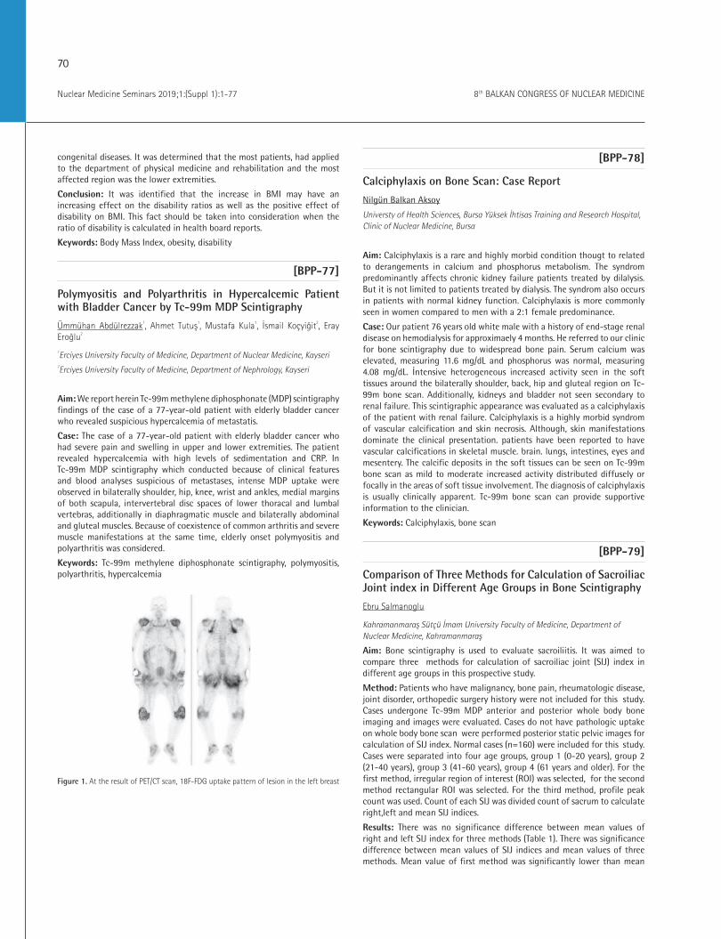
70
Nuclear Medicine Seminars 2019;1:(Suppl 1):1-77 8th BALKAN CONGRESS OF NUCLEAR MEDICINE
congenital diseases. It was determined that the most patients, had applied to the department of physical medicine and rehabilitation and the most affected region was the lower extremities.
Conclusion: It was identified that the increase in BMI may have an increasing effect on the disability ratios as well as the positive effect of disability on BMI. This fact should be taken into consideration when the ratio of disability is calculated in health board reports.
Keywords: Body Mass Index, obesity, disability
[BPP-77]
Polymyositis and Polyarthritis in Hypercalcemic Patient with Bladder Cancer by Tc-99m MDP Scintigraphy
Ümmühan Abdülrezzak1, Ahmet Tutuş1, Mustafa Kula1, İsmail Koçyiğit2, Eray Eroğlu2
1Erciyes University Faculty of Medicine, Department of Nuclear Medicine, Kayseri2Erciyes University Faculty of Medicine, Department of Nephrology, Kayseri
Aim: We report herein Tc-99m methylene diphosphonate (MDP) scintigraphy findings of the case of a 77-year-old patient with elderly bladder cancer who revealed suspicious hypercalcemia of metastatis.
Case: The case of a 77-year-old patient with elderly bladder cancer who had severe pain and swelling in upper and lower extremities. The patient revealed hypercalcemia with high levels of sedimentation and CRP. In Tc-99m MDP scintigraphy which conducted because of clinical features and blood analyses suspicious of metastases, intense MDP uptake were observed in bilaterally shoulder, hip, knee, wrist and ankles, medial margins of both scapula, intervertebral disc spaces of lower thoracal and lumbal vertebras, additionally in diaphragmatic muscle and bilaterally abdominal and gluteal muscles. Because of coexistence of common arthritis and severe muscle manifestations at the same time, elderly onset polymyositis and polyarthritis was considered.
Keywords: Tc-99m methylene diphosphonate scintigraphy, polymyositis, polyarthritis, hypercalcemia
Figure 1. At the result of PET/CT scan, 18F-FDG uptake pattern of lesion in the left breast
[BPP-78]
Calciphylaxis on Bone Scan: Case Report
Nilgün Balkan Aksoy
Universty of Health Sciences, Bursa Yüksek İhtisas Training and Research Hospital, Clinic of Nuclear Medicine, Bursa
Aim: Calciphylaxis is a rare and highly morbid condition thougt to related to derangements in calcium and phosphorus metabolism. The syndrom predominantly affects chronic kidney failure patients treated by dilalysis. But it is not limited to patients treated by dialysis. The syndrom also occurs in patients with normal kidney function. Calciphylaxis is more commonly seen in women compared to men with a 2:1 female predominance.
Case: Our patient 76 years old white male with a history of end-stage renal disease on hemodialysis for approximaely 4 months. He referred to our clinic for bone scintigraphy due to widespread bone pain. Serum calcium was elevated, measuring 11.6 mg/dL and phosphorus was normal, measuring 4.08 mg/dL. İntensive heterogeneous increased activity seen in the soft tissues around the bilaterally shoulder, back, hip and gluteal region on Tc-99m bone scan. Additionally, kidneys and bladder not seen secondary to renal failure. This scintigraphic appearance was evaluated as a calciphylaxis of the patient with renal failure. Calciphylaxis is a highly morbid syndrom of vascular calcification and skin necrosis. Although, skin manifestations dominate the clinical presentation. patients have been reported to have vascular calcifications in skeletal muscle. brain. lungs, intestines, eyes and mesentery. The calcific deposits in the soft tissues can be seen on Tc-99m bone scan as mild to moderate increased activity distributed diffusely or focally in the areas of soft tissue involvement. The diagnosis of calciphylaxis is usually clinically apparent. Tc-99m bone scan can provide supportive information to the clinician.
Keywords: Calciphylaxis, bone scan
[BPP-79]
Comparison of Three Methods for Calculation of Sacroiliac Joint index in Different Age Groups in Bone Scintigraphy
Ebru Salmanoglu
Kahramanmaraş Sütçü İmam University Faculty of Medicine, Department of Nuclear Medicine, Kahramanmaraş
Aim: Bone scintigraphy is used to evaluate sacroiliitis. It was aimed to compare three methods for calculation of sacroiliac joint (SIJ) index in different age groups in this prospective study.
Method: Patients who have malignancy, bone pain, rheumatologic disease, joint disorder, orthopedic surgery history were not included for this study. Cases undergone Tc-99m MDP anterior and posterior whole body bone imaging and images were evaluated. Cases do not have pathologic uptake on whole body bone scan were performed posterior static pelvic images for calculation of SIJ index. Normal cases (n=160) were included for this study. Cases were separated into four age groups, group 1 (0-20 years), group 2 (21-40 years), group 3 (41-60 years), group 4 (61 years and older). For the first method, irregular region of interest (ROI) was selected, for the second method rectangular ROI was selected. For the third method, profile peak count was used. Count of each SIJ was divided count of sacrum to calculate right,left and mean SIJ indices.
Results: There was no significance difference between mean values of right and left SIJ index for three methods (Table 1). There was significance difference between mean values of SIJ indices and mean values of three methods. Mean value of first method was significantly lower than mean
71
Nuclear Medicine Seminars 2019;1:(Suppl 1):1-778th BALKAN CONGRESS OF NUCLEAR MEDICINE
values of other two methods. Mean value of female was lower than male for right, left SIJ and mean SIJ indices. Lowest mean value was found in group 4. A significantly negative correlation was found between age and right SIJ index, left SIJ index of three methods. Statistically significance was found between mean values of three methods for each age groups.
Conclusion: Each patient should be evaluated independently. Each nuclear medicine center should establish their own threshold value according to their own data set in normal cases and patients.
Keywords: Sacroiliac joint index, bone scintigraphy, Tc-99m MDP
Table 1. Comparison of mean values of three methods for calculation of SIJ index (paired t test)
Variables Group n X ± SD t p value
First method-SIJ index Right 160 1.03±0.11 -0.412 0.681
First method-SIJ index Left 160 1.03±0.10
Second method-SIJ index
Right 160 1.12±0.12 0.005 0.996
Second method-SIJ index
Left 160 1.12±0.11
Third method-SIJ index
Right 160 1.13±0.16 -1.383 0.169
Third method-SIJ index
Left 160 1.14±0.17
[BPP-80]
Functional Particularities of Metastases in Male Patients Diagnosed with Breast Cancer
Ovidiu Emanuel Chifor1, Irena Grierosu2, Mihai Gutu2, Ana Maria Statescu1, Vlad Ghizdovat2, Lucian Miron2, Roxana Iacob2, Cipriana Ștefănescu2
1St. Spiridon Emergency Hospital, Clinic of Nuclear Medicine Laboratory, Iaşi, Romania 2Grigore T. Popa University, Faculty of Medicine and Pharmacy, Iaşi, Romania
Aim: Breast cancer (BC) in male is rare, having less than 1% incidence, median age at diagnostic 61 years (5-93 years), with evolution particularities that continue to be evidenced. Our aim is to emphasize on functional particularities of metastases (MTS), to contribute to the personalized approach needed by this BC type.
Method: We selected the cases diagnosed with BC between 2000 and 2018 from our laboratory of nuclear medicine’s archive, collected from 1/3 of the Nord-East region in Romania. The inclusion criteria were: a valid, positive histo-pathological confirmation, at least one bone scintigraphy, with complementary image investigation [computerized tomography (CT)/positron emission tomography (PET)-CT/RMN]. In these 18 years, there were 17 patients, with a mean age at diagnostic of 66 years (35-80 years). Treatment consisted in: curative radical surgery-12 patients, chemotherapy, radiotherapy, hormone therapy-related to the diagnostic, palliative care-4 patients. At the time of the scintigraphy, 15 out of 17 patients had bone pain (88%). Whole body bone scan 2 hours after iv administration of 10.5 MBq/kgbw Tc-99m-MDP (12 patients), respectively Tc-99m-HDP (5 patients), with supplementary single photon emission computerized tomography (SPECT) images, were realized. Images had been qualitatively and quantitatively assessed.
Results: Bone scan analysis revealed the presence of secondary bone MTS in 12 out of 17 patients (70%). 10 out of 12 positive patients had more than one radiotracer MTS hot sites. There were no osteolytic sites. In 2 patients with an initial normal scan, bone MTS were revealed in the one-year follow-up scan. The most common localizations for MTS were: vertebral spine (50%), basin (50%), skull (30%), and ribs (15%), showing a pattern which is different from bone MTS pattern in women found in the literature. These particularities sustain the hypothesis that the infaust prognosis of undifferentiated BC in men could be related both to the histological type of the tumor and the anatomy of the male mammary gland, that allows the tumor to spread more rapidly to the neighboring tissues, explaining the rapid evolution and high mortality observed in the first years following the diagnostic, even in the context of proper treatment.
Conclusion: Our study shows particular bone MTS functional pattern characteristics of the distribution, radiotracer uptake and evolution in male BC. Careful personalization and repetitive functional evaluation of MTS for treatment reevaluation are essential in male BC precision medicine.
Keywords: Male breast cancer,bone scintigraphy, bone metastases, functional particularities.
[BPP-81]
Assessment of Congenital Anomalies of Kidney With Scintigraphic Imaging Methods
Ebru Salmanoglu1
1Kahramanmaraş Sütçü İmam University Faculty of Medicine, Department of Nuclear Medicine, Kahramanmaraş
Aim: Congenital anomalies of kidney have a large spectrum from being asymptomatic to end stage renal disease. Therefore they should be evaluated carefully. Renal ectopia and renal fusion anomalies are common abnormalities of the kidney. Renal ectopia is abnormal anatomic localization of the kidney. Cross ectopic kidney is usually fused to the lower pole of the kidney. Horseshoe kidney which is the most common fusion anomaly of the kidney, lower poles of two kidneys are fused with a fibrous band. Scintigraphic evaluation of kidneys provides both functional imaging and information about kidney functions via numerical parameters. Therefore, scintigraphic imaging methods are superior to radiologic imaging methods. It was aimed in this retrospective study to demonstrate importance of nuclear medicine for the evaluation of congenital kidney abnormalities.
Method: Patients (n=345) who undergone renal scintigraphy (Tc-99m DMSA, Tc-99m DTPA, Tc-99m MAG-3) were examined retrospectively. The patients with congenital renal anomalies were evaluated.
Results: Thirteen of three hundred forty five patients had congenital renal anomalies. Six of them had horseshoe kidney, six of them had pelvic kidney, one of them had cross ectopic kidney. Differential renal function of kidneys were evaluated. Contribution to renal function of the horseshoe kidney was analyzed with using a drawing technique from anterior and posterior field of kidney.
Conclusion: Renal scintigraphy is a necessary method for calculation of the split renal function especially for congenital kidney abnormalities.
Keywords: Tc-99m DTPA, Tc-99m DMSA, horseshoe kidney,renal scintigraphy

72
Nuclear Medicine Seminars 2019;1:(Suppl 1):1-77 8th BALKAN CONGRESS OF NUCLEAR MEDICINE
[BPP-82]
Indiana Pouch on Dynamic Renal Scintigraphy
Derya Çayır, Mehmet Bozkurt
University of Health Sciences, Dışkapı Yıldırım Beyazıt Training and Research Hospital, Department of Nuclear Medicine, Ankara
Aim: Dynamic renal scintigraphy is frequently used in routine practice to evaluate the perfusion, concentration, and excretion functions of the kidneys. One of the continent diversion types performed after radical cystectomy is the continent urinary reservoir ileal conduit (Indiana pouch) operation. In this case, Indiana pouch appearance is presented on Tc-99m diethylene-triamine-pentaacetate (DTPA) dynamic renal scintigraphy.
Case: A 52-year-old female patient complained with left flank pain underwent Tc-99m DTPA dynamic diuretic renal scintigraphy. The patient had a history of past surgery for squamous cell carcinoma of cervix and chemoradiotherapy, in 2005. After external and internal radiation therapy for 45 days, which caused a radiation-induced bowel and bladder injury and colostomy and Indiana pouch operation were performed. The serum levels were as follows: urea: 61 mg/dL (17-43), Creatinine: 1.53 mg/dL (0.66-1.09). In diuretic dynamic renal scintigraphy, the perfusion and concentration functions of the left kidney were delayed and decreased, and no excretion from the left kidney was observed. The perfusion function of the right kidney was delayed and decreased, the concentration of the right kidney was on time, however declined. The excretion function of the right kidney was initiated on time and excretion function was prolonged with pelvicalyceal stasis responsive to iv diuretic (Figure 1, 2). During the dynamic study, the bladder could not be visualized (past cystectomy) and increased radioactivity accumulation in the right lower quadrant, which was in concordance with Indiana pouch, was noticed. The patient is still followed up by department of oncology.
Conclusion: After radical cystectomy, to redirect urine is possible with an continent or incontinent urinary diversion and reconstruction procedure. Continent urinary diversion is favoured in patients with normal renal and hepatic functions, who have the mental and physical health to properly catheterize themselves. Indiana pouch is a technique of cutaneous urinary diversions which based on a conic ileal segment and a plicated ileocecal valve to maintain continence. In the evaluation of dynamic renal scintigraphy of these patients, the gradual increase of radioactivity in the pouch should not be misinterpreted as kidney activity.
Keywords: Tc-99m DTPA, dynamic renal scintigraphy, Indiana pouch
Figure 1. At the result of PET/CT scan, 18-F-FDG uptake pattern of lesion in the left breast
Figure 2. Coronary vessel calcification in fusion and CT images of PET/CT scans
[BPP-83]
Unexpected Focal Pulmonary Uptake of Tc-99m-DMSA
Fadimana Nur Aydınbelge, Aylin Akbulut, Gokhan Koca, Meliha Korkmaz
University of Health Sciences, Ankara Training and Research Hospital, Clinic of Nuclear Medicine, Ankara
Aim: Extrarenal uptake of Tc-99m DMSA is a rare finding, which has been described previously in the unusual situations. This report includes a case of abnormal Tc-99m DMSA uptake in the left lung, which remained unexplained even after contrast-enhanced thorax computerized tomography (CT). Renal scintigraphy with Tc-99m DMSA is a well-accepted method for the evaluation of the cortical pathologies. We presented a case report that was referred for renal scintigraphy and had a focal radiotracer uptake in the left lung at renal scintigraphy with Tc-99m DMSA.
Case: Renal scintigraphy with Tc-99m DMSA was performed in a 22-year-old man, referred for the evaluation of recently detected nephrolithiasis in left kidney. His renal function tests were normal. Renal scintigraphy showed that left kidney was small sized and decreased radiotracer uptake with a hypoactive area causing flattening of the contour at the medial upper pole. The right kidney was normal. Also, there was an abnormal focal Tc-99m DMSA uptake on the lower lobe field of the left lung. Then the patient underwent contrast-enhanced thorax CT, which demonstrated no abnormality in the left lung.
Conclusion: Extrarenal abnormal uptake of Tc-99m DMSA is a rare finding. However, there are some case reports about abnormal uptake in splenic amyloidosis, bone metastases, renal pelvic diverticulum, infantile hemangioma and abdominal aortic aneurysm. In that case, contrast-enhanced chest CT was normal, and we could not find any anatomic explanation for the abnormal uptake. Thorough the literature, it was found that DMSA could be trapped and non-specifically bound by in vitro blood clotting. Hence, we assumed that the cause was microembolism at a pulmonary arteriole by blood clots trapping Tc-99m DMSA. In conclusion, focal pulmonary uptake of Tc-99m-DMSA can be rarely observed as an extrarenal finding.
Keywords: Tc-99m DMSA, extrarenal uptake, microembolism
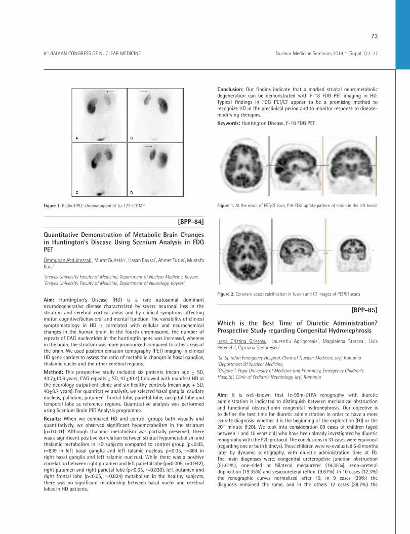
73
Nuclear Medicine Seminars 2019;1:(Suppl 1):1-778th BALKAN CONGRESS OF NUCLEAR MEDICINE
Figure 1. Radio-HPLC chromatogram of Lu-177-EDTMP
[BPP-84]
Quantitative Demonstration of Metabolic Brain Changes in Huntington’s Disease Using Scenium Analysis in FDG PET
Ümmühan Abdülrezzak1, Murat Gultekin2, Hasan Baysal2, Ahmet Tutus1, Mustafa Kula1
1Erciyes University Faculty of Medicine, Department of Nuclear Medicine, Kayseri 2Erciyes University Faculty of Medicine, Department of Neurology, Kayseri
Aim: Huntington’s Disease (HD) is a rare autosomal dominant neurodegenerative disease characterized by severe neuronal loss in the striatum and cerebral cortical areas and by clinical symptoms affecting motor, cognitive/behavioral and mental function. The variability of clinical symptomatology in HD is correlated with cellular and neurochemical changes in the human brain. In the fourth chromosome, the number of repeats of CAG nucleotides in the huntingtin gene was increased, whereas in the brain, the striatum was more pronounced compared to other areas of the brain. We used positron emission tomography (PET) imaging in clinical HD gene carriers to assess the ratio of metabolic changes in basal ganglias, thalamic nuclei and the other cerebral regions.
Method: This prospective study included six patients (mean age ± SD, 43.7±10.6 years; CAG repeats ± SD, 47±10.4) followed with manifest HD at the neurology outpatient clinic and six healthy controls (mean age ± SD, 40±8.7 years). For quantitative analysis, we selected basal ganglia, caudate nucleus, pallidum, putamen, frontal lobe, parietal lobe, occipital lobe and temporal lobe as reference regions. Quantitative analysis was performed using Scenium Brain PET Analysis programme.
Results: When we compared HD and control groups both visually and quantitatively, we observed significant hypometabolism in the striatum (p<0.001). Although thalamic metabolism was partially preserved, there was a significant positive correlation between striatal hypometabolism and thalamic metabolism in HD subjects compared to control group (p<0.05, r=839 in left basal ganglia and left talamic nucleus, p<0.05, r=884 in right basal ganglia and left talamic nucleus). While there was a positive correlation between right putamen and left parietal lobe (p=0.005, r=0.942), right putamen and right parietal lobe (p<0.05, r=0.830), left putamen and right frontal lobe (p<0.05, r=0.824) metabolism in the healthy subjects, there was no significant relationship between basal nuclei and cerebral lobes in HD patients.
Conclusion: Our findins indicate that a marked striatal neurometabolic degeneration can be demonstrated with F-18 FDG PET imaging in HD. Typical findings in FDG PET/CT appear to be a promising method to recognize HD in the preclinical period and to monitor response to disease-modifying therapies.
Keywords: Huntington Disease, F-18 FDG PET
Figure 1. At the result of PET/CT scan, F18-FDG uptake pattern of lesion in the left breast
Figure 2. Coronary vessel calcification in fusion and CT images of PET/CT scans
[BPP-85]
Which is the Best Time of Diuretic Administration? Prospective Study regarding Congenital Hydronephrosis
Irena Cristina Grierosu1, Laurentiu Agrigoroaie2, Magdalena Starcea3, Livia Petenchi2, Cipriana Stefanescu1
1St. Spiridon Emergency Hospital, Clinic of Nuclear Medicine, Iaşi, Romania 2Department Of Nuclear Medicine, 3Grigore T. Popa University of Medicine and Pharmacy, Emergency Children’s Hospital, Clinic of Pediatric Nephrology, Iaşi, Romania
Aim: It is well-known that Tc-99m-DTPA renography with diuretic administration is indicated to distinguish between mechanical obstruction and functional obstructionin congenital hydronephrosis. Our objective is to define the best time for diuretic administration in order to have a more ccurate diagnosis: whether it is the beginning of the exploration (F0) or the 20th minute (F20). We took into consideration 69 cases of children (aged between 1 and 15 years old) who have been already investigated by diuretic renography with the F20 protocol. The conclusions in 31 cases were equivocal (regarding one or both kidneys). These children were re-evaluated 6-8 months later by dynamic scintigraphy, with diuretic administration time at F0. The main diagnoses were: congenital ureteropelvic junction obstruction (51.61%), one-sided or bilateral megaureter (19.35%), reno-ureteral duplication (19.35%) and vesicoureteral reflux (9.67%). In 10 cases (32.3%)the renographic curves normalized after F0, in 9 cases (29%) the diagnosis remained the same, and in the others 12 cases (38.7%) the
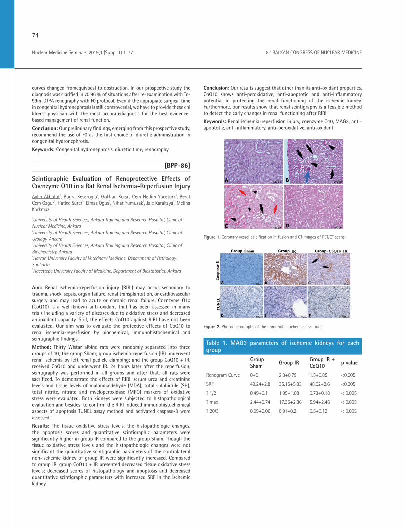
74
Nuclear Medicine Seminars 2019;1:(Suppl 1):1-77 8th BALKAN CONGRESS OF NUCLEAR MEDICINE
curves changed fromequivocal to obstruction. In our prospective study the diagnosis was clarified in 70.96 % of situations after re-examination with Tc-99m-DTPA renography with F0 protocol. Even if the appropiate surgical time in congenital hydronephrosis is still controversial, we have to provide these chi ldrens’ physician with the most accuratediagnosis for the best evidence-based management of renal function.
Conclusion: Our preliminary findings, emerging from this prospective study, recommend the use of F0 as the first choice of diuretic administration in congenital hydronephrosis.
Keywords: Congenital hydronephrosis, diuretic time, renography
[BPP-86]
Scintigraphic Evaluation of Renoprotective Effects of Coenzyme Q10 in a Rat Renal Ischemia-Reperfusion Injury
Aylin Akbulut1, Bugra Keseroglu2, Gokhan Koca1, Cem Nedim Yuceturk2, Berat Cem Ozgur2, Hatice Surer3, Elmas Ogus3, Nihat Yumusak4, Jale Karakaya5, Meliha Korkmaz1
1University of Health Sciences, Ankara Training and Research Hospital, Clinic of Nuclear Medicine, Ankara 2University of Health Sciences, Ankara Training and Research Hospital, Clinic of Urology, Ankara 3University of Health Sciences, Ankara Training and Research Hospital, Clinic of Biochemistry, Ankara 4Harran University Faculty of Veterinary Medicine, Department of Pathology, Şanlıurfa 5Hacettepe University Faculty of Medicine, Department of Biostatistics, Ankara
Aim: Renal ischemia-reperfusion injury (RIRI) may occur secondary to trauma, shock, sepsis, organ failure, renal transplantation, or cardiovascular surgery and may lead to acute or chronic renal failure. Coenzyme Q10 (CoQ10) is a well-known anti-oxidant that has been assessed in many trials including a variety of diseases due to oxidative stress and decreased antioxidant capacity. Still, the effects CoQ10 against RIRI have not been evaluated. Our aim was to evaluate the protective effects of CoQ10 to renal ischemia-reperfusion by biochemical, immunohistochemical and scintigraphic findings.
Method: Thirty Wistar albino rats were randomly separated into three groups of 10; the group Sham; group ischemia-reperfusion (IR) underwent renal ischemia by left renal pedicle clamping; and the group CoQ10 + IR, received CoQ10 and underwent IR. 24 hours later after the reperfusion, scintigraphy was performed in all groups and after that, all rats were sacrificed. To demonstrate the effects of RIRI, serum urea and creatinine levels and tissue levels of malondialdehyde (MDA), total sulphidrile (SH), total nitrite, nitrate and myeloperoxidase (MPO) markers of oxidative stress were evaluated. Both kidneys were subjected to histopathological evaluation and besides; to confirm the RIRI induced immunohistochemical aspects of apoptosis TUNEL assay method and activated caspase-3 were assessed.
Results: The tissue oxidative stress levels, the histopathologic changes, the apoptosis scores and quantitative scintigraphic parameters were significantly higher in group IR compared to the group Sham. Though the tissue oxidative stress levels and the histopathologic changes were not significant the quantitative scintigraphic parameters of the contralateral non-ischemic kidney of group IR were significantly increased. Compared to group IR, group CoQ10 + IR presented decreased tissue oxidative stress levels; decreased scores of histopathology and apoptosis and decreased quantitative scintigraphic parameters with increased SRF in the ischemic kidney.
Conclusion: Our results suggest that other than its anti-oxidant properties, CoQ10 shows anti-peroxidative, anti-apoptotic and anti-inflammatory potential in protecting the renal functioning of the ischemic kidney. Furthermore, our results show that renal scintigraphy is a feasible method to detect the early changes in renal functioning after RIRI.
Keywords: Renal ischemia-reperfusion injury, coenzyme Q10, MAG3, anti-apoptotic, anti-inflammatory, anti-peroxidative, anti-oxidant
Figure 1. Coronary vessel calcification in fusion and CT images of PET/CT scans
Figure 2. Photomicrographs of the immunohistochemical sections
Table 1. MAG3 parameters of ischemic kidneys for each group
Group Sham Group IR Group IR +
CoQ10 p value
Renogram Curve 0±0 2.8±0.79 1.5±0.85 <0.005
SRF 49.24±2.8 35.15±5.83 48.02±2.6 <0.005
T 1/2 0.49±0.1 1.95±1.08 0.73±0.18 < 0.005
T max 2.44±0.74 17.35±2.86 5.94±2.46 < 0.005
T 20/3 0.09±0.06 0.91±0.2 0.5±0.12 < 0.005

75
Nuclear Medicine Seminars 2019;1:(Suppl 1):1-778th BALKAN CONGRESS OF NUCLEAR MEDICINE
[BPP-87]
F-18-FDG PET/MRI Renal Function Assessment in Animal Model Cardio-Renal Syndrome
Mihai Marius Gutu1, Laurentiu Agrigoroaie2, Mihai Dan Furcea2, Cristina Uritu3, Gabriela Dumitrita Stanciu3, Cosmin Teodor Mihai3, Ioannis Gardikiotis3, Vlad Ghizdovat1, Cati Stolniceanu2, Radu Iliescu1, Cipriana Stefanescu1
1Grigore T. Popa University of Medicine and Pharmacy, Department of Nuclear Medicine, Iaşi, Romania 2St. Spiridon Emergency Hospital, Clinic of Nuclear Medicine Laboratory, Iaşi, Romania 3Advanced Center for Research and Development in Experimental Medicine Cemex, Iaşi, Romania
Aim: The results presented here are a part of an exploratory positron emission tomography (PET)/magnetic resonance imaging (MRI) study in a rat model of cardiorenal syndrome (CRS), where we have used parametric quantification in target organs (heart, kidney, vessels), based on image analysis. As an incremental approach to the final goal of the experiment, we aimed at obtaining a valid technique for approximating the glomerular filtration rate (GFR) using F-18-FDG tracer dynamics on control lot versus CRS induced by heart failure (HF).
Method: We studied a number of 12 male Wistar rats, divided into 2 lots: control (L1) and heart failure (L2). Heart failure was induced by intraperitoneal doxorubicin administration for a period of 6 weeks. F-18-FDG PET/MRI scans (nanoScan PET/MRI, Mediso) were acquired at week 0 for L1 and at week 6 for L2. Each rat underwent a succession of MRI examination (T1 weighted), injection of F-18-FDG and PET data acquisition, in the same session. Manually defined regions of interest were drawn on MRI images for kidneys and left ventricular cavity, which were used for volumetric assessment and delineation of VOIs on PET frames. Weight based standardized uptake values (SUV) for F-18-FDG were calculated for each frame of the dynamic PET study, with the 3 to 20 minute interval being selected for data analysis. PET derived GFR estimation was calculated based on the following formula: GFR: Ki
right[min-1]*Vright kidney[ml]+ Kileft[min-1]*Vleft
kidney[mL], where Ki is approximated as the slope of the linear regression of arterial tracer concentration by taking the renal activity as independent variable.
Results: Results are reported as mean ± standard error of the mean. The variation in arterial concentration explained by renal tracer dynamics of the groups was R2
L1= 0.97, R2L2= 0.99. Estimated GFR for the control group,
compared to literature data (1.38±0.1 mL/min), was within 1.26±0.23 mL/min. L2 group showed a departure from the control group with values of 0.88±0.14 mL/min.
Conclusion: Our results are in agreement with the reported literature for healthy Wistar rats and it provides us with an intra-experiment assessment method for kidney dysfunction by F-18-FDG PET/MRI.
Keywords: PET/MRI, F-18-FDG, cardiorenal syndrome, Wistar rat, kidney function
[BPP-88]
Breast Tissue Attenuation in Myocardial Perfusion SPECT CT Imaging: Cardiac Phantom Study
Fatih Boğa1, Fevziye Canbaz Tosun2
1Ondokuz Mayıs University Faculty of Medicine, Department of Nuclear Medicine, Samsun 2Ondokuz Mayıs University, Faculty of Physics
Aim: The aim of our study is to investigate the attenuation effect of breast tissue in different size and density on myocardial perfusion imaging (MPI) and to evaluate the changes after computerized tomography (CT) attenuation correction (CTAC) using a modeled breast cardiac phantom.
Method: The breast models in our study were designed using 400, 700, 1000 and 1300 milliliters of balloons filled with water, oil and liquid soap to simulate the breast tissues of different size and density. The cardiac part of the phantom was filled with diluted water of 37 MBq (1 mCi) Tc-99m pertechnetate activity. The breast models were attached to cardiac phantom respectively. Using a double detector hybrid single photon emission computerized tomography (SPECT)/CT system, imaging was performed with the parameters used in routine clinical settings of MPI (64x64 matrix; 20 seconds/projection, a total of 32 projections). Cedars Sinai QPS perfusion quantitative software (V. 2009) was used in processing and data collection. We analyzed the images, semi-quantitatively (5 point scoring system), using 17 segment model through polar maps. The analyzing of data belonging to standard and CTAC results were carried out via SPSS (V 22.0) statistical program by using Wilcoxon, Man-Whitney U, Kruskal-Wallis tests on the base of the myocardial segment, wall, and level (apex to basal).
Results: After CTAC, there were no significant differences (∆) between the groups (p>0.05) regarding the density and size but among the left ventricular walls. The highest mean difference values between standard and CTAC images were detected on septum in all breast models. The mean differences for myocardial septum wall were: ∆ = 18.2±10.0; 20.4±9.6; 18.2±8.2 for oil, water and liquid soap groups, respectively; and ∆=19.1±10.8, 19.0±9.5, 19.6±9.0 and 17.8±8 were obtained for 400, 700, 1000 and 1300 volume groups, respectively. A heterogenic changing was seen on lateral wall {∆ = [(-0.7±11.1)-(6.9±14.4)]}, after CTAC. Also, lateral wall defect was striking after CTAC especially in the breast model having both the highest volume and size (Figure 1, 2)
Conclusion: The results in our study support that a considerable improvement was seen especially on cardiac septum after CT attenuation correction without depending on the size and density of breasts; but the physicians should be aware that, although without SPECT/CT misregistration, fixed or new perfusion defects can occur after CTAC, especially on the lateral myocardial wall.
Keywords: Myocardial perfusion SPECT/CT, CT attenuation correction, breast attenuation, cardiac phantom

76
Nuclear Medicine Seminars 2019;1:(Suppl 1):1-77 8th BALKAN CONGRESS OF NUCLEAR MEDICINE
Figure 1. At the result of PET/CT scan, F-18-FDG uptake pattern of lesion in the left breast
Figure 2. Coronary vessel calcification in fusion and CT images of PET/CT scans
[BPP-89]
Incidental Diagnosis of Takayasu Arthritis in PET/CT in a Patient with Solitary Pulmonary Lesion
Berna Okudan, Ceren Deniz Kapulu Akça1University of Health Sciences, Ankara Numune Training and Research Hospital, Clinic of Nuclear Medicine, Ankara
Aim: Takayasu’s arteritis (TAK), is a rare form of vasculitis disease involving inflammation in the walls of the largest arteries in the body: the aorta and its main branches. The disease results from an attack by the body’s own immune system, causing inflammation in the walls of arteries. The inflammation leads to narrowing of the arteries, and this can reduce blood flow to many parts of the body. Sometimes patients with TAK may have no symptoms, and the disease is so rare that doctors may not easily recognize it. Thus, there is often a delay in detecting it, sometimes several years.
Case: A 57-year-old female patient presented to Ankara Numune Training and Research Hospital, Department of Rheumatology with complaints of fatigue, exhaustion and dyspepsy. Laboratory tests revealed the sedimentation rate as 52 mm/hr and alkaline phosphatase as 110 IU/L. The patient was referred to department of radiology for computerized tomography (CT) with a pre-diagnosis of malignancy. CT scan showed no pathological findings other than a solitary pulmonary nodule in the anterior segment of right upper lobe. The patient was then admitted to department of nuclear medicine for a positron emission tomography (PET)/CT scan which revealed increased FDG uptake major vessels, more explicit in aortic arc, starting from the cervical level, evident in ascending aorta,
the aortic arch, the descending aorta and the distal abdominal aorta. The patient was presciribed Prednol and PPI treatment. Control PET/CT images taken 5 months later showed a significant decrease in vascular involvement (SUVmax: 2.5). PET/CT images taken after 11 months showed no pathological FDG uptake. Takayasu arteritis was diagnosed with PET/CT insidentally and treatment was very effective.
Keywords: Takayasu’s arteritis, PET/CT, vasculitis
Figure 1. Takayasu’s arteritis after theraphy
Figure 2. Takayasu’s arteritis before theraphy
[BPP-90]
Bone Scintigraphy Correlated with Echocardiography for Detecting Transtiretin Cardiac Amyloidosis Subtypes
Teodor Marian Ionescu1, Laura Polixenia Grecu1, Paul Firicel1, Cati Raluca Stolniceanu1, Ana-Maria Statescu1, Irena Grierosu1, Mihai Gutu1, Adrian Gavrilescu2, Antoniu Petris2, Cipriana Stefanescu1
1St. Spiridon Emergency Hospital, Clinic of Nuclear Medicine Laboratory, Iaşi, Romania 2St. Spiridon Emergency Hospital, Clinic of Cardiology, Iaşi, Romania 3Grigore T. Popa University of Medicine and Pharmacy, Department of Nuclear Medicine, Iaşi, Romania
Aim: Systemic amyloidosis represents the deposition of amyloid throughout the body. The heart is infiltrated at the myocardium and coronary arteries level. This results in a progressive thickening of the myocardium wall, interatrial septum and atrioventricular valves. Bone scintigraphy can detect and differentiate between transtiretin cardiac amyloidosis (ATTR) and light chain cardiac amyloidosis (AL). Echocardiography explores the heart by using ultrasounds. It can study the dimensions and motility of the ventricular walls (mainly the thickness and ejection fraction-EF), septum, valves and cavities. The aim of the current paper is to establish a correlation between bone scan and echocardiography (using the left ventricule EF) for the purpose of differentiating the subtype of ATTR, namely wild type (ATTRwt) from mutant (ATTRm).

77
Nuclear Medicine Seminars 2019;1:(Suppl 1):1-778th BALKAN CONGRESS OF NUCLEAR MEDICINE
Method: Ten patients, mostly females, with ages between 52-87 years old, diagnosed with cardiac amyloidosis where sent for bone scintigraphy examination. The patients underwent a whole body bone scan followed by SPECT centered on the thorax, 2 hours after the i.v. administration of Tc-99m-HDP (dose: 9.86 MBq/kg). The purpose of the examination was to differentiate between ATTR from AL amyloidosis and correlate the results with the echocardiography in order to determine the subtype of ATTR involved.
Results: The bone scan revealed high radiopharmaceutical uptake in the myocardium in one of the ten patients, representative for ATTR. Correlating this result with the echocardiography can determine if the ATTR involved is wild type or mutant. The gold standard in detecting cardiac amyloidosis is represented by endomyocardial biopsy. But this technique has its
limitations by not been able to differentiate the etiological type. Cardiac uptake on bone scan can distinguish the ATTR from AL type and allows for early diagnosis of the disease, before echocardiography abnormalities. Cardiac uptake on bone scan reflects the extent of amyloid infiltration, later confirmed by endomyocardial biopsy. The scintigraphy correlated with the echocardiography can distinguish the subtype of ATTR involved, confirmed later by genetic testing.
Conclusion: Bone scan detects, differentiates and reflects the extent of amyloid infiltration in patients with cardiac amyloidosis. Furthermore correlated with the echocardiography may help in trying to distinguish the subtype of ATTR for a personalized approach of the cardiac amyloidosis.
Keywords: Bone scintigraphy, echocardiography, transtiretin cardiac amyloidosis wild type, transtiretin cardiac amyloidosis mutant