6451.pdf - Allama Iqbal Open University
345
-
Upload
khangminh22 -
Category
Documents
-
view
2 -
download
0
Transcript of 6451.pdf - Allama Iqbal Open University


BIOLOGY-I
B.Ed Science Education
Course Code: 6451 Units: 1-9
Science Education Department
Faculty of Education ALLAMA IQBAL OPEN UNIVERSITY, ISLAMABAD
i

(All Rights are Reserved with the Publisher)
Year of Printing.......................... 2019
Quantity ......................................
Price ............................................ Rs.
Printer ......................................... AIOU-Printing Press, H-8, Islamabad.
Publisher ..................................... Allama Iqbal Open University, Islamabad
ii

COURSE TEAM Dean: Prof.Dr.Nasir Mehmood
HoD: Dr.Muhammad Samiullah
Course Development Coordinator: Arshad Mehmood Qamar
Writers: 1. Dr. Muhammad Waseem
Assistant Professor, Department of Biology, AIOU
2. Ms. Aymen Sehar Lecturer, Foundation School and College ,Islamabad
3. Arshad Mehmood Qamar Lecturer, Science Education Department, AIOU
4. Tauseef Anwar, Assistant Professor IMCB I – 8/3 Islamabad
Reviewers: 1. Dr. Muhammad Samiullah
Assistant Professor, Science Education Department,AIOU
2. Dr. Muhammad Waseem Assistant Professor, Department of Biology, AIOU
3. Arshad Mehmood Qamar Lecturer Science Education Department, AIOU
4. TAuseef Anwer Assistant Professor IMCB I-8/3, Islamabad
5. Summayya Kanwal Scholar QAU, Islamabad
Composer: Rehan Yaqoob Producer: Umer Abbasi Layout Designer: Muhammad Javed
iii

FOREWORD
This course has been designed for fulfilling the content expertise of prospectus teachers who will be enrolled in B.Ed 4years or B.Ed 2.5 Years in Allama Iqbal Open University. This book is very useful for making up the need of the advance content for students and teachers. This book will be helpful to reduce the controversy that what type of knowledge, skills and values Science teachers need. Some teachers need more content knowledge whereas some science teachers want to enhance teaching strategies. Further Scientific knowledge is expanding at very high speed. This is the era of scientific innovation and creations. Innovations and creations need skillful technologies. Allama Iqbal Open University and Science education department has promised to maintain the quality and acceptability. This book is one of those series of books which will enable the teachers to cope with changing needs of the society and students. This book is not written by a single author but a group of authors having vast experience in the field of Biological sciences. Arshad Mehmood Qamar Lecturer Science Education Department along with Dr. Muhammad Waseem Assistant Professor Department of Biology, Dr.Sobia Mushtaq Visiting Faculty at Arid Agriculture University Rawalpindi was committed to make it possible in this shape. Now it is a complete book written according to the format of AIOU. Other students who arre not students of this University can also get benefit from this text. The focus of this book is to provide the students with best knowledge, skills and content in the subject of biological sciences. With the help of this book science students can explore the natural world, maintain their health by avoiding the diseases and discover new dimensions in the field of Bio-sciences. Keeping in view the qualitative aspect of education and on increasing demand of science teachers, stress is laid upon science content as well as strengthening their professional skills and knowledge. The elements of motivation and love are also considered. I as Vice Chancellor of this University congratulate the whole team for development of this book. I especially congratulate Dr.Muhammad Samiullah in charge Science Education Department, Arshad Mehmood Qamar Course Development Coordinator and all writers and reviewers for achieving this milestone. We welcome suggestions and comments for improvements from the readers, teachers and public at large for the improvement of this course.
Prof.Dr.Zia Ul-Qayyum Vice Chancellor
iv

PREFACE Though there is lot of books available in market, but there is no book which fulfills the requirements of University approved outlines. Some cover one area of content while other covers another area. In this way there would be a lot of financial burden and dispersed focus. Further AIOU has its own requirement either to provide compiled material or text book. This book is one of those series for coverage of content area requirement for B.Ed 4 Year and B.Ed 2.5 years in the field of Biology. This book is written as per prescribed procedure of book development. After approval of content from all statuary bodies. Approval for starting development of this book was sought. Then selection of writers and reviewers was completed. Time and again reminders to Unit writers and telephonic conversation were done with the writers to expedite the process of writing and review. In spite of very tedious work of writers and reviewers, coordinators had to look into everything i. format, self assessment exercises, alignment of the content and addition of some essential things and removal of irreverent things. Great stress has been laid in making the course to facilitate prospectus, in service and pre-service teachers for content knowledge regarding Biology. The course is equipped with illustrations for better understanding of the reader. At the end of each main section self assessment exercises and activities are given. AIOU hope that this book will prove best for the content knowledge regarding Biology.
Arshad Mehmood Qamar Course Development Coordinator
v

ACKNOWLEDGEMENTS
Allama Iqbal Open University and the Course development coordinator along with course development team are grateful to the writers and publishers of Biology books for adopting their books and materials, internet for providing useful information regarding Biology, and reference materials for the development of the course of Biology-I for B.Ed Science Education. All are specially acknowledged whose information and material has been quoted in the course that Allama Iqbal Open University is a non- commercial educational University in Pakistan which is providing educational facilities to under-privileged remote rural areas through distance and non-formal mode. It is a matter of pleasure for department of science education and AIOU to acknowledge all those whose efforts and hard work make it possible to frame Contents of this book. Committee of courses tried her best to make necessary changes and then approved the contents of this course. Highly acknowledged members of CoC Prof.Dr.Rizwan Akram Rana, Dr.Muhammad Idrees, Dr.Hafiz Athar Khan, Dr. Fazal Ur Rahman, Dr.Muhammad Samiullah, Dr.Farkhunda Rashid Ch. and Arshad Mehmood Qamar. My special thanks to Mr. Arshad Mehmood Qamar Course Development Coordinator who got the approval of Course Team including Dr. Muhammad Waseeem, Dr.TAuseef Anwar, Ms. Aymen Sehar and Dr. Muhammad Samiullah. I acknowledge the writers for writing the units efficiently. I also acknowledge the team of CP, editor, lay our designer and producer for giving their input to make this book more beautiful. Our PPU team is very cooperative and helpful for publishing the book Finally I acknowledged all those who in one way or the other put their efforts for completion of this task.
Prof. Dr.Nasir Mehmood Chairman/ Dean
Faculty of Education
vi

INTRODUCTION
Due to globalizations and frequent expanding knowledge the challenges for teaching and learning are emerging at faster rate as compared to past. Content Knowledge and how content knowledge is delivered to the learner are both equally important for researchers and society. Demands of society regarding education of their children are very high. These demands can be fulfilled by equipping the teachers with high level content knowledge alongwith modern and tested teaching methods and technologies. With the developed of world, paradigm has been shifted from traditional to most modern teaching and learning methodologies. The focus of this course is to reinforce and enhance the content knowledge of the perspective teachers in the subject of Biology so that they may teach the science students with best of their capabilities to the science students. In first Unit, students will learn about Viruses. All basic concepts regarding viruses’ structure, Nature, life cycle and reproduction of viruses has been elaborated in a very superb manner. Further diseases of viruses in animals and plants, structures and examples of diseases causing viruses are also part of this unit. In second unit “Bacteria and cyanobacteria” Ecology, diversity, structure, size, shape ,classification of bacteria, modes of nutrition, growth and reproduction in bacteria, benefits of bacteria, bacterial flora of humans , general characteristics of cyanobacteria with particular examples and finally importance of cyanobacteria in terms of nitrogen fixation. In third unit “Algae” occurence, characteristics and classification of algae, reproduction in Algae, Algae as dietary ingredients and Algae as food additives are elaborated and discussed. General Charactyeristics of Fungi, life cycle, reproduction, and importance of fungi are given in unit 4. Unit 5 is very interesting from uses of Lichens for dying. General characteristics, life history and structure of lichens, types of lichens and uses of lichens for dyeing are also included in this unit. Unit 6 “Bryophytes” contains brief introduction of bryophytes, evolution of Bryophytes, classification of bryophytes and life cycles with specific examples like Riccia, Anthoceros and Funaria.
vii

Morphology of Pteridophytes, classification of pteridophytes under classes of Psilopsida, Lycopsida, Sphenopsida and Pteropsida are comprehensively described in unit7. Unit 8&9 contains major types of plants Gymnosperms and Angiosperms. Gymnosperms contain characteristics of Cycus, Pinus and Ephedra. Angiosperms contain the information about characteristics of angiosperms life cycles of Angiosperms, classification of angiosperms and economic importance of angiosperms are discussed in unit 9. This course has been approved by different satutary bodies and is equally important for B.Ed 4Years and B.ed 2.5 Years in Science Education. This course is quite comprehensive and is not only useful resource for B.Ed graduates but also useful for the students of BS Biology, teachers, coordinators and working science teachers.
Arshad Mehmood Qamar Course Coordinator
viii

OBJECTIVES OF THE COURSE
After completing this course you will be able to: 1. Recognize the diversity of plants and their significance. 2. Describe the structure, types and diseases caused by viruses and Bacteria. 3. Differentiate between types and modes of Bacteria. 4. Differentiate between Bacteria and Cyanobacteria. 5. Describe the structure, diseases, and importance of Bacteria. 6. Describe different types of Algae with the help of examples. 7. Identify different edible and poisonous fungi. 8. Differentiate between Algae, Fungi and Lichens in Different aspects. 9. Describe the characteristics of Bryophytes, Pteridophytes, Angiosperms and
Gymnosperms. 10. Differentiate between Gymnosperms and Angiosperms
ix

CONTENTS
Unit Topics Page No. Unit 1: Viruses ........................................................................................................ 01
Unit 2: Bacteria abd Cyanobacteria ...................................................................... 41
Unit 3: Algae .......................................................................................................... 77
Unit 4: Fungi ......................................................................................................... 115
Unit 5: Lichens ..................................................................................................... 159
Unit 6: Bryophytes ............................................................................................... 191
Unit 7: Pteridophytes............................................................................................ 235
Unit 8: Gymnosperms .......................................................................................... 257
Unit 9: Angiosperms ............................................................................................ 299
x

Unit 1
“VIRUSES”
Writer: Arshad Mehmood Qamar
Reviewer: Dr. Muhammad Waseem
1

CONTENTS
Introduction ...................................................................................................... 03
Objectives ......................................................................................................... 03
1.1 Viruses- Discovery and Structure .................................................................. 04
1.2 Parasitic Nature of Viruses ............................................................................. 07
1.3 Life Cycle of Bacteriophage ........................................................................... 11
1.4 Life Cycle of HIV ............................................................................................ 15
1.5 Reproduction in Viruses .................................................................................. 20
1.6 Viral Diseases (Hepatitis, Herpes, polio and Leaf Curl disease of Cotton) 22
1.7 Proins and Viroids .......................................................................................... 35
References ........................................................................................................ 40
2

INTRODUCTION This world is full of diversity and there are varieties of organisms which differ in size ranging from very small to very large organisms. Cell is the basic unit of all organisms. But there are some organisms which are even smaller than a single cell, and sometimes are called Acellular. Virus is neither a cell nor an organism. From structural point of view it consists of a core of nucleic acid (DNA or RNA), and coat of protein (capsid). They are considered of great importance due to the wide host range that is defined as their ability to cause diseases in variety of organisms including Bacteria, Algae, Fungi, animals, Plants and even humans. Viruses are found in many shapes. They can reproduce inside the body of other organisms. They control the machinery of their host cells and continue to reproduce their progenies until the host cells burst and release these viruses to infect the other cells and hence continue their cycle.
OBJECTIVES After studying this Unit you will be able to. • Understand Virus, its discovery and structure. • Describe all the types of viruses. • Explain the mode of reproduction in viruses. • Identify the diseases caused by viruses. • Elaborate the importance of viruses.
3

1.1 INTRODUCTION TO VIRUS The word virus is derived from Latin word Venom, which means poison. It can be defined as a non-cellular entity that contains either DNA or RNA enclosed in a protein coat and can only reproduce inside the living cell hence called intracellular. About a century ago at the time of Louse Pasteur (1822-1895) and Robert Koch (1843-1910), the word “virus” was generally referred to as a poison associated with disease and death. Presently viruses are recognized as particles of nucleic acid enclosed with protein coat. They reproduce inside the body of living organisms and cause many diseases such as influenza, hepatitis, small pox and AIDS. Apart from it, now scientists have managed to get benefits from viruses in the form of vaccines. The study of viruses is called as virology. Viruses are very small and they are usually measure in nanometers. They can be seen only with the help of an electron microscope and have a size range of 20 nanometers to 250 nanometers. 1.1.1 Discovery of Virus In 1884 Pasteur and his coworkers filterable bodies from Chamber land Pasteur filter. Adolf Eduard Mayer in 1886 showed that the “mosaic disease” of tobacco could be transmitted to other plants by rubbing a liquid extract that can be filtered through paper, and can be transmit from an infected plant onto the leaves of a healthy plant. Dmitri Ivanovsky used porcelain filters on an infectious extract of Tobacco plants with mosaic disease, and showed that it remained infectious. He concluded that the agent was toxicand too small that it can even pass through filters. The Dutch scientist Martinus Beijerinck in 1898 described the agent of Tobacco mosaic disease as a “Contagious living fluid” and was convinced that infectious agent had a liquid nature. The extract was completely sterile, could be kept for years, but remained infectious. The term virus was later used to describe such fluids, also called “filterable agents”, which were thought to contain no particles. The virus causing mosaic disease is now known as Tobacco mosaic virus (TMV). Second virus was discovered by Friedrich and Loeffler German Scientists in 1998. This virus is called as mouth and foot disease virus (MFDV). Small Pox virus was discovered by G Sanarelli in 1998 .US army physician Walter Reed reported yellow fever virus and hypothesized that mosquitoes transmitted this disease. In 1904, E Baur in Germany described an infectious variegation of Abutilon that could only be transmitted by grafting, that was not associated with visible
4

bacteria. This is now known to be due to Abutilon mosaic virus, now known to be a single-stranded DNA Gemini virus.
Fig.1.1: Abutilon mosaic In 1906, Zimmermann proposed – in a paper entitled “Die Krausel krankheit des Maniok” – that the agent of mosaic disease of cassava that had first been described from German East Africa (now Tanzania) in 1894 was a filterable virus. This was the second Gemini virus discovered, although this was only proved in the 1970s.
Fig.1.2 Mosaic Virus Cassava affected by a recombinant African cassava mosaic virus in western Kenya, 1997. In 1908 Karl Landsteiner and Erwin Popper in Germany found that Polio in humans was caused by a virus. They proved it by injecting a cell free extract of a suspension of spinal cord of a dead child into monkeys. Monkeys showed the symptoms of disease. In 1908, Oluf Bang and Vilhelm Ellerman in Denmark were the first to associate a virus with leukaemia: they successfully used a cell-free filtrate from chickens with avian leukosis to transmit the disease to healthy chickens.
5

In 1915, Frederick Twort in the UK accidentally found a filterable agent that infected the bacteria in a way that it was replicating in bacteria and resulting in its lysing, or bursting. Although he showed that it could pass through porcelain filters, and could be transmitted to other colonies of the same bacteria, he was not sure whether or not it was a virus, and referred to it as “the bacteriolytic agent”. In 1917 Felix d’Herelle discovered a virus that caused human dysentery, or diarrhea. He named this virus as bacteriophage. The filtrate agents were first purified in 1935, when Stanley was successful in crystallizing the Tobacco mosaic Virus. Chemical analysis of these particles showed that they only contained nucleic acid and protein. This shows that viruses are of simple chemical composition. 1.1.2 Structure of Viruses Structure Viruses are strictly parasites. They can perform their metabolic activities only inside the body of other organisms (host). When viruses are outside the body of host, viruses are known as Virion. The structure of Virion consists of an outer and inner part. Outer part consists of protein coat called as Capsid and inner part contains either DNA or RNA but never both. The Capsid gives definite shape to virion. The Capsid consists of many subunits called capsomeres. The number of capsomeres is characteristics of a particular virus. For example 162 capsomeres are present in the capsid of Herpes virus and 252 in the capsid of adenovirus. Model structure of a virus is given in the fig.1.1 Self-Assessment Exercise 1.1 Q.1 Choose the most appropriate answer from options given at the end of each
statement. i. The word virus is derived from Latin word Venom meaning a) Poison b) curative c) healthy d) resourceful ii. The biologist who declared virus as “Contagious living fluid” is------ a) Adolf Eduard Mayer b) Dmitri Ivanovski c) Friedrich and Loeffler d) Martinus Beijerinck iii. Outer proteinous part of virus is called. a) Virion b) Capsid c) Chromatid d) Core Q.2 Write a note on discovery of Virus? Q.3 Draw and label the structure of Virus?
6

1.2 PARASITIC NATURE OF VIRUS Viruses are strictly parasites. It is also a fact that viruses are always classified as on the border line of living and non- living things. But when they execute life activities they can do it only in or on the body of living things including plants, animals or any other kind of living creature. When virus is not a parasite they become inactive completely and form cysts like structure. It is because living bodies provide a site for viral activities and virus inside living cell become able to utilize its living machinery and can only reproduce inside the body of these living organisms. Nature of viruses can be traced out from different shapes, types and site of infection of these viruses. The reasons why viruses are obligatory parasites are given below. Size Viruses are the smallest creature in this universe. They are even smaller than bacteria. Most of the viruses range 200A0 in diameter. Metabolism Viruses lack independent metabolic system because they don’t have enzyme system and protein synthetic machinery. Metabolism is a great sign of life. Viruses use body of other living things to perform metabolic activities and show their living activities. Therefore viruses live as parasitic mode of life on the body of other living things. Simple structure Viruses possess a very simple structure, even their structure is every different from any simplest cell. It also lacks cellular structures like cytoplasm, hence no Nucleus, Golgi Bodies, endoplasmic reticulum, ribosome, lysosomes, and many other structures therefore virus is called as Acellular. Growth and Division Virus lacks most of the cellular structures which are necessary for growth anddivision. Genetic material of the virus need site of other living things i.e. plants, animals, humans and bacteria where it canreproduce and grow. In this way, viruses require the machinery of the host. The functioning of the host material becomes disturbed. This disturbance of the working of machinery of the host is called disease. 1.2.1 Nature associated with types of viruses Viruses have been classified into different types on the basis of nucleic acid, capsid type and shape. Apart from this, types of viruses can be identified by the type of host. For example the viruses which live inside the body of animals are called
7

animal viruses. The viruses which live in the body of plants are called plant viruses. Some viruses live in the body of bacteria are called bacteriophage. Viruses are also grouped in different types on the basis of some characteristics, like; • Nature of the nucleic acid eitherRNA or DNA • Symmetry of the capsid • Presence or absence of an envelope • Dimensions of the virion and capsid. • Types of viruses (RNA and DNA Types) There are two basic types of viruses, one which have a genome of DNA and those that have a genome of RNA. Virus genetic material is more variable than that of living organisms. The virus genetic material has about 4 to a few hundred genes and is either a single linear nucleic acid or a circular nucleic acid molecule. The genetic material may be in the form of: • Double-stranded DNA • Single-stranded DNA • Double-stranded RNA • Single-stranded RNA A membranous coating or viral envelope may enclose a number of individual virus particles. The human flu virus is an enveloped virus that has 8 RNA molecules, each having its own capsid. 1.2.2 DNA Viruses Some types of DNA viruses are listed below in table with family name, genus name, common names and diseases they cause. Sr. Family name Genus name Common name Disease 1 Hepadoviridae Hepadovirus Hepatitis B virus Serum hepatitis.
2 Herpsviridae Simplexvirus Herpes simplex(1) Fever blister, cold
sores 3 Herpesviridae Simplex virus Herpes simplex(2) Genital herpes 4 Adenoviridae Varicellovirus Varicella zoster
virus (VZV) Chicken pox, shingles
5 Cytomegalovirus Human Cytomegalovirus (CMV)
CMV infections
8
6 Mastadenovirus Human adenoviruses
Adenovirus infection
7 Poxviridae Orthopoxvirus Variola and vaccinia
Smallpox, cowpox
8 Parvoviridae Erythrovirus Parvovirus B19 Erythema infectiosum Papovaviridae Papillomavirus Human
papillomavirus (HPV)
Several types of warts
9 Papovaviridae Polyomavirus JC virus (JCV) Progressive multifocalleukoencephalopathy (PML)
1.2.3 RNA Viruses
Sr. Family name Genus Name Common Name Disease 1 Picornaviridae Enterovirus Poliovirus Poliomyelitis Coxsackievirus Hand-foot-mouth
disease Rhinovirus Human rhinovirus Common cold,
bronchitis Hepatovirus Hepatitis A virus
(HAV) Short-term hepatitis
2 Togaviridae Alphavirus Eastern equine encephalitis virus,Western equine encephalitis virus,Yellow fever virus,St. Louis encephalitis virus
Eastern equine encephalitis (EEE) Western equine encephalitis (WEE) Yellow fever, St. Louis encephalitis
Rubivirus Rubella virus Rubella (German measles)
3 Bunyaviridae Bunyavirus Bunyamwera viruses
California encephalitis
Hantavirus Sin Nombre virus Respiratory distress syndrome
Phlebovirus Rift Valley fever virus
Rift Valley fever
Nairovirus Crimean–Congo hemorrhagic
Crimean–Congo
4 Filoviridae Filovirus Ebola, Marburg virus
Ebola fever
5 Orthomyxoviridae Influenza virus
Influenza virus, type A (Asian, Hong Kong, and swine influenza viruses
Influenza or “flu”
9

6 Rhabdoviridae Lyssavirus Rabies virus Rabies (hydrophobia)
7 Paramyxoviridae Paramyxovirus Parainfluenza virus, types 1–5
Parainfluenza
Mumps virus Mumps Morbillivirus Measles virus Measles (red) Pneumovirus Respiratory
syncytial virus (RSV)
Common cold syndrome
8 Retroviridae Oncornavirus Human T-cell leukemia virus (HTLV)
T-cell leukemia
Lentivirus HIV (human immunodeficiency viruses 1 and 2
Acquired immunodeficiency
9 Arenaviridae Arenavirus Lassa virus Lassa fever 10 Coronaviridae Coronavirus Infectious
bronchitis virus (IBV)
Bronchitis
Enteric corona virus
Coronavirus enteritis
SARS virus Severe acute respiratory syndrome
Self-Assessment Exercise 1.2 Q.1 Select the best option given at the end of each statement. i. The number of Genomic Units in Human Flu virus is. a) 3 b) 4 c) 6 d) 8 ii. Viruses lack independent metabolic machinery owes that viruses are: a) Non-living b) Obligatory parasites c) partially parasites d) Live on dead bodies. iii. IBV refers to a) Hepatitis B virus b) Infectious bronchitis virus c) Induced buccal virus d) Both b and c iv. Family name of Virus “Vaccinia” is. a) Parvoviridae b) Papovaviridae c) Poxviridae d) Coronaviridae Q.2 Describe the parasitic nature of virus.
10

1.3 LIFE CYCLE OF BACTERIOPHAGE Bacteriophage is defined as the virus that infects the bacteria.Simple structure of Bacteriophage is described as follows.
Figure: 1.3 Bacteriophage
The host of Bacteriophage is Bacteria. Bacteriophage is also termed as Phage. Through researchers, the T Phages are the best known types of Bacteriophage. T2 and T4 types of phages are mostly used in phage studies. Here the T4 type of phage is described. Its structure resembles to that of a tadpole, consisting of a Head and a Tail. Life cycle of Bacteriophage consists of two phases. 1. Lytic cycle & 2. Lysogenic Cycle 1. Lytic Cycle Lytic cycle is divided into following stages: 1. Attachment of virus to host ( i.e. Bacteria) 2. Penetration of Viral DNA 3. Replication of Viral DNA and Protien Synthesis 4. Assembling of Viral DNA and Protien 5. Lyses.
11

1. Attachment of virus to host (Bacteria) The first step of Lytic cycle of Bacteriophage is attaching with the walls of the bacteria. The virus attaches at the receptor site of the host. A weak chemical union of receptor site and virion takes place.
1.3.2 The bacteriophage binds to receptors on the bacterial cell wall
2. Penetration of Viral DNA Lysozyme enzyme is released by the tail of the virus, which dissolves the cell wall of the bacteria. The virus injects its DNA in the cell just as syringe is used to inject any medicine into the blood. The head and the tail remain outside the cell wall of the host.
Fig: 1.3.2 Penetration during the Lytic Life Cycle of a Lytic Bacteriophage. The bacteriophage injects its genome into the cytoplasm of the bacterium. 3. Replication of Viral DNA and Protein Synthesis
After the entrance of Viral DNA in the host cell, viral Nucleic acid takes the control of the host Biosynthetic machinery and directs the host machinery to
12

synthesize necessary components for the viral body and hence begins to multiply.
Fig: 1.3.3 &1.3.4. Early Replication during the Lytic Life Cycle of a Lytic Bacteriophage. The bacteriophage genome replicates and bacteriophage components begin to be produced by way of the metabolic machinery of the host bacterium. 4. Assembling of Viral DNA and Protien
In this stage protein of the host provide head and tail of the new phage. DNA is incorporated into the head region. Thus the viruses assemble. Lysozyme is also manufactured by the viral DNA.
Fig: 1.3.5 Maturation during the Lytic Life Cycle of a Lytic Bacteriophage. The bacteriophage components assemble 5. Lysis.
After twenty five minutes of initial injection, approximately two hundred new bacteriophages are formed. Bacterial cell wall bursts and releases a number of bacteriophages. Enzyme helps in lysis of cell wall of host bacteria. When these phages are free they are ready to infect other bacteria and a new cycle starts. As in this phase lysis of the host cell take place, therefore this phase is known as Lytic cycle or virulent phage.
13

Fig.1.3.6: Release during the Lytic Life Cycle of a Lytic Bacteriophage. A bacteriophage-coded enzyme breaks down the peptidoglycan in the bacterial cell wall causing osmotic lysis 2. Lysogenic Cycle
In both the phases, the first two steps are same i.e Attachment of virus to host (Bacteria) & Penetration of Viral DNA, lysogenic phase differs in step three of the life cycle of the phages. In lysogenic phase, the phages don’t take the charge of the host’s machinery, rather they become the part of bacterial chromosomes. Phages in this state are called Prophage and this process is called lysogeny. In this condition the bacterial cell lives and reproduces normally. Viral DNA is also replicated alongwith bacterial DNA. In this way viral DNA passes to each daughter cell of the bacteria in successive generations.
Sometimes the viral DNA gets detached from the Host Chromosomes and lytic cycle proceeds.This process is called induction. This phase is also called avirulent phage or temperate phage, because lysogenic bacteria become resistant to infection by the same or related phages. Activity: Read the life cycle of bacteriophage carefully and write the differences in both these cycles. Self-Assessment Exercise 1.3 Q.1 Choose the best answer: i. How many new bacteriophages are formedafter twenty five minutes of
initial injection? a) 200 b) 400 c) 600 d) 800 ii. Phages when become the part of bacterial chromosomes are called a) Prephage b) Obligatory phage c) healthy phage d) Prophage
14

iii. Lysogenic bacteria become ………….. to infection by the same or related phages
a) Susceptible b) More prone c) Resistant d) Both b and c iv. Sometimes the viral DNA gets detached from the Host Chromosomes and
lytic cycle proceeds. This process is called a) Stimulation b) Induction c) Activation d) Opsonisation Q.2 Describe the differences between the Virulent and Avirulent life cycles of
bacteriophage. 1.4 LIFE CYCLE OF HIV Life cycle of HIV includes the following steps: 1. Binding and Fusion: HIV begins its life cycle when it binds to a CD4
receptor and one of two co-receptors on the surface of a CD4+ T lymphocyte. The virus then fuses with the host cell.
2. Uncoating: After fusion, the virus releases RNA, its genetic material, into the host cell followed by translation and reverse transcription.
3. Reverse Transcription: An HIV enzyme called reverse transcriptase converts the singlestranded HIV RNA to double-stranded HIV DNA.
4. Integration: The newly formed HIV DNA enters the host cell's nucleus, where an HIV enzyme called integrase "hides" the HIV DNA within the host cell's own DNA. The integrated HIV DNA is called provirus. The provirus may remain inactive for several years, producing few or no new copies of HIV.
5. Transcription: When the host cell receives a signal to become active, the provirus uses a host enzyme called RNA polymerase to create copies of the HIV genomic material, as well as shorter strands of RNA called messenger RNA (mRNA). The mRNA is used as a blueprint to make long chains of HIV proteins.
6. Assembly: An HIV enzyme called protease cuts the long chains of HIV proteins into smaller individual proteins. As the smaller HIV proteins come together with copies of HIV's RNA genetic material, a new virus particle is assembled.
15

Fig: 1.4.1 Life cycle of HIV
7. Budding/Release: The newly assembled virus pushes out ("buds") from the host cell. During budding, the new virus steals part of the cell's outer envelope. This envelope, which acts as a covering, is studded with protein/sugar combinations called HIV glycoproteins. These HIV glycoproteins are necessary for the virus to bind CD4 and co-receptors. The new copies of HIV can now move on to infect other cells.…….Several different kinds of cells have proteins on their surface that are called CD4 receptors. HIV searches for nearby cells that have CD4 surface receptors, because this particular protein enables the virus to bind to the cell. Although HIV infects a variety of cells, its main target is the T4-lymphocyte (also called the “T-helper cell”), a kind of white blood cell that has lots of CD4 receptors. The T4-cell is responsible for warning your immune system that there are invaders in the system. Once HIV binds to a cell, it hides HIV DNA inside the cell’s DNA: This turns the cell into a sort of HIV factory and replicates itself.
Explanation Step1: Binding A virus consists of an outer envelope of protein, fat and sugar wrapped around a set of genes (in the case of HIV, genetic information is carried as RNA instead of DNA) and special enzymes. HIV has proteins on its envelope that are strongly attracted to the CD4+ surface receptor on the outside of the T4-cell. When HIV binds to a CD4+ surface receptor, it activates other proteins on the cell’s surface, allowing the HIV envelope to fuse to the outside of the cell. Entry can be blocked by entry inhibitors.
16

Fig. 1.4.2 Binding Step in Life Cycle of HIV
Step2: ReverseTranscription HIV’s genes are carried in two strands of RNA, while the genetic material of human cells is found in DNA. In order for the virus to infect the cell, a process called “reverse transcription” makes a DNA copy of the virus’s RNA. After the binding process, the viral capsid (the inside of the virus that contains the RNA and important enzymes) is released into the host cell. A viral enzyme called reverse transcriptase makes a DNA copy of the RNA. This new DNA is called “proviral DNA.” Reverse transcription can be blocked by nucleoside reverse transcriptase inhibitors (NRTIs) and non-nucleoside reverse transcriptase inhibitors (NNRTIs).
Fig. 1.4.3: Reverse Transcription inLife Cycle of HIV
Step3: Integration The HIV DNA is then carried to the cell’s nucleus (center), where the cell’s DNA is kept. Then, another viral enzyme called integrase hides the proviral DNA into the cell’s DNA. Then, when the cell tries to make new proteins, it can accidentally make new HIV.
17

Integration can be blocked by integrase inhibitors.
Fig. 1.4.4: Integration in Life Cycle of HIV Step
Step4: Transcription Once HIV’s genetic material is inside the cell’s nucleus, it directs the cell to produce new HIV. The strands of viral DNA in the nucleus separate and special enzymes create a complementary strand of genetic material called messenger RNA or mRNA (instructions for making new HIV). Transcription can be blocked by antisense antivirals or transcription inhibitors (TIs), new classes of drugs that are in the earliest stage of research.
Fig. 1.4.5: Transcription Life Cycle of HIV Step Step5: Translation The mRNA carries instructions for making new viral proteins from the nucleus to a kind of workshop in the cell. Each section of the mRNA corresponds to a protein building block for making a part of HIV. As each mRNA strand is processed, a corresponding string of proteins is made. This process continues until the mRNA strand has been transformed or “translated” into new viral proteins needed to make a new virus.
18

Fig. 1.4.6: Translation in Life Cycle of HIV Step6:Viral Assembly and Maturation The final step begins with the assembly of new virus. Long strings of proteins are cut up by viral enzyme called protease into smaller proteins. These proteins serve a variety of functions; some become structural elements of new HIV, while others become enzymes, such as reverse transcriptase. Once the new viral particles are assembled, they bud off the host cell and create a new virus. The virus then enters the maturation stage, which involves the processing of viral proteins. Maturation is the final step in the process, and is required for the virus to become infectious.With viral assembly and maturation completed, the virus is able to infect new cells. Each infected cell can produce a lot of new viruses.Viral assembly can be blocked by protease inhibitors (PIs). Maturation, a new target of companies developing anti-HIV drugs, may be blocked using maturation inhibitors.
Fig. 1.4.7: Maturation and Assembly in Life Cycle of HIV
19

Self-Assessment Exercise 1.4 Q.1 Choose the best answer: i. HIV has proteins on its envelope that are strongly attracted to the
……………. surface receptor on the outside of the T4-cell a) CD4+ b) CD8+ c) CD45+ d) CD55+ ii. ……………. can be blocked by antisense antivirals or inhibitors (TIs), new
classes of drugs that are in the earliest stage of research. a) Translation b) Entry c) Transcription d) Fusion iii. Which viral enzyme hides the proviral DNA into the cell’s DNA a) Protease b) Transcriptase c) Integrase d) RNase and DNase iv. Which process makes a DNA copy of the virus’s RNA in order for the virus
to infect the cell? a) Maturation b) Transcription c) Replication d) Reverse transcription Q.2 Describe the life cycle of HIV in detail. Why receptors are required for this
process? 1.5 REPRODUCTION IN VIRUSES Viral Entry and Replication Entry of adenoviruses into the host cell involves two sets of interactions between the virus and the host cell. First, entry into the host cell is initiated by the knob domain of the fiber protein binding to a host cell receptor, either CD46 for the group B human adenovirus serotypes, or the coxsackie virus adenovirus receptor for all other serotypes. Next, a specialized motif in the penton base protein interacts with αv integrin, stimulating internalization of the adenovirus via clathrin-coated pits, resulting in entry of the virion into the host cell within an endosome. Following internalization, the endosome acidifies, which alters virus topology, causing capsid components to disassociate. These changes, as well as the toxic nature of the pentons, result in the release of the virion into the cytoplasm. With the help of cellular microtubules, the virus is transported to the nuclear pore complex, where viral gene expression can occur. The adenovirus life cycle is separated by the DNA replication process into two phases: an early and a late phase. In both, a primary transcript that is alternatively
20

spliced to generate monocistronic mRNAs compatible with the host's ribosome is generated, allowing for the products to be translated. The early genes are responsible for expressing mainly non-structural, regulatory proteins. The goal of these proteins is threefold: to alter the expression of host proteins necessary for DNA synthesis; to activate other viral genes (such as the virus-encoded DNA polymerase); and to avoid premature death of the infected cell by the host-immune defenses (blockage of apoptosis, blockage of interferon activity, and blockage of MHC class I translocation and expression). The late phase of the adenovirus lifecycle is focused on producing sufficient quantities of structural protein to pack all the genetic material produced by DNA replication. Once the viral components have successfully been replicated, the virus is assembled into its protein shells and released from the cell as a result of virally induced cell lysis . Transmission Adenoviruses are unusually stable to chemical or physical agents and adverse pH conditions, allowing for prolonged survival outside of the body and water. Adenoviruses are spread primarily via respiratory droplets; however, they can also be spread by fecal routes. Humans infected with adenoviruses display a wide range of responses, from no symptoms at all to the severe infections typical of Adenovirus serotype 14. In the past, U.S. military recruits were vaccinated against two serotypes of adenoviruses, with a corresponding decrease in illnesses caused by those serotypes. Although the vaccine is no longer manufactured for civilians, military personnel can receive the vaccine as of 2014. Self-Assessment Exercise 1.5 Q.1 Choose the best answer: i. Entry of adenoviruses into the host cell involves ……………. sets of
interactions between the virus and the host cell. a) Two b) Three c) Five d) Six ii. The late phase of the adenovirus lifecycle is focused on producing
sufficient quantities of ……………. protein to pack all the genetic material produced by DNA replication
a) Capsid b) Attachment c) Functional d) Structural
21

iii. Adenoviruses are spread by ……………. and …………….; a). Respiratory and oral route b). Vertical and Horizontal transmission c). Respiratory and Fecal routed). Inhalation and aerosols iv. Adenoviruses are stable to ……………. agents and adverse pH
conditions, allowing for prolonged survival outside of the body and water a) Bioactive b) Volatile c) Chemical or physical d) Hazardous Q.2 How transmission of viruses is important for the life cycle of virus? 1.6 VIRAL DISEASES All viruses are strict parasites. This means they can only live in the body of other living things and results in various diseases in them which can be fatal. Brief description of some diseases is given as follows: 1.6.1 Hepatitis What is Hepatitis? This is a viral disease in which inflammation take place (Hepa refers to liver whereas titis refers to inflammation therefore Hepatitis is defined as inflammation of liver). It includes symptoms of many other diseases as well. There are many types of Hepatitis such as Hepatitis A, Hepatitis, Hepatitis C, Hepatitis D, and Hepatitis E. 1.6.1 Hepatitis A Hepatitis A virus was discovered in 1973. It is RNA virus (Non-enveloped). This type of Hepatitis results in infection and inflammation but it does not damage the liver very seriously. It nevercauses serious liver damage or death of the patient. It is most commonly found in area that lack proper sanitation system. It can spread through contaminated water, food or via person contacts. Symptoms Most of the people appear to have no symptoms at all despite of having hepatitis A. But the symptoms appear after fifteen to fifty days. The symptoms reported by patients of hepatitis A include; nausea, vomiting, fever, loss of appetite, malaise and fatigue, joint pain, jaundice, dark coloured urine and palke stools.
22

Causes A person with HAV will excrete the virus in the stool, or feces. It can be passed on when an uninfected person consumes food or water that has been contaminated with the feces of an infected person. The virus can survive for a month or more in seawater, fresh water, wastewater, and soil. Most infections are passed on through close personal contact with an infected household member or sex partner, not through casual contact. Prevention from Hepatitis A It is necessary for everyone to adopt hygienic principles throughout life. If however there is doubt about this disease one can take following steps to avoid the disease. • Always drink clean water. Avoid drinking contaminated water. • Avoid undercooked food, and fish. • Always wash hands before eating food, after bathroom use, or after
changing diapers etc. • Vaccination is also necessary for prevention of Hepatitis A. • Regular medical checking and medical tests should be carried out. 1.6.2 Hepatitis B Hepatitis B is caused by hepatitis B virus. It is an infectious disease. Acute and chronic are two possible phases of hepatitis B. Acute hepatitis B refers to new infections. Symptoms are noticed after 1 to 4 months after viral infection. Acute hepatitis B is cured after weeks or months. Only a small number of people develop a very severe and dangerous form of acute hepatitis B called fulminant hepatitis. But chronic hepatitis lasts for more than 6 months. Chronic infection never heals completely. The hepatitis B virus is a DNA type virus that belongs to family Hepadnaviridae. This virus is mainly found in liver but it can be found it blood and other body fluids. Hepatitis B virus consists of a core particle (central portion) and a surrounding envelope (outer coat). The core is made up of DNA and the core antigen (HBcAg). The envelope contains the surface antigen (HBsAg). These antigens are present in the blood and are markers that are used in the diagnosis and evaluation of patients with suspected viral hepatitis.
23

Figure1.6.1: Hepatitis Virus Symptoms of Hepatitis B Most of the people infected with hepatitis B virus have no symptoms of hepatitis. Symptoms develop within 1-4 months after a person has been victimized. Primarily patients have symptoms of flu in the start. Common symptoms of hepatitis are: Appetite loss, feeling tiredness, vomiting and feeling of nausea, itching in the body, Jaundice, dark Urine, pale colored stools etc. Transmission of Hepatitis B Virus Hepatitis B is spread mainly by exposure to infected blood or body secretions. In infected individuals, the virus can be found in the blood, semen, vaginal discharge, breast milk, and saliva. Hepatitis B is not spread through food, water, or by casual contact. Additionally, hepatitis B can be transmitted through sharing toothbrushes and razors contaminated with infected fluids or blood. Hepatitis B may also be spread from infected mothers to their babies at birth (also known as 'vertical' transmission). This is the most prevalent means of transmission in regions of the world where hepatitis B rates are high. The rate of transmission of hepatitis B from mother to newborn is very high, and almost all infected infants will develop chronic hepatitis B. Fortunately, transmission can be significantly reduced. Preventive measures of Hepatitis B • Always good hygienic conditions and good life habits help to avoid
hepatitis. But if any person becomes infectious then following measures will help to avoid.
• Vaccination is the best way to control hepatitis B.
24

• Don’t share toothbrushes, razors, nail clippers with patients of hepatitis B. • Treat the cuts and external body infections carefully to avoid the attacks of
virus. • Blood transfusion should be virus free and pre-tested. • Wear safety materials when working in hazards areas. • Liver function tests must be done periodically to find the functionality of the
liver. • Eat good foods which help the liver proper function. 1.6.3 Hepatitis C Hepatitis C virus is single stranded RNA (enveloped). Its family is Flaviviridae. Hepatitis C virus is the cause of Hepatitis C disease and some cancers such as liver cancer and lymphomas in humans. Hepacivirus is the scientific name of C virus. Symptoms of Hepatitis C disease Majority of the people are unable to eliminate the C virus from their bodies if not given treatment properly. Most of the people with acute infection don’t show symptoms. Less than 25 % of the people with acute infection show the following symptoms. • Poor appetite • Upper right sided abdominal pain • Fatigue • Low temperature fever for man days • Jaundice • Nausea
Image Courtesy Public Health Image Library (PHIL), Department of Health and Human Services, Centers for Disease Control and Prevention (CDC-USA) CDC,Dr. Thomas F. Sellers / Emory University
Fig 1.6.2
25

If people who are suffering from acute hepatitis C are not treated well in time, it may take longer time and is converted into chronic hepatitis C.It may cause inflammation in liver. Following symptoms are seen in such patients. • Severe Tiredness and fatigue • Malaise conditions • Muscles itching • Body rash • Nausea, diarrhea and vomiting • Pain or discomfort in upper right side of the body Transmission of Hepatitis C Hepatitis C is spread through blood transfusion, needle puncture and non- sterile medical procedures. Diagnosis Hepatitis C is diagnosed by blood tests. There are two types of tests to confirm hepatitis C infection • Antibody test • Hepatitis C detection via PCR test Antibody test This test detects antibodies against hepatitis C virus (anti-HCV) and is the most commonly used test. Antibodies are proteins in the blood which the body produces to try to destroy the virus, although with hepatitis C virus this is usually not successful. It may take 3 to 6 months for these kinds of tests to be positive after infection occurs. Anti-HCV antibodies can be detected in 50 to 70% of patients at the onset of symptoms and in about 90% of patients 3 months after the onset of infection; therefore a negative antibody result may not exclude acute hepatitis C infection. If the antibody test is positive it means that the person has been exposed to the hepatitis C virus at some point in his or her life. Hepatitis C PCR test Also known as Hepatitis C RNA test or viral RNA test (a test for detection of genetic material of the virus –that confirms the presence of virus) A PCR (polymerase chain reaction) test in a pathology laboratory is necessary to see if the virus is still present and whether the person is still likely to be infectious. Hepatitis C viral RNA can be detected within 1 to 2 weeks of exposure. Persistence of hepatitis C virus RNA in the blood, even when symptoms start to settle, indicates chronic infection. The levels of viral RNA vary over time and may be undetectable even in the presence of active hepatitis C infection. However, repeatedly negative PCR tests are likely to indicate clearance of the virus.
26

Treatment The patient of hepatitis may be treated in the following ways. • Antiviral therapy • Avoid drinking alcohol or such liquids which promote the replication of
Virus • Monitoring of hepatitis C by general practitioner and liver specialists • Vaccination of the hepatitis C • Preventive measures • Everyone has a responsibility to help prevent the spread of hepatitis C and to
take care of themselves and others by: • Not sharing or re-using any injecting equipment - not only needles but also
syringes, filters, spoons, swabs and tourniquets. • Avoiding body tattooing or body piercing performed by those who are
untrained and unregulated. Sterile technique under sterile conditions in premises which are regularly inspected by environmental health officers is recommended. Equipment, ointments, dyes and dye pot surfaces should be sterile. Ask about sterilizing procedures.
• Covering any open sores, cuts or abrasions with waterproof dressings. • People with hepatitis C virus or at risk of infection with the virus should not
donate blood, organs or other tissue. All donated blood and body organs are screened for hepatitis C virus.
1.6.4 Hepatitis D Hepatitis D Virus (HAD) has been discovered in 1977. It is also called delta Virus. It is a kind of RNA virus. HDV cause infection in the assistance of Hepatitis B virus. Its infectious condition varies from acute, self-limited to fulminant liver failure. Chronic infection can lead to end-stage liver disease. Symptoms of Hepatitis D This virus has the same symptoms as of HBV such as Appetite loss, feeling tiredness, vomiting and feeling of nausea, itching in the body, Jaundice, dark Urine, pale colored stools etc. 1.6.5 Herpes A group of virus diseases are also caused by herpes viruses that affect the skin (often related with blisters) or the nervous system. Herpesviridae is the families of herpes virus.Herpesviruses are ubiquitous and contagious. Medicationususally prescribed are Aciclovir, valaciclovir, paracetamol (acetaminophen), topical lidocaine Symptoms: Blisters that break open and form small ulcers, fever, swollen lymph nodes
27

Causes: Herpes simplex viruses arespread by direct contact
Fig. 1.6.3 Herpesviruses
Risk factors: Decreased immune function, stress, sunlight Frequency: 60–95% (adults) Duration: 2–4 weeks Herpes Simplex Structure Herpes viruses have a unique four-layered structure: a core containing the large, double-stranded DNA genome is enclosed by an icosapentahedral capsid which is composed of capsomers. The capsid is surrounded by an amorphous protein coat called the tegument. On the outside of the particle is the lipid bilayer envelope, which contains a large number of Glycoproteins.
Fig. 1.6.4 Herpesviruses
28

Herpes simplex viruses are also called herpes. Categorization They are categorized into two types viz HSV-1 and HSV-2. HSV-1 type causes sores around the mouth and lips. These symptoms are sometimes called as fever blisters or cold sores. HSV-1 is also called as oral herpes. In HSV-2 or genital herpes infected person may have sores around the genitals or rectum. Causes of Herpes Simplex transmission Herpes simplex type 1, which is transmitted through oral secretions or sores on the skin, can be spread through kissing or sharing objects such as toothbrushes or eating utensils. In general, a person can only get herpes type 2 infections during sexual contact with someone who has a genital HSV-2 infection. It is important to know that both HSV-1 and HSV-2 can be spread even if sores are not present. Pregnant women with genital herpes should talk to their doctor, as genital herpes can be passed on to the baby during childbirth. For many people with the herpes virus, which can go through periods of being dormant, attacks (or outbreaks) can be brought on by the following conditions: • General illness (from mild illnesses to serious conditions) • Fatigue • Physical or emotional stress • Immunosuppression due to AIDS or such medications as chemotherapy or
steroids • Trauma to the affected area, including sexual activity • Menstruation Symptoms Symptoms of herpes simplex virus typically appear as a blister or as multiple blisters on or around affected areas -- usually the mouth, genitals, or rectum. The blisters break, leaving tender sores. Adenovirus It contains double stranded DNA genome. The icosahedral capsid (70 to 100 nm) is made up of 252 capsomeres. The adenoviruses are named after the human adenoids, from which they were first isolated.
29

Fig. 1.6.5
1.6.6 Polio Polio is a viral infection that can cause paralysis and death in its most severe forms. It can spread easily from person to person. The World Health Organization (WHO) aim is to eradicate polio completely and, if this happens, it will be only the third disease to have been beaten in this way, after smallpox and rinderpest.Nigeria, Pakistan, and Afghanistan are the only three countries in which polio has not successfully been stopped. The reach and spread, however, has been reduced in these areas over time. Symptoms of Polio Most of the people victimized with polio don’t show any prominent symptoms. Symptoms may differ depending upon the type of polio. Polio may be mild or severe. Mild is also called non-paralytic or abortive polio and severe which take place in less than 1 % people is called paralytic polio. Non-paralytic polio symptoms Non-paralytic polio, also called abortive poliomyelitis, leads to flu-like symptoms that last for a few days or weeks. These include: • Fever & Headache • Sore throat • Vomiting • Fatigue arm and leg stiffness • Back and neck pain • Muscle tenderness and spasms • Meningitis, an infection of the membranes surrounding the brain Paralytic polio symptoms Paralytic polio affects only a small percentage of those invaded by the polio virus. In these cases, the virus enters motor neurons where it replicates and destroys the cells. These cells are in the spinal cord, brain stem, or motor cortex, which is an area of the brain important in controlling movements.
30

Symptoms of paralytic polio often start in a similar way to non-paralytic polio, but later progress to more serious symptoms such as: • Loss of muscle reflexes • Severe muscle pain and spasms • Loose or floppy limbs that are often worse on one side of the body Classification Paralytic polio may also be classified as: Spinal polio: The virus attacks motor neurons in the spinal cord that causes paralysis in the arms and legs, and breathing problems. Bulbar polio: The virus affects the neurons responsible for sight, taste, swallowing, and breathing. Bulbospinal polio: The virus causes symptoms of both spinal and bulbar polio. Complications and post-polio syndrome Post-polio syndrome describes a cluster of symptoms that affect up to 64 percent of all polio patients. It occurs several years after polio has passed. On average, post-polio syndrome occurs 35 years after the infection. Signs and symptoms include: • Muscle and joint pain and weakness that slowly progresses • Muscle atrophy or shrinkage • Exhaustion for no reason • Swallowing and breathing difficulties • Suffering in colder temperatures • Sleep-related problems, such as apnea • Concentration and memory difficulties • Mood swings and depression Post-polio syndrome is a slow, progressive disease. There is no cure, but it is not infectious or contagious. Polio diagnosis Polio is often recognized because of symptoms, such as neck and back stiffness, abnormal reflexes, and trouble with swallowing and breathing. A doctor who
31

suspects polio will perform laboratory tests that check for poliovirus by examining throat secretions, stool samples, or cerebrospinal fluid. Treatment of Polio The best method of Polio treatment is vaccination. There are two types of Polio vaccines Inactivated poliovirus (IPV) Oral polio vaccine (OPV) • IPV consists of a series of injections that start 2 months after birth and
continue until the child is 4 to 6 years old. This version of the vaccine is provided to most children in the U.S. The vaccine is made from inactive poliovirus. It is very safe and effective and cannot cause polio.
• OPV is created from a weakened form of poliovirus. This version is the vaccine of choice in many countries because it is low cost, easy to administer, and gives an excellent level of immunity. However, in very rare cases, OPV has been known to revert to a dangerous form of poliovirus, which is able to cause paralysis.
Polio vaccinations, or boosters, are highly recommended for anyone who is not vaccinated or is unsure whether they are. Because there is no cure for polio once a person develops the virus, treatments are focused on increasing comfort, managing symptoms, and preventing complications. This can include bed rest, antibiotics for additional infections, painkillers, ventilators to help breathing, physiotherapy, moderate exercise, and a proper diet. Historically, a person who developed lung paralysis due to polio was placed into an iron lung, a device that would push and pull chest muscles to make them work. However, more modern portable ventilators and jacket-type ventilators are now used instead. 1.6.7 Leaf Curl Virus Cotton leaf curl virus is a plant pathogenic virus. It belongs to family Geminiviridae. In Asia and Africa the major disease of cotton is caused by the Cotton leaf curl geminivirus. Scientific name: Cotton leaf curl virus Higher classification: Begomovirus
32

Figure 1.6.6 Leaf curl Virus Spread of Leaf curl Virus This virus is neither seed born nor soil borne. It has some alternate hosts where it survives these alternate hosts are Tomato, Tobacco, Lehli, Dhatura, Okra, China Rose etc. The most important means of virus transmission is whitefly (Bemisia tabaci), some scientists also considered Bemisia argentifoli as insect vector of cotton leaf curl virus. Whitefly has 473 different host plants. This whitefly acquires the virus from infected plant and transmits it to the healthy ones. Ones the virus is acquired by the whitefly it remains in it throughout its life. Cotton leaf curl virus requires 30 minutes of feeding on infected plant to acquire the virus and a latent period of 24 hours and then 30 minutes of feeding on healthy plant to transmit the virus leading to unnoticeable changes at the initial stage to remarkable variations in growth patterns at later stages of cotton plant development. Symptoms of Leaf Curl Virus Symptoms shown by cotton leaf curl virus are the upward or downward curling of leaf. Vein thickening is shown by the leaves which are small veins thickening and main vein thickening. Infected plants become dark green in color. Plants become stunted in growth with no proper yield patterns and the petioles become twisted or deform to spring shape. Enation occurs on the leaf which is a small leaf like structure forms under the leaf mainly due to tissue malformation and sometimes due to blockage of veins and hence hindrance in food and water channels. Enation is the main identification mark of cotton leaf curl virus disease. Many environmental factors are responsible for the establishment of cotton leaf curl virus. Temperature range of 28 – 40oC, relative humidity of 58 – 60%, wind speed at the rate of 6 – 12 km/h is suitable for the development of cotton leaf curl
33

virus. Similarly, optimum environmental conditions are important for the whitefly (Bemisia tabacii) or (B.argentifoli). These optimum conditions are less rainfall, less humidity and optimum maximum and minimum air temperature. Alternate hosts also provide support in the survival of the virus. Control of Leaf Curl Virus • For managing cotton leaf curl virus disease, there are several methods which
can be adopted to manage this destructive lethal disease. The use of resistant varieties are an important source for the control of cotton leaf curl virus disease but now the resistance has been broken by a strain of cotton leaf curl virus called “Burewala strain” mainly in prominent attack noted from about in 2005 but still the use of resistant varieties cannot be left to avoid heavy and unbearable losses. Some resistant varieties are NIAB – 884, NIBGE – 2.
• Field sanitation practices should be properly adopted. • Strict quarantine regulations should be imposed in order to check the
incoming planting material for any viral or insect infection. There should be no presence of alternate host near the field where cotton is grown. Foliar application of macro nutrients and micro nutrients is very helpful for managing this disease. If an infected plant is seen in the field, uproot that plant immediately and burn that infected plant. Diseased plant debris should also be burned.
• Control of insect vector by using insecticides like Diafenthiuron, Buperofezan, Imidacloprid. But World Trade Organization recommends the judicious use of pesticides in order to avoid the residual effects which pollute our environment. Biological control is another option as this is environmental friendly.
• Use of laundry detergent mixed with plant derived oil is suitable for control of whitefly. Similarly, spraying the mixture of plant derived oil in large volume of water also reduces whitefly population which ultimately reduces cotton leaf curl virus disease.
• Use of furnace oil and mixture of Nimbokil and furnace oil is also found suitable for managing cotton leaf curl virus. Neem extract can also be used.
Self-Assessment Exercise 1.6 Q.1 Choose the best answer: i. Oral Polio Virus is created from a ………….. form of poliovirus a) Vaccinated b) Immunized c) Weakened d) Exposed ii. Hepatitis D virus is similar to a) HAV b) HBV c) HCV d) HDV
34

iii. (IPV) stands for: a) Inactivated poliovirus b) Insufficient Polio vaccine c) Incapacitated parvovirus d) Intolerant polio virus iv. Biological control is the best option for the control of leaf curl virus
because it is a) Environmental friendly. b) Economical c) Less time consuming d) Effective v. The resistant varieties are a) NIAB-544,NIBGE-4 b) NIAB-554,NIBGE-1 c) NIAB-564,NIBGE-6 d) NIAB – 884, NIBGE – 2. Q.2 Write a note on viral diseases. 1.7 Prions and Viroids Prions are infectious particles, smaller than virus and composed of only proteins –having no DNA or RNA. They also cause diseases in animals. Stanely Prusinger convinced most of the biologists that such particles exist. It was shown that diseases like fatal neurodegenerative diseases in human and cattles were transmitted by prions. The disease was spread by the consumption of meat, nervous tissue, or internal organs between members of the same species. Kuru, native to humans in Papua New Guinea, was spread from human to human via ritualistic cannibalism. BSE, originally detected in the United Kingdom, was spread between cattle by the practice of including cattle nervous tissue in feed for other cattle. Individuals with Kuru and BSE show symptoms of loss of motor control and unusual behaviors, such as uncontrolled bursts of laughter with Kuru, followed by death. Kuru was controlled by inducing the population to abandon its ritualistic cannibalism. On the other hand, BSE was initially thought to only affect cattle. Cattle dying of the disease were shown to have developed lesions or “holes” in the brain, causing the brain tissue to resemble a sponge. Later on in the outbreak, however, it was shown that a similar encephalopathy in humans known as variant Creutzfeldt-Jakob disease (CJD) could be acquired from eating beef from animals with BSE, sparking bans by various countries on the importation of British beef and causing considerable economic damage to the British beef industry (Figure 1). BSE still exists in various areas, and although a rare disease, CJD is difficult to treat. The disease can be spread from human to human by blood, so many countries have banned blood donation from regions associated with BSE.
35

Figure 1.7.1. (PrPsc)
when it encounters this variant form of the protein. PrPsc may arise spontaneously in brain tissue, especially if a mutant form of the protein is present, or it may occur via the spread of misfolded prions consumed in food into brain tissue. (b) This prion-infected brain tissue, visualized using light microscopy, shows the vacuoles that give it a spongy texture, typical of transmissible spongiform encephalopathies. (credit b: modification of work by Dr. Al Jenny, USDA APHIS; scale-bar data from Matt Russell) The cause of spongiform encephalopathies, such as kuru and BSE, is an infectious structural variant of a normal cellular protein called PrP (prion protein). It is this variant that constitutes the prion particle. PrP exists in two forms, PrPc, the normal form of the protein, and PrPsc, the infectious form. Once introduced into the body, the PrPsc contained within the prion binds to PrPc and converts it to PrPsc. This leads to an exponential increase of the PrPsc protein, which aggregates. PrPsc is folded abnormally, and the resulting conformation (shape) is directly responsible for the lesions seen in the brains of infected cattle. Thus, although not fully accepted among scientists, the prion seems likely to be an entirely new form of infectious agent, the first one found whose transmission is not reliant upon genes made of DNA or RNA. Viroids Viroids are plant pathogens: small, single-stranded, circular RNA particles that are much simpler than a virus. They do not have a capsid or outer envelope, but like viruses can reproduce only within a host cell. Viroids do not, however, manufacture any proteins, and they only produce a single, specific RNA molecule. Human diseases caused by viroids have yet to be identified.
36
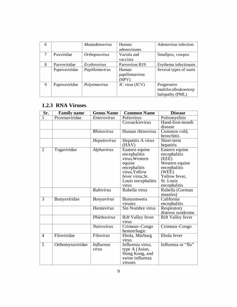
Viroids are known to infect plants and are responsible for crop failures and the loss of millions of dollars in agricultural revenue each year. Some of the plants they infect include potatoes, cucumbers, tomatoes, chrysanthemums, avocados, and coconut palms. For example, the potato spindle tuber viroid (PSTVd), which typically spreads when infected knives cut healthy potatoes in preparation for planting, can affect potatoes and tomatoes. The symptoms of PSTVd can be seen in Figure.
Figure 1.7.2. These potatoes have been infected by the potato spindle tuber viroid. (Credit: Pamela Roberts, University of Florida Institute of Food and Agricultural Sciences, USDA ARS) Some viruses are useful Some viruses are studied because they have useful current or potential applications. • Phage typing of bacteria.Some groups of bacteria, such as some
Salmonellaspecies, are classified into strains on the basis of the spectrum of phages to which they are susceptible. Identification of the phage types of bacterial isolates can provide useful epidemiological information during the outbreaks of disease caused by these bacteria.
• Sources of enzymes.A number of enzymes used in molecular biology are virus enzymes. Examples include reverse transcriptase from retroviruses and RNA polymerases from phages.
• Pesticides.Some insect pests are controlled with baculo viruses and myxoma virus has been used to control rabbits.
• Anti-bacterial agents.In the mid-20th century phages were used to treat some bacterial infections of humans. Interest waned with the discovery of antibiotics, but has been renewed with the emergence of antibiotic –resistant strain of bacteria.
37

• Anti-cancer agents.Genetically modified strains of viruses, such as herpes simplex virus and vaccinia virus, are being investigated for treatment of cancers.These strains have been modified so that they are able to infect and destroy specific tumor cells, but are unable to infect normal cells.
• Gene vectors for protein production.Viruses such as certain baculoviruses and adenoviruses are used as vectors to take genes into animal cells growing in culture. This technology can be used to insert into cells genes encoding useful proteins, such as vaccine components, and the cells can then be used for mass production of the proteins.
• Gene vectors fortreatment of genetic diseases.Children with severe combined immunodeficiency (baby in the bubble syndrome) have been successfully treated using retroviruses as vectors to introduce into their stem cells a non-mutated copy of the mutated gene responsible for the disease
Virus studies have contributed to knowledge as: Much of the basic knowledge of molecular biology, cell biology and cancer has been derived from studies with viruses. Here are a few examples. • A famous experiment carried by Alfred Hershey and Martha Chase, and
published in 1952, used phage T2 and E. coli to provide strong evidence that genes are composed of DNA.
• The first enhancers to be characterized were in genes of simian virus 40 (SV40).
• The first transcription factor to be characterized was the transplantation (T) antigen of SV40.
• The first nuclear localization signal of a protein was identified in the T antigen of SV40.
• Introns were discovered during studies of adenovirus transcription. • The role of the cap structure at the 5_ end of eukaryotic messenger RNA was
discovered during studies with vaccinia virus and a reovirus. • The first internal ribosomal entry site to be discovered was found in the RNA
of poliovirus. • The first RNA pseudo knot to be discovered was that in the genome of turnip
yellow mosaic virus. Self-Assessment Exercise 1.7 Q.1 Choose the best answer: i. The first IRES to be discovered was found in the RNA of ……. a) Hepatitis Virus b) Parvovirus c) Poliovirus d) HIV ii. The infectious form of protein is a) PrPca b) PrPsc c) PrPps d) PrPc
38

iii. (IPV) stands for: a) Adenoviruses b) Retroviruses c) Adeno Associated viruses d) All of the above iv. The first RNA pseudo knot to be discovered was that in the genome of
………………yellow mosaic virus. a) Brinjal b) Turnip c) Capsicum d) Tomato v. Identification of the phage types of ……………… isolates can provide
useful epidemiological information during the outbreaks of disease caused by these organisms.
a) Algal b) Fungal c) Protozoal d) Bacterial Q.2 How prions and Viroids are different from typical virus particles? Q.3 How Virus is taken as a useful entity and in terms of contributor to
knowledge? Glossary DNA: DNA is like the “blueprint” for building living cells. Enzymes: Enzymes are like the workers of a cell. They build new proteins, transport materials around the cell and carry out other important cellular functions. RNA: RNA is like the construction boss. Cells use RNA to tell enzymes how to build a specific part of a cell. To make a new protein, enzymes will copy a specific part of the DNA into a piece of RNA. This RNA is then used by other enzymes to build a new protein or enzyme. Proteins: The building blocks that is used to make living things. Nucleus: A small package inside the cell where the genetic material is kept.
39

References • Alberts B. et al. (2004) Essential Cell Biology, 2nd edition, Garland • Brown W. M. and Brown P. M. (2002) Transcription, Taylor and Francis • Cooper G. M. and Hausman R. E. (2004) The Cell: a Molecular Approach,
3rd edition, ASM Press • Drlica K. (2004) Understanding DNA and Gene Cloning, 4th edition, Wiley • Lodish H. et al. (2004) Molecular Cell Biology, 5th edition, Freeman • Muhammad Wajid Javed is student of B.Sc. (Hons.) Agriculture
(Department of Agri. Entomology, University of Agriculture, Faisalabad). He is also affiliated with Agrihunt as an author.
• Muhammad Hamza is student of B.Sc. (Hons.) Agriculture (Department of plant pathology, University of Agriculture, Faisalabad.) he is also affiliated with Agrihunt as an author.
• Pollard T. D. and Earnshaw W. C. (2004) Cell Biology, Saunders • Reece R. J. R. (2004) Analysis of Genes and Genomes,Wiley • Watson J. D. et al. (2004) Molecular Biology of the Gene, 5th edition,
Addison-Wesley • Weaver R. F. (2005) Molecular Biology, 3rd edition, McGraw-Hill
40

Unit 2
BACTERIA AND CYANOBACTERIA
Writer: Arshad Mehmood Qamar Reviewers: Aymen Sehar
Summaya KAnwal
41

CONTENTS
Introduction ...................................................................................................... 43
Objectives ......................................................................................................... 43
2.1 Bacteria; Ecology and Diversity ..................................................................... 44
2.2 Structure; shape, size of Bacteria and classification of Bacteria .................. 45
2.3 Modes of Nutrition in Bacteria ....................................................................... 50
2.4 Growth and reproduction in Bacteria ............................................................. 53
2.5 Importance of Bacteria (beneficial and Harmful Bacteria) ........................... 53
2.6 The Bacterial Flora of Human ........................................................................ 55
2.7 General Characteristics of Cyanobacteria ...................................................... 70
2.8 Habitat, Structure, nutrition and reproduction in Nostoc .............................. 70
2.9 Importance of cyanobacteria in terms of Nitrogen fixation .......................... 73
References ........................................................................................................ 76
42

INTRODUCTION This world is full of diversity and there are varieties of organisms which differ in size ranging from very small to very large organisms. They are considered of great importance due to the wide host range that is defined as their ability to cause diseases in variety of organisms including Bacteria, Algae, Fungi, animals, Plants and even humans. Bacteria are important due to its usability and harmful effects. In unit no2, ecology, diversity, Structure; shape, size of Bacteria and classification of Bacteria, Modes of Nutrition in Bacteria, Growth and reproduction in Bacteria, Importance of Bacteria (beneficial and Harmful Bacteria), the Bacterial Flora of Human, General Characteristics of Cyanobacteria, Habitat, Structure, nutrition and reproduction in Nostoc and Importance of cyanobacteria in terms of Nitrogen fixation are of vital importance described in this unit.
OBJECTIVES After studying this unit the students will be able to: • Describe the ecology and diversity of bacteria. • Classify bacteria. And identify bacteria on the basis of nutrition. • Explain benefits of bacteria, and differentiate harmful bacteria from useful
bacteria. • Elaborate the importance of Flora of Humans • Tell habitat, structure, nutrition and reproduction in Nostoc. • Describe the importance of Cyanobacteria for Nitrogen fixation.
43

1.1 BACTERIA; ECOLOGY AND DIVERSITY Bacteria are unicellular microscopic organisms that were first discovered by Dutch microscopist Antony Van Leeuwenhoek in 1673. He used simple microscope to describe bacteria and called those tiny creatures as animalcules.The progress in understanding of those tiny molecules was slow. The microbe’s existence was further confirmed by Louis Pasteur. He developed vaccines for rabies, cholera and anthrax. He proved that microorganisms are involved in causing diseases. Robert Koch formulated the germ theory of disease. Main postulates of germ theory are as under: The microorganism must be found in abundance in all organisms suffering from the disease, but should not be found in healthy organisms. The microorganism must be isolated from a diseased organism and grown in pure culture. The cultured microorganism should cause disease when introduced into a healthy organism. The microorganism must be re-isolated from the inoculated, diseased experimental host and identified as being identical to the original specific causative agent. Eventually, germ theory led to the discovery of causative agents of many diseases. So after that golden era of bacteriology got started which revealed many important information about bacteria.Bacteria are present almost everywhere from hot springs to Polar Regions. Bacterial cells are very simple in organization; they do not contain proper centralized nucleus and membrane-bound organelles. They occur in wide range of size and shapes. Bacteria also form symbiotic and parasitic associations with plants and animals. They serve as a key part of plants animals and human micro flora. Bacteria are beneficial as well as harmful to the environment. Their beneficiary aspects include recycling of the nutrients by different mechanisms like nitrogen fixation in nitrogen cycle, food processing in industries, and production of alcohol, waste treatment plants, production of biofuel and bio fertilizer.
44

Besides the number of beneficial roles of bacteria, some harmful aspects are the spread of disease and epidemics. Several diseases are caused by pathogenic species of bacteria including cholera, syphilis, anthrax, leprosy, tuberculosis and bubonic plague. 2.2 SHAPE, SIZE, STRUCTURE AND CLASSIFICATION OF BACTERIA Bacteria, despite their simplicity, contain a well-developed cell structure that is accountable for several of their distinctive biological structures. Several structural features are distinctive to bacteria only and aren't found among archaea or eukaryotes.
Fig 2.1 Structure of Bacteria All bacterial cells have cell membrane, cytoplasm, ribosomes and chromatin bodies. Many bacterial types Possess cell wall which provides shape to the cell. Some of the structures like capsule, slime, flagella, piliand granules are not found in all bacteria. 2.2.1 Shapes and Size of Bacteria Bacteria come in a wide variety of shapes. Perhaps the most elemental structural property of bacteria is their morphology (shape).
45

Generally bacteria are classified into three categories. These are cocci, bacilli and spiral. Many of the bacteria have constant shape however some types of bacteria exist in more than one shape. The cocci are rounded shaped bacteria having one of the several well defined arrangements on the basis of planes of division. One plane division causes diplococcus or streptococcus type of arrangement. Square of 4 cocci give rise to tetrad shape. When division takes place in three planes the resultant arrangement is called sarcina which is cube of 8 cocci. Staphylococcus arrangement comes out when arrangement is in random planes. Bacilli are rod shaped bacteria and they all divide in one plane to give rise Streptobacillus or diplobacillus. The spirally coiled bacteria form spiral shape of bacteria. A spiral could be one of vibrio, spirillum or a spirochete. Vibrio is comma shaped bacteria. Spirillum is a thick, rigid spiral whereas Spirochete is a thin, flexible spiral. Bacteria range in size from about 0.1 to 600 µm over a single dimension. They vary in size as much as in shape. The smallest bacteria are about 100 to 200 nm in diameter. Some spirochetes occasionally reach 500 µmin lengths whereas staphylococcus and streptococci can only reach to 0.75-1.25µm in diameter.
Fig 2.2: Shapes of Bacteria
46

Self-Assessment Exercise Q. 1 Select the most appropriate answer from the options given at the end of each
statement. i. Germ theory was proposed by: a) Louis Pasteur b) Alexander Fleming c) Robert Koch d) Robert Brown ii. Spiral comes in the form: a Vibrio b) Spirillum c) Spirochete d) All a, b, c iii. The bacteria range in size from about 0.1 to -------µm over a single
dimension a) 500 b) 600 c) 700 d) 800 iv. When cocci form long chain of cells, then arrangement is called: a) Tetrad b) Diplococcus c) Sarcina d) Streptococci v. Leeuwenhoek used a simple microscope to describe bacteria and
protozoa and called these small creatures: a) Micro animals b) Animalcules c) Beastcules d) Micro monster Q.2 Give short answers: i. Write shape of the bacteria. ii. write range of size of bacteria. 2.2.2 Structure of Bacterial Cell Flagella Flagella are the outermost structure of bacterial cell. They are thin hair like appendages which are meant for bacterial locomotion. They originate from basal body and are made up of protein called flagellin. On the basis of presence, pattern of attachment and number of flagella, bacteria are classified into different taxonomic groups. Those bacteria which do not possess any flagella are termed as Atrichous and those with single polar flagellum are Monotrichous. If the tuft of flagella is present on one pole then the bacteria are termed as lophotrichousand when on both poles, bacteria are amphitrichous. In peritrichousflagella surround the whole cell.
47

Fig.2.3: Structure of Bacteria with respect of Flagella Cell Wall Cell wall is present beneath the extracellular substances and external to cytoplasmic contents. It is rigid and determines the shape of bacterium. Cell wall also protect cell from osmotic lysis.Mycoplasmas are the only bacterial types which do not possess cell wall. Bacteria could be divided into two groups on the basis of its response to gram staining procedure, a method devised by Christian Gram. In this technique, purple stained bacteria are termed as Gram positive because they retain the primary dye and pink stained bacteria are termed as Gram negative as they retain the secondary dye.Cell wall of bacteria is generally composed of unique macromolecule called peptidoglycan which is actually the long glycan chains cross linked with peptide fragments.
Fig.2.4: Structure of Cell wall of Bacteria
48

Cell Membrane A very thin and flexible membrane beneath the cell wall is the cell membrane or plasma membrane. It is so delicate that any damage to it can cause death of the cell. Unlike eukaryotic membrane, bacterial membrane lacks cholesterol.Cell membrane functions in the regulation of the transport of proteins, nutrients, sugar and other metabolites. It also possesses respiratory metabolism enzymes. Nucleoid Bacterial cell lacks distinct chromosomes and nuclear membrane. Single, circular and double stranded DNA present near the center of the cell. It is primarily a very long molecule of DNA that is tightly packed so as to fit inside the cell compartment. Ribosomes Ribosomes contain bacterial RNA and proteins. Those ribosomes which synthesize proteins remain attached to plasma membrane. So are termed as protein factories. Plasmid Many bacteria have circular, double stranded DNA molecule in addition to chromosomes which are called plasmids. Plasmids are self -replicating like nuclear DNA. They are important vectors in modern genetic engineering as they possess genes for drug resistant, heavy metals and insect resistant. Special Structures of Bacterial Cell Capsule Sometimesa thicker gummy type of capsule surrounds the bacterial cell which is made up of repeating polysaccharides unit and proteins. Its function is to give sticky character to encapsulated colonies of bacteria. Spores Some species of bacteria produce specialized dormant structure called spore in response to harsh environmental conditions like temperature, pH, light, heat etc. Spores can be of two types i.e. external to vegetative cells or within the vegetative cells. When environment become favorable the spore can germinate and form vegetative cells. Cyst Dormant, thick walled, desiccation resistant cell is formed by bacterial cell during vegetative cells differentiation. These cysts can germinate under favorable conditions. Cysts are not heat resistant.
49

Self-Assessment Exercise 2.2 Q.1 Select the most appropriate answers given at the end of each statement. i. Primary function of flagella is to help in: a) Walking b) Motility c) Running d) Rest ii. The cell wall of most bacteria have unique macromolecule called: a) Teichoic acid b) Lipoproteins c) Peptidoglycan d) Polysaccharide iii. By Gram staining technique, gram positive bacteria are stained----- in color: a) Yellow b) Pink c) Purple d) Blue iv. Mycoplasmas do not possess: a) Cell membrane b) Cell wall c) DNA d) Cytoplasm v. Bacterial membrane differs from eukaryotic membrane in: a) Lacking protein b) Lacking lipids c) Lacking polysaccharides d) Lacking cholesterol Q.2 Answer the following short questions. 1. What are the specialized features of bacterial nucleoid? 2. What are the protein factories in bacterial cell? 3. What are the uses of plasmid? 4. Draw the generalized structure of bacteria and label it. 5. What do you know about the capsule of bacterial cell? 6. What are cysts? 7. How spores are formed in bacteria? State their function. 2.3 MODES OF NUTRITION IN BACTERIA Bacteria exhibit different modes of nutrition. On this basis, broadly two types of bacteria can be recognized autotrophic bacteria and heterotrophic bacteria. 2.3.1 Autotrophic Bacteria These are bacteria which are able to synthesize their own organic food from inorganic substances. They use carbon dioxide for obtaining carbon and utilize hydrogen sulphide (H2S) or ammonia (NH3) or hydrogen (H2) as the source of hydrogen to reduce carbon. These bacteria can be distinguished further into two types as follows:
50

Photoautotrophic Bacteria The photoautotrophic bacteria possess photosynthetic pigments in membrane bound lamellae (or thylakoids) and utilize solar energy. The bacterial photosynthesis is different from that of green plants since here water is not used as a hydrogen donor. Hence oxygen is not released as a byproduct. For this reason, the process is described as anoxygenic photosynthesis.
CO2 + H2 S Sugars +Sulpher +Water Chemosynthetic Bacteria These are bacteria which manufacture organic compounds from inorganic raw materials utilising energy liberated from the oxidation of inorganic substances. Following are the common types of chemo autotrophic bacteria. 1. Nitrifying bacteria which derive energy by oxidizing ammonia into nitrates.
Eg: Nitrosomonas, Nitrobacter.
NH4 + + 2O2 NO2 + 2H2 O+ energy
2. Sulphur bacteria which derive energy by oxidising hydrogen sulphide to
sulphur. Eg: Thiobacillus, Beggiatoa.
2H2 S +O2 2S +2H2O + energy
3. Iron Bacteria which derive energy by oxidising ferrous ions into ferric form. Eg: Ferrobacillus, Gallionella.
4FeCO3 + 6H2 O + O2 4Fe(OH)3 + 4CO2 + energy
2.3.2 Heterotrophic Bacteria These are bacteria which are unable to manufacture their own organic food and hence are dependent on external source. These bacteria can be distinguished into three groups as follows: • Saprophytic Bacteria: These bacteria obtain their nutritional requirements
from dead organic matter. They breakdown the complex organic matter into simple soluble form by secreting exogenous enzymes. Subsequently they absorb the simple nutrients and assimilate them, during which they release energy. These bacteria have a significant role in the ecosystem, functioning as decomposers.
• The aerobic breakdown of organic matter is called as decay or decomposition. It is usually complete and not accompanied by the release of
51

foul gases. Anaerobic breakdown of organic matter is called fermentation. It is usually incomplete and is always accompanied by the release of foul gases. Anaerobic breakdown of proteins is called putrefaction.
The property of decomposition of organic compounds is employed in several industrial processes such as ripening of cheese, in the retting of fibers and in the curing of tobacco. Symbiotic Bacteria These are bacteria which live in a mutually beneficial association with other organisms. Such bacteria derive the essential nutrients from their host organisms and in that process help the host through some of their biological activities. 1. The most familiar example of symbiotic bacteria are the nitrogen fixing
bacteria found in the root nodules of leguminous plants. Bacteria such as Rhizobium and Pseudomonas reside in the root nodules and reduce atmospheric nitrogen directly to ammonia. This becomes the source of nitrogen for the host plants. The plants in return provide bacteria with nutrients and protection.
2. The bacteria found in the human alimentary canal Escherichia coli are nonpathogenic. These bacteria check the growth of harmful putrefying bacteria. In addition, these bacteria release vitamins K and B12 which are necessary for blood components. The human host provides shelter and food for these bacteria.
3. A similar example is that of cellulose digesting bacteria which occur in the alimentary canal of ruminant mammals such as cows and goats.
Parasitic Bacteria These are bacteria which occur in the body of animals and plants, obtaining their organic food from there. Most of these bacteria are pathogenic, causing serious diseases in the host organisms either by exploiting them or by releasing poisonous secretions called toxins. SelfAssessment Exercise 2.3 Q.1 Answer the following short questions. 1. What are different categories of autotrophic bacteria? 2. What are photosynthetic bacteria? How they differ from plants? 3. Differentiate between photosynthetic and chemosynthetic bacteria?
52

2.4 GROWTH AND REPRODUCTION IN BACTERIA Bacterial growth refers to increase in number of bacterial cells. Bacteria undergo asexual reproduction called Binary fission. In this type of reproduction, cell increases in size, its chromatin material duplicates and then cell membrane pinches inwards right at the center of the cell causing the division of cell into two containing equal amount of nuclear material in it. There are four distinct phases in bacterial growth curve: Lag phase It is the phase in which bacteria prepares itself for division so, no growth is seen. Log phase In this phase bacteria grow at exponential rate so, it is phase of rapid growth. Stationary phase In this phase growth rate becomes equal to death rate. Death/ Decline phase In this phase bacteria starts dying. Rate of reproduction will be less than rate of death.
Fig. 2.5 Growth of Bacteria 2.5 IMPORTANCE OF BACTERIA Bacteria are useful as well as harmful to the environment. Some of the beneficial aspects of bacteria are:
53

Bacteria play significant role in completion of cycles of nitrogen, carbon and phosphorus. They help in the production of yogurt, cheese and alcoholic beverages. They are used by the pharmaceuticals in the production of variety of drugs, antiobiotics and insulin. They have the key role in the chemical industry for the production of acetone and butanol.
Bacteria are efficient decomposers.
They are also involved in symbiosis.
Some of the harmful aspects of bacteria are as under:
They are very common pathogens of humans and cause number of diseases.
They are responsible for spoilage of food and vegetables.
They are also involved in food poisoning.
They play major role in the destruction of domestic articles.
They cause the reduction in soil fertility by their activities. Self-Assessment Exercise 2.4 i. Bacterial growth means: a) Increase in size b) Change in shape c) Increase in flagella d) Increase in number ii. Rapid phase of bacterial growth is: a) Lag b) Log c) Stationary d) Death iii. Bacteria reproduce by: a) Fragmentation b) Sexual reproduction c) Binary fission d) Budding iv. Bacteria prepare itself for division during which phase: a) Lag b) Log c) Stationary d) Death v. When the reproduction rate of bacteria becomes lower: a) Lag b) Log c) Stationary d) Death
54

Q.2 Answer the questions. i. How bacteria play role in medical field? ii. What is the role of bacteria in food industry? iii. State two harmful aspects of bacteria? 2.6 THE BACTERIAL FLORA OF HUMANS Bacterial Flora of Humans is very complex consisting of more than 200 species of Bacteria. Genetics, sex, age, nutrition, stress and diet of an individual influence the makeup of flora. The composition of normal flora varies in intestinal tract, oral cavity and vagina. Generally the bacterial flora of human remains constant and can be described generally. First bacterial flora establishes at the time of birth. Handling and feeding of the infant after birth leads to establish a stable flora on skin, oral cavity and intestinal tract in about forty eight hours. It has been calculated that a human adult houses about 1012 bacteria on the skin, 1010 in the mouth, and 1014 in the gastrointestinal tract Normal Flora of the Skin Human body is covered with skin of area two square meters. The population of bacteria is very low at some sites of the skin and fairly high at few of the places called as the most suitable or favorable sites. The highly moisturized areas on the skin support the activity and growth of the bacteria. Hence there is high density of bacterial cells on these moist sites. Most bacteria on the skin are sequestered in sweat glands. In most of the areas like superficial layers of the epidermis and upper parts of the hair follicles, many non-pathogenic and commensal bacteria like gram-positive cocci including Staphylococcus epidermidis, Micrococcus and corynebacteria ( Propionibacteriumsp ) are found which result in no harm becauseStaphylococci and Propionibacterium produce fatty acids that inhibit the growth of fungi and yeast on the skin. But, if Propionibacterium acnes, a normal inhabitant of the skin is present on the skin and have been found to be trapped in hair follicle, it may grow rapidly and cause inflammation and acne. Sometimes pathogenic gram positive cocci like Staphylococcus aureusis a common species found especially on nasal mucous membrane and skin. Nasal carriage of S. aureus is a risk factor for subsequent infection. Such individuals may auto-inoculate themselves with the bacteriaand also result in the spread of it to other individuals or foods.
55

Bacteria commonly found on the surface of the human body are listed in table 1 and Predominant bacteria at various anatomical locations in adults are shown in table 2 below. Bacteria commonly found on the surfaces of the human body. Table.1:
++ = nearly 100 percent + = common (about 25 percent) +/- = rare (less than 5%) * = Potential pathogen
S.No Bacterial species Skin Cojunctiva Nose Pharynx Lower GI
Ant. urethra
Vagina
1 Staphylococcus epidermidis (1)
++ + ++ ++ ++ + ++
2 Staphylococcus aureus* (2)
+ +/- + + + ++ +/-
3 Streptococcus mitis + ++ +/- + 4 Streptococcus
salivarius ++ ++
5 Streptococcus mutans* (3)
+ ++
6 Enterococcus faecalis* (4)
+/- + ++ +
7 Streptococcus pneumoniae* (5)
+/- +/- + +
8 Streptococcus pyogenes* (6)
+/- +/- + + +/-
9 Neisseria sp. (7) + + ++ + + 10 Neisseria
meningitidis* (8) + ++ +
11 Enterobacteriaceae*(Escherichia coli) (9)
+/- +/- +/- + ++ +
12 Pseudomonas aeruginosa* (10)
+/- +/- + +/-
13 Haemophilus influenzae* (11)
+/- + + +
14 Bacteroides sp.* ++ + 15 Bifidobacterium
bifidum (12) ++
16 Lactobacillus sp. (13) + ++ ++ 17 Clostridium sp.* (14) +/- ++ 18 Clostridium tetani (15) +/- 19 Corynebacteria (16) ++ + ++ + + + + 20 Mycobacteria + +/- +/- + + 21 Actinomycetes + + 22 Spirochetes + ++ ++
56

Table.2: Predominant bacteria at various anatomical locations in adults S.No Anatomical
Location Predominant bacteria
1 Skin Staphylococci and Corynebacteria 2 Conjunctiva Sparse, Gram-positive cocci and Gram-negative rods 3 Oral Cavity Streptococcus mutans,Streptococcus salivarius and Prevotella 4 Teeth Streptococci, Lactobacillietc 5 Mucous
Membranes Streptococci and lactic acid bacteria
6 Upper Respiratory Tract
Bordetella pertussis, Streptococcus pyogenes, Chlamydia pneumoniae and Mycoplasma pneumoniae
7 Nares (Nasal Membranes)
Staphylococci and corynebacteria
8 Pharynx (Throat)
Streptococci, neisseria, Gram-negative rods and cocci
9 Lower Respiratory Tract
None
10 Gastrointestinal Tract
Shigella,S. enterica, E.coli and S. bongori
11 Stomach Helicobacter pylori (up to 50%) 12 Small Intestine Lactics, enterics, enterococci, bifidobacteria 13 Colon Bacteroides,lactics,Enterics,Enterococci,Clostridia,Methanog
ens 14 Urogenital Tract S.saprophyticus,S.epidermidis, Enterococcus faecalis 15 Anterior Urethra Sparse, Staphylococci, Corynebacteria, Enterics 16 Vagina Lactic acid bacteria during child-bearing years; otherwise
mixed 2.6.1 Normal Flora of the Oral Cavity Mouth is the most favorable habitat for a variety of bacteria.Streptococci, Lactobacilli, Staphylococci and Corynebacteria are some examples of bacteria found in mouth. Composition of flora of oral cavity changes with age. At the time of birth, the oral cavity is composed solely of the soft tissues of the lips, cheeks, tongue and palate, which are kept moist by the secretions of the salivary glands. During this time, the oral cavity is sterile but rapidly becomes colonized from the environment, particularly when the mother first feeds the child. Streptococcus salivarius is dominant and may make up 98% of the total oral flora until the appearance of the teeth (6 - 9 months in humans). The eruption of the teeth during the first year
57

leads to colonization of S. mutans and S. sanguis. These bacteria require a non-desquamating (non-epithelial) surface in order to colonize. They have the ability to persist as long as teeth remain. Other strains of Streptococci adhere strongly to the gums and cheeks but not to the teeth. The creation of the gingival crevice area (supporting structures of the teeth) increases the habitat for the variety of anaerobic species found. The complexity of the oral flora continues to increase with time, and Bacteroides and Spirochetes colonize around the puberty of an individual.
Figure 2.6.1. Various Streptococci observed in the biofilms of oral cavity The bacteria in the oral cavity get maximum benefits of nutrients and space. Hosts also get few benefits from bacteria due to this interaction like bacterial flora leaves very less space for other germs to settle in the oral cavity. They also provide some vitamins that are useful for host and antibiotics which may cross react with pathogens. Bacterial flora is source of some inhibitory substances such as fatty acids, peroxides and bacteriocins. Whereas bacterial flora is also harmful for host in many ways as it causes many diseases in humans such as dental caries, abscesses, gingivitis and periodontal malfunctioning. The access of bacteria to deeper tissues can cause abscesses of bone, brain lungs or other parts of the body. If oral streptococciare introduced into wounds created by dental treatment, they may adhere to heart valves and initiate sub-acute bacterial endocarditis.
58

Normal Flora of the Respiratory Tract A large number of bacterial species colonize the upper respiratory tract (nasopharynx). The nares (nostrils) are always heavily colonized, predominantly with Staphylococcus epidermidis and Corynebacteria, and often (in about 20% of the general population) with Staphylococcus aureus, this being the main carrier site of this important pathogen. The healthy sinuses, in contrast are sterile. The pharynx (throat) is normally colonized by Streptococci and various Gram-negative cocci. Sometimes pathogens such as Streptococcus pneumoniae, Streptococcus pyogenes, Haemophilus influenzae and Neisseria meningitides colonize the pharynx. The lower respiratory tract (trachea, bronchi, and pulmonary tissues) is virtually free of microorganisms, mainly because of the efficient cleansing action of the ciliated epithelium which lines the tract. Any bacteria reaching the lower respiratory tract are swept upward by the action of the mucociliary blanket that lines the bronchi, to be removed subsequently by coughing, sneezing, swallowing, etc. If the respiratory tract epithelium becomes damaged, as in bronchitis or viral pneumonia, the individual may become susceptible to infection by pathogens such as H. influenzae or S. pneumoniae descending from the nasopharynx. Normal Flora of the Urogenital Tract Urine is normally sterile, and since the urinary tract is flushed with urine every few hours, microorganisms have problems gaining access and becoming established. The flora of the anterior urethra, as indicated principally by urine cultures, suggests that the area may be inhabited by a relatively consistent normal flora consisting of Staphylococcus epidermidis, Enterococcus faecalis and some alpha-hemolytic streptococci. Their numbers are not plentiful, however. In addition, some enteric bacteria (e.g. E. coli, Proteus) and corynebacteria, which are probably contaminants from the skin, vulva or rectum, may occasionally be found at the anterior urethra. The vagina becomes colonized soon after the birth with Corynebacteria, Staphylococci, Streptococci, E. coli, and a lactic acid bacterium historically named "Doderlein's bacillus" (Lactobacillus acidophilus). During reproductive life, from puberty to menopause, the vaginal epithelium contains glycogen due to the actions of circulating estrogens. Doderlein's bacillus predominates, being able to metabolize the glycogen to lactic acid. The lactic acid and other products of metabolism inhibit colonization by all except this lactobacillus and a select number of lactic acid bacteria. The resulting low pH of the vaginal epithelium prevents establishment by most other bacteria as well as the potentially-
59

pathogenic yeast, Candida albicans. This is a striking example of the protective effect of the normal bacterial flora for their human host.
Figure 2.6.2. A Lactobacillus species, possibly Doderlein's bacillus, in association with a vaginal epithelial cell
Figure 2.6.3: Helicobacter pylori. The proximal small intestine has a relatively sparse Gram-positive flora, consisting mainly of lactobacilli and Enterococcus faecalis. This region has about 105 - 107 bacteria per ml of fluid. The distal part of the small intestine contains greater numbers of bacteria (108/ml) and additional species, including coliforms (E. coli and relatives) and Bacteroides, in addition to lactobacilli and enterococci. The flora of the large intestine (colon) is qualitatively similar to that found in feces. Populations of bacteria in the colon reach levels of 1011/ml feces. Coliforms become more prominent, and enterococci, clostridia and lactobacilli can be regularly found, but the predominant species are anaerobic Bacteroidesand
60

anaerobic lactic acid bacteria in the genus Bifidobacterium (Bifidobacterium bifidum). These organisms may outnumber E. coli by 1,000:1 to 10,000:1. Sometimes, significant numbers of anaerobic methanogens (up to 1010/gm) may reside in the colon of humans. This is our only direct association with archaea as normal flora. The range of incidence of certain bacteria in the large intestine of humans is shown in Table 3 below.
Table 3 Bacteria found in the large intestine of humans.
BACTERIUM RANGE OF INCIDENCE Bacteroides fragilis 100 Bacteroides melaninogenicus 100 Bacteroides oralis 100 Lactobacillus 20-60 Clostridium perfringens 25-35 Clostridium septicum 5-25 Clostridium tetani 1-35 Bifidobacterium bifidum 30-70 Staphylococcus aureus 30-50 Enterococcus faecalis 100 Escherichia coli 100 Salmonella enteritidis 3-7 Klebsiella sp. 40-80 Enterobacter sp. 40-80 Proteus mirabilis 5-55 Pseudomonas aeruginosa 3-11 Peptostreptococcus sp. common Peptococcus sp. common At birth the entire intestinal tract is sterile, but bacteria enter with the first feed. The initial colonizing bacteria vary with the food source of the infant. In breast-fed infants, Bifidobacteriaaccounts for more than 90% of the total intestinal bacteria. Enterobacteriaceae and Enterococci are regularly present, but in low proportions, while Bacteroides, Staphylococci, Lactobacilli and Clostridia are practically absent. In bottle-fed infants, Bifidobacteria are not predominant. When breast-fed infants are switched to a diet of cow's milk or solid food, Bifidobacteria are progressively joined by Enterics, Bacteroides, Enterococci, Lactobacilli and Clostridia. Apparently, human milk contains a growth factor that enriches for growth of Bifidobacteria, and these bacteria play an important role in preventing colonization of the infant intestinal tract by non-indigenous or pathogenic species.
61

Figure 2.6.4: Clostridium difficile when stained.
The growth of "C. difficle" in the intestinal tract is normally held in check by other members of the normal flora. When antibiotics given for other infections cause collateral damage to the normal intestinal flora, the clostridium may be able to "grow out" and produce a serious diarrheal syndrome called pseudomembranous colitis. This is an example of an "antibiotic induced diarrheal disease". The composition of the flora of the gastrointestinal tract varies along the tract (at longitudinal levels) and across the tract (at horizontal levels) where certain bacteria attach to the gastrointestinal epithelium and others occurs in the lumen. There is frequently a very close association between specific bacteria in the intestinal ecosystem and specific gut tissues or cells (that is evidence of tissue tropism and specific adherence). Gram-positive bacteria, such as the streptococci and lactobacilli, are thought to adhere to the gastrointestinal epithelium using polysaccharide capsules or cell wall teichoic acids to attach to specific receptors on the epithelial cells. Gram-negative bacteria such as the enteric bacteria may attach by means of specific fimbriae which bind to glycoproteins on the epithelial cell surface. It is in the intestinal tract that we see the greatest effect of the bacterial flora on their host. This is due to their large mass and numbers. Bacteria in the human GI tract have been shown to produce vitamins and may otherwise contribute to nutrition and digestion. But their most important effects are in their ability to protect their host from establishment and infection by alien microbes and their ability to stimulate the development and the activity of the immunological tissues.
62

On the other hand, some of the bacteria in the colon (e.g. Bacteroides) have been shown to produce metabolites that are carcinogenic, and there may be an increased incidence of colon cancer associated with these bacteria. Alterations in the GI flora brought on by poor nutrition or perturbance with antibiotics can cause shifts in populations and colonization by nonresidents that leads to gastrointestinal disease. Beneficial Effects of the Normal Flora The effects of the normal flora are inferred by microbiologists from experimental comparisons between "germ-free" animals (which are not colonized by any microbes) and conventional animals (which are colonized with a typical normal flora). Briefly, some of the characteristics of germ-free animals that are thought to be due to lack of exposure to a normal flora are: 1. Vitamin deficiencies, especially vitamin K and vitamin B12. 2. Increased susceptibility to infectious disease. 3. Poorly developed immune system, especially in the gastrointestinal tract. 4. Lack of "natural antibody" or natural immunity to bacterial infection. Because these conditions in germ-free mice and hamsters do not occur in conventional animals, or are alleviated by introduction of a bacterial flora (at the appropriate time of development), it is tempting to conclude that the human normal flora make similar contributions to human nutrition, health and development. The overall beneficial effects of microbes are summarized below. 1. The normal flora synthesizes and excretes vitamins in excess of their own
needs, which can be absorbed as nutrients by their hosts. For example, in humans, enteric bacteria secrete Vitamin K and Vitamin B12, and lactic acid bacteria produce certain B-vitamins. Germ-free animals may be deficient in Vitamin K to the extent that it is necessary to supplement their diets.
2. The normal flora prevents colonization by pathogens by competing for attachment sites or for essential nutrients. This is thought to be their most important beneficial effect, which has been demonstrated in the oral cavity, the intestine, the skin, and the vaginal epithelium. In some experiments, germ-free animals can be infected by 10 Salmonella bacteria, while the infectious dose for conventional animals is near 106 cells.
3. The normal flora may antagonize other bacteria through the production of substances which inhibit or kill nonindigenous species. The intestinal bacteria produce a variety of substances ranging from relatively nonspecific
63

fatty acids and peroxides to highly specific bacteriocins, which inhibit or kill other bacteria.
4. The normal flora stimulates the development of certain tissues, i.e., the caecum and certain lymphatic tissues (Peyer's patches) in the GI tract. The caecum of germ-free animals is enlarged, thin-walled, and fluid-filled, compared to that organ in conventional animals. Also, based on the ability to undergo immunological stimulation, the intestinal lymphatic tissues of germ-free animals are poorly-developed compared to conventional animals.
5. The normal flora stimulate the production of natural antibodies.Since the normal flora behave as antigens in an animal, they induce an immunological response, in particular, an antibody-mediated immune (AMI) response. Low levels of antibodies produced against components of the normal flora are known to cross react with certain related pathogens, and thereby prevent infection or invasion. Antibodies produced against antigenic components of the normal flora are sometimes referred to as "natural" antibodies, and such antibodies are lacking in germ-free animals.
2.6.2 Harmful Effects of the Normal Flora Harmful effects of the normal flora that have been observed in studies with germ-free animalscan be put in the following categories. All but the last two are fairly insignificant. 1. Bacterial synergism between a member of the normal flora and a potential
pathogen. This means that one organism is helping another to grow or survive. There are examples of a member of the normal flora supplying a vitamin or some other growth factor that a pathogen needs in order to grow. This is called cross-feeding between microbes. Another example of synergism occurs during treatment of "staph-protected infections" when a penicillin-resistant staphylococcus that is a component of the normal flora shares its drug resistance with pathogens that are otherwise susceptible to the drug.
2. Competition for nutrients
Bacteria in the gastrointestinal tract must absorb some of the host's nutrients for their own needs. However, in general, they transform them into other metabolisable compounds, but some nutrient(s) may be lost to the host. Germ-free animals are known to grow more rapidly and efficiently than conventional animals. One explanation for incorporating antibiotics into the food of swine, cows and poultry is that the animal grows faster and can therefore be marketed earlier. Unfortunately, this practice contributes to the
64

development and spread of bacterial antibiotic resistance within the farm animals, as well as humans.
3. Induction of a low grade toxemia Minute amounts of bacterial toxins (e.g. endotoxin) may be found in the
circulation that stimulatesthe formation of natural antibodies. 4. The normal flora may be agents of disease. Members of the normal flora may cause endogenous disease if they reach a site or tissue where they cannot be restricted or tolerated by the host defenses. Many of the normal flora are potential pathogens, and if they gain access to a compromised tissue from which they can invade, disease may result. 5. Transfer to susceptible hosts Some pathogens of humans that are members of the normal flora may also
rely on their host for transfer to other individuals where they can produce disease. This includes the pathogens that colonize the upper respiratory tract such as Neisseria meningitidis, Streptococcus pneumoniae, Haemophilus influenzae and Staphylococcus aureus, and potential pathogens such as E. coli, Salmonella or Clostridium in the gastrointestinal tract.
2.6.3 Dental Caries, Gingivitis and Periodontal Disease The most frequent and economically-important condition in humans resulting from interactions with our normal flora is probably dental caries. Dental plaque, dental caries, gingivitis and periodontal diseaseare due to the actions that are initiated and carried out by the normal bacterial flora. Dental plaque is the adherence of certain materials to the teeth such as consisting of bacterial cells (60-70% the volume of the plaque), salivary polymers, and bacterial extracellular products. Plaque is a naturally-constructed biofilm, in which the consortia of bacteria may reach a thickness of 300-500 cells on the surfaces of the teeth. These accumulations subject the teeth and gingival tissues to high concentrations of bacterial metabolites, which result in dental disease. The dominant bacterial species in dental plaque are Streptococcus sanguis and Streptococcus mutans, both of which are considered responsible for plaque.
65

2.6.4: Streptococcusmutans. Gram stain. CDC
Plaque formation is initiated by a weak attachment of the Streptococcal cells to salivary glycoproteins forming a pellicle on the surface of the teeth. This is followed by a stronger attachment by means of extracellular sticky polymers of glucose (glucans) which are synthesized by the bacteria from dietary sugars (principally sucrose). An enzyme on the cell surface of Streptococcus mutans, glycosyl transferase, is involved in initial attachment of the bacterial cells to the tooth surface and in the conversion of sucrose to dextran polymers (glucans) which form plaque.
Fig, 2.6.5:Dental plaque, scanning electron micrograph illustrating the diversity of microbes in plaque Image courtesy of Rachel Sammons, University of Birmingham School of Dentistry (UK)
66

Dental Caries is the destruction of the enamel, dentin or cementum of teeth due to bacterial activities. Caries are initiated by direct demineralization of the enamel of teeth due to lactic acid and other organic acids which accumulate in dental plaque. Lactic acid bacteria in the plaque produce lactic acid from the fermentation of sugars and other carbohydrates in the diet of the host.Streptococcus mutans and Streptococcus sanguis are most consistently been associated with the initiation of dental caries, but other lactic acid bacteria are probably involved as well. These organisms normally colonize the occlusal fissures and contact points between the teeth, and this correlates with the incidence of decay on these surfaces.
Figure: 2.6.6 Tooth Plague
Cross section of a tooth illustrating the various structural regions susceptible to colonization or attack by microbes Streptococcus mutans in particular has a number of physiological and biochemical properties which implicate it in the initiation of dental caries. These are given as follows: It is a regular component of the normal oral flora of humans which occurs in relatively large number. It readily colonizes the tooth surfaces: salivary components (mucins, which are glycoproteins) and form a thin film on the tooth called the enamel pellicle. The adsorbed mucins are thought to serve as molecular receptors for ligands on the bacterial cell surface.
67

It contains a cell-bound protein, glycosyl transferase, which serves an adhesin for attachment to the tooth, and as an enzyme that polymerizes dietary sugars into glucans that leads to the formation of plaque. It produces lactic acid from the utilization of dietary carbohydrate which demineralizes tooth enamel. S. mutans produces more lactic acid and is more acid-tolerant than most other streptococci. It stores polysaccharides made from dietary sugars which can be utilized as reserve carbon and energy sources for production of lactic acid. The extracellular glucans formed by S. mutans are, in fact, bacterial capsular polysaccharides that function as carbohydrate reserves. The organisms can also form intracellular polysaccharides from sugars which are stored in cells and then metabolized to lactic acid. Streptococcus mutans appears to be important in the initiation of dental caries because its activities lead to colonization of the tooth surfaces, plaque formation, and localized demineralization of tooth enamel. It is not however, the only cause of dental decay. After initial weakening of the enamel, various oral bacteria gain access to interior regions of the tooth. Lactobacilli, Actinomyces, and various proteolytic bacteria are commonly found in human carious dentin and cementum, which suggests that they are secondary invaders that contribute to the progression of the lesions.
Figure 2.6.6: Actinomyces Israeli
Periodontal Diseases are bacterial infections that affect the supporting structures of the teeth (gingiva, cementum, periodontal membrane and alveolar bone). The most common form, gingivitis, is an inflammatory condition of the gums. It is associated with accumulations of bacterial plaque in the area. Increased
68

populations of Actinomyces have been found, and they have been suggested as the main cause. Diseases that are confined to the gum usually do not lead to loss of teeth, but there are other more serious forms of periodontal disease that affect periodontal membrane and alveolar bone resulting in tooth loss. Bacteria in these lesions are very complex populations consisting of Gram-positive organisms (including Actinomyces and Streptococci) and Gram-negative organisms (including spirochetes and Bacteroides). The mechanisms of tissue destruction in periodontal disease are not clearly defined but hydrolytic enzymes, endotoxins, and other toxic bacterial metabolites seem to be involved in the destruction. Self Assessment Exercise 2.5 Q.1 Select correct answer i. First bacterial flora is established in human beings most probvably at the
time of: a) before birth b) after Birth c) after 10 years of birth d) when man gets first disese ii. Staphylococi and Propionibacteria produce -----, that inhibite the growth o
fungi and yeast on the skin: a) Vitamins b) Hormones c) Fatty Acid d) Peptidoglycon iii. The site where the streptococcus Mitis are not found is: a) Skin b) Pharyanx c) Vagina d) Urethra iv. The bacteria which are predominately found in small intestine is: a) Pyogenes b) Enterococci c) Streptococci d) Lactic Acid Bacteria v. Bacteria responsible for plague are called: a) S.Muyans b) E.Coli c) S,Cocci d) none of these Q.2 Give short answers i. Explain howStreptococcus mutans results in the initiation of dental caries. ii. Explain the most common form of periodontal disease. iii. Illustrate and compare the role and mechanism of Streptococcus
mutansinvolved that result in dental plaque and dental caries. iv. Why Streptococcus mutans appears to be important in the initiation of dental
caries? v. Which organisms are termed as the secondary invaders in the progression of
the lesions?
69

2.7 GENERAL CHARACTERISTICS OF CYANOBACTERIA The largest and the most diverse group of photosynthetic bacteria are the cyanobacteria which were previously known as blue green algae. Their few of the characteristics are that they are the true prokaryotes and vary in shape and size. Mostly cyanobacteria are unicellular, form colonies or exist as filaments consisting of trichomes which are the long chains of cells surrounded by mucilaginous sheath. They possess typical gram negative type of cell wall. Cyanobacteria do not have flagella to move instead they possess gas vesicles for their movement. The photosynthetic machinery of cyanobacteria resembles that of the eukaryotes as they have chlorophyll a and photosystem ӀӀ. Phycobilins are their photosynthetic pigments which are located in thylakoid membranes of chloroplast linked with phycobilisomes. Glycogen is the food storage material in cyanobacteria. Their mode of reproduction is binary fission and fragmentation. Akinetes and heterocysts are present in cyanobacteria. Akinetes are thick walled, enlarged vegetative cells which accumulate food and become resting cells but when the favorable conditions arrive, akinetes form normal vegetative cells. 2.7.1: Figure showing the presence of heterocysts and akinetes in Cyanobacteria 2.8 HABITAT, STRUCTURE, NUTRITION AND REPRODUCTION IN NOSTOC One of the common terrestrial and subaerialcyanobacterium is the Nostoc. It belongs to the kingdom Monera and group Cyanophyta . It is mostly found in alkaline soils and on moist rocks in cliffs. Its trichomes are usually unbranched and appear like beads but individual cells are mostly spherical or sometimes having barrel shape. Usually all cells in the trichome possess similar structure but there exists a slightly large, round, light yellowish thick walled cells at the
70

intervals which are termed as heterocysts. Trichomes break near heterocyst and the resultant is called as hormogonia. Nostoc reproduces asexually by forming hormogonia which are the broken products of trichome. 2.8.1 Structure of Nostoc The body of Nostoc is thallus like, consisting of much thread like structures called filaments. Each filament is composed of many round shaped cells, arranged in a chain or in beaded form.All the cells are similar. There is a gelatinous sheeth around the filaments.
Figure. 2.8.1: Structure of Nostoc
Somwhere in the filament some cells become larger and thickwalled. These cells are called heterocycts. Heterocycts perform the functions of Nitrogen Fixation and reproduction. You will study role of nitrogen fixation in section 2.9. Trichomes mostly break near heterocyst and forms hormogonia and thus help in fragmentation. 2.8.2 Structure of a cell of Nostoc Each cell of Nostoc is rounded in shape. It is covered by an outer cell wall. The protoplasm of the cell consists of two parts: a) Chromoplasm
It is the outer colored part along the cell wall, which contains blue-green pigments (Phycocyanin). It also prepares food material due to the presence of Chlorophyll pigments.
71

Fig.2.8.2: Structure of Cell of Nostoc. B Centroplasm or Central Body It is the inner colorless part of the cell, which contains stored food granules.
It also acts as a nucleus. The cell does not contain a true nucleus. The central body performs the function of the nucleus. It has no nuclear membrane or nucleolus, so it is called incipient nucleus. i.e.(Incomplete nucleus).
Reproduction in Nostoc Sexual reproduction is absent in Nostoc. In Nostoc, the reproduction takes place by the following methods: 1. By Hormogonia OR by Fragmentation Hormogonia are formed by Fragmentation. The filament breaks at different
points and each broken filament is called hormogonia. The filament breaks due to decay and death of ordinary cells. Heterocyst may form the breaking point. Each hormogonium grows into a new filament by repeated cell division.
Fig.2.8.3: Reproduction in Nostoc by Fragmentation or Hormogonia
72

2. By Arthrospores OR by Akinetes Formation During unfavorable conditions, some cells of filament become enlarge and
they are covered by a thick wall, they are called Akinetes or Arthrospores or Resting Spores. They also store food material. They germinate during favorable conditions into new filaments.
Fig. 2.8.4: Reproduction in Nostoc by Akinetes.
3. By Heterocyst At the time of reproduction, the heterocysts are separated from Nostoc
filament. These heterocysts are changed into a normal reproductive cell. By the process of cell division, it is converted into a new filament.
2.8.3 Nutrition in Nostoc Nostoc contains chlorophyll, so it can manufacture its own food i.e. it
is Autotroph in nature. 2.9 IMPORTANCE OF CYANOBACTERIA IN TERMS OF NITROGEN FIXATION N2 fixation in the ocean is restricted to the (sub) tropics and carried out by free‐living non-heterocystous filamentous and unicellular cyanobacteria and by symbiotic cyanobacteria living with microalgae. Cyanobacteria may fix the ubiquitously available CO2 and N2, and therefore cover the demand of the two most important elements. The fixation of N2 comes at a high metabolic energy cost, but cyanobacteria are phototrophic organisms that use sunlight to cover their energy demand. Nitrogenase, the enzyme complex responsible for the fixation of N2, is sensitive
73

to oxygen and requires a near‐to‐anoxic environment. Cyanobacteria evolve oxygen and they developed various strategies to combine this with N2 fixation. In order to fix N2, cyanobacteria separate the incompatible processes of oxygenic photosynthesis and N2 fixation spatially (in different cells) or temporally (during the night), or a combination of both. Heterocystous cyanobacteria are found in freshwater, brackish water, terrestrial environments and symbiotic in plants and algae. Cyanobacteria have a circadian clock that synchronizes metabolic processes and allow for the fixation of N2. Self Assessment 2.6 Q.1 Circle the correct answer i. Potosynthetic machinery of cyanobacteria resembles to …………….. due to
chlorophyll a and photosystem. a) Prokaryotes b) Eukaryotes c) Viruses d) Higher plants ii. Akinetes are thick walled resting cells in a) Bryophytes b) Cyanobacteria c) Bacteria d) Viruses iii. Nostoc belongs to kingdom – a) Plantae b) Prostista c) monera d) Fingi iv. Blue-coloured pigment (Phycocyanin) is found in.. a) Funaria b) Volvox c) Nostoc d) None of these v. When filament of Nostoc breaks ……………… are formed. a) Akinetes b) Fragments c) Hormogonia d) Sporohonia
74

Q.2 Give answers of the following questions as per requirement. i. What is the difference between cyanobacteria and plants regarding their
reserve food? ii. How cyanobacteria reproduce? iii. How movement occurs in cyanobacteria? iv. How photosynthetic systems of cyanobacteria closely resemble that of
eukaryotes? v. What are Akinetes? vi. What is habitat of Nostoc? vii. What is trichome in Nostoc? viii. List various method of reproduction in Nostoc? ix. Draw the structure of Nostoc and label it.
75

References Lee, R.E. 1999. Phycology .Cambridge University Press, UK Prescott, L.M., Harley, J.P. and Klein, A.D. 2004. Microbiology, 3
rd ed. WM. C. Brown Publishers. Alexopoulos, C.J., Mims, C.W. and Blackwell, M. 1996. Introductory Mycology.4
th ed. John Wiley and Sons Publishers. Agrios, G.N. 2004. Plant pathology.8
th ed. Academic press London. Vashishta, B.R. 1991. Botany for degree students (all volumes). S. Chand and Company. Ltd.New Delhi. Mauseth, J.D. 2003. Botany: An Introduction to Plant Biology 3rd ed., Jones and Bartlett Pub. UK Marti.J.Ingrouille & Plant: Diversity and Evolution. 2006 CUP Kenrick, Paul; Crane, Peter R. (4 September 1997). "The origin and early evolution of plants on land" (PDF). Nature. 389 (6646): 33–39. doi:10.1038/37918.
76
Unit 3
ALGAE
Writer: Dr. Muhammad Waseem Reviewer: Arshad Mehmood Qamar
77

CONTENTS
Introduction ...................................................................................................... 79
Objectives ......................................................................................................... 79
3.1 Occurrences, characteristics and classification of Algae with following
specific examples. ............................................................................................ 80
3.1.1 Chlamydomonas .................................................................................. 85
3.1.2 Spirogyra .............................................................................................. 88
3.1.3 Vaucheria ............................................................................................ 91
3.1.4 Pinnularioa .......................................................................................... 97
3.1.5 Caledophora ........................................................................................ 99
3.1.6 Spirulina ........................................................................................... 101
3.1.7 Polysiphonia ..................................................................................... 103
3.2 Reproduction in Algae .................................................................................. 109
3.3 Algae as dietary ingredient ........................................................................... 110
3.4 Algae as feed additive.................................................................................... 111
78

INTRODUCTION Algae singular alga is a large, diverse group of chlorophyll bearing aquatic thalloid Organisms ranging from unicellular genera, such as Chlamydomonas, to multicellular forms, such as the giant kelp, a large brown alga that may grow up to 50 meters in length. Mostly, Algae are aquatic and autotrophic. There is no generally accepted definition of algae. One definition is that algae "have chlorophyll as their primary photosynthetic pigment and lack a sterile covering of cells around their reproductive cells". Algae are basically Plant like organisms that are usually photosynthetic and aquatic, but do not have true roots, stems, leaves, vascular tissue and have simple reproductive structures. Their body is unicellular, colonial, or multicellular. They are either simple or branched filaments or have large unspecialized, flattened bodies. There are about 30,000 Species of Algae. Algae produce an estimated 30 to 50 percent of the net global oxygen available to humans and other terrestrial Animals for respiration.In this unit Occurrence, characteristics and classification of Algae and explanation of some types of algae is given. It is tried to give illustrations for enhancing the understanding of reader. Reproduction in algae, algae as dietry ingredient and algae as feed additive are also elaborated in this unit.
OBJECTIVES
After studying this unit you will be able to: • Describe occurrence and characteristics of different types of Algae. • Explain reproduction in Algae. • Elaborate and apply importance of Algae as dietry ingredients. • Share Algae as food additive.
79

3.1 OCCURRENCE Found in a variety of habitats (Fresh water, marine, on rocks, with in plants or animals) Aquatic forms are most common. On the basis of habitat, algae are classified into three groups 1. Aquatic forms 2. Terrestrial forms 3. Algae of unusual habitats 1. Aquatic algae: Two types: Fresh water and marine forms (a) Fresh water forms: Occurs in ponds, lakes, river etc. (Spirogyra) (b) Marine water forms: Occurs in saline condition such as seas and oceans (Most of
the Red and Brown algae such as Polysiphonia and Sargassum) 2. Terrestrial Algae: Found in/on soil, rocks, moist wall, tree trunks etc. Example: Vaucheria and Fritschiella found on the surface of soil 3. Algae of unusual habitat: Halophytic algae: Algae present in highly saline water (Example: Dunaliella) Epiphytic algae: Algae grown on the surface of other plants/algae (Example Oedogonium) Epizoic algae: Algae grown on animals such as snails and fishes (Example: Cladophora grows on the shells of snails) Endozoic algae: Algae growing inside the animals (Example: Zoochlorella grow inside Hydra) Symbiotic algae: Symbiotic (mutual) association with fungi in lichen, in Bryophytes (Anthoceros), in Pteridophytes (Azolla), gymnosperms (corolloid roots of Cycas) and in angiosperms. Parasitic algae: Grow as parasite on plants or animals (Example: Cephaleuros is parasitic green algae grow on the leaves of many plants causing red rust diseases) Thermophytic algae: Grow in hot springs. (Example: Heterohormogonium) Fluviatile algae: Algae found in rapidly running water such as water falls (Example: Ulothrix occurs in mountains waterfalls.
80

3.1(A) GENERAL CHARACTERISTICS OF ALGAE Mostly Aquatic: Algae are mostly aquatic. They are generally found in freshwater ponds, streams, lakes, brackish waters and oceans making up the Plankton. Plankton is the food for many aquatic Animals. The drifting Algae constitute Phytoplankton. Eukaryotic: Almost all the Algae are Eukaryotes i.e. their cells have nuclear material of deoxyribonucleic acid (DNA) organized within a discrete, membrane bounded organelle, known as the Nucleus. The exception being the Blue Green Algae, which are Prokaryotic, i.e. they do not have an organized nucleus. Cells are covered by a rigid cellulose cell wall. Absence of Roots, Stems and Leaves: Algae are a diverse group of simple and plant-like organism but unlike plants their bodies cannot be differentiated into Leaves, Stems and Roots therefore usually termed as thalli (Singular: thallus). The thallus body may be divided into holdfast, stipe and lamina. Holdfast is for attachment onto the rock surface, stipe forms the axis, and lamina serves as the leaf like photosynthetic part. Absence of conducting (vascular) and mechanical tissues: The entire thallus is formed of only parenchyma cells and lack conducting (vascular) and mechanical tissues characteristic of plants. Photoautotrophic: Most of the Algae are Photoautotrophic i.e. they are capable of synthesizing their own food from inorganic substances using light as an energy source. They conduct photosynthesis within membrane bound cellular organelles called Chloroplasts. Chloroplasts absorb sunlight and use it as an active chemical ingredient to combine water and carbon dioxide to create sugars as food. Reserve Food: The reserve food includes mostly starch and oils (in Chlorophyceae starch; in Xanthophyceae and Bacillariophyceae chrysolaminarin and oils; in Phaeophyceae laminarin, mannitol and oils, in Rhodophyceae floridean starch and galactan. Presence of Flagellum: A Flagellum is a tail like projection that protrudes from the cell body of Algae and lash back and forth. Many unicellular Algae move by means of Flagella. Kelps and many other multicellular Algae are nonmotile. Each flagellum consists of an Axoneme, or cylinder, with 9 outer pairs of microtubules surrounding two central microtubules. The Axoneme is surrounded by a membrane. Reproduction: Reproduction occurs by vegetative, asexual and sexual methods. Vegetative reproduction is by fragmentation, hormogonia, akinetes etc. Asexual reproduction is by motile zoospores, or by non motile apalnospores, autospores, hypnospores, exospores, endospores, carpsospores etc. Spores are produced in sporangia. Sexual reproduction may be isogamous, anisogamous or oogamous. Oogamous species possess antheridia and oogonia. Sex organs are usually unicellular and non jacketed; multicellular sex organs are rare and in them each cell is fertile without a jacket of sterile cells.
81

Alternation of Generations: During sexual reproduction, Algae form differentiated sex cells that fuse to produce a diploid zygote with two sets of chromosomes. The zygote develops into a sexual spore, which germinates when conditions are favourable to reproduce and reform the haploid organism having a single set of chromosomes. This pattern of reproduction is called Alternation of Generations. 3.1(B) CLASSIFICATIONS OF ALGAE Algae possess diverse characters in their pigments, nature of reserve food, nature of cilia etc. According to these morphological and physiological differences they are classified by many people. On the basis of type of pigments, nature of reserve food material, mode of reproduction etc. followings are the major phyla of algae. 1. Cyanophyta (Blue green algae)
Occurrence: Found in sea and fresh water, Pigments: Chlorophyll, carotenes, xanthophylls, and phycocyain and phycoerythrin. The ratio of last two pigments exhibits colour variation, commonly blue green. Reserve food: Sugars and Glycogen Structure: Simple type of cell to filamentous, some of the filamentous forms show false or true branching, very rudimentary nucleus (prokaryotic), no proper chromatophores, the photosynthetic pigments being diffused throughout the peripheral position. No motile stages. Reproduction: There is no sexual reproduction. Example: Oscillatoria, Nostoc 2. Euglenophyta (Green algae) Occurrence: Only fresh water forms are known Pigments: Chlorophyll a, b, carotene and xanthophylls. Reserve food: Polysaccharide and Paramylon Structure: Motile flagellates, flagella may be one or two arising from the base of canal like invagination at the front end. Complex vacuolar system and a large and prominent nucleus. Reproduction: Sexual reproduction is not substantially known. It is isogamous type. Example: Euglena 3. Dinophyta (Dinoflagellates) Occurrence: Plants occur widely as sea water planktons. A few may be fresh water forms. Pigments: Chlorophyll a, c, fucoxanthin and peridinin Reserve food: Starch and oil Structure: plants are unicellular motile to branched filamentous. Reproduction: Sexual reproduction is of isogamous type. it is rare and not very definite. Example: Dinoflagellate, Ceratium
82

4. Xanthophyta (Yellow green algae) Occurrence: Most forms are fresh water but a few are marine. Pigments: In addition to chlorophyll a and c, Yellow xanthophyll is found abundantly. Reserve food: oil Structure: Unicellular motile to simple filamentous. Pectin-rich cellulosic cell wall and composed of two equal pieces overlapping at their edges. Motile cells have two very unequal flagella. Pyrenoids absent Reproduction: Sexual reproduction is rare and always isogamous. Example: Vaucheria 5. Chrysophyta (Golden yellow algae) Occurrence: Most forms occur in cold fresh water but a few are marine. Pigments: Chlorophyll a, c, carotene and several xanthophylls. Phycochrysin serves as chief accessory pigments. Reserve food: Fat and leucosin. Structure: Plants are unicellular motile to branched filamentous. Flagella are unequal attached at front end. Cells commonly contain one or two parietal chrmoatophores. Reproduction: Sexual reproduction seldom occurs but is of isogamous type. 6. Bacillariophyta (Diatoms) Occurrence: In all kind of fresh water, sea, soil and terrestrial habitats. Pigments: Chlorophyll a, c, carotene and xanthophylls including diatoxanthin Reserve food: Fat and volutin. Structure: All the members are unicellular or colonial. Cell wall is partly composed of silica and partly of pectic substances. It consists of two halves and each has two or more pieces. Cell wall is richly ornamental Reproduction: Forms are diploid. Sexual reproduction is special type, occurs by fusion of protoplasts of the ordinary individuals. Example: Pinnularia 7. Chlorophyta (Grass green Algae): Occurrence: Most forms are fresh water and a few are marine. Pigments: Chief pigments are chlorophyll a and b, carotenoids and several xanthophylls Reserve food: Starch Structure: Unicellular motile to heterotrichous filaments. Cell wall consists of Cellulose. Pyrenoids are commonly surrounded by starch sheath. Motile cells have equal flagella (2-4). Reproduction: Sexual reproduction ranges from isogamous to advanced oogamous type. Example: Chlamydomonas, Volvox, Chlorella.
83

8. Charophyta (Stoneworts) Occurrence: Both in brackish and fresh waters Pigments: Chlorophyll a, b. several xanthopylls. Reserve food: Starch resembling higher plants. Structure: Erect in quite waters and bend with the current of running water. Main axis of the plant body divided into nodes and internodes. Each node has a whorl of which may be of limited or unlimited growth. Reproduction: Vegetative as well as advanced oogamous type. Example: Chara 9. Phaeophyta (Brown algae) Occurrence: Mostly marine Pigments: chl a, c, carotenes, xanthophylls including fucoxanthin Reserve food: Mannitol as well as laminarin and fats Structure: The plants may be simple filamentous to bulky parenchymatous forms. Several plants attain giant size, external and internal differentiation. Reproduction: Sexual reproduction ranges isogamous to oogamous. Motile gametes have two laterally attached flagella. Varied types of alternation of generation. Example: Ectocarpus, Sargassum 10. Rhodophyta (Red algae) Occurrence: Few forms are fresh water and others are marine. Pigments: Chlorophyll a, d, carotenes and xanthophylls. Chromatophores are red blue containing pigments like red phycoerythrin and blue phycocyanin. Reserve food: Floridean starch Structure: Simple filamentous to attaining considerable complexity of structure. Motile structures are not known. Reproduction: Sexual reproduction is advanced oogamous type. The male organ produces non motile gametes and the female organ has a long receptive neck. After sexual reproduction special spores (carpospores) are produced Example: Batrachospermum, Polysiphonia Self Assessment Exercise 3.1 Q.1 Fill in the blanks with correct answers. i. Algae growing inside the animals is called ……………….. ii. The reserve food in Algae ……………….. is Oil. iii. Pinnularia is an example of ……………….. Algae.
84

Q.2 Select the correct answer from the options given below. i. Polysiphonia belongs to group: a) Phaeophyta b) Rhodophyta c) Chlorophyta d) Chrysophyta ii. Chlorophyll a, b, carotene and xanthophylls is found in:
a) Blue green Algae b) Yellow green algae c) Green Algae d) Golden yellow Algae.
iii. Which one reproduces by only asexual reproduction mode. a) Bacillariophyta b) Cyanophyta c) Chlorophyta d) Phaeophyta Q.3 Match the coulumn Coulmn A Column B 1. Cephaleuros a) hot spring 2. Thermophytic b) rapid running water 3. Flaviatile Algae c) Surface of other plants 4. Halophytic d) Parasites on animals and plants. 5. Epiphytic Algae e) Highly saline water Q.4 Write occurrences of Fungi? Q.5 Explain general reproduction in Fungi. 3.1.1 Chlamydomonas
Chlamydomonas is the name given to a genus of microscopic, unicellular grass green algae which live in fresh water. Typically their single-cell body is approximately spherical, about 0.02 mm across, with a cell wall surrounding the cytoplasm and a central nucleus. Two flagella, (sing. flagellum), extend from one end are meant for the movement of chlamydomonas through the water and rotate it at the same time. A single, cup-shaped chloroplast occupies the greater part of the cell. In this chloroplast is a protein region called a pyrenoid, which is involved in starch production and may be surrounded by starch granules.
85

Fig: 3.1: Structure of Chalamydomonas
A region of cytoplasm near the origin of the flagella called a red pigment spot is sensitive to light of high intensity. This region is thought to cause turning movements of the chlamydomonas in order to bring it into the region where the intensity of the light is most suitable for it. In this anterior region are seen two spherical vacuoles which swell and collapse alternately. These contractile vacuoles are concerned with the expulsion of excess water absorbed by the chlamydomonas through osmosis. Nutrition: Chlamydomonas makes its food in the same way as green plants, but without the elaborate system of roots, stem and leaves of the higher plants. It is surrounded by water containing dissolved carbon dioxide and salts so that in the light, with the aid of its chloroplast, it can build up starch by photosynthesis. From this carbohydrate, with additional elements, it can synthesize all the other materials necessary for its existence. Gaseous exchange: No special breathing organs are present; the oxygen needed for respiration diffuses in from the water through the entire surface of the cell. Similarly, carbon dioxide diffuses in during photosynthesis.
flagellum
contractile vacuoles
pigment spot
nucleus
pyrenoid
chloroplast
cell wall
cytoplasm
86

Reproduction: Chlamydomonas reproduce both by asexual and sexual means. Asexual reproduction occurs during the favourable as well as unfavourable condition. The sexual reproduction takes only under favourable condition. Asexual reproduction: Chlamydomonas, under favourable conditions, withdraws flagella and it comes at a resting stage. During this stage, the protoplasm is withdrawn inwards from the cell wall and the contractile vacuole disappears. It is followed by repeated longitudinal division of the protoplasm giving rise to 8- 16 daughter protoplasts. Around each daughter protoplast cell wall is developed followed by the formation of two flagella and becomes parent like organism. These newly formed daughter cells are called zoospores. Zoospores are held inside the parent cell wall which the gets ruptured. Each of the released zoospores then develops to a new chlamydomonas. Under unfavourable conditions, the protoplasm of the chalamydomonas also divides repeatedly forming number of daughter protoplasts. These daughter protoplasts do not develop flagella and are known as endospores. These endospores remain in the gelatinous matrix. This stage very much resembles the alga Palmella, so known as the Palmella stage.On the onset of favourable conditions endospores become zoospores.
1
2
3
Fig.3.2: Reproduction of Chlamydomonas
Sexual reproduction: Sexual reproduction in chlamydomonas may be divided in three stages. They are the; a. Gametogenesis: The process of gametogenesis occurs by repeated division
of the protoplast giving rise to 16-32 gametes from a single cell. The gametes produced are similar o zoospores which are motile and biflagellate but function as gametes. These are later released to the environment where the syngamy is supposed to take place.
87

b. Syngamy: It is the process of fusion of gametes which results in the formation of zygote. The zygote thus formed after secreting a thick wall around it is transformed into zygospore. On basis of the nature and mode of the fusion of gametes, syngamy can be divided into three types; i. Isogamy: The fusion of gametes similar in size and shape is known
a isogamy. ii. Anisogamy: The fusion gametes that are similar in shape but
dissimilar in size is known as anisogamy. iii. Oogamy: The fusion of motile male gamete with non-motile female
gamete is known as oogamy. c. Zygospore formation: Zygospores are the spores which have thick wall to
protect from the extreme environment. Under favourable conditions, the zygospore germinates. The diploid nucleus meiotically produces four haploid daughter nuclei. Then wall formation occurs around each daughter nucleus and a bit of protoplasm which is followed by the development of two flagella. The wall of the zygospore opens and releases the daughter zoospores which then give rise to vegetative organisms
(a) Gametes meet
(b) gametes fuse (c) nuclei fuse
(d) flagella withdrawn
zygote rounds off
(e) zygote secretes thick
wall to become zygospore
Fig.3.3 Sexual Reproduction in Chlamydomonas
3.1.2 Spirogyra Spirogyra is a genus of filamentous Chlorophyta or grass green algae commonly found in freshwater areas. Its cells form long, thin strands that, in vast numbers,
88

contribute to the familiar green, slimy ‘blanket weed’ in ponds. When seen under the microscope, each filament consists of an extensive chain of identical cells. Each cell contains a helical chloroplast, a nucleus, cytoplasm and a vacuole enclosed in a cellulose cell wall.
cell wall
cytoplasm
chloroplast
nucleus vacuole
Single cell of spirogyra
Strand of Cytoplasm
Chloroplast
Nucleus
cell wall
Fig.3.4: cell structure of spirogyra
strand of
cytoplasm
89

Reproduction: Spirogyra species can reproduce both sexually and asexually. Asexual reproduction: Asexual reproduction is by fragmentation only. The strands grow until they break, thus forming more individuals. This only takes place under favourable conditions of moisture, temperature, light and nutirent availability. Sexual reproduction (Conjugation): Sexual reproduction only takes place under adverse conditions, for example, when the pond dries up. During sexual reproduction, tubular structures grow out from each cell of a pair of filaments lying parallel to each other. The tubes join up to make a passage between each cell and its partner (conjugation tube). The chloroplasts and other structures become less distinct and the cytoplasm pulls free from the cell wall to form a rounded structure. The cytoplasmic contents of the cells of one filament then pass through the tube (conjugation tube) and fuse with the cytoplasm of the cells of the adjacent filament. Fig.3.5: Sexual reproduction in Spirogyra
Cells of each filament put out a conjugation tube
The conjugation tubes meet and join up. The chloroplast starts to break down. The cell contents become a gamete.
90

The cell contents of one filament pass through the conjugation tube and the nuclei fuse. Although there is no visible difference between the filaments, one is clearly acting as the producer of ‘male’ gametes.
The zygote forms a wall round itself and becomes a spore which can resist. Adverse conditions, e.g. cold and lack of light. When conditions improve, the spore germinates to produce a new filament 3.1.3 Vaucheria Vaucheria grows on moist sand, soil, and salt marsh banks, sometimes forming dark green mats; Vaucheria may be found submerged in shallow freshwater or in marine environments. It is common in streams, rivers, ponds, lakes, ditches, and
spore cell wall breaks down
91

seeps. Growths are especially abundant in waters with high nutrient levels, such as downstream habitats affected by runoff and nutrient loading from human land use. Vaucheria has siphonaceous, coenocytic filaments that can form feltlike mats, due to which Vaucheria is also known as "water felt". Cytokinesis does not usually follow mitosis, so the cells retain multiple nuclei. The thallus has cross walls only when the gametes or zoospores were to be produced. Thallus may be branched. Due to the presence of large vacuoles, the cytoplasm of Vaucheria is pushed to the cell periphery. The cytoplasm contains many nuclei and discoid plastids. The plastids can change their orientation in response to changes in light levels. Due to continuous cytoplasmic streaming the large cells can move materials around as and when needed.
Fig.3.6: Vegetative structure of Vaucheria; A). Thallus, B). A part of thallus Reproduction The reproduction takes place by vegetative, asexual and sexual methods. 1. Vegetative reproduction: This type of reproduction takes place by
accidental breaking of thalli into small fragments. Each fragment of the filament is capable of growing into a new plant.
2. Asexual reproduction: The asexual reproduction takes place by several means, e.g., by zoospores, aplanospores, hypnospores, etc.
92

(a) By zoospores: The multiflagellate compound zoospores also known as ‘synzoospores’ or ‘coenozoospores’ are developed usually in aquatic forms. Any distal branch of the thallus may be converted into a zoosporangium. Much of the food reserves, chloroplasts and nuclei accumulate in this distal end of the branch of the thallus. This distal end is comparatively swollen. Very soon a septum appears at the base of this swollen end. A single compound zoospore develops in a single zoosporangium.
Fig.3.7: Asexual reproduction in vaucheria by zoospore;A). Zygospoargium; B). Liberation of zoospore; C), Compound zoospore; D). A part of compound zoospore, E). Germinating zoospore.
Fig.3.8: Vaucheria; A0.Transverse section of filament, B). Transverse section of synzoospore The terminal portion of the zoosporangium softens and a small pore develops. Through this small terminal aperture the zoospore squeezes out and swims freely in the water. The zoospore is ovoid or elliptical. It is multiflagellate. From each peripheral nucleus, a pair of flagella is given out. The zoospore swims for about
93

15 to 20 minutes and then settles down to some substratum, withdraws its flagella and secretes a wall around it. Very soon it germinates giving rise to 2 or 3 tubular outgrowths and a new branched thallus develops. (b) By aplanospores: The aplanospores develop in dry conditions and
especially in terrestrial forms. Here the contents of an aplanosporangium develop into a non-motile aplanospore. This aplanospore escapes by the irregular rupture of the aplanosporangium. On the approach of favourable conditions, the aplanospore germinates producing tubular outgrowths. They may germinate after liberation or within the aplanosporangium.
(c)
Fig.3.9: Vaucheria; Sexual reproduction; A-B, Formation of aplianospore in aplianosporangium’ C). an Aplianospore; D). germination of aplianospore (d) Hypnospores and cysts: Sometimes in terrestrial species the segmentation
takes place in the tubular branches forming many small compartments. Around the protoplast of each segment, a thick wall develops and they are called ‘hypnospores’. The hypnospores either germinate directly producing new thalli or they divide producing thin-walled cyst.
Each cyst germinates in a special way. The cyst breaks and a pore develops at one end of it. The protoplast of this comes out in amoeboid fashion, becomes rounded and develops, into a new thallus. The segmented thallus looks like an alga Gongrosira and this stage is called ‘Gongrosira stage’.
94

Fig.3.10: VAucheria Asexual reproduction. A). formation of thick walled akinetes, B). liberation of content s from hypnospore and three amoebae; C). germination of cyst. 3. Sexual reproduction: The sexual reproduction takes place in the forms growing in damp soil or still waters. The sexual reproduction is of oogamous type. In the monoecious species the oogonia and antheridia develop on the same thallus and in dioecious species these organs develop on two different thalli. Here we will consider the sexual reproduction of fresh water forms (homothallic). Development of antheridium: Mostly the species are protandrous, i.e., antheridium develops first and oogonium afterwards. The antheridia develop on the lateral branches at their ends shortly before the formation of oogonia. In most of the fresh water species, slender hook-like antheridium develops having a pore at its distal end. A septum develops just beneath the curved portion of the antheridium. The nuclei of the antheridium divide mitotically again and again, and around each nucleus cytoplasm is deposited. Each such small bit develops into a biflagellate antherozoid. These antherozoids are liberated through the apical round opening of the antheridium. Development of oogonium: At the time of the development of oogonium, a small mass of colourless multinucleate cytoplasm is accumulated in the thallus just near the antheridium. This colourless mass of multinucleate cytoplasm moves in the apical part of the oogonium developed by the lateral outgrowth of the filament.
95

Gradually the oogonium outgrowth becomes globular and sufficiently large in size. Enough of cytoplasm, many nuclei and numerous chloroplasts migrate in this young globular oogonium. A septum develops at the base of the oogonium which separates the oogonium from the rest of the plant body. A colourless beak-like structure develops at the upper end of the oogonium. All the nuclei but one disintegrate and a round uninucleate egg develops inside the oogonium. At maturity, the beak gelatinizes and an opening develops. As soon as the antherozoids liberate from the apical opening of the antheridium in the morning, they approach the oogonium. Many antherozoids enter through the opening of the oogonium but only one lucky antherozoid penetrates the egg. The plasmogamy is followed by karygoamy. The small nucleus of antherozoid enlarges and becomes of the size of female nucleus and only then fuses with the large nucleus of the egg. At the time of the fertilization of the female nucleus shifts to the periphery of the egg to receive the male nucleus. After fusion, the large diploid nucleus again shifts in the centre of the zygote. The zygote secretes a multilayered wall around it and called the oospore.
Fig.3.11: Vaucheria; Sexual Reproduction: A-C,Simaltineuous development of antheridium and oogonium; D), mature oogonium and coiled antheridium; E, antherozoids; F.fertilization; G,Oospore; H, germinating oospore
96

Germination of zygote: The thick walled zygote (oospore) undergoes a resting period which may be of several months. On the approach of favourable conditions, the zygote germintes producing tubular structure which develops into a new thallus. 3.1.4 Pinnularia Pinnularia is a fresh water alga and a type of Bacilliophyta (diatoms) usually found in ponds and moist soil. They can also be found in springs, estuaries, sediments and in the ocean. Pinnularia are unicellular. The cells are elongated and elliptical. Their cell walls are composed chiefly of pectic substances on a rigid silica framework. Their walls are composed of two halves called thecae (or less formally, valves). These halves overlap like a petri dish and its cover. The margins of the two thecae are covered by a connecting band called a cingulum and all together are referred to as a frustule. The surface view is called valve view and band view is called girdle view. The outer larger valve is called epitheca and the smaller inner valve is called hypotheca. The cell is covered by a mucilaginous layer. The surface of valve has minute pores or pits. It produces characteristic markings on the surface of valves. The marking free area is called axial field. The markings are arranged in linear rows. The axial field contains a longitudinal slit raphe. The raphe is not continuous throughout the length of the valve. It has circular thickening called central nodules in the centre. Similar Thickening also present at the outer ends of raphe These are called polar nodules. Cell structure: Cytoplasm is arranged in parietal layer. A large central vacuole is present inside the cell. A single nucleus is suspended in the centre of the vacuole by a transverse cytoplasmic bridge. Two chloroplasts are present along the sides of the cells. They contain chlorophyll a, c, beta-arotene and fucoxanthin pigments. One or two pyrenoids are usually present in each chloroplast but they store their energy as fat and not as starch. The cytoplasm also contains chrysolaminarin and some volutin.
97

Fig.3.12: Representatives of Diatoms Reproduction: Vegetative reproduction: Vegetative reproduction in diatoms occurs by simple cell division. Nuclear division occurs by mitosis and cell divides into two parts. Each half receives one half of the parent cell. It synthesize new valve. New valve fits into the parent valve. . Thus one daughter is the same size as the parent, and one is slightly smaller. With subsequent generations the average cell size of a Pinnularia population gradually becomes smaller. Ultimately, it reaches to minimum size. Then its size is restored by auxospore formation.
Fig.3.13: Vegetative reproduction in diatom
98

Sexual Reproduction Auxospore formation There are two methods of auxospore formation: 1. Gamete formation: In some species of Pinnularia, two cells from common
parent or different parents envelope in a common mucilaginous sheath. The nuclei of both cells divide by meiosis to form four nuclei. Three nuclei disintegrate. The fourth one enlarges. Its protoplast metamorphosed into gamete. The gametes are liberated from the parent frustules. They fuse to form zygote. The zygote enlarges to form auxospore. The auxospore secretes new valve and become adult.
2. Parthenogenesis: The protoplast of cell secretes a large amount of
mucilage. Therefore, two valves are separated from each other. It separates the inner protoplast. The protoplast grows to its maximum size. It secretes silica rich pectic membrane. This membrane is called prizonium. New valves are secreted inside the prizonium. Nuclear division does not occur. Therefore, auxospores are formed by parthenogenesis.
3.1.5 Cladophora Cladophora is a genus of reticulated filamentous green algae. When examined with the microscope, Cladophora filaments are branched, which are often spaced distantly and sparingly. Fronds are the whole system of branched filaments and are attached to rocks on the stream bottom. Fronds of Cladophora are frequently covered with ephiphytic diatoms, in fact, there can be so many diatoms that it is difficult to observe the Cladophora cells.
Fig.3.14: Cladophora
99

Reproduction: The reproduction in Cladophora may be (a) vegetative (b) asexual (c) sexual Vegetative reproduction: The vegetative production takes place by (1) fragmentation (2) stolons (3) tubers and (4) akinetes Fragmentation: The filaments break in small filaments, each fragment may give rise to a new plant. Stolons: In Cladophora usually the cells of dying rhizoids are filled up with starch called tubers. These structures are filled up with food reserves. In favourable conditions these tubers germinate and produce new plant. Akinetes: The akinetes are vegetative bodies. The protoplast of the cell becomes round and thick-walled and known as akinete. Such structures are filled up with food reserves and germinate in favourable conditions. Asexual Reproduction: The asexual reproduction takes place by means of zoospores. The zoospores are produced inside the cells, called zoosporangia. At the times of zoospore formation the protoplast divides into several bits. Simultaneously, the nuclei also divide. Each protoplasmic bit having a nucleus in it metamorphoses into a biflagellate or quadriflagellate zoospore. The zoospores usually develop in the terminal cells of the finer branches. The zoospores liberate from the cell through a small lens shaped arc at just below the apical end of the cell. The zoospores move here and there in the water after liberation. The cilia are retracted. The one-celled structure secretes a wall around it. This structure soon becomes elongated and coenocytic simultaneously a cross wall develops. The upper cell develops into filamentous plant body and the lower cell in rhizoidal system. Sexual reproduction: In cladophora the sexual reproduction is isogamous. Almost all the species are heterothallic. The isogametes are formed in the same way as the zoospores are formed. Here the parent cells may be called gametangia instead of zoosporangia. After their liberation from different parents they unite in pairs and the zygote is formed. Soon the flagella are retracted and a wall is secreted around the zygote to form zygospore. This zygote germinates immediately and has no resting period. The protoplast of the zygote divides meiotically producing 4 quadriflagellate zoospores. Thus four quadriflagellate zoospores are produced which escape through an apical pore. The zoospores swims for some time and the flagella are withdrawn. The structure becomes elongated and a wall is secreted around it. It
100

becomes multinucleate and a cross wall develops in it. The lower cell of the germling acts as rhizoidal cell and the upper develops in a new plant.
Fig.3.15:
3.1.6 Spirulina Spirulina is very nutritious and edible blue green algae. Spirulina easily grows in the tropical and sub-tropical lakes. Spirulina also has the very unique capability of surviving in the toughest of conditions like warm waters, alkaline lakes, or the temporary reservoirs with high percentage of carbonates in them. Spirulina is an organism lacking a membrane bound nucleus and membrane bound cell organelles thus belonging to the prokaryotic blue-green algae or Cyanophyta. The size of the cyanobacterium cell is between 1 and 10 microns. Under the microscope, it appears as a mass of intertwined unicellular spiral filaments, or trichomes, each of variable length (typically 100–200 microns) and with a diameter close to 8–10 microns. The usual organic tissues of the protoplasm are absent, yet it is considered an alga by many authors because it contains chlorophyll, like all green plants. The chlorophyll is located in the cytoplasm itself rather than in a chloroplast, and is stored in ultramicroscopic platelets that bear the complex chlorophyll pigment and carry out primary photosynthesis. Its wall is classic gram-negative.
101

The granules contained in the membranes are called “phycobilisomes” and contain an essential pigment that transports energy to the PS-II, the phytocyanin that is a protein belonging to the prosthetic group. The cells contain gas vacuoles (air sacs). In the living cell these gas vacuoles help the Spirulina to be buoyed up to the surface of the pond to get light.
Fig.3.16: Filament of Spirulina Reproduction: Reproduction is rapid by division; growth rate is 30% per day. Cellular division is not mitotic. It grows and develops quickly, by cellular division, budding or even random fragmentation, in stagnant brackish and alkaline warm waters, where it forms a blue-green slime. There are three fundamental stages: Trichomes fragmentation, hormogonia cells enlargement and maturation processes, and trichome elongation. Then this mature trichomes get divided into filaments or hormogonia,cells in the hormogonias gets increased by binary fission,grows lengthwise and takes their helical form.
Fig.3.17 : Life cycle of Spirulina.
102
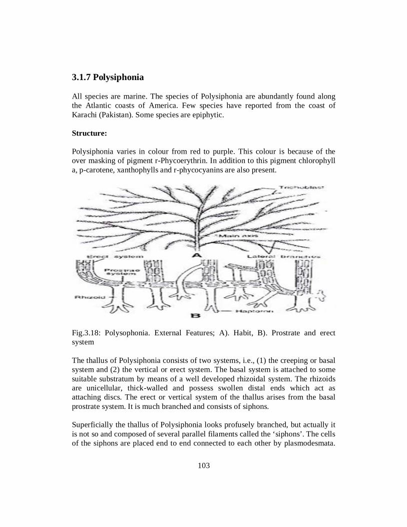
3.1.7 Polysiphonia All species are marine. The species of Polysiphonia are abundantly found along the Atlantic coasts of America. Few species have reported from the coast of Karachi (Pakistan). Some species are epiphytic. Structure: Polysiphonia varies in colour from red to purple. This colour is because of the over masking of pigment r-Phycoerythrin. In addition to this pigment chlorophyll a, p-carotene, xanthophylls and r-phycocyanins are also present.
Fig.3.18: Polysophonia. External Features; A). Habit, B). Prostrate and erect system The thallus of Polysiphonia consists of two systems, i.e., (1) the creeping or basal system and (2) the vertical or erect system. The basal system is attached to some suitable substratum by means of a well developed rhizoidal system. The rhizoids are unicellular, thick-walled and possess swollen distal ends which act as attaching discs. The erect or vertical system of the thallus arises from the basal prostrate system. It is much branched and consists of siphons. Superficially the thallus of Polysiphonia looks profusely branched, but actually it is not so and composed of several parallel filaments called the ‘siphons’. The cells of the siphons are placed end to end connected to each other by plasmodesmata.
103

The siphons may be of two types. There is a central siphon surrounded by 4-24 pericentral siphons. This number varies from species to species. The cells of the central siphon are somewhat elongated and larger than the pericentral cells.
Fig.3.19: (A-C0 polysiphonia. Thallus structure. A). Part of the areal branch, B). T.S of areal axis, C). vertical section In the older branches of the thallus, the pericentral cells divide periclinally thus giving rise to comparatively smaller cells. These cells make the cortical region or cortex. The protoplasmic connections (plasmodesmata) also develop between them and the pericentral cells. In the apical region the thallus consists only of the central siphon. The growth of the thallus is always apical and takes place by the divisions of a dome-shaped apical cell. The pericentral cells have been cut from the central cells by tangential longitudinal divisions. Trichoblasts: The small monosiphonous branches of limited growth are called the trichoblasts. The trichoblast initial is initiated by the oblique division of the apical cell. The small apical trichoblast initial divides again and again giving rise to a multicelled uniseriate dichotomously forked filament, the trichoblast. Structure of a cell: The cells are genetically related and connected to each other by cytoplasmic connections (pit connections) the plasmodesmata. The cell wall is two layered. The outer layer consists of pectic substances and the layer next to protoplast is
104

made up of cellulose. The protoplasm of the cell is highly viscous. There is a conspicuous central vacuole. Each vegetative cell is uninucleate. In each cell there are discoid chromatophores. The reserve food material is fioridean starch. Reproduction Polysiphonia reproduces asexually as well as sexually. Polysiphonia shows an isomorphic alternation of generation. Both gametophytic (haploid) and sporophytic (diploid) generations are identical. Sexual reproduction occurs in gametophytic plants and the asexual reproduction occurs in sporophytic plants. Sexual Reproduction Majority of the species are heterothallic (dioecious). The male sex organs are spermatangia. Spermatangia are produced in clusters on fertile branches. Each fertile branch has a central row of cells. This row of cells is surrounded by pericentral cells. Each pericentral cell cuts off one or more spermatangial mother cells. Each spermatangial mother cell produces one to four spermatangia. The contents of each spermatangium are changed in to single non-motile spermatium. The spermatangial wall ruptures and releases spermatium. Water currents carry them to the carpogonia.
Fig.3.20: Polysiphonia; A). a terminal portion of the thallus with antheridia; B). a part of the antheridium with spermatangial mother cells and spermatangia in longitudinal view; C). same as B; D). Releasing spermatium from sporangium. Carpogonia: The female sex organs are carpogonia. They are produced singly on the female fertile branches. Fertile branch remains short. They are about five to seven cells in length. The two basal cells produce the pericentral cells. The adaxial (that facing the main axis) pericentral cell of the upper tier (second from
105
the base) produces a carpogonial initial. Transverse divisions occur in carpogonial initial. It produces a curved four-celled filament carpogonial filament. The apical cell of this carpogonial filament is charged into carpogonium. Carpogonium is flask shaped. its basal portion has single egg or oosphere. Its upper elongated neck is called trithogyne. The supporting cells produce a short sterile filament. The pericentral cells adjacent to the supporting cell divide simultaneously. They produce a single layered covering around the carpogonium. This sterile covering develops an opening at the tip. This opening is called ostiole, Spermatium falls on the trichogyne. The wall between the two dissolves. The spermatial nucleus with its cytoplasm enters into the trichogyne. It moves down to the basal portion of the carpogonium. Fertilization occurs and diploid zygotic nucleus is produced. It soon divides into two nuclei. 1. After fertilization, the supporting cell cuts off an auxiliary cell towards the
upper side. This auxiliary cell develops a tubular connection with the basal portion of the carpogonium.
2. One daughter diploid nuclei move from the carpogonium to the auxiliary cell through this connection. After that the carpogonium withers.
3. Auxiliary cell, supporting cell and cells of sterile filaments fuse together. They form a single large cell called the placental cell. The diploid nucleus in the auxiliary cell divides several times. It produces a large number of diploid nuclei. The original haploid nuclei in the placental cell disintegrate.
4. Several small protuberances are produced on the surface of the placental cell. One diploid nucleus migrates into each “protuberance. The tube is cut off by the formation of a septum. It functions as an initial of the gonimoblast filament. This initial undergoes few transverse divisions. It produces a short gonimoblast filament.
5. The terminal cell of each gonimoblast filament acts as a carposporangium. It produces a single non-motile diploid carpospore.
6. The mass of gonimoblast filaments and the placental cell form he carposporophyte. It is covered with sterile covering or pericarp. Pericarp forms the fruiting body or the cystocarp. The carposporangium ruptures and releases carpospores through ostiole.
7. The carpospores germinates to produce sporophyte. Sporophyte is also known as tetrasporophyte.It produces tetraspores.
106

Fig.3.21: Polysiphonia; Sexual reproduction, A). longitudinal section of the terminal part of the thallus before the development of carpogonial filament; B). developing carpogonial filament on the supporting cell, ; C). carpogonial filament; D). Basal sterile initials, E). lateral sterile filament cut soo, F, post fertilization stages and development of auxillary cell. Asexual reproduction Asexual reproduction occurs in sporophyte. It produces tetraspores in terasporangium. Tetrasporangia are produced from the fertile pericentral cells. Only one pericentral cell in each tier is fertile. The fertile pericentral cell cut off a small peripheral cell and one or two cover cells. The pericentral cell itself divides transversely. It forms two unequal cells. The lower smaller cell becomes the stalk cell. The upper larger cell becomes tetrasporangium. The nucleus of tetrasporangium undergoes meiosis (reduction division). It produces four haploid tetraspores. The sporangium ruptures and releases these spores. The haploid tetraspores germinate to firm gametophyte.
107

Self Assessment Exercise 3.2 Q. 1 Fill in the blanks with suitable word.
i. Chlamydomonas contains cup shaped ………………... (Chlorophyll) ii. Pyrenoid helps in ……………….. Production. (Starch) iii. The type of syngamy where fusion of similar (in shape and size)
gametes take place.This is called………………... (Isogamy) iv. All species of Polysiphonia are ………………... (Marine). v. Carpogonia are ……………….. in Polysiphonia. (Female sex Organs).
Q. 2 Pick the most suitable option from the given list. i. Function of contractile vacuole is: a) Storage of food b) Expulsion of food c) Expulsion of extra water d) Help in reproduction ii. Conjugation is a type of sexual reproduction which takes place in. a) under adverse conditions b) When water is abundant c) every time d) under favourable conditions iii. Asexual reproduction takes place by zoospore, aplanospore or Hypnospores in: a) Spirogyra b) Vaucheria c) Pinnularia d) Cladophora iv. Sexual reproduction through auxospores mostly takes place in: a) Spirulina b) Cladophora c) Spirogyra d) Pinnularia v). Which group of Algae can survive in toughest of conditions like warm
waters, alkaline lakes etc. a) Cladophora b) Pinnularia c) Spirogyra d) Spirulina Q.3 Give short answers
1. Draw life cycle of Spirogyra 2. Write general Characteristics of Cladophora.
3. Describe cell structure of Pinnularia. 4. Describe conjugation in Spirogyra. 5. Write a note on Nutrition in chlamydomonas.
6. Explain development of Oogonium in Vaucheria.
108

3.2 REPRODUCTION IN ALGAE Reproduction in Algae occurs in both asexual and sexual forms. However, most algae reproduce asexually. Many small algae reproduce asexually by ordinary cell division or by fragmentation, whereas larger algae reproduce by spores. Some red algae produce Monospores (walled, nonflagellate, spherical cells) that are carried by water currents and upon germination produce a new organism. Some green algae also produce non motile spores called Aplanospores. Sexual Reproduction There are two methods of asexual reproduction utilized by algae: Daughter Colony Formation: A limited number of colonial algae produce miniature replicates of the colonies. These are termed daughter colonies. These may be produced inside the hollow, spherical colonies or inside the actual cells of the parent colony. Eventually, the parent colony ruptures to release the new daughter colonies. Sporulation: It is the most common form of asexual reproduction in the Algae. Sporulation is a process in which any cell of an organism produces one or more reproductive cells inside its cell walls. The original cell is termed a sporangium and the new cells are termed spores. Spores are often produced in large numbers for the rapid increase in population size. Sexual Reproduction Sexual reproduction is usually regulated by environmental events. In many Species, when temperature, salinity, inorganic nutrients (e.g., phosphorus, nitrogen, and magnesium), or day length become unfavourable, sexual reproduction is induced. The algae have evolved many variations in sexual reproduction such as different types of gametes, different means of gamete transfer, and different locations of fertilization. There are two methods of sexual reproduction: Isogamy: Isogamy is the form of sexual reproduction in which the gametes produced are identical in shape, size and motility. There is no structural distinction between "male" and "female" gametes. Pairs of isogametes align themselves with their flagellar poles touching and after several seconds, the motile gametes fuse to form a single, nonmotile, diploid zygote. Isogametes, less commonly, may be nonmotile structures. A specific example exhibiting nonmotile Isogametes is the reproductive process known as conjugation, which occurs in the filamentous green algae, Spirogyra sp. Heterogamy: In heterogamy, two different types of gametes are produced. The male gamete, the sperm cell, is typically very small, highly motile and is produced
109
in very large numbers. The female gamete, the egg cell, is much larger and nonmotile. Oedogonium sp. is a green algae that produces heterogametes. 3.3 ALGAE AS DIETARY INGREDIENT The algae are used as a direct source of food by several sea animals and fishes. The marine algae are rich in iodine and several other important minerals. This makes the fundamental source of food for all marine animals and in this respect sea is the richest food producing area. Marine planktonic diatoms together with Dinoflagellata are of fundamental biological importance since all life of sea is dependent upon them. Direct use of algae as food for man: Since the pre-historic times, several sea weeds have been used as direct source of food to human beings. Several fresh water algae have also been utilized in the preparation of various kinds of vitaminized food. As we know well that the fundamental food of sea living stock are algae and they are used as food by human beings. Large numbers of Algae are used as source of food by human beings. They are rich in carbohydrates, inorganic substances and vitamins. Vitamins A C D and E are main constituents of algae. Laminaria species is the important edible seaweed in Japan and the food item ‘Kombu’ is prepared from it. Spirogyra is chief source of food in many countries. Ulva lactuca is used in Scotland for the preparation of salad and soups. Recently green algae chlorella has drawn attention of people as it is high in protein and lipid contents. It has all the essential amino acid contents in it, therefore used as substitute food especially in space flights. Percentage of Protein in this alga is too much as compared with other vegetable or egg. It contains vitamins A to D. Algae are also used to decorate pastries, sandwiches, rice, fish, cakes and jelly cakes in Japan. Agar is used in preparation of ice-cream and jellies. Geliduim, Gracillaria algae are chief source of agar agar. Another algae Rhodomenia palmata is chewed like tobacco in Scotland. Many algae are also used as food for animals like sheep, goat and cattle in New Zealand. Laminaria saccharina, Rhodymenia palmata have been used as food in parts of Scotland and Ireland and Porphyra is considered to be a tasteful culinary dish in many parts of England. Among the more important algal food industries may be mentioned carrageen. This is a product of several sea weeds but principally Chondrus crispus. Carrageen is used by soaking it in water and mixing it with milk. Carrageen possesses the properties of gelling which is one of the factors which enhances its usefulness. Fruit juice may also be mixed with it forming fruit
110

jelly. It is also employed in the preparation of ice creams and in the confectionery industry. The people of China and Japan consume the sea weeds on large scale. The people living on the sea coasts in these countries commonly use fresh sea weeds as food. In Japan, Porphyra tenera happens to be one of the most important edible alga and a product by the name of Amanori and Asakusa-Nori are made from it. 3.4 ALGAE AS FEED ADDITIVE Animal & Fish Feed: About five decades ago, the mass production of certain protein-rich micro-algae was considered as a possibility to close the predicted so called “protein gap”. Comprehensive analyses and nutritional studies have demonstrated that these algal proteins are of high quality and comparable to conventional vegetable proteins. However, due to high production costs as well as technical difficulties to incorporate the algal material into palatable food preparations, the propagation of algal protein is still in its infancy. Aquaculture: Micro-algae are an essential food source in the rearing of all stages of marine bivalve molluscs (clams, oysters, and scallops), the larval stages of some marine gastropods (abalone, conch), larvae of several marine fish species and penaeid shrimp, and zooplankton. Microalgae are used as essential live feeds and supplements in the aquaculture of larval and juvenile animals including oyster spat, juvenile abalone, finfish larvae and rotifer. The effects of the presence of micro-algae in the larval rearing tank are still not fully understood and include: • stabilizing the water quality in static rearing systems (remove metabolic by-
products, produce oxygen), • a direct food source through active uptake by the larvae with the
polysaccharides present in the algal cell walls possibly stimulating the non-specific immune system in the larvae,
• an indirect source of nutrients for fish larvae through the live feed (i.e. by maintaining the nutritional value of the live prey organisms in the tank),
• increasing feeding incidence by enhancing visual contrast and light dispersion, and
• microbial control by algal exudates in tank water and/or larval gut.
111

Shrimp Feed • Pavlova is a small golden/brown flagellate that is very similar to Isochrysis.
It has a very high DHA profile and is excellent for enriching rotifers and other zooplankton.
• Tetraselmis is a large green flagellate with a very high lipid level. It also contains natural amino acids that stimulate feeding in marine animals. It is an excellent feed for larval shrimp.
• Nannochloropsis is a small green alga that is extensively used in the aquaculture industry for growing small zooplankton such as rotifers and for Greenwater. It is also used in reef tanks for feeding corals and other filter feeders.
• Isochrysis is a small golden/brown flagellate that is very commonly used in the aquaculture industry. It is high in DHA and often used to enrich zooplankton such as rotifers or Artemia.
• Thalassiosira weissflogii is a large diatom that is used in the shrimp and shellfish larviculture industry. This alga is considered by several hatcheries to be the single best algae for larval shrimp.
Shellfish Diet: Shellfish Diet 1800 is a mix of five marine microalgae that all have demonstrated success with a variety of shellfish including oysters, clams, mussels, and scallops. A mixed diet provides a much better nutritional profile for all types of shellfish, increasing both growth rates and survival. Marine Fish Larvae Cultivation: Apart from the requirement for micro-algae for culturing and/or enriching live prey organisms such as Artemia and rotifers, algae are often used directly in the tanks for rearing marine fish larvae. This “green water technique” is part of the commonly applied techniques for rearing larvae of marine fishes. Many bivalve hatcheries increase larval production by including an algal culture operation within the facility. Mats of dense algae provide mollusc larvae with a nutritious diet. The high quality diet accelerates growth and shortens the time to larval settlement or spat set. Production of algae in large amounts (mass cultures) is accomplished by providing a favorable environment for the species being cultured. Livestock Feed Algae Suspension for Livestock Production - Although the mechanism has not been understood yet, algal suspension appeared to improve the balance of
112

nutrients in a straw-based diet and thus increased the efficiency of conversion of feed to products. Nitrogen deficient straw being the main source of nutrients for ruminants in Bangladesh (Tareque and Saadullah 1988), the introduction of algal suspension in the feeding system would certainly help economic livestock production. Many livestock farmers, particularly in the urban and suburban areas, raise their animals absolutely on straw and concentrate with either very little or no green grasses. This system of feeding is often associated with infertility, night blindness or even total blindness or other symptoms of vitamin A deficiency. Algae are a very rich source of carotene and algal suspension could be a potential source of vitamin A to combat such deficiencies. Chlorella as Livestock Feed - In 1924, the German scientists Garder and Uitsh noted the key necessity of Chlorella industrial cultivation for the production of feed additives. H. Nakamura (1961) showed that Chlorella was digested much more easily in the form of paste. He recommended up to 5 percent in the daily diet. Chlorella protein digestibility for pigs was 56 %. The average daily weight gain of pigs doubled due to the use of Chlorella paste. Similarly, trials have found that use of Chlorella resulted in weight gain for sheep. It has also been found that the use of Chlorella as a feed additive could become the best choice for solving problems associated with the use of antibiotics, organic acids, or other ingredients in feed because microalgae contain natural organic acids, reducing the colonization of pathogens. Algae may be an inexpensive way to harvest proteins in developing countries where farmland is scarce. Self Assessment Exercise 3.3 Q. 1 Fill in the blanks with suitable word. i. Non- motile spores called ……………. are produced by some green algae.
Aplanospores. a) Aplanospores b) Monospores c) Megaspores d) Crispospores ii. A type of sexual reproduction in which gametes are identical in shape, size and
motility is: a) Heterogamy b) Polygamy c) Isogamy d) Monogamy iii. A large green flagellate ……………which has very high lipid level and contains
natural amino acids. a) Isochrysis b) Nannochloropsis c) Pavlova d) Tetraselmis
113

iv. Amanori product is made from………………… a) Porphyra tenera b) Isochrysis c) Laminaria d) Ulva lactuca Q.2 How Algae reproduce? Q.3 Explain Algae as dietary ingriedient? Q.4 Why it is necessary to study about Algae? Give detailed note.
114

Unit 4
FUNGI
Writer: Dr.Muhammad Waseem Reviewer: Arshad Mehmood Qamar Dr. Sobia Mushtaq
115

CONTENTS Introduction .................................................................................................... 117
Objectives ....................................................................................................... 117
4.1 General Characteristics of Fungi .................................................................. 118
4.2 Life Cycle, Classification with Following Specific Examples ................... 120
4.3 Diversity among Fingi (Zygomycota, Ascomycota, BAsidiomycota) ....... 146
4.4 Importance of Fingi(Edible and poinois Fungi)........................................... 154
References ...................................................................................................... 158
116

INTRODUCTION This is the fourth unit of Biology –I for the students of B.Ed. (Science Education) years. The writer intends to cover all the objectives of this Unit. This unit consists of content related to Fungi. The study of Fungi is called Mycology. Fungi is derived from the Latin word FUNGOUR which means to flourish. The term mycology was derived from the Greek word MYKES. The Fungi are eukaryotic, organisms consisting of filamentous somatic structures called hyphae. The hyphae are surrounded by cell walls formed of chitin or cellulose. They reproduce asexually and sexually. About 51,00 genera and more than 50,000 species of the fungi are known today.
OBJECTIVES After reading this Unit you will be able to: • Describe the general characteristics of fungi • Classify fungi • Describe life cycle of different types of fungi. • Discuss benefits of fungi • Tell peculiar characteristics of different groups of Fungi.
117

4.1 GENERAL CHARACTERISTICS OF FUNGI 4.1.1 Occurrence Fungi are found almost everywhere on the earth. Fungi are found in water, in soil etc. Some fungi live as saprophytes on dead and decaying organic matter while others live as parasite i.e. on other living things. They have the ability to cause diseases in plants and animals. 4.1.2 Plant Body of Fungi The vegetative phase of Fungus is a thallus. The thallus is not differentiated into stem, leaves or roots. They lack vascular tissues (Xylem and Phloem). 4.1.3 Types of Thallus The thallus is either unicellular or filamentous. Some are unicellular. E.g. Yeast whereas most of the fungi have a multicellular plant body that consists of thread like tubular filaments called hyphae. 4.1.4 Hyphae Hyphae are made up of a thin transparent tubular wall filled with a layer of cytoplasm. It can be septate or aseptate. In simple fungi, the hyphae are aseptate but in complex fungi the hyphae are septate. If cells have internal cross-walls, it is called septate hyphae. E.g. Aspergillus. If cells have no internal crosswalls, it is called aseptate hyphae. Aseptate hyphae are also called coenocytic hyphae.
Figure4.1: showing the mass of branched, tubular filaments (hyphae) of fungi. 4.1.5 Mycelium The mass of hyphae making the vegetative body (thallus) of fungus is called the mycellium.If the mycellium is able to penetrate the host tissues, it is said to be intracellular and if it confines itself to the intercellular spaces, it is termed as intercellular mycelium.
118

4.1.6 Cell Structure of Fungi The cells of fungi are typically eukaryotic .They lack chloroplasts and consist of two main parts viz cell wall and protoplast. Cell Wall The cell wall is the outer covering of protoplast. In most of the fungi, the wall lacks cellulose but contains a form of chitin (Fungal cellulose). The formula of fungus chitin is (C22 H54 N4 O2) n .It is polysaccharide based on nitrogen containing sugar (Glucosamine). In addition to chitin, pectin, protein, lipids, cellulose, callose and minerals are also present in the cell wall of fungi. Protoplast The parts in protoplast are plasmalema, endoplasmic reticulum, mitochondria, Golgi bodies, vacuoles, Ribosomes, cytoplasmic reticulum, and Nucleus. Plasmalema This is inner to the cell wall. It is basically unit-membrane and consists of protein layers that are separated by a lipid layer. Endoplasmic Reticulum A network of channels is present throughout the cytoplasm. Both, rough and smooth endoplasmic reticulum is present in the cells of fungi. Mitochondria A large number of mitochondria are found in Hyphae. It is double membrane organelle. Inner membrane has infoldings while the outer membrane continues with the endoplasmic reticulum. Golgi Bodies Golgi Bodies are rare, however when present then they composed of four or five flattened cisternae. They are present throughout the cytoplasm and are concerned with cytoplasmic movement and maintenance of the cell shape. Ribosomes These are the smallest organelles that can be found in the cells of fungi. They can be present freely or associated with endoplasmic reticulum and concerned with protein synthesis.
119

Nucleus The fungi have well defined Nucleus. The outermost covering of nucleus is called nuclear envelope. Nucleoplasm contains a nucleolus and chromatin strands. The nuclear envelope constricts in a dumbbell like shape and separates into two daughter nuclei. The nucleolus consists mainly of DNA. Self-Assessment Exercise 4.1 Q.1 Select the best option given at the end of each statement. i. The vegetative phase of Fungus is a: a) Thallus b) Shrub c) Stem d) Root ii. Golgi Bodies are composed of four or five flattened …………….. a) Tubes b) Cisternae c) Proteasomes d) Vesicles iii. Plasmalema consists of protein layers that are separated by a ……………..
layer. a) Phospholipid b) Glycolipid c) Lipid d) Sphingolipids iv. The nuclear envelope of fungi separates into …………….. daughter nuclei a) Two b) Four c) Eight d) Sixteen v. Fungal nucleolus contains ………………… a) DNA b) RNA c) Either DNA or RNA but never both d) Both Q.2 Describe the general morphology of fungi. 4.2 CLASSIFICATION OF FUNGI Linnaeus classified fungus as the 24th class of his classification system, the Cryptogamia. Algae and fungi were placed in same group Thalophyta due to three common characters which were: The plant body of both groups was thallus. The sex organs of both groups were unicellular, and when these organs were multicellular, all the cells showed fertility. The zygote does not develop into a multicellular embryo while it remained attached to the female reproductive organ. At that time it was believed that fungi were evolved from the Algae by the loss of chlorophyll. But present proposition is that fungi evolved from the protozoan- like ancestors. The physiology and cell wall composition of fungi cell is entirely
120
different from that of Algae. Therefore it is not accurate and fair to keep both Algae and fungi in the same group. Ainsworth’s system is considered the ideal scheme of classification. In this classification, natural relationship is shown among the members of the group. John Webster (1980) also followed Ainsworth system of classification. This system of classification is as follows. 4.1.1 Criteria for Classification Most classification systems use the following criteria: The nature of somatic phase, whether it’s unicellular or mycelium, and if its mycelium then either septate or aseptate. The kind of sexual spores, sporangiospores or conidia, their number, presence or absence of organs of locomotion, the flagella, and if possess flagella then its form and arrangement. The kind of sporangia The nature of life cycle. The presence or absence of the complete or incomplete sexual stage. Dyal’s (1975) classification of fungi is based upon these seven characters which are morphological characters, host specialization, physiological characters, cytological and general characters, serological characters, biochemical characters and numerical taxanomy. 4.2.2 Nomenclature According to the committee of international rules of botanical Nomenclature, following suffixes should be used to denote the various categories of Fungi. i. Division should ends in –mycota ii. Sub division in-mycotina iii. Class –mycetes iv. Sub-class in- mycetidae v. Families in- aceae vi. Genera have no standard endings. 4.2.3 Ainsworth System Of Classification Kingdom: Fungi The kingdom is divided into two divisions. Division 1: Myxomyta This division includes slime molds. It is further classified into four classes. Class 1: Acrasiomycetes: It includes slime molds in which the free living assimilatory phase is amoebae which unite as a pseudo-plasmodium before reproduction.
121

Class 2: Labyrinthulates This class includes plasmodium. Class 3: Mycomycetes. It includes free living saprophytic plasmodium. Class 4: Plasmodiophoromycetes. This class includes parasitic plasmodia present within the host cells. Division II: Eumycota. This includes true fungi that possess mycelium. The eumycota is further divided into five sub-divisions. Sub-Division A: Mastigomycotina The fungi which reproduce asexually by zoospores and sexually by oospores are kept in this sub-division. The sub- division is further divided into three classes: Class 1: Chytridiomycetes The zoospores are uni-flagellated. The flagellum is posteriorly inserted and is of whiplash type. Class 2: Hyphochytridiomycetes The zoospores are uni-flagellated but the flagellum is posteriorly inserted and is Tinsel type. Class 3: Oomycetes The zoospores are biflagellate, one posteriorly and other anteriorly inserted. The posterior is whiplash and anterior one is tinsel type. The cell wall is Cellulosic. Sub-Division B: Zygomycotina The members of this group lack asexual reproduction by Zoospore. However the sexual reproduction takes place by Zygospores. This sub-division has two classes. Class1: Zygomycetes Most of the members of this class are saprobes. Some parasitic or predacious forms are also found in this class with their mycellium immersed in the host tissues. Class2: Trichiomycetes This class has those fungi that are attached to the cuticle or the digestive tract of the arthropods by a holdfast and the mycelium is not immersed in the host tissue. Sub-Division C: Ascomycotina This sub-division includes those fungi in which zoospores and zygospores both are absent and the perfect spores are ascospores. This sub-division is divided into six classes:
122

Class 1: Hemiascomycetes. The members of this group have mycelium or yeast-like thallus. The ascogenous hyphae and ascocarp formation is absent. Class 2: Locoloascomycetes This type of fungi has ascogenous hyphae and ascocarp formation. The thallus is mycelium. Class 3: Plectomycetes They have mycellial thalli and formation of ascogenous and ascocarp take place. The ascospores are absent. Class 4: Laboulbeniomycetes The fungi of this group are exoparasites of arthropods. The ascocarp is a Perithecium with asci arranged in the form of a basal layer within the perithecium. The asci are inoperculate. Class 5: Pyrenomycetes The members of this group have perithecium type ascocarps. The asci are inoperculate and open through an apical pore. Class 6: Discomycetes The members of this class have apothecium type ascoarps that may be epigial or hypogial. The asci may be operculate or inoperculate. Sub-Division D: Basidiomycotina. The members of this group lack zoospores and zygospores. The sexual spores are basidiospores. This sub-division has three classes: Class 1: Telliomycetes Fungi of this class are parasites on vascular plants. The basidiocarps are replaced by teliospores or chlamydospores which are grouped in sori or scattered within the host tissue. Class2: Hymenomycetes They are mostly saprobes. The basidiocarps are present and these may be gymnocarpous or semi-angiocarpous. Gymnocarpous or semi angiocarps types basidiocarps are found. Basidia are organized in the form of a hymenium. The basidiospores are ballistospores.
123

Class 3: Gastromycetes Most members are saprophytes. Well-developed basidiocarps are typically angiocarpous. Sub-Division E: Deuteromycotina The members of this sub-division have no zoospores. The spores found are conidiospores. This Sub-division is classified into three classes. Class1: Blastomycetes The members of this class lack true mycelium, or if present developed poorly. Budding Cells with or without pseudo-mycelium are present. Class2: Hyphomycetes The members of this class have well developed true mycelium. The mycelium may be sterile or bears spores directly on special branches, the sporophores. Class3: Coelomycetes The members of this class have well developed mycelium and the spores are produced in pycnidia or acervuli. Self-Assessment Exercise 4.2 Q.1 What characters Dyal’s focused on to classify Fungi? Q.2. Describe in detail the Ainsworth system of classification. 4.2.3 MUCOR Classification: Kingdom: Fungi Division: Mycota Class: Zygomycetes Order: Mucorates Family: Mucoraceae Genus: Mucor Habitat Mucor appear on damp horse dung when kept under a bell jar. It is also found in soil, bread and other food materials.
124

Vegetative Structure Numerous filamentous hyphae form a fluffy, white mass called mycelium. Numerous black, pin head like structures developed on the mycelium called as sporangia. The hyphae are coenocytic and aseptate. During the vegetative phase, the mycelium consists of stolon hyphae and rhizoidal hyphae. The rhizoidal hyphae help in the absorption of nutrients from the substrate. The cell wall is made up of chitin. Cellulose is absent. The cytoplasmic organelles like mitochondria and endoplasmic reticulum etc. are present in the cell.
Fig.4.2 Mucor –Mycellium with Sporangia and spores. Means of Vegetative reproduction i. Fragmentation
In fragmentation, hyphae are separated from aerial mycelium. These form fragments, each of which has the ability to grow.
ii. Chlamydospore Formation Under the unfavorable conditions, fungi produce special thick walled resting
cells called chlaydospores. These spores contain food and resist against desiccation. When the favorable conditions arrive, it germinates to produce a new mycelium.
Asexual Reproduction Asexual reproduction takes place by the formation of sporangiophore. They are non- motile and multinucleated. At the tips of sporangiophore, single sporangia arise. The young sporangium is white spherical structure. At maturity, it turns black due to the production of spores. In the center of sporangium is a dome shaped structure called collumella. The sporangia are haploid. They are produced on haploid mycelium and contain haploid spores.
125

The spores dispersed after the rupturing of the sporangiophore. They are usually dispersed by wind. The spores are non-motile, oval and protoplast surrounded by single layered cell wall. Under favorable conditions, the spore germinates by the formation of germ tube. The hyphae arise from the germination of tube and spread in all directions. Sexual Reproduction Mucor undergoes a sexual process by conjugation. In conjugation, gametangium fuses their content. Gametangial Copulation Sexual reproduction takes place by gametangial copulation. The sexual process starts with the formation of two special hyphae termed as zygophores. They arise near the apices of active growing somatic hyphae of +ve and –ve strains. The zygospores meet and adhere in pairs at their tips to form fusion septum. The tip of zygospores begins to swell due to the flow of cytoplasm and nuclei in them. They are called pro-gametangia. The combined protoplast in pro-zygosporangium containing a number of diploid nuclei is called a zygospore. It secretes a thick wall and become a zygosporangium. Germination of zygospore Under favorable conditions, the zygospores germinate to produce germ tube. Due to reduction division, numerous haploid nuclei are produced of opposite strain. The germ tube soon bears a terminal sporangium. The protoplast produce non-motile spores called germ spores. The germ spores on falling on substratum germinate to form the mycelium. Self-Assessment Exercise 4.3
Q.1 Draw and explain the vegetative structure of Mucor. Q.2. Describe in detail the types of reproduction in Mucor. 4.2.4 Phyllactinia Classification Kingdom Fungi Phylum Ascomycota Class leotiomycetes Order Erysiphales Family Phyllactina Species Phyllactina guttata
126
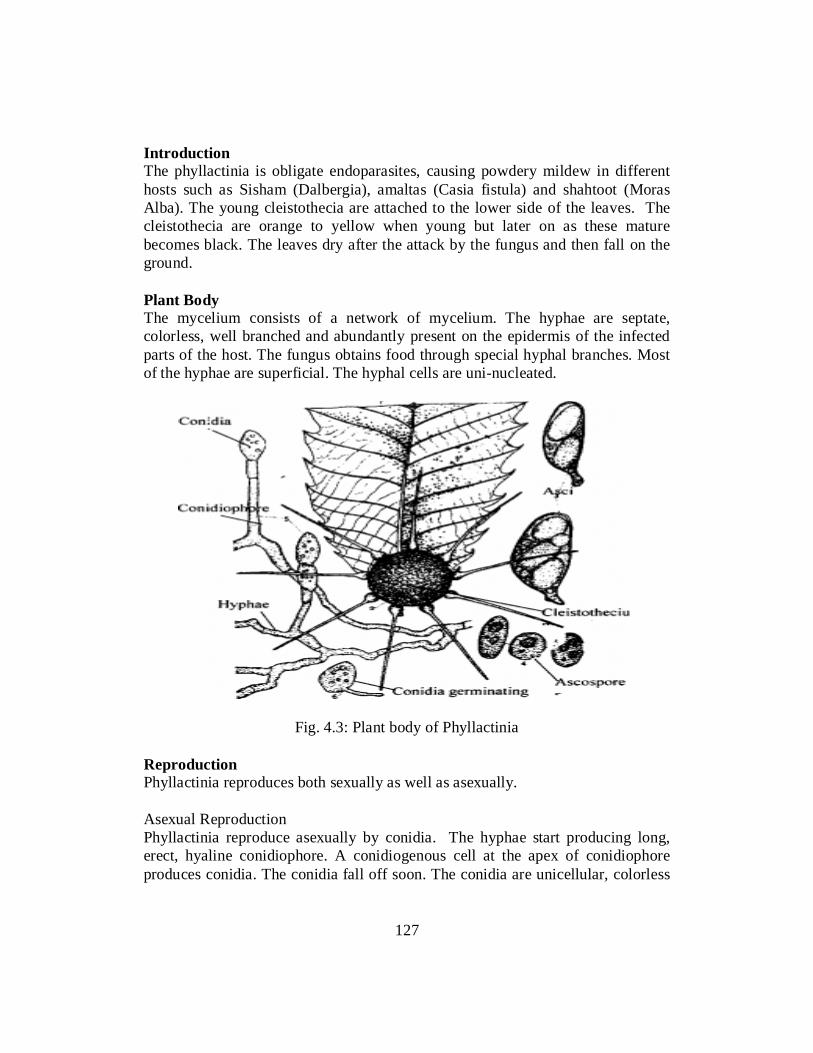
Introduction The phyllactinia is obligate endoparasites, causing powdery mildew in different hosts such as Sisham (Dalbergia), amaltas (Casia fistula) and shahtoot (Moras Alba). The young cleistothecia are attached to the lower side of the leaves. The cleistothecia are orange to yellow when young but later on as these mature becomes black. The leaves dry after the attack by the fungus and then fall on the ground. Plant Body The mycelium consists of a network of mycelium. The hyphae are septate, colorless, well branched and abundantly present on the epidermis of the infected parts of the host. The fungus obtains food through special hyphal branches. Most of the hyphae are superficial. The hyphal cells are uni-nucleated.
Fig. 4.3: Plant body of Phyllactinia Reproduction Phyllactinia reproduces both sexually as well as asexually. Asexual Reproduction Phyllactinia reproduce asexually by conidia. The hyphae start producing long, erect, hyaline conidiophore. A conidiogenous cell at the apex of conidiophore produces conidia. The conidia fall off soon. The conidia are unicellular, colorless
127

and clavate in shape. The conidia are dispersed by wind and germinate after falling on a host organ into new hyphae. Sexual Reproduction In late summer the formation of conidia slows down and finally stops while sex organs start developing. The sexual branches are uninucleate and grow on different hyphae. In the beginning, the male and female hyphae are similar. A papillate outgrowth arise from the hypha, the nucleus of the hyphae divides into two. One daughter nucleus migrates into the outgrowth, which is separated from the mother hyphae by the formation of a septum. In the later part of development, the female branch becomes stouter, curved and anastomoses the developing male branch. The nucleus of the female branch divides and the division is followed by septum formation resulting in a stalk cell and a terminal ascogonium. The male branch remains erect and division of its nucleus and subsequent septum formation results in the formation of stalk and a terminal antheridium. Nucleus of antheridium degenerates, therefore, the process of fertilization does not take place. Ascogonium divides into two. The ascogonium becomes binucleated. Both the ascogonial nuclei divide and septa are laid down between the daughter nuclei in such a way that a three-celled filament is formed. The middle cell of this filament is binucleated and the other two cells present on its either side are uninucleate. Two nuclei present in the middle cell belong to different nuclei of the original pair present in the ascogonium. Development of Ascocarp Sterile hyphae arise from the stalk cell of female branch. Development of ascocarp starts from the somatic hyphae present below the sterile hyphae. From the basal uninucleate cell of the three –celled filament hyphal filaments arise which form a layer lining the wall (peridium) of the young ascocarp. The ascocarp formed is globular and without an opening and is therefore, cleistothecium. Ascus Development Many ascogenous hyphae develop from the middle cell, when its nuclei divide. The tip cells of these hyphae are uninucleate but penultimate (last but one) are binucleate. These act as ascus mother cells and develop into an ascus. The nuclei fuse to form a diploid nucleus that undergoes meiotic division followed by a mitotic division to produce eight nuclei. Six of these eight nuclei degenerate and the remaining two develop into ascospores.
128

Fig.4.4: Ascus. Ascospore Structure of Cleistothecium The Cleistothecium is globular, orange to yellow in color when young but at maturity becomes black. Stothecia are provided with characteristics appendages which are rigid, spear-like with a bulbous base and pointed tip in addition to these appendages, the cleistothecia bear a crown of short, apical gloeoid, branched appendages whose function is unknown, perhaps help the cleistothecia to stick to the substratum. The long bulbous appendages bend and lift the cleistothecia above the leaf surface. This helps in dissemination of ascospores effectively. Dispersal of Spores The asci open through an apical opening and the ascospores are released. These are carried away by wind and when fall on a suitable host germinate to form a new mycelium. Self-Assessment Exercise 4.4 Q.1 How the ascocarp development occurs in Phyllactinia? Q.2. What type of parasite Phyllactinia is and how it reproduces?
129

4.2.5 USTILAGO Taxonomic Position Kingdom: Fungi Division: Amastigomycotins Sub-Division: Basidiomycota Class: Basidiomycetes Order: Ustilagnales Family: Ustilagnacae Genus: Ustilago Species: U.Triticci Introduction Ustilago is a genus of smut fungi. It is strictly parasite and cause smut in host. It has almost 400 species. All species of the genus complete their life cycle on the host and is thus called as autoceous parasites. Its various species attack the ovaries of grasses and crops, sometimes destroying the whole leaves and producing a black powdery mass of spores. Ustilago is very common in Pakistan and its various species attack the wheat, maize, corn, oat, barley.etc.
Fig4.5:Ustilago The leaves on which this species grows is called ear. The diseases make its appearance when the ear is produced. The ear in the diseases plants emerges earlier than the healthy plants. All flowers are replaced by black powdery mass of spores. These spores are called as chlamydospores teleuto spores. In the young spikelets the spores are blown off, only the central axis of the inflorescence is left behind.
130

Very often disease is seed born. The dikaryotic hyphae of the fungus are present in the seed. When the infected seeds are sown, the embryos starts germination, the dormant mycelium also become active and keeps on growing in the apical meristem without causing harm to the plant. When the ears are produced the mycelium enters the flower primordial and ultimately forms a mass of hyphae in the ovaries of young flower. The mass of hyphae consists of short cells, which are binucleated. The wall of each cell gelatinized and its protoplast secretes a thick wall around itself and is transformed into spore. These spores are produced in such a large number that whole inflorescence that become transformed into a black powdery mass around the central axis. The spores are called as Chlamadospores or teleotospores. Each spore is unicellular and binucleate. The wall of spore is very thick and its surface is variously sculptured. The thick spore wall is differentiated into two layers. The outer wall or exosporium is thick while the intine is always thick. Dispersal of Spores The spores are dry and are dispersed by wind, insects or by water. They are logged on the ears of healthy plants. Germination of Spores The spores fall on the stigma of flower and germinate to infect the new born ears. They germinate at the temperature ranging from 20-30OC. Before germination the two nuclei fuses with each other to produce a diploid zygotic nucleus (Sankaryon). The zygote nucleus undergoes reduction division. In the meantime, a short germ tube of pro-mycelium is produced. The promycelium is also called epibasidium or metabasidium. The four haploid nuclei formed as result of meiosis is migrated to promycelium. These are called as primary mycelium. The mycelium consists of hyaline transversely septate producing four cells. Since the segregation of the sexual stage takes place during meiosis two of these nuclei are of plus stain and two of minus stain. Infection The diplophasic mycelium infects the ovaries, grows intercellulary, and ultimately invades the embryo. The mycelium after establishment in the embryo becomes dormant without causing damage to the embryo. Infected seeds are as healthy as the uninfected one.
131

The Smut Fungus Smut is very important disease as it caused losses in millions. The smut diseases are of two types. Loose Smut In loose smut, the spores are not covered by any membranous structure. They are directly exposed to the air at the time of flowering and can be easily disseminated by wind.e.g.in loose smut of wheat, oat and doob-grass. Covered Smut In covered smut, the spores remain enclosed by a membranous covering of the grain. Mature spores are not directly exposed to the air and cannot be disseminated by wind. They are liberated only when the wall of the grain is broken e.g. in covered smut of barley and sugar cane.
Fig 4.6: Smut Fungus
Control of Diseases Various methods used for the control of diseases are as follow. i. Roughing The infected plants must be uprooted and burnt outside the field. This
method is practicable as the infected ears emerge earlier than the healthy ones and their destruction will help in controlling the spread of disease.
ii. Seed Selection As the disease is seed borne, a careful selection of seeds from a healthier
crop will help in control of the diseases.
132

iii. Hot Water Treatment This method involves the soaking of seed for 4-5 hours in water at normal
temperature ranging from 26-30oC in order to activate the dormant mycelium. Then the seeds are transferred to hot water having temperature of 54oC for ten minutes to kill the activated mycelium. The grains are then dried and sown. This method requires strict vigilance as a little low range of temperature will fail to kill the mycelium and a high range may also damage the seeds. Even a temperature of 56oC may kill the embryo.
iv. Solar Treatment During this control method, the seeds are soaked in water from 8 am to 12
noon (for about four hours) at normal temperature in months of May and June. This will activate the dormant mycelium. The seeds are then dried in open sun in very thin layers for about four hours. The dying kills the activated mycelium.
v. Use of systemic Fungicides Systemic fungicides Vitavax (D 735) and Plantavax (F 461) are much
effective in controlling the disease. Similarly Thomas and Chatrath (1975) found that when the seeds were treated with 0.1 to 0.2 % Thiabendazole, excellent results were obtained.
vi. Resistant Varieties Raising and sowing resistant varieties of wheat is one of the simplest and
effective methods of control of the diseases. Self-Assessment Exercise 4.5 Q. 1 Fill in the blanks with suitable words. i. Ustilago is a genus of …………….Fungi. (Smut) ii. The ear in the diseased plants emerges ……………. than the healthy plants.
(Earlier) iii. The zygotic nucleus undergoes ……………. division. (Reduction) iv. Covered smut is often found in …………….. (Sugar-cane) Q. 2 Choose the correct answer i. The family name of Ustilago is. a) Fungi b) Ustilaginales c) Ustilaginacae d) Basidiomycetes (B)
133

ii. Species which complete their live cycles on the host is called? a) Heterocous b) Autoecious c) Parasites d) Saprophytes. (B) iii. The spores of Ustilago are called.. a) Chlamadospores b) basidiospores c) Ascospores d) conidiospores (A) iv. The seeds are exposed in open sun in technique. a) Roqueing b) Solar treatment c) Resistant varieties d) Hot-water treatment (B) Q. 3 Write a note on germination of spores in Ustilago Q. 4 How can you control the diseases caused by Ustilago? 4.2.6 Penicillium Taxonomic Position KINGDOM: Fungi DIVISION: Ascomycota CLASS: Eurotiomycetes ORDER: Eurotiales FAMILY: Trichomaceae GENUS: Penicillium SPECIE: Penicillium expansum Penicillium is commonly known as blue and green molds. Occurrence The fungus is found on wide variety of substrate such as citrus fruits, jellies, foodstuffs, old leather, and paper and in the soil. The conidia of Penicillin are found everywhere in the air and in the soil. Contamination of Fleming’s bacterial culture by Penicillium Notatum led to the discovery of Penicillin. Plant Body The mycelium of penicillium is well developed and is profusely branched. The hyphae are colorless, slender, tubular, branched and septate. Some of the hyphae invade the substratum to absorb nourishment from the substratum. The mycelium becomes colored due to the production of colored conidia. \
134

Fig4.7: Pencillium Cell Structure The hyphae are septate and each cell is uninucleated. The cell wall is micro-fibrillar and in Penicillium Notatum it is reported to consist of three or four layers. The outermost layer is composed of Glucans, the next of protiensird of chitin fibrils embedded in a granular matrix, and the innermost of pectic or hemicellulosic material. The Plasma membrane surrounds the cytoplasm in which mitochondria, ribosomes and endoplasmic reticulum is embedded. The reserve food is in the form of oil globules. The septa between the cells have a central pore through which the protoplasm of the adjacent cells remains in contact.
Figure4.8: Cell structure of Pencillium
135

Reproduction The Penicillium reproduces vegetatively, asexually and sexually. Vegetative Reproduction The vegetative reproduction takes place by fragmentation. During fragmentation the hyphae break up into short fragments, which grow by repeated divisions into a new mycelium. Asexual Reproduction Asexual reproduction takes place by the production of conidia, which are produced on conidiophores. The conidia are erect, branched. Each branch bears short branches at its tip called as metulae. There are group of bottled shaped sterigmata at the tip of each matula. Conidia are produced in chains at tips of sterigmata. The whole group of matulae and sterigmata on each branch is called pencillus. The conidia are tiny, uninucleate, spore-like structures which may be globose to ovoid in form. The spore wall is pigmented and is differentiated into two layers, an exine (outer thick, ornamental layer), and intine (inner smooth and thin layer). Inside the spore, the wall plasma membrane is present which encloses the cytoplasm. Mitochondria and ribosomes are embedded in the cytoplasm. The reserve food is oil globules. Development of Conidia Each sterigmata is a uninucleated structure. During the production of conidia, the nucleus divides into two.One daughter nucleus along with some cytoplasm migrate into the slightly swollen tip of the sterigmata. This portion is then cut off from the rest of the sterigmata by the formation of cross wall. The protoplast is then the protoplast then contracts slightly and secretes a wall of its own. This process is repeated several times and a chain of conidia is produced at the tip of each sterigmata. Each newly formed conidium is uninucleated but later on this nucleus divided several times, making the conidium multinucleated. Germination of Conidium The conidia germinate by producing hyphae directly on falling on suitable substrate. Oidia Formation The Penicillium hyphae, when grown in a medium of sugar solution, divide into small, uninucleate segments which become rounded and separate into thin walled spore-like structures called Oidia or Oidiospore. These increase in number by budding. This is called torula stage and the fungus in this stage brings about fermentation of sugar into alcohol. The Oidia when transferred to a solid substratum germinate to form a new mycelium.
136

Sexual Reproduction It is the perfect state of Penicillium. All the species are homothallic. The sexual reproduction is Oogamous. The male sex organs are antheridia and female sex organs are ascogonia. Ascogonium A mature ascogonium is a long erect, multinucleated, tubular structure with curved upper end.it arises from uninucleate, septate hyphae as a finger- like, lateral outgrowth which elongate into an ascogonium. The nucleus of the ascogonium divides many times mitotically to produce 32 or 64 nuclei. Antheridium While the ascogonium is developing a slender uninucleate branch originate from a cell of the same hypha adjacent to the developing ascogonium, or from a neighboring hypha. This is the antheridial branch. It grows up and is cut off from rest of the branch to form a uninucleate antheridium. Fertilization The tip of antheridium comes in contact with the ascogonium and the wall of contact between the two dissolves to form a pore. The protoplasts of the gametangia come in contact with each other through this pore. The antheridial and ascogonial nuclei arrange themselves in pairs. Each pair is called a dikaryon. Development of Ascus and ascopores The stimulus of plasmogamy results in septation of ascogonium into binucleated cells. Some of these segments, usually those present in the middle, produce outgrowth called ascogenous initials which develop into ascogenous hyphae composed of binucleated cells. The tip cells of these hyphae act as ascus mother cells which develops either simply by elongation, or by crozier formion into an ascus . Karyogamy takes place in the ascus mother cell and this diploid nucleus undergoes meosis to produce four haploid nuclei. A mitotic division results in the formation of eight haploid nuclei. These are transformed into ascospores by free cell formation. The asci are globose or pear-shaped and the unicellular, uninucleate and lens shaped with a groove around the edge. Development of Cleistothecium As soon as the septation of ascogonium and development of ascogenous hyphae starts, the sterile hyphae present at the base of the ascogonium grow upward and surround the sexual apparatus. These hyphae become interwoven to form a hollow, ball-like structure, the peridium, and the walls of the ascocarp. The ascocarp continues to grow even after the maturation of ascospores. The ascocarp is without an opening, therefore, is a cleistothecium.
137

Dispersal of the Ascospores and Germination The asci are evanescent, i.e. dissolve soon after the formation of ascospores, leaving the ascospores free in the ascocarp. The ascospores are released by the decay of the wall of the ascocarp. The ascospores germinate by formation of a germ tube to form a new mycelium. Self-Assessment Exercise 4.6 Q.1 Fill in the blanks with suitable words i. Order of “genus” Penicillium is ……………….. (Eurotailes) ii. Penicillium is often found on……………….. and in the soil. (Citrus fruits) iii. All Species of Penicillium are ……………….. (homothallic) iv. Cleistothecium is found in ……………….. (Penicillium). Q.2 Choose the correct answer. i. Penicillium develops by asexual means by the formation of ………………... a) Budding b) Conidia c) basidiocarp d) Soredia (B) ii. In which stage fungus brings fermentation of sugar a) morula b) fibula c) torula d) Oidia (c) iii. Newly formed conidium is. a) Multinucleated b) Uninucleated c) Dinucleated d) Anucleated (b) iv. Which mode of vegetative reproduction is common in Penicillium? a) budding b) layering c) Fragmentation d) all Q. 3 Answer the following questions i. Write taxonomic classification of Penicillium? ii. Describe the cell structure of Penicillium iii. What is cleistothecoium? iv. How ascospores in Penicillium are dispersed?
138

PUCCINIA Taxonomic classification Kingdom: Fungi Division: Amastigomycotins Sub-Division: Basidiomycota Class: Basidiomycetes Order: Uredinales Family: Pucciniaceae Genus: Puccinia Species: P.Graminis Introduction Genus Puccinia has many species which are obligate parasite on a wide variety of host. Puccinia Graminis tritici is a common on wheat and also parasites on various species of grasses such as oat, barley, rye etc. The fungus completes its life cycle on two different hosts and is called as heteroecious rust. Five stages (Uridinal stage, Telial stage, Basidial stage, Pycenial stage and Asial stage) are present in the typical life cycle of Puccinia. Uridinial and Telial stage is present on wheat plant while the Pycenial and Asial stages are present on the Barberry plant. Life Cycle of Wheat Plant Puccinia Graminis disturbs mostly the stem of wheat causing the black stem rust. The first sign appears early in March in the form of elangated, reddish brown granular postules or sori.these Sori are called uredosori. Uredinial Stage The Uredinial are one celled, binucleate d somewhat globose, slightly thick walled spores. These spores are produced on the stalks. The spores exert pressure on the underlying epidermis causing break down of the host epidermis to form Uredinia and the stage is called as uredinial stage. Dispersal of Spores The uredospores are blown away by the wind and logged on the wheat plant. Germination of Spores The spores germinate within few hours after falling on the wheat plant. Spores form germ tube and germinate. The germ tube reaches the stomata and the tip swells into the vesicle called appresorium. The cytoplasm along with nuclei
139

migrates into the appresorium, which is then cut off by the formation of septum. A very fine tube from the appresorium is then entering into the sub- stomatal chamber. Then hyphae are produced in all directions. The hyphae are branched, septate and each cell is binucleated. The growth of the hyphae is intercellular and gets nourishment by sending club shaped historia in the cell. 1. Production of Uredospores
Under the epidermis a number of hyphae are produced and a crop of new uredodpores are produced. This production of large number of spores exerts pressure on the underlying epidermis and sorous is exposed and postules are formed. As the uredospore are produced on the dikaryotic mycelium and they again give rise the dikaryotic mycelium again, thus this stage is called as asexual stage. Several crops of uredospores are produced throughout the season and in this way the disease spread to large number of plants rapidly.
2. Telial Stage or Teleuospores At the end of growing season the color of uredospori changes from
brownish to dark brownish and to dark reddish or rusty. This color is due to the production of new spores called as teleuspores. The teleurospores are two celled, binucleated, slightly oval and having thick wall. These spores are elongated or oval in shape with tapering end. Each cell of spore contains two nuclei and one germ pore.
Dispersal of Teleurospores These spores are dispersed by wind and do not germinate immediately. They can withstand unfavorable condition and survive for a long period.
3. Basidial Stage On germination, each cell of the teleurospores produces germ tube called as
promycelium or epibasidium. The two nuclei in each cell fuse together to form diploid nuclei. Four haploid nuclei are produced after meiosis. The cytoplasm along with nuclei migrates into the promycelium. The promycelium or epibasidium is then divided into four cells by the formation of transverse septa. Each cell contains only one nucleus. Each cell of the epibasidium produces lateral sterigmata. The basidiospores are produced on the sterigmata. Each basidiospore is one celled and mononucleated. The basidiospores or sporidia are forcibly discharged and are dispersed by wind.
Life cycle of Barbary Plant Basidiospores are not able to infect the wheat plant but they infect the alternative host Berberis vulgaris.
140

Germination of Basidiospores When the basidiospores logged on the surface of leaf, they germinate by producing a short germination tube. The tube enters the epidermal cell. The hyphae grow in all direction and remify the mesophyll tissues. The hyphae are intercellular and obtained food by sending historia in the cell.
4. Pycenial Stage The mycellium produced by the germination of basidiospores is
monokaryotic. The hyphae of the monokaryotic mycelium form small knots below the upper epidermis. These cells are then transformed into flask shaped cavities called pycenidia or spermatia. Each spermatia open to outside by a small aperture called as ostiole. Numerous small uninucleated spores called pycenidiospores are produced on short hyphae inside the spermatengia. Numerous paraphysis come out of the Ostiole and some of them act as receptive hyphae. The spermatia come out through the ostiole in mucilage liquid. They are dispersed by insects.
Dikaryotization When a spermatium becomes attached to the receptive hyphae of the opposite
sexual phase, its nucleus enters the receptive hyphae of the opposite sexual phase. Spermatia of one sexual phase can fuse with the other phase.
5. Ascial Stage In receptive hyphae the nucleus of the spermatium divides several times and
the daughter nuclei migrate to the other nuclei. The dikaryotic cells form group of cells just below the lower epidermis. These binucleated cells divide transversely to form a chain of cells. Each cell of the chain divides into a larger and smaller cell. The larger cell is transformed into rounded spores called as aeciospore. Certain cells form wall around the spores to form peridium. As the spores are mature, the wall of the peridium ruptures and a cup shape structure called as acedium and aeciospore are released. The spores are dispersed by wind. Each spore is unicellular, binucleated and generally rounded in form.
Germination of Aeciospores The aeciospores do not attack the barberry plant but they can only infect the wheat plant. They germinate on the surface of the wheat by producing a germ tube. It infects the host through the stomata by the formation of appresorium. This infection results in the formation of dikaryotic mycellium in the host tissues. The uredospores are produced and the life cycle is completed. It takes two years to complete the life cycle. Self-Assessment Exercise 4.7 Q.1. Write a note on Life cycle of Barbary Plant.
141

AGARICUS Taxonomic Classification Kingdom: Fungi Division: Amastigomycotins Sub-Division: Basidiomycotina Class: Holobasidiomycetideae Order: Agaricales Family: Agaricaceae Genus: Agaricus Species: Agaricus campestris Introduction Agaricus species are commonly called meadow mushrooms since these are typically found in open, well manure, and quite often grassy areas. The genus contains some of the fine edible mushrooms including Agaricus Campestris and Agaricus bisporus. Agaricus campestris is latin derivation from Campester: meanings plain or flat field. It is common, wild and edible species. It grows on damp and dead organic substances such as humus, horse dung, damp rotten wooden logs, tree trunks and meadows rich in decaying organic matter. Plant Body The plant body is divided into two parts vegetative mycelium and basidiocarp. Vegetative Mycelium The mycelium may be primary, secondary or tertiary Primary Mycelium It is monokaryotic, short-lived and develops from germination of a basidiospore. The hyphae are septate and hyaline. The cells are short and contain oil globules, vacuoles and thin protoplasm. Secondary Mycelium. The secondary mycelium is dikaryotic and arises by dikaryotization through somatogamy of two compatible monokaryotic hyphae. The hyphae composing secondary mycelium are long, branched septate and have short cells. Typical dolipore septa are present between, and produce mushrooms a year after year.
142

Tertiary Mycelium The mycelium producing basidiocarps or mushrooms is referred to as tertiary mycelium. It forms complex tissue which produces basidia.The Mycelium forms microscopic, thick, tangled, wooly masses called spawns. Reproduction The fungus reproduces usually sexually. They reproduce by a process of plasmogamy. In plasmogamy two hyphae fuse and form dikaryon. The nuclei of the dikaryon undergo conjugate divisions with that of the dikaryon to produce dikaryotic secondary mycelium. The basidiocarps develop from the hyphae of subterranean, parennial secondary mycelium. Basidiocarp—A Mushroom The aerial part of the fungus is the fruiting body (basidiocarp) of the fungus and is popularly known as mushroom. Structure of Basidiocarp A mushroom is generally umbrella shaped. The upper part called Pileus or cap, is circular and expanded. In young condition the pileus is more or less button like in shape and is completely enclosed by a thin whitish membrane called Velum. The pileus is borne on a fleshy, pinkish-white, stalk called the stipe.
Fig 4.8: Basidiocarp
As the pileus grows in size, the velum ruptures and its remnants surround the stipe to form annulus. A cavity is present under the pileus in which a large number of thin, vertical, plate-like structures called lamellae or gills run out towards the edge
143

of the pileuss. Basidiospores develop on both surfaces of these lamellae. The gills are first pinkish but as the spores mature become dark brown in color. Development of Basidiocarp The development of the basidiocarp starts as a tiny swelling consisting of a knot of the subterranean hyphae of secondary dikaryotic mycelium. This swelling enlarges into a broadly ovoid structure, the basidiocarp primordium.Later there is differentiation of a transverse internal ring of vertical hyphae followed by development of a ring- shaped cavity, the pre-lamellar chamber. The portion of the primordium above the pre-lamellar cavity finally develops into the pileus, that below into the stipe. The tissue adjoining the upper side of the pre-lamellar cavity becomes differentiated into alternate radical bonds of slow- and fast- dividing cells. Each band of fast dividing cells act as lamellar primordium that soon projects downward into the pre-lamellar cavity. The diameter of the pileus increases greatly after the lamellae begin to develop. This results in the formation of spaces between the lamellae. This stage of the basidiocarp is called button stage, and the edges of the pileus are joined to the stipes through a thin sheet of tissue, the velum.
Fig 4.9: Successive stages of development of basidiocarp
Internal Structure of Basidiocarp The basidiocarp is composed of interwoven hyphae which run parallel to each other in stipe region and are compactly arranged to form a pseudo-parenchymatous tissue in the pileus region. In stipe the central hyphae are loosely arranged with prominent air apices, forming the medulla whereas the outer
144

hyphae constitute the cortex. The pileus also shows similar arrangement of hyphae into central medulla and outer cortex. Some of the hyphae of pileus develop into lamellae or gills. Structure of a Lamella Each lamella consist of three tissues viz Hymenium, Trama and Sub-Hymenium i. Hymenium It is the fertile region of the lamella and consists of club-shaped, binucleate
cells forming a palisade –like layer on both sides of the lamella. All the cells of Hymenial layer are not fertile; some of these develop into basidia, others into sterile paraphysis, and still others into elongated cystidia that protrude beyond the paraphysis.
ii. Trama Internal to the hymenium, there is a region of multinucleate elongated cells
which extend from the hyphae of the pileus, the trama. The hyphae are so oriented that their long axes lie parallel to the hymenium.
iii. Sub-Hymenium In between the hymenium and trama is a region composed of hyphae
arrange at right angles to those of trama. The cells of these hyphae are isodiametric and binucleate.
Fig 4.10: T.S of Lamella Development of Basidium andbasidiospores A fertile cell of hymenial layer acts as basidial initial. It enlarges in size to become a basidium. It is binucleated at first but as it enlarges, the two nuclei fuse
145

(Karyogamy). The zygote nucleus migrates to the apex of the basidium and undergoes meosis to produce four haploid daughter nuclei. The same time four peg-like outgrowth called sterigmata are produced at the apex of the basidium. The tip of each sterigmata swells and one daughter nucleus migrates into it. The sterigma is cut off from the basidium to become a basidiopore. Two basidiospores are of positive stain and the other is negative stain. A small lateral outgrowth called hilum develops at the junction of basidiospore and the sterigmata. A small drop of liquid appears upon the hilum. This drop increases in size and when it reaches about 1/5th size of the spore, the basidiospore shoot off from the sterigmata along with the drop of liquid. The spores are discharged one by one at intervals. The basidiospores upon falling a suitable substratum germinate into primary mycelium ofstain+ve or –ve.
Fig 4.11: Development of basidium and basidiospore 4.3 (A) ZYGOMYCOTA: THE CONJUGATED FUNGI The zygomycetes are a relatively small group of fungi belonging to the Phylum Zygomycota. They include the familiar bread mold, Rhizopus stolonifer, which rapidly propagates on the surfaces of breads, fruits, and vegetables. Most species are saprobes, living off decaying organic material; a few are parasites, particularly of insects. Zygomycetes play a considerable commercial role. The metabolic products of other species of Rhizopus are intermediates in the synthesis of semi-synthetic steroid hormones. Reproduction in Zygomycota Zygomycetes have a thallus of coenocytic hyphae in which the nuclei are haploid when the organism is in the vegetative stage. The fungi usually reproduce
146

asexually by producing sporangiospores (Fig4.13, 4.14). The black tips of bread mold are the swollen sporangia packed with black spores (Fig 4.13, 4.14 ). When spores land on a suitable substrate, they germinate and produce a new mycelium. Sexual reproduction starts when conditions become unfavorable. Two opposing mating strains (type + and type –) must be in close proximity for gametangia from the hyphae to be produced and fuse, leading to karyogamy.
Fig.4.13: Zygomycetes have asexual and asexual life cycles. In the sexual life cycle, plus and minus mating types conjugate to form a zygosporangium.
The developing diploid zygospores have thick coats that protect them from desiccation and other hazards. They may remain dormant until environmental conditions are favorable. When the zygospore germinates, it undergoes meiosis
147

and produces haploid spores, which will, in turn, grow into a new organism. This form of sexual reproduction in fungi is called conjugation (although it differs markedly from conjugation in bacteria and protists), giving rise to the name “conjugated fungi”.
Fig.4.14: Sporangia grow at the end of stalks, which appear as white fuzz seen on this bread mold, Rhizopus stolonifer. The (b) tips of bread mold are the spore-containing sporangia. (Credit b: modification of work by "polandeze"/Flickr) 4.3 (B) ASCOMYCOTA: THE SAC FUNGI The majority of known fungi belong to the Phylum Ascomycota, which is characterized by the formation of an ascus (plural, asci), a sac-like structure that contains haploid ascospores. Many ascomycetes are of commercial importance. Some play a beneficial role, such as the yeasts used in baking, brewing, and wine fermentation, plus truffles and morels, which are held as gourmet delicacies. Aspergillus oryzae is used in the fermentation of rice to produce sake. Other ascomycetes parasitize plants and animals, including humans. For example, fungal pneumonia poses a significant threat to AIDS patients who have a compromised immune system. Ascomycetes not only infest and destroy crops directly; they also produce poisonous secondary metabolites that make crops unfit for consumption. Filamentous ascomycetes produce hyphae divided by perforated septa, allowing streaming of cytoplasm from one cell to the other. Conidia and asci, which are used respectively for asexual and sexual reproductions, are usually separated from the vegetative hyphae by blocked (non-perforated) septa. Asexual reproduction is frequent and involves the production of conidiophores that release haploid conidiospores (Fig. 4.13). Sexual reproduction starts with the development of special hyphae from either one of two types of mating strains (Fif. 4.13). The “male” strain produces an antheridium and the “female” strain develops an ascogonium. At fertilization, the antheridium and the ascogonium
148
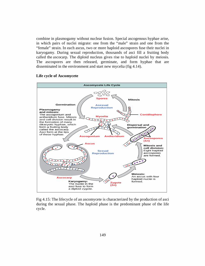
combine in plasmogamy without nuclear fusion. Special ascogenous hyphae arise, in which pairs of nuclei migrate: one from the “male” strain and one from the “female” strain. In each ascus, two or more haploid ascospores fuse their nuclei in karyogamy. During sexual reproduction, thousands of asci fill a fruiting body called the ascocarp. The diploid nucleus gives rise to haploid nuclei by meiosis. The ascospores are then released, germinate, and form hyphae that are disseminated in the environment and start new mycelia (fig 4.14). Life cycle of Ascomycete
Fig 4.15: The lifecycle of an ascomycete is characterized by the production of asci during the sexual phase. The haploid phase is the predominant phase of the life cycle.
149

Fig 4.16: The bright field light micrograph shows ascospores being released from asci in the fungus Talaromyces flavus var. flavus. (credit: modification of work by Dr. Lucille Georg, CDC; scale-bar data from Matt Russell) Self Assessment Exercise 4.8 Q.1 Which of the following statements is true? i. A dikaryotic ascus that forms in the ascocarp undergoes karyogamy,
meiosis, and mitosis to form eight ascospores. ii. . A diploid ascus that forms in the ascocarp undergoes karyogamy, meiosis,
and mitosis to form eight ascospores. iii. A haploid zygote that forms in the ascocarp undergoes karyogamy, meiosis,
and mitosis to form eight ascospores. iv. A dikaryotic ascus that forms in the ascocarp undergoes plasmogamy,
meiosis, and mitosis to form eight ascospores.
Q.2 Write a note on the lifecycle of an ascomycete.
150

4.3 (C) .BASIDIOMYCOTA: THE CLUB FUNGI The fungi in the Phylum Basidiomycota are easily recognizable under a light microscope by their club-shaped fruiting bodies called basidia (singular, basidium), which are the swollen terminal cell of a hypha. The basidia, which are the reproductive organs of these fungi, are often contained within the familiar mushroom, commonly seen in fields after rain, on the supermarket shelves, and growing on your lawn (fig. 4.17 ). These mushroom-producing basidiomyces are sometimes referred to as “gill fungi” because of the presence of gill-like structures on the underside of the cap. The “gills” are actually compacted hyphae on which the basidia are borne. This group also includes shelf fungus, which cling to the bark of trees like small shelves. In addition, the basidiomycota includes smuts and rusts, which are important plant pathogens; toadstools, and shelf fungi stacked on tree trunks. Most edible fungi belong to the Phylum Basidiomycota; however, some basidiomycetes produce deadly toxins. For example, Cryptococcus neoformans causes severe respiratory illness.
Fig 4.17: The fruiting bodies of a basidiomycete form a ring in a meadow, commonly called “fairy ring.” The best-known fairy ring fungus has the scientific name Marasmius oreades. The body of this fungus, its mycelium, is underground and grows outward in a circle. As it grows, the mycelium depletes the soil of nitrogen, causing the mycelia to grow away from the center and leading to the
151

“fairy ring” of fruiting bodies where there is adequate soil nitrogen. (Credit: "Cropcircles"/Wikipedia Commons)] Life Cycle of Basidiomycetes The lifecycle of basidiomycetes includes alternation of generations (Fig 4.18). Spores are generally produced through sexual reproduction, rather than asexual reproduction. The club-shaped basidium carries spores called basidiospores. In the basidium, nuclei of two different mating strains fuse (karyogamy), giving rise to a diploid zygote that then undergoes meiosis. The haploid nuclei migrate into basidiospores, which germinate and generate monokaryotic hyphae. The mycelium that results is called a primary mycelium. Mycelia of different mating strains can combine and produce a secondary mycelium that contains haploid nuclei of two different mating strains. This is the dikaryotic stage of the basidiomyces lifecyle and it is the dominant stage. Eventually, the secondary mycelium generates a basidiocarp, which is a fruiting body that protrudes from the ground—this is what we think of as a mushroom. The basidiocarp bears the developing basidia on the gills under its cap.
Fig. 4.18 The lifecycle of a basidiomycete alternates generation with a prolonged stage in which two nuclei (dikaryon) are present in the hyphae.
152

Self Assesment Exercise 4.9 Q.1 Which of the following statements is true? i. A basidium is the fruiting body of a mushroom-producing fungus, and it
forms four basidiocarps. ii. The result of the plasmogamy step is four basidiospores. iii. Karyogamy results directly in the formation of mycelia. iv. A basidiocarp is the fruiting body of a mushroom-producing fungus. Asexual Ascomycota and Basidiomycota Imperfect fungi—those that do not display a sexual phase—use to be classified in the form phylum Deuteromycota, , a classification group no longer used in the present, ever-developing classification of organisms. While Deuteromycota use to be a classification group, recent moleclular analysis has shown that the members classified in this group belong to the Ascomycota or the Basidiomycota classifications. Since they do not possess the sexual structures that are used to classify other fungi, they are less well described in comparison to other members. Most members live on land, with a few aquatic exceptions. They form visible mycelia with a fuzzy appearance and are commonly known as mold. Reproduction of the fungi in this group is strictly asexual and occurs mostly by production of asexual conidiospores (Figure 24.2.824.2.8). Some hyphae may recombine and form heterokaryotic hyphae. Genetic recombination is known to take place between the different nuclei.
Fig. 4.19: Aspergillus niger is an asexually reproducing fungus (phylum Ascomycota) commonly found as a food contaminant. The spherical structure in
153

this light micrograph is a conidiophore. (credit: modification of work by Dr. Lucille Georg, CDC; scale-bar data from Matt Russell) The fungi in this group have a large impact on everyday human life. The food industry relies on them for ripening some cheeses. The blue veins in Roquefort cheese and the white crust on Camembert are the result of fungal growth. The antibiotic penicillin was originally discovered on an overgrown Petri plate, on which a colony of Penicillium fungi killed the bacterial growth surrounding it. Other fungi in this group cause serious diseases, either directly as parasites (which infect both plants and humans), or as producers of potent toxic compounds, as seen in the aflatoxins released by fungi of the genus Aspergillus. Glomeromycota The Glomeromycota is a newly established phylum which comprises about 230 species that all live in close association with the roots of trees. Fossil records indicate that trees and their root symbionts share a long evolutionary history. It appears that all members of this family form arbuscular mycorrhizae: the hyphae interact with the root cells forming a mutually beneficial association where the plants supply the carbon source and energy in the form of carbohydrates to the fungus, and the fungus supplies essential minerals from the soil to the plant. The glomeromycetes do not reproduce sexually and do not survive without the presence of plant roots. Although they have coenocytic hyphae like the zygomycetes, they do not form zygospores. DNA analysis shows that all glomeromycetes probably descended from a common ancestor, making them a monophyletic lineage. 4.4 IMPORTANCE OF FUNGI (EDIBLE AND POISONOUS FUNGI) Importance of Fungi in Human Life Although we often think of fungi as organisms that cause disease and rot food, fungi are important to human life on many levels. They influence the well-being of human populations on a large scale because they are part of the nutrient cycle in ecosystems. They also have other ecosystem uses, such as pesticides. Biological Insecticides As animal pathogens, fungi help to control the population of damaging pests. These fungi are very specific to the insects they attack; they do not infect animals or plants. Fungi are currently under investigation as potential microbial insecticides, with several already on the market. For example, the fungus
154

Beauveria bassiana is a pesticide being tested as a possible biological control agent for the recent spread of emerald ash borer.
Fig 4.12 Biological Insecticide Fungal Insecticides: The emerald ash borer is an insect that attacks ash trees. It is in turn parasitized by a pathogenic fungus that holds promise as a biological insecticide. The parasitic fungus appears as white fuzz on the body of the insect. Farming The mycorrhizal relationship between fungi and plant roots is essential for the productivity of farm land. Without the fungal partner in root systems, 80–90 percent of trees and grasses would not survive. Mycorrhizal fungal inoculants are available as soil additives from gardening supply stores and are promoted by supporters of organic agriculture. Food Fungi figure prominently in the human diet. Morels, shiitake mushrooms, chanterelles, and truffles are considered delicacies. The meadow mushroom, Agaricus campestris, appears in many dishes. Molds of the genus Penicillium ripen many cheeses. They originate in the natural environment such as the caves of Roquefort, France, where wheels of sheep milk cheese are stacked to capture the molds responsible for the blue veins and pungent taste of the cheese.
155

Fig. 4.13 Morel Mushroom Morel mushroom: The morel mushroom is an ascomycete much appreciated for its delicate taste. Fermentation of grains to produce beer and fermentation of fruits to produce wine is an ancient art that humans in most cultures have practiced for millennia. Ancient humans acquired wild yeasts from the environment and used them to ferment sugars into CO2 and ethanol under anaerobic conditions. It is now possible to purchase isolated strains of wild yeasts from different wine-making regions. Louis Pasteur was instrumental in developing a reliable strain of brewer’s yeast, Saccharomyces cerevisiae, for the French brewing industry in the late 1850s. Saccharomyces cerevisiae, also known as baker’s yeast, is an important ingredient in bread, a food that has been considered a staple of human life for thousands of years. Before isolated yeast became available in modern times, humans simply let the dough collect yeast from the air and rise over a period of hours or days. A small piece of this leavened dough was saved and used as a starter (source of the same yeast) for the next batch, much in the same way sour dough bread is made today.
156

Saccharomyces cerevisiae: The yeast Saccharomyces cerevisiae is approximately 5 µm in diameter and is important for the production of wine, beer, and bread. The yeast also has many applications in medical research. Medicine Many secondary metabolites of fungi are of great commercial importance. Fungi naturally produce antibiotics to kill or inhibit the growth of bacteria, limiting their competition in the natural environment. Important antibiotics, such as penicillin and the cephalosporin, can be isolated from fungi. Valuable drugs isolated from fungi include the immunosuppressant drug cyclosporine (which reduces the risk of rejection after organ transplant), the precursors of steroid hormones, and ergot alkaloids used to stop bleeding. Psilocybin is a compound found in fungi such as Psilocybe semilanceata and Gymnopilus junonius, which have been used for their hallucinogenic properties by various cultures for thousands of years. As simple eukaryotic organisms, fungi are important model research organisms. Many advances in modern genetics were achieved by the use of the red bread mold Neurospora crassa. Additionally, many important genes originally discovered in S. cerevisiae served as a starting point in discovering analogous human genes. As eukaryotic organism, the yeast cell produces and modifies proteins in a manner similar to human cells, as opposed to the bacterium Escherichia coli, which lacks the internal membrane structures and enzymes to tag proteins for export. This makes yeast a much better organism for use in recombinant DNA technology experiments. Like bacteria, yeasts grow easily in culture, have a short generation time, and are amenable to genetic modification. Ecological impact of Fungi Fungi have a tremendous Ecological impact such as: Fungi as Decomposers: Fungi and bacteria are the principal decomposers that reduce complex polysaccharides and proteins to simple organic compounds that plants can assimilate as raw material for photosynthesis. Fungi as Spoilers: Fungi decompose food, wood and even certain plastics. Pathogenic Fungi: Many fungi are pathogenic (e.g. athletes foot, ringworm and yeast infections). Plants are particularly susceptible. For example, Dutch elm disease, caused by an ascomycete, drastically changed the landscape of northeastern U.S. Ergots = Purple structure on rye caused by an ascomycete results in gangrene hallucinations and burning sensations (St. Anthony's fire) and produces lysergic acid, from which LSD is made. Toxins from fungi may be used in weak doses for medical purposes such as treating high blood pressure.
157

Edible Fungi: They are consumed as food by a variety of animals, including humans. In the U.S., mushroom (basidiomycete) consumption is usually restricted to one species of Agaricus, which is cultivated commercially on compost in the dark. In many other countries, however, people eat a variety of cultivated and wild mushrooms. Truffles prized by gourmets are underground ascocarps of mycelia that are mycorrhizal on tree roots. The fruiting bodies (ascocarps) release strong odors that attract mammals and insects - consumers that excavate the truffles and disperse their spores. Since it is difficult for novices to distinguish between poisonous and edible mushrooms, only qualified experts at identification should collect wild mushrooms for eating. Self Assessment Exercise 1. Classify fungi into the five major phyla. 2. Describe each phylum in terms of major representative species and patterns
of reproduction. References Lee, R.E. 1999. Phycology .Cambridge University Press, UK Prescott, L.M., Harley, J.P. and Klein, A.D. 2004. Microbiology, 3
rd ed. WM. C. Brown Publishers. Alexopoulos, C.J., Mims, C.W. and Blackwell, M. 1996. Introductory Mycology.4
th ed. John Wiley and Sons Publishers. Agrios, G.N. 2004. Plant pathology.8
th ed. Academic press London. Vashishta, B.R. 1991. Botany for degree students (all volumes). S. Chand and Company. Ltd.New Delhi. Mauseth, J.D. 2003. Botany: An Introduction to Plant Biology 3rd ed., Jones and Bartlett Pub. UK Marti.J.Ingrouille & Plant: Diversity and Evolution. 2006 CUP
158

Unit 5
LICHENS
Writer: Arshad Mehmood Qamar Reviewer: Dr. Muhammad Waseem
159
CONTENTS Introduction .................................................................................................... 161
Objectives ....................................................................................................... 161
5.1 General Characteristics of Lichens ............................................................... 162
5.2 Life History and Structure of Lichens .......................................................... 164
5.3 Life Cycle of Lichens .................................................................................... 166
5.4 Uses of Lichens in Daily Life ....................................................................... 182
References ...................................................................................................... 190
160

INTRODUCTION This is the fifth unit of Biology-I. This course is equally beneficial for students of B.Ed. 4 Years and B.Ed. 2.5 Years with specialization of Science education. Algae are not a distinct group of plants. They show association with Fungi for making this world more beautiful and productive. When Algae and Fungi live in close relationship together, this association is termed as Lichens. The study of such relationships is termed as Lichenology. In this unit you will study about general characteristics of Lichen their life history and structure will be of great interest for students. Types of Lichens are also described in this unit. Apart from this, uses of lichens are also discussed here in great detail. Lichen is not a single organism. Rather, it is a symbiosis between different organisms – including a fungus and an alga or cyanobacterium. The non-fungal partner contains chlorophyll and is called the phycobiont. The fungal partner is referred as the mycobiont. Therefore, the Lichens is one kind of association involving both phycobiont and mycobiont growing together to form a plant body of recognizable structure. There are about 18,000 to 20,000 known species of Lichens.
OBJECTIVES After having deep understanding and learning of this unit,you will be able: • To define lichens and explain their general characteristics. • To classify them according to their various types. • Describe the Life History and structure of lichens. • Understand the use of lichens.
161

5.1 GENERAL CHARACTERISTICS OF LICHENS Lichens are recognized by their specialized characteristics which are described below: Lichens grow in areas where Fungi or Algae cannot survive alone and their habitat include trees, rooftops, rocks, cemented places, and newly exposed soils. They are found throughout the world and usually grow at the extremes of hotness and coldness. They grow very slowly and have the ability to produce chemicals and acids that break down the rocks. They gather the nutrients which are necessary for plant growth. They are very sensitive to air pollution especially from pollutant of Sulpher dioxide and never found in the areas where pollution is frequent like big cities therefore their presence is the indicator of less pollution in that area. They are thallophytic plants body, with irregular shape and deeply pigmented. They impart colors which are due to the algal partners that have certain pigments. These algal partners either belong to blue green algae like Nostoc, Stigonema, or green algae like Trebouxia. The Fungal partner mainly belongs to ascomycetes apart from bascidiomycetes and rarely deuteromycetes. Reproduction in Lichens is different to that of reproduction in Algae and Fungi. Vegetative reproduction in Lichens is through the development of special propagules (diaspores). Soredia and insidia are the most common diaspores of Lichens. The fungal partner of lichens reproduces sexually. Sexual reproduction in initiated by a fruiting body and spores are formed afterwards. These spores are called ascospores. Ascospores are resting spores and have resistive powers against adverse conditions. Fungus protects Algae from unfavorable conditions, Algae in turn supplies food of Fungus. This type of symbiosis is called helotism (master and slave relationship). Lichens are highly pigmented and have various colors (bluish, green, red Yellow orange etc.). Coloration is due to the pigmentation of algal component. 5.1.1 Taxonomic classification Lichens are classified on the basis of habitat, group of fungal partner, thallus structure and the distribution of algal component in thallus.
162

Distribution based on habitat Lignicolours: Lives on wood. e.g. Cyphellum Terricolours: Lives in soil e.g. cladonia floerkeama Corticolous: Live on bark. e.g. parmelia Saxicolours: Rock dwellers e.g. peltegera Group of fungal partner 1. Ascolichen: Fungal partner is ascomycete 2. Gymnocarpae: Fruiting body apothecium e.gUsnea 3. Pyrenocarpae: Fruiting body perithecium e.g.verrucaria 4. Basidiolichens: Fungal partner is a basidiomycete e.g. Dictynema Based on Thallus Structure Leprose Lichenminutes seales attached superficially to the substratum e.g. Kepraria Crustose: Crust like, closely attached to the substratum at several points e.g Graphis Foliose: Flat leaf like lobed thallus attached to the substratum at one or many points. E.g. Parmelia Fruticose: Branched, erect or pendulous with a bushy appearance; attached to substratum by a basal disc. E.g Usnea Based on Distribution of Algal component in the Thallus. Homoisomerous Thallus: Algal cells and fungal hyphae uniformly distributed in the thallus e.g.Collema Heteromerous Thallus: Algal cells form a distinct layer or algal zone and fungal hyphae in different layers e.g Parmelia. Self-Assessment Exercise 5.1 Q.1 Select the best option given at the end of each statement. i. Graphis is the example of a) Leprose b) Crustose c) oliose d) Frucicose ii. Reproduction in Lichens is ………….. to that of reproduction in Algae and
Fungi. a) Similar b) Different c) Identical d) Partially identical iii. Ascomycete is the fungal partner of a) Basidiolichens b) Pyrenocarpae c) Gymnocarpae d) Ascolichen iv. Lichens usually grow at the ---------of hotness and coldness. a) Depth b) Extremes c) Limits d) Distance
163

Q.2 What is the difference between Homoisomerous Thallus and Heteromerous Thallus?
5.2 LIFE HISTORY AND STRUCTURE OF LICHENS Lichens are formed from a combination of a fungal partner (mycobiont) and an algal partner (phycobiont). The fungal filaments surround and grow into the algal cells, and provide the majority of the lichen's physical bulk and shape. In the picture below at left of the lichen Physia, the fungal filaments have been stained blue, and the scattered algal cells red. Also in the Physia section, you may notice a dark red layer along the top. This is an apothecium, much like the ones atop the British soldier lichen, below at right. An apothecium is a fungal reproductive structure, in which the fungus reproduces itself through the production of spores. These spores will disperse and germinate into new fungi, but they will not produce new lichens. For lichen to reproduce, but the fungus and the alga must disperse together. Lichens reproduce in two basic ways. Firstly, lichen may produce soredia, or a cluster of algal cells wrapped in fungal filaments. These may disperse and form new lichens. A second way for the lichen to reproduce itself is through isidia, which are much like soredia except that isidia are enclosed within a layer of protective cortex tissue. An isidium is much more like miniature lichen.
Fig.5.1 (a,b): Lichens
Lichens will grow almost anywhere that a stable and reasonably well-lit surface occurs. This may include soil, rock, or even the sides of trees. Lichen may absorb certain mineral nutrients from any of these substrates on which it grows, but is generally self-reliant in feeding itself through photosynthesis in the algal cells. Thus, lichens growing on trees are not parasites on the trees and do not feed on them, any more than you feed on the chair you sit in. Lichens growing in trees are simply using the tree as a home. Lichens growing on rocks, though, may release
164

chemicals which speed the degradation of the rock into soil, and thus promote production of new soils. Most lichens are temperate or arctic, though there are many tropical and desert species. Lichens seem to do better in drier environments, where they are not often left in standing water. What the lichen considers dry, however, may not be what we would consider to be dry. In bayous and in cool rainforests, large lichens known as "old man's beard" may often be seen hanging from the branches of trees. Though there is considerable water in these habitats, the air is not saturated, and drying breezes may serve to dessicate arboreal organisms.
Fig.5.2 Lichens
Lichens are most noticeable on the tundra, where lichens, mosses, and liverworts constitute the majority of ground cover. This cover helps to insulate the ground, and may provide forage for grazing animals. The so-called Reindeer "moss" is one such lichen. Thus, lichens are hardy creatures able to survive in scorching deserts and frosty tundra. Their secrets of success are not well understood however. Two key features suggested as having important roles are (1) Their ability to survive drying and (2) Their complex chemistry. Lichens may dry completely when moisture is unavailable. This is not simply dehydration as it occurs in plants and animals we are familiar with, but a complete loss of body water such that the lichen becomes quite brittle. When moisture is again available, they quickly absorb water, becoming soft and fleshy again. Not only can lichens undergo this drying, but while they are dry and brittle, pieces may flake off and later grow into new lichens. The chemistry of lichens is rather complex but well-studied. Lichens manufacture a host of chemicals which presumably serve to reduce attacks by predators. Only a few insects feed on lichens -- some moths and beetles among them.
165

The most serious threat to the continued health of lichens is not predation, but the increased pollution of this century. Several studies have shown serious impacts on the growth and health of lichens resulting from factory and urban air pollution. Because some lichens are so sensitive, they are now being used to quickly and cheaply assess levels of air toxins in Europe and North America. Self-Assessment Exercise 5.2 Q.1 Select the best option given at the end of each statement. i. Lichens can survive a) Drying b) Moist c) PH change d) Osmotic pressure ii. Isidia is much like……………… a) Parmela b) Soredia c) Tundra d) Physia iii. The insect that can feed on lichens is a) Caterpillar b) Ant c) Fly d) Beetle iv. Lichens are now being used in Europe to assess the level of air a) Elements /gases b) Toxins c) Pollution d) Oxygen and Nitrogen Q.2 How the lichens reproduce by isidia and soredia? 5.3 LIFE CYCLE OF LICHENS Lichens also undergo life cycle and reproduce. Three kinds of reproduction occur in lichens; Vegetative reproduction, asexual reproduction and Sexual reproduction take place in Lichens. a) Vegetative Reproduction Lichens reproduce by Fragmentation, Soredia and Isidia. Fragmentation When Lichens become mature and older, some parts of the thallus break-up into fragments. The size of the fragments can be of different sizes. They break either by death or decaying process, sometimes naturally and accidentally breakup into smaller parts. These fragments contain both the algal and fungal components. These pieces are dispersed by dispersal agents like wind and reach to suitable substratum where they begin to grow into new plant. This type of reproduction is mostly seen in pendant thalli. Example of Lichen is Ramalina. Isidia Numerous minute outgrowths produce peg-like, coral-like or globular structures. Each of these is an isidium. Isidia contain algal cells as well as fungal tissue, i.e.
166

the photobiontic layer also extends up into the isidia, and eventually they are liable to break away and be dispersed, leaving tiny scars on the thallus surface. With the cortex providing a tough outer surface, this package of fungus plus algal cells is a durable unit of dispersal.
Fig.5.3 (a,b): Vertical Section of Insidia of Paramedia scortea Ach. A. Early Stage, B later stage. Development of Insidea Thallus shows from the upper cortex. Tissues from medulla, notably including the photobionic layer, grows into these outgrowths to form cortex-bounded, peg-like or coralloid insidea, which can then break off and be dispersed. Each insidean contains both fungus and alga(from Smith,1921)
Fig.5.4: Isidia in Melanelixia glabratula, (Paisley, Renfrewshire, 2012)
Cylindrical and branched isidia project from the lobe surfaces, and readily break away. The shape of the isidia is of value in identifying M. glabratula and similar
167

species of Melanelixia and Melanohalea.This is a common species, but apothecia are infrequent, suggesting that most dispersal is by the isidia. Soredium Like isidia, soredia are dispersal packages of both fungus and alga. Unlike isidia, a soredium is not enclosed in a protective outer cortical layer. Soredia can be produced in much greater numbers, and generally are like powder, but probably are shorter-lived. On substrates such as damp bark, they can germinate quickly and are a highly efficient medium of at least local dispersal. Soredia are produced from exposed medulla tissue. They may be produced from lichen surfaces lacking a cortex, e.g. uncorticated areas of a Cladonia podetium, but commonly they originate from a defined structure, a soralium.
Fig.5.5: Vertical Section of Young soralium of Evernia furfuracae var. soralifera Development of Soredia from a soralium, A vertical section of the thallus shows a break in the upper cortex, exposing the medulla. Tissue from the photobiontic layer is differentiating into rounded, powdery granules, each consisting of algal cells wrapped in fungal hyphae. (From Smith, 1921).
168

Fig. 5.6: Sorediain Evernia prunastri,(Slapton,Devon,2011
Soralia vary in its form and location, the character often being of identification value. On lobes of foliose species they may be laminar (produced from the lobe upper surface), marginal or terminal. They may be pretty much level with the lobe or crust surface, markedly convex, or in some species they are characteristically excavate (somewhat hollowed). They may be irregular or delimited, sometimes disc-like, or produced from fissures in the lobe surfaces or margins.
Fig. 5.7: Disc-like, marginal soralia in Ramalina farinacea, (Glen Tilt, Perthshire, 2008
169

b) Lichen sexual reproduction The great majority of lichens belong to the Ascomycota, in which the sexual
reproductive unit is the ascospore. This is fine; ascospores are generally thick-walled and are tough, resilient units of dispersal and there is a realistic chance of a small number of the ascospores arriving at suitable sites for germination. However, lichen fungi require a suitable algal component (phycobiont) in order to survive and to form lichen. Ascospores disperse only the fungus.
Consequently, one of two things must happen: Either the spore could fortuitously land in a place where the alga already exists in a free state. The spore could land on some other lichen, the fungus then taking over the phycobiont from the existing lichen. This is a common phenomenon; e.g. species of Diploschistes, a common genus of crustose lichens on boulders, mostly grow initially as parasites of other lichens. The lichen Rhizocarpon viridiatrum begins as a parasite on Aspicilia caesiocinerea
Fig.5.8: Parasitism by Rhizocarpon viridiatrum, (Nenthead, Cumberland, August 2012) R. viridiatrum (yellow) is invading and taking over a thallus of Aspicilia caesiocinerea (grey), presumably retaining the phycobiont. Unparasitised A.caesiocinerea is to be seen at the right.
170

If, instead, the lichen can produce units of dispersal that contain both fungus and alga, this will obviously allow and speed up colonization of new sites. Consequently, lichens have evolved various methods of transmitting the alga along with the fungus. c) Units of asexual dispersal 1. Squamules The lichen primary thallus may be composed of squamules, or squamules
may be produced from the surfaces of secondary structures such as podetia. Either way, squamules are easily broken away and may be distributed by animal activity, trampling or wind, before generating to produce new thalli. It is likely that this is a major mode of spread in Cladonia species, particularly at existing sites.
Fig.5.9: Squamulose podetia in Cladonia bellidiflora,(Aberdeenshire, 2008)
The podetia are thickly clothed in squamules, any one of which might break away and, if lodging in a suitable site, may regenerate to produce a new thallus. It should be noted that in Cladonia, squamules in some species are sorediate, soredia being produced from the exposed medulla on the underside, or from the margins. Presumably, these give a greater chance of longer-range dispersal
171

Fig.5.10: Stereocaulon vesuvianum, (Gleniffer Braes, Renfrewshire, April 2011 Phyllocladia, produced from the surfaces of pseudopodetia in Stereocaulon species, presumably have a similar disperal role to that of squamules. 2. Blastidia Blastidia are not easily distinguished from soredia and in practical terms can
be considered much the same. Technically, blastidia bud from other blastidia and originally from the thallus edge or surface, and so might be in clusters or short chains, and they may be partly corticate. As they may aggregate together, they tend to be less powdery.
In the small species of Caloplaca in the "C. citrina group" (genus
Flavoplaca as separated by Arup et al., 2013), some species are sorediate, the soredia perhaps initially produced at the areolar edges and later extending across the areole surfaces. Other species are considered to produce blastidia instead, again often initially at the areolar edges. The blastidia are less powdery but there is little to distinguish them from soredia by normal microscopic examination. Fortunately, the distinction is rarely critical in identification.
172

Fig.5.11: Caloplaca limonia, (Great Cumbrae, Clyde Isles, 2012)
C.limonia characteristically has the areolar surfaces clothed in relatively large, somewhat angular propagules that are considered to be blastidia, or a mixture of blastidia and soredia. In the closely related C. austrocitrina, the propagules are smaller and powdery and considered to be soredia. A variety of other small, granular propagules may be seen in different lichens. In Cladonia, the distinction between soredia and larger, perhaps partly corticate granules on podetial surfaces can be of identification importance 3. Phyllidia and Folioles Phyllidia are small, leaf-like or scale-like outgrowths from an always (?)
foliose thallus, constricted at the point of attachment and so readily detached and dispersed by wind or animals. They are corticate on both surfaces, so differing from squamules. Folioles (or lobules) are much the same thing, though the term is generally used for what appear more like miniature lobes that detach a little less readily. Casual observation of folioles in a local colony of Parmelia ernstiae confirms that while they are abundantly produced early in the year, they are largely gone by early summer, supporting their evident role as dispersal agents. Phyllidia and folioles can be considered as leaf-like, flattened isidia.
Fig.5.12: Phyllidia in Degelia ligulata,(Isle of Muck, 2012)
173

Phyllidia are abundantly produced along the older lobe margins and break away, potentially to regenerate to produce a new thallus. In related species, globose isidia fulfil the same function. [Some published descriptions of D. ligulata use the term "schizidia" rather than "phyllidia", but this is incorrect.]
Fig.5.13: Folioles in Parmelia ernstiae, (Lochwinnoch, Renfrewshire, 2010)
This species, a close relative of P. saxatilis, produces abundant folioles on the lobe margins early in the year (along with clusters of isidia on the lobe surfaces). Later in the year, this same thallus was almost devoid of folioles. 4. Schizidia Schizidia are small, scale-like portions of upper cortex, along with some
underlying tissue (photobiontic layer and medulla), that break away from a thallus surface. The term is over-used (see above) and true examples are evidently few, but include Fulgensia species.
Fig.5.14: Fulgensia fulgens ('Scrambled-egg Lichen'), (Stackpole, Pembrokeshire, 2009)
174

Schizidia have flaked from the thallus surface, leaving white areas of exposed medulla Ryan et al. (2002) regard the disc-like granules, with corticate upper surfaces, produced within the cups of Cladonia pyxidata, as schizidia. 5. Conidia Conidia are asexual fungal spores, commonly thin-walled and usually
functioning more as a means of rapid, local reproduction than of longer range dispersal. Fungi in which a sexual stage is very rare, or apparently non-existent, may have thicker-walled, more resilient conidia, but those of lichenised fungi are typically of the thin-walled and presumably short-lived type.
The role of conidia in lichens is much more a matter for debate. While
apparently often simply for asexual reproduction, it appears that at least some may also or alternatively function as male gametes in the sexual process. Conidia are commonly produced from within pycnidia, closed, flask-like structures, often visible as black dots set in the lichen thallus.
While necessarily included here as units of asexual dispersal, conidia cannot carry the phycobiont and so are not units for reproduction and dispersal of the complete lichen association. A good review of lichen conidia, their variation and the structures that produce them has been provided by Bungartz (2002) Conidia are asexual fungal spores, commonly thin-walled and usually functioning more as a means of rapid, local reproduction than of longer range dispersal. Fungi in which a sexual stage is very rare, or apparently non-existent, may have thicker-walled, more resilient conidia, but those of lichenised fungi are typically of the thin-walled and presumably short-lived type. The role of conidia in lichens is much more a matter for debate. While apparently often simply for asexual reproduction, it appears that at least some may also or alternatively function as male gametes in the sexual process. Conidia are commonly produced from within pycnidia, closed, flask-like structures, often visible as black dots set in the lichen thallus. While necessarily included here as units of asexual dispersal, conidia cannot carry the phycobiont and so are not units for reproduction and dispersal of the complete lichen association. A good review of lichen conidia, their variation and the structures that produce them has been provided by Bungartz (2002).
175

Fig.5.15: Conidia
Self-Assessment Exercise 5.2 Q.1 Select the best option given at the end of each statement. i. How many types of reproduction occur in lichens..? a) 2 b) 3 c) 4 d) 5 ii. Lichens can break either by death or ……………………. process a) Dehydration b) Decaying c) Deliberate killing d) Drying iii. Isidium contains……………………. a) Fungus b) Lichens c) Algae d) Both a and c iv. Ascospores disperse……………………. a) Fungus b) Lichens c) Algae d) Both a and b v. ……………………. are not units for reproduction. a) Isidium b) Conidia c) Soralium d) Fruiting body Q.2 Describe the units of asexual dispersal?
176

5.3 (A) TYPES OF LICHENS Main types of lichens are as follows: Crustose lichens Foliose lichens Fruticose lichens 5.3.1 Crustose Lichens These lichens, as the name indicates, grow and spread over the rocky substratum as flattened, thin crust- like thalli, forming an inseparable coat over (Fig.). The thalli are leathery textured or of gelatinous nature and are embedded partly or wholly in the rocky or soil substratum. In fully embedded form, only the fruiting bodies of the fungus are seen over the surface while the vegetative thalli lie inside the substratum. Internally these lichens are undifferentiated due to mixing of algal cells with the fungal hyphae, hence are called Homoimerous.
Fig: 5.16: Structures of Crustose Lichens Examples: Rhizocarpon, Lecidea, Sarcogyne. Rhizocarpon This is a species of lichens which grow are found on rocks in hilly areas .Each plant is a flat patch having border of black lines of spores. These are used in lichenometry.
177

Fig.5.17: Rhizocarpon an example of Crustose Lichen 5.3.2 Foliose Lichens These lichens are leaf- like in appearance due to flat, dorsi-ventrally differentiated and lobed leaf- like thalli of fairly large size. These thalli are firmly attached to the substratum due to hyphal-strands, called Rhizines that protrude down-wards from the ventral surface. Internally the thallus of foliose- lichens is differentiated into the following four layers of tissues
Fig: 5.18: Thallus structure of Foliose Lichen
178

Upper cortex Algal layer Medullar-rays Lower cortex The upper-cortex consists of pseudo-parenchymatous firmly packed interwoven fungal-hyphae, with or without intercellular spaces. The spaces when present are filled with gelatinous substances. The algal layer lies beneath the upper-cortex where the algal cells ar4e intermingled with loosely interwoven fungal hypae. This layer is referred to as gonidial-layer as the cells were thought once to be gonidial or reproductive-cells of the thallus. The medulla or pith occupies central position and consists of loosely arranged interwoven fungal hypae with large intercellular spaces/ The lower cortex is compact layer with intercellular spaces either absent or filled with gelatinous substances. From the lower-cortex protrude downwards root-like hyphal stands, called rhizines, for attachment of the thallus to the substratum. In addition several number of small depressions develop on the ventral surface for gaseous exchange, hence are respiratory in function. Examples: Parmelia, Gyrophora, Physcia. Parmelia It is an example of foliose lichen. It is found on barks and rocks intermixed with mosses. They are found at higher altitudes above 4000 ft. it is attached to the substratum with rhizinae which arise from its lower darker surface. Generally the thallus is compartmentalized into four tisues: upper cortex, algal layer, medulla and lower cortex. The phycobiont is a green Pleurococcus. (Fig 5.2) Paramelia reproduces vegetative way by fragmentation and by insidia and soredia. The spores are produced in apothecia, a cup shaped structure which may be sessile or stalked.
179

Fig.5.19: Structure of Parmelia An example of Foliose Lichen 5.3.3 Fruticose Lichens The thalli of these lichens are cylindrical, flattened or stellate in appearance. They are much branched and so are bush-like in outline. The thalli may be erect (Cladonia) or pendant (Usnea) and are attached to the substratum by basal portion composed of densely packed hyphae (Fig...) Internally these thalli are differentiated into layers as (Fig. 5.7)
Figure: 5.20: Fruticose Lichen Upper Cortex (Fungal Part ) Algal Layer (Algal cells arranged in patches or layers) Medulla (Pith). (Fungal Nature- central layer) Examples: Usnea, Evernia and Cladonia etc. Usnea Usnea is an example of fruticose. Its thallus is ribbon likes, cylindrical or branched. The thallus is grey-green in color and consists of a single main stalk or many stalks.
180

Fig5.21: Example of Fruticose Lichens Comparison of Types of Lichens by internal Structures
Fig.5.22: Comparison of types of lichens by internal structures Self-Assessment Exercise 5.3 Q.1 Select the best option given at the end of each statement. i. The crustose lichens that are used in lichenometry are: a) Rhizocarpon b) Lecidea
c) Sarcogyne d) Parmelia ii. Thallus is compartmentalized into …………………….tisues a) Two b) Three c) Four d) Five
181

iii. Homoimerous are undifferentiated due to mixing of……………………. with …………………….,
a) Fungus with lichens b) Algal cells with the fungal hyphae c) Algae and Protista d) Fungal hyphae with algal thallus iv. The function of rhizines is; a) Chemotaxis of thallus towards the substratum b) Attachment of the thallus to the substratum c) Chemotaxis of thallus away from the substratum
d) Both a and b v. The spores of Parmelia are produced in ……… a) Apothecia b) Perithecium c) Cleistothecia d) Perithecia Q.2 How reproduction differs of lichens from one another on the basis of their
types? 5.4 USES OF LICHENS IN DAILY LIFE
Fig.5.23: Lichens Growing on Tree branches
Lichens are very useful organisms and found extremely useful for human beings. Man has explored how to use Lichens for the purpose of dyeing, scents for perfumes, and as food. Lichens are famous to be used as dyes. There are number of Lichens which are used as medicines, and biological sensors which are used for information regarding environment. They also add beauty in nature (Figure 5.24)
182

Figure 5.24: Striking Lichens on mountains They are found in different colors such as black, brown, blue-grey, grey, green, orange, red and yellow. 5.4.1 Uses of Lichens for Dyeing Dyeing is a natural thing for humans since many ages as they have been using clothing of different colors. They color cloths and fabrics with natural dyes that have been obtained from plants and Lichens. Lichens impart colors due to the presence of pigments. Dyeing with the help of Lichens is still in practice. Many lichens are of grey color when they're dry. In lichens,the algal cells give a deeper hue whereas the fungus component is often colorless, but in some cases it contains a pigment that gives the lichen a vivid color. Making wool and fabric dyes from lichens is an ancient process that is still performed. Suitable specimens are collected, cut into pieces and then water is added. Ammonia is also added to the water sometimes. At one time, urine has also been used as the water-ammonia solution. The mixture is left for several weeks in order for the dye to appear.
183

The dye made from lichen often has a different color due to the intact organisms. Brown, gold, orange, green, purple, blue and red colors all are possible, depending on the species of lichen used and the type of extraction process. Modern wool and fabric dyers often emphasize conservation as they collect lichens. They tend to gather specimens that have already become detached from their substrate or that are growing in a place from which they are likely to be removed, such as trees that have died. (Lichens don't harm trees.)
5.25: The red reproductive structures of the British soldiers lichen, or Cladonia cristatella; the lichen is growing in the company of mosses A British soldier is a fruticose lichen found in Ontario and the northeastern part of the United States. The name is derived from the red uniforms worn by British troops during the American Revolution. 5.4.2 Lichen-A Useful Dye and an Interesting Pigment Litmus Paper Litmus paper is very commonly used as an acid-base indicator, especially by students who need to know only the approximate pH of a substance. Litmus is a
184

mixture of dyes extracted from specific lichens, especially Rosella tinctoria. Litmus paper is made from filter paper that has been treated with the dye. Neutral litmus paper is purple in color. It turns red when exposed to an acid and blue when exposed to a base (alkali). Natural Sunscreens Xanthoria parietina is foliose lichen which contains a yellow pigment called parietin. This pigment absorbs ultraviolet radiation, acting as a sunscreen to protect the algal cells inside the lichen. Some other lichens contain sunscreens, too. It's been suggested that the protective chemicals could be useful in human sunscreens.
Fig.5.26: Xanthoria parietina is a foliose lichen that has a high resistance to pollution, especially in the form of nitrogen; the orange cup-like structures are apothecia and produce reproductive spores
185

5.27: Usnea often hangs from branches and is sometimes known as old man's beard. This is Usnea filipendula.
5.4.3 Lichens Use as antibiotics, Preservatives, and Toxins Usnea Usnic acid has been found in several lichen species, including members of the Usnea genus. In natural medicine Usnea is used as an antibiotic and an anti-inflammatory substance. It can also be used in some products as a preservative. Tests in laboratory equipment and lab animals show that usnic acid has antimicrobial properties and kills bacteria, fungi and viruses. It also decreases inflammation and prevents some types of cancer cells from reproducing. Unfortunately, it may cause serious liver damage in humans. Clinical tests of the effectiveness of usnic acid in the human body are lacking. Wolf Lichen The wolf lichen (Letharia vulpina) has a bright yellow-green color and grows in Europe and western North America. It contains a yellow chemical called vulpinic acid, which is poisonous to mammals. In the past, wolf lichen was mixed with ground glass and meat and has been used as a poison for wolves. It is unknown that whether the lichen or the glass, which thing was responsible more for the death of the animals. Wolf lichen has also been used for dye extraction and was once used medicinally by native peoples. Lab research shows that vulpinic acid can kill certain kinds of
186

bacteria. As in the case of usnic acid, if vulpinic acid is proven to be helpful for humans, we need to find a way to prevent the chemical from resulting in any harm for humans before its use as an antibiotic. It's important to keep this in note that although this chemical has antibacterial properties in isolated cells but in lab animals it has been shown that it does not have the same effect inside the living body.
Fig.5.28: Letharia vulpina or wolf lichen 5.4.4 Lichen- Ingredients in Perfumes and Deodorants Oakmoss Oakmoss (Evernia prunastri) is used to provide scents and fixatives for perfumes. It grows in Europe and North America but is especially valued in France. It lives on oak trees as well as other trees and is actually fruticose lichen and not a moss. Both essential oils and absolutes are extracted from the oakmoss. Essential oils are usually obtained by steam distillation. Absolutes are obtained by solvent extraction and are generally more concentrated than essential oils. The extracts of oakmoss are said to have a lovely earthy scent that resembles the aroma of moss and has an undertone of pine. Some oakmoss extracts advertise the fact that they are low in atranol. This chemical is allergenic for some people, so these are the worth seeking products that have little or no atranol.
187

Pseudevernia furfuracea Pseudevernia furfuracea is another type of fruticose lichen that has been used in the perfume industry. The lichen was used to fill the body cavity of Ancient Egyptian mummies. It's not known if the lichen has been used as a preservative or to provide a pleasant scent. Today lichen components are used in deodorants as well as perfumes because of their pleasing aroma.
5.29: Oakmoss is lichen, despite its name. 5.4.5: Lichens as Food for People We shouldn't pick up lichen from a neighborhood rock or tree and eat it because in this way some species are consumed by humans. Many species are mildly toxic; a few are poisonous and most are indigestible in their raw form. Some cultures have learned to prepare lichens in a way that improves their digestibility; however, this contributes towards their delicacy. Reindeer moss, or Cladonia rangiferina, is fruticose lichen that is a staple food of reindeer and caribou. (This is yet another "moss" that is really a lichen.) Some Arctic inhabitants mix the partly digested lichen from caribou stomachs with raw fish eggs. The result is a concoction known as "stomach ice cream". Umbilicaria esculenta is black foliose lichen that grows on rocks. It's used in Asian cuisine after being fried. Umbilicaria lichens are often knows as rock tripe. The North American version was used as an emergency food by early explorers. Some lichens are boiled and mixed with fruit and flavoring agents such as onions before being eaten. With a few exceptions, however, lichens are generally used as food in famine situations instead of by choice.
188
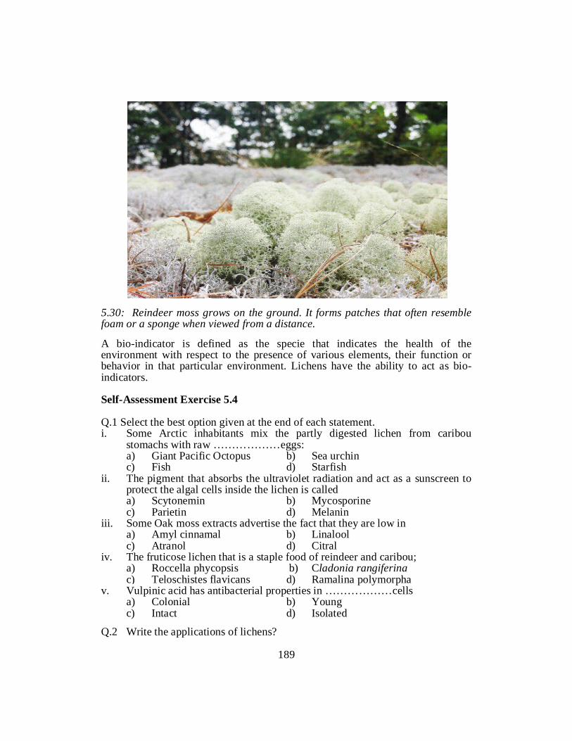
5.30: Reindeer moss grows on the ground. It forms patches that often resemble foam or a sponge when viewed from a distance. A bio-indicator is defined as the specie that indicates the health of the environment with respect to the presence of various elements, their function or behavior in that particular environment. Lichens have the ability to act as bio-indicators. Self-Assessment Exercise 5.4 Q.1 Select the best option given at the end of each statement. i. Some Arctic inhabitants mix the partly digested lichen from caribou
stomachs with raw ………………eggs: a) Giant Pacific Octopus b) Sea urchin c) Fish d) Starfish ii. The pigment that absorbs the ultraviolet radiation and act as a sunscreen to
protect the algal cells inside the lichen is called a) Scytonemin b) Mycosporine c) Parietin d) Melanin iii. Some Oak moss extracts advertise the fact that they are low in a) Amyl cinnamal b) Linalool c) Atranol d) Citral iv. The fruticose lichen that is a staple food of reindeer and caribou; a) Roccella phycopsis b) Cladonia rangiferina c) Teloschistes flavicans d) Ramalina polymorpha v. Vulpinic acid has antibacterial properties in ………………cells a) Colonial b) Young c) Intact d) Isolated
Q.2 Write the applications of lichens?
189

References Arup, U., Søchting, U., & Frödén, P. (2013). A new taxonomy of the family Teloschistaceae. Nordic Journal of Botany 13: 16–83. Bungartz, F. (2002). Morphology and anatomy of conidia-producing structures, in Nash III, T.H., Ryan, B.D., Gries, C., & Bungartz, F. (eds.) Lichen flora of the Greater Sonoran Desert Region, vol. 1, Arizona State University, Tempe, pp. 35–40. Hawksworth, D.L., & Hill, D.J. (1984). The lichen-forming fungi, Blackie, Glasgow. Ryan, B.D., Bungartz, F., & Nash III, T.H. (2002). Morphology and anatomy of the lichen thallus, in Nash III, T.H., Ryan, B.D., Gries, C., & Bungartz, F. (eds.) Lichen flora of the Greater Sonoran Desert Region, vol. 1, Arizona State University, Tempe, pp. 8–23. Smith, A.L. (1921). Lichens, Cambridge University Press, Cambridge. Smith, C.W., Aptroot, A., Coppins, B.J., Fletcher, A., Gilbert, O.L., James, P.W., & Wolseley, P.A. (eds.) (2009). The lichens of Great Britain and Ireland, British Lichen Society, London
190

Unit 6
BRYOPHYTES
Writer: Arshad Mehmood Qamar Reviewer: Dr.Muhammad Waseem
191

CONTENTS
Introduction .................................................................................................... 193
Objectives ....................................................................................................... 193
6.1 Introduction to Bryophytes............................................................................ 194
6.2 Evolution of Bryophytes ............................................................................... 195
6.3 Classification of Bryophytes ......................................................................... 199
6.4 Life cycle of Bryophytes with specific examples (Riccia, Anthoceros,
Funaria ............................................................................................................ 201
References ...................................................................................................... 234
192
INTRODUCTION
This chapter is about bryophytes which is a traditional name used to refer to all embryophytes that do not have true vascular tissue and are therefore called "non-vascular plants" This chapter include bryophytes like Riccia, Anthoceros and Funaria explaining their life cycle and mode of reproduction. In this unit life cycle of bryophytes with specific examples is elaborated.
OBJECTIVES After reading this unit the you will be ablt to: • Describe bryophytes. • Understand the concepts of antheridium and archegonia. • The reproduction mechanisms of bryophytes. • Differentiate and illustrate the life cycles of various bryophytes.
193

6.1 INTRODUCTION TO BRYOPHYTES This is a diverse group of non-vascular plants that can produce embryo and considered as evolved from other kland plants. They were earlier land plants that evolved from green algae .They are moatly found on moist habitats. There are three main groups of Bryophytes, including mosses, liverworts and hornworts. There are round about 14000 species of mosses, approximately 8500species of Liverworts and more than 200 species of hornworts are discovered. Bryophytes have multicellular sex organs, i.e the gametes are enclosed by a sterile jacket of cells, retain the zygote within the female sex organ and allow it to develop into an embryo there. They have a layer of cutin on the plant parts and spors.
Thallose liverwort - bilaterally symmetrical
Leafy liverwort - bilaterally
symmetrical
Moss - radially symmetrical
leaves usually with midrib
Fig. 6.1: Examples of Bryophytes Bryophytes require moist habitat at least part of their loife cycle. They may grow as epiphytes.(grow on other plants). Most of them are pioneer plants growing on bare rocks. A number of mosses are resistant to desiccation. They can lose water in dry season, remain alive and take water when it rains.
194

They grow in prostrate way. Being prostrate they have their surface in contact with the substratum and readily absorb moisture this way. External water movement is important so water moves along the surface of the plants by capillary action. In many mosses specialized transport cells, hydroids and leptoids, analogous to xylem and phloem are found in the center of the stem. Gases diffuse across the plants surface but liverworts also have special pores through which gas exchanges.some mosses have stomata on their Sporophyte. Some bryophytes are dioecious and others are monoecious. When male and female sex organs born on separate gametophytes, they are called dioecious. But when both sexes are born on a sigle gametophyte they are called monoecious. Typically sex organs are borne in clusters, surrounded by sterile hairs. In some species they can be borne in a head on the stalk. On maturity the antheridium bursts releasing the sperm cells (antherozoids). The sperm cells are very close to the nucleus inside the cytoplasm. Sperms are attracted towards the egg due to some chemicals. On reaching near egg the sperms swim down the neck of archegonium and fertilize the egg. The zygote thus formed is the start of sporophyte generation. 6.2 EVOLUTION OF BRYOPHYTES Bryophytes lack fossil records because of their soft and delicate body. But between 510-630 million yeqars land plants evolved from equatic plants, especially from green algae. Molecular Phylogenttic studies reveal that bryophytes were the first and earliest diverging lineage of the land plants. Therefore they are important to study on the basis of migration from equatic environment to land environment. There are two schools of thoughts regarding evolution of bryophytes. According to first school of thought they were evolved from the green thallophyta a kind of algae. And according to the second school of thought tghey have been evolved from pteridophytes. Most of the evolutionists regard tghat they evolved from algal ancestors. 6.2.1 Evolution from Algae Most of the bryologists are in support of this evolution. Though there is no evidence about connection of algae and bryophytes through fossils record. Even then following points arte enough to support that they have been evolved from algal ancestors.
195

The presence of water for the process of fertilization shows their amphibious nature, the presence of ciliated antherozoid.
Fig. 6.2: Origion of Bryophytes. Figure illustrating the hypothetical origin of the antheridium and archegonium from the Plunicular sporangium of certain Phaeophyceae (Eclocarpus) No doubt that there is no apparent relation between the anthridium and archegonium of the algae with anthridium nand archegonium of bryophytes. In bryophytes the egg is surrounded by a cellular jacket, whereas there is no cellular protective layer is found around the egg of algae. Lignier in 1903, pointed out that the algae gave rise to a connecting link known as ‘prohepa tics’ and thereafter bryophytes originated from this connecting link on one hand and the pteridophytes on the other. Bower (1908) also supported this view and said that the Archegoniatae have been evolved from the aquatic ancestors, i.e., the algae. The bryophytes resemble in many respects the green algae, i.e., Chlorophyceae, and Fritsch (1916, 1945) has advocated that the Chaetophorales gave rise to the bryophytes.
196

But according to many biologists the sex orhans of the bryophytes have been evolved from thoise of algae due to following reason/s. Inside the water there was less need of protective layer around the egg. Antheridium and archegonium of bryophytes originated from gametangia of a type similar to that of Ectocarpus. In ectocarpus the gametangia consists of many cells, each of which give rise to a gamete. With the migration of these cells from water to land, the need arise for protection of gamete from desiccation. With the result of this outer most cells of the gametes became sterile and adopted functions of protective layer. In this way gamete of bryophytes was derived from the algal gametangium. According to first view the neck canal cells were origionally female gametes and lator on there walls and cytoplasm were lost. According to Smith (1938) the reproductive cells of Schizomeris and antheridia of Chaetonema are quite alike to that of the gametangia of Ectocarpus. According to Church (1919), the bryophytes have been originated from the marine ancestors and not from the fresh water ancestors. This theory could not get general support because of the lack of evidences from paleobotany and geology. According to majority of workers the bryophytes have been originated from Chlorophyceae which are commonly found in fresh waters and rarely in sea waters. 6.2.2 Evolution from pteridophytes According to second school of thought the bryophytes have been evolved from pteridophytes by means of reduction. Many bryologists including Lang(1917), Kidston and Lang(1917), Scott(1923), Halle(1936), Haskell (1949) and Christensen (1954) revelead that bryophytes evolved from pteridophytes through process of reduction. Kashyap (1919) also supported the view, because of common resemblances of the two groups. Similarities between sporangia of some members of Psilophytales (Rhynia, Horneophyton and Sporogonites) with capsules of Anthocerotales, Sphagnum and Andreaea led to conclude this hypothesis. The Psilophytales are the oldest pteridophytes in which the sporophytes were rootless, leafless and dichotomously branched with terminal sporangia. Such sporophytes resemble the bryophytes, especially the members of Anthocerotales and are thought to have evolved by progressive reduction. Proskaeur (1960) thinks that if bryophytes are polyphyletic in origin, at least Anthocerotales originated from Psilophytales like Horneophyton. According to Kashyap (1919), “bryophytes represent a degenerate evolutionary line of
197

pteridophytes or in more correct term, the bryophytes are descendents of pteridophytes.” 6.2.3 Origin of the Sporophyte It is fact that alternation of generation in most of the plants has been evolved from equatic algae. It is also a thinking not necessarily all plant migrating from equatic to terrestrial environment must appear alternation of generation among them. Algae have both types of alternation of generation viz; isomorphic and hetereporphic. The theories regarding evolution of sporophyte of bryophytes are as under 1. Modification Theory 2. Intercalation Theory According to modification theory or homologous theory the sporophyte is a direct modification of the gametophyte. The supporter of this theory advocated that the sporophyte is to be interpreted as a neutral generation and its primary function is the production of spores. Some facts which support this theory are –the presence of isomorphisim alternation of generation in some algae; photosynthesis in sporophyte of bryophytes; presence of trachieds in gametophyte of pterdophytes; apogamy and apospory. Supporters of the modification theory (Church, 1919; Fritsch, 1945; Zimmermann, 1932) are of the opinion that the two generations were isomorphic, independent and of a complex external structure. They think that in time the sporophyte became permanently attached and partially dependent upon the sporophyte and resulted in a reduction in the complex structure of the sporophyte. According to intercalation or antithetic theory the sporophyte has been interpreted as a entirely new structure intercalated between two successive gametophytic generations. This theory further supports that the bryophytics sporophyte did not make its appearance until after a gametophyte had evolved an archegonium. The zygote found in the venter of archegonium divided mitotically forming a number of diploid (2n) cells, each of which divided meiotically to form four haploid (n) spores. Thus a rudimentary type of sporophyte was developed in which all cells were sporogenous.
198

Further evolution of the sporophyte took place as a result of sterilization of sporogenous tissue and foot, seta and capsule were differentiated. According to this theory there was a progression from a simple to a more complex sporophyte by a progressive sterilization of sporogenous tissue. Self-Assessment Exercise.6.1 Q.1 Write the views about evolution of Bryophytes. Q.2 Compare and contrast Modification and intercalation theories of Evolution
of Bryophytes. 6.3 CLASSIFICATION OF BRYOPHYTES Eichler (1883) was the first to divide Bryophyta into two groups as follows. 1. Group.1 Hepaticae 2. Group 2. Musci Engler (1892) recognized hepaticae and Musci as two classes and divided each class into following three orders: Division: Bryophyta Class.1 Hepaticae Order.1 Marchantiales Order.2 Jungermanniales Order.3 Anthocerotales Class.2 Musci Order.1 Sphagnales Order.2 Andreales Order.3 Bryales Due to isolated characters of Antheros and related genera. Howe (1899) raised Anthocerotales to the rank of a class and divided Bryophyta into three classes. Class.1 Hepaticae Class.2 Anthocerotes Class.3 Musci This system of classification was followed by smith (1938, 1955), Takhtajan (1953), Wardlaw (1955) and Schutser (1958) but preferred to call class Anthocerotes as Anthocerotae.
199

International code of Botanical Nomenclature (ICBN) suggested in 1956 that the suffix-opsida should be used foe classes and such usage had already been proposed by Rothmaler (19510 for the classes of bryophytes. Rothmler changes the class names as Class.1 Hapaticae as Hapaticopsida Class.2 Anthocerotae as Anthocerosida Class.3 Musci as Bryopsida Proskauer (1957), Parihar (1965) and Holmes (1986) proposed the system of Classification which is with few exceptions used till now. This classification is given as under. Class1. Hepaticopsida (Liverworts) Order1. Marchantiales (Riccia, Marchantia) Order.2 Sphaerocarpales (Sphaerocarpos) Order.3 Jungermanniales (Pellia) Order.4 Calobryales (Calobryum) Class.2. Anthocerotopsida (Hornworts) Order. Anthocerotopsida Family.1 Anthocerotacae (Anthoceros) Family.2 Notothylacae (NotoThylas) Class.3 Bryopsida (Mosses) Sub-Class .1 Sphanidae Order.Sphagnales Sub-class .2 Andreaeidae Order. Andreaeales Class.3 Bryidae (It has 12 Orders) Class.4 Buxbaumiidae Order .Buxbaumilaes (Two Families) Class.5. Polytrichidae Order.1 Polytrichales Order.2 Dawsoniales Self Assessment Exercise 6.2 Q.1 Fill in the Blanks with suitable words. i. . Howe classified bryophyte into ………………... ii. Mosses belong to class ………………... iii. Hornworts belong to ………………...class. iv. Marchantia is the order of class………………... v. Engler divided the bryophyte into ………………... classes. Q.2 Write classification of bryophyte according to ICBN
200

6.4 LIFE CYCLES OF BRYOPHYTES WITH EXAMPLES It is better to describe the characteristics of bryophytes with the help of examples for more clarity and knowledge. Riccia, Anthoceros and Funaria are discussed with all details including classification, occurrences, structure of the body and life cycles. Riccia Taxonomic Classification: Division: Bryophyta Class: Hepaticopsida Order: Marchantiales Family: Ricciacae Genus: Riccia Occurrence Some botanists have reported 200 species of Riccia. Many of its species are found in Punjab, Kashmir and Western Himalayas. The species are found scattered from hilly and plains areas. All the species are terrestrial except R.fluitans and R. natans which are free floating or submerged aquatics. Most of the species are found in the Punjab plains growing on the moist soil in places like river beds, banks of rivers and canals, and along the streams.
Fig6.3 Riccia Plant Body Gametophyte is the dominant phase of bryophytes; therefore, the main plant body (The thallus) of Riccia represents a well-developed gametophytic phase.
201

External Features The plant body is prostrate, flat, somewhat fleshy and green dichotomously branched thallus. Branching determine different shapesof the thallus. The shape of thallus with examples is given in the table
Sr. Specie name Shape 1. R. pathankotensis Rossete or circular patch 2 R.cruciata & R. pandei Cruciform arrangement 3 R.beyrichiana Overlapping arrangement 4 R.reticulata Thick, twice forked ,crowded
The dorsal surface is light green or dark green body, each branch having a thick midrib. It is traversed by a conspicuous median longitudinal groove which ends in a depression at the apical region forming an apical notch. Plant grows from the point situated in apical notch. The main function of the mid- dorsal groove is to retain water required for fertilization.
Fig 6.4.Riccia Thallus of R.Melanospora
The ventral surface bears numerous rhizoids and scales. Riccia has two types of rhizoids. The simple rhizoids have smooth inner wall. The tuburculate have prominent peg-like projections, also known as tubercles. The scales are arranged in a transverse row. These are multicellular, membranous, pink, red, violet or black and one celled thick structures. Scales contain anthocyanin pigment which in a very moist habitat may develop a faint green color due to chlorophyll. The prominence and persistence of the scales depend
202

upon the nature of habitat of the plant. In moist place they are small and soon perish but in dry habitat they are large and persistent. Internal Features A vertical cross section of thallus of terrestrial species of Riccia shows a more elaborate internal differentiation of tissues arranged in two distinct regions. An upper assimilatory region A lower storage region/zone Assimilatory Region The assimilatory region of the thallus consists of unbranched green photosynthetic filaments. These filaments are arranged in vertical rows each consisting of a single row of cells. All cells of the filaments are alike and contain numerous discoid chloroplasts capable of making food. Vertical rows of assimilatory filament s enclose vertical spaces called air chambers, air canals or air channels. Considering the fact of narrow or wideness of the air chambers, Riccia is subdivided into two sub groups. Euriccia: have narrow air chambers, Ricciella: having wider air chambers. Storage Zone The assimilatory region merges gradually into compact colorless, undifferentiated parenchymatous storage region. The cells of this portion are comparatively larger, thin walled having no chloroplast, and intercellular spaces. Prominent reserve food granules occupy the major part of these cells. Rhizoids arise from the mid lower surface of the storage region. Scales are emerged from the margins. Growth of Thallus As mentioned earlier that growing point lies in the apical notch in the form of one or several meristematic cells. The apical cell appears triangular with three cutting faces, in a vertical longitudinal section. In a surface view it appears rectangular with four cutting faces: a dorsal, a ventral and two laterals. The cells formed from the dorsal and ventral cutting faces, constitute the major portion of the thallus. Each cell cut off by dorsal face divides transversely into an upper and lower cell. The upper cell by repeated divisions gives rise to the assimilatory zone and sex organs whereas the lower cell constitutes the storage region. The ventral cell
203

divides to produce an outer and an inner cell. The outer cell forms the scale whereas the inner cell forms the rhizoids and a part of the storage region. Formation of air chambers The formation of air chambers has drawn attention of scientists regarding mode of origin of air chambers. There are two schools of thoughts explaining the development of the air chambers. First view: the air chambers are formed during apical growth. The growth pattern is variable,i.e. for a short duration at places; the growth of the thallus is slow followed by rapid growth resulting in the formation of depressions. These depressions become deep pits and ultimately air chambers. This school of thought is supported by Leitgeb(1879), Hirsh(1910), and black (1913). Second View: the air chambers are formed by splitting of the internal vertical cell walls in the close proximity of apical growing regions. The nature of origin of air chambers is of schizogenous type. It is similar to that of intercellular spaces found among parenchymatous cells of the higher plants. This splitting may be endogenous i.e. splitting begins from the below and extends upwards or exogenous i.e. splitting begins from upper surface of thallus and approaches towards the lower region. Life Cycle of Riccia Reproduction Riccia reproduce by means of vegetative and sexual reproduction which occurs after a certain stage of maturity or due to sudden changes in the environment. Vegetative reproduction The plants multiply by the following methods during favorable season. Fragmentation It is a common method of vegetative reproduction. The older parts of the thalli die of old age. This progressive death and decay, on reaching to dichotomy, separates the two lobes. Each survived lobe, by apical growth, develops into a new plant. Adventitious branches Adventitious branches arise from the mid ventral surface of the thallus. These adventitious branches get detached and develop into new plants. This type of reproduction is found in R.Fluitans.
204

Tubers A few species such as R. discolor, R. bollardieri, R.vesicata and R. perennis reproduce by pereniating tubers formed at the apices of the thallus. The tubers which remain dormant during unfavorable conditions resume their growth under favorable conditions. Persistent Species In R.discolor and many other species, the apices of the thallus grow down into the soil and become thickened at the end of the growing season. The plant parts other than the undergroundpersistent apices die. With the favorable season, these apices resume their growth .they come up and develop into new plants. Exact copies of thalli R. gluaca exhibits a different method of vegetative reproduction. The terminal ends of the rhizoids as a result of a multiplication form new thalli similar to that formed from the germ tube of spore. Sexual reproduction The sex organs are highly differentiated and well developed. The male sex organs are called antheridium and female sex organs are called archegonium. Some plants are monoecious or homothallic (both sex organs grow on the same plant), others are dioecious or heterothallic (antheridium and archegonium grow on different plants).
Fig.6.5 Sexual Reproduction in Riccia
205

The Antheridium Structure of the antheridium A mature antheridium of Riccia consists of the following structures. A short stalk is attached to the base of antheridial body. The antheridial body is an oval structure with flat base and conical apex. The antheridial body has two distinct regions of the thin walled cells. a) An outer sterile layer of thin walled cells called jacket layer, b) A central mass of large number of cubical androgonial cells. The antheridium is enclosed within an antheridial chamber. The antheridial chamber communicates outside with the dorsal surface by a pore. Development of Antheridium The antheridium develops near the apex from a superficial cell. This superficial cell is termed as antheridial initial.
Fig.6.6. Riccia Development of Antheridium
206

The initial antheridial cell divides transversely into a lower cell and upper cell.fig 6.6 .A) cell divides two times and forms a filament of four cells. (Fig. 6.6 B&C). The upper two cells form primary antheridial cells and the lower two primary stalk cells. The primary stalk cells further divide to form two celled broad stalk. The primary antheridial cell divides by two vertical divisions at right angle to each other and forms two tiers of four cells each. When viewed from the surface, only four cells can be seen at a time. Fig.6.6 (E). The planes of divisions are best seen in cross sections of antheridial cells. In the cells of both the tiers, periclinical division s occurs so as to delimit outer 8 cells from the inner 8 cells. The outer cells are known as primary jacket cells. These enclose primary androgonial cells. The primary jacket cells divide anticlinically to form one celled thick jacket layer. The primary androgonial cells, also known as spermatogenious cells, undergo repeated divisions to produce a large number of androgonial cells. A small cubical shaped cell androcyte mother cell is formed. The androcyte mother cell show a peculiar division by the formation of diagonal wall to produce two triangular androcytes enclosed in a common parent of andocyte mother cell. Fig.6.6 K) Each androcyte metamorphoses into an antherozoid (spermatozoid). Metamorphosis of Androcytes into Antherozoids An extra –nuclear body called blepharoplast arises near the periphery of protoplast. The blephroplast elongates as a rod shaped structure .Fig. 6.6 (L, M). Soon after this nucleus becomes crescent shaped and androcyte assumes spherical shape. Two flagella arise near the anterior end and now it is called antherizoid. Release of antherozoids During favorable environment and availability of moisture the jacket cells take in the water and disorganize to come out the mass of androcytes. Soon after, the antherozoids are formed and are released in semi-fluid surface film. They swim to reach the archegonium. Rain helps antherozoids to enter the neck of the archegonium. The Archegonium Structure of Archegonium A mature archegonium of Riccia consists of following parts/structures. Archegonium is attached with thallus by a short stalk. Archegonium is a long neck like structure differentiated into venter and neck
207

The venter is a basal swollen portion of the archegonium. It consists of a single layered wall. The neck is a single layered, slender and elongated tube enclosing a narrow canal consisting of 4-6 neck canal cells in a single row .the neck consists of 6-9 tiers of elongated cells arranged in six vertical rows. The upper portion of the neck canal is capped by 4 specialized cover cells. The archegonium is enclosed within an archegonial chamber which communicates with the dorsal surface by a pore. The number of the archegonium in each archegonial chamber is usually one or two each containing a single egg. Fertilization Antherozoids swim in water. Water is also needed for the rupturing of the antheridium, liberation of antherozoids and opening of the archegonial neck. Water retains in the dorsal furrow of the thallus. The antherozoids available in the film of water are attracted chemotactically by the mucilaginous substance oozed out from the open neck of the archegonium. Mucilage is rich in some chemical substance such as soluble protein and inorganic salts under the influenceof which antherozoids move towards the neck of the archegonium .Usually a single antherozoid, probably first to arrive of those many free swimming and travelling through the neck down towards the egg, fuses with the egg and form a zygote, the first diploid or sporophytic cell. The Sporophyte Post- fertilization changes The sporophyte of Riccia possesses only a globular capsule that has the simplest organization among the bryophytes. Foot and seta are absent. The zygote secretes a wall and increase in volume until it nearly fills the cavity within the venter. Simultaneously the cells of the venter divide periclinically and then by anticlinical wall and enlarge in size. As only one periclinical division occurs, the water forms a two layered calyptras enclosing the developing sporophyte.
208

Fig.6.7: A-G Successive stages in the development of sporophyte, H, A mature sporophyte, I A spore Development of the Sporogonium Zygote divides at right angle to the long axis of the archegonium resulting two equal cells. There are two types of embryo. Quadrant embryo: It is formed by another vertical division at right angles to the first. Filamentous embryo: This is formed by transverse division in each cell. Subsequent development of both the types of embryo is resulted by a vertical division so as to produce an either celled embryo. This octant embryo, with all the cells nearly equal in size, divides irregularly and produces a more or less spherical mass of 20-40 cells. The spherical cells in this mass divide periclinically and delimit an outer layer of cells, the amphithecium from the inner mass of cells, the endothecium. The cells of the endothecium are usually uniform in size and larger than the outer layer of cells. The cells of the amphithecium appear flat and divide anticlinically and form
209

one cell thick, protective, sterile jacket of the sporophyte. The endothecium represents the archesporium of which the cells divide rapidly and form a large mass of sporogenous cells. Sometimes few nurse cells (Sterile) with watery vacuolated cytoplasm are also formed. These soon disorganize and provide nourishment to the young growing sporophytes. Spore mother cells are the last cells of sporophyte. By disintegration of nurse cells a viscous fluid is formed. In this fluid the spore mother cells remain suspended. Sporogenesis Each functional spore mother cell undergoes meiosis and forms four haploid spores. A tetrad spore remains enclosed within the common wall of spore mother cell. The spores of a tetrad in the beginning are arranged opposite to one another. Structure of Mature Sporophyte The mature sporophyte of Riccia, enclosed in a sac provided by the parent gametophyte, is somewhat spherical structure representing spore sac and capsule. There is no foot and seta. A mass of mature spores enclosed within the outer layer of calyptras is commonly known as the sporogonium. Cells of calyptras and wall of capsule (Jacket) get disorganized to form nourishing fluid for the developing spores. Spore dispersal The spores of Riccia are dispersed without involving any special mechanism, i.e. it occurs when decay of capsule wall, calyptras and overlapping tissues of the thallus take place. Spores are carried by splashing rain drops or air. The Gametophyte The Spore The spore is the first cell of the gametophyte generation. Each spore has two faces a) Convex distal face towards the free outer parts and b) A proximal face with three surfaces, the sites for attachment of other three
spores. Each spore appears pyramidal in shape and contains a single nucleus with oil globules and other soluble substances in the cytoplasm. Spores are minute ranging from 0.05-0.12 mm in diameter and may be yellow, orange or black. The wall of the spores is unevenly thickened by three layers. The outer, strong cutinized and sculptured, the exosporium. In most cases thickening is reticulate but may be irregular or tuberculate.
210

The middle cuticularised and differentiated into three concentric zones, the mesosporium, The innermost pectic and callose, homogenous layer the endosporium. Walls are laid in centripetal fashion; i.e. exosporium is formed first and endosporium in the last. Germination of the spore The spores germinate under favorable moist conditions. Self Assessment Exercise 6.3 Q.1 Fill in the blanks i. Riccia belongs to …………….. class. ii. The shape of …………….. specie 0f Riccia is in cruciform arrangement. iii. Internal differentiation of tissues interrestrial species of riccia consists of
…………….. regions. iv. Adventious branches arise from …………….. of thallus in Roiccia. v. Development of antheridium in Riccia starts from…………….. Q.2 Give short answers i. Write structure of antheridium in Riccia. ii .Describe structure of archegonium. iii. Differentiate formation of quardrant embryo and filamentous embryo. iv. Explain shape of spores in riccia. v. Write habitat of Riccia. Anthoceros Introduction Systematic Position: Division: Bryophyta Class: Antherotopsida Order: Anthocerotales Family: Anthocerotaceae Genus: Anthoceros Distribution and Habitat Anthoceros comprises of 200 species. The species are widely distributed all over the world, both in tropical and temperate regions of the world. 25 species have
211

been reported from India.Five species from west Himaliya.Himaliayans occur at an altitude of 5000-8000ft. The important species are A. Himalyensis, A.erectus. A Chambensis. Antheros species prefer shady and moist places. They grow along bank of streams, ditches, moist hill slopes. They cannot withstand dry conditions.
Fig.6.8: Antheros sp. Vegetative reproductiobn .A.Thallusof Antheros Himalayensis with tubers; B). Thallus of Antheros Laevis with tubers. Vegetative Gametophyte External Morphology Anthoceros plant is Gametophyte. The thallus is small, dark green or yellowish. The thallus is rosette like and comprises of lobes whose margins are divided into small lobes-segments. Dorsal surface of thallus is either smooth or rough due to the presence of ridges. Smooth walled rhizoids arise from the ventral surface of the thallus. Small bluish-green cavities present on the ventral surface of the thallus where blue green algae, especially Nostoc grow. Nature of association between Anthoceros thallus and Nostoc is not definitely known. Ridgway (1955) supported that the association is beneficial to Anthoceros for its growth. Apical growth of the thallus is either by a group of meristematic cells on a single apical cell. The apical cell is often pyramidal in shape and has four cutting faces. Apical cell lies embedded in the tissues of the anterior of each thallus lob. At maturity long, cylindrical sporogonia in clusters can be seen on the dorsal surface of the thallus.
212

Fig.6.9: (a-f) External Features of A. Anthero;. A). A. Himalayensis, B). A. Erectus, C). A.Gemmulosus, D). A.Crisplus(Dorsal View, E). A.Crisplus (Ventral Surface, F). A. Smooth Walled Rhizoids, G. Thallus with Tuber. Internal Structure of Thallus Internal structure of thallus is very simple. There is no internal differentiation of tissues. The Thallus consists of parenchymatous tissues. There is no air pore, or air chambers in the thallus. In each thallus there are 8-18 rows of parenchymatous cells. Each cell contains a lens-shaped chloroplast. Deep lying cells may contain 2-8 chloroplasts. Pyrenoid lies inside each chloroplast .Presence of chloroplast and pyrenoid is an Algal character, but nature of chloroplast differs in both Anthoceros and Algae. In some species according to Proskauer (1961) micilagenous cavities are completely absent.
213
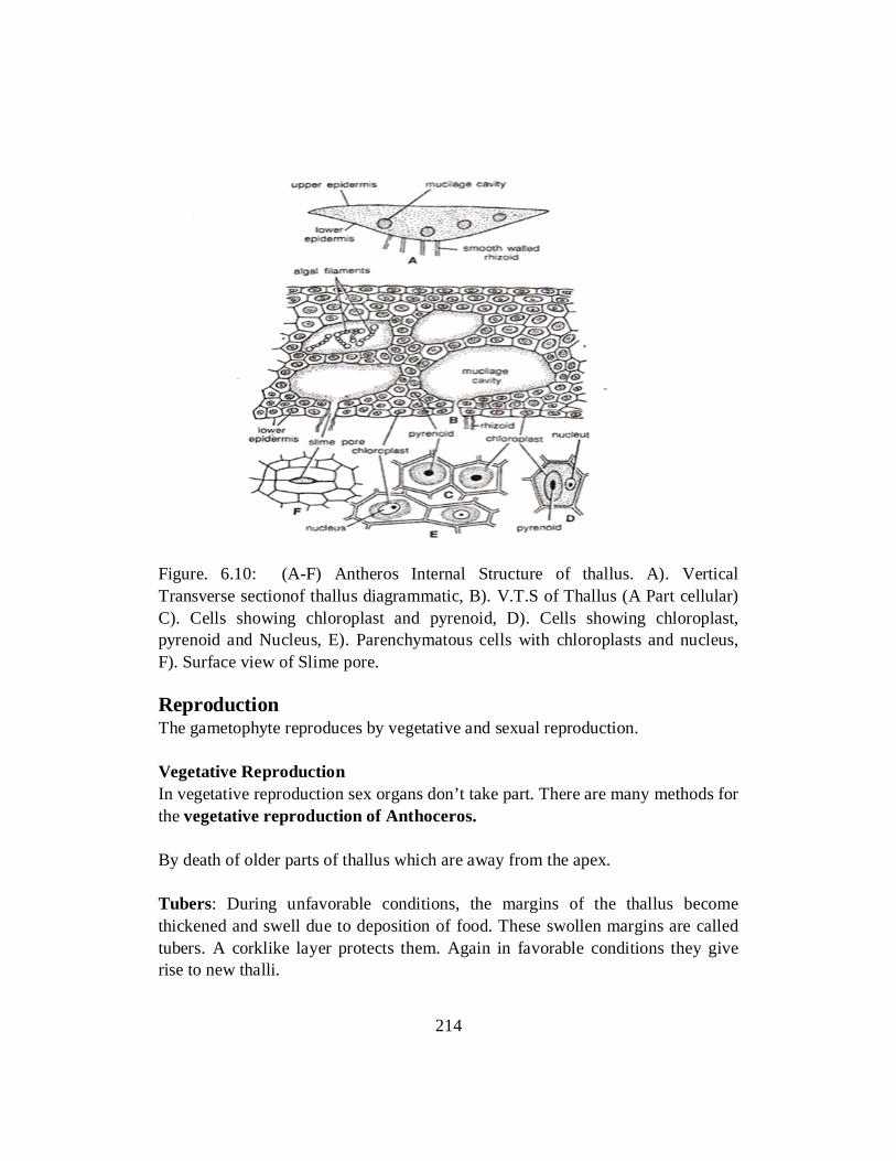
Figure. 6.10: (A-F) Antheros Internal Structure of thallus. A). Vertical Transverse sectionof thallus diagrammatic, B). V.T.S of Thallus (A Part cellular) C). Cells showing chloroplast and pyrenoid, D). Cells showing chloroplast, pyrenoid and Nucleus, E). Parenchymatous cells with chloroplasts and nucleus, F). Surface view of Slime pore. Reproduction The gametophyte reproduces by vegetative and sexual reproduction. Vegetative Reproduction In vegetative reproduction sex organs don’t take part. There are many methods for the vegetative reproduction of Anthoceros. By death of older parts of thallus which are away from the apex. Tubers: During unfavorable conditions, the margins of the thallus become thickened and swell due to deposition of food. These swollen margins are called tubers. A corklike layer protects them. Again in favorable conditions they give rise to new thalli.
214

Persistent Apices. In some species, the thalli die due during hot summer except for their apices. These are called persistent apices. These persistent apices remain buried in the soil and develop into new plants on return of favorable conditions for growth. Gemmae formation. Some species grow vegetatively by formation of gemmae produced on short stalks at the dorsal surface of the thallus. Sexual Reproduction Most of the anthoceros species are homothallic. Antheridia are male sex organs and archegonia are female sex organs. The antheridia develop first as compared to archegonia. The sex organs, i.e. antheridia and archegonia are found embedded in the tissue of the dorsal side of the thallus. Structure of Antheridia The mature antheridium is club shaped and stalked. The antheridium is properly covered by a jacket of single layered cells. Inside the jacket, there are many androcytes which become antherozoids. The stalk of the antheridium may consist of many cells, e.g., Anthoceros laevis, or it may consist of the four rows of the cells, as in Anthroceros erectus and anthoceros punctatus. When antheridium matures, the roof of the antheridial chamber disintegrates, as a result the antheridia are exposed to outside. Soon after, the antheridium absorbs water and burst at the apical ends to release antherozoids outside. The antherozoid is spindle like and biciliate. The cilia are attached at the anterior side. The antherozoids swim in water with the help of cilia like flagella. Development of Antheridium The antheridia develop in a cavity called antheridial chamber. Antheridial chamber is present on the dorsal surface of the thallus. The development is endogenous. One to twenty five antheridia may develop in each antheridial chamber. A dorsal superficial cell situated near the apex divides into two daughter cells. The upper daughter cell acts as roof initial and the lower one as antheridial initial. A mucilage filled space appears between roof initial and antheridial initial. This
215

mucilage space enlarges in size and becomes an antheridial chamber. Roof initial is not involved in the development of antheridium.
Fig.6.11: Antheros sp.Male Sex organs .A & B stalked mature antheridia of ACrispulusand A.Laevis repectively. C). Young and mature antheridia inside the antheridial chamber, secondary budded antheridia are seen on the stalks of primary antheridia; D). biflaggelate sperm or antherozoid. The roof initial divides many times and form antheridial chamber. In the same time the antheridial initial develops into a single antheridium or a group of antheridia. The species single antheridium develops in species such as Antheros pearsoni and sometimes Antoceros himalayensis. According to Mehra and Handoo (1953), in Anthoceros erectus a number of antheridia develop in an antheridial chamber. The antheridial initial divides many times producing many cells and each cell develops into an antheridium. The antheridial initial divides twice by vertical walls at right angles to each other and produce four cells. These cells undergo transverse divisions to form two tiers of four cells each. The four cells of the upper tier divide transversely giving rise to eight cells. This is called octal stage. All o9f the cells of octant stage divide periclinally giving rise to eight outer primary jacket cells and eight inner primary
216

androgonial cells. The four cells of the lower tier develop into a stalk of the antheridium consisting of the four rows of cells. The androgonial cells divide repeatedly and form androcytes mother cells. Each androcyte mother cell divides diagonally into two androcytes. Each androcyte forms an anthrozoid (spindle like biciliate structure)
Fig.6.12: (A-N) Antheros (A-H) Successive stages in the development of antheridium, I). Mature antheridium of A.Punctatus, J). Mature antheridium of A.Laevis, (K,L). Antheros of A. Laevis, (M,N) Antherozidsof puinctatus> Dehiscence of Antheridium: Water helps in the dehiscence of the antheridium. As the antheridia mature the roof of the antheridial chamber breaks down irregular, exposing the antheridia in a cup like chamber. The antheridia absorb water and the uppermost tier of triangular cells fall apart releasing a mass of antherozoids. After dehiscence the antheridium loses turgor and collapses. It is followed by another antheridia to converge towards the opening in the roof and in this way a
217

continuous stream of antherozoids is possible. It explains the formation of large number of sporophytes Hornworts. (Fig. 6.13).
Fig.6.13: Antheros Dehiscence of anthridium
Antherozoid The antherozoids may be spindle shaped, linear or curved as a loop. They are usually broader at one end and narrow at the other end. Narrow end have two long flagella. The broad end of the antherozoid is perhaps due to the adherent remains of the cytoplasm of the androcytes. Antherozoids swim in the water by lashing movement of their flagella. Structure of Archegonium Archegonium of Anthoceros consists of two to four covered cells, an axial row of 4-6 neck canal cells, a ventral canal cell and an egg. There is no jacket layer around the archegonium. Structure of Archegonium Archegonia develop in the flesh of the thallus on dorsal surface. The place of an archegonium on a thallus can be identified by the presence of a mucilage mound (Fig.)
218

Figure. 6.14: Antheros. Mature archegonium with mucilage mound. A mature archegonium consists of two to four cover cells, an axial row of four to six neck canal cells, a venter canal cell and an egg. The jacket layer is not distinct from the other vegetative cells like other Bryophytes. The archegonia develop embedded in the thallus are in direct contact with vegetative cells. There is no jacket layer around the archegonium. Only the neck is covered with cover cells. Each archegonium consists of an elongated neck containing an axial row of 4-6 neck canal cells; and a basal swollen portion, the venter that contains a ventral canal cell and an egg. The archegonium of anthoceros resembles archegonium of some pteridophytes. Development of Archegonium: The development of the archegonium starts on the dorsal surface of the thallus from a single superficial cell which acts as an archegonial initial (Fig. 6.15 A). It can be differentiated from other cells by its dense cytoplasmic content and a prominent nucleus. The archegonial initial may divide by transverse division to form an upper primary archegonial cell and lower primary stalk cell (e.g., A. crispulus, A. gemmulosus) or it may directly functions as primary archegonial cell (e.g., A. erectus). The primary archegonial cell divides by three’ successive intersecting walls or periclinal vertical walls to form the three peripheral initials or jacket initials and a fourth median cell, the primary axial cell (Fig. 6.15 B, C). Jacket initials divide by transverse divisions to form into two tiers of three cells each. The cells of the upper tier divide by anticlinal division to form six cells. These cells divide transversely to form a jacket of six rows of sterile neck cells. The three cells of the lower tier divide by transverse and vertical divisions to form venter
219

wall. Since the archegonium is embedded in the thallus, it is difficult to trace the development of the cells and to distinguish them from the vegetative cells (Fig. 6). The primary axial cells divide by a transverse division to form an outer cell and inner (central) cell (Fig. 6 D). The outer cell divides by a transverse division to form terminal cover initial and inner primary neck canal cell (Fig. 6.15 E). The inner cell directly functions as primary venter cell and divide only once to form upper small venter canal cell and a lower large egg. Primary neck canal cell divides by series of transverse divisions to form four to six neck canal cells. Cover initial divided by one two vertical division to form two to four rosette like cover cell at the tip of the neck (fig 6.15 G, H)
Fig.6.15: Antheros. Development of Archegonium. A).Archegonial initial, B). Differentiation of primary axial sell in L.S; D).Division of axial cell into inner and outer cells; E). Development of cover initial, primary neck canal cell and primary ventral cell; F). antheridium with egg, ventral canal cell, two canal cells and cover cells; G). Nearly mature archegonium; H). mature archegonium
220

Fertilization At the time of fertilization neck canal cells and ventral canal cells disintegrate. The cover cells are thrown off. Neck canal and ventral canal cells are converted into a mucilaginous substance. The egg usually enlarges and fills Venter cavity. Anthropoids, through watery medium, reach the mucilaginous mound where they are caught. At the opening of the archegonium the mucilaginous substance is rich with some chemicals which attract the anthroizoids. Anthropoids travel from neck to the egg. One of these anthropoids fuses with the egg and form the zygote. The zygote secretes a wall and starts dividing to form embryo. At the same time the cells of archegonium and thallus tissues start dividing to form a protective sheath called calyptera around the developing embryo. Asexual Reproduction In asexual reproduction of Anthoceros Spores are formed. Spores are produced in a structure called sporangium. This generation is called sporophyte generation. Structure of Sporangium The sporophyte or sporogonium of Anthoceros is unique as it has some features, which is different from other liverwort. The sporangium developed on the gametophyte, but not fully dependent on the gametophyte. Mostly a number of cylindrical and straight sporangia are produced on a rosette. When young they are green. On maturity gradual blackening or deep yellow coloration starts from top to downwards. Sporangia looks like horns, hence the plants are called “hornworsts”.
Fig.6.16: Mature sporophyte of Anthoceros: A.L.S(Semidiagramatic). Different portions are shown magnified on the right hand side. B). T.S at the middle of the sporophyte .C). A stoimata in the jacket epidermis. D). Ruptured tip of a mature sporophyte capsule.
221

The sporangium consists of following parts. The Foot: The foot is basal, somewhat expanded structure made up of parenchymatous cells. At the base there are rhizoid like structures. They increase the absorptive surface of the foot. The foot anchors the plant body, absorbs water and nutritive particles from the neighboring gametophytic tissues. The intermediate zone (Seta) This lies just above the foot. Being meristematic, it continuously adds news cells to the tissues of the capsule. There is no distinct stalk or seta in Anthoceros. The Capsule The capsule, a major part of the sporophyte, is a upright cylindrical structure. This consists of following parts. i. Capsule Wall (Epidermis) Wall of the capsule consists of parenchyma cells arranged in 4-6 layers.
Cells of the outermost layer forms epidermis with cutinised cells. In the epidermis there are pores called stomata. Each stomata has two guard cells. Each cell in the outer wall contains two chloroplasts. So wall of the sporophyte isphotosynthetic, which makes partially dependent of the gametophyte except for the water and mineral nutrients.
ii. Sporogenious Tissues Just beneath the capsule wall lies a cylinder of sporogenious tissues that
extend s over the top of the columella like a dome. These tissues produce spores, elater mother cells which can be seen in one and the same capsule from the base upwards.
In most of the species of Anthoceros, the elater appears as a branched or unbranched row of elongated cells. These chains may be 1-4 celled but in some cases the elaters may be single celled. The elators lack regular spiral thickenings. Their walls may be thick with an extremely reduced lumen. They are of no regular shape.
iii. Columella It is a solid column of sterile tissues occupying the Centre of the capsule. It
starts from the base and extends nearly to the tip of the capsule. Thecells of columella are narrow and elongated with their walls more or less uniformly thickened. They are arranged in 16 vertical rows. In a young capsule the
222

cells are arranged in 4 vertical rows only. It helps the spores to disperse and is probably associated with the conduction of water.
Dehiscence of the Capsule Soon after the capsule matures, its tip becomes dark brown or black in color. This is because of the maturity of underlying spores. The color variation of capsule depends upon species. Loss of water from the cell walls or capsule, spores, elaters and columella helps the dehiscence. The maturation and dehiscence of the capsule starts basipetally, i.e. from the tip downwards. At first small longitudinal slit develops at the dried and shrinked tip of the capsule along one line of the dehiscence. This slit extends downwards and then along the other line of dehiscence situated opposite the first. Two valves thus formed are still attached at the tip of the capsule. A mass of elators and spores and a part of columella get exposed. The valves extend downward and expose the spores and elators of the underlying capsule. Progressive upward separation of the valves on reaching the tip may result the valve to become strap- shaped and later spirally coiled. The spores are thrown away for some distance with little force or carried away to a long distance by air current. The young gametophyte The spore The mature spores are tetrahedral and thick walled. Each spore contains a single nucleus, a plastid with proto- chlorophyll, oil globules and other reserve food materials. Germination of the Spores The spores may germinate immediately (Mehra and Kachroo, 1962) or after undergoing a period of rest. They absorb water and swell. The exospores raptures along the triradiate mark and the endospore protrudes through a cleft in the form of a short or long germ tube. The germ tube contains a single chloroplast, oil globules and protein granules. The exospores may still remain intact at the base of the sporeling. Two successive transverse walls are laid down near the tip and a three celled filament is formed. Two terminal cells of this filamentous sporeling divide by two vertical walls at right angles to each other and produce an octant. The further growth of this sporeling is initiated by four terminal cells of the octant. These cells constitute the growing region in the form of a marginal meristem and bring about the further growth of the sporeling and form the new thallus.
223
Self Assessment Exercise 6.4 Q.1 Circle the correct option i. The genus of Antheros is: a) Bryophyta b) Anthropsida c) Anthocerotales d) Anthoceros ii. Anthoceros isgametophyte which is small and have colour a) darkgreen or yellow b) red or pink c) black or brown d) Blue or white iii. The thallus of Antheros consists of ---tissues mainly. a) Collenchyma b) Parenchyma c) Phloem d) Tough and hard iv. Antherozoids swim with the help of…. a) Flagellum b) Podia c) Cilia like flagellum d) Non of these v. Archegonia in Anthoceros develop in flesh of thallus on----side. a) Ventral side b) dosal side c) Lateral side d) Anterior side Q.2 Write classification of Antheros Q.3 Write External morphology of Antheros Q.3 Describe internal structure of thallus of Anthoceros Q.4 Give detailed information about structure of Sporongium in Antheros. Funaria Classification Division: Bryophyta Class: Bryopsida Sub-class: Bryidae Order: Funariales Family: Funariaceae Genus: Funaria Occurrence It is a common moss. It grows both in tropical and temperate regions of the world. It includes 117 species. Plants are mostly found in moist shady areas and grow in
224

patches on damp soils, moist rocks and burnt humid ground rich in plants ashes, damp and rarely on the tree trunks. External form and Morphology The adult gametophyte plants are short about 1-3 cm. high, green branched. The plant body consists of a small erect axis (Stem) surrounded by spirally arranged leaves and numerous rhizoids at its base. Rhizoids are branched, slender and multicellular structures. The rhizoids grow vertically downwards deep in substratum and form a tangled mass. When young, they are colorless but in later stage become brown or red. The main axis of plant is slender, upright and branched. Branches arise from the lower part of the plant. They become erect and vertical similar to main axis. The arrangement of leaves on the stem is spiral. The leaves are flat, simple, bright green,membranous, somewhat ovate and sessile. Each leave has a well-defined midrib. The lower leaves, not regular in shape, are smaller and scattered while the upper leaves are larger and crowded at the apex of the axis. The stem leaves and rhizoids are different to that of higher vascular plants. However they are analogous.
Fig. 6.17: External Morphology of Funaria Internal Morphology Internally the axis (stem) is differentiated into epidermis, cortex, and central cylinder. Epidermis is single celled thick outer layer and consists of thick walled cells. The cells contain chloroplasts but no stomata. Just beneath the epidermis, a
225

multilayered cortex is situated. Chloroplast becomes extinct in older stem. The cells of outer part of cortex are thick while inner part is thin. Colourless cells which lack protoplasm form a central core of the axis, called central cylinder. It provides mechanical strength and help in conduction of water and other salts. For further elaboration of internal structure please study the figure 6.18 below.
Fig.6.18: Internal morphology of Funaria: A. L.S of entire capsule, B.L.S through annulus region, C. Structure of stomata, D. Stomata on surface view. Leaf of the funaria is single layered except the portion of midrib. Midrib consists of several layers of cells. Midrib is also rich in chloroplast. The center of the midrib is analogous to that of central core of the stem.
226

Fig: 6.19: Internal structure of leaf in Funaria Life Cycle Funaria reproduce both by vegetative and sexual modes. Vegetative reproduction The primary protoplasm developed as a result of the spore germination multipliers vegetatively either by breaking up of the branches or small terminal groups of cells, the buds. Each such branch or the group of cell becomes separated by certain intercalary colorless cells and develops into a new plant. The protonema develops the parts of the plant other than spores called secondary protonema. Secondary protonema may be formed from any of the detached living part of the leafy gametophyte, such as from the axis, leaves, paraphysis and sex organs. This detached secondary protonema forms the new plants. In some cases, terminal cells of the protonemal branches divide by longitudinal oblique or transverse walls to produce a small multicellular structure, the gemmae.Fig. The cells of gemma are thin walled and contain abundant chloroplasts. Sometimes tubers also arise from the axis. The gemmae or tuburs develop into new plants under favorable conditions. After the death and decay of the prostrate branches of the adult gametophyte and the rhizoids, each erect branch behaves like an independent plant.
227

Fig.6.20: Life cycle of Funaria Hygrometrica
Sexual Reproduction The plants are monoecious and potandrous, i.e. antheridia and archegonia occur on different branches of the same plant. The sex organs are born e in terminal groups on the apices of fertile branches, the gametophores. The Antheridium The main axis of the gametophyte plant bears male sex organs, the antheridia. The antheridia are erect and occur in groups. They are surrounded by a large number of closely set perigonial leaves spreading out like a rosette of petals and a cup that protects the antheridia.Fig.A number of reddish brown or orange antheridia in their various stages of development give the male shoot a distinct color. The male shoot is relatively small, about 1cm.high and is easily recognizable due to its distinct shape and color. A large number of sterile structures lie within the antheridia. They are green and occur in clusters. Paraphysis plays the role of photosynthetic role. They also protect the developing antheridia. They also protect the young antheridia from dryness.
228

Development of the Antheridium At the apex of the male shoot asingle superficial cell becomes papillate and acts as antheridial initial. Antheridial initial divides by transverse wall into upper and lower cells. The upper cell forms the antheridial proper and the lower cell forms the embedded portion of the stalk. The upper cell further divides by successive transverse divisions to form a linear filament of 4-5 cells. Fig. 6.21 The 5-7 cells formation and their arrangement into two rows give the appearance of multicellular structures. Fig.6.21 the lower most segments formed by the activity of the apical cell finally gives rise to the stalk.
Fig. 6.21: Development of Antheridium in Funaria An oblique vertical wall laid down in the segment 3 or 4 cells away from the apical cells forms two unequal cells. Soon after, such divisions proceed in above lying segments. Of the two daughter cells of each segment so divided, the smaller and peripheral cell function as the first jacket initial. Its larger cell undergoes another similar division and separates a second jacket initial outer side and primary androgonial cell on the inner side. Thus each cell gives rise to three cells out of which two cells are jacket initials and an inner primary androgonial cell that appears to be conical. All the jacket initials divide by anticlinical walls and
229

form a single layered wall of the antheridium. It remains as such or may divide into an outer cellwall and a lower androgonia cell. The outer segment of the apical cell or the operculum formed by the apical cell is larger than the other. The androcyte mother cells are formed by repeated division of primary androgonial cell. Each androcyte mother cell divides and forms two androcytes. Each androcyte metamorphoses into a biflagellate spermatozoid in the same manner as in the Hepaticopsida. The structure of mature antheridium A mature antheridium consists of a short multicellular stalk on which lies a club shaped antheridial body. The jacket of the body consists of a single layer of polyhedral and flattened cells. The chloroplasts in jacket cells of young antheridia become changed into orange or reddish brown chromoplasts on maturity. Androcytes present in antheridium afterwards changes into biflagellate spermatozoids. The free distal end of the antheridium bears one or two large thick walled colorless cells, the opercular cells. The paraphysis occurs intermingled with the antheridia.
Fig.6.22 : Sections of Gametophyte and Antheridium Dehiscence of Antheridium Dehiscence of mature antheridium takes place when it comes in contact with water. The opercular cell becomes mucilaginous, absorbs water and swells. It causes a pressure that ruptures its inner wall. The cell contents become free,
230

move towards the inner side and reach antheridial cavity. Soon after the disorganization of the outer wall, the opercular cell breaks its connections with the neighboring cells and a narrow pore is formed. Androcytes ooze out in the form of viscoid fluid by hydrostatic pressure within the antheridial cavity.The androcytes separated and meta-morphosed into a spermatozoid very soon asin Riccia and Anthoceros. Each spermatozoid is an elongated spirally coiled, biflagellate structure. The Archegonium Female Shoot The archegonia intermingled with paraphysis at the apex of the female shoot. The leaves that enclose the group of archegonia and sterile paraphyses are similar to the ordinary foliage leaves. These give the female shoot an appearance of sterile shoot. The female shoot emerges laterally from the base of the male shoot and goes higher up. It is usually larger than the male shoot. Development of the Archegonium Cells at the tip of the shoot become differentiated into the archegonial initial. Fig. 6.23 It divides transversally and forms an upper archegonial mother cell and a lower cell. The lower cell divides and redivides to form stalk of the archegonium. The archegonial mother cells divide by three intersecting walls forming three peripheral cells that enclose a central tetrahedral axial cell with four cutting faces. Fig.6.23 The peripheral cells divide anti-clinically and form a single layered jacket or wall of the venter. Later it becomes two layered thick. The axial cell divides by a transverse wall into an outer and a central cell. Fig. 6.23 The central cell divides and gives rise to an egg, ventral canal cell and a primary neck canal cell. The outer primary cover cell acts as an apical cell and cuts off successively three lateral segments and a basal segment. The lateral segments divide by vertical walls and form the neck of the archegonium which consists of six vertical rows of cells. The basal segment cuts off neck canal cells by transverse divisions. Thus the single layered long neck of the archegonium encloses several (4-10) neck canal cells. The neck cells do not remain arranged in straight rows but appear obliquely arranged on account of torsion caused by the elongation of the neck of the mature archegonium. Structure of archegonium The stalk of archegonium is larger than that of antheridium. Each archegonium is a flask shaped structure consisting of a basal swollen venter and a ling neck. Venter is enclosed into two layered wall whereas neck has one layered thick cells. In a fully mature archegonium ventral canal cells and neck canal cells disintegrate
231

forming a mucilaginous substance. The terminal cells (cap cells) separate apart and later are thrown off, thus a passage is left for the cavity within the archegonium.
Fig6.23: Development of Archegonium of Funaria Fertilization A pressure develops as a result of absorption of water by the mucilage filled within the archegonium; the terminal cell separates out apart and sugar containing mucilaginous substance ooze out. The antheridia are attracted chemotactically. The antherizoids reach the archegonium by the agency of water, and trickling of rain drops from male shoot to underlying female shoot. The antherizoids passing through the mucilage filled neck canal reach the egg containing venter cavity where finally one of them fuses with the egg nucleus and form zygote. The Sporophyte Development of the Sporangium The fertilized egg is known as zygote. It swells up, increases in size and secretes a wall around it. It divides transversely into an upper epi-basal and lower hypo-basal cell. Fig.
232

Self-Assessment Exercise 6.5 Q.1 Select the best option given at the end of each statement. i. The archegonium of anthoceros resembles archegonium of some ……….. a) Angiosperms b). Pteridophytes c). Gymnosperms d).All of the above ii. Androcytes present in antheridium afterwards changes into ………………..
spermatozoids a) Atrichous Flagella b) Monotrichous Flagella c) Biflagellate d) Peritrichous Flagella iii. Female shoot of an archegonium has an appearance of …… shoot a) Male b) Sterile c) Rough d) Fertile iv. The antherizoids reach the archegonium by the agency of a) Glue b) Flagella c) Sticky fluid d) Water v. The cells in Funaria contain chloroplasts but no…….. a) Plastids b) Photosynthetic pigments c) Stomata d) Photochromes vi. The height of adult funaria plant os about. a) 1-3 cm b) 3-5 cm c) 5-7 cm d) 7-9cm Q.2 Write a note on funaria? Q.3 Explain the dehiscence of Antheridium. Q.4 Explain development of antheridium and archegonium in Funaria.
233

References A TEXTBOOK OF BOTANY - ANGIOSPERMS by B.P. Pandey. S. Chand and Company Ltd., New Delhi. A TEXTBOOK OF BOTANY - by S.N. Pandey, S.P. Mishra and P.S. Trivedi (2004).Vikas Publishing House Pvt. Ltd., New Delhi. CONIFERS - THE ILLUSTRATED ENCYCLOPEDIA.Vol. I & II - by D.H. van Gelderen& J.R.P. van Hoey Smith (1996), Timber Press, Inc. Orgon, U.S.A. Crane, P. R., E. M. Friis, and K. R. Pederson."The Origin and Early Diversification of Angiosperms." Nature 374 (1995): 27–33. Judd, Walter S., Christopher S. Campbell, Elizabeth A. Kellogg, and Peter F. Stevens. Plant Systematics: A Phylogenetic Approach. Sunderland, MA: Sinauer Associates, Inc., 1999. Kenrick, Paul. "The Family Tree Flowers." Nature 402 (1999): 358–359. MORPHOLOGY AND EVOLUTION OF VASCULAR PLANTS - by E.M. Gifford, & A.S. Foster (1989) W.H. Freeman & Co., New York. MORPHOLOGY OF VASCULAR PLANTS - by D.W. Bierhorst (1971), The Macmillan Co., New York. Parkinson, C. L., K. L. Adams, and J. D. Palmer. "Multigene Analyses Identify the Three Earliest Lineages of Flowering Plants." Current Biology 9 (1999): 1485–1488. PLANT ANATOMY by A. Fahn (1990).Butterworth Heinemann Ltd. Linacre House, Jordon Hill, Oxford OX2 8DP. THE PRIVATE LIFE OF PLANTS - by David Attenborough (1995), BBC Enterprises, Ltd. Woodlands, London. Shaw, A. Jonathan; Szövényi, Péter; Shaw, Blanka (2011-03-01). "Bryophyte diversity and evolution: Windows into the early evolution of land plants". American Journal of Botany. 98 (3):352–369. doi:10.3732/ajb.1000316. ISSN 0002-9122. PMID 21613131
234

Unit 7
PTERIDOPHYTES
Writer: Dr. Muhammad Waseem
Reviewer: Dr. Sobia Mushtaq
235

CONTENTS
Introduction .................................................................................................... 237
Objectives ....................................................................................................... 237
7.1 Morphology of Pteridophytes ....................................................................... 238
7.2 Classification of Pteridophytes ..................................................................... 239
7.3 Psilopsida ( Psilotum) .................................................................................... 240
7.4 Lycopsida( Selaginella) ................................................................................. 244
7.5 Sphenopsida(Equisetum ............................................................................... 246
7.6 Pteropsida (Marsilea) ..................................................................................... 249
References ...................................................................................................... 256
236

INTRODUCTION Pteridophytes are vascular plants (plants with xylem and phloem) that reproduce and disperse with the help of spores. They consist of leaves (known as fronds), roots and sometimes true stems, and tree ferns have full trunks.They can neither produce flowers nor seeds that’s why they are referred to as cryptogams. This group includes ferns, horsetails, clubmosses; spikemosses and quillworts.There are about eleven thousand different species of pteridophytes, making them the most diverse land plants after the flowering plants (angiosperms). Pteridophytes may represent the closest living relatives to the seed plants.
OBJECTIVES The objectives of this chapter are: • To understand Pteridophytes and their morphology • To classify them into sub-divison. • To understand their divisions and compare their body structures, their mode
of reproduction and their production of spores.
237

7.1 MORPHOLOGY OF PTERIDOPHYTES Pteridophytes range greatly in size. There are tiny floating ferns used as "green fertilizer" in rice paddies.In some tropical forests, the largest plants are tree ferns that can be up to 30 meters (100 feet) tall and have huge spreading leaves up to 4.5 meters (15 feet) in length. The adult plant body in these plants is a sporophyte. It shows differentiation into true roots, stem and leaves. Stem The stem is mostly herbaceous. The branching of the stem may be of monopodial or dichotomous type.There are two main categories of form and structure, one category comprises of megaphyllous types, in which the leaves are large in relation to the stem (e.g., ferns); the second comprises of microphyllous types in which the leaves are quite small in relation to the stem (e.g., Lycopods and Horse-tails).All the vegetative parts of the sporophyte possess vascular supply. Leaves Stem appendages known as leaves take various forms that evolved independently in different groups of lower vascular plants. The simplest are scalelike emergences, orenations, found in some extinct groups and in modern whisk ferns (Psilotum). The lycophytes have scalelike, needlelike, or awl-shaped “microphylls” with a single, unbranched vein. The sphenophytes have “sphenophylls”—scalelike leaves with a single vein in the modern Equisetum or wedge-shaped leaves with a dichotomously forking vein system in many of the fossil forms. Roots Taproots are unknown in lower vascular plants. All pteridophyte roots are referred to as adventitious, in the sense that they arise at points along the stem. They entirely lack secondary growth (continued growth in thickness). The root hairs have fundamental importance in absorption of water and nutrients and in attachment of the plant to the soil or other growing surface. Self Assessment Exercise.1 Q. Fill in the blanks i. The adult plant body in Pteridophytes is ……………….. ii. In ……………….. pteridophytes the leaves are quite small in relation to the
stem (e.g., Lycopods and Horse-tails). iii. All the vegetative parts of the sporophyte of pteridophytes possess
………………..
238

iv. ……………….. have scalelike, needlelike, or awl-shaped “microphylls” with a single, unbranched vein.
v. Roots of all pteridophytes are ……………….. type. Q.2: Explain morphology of Pteridophytes. 7.2 CLASSIFICATION OF PTERIDOPHYTES Pteridophytes are subdivided into the following four Sub-Divisions. Distinguishing features of these Sub-Divisions/ Sub-Phyla are given below: Sub-Division Psilopsida 1. These are the oldest known vascular plants. Most of them have become
extinct (e.g., Rhynia, Horneophyton). Only two living species, Psilotum and Tmesipteris, are now available.
2. Plant body is very simple and does not show much differentiation. 3. Dichotomously branched rhizome takes the place of roots. 4. Stem or “axis” is aerial, but either naked or have small spirally arranged
leaves. 5. Sporangia are directly borne on the stem (i.e., cauline), either terminal or
lateral. Sub-Division — Lycopsida 1. Plant body more advanced and shows differentiation into root, stem and
leaves. 2. Leaves are microphyllous (small) having a single unbranched vein in the
midrib region. 3. Sporangia are borne in the axil of the fertile leaves (sporophylls). 4. Sporophyll form compact strobili (singular strobilus). e.g., Lycopodium,
Selaginella. Sub-Division —Sphenopsida 1. Plant body still more advanced and shows differentiation into nodes and
internodes like higher vascular plants. 2. Leaves microphyllous, and arise in whorls at each node. 3. Sporangia develop on sporangiophores which form compact cones at the
apex of fertile branches (e.g., Equisetum).
239

Sub-Division – Pteropsida (Ferns) 1. Plant body shows much advancement towards higher vascular plants, and is
well differentiated into root, stem and leaves. 2. Leaves also show great advancement, and are megaphyllous (large) and
pinnately compound. 3. Sporangia develop on the ventral surface of the sporophylls, and usually
aggregated into sori (e.g., Dryopteris, Pteris, Pteridium, Polypodium etc.) 7.3 SUB-DIVISION PSILOPSIDA “Type Psilotum” The plant body of Psilotum is a sporophyte without true leaves or roots. Basic organ is a true stem with rhizomes or underground stem. The sporophytic plant body consists of branched rhizome system and dichotomously branched, slender, upright, green aerial systems (Fig. 7.1 A) that bears small appendages and synangia (singular: synangium).
Fig: 7.1 A sporophyte plant of psilotum: A, a sporophyte plant, B. enlarged part of stem showing scaly appearance, C.A fertile twig.
240

Aerial Stem Any one of the rhizome tips may turn upward and undergo several dichotomies to give rise to a green aerial shoot (Fig. 7.1 A). The aerial shoots are slender, generally erect. They are perennial and become shrubby by repeated dichotomies and sometimes attain a height up to one meter. The basal part of the aerial axes is smooth but the distal part bears small, scaly appendages and synangia (Fig. 7.1, B, C). The aerial stems are photosynthetic and the general appearance is xerophytic although the plants grow in very moist environment. Rhizome The basal subterranean branched rhizome is generally hidden beneath the soil or humus. It bears numerous rhizoids, instead of roots, which perform the functions of absorption and anchorage. Appendages These are small scale-like structures helically arranged on the upper part of the aerial system. Reproduction in Psilotum The Psilotum reproduces vegetatively as well as by spores. i. Vegetative Reproduction The sporophyte as well as gametophyte of Psilotum (e.g., P. nudum)
propagates vegetatively through the production of Gemmae (Fig. 7.2). They are small, multicellular and ovoid structures developing on surface of rhizome (in sporophytic plant body) or prothallus (in the gametophyte).
After detachment from the parent body, gemmae of sporophyte may germinate to form a subterranean shoot, while the gemmae of prothallus, on germination, form a new prothallus.
Fig.7.2: A gemma of Psilotum
241

ii. Reproduction by Spores Spore-Producing Structure At maturity, many of the dichotomously branched aerial shoots become fertile and produce trilocular sporangia known as synangia (Fig. 7.3). The mature synangium is generally a three-lobed structure (Fig.7.3) and each lobe of the synangium corresponds to a sporangium. The synangia located at the tip of very short axis, measuring 1-2 mm in diameter and closely associated with a forked, foliar appendage. At maturity, the synan-gium exhibits loculicidal dehiscence.
Fig.7.3: Psilotun nuctum: A. part of the fertile axis bearing sporangia with bifid appendages, B. a triangular synangia showing dehiscence During the life cycle of Psilotum, a dominant sporophyte plant (1), bearing trilobed sporangia (2), produces homosporous spores (3) which germinate into haploid gametophytes (4). The underground gametophyte forms mycorrhizal associations with fungi for nutrition and develops archegonia (5), each with 1 egg, and antheridia (6) with flagellated sperm. After fertilization a diploid zygote (7) forms and eventually develops into a sporophyte plant (8).
242
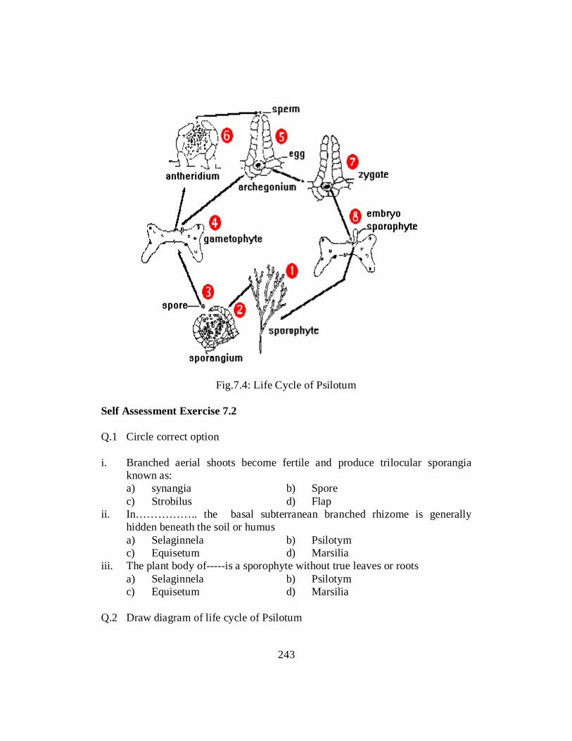
Fig.7.4: Life Cycle of Psilotum
Self Assessment Exercise 7.2 Q.1 Circle correct option i. Branched aerial shoots become fertile and produce trilocular sporangia
known as: a) synangia b) Spore c) Strobilus d) Flap ii. In…………….. the basal subterranean branched rhizome is generally
hidden beneath the soil or humus a) Selaginnela b) Psilotym c) Equisetum d) Marsilia iii. The plant body of-----is a sporophyte without true leaves or roots a) Selaginnela b) Psilotym c) Equisetum d) Marsilia Q.2 Draw diagram of life cycle of Psilotum
243
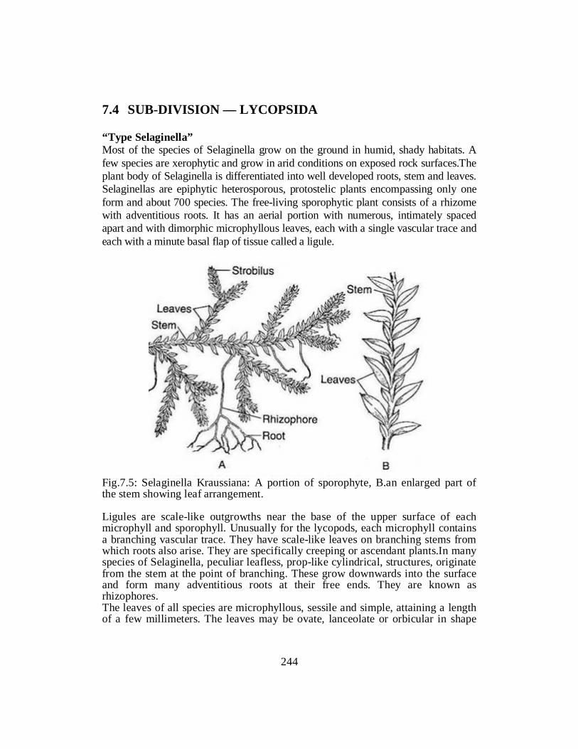
7.4 SUB-DIVISION — LYCOPSIDA “Type Selaginella” Most of the species of Selaginella grow on the ground in humid, shady habitats. A few species are xerophytic and grow in arid conditions on exposed rock surfaces.The plant body of Selaginella is differentiated into well developed roots, stem and leaves. Selaginellas are epiphytic heterosporous, protostelic plants encompassing only one form and about 700 species. The free-living sporophytic plant consists of a rhizome with adventitious roots. It has an aerial portion with numerous, intimately spaced apart and with dimorphic microphyllous leaves, each with a single vascular trace and each with a minute basal flap of tissue called a ligule.
Fig.7.5: Selaginella Kraussiana: A portion of sporophyte, B.an enlarged part of the stem showing leaf arrangement. Ligules are scale-like outgrowths near the base of the upper surface of each microphyll and sporophyll. Unusually for the lycopods, each microphyll contains a branching vascular trace. They have scale-like leaves on branching stems from which roots also arise. They are specifically creeping or ascendant plants.In many species of Selaginella, peculiar leafless, prop-like cylindrical, structures, originate from the stem at the point of branching. These grow downwards into the surface and form many adventitious roots at their free ends. They are known as rhizophores. The leaves of all species are microphyllous, sessile and simple, attaining a length of a few millimeters. The leaves may be ovate, lanceolate or orbicular in shape
244

with one vein running nearly the entire length of the leaf. All the leaves are of same type and the arrangement is spiral in subgenus Homoeophyllum. In the sub-genus Heterophyllum, the leaves are of two types; small leaves and large leaves that are arranged in four rows along the stem. There are two rows of small leaves on the dorsal side of the stem and two rows of larger leaves on the ventral side or in a lateral position (Fig. 7.5 B). Reproduction in Selaginella The sporophyte of Selaginalla reproduces by vegetative and sexual methods. i. Vegetative Reproduction The vegetative reproduction in Selaginella takes place by tubers (e.g., S.
chrysocaulis), bulbils (S. chrysorhizos), dormant buds (S. chrysocaulis) and fragmentation (S. rupestris). Bulbils and dormant buds are produced in aerial branches, while tubers may be aerial or underground. In favourable condition they germinate to produce new sporophytic plants. In S. rupestris, the trailing branches of the stem develop adventitious branches that separate from the parent plant and grow into new sporophyte.
ii. Sexual Reproduction Life Cycle of Selaginella
Fig.7.6: Life cycle of Sellaginella
245

Starting with the sporophyte (1), some branches bear at their tips aggregations of sporangium-bearing leaves, called sporophylls. They form loose, conelike structures called strobili (2). Each strobilus usually contains two kinds of sporophylls, microsporophylls bearing microsporangia and megasporophylls bearing megasporangia. The outer wall of the sporangia is a sterile jacket of cells that surrounds and protects the enclosed sporogenous tissue. In the microsporangia, the diploid sporocyte cells undergo meiosis to form numerous meiospores which in turn form haploid microspores. In the megasporangium, the entire contents of the megasporangium form one large megasporocyte that, by meiosis, produces four haploid megaspores. Upon rupture of mature microsporangia, the small, light weight microspores (3) are readily dispersed by air currents but the larger, heavier megaspores often remain in place in the strobili. Germination of a microspore produces a male gametophyte (microgametophyte) that develops totally within the confines of the microspore wall (endosporic). At maturity, the entire male gametophyte consists of a one-cell-layer-thick antheridium (4) and fertile tissue that forms biflagellated sperm. Sperm are released by rupture of the microspore wall (6). Megaspores (3) germinate to begin development of a female gametophyte (megagametophyte) (5) within the confines of the megaspore wall (endosporic). Absorptive hair like structures, rhizoids which absorb water and dissolved minerals, develop on the female gametophyte. Archegonia also develop on the exposed upper surface of the female gametophyte. Each archegonium contains a single egg, and at maturity, the neck and ventral canal cells disintegrate to form an open passageway to the egg (7). Sperm, attracted by substances released from the matured archegonlum, swim through a film of water and enter the open passageway. One sperm eventually fertilizes the egg to form a zygote (8). Development of the embryonic sporophyte occurs within the archegonium in the female gametophyte. During its early development as an embryo, the sporophyte is parasitic upon the female gametophyte. Breakdown of the parent sporophytic strobili releases the germinated megaspores (female gametophyte), and the young sporophytes soon develop a root and shoot system to become independent plants (9). 7.5 SUB-DIVISION — SPHENOPSIDA “Type Equisetum” The plant body of Equisetum has an aerial part and an underground rhizome part (Fig. 7). The rhizome is perennial, horizontal, branched and creeping in nature.
246

The aerial part is herbaceous and usually annual. Majority of the species are small with a size range in between 15 and 60 cm in height and 2.0 cm in diameter. However some species grow up in higher heights [e.g., E. giganteum (13 m), E. telmateia (2 m); E. ramosissimum (4 m), though their stem are relatively thin (0.5-2.0 cm in diam.)] showing vine-like habit and climb over adjacent forest trees.
Fig.7.7: Equisetum arvense sporophyte
The sporophytic plant body of Equisetum is differentiated into stem, roots and leaves (Fig. ).The stem of Equisetum has two parts: perennial, underground, much-branched rhizome and an erect, usually annual aerial shoot. The branching is monopodial, shoots are differentiated into nodes and internodes. In majority of the species, all the shoots are alike and chlorophyllous and some of them bear strobili at their apices (e.g., E. ramosissimum, E. debile). Sometimes shoot shows dimorphism (two types of shoots i.e., vegetative and fertile) e.g., E. arvense. Some shoots are profusely branched, green (chlorophyllous) and purely vegetative. The others are fertile, unbranched, and brownish in colour (achlorophyllous) and have terminal strobili. The underground rhizome and the aerial axis appear to be articulated or jointed due to the presence of distinct nodes and internodes. Externally, the internodes have longitudinal ridges and furrows and, internally, they are hollow, tube-like structures. The ridges of the successive internodes alternate with each other and the leaves are normally of the same number as the ridges on the stem.The primary
247
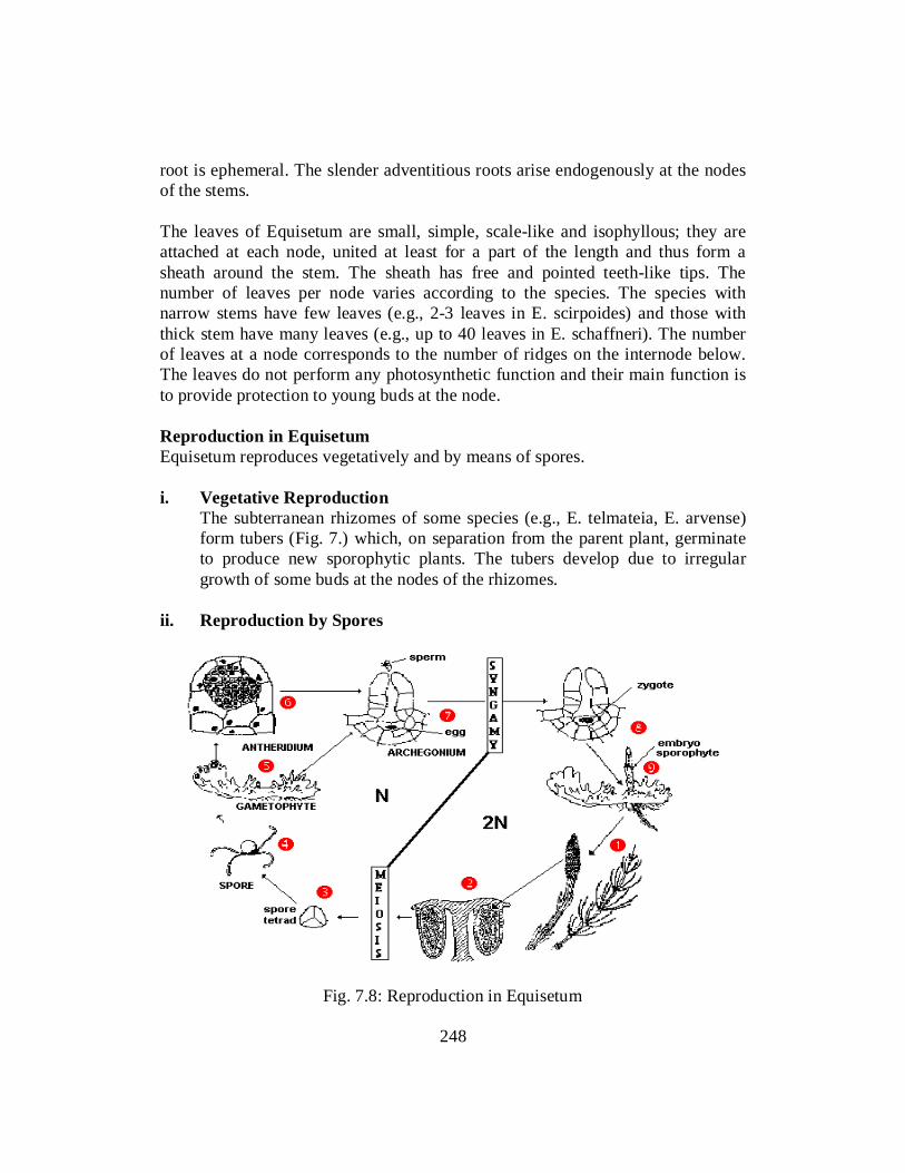
root is ephemeral. The slender adventitious roots arise endogenously at the nodes of the stems. The leaves of Equisetum are small, simple, scale-like and isophyllous; they are attached at each node, united at least for a part of the length and thus form a sheath around the stem. The sheath has free and pointed teeth-like tips. The number of leaves per node varies according to the species. The species with narrow stems have few leaves (e.g., 2-3 leaves in E. scirpoides) and those with thick stem have many leaves (e.g., up to 40 leaves in E. schaffneri). The number of leaves at a node corresponds to the number of ridges on the internode below. The leaves do not perform any photosynthetic function and their main function is to provide protection to young buds at the node. Reproduction in Equisetum Equisetum reproduces vegetatively and by means of spores. i. Vegetative Reproduction The subterranean rhizomes of some species (e.g., E. telmateia, E. arvense)
form tubers (Fig. 7.) which, on separation from the parent plant, germinate to produce new sporophytic plants. The tubers develop due to irregular growth of some buds at the nodes of the rhizomes.
ii. Reproduction by Spores
Fig. 7.8: Reproduction in Equisetum
248

The sporophyte (1) of Equisetum produces a terminal strobilus. The strobilus is composed of an axis with whorls of sporangiophores. Each sporangiophore is a stalked structure bearing a hexagonal peltate disc at its distal end On the under surface of the sporangiophore disc 5-10 elongate, cylindrical hanging sporangia are borne near the periphery in a ring (2) containing homosporous spores. The spores go through meiosis and produced spore tetrads (3). The spore tetrads mature with attached elater tissue necessary to keep the spores air borne (4). They germinate to produce inconspicuous gametophytes (5) that have antheridia (6) and archegonia (7). Spermatids/antherozoids produced in antheridium move in water to the archegonium where one of them fuses with the egg present at the base of archegonium and the zygote (8) is formed. The zygote then develops into the sporophyte of the next generation (9). 7.6 SUB-DIVISION — PTEROPSIDA “Type Marsilea” There are about 65 species of Marsilea distributed all over the world. They are hydrophytic or amphibious plants. They grow rooted in the mud of marshes and shallow pools. The species of Marsilea possess a rhizome which creeps on or just beneath the surface of the soil. The rhizome is slender, branched and possesses nodes and internodes. The leaves are borne alternately along the upper side of the rhizome usually at the nodes. One or more adventitious roots come out from each node on the underside of the rhizome. The rhizome is dichotomously branched and is capable of indefinite growth in all directions and covers an area more than twenty-five metre in diameter.
Fig.7.9: Marsilea quadrifolia: a sporophyte with sporocarp. Rhizome, leaves and roots
249

The leaves are borne alternately along the upper side of the rhizome at the nodes. The leaves possess circinate vernation typical of most Filicales (ferns). The young parts of the leaves of M. minuta and others are covered with numerous multi-cellular hairs. The leaves of submerged plants possess long flexible petioles and leaf lamina that float on the water surface. When the plants grow on mud or marshy places the leaves get shorter and the petioles become erect and stouter which spread the leaves in the air. The leaves are compound. The lamina of each leaf is divided into four leaflets pinnae arising from the apex of the petiole. Division of a leaf-blade into four leaflets results from two dichotomies arising in close succession to each other. The veins of each pinna are dichotomously branched and have numerous cross connections resulting in close reticulum of veins. One or more adventitious roots are borne at each node on the underside of the rhizome. The adventitious roots may arise even from internodes, e.g., in M. aegyptiaca. In M. minuta the roots may develop laterally. Reproduction The reproduction in Marsilea takes place by vegetative and sexual methods. Vegetative propagation The vegetative reproduction takes place by means of specially developed structures known as tubers. In the dry conditions certain branches develop from the rhizome, which swell because of the storage of food material, and are termed as tubers. These tubers survive in the unfavourable and drought conditions, and on the return of favourable conditions they germinate into new plants, e.g., M. hirsuta. Sexual reproduction The plant is a sporophyte. It bears the special structures known as sporocarps which contain micro and megasporangia in them. It is heterosporous. The sporocarps are borne on short peduncles above the base of the petiole. In most cases the peduncle is unbranched and bears a single sporocarp at its apex. In M. quadrifolia the peduncle is dichotomously branched and bears 2 to 5 sporocarps. The sporocarps may be oval or bean-shaped. In the earlier stages of its development it is soft and green but later on it becomes sufficiently hard and brown in colour.
250

Fig.7.10: life cycle of Marsilia The outer wall of the sporocarp is extremely hard and strongly resistant to mechanical injury and drying out. In natural conditions the sporocarp may burst even two or three years later its formation. The spores within the sporocarp may remain viable 20 to 30 years and sometimes upto 50 years. The sporocarp may easily be germinated by injuring it on the ventral median line and keeping it in the water. The water enters to the interior of the sporocarp. Within half an hour the gelatinous ring imbibes water and becomes swollen with the result the sporocarp bursts into two valves from its ventral margin and the spores are liberated from sporangium present in the sporocarp. The germinating spores and the developing gametophytes remain embedded in the gelatinous matrix upto their maturity. There are two kinds of spores found in micro and megasporangia, known as microspores and megaspores. In microsporangium generally all young spores mature so that each microsporangium contains 32 or 64 microspores. The
251

microspore develops into a fully mature microgametophyte. The primary androgonial cell in the microgametophyte divides four times in a series giving rise to 16 androcytes. The androcytes are metamorphosed into antherozoids. Shortly before the antherozoids are mature, the spore wall bursts and the antherozoids are liberated in the water. In megasporangium only one of the spores matures which becomes quite large in size and thick walled. The megaspore begins to germinate immediately after its formation to produce megagametophyte A mature megagametophyte is surrounded by a broad, ovoid gelatinous zone which there in a watery funnel shaped opening directly upon the archegonium. Many antherozoids are attracted towards the funnel shaped structure of megagametophyte. The antherozoids enter gelatinous envelope and reach to the archegonium. Only one antherozoid enters the egg’s cytoplasm anteriorly, but later on its orientation becomes reversed and it penetrates the egg nucleus posterior end forward to accomplish fertilization. The zygote thus formed divides first by a vertical division. Thereafter the two daughter cells divide vertically in a plane perpendicular to that of first division. Thus, a four-celled embryo is formed. Two sister cells of outer side develop into cotyledon and stem and the other two of inner side develop into the foot and root of the embryo.
Fig: 7.11. Life History of Marsillea
252

The water fern, Marsilea, looks like a four-leaf clover, but circinate vernation gives it away as a fern. It is rhizomatous from which leaves emerge at the nodes. The the base of some of the leaves, a sporocarp (a hardened folded leaf with sporangia inside) develops (Top a&b). The sporocarp develops as a gelatinous ring which allows the sori to emerge into the water (Bottom a&b). These are heterosporous. Microspores develop into multiflagellate sperm and the megaspores develop into a megagametophyte which does not exceed the bounds of the spore wall. The daughter sporophyte grows from the zygote in the archegonium in the megaspore and appears almost like a germinating seed.
Fig.7.12:
Self Assessment Exercise 7.4 Q.1 Circle the best possible correct answer. 1. The vascular tissue is confined to the central region of the stem forming: (a) Bundles (b) stele (c) Cortex (d) Pcricycle 2. The leaves which bear the sporangia are called: (a) sporophyl Is (b) Bract (c) Cone (d) Strobilus 3. One or two peripheral layers persist for the nourishment of the developing
spores. These nourishing cells form: (a) Elators (b) Sopores (c) Jacket (d) tapetum. 4. Match gametophyte with one of the followings: (a) Prothellus (b) Thallus (c) Cone (d) Strobilus
253

5. Selaginella belongs to division (a) Lycopsida (b) Pteropsida (c) Psilopsida (d) Sphenopsida 6. Hone tails are: (a) Lycopsida (b) Pteropsida (c) Psilopsida (d) Sphenopsida 7. Marsilea belongs: (a) Lycopsida (b) Ptcropsida (c) Psilopsida (d) Sphenopsida 8. Which of the followings is fern? (a) Psilopsida (b) Pteropsida (c) Lycopsida (d) Sphenopsida 9. Which of the followings is most primitive division? (a) Lycopsida (b) Pteropsida (c) Psilopsida (d) Sphenopsida 10. Club mosses are: (a) Lycopsida (b) Pteropsida (c) Psilopsida (d) Sphenopsida 11. The protostele in which xylem core is Smooth and rounded is: (a) Haplostele (b) Actinostlele (c) Plectostele (d) Siphonostele 12. The protostele in which xylem core is star like is called: (a) Haplostele (b) Actinostlele (c) Plectostele (d) Siphonostele 13. The siphonostele in–which two cylinders of vascular tissue are present in the
stele is: (a) Haplostele (b) Actinostlele (c) Plectostele (d) Polycyclic 14. In Xylem in which protoxylem is lying in the middle of Metaxylem is: (a) Exarcsh (b) Mesarch (c) Endarch (d) Diarch 15. The stele in which xylem forms several plates is: (a) Haplostele (b) Actinostlele (c) Plectostele (d) Polycyclic 16. The xylem in which protoxylem is lying on the periphery of metaxylem is: (a) Exarch (b) Mesarch (c) Endarclt (d) Diarch 17. The triad of sporangia is called: (a) hirers (b) synangium (c) Jacket (d) tapetum
254

18. The primary androgonial cell divides to produces a mass of: (a) Androgonial (b) androcytes (c) Antherozold (d) None 19. Match ligule with one of the followings: (a) Root (b) Leaf (c) stem (d) Rhizome 20. Many sporophylls form (a) Cones (b) Spores (c) Sporangia (d) None 21. The innermost layer of sporangium of selaginella is: (a) Elators (b) Synangium (c) Jacket (d) tapetum 22. Primary ventral cell is formed from: (a) cover cell (b) cap cell (c) central cell (d) axial cell 23. The primary ventral cell gives rise to: (a) Jacket (b) Tapetum (c) Oosphere (d) oospore 24. The Oospore divides into two cells. The upper cell enlarges, it is called: (a) Suspensor (b) Cotyledon (c) Embryonal (d) Protonema 25. Mature vascular bundles have a canal called: (a) carinal (b) vellular (c) central (d) none 26. The stele present in equisetum is: (a) haplostele (b) Actinostele (c) Plectostels (d) Siphonostels 27. The outermost layer epispope splits to form four bands. These bands
separate from the spore on drying. The bands are called: (a) Jacket (b) Tapetum (c) Oosphere (d) Elators 28. Sporocarp is formed in: (a) Adiantum (b) Marsillea (c) Equisetum (d) Polypodium 29. In hard fruit body in Marsillea the daughter cells produced are called: (a) Sporangia (b) Sporophylls (c) Ovule (d) Sporocarps 30. In the megasporangium only one spore develops further. All other
disintegrate forming a mucilaginous mass or: (a) Jacket (b) tapetum (c) Plasmodium (d) Elators
255

31. Number of layers in the Tapetum of Polypodium are: (a) one (b) two (c) three (d) four Q.1 Write brief Life History of Marsilea. Q.2 Classify pteridophytes in sub-divisions and compare their mode of
reproduction, general body structure and spores production. Answers 1. (b) 2. (a) 3. (d) 4. (a) 5. (a) 6:(d) 7. (b) 8. (b) 9. (c) 10. (a) 11. (a) 12. (b) 13. (c) 14. (d) 15. (b) 16. (a) 17. (b) 18. (b) 19. (b) 20. (a) 21. (d) 22. (c) 23. (c) 24. (a) 25. (a) 26. (d) 27. (d) 28. (b) 29. (d) 30. (c) 31. (b) Referemces Lee, R.E. 1999. Phycology .Cambridge University Press, UK Prescott, L.M., Harley, J.P. and Klein, A.D. 2004. Microbiology, 3
rd ed. WM. C. Brown Publishers. Alexopoulos, C.J., Mims, C.W. and Blackwell, M. 1996. Introductory Mycology.4
th ed. John Wiley and Sons Publishers. Agrios, G.N. 2004. Plant pathology.8
th ed. Academic press London. Vashishta, B.R. 1991. Botany for degree students (all volumes). S. Chand and Company. Ltd.New Delhi. Mauseth, J.D. 2003. Botany: An Introduction to Plant Biology 3rd ed., Jones and Bartlett Pub. UK Marti.J.Ingrouille & Plant: Diversity and Evolution. 2006 CUP Kenrick, Paul; Crane, Peter R. (4 September 1997). "The origin and early evolution of plants on land" (PDF). Nature. 389 (6646): 33–39. doi:10.1038/37918
256

Unit 8
GYMNOSPERMS
Writer: Tauseef Anwar Reviewer: Dr. Muhammad Sami Ullah
257

CONTENTS
Introduction .................................................................................................... 259
Objectives ....................................................................................................... 259
8.1 General Characteristics of Gymnosperms .................................................... 260
8.2 Life Cycle of the Following Specific Examples .......................................... 264
8.2.1 Cucis ...................................................................................................... 264
8.2.2 Pinus ...................................................................................................... 275
8.2.3 Ephedra ................................................................................................. 288
References ...................................................................................................... 298
258

INTRODUCTION The gymnosperms were the first seed plants and bear their ovules and seeds exposed on the plant surface. This is in marked contrast to the more recent group of seed plants, the flowering plants, which have their ovules sealed within the flower and their seeds contained within a fruit. The name gymnosperm describes this feature. It literally means naked seed. The gymnosperms include the oldest and largest trees known. The Bristle Cone Pines, some over 4000 years old are the oldest living plants. The Giant Redwoods are over 100 m tall - the tallest plants known. Both are native to California.
OBJECTIVES After completion of this unit the you will be able to: • Define gymnosperms • Describe characteruistics of gymnosperms • Describe life cycles of cucus, pinus and ephedra
259

8.1 GENERAL CHARACTERUSTICS OF GYMNOSPERMS 1. The plants have a long lasting tap root system. The vascular cylinder is di-
to polyarch, xylem exarch. Main elements of xylem are tracheids. Phloem is composed of sieve cells and lacks companion cells. Some forms exhibit additional symbiotic relationship between roots and algae in coralloid roots (Cycas) and between roots and fungi in mycorrhizic roots (Pinus).
2. The sporophyte shows unlimited growth of aerial trunk by means of apical and lateral meristem which produces secondary vascular tissues.
3. The aerial trunk is branched or unbranched and woody. Majority of gymnosperms have branched stem (except in Cycas and Zamia). In Pinus branches are of two types :- (a) long shoots or branches of unlimited growth (b) dwarf shoots or branches of limited growth bearing clusters of variable number of needle shaped leaves and collectively called as spur. Vascular bundle of stems are collateral, endarch or mesarch, open and arranged in a ring.
4. Secondary growth very pronounced. Secondary vascular tissues consisting mainly of tracheids and sieve cells. Normally xylem lacks vessel elements and the phloem lacks companion cells. But both vessels and companion cells present in members of Gnetales. Mature metaxylem and secondary xylem elements have bordered pits of various types. Wood or secondary xylem of two types namely, manoxylic and pycnoxylic. In manoxylic type (as in Cycas) wood is less, porous and soft. There is a large cortex and pith and parenchymatous rays are wide. In pycnoxylic type (as in Pinus) wood is dense or compact with small xylem rays and reduced pith and cortex.
5. Leaves are diverse in form and arrangement. They are both simple and compound ranging in size from a minute scale to leaves a few meters long. Arrangements of leaves usually spiral, they may also be whorled as in Cedrus or opposite decussate as in Cupressaceae, Welwitshchia andGnetum. Scale leaves are microphyllous whereas larger leaves are megaphyllous and their vascular supply always leaves a leaf gap in the stem stele. Venation may be parallel (Agathis and Welwitschia), reticulate (Gnetum) or dichotomous (Ginkgo) or as in most genera there may be a single vein. Leaves may be dorsiventral or isobilateral, amphistomatic or hypostomatic with sunken stomata. Mesophyll may be differentiated into spongy and palisade parenchyma (Cycas, Gnetum) or undifferentiated (Pinus). Transfusion tissue is a prominent feature. Leaf surface protected by a thick cuticle and sometimes by an additional waxy layer. Resin canals occur in all conifers (except Taxus) and mucilage ducts in cycads and Ginkgo and latex tubes in Gnetum. Vasculature of petiole quite variable. In Cycas, vascular bundles are arranged in a horse-shoe shaped manner or look like an inverted omega (U). They are diploxylic having both centripetal and
260

centrifugal xylem. In Ginkgo petiole receives two vascular bundles and each half of lamina is supplied by one bundle which later dichotomizes. In Gnetum, the petiole shows an arc-shaped arrangement.
6. Vegetative methods of reproduction are rare in gymnosperms but vegetatively propagating bulbils are known in Cycas.
7. Plants are heterosporous. They may be monoecious (Pinus) or dioecious (Cycas). Reproductive structures are borne in cones or strobili that are either staminate (male) or ovulate (female) except in Cycas where ovules are borne on loose megasporophylls. Sporangia are borne on fertile leaves or leaf-like structures called microsporophylls (in male cone) and megasporophylla (in female cones) which are arranged spirally around a central axis.
8. Microsporangia are borne on abaxial or lower surface of microsporophylls. They may be numerous and grouped in sori (Cycas) or reduced to two (Pinus). Microspores are produced in tetrads.
9. Megasporangium or ovule is borne on adaxial or upper surface of megasporophyll or ovuliferous scale and are generally orthotropous. Young megasporangium consists of a nucellus which is surrounded by a sheath or integument and inside the nucellus is single functional megaspore. A narrow passage above the nucellus in the integument is the micropyle.
10. Microspore and megaspore germination in situ, producing micro-and megagametophytes which are not autotrophic. Microspores have partly developed endosporic male gametophytes when they are transferred to micropyle of ovule. The microspore or pollen grain at the time of shedding may have only one prothallial cell (Cycas) or two prothallial cells (Pinus), the former being liberated at three-celled stage and latter at four-celled stage.
11. The microspores or pollen grains are borne by wind and enter the ovule directly through the micropylar canal. The micropyle in almost all gymnosperms secretes a sugary exudate called the "pollination drop" which not only receives the pollen grains but also transports them to nucellus of ovule.
12. The development of female gametophyte is monosporic (except in Welwitschia and Gnetum where it is tetrasporic). It is permanently retained inside the megaspore wall and dependent on parent sporophyte for its nutrition. The archegonia in female gametophyte may be two (in Ginkgo) to many (as in many conifers). Archegonia are quite large and elongated and lack neck canal cells. Often the ventral canal cell too, is eliminated. Gametophytic cells around the archegonia develop into a nutritive layer or jacket. However, the nutritive layer and archegonia are absent in Welwitschia and Gnetum.
13. Fertilization is effected by means of a pollen tube (siphonogamy). The multiflagellate spermatozoids or male cells within the pollen tube have no specialized means of locomotion and occur within the ovule. The pollen
261

tube bursts liberating two large flagellated sperms into the cavity (archegonial chamber) directly above female gametophyte. One sperm fertilizes the large egg cell giving rise to a zygote (2n).
14. At the beginning of embryogeny, zygote shows free nuclear division in all except Welwitschia, Gnetum and Sequoia sempervirens. Later embryo becomes cellular after wall formation and gradually differentiates into a suspensor, shoot apex, cotyledons, hypocotyl and radicle. Polarity is endoscopic with the shoot end directed away from the micropyle. Embryo remains contained within the seed developed from the ovule. Mature embryo is differentiated into root, stem and leaves.
15. In most gymnosperms a common feature in embryogeny is polyembryony with young seeds having more than one embryo. It may be simple polyembryony when more than one archegonium is fertilized producing several zygotes or cleavage polyembryony when single zygote gives rise to multiple embryos due to cleavage or splitting of embryonal layer. The mature seed normally has only one embryo.
16. The young embryo draws its nutrition from the endosperm which develops before fertilization and is haploid (n). Endosperm develops from female gametophyte that has absorbed the food from nucellus.
17. During last phases of embryogeny, the nucellar tissue of ovule becomes disorganized and frequently persists only as a paper-like cap of dry tissue at micropylar end of the seed.
18. Gymnosperm ovules and seeds are unprotected and not surrounded by an ovary wall, hence true fruits like that of angiosperms are not formed.
19. Histological maturation of various layers of integument continues and the stony layer becomes an extremely hard, resistant shell which effectively encloses and mechanically protects the female gametophyte and the embryo.
20. The detached seeds of all gymnosperms (except for cycads and Ginkgo) remain dormant for sometime undergoing a resting period.
21. Gymnosperms being mostly temperate plants (except for cycads, Gnetum and Ephedra) growing in tropics and subtropics show very little activity in the development of reproductive structures during winter. The activity is renewed in the spring. Under favourable conditions the embryo resumes growth and after rupturing the seed coat develops into a new sporophyte plant.
22. Polyploidy is rare in gymnosperms, except in Juniperus (partly) Sequoia and Ephedra (partly) where gametic number is multiple of the basic number of the families. Sequoia is the solitary gymnosperm which is hexaploid. Ephedra is the only genus where polyploidy is common.
Although angiosperms and gymnosperms are both seed bearing plants, the latter are considered to be less advanced than angiosperms because their ovules and
262
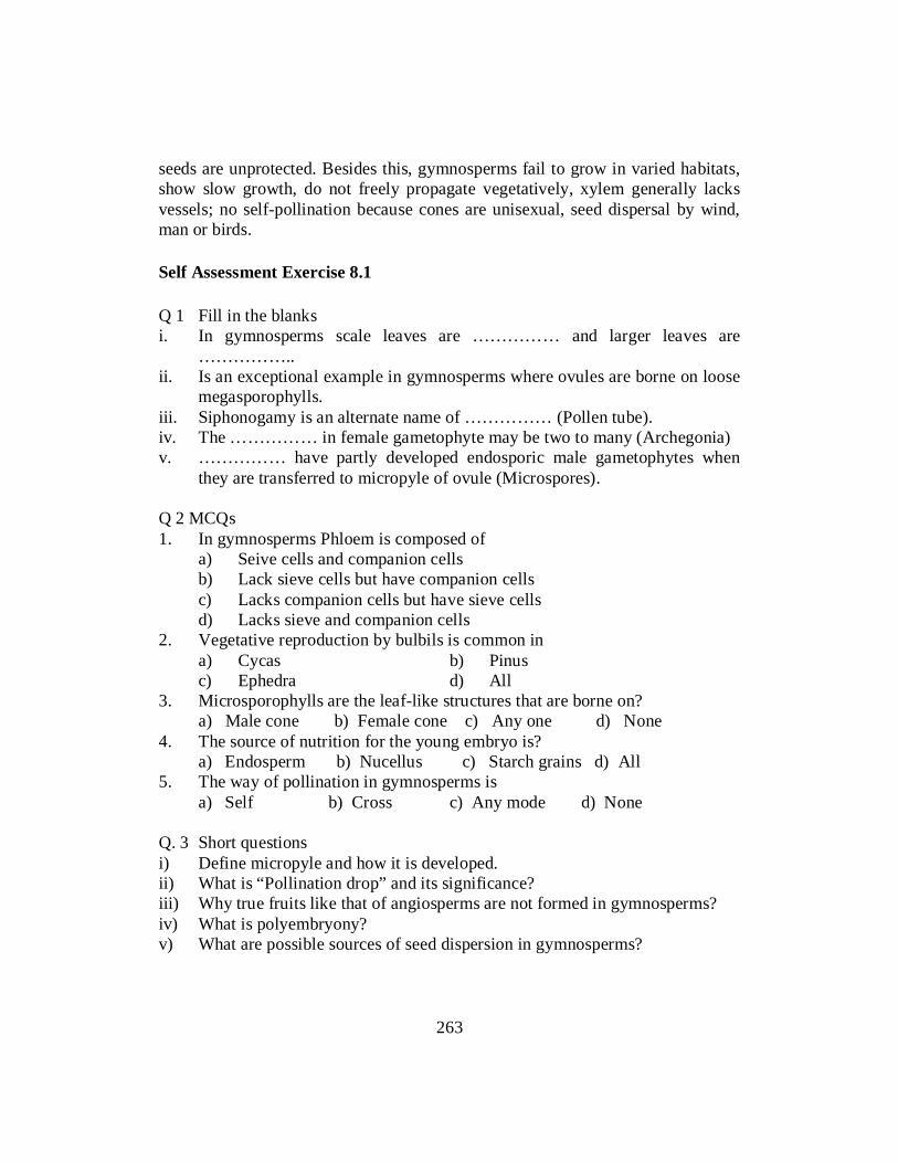
seeds are unprotected. Besides this, gymnosperms fail to grow in varied habitats, show slow growth, do not freely propagate vegetatively, xylem generally lacks vessels; no self-pollination because cones are unisexual, seed dispersal by wind, man or birds. Self Assessment Exercise 8.1 Q 1 Fill in the blanks i. In gymnosperms scale leaves are …………… and larger leaves are
…………….. ii. Is an exceptional example in gymnosperms where ovules are borne on loose
megasporophylls. iii. Siphonogamy is an alternate name of …………… (Pollen tube). iv. The …………… in female gametophyte may be two to many (Archegonia) v. …………… have partly developed endosporic male gametophytes when
they are transferred to micropyle of ovule (Microspores). Q 2 MCQs 1. In gymnosperms Phloem is composed of a) Seive cells and companion cells b) Lack sieve cells but have companion cells c) Lacks companion cells but have sieve cells d) Lacks sieve and companion cells 2. Vegetative reproduction by bulbils is common in a) Cycas b) Pinus c) Ephedra d) All 3. Microsporophylls are the leaf-like structures that are borne on? a) Male cone b) Female cone c) Any one d) None 4. The source of nutrition for the young embryo is? a) Endosperm b) Nucellus c) Starch grains d) All 5. The way of pollination in gymnosperms is a) Self b) Cross c) Any mode d) None Q. 3 Short questions i) Define micropyle and how it is developed. ii) What is “Pollination drop” and its significance? iii) Why true fruits like that of angiosperms are not formed in gymnosperms? iv) What is polyembryony? v) What are possible sources of seed dispersion in gymnosperms?
263

8.2 LIFE CYCLE OF THE FOLLOWING SPECIFIC EXAMPLES 8.2.1 Cycus Cycas revoluta is extensively propagated vegetatively by means of adventitious buds or bulbils. The male plants of C revoluta are not found in Northern India. Hence no seeds of this species are available in this part country. The female plants are raised entirely by means of bulbils. The male plants of C. circinalis are relatively more common. All Cycas spp are dioecions. In the vegetative state the male plants are indistinguishable from the female plants. The appearance of the male and female reproductive structures distinguishes the two sexes. The reproductive cycle starts after several years of vegetative growth. Male Cone The male cones are borne singly and terminally on the main stem. The apical growth of the stem is continued by the formation of the axillary bud at the base of the cone. It becomes the new stem apex. As it grows the male cone is displaced to one side. The new shoot apex soon takes over forming a crown of leaves and scales, ultimately bearing another male cone and the process is repeated. The stem of the male plant of cycas is therefore a sympodium.
Figure 8.1: Male cone of cycus
264

When young, the surface of the cone is covered all over by brown scales. An old male cone is very large hard & woody oval or conical in shape.It emits a very characteristic odour which can be smelt from a distance. It has a central cone axis that bears numerous, almost perpendicularly attached microsporophylls arranged in a spiral manner. The microsporophylls at the apex and the base of the cone are sterile a mature microsporophylls is a hard, horizontally flattened woody structure consisting of a wedge shaped portion and a sterile extension tapering into an upcurved apex (Apophysis. Numerous sporangia are borne on the lower or abaxial surface microsporangia occur in groups of 3,4 or 5 and such groups are called sori (single sorus) The average number at sporangia per sporophylls is 700 in C. circinalis and 1160 in C. media. the sporangia dehisce by longitudinal slits radiating outwards from the center. Microsporangium Microsporangium of Cycas (at a fairly well advanced stage of development) shows 5 or 6 wall layers. The epidermal cells are thin walled of uniform size and covered by a thick cuticle. The cells of the tapetum are small densely cyloplasmic and completely surround the developing sporogenous tissue. The tapetum is of secretary type and starts degenerating during meiosis in the microspore mother cells. By the time the pollen grains are mature, the tapetal cells are completely absorbed. The sporogenous cells are large with prominent nuclei and their cytoplasm rounds upto form the microspore mother cells. The microspore mother cells undergo meiotic division to give rise to tetrads of microsposes enclosed in a sheath of callose. Microsporogenesis The microspore mother cells undergo reduction divisions after meiosis-I, a ring-like thickening appears on the callose wall of the mother cell in the equatorial plate. Usually, the wall laid down after meiosis-I seems to be thicker than the one which develops after meiosis-II. It is likely that the abundance of starch, especially on the equatorial plate, obstructs a clear view of the cell plate. The cytoplasm of each microspore contracts and secretes a membrane which forms its independent wall. Thus, each member of a tetrad is contained within its own chamber. The original wall of the mother cell, which appears to be made up of peptic substances, collapses and no trace is left after a week of division. The pollen grain of cycads shows an oval germinal furrow or sulcus on its distal face (Millay and Taylor 1976). The furrow develops by a modification of the exine in the region and can be differentiated into a peripheral and a central zone (Audran 1965, 1971). According to Gulliver (1966) in C. Revoluta the exine is made up of
265

two layers. The outer one comprises orbicules and/or a sculptured granular layer, while the inner layer is lamellated. The newly formed micropsers are uninveleate. Excluding the epidermis, the sporangial wall now consist of tree or four layers of thin walled polygonal cells; most of the tapetum has already been consumed. Male Gametophyte The microspores are set free from the callose wall and each uninucleate microspore develops a thick exine and thin intine. The latter is the last layer to be laid down and is of variable thickness (Audran 1965), thinnest at the proximal or prothallial cells end, and thichest at the distal end of the grain. (The exine is thicker on the proximal side of the spore in the tetrad which thins out on the distal end.) The Microspore nucleus divides to form a small prothallial cells and a large antheridial initial. The former does not divide any further and remains as a small less-shaped cell. The latter divides into an antheridial cell and a tube cell.The pollen grains are shed at the 3- celled stage. They are boat – shaped due to the presence of a depression on the distal side. SEM studies (Sahashi & Ueno, 1986) on the pollen grains of cycas revoluta have shown them to be oblong with sulcate shrunken aperture when dry. The fullly expanded grain is almost spherical with a large rounded germinal aperture. The inner layer of exine shows reliculum like sculpturing. Structure of mature microsporangium The wall layers of the sporangium become disorganized and only the epidermis is conspicuous which forms the exothecium. The cells at the upper corners of the micro sporangium are very much alongated, gradually becoming smaller at the region of dehiscence which comprises two rows of thin walled cells. All the cells develop heavy thickenings on the radial and inner tangential walls and remain thin on the outer tangential side. The Poller grains are at three- celled stage: Prothallial, antheridial cell and tube cell. The antheridial cell is small as compared to tube cell and is attached to the intine at the site of the prothallial cell. The tube cells is usually vacuolated and has a large nucleus. Dehiscence of Microsporangium The microsporangia dehisce by longitudinal slits. The lines of dehiscence of various sporangia radiate out from the centre of the sours towards the tip of the sporangia, are usually marked by unthickened cells of the exothecium. After the sporangia open, the cone axis elongate and the tightly packed sporophylls separate from each other, this helps in the release at pollen. In a strobilus, the microsponphylls mature, progressively from the apex to the base, and it takes
266

several days for all the pollen in a strobilus to be shed. The numerous light pollen grains are easily blows by wind. Megasporophyll Among the living cycads, the Cycas is the only genus which does not produce compact female cones. (According to Chamberlain 1935, the living cycads possess the largest cones). There is a loose crown of megasporophylls arising spirally in acropetalous succession at the apex of the stem in between cluster af foliage and scale leaves (The female plant bears successive zones of appendages in the order vegetative leaves, scale leaver, megerprophylls, scale leave, vegetative leaves and so on.) The apical growing point continuous further growth.The stem of the female plant is, therefore, monopodial.Each megasponphyll is a leaf – like structure, 15-30 cm long, and pinnately dissected in its upper region. In the lower portion, one to five pairs of ovules are borne on either side of the sporophyll. The sporophyll and ovules are covered with soft brownish hairs (ramenta) which fall off as the ovules ripen;. In various species there is a gradual reduction in the size of the sporophyll and its leaflets to a mere serrate margin, and in the number ovuler to only two which is the number in other genera. Ovule Each megasporophyll bears two rows of opposite or sub-opposite, 2-12 orthotropous and shortly started ovules. Cycas produces the largest ovules in the plant kingdom (6 cm long in C circinalis and 7 cm in C. thourasi and up to 4cm in diameter) The mature seeds are elliptical or egg shaped, slightly flattened radially and bilobate. They are fleshy, and bright orange or red. The apex form a short micropylar tube. The ovule surface may be smooth or hairy, hairs being shed off at maturity. The ovules are unitegmic and crassinucellate, the integument is fused with the nucellus for most part, and is free in the upper one third portion. It can be differentiated into three layers. (a), the outer fleshy layer which becomes variously coloured at maturity, (b) middle stony layer, and (c) an inner fieshy layer which is resorbed and becomes papery even before maturity.. The integument extends beyonds the nucelles to form the micropylar tube. The apex of the nucellus grows up into a beak like outgrowth, the nucellar beak which projects into the micropyle. As the female gametophyte develops some of the cells of the nucellar beak disorganise to form flask-like hollow cavity, the pollen chamber. The micropyle leads into the pollen chamber.
267

Megasporogenesis Generally, the young nucellus has one to several hypodermal archesporial cells which divide periclinally to form the inner primary sporogenous and the outer primary parietal layer. The latter and its derivatives repeatedly divide periclinally to form a massive parietal tissue above the primary sporogenous cells. The latter may divide so that the sporogenous tissue increases. One of the sporogenous cells functions as the megaspore mother cell which become distinct by their prominent nuclei and dense cytoplasm. The megaspore mother cell divides meiotically to form a row of three cells representing an undivided upper dyad cell and two megaspores. Of the two megaspores, the lower one functions. Some cells of the nucellus, surrounding the developing megaspore, are small, radially elongated and densely cytoplasmic. They divide rapidly keeping pace with the enlarging gametophyte and are probably nutritive in function. These cells constitute the spongy tissue. How nucellus plays a role in gametophyte development? Female Gametophyte The nucleus of the functional megaspore divides many times forming nearly a thousand nuclei unaccompanied by wall formation. The megaspore, in the meantime, enlarges rapidly and develops a central vacuole in its cytoplasm. The nuelei come to lie in a thin film of cytoplasm around the big central vacuole. Walls are laid down in the free nuclear gametophyte in a centripetal fashion from periphery inwards. Gradually, the entire female gametophyte becomes cellular. The peripheral cells are small, isodiametric and without any food reserves, whereas the inner cells are large, irregular and packed with starch. Archegonium At the micropylar tip of the gametophyte, two to six archegonia differentiate. The archegonial initial divides to form a primary neck cell and a central cell. The former divides anticlinally, resulting in two neck cells. They divide once again, just before fertilization, to form four neck cells . The second wall is much thinner than the first one. By the time the nucleus of the central cell is ready to divide, the neck cells become large and turgid and project into the archegonial chamber. The central cell continues to grow for several months before its nucleus divides to form the ventral canal nucleus and egg nucleus where it soon disorganizes. Occasionally, however, it enlarges and simulates the egg nucleus. The maturation of different archegonia in a gametophyte is synchronous. The egg cytoplasm shows a number of proteid vacuoles which increase after fertilization. In C. rumphii they are rich in proteins (De Silva and Tambiah 1952). The gametophytic region around the archegonia grows upwards and forms a depression in the
268

middle, the archegonial chamber. The archegonia occur singly, and each has a well-developed single layered jacket. There is usually a thick wall with simple pits between the jacket cells and the central cell of the archegonium. According to Swamy (1948), the pores become occluded by plug-like thickenings, and the jacket cells become depleted of their contents. The thick cell wall forms the so-called egg-membrane during the post-fertilization stages. The egg membrane thickens as the egg matures. It retains its form and remains connected with the suspensor of the embryo when the latter is fairly well advanced. There are prominent pits on the thick wall. Extension of the egg cytoplasm come in contact with the cytoplasm of the jacket cells, through the plasmodesmata present in the pit areas. Pollination The male cones mature when the young megasporophylls have emerged on the female plant, and the ovules are at the free-nuclear gametophytic stage and ready for pollination. The single integument is drawn out into a short micropylar tube, which secretes a sugary exudate (pollination drop) close to the nucellus of the ovule.The wingless pollen grains usually float into the pollination drop, which retracts soon after. The pollen grains show a germinal furrow, which closes in dry weather and is wide open in high humidity. After landing on the pollination drop, the pollen grains imbibe water and other substances from the exudate, become heavy and sink down the micropyle to reach the nucellus, where they germinate. The male conses of Cycas growing in warm climate emit a strong odour (described earlier); several insects visit them when the pollen ripens (see Chamberlain 1935, Pant 1973). Thus, these insects may also be carrriers of pollen in cycads, Following pollination, the ovules enlarge and reach their full size. unpollinated ovules remain small and eventually dry up. Tang 1987, has reported inscet pollination in Zamia pumila and Norstog et at, 1986 reported role of beefless in pollintion in Z furacea) Pant and Singh (1990) found that in Cycas, beetles, insects and even ants visited only the male cones and continued to live there till the next generation. They did not find there beetles, etc. pollinating the ovules in female plants. inBowerta surrulata pollination is amphiptition (see Wilson 1993) Tang (1987) reported widespread heat production in the male cones of 42 cycad species followed a circadian rhythm. It is released every evening during cone elongation and pollen shedding stage. Concurrent to heat production is the release of odours that helped in attracting insects for pollination.
269

After the pollen grains land in the pollen chamber (situated in the upper beak-shaped portion of the nucellus), a group of cells lying below begin to degenerate and extend the lower limit of the chamber. This post-pollination extension is the lower pollen chamber or intermediary chamber. How the development of pollen tube in cycas is different compared to other gymnosperms.The intine of the pollen grain ruptures the exine at the distal end, grows into a tube in the region of the furrow, and the tube nucleus migrates into it. The pollen tube grows laterally and intercellular into the tissue of the nucellus, instead of towards the archegonia as in other gymnosperms. During its course through the nucellus, the tube often branches horizontally, derives nourishment as it digests its way, and accumulates considerable amount of starch for the development of the male gametophyte. The pollen tube does not transport the male gamete but establishes only a hausterial system. (In Zamia furfuraea Choi & Friedman (1991) The pollen tube, while growing intercellularly in the nucellar tissue, develops additional localized hausterial growth which penetrate adjoining nucellar cells. This intracellular hausterial growth leads to the death of individual cells and also induce the degeneration of neighbouring nuclellar tissue. The pollen tube, thus, act more as a hausterium than a sperm carrier which is its normal function in other gymnosperms. In this respect, cycads, together with Ginkgo, can be said to have the most primitive male gametophyte among the living gymnosperms.) As many as a dozen pollen tubes may sometimes be seen inside the pollen chamber growing in the nucellus.The antheridial cell divides periclinally; the cell adjacent to the prothallial cell is sterile and designated the stalk cell; the other spermatogenous cell (body cell). The stalk cell enlarges considerably and accumulates starch and other materials. Simultaneously, the prothallial cell bulges and its upper end penetrates through the stalk cell, which forms a life-belt-like ring around the prothullial cells. The body cell (spermatogenous) begins to enlarge and elongate along the longitudinal axis of the pollen grain. The nuceleus of the body cell also enlarges, and two small granular bodies (the blepharoplasts) appear, one on each side. Cytoplasmic rays (also called astral rays) radiate from the blepharoplasts. The astral rays are the microtubules which extend from the matrix or the surface of the blepharoplasts. The latter comes to lie on the two poles of the body cell along the longitudinal axis of the pollen grain. The body cell enlarges, becomes spherical, and the blepharoplasts move through 90° and come to lie at right angles to the long axis of the pollen. The blepharoplasts increase in size and become very prominent.
270

The mitotic spindle of the body cell lies at right angles to the long axis of the pollen tube, so that the two sperms lie side by side enclosed in the wall of the body cell till they mature. The blepharoplast forms a part of clock-spring-band-bearing cilia, around the gradually or abruptly tapering distal part of the sperm. This band in C. revoluta shows a right-hand coiling (as seen from the apex of the sperm). The spiral band has six turns (gyres), and in a radial section (C. circinalis) appear stair-like. The sperms start rotating movement while still enclosed in the body cell. The sperms of C. revoluta are 180-210μ in length, and visible to the naked eye. They are released by the rupture/dissolution of the outer walls of the sperm mother cells into the basal end of the pollen grain. By their cilia they move freely in the fluid of the turgid pollen grain-end and remain active for several hours. The nucellar tissue between the pollen and archegonial chamber collapses, and the proximal end of the pollen grain pushes down through this opening and "hangs" freely in the dry archegonial chamber. A bunch of pollen grains grow downward and disorganize it completely between the intermediary chamber and the female gametophyte. The proximal end of the pollen grain lies in a longitudinally continuous cavity in the middle of the nucelles. Male Gametes (Ciliate Sperms) The spermatozoids of cycads are the largest flagellate male gametes, visible to the naked eye. These measure 180-210 μ in Cyeas revoluta. (The largest reported so far is 400 μ in Zamia chigua by Norstog, 1977) These spermatozoids are of considerable importance because of phylogenetic implications of their size, motility and morphology. The gamete is without any cell wall and the nucelles occupies the greater part of the cell. The flagella (There are about 40,000-50,000 flagella) arise from the spiral channel which is formed by a depression in the plasmamembrane and cytoplasm. Fertilization The proximal end of numerous pollen tubes hangs into archegonial chamber. Finally, it bursts and the sperms and the pollen tube cytoplasm are released in the archegonial chamber. The gametes swim for some time with a forward and circular motion; the band of flagella form the anterior end. The pollen tube cytoplasm is osmotically rich consing plasmolysis of the neck cells, facilitating the entery of the motile sperms. The sperm casts off its ciliary band, and its nucleus approaches the egg. The male nucleus penetrates deep into the larger egg nucleus before its membrane disappears. The spiral band usually persists in the egg cytoplasm long after fertilization.
271

Embryogeny The zygote nucleus divides in situ followed by several free-nuclear divisions The nuclei are distibuted throughout the young proembryo. In later stages, the free nuclei mostly concentrate at the base of the proembryo and only a few nuclei are present in the upper thin cytoplasm. Subsequently, only the nuclei at the base divide while the upper nuclei show signs of degeneration. At the time of wall formation, there are 512 free nuclei in C. circinalis (Rao 1963.) Cells are formed only in the lower portion of the archegonial cavity, the nuclei present in the upper area remain free. During later stages, the free nuclei, along with the circumjacent cytoplasm, degenerate and form a plug. Following wall formation, the cells at the base divide and function as embryonal cells. The upper cells (just above the embryonal cells) differentiate into a suspensor and the uppermost layer of cells as the buffer cells, the latter form a few layers of tissue around a central vacuole (Pant 1973) The thick inner tangential walls of the jacket cells persist till an advanced stage of proembryo. The suspensor penetrates the chalazal end to the thick wall which is very prominent and is called the egg membrane. Differentiation of Embryo:-After the proembryo becomes cellular, the cells of the upper region elongate to form a very long suspensor. It grows rapidly, and thrusts the basal embryonic region through the egg membrane and archegonial jacket deep into the gametophytic cells, which have abundant food reserve. This is due to enzymatic digestion of the gametophytic cells when they come in contact with the embryonic cells. Due to rapid elongation, the suspensor becomes much coiled and twisted. Usually, more than one archegonium in an ovule is fertilized, and the development of multiple zygotes leads to simple or archegonial polyembryony. At first, all the zygotes in an ovule appear to develop with equal vigour, the embryos project into the gametophytic tissue, and their suspensors become closely intertwined with each other. Due to competitive growth, ultimately only one embryo develops while the rest abort at various stages of development. The suspensor of the aborted embryos remains attached to the tough egg membrane, and persist for a long time. The suspensor of the successful embryos is, therefore, a composite structure of the coiled suspensors of the entire group of embryos in an ovule. When stretched, the suspensor may be up to 10 cm long. Seed The maturation of the embryo in seed takes over a year after fertilization. The seed is shed at any stage during this period, and the development of the embryo is completed on the ground. To begin with, the embryo only increases in size without differentiation into its organs. Then the coleorhiza develops at the
272

micropylar end of the embryo. The latter shows internal differentiation into two polar meristems, epidermis, cortex, procambium and pith; the hypocotyl is rather short. The number of cotyledons varies from one to three, closely adpressed to one another and appear as a single structure. The coleorhiza is partly derived from the suspensor and becomes quite "hard" in a mature embryo. It is morphologically equivalent to the root cap. At maturity several outer layers of the root cap become especially thick-walled to form a distinctive caplike structure. There are abundant mucilage cells in the tissues of the embryo. The integument hardens to form the seed coat. Its outer fleshy layer adheres firmly to the stony middle layer forming a hard type of seed. The inner fleshy layer had already shriveled. The nucellus is completely crushed. Within the seed coat lies the female gametophyte (endosperm). It is fleshy and function as food storage region of seed lying embedded in the middle of female gametophyte is the straight embryo. The attractive, red or orange fleshy seeds contain abundant food reserve. Quite likely, they are dispersed by birds and rodents. The cycad seeds do not have a resting period and the viability is short. Germination of the seed is epigeal. Temporal Consideration As compared to the temperate gymnosperms, the reproductive cycle of cycads occurs at different times of the year. The cones in the South Indian.C. circinalis are initiated probably in April, and show micro- and megaspore mother cells during June-July (Rao 1963). The ovules have a free-nuclear gametophyte at the time of pollination (8 months after initiation) in December. Fertilization occurs in May-June (5-6 months after pollination), followed by a very slow development of the embryo, in which the cotyledons appear during November-December. The seeds with an immature embryo are shed during May and June (1 year after fertilization). The embryo matures and attains full size, and the seeds germinate during September-October (4 months after shedding). Thus, this taxon takes 2 years and 5 months to complete its life cycle, although there is no winter rest. In C. rumphii, the ovules are pollinated in May (De Silva and Tambiah 1952), fertilization occurs 13 months later (i.e. the following June), and the seeds are shed in January but the embryo matures only by March. Consequently, the time lapse between pollination and fertilization is much longer. The general reproductive cycle of cycads is: 1. There is a long time gap from the time of ovule initiation to pollination (the
ovules are in an advanced stage of development at the time of pollination, as compared to conifers).
273
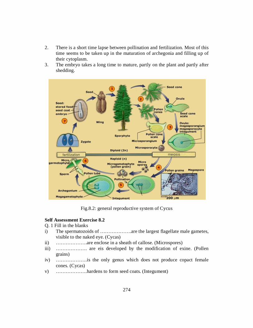
2. There is a short time lapse between pollination and fertilization. Most of this time seems to be taken up in the maturation of archegonia and filling up of their cytoplasm.
3. The embryo takes a long time to mature, partly on the plant and partly after shedding.
Fig.8.2: general reproductive system of Cycus
Self Assessment Exercise 8.2 Q. 1 Fill in the blanks i) The spermatozoids of ……………….are the largest flagellate male gametes,
visible to the naked eye. (Cycas) ii) ……………….are enclose in a sheath of callose. (Microspores) iii) ………………. are eis developed by the modification of exine. (Pollen
grains) iv) ……………….is the only genus which does not produce copact female
cones. (Cycas) v) ……………….hardens to form seed coats. (Integument)
274

Q. 2 MCQs 1. Which of the following gymnosperms is dioecious? a) Cycas b) Pinus c) Ephedra d) All 2. In which region of cone sterile microsporophylls are present? a) Apex b) Base c) Apex and base both d) None of these 3. Microspores in cycas are a) Uninucleate b) Binucleate c) Multinucleate d) All 4. In cycas pollen grains are shed at which stage? a) 2-celled stage b) 3-celled stage c) 4-celled stage d) None of these 5. Largest ovules in the plant kingdom are produced by a) Cycas b) Pinus c) Ephedra d) All Q. 3 Short questions i) Define sori? ii) Define integument and how it is developed into three differentiated layers? iii) What is “nucellar beak” and how it is formed? iv) How nucellus plays a role in gymnosperms development? v) What is archegonial chamber? vi) What is egg membrane and at which stage it is formed? 8.2.2 The Reproductive Cycle of Pinus Strobili The plants are monoecious. The male and female cones are borne on different branches of the same tree. Male cones occur laterally in clusters, each in the axil of a scale leaf at the base of a terminal. Vegetative bud .The male cones replace the dwarf shoots at the base of the developing bud or shoot of the current year and are spirally arranged on long shoot. Each male cone is shortly stalked and consist of an elongated central axis, bearing a number of small spirally arranged and closely fitting scale like microsponophlls with their scaly apices upturned. The microsporophyll is attached to the axis by a short stalk and bears two microsporangia on its lower (abaxial) side . The microsporangium dehisces by a longitudinal slit. (The young male cones of P. roxburghii are about 1.5 cm-2 cm long and 1 cm in diameter. At the time of dehiscence these are 2.5cm – 3cm in length.)
275

The male cones arise on the lower and the female cones on the upper branches. The female cones replace the long shoots (Fig.13B) and are generally borne in pairs, but the number may go up to six. Each female cone consists of a central axis which bears paird scales 180-90 in number) in a close spiral. The lower scale of the pair is small known as bract scale. It is leathery and directly attached to the cone axis. The upper scale of the pair is larger, thicker and stouter. It is the ovuliferous scale/ megasporophyll. The bract and the ovuliterous scales together form a seed-scale-complex. Each ovuliferous scale bears two-two ovules on the upper or adaxial surface. The ovules are inverted with the micropyle facing the cone axis. The bract & scale is larger than the ovuliferous scale before pollination, but in post pollination stages the ovuliferous scale overgrows the bract scale. The ovuliferous is woody and wedge shaped with its broader sterile end, the apophysis directed outwards. The bract scales are thus concealed from view. The surface of the cone is marked by rhomboidal areas each with a small central conical point- the umbo. Sporophylls at the apex and base at a cone are generally sterile. It takes approximately twenty-seven months from the time the female cones are initiated to the shedding of seeds. Seeds generally have conspicuous wings excepting a few like P. gerardiana where the wings are rudimentary. Microsporangium A transection of young microsporangium shows a mass of meristematic tissue surrounded by an epidermis. One, two or more cells of the hypodermal region, by repeated divisions, give rise to an archesporial tissue which has dense cytoplasm and prominent nuclei . The peripheral cells of the archesporium undergo periclinal divisions and cut off the primary parietal layer towards the periphery and sporogenous cells on the inside. Further periclinal divisions in the parietal layer forms an outer three- or four-layered wall and the inner mass of sporogenous tissue. The innermost layer of the wall cells develop into the tapetum. Simultaneously, the epidermal cells undergo anticlinal divisions; most of the cells are filled with tannin except two rows of smaller cells, which form the line of dehiscence. At the time of dehiscence of the microsporangium, the epidermis attains its maximal development with fibrous thickenings, and functions as the exothecium. As the microspore mother cells initiate divisions, the epidermal cells lose their nuclei, become filled with a homogeneously staining substances. The outer wall of the epidermal cells gradually becomes cutinized. The tapetum is of the secretory type. Young tapetal cells are richly cytoplasmic and multinucleate. They Iecome very conspicuous during meiosis in the microspore mother cells and degenerate soon after the spores are released from
276

the tetrads (17D). The tapetum is involved in the nourishing of the sporogenous cells/young microspores, and in the formation of exine on the spores. The development of tapetum and sporogenous cells takes place simultaneously. There appears to be a correlation between the stage of the microspore mother cell (meiocyte) and the structure of tapetal cytoplasm. Accordingly, three phases of development of tapetal cell can be distinguished corresponding to pre-meiotic, meiotic and post-meiotic phases of microsporogenesis. Microsporogenesis Prior to meiosis the cytoplasm of microspore mother cells and tapetal cells looks alike. (Dickinson & Bell, 1976). However, with the anset of meiotic division, the mother cell wall lyses completely. At diplotene stage, the callose starts enveloping mother cells. The deposition of callose is brought about by the activity of the Golgi bodies which give out a large number of vesicles at deplotene stage.Callose, being a substance of low permeability, isoslutcs the mother cell from surrounding influences, thus helping the micropore mother cells to follow an independent course of development during meiosis.The nucleus of the mother cell undergoes reduction division, which is followed by simultaneous wall formation to give rise to a tetrad of microspores .At the end of the division, the callose sheath is dissolved by enzymatic action and the young haploid microspores are set free. Microspore: Each microspore has large nucleus and soon after the formation of microspores, a special wall is secreted around each of the spores which results in the formation of on outer exine (cappa) and inner intine (capulla). Later, the exine expands at two points on opposite sides of the microspore. This result in a cavity formed between the exine and intire which later forms saccii (wings or air sacs) in a mature pollen grain. The saccus becomes beautifully marked by the formation of delicate, irregular ridges over the entire surface expect along point of fusion. The pollen grains are heteropolar and rediosymmetric and consists at a body or corpes and the air sacs or sacci. It has a single aperture or tenuitas at its distal end. The wings or sacci are separated from the body or the corpus by the saccate nexine. The nucleus lies towards the distal end. Male Gametophyte The uninucleate microspore is the first cell of male gametophyte. The microspore nucleus cuts off a small lens-shaped prothallial cell towards the proximal end and a large central on the distal end.These are initially separated by an ephemeral callose wall which is later replaced by a cellulosic wall. The central cell cuts off a second prothallial cell and an anthendial initial. Both the prothallial cells are ephemeral. The antheridial initial now divides to give rise to a small antheridia
277

cell and a large tube cell.The pollen grains are shed at the four celled stage (two degenerated prothallial cells an antheridial cells and tube cell). The development of the pollen grain wall has been studied in Pinus sylvestris (Willemse, 1971) and P. banksiana (Dickinson, 1971) by using EM. The exine consists of an outer sexine and inner nexine. The nexine, in turn, consist of nexine I (on outer side, i.e. towards sexine) and nexine II (on inner sides i.e. towards intire. The outermost, layer, the sexine and nexine I are laid down when microspores are still at the tetrad stage and within the callose wall. A space is formed between plasmalemma and callose layer by the contraction of former. This space is much enlarged in the region where, subsequently air sacs would develop the sexine is first laid over the area where sacci are destined to be formed; it then slowly spread over the whole area, except in the region of the germinal pore.Long stretches at distinctly trilamellar tapes at lamellae are seen at this stage at development at primexine. The next layer to be laid down is nexine I. It is formed by the deposition of sporopollenin on trilaminar tapes, in the inner side of primexine. Lipid granules are the primary source of nexine I. The nexine surrounds the cytoplasm except at the region where wings would develop. Marten & Waterkeyn (1962) studied the wing formation and found them to be the extension of outer exine only. After the formation of nexine I, the microspores come out of the callose wall which is enzymatically degraded. The sporopollenin continues to be deposited on the sexine layer. Furhter development of the pollen wall takes place when the microspores are liberated from tetrads. Small granular particles at sporopollenin present between the plasma lemma and nexine I coalesce to form nexine II. Nexine is thick on the germinal pore side. Intine is the last layer to be laid down. It consists of two layers viz., an outer and the inner intine. The inner inter is a continuous layer as compared to the outer entire which is discontinuous. The inner intine which is next to the cytoplasm is thicker at the germinal pore (distal end) and thinner at the region of prothallial cells (proximal cells). It is made of cellulose and pectin. The incomplete outer entire covers the inner entire except at the germinal pore. The inner intine is responsible for the formation of pollen tube. The development of air sac has been traced in great detail in Pinus banksiana by Dichinson & Bell (1970). Like exine their formation starts when microspores are still with the tetrads and primexine is beinglaid down between the plasmamembrane and the callose layer. Megasporangium / ovule First Period of Growth of the Ovule: The ovule is unitegmic and crassinucellate. The integument is free from the nucellus except at the chalazal end, and forms a symmetrical micropylar tube well beyond the level of the nucellus. AdaxialIy, the edges of the integument extend into two long arms, which curve inwards before pollination, outwards during and curve back after pollination and finally dry up. There is no vascular supply to the ovule (Konar and Ramchandani 1958).
278

Megasporogenesis. The archesporial cell becomes distinguishable while the female cone is still covered by the scale leaves. It differentiates at the broad apical end of the nucellus and divides transversely to give rise to primary parietal and primary sporogenous cell. The former undergoes both vertical and transverse divisions, so that the sporogenous cell is pushed deep into the nucellus, and later functions as the megaspore mother cell . Starch grains accumulate at the chalazal end of the megaspore mother cell (P. sylvestris). The latter undergoes meiosis I and produces a dyad. Only a triad is formed if the upper dyad cell does not undergo meiosis II or a tetrad if both the dyads undergo meiosis II. In a triad and linear tetrad, the upper dyad cell and the adjoining megaspores or the upper three megaspores in a tetrad usually degenerate. The chalazal megaspore functions so that the development of the gametophyte is monosporic. Three to five layers of cells round the functional megaspore become densely cytoplasmic with prominent nuclei. This is the spongy or nutritive tissue. The spongy tissue comprises a definite zone of physiologically active cells which are concerned in the nutrition of the young gametophyte, especially at the resting stage. The cells of this zone contain abundant starch. Female Gametophyte (First Period of Growth). The functional megaspore enlarges and shows a large vacuole even before the nuclear divisions commence. It forms only a few free nuclei (maximum at 32 nuclei) and apparently remains inactive for 8-9 months (first period of rest). Pollination Each tree produces an enormous number of pollen grains dispersed by wind. The surrounding area becomes clouded by the yellow powder (also known as sulphur shower). However, only a few pollen grains reach the pollen chamber and develop further. A pollination drop is secreted at the flared-out tip of the integument. The secretion starts a few days after the female cones emerge out of the scale leaves, and the ovuliferous scales separate sufficiently to permit the free entry of pollen. Under high humidity and cell turgor, the secretion begins around midnight, and by early morning the micropyle dries up. The arrival of the wind-borne pollen at the micropyle is purely a chance phenomenon. The pollen is caught in the pollination drop, grains stick to the two-pronged micropylar canal and migrate to the nucellar tip. The secretion of the drop is reported to be a cyclic (24 hr cycle) phenomenon. The secretion normally continues for a few days or till the time of pollination. In the presence of pollen there is a permanent withdrawal of the exudates. The cells at the
279

apex of the nucellus are involved in the secretion of the drop. Mc William (1958) made a chemical analysis of the pollination drop of Pinus nigra and reported that the fluid contains three sugars namely D-glucose, D-fructose and sucrose. Doyle and O'Leary (1935) and Doyle (1945) have studied the mechanism of pollination. According to Doyle, the wings orient the grain on the hanging pollination drop in such a way that the germinal pore/ furrow of the grain faces the surface of the drop. This orientation is particularly necessary since the ovules are inverted. The pollen then floats upward (due to buoyancy caused by the air sacs) and reaches the nucellus with the germinal furrow faceing the nucellus. The pollen tube enters the nucellus without curving or twisting. McWilliam (1958) emphasizes that in P. ellioltii, P. nigra and P. wallichiana, the stickiness of the neck and arm of the micropyle may be due to either a local secretion, or sugar residue resulting from the retreating micropylar fluid. This is an effective method for retaining the pollen at the site, and the prime mover of the grain is the active absorption of the fluid. McWilliams did not observe any preferred orientation of pollen on the nucellus. At the time of pollination, the integument is four-to-five-layered, and soon after the pollen grains reach the nucellus, the micropyle closes due to a rapid division and enlargement of the cells of the inner layer of the integument. Second period of growth of the ovule The female gametophyte resumes its activity in the following year in the month of February (P roxburghii) or a little later in the trees at higher altitudes (P wallichiana and P gerardiana). The additional tissues formed in the ovuliferous scales after the resumptionof growth are green. Thus both the bract scale and ovuliferous scales become very much enlarged and the umbo of the oveulifenous scale are pushed up. Female Gametophyte (Second period of Growth) As the ovule increased in size, the gametophyte also enlarges. The free nuclear divisions in the latter continue until the gametophyte occupies the entire basal and central part of the nucellus. During early stages, the nuclear divisions are synchronous. The number of nuclei in a free nuclear gametophyte is constant for a particular species; in P roxburghii and P wallchiana it is about 2500 and approx. 2000 in P gerardiana. With the enlargement of megaspore, a vacuole develops in the centre gradually pushing the cytoplasm along with the nuclei towards the periphery. At the end of last mitosis, every nucleus gets connected to six neighbouring nuclei by the formation of secondary spindles. Wall formation taken place through alveoli.The newly synthesized walls are unevenly thick and grow
280

centripelally as open tubes or pipes from megaspore cell wall till they reach the centre of the vacude. Each alveolus has a nucleus at its mouth and directs its growth. Now the cross walls are laid down on each alveolus forming uninucleate cells till the entire gametophyte becomes cellular. Development of the Archegonium: A few cells (2-4) at the micropylar end of the female gametophyte become large and prominent by accumulating protein and RNA, and function as archegonial initials. Each archegonial initial soon divided into a large central cell and a small primary neck initial. The latter divides by two vertical walls at right angles to each other to form a neck of four cells. The neck cells are arranged in a single tier (P wallichiana, P roxburghii) or a transverse division in them results in eight cells in two tiers of four cells (P austriaca, P Montana, P rigida). The central cell envlarges very rapidly so that numerous valuoles are formed. This is referred to as the “foam stage” of the archegonial development. The central cell nucleus, meanwhile, divides into an ephemeral ventral canal cell and a large egg cell . A gradual accumulation of archegonial cytoplasm takes place with the disappearance of most of its vacude. The developing egg cytoplasm contains a number of densely staining, protein rich bodies. which hare been variously termed as proteid vacuoles, paranuclei and Hofmeister’s granules. Electron microscopic studies (Camefort, 1962) have shown that traditionally known proteid vacuoles are basically membrane bound cyloplasmic bodies of two sizes: (a) small inclusion, and (b) large inclusions (Fig.). The small inclusions are portions of general cytoplasm, partly enclosed by a single membrane. These inclusions maintain connections with the general cytoplasm by short peduncles. The large inclusions are less frequent, bound by a double membrane. The included cytoplasm contains dictyosomes, mitochondria and small inclusions. Camefort (1966, 1968) reported that the large inclusions are portions of cytoplasm surrounded by highly deformed and hybertrophied plastids. Thus, in the egg of Pinus one can distinguish two types of cytoplasm: (a) “fundamental cytoplasm or the general cytoplasm, and (b) enclosed cytoplasm. The newly formed egg nucleus is Feulgen – positive, but as it increases in size, it loses its Feulgen. Positivity so much so that a mature egg is completely Feulgen-negative. EM studies on developing egg have shown an interesting relationship between the egg nucleus, the mitochondria and formation of perinuclear zone. (for details refer: Camefort, 1962, 1967, Chesnoy and Thomas, 1969, Thomas & Chenoy 1969, Moitra 1980). The gametophytic cells near the apical end of the individual archegonium grow vigonously resulting in the sinking of the latter. Thus each archegonium has its archegonial chamber. Cells of the gametophyte surrounding the archegonium
281

form a special covering layer, the jacket. Numerous pits are present on the inner thickened walls i.e. facing the archeogonium. The oarchegonium maintains contact with the surrounding tissue through these pits. Megaspore wall: It is prominently developed. The exine of megaspore wall consists of an outer sculplured sexine and an inner smooth nexine. The sexine is piloid and the pila are free from each other. Post-Pollination Development of Male Gametophyte.The Pollen deposited on the nucellar apex germinates immediately. The pollen tube arises from the inner pectin-celluosic layer at intine or inner intine (Martens & Waterkeyn, 1962) While moving through the nucellus the pollen tube secretes enzymes such as pectinase and cellulose dissolving the nucellar cell walls (Willemse & Linshen, 1969) As the tube emerges, the tape nucleus is the first to move into it. Renewed activity of the male gametophyte in the second year: Initially, the growth of the pollen tube is very slow as compared to the rapid development of the ovule. The apicul part of the nucellus, which had been penetrated by pollen tubes during the previous year, does not become active again. The cells at this part become thickened and empty. And the cells lose their contents. The underlying cells are meristematic and rich in starch. Due to their growth, the tip of the nucellus with the pollen tubes is lifted above and away from the developing female garnetophyte. Towards the end of March or later, depending on the species (P. Maheshwari and Konar 1971), the pollen tube resumes active growth. In P. sylvestris the nucellar cells adjoining the pollen tube degenerate; the hemicellulose and pectin are affected first (Willemse 1968, Willemse and Linskens 1969). During this period, the antheridial cell in the pollen grain divides to form the stalk and body (spermatogenous) cell. However, there is much variation in the time of division, not only in different species but also in the same species. Willemse and Linskens (1969) observed that, following the division of antheridial cell (called generative cell by them), the stalk cell has fewer organelles as compared to the body (spermatogenous) cell. Also, the cytoplasm of the stalk cell is vacuolate, but that of the body cell is dense. The stalk and body cell migrate into the pollen tube. Soon after, the body cell divides into two male gametes. To begin with, both the male gametes are equal, but later, one of them enlarges considerably.These two
282

unequal male gametes have a distinct cytoplasmic sheath around them. The male gametes are Feulgen-negative, stain faintly for RNA. Fertilization The pollen tube, just before fertilization, grows rapidly and reaches the neck of the archegonium. The tip has a dense cytoplasm, a large number of starch grains, tube nucleus, stalk cell and the two male gametes enveloped in a common cytoplasmic sheath . The pollen tube penetrates into the neck of the archegonium, and discharges its contents at the tip of the cytoplasm of the female gamete. One of the two male gametes moves towards the female gamete. The second male gamete, along with the stalk cell and tube nucleus, and the original male cytoplasmic sheath, persist at the apex of the egg and eventually degenerate .The functional male gamete is devoid of the cytoplasmic sheath as it moves down the archegonium to fuse with the female gamete (Willemse and Linskens 1969). The mitochondria and plastids brought by the pollen tube are morphologically different from those of the female gamete. They remain grouped in the upper part of the egg cytoplasm during the division of the zygote (Camefort 1965, 1967). The functional male gamete lodges in a depression on the female gamete . As soon as contact is established, junctions between the nuclear membranes of the two gametic nuclei are established at several points. The two nuclei establish contact by minute pores which develop in the nuclear membranes (Chesnoy and Thomas, 1971) These bridge of communication gradually increase in size and number. The zygote initially is separated from the general cytoplasm by the nuclear membrane. By the time first mitosis tabes place, the nuclear membrane disappears. The two free nuclei come to lie within the neocytoplasm (neo = new/which is jointly contributed by the nucleoplasms at the male and female nucles, and is entirely different from the mother (archegonial) cytoplasm. The embryo develops in this neocytoplasm. Embryogeny Proembryo development: The zygote nucleus divides mitotically to give rise to two nuclei. The division is intranuclear and the resulting nuclei are formed within the nucleoplasm of the zygote The next division immediately follows resulting in four nuclei which move to the base of the archegonium.A third synchronous division results in eight free nuclei. Secondary spindles develop and wall formation takes place and gives rise to an upper group of four cells, the primary upper tier (pU) and a lower group of four cells, the primary embryonal tied (pE). The pU tier has no wall towards the upper side and is, thus open. Internal divisions in both tiers results in four tiers of
283

four cells each. The lower two tiers comprise the embryonal tier (E) followed by disfunctional suspensor (ds) previously termed the rosette tier. The uppermost tier, U, is the upper derivative of pU. This is open above and was earlier referred to as the open tier, a term which is no longer used. Embryo differentiation: The upper four cells of the E-tier elongate and function as the embryonal suspensor (Es), and the lower four cells form the embryonal mass . Subsequently, there are several layers of Es, which are termed Es1, Es2, Es3…., formed by divisions in the E-tier. The Es cells elongate rapidly and thrust the terminal cells (embbryonal cells) forward into the corrosion cavity. The latter enlarges and facilitates elongation of the Es. Later, as the elongation of Es exceeds the rate of enlargement of the corrosion cavity, the Es becomes coiled and twisted.The elongation of the Es system pushes the growing embryonal mass into central portion of the female gametophyte. As development proceeds further, the earlier suspensor system collapses, while newer Es are formed. Polyembryony is common in Pinus.Additional embryos originate by simple polyembryony, i.e. from multiple zygotes, as well as due to cleavage polyembryony, i.e. a single zygote gives rise to multiple embryos by cleavage or splitting of the embryonal tier E. The separation into embryonal units occurs at Es2 formation. These units remain unchanged during the earlier stages of Es elongation, but later they divided and form a multicellular mass. There is a period of embryonal selection due to competition between the four embryos from each zygote, and between the embryos from different zygotes. This phase of competition lasts for about 6 weeks from the time of fertilization (P. Maheshwari and Konar 1971). Generally, the deep-seated terminal embryo succeeds and develops further. The remaining embryos become arrested at different stages of development. The embryonal cells of the developing embryo divide in various planes and formed embryonal mass which is a smooth paraboloid structure. It has a hemispherical apex at its distal end ,and a suspensor which is continuous with it at the proximal end. Further development at the proximal end gives rise to a well-developed root cap. It differentiates independently into a column and a peripheral region; the former does not contribute to the development of the latter. As the root cap and root initials organize at the basal end .An incipient pith is recognizable in the hypocotyl between the epicotyl and the root initials. It is evident by the predominant transverse divisions, cell enlargement and vacuolization. The cortex becomes demarcated soon after. The formation of pro-cambium is associated with the early phases of cotyledon formation though it is
284

well developed before distinct cotyledonary primordial originate. Simultaneously, several long, uninucleate and multinucleolar secretory cells become distinguishable in the cortical region. Finally, 3-18 cotyledons and the shoot apex differentiate. The cotyledons are traversed by the procambial strands, and show development of mesophyll. Generally, only one embryo matures, occasionally even two may reach maturity. The mature embryo has distinct epicotyl; root axis with remnants of the suspensors, and a hypocotyl-shoot axis bearing several cotyledons. Structure of the Seed: There is a outer hard shell or seed coat (testa) which protects the inner parts of the seed . In a young ovule the integument comprises three layers. As the ovule develops further, the integumentary cells divide and become differentiated into three zones:(i) The outer fleshy zone of 7 or 8 layers of cells with shrunken nuclei and vacuolated cytoplasm its outermust 2 or 3 layers get filled with some shiny granular material, (ii) the middle stony zone of 18-20 layers of cells with lignified walls, and (iii) the inner fleshy zone consisting at 7 or 8 layers at thin and elongated cells. The seed is winged and the development of the wing is closely linked with the development of the seed coat. There are however some species which possess rudimentary wings (P flexilis, P. cembra) or are wingless (P koraiensis, P cembroides). The outer layer of the integument and part of the ovuliferous scale contribute to the formation of wing. The wings are normally thin and papery, but in some species they are thickened at the base. The wings may be adherent or easily detachable. Within the seed coat is the brown papery tegmen (derived from nucellus) The latter surrounds a white fleshy female gametophyte (Analogous to endosperm) which surrounds the embryo. Towards the pointed end of the female gametophyte is the nucellar cap which represents the remains of he nucellus. During development both embryo and female gametophyte store reserve food materials which are med at the time of germination.(Please refer Hakansson 1956, Simola 1974, Mia 2 Durzan 1974 Durzan and Chalupa 1968, Kalsuta 1961, Hatano 1957, Kanar 1958, Nyman 1966). In the centre of the endosperm is a distinct central cavity in which lies he embryo. The embryo consist at a short axis differentiated into radicle towards the micropylar end. The hypocotyl forming a major portion below the cotyledons and the stem apex the epicotyl (plumule) which is surrounded by 8-13 cotyledons. The tip of the radide is attached to the dried up suspensor.
285

Dispersal and Germination of Seeds The pine seeds are normally dispersed by wind to long distances. Water sometimes may help in dispersal along the streams. Cones may also roll down higher elevations thus establishing plants at lower elevation. In such of the pine species where the female cones remain closed at maturity, the seeds remain viable for a long time. Seeds of haploxylon pines lose their viability quicher than those of diploxylon pines. When stored at low temperature around 50C, seeds beep viable for a longer period then bept at room temperature. It has been suggested that changes occurring in the unsaturated fatty acids in pine seeds appear to be connected with the loss in their viability (Mirov, 1967). The germination at seed is epigeal. The food stored in the seed breaps down during germination and is the only source of energy for the germinating embryo. Several changes to be place after the seed has imbibed water. (for details please refer Durzan et al 1971, Simula 1974, Ching 1970, Lopez-Perez et al 1974, Konar and Moitra, 1980). Temporal Considerations In P. roxburghii (growing at an altitude between 500 and 2500 m in the NW Himalayas, India) the male cones are initiated in September. Pollination takes place (pollen grains at the four-celled stage) in March. Pollen germinates on the nucellus immediately after shedding. There is a period of rest for approximately 10 months (from May to February – 2nd year). Growth is resumed in March (2nd year) and fertilization occurs in April (2nd year). The female cones are initiated in February. Soon after pollination in March, the cone undergoes a period of rest from April to January for ca. 10 months. The female gametophyte is at the free-nuclear stage at this time. Growth is resumed from February (2nd year), and fertilization occurs in April. The embryo developes and matures by December (2nd year). The cones open and shed their seeds in Apirl-May (3rd year). Usually, the cones dehisce in the third year; the undehisced cones may also contain mature seeds. There are three species of Pinus – P. pinea, P. leiophylla and P. torreyana – which have a 3-year-type reproductive cycle. These taxa undergo three winter rests, and the interval between pollination and fertilization is 2 years. The cones are initiated during autumn and the ovules overwinter (1st rest period). Pollination occurs during spring when the ovules show sporogenous cells. Megasporogenesis occurs during autumn and the ovule with a slightly enlarged megaspore overwinters (2nd rest period). The megaspore matures through the coming spring
286

and summer. It undergoes free-nuclear divisions during autumn. A massive spongy tissue develops around the megaspore, and the young female gametophyte. The ovule overwinters again (3rd rest period) at the free-nuclear gametophytic stage. In the coming spring, the gametophyte develops more rapidly and fertilization takes place by June. The interval between pollination and fertilization is 2 years. The embryogeny is completed and the mature seeds are shed in autumn or early winter. The reproductive cycle in tropical and temperate pines is conditioned by the environmental factors and, therefore, the 2 and 3- year cycle.
Fig.8.3: Self Assessment Exercise 8.3 Q. 1 Fill in the blanks i) Microsporophylls bears 2 ………………on its lower side. (Microsporangia) ii) The ……………… cone arise on the lower and ………………cone on the
upper branch. (Male & female) iii) The innermost layer of the wall cells develop into ………………. (tapetum) iv) Outer exine is known as ………………and inner intine is known as
………………. (Cappa and Capula).
287

v) ……………… is the first cell for male gametophyte development. (Microspore).
vi) ………………is the first cell for female gametophyte development. (Megaspore).
Q. 2 MCQs 1. Monoecious is a characteristic of a) Cycas b) Pinus c) Ephedra 2. The deposition of callose is brought about by the activity of a) Golgi bodies b) Mitochondria c) Endoplasmic reticulum d) Nucleus 3. The deposition of callose occur at one of the following stages a) Lipotene b) Diplotene c) Zygotene d) Pachytene 4. Which one of the following granules is the primary source of nexine I? a) Starch b) Lipid c) Carbohydtrates 5. Inner intine is composed of a) Cellulose b) Pectin c) Cellulose and pectin d) None of these Q. 3 Short questions i) Define bract scales and sulpher shower? ii) How seed-scale-complex is formed? iii) What is significance of tapetum? iv) What are three phases of tapetal development? v) What is the composition of drop/execudate? vi) What is “foam stage” of archegonial development? vii) Define following terms a) Proteid vacuoles b) Paranuclei c) Hofmeisteris granules viii) What are the two types of cytoplasm in the egg of pinus. ix) How jacket is formed in pinus? x) How embryo differentiation occurs in pinus? 8.2.3 The Reproductive Cycle of Ephedra All the species of Ephedra are dioccious and bear male and female reproductive organs on different plants. These plants do not show any difference in their vegetative organisation and can be recognised only when reproductive structures
288

appear. Monoecious specimens, with bisporangiaten strolbili, have also been reported (Pearson, 1929) Male or Microsportiangiate Strobilus: The microporiangiate strobilus is a compound structure. Each strobilus arises in the axils of scale leaf present at the node of branches. These arise in clusters and their number at the node depends upon the number of scale leaves. The male strobilus consists of a central axis that bear two to eight pairs of decussately arranged simple, broad and cupped bracts. The basal one or two pairs of these bracts are sterile, whereas the upper ones bear each a solitary microsporangiate shoot in their axils. This shoot is continued into a short axis called microsporangiophore and bears at its base a pair of bracteoles. The fused microsporangiophore bears terminally 3-12 microsporangia that are sessile and dehisce terminally. This strobilus is called as simple strobilus and compound male strobilus consists of many simple strobili. (the microsporangiophore bearing 3-12 sessile, bilobed microsporangia is also termed as male flower (Fig.8B-E) Microsporangium: In the axil of each bract of a male cone a microsporangiophore (antherophore) arises as a protuberance; two perioanth primordial differentiate laterally (Fig.9A-D) The protuberance has a tunica-capon organization which is disturbed due to peridinal divisions in the tunica soon after the differentiation of perianth. The terms inflorescence, flower, perianth, stamen and anther (commouly met for describing anglosperms) are often uses. This type of terminology creates confusion while deseribing a gymnospermons plant. Eames (1952) has strongly criticized such a practice. A group of hypodermal archesporial cells appear in the protuberance after the demarcation at perianth . After some time, the protuberance becomes lobed. Each lobed is the primordium of a sporangium and contains a plate of hypodermal archesporial cells. Later in each sporangium, a band of cells becomes sterile, forming two chambers. The outermost cells of the archesporial tissue divide peridivally and give rise to an inner primans sporogenous layer and the outer wall layer. The latter divides again to form a middle layer and the tapetum Anticlinal divisions occur in the wall layers to keep pace with the growth of the microsporongium. The primary Sporogenous cells, in turn, give rise to the sporogenous tissue, whose cells eventually become the microspore mother cells .By this time, the wall layers start degenerating. The tapetal cells, however, become very prominent, accumulate a lot of RNA and proteins and become multinucleate . The tapetal cells degenerate in situ and are, thus, of the secretory type. Only the thick-walled epidermis persists in a mature sporangium.
289

A gradual loss of PAS positivity is evident in the inner tangential and radial walls of tapetal cells. Some finely granular bodies or orbicules appear in the tapetal cytoplasm. These orbicules aggregate on the membrane facing the pollen locule. Quite a few of these orbicules come out of the tapetal cytoplasm into the pollen locule and completely mask the developing pollen grains (Moitra, 1980). Peritapetal membrance completely invests tapetum and pollen locule. It is coated with sporopollenin which is auto-fluorescent and acetolysis resistant. Microsporogenesis Detailed histochemical studies are available on the microsporogenesis in Ephedra foliate (Moitra, 1980). There exists a difference in the chemical nature of the cell walls of sporogenous / tapetum and that of middle layers/ epidermis. The former are mainly pectinaceous in nature, whereas the latter are cellulosic. A depletion of starch and other polysaccharides takes place in the tapetal and middle layer cells during meiosis and afterwards. The basic pattern of nucleic acid synthesis is evident by an increase in Feulgen stainability at three stages during microsporogenesis and microgametogenesis viz., (a) just before meiosis, (b) at the interphase between meiosis and microspore mitosis, and (c) in the antheridial cells. DNA content falls at the end of meiosis and during microspore maturation. A very distinct increase in chromation content takes place in the antheridial nucleus as compared to the tube nucleus (see Moitra & Bhatnagar, 1982). The sporogenous cells are rich in RNA and total proteins, being maximum in mother cells prior to meiosis. A reduction in the amount is evident during meiotic division but, later, a build up of both RNA and protein takes place during microspore mitosis, especially in the tube nucleus. In the male gametophyte, the antheridial nucleus has more DNA content that the tube nucleus, but lesser amount of RNA and total proteins. The microspore mother cells, soon after their formation, undergo meiosis to give rise to tetrads of microspores embedded in a massive layer of callose. After the dissolution of callose, the microspores are set free. Male gametophyte: The microspore nucleus moves to one end of the cell and divides to form a small, ephemeral lens-shaped prothallial cell and a large central cell .The latter divides again in the same fashion forming the second prothallial cell and the large antheridial initial. The latter by one more division, gives rise to a tube cell and an antheridial cell. The antheridial cell on division forms a stalk cell and a spermatogenous cell. The shedding of pollen, thus, takes place at the 5-
290

celled stage (viz., two prothallials, one tube cell, a stalk cell and a spermatogenous cell;) The pollen wall is laid down concurrent to the release of microspores from the tetrads. It consists of an outer sporopollenin coated, autofluorescent, acetolysis resistant exine and an inner cellulosic intine. The exine consists of a lamellate exine and a two-zoned exine, the latter comprising a granular infrastructural layer and a thick tectum. The two components of sexine are either thin or absent in the furrows (see Osborn et al. 1993). The exine is ribbed consisting of longitudinal ridges and furrows . About 16 such ridges run longitudinally in E. foliate, whereas in E. gerardiana only eleven such ridges are present. This characteristic exine has helped to identify Ephedra pollen in the fossil record from Cretaceous and Tertiary periods. Female or Megasporangiate Strobilus The female cones arise in pairs at each node, in the axit of scale leaves. Sometimes three or four strobili may be borne at each node. Rarely a single strobilus may terminate the main axis (single and Maheshwari, 1962). A female cone appears to be an elliptical structure with a pointed apex . It retain the same compound strobilar organization as the male strobilus and consists of a short axis to which are attached three to four pairs decussate bracts. The lower bracts are sterile whereas the upper two bear an ovule in their axils (one bract bears one ovule). Each ovule terminates a short stalk. The stalk and the terminal ovule are also termed as female flower. Megasporangium A young ovule has a nucellus made up of parenchymations cells that show a hypodermal archesporial cells. The nucellus is surrounded by a two-layerd envelope designeated as outer and inner integuments/envelope. The inner integument grows at its tip into a long cylindricat tube (Micropyle tube) that projects through the apical opening of the outer integument. The inner integument is fused with the nucellus at its base and a little distance above, but is free above. A single hypodermal archesporial cell differentiates which divides periclinally to produce a primary parietal cell and a sporogenous cell. The primary parietal cell gives rise to several layers of parietal cells. A few cells of the nucellar epidermis also divide periclinally and anticlinally so that the megaspore mother cell is pushed deep in the massive nucellar tissue. The megaspore mother cell enlarges and divides meiotically to form a linear tetrad of megaspores the lowermost of which is the functional megaspore. The development is thus monosporic. In E distachga wall formation is delayed between the four megaipores so that a false tetrasporic sac is indicated (Lehmann, 1967).
291

Usually the lowest or chalazal megaspore is functional and develops into a cellular female gametophyte that has a broader micropylar end and a narrow and pointed chalazal end. One to three archegonia develop at the micropylar end of the gametophyte a distinct plate of small colourles and thin walled cells is present at the chalazal end of the ovule this is called hypostase (singh and maheshwari, 1962), Below this plate xylem tracheids with spiral and pitted thichevings are present. Singh and Maheshwari (19620 report that in E. gerardiana the nucellar cells in the micropylar region become binnuleate and their cytoplasmic contents stain deeply. Female Gametophyte The functional megaspore enlarges considerably and a central vacuole appears in its cytoplasm. Its nucleus, at the same time, divides mitotically, unaccompanied by wall formation, to give rise to a free-nuclear gametophyte. As many as 256 free unclei are produced in E. trifurca, and nearly 512 in E. foliate and 1000in E. distachya. The nuclei arrange themselves in a thin layer of cytoplasm around the central vacuole. The wall formation takes place through alveolation .The cellular gametophyte can be demarcated into two zones: a broad micropylar zone of radially elongated, thin-walled and hyaline cells with little cytoplasm, and a narrow chalazal zone of compact, small and polygonal cells with dense cytoplasm. When the archegonia are mature the chalazal part is distinguishable into an upper region of actively dividing polygonal cells and a lower region of large densely cytoplasmic cells with prominent nuclei. The gametophyte which is conical at this stage has a long tubular process that goes deep into the chalazal region depleting the surrounding cells which eventually collapse. The mature gametophyte is thus divisible into three zones: (i) an upper fertile zone bearing archegonia, (ii) a middle storage zone, and (iii) a lower haustorial zone. Archegonium: Three or four (rarely more) cells at the micropylar end of the female gametophyte become archegonial initials and are distinguishable from the neighbouring cells by their large nuclei and dense cytoplasm. These archegonial initials divide periclinally to form a small primary neck initial and a larger central cell.The former divides irregularly to forma long column of about 30-40 neck cells which generally merge with the surrounding gametophytic cells so that an exact count is difficult. The long, columnar neck appears similar to the transmitting tissue in the style of angiosperms (see Singh, 1978). The central cell is highly vacuolate to begin with. Its nucleus divides to form an ephemeral ventral canal nucleus and an egg nucleus. In some species the ventral canal nucleus is known to persist and
292

remain in the upper part of archegonium. The egg nucleus enlarges and comes to lie in the centre of archegonial cytoplasm. The archegonium, in the meanwhile, acquires a lot of cytoplasm and becomes granular. The archegonium of Ephedra contains only small inclusions and is rich in protein but poor in polysaccharides and RNA. The mature egg is Feulgen negative. The gametophytic cells in the immediate vicinity of the developing archegonium differentiate to form a single or a double-layered jacket. The jacket cells are elongated, densely cytoplasmic and uni- or multinucleate. Generally, each archegonium has a separate jacket but, sometimes, 2 or 3 arhegonia are enclosed in a common jacket layer (Fig.13B). The archegonium of E. nevadensis is surrounded by a multilayered jacket of bi- or multinucleate densely cytoplasmic cells. There is a high frequency of migration of jacket nuclei into the central cell or egg. The egg nucleus moves down the egg into 'comet-like', 'cytoplasmic dense zone', having rich amount of organelle DNA (Friedman, 1990). In another species, E. distachya, Moussel (1977, 1978) reported numerous plastids and mitochondria in the cytoplasmic dense zone. In the micropylar part, where ventral canal nucleus is located, most of these organelles are absent. Pollination The pollen grains of Ephedra are elliptic, generally inaperturate, wingless and are airborne. However, steeves & Barghoorn (1959)observed a colpus at the base of each furrow, i.e.pollen are polycolpate (see also Bharadhwaj, 1963) or polyplicate according to Kedves (1987). Pollen are caught in the pollination drop which is present on the micropylar tip and are sucked in. The pollination drop contains several amino acids, peptides, malic and citric acids (Ziegler, 1959). Inorganic phosphates and sugar also occur. The concentration of sucrose is very high, nearly 25%. This is supported through artificial cultures where pollen of Ephedra have been found to germinate in a high concentration of sugar. Whereas winds pollination is important, entomophily too has been reported in E aphylla and E. campylopoda where ants and other insects visit cones. Drops of nectar rich in sugar occur in both male and female cones. The insects during their visits to nectar transfer pollen grains from their bodies to the ovules. (Among the frequently encounitered insect visitors are Lucilia caeson (calliphoridac), Metasyrphus corollae, corollace Episyrophus balteatus and scaeva albomaculata (Syrphidae).
293

During development, the nucellar tissue, directly above the female gametophyte, disorganizes and becomes obliterated. Thus, a deep and conspicuous pollen chamber is formed. The upper portion of the gametophyte is exposed at the base of this chamber, a unique feature in Ephedra. The pollen grains come into direct contact with the female gametophyte. The pollen grains swell in the pollination drop resulting in the rupture of exine which is cast off before germination. The tube nucleus is first to move followed by the spermatogenous cell. The latter divides in the pollen tube to give rise to two equal male nuclei. Interestingly when the pollen grain germinates, the pollen tube enters the archegonium neck which is a gametophytic tissue, unlike in conifers where the pollen tube grows first through the nucellus which is a sporophytic tissue. Pollination in Ephedra occurs when the ovule has fully formed archegonia. After pollination, the cells of the outer envelope show papillate projections which later elongate and become thick-walled. This results in closing the space between the two envelopes. The pressure so exerted on the micropylar tube seals off the micropyle. The cells lining the micropylar tube, however, do not undergo any change. Fertilization The pollen tube pushes its way between the neck cells of the archegonium. The tip ruptures and discharges fuse nuclei (tube, stalk cell nucleus and two male gametes) into the archegonium. The male nucleus (unaccompanied by male cytoplasm) approaches the egg and makes contact with it. The nuclear membrane, at the point of contact dissolves away and gradually the chromatin maxes to give rise to the zygote. Friedman (1990, 1991, and 1992) reported a regular occurrence of double fertilization in E.nevadensisand E.trifurca. Both the male nuclei are released in the egg. One male nucleus moves down to meet the egg nucleus and fuses with it in the dense cytoplasmic zone forming the zygote. When the male and the eggnuclei are in an advanced stage of fusion, the ventral canal nucleus also moved down in the egg followed by the second male nucleus. Both the nuclei fuse and this is the second fertilization, first one being between the egg nucleus and the first male nucleus. Similar reports of double fertilization have earlier been made on some other species of Ephedra (Land, 1907; Herzfeld, 1992; Khan, 1940, 1943; Moussel, 1979), but Friedman’s observations are noteworthy as these have provided convincing evidence well supported by photomicrographs.
294

Friedman (1994) has put forward an hypothesis that double fertilization first evolved in a common ancestor of Gnetales and angiosperms. The product of second fertilization was diploid giving rise to a supernumerary embryo as reported in Ephedra. Later this supernumerary emnbryo led to an embryo nourshing structure or the endosperm as seen in angiosperms. Thus, the occurrence of double fertilization in both Ephedra and angiosperms suggests that this important event in the reproduction may be evolutionarly homologous in both the groups of seed plants. Embryogeny The zygote nucleus divides in situ; the two nuclei move apart occasionally to the two poles of the zygote. Two more mitoses follow which results in eight nuclei. Two more mitosis follows which results in eight nuclei. Each nucleus is surrounded by a ahcytoplasmic sheath which radiates stands (Fig.17B) At the eight-nucleate stage, a wall develops around each nucleus , followed by cleavage, which results in eight units. As each of these proembryonic cells may develop independently into an embryo, according to Gifford and foster (1989), this is precocious type of polyembryong. Each cell of the procmbryo pits out a tubular out growth. The nucleus may divide before the outgrowth is formed, or it may move into the tube and divide. A transverse wall is laid down, which gives rise to an embryonal cell and a suspensor cell. Morphologically the latter is an embryonal suspensor.All the eight proembryos in an archegonium are potentially alike, but those present at the micropylar end gradually degenerate. Recent investigations on embryo development in Ephedra provide new interpretation. According to Friedman (1992) in E. trifurca, of the eight free nuclei in the fertilized archegonium, four nuclei in the lower half are produced from the zygote and the remaining four nuclei in the upper half are derived from the fusion product of the ventral canal nucleus and the second sperm nucleus (second fertilization). After cellularization of individual proembryonal nuclei, eight proembryos are formed, four derived from the zygote and four from second fertilization product. Eventually, however, only one embryo develops in the seeds. The embryonal suspensor becomes prominent in E.foliata but in other species such a s E. distachya, E. altissima, and E. trifurca it elongates and subsequently pushes the embryonal cell deeper into the gametophyte. The embryonal cell now divides transversely to form a basal (embryo cell) and a terminal cell. The terminal cell divides immediately in a longitudinal fashion to form two cells
295

which elongate considerably to form the embryonal tubes or secondary suspensor. The embryo cell divides by two obliquely intersecting longitudinal walls thereby cutting off an apical cell. The apical cell and subsequent cells undergo repeated divisions in all planes to form a massive embryo at the chalazal end. More than one embryo (usually,start developing (due to simple and cleavage polyembryony) but it is the centrally placed, deep seated one that finally matures. The shoot apex is organized at the tip towards the chalazal end and the cotyledons arise as two small protuberances just below the shoot apex. Root apex differentiates near secondary suspensor cells. Seed The two subtending bracts of the megasporangiate strobilus, in a mature seeds, form the outer fleshy additional covering, which may become ivory white, red or orange in colour. Of the two envelopes, the outer one gives rise to the hard seed coat, the Inner envelope remains papery. In a mature seed only the compressed remains of the nucellus persist. It is followed by a massive female gametophyte (endosperm). Which can be distinguished into four zones from periphery inwards. (a) The haustorial zone is a single layer of especially differentiated outer
tangentialcells which are richly cytoplasmic. (b) The proteolipid and starch zone differentiates as the apical embryonal cell
divides to initiate the formation of embryonal mass. (c) The starchy zone (d) The ceutral zone is very narrow and devoid of storage products. The accumulated food reserves in the endosperm are utilized at the time of seed germination. The embryo is dicotyledonous.The germination of seed is epigeal. Temporal Considerations Ephedra shows a non-synchronous reproductive behaviour. In E. Foliata , Initiation of cones, anthesis, pollination and ripening of seed occurred (in different cones) during February-May. Thus, at the same time, young ovules and mature seeds can be collected from any one plant. In E. Gerardiana the cones appear in April and the seeds ripen towards the end of September. The bracts of the female cone become fleshy and red on ripening, which makes the plant very attractive (H. Singh and K. Maheshwari 1962).
296

Fig.8.4: reproduction in gymnosperms Self Assessment Exercise 8.4 Q. 1 Fill in the blanks i) The male strobilus consists of 2-8 pairs of …………….. (Cupped bracts). ii) An alternate name of microsporangiophore …………….. (Antherophore). iii) The stalk and terminal ovules are termed as …………….. (Female flower). iv) The megaspore mother cell divides meiotically to form ……………..
(Megaspores). Q. 2 MCQs 1. Microsporangiophore bears -------- number of microsporangia a) 2-12 b) 3-12 c) 4-14 d) 10-12 2. Tapetal cells become multinucleate by the accumulation of a) DNA and Proteins b) RNA and Proteins c) DNA and RNA d) Only proteins
297

3. Nature of tapetum/sporogenous is a) Pectinaceous b) Cellulosic c) Pectinaceous and Cellulosic d) Proteinaceous 4. Shedding of pollen grains takes place at a) 3-celled stage b) 4-celled stage c) 5-celled stage d) 6-celled stage Q. 3 Short questions i) What is microsporangiophore and what it bears? ii) How the basic pattern of nucleic acid synthesis is evidenced? iii) Briefly explain the pollen wall and which components are involved in its
formation. iv) How many distinguished zones of female gametophyte and describe each of
them briefly. Refereces Lee, R.E. 1999. Phycology .Cambridge University Press, UK Prescott, L.M., Harley, J.P. and Klein, A.D. 2004. Microbiology, 3
rd ed. WM. C. Brown Publishers. Alexopoulos, C.J., Mims, C.W. and Blackwell, M. 1996. Introductory Mycology.4
th ed. John Wiley and Sons Publishers. Agrios, G.N. 2004. Plant pathology.8
th ed. Academic press London. Vashishta, B.R. 1991. Botany for degree students (all volumes). S. Chand and Company. Ltd.New Delhi. Mauseth, J.D. 2003. Botany: An Introduction to Plant Biology 3rd ed., Jones and Bartlett Pub. UK Marti.J.Ingrouille & Plant: Diversity and Evolution. 2006 CUP
298

Unit 9
ANGIOSPERMS
Writer: Tauseef Anwar Reviewer: Arshad Mehmood Qamar
299

CONTENTS
Introduction .................................................................................................... 301
Objectives ....................................................................................................... 303
9.1 Characteristics of Angiosperm ...................................................................... 304
9.2 Angiosperm Life Cycle ................................................................................. 306
9.3 Classification of Angiosperms ..................................................................... 312
9.4 Economic Importance of Angiosperms ........................................................ 317
References ...................................................................................................... 334
300

INTRODUCTION Angiosperm “flowering plants” form the largest group of plant kingdom including about 300-400 families, 8000 genera and more than 300,000 species. They are considered to be the highest evolved plants on earth arising in the Mesozoic (Cretaceous) Age and forming the dominant vegetation today. The group includes crop plants (e.g. wheat, rice, maize etc.) that form the basic food supply of the world. They are necessary for human survival in the form of wood for buildings, drugs and fibres. They are found growing almost everywhere in each possible type of habitat and climate. They occur in deep lakes, deserts, in sea beds and even on high mountain peaks. Species of Opuntiagrowing under acute desert conditions can survive without water whereas the aquatic Hydrillais extremely sensitive to dry conditions. Some species of "river weed" family or Podostemaceae grow attached to rocks in swift flowing streams or waterfalls, while some are marine like Rhizophorafound growing near sea water and forming a part of "mangrove vegetation". They exhibit tremendous variations ranging from small stemless free floating plants like the minute sized Wolffiamicroscopicaor the attached thalloid aquatic angiosperm of Podostemaceae family having variable shape and size often resembling algae, lichens or liverworts. The plant body of Podostemads consists of thallus, secondary shoots with leaves and anchoring organs like haptera or root hairs. Besides these thalloid forms angiosperms also include some of the tallest trees of the world like species of Eucalyptus from Australia. Angiosperms may be annuals, biennials or perennials. They may be herbs, shrubs, trees, climbers, twiners and lianas. They may be epiphytes, parasites, saprophytes, symbionts or even insectivores. Some species of angiosperms have adapted intelligent means to survive in their otherwise hostile environment. To cite the example of a few-the Pebble plant (Lithopssp.) growing in the harsh Namib dessert of Africa has leaves that mimic their surroundings to avoid attack of prey. The plant has two rounded, succulent leaves which not only reduce surface area for evaporation but also make it difficult to be identified among the gravel and pebbles. The colour of the plant varies from grey or blue, to yellow, orange or brown just like the surrounding pebbles and thirsty grazers simply don't see it. Others like the fescue grass and silverweed establish their dominance by increasing their territory year after year. They do so by putting out traveling stems that advance horizontally close to ground. This habit of the fescue grass not only makes it one of the longest lived of all herbaceous plants but also one of the largest too. Angiosperms can also boast of having the tallest of all seed-bearing plants like the Australian mountain ash -Eucalyptus regnans. The gigantic noble tree formed great forests in the hills of Victoria and Tasmania. They reached heights above 325 feet. A fallen trunk of the species was measured to be 435 ft. long. Flowers of many angiosperms arise in inflorescences and the inflorescence of a species of the arum family is a monster. If the inflorescence may be regarded as a single flower, then the titan arum (Amorphophalussp.) could be the largest flower in the world. Found growing in central Sumatra in Indonesia, the inflorescence has a trumpet- shaped spathe from the centre of which projects a spadix. The spathe is 3 feet across and 4 ft. long and the spadix is 9 ft. tall. The inflorescence produces a pungent aroma (smelling mildly of rotting fish) to attract pollinators. The giant "flower" arises from a corm which is a modified
301

underground stem. The seedlings produce a leaf which when full grown may be 20 ft. tall and forms an umbrella-like canopy 15 ft. across. Compared to the titan arum which has the biggest flower like inflorescence, the fig (Ficusglomerata) has the distinction of bearing several hundred small flowers inside a marble sized green sphere. The inflorescence (hypanthodium) which are produced in bunches that arise directly from the fig trunk. Each green sphere has a small hole opposite the stalk. This is the entrance to the inflorescence. The flowers nearest the entrance are male flowers and deeper down are female flowers. Amongst the many fresh water aquatic angiosperms the giant Amazon water lily is quite spectacular. The plate-like giant leaves, 6ft. across have upturned rims and a single plant may produce forty to fifty such leaves that float on the surface rarely allowing others to grow alongside or below it Like the leaf the lily's flower is also outsize running more than a foot in diameter. Its petals are initially pure white in colour and open in the evening. The flower emits a rich perfume for a particular species of beetle which pollinate the flowers. The beetles feed on the glistening drops containing sugar and starch and while leaving carry the pollen grains to other flowers. The colour of the flower turns pink on the next night. Out of the many effective means of seed dispersal seen in angiosperms a Brazilian tree Hurasp.commonly called as monkey's dinner bell, throws out its seeds with a great explosion from the pods sometimes at distances over forty feet. Some seeds are small like cotton seed covered with tuft of hair which can make them fly for miles by catching the wind. Some trees disperse their seeds by sea, as in coconut palm. The seed has a hard shell that contains everything needed for a long voyage. Inside is the food rich endosperm and outside is the fibrous mesocarp which keeps the fruit afloat. While normally animals turn to plants for food there are rare instances where plants growing in nutrient impoverished environments- bogs, mountains slopes washed by heavy rains, moorlands etc. feed on animals to fulfill their nutritional requirements. The simplest vegetable carnivore is Heliamphoraor marsh pitcher which has a simple trap. The foot-long leaves are curled lengthwise and joined at margin to form a tall vertical tube. The top of the midrib region enlarges to form a reddish rimmed hood which has numerous nectar producing glands. Flies and mosquitoes attracted by the sweet fragrance of nectar alight on the hood and move down into the tube. The tube is covered with long, slippery downward pointing hairs. The base of the tube has water into which the flies and mosquitoes drown. Bacterial decay dissolves the resulting soup. The shape of the pitcher plant traps varies in different species and where they develop on the plant. The Phial pitcher has globular traps, whereas those of Raffles' pitcher are a foot deep. Low's pitcher is a climber and dangles its traps in the air, whereas hairy pitcher rests its six inch traps on the ground. Besides pitchers and trumpets leaves of some animal hunting plants may be converted into active traps as in Sundews (Drosera) and Venus' fly trap (Dionea). Plants may be climbers or rosette plants. The leaves in Droseraare covered with
302

hairs with each hair bearing a shining drop of fluid at the tip. The fluid contains not only glue but also digestive substances that dissolve the insect's body. Another animal hunting plant is the Bladderwort (Utricularia). It is aquatic and lacks roots. Their traps, the bladders from which they get their name, are tiny transparent capsules. There are glands on the inner surface of capsules which are able to absorb water. Each capsule has a small lid fringed with sensitive bristles. The tightly fitting lid opens if a small water creature accidentally touches the bristles. Water rushes in carrying the little organisms with it. All angiosperms may not be autotrophs some may develop close relationship with other plants by parasitizing other plants through roots or cling directly on to trunk or branch of a tree. The mistletoe (Loranthaceae) family has over a thousand different species most of which live in tropics and are parasites. They may form tall trees, or may be dwarf members. Besides the mistletoes a very common leafless plant parasite is Dodder (Cuscuta sp.) . which is a climber and totally dependent upon its host. But the most spectacular of all plant parasites is Rafflesiagrowing on a host vine in the forests of Borneo and Sumatra. The orange coloured flower is 3 ft in diameter - believed to be the largest in the world, and when fully open emits a repulsive smell of rotting flesh. Some angiosperms are known to inhabit hostile terrains like the Arctic circle, harsh deserts, mangrove swamps etc. The plants evolve different forms for better adaptation to their new circumstances. Where cold remains a major danger, a furry blanket helps keep off the chills as is seen in a Himalayan plant Saussurea (Brahmkamal) which protects its leaves within the mound of fur. There is a hole at the top through which pollinating bees reach the flowers. Angiosperms in the mangrove swamps of the tropics deal with the difficult problem of their roots becoming drowned twice a day. Where the river reaches the sea, plants anchor themselves into the sticky mud by means of their roots and support themselves with numerous arching prop roots which grow close together. Oxygen supply to the water logged roots takes place through lenticels present in arching prop roots. While the adult trees become considerably stable with the help of prop roots the seedlings find difficulty in anchoring themselves in the moving surface of the mud. The seeds germinate while they are still attached to their parent (vivipary). A long green stem develops that hangs vertically downwards, becomes detached and develops tiny rootlets to anchor itself in the mud. These are but only a few interesting features among myriads exhibited by some of the fruit bearing seed plants.
OBJECTIVES
After completion of this unit the students will be able to: • Define angiosperms on the basis of characteristics. • Identify different characteristics of plants . • Describe different phases in the life cycle of angiosperms. • Classify angiosperms • Tell the economic importamnce of angiosperms.
303

9.1 CHARACTERISTICS OF ANGIOSPERM 1. The sporophyte is the dominant and long lived phase in the life cycle of
angiosperms. They may be trees, shrubs, herbs or climbers etc. The plant body is differentiated into roots, stems and leaves. The sporophyte shows unlimited growth by means of apical and lateral meristem which produce secondary vascular tissues (monocots lack the type of cambial activity commonly present in dicots.) The root system in well developed and roots may be diarch, triarch, tetrarch or polyarch. Primary xylem is exarch. Stem stele is eustelic and show secondary growth in most dicots.
2. The vascular system comprising of xylem and phloem are highly evolved. The xylem possess vessels in addition to tracheids. Among primitive vesselless forms are Degeneria, Drimys, Austrobaileya, Tetracentron, Trochodendron, Pseudowinteria, SarcandraandNympheales. The phloem is composed of two types of characteristic cells - the sieve tube elements and the companion cells. The phloem cells are placed in an end to end series with sieve plates being confined to end walls.
3. The sporophytes are heterosporous producing two kinds of spores - microspores (pollen grains) and megaspores.
4. The reproductive structure is called as the flower and one of the most definitive organs of the flower is the carpel which resembles a megasporophyll in function but bears ovules (or megasporangia) enclosed within its hollow basal portion called as ovary. Each ovary terminates in a stigma which serves as a receptive structure for pollen grains.
The flower consists of the terminal part of the floral axis called as the receptacle to which are attached the floral parts. Flowers are bisexual or sometimes unisexual. The basal outermost segments of the flower are the sepals collectively called as calyx. Inner to the calyx is the corolla consisting of a variable number of petals. Inner to the corolla are the androecium comprising of the stamens. Each stamen consists of a fertile distal part termed as anther inside which pollen is produced and a sterile proximal part called as filament. The innermost segment of the flower consists of one or more separate carpels. Carpels may be fused into one unit called a pistil. They form the gynoecium. A single carpel or the fused carpels are composed of three regions - the stigma, style and ovary. Inside the ovary are enclosed ovules which become seeds in a mature ovary (fruit). Ovules are attached to the inner wall of ovary by strips of specialized tissue termed placentae. Vascular supply to the flower closely resemble that typical of a vegetative shoot.
5. The functional megaspore is permanently retained within the nucellus or megasporangium of ovule.
304

6. Mode of pollination is frequently zoophilous or sometimes anemophilous. The flower is attractively designed, coloured, often scented and may have nectar secreting glands. All these phenomena are connected with zoophily. Pollen grains deposited on the stigma of the carpel never get inside the microphyle of ovules and only pollen tubes travel through the stylar canal and enter the ovules.
7. Spore dimorphism results in the production of two types of gametophytes - male and female gametophytes. The gametophytes are endosporic and reduced.
The male gametophyte is extremely reduced consisting of two cells (a generative cell and vegetative cell) at the time of dispersal and after germination of pollen grain the pollen tube consists of the tube nucleus and two non-ciliated male gametes.
The female gametophyte may be of monosporic type (developing from one megaspore out of the four megaspores produced after meiosis of megaspore mother cell) or bisporic or tetrasporic type resulting from elimination of wall formation between the first two or four daughter nuclei of the megaspore mother cell. The female gametophyte or embryo-sac lacks. archegonia, has no extensive development of vegetative tissue and is eight nucleate. The eight nuclei arise when the megaspore undergoes three mitotic divisions. After first division each of the two nuclei moves to opposite poles and after the third division there are four nuclei at each pole. The embryo-sac has an egg apparatus comprised of three cells (a central egg cell flanked on either side by two synergid cells) towards the micropyle, three antipodal cells at the chalazal pole and two polar nuclei in the central cell.
8. The non-motile male gametes are carried by the pollen tube into the embryo sac where one of the gametes fuses with the egg cell nucleus to form the zygote and the other fuses with the two polar nuclei (fused together to form secondary nucleus) to form the primary endosperm nucleus.
9. This occurrence of "double fertilization" or "triple fusion" is an entirely novel phenomenon in angiosperms.
10. The endosperm is a post-fertilization nutritive tissue. It may be diploid to polyploid.
11. The whole of the zygote (fertilized egg) forms the embryo and the first division of the zygote is followed by wall formation and free nuclear divisions are almost absent. Polarity of embryo is endoscopic where root apex of the embryo is directed towards the micropyle.
12. The embryo is enclosed within the seed (fertilized ovule). Seeds are enclosed within the ovary wall which develops into the fruit.
13. Polyembryony is generally absent in angiosperms.
305

Self Assessment Exercise 9.1 Q.1 Fill in the blanks 1. The …………… is the dominant and long lived phase in the life cycle of
angiosperms. (Sporophyte) 2. The flower consists of the terminal part of the floral axis called as ……………. (Receptacle) 3. The sepals of the flowers are collectively called as ……………. (Calyx) 4. The gametophytes of angiosperm are …………… and reduced. (Endosporic) Q.2 MCQS 1. The roots of angiosperms may be a) Diarch b) Triarch c) Tetrarch d) Above all 2. The sporophytes are …………… producing two kinds of spores a) Heterosporous b) Eusporous c) Both d) .None of above 3. Carpels may be fused into one unit called a) Stamen b) Corolla c) Pistil d) calyx 4. The female gametophyte may be of: a) monosporic b) bisporic c) tetrasporic d) above all Q.3 Short Questions 1. What is mode of action of pollination in angiosperms? 2. How Pollen grains deposited on the stigma of the carpel? 3. What is embryo-sac and how cells arranged in it? 4. Why double fertilization is an entirely novel phenomenon in angiosperms? Activity: pick the leaves and seeds of ten angiosperms. Write how leaves and seeds of angiosperms are different from that of the gymnosperms. 9.2 ANGIOSPERM LIFE CYCLE The word "angiosperm" comes from the Greek words for "vessel" and "seed" and translates roughly as "enclosed seed". In part, angiosperms (the flowering plants, phylumAnthophyta) are defined by the fact that their seeds are enclosed by an ovule. The life cycle of an angiospermis defined by the formation of the seed and its development to a full-grown plant this, in turn, produces seeds. Angiosperms are vascular plants with flowers that produce seeds enclosed in an ovule—a fact that is recognized as the angiospermy condition. In general, angiosperms have a floral axis with four floral parts, two of which are fertile. At the receptacle, or tip, of the axis there is an ovule-bearing leaf structure known as the carpel. The ovule or ovules can be found inside the
306

pistil. Three portions compose the pistil: the ovary, the style, and the stigma, where the pollen usually germinates. The mature ovule consists of the placenta, the integuments that are modified leaves that cover the entrance to the embryo sac, the micropyle, and the chalaza. These latter two parts of the ovule complement each other in their positions and functions. While the micropyle receives and guides the pollen tube, the chalaza relates to the vascular supply of the ovule, nutrition, and support. The stamens, which are often composed of the filament and sporangia sacs that make up the anther, surround the pistil. Stamens carry the male gametes, and the pistil carries the female gamete needed for sexual reproduction. It is believed that the great diversity and adaptability of the angiosperms is related to the presence of a unique reproductive cycle. This cycle consists of an alternation of generations and the production of a pair of spores on two types of sporophylls: microspores (which become male gametophytes) and megaspores (which become female gametophytes). Male Gamete Development The angiosperm reproductive cycle begins with the process of microsporogenesis, or microspore formation. The stamen consists of a filament and the anther, also known as the microsporangium. Inmost of the cases, the anther consists of four pollen sacs, orlocules. Within each locule, the archesporial cell develops through mitosis and extends as a row of cells throughout the entire length of the young anther. These mitotic cell divisions generate the anther wall, which is made up of several cell layers, the outermost of which transforms itself into the epidermis. The layer of cells belowthe epidermis is known as the endothecium. During anther development, the endothecial cells acquire thickenings whose function is related to anther opening and pollen release. The innermost layer of the anther wall is the tapetum, whose primary function correlates with the nourishment of the young pollen and the deposition of the exine, a coating of the pollen grain. As development proceeds, the sporogenous cells located below the tapetum transform into microsporocytes. These microsporocytes will undergo meiosis, and tetrads (units of four) of microsporeswill form.
307

Shortly after their formation, the tetrads separate into individual microspores. Each microspore is haploid, and often it will enlarge and separate from the tetrad, becoming sculptured by the deposition of sporopollenin and other substances that will turn into the ornamented surface of the pollen grain. The second phase of pollen development is known as microgametogenesis. Themicrospore is the first cell of the gametophytic generation, the cell that generates themature pollen. The single-nucleus microspore develops into the male gametophyte before the pollen is released. This developmental process occurs through two or three unequalmitotic divisions of the nucleus and subsequent cytokinesis (cell separation). The two daughter nuclei and cells differ in size and in form. The larger cell represents the tube cell and nucleus, while the smaller cell represents the generative cell and nucleus. At maturity, the grain can be shred in two or three nucleate conditions. When the anther opens, the mature male gametophytes or pollen grains will be disseminated and ready for germination. Female Gamete Development The ovule (female sex organ) consists of two opposite ends: the micropyle, where the integuments come together, and a more distant end, where the ovular tissue is more massive. This part is also known as the chalaza, and it lies directly opposed to the micropyle. The mature ovule is composed of three layers: the outer integument; the inner integument; and, underneath the integuments, the nucellus. During ovular development, one cell lying below the nucellar epidermis changes into a primary archesporial; this will divide to form the primary parietal cell and primary sporogenous cell. The primary sporogenous cell functions as the megaspore mother cell, which divides meiotically, originating four haploid megaspores. In the majority of angiosperms, three of the megaspores will degenerate, and only the chalazal one will develop into the megagametophyte (embryo sac). After the completion of the embryo sac stage, a series of cellular events occurs, ending with the formation of the mature embryo sac, ready for fertilization by the male gametes. The chalazal megaspore enlarges and undergoes threemitoses, giving rise to eight haploid cells. The mature megagametophyte consists of two groups of four cells located at both ends of the embryo sac.
308

The result is three antipodals at the chalazal end: the egg apparatus (consisting of the egg and two synergids at the micropylar end) and the polar nuclei. These two cells, present at both ends, usually fuse before pollination and during fertilization they formthe primary endosperm nucleus. Pollination The plant reproductive structures are now ready for the union of male and female gametes or fertilization, which eventually will produce a seed with a viable embryo and cotyledons. Before that step takes place, however, the pollen must be transferred from the anther to the stigma. Biotic agents (such as birds, insects, or mammals) or abiotic agents (such as wind or water) can accomplish this transfer process, known as pollination. After landing on the stigma, pollen tubes will emerge through the grain apertures if the environ- ment is high in humidity. Successful germination of the pollen in the stigma requires nutrients. In most plants, growth of the pollen tube lasts between twelve and forty-eight hours, frompollen germination to fertilization. Pollen germination starts with pollen-tube initiation, elongation, and penetration of the stigmatic tissue. During this period intense metabolic activity takes place, for the tube must synthesize membrane material and cell wall for growth and expansion. Simultaneously, at its tip the tube carries the vegetative cell nucleus, fol- lowed by the germinative cell. ngiosperms have evolved complex breeding systems that ensure they will be pollinated by their own species. Today it is recognized that two pollination syndromes exist: self-pollination and cross-pollination. • In self-breeding species, the pollen comes from the anther of the same
flower. • In cross-pollination (or outcrossing) species, the pollen comes from the
anthers of a different flower or even a different plant of the same species. In these plants, incompatibility in the stigma guarantees that only pollen from other flowers will germinate.
Fertilization The union of one sperm with the egg is known as fertilization. However, several developmental processes in the vegetative and germinative cells prepare the two sperms for a process known as double fertilization. A mitotic division of the germinative cell generates the spermcells. This process that can take place on the growing pollen tube or inside the pollen grain.
309

In a growing pollen tube, the vegetative nucleus disintegrates and the sperm cells will take the lead and enter the embryo sac for successful fertilization. Usually, the interactions between the pollen grain and the pistil ensure that the sperm cells will often reach the micropyle of the ovule. Once the spermreachthemicropyle, the growth of other tubes stops. In the embryo sac (female gametophyte), four cells are located at themicropylarside.Of those four, the first pair that the spermcells will encounter are the synergids. One of these is always bigger than the other and carries the filiform apparatus, a structure resembling hairs that degenerates after pollination and before fertilization. The synergids act as chemical attractants to the pollen tube, which penetrates the synergids via the filiform apparatus and then releases the two sperm cells. One of the sperm cells will fuse with the egg, producing the zygote; the other sperm cell will fuse with the primary endosperm nucleus, generating the endosperm. The remaining cells of the female gametophyte are the antipodals; they usually degenerate after fertilization has taken place. Seed and Fruit Formation Once fertilization occurs, the ovule goes through a series of metabolic steps ending with the formation of the seed and the fruit. The recently created zygote transforms into amulticellular and complex embryo that has two well-defined polar ends: the radicle, or primary root, and the embryonic apical meristem with the first leaves. After successive mitosis, the mature endosperm usually grows close to the embryo and may provide nutrients needed for germination. The integuments will undergo further transformation, replication, and elongation and will become the seed coat—of variable texture, consistency, and colors, depending on the type of plant. In general, after pollination or during fertilization, the ovary undergoes a series of physiological changes regulated by synchronized hormonal and genetic alterations that will modify the size of the parenchyma cells and its sugar and organic acids contents. This process turns the ovary into fruit—in many cases familiar as the edible fruits familiar in human diets. The fruit provides nourishment for the seed until it ripens and drops to the ground, where the next stage in the life cycle begins.
310

Germination, Seedling Development, and Maturation Seeds are released from the fruit in a large variety of ways that have evolved to ensure the survival of species. Whether ingested by mammals and passed through their feces to the ground, borne by wind on feathery "wings", or simply falling from rotting fruit that has abscissed and dropped from the plant, the seed must next undergo a process called germination, in which the embryo enclosed in the seed begins its growth. The embryo develops a hypocotyl (root axis) and a fleshy part known as the cotyledon; inmonocots there is one cotyledon, in dicots, two. Germination requires certain conditions, such as the softening of the seed coat, moisture, and adequate warmth, to occur. During germination, the hypocotyl begins growing downward to become the root; the cotyledon(s) will develop into the shoot, stems, and leaves. The process of germination results in the sprouting through the ground’s surface of the seedling, which will develop into the mature plant with flowers. The cycle then begins again.
Figure.9.1: female gamete of Angiosperm
311

Self Assessment exercise 9.2
Q. 1 Fill in the blanks 1. At the tip of the axis there is an ovule-bearing leaf structure known as ___.
(Carpel) 2. The ………………. relates to the vascular supply of the ovule, nutrition,
and support. (Chalaza) 3. The stamen also known as the ……………….. (Microsporangium) 4. ………………. is the innermost layer of the anther wall.(tapetum)
Q. 2 MCQS 1. Seeds are enclosed by an ovule: a) In angiosperm b) In gymnosperm c) In both d) None of above 2. The mature ovule consists of following: a) Placenta b) Micropyle c) Chalaza d) Above all 4. The mature ovule is composed of ………………. layers a) Three layers b) Four layers c) Five layers d) None of above Q.3 Short Questions 1. How the archesporial cell develops in locule? 2. What are endothecial cells and their functions? 3. What do you know about the process of pollination of angiosperms? 4. Differentiate between self-pollination and cross-pollination? 9.3 CLASSIFICATION OF ANGIOSPERMS Many systems of classification of angiosperms have been proposed by many taxonomists from time to time. It can be divided into three broad categories: i. Artificial Systems based on superficial features. ii. Natural systems based on form relationships. iii. Phylogenetic systems based on evolutionary and genetic relationships. (i) Artificial systems These systems of classification were based on one or few morphological
characters. In some systems habit and habitat have been considered for this purpose: (i) Theophrastus (370 – 285 BC), a Greek philosopher, in his book
HistoriaPlantarum classified about 480 plants into four groups on the basis of their habit-herbs, undershrub’s, shrubs and trees.
312

(ii) Otto Brunfels (1464-1534) for the first time classified plants into Perfecti and Imperfect based on the presence or absence of flowers.
(iii) Andrea Caesalpino (1519-1603), an Italian botanist and Physician, in his book De Plant is classified about 1500 plants on the basis of habit (herbs and trees) and then subdivided them on the basis of fruits and seeds which they produced.
(iv) Joseph Pitton de Tournefort (1656-1708), a French Botanist and Physician, in his book Elements de botanique divided flowering plants into herbs and trees. He further sub-divided them on the basis of several morphological features, such as petal bearing or non-petal bearing flowers, simple or compound flowers (now referred to as polypetalous and gamopetalous), flowers regular or irregular.
(v) John Ray (1627-1705), an English naturalist, in his book Methods Plantarum Nova (1682) for the first time divided herbs, shrubs and trees into dicotyledons and monocotyledons on the basis of two or one cotyledons. Broadly he divided the plants as under:
(vi) Carolus Linnaeus (also called Carl Linnaeus) (1707-1778), a Swedish naturalist in his book Species Plantarum (1753) classified 7300 species of plants into 24 classes, mainly on the basis of number, union and length of stamens. For example, he described the classes as Monandria (1 Stamen), Diandria (with 2 stamens), Triandria (with 3 stamens) and so on Polyandria (with 20 or more stamens). This system is commonly known as sexual system of classification.
(ii) Natural Systems In these systems the organisms are classified on the basis of their natural
affinities (i.e. the basic similarities in the morphology) rather than on a single character for determining the affinities.
(i) A.L. de Jussieu (1748-1836) published a natural system of classification of
plants in his book Genera Plantarumsecundus ordines NaturalesDisposita. He grouped all plants into 15 classes which were further divided into 100 orders (now called families).
313

He divided the plants into three main groups, i.e. Acotyledones (plants without cotyledons, e.g., algae, fungi, mossesetc.) Monocotyledones (plants with one cotyledon) and Dicotyledones (plants with two Cotyledons). He mainly emphasized on the number of cotyledons and their presence or absence, number of petals and their presence or absence, and position of stamens.
(ii) A.P. de Candolie (1778-1841) a French botanist published Theorieelementaire de la Boanique in which he classified about 58,000 species into 161 families. He divided plants into two major groups i.e. cellulares (non-vascular plants) and vasculares (vascular plants).
(iii) Bentham and Hooker’s Classification The most important and the last of the natural systems of classification of
seed plants was proposed by two British taxonomists George Bentham (1800-1884), a self trained botanist, and Joseph Dalton Hooker (1817-1911), the first director of the Royal Botanical Garden, Kew (England).
They recorded precise description of most of the plants known at that time. Their monumental work which took about quarter of a century for completion was described in three volumes of Genera Plantarum, published in Latin during July 1862 and April 1883. Bentham and Hooker’s system of classification is still used and followed in several herbaria of the world. It is supposed to be the best system for the students to identify plants in the laboratory.
Salient Features of Bentham and Hooker’s system: 1. It is a classification of only the “seed plants” or phanerogams. 2. They described 97,205 species of seed plants belonging to 7,569 genera of
202 families starting from Ranunculaceae up to Gramineae. 3. They classified all the seed plants into 3 groups or classes i.e. Dicotyledons
(165 families), gymnosperms (3 families) and monocotyledons (34 families).
4. They included disputed orders among Ordines Anomali which they could not place satisfactorily.
5. Monocotyledons were described after the dicotyledones. 6. The dicotyledons were divided into 3 Divisions (Polypetalae, Gamopetalae
and Monochlamydeae) and 14 series. Each series again divided into cohorts (modern orders) and cohorts into orders (modern families).
7. The authors did not mention anything about the origin of the angiosperms. 8. Creation of the Disciflorae, a taxon not described by the earlier taxonomists. 9. Among the Monochlamydeae, major taxa, like the series, were divided on
the basis of terrestrial and aquatic habits.
314

10. Polypetalae carries 82 families, 2610 genera & 31,874 species. Gamopetalae carries 45 families 2619 genera & 34,556 species. Monochlamydae includes 36 families, 801 genera & 11,784 species. Similarly Monocotyledons consist 34 families, 1495 genera and 18,576 species.
Merits of Bentham and Hooker’s System 1. Each plant has been described either from the actual specimen or preserved
herbarium sheets so that the descriptions are detailed as well as quite accurate.
2. The system is highly practical and is useful to students of systematic botany for easy identification of species.
3. The flora describes geographical distribution of species and genera. 4. The generic descriptions are complete, accurate and based on direct
observations. 5. Larger genera have been divided into sub genera, each with specific number
of species. 6. Dicots begin with the order Ranales which are now universally considered
as to be the most primitive angiosperms. 7. Placing of monocots after the dicot is again a natural one and according to
evolutionary trends. 8. The placing of series disciflorae in between thalami florae and calyciflorae
is quite natural. 9. The placing of gamopetalae after polypetalae is justified since union of
petalsis considered to be an advanced feature over the free condition. Demerits of Bentham and Hooker’s System: 1. Keeping gymnosperms in between dicots and monocots is anomalous. 2. Subclass monochlamydeae is quite artificial.
315
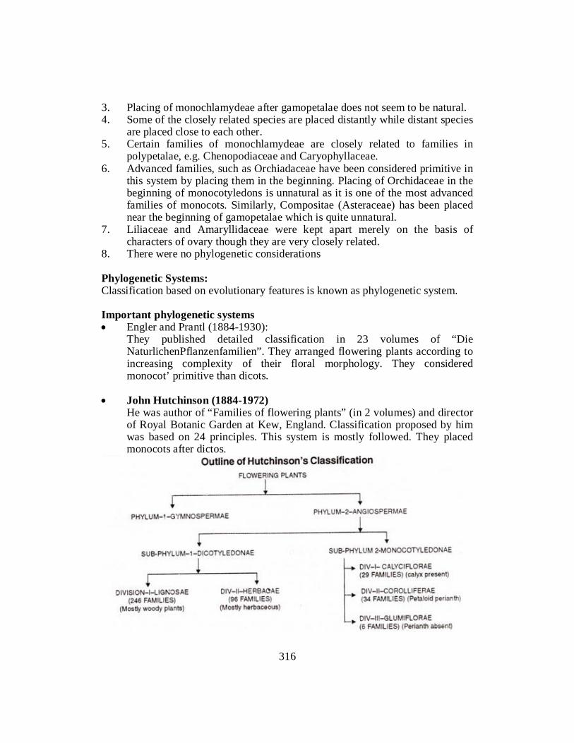
3. Placing of monochlamydeae after gamopetalae does not seem to be natural. 4. Some of the closely related species are placed distantly while distant species
are placed close to each other. 5. Certain families of monochlamydeae are closely related to families in
polypetalae, e.g. Chenopodiaceae and Caryophyllaceae. 6. Advanced families, such as Orchiadaceae have been considered primitive in
this system by placing them in the beginning. Placing of Orchidaceae in the beginning of monocotyledons is unnatural as it is one of the most advanced families of monocots. Similarly, Compositae (Asteraceae) has been placed near the beginning of gamopetalae which is quite unnatural.
7. Liliaceae and Amaryllidaceae were kept apart merely on the basis of characters of ovary though they are very closely related.
8. There were no phylogenetic considerations Phylogenetic Systems: Classification based on evolutionary features is known as phylogenetic system. Important phylogenetic systems • Engler and Prantl (1884-1930): They published detailed classification in 23 volumes of “Die
NaturlichenPflanzenfamilien”. They arranged flowering plants according to increasing complexity of their floral morphology. They considered monocot’ primitive than dicots.
• John Hutchinson (1884-1972) He was author of “Families of flowering plants” (in 2 volumes) and director
of Royal Botanic Garden at Kew, England. Classification proposed by him was based on 24 principles. This system is mostly followed. They placed monocots after dictos.
316

Self-Assessment Exercise 9.3 Q.1 Fill in the blanks 1. ………………. for the first time classified plants into Perfecti and Imperfect
based on the presence or absence of flowers. (Otto Brunfels) 2. ………………. divided flowering plants on the basis of petal bearing or
non-petal bearing flowers, simple or compound flowers regular or irregular flowers. (Joseph Pitton de Tournefort)
3. ………………. were divided on the basis of terrestrial and aquatic habits. (Monochlamydeae)
4. ………………. begin with the order Ranales which are now universally considered as to be the most primitive angiosperms.
Q. 2 MCQS 1. A.P. de Candolie divided plants into ………………. groups a) Cellular b) Vasculares c) non-vascular plants d) A and B 2. The Bentham and Hooker’s system of classification is for only: a) Seed Plants b) Flowering Plants c) Monocotyledons d) All 3. The Bentham and Hooker’s system classified all the seed plants into
………………. group a) Dicotyledon b) gymnosperm c) monocotyledons d) Above all 4. ………………. carries 82 families, 2610 genera & 31,874 species. a) Monochlamydae b) Polypetalae c) Gamopetalae d) None of above Q. 3 Short Questions 1. What are broad categories of classification of angiosperms? 2. What is difference between Polypetalae, Gamopetalae and Monocotyledons. 3. Briefly describe the merits of Bentham and Hooker’s System. 4. What is the Phylogenetic Systems of classification and its importance? 9.4 ECONOMIC IMPORTANCE OF ANGIOSPERMS Some of the most important families of flowering plant and their economic importance are as follows:
317

Family – Papilionaceae (fabaceae)
Distribution The family includes about400 genera. The members are distributed in the temperate regions of both northern and southern hemisphere. In India, the family is found both on hills and plains. Habits Plants are mostly herbs and shrubs, (often climbing). Few tree species are also known. Plants may be xerophyte, hydrophytes or mesophytes. Most of the plants are cultivated in fields. Root: Tap root branched, bearing root nodules, containing symbiotic nitrogen fixing bacteria (Rhizobia etc.) Stem: Erect, branched, angular or cylindrical, may be herbaceous or woody. Stem climbs with the help of tendrils. Leaf Ramal or cauline, alternate, simple or compound (digitate or pinnate). Leaves are stipulate (stipules arefoliaceous and large). Inflorescence Racemose, raceme, spikes or head. Flower Flowers are pedicellate, zygomorphic, irregular, pentamerous, hermaphrodite, complete, perigynous, and papilionaceous.
318

Calyx Sepals 5, gamosepolous (fused), equal or unequal, united in the form of tubular calyx. Aestivation is ascending imbricate. Corolla: Petals are five and Unequal. Uppermost is the largest, it is called standard or vexilluin. Two free lateral petals are called wings or alae. The anterior united pair of petals is called keel or carina. The keel encloses stamen and pistils. Aestivation is descending imbricate. Androecium Stamens are inserted on a disc, below the calyx. Stamens are generally 10, arranged in a ring. Nine stamens are united and remaining one is free (Diadelphous)
Fig.9.3: Floral Structure and flora : diagram of family Fabaceae Gynoecium Carpel one, free, semi superior ovary, unilocular, marginal placentation, short, hairy stigma. Fruit Legume or pod or lomentum.
319

Seed Exalbumincasus endosperm).Usually flattened.
Economic Importance of family The plants of this family are unique in that they have root nodules which contain nitrogen fixing symbiotic bacteria. Thus the products of these plants are the rich source of proteins. i. Plants used as food The family contains many food plants including pulses, grams and soyabean etc. Important such plants are:
• Arhar (Cajanuscajan), • Masoor (Lens esculenata), • Urd (Phaseolus mungo), • Mungo (Phaseolus aureus), • Lobia (Vignasinenis), • Pea (Pisumsativum), • Gram (Cicer arietinum), • Soyabean (Glycine max).
ii. Plants used for oils Groundnut (Arachis hypogea), Soyabean (Glycene soya) iii. Plants used for ornamental purposes
• Sesbania grandiflora, • Sweet pea (Lathyrusodomtus), • Lupin (Lupinus), • Butea monosperma etc.
iv. Plants giving timber and fibres Shishum (Dalbargiasisso), Sunn hemp (Crotolariajuncea). v. Plants used for other purposes Indigo plant (Indigoferatinctoria) it gives indigo dye. Plant Butterfly pea
(Clitoriaternatea) is used for medicinal purposes. Important Plants i. Pea (Pisumsativum) ii. Gram (Cicer arietinum)
320

iii. Soybean (Glycine max) iv. Sweet pea (Lathyrusodoratus) v. Shishum (Dalbargiasisso) vi. Sunn hemp (Crotolariaguncea) vii. Groundnut (Arachis hypogea) viii. Testu plant (Butea monospenna)
Family – Malvaceae (Mallow Family)
Systematic Position Distribution Malvaceae is a cosmopolitan family which includes about 80 genera and 1500 species. Most of the species and confined to tropics and subtropics. Habits Annual, biennial, herb, shrub or tree, mucilage rich. Root Tap root system, branched. Stem Erect, branched, cylindrical, usually mucilaginous. Leaves Stipulate, alternate, simple, petiole entire or lobed, reticulate venation. Inflorescence Solltary axillary or terminal racemose or hellicoid cyme.
321

Fig.9.4: Systematic study of floral diagram of Malvacae Flower Bracteate or ebracteate, actinomorphic, pedicellate, hermaphrodite, complete, hypogynous, pentamerous.Usually protected by a whorl of 3 to 12 bracteoles, collectively termed as Epicalyx.In Sida and Abutilon epicalyxis absent. Calyx Sepals are 5 in number, basally connate or polysepalous, valvate aestivation, usually epicalyx present. Corolla Petals 5, polypetalous but basally fused with staminal tube, twisted aestivation, showy. Androecium Numerous stamens, monadelphous, filaments fused to form astaminal tube, adnate to basal part of petals, epipetalous, anther lobes unilocular (monothecous), reniform, extrose. Gynoecium 3-carpels, syncarpous.multiloculai, ovary superior, axile placentation eachlocule has l-oo ovules, style single and simple, stigma multilobed. Fruit Dry loculicidal capsule (Abelmoschus and Gossypium) or carcerulus (Abutilon and Althaea).
322

Economic Importane i. Plants used as food 1. Hibiscus esculentus (=Abelmoschusesculentus) (Hindi – Rhindi): Fruits are
used as vegetables. ii. Plants used in medicines 1. Urenarependa: Juice extracted from roots and bark is used in hydrophobia. 2. Abutilon indicum: Root, stem, leaves and seeds are used in preparing
certain Ayurveda medicines. iii. Fibre yielding plants 1. Gossypiumsp: Fibres of seed coal are used in textile industry. Important
species of cotton are G. arboreum, G. herbaceum and G. barbadense. 2. Hibiscus connabinus, H. mutabilis and H. sabdariffa are used in rope
industry Fibre obtained from Urenalobata is used in preparing bags. iv. Ornamental Plants 1. Hibiscus rosa-sinensis (China rose) 2. Althea rosea (HollyHock).
FAMILY – ASTERACEAE (COMPOSITAE) (Sun-Flower Family)
Systematic Position Distribution It is the largest and the most advanced family of dicotyledones. It is represented by over 1000 genera and about 20,000 species. Members of the family are distributed worldwide. Habits Plants are usually herbs or shrubs and rarely trees and climbers. Many plants possess milky juice (latex), while others have watery, resinous and bitter juice.
323

Root Roots are tap and branched. Sometimes these are tuberously thickened (in Helianthus tuberosus). Stem Herbaceous, aerial, erect, branches usually covered with pubescence or glands for the secretion of milky latex. Leaf Simple or compound. Sometimes alternate, acicular (needle like or reduced to scales in xerophytes), exstipulate, petiolate. Surface is hairy. Leaf margins arc entire or serrated. Tip is acute or obtuse, venation reticulate.
Fig.9.5: Floral structure and diagram of typical member (Sunflower) of family Compositae Inflorescence Racemose, Head or capitulum, with an involuere of bracts.Racemose, rarely in spike. In capitulum, whole inflorescence looks like a single flower. Infect flowers in such cases are small and are called florets. Flower Flowers are arranged in inflorescence in the form of florets – ray floret and disc florets.
324

Ray florets Sessile, unisexual, zygomorphic, irregular, ligulate or bracteate. Calyx Represented by pappus (small hairy structure) or may be absent. Corolla Petals 5, gamopetalous, may be bilabiate or ligulate. Androecium Absent Gynoecium Carpels –2 (bicarpellary), inferior ovary, unilocular, with single anatropous ovule, basal placentation, single (achemal single seeded fruit). Fruit Cypsella (achenial single seeded fruit). Seed Non-endospermic.
Disc Florets Sessile, bisexual, actinomorphic, regular, bracteatc, complete, epigynous. Calyx Rudimentary, pappus. Corolla Petals 5, gamopetalous, tubular, 5 lobed, swollen near the base (where nectaries are present), valvate aestivation, variously coloured. Androecium 5 stamens, opipetalous, alternate with corollalobes, anthers are introse, dithecous. United laterally in to a lube, filaments free, syngenosious. Gynoecium Carpels are two hence known as (bicarpellary), inferior ovary, unilocular, with single anatropous ovule, basal placentation, single (achenial single seeded fruit). Fruit Cypsela (achenial single seeded fruit).
325

Seed Non-endospermic.
Economic Importance of family Plants used as food – Lettuce or salad (Lactucn sativa), Sun flower (Helianthus annuus). Medicinal Plants Salt minis obtained from Artemisia absinthium, Insulin is obtained from Cynarascolymus. For teeth pain, Centipeda orbicularis and Xanthium stramonium are used. Insecticide Chrysanthemum cinerarie folium gives natural insecticide – Pyrethrum. Important Plants i. Sun flower (Helianthus annuus) ii. Lettuce (Lactuca sativa) iii. Merigold (Tagetus erect us) v. Zinnia, Chrysanthemum, Tridax.
Family – Cruciferae (Brassicaceae) (Mustard Family)
Distribution It includes 350 genera and about 3000 species. The members of the family are cosmopolitan but majority of the members are found in north temperate region. Habits Plants are annual, biennial or perennial herbs. Root Roots are usually tap and branched. Several modifications of tap roots arc found such as fusiform (in Raphanussativus), conical (in Radish) and napiform (in turnip-Brassica rapa). These modifications mainly serve to store the food materials.
326

Stem Herbaceous, erect, cylindrical, usually reduced or condensed (in turnip and radish etc.). Leaf Simple, alternate, exstipulate and possess simple or branched hairs. Leaves are usually radical or cauline, sub-sessile or sessile lyrate. Radical roots are found in rosettes. Sometimes bulbils are born in the axils of upper leaves which are used for vegetative reproduction. Inflorescence Racemose and usually raceme. Flower Ebracteate, regular (actinomorphic), pedicillate, complete, hermaphrodite, tetramerous, cruciform and hypogynous. Calyx Consist of 4 sepals, polysepalous, arranged in two whorls of two each. The inner lateral sepals are sometimes pouched at the base and serve as nectar containers. Aestivation is imbricate. Corolla Have 4 Petals, polypetalous, cruciform arranged in a single whorl. These are alternate to the sepals. Each petal is differentiated into a narrow claw and a broad expanded limb, valvate.
Fig.9.6: Floral structure and floral diagram of a typical plant (Mustard –Brassica Campestris) of Family Brassiceae
327
Androecium Possess 6 stamens, arranged in two whorls of 2 and 4. Outer two stamens are short and inner 4 stamens are long, (tetradynamous). At the base of the outer two short stamens are present green glands or nectaries. Anthers are bilobed, basifixed and introse. Gynoecium Bicarpellary, syncarpous, superior ovary, unilocular, but becomes bilocular (due to the development of false septum or replum from the ingrowths of the parietal placentae). Placentation is parietal. Ovules are anatropous or campylotropous. Style is short with bilobed stigma. Fruit Siliqua or silicula. Sometimes it is lomentum (Raphanussativus). Seed Small, exalbuminous (non endospermic), with large curved embryo.Testa is mucilaginous.
Economic Importance of family i. Vegetable yielding plants
• Mustard (Brassica campestris), • Cabbage (Brassica oleraceavarcapitata), • Cauliflower (Brassica oleraceavar botrytis), • Radish (Raphanussativus).
ii. Oil yielding plants
• Brassica campestris, • Rye (B .Juncea) • Blackmustard (B. nigra), • Mustard (B. alba) etc.
iii. Medicinal plants
• Sisymbriumofficinale, • Nasturtium indica, • Eruca sativa, • Lepidiumsativum.
iv. Ornamental
• Candytuft (Iberisamara), • Myssum, • Methiola, • Wall flower (Cheiranthus).
328

Important Plants i. Mustard (Brassica campestris) ii. Cabbage (Brassica oleraceavarcapitata) iii. Cauliflower (Brassica oleraceavar botrytis) iv. Radish (Raphanussativus) v. Rye (Brassica Juncea) vi. Candytuft (Iberisamara)
Family -Solanaceae Systematic Position
Distribution The family is represented byabout.2500 species. This family is primarily distributed in tropical America and South America. Family members are also found in temperate regions. Solanum is the largest genus of the family with about 1500 species. Habits Plants are erect or climbing herbs, shrubs or small trees. Solanumnigrum is small annual herb. Datura is large annual herb. Root Have tap root or adventitious. Stem Herbaceous, woody, branched, erect or twining or creeping, (sometimes modified into tubers). Leaf Alternate or opposite (near the inflorescence), rarely whorled.Exstipulate, entire, simple, rarely dissected or pinnate. Inflorescence May be cymose or solitary. Flower Flower is pedicellate, hermaphrodite, actinomorphic (usually), regular, hypogynous, complete, pentamerous, bracts or bracteoles absent.
329

Calyx Consist of 5 sepals fused (gamosepalous), persistent. Corolla Have 5 petals, gamopetalous, funnel shaped, rotate, or tubular, variously shaped.Plicate or convolute (rarely valvate). Androecium Consist of 5 stamens, epipetalous (fused with petals and Inserted into corolla tube), alternating with petals. Anthers are dithecous, filamentous with unequal length. Gynoecium Bicarpellary, syncarpous, superior ovary, bilocular, axile placentation, many ovules, ovules are anatropous (usually) or amphitropous. Carpels are placed obliquely in the flowers. Style one, stigma is bilobed and capitate. Fruit Berry or septicidal capsule Seed Albuminous (endospermic), smooth or pitted, curvedem
Figure. 9.7: Floral structure and floral diagram of a typical plant (Solanum nigrum) of Family Solanaceae
330

Economic Importance of family i. Food Plant
• Potato [Solatiumtuberosum), • Brinjal (Solanummelongena), • Tomato (Lycopersiconesculentum), • Red chillies (Capsicumannuum).
ii. Medicinal plants
• Atropine (Atropabelladona), • Henbane (Hyoscyamus Niger), • Asgandh (Withaniasomnifera), • Stramonium or Datura (Daturastramonium) etc.
iii. Ornamented Plant
• Queen of night (Cestrum nocturnum), • King of Day (Cestrum diurnum), • Petunia (Petunia violacea) etc.
iv. Tobacco Plant Nicotianatabacum and N. rustica provide alkaloid nicotine, which is used for
chewing smoking and also as insecticide. Important Plants • Potato (Solanumtuberosum) • Tomato (Lycopersicionesculentum) • Atropine (Atropabelladona) • Tobacco (Nicotiacum)
Family – Liliaceae
Distribution Plants are cosmopolitan and chiefly found in temperate and subtropical regions. These plants survive in most varied conditions.
331

Habits The plants are annual, biennial and perennial herbs and shrubs. The plants are mostly xerophytic.
Fig.9.8: Floral structure and floral diagram of family Lilliacae (Asphodelus tenuilifollius) Self Assessment Exercise 9.4 Q.1 Fill in the blanks 1. Sesbania grandiflora, Sweet pea (Lathyrusodomtus), Lupin (Lupinus), Butea
monosperma are used for …………. purposes. (ornamental purposes) 2. The fibers and timber are collected by …………. and …………. plants.
(Shishum, Sunn hemp) 3. Plant …………. is used for medicinal purposes. (Butterfly pea) 4. Dry …………. capsule and …………. are fruits of Malvaceae family.
(loculicidal, carcerulus) Q.2 MCQS 1. The family includes about…………. genera. a) 400 b) 500 c) 200 d) 4000 2. The plants of Papilionaceae (fabaceae) family have unique characteristics of: a) Nitrogen fixation b) Carbon fixation c) Hydrogen fixation d) Above all
332

3. Lobia, Masoor and urd are food of …………. family a) Fabaceae b) Malvaceae c) Asteraceae d) A and B 4. The plants used for oils production; a) Groundnut b) Soyabean c) Sweet pea d) Both A and B Q.3 Give Short Answers to the Following Questions: 1. Describe fiber yielding plants of Malvaceaefamily? 2. Briefly describe features of Asteraceae family? 3. Brief inflorescence of Asteraceae family? 4. What is economic importance of family Asteraceae family?
333

References A TEXTBOOK OF BOTANY - ANGIOSPERMS by B.P. Pandey. S. Chand and Company Ltd., New Delhi. A TEXTBOOK OF BOTANY - by S.N. Pandey, S.P. Mishra and P.S. Trivedi (2004).Vikas Publishing House Pvt. Ltd., New Delhi. CONIFERS - THE ILLUSTRATED ENCYCLOPEDIA.Vol. I & II - by D.H. van Gelderen& J.R.P. van Hoey Smith (1996), Timber Press, Inc. Orgon, U.S.A. Crane, P. R., E. M. Friis, and K. R. Pederson."The Origin and Early Diversification of Angiosperms." Nature 374 (1995): 27–33. Judd, Walter S., Christopher S. Campbell, Elizabeth A. Kellogg, and Peter F. Stevens. Plant Systematics: A Phylogenetic Approach. Sunderland, MA: Sinauer Associates, Inc., 1999. Kenrick, Paul. "The Family Tree Flowers." Nature 402 (1999): 358–359. MORPHOLOGY AND EVOLUTION OF VASCULAR PLANTS - by E.M. Gifford, & A.S. Foster (1989) W.H. Freeman & Co., New York. MORPHOLOGY OF VASCULAR PLANTS - by D.W. Bierhorst (1971), The Macmillan Co., New York. Parkinson, C. L., K. L. Adams, and J. D. Palmer. "Multigene Analyses Identify the Three Earliest Lineages of Flowering Plants." Current Biology 9 (1999): 1485–1488. PLANT ANATOMY by A. Fahn (1990).Butterworth Heinemann Ltd. Linacre House, Jordon Hill, Oxford OX2 8DP. THE PRIVATE LIFE OF PLANTS - by David Attenborough (1995), BBC Enterprises, Ltd. Woodlands, London.
334