3D QSAR studies on binding affinities of coumarin natural products for glycosomal GAPDH of...
14
Journal of Computer-Aided Molecular Design 17: 277–290, 2003. © 2003 Kluwer Academic Publishers. Printed in the Netherlands. 277 3D QSAR studies on binding affinities of coumarin natural products for glycosomal GAPDH of Trypanosoma cruzi Irwin R.A. Menezes 1 , Julio C.D. Lopes 1 , Carlos A. Montanari 1,∗ , Glaucius Oliva 2 , Fernando Pavão 2 , Marcelo S. Castilho 2 , Paulo C. Vieira 3 & Mônica T. Pupo 4 1 N´ ucleo de Estudos em Qu´ ımica Medicinal, NEQUIM. Chemistry Department, University of Minas Gerais, 31270- 901, Belo Horizonte, MG, Brazil; 2 Laborat´ orio de Cristalograf` ıa de Prote´ ınas e Biologia Estrutural, Instituto de F´ ısica de São Carlos, Universidade de São Paulo, USP, Av. do Trabalhador Sãocarlense 400, PO Box 369, 13566- 590 São Carlos, SP, Brazil; 3 Departamento de Qu´ ımica – Universidade Federal de São Carlos. Via Washington Luiz Km 235 – CP 676, 13565-905 São Carlos, SP, Brazil; 4 Faculdade de Ciências Farmacêuticas de Ribeirão Preto, USP, Av. do Caf´ e, s/n, 14040-903, Monte Alegre, Ribeirão Preto, SP, Brazil Key words: Chagas’ disease, Coumarin natural products, glycosomal glyceraldehyde-3-phosphate dehydrogenase (gGAPDH), CoMFA, GRID/VolSurf/GOLPE. Summary Drug design strategies based on Comparative Molecular Field Analysis (CoMFA) have been used to predict the activity of new compounds. The major advantage of this approach is that it permits the analysis of a large number of quantitative descriptors and uses chemometric methods such as partial least squares (PLS) to correlate changes in bioactivity with changes in chemical structure. Because it is often difficult to rationalize all variables affecting the binding affinity of compounds using CoMFA solely, the program GRID was used to describe ligands in terms of their molecular interaction fields, MIFs. The program VolSurf that is able to compress the relevant information present in 3D maps into a few descriptors can treat these GRID fields. The binding affinities of a new set of compounds consisting of 13 coumarins, for one of which the three-dimensional ligand-enzyme bound structure is known, were studied. A final model based on the mentioned programs was independently validated by synthesizing and testing new coumarin derivatives. By relying on our knowledge of the real physical data (i.e., combining crystallographic and binding affinity results), it is also shown that ligand-based design agrees with structure-based design. The compound with the highest binding affinity was the coumarin chalepin, isolated from Rutaceae species, with an IC 50 value of 55.5 µM towards the enzyme glyceraldehyde-3-phosphate dehydrogenase (gGAPDH) from glycosomes of the parasite Trypanosoma cruzi, the causative agent of Chagas’ disease. The proposed models from GRID MIFs have revealed the importance of lipophilic interactions in modulating the inhibition, but without excluding the dependence on stereo-electronic properties as found from CoMFA fields. Abbreviations: CAMD, Computer-Aided Molecular Design; CoMFA, Comparative Molecular Field Analysis; CV, Cross-Validation; DALYs, disability-adjusted life years; FFD, Fractional Factorial Design; GOLPE, Gen- erating Optimal Linear PLS Estimations; gGAPDH, glycosomal Glyceraldehyde-3-Phosphate DeHydrogenase; MIFs, Molecular Interaction Fields; PC, Principal Component; PLS, Partial Least Squares; QSAR, Quantitative Structure-Activity Relationships; SAMPLS, SAMple-distance Partial Least Squares; VRS, Virtual Receptor Site. Introduction Chagas’ disease is estimated to affect ca. 18 mil- lion people, mostly from South and Central Amer- ica, though increasing incidence has been reported in ∗ To whom correspondence should be addressed. E-mail: [email protected] urban areas of North America. About 3 million of the infected people develop severe complications, such as chronic cardiopathy, digestive lesions and neuro- logical disorders, causing 45,000 deaths per year and a loss of ca. 3 million disability-adjusted life years (DALYs) [1]. Blood transfusion and congenital trans-
-
Upload
independent -
Category
Documents
-
view
0 -
download
0
Transcript of 3D QSAR studies on binding affinities of coumarin natural products for glycosomal GAPDH of...
Journal of Computer-Aided Molecular Design 17: 277–290, 2003.© 2003 Kluwer Academic Publishers. Printed in the Netherlands.
277
3D QSAR studies on binding affinities of coumarin natural products forglycosomal GAPDH of Trypanosoma cruzi
Irwin R.A. Menezes1, Julio C.D. Lopes1, Carlos A. Montanari1,∗, Glaucius Oliva2, FernandoPavão2, Marcelo S. Castilho2, Paulo C. Vieira3 & Mônica T. Pupo4
1Nucleo de Estudos em Quımica Medicinal, NEQUIM. Chemistry Department, University of Minas Gerais, 31270-901, Belo Horizonte, MG, Brazil; 2Laboratorio de Cristalografıa de Proteınas e Biologia Estrutural, Instituto deFısica de São Carlos, Universidade de São Paulo, USP, Av. do Trabalhador Sãocarlense 400, PO Box 369, 13566-590 São Carlos, SP, Brazil; 3Departamento de Quımica – Universidade Federal de São Carlos. Via WashingtonLuiz Km 235 – CP 676, 13565-905 São Carlos, SP, Brazil; 4Faculdade de Ciências Farmacêuticas de RibeirãoPreto, USP, Av. do Cafe, s/n, 14040-903, Monte Alegre, Ribeirão Preto, SP, Brazil
Key words: Chagas’ disease, Coumarin natural products, glycosomal glyceraldehyde-3-phosphate dehydrogenase(gGAPDH), CoMFA, GRID/VolSurf/GOLPE.
Summary
Drug design strategies based on Comparative Molecular Field Analysis (CoMFA) have been used to predict the activity of newcompounds. The major advantage of this approach is that it permits the analysis of a large number of quantitative descriptorsand uses chemometric methods such as partial least squares (PLS) to correlate changes in bioactivity with changes in chemicalstructure. Because it is often difficult to rationalize all variables affecting the binding affinity of compounds using CoMFAsolely, the program GRID was used to describe ligands in terms of their molecular interaction fields, MIFs. The programVolSurf that is able to compress the relevant information present in 3D maps into a few descriptors can treat these GRIDfields. The binding affinities of a new set of compounds consisting of 13 coumarins, for one of which the three-dimensionalligand-enzyme bound structure is known, were studied. A final model based on the mentioned programs was independentlyvalidated by synthesizing and testing new coumarin derivatives. By relying on our knowledge of the real physical data (i.e.,combining crystallographic and binding affinity results), it is also shown that ligand-based design agrees with structure-baseddesign. The compound with the highest binding affinity was the coumarin chalepin, isolated from Rutaceae species, with anIC50 value of 55.5 µM towards the enzyme glyceraldehyde-3-phosphate dehydrogenase (gGAPDH) from glycosomes of theparasite Trypanosoma cruzi, the causative agent of Chagas’ disease. The proposed models from GRID MIFs have revealed theimportance of lipophilic interactions in modulating the inhibition, but without excluding the dependence on stereo-electronicproperties as found from CoMFA fields.
Abbreviations: CAMD, Computer-Aided Molecular Design; CoMFA, Comparative Molecular Field Analysis;CV, Cross-Validation; DALYs, disability-adjusted life years; FFD, Fractional Factorial Design; GOLPE, Gen-erating Optimal Linear PLS Estimations; gGAPDH, glycosomal Glyceraldehyde-3-Phosphate DeHydrogenase;MIFs, Molecular Interaction Fields; PC, Principal Component; PLS, Partial Least Squares; QSAR, QuantitativeStructure-Activity Relationships; SAMPLS, SAMple-distance Partial Least Squares; VRS, Virtual Receptor Site.
Introduction
Chagas’ disease is estimated to affect ca. 18 mil-lion people, mostly from South and Central Amer-ica, though increasing incidence has been reported in
∗To whom correspondence should be addressed. E-mail:[email protected]
urban areas of North America. About 3 million of theinfected people develop severe complications, suchas chronic cardiopathy, digestive lesions and neuro-logical disorders, causing 45,000 deaths per year anda loss of ca. 3 million disability-adjusted life years(DALYs) [1]. Blood transfusion and congenital trans-

278
Figure 1. Structure of compounds used in the treatment of Chagas’ disease.
mission are currently the major causes of the spread ofthe disease.
The available drugs, nifurtimox and benznidazolehave strong side effects [2]. Although administeredsince the 1970s, [2], nifurtimox is no longer useddue to severe side effects [3]. Other important mo-lecules for treatment of Chagas’ disease are allop-urinol, ketoconazole, fluconazole and itraconazole [2].Figure 1 shows their structures along with nifurtimoxand benznidazole.
We are focusing on the new glyceraldehyde-3-phosphate dehydrogenase, gGAPDH, for the discov-ery of drugs acting against T. cruzi [4–6]. Sinceintracellular amastigotes derive their energy mainlyfrom glycolysis, the inhibition of gGAPDH poten-tially prevents T. cruzi from being infective [2]. Theneed for discovering bioactive new chemical entities,BIONCEs, against Chagas’ disease has stimulated ourgroup to begin a study of screening natural compoundsagainst recombinant T. cruzi gGAPDH obtained in apET3a-Escherichia coli expression system [4, 7].
Natural products derived from medicinal plantsrepresent a very powerful array of compounds thatcan be used as ‘hit-to-lead’ candidates of interest inmedicinal chemistry [8]. The isolation and identifica-tion of natural products candidates are widespread intropical Countries. Brazilian biodiversity is potentiallyfull of hits, which are just waiting to be leads. Nev-ertheless, mass screening is not an easy task since itcan be quite expensive and time consuming. Chem-
ical diversity is buried throughout many laboratorieswaiting to be discovered. In attempting to circumventthe problem we have carried out a computer aided mo-lecular design, CAMD, to perform virtual screening ofnatural products [9, 10]. The joint tools of structure-based design and classical or 3D QSAR studies can beemployed when structure of the receptor is known. Af-finities can be improved in advance through CAMD.After that, binding can be measured experimentallyand the system re-cycled to aid in the choice of onlya small number of molecules to be screened. At thispoint, the medicinal chemistry project can be started[8].
The Brazilian physician Carlos Chagas discoveredthe disease in 1909. Though many years have pastsince Dr. Chagas discovered the ethiology of thedisease no new drug has entered in the market tohelp the myriad of patients who severely suffer fromit. The main objective of this study is to find newnatural product hits from Brazilian flora, which arecapable of acting against the T. cruzi glycosomalenzyme gGAPDH. Structure-Activity Relationships,SAR, will be developed in order to help fulfil this goal.

279
Materials and methods
Computer hardware
All calculations presented were performed on aR10000 O2 Silicon Graphics workstation.
In-house natural products database
The starting compounds analyzed in this study werenatural products, isolated and structurally identified,in our on-going project of identifying molecules frommedicinal plants of tropical disease interest.
Molecular docking
A library of 93,000 compounds was ranked accordingto DOCK3.5 scores [11], using the gGAPDH X-Raycrystallographic structure as target [4, 12]. The bind-ing site of the adenosine ring of the NAD+ cofactorand the catalytic site of gGAPDH possesses significantdifferences between the parasite and the homologoushuman enzyme [4], thereby being an attractive tar-get for trypanocidal drug design [13]. The processof using DOCK provided some new coumarin nat-ural products highly ranked among many compoundsfrom the database. These well-scoring compounds,isolated from several previous phytochemical studiesand available in our laboratories, were among othersscreened against our recombinant T. cruzi enzyme [7].
Receptor binding affinities
The ligands’ in vitro affinity for T. cruzi gGAPDHreceptor is the dependent variable considered in thisstudy. In the binding study, the affinity of the com-pounds was determined by their ability to displaceNAD+ from the enzyme receptor site [7]. Receptorbinding affinities were expressed as IC50 (µM) val-ues. The natural coumarin chalepin was selected fordetermining the co-complex with gGAPDH [7, 12,13] because it has the best inhibitory concentrationtowards the enzyme.
X-Ray crystallographic data and structuredetermination of chalepin bound to gGAPDH
The structure of the complex of T. cruzi gGAPDHwith chalepin has been reported [12]. The gGAPDHreceptor site was studied in order to compare its calcu-lated GRID MIFs with those ones obtained for ligandsthemselves, with the aim of establishing relationshipsamong them two.
Molecular fields generated in the Sybyl/CoMFA andGRID program
In both SYBYL/CoMFA [14] and GRID, [15–17] agrid big enough to enclose all ligands was created.The interaction energies between a probe (Csp3+ forCoMFA; OH2 and DRY for GRID), and the targetmolecules were calculated, in each grid point.
Molecular modelling
Sybyl Tripos force field [18] was used for modellingthe 13 molecules found in Table 1. Derived partialcharges obtained from Gasteiger-Hückel method tocalculate the electrostatic field interactions in CoMFAwere used throughout this work.
The starting chalepin conformation was the oneobtained from our X-Ray structure of the co-complexbetween chalepin and gGAPDH, which has been takenas a template to build all molecules in the set. Theconformational space, of all ligands, was explored bya conformational search procedure using the system-atic search method, as implemented in Sybyl. Thenew minimized selected conformations were outside5 kcal mol−1 difference, whenever feasible.
The steric and electrostatic potential energies werecalculated with the standard CoMFA procedure. TheSAMPLS algorithm implemented in Sybyl6.5 wasused to perform the cross-validation analyses. Thenon-cross-validated data were used in the analysis ofthe field results, calculations and the predictions.
Molecular interaction fields, MIFs
MIFs were obtained using the program GRID version20. Computed on gGAPDH receptor site, the MIFsidentified regions where water and DRY probes in-teracted favourably, suggesting positions where func-tional groups should be placed in ligands.
MIFs were also computed for the ligands them-selves. In this case, the regions showing favourableenergy of interaction represent positions where groupsof the receptor interact with the ligands. Using dif-ferent probes, we can obtain for a certain liganda set of such positions, which characterizes a ‘Vir-tual Receptor Site’ (VRS). This abstract entity definesan ideal complementary site for a certain chemicalcompound and represents its potential ability to bindgGAPDH. The regions defined in its VRS overlapgroups of the real receptor site where at least a sub-set of the VRS is relevant for representing the bindingproperties of the ligands.

280
VolSurf descriptors
The GRID-VolSurf [19] procedure is completely auto-mated. Firstly, it generates MIFs by using GRID20.Then, it treats the fields accordingly by producingdescriptors that encode the information content fromthe used probes. VolSurf has the advantage of produ-cing descriptors using the 3D information embeddedin any map. VolSurf is also alignment independent andconformation insensitive (independent, in this study).The VolSurf transformation is fast and its results areusually easy to interpret. The descriptors have a clearchemical meaning and are lattice-independent.
The GRID-VolSurf descriptors [19] (water and hy-drophobic DRY based probes) on studied coumarinsexported to GOLPE program were treated by the vari-able selection method of Fractional Factorial Design,FFD, [20]. The FFD variable selection procedure wasapplied in two runs, but only the first one yielded theappropriate model (active variables: 56, Dummies: 15,X-comb.: 256, SDEP = 0.190, r2
cv (Q2) = 0.610, Max.dimensionality: 1, Validation Mode: LOO, Recalcu-late weights: yes, Comb./Var. ratio: 2.0). After theFFD run, the final number of X-variables was reducedto 28.
The partial least squares (PLS) model
In the context of 3D-QSAR, the biological activitymay be seen as a function of the physiochemical char-acteristics (such as electronic properties or energiesof interaction within a given force field) of the com-pounds of interest. The need to convert such numericaldata to useful information has led to the developmentof methodologies that rely on statistics and appliedmathematics. The PLS model [21] is a two-block pro-jection method that relates a matrix X (containing thechemical descriptors) to a matrix Y (containing thebiological activities) with the aim of predicting the val-ues in Y from the information contained in X. This hasbeen the method used in CoMFA and Volsurf analyses.
Generating optimal linear PLS estimation (GOLPE)
GOLPE [22] is defined as an advanced variable se-lection procedure aimed at obtaining PLS regressionmodels with the highest prediction capability, whichrelies on the validation of a number of reduced mod-els on variable combinations, selected according to afactorial design strategy. The power of a GOLPE pro-cedure will depend on some method of pre-treatment
of the data. In this paper, descriptors generated viaVolSurf analysis were the input.
Results and discussion
From our natural products program on the identific-ation, isolation, and structure elucidation of smallmolecules relating to drug design, coumarins obtainedfrom Rutaceae species [7], were found to be hits forthe key enzyme gGAPDH from T. cruzi, through avirtual screening procedure. The study molecules weredocked in the receptor site of gGAPDH. The interac-tion energies ranked according to the scoring functionof DOCK3.5, which allowed them to be selected forthe screening assay against the enzyme. Sampling thecoordinate space of the binding site and scoring eachpossible ligand according to the interaction energies,resulted in the predicted binding mode for that com-pound. The whole procedure is described elsewhere[23].
A defined protocol [7] was conducted for measur-ing their inhibitory concentrations. The binding affin-ity data of the 13 chosen coumarin compounds can befound in Table 1, along with their chemical structures,and the calculated binding affinities.
Two different computer aided molecular designmethods were applied to describe the binding affinit-ies of chalepin analogues towards gGAPDH, namelyCoMFA and GRID. These two methods were chosenbecause together they can incorporate information onbinding affinities according to electrostatic, steric andhydrophobic fields.
From the rigid docking analyses [23], no ligandflexibility was taken into consideration. The con-formation of a compound bound to the receptor sitemight be different from the conformation of the un-bound form in solution. Hence, having started withCoMFA analysis of coumarin ligands shown in Table1, we generated and relaxed conformations from asystematic conformational search of coumarin flexiblemoieties, as available in Sybyl6.5. The bound con-formation of chalepin to gGAPDH was also tested asthe pharmacophoric one in obtaining CoMFA fields.The needed similar analogue conformations were gen-erated as described elsewhere [24]. All compoundswere then aligned atom-by-atom as follows: (i) overcompound 1(TC_cum_1), chalepin, as obtained fromthe receptor site, (ii) over the lowest energy conforma-tion of all compounds, and (iii) over the highest energyconformation; but all sharing the core of coumarin

281
Table 1. Coumarins assayed and their binding affinities (actual/calculated) toward gGAPDH.
Compounds Name Structures1 Actual2 CoMFA3 CoMFA
(log1 / IC50) calculated Residual
1 Chalepin TC_cum_1 4.26∗ 4.26 0.0022
2 TC_cum_10 3.59 3.55 0.04
3 TC_cum_11 3.46 3.43 0.03
4 TC_cum_12 3.21 3.25 −0.04
5 TC_cum_13 3.16 3.16 −0.002
6 TC_cum_2 3.84 3.88 −0.04
7 TC_cum_3 3.71∗ 3.71 −0.0039
8 TC_cum_4 4.13∗ 4.12 0.01
9 TC_cum_5 3.91 3.92 −0.01
10 TC_cum_6 3.89 3.88 0.01
11 TC_cum_7 3.79 3.82 −0.03
12 TC_cum_8 3.72 3.69 0.03
13 TC_cum_9 3.68 3.68 −0.005
1Chemical structures of selected coumarin natural products from virtual screening of DOCK3.5.References [7, 23].2Binding affinity magnitude values obtained from in vitro study of the T. cruzi gGAPDH inhib-ition. Values are taken as log1/IC50, in µM, [7]. Marked values (∗) were corrected before beingapplied in this work.3CoMFA internal calculated binding affinities, according to steric and electrostatic molecularfields.

282
Figure 2. Coumarin atom-by-atom superposition used for CoMFAanalysis.
rings. Thirty percent of coumarin compounds listedin the Table 1 was dropped from the analysis to actas test compounds. By using the standard Tripos forcefield from Sybyl version 6.5 program [18], we calcu-lated the electrostatic and steric fields. Figure 2 showsthe structures and the alignment used for all phar-macophoric (bound) conformations as obtained fromreceptor site.
The results shown in Table 2 do reveal relation-ships between steric and electrostatic molecular fieldscalculated from CoMFA.
The optimum number of components was foundto be two (Table 2). Those values were sufficient fora 3D QSAR model. Furthermore, the correspondingnon-cross-validated QSAR model has a good fit withr2 (0.99) and a small s value, and with reasonable Fvalue. The model also suggests there is a major stericcontribution of binding affinity towards gGAPDH.
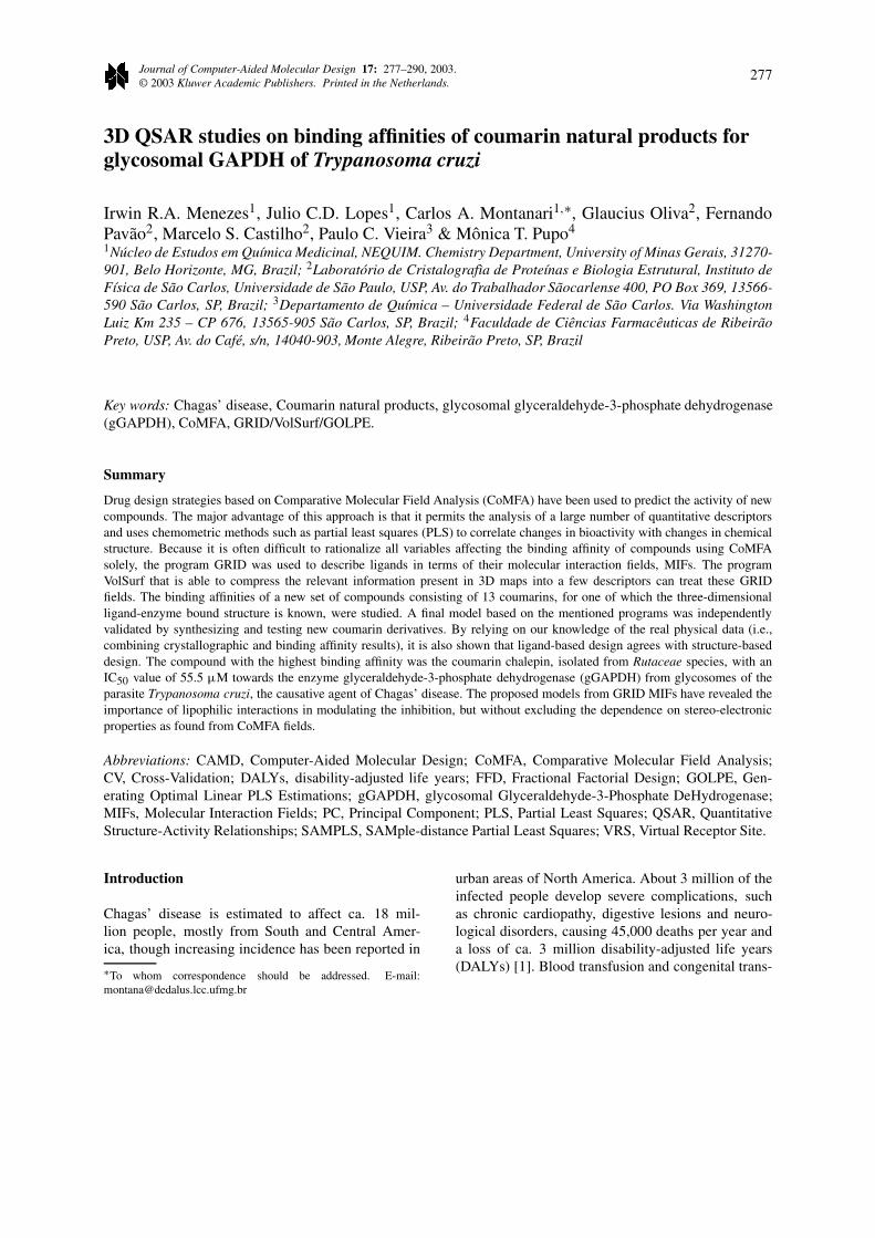
The CoMFA coefficient map for the model wascontoured around chalepin, with the highest affinitytowards gGAPDH (Figure 3) to illustrate the locationsthat affect binding. Both steric and electrostatic fieldsare displayed at the same location. They were con-toured as follows: the contours of the steric map areshown in yellow; those of the electrostatic map areshown in blue. Greater values of binding affinities arecorrelated with less bulk near yellow, more positivecharge near blue. The steric maps (Figure 3) indicatethat less bulk constituents at the both ends of chalepinare favourable for increasing binding affinities.
Conducting predictions on the internal test set val-idated the predictive capability of the CoMFA model.As a result, by using the contour maps from Fig-ure 3, we have investigated new derivatives to besynthesized. This has been done by changing the 1,1-
dimethylallyl moiety of chalepin with bulkier groups,since synthetic efforts were firstly made possiblethrough modifications of the double bond. Of course,with the CoMFA prediction, chalepin derivatives werenot better then the chalepin itself. Nevertheless, thisnegative result is enough to validate externally ourCoMFA model, because predicted bulkier groups atthe 1,1-dimethylallyl moiety to be detrimental forbinding to gGAPDH.
The designed new molecules with better calculatedbinding affinities based on coumarin 8(TC_cum_4)are not yet synthetically feasible in our laboratories.Since we would like to have molecules with higheraffinities toward gGAPDH, and make them synthet-ically possible, we decided to attempt incorporation ofnew molecular interaction fields, MIFs, in the model.For this reason, we have included hydrophobic fieldsfrom GRID, because these fields are needed to keepchalepin at the receptor site, (see below).
GRID-VolSurf
The existence of H-bonding and hydrophobic pock-ets in the receptor site may be investigated through anew procedure called VolSurf [25, 26]. This approachhas been used to correlate 3D molecular interactionfields, MIFs, with physico-chemical and pharma-cokinetic properties, plasticization [27], bioactivities[28], chemical space navigation [29]. To date themain use of VolSurf is related to structure-property re-lationships, albeit structure-activity relationships andstructure-binding affinities should be possible. Duringthe preparation of this manuscript, the use of VolSurfto calculate surface descriptors for protein-ligand af-finity was published [30].
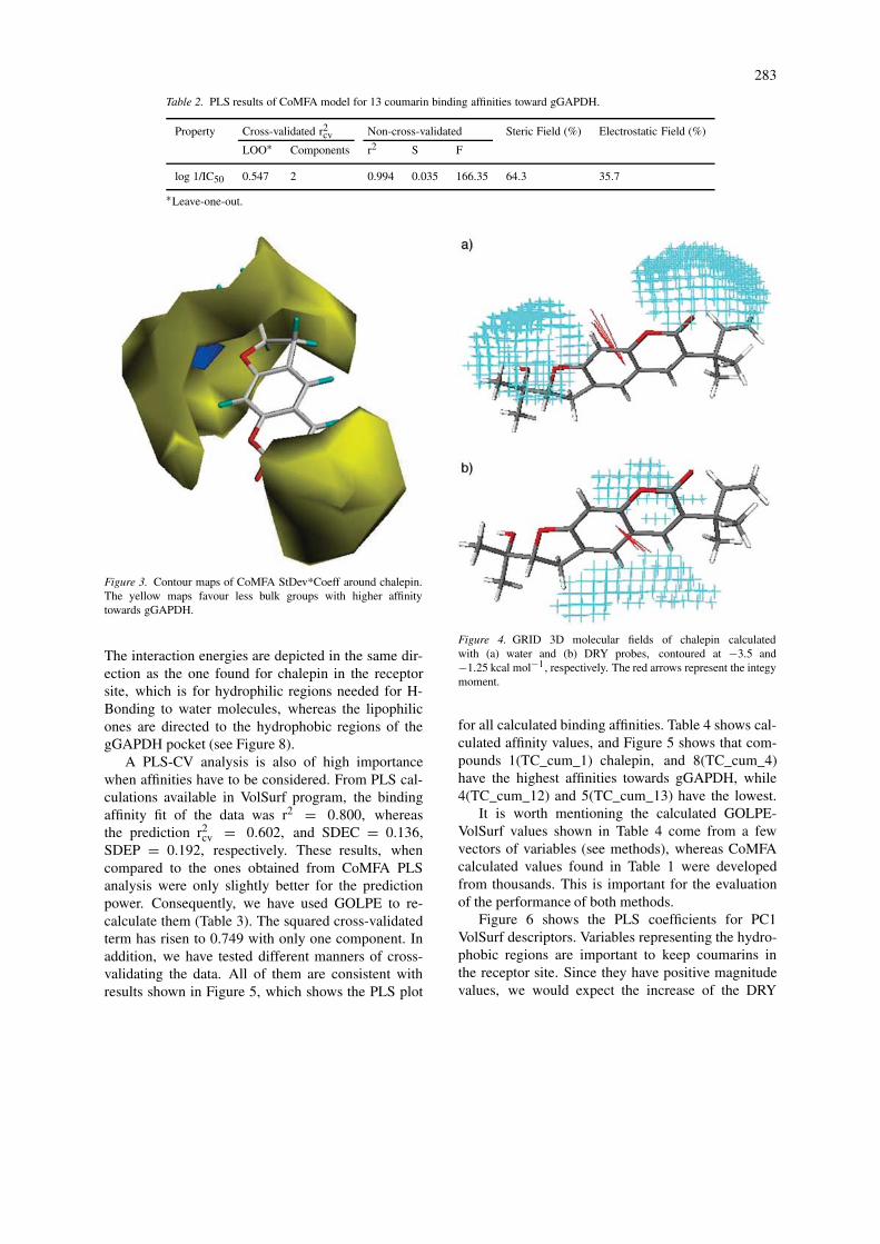
Hydrogen bonding and hydrophobicity, duly cal-culated by GRID force field [15–17], mediate someimportant interactions between chalepin and its re-ceptor site. Figure 4 shows chalepin calculated MIFsfor water and DRY (the hydrophobic probe).
The coloured regions represent where the inter-action between the ligand and the (a) water and (b)DRY probes are highly favourable. The contour en-closes regions where the target molecule can make(a) hydrogen bonds and (b) lipophilic interactions.The interaction energy moments, red arrows in Fig-ure 4, referring to (a) hydrophilic regions, are vectorspointing from the centre of mass to the centre of thehydrophilic regions; (b) hydrophobic regions, measurethe unbalance between the centre of mass of a mo-lecule and the barycentre of the hydrophobic regions.
283
Table 2. PLS results of CoMFA model for 13 coumarin binding affinities toward gGAPDH.
Property Cross-validated r2cv Non-cross-validated Steric Field (%) Electrostatic Field (%)
LOO∗ Components r2 S F
log 1/IC50 0.547 2 0.994 0.035 166.35 64.3 35.7
∗Leave-one-out.
Figure 3. Contour maps of CoMFA StDev*Coeff around chalepin.The yellow maps favour less bulk groups with higher affinitytowards gGAPDH.
The interaction energies are depicted in the same dir-ection as the one found for chalepin in the receptorsite, which is for hydrophilic regions needed for H-Bonding to water molecules, whereas the lipophilicones are directed to the hydrophobic regions of thegGAPDH pocket (see Figure 8).
A PLS-CV analysis is also of high importancewhen affinities have to be considered. From PLS cal-culations available in VolSurf program, the bindingaffinity fit of the data was r2 = 0.800, whereasthe prediction r2
cv = 0.602, and SDEC = 0.136,SDEP = 0.192, respectively. These results, whencompared to the ones obtained from CoMFA PLSanalysis were only slightly better for the predictionpower. Consequently, we have used GOLPE to re-calculate them (Table 3). The squared cross-validatedterm has risen to 0.749 with only one component. Inaddition, we have tested different manners of cross-validating the data. All of them are consistent withresults shown in Figure 5, which shows the PLS plot
Figure 4. GRID 3D molecular fields of chalepin calculatedwith (a) water and (b) DRY probes, contoured at −3.5 and−1.25 kcal mol−1, respectively. The red arrows represent the integymoment.
for all calculated binding affinities. Table 4 shows cal-culated affinity values, and Figure 5 shows that com-pounds 1(TC_cum_1) chalepin, and 8(TC_cum_4)have the highest affinities towards gGAPDH, while4(TC_cum_12) and 5(TC_cum_13) have the lowest.
It is worth mentioning the calculated GOLPE-VolSurf values shown in Table 4 come from a fewvectors of variables (see methods), whereas CoMFAcalculated values found in Table 1 were developedfrom thousands. This is important for the evaluationof the performance of both methods.
Figure 6 shows the PLS coefficients for PC1VolSurf descriptors. Variables representing the hydro-phobic regions are important to keep coumarins inthe receptor site. Since they have positive magnitudevalues, we would expect the increase of the DRY

284
Table 3. PLS results of GOLPE model, based on GRID/VolSurf MIFs and descriptors.
Property Cross-validated (r2cv) Non-cross-validated
LOO1 LTO2 5 random groups out Components SDEP3 r2 SDEC4
log 1/IC50 0.758 0.746 0.746 1 0.192 0.838 0.122
1Leave-one-out.2Leave-two-out.3Standard Deviation of Estimated Prediction.4 Standard Deviation of Estimated Calculation.
Table 4. GOLPE-VolSurf calculated binding affinities forstudy coumarins.
Compounds1 GOLPE-VolSurf2 GOLPE-VolSurf
calculated residual
1 Chalepin 4.16 0.1
2 3.66 −0.07
3 3.73 −0.27
4 3.46 −0.25
5 3.27 −0.11
6 3.67 0.17
7 3.66 0.05
8 4.03 0.1
9 3.57 0.34
10 3.65 0.24
11 3.87 −0.08
12 3.79 −0.07
13 3.75 −0.07
1See Table 1 for structures.2GOLPE calculated values of binding affinities, accordingtoVolSurf descriptor calculations.See text for explanation.
Figure 5. PLS plot of T versus U, for one component only.
volumes in molecules to increase affinities. However,the hydrophilic-lipophilic regions, albeit a bit lowerin height as compared to the hydrophobic peaks, ex-hibit the same trend. On the other hand, the integymoments are negatively related to binding affinities,and thus we would expect as they increase the bindingwould decrease. These high integy moments representstrong polar regions concentrated in few regions of themolecular surface. This can be seen from Figure 4where the polar regions are within specific parts ofthe molecule. Size and shapes can be used for similarinterpretation of binding affinities. These observationsare essentially the same as found from PLS partialweights plot (not shown).
From these models, we designed syntheticallyfeasible new coumarin derivatives in order to validatethem now externally.
Considering that steric, electrostatic and hydro-phobic requirements for the new structures in the 3DMIFs were used to account for coumarin gGAPDHbinding affinities, we tailored the ligands with syn-thetic feasibility in mind. However, this time focus-ing on molecular modifications of the coumarin atpositions X,Y,Z (Figure 7).
Introducing different groups at positions 3 and6 of the coumarin ring (Z = H), and calculatingtheir binding affinities via both of the proposed mod-els (CoMFA and VolSurf) resulted in the structuresshown in Table 5. Firstly, by analogy to early pub-lished flavones [31], with affinities toward gGAPDH,a piperonyl group was linked to position 3 of thecoumarin ring. Functionality at position 6 was ac-cessed by the existence of nitro and hydroxyl groups.Reduction of the first to the amino group is appropri-ate for imino-de-oxo-bisubstitutions, yielding stableSchiff bases, whereas the second one is suitable foracid esterifications, for instance.
The introduction of the groups at positions 3 and6 reinforced the need for diminishing the strength ofintegy moments found in compounds with lower affin-

285
Figure 6. PLS coefficients for PC1 VolSurf descriptors.
ities towards gGAPDH (e.g. compounds TC_cum_12and TC_cum_13). Accordingly, compounds withhigher affinities have negligible integy moments (e.g.chalepin and compound TC_cum_4), as found for the3 new coumarin derivatives herein proposed. This canbe easily seen from the high negative values of integymoments in Figure 6, which are detrimental to bindingaffinities.
Out of many searched sub-structures from ourdatabase, using the featured VolSurf descriptors, the3 reported herein were properly validated in accord-ance with the model depicted at Figure 5 (also fromthe CoMFA model).
In a joint collaboration project, Pupo and co-workers [32] have synthesized and tested the threenew coumarin analogues. Table 5 shows the resultsof binding affinities towards gGAPDH, along withour CAMD predictions. The CoMFA results are alsoshown to allow a comparison between all values.
Figure 7. Coumarin template used for molecular modifications.
As can be seen from Table 5 all 3 newly synthes-ized and tested coumarin analogues against gGAPDHhave similar binding affinities with chalepin, hencevalidating externally our models. Consequently, weare now building up a focused combinatorial librarywith the available VolSurf descriptors in order to fur-ther search the chemical space, synthesize new cou-marins and test them against gGAPDH. The results ofthese studies shall be published elsewhere in due time.

286
Figure 8. T. cruzi gGAPDH-chalepin H-bond interactions.
Ligand-based design agrees with X-Raycrystallographic complex between chalepin andgGAPDH
Although the above results are sufficient to understandthe major reasons for binding of chalepin analoguestowards gGAPDH, one further question has arisen:how do we know there is a relationship between(Q)SARs for ligands and their role in the receptor site?This is a key question because we need to be sure theyare binding in the same way to the receptor site. Bystudying the receptor site itself, we concluded that allthe above results agree with the co-complex betweenchalepin and gGAPDH. This kind of validation [33] isvery important and we have been using it with success[24, 34].
There are two water molecules in the site (upheldby our GRID20 calculations), Figure 8: one is W739,at 2.72 A from chalepin and 2.62, 2.65 A from Thr167residue, whilst the second one W813 bridges betweenchalepin and Arg249, with H-bonding system at 3.02,3.06 and 3.09 A. The 1-hydroxy-1-methylethyl groupinteracts weakly with the residue Asp210 by hydro-gen bond mediated by a third water molecule W812.The close contact of chalepin 1,1-dimethylallyl moi-ety with Cys166 at ca. 2.9 A is in agreement with ourCoMFA results, where bulkily substitutions would notbe favourable.
Figure 9 shows the GRID plots for gGAPDH sitewith (a) water, (b) DRY and (c) Csp2 probes, con-toured at −7.0, −0.5 and −3.0 kcal mol−1, respect-ively. Figure 9(a) shows water fields (WO) located atthe same region of the X-Ray crystallographic struc-ture (W739, W812 and W813). The first field isbridged between Thr167 and chalepin itself, BRZ960,(shown in Figure 9 for clarity), which identifies the in-teractions found so far in the X-Ray complex structure,while the second one is H-bonding to the chalepin Oatom from the benzo-dihydrofuran group. The thirdone is in contact with the 1-hydroxy-1-methylethylgroup of chalepin. Figure 9(b) shows the field forDRY probe. The lipophilic pocket that can properlyaccommodate the ligand is contoured just above thecoumarin moiety. Due to the finding of this hydro-phobic pocket and also to the postulated interactionbetween Cys166 and the 1,1-dimethylallyl moiety ofchalepin, at 2.9 A (Figure 8), we also investigated thepossibility of any π-stacking or H interaction between–SH and the chalepin double bond. Figure 9(c) showsthe Csp2 field clearly located in this region, which cor-roborates such interaction. This does resemble a two-dimensional structural interaction [35, 36] between the
chalepin π bond and sulphur atom, S, , not SH
[37, 38]. It is of Lennard-Jones type and at the site dis-closes 0.3 kcal.mol−1of favourable energy. This resultis also in agreement with our CoMFA model: substi-tution at the 1,1-dimethylallyl moiety would preventthis interaction site to be of any importance for theco-complex between chalepin and gGAPDH.
The above discussion is coherent with the CoMFAand VolSurf analyses. Through the first method, thesteric molecular fields were unveiled to be importantin modifying the chalepin structure, while the VolSurfmethod has shed light on the way coumarin analoguesbind to the receptor site. All results are consistent with

287
Table 5. CAMD prediction results and binding affinities for some newly designed and synthesizedcoumarin analogues. External validation of the proposed CoMFA and VolSurf predictions.
Test of new coumarin analogues log 1/IC50 (µM)1 CAMD
Similar CAMD CAMD
affinity higher higher
analogues affinity affinity
To analogues analogues
chalepin (VolSurf) (CoMFA)
pred2. R = OAc TC_cum_pred2: TC_cum_pred2: TC_cum_pred2:
R = 4.194 4.544 4.546
TC_cum_pred7: TC_cum_pred7: TC_cum_pred7:
4.252 4.552 4.518
pred7. x = NH TC_cum_pred9: TC_cum_pred9: TC_cum_pred9:
pred9. X = O 4.252 4.537 4.442
1Experimentally obtained through the same binding affinities protocol, as from those found in Table 1.
Figure 9. GRID plots for gGAPDH site with (a) water, (b) DRY and (c) Csp2 probes, contoured at −7.0, −0.5 and −3.0 kcal mol−1,respectively.

288
Figure 9. Continued.
the major interactions identified by the X-ray data.Joining the ligand-based design with data from X-ray crystallographic information represents the bridgebetween the ligand structure and the receptor site.
Conclusions
From binding affinity data and crystallographic know-ledge of the 3D structure of the chalepin-gGAPDHcomplex, we have obtained a 3D QSAR model for aseries of 13 coumarin analogues isolated from Ruta-ceae species. These ligands were described quantit-atively with CoMFA and GRID molecular interactionfields. The small number of variables to optimizethe information content on VolSurf descriptors, gen-erated results that can be considered sufficient for a3D QSAR model. The final CoMFA model (obtainedwith thousands of vectors of variables) had a r2
cv of0.55, whereas from the GOLPE analysis it ranged
from 0.72 to 0.74. The model makes sense regard-ing the predictability (r2
cv) and the graphics reliability(grid plots). This model has not only been valid-ated internally, but we have investigated its predictiveability by synthesizing new coumarin analogues, andcompared predicted affinities to experimental ones.CoMFA analyses were insensitive to the generatedconformations, but not to alignments. On the otherhand, VolSurf was independent of both conforma-tions and alignment. Yet, both methods have predictedbinding affinity values in the same magnitude order.
Taken together, the results from our CoMFAand GRID/VolSurf/GOLPE studies afford coherent in-formation about the nature and spatial location of themain interactions underlying the potency of gGAPDHinhibitors. The main features required for bindingare the lipophilic character of coumarin ring andthe presence of focused polar regions with low bulksubstituents at the terminals of chalepin.

289
Figure 9. Continued.
The first coordinated application of 3D-QSARmethods with crystallographic data yields significantand complementary insights into SARs and offersa clear three-dimensional level picture of the mainforces modulating these interactions.
Acknowledgements
The authors are indebted to Brazilian Granting Agen-cies CNPq, FAPEMIG, and FAPESP whose support toour joint group made possible this research. We thankDr. David W. Boykin, from Department of Chemistry,Georgia State University, Atlanta, Georgia, USA,and Dr. Christophe L. M. J. Verlinde, Department ofBiochemistry, University of Washington, USA, forthoroughly reading the manuscript and their criticalcomments.
References
1. World Health Organization Statistical Information Sys-tem Website, at http://www.who.ch/whosis/whosis.html, onNovember 9th, 2002.
2. Coura, J.R., Castro, S.L., Mem. Inst. Oswaldo Cruz, 97 (2002)3.
3. Fairlamb, A.H., Medicina-Buenos Aires, 59 (1999) 179.4. Souza, D.H.F., Garratt, R.C., Araújo, A.P.U., Guimarães,
B.G., Jesus, W.D.P., Michels, P.A.M., Hannaert, V., Oliva,G., FEBS Lett., 424 (1998) 131.
5. (a) Aronov, A.M., Verlinde, C.L.M.J., Hol, W.G.J., Gelb,M.H., J. Med. Chem., 41 (1998) 4790 (b) Verlinde, C.L.M.J.,Bressic, J.C., Choea, J., Suresha, S., Bucknerd, F.S., VanVoorhisd, W.C., Michelsf, P.A.M., Gelbb, M.H., Hol, W.G.J.,J. Braz. Chem. Soc., 13 (2002) 843.
6. Bressi, J.C., Verlinde, C.L.M.J., Aronov, A.M., Le Shaw, M.,Shin, S.S., Nguyen, L.N., Suresh, S., Buckner, F.S., VanVoorhis, W.C., Kuntz, I.D., Hol, W.G.J., Gelb, M.H., J. Med.Chem., 44 (2001) 2080.

290
7. Vieira, P.C., Mafezoli, J., Pupo, M.T., Fernandes, J.B., daSilva, M. F.D.F., de Albuquerque, S., Oliva, G., Pavão, F.,Pure Appl. Chem., 73 (2001) 617.
8. Montanari, C.A., Bolzani, V.S., Quim. Nova, 24 (2001) 105.9. (a) Leitão, A., Montanari, C.A. and Donnici, C.L., Quim.
Nova, 23, (2000) 178 (b) Gorse, D., Rees, A., Kaczorek, M.,Lahana, R., DDT, 4 (1999) 257 (c) Duffieux, F., Van Roy, J.,Michels, P.K.M., Opperdoes, F.R., J. Biol. Chem., 275 (2000)27559.
10. Lyne, P.D., DDT 7, (2002) 1047.11. Ewing, T.J.A., Kuntz, I.D.J., Comput. Chem. 18 (1997) 1175.12. Pavao, F, Castilho, M.S., Pupo, M.T., Dias, R.L.A., Correa,
A.G., Fernandes, J.B., da Silva, M.F.G.F., Mafezoli, J., Vieira,P.C., Oliva, O., FEBS Lett., 520 (2002) 13.
13. Monteiro, M.R., Vieira, P.C., Fernandes, J.B., da Silva,M.F.G.F., Pupo, M.T., Pavão, F., Oliva, G., Albuquerque, S.,Mem. Inst. Oswaldo Cruz, Suppl. I, 92 (1997) 327.
14. Cramer, R.D., Patterson, D.E., Bunce, J.D., J. Am. Chem.Soc., 110 (1988) 5959.
15. Goodford, P.J., J. Med. Chem., 28 (1985) 849.16. Boobbyer, D.N.A., Goodford, P.J., McWhinnie, P.M., Wade,
R.C., J. Med. Chem., 32 (1989) 1083.17. Wade, R.C., Clark, K.J., Goodford, P.J., J. Med. Chem., 36
(1993) 140.18. Sybyl 6.5, Tripos, Inc.19. Cruciani, G., Crivori, P., Carrupt, P.-A., Testa, B., J. Mol.
Struct. (Theochem.), 503 (2000) 17.20. Nilson, J., Wikström, H., Smilde, A., Glase, S., Pugsley, T.,
Cruciani, G., Pastor, M., Clementi, S., J. Med. Chem., 40(1997) 833.
21. Höskuldsson, A.J., Chemom., 2 (1988) 211.22. Baroni, M., Costantino, G., Cruciani, G., Riganelli, D., Valigi,
R., Clementi, S., Quant. Strut.-Act. Relat., 12 (1993) 9.
23. Pavão, F., Sci. Thesis, Chemistry Department, University ofSão Paulo, 1996.
24. Montanari, C.A., Tute, M.S., Beezer, A.E., Mitchell, J., J.Comput. Aided Mol. Des., 10 (1996) 67.
25. Cruciani, G., Pastor, M., Mannhold, R., J. Med. Chem., 45(2002) 2685.
26. Cruciani, G., Pastor, M., Guba, W., Eur. J. Pharm. Sci., 11(2000) S29.
27. Tarvainen, M., Satinen, R., Somppi, M., Paronen, P., Poso, A.,Pharmaceut. Res., 18 (2001) 1760.
28. Filipponi, E., Cruciani, G., Tabarrini, O., Cecchetti, V.,Fravolini, A., J. Comput. Aid. Mol. Des., 15 (2001) 203.
29. Oprea, T.I., Zamora, I., Ungell, A.L., J. Comb. Chem., 4(2002) 258.
30. Ismael Zamora, I., Oprea, T., Cruciani, G., Pastor, M., Ungell,A.-L., J. Med. Chem., 46 (2003), 25.
31. Tomazela, D.M., Pupo, M.T., Passador, E.A.P., Da Silva,M.F.G.F., Vieira, P.C., Fernandes, J.B., Fo, E.R., Oliva, G.,Pirani, J.R., Phytochemistry, 55 (2000) 643.
32. DeMarchi, A.A., Archanjo, F.C., Pupo, M.T., DelPonte, G.,Castilho, M.S., Oliva, G., 25th Brazilian Chemical SocietyMeeting, Poços de Caldas, MG, Brazil, MD-023, 2002.
33. Montanari, M.L.C., Montanari, C.A., Gáudio, A.C., Quim.Nova, 25 (2002) 231.
34. Gáudio, A.C., Montanari, C.A., J. Comput. Aided Mol. Des.,16 (2002) 287.
35. Olsson, M.H.M., Ryde, U., Roos, B.O., Pierloot, K., J. Biol.Inorg. Chem., 3 (1998) 109.
36. Dong, S.L., Spiro, T.G., J. Am. Chem. Soc., 120 (1998)10434.
37. Duan, G.L., Smith, V.H., Weaver, D.F., Mol. Phys., 99 (2001)1689.
38. Thomas, G.J., Biopolymers, 67 (2002) 214.