39.1 | Systems of Gas Exchange - The Expert TA
7
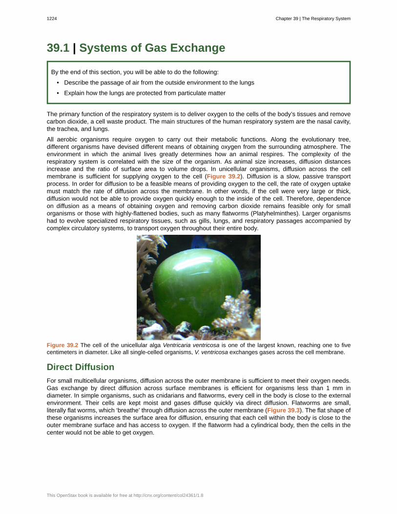
39.1 | Systems of Gas Exchange By the end of this section, you will be able to do the following: • Describe the passage of air from the outside environment to the lungs • Explain how the lungs are protected from particulate matter The primary function of the respiratory system is to deliver oxygen to the cells of the body’s tissues and remove carbon dioxide, a cell waste product. The main structures of the human respiratory system are the nasal cavity, the trachea, and lungs. All aerobic organisms require oxygen to carry out their metabolic functions. Along the evolutionary tree, different organisms have devised different means of obtaining oxygen from the surrounding atmosphere. The environment in which the animal lives greatly determines how an animal respires. The complexity of the respiratory system is correlated with the size of the organism. As animal size increases, diffusion distances increase and the ratio of surface area to volume drops. In unicellular organisms, diffusion across the cell membrane is sufficient for supplying oxygen to the cell (Figure 39.2). Diffusion is a slow, passive transport process. In order for diffusion to be a feasible means of providing oxygen to the cell, the rate of oxygen uptake must match the rate of diffusion across the membrane. In other words, if the cell were very large or thick, diffusion would not be able to provide oxygen quickly enough to the inside of the cell. Therefore, dependence on diffusion as a means of obtaining oxygen and removing carbon dioxide remains feasible only for small organisms or those with highly-flattened bodies, such as many flatworms (Platyhelminthes). Larger organisms had to evolve specialized respiratory tissues, such as gills, lungs, and respiratory passages accompanied by complex circulatory systems, to transport oxygen throughout their entire body. Figure 39.2 The cell of the unicellular alga Ventricaria ventricosa is one of the largest known, reaching one to five centimeters in diameter. Like all single-celled organisms, V. ventricosa exchanges gases across the cell membrane. Direct Diffusion For small multicellular organisms, diffusion across the outer membrane is sufficient to meet their oxygen needs. Gas exchange by direct diffusion across surface membranes is efficient for organisms less than 1 mm in diameter. In simple organisms, such as cnidarians and flatworms, every cell in the body is close to the external environment. Their cells are kept moist and gases diffuse quickly via direct diffusion. Flatworms are small, literally flat worms, which ‘breathe’ through diffusion across the outer membrane (Figure 39.3). The flat shape of these organisms increases the surface area for diffusion, ensuring that each cell within the body is close to the outer membrane surface and has access to oxygen. If the flatworm had a cylindrical body, then the cells in the center would not be able to get oxygen. 1224 Chapter 39 | The Respiratory System This OpenStax book is available for free at http://cnx.org/content/col24361/1.8
-
Upload
khangminh22 -
Category
Documents
-
view
1 -
download
0
Transcript of 39.1 | Systems of Gas Exchange - The Expert TA
39.1 | Systems of Gas Exchange
By the end of this section, you will be able to do the following:
• Describe the passage of air from the outside environment to the lungs
• Explain how the lungs are protected from particulate matter
The primary function of the respiratory system is to deliver oxygen to the cells of the body’s tissues and removecarbon dioxide, a cell waste product. The main structures of the human respiratory system are the nasal cavity,the trachea, and lungs.
All aerobic organisms require oxygen to carry out their metabolic functions. Along the evolutionary tree,different organisms have devised different means of obtaining oxygen from the surrounding atmosphere. Theenvironment in which the animal lives greatly determines how an animal respires. The complexity of therespiratory system is correlated with the size of the organism. As animal size increases, diffusion distancesincrease and the ratio of surface area to volume drops. In unicellular organisms, diffusion across the cellmembrane is sufficient for supplying oxygen to the cell (Figure 39.2). Diffusion is a slow, passive transportprocess. In order for diffusion to be a feasible means of providing oxygen to the cell, the rate of oxygen uptakemust match the rate of diffusion across the membrane. In other words, if the cell were very large or thick,diffusion would not be able to provide oxygen quickly enough to the inside of the cell. Therefore, dependenceon diffusion as a means of obtaining oxygen and removing carbon dioxide remains feasible only for smallorganisms or those with highly-flattened bodies, such as many flatworms (Platyhelminthes). Larger organismshad to evolve specialized respiratory tissues, such as gills, lungs, and respiratory passages accompanied bycomplex circulatory systems, to transport oxygen throughout their entire body.
Figure 39.2 The cell of the unicellular alga Ventricaria ventricosa is one of the largest known, reaching one to fivecentimeters in diameter. Like all single-celled organisms, V. ventricosa exchanges gases across the cell membrane.
Direct Diffusion
For small multicellular organisms, diffusion across the outer membrane is sufficient to meet their oxygen needs.Gas exchange by direct diffusion across surface membranes is efficient for organisms less than 1 mm indiameter. In simple organisms, such as cnidarians and flatworms, every cell in the body is close to the externalenvironment. Their cells are kept moist and gases diffuse quickly via direct diffusion. Flatworms are small,literally flat worms, which ‘breathe’ through diffusion across the outer membrane (Figure 39.3). The flat shape ofthese organisms increases the surface area for diffusion, ensuring that each cell within the body is close to theouter membrane surface and has access to oxygen. If the flatworm had a cylindrical body, then the cells in thecenter would not be able to get oxygen.
1224 Chapter 39 | The Respiratory System
This OpenStax book is available for free at http://cnx.org/content/col24361/1.8
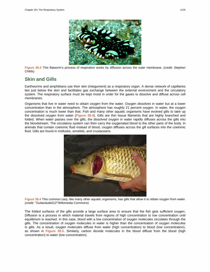
Figure 39.3 This flatworm’s process of respiration works by diffusion across the outer membrane. (credit: StephenChilds)
Skin and Gills
Earthworms and amphibians use their skin (integument) as a respiratory organ. A dense network of capillarieslies just below the skin and facilitates gas exchange between the external environment and the circulatorysystem. The respiratory surface must be kept moist in order for the gases to dissolve and diffuse across cellmembranes.
Organisms that live in water need to obtain oxygen from the water. Oxygen dissolves in water but at a lowerconcentration than in the atmosphere. The atmosphere has roughly 21 percent oxygen. In water, the oxygenconcentration is much lower than that. Fish and many other aquatic organisms have evolved gills to take upthe dissolved oxygen from water (Figure 39.4). Gills are thin tissue filaments that are highly branched andfolded. When water passes over the gills, the dissolved oxygen in water rapidly diffuses across the gills intothe bloodstream. The circulatory system can then carry the oxygenated blood to the other parts of the body. Inanimals that contain coelomic fluid instead of blood, oxygen diffuses across the gill surfaces into the coelomicfluid. Gills are found in mollusks, annelids, and crustaceans.
Figure 39.4 This common carp, like many other aquatic organisms, has gills that allow it to obtain oxygen from water.(credit: "Guitardude012"/Wikimedia Commons)
The folded surfaces of the gills provide a large surface area to ensure that the fish gets sufficient oxygen.Diffusion is a process in which material travels from regions of high concentration to low concentration untilequilibrium is reached. In this case, blood with a low concentration of oxygen molecules circulates through thegills. The concentration of oxygen molecules in water is higher than the concentration of oxygen moleculesin gills. As a result, oxygen molecules diffuse from water (high concentration) to blood (low concentration),as shown in Figure 39.5. Similarly, carbon dioxide molecules in the blood diffuse from the blood (highconcentration) to water (low concentration).
Chapter 39 | The Respiratory System 1225
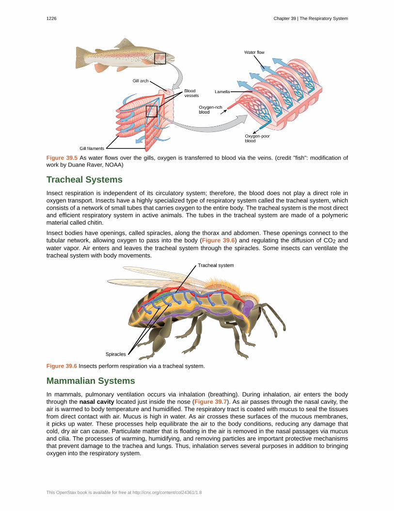
Figure 39.5 As water flows over the gills, oxygen is transferred to blood via the veins. (credit "fish": modification ofwork by Duane Raver, NOAA)
Tracheal Systems
Insect respiration is independent of its circulatory system; therefore, the blood does not play a direct role inoxygen transport. Insects have a highly specialized type of respiratory system called the tracheal system, whichconsists of a network of small tubes that carries oxygen to the entire body. The tracheal system is the most directand efficient respiratory system in active animals. The tubes in the tracheal system are made of a polymericmaterial called chitin.
Insect bodies have openings, called spiracles, along the thorax and abdomen. These openings connect to thetubular network, allowing oxygen to pass into the body (Figure 39.6) and regulating the diffusion of CO2 andwater vapor. Air enters and leaves the tracheal system through the spiracles. Some insects can ventilate thetracheal system with body movements.
Figure 39.6 Insects perform respiration via a tracheal system.
Mammalian Systems
In mammals, pulmonary ventilation occurs via inhalation (breathing). During inhalation, air enters the bodythrough the nasal cavity located just inside the nose (Figure 39.7). As air passes through the nasal cavity, theair is warmed to body temperature and humidified. The respiratory tract is coated with mucus to seal the tissuesfrom direct contact with air. Mucus is high in water. As air crosses these surfaces of the mucous membranes,it picks up water. These processes help equilibrate the air to the body conditions, reducing any damage thatcold, dry air can cause. Particulate matter that is floating in the air is removed in the nasal passages via mucusand cilia. The processes of warming, humidifying, and removing particles are important protective mechanismsthat prevent damage to the trachea and lungs. Thus, inhalation serves several purposes in addition to bringingoxygen into the respiratory system.
1226 Chapter 39 | The Respiratory System
This OpenStax book is available for free at http://cnx.org/content/col24361/1.8
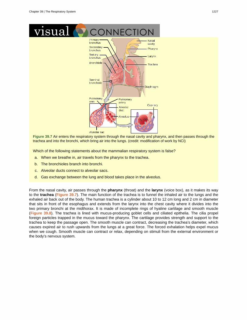
Figure 39.7 Air enters the respiratory system through the nasal cavity and pharynx, and then passes through thetrachea and into the bronchi, which bring air into the lungs. (credit: modification of work by NCI)
Which of the following statements about the mammalian respiratory system is false?
a. When we breathe in, air travels from the pharynx to the trachea.
b. The bronchioles branch into bronchi.
c. Alveolar ducts connect to alveolar sacs.
d. Gas exchange between the lung and blood takes place in the alveolus.
From the nasal cavity, air passes through the pharynx (throat) and the larynx (voice box), as it makes its wayto the trachea (Figure 39.7). The main function of the trachea is to funnel the inhaled air to the lungs and theexhaled air back out of the body. The human trachea is a cylinder about 10 to 12 cm long and 2 cm in diameterthat sits in front of the esophagus and extends from the larynx into the chest cavity where it divides into thetwo primary bronchi at the midthorax. It is made of incomplete rings of hyaline cartilage and smooth muscle(Figure 39.8). The trachea is lined with mucus-producing goblet cells and ciliated epithelia. The cilia propelforeign particles trapped in the mucus toward the pharynx. The cartilage provides strength and support to thetrachea to keep the passage open. The smooth muscle can contract, decreasing the trachea’s diameter, whichcauses expired air to rush upwards from the lungs at a great force. The forced exhalation helps expel mucuswhen we cough. Smooth muscle can contract or relax, depending on stimuli from the external environment orthe body’s nervous system.
Chapter 39 | The Respiratory System 1227
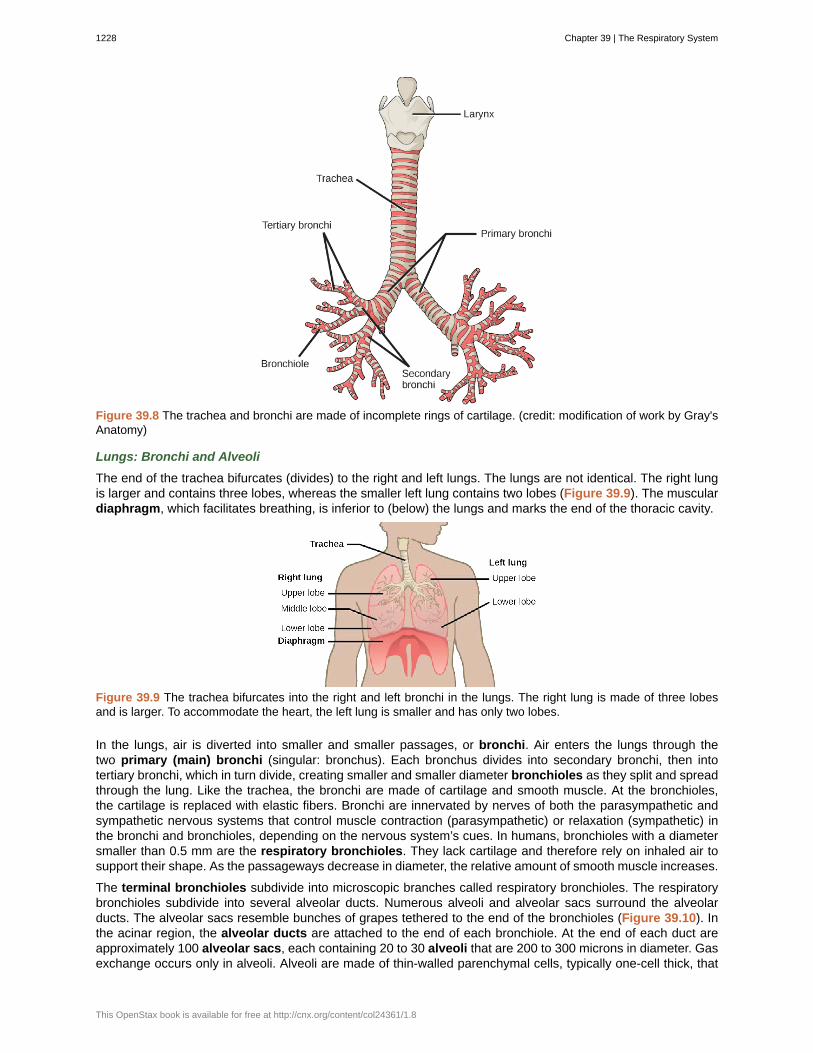
Figure 39.8 The trachea and bronchi are made of incomplete rings of cartilage. (credit: modification of work by Gray'sAnatomy)
Lungs: Bronchi and Alveoli
The end of the trachea bifurcates (divides) to the right and left lungs. The lungs are not identical. The right lungis larger and contains three lobes, whereas the smaller left lung contains two lobes (Figure 39.9). The musculardiaphragm, which facilitates breathing, is inferior to (below) the lungs and marks the end of the thoracic cavity.
Figure 39.9 The trachea bifurcates into the right and left bronchi in the lungs. The right lung is made of three lobesand is larger. To accommodate the heart, the left lung is smaller and has only two lobes.
In the lungs, air is diverted into smaller and smaller passages, or bronchi. Air enters the lungs through thetwo primary (main) bronchi (singular: bronchus). Each bronchus divides into secondary bronchi, then intotertiary bronchi, which in turn divide, creating smaller and smaller diameter bronchioles as they split and spreadthrough the lung. Like the trachea, the bronchi are made of cartilage and smooth muscle. At the bronchioles,the cartilage is replaced with elastic fibers. Bronchi are innervated by nerves of both the parasympathetic andsympathetic nervous systems that control muscle contraction (parasympathetic) or relaxation (sympathetic) inthe bronchi and bronchioles, depending on the nervous system’s cues. In humans, bronchioles with a diametersmaller than 0.5 mm are the respiratory bronchioles. They lack cartilage and therefore rely on inhaled air tosupport their shape. As the passageways decrease in diameter, the relative amount of smooth muscle increases.
The terminal bronchioles subdivide into microscopic branches called respiratory bronchioles. The respiratorybronchioles subdivide into several alveolar ducts. Numerous alveoli and alveolar sacs surround the alveolarducts. The alveolar sacs resemble bunches of grapes tethered to the end of the bronchioles (Figure 39.10). Inthe acinar region, the alveolar ducts are attached to the end of each bronchiole. At the end of each duct areapproximately 100 alveolar sacs, each containing 20 to 30 alveoli that are 200 to 300 microns in diameter. Gasexchange occurs only in alveoli. Alveoli are made of thin-walled parenchymal cells, typically one-cell thick, that
1228 Chapter 39 | The Respiratory System
This OpenStax book is available for free at http://cnx.org/content/col24361/1.8
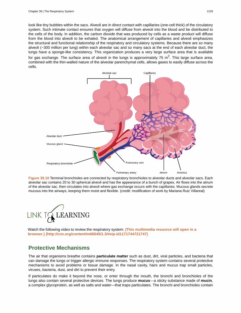
look like tiny bubbles within the sacs. Alveoli are in direct contact with capillaries (one-cell thick) of the circulatorysystem. Such intimate contact ensures that oxygen will diffuse from alveoli into the blood and be distributed tothe cells of the body. In addition, the carbon dioxide that was produced by cells as a waste product will diffusefrom the blood into alveoli to be exhaled. The anatomical arrangement of capillaries and alveoli emphasizesthe structural and functional relationship of the respiratory and circulatory systems. Because there are so manyalveoli (~300 million per lung) within each alveolar sac and so many sacs at the end of each alveolar duct, thelungs have a sponge-like consistency. This organization produces a very large surface area that is available
for gas exchange. The surface area of alveoli in the lungs is approximately 75 m2. This large surface area,combined with the thin-walled nature of the alveolar parenchymal cells, allows gases to easily diffuse across thecells.
Figure 39.10 Terminal bronchioles are connected by respiratory bronchioles to alveolar ducts and alveolar sacs. Eachalveolar sac contains 20 to 30 spherical alveoli and has the appearance of a bunch of grapes. Air flows into the atriumof the alveolar sac, then circulates into alveoli where gas exchange occurs with the capillaries. Mucous glands secretemucous into the airways, keeping them moist and flexible. (credit: modification of work by Mariana Ruiz Villareal)
Watch the following video to review the respiratory system. (This multimedia resource will open in a browser.) (http://cnx.org/content/m66645/1.3/#eip-id1171744721747)
Protective Mechanisms
The air that organisms breathe contains particulate matter such as dust, dirt, viral particles, and bacteria thatcan damage the lungs or trigger allergic immune responses. The respiratory system contains several protectivemechanisms to avoid problems or tissue damage. In the nasal cavity, hairs and mucus trap small particles,viruses, bacteria, dust, and dirt to prevent their entry.
If particulates do make it beyond the nose, or enter through the mouth, the bronchi and bronchioles of thelungs also contain several protective devices. The lungs produce mucus—a sticky substance made of mucin,a complex glycoprotein, as well as salts and water—that traps particulates. The bronchi and bronchioles contain
Chapter 39 | The Respiratory System 1229
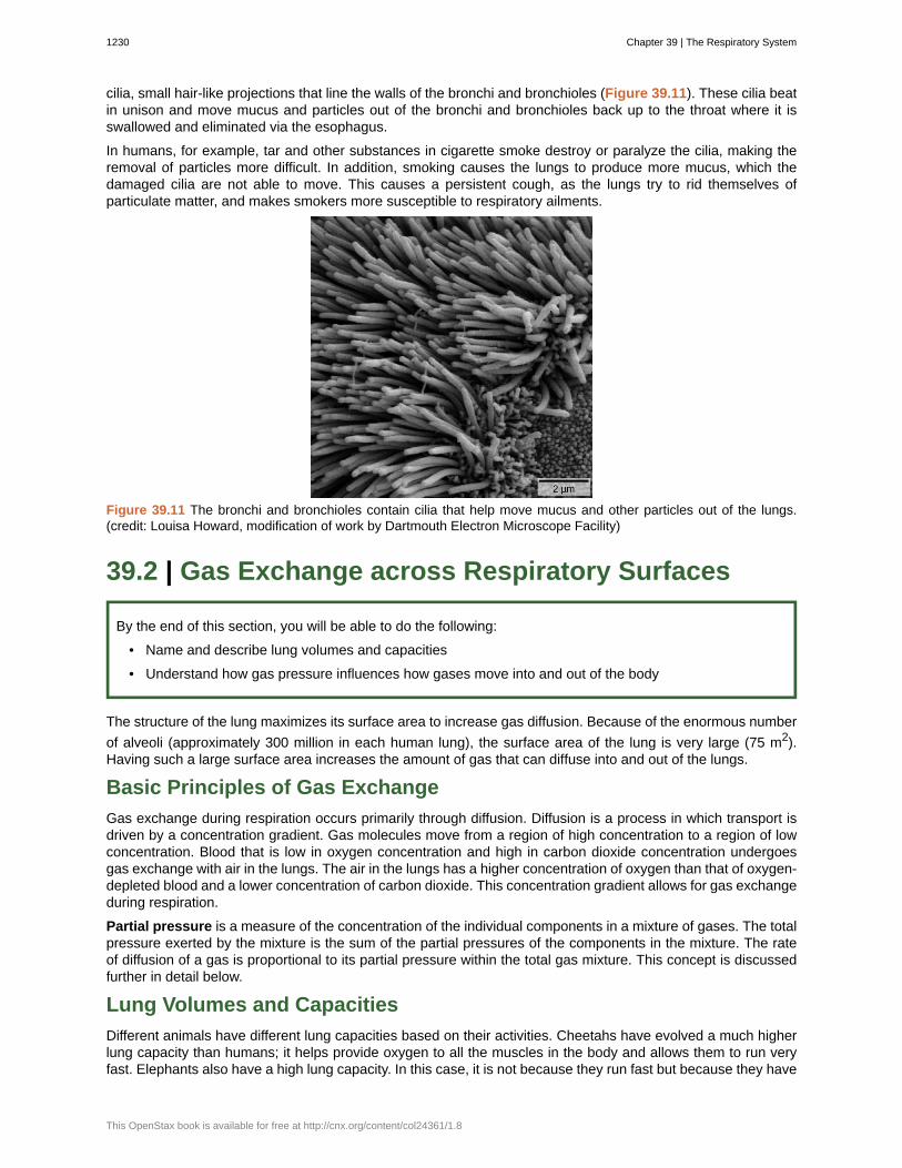
cilia, small hair-like projections that line the walls of the bronchi and bronchioles (Figure 39.11). These cilia beatin unison and move mucus and particles out of the bronchi and bronchioles back up to the throat where it isswallowed and eliminated via the esophagus.
In humans, for example, tar and other substances in cigarette smoke destroy or paralyze the cilia, making theremoval of particles more difficult. In addition, smoking causes the lungs to produce more mucus, which thedamaged cilia are not able to move. This causes a persistent cough, as the lungs try to rid themselves ofparticulate matter, and makes smokers more susceptible to respiratory ailments.
Figure 39.11 The bronchi and bronchioles contain cilia that help move mucus and other particles out of the lungs.(credit: Louisa Howard, modification of work by Dartmouth Electron Microscope Facility)
39.2 | Gas Exchange across Respiratory Surfaces
By the end of this section, you will be able to do the following:
• Name and describe lung volumes and capacities
• Understand how gas pressure influences how gases move into and out of the body
The structure of the lung maximizes its surface area to increase gas diffusion. Because of the enormous number
of alveoli (approximately 300 million in each human lung), the surface area of the lung is very large (75 m2).Having such a large surface area increases the amount of gas that can diffuse into and out of the lungs.
Basic Principles of Gas Exchange
Gas exchange during respiration occurs primarily through diffusion. Diffusion is a process in which transport isdriven by a concentration gradient. Gas molecules move from a region of high concentration to a region of lowconcentration. Blood that is low in oxygen concentration and high in carbon dioxide concentration undergoesgas exchange with air in the lungs. The air in the lungs has a higher concentration of oxygen than that of oxygen-depleted blood and a lower concentration of carbon dioxide. This concentration gradient allows for gas exchangeduring respiration.
Partial pressure is a measure of the concentration of the individual components in a mixture of gases. The totalpressure exerted by the mixture is the sum of the partial pressures of the components in the mixture. The rateof diffusion of a gas is proportional to its partial pressure within the total gas mixture. This concept is discussedfurther in detail below.
Lung Volumes and Capacities
Different animals have different lung capacities based on their activities. Cheetahs have evolved a much higherlung capacity than humans; it helps provide oxygen to all the muscles in the body and allows them to run veryfast. Elephants also have a high lung capacity. In this case, it is not because they run fast but because they have
1230 Chapter 39 | The Respiratory System
This OpenStax book is available for free at http://cnx.org/content/col24361/1.8