3003078saranyaps.pdf - Electronic Theses and Dissertations ...
87
IDENTIFICATION OF PROBLEMS & EXECUTION OF NURSING STRATEGIES FOR MOTHERS WITH OLIGOHYDRAMNIOS AT KOVAI MEDICAL CENTRE & HOSPITAL, COIMBATORE. Reg. No : 30104423 A DISSERTATION SUBMITTED TO THE TAMIL NADU Dr.M.G.R MEDICAL UNIVERSITY, CHENNAI, IN PARTIAL FULFILLMENT OF REQUIREMENT FOR THR DEGREE OF MASTER OF SCIENCE IN NURSING. APRIL 2012
-
Upload
khangminh22 -
Category
Documents
-
view
1 -
download
0
Transcript of 3003078saranyaps.pdf - Electronic Theses and Dissertations ...
IDENTIFICATION OF PROBLEMS & EXECUTION OF NURSING
STRATEGIES FOR MOTHERS WITH OLIGOHYDRAMNIOS
AT KOVAI MEDICAL CENTRE & HOSPITAL,
COIMBATORE.
Reg. No : 30104423
A DISSERTATION SUBMITTED TO THE TAMIL NADU
Dr.M.G.R MEDICAL UNIVERSITY, CHENNAI, IN
PARTIAL FULFILLMENT OF REQUIREMENT
FOR THR DEGREE OF MASTER OF
SCIENCE IN NURSING.
APRIL 2012

CERTIFICATE
This is to certify that the dissertation entitled “IDENTIFICATION OF PROBLEMS &
EXECUTION OF NURSING STRATEGIES FOR MOTHERS WITH OLIGOHYDRAMNIOS
AT KOVAI MEDICAL CENTRE & HOSPITAL, COIMBATORE” is submitted to the Faculty of
Nursing, Tamilnadu Dr.M.G.R Medical University, Chennai by Ms.P.S.Saranya in partial
fulfilment of requirement for the degree of Master of Science in Nursing. It is the bonafide work done
by her and the conclusions are her own. It is further certified that, this dissertation or any part thereof
has not formed the basis for award of any degree, diploma or similar titles.
Prof. DR. S. Madhavi, M.Sc., (N), Ph.D.,
Principal & Head of the Department of
Medical & Surgical Nursing,
KMCH College of Nursing,
Coimbatore – 641014,
Tamilnadu.

IDENTIFICATION OF PROBLEMS & EXECUTION OF NURSING
STRATEGIES FOR MOTHERS WITH OLIGOHYDRAMNIOS
AT KOVAI MEDICAL CENTRE & HOSPITAL,
COIMBATORE. APPROVED BY THE DISSERTATION COMMITTEE ON FEBRUARY 2011
1. RESEARCH GUIDE : ....……………………………………………….
DR.O.T. Buvaneswaran, M.A., M.Phil., Ph.D.,
Head of the department of Medical Sociology,
KMCH, College of Nursing,
Avanashi Road, Coimbatore – 641014.
2. CLINICAL GUIDE : ...……………………………………………….
Prof. Mrs. Renuka, M.Sc (N),
Head of the department of Obstetrics and
Gynaecological Nursing,
KMCH, College of Nursing,
Avanashi Road, Coimbatore – 641014.
3. MEDICAL EXPERT : ....………………………………………………
Dr. C. S. Dhevasena, DGO., DNB.,
Consultant Obstetrician and Gynaecologist,
Kovai Medical Centre and Hospital,
Coimbatore – 641014.
A DISSERTATION SUBMITTED TO THE TAMIL NADU
Dr.M.G.R MEDICAL UNIVERSITY, CHENNAI, IN
PARTIAL FULFILLMENT OF REQUIREMENT
FOR THR DEGREE OF MASTER OF
SCIENCE IN NURSING.
APRIL 2012

ACKNOWLEDGEMENT
.
All things are possible only by the grace of God Almighty. I humble before the lord
almighty with heartful joy to offer my thanks and praise to him for his shower of blessings on me and
will power to fulfil this task successfully.
I take this opportunity to express my deep heartful gratitude to our chairman
Dr. Nalla.G.Palanisamy, M.D., AB (USA)., and our Trustee Dr. Thavamani D Palanisamy, M.D.,
AB (U.S.A ) for giving me the opportunity to undertake my PG degree in this esteemed institution and
grant me permission to conduct the study in kovai medical centre and hospital.
I express my deep heartful gratitude and sincere thanks to Prof.DR.S.Madhavi,M.Sc(N),
Ph.D, Principal , KMCH College of Nursing for her constant guidance and support.
I would like to acknowledge the valuable support of a very special individual
Dr. C. S. Dhevasena, DGO., DNB., Consultant Obstetrician and Gynaecologist, KMCH. It is my
long felt desire to express my heartiest gratitude to her devoting her attention, time and support, which
gave me an impetus to complete this study.
It is a great pleasure to express my sincere and special gratitude to
DR. O.T.Buvaneswaran, M.A., M.Phil., Ph.D., Head of the department of Medical Sociology, for
his valuable guidance and help in the statistical analysis of the data, which is the core of the study.
My faithful thanks to Prof.Mrs.Rm.Sivagami, M.Sc(N) Vice Principal, KMCH College
of Nursing, for her advice and continuous support in successful completion of study.
It is my privilege to express my deep heartful thanks to Prof.Mrs.Renuka.S, M.Sc(N),
Head of the Department of Obstetrics & Gynecological Nursing , KMCH College of Nursing,
who spared her precious time & energy, and contributed to the overall vision of the study, without her
valuable guidance, effort, astute observations and meticulous conscientious attention many of the task
necessary to produce this study would never have been completed.

I express my special thanks to Mrs. Indumathi. R, M.Sc., (N) Associate Professor,
Mrs. Padma. P, M.Sc., (N) Assistant Professor & Mrs. Manavalam M.Sc(N) Lecturer for their
personal motivation to achieve this great task.
I wish to sincerely thank Mrs. A. Bhuvaneswari, M.A., M.Phil., M.A., M.Phil., B.Ed.,
Assistant Professor in Sociology and English for the contribution toward my study.
I express my hearty thanks to Mrs. Jane Ebenezer R.N., R.M., Nursing Supervisor,
KMCH for her help during my data collection period.
I wish to thank chief librarian Mr. Damodharan and Assistant Librarians, for their
whole hearted help and assistance in search and reference.
I extend my thanks to all the mothers who have participated in the study.
My special thanks comes from each beat of my heart to my parents and my sister for
nurturing my cherished dreams into reality and their constant support, affection, prayer, & co-operation
throughout my study.
My special thanks to my class mates and all other well wishers for their help,
encouragement, supports and good wishes for the success of this study.
I thank sincerely each and every one who helped me directly and indirectly who build up
this study.

TABLE OF CONTENTS
CHAPTER
TITLE
PAGE NO
I INTRODUCTION 1
NEED FOR THE STUDY 3
STATEMENT OF THE PROBLEM 5
OBJECTIVES OF THE STUDY 5
OPERATIONAL DEFINITIONS 5
ASSUMPTION 5
CONCEPTUAL FRAMEWORK 6
II REVIEW OF LITERATURE 8
III METHODOLOGY 18
RESEARCH DESIGN 18
SETTING OF THE STUDY 18
POPULATION OF THE STUDY 18
SAMPLE SIZE 18
SAMPLING TECHNIQUE 18
CRITERIA FOR SAMPLE SELECTION 18
DEVELOPMENT AND DESCRIPTION OF THE TOOL 19
TESTING OF THE TOOL 20
PILOT STUDY 20
PROCEDURE FOR DATA COLLECTION 20
STATISTICAL ANALYSIS 20
IV DATA ANALYSIS AND INTERPRETATION 21
V DISCUSSION, SUMMARY, CONCLUSION,
IMPLICATIONS, LIMITATIONS AND
RECOMMENDATIONS.
42
ABSTRACT 56
REFERENCES 57
APPENDICES
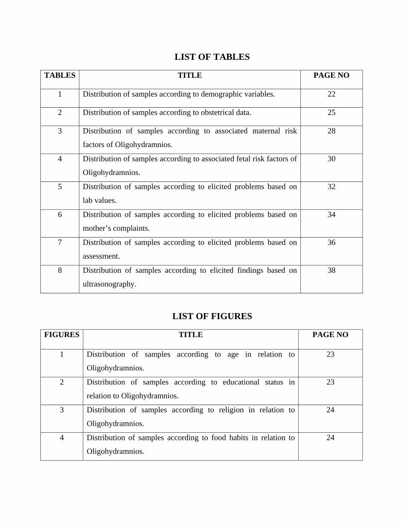
LIST OF TABLES
TABLES TITLE PAGE NO
1 Distribution of samples according to demographic variables. 22
2 Distribution of samples according to obstetrical data. 25
3 Distribution of samples according to associated maternal risk
factors of Oligohydramnios.
28
4 Distribution of samples according to associated fetal risk factors of
Oligohydramnios.
30
5 Distribution of samples according to elicited problems based on
lab values.
32
6 Distribution of samples according to elicited problems based on
mother’s complaints.
34
7 Distribution of samples according to elicited problems based on
assessment.
36
8 Distribution of samples according to elicited findings based on
ultrasonography.
38
LIST OF FIGURES
FIGURES TITLE PAGE NO
1 Distribution of samples according to age in relation to
Oligohydramnios.
23
2 Distribution of samples according to educational status in
relation to Oligohydramnios.
23
3 Distribution of samples according to religion in relation to
Oligohydramnios.
24
4 Distribution of samples according to food habits in relation to
Oligohydramnios.
24

5 Distribution of samples according to gravida in relation to
Oligohydramnios.
26
6 Distribution of samples according to weeks of gestational age in
relation to Oligohydramnios.
26
7 Distribution of samples according to pre – existing illness in
relation to Oligohydramnios.
27
8 Distribution of samples according to maternal risk factors along
with Oligohydramnios.
29
9 Distribution of samples according to fetal risk factors along with
Oligohydramnios.
31
10 Distribution of samples according to elicited problems based on
lab values.
33
11 Distribution of samples according to elicited problems based on
mother’s complaints.
35
12 Distribution of samples according to elicited problems based on
assessment.
37
13 Distribution of samples according to fetal movements based on
ultrasonography.
39
14 Distribution of samples according to Amniotic fluid index based
on ultrasonography.
39
15 Distribution of samples according to fetal presentation based on
ultrasonography.
40
16 Distribution of samples according to diastolic notch based on
ultrasonography.
40
17 Distribution of samples according to fetal weight related to
gestational age based on ultrasonography.
41

LIST OF APPENDICES
APPENDIX
TITLE
A
a) Demographic data.
b) Obstetrical data.
c) Maternal Assessment Tool.
d) Nursing Process Application.
e) Risk Factors Assessment Tool.
B
Discussion about high risk mothers.
C
Copy of Letter seeking permission to conduct the study.
D
Requisition for Content Validity of the Tool.
E
Copy of Certificates of Content Validity.
F
List of Experts.

LIST OF ABBREVIATIONS
S.NO
ABBREVIATIONS
1
AFI : Amniotic Fluid Index.
2
IUGR : Intrauterine Growth Retardation.
3
IUD : Intrauterine Death.
4
ACE Inhibitors : Angiotensin Converting Enzyme Inhibitors.
5
NST : Non Stress Test.
6
BPP : Biophysical profile.
7
SGA : Small for Gestational Age.
8
AGA : Appropriate for Gestational Age.
9
PKD : Polycystic Kidney Disease.
10
USG : Ultrasonography.
11
PROM : Premature Rupture of Membranes.

CHAPTER – I
INTRODUCTION
“The ocean which corresponds to the amniotic fluid in which human life begins”.
- Adrienne Rich.
Amniotic fluid is the fluid surrounds the fetus, it is otherwise called as liquor amnii. The origin
of the liquor amnii is probably of mixed maternal & fetal origin. It is secreted by amnion, especially
the part covering the placenta, umbilical cord some from fetal vessels in the placenta. Fetal urine also
contributes to the amniotic fluid volume from 10th weeks of gestation onwards. The water of amniotic
fluid is exchanged every 3hrs once. Amniotic fluid is a clear, pale straw coloured, consists of 99%
water and remaining 1% is dissolved solid matter such as waste products, fetus sheds skin cells, vernix
caseosa & lanugo. This fluid is faintly alkaline with low specific gravity of 1.010.
Amniotic fluid is inhaled & ingested by the fetus. Inhaled fluid is essential for lung
development & ingested fluid is necessary for gastrointestinal system development. Swallowed
amniotic fluid also creates urine & contributes to the formation of meconium. Amniotic fluid is like
cushion, promotes development of the bones & muscles. It acts as a shock absorber, protecting the
fetus from extraneous injury. It maintains constant temperature for the fetus, provides small amount of
nutrients for fetal growth. It allows the for free movement & prevents adhesions between the fetal parts
and amniotic sac. In labour, as long as membranes remain intact, the amniotic fluid protects the
placenta & umbilical cord from the pressure of contraction. It also aids effacement of the cervix &
dilatation of uterine os.
Amniotic fluid volume measures 50ml at 12weeks; 400ml at 20weeks & reaches its peak of 1lit
at 36-38weeks. After that, the fluid volume diminishes to 600-800ml at term. In post term period
further reduction occurs to the extend of about 200ml at 43weeks. Amniotic Fluid Index (AFI) is an
estimation of amount of amniotic fluid & index for fetal wellbeing. AFI is estimated by ultrasound.
Normal level of AFI is between 5 to 25cm or between 5th to 95th percentiles.

Oligohydramnios (too little amniotic fluid ) is described as an lessthan 300 ml of amniotic
fluid, Amniotic Fluid Index (AFI) < 5 percentile or < 5cm, maximum vertical pocket (MVP) is lessthan
2cm. Oligohydramnios may be due to a variety of conditions including urinary tract abnormalities such
as renal agenesis, bilateral renal obstruction, bilateral renal dysplasia & posterior urethral valves or
atresia; prerenal abnormalities including uteroplacental insufficiency leading to IUGR, postterm
pregnancy & premature rupture of membranes. There is a chance to increased perinatal morbidity &
occasionally fetal or neonatal death in the presence of Oligohydramnios.
Cunningham, et al., (2001), reported that postterm pregnancy is more likely to be complicated
by Oligohydramnios. It increases the incidence of cord compression with subsequent development of
fetal distress during labour. Postterm pregnancy is at risk for increased perinatal mortality & morbidity
during labour.
Lin, et al., (1990), conducted study on the association between Oligohydramnios & IUGR.
They found that Oligohydramnios diagnosed in the second trimester of pregnancy, the fetal prognosis
is poor. The result indicates that Oligohydramnios occurs often in IUGR than non IUGR pregnancies.
National Institute for Health & Clinical excellence (2011), conducted study on therapeutic
amnioinfusion for Oligohydramnios during pregnancy, it showed that amnioinfusion involves infusion
of fluid by a needle inserted into womb and the space surrounding the fetus to increase amount of
amniotic fluid.
Ghafarnejad et al., (2009) conducted study on oral hydration in Oligohydramnios. They
reported that acute oral hydration is a non invasive easily accessible, cheap intervention & an effective
way of increasing amniotic fluid.
Kilpatrick,S.J., Safford,K.L.(1993), demonstrated an increase in the amniotic fluid index of
30% when women with Oligohydramnios were treated with hydration of 2lit of water.
Nursing intervention for Oligohydramnios mothers as follow as, monitor maternal & fetal
status closely, including vital signs & fetal heart rate pattern. Monitor maternal weight gain pattern,
provide emotional support before, during & after ultrasonography. Inform the mother about coping
measures if fetal anomalies are suspected. Instruct about signs and symptoms of labour including those
need for close supervision & follow up. Encouraged the mother to lie on her left side. Assist with

amnioinfusion as indicated. Continuously monitor maternal vital signs & fetal heart rate during the
amnioinfusion procedure. Note the development of uterine contraction & continue to monitor closely.
Maintain strict sterile technique during amnioinfusion.
Prevention of Oligohydramnios is not possible. Necessary to prevent the underlying cause like
good control of maternal diabetes & prevention of infection transmittable from mother to fetus are two
approaches for a subset of causes.
NEED FOR THE STUDY
In global level, Oligohydramnios in the second trimester is found in about 1 in 500 pregnancies
(Pilu, 2000). Oligohydramnios incidence is 2.3 % of all pregnancies. It is associated with increased
pregnancy complication, congenital anomalies & perinatal mortality (Jandial, 2007).
Past incidence of Oligohydramnios was about 0.1%, but in recent years through ultrasound,
Oligohydramnios detection rate (0.5 to 4%) increased. It seriously affect the prognosis of children with
perinatally. Oligohydramnios in prolonged pregnancy, rate of incidence is 20-30% (Qiong, 2011).
Shenker & colleagues (1991) described 34 mid trimester pregnancies complicated by
Oligohydramnios diagnosed ultrasound by the absence of amniotic fluid pockets greater than 1cm.
Nine fetuses had (one fourth) anomalies, 25 out of 34 who were normal either aborted spontaneously
or were stillborn because of severe maternal hypertension, restricted fetal growth or placental
abruption. Among that 14 live born infants, 8 were preterm & seven died. The 6 infants who were
delivered at term, did well.
Garmel & coworkers (1997) observed that approximately grown fetuses associated with
Oligohydramnios prior to 37weeks had a threefold increase in preterm birth but not of later growth
restriction or fetal death.
Newbould & colleagues (1994) described autopsy findings in 89 infants with the
Oligohydramnios sequence or potter syndrome. Only 3% had a normal renal tract; 54% had bilateral
renal agenesis; 34% had bilateral cystic dysplasia; 9% had unilateral agenesis with dysplasia & 10%
had minor urinary abnormalities.

Several conditions have been associated with diminished amniotic fluid. Oligohydramnios
almost always is evident when there is either obstruction of the fetal urinary tract or renal agenesis.
Therefore anuria almost certainly has an etiological role. A chronic leak from a defect in the fetal
membranes may reduce the volume of fluid appreciably, most often labour soon ensures. Exposure to
angiotensin converting enzyme inhibitors has been associated with Oligohydramnios and 15 to 25% of
cases are associated with the fetal anomalies.
Pryde & co-workers (2000), conducted study on severe Oligohydramnios with intact
membranes an indication for diagnostic amniofusion. They were able to visualize fetal structures in
only half of women referred for ultrasonic evaluation of midtrimester Oligohydramnios. They
performed amnioinfusion & were able to visualize 77 % of routinely imaged structures. Identification
of associated anomalies increased from 12 to 31 % of fetuses. They reported that fetal outcome is poor
with early onset Oligohydramnios.
McNamara & associates (1995) described findings from 2 sets of monoamniotic twins with
discordant renal anomalies. They provided evidence that normal amniotic fluid volume in the presence
of fetal renal obstruction allows normal lung development.
Management of Oligohydramnios in late pregnancy depends on the clinical situation. An
evaluation for fetal anomalies & estimation of growth is critical due to Oligohydramnios. Close fetal
surveillance is important because of associated morbidity. Delivery is recommended for fetal or
maternal indications eventhough gestational age is considered in this decision. Evidence for fetal or
maternal compromise, usually overrides potential complications from preterm delivery.
Chauhan & associates (1999) performed meta- analysis of 18 studies comprising more than
10,500 pregnancies in which the intrapartum AFI was less than 5cm compared with controls whose
AFI was over 5cm. Women with Oligohydramnios had a significantly increased 2.2 fold, risk for
cesarean delivery for fetal distress & 5.2 fold increased risk for a 5mt Apgar score of less than 7.
Pierce & colleagues (2000) performed meta-analysis of 13 studies with 1924 antenatal mothers
& were randomized to amnioinfusion or no treatment. They found that amnioinfusion resulted in
significantly decreased adverse maternal & fetal outcomes.

Pregnancy complicated by severe Oligohydramnios have been shown to be at increased risk for
fetal morbidity, rate high as 80-90 % have been reported with Oligohydramnios diagnosed in the
second trimester. In renal agenesis the fetal mortality rate is 100 %. Decreased amniotic fluid volume
raises management issues & requires that Nurse - Midwives arrange collaborative care. During clinical
experience, the researcher identified mothers with Oligohydramnios in KMCH hospital and researcher
actively participated & closely monitored in order to identify problems of Oligohydramnios & provide
evidence based care. To reduce maternal & fetal risk and to ensure active participation of nursing care
& to create awareness regarding problems of Oligohydramnios to Nurses, the researcher selected this
topic for the study.
STATEMENT OF THE PROBLEM
Identification of problems & execution of Nursing strategies for mothers with
Oligohydramnios at Kovai Medical Center & Hospital, Coimbatore.
OBJECTIVES
Objectives were,
• assess the risk factors associated with Oligohydramnios.
• identify the problems of mother with Oligohydramnios.
• execute Nursing strategies on mother with Oligohydramnios.
• evaluate the Nursing strategies executed on mothers with Oligohydramnios.
OPERATIONAL DEFINITION
Problems
Maternal biophysiological & psychological problems as well as the fetal problems which
warrants prompt nursing intervention.
Nursing strategies
Nursing measures taken & directed to manage identified Nursing problems.
Oligohydramnios
Insufficient amniotic fluid in the gestational sac during pregnancy & Amniotic fluid index
(AFI) is less than 8cm is called as Oligohydramnios.
ASSUMPTION
Fetal outcome is poor due to Oligohydramnios which needs prompt identification &
management.

CONCEPTUAL FRAMEWORK
Conceptual frame work for this study was developed on the basis of Ida Jean Orlando. She
proposed her model in 1961. The Nursing process is based on individual action. The Nursing process is
used by a nurse to meet a mother’s need for help & meeting this need improves the patient’s
behaviour.
The components of Orlando’s Nursing Process Theory are patient behaviour, nurse reaction &
nurse activity.
i) Patient behaviour:
The Nursing process is formed by the patient’s need.
ii) Nurse reaction:
It forms the basis for determining how a nurse acts; the nurse identifies reaction.
iii) Nurse activity:
Nurse activity is towards the benefit of the patient. It occurs after the nurse interprets the
patient’s behaviour.
Nursing process is an organized by 5 step approach – identify the problems by assessment,
based on the assessment Nursing diagnosis were formulated; the care was planned, implemented &
evaluated. Nursing cares focused on improvement of health condition of mother and minimize the
complication in pregnancy & labour.

Conceptual Framework – Orlando’s Nursing Process Model
Assessment
• ital signs.
• bdominal examination
- ize, Abdominal girth.
- eight of fundus.
- etational age.
- alpation.
Implementation
Highly individualized nursing care focused on improvement in health status of mother, reduce discomfort like pain, fear, anxiety & preventing complication of Oligohydramnios.
Evaluation
•ncrease cardiac output.
•issue perfusion will be maintained.
•mprovement in fetal movements.
•elief from back pain.
•nfection will be prevented.
Planning
•rovide left lateral position.
•onitoring fetal heart rate.
•aintain kick count chart.
•dvice on bed rest.
•dvise to drink more fluids.
•dvise to take well balanced diet.
•
Nursing diagnosis
• ecreased cardiac output
• neffective tissue perfusion.
• xcessive fluid volume.
• ack pain.
• mbalanced nutritional pattern.
• ear, anxiety

CHAPTER II
REVIEW OF LITERATURE
This chapter deals with the information about present study through published materials, books for
foundation to carry out the research work. The existing research studies & results are often useful in
helping the researcher to focus on a specific problem and to describe the suitable research process.
Review of literature is categorized as follows,
Section A : Literature related to prevalence of Oligohydramnios.
Section B : Literature related to causes of Oligohydramnios.
Section C : Literature related to treatment of Oligohydramnios.
Section D : Literature related to outcome of Oligohydramnios.
LITERATURE RELATED TO PREVALENCE OF OLIGOHYDRAMNIOS
Shanks et al., (2011) conducted study on assessing the optimal definition of Oligohydramnios
associated with adverse neonatal outcome. They adopted retrospective cohort study from 1998 to 2008.
Lessthan 5th percentile was compared to normal AFI. Primary outcome measures was NICU
admission.17,887 mothers were included in this study. There were 145 NICU admission with an AFI
<5cm (relative risk, 2.2) compared to 235 with AFI < 5th percentile for gestational age (relative risk
2.37). The sensitivity & specificity for NICU admission using an AFI < 5cm were 10.9 % & 95.2%
compared to 17.6 % & 92.5 % for AFI < 5th percentile for gestation age. They concluded that
Oligohydramnios defined as an AFI lessthan 5th percentile better predicts fetuses at risk for adverse
perinatal outcome compared to an < 5cm.
Ramos et al., (2010) conducted study on accuracy of prenatal diagnosis in elective termination
of pregnancy : 385 cases from 2000 to 2007. They used retrospective analysis of 385 medical
termination of pregnancy performed due to fetal anomalies. They found that chromosomal
abnormalities (39%) disorders of central nervous system (20%) monogenic disorder(11 %),sequences
(9.6%), polymalformative syndrome (5.2%) & isolated congenital heart disease. Seqences were present
in 37 cases (9.6%), the most common sequence was Oligohydramnios sequece in 17 cases.

Feldman et al. (2009), conducted study on Oligohydramnios common during summer season.
They adopted retrospective study to assess risk of Oligohydramnios during summer season compared
to rest of years among a population of Jewish & Bedouin, from 1988 to 2007. 1,91,558 deliveries
occurred during the study period. Among these deliveries 4335 (2.26%) were diagnosed as
Oligohydramnios. 1553 deliveries in the summer month & 2782 during the rest of the year.
Oligohydramnios in summer month was higher than the rest of the years (2.5 Vs 2.1%). Regression
analysis showed that the summer season was an independent risk factor for Oligohydramnios.
Chhabra,S. et al., (2007) conducted study on Oligohydramnios: A potential marker for serious
obstetric complication. Retrospective & prospective designs were adopted. They showed the result that
the incidence of Oligohydramnios over 7yrs was 4.45%, 4.9% in retrospective & 4% in prospective
cases. Due to Oligohydramnios placenta abruption had occurred in 9.8% retrospective, 8.3% in
prospective cases, labour were induced 18.2% of retrospective cases & 13.9% in prospective cases,
caesarean section rate with spontaneous labour was 42.4% in retrospective cases, 50.4% in
prospective cases & induced labour 38.5% in retrospective and 29.3% in prospective cases. Perinatal
mortality rate in cases of Oligohydramnios was 87.7 % & 4.15% babies had congenital anomalies.
James.K. et al., (2001) reported that about 7% of pregnancies with Oligohydramnios are
associated with congenital malformation. The incidence rises to 26-35 % when rupture of membranes
occurs in the second trimester.33-57 % had renal anomalies like bilateral renal agenesis or
multicystic/dysplastic kidneys & urinary tract obstruction. In these cases, Oligohydramnios is
secondary to occur because of decreased renal function, since normal volume may be restored by
placement of a vesicoamniotic shunt. Mid trimester fetal loss rates of 43-88% have been reported with
Oligohydramnios.
LITERATURE RELATED TO CAUSES OF OLIGOHYDRAMNIOS
Chen et al., (2010) conducted study on Mechanism of Oligohydramnios induced pulmonary
hypoplasia. They reported that the exact mechanism by which Oligohydramnios alters the respiratory
system remains unknown. Pulmonary hypoplasia is common in perinatal period & is a significant
cause of death in newborn & Oligohydramnios is one of the most commonly associated abnormalities.
Neonates exposed to Oligohydramnios caused high risk of acute respiratory morbidity.

Podymow & Phyllis (2008) conducted study on update on the use of antihypertensive drugs in
pregnancy. They used randomized control trail with 1000 to 3000 women with hypertension. Reported
that angiotensin converting enzyme inhibitors are contraindicated in 2nd & 3rd trimester because of
toxicity associated with reduced perfusion of fetal kidneys; use is associated with potter’s syndrome
including Oligohydramnios as a result of fetal oliguria, renal dysgenesis, IUGR & pulmonary
hypoplasia.
Blackburn et al., (2007) suggested that Oligohydramnios may be associated with the following
factors like poor placental blood flow, preterm membrane rupture, failure of fetal kidney development,
blocked urinary excretion, poor fetal lung development(pulmonary hypoplasia) & malformation such
as skeletal abnormalities may result from compression of fetal parts. It increased the risk of perinatal
mortality & morbidity.
Chauhan,S,P. (2007) reported that bladder or renal disorder in fetus is usually cause
Oligohydramnios. It can occur from IUGR. In this condition the fetus is cramped for space, muscles
are left weak at birth, lungs fails to develop (hypoplastic lung), and leading to severe breathing
difficulty after birth & features of the face become distorted.
Hendricks,K., & Smith,R. (2005) reported that the use of prostaglandin synthetase inhibitor
(indomethacin & ibuprofen ) for > 72 hrs in women with preterm labour (n=67) was significantly (p<
0.001) associated with ultrasound recorded Oligohydramnios compared with the use of ritodrine or
magnesium sulphate (control group, n= 67). Oligohydramnios developed in 26 of 37 women (70 %)
treated with indomethacin & 8 of 30 treated with ibuprofen (27 %) (p <0.01). Only 2 control group
subjects had Oligohydramnios. All 34 cases of Oligohydramnios in the treatment group resolved after
stoping medication, where as 2 control group did not resolve. No instance of renal failure, premature
closure of ductus arteriosus, pulmonary hypertension or bleeding disorders were noted in treated &
control group infants.
Pietrement et al., (2003) conducted study on perinatal/ neonatal case presentation: neonatal
acute renal failure secondary to maternal exposure to telmisartan, angiotensin II receptor antagonist.
They reported that treatment of maternal hypertension by ACE inhibitors during pregnancy associated
with fetal & neonatal morbidity and mortality. Sartans are new class of antihypertensive drug that
directly inhibit the angiotensin II receptors. Telmisartan is a specific angiotensin II receptor antagonist

used for adult hypertension therapy. 5 fetal death & 1 neonatal death occur at 4th day after delivery. All
cases presented severe Oligohydramnios, three fetuses had foot face deformities of the
Oligohydramnios sequence & skull bones were hypoplastic due to ACE inhibitor toxicity. Autopsies of
these 5 cases shown pulmonary hypoplasia in one patient,kidneys were enlarged with tubular
dysgenesis, retraction of glomerular tufts & thickening of arteries. They include a spectrum of adverse
effects from fetal death to transient renal failure.
Sprong (2003) reported that AFI is considered indicative of the long term function of the
placenta. An AFI greater than 5cm represents an adequate volume of amniotic fluid. A modified BPP
is considered abnormal if the AFI is less than 5cm, regardless of reactivity of NST.
Oz & colleagues (2002) conducted study on Oligohydramnios & post term pregnancy. They
found that, renal artery Doppler was more predictive of Oligohydramnios. The reduced renal artery end
diastolic velocity suggests that increased arterial impedance is an important factor in the development
of Oligohydramnios in prolonged pregnancies.
Schrimmer & Moore. (2002) described that amniotic fluid volume is an important component
of the BBP. The kidneys and lungs are the principal source of amniotic fluid. Amniotic fluid volume
also may be most obvious sign of chronic fetal hypoxia. Abnormal amniotic fluid volume may indicate
congenital anatomical anomalies or intrauterine growth restriction.
Chanoufi,M.B et al., (2000) conducted study on Oligohydramnios and fetal malformation
association. They reported that oligamnios caused by renal malformation, obstructive myopathies,
polymalformative syndrome & bilateral renal malformation.
Burrow,N., & Duffy,P. (1999) stated that inadequate fluid volume is associated with fetal
urinary obstruction & placental insufficiency. Umbilical cord compression, fetal distress, meconium
passage & fetal asphyxia are observed with increased frequency in Oligohydramnios mothers.
Prolonged oligohydramnios interferes with normal lung growth, resulting potentially lethal pulmonary
hypoplasia.
Doi et al., (1999) conducted study on relationship of AFI & cord blood erythropoietin levels in
small for gestational age fetuses and appropriate for gestational age fetuses. They used experimental
design to measure erythropoietin levels in cord blood in 134 high risk mothers, including 40 with AFI
(7cm or less) & 94 with normal AFI (> 7cm). They were in 32 to 39 weeks of gestation age. Infants
were divided in to the SGA & AGA based on birth weight. Cord erythropoietin levels in SGA fetus
with low AFI (n=24) were significantly higher than SGA fetuses with normal fluid volume (n=
16)(171.6 +/- 207.4 mIU/dl) compared with 36.1 +/- 24.1 mIU, p< 0.001. Conversely, cord blood
erythropoietin level in AGA fetuses with low AFI (n= 16) were not significantly different than AGA
fetuses with normal fluid volume (n=78)(32.1+/- 18.7 mIU/dl) compared with 29.5 +/- 15.3 mIU/dl. A
significant partial correlational between AFI & erythropoietin level demonstrated only within the SGA
group (p< 0.001, r = -.67). Low AFI could indicate the degree of antenatal fetal hypoxia in SGA
fetuses. The impact of reduced amniotic fluid volume on antenatal fetal condition might be less severe
in AGA fetuses than in SGA fetuses.
Shimada,K (1994) conducted study on fetal genitourinary abnormalities associated with
Oligohydramnios. They analysed urological disorders & ultimate outcomes in 45 fetuses with
Oligohydramnios. Clinical diagnosis include bilateral renal dysplasia in 20patients, urethral atresia
with prune belly deformity in 9, posterior urethral valve in 6, renal dysplasia in 2, PKD in 4, &
hydrometrocolpos in 2 fetuses.
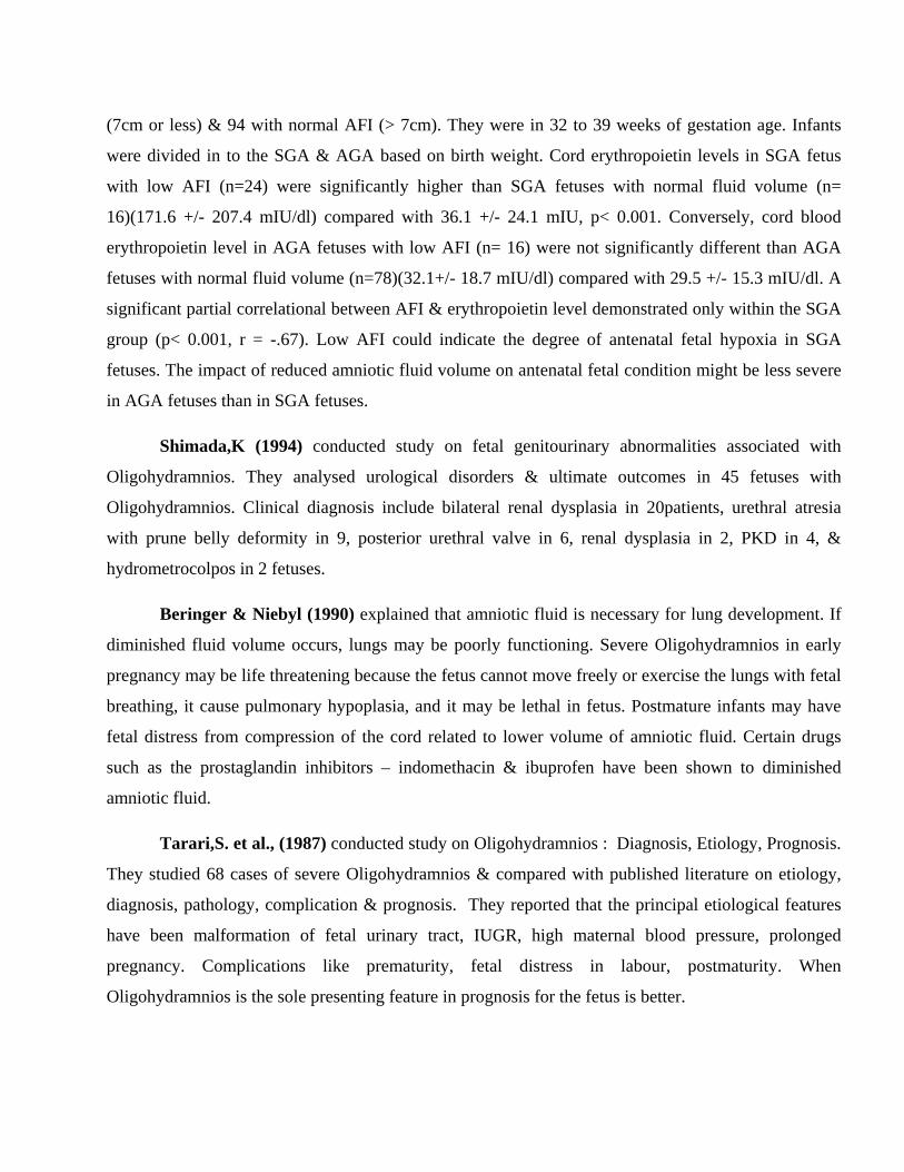
Beringer & Niebyl (1990) explained that amniotic fluid is necessary for lung development. If
diminished fluid volume occurs, lungs may be poorly functioning. Severe Oligohydramnios in early
pregnancy may be life threatening because the fetus cannot move freely or exercise the lungs with fetal
breathing, it cause pulmonary hypoplasia, and it may be lethal in fetus. Postmature infants may have
fetal distress from compression of the cord related to lower volume of amniotic fluid. Certain drugs
such as the prostaglandin inhibitors – indomethacin & ibuprofen have been shown to diminished
amniotic fluid.
Tarari,S. et al., (1987) conducted study on Oligohydramnios : Diagnosis, Etiology, Prognosis.
They studied 68 cases of severe Oligohydramnios & compared with published literature on etiology,
diagnosis, pathology, complication & prognosis. They reported that the principal etiological features
have been malformation of fetal urinary tract, IUGR, high maternal blood pressure, prolonged
pregnancy. Complications like prematurity, fetal distress in labour, postmaturity. When
Oligohydramnios is the sole presenting feature in prognosis for the fetus is better.

LITERATURE RELATED TO TREATMENT OF OLIGOHYDRAMNIOS
Butt,T., & Ahmed. (2011) conducted retrospective study to evaluate the role of antepartum
transabdominal amnioinfusion in the management of Oligohydramnios in pregnancy. The study
consisted 17 pregnant mothers with Oligohydramnios who were treated amnioinfusion during
pregnancy in the period from 2003 to 2006. Mean getational age at first treatment was 24 weeks. Mean
pre produre AFI was 1.8cm, post procedure was 3.8cm. The mean first infusion to delivery interval
was 31 days. Prenatal mortality was 88%, neonatal mortality was 35%, three cases had
chorioamnionitis, with of these cases presenting with premature rupture of membranes at the time of
amnioinfusion. This procedure increases the latency period, it may be useful in preterm pregnancies
where prolonging the pregnancy duration may result in better perinatal outcome.
Ross,G. (2011) reported that the potential of a novel treatment for Oligohydramnios utilizing
maternal administeration of a drug (dDAVP- 1-deamino-8-D-arginine-vasopressin) to hydrate mother
& secondly baby with amniotic fluid. The fetus hydrates in relation to mother & increases its urine
production & amniotic fluid.
Qureshi & Yusuf. (2011) conducted study on intravenous aminoacids in third trimester
isolated Oligohydramnios. They followed prospective design in Lahore from June 2008 to May 2010,
took 42 pregnant women undergone USG. Sonographically proven isolated Oligohydramions in the
third before 36weeks were administered aminoacid solution intravenous after excluding case of
PROM, congenital anomaly of fetus, maternal pulmonary, cardiovascular, hypertensive disorders &
severe placental insufficiency. Pre infusion AFI was measured & repeated weekly. Women were
followed till delivery. Liberal use of caesarean section in Oligohydramnios mothers.
Skovgaard,L., & Silvonek,L., (2011) conducted study on Oligohydramnios – literature review
& case study. They showed that decreased amniotic fluid volume raises management issues & requires
that Nurse-Midwives arrange collaborative care. Evaluation of amniotic fluid volume is now widely
used to evaluate fetus status during pregnancy.
Ulker et al., (2011) conducted prospective study on effect of the maternal position and rest on
the fetal urine production rate. They included 54 pregnant women with normal fluid volume between
26 to 40 weeks of gestation. AFI & fetal urine production rate before and after left lateral position rest
period were compared by paired student t test. The mean AFI before & after rest period were 151.0 +/-

45.0 and 172.5 +/- 46.7mm, it indicate increases in AFI(p< 0.05) mean fetal urine production rate
before and after the rest period were 73.7 +/- 66.8 & 151.8 +/- 119.9ml/hr, it indicate that increased in
fetal urine production (p<.05). They conclude that fetal urine production rate & AFI are markedly
increased by maternal rest in the left lateral decubitus position.
Hofmeyr,G.J., et al., (2010) conducted study on maternal hydration for increasing amniofluid
volume in Oligohydramnios. They used randomised trail with 122 women. The women were asked to
drink 2lit of water before having a repeat ultrasound examination. Maternal hydration in women with
& without Oligohydramnios was associated with an increase in Amniotic fluid volume (mean
difference : MD) for women with oligohydramnios 2.01, 95%. Confidence interval 1.43 to 2.60; & MD
for women with normal Amniotic fluid volume 4.50, 95% confidence interval 2.92 to 6.08. intravenous
hypotonic hydration in women with oligohydramnios was associated with an increase in Amniotic
fluid volume( MD 1.35,95% CI 0.61 to 2.10). isotonic intravenous hydration had no measurable effect.
They concluded that women who drank extra fluid (2lit over 2hrs) dripped directly into their blood
stream increased volume of fluid surrounding the fetus.
Lorzadeh,N. et al., (2010) conducted study on comparison of the effect of oral & intravenous
therapy on women with Oligohydramnios. They used clinical trials on mothers with low AFI &
gestational ages over 35weeks without maternal complication were randomized into 4 groups. 2L/2
oral water, 2L/2hr intravenous isotonic fluid, 2L/2hr IV hypotonic fluid. Maternal AFI were measured
before and after hydration. Data were analysed and made comparison between the groups. The mean
increase in AFI after hydration was significantly greater than in the oral hydration group but not in
intravenous isotonic,hypotonic group compared with control group. They concluded that maternal
hydration with oral water was more effective than other groups.
Hong-ne chu & Mei-juan shen. (2008) conducted study on treating Oligohydramnios with
extract of salvia miltiorrhiza. They used experimental study design on 32 pregnant women with
Oligohydramnios received a daily intravenous dose of 30ml of salvia extract mixed with 5% glucose
500ml. Control group of 41 women received daily 5% glucose 500ml only. The AFI was assessed
atleast twice a week. They found that a mean of 7.2 +/- 2.7 day’s therapy, ranging from 3 to 18 days,
the AFI increased significantly from a mean of 4.9 +/- 2.3cm to a mean of 7.12+ 2.36cm,by a mean of
AFI 0.08 +/- 0.06cm/day(paired t =3.62,p< 0.005). in the control group, the AFI increased from a mean

of 5.1 +/- 2.4cm to a mean of 5.5 +/- 3.1cm after a mean of 6.1+ 3.3 days treatment, ranging from 4 to
15 days. The effect of salvia treatment on AFI in the salvia group was significantly greater than in the
control group (p< 0.001). No side effects observed in treated mothers.
Chhabra, Dargan & Nasare. (2007) stated that, amnioinfusion or instillation of fluid in to
uterus by amniocentesis procedure can help to relieve Oligohydramnios concern. After delivery baby
need careful inspection to rule out kidney disease & compromised lung development.
Flack,J., et al., (2004) conducted a prospective study on acute maternal hydration in third
trimester oligohydramnios: Effects on amniotic fluid volume, uteroplacental perfusion, fetal blood
flow & urine output. They included 10 women with third trimester Oligohydramnios (AFI < 5cm) &
10 control group with normal amniotic fluid volume (AFI >7cm). Doppler flow velocimetry of
maternal uterine artery, fetal umbilical, descending aorta, middle cerebral & renal arteries, maternal
plasma & urine osmolality,AFI, hourly fetal urine production rate were determined before & after oral
hydration by drink 2lit of water over 2 hrs. There was a significant reduction on maternal plasma (p<
0.05) and urine osmolality (p< 0.0001) in both groups after oral hydration. Oral hydration increased
amniotic fluid volume in women with Oligohydramnios( mean change in AFI 3.2cm, 95% CI 1.1 to
5.3; p< 0.02) but not in normal amniotic fluid volume( mean change in AFI -2.0, 95% CI -4.1+0.2).
Hourly urine production rate did not increase in either group (mean changr in hourly fetal urine
production rate 3.5ml/hr, 95% CI -11.7 to +18.7 and -6.8 ml/hr, 95% CI-2.9 to -10.7). Hydration was
increased in uterine artery mean velocity in the Oligohydramnios group (mean change in mean velocity
16.7cm/sec, 95% CI 8.0 to 25.3; p<0.006) but not in controls (mean change in mean velocity
1.2cm/sec, 95% CI -19.7 to + 22.1). No change in pulsatility index in either group. They concluded
that short term maternal oral hydration increases the AFI in women with third trimester
Oligohydramnios, it could not be accounted for by fetal urination but it was associated with improved
uteroplacental perfusion.
Pitt, C. et al., (2000) they used meta -analysis of randomised controlled trials concluded that
prophylactic intrapartum amnioinfusion in women with Oligohydramnios resulted in lower caesarean
section rates & improved neonatal outcome. Early indications are useful intervention.

LITERATURE RELATED TO OUTCOME OF OLIGOHYDRAMNIOS
Grijseels,E. et al., (2011) conducted study on outcome of pregnancies complained by
Oligohydramnios or anhydramnios of renal origin. They performed retrospective study of all
pregnancies diagnosed with Oligohydramnios & associated kidney anomalies during the period 2000-
2008. 71 pregnant mothers were undergone USG, out of 71, 36 fetus had cystic dysplasia,15 had PKD,
20 had hydronephrosis. 32% had associated anomalies. In 49 fetus (69%), the diagnosis had been made
before 24weeks of gestational age; 41 of these pregnancies were terminated. 25 neonates were live
born; 10 survived, 15 died. Severity of Oligohydramnios (1 case of anhydramnios in the survivors Vs 7
in the non- survivors), P = 0.08 was not significant. The 1 yr GFR was below 50ml/mt. 1.73m2 in four
of the survivors.
Ahmad, H., & Munim,S., (2009) conducted study on isolated Oligohydramnios is not an
indicator for adverse perinatal outcome. They used prospective cohort study between May 2005 to
Deccember 2005, 421 mothers were included. Among 421 mothers, 71 were exposed & 350 were
unexposed. When compared to unexposed group with Oligohydramnios had significantly lower birth
weight babies & were delivered at earlier gestational age. There was no statistical difference in the
Apgar score at birth & NICU admissions between the two groups. The number of indication &
caesareans done for fetal reasons were significantly higher in the exposed group.
Gabbe et al., (2002) suggested that decreased amniotic fluid places the fetus at risk for
impaired musculoskeletal development because of inability to move freely in the uterus & tangling of
the long cord around an extremity or cord compression from twisting or kinking, resulting in fetal
distress.
Levine,D., et al., (1997) conducted study on the effect of Oligohydramnios on detection of
fetal anomalies with sonography. They found that 345 mothers with history of PROM (175 with
Oligohydramnios & 170 without Oligohydramnios), gestational age of fetuses was 16–38 weeks.
Major congenital anomalies include hydronephrosis, ventriculomegaly,intestinal atresias, hydrops,
congenital diaphragmatic hernia, skeletal dysplasia, cloacal malformations, gastroschisis were revealed
on sonography in 13 of 175 pregnancy with Oligohydramnios & in 17 of the 170 pregnancies in
control group. Major anomalies missed in Oligohydramnios group include cardiac anomalies, club
foot, small ventral hernia, limb reduction defect & anal atresia. Major anomalies missed in control

group were club foot, atresia & tracheoesophageal fistula. All the major anomalies missed in both
groups were difficult to diagnose before birth & that are frequently missed on sonography.
Oligohydramnios subjectively degrades image resolution; sonography still reveals important anatomic
landmarks. Major anomalies can be detected on sonography even lessthan the normal amount of
amniotic fluid volume.
Queenan & Hobbins (1996) stated that Oligohydramnios is acute or related to earlier rupture
of membranes, the first line of treatment is to expand maternal blood volume by a rapid intravenous
infusion of 1000ml Lactated Ringer’s solution. Amnioinfusion may be attempted during labour.
Caesarean section is planned because the fetus cannot withstand the pressures of labour without the
cushioning effect of adequate amniotic fluid.
Stener,H., et al., (1993) conducted study on outcome after artificial amniotic fluid instillation
in early Oligohydramnios. They took 50 pregnant mothers with Oligohydramnios in the second &
early third trimester in which artificial fluid instillation had been performed. Through artificial fluid
instillation, the rapid diagnosis was possible or was made earlier or additional malformation was
detected. This procedure is associated with risk of induction of labour, a possible iatrogenic rupture of
membranes in 3/50 cases, 37 ended in IUD, spontaneous abortion or lethal malformation, induced
abortion. 10 babies were born alive, but within 6months 6 of them died. 4(8%) were alive & healthy.
Strong et.al., (1990) stated that in Oligohydramnios variable decelerations are seen. Decreased
AFI (<5cm) is an indication for amnioinfusion. It is the infusion of NS into the amniotic cavity through
intrauterine catherter, can be used to decrease the frequency & severity of variable deceleration during
labour.

CHAPTER III
METHODOLOGY
This chapter deals with research design, setting of the study, population for the study, sample
size, sampling technique, criteria for the sample selection, development and description of the tool,
content validity, pilot study, procedure for data collection & statistical analysis.
RESEARCH DESIGN
Case study design was adopted for this study. This study design involves identification of
problems of mothers with Oligohydramnios & related nursing intervention.
SETTING OF THE STUDY
The study was conducted at KMCH maternity ward. It is a super speciality hospital, consisting
657 beds. Average of 1 or 2 mothers with Oligohydramnios are admitted per week.
POPULATION FOR THE STUDY
Mothers diagnosed as Oligohydramnios are admitted at KMCH.
SAMPLE SIZE
Sample size was 15.
SAMPLING TECHNIQUE
Non probability purposive sampling technique was adopted for this study.
CRITERIA FOR SAMPLE SELECTION
Inclusion criteria:
• Pregnant mothers admitted with Oligohydramnios in second or third trimester period at
KMCH.
• Pregnant mothers admitted with Oligohydramnios & co-existing illness.

• Both primi and multigravida mothers were included as a sample, irrespective of their order of
pregnancy.
DEVELOPMENT & DESCRIPTION OF TOOL
The tool consists of 5 sections namely,
Section A: Demographic data.
Section B: Obstetrical data.
Section C: Maternal Assessment Tool.
Section D: Nursing Process Application.
Section E: Risk Factors Assessment Tool.
Section A : Demographic data.
It includes sample number, age, education, religion, occupation, income, food habits.
Section B : Obstetrical data.
It includes LMP, EDD, obstetrical score, weeks of gestation, pre existing illness & maternal
drug exposure.
Section C : Maternal Assessment tool.
It consists of general condition, vital signs, head to foot assessment, obstetrical examination
and investigations.
Section D : Nursing Process application.
The researcher maintained a note on Nursing measures & evaluation.
Section E : Risk factors assessment tool.
It includes current pregnancy risk factors associated with Oligohydramnios.

CONTENT VALIDITY:
The researcher formulated the tool based on the objectives after thorough literature review. The
tool was submitted to the experts in field of Nursing & Medicine to establish the content validity.
Based on expert’s suggestions, the researcher finalized the tool for original study.
PILOT STUDY:
The pilot study was conducted for a period of 2 weeks among 2 samples in mothers with
Oligohydramnios. The assessment tool was prepared and used to collect the necessary data. The
researcher found the problems & executed Nursing interventions. The researcher provided continuous
care to sample for 5 days.
PREPARATION FOR DATA COLLECTION:
The data was collected for a period of 6weeks. A formal permission was obtained from the
Management, Chairma & HOD of OBG Dept at KMCH. The samples were selected as per selection
criteria. The researcher provided Nursing care continuously & evaluated the outcome of care.
During the absence of researcher, care was given by staff Nurses & researcher drawn the
information from the Nurse’s record. Doctor’s order was also considered.
STATISTICAL ANALYSIS:
Researcher analyzed the data with help of descriptive statistics.

CHAPTER –IV
DATA ANALYSIS & INTERPRETATION
The collected data regarding elicited problems & nursing intervention executed on mothers
with Oligohydramnios were organized, analysed & interpreted as follow,
ORGANIZATION OF DATA
Section A: Demographic variables of the samples.
Section B: Obstetrical data of the samples.
Section C: Description about risk factors along with Oligohydramnios.
Section D: Elicited problems of the samples based on lab values, mother’s complaints, assessment &
ultrasonography.
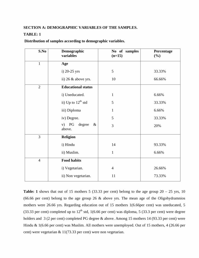
SECTION A: DEMOGRAPHIC VARIABLES OF THE SAMPLES.
TABLE: 1
Distribution of samples according to demographic variables.
S.No Demographic variables
No of samples (n=15)
Percentage (%)
1 Age
i) 20-25 yrs
ii) 26 & above yrs.
5
10
33.33%
66.66%
2 Educational status
i) Uneducated.
ii) Up to 12th std
iii) Diploma
iv) Degree.
v) PG degree & above.
1
5
1
5
3
6.66%
33.33%
6.66%
33.33%
20%
3 Religion
i) Hindu
ii) Muslim.
14
1
93.33%
6.66%
4 Food habits
i) Vegetarian.
ii) Non vegetarian.
4
11
26.66%
73.33%
Table: 1 shows that out of 15 mothers 5 (33.33 per cent) belong to the age group 20 – 25 yrs, 10
(66.66 per cent) belong to the age group 26 & above yrs. The mean age of the Oligohydramnios
mothers were 26.66 yrs. Regarding education out of 15 mothers 1(6.66per cent) was uneducated, 5
(33.33 per cent) completed up to 12th std, 1(6.66 per cent) was diploma, 5 (33.3 per cent) were degree
holders and 3 (2 per cent) completed PG degree & above. Among 15 mothers 14 (93.33 per cent) were
Hindu & 1(6.66 per cent) was Muslim. All mothers were unemployed. Out of 15 mothers, 4 (26.66 per
cent) were vegetarian & 11(73.33 per cent) were non vegetarian.

Figure :1 Distribution of age in relation to Oligohydramnios.
Figure : 2 Distribution of educational status in relation to Oligohydramnios.

Figure : 3 Distribution of religion in relation to Oligohydramnios.
Figure: 4 Distribution of food habits in relation to Oligohydramnios.

SECTION B: OBSTETRICAL DATA OF THE SAMPLES.
TABLE : 2
Distribution of samples according to obstetrical data.
S.No Obstetrical data No of samples
(n = 15)
Percentage
(%)
1 Gravida:
i)primi
ii)multi
12
3
80%
20%
2 Weeks of gestation :
i) 29 – 42 weeks
15
100%
3 Pre-existing illness:
i) Diabetes mellitus.
1
6.66%
Table: 2 shows that, out of 15 mothers, 12(80 per cent) were to primi gravida, 3(20 per cent) were
multi gravida & 2(13.33 per cent) were elderly primi & 15(100 per cent) belong to 29 – 42 weeks of
gestation. The mean gestational age of mothers with oligohydramnios was 35.33 weeks. Regarding
pre-existing illness, out of 15 mothers only 1 (6.66 per cent) had diabetes mellitus.

Figure : 5 Distribution of gravida in relation to Oligohydramnios.
Figure : 6 Distribution of weeks of gestation in relation to Oligohydramnios

Figure : 7 Distribution of pre -existing illness in relation to Oligohydramnios.

SECTION C:
DESCRIPTION ABOUT RISK FACTORS ALONG WITH OF OLIGOHYDRAMNIOS.
TABLE: 3
Distribution of samples according to maternal risk factors along with Oligohydramnios.
S.No Maternal risk factors No. of samples
(n =15)
Percentage
(%)
1
2
3
4
5
6
Mildpre-eclampsia.
Moderate to severe pre-
eclampsia.
Gestational diabetes mellitus.
HBV infection.
Rh sensitization.
Isolated Oligohydramnios
without Complication.
1
1
2
1
2
8
6.66%
6.66%
13.33%
6.66%
13.33%
53.33%
Table: 3 shows that out of 15 mothers 1(6.66 per cent) had mild pre eclampsia, 1(6.66 per cent) had
moderate to severe preeclampsia, 2(13.33 per cent) had gestational diadetes mellitus, 1(6.66 per cent)
had viral disease, 2(13.33 per cent) had Rh sensitization & 8 (53.33 per cent) had Oligohydamnios.
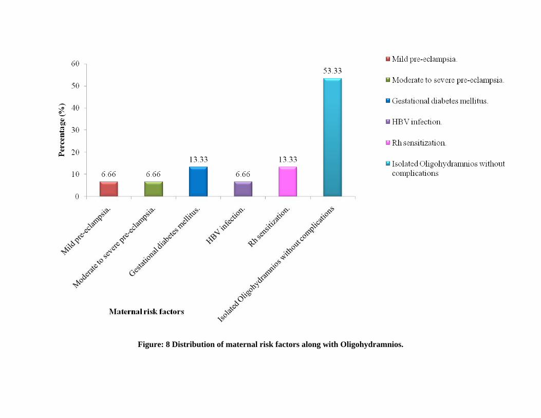
Figure: 8 Distribution of maternal risk factors along with Oligohydramnios.

Table: 4 Distribution of samples according to fetal risk factors along with of Oligohydramnios.
S.No Fetal risk factors No of samples
(n=15)
Percentage (%)
1
2
3
Intra uterine growth restriction.
Premature rupture of membranes.
Intrauterine Death.
4
1
1
26.66%
6.66%
6.66%
Table: 4 shows that, out of 15 mothers 4 (26.66 per cent) had Intra uterine growth restriction, 1(6.66
per cent) had rupture of membranes & 1(6.66 per cent) had intra uterine death.

Figure: 9 Distribution of fetal risk factors along with Oligohydramnios.
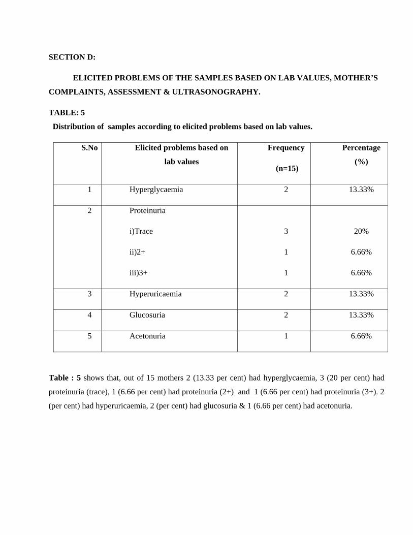
SECTION D:
ELICITED PROBLEMS OF THE SAMPLES BASED ON LAB VALUES, MOTHER’S
COMPLAINTS, ASSESSMENT & ULTRASONOGRAPHY.
TABLE: 5
Distribution of samples according to elicited problems based on lab values.
S.No Elicited problems based on
lab values
Frequency
(n=15)
Percentage
(%)
1 Hyperglycaemia 2 13.33%
2 Proteinuria
i)Trace
ii)2+
iii)3+
3
1
1
20%
6.66%
6.66%
3 Hyperuricaemia 2 13.33%
4 Glucosuria 2 13.33%
5 Acetonuria 1 6.66%
Table : 5 shows that, out of 15 mothers 2 (13.33 per cent) had hyperglycaemia, 3 (20 per cent) had
proteinuria (trace), 1 (6.66 per cent) had proteinuria (2+) and 1 (6.66 per cent) had proteinuria (3+). 2
(per cent) had hyperuricaemia, 2 (per cent) had glucosuria & 1 (6.66 per cent) had acetonuria.

Figure: 10 Distribution of samples according to elicited problems based on lab values.
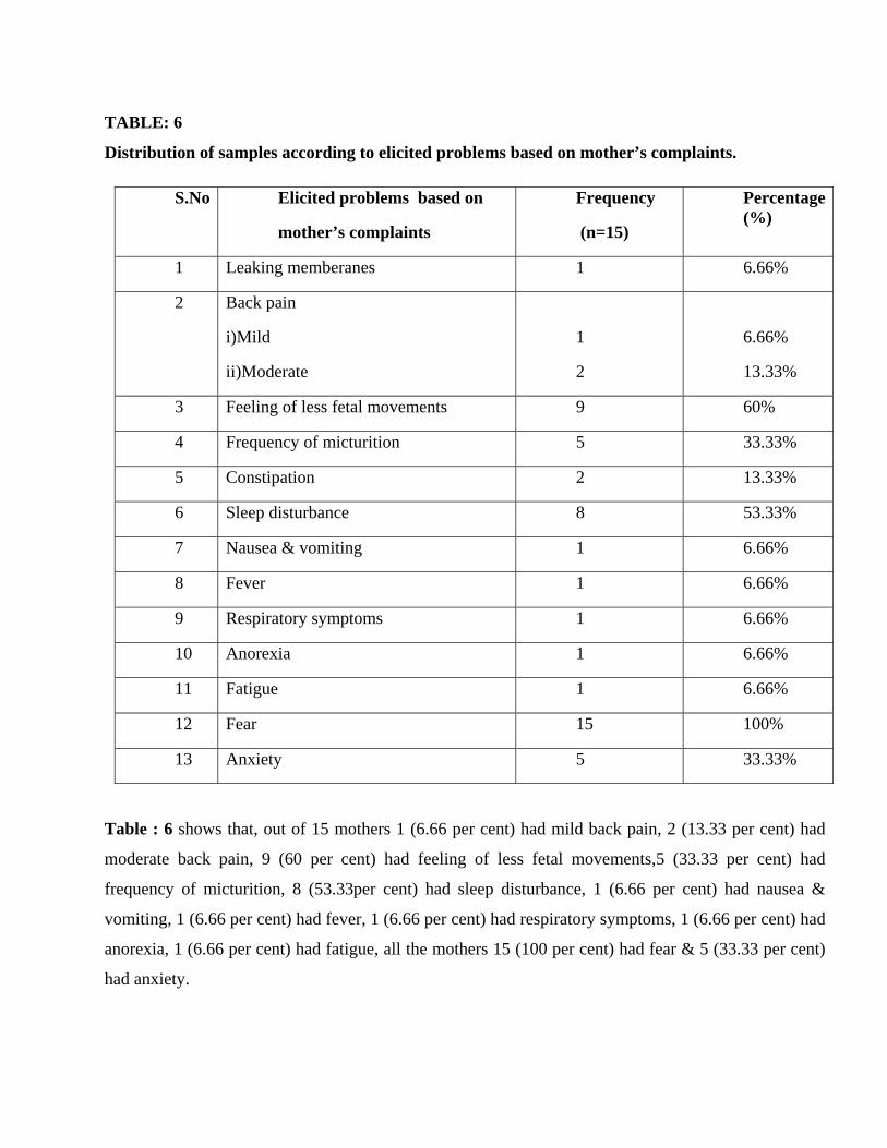
TABLE: 6
Distribution of samples according to elicited problems based on mother’s complaints.
S.No Elicited problems based on
mother’s complaints
Frequency
(n=15)
Percentage (%)
1 Leaking memberanes 1 6.66%
2 Back pain
i)Mild
ii)Moderate
1
2
6.66%
13.33%
3 Feeling of less fetal movements 9 60%
4 Frequency of micturition 5 33.33%
5 Constipation 2 13.33%
6 Sleep disturbance 8 53.33%
7 Nausea & vomiting 1 6.66%
8 Fever 1 6.66%
9 Respiratory symptoms 1 6.66%
10 Anorexia 1 6.66%
11 Fatigue 1 6.66%
12 Fear 15 100%
13 Anxiety 5 33.33%
Table : 6 shows that, out of 15 mothers 1 (6.66 per cent) had mild back pain, 2 (13.33 per cent) had
moderate back pain, 9 (60 per cent) had feeling of less fetal movements,5 (33.33 per cent) had
frequency of micturition, 8 (53.33per cent) had sleep disturbance, 1 (6.66 per cent) had nausea &
vomiting, 1 (6.66 per cent) had fever, 1 (6.66 per cent) had respiratory symptoms, 1 (6.66 per cent) had
anorexia, 1 (6.66 per cent) had fatigue, all the mothers 15 (100 per cent) had fear & 5 (33.33 per cent)
had anxiety.

Figure: 11 Distribution of samples according to elicited problems based on mother’s complaints.
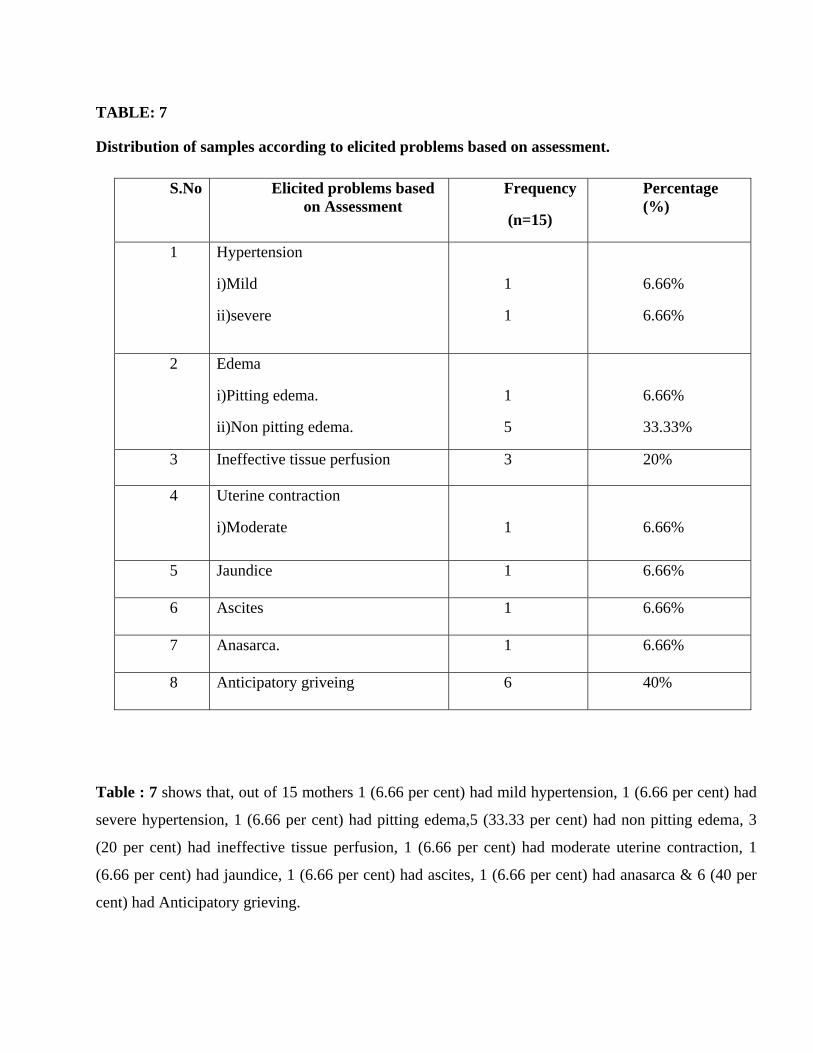
TABLE: 7
Distribution of samples according to elicited problems based on assessment.
S.No Elicited problems based on Assessment
Frequency
(n=15)
Percentage (%)
1 Hypertension
i)Mild
ii)severe
1
1
6.66%
6.66%
2 Edema
i)Pitting edema.
ii)Non pitting edema.
1
5
6.66%
33.33%
3 Ineffective tissue perfusion 3 20%
4 Uterine contraction
i)Moderate
1
6.66%
5 Jaundice 1 6.66%
6 Ascites 1 6.66%
7 Anasarca. 1 6.66%
8 Anticipatory griveing 6 40%
Table : 7 shows that, out of 15 mothers 1 (6.66 per cent) had mild hypertension, 1 (6.66 per cent) had
severe hypertension, 1 (6.66 per cent) had pitting edema,5 (33.33 per cent) had non pitting edema, 3
(20 per cent) had ineffective tissue perfusion, 1 (6.66 per cent) had moderate uterine contraction, 1
(6.66 per cent) had jaundice, 1 (6.66 per cent) had ascites, 1 (6.66 per cent) had anasarca & 6 (40 per
cent) had Anticipatory grieving.
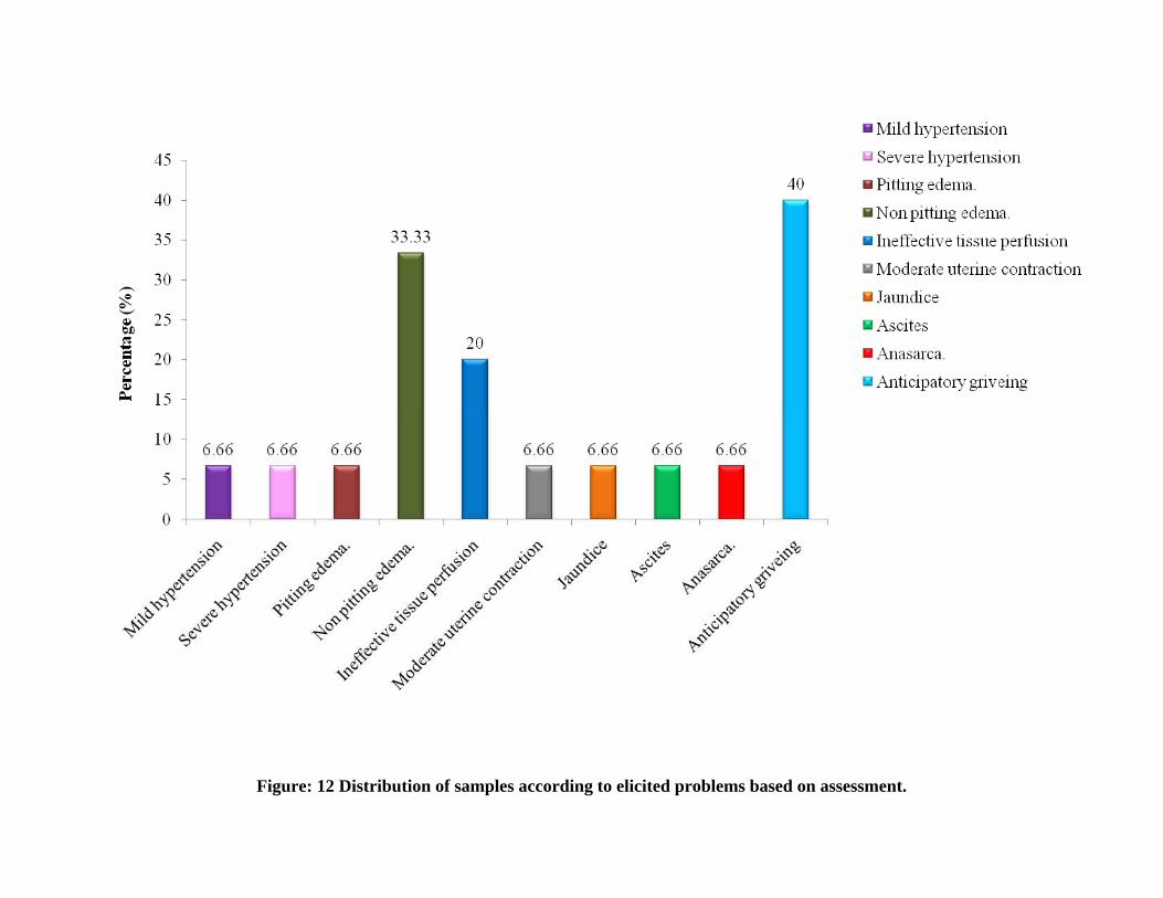
Figure: 12 Distribution of samples according to elicited problems based on assessment.

Table: 8 Distribution of samples according to elicited findings based on ultrasonography.
Table : 8 shows that out of 15 mothers,12(80 per cent) had good fetal movements, 3(20 per cent) had
less fetal movements, 12(80 per cent) had AFI between 6-7.9cm, 2(13.33 per cent) had AFI between 4-
5.9 cm, 1(6.66 per cent) had AFI below 4cm, 2(13.33 per cent) had breech presentation, 13(86.66 per
cent) had cephalic presentation, 3(20 per cent) had presence of diastolic notch, 12(80 per cent) had
absence of diastolic notch, 7(46.66 per cent) had appropriate fetal weight related to gestational age &
8(53.33 per cent) had less fetal weight related to gestational age.
S.No Variables Frequency (n=15)
Percentage (%)
1 Fetal movements Good 12 80%
Less 3 20%
2 Amniotic fluid index
(AFI)
6 - 7.9cm 12 80%
4 - 5.9cm 2 13.33%
Below 4cm 1 6.66%
3 Fetal Presentation Cephalic 13 86.66%
Breech 2 13.33%
4 Diastolic notch Present 3 20%
Absent 12 80%
5 Fetal weight related to gestational age.
Appropriate 7 46.66%
Less 8 53.33%

Figure: 13 Distribution of samples according to fetal movements based on ultrasonography.
Figure:14 Distribution of samples according to Amniotic fluid index based on ultrasonography

Figure:15 Distribution of samples according to fetal presentation based on ultrasonography
Figure : 16 Distribution of samples according to diastolic notch based on ultrasonography

Figure : 17 Distribution of samples according to fetal weight related to gestational age based on
ultrasonography

CHAPTER - V
DISCUSSION, SUMMARY, CONCLUSION, IMPLICATION, LIMITATION &
RECOMMENDATION
DISCUSSION
The main focus of the study was to provide individualized Nursing care to mother’s with
Oligohydramnios, according to Skovgared. L (2011) decreased amniotic fluid volume raises
management issues & requires that Nurse-Midwives collaborative care. The study was conducted at
KMCH in Coimbatore. This is the case study to identify the problems & execution of nursing care for
mothers with Oligohydramnios. The sample size was 15 antenatal mothers with Oligohydramnios. The
results of study according to objectives are discussed as follows,
Demographic profile:
Out of 15 mothers 5(33.33per cent) were belong to the age group 20-25yrs, 10(66.66 per cent)
were belong to the age group 26 & above yrs. Regarding education out of 15 mothers 1(6.66 per cent)
was uneducated, 5(33.33 per cent) completed up to 12th std, 3(20 per cent) completed PG degree &
above. Among 15 mothers 14(93.33 per cent) were Hindu, 1(6.66 per cent) was Muslim. All mothers
were employed. Among 15mothers 4(26.66 per cent) were vegetarian & 11(73.33 per cent) were non
vegetarian.
Obstetrical data:
Out of 15 mothers, 12(80 per cent) were belong to primi gravida, 3(20 per cent) were belong to
multi gravida & 15(100 per cent) belong to 29 – 42 weeks of gestation. The mean gestational age of
mothers with Oligohydramnios was 35.33 weeks. Regarding pre-existing illness, out of 15 mothers
only 1 (6.66 per cent) had diabetes mellitus.

Informations about mothers:
Out of 15 mothers, 12 were discussed below,
• Sample No: 1 Mrs.A (27yrs) 29+6 weeks of gestation, primi mother admitted on 16/7/2011 with
the complaints of premature rupture of memberances & less fetal movements. She is a known
case of diabetes mellitus since 3yrs & she was in insulin treatment also. She had AFI 6.3cm &
diastolic notch in umbilical artery. Eventhough after treatment, hyperglycaemia was not
controlled. So, she underwent caesarean delivery on 18/7/2011.
• Sample No: 2 Mrs.A (27yrs) 35+3 weeks of gestation, primi mother admitted on 23/7/2011 with
the complaint of less fetal movements. She had AFI 7.3cm, diastolic notch in umbilical artery
& fetus in breech presentation. So, she underwent caesarean delivery on 27/7/2011.
• Sample No: 3 Mrs.A (21yrs) 39+5 weeks of gestation, primi mother admitted on 24/7/2011 with
the complaint of less fetal movements. She had AFI 7.8cm, so she underwent normal vaginal
delivery on 27/7/2011.
• Sample No: 5 Mrs.A (27yrs) 36 weeks of gestation, primi mother admitted on 29/7/2011 with
the complaint of pedal edema. She had AFI 7.3cm. Due to low AFI she underwent caesarean
delivery on 1/8/2011.
• Sample No: 6 Mrs.A (27yrs) 38+3weeks of gestation (G4P0L0A3), mother admitted on 4/8/2011
with the complaints of less fetal movements & pedal edema. She had AFI 7.4cm. Due to low
AFI she underwent caesarean delivery on 6/8/2011.
• Sample No: 7 Mrs.A (26yrs) 35+1weeks of gestation, primi mother admitted on 4/8/2011 with
the complaint of less fetal movements. She had AFI 5.3cm. Due to low AFI she underwent
caesarean delivery on 5/8/2011.
• Sample No: 8 Mrs.A (25yrs) 37+6weeks of gestation (G4P0L0A1), mother admitted on 6/8/2011
with the complaint of less fetal movements. She had AFI 6.3cm & fetus in breech presentation.
Due to mal presentation & low AFI she underwent caesarean delivery on 8/8/2011.
• Sample No: 10 Mrs.A (28 yrs) 35 weeks of gestation, primi mother admitted on 9/8/2011 due to
low AFI (3.4cm) & diastolic notch in umbilical artery. Eventhough low AFI, she underwent
normal vaginal delivery on 13/8/2011.

• Sample No: 12 Mrs.A (21yrs) 35+5weeks of gestation, primi mother admitted on 22/8/2011 due
to low AFI (4.3cm). Hence she underwent caesarean delivery on 25/8/2011.
• Sample No: 13 Mrs.A (36yrs) 33+4weeks of gestation, primi mother admitted on 24/8/2011 due
to low AFI (7.5cm). She got Inj. Astymin forte infusion, after that she discharged with follow
up care instructions on 25/8/2011.
• Sample No: 14 Mrs.A (35yrs) 35 weeks of gestation, primi mother admitted on 31/8/2011 with
the complaint of less fetal movements. She had a gestational diabetes mellitus & she is in
insulin treatment also. She had AFI 7.6cm. Because of uncontrolled hyperglycaemia & low
AFI, she underwent caesarean delivery on 2/9/2011.
• Sample No: 15 Mrs.A (24yrs) 35 weeks of gestation, primi mother admitted on 5/9/2011 due to
low AFI (6.2cm) & IUGR. So, she underwent caesarean delivery on 7/9/2011.
• Remaining samples (No: 4, 9, 11) were discussed elaborately in Appendix - B.
Objectives:
The first objective of study was to assess the risk factors associated with oligohydrmnios.
Regarding maternal risk factors along with Oligohydramnios, out of 15mothers 1(6.66 per cent)
had mild preeclampsia, 1(6.66 per cent) had severe pre-eclampasia, 2(13.33 per cent) had gestational
diabetes mellitus, 1(6.66 per cent) had hepatitis B infection, 2(13.33 per cent) had Rh sensitization &
8(53.33 per cent) had isolated Oligohydramnios without complication. Regarding fetal risk factors
along with Oligohydramnios, out of 15 mothers 4(26.66 per cent) had Intra uterine growth restriction,
1(6.66 per cent) had premature rupture of membranes & 1(6.66 per cent) had Intra uterine death.
The second objective of the study was to identify the problems of mothers with Oligohydramnios.
i) Elicited problems based on lab values,
Out of 15 mothers 2 (13.33 per cent) had hyperglycaemia, 3 (20 per cent) had proteinuria
(trace), 1 (6.66 per cent) had proteinuria (2+) and 1 (6.66 per cent) had proteinuria (3+). 2 (per cent)
had hyperuricaemia, 2 (per cent) had glucosuria, 1 (6.66 per cent) had acetonuria.

ii) Elicited problems based on mother’s complaints,
Out of 15 mothers,1 (6.66%) had leaking memberanes, 1 (6.66 per cent) had mild back pain, 2
(13.33 per cent) had moderate back pain, 9 (60 per cent) had feeling of less fetal movements,5 (33.33
per cent) had frequency of micturition, 8 (53.33per cent) had sleep disturbance, 1 (6.66 per cent) had
nausea & vomiting, 1 (6.66 per cent) had fever, 1 (6.66 per cent) had respiratory symptoms, 1 (6.66 per
cent) had anorexia, 1 (6.66 per cent) had fatigue, 15 (100 per cent) had fear & 5 (33.33 per cent) had
anxiety.
iii) Elicited problems based on assessment,
Out of 15 mothers 1 (6.66 per cent) had mild hypertension, 1 (6.66 per cent) had severe
hypertension, 1 (6.66 per cent) had pitting edema,5 (33.33 per cent) had non pitting edema, 3 (20 per
cent) had ineffective tissue perfusion, 1 (6.66 per cent) had moderate uterine contraction, 1 (6.66 per
cent) had jaundice, 1 (6.66 per cent) had ascites, 1 (6.66 per cent) had anasarca, 6 (40 per cent) had
Anticipatory grieving.
iv) Elicited problems based on ultrasonography,
Out of 15 mothers,12(80 per cent) had good fetal movements, 3(20 per cent) had less fetal
movements, 12(80 per cent) had AFI between 6-7.9cm, 2(13.33 per cent) had AFI between 4-5.9 cm,
1(6.66 per cent) had AFI below 4cm, 2(13.33 per cent) had breech presentation, 13(86.66 per cent) had
cephalic presentation, 3(20 per cent) had presence of diastolic notch, 12(80 per cent) had absence of
diastolic notch, 7(46.66 per cent) had appropriate fetal weight related to gestational age & 8(53.33 per
cent) had less fetal weight related to gestational age.
The third objective of the study was to execution of nursing strategies on mothers with
oligohydramnios,
i) Elicited problems based on lab values,
1) Hyperglycemia:
• Checked the blood sugar level.
• Medications are administered as prescribed. (Injection insulin).

• Advised to take low carbohydrate diet & high protein rich diet.
2) Proteinuria:
• Checked the grade of protein excretion in urine.
• Advised to take protein rich diet (100gm/day) like milk, pulses & cereals.
3) Hyperuricaemia:
• Advised to drink more fluids.
• Advised to avoid starvation.
• Educated to take balanced diet.
• Monitored blood pressure (prone to get pregnancy induced hypertension).
4) Glucosuria:
• Avoid intake of concentrated simple sugar.
• Checked blood pressure daily.
• Advised to take adequate rest.
• Advised to take more amount of fluids.
5) Acetonuria:
• Monitored blood sugar level regularly.
• Advised to drink oral fluids to overcome dehydration.
• Administered more intravenous fluids.
• Advised to take small & frequent diet.
ii) Elicited problems based on mother’s complaints,
1) Leaking memberanes.
• Ensured memberanes is an intact or not.
• Perineal care was given.
• Inj.Betnesol (12mg) 2 doses was given 12hrs apart, for fetal lung maturity.
• Inj.Duvadilan (40mg) mixed with 500ml DNS (15 drops/mt) was given to premature uterine contraction.
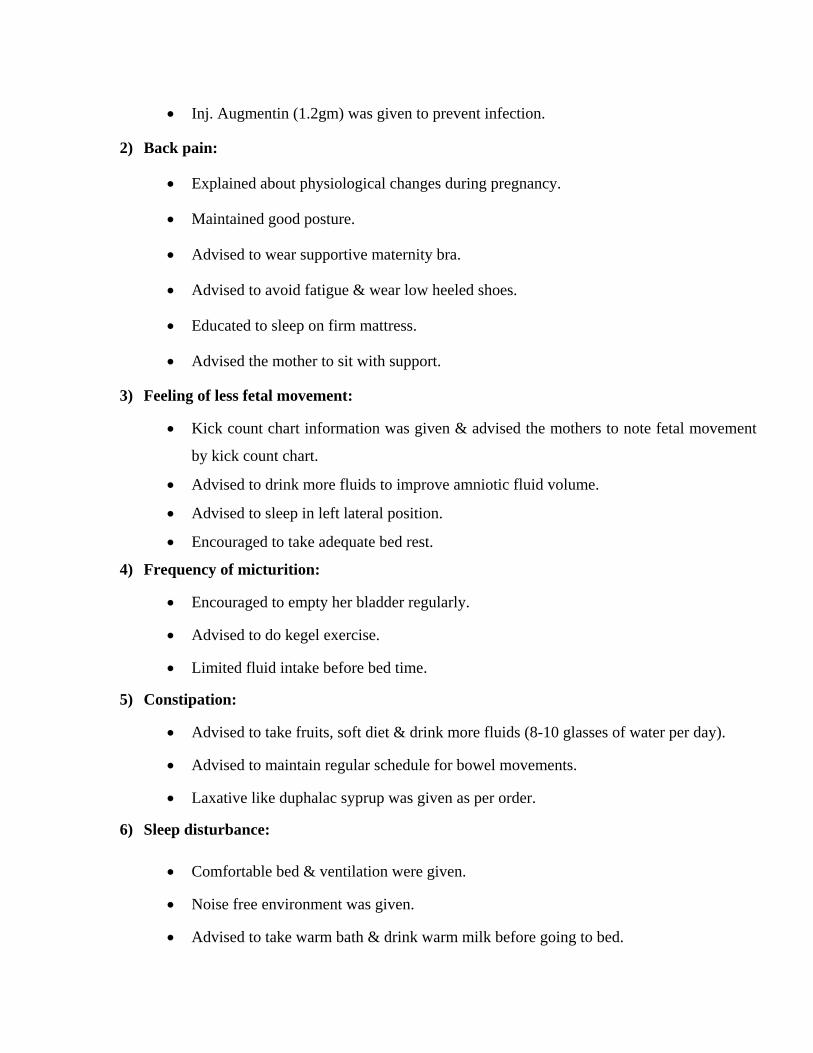
• Inj. Augmentin (1.2gm) was given to prevent infection.
2) Back pain:
• Explained about physiological changes during pregnancy.
• Maintained good posture.
• Advised to wear supportive maternity bra.
• Advised to avoid fatigue & wear low heeled shoes.
• Educated to sleep on firm mattress.
• Advised the mother to sit with support.
3) Feeling of less fetal movement:
• Kick count chart information was given & advised the mothers to note fetal movement
by kick count chart.
• Advised to drink more fluids to improve amniotic fluid volume.
• Advised to sleep in left lateral position.
• Encouraged to take adequate bed rest.
4) Frequency of micturition:
• Encouraged to empty her bladder regularly.
• Advised to do kegel exercise.
• Limited fluid intake before bed time.
5) Constipation:
• Advised to take fruits, soft diet & drink more fluids (8-10 glasses of water per day).
• Advised to maintain regular schedule for bowel movements.
• Laxative like duphalac syprup was given as per order.
6) Sleep disturbance:
• Comfortable bed & ventilation were given.
• Noise free environment was given.
• Advised to take warm bath & drink warm milk before going to bed.

• Advised to use pillows to support body parts & reassured the mother.
7) Nausea & vomiting:
• Advised to avoid empty & overload stomach.
• Advised to avoid spicy & gas forming foods.
• Encouraged to drink more fluids & eat soft and liquid diet.
• Administered antiemetic drug as per order. (if needed)
8) Fever:
• Checked the vital signs.
• Advised to drink more fluids & wear loose cotton cloth.
• Administered intravenous fluid & antipyretics.
• Encouraged to take easily digestible food.
9) Respiratory symptoms:
• Checked vital signs.
• Fowler’s position was given.
• Expectorant was given.
10) Anorexia:
• Assess the dietary intake & nutritional status through diet history.
• Checked the weight gain daily.
• Advised to take high carbohydrate & protein diet.
11) Fatigue:
• Advised to take more oral liquids.
• Administered intravenous fluids.
• Advised to take bed rest.
• Advised to take well balanced diet (high carbohydrate & vitamins).
12) Fear:
• Encouraged to express their feelings.
• Therapeutic touch was used.
• Advised the family members to involve in mother’s care.
• Psychological support was given.
• Explained the fetal condition clearly.
13) Anxiety:
• Explained the all the procedures.
• Maintained accustomed environmental structure.
• Avoided excessive reassurance.
iii) Elicited problems based on assessment,
1) Hypertension:
• Checked the blood pressure 4hrly once or 2hrly once.
• Advised to take rest.
• Advised to drink more fluids to promote urine output.
• Administered antihypertensive drugs as per order.
2) Pedal edema:
• Checked the degree of edema.
• Advised to elevate the foot end of the bed.
• Encouraged the mother to do foot & leg exercise.
• Checked blood pressure 4hrly (if mother has PIH).
3) Ineffective tissue perforation:
• Advised to sleep in left lateral position.
• Encouraged to count the fetal movements to know fetal wellbeing.
• Advised to drink more fluids.

• Advised to take adequate bed rest.
4) Uterine contraction:
• Checked the duration, frequency & intensity of uterine contration.
• Checked the fetal heart rate by NST.
• Ensured mother is in labour or not.
5) Jaundice:
• Degree of edema is noted & recorded.
• Advised to take rest & administered intravenous fluid.
• Advised to take high carbohydrate diet.
• Administered antibiotics as per order.
6) Ascites:
• Maintained fowler’s position.
• Advised to take bed rest.
• Maintained intake & output chart.
• Advised to take diet rich in carbohydrate & adequate protein (if tolerated).
• Avoided injury.
7) Anasarca:
• Assess the skin integrity & degree of edema.
• Restricted sodium as prescribed.
• Unwrinkled bed was given.
• Advised to avoid irritating soaps.
• Position was changed frequently.
• Finger nails kept short & smooth to prevent infection from scratching.

8) Anticiparitory grieving:
• Advised the family members to involve in mother’s care.
• Emotional support was provided.
• Encouraged the family to restructure their daily activities.
The fourth objective of the study was to evaluate the Nursing strategies executed on mothers
with Oligohydramnios.
Based on the identified problems, related Nursing interventions were executed on mothers
with Oligohydramnios & were evaluated.
• The executed measures to maintain normal blood sugar level in 2(13.33 per cent) of the
samples were not succeeded. So the mothers underwent caesarean section due to
hyperglycemia along with low AFI.
• Measures were executed to control proteinuria among 3(20 per cent)samples : Protenuria trace,
1(6.66 per cent)sample : proteinuria 2+ & 1(6.66 per cent)sample : proteinuria 3+. These
measures failed to control proteinuria among 3samples hence the mothers shifted to caesarean
section in order to prevent complications to both mother & fetus. But these measures succeeded
among 2samples (Protenuria trace), in that 1 was discharged and another one undergone normal
delivery.
• Interventions were executed to control hyperuricaemia among 2(13.33 per cent) samples. In
terms of associated conditions, 1 had pregnancy induced hypertension & another had
gestational diabetes mellitus. These measures failed to control hyperuricaemia among both
these mothers hence the mothers shifted to caesarean section.
• Measures were instituted to control glucosuria among 2(13.33 per cent) samples. Among them
1 discharged due to controlled blood pressure & another one undergone caesarean section.
• Non pharmacological measures were provided to control acetonuria among 1(6.66 per cent)
sample. For this mother acetonuria developed due to hepatitis B infection. Along with
associated complications, IUD occurred.
• Measures were executed for leaking memberanes among 1(6.66 per cent) sample. Interventions
included antibiotics, corticosteroid & uterotonic agent.

• Non pharmacological measures were executed for 2(13.33 per cent) samples who had back pain
was successful.
• Measures were executed to improve feeling fetal movements for 9 (60 per cent) samples.
Among that one had normal delivery, one discharged, one had IUD & 6 samples undergone
caesarean section.
• Non pharmacological measures were executed for 5(33.33 per cent) samples who had
frequency of micturition was successful.
• Constipation was relieved by non pharmacological measures & it was instituted to 1(6.66 per
cent) sample & for another 1 (6.66 per cent) sample it was relieved by laxative (duphalac
syrup).
• Non pharmacological measures were executed for 8 (53.33 per cent) samples who had sleep
disturbance was successful.
• Nausea & vomiting were controlled by non pharmacological measures to 1(6.66 per cent)
sample. Fever was reduced by antipyretic to 1(6.66 per cent) sample.
• The measures were instituted to 1 (6.66 percent) sample with respiratory symptoms were
effective.
• Anorexia was treated by non pharmacological measures to 1(6.66 per cent) sample.
• Some measures were tried to relieve fatigue for 1(6.66 per cent) sample, but it was not
effective.
• The measures were executed for fear 15 (100 per cent) of the sample & anxiety 5 (33.33 per
cent) with explanation & reassurance.
• The executed measures to maintain normal blood pressure for 1(6.66 per cent) sample was not
controlled. But for another 1(6.66 per cent) sample, blood pressure controlled.
• Non pitting edema was reduced by non pharmacological measures to 5(33.33 per cent) samples
& pitting edema was not reduced for 1 (6.66 per cent) sample.
• Tissue perfusion was not improved by pharmacological measures & non pharmacological
measures to 3 (20 per cent) samples.
• The measures executed for 1 (6.66 per cent) sample with premature uterine contraction was
treated by tocolytic agent and it was effective.

• Interventions were executed for 1 (6.66 per cent) sample with jaundice treated by HBV vaccine
& antiretroviral therapy & it was not effective. That mother needs liver transplantation.
• Anasarca & ascites were not reduced for 1 (6.66 per cent) sample by non pharmacological
measures.
• Emotional support was given to 6 (40 per cent) sample in order to cope up with grieving
process.
SUMMARY
The study was conducted for identify the problems & execution of Nursing strategies for
mothers with Oligohydramnios,
The objectives of study as follows to,
assess the risk factors associated with Oligohydramnios.
identify the problems of mothers with Oligohydramnios.
execution of nursing strategies on mothers with Oligohydramnios.
evaluate the nursing strategies executed on mothers with Oligohydramnios.
The purpose of the study was to develop a Nursing module {regarding follow up care for mothers
with Oligohydramnios}.The conceptual frame work for this study was based on Orlando Nursing
Process model.
The literature review helped to carry out research work like formulation of tool, preparation of
module. Tool developed for this study consists of demographic data, obstetrical data, maternal
assessment tool, nursing process application & risk factor assessment tool.
The content validity was obtained from Nursing & Medical experts. Pilot study was conducted
for a period of 2weeks. The data for main study was collected for a period of 6weeks at KMCH by non
probability purposive sampling technique among 15 samples. Data were collected, analyzed &
interpreted by using descriptive statistics.

MAJOR FINDING OF THE STUDY
Out of 15 mothers, 5(33.33per cent) were belong to the age group 20-25yrs, 10(66.66per cent)
were belong to the age group 26 & above yrs.
Among 15 mothers, 1(6.66per cent) was uneducated, 5(33.33per cent) completed up to 12thstd,
1(6.66 per cent) was diploma holder, 5(33.33 per cent) were degree holders and 3(20per cent)
completed PG degree & above.
Among 15 mothers, 14(93.33per cent) were Hindu, 1(6.66per cent) was Muslim. All mothers
were unemployed
In terms of food habits, 4(26.66per cent) were vegetarian & 11(73.33per cent) were non
vegetarian.
Out of 15 samples, 12(80 per cent) were belong to primi gravida, 3(20 per cent) were belong to
multi gravida.
All mothers were belong to 29-42 weeks of gestation. The mean gestational age of mothers
with Oligohydramnios was 35.33 weeks.
Regarding pre-existing illness, 1(6.66per cent) had diabetes mellitus.
Regarding maternal risk factors associated with Oligohydramnios, 1(6.66per cent) had mild
preeclampsia, 1(6.66per cent) had severe pre-eclampasia, 2(13.33per cent) had gestational
diabetes mellitus, 1(6.66per cent) had hepatitis B infection, 2(13.33per cent) had Rh
sensitization & 8(53.33per cent) had isolated Oligohydramnios without complication.
Regarding fetal risk factors associated with Oligohydramnios, 4(26.66 per cent) had IUGR,
1(6.66per cent) had premature rupture of membranes & 1(6.66per cent) had intra uterine death.
Among 15 mothers 2 (13.33 per cent) had hyperglycaemia, 3 (20 per cent) had proteinuria
(trace), 1 (6.66 per cent) had proteinuria (2+) and 1 (6.66 per cent) had proteinuria (3+). 2 (per
cent) had hyperuricaemia, 2 (per cent) had glucosuria and 1 (6.66 per cent) had acetonuria.
Among 15 mothers,1 (6.66%) had leaking memberanes, 1 (6.66 per cent) had mild back pain, 2
(13.33 per cent) had moderate back pain, 9 (60 per cent) had feeling of less fetal movements,5
(33.33 per cent) had frequency of micturition, 8 (53.33per cent) had sleep disturbance, 1 (6.66
per cent) had nausea & vomiting, 1 (6.66 per cent) had fever, 1 (6.66 per cent) had respiratory
symptoms, 1 (6.66 per cent) had anorexia, 1 (6.66 per cent) had fatigue, 15 (100 per cent) had
fear & 5 (33.33 per cent) had anxiety.

Out of 15 mothers 1 (6.66 per cent) had mild hypertension, 1 (6.66 per cent) had severe
hypertension, 1 (6.66 per cent) had pitting edema,5 (33.33 per cent) had non pitting edema, 3
(20 per cent) had ineffective tissue perfusion, 1 (6.66 per cent) had moderate uterine
contraction, 1 (6.66 per cent) had jaundice, 1 (6.66 per cent) had ascites, 1 (6.66 per cent) had
anasarca and 6 (40 per cent) had Anticipatory grieving.
Out of 15 mothers, 12(80 per cent) had good fetal movements, 3(20 per cent) had less fetal
movements, 12(80 per cent) had AFI between 6-7.9cm, 2(13.33 per cent) had AFI between 4-
5.9 cm, 1(6.66 per cent) had AFI below 4cm, 2(13.33 per cent) had breech presentation,
13(86.66 per cent) had cephalic presentation, 3(20 per cent) had presence of diastolic notch,
12(80 per cent) had absence of diastolic notch, 7(46.66 per cent) had appropriate fetal weight
related to gestational age & 8(53.33 per cent) had less fetal weight related to gestational age.
Among 15 mothers, 10(66.66per cent) were undergone caesarean section, 2(13.33per cent)
were undergone normal vaginal delivery, 2(13.33per cent) were discharge after treatment &
1(6.66per cent) had IUD.
CONCLUSION
Among 15 mothers 9 (60 per cent) had complaints of less fetal movements. In that 1 (6.66 per
cent) had hepatitis B infection, 1 (6.66 per cent) had mild pre-eclampsia, 2 (13.33 per cent) had
gestational diabetes mellitus & remaining 5(33.33 per cent) had isolated Oligohydramnios. All mothers
underwent ultrasonography. Less fetal movements were clearly evidenced in the results of 3 mothers –
1 (6.66 per cent) mother with hepatitis B infection & 2 (13.33 per cent) mothers with isolated
Oligohydramnios. By the end of my nursing intervention, all mothers had improvement in fetal
movements except hepatitis B infected mother. Fetal movements is a main indicator to detect
Oligohydramnios. As a midwife, we should concern in monitoring the fetal movements through kick
count chart. The midwife can assess the cause & associated risk factors for Oligohydramnios & can
play an important role in helping the mother to achieve improvement in health. The present study
clearly shows the need for the constant assessment & comprehensive nursing care to prevent
complications.

IMPLICATION
This study has its own implication in nursing practice, nursing education, nursing administration &
nursing research.
i) Nursing Practice
Midwives plays a vital role in identify the associated risk factors of Oligohydramnios &
appropriate care.
Midwives can educate the antenatal mothers regarding follow-up care & preventive measures
of Oligohydramnios.
ii) Nursing Education
The student nurse should have adequate knowledge on care for mothers with Oligohydramnios.
Student nurse must be prepare & formulate nursing process for mothers with Oligohydramnios.
Teach the students about importance of assessing risk factors associated with Oligohydramnios.
iii) Nursing Administration
Nursing Administrators can train the nurse to identify the mothers with Oligohydramnios &
give counseling. Training programmes like in-service education & continuous education for the
nursing staff can be arranged on Oligohydramnios.
iv) Nursing Research
Extensive research must be conducted in this area to increase the amniotic fluid volume in
Oligohydramnios.
Research can be done on identification of preventive measures.
This study can be a baseline for future studies to be built.
LIMITATION
• This study included only the mothers who admitted KMCH, Coimbatore.
• The study was confined to small number of subjects.

RECOMMENDATION
• A study can be done to assess the effectiveness of oral hydration therapy on Oligohydramnios.
• Comparative study can be done on effect of oral hydration therapy & intravenous therapy on
women with Oligohydramnios.
• Study can be done on placental pathologies associated with IUGR complicated with or without
Oligohydramnios.

ABSTRACT
The present study entitled “Identification of problems & execution of nursing strategies for
mothers with Oligohydramnios at Kovai Medical Center & Hospital, Coimbatore” was
undertaken during the year 2011-2012. In partial fulfillment of the requirement for the degree of
MASTER OF SCIENCE IN NURSING AT KMCH COLLEGE OF NURSING, Coimbatore. This
is affiliated to the THE TAMILNADU DR.M.G.R. MEDICAL UNIVERSITY, CHENNAI.
Objectives: Assess the risk factors associated with Oligohydramnios, identify the problems of the
mothers with Oligohydramnios, execution of nursing strategies on mothers with Oligohydramnios &
evaluate the nursing strategies executed on mothers with Oligohydramnios. Design: Case study design.
Setting: KMCH maternity ward. Sample: Antenatal mothers with Oligohydramnios. The sample size
was 15. Sampling technique: Non probability purposive sampling technique. Conceptual frame
work: Orlando’s Nursing process model. Data collection: Maternal assessment tool, risk factor
assessment tool. Outcome measures: Mothers problems & risk factors associated with
Oligohydramnios were assessed & nursing interventions were executed. Results: Nursing care was
evaluated according to the nursing process. Conclusion: Decreased amniotic fluid volume requires
nurse-midwives collaborative care.

REFERENCES
BOOKS
1) Adele Pillitteri. (2010). Maternal & child health nursing: care of the child bearing & child
rearing family. 6th edition. New York. Wolter/ Lippincott William & Wilkins. Page no: 585.
2) Blackburn,Guyton, Hall. (2007). Maternal, fetal & neonatal physiology : A clinical
perspective. 3rd edition. Louis publishers.
3) Burrow,N., & Duffy,P. (1999). Medical complication during pregnancy. 5th edition. Singapore.
Harcourt publishers. Page no: 12.
4) Cunningham,F.G.,Gant,N.F.,Leveno,K.J.,Gilstrapl,C.,Hauth,J.C.,Wenstrom,K.D.(2001).
Postterm pregnancy. In willams obstetrics. 21st edition. Page no: 729-742
5) Daftary, N., Chakravarti, Daftary, S. (2005). Manuals of Obstetrics. 2nd edition. New Delhi.
Reed Elsevier private .Ltd. Page no: 245-246.
6) Dutta, D.C. (2004). Text book of Obstetrics. 6th edition. Calcutta. New central book agency
(P) LTD. Page no: 218.
7) Fleischer,C.(1991). The Principles & Practice of Ultrasound in Obstetrics & Gynaecology.
4th edition. Connecticut. Prentice- Hall international Publication.Page no: 419.
8) Fraser,M., Cooper,A. (2003). Myles Text book for Midwives. 14thedition. New york.
Churchill Livingstone. Page no: 314-315.
9) Gabbe, S., Niebyl,J., Simpson,J. (2002). Obstetrics: Normal & problem pregnancy. 4th edition.
NewYork. Churchill Livingstone publication.
10) Gupta,S.P. (2007). Statistical Methods. 35th edition. New Delhi. Sultan Chand & sons
publication. Page no: 181-182.
11) James,K., Steer,J., Weiner,P., Gonik.(2001). High risk pregnancy. 2nd edition. London.
Harcourt Publishing Limited. Page no: 313-318.
12) Jayne klossner,N., Hatfield,T. (2006). Introductory Maternity & Paediatric Nursing. 1st
edition. New York. Lippincott William & Wilkins. Page no: 444.
13) Knuppel,A., Drukker, E. (1993). High risk pregnancy. 2nd edition. Philadelphia.
W.B.Saunders company. Page no: 428-429.
14) Lowdermilk Perry. (2007). Maternity & women’s health care. Missouri. Mosby Elsevier.
Page no: 318.

15) Mc Cann, A. (2008). Straight A’s in Maternal – Neonatal Nursing.2nd edition. New York.
Kluwer/Lippincott William & Wilkins. Page no: 119-120.
16) Murray & Mc Kinney. (2010). Foundation of maternal – newborn & women’s health
nursing. 5th edition. Missouri. Saunders Elsevier Publication. Page no: 104-105.
17) Orshan, A. (2008). Maternity, Newborn & Women’s health Nursing. 1st edition. New York.
Wolter/ Lippincott . Page no: 473.
18) Perry,E., Hockenberry,J., Lowdermilk, David Wilson. (2010). Maternal Child Nursing care.
4th edition. Missouri. Mosby Elsevier. Page no: 518.
19) Pillai,R.S.N., Bagavathi,V.(2007). Statistics.18th edition. New Delhi. Schand & company LTD.
Page no: 124-125.
20) Polit,F., & Beck. (2008). Nursing Research: Generating & Assessing Evidence for Nursing
Practice. 8th edition. New York. Wolterskluwer/Lippincott Williams & wilkins.
Page no: 235-236.
21) Queenan,J.T., & Hobbins,J,C. (1996). Protocol for high risk pregnancy. 3rd edition.
Cambridge. Blackwell science publication.
22) Reeder,J., Martin,L., Griffin. (1997). Maternity Nursing. 18th edition. New York.
Lippincott Publication . Page no: 1112.
23) Ricci & kyle. (2009). Maternity & paediatric nursing. 1st edition. New York. Lippincott
William & Wilkins. Page no: 580.
24) Rose Marie. (1998). Foundation of Nursing Research. 3rd edition. Connecticut. Appleton &
Lange publication. Page no: 156-157.
25) Sprong,C,Y. (2003). Danforth’s Obstetrics & Gynaecology. 9thedition. Philadelphia.
Lippincott Williams & Wilkins publication.
JOURNALS
26) Chauhan,S.P., Sanderson,M., Hendrix,N.W. (1999). Perinatal outcome and AFI in antepartum
and intrapartum periods: A Metaanalysis. American Journal Obstetrics Gynaecology. Vol : 181.
Page.no: 1473.
27) Garmel,S.H.,B Chelmow,D., Sha,S.J., (1997). Oligohydramnios & the appropriately Grown
fetus. American Journal of Perinatology. Vol.No:14, Page no: 359.

28) Ghafarnejad, Tehrani, Anaraki, IzadiMood, Nasehi. (2009). Oral hydration therapy in
Oligohydramnios. . Journal of Obstetrics & Gynaecology.Vol : 35. Page no: 895-900.
29) Hendricks,K., Smith,R., Moore,E., Brown,A. (2005). Oligohydramnios associated with
prostaglandin synthetase inhibitors in preterm labour. Vol : 97, Page : 312-316.
30) Kilpatrick,S.J., Safford,K.L. (1993). Maternal hydration increases AFI with women with
normal amniotic fluid. Journal of Obstetrics Gynaecology. Vol : 81. Page .no: 49.
31) Lin, Chin-Chu, Sheikh, Zubie, Lobata, Randee. (1990).Association between the
oligohydramnios & IUGR. Journal of obstetrics & Gynaecology. Vol :0029. Page no: 7844.
32) Lorzadeh,N., Rad,S., Najafi,S., Lorzadeh,M., Mumeni, G. (2010). Comparison of the effect of
oral & intravenous therapy on women with Oligohydramnios. Obstetrics & Gynaecology
Survey Journal. Vol : 65, Page no : 145-146.
33) McNamara,M.F., McCurty,C.M., Reed,R.L. (1995). The relationship between pulmonary
hypoplasia and amniotic fluid volume. Journal of Obstetrics and Gynaecology. Vol :85.
Page no : 867.
34) Newbould,M.J., Lendon, M., Barson, A.J., (1994). Oligohydramnios sequence: the spectrum of
renal malformation. British Journal of Obstetrics & Gynaecology.Vol :101. Page no: 598.
35) Oz & colleagues. (2002). Oligohydramnios & Postterm pregnancy. High Beam Research
Obstetrics & Gynaecological Clinical Alert. Vol : 4,Page no : 715-718.
36) Pierce,J., Gaudier,F.L., Sanchez-Ramos,L. (2000). Intrapartum amnioinfusion formeconium
stained fluid: Metaanalysis of prospective clinlcal trials. Obstetrics Gynaecology. Vol : 95.
Page .no: 1051.
37) Pryde,P.G., Hallak,M., Lauria,M.R. (2000) . Severe Oligohydramnios with intact membranes-
an indication for diagnostic Amnioinfusion. Journal of Obstetrics & Gynaecology. Vol :15.
Page no: 46.
38) Shenker,L., Reed,K.L., Anderson,C.F. (1991).Significance of Oligohydramnios complicating
pregnancy. American Journal Obstetrics Gynaecology. Vol : 164. Page.no: 1597.

ONLINE JOURNALS
39) Ahmad, H., & Munim,S. (2009). Isolated Oligohydramnios is not an indicator for adverse
perinatal outcome. Journal of Pakistan Medical Association. Vol: 59, Page no: 691-694.
www.mendeley.com
40) Beringer,R.E., & Niebyl,J.R. (1990). The safety & efficacy of tocolytic agents for the
treatment of preterm labour. Journal of Obstetrics & Gynaecology. Vol : 45, Page no: 415. www.ncbi.nlm.nih.gov
41) Butt,T., & Ahmed. (2011). The role of antepartum transabdominal amnioinfusion in the
management of Oligohydramnios in pregnancy. Journal of Maternal – Fetal & Neonatal
Medicine. Vol : 24. Page no: 453-457. www.ncbi.nlm.nih.go
42) Chanoufi,M.B., Romdhane,B., Masmoudi,A., Smaili,L. (2000). Oligohydramnios and fetal
malformation association. Pubmed Journal. Vol : 78, Page no : 158-161.
www.ncbi.nlm.nih.gov
43) Chauhan, S, P. (2007). IUGR & Oligohydramnios among high risk patients. American
Journal of Perinatology. Vol : 24, Page no : 215-221. www.mendeley.com
44) Chen,Wanq,Chou,Lanq. (2010). Mechanism of Oligohydramnios induced pulmonary
hypoplasia. Journal of Experimental & Clinical Medicine. Vol: 2, Page no: 104-110.
www.jecm-online.com
45) Chhabra,S., Dargan,R., Bawaskar,R. (2007). Oligohydramnios : A potential marker for serious
obstetrical complication. Journal of Obstetrics & Gynaecology. Vol : 27, Page no: 680-683.
informahealthcare.com
46) Chhabra,S., Dargan,R., Nasare,M. (2007). Antepartum Transabdominal Amnioinfusion.
International Journal of Obstetrics & Gynaecology. Vol: 97, Page no: 95-99.
www.mendeley.com
47) Doi,Osada,Seki,Sekiya. (1999). Relationship of Amniotic Fluid Index & cord blood
erythropoietin levels in small for & appropriate for gestational age fetuses. Journal of
Obstetrics & Gynaecology. Vol : 94. www.mendeley.com
48) Feldman, Friger,Michael,Wiznitzer,Arnon, Mazor, et al. (2009). Oligohydramnios more
common during the summer season. Obstetrical & Gynaecological Survey Journal. Vol : 64,
Page no : 774-776. www.ingentaconnect.com

49) Flack,J., Sepulveda, Bower, Fisk,M. (2004). Acute maternal hydration in third
trimesteroligohydramnios: Effects on amniotic fluid volume, uteroplacental perfusion, fetal
blood flow & urine output. American Journal of Obstetrics & Gynaecology. Vol : 173, Page no
: 1186-1191. www.mendeley.com
50) Grijseels,E.W.M., Hornasta, Govaret’S,L.C.P., Cohen-overbeek,T.E., Krijger,R.R., Smit,B.J.,
et al. (2011). Outcome pregnancies complicated by Oligohydramnios or anhydramnios of renal
origin. Journal of Obstetrics & Gynaecology. Vol : 31. Page no: 1039-1045.
www.imagingsciencetoday.com
51) Hofmeyr,G.J., Gulmezoglu,A.M., Novikova,N. (2010). Maternal hydration for increasing
amniotic fluid volume in Oligohydramnios. The Conchrane Library. Vol : 1. summaries.cochrane.org
52) Hong-ne chu & Mei-juan shen. (2008). Treating Oligohydramnios with extract of salvia
miltiorrhiza. Pubmed Central Journal. Vol : 4, Page no : 287-290. www.ukpmc.ac.
53) Jandial, Gupta, Sharma. (2007). Perinatal outcome after Antepartum diagnosis of
Oligohydramnios at or beyond 34weeks. www.Jkscience.org
54) Levine,D., Goldstein,R.B., Callen,P.W., Damato,N., Kilpatrick,S. (1997). The effect of
Oligohydramnios on detection of fetal anomalies with sonography. Journal of Diagnostic
Medical Sonography. Vol :13. Page no: 265. www.deepdyve.com
55) Pietrement, Malot, Santerene, Roussel, et al. (2003). Perinatal/Neonatal case presentation:
Neonatal acute renal failure secondary to maternal exposure to telmisartan, angiotensin II
receptor antagonist. Journal of Perinatology. Vol : 23, Page no: 254-255. www.nature.com
56) Pitt,C., Sanchez- Ramos L, Kaunitz. (2000). Prophylactic amnioinfusion for intrapartum
Oligohydramnios : A meta -analysis of Randomised controlled trials. Journal of Obstetrics &
Gynaecology. Vol : 96, Page no : 861-866.
57) Podymow & Phyllis. (2008). Update on the use of antihypertensive drugs in pregnancy.
Americal Journal of Heart Association. Vol :51, Pageno : 960-969.
www.hyper.ahajournals.org
58) Qureshi & Yusuf. (2011). Intravenous aminoacids in third trimester isolated oligohydramnios.
Annals of King Edward Medical University. Vol: 17. www.annalskemu.org

59) Ramos, Mia, Branco, Raposo, Sousa, Venancio. (2010). Accuracy of prenatal diagnosis in
elective termination of pregnancy. International Scholarly Research Network Journal. Vol: 1,
Page no : 5. www.ism.com
60) Ross, G. (2011). Maternal dDAVP for treatment of Oligohydramnios. National Centre for
Research Resources. www.researchgrantdatabase.com
61) Schrimmer,D,B., & Moore,T.R. (2002). Sonographic evaluation of amniotic fluid volume.
Journal of Clinical Obstetrics & Gynaecology.Vol : 25, Page no : 1026-1038. journals.lww.com
62) Shanks,Tuuli, Schaecher, Odibo,O. (2011). Assessing the optimal definition of
Oligohydramnios associated with adverse neonatal outcome. Journal of American Insiiute of
Ultrasound in Medicine. Vol : 3, Page no: 303-307.www.jultrasoundmed.org
63) Shimada,K., Hosokawa,S., Sakaus,K., Kishima,Y. (1994). Fetal genitourinary abnormalities
associated with Oligohydramnios. Pubmed Journal. Vol : 85, Page no : 990-995.
www.ncbi.nlm.nih.gov
64) Skovgaard,L., & Silvonek,L. (2011). Oligohydramnios – Literature review & case study.
Journal of Midwifery & Women’s Health. Vol: 38, Page no: 208-215. onlinelibrary.wiley.com
65) Stener,H., Spitzer,D., Diem,A., Batka,M., et al. (1993). Outcome after artificial amniotic fluid
instillation in early oligohydramnios. Pubmed Journal. Vol : 53, Page no: 559-63.
www.ncbi.nlm.nih.gov
66) Strong,T,H., Hetzler,G., Paul,R.H. (1990). Amniotic fluid increases after amnioinfusion of a
fixed volume. American Journal of Obstetrics & Gynaecology. Vol : 162, Page no : 746-748.
www.ncbi.nlm.nih.gov
67) Tarari,S., Tresser,A., Renaud,R. (1987). Oligohydramnios : Diagnosis, Etiology, Prognosis.
Journal of Obstetrics & Gynaecology. Vol: 16, Page no: 755-763. www.ncbi.nlm.nih.gov
68) Ulker, Cecen, Temur, Karaca. (2011). Effect of the maternal position and rest on the fetal urine
production rate. Journal of Ultrasound in Medicine. Vol : 30, Page no: 481-486.
www.ncbi.nlm.nih.gov
Technical reports 69) Campbell. (2011). Therapeutic amnioinfusion for oligohydramnios during pregnancy (except
labour). National Institute for Health & Clinical excellence. www.nice.org.uk/ip338overview
70) Pilu, Nicolaides,Ximenes & Jeanty. (2000). Abnormalities of the Amniotic Fluid Volume.
Centrus. Renato Ximenes.

71) Qiong. (2009). How Oligohydramnios Diagniosed. www.17today.com

APPENDIX - A
SECTION A : DEMOGRAPHIC DATA. SECTION B: OBSTETRICAL DATA.
i) Age : i) LMP :
ii) Education ii) EDD :
iii) Religion : iii) Obstetrical score :
iv) Occupation : iv) Weeks of gestation :
v) Income : v) Pre existing illness :
vi) Food Habits : vi) Maternal drug exposure:
vii) Date of admission : - Prostaglandin synthesis inhibitors.
viii) Date of discharge : - ACE inhibitors.

SECTION C : MATERNAL ASSESSMENT TOOL.
PHYSICAL EXAMINATION :
General condition :
Height : Weight : BMI : Gait : Posture :
Vital signs:
Temperature: Respiration rate: Pulse rate: Blood pressure:
General Appearance :
Body built : Health status : Activity :
Mental status :
Orientation : Facial expression :normal/anxious/depressed Any other :
Head :
Hair :. Colour : Texture :
Scalp : clean/Dandruff Any other :
Face :
Colour : Edema : Cholasma : Any other :

Eyes :
Vision : Pupillary reaction : Conjunctiva : Sclera:
Eye lids : Eye ball: Periorbital edema : Any other :
Ears :
Hearing : Any other :
Nose :
Septal deviation : Any other :
Mouth :
Lips: Tongue: Palate : Teeth:
Gums : Mucosa: Any other :
Neck :
Lymph nodes : Thyroid : Jugular vein :
Carotid pulsation: Any other :
Chest :
Shape : Movement : Respiration :
Breath sound : Heart beats : Murmur: Any other :

Extremities (upper & lower):
Pulsation : Symmetry : Nails :
Tremors : ROM : Edema :
Axilla : Varicose vein : Any other :
Back :
Deformity : Any other :
OBSTETRICAL EXAMINATION :
Breast :
Size : Consistency : Symmetry :
Veins : Areola primary : Secondary :
Montgomery’s tubercles : Nipple protractility : Discoloration :
Nodules /lumps : Axillary nodes : Any other :
Perineum :
Hygiene : Discharge : Bleeding :
Oedema : Pruritis : Infection :
Any other :

Abdomen :
Inspection :
Shape : Size : Any other :
Contour : lightening/muscle wall laxity/umbilicus/flank fullness :
Skin changes : Fetal movement :
Palpation :
Height of fundus : Finger breadth method : Abdominal girth :
Fundal palpation :
Lateral : Right :
Left :
Pelvic palpation : Grip I : Grip II :
Summary of the findings :
Lie : Presentation : Position :
Attitude : Engagement : Auscultation :
Rhythm : regular/irregular: Other complaints : -
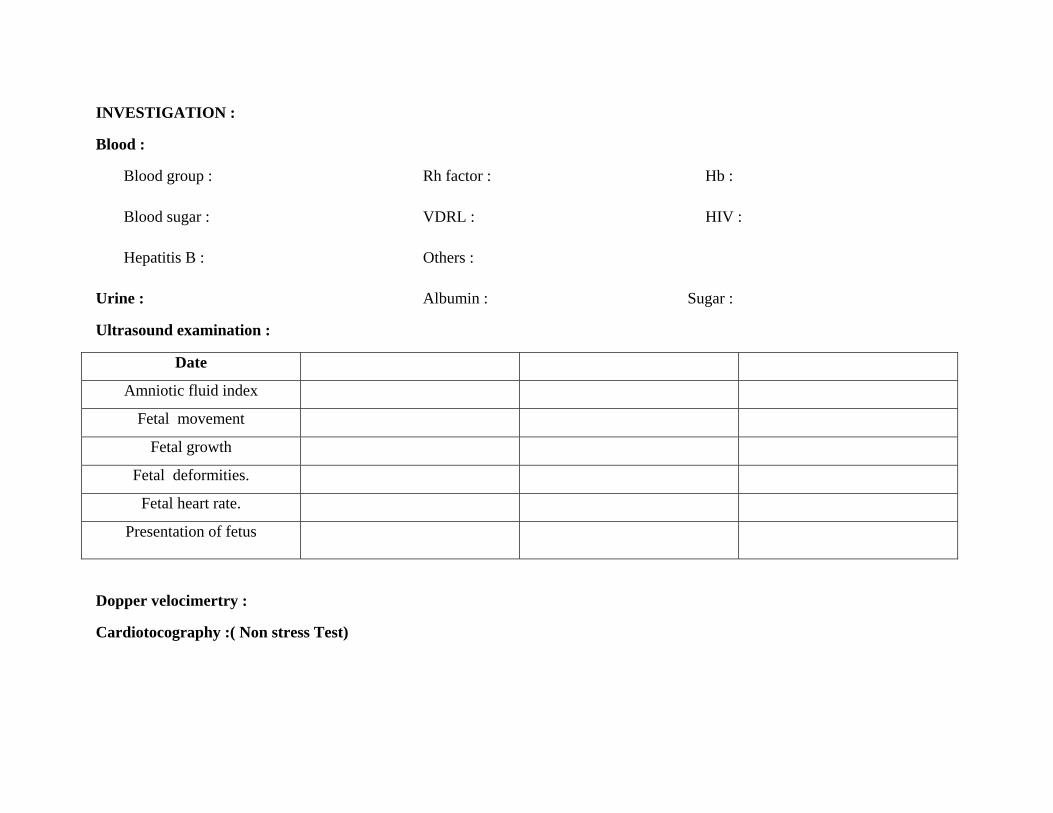
INVESTIGATION :
Blood :
Blood group : Rh factor : Hb :
Blood sugar : VDRL : HIV :
Hepatitis B : Others :
Urine : Albumin : Sugar :
Ultrasound examination :
Date
Amniotic fluid index
Fetal movement
Fetal growth
Fetal deformities.
Fetal heart rate.
Presentation of fetus
Dopper velocimertry :
Cardiotocography :( Non stress Test)

KICK COUNT CHART:
Sample No : LMP :
Obstetrical score : EDD :
Gestational age : Diagnosis :
NOTE : Mark an “X” in the box every time feel a fetal movement. Stop when reach 10 movements.
MEDICATION :( as prescribed):
DATE DAYS TIME NO. OF MOVEMENTS
1ST Start Time : Stop Time :
2ND
Start Time: Stop Time :
3RD
Start Time : Stop Time :
4TH
Start Time : Stop Time :
5TH
Start Time : Stop Time :

Section D: Nursing Process Application.
Date Nursing Assessment
Nursing diagnosis
Expected outcome
Nursing Intervention Rationale Implementation Evaluation

SECTION E: RISK FACTOR ASSESSMENT TOOL
Current pregnancy risk factors associated with oligohydramnios.
MOTHER : FETUS :
S.NO CONTENT YES NO
1 Chromosomal abnormalities
2 Congenital anomalies
3 Intra uterine growth retardation
4 Premature rupture of the membrane
5 Postterm pregnancy
6 Intrauterine Death
S.NO CONTENT YES NO
1 Mild pre eclampsia
2 Moderate to severe pre eclampsia
3 Gestational diabetes mellitus
4 Multiple pregnancy
5 Placenta abruption
6 Placenta previa
7 Viral disease
8 Kidney infection
9 Bladder Infection
10 Mild anaemia 9gm/dl
11 Severe anaemia < 9gm/dl
12 Rh sensitization
13 Moderate alcohol use
14 Excessive use of drugs /alcohol
15 Smocking >= 1 pack per day
16 Oligohydramnios

APPENDIX – F
LIST OF EXPERTS 1) Dr.C.S.Dhevasena DGO., DNB.,
Consultant Obstetrician & Gynaecologist,
Kovai Medical Centre & Hospital,
Coimbatore -641014.
2) Prof. Mrs.S.P.Latha M.Sc(N),
Principal,
RVS College of Nursing,
Kannampalayam,
Coimbatore -641402.
3) Mrs. Jesi Rani M.Sc(N),
Associate Professor,
HOD of OBG Department,
RVS College of Nursing,
Sulur, Coimbatore -641402.
4) DR. Mrs.Mahalakshmi Priya,
HOD of Tamil Department,
Dr. N.G.P Arts & Science College,
Coimbatore -641435.
5) Mrs.R.Indumathi,
Associate Professor,
OBG Department,
KMCH College of Nursing,
Coimbatore -641014.

APPENDIX - D
REQUISITION FOR CONTENT VALIDITY
From
Ms.Saranya.P.S.
II year M.Sc Nursing,
KMCH College of Nursing,
Coimbatore – 641 014.
To
Through: The principal
Sub : Content validity
Respected Madam,
I wish to undertake a study titled “IDENTIFICATION OF PROBLEMS &
EXECUTION OF NURSING STRATEGIES FOR MOTHERS WITH OLIGOHYDRAMNIOS
AT KOVAI MEDICAL CENTRE & HOSPITAL, COIMBATORE”. It will be of immense help to
me if you could peruse the proposal and the research tool. Herewith I am enclosing the copy to the
same.
Kindly do the needful.
Thanking you.
Place: Coimbatore Yours faithfully,
Date: