2021 Annual Report - Shiley Eye Institute
94
1
-
Upload
khangminh22 -
Category
Documents
-
view
1 -
download
0
Transcript of 2021 Annual Report - Shiley Eye Institute
1

2
CONTENTS
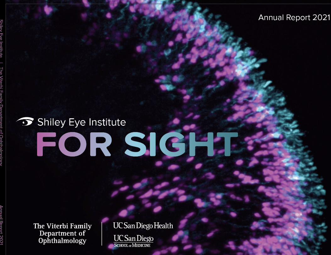
Image on the Cover: Living human retinal organoids with an emphasis on cone photoreceptors from Karl Wahlin, PhD and his laboratory team.
38NEW DIVISIONAT SEI
08SHILEYGIFT
35OFF THE PLANET
12NIXONGIFT
30RESEARCHIN A MAZE
19GLOBALRESOLUTION
03
06
10
12
44
52
68
84
Letters from Leaders
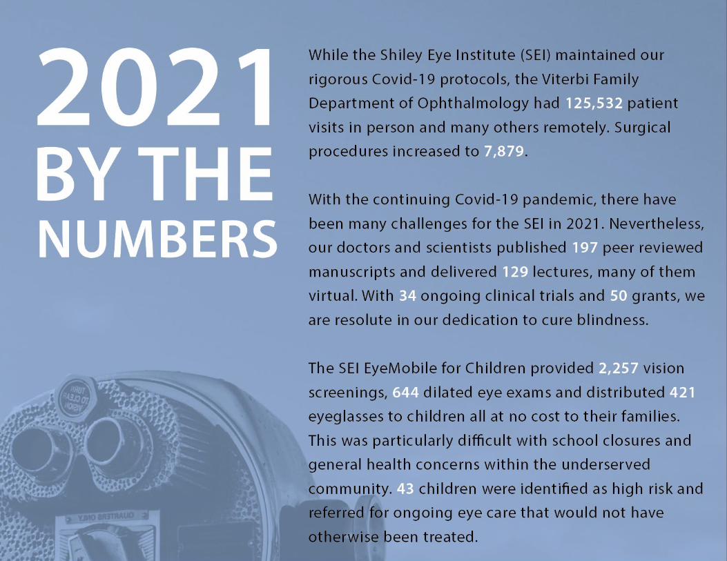
2021 by the Numbers
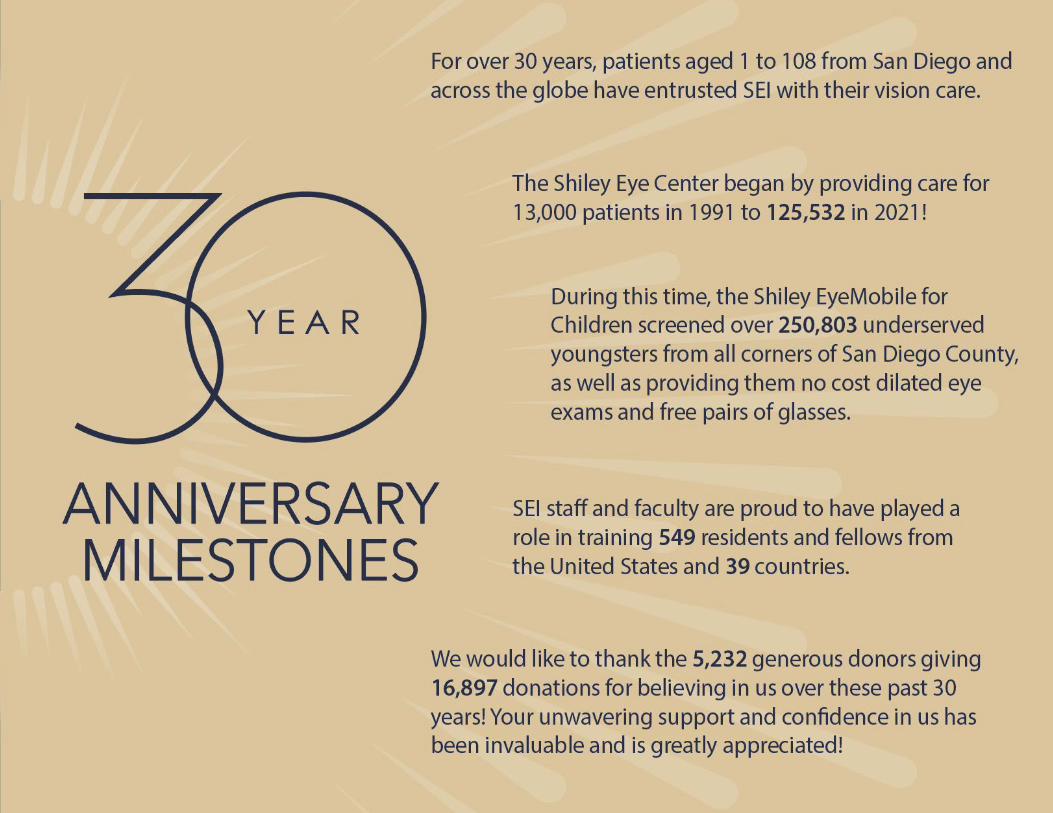
Celebration of 30 Years of SEI
Highlights
Faculty
Residents & Fellows
Publications, Clinical Trials & Grants
Giving

3
Robert N. Weinreb, MDChair and Distinguished Professor,
Ophthalmology Director, Shiley Eye Institute
Dear Friends,
This past year was like no other. Although 2021 was filled with unprecedented challenges, Shiley Eye Institute staff, clinicians, residents and fellows were resilient and never wavered in providing outstanding clinical care to our patients. Moreover, the vision research in the Viterbi Family Department of Ophthalmology continued to grow as we translated our laboratory discoveries to our patients. There are so many exciting items to share with you!
2021 marked the 30th Anniversary of the Shiley Eye Center and there has been numerous notable accomplishments and activities during the past year that are described in the Annual Report including:
• Another transformative donation from Darlene Shiley, our partner from the very beginning, will enable the renovation of the second floor and expansion of our clinical space to better serve our patients.
• Two outstanding faculty were appointed to Viterbi Family Chairs.
• The Nixon family provided a generous gift for genetic research for a rare and disabling inherited retinal disease.
• The planning of the Viterbi Family Vision Research Center is nearing completion
and ground should soon be broken on this 100,000 square foot structure.
• Collaborative clinical partnerships throughout UCSD Health and vision research throughout the UCSD campus continued to grow.
• Service to the underserved children in San Diego, the most vulnerable inhabitants, continued despite pandemic hardships
While reflecting on the past 30 years, I am heartened to know that our entire team is excited by the upcoming changes and future of the Shiley Eye Institute and Viterbi Family Department of Ophthalmology. Now, more than ever, we are inspired by our patients and cognizant every day of the trust from our donors who have generously supported our vision research.
On behalf of the Shiley Eye Institute and Viterbi Family Department of Ophthalmology at UC San Diego, I thank you for your partnership and support.
Sincerely,
Letter from the Chair
4
Dear Friends,
Year after year, the Shiley Eye Institute and Viterbi Family Department of Ophthalmology at UC San Diego Health exemplify excellence in education, collaborative research, and innovative clinical care. In 2021, we commemorated the 30th anniversary of the Shiley Eye Institute, celebrating its groundbreaking discoveries, recognizing generous community support, and planning exciting new research initiatives. The many inspiring stories highlighted in this annual report feature successful patient outcomes, innovative research, and groundbreaking discoveries to treat and cure eye diseases.
These successes are possible, in great part, because of the generous and ongoing support of our community partners. Recently, physician-scientists discovered enzymes that demonstrate potential to prevent optic nerve cell degeneration resulting from glaucoma or inherited retinal disease. We are grateful for generous support from the Nixon Vision Foundation that will fund genetic studies in pursuit of macular degeneration diagnosis and treatment.
Pradeep K. Khosla, PhDChancellor, UC San Diego
And this year, a $10 million gift from Darlene Shiley to revitalize the institute’s facilities will help move ongoing and new research to the next level of excellence. We are grateful for the opportunities that this transformational gift will create. The discoveries and innovations found through this important work will truly honor the enduring legacy of Darlene and Donald Shiley.
Thank you to all of our donors for their commitment and continued partnership with UC San Diego and the Shiley Eye Institute. Your support profoundly changes patients’ lives and makes a meaningful, lasting impact at UC San Diego. Your gifts drive transformative research, treatments, and cures. With gratitude, I look forward to all we will continue to accomplish together.
With Kind Regards,
Letter from the Chancellor

5
Dear Friends,
Amid one of the most challenging and historic public health crises, the Shiley Eye Institute and Viterbi Family Department of Ophthalmology have remained at the forefront of vision expertise and excellence in eye care. Our teams have been ready, responsive and focused on ensuring that patients receive the quality care they deserve in a safe environment. It
Patty Maysent, MPH, MBACEO, UC San Diego Health
Dear Friends,
Year after year, I am proud of the accomplishments of the faculty and staff of the Viterbi Family Department of Ophthalmology and Shiley Eye Institute. Despite the continuation of the global pandemic, this year is no different.
We are thrilled to celebrate another
Steven R. Garfin, MDInterim Dean,UC San Diego School of Medicine
year of achievement by our distinguished faculty. From driving innovation through leading-edge translational research to training a diverse set of residents and fellows in the latest treatment options across all areas of eye care, the Viterbi Family Department of Ophthalmology and Shiley Eye Institute remain among the premier destinations for ophthalmology in the nation.
Thank you for your commitment to ensuring UC San Diego remains a leader in delivering outstanding patient care, pursuing diverse avenues of research through collaborative team science, and offering an exceptional academic environment for our students and trainees. Your support of the Viterbi Family Department of Ophthalmology and Shiley Eye Institute is vital to these efforts, and we remain deeply grateful for your partnership.
Sincerely,
is because of this that patients from Southern California to across the world continue to seek out the specialized care that exists only at UC San Diego Health. I am incredibly proud of the work being done by our staff, trainees, clinicians and faculty leadership.
In addition to clinical excellence, their vision research is growing and transformative. Among their many research activities, some SEI clinicians and scientists are studying the effects of the pandemic on eyesight. Others are exploring the progression of glaucoma through artificial intelligence, and still others are developing gene therapies to restore vision by investigating rare gene mutations that can lead to loss of vision.
We also have continued to increase our ophthalmology offerings thanks to your generous support and, in particular, the support of Darlene Shiley who recently gave a $10 million gift for the clinical space expansion of the Shiley Eye Institute. Her gift, along with the additional funds provided by UC San Diego Health, will enable increased patient access to our dedicated clinical team.
From treating potentially blinding eye diseases such as diabetic retinopathy, macular degeneration and glaucoma, to discovering pathways to improve patient health on a broader scale, the Shiley Eye Institute and the Viterbi Family Department of Ophthalmology are advancing clinical care and vision research every day.
Thank for you for your support.
Letter from the CEO Letter from the Interim Dean
6
7
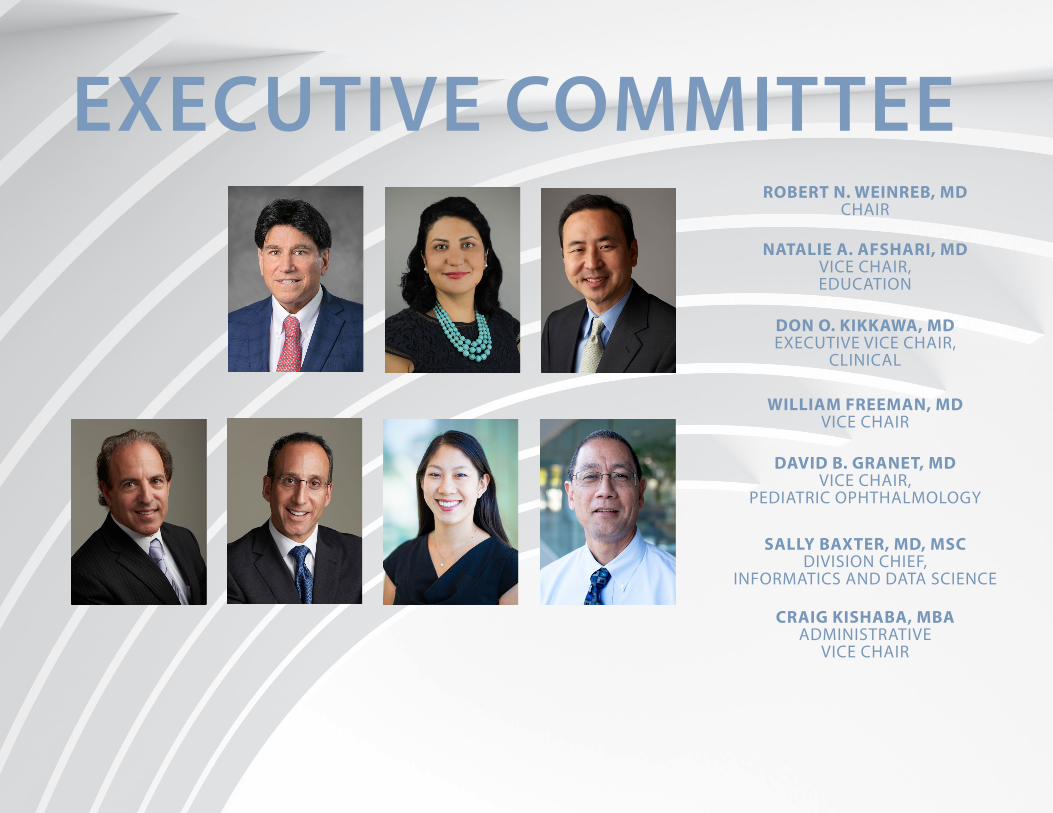
EXECUTIVE COMMITTEEROBERT N. WEINREB, MD
CHAIR
NATALIE A. AFSHARI, MDVICE CHAIR, EDUCATION
DON O. KIKKAWA, MDEXECUTIVE VICE CHAIR,
CLINICAL
WILLIAM FREEMAN, MDVICE CHAIR
DAVID B. GRANET, MDVICE CHAIR,
PEDIATRIC OPHTHALMOLOGY
SALLY BAXTER, MD, MSCDIVISION CHIEF,
INFORMATICS AND DATA SCIENCE
CRAIG KISHABA, MBAADMINISTRATIVE
VICE CHAIR

8
WITH $10 MILLION GIFT, DARLENE SHILEY BUILDS UPON HER HUSBAND’S LEGACY
Marking its 30th anniversary and her enduring interest and support, philanthropist Darlene Shiley has given a $10 million gift for the clinical space expansion of the Shiley Eye Institute at UC San Diego Health.
The gift will finance the expansion of the clinical space and function of the Institute. Already an architectural landmark, the expansion will usher in a new era of vision care and research benefiting San Diego and beyond thanks to increased patient care capacity and
Gift will target expansion of the clinical space at Shiley Eye Institute, which opened 30 years ago with foundational support from the couple; it is part of a $27 million renovation and improvement project being undertaken by UC San Diego
the Shiley Eye Institute and her centers of care. It was one of our first philanthropic projects we supported and were loyal to for the “long haul” as Donald would say. I now continue that legacy with pride and the knowledge that we have and continue to believe in improving medical care and research that benefits all of us.”
With her late husband Donald, who passed away in 2010, Darlene Shiley has been a longtime supporter of UC San Diego across multiple endeavors, including the Shiley-Marcos Alzheimer’s Disease Research Center, named in part to honor Darlene’s mother, Dee Marcos.
“I can’t emphasize enough how far-reaching the influence of Darlene and Donald Shiley has been on UC San Diego and elsewhere. Vision care, Alzheimer’s disease, and so many other important areas of research and care on our campus are sustained thanks to the Shileys’
generosity. It is remarkable to see the legacy of transformation they have made here,” said Chancellor Pradeep Khosla.
The latest gift will further burnish, in form and function, the Shileys’ vision of their eponymous eye institute, which has grown dramatically over the last three decades.
In 1983, Department of Ophthalmology, part of UC San Diego Health, debuted in an 800-square-foot, three-room clinic, progressing to a 3,000-square-foot trailer in 1985 and then the 1991 opening of the original $8 million Donald P. and Darlene V. Shiley Eye Center, under the leadership of Stuart I. Brown, MD. Over the years, the Shiley family has donated more than $10 million for various clinical improvements, equipment, research and leadership chair funding. Additionally, other members of the family contributed to the Shiley Eye Institute as well,
Donald Shiley
expanded research infrastructure.
“Over the last three decades, Donald and I have supported the growth and excellence of
9
including support of the Low Vision Clinic and Shiley Eye Mobile clinic by granddaughter Patricia.
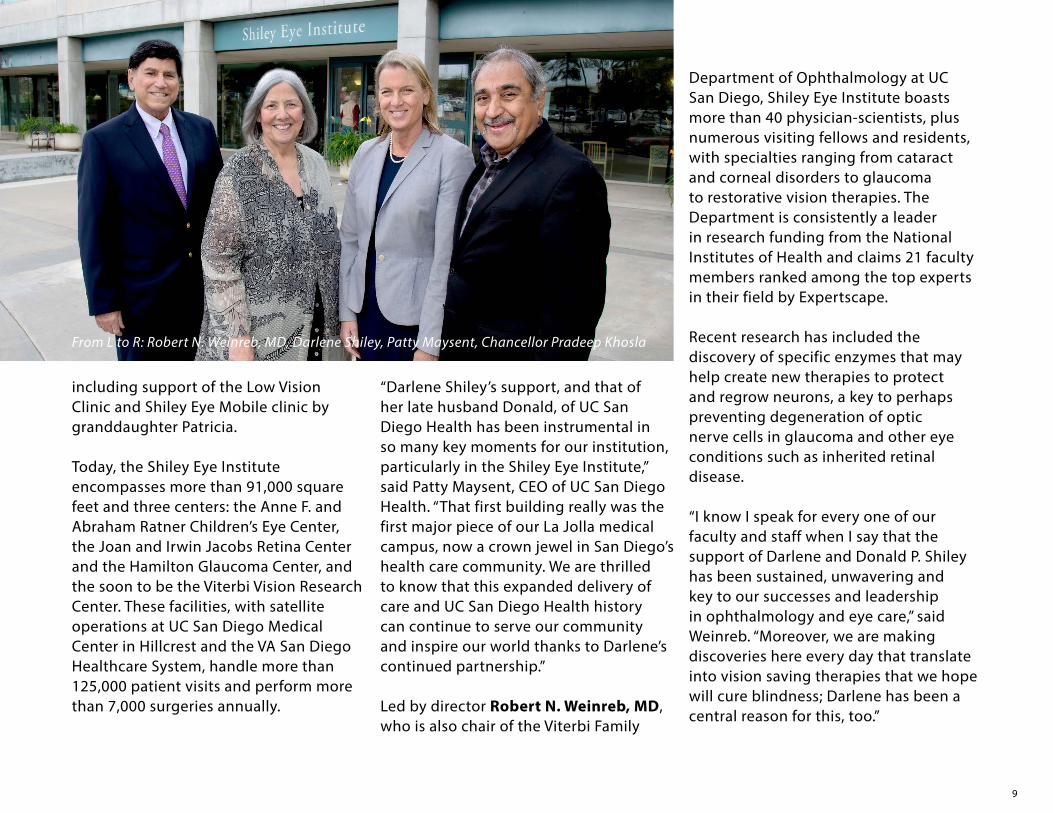
Today, the Shiley Eye Institute encompasses more than 91,000 square feet and three centers: the Anne F. and Abraham Ratner Children’s Eye Center, the Joan and Irwin Jacobs Retina Center and the Hamilton Glaucoma Center, and the soon to be the Viterbi Vision Research Center. These facilities, with satellite operations at UC San Diego Medical Center in Hillcrest and the VA San Diego Healthcare System, handle more than 125,000 patient visits and perform more than 7,000 surgeries annually.
“Darlene Shiley’s support, and that of her late husband Donald, of UC San Diego Health has been instrumental in so many key moments for our institution, particularly in the Shiley Eye Institute,” said Patty Maysent, CEO of UC San Diego Health. “That first building really was the first major piece of our La Jolla medical campus, now a crown jewel in San Diego’s health care community. We are thrilled to know that this expanded delivery of care and UC San Diego Health history can continue to serve our community and inspire our world thanks to Darlene’s continued partnership.”
Led by director Robert N. Weinreb, MD, who is also chair of the Viterbi Family
From L to R: Robert N. Weinreb, MD, Darlene Shiley, Patty Maysent, Chancellor Pradeep Khosla
Department of Ophthalmology at UC San Diego, Shiley Eye Institute boasts more than 40 physician-scientists, plus numerous visiting fellows and residents, with specialties ranging from cataract and corneal disorders to glaucoma to restorative vision therapies. The Department is consistently a leader in research funding from the National Institutes of Health and claims 21 faculty members ranked among the top experts in their field by Expertscape.
Recent research has included the discovery of specific enzymes that may help create new therapies to protect and regrow neurons, a key to perhaps preventing degeneration of optic nerve cells in glaucoma and other eye conditions such as inherited retinal disease.
“I know I speak for every one of our faculty and staff when I say that the support of Darlene and Donald P. Shiley has been sustained, unwavering and key to our successes and leadership in ophthalmology and eye care,” said Weinreb. “Moreover, we are making discoveries here every day that translate into vision saving therapies that we hope will cure blindness; Darlene has been a central reason for this, too.”

10
11

12
MAJOR GIFT FOCUSES EFFORTS ON A RARE, BUT DEVASTATING, GENETIC EYE DISEASE
In healthy vision, a gene called PRPH2 provides instructions to make a protein called peripherin 2 (PRPH2), which plays a key role in the normal functioning of photoreceptors that detect light and color and which line the back of the eye.
When there are mutations in the PRPH2 gene, the result can be macular dystrophy, an impairment of the retina that progressively diminishes the ability to see clearly and may eventually result in vision loss. Currently, there are no effective treatments to slow or prevent the condition.
The Nixon Visions Foundation, led by philanthropists Brandon and Janine Nixon, has given a significant gift to the Viterbi Family Department of Ophthalmology and Shiley Eye Institute, both part of UC San Diego Health, to launch the Nixon Visions Foundation Macular Dystrophy-PRPH2 Research Fund,
which will focus studies of the PRPH2 gene and related mutations and help upgrade stem cell technologies that may eventually provide a proven therapeutic remedy. Nixon Visions Foundation is also building capacity with the Foundation Fighting Blindness to further advance national and global research in this space as part of this effort.
“We are impressed with the impactful work at UC San Diego and specifically in the Department of Ophthalmology and at Shiley Eye Institute,” said the Nixons. “We believe this gift can accelerate efforts to make a tremendous impact for people with this inherited eye disease and will improve the lives of others for generations to come.”
“Macular dystrophy is such a challenging disease for people who have it, but UC San Diego Health has the expertise to discover new ways of treating this illness
and creating a healthier world,” said UC San Diego Chancellor Pradeep Khosla. “Thanks to the generosity of the Nixon Visions Foundation, we can pursue the most promising leads and follow the science wherever it takes us.”
Macular dystrophy is a relatively rare eye condition. It affects the central retina or macula, which has the highest concentration of light-sensitive cells or photoreceptors. It is different from the more common eye disease known as macular degeneration, which is often caused by age-related deterioration of the retina and macula. Macular dystrophy is associated with genetic mutations that — for no known reason — trigger degradation of retinal cells. Some forms of the disease appear in childhood; some in adulthood.
“There really aren’t viable therapies for macular dystrophy and even fewer
Funding from the Nixon Visions Foundation will support studies of the PRPH2 gene linked to macular dystrophy and boost stem cell research aimed at developing early diagnosis and a cure
13
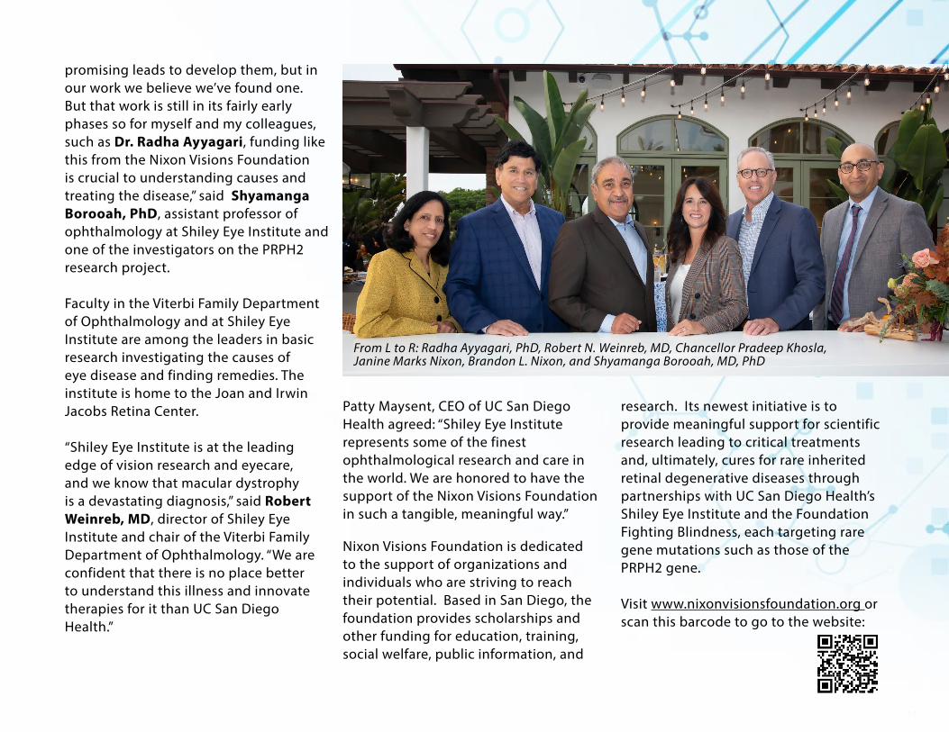
From L to R: Radha Ayyagari, PhD, Robert N. Weinreb, MD, Chancellor Pradeep Khosla, Janine Marks Nixon, Brandon L. Nixon, and Shyamanga Borooah, MD, PhD
promising leads to develop them, but in our work we believe we’ve found one. But that work is still in its fairly early phases so for myself and my colleagues, such as Dr. Radha Ayyagari, funding like this from the Nixon Visions Foundation is crucial to understanding causes and treating the disease,” said Shyamanga Borooah, PhD, assistant professor of ophthalmology at Shiley Eye Institute and one of the investigators on the PRPH2 research project.
Faculty in the Viterbi Family Department of Ophthalmology and at Shiley Eye Institute are among the leaders in basic research investigating the causes of eye disease and finding remedies. The institute is home to the Joan and Irwin Jacobs Retina Center.
“Shiley Eye Institute is at the leading edge of vision research and eyecare, and we know that macular dystrophy is a devastating diagnosis,” said Robert Weinreb, MD, director of Shiley Eye Institute and chair of the Viterbi Family Department of Ophthalmology. “We are confident that there is no place better to understand this illness and innovate therapies for it than UC San Diego Health.”
Patty Maysent, CEO of UC San Diego Health agreed: “Shiley Eye Institute represents some of the finest ophthalmological research and care in the world. We are honored to have the support of the Nixon Visions Foundation in such a tangible, meaningful way.”
Nixon Visions Foundation is dedicated to the support of organizations and individuals who are striving to reach their potential. Based in San Diego, the foundation provides scholarships and other funding for education, training, social welfare, public information, and
research. Its newest initiative is to provide meaningful support for scientific research leading to critical treatments and, ultimately, cures for rare inherited retinal degenerative diseases through partnerships with UC San Diego Health’s Shiley Eye Institute and the Foundation Fighting Blindness, each targeting rare gene mutations such as those of the PRPH2 gene.
Visit www.nixonvisionsfoundation.org or scan this barcode to go to the website:
14
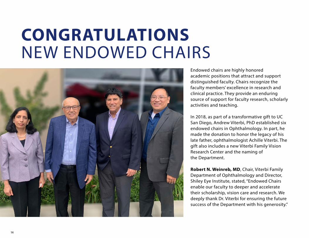
CONGRATULATIONS NEW ENDOWED CHAIRS
Endowed chairs are highly honored academic positions that attract and support distinguished faculty. Chairs recognize the faculty members’ excellence in research and clinical practice. They provide an enduring source of support for faculty research, scholarly activities and teaching.
In 2018, as part of a transformative gift to UC San Diego, Andrew Viterbi, PhD established six endowed chairs in Ophthalmology. In part, he made the donation to honor the legacy of his late father, ophthalmologist Achille Viterbi. The gift also includes a new Viterbi Family Vision Research Center and the naming of the Department.
Robert N. Weinreb, MD, Chair, Viterbi Family Department of Ophthalmology and Director, Shiley Eye Institute, stated, “Endowed Chairs enable our faculty to deeper and accelerate their scholarship, vision care and research. We deeply thank Dr. Viterbi for ensuring the future success of the Department with his generosity.”

15
Chair III enables the bold pursuit of new solutions to debilitating eye diseases that may cause blindness – thus preserving sight and improving lives”, stated Dr. Ayyagari.
Her research interests include molecular genetics of macular and retinal dystrophy and glaucoma, biological mechanisms underlying retinal diseases, age-related macular degeneration and diabetic retinopathy. She has been honored with the Frank A. Bennack, Jr. Research Fellowship, Sybil B. Barrington Scholar Award, University of Michigan Research Faculty Recognition Award and the Lew Wassermen Award.
Alex A. Huang, MD, PhD, new Associate Professor of Ophthalmology, has been appointed the inaugural holder of the
Dr. Huang notes, “I am honored as a new faculty member to be the inaugural chairholder of the prestigious Vogt Chair. This distinction will help to propel my research forward and create more opportunities to collaborate with colleagues worldwide in treating and curing optic nerve and glaucoma related eye diseases.”
Through his research, Dr. Huang hopes to better understand how glaucoma therapies work, identify their stengths and weaknesses, as well as develop better drug treatments or surgical techniques for the best possible patient outcomes.
Radha Ayyagari, PhD, Professor of Ophthalmology and Pathology, has been appointed as the Viterbi Family Chair of Ophthalmic Genetics in the Viterbi Family
Department of Ophthalmology, Shiley Eye Institute at UC San Diego.
Dr. Ayyagari is Chief of the Ophthalmic Molecular Genetics Laboratory and Director of the Downtown San Diego Lions Biobank for Vision. She completed her undergraduate studies at Andhra University in Vizag, India, her graduate studies at Osmania University in Hyderabad, India and fellowship in Ophthalmic Molecular Genetics at the National Eye Institute, National Institute of Health, Bethesda, Maryland.“The research and teaching support from endowed chairs like the Viterbi Family
Alfred Vogt Chair in Ophthalmology in the Viterbi Family Department of Ophthalmology, Shiley Eye Institute at UC San Diego.
Dr. Viterbi included The Alfred Vogt Chair in Ophthalmology in his gift to UC San Diego. This chair is named after internationally recognized Swiss ophthalmologist Professor Alfred Vogt, who not only served as Dr. Achille Viterbi’s mentor but he helped the Viterbi family flee Italy in 1939 by securing them visas to Switzerland and then on to the United States.
16
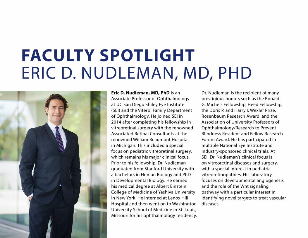
Eric D. Nudleman, MD, PhD is an Associate Professor of Ophthalmology at UC San Diego Shiley Eye Institute (SEI) and the Viterbi Family Department of Ophthalmology. He joined SEI in 2014 after completing his fellowship in vitreoretinal surgery with the renowned Associated Retinal Consultants at the renowned William Beaumont Hospital in Michigan. This included a special focus on pediatric vitreoretinal surgery, which remains his major clinical focus. Prior to his fellowship, Dr. Nudleman graduated from Stanford University with a bachelors in Human Biology and PhD in Developmental Biology. He earned his medical degree at Albert Einstein College of Medicine of Yeshiva University in New York. He interned at Lenox Hill Hospital and then went on to Washington University School of Medicine in St. Louis, Missouri for his ophthalmology residency.
Dr. Nudleman is the recipient of many prestigious honors such as the Ronald G. Michels Fellowship, Heed Fellowship, the Doris P. and Harry I. Wexler Prize, Rosenbaum Research Award, and the Association of University Professors of Ophthalmology/Research to Prevent Blindness Resident and Fellow Research Forum Award. He has participated in multiple National Eye Institute and industry-sponsored clinical trials. At SEI, Dr. Nudleman’s clinical focus is on vitreoretinal diseases and surgery, with a special interest in pediatric vitreoretinopathies. His laboratory focuses on developmental angiogenesis and the role of the Wnt signaling pathway with a particular interest in identifying novel targets to treat vascular diseases.
FACULTY SPOTLIGHTERIC D. NUDLEMAN, MD, PHD
17
WHY DID YOU GO INTO MEDICINE?
I was exposed to academic medicine from an early age. My father is a neurologist and spent most of his career at UC Irvine. As a child, if my mother was busy on the weekends he would take me to work with him to round on a sick patient, finish paperwork in the office, or meet with his research team. It was easy to recognize his passion for the work and the intense appreciation he felt for the privilege to provide healthcare.
As a student, I was always interested in science. After my undergraduate studies, I entered a PhD program in Developmental Biology at Stanford. I loved doing basic science research, but the human connection to the work that I saw my father experience was missing for me. I went to medical school so that I could combine those interests. The opportunity to both practice medicine and help individual patients, as well as do basic science work to discover new therapies, has been tremendously rewarding.
HAVE ANY OF YOUR PATIENTS AFFECTED YOU SIGNIFICANTLY?
Without question, every patient affects me. As a retina surgeon, some of the common diseases we see (such as diabetic retinopathy and macular degeneration) require frequent visits. This allows me to form very close relationships, often over many years. I also focus on pediatric retinal diseases, which allows me to take care of some patients from infancy. It is a unique and privileged role to play in their lives. When I see patients do well with existing therapies, it is a tremendous joy. However, when they do poorly, it is a deep disappointment. Seeing the challenges in people who I have gotten to know well is the frustration that motivates the research that I do in the lab.
HOW DO COLLABORATIONS AND PARTNERSHIPS FIT INTO YOUR ROLE AS A RESEARCHER?
Collaborations are a critical component of any research endeavors. We are very fortunate at UC San Diego (UCSD) to be surrounded by tremendous expertise in virtually every discipline.
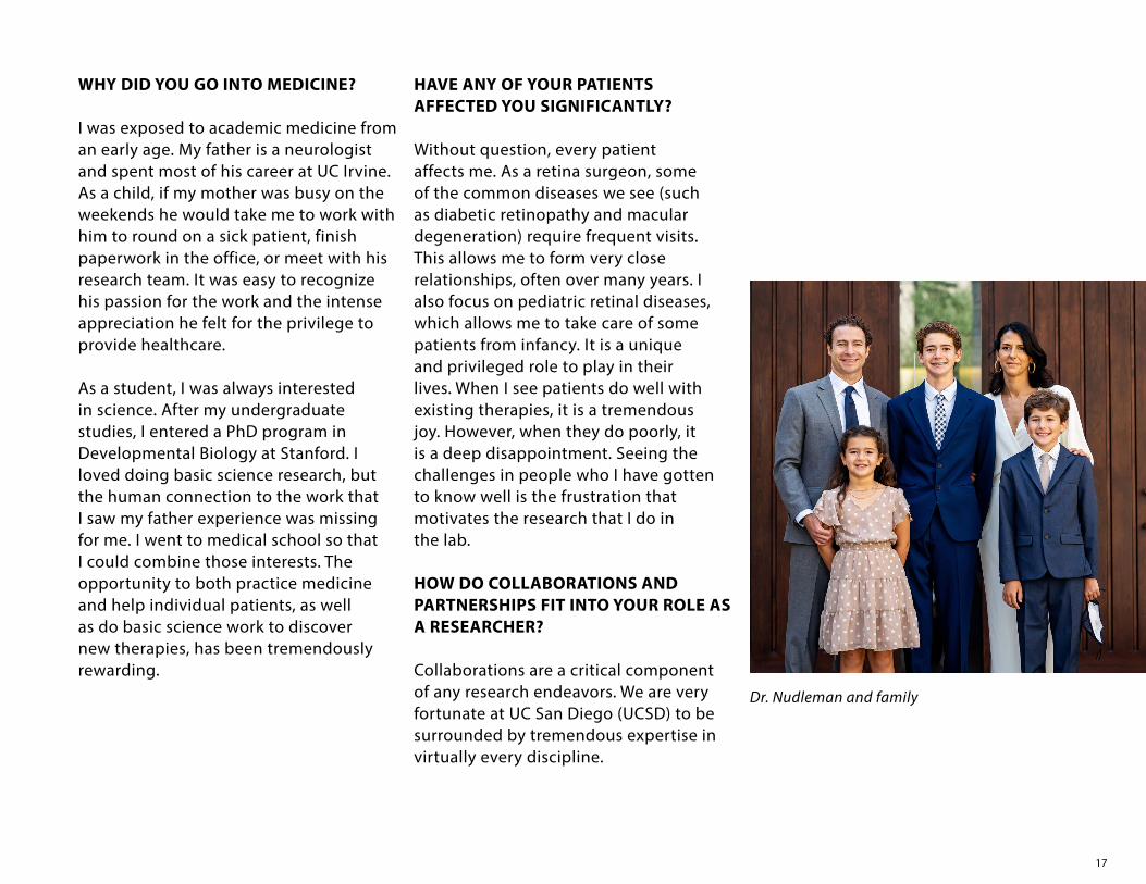
Dr. Nudleman and family
18
My research is primarily focused on diseases of the blood vessels in the retina. Since coming to UCSD, I have worked closely with fellow faculty member Napoleone Ferrara, MD, Distinguished Professor of Ophthalmology and Pathology at UC San Diego Health. He is an expert in vascular biology and discovered Vascular Endothelial Growth Factor (VEGF), the target of the most common drugs we use as retina specialists. In addition, we collaborate with Richard Daneman, PhD, Professor of Neurosciences and Pharmacology at UC San Diego, an expert
in the blood-brain barrier, which is similar to the blood-retinal barrier. The key to these collaborations is to identify the molecular abnormalities that occur in diseases of the retinal blood vessels in order to target new therapies. Each part of the team provides unique and highly valuable skills. WHAT DO YOU SEE AS THE NEXT BIG ADVANCES IN YOUR FIELD?
Possibly the biggest advance ever in our field was the discovery of VEGF by Dr. Ferrara, and the subsequent
development of inhibitor therapies. These drugs have prevented blindness in many millions of people worldwide. However, despite their incredible efficacy, they have to be injected frequently, which is a major burden for patients, providers and the healthcare system. A great deal of effort right now is being directed towards therapies that will be longer lasting. In addition, some patients fail to improve. Often that is due to scarring, which has no effective treatment. Therapies that would prevent scarring in the retina would have a tremendous impact on saving vision.
WHAT DO YOU DO IN YOUR FREE TIME?
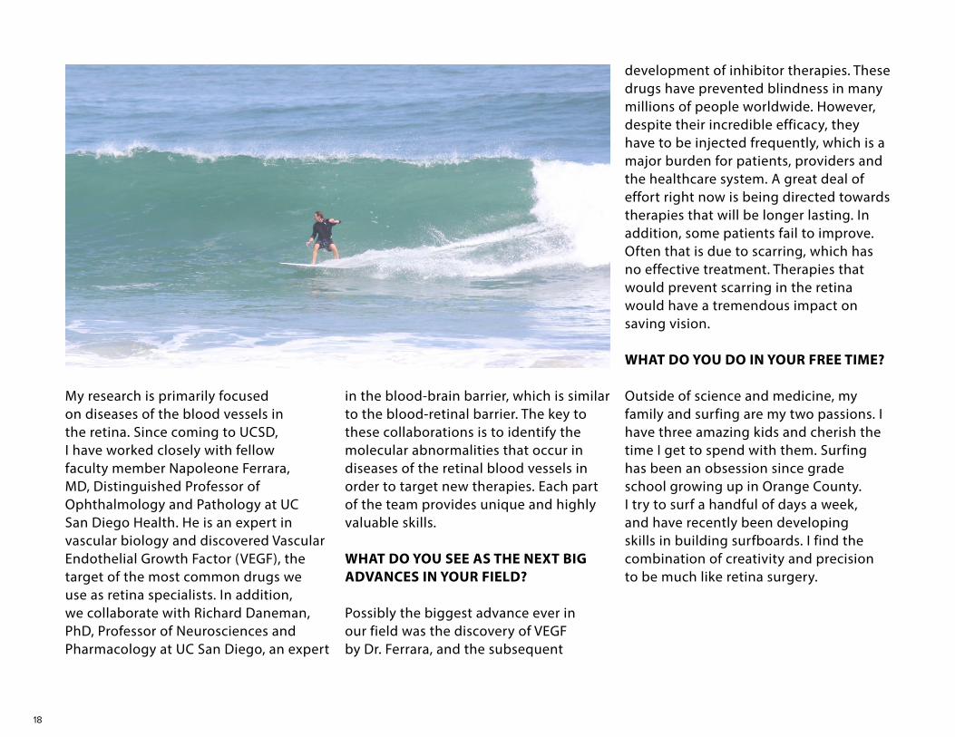
Outside of science and medicine, my family and surfing are my two passions. I have three amazing kids and cherish the time I get to spend with them. Surfing has been an obsession since grade school growing up in Orange County. I try to surf a handful of days a week, and have recently been developing skills in building surfboards. I find the combination of creativity and precision to be much like retina surgery.

19
The United Nations General Assembly unanimously adopted the first ever resolution on vision, designating its 193 member nations to ensure community access to eye health for the 1.1 billion people worldwide living with preventable sight loss by 2030. The resolution was introduced by Bangladesh’s U.N. Ambassador and sponsored by Antigua and Ireland as well as being co-sponsored by 100 other countries.
The resolution titled, "Vision for Everyone", is designed to encourage countries to increase access for vision care services for their populations and make eye health part of their nation’s governmental health agenda. The resolution says, “Global eye care needs are projected to increase substantially
with half the global population expected to be living with a vision impairment by 2050.” It also calls on international financial institutions and philanthropies to focus on the increasing impact of vision loss on economic and social development.
Robert N. Weinreb, MD, Chair and Professor, Viterbi Family Department of Ophthalmology and Director, Shiley Eye Institute, states, “This announcement is a welcome and important milestone in worldwide efforts to treat and prevent blinding eye diseases.”
This resolution gives the global ophthalmology community opportunity to improve access to eye care for millions of people living with impaired vision
UN ADOPTS RESOLUTION ON VISION
and blindness around the world. These General Assembly resolutions are not legally binding, but they do indicate worldwide outlook.
For more information, go to:
“Global eye care needs are projected to increase substantially with half the global population expected to be living with a vision impairment by 2050.”

20
EYEMOBILE CELEBRATES 20 YEARS & 100,000 MILES!The UC San Diego Shiley EyeMobile for Children is a program of Community Ophthalmology at the Shiley Eye Institute (SEI) and Viterbi Family Department of Ophthalmology.
In 1998, the goal of Stuart I. Brown, MD, founder of the EyeMobile, was to partner with the community to develop a model that would deliver free eye care to the low-income children in San Diego who were needlessly losing sight, struggling to fulfill their potential to learn and contribute to society. Community leaders and philanthropists shared this vision to establish a mobile pediatric eye care program that would overcome barriers and bring vision care to underserved
sectors of San Diego County. In addition to benefiting the underserved youngsters, this innovative program would provide a viable model for other communities and a platform for research studies.
Funded by several foundations, corporations and individuals, the dream was realized with the launching of our first UC San Diego Shiley EyeMobile for Children in April 2001. It was a specially furnished recreational vehicle with an optometric exam room and waiting area for children and families. In 2008, the program was able to expand with a new EyeMobile equipped with two exam rooms.
Before COVID-19 struck, the vision of 13,000 students’ was screened at over 225 locations across San Diego County. The EyeMobile program, provided at no cost to the family, includes: vision screening, dilated eye examinations by an optometrist, if needed - a free pair of glasses, follow-up monitoring with teachers and parents, referral for subspecialist care as needed to the Anne F. and Abraham Ratner Children’s Eye Center at SEI. There is also bilingual parent and teacher information informing them on the importance of eye/brain development, the need for eye care and its crucial role in preparing children to learn, as well as program evaluation.

21
Since the first EyeMobile went out, the program has screened over 250,000 youngsters across San Diego County. Keys to the EyeMobile’s success include a dedicated multilingual staff, as well as students and community volunteers. With the medical unit’s mobility, the program overcomes the transportation, language, cultural and financial barriers that low-income families with children face. We have created community partnerships with the San Diego County Office of Education, Chula Vista, San Ysidro, La Mesa, Lemon Grove, and Cajon Valley School Districts, Head Start, San Diego-Imperial Counties Developmental Services, and others responsible for the education and health of young children.
Early detection and treatment have proven to reduce the negative impact vision problems may have on a child’s learning ability and development. If left untreated, conditions such as amblyopia, could lead to irreversible vision loss and psychosocial effects. The EyeMobile
program provides children with the best sight to allow them to be "school ready" so they can learn at their maximum potential.
COVID-19 severely impacted the ability for the EyeMobile to travel in the community to see youngsters around San Diego in the school locations. We are happy to report that COVID-19 didn’t completely close down the EyeMobile entirely. Although a different model, the EyeMobile is safely continuing to provide no cost exams and glasses to children ages 3-14 in community locations around San Diego County.
This unique program has been embraced throughout the community and is giving underserved young children the vision, they need to succeed in school and life. When children can see, they are able to learn which then expands the educational opportunities for under-represented students.
The long-term goal is to expand with a more efficient and larger EyeMobile in order to provide eye care services for 20,000 low-income young children annually. In addition, the expansion of vision care services for underserved seniors throughout San Diego County is being planned.
22
Shira L. Robbins, MD, Professor of Clinical Ophthalmology, has been elected UC San Diego Health (UCSDH) Vice Chief of Staff. Elected by the UC San Diego Active Medical Staff that includes over 1,300 physicians, psychologists and podiatrists, in 2 years she will become UC San Diego Health (UCSDH) Chief of Staff.
“Our department is thrilled by Dr. Robbins’ election to this important position. She is a leader and an asset to UC San Diego Health. I know that she will do an excellent job.” said Robert N. Weinreb, MD, Chair of the UC San Diego Viterbi Family Department of Ophthalmology and Director of the Shiley Eye Institute.
Dr. Robbins specializes in Pediatric Ophthalmology and Adult Eye Re-Alignment. Her presence at Jacobs Medical Center NICU and Newborn Nursery as Director of Neonatal Ophthalmology protects the visual development of our smallest and most fragile of patients. She heads the new Myopia Control Clinic which researches and treats the worldwide epidemic of
nearsightedness that can lead to other blinding eye conditions. She performs surgery on adults with double vision to make them more functional in society. The author of over 80 scientific articles and book chapters on topics from the economics of medicine to surgical safety, Dr. Robbins’ work is recognized worldwide by her colleagues.
“I am both honored and excited to be elected to this impactful position at UC San Diego Health,” said Dr. Robbins. “I look forward to interacting with the physician leaders, CEO and management team to represent the Medical Staff in promoting the best possible care for our patients throughout the Health System.”
This position of Vice Chief of Staff merges patient safety with UCSD policies while being a physician advocate. In this new role, Dr. Robbins will serve as a member of the Medical Staff Executive Committee (MSEC), the Professionalism Committee and the Joint Conference Committee (liaising between the medical staff and the executive group board). She will be responsible for promoting quality of
care, translating regulatory guidelines to enable cutting-edge medical care, ensuring procedural safeguards are in place, being a spokesperson for the Medical Staff in external professional and public relations, serving on liaison committees with the UCSDH Executive Governing Body and Health Center Administration, and interacting with the Chief Executive Officer and Executive Governing Body in all matters of mutual concern at UCSDH.
Dr. Robbins will assume her new role effective July 1, 2021 succeeding Christian Tomaszewski, MD, as he steps into his role as Chief of Staff.
CONGRATULATIONS
Natalie Afshari, MD, Professor and Vice Chair, is collaborating with Gene Yeo, PhD (Professor of Cellular and Molecular Medicine) and scientists at Case Western Reserve University to investigate the genetic basis of Fuchs Endothelial Dystrophy. They are searching for novel targets to develop gene therapies. R01EY029166 (Yeo and Afshari)
Radha Ayyagari, PhD, Professor of Ophthalmology and Pathology, is collaborating with Bing Ren, PhD (Professor of Cellular and Molecular Medicine, and Director, Center for Epigenomics) and Kelly A. Frazier, PhD (Professor of Pediatrics and Director of the Institute of Genomic Medicine) using genetics and epitranscriptomics to study the contribution of individual retinal cell type specific epigenomic changes on retinal aging, early and late-onset retinal/macular degeneration pathology. RO1EY031663 (Ayyagari, Frazer and Ren).
Sally Baxter, MD, MSc, Assistant Professor, is a member of a team of
clinical informaticists led by Lucila Ohno-Machado, MD, PhD, (Professor of Medicine/Division of Biomedical Informatics) to develop and deploy queries related to COVID-19 across multiple electronic health record (EHR) systems. This is supported by the Gordon and Betty Moore Foundation.
Sally Baxter, MD, MSc is collaborating with Ming Tai-Seale, PhD, (Professor of Family Medicine) to analyze associations between EHR use and physician burnout with support from the American Medical Association (AMA) Practice Transformation Initiative.
Shyamanga Borooah MD, PhD, Assistant Professor, is collaborating with Eric Adler MD, (Professor of Medicine and Medical Director of the Heart Transplant Program at UCSD Health) to develop a gene therapy for children with a rare inherited sight threatening and heart disease. This work is supported by the Knights Templar Eye Foundation (PI: Borooah).
SEI@UCSDThe faculty at the Shiley Eye Institute and Viterbi Family Department of Ophthalmology is fortunate to collaborate on research projects with many areas across the UC San Diego campus. These partnerships often open doors to funding and new relationships but more importantly are translating our research into real world discoveries and treatments for patients with eye diseases and vision disorders.
Andrew Camp, MD, Assistant Professor, and Robert N. Weinreb, MD, Distinguished Professor and Chair, are collaborating with James Friend, PhD (Professor of Mechanical and Aerospace Engineering and Director, Medically Advanced Devices Lab) to develop a novel eye pressure measurement device to provide a gold standard that can be used in any patient, including those with corneal disease or injury in whom current methods are inaccurate.
In collaboration with Mark Tuszynski, MD, PhD, (Distinguished Professor of Neurosciences and Director, UCSD Translational Neuroscience Institute), Jiun Do, MD, PhD, Assistant Professor, is focusing on regenerating the optic nerve by adapting stem cells strategies used in spinal cord injury research to regenerate the optic nerve and enable whole eye transplants. K08EY033032 (Do).
William Freeman, MD, Distinguished Professor and Vice Chair, is collaborating with Truong Q. Nguyen, PhD (Professor of
23
Electrical and Computer Engineering) to use Artificial Intelligence to enhance our analysis of retinal imaging scans to better understand retinal disease, treatments and help guide clinical trials. The work is funded by R01EY033847 (Nguyen, Freeman).
Catherine Liu, MD, PhD, Assistant Professor, is collaborating with David Peterson, PhD (UCSD Institute for Neural Computation and Salk Institute for Biological Studies) to study blepharospasm, a movement disorder involving the periocular region that can be functionally blinding. They are using computer vision and machine learning to model and understand the pathologic, dynamic features of blepharospasm and hemifacial spasm. Sasan Moghimi, MD, Associate Professor, is collaborating with Tara Javidi, PhD, (Professor of Electrical and Computer Engineering and the Halicioglu Data Science Institute), to investigate the structure, function and microvasculature of the optic nerve and retina with artificial intelligence to improve monitoring of advanced glaucoma. The work is funded by R01EY029058 (Weinreb).
Eric Nudleman, MD, PhD, Associate Professor and Napoleone Ferrara, MD, Distinguished Professor of Pathology
and Ophthalmology and Senior Deputy Director for Basic Sciences at the UCSD Moores Cancer Center, are collaborating on a novel long-acting VEGF inhibitor. The research is funded in part by R01 EY031345-01 (Ferrara). Eric Nudleman, MD, PhD, Associate Professor is collaborating with Richard Daneman, PhD (Associate Professor of Pharmacology and Neurosciences) to study the mechanism of blood-retinal barrier dysfunction, including the development of scarring (fibrosis) in response to abnormal vascular function. Dr. Daneman recently received the prestigious Research to Prevent Blindness Stein Innovation Award.
Jolene Rudell MD, PhD, Assistant Professor, is collaborating with Marianna Alperin, PhD (Associate Professor of Obstetrics, Gynecology, and Reproductive Sciences) studying the biology of extraocular muscles in eye movement disorders such as strabismus and its effects on visual development. Her work was supported by K12EY024225 (Weinreb).
Using stem cell-based models of human retinal development and disease, Karl Wahlin, PhD, Assistant Professor, and Stuart Lipton, MD, PhD (Adjunct Professor of Neurosciences) are collaborating to investigate a
link between microglia in human Alzheimer’s disease and inherited retinal degenerations. It is hoped that these studies will uncover new therapeutic drug targets for treating retinal degenerations that might otherwise lead to vision loss.
In collaboration with Todd Coleman, PhD (Professor, Bioengineering UCSD and Stanford University) and Camille Nebeker, PhD (Associate Professor, Family Medicine and Wertheim School of Public Health), Robert N. Weinreb, MD, Distinguished Professor and Chair, and Sally Baxter MD, MSc, Assistant Professor, are evaluating and seeking to improve medication adherence of underrepresented minorities with glaucoma, a leading cause of blindness. The work is funded by R01MD014850 (Weinreb).
In collaboration with Michael Pazzani, PhD, (Distinguished Scientist at UC San Diego’s Halıcıoğlu Data Science Institute), Linda Zangwill, PhD, Professor and interim Research Director and colleagues are employing deep learning models to determine whether a patient has glaucoma and how clinicians can use these results to manage glaucoma. In part, this work was funded through Dr. Pazzani’s Defense Advanced Research Projects Agency (DARPA) grant “Explainable Machine Learning.”

24
Faculty, staff and trainees, at the Shiley Eye Institute and Viterbi Family Department of Ophthalmology, not only worked tirelessly in the clinic but many also volunteered with the underserved around San Diego County as well as in Mexico.
Bobby Korn, MD, PhD, Henry Ferreyra, MD, Sally Baxter, MD, MSc and Jiun Do, MD, PhD along with some staff and trainees volunteered at the Petco Park UC San Diego Health Vaccination Station.
Andrew Camp, MD and Jiun Do, MD, PhD volunteered with SEI trainees and UC San Diego medical students at the UC San Diego School of Medicine Student Run Free Clinic in downtown San Diego.
Gustavo Wanderer, certified ophthalmic technician, volunteered for the Mercy Outreach Surgical Team in central Mexico with other San Diego physicians and nurses.
Sally Baxter, MD, MSc volunteered to screen and perform ophthalmic examinations on low income parents and grandparents who accompanied children being seen on the Shiley EyeMobile for Children as it traveled throughout San Diego County.
SEI IN THE COMMUNITY
UC San Diego Health Vaccination StationHenry Ferreyra, MD and Sally Baxter, MD, MSc
UC San Diego Health Vaccination StationBobby Korn, MD, PhD
UC San Diego Health Vaccination StationJiun Do, MD, PhD, Sally Baxter, MD, MSc and Volunteer
Student Run Free Clinic in downtown San DiegoJiun Do, MD, PhD with Volunteers
UC San Diego Health Vaccination StationBobby Korn, MD, PhD and volunteers
Student Run Free Clinic in downtown San DiegoAndrew Camp, MD and volunteer
Mercy Outreach Surgical Team in MexicoGustavo Wanderer

25
Veronica Rubio began her career at the Shiley Eye Institute (SEI) as a front desk receptionist 17 years ago and is now a Clinical Research Supervisor at the Hamilton Glaucoma Center (HGC). She was trained extensively in research protocols by UC San Diego. She oversees 30 research studies conducted by SEI investigators Robert N. Weinreb, MD, Linda Zangwill, PhD, Andrew Camp, MD, Jiun Do, MD, PhD, and Sasan Moghimi, MD.
Veronica manages the FDA regulated clinical trials with several pharmaceutical
and medical device companies. She also administers the Institute Review Board (IRB) funding and investigator-initiated studies by assisting with patient recruitment and follow up on the studies’ protocols.
Over two years ago, Veronica’s mother, Marta Haight became a Clinical Research Coordinator at SEI for Sally Baxter, MD, MSc. She oversees the IRB studies, financial and administrative requirements for each project and recruits patients.
Most recently, Veronica’s daughter, Janelle Rubio Nuno became a UC San
“We are fortunate to have such an amazing and service-oriented family as part ofTeam Shiley”, stated Dr. Weinreb.
Diego Volunteer at the HGC where she assists with checking in research patients, making reminder calls, conducting COVID screenings, data entry and various administrative tasks.
THREE GENERATIONS OF CARING AT SEI
26
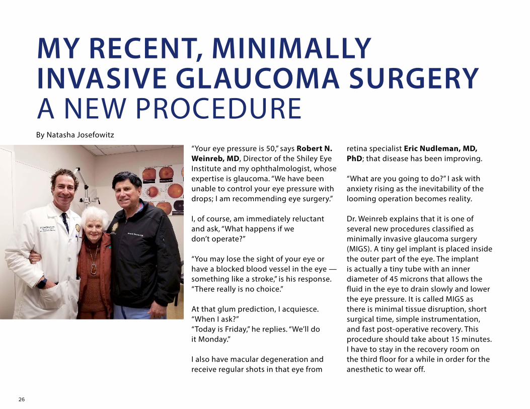
“Your eye pressure is 50,” says Robert N. Weinreb, MD, Director of the Shiley Eye Institute and my ophthalmologist, whose expertise is glaucoma. “We have been unable to control your eye pressure with drops; I am recommending eye surgery.”
I, of course, am immediately reluctant and ask, “What happens if we don’t operate?”
“You may lose the sight of your eye or have a blocked blood vessel in the eye — something like a stroke,” is his response. “There really is no choice.”
At that glum prediction, I acquiesce. “When I ask?” “Today is Friday,” he replies. “We’ll do it Monday.”
I also have macular degeneration and receive regular shots in that eye from
retina specialist Eric Nudleman, MD, PhD; that disease has been improving.
“What are you going to do?” I ask with anxiety rising as the inevitability of the looming operation becomes reality.
Dr. Weinreb explains that it is one of several new procedures classified as minimally invasive glaucoma surgery (MIGS). A tiny gel implant is placed inside the outer part of the eye. The implant is actually a tiny tube with an inner diameter of 45 microns that allows the fluid in the eye to drain slowly and lower the eye pressure. It is called MIGS as there is minimal tissue disruption, short surgical time, simple instrumentation, and fast post-operative recovery. This procedure should take about 15 minutes. I have to stay in the recovery room on the third floor for a while in order for the anesthetic to wear off.
MY RECENT, MINIMALLY INVASIVE GLAUCOMA SURGERY A NEW PROCEDUREBy Natasha Josefowitz
27
little Versed, the usual culprit, and some Fentanyl. After just a couple of minutes of conversation, Dr. Schwartz says: “Don’t micro-manage during the operation.” How did he get my number so quickly?
Dr. Weinreb comes over to reassure me and tells me everything is ready. I am wheeled into the operating room and a sheet is placed over my face with an opening over my eye. After some numbing eye drops, I feel no pain during the operation, only some pressure over the eye. I know how to relax from my meditation class. Before I know it, Dr. Weinreb says that the operation is over. He is pleased; the procedure was successful. The entire staff was impressed as he performed it in a mere four minutes.
I rest on my gurney for half an hour, and I’m off in a wheelchair to the waiting car. Sheri had already called ahead to White Sands so that upon my arrival a
wheelchair was waiting to take me to my room. My wonderful caregiver, Melissa, puts a drop in my eye; thereafter, I could do the drops on my own — every two hours while I’m awake. I sleep well; I am surprised that I have no aftereffects from the anesthesia. My eye is barely sore.
The next morning I return to the hospital for a post-op follow-up appointment. While Dr. Weinreb examines me, he invites a resident and two fellows to come and take a look at the results of a successful operation. (The institute as part of UC San Diego Health is a teaching hospital.) My eye pressure went from 50 to nine.
MIGS, the type of surgery performed by Dr. Weinreb, has been available for only a few years and is replacing much riskier procedures for many patients with glaucoma. I feel fortunate to be able benefit from this miracle of modern surgery and very grateful to Dr. Weinreb, Dr. Nudleman, and their entire team at the Shiley Eye Institute. I am now free of the fear of losing my sight.
Natasha Josefowitz is the author of 21 books. She currently resides at White Sands Retirement Community in La Jolla. Copyright © 2021. Natasha Josefowitz. All rights reserved. Reprinted with permission from sdnews.com (San Diego Community Newspaper Group.)
“Oh my goodness,” I say. I had read that for people my age, anesthesia can impact cognition. I am reassured that it won’t be general, just enough to relax me. I will not be allowed to bend down or lift heavy objects for two weeks following the surgery, so I have two days to arrange my apartment. I have restless nights.
Monday does eventually come. Sheri, one of our White Sands drivers, takes me at 8:15 in the morning. Upon arrival, after registering, I am taken to a pre-op room to change into the proverbial hospital gown that ties in the back. I lie down on a gurney with an IV, a blood pressure monitor (that inflates itself every few minutes throughout the operation), and a pulse oximeter on my finger. Mark Schwartz, MD the anesthesiologist, comes over. I tell him of my concern about the pending loss of brain cells from the anesthesia for people my age (I am 94). He agrees to give me very
I am now free of the fear of losing my sight.

28
INFANTILE CATARACTS ARE A BLIND SPOTThree days after Canyon Brown was born, the attending pediatrician discovered a problem.
During a standard red reflex test, in which a red dot of light is shined from an ophthalmoscope into the pupil, the boy’s right eye, unlike his left, reflected no light back.
People can be born with cataracts. They can form them at any point in life. The clouding of the lens is not just an artifact of advancing age. According to the American Academy of Ophthalmology, the incidence of infantile cataracts is 3 to 4 per 10,000 live births per year. They account for 5 to 20 percent of childhood blindness worldwide.
An even lesser known fact is that cataracts are much more serious in an infant than in an adult.
“People always think, ‘Oh my grandpa had his cataracts removed and it was in
and out of surgery in 20 minutes and he never saw a surgeon again, no big deal,’ but with kids, it's the opposite,” said Jolene Rudell, MD, PhD, Assistant Professor and pediatric ophthalmologist at Shiley Eye Institute at UC San Diego Health.
Rudell removed Canyon’s clouded lens at 6 weeks old. This was considered an emergency surgery for several reasons, each of which distinguishes infantile cataracts from the adult kind.
“One of the things we always worry about is cancer,” said Rudell, explaining that retinoblastoma can sometimes look like a congenital cataract but, if discovered, could require “radiation, chemotherapy and possibly even removing the eye” to treat.
“That was terrifying to us,” said Rochelle Gaudette, Canyon’s mother. “We have a newborn that could possibly have this life-threatening disease?”
The condition is more common — and problematic — than thought
In Canyon’s case, no tumor was detected. His cataract was due to an unexplained underdevelopment of the eye in utero. (If Canyon had developed cataracts in both eyes that might have been a sign of a more serious problem, such as an infection or genetic syndrome that can affect a child’s development and health.)
No matter their cause, treating cataracts is also much more urgent for infants than adults because the brain starts shutting off vision from an underperforming eye immediately after birth. That’s why, like most people who develop cataracts in infancy, Canyon also has amblyopia (more commonly called lazy eye).
“There is a very small window of time when we can operate, which is four to six weeks, to have a chance at saving any vision in the eye,” said Rudell, who regularly performs these operations, “because, from day one, the brain favors the eye without the cataract.”
29
Finally, treating cataracts is much more complicated in infants because clouded lenses cannot immediately be replaced with artificial ones. Eyes keep growing, and changing shape, according to Rudell. So Canyon must wear a contact lens, or very thick glasses, to have any focusing power in his problem eye until he is a little older, when his parents have the option of getting him an intraocular transplant or continuing with the contact lens indefinitely.
“When he wakes up, you have to clean the contact lens and then try to pry his eyeball open and pop it in without him swatting your hand away or screaming or squirming,” Gaudette said. “And the glasses don’t work because he won’t wear his glasses anymore. He pulls them off.”
Because of his amblyopia, Canyon must also wear a patch over his unaffected eye for three to six hours per day to strengthen his problem eye.
“It really changed the routine of our whole family,” Gaudette said, “and every step of the way, there's things that come up.”
Rudell calls correcting infant cataracts “a lifelong process.”
“At any moment after surgery, from days to decades later, there are many potential problems that can arise, and likely additional surgeries to correct them,” she said. “Even if their surgeries go perfectly and the right things are done at the right time, kids born with underdeveloped eyes are more at risk for glaucoma and other forms of blindness. Some of them may end up losing their vision anyway.”
And the expense of this treatment can break the average family’s bank. Canyon’s lensectomy alone was prohibitively expensive and was billed to his insurance, which denied the claim. Fortunately, a “cash pay” arrangement for Gaudette and her husband reduced the cost and this was footed in its entirety by generous friends and family through a GoFundMe campaign.
Many families end up deciding it’s not worth all the trouble just to save the vision in one eye, according to Rudell.
“It can be a difficult decision to put a six-week-old child under anesthesia for something that won’t kill them,” Rudell said. “After all, you can still legally drive with only one eye, and Canyon will still be able to do things other kids can do.”
The problem, Rudell said, is if something ever happens to the unaffected eye.
“When a child grows up and they develop macular degeneration that happens to get worse in their good eye, they essentially become blind,” she said. “That’s why we always try to maximize whatever vision we’re able to, when we can.”
Rudell says more research is needed in the field of congenital cataracts.
“We have options to treat patients like Canyon, but we still don’t have great solutions, and so many questions still remain on what is the best way to manage congenital cataracts,” she said. “It is unfortunately not well-studied. But I’m hoping more research can only help improve visual outcomes for children with eye diseases, including cataracts.”
For now, Canyon is healthy and developing normally at 18 months old, which is what his parents focus on.
“He interacts well with others and his language is developing nicely,” Gaudette said. “I almost think that he might be ahead of the curve in some ways.” She paused and added “It's not like this is a walk in the park, but just the fact that you can treat it makes us just want to go for it and hope for the best.”

30
The Shiley Eye Institute (SEI) and the Viterbi Family Department of Ophthalmology at UC San Diego Health have recently constructed two new visual mobility courses (or mazes) near the La Jolla campus. These full size, human visual mobility courses are designed to test visual function in retinal degeneration patients. Research patients are tested to learn how they navigate through the mazes under different lighting conditions and are asked to negotiate obstacles just as they would in the real word.
The new mazes are being utilized under the leadership of Shyamanga Borooah, MBBS, PhD, Director of the Retinal Degeneration Clinic at SEI. He plans to apply these mazes to test the effectiveness of novel trial therapies for retinal degeneration as part of the new Retinal Degeneration Clinic.
Dr. Borooah states, “These mazes are an exciting new addition to our department. We are fortunate to have been selected as one of only a handful of sites around the world participating in a number of landmark clinical trials targeting retinal degeneration which will utilize our mobility courses. They give us the capability to test visual function before and after treatment to help assess the effectiveness of potential new therapies. Ultimately, the mazes and these ground-breaking studies will contribute to the global efforts to prevent sight loss in retinal degeneration.”
Retinal degenerations are the leading cause of blindness worldwide. The retina is the light sensitive tissue at the back of the eye that contains photoreceptors. Photoreceptors are the cells that begin the process of seeing by absorbing and
A MAZE FOR INVESTIGATIONSOF RETINA DEGENERATIONS

31
converting light into electric signals that are sent to the optic nerve and the brain. Retinal degeneration, or death of the retinal cells, has many causes but ultimately all causes lead to sight loss. Common retinal degenerations include age-related macular degeneration and inherited retinal diseases such as retinitis pigmentosa.
Inherited retinal degenerations (IRD) often result in advanced visual loss due to genetic changes. In many IRD patients, standard ophthalmic assessments, like eye charts or visual fields, do not work. Therefore, an alternative vision test was
developed to measure visual function – the visual mobility course.
Utilizing these mazes, IRD patients’ can be assessed for walking accuracy, number of errors, and speed through the course. These measures can be used to reliably and objectively assess patient visual function. The courses have real life obstacles that the patient must distinguish while walking through. Obstacles can be representations of walls, people, plants or pets. These courses can accommodate various types and severities of vision loss by modifying light levels.
The primary aim of the new SEI Retinal Degeneration Clinic is to combine world-class clinical care with the latest research for patients with retinal degenerations. The center is fully structured around retinal degeneration patient’s needs. The center’s experienced team utilizes state of the art diagnostics and imaging, genetic testing, genetic counseling. It also connects patients to visual or low vision rehabilitation. Patients also have the opportunity to participate in pioneering studies, such as gene therapy and gene editing clinical trials, which utilize the new mazes to test new treatment approaches.
Intricate lighting system over the maze Dr. Borooah and a patient entering the maze.
32
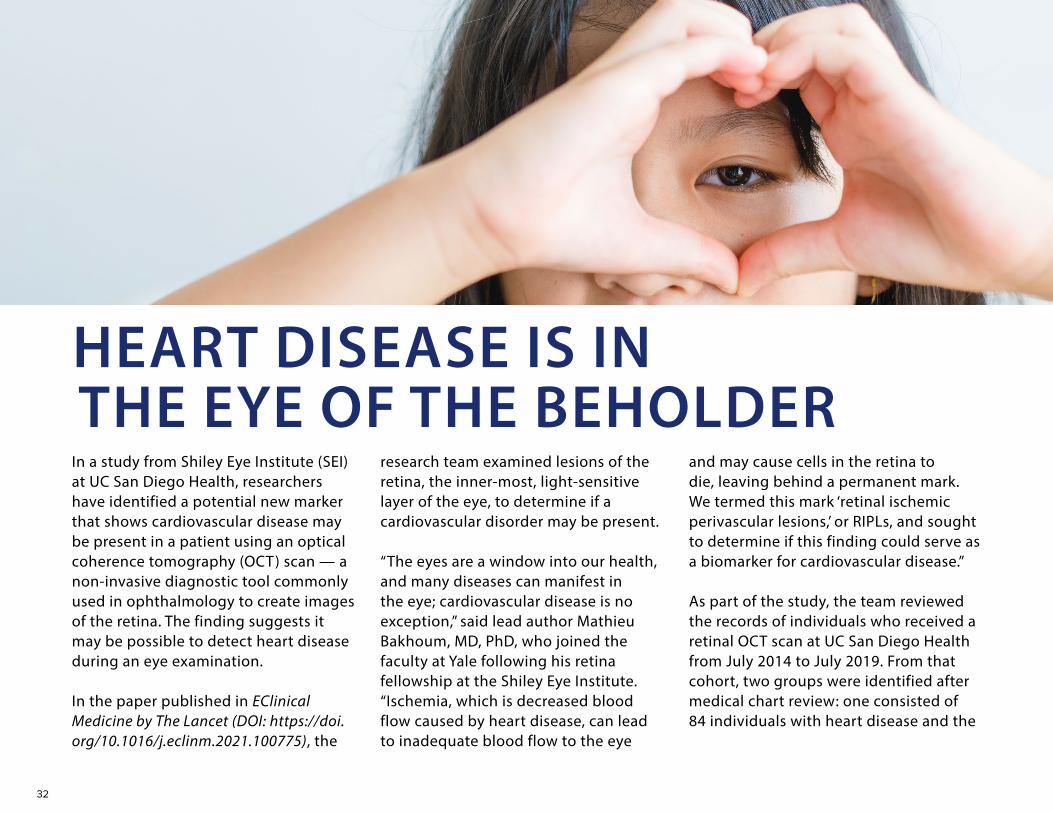
In a study from Shiley Eye Institute (SEI) at UC San Diego Health, researchers have identified a potential new marker that shows cardiovascular disease may be present in a patient using an optical coherence tomography (OCT) scan — a non-invasive diagnostic tool commonly used in ophthalmology to create images of the retina. The finding suggests it may be possible to detect heart disease during an eye examination.
In the paper published in EClinical Medicine by The Lancet (DOI: https://doi.org/10.1016/j.eclinm.2021.100775), the
research team examined lesions of the retina, the inner-most, light-sensitive layer of the eye, to determine if a cardiovascular disorder may be present.
“The eyes are a window into our health, and many diseases can manifest in the eye; cardiovascular disease is no exception,” said lead author Mathieu Bakhoum, MD, PhD, who joined the faculty at Yale following his retina fellowship at the Shiley Eye Institute. “Ischemia, which is decreased blood flow caused by heart disease, can lead to inadequate blood flow to the eye
and may cause cells in the retina to die, leaving behind a permanent mark. We termed this mark ‘retinal ischemic perivascular lesions,’ or RIPLs, and sought to determine if this finding could serve as a biomarker for cardiovascular disease.”
As part of the study, the team reviewed the records of individuals who received a retinal OCT scan at UC San Diego Health from July 2014 to July 2019. From that cohort, two groups were identified after medical chart review: one consisted of 84 individuals with heart disease and the
HEART DISEASE IS IN THE EYE OF THE BEHOLDER

33
other included 76 healthy individuals as the study’s control group. An increased number of RIPLs was observed in the eyes of individuals with heart disease. According to the researchers, the higher number of RIPLs in the eye, the higher the risk for cardiovascular disease.
A person’s risk for cardiovascular disease is determined by the atherosclerotic cardiovascular disease (ASCVD) risk score calculator, the national guideline developed by the American College of Cardiology. The guideline is considered the gold standard for assessing a patient’s 10-year risk of experiencing a cardiovascular event, such as heart attack or stroke. In the study, researchers found a correlation between the number of RIPLs in a patient’s eye and their ASCVD risk score.
“Individuals with low and borderline ASCVD scores had a low number of RIPLs in their eyes, but as the ASCVD risk increased, so did the number of RIPLs,” said Bakhoum.
The research team hopes this paper and future studies will result in RIPLs becoming a common ophthalmological marker for identifying potential cardiovascular disease, and incorporated into the overall ASCVD risk score.
“Globally, cardiovascular disease is the number one cause of death and unfortunately many people are unaware they may have heart issues,” said Bakhoum. “The key in preventing this is early detection and treatment. It’s our hope that by identifying RIPLs as a marker for cardiovascular disease providers will be able to identify heart issues before a catastrophic event, such as a heart attack or a stroke, occurs.”
Additional co-authors of the study include: Michael H. Goldbaum, MD, William R Freeman, MD, Anthony N. DeMaria, MD, Christopher P. Long, MD, Christine Y. Bakhoum, MD, MAS, Anupam K. Garg, MD, PhD, medical students Alison X. Chan and Samantha Madala, as well as former SEI resident Christopher B. Toomey, MD, PhD.
The eyes are a window into our health, and many diseases can manifest in the eye; cardiovascular disease is no exception

34
WELCOME NEW FACULTYALEX A. HUANG, MD, PHD
The Viterbi Family Department of Ophthalmology and the Shiley Eye Institute at UC San Diego welcomes Alex A. Huang, MD, PhD, who enters to the Viterbi Department of Ophthalmology as an Associate Professor in the Glaucoma Division. He is a glaucoma specialist and clinician-scientist who performs all current (and minimally invasive) glaucoma surgical procedures.
Dr. Huang earned both his PhD in neuroscience and MD at The Johns Hopkins University School of Medicine. He completed his residency in ophthalmology at the University of Southern California. Dr. Huang is
recognized by patients and staff upon his return as he was a postdoctural fellow in glaucoma at the SEi in 2012-2013.
When asked why he became a doctor, Dr. Huang answered, “I chose to become a physician-scientist ophthalmologist to help patients maximize their vision and to lead what I call a “sand-box” life.” He went on to state, “Clinician-scientists explore challenging clinical and basic science questions like an inquisitive kid in a sand-box. Creatively free, we see patients and then are inspired by them to chase innovative diagnostic and treatment ideas to improve patient-care.”Dr. Huang’s clinical goal is to maximize
the efficacy of glaucoma therapeutics while decreasing their burden to augment the best quality-of-life. He carries his interests regarding angle-based approaches and native outflow pathway improvement into his laboratory as a National Institutes of Health (NIH) R01-supported scientist. Thus, his NIH research program focuses on improving glaucoma surgical outcomes by enhancing aqueous humor outflow understanding.
He is also supported by the National Aeronautics and Space Administration (NASA) to protect the eyes of American astronauts on the International Space

35
Station from Space Flight-Associated Neuro-ocular Syndrome (SANS).
Dr. Huang is looking forward to the UC San Diego research collaborations and partnerships in his new position. He noted, “Put nicely, teamwork is the key. Put factually, no single person is smart enough to do everything and take any idea to fruition. UC San Diego epitomizes this ideal by bringing together the best and most diverse family of clinicians and scientists. This is why I am excited to be here - to work with other experts like Robert N. Weinreb, MD to tackle the most challenging glaucoma and eye care questions in the future.”
The international publication, The Ophthalmologist named Dr. Huang the #1 Rising Star in the World in 2017 and recognized him on The Ophthalmologist Power 100 List in 2020. In 2021, Dr. Huang received the Association for Research in Vision and Ophthalmology (ARVO) Foundation Pfizer Ophthalmics Carl Camras Translational Research Award.
Alex A. Huang, MD, PhD conducts research for the National Aeronautics and Space Administration (NASA) to safeguard American astronauts’ eyes on the International Space Station (ISS). His goal is to determine the cause of Spaceflight Associated Neuro-ocular Syndrome (SANS) and develop countermeasures necessary for a long-haul spaceflight Mission to Mars.
Eye health is a hurdle for the Mission to Mars. Based on the rotation of the planets and how fast spaceships fly, a planned human Mission to Mars would take years to travel there, to explore the planet and to return to Earth safely. Since 2011, it was discovered that the optic nerves of American astronauts on the ISS could become swollen, the eye globe flattens, and/or the retina folds. This new disease is called SANS. Since that time, 1 in 3 astronauts flying long duration ISS missions have developed these symptoms, and the longer astronauts resided in the weightless environment of the ISS, the worse the swelling.
OFF THE PLANET

36
For more information, scan this barcode to reach the publication.
Thus, SANS represents a major barrier to long-haul exploration class spaceflight because of the risk to vision and the potential inability for astronauts to complete their missions or come home. It is urgent that SANS be better understood, risk factors identified, and mitigation strategies developed.
Many reasons have been hypothesized for why SANS occurs, and one leading concept involves the fluid in our bodies. Without gravity, fluid in our legs re-distributes to our heads, and the extra
volume may be the culprit. Thus, Dr. Huang’s team is currently developing and testing countermeasures designed to reverse the fluid shift. Partnering with NASA, these countermeasures are currently being tested at the German Aerospace Center (Deutsches Zentrum fur Luft- und Raumfahrt or DLR).
Eye research is further performed directly on the ISS. The ocular nerves, eye blood flow, and eye electrical function are being directly tested on American astronauts in space. With these studies, Dr. Huang and
Image courtesy of DLR.
SANS is modeled on Earth by placing subjects in head-down tilt so that lower body fluid shifts to the head. A lower-body negative pressure [LBNP] chamber, and other potential countermeasures, can be tested in this body position.
his multi-centered team hope to improve the safety of future spaceflight and allow for the exploration of this next frontier. Lessons from these SANS investigations may also shed light onto Earthly eye disorders such as glaucoma optic neuropathy.
37
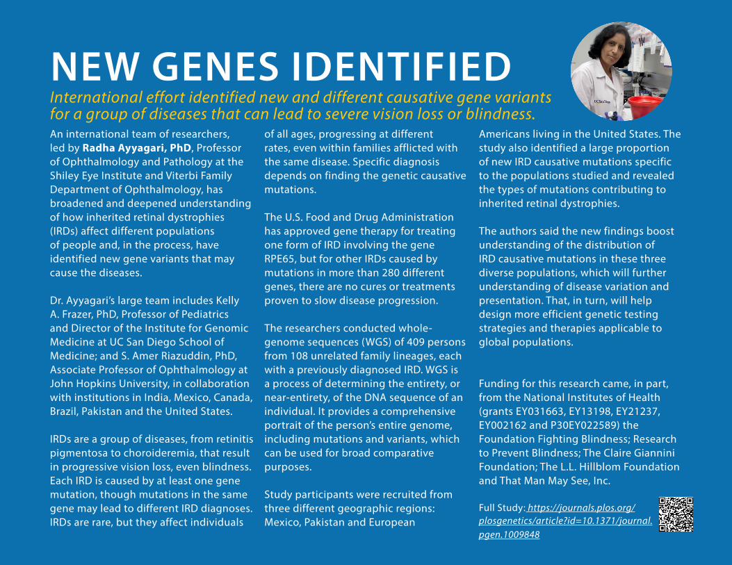
An international team of researchers, led by Radha Ayyagari, PhD, Professor of Ophthalmology and Pathology at the Shiley Eye Institute and Viterbi Family Department of Ophthalmology, has broadened and deepened understanding of how inherited retinal dystrophies (IRDs) affect different populations of people and, in the process, have identified new gene variants that may cause the diseases.
Dr. Ayyagari’s large team includes Kelly A. Frazer, PhD, Professor of Pediatrics and Director of the Institute for Genomic Medicine at UC San Diego School of Medicine; and S. Amer Riazuddin, PhD, Associate Professor of Ophthalmology at John Hopkins University, in collaboration with institutions in India, Mexico, Canada, Brazil, Pakistan and the United States.
IRDs are a group of diseases, from retinitis pigmentosa to choroideremia, that result in progressive vision loss, even blindness. Each IRD is caused by at least one gene mutation, though mutations in the same gene may lead to different IRD diagnoses. IRDs are rare, but they affect individuals
of all ages, progressing at different rates, even within families afflicted with the same disease. Specific diagnosis depends on finding the genetic causative mutations.
The U.S. Food and Drug Administration has approved gene therapy for treating one form of IRD involving the gene RPE65, but for other IRDs caused by mutations in more than 280 different genes, there are no cures or treatments proven to slow disease progression.
The researchers conducted whole-genome sequences (WGS) of 409 persons from 108 unrelated family lineages, each with a previously diagnosed IRD. WGS is a process of determining the entirety, or near-entirety, of the DNA sequence of an individual. It provides a comprehensive portrait of the person’s entire genome, including mutations and variants, which can be used for broad comparative purposes.
Study participants were recruited from three different geographic regions: Mexico, Pakistan and European
Americans living in the United States. The study also identified a large proportion of new IRD causative mutations specific to the populations studied and revealed the types of mutations contributing to inherited retinal dystrophies.
The authors said the new findings boost understanding of the distribution of IRD causative mutations in these three diverse populations, which will further understanding of disease variation and presentation. That, in turn, will help design more efficient genetic testing strategies and therapies applicable to global populations.
Funding for this research came, in part, from the National Institutes of Health (grants EY031663, EY13198, EY21237, EY002162 and P30EY022589) the Foundation Fighting Blindness; Research to Prevent Blindness; The Claire Giannini Foundation; The L.L. Hillblom Foundation and That Man May See, Inc.
Full Study: https://journals.plos.org/plosgenetics/article?id=10.1371/journal.pgen.1009848
NEW GENES IDENTIFIEDInternational effort identified new and different causative gene variants for a group of diseases that can lead to severe vision loss or blindness.
38
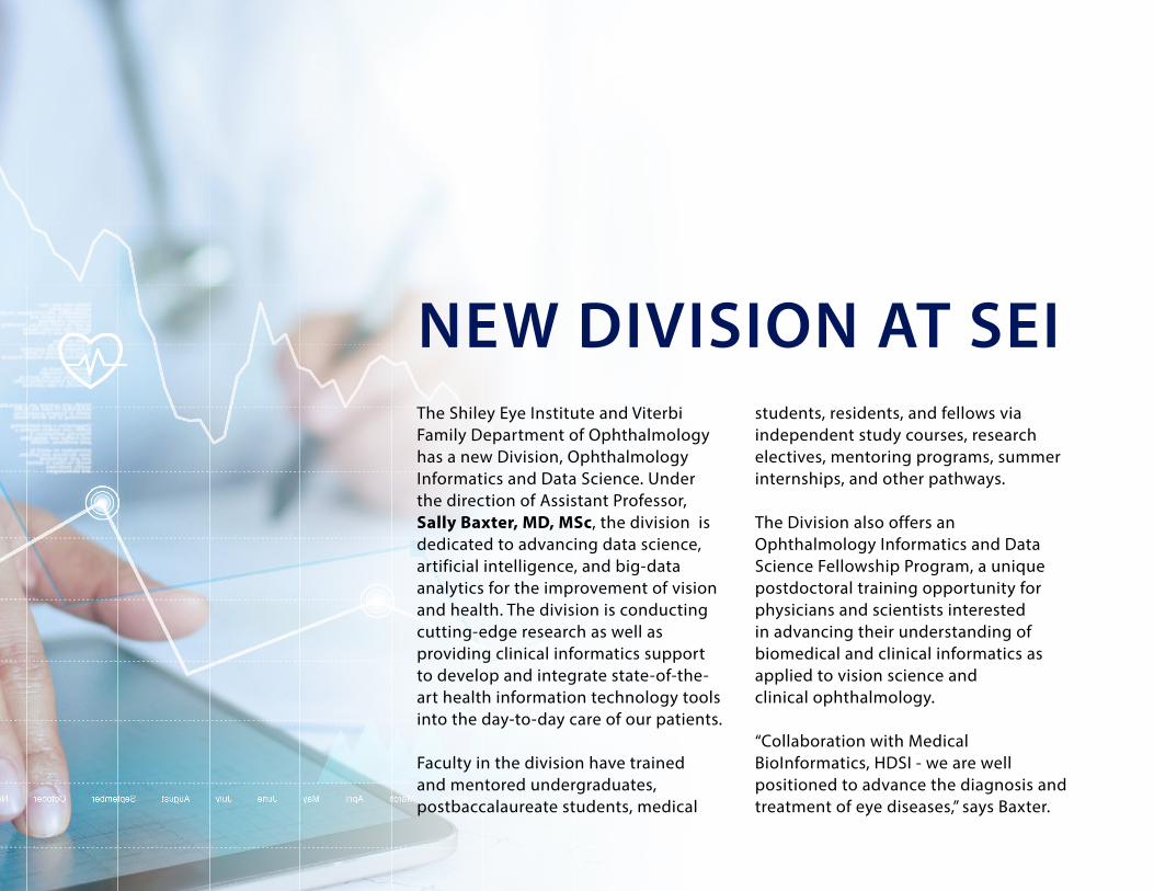
The Shiley Eye Institute and Viterbi Family Department of Ophthalmology has a new Division, Ophthalmology Informatics and Data Science. Under the direction of Assistant Professor, Sally Baxter, MD, MSc, the division is dedicated to advancing data science, artificial intelligence, and big-data analytics for the improvement of vision and health. The division is conducting cutting-edge research as well as providing clinical informatics support to develop and integrate state-of-the-art health information technology tools into the day-to-day care of our patients.
Faculty in the division have trained and mentored undergraduates, postbaccalaureate students, medical
students, residents, and fellows via independent study courses, research electives, mentoring programs, summer internships, and other pathways.
The Division also offers an Ophthalmology Informatics and Data Science Fellowship Program, a unique postdoctoral training opportunity for physicians and scientists interested in advancing their understanding of biomedical and clinical informatics as applied to vision science and clinical ophthalmology.
“Collaboration with Medical BioInformatics, HDSI - we are well positioned to advance the diagnosis and treatment of eye diseases,” says Baxter.
NEW DIVISION AT SEI
39
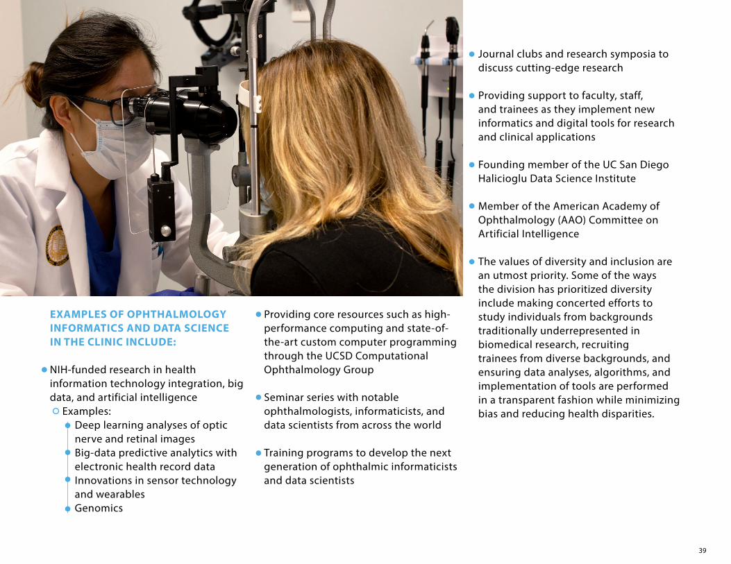
EXAMPLES OF OPHTHALMOLOGY INFORMATICS AND DATA SCIENCE IN THE CLINIC INCLUDE:
NIH-funded research in health information technology integration, big data, and artificial intelligence
Examples: Deep learning analyses of optic nerve and retinal images Big-data predictive analytics with electronic health record dataInnovations in sensor technology and wearables Genomics
Providing core resources such as high-performance computing and state-of-the-art custom computer programming through the UCSD Computational Ophthalmology Group
Seminar series with notable ophthalmologists, informaticists, and data scientists from across the world
Training programs to develop the next generation of ophthalmic informaticists and data scientists
Journal clubs and research symposia to discuss cutting-edge research
Providing support to faculty, staff, and trainees as they implement new informatics and digital tools for research and clinical applications
Founding member of the UC San Diego Halicioglu Data Science Institute
Member of the American Academy of Ophthalmology (AAO) Committee on Artificial Intelligence
The values of diversity and inclusion are an utmost priority. Some of the ways the division has prioritized diversity include making concerted efforts to study individuals from backgrounds traditionally underrepresented in biomedical research, recruiting trainees from diverse backgrounds, and ensuring data analyses, algorithms, and implementation of tools are performed in a transparent fashion while minimizing bias and reducing health disparities.

40
Informatics is the “science of how to use data, information, and knowledge to improve human health and the delivery of health care services” (American Medical Informatics Association).
Data Science is the study of data or facts and figures/numbers. It involves recording, storing, and analyzing data to effectively extract useful information.
Artificial Intelligence is the ability of a computer to do tasks that are
AFFILIATED SEI FACULTYSally L. Baxter, MD, MSc – Division Chief,
Dual Board-Certified in Ophthalmology and Clinical Informatics
Dirk-Uwe Bartsch, PhDAkram Belghith, PhD Chris Bowd, PhDMark Christopher, PhDWilliam Freeman, MDMichael Goldbaum, MD Sasan Moghimi, MD Robert N. Weinreb, MDDerek Welsbie, MD, PhDLinda Zangwill, PhD
usually done by humans because they require intelligence and discernment. In other words, it is the simulation of human intelligence in machines that are programmed to think like humans and mimic their actions. Examples are learning and problem solving.
Big Data is extremely large groups of numbers or data sets that can be analyzed computationally to reveal patterns, trends, and associations –
especially relating to human behavior or medically recorded numeric information.
Deep Learning is an artificial intelligence function that imitates the working of the human brain in processing data and creating patterns for use in decision making.
COLLABORATING FACULTY FROM OTHER DIVISIONS/DEPARTMENTS AT UC SAN DIEGOUCSD Health Department of
Biomedical InformaticsUCSD Health Information Services UCSD Halicioglu Data Science InstituteUCSD Division of Biostatistics &
Bioinformatics in the Department of Family Medicine and Public Health
UCSD Department of Computer Science and Engineering
SOME HELPFUL DEFINITIONS

41
LIONS BIOBANK UPDATEThe Shiley Eye Institute (SEI) gratefully accepted a groundbreaking donation to establish the Downtown San Diego Lions Club BioBank for Vision in 2017. The BioBank stores a library of biological samples with complete background information that researchers can utilize to investigate predictors for diseases and effectiveness of therapies. The goal of the BioBank is to leverage the latest in bioinformatics technology and genetic sequencing tools to advance the understanding of eye diseases such as glaucoma and inherited retinal diseases.
Demographic, ethnic, medical and risk factor history data are collected from qualified patients in the SEI clinic. The details of sample collection, processing, analysis and exact freezer storage location of samples are recorded in the BioBank database system. Each
in the BioBank. In the past year, the Biobank obtained four thousand blood mononuclear cells from 2,315 patients to generate induced pluripotent stem cells for studying eye diseases. These cells have been carefully and confidentially catalogued along with their clinical status and stored at ultralow temperature (<160 degrees C) in liquid nitrogen freezers for future use. RNA samples were obtained from 3,422 patients and stored at -80 degrees C temperature in our BioBank freezers. These samples and others are awaiting proteomic analysis, a systematic identification and quantification of the proteins of a cell, tissue or biological fluid. SEI researchers will then examine how the samples contribute to the regulation of eye pressure and the delivery of nutrients to the eye.
step of the process ensures that all patient data and samples are stored, tracked and readily available to share with investigators, along with all linked clinical, demographic, genotype, and phenotype information while maintaining strict confidentiality. This process has been approved by the UCSD Institutional Review Board Committee and involves all activities including the sample collection, sample processing and intended use and handling protocol.
Patients with inherited eye diseases who participate in this research have provided blood samples that are processed into DNA, RNA and other important components of blood. These samples are isolated, quantified and standardized in preparation for future genetic analysis. Moreover, induced pluripotent stem cells from specific individuals also are stored

42
2021MARKS TEN YEARS OFFOR SIGHTANNUAL REPORTS
VIEW OUR ENTIRE COLLECTION OF ANNUAL
REPORTS ONLINE HERE:

43
Sally L. Baxter, MD, MSc, Assistant Professor, received the 2020 NIH Director’s Early Independence Award. This is among the most prestigious grants awarded by the National Institutes of Health, and Dr. Baxter is one of the only ophthalmologists to be so recognized.
Andrew Camp, MD, Assistant Professor, was awarded the 2021 Whitehill Prize for Excellence from UC San Diego.
Alex A. Huang, MD, PhD, Associate Professor, received the 2021 Achievement Award from the Academy of Ophthalmology.
Don O. Kikkawa, MD, Professor and Executive Vice Chair, was named to the Ophthalmology Residency Review Committee of the Accreditation Council for Graduate Medical Education (ACGME), the national organization that accredits resident training programs.
Shira L. Robbins, MD, Professor of Ophthalmology, has been elected UC San Diego Health (UCSDH) Vice Chief of Staff. She was elected by the UC San Diego Active Medical Staff that includes over 1,300 physicians and other clinicians. In 2 years, she will become UC San Diego Health (UCSDH) Chief of Staff. This position of Vice Chief of Staff merges patient safety with UCSD policies while being a physician advocate.
Robert N. Weinreb, MD, Distinguished Professor and Chair, received the Dr. Robert Ritch Award for Excellence and Innovation in Glaucoma from The Glaucoma Foundation (New York).
Robert N. Weinreb, MD, Distinguished Professor and Chair, was recognized by Ophthalmology Management as one of the 2021 “25 Leaders in Innovation” who have made notable contributions to ophthalmology.
Robert N. Weinreb, MD, Distinguished Professor and Chair, and Jeffrey E. Lee, MD, Associate Professor, were named to Newsweek’s “America’s Best Eye Doctors 2021”.
Among 21 Viterbi Family Department of Ophthalmology faculty who were recognized by Expertscape as being among the best in their respective fields, Robert N. Weinreb, MD was named for the seventh consecutive year as the world’s leading glaucoma expert. Also ranked highly at #12 was Linda Zangwill, PhD, Professor and interim Research Director.
Every year, Shiley Eye Institute specialists have been honored as being “the best” by every major national and local organization.
CONGRATULATIONS
The OphthalmologistExpertscapeCastle ConnollySan Diego MagazineTOP Doctors
U.S. News & World ReportBest DoctorsSuperDoctorsNewsweek

44
Chair & Distinguished Professor, Viterbi Family Department of Ophthalmology
Director, Shiley Eye InstituteDirector, Hamilton Glaucoma CenterDistinguished Professor of BioengineeringMorris Gleich, MD Chair in Glaucoma
MEDICAL SCHOOLHarvard Medical School
RESIDENCY & FELLOWSHIPUniversity of California San Francisco
Robert N. Weinreb, MDGLAUCOMA
Professor of OphthalmologyCo-Director of Clinical Research,
Hamilton Glaucoma CenterDirector, Hamilton Glaucoma Center,
Data Coordinating CenterRichard K. Lansche, MD & Tatiana A. Lansche
Chair in Ophthalmology
GRADUATE SCHOOLHarvard School of Public Health (MS)Ben-Gurion University of the Negev (PhD)
POSTDOCTORAL FELLOWSHIP University of Waterloo, Waterloo, Ontario, Canada
Linda M. Zangwill, PhDGLAUCOMA
Assistant Project Scientist of Ophthalmology
GRADUATE SCHOOLUniversity of Strasbourg, France
POSTDOCTORAL FELLOWSHIP University of California San Diego
Akram Belghith, PhDGLAUCOMA
Christopher Bowd, PhDResearch Scientist of OphthalmologyDirector of the Hamilton Glaucoma
Center-based Visual Field Assessment CenterCo-Director of the Hamilton Glaucoma Center-based Imaging Data
Evaluation and Analysis (IDEA) Center
GRADUATE SCHOOLWashington State University
POSTDOCTORAL FELLOWSHIP University of California San Diego
GLAUCOMA
Assistant Professor of Ophthalmology
MEDICAL SCHOOLUniversity of Miami Miller School of Medicine
RESIDENCYBascom Palmer Eye Institute at the
University of Miami Miller School of Medicine
FELLOWSHIPUniversity of California San Diego
Andrew S. Camp, MDGLAUCOMA
Assistant Project Scientist of Ophthalmology
GRADUATE SCHOOLUniversity of Iowa
POSTDOCTORAL FELLOWSHIPUniversity of California San Diego
Mark Christopher, PhDGLAUCOMA
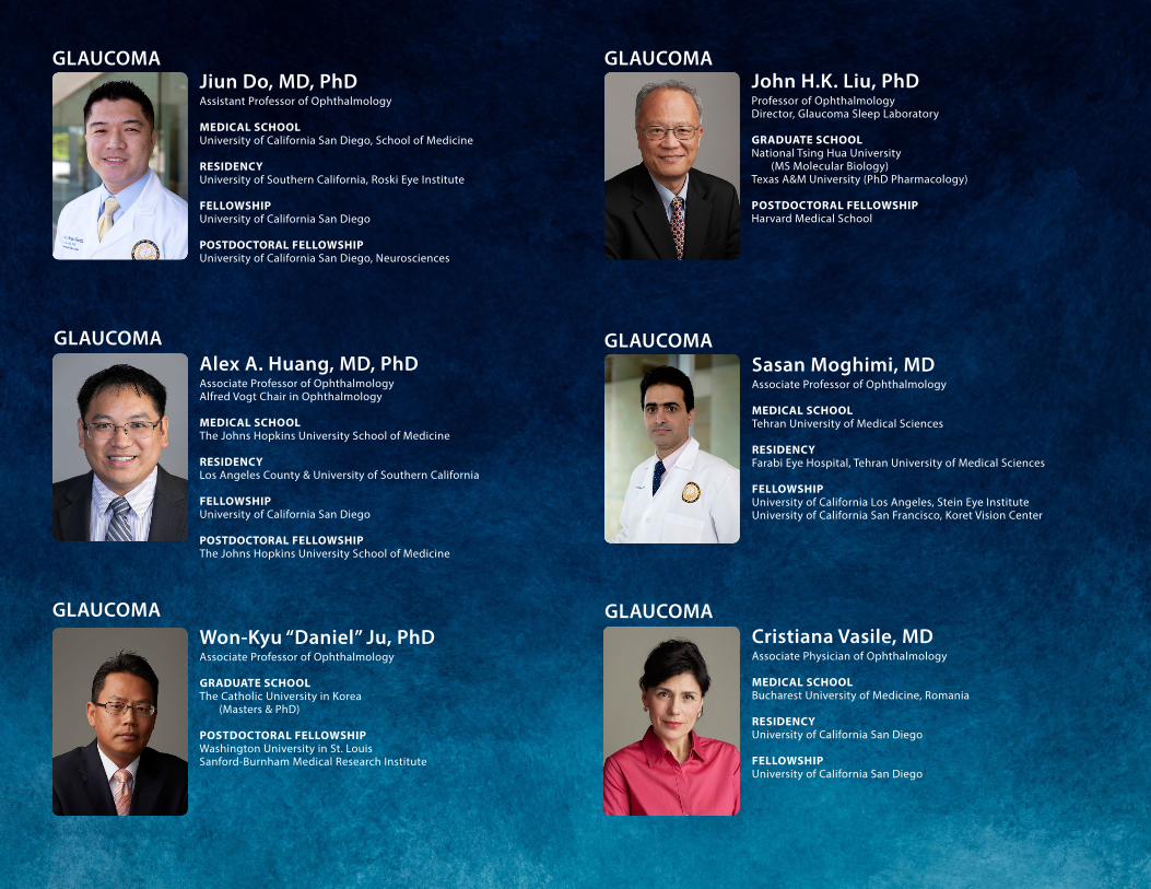
45
Associate Professor of Ophthalmology
GRADUATE SCHOOLThe Catholic University in Korea
(Masters & PhD)
POSTDOCTORAL FELLOWSHIP Washington University in St. LouisSanford-Burnham Medical Research Institute
Won-Kyu “Daniel” Ju, PhDGLAUCOMA
Assistant Professor of Ophthalmology
MEDICAL SCHOOLUniversity of California San Diego, School of Medicine
RESIDENCYUniversity of Southern California, Roski Eye Institute
FELLOWSHIPUniversity of California San Diego
POSTDOCTORAL FELLOWSHIPUniversity of California San Diego, Neurosciences
Jiun Do, MD, PhDGLAUCOMA
Professor of Ophthalmology Director, Glaucoma Sleep Laboratory
GRADUATE SCHOOLNational Tsing Hua University
(MS Molecular Biology) Texas A&M University (PhD Pharmacology)
POSTDOCTORAL FELLOWSHIP Harvard Medical School
John H.K. Liu, PhDGLAUCOMA
Associate Professor of OphthalmologyAlfred Vogt Chair in Ophthalmology
MEDICAL SCHOOLThe Johns Hopkins University School of Medicine
RESIDENCYLos Angeles County & University of Southern California
FELLOWSHIPUniversity of California San Diego
POSTDOCTORAL FELLOWSHIPThe Johns Hopkins University School of Medicine
Alex A. Huang, MD, PhDGLAUCOMA
Associate Professor of Ophthalmology
MEDICAL SCHOOLTehran University of Medical Sciences
RESIDENCYFarabi Eye Hospital, Tehran University of Medical Sciences
FELLOWSHIP University of California Los Angeles, Stein Eye InstituteUniversity of California San Francisco, Koret Vision Center
Sasan Moghimi, MDGLAUCOMA
Associate Physician of Ophthalmology
MEDICAL SCHOOLBucharest University of Medicine, Romania
RESIDENCYUniversity of California San Diego
FELLOWSHIP University of California San Diego
Cristiana Vasile, MDGLAUCOMA

46
Associate Professor of Ophthalmology
MEDICAL SCHOOLUniversity of California Los Angeles
RESIDENCYThe Johns Hopkins University School of Medicine/
Wilmer Eye Institute
FELLOWSHIPThe Johns Hopkins University School of Medicine/
Wilmer Eye Institute
POSTDOCTORAL FELLOWSHIPDavid Gessen School of Medicine UCLA
Derek S. Welsbie, MD, PhDGLAUCOMA
Professor of OphthalmologyVice Chair for Education,
Department of OphthalmologyChief, Division of Cornea and Refractive SurgeryStuart I. Brown MD Chair in Ophthalmology
in Memory of Donald P. Shiley
MEDICAL SCHOOLStanford University School of Medicine
RESIDENCY Harvard University, Massachusetts Eye and Ear Infirmary
FELLOWSHIPHarvard University, Massachusetts Eye and Ear Infirmary
Natalie A. Afshari, MD, FACSCORNEA & REFRACTIVE
Professor of Ophthalmology
MEDICAL SCHOOLUniversity of Illinois Medical School
RESIDENCYTulane Medical School
FELLOWSHIP Harvard University, Massachusetts Eye and Ear Infirmary
Stuart I. Brown, MDCORNEA & REFRACTIVE
Assistant Professor of OphthalmologyChief, Division of Ophthalmology Informatics and Data Science
MEDICAL SCHOOL Perelman School of Medicine at the University of Pennsylvania
RESIDENCY University of California San Diego
FELLOWSHIP University of California, San Diego Health
Department of Biomedical Informatics
Sally L. Baxter, MD, MScCOMPREHENSIVE OPHTHALMOLOGY
Associate Clinical Professor of Ophthalmology Program Director, Ophthalmology Residency
MEDICAL SCHOOLUniversity of California San Diego
RESIDENCYUniversity of California San Diego
Jeffrey E. Lee, MDCOMPREHENSIVE OPHTHALMOLOGY
Assistant Professor of Ophthalmology
MEDICAL SCHOOLUniversity of Oklahoma, Tulsa
RESIDENCY University of Rochester, New York
FELLOWSHIPUniversity of California San Diego
Thao P. Nguyen, MDCOMPREHENSIVE OPHTHALMOLOGY

47
OPHTHALMIC PLASTIC & RECONSTRUCTIVE SURGERY
Professor of Ophthalmology and Plastic SurgeryVice Chair for Clinical Services, Department of OphthalmologyChief, Division of Oculofacial Plastic and Reconstructive SurgeryDr. Trude Kahn Hollander Chair in Ophthalmology
MEDICAL SCHOOLSt. Louis University School of Medicine
RESIDENCYUniversity of California Los Angeles
FELLOWSHIP University of Wisconsin, Madison
Don O. Kikkawa, MD, FACSGENETICS
Professor of Ophthalmology & PathologyChief of Ophthalmic Molecular Diagnostic Laboratory (CLIA certified)Director of Downtown San Diego Lions Club BioBank for VisionThe Viterbi Family Chair III
GRADUATE SCHOOLOsmania University, Hyderabad, India
POSTDOCTORAL FELLOWSHIP Molecular Genetics at the National Eye Institute, NIH, Bethesda
Radha Ayyagari, PhD
Clinical Professor of OphthalmologyChief of Ophthalmology at Veterans
Administration Medical Hospital
MEDICAL SCHOOLUniversity of California Los Angeles School of Medicine
RESIDENCYStanford University School of Medicine
FELLOWSHIP Stanford University School of Medicine
Weldon W. Haw, MDCORNEA & REFRACTIVE
Clinical Professor of Ophthalmology
MEDICAL SCHOOLChicago Medical School
RESIDENCYUniversity of California San Diego
FELLOWSHIPUniversity of California San Diego
Chris W. Heichel, MD, FACSCORNEA & REFRACTIVE
NEURO-OPHTHALMOLOGY
Clinical Professor of Ophthalmology
MEDICAL SCHOOLDuke University
RESIDENCYMcGill University, Montreal
FELLOWSHIPBascom Palmer Eye Institute, University of MiamiMontreal Neurological Institute, McGill University
Lanning Kline, MD
Professor of Ophthalmology & Neurosciences
MEDICAL SCHOOLUniversity of Bologna School of Medicine, Italy
RESIDENCYGeorgetown University Medical Center
FELLOWSHIP University of Miami
Peter J. Savino, MDNEURO-OPHTHALMOLOGY

48
Professor of Ophthalmology and PediatricsVice Chair, Department of OphthalmologyDirector, Anne F. & Abraham Ratner Children’s Eye CenterAnne Ratner Chair of Pediatric Ophthalmology
MEDICAL SCHOOLYale University School of Medicine
RESIDENCYNew York University Medical Center
FELLOWSHIP Children’s Hospital of Philadelphia University of Pennsylvania
David B. Granet, MD, MHCM, FACS, FAAO, FAAP
PEDIATRIC OPHTHALMOLOGY & ADULT EYE REALIGNMENT
Professor of OphthalmologyEducational Director of Pediatric Ophthalmology/
Strabismus Division
MEDICAL SCHOOLMedical College of Pennsylvania Hospital
RESIDENCY Hahnemann University Hospital
FELLOWSHIP University of California San Diego & Naval Medical Center
Shira L. Robbins, MD, FAAO, FAAP
PEDIATRIC OPHTHALMOLOGY & ADULT EYE REALIGNMENT
Professor of Ophthalmology and Plastic Surgery
MEDICAL SCHOOLUniversity of Texas, Southwestern Medical School
RESIDENCYUniversity of California San Diego
FELLOWSHIPUniversity of California San Diego
POSTDOCTORAL FELLOWSHIP Massachusetts Institute of Technology
Bobby S. Korn, MD, PhD, FACSOPHTHALMIC PLASTIC & RECONSTRUCTIVE SURGERY
Assistant Professor of Ophthalmology
MEDICAL SCHOOLAlbert Einstein College of Medicine
RESIDENCYUniversity of California Irvine
FELLOWSHIPIllinois Eye and Ear Infirmary at the University of Illinois, Chicago
POSTDOCTORAL FELLOWSHIPAlbert Einstein College of Medicine
Yunxiang “Catherine” Liu, MD, PhDOPHTHALMIC PLASTIC & RECONSTRUCTIVE SURGERY
Associate Clinical Professor of Ophthalmology
MEDICAL SCHOOLUniversity of Medicine and Dentistry of New Jersey -
Robert Wood Johnson
RESIDENCYLong Island Jewish Medical Center
FELLOWSHIP The University of Wisconsin in Madison
Mansoor Movaghar, MD
PEDIATRIC OPHTHALMOLOGY & ADULT EYE REALIGNMENT
Assistant Professor of Ophthalmology
MEDICAL SCHOOLUniversity of California Davis
RESIDENCY University of California Davis
FELLOWSHIP University of Washington/Seattle Children’s Hospital
POSTDOCTORAL FELLOWSHIPUniversity of California Davis
Jolene Rudell, MD, PhD
PEDIATRIC OPHTHALMOLOGY & ADULT EYE REALIGNMENT

49
RETINA & PSYCHOLOGY
Adjunct Professor of OphthalmologyAssistant Research Scientist Department of Psychology
GRADUATE SCHOOLOregon Health and Science University
POSTDOCTORAL FELLOWSHIP National Institutes of Health
Nicholas Oesch, PhD
RETINAL VASCULAR
Distinguished Professor of Ophthalmology and PathologySenior Deputy Director for Basic Sciences,
UCSD Moores Cancer CenterBen and Wanda Hildyard Chair for Diseases of the Eye
MEDICAL SCHOOL & RESIDENCYUniversity of Catania Medical School, Catania, Italy
FELLOWSHIP University of California San Francisco
Napoleone Ferrara, MD
Assistant Professor of OphthalmologyDirector, Richard C. Atkinson Laboratory
for Regenerative Ophthalmology
GRADUATE SCHOOLThe Johns Hopkins School of Medicine POSTDOCTORAL FELLOWSHIP The Johns Hopkins School of Medicine/Wilmer Eye Institute
Karl Wahlin, PhDREGENERATIVE OPHTHALMOLOGY
Adjunct Professor of Ophthalmology
GRADUATE SCHOOLUniversity of California San Diego
POSTDOCTORAL FELLOWSHIP University of California San Diego
Dirk-Uwe Bartsch, PhD
RETINA & VITREOUS
Assistant Professor of Ophthalmology
MEDICAL SCHOOLImperial College London
RESIDENCYUniversity of Edinburgh
FELLOWSHIP Moorfields Eye Hospital London
POSTDOCTORAL FELLOWSHIP University of Edinburgh
Shyamanga Borooah, MBBS, MRCP (UK), MRCSEd, FRCOphth, PhD
RETINA & VITREOUS
Clinical Professor of Ophthalmology
MEDICAL SCHOOLUniversity of California San Diego
RESIDENCYUniversity of California San Diego
FELLOWSHIP University of California San Diego
Henry A. Ferreyra, MDRETINA & VITREOUS
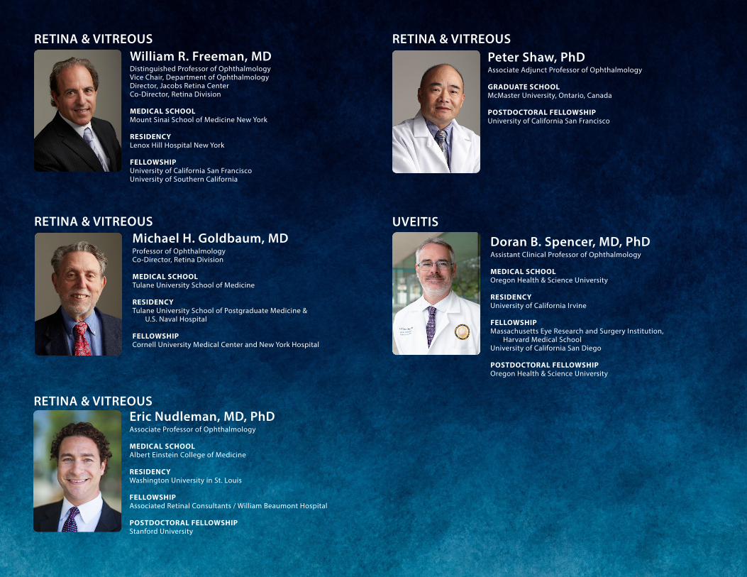
50
Assistant Clinical Professor of Ophthalmology
MEDICAL SCHOOLOregon Health & Science University
RESIDENCYUniversity of California Irvine
FELLOWSHIP Massachusetts Eye Research and Surgery Institution,
Harvard Medical SchoolUniversity of California San Diego
POSTDOCTORAL FELLOWSHIPOregon Health & Science University
Doran B. Spencer, MD, PhDUVEITIS
Distinguished Professor of OphthalmologyVice Chair, Department of OphthalmologyDirector, Jacobs Retina CenterCo-Director, Retina Division
MEDICAL SCHOOLMount Sinai School of Medicine New York
RESIDENCYLenox Hill Hospital New York
FELLOWSHIP University of California San Francisco University of Southern California
William R. Freeman, MDRETINA & VITREOUS
Associate Professor of Ophthalmology
MEDICAL SCHOOLAlbert Einstein College of Medicine
RESIDENCY Washington University in St. Louis
FELLOWSHIP Associated Retinal Consultants / William Beaumont Hospital
POSTDOCTORAL FELLOWSHIPStanford University
Eric Nudleman, MD, PhDRETINA & VITREOUS
Professor of Ophthalmology Co-Director, Retina Division
MEDICAL SCHOOLTulane University School of Medicine
RESIDENCY Tulane University School of Postgraduate Medicine &
U.S. Naval Hospital
FELLOWSHIP Cornell University Medical Center and New York Hospital
Michael H. Goldbaum, MDRETINA & VITREOUS
Associate Adjunct Professor of Ophthalmology
GRADUATE SCHOOLMcMaster University, Ontario, Canada
POSTDOCTORAL FELLOWSHIP University of California San Francisco
Peter Shaw, PhDRETINA & VITREOUS

51
OPTOMETRY & LOW VISION
John F. Kulischak, ODOPTOMETRY SUPERVISOR
OPTOMETRY SCHOOLUniversity of California Berkeley
RESIDENCYPalo Alto VA Medical Center
Anne B. Lam, ODOPTOMETRY SCHOOLSouthern California College
of Optometry at Marshall B. Ketchum University
Alicia Lau, ODOPTOMETRY SCHOOLUniversity of California Berkeley
RESIDENCYRaymond G. Murphy VA Medical Center
SPECIALTYGlaucoma and Ocular Disease
Esmeralda McClean, ODOPTOMETRY SCHOOLUniversity of California Berkeley
SPECIALTYOcular Disease
Maria Laura Gomez, MD, ODMEDICAL SCHOOL & RESIDENCYRosario University & Barraquer
Institute of America, Bogota, Colombia
FELLOWSHIPMoorfield Eye Hospital,
LondonUniversity of California San Diego
SPECIALTY Cornea and Dry Eye
Pamela A. Hoo, ODOPTOMETRY SCHOOLSouthern California College
of Optometry
Lianne Mizoguchi, ODOPTOMETRY SCHOOLNew England College of Optometry
Andrew Vo, ODOPTOMETRY SCHOOLUniversity of California Berkeley
RESIDENCYSouthern California College
of Optometry at Marshall B. Ketchum University
SPECIALTYSpecialty Contact Lens
Carol Yu, ODOPTOMETRY SCHOOLUniversity of California Berkeley
RESIDENCYNova Southeastern University
SPECIALTYSpecialty Contact Lens
and Ocular Disease
Lara D. Hustana, ODOPTOMETRY SCHOOLPacific University
RESIDENCYIndian Health Services
NOT PICTURED:
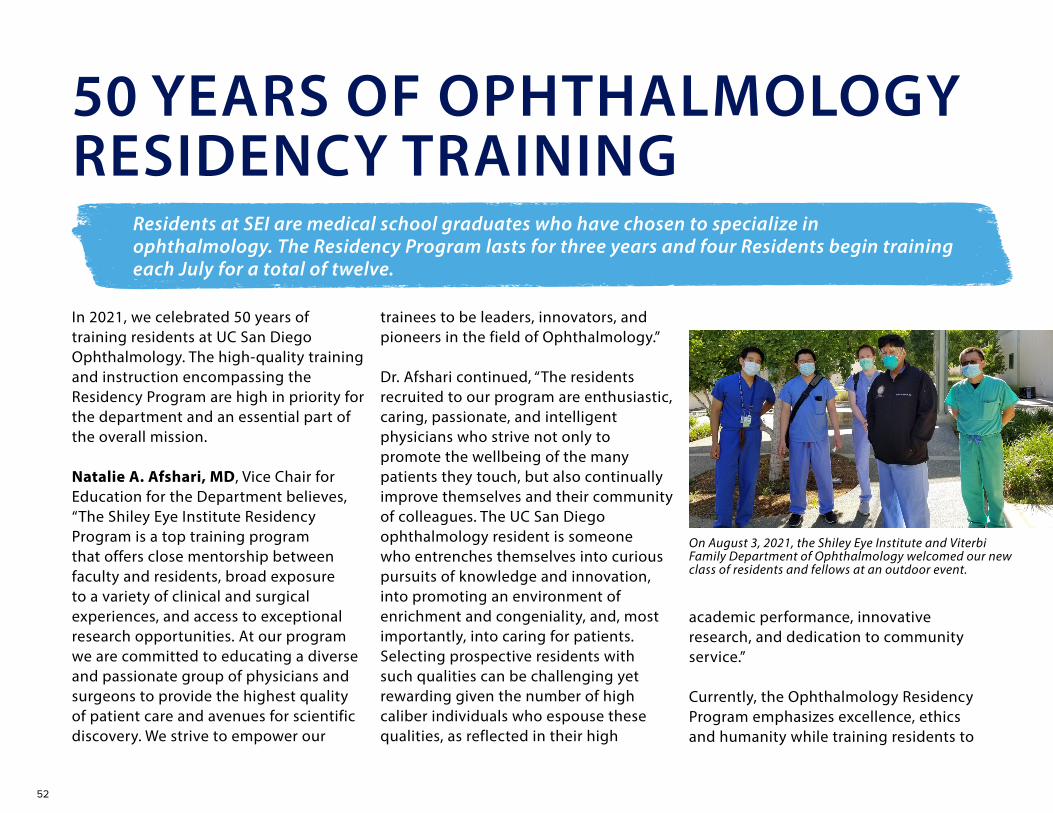
52
In 2021, we celebrated 50 years of training residents at UC San Diego Ophthalmology. The high-quality training and instruction encompassing the Residency Program are high in priority for the department and an essential part of the overall mission.
Natalie A. Afshari, MD, Vice Chair for Education for the Department believes, “The Shiley Eye Institute Residency Program is a top training program that offers close mentorship between faculty and residents, broad exposure to a variety of clinical and surgical experiences, and access to exceptional research opportunities. At our program we are committed to educating a diverse and passionate group of physicians and surgeons to provide the highest quality of patient care and avenues for scientific discovery. We strive to empower our
trainees to be leaders, innovators, and pioneers in the field of Ophthalmology.”
Dr. Afshari continued, “The residents recruited to our program are enthusiastic, caring, passionate, and intelligent physicians who strive not only to promote the wellbeing of the many patients they touch, but also continually improve themselves and their community of colleagues. The UC San Diego ophthalmology resident is someone who entrenches themselves into curious pursuits of knowledge and innovation, into promoting an environment of enrichment and congeniality, and, most importantly, into caring for patients. Selecting prospective residents with such qualities can be challenging yet rewarding given the number of high caliber individuals who espouse these qualities, as reflected in their high
50 YEARS OF OPHTHALMOLOGY RESIDENCY TRAINING
Residents at SEI are medical school graduates who have chosen to specialize in ophthalmology. The Residency Program lasts for three years and four Residents begin training each July for a total of twelve.
On August 3, 2021, the Shiley Eye Institute and Viterbi Family Department of Ophthalmology welcomed our new class of residents and fellows at an outdoor event.
academic performance, innovative research, and dedication to community service.”
Currently, the Ophthalmology Residency Program emphasizes excellence, ethics and humanity while training residents to

53
become exceptional ophthalmologists equipped to succeed in any aspect of ophthalmology, be it academics, research, or the private sector. The overarching goal is the acquisition of the knowledge, skills, clinical judgment, and attitudes necessary to provide skilled and compassionate care to patients.
Residency Program Director and alumni, Jeffrey Lee, MD, states, “I have been fortunate to have done my training here at Shiley and experienced what a world class organization is like at all levels, starting with residency then becoming faculty – I see the big picture. It is an amazing organization to be a part of.”
Dr. Lee’s goals for the Program are to create individualized educational plans for each Resident so they have clinical, surgical, elective and research opportunities with a flexible schedule, therefore optimizing each person’s learning experiences. He went on to note, “We want every Resident to become the best version of themselves when they graduate from our Program.”
Residents are provided with clinical exposure in multiple facilities to gain mastery of the ophthalmology knowledge and judgment needed while assuming increasing levels of responsibility for the medical and surgical management of patients
with a wide variety of acute and chronic ophthalmological diseases. Because we recognize that ophthalmology education continues beyond the residency training years, we emphasize the importance of self-directed study habits.
Four residents begin training each July for a total of twelve. While training at our multiple facilities, each resident is exposed to a diverse patient population with a wide variety of ophthalmic needs—all of this while being given increasing responsibilities in a one-on-one teaching environment with our clinical attending physicians.
Department Chair, Robert N. Weinreb, MD noted, “We are so proud of our residents. They have distinguished themselves in academic positions and also in their communities. As faculty, it is gratifying to mentor the leaders of tomorrow and watch them grow into clinical, surgical and research trailblazers.”
Since 1971, the Department has graduated 137 residents. Many receive additional training and they enter academic or private practices throughout the country. The SEI faculty also collaborates with many of our graduates on multi-center research investigations.
Jeffrey Lee, MD, Natalie A. Afshari, MD and Robyn Austin pictured with the residents.
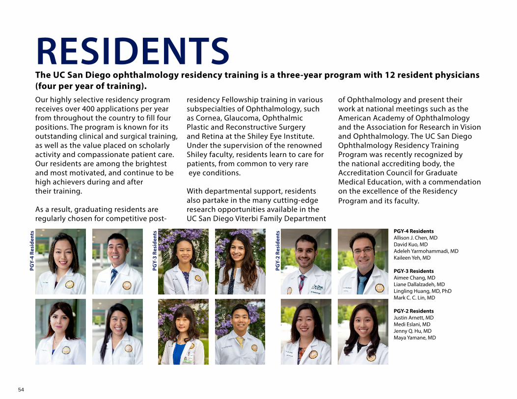
54
The UC San Diego ophthalmology residency training is a three-year program with 12 resident physicians (four per year of training).Our highly selective residency program receives over 400 applications per year from throughout the country to fill four positions. The program is known for its outstanding clinical and surgical training, as well as the value placed on scholarly activity and compassionate patient care. Our residents are among the brightest and most motivated, and continue to be high achievers during and after their training.
As a result, graduating residents are regularly chosen for competitive post-
residency Fellowship training in various subspecialties of Ophthalmology, such as Cornea, Glaucoma, Ophthalmic Plastic and Reconstructive Surgery and Retina at the Shiley Eye Institute. Under the supervision of the renowned Shiley faculty, residents learn to care for patients, from common to very rare eye conditions.
With departmental support, residents also partake in the many cutting-edge research opportunities available in the UC San Diego Viterbi Family Department
RESIDENTSof Ophthalmology and present their work at national meetings such as the American Academy of Ophthalmology and the Association for Research in Vision and Ophthalmology. The UC San Diego Ophthalmology Residency Training Program was recently recognized by the national accrediting body, the Accreditation Council for Graduate Medical Education, with a commendation on the excellence of the Residency Program and its faculty.
PGY-4 ResidentsAllison J. Chen, MDDavid Kuo, MDAdeleh Yarmohammadi, MDKaileen Yeh, MD
PGY-3 ResidentsAimee Chang, MDLiane Dallalzadeh, MDLingling Huang, MD, PhDMark C. C. Lin, MD
PGY-2 ResidentsJustin Arnett, MDMedi Eslani, MDJenny Q. Hu, MDMaya Yamane, MD
PGY-
4 Re
side
nts
PGY-
3 Re
side
nts
PGY-
2 Re
side
nts

55
Jasmin Rezapour, MD
Alireza Kamalipour, MDNevin El-Nimri, OD, PhD
FELLOWS Shiley Eye Institute offers world-class fellowships in cornea, glaucoma, ophthalmic plastic and reconstrutive surgery, pediatric ophthalmology, and retina. Fellows are exposed to expert training in both the clinical and research settings. Many go on to prominent academic positions around the world as well as practicing as outstanding clinicians in the global ophthalmic community.
GLAUCOMA
Takashi Nishida, MD, PhD
Melina Cavichini Cordeiro, MD
Not Pictured: GLAUCOMA Rui Fan, PhD
Joo Youn Shin, MD, PhD
GLAUCOMA
RETINA
Ryan Phan, MD
Jo-Hsuan Wu, MD Yue “Carrie” Zhao, MD
Darren Knight, MD Alexa Li, MD Alexandra Warter, MD Chris Wu, MD DJ Ozzello, MD
OCULOPLASTICS
CORNEA
Michael J. Ang, MD Alex Beazer, MD
Golnoush Mahmoudi Nezad, MD, MPH
Eleonora Micheletti, MD
RETINA Tahmineh Motevasseli, MD
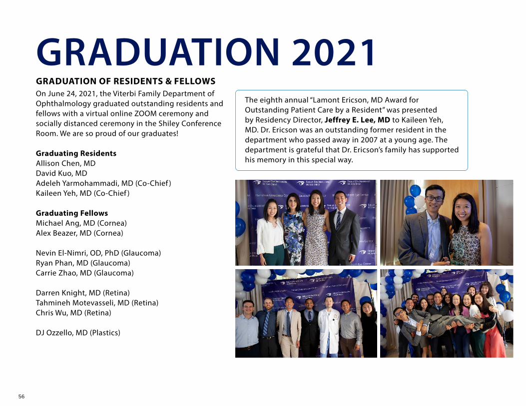
56
GRADUATION 2021GRADUATION OF RESIDENTS & FELLOWSOn June 24, 2021, the Viterbi Family Department of Ophthalmology graduated outstanding residents and fellows with a virtual online ZOOM ceremony and socially distanced ceremony in the Shiley Conference Room. We are so proud of our graduates!
Graduating ResidentsAllison Chen, MDDavid Kuo, MDAdeleh Yarmohammadi, MD (Co-Chief )Kaileen Yeh, MD (Co-Chief )
Graduating FellowsMichael Ang, MD (Cornea)Alex Beazer, MD (Cornea)
Nevin El-Nimri, OD, PhD (Glaucoma)Ryan Phan, MD (Glaucoma)Carrie Zhao, MD (Glaucoma)
Darren Knight, MD (Retina)Tahmineh Motevasseli, MD (Retina)Chris Wu, MD (Retina)
DJ Ozzello, MD (Plastics)
The eighth annual “Lamont Ericson, MD Award for Outstanding Patient Care by a Resident” was presented by Residency Director, Jeffrey E. Lee, MD to Kaileen Yeh, MD. Dr. Ericson was an outstanding former resident in the department who passed away in 2007 at a young age. The department is grateful that Dr. Ericson’s family has supported his memory in this special way.

57
Award for Outstanding Surgical Teaching
2021 Christopher W. Heichel, MD2020 Jeffrey E. Lee, MD2019 Weldon Haw, MD 2018 Christopher W. Heichel, MD2017 Jeffrey E. Lee, MD2016 Weldon Haw, MD
Award for Teaching by a Resident Rounds or Didactics (Formerly Resident Teaching Award)
2021 Allison J. Chen, MD, MPH2021 Kaileen Yeh, MD2020 James T. Walsh MD, PhD2019 Ziyong Yang, MD, PhD2018 Amir Marvasti, MD2017 Kyle Godfrey, MD2016 Abigail E. Huang, MD
Award for Fellow Appreciation 2021 Darren Knight, MD2020 Daniel James (DJ) Ozzello, MD2019 Doran Spencer, MD, PhD2018 Tyler Ofstad, MD 2017 Audrey Ko, MD2017 Joseph Ho, MD2016 Frank F. Tsai, MD
Award for Outstanding Clinical Teaching
2021 Lanning Kline, MD2020 Henry A. Ferreyra, MD2020 Derek S. Welsbie, MD, PhD2019 Shira Robbins, MD2018 Andrew Camp, MD, PhD2017 Henry A. Ferreyra, MD2016 Christopher W. Heichel, MD
Knowledge & Academic Performance 2021 Lingling Huang, MD, PhD2020 James T. Walsh, MD, PhD2019 James T. Walsh, MD, PhD2018 Zhiyong Yang, MD, PhD2017 Amir Marvasti, MD2016 Brian Chang, MD
Award for OKAP Teaching2021 Jiun Do, MD, PhD2020 Daniel James Ozzello, MD2019 Don Kikkawa, MD2018 Bobby S. Korn, MD, PhD, FACS2018 Peter Savino, MD2017 Derek S. Welsbie, MD2016 Bobby S. Korn, MD, PhD, FACS
Inaugural Residency Leadership Award 2016 Robert N. Weinreb, MD
Outstanding Volunteer Faculty Teaching Award
2019 Scott K. McClatchey, MD2018 Arthur C. Perry, MD2017 Sirini S. Iyengar, MD, FACS2016 Ray Gariano, MD
Award for Teaching Excellence 2021 Tony Ly2020 Erika C. Acera, OC(C)
Lamont Ericson, MD Award2021 Kaileen Yeh, MD2020 Heather Chen, MD2019 Jeffrey Wang, MD2018 Sally Baxter, MD, MSc2017 Sally Baxter, MD, Msc2016 Abigail E. Huang, MD
Whitehill Award2021 Andrew Camp, MD2020 Catherine Liu, MD, PhD2019 Daniel Chao, MD2018 Andrew Camp, MD2017 Derek S. Welsbie, MD
GRADUATION AWARDS
ASCRS – Resident Excellence Award Shiley Chief Resident Liane Dallalzadeh, MD was awarded the 2021 Resident Excellence Award by the American Society of Cataract and Refractive Surgery (ASCRS).
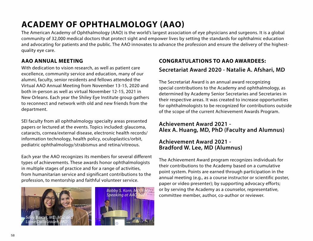
58
ACADEMY OF OPHTHALMOLOGY (AAO)
With dedication to vision research, as well as patient care excellence, community service and education, many of our alumni, faculty, senior residents and fellows attended the Virtual AAO Annual Meeting from November 13-15, 2020 and both in-person as well as virtual November 12-15, 2021 in New Orleans. Each year the Shiley Eye Institute group gathers to reconnect and network with old and new friends from the department.
SEI faculty from all ophthalmology specialty areas presented papers or lectured at the events. Topics included: glaucoma, cataracts, cornea/external disease, electronic health records/information technology, health policy, oculoplastics/orbit, pediatric ophthalmology/strabismus and retina/vitreous.
Each year the AAO recognizes its members for several different types of achievements. These awards honor ophthalmologists in multiple stages of practice and for a range of activities, from humanitarian service and significant contributions to the profession, to mentorship and faithful volunteer service.
CONGRATULATIONS TO AAO AWARDEES:
Achievement Award 2021 - Alex A. Huang, MD, PhD (Faculty and Alumnus)
Achievement Award 2021 - Bradford W. Lee, MD (Alumnus)
The Achievement Award program recognizes individuals for their contributions to the Academy based on a cumulative point system. Points are earned through participation in the annual meeting (e.g., as a course instructor or scientific poster, paper or video presenter); by supporting advocacy efforts; or by serving the Academy as a counselor, representative, committee member, author, co-author or reviewer.
Sally Baxter, MD, MSc andLiane Dallalzadeh, MD
Bobby S. Korn, MD, PhDSpeaking at AAO 2021
AAO ANNUAL MEETING
Secretariat Award 2020 - Natalie A. Afshari, MD
The Secretariat Award is an annual award recognizing special contributions to the Academy and ophthalmology, as determined by Academy Senior Secretaries and Secretaries in their respective areas. It was created to increase opportunities for ophthalmologists to be recognized for contributions outside of the scope of the current Achievement Awards Program.
The American Academy of Ophthalmology (AAO) is the world’s largest association of eye physicians and surgeons. It is a global community of 32,000 medical doctors that protect sight and empower lives by setting the standards for ophthalmic education and advocating for patients and the public. The AAO innovates to advance the profession and ensure the delivery of the highest-quality eye care.
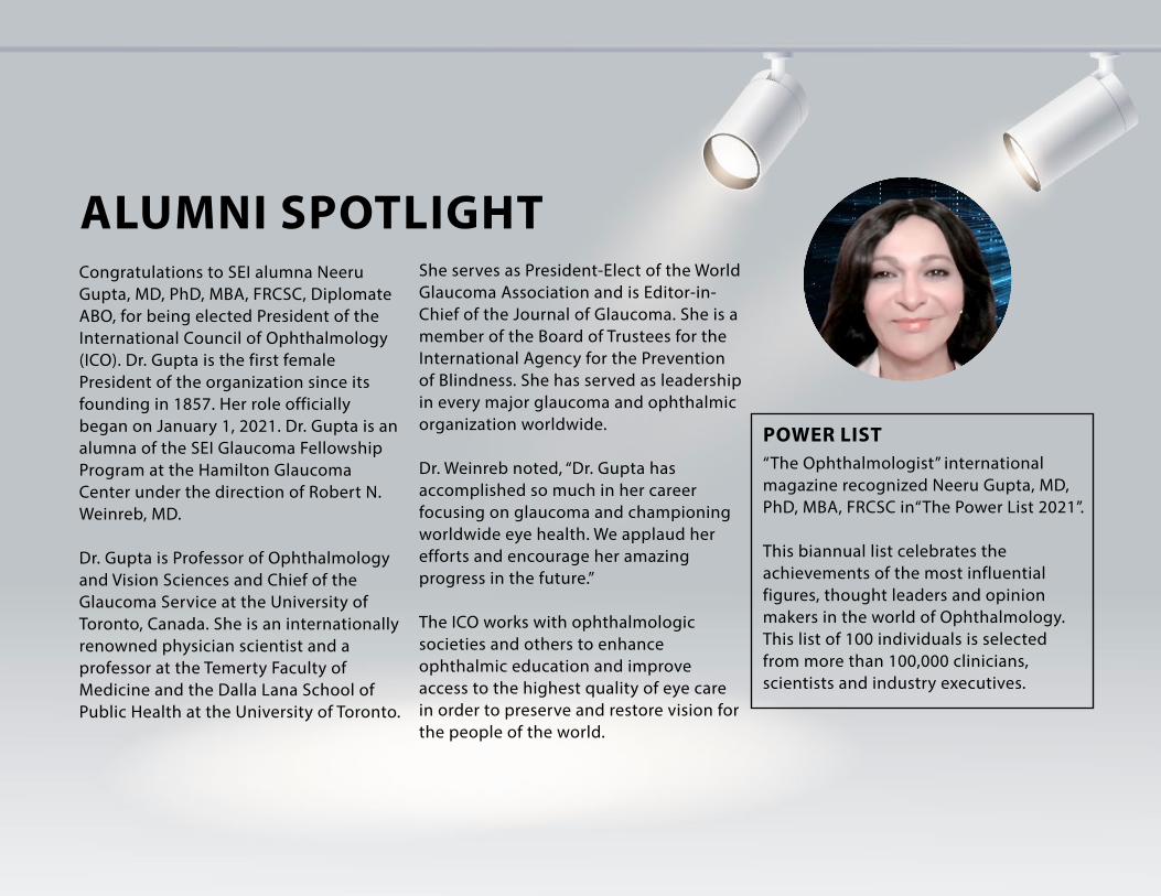
59
Congratulations to SEI alumna Neeru Gupta, MD, PhD, MBA, FRCSC, Diplomate ABO, for being elected President of the International Council of Ophthalmology (ICO). Dr. Gupta is the first female President of the organization since its founding in 1857. Her role officially began on January 1, 2021. Dr. Gupta is an alumna of the SEI Glaucoma Fellowship Program at the Hamilton Glaucoma Center under the direction of Robert N. Weinreb, MD.
Dr. Gupta is Professor of Ophthalmology and Vision Sciences and Chief of the Glaucoma Service at the University of Toronto, Canada. She is an internationally renowned physician scientist and a professor at the Temerty Faculty of Medicine and the Dalla Lana School of Public Health at the University of Toronto.
She serves as President-Elect of the World Glaucoma Association and is Editor-in-Chief of the Journal of Glaucoma. She is a member of the Board of Trustees for the International Agency for the Prevention of Blindness. She has served as leadership in every major glaucoma and ophthalmic organization worldwide.
Dr. Weinreb noted, “Dr. Gupta has accomplished so much in her career focusing on glaucoma and championing worldwide eye health. We applaud her efforts and encourage her amazing progress in the future.”
The ICO works with ophthalmologic societies and others to enhance ophthalmic education and improve access to the highest quality of eye care in order to preserve and restore vision for the people of the world.
“The Ophthalmologist” international magazine recognized Neeru Gupta, MD, PhD, MBA, FRCSC in“The Power List 2021”.
This biannual list celebrates the achievements of the most influential figures, thought leaders and opinion makers in the world of Ophthalmology. This list of 100 individuals is selected from more than 100,000 clinicians, scientists and industry executives.
POWER LIST
ALUMNI SPOTLIGHT
60
VIRTUAL GRAND ROUNDSThe community and SEI alumni are also invited to the departmental weekly Grand Rounds on Monday afternoon. The Grand Rounds consist of a special lecture from a prominent physician scientist from around the world and case presentations with moderated discussion. Interesting eye diseases, treatment dilemmas and surgical challenges are often the theme. Due to the COVID-19 pandemic, the Grand Rounds became virtual to follow safety guidelines. CME credits are given for this event as well.
September 14, 2020Moderated by Robert N. Weinreb, MD and William Freeman, MD GUEST LECTURER: EMILY Y. CHEW, MDDirector of Division of Epidemiology and Clinical Applications,National Eye Institute/National Institutes of HealthTITLE: “Age-Related Macular Degeneration: The Update of the Age-Related Eye Disease Studies (AREDS/AREDS2) 10 Year Follow-Up”
CASE PRESENTATIONSLingling Huang, MD, PhDPGY-3 ResidentTITLE: “Can We Treat Geographic Atrophy?”
Alexandra Warter, MDRetina FellowTITLE: “Combined Anti-VEGF and Steroid Therapy in DME (Diabetic Macular Edema)”
September 21, 2020Moderated by Robert N. Weinreb, MDGUEST LECTURER: ARTHUR J. SIT, MDConsultant, Department of OphthalmologyPractice Chair, Department of OphthalmologyMayo ClinicRochestor, MinnesotaTITLE: “Understanding Glaucoma Therapy through Aqueous Humor Dynamics”
CASE PRESENTATIONMark Lin, MDPGY-3 ResidentTITLE: “Nearly Glaucoma?”
October 5, 2020Moderated by Robert N. Weinreb, MDGUEST LECTURER: IVAN MAYNART TAVARES, MD, PHDProfessor and ChairDept. of Ophthalmology and Visual Sciences
Paulista School of Medicine -Univ Federal Sao Paulo (UNIFESP)Glaucoma Fellow - UCSD (2005-2006)TITLE: “A Clinical Case about Iris Cysts. Introduction to the UNIFESP Dept Ophthalmology”CASE PRESENTATIONSMichael J. Ang, MDCornea & Refractive FellowTITLE: “Lumps and Bumps”
Yue “Carrie” Zhao, MDGlaucoma FellowTITLE: “What Next?”
October 19, 2020Moderated by Robert N. Weinreb, MD and Peter J. Savino, MDGUEST LECTURER: NEIL R. MILLER, MDFrank B. Walsh Professor of Neuro-ophthalmologyProfessor of OphthalmologyWilmer Eye Institute
EDUCATION PHYSICIANS
61
Johns Hopkins University School of MedicineTITLE: “The Relative Afferent Defect in Clinical Practice”
CASE PRESENTATIONSAdeleh Yarmohammadi, MDPGY-4 ResidentTITLE: “To Shunt or Fenestrate, That is the Question”David Kuo, MDPGY-4 ResidentTITLE: “Occam v Hickam”
October 26, 2020Moderated by Robert N. Weinreb, MD and Don O. Kikkawa, MDGUEST LECTURER: KYLE J. GODFREY, MDOrbital and Oculofacial Plastic SurgeryAssistant Professor of OphthalmologyAssociate Director, Ophthalmology ResidencyWeill Cornell Medical CollegeNew York Presbyterian HospitalTITLE: “Stop the Postop Drop: Understanding and Preventing Postoperative Ptosis”
CASE PRESENTATIONSLiane Dallalzadeh, MDPGY-3 ResidentTITLE: “Fix One, But Which One?”
Aimee Chang, MDPGY-3 ResidentTITLE: “Aiming Higher”
November 2, 2020Moderated by Robert N. Weinreb, MD and David B. Granet, MDGUEST LECTURER: JESSE L. BERRY, MDAssociate Professor of Ophthalmology, Clinical ScholarAssociate Director, The Vision CenterChildren’s Hospital Los Angeles & The USC Roski Eye InstituteUSC Keck School of MedicineTITLE: “Innovations and Updates in the Management of Retinablastoma”
CASE PRESENTATIONAimee Chang, MDPGY-3 ResidentTITLE: “Small Eye, Big Problems”
November 9, 2020Moderated by Robert N. Weinreb, MD and Natalie A. Afshari, MDGUEST LECTURER: ALI DJALILIAN, MDProfessor of OphthalmologyCornea ServiceUniversity of Illinois College of MedicineDepartment of OphthalmologyTITLE: “Diagnosis and Medical Management of Limbal Stem Cell Deficiency in the Community”
CASE PRESENTATIONSAllison Chen, MDPGY-4 ResidentTITLE: “And Then There Was Light”
Michael Ang, MDCornea & Refractive FellowTITLE: “Lumps and Bumps”
November 23, 2020Moderated by Robert N. Weinreb, MD and Michael H. Goldbaum, MDGUEST LECTURER: LEE JAMPOL, MDNorthwestern Medicine Department of OphthalmologyTITLE: “DRCR Retina Network: Past”
CASE PRESENTATIONSLingling Huang, MD, PhDPGY-3 ResidentTITLE: “Risks vs. Benefits”
Alexandria Li, MDRetina FellowTITLE: “Mystery Swelling”
November 30, 2020Moderated by Robert N. Weinreb, MDGUEST LECTURER: CHRIS GIRKIN, MDChair, University of Alabama at Birmingham (UAB)Department of OphthalmologyChief Medical Officer, Callahan Eye Hospital

62
TITLE: “What can Imaging of the Deep Optic Nerve Head Tell Us About Glaucoma?”
CASE PRESENTATIONSKaileen Yeh, MDPGY-4 ResidentTITLE: “Use It or Lose It”
Ryan Phan, MDGlaucoma FellowTITLE: “After Trab and Tube”
January 4, 2021Moderated by Robert N. Weinreb, MDGUEST LECTURER: CHERYL ANDERSON, PHD, MPH, MSProfessor and Dean,University of California San Diego Herbert Wertheim School of PublicHealth and Human Longevity ScienceTITLE: “Advancing Health Equity and Justice: The Promise of Public Health Partnerships”
CASE PRESENTATIONLiane Dallalzadeh, MDPGY3 ResidentTITLE: “Through Thick and Thin”
January 11, 2021Moderated by Robert N. Weinreb, MD and Doran B. Spencer, MD, PhD
GUEST LECTURER: JENNIFER THORNE, MD, PHDChief, Division of Ocular ImmunologyCross Family Professor of Ophthalmology & EpidemiologyWilmer Eye Institute - Johns Hopkins MedicineTITLE: “Use of Systemic Corticosteroids and Immunosuppressive Agents in the Treatment of Uveitis”
CASE PRESENTATIONSMedi Eslani, MDPGY-2 ResidentTITLE: “Pick Your Poison”Darren Knight, MDRetina FellowTITLE: “The other C(O)VID”
January 25, 2021Moderated by Robert N. Weinreb, MDGUEST LECTURER: MICHELLE TARVER, MD, PHDActing Deputy Director,Office of Strategic Partnerships and Technology InnovationDirector, Patient Science and Engagement, Program Director forPatient Science, Digital Health Center of ExcellenceFood and Drug AdministrationTITLE: “Integrating Patient Perspectives in Medical Device Innovation”
CASE PRESENTATIONSAllison Chen, MDPGY-4 ResidentTITLE: “And Then There Were None”
Kaileen Yeh, MDPGY-4 ResidentTITLE: “When Friend Becomes Foe”
February 1, 2021Moderated by Robert N. Weinreb, MDGUEST LECTURER: ROBERT T. “CHIP” SCHOOLEY, MDInterim Faculty Director, Global EducationSenior Director, International InitiativesProfessor of Medicine, Division of Infectious Diseasesand Global Public HealthCo-Director, Center for Innovative Phage Applicationsand TherapeuticsUC San DiegoTITLE: “COVID-19: The End of the Beginning”
CASE PRESENTATIONSAimee Chang, MDPGY-3 ResidentTITLE: “More than the Common Cold”
Liane Dallalzadeh, MDPGY-3 ResidentTITLE: “Right Under Our Noses“

63
February 22, 2021Moderated by Robert N. Weinreb, MD and William Freeman, MDGUEST LECTURER: J. FERNANDO AREVALO, MD, PHD, FACSEdmund F. and Virginia B. Ball Professor of OphthalmologyChairman, Department of OphthalmologyJohns Hopkins Bayview Medical CenterRetina Division, Wilmer Eye InstituteThe Johns Hopkins University School of MedicineBaltimore, MD, USATITLE: “Lessons Learned from PACORES in PDR Management: Real World Data fromLatin America & Spain”
CASE PRESENTATIONSMark Lin, MDPGY-3 ResidentTITLE: “Recurrent CSCR”
Darren Knight, MDRetina & Vitreous FellowTITLE: “An Approach to Recurrent Macular Holes “
March 22, 2021Moderated by Robert N. Weinreb, MDGUEST LECTURER: LEON W. HERNDON, JR., MDChief of GlaucomaProfessor of OphthalmologyDuke Ophthalmology
Duke University School of MedicineTITLE: “Inequity in Glaucoma Services Costs Sight”
CASE PRESENTATIONSLingling Huang, MD, PhDPGY-3 Resident
Yue “Carrie” Zhao, MDGlaucoma Fellow
March 29, 2021Moderated by Robert N. Weinreb, MD and William Freeman, MDGUEST LECTURER: GLENN J. JAFFEE, MDRobert Machemer Professor of OphthalmologyChief, Vitreoretinal DivisionDirector, Duke Reading CenterDuke Eye CenterTITLE: “The Age-related Macular Degeneration Treatment Trials - Are the Lessons Learned Still Useful in 2021?”
CASE PRESENTATIONSChris Wu, MDRetina FellowTITLE: “A White Macula After Pars Plana Vitrectomy”
Alexandra Warter, MDRetina Fellow
TITLE: “Combined Anti-VEGF and Steroid Therapy in Resistant Wet AMD (Wet Age-related Macular Degeneration)”
April 19, 2021Moderated by Robert N. Weinreb, MD and Natalie A. Afshari, MDGUEST LECTURER: JENNIFER LI, MDDirector, Cornea and External Disease ServiceProfessor, Department of OphthalmologyUC Davis Medical CenterTITLE: “The Current State of Eye Banking and the Impact of COVID-19”
CASE PRESENTATIONAlex Beazer, MDCornea & Refractive FellowTITLE: “Corneal White Dot Syndrome”
April 26, 2021Moderated by Robert N. Weinreb, MD and Eric Nudleman, MD, PhDGUEST LECTURER: COLE FERGUSON, MD, PHDAssistant ProfessorDepartment of PathologyUniversity of California, San DiegoTITLE: “The Diagnosis and Management of Incidental Pigmented Lesions of the Choroid”

64
CASE PRESENTATIONAlexandria Li, MDRetina & Vitreous FellowTITLE: “Out of the Blue”
May 10, 2021Moderated by Robert N. Weinreb, MD and Peter J. Savino, MDGUEST LECTURER: M. TARIQ BHATTI, MDSenior Associate ConsultantNeuro-OphthalmologyProfessor of Ophthalmology and NeurologyMayo Clinic College Of MedicineTITLE: “Stem Cell Therapy for Optic Neuropathy: Real Science or Science Fiction?”
CASE PRESENTATIONSAllison Chen, MDPGY-4 ResidentTITLE: “Sometimes, Always, Never”David Kuo, MDPGY-4 ResidentTITLE: “Disc-y Business”
May 17, 2021Moderated by Robert N. Weinreb, MD and Shyamanga Borooah, MBBS, MRCP (UK), MRCSEd, FRCOphth, PhDGUEST LECTURER: AMIR H. KASHANI, MD, PHDAssociate Professor of Ophthalmology (PAR)
Wilmer Eye InstituteJohns Hopkins School of MedicineTITLE: “Revisiting the Eye-Brain Connection Using Optical Coherence Tomography Angiography”
CASE PRESENTATIONSAlexandria Li, MDRetina & Vitreous FellowTITLE: “Lord of the Eyes”
Darren Knight, MDRetina & Vitreous FellowTITLE: “Clinical Findings in COL18A1 Related Chorioretinal Degeneration”
May 24, 2021Moderated by Robert N. Weinreb, MD and David B. Granet, MDGUEST LECTURER: STEPHEN KRAFT, MDStaff Ophthalmologist, Depts. of Ophthalmology and Vision Sciences at University HealthNetwork and SickKids Hospital, Toronto, Professor, Dept. Ophthalmology and Vision Sciences,Temerty Faculty of Medicine, University of TorontoTITLE: “The Functional Benefits of Adult Strabismus Surgery: It Is Not Cosmetic!”
CASE PRESENTATIONAimee Chang, MDPGY-3 ResidentTITLE: “A Helping Hand”
The Vision Research Lecture Series addresses the latest advances in vision science and clinical ophthalmology. Each presentation features UC San Diego Department of Ophthalmology’s faculty, as well as a selection of leading vision scientists from around the globe. These lectures are held in the Shiley Eye Institute Education Center.
VISION RESEARCH LECTURES
April 22, 2021Sayantan Datta, BSc, MSc, PhDAssistant Professor of Ophthalmology Emory Eye CenterTITLE: “Identification of Novel Mitochondrial Retrograde Signaling Pathway that Affects RPE Structure and Function”
June 24, 2021Debasish Sinha, PhDProfessor of Ophthalmology, Cell Biology and Developmental BiologyUniversity of Pittsburgh School of MedicineAdjunct Faculty, Ophthalmology, The John’s Hopkins University School of MedicineTITLE: “The Potential Role of Neutrophils and NK Cells in the Pathogenesis of Atrophic AMD”

65
2021 OPHTHALMOLOGY UPDATEChaired by Don O. Kikkawa, MD and Robert N. Weinreb, MD, the 2021 Ophthalmology Update, sponsored by the Viterbi Family Department of Ophthalmology and the Shiley Eye Institute at UC San Diego, was held virtually on February 12-13, 2021. Don O. Kikkawa, MD, Robert N. Weinreb, MD, Natalie Afshari, MD and William Freeman, MD served as Program Moderators. The interdisciplinary faculty of ophthalmic subspecialists reviewed the continuing progress, latest surgical techniques, innovative ideas and cutting-edge translational research in ophthalmology. The special guest speaker was Robert T. “Chip” Schooley, MD who discussed “COVID-19: An Evolving Virus in an Evolving Epidemic – Implications for Vaccine Effectiveness.”
William R. Freeman, MD and Michael Goldbaum, MD, Co-Directors of the Retina Division, host Tuesday morning Retina Case Reviews at the Joan and Irwn Jacobs Retina Center for the Shiley Eye Institute (SEI) retina faculty, residents, fellows and alumni to discuss complex cases and new treatments.
RETINA CONFERENCE

66
FIRST GLOBAL WEBINAR OF THE WORLD GLAUCOMA ASSOCIATION ON GLAUCOMA SURGERYThe first global webinar of the World Glaucoma Association on glaucoma surgery was moderated by Robert N. Weinreb, MD on October 10, 2020, garnering over 9,000 live views from 137 different countries.
FACULTY FEATURED LECTURES
GLAUCOMA RESEARCH FOUNDATION PRESENTS AN INNOVATIONS IN GLAUCOMA WEBINAR: “UNDERSTANDING VISION RESTORATION”January 22, 2021, Derek Welsbie, MD, PhD presented his newest research on vision restoration during an Innovations in Glaucoma Webinar: “Understanding Vision Restoration” hosted by the Glaucoma Research Foundation. He is a principle investigator in the Catalyst for a Cure Vision Restoration Initiative.
BRAILLE INSTITUTEDavid Granet, MD was featured at the Low Vision Seminar hosted by the Braille Institute discussing Pediatric Low Vision on Thursday, February 4, 2021. Shyamanga Borooah, MBBS, PhD, discussed the latest research involving stem cells, with a focus on technologies that are at or near clinical trial research, to treat sight loss on February 25, 2021 at the Braille Institute.
“ASK THE EXPERTS” VIRTUAL WORKSHOPDavid Granet, MD spoke at “Ask the Experts” Virtual Workshop on February 10, 2021 on “Eyes on Screens: Maintaining Your Kids’ Ocular Health in a Digital World.”
DATA SCIENCE AND OPHTHALMOLOGY MIXERThis was a collaboration with the Viterbi Family Department of Ophthalmology and the UC San Diego Halıcıoğlu Data Science Institute (HDSI) on Friday May 14, 2021. It was a special HDSI Faculty Seminar featuring SEI faculty guest speakers for lightning talks on interdisciplinary research in ophthalmology and data science.
10TH ANNUAL “FOCUS ON EYE HEALTH NATIONAL SUMMIT” BY PREVENT BLINDNESSSally Baxter, MD, MSc lectured at the 10th Annual “Focus on Eye Health National Summit” hosted by Prevent Blindness on Thursday July 15, 2021.
10th Annual “Focus on Eye Health National Summit” by Prevent Blindness
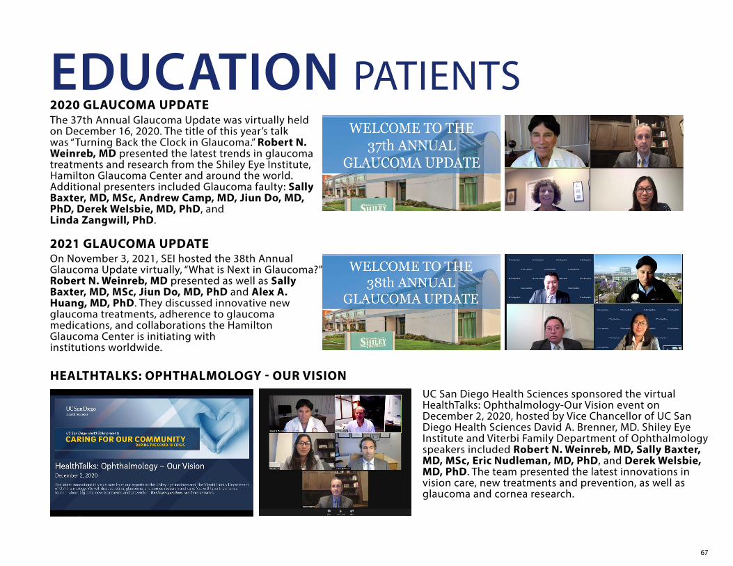
67
2020 GLAUCOMA UPDATE
On November 3, 2021, SEI hosted the 38th Annual Glaucoma Update virtually, “What is Next in Glaucoma?” Robert N. Weinreb, MD presented as well as Sally Baxter, MD, MSc, Jiun Do, MD, PhD and Alex A. Huang, MD, PhD. They discussed innovative new glaucoma treatments, adherence to glaucoma medications, and collaborations the Hamilton Glaucoma Center is initiating with institutions worldwide.
HEALTHTALKS: OPHTHALMOLOGY - OUR VISION
EDUCATION PATIENTS
UC San Diego Health Sciences sponsored the virtual HealthTalks: Ophthalmology-Our Vision event on December 2, 2020, hosted by Vice Chancellor of UC San Diego Health Sciences David A. Brenner, MD. Shiley Eye Institute and Viterbi Family Department of Ophthalmology speakers included Robert N. Weinreb, MD, Sally Baxter, MD, MSc, Eric Nudleman, MD, PhD, and Derek Welsbie, MD, PhD. The team presented the latest innovations in vision care, new treatments and prevention, as well as glaucoma and cornea research.
2021 GLAUCOMA UPDATE
The 37th Annual Glaucoma Update was virtually held on December 16, 2020. The title of this year’s talk was “Turning Back the Clock in Glaucoma.” Robert N. Weinreb, MD presented the latest trends in glaucoma treatments and research from the Shiley Eye Institute, Hamilton Glaucoma Center and around the world. Additional presenters included Glaucoma faulty: Sally Baxter, MD, MSc, Andrew Camp, MD, Jiun Do, MD, PhD, Derek Welsbie, MD, PhD, and Linda Zangwill, PhD.

68
PUBLICATIONS 2020-2021CORNEAEditorial: Surgical Advancements and Innovations in Cataract Surgery for the Complex Patient. Gali HE, Afshari NA. Curr Opin Ophthalmol. 2020 Jan;31(1):1-2. doi: 10.1097/ICU.0000000000000638. PMID: 31724967 No abstract available.
Iris Reconstruction Suturing Techniques. Lian RR, Siepser SB, Afshari NA. Curr Opin Ophthalmol. 2020 Jan;31(1):43-49. doi: 10.1097/ICU.0000000000000628. PMID: 31770162 Review.
The Quest for Homeopathic and Nonsurgical Cataract Treatment. Lian RR, Afshari NA. Curr Opin Ophthalmol. 2020 Jan;31(1):61-66. doi: 10.1097/ICU.0000000000000631. PMID: 31770163 Review.
Hand Hygiene and Instrument Sanitization in Ophthalmology Clinics. Abbas AA, Lian RR, Afshari NA. Curr Opin Ophthalmol. 2020 Jan;31(1):28-32. doi: 10.1097/ICU.0000000000000630. PMID: 31770164 Review.
Accuracy of IOL Power Calculations in the Very Elderly. Sella R, Chou L, Schuster AK, Gali HE, Weinreb RN, Afshari NA. Eye (Lond). 2020 Oct;34(10):1848-1855. doi: 10.1038/s41433-019-0752-0. Epub 2020 Jan 13. PMID: 31932707 Free PMC article.
Dupilumab-Associated Mucin Deficiency (DAMD). Barnett BP, Afshari NA. Transl Vis Sci Technol. 2020 Feb 25;9(3):29. doi: 10.1167/tvst.9.3.29. PMID: 32742759 Free PMC article.
Intraocular Lens Power Calculation in the Elderly Population Using the Kane Formula in Comparison with Existing Methods. Reitblat O, Gali HE, Chou L, Bahar I, Weinreb RN, Afshari NA, Sella R. J Cataract Refract Surg. 2020 Nov;46(11):1501-1507. doi: 10.1097/j.jcrs.0000000000000308. PMID: 33149066
Cultivating the Physician-Patient Relationship in Ophthalmology. Afshari NA, Lian RR. Am J Ophthalmol. 2021 Mar;223:A1-A3. doi: 10.1016/j.ajo.2020.11.009. Epub 2020 Nov 27. PMID: 33249053 No abstract available.
Editorial: Advances in Cataract Surgery Planning, Methods, and Outcomes in the Pandemic Era. Hakim DF, Afshari NA. Curr Opin Ophthalmol. 2021 Jan;32(1):1-2. doi: 10.1097/ICU.0000000000000721. PMID: 33278115 No abstract available.
Cataract and Systemic Disease: A Review. Ang MJ, Afshari NA. Clin Exp Ophthalmol. 2021 Mar;49(2):118-127. doi: 10.1111/ceo.13892. Epub 2021 Jan 10. PMID: 33426783 Review.
Disease Expression and Familial Transmission of Fuchs Endothelial Corneal Dystrophy with and without CTG18.1 Expansion. Xu TT, Li YJ, Afshari NA, Aleff RA, Rinkoski TA, Patel SV, Maguire LJ, Edwards AO, Brown WL, Fautsch MP, Wieben ED, Baratz KH. Invest Ophthalmol Vis Sci. 2021 Jan 4;62(1):17. doi: 10.1167/iovs.62.1.17. PMID: 33444430 Free PMC article.
August consultation #7. Afshari NA. J Cataract Refract Surg. 2021 Aug 1;47(8):1100. doi: 10.1097/01.j.jcrs.0000769512.47571.ee. PMID: 34292900 No abstract available.
Intraoperative Management of Zonular Weakness. Barnett, Brad P, Heichel CW In: Duke Manual of Corneal and Cataract Surgery. 1st Edition. Wolters Kluwer. (2021)
GLAUCOMADeep Learning Approaches Predict Glaucomatous Visual Field Damage from OCT Optic Nerve Head En Face Images and Retinal Nerve Fiber Layer Thickness Maps. Christopher M, Bowd C, Belghith A, Goldbaum MH, Weinreb RN, Fazio MA, Girkin CA, Liebmann JM, Zangwill LM. Ophthalmology. 2020 Mar;127(3):346-356. doi: 10.1016/j.ophtha.2019.09.036. Epub 2019 Sep PMID: 31718841
Genetically Encodable Contrast Agents for Optical Coherence Tomography. Lu GJ, Chou LD, Malounda D, Patel AK, Welsbie DS, Chao DL, Ramalingam T, Shapiro MG. ACS Nano. 2020 Jul 28;14(7):7823-7831. doi: 10.1021/acsnano.9b08432. Epub 2020 Feb 10. PMID: 32023037 Free PMC article.
Novel Mutations in LTBP2 Identified in Familial Cases of Primary Congenital Glaucoma. Rauf B, Irum B, Khan SY, Kabir F, Naeem MA, Riazuddin S, Ayyagari R, Riazuddin SA. Mol Vis. 2020 Feb 24;26:14-25. eCollection 2020. PMID: 32165823
Gradient-Boosting Classifiers Combining Vessel Density and Tissue Thickness Measurements for Classifying Early to Moderate Glaucoma. Bowd C, Belghith A, Proudfoot JA, Zangwill LM, Christopher M, Goldbaum MH, Hou H, Penteado RC, Moghimi S, Weinreb RN. Am J Ophthalmol. 2020 Sep;217:131-139. doi: 10.1016/j.ajo.2020.03.024. Epub 2020 Mar 25. PMID: 32222368
Effects of Study Population, Labeling and Training on Glaucoma Detection Using Deep Learning Algorithms. Christopher M, Nakahara K, Bowd C, Proudfoot JA, Belghith A, Goldbaum MH, Rezapour J, Weinreb RN, Fazio MA, Girkin CA, Liebmann JM, De Moraes G, Murata H, Tokumo K, Shibata N, Fujino Y, Matsuura M, Kiuchi Y, Tanito M, Asaoka R, Zangwill LM. Transl Vis Sci Technol. 2020 Apr 28;9(2):27. doi: 10.1167/tvst.9.2.27. eCollection 2020 Apr. PMID: 32818088
Impact of Pupil Dilation on Optical Coherence Tomography Angiography Retinal Microvasculature in Healthy Eyes. Villatoro G, Bowd C, Proudfoot JA, Manalastas PIC, Nguyen KD, Hou H, Penteado RC, Li AJ, Moghimi S, Ghahari E, Weinreb RN, Zangwill LM. J Glaucoma. 2020 Nov;29(11):1025-1029. doi: 10.1097/IJG.0000000000001647. PMID: 32890106
Central Visual Field Defects in Patients with Distinct Glaucomatous Optic Disc Phenotypes. Ekici E, Moghimi S, Hou H, Proudfoot J, Zangwill LM, Do JL, Oh WH, Kamalipour A, Liebmann JM, De Moraes CG, Girkin CA, El-Nimri N, Weinreb RN. Am J Ophthalmol. 2021 Mar;223:229-240. doi: 10.1016/j.ajo.2020.10.015. Epub 2020 Oct 29. PMID: 33129812
Investigation of Associations between Piezo1 Mechanoreceptor Gain-of-function Variants and Glaucoma-related Phenotypes in Humans and Mice. Baxter SL, Keenan WT, Athanas AJ, Proudfoot JA, Zangwill LM, Ayyagari R, Liebmann JM, Girkin CA, Patapoutian A, Weinreb RN. Sci Rep. 2020 Nov 4;10(1):19013. doi: 10.1038/s41598-020-76026-0. PMID: 33149214
Agreement between 10-2 and 24-2C Visual Field Test Protocols for Detecting Glaucomatous Central Visual Field Defects. Chakravarti T, Moghadam M, Proudfoot JA, Weinreb RN, Bowd C, Zangwill LM. J Glaucoma. 2021 Jun 1;30(6):e285-e291. doi: 10.1097/IJG.0000000000001844. PMID: 33813563
OCT Angiography Artifacts in Glaucoma. Kamalipour A, Moghimi S, Hou H, Penteado RC, Oh WH, Proudfoot JA, El-Nimri N, Ekici E, Rezapour J, Zangwill LM, Bowd C, Weinreb RN. Ophthalmology. 2021 Apr 2:S0161-6420(21)00241-4. doi: 10.1016/j.ophtha.2021.03.036. Online ahead of print. PMID: 33819524
Racial Differences in the Rate of Change in Anterior Lamina Cribrosa Surface Depth in the African Descent and Glaucoma Evaluation Study. Girkin CA, Belghith A, Bowd C, Medeiros FA, Weinreb RN, Liebmann JM, Proudfoot JA, Zangwill LM, Fazio MA. Invest Ophthalmol Vis Sci. 2021 Apr 1;62(4):12. doi: 10.1167/iovs.62.4.12. PMID: 33844828
The Influence of Axial Myopia on Optic Disc Characteristics of Glaucoma Eyes. Rezapour J, Bowd C, Dohleman J, Belghith A, Proudfoot JA, Christopher M, Hyman L, Jonas JB, Fazio MA, Weinreb RN, Zangwill LM. Sci Rep. 2021 Apr 23;11(1):8854. doi: 10.1038/s41598-021-88406-1. PMID: 33893383
Superficial and Deep Macula Vessel Density in Healthy, Glaucoma Suspect, and Glaucoma Eyes. El-Nimri NW, Manalastas PIC, Zangwill LM, Proudfoot JA, Bowd C, Hou H, Moghimi S, Penteado RC, Rezapour J, Ekici E, Shoji T, Ghahari E, Yarmohammadi A, Weinreb RN. J Glaucoma. 2021 Jun 1;30(6):e276-e284. doi: 10.1097/IJG.0000000000001860. PMID: 33899812
Deep Learning Estimation of 10-2 and 24-2 Visual Field Metrics Based on Thickness Maps from Macula OCT. Christopher M, Bowd C, Proudfoot JA, Belghith A, Goldbaum MH, Rezapour J, Fazio MA, Girkin CA, De Moraes G, Liebmann JM, Weinreb RN, Zangwill LM. Ophthalmology. 2021 Apr 23:S0161-6420(21)00316-X. doi: 10.1016/j.ophtha.2021.04.022. Online ahead of print. PMID: 33901527
Estimated Utility of the Short-term Assessment of Glaucoma Progression Model in Clinical Practice. Proudfoot JA, Zangwill LM, Moghimi S, Bowd C, Saunders LJ, Hou H, Belghith A, Medeiros FA, Williams-Steppe E, Acera T, Dirkes K, Weinreb R. JAMA Ophthalmol. 2021 Jun 10:e211812. doi: 10.1001/jamaophthalmol.2021.1812. Online ahead of print. PMID: 34110362
Individualized Glaucoma Change Detection Using Deep Learning Auto Encoder-Based Regions of Interest. Bowd C, Belghith A, Christopher M, Goldbaum MH, Fazio MA, Girkin CA, Liebmann JM, de Moraes CG, Weinreb RN, Zangwill LM. Transl Vis Sci Technol. 2021 Jul 1;10(8):19. doi: 10.1167/tvst.10.8.19. PMID: 34293095
69
Agreement between Compass Fundus Perimeter New Grid and 10-2 Testing Protocols for Detecting Central Visual Field Defects. El-Nimri NW, Penteado RC, Bowd C, Proudfoot JA, Hou H, Manalastas PIC, Ghahari E, Zangwill LM, Moghimi S, Weinreb RN. Ophthalmol Glaucoma. 2021 Jul 19:S2589-4196(21)00174-5. doi: 10.1016/j.ogla.2021.07.006. Online ahead of print. PMID: 34293492
Review of Glaucoma Medication Adherence Monitoring in the Digital Health Era. Erras A, Shahrvini B, Weinreb RN, Baxter SL. Br J Ophthalmol. 2021 Apr 15:bjophthalmol-2020-317918. doi: 10.1136/bjophthalmol-2020-317918. Online ahead of print. PMID: 33858837 Review.
Predictive Analytics for Glaucoma Using Data From the All of Us Research Program. Baxter SL, Saseendrakumar BR, Paul P, Kim J, Bonomi L, Kuo TT, Loperena R, Ratsimbazafy F, Boerwinkle E, Cicek M, Clark CR, Cohn E, Gebo K, Mayo K, Mockrin S, Schully SD, Ramirez A, Ohno-Machado L; All of Us Research Program Investigators. Am J Ophthalmol. 2021 Jul;227:74-86. doi: 10.1016/j.ajo.2021.01.008. Epub 2021 Jan 23. PMID: 33497675
Is Diabetes Mellitus a Blessing in Disguise for Primary Open-Angle Glaucoma? Hou H, Moghimi S, Baxter SL, Weinreb RN. J Glaucoma. 2021 Jan 1;30(1):1-4. doi: 10.1097/IJG.0000000000001719. PMID: 33074964 Free PMC article.
Standard Reliability and Gaze Tracking Metrics in Glaucoma and Glaucoma Suspects. Camp AS, Long CP, Patella VM, Proudfoot JA, Weinreb RN. Am J Ophthalmol. 2021 Jul 16:S0002-9394(21)00367-6. doi: 10.1016/j.ajo.2021.06.038. Online ahead of print. PMID: 34280366
Qualitative Evaluation of the 10-2 and 24-2 Visual Field Tests for Detecting Central Visual Field Abnormalities in Glaucoma. Orbach A, Ang GS, Camp AS, Welsbie DS, Medeiros FA, Girkin CA, Fazio MA, Oh WH, Weinreb RN, Zangwill LM, Wu Z. Am J Ophthalmol. 2021 Feb 21;229:26-33. doi: 10.1016/j.ajo.2021.02.015. Online ahead of print. PMID: 33626360
Characteristics of Central Visual Field Progression in Eyes with Optic Disc Hemorrhage. David RCC, Moghimi S, Do JL, Hou H, Proudfoot J, Zangwill LM, Kamalipour A, Nishida T, De Moraes CG, Girkin CA, Liebmann JM, Weinreb RN. Am J Ophthalmol. 2021 Jun 6;231:109-119. doi: 10.1016/j.ajo.2021.05.026. Online ahead of print. PMID: 34107310
Rates of Retinal Nerve Fiber Layer Thinning in Distinct Glaucomatous Optic Disc Phenotypes in Early Glaucoma. David RCC, Moghimi S, Ekici E, Do JL, Hou H, Proudfoot JA, Kamalipour A, Nishida T, Girkin CA, Liebmann JM, Weinreb RN. Am J Ophthalmol. 2021 Apr 25;229:8-17. doi: 10.1016/j.ajo.2021.04.010. Online ahead of print. PMID: 33910053
Branch Retinal Vein Occlusion Associated with Fingolimod Treatment for Multiple Sclerosis. Do JL, Patel VR. Can J Ophthalmol. 2021 Feb;56(1):e21-e22. doi: 10.1016/j.jcjo.2020.07.015. Epub 2020 Aug 14. PMID: 32805204 No abstract available.
A Randomized Controlled Trial Comparing Subconjunctival Injection to Direct Scleral Application of Mitomycin C in Trabeculectomy. Do JL, Xu BY, Wong B, Camp A, Ngai P,
Long C, Proudfoot J, Moghimi S, Yan D, Welsbie DS, Weinreb RN. Am J Ophthalmol. 2020 Dec;220:45-52. doi: 10.1016/j.ajo.2020.07.002. Epub 2020 Jul 6. PMID: 32645310 Clinical Trial.
Sheath-Preserving Optic Nerve Transection in Rats to Assess Axon Regeneration and Interventions Targeting the Retinal Ganglion Cell Axon. Do JL, Allahwerdy S, David RC, Weinreb RN, Welsbie DS. J Vis Exp. 2020 Sep 6;(163). doi: 10.3791/61748. PMID: 32955495
Pseudophakic Glaucoma and Angle Closure in a Patient with an Anterior Chamber Intraocular Lens. Chan AX, Yeh K, Bakhoum M, Do JL. Am J Ophthalmol Case Rep. 2020 Aug 16;20:100877. doi: 10.1016/j.ajoc.2020.100877. eCollection 2020 Dec. PMID: 32875158 Free PMC article.
Optic Nerve Engraftment of Neural Stem Cells. Do JL, Allahwerdy S, David RCC, Weinreb RN, Tuszynski MH, Welsbie DS. Invest Ophthalmol Vis Sci. 2021 Jul 1;62(9):30. doi: 10.1167/iovs.62.9.30. PMID: 34283208 Free PMC article.
Predicting Glaucoma before Onset Using Deep Learning. Thakur A, Goldbaum M, Yousefi S. Ophthalmol Glaucoma. 2020 Jul-Aug;3(4):262-268. doi: 10.1016/j.ogla.2020.04.012. Epub 2020 Apr 29. PMID: 33012331
Convex Representations Using Deep Archetypal Analysis for Predicting Glaucoma. Thakur A, Goldbaum M, Yousefi S. IEEE J Transl Eng Health Med. 2020 May 28;8:3800107. doi: 10.1109/JTEHM.2020.2982150. eCollection 2020. PMID: 32596065
Corrigendum to “Inhibition of cAMP/PKA Pathway Protects Optic Nerve Head Astrocytes against Oxidative Stress by Akt/Bax Phosphorylation-Mediated Mfn1/2 Oligomerization”. Ju WK, Shim MS, Kim KY, Park TL, Ahn S, Edwards G, Weinreb RN. Oxid Med Cell Longev. 2020 Sep 30;2020:9410289. doi: 10.1155/2020/9410289. eCollection 2020. PMID: 33082914 Free PMC article.
Effect of Ubiquinol on Glaucomatous Neurodegeneration and Oxidative Stress: Studies for Retinal Ganglion Cell Survival and/or Visual Function. Edwards G, Lee Y, Kim M, Bhanvadia S, Kim KY, Ju WK.Antioxidants (Basel). 2020 Oct 3;9(10):952. doi: 10.3390/antiox9100952.PMID: 33023026 Free PMC article.
AIBP Protects Retinal Ganglion Cells against Neuroinflammation and Mitochondrial Dysfunction in Glaucomatous Neurodegeneration. Choi SH, Kim KY, Perkins GA, Phan S, Edwards G, Xia Y, Kim J, Skowronska-Krawczyk D, Weinreb RN, Ellisman MH, Miller YI, Ju WK. Redox Biol. 2020 Oct;37:101703. doi: 10.1016/j.redox.2020.101703. Epub 2020 Aug 27. PMID: 32896719 Free PMC article.
cAMP/PKA pathway and mitochondrial protection in oxidative stress-induced optic nerve head astrocytes. Kim KY, Ju WK. Neural Regen Res. 2021 Jan;16(1):108-109. doi: 10.4103/1673-5374.286962. PMID: 32788457 Free PMC article. No abstract available.
Central Macular OCTA Parameters in Glaucoma. Nishida T, Oh WH, Moghimi S, Yarmohammadi A, Hou H, David RCC, Kamalipour A, Shoji T, El-Nimri N, Rezapour J, M Zangwill L, Weinreb RN. Br J Ophthalmol. 2021
Aug 23:bjophthalmol-2021-319574. doi: 10.1136/bjophthalmol-2021-319574. Online ahead of print. PMID: 34426401
Macular Optical Coherence Tomography Imaging in Glaucoma. Kamalipour A, Moghimi S. J Ophthalmic Vis Res. 2021 Jul 29;16(3):478-489. doi: 10.18502/jovr.v16i3.9442. eCollection 2021 Jul-Sep. PMID: 34394875 Free PMC article.
Macular Thickness and Microvasculature Loss in Glaucoma Suspect Eyes. Hou H, Moghimi S, Kamalipour A, Ekici E, Oh WH, Proudfoot JA, El-Nimri N, Penteado RC, Nishida T, David RC, Weinreb RN. Ophthalmol Glaucoma. 2021 Jul 30:S2589-4196(21)00180-0. doi: 10.1016/j.ogla.2021.07.009. Online ahead of print. PMID: 34339877
Optical Microangiography and Progressive Retinal Nerve Fiber Layer Loss in Primary Open Angle Glaucoma. Rao HL, Dasari S, Puttaiah NK, Pradhan ZS, Moghimi S, Mansouri K, Webers CA, Weinreb RN. Am J Ophthalmol. 2021 Jul 25:S0002-9394(21)00390-1. doi: 10.1016/j.ajo.2021.07.023. Online ahead of print. PMID:
Diurnal Variation of Retinal Vessel Density in Healthy Human Eyes. Wu JH, Penteado RC, Moghimi S, Zangwill LM, Proudfoot JA, Weinreb RN. J Glaucoma. 2021 Sep 1;30(9):820-826. doi: 10.1097/IJG.0000000000001903. PMID: 34172629
A Bibliometric and Mapping Analysis of Glaucoma Research between 1900 and 2019. López-Muñoz F, Weinreb RN, Moghimi S, Povedano-Montero FJ. Ophthalmol Glaucoma. 2021 May 31:S2589-4196(21)00140-X. doi: 10.1016/j.ogla.2021.05.008. Online ahead of print. PMID: 34082178
Response to Letter to the Editor: Optical Coherence Tomography Angiography and Visual Field Progression in Primary Angle Closure Glaucoma. Rao HL, Srinivasan T, Pradhan ZS, Sreenivasaiah S, Rao DAS, Puttaiah NK, Devi S, Moghimi S, Mansouri K, Webers CAB, Weinreb RN. J Glaucoma. 2021 Aug 1;30(8):e375-e376. doi: 10.1097/IJG.0000000000001871. PMID: 34008528 No abstract available.
Changes in Corneal Biomechanics and Glaucomatous Visual Field Loss. Chan E, Yeh K, Moghimi S, Proudfoot J, Liu X, Zangwill L, Weinreb RN. J Glaucoma. 2021 May 1;30(5):e246-e251. doi: 10.1097/IJG.0000000000001819. PMID: 33596020
Automated Evaluation of Parapapillary Choroidal Microvasculature in Pseudoexfoliation Syndrome and Pseudoexfoliation Glaucoma. Aghsaei Fard M, Safizadeh M, Shaabani A, Kafieh R, Hojati S, Afzali M, Suwan Y, Ritch R, Moghimi S. Am J Ophthalmol. 2021 Apr;224:178-184. doi: 10.1016/j.ajo.2020.12.002. Epub 2020 Dec 10. PMID: 33309810
Central-most Visual Field Defects in Early Glaucoma. Chakravarti T, Moghimi S, De Moraes CG, Weinreb RN. J Glaucoma. 2021 Mar 1;30(3):e68-e75. doi: 10.1097/IJG.0000000000001747. PMID: 33273288
Visual Field Artifacts in Glaucoma with Face Mask Use during the COVID-19 Pandemic. El-Nimri NW, Moghimi S, Fingeret M, Weinreb RN. J Glaucoma. 2020 Dec;29(12):1184-1188. doi: 10.1097/IJG.0000000000001706. PMID: 33116056

70
Nocturnal Variability of Intraocular Pressure Monitored with Contact Lens Sensor Is Associated with Visual Field Loss in Glaucoma. Yang Z, Mansouri K, Moghimi S, Weinreb RN. J Glaucoma. 2021 Mar 1;30(3):e56-e60. doi: 10.1097/IJG.0000000000001727. PMID: 33137021
Differences in Ocular Biometric Measurements among Subtypes of Primary Angle Closure Disease: The Chinese American Eye Study. Xu BY, Liang S, Pardeshi AA, Lifton J, Moghimi S, Lewinger JP, Varma R. Ophthalmol Glaucoma. 2021 Mar-Apr;4(2):224-231. doi: 10.1016/j.ogla.2020.09.008. Epub 2020 Sep 14. PMID: 32942063
Referenced Scans Improve the Repeatability of Optical Coherence Tomography Angiography Measurements in Normal and Glaucoma Eyes. Rao HL, Dasari S, Riyazuddin M, Lavanya R, P NK, Pradhan ZS, Moghimi S, Mansouri K, Webers CAB, Weinreb RN. Br J Ophthalmol. 2020 Sep 22:bjophthalmol-2020-316480. doi: 10.1136/bjophthalmol-2020-316480. Online ahead of print. PMID: 32962991
Optic Nerve Head Vessel Density in Different Stages of Pseudoexfoliation Disease. Safizadeh M, Shaabani A, Kamalipour A, Fard MA, Yeh K, Yaseri M, Hamzeh N, Khatibi N, Rao HL, Weinreb R, Moghimi S. Br J Ophthalmol. 2020 Nov 27:bjophthalmol-2020-317605. doi: 10.1136/bjophthalmol-2020-317605. Online ahead of print. PMID: 33246938
Optical Coherence Tomography Angiography and Visual Field Progression in Primary Angle Closure Glaucoma. Rao HL, Srinivasan T, Pradhan ZS, Sreenivasaiah S, Rao DAS, Puttaiah NK, Devi S, Moghimi S, Mansouri K, Webers CAB, Weinreb RN. J Glaucoma. 2021 Mar 1;30(3):e61-e67. doi: 10.1097/IJG.0000000000001745. PMID: 33273281
Juxtapapillary Deep-Layer Microvasculature Dropout and Retinal Nerve Fiber Layer Thinning in Glaucoma. Kwon JM, Weinreb RN, Zangwill LM, Suh MH. Am J Ophthalmol. 2021 Jul;227:154-165. doi: 10.1016/j.ajo.2021.02.014. Epub 2021 Feb 22. PMID: 33631124
Association of Foveal Avascular Zone Area with Structural and Functional Progression in Glaucoma Patients. Li F, Lin F, Gao K, Cheng W, Song Y, Liu Y, Wang YM, Lam A, Tham CC, Cheung C, Zhang X, Zangwill LM.Br J Ophthalmol. 2021 Apr 7:bjophthalmol-2020-318065. doi: 10.1136/bjophthalmol-2020-318065. Online ahead of print. PMID: 33827858
Intraocular Pressure and Choroidal Thickness Respond Differently to Lower Body Negative Pressure During Spaceflight. Greenwald SH, Macias BR, Lee SMC, Marshall-Goebel K, Ebert DJ, Liu JHK, Ploutz-Snyder RJ, Alferova IV, Dulchavsky SA, Hargens AR, Stenger MB, Laurie SS. J Appl Physiol (1985). 2021 Aug 1;131(2):613-620. doi: 10.1152/japplphysiol.01040.2020. Epub 2021 Jun 24. PMID: 34166098 Free PMC article.
Reversal of a Glaucomatous Optic Disc Pit. Kim BG, Kim KE, Weinreb RN, Oh WH. Am J Ophthalmol Case Rep. 2021 Jun 16;23:101143. doi: 10.1016/j.ajoc.2021.101143. eCollection 2021 Sep. PMID: 34195476 Free PMC article.
Evaluation of Anterior Segment Parameters in Pseudoexfoliation Disease Using Anterior Segment Optical Coherence Tomography. Mohammadi M, Johari M, Eslami Y, Moghimi S, Zarei R, Fakhraie G, Safizadeh M. Am J Ophthalmol. 2021 Jul 27:S0002-9394(21)00392-5. doi: 10.1016/j.ajo.2021.07.025. Online ahead of print. PMID: 34329617
Progressive Thinning of Retinal Nerve Fiber Layer and Ganglion Cell-Inner Plexiform Layer in Glaucoma Eyes with Disc Hemorrhage. Liu X, Lau A, Hou H, Moghimi S, Proudfoot JA, Chan E, Do J, Camp A, Welsbie D, Gustavo de Moraes C, Girkin CA, Liebmann JM, Weinreb RN. Ophthalmol Glaucoma. 2021 Jan 30:S2589-4196(21)00031-4.
Quaranta L, Micheletti E, Riva I, Weinreb RN. Intraocular Pressure Measurement in Patients Wearing Filtering Facepiece Masks. J Glaucoma. 2020;29:999-1000
Meduri E, Gillmann K, Bravetti GE, Niegowski LJ, Mermoud A, Weinreb RN, Mansouri K. Iridocorneal Angle Assessment after Laser Iridotomy with Swept-source Optical Coherence Tomography. J Glaucoma. 2020;29:1030-1035
Medeiros FA, Walters TR, Kolko M, Coote M, Bejanian M, Goodkin ML, Guo Q, Zhang J, Robinson MR, Weinreb RN; ARTEMIS 1 Study Group. Phase 3, Randomized, 20-Month Study of Bimatoprost Implant in Open-Angle Glaucoma and Ocular Hypertension (ARTEMIS 1). Ophthalmology. 2020;127:1627-1641
Li J, Yang D, Kwong MK, Fu J, Hou R, Jonas JB, Wang H, Zhang Z, Chen W, Li Z, Sang J, Xie X, Ren R, Weinreb RN, Wang N. Long-term Follow-Up of Optic Neuropathy in Chronic Low Cerebrospinal Fluid Pressure Monkeys: the Beijing intracranial and Intraocular Pressue (iCOP) Study. Sci. China Life Sci. 2020;63:1762-1765
Saraswathy S, Bogarin T, Barron E, Francis BA, Tan JCH, Weinreb RN, Huang AS. Segmental Differences Found in Aqueous Angiographic-Determined High- and Low-Flow Regions of Human Trabecular Meshwork. Exp Eye Res. 2020;196:108064. [PMCID: PMC7346721]
Ekici E, Moghimi S, Bowd C, Hou H, Penteado RC, Proudfoot J, Yang D, Weinreb RN. Capillary Density Measured by Optical Coherence Tomography Angiography in Glaucomatous Optic Disc Phenotypes. Am J Ophthalmol. 2020;219:261-270. [PMCID: PMC7606542]
Riva I, Micheletti E, Oddone F, Bruttini C, Montescani S, De Angelis G, Rovati L, Weinreb RN, Quaranta L. Anterior Chamber Angle Assessment Techniques: A Review. J Clin Med. 2020;9:3814. [PMCID: PMC7759936]
Eichenbaum DA, Liebmann JM, Barton K, Weinreb RN, Gupta PK, McCabe CM, Wolfe JD, Ahmed I, Sheybani A, Craven ER. Response to letter to the Editor: Evolving guidelines for intracameral injection. J Glaucoma. 2021;30:e123-e124.
Weinreb RN, Camp A. From Medical to Surgical Therapy. In: Netland PA, Tanna AP (eds). Glaucoma Medical Therapy: Principles and Management. 3rd edition. Kugler Publications. 2021
Tanna AP, Weinreb RN. Prostaglandin Analogs. In: Netland PA, Tanna AP (eds). Glaucoma Medical Therapy: Principles and Management. 3rd edition. Kugler Publications. 2021
Chen S, Moult EM, Zangwill L, Weinreb RN, Fujimoto JG. Geometric Perfusion Deficits: a Novel OCT Angiography Biomarker For Diabetic Retinopathy Based on Oxygen Diffusion. Am J Ophthalmol. 2021;222:256-270. [PMCID: PMC8015788]
Moghimi S, Zangwill LM, Hou H, Wong B, Proudfoot J, Penteado RC, Ekici E, Bowd C, Weinreb RN. Comparison of Peripapillary Capillary Density in Glaucoma Patients of African and European Descent. Ophthalmology Glaucoma. 2021;4:51-62. [PMCID: PMC7854768]
Mansouri K, Gillmann K, Rao HL, Weinreb RN, ARGOS-2 Study Group. Weekly and Seasonal Changes of Intraocular Pressure Measured with an Implanted Intraocular Telemetry Sensor. Br J Ophthalmol. 2021;105:387-391
Moghimi S, SafiZadeh M, Camp A, Weinreb RN. OCT and Glaucoma: Interpretation. In: Mohammadpour M (ed). Diagnostics in Ocular Imaging: Cornea, Retina, Glaucoma and Orbit. Switzerland: Springer Nature. 2021
Moghimi S, SafiZadeh M, Camp A, Weinreb RN. OCT and Glaucoma: Case Review. In: Mohammadpour M (ed). Diagnostics in Ocular Imaging: Cornea, Retina, Glaucoma and Orbit. Switzerland: Springer Nature. 2021
Moghimi S, SafiZadeh M, Camp A, Weinreb RN. OCT Artifacts in Glaucoma. In: Mohammadpour M (ed). Diagnostics in Ocular Imaging: Cornea, Retina, Glaucoma and Orbit. Switzerland: Springer Nature. 2021
Moghimi S, SafiZadeh M, Do J, Weinreb RN. Confocal scanning laser ophthalmoscopy and glaucoma. In: Mohammadpour M (ed). Diagnostics in Ocular Imaging: Cornea, Retina, Glaucoma and Orbit. Switzerland: Springer Nature. 2021
Mansouri K, Rao HL, Weinreb RN, ARGOS-02 Study Group. Short-term and Long-term Variability of Intraocular Pressure Measured with an Intraocular Telemetry Sensor in Patients with Glaucoma. Ophthalmology. 2021;128:227-233
Xu Y, Hu M, Liu H, Yang H, Wang H, Lu S, Liang T, Li X, Xu M, Li L, Li H, Ji X, Wang Z, Li L, Weinreb RN, Wang N. A Hierarchical Deep Learning Approach with Transparency and Interpretability Based on Small Samples for Glaucoma Diagnosis. NPJ Digit Med. 2021;48:1-11. [PMCID: PMC7952384]
Gillmann K, Weinreb RN, Mansouri K. The Effect of Daily Life Activities on Intraocular Pressure Related Variations in Open-Angle Glaucoma. Sci Rep. 2021;11:1-7. [PMCID: PMC7988182]
Van den Bosh JJON, Pennisi V, Invernizzi A, Mansouri K, Weinreb RN, Thieme H, Hoffmann MB, Choritz L. Implanted Microsensor Continuous IOP Telemetry Suggests Gaze and Eyelid Closure Effects on IOP – A Preliminary Study. Invest Ophthalmol Vis Sci. 2021;62:1-12. [PMCID: PMC8084917]

71
Macula Thickness and Microvasculature Loss in Glaucoma Suspect Eyes. Hou H, Moghimi S, Kamalipour A, Ekici E, Oh WH, Proudfoot JA, El-Nimri N, Penteado RC, Nishida T, David RC, Weinreb RN. Ophthalmol Glaucoma. 2021 Jul 30:S2589-4196(21)00180-0
Ocular Biometric Determinants of Anterior Chamber Angle Width in Chinese Americans: The Chinese American Eye Study. Xu BY, Lifton J, Burkemper B, Jiang X, Pardeshi AA, Moghimi S, Richter GM, McKean-Cowdin R, Varma R. Am J Ophthalmol. 2020 Dec;220:19-26
Microvasculature Dropout in Advanced Primary Open Angle Glaucoma. Hou H, Moghimi S, Weinreb RN 2020 Dec 2
Mosaed S, Liu JHK, Minckler DS, Fitzgerald RL, Grelotti D, Sones E, Sheils CR, Weinreb RN, Marcotte TD, The effect of inhaled cannabis on intraocular pressure in healthy adult subjects. touchREVIEWS in Ophthalmology 2021;15(1):33-37. doi:https//doi.org/10.17925/OPHT.2021.15.1.33
Aqueous Angiography in Normal Canine Eyes. Burn JB, Huang AS, Weber AJ, Komáromy AM, Pirie CG. Transl Vis Sci Technol. 2020 Aug 28;9(9):44. doi: 10.1167/tvst.9.9.44. eCollection 2020 Aug. PMID: 32934894 Free PMC article.
Aqueous Humour Outflow Imaging: Seeing is Believing. Lee JY, Akiyama G, Saraswathy S, Xie X, Pan X, Hong YK, Huang AS. Eye (Lond). 2021 Jan;35(1):202-215. doi: 10.1038/s41433-020-01215-0. Epub 2020 Oct 15. PMID: 33060830 Review.
Age- and Refraction-related Changes in Anterior Segment Anatomical Structures Measured by Swept-source Anterior Segment OCT. Xie X, Corradetti G, Song A, Pardeshi A, Sultan W, Lee JY, Yu F, Zhang L, Chen S, Chopra V, Sadda SR, Xu B, Huang AS. PLoS One. 2020 Oct 23;15(10):e0240110. doi: 10.1371/journal.pone.0240110. eCollection 2020. PMID: 33095821 Free PMC article.
Functional, Structural, and Molecular Identification of Lymphatic Outflow from Subconjunctival Blebs. Akiyama G, Saraswathy S, Bogarin T, Pan X, Barron E, Wong TT, Kaneko MK, Kato Y, Hong Y, Huang AS. Exp Eye Res. 2020 Jul;196:108049. doi: 10.1016/j.exer.2020.108049. Epub 2020 May 6. PMID: 32387381 Free PMC article.
Organogenesis and Distribution of the Ocular Lymphatic Vessels in the Anterior Eye. Wu Y, Seong YJ, Li K, Choi D, Park E, Daghlian GH, Jung E, Bui K, Zhao L, Madhavan S, Daghlian S, Daghlian P, Chin D, Cho IT, Wong AK, Heur M, Zhang-Nunes S, Tan JC, Ema M, Wong TT, Huang AS, Hong YK. JCI Insight. 2020 Jul 9;5(13):e135121. doi: 10.1172/jci.insight.135121. PMID: 32641580 Free PMC article.
Intradevice Repeatability and Interdevice Agreement of Ocular Biometric Measurements: A Comparison of Two Swept-Source Anterior Segment OCT Devices. Pardeshi AA, Song AE, Lazkani N, Xie X, Huang A, Xu BY. Transl Vis Sci Technol. 2020 Aug 7;9(9):14. doi: 10.1167/tvst.9.9.14. eCollection 2020 Aug. PMID: 32879770 Free PMC article.
Assessing Accommodative Presbyopic Biometric Changes of the Entire Anterior Segment Using Single Swept-source OCT Image Acquisitions. Xie X, Sultan W, Corradetti G, Lee JY, Song A, Pardeshi A, Yu F, Chopra V, Sadda SR, Xu BY, Huang AS. Eye
(Lond). 2021 Feb 25. doi: 10.1038/s41433-020-01363-3. Online ahead of print. PMID: 33633350
INFORMATICS (BIOMEDICAL)Multicenter Analysis of Electronic Health Record Use among Ophthalmologists. Baxter SL, Gali HE, Mehta MC, Rudkin SE, Bartlett J, Brandt JD, Sun CQ, Millen M, Longhurst CA. Ophthalmology. 2021 Jan;128(1):165-166. doi: 10.1016/j.ophtha.2020.06.007. Epub 2020 Jun 7. PMID: 32525047+B101:B111
Pre-Clinical Remote Undergraduate Medical Education During the COVID-19 Pandemic: A Survey Study . Shahrvini B, Baxter SL, Coffey CS, MacDonald BV, Lander L. Res Sq. 2020 Jun 10:rs.3.rs-33870. doi: 10.21203/rs.3.rs-33870/v1. Preprint. PMID: 32702722 Free PMC article. Updated.
Text Processing for Detection of Fungal Ocular Involvement in Critical Care Patients: Cross-Sectional Study. Baxter SL, Klie AR, Radha Saseendrakumar B, Ye GY, Hogarth M. J Med Internet Res. 2020 Aug 14;22(8):e18855. doi: 10.2196/18855. PMID: 32795984 Free PMC article.
Student Perspectives on Remote Medical Education in Clinical Core Clerkships during the COVID-19 Pandemic. Coffey CS, MacDonald BV, Shahrvini B, Baxter SL, Lander L. Med Sci Educ. 2020 Oct 14;30(4):1-8. doi: 10.1007/s40670-020-01114-9. Online ahead of print. PMID: 33078085 Free PMC article.
Measures of Electronic Health Record Use in Outpatient Settings Across Vendors. Baxter SL, Apathy NC, Cross DA, Sinsky C, Hribar MR. J Am Med Inform Assoc. 2021 Apr 23;28(5):955-959. doi: 10.1093/jamia/ocaa266. PMID: 33211862 Free PMC article.
Clinical Implementation of Predictive Models Embedded within Electronic Health Record Systems: A Systematic Review. Lee TC, Shah NU, Haack A, Baxter SL. Informatics (MDPI). 2020 Sep;7(3):25. doi: 10.3390/informatics7030025. Epub 2020 Jul 25. PMID: 33274178 Free PMC article.
Electronic Health Record Use among Ophthalmology Residents while on Call. Long CP, Tai-Seale M, El-Kareh R, Lee JE, Baxter SL. J Acad Ophthalmol. 2020 Jul;12(2):e143-e150. doi: 10.1055/s-0040-1716411. PMID: 33274310 Free PMC article.
Barriers to Implementing an Artificial Intelligence Model for Unplanned Readmissions. Baxter SL, Bass JS, Sitapati AM. ACI open. 2020 Jul;4(2):e108-e113. doi: 10.1055/s-0040-1716748. PMID: 33274314 Free PMC article.
Evaluating the Neuroprotective Impact of Senolytic Drugs on Human Vision. El-Nimri NW, Moore SM, Zangwill LM, Proudfoot JA, Weinreb RN, Skowronska-Krawczyk D, Baxter SL. Sci Rep. 2020 Dec 10;10(1):21752. doi: 10.1038/s41598-020-78802-4. PMID: 33303874 Free PMC article.
Pre-Clinical Remote Undergraduate Medical Education during the COVID-19 Pandemic: A Survey Study. Shahrvini B, Baxter SL, Coffey CS, MacDonald BV, Lander L. BMC Med Educ. 2021 Jan 6;21(1):13. doi: 10.1186/s12909-020-02445-2. PMID: 33407376 Free PMC article.
Internal Carotid Artery Aneurysm Presenting as Diplopia Via Telemedicine During COVID-19. Baxter SL, Kuo DE, Robbins SL. J Telemed Telecare. 2021 Jan 7:1357633X20985392. doi: 10.1177/1357633X20985392. Online ahead of print. PMID: 33412986 Free PMC article.
Gaps in Standards for Integrating Artificial Intelligence Technologies into Ophthalmic Practice. Baxter SL, Lee AY. Curr Opin Ophthalmol. 2021 Sep 1;32(5):431-438. doi: 10.1097/ICU.0000000000000781. PMID: 34231531
NEURO-OPHTHALMOLOGYNonarteritic Anterior Ischemic Optic Neuropathy: Exceptions to the Rules. Vaphiades MS, Al-Sadah ZM, Kline LB. J Neuroophthalmol. 2021 Jun 1;41(2):e139-e141. doi: 10.1097/WNO.0000000000000949. PMID: 32441900 No abstract available.
Prognostic Utility of Optical Coherence Tomography for Long-Term Visual Recovery Following Pituitary Tumor Surgery. Wang MTM, King J, Symons RCA, Stylli SS, Meyer J, Daniell MD, Savino PJ, Kaye AH, Danesh-Meyer HV. Am J Ophthalmol. 2020 Oct;218:247-254. doi: 10.1016/j.ajo.2020.06.004. Epub 2020 Jun 10. PMID: 32533947
Neuro-ophthalmic Manifestations of Wernicke Encephalopathy. Isen DR, Kline LB. Eye Brain. 2020 Jun 30;12:49-60. doi: 10.2147/EB.S234078. eCollection 2020. PMID: 32636690 Free PMC article. Review.
The Legacy of William F. Hoyt, MD. Kline LB. J Neuroophthalmol. 2020 Sep;40 Suppl 1:S1-S2. doi: 10.1097/WNO.0000000000000989. PMID: 32796338 No abstract available.
Optic Nerve Infarction in a Patient with Coronavirus Disease 2019. Tavakoli M, Sotoudeh H, Rezaei A, Saadatpour Z, Vaphides MS, Kline LB. J Neuroophthalmol. 2021 Mar 23. doi: 10.1097/WNO.0000000000001232. Online ahead of print. PMID: 33560703 No abstract available.
Case Report 68Ga-DOTATATE of Optic Nerve Sheath Meningioma. Yarmohammadi A, Savino PJ, Koo SJ, Lee RR. Am J Ophthalmol Case Rep. 2021 Mar 5;22:101048. doi: 10.1016/j.ajoc.2021.101048. eCollection 2021 Jun. PMID: 33748535 Free PMC article.
Temporal Patterns of Visual Recovery Following Pituitary Tumor Resection: A Prospective Cohort Study. Wang MTM, King J, Symons RCA, Stylli SS, Daniell MD, Savino PJ, Kaye AH, Danesh-Meyer HV. J Clin Neurosci. 2021 Apr;86:252-259. doi: 10.1016/j.jocn.2021.01.007. Epub 2021 Feb 16. PMID: 33775337
Disorders of the Fourth Cranial Nerve. Kline LB, Demer JL, Vaphiades MS, Tavakoli M. J Neuroophthalmol. 2021 Jun 1;41(2):176-193. doi: 10.1097/WNO.0000000000001261. PMID: 33999889
Vitamin A as a Cause of Pseudotumor Cerebri Syndrome: Not to Be Forgotten. Fuerst JS, Wann RC, Kline LB. J Neuroophthalmol. 2021 Apr 26. doi: 10.1097/WNO.0000000000001230. Online ahead of print. PMID: 34001735 No abstract available.

72
Nuclear Fourth Nerve Palsy. Kline L, Feldman S, Prattipati V. Ophthalmology. 2021 Aug;128(8):1179. doi: 10.1016/j.ophtha.2021.01.020. PMID: 34294195 No abstract available.
Cerebral Venous Sinus Thrombosis Following Ventriculoperitoneal Shunting for Idiopathic Intracranial Hypertension. Luckett JP, Sotoudeh H, Tabibian BE, Marlent JM, Kline LB. Neuro-Ophthalmology 2021; 45 [epub ahead of print].
OCULOPLASTICSDoes Intraoperative Ketorolac Increase Bleeding in Oculoplastic Surgery? Chang M, Gould A, Gur Z, Buhbut O, Hosalkar H, Liu CY, Korn BS, Kikkawa DO. Ophthalmic Plast Reconstr Surg. 2020 Jul/Aug;36(4):355-358. doi: 10.1097/IOP.0000000000001549. PMID: 31809483+B126:B154
Prevalence of Mismatch Repair Gene Mutations in Uveal Melanoma. Toomey CB, Protopsaltis NJ, Phou S, Bakhoum MF, Thorson JA, Ediriwickrema LS, Korn BS, Kikkawa DO, Goldbaum MH, Lin JH. Retina. 2020 Nov;40(11):2216-2220. doi: 10.1097/IAE.0000000000002732. PMID: 32032254
Promoting Quality Face-to-Face Communication during Ophthalmology Encounters in the Electronic Health Record Era. Baxter SL, Gali HE, Chiang MF, Hribar MR, Ohno-Machado L, El-Kareh R, Huang AE, Chen HE, Camp AS, Kikkawa DO, Korn BS, Lee JE, Longhurst CA, Millen M. Appl Clin Inform. 2020 Jan;11(1):130-141. doi: 10.1055/s-0040-1701255. Epub 2020 Feb 19. PMID: 32074650 Free PMC article.
Newly Designed Universal Trans-punctal Rigid Lacrimal Microendoscope. Mimura M, Ko AC, Alameddine RM, Korn BS, Kikkawa DO, Oku H, Sato B, Ikeda T. Ophthalmic Plast Reconstr Surg. 2020 Nov/Dec;36(6):579-581. doi: 10.1097/IOP.0000000000001653. PMID: 32251177
A Modified Hughes Flap for Correction of Refractory Cicatricial Lower Lid Retraction with Concomitant Ectropion. Chen Y, Al-Sadah Z, Kikkawa DO, Lee BW. Ophthalmic Plast Reconstr Surg. 2020 Sep/Oct;36(5):503-507. doi: 10.1097/IOP.0000000000001633. PMID: 32265375
A Case of Mistaken Identity: Saksenaea Vasiformis of the Orbit. Chen AJ, Ediriwickrema LS, Verma R, Vavinskaya V, Shaftel S, Deconde AS, Korn BS, Kikkawa DO, Liu CY. Orbit. 2020 Aug 30:1-4. doi: 10.1080/01676830.2020.1814354. Online ahead of print. PMID: 32862746
Stereotactic Navigation Improves Outcomes of Orbital Decompression Surgery for Thyroid Associated Orbitopathy. Topilow NJ, Chen Y, Korn BS, Kikkawa DO, Lee BW. Ophthalmic Plast Reconstr Surg. 2020 Sep/Oct;36(5):520-521. doi: 10.1097/IOP.0000000000001810. PMID: 32925676 No abstract available.
The Effect of Photographic Visual Aids in Preoperative Patient Counseling in Oculoplastic Surgery. Yom KH, Shriver EM, Carter KD, Korn BS, Kikkawa DO, Ko AC. Ophthalmic Plast Reconstr Surg. 2021 May-Jun 01;37(3S):S70-S75. doi: 10.1097/IOP.0000000000001817. PMID: 32976331 Clinical Trial.
Canalicular Laceration Repair Using a Self-Retaining, Bicanalicular, Hydrophilic Nasolacrimal Stent. Men CJ, Ko AC, Ediriwickrema LS, Liu CY, Kikkawa DO, Korn BS. Orbit. 2021
Jun;40(3):239-242. doi: 10.1080/01676830.2020.1768559. Epub 2020 May 20. PMID: 32431204
Characterization of Facial Trauma Associated with Standing Electric Scooter Injuries. Yarmohammadi A, Baxter SL, Ediriwickrema LS, Williams EC, Kobayashi LM, Liu CY, Korn BS, Kikkawa DO. Ophthalmology. 2020 Jul;127(7):988-990. doi: 10.1016/j.ophtha.2020.02.007. Epub 2020 Feb 14. PMID: 32359837 Free PMC article. No abstract available.
Early Experience with Teprotumumab for Chronic Thyroid Eye Disease. Ozzello DJ, Kikkawa DO, Korn BS. Am J Ophthalmol Case Rep. 2020 May 15;19:100744. doi: 10.1016/j.ajoc.2020.100744. eCollection 2020 Sep. PMID: 32462101
Colorectal Carcinoma Presenting in the Orbit: Mass Effect from an Uncommon Cause. Long CP, Lu T, Ediriwickrema LS, Lin JH, Korn BS, Kikkawa DO, Liu CY. Orbit. 2021 Aug;40(4):338-341. doi: 10.1080/01676830.2020.1787466. Epub 2020 Jul 9. PMID: 32643502
Glycogen Synthase Kinase-3β Mediates Proinflammatory Cytokine Secretion and Adipogenesis in Orbital Fibroblasts from Patients with Graves’ Orbitopathy. Lee JS, Chae MK, Kikkawa DO, Lee EJ, Yoon JS. Invest Ophthalmol Vis Sci. 2020 Jul 1;61(8):51. doi: 10.1167/iovs.61.8.51. PMID: 32735324
Finite Element Analysis of 2- and 3-Point Internal Fixation Methods for the Treatment of Zygomaticomaxillary Complex Fracture. Li Y, Wu P, Liu S, Tang M, Yu S, Kikkawa DO, Lu W. J Craniofac Surg. 2020 Nov/Dec;31(8):2208-2212. doi: 10.1097/SCS.0000000000006811. PMID: 33136856
Evaluation of Electronic Health Record Implementation in an Academic Oculoplastics Practice. Baxter SL, Chen AJ, Gali HE, Long CP, Ozzello DJ, Liu CY, Korn BS, Kikkawa DO. Ophthalmic Plast Reconstr Surg. 2020 Nov/Dec;36(6):623-624. doi: 10.1097/IOP.0000000000001833. PMID: 33156220 No abstract available.
Long-Term Surgical Outcomes of Levator Resection in Patients with Marcus-Gunn Jaw-Winking Ptosis. Al-Essa RS, Althaqib RN, Kikkawa DO, Alsuhaibani AH. Orbit. 2021 Jan 11:1-5. doi: 10.1080/01676830.2021.1872089. Online ahead of print. PMID: 33430670
The Effect of Photographic Visual Aids in Preoperative Patient Counseling in Oculoplastic Surgery. Yom KH, Shriver EM, Carter KD, Korn BS, Kikkawa DO, Ko AC. Ophthalmic Plast Reconstr Surg. 2021 Mar-Apr 01;37(2):191. doi: 10.1097/IOP.0000000000001935. PMID: 33661776 No abstract available.
Managing the Esthetic Patient with Thyroid Eye Disease. Chang AC, Ting MAJ, Liu CY, Kikkawa DO, Korn BS. Int Ophthalmol Clin. 2021 Apr 1;61(2):161-173. doi: 10.1097/IIO.0000000000000356. PMID: 33743536 No abstract available.
Secondary Infection with Rhino-Orbital Cerebral Mucormycosis Associated with COVID-19. Dallalzadeh LO, Ozzello DJ, Liu CY, Kikkawa DO, Korn BS. Orbit. 2021 Mar 23:1-4. doi: 10.1080/01676830.2021.1903044. Online ahead of print. PMID: 33752571
Minimal Dissection Direct Frontalis Muscle Advancement Flap for Congenital Ptosis Repair. Dallalzadeh LO, Park KS, Korn
BS, Kikkawa DO, Liu CY. J Craniofac Surg. 2021 May 28. doi: 10.1097/SCS.0000000000007761. Online ahead of print. PMID: 34054081
Outcomes of Single Suture Mueller’s Muscle Conjunctival Resection: Ethnic Considerations. Gur Z, Chang M, Liu CY, Korn BS, Kikkawa DO. Ophthalmic Plast Reconstr Surg. 2021 Jun 4. doi: 10.1097/IOP.0000000000001980. Online ahead of print. PMID: 34085991
Cytomegalovirus Blepharitis and Keratitis Masquerading as Eyelid Malignancy. Long CP, Ozzello DJ, Kikkawa DO, Liu CY. Ophthalmic Plast Reconstr Surg. 2021 Jun 4. doi: 10.1097/IOP.0000000000001992. Online ahead of print. PMID: 34085992 No abstract available.
Signal Transducer and Activator of Transcription 3 as a Potential Therapeutic Target for Graves’ Orbitopathy. Ko J, Kim JY, Kim BR, Lee EJ, Kikkawa DO, Yoon JS. Mol Cell Endocrinol. 2021 Aug 20;534:111363. doi: 10.1016/j.mce.2021.111363. Epub 2021 Jun 9. PMID: 34116129
Eyelid and Periorbital Soft Tissue Trauma. Ko AC, Satterfield KR, Korn BS, Kikkawa DO. Oral Maxillofac Surg Clin North Am. 2021 Aug;33(3):317-328. doi: 10.1016/j.coms.2021.04.004. PMID: 34210399 Review.
Giant Deep Orbital Dermoid Cyst Presenting in Infancy. Park KS, Ting M, Dallalzadeh LO, Movaghar M, Korn BS, Kikkawa DO, Liu CY. J Craniofac Surg. 2021 Jul 7. doi: 10.1097/SCS.0000000000007860. Online ahead of print. PMID: 34238883
Lateral Wall Implant as an Adjunct to Lateral Wall Orbital Decompression in Severe Thyroid Eye Disease. Men CJ, Gur Z, Ko AC, Liu CY, Korn BS, Kikkawa DO. Ophthalmic Plast Reconstr Surg. 2021 Jul 21. doi: 10.1097/IOP.0000000000002007. Online ahead of print. PMID: 34293782
Inadvertent Intraocular Soft Tissue Filler Injection During a Facial Enhancement Procedure. Liu F, Ma Y, Tang M, Zeng X, Kikkawa DO, Lu W. Ophthalmic Plast Reconstr Surg. 2021 Jul 21. doi: 10.1097/IOP.0000000000002016. Online ahead of print. PMID: 34293788
Correction of Lower Eyelid Retraction Combined with Entropion in Thyroid Eye Disease Patients of East Asian Ancestry. Ma Y, Tang M, Kikkawa DO, Lu W. Eur J Ophthalmol. 2021 Jul 31:11206721211035613. doi: 10.1177/11206721211035613. Online ahead of print. PMID: 34334010
Enrichment of IGF-1R and Pparγ Signalling Pathways in Orbital Inflammatory Diseases: Steps Toward Understanding Pathogenesis. Verma R, Choi D, Chen AJ, Harrington CA, Wilson DJ, Grossniklaus HE, Dailey RA, Ng J, Steele EA, Planck SR, Korn BS, Kikkawa D, Czyz CN, Foster JA, Kazim M, Harris GJ, Edward DP, Al-Hussain H, Maktabi AMY, Alabiad C, Garcia A, Rosenbaum JT. Br J Ophthalmol. 2021 Feb 26:bjophthalmol-2020-318330.
PEDIATRIC OPHTHALMOLOGYThe Impact of the COVID-19 Shutdown on US Pediatric Ophthalmologists. Robbins SL, Packwood EA, Siegel

73
LM; AAPOS Socioeconomic Committee. J AAPOS. 2020 Aug;24(4):189-194. doi: 10.1016/j.jaapos.2020.06.002. Epub 2020 Jul 27. PMID: 32730982 Free PMC article. Review.B156:B160
As the Worm Turns: An Infant with Exotropia. Hennein L, de Alba Campomanes AG, Afshar AR, Robbins SL. Surv Ophthalmol. 2020 Dec 10:S0039-6257(20)30175-2. doi: 10.1016/j.survophthal.2020.12.003. Online ahead of print. PMID: 33309950
Definition of Successful Outcomes after Surgery for Each Type of Strabismus: A Delphi Study. Serafino M, Granet DB, Kushner BJ, Dagi LR, Kekunnaya R, Nucci P, Kreatsoulas C. J AAPOS. 2021 Feb;25(1):3.e1-3.e5. doi: 10.1016/j.jaapos.2020.08.014. Epub 2021 Feb 16. PMID: 33607273
The Relationship between Education Levels, Lifestyle, and Religion Regarding the Prevalence of Myopia In Israel. Armarnik S, Lavid M, Blum S, Wygnanski-Jaffe T, Granet DB, Kinori M. BMC Ophthalmol. 2021 Mar 16;21(1):136. doi: 10.1186/s12886-021-01891-w. PMID: 33726690 Free PMC article.
Childhood Onset Strabismus: A Neurotrophic Factor Hypothesis. Rudell JC, Fleuriet J, Mustari MJ, McLoon LK. J Binocul Vis Ocul Motil. 2021 Apr-Jun;71(2):35-40. doi: 10.1080/2576117X.2021.1893585. Epub 2021 Apr 19. PMID: 33872122
Global Ophthalmology Practice Patterns during COVID-19 Pandemic and Lockdown. Sanjay S, Leo SW, Au Eong KG, Adriono GA, Fong KC, Anand K, Kadarisman RS, Granet DB, Mahendradas P, Shetty R, Souza SD, Iyer SP. Ophthalmic Epidemiol. 2021 Jun 24:1-12. doi: 10.1080/09286586.2021.1934037. Online ahead of print. PMID: 34167454
Socioeconomics of Retinopathy of Prematurity Screening and Treatment in the United States. Bhatia SK, Siegel L, Braverman R, Enzenauer R, Granet DB, Robbins SL. J AAPOS. 2021 Aug;25(4):227.e1-227.e6. doi: 10.1016/j.jaapos.2021.03.014. Epub 2021 Jul 13. PMID: 34271210
Effect of Fibroblast Growth Factor 2 on Extraocular Muscle Structure and Function. Rudell JC, McLoon LK. Invest Ophthalmol Vis Sci. 2021 Jul 1;62(9):34. doi: 10.1167/iovs.62.9.34. PMID: 34293078 Free PMC article.
Negative Fusional Vergence Is Abnormal in Children with Symptomatic Convergence Insufficiency. Scheiman MM, Alvarez TL, Cotter SA, Kulp MT, Sinnott LT, Plaumann MD, Jhajj J, Convergence Insufficiency Treatment Trial Investigator Group. Optom Vis Sci Jan 2021;98:32-40.
Park KS, Ting M, Dallalzadeh LO, Movaghar M, Korn BS, Kikkawa DO, Liu CY. Giant deep orbital dermoid cyst presenting in infancy. Journal of Craniofacial Surgery: July 07, 2021 - Volume - Issue
RETINAA Transcriptome-wide Association Study Based on 27 Tissues Identifies 106 Genes Potentially Relevant for Disease Pathology in Age-related Macular Degeneration. Strunz T,
Lauwen S, Kiel C; International AMD Genomics Consortium (IAMDGC), Hollander AD, Weber BHF. Sci Rep. 2020 Jan 31;10(1):1584. doi: 10.1038/s41598-020-58510-9. PMID: 32005911
Evaluation of the Clinical Utility of Optical Coherence Tomography Angiography in Age-related Macular Degeneration. Cavichini M, Dans KC, Jhingan M, Amador-Patarroyo MJ, Borooah S, Bartsch DU, Nudleman E, Freeman WR. Br J Ophthalmol. 2021 Jul;105(7):983-988. doi: 10.1136/bjophthalmol-2020-316622. Epub 2020 Aug 21. PMID: 32826223
Retinal Involvement in Severe Noncerebral Malaria. Lando L, Borooah S, Maude RJ. Can J Ophthalmol. 2020 Dec;55(6):530-531. doi: 10.1016/j.jcjo.2020.06.017. Epub 2020 Aug 21. PMID: 32828723 No abstract available.
Potential of Ocular Transmission of SARS-CoV-2: A Review. Barnett BP, Wahlin K, Krawczyk M, Spencer D, Welsbie D, Afshari N, Chao D. Vision (Basel). 2020 Sep 1;4(3):40. doi: 10.3390/vision4030040. PMID: 32883010 Free PMC article. Review.
Influence of Vitrectomy on the Progression of Dry Age-related Macular Degeneration. Muftuoglu IK, Lin T, Bartsch DU, Freeman WR. Graefes Arch Clin Exp Ophthalmol. 2021 Apr;259(4):847-853. doi: 10.1007/s00417-020-04943-x. Epub 2020 Oct 16. PMID: 33064198
Replacement of Lidocaine Gel with Topical Proparacaine Anesthesia for Routine Intravitreal Injections: A Comparative Study. Alex V, Singh SR, Motevasseli T, Cavicchini M, Jhingan M, Bartsch DU, Cheng L, Freeman WR. Retina. 2021 Jun 1;41(6):1309-1313. doi: 10.1097/IAE.0000000000003013. PMID: 33141787
Characterizing the Natural History of Foveal-Sparing Atrophic Late-Onset Retinal Degeneration. Borooah S, Papastavrou VT, Lando L, Moghimi S, Lin T, Dans K, Motevasseli T, Cameron JR, Freeman WR, Dhillon B, Browning AC. Retina. 2021 Jun 1;41(6):1329-1337. doi: 10.1097/IAE.0000000000003017. PMID: 33149097
Artificial Intelligence for Automated Overlay of Fundus Camera and Scanning Laser Ophthalmoscope Images. Cavichini M, An C, Bartsch DG, Jhingan M, Amador-Patarroyo MJ, Long CP, Zhang J, Wang Y, Chan AX, Madala S, Nguyen T, Freeman WR. Transl Vis Sci Technol. 2020 Oct 20;9(2):56. doi: 10.1167/tvst.9.2.56. eCollection 2020 Oct. PMID: 33173612 Free PMC article.
Progress Evaluation in Eyes with Geographic Atrophy and Retina Pseudocyst. Motevasseli T, Jhingan M, Bartsch DU, Singh SR, Alex V, Cavichini-Cordeiro M, Freeman WR. Ophthalmol Retina. 2021 Jun;5(6):596-598. doi: 10.1016/j.oret.2020.11.005. Epub 2020 Nov 18. PMID: 33217619 No abstract available.
Detection and Validation of Novel Mutations in MERTK in a Simplex Case of Retinal Degeneration Using WGS and hiPSC-RPEs Model. Biswas P, Borooah S, Matsui H, Voronchikhina M, Zhou J, Zawaydeh Q, Raghavendra PB, Ferreyra H, Riazuddin
SA, Wahlin K, Frazer KA, Ayyagari R. Hum Mutat. 2021 Feb;42(2):189-199. doi: 10.1002/humu.24146. Epub 2020 Dec 13. PMID: 33252167
Reticular Pseudodrusen in Late-Onset Retinal Degeneration. Borooah S, Papastavrou V, Lando L, Han J, Lin JH, Ayyagari R, Dhillon B, Browning AC. Ophthalmol Retina. 2020 Dec 22:S2468-6530(20)30493-0. doi: 10.1016/j.oret.2020.12.012. Online ahead of print. PMID: 33352318
Pachychoroid Spectrum Disease. Borooah S, Sim PY, Phatak S, Moraes G, Wu CY, Cheung CMG, Pal B, Bujarborua D. Acta Ophthalmol. 2021 Sep;99(6):e806-e822. doi: 10.1111/aos.14683. Epub 2020 Nov 30. PMID: 33258304 Review.
Prevalence of Subclinical Retinal Ischemia in Patients with Cardiovascular Disease - A Hypothesis Driven Study. Long CP, Chan AX, Bakhoum CY, Toomey CB, Madala S, Garg AK, Freeman WR, Goldbaum MH, DeMaria AN, Bakhoum MF. EClinicalMedicine. 2021 Mar 2;33:100775. doi: 10.1016/j.eclinm.2021.100775. eCollection 2021 Mar. PMID: 33842865 Free PMC article.
Robust Content-Adaptive Global Registration for Multimodal Retinal Images Using Weakly Supervised Deep-Learning Framework. Wang Y, Zhang J, Cavichini M, Bartsch DG, Freeman WR, Nguyen TQ, An C. IEEE Trans Image Process. 2021;30:3167-3178. doi: 10.1109/TIP.2021.3058570. Epub 2021 Feb 25. PMID: 33600314
Depigmented Chorioretinal Lesions Following Varicella-Zoster Virus Infection. Long CP, Bakhoum MF, Freeman WR. JAMA Ophthalmol. 2020 Nov 1;138(11):e201652. doi: 10.1001/jamaophthalmol.2020.1652. Epub 2020 Nov 12. PMID: 33180134 No abstract available.
A Sustained Dual Drug Delivery System for Proliferative Vitreoretinopathy. Xiao Y, Choi KS, Warther D, Huffman K, Landeros S, Freeman WR, Sailor MJ, Cheng L. Drug Deliv. 2020 Dec;27(1):1461-1473. doi: 10.1080/10717544.2020.1833382. PMID: 33100053 Free PMC article.
Patterns and Predictors of Successful Treatment Discontinuation in Retinal Vein Occlusions with Macular Edema in the Real World. Lo T, Lent-Schochet D, Luu KY, Kuriyan AE, Weiss MY, Rachitskaya AV, Singh RP, Wai KM, Campbell JP, Gupta K, Nudleman E, Chen KC, Yiu G. Ophthalmic Surg Lasers Imaging Retina. 2021 Feb 1;52(2):84-92. doi: 10.3928/23258160-20210201-05. PMID: 33626169 Free PMC article.
The Best Course of Action. Vaphiades MS, Nudleman E. Surv Ophthalmol. 2021 Jan 30:S0039-6257(21)00025-4. doi: 10.1016/j.survophthal.2021.01.013. Online ahead of print. PMID: 33524461
Traumatic Retinal Detachment in Patients with Self-Injurious Behavior: An International Multicenter Study. Rossin EJ, Tsui I, Wong SC, Hou KK, Prakhunhungsit S, Blair MP, Shapiro MJ, Leishman L, Nagiel A, Lifton JA, Quiram P, Ringeisen AL, Henderson RH, Arruti N, Buzzacco DM, Kusaka S, Ferrone PJ, Belin PJ, Chang E, Hubschman JP, Murray TG, Leung EH, Wu WC, Olsen KR, Harper CA 3rd, Rahmani S, Goldstein J, Lee T, Nudleman E, Cernichiaro-Espinosa LA, Chhablani J, Berrocal

74
AM, Yonekawa Y. Ophthalmol Retina. 2021 Aug;5(8):805-814. doi: 10.1016/j.oret.2020.11.012. Epub 2020 Nov 22. PMID: 33238225
Multimodal Imaging of Idiopathic Fovea Plana. Wu CY, Knight D, Nudleman E. Ophthalmol Retina. 2020 Oct;4(10):972. doi: 10.1016/j.oret.2020.04.020. PMID: 33019989 No abstract available.
Inhibition of GCK-IV Kinases Dissociates Cell Death and Axon Regeneration in CNS Neurons. Patel AK, Broyer RM, Lee CD, Lu T, Louie MJ, La Torre A, Al-Ali H, Vu MT, Mitchell KL, Wahlin KJ, Berlinicke CA, Jaskula-Ranga V, Hu Y, Duan X, Vilar S, Bixby JL, Weinreb RN, Lemmon VP, Zack DJ, Welsbie DS. Proc Natl Acad Sci U S A. 2020 Dec 29;117(52):33597-33607. doi: 10.1073/pnas.2004683117. Epub 2020 Dec 14. PMID: 33318207 Free PMC article.
Transcriptome Landscape of Epithelial to Mesenchymal Transition of Human Stem Cell-Derived RPE. Invest Ophthalmol Vis Sci. Sripathi SR, Hu MW, Liu MM, Wan J, Cheng J, Duan Y, Mertz JL, Wahlin KJ, Maruotti J, Berlinicke CA, Qian J, Zack DJ. 2021 Apr 1;62(4):1. doi: 10.1167/iovs.62.4.1. PubMed PMID: 33792620; PubMed Central PMCID: PMC8024778.
Heparin-binding VEGFR1 variants as long-acting VEGF inhibitors for treatment of intraocular neovascular disorders. Xin H, Biswas N, Li P, Zhong C, Chan TC, Nudleman E, Ferrara N. Proc Natl Acad Sci U S A. 2021 May 25;118(21):e1921252118. doi: 10.1073/pnas.1921252118. PMID: 34006633 Free PMC article.
The Interplay of Oxidative Stress and ARMS2-HTRA1 Genetic Risk in Neovascular AMD. Lu ZG, May A, Dinh B, Lin V, Su F, Tran C, Adivikolanu H, Ehlen R, Che B, Wang ZH, Shaw DH, Borooah S, Shaw PX. Vessel Plus. 2021;5:4. doi: 10.20517/2574-1209.2020.48. Epub 2021 Jan 15. PMID: 34017939 Free PMC article.
Rationale for American Society of Retina Specialists Best Practice Recommendations for Conducting Vitreoretinal Surgery during the COVID-19 Era. Chao DL, Sridhar J, Kuriyan AE, Leng T, Barnett BP, Carlin AF, Wykoff CC, Gayer S, Mruthyunjaya P, Yonekawa Y, Fawzi AA, Berrocal AM, Yeh S, Ting D, Modi Y, Zacks DN, Yannuzzi N, Afshari NA, Murray T. J Vitreoretin Dis. 2020 Oct 1;4(5):420-429. doi: 10.1177/2474126420941707. Epub 2020 Jul 27. PMID: 34222758 Free PMC article.
Proteome Landscape of Epithelial-to-Mesenchymal Transition (EMT) of Retinal Pigment Epithelium Shares Commonalities with Malignancy-Associated EMT. Sripathi SR, Hu MW, Turaga RC, Mertz J, Liu MM, Wan J, Maruotti J, Wahlin KJ, Berlinicke CA, Qian J, Zack DJ. Mol Cell Proteomics. 2021 Aug 27;20:100131. doi: 10.1016/j.mcpro.2021.100131. Online ahead of print. PMID: 34455105 Free PMC article.
Choroidal Imaging Biomarkers to Predict Highly Responsive and Resistant Cases Treated with Standardized Anti-Vascular Endothelial Growth Factor Regimen in Neovascular Age-Related Macular Degeneration. Jhingan M, Cavichini M, Amador M, Dans K, Bartsch DU, Cheng L, Chhablani J, Freeman WR. Retina. 2021 Oct 1;41(10):2115-2121. doi: 10.1097/IAE.0000000000003156. PMID: 34543243
A Guide to the Development of Human Cornea Organoids from Induced Pluripotent Stem Cells in Culture. Foster J, Wahlin K, Chakravarti S. 2020. 51-58p. Available from: https://doi.org/10.1007/978-1-0716-0599-8_5.
Intravitreal Safety Profiles of Sol-Gel Mesoporous Silica Microparticles and the Degradation Product. Sun Y, Huffman K, Freeman WR, Sailor M, Cheng L. (Si(OH)4). Drug Delivery 2020. 27:1, 703-711
Study on Correlation Subjective and Objective Metrics for Multimodal Retinal Image Registration. Y. Wang, J. Zhang, M. Cavichini, D. G. Bartsch, W. R. Freeman, T. Q. Nguyen, C. An, IEEE Access, Oct. 2020.
Fovea Localization Neural Network for Multimodal Retinal Imaging. C. An, Y. Wang, J. Zhang, et al. Applications of Machine Learning 2020, Vol. 11511. International Society for Optics and Photonics, Aug. 2020.
Epithelial to Mesenchymal Transition (EMT) of Human Stem Cell-Derived Retinal Pigment Epithelium Shares Commonalities with Malignancy-Associated EMT: A Proteomic Analysis. Sripathi SR, Hu MW, Mertz JL, Liu MM, Wan J, Maruotti J, Wahlin KJ, Berlinicke CA, Qian J, Zack DJ. Molecular & Cellular Proteomics.
Proteome Landscape of Epithelial-to-Mesenchymal Transition (EMT) of Retinal Pigment Epithelium Shares Commonalities with Malignancy-Associated EMT. Sripathi SR, Hu MW, Turaga RC, Mertz J, Liu MM, Wan J, Maruotti J, Wahlin KJ, Berlinicke CA, Qian J, Zack DJ. Mol Cell Proteomics. 2021 Aug 27;20:100131. doi: 10.1016/j.mcpro.2021.100131. Online ahead of print. PMID: 34455105 Free PMC article.
NEW GRANTSasan Moghimi MD, Associate Professor, is seeking to enhance understanding about smoking-related effects on the microvasculature and neural tissue of the retina in eyes of individuals with glaucoma and their association on glaucoma progression. In more than 350 patients with glaucoma, his team (including Robert N. Weinreb, MD and others) has observed that visual field progression and retinal nerve fiber thinning are linked to high smoking intensity. Evaluation of smoking intensity adds essential information to the assessment of risk of glaucoma progression. This research is funded by the University of California Tobacco Related Disease Research Program (“Racial differences in smoking-related glaucoma progression”).

75
RICHARD C. ATKINSON LABORATORY FOR REGENERATIVE OPHTHALMOLOGY UPDATESTEM CELL REGENERATION & TRANSPLANTATION
Using stem cell technologies, Jiun Do, MD, PhD, investigates potential treatments to regenerate the optic nerve and restore the connections between the eye and the brain. These connections are lost in glaucoma and many other eye diseases, resulting in permanent vision loss. In collaborations with Karl Wahlin, PhD, and Derek Welsbie, MD, PhD, Dr. Do is developing methods to directly replace the cells that are lost from eye diseases. The process starts by genetically manipulating and optimizing stem cells for transplantation then transferring the modified stem cells into the appropriate location within the eye using novel intraocular techniques.
Additionally, Dr. Do is pioneering the development of “neuronal relays” to reconnect the injured optic nerve to the brain. By adapting this stem cell strategy to the optic nerve, it may be possible to successfully transplant a whole eye. Ultimately, these research directions aim to provide vision restorative therapies that do not yet exist.
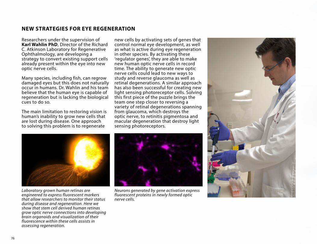
76
NEW STRATEGIES FOR EYE REGENERATION
Researchers under the supervision of Karl Wahlin PhD, Director of the Richard C. Atkinson Laboratory for Regenerative Ophthalmology, are developing a strategy to convert existing support cells already present within the eye into new optic nerve cells.
Many species, including fish, can regrow damaged eyes but this does not naturally occur in humans. Dr. Wahlin and his team believe that the human eye is capable of regeneration but is lacking the biological cues to do so.
The main limitation to restoring vision is human’s inability to grow new cells that are lost during disease. One approach to solving this problem is to regenerate
Laboratory grown human retinas are engineered to express fluorescent markers that allow researchers to monitor their status during disease and regeneration. Here we show that stem cell derived human retinas grow optic nerve connections into developing brain organoids and visualization of their fluorescence within these cells assists in assessing regeneration.
Neurons generated by gene activation express fluorescent proteins in newly formed optic nerve cells.
new cells by activating sets of genes that control normal eye development, as well as what is active during eye regeneration in other species. By activating these ‘regulator genes’, they are able to make new human optic nerve cells in record time. The ability to generate new optic nerve cells could lead to new ways to study and reverse glaucoma as well as retinal degenerations. A similar approach has also been successful for creating new light sensing photoreceptor cells. Solving this first piece of the puzzle brings the team one step closer to reversing a variety of retinal degenerations spanning from glaucoma, which destroys the optic nerve, to retinitis pigmentosa and macular degeneration that destroy light sensing photoreceptors.
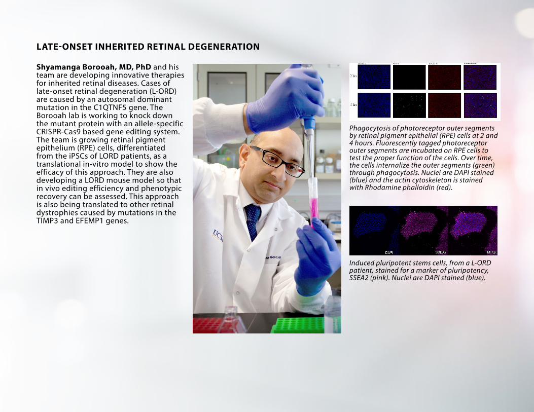
77
LATE-ONSET INHERITED RETINAL DEGENERATION
Shyamanga Borooah, MD, PhD and his team are developing innovative therapies for inherited retinal diseases. Cases of late-onset retinal degeneration (L-ORD) are caused by an autosomal dominant mutation in the C1QTNF5 gene. The Borooah lab is working to knock down the mutant protein with an allele-specific CRISPR-Cas9 based gene editing system. The team is growing retinal pigment epithelium (RPE) cells, differentiated from the iPSCs of LORD patients, as a translational in-vitro model to show the efficacy of this approach. They are also developing a LORD mouse model so that in vivo editing efficiency and phenotypic recovery can be assessed. This approach is also being translated to other retinal dystrophies caused by mutations in the TIMP3 and EFEMP1 genes.
Induced pluripotent stems cells, from a L-ORD patient, stained for a marker of pluripotency, SSEA2 (pink). Nuclei are DAPI stained (blue).
Phagocytosis of photoreceptor outer segments by retinal pigment epithelial (RPE) cells at 2 and 4 hours. Fluorescently tagged photoreceptor outer segments are incubated on RPE cells to test the proper function of the cells. Over time, the cells internalize the outer segments (green) through phagocytosis. Nuclei are DAPI stained (blue) and the actin cytoskeleton is stained with Rhodamine phalloidin (red).

78
CLINICAL TRIALS 2021DIVISION
Cornea
Cornea
Glaucoma
Glaucoma
Glaucoma
Glaucoma
Glaucoma
Glaucoma
Glaucoma
Glaucoma
Glaucoma
Glaucoma
Glaucoma
Glaucoma
GlaucomaOculoplastic
PRINCIPAL INVESTIGATOR
Afshari, Natalie, MD
Heichel, Christopher, MD
Bowd, Christopher, PhD
Camp, Andrew, MD, PhD
Camp, Andrew, MD, PhD
Camp, Andrew, MD, PhD
Camp, Andrew, MD, PhD
Camp, Andrew, MD, PhD
Weinreb, Robert, MD
Weinreb, Robert, MD
Weinreb, Robert, MD
Zangwill, Linda, PhD
Zangwill, Linda, PhD
Zangwill, Linda, PhD
Zangwill, Linda, PhDLiu, Catherine, MD
TITLE
Vision Restoration with a Collagen Crosslinked Boston Keratoprosthesis Unit Study
A Thirteen-year Study of the Indications and Visual Outcomes of Capsular Tension Ring Implantation in Cataract Surgery 2016 PI: Christopher W. Heichel, MD
A0081096 Prospective Randomized 12 233k Controlled Study of Visual Field Change in Subjects with Partial seizures Receiving Pregabalin or Placebo - Pfizer Inc PI: Christopher J. Bowd, PhD
The Efficacy and Safety of Bimatoprost SR in Patients With Open-angle Glaucoma or Ocular Hypertension PI: Andrew S. Camp MD
Efficacy and safety of AbGn-168H in Patients with Active Psoriatic Arthritis: A 24-Week, Open-Label, Multi-Center, Phase II Proof of Principle Trial PI:
Andrew S. Camp, MDEffects of the Body Position on Episcleral Venous Pressure and
Intraocular Pressure in Glaucoma.XEN-45 Gel Stent Versus Trabeculectomy in glaucoma: Gold Standard
Pathway Study (GPS)An Extension Trial to Evaluate the Long-term Safety and Efficacy of
Bimatoprost SR in Patients with Open Angle Glaucoma or Ocular Hypertension
Subconjuctival Mitomcyin-C Injection Versus Direct Sclera Application in Trabeculectomy PI: Robert N. Weinreb, MD, Sub-I: Jiun Do, MD
Determining the Correlation Between Intraocular Pressures Measured by Self-Monitoring Rebound Tonometry and Glaucoma Development or Progression PI: Robert N. Weinreb, MD, Sub-I: Jiun Do, MD
Evaluation of the Repeatability and Reproducibility of AngioVue in Normal Subjects, Retinal Patients, and Glaucoma Patients PI: Robert N. Weinreb, MD
Effects of a Single Osteopathic Manipulative Treatment (OMT) on Intraocular Pressure (IOP) Reduction PI: Hollis King, Sub-I: Linda M. Zangwil, PhD
Multi-Center Study for a Reference Database of Optic Nerve Head, Retinal Nerve Fiber Layer, and Macula Parameters Measured with the Heidelberg Spectralis OCT within a Hispanic Population PI: Linda M. Zangwill, PhD
Multi-Center Study for a Reference Database of Optic Nerve Head, Retinal Nerve Fiber Layer, and Macula Parameters Measured with the Heidelberg Spectralis OCT within an African-American population PI: Linda M. Zangwill, PhD
Grading the Ocular Images and Datasets for the B-2018-4 StudyA Phase 4, Randomized, Double-masked, Placebo-controlled, Multicenter Trial
to Evaluate the Efficacy and Safety of TEPEZZA in Treating Patients with Chronic (Inactive) Thyroid Eye Disease
FUNDING AGENCY
Massachusetts Eye & Ear Infirmary; DOD as Prime
Pfizer
Allergan
Allergan
Allergan
Heidelberg Engineering
Heidelberg Engineering
Heidelberg EngineeringHorizon Therapeutics USA, Inc.

79
DIVISION
Pediatrics
Retina
Retina
Retina
Retina
Retina
Retina
Retina
Retina
Retina
Retina
Retina
Retina
PRINCIPAL INVESTIGATOR
Robbins, Shira, MD
Borooah, Shyamanga, PhD
Borooah, Shyamanga, PhD
Borooah, Shyamanga, PhD
Borooah, Shyamanga, PhD
Freeman, William, MD
Freeman, William, MD
Freeman, William, MD
Freeman, William, MD
Freeman, William, MD
Freeman, William, MD
Freeman, William, MD
Freeman, William, MD
TITLE
A Multi-Center, Double-Masked, Randomized, Placebo-Controlled Phase 3 Study of the Safety and Efficacy of Atropine 0.1% and 0.01% Ophthalmic Solutions Administered with a Microdose Dispenser for the Reduction of Pediatric Myopia Progression [CHAPERONE]
Natural History Study of Patients with X-linked Retinal Dystrophy Associated with Mutations in Retinitis Pigmentosa GTPase Regulator (RPGR)
Phenotyping and Genotyping Patients with Achromatopsia in Preparation for Gene Therapy Trials
A Phase 2 Randomized Placebo-Controlled Double-Masked Study to Assess Safety & Efficacy of Multiple Doses of IONIS-FB-LRX an Antisense Inhibitor of Complement FactorB in Patients w/GA 2nd to AMD
“Open-Label, Single Ascending Dose Study to Evaluate the Safety, Tolerability, and Efficacy of EDIT-101 in Adult and Pediatric Participants with Leber Congenital Amaurosis Type 10 (LCA10), with Centrosomal Protein 290 (CEP290)-Related Retinal Degeneration Caused by a Compound Heterozygous or Homozygous Mutation Involving c.2991+1655A>G in Intron 26 (IVS26) of the CEP290 Gene (“LCA10-IVS26”)”
A Phase 2b, Randomized, Double-Masked, Controlled Trial to Assess the Safety and Efficacy of Zimura (Anti-Complement Factor 5 Aptamer) in Subjects with Geographic Atrophy 2nd to AMD [ZIMURA]
A Multicenter, Open-Label, Extension Study to evaluate the Long-Term Safety and Tolerability of the Port Delivery System with Ranibizumab in Patients with Neovascular AMD [PORTAL]
A Phase 2, Prospective, Randomized, Double-masked, Active Comparator-controlled, Multi-center Study to Investigate the Efficacy and Safety of Repeated Intravitreal Administration of KSI-301 in Subjects with Neovascular (Wet) Age-related Macular Degeneration [DAZZLE]
A Phase 3 Multicenter, Randomized, Double-Masked, Sham-Controlled Clinical Trial to assess the Safety and Efficacy of Intravitreal Administration of ZIMURA™ (Complement C5 Inhibitor) in patients with Geographic Atrophy Seondary to Dry Age-Related Macular Dengeneration [GATHER2]
A Randomized, Double-masked, Phase 3 Study of ABP 938 Efficacy and Safety Compared to Aflibercept (Eylea®) in Subjects with Neovascular Age-related Macular Degeneration
Randomized, Double-Masked, Active-Controlled, Phase 3 Study of the Efficacy and Safety of High Dose Aflibercept in Patients With Neovascular Age-Related Macular Degeneration [PULSAR]
A Prospective, Randomized, Double-masked, Active Comparator-controlled, Multi-center, Two-arm, Phase 3 Study to Evaluate the Efficacy and Safety of Intravitreal KSI-301 Compared with Intravitreal Aflibercept in Participants with Visual Impairment Due to Treatment-naïve Macular Edema Secondary to Retinal Vein Occlusion [BEACON]
A Prospective, Randomized, Double-masked, Active Comparator-controlled, Multi-center, Two-arm, Phase 3 Study to Evaluate the Efficacy and Safety of Intravitreal KSI-301 Compared with Intravitreal Aflibercept in Participants with Visual Impairment Secondary to Treatment-naïve Diabetic Macular Edema [GLEAM]
FUNDING AGENCY
Eyenovia, Inc.
Meiragtx UK II Limited
MeiraGTx UK II Ltd
Syneos Health Inc.
Editas Medicine
Ophtotech Corp.
Genentech, Inc.
Kodiak Sciences, Inc.
IVERIC Biosciences
AMGEN Pharmaceuticals
BAYER AG
Kodiak Sciences, Inc.
Kodiak Sciences, Inc.

80
DIVISION
Retina
Retina
Retina
Retina
Retina
PRINCIPAL INVESTIGATOR
Freeman, William, MD
Nudleman, Eric, MD, PhD
Nudleman, Eric, MD, PhD
Nudleman, Eric, MD, PhD
Spencer, Doran, MD
FUNDING AGENCY
Apellis Pharmaceuticals
Novartis Pharmaceuticals
Regeneron Pharmaceuticals
Regeneron Pharmaceuticals
Genentech, Inc.
TITLE
A Phase 3, Open-Label, Multicenter, Extension Study to Evaluate the Long-Term Safety and Efficacy of PEGCETACOPLAN in subjects with Geographic Atrophy 2nd to AMD [GALE]
A 2-year, Three-Arm, Multicenter, Randomized, Double-Masked, Multicenter, Phase III Study assessing the Efficacy and Saferty of Brolucizumab vs Aflibercept in Adult Patients with Visual Impairment due to Diabetic Macular Edema [KESTREL]
A Randomized, Controlled, Multi-Center Study to Assess the Efficacy, Safety, and Tolerability of Intravitreal Aflibercept Compared to Laser Photocoagulation in Patients with Retinopathy of Prematurity [ROP]
“An Extension Study to Evaluate the Long-Term Outcomes of Patients Who Received Treatment for Retinopathy of Prematurity in the VGFTE-ROP-1920 Study” [BUTTERFLEYE]
A Phase III, Multicenter, Randomized, Double-Masked, Active Comparator-controlled Study to evaluate the Efficacy and Safety of Faricimab in Patients with Neovascular Age-related Macular Degeneration [TENAYA]
STAFF SPOTLIGHTCongratulations to Juan Arias, the new Assistant Director of Ophthalmology Services, at the Shiley Eye Institute. Arias is now a member of the Viterbi Family Department of Ophthalmology’s Operations Management Team and partners with the Director of Ophthalmic Services, Cathi Lyons. The team plans the Department’s business development, operational and programmatic growth aiming to create high quality clinic flow and processes via EPIC, training and clinical standards.
Juan is responsible for providing clinical and technical support in key areas such as imaging and documentation. He is developing and managing strategic initiatives to improve efficiencies and quality, leading teams, and projects to address and resolve complex issues in all Ophthalmology and Optometry clinics.
Arias was awarded a Biology Undergraduate and Master’s Mentorship Program Award from UC San Diego to study the use of TENS machines in
the reduction of pain associated with wet age-related macular degeneration injections in the clinic. He has started the MBA program at Point Loma University.

81
GRANTS 2021DIVISION
CorneaCorneaGlaucoma
Glaucoma
Glaucoma
GlaucomaGlaucomaGlaucomaGlaucomaGlaucoma
Glaucoma
Glaucoma
Glaucoma
GlaucomaGlaucoma
GlaucomaGlaucoma
GlaucomaGlaucomaGlaucomaGlaucoma
Glaucoma
Glaucoma
Glaucoma
Glaucoma
PRINCIPAL INVESTIGATOR
Afshari, Natalie, MDShaw, Peter, PhDBaxter, Sally, MD
Baxter, Sally, MD
Christopher, Mark, PhD
Coleman, Todd, PhDDo, Jiun, MD, PhDDo, Jiun, MD, PhDJu, Wonkyu, PhDMoghimi, Sasan, MD
Weinreb, Robert, MD
Weinreb, Robert, MD
Weinreb, Robert, MD
Weinreb, Robert, MDWeinreb, Robert, MD
Weinreb, Robert, MDWeinreb, Robert, MD
Welsbie, Derek, MD, PhDWelsbie, Derek, MD, PhDWelsbie, Derek, MD, PhDWelsbie, Derek, MD, PhD
Zangwill, Linda, PhD
Zangwill, Linda, PhD
Zangwill, Linda, PhD
Zangwill, Linda, PhD
FUNDING AGENCY
NIH/NEI R01UC Academic SenateNIH/NEI
NIH/NEI
NIH/NEI
NIH/NEIAmerican Glaucoma SocietyGlaucoma Research FoundationNIH/NEI R01Tobacco-Related Disease Research ProgramNIH/NEI K12
Research to Prevent Blindness
NIH/NEI R01
NIH/NEI R01Bausch & Lomb
NIHNIMHD
NIH/NEI R01Glaucoma Research FoundationResearch to Prevent BlindnessOriole Therapeutics, Inc.
NIH/NEI R01
NIH/NEI R01
NIH/NEI
NIH/NEI
TITLE
Application of RNA-Targeting CAS9 to Fuchs’ DystrophyInterplay of Genetic Risk and Oxidative Stress in AMD PathogenesisMulti-modal Health Information Technology Innovations for Precision
Management of GlaucomaSupplemental. Graduate Student Support for Multi-modal Health Information
Technology Innovations ProjectDeep Learning Approaches to Detect Glaucoma and Predict Progression from
Spectral Domain Optical Coherence TomographyBiosensor and Eyedrop Bottle Technologies for Glaucoma Adherence MonitoringOptic Nerve Relays to Restore VisionOptic Nerve Relays for the Restoration of Visual FunctionMitochondrial Protection in Glaucomatous Optic NeuropathyRacial Differences in Smoking-related Glaucoma Progression: Effect on Neural
and Vascular TissueOphthalmology and Visual Sciences Career Development K12 Program PI: Robert
N. Weinreb, MD, Co-I: Natalie A. Afshari, MD, NIH, April 2015 – July 2026Unrestricted and Challenge Grant - Research to Prevent Blindness PI: Robert N.
Weinreb, MD, January 2018 - December 2023 Diagnosis and Monitoring of Glaucoma with Optical Coherence Tomography
Angiography PI: Robert N. Weinreb, MD, May 2018 - April 2022Molecular Mechanisms of GlaucomaA Randomized, Single Center, Masked, Crossover Study Comparing the Effects of
Latanoprostene Bunod and Timolol on Retinal Blood Vessel Density and Visual Acuity in Patients with Ocular Hypertension or Primary Open Angle Glaucoma
Ocular Hypertension Treatment Study 20-Year Follow-up: Clinical Center GrantiGLAMOUR Study: Innovations in Glaucoma Adherence and Monitoring of Under-
Represented MinoritiesKinase Multitargeting for Glaucoma NeuroprotectionHigh-Throughput Functional Genomic Screening in Retinal Ganglion CellsDeveloping an Optic Nerve Relay for Vision RestorationDevelopment of Small Molecule and Gene Therapy Approaches to Inhibit
Dual Leucine Zipper Kinase and Accessory Pathways for Retinal Ganglion Cell Neuroprotection
Diagnostic Innovations in Glaucoma Study (DIGS): High Myopia and Advanced Diseases PI: Linda Zangwill, PhD, Co-I: Robert N. Weinreb, MD, Co-I: Christopher Bowd, PhD, NIH, March 2017 – February 2022
African Descent and Glaucoma Evaluation (ADAGES) IV: Alterations of the Lamina Cribrosa in Progression PI: Linda Zangwill, PhD, Co-I: Robert N. Weinreb, MD, NIH, April 2017 – March 2021
Translational Vision Research Training at UCSD PI: Linda Zangwill, PhD, Co-I: Radha Ayyagari
Personalized Forecasting of Disease Trajectory for Patients with Open Angle Glaucoma

82
Glaucoma
GlaucomaGlaucomaGlaucoma
Glaucoma
Oculoplastics
Pediatric
Retina
RetinaRetinaRetina
RetinaRetina
RetinaRetinaRetina
Retina
RetinaRetinaStem Cell
Stem Cell
Stem Cell
Stem Cell
Stem Cell
Stem Cell
Stem Cell
Zangwill, Linda, PhD
Zangwill, Linda, PhDZangwill, Linda, PhDZangwill, Linda, PhD
Zangwill, Linda, PhD
Kikkawa, Don O., MD
Rudell, Jolene, M.D., Ph.D.
Ayyagari, Radha, PhD
Ayyagari, Radha, PhDAyyagari, Radha, PhDAyyagari, Radha, PhD
Bartsch, Dirk-Uwe, PhDBorooah, Shyamanga, PhD
Borooah, Shyamanga, PhDFerrara, Napoleone, MDFerrara, Napoleone, MD
Freeman, William, MD
Nudleman, Eric, MD, PhDOesch, Nicholas, PhDWahlin, Karl, PhD
Wahlin, Karl, PhD
Wahlin, Karl, PhD
Wahlin, Karl, PhD
Wahlin, Karl, PhD
Wahlin, Karl, PhD
Wahlin, Karl, PhD
Dietary Interventions to Improve Vision UCSD Krupp Endowment Fund (KEF) Research Awards, PI: Linda Zangwill, PhD
P30 NEI Center Core Grant for Vision ResearchOCT in the Ocular Hypertension Treatment Study: 20 Year Follow-UpA Randomized Clinical Trial Evaluating Fenofibrate for Prevention of Diabetic
Retinopathy WorseningImprove OCT Detection of Optic Nerve Head (ONH)
Gene Expression in Nonspecific Orbital Inflammation Disease Co-I: Don O. Kikkawa, MD NIH, September 2016 - August 2021
The Characterization of the Neuromuscular Junction in Extraocular Muscles in Patients with Strabismus
Identification of the Elusive Genetic Causality of Inherited Retinal Degenerations (IRDs)
Molecular Basis of Hereditary Retinal DegenerationsMolecular Mechanism Underlying Late-Onset Retinal/Macular DegenerationUnraveling the Molecular Pathology of Retinal Degeneration Through Single Cell
GenomicsMechanistic-Based Non-Invasive Assessment of Retinal Damage in HAART EraCombining the Utility of Human Induced Pluripotent Stem Cell Modeling and
CRISPR-Cas9 Gene Editing with Adenoassociated Virus Vector Gene Delivery to Develop and Optimize Novel Gene Editing in Inherited RPE Disease
Rescue of LAMP2 Associated RetinopathyIdentification of Novel Inhibitors of Ocular NeovascularizationLong Novel-Acting Inhibitors of Vascular Endothelial Growth Factor (VEGF) for
Treatment of Intraocular Vascular Disorders; Co-I: Eric Nudleman, MD, PhDMultimodal Retina Image Alignment and Applications Co-I: William Freeman, MD,
Contact PI: Truong Q Nguyen, PhD, September 2021 - August 2025Role of PDLIM1 in Re.tinal Vascular Leakage and ProliferationComputing Luminance and Contrast in Prosthetically Driven RetinaEndogenous Generation of Cone Photoreceptors to Increase Light Responses in
Foveal HypoplasiaModeling Photoreceptor Development and Disease with Human Pluripotent
Stem CellsA Stem Cell Based Optic Nerve Model for Studies of Axon Guidance and
RegenerationPluripotent Stem Cell Derived 3D Retinas for Studies of Early Onset Retinal
DegenerationComplement Factor H Mutant Pluripotent Stem Cells to Model Early Onset
Macular Degeneration and Their Application in Drug DiscoveryDissecting the Biochemical Role of Epigenetically Modified Regulatory
Sequences within the Genomes of Retinal Neurons (A1)Differential Expression of miRNAs Serve as Biomarkers of AMD-like Disease
Progression in Human Stem Cell-derived RPE
Krupp Foundation
NIH/NEINIH/NEINIH/NEI
Glaucoma/Myopia OCT Phenotyping ConsortiumNIH/NEI
Knights Templar Eye Foundation
The Foundation Fighting BlindnessNIH/NEINIH/NEI R01NIH/NEI R01
NIH/NEI R01The Foundation Fighting Blindness
Knights Templar Eye FoundationNIH/NEINIH/NEI R01
NIH/NEI R01
NIH/NEINIH/NEI R01Vision of Children
NIH/NEI R01
NIH/NEI
NIH/NEI R01BrightFocus Foundation
James Madison University; NIH/NEI as Prime
Bayer AG
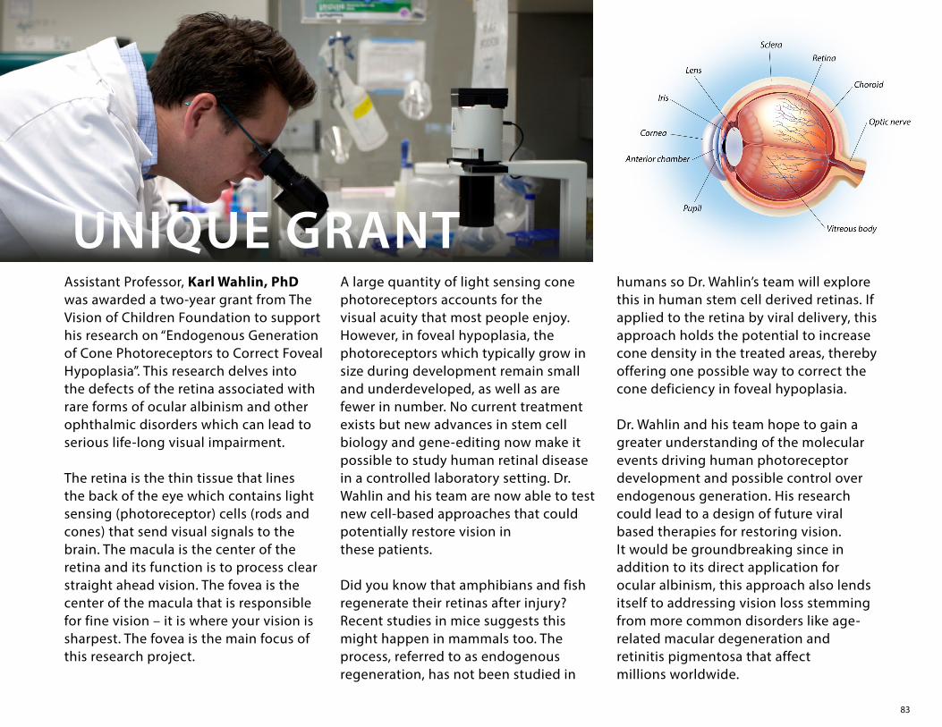
83
Assistant Professor, Karl Wahlin, PhD was awarded a two-year grant from The Vision of Children Foundation to support his research on “Endogenous Generation of Cone Photoreceptors to Correct Foveal Hypoplasia”. This research delves into the defects of the retina associated with rare forms of ocular albinism and other ophthalmic disorders which can lead to serious life-long visual impairment.
The retina is the thin tissue that lines the back of the eye which contains light sensing (photoreceptor) cells (rods and cones) that send visual signals to the brain. The macula is the center of the retina and its function is to process clear straight ahead vision. The fovea is the center of the macula that is responsible for fine vision – it is where your vision is sharpest. The fovea is the main focus of this research project.
A large quantity of light sensing cone photoreceptors accounts for the visual acuity that most people enjoy. However, in foveal hypoplasia, the photoreceptors which typically grow in size during development remain small and underdeveloped, as well as are fewer in number. No current treatment exists but new advances in stem cell biology and gene-editing now make it possible to study human retinal disease in a controlled laboratory setting. Dr. Wahlin and his team are now able to test new cell-based approaches that could potentially restore vision in these patients.
Did you know that amphibians and fish regenerate their retinas after injury? Recent studies in mice suggests this might happen in mammals too. The process, referred to as endogenous regeneration, has not been studied in
UNIQUE GRANThumans so Dr. Wahlin’s team will explore this in human stem cell derived retinas. If applied to the retina by viral delivery, this approach holds the potential to increase cone density in the treated areas, thereby offering one possible way to correct the cone deficiency in foveal hypoplasia.
Dr. Wahlin and his team hope to gain a greater understanding of the molecular events driving human photoreceptor development and possible control over endogenous generation. His research could lead to a design of future viral based therapies for restoring vision. It would be groundbreaking since in addition to its direct application for ocular albinism, this approach also lends itself to addressing vision loss stemming from more common disorders like age-related macular degeneration and retinitis pigmentosa that affect millions worldwide.
84
Annual Gifts – Circle of Sight
Started in 1996, the Circle of Sight is the Shiley Eye Institute’s recognition program that acknowledges donors who make annual gifts of $250 or more to support the greatest needs of the Department. Several times a year, the Shiley Eye Institute’s Circle of Sight members are invited to attend Vision Research Lectures and receptions where members get to personally know our faculty. The members are also ambassadors for the Shiley Eye Institute within the San Diego community. The Circle of Sight group is the backbone of many of our successful initiatives.
Planned Gifts – Your Vision for Tomorrow
Please consider a charitable bequest in your estate plan that will benefit the future of the Shiley Eye Institute and directly support the Viterbi Family Department of Ophthalmology. We would be pleased to provide you, your attorney, accountant or tax advisor with specific bequest language for inclusion in your will, trust or as a beneficiary of your retirement account – all of which can lessen the impact of taxes on your heirs or give you comfort of knowing that your assets will benefit those you leave behind.
Outright Gifts – Immediate Impact
Outright gifts of all sizes made with cash, check, credit cards, savings bonds, stocks, marketable securities or property provide immediate impact to our faculty and facility. If writing a check, please make payable to the “UC San Diego Foundation” and put the Shiley Eye Institute in the memo section. The check should be accompanied with a letter stating the focus of your donation and mail to
The Shiley Eye Institute, Mail Code 0946, 9415 Campus Point Drive, Room 241B, La Jolla, CA, 92093-0946 (Attention: Karen Anisko Ryan)
For over 30 years, the philanthropic support from generous individuals, foundations and corporations has provided the Shiley Eye Institute and Viterbi Family Department of Ophthalmology with valuable resources for patient care, research, education and community service. As a friend of the Department of Ophthalmology, there are several giving options for those who wish to contribute to our tradition of excellence.
GIVINGOPPORTUNITIES

85
Tribute Gifts – Acknowledge Someone Special
Contributions can be made in memory, honor or in celebration of a loved one or to commemorate a special occasion. Gifts can be made to honor a special physician, for example, who has played a significant role in your eye health. Such a gift creates a legacy and memorializes the person by providing direct support to the Department. Matching Gifts – Double or Triple your Gift
Many employers offer a matching gift program to their employees meaning that your donations are worth even more. All you need is a Matching Gift Form from your employer.
Endowments – Gifts in Perpetuity
A gift of endowment demonstrates your long-term commitment to the Viterbi Family Department of Ophthalmology since the fund is maintained in perpetuity. Your gift can support programs, lectures, awards, fellowships and Chairs. An endowment serves as an enduring legacy since it often bears the name of a donor or loved one.
For more information about any of these gifting ideas, or for help in finding the best gift options for achieving your charitable goals, please contact Karen Anisko Ryan at [email protected] or 858-534-8017. To learn how these giving options might affect your specific financial situation, please consult with your tax, legal or financial advisors.
Gifts of Real Estate
Making a gift of real estate is a generous and financially advantageous way to support Shiley Eye Institute. Many people have residential rental units or vacation homes that no longer serve their needs or have become too burdensome to maintain. Even commercial property and vacant land can be a used as gifts to support Shiley’s programs. Real estate can be given as an outright gift or in a bargain sale; it can be used to create a retained life estate, a charitable gift annuity or a charitable remainder trust. There are many creative ways that your real estate can unlock financial security and provide tax benefits for you now while supporting Shiley Eye Institute and the programs that mean the most to you.
Every donation makes a direct impact on our patients, faculty, researchers, residents, fellows and staff, as well as the field of Ophthalmology. We cherish the partnership that we have developed with those generous members of the community who invest in us. There are also naming opportunities for gifts including: endowed chairs, laboratories, specialized ophthalmic clinics and research initiatives. We would welcome
the opportunity to have a confidential conversation with you, so we clearly understand how you want your gift to be used.

86
VISIONARY CIRCLE
Members of the Visionary Circle are cumulative lifetime contributors of one million dollars or more to the Department of Ophthalmology. We appreciate their generosity.
AnonymousEleanor & John E.* Barbey, Jr.Mr. & Mrs. Woody CarterDavid J. Dunn*The Foundation Fighting BlindnessMartin* & Enid* GleichJean Hahn Hardy*Hildyard Family TrustTrude Kahn Hollander, MD*Joan & Irwin JacobsDorothy R. Kerrigan TrustTatiana* and Richard Kindell* LanscheMichael Luzich, in Honor of Norman &
Carol LuzichArthur Murray & Carol-Faith Murray TrustNixon Visions FoundationIrvin Olson & Rita A. Olson TrustMarc PaskinAnne F. Ratner*Ruth L. Schulman*Darlene Marcos Shiley
in Memory of Donald P. ShileyMichael* & Rebecca ShileyPatricia ShileyAndrew J. Viterbi, Ph.D.Frances Hamilton White*
*Deceased
Linda Zangwill, PhD was appointed as the holder of the Richard K. Lansche and Tatiana A. Lansche Endowed Chair in Ophthalmology in 2019 to support her research and teaching activities at the Shiley Eye Institute and Viterbi Family Department of Ophthalmology.
LANSCHE CHAIR COMPLETED
Linda Zangwill, PhD accepting the final pledge payment from the Lansche Family represented by Rick Mitchell and Francie Murphy.
The endowed chair honors the work and memory of the late Richard K. Lansche, MD (deceased in 2000), a respected San Diego area ophthalmologist and his wife Tatiana. Sadly, Mrs. Lansche passed away in October 2020 at the age of 99.

87
I am an active sixty year old woman and have run in 20 marathons. Unfortunately, my eyes were not as healthy. For more than fifteen years, I have been a glaucoma patient at the Shiley Eye Institute (SEI) under the care of Robert N. Weinreb, MD. I have participated in multiple clinical trials at the Hamilton Glaucoma Center and have had several eye surgeries. Dr. Weinreb and his wonderful team have taken excellent care of me so that my vision is stable going into my later years.
Shiley is an institution that I care deeply about and therefore I have chosen to include it in my will. I believe that my future financial contribution to SEI is opening a window of opportunity, however small, for exploring and proving novel solutions for eye diseases.
Shiley’s innovative research holds promise for improved outcomes for patients facing the challenges of eye disease. Combining world class medicine
and engineering with caring and dedicated clinicians, SEI has sewn fertile ground with hopeful seeds of a future where fewer children will lose vision and more adults will enjoy good vision for a lifetime.
I feel so lucky to have Dr. Weinreb and Shiley here in San Diego and I want my legacy to support their world class eye care for years to come.
Anyone who cares about being a part of this exploration for better vision should consider including SEI in their estate plan.
To learn more about including the Shiley Eye Institute and Viterbi Family Department of Ophthalmology at UC San Diego in your estate plan and leave a lasting legacy, please contact Karen Anisko Ryan at [email protected] or 858-534-8017.
PATIENT PLANNING FOR SEI’S FUTURE
Robert N. Weinreb, MD and author Jill Herndon

88
YOUR GIFTS MAKE A DIFFERENCEAs a result of COVID-19, 86-year-old San Diego businessman Tom Fetter had gone three years since his customary eye exam at the Shiley Eye Institute (SEI). An SEI optometrist immediately noticed a problem when Fetter had difficulty reading the eye chart with his left eye and referred him to see retina specialist, Eric Nudleman, MD, PhD, Associate Professor of Ophthalmology. Fetter had been completely unaware of the problem, as his right eye was compensating for the left. Dr. Nudleman diagnosed Fetter with wet age-related macular degeneration (AMD). Fetter was immediately concerned, as one of his long-term employees had retired and was now almost blind from AMD. On Fetter’s first appointment in March 2021, after having his vision and intraocular eye pressure checked, he was dilated then directed to the Imaging Department for photos of his retina. As he approached, he saw patients sitting in the hall waiting in line. After getting
information and a possible delivery date for the OCT machine. When confirmed, he wrote a check for $109,470 and the machine was ordered.
In June, Oeinck mentioned that an additional Optos machine was needed in order to fully service the patient flow and would cost $172,395. Fetter agreed to provide $100,000 to initiate this purchase as long as the Department was able to fund the difference. The Optos machine was ordered as well. Both of the new machines were installed and are now fully operational.
Fetter now receives monthly injections given by Dr. Nudleman to treat his AMD. Dr. Nudleman’s laboratory work focuses on developmental angiogenesis and future less invasive treatments for retinal degenerations. He also collaborates with Napoleone Ferrara, MD, Distinguished Professor of Ophthalmology and Pathology, in their UC San Diego laboratory. With support
his photos taken, he asked Ophthalmic Photographer, Chris Oeinck, “why are there so many patients waiting?” Oeinck responded that unfortunately, SEI had only one optical coherence tomography (OCT) device and one Optos machine, both of which are needed to screen over 100 patients per day.
Each of these retinal imaging devices are non-invasive evaluation machines which help ophthalmologists diagnose and treat a variety of retinal diseases. The OCT uses light waves to take cross section pictures of the retina, allowing doctors to evaluate details of the retinal anatomy. The Optos machine takes ultra-wide images using low powered red, green, blue and infrared laser waves, simultaneously providing panoramic views of the inside of the eye. A second set of machines had been on the wish list for several years.
On his next visit in April to see Dr. Nudleman, Fetter asked for detailed
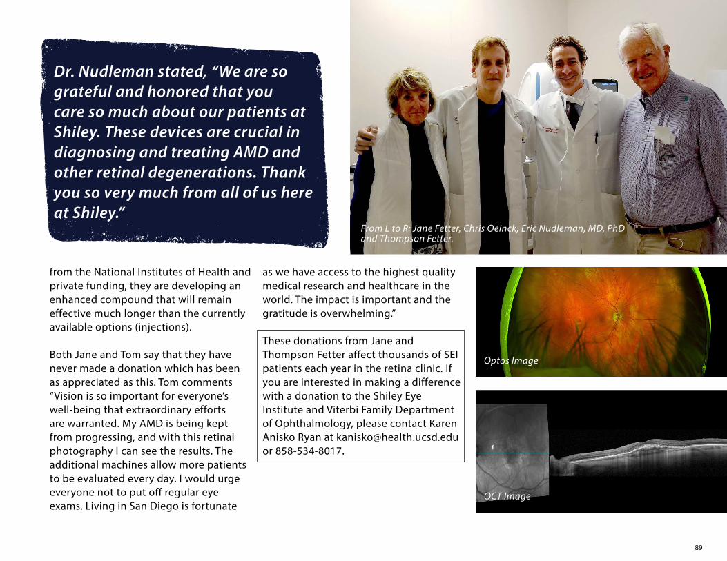
89
from the National Institutes of Health and private funding, they are developing an enhanced compound that will remain effective much longer than the currently available options (injections). Both Jane and Tom say that they have never made a donation which has been as appreciated as this. Tom comments “Vision is so important for everyone’s well-being that extraordinary efforts are warranted. My AMD is being kept from progressing, and with this retinal photography I can see the results. The additional machines allow more patients to be evaluated every day. I would urge everyone not to put off regular eye exams. Living in San Diego is fortunate
as we have access to the highest quality medical research and healthcare in the world. The impact is important and the gratitude is overwhelming.”
These donations from Jane and Thompson Fetter affect thousands of SEI patients each year in the retina clinic. If you are interested in making a difference with a donation to the Shiley Eye Institute and Viterbi Family Department of Ophthalmology, please contact Karen Anisko Ryan at [email protected] or 858-534-8017.
From L to R: Jane Fetter, Chris Oeinck, Eric Nudleman, MD, PhD and Thompson Fetter.
Dr. Nudleman stated, “We are so grateful and honored that you care so much about our patients at Shiley. These devices are crucial in diagnosing and treating AMD and other retinal degenerations. Thank you so very much from all of us here at Shiley.”
OCT Image
Optos Image

90
The work of the UC San Diego Shiley Eye Institute and Viterbi Family Department of Ophthalmology is made possible through the generosity of our donors. We appreciate your investment and will continue to provide world class patient care, groundbreaking research, education excellence and community service. Thank you to all the individuals, foundations and corporations listed below on our Honor Roll who have donated from July 1, 2020 to June 30, 2021.
Gifts of Over $1 Million
Nixon Visions FoundationDarlene Marcos Shiley
in Memory of Donald P. Shiley
Gifts of $100,000 and Above
Marilyn and David AndersonGeorge Davies and Audrey (Ronnie)
Warren TrustDavid J. Dunn*The Marian and Walter Trevor, Jane Trevor
Fetter and Thompson Fetter Fund The Joan and Irwin Jacobs Fund of the Jewish
Community FoundationResearch to Prevent BlindnessThe Vision of Children FoundationAndrew J. Viterbi, Ph.D.
Gifts of $50,000 to $99,999
AnonymousLa Verne* and Blaine* BriggsMr. and Mrs. Woody CarterEquinox Ophthalmic, Inc.The Paul R. Gertsch TrustGlaucoma Research FoundationMargaret* and Robert* HulterKnights Templar Eye Foundation, Inc.The Richard and Claudia Libenson Fund of
the Jewish Community Foundation
Price Philanthropies FoundationSan Diego Lions Welfare FoundationJ. Joseph and Ethel Shelley Living Trust
Gifts of 25,000 to $49,999
GenentechBuzz and Peg GitelsonWilson and Jean Sexton
Gifts of $10,000 to $24,999
AnonymousSteve and Sheri AltieriDr. and Mrs. Stuart I. BrownDorothy Conte*Costco Wholesale Executive MatchBruce L. DeckAlbert and Kathleen FredmanTully and Elise FriedmanCaroline and Daniel HamlinWarner C. and Debra E. Lusardi FoundationMr. and Mrs.* Morris K. NachtomiHalpin and Helen O’Reilly Charitable TrustStrayve Family TrustCarol and Larry VeitVision Health and Development in
Children, Inc.David J. WinklerJames Zolin
Gifts of $5,000 to $9,999
Anonymous (2)Hjalmar and Carolyn BjornsonToby and Howard CohenThe Friedman Family Fund of the Jewish
Community FoundationCarole and Howard GoldfederLanna C. LewinDr. Thomas MackMs. Laura Davies Mateo, Lakeside FoundationGale and Ken NillAl and Sharon RappaportPatrick Sheely FamilyPatricia K. ShumwayJudith TamkinIrving TragenJocelyn and Dick VortmannToby WolfCapt. Judy H. Zygowicz and
Mr. John J. Zygowicz
Gifts of $1,000 to $4,999
52 & Convoy Corp. / Mr. and Mrs. Thompson Fetter
Anonymous (5)Farooq AzamJohn BaileyMr. and Mrs. Floyd BernardMiles and Patricia BowlerHoward and Sue BusbyFrank and Lisa Casella
WITH APPRECIATION
91
Bill ComerWayne ComfortEngel Family FoundationNomi FeldmanLinda FortierGerald Freedman and Elizabeth SacksThe Morris and Phyllis Gold Fund of the
Jewish Community FoundationJane Goodwin, ODJohn and Mary GriffinElizabeth HansenStanley C. HarazimJulie HillArthur C. Johnson, MDMr. and Mrs. Allan KleinmanAlexa and Bela KozakRosemarie KubesDr. and Mrs. Bruce R. LawrenceJeffrey H. LinJames LuckowJeff and Patty MaysentJ. Michael and Karen S. McCulleyD. Asoka and Janine P. MendisThe Millard Group Team - “Millard Cares 2020”Jeffrey T. MillerDennis and Barbara MoranLois M. MulcaheyElspeth and Jim MyerMark G. NewmarkFran Osborn and Tom RyanVictoria G. PappasFrank PisciottaJeffrey and Susan RichardsJanis Lynn SammartinoDr. Jonathan E. Shelton and
Dr. Nicole H. PurcellJohn and Linda SmallJohn and Socorro SoCarol TowneR.J. and Joy UrichMargot Walk and Jerry FreelandJo and Howard Weiner
Robert N. Weinreb, MDOlive WithallBetty WongDr. and Mrs. Philip R. Ziring
Gifts of $500 to $999
AnonymousAnthony AranesJohn and Elizabeth BagbyLorrie BernsteinArun K. Bhatia and Neera BhatiaDennis W. BuckleyLawrence DirkesAlan and Pauline FatayerjiJoaquin and Elisabeth FusterLouis GalperBrenda Goldbaum and
Michael H. Goldbaum, M.D.Mr. Francesco Grilli and
Dr. Anna A. De AngelisDr. and Mrs. Harold R. HallJaye and Bill HanleySteve HardenTom and Terri HunterNick A. JohnstonViktor KerzhanovichMark KreyBernard Kulchin and Paula TaylorTony and Mary LovettChrista McReynolds Fund David PerrinBarbara RasmussenParvin RezainiaMrs. Sally F. SaltzsteinElizabeth and Lester StielRichard F. Tucker
Gifts of $250 to $499
Dr. and Mrs. Thomas S. AhernDiane AmatangeloTrudy H. AndersonAnonymousFrancine and Marvin BartRobert Bennett, MD and Tamara BennettDr. and Mrs. Michael R. BramsBarry BerkovIrene Carr and Paul D. ZaluskyFred and Marjorie* CaserioCrafts Family TrustMr. and Mrs. Joseph DohnWilliam DollCarla P. Domenici IMO Floyd PrandoStephen L. EdwardsNorman ErnstEdward J. FarrellMonique and Stanley FriedmanManny HernandezJerome HickmondAndrea JohnsonF. Stephen Kohl, MDAnne and Thomas LeePatricia LubarskyMr. and Mrs. Frederick MandabachRobert and Susan MardonDeborah McCormacEileen and Anthony McKoyMr. and Mrs. Gary McMullenJonathan, Suzanne and Andrew MorrisPaul and Lois NivenLaurie PriceQualcomm Matching GiftMary and Herb RabeDonna E. RobinsonBarbara RoperRonald L. and Diane L. SeditaHarold F. Skelly, Sr.*Mr. and Mrs. Clinton R. SpanglerFredricka Taubitz and Dennis M. Griffin

92
Dr. April TeitelbaumHilda van Neck-YoderSusie M. VidrihCharles and Tara WegnerRobert E. WelkAnnasue McCleave Wilson and John Wilson
Gifts of $100 to $249
Anonymous (6)Georgios AnagnostopoulosAmelia AndradeConstance and Alexander AthensDavid AuldMr. and Mrs. Michael J. BellNatalie BellerLouise BenavidezDolores Boily IMO James BoilyThe Honorable and Mrs. Peter W. BowieJack* and Charmian BowsherDonna Brewster IMO Fayette LeungLewis and Carol BrownAnn S. BurgessJon H. BusseCarol V. BuuckSung H. ChoBrian and Nancy CookGary V. CrooksSylvia and Ron DamronMs. Suzanne E. DemongCorina Dern and Theodore L. DernZenaida DimalantaMary and Hudson DrakeMr. and Mrs. Laurence DuclosTeresa R. DuncanMr. and Mrs. Jeffrey EbsteinCapt. and Mrs. Dan FennAlana C. ForbesJosie FoulksMr. George L. GainesLeslie Garcia
Barbara GenoveseW. E. Greer, IIIPia Harris-EbertMargaret J. HeiserHarriet Jill HerndonChristine HillgerMr. and Mrs. Fred R. HottingerRobert R. JacksonMichele JannetteMargareta JanssonWilliam and Irene JerniganIngrid KamrathMr. and Mrs. Robert H. KippermanHisako KoikeHoward G. LangeKaren L. Lee and Joseph K. DragNancy and James LeitnerAmanda Levin FamilyEduardo LinaresRose and Whalen Lou IMO Fayette LeungClaudia LowensteinArt MadridMahmood F. Mafee, MDLarry ManzerDiane MartinRobert W. McNamaraSonja D. MetscherRena C. MongeBrian K. and Luisa D. MonsonRefugio MontezThomas and Mary Anne NagelPatricia R. NelsonMary NguyenToni NickellBeverly NishitaniMr. and Mrs. Philip NordhuesPauline OenLe M. Ong and Phong LyAida OrtegaRohinton M. Panthaky FamilyNigel ParkerAnnie and Maggie Parkinson
Betty L. PeabodyAngela PiccilloPaul Russell PoitrasRobert K. PoonHermila C. RamirezAudrey RatnerJames and Janet RespessRebeca RodkinToni K. RoebuckArlene RosenDr. Howard* and Mrs. Judith RubensteinLCDR John R. Savory, USN (Ret.)John ScharlerJohn SchulerKobi M. SethnaMatthew ShakterJerry and Patty SheldonDr. and Mrs. Ronald SimonMartha S. SloanCarol SmithKyle and Yong Cha SmithStephen SmithSuzanne M. Spector, PhDJoyce M. SuhlMaria Swann, PhDR. Michael TempletonCarlos TomaszewskiLi-Hsueh C. UangJanice T. WebbWestco Development LLCSusie WolfeTaihe YangBarry and Janet YoungTheresa Zysk
Gifts up to $99
Celsa AguilarMiCarmen AlmaguerAnonymous (10)Doris Argoud
93
Joyce and Daniel ArovasAsimakis AsimakopoulosLorna K. BaillifMrs. Concepcion A. BandayrelTeany BaronJames BelshaMary Berens IMO Fayette LeungMrs. Elaine Bleau-RichardsMilton BoeMarina BortsKathrine BorundaStan and Margaret BrockEmerlita C. BundalianBenjamin and Ninfa T. BustriaNatividad Castillo-LopezJana ChallengerJoanie K. CheverezIvonne Choy-MartinezYan T. ChuiRegina L. ColinaJudith A. ConcannonGuillermo and Mariaelena CornejoMr. and Mrs. Johnny DavidsonRayburn M. DavisConstance K. DawsonJerry and RoseAnn DelCastilloFredrick J. Di CarloMr. David J. DickJames FallsBrenda FietzeTamara and Timothy FryLinda FugateMr. and Mrs. Sonny GalzaMr. and Mrs. Reynaldo D. GavinoSusan GilsonRobert Ginsberg, MDJan GlennBenjamin and June GreenJerry and Jacquie GrinsteadGeorge GuestAnthony Haftel, MDMarcella E. HamlinJane S. Heins
Marjorie HentonMark S. HepplerRosangela HilbigDavid HoppeXiuhua HuangSuzanne HutchinsMuhammad ImranJames Ray JohnsenFrancisca KandelEdith S. KodmurElaine C. LaneMark LeamyDominic LeeAnn LernerNoemi G. LizarragaEucebio Lopez HerreraRoselina A. MangosingJuanito and Carmelita MarianoJanet McDowellJacqueline De C McNallyGhanim (Guy) MichaelThomas W. MillerRosemary MoffettSarah A. MooreMr. and Mrs. S. W. MooreMary C. NessGregory, Sue and Paul NovickiElse K. Sjostrand OttesenMichael PelkeyLaurie Pellito IMO Fayette LeungDanelle PoradoskyKumara PrathipatiMaria T. RiveraConstantino RobleroPedro RomeroFaith Rossi IMO Fayette LeungMel and Joanne SatterburgRita SchimmelNikki SchweitzerPatricia Seitter IMO Fayette LeungMary SuttonMr. and Mrs. John J. ThomersonPeter Tortora
MEMORIALS
The Viterbi Family Department of Ophthalmology and the Shiley Eye Institute at UC San Diego sadly acknowledges friends and key supporters who have passed away during the past year. They remain in our thoughts.
Rita L. AtkinsonMarjorie S. BeckJack E. BowsherLaVerne BriggsMarjorie C. Caserio, PhDDorothy R. ConteAnne Marie DempseyDavid J. DunnCharles E. Gibb, PhDJean Hahn HardyJoan M. KontjeBarbara B. MartinMichel MathieuSusan N. NachtomiPaul E. PetersonElmer “Bud” L. RichterMadelyn D. SheetsHarold “Hal” F. SkellyFrances Hamilton WhiteFlorence ZahnIrwin Zahn
Beba UsherenkoBenjamin R. ValdezBienvenido ValerioMr. and Mrs. Walter D. VaughnJean VeseyAnn WatanabeTed WitteRicardo R. YumulDaniel ZalinskiMrs. Effat ZamanJudith W. Zyskind and Douglas W. Smith, PhD
*Deceased

94
CHANCELLOR, UC SAN DIEGOPradeep K. Khosla, PhD
VICE CHANCELLOR, UC SAN DIEGO HEALTH SCIENCESDavid Brenner, MD
INTERIM DEAN, SCHOOL OF MEDICINESteven Garfin, MD
CEO, UC SAN DIEGO HEALTHPatty Maysent, MPH, MBA
DIRECTOR, SHILEY EYE INSTITUTERobert N. Weinreb, MD
Contributors
EditorsKaren Anisko Ryan, MSRobert N. Weinreb, MD
CreativeGrace ChangRey Moreno
Photography Robyn AustinGrace ChangLilian GischlerChris Park
PrintingTu’s Printing & Graphics
CONNECT WITH US ONLINE!
@ShileyEye
FACEBOOK UCSDShileyEye
YELP Shiley Eye Institute
INSTAGRAM @ShileyEye