2020 ESC Guidelines on sports cardiology and exercise in ...
80
2020 ESC Guidelines on sports cardiology and exercise in patients with cardiovascular disease The Task Force on sports cardiology and exercise in patients with cardiovascular disease of the European Society of Cardiology (ESC) Authors/Task Force Members: Antonio Pelliccia* (Chairperson) (Italy), Sanjay Sharma* (Chairperson) (United Kingdom), Sabiha Gati (United Kingdom), Maria B€ ack (Sweden), Mats Bo ¨rjesson (Sweden), Stefano Caselli (Switzerland), Jean-Philippe Collet (France), Domenico Corrado (Italy), Jonathan A. Drezner (United States of America), Martin Halle (Germany), Dominique Hansen (Belgium), Hein Heidbuchel (Belgium), Jonathan Myers (United States of America), Josef Niebauer (Austria), Michael Papadakis (United Kingdom), Massimo Francesco Piepoli (Italy), Eva Prescott (Denmark), Jolien W. Roos-Hesselink (Netherlands), A. Graham Stuart (United Kingdom), Rod S. Taylor (United Kingdom), Paul D. Thompson (United States of America), Monica Tiberi (Italy), Luc Vanhees (Belgium), Matthias Wilhelm (Switzerland) Document Reviewers: Marco Guazzi (CPG Review Coordinator) (Italy), Andre ´ La Gerche (CPG Review Coordinator) (Australia), Victor Aboyans (France), Paolo Emilio Adami (Italy), Johannes Backs (Germany), Aaron Baggish (United States of America), Cristina Basso (Italy), Alessandro Biffi (Italy), Chiara Bucciarelli-Ducci (United Kingdom), A. John Camm (United Kingdom), Guido Claessen (Belgium), Victoria Delgado (Netherlands), Perry M. Elliott (United Kingdom), Maurizio Galderisi † (Italy), Chris P. Gale (United Kingdom), Belinda Gray (Australia), Kristina Hermann Haugaa (Norway), Bernard Iung (France), Hugo A. Katus (Germany), Andre Keren (Israel), Christophe Leclercq (France), * Corresponding authors: Antonio Pelliccia, Department of Medicine, Institute of Sport Medicine and Science, Rome, Italy. Tel: þ39 06 3275 9230, Email: antonio.pelliccia@coni. it; [email protected]. Sanjay Sharma, Cardiology Clinical Academic Group, St George’s, University of London, London, United Kingdom. Tel: þ44 (0)20 8725 6878, Email: [email protected]. † We would like to pay tribute to Professor Galderisi who passed away in March 2020. ESC Committee for Practice Guidelines (CPG), National Cardiac Societies document reviewers and Author/Task Force Member affiliations: listed in the Appendix. ESC entities having participated in the development of this document: Associations: Association of Cardiovascular Nursing & Allied Professions (ACNAP), European Association of Cardiovascular Imaging (EACVI), European Association of Preventive Cardiology (EAPC), European Heart Rhythm Association (EHRA), Heart Failure Association (HFA). Working Groups: Adult Congenital Heart Disease. The content of these European Society of Cardiology (ESC) Guidelines has been published for personal and educational use only. No commercial use is authorized. No part of the ESC Guidelines may be translated or reproduced in any form without written permission from the ESC. Permission can be obtained upon submission of a written request to Oxford University Press, the publisher of the European Heart Journal and the party authorized to handle such permissions on behalf of the ESC ([email protected]). Disclaimer. The ESC Guidelines represent the views of the ESC and were produced after careful consideration of the scientific and medical knowledge and the evidence available at the time of their publication. The ESC is not responsible in the event of any contradiction, discrepancy and/or ambiguity between the ESC Guidelines and any other official recom- mendations or guidelines issued by the relevant public health authorities, in particular in relation to good use of healthcare or therapeutic strategies. Health professionals are encour- aged to take the ESC Guidelines fully into account when exercising their clinical judgment, as well as in the determination and the implementation of preventive, diagnostic or therapeutic medical strategies; however, the ESC Guidelines do not override, in any way whatsoever, the individual responsibility of health professionals to make appropriate and accurate decisions in consideration of each patient’s health condition and in consultation with that patient and, where appropriate and/or necessary, the patient’s caregiver. Nor do the ESC Guidelines exempt health professionals from taking into full and careful consideration the relevant official updated recommendations or guidelines issued by the competent public health authorities, in order to manage each patient’s case in light of the scientifically accepted data pursuant to their respective ethical and professional obligations. It is also the health professional’s responsibility to verify the applicable rules and regulations relating to drugs and medical devices at the time of prescription. V C The European Society of Cardiology 2020. All rights reserved. For permissions, please email: [email protected]. European Heart Journal (2021) 42, 1796 ESC GUIDELINES doi:10.1093/eurheartj/ehaa605 Downloaded from https://academic.oup.com/eurheartj/article/42/1/17/5898937 by guest on 18 January 2022
-
Upload
khangminh22 -
Category
Documents
-
view
2 -
download
0
Transcript of 2020 ESC Guidelines on sports cardiology and exercise in ...

2020 ESC Guidelines on sports cardiology and
exercise in patients with cardiovascular disease
The Task Force on sports cardiology and exercise in patients withcardiovascular disease of the European Society of Cardiology (ESC)
Authors/Task Force Members: Antonio Pelliccia* (Chairperson) (Italy),
Sanjay Sharma* (Chairperson) (United Kingdom), Sabiha Gati (United Kingdom),
Maria B€ack (Sweden), Mats Borjesson (Sweden), Stefano Caselli (Switzerland),
Jean-Philippe Collet (France), Domenico Corrado (Italy), Jonathan A. Drezner
(United States of America), Martin Halle (Germany), Dominique Hansen (Belgium),
Hein Heidbuchel (Belgium), Jonathan Myers (United States of America),
Josef Niebauer (Austria), Michael Papadakis (United Kingdom),
Massimo Francesco Piepoli (Italy), Eva Prescott (Denmark),
Jolien W. Roos-Hesselink (Netherlands), A. Graham Stuart (United Kingdom),
Rod S. Taylor (United Kingdom), Paul D. Thompson (United States of America),
Monica Tiberi (Italy), Luc Vanhees (Belgium), Matthias Wilhelm (Switzerland)
Document Reviewers: Marco Guazzi (CPG Review Coordinator) (Italy), Andre La Gerche (CPG ReviewCoordinator) (Australia), Victor Aboyans (France), Paolo Emilio Adami (Italy), Johannes Backs(Germany), Aaron Baggish (United States of America), Cristina Basso (Italy), Alessandro Biffi (Italy),Chiara Bucciarelli-Ducci (United Kingdom), A. John Camm (United Kingdom), Guido Claessen (Belgium),Victoria Delgado (Netherlands), Perry M. Elliott (United Kingdom), Maurizio Galderisi† (Italy),Chris P. Gale (United Kingdom), Belinda Gray (Australia), Kristina Hermann Haugaa (Norway),Bernard Iung (France), Hugo A. Katus (Germany), Andre Keren (Israel), Christophe Leclercq (France),
* Corresponding authors: Antonio Pelliccia, Department of Medicine, Institute of Sport Medicine and Science, Rome, Italy. Tel: þ39 06 3275 9230, Email: [email protected]; [email protected].
Sanjay Sharma, Cardiology Clinical Academic Group, St George’s, University of London, London, United Kingdom. Tel: þ44 (0)20 8725 6878, Email: [email protected].† We would like to pay tribute to Professor Galderisi who passed away in March 2020.
ESC Committee for Practice Guidelines (CPG), National Cardiac Societies document reviewers and Author/Task Force Member affiliations: listed in the Appendix.
ESC entities having participated in the development of this document:
Associations: Association of Cardiovascular Nursing & Allied Professions (ACNAP), European Association of Cardiovascular Imaging (EACVI), European Association ofPreventive Cardiology (EAPC), European Heart Rhythm Association (EHRA), Heart Failure Association (HFA).
Working Groups: Adult Congenital Heart Disease.
The content of these European Society of Cardiology (ESC) Guidelines has been published for personal and educational use only. No commercial use is authorized. No part ofthe ESC Guidelines may be translated or reproduced in any form without written permission from the ESC. Permission can be obtained upon submission of a written request toOxford University Press, the publisher of the European Heart Journal and the party authorized to handle such permissions on behalf of the ESC ([email protected]).
Disclaimer. The ESC Guidelines represent the views of the ESC and were produced after careful consideration of the scientific and medical knowledge and the evidence available atthe time of their publication. The ESC is not responsible in the event of any contradiction, discrepancy and/or ambiguity between the ESC Guidelines and any other official recom-mendations or guidelines issued by the relevant public health authorities, in particular in relation to good use of healthcare or therapeutic strategies. Health professionals are encour-aged to take the ESC Guidelines fully into account when exercising their clinical judgment, as well as in the determination and the implementation of preventive, diagnostic ortherapeutic medical strategies; however, the ESC Guidelines do not override, in any way whatsoever, the individual responsibility of health professionals to make appropriate andaccurate decisions in consideration of each patient’s health condition and in consultation with that patient and, where appropriate and/or necessary, the patient’s caregiver. Nor dothe ESC Guidelines exempt health professionals from taking into full and careful consideration the relevant official updated recommendations or guidelines issued by the competentpublic health authorities, in order to manage each patient’s case in light of the scientifically accepted data pursuant to their respective ethical and professional obligations. It is also thehealth professional’s responsibility to verify the applicable rules and regulations relating to drugs and medical devices at the time of prescription.
VC The European Society of Cardiology 2020. All rights reserved. For permissions, please email: [email protected].
European Heart Journal (2021) 42, 17�96 ESC GUIDELINESdoi:10.1093/eurheartj/ehaa605
Dow
nloaded from https://academ
ic.oup.com/eurheartj/article/42/1/17/5898937 by guest on 18 January 2022

..
..
..
..
..
..
..
..
..
..
..
..
..
..
..
..
..
..
..
..
..
..
..
..
..
..
..
..
..
..
..
..
..
..
..
..
..
..
..
..
..
..
..
..
..
..
..
..
..
..
..
..
Basil S. Lewis (Israel), Lluis Mont (Spain), Christian Mueller (Switzerland), Steffen E. Petersen (UnitedKingdom), Anna Sonia Petronio (Italy), Marco Roffi (Switzerland), Kai Savonen (Finland), Luis Serratosa(Spain), Evgeny Shlyakhto (Russian Federation), Iain A. Simpson (United Kingdom), Marta Sitges (Spain),Erik Ekker Solberg (Norway), Miguel Sousa-Uva (Portugal), Emeline Van Craenenbroeck (Belgium),Caroline Van De Heyning (Belgium), William Wijns (Ireland)
The disclosure forms of all experts involved in the development of these Guidelines are available on theESC website www.escardio.org/guidelines
For the Supplementary Data which include background information and detailed discussion of the datathat have provided the basis for the Guidelines see European Heart Journal online.
Click here to access the corresponding chapter in ESC CardioMed - section 55 - Sports and heart disease
...................................................................................................................................................................................................
Keywords Guidelines • adult congenital heart disease • aortopathies • arrhythmias • cancer • cardiomyopathy • car-diovascular risk factors • chronic coronary syndromes • exercise • heart failure • pregnancy • peripheralvascular disease • recommendations • risk stratification • sport � special environments • valvular heart disease
Table of Contents
Abbreviations and acronyms . . . . . . . . . . . . . . . . . . . . . . . . . . . . . . . . . . . . . . . 21
1 Preamble . . . . . . . . . . . . . . . . . . . . . . . . . . . . . . . . . . . . . . . . . . . . . . . . . . . . . . . . 22
2 Introduction . . . . . . . . . . . . . . . . . . . . . . . . . . . . . . . . . . . . . . . . . . . . . . . . . . . . . 24
3 Identification of cardiovascular disease and risk stratification in
individuals participating in recreational and competitive sports . . . . . . . 25
3.1 Introduction . . . . . . . . . . . . . . . . . . . . . . . . . . . . . . . . . . . . . . . . . . . . . . . . 25
3.2 Definitions of recreational and competitive athletes . . . . . . . . . . . 25
3.3 Exercise-related major adverse cardiovascular events . . . . . . . . . 25
3.4 Incidence of sudden cardiac death in athletes . . . . . . . . . . . . . . . . . 26
3.5 Aetiology of sudden cardiac death during exercise . . . . . . . . . . . . 26
3.6 Screening modalities for cardiovascular disease in young
athletes . . . . . . . . . . . . . . . . . . . . . . . . . . . . . . . . . . . . . . . . . . . . . . . . . . . . . . . . . 26
3.7 Screening for cardiovascular disease in older athletes . . . . . . . . . . 26
4 Physical activity, leisure exercise, and competitive sports
participation . . . . . . . . . . . . . . . . . . . . . . . . . . . . . . . . . . . . . . . . . . . . . . . . . . . . . . 27
4.1 General introduction . . . . . . . . . . . . . . . . . . . . . . . . . . . . . . . . . . . . . . . . 27
4.1.1 Definition and characteristics of exercise interventions . . . . 27
4.1.1.1 Type of exercise . . . . . . . . . . . . . . . . . . . . . . . . . . . . . . . . . . . . 27
4.1.1.2 Exercise frequency . . . . . . . . . . . . . . . . . . . . . . . . . . . . . . . . . . 28
4.1.1.3 Exercise intensity . . . . . . . . . . . . . . . . . . . . . . . . . . . . . . . . . . . 28
4.1.1.4 Training volume . . . . . . . . . . . . . . . . . . . . . . . . . . . . . . . . . . . . . 28
4.1.1.5 Type of training . . . . . . . . . . . . . . . . . . . . . . . . . . . . . . . . . . . . . 28
4.1.2 Classification of exercise and sports . . . . . . . . . . . . . . . . . . . . . . 29
4.2 Exercise recommendations in individuals with cardiovascular
risk factors . . . . . . . . . . . . . . . . . . . . . . . . . . . . . . . . . . . . . . . . . . . . . . . . . . . . . . 30
4.2.1 General introduction . . . . . . . . . . . . . . . . . . . . . . . . . . . . . . . . . . . . 30
4.2.2 Obesity . . . . . . . . . . . . . . . . . . . . . . . . . . . . . . . . . . . . . . . . . . . . . . . . . 34
4.2.3 Hypertension . . . . . . . . . . . . . . . . . . . . . . . . . . . . . . . . . . . . . . . . . . . 35
4.2.4 Dyslipidaemia . . . . . . . . . . . . . . . . . . . . . . . . . . . . . . . . . . . . . . . . . . . 35
4.2.5 Diabetes mellitus . . . . . . . . . . . . . . . . . . . . . . . . . . . . . . . . . . . . . . . . 36
4.2.5.1 Effect of exercise on diabetic control, risk factors
and outcomes . . . . . . . . . . . . . . . . . . . . . . . . . . . . . . . . . . . . . . . . . . . . . 36
4.2.5.2 Recommendations for participation in exercise in
individuals with diabetes mellitus . . . . . . . . . . . . . . . . . . . . . . . . . . . . 36
4.2.5.3 Cardiac evaluation before exercise in
individuals with diabetes mellitus . . . . . . . . . . . . . . . . . . . . . . . . . . . . 36
4.3 Exercise and sports in ageing . . . . . . . . . . . . . . . . . . . . . . . . . . . . . . . . . 37
4.3.1 Introduction . . . . . . . . . . . . . . . . . . . . . . . . . . . . . . . . . . . . . . . . . . . . 37
4.3.2 Risk stratification, inclusion/exclusion criteria . . . . . . . . . . . . . 37
4.3.3 Exercise modalities and recommendations for exercise and sport
in the elderly . . . . . . . . . . . . . . . . . . . . . . . . . . . . . . . . . . . . . . . . . . . . . . . . . . . . 37
5 Exercise in clinical settings . . . . . . . . . . . . . . . . . . . . . . . . . . . . . . . . . . . . . . . . 38
5.1 Exercise programmes for leisure-time and competitive
sport participation in chronic coronary syndrome . . . . . . . . . . . . . . . . 38
5.1.1 Individuals at risk of atherosclerotic coronary artery
disease and asymptomatic individuals in whom coronary
artery disease is detected at screening . . . . . . . . . . . . . . . . . . . . . . . . . 39
5.1.1.1 Recommendations for sports participation . . . . . . . . . . . 39
5.1.2 Established (long-standing) chronic coronary syndrome . . . 40
5.1.2.1 Antithrombotic treatment . . . . . . . . . . . . . . . . . . . . . . . . . . . 42
5.1.3 Myocardial ischaemia without obstructive disease in
the epicardial coronary artery . . . . . . . . . . . . . . . . . . . . . . . . . . . . . . . . . 42
5.1.4 Return to sport after acute coronary syndrome . . . . . . . . . . 42
5.1.4.1 Competitive athletes . . . . . . . . . . . . . . . . . . . . . . . . . . . . . . . . 42
5.1.4.2 Recreational athletes . . . . . . . . . . . . . . . . . . . . . . . . . . . . . . . . 42
5.1.5 Anomalous origin of coronary arteries . . . . . . . . . . . . . . . . . . . 42
5.1.5.1 Background . . . . . . . . . . . . . . . . . . . . . . . . . . . . . . . . . . . . . . . . . 42
5.1.5.2 Eligibility for sports . . . . . . . . . . . . . . . . . . . . . . . . . . . . . . . . . . 42
5.1.6 Myocardial bridging . . . . . . . . . . . . . . . . . . . . . . . . . . . . . . . . . . . . . . 44
5.1.6.1 Background . . . . . . . . . . . . . . . . . . . . . . . . . . . . . . . . . . . . . . . . . 44
5.1.6.2 Eligibility . . . . . . . . . . . . . . . . . . . . . . . . . . . . . . . . . . . . . . . . . . . . 445.2 Exercise recommendations in individuals with chronic
heart failure . . . . . . . . . . . . . . . . . . . . . . . . . . . . . . . . . . . . . . . . . . . . . . . . . . . . . 445.2.1 Background: rationale for exercise in chronic heart
failure . . . . . . . . . . . . . . . . . . . . . . . . . . . . . . . . . . . . . . . . . . . . . . . . . . . . . . . . 44
5.2.2 Risk stratification and preliminary evaluation . . . . . . . . . . . . . . 44
18 ESC GuidelinesD
ownloaded from
https://academic.oup.com
/eurheartj/article/42/1/17/5898937 by guest on 18 January 2022

..
..
..
..
..
..
..
..
..
..
..
..
..
..
..
..
..
..
..
..
..
..
..
..
..
..
..
..
..
..
..
..
..
..
..
..
..
..
..
..
..
..
..
..
..
..
..
..
..
..
..
..
..
..
..
..
..
..
..
..
..
..
..
..
..
..
..
..
..
..
..
..
..
..
..
..
..
..
..
..
..
..
..
..
..
..
.5.2.3 Exercise modalities and sports participation in heart
failure . . . . . . . . . . . . . . . . . . . . . . . . . . . . . . . . . . . . . . . . . . . . . . . . . . . . . . . . 455.2.3.1 Aerobic/endurance exercise . . . . . . . . . . . . . . . . . . . . . . . . . 45
5.2.3.2 Resistance exercise . . . . . . . . . . . . . . . . . . . . . . . . . . . . . . . . . 45
5.2.3.3 Respiratory exercise . . . . . . . . . . . . . . . . . . . . . . . . . . . . . . . . 45
5.2.3.4 Aquatic exercise . . . . . . . . . . . . . . . . . . . . . . . . . . . . . . . . . . . . 45
5.2.4 Sports participation and return to sports . . . . . . . . . . . . . . . . . 46
5.2.4.1 Competitive sports . . . . . . . . . . . . . . . . . . . . . . . . . . . . . . . . . 46
5.2.4.2 Recreational sports . . . . . . . . . . . . . . . . . . . . . . . . . . . . . . . . . 46
5.2.5 Heart failure with preserved ejection fraction . . . . . . . . . . . . . 47
5.2.5.1 Exercise modalities and sports participation . . . . . . . . . . 47
5.2.6 Exercise in individuals after heart transplantation . . . . . . . . . . 47
5.2.6.1 Exercise modalities and sports participation . . . . . . . . . . 47
5.3 Exercise recommendations in individuals with valvular
heart disease . . . . . . . . . . . . . . . . . . . . . . . . . . . . . . . . . . . . . . . . . . . . . . . . . . . . 48
5.3.1 Introduction . . . . . . . . . . . . . . . . . . . . . . . . . . . . . . . . . . . . . . . . . . . . 48
5.3.1.1 General principles in assessment and risk
stratification of individuals with valvular heart disease
prior to leisure exercise or competitive sports . . . . . . . . . . . . . . 48
5.3.1.2 Surveillance . . . . . . . . . . . . . . . . . . . . . . . . . . . . . . . . . . . . . . . . . 48
5.3.2 Aortic valve stenosis . . . . . . . . . . . . . . . . . . . . . . . . . . . . . . . . . . . . 48
5.3.3 Aortic valve regurgitation . . . . . . . . . . . . . . . . . . . . . . . . . . . . . . . . 49
5.3.4 Bicuspid aortic valve . . . . . . . . . . . . . . . . . . . . . . . . . . . . . . . . . . . . . 50
5.3.5 Primary mitral regurgitation . . . . . . . . . . . . . . . . . . . . . . . . . . . . . . 51
5.3.5.1 Mitral valve prolapse . . . . . . . . . . . . . . . . . . . . . . . . . . . . . . . . 52
5.3.6 Mitral stenosis . . . . . . . . . . . . . . . . . . . . . . . . . . . . . . . . . . . . . . . . . . 52
5.3.7 Tricuspid regurgitation . . . . . . . . . . . . . . . . . . . . . . . . . . . . . . . . . . 53
5.4 Exercise recommendations in individuals with aortopathy . . . . . 53
5.4.1 Introduction . . . . . . . . . . . . . . . . . . . . . . . . . . . . . . . . . . . . . . . . . . . . 53
5.4.2 Risk of dissection . . . . . . . . . . . . . . . . . . . . . . . . . . . . . . . . . . . . . . . . 54
5.4.3 Sporting disciplines . . . . . . . . . . . . . . . . . . . . . . . . . . . . . . . . . . . . . . 54
5.4.4 Effect on aortic diameter and wall stress . . . . . . . . . . . . . . . . . . 54
5.4.5 Recommendations . . . . . . . . . . . . . . . . . . . . . . . . . . . . . . . . . . . . . . 55
5.5 Exercise recommendations in individuals with
cardiomyopathies, myocarditis, and pericarditis . . . . . . . . . . . . . . . . . . . 55
5.5.1 Hypertrophic cardiomyopathy . . . . . . . . . . . . . . . . . . . . . . . . . . . 55
5.5.1.1 Risk stratification in hypertrophic cardiomyopathy . . . . 55
5.5.1.2 Baseline assessment of patients with HCM . . . . . . . . . . . 55
5.5.1.3 History . . . . . . . . . . . . . . . . . . . . . . . . . . . . . . . . . . . . . . . . . . . . . 55
5.5.1.4 Resting and ambulatory ECG . . . . . . . . . . . . . . . . . . . . . . . . 56
5.5.1.5 Echocardiography . . . . . . . . . . . . . . . . . . . . . . . . . . . . . . . . . . . 56
5.5.1.6 Cardiac magnetic resonance imaging . . . . . . . . . . . . . . . . . 56
5.5.1.7 Exercise testing . . . . . . . . . . . . . . . . . . . . . . . . . . . . . . . . . . . . . 56
5.5.1.8 Genetic testing . . . . . . . . . . . . . . . . . . . . . . . . . . . . . . . . . . . . . . 56
5.5.1.9 ESC risk score in HCM . . . . . . . . . . . . . . . . . . . . . . . . . . . . . . 56
5.5.1.10 Exercise recommendation . . . . . . . . . . . . . . . . . . . . . . . . . 56
5.5.1.11 Special considerations . . . . . . . . . . . . . . . . . . . . . . . . . . . . . . 56
5.5.1.12 Follow-up . . . . . . . . . . . . . . . . . . . . . . . . . . . . . . . . . . . . . . . . . 56
5.5.2 Arrhythmogenic cardiomyopathy . . . . . . . . . . . . . . . . . . . . . . . . 57
5.5.2.1 Risk stratification in arrhythmogenic
cardiomyopathy . . . . . . . . . . . . . . . . . . . . . . . . . . . . . . . . . . . . . . . . . . . 57
5.5.2.2 Baseline assessment of patients with arrhythmogenic
cardiomyopathy . . . . . . . . . . . . . . . . . . . . . . . . . . . . . . . . . . . . . . . . . . . 57
5.5.2.3 History . . . . . . . . . . . . . . . . . . . . . . . . . . . . . . . . . . . . . . . . . . . . . 58
5.5.2.4 Resting and ambulatory ECG . . . . . . . . . . . . . . . . . . . . . . . . 58
5.5.2.5 Echocardiography and cardiac magnetic resonance
imaging . . . . . . . . . . . . . . . . . . . . . . . . . . . . . . . . . . . . . . . . . . . . . . . . . . . . 58
5.5.2.6 Exercise testing . . . . . . . . . . . . . . . . . . . . . . . . . . . . . . . . . . . . . 58
5.5.2.7 Genetic testing . . . . . . . . . . . . . . . . . . . . . . . . . . . . . . . . . . . . . . 58
5.5.2.8 Exercise recommendations . . . . . . . . . . . . . . . . . . . . . . . . . . 58
5.5.2.9 Special considerations . . . . . . . . . . . . . . . . . . . . . . . . . . . . . . . 58
5.5.2.10 Follow-up . . . . . . . . . . . . . . . . . . . . . . . . . . . . . . . . . . . . . . . . . 58
5.5.3 Exercise recommendations in individuals with left
ventricular non-compaction . . . . . . . . . . . . . . . . . . . . . . . . . . . . . . . . . . 59
5.5.3.1 Risk stratification . . . . . . . . . . . . . . . . . . . . . . . . . . . . . . . . . . . . 59
5.5.3.2 Follow-up . . . . . . . . . . . . . . . . . . . . . . . . . . . . . . . . . . . . . . . . . . 59
5.5.4 Exercise recommendations in individuals with
dilated cardiomyopathy . . . . . . . . . . . . . . . . . . . . . . . . . . . . . . . . . . . . . . . 59
5.5.4.1 Baseline assessment of patients with dilated
cardiomyopathy . . . . . . . . . . . . . . . . . . . . . . . . . . . . . . . . . . . . . . . . . . . 60
5.5.4.2 Special considerations . . . . . . . . . . . . . . . . . . . . . . . . . . . . . . . 60
5.5.5 Exercise recommendations in individuals with
myocarditis and pericarditis . . . . . . . . . . . . . . . . . . . . . . . . . . . . . . . . . . . 60
5.5.5.1 Myocarditis . . . . . . . . . . . . . . . . . . . . . . . . . . . . . . . . . . . . . . . . . 60
5.5.5.2 Diagnosis . . . . . . . . . . . . . . . . . . . . . . . . . . . . . . . . . . . . . . . . . . . 61
5.5.5.3 Risk stratification . . . . . . . . . . . . . . . . . . . . . . . . . . . . . . . . . . . . 61
5.5.5.4 Exercise recommendations for individuals with
myocarditis . . . . . . . . . . . . . . . . . . . . . . . . . . . . . . . . . . . . . . . . . . . . . . . 61
5.5.6 Pericarditis . . . . . . . . . . . . . . . . . . . . . . . . . . . . . . . . . . . . . . . . . . . . . . 61
5.5.6.1 Diagnosis . . . . . . . . . . . . . . . . . . . . . . . . . . . . . . . . . . . . . . . . . . . 61
5.5.6.2 Risk stratification . . . . . . . . . . . . . . . . . . . . . . . . . . . . . . . . . . . . 61
5.5.6.3 Exercise recommendations for individuals with
pericarditis . . . . . . . . . . . . . . . . . . . . . . . . . . . . . . . . . . . . . . . . . . . . . . . . 62
5.6 Exercise recommendations in individuals with arrhythmias
and channelopathies . . . . . . . . . . . . . . . . . . . . . . . . . . . . . . . . . . . . . . . . . . . . 62
5.6.1 A general management framework . . . . . . . . . . . . . . . . . . . . . . . 62
5.6.2 Atrial fibrillation . . . . . . . . . . . . . . . . . . . . . . . . . . . . . . . . . . . . . . . . . 62
5.6.2.1 Patients without atrial fibrillation . . . . . . . . . . . . . . . . . . . . . 62
5.6.2.2 Prognostic and symptomatic relevance of AF
during sports . . . . . . . . . . . . . . . . . . . . . . . . . . . . . . . . . . . . . . . . . . . . . . 63
5.6.2.3 Impact of continuing sport on the natural
progression of atrial fibrillation after ablation . . . . . . . . . . . . . . . . 63
5.6.3 Supraventricular tachycardia and Wolff-Parkinson-
White syndrome . . . . . . . . . . . . . . . . . . . . . . . . . . . . . . . . . . . . . . . . . . . . . 64
5.6.3.1 Prognostic and symptomatic relevance of
paroxysmal supraventricular tachycardia without
pre-excitation . . . . . . . . . . . . . . . . . . . . . . . . . . . . . . . . . . . . . . . . . . . . . 64
5.6.3.2 Prognostic and symptomatic relevance of
pre-excitation . . . . . . . . . . . . . . . . . . . . . . . . . . . . . . . . . . . . . . . . . . . . . 64
5.6.4 Premature ventricular contractions and non-sustained
ventricular tachycardia . . . . . . . . . . . . . . . . . . . . . . . . . . . . . . . . . . . . . . . . 65
5.6.4.1 Relation between number of premature
ventricular contractions and risk . . . . . . . . . . . . . . . . . . . . . . . . . . . . 65
5.6.4.2 Morphology of premature ventricular contractions . . . 65
5.6.4.3 Premature ventricular contractions: response to
exercise . . . . . . . . . . . . . . . . . . . . . . . . . . . . . . . . . . . . . . . . . . . . . . . . . . . 65
5.6.4.4 Practical management of cardiac patients with
premature ventricular contractions or non-sustained
ventricular tachycardia who want to engage in sports . . . . . . . . 66
5.6.5 Long QT syndrome . . . . . . . . . . . . . . . . . . . . . . . . . . . . . . . . . . . . . 66
5.6.6 Brugada syndrome . . . . . . . . . . . . . . . . . . . . . . . . . . . . . . . . . . . . . . 67
5.6.7 Following device implantation . . . . . . . . . . . . . . . . . . . . . . . . . . . . 68
5.6.7.1 Pacemakers . . . . . . . . . . . . . . . . . . . . . . . . . . . . . . . . . . . . . . . . . 68
5.6.7.2 Implantable cardioverter defibrillators . . . . . . . . . . . . . . . 68
ESC Guidelines 19D
ownloaded from
https://academic.oup.com
/eurheartj/article/42/1/17/5898937 by guest on 18 January 2022

..
..
..
..
..
..
..
..
..
..
..
..
..
..
..
..
..
..
..
..
..
..
..
..
..
..
..
..
..
..
..
..
..
..
..
..
..
..
..
..
..
..
..
..
..
..
..
..
..
..
..
..
..
..
..
..
..
..
..
..
..
..
..
..
..
..
..
..
..
..
..
..
..
..
..
..
..
..
..
..
..
..
..
..
..
..
.5.7 Exercise recommendations in individuals with adult
congenital heart disease . . . . . . . . . . . . . . . . . . . . . . . . . . . . . . . . . . . . . . . . . 69
5.7.1 Introduction . . . . . . . . . . . . . . . . . . . . . . . . . . . . . . . . . . . . . . . . . . . . 69
5.7.2 The increasing numbers of athletes with congenital
heart disease . . . . . . . . . . . . . . . . . . . . . . . . . . . . . . . . . . . . . . . . . . . . . . . . . 69
5.7.3 Non-cardiac abnormalities in congenital heart disease
and Paralympic sport . . . . . . . . . . . . . . . . . . . . . . . . . . . . . . . . . . . . . . . . . 69
5.7.4 General considerations in the congenital heart
disease athlete . . . . . . . . . . . . . . . . . . . . . . . . . . . . . . . . . . . . . . . . . . . . . . . 69
5.7.5 Sudden death during sport . . . . . . . . . . . . . . . . . . . . . . . . . . . . . . . 70
5.7.6 Exercise in athletes with congenital heart disease:
current guidelines and recommendations . . . . . . . . . . . . . . . . . . . . . . 70
5.7.7 Assessment of the athlete with congenital heart disease . . . 70
6 Key messages . . . . . . . . . . . . . . . . . . . . . . . . . . . . . . . . . . . . . . . . . . . . . . . . . . . . 72
7 Gaps in evidence . . . . . . . . . . . . . . . . . . . . . . . . . . . . . . . . . . . . . . . . . . . . . . . . 73
8 Sex differences . . . . . . . . . . . . . . . . . . . . . . . . . . . . . . . . . . . . . . . . . . . . . . . . . . 74
9 ‘What to do’ and ‘what not to do’ messages from the Guidelines . . . 75
10 Supplementary data . . . . . . . . . . . . . . . . . . . . . . . . . . . . . . . . . . . . . . . . . . . . 78
11 Appendix . . . . . . . . . . . . . . . . . . . . . . . . . . . . . . . . . . . . . . . . . . . . . . . . . . . . . . 78
12 References . . . . . . . . . . . . . . . . . . . . . . . . . . . . . . . . . . . . . . . . . . . . . . . . . . . . . 80
List of tables
Table 1 Classes of recommendations . . . . . . . . . . . . . . . . . . . . . . . . . . . . . . . 23
Table 2 Levels of evidence . . . . . . . . . . . . . . . . . . . . . . . . . . . . . . . . . . . . . . . . . 23
Table 3 Characteristics of exercise . . . . . . . . . . . . . . . . . . . . . . . . . . . . . . . . . 28
Table 4 Indices of exercise intensity for endurance sports from
maximal exercise testing and training zones . . . . . . . . . . . . . . . . . . . . . . . . . 30
Table 5 Cardiovascular risk categories . . . . . . . . . . . . . . . . . . . . . . . . . . . . . . 33
Table 6 Potential risks for older people during exercise . . . . . . . . . . . . . 38
Table 7 Exercise prescription in the elderly . . . . . . . . . . . . . . . . . . . . . . . . . 38
Table 8 Exercise activities for older people according to exercise
type and intensity . . . . . . . . . . . . . . . . . . . . . . . . . . . . . . . . . . . . . . . . . . . . . . . . . . 38
Table 9 Borderline or uninterpretable ECG findings . . . . . . . . . . . . . . . . . 39
Table 10 Factors determining risk of adverse events during
intensive exercise and competitive sports in asymptomatic
individuals with long-standing coronary artery disease . . . . . . . . . . . . . . . 40
Table 11 High-risk features for exercise-induced adverse cardiac
events in patients with atherosclerotic coronary artery disease . . . . . . 40
Table 12 Optimal exercise training dose for patients with chronic
heart failure . . . . . . . . . . . . . . . . . . . . . . . . . . . . . . . . . . . . . . . . . . . . . . . . . . . . . . . 45
Table 13 Factors influencing decreased exercise capacity
(peak VO2) and reduced cardiac output in individuals with heart
transplants . . . . . . . . . . . . . . . . . . . . . . . . . . . . . . . . . . . . . . . . . . . . . . . . . . . . . . . . 47
Table 14 Classification of risk to perform sports in patients with
aortic pathology . . . . . . . . . . . . . . . . . . . . . . . . . . . . . . . . . . . . . . . . . . . . . . . . . . . 54
Table 15 Findings during an invasive electrophysiological study
(with the use of isoprenaline) indicating an accessory
pathway with increased risk of sudden death . . . . . . . . . . . . . . . . . . . . . . . . 48
Table 16 Baseline parameters for assessment in congenital
heart disease . . . . . . . . . . . . . . . . . . . . . . . . . . . . . . . . . . . . . . . . . . . . . . . . . . . . . . 55
List of figures
Figure Central illustration Moderate physical activity should be promoted
in all individuals with cardiovascular disease . . . . . . . . . . . . . . . . . . . . . . . . . 24
Figure 1 Components for expression of physical fitness . . . . . . . . . . . . . 27
Figure 2 Sporting discipline in relation to the predominant component
(skill, power, mixed and endurance) and intensity of exercise. Intensity of
exercise must be individualized after maximal exercise testing, field test-
ing and/or after muscular strength testing . . . . . . . . . . . . . . . . . . . . . . . . . . . 29
Figure 3a and 3b SCORE charts for European populations of
countries at HIGH and LOW cardiovascular disease risk . . . . . . . . . . . . 31
Figure 4 Proposed algorithm for cardiovascular assessment in
asymptomatic individuals with risk factors for and possible
subclinical chronic coronary syndrome before engaging in sports
for individuals aged >35 years . . . . . . . . . . . . . . . . . . . . . . . . . . . . . . . . . . . . . . 34
Figure 5 Clinical evaluation and recommendations for sports
participation in individuals with established coronary artery disease . . . . 41
Figure 6 Schematic representation of the most frequent
anomalous origin of coronary arteries and associated risk of
sudden cardiac death . . . . . . . . . . . . . . . . . . . . . . . . . . . . . . . . . . . . . . . . . . . . . . 43
Figure 7 Schematic representation of a myocardial bridge . . . . . . . . . . . 44
Figure 8 Specific markers of increased risk of sudden cardiac
death with mitral valve prolapse . . . . . . . . . . . . . . . . . . . . . . . . . . . . . . . . . . . . 52
Figure 9 Pre-participation assessment of individuals with congenital
heart disease. . . . . . . . . . . . . . . . . . . . . . . . . . . . . . . . . . . . . . . . . . . . . . . . . . . . . . . 55
Tables of recommendations
General recommendations for exercise and sports in healthy
individuals . . . . . . . . . . . . . . . . . . . . . . . . . . . . . . . . . . . . . . . . . . . . . . . . . . . . . . . . . 33
Recommendations for cardiovascular evaluation and regular
exercise in healthy individuals aged >35 years . . . . . . . . . . . . . . . . . . . . . . . 34
Special considerations for individuals with obesity, hypertension,
dyslipidaemia, or diabetes . . . . . . . . . . . . . . . . . . . . . . . . . . . . . . . . . . . . . . . . . . 37
Recommendations for exercise in ageing individuals . . . . . . . . . . . . . . . . . 38
Recommendations for exercise in individuals at risk of
atherosclerotic coronary artery disease and asymptomatic
individuals in whom coronary artery disease is detected at
screening . . . . . . . . . . . . . . . . . . . . . . . . . . . . . . . . . . . . . . . . . . . . . . . . . . . . . . . . . 40
Recommendations for exercise in individuals with long-standing
chronic coronary syndrome . . . . . . . . . . . . . . . . . . . . . . . . . . . . . . . . . . . . . . . 41
Recommendations for return to exercise after acute coronary
syndrome . . . . . . . . . . . . . . . . . . . . . . . . . . . . . . . . . . . . . . . . . . . . . . . . . . . . . . . . . 42
Recommendations for exercise in young individuals/athletes
with anomalous origins of coronary arteries . . . . . . . . . . . . . . . . . . . . . . . . 43
Recommendations for exercise/sports in individuals with
myocardial bridging . . . . . . . . . . . . . . . . . . . . . . . . . . . . . . . . . . . . . . . . . . . . . . . . 44
Recommendations for exercise prescription in heart failure
with reduced or mid-range ejection fraction . . . . . . . . . . . . . . . . . . . . . . . . 46
Recommendations for participation in sports in heart failure . . . . . . . . . 46
Recommendations for exercise and participation in sport in
individuals with heart failure with preserved ejection fraction . . . . . . . . 47
Recommendations for exercise and participation in sport in
heart transplant recipients . . . . . . . . . . . . . . . . . . . . . . . . . . . . . . . . . . . . . . . . . 48
Recommendations for exercise and participation in recreational/
leisure-time sports in asymptomatic individuals with aortic stenosis . . . . 49
Recommendations for participation in competitive sports in
asymptomatic individuals with aortic stenosis . . . . . . . . . . . . . . . . . . . . . . . 49
Recommendations for participation in recreational/leisure-time
sports in asymptomatic individuals with aortic regurgitation . . . . . . . . . 50
Recommendations for participation in competitive sports in
asymptomatic individuals with aortic regurgitation . . . . . . . . . . . . . . . . . . 50
20 ESC GuidelinesD
ownloaded from
https://academic.oup.com
/eurheartj/article/42/1/17/5898937 by guest on 18 January 2022

..
..
..
..
..
..
..
..
..
..
..
..
..
..
..
..
..
..
..
..
..
..
..
..
..
..
..
..
..
..
..
..
..
..
..
..
..
..
..
..
..
..
..
..
..
..
..
..
..
..
..
..
..
..
..
..
..
..
..
..
..
..
..
..
..
..
..
..
..
..
..
..
..
..
..
..
..
..
..
..
..
..
..
..
..
..Recommendations for participation in recreational/leisure-time
sports in asymptomatic individuals with mitral regurgitation . . . . . . . . . 51
Recommendations for participation in competitive sports in
asymptomatic individuals with mitral regurgitation . . . . . . . . . . . . . . . . . . 51
Recommendations for participation in recreational/leisure-time
sports in individuals with mitral stenosis . . . . . . . . . . . . . . . . . . . . . . . . . . . . 53
Recommendations for participation in competitive sports in
asymptomatic individuals with mitral stenosis . . . . . . . . . . . . . . . . . . . . . . . 53
Recommendations for exercise and participation in sports in
individuals with aortic pathology . . . . . . . . . . . . . . . . . . . . . . . . . . . . . . . . . . . 55
Recommendations for exercise and sports participation in
individuals with hypertrophic cardiomyopathy . . . . . . . . . . . . . . . . . . . . . . 57
Recommendations for exercise and sports participation in
individuals with arrhythmogenic cardiomyopathy . . . . . . . . . . . . . . . . . . . 58
Recommendations for exercise in individuals with left ventricular
non-compaction cardiomyopathy . . . . . . . . . . . . . . . . . . . . . . . . . . . . . . . . . . 59
Recommendations for exercise in individuals with dilated
cardiomyopathy . . . . . . . . . . . . . . . . . . . . . . . . . . . . . . . . . . . . . . . . . . . . . . . . . . . 60
Recommendations for exercise in individuals with myocarditis . . . . . . . 62
Recommendations for exercise and sports participation in
individuals with pericarditis . . . . . . . . . . . . . . . . . . . . . . . . . . . . . . . . . . . . . . . . 62
Recommendations for exercise in individuals with atrial fibrillation . . . 63
Recommendations for exercise and sports participation in
individuals with paroxysmal supraventricular tachycardia and
pre-excitation . . . . . . . . . . . . . . . . . . . . . . . . . . . . . . . . . . . . . . . . . . . . . . . . . . . . . 65
Recommendations for exercise in individuals with premature
ventricular contractions or non-sustained ventricular tachycardia . . . . 66
Recommendations for exercise in long QT syndrome . . . . . . . . . . . . . . . 67
Recommendations for exercise in Brugada syndrome . . . . . . . . . . . . . . . 67
Recommendations for exercise in individuals with pacemakers
and implantable cardioverter defibrillators . . . . . . . . . . . . . . . . . . . . . . . . . . 69
Recommendations for exercise in individuals with congenital
heart disease . . . . . . . . . . . . . . . . . . . . . . . . . . . . . . . . . . . . . . . . . . . . . . . . . . . . . . 72
Abbreviations and acronyms
ACE Angiotensin-converting enzymeACHD Adults with congenital heart diseaseACM Arrhythmogenic cardiomyopathyACS Acute coronary syndromesAED Automatic external defibrillatorAHA American Heart AssociationAF Atrial fibrillationAFL Atrial flutterAMI Acute myocardial infarctionAN-SUD Autopsy-negative sudden unexplained deathAP Accessory pathwayAOCA Anomalous origin of coronary arteriesAR Aortic valve regurgitationARVC Arrhythmogenic right ventricular cardiomyopathyAS Aortic valve stenosisASI Aortic size indexAVNRT Atrioventricular nodal re-entrant tachycardiaAVRT Atrioventricular re-entrant tachycardiaBAV Bicuspid aortic valveBMI Body mass index
BP Blood pressureBrS Brugada syndromeCAC Coronary artery calciumCAD Coronary artery diseaseCCS Chronic coronary syndromeCCTA Coronary computed tomography angiographyCHD Congenital heart diseaseCKD Chronic kidney diseaseCMD Coronary microvascular dysfunctionCMR Cardiac magnetic resonanceCPET Cardiopulmonary exercise testCPR Cardiopulmonary resuscitationCT Computed tomographyCV CardiovascularCVA Cerebrovascular accidentCVD Cardiovascular diseaseDBP Diastolic blood pressureDCM Dilated cardiomyopathyEACPR European Association for Cardiovascular Prevention
and RehabilitationEAPC European Association of Preventive CardiologyECV Extracellular volumeECG ElectrocardiogramEDS Ehlers Danlos syndromeEF Ejection fractionEP ElectrophysiologicalESC European Society of CardiologyEx-R Exercise-relatedexCR Exercise-based cardiac rehabilitationFFR Fractional flow reserveFITT Frequency, intensity, time, and typeHCM Hypertrophic cardiomyopathyHDL High-density lipoproteinHF Heart failureHIIT High-intensity interval trainingHR Heart rateHFmrEF Heart failure with mid-range ejection fractionHFpEF Heart failure with preserved ejection fractionHFrEF Heart failure with reduced ejection fractionHRmax Maximal heart rateHRR Heart rate reserveHTAD Hereditary thoracic aortic diseaseHTx Heart transplantICD Implantable cardioverter defibrillatorIMT Intima�media thicknessINOCA Ischaemic and non-obstructive coronary artery diseaseLBBB Left bundle branch blockLDL Low-density lipoproteinLEAD Lower extremity artery diseaseLGE Late gadolinium enhancementLV Left ventricularLVEDD Left ventricular end-diastolic diameterLVEF Left ventricular ejection fractionLVNC Left ventricular non-compactionLVOT Left ventricular outflow tractLQTS Long QT syndrome
ESC Guidelines 21D
ownloaded from
https://academic.oup.com
/eurheartj/article/42/1/17/5898937 by guest on 18 January 2022

..
..
..
..
..
..
..
..
..
..
..
..
..
..
..
..
..
..
..
..
..
..
..
..
..
..
..
..
..
..
..
..
..
..
..
..
..
..
..
..
..
..
..
..
..
..
..
..
..
..
..
..
..
..
..
..
..
..
..
..
..
..
..
..
..
..
..
..
..
..
..
..
..
..
..
..
..
..
..
..
..
..
..
..
..
.MACE Major adverse cardiovascular eventsMB Myocardial bridge/bridgingMCE Moderate continuous exerciseMET Metabolic equivalentMFS Marfan syndromeMI Myocardial infarctionMR Mitral regurgitationMS Mitral stenosisMVA Mitral valve areaMVP Mitral valve prolapseNSVT Non-sustained ventricular tachycardiaNYHA New York Heart AssociationOAC Oral anticoagulantsPA Physical activityPAD Peripheral arterial diseasePAP Pulmonary artery pressurePCI Percutaneous coronary interventionPCSK-9 Proprotein convertase subtilisin/kexin type 9PET Positron emission tomographyPH Pulmonary hypertensionPM PacemakerPSVT Paroxysmal supraventricular tachycardiaPVC Premature ventricular contractionPVI Pulmonary vein isolationRBBB Right bundle branch blockRM Repetition maximumRPE Rating of perceived exertionRT-PCR Reverse transcriptase polymerase chain reactionRV Right ventricularRVOT Right ventricular outflow tractSBP Systolic blood pressureSCA Sudden cardiac arrestSCAD Spontaneous coronary artery dissectionSCD Sudden cardiac deathSCORE Systematic Coronary Risk EvaluationsPAP Systolic pulmonary artery pressureSPECT Single-photon emission computed tomographyTIA Transient ischaemic attackTR Tricuspid regurgitationT2DM Type II diabetes mellitusUS United StatesVA Ventricular arrhythmiaVAD Ventricular assist deviceVF Ventricular fibrillationVT Ventricular tachycardiaVO2 Oxygen consumptionVO2max Maximum oxygen consumptionVO2peak Peak oxygen consumptionWADA World Anti-Doping AgencyWPW Wolff-Parkinson-White
1. Preamble
Guidelines summarize and evaluate available evidence with the aim ofassisting health professionals in proposing the best managementstrategies for an individual patient with a given condition. Guidelines
and their recommendations should facilitate decision making ofhealth professionals in their daily practice. However, the final deci-sions concerning an individual patient must be made by the responsi-ble health professional(s) in consultation with the patient andcaregiver as appropriate.
A great number of Guidelines have been issued in recent years bythe European Society of Cardiology (ESC), as well as by other soci-eties and organizations. Because of their impact on clinical practice,quality criteria for the development of guidelines have been estab-lished in order to make all decisions transparent to the user. The rec-ommendations for formulating and issuing ESC Guidelines can befound on the ESC website (http://www.escardio.org/Guidelines-&-Education/Clinical-Practice-Guidelines/Guidelines-development/Writing-ESC-Guidelines). The ESC Guidelines represent the official posi-tion of the ESC on a given topic and are regularly updated.
In addition to the publication of Clinical Practice Guidelines, theESC carries out the EurObservational Research Programme of inter-national registries of cardiovascular diseases and interventions whichare essential to assess, diagnostic/therapeutic processes, use ofresources and adherence to Guidelines. These registries aim at pro-viding a better understanding of medical practice in Europe andaround the world, based on high-quality data collected during routineclinical practice.
Furthermore, the ESC has developed and embedded, in some ofits guidelines, a set of quality indicators (QIs) which are tools to evalu-ate the level of implementation of the Guidelines and may be used bythe ESC, hospitals, healthcare providers and professionals to measureclinical practice as well as used in educational programmes, alongsidethe key messages from the Guidelines, to improve quality of care andclinical outcomes.
The Members of this Task Force were selected by the ESC, includ-ing representation from its relevant ESC sub-specialty groups, inorder to represent professionals involved with the medical care ofpatients with this pathology. Selected experts in the field undertook acomprehensive review of the published evidence for management ofa given condition according to ESC Committee for PracticeGuidelines (CPG) policy. A critical evaluation of diagnostic and thera-peutic procedures was performed, including assessment of therisk�benefit ratio. The level of evidence and the strength of the rec-ommendation of particular management options were weighed andgraded according to predefined scales, as outlined below.
The experts of the writing and reviewing panels provided declarationof interest forms for all relationships that might be perceived as real orpotential sources of conflicts of interest. Their declarations of interestwere reviewed according to the ESC declaration of interest rules andcan be found on the ESC website (http://www.escardio.org/guidelines).This process ensures transparency and prevents potential biases in thedevelopment and review processes. Any changes in declarations ofinterest that arise during the writing period were notified to the ESCand updated. The Task Force received its entire financial support fromthe ESC without any involvement from the healthcare industry.
The ESC CPG supervises and coordinates the preparation ofnew Guidelines. The Committee is also responsible for theendorsement process of these Guidelines. The ESC Guidelinesundergo extensive review by the CPG and external experts. Afterappropriate revisions the Guidelines are approved by all the
22 ESC GuidelinesD
ownloaded from
https://academic.oup.com
/eurheartj/article/42/1/17/5898937 by guest on 18 January 2022

..
..
..
..
..
..
..
..
..
..
..
..
..
..
..
..
..experts involved in the Task Force. The finalized document isapproved by the CPG for publication in the European HeartJournal. The Guidelines were developed after careful considera-tion of the scientific and medical knowledge and the evidenceavailable at the time of their dating.
The task of developing ESC Guidelines also includes the creationof educational tools and implementation programmes for the rec-ommendations including condensed pocket guideline versions,summary slides, booklets with essential messages, summary cardsfor non-specialists and an electronic version for digital applications(smartphones, etc.). These versions are abridged and thus, for
more detailed information, the user should always access to the fulltext version of the Guidelines, which is freely available via the ESCwebsite and hosted on the EHJ website. The National CardiacSocieties of the ESC are encouraged to endorse, adopt, translateand implement all ESC Guidelines. Implementation programmesare needed because it has been shown that the outcome of diseasemay be favourably influenced by the thorough application of clinicalrecommendations.
Health professionals are encouraged to take the ESC Guidelinesfully into account when exercising their clinical judgment, as well as inthe determination and the implementation of preventive, diagnostic
Table 1 Classes of recommendations
Cla
sses
of r
ecom
men
datio
ns Class I Evidence and/or general agreement that a given treatment or procedure is
Is recommended or is indicated
Wording to use
Class III Evidence or general agreement that the given treatment or procedure is not useful/effective, and in some cases may be harmful.
Is not recommended
Class IIbestablished by evidence/opinion.
May be considered
Class IIa Weight of evidence/opinion is in Should be considered
Class II
©ES
C 2
020
Table 2 Levels of evidence
Level of evidence A
Data derived from multiple randomized clinical trials or meta-analyses.
Level of evidence B
Data derived from a single randomized clinical trialor large non-randomized studies.
Level of evidence C
Consensus of opinion of the experts and/or small studies, retrospective studies, registries.
©ES
C 2
020
ESC Guidelines 23D
ownloaded from
https://academic.oup.com
/eurheartj/article/42/1/17/5898937 by guest on 18 January 2022

..
..
..
..
..
..
..
..
..
..
..
..
..
..
..
..
..
..
..
..
..
..
.or therapeutic medical strategies. However, the ESC Guidelines donot override in any way whatsoever the individual responsibility ofhealth professionals to make appropriate and accurate decisions inconsideration of each patient’s health condition and in consultationwith that patient or the patient’s caregiver where appropriate and/ornecessary. It is also the health professional’s responsibility to verifythe rules and regulations applicable in each country to drugs and devi-ces at the time of prescription.
2. Introduction
Exercise recommendations and eligibility criteria for sports participa-tion in competitive athletes with cardiovascular disease (CVD) wereoriginally published by the Sports Cardiology Section of the
European Society of Cardiology (ESC) in 20051 and some aspectswere subsequently updated in 2018 and 2019.2,3 The overarchingaim of these recommendations was to minimize the risk of adverseevents in highly trained athletes. It is important to recognize, how-ever, that most of the exercising population engages in leisure sportand solo recreational exercise and, unlike elite athletes, these individ-uals have a higher prevalence of risk factors for atherosclerosis andestablished CVD.
Regular physical activity (PA), including systematic exercise, is animportant component of therapy for most CVDs and is associatedwith reduced cardiovascular (CV) and all-cause mortality. In an erawhere there is an increasing trend towards a sedentary lifestyle and arising prevalence of obesity and associated CVDs, the promotion ofPA and regular exercise is more crucial than ever and at the forefrontof priorities for all scientific CV societies. Even during routine
©ES
C 2
020
Figure Central illustration Moderate physical activity should be promoted in all individuals with cardiovascular disease. Appropriate risk stratifi-cation and optimal therapy are essential for providing exercise prescription for more vigorous activity. Individuals should be involved in the decision makingprocess and a record of the discussion and exercise plan should be documented in the medical records.
24 ESC GuidelinesD
ownloaded from
https://academic.oup.com
/eurheartj/article/42/1/17/5898937 by guest on 18 January 2022

..
..
..
..
..
..
..
..
..
..
..
..
..
..
..
..
..
..
..
..
..
..
..
..
..
..
..
..
..
..
..
..
..
..
..
..
..
..
..
..
..
..
..
..
..
..
..
..
..
..
..
..
..
..
..
..
..
..
..
..
..
..
..
..
..
..
..
..
..
..
..
..
..
..
..
..
..
..
..
..
..
..
..
..
..
..
.consultations for other considerations, physicians are encouraged topromote exercise in all patients.
Although proportionately scarce, exercise may paradoxically trig-ger sudden cardiac arrest (SCA) in individuals with CVD, particularlythose who were previously sedentary or have advanced CVD.4,5 Inparallel with the drive to promote exercise in all individuals,6 it isanticipated that physicians will be confronted with an increasing num-ber of enquiries from individuals with established risk factors for cor-onary artery disease (CAD) or established CVDs about participationin exercise programmes and recreational sports activities. Such con-sultations need to strike a balance between the multiple benefits ofexercise, the small risk of sudden death, and the patient’s goals forcardiorespiratory fitness and ongoing participation in relatively stren-uous exercise following a CV diagnosis.
The current Guidelines for exercise and sports participation inindividuals with CVD are the first of a kind by the ESC. Sports cardiol-ogy is a relatively novel and emerging sub-speciality, therefore theevidence base for the natural history of disease progression or risk ofdeath during intensive exercise and competitive sport among individ-uals with CVD is relatively sparse. This is reflected by the fact that adisproportionately large number of recommendations are reliant onthe wisdom and vast experience of the consensus group rather thanon large prospective studies. We acknowledge the inherent difficul-ties in formulating recommendations for all scenarios in a heteroge-neous population with a diverse spectrum of CVDs in light of thelimited availability of evidence. Therefore, these recommendationsshould not be considered as legally binding and should not discourageindividual physicians from practising outside the remit of this docu-ment, based on their clinical experience in sports cardiology.
Where possible, the Guidelines have included the most up-to-dateresearch in exercising individuals with CVD. The current Guidelinesalso draw upon existing ESC Guidelines for the investigation, riskassessment, and management of individuals with CVDs to aid physi-cians when prescribing exercise programmes or providing advice forsports participation. We hope that the document will serve as a use-ful clinical guide but also as an incentive for future research to chal-lenge established wisdom.
In line with good clinical practice, the present document encour-ages shared decision making with the athlete patient and respects theautonomy of the individual after provision of detailed informationabout the impact of sports and the potential risks of complicationsand/or adverse events (Central illustration). Similarly, all exercise pre-scription and related discussions between the individual and thephysician should be documented in the medical report.
3. Identification of cardiovasculardisease and risk stratification inindividuals participating inrecreational and competitivesports
3.1 IntroductionHigher levels of PA and fitness are associated with lower all-causemortality, lower rates of CVD, and lower prevalence of several
known malignancies.7�16 Despite the substantial health benefits pro-vided by regular PA, intense exercise may paradoxically act as a trig-ger for life-threatening ventricular arrhythmias (VAs) in the presenceof underlying CVD. Indeed, sudden cardiac death (SCD) is the leadingcause of sports and exercise-related mortality in athletes.17�19 CVsafety during sports participation for individuals at all levels and agesis imperative to avoid catastrophic and often preventable SCD andhas become a common goal among medical and sports governingorganizations.20�24
Pre-participation CV screening aimed at the detection of disordersassociated with SCD is universally supported by major medical soci-eties.20�22,25,26 However, the best method for CV screening ofyoung competitive athletes (<35 years old) remains controversial,and limited data are available to guide recommendations in masterathletes (>_35 years old)
Screening strategies must be tailored to the target population andthe specific disorders with highest risk. SCD in young athletes iscaused by a variety of structural and electrical disorders of the heart,including cardiomyopathies, ion channel disorders, coronary anoma-lies, and acquired cardiac conditions.17,27,28 In adult and senior ath-letes, atherosclerotic CAD is the primary condition leading to majoradverse cardiovascular events (MACE).28,29
3.2 Definitions of recreational andcompetitive athletesThe ESC defines an athlete as ‘an individual of young or adult age,either amateur or professional, who is engaged in regular exercisetraining and participates in official sports competition’.1,30 Similarly,the American Heart Association (AHA) and others define a competi-tive athlete as an individual involved in regular (usually intense) train-ing in organized individual or team sports, with an emphasis oncompetition and performance.31,32 Athletes involved in competitivesports span the age spectrum and can compete at the youth, highschool, academy, university, semi-professional, professional, national,international, and Olympic levels. As a distinction, a recreational ath-lete engages in sports for pleasure and leisure-time activity, whereasa competitive athlete is highly trained with a greater emphasis on per-formance and winning. In a proposed classification of athletes basedon the minimum volume of exercise, ‘elite’ athletes (i.e. nationalteam, Olympians, and professional athletes) generally exercise >_10h/week; ‘competitive’ athletes [i.e. high school, college, and older(master) club level athletes] exercise >_6 h/week; and ‘recreational’athletes exercise >_4 h/week.33 This distinction is somewhat arbitrarysince some recreational athletes, such as long-distance cyclists andrunners, engage in exercise at higher volumes than some professionalathletes participating in skill sports.
3.3 Exercise-related major adverse cardi-ovascular eventsExercise-related MACE include SCA and SCD; acute coronary syn-dromes (ACS) such as myocardial ischaemia and myocardial infarc-tion (MI); transient ischaemic attacks (TIA) and cerebrovascularaccidents (CVA); and supraventricular tachyarrhythmias.
SCA is defined as an unexpected collapse due to a cardiac cause inwhich cardiopulmonary resuscitation (CPR) and/or defibrillation isprovided in an individual regardless of the survival outcome.17,27,32
ESC Guidelines 25D
ownloaded from
https://academic.oup.com
/eurheartj/article/42/1/17/5898937 by guest on 18 January 2022

..
..
..
..
..
..
..
..
..
..
..
..
..
..
..
..
..
..
..
..
..
..
..
..
..
..
..
..
..
..
..
..
..
..
..
..
..
..
..
..
..
..
..
..
..
..
..
..
..
..
..
..
..
..
..
..
..
..
..
..
..
..
..
..
..
..
..
..
..
..
..
..
..
..
..
..
..
..
..
..
..
..
..
..
..
..
.SCD is defined as a sudden unexpected death due to a cardiac cause,or a sudden death in a structurally normal heart at autopsy with noother explanation for death and a history consistent with cardiac-related death (i.e. requiring cardiac resuscitation).17,27,32 In order tocompare previously reported data on SCA and SCD using variabledefinitions, the timing of the event should be categorized as occurringduring the episode, within the first hour post-exercise, or between 1to 24 h post-exercise.30 The activity at the time of the event can befurther characterized as occurring during training or competition, atrest, or during sleep.30
Exercise-induced ACS are most likely to affect adult and seniorathletes and result from atherosclerotic plaque disruption and coro-nary thrombosis in most cases.34,35 More than 50% of patients whoexperience acute MI (AMI) and SCA do not have pre-existing symp-toms or a known history of CAD.36,37 In long-term endurance ath-letes, SCA and myocardial ischaemia can also occur from ‘demand’ischaemia due to an imbalance between oxygen supply and demandresulting from stable calcified plaque and a fixed stenosis.38 In a studyof United States (US) marathon and half-marathon races, none of therunners with SCA with serious (>80% coronary artery stenosis in aproximal left coronary artery or three-vessel disease) coronary athe-rosclerosis had angiographic evidence of acute plaque rupture orthrombus.38
3.4 Incidence of sudden cardiac death inathletesCurrent estimates of the incidence of SCD in competitive athletesrange from almost 1 in a million to 1 in 5000 athletes per year.17,39,40
Differences in current estimates are largely due to inconsistent studymethodology and heterogeneous population comparisons.
Because reporting of SCD in athletes is not mandatory in mostcountries, studies risk underestimating the true incidence due toincomplete case ascertainment. For instance, studies using mediareports as their main source to detect incidents of SCD identify only5 - 56% of cases, even in high-profile competitive athletes.41�44
Similarly, use of catastrophic insurance claims as the only method forcase identification missed 83% of SCD cases and 92% of all SCA casesin Minnesota high school athletes.40,45
The athlete population being studied also needs to be preciselydefined. Census population statistics, cross-sectional surveys, andself-reported athlete participation data all produce less reliable calcu-lations. Other study details should also be considered. Does thestudy include all cases of SCA (survivors plus deaths) or only SCD?Does the study include cases occurring at any time (i.e. during exer-cise, rest, or sleep), or only those that occur during sports? Studiesindicate that 56 - 80% of SCA in young athletes occurs during exer-cise with the remainder non-exertional.17,18,46
Evidence supports that some athletes display a higher risk for SCAbased on sex, race, or sport.17,40,41,45�50 Incidence rates are consis-tently higher in male athletes than in female athletes, with a relativerisk ranging from 3: 1 to 9: 1 (male: female).17,45,47�49,51,52 Black ath-letes of African Caribbean descent also have a higher risk than whiteathletes. In US college athletes, males had a higher risk than females(1 in 38 000 vs. 1 in 122 000), and black athletes had a 3.2 times higherrisk than white athletes (1 in 21 000 vs. 1 in 68 000).17 Male basketballplayers had the highest annual risk of SCD (1 in 9000), and male black
basketball players had a risk of 1 in 5300.17 Based on available studiesand a systematic review of the literature, a generally accepted annualincidence of all SCA is approximately 1 in 80 000 in high school-agedathletes and 1 in 50 000 in college-aged athletes.50 Male athletes,black athletes, basketball (US) and soccer (Europe) athletes repre-sent higher risk groups. Limited estimates are available for youth, pro-fessional, and master athletes.
3.5 Aetiology of sudden cardiac deathduring exerciseSCD in young athletes is usually caused by a genetic or congenitalstructural cardiac disorder.17�19,42,53,54 However, autopsy-negativesudden unexplained death (AN-SUD), also referred to as suddenarrhythmic death syndrome, is reported on post-mortem examina-tion in up to 44% of presumed SCD cases depending on the studypopulation.17,28,42,53�56 In apparently healthy young athletes theprevalence of cardiac disorders associated with SCD is approxi-mately 0.3%, and this figure is supported by multiple studies usingnon-invasive evaluation tools to detect cardiac disorders at elevatedrisk of SCD.20,57�65
In athletes >35 years of age, more than 80% of all SCD is due toatherosclerotic CAD, and vigorous physical exertion is associatedwith an increased risk of AMI and SCD.34,66�70 The athletes at great-est risk are those with little or no background in systematic training.
3.6 Screening modalities forcardiovascular disease in young athletesMost experts believe that early detection of potentially lethal disor-ders in athletes can decrease CV morbidity and mortality throughrisk stratification, disease-specific interventions, and/or exercise mod-ifications.22,57,58,71 CV screening by history and physical examinationor by electrocardiogram (ECG) presents unique challenges and limi-tations. Several studies have documented the low sensitivity and highpositive response rate of pre-participation history ques-tionnaires.64,65,72�75 In CV screening studies in which experiencedclinicians use contemporary ECG interpretation standards, ECGscreening outperforms history and physical examination in all statisti-cal measures of performance.58,59,62,64,65,74,76
While echocardiography may identify additional structural disor-ders, there is insufficient evidence to recommend an echocardiogramfor routine screening.77
3.7 Screening for cardiovascular diseasein older athletesThe recommendations and evidence base for CV screening in ath-letes >35 years of age are limited. CV screening in adult and seniorathletes must target the higher prevalence of atherosclerotic CAD.However, routine screening for ischaemia with exercise testing inasymptomatic adults has a low positive predictive value and a highnumber of false-positive tests and is not recommended.78�80
A screening ECG may still discover undiagnosed cardiomyopathiesand primary electrical disorders in older athletes, and risk factorassessment for CVD may identify higher risk individuals who warrantadditional testing. Thus, consistent with a 2017 ESC position paperon pre-participation CV screening, exercise ECG testing should bereserved for symptomatic athletes or those deemed at high risk of
26 ESC GuidelinesD
ownloaded from
https://academic.oup.com
/eurheartj/article/42/1/17/5898937 by guest on 18 January 2022

..
..
..
..
..
..
..
..
..
..
..
..
..
..
..
..
..
..
..
..
..
..
..
..
..
..
..
..
..
..
..
..
..
..
..
..
..
..
..
..
..
..
..
..
..
..
..
..
.CAD based on the ESC Systematic Coronary Risk Evaluation(SCORE) system (see chapters 4 and 5).6,81
Exercise testing may also be useful to evaluate the blood pressure(BP) response to exercise, the occurrence of exercise-inducedarrhythmias, and to assess symptoms or physical performance and itsrelation to exercise training.81 In adult and elderly individuals, espe-cially those naıve to moderate to vigorous PA, exercise testing or car-diopulmonary exercise testing (CPET) is a useful means to assessoverall CV health and performance, allowing individualized recom-mendations regarding sports and exercise type and intensity, as willbe discussed in subsequent sections.82
4. Physical activity, leisureexercise, and competitive sportsparticipation
4.1 General introductionRecommendations for prescription of exercise require a basic knowl-edge of physiological responses to exercise, along with an under-standing of concepts and characteristics of PA, exerciseinterventions, and their implications for sports participation.Although exercise and PA are often used interchangeably, it is impor-tant to recognize that these terms differ. PA is defined as any bodilymovement produced by the skeletal muscle that results in energyexpenditure. Exercise or exercise training, on the other hand, by defi-nition, is PA that is structured, repetitive, and purposeful to improveor maintain one or more components of physical fitness.83
Physical fitness may be expressed by five major components(Figure 1):83 a morphological component (body mass relative toheight, body composition, subcutaneous fat distribution, abdominal
visceral fat, bone density, and flexibility);84 a muscular component(power or explosive strength, isometric strength, muscular endur-ance);85 a motor component (agility, balance, coordination, speed ofmovement);85 a cardiorespiratory component (endurance or sub-maximal exercise capacity, maximal aerobic power, heart function,lung function, BP); and a metabolic component (glucose tolerance,insulin sensitivity, lipid and lipoprotein metabolism, substrate oxida-tion characteristics).86
4.1.1 Definition and characteristics of exercise
interventions
The basic tenets of exercise prescription have been described usingthe ‘FITT’ concept (frequency, intensity, time, and type). The mode ofexercise (Table 3) is also an important characteristic. The followingsections will describe each of these components related to aerobicexercise followed by components of strength exercise.
4.1.1.1 Type of exerciseTraditionally, different forms of exercise are classified in binary termsas endurance or resistance (strength) exercise. However, this classifi-cation is somewhat oversimplified. Further classifications of exerciseare metabolically related (aerobic vs. anaerobic exercise) or thoserelated to the type of muscle contraction: isotonic [contractionagainst resistance in which the length of the muscle shortens (con-centric) or lengthens (eccentric)] and isometric (static or withoutchange in length of the muscle).
Aerobic exercise refers to activity performed at an intensity thatallows metabolism of stored energy to occur mainly through aerobicglycolysis. Besides the glycolytic pathway, fat metabolism (b-oxidation)is also involved during aerobic exercise. Aerobic exercise involveslarge muscle groups performing dynamic activities, resulting in
©ES
C 2
020
Figure 1 Components for expression of physical fitness.
ESC Guidelines 27D
ownloaded from
https://academic.oup.com
/eurheartj/article/42/1/17/5898937 by guest on 18 January 2022

..
..
..
..
..
..
..
..
..
..
..
..
..
..
..
..
..
..
..
..
..
..
..
..
..
..
..
..
..
..
..
..
..
..
..
..
..
..
..
..
..
..
..
..
..
..
..
..
..
..
..
..
..
..
..
..
..
..
..
..
..
..
..
..
..
..
..
..
..
..
..
..
..
..
..
..
..
..
..
..
..
..
..
..
substantial increases in heart rate and energy expenditure. Examplesof aerobic exercise include cycling, running, and swimming performedat low to moderate intensity.84 In contrast, anaerobic exercise refersto movement performed at high intensity unsustainable by oxygendelivery alone and requiring metabolism of stored energy to be proc-essed largely by anaerobic glycolysis. A sustained isometric muscleaction that is not working maximally but does not necessarily dependentirely upon oxygen during the muscle contraction is an example ofanaerobic exercise. Another example of anaerobic exercise is inter-mittent high-intensity exercise.85
4.1.1.2 Exercise frequencyExercise frequency is usually expressed as the number of times anindividual engages in exercise per week. Guidelines suggest that mod-erate exercise should be performed most days of the week, amount-ing to a minimum of 150 min/week.
4.1.1.3 Exercise intensityOf all the basic elements of exercise prescription, exercise intensity isgenerally considered to be the most critical for achieving aerobic fit-ness and to have the most favourable impact on risk factors.86,87
Absolute intensity refers to the rate of energy expenditure duringexercise and is usually expressed in kcal/min or metabolic equivalents
(METs).84,88 Relative exercise intensity refers to a fraction of an indi-vidual’s maximal power (load) that is maintained during exercise andis usually prescribed as a percentage of maximal aerobic capacity(VO2max) on the basis of a CPET.88 Training intensity can also beexpressed as a percentage of maximal heart rate (HRmax) recordedduring an exercise test89 or predicted on the basis of the equation[HRmax = 220 - age].90 The use of prediction equations for HRmax isnot recommended, because there is a large standard deviationaround the regression line between age and HRmax.
91 Alternatively,exercise intensity can be expressed relative to a percentage of a per-son’s HR reserve (HRR), which uses a percentage of the differencebetween HRmax and resting HR and adds it to the resting HR(Karvonen formula).92 There are caveats to the use of HR for pre-scribing and evaluating exercise intensity in persons using beta-block-ers.93 Ideally, the HR derived for training should only be used iffunctional capacity was determined (an exercise test was performed)while taking the medication. Intensity is also commonly monitoredusing the rate of perceived exertion scale (e.g. 12 - 14 on the Borg6 - 20 scale) or ‘talk test’, e.g. ‘to be able to talk while exercising’.91,94
General zones for various exercise intensities are shown in Table 4.
4.1.1.4 Training volumeExercise intensity is inversely related to exercise time. Their prod-uct (in kcal or kJ) defines the volume of each training unit, which inturn multiplied by frequency provides an estimate of the energyexpenditure of the training bout or session. The frequency oftraining sessions and the duration of the training period providetotal energy expenditure of a training programme. Meeting theminimal activity guidelines equates to approximately 1000 kcal/week or about 10 MET/h/week (the product of MET level andduration in hours per week). Training volume should increaseweekly either by 2.5% in intensity95 or 2 mins’ duration,95 althoughthe rate of progression should be individualized according to thebiological adaptation of the individual. Training adaptation is alsoinfluenced by age, genetics,96 fitness, and environmental factors,such as hydration, heat, cold, and altitude.97
4.1.1.5 Type of training
Aerobic training. Aerobic exercise training can either be continuousor interval based. There is a plethora of evidence and many guidelineson continuous aerobic exercise, but there is also strong evidenceemerging about the benefits of interval-type training. The intervaldesign involves the completion of short bouts of exercise at highintensities, interspersed with recovery periods. When comparedwith continuous training, this approach provides a greater challengeto the cardiopulmonary, peripheral, and metabolic systems andresults in a more efficient training effect.98 Interval training has beenreported to be motivating, since the traditional continuous trainingcan often be tedious. Interval training should be employed only in sta-ble cardiac patients because it places a higher stress on the CV sys-tem.99 Since intermittent training exposes subjects to near maximaleffort, rest intervals of appropriate duration, preferably active ones,are recommended.100 The exercise to rest ratio varies.101 There area number of different approaches used, which should be individual-ized according to fitness and comorbidities.
Table 3 Characteristics of exercise
©ES
C 2
020
HR = heart rate; HRR = heart rate reserve; RM = repetition maximum; VO2 =oxygen consumption; VO2peak = peak oxygen consumption.
28 ESC GuidelinesD
ownloaded from
https://academic.oup.com
/eurheartj/article/42/1/17/5898937 by guest on 18 January 2022

..
..
..
..
..
..
..
..
..
..
..
..
..
..
..
..
..
..
..
..
..
..
..
..
..
..
..
..
..
..
..
..
..
..
..
..
..
..
..
..
..
..
..
..
..
.Resistance training. Exercise intensity: The intensity of resistanceexercise is typically prescribed in terms of one repetition maximum(1 RM). One RM is defined as the maximum amount of weight a per-son can lift throughout a range of motion with one repetition. Eventhough the performance of 1 RM appears to be a safe approach forevaluating strength102 and no significant CV events have beenreported using this approach,103 for convenience and compliancereasons the use of multiple (usually five) repetitions (5 RM) is sug-gested. Five RM is the maximum amount of weight that can be per-formed five times. It has been reported that 1 RM can be accuratelyestimated from multiple repetitions and that 5 RM is an appropriatereflection of maximal strength.104
Exercise training zones: Resistance training using less than 20% 1 RM isgenerally considered aerobic endurance training. With more than20% 1 RM, the muscular capillaries become compressed duringmuscle contraction resulting in a hypoxic stimulus responsible fortraining effects. The number of repetitions should be inversely relatedto the training intensity. A moderate training intensity of 30�50% 1RM with 15�30 repetitions is considered muscular endurance train-ing. Higher training intensities of 50�70% 1 RM with 8�15 repeti-tions are optimal for strength gains.Training volume: Optimal strength gains occur when resistancetraining is performed 2�3 times per week. Approaches to resist-ance training often follow either a station or a circuit approach. Inthe former approach, individuals typically complete all of the setsfor a given exercise per muscle group before moving to anotherexercise and muscle group. In the latter approach, individuals typi-cally perform one set of a given muscle group and then rotate toanother exercise and muscle group until the full set of exercises incompleted per muscle group. One to three sets of 8�15
repetitions should be performed including flexion and extensionof each muscle group. Multiple sets are superior to a single set.105
A variety of 8�10 resistance exercises should be prescribed tocover most of the muscular groups.88 Muscular power is bestmaintained when 3�5 min rest intervals are used instead of shortrest intervals (<1 min).106
Mode of training: Resistance training can either be isometric (i.e.unchanged muscle length without joint movement) or dynamic (con-traction with change in length of the muscle and movement of thejoint throughout a range of motion). Isometric (static) muscle actionsmay induce a Valsalva manoeuvre at moderate to high loads, if it isnot intentionally prevented by regular breathing, and may lead to anunnecessary fluctuation of BP. Dynamic training may include constantor variable resistance through the range of motion using either freeweights or weight machines. In both of these modes, the type of con-traction and the velocity of movement vary throughout the range ofmotion. This type of muscle activity mirrors muscle loading faced indaily activity. Muscles can contract in a concentric fashion, in whichmuscle shortening is exhibited during the movement, or eccentricfashion, in which a lengthening of the muscle occurs. Resistance train-ing is an advanced application in which participants carry out a seriesof rapid concentric and eccentric muscle actions often at a relativelyhigh load.
4.1.2 Classification of exercise and sports
A precise classification of sports by using the different components ofFITT is difficult because of the differences in the type of muscularwork, the mode, and the volume and intensity of exercise. Moreover,most sports consist of an isotonic and isometric muscular
Figure 2 Sporting discipline in relation to the predominant component (skill, power, mixed, and endurance) and intensity of exercise. Intensity of exer-cise must be individualized after maximal exercise testing, field testing and/or after muscular strength testing (Table 2).
ESC Guidelines 29D
ownloaded from
https://academic.oup.com
/eurheartj/article/42/1/17/5898937 by guest on 18 January 2022
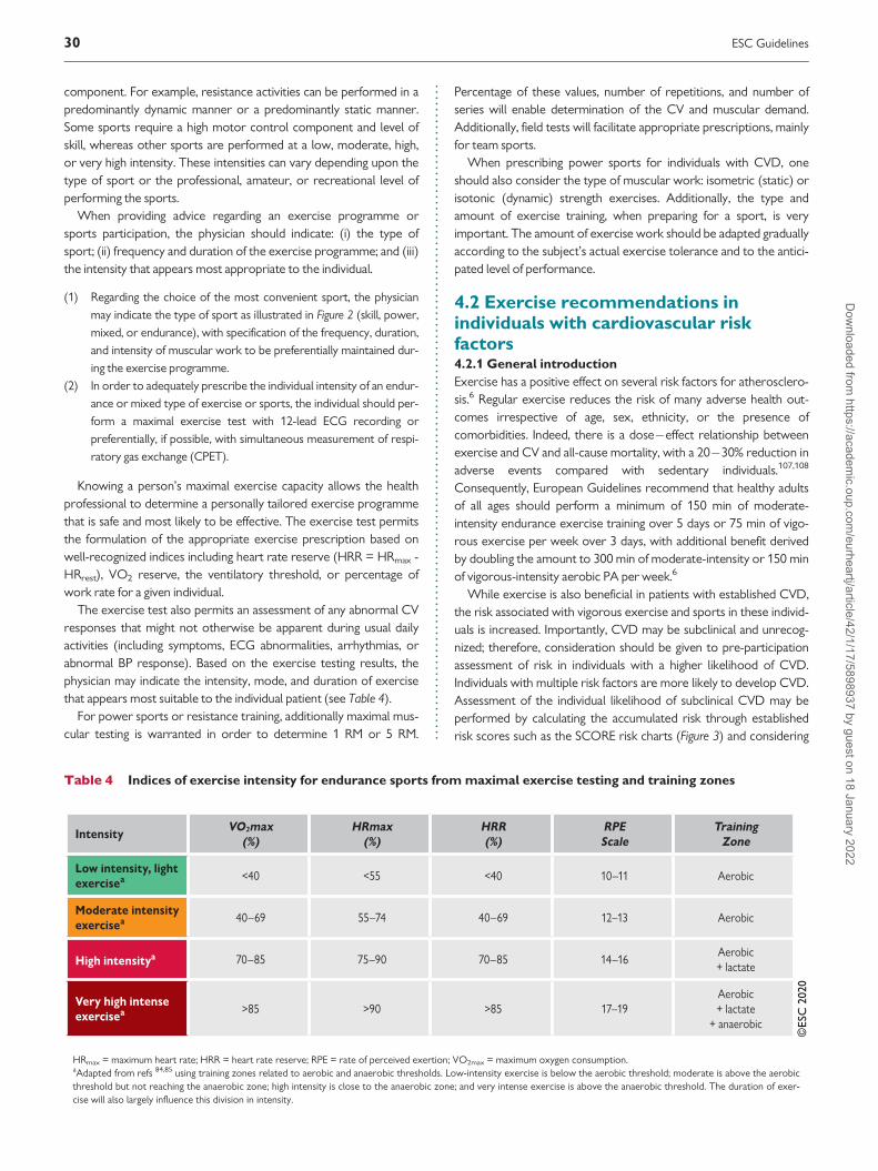
..
..
..
..
..
..
..
..
..
..
..
..
..
..
..
..
..
..
..
..
..
..
..
..
..
..
..
..
..
..
..
..
..
..
..
..
..
..
..
..
..
..
..
..
..
..
..
..
..
..
..
..
..
..
..
..
.component. For example, resistance activities can be performed in apredominantly dynamic manner or a predominantly static manner.Some sports require a high motor control component and level ofskill, whereas other sports are performed at a low, moderate, high,or very high intensity. These intensities can vary depending upon thetype of sport or the professional, amateur, or recreational level ofperforming the sports.
When providing advice regarding an exercise programme orsports participation, the physician should indicate: (i) the type ofsport; (ii) frequency and duration of the exercise programme; and (iii)the intensity that appears most appropriate to the individual.
(1) Regarding the choice of the most convenient sport, the physician
may indicate the type of sport as illustrated in Figure 2 (skill, power,
mixed, or endurance), with specification of the frequency, duration,
and intensity of muscular work to be preferentially maintained dur-
ing the exercise programme.
(2) In order to adequately prescribe the individual intensity of an endur-
ance or mixed type of exercise or sports, the individual should per-
form a maximal exercise test with 12-lead ECG recording or
preferentially, if possible, with simultaneous measurement of respi-
ratory gas exchange (CPET).
Knowing a person’s maximal exercise capacity allows the healthprofessional to determine a personally tailored exercise programmethat is safe and most likely to be effective. The exercise test permitsthe formulation of the appropriate exercise prescription based onwell-recognized indices including heart rate reserve (HRR = HRmax -HRrest), VO2 reserve, the ventilatory threshold, or percentage ofwork rate for a given individual.
The exercise test also permits an assessment of any abnormal CVresponses that might not otherwise be apparent during usual dailyactivities (including symptoms, ECG abnormalities, arrhythmias, orabnormal BP response). Based on the exercise testing results, thephysician may indicate the intensity, mode, and duration of exercisethat appears most suitable to the individual patient (see Table 4).
For power sports or resistance training, additionally maximal mus-cular testing is warranted in order to determine 1 RM or 5 RM.
Percentage of these values, number of repetitions, and number ofseries will enable determination of the CV and muscular demand.Additionally, field tests will facilitate appropriate prescriptions, mainlyfor team sports.
When prescribing power sports for individuals with CVD, oneshould also consider the type of muscular work: isometric (static) orisotonic (dynamic) strength exercises. Additionally, the type andamount of exercise training, when preparing for a sport, is veryimportant. The amount of exercise work should be adapted graduallyaccording to the subject’s actual exercise tolerance and to the antici-pated level of performance.
4.2 Exercise recommendations inindividuals with cardiovascular riskfactors4.2.1 General introduction
Exercise has a positive effect on several risk factors for atherosclero-sis.6 Regular exercise reduces the risk of many adverse health out-comes irrespective of age, sex, ethnicity, or the presence ofcomorbidities. Indeed, there is a dose�effect relationship betweenexercise and CV and all-cause mortality, with a 20�30% reduction inadverse events compared with sedentary individuals.107,108
Consequently, European Guidelines recommend that healthy adultsof all ages should perform a minimum of 150 min of moderate-intensity endurance exercise training over 5 days or 75 min of vigo-rous exercise per week over 3 days, with additional benefit derivedby doubling the amount to 300 min of moderate-intensity or 150 minof vigorous-intensity aerobic PA per week.6
While exercise is also beneficial in patients with established CVD,the risk associated with vigorous exercise and sports in these individ-uals is increased. Importantly, CVD may be subclinical and unrecog-nized; therefore, consideration should be given to pre-participationassessment of risk in individuals with a higher likelihood of CVD.Individuals with multiple risk factors are more likely to develop CVD.Assessment of the individual likelihood of subclinical CVD may beperformed by calculating the accumulated risk through establishedrisk scores such as the SCORE risk charts (Figure 3) and considering
Table 4 Indices of exercise intensity for endurance sports from maximal exercise testing and training zones
©ES
C 2
020
HRmax = maximum heart rate; HRR = heart rate reserve; RPE = rate of perceived exertion; VO2max = maximum oxygen consumption.aAdapted from refs 84,85 using training zones related to aerobic and anaerobic thresholds. Low-intensity exercise is below the aerobic threshold; moderate is above the aerobicthreshold but not reaching the anaerobic zone; high intensity is close to the anaerobic zone; and very intense exercise is above the anaerobic threshold. The duration of exer-cise will also largely influence this division in intensity.
30 ESC GuidelinesD
ownloaded from
https://academic.oup.com
/eurheartj/article/42/1/17/5898937 by guest on 18 January 2022

180
160
140
120
WOMEN
SCORE Cardiovascular Risk Chart10-year risk of fatal CVD
High-risk regions of Europe
MEN
Total cholesterol (mmol/L)
Age
70
65
60
55
50
Non-smoker Smoker
15
8 9
7 7 8 9
40
4
<3%
5 6 7 4 5 6 7
Syst
olic
blo
od p
ress
ure
(mm
Hg)
12 13 14
10 11 12 13
10 10
21
12 13
10 10 11 12
17 19 20
14 15 16 18
14 15
180
160
140
120
9
4 4
3 3 4 4
7 8 8
5 6 6 7
5 5
15
7 7
5 5 6 7
11 12 13
9 9 10 11
8 9
180
160
140
120
5
2 2
1 1 2 2
4 4 5
3 3 3 4
2 3
10
4 4
3 3 3 3
7 8 9
5 6 6 7
4 5
180
160
140
120
3
1 1
1 1 1 1
2 2 3
1 2 2 2
1 1
7
2 2
1 1 2 2
5 5 6
3 3 4 4
2 3
180
160
140
120
2
0 0
0 0 0 0
1 1 2
1 1 1 1
1 1
4
1 1
1 1 1 1
3 3 4
2 2 2 3
1 2
180
160
140
120
1
0 0
3–4% 5–9% ≥10%
0 0 0 0
0 0 1
0 0 0 0
0 0
2
0 0
0 0 0
1 1 2
1 1 1 1
0 1
Non-smoker Smoker
33
16 18
13 15 17 20
4 5 6 7 4 5 6 7
24 26 30
20 22 25 28
21 24
45
23 26
19 22 25 28
33 36 40
27 31 34 39
29 33
23
9 11
7 8 10 11
15 17 20
12 14 16 18
12 14
34
14 16
11 13 15 17
23 26 30
18 21 24 27
19 22
15
5 6
4 4 5 6
10 11 13
7 8 10 11
7 8
25
9 10
6 7 9 10
16 19 22
12 14 16 19
12 14
10
3 3
2 2 3 3
6 7 9
4 5 6 7
4 5
18
5 6
4 4 5 6
11 13 16
8 9 11 13
7 9
7
2 2
1 1 1 2
4 5 6
2 3 3 4
2 3
13
3 4
2 2 3 4
8 9 11
5 6 7 9
5 6
3
0 1
0 0 0 1
2 2 2
1 1 1 2
1 1
7
1 1
1 1 1
4 4 5
2 2 3 4
2 2
0 1©
ESC
202
0
Figure 3a SCORE charts for European populations at high cardiovascular disease (CVD) risk109. The 10-year risk of fatal CVD in populations at high CVD risk isbased on the following risk factors: age, gender, smoking, systolic blood pressure, and total cholesterol. To convert the risk of fatal CVD to risk of total (fatalþ non-fatal) CVD, multiply by 3 in men and 4 in women, and slightly less in older people. Note: the SCORE chart is for use in people without overt CVD, diabetes (type 1 and2), chronic kidney disease, familial hypercholesterolaemia, or very high levels of individual risk factors because such people are already at high risk and need intensive riskfactor advice. Cholesterol: 1 mmol/L¼ 38.67 mg/dL. The SCORE risk charts presented above differ slightly from those in the 2016 ESC/EAS Guidelines for the manage-ment of dyslipidaemias and the 2016 European Guidelines on cardiovascular disease prevention in clinical practice, in that: (i) age has been extended from65 to 70 years; (ii) the interaction between age and each of the other risk factors has been incorporated, thus reducing the overestimation of risk in older persons inthe original SCORE charts; (iii) the cholesterol band of 8 mmol/L has been removed since such persons will qualify for further evaluation in any event.SCORE = Systematic Coronary Risk Evaluation.
ESC Guidelines 31D
ownloaded from
https://academic.oup.com
/eurheartj/article/42/1/17/5898937 by guest on 18 January 2022

180
160
140
120
WOMEN
SCORE Cardiovascular Risk Chart10-year risk of fatal CVD
Low-risk regions of Europe
MEN
Total cholesterol (mmol/L)
Age
70
65
60
55
50
Non-smoker Smoker
9
5 5
4 4 5 5
40
4
<3%
5 6 7 4 5 6 7
Syst
olic
blo
od p
ress
ure
(mm
Hg)
7 8 8
6 6 7 7
6 6
13
7 8
6 6 7 7
11 11 12
9 9 10 11
8 9
180
160
140
120
5
2 3
2 2 2 2
4 4 5
3 3 4 4
3 3
9
4 4
3 3 3 4
7 7 8
5 6 6 7
5 5
180
160
140
120
3
1 1
1 1 1 1
2 3 3
2 2 2 2
1 2
6
2 2
2 2 2 2
4 5 5
3 3 4 4
3 3
180
160
140
120
2
1 1
0 0 0 1
1 1 2
1 1 1 1
1 1
4
1 1
1 1 1 1
3 3 3
2 2 2 3
1 2
180
160
140
120
1
0 0
0 0 0 0
1 1 1
0 0 1 1
0 0
3
1 1
0 0 0 1
2 2 2
1 1 1 2
1 1
180
160
140
120
0
0 0
3–4% 5–9% ≥10%
0 0 0 0
0 0 0
0 0 0 0
0 0
1
0 0
0 0 0
1 1 1
0 0 0 1
0 0
Non-smoker Smoker
17
8 9
7 8 9 10
4 5 6 7 4 5 6 7
12 14 15
10 11 13 14
10 12
24
12 13
10 11 12 14
18 20 22
15 16 18 20
15 17
12
5 5
3 4 5 5
8 9 10
6 7 8 9
6 7
18
7 8
5 6 7 8
12 14 16
9 11 12 14
9 11
8
3 3
2 2 2 3
5 6 7
4 4 5 5
3 4
13
4 5
3 4 4 5
8 10 11
6 7 8 9
6 7
5
1 2
1 1 1 2
3 4 4
2 2 3 3
2 2
9
3 3
2 2 2 3
6 7 8
4 4 5 6
3 4
3
1 1
0 1 1 1
2 2 3
1 1 2 2
1 1
6
1 2
1 1 1 2
4 5 5
2 3 3 4
2 3
1
0 0
0 0 0 0
1 1 1
0 0 1 1
0 0
3
1 1
0 0 0
2 2 3
1 1 1 2
1 1
0 1©
ESC
202
0
Figure 3b SCORE chart for European populations at low cardiovascular disease (CVD) risk. The 10-year risk of fatal CVD in populations at low CVDrisk is based on the following risk factors: age, gender, smoking, systolic blood pressure, and total cholesterol. To convert the risk of fatal CVD to risk oftotal (fatal þ non-fatal) CVD, multiply by 3 in men and 4 in women, and slightly less in older people. Note: the SCORE chart is for use in people withoutovert CVD, diabetes (type 1 and 2), chronic kidney disease, familial hypercholesterolaemia, or very high levels of individual risk factors because such peopleare already at high risk and need intensive risk factor advice. Cholesterol: 1 mmol/L = 38.67 mg/dL. The SCORE risk charts presented above differ slightlyfrom those in the 2016 ESC/EAS Guidelines for the Management of Dyslipidaemias and 2016 European Guidelines on cardiovascular disease prevention inclinical practice, in that: (i) age has been extended from 65 to 70 years; (ii) the interaction between age and each of the other risk factors has been incorpo-rated, thus reducing the overestimation of risk in older persons in the original SCORE charts; (iii) the cholesterol band of 8 mmol/L has been removedsince such persons will qualify for further evaluation in any event. SCORE = Systematic Coronary Risk Evaluation.
32 ESC GuidelinesD
ownloaded from
https://academic.oup.com
/eurheartj/article/42/1/17/5898937 by guest on 18 January 2022

..
..
..
..
..
..
..
..
..
..
..
..
..
..
..
..
..
..
..
..
..
..
..
..
..
..
..
..
..
..
..
..
..
..
..
..
..
..
..
..
..
..
..
.individual risk factors such as very high total cholesterol and low-density lipoprotein (LDL), diabetes mellitus, or a strong family historyof CVD.6 Based on this assessment the individual CV risk can be cate-gorized from low to very high risk (Table 5).
Preliminary evaluation should consist of a self-assessment relatingto symptoms and calculation of SCORE. Individuals who are habitu-ally active and at low or moderate risk should not have any restric-tions for exercise including competitive sports. Sedentary individualsand individuals at high or very high risk may engage in low-intensityexercise without further evaluation. Sedentary individuals and/orthose at high or very high risk planning to undertake high-intensity
exercise as well as selected individuals planning to undertakemoderate-intensity exercise should undergo a physical examination,12-lead ECG, and exercise stress test. The aim of the exercise test isto identify prognostically important CAD and to assess the presenceof exercise-induced arrhythmias. Individuals with symptoms, abnor-mal findings on physical examination, abnormal ECG, or abnormalexercise test should be investigated further according to current ESCGuidelines for chronic coronary syndromes.110 Following normalinvestigations, there should be no restrictions to sports participation.All individuals should, however, be thoroughly informed that devel-opment of symptoms during exercise should prompt reassessment.
While a normal exercise test and a high exercise capacity is associ-ated with a good prognosis, the test has limited sensitivity in diagnos-ing mild to moderate obstructive CAD.111,112 Currently there is noevidence for incorporating routine cardiac imaging in pre-participation screening among asymptomatic individuals aged >35years old with a normal exercise stress test. However, in asympto-matic adults considered to be at high risk or very high risk (diabetes,strong family history of CAD, previous risk assessment suggestinghigh risk for CAD) a functional imaging test or coronary computedtomography angiography (CCTA) should be considered in the riskassessment (Figure 4).110 Identification of atherosclerotic CAD shouldprompt aggressive management of risk factors and preventive medi-cal treatment. Among individuals with proven obstructive CAD, fur-ther assessment and treatment is indicated.
Table 5 Cardiovascular risk categories
©ES
C 2
020
General recommendations for exercise and sports inhealthy individuals
Recommendations Classa Levelb
At least 150 min/week of moderate-intensity, or
75 min/week of vigorous-intensity aerobic exer-
cise, or an equivalent combination thereof is
recommended in all healthy adults.113�118
I A
A gradual increase in aerobic exercise to 300
min/week of moderate-intensity, or 150 min/
week of vigorous-intensity aerobic exercise, or
an equivalent combination is recommended for
additional benefits in healthy adults.114,116
I A
Regular assessment and counselling to promote
adherence and, if necessary, to support an
increase in exercise volume over time are
recommended.119
I B
Multiple sessions of exercise spread throughout
the week, i.e. on 4�5 days a week and prefera-
bly every day of the week, are
recommended.113,114
I B
aClass of recommendation.bLevel of evidence.
ESC Guidelines 33D
ownloaded from
https://academic.oup.com
/eurheartj/article/42/1/17/5898937 by guest on 18 January 2022

..
..
..
..
..
..
..
..
..
..
..
..
..
..
..
..
..
..
..
..
..
..
..
..
..
..
..
..
..
..
..
..
..
..
..
..
..
..
..
..
..
..
..
..
.. 4.2.2 Obesity
A person with a body mass index (BMI) >30 kg/m2 or (preferentially)a waist circumference >94 cm for males and >80 cm for females(both for European Caucasians) is considered obese.120,121
European guidelines for obese individuals recommend that a mini-mum of 150 min/week of moderate-intensity endurance exercisetraining should be combined with three weekly sessions of resistanceexercise.121 Such intervention leads to a reduction in intra-abdominalfat mass, increments in muscle and bone mass, attenuation in theweight loss-induced decline of resting energy expenditure, reductionin BP and chronic inflammation, and improvement in glucose toler-ance, insulin sensitivity, lipid profile, and physical fitness.121,122 Thereis also a positive influence on the long-term maintenance of weightreduction, general well-being and self-esteem, and reduction in anxi-ety and depression.121 The impact of exercise intervention alone onfat mass is modest.123 According to a series of large randomized con-trolled trials a high endurance-type exercise volume, >225 min/week,is required to maximize fat mass loss in obese individuals.124
A pre-participation CV assessment is warranted in obese indi-viduals who intend to engage in high-intensity exercise (Figure 4),due to associated comorbidities such as type 2 diabetes, hyperten-sion, dyslipidaemia, and CV and respiratory diseases.121 Obeseindividuals with a normal CV assessment should not have anyrestrictions on exercise. There is evidence from healthy, non-obese and non-athletic individuals that running, and abruptincreases in training volume, contribute to musculoskeletalinjuries.125�127 Therefore, it may be reasonable to consider thatobese individuals should limit high-volume weight-bearing exer-cises on a hard surface (i.e. <2 h/day) until a considerable
Recommendations for cardiovascular evaluation andregular exercise in healthy individuals aged >35 years
Recommendations Classa Levelb
Among individuals with low to moderate CVD
risk, the participation in all recreational sports
should be considered without further CV
evaluation.
IIa C
Cardiac screening with family history, symptoms,
physical examination, and 12-lead resting ECG
should be considered for competitive athletes.
IIa C
Clinical evaluation, including maximal exercise
testing, should be considered for prognostic pur-
poses in sedentary people and individuals with
high or very high CV risk who intend to engage in
intensive exercise programmes or competitive
sports.
IIa C
In selected individuals without known CAD who
have very high CVD risk (e.g. SCORE>10%, strong
family history, or familial hypercholesterolaemia)
and want to engage in high- or very high-intensity
exercise, risk assessment with a functional imaging
test, coronary CCTA, or carotid or femoral artery
ultrasound imaging may be considered.
IIb B
CCTA = coronary computed tomography angiography; CV = cardiovascular;CVD = cardiovascular disease; SCORE = Systematic Coronary Risk Evaluation.aClass of recommendation.bLevel of evidence.
©ES
C 2
020
Figure 4 Proposed algorithm for cardiovascular assessment in asymptomatic individuals aged >35-years-old with risk factors for cardiovascular disease andpossible subclinical chronic coronary syndrome before engaging in sports. *Consider functional test or CCTA if exercise stress test is equivocal or the ECG isuninterpretable. aSee text for examples of functional imaging. bSingle-photon emission computed tomography: area of ischaemia >_10% of the left ventricularmyocardium; stress echocardiography: >_3 of 16 segments with stress-induced hypokinesia or akinesia; stress cardiovascular magnetic resonance: >_2 of 16 seg-ments with stress perfusion defects or >_3 dobutamine-induced dysfunctional segments; coronary computed tomography angiography (CCTA): three-vesseldisease with proximal stenoses; left main disease; proximal left anterior descending disease.110 CVD = cardiovascular disease; ECG = electrocardiogram;SCORE = Systematic Coronary Risk Evaluation.
34 ESC GuidelinesD
ownloaded from
https://academic.oup.com
/eurheartj/article/42/1/17/5898937 by guest on 18 January 2022

..
..
..
..
..
..
..
..
..
..
..
..
..
..
..
..
..
..
..
..
..
..
..
..
..
..
..
..
..
..
..
..
..
..
..
..
..
..
..
..
..
..
..
..
..
..
..
..
..
..
..
..
..
..
..
..
..
..
..
..
..
..
..
..
..
..
..
..
..
..
..
..
..
..
..
..
..
..
..reduction in body weight is achieved. Moreover, if high-volumeexercise (>2 h/day) is desired, a sufficient recovery time should beallowed for between periods of exercise (optimally 48 h). It isimportant to emphasize that good physical and muscular fitnessand neuromuscular coordination may protect obese individualsfrom musculoskeletal injuries, hence non-weight-bearing exer-cises such as cycling or swimming128 may be beneficial. Finally,there is no compelling evidence that resistance training, whenexecuted properly, will increase the risk for musculoskeletal inju-ries or provoke musculoskeletal symptoms in obeseindividuals.129
4.2.3 Hypertension
A person with a persistent systolic BP (SBP) >_140 mmHg and/or dia-stolic BP (DBP) >_90 mmHg is considered hypertensive.130,131
Hypertensive individuals should participate in at least 30 min ofmoderate-intense dynamic aerobic exercise (walking, jogging, cycling,or swimming) for 5�7 days per week.132 Such exercise interventionis associated with a mean reduction in SBP of 7 mmHg and DBP of 5mmHg.133 Additional resistance training is highly effective in reducingBP further and resistance training 2�3 days per week is alsoadvised.132 Indeed, the BP-lowering effect of resistance and isometricexercise may be comparable to, or even greater than, that of aerobicexercise.134
If high-intensity sports participation is desired, a pre-participationCV assessment is warranted to identify athletes with exercise-induced symptoms, excessive BP response to exercise130, and thepresence of end organ damage. Individuals with symptoms suggestiveof CAD require further assessment and optimization of medical ther-apy before participation in sports. If arterial hypertension is poorlycontrolled (resting SBP > 160 mmHg), a maximal exercise test shouldbe postponed until the BP is controlled.
Non-pharmacological measures should be considered as thefirst step in the management of hypertension in athletes, includ-ing: restriction of salt intake and alcohol consumption, weightreduction if applicable, balanced diet (e.g. Mediterranean diet),and cessation of smoking. Aerobic exercise programmes shouldherein complement the individual’s training schedule.131 If suchlifestyle changes do not lower BP after 3 months, antihyperten-sive drugs should be commenced if SBP remains >140 mmHg.Antihypertensive therapy alongside lifestyle intervention shouldbe considered in all individuals aged >65 years but <80 years,provided it is well tolerated.131,132 It is important to considerthat beta-blockers are prohibited in certain competitive skillsports such as shooting [see World Anti-Doping Association(WADA) for complete list135], and can induce bradycardia and/or lower aerobic exercise capacity.131 Diuretics are prohibited inall competitive sports.135Angiotensin-converting enzyme (ACE)inhibitors, angiotensin II receptor blockers, and calcium antago-nists are the preferred drugs of choice in exercising individuals. Itis noteworthy that the use of non-selective non-steroidal
inflammatory drugs for musculoskeletal pain may contribute toraised BP.136
When BP is uncontrolled, temporary restriction from competitivesports is recommended, with the possible exception of skill sports.131
In individuals with a high-risk profile, including those with end organdamage [left ventricular (LV) hypertrophy, diastolic dysfunction,ultrasound evidence of arterial wall thickening or atherosclerotic pla-que, hypertensive retinopathy, increased serum creatinine (men1.3�1.5 mg/dL, women 1.2�1.4 mg/dL), and/or microalbuminuria]in whom BP is controlled, participation in all competitive sports ispossible, with the exception of the most intensive power disciplinessuch as discus/javelin throwing, shot-putting, and weightlifting (seesection 4.1).131
During sports participation, regular follow-up is recommendeddepending on the severity of hypertension and the category of risk. Inindividuals with borderline BP readings, regular ambulatory assess-ment of BP should be considered. In individuals with low or moderateCV risk and well-controlled BP, there should be no restrictions tosports participation, however, intensive heavy weightlifting, especiallywhen this includes substantial isometric (static) muscle work, canhave a marked pressor effect and should be avoided. In thiscontext, avoiding the Valsalva manoeuvre in particular is warrantedbecause breath holding during muscular contraction is associatedwith a greater elevation in SBP and DBP.135 When executed cor-rectly, high-intense dynamic resistance training (up to 80% of 1 RM),with a low number of repetitions (n < 10) does not induce greaterincrements in BP compared with low-intense dynamic resistancetraining (<50% of 1 RM) with a high number of repetitions (n >_20).137�142
Some individuals who are normotensive at rest will have an exag-gerated BP response to exercise. An exaggerated BP response toexercise increases the risk for incident hypertension in highly trainedand normotensive athletes over a middle-term period.143 If SBP risesto >200 mmHg at a workload of 100 W during exercise testing,144
antihypertensive medical therapy should be optimized and clinicalevaluation, including ECG and echocardiography, should be consid-ered, even if the athlete is normotensive at rest.131 Moreover, inyoung Olympic athletes a peak SBP of >220 mmHg in males and>200 mmHg in females measured during cycle ergometry are beyondthe 95th percentile.131
4.2.4 Dyslipidaemia
Physical activity has favourable effects on lipid metabolism by reduc-ing serum triglycerides by up to 50% and increasing high-density lipo-protein (HDL) cholesterol by 5�10%.85,145 Exercise may alsoreduce LDL cholesterol by up to 5% and shift the more atherogenicsmall, dense LDL fraction towards larger LDL particles in a dose-dependent fashion.146 These metabolic improvements can beachieved through 3.5 - 7 h of moderately vigorous PA per week or30 - 60 min of exercise on most days.
In individuals with hypertriglyceridaemia or hypercholesterolae-mia, a higher intensity of exercise is recommended, as this may
ESC Guidelines 35D
ownloaded from
https://academic.oup.com
/eurheartj/article/42/1/17/5898937 by guest on 18 January 2022

..
..
..
..
..
..
..
..
..
..
..
..
..
..
..
..
..
..
..
..
..
..
..
..
..
..
..
..
..
..
..
..
..
..
..
..
..
..
..
..
..
..
..
..
..
..
..
..
..
..
..
..
..
..
..
..
..
..
..
..
..
..
..
..
..
..
..
..
..
..
..
..
..
..
..
..
..
..
..
..improve the lipid profile and reduce CV risk. Before embarking onhigh-intensity exercise, a clinical assessment should be performedincluding symptomatic status, and a maximal exercise stress test,functional imaging test, or CCTA may be considered in the riskassessment110 (Figure 4), particularly in individuals with familial hyper-cholesterolaemia. Among athletes with hypercholesterolaemia, regu-lar exercise will rarely reduce LDL cholesterol to normal or near-normal values; therefore, guidelines on pharmacological treatment inprimary and secondary prevention should be followed strictly.Individuals with dyslipidaemia should be assessed at least every 2�5years for primary prevention and annually for secondary prevention.
Pharmacological intervention, particularly with statins, is superiorto exercise and lifestyle intervention alone for reducing LDL choles-terol and improving prognosis.147 Despite the minor effects of endur-ance exercise on serum LDL cholesterol, the clinically beneficialrelationship between increased physical fitness and reduced CVevents remains beyond the effects of statins.147,148
Physically active individuals with dyslipidaemia may experiencemuscle pain and soreness or tendinopathy accompanied by elevatedmuscle enzymes.149 In these cases, measures such as stopping medi-cation temporarily followed by repeat challenge with another statindrug, with or without an alternate day regimen, or introducing otherlipid-lowering agents such as ezetimibe or proprotein convertasesubtilisin/kexin type 9 (PCSK-9) inhibitors should be considered.109
Individuals who develop rhabdomyolysis due to a statin should beprescribed an alternative lipid-lowering agent.
4.2.5 Diabetes mellitus
Physical inactivity is a major cause of type 2 diabetes mellitus(T2DM).150 The risk of developing T2DM is 50�80% higher in indi-viduals who are physically inactive compared to their active counter-parts. However, exercise does not entirely compensate for the effectof obesity.151�154 Diabetes is also independently associated with anaccelerated decline in muscular strength and, partly because ofhyperglycaemia, may lead to reduced joint mobility.
4.2.5.1 Effect of exercise on diabetic control, risk factors and outcomesAerobic exercise in patients with T2DM improves glycaemic controland reduces visceral fat and insulin resistance. Exercise also has bene-ficial effects on BP and lipid profile, and leads to modest weightloss.155,156 Both aerobic and resistance training promote prolongedadaptations in skeletal muscle, adipose tissue, and the liver associatedwith enhanced insulin action.157 Observational studies have shownlower mortality with exercise in both type 1 diabetes mellitus andT2DM.158
In patients with pre-diabetes or metabolic syndrome, both aerobicand resistance exercise may prevent the development of overt dia-betes.159�162 Intensity of exercise seems to be of greater importancethan the volume of exercise; individuals who exercise at moderate orhigh intensity have a lower risk of developing metabolic impairmentcompared with those who have a similar energy expenditure at alower intensity.160,163
The effects on muscle insulin sensitivity are observed with a rela-tively low volume of exercise (400 kcal/week) in previously sedentaryadults, but increase with higher volumes of exercise.164 The optimalcombination of duration and intensity is not well established. High-intensity interval training may be superior to moderate aerobic train-ing in achieving metabolic effects and improvement in exercisecapacity; however, whether long-term results are superior isunknown.165,166
Diabetes is a cause of coronary microvascular dysfunction (CMD),which is associated with lower exercise capacity and adverse out-comes167,168 and can be improved by exercise training.162,169�171
Large randomized trials have confirmed the beneficial effect of exer-cise intervention on glycaemic control and risk factors, but this hasnot translated into a significant improvement in survival, partlybecause of suboptimal long-term maintenance of lifestyle changes.172
During an acute bout of exercise, glucose uptake in the muscles isincreased for up to 2 h afterwards through mechanisms that are inde-pendent of insulin. The exercise-induced hypoglycaemic effect can bediminished by performing resistance training or interval training inpatients with type 1 diabetes.173 There is a dose�response relation-ship between intensity and volume of exercise and duration of glu-cose uptake by skeletal muscle that may last up to 48 h after exercise.These factors must be considered in individuals with diabetes whoare undertaking intensive exercise or competitive sports in order toavoid hypoglycaemia.
4.2.5.2 Recommendations for participation in exercise in individuals withdiabetes mellitusBoth aerobic and resistance training are effective for glycaemic con-trol, BP reduction, weight loss, peak exercise capacity, and dyslipide-mia.174 A programme combining aerobic and resistance training hasbeen shown to be superior in terms of glycaemic control, whereasthe effect on other outcomes is unproven.174�176
The ideal exercise programme to achieve the full potential of ben-efits in patients with diabetes is daily exercise of at least moderateintensity, e.g. brisk walking, for at least 30 min, resistance training for15 min on most days, and lighter-intensity activities (standing, walk-ing) every 30 min. This can be supplemented by flexibility and balanceexercise, particularly in older individuals or patients with microvascu-lar complications due to their diabetes.
4.2.5.3 Cardiac evaluation before exercise participation in individualswith diabetes mellitusIndividuals with diabetes have a priori a higher likelihood of subclinicalCAD; therefore, all individuals with diabetes should undergo CVassessment as outlined in Figure 4 before taking up an exercise pro-gramme of high intensity. This should be supplemented by an evalua-tion of glycaemic status, including risk factors for hypoglycaemia,history of hypoglycaemic episodes, presence of autonomic neuropa-thy, and antidiabetic treatment.177
Asymptomatic individuals with diabetes mellitus and a normal CVassessment and maximal exercise test may engage in all sports butshould be warned about the potential risk of iatrogenic
36 ESC GuidelinesD
ownloaded from
https://academic.oup.com
/eurheartj/article/42/1/17/5898937 by guest on 18 January 2022

..
..
..
..
..
..
..
..
..
..
..
..
..
..
..
..
..
..
..
..
..
..
..
..
..
..
..
..
..
..
..
..
..
..
..
..
..
..
..
..
..
..
..
..
..
..
..
..
..
..
..
..
..
..
..
..
..
..
..
..
..
..
..
..
..
..
..
..
..
..
..
..
..
..
..
..
..
..
..
..
..
..
..
..hypoglycaemia in the event of inadequate caloric intake. Importantly,all patients with diabetes should be aware of warning symptoms andattention should be given to chest discomfort or unusual breathless-ness during exercise as this may be indicative of CAD.
4.3 Exercise and sports in ageing4.3.1 Introduction
The elderly are defined as adults aged above 65 years. Similar to thegeneral population, higher exercise capacity in this age group is alsoassociated with reduced mortality.179 A physically active lifestylemaintained through middle and older age translates into betterhealth180 and longevity.181�185 Commencing a new exercise regimenamong sedentary elderly individuals has shown significant healthimprovements180,186 including cognitive capacity.187�190 Moreover,regular exercise exerts beneficial effects in reducing the risk of devel-oping CV and metabolic disease through improved control of CV riskfactors,191,192 also preserving cognitive function.187�190 Importantly,exercise helps to preserve neuromuscular competence,193,194 thusmaintaining balance and coordination, which reduces the risk offalling.195,196
4.3.2 Risk stratification, inclusion/exclusion criteria
Moderate-intensity exercise is generally safe for older healthy peopleand medical consultation before starting or progressing the level ofexercise programme is not usually required.81,197 The general recom-mendation for exercise implementation for the general populationalso applies to healthy elderly people.
Nevertheless, due to potential risks of exercising among the eld-erly (Table 6), the European Association of Preventive Cardiology(EAPC) recommends self-assessment by a brief questionnaire81 todetermine the need for advice from health professionals, but thisapproach has not been tested prospectively.
Community-dwelling frail or sedentary older adults may have aslightly increased risk of falls during exercise; however, there is noevidence of serious adverse outcomes, injury, or CVevents.195,196,198,199 Exercise interventions to improve balance inthose diagnosed with dementia bring numerous benefits without anincreased risk of adverse outcomes.200 Resistance exercise in olderadults is rarely associated with adverse events.201,202 No major riskshave been reported in older individuals performing low- andmoderate-intensity aerobic exercise, and even more intense aerobicactivities are associated with a relatively small risk.203�205 CV eventsduring intense exercise occur at a rate of around 1 event per 100years of vigorous activity.206 Risks are highest during the first fewweeks of beginning vigorous exercise; therefore both exercise inten-sity and duration should be increased gently (for example, every 4weeks).81,197,207�210 Among older individuals who are well preparedand accustomed to intense exercise, participation in competitive vig-orous sports does not confer higher risk compared with youngeradults.38,211
4.3.3 Exercise modalities and recommendations for
exercise and sport in the elderly
The physical exercises for elderly persons should be designedaccording to their biological age, exercise experience, functionalcapacity, safety, ageing trajectories, comorbidity, lifestyle habits, andprevious experience of exercise.
Elderly people should perform endurance and strength exercise,and specific exercises for flexibility and balance (Table 7).201,212,213
Endurance exercise exerts beneficial effects on the cardiorespiratorysystem and resistance exercise prevents the decrease in muscle massand sarcopenia.192 Achieving >150 min/week moderate-intensityaerobic exercise (i.e. walking or other moderate intensity aerobics-type activities) is associated with at least 30% lower risk of morbidity,mortality, disability, frailty, and dementia compared with being inac-tive.212,214,215 The strength exercises for the major muscle groupsshould be performed at least twice a week (8�10 different exercises,10�15 repetitions).
Accustomed senior athletes should continue performing exerciseand sports activities, without any predetermined age limit.38,211,216
Sports activities for older people according to exercise type andintensity are reported in Table 8. Annual clinical assessment includinga maximal exercise test (preferably with simultaneous CPET) is rec-ommended in master athletes performing a high level of sports andexercise programmes.217
Special considerations for individuals with obesity,hypertension, dyslipidaemia, or diabetes
Recommendations Classa Levelb
In obese individuals (BMI>_30 kg/m2 or a waist cir-
cumference >80 cm for females or >94 cm for
males) resistance training >_3 times per week, in
addition to moderate or vigorous aerobic exercise
(at least 30 min, 5�7 days per week) is recom-
mended to reduce CVD risk.121
I A
In individuals with well-controlled hypertension,
resistance training >_3 times per week in addition to
moderate or vigorous aerobic exercise (at least 30
min, 5�7 days per week) is recommended to
reduce blood pressure and CVD risk. 132
I A
Among individuals with diabetes mellitus, resistance
training >_3 times per week in addition to moderate
or vigorous aerobic exercise (at least 30 min, 5�7
days per week) is recommended to improve insulin
sensitivity and achieve a better CVD risk
profile.176,178
I A
Among adults with well-controlled hypertension
but high risk and/or target organ damage, high-inten-
sity resistance exercise is not recommended.
III C
In individuals with uncontrolled hypertension
(SBP>160 mmHg) high-intensity exercise is not rec-
ommended until blood pressure has been
controlled.
III C
CVD = cardiovascular disease; SBP = systolic blood pressure.aClass of recommendation.bLevel of evidence.
ESC Guidelines 37D
ownloaded from
https://academic.oup.com
/eurheartj/article/42/1/17/5898937 by guest on 18 January 2022
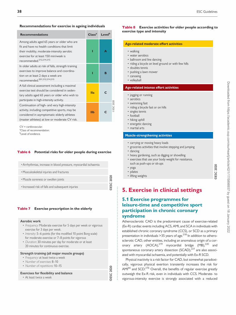
..
..
..
..
..
..
..
..
..
..
..
..
..
..
..
..
..
..
..
..
..
..
..
..
..
..
..
..
..
..5. Exercise in clinical settings
5.1 Exercise programmes forleisure-time and competitive sportparticipation in chronic coronarysyndromeAtherosclerotic CAD is the predominant cause of exercise-related(Ex-R) cardiac events including ACS, AMI, and SCA in individuals withestablished chronic coronary syndrome (CCS), or SCD as a primarypresentation in individuals >35 years of age.218 In addition to athero-sclerotic CAD, other entities, including an anomalous origin of a cor-onary artery (AOCA),219 myocardial bridge (MB),220 andspontaneous coronary artery dissection (SCAD),221 are also associ-ated with myocardial ischaemia, and potentially with Ex-R SCD.
Physical inactivity is a risk factor for CAD, but somewhat paradoxi-cally, vigorous physical exertion transiently increases the risk forAMI66 and SCD.216 Overall, the benefits of regular exercise greatlyoutweigh the Ex-R risk, even in individuals with CCS. Moderate- tovigorous-intensity exercise is strongly associated with a reduced
Table 6 Potential risks for older people during exercise
©ES
C 2
020
Table 7 Exercise prescription in the elderly
©ES
C 2
020
Recommendations for exercise in ageing individuals
Recommendations Classa Levelb
Among adults aged 65 years or older who are
fit and have no health conditions that limit
their mobility, moderate-intensity aerobic
exercise for at least 150 min/week is
recommended.212,214,215
I A
In older adults at risk of falls, strength training
exercises to improve balance and coordina-
tion on at least 2 days a week are
recommended.201,212,214,215
I B
A full clinical assessment including a maximal
exercise test should be considered in seden-
tary adults aged 65 years or older who wish to
participate in high-intensity activity.
IIa C
Continuation of high- and very high-intensity
activity, including competitive sports, may be
considered in asymptomatic elderly athletes
(master athletes) at low or moderate CV risk.
IIb C
CV = cardiovascular.aClass of recommendation.bLevel of evidence.
Table 8 Exercise activities for older people according toexercise type and intensity
©ES
C 2
020
38 ESC GuidelinesD
ownloaded from
https://academic.oup.com
/eurheartj/article/42/1/17/5898937 by guest on 18 January 2022

..
..
..
..
..
..
..
..
..
..
..
..
..
..
..
..
..
..
..
..
..
..
..
..
..
..
..
..
..
..
..
..
..
..
..
..
..
..
..
..
..
..
..
..
..
..
..
..
..
..
..
..
..
..
..
..
..
..
..
..
..
..
..
..
.
incidence of adverse outcomes from CAD, but prolonged, high-intensity endurance exercise has been associated with increased cor-onary artery calcium (CAC), a marker of atherosclerosis,58,222 andcoronary plaques58 but without an increase in mortality112 in themedium term. Importantly, the diagnosis of myocardial injury is alsomore complex in athletes because intense exercise may increaseserologic markers of myocardial injury, including cardiac troponin Iand T.223,224
5.1.1 Individuals at risk of atherosclerotic coronary artery
disease and asymptomatic individuals in whom coronary
artery disease is detected at screening
Athletes or individuals participating in sports or regular exercisetraining may have risk factors for CAD and/or subclinical CCS.225
Such individuals may be identified by routine pre-participationscreening as recommended by the ESC21 or by pre-evaluation ofmaster athletes, as suggested by the European Association forCardiovascular Prevention and Rehabilitation (EACPR) 2011207 andthe AHA.226
In addition to the SCORE risk stratification described earlier(Table 5), the increasing use of cardiac imaging techniques allows theidentification of a greater number of individuals with asymptomaticCCS,227 including competitive master athletes.227
Newer predictive measures, such as high-sensitive C-reactive pro-tein and carotid intima�media thickness (IMT) add little to the tradi-tional risk factors.110 The exception is CAC, which providesadditional predictive information in individuals with a moderate-riskprofile,228 dividing them into low- or high-risk individuals. The mostprudent and cost-effective method of utilizing CAC may thus be addi-tive to the traditional risk factors,229 as suggested by the EAPC.230
Clinical evaluation of asymptomatic individuals with possible sub-clinical CCS should include (Figure 4):112
(1) Assessment of risk of CVD110 (Table 5)
(2) Consideration of intensity of intended exercise programme
(3) Clinical evaluation, including maximal exercise stress test
(4) Further diagnostic testing in selected individuals.
Many middle-aged individuals in the general population can beexpected to have some level of subclinical CCS as assessed withimaging techniques. Anatomical coronary imaging alone does notprovide information relating to the coronary flow and reserve, whichis important in assessing the risk of Ex-R ischaemia or SCD/SCA;therefore functional evaluation is necessary. Several methods ofstress testing (e.g. cycle ergometry or treadmill testing), stress echo-cardiography, adenosine or dobutamine stress cardiac magnetic reso-nance (CMR), or positron emission tomography (PET)/single-photonemission computed tomography (SPECT), can be used to detectinducible myocardial ischaemia231. Exercise stress-echo is preferredin athletes because it is free from radiation and does not involveadministration of drugs.
Exercise testing is the most widely available functional test, andprovides information on exercise capacity, heart rate, and BPresponse, and detection of exercise-induced arrhythmias,2 but has a
lower specificity for myocardial ischaemia than other functional tests,especially in asymptomatic and low-risk individuals. It is recom-mended that a truly maximal exercise test232 (with or without CPET)should be performed when evaluating individuals with possible sub-clinical (or clinical) CCS who intend to or are participating in system-atic exercise including recreational or competitive sports. Whetherthe initial exercise test includes imaging or not depends on factorssuch as the baseline ECG (Table 9) and feasibility of performing func-tional imaging tests in a given institute.
• If the clinical assessment, including a maximal exercise test is nor-mal, the presence of ‘relevant CAD’ is assumed to be unlikely(Figure 4).
• In the event of a borderline or uninterpretable exercise test result,it is recommended that a more specific imaging stress test is per-formed such as stress-echocardiography, CMR perfusion imaging,or SPECT. Maximal exercise SPECT and exercise echocardiogra-phy or nuclear perfusion techniques utilizing exercise rather thanpharmacological stress may preferentially be used, depending onavailability and local expertise.
• If the exercise test is positive, an invasive coronary angiogramshould be performed to confirm the presence, extent, andseverity of CAD (Figure 4).
5.1.1.1 Recommendations for sports participationIndividuals at risk of CAD and asymptomatic individuals in whom CADis detected at screening should have aggressive management of risk fac-tors for atherosclerosis.6,131,132,202 Considering the benefits of exerciseon primary and secondary prevention of CCS,6,234 individuals with riskfactors should be restricted from competitive sport only when there issubstantial risk of an adverse event, as indicated by functional tests, orwhen there is evidence of disease progression during serial evalua-tions.233 Exercise recommendations should be individually tailoredbased on the intensity of the exercise and the sporting discipline.Participation in competitive endurance, power, and mixed disciplines(see sections 4.2 and 5.1.3) generally requires vigorous effort and ismore likely to induce myocardial ischaemia, whereas leisure sports orintentional recreational exercise allows for greater control of physicaleffort. Individuals with a high risk of atherosclerotic CAD and asympto-matic individuals in whom CAD is detected at screening who partici-pate in intensive exercise should be assessed with a maximal exercisetest or functional imaging test on an annual basis.
Table 9 Borderline or uninterpretable ECG findings
©ES
C 2
020
ESC Guidelines 39D
ownloaded from
https://academic.oup.com
/eurheartj/article/42/1/17/5898937 by guest on 18 January 2022

..
..
..
..
..
..
..
..
..
..
..
..
..
..
..
..
..
..
..
..
..
..
..
..
..
..
..
..
..
..
..
..
..
.
5.1.2 Established (long-standing) chronic coronary
syndrome
All individuals with established (long-standing) CCS should beencouraged to perform the minimal PA recommendations for gen-eral and CV health.235 This applies to individuals with stable angina,asymptomatic and symptomatic individuals stabilized <1 year afterACS, or individuals with recent revascularization, and asymptomaticand symptomatic individuals >1 year after initial diagnosis or revascu-larization.110 Advice on intensive exercise and participation in mostcompetitive sports in asymptomatic individuals with long-standingCCS should be based on several factors, which are determinedthrough clinical history, exercise stress testing, or functional imagingand echocardiography (Table 10).
Individuals with long-standing CCS who do not show any abnormal-ities on a maximal exercise test or functional imaging test, or haveunimpaired LV function, may be considered as low risk for an exercise-induced adverse event236�238 (Table 11). Such individuals may engagein all competitive sports on an individual basis (Figure 5). Some restric-tions may apply for high-intensity power, mixed, and endurance sports
(see Figure 2, section 4.1.2) for older patients (>60 years old) with CCS.This is due to the fact that age is an additional, strong predictor ofadverse events during exercise. There are no restrictions in low-riskpatients for skills sports regardless of age (Figure 2).
Individuals with inducible ischaemia during functional testing,despite adequate treatment, should undergo coronary angiography;those with high-risk lesions on coronary angiography (Table 11)should have revascularization prior to considering high-intensityexercise programmes or competitive sport (Figure 5). Individuals withhigh-risk coronary features may gradually return to sport 3�6months after successful revascularization pending a normal maximalexercise or functional imaging test.
When ischaemia cannot be treated despite adequate therapy,including revascularization, the individual should be restricted fromcompetitive sports, with the possible exception of individually rec-ommended low-intensity skill sports. Such individuals may engage inregular recreational exercise of low and moderate intensity providedrisk factors and symptoms are treated adequately and there is regularclinical surveillance. These individuals may also participate in leisuresports, 2�3 times/week, in selected cases, if the intended activity isbelow (around 10 beats) the ischaemic threshold and below the levelof arrhythmias.231
5.1.2.1 Antithrombotic treatmentIndividuals with CAD should receive conventional antithrombotictreatment for secondary prevention, according to published guide-lines for the general population.233,239,240 Individuals taking dual anti-platelet agents should avoid sports with bodily collision, especiallywhen they are combined with oral anticoagulants, due to the risk ofhaemorrhage.241
Recommendations for exercise in individuals at risk ofatherosclerotic coronary artery disease and asympto-matic individuals in whom coronary artery disease isdetected at screening
Recommendations Classa Levelb
Among individuals with asymptomatic CCS, defined
as CAD without inducible myocardial ischaemia on
a functional imaging or conventional exercise stress
test, 233 participation in all types of exercise, includ-
ing competitive sports, should be considered based
on individual assessment.
IIa C
CAD = coronary artery disease; CCS = chronic coronary syndrome.aClass of recommendation.bLevel of evidence.
Table 10 Factors determining risk of adverse events dur-ing intensive exercise and competitive sports in asympto-matic individuals with long-standing coronary arterydisease
©ES
C 2
020
Table 11 High-risk features for exercise-induced adversecardiac events in patients with atherosclerotic coronaryartery disease233
©ES
C 2
020
ACS = acute coronary syndrome; FFR = fractional flow reserve; iFR = instantflow reserve; NSVT = non-sustained ventricular tachycardia; PCI = percutaneouscoronary intervention.
40 ESC GuidelinesD
ownloaded from
https://academic.oup.com
/eurheartj/article/42/1/17/5898937 by guest on 18 January 2022

©ES
C 2
020
Figure 5 Clinical evaluation and recommendations for sports participation in individuals with established coronary artery disease. CCS = chronic coro-nary syndrome; LAD = left anterior descending coronary artery; LM = left main coronary artery; LVEF = left ventricular ejection fraction. *With docu-mented ischaemia or a haemodynamically relevant lesion defined by FFR <0.8 or iFR <0.9.
Recommendations for exercise in individuals with long-standing chronic coronary syndrome
Recommendations Classa Levelb
Risk stratification for exercise-induced adverse events is recommended in individuals with established (long-standing) chronic cor-
onary syndrome (CCS) prior to engaging in exercise.233 I C
Regular follow-up and risk stratification of patients with CCS is recommended.233 I B
It is recommended that individuals at high risk of an adverse event from CAD are managed according to the current Guidelines
on CCS.233 I C
Competitive or leisure sports activities (with some exceptions such as older athletes and sports with extreme CV demands)
should be considered in individuals at low risk of exercise-induced adverse events (Table 11).233 IIa C
Leisure-time exercise, below the angina and ischaemic thresholds, may be considered in individuals at high risk of exercise-
induced adverse events (Table 11), including those with persisting ischaemia.233 IIb C
Competitive sports are not recommended in individuals at high risk of exercise-induced adverse events or those with residual
ischaemia, with the exception of individually recommended skill sports.233 III C
CAD = coronary artery disease; CCS = chronic coronary syndrome; CV = cardiovascular.aClass of recommendation.bLevel of evidence.
ESC Guidelines 41D
ownloaded from
https://academic.oup.com
/eurheartj/article/42/1/17/5898937 by guest on 18 January 2022
..
..
..
..
..
..
..
..
..
..
..
..
..
..
..
..
..
..
..
..
..
..
..
..
..
..
..
..
..
..
..
..
..
..
..
..
..
..
..
..
..
..
..
..
..
..
..
..
..
..
5.1.3 Myocardial ischaemia without obstructive disease in
the epicardial coronary artery
Ischaemia and non-obstructive CAD (INOCA) is an under-recognized entity associated with increased risk of adverse events242
that is usually detected during evaluation of anginal symptoms. StressCMR and PET can detect abnormal coronary flow reserve and sug-gest coronary microvascular dysfunction with non-critical lesions.There are no established treatments for microvascular angina.However, the panel suggests adhering to the same exercise recom-mendations as for long-standing CCS.
5.1.4 Return to sport after acute coronary syndrome
Exercise-based cardiac rehabilitation (exCR) reduces cardiacmortality, hospital readmission,234234 and anxiety.243 Individualswho have experienced an ACS, cardiac surgery, or percutaneousintervention should be referred to an early exCR pro-gramme,235,242 soon after the discharge,6,235,244 for 8�12 weeksafter the cardiac event.235,244 Every week that exercise is delayedrequires an additional month of exercise to accomplish the samelevel of benefit.245
Exercising individuals with CAD may start performing low- tomoderate-intensity recreational sporting activities in parallel withparticipation in the structured progressive exercise programmes. Alltypes of sports activities may be considered, at an appropriate inten-sity level; however, careful attention should be paid to the develop-ment of new symptoms.218
In general, structured outpatient exercise programmes, for 3�6months, are required to achieve the appropriate level of activity forsports participation in patients with CAD. In individuals with non-STsegment elevation MI or CCS who have had complete revascularizationand do not have residual ischaemia, exercise training can be progressedat a faster pace until the recommended exercise level is reached.
5.1.4.1 Competitive athletesCareful individual evaluation is required before starting high-intensitycompetitive sports. In competitive athletes, an echocardiogram, max-imal exercise test with 12-lead ECG recording or CPET is recom-mended for risk stratification before return to sports (see section5.1.2). CPET specifically adds information on aerobic and anaerobicthresholds, guiding exercise intensity prescription and progression(see section 4.2).
5.1.4.2 Recreational athletesFor individuals intending to participate in non-competitive, recrea-tional sports and leisure-time activity, similar principles applyregarding risk stratification. A symptom-limited/maximal exercisetest should precede the return to sports. Higher-risk patients withCCS (Table 11) are not eligible for competitive sports (see section5.1.2); however, low-intensity skill sports, such as golf, may beconsidered, at intensities below the angina threshold. If aerobicexercise is not tolerated, predominantly strength-related sportswith a small amount of muscular work are recommended(Figure 2, section 4.1.2).
5.1.5 Anomalous origin of coronary arteries
5.1.5.1 BackgroundThe prevalence of AOCA (left and right coronary artery) is 0.44% inthe general population of adolescents.246 AOCA is considered to bea common cause of SCD in young athletes17,18,247,248 but is rarelyimplicated in individuals >40 years of age.249,250
Chest pain, exertional syncope and SCD may be the first mani-festation of AOCA,251 however, over two thirds of patients areasymptomatic.252 Mechanisms leading to SCD likely includerepeated bursts of ischaemia with consequent increase in myocar-dial fibrosis and a proclivity to develop VAs during exercise.Ischaemia may result from the compression of the anomalous ves-sel coursing between the aorta and the pulmonary artery and/orfrom the acute angled take-off from the aorta and/or the proximalintramural course of the anomalous vessel (Figure 6).253 Both leftand right anomalous coronary origins have been implicated in Ex-R SCD, although the risk has traditionally been thought to be con-siderably higher with an anomalous left coronary artery origin.252
Exercise testing rarely reveals myocardial ischaemia and multi-slice contrast-enhanced CT, CCTA, or CMR are the mainstay ofdiagnosis.
5.1.5.2 Eligibility for sportsEligibility for competitive sports is based on the anatomicaltype of AOCA and on the presence of ischaemia. A highly posi-tive inotropic and positive chronotropic exercise stress test isthe best approach to demonstrate or rule out ischaemia. AOCAwith acute angled take-off from the aorta resulting in a slit-likeorifice with reduced lumen and anomalous coursing between theaorta and the pulmonary artery is associated with the greatestrisk for SCA/SCD whether or not the anomalous artery origi-nates from the left or right sinus of Valsalva, and strong consider-ation should be given to surgical correction of such an anomaly
Recommendations for return to exercise after acutecoronary syndrome
Recommendations Classa Levelb
Exercise-based cardiac rehabilitation is recom-
mended in all individuals with CAD to reduce
cardiac mortality and rehospitalization.234
I A
During the initial period, motivational and psy-
chological support, and individualized recom-
mendations on how to progress the amount and
intensity of sports activities, should be consid-
ered in patients with CAD.
IIa B
All sports activities should be considered, at an
individually adapted intensity level in low-risk
individuals with CCS.
IIa C
CAD = coronary artery disease; CCS = chronic coronary syndrome.aClass of recommendation.bLevel of evidence.
42 ESC GuidelinesD
ownloaded from
https://academic.oup.com
/eurheartj/article/42/1/17/5898937 by guest on 18 January 2022

..
..
..
..
..
..
.in symptomatic individuals. Prior to successful correction,participation in sports, other than low-intensity skill sports, isdiscouraged regardless of symptoms. We are unable toprovide exercise or sport recommendations for older patients
(>40 years) with AOCA, due to the paucity of studies.However, recreational exercise of moderate intensity seems rea-sonable, but a cautious approach is advised to more vigorousexercise.
©ES
C 2
020
Figure 6 Schematic representation of the most frequent anomalous origin of coronary arteries and associated risk of sudden cardiac death. RCA = rightcoronary artery; LMCA = left main coronary artery; LAD = left anterior descending artery; LCCA = left circumflex coronary artery.
Recommendations for exercise in young individuals/athletes with anomalous origins of coronary arteries
Recommendations Classa Levelb
When considering sports activities, evaluation with imaging tests to identify high-risk patterns and an exercise stress test to check for
ischaemia should be considered in individuals with AOCA.IIa C
In asymptomatic individuals with an anomalous coronary artery that does not course between the large vessels, does not have a slit-
like orifice with reduced lumen and/or intramural course, competition may be considered, after adequate counselling on the risks,
provided there is absence of inducible ischaemia.
IIb C
After surgical repair of an AOCA, participation in all sports may be considered, at the earliest 3 months after surgery, if they are asympto-
matic and there is no evidence of inducible myocardial ischaemia or complex cardiac arrhythmias during maximal exercise stress test.IIb C
Participation in most competitive sports with a moderate and high cardiovascular demand among individuals with AOCA with an
acutely angled take-off or an anomalous course between the large vessels is not recommended.cIII C
AOCA = anomalous origin of coronary arteries.aClass of recommendation.bLevel of evidence.cThis recommendation applies whether the anomaly is identified as a consequence of symptoms or discovered incidentally, and in individuals <40 years of age.
ESC Guidelines 43D
ownloaded from
https://academic.oup.com
/eurheartj/article/42/1/17/5898937 by guest on 18 January 2022
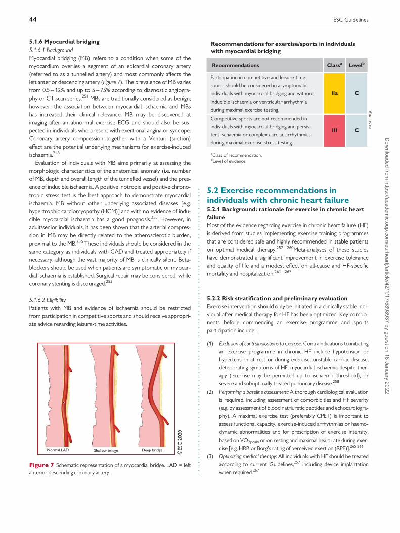
..
..
..
..
..
..
..
..
..
..
..
..
..
..
..
..
..
..
..
..
..
..
..
..
..
..
..
..
..
..
..
..
..
..
..
..
..
..
..
..
..
..
..
..
..
..
..
..
..
..
..
..
..
..
.
5.1.6 Myocardial bridging
5.1.6.1 BackgroundMyocardial bridging (MB) refers to a condition when some of themyocardium overlies a segment of an epicardial coronary artery(referred to as a tunnelled artery) and most commonly affects theleft anterior descending artery (Figure 7). The prevalence of MB variesfrom 0.5�12% and up to 5�75% according to diagnostic angiogra-phy or CT scan series.254 MBs are traditionally considered as benign;however, the association between myocardial ischaemia and MBshas increased their clinical relevance. MB may be discovered atimaging after an abnormal exercise ECG and should also be sus-pected in individuals who present with exertional angina or syncope.Coronary artery compression together with a Venturi (suction)effect are the potential underlying mechanisms for exercise-inducedischaemia.248
Evaluation of individuals with MB aims primarily at assessing themorphologic characteristics of the anatomical anomaly (i.e. numberof MB, depth and overall length of the tunnelled vessel) and the pres-ence of inducible ischaemia. A positive inotropic and positive chrono-tropic stress test is the best approach to demonstrate myocardialischaemia. MB without other underlying associated diseases [e.g.hypertrophic cardiomyopathy (HCM)] and with no evidence of indu-cible myocardial ischaemia has a good prognosis.255 However, inadult/senior individuals, it has been shown that the arterial compres-sion in MB may be directly related to the atherosclerotic burden,proximal to the MB.256 These individuals should be considered in thesame category as individuals with CAD and treated appropriately ifnecessary, although the vast majority of MB is clinically silent. Beta-blockers should be used when patients are symptomatic or myocar-dial ischaemia is established. Surgical repair may be considered, whilecoronary stenting is discouraged.255
5.1.6.2 EligibilityPatients with MB and evidence of ischaemia should be restrictedfrom participation in competitive sports and should receive appropri-ate advice regarding leisure-time activities.
5.2 Exercise recommendations inindividuals with chronic heart failure5.2.1 Background: rationale for exercise in chronic heart
failure
Most of the evidence regarding exercise in chronic heart failure (HF)is derived from studies implementing exercise training programmesthat are considered safe and highly recommended in stable patientson optimal medical therapy.257�260Meta-analyses of these studieshave demonstrated a significant improvement in exercise toleranceand quality of life and a modest effect on all-cause and HF-specificmortality and hospitalization.261�267
5.2.2 Risk stratification and preliminary evaluation
Exercise intervention should only be initiated in a clinically stable indi-vidual after medical therapy for HF has been optimized. Key compo-nents before commencing an exercise programme and sportsparticipation include:
(1) Exclusion of contraindications to exercise: Contraindications to initiating
an exercise programme in chronic HF include hypotension or
hypertension at rest or during exercise, unstable cardiac disease,
deteriorating symptoms of HF, myocardial ischaemia despite ther-
apy (exercise may be permitted up to ischaemic threshold), or
severe and suboptimally treated pulmonary disease.258
(2) Performing a baseline assessment: A thorough cardiological evaluation
is required, including assessment of comorbidities and HF severity
(e.g. by assessment of blood natriuretic peptides and echocardiogra-
phy). A maximal exercise test (preferably CPET) is important to
assess functional capacity, exercise-induced arrhythmias or haemo-
dynamic abnormalities and for prescription of exercise intensity,
based on VO2peak, or on resting and maximal heart rate during exer-
cise [e.g. HRR or Borg’s rating of perceived exertion (RPE)].265,266
(3) Optimizing medical therapy: All individuals with HF should be treated
according to current Guidelines,257 including device implantation
when required.267
©ES
C 2
020
Figure 7 Schematic representation of a myocardial bridge. LAD = leftanterior descending coronary artery.
Recommendations for exercise/sports in individualswith myocardial bridging
Recommendations Classa Levelb
Participation in competitive and leisure-time
sports should be considered in asymptomatic
individuals with myocardial bridging and without
inducible ischaemia or ventricular arrhythmia
during maximal exercise testing.
IIa C
Competitive sports are not recommended in
individuals with myocardial bridging and persis-
tent ischaemia or complex cardiac arrhythmias
during maximal exercise stress testing.
III C
aClass of recommendation.bLevel of evidence.
44 ESC GuidelinesD
ownloaded from
https://academic.oup.com
/eurheartj/article/42/1/17/5898937 by guest on 18 January 2022

..
..
..
..
..
..
..
..
..
..
..
..
..
..
..
..
..
..
..
..
..
..
..
..
..
..
..
..
..
..
..
..
..
..
..
..
..
..
..
..
..
The exercise session should be individually tailored for severalweeks, according to symptoms and objective findings during exercisetesting such as maximal exercise capacity, heart rate response, orarrhythmias. In atrial fibrillation (AF), exercise can only be monitoredby power or Borg’s RPE.
High-risk patients should be counselled more frequently during theinitial phases. Ideally exercise should be supervised through an exercise-based cardiac rehabilitation programme while non-supervised home-based sessions should be gradually added.260 When all these measuresare followed, the overall risk of exercise is low, even during higher-intensity exercises and in patients with more severe HF.268,269
Follow-up examinations for exercise recommendations should bescheduled at least every 3�6 months. Intervals between examinationsshould depend on disease severity and comorbidities, setting of thesessions (supervised vs. home-based), patient’s age and adherence.
5.2.3 Exercise modalities and sports participation in heart
failure
Following risk factor control and therapy optimization, the individualwith HF should be encouraged to start exercise programmes withoutdelay.242,244,270 Initially home-based exercise programmes may alsobe prescribed and monitored.270,271
In uncomplicated cases low to moderate-intensity recreationalsporting activities may be considered in parallel to the structuredexercise programme. When prescribed, maximal exercise intensitiesshould be monitored, for example, by heart rate monitors. If moni-toring does not reveal any exercise-induced arrhythmias or otherabnormalities, then all types of recreational sports activities are per-mitted (see Figure 2, section 4.1.2).
5.2.3.1 Aerobic/endurance exerciseAerobic exercise is recommended for stable patients [New YorkHeart Association (NYHA) class I�III], because of its well-demonstrated efficacy and safety.260 Recommendations on optimalexercise dose have been previously described in ESC and AHAGuidelines.242,270�272 The most commonly evaluated exercise modeis moderate continuous exercise (MCE).242,270�272 In patients inNYHA functional class III, exercise intensity should be maintained at alower intensity (<40% of VO2peak), according to perceived symptomsand clinical status during the first 1�2 weeks. This should be followedby a gradual increase in intensity to 50�70% VO2peak, and if toler-ated, up to 85% VO2peak as the primary aim.270,271
Recently, high-intensity interval training (HIIT) programmes havebeen considered as an alternative exercise modality for low-riskpatients.269 The most recent meta-analysis showed that HIIT was supe-rior to MCE in improving VO2peak in individuals with HF with reduced(<40%) ejection fraction (HFrEF) in the short term.273 However, thissuperiority disappeared in subgroup analysis of isocaloricprotocols. HITT programmes may be recommended initially to preparelow risk patients with stable HF who want to return to high intensityaerobic and mixed endurance sports (Figure 2, section 4.1.2).
5.2.3.2 Resistance exerciseResistance exercise training may complement, but not substitute,aerobic exercise training because it reverses skeletal muscle massloss and deconditioning without excessive stress on the heart.270,274
The training intensity can preferably be set at the level of resistance
at which the patient can perform 10�15 repetitions at 15 on Borg’sRPE scale (Table 12).242,270 In patients with altered skeletal musclefunction and muscle wasting, exercise training should focus initiallyon increasing muscle mass by using resistance programmes.275,276
Resistance programmes may specifically be considered for low-risk stable patients, who want to return to strength-related powersports, e.g. weightlifting (Figure 2, section 4.1.2). A meta-analysisshowed that resistance exercise as a single intervention has thecapacity to increase muscle strength, aerobic capacity, and quality oflife in HFrEF patients who are unable to participate in aerobic exer-cise programmes.277 Also, in advanced HF or in patients with verylow exercise tolerance, resistance exercise can be safely applied ifsmall muscle groups are trained.270,277,278
5.2.3.3 Respiratory exerciseInspiratory muscle training improves VO2peak, dyspnoea, and musclestrength,279�282 and it typically involves several sessions per weekwith intensity ranging from 30% to 60% of maximal inspiratory pres-sure, and duration from 15�30 min for an average of 10�12weeks.279 This training modality should be recommended to themost severely deconditioned individuals as an initial alternative whomay then transition to conventional exercise training and sports par-ticipation, to optimize cardiopulmonary benefits.280
5.2.3.4 Aquatic exerciseAquatic exercise has not been recommended for individualswith HF, due to concerns that the increase in central blood
Table 12 Optimal exercise training dose for patientswith chronic heart failure
©ES
C 2
020
1 RM = one repetition maximum; RPE = rating of perceived exertion; VO2peak =peak oxygen consumption.
ESC Guidelines 45D
ownloaded from
https://academic.oup.com
/eurheartj/article/42/1/17/5898937 by guest on 18 January 2022
..
..
..
..
..
..
..
..
..
..
..
..
..
..
..
..
..
..
..
..
..
..
..
..
..
..
..
..
..
..
..
..
..
..
..
..
..
..
..
..
..
..
..
..
..
..
..
..
.volume and cardiac preload as a consequence of hydrostaticpressure may not be tolerated.283 However, a recent meta-analysishas shown that aquatic exercise training may be safe and clinicallyeffective.284
5.2.4 Sports participation and return to sports
In addition to risk stratification (section 5.2.3), the evaluation for par-ticipation in sports includes intensity and type of sports (competitivevs. recreational), and determining the individual fitness level.
5.2.4.1 Competitive sportsParticipation in competitive sports may be considered in a groupof selected low-risk individuals. A thorough individual evaluationusing a maximal exercise test (or preferably CPET) is recom-mended before returning to sports, particularly before startingmoderate- to high-intensity sports, mixed and power sports(Figure 2, section 4.1.2).
Asymptomatic individuals with preserved (>_50%) EF (HFpEF) orwith mid-range (>_40�59%) EF (HFmrEF) who are optimally treatedmay be eligible to participate in some competitive sports in theabsence of exercise-induced arrhythmias or exercise-induced hypo-tension. In such cases, a progressive increase in exercise dose is rec-ommended. The duration of this process is dependent upon thefunctional capacity and perceived symptoms. Some restrictions mayapply to high-intensity endurance, mixed and power sports with high
demands, especially in older patients. No restrictions should applyfor skill-related sports.
Asymptomatic patients with HFrEF who are optimally treatedmay only be considered safe to perform specific low-intensity skillsports at a competitive level (Figure 2). Higher-risk patients includ-ing those who are suboptimally treated, those that remain inNYHA II or III despite optimal therapy, and those with exercise-induced arrhythmias or exercise-induced hypotension should notparticipate in competitive sports, particularly those sports withmoderate to high cardiopulmonary strain during training orcompetition.
5.2.4.2 Recreational sportsFor patients intending to participate in recreational sports andleisure-time activity, similar principles apply regarding risk stratifica-tion. A progressive increase in exercise dose is recommended. Low-to moderate-intensity skill, power, mixed, and endurance sports maybe considered in all asymptomatic individuals.
As with competitive sports, high-intensity recreational sportsshould only be considered in asymptomatic individuals with HFmrEF(EF 40�49%) who do not have exercise-induced arrhythmias orexercise-induced hypotension. Asymptomatic individuals with HFrEFwho are optimally treated may engage in low- to moderate-intensityskill-related recreational sports, and selectively in low-intensityendurance sports (Figure 2).
In patients with HFrEF with very low exercise tolerance, frequentdecompensation, or patients with LV assist devices (seeSupplementary Data), participation in low-intensity skill-related sportsis possible, if tolerated. Regular low-intensity endurance activities, e.g.walking or cycling, should generally be recommended to improvebasic exercise capacity.
Recommendations for exercise prescription in heartfailure with reduced or mid-range ejection fraction
Recommendations Classa Levelb
Regular discussion about exercise participation
and provision of an individualized exercise pre-
scription is recommended in all individuals with
heart failure.260,261,285
I A
Exercise-based cardiac rehabilitation is recom-
mended in all stable individuals to improve exer-
cise capacity, quality of life, and to reduce the
frequency of hospital readmission.260,261,285
I A
Beyond annual cardiac assessment, clinical reas-
sessment should be considered when the inten-
sity of exercise is increased.
IIa C
Motivational and psychological support and indi-
vidualized recommendations on how to progress
the amount and intensity of sports activities
should be considered.
IIa C
Low- to moderate-intensity recreational sport-
ing activities and participation in structured exer-
cise programmes may be considered in stable
individuals.
IIb C
High-intensity interval training programmes may
be considered in low-risk patients who want to
return to high-intensity aerobic and mixed
endurance sports.
IIb C
aClass of recommendation.bLevel of evidence.
Recommendations for participation in sports in heartfailure
Recommendations Classa Levelb
Before considering a sport activity, a preliminary
optimization of heart failure risk factor control
and therapy, including device implantation (if
appropriate), is recommended.
I C
Participation in sports activities should be con-
sidered in individuals with heart failure who are
at low risk, based on a complete assessment and
exclusion of all contraindications, in stable condi-
tion for at least 4 weeks, optimal treatment, and
NYHA functional class I status.
IIa C
Non-competitive (low- to moderate-intensity rec-
reational) skill, power, mixed, or endurance sports
may be considered in stable, asymptomatic, and
optimally treated individuals with HFmrEF.
IIb C
High-intensity recreational sports, adapted to
the capabilities of the individual patient, may be
considered in selected stable, asymptomatic, and
optimally treated individuals with HFmrEF with
an age-matched exercise capacity beyond
average.
IIb C
Continued
46 ESC GuidelinesD
ownloaded from
https://academic.oup.com
/eurheartj/article/42/1/17/5898937 by guest on 18 January 2022

..
..
..
..
..
..
..
..
..
..
..
..
..
..
..
..
..
..
..
..
..
..
..
..
..
..
..
..
..
..
..
..
..
..
..
..
..
..
..
..
..
..
..
..
..
..
..
..
..
..
..
..
..
..
..
..
..
..
..
.
5.2.5 Heart failure with preserved ejection fraction
Exercise-based cardiac rehabilitation programmes are a cornerstonein the holistic prevention and management of HFpEF.260,285 Exerciseintervention for 12�24 weeks increases functional capacity and qual-ity of life.286�292 The beneficial effects seem to be mediated byimprovement in oxidative muscle metabolism and vascular func-tion.293 In obese patients, weight reduction has been shown to havesimilar effects to exercise alone,288 therefore a stable weight reduc-tion of 10% over 2�4 years is recommended.294
5.2.5.1 Exercise modalities and sports participationHigher endurance intensities such as HIIT (4 � 4 min at 85�90%peak heart rate, with 3 min active recovery) have revealed positiveeffects on myocardial function, but data are limited to a small groupof patients with diabetes.295 HIIT performed over 4 weeks signifi-cantly improved VO2peak and LV diastolic function.296 Higher-intensity exercise should be limited to stable patients and could begradually introduced after 4 weeks of MCE.
Exercise sessions should start with short phases of 10 min ofendurance and 10 min of resistance exercises, which should graduallybe extended in time over a period of 4 weeks. The final aim shouldbe at least 30�45 min for >_3 days per week. Depending on thepatient’s symptomatic status and functional capacity, intervals ofhigher intensity may be introduced.
Duration of intervention seems to be important for inducing func-tional and structural CV changes in HFpEF. Interventions over 2 yearsin healthy individuals reversed early signs of diastolic dysfunc-tion.297,298 Regarding sports participation refer to section 5.2.3.
5.2.6 Exercise in individuals after heart transplantation
The exercise capacity in heart transplantation (HTx) recipients isreduced by 50�60% compared with healthy age and sex-matchedindividuals in the general population,300�302 due to several factors(Table 13).303 Exercise reduces CV risk induced by post-transplantation immunosuppressive medical therapy,304 andincreases physical performance,305 enabling HTx patients to achievelevels comparable to age-matched controls.306 HTx recipients partic-ipating in exercise-based cardiac rehabilitation programmes reveal afavourable outcome with respect to hospital readmission and long-term survival.305,307,308
Improvements in exercise capacity are primarily dependent on thevolume of exercise. Increased functional capacity is primarily due toperipheral adaptations in the skeletal muscle including increased oxi-dative capacity and capillary conductance. Cardiac allograft neuralreinnervation also contributes to improved functional capacity in thefirst year.304,309,310 If these occur, training can be performed at highlevels, enabling selected HTx patients to perform marathon runs ortriathlons.304,309,310
5.2.6.1 Exercise modalities and sports participationA combination of endurance and resistance exercise is consideredto be the preferred exercise programme. Mean enduranceexercise intensity should start at a moderate intensity (60% VO2peak),which can later be increased to 80% of VO2peak, a regimen levelapplied in the majority of exercise intervention studies in HTx.305 Inuncomplicated cases these intensities can be increased to maximumlevels.
It is recommended that individuals should perform up to fivebouts of 30 min of exercise per week; however, exercise durationand frequency have ranged from 30 min to 90 min for 2 to 5 timesper week, in previous HTx studies.305,311 Both endurance and resist-ance training is included in these training sessions; however, an addi-tional 2�3 sessions of resistance training may be performedeach week.
Resistance exercise should focus on large muscle groups usingown body weight exercises or exercises on weight machines. Upperbody resistance exercise should start at least 3 months after surgery,and intensity should gradually increase from low to moderate but can
Non-competitive (low-intensity recreational
skill-related sports) may be considered (when
tolerated) in stable, optimally treated individuals
with HFrEF.
IIb C
High-intensity power and endurance sports are
not recommended in patients with HFrEF irre-
spective of symptoms.
III C
HFmrEF = heart failure with mid-range ejection fraction; HFrEF = heart failurewith reduced ejection fraction; NYHA = New York Heart Association.aClass of recommendation.bLevel of evidence.
Recommendations for exercise and participation insport in individuals with heart failure with preservedejection fraction
Recommendations Classa Levelb
Moderate endurance and dynamic resistance
exercise, together with lifestyle intervention and
optimal treatment of cardiovascular risk factors
(i.e. arterial hypertension and type 2 diabetes)
are recommended.287,289�292,299
I C
Competitive sports may be considered in
selected stable patients without abnormalities
on maximal exercise testing.
IIb C
aClass of recommendation.bLevel of evidence.Refer also to the recommendation in section 5.2.5.
Table 13 Factors influencing decreased exercise capacity(peak VO2) and reduced cardiac output in individuals withheart transplants
©ES
C 2
020
ESC Guidelines 47D
ownloaded from
https://academic.oup.com
/eurheartj/article/42/1/17/5898937 by guest on 18 January 2022

..
..
..
..
..
..
..
..
..
..
..
..
..
..
..
..
..
..
..
..
..
..
..
..
..
..
..
..
..
..
..
..
..
..
..
..
..
..
..
..
..
..
..
..
..
..
..
..
..
..
..
..
..
..
..
..
..
..
..
..
..
..
..
..
..
..
..
..
..
..
..
..
..
..
..
..
..
..
..
..
..
..
..
..
..
..
.also be performed up to submaximal intensities, in case of uncompli-cated disease (see section 4.1.1).
A major limitation of endurance exercise is the reduced chrono-tropic response to exercise because of allograft denervation. Apartfrom chronotropic incompetence, other pathophysiological changespresent after HTx should also be considered when prescribing andconducting an exercise programme (Table 13). Exercise-inducedischaemia from cardiac allograft vasculopathy should be considered,particularly when performing higher-intensity exercise, which hasbeen advocated to have some superior effects on improving exercisecapacity in these patients.311,312
Feasibility and safety of sports participation in stable asymptomaticHTx patients, after therapy optimization, has been reported.Therefore, participation in competitive sports, avoiding high-intensitypower and endurance disciplines, may be considered in selectedindividuals.
5.3 Exercise recommendationsin individuals with valvular heartdisease5.3.1 Introduction
Valvular heart disease affects approximately 1�2% of young exer-cising individuals in the general population. Reports on the naturalhistory of valvular heart disease in athletes are sparse; however,there is a theoretical possibility that a large stroke volume, coupledwith vigorous mechanical contractions of the heart, and anincreased chronotropic state induced by exercise may acceleratevalve dysfunction. The ensuing effects on chronic stenotic orregurgitant lesions may cause compensatory cardiac hypertrophy,impaired ventricular function, myocardial ischaemia, cardiacarrhythmias, and possibly SCD.
5.3.1.1 General principles in assessment and risk stratification of individ-uals with valvular heart disease prior to leisure exercise or competitivesportsThere are no prospective studies examining the impact of exerciseon the progression of valvular disease; therefore, general guidancepresented in this section is based on consensus opinions and long-term follow-up studies from non-athletic populations. Most individu-als with valvular heart disease are asymptomatic or mildly sympto-matic and some may aspire to engage in regular exerciseprogrammes including leisure and competitive sports. The manage-ment of these individuals requires assessment of the symptomaticstatus, functional capacity, the nature of the valvular lesion, andimpact of the resulting loading conditions on cardiac structure andfunction. All individuals should be assessed with a clinical history,physical examination, ECG, echocardiography, and exercise stresstest. The clinical history should enquire about cardiac symptoms andfunctional capacity. Echocardiography should focus on the valve mor-phology and function with particular attention to the severity and theimpact of cardiac chamber size and function. Exercise testing shouldresemble the intensity of the sport being engaged in and should focuson inducibility of symptoms, arrhythmias, myocardial ischaemia, andthe haemodynamic (BP) response to exercise. Some individuals mayrequire exercise echocardiography to assess the severity of the valvedefect.
Asymptomatic individuals with mild to moderate valvular dysfunc-tion who have preserved ventricular function and show good func-tional capacity without exercise-inducible myocardial ischaemia,abnormal haemodynamic response, or arrhythmias are consideredto be at low risk and may participate in all sports. Indeed, mild valvularregurgitation (mostly tricuspid and pulmonary) are common amongtrained athletes and likely represent a feature of the athlete’s heart.Conversely, individuals with exertional symptoms, moderate orsevere valvular dysfunction, left or right ventricular dysfunction, pul-monary hypertension, and exercise-induced cardiac arrhythmias orabnormal haemodynamic response are considered to be at high riskand should be considered for invasive intervention.
5.3.1.2 SurveillanceAll individuals with valvular heart disease should be assessed on a reg-ular basis. The frequency of the assessment may vary from 6 monthlyto 2 yearly depending on symptomatic status and the severity of valvedysfunction.
5.3.2 Aortic valve stenosis
Aortic valve stenosis (AS) is most frequently the result of an age-dependent degenerative process causing progressive thickening, cal-cification, and reduced mobility of the cusps.313 AS causes an increasein transvalvular pressure gradient and LV workload, with consequentLV hypertrophy, fibrosis, and increased myocardial oxygen demand.Left ventricular ejection fraction (LVEF) is usually preserved. Affectedindividuals may have a normal cardiac output at rest and even duringexercise, therefore, some individuals with AS are capable of goodexercise performance. Nonetheless, severe AS is associated withincreased risk of heart failure and SCD from mechanical outflowobstruction, malignant VAs, or coronary hypoperfusion.18,314
The diagnosis and grading of AS during echocardiography is basedon well-established criteria.315 Specifically, severe AS is defined by: (i)
Recommendations for exercise and participation insport in heart transplant recipients
Recommendations Classa Levelb
Regular exercise through cardiac rehabilitation,
combining moderate-intensity aerobic and resist-
ance exercise, is recommended to revert patho-
physiology to pre-transplantation time, reduce
cardiovascular risk induced by post-transplanta-
tion medical treatment, and improve clinical
outcome.305�312
I B
Recreational (low-intensity recreational) sports
participation should be considered and encour-
aged in stable, asymptomatic individuals after
therapy optimization.
IIa C
Eligibility for competitive sports involving low-
and moderate-intensity exercise may be consid-
ered in selected, asymptomatic individuals with
an uncomplicated follow-up.304,309,310
IIb C
aClass of recommendation.bLevel of evidence.
48 ESC GuidelinesD
ownloaded from
https://academic.oup.com
/eurheartj/article/42/1/17/5898937 by guest on 18 January 2022

..
..
..
..
..
..
..
..
..
..
..
..
..
..
..
..
..
..
..
..
..
..
..
..
..
..
..
..
..
..
..
..
..
..
..
..
..
..
..
..
..
..
..
..
..
..
.
a transvalvular Doppler velocity >_4.0 m/s; (ii) a mean gradient >_40mmHg; and (iii) a calculated aortic valve area <1.0 cm2 or an indexedarea (recommended in athletes) <0.6 cm2/m2.315 In cases with a lowgradient (<40 mmHg) and calculated valve area <1.0 cm2, with EF <50% and stroke volume index <35 mL/m2, low-dose dobutaminestress echocardiography is recommended to identify pseudo-severeAS or true severe AS.315,316 Assessment of the aortic valve calciumscore with CT can be useful in borderline cases where the severity ofAS remains unclear.313,316
Exercise testing is particularly important to assess the haemody-namic response in AS and to serve as a guide to exercise prescriptionin cases of asymptomatic moderate and severe AS. A progressivedrop in SBP with exercise, or failure to increase SBP by at least 20mmHg, identifies subjects at higher risk.317Exercise-induced ventricu-lar tachycardia should also be considered a criterion for exerciserestrictions.
Asymptomatic individuals with mild AS may participate in allsports. Asymptomatic athletes with severe AS should not participatein any competitive or leisure sports with the exception of low-intensity skill sports. However, low-intensity aerobic exercise couldbe encouraged in asymptomatic individuals to improve functionalcapacity and general well-being.
Individuals with symptomatic AS should not participate in anycompetitive sport or recreational sport/exercise and valve replace-ment is recommended. Mild exercise, that does not provoke symp-toms, may be considered in these individuals for general healthbenefits. 5.3.3 Aortic valve regurgitation
Aortic valve regurgitation (AR) is usually caused by a congenitallyabnormal valve (i.e. bicuspid valve), degeneration of a tricuspid valve,or loss of coaptation due to aortic root enlargement.313,318 Lesscommon causes of AR include infective endocarditis or aorticdissection.
The haemodynamic consequence of chronic AR is characterizedby a pressure and volume overload that typically leads to a dilatedand hypertrophied LV. To accommodate the concomitant forwardflow from the mitral valve and the backward flow from the aorticvalve during diastole, the LV progressively increases in size and mass.This remodelling may occasionally be difficult to distinguish from car-diac adaptation in athletes, especially in males with a large body sizewho engage in endurance activities, and therefore LV size should beinterpreted in the context of the sport participated in, and the genderand body surface area of the individual.319 Males with a LV end-diastolic diameter >35 mm/m2 or a LV end-systolic diameter >50mm, and females with a LV end-diastolic diameter >40 mm/m2 or aLV end-systolic diameter >40 mm, should be considered to havepathological LV enlargement, irrespective of the level of physicaltraining. These individuals should be closely monitored for a progres-sive increase in LV end-systolic diameter.
In individuals with suboptimal echocardiographic images, CMR hasthe advantages of providing an accurate assessment of LV volumeand EF, flow calculations and detecting the presence of myocardialscar,319 in individuals with severe AR. Furthermore, the whole thora-cic aorta can be visualized during the same examination.
Asymptomatic individuals with mild and moderate AR may partici-pate in all sports. Asymptomatic individuals with severe AR, moder-ately dilated LV, and good LV systolic function may participate in
Recommendations for exercise and participation in rec-reational/leisure-time sports in asymptomatic individu-als with aortic stenosis
Aortic stenosisc
Recommendation Classa Levelb
Mild Participation in all recreational
sports, if desired, is recommended.I C
Moderate Participation in all recreational
sports involving low to moderate
intensity, if desired, should be con-
sidered in individuals with
LVEF>_50%, good functional
capacity, and normal exercise test.
IIa C
Severe Participation in all recreational
sports/exercise involving low inten-
sity, if desired, may be considered in
individuals with LVEF>_50% and nor-
mal BP response during exercise.
IIb C
Participation in competitive or rec-
reational sports/exercise of moder-
ate and high intensity is not
recommended.
III C
BP = blood pressure; LVEF = left ventricular ejection fraction.aClass of recommendation.bLevel of evidence.cFor mixed valvular disease, the recommendation for the predominant lesion(stenotic or regurgitant) should be followed.
Recommendations for participation in competitivesports in asymptomatic individuals with aortic stenosis
Aortic stenosisc
Recommendation Classa Levelb
Mild Participation in all competitive
sports, if desired, is recommended.I C
Moderate Participation in all competitive
sports involving low to moderate
effort, if desired, may be considered
in individuals with LVEF>_50%, good
functional capacity, and normal BP
response during exercise.
IIb C
Severe Participation in low-intensity skill
sports may be considered in a select
group of individuals with LVEF>_50%.
IIb C
Participation in sports or exercise of
moderate or high intensity is not
recommended.
III C
BP = blood pressure; LVEF = left ventricular ejection fraction.aClass of recommendation.bLevel of evidence.cFor mixed valvular disease, the recommendation for the predominant lesionshould be followed.
ESC Guidelines 49D
ownloaded from
https://academic.oup.com
/eurheartj/article/42/1/17/5898937 by guest on 18 January 2022

..
..
..
..
..
..
..
..
..
..
..
..
..
..
..
..
..
..
..
..
..
..
..
..
..
..
..sports involving low and moderate intensity and may be consideredfor more intensive exercise on an individual basis. Such individualsrequire more frequent surveillance on a 6-monthly basis to assess LVfunction. In asymptomatic individuals with severe AR and reducedLVEF, surgical valve replacement/repair is indicated, and they shouldnot participate in competitive sports but may participate in leisuresports involving only low-intensity exercise. Surgery is recommendedin symptomatic individuals with severe AR. These individuals shouldnot participate in competitive or leisure sports; however, low-intensity aerobic exercise activity is encouraged to improve func-tional capacity and general well-being.
5.3.4 Bicuspid aortic valve
Bicuspid aortic valve (BAV) is a common congenital abnormality witha prevalence of 1�2% in the general population.320 BAV may beassociated with AS or AR and increased risk for ascending aorticaneurysm or dissection, and SCD.28,321 Compared with Marfan
syndrome, the risk of aortopathy is lower; nonetheless BAV is muchmore frequent, and the relative risk of aortic dissection has beenreported to be eight times greater than with a tricuspid aorticvalve.321 BAV may not be identified during physical examination inthe absence of valve dysfunction;58,322 however, the outcome ofyoung individuals without valvular dysfunction is good.323,324
It is unclear whether intensive exercise accelerates aortic dilatationin the long term. A previous study comparing athletes with BAV,non-athletes with BAV, and athletes with a normal aortic valvereported that athletes with BAV showed a 0.11 ± 0.59 mm/yearincrease in aortic size at the sinuses of Valsalva and 0.21 ± 0.44 mm/year for the proximal ascending aorta, which was not dissimilar tonon-athletes with a BAV.325 Currently, expert consensus panelsadvise a cautious approach to sports activities when the ascendingaorta is above the normal limits (see section 5.4). In the absence ofaortopathy, exercise recommendations for individuals with BAV areidentical to those in individuals with tricuspid aortic valvedysfunction.
Recommendations for participation in recreational/leisure-time sports in asymptomatic individuals with aorticregurgitation
Aortic regurgitationc
Recommendation Classa Levelb
Mild Participation in all recreational sports, if desired, is recommended. I C
Moderate Participation in all recreational sports, if desired, should be considered in asymptomatic indi-
viduals with a non-dilated LV with LVEF>50% and normal exercise stress test.IIa C
Severe Participation in all recreational sports involving low and moderate intensity, if desired, may
be considered with a mild or moderately dilated LV with LVEF>50% and normal exercise
stress test.
IIb C
Participation in any moderate- or high-intensity recreational exercise is not recommended
with LVEF<_50% and/or exercise-induced arrhythmias.III C
LV = left ventricle; LVEF = left ventricular ejection fraction.aClass of recommendation.bLevel of evidence.cFor mixed valvular disease, the recommendation for the predominant lesion should be followed.
Recommendations for participation in competitive sports in asymptomatic individuals with aortic regurgitation
Aortic regurgitationc
Recommendation Classa Levelb
Mild Participation in all competitive sports, if desired, is recommended. I C
Moderate Participation in all competitive sports, if desired, should be considered in individuals with
LVEF>50% and normal exercise test.IIa C
Severe Participation in most competitive sports involving low to moderate intensity may be consid-
ered in individuals with a mild or moderately dilated LV with LVEF>50% and normal exercise
stress test.
IIb C
Participation in any moderate- or high-intensity competitive sports is not recommended in
individuals with severe AR and/or LVEF<_50% and/or exercise-induced arrhythmiasIII C
AR = aortic regurgitation; LV = left ventricle; LVEF = left ventricular ejection fraction.aClass of recommendation.bLevel of evidence.cFor mixed valvular disease, the recommendation for the predominant lesion should be followed.
50 ESC GuidelinesD
ownloaded from
https://academic.oup.com
/eurheartj/article/42/1/17/5898937 by guest on 18 January 2022

..
..
..
..
..
..
..
..
..
..
..
..
..
..
..
..
.5.3.5 Primary mitral regurgitation
Most individuals with mitral valve disease have primary mitral regurgi-tation (MR) from myxomatous disease.326 MR is confirmed and quan-tified by echocardiography. General recommendations regardingexercise and sports are based on symptomatic status, severity of MR,LV function, systolic pulmonary artery pressure (sPAP), and the pres-ence or absence of arrhythmias during exercise. Both athletic trainingand MR may be associated with an enlarged LV cavity; however, anenlarged LV that is disproportionate to the level of exercise may besuggestive of severe MR and an indication to refrain from competitiveor leisure sports involving moderate- or high-intensity exercise.
Asymptomatic individuals with mild or moderate MR may com-pete in all sports if they have good functional capacity, preserved LVfunction, sPAP < 50 mmHg and absence of complex arrhythmias dur-ing exercise. Individuals with symptomatic MR and reduced exercisecapacity or individuals with MR with exercise-induced complexarrhythmias should not participate in competitive or leisure sport;however, low-intensity aerobic exercise should be encouraged toimprove functional capacity and general well-being. Individuals onlong-term anticoagulation therapy for AF should not engage in con-tact/collision sport.
Recommendations for participation in recreational/leisure-time sports in asymptomatic individuals with mitralregurgitation
Mitral regurgitationc,d
Recommendation Classa Levelb
Mild Participation in all sports, if desired, is recommended. I C
Moderate Participation in all recreational sports, if desired, should be considered in individuals fulfilling the following:
• LVEDD<60 mm327 or <35.3 mm/m2 in men and <40 mm/m2 in women
• LVEF>_60%
• Resting sPAP<50 mmHg
• Normal exercise test
IIa C
Severe Participation in all recreational sports involving low and moderate intensity, if desired, may be considered in
individuals fulfilling the following:
• LVEDD<60 mm327 or <35.3 mm/m2 in men and <40 mm/m2 in women
• LVEF>_60%
• Resting sPAP<50 mmHg
• Normal exercise test
IIb C
LVEDD = left ventricular end-diastolic diameter; LVEF = left ventricular ejection fraction; MR = mitral regurgitation; sPAP = systolic pulmonary artery pressure.aClass of recommendation.bLevel of evidence.cFor mixed valvular disease, the recommendation for the predominant valve lesion should be followed.dNo collision or body contact sports if anticoagulated for atrial fibrillation.
Recommendations for participation in competitive sports in asymptomatic individuals with mitral regurgitation
Mitral regurgitationc,d
Recommendation Classa Levelb
Mild Participation in all competitive sports, if desired, is recommended. I C
Moderate Participation in all competitive sports, if desired, should be considered in individuals fulfilling
the following:
• LVEDD<60 mm327 or <35.3 mm/m2 in men and <40 mm/m2 in women
• LVEF>_60%
• Resting sPAP<50 mmHg
• Normal exercise test
IIa C
Severe Participation in competitive sports involving low exercise intensity, if desired, may be considered in individuals
fulfilling the following:
• LVEDD<60 mm327 or <35.3 mm/m2 in men and <40 mm/m2 in women
• LVEF>_60%
• Resting sPAP<50 mmHg
• Normal exercise test
IIb C
Participation in competitive sports is not recommended in individuals with a LVEF<60% III C
LVEDD = left ventricular end-diastolic diameter; LVEF = left ventricular ejection fraction; MR = mitral regurgitation; sPAP = systolic pulmonary artery pressure.aClass of recommendation.bLevel of evidence.cFor mixed valvular disease, the recommendation for the predominant valve lesion should be followed.dNo collision or body contact sports if anticoagulated for atrial fibrillation.
ESC Guidelines 51D
ownloaded from
https://academic.oup.com
/eurheartj/article/42/1/17/5898937 by guest on 18 January 2022

..
..
..
..
..
..
..
..
..
..
..
..
..
..
..
..
..
..
..
..
..
..
..
..
..
..
..
..
..
..
..
..
..
..
..
..
..
..
..
..
..
..
..
..
..
..
..
..
.5.3.5.1 Mitral valve prolapse
Mitral valve prolapse (MVP) is characterized by fibro-myxomatouschanges of the mitral valve leaflets and has a prevalence of1�2.4%.328,329 The diagnosis of MVP is defined as >2 mm displace-ment of one or both leaflets of the mitral valve beyond the annuluswithin the left atrium in end-systole.330 MVP is generally benign witha 10-year mortality risk of 5%.331 The majority of individuals are iden-tified incidentally during cardiac auscultation, or echocardiography.
The most common complication of MVP is the progression tochronic severe MR, in 5�10% of individuals with MVP. Other compli-cations include HF from chronic MR, pulmonary hypertension, infec-tive endocarditis, supraventricular and VAs, and, occasionally,SCD.313 In the Italian cardiac pathology registry of 650 SCDs in youngadults, 7% were attributed to MVP.332 Most decedents showed scar-ring in the infero-basal wall and papillary muscles and bi-leaflet pro-lapse. Myocardial scarring, mitral valve annular disjunction (i.e. anabnormal atrial displacement of the mitral valve leaflet hinge pointduring systole),333 T-wave inversion in the inferior leads and VAs aris-ing from the LV [right bundle branch block (RBBB) morphology]were high-risk features for SCD.334 The mechanical strain of MVP onpapillary muscles and adjacent myocardium is thought to be responsi-ble for the myocardial scarring, which may be a possible mechanismfor life-threatening arrhythmias in some individuals.335,336
In general, exercising individuals with MVP have an excellent prog-nosis. In a recent Italian study of 7449 young competitive athletes,MVP was identified in 2.9%. During a follow-up period of 8 ± 2 yearsthere were no fatalities.337 Adverse events, including progressive MRwith LV dilatation, ischaemic stroke, and AF occurred at a rate of0.5% per annum and were more common in older athletes with base-line mitral valve disjunction or VAs.
Individuals with MVP should be evaluated with an exercise test and24-hour ECG. Individuals with inferior T-wave inversion or
ventricular premature beats arising from the LV should undergo aCMR imaging scan to check specifically for myocardial fibrosis affect-ing the infero-basal wall. Other potentially high-risk markers includeevidence of mechanical dispersion detected by speckled trackingechocardiography,338 coexisting prolongation of the QT interval andmitral annular disjunction.333
Given the relatively benign nature of MVP, asymptomatic patientswith mild or moderate MR can participate in all competitive sportsand leisure sports in the absence of the aforementioned risk factors(Figure 8). Asymptomatic patients with severe MR but none of theabove high-risk markers may compete in low- to moderate-intensitysports after detailed discussion with their specialist in the presence ofLV end-diastolic diameter (LVEDD) <60 mm (or <35.5 mm/m2 inmen and <40 mm/m2 in women) with LVEF >_ 60%, resting sPAP <50 mmHg, and normal exercise test.
Symptomatic patients with MVP and any of the aforementionedhigh-risk features (Figure 8) should not participate in recreationalor competitive sports; however, low-intensity aerobic exerciseshould be encouraged to improve functional capacity and generalwell-being.
5.3.6 Mitral stenosis
Although rheumatic valve disease is uncommon in the westernworld, the increase in emigration patterns means that cardiologistsmay encounter individuals with rheumatic mitral stenosis (MS) whoaspire to exercise. Individuals with advanced MS are usually sympto-matic and incapable of engaging in exercise regimens with a high CVdemand. The risk stratification of exercising individuals with MS isbased largely on a detailed echocardiogram with specific interest inthe severity of the lesion and accompanying systolic PAP. In addition,assessment should include a maximal exercise stress test to identifyconcealed symptoms and functional capacity.
Figure 8 Specific markers of increased risk of sudden cardiac death (SCD) with mitral valve prolapse. LV = left ventricular; MR = mitral regurgitation;MV = mitral valve. Adapted from Gati et al.336
52 ESC GuidelinesD
ownloaded from
https://academic.oup.com
/eurheartj/article/42/1/17/5898937 by guest on 18 January 2022

..
..
..
..
..
..
..
..
..
..
..
..
..
..
..
..
..
..
..
..Asymptomatic individuals with mild MS [mitral valve area (MVA)1.5�2.0 cm2] and moderate MS (MVA 1.0�1.5 cm2) who are in sinusrhythm and demonstrate good functional capacity on exercise testingand a normal sPAP may participate in all competitive and leisuresports. Mildly symptomatic individuals with severe MS (MVA < 1.0cm2) may only participate in leisure exercise involving physical effortof low intensity. Individuals with symptomatic MS should be referredfor intervention and advised to abstain from participation in sportsand recreational exercise of moderate or high intensity. Individualswith AF should be anticoagulated and avoid contact/collision sport. Incases of balloon mitral valvuloplasty with good results (i.e. MVA > 2.0cm2) regular exercise and competitive sport may be considered inasymptomatic individuals with good functional capacity.
5.3.7 Tricuspid regurgitation
Tricuspid regurgitation (TR) is usually secondary to left heart disease,pulmonary hypertension, or right ventricular (RV) dysfunction. Inmost patients with secondary TR, exercise limitations relate to theunderlying pathology.
Mild TR is common in athletes and accompanied by physiologicaldilatation of the inferior vena cava, which is easily collapsible withinspiration. Severe TR is characterized by increasing tricuspid annulardilatation and RV remodelling that eventually leads to RV dysfunctionand a non-reactive inferior vena cava. Individuals with severe TR mayalso have a reduced exercise capacity due to an impaired cardiac out-put response with exercise.339 Furthermore, they may experienceincreased right- and left-sided filling pressures during exercise, the lat-ter being due to diastolic ventricular interaction.340
In general, asymptomatic patients with TR who have good func-tional capacity, non-dilated right ventricle, preserved ventricular func-tion, sPAP<40 mmHg, and absence of complex arrhythmias maycompete in all competitive and recreational sports.
5.4 Exercise recommendations inindividuals with aortopathy5.4.1 Introduction
Thoracic aortic aneurysms are largely asymptomatic until a suddenand catastrophic event, including aortic rupture or dissection, occurs,
Recommendations for participation in recreational/leisure-time sports in individuals with mitral stenosis
Mitral stenosisc,d
Recommendation Classa Levelb
Mild (MVA 1.5�2.0 cm2) Participation in all recreational sports, if desired, is recommended in individuals with a resting
sPAP<40 mmHg and normal exercise test.I C
Moderate (MVA 1.0�1.5 cm2) Participation in all recreational sports involving low and moderate intensity, if desired, may be
considered in individuals with resting sPAP<40 mmHg and a normal exercise test.IIb C
Severe (MVA<1 cm2) Participation in leisure sports of moderate or high intensity is not recommended. III C
MVA = mitral valve area; sPAP = systolic pulmonary artery pressure.aClass of recommendation.bLevel of evidence.cFor mixed valvular disease, the recommendation for the predominant valve lesion should be followed.dNo collision or body contact sports if anticoagulated for atrial fibrillation.
Recommendations for participation in competitive sports in asymptomatic individuals with mitral stenosis
Mitral stenosisc,d
Recommendation Classa Levelb
Mild (MVA 1.5�2.0 cm2) Participation in all competitive sports, if desired, is recommended in individuals with a resting
sPAP<40 mmHg and a normal exercise test.I C
Moderate (MVA 1.0�1.5 cm2) Participation in all competitive sports involving low intensity may be considered in individuals
with a resting sPAP<40 mmHg and normal exercise test.IIb C
Severe (MVA<1.0 cm2) Participation in competitive sports is not recommended. III C
MVA = mitral valve area; sPAP = systolic pulmonary artery pressure.aClass of recommendation.bLevel of evidence.cFor mixed valvular disease, the recommendation for the predominant valve lesion should be followed.dNo collision or body contact sports if anticoagulated for atrial fibrillation.
ESC Guidelines 53D
ownloaded from
https://academic.oup.com
/eurheartj/article/42/1/17/5898937 by guest on 18 January 2022

..
..
..
..
..
..
..
..
..
..
..
..
..
..
..
..
..
..
..
..
..
..
..
..
..
..
..
..
..
..
..
..
..
..
..
..
..
..
..
..
..
..
..
..
..
..
..
..
..
..
..
..
..
..
.which can be rapidly fatal. Advanced age, male sex, long-term historyof arterial hypertension, and the presence of aortic aneurysm conferthe greatest population attributable risk for aortic dissection.However, patients with genetic connective tissue disorders such asMarfan (MFS), Loeys Dietz, Turner, or Ehlers Danlos (EDS) syn-dromes, and patients with BAV are at increased risk at a muchyounger age. BAV has a prevalence of about 1�2% in the generalpopulation. These patients have a relatively low risk for aortic dissec-tion in comparison with patients with hereditary thoracic aortic dis-ease (HTAD). Having a family history of aortic dissection or suddendeath is a risk factor and a larger diameter of the aorta carries a higherrisk, although dissection can occur at any diameter and especially inEDS patients there is no clear association with aorticdiameter.341�343
A dilated aortic root (>40 mm) is not a feature of athlete’s heart,with only a small minority of young athletes (0.3%) having an enlargedaortic root diameter.344�347 During follow-up no progressiveenlargement of the aortic diameter was observed in these athletes,and no aortic events occurred during a 5-year period.347
5.4.2 Risk of dissection
Because of the increase in BP and wall stress associated with intensiveexercise and sports, such activities are potentially associated with anenhanced risk of expansion of the aorta and acute aortic dissection.However, daily exercise is important in maintaining an ideal BP, heartrate, and body weight and a sedentary lifestyle is an important modifi-able risk factor for CV disease and mortality. Physical activity isadvised in all patients with aortic pathology, even when the aorta isdilated.
There are no randomized controlled trials on competitive sportsin patients with thoracic aortic disease, or any prospective dataregarding the risks of competitive athletics in patients after surgicalcorrection; however, even after aortic root replacement, patientswith MFS and other HTAD remain at risk of aortic complications.One small prospective cohort study evaluated the feasibility and
effects of a 3-week rehabilitation training programme in 19 MFSpatients with a mean age of 47 years. During the 1-year follow-up,there were no adverse events but there was improvement in physicalfitness and reduction in psychological distress. These effects weredetectable after 3 weeks of rehabilitation, and mostly persistedthrough the 1-year follow-up. Unfortunately, no information onaortic diameters was provided.348
5.4.3 Sporting disciplines
Exercise-related acute thoracic aortic dissections are described inthe literature in a total of 49 case reports. Of these, 42 patients suf-fered Stanford type A thoracic aortic dissections. In the majority (26/49) weightlifting was associated with aortic dissection.349
Furthermore, a recently published retrospective cohort study of 615patients with acute type A aortic dissection found that 4.1% caseswere related to sports activities. The type of sports most oftenreported was golf (32%), but this was not corrected for the percent-age of participants in the sport and probably reflects that golfers arefrequently older with an increased risk of hypertension and hencethe potential for dissection.350
5.4.4 Effect on aortic diameter and wall stress
One cross-sectional study, which included 58 competitive athleteswith BAV, showed no correlation between aortic dimensions andduration of training.351 Two studies compared athletes and sedentaryindividuals with BAV and reported no difference in aortic growthrate between the two groups.
Two MFS mouse models investigating the effects of mild-moderate dynamic exercise on the aortic wall showed a reduction inthe growth rate of the aortic diameter in mice with MFS that per-formed mild to moderate dynamic exercise compared to sedentarymice with MFS.352,353 Among exercising mice, the aortic wall becamestronger and a larger mechanical stress was required to induce aorticrupture. An optimum protective effect was found at a training inten-sity level of 55�65% of maximum oxygen uptake (VO2max).
Table 14 Classification of risk to perform sports in patients with aortic pathology
©ES
C 2
020
ASI = aortic size index; BAV = bicuspid aortic valve; HTAD = hereditary thoracic aortic disease; MFS = Marfan syndrome.
54 ESC GuidelinesD
ownloaded from
https://academic.oup.com
/eurheartj/article/42/1/17/5898937 by guest on 18 January 2022

..
..
..
..
..
..
..
..
..
..
..
..
..
..
..
..
..
..
..
..
..
..
..
..
..
..
..
..
..
..
..
..
..
..
..
..
..
..
..
..
..
..
..
..
..
..
..
..
..
..
..
..
..
..
..
..
..
..
..
..
..
..
..
..
..
..
..
..
..
..
..
..
..
..
..
..
..
..
..
..
..
..
..
..
..
..
.5.4.5 Recommendations
Regular exercise has a well-documented benefit for fitness, psycho-logical well-being, and social interaction, as well as a positive effect onhypertension and concomitant future risk of dissection. Most individ-uals with aortic pathology benefit from a certain minimal exerciseprogramme and can at least participate in recreational sports(Table 14). Some lesions are not compatible with endurance trainingand athletic sports, due to their high risk of dissection or rupture.Recommendations for exercise and sports should be individualizedand based on the underlying diagnosis, the aortic diameter, family his-tory for dissection or sudden death (risk factor), and the pre-existingfitness and experience. An exercise test with an assessment of bloodpressure response is recommended before engaging in sports.
5.5 Exercise recommendations inindividuals with cardiomyopathies,myocarditis, and pericarditisCardiomyopathies are an important cause of SCD/SCA in young indi-viduals and exercise has been implicated as a trigger for fatalarrhythmias.17�19,28 The detection of a cardiomyopathy in an individ-ual has important implications with respect to ongoing participationin exercise. The advent of preventive strategies for SCD has led to asignificant expansion in the number of predominantly asymptomaticyoung patients with cardiomyopathies who aspire to exercise. Whenadvising such individuals, it is essential to strike a balance betweenprotecting patients from the potentially adverse effects of exerciseand depriving them of the multiple benefits of exercise.
5.5.1 Hypertrophic cardiomyopathy
The diagnosis of HCM is based on the presence of unexplained LVhypertrophy, defined as a maximum end-diastolic wall thickness >_15mm, in any myocardial segment on echocardiography, CMR, or CTimaging.355 HCM may also be considered in individuals with a lesserdegree of LV hypertrophy (wall thickness >_13 mm) in the context ofa family history of definite HCM or a positive genetic test.355
5.5.1.1 Risk stratification in hypertrophic cardiomyopathyCircumstantial evidence and a large systematic collection of youngSCDs in sports in the US suggest that exercise increases the risk ofSCD/SCA in individuals with HCM.18 Consistently, previous consen-sus recommendations have restricted all athletes with HCM fromcompetitive sports.1,356,357
More recently, relatively small longitudinal clinical studies indicatethat the risk of SCD during exercise may be considerably lower thaninitially considered. Lampert et al. reported that individuals withHCM who continued participating in sports after implantable cardi-overter defibrillator (ICD) implantation did not reveal an increasednumber of shocks during exercise.358,359 In a cross-sectional study of187 patients with HCM, vigorous exercise was not associated withthe occurrence of VAs.358 Pelliccia et al. reported outcomes in acohort of 35 athletes with HCM, engaged in training and competi-tions for 5 to 31 years (mean 15 ± 8). During a 9-year follow-upperiod, there were no differences in the incidence of symptoms ormajor events between athletes who ceased exercise (n = 20) com-pared with athletes who continued competitive sports (n = 15).360 Ina post-mortem series, only 23% of 194 deaths from HCM occurredduring sport, and affected males with a mean age of 30 years.361
Finally, individuals with HCM who participated in rehabilitation pro-grammes demonstrated a significant improvement in functionalcapacity without adverse events.362,363
In conclusion, there is limited evidence to indicate that all individu-als with HCM are vulnerable to fatal arrhythmias during exercise andsport participation. In this regard, systematic restriction from com-petitive sports in all affected individuals is probably unjustified and amore liberal approach to sports participation is reasonable in someindividuals after careful evaluation.3 This is particularly important forthe majority of individuals with HCM who wish to participate in ama-teur sports or leisure-time exercise to maintain their physical andpsychological well-being.
5.5.1.2 Baseline assessment of patients with HCMA systematic approach is required when assessing an individual withHCM who requests exercise advice. The baseline evaluation shouldinclude a comprehensive personal and family history with considera-tion of the age of the individual and years of exercise prior to diagno-sis, assessment of the severity of the HCM phenotype, and thepresence of any conventional risk factors for SCD/SCA. In olderpatients with HCM, the physician should review the presence of car-diac comorbidities such as hypertension and ischaemic heart disease,which may confer a worse prognosis in HCM.364,365
5.5.1.3 HistoryThe presence of symptoms attributed to HCM should prompt moreconservative exercise recommendations. Individuals with a history ofcardiac arrest or unheralded syncope and individuals with exercise-
Recommendations for exercise and participation insports in individuals with aortic pathology
Recommendations Classa Levelb
Prior to engaging in exercise, risk stratification,
with careful assessment including advanced imag-
ing of the aorta (CT/CMR) and exercise testing
with blood pressure assessment is
recommended.
I C
Regular follow-up including risk assessment is
recommended.I C
Dynamic exercise should be considered more
suitable than static exercise.IIa C
Participation in competitive or leisure-time
sports activities (except power sports) should
be considered in low-risk individuals (Table 14).
IIa C
Participation in individualized leisure exercise
programmes may be considered in high-risk indi-
viduals (Table 14).
IIb C
Competitive sports are not recommended in
individuals who are at high risk (Table 14).III C
CMR = cardiac magnetic resonance; CT = computed tomography.aClass of recommendation.bLevel of evidence.
ESC Guidelines 55D
ownloaded from
https://academic.oup.com
/eurheartj/article/42/1/17/5898937 by guest on 18 January 2022

..
..
..
..
..
..
..
..
..
..
..
..
..
..
..
..
..
..
..
..
..
..
..
..
..
..
..
..
..
..
..
..
..
..
..
..
..
..
..
..
..
..
..
..
..
..
..
..
..
..
..
..
..
..
..
..
..
..
..
..
..
..
..
..
..
..
..
..
..
..
..
..
..
..
..
..
..
..
..
..
..
..induced symptoms should be advised to engage in low-intensity rec-reational sports only.
5.5.1.4 Resting and ambulatory ECGThe resting 12-lead ECG has limited value in risk stratification.Ambulatory ECG monitoring, preferably for 48 h, is important fordetecting ventricular and supraventricular arrhythmias. The monitor-ing period should include an exercise session. Asymptomatic non-sustained ventricular tachycardia (NSVT) confers considerable risk ofSCD in younger individuals (<_35 years).355 Paroxysmal supraventric-ular arrhythmias may pose significant implications for functionalcapacity and, in the case of AF, for stroke prevention.366
5.5.1.5 EchocardiographyIn relation to risk stratification for SCD the clinician should assess thefollowing echocardiographic indices: (i) LV wall thickness; (ii) LV out-flow tract (LVOT) gradient; and (iii) left atrial diameter.355 All individ-uals should have the LVOT gradient assessed at rest, during theValsalva manoeuvre, on standing suddenly, and after light exercise onthe spot, such as repeated squats. By convention, LVOT obstructionis defined as a peak pressure gradient >_30 mmHg at rest or duringphysiological provocation. A gradient >_50 mmHg is considered to behaemodynamically important. Exercise stress echocardiographyshould be considered in individuals with exertional symptoms whohave resting systolic anterior motion of the mitral valve leaflets butwho do not reveal LVOT obstruction or show only mild to moderateLVOT obstruction with the aforementioned manoeuvres.
5.5.1.6 Cardiac magnetic resonance imagingCMR imaging is increasingly recognized as a necessary tool for con-firming diagnosis and to assess risk stratification in individuals withHCM. Late gadolinium enhancement (LGE), indicative of myocardialfibrosis, may be present in up to 75% of patients with HCM and, byitself, is a poor discriminator of outcomes. However, the presence ofextensive (>_15% of LV myocardium) LGE may identify individuals atincreased risk of ventricular tachyarrhythmias and SCD.367�370
5.5.1.7 Exercise testingExercise testing (or CPET) should be part of the routine evaluationto assess functional capacity in an individual with HCM who intendsto exercise. In addition, an abnormal BP response to exercise(defined as <20 mmHg increase in SBP from baseline, or exercise-induced hypotension)371,372 and the presence of exercise-inducedsymptoms or arrhythmias are markers of high risk and should resultin more conservative exercise recommendations.
5.5.1.8 Genetic testingCurrently genetic testing is reserved for familial cascade screening. Itdoes not inform decisions relating to the risk of SCD/SCA and shouldnot be performed for exercise risk stratification.
5.5.1.9 ESC risk score in HCMThe ESC risk score uses seven variables (age, syncope, familyhistory of SCD from HCM, maximal LV wall thickness, left atrial
diameter, LV outflow obstruction, NSVT) to assess the riskof SCD of patients with HCM.355,373 This information can beinserted into an online calculator (https://doc2do.com/hcm/webHCM.html) to estimate individualized 5-year risk to provideguidance on whether a prophylactic ICD is indicated. Forthe purposes of these Guidelines the risk of SCD is defined as lowif <4%, moderate if between >_4% and <6%, and high if >_6% in5 years.
5.5.1.10 Exercise recommendationOn completion of the baseline evaluation, the physician should con-sider: (i) the presence of symptoms; (ii) ESC risk score; (iii) presenceof resting or inducible LVOT obstruction during exercise; (iv) thehaemodynamic (BP) response to exercise; and (v) the presence ofresting or exercise-induced arrhythmias before recommending theappropriate form and intensity of exercise.
Although these Guidelines advocate for a more liberal approachto sports participation, it is indisputable that even the absence ofall major risk factors does not convey immunity to SCD.374 In addi-tion, the ESC risk score relies on evidence derived from predomi-nantly non-athletic cohorts.373 Whereas there are no data tosuggest that this approach to risk estimation is less valid in athleticindividuals, one has to consider that it may not accurately reflectthe risk of SCD in individuals exposed to the haemodynamic andmetabolic stresses of high-intensity sports. Therefore, when advis-ing an individual with HCM regarding participation in high-intensityexercise programmes and competitive sports, this considerationshould be an integral part of the discussion during the shareddecision-making process.
5.5.1.11 Special considerationsThe age of an individual may have an impact on risk. The mean ageof death in the largest series of SCD from the US was 18 years,with 65% of deaths occurring in athletes <_17 years.354 Althoughyoung age should not exclude an individual from high-intensityexercise in the absence of additional risk factors, it should be con-sidered in the discussion with the individual and the parents orguardians. In addition, specific sports may pose a higher risk forSCD, such as highly dynamic, start�stop sports like basketball andfootball.17,58
Individuals who have a positive genotype but who do not revealany phenotypic structural or arrhythmia features of HCM may engagein all sports. Such individuals should be assessed annually for pheno-typic features and risk stratification purposes.
5.5.1.12 Follow-upAnnual follow-up is recommended for most individuals with HCMwho exercise on a regular basis. More frequent (6-monthly) follow-up should be considered in adolescent individuals and young adultswhose phenotype, and therefore risk of SCD, may still be evolvingand who are more vulnerable to exercise-related SCD.58,239Follow-up evaluation should focus on assessment of disease progression andrisk stratification. New symptoms should prompt interruption ofexercise and re-evaluation.
56 ESC GuidelinesD
ownloaded from
https://academic.oup.com
/eurheartj/article/42/1/17/5898937 by guest on 18 January 2022

..
..
..
..
..
..
..
..
..
..
..
..
..
..
..
..
..
..
..
..
..
..
..
..
..
..
..
..
..
..
..
..
..
..
..
..
..
..
..
..
..
..
..
..
..
..
..
..
..
..
..
..
..
..
..
..
..
..
..
..
..
..
..
..
..
..
..
..
..
..
..
..
..
..
..
..
..
..
..
..
..
..
..
..
..
..
.
5.5.2 Arrhythmogenic cardiomyopathy
Arrhythmogenic right ventricular cardiomyopathy (ARVC) is definedpathologically by the presence of fibro-fatty replacement of the rightventricle and clinically by life-threatening VAs. The condition was ini-tially recognized as a predominantly RV disease, and diagnosis is cur-rently based on probabilistic Task Force Criteria that encompasselectrophysiological, anatomical, functional, and clinical features ofthe disease.375 Since its first description, the concept of ARVC hasevolved to include concealed or subclinical phenotypes and biventric-ular disease. It is now well established that both ventricles areaffected in most cases.376�378 This has led to the development of anew term, arrhythmogenic cardiomyopathy, that embraces an arrayof diagnostic terms for different (genetic and acquired) pathologies.Although the definition of ‘arrhythmogenic cardiomyopathy’ is yet to
be agreed, it can be considered as an umbrella term for a family of dis-eases that are characterized by biventricular myocardial abnormal-ities, including fibro-fatty infiltration and scarring, identified bypathological examination and/or cardiac imaging and VA.
The term arrhythmogenic cardiomyopathy (ACM) is usedthroughout these recommendations; however, it is important to rec-ognize that most of the literature on the influence of exercise on dis-ease progression and risk of SCD is derived from cohorts withclassical ARVC. This is reflected in the recommendations provided inthese Guidelines. It is possible therefore that the recommendationsmay not accurately reflect predominantly LV disease, which consti-tutes a small proportion of the disease spectrum where the impact ofexercise on disease phenotype and risk is less clarified than the RVvariant. Where appropriate, guidance is provided for other condi-tions that can be reasonably considered under the umbrella of ACM[including subtypes of dilated cardiomyopathy (DCM)].
5.5.2.1 Risk stratification in arrhythmogenic cardiomyopathyACM accounts for a significant proportion of SCDs in young and ath-letic individuals.28 Established risk factors for SCD that shouldprompt consideration for an ICD include aborted SCD, unheraldedsyncope, ventricular tachycardia, and impaired RV and/or LV systolicfunction.379 A novel risk prediction model for VAs has recently beenproposed but is yet to be validated.380 Regular and high-intensityexercise programmes are associated with acceleration of the diseaseprocess and worse outcomes.381�389
In an experimental model of heterozygous plakoglobin-deficientmice, exercise training accelerated RV dysfunction and arrhyth-mias.382 Similar results have been confirmed in human desmosomalmutation carriers participating in vigorous (>70% VO2max) endurancesports.384 Similar findings were reported in patients with ACM andasymptomatic gene-positive family members, despite a more conser-vative definition of athletic status (exercise with intensity >_6 METsfor >_4 h/week for >_6 years).386 Recently, the results from the NorthAmerican multidisciplinary study reported that patients engaging incompetitive sports were at two-fold increased risk of ventriculartachyarrhythmias or death and earlier presentation of symptoms,compared with patients who participated in recreational sports andsedentary individuals.385 Among patients engaging in competitivesports, early age of sports initiation was associated with prematurepresentation of symptoms and adverse clinical profile. Reducing exer-cise intensity was associated with a substantial decrease in the risk ofventricular tachyarrhythmias or death, to the same level as inactivepatients.385 Finally, in a multinational registry of 393 competitive ath-letes implanted with an ICD who continued to participate in regularcompetitions, 20% of athletes with ACM received a shock duringexertion compared to 10% at rest, during a median follow-up of 44months. The diagnosis of ACM was the only variable associated withreceiving appropriate shocks during competition.359,389
5.5.2.2 Baseline assessment of patients with arrhythmogeniccardiomyopathyA systematic approach is required when assessing individuals withACM who request exercise advice. The baseline evaluation shouldinclude a comprehensive history of symptoms and family history ofACM or SCD, assessment of the severity of the ACM phenotype,and the presence of any conventional risk factors for SCD/SCA.
Recommendations for exercise and sports participationin individuals with hypertrophic cardiomyopathy
Recommendations Classa Levelb
Exercise recommendations
Participation in high-intensity exercise/competitive
sports, if desired (with the exception of those
where occurrence of syncope may be associated
with harm or death), may be considered for indi-
viduals who do not have any markers of increased
riskc following expert assessment.
IIb C
Participation in low- or moderate-intensity recrea-
tional exercise, if desired, may be considered for
individuals who have any markers of increased
riskc following expert assessment .
IIb C
Participation in all competitive sports, if desired,
may be considered for individuals who are gene
positive for HCM but phenotype negative.
IIb C
Participation in high-intensity exercise (including
recreational and competitive sports) is not recom-
mended for individuals who have ANY markers of
increased riskc.
III C
Follow-up and further considerations relating to risk
Annual follow-up is recommended for individuals
who exercise on a regular basis.I C
Six-monthly follow-up should be considered in
adolescent individuals and young adults who are
more vulnerable to exercise-related SCD.
IIa C
Annual assessment should be considered for gen-
otype-positive/phenotype-negative individuals for
phenotypic features and risk stratification
purposes.
IIa C
BP = blood pressure; ESC = European Society of Cardiology; HCM = hypertro-phic cardiomyopathy; LVOT = left ventricular outflow tract obstruction cardio-myopathy; SCD = sudden cardiac death.aClass of recommendation.bLevel of evidence.cMarkers of increased risk include: (i) cardiac symptoms or history of cardiacarrest or unexplained syncope; (ii) moderate ESC risk score (>_4%) at 5 years; (iii)LVOT gradient at rest >30 mmHg; (iv) abnormal BP response to exercise; (v)exercise-induced arrhythmias.Refer to Table 4 for different indices of exercise intensity and training zones.
ESC Guidelines 57D
ownloaded from
https://academic.oup.com
/eurheartj/article/42/1/17/5898937 by guest on 18 January 2022

..
..
..
..
..
..
..
..
..
..
..
..
..
..
..
..
..
..
..
..
..
..
..
..
..
..
..
..
..
..
..
..5.5.2.3 HistorySyncope due to presumed arrhythmia is an important risk markerfor SCD/SCA and a predictor of future appropriate ICDtherapies.390�394 The presence of symptoms attributed to ACMshould reinforce the conservative exercise recommendations.Individuals with a history of cardiac arrest or unheralded syncopeand individuals with exercise-induced symptoms should beadvised to engage only in low-intensity recreational exerciseprogrammes.
5.5.2.4 Resting and ambulatory ECGApart from its diagnostic utility, the 12-lead ECG may provide usefulinformation relating to risk stratification in ACM. The presence of exten-sive T-wave inversion affecting >_3 precordial leads or T-wave inversionin two of the three inferior leads confers some additional risk for SCD/SCA.395,396
Ambulatory ECG monitoring is important for detecting VAs.Every effort should be made for the monitoring period toinclude the proposed exercise session. The presence of NSVT orsignificant burden of ventricular ectopy (>_1000/24 h), even inasymptomatic individuals, confers an increased risk of fatalarrhythmias.392,393,397
5.5.2.5 Echocardiography and cardiac magnetic resonance imagingIn relation to risk stratification for SCD, the clinician shouldassess the severity of RV and LV involvement in terms of ventric-ular dilatation and systolic dysfunction. CMR imaging is more use-ful than echocardiography for assessing RV wall motionabnormalities and can also quantify the degree of myocardial fatinfiltration and/or scar. The more extensive the disease thehigher the arrhythmic risk.398,399
5.5.2.6 Exercise testingExercise testing should be part of the routine assessment of everyindividual with ACM who wishes to exercise, as it can provide infor-mation regarding functional capacity and risk stratification. Exercisetesting in patients with ACM should not be performed during ‘hotphases’. The presence of exercise-induced symptoms or arrhythmiasshould result in more conservative recommendations.
5.5.2.7 Genetic testingGenotype may also be of prognostic value. In the ARVC variant, anumber of studies have reported that carriers of multiple pathogenicvariants in the same desmosomal gene or mutations in >_2 genes mayhave an almost four-fold higher arrhythmic risk than those with a sin-gle mutation.400 Particular genotypes such as DSP and TMEM43, butalso LMNA and FLNC, associated with other ACM phenotypes (seesection 5.5.4) have a propensity for high arrhythmic burden that canpre-date the structural phenotype.401,402
5.5.2.8 Exercise recommendationsThe overall scientific evidence supports the concept that in patientswith ACM participation in high-intensity sports should be discour-aged, because it is associated with accelerated disease progression,greater risk of VAs and major events. This recommendation is alsoapplicable to genetic carriers of pathogenic variants for ACM even inthe absence of overt disease phenotype.
5.5.2.9 Special considerationsYoung age of presentation and male sex are associated withincreased risk of malignant arrhythmias in ACM.379 Although youngage should not exclude an individual from moderate-intensity exer-cise in the absence of high-risk features, age should be considered inthe discussion with the patient and the parents. In addition, oneshould consider that specific highly dynamic, start�stop sports, suchas basketball and football, may pose a higher risk of SCD particularlyin athletes who compete at the highest level.17,365
5.5.2.10 Follow-upAn annual follow-up is recommended for most individualswith ACM who exercise on a regular basis. More frequent (6-monthly) follow-up should be considered for adolescent andyoung adults whose ACM phenotype, and therefore risk of SCD,may still be evolving, particularly if they engage in moderate- tohigh-intensity exercise. More frequent follow-up should also beconsidered in individuals with high arrhythmic risk genotypes suchas DSP, TMEM43, and carriers of multiple pathogenic variants.New symptoms should prompt interruption of exercise and re-evaluation.
Recommendations for exercise and sports participationin individuals with arrhythmogenic cardiomyopathy
Recommendations Classa Levelb
Exercise recommendations
Participation in 150 min of low-intensity exercise
per week should be considered for all individuals.IIa C
Participation in low- to moderate-intensity recrea-
tional exercise/sports, if desired, may be consid-
ered for individuals with no history of cardiac
arrest/VA, unexplained syncope, minimal struc-
tural cardiac abnormalities, <500 PVCs/24 h and
no evidence of exercise-induced complex VAs.
IIb C
Participation in high-intensity recreational exercise/
sports or any competitive sports is not recom-
mended in individuals with ACM, including those
who are gene positive but phenotype negative.384,386
III B
Follow-up and further considerations relating to risk
Annual follow-up is recommended for individuals
who exercise on a regular basis.I C
Six-monthly follow-up should be considered in
adolescent individuals and young adults who are
more vulnerable to exercise-related SCD.
IIa C
Annual assessment should be considered for geno-
type-positive/phenotype-negative individuals for
phenotypic features and risk stratification purposes.
IIa C
Six-monthly follow-up should also be considered
in individuals with high arrhythmic risk genotypes
such as DSP, TMEM43, and carriers of multiple
pathogenic variants.
IIa C
ACM = arrhythmogenic cardiomyopathy; PVC = premature ventricular contrac-tion; SCD = sudden cardiac death; VA = ventricular arrhythmia.aClass of recommendation.bLevel of evidence.Refer to Table 4 for different indices of exercise intensity and training zones.
58 ESC GuidelinesD
ownloaded from
https://academic.oup.com
/eurheartj/article/42/1/17/5898937 by guest on 18 January 2022

..
..
..
..
..
..
..
..
..
..
..
..
..
..
..
..
..
..
..
..
..
..
..
..
..
..
..
..
..
..
..
..
..
..
..
..
..
..
..
..
..
..
..
..
..
..
..
..
..
..
.
5.5.3 Exercise recommendations in individuals with left
ventricular non-compaction
LV non-compaction (LVNC) is an unclassified cardiomyopathy char-acterized by prominent trabeculation and deep recesses that com-municate with the LV cavity.403,404 Clinical presentation of LVNCincludes progressive LV systolic dysfunction, ventricular tachyar-rhythmias, and thromboembolic events.404
Athletes often demonstrate LV hypertrabeculation and up to 8% fulfilthe echocardiographic criteria for a diagnosis of LVNC.405 It is hypothe-sized that an increased cardiac preload may unmask LV trabecular mor-phology.406 Therefore, among athletic individuals, the suspicion ofLVNC should only be considered in those who fulfil echocardiographiccriteria for LVNC but also have either LV systolic dysfunction (EF <50%), symptoms suggestive of cardiac disease, or a positive family his-tory of LVNC.407�409 Additional echocardiographic criteria include avery thin compacted epicardial layer (5 mm in end-diastole on CMR, or<8 mm in systole) and abnormal myocardial relaxation (average E’ < 9cm/s on tissue Doppler imaging).404,405,410,411 Such athletes will requirefurther assessment with CMR, exercise echocardiography, and Holtermonitor to assess the presence of LV fibrosis, cardiac thrombi, contrac-tile reserve, and exercise-induced complex arrhythmias.405,406
5.5.3.1 Risk stratificationThe clinical outcomes of LVNC are determined by the presence ofsymptoms, severity of LV dysfunction, and the nature of the VAs.There are no reported adverse cardiac events in the absence of LVdysfunction regardless of the severity of LV trabeculation.405�409
5.5.3.2 Follow-upRegular follow-up is recommended for individuals with LVNC. Newsymptoms should prompt interruption of exercise and re-evaluation.
5.5.4 Exercise recommendations in individuals with
dilated cardiomyopathy
DCM is characterized by LV or biventricular systolic dysfunction withor without dilatation that are not explained by abnormal loading con-ditions or CAD. Possible causes include genetic predisposition, myo-carditis, drugs, toxins, peripartum cardiomyopathy, and, in somecases, the cumulative effect of more than one factor.412
The clinical spectrum of disease may range from a mild phenotypicexpression characterized by absence of symptoms, isolated LV dilata-tion and normal or low-normal systolic function, to an overt diseasephenotype with limiting symptoms and significant systolic dysfunc-tion. Ventricular arrhythmias are common in DCM, particularly inindividuals with previous myocarditis, or with lamin A/C mutationsand filamin C mutations.413,414 The risk of SCD in DCM is 2�3% peryear and increases with lower EF and higher NYHA class.415 Exercisetraining improves functional capacity, ventricular function, and qualityof life in patients with DCM and should therefore be considered asan integral part of the management of affected individuals.416,417
However, intensive exercise and competitive sports are reported asa cause of SCD in DCM. 28,46,58,413,418
LV cavity enlargement in trained individuals that is not associatedwith systolic dysfunction and outside the context of a familial disease,represents a benign physiological adaptation if it is consistent with thetype of sports participated (usually, endurance sports) and the body sizeof the athlete. Conversely, a mildly reduced EF (45�50%) in an athletewith an enlarged LV cavity should not merely be considered as a normaladaptation. In such cases assessment of LV function during exercise mayprovide important diagnostic clues.319 Failure to increase EF at peakexercise by >10% compared with the baseline value may suggest apathological condition.319,419,420 The presence of diastolic dysfunctionor reduced peak oxygen consumption on CPET may also provide sup-porting information for the differential diagnosis. CMR has emerged asan important tool for the diagnosis and risk stratification of DCM.
Recommendations for exercise in individuals with leftventricular non-compaction cardiomyopathy
Recommendation for diagnosis Classa Levelb
A diagnosis of LVNC in athletic individuals should
be considered if they fulfil imaging criteria, in asso-
ciation with cardiac symptoms, family history of
LVNC or cardiomyopathy, LV systolic (EF<50%)
or diastolic (E’<9 cm/s) dysfunction, a thin com-
pacted epicardial layer (<5 mm in end-diastole on
CMR, or <8 mm in systole on echocardiography),
or abnormal 12-lead ECG.404,405,410,411
IIa B
Exercise recommendations
Participation in high-intensity exercise and all
competitive sports, if desired, with the exception
where syncope may cause serious harm or death,
may be considered in asymptomatic individuals
with LVNC and LVEF>_50% and absence of fre-
quent and/or complex VAs.
IIb C
Participation in recreational exercise programmes of
low to moderate intensity, if desired, may be consid-
ered in individuals with LVEF 40�49% in the absence
of syncope and frequent or complex VAs on ambula-
tory Holter monitoring or exercise testing.
IIb C
Continued
Participation in high- or very high-intensity exer-
cise including competitive sports, if desired, may
be considered for individuals who are gene posi-
tive for LVNC but phenotype negative (with the
exception of lamin A/C or filamin C carriers).
IIb C
Participation in high-intensity exercise or competi-
tive sports is not recommended in individuals with
any of the following: symptoms, LVEF<40% and/or
frequent and/or complex VAs on ambulatory
Holter monitoring or exercise testing.
III C
Follow-up and further considerations
Annual assessment for risk stratification is recom-
mended for individuals with LVNC and genotype-
positive/phenotype-negative individuals who exer-
cise on a regular basis.
I C
CMR = cardiac magnetic resonance; ECG = electrocardiogram; EF = ejectionfraction; LV = left ventricular; LVEF = left ventricular ejection fraction; LVNC =left ventricular non-compaction; VA = ventricular arrhythmia.aClass of recommendation.bLevel of evidence.
ESC Guidelines 59D
ownloaded from
https://academic.oup.com
/eurheartj/article/42/1/17/5898937 by guest on 18 January 2022
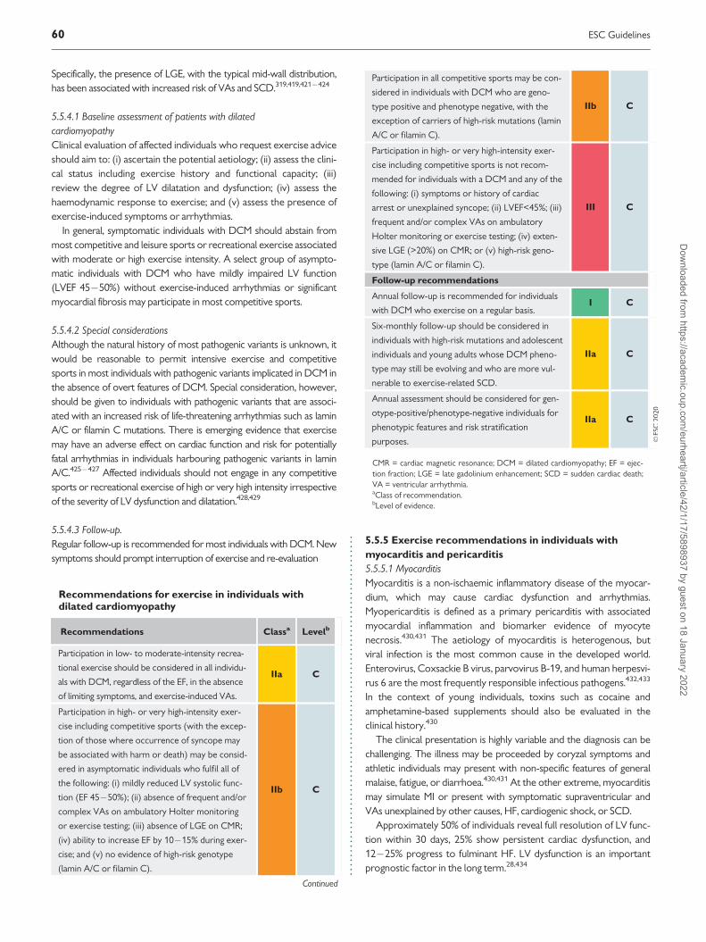
..
..
..
..
..
..
..
..
..
..
..
..
..
..
..
..
..
..
..
..
..
..
..
..
..
..
..
..
..
..
..
..
..
..
..
..
.
Specifically, the presence of LGE, with the typical mid-wall distribution,has been associated with increased risk of VAs and SCD.319,419,421�424
5.5.4.1 Baseline assessment of patients with dilatedcardiomyopathyClinical evaluation of affected individuals who request exercise adviceshould aim to: (i) ascertain the potential aetiology; (ii) assess the clini-cal status including exercise history and functional capacity; (iii)review the degree of LV dilatation and dysfunction; (iv) assess thehaemodynamic response to exercise; and (v) assess the presence ofexercise-induced symptoms or arrhythmias.
In general, symptomatic individuals with DCM should abstain frommost competitive and leisure sports or recreational exercise associatedwith moderate or high exercise intensity. A select group of asympto-matic individuals with DCM who have mildly impaired LV function(LVEF 45�50%) without exercise-induced arrhythmias or significantmyocardial fibrosis may participate in most competitive sports.
5.5.4.2 Special considerationsAlthough the natural history of most pathogenic variants is unknown, itwould be reasonable to permit intensive exercise and competitivesports in most individuals with pathogenic variants implicated in DCM inthe absence of overt features of DCM. Special consideration, however,should be given to individuals with pathogenic variants that are associ-ated with an increased risk of life-threatening arrhythmias such as laminA/C or filamin C mutations. There is emerging evidence that exercisemay have an adverse effect on cardiac function and risk for potentiallyfatal arrhythmias in individuals harbouring pathogenic variants in laminA/C.425�427 Affected individuals should not engage in any competitivesports or recreational exercise of high or very high intensity irrespectiveof the severity of LV dysfunction and dilatation.428,429
5.5.4.3 Follow-up.Regular follow-up is recommended for most individuals with DCM. Newsymptoms should prompt interruption of exercise and re-evaluation
5.5.5 Exercise recommendations in individuals with
myocarditis and pericarditis
5.5.5.1 MyocarditisMyocarditis is a non-ischaemic inflammatory disease of the myocar-dium, which may cause cardiac dysfunction and arrhythmias.Myopericarditis is defined as a primary pericarditis with associatedmyocardial inflammation and biomarker evidence of myocytenecrosis.430,431 The aetiology of myocarditis is heterogenous, butviral infection is the most common cause in the developed world.Enterovirus, Coxsackie B virus, parvovirus B-19, and human herpesvi-rus 6 are the most frequently responsible infectious pathogens.432,433
In the context of young individuals, toxins such as cocaine andamphetamine-based supplements should also be evaluated in theclinical history.430
The clinical presentation is highly variable and the diagnosis can bechallenging. The illness may be proceeded by coryzal symptoms andathletic individuals may present with non-specific features of generalmalaise, fatigue, or diarrhoea.430,431 At the other extreme, myocarditismay simulate MI or present with symptomatic supraventricular andVAs unexplained by other causes, HF, cardiogenic shock, or SCD.
Approximately 50% of individuals reveal full resolution of LV func-tion within 30 days, 25% show persistent cardiac dysfunction, and12�25% progress to fulminant HF. LV dysfunction is an importantprognostic factor in the long term.28,434
Recommendations for exercise in individuals withdilated cardiomyopathy
Recommendations Classa Levelb
Participation in low- to moderate-intensity recrea-
tional exercise should be considered in all individu-
als with DCM, regardless of the EF, in the absence
of limiting symptoms, and exercise-induced VAs.
IIa C
Participation in high- or very high-intensity exer-
cise including competitive sports (with the excep-
tion of those where occurrence of syncope may
be associated with harm or death) may be consid-
ered in asymptomatic individuals who fulfil all of
the following: (i) mildly reduced LV systolic func-
tion (EF 45�50%); (ii) absence of frequent and/or
complex VAs on ambulatory Holter monitoring
or exercise testing; (iii) absence of LGE on CMR;
(iv) ability to increase EF by 10�15% during exer-
cise; and (v) no evidence of high-risk genotype
(lamin A/C or filamin C).
IIb C
Continued
Participation in all competitive sports may be con-
sidered in individuals with DCM who are geno-
type positive and phenotype negative, with the
exception of carriers of high-risk mutations (lamin
A/C or filamin C).
IIb C
Participation in high- or very high-intensity exer-
cise including competitive sports is not recom-
mended for individuals with a DCM and any of the
following: (i) symptoms or history of cardiac
arrest or unexplained syncope; (ii) LVEF<45%; (iii)
frequent and/or complex VAs on ambulatory
Holter monitoring or exercise testing; (iv) exten-
sive LGE (>20%) on CMR; or (v) high-risk geno-
type (lamin A/C or filamin C).
III C
Follow-up recommendations
Annual follow-up is recommended for individuals
with DCM who exercise on a regular basis.I C
Six-monthly follow-up should be considered in
individuals with high-risk mutations and adolescent
individuals and young adults whose DCM pheno-
type may still be evolving and who are more vul-
nerable to exercise-related SCD.
IIa C
Annual assessment should be considered for gen-
otype-positive/phenotype-negative individuals for
phenotypic features and risk stratification
purposes.
IIa C
CMR = cardiac magnetic resonance; DCM = dilated cardiomyopathy; EF = ejec-tion fraction; LGE = late gadolinium enhancement; SCD = sudden cardiac death;VA = ventricular arrhythmia.aClass of recommendation.bLevel of evidence.
60 ESC GuidelinesD
ownloaded from
https://academic.oup.com
/eurheartj/article/42/1/17/5898937 by guest on 18 January 2022

..
..
..
..
..
..
..
..
..
..
..
..
..
..
..
..
..
..
..
..
..
..
..
..
..
..
..
..
..
..
..
..
..
..
..
..
..
..
..
..
..
..
..
..
..
..
..
..
..
..
..
..
..
..
..
..
..
..
..
..
..
..
..
..
..
..
..
..
..
..
..
..
..
..
..
..
..
..
..
..
..
..
..
..
..5.5.5.2 DiagnosisSerum cardiac troponin is usually elevated in myopericarditis and is asensitive marker of cardiac inflammation-induced myocytenecrosis.435
The ECG has low sensitivity and electrical anomalies are non-specific. ECG patterns vary from non-specific T-wave and ST-changes to ST-segment elevation mimicking MI, left bundle branchblock (LBBB), or frequent and/or complex ventricular and supraven-tricular arrhythmias, or atrioventricular block, or low QRS voltages inthe presence of a pericardial effusion.436
Recognized echocardiographic features of overt myocarditisinclude a non-dilated LV cavity with increased myocardial wall thick-ness (when oedema is present), or mildly dilated LV cavity with athinned myocardial wall, usually with regional wall motion abnormal-ities.437 The global LV systolic function may range from being almostnormal to severely depressed. Regional wall motion abnormalitiesmay be present.438
CMR is the most useful diagnostic tool and has excellent sensitivityfor detecting myocardial hyperaemia, inflammation, oedema and/orfocal scar.439,440 The Lake Louise Criteria and LGE are now comple-mented by CMR techniques of T1/T2 mapping and extracellular vol-ume fraction (ECV).440�442 The extent and distribution of LGE withnon-ischaemic pattern are independent predictors of CV events dur-ing follow-up.439,440,443�447 Namely, a 10% increase of LGE volumeconveys a 79% increase in the risk of major CV events.448,449
Endomyocardial biopsy is the gold standard for the diagnosis ofmyocarditis.450,451 A histological diagnosis allows distinctionbetween the different types of inflammatory processes, (i.e. giantcell myocarditis), and guides treatment in life-threatening presen-tations.430,445,452 The diagnostic yield of endomyocardial biopsycan be improved by analysing the viral genome through DNA-RNA extraction and reverse transcriptase polymerase chain reac-tion (RT-PCR) amplification, which has the advantage of identify-ing the disease-causing pathogen.445
5.5.5.3 Risk stratificationCase series have established myocarditis as a risk factor for SCD,which accounts for up to 2�20% of sudden death in ath-letes.17,18,28,430,453,454 Murine models have shown that daily exercisein mice infected with Coxsackie virus is associated with increasedviral titres, fulminant myocarditis, and sudden death.455 These animalmodels provide some insight into the mechanisms of SCD with exer-cise, which appears to cause an accelerated and progressive inflam-matory response.455�458
5.5.5.4 Exercise recommendations for individuals with myocarditisAthletic individuals with a probable or definitive diagnosis of recentmyocarditis should be advised to abstain from competitive sports orleisure sports while active inflammation is present, regardless of age,sex, or extent of LV systolic dysfunction.459,460
The duration of myocardial inflammation can be highly variableand may take several months for full resolution. Both the ESC andAHA recommend abstinence from moderate- to high-intensityexercise for a period of 3�6 months,459,460 although the precisetiming for return to competitive or recreational sports involvingmoderate- or high-intensity exercise may be guided by the
presence of inflammation on T2-weighted images and LGE uptakeon CMR.3,461
Individuals with myocarditis should have a comprehensive evaluationafter complete recovery to assess the risk of exercise-related SCD.Imaging studies, exercise stress test, and Holter monitor provide essen-tial information for risk stratification. Depressed LV function, presenceof LGE and complex VAs during exercise or Holter monitoring arerecognized risk markers for adverse outcomes.455,462,463
Repeat evaluation should consist of measurement of troponin andbiomarkers of inflammation, echocardiography, and prolonged ECGmonitoring. Individuals without evidence of ongoing inflammationshould undergo an exercise stress test. A CMR should be repeated ifmyocardial oedema or LGE was present during the acute illness.Return to sporting activities should be considered, in asymptomaticindividuals, with normal troponin and biomarkers of inflammation,normal LV systolic function on echocardiography and CMR, no evi-dence of ongoing inflammation or myocardial fibrosis on CMR, goodfunctional capacity, and absence of complex arrhythmias during exer-cise on prolonged ECG monitoring .430,434,453,459,460,464
Individuals with previous myocarditis are at risk of recurrence andsilent clinical progression, and the presence of LGE during the acutepresentation is associated with increased incidence of major adversecardiac events; therefore, periodic re-evaluation is advised on anannual basis.443,445,454,463
Among individuals with healed myocarditis with persistence ofLGE on CMR but no myocardial oedema at 3�6 months, those whoare asymptomatic, with normal troponin and biomarkers of inflam-mation, normal LV systolic function, no evidence of ongoing inflam-mation on CMR, and absence of complex arrhythmias duringexercise on prolonged ECG monitoring (48 h Holter ECG and exer-cise stress testing), should be evaluated on a case by case basis andmay return to competitive sports on an individual basis. In contrast,individuals with extensive myocardial scar (>20% LGE) and persistentLV dysfunction should abstain from exercise programmes and sportsactivities involving moderate or high physical intensity.
5.5.6 Pericarditis
Pericarditis is defined as an inflammatory disorder of the pericar-dium,430,465 which may be preceded by upper respiratory or gastroin-testinal symptoms. As with myocarditis, viral pathogens are the mostcommonly implicated pathogens in the western world.
5.5.6.1 DiagnosisThe ECG is non-specific but may reveal characteristic concave ST-segment elevation in most leads and/or PQ depression in the acutephase. Echocardiography may reveal a pericardial effusion. CMRshould be considered in individuals with raised cardiac troponin levelsto assess for concomitant myocardial inflammation. Furthermore,CMR will identify active inflammation of the pericardium, thickenedpericardial layers, and any signs of pericardial constriction.
5.5.6.2 Risk stratificationPericarditis is generally associated with an excellent prognosis.430,465,466
However, there are a subset of patients who may be at greater risk ofrecurrence and these include individuals with a temperature >38�C at
ESC Guidelines 61D
ownloaded from
https://academic.oup.com
/eurheartj/article/42/1/17/5898937 by guest on 18 January 2022

..
..
..
..
..
..
..
..
..
..
..
..
..
..
..
..
..
..
..
..
..
..
..
..
..
..
..
..
..
..
..
..
..
..
..
..
..
..
..
..
..
..
..
..
..
..
..
..
..
.
presentation, large pericardial effusion, and those who are resistant totherapy with non-steroidal anti-inflammatory drugs.465,466
5.5.6.3 Exercise recommendations for individuals with pericarditisExercise should be avoided in individuals during active pericarditis.Individuals can return to exercise after complete resolution of theactive disease.467 Individuals with a milder clinical course and rapidresolution can return to sporting activities within 30 days. However,in more severe cases, it may be necessary to wait for a period of 3months for complete resolution followed by re-evaluation beforereturning to sports.
Asymptomatic individuals with a small pericardial effusion occa-sionally detected on echocardiography in the absence of clinical cor-relates should be monitored with periodic surveillance, but shouldnot be restricted from sports participation. Competitive sports and/or moderate- to high-intensity leisure-time activities should beavoided in individuals with constrictive pericarditis. Individuals withmyopericarditis should be managed according to the recommenda-tions for myocarditis.
5.6 Exercise recommendations inindividuals with arrhythmias andchannelopathies5.6.1 A general management framework
When individuals with known arrhythmias or with a potentiallyarrhythmogenic condition want to engage in sports activity, threeprinciple questions should guide management: (1) is there anincreased risk for life-threatening arrhythmias?; (2) how does onecontrol symptoms due to arrhythmias, during sports, but also atrest?; and (3) what is the impact of sports on the natural progressionof the arrhythmogenic condition? The general view on the associa-tion between sports and arrhythmias is that exercise sets the stagefor an arrhythmia in the context of an underlying and pre-existingcondition, be it structural, electrical, inherited, or acquired. Moreover,regular exercise programmes may induce or accelerate the progres-sion of ARVC,382,384 even among those without underlying muta-tions.383,387,468 Conceptually, all the structural and functional cardiacadaptations to regular intensive exercise may contribute to the devel-opment of arrhythmias, at the atrial, nodal, and ventricular level.469
This concept explains why recommendations for sports participationin individuals with arrhythmogenic conditions are so complex.
5.6.2 Atrial fibrillation
5.6.2.1 Patients without atrial fibrillationModerate, regular PA is a cornerstone in the prevention of AFthrough modifying many of its predisposing factors.297,470�473
Patients at risk of AF should therefore be motivated to exercise (seesection 4.2). Conversely, AF is more prevalent in active and formermale master athletes and those performing high-intensity endurancesports, suggesting a U-shaped relationship between habitual exerciseand AF.471,474�477 478�481 This association has not been confirmed inwomen.474
Recommendations for exercise in individuals withmyocarditis
Recommendations Classa Levelb
Comprehensive evaluation, using imaging studies,
exercise stress test and Holter monitoring, is rec-
ommended following recovery from acute myo-
carditis to assess the risk of exercise-related
SCD.455,462,463
I B
Return to all forms of exercise including competi-
tive sports should be considered after 3�6
months in asymptomatic individuals, with normal
troponin and biomarkers of inflammation, normal
LV systolic function on echocardiography and
CMR, no evidence of ongoing inflammation or
myocardial fibrosis on CMR, good functional
capacity, and absence of frequent and/or complex
VAs on ambulatory Holter monitoring or exercise
testing.430,434,453,459,460,464
IIa C
Among individuals with a probable or definitive
diagnosis of recent myocarditis, participation in
leisure-time or competitive sports while active
inflammation is present is not
recommended.459,460
III C
Participation in moderate- to high-intensity exer-
cise for a period of 3�6 months after acute myo-
carditis is not recommended.459�461,467
III B
Participation in leisure exercise or competitive
sports involving high intensity in individuals with
residual myocardial scar and persistent LV dys-
function is not recommended.
III C
CMR = cardiovascular magnetic resonance; LV = left ventricular; SCD = suddencardiac death; VA = ventricular arrhythmia.aClass of recommendation.bLevel of evidence.
Recommendations for exercise in individuals withpericarditis
Recommendations Classa Levelb
Return to all forms of exercise including competi-
tive sports is recommended after 30 days to 3
months for individuals who have recovered com-
pletely from acute pericarditis, depending on clini-
cal severity.459,460
I C
Participation in leisure-time or competitive sports
is not recommended for individuals with a prob-
able or definitive diagnosis of recent pericarditis
while active inflammation is present, regardless of
age, sex, or extent of LV systolic
dysfunction.459,460
III C
Participation in moderate- to high-intensity exer-
cise, including competitive sports, is not recom-
mended for individuals with constrictive
pericarditis.
III C
LV = left ventricle.aClass of recommendation.bLevel of evidence.
62 ESC GuidelinesD
ownloaded from
https://academic.oup.com
/eurheartj/article/42/1/17/5898937 by guest on 18 January 2022

5.6.2.2 Prognostic and symptomatic relevance of AF during sportsUnderlying structural heart disease or pre-excitation should alwaysbe excluded before advising sports activity in individuals with recog-nized AF. It is also important to exclude hyperthyroidism, alcoholabuse, and (illicit) drug use. Intensive sports participation should betemporarily stopped until an identified underlying cause is corrected.
Rapid atrioventricular nodal conduction of AF during exercise maylead to symptoms, including dizziness, syncope, fatigue, or impairedphysical performance. Therefore, the individual should be instructed tostop PA on the emergence of symptoms and rate control should beoptimized. Rapid 1: 1 conduction can occur, especially during atrialtachycardia or atrial flutter (AFL); therefore, if AFL has been docu-mented, prophylactic cavo-tricuspid isthmus ablation should be consid-ered. If there is evidence for adequate rate control while in AF throughan exercise stress test or ECG monitoring during training or competi-tion, all sports participation is possible in asymptomatic individuals.
Achieving adequate rate control can be difficult, however. Beta-blockers are the logical choice but may not be tolerated due to theirimpact on physical performance. Calcium-channel blockers and digi-talis are usually not potent enough when used alone. Often a combi-nation of individually titrated negatively chronotropic agents isneeded, while avoiding sinus bradycardia at rest or chronotropicincompetence during exercise.
Rhythm control is equally complicated. Class III antiarrhythmic drugsare usually insufficient for control (sotalol) or relatively contraindicatedin a young population (amiodarone). Although class I drugs may be ableto prevent recurrences of AF, they should not be used in monotherapy,since these may increase the propensity to develop AFL (‘class I AFL’),which in the absence of adequate rate control may lead to 1: 1 atrioven-tricular conduction, high ventricular rates, and very profound intraven-tricular conduction slowing, with haemodynamic compromise.482,483
Therefore, prophylactic cavo-tricuspid-isthmus ablation should be con-sidered if class I drugs are prescribed in monotherapy in athletes.
In patients with sporadic AF, class I drugs may be considered onlyfor acute cardioversion, i.e. as a ‘pill-in-the-pocket’ approach. Thesepatients should refrain from sports as long as AF persists, and untiltwo half-lives of the antiarrhythmic drug have passed.484
Prescription of oral anticoagulants (OAC) depends on the clinicalrisk profile (mainly CHA2DS2-VASc score).485 Sports with direct bodilycontact or prone to trauma should be avoided in patients on OAC.486
Catheter ablation by pulmonary vein isolation (PVI) should be consid-ered if drug therapy fails or as first-line therapy if drug therapy is notdesired.487 Several small series have shown that the outcome of PVI inathletes with paroxysmal AF is similar to that in non-athleticpatients.488,489
5.6.2.3 Impact of sports continuation on the natural progression of atrialfibrillation after ablationIf there are no recurrences of AF within 1 month of a successful abla-tion procedure, sports activity may be resumed. It is unknownwhether continuation of sports after successful PVI might progressthe disease process and lead to recurrence of non-pulmonary vein-dependent AF in the future. Therefore, no firm recommendation canbe made about the ‘safe’ dose of sports after ablation.
Recommendations for exercise in individuals with atrialfibrillation
Recommendations Classa Levelb
Regular physical activity is recommended to pre-
vent AF.297,470�473 I A
Evaluation and management of structural heart
disease, thyroid dysfunction, alcohol or drug
abuse, or other primary causes of AF is recom-
mended before engaging in sports.485
I A
Counselling about the effect of long-lasting intense
sports participation on (recurrence of) AF is rec-
ommended in individuals with AF who exercise
vigorously for prolonged periods, especially in
middle-aged men.471,475,481,490
I B
AF ablation is recommended in exercising
individuals with recurrent symptomatic AF, and/
or in those who do not want drug therapy,
given its impact on athletic performance.488,489
I B
The ventricular rate while exercising with AF
should be considered in every exercising indi-
vidual (by symptoms and/or by ECG monitor-
ing), and titrated rate control should be
instituted.
IIa C
Participation in sports without antiarrhythmic
therapy should be considered in individuals with-
out structural heart disease, and in whom AF is
well tolerated.
IIa C
Cavo-tricuspid isthmus ablation should be consid-
ered in those with documented flutter who want
to engage in intensive exercise, to prevent atrial
flutter 1 : 1 atrioventricular conduction.
IIa C
Prophylactic cavo-tricuspid isthmus ablation to
prevent flutter should be considered in individu-
als with AF who want to engage in intensive
exercise and in whom class I drug therapy is
initiated.
IIa C
The use of class I antiarrhythmic drugs as mono-
therapy, without proof of adequate rate control
of AF/AFL during vigorous exercise, is not
recommended.482,483
III C
After ingestion of pill-in-the-pocket flecainide or
propafenone, participation in intensive sports is
not recommended until two half-lives of the anti-
arrhythmic drug have elapsed (i.e. up to 2
days).484
III C
Sports with direct bodily contact or prone to
trauma are not recommended in exercising indi-
viduals with AF who are anticoagulated.485
III A
AF = atrial fibrillation; AFL = atrial flutter; ECG = electrocardiogram.aClass of recommendation.bLevel of evidence.
ESC Guidelines 63D
ownloaded from
https://academic.oup.com
/eurheartj/article/42/1/17/5898937 by guest on 18 January 2022

..
..
..
..
..
..
..
..
..
..
..
..
..
..
..
..
..
..
..
..
..
..
..
..
..
..
..
..
..
..
..
..
..
..
..
..
..
..
..
..
..
..
..
..
..
..
..
..
..
..
..
..
..
..
..
..
..
.5.6.3 Supraventricular tachycardia and Wolff-Parkinson-
White syndrome
The term paroxysmal supraventricular tachycardia (PSVT) includes(i) atrioventricular nodal re-entrant tachycardia (AVNRT; most com-mon); (ii) atrioventricular re-entrant tachycardia (AVRT) involving anaccessory pathway; or (iii) atrial tachycardia.
Ventricular pre-excitation on the resting ECG is due to an acces-sory pathway (AP) with antegrade conduction. The prevalence ofpre-excitation in the general population varies from 0.1�0.3%.491,492
Wolff-Parkinson-White (WPW) syndrome is defined as the presenceof paroxysmal arrhythmias in a patient with pre-excitation.
5.6.3.1 Prognostic and symptomatic relevance of paroxysmal supraven-tricular tachycardia without pre-excitationPSVT without pre-excitation and without associated structural heartdisease is not life-threatening, although the arrhythmia may result indizziness and exhaustion that requires cessation of exercise. Syncopeis uncommon. Pre-excitation, however, may be associated with sud-den death (see later); therefore, it is important to exclude the pres-ence of latent pre-excitation, by performing carotid sinus massage oran adenosine-test in sinus rhythm.493
Athletes with PSVT should stop exercise in the event of palpita-tions since rapid heart rates may cause (pre)syncope. Individuals withproven PSVT without pre-excitation should be educated on how tosafely perform vagal manoeuvres (such as carotid sinus massage or,preferably, Valsalva manoeuvre) to facilitate termination of thearrhythmia.494 Exercise may be resumed after termination of thearrhythmia. Prophylactic drug treatment with beta-blockers or cal-cium antagonists with atrioventricular nodal blocking properties canbe considered, although it has limited efficacy. Class I drugs have norole in the management of PSVT, since they can cause life-threatening arrhythmias (see earlier).
If competitive athletic activity is desired, curative treatment byablation should be considered. Ablation outcome is equally safe andhas similar acute success rates in athletes and non-athletes.495 If thePSVT is only sporadic and transient and not associated with haemo-dynamic consequences, even when it develops during exercise, or incases where ablation is not desired or unsuccessful, sports activity ispermissible when there is no increased risk of a fatality from a poten-tial loss of consciousness (such as motorsports drivers, parachutejumpers, divers, and so on).
5.6.3.2 Prognostic and symptomatic relevance of pre-excitationIt has been estimated that one third of patients with WPW syndromemay develop AF and, in such cases, rapid conduction over the AP canlead to ventricular fibrillation (VF) and sudden death. Given the factthat AF is more common in athletes, pre-excitation constitutes aprognostic concern in athletes. The risk for sudden death in patientswith pre-excitation varies in population-based studies from0.15�0.20%, and usually presents during exercise or emotionalstress.496
Evaluation of the athlete with ventricular pre-excitation shouldexclude associated structural cardiac disease, such as HCM or Ebsteinanomaly. Minimal or ‘latent’ pre-excitation can be unmasked on a 12-lead ECG during sinus rhythm by vagal manoeuvres or intravenousadministration of adenosine. Prolongation of the PR interval without achange in the QRS morphology, or transient atrioventricular block,
excludes non-intermittent latent pre-excitation. Pre-excitation may beintermittent, which usually indicates low risk properties of the pathway.However, some accessory pathways may be potentiated by adrenergicstimuli. Therefore, exercise testing excluding pre-excitation at peakexercise is recommended before clearance for sports.
Ablation of the AP is recommended in competitive and recrea-tional athletes with pre-excitation and documented arrhythmias. Inthe event of transient, infrequent well-tolerated arrhythmia (evenduring exercise), good anticipation of an ablation procedure withincreased risk (e.g. anteroseptal AP), or reluctance of the athlete toundergo ablation, management should be guided by assessment ofthe antegrade conduction characteristics of the AP using either non-invasive tests or an invasive electrophysiological (EP) study.
Non-invasive investigation examines for intermittent pre-excitation on ECG or Holter, for abrupt disappearance of pre-excitation after administration of a low dose of class I drugs, or for itsabrupt disappearance during an exercise test.497 In cases of a longrefractory period and hence low risk for sudden death, continuationof sports activity is permitted without ablation on the understandingthat sporting activity should be stopped in the event of recurrence ofpalpitations.
In competitive athletes with asymptomatic pre-excitation an EPstudy is warranted to evaluate the risk for sudden death. In the eventof a high-risk finding (Table 15), ablation of the AP is recommended.For athletes who refuse ablation, or if the procedure is associatedwith high risk, such as an anteroseptal accessory pathway, participa-tion in competitive sports activities can be discussed on an individualcase by case basis including the use of pharmacological therapy,although there are currently no data about its efficacy. Sports inwhich the potential loss of consciousness could be fatal should bediscouraged.
In recreational athletes with asymptomatic pre-excitation, riskassessment may first be pursued via non-invasive testing.497 The sen-sitivity of non-invasive screening for AP properties that facilitate a fastventricular response to AF/AFL is good, but its specificity is low.498
Of note, in children younger than 12 years, the risk of AF-inducedVF and sudden death is very low. Generally, a conservative approach
Table 15 Findings during an invasive electrophysiologicalstudy (with the use of isoprenaline) indicating an accessorypathway with increased risk of sudden death
©ES
C 2
020
AF = atrial fibrillation; AVRT = atrioventricular re-entrant tachycardia.
64 ESC GuidelinesD
ownloaded from
https://academic.oup.com
/eurheartj/article/42/1/17/5898937 by guest on 18 January 2022

..
..
..
..
..
..
..
..
..
..
..
..
..
..
..
..
..
..
..
..
..
..
..
..
..
..
..
..
..
..
..
..
..
..
..
..
..
..
..
..
..
..
..
..
..
..
..
..
..
..
..
..
..
..
..
..
..
..
..
..
..
..
..
..
..
..
..
..
..
..
..
..
..
..
..
..
..
..
..
..
..
..
..
..
..
..
.is recommended in this age group, although one study499 suggestedthat prophylactic assessment and ablation reduces the risk of suddendeath. There is a knowledge gap in the benefit/risk ratio of thisapproach and large-scale studies are required to address the issue.
Leisure-time and low- to medium-intensity exercise programmescan generally be resumed 1 week after ablation if there is no particu-lar risk of recurrence of arrhythmia.
Resuming competitive sports is possible after 1�3 months, withfurther ECG follow-up at 6 months and 1 year (given the very smallrisk for late recurrence of pre-excitation).
Although there may be an association between (type of) AVNRTand history of sports, there are no data about higher recurrence ratepost ablation when sports are resumed or not, and hence no reasonto limit exercise programmes for such reason.
5.6.4 Premature ventricular contractions and
non-sustained ventricular tachycardia
5.6.4.1 Relation between number of premature ventricular contractionsand riskOnly a minority of athletes exhibit frequent or complex VA with aprevalence similar to that of their sedentary counterparts.502�505
Premature ventricular contractions (PVCs) may be a marker ofunderlying heart disease, the presence of which would confer anunfavourable prognosis even in asymptomatic individuals. Specificcharacteristics of the PVCs, including morphology (origin from the
apex or free wall of the LV or RV), high burden, complexity (e.g. cou-plets, triplets, or non-sustained runs), multifocal origin, and/orincreasing frequency with exercise should alert to the possibility ofelectrical, ischaemic, or structural heart disease.505,506
There is no absolute threshold of the number of PVCs that can beused as a cut-off for underlying disease. One study has shown that inasymptomatic athletes with >2000 PVCs per day, there was a 30%chance of finding an underlying structural or cardio-geneticdisease.503
5.6.4.2 Morphology of premature ventricular contractionsThe morphology of the PVCs may provide important prognosticinformation since some foci of origin are recognized as benign. Themost prevalent entity in this respect are PVCs originating from theright or left ventricular outflow regions (RVOT/LVOT), showing aclear inferior axis with high voltages in the inferior limb leads. Earlyprecordial transition (in V2, and certainly when V1 shows a right bun-dle branch morphology) suggests a left-sided origin.507 RVOT/LVOTPVCs are thought to be the result of triggered activity, i.e. a local cel-lular cause, which has no negative prognostic implications. Althoughthese RVOT/LVOT arrhythmias usually occur in structurally normalhearts, they may be the expression of subclinical arrhythmogenic car-diomyopathy. Cardiac imaging tests can help exclude structural heartdisease in such athletes.
Less common locations of focal PVCs are around the mitral or tri-cuspid annulus, most often in a postero-septal location. These have asuperior axis with LBBB or RBBB morphology. The PVCs originatingfrom the His-Purkinje system typically have relatively narrow QRScomplex with RBBB morphology and either left anterior or left pos-terior hemi-block. Lastly, intramyocardial foci may occur, oftenrelated to the papillary muscles or moderator band.508
PVCs of differing morphologies from the RV (i.e., wide LBBB andsuperior axis) in individuals with normal LV function should promptinvestigations to exclude arrhythmogenic cardiomyopathy or sarcoi-dosis. Similarly, wide RBBB pattern, with superior axis and multifocalPVCs of LV origin should trigger investigations for non-ischaemiccardiomyopathy.
Very rarely, otherwise ‘benign’ PVCs arising from the Purkinje net-work may give rise to polymorphic ventricular tachycardia (VT) orVF due to their short coupling interval.509,510 In such patients, themalignant electrical presentation mandates aggressive treatment.Finally, frequent but otherwise benign PVCs (usually defined as>10�15% of the total number of beats per 24 h) can impair LV func-tion over time (PVC-induced cardiomyopathy), which may be rever-sible with medical treatment or catheter ablation.511,512
5.6.4.3 Premature ventricular contractions: response to exerciseReduction or resolution of PVCs with increasing exercise load is typi-cal of idiopathic and benign VAs, particularly those with an outflowtract morphology.513,514 PVCs induced by exercise should be consid-ered as a ‘red flag’, because VAs associated with heart diseases areoften made worse by adrenergic stimulation.19,502,511,512,515�520 Ahigher prevalence of myocardial substrates (mainly mid-wall or sube-picardial non-ischaemic LV scars) was found in a CMR study amongathletes with exercise-induced PVCs compared to those withexercise-suppressed VAs (56% vs. 21%).516
Recommendations for exercise and sports participationin individuals with paroxysmal supraventricular tachy-cardia and pre-excitation
Recommendations Classa Levelb
In individuals with palpitations, a comprehensive
assessment to exclude (latent) pre-excitation,
structural heart disease, and VAs is
recommended.500
I B
Participation in all sports activities is recom-
mended in individuals PSVT without pre-
excitation.500
I C
Ablation of the accessory pathway is recom-
mended in competitive and recreational athletes
with pre-excitation and documented
arrhythmias.500
I C
In competitive/professional athletes with asymp-
tomatic pre-excitation, an EP study is recom-
mended to evaluate the risk for sudden
death.497,500
I B
In competitive athletes with PSVT but without
pre-excitation, curative treatment by ablation
should be considered.
IIa C
EP = electrophysiological; PSVT = paroxysmal supraventricular tachycardia; VA =ventricular arrhythmia.aClass of recommendation.bLevel of evidence.
ESC Guidelines 65D
ownloaded from
https://academic.oup.com
/eurheartj/article/42/1/17/5898937 by guest on 18 January 2022

..
..
..
..
..
..
..
..
..
..
..
..
..
..
..
..
..
..
..
..
..
..
..
..
..
..
..
..
..
..
..
..
..
..
..
..
..
..
..
..
..
..
..
..
..
..
..
..
..
..
..
..
..
..
..
..
..
..
..
..
..
..
..
..
..
..
..
..
..
..
..
..
..
..
..
..Of note, exercise-induced isolated or repetitive PVCs with multi-ple morphologies, especially with beat-to-beat alternating morpholo-gies (so-called ‘bi-directional’ pattern), may be the expression ofcatecholaminergic polymorphic VT, which can degenerate intoVF.518,521
5.6.4.4 Practical management of cardiac patients with premature ven-tricular contractions or non-sustained ventricular tachycardia who wantto engage in sportsThe most important task in individuals with PVCs or NSVT whowant to engage in sports is to exclude underlying structural or familialarrhythmogenic conditions, since sports activity may trigger sustainedVT. It has been suggested that the presence of >_2 PVCs on a baselineECG (or even >_1 PVC in the case of high-endurance athletes) shouldprompt a more thorough evaluation.522Work-up includes a familyhistory, assessment of the number, morphology, and complexity ofPVCs by Holter and 12-lead ECG, inducibility by exertion (via exer-cise test or long-term ECG recording during sports activities), andtailored additional imaging.1 Further diagnostic evaluation withmolecular genetic testing may be indicated in selected cases if the sus-picion for familial disease is high. Finally, repeat evaluation may beneeded after 6 months to 2 years. Recommendations for sports par-ticipation of athletes with PVC should be individualized based onevaluation for underlying cardiac conditions as described earlier, andoften requires shared decision making.
5.6.5 Long QT syndrome
The QT and corrected QTc intervals vary by sex andphysical training. Congenital long QT syndrome (LQTS) should bedistinguished from acquired forms, i.e. due to circumstances, whichcan be reversed and prevented. Once acquired LQTS is established,sports activity should be prohibited until the underlying cause iscorrected.
A definitive diagnosis of congenital LQTS is often difficult.523
Congenital LQTS should be suspected on a routine ECG or 4 mininto recovery after an exercise stress test,524 if the corrected QTcinterval according to Bazett’s formula is >_470 ms or >_480 ms inasymptomatic male or female athletes, respectively.525 A QTc of>_500 ms is diagnostic.526 In the case of a borderline long QTcinterval and a negative personal and familial history, subclinicalarrhythmias should be excluded by exercise testing and long-termECG recording.
Since the risk of cardiac events during sports activities is largelygene-specific, genetic testing and cascade screening of family membersshould be performed following a clinical diagnosis of LQTS. Individualswith LQT1 are at highest risk during stressful exercise.527,528
Symptomatic athletes should not engage in competitive sports.Individuals with LQT1 should not engage in sports that involve div-ing into cold water since this is associated with increased risk ofarrhythmias. General precautions include avoidance of QT-prolonging drugs, dehydration, and electrolyte imbalance. Beta-blocker therapy is extremely effective in LQT1 and additionaltherapies are only needed to control more severe cases or specificgenotypes.529
Survivors of SCA (certainly while taking beta-blocker therapy)should be referred for an ICD. Similarly, individuals who have experi-enced sudden syncope despite beta-blocker therapy should also bereferred for an ICD or sympathetic cardiac denervation.530 ICDimplantation does not constitute clearance for intensive or competi-tive sports. Continued sports participation with an ICD is possible,but specific recommendations apply (see section 5.5.6). Americanguidelines are more lenient with respect to participation in competi-tive sport (except for LQT1), provided that precautions include thepresence of an automatic external defibrillator (AED) ‘as part of theathlete’s personal sports safety gear’.531 We consider such obligationto be impractical (e.g. winter sports, water sports), and it places anadded responsibility on clubs or other bystanders, which cannot bejustified by a medical recommendation for an individual athlete.Moreover, although LQTS-related cardiac arrest is uncommon, evenduring competitive sports,527 AED efficacy is not 100% in suchcases.532
In asymptomatic LQTS mutation carriers without a prolonged QTinterval, i.e. <470 ms in men and <480 ms in women (‘genotype posi-tive/phenotype negative’), shared decision making is required, balanc-ing the risk for arrhythmias with psychological well-being. A negativeexercise stress test has no predictive value.
Recommendations for exercise in individuals with pre-mature ventricular contractions or non-sustained ven-tricular tachycardia
Recommendations Classa Levelb
In exercising individuals with >_2 PVCs on a base-
line ECG (or >_1 PVC in the case of high-endur-
ance athletes) thorough evaluation (including a
detailed family history) to exclude underlying
structural or arrhythmogenic conditions is
recommended.503,522
I C
Among individuals with frequent PVCs and non-
sustained VT a thorough investigation with
Holter monitoring, 12-lead ECG, exercise test,
and suitable imaging is recommmended.503
I C
It is recommended that all competitive and lei-
sure-time sports activities are permitted, with
periodic re-evaluation in individuals without fam-
ilial or structural underlying disease.503
I C
ECG = electrocardiogram; PVC = premature ventricular contractions; VT = ven-tricular tachycardia.aClass of recommendation.bLevel of evidence.
66 ESC GuidelinesD
ownloaded from
https://academic.oup.com
/eurheartj/article/42/1/17/5898937 by guest on 18 January 2022
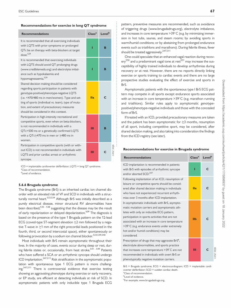
..
..
..
..
..
..
..
..
..
..
..
..
..
..
..
..
..
..
..
..
..
..
..
..
..
..
..
..
..
..
..
..
..
..
..
..
..
..
..
..
..
5.6.6 Brugada syndrome
The Brugada syndrome (BrS) is an inherited cardiac ion channel dis-order with an elevated risk of VF and SCD in individuals with a struc-turally normal heart.533,534 Although BrS was initially described as apurely electrical disease, minor structural RV abnormalities havebeen described,535�538 suggesting that the disease may be the resultof early repolarization or delayed depolarization.509 The diagnosis isbased on the presence of the type 1 Brugada pattern on the 12-leadECG (coved-type ST-segment elevation >_2 mm followed by a nega-tive T-wave in >_1 mm of the right precordial leads positioned in thefourth, third, or second intercostal space), either spontaneously orfollowing provocation by a sodium ion channel blocker.523,539,540
Most individuals with BrS remain asymptomatic throughout theirlives. In the majority of cases, events occur during sleep or rest, dur-ing febrile states or, occasionally, from heat stroke.541�549 Patientswho have suffered a SCA or an arrhythmic syncope should undergoICD implantation.247,511 Risk stratification in the asymptomatic popu-lation with spontaneous type 1 ECG pattern is more challeng-ing.247,511 There is controversial evidence that exercise testingshowing an aggravating phenotype during exercise or early recovery,or EP study, are efficient at detecting individuals at risk of SCD. Inasymptomatic patients with only inducible type 1 Brugada ECG
pattern, preventive measures are recommended, such as avoidanceof triggering drugs (www.brugadadrugs.org), electrolyte imbalance,and increases in core temperature >39�C (e.g. by minimizing immer-sion in hot tubs, saunas, and steam rooms; by avoiding sports inwarm/humid conditions; or by abstaining from prolonged enduranceevents such as triathlons and marathons). During febrile illness, fevershould be treated aggressively.247,511
One could speculate that an enhanced vagal reaction during recov-ery550 and a predominant vagal tone at rest551 may increase the sus-ceptibility of highly trained individuals to develop arrhythmias duringrecovery or at rest. However, there are no reports directly linkingexercise or sports training to cardiac events and there are no largeprospective studies evaluating the effect of exercise and sports inBrS.
Asymptomatic patients with the spontaneous type I BrS ECG pat-tern may compete in all sports except endurance sports associatedwith an increase in core temperature >39�C (e.g. marathon runningand triathlons). Similar rules apply to asymptomatic genotype-positive/phenotype-negative individuals and those with the concealedform of BrS.
If treated with an ICD, provided precautionary measures are takenand the patient has been asymptomatic for >_3 months, resumptionof all sport, including competitive sport, may be considered, aftershared decision making, and also taking into consideration the findingsfrom the ICD registry (see later).
Recommendations for exercise in long QT syndrome
Recommendations Classa Levelb
It is recommended that all exercising individuals
with LQTS with prior symptoms or prolonged
QTc be on therapy with beta-blockers at target
dose.529
I B
It is recommended that exercising individuals
with LQTS should avoid QT prolonging drugs
(www.crediblemeds.org) and electrolyte imbal-
ance such as hypokalaemia and
hypomagnesaemia.529
I B
Shared decision making should be considered
regarding sports participation in patients with
genotype-positive/phenotype-negative LQTS
(i.e. <470/480 ms in men/women). Type and set-
ting of sports (individual vs. team), type of muta-
tion, and extent of precautionary measures
should be considered in this context.
IIa C
Participation in high-intensity recreational and
competitive sports, even when on beta-blockers,
is not recommended in individuals with a
QTc>500 ms or a genetically confirmed LQTS
with a QTc>_470 ms in men or >_480 ms in
women.
III B
Participation in competitive sports (with or with-
out ICD) is not recommended in individuals with
LQTS and prior cardiac arrest or arrhythmic
syncope.
III C
ICD = implantable cardioverter defibrillator; LQTS = long QT syndrome.aClass of recommendation.bLevel of evidence.
Recommendations for exercise in Brugada syndrome
Recommendations Classa Levelb
ICD implantation is recommended in patients
with BrS with episodes of arrhythmic syncope
and/or aborted SCD.247
I C
Following implantation of an ICD, resumption of
leisure or competitive sports should be consid-
ered after shared decision making in individuals
who have not experienced recurrent arrhyth-
mias over 3 months after ICD implantation.
IIa C
In asymptomatic individuals with BrS, asympto-
matic mutation carriers and asymptomatic ath-
letes with only an inducible ECG pattern,
participation in sports activities that are not
associated with an increase in core temperature
>39�C (e.g. endurance events under extremely
hot and/or humid conditions) may be
considered.
IIb C
Prescription of drugs that may aggravate BrSc,
electrolyte abnormalities, and sports practice
that increases core temperature >39�C are not
recommended in individuals with overt BrS or
phenotypically negative mutation carriers.
III C
BrS = Brugada syndrome; ECG = electrocardiogram; ICD = implantable cardi-overter defibrillator; SCD = sudden cardiac death.aClass of recommendation.bLevel of evidence.cFor example, www.brugadadrugs.org.
ESC Guidelines 67D
ownloaded from
https://academic.oup.com
/eurheartj/article/42/1/17/5898937 by guest on 18 January 2022

..
..
..
..
..
..
..
..
..
..
..
..
..
..
..
..
..
..
..
..
..
..
..
..
..
..
..
..
..
..
..
..
..
..
..
..
..
..
..
..
..
..
..
..
..
..
..
..
..
..
..
..
..
..
..
..
..
..
..
..
..
..
..
..
..
..
..
..
..
..
..
..5.6.7 Following device implantation
5.6.7.1 PacemakersPacemaker (PM) implantation is common. Generally, individualsimplanted with a PM have less severe disease and comorbidities thanpatients with an ICD. Moreover, there is less risk of malfunction for aPM than for an ICD during sports.552 For all these reasons, recom-mendations for sports practice are more liberal in patients with a PMthan in patients with an ICD. Patients with a PM may participate incompetitive or recreational sports in the absence of structural orother heart disease for which exercise may be prohibited.
In the first weeks after device implantation, sports activities thatincrease the risk of lead dislocation (e.g. strong upper extremitymovements) should be avoided. Exercise testing and/or Holter moni-toring during sports may improve individualized programming of theupper sensor and the tracking rate and exclude inappropriate rateacceleration in other circumstances (e.g. horse riding).553
For all patients with cardiac devices (PM, cardiac resynchronizationtherapy, and ICD), sports activities associated with a risk of chesttrauma should be avoided.554�557 Some sports such as soccer, bas-ketball, and baseball may be possible while wearing appropriate pad-ding. It is noteworthy that sports with pronounced arm movementssuch as volleyball, basketball, tennis, golf, and climbing may increasethe risk for late lead damage due to subclavian crush (with insulationor conductor failure).554,558,559 Implantation on the contralateral sideof the dominant arm, fixation within the pocket, or submuscularplacement may improve durability of the system. It is not knownwhether subcostal or epicardial implant techniques provide long-term benefit. Electromagnetic interference is unlikely with moderndevices, and no cases have been reported, but it should always besuspected and closely evaluated in specific athletic environmentswith electronic equipment (e.g. fencing). Myopotential inhibition mayresult in inhibition of pacing, a problem that is more common withunipolar electrodes, although it can usually be corrected with appro-priate reprogramming of the device.560,561 Bipolar leads are less sen-sitive to this problem, but may have a reduced longevity.
5.6.7.2 Implantable cardioverter defibrillatorsA large multinational ICD Sports Safety Registry has shown that aftera median follow-up of 44 months, there were no deaths or arrhyth-mia or shock-related physical injury in 440 athletes who continuedorganized competitive or high-risk sports after ICD implanta-tion.359,389 An additional analysis in 82 non-professional recreationalathletes confirmed these reassuring outcomes,562 which is relevantfor the many ICD recipients who want to continue recreationalsports activities after implantation. Therefore, shared decision makingis appropriate when deciding whether or not to continue sports andthe level of participation with an ICD.
However, three important considerations come into play. First, ifsport is contraindicated because it can contribute to the progressionof the underlying disease (such as in arrhythmogenic cardiomyopathyor lamin A/C mutations),384,425 an ICD cannot be considered as asubstitute for sports restriction, and participation in moderate- andhigh-intensity exercise should be discouraged. Recommendations insuch circumstances should have a lifetime perspective and be basedon the optimal preservation of structural cardiac integrity. The ICD,however, may allow for light to moderate exercise without concernsabout risk of arrhythmia or shocks, and may help individuals to regainautonomy and overcome fears relating to exercising unattended.
The second consideration is that ICD shocks in general, evenwhen appropriate and safe, will have a psychological impact on theathlete; 30�40% of the athletes who experienced shocks in the mul-tinational ICD Sports Safety Registry stopped participation, at leasttemporarily, out of fear of repeat shocks.389 Moreover, the propor-tion of athletes with appropriate and inappropriate shocks duringexercise was higher in competitive than in recreational sports,emphasizing the known triggering effect of high-intensity exercise.562
Considering that ICD therapy is lifelong, where quality of life is notonly dependent on the ability to perform sports activities, but alsoand mostly on continued trust in the device, physicians should beaware that their own belief in the effectiveness and safety of ICDtherapy during sports should not implicitly put pressure on the ath-lete to continue sports. Again, informed decision making needs to re-evaluate all options of the athlete aftershocks, including continuing,reducing, or stopping sports.563,564
Third, situations where loss of focus or loss of consciousness couldcause harm to a third party or the athlete (such as in motor sports,diving, mountain climbing, even cycling) should be avoided.
The athlete must be aware of the programmed detection rate cut-offs to be able to avoid reaching those during exercise. Conversely,detection zones need to be programmed sufficiently high to allow foradequately high heart rates during the desired exercise levels. Thispractice proved safe and reduced the occurrence of shocks in theICD Sports Safety Database.565 The most common cause of inappro-priate shocks in individuals with an ICD is the occurrence of sinustachycardia and supraventricular arrhythmias.566,567 Underlying heartdisease and endurance sport itself carries a higher risk for developingAF.474,480,568 Implantation of a dual system ICD for the sole reason ofatrial arrhythmia detection and discrimination is not warrantedbecause it is usually not effective.565,569�572 Given the fact that manyof these athletes are young, there is a higher risk for long-term leadcomplications when more leads are implanted; therefore, implanta-tion of more complex ICD systems, and their indication, should beweighed in every patient. Routine inclusion of athletes in remotemonitoring programmes is highly recommended.
68 ESC GuidelinesD
ownloaded from
https://academic.oup.com
/eurheartj/article/42/1/17/5898937 by guest on 18 January 2022

..
..
..
..
..
..
..
..
..
..
..
..
..
..
..
..
..
..
..
..
..
..
..
..
..
..
..
..
..
..
..
..
..
..
..
..
..
..
..
..
..
..
..
..
..
..
..
..
..
..
..
..
..
..
..
..
..
..
..
..
..
..
..
..
..
..
..
..
..
..
..
..
..
..
..
..
..
..
..
..
..
..
..
..
5.7 Exercise recommendations inindividuals with adult congenital heartdisease5.7.1 Introduction
Congenital heart disease (CHD) has a prevalence of 8�9 per 1000live births and is the most common birth defect.573 Most childrenwith CHD survive to adulthood, including over 85% with complexdisease. Regular exercise is important for adults with congenital heartdisease (ACHD) and exercise participation should be discussed atevery patient encounter.574 However, CHD represents a spectrumof conditions with widely varying physiological consequences.Individualized assessment is essential before advising on sports partic-ipation. This requires detailed understanding of the congenital heart
defect, its physiological consequences, and the effect of surgical ortranscatheter intervention.
5.7.2 The increasing numbers of athletes with congenital
heart disease
Athletes with CHD include those with minor unoperated lesions andpalliated and repaired CHD. Some athletes will be diagnosed withCHD for the first time during pre-participation screening.Approximately 1 in 150 adults have a congenital heart defect. This is anincrease of over 50% in the last 10 years and reflects improved diagno-sis and long-term survival.575 Overall 90% of adults with CHD are inNYHA functional class I or II.576 The number of professional and rec-reational athletes with CHD is unknown but the elite athlete popula-tion is likely to be small. In a study of 2352 Olympian athletes, only nine(0.4%) had a congenital heart defect.577 However, many more CHDpatients compete at a recreational level. It is likely that the number ofathletes with CHD is increasing due to improved survival, betterunderstanding of the benefits of exercise, and the increased number ofprofessional athletes from countries where poorly developed health-care systems are associated with under-diagnosis of CHD.573,574
5.7.3 Non-cardiac abnormalities in congenital heart dis-
ease and Paralympic sport
CHD has a multifactorial aetiology, but there is a significant geneticcomponent and a recurrence risk of 2�5%.578 The genetic originmay be due to a chromosome anomaly, a heritable syndrome withMendelian pattern, or sporadic with variable penetrance.578
Approximately 14% of patients with CHD have additional non-cardiac abnormalities.579 Thus, 44% of ACHD patients have lungfunction abnormalities.580 This can make advising sports participationfor the ACHD athlete more difficult. There is no specific classificationfor CHD in Paralympic sport.
5.7.4 General considerations in the congenital heart
disease athlete
There is considerable variation in the haemodynamic consequencesand prognosis of different CHD lesions. Furthermore, the consequen-ces of any individual lesion can vary hugely between individuals. Thus,Ebstein’s anomaly may present in infancy with pulmonary hypoplasiadue to compression by a giant right atrium or may present in late mid-dle age with supraventricular tachycardia and minimal haemodynamicconsequences. Similarly, a ventricular septal defect may be small andasymptomatic, or large presenting with HF in infancy, or may presentwith extreme cyanosis, reduced exercise capacity, and Eisenmengersyndrome in the adult. Consequently, these recommendations arebased on a physiological assessment rather than underlying anatomicdiagnosis. An understanding of the range of severity and a lesion-basedknowledge of potential complications is essential. The cardiologistshould work in conjunction with a CHD specialist. Athletes with CHDwho participate in regular training and competitive sports shouldundergo a comprehensive annual assessment that includes clinicalexamination, ECG, echocardiogram, and exercise stress test, ideallyCPET.
Many athletes with CHD will have undergone corrective or pallia-tive surgery. In young adults with post-surgical CHD, valve
Recommendations for exercise in individuals with pace-makers and implantable cardioverter defibrillators
Recommendations Classa Levelb
It is recommended that individuals with
implanted devices with/without resynchroniza-
tion and underlying disease follow the recom-
mendations pertaining to the underlying
disease.384,425
I B
Participation in sports and exercise (except colli-
sion sports) should be considered in individuals
with pacemaker therapy who do not have patho-
logical substrates for fatal arrhythmias.
IIa C
Prevention of direct impact to the implanted
device by adapting the site of lead and/or device
implantation, padding, or restricting direct impact
sports should be considered.
IIa C
Holter recordings and device interrogation dur-
ing and after resuming sports should be consid-
ered to allow appropriate tailoring of rate-
responsive pacing parameters, exclusion of myo-
potential or electromagnetic inhibition, and
detection of VAs.
IIa C
Shared decision making should be considered dur-
ing decisions relating to continuation of intensive
or competitive sports participation in individuals
with an ICD, taking into account the effect of
sports on the underlying substrate, the fact that
intensive sports will trigger more appropriate and
inappropriate shocks, the psychological impact of
shocks on the athlete/patient, and the potential
risk for third parties.
IIa C
An ICD is not recommended as a substitute for
disease-related recommendations when these
mandate sports restrictions.
III C
ICD = implantable cardioverter defibrillator; VA = ventricular arrhythmia.aClass of recommendation.bLevel of evidence.
ESC Guidelines 69D
ownloaded from
https://academic.oup.com
/eurheartj/article/42/1/17/5898937 by guest on 18 January 2022

..
..
..
..
..
..
..
..
..
..
..
..
..
..
..
..
..
..
..
..
..
..
..
..
..
..
..
..
..
..
..
..
..
..
..
..
..
..
..
..
..
..
..
..
..
..
..
..
..
..
..
..
..
..
..
..
..
..
..
..
..
..
..
..
..
..
..
..
..
..
..
..
..
..
..
..
..
..
..
..
..
..
..
..
..
..
.incompetence and arrhythmias are common problems, but withincreasing age arrhythmias and HF predominate. In master athletes,problems related to previous corrective or palliative surgery becomeprevalent. These include cardiac arrhythmias, systemic ventriculardysfunction, valvar incompetence, and prosthetic conduit obstruc-tion. Redo valve or conduit surgery and arrhythmia ablation due tore-entry arrhythmias (secondary to surgical scar) are frequent in thisage group. In addition, with increasing age, acquired CVD maydevelop.581,582 This is of particular relevance in the assessment of themaster athlete with CHD.
5.7.5 Sudden death during sport
SCD in CHD is rare (<0.1% per year)583 and only 8% of deaths occurduring exercise.584 Many complex CHD patients at highest risk ofSCD have reduced exercise tolerance and cannot participate in sig-nificant athletic activity. However, some diagnostic groups, e.g. posttetralogy of Fallot repair, carry a risk of SCD but can still compete inelite sport.585 Other high-risk congenital lesions such as anomalouscoronary origin, mitral valve prolapse, and aortopathy are describedin sections 5.1, 5.3, and 5.4, respectively. In a large population-basedstudy of SCD in CHD patients under 35 years, 87% of SCD was dueto a presumed arrhythmia and 41% occurred in patients with undiag-nosed CHD.586Activity-related SCD was more common in theundiagnosed group (18% vs. 4%), which supports the case for pre-participation athletic screening.586 It is not known whether exerciseintensity is a risk factor for SCD in CHD. There is reasonable evi-dence that moderate to vigorous exercise is safe in most ACHDpatients even when symptomatic (NYHA II�III).576,587,588 However,cardiac arrhythmias are a common cause of hospital admission inACHD and extreme exercise may expose a latent arrhythmicsubstrate.589,590
5.7.6 Exercise in athletes with congenital heart disease:
current guidelines and recommendations
Regular structured exercise is safe and effective therapy for mostpatients with CHD. This is true for most diagnostic groups, includingsymptomatic patients, and includes aerobic and strength-based exer-cise.588,591�596 Exercise intolerance in CHD is a strong predictor ofboth outcome and SCD.576 Specific precautions are necessary inextreme environments including underwater sports and this is con-sidered below (see Supplementary Data, section 4). This is particularlytrue if there is a potential for a right to left shunt. Guidelines are avail-able for exercise assessment and prescription in both children andadults with CHD, although these are not specifically designed for theathlete.597,598 The paediatric guidelines are based primarily on theunderlying anatomical diagnosis, but the teenage and young adultCHD guidelines have taken a functional approach based on underly-ing haemodynamics and arrhythmia risk. This latter approach is bettersuited for assessment of the ACHD athlete (Figure 9).
5.7.7 Assessment of the athlete with congenital heart
disease
The guidelines for exercise prescription in adolescents and adultswith CHD use a structured methodology described by Budts et al.597
This can be modified for use in the assessment of athletes with CHD.
Stage 1. A full history and physical examination are carried out.This should include details of underlying CHD diagnosis, any trans-catheter or surgical interventions, current medications and CV symp-toms (at rest and on exercise). Attention should be paid to anyassociated non-cardiac diagnoses including pulmonary dysfunction. Afull exercise and sports participation history should be taken includingprecise details of current training schedule and any dietary supple-ments. Details of the planned or current sports activity should beestablished to include an assessment of the static component andintensity as described in section 4.1 (see Figure 2). If necessary, adviceshould be sought from a sports fitness coach or sports medicinespecialist.
Stage 2. The following five baseline parameters should then beevaluated597 (Table 16).
(1) Ventricular function
Assessment of ventricular function can usually be achieved usingechocardiography. The aim is to establish whether function isreduced (EF < 55%), and if so, is it mild (45�55%), moderate(30�45%), or severe (<30%). This is used for baseline assessmentand subsequent monitoring of the effects of exercise training.Echocardiography can also evaluate inflow and outflow abnormalities,which may become more severe during acute exercise (e.g. LV out-flow obstruction or systemic atrioventricular valve regurgitation).CMR scanning may be a preferable modality in complex disease. Thishas the additional benefit of evaluating intracardiac scar, which mayinform the assessment of arrhythmia risk.599,600
(2) Pulmonary artery pressure
Pulmonary hypertension (PH) is diagnosed when the mean PAP is>20 mmHg.601 PH may occur in the context of a chronic left�rightshunt (e.g. atrial septal defect, ventricular septal defect, patent ductusarteriosus) that allows unrestricted volume/pressure overload.Eventually this can result in supra-normal PAP with reversal of shunt-ing and elevated pulmonary vascular resistance (Eisenmenger syn-drome). An increased RV afterload limits the ability to increasecardiac output by increasing stroke volume and can impair LV func-tion through disruption of normal RV�LV interaction.
Few CHD patients with significant PH will take part in competitivesports due to reduced exercise capacity. However, some athleteswith CHD will have mild elevation of pulmonary vascular resistancethat may be exacerbated by factors such as altitude training.602 PAPrises during exercise. This rise is accentuated with increasing age andmay be exaggerated in CHD athletes due to elevated pulmonary vas-cular resistance.603 In addition, valvar pulmonary stenosis or distalstenoses in branch pulmonary arteries may cause exercise-relatedRV hypertension. The non-invasive assessment of PH can be difficultand full assessment guidelines have been published.604,605 Furtherinformation can be gained by CPET and a VO2 of >25.2 mL/min/kgmakes significant PH unlikely.606 As PH can also be a late postopera-tive complication after surgical correction of CHD607 the assessmentof PAP should be part of every echocardiographic examination in ath-letes with CHD. Although exercise training is generally regarded assafe in PH, competitive sport is not recommended.1,608
(3) Aortic assessment
Many CHD patients are at risk of aortic dilatation, in particular,patients with tetralogy of Fallot, coarctation of the aorta and certain
70 ESC GuidelinesD
ownloaded from
https://academic.oup.com
/eurheartj/article/42/1/17/5898937 by guest on 18 January 2022

syndromes such as 22q11 microdeletion and Turner syndrome.However, aortic dissection is very rare in CHD.609 Athletes havemildly increased aortic dimensions in comparison to sedentary con-trols, but it is not known if this has a cumulative effect in CHD ath-letes with aortic dilatation.345 The presence of ascending aortadilatation should lead to assessment for coarctation of the aorta asthis can be associated with severe coarctation, which may be missedon clinical assessment but may cause severe exercise-related hyper-tension.610 Exercise risks in aortopathy are described in detail in sec-tion 5.4. Contact sports should be avoided in patients with dilatedaortas >5 cm.
(4) Arrhythmia assessment
Arrhythmias are responsible for 25% of CHD hospital admissions.Over 80% are atrial but life-threatening VAs can occur.611
Independent risk factors include increasing age, male gender, doubleoutlet right ventricle, atrioventricular septal defect, HF, obstructivesleep apnoea, transposition of the great arteries, congenitally cor-rected transposition, and tetralogy of Fallot.599 Assessment of theCHD athlete should include a symptom history with evaluation ofpalpitations, presyncope and syncope, particularly during exercise.
©ES
C 2
020
Figure 9 Pre-participation assessment of individuals with congenital heart disease.598 CPET = cardiopulmonary exercise test; HR = heart rate; MHR =maximum heart rate; RPE = rate of perceived exertion. A�E represent pathways linking static and intensity components for each column. After assess-ment of CPET and the five variables (Table 16), an individual recommendation can be given (solid arrow). If a higher static level sport is chosen, then a lowerintensity level is advised (dotted arrow).
Table 16 Baseline parameters for assessment in congeni-tal heart disease
©ES
C 2
020
CMR = cardiac magnetic resonance; ECG = electrocardiogram.
ESC Guidelines 71D
ownloaded from
https://academic.oup.com
/eurheartj/article/42/1/17/5898937 by guest on 18 January 2022

..
..
..
.Arrhythmias may be the first sign of underlying haemodynamic dete-rioration and new-onset arrhythmias should lead to a full haemody-namic assessment. Detailed guidelines for the assessment andtreatment of arrhythmias in CHD are available.612 In the athlete,arrhythmia therapy may be complicated by the need to minimize thenegative chronotropic effects of antiarrhythmic treatment, whichmay affect performance. If symptomatic arrhythmias are present thenan exercise ECG, prolonged ECG monitoring, loop recorder implan-tation, and even electrophysiology testing might be required. Thisshould be coordinated by an electrophysiologist with expertise inCHD.(5) Assessment of saturations/lung function
CHD athletes should be assessed for the potential of an underlyingintracardiac right to left shunt. This can be assessed using pulse oxi-metry, but a resting saturation >95% does not exclude exercise-related central cyanosis, and exercise assessment is essential. Thepotential for a pulmonary cause of cyanosis must be considered andlung function should be assessed as part of a cardiopulmonary exer-cise test. Even after surgical correction of the cardiac defect theremay be residual intracardiac shunting.
Stage 3. Cardiopulmonary exercise testingCPET is invaluable in risk stratifying the adult with CHD and can
predict outcome.613,614 It is also extremely useful in the evaluation ofthe athlete with CHD as it allows an assessment of PAP, respiratoryproblems, cardiac output, exercise-related haemodynamics, andarrhythmias. This should be used in conjunction with an evaluation ofeffort such as the Borg scale.615 Normal CPET values are available fornon-athlete CHD patients.616 CPET should be carried out in con-junction with a 12-lead ECG to allow detection of arrhythmias andevaluate chronotropic incompetence. Reduced VO2max and peakoxygen pulse may reflect reduced stroke volume and are found incomplex CHD as well as in other forms of CHD, including repairedtetralogy of Fallot, aortic regurgitation, and coarctation of the aorta.In CHD, ventilatory anaerobic threshold may be reduced and thisimpairs gas exchange efficiency in dynamic and endurance sports.This may relate to previous thoracotomy or lung disease. Similarly,there may be an elevated minute ventilation/carbon dioxide produc-tion (VE/VCO2) slope,601 indicating an inadequate pulmonary vascu-lar bed.610,611,613�616 Reduced skeletal muscle mass is common inadults with CHD and can contribute to decreased tissue oxygenuptake. The extent to which this is modified in athletes with CHDhas not been evaluated. However, even in complex disease, regularresistance exercise participation improves muscle mass and regularexercise participation is a dominant factor in determining long-termoutcome.593,617
Stage 4. Exercise intensity and prescriptionAn assessment of the athlete’s sports and exercise participation
should take place as discussed in the introduction (Table 16). Thisshould include an assessment of the intensity, aerobic and resistancecomponents (isometric and dynamic), and the overall volume ofexercise, which should include training and competition. The haemo-dynamic consequences should be considered in light of an under-standing of known lesion-specific consequences and individualathlete-specific changes identified on assessment.
Stage 5. Follow-up and repeat assessmentIn the CHD athlete, serial assessment should be carried out—usu-
ally on an annual basis—while participating in sport. This recognizes
the age-related changes that occur in the CHD athlete and thepotential for onset of degenerative CVD.
6. Key messages
CV screening before participation in recreational and competitivesports is aimed at the detection of disorders associated with SCDand has the potential to lower CV risk through disease-specific andindividualized patient management.
CV screening in adult and senior athletes should target the higherprevalence of atherosclerotic CAD including an assessment of CVDrisk factors and exercise stress test. CAC scoring may be performedin asymptomatic athletes with a moderate atherosclerotic riskprofile.
Healthy adults of all ages and individuals with known cardiac dis-ease should exercise on most days, totalling at least 150 min/week ofmoderate-intensity exercise.
Individuals with CAD, at low risk for exercise-induced adverseevents, should be considered eligible for competitive or leisure sportsactivities, with few exceptions.
Competitive sports are not recommended in individuals withCAD, at high risk of exercise-induced adverse events or those withresidual ischaemia, with the exception for individually low-intensityskill sports.
Exercise programmes in HF improve exercise tolerance and qual-ity of life and have a modest effect on all-cause and HF-specific mor-tality, and all-cause hospitalization and HF-specific hospitalization.
Exercise recommendations for individuals with congeni-tal heart disease
Recommendations Classa Levelb
Participation in regular moderate exercise is rec-
ommended in all individuals with
CHD.588,591�594,619
I B
A discussion on exercise participation and provi-
sion of an individualized exercise prescription is
recommended at every CHD patient
encounter.574,597,598,617
I B
Assessment for ventricular function, pulmonary
artery pressure, aortic size, and arrhythmia risk
is recommended in all athletes with
CHD.342,348,573,597
I C
Competitive sports participation should be con-
sidered for CHD athletes in NYHA class I or II
who are free from potentially serious arrhyth-
mias after individual tailored evaluation and
shared decision making.573,595,597,598
IIa C
Competitive sports are not recommended for
individuals with CHD who are in NYHA class
III�IV or with potentially serious
arrhythmias.605,608
III C
CHD = congenital heart disease; NYHA = New York Heart Association.aClass of recommendation.bLevel of evidence.
72 ESC GuidelinesD
ownloaded from
https://academic.oup.com
/eurheartj/article/42/1/17/5898937 by guest on 18 January 2022

..
..
..
..
..
..
..
..
..
..
..
..
..
..
..
..
..
..
..
..
..
..
..
..
..
..
..
..
..
..
..
..
..
..
..
..
..
..
..
..
..
..
..
..
..
..
..
..
..
..
..
..
..
..
..
..
..
..
..
..
..
..
..
..
..
..
..
..
..
..
..
..
..
..
..
..
..
..
..
..
..
..
..
..
..
..
.Asymptomatic individuals with mild valvular heart disease may par-
ticipate in all sporting activities including competitive sports.A select group of asymptomatic individuals with moderate valve
disease who have good functional capacity and no evidence of myo-cardial ischaemia, complex arrhythmias, or haemodynamic compro-mise on a maximal exercise stress test may be considered forcompetitive sports after careful discussion with an expertcardiologist.
Implementation of healthy lifestyle behaviours including sports par-ticipation decreases the risk of CV events and mortality in individualswith aortopathies.
Individuals with acute myocarditis or pericarditis should abstainfrom all sports while active inflammation is present.
Individuals with cardiomyopathy or resolved myocarditis or peri-carditis, who wish to participate in regular sports, should undergocomprehensive evaluation, including an exercise test, to assess therisk of exercise-induced arrhythmias.
Individuals who are genotype positive/phenotype negative or havea mild cardiomyopathy phenotype and absence of symptoms or anyrisk factors, may be able to participate in competitive sports. A nota-ble exception is arrhythmogenic cardiomyopathy where high-intensity exercise and competitive sports should be discouraged.
Managing sports participation in individuals with arrhythmogenicconditions is guided by three principles: (i) preventing life-threateningarrhythmias during exercise; (ii) symptom management to allowsports; and (iii) preventing sports-induced progression of the arrhyth-mogenic condition. In each case, these three basic questions need tobe addressed.
Pre-excitation should be excluded in all athletes with PSVT, andablation of the accessory pathway is recommended if present.
In individuals with PVCs who want to engage in sports, underlyingstructural or familial arrhythmogenic conditions must be excluded,since sports activity may trigger more malignant arrhythmias if thoseunderlying conditions are present.
Athletes with electrical abnormalities of genetic origin, such as theinherited ion channelopathies, require assessment and shared deci-sion making in which cardiogeneticists are involved, given the com-plex interplay of genotype, phenotype, potential modifiers, andexercise.
Individuals with pacemakers should not be discouraged from par-ticipation in sport because of the device but need to tailor theirsports participation according to the underlying disease.
Participation in leisure-time and competitive sport in patients withan ICD may be considered, but requires shared and individualizeddecision making, based on a higher likelihood of appropriate and inap-propriate shocks during sports, and the potential consequences ofshort episodes of loss of consciousness.
Patients with CHD should be encouraged to exercise and shouldbe given a personalized exercise prescription.
7. Gaps in evidence
Outcomes in exercising individuals with cardiovascular dis-
eases. The natural history and absolute risk of conditions associatedwith SCD in athletes identified through CV screening is largelyunknown, making it difficult to quantify short- and long-term risk.
Prospective outcomes data, including the occurrence of major CVevents and other CV morbidity, is needed to better guide risk stratifi-cation, management, and eligibility recommendations for athletesdiagnosed with CVD.
Cardiovascular evaluation in master athletes. Currentmethods for screening individuals for atherosclerotic CAD are basedon symptoms and a maximal exercise test; however, they do notidentify individuals with mild to moderate atherosclerotic plaques.More knowledge on the optimal pre-participation screening algo-rithm for identifying individuals at risk of adverse cardiac events dur-ing exercise is required.
Safety of high-intensity exercise in cardiovascular disease.
There are limited data on the safety of high-intensity exercise trainingand sports participation in healthy individuals with a high burden ofrisk factors; more information can be gained through high-qualityprospective studies in the future.
Risk of anomalous origins of the coronary arteries in
older individuals. Although AOCA from the opposite sinus ofValsalva are a recognized cause of mortality in young athletes, moreknowledge is required on the risk of high-intensity exercise in olderindividuals (>40 years old), with this congenital abnormality.
Risk of myocardial bridge. The precise significance of MB incausing MI or sudden death during exercise is unknown.
Benefit of regular exercise on survival in chronic heart
failure. Although exercise improves functional capacity and qualityof life in individuals with HF, strong evidence on the benefit of regularexercise on survival is still lacking.
Role of high-intensity exercise in chronic heart failure.
The impact of high-intensity exercise in asymptomatic individualswith HF with reduced and preserved systolic function is unknownand large multicentre studies are required to facilitate exercise pre-scription in the future.
Initiation of an exercise programme after decompen-
sated heart failure. The start time of an exercise programme ofmoderate-high intensity and return to sports after an acute event inHF is unknown.
Aquatic exercise in heart failure. The safety of aquatic exer-cise in HF needs to be confirmed by larger trials.
Effect of exercise on the natural history of valvular heart
disease. There is a paucity data on the effects of regular intensiveexercise in individuals with valvular heart disease. Large-scale longitu-dinal studies are required to provide more evidence-based guidelineson exercise prescription in individuals with valvular heart disease.
Effect of exercise on the natural history of aortopathies.
There is a lack of knowledge on the impact of sport on progressionof aortic disease and risk of dissection or aortic rupture among indi-viduals with aortopathy. The impact of beta-blockers on progressionof aortic disease among individuals with aortopathy who participatein regular exercise is unknown and should be investigated in arandomized controlled trial.
Optimal safe exercise dose in cardiomyopathy. With anexception for arrhythmogenic cardiomyopathy, current practicerelating to exercise recommendations in individuals with cardiomy-opathy or following myopericarditis are largely based on circumstan-tial evidence. Large, adequately powered randomized prospectivestudies are necessary to provide evidence-based recommendationsfor optimal exercise prescription without compromising safety. Such
ESC Guidelines 73D
ownloaded from
https://academic.oup.com
/eurheartj/article/42/1/17/5898937 by guest on 18 January 2022

..
..
..
..
..
..
..
..
..
..
..
..
..
..
..
..
..
..
..
..
..
..
..
..
..
..
..
..
..
..
..
..
..
..
..
..
..
..
..
..
..
..
..
..
..
..
..
..
..
..
..
..
..
..
..
..
..
..
..
..
..
..
..
..
..
..
..
..
..
..
..
..
..
..
..
..
..
..
..
..
..
..
..
..
..
..
.studies should also prove useful for validating current risk stratifica-tion protocols derived from a relatively sedentary population.
Exercise and atrial fibrillation. The threshold lifetime sportsactivity for increasing the risk of developing AF is unknown. It is alsounknown whether ongoing participation in vigorous exercise at thesame intensity after successful AF ablation is associated with a higherrisk of AF recurrence.
Benefit of invasive electrophysiological studies in adoles-
cents with accessory pathways. The benefit/risk ratio of earlyinvasive EP evaluation of AP conduction properties in young athleteswith asymptomatic pre-excitation (<12�14 years old), and prophy-lactic ablation, remains unclear and requires large-scale studies forclarification.
Exercising with an implantable cardioverter defibrillator.
Although an international registry has shown absence of sports-related sudden death or injury in individuals with an ICD who per-form competitive, high-intensive recreational or high-risk sports,many data are sparse and have not examined specific athletic popula-tions with reduced LV function, catecholaminergic polymorphic VT.Moreover, the long-term physical and psychological impact of appro-priate and inappropriate shocks, which become more frequent withincreasing sports intensity, is unknown.
Arrhythmic risk of exercise in adult congenital heart dis-
ease. The relationship between intensity of exercise and risk ofarrhythmias in individuals with CHD is unknown and further researchis needed. Although exercise prescription is recommended in CHDpatients, further studies are necessary to identify the optimal methodof improving adherence to an exercise prescription.
8. Sex differences
For many years sports participation, especially at the highest eche-lons, has been dominated by males, but the last four decades havewitnessed an increasing number of females participating in a largevariety of sporting disciplines at elite level. Females constituted 45%of all athletes at the 2016 Rio Olympics and participated in 26 of the28 different sporting disciplines including those that were traditionallyconsidered to be ‘male sports’, such as footfall, rugby, and boxing.Although the guidelines in this document are applicable to bothsexes, there are some pertinent sex-based differences relative toexercise and sport including: (i) the incidence of SCD during exercise;(ii) quantitative differences in CV adaptation to regular intensiveexercise and the overlap with cardiomyopathy; (iii) the predilectionof adverse events with specific CVDs in women; and (iv) the additivehaemodynamic effects of exercise in pregnant women with a struc-turally abnormal heart.
Current evidence suggests a significantly lower prevalence ofexercise-related SCD in females who represent a small minority ofathletes dying suddenly with a male to female ratio ranging from 3 to10: 1.17,18,46,49,52,620 This disproportionately lower incidence of SCDamong young competitive female athletes also holds true for olderrecreational athletes where deaths in males are 20-fold greater thanin females.
Apart from a lower incidence of exercise-related SCD, thereappear to be sex differences related to the diseases predisposing toSCD. Unlike males, female athletes rarely succumb to SCD from
HCM. In the US National Registry, females comprised only 3% of the302 individuals who died from HCM.621 Potential determinants ofthis disproportionate prevalence of death in males may include alower absolute volume and intensity of training load in females, whichcould make them less susceptible to ventricular tachyarrhythmias.However, it is also plausible that certain protective metabolic or hor-monal mechanisms could reduce the arrhythmic risk during intensephysical exertion in females with HCM. This observation is relevantto prescription of intensive exercise or competitive sports in HCM,which is relatively conservative, but could be of less concern andmore liberal in affected females in the future. The diagnosis of HCMin an athlete is also relevant in this regard, because male athletes, par-ticularly those competing in endurance sports, show quantitativelygreat structural changes within the heart including LV hypertrophy.Approximately 2% of white male athletes and 13% of black male ath-letes show a LV wall thickness of 12�15 mm that overlaps with mor-phologically mild HCM and can pose a diagnostic dilemma. Incontrast female athletes rarely reveal a LV wall thickness >12 mm orconcentric LV remodelling, hence the diagnosis of HCM is clear-cut.3
In contrast, a different scenario is represented in females withMVP, where recommendation for participation in intensive exercisewarrants a more detailed risk stratification. In the Italian pathologyregistry of 650 SCDs, 7% were attributed to MVP. Of these, themajority (60%) were female who presented a marked elongation ofboth leaflets due to an extensive myxomatous degeneration.332
It is also noteworthy that the majority of exercise-related SCDs inyoung females are associated with a structurally normal heart atautopsy. This observation from several registries suggests that geneticelectrical diseases (namely, LQTS, Brugada syndrome, or catechola-minergic polymorphic VT) are likely responsible for a significant pro-portion of these deaths. Differences in cardiac repolarizationbetween men and women have been reported in healthy subjectsand in individuals with LQTS.622 Healthy females also have a longerQTc interval compared with males. Therefore, they are more oftenclinically affected by the syndrome than men, despite the equal sexdistribution of the disease genotype. Females are also at a higher riskthan males of developing arrhythmias in response to QT-prolongingdrugs, and electrolyte disturbances compared with males.Furthermore, female sex is an independent risk factor for cardiacevents in LQTS.623 Unfortunately, little is known about the influenceof sex hormones on cardiac repolarization, except that androgensmay shorten the QTc interval. Based on these considerations, werecommend particular caution when advising competitive sports inwomen with a probable or definite diagnosis of LQTS.
Pregnancy is associated with a 50% increase in plasma volume andcardiac output. The stresses of exercise in pregnant women withstructural heart disease have the potential to cause haemodynamiccompromise. Moderate aerobic exercise is generally safe in allwomen and is associated with a lower prevalence of excessive weightgain, post-partum obesity, gestational diabetes, and pre-eclampsia.Female athletes may continue training intensively during pregnancy,although it is recommended that women do not exceed a heartrate of >90% of the maximum predicted for age to reduce the riskof foetal bradycardia. Pregnant women with known structural heartdisease require evaluation before embarking on intensive exerciseprogrammes. Exercise or sports involving forceful physical contact,risk of falling or abdominal trauma, heavy lifting, scuba diving, and
74 ESC GuidelinesD
ownloaded from
https://academic.oup.com
/eurheartj/article/42/1/17/5898937 by guest on 18 January 2022

..
..
..
..
..
..
.exercising at high altitude in an unacclimatized state are notrecommended.
Finally, females represent the largest population of obese individu-als worldwide and females with T2DM have a higher prevalence of
cardiovascular complications and death compared with men. In thisregard all women should be incentivized to engage in regular PA andexercise programmes irrespective of age, ethnicity, and CVmorbidities.
Recommendations: What to do and what not to do for exercise and sports in healthy individuals Classa Levelb
General recommendations for exercise and sports in healthy individuals
At least 150 min/week of moderate-intensity, or 75 min/week of vigorous-intensity aerobic exercise or an equivalent combination
thereof is recommended in all healthy adults.I A
Regular assessment and counselling to promote adherence and, if necessary, to support an increase in exercise volume over time are
recommended.I B
Multiple sessions of exercise spread throughout the week, i.e. on 4�5 days a week and preferably every day of the week, are
recommended.I B
Special considerations for individuals with obesity, hypertension, dyslipidaemia, or diabetes
In obese individuals (BMI>_30 kg/m2 or a waist circumference >80 cm for females or >94 cm for males) resistance training >_3 times
per week, in addition to moderate or vigorous aerobic exercise (at least 30 min, 5�7 days per week) is recommended to reduce
CVD risk.
I A
In individuals with well-controlled hypertension, resistance training >_3 times per week in addition to moderate or vigorous aerobic
exercise (at least 30 min, 5�7 days per week) is recommended to reduce blood pressure and CVD risk.I A
Among individuals with diabetes mellitus, resistance training >_3 times per week) in addition to moderate or vigorous aerobic exercise
(at least 30 min, 5�7 days per week) is recommended to improve insulin sensitivity and achieve a better CVD risk profile.I A
In individuals with uncontrolled hypertension (SBP>160 mmHg) high-intensity exercise is not recommended until BP has been
controlled.III C
Recommendations for exercise in ageing individuals
Among adults aged 65 years or older who are fit and have no health conditions that limit their mobility, moderate-intensity aerobic
exercise for at least 150 min/week is recommended.I A
In older adults at risk of falls, strength training exercises to improve balance and coordination on at least 2 days a week are
recommended.I B
Recommendations for exercise in individuals with coronary artery disease
Recommendations for exercise in individuals with long-standing chronic coronary syndrome
Risk stratification for exercise-induced adverse events is recommended in individuals with established (long-standing) CCS prior to
engaging in exercise.I C
Competitive sports are not recommended in individuals at high risk of exercise-induced adverse events or those with residual ischae-
mia, with the exception of individually recommended skill sports.III C
Recommendations for return to exercise after acute coronary syndrome
Exercise-based cardiac rehabilitation is recommended in all individuals with CAD to reduce cardiac mortality and rehospitalization. I A
Recommendations for exercise in young individuals/athletes with anomalous origins of coronary arteries
Participation in most competitive sports with a moderate and high CV demand among individuals with AOCA with an acutely angled
take-off or an anomalous course between the large vessels is not recommended.III C
Recommendations for exercise/sports in individuals with myocardial bridging
Competitive sports are not recommended in individuals with myocardial bridging and persistent ischaemia or complex cardiac
arrhythmias during maximal exercise stress testing.III C
Recommendations for exercise in chronic heart failure
Recommendations for exercise prescription in heart failure with reduced or mid-range ejection fraction
Regular discussion about exercise participation and provision of an individualized exercise prescription is recommended in all individ-
uals with HF.I A
Exercise-based cardiac rehabilitation is recommended in all stable individuals to improve exercise capacity and quality of life and to
reduce the frequency of hospital readmission.I A
Continued
9. ‘What to do’ and ‘what not to do’ messages from the Guidelines
ESC Guidelines 75D
ownloaded from
https://academic.oup.com
/eurheartj/article/42/1/17/5898937 by guest on 18 January 2022

Recommendations for participation in sports in heart failure
Before considering a sport activity, a preliminary optimization of HF risk factor control and therapy, including device implantation (if
appropriate), is recommended.I C
High-intensity power and endurance sports are not recommended in patients with HFrEF irrespective of symptoms. III C
Recommendations for exercise and participation in sports in individuals with heart failure and preserved ejection fraction
Moderate endurance and dynamic resistance exercise, together with lifestyle intervention and optimal treatment of CV risk factors
(i.e. arterial hypertension and type 2 diabetes) are recommended.I C
Recommendations for exercise and participation in sports in individuals with heart transplant recipients
Regular exercise through cardiac rehabilitation combining moderate-intensity aerobic and resistance exercise is recommended to
revert pathophysiology to pre-transplantation time, reduce CV risk induced by post-transplantation medical treatment, and improve
clinical outcome.
I B
Recommendations for exercise in asymptomatic individuals with valvular heart disease
Participation in competitive or recreational sports/exercise of moderate and high intensity is not recommended in individuals with
severe aortic stenosis.III C
Participation in competitive or recreational sports/exercise of moderate and high intensity is not recommended in individuals with
severe aortic regurgitation with LVEF<_50% and/or exercise-induced arrhythmias.III C
Participation in competitive sports is not recommended in individuals with severe mitral regurgitation with a LVEF<60%. III C
Participation in any competitive sport, or leisure sports/exercise of mild to moderate intensity, is not recommended in individuals
with severe (MVA<1 cm2) mitral stenosis.III C
Recommendations for exercise in individuals with aortic pathology
Prior to engaging in exercise, risk stratification, with careful assessment including advanced imaging of the aorta (CT/CMR) and exer-
cise testing with BP assessment, is recommended.I A
Competitive sports are not recommended in individuals who are at high risk (Table 14). III C
Recommendations for exercise in individuals with cardiomyopathy
General recommendation
Annual assessment for risk stratification is recommended in all individuals with cardiomyopathy who exercise regularly. I C
Recommendations for exercise and sports participation in individuals with hypertrophic cardiomyopathy
Participation in high-intensity exercise (including recreational and competitive sports) is not recommended in individuals who have
ANY markers of increased risk [(i) cardiac symptoms or history of cardiac arrest or unexplained syncope; (ii) moderate ESC risk
score (>_4%) at 5 years; (iii) LVOT gradient at rest >30 mmHg; (iv) abnormal BP response to exercise; (v) exercise-induced
arrhythmias].
III C
Recommendations for exercise and sports participation in individuals with arrhythmogenic cardiomyopathy
Participation in high-intensity recreational exercise/sports or any competitive sports is not recommended in individuals with ACM
including those who are genotype positive and phenotype negative.III C
Recommendations for exercise and sports participation in individuals with left ventricular non-compaction cardiomyopathy
Participation in high-intensity exercise or competitive sports is not recommended in individuals with symptoms, LVEF<40% and/or
frequent and/or complex VAs on ambulatory Holter monitoring or exercise testing.III C
Recommendations for exercise and sports participation in individuals with dilated cardiomyopathy
Participation in high- or very high-intensity exercise including competitive sports is not recommended for individuals with a DCM and
any of the following: (i) symptoms or history of cardiac arrest or unexplained syncope; (ii) LVEF<45%; (iii) frequent and/or complex
VAs on ambulatory Holter monitoring or exercise testing; (iv) extensive LGE (>20%) on CMR; or (v) high-risk genotype (lamin A/C
or filamin C).
III C
Recommendations for exercise in individuals with myocarditis and pericarditis
Comprehensive evaluation, using imaging studies, exercise stress test, and Holter monitoring, is recommended following recovery
from acute myocarditis to assess the risk of exercise-related SCD.I B
Return to all forms of exercise including competitive sports is recommended after 30 days to 3 months of rest for individuals who
have recovered completely from acute pericarditis, depending on clinical severity.I C
Among individuals with a probable or definitive diagnosis of recent myocarditis or pericarditis, participation in leisure-time or com-
petitive sports while active inflammation is present is not recommended.III C
Participation in moderate- to high-intensity exercise for a period of 3�6 months after acute myocarditis is not recommended. III B
Participation in moderate- to high-intensity exercise, including competitive sports, is not recommended for individuals with constric-
tive pericarditis.III C
Continued
76 ESC GuidelinesD
ownloaded from
https://academic.oup.com
/eurheartj/article/42/1/17/5898937 by guest on 18 January 2022

Recommendations for exercise in individuals with arrhythmias and implantable cardiac devices
Exercise recommendations in individuals with atrial fibrillation
Regular physical activity is recommended to prevent AF. I A
Evaluation and management of structural heart disease, thyroid dysfunction, alcohol or drug abuse, or other primary causes of AF is
recommended before engaging in sports.I A
AF ablation is recommended in exercising individuals with recurrent, symptomatic AF and/or in those who do not want drug therapy,
given its impact on athletic performance.I B
Sports with direct bodily contact or prone to trauma are not recommended in exercising individuals with AF who are anticoagulated. III C
Recommendations for exercise and sports participation in individuals with paroxysmal supraventricular tachycardia and pre-excitation
In individuals with palpitations, a comprehensive assessment to exclude (latent) pre-excitation, structural heart disease, and VAs is
recommended.I B
Ablation of the accessory pathway is recommended in competitive and recreational athletes with pre-excitation and documented
arrhythmias.I C
In competitive/professional athletes with asymptomatic pre-excitation, an EP study is recommended to evaluate the risk for sudden
death.I B
Exercise recommendations in individuals with premature ventricular contractions or non-sustained ventricular tachycardia
In exercising individuals with >_2 PVCs on a baseline ECG (or >_1 PVC in case of high-endurance athletes) a thorough evaluation
(including a detailed family history) to exclude underlying structural or arrhythmogenic conditions is recommended.I C
Among individuals with frequent PVCs and non-sustained VT a thorough investigation with Holter monitoring, 12-lead ECG, exercise
test, and suitable imaging is recommended.I C
Exercise recommendations in long QT syndrome
It is recommended that all exercising individuals with LQTS with prior symptoms or prolonged QTc be on therapy with beta-block-
ers at target dose.I B
It is recommended that exercising individuals with LQTS should avoid QT-prolonging drugs (www.crediblemeds.org) and electrolyte
imbalance such as hypokalaemia and hypomagnesaemia.I B
Participation in high-intensity recreational and competitive sports, even when on beta-blockers, is not recommended in individuals
with a QTc>500 ms or a genetically confirmed LQTS with a QTc>_470 ms in males or >_480 ms in females.III B
Participation in competitive sports (with or without ICD) is not recommended in individuals with LQTS and prior cardiac arrest or
arrhythmic syncope.III C
Exercise recommendations in Brugada syndrome
ICD implantation is recommended in patients with BrS with episodes of arrhythmic syncope and/or aborted SCD. I C
Prescription of drugs that may aggravate BrS, electrolyte abnormalities, and sports practices that increase core temperature >39�C
are not recommended in individuals with overt BrS or phenotypically negative mutation carriers.III C
Exercise recommendations in individuals with implantable cardiac electronic devices
It is recommended that individuals with implanted devices with/without resynchronization and underlying disease follow the recom-
mendations pertaining to the underlying disease.I B
An ICD is not recommended as a substitute for disease-related recommendations when these mandate sports restrictions. III C
Recommendations for patients with congenital heart disease
Participation in regular moderate exercise is recommended in all individuals with CHD. I B
A discussion on exercise participation and provision of an individualized exercise prescription is recommended at every CHD patient
encounter.I B
Competitive sport is not recommended for individuals with CHD who are in NYHA class III�IV or with potentially serious
arrhythmias.III C
Recommendations for exercise in pregnancy
Among pregnant women without medical or obstetric contraindications, participation in at least 150 min/week of moderate-intensity
aerobic exercise before, during, and after pregnancy is recommended.I B
Re-evaluation before continuing exercise or training is recommended in pregnant woman if they experience excessive shortness of
breath, severe chest pain, dizziness or syncope, regular painful contractions, vaginal bleeding, or amniotic fluid leakage.I A
Among women with CVD who were habitually engaged in strength training or power sport disciplines before pregnancy, discussing
the option with the medical team before continuing and avoiding the Valsalva manoeuvre is recommended.I A
Exercise or sport involving forceful physical contact, risk of falling or abdominal trauma, heavy lifting, scuba diving, or exercising at
high altitude when unacclimatized are not recommended.III C
Continued
ESC Guidelines 77D
ownloaded from
https://academic.oup.com
/eurheartj/article/42/1/17/5898937 by guest on 18 January 2022

..
..
..
..
..
..
..
..
..10. Supplementary data
Supplementary Data with additional Supplementary Figures, Tables,and text complementing the full text are available on the EuropeanHeart Journal website and via the ESC website at www.escardio.org/guidelines.
11. Appendix
Author/Task Force Member Affiliations: Sabiha Gati, Facultyof Medicine, National Heart & Lung Institute, Imperial College,London, United Kingdom and Cardiology, Royal Brompton &Harefield Hospital NHS Foundation Trust, London, United Kingdom;
Vigorous exercise associated with a maximal predicted heart rate >90% of the predicted heart rate is not recommended during
pregnancy.III B
Exercising while lying supine on a hard surface is not recommended after the first trimester due to the risk of decreased venous
return and uterine blood flow.III B
Recommendations for exercise in chronic kidney disease
Low- to moderate-intensity aerobic exercise training (up to 150 min/week), and low- to moderate-intensity resistance exercise train-
ing (2 day per week, 8�12 exercises, 12�15 repetitions), and flexibility exercises are recommended in all individuals with CKD.I A
Among patients with established osteodystrophy/osteoporosis, or coagulopathies, participation in contact sports is not
recommended.III C
Participation in sport is not recommended in the following circumstances: electrolyte abnormalities, recent changes to the ECG,
excess inter-dialysis weight gain, changing or titration of medication regimen, pulmonary congestion, and increasing peripheral
oedema.
III C
Recommendations for exercise in individuals with cancer
Regular exercise during and after cancer therapy is recommended to reduce cancer-related fatigue, and improve quality of life, physi-
cal fitness, and prognosis.I A
Among individuals treated with cardiotoxic medications, echocardiography before participation in high-intensity exercise is
recommended.I A
Recommendations for exercise in individuals with spinal cord injury
In adults with spinal cord injury participation in 20 min of moderate- to vigorous-intensity aerobic exercise at least 3 times a week,
along with moderate-intensity resistance training 2�3 times per week, is recommended for cardiorespiratory fitness, cardiometabolic
health, and muscle strength benefits.
I A
Artificial methods of inducing autonomic dysreflexia by causing intentional pain to the lower half of the body (‘boosting’) through
obstruction of an indwelling urinary catheter, overly tight leg straps and electrical shocks or other methods of pain to the genitalia or
lower limbs can be life-threatening and are not recommended.
III C
Recommendations for exercise in individuals with ventricular assist devices
Regular exercise, through cardiac rehabilitation, combining moderate-intensity aerobic and resistance exercise, is recommended in
individuals with a ventricular assist device (VAD).I A
Sports that may potentially affect any of the VAD components (e.g. with body contact) are not recommended. III C
Physical activity and sports recommendations in patients and athletes with peripheral arterial disease
It is recommended that patients with atherosclerotic PAD perform regular exercise (at least 150 min/week of moderate aerobic
exercise or 75 min/week of vigorous aerobic exercise or a combination thereof) as part of the secondary prevention strategy.I A
In patients with symptomatic LEAD, supervised exercise training programmes including walking to the maximal or submaximal dis-
tance for at least 3 h/week are indicated.I A
Continuation of competitive sports is recommended in athletes with traumatic or non-traumatic PAD following recovery after suc-
cessful open surgery or percutaneous revascularization.I C
ACM = arrhythmogenic cardiomyopathy; AF = atrial fibrillation; AOCA = anomalous origin of coronary arteries; BP = blood pressure; BMI = body mass index; BrS = Brugadasyndrome; CAD = coronary artery disease; CCS = chronic coronary syndrome; CHD = congenital heart disease; CKD = chronic kidney disease; CMR, cardiovascular magneticresonance; CT = computed tomography; CV = cardiovascular; CVD= cardiovascular disease; EEG = electrocardiogram; EP = electrophysiological; ESC = European Society ofCardiology; EP = electrophysiological; HF = heart failure; HFrEF = heart failure with reduced ejection fraction; ICD = implantable cardioverter defibrillator; LEAD = lowerextremity artery disease; LQTS = long QT syndrome; LVEF = left ventricular ejection fraction; LVOT = left ventricular outflow tract; MVA = mitral valve area; NYHA = NewYork Heart Association; PAD = peripheral arterial disease; PVC = premature ventricular contractions; SCD = sudden cardiac death; SBP = systolic blood pressure; VA = ven-tricular arrhythmia; VAD = ventricular assist device; VT = ventricular tachycardia.aClass of recommendation.bLevel of evidence.
78 ESC GuidelinesD
ownloaded from
https://academic.oup.com
/eurheartj/article/42/1/17/5898937 by guest on 18 January 2022

..
..
..
..
..
..
..
..
..
..
..
..
..
..
..
..
..
..
..
..
..
..
..
..
..
..
..
..
..
..
..
..
..
..
..
..
..
..
..
..
..
..
..
..
..
..
..
..
..
..
..
..
..
..
..
..
..
..
..
..
..
..
..
..
..
..
..
..
..
..
..
..
..
..
..
..
..
..
..
..
..
..
..
..
..
..
.Maria B€ack, Unit of Physiotherapy, Department of Health,Medicine and Caring Sciences, Linkoping University, Linkoping,Sweden, and Department of Molecular and Clinical Medicine,Institute of Medicine, Sahlgrenska Academy, University ofGothenburg, and Sahlgrenska University Hospital, Gothenburg,Sweden; Mats Borjesson, Department of Molecular and ClinicalMedicine, Institute of Medicine, Sahlgrenska Academy, University ofGothenburg, and Sahlgrenska University Hospital, Gothenburg,Sweden, and Center for Health and Performance, Department ofFood, Nutrition and Sports Science, Gothenburg University,Gothenburg, Sweden; Stefano Caselli, Cardiovascular CenterZurich, Hirslanden Klinik im Park, Zurich, Switzerland; Jean-
Philippe Collet, Sorbonne Universite, Institut de Cardiologie,Hopital Pitie-Salpetriere (APHP), Paris, France; Domenico
Corrado, Cardiac, Thoracic and Vascular Sciences, University ofPadova, Padova, Italy; Jonathan A. Drezner, University ofWashington, Seattle, United States of America; Martin Halle,Prevention and Sports Medicine, Klinikum rechts der Isar, TechnicalUniversity of Munich, Munich, Germany; Dominique Hansen,Hasselt University, BIOMED/REVAL/Heart Centre Hasselt, JessaHospital, Hasselt, Belgium; Hein Heidbuchel, Cardiology,University Hospital Antwerp and Antwerp University, Antwerp,Belgium; Jonathan Myers, Cardiology Division, VA Palo AltoHealth Care System, Palo Alto, California, United States of Americaand Cardiology Division, Stanford University, Palo Alto, California,United States of America; Josef Niebauer, University Institute ofSports Medicine, Prevention and Rehabilitation, Paracelsus MedicalUniversity, Salzburg, Austria and Ludwig Boltzmann Institute forDigital Health and Prevention, Salzburg, Austria; Michael
Papadakis, Cardiology Clinical Academic Group, St George’s,University of London, London, United Kingdom; Massimo
Francesco Piepoli, Heart Failure Unit, Cardiology, Guglielmo daSaliceto Hospital and University of Parma, Piacenza, Italy; Eva
Prescott, Department of Cardiology, Bispebjerg UniversityHospital, Copenhagen, Denmark; Jolien W. Roos-Hesselink,Department of Cardiology, Erasmus MC, Rotterdam, Netherlands;A. Graham Stuart, Congenital Heart Unit, Bristol Heart Institute,Bristol, United Kingdom; Rod S. Taylor, University of Glasgow,Glasgow, Scotland, United Kingdom; Paul D. Thompson, Divisionof Cardiology, Hartford Hospital, Hartford, Connecticut, UnitedStates of America; Monica Tiberi, Department of Health-SportMedicine, Azienda Unica Regionale Marche, Area Vasta 1, Pesaro,Italy; Luc Vanhees, Rehabilitation Sciences, Kuleuven, Leuven,Belgium; and Matthias Wilhelm, Department of Cardiology,Inselspital, University Hospital Bern, University of Bern, Bern,Switzerland.
ESC Committee for Practice Guidelines (CPG): StephanWindecker (Chairperson) (Switzerland), Victor Aboyans (France),Colin Baigent (United Kingdom), Jean-Philippe Collet (France),Veronica Dean (France), Victoria Delgado (Netherlands), DonnaFitzsimons (United Kingdom), Chris P. Gale (United Kingdom),Diederick E. Grobbee (Netherlands), Sigrun Halvorsen (Norway),Gerhard Hindricks (Germany), Bernard Iung (France), Peter Juni(Canada), Hugo A. Katus (Germany), Ulf Landmesser (Germany),Christophe Leclercq (France), Maddalena Lettino (Italy), Basil S.Lewis (Israel), Bela Merkely (Hungary), Christian Mueller
(Switzerland), Steffen E. Petersen (United Kingdom), Anna SoniaPetronio (Italy), Dimitrios J. Richter (Greece), Marco Roffi(Switzerland), Evgeny Shlyakhto (Russian Federation), Iain A.Simpson (United Kingdom), Miguel Sousa-Uva (Portugal), and RhianM. Touyz (United Kingdom).
ESC National Cardiac Societies actively involved in the reviewprocess of the 2020 ESC Guidelines on Sports Cardiology andExercise in Patients with Cardiovascular Disease: Algeria: AlgerianSociety of Cardiology, Mohamed Tahmi; Armenia: ArmenianCardiologists Association, Parounak H. Zelveian; Austria: AustrianSociety of Cardiology, Thomas Berger; Azerbaijan: AzerbaijanSociety of Cardiology, Rahima Gabulova; Belarus: BelorussianScientific Society of Cardiologists, Svetlana Sudzhaeva; Belgium:
Belgian Society of Cardiology, Patrizio Lancellotti; Bosnia and
Herzegovina: Association of Cardiologists of Bosnia andHerzegovina, �Sekib Sokolovi�c; Bulgaria: Bulgarian Society ofCardiology, Ivan Gruev; Croatia: Croatian Cardiac Society, VedranVelagic; Cyprus: Cyprus Society of Cardiology, Evagoras Nicolaides;Czech Republic: Czech Society of Cardiology, Vladimir Tuka;Denmark: Danish Society of Cardiology, Hanne Rasmusen; Egypt:
Egyptian Society of Cardiology, Hazem Khamis; Estonia: EstonianSociety of Cardiology, Margus Viigimaa; Finland: Finnish CardiacSociety, Jari A. Laukkanen; France: French Society of Cardiology,Gilles Bosser; Germany: German Cardiac Society, RainerHambrecht; Greece: Hellenic Society of Cardiology, AlexandrosKasiakogias; Hungary: Hungarian Society of Cardiology, BelaMerkely; Iceland: Icelandic Society of Cardiology, Gunnar ThorGunnarsson; Ireland: Irish Cardiac Society, Brendan McAdam;Israel: Israel Heart Society, Andre Keren; Italy: Italian Federation ofCardiology, Pasquale Perrone-Filardi; Kosovo (Republic of):
Kosovo Society of Cardiology, Gani Bajraktari; Kyrgyzstan: KyrgyzSociety of Cardiology, Erkin Mirrakhimov; Latvia: Latvian Society ofCardiology, Sandra Rozen�stoka; Lithuania: Lithuanian Society ofCardiology, Germanas Marinskis; Luxembourg: LuxembourgSociety of Cardiology, Cristiana Banu; Malta: Maltese CardiacSociety, Mark Abela; Moldova (Republic of): Moldavian Society ofCardiology, Eleonora Vataman; Montenegro: Montenegro Societyof Cardiology, Natasa Belada; Morocco: Moroccan Society ofCardiology, Hasnaa Belghiti; Netherlands: Netherlands Society ofCardiology, Harald Thune Jorstad; North Macedonia: NorthMacedonian Society of Cardiology, Elizabeta Srbinovska Kostovska;Norway: Norwegian Society of Cardiology, Kristina Haugaa;Poland: Polish Cardiac Society, Renata Gł�owczy�nska; Portugal:
Portuguese Society of Cardiology, Helder Dores; Romania:
Romanian Society of Cardiology, Florin Mitu; Russian Federation:
Russian Society of Cardiology, Andrey Smolensky; San Marino: SanMarino Society of Cardiology, Marina Foscoli; Serbia: CardiologySociety of Serbia, Ivana Nedeljkovic; Slovakia: Slovak Society ofCardiology, Stefan Farsky; Slovenia: Slovenian Society ofCardiology, Zlatko Fras; Spain: Spanish Society of Cardiology,Araceli Boraita; Sweden: Swedish Society of Cardiology, PederSorenssen; Switzerland: Swiss Society of Cardiology, ChristianSchmied; Syrian Arab Republic: Syrian CardiovascularAssociation, Walid Bsata; Tunisia: Tunisian Society of Cardiologyand Cardio-Vascular Surgery, Lilia Zakhama; Turkey: TurkishSociety of Cardiology, Mehmet Uzun; Ukraine: Ukrainian
ESC Guidelines 79D
ownloaded from
https://academic.oup.com
/eurheartj/article/42/1/17/5898937 by guest on 18 January 2022

..
..
..
..
..
..
..
..
..
..
..
..
..
..
..
..
..
..
..
..
..
..
..
..
..
..
..
..
..
..
..
..
..
..
..
..
..
..
..
..
..
..
..
..
..
..
..
..
..
..
..
..
..
..
..
..
..
..
..
..
..
..
..
..
..
..
..
..
..
..
..
..
..
..
..
..
..
..
..
..
..
..
..
..
..
.Association of Cardiology, Elena Nesukay; United Kingdom of
Great Britain and Northern Ireland: British CardiovascularSociety, Dhrubo Rakhit.
12. References1. Pelliccia A, Fagard R, Bjornstad HH, Anastassakis A, Arbustini E, Assanelli D, Biffi
A, Borjesson M, Carre F, Corrado D, Delise P, Dorwarth U, Hirth A, HeidbuchelH, Hoffmann E, Mellwig KP, Panhuyzen-Goedkoop N, Pisani A, Solberg EE, van-Buuren F, Vanhees L, Blomstrom-Lundqvist C, Deligiannis A, Dugmore D, GliksonM, Hoff PI, Hoffmann A, Hoffmann E, Horstkotte D, Nordrehaug JE, Oudhof J,McKenna WJ, Penco M, Priori S, Reybrouck T, Senden J, Spataro A, Thiene G.Recommendations for competitive sports participation in athletes with cardiovas-cular disease: a consensus document from the Study Group of Sports Cardiologyof the Working Group of Cardiac Rehabilitation and Exercise Physiology and theWorking Group of Myocardial and Pericardial Diseases of the European Societyof Cardiology. Eur Heart J 2005;26:1422�1445.
2. Borjesson M, Dellborg M. Exercise testing post-MI: still worthwhile in the inter-ventional era. Eur Heart J 2005;26:105�106.
3. Pelliccia A, Solberg EE, Papadakis M, Adami PE, Biffi A, Caselli S, La Gerche A,Niebauer J, Pressler A, Schmied CM, Serratosa L, Halle M, Van Buuren F,Borjesson M, Carre F, Panhuyzen-Goedkoop NM, Heidbuchel H, Olivotto I,Corrado D, Sinagra G, Sharma S. Recommendations for participation in competi-tive and leisure time sport in athletes with cardiomyopathies, myocarditis, andpericarditis: position statement of the Sport Cardiology Section of the EuropeanAssociation of Preventive Cardiology (EAPC). Eur Heart J 2019;40:19�33.
4. Marijon E, Uy-Evanado A, Reinier K, Teodorescu C, Narayanan K, Jouven X,Gunson K, Jui J, Chugh SS. Sudden cardiac arrest during sports activity in middleage. Circulation 2015;131:1384�1391.
5. Chugh SS, Weiss JB. Sudden cardiac death in the older athlete. J Am Coll Cardiol2015;65:493�502.
6. Piepoli MF, Hoes AW, Agewall S, Albus C, Brotons C, Catapano AL, Cooney M-T, Corra U, Cosyns B, Deaton C, Graham I, Hall MS, Hobbs FDR, Lochen M-L,Lollgen H, Marques-Vidal P, Perk J, Prescott E, Redon J, Richter DJ, Sattar N,Smulders Y, Tiberi M, van der Worp HB, van Dis I, Verschuren WMM, Binno S.2016 European Guidelines on cardiovascular disease prevention in clinical prac-tice: the Sixth Joint Task Force of the European Society of Cardiology and OtherSocieties on Cardiovascular Disease Prevention in Clinical Practice. Eur Heart J2016;37:2315�2381.
7. Mandsager K, Harb S, Cremer P, Phelan D, Nissen SE, Jaber W. Association ofcardiorespiratory fitness with long-term mortality among adults undergoing exer-cise treadmill testing. JAMA Netw Open 2018;1:e183605.
8. Shiroma EJ, Lee I-M. Physical activity and cardiovascular health: lessons learnedfrom epidemiological studies across age, gender, and race/ethnicity. Circulation2010;122:743�752.
9. Radford NB, DeFina LF, Leonard D, Barlow CE, Willis BL, Gibbons LW, GilchristSC, Khera A, Levine BD. Cardiorespiratory fitness, coronary artery calcium, andcardiovascular disease events in a cohort of generally healthy middle-age men:results from the Cooper Center Longitudinal Study. Circulation2018;137:1888�1895.
10. Shah RV, Murthy VL, Colangelo LA, Reis J, Venkatesh BA, Sharma R, Abbasi SA,Goff DCJ, Carr JJ, Rana JS, Terry JG, Bouchard C, Sarzynski MA, Eisman A,Neilan T, Das S, Jerosch-Herold M, Lewis CE, Carnethon M, Lewis GD, LimaJAC. Association of fitness in young adulthood with survival and cardiovascularrisk: the Coronary Artery Risk Development in Young Adults (CARDIA) study.JAMA Intern Med 2016;176:87�95.
11. Hussain N, Gersh BJ, Gonzalez Carta K, Sydo N, Lopez-Jimenez F, Kopecky SL,Thomas RJ, Asirvatham SJ, Allison TG. Impact of cardiorespiratory fitness on fre-quency of atrial fibrillation, stroke, and all-cause mortality. Am J Cardiol2018;121:41�49.
12. Juraschek SP, Blaha MJ, Whelton SP, Blumenthal R, Jones SR, Keteyian SJ,Schairer J, Brawner CA, Al-Mallah MH. Physical fitness and hypertension in apopulation at risk for cardiovascular disease: the Henry Ford ExercIse Testing(FIT) Project. J Am Heart Assoc 2014;3:e001268.
13. Juraschek SP, Blaha MJ, Blumenthal RS, Brawner C, Qureshi W, Keteyian SJ,Schairer J, Ehrman JK, Al-Mallah MH. Cardiorespiratory fitness and incident dia-betes: the FIT (Henry Ford ExercIse Testing) Project. Diabetes Care2015;38:1075�1081.
14. Vainshelboim B, Muller J, Lima RM, Nead KT, Chester C, Chan K, Kokkinos P,Myers J. Cardiorespiratory fitness and cancer incidence in men. Ann Epidemiol2017;27:442�447.
15. Powell KE, King AC, Buchner DM, Campbell WW, DiPietro L, Erickson KI,Hillman CH, Jakicic JM, Janz KF, Katzmarzyk PT, Kraus WE, Macko RF, MarquezDX, McTiernan A, Pate RR, Pescatello LS, Whitt-Glover MC. The Scientific
Foundation for the Physical Activity Guidelines for Americans, 2nd Edition. J PhysAct Health 2018;1�11.
16. Kyu HH, Bachman VF, Alexander LT, Mumford JE, Afshin A, Estep K, VeermanJL, Delwiche K, Iannarone ML, Moyer ML, Cercy K, Vos T, Murray CJL,Forouzanfar MH. Physical activity and risk of breast cancer, colon cancer, diabe-tes, ischemic heart disease, and ischemic stroke events: systematic review anddose-response meta-analysis for the Global Burden of Disease Study 2013. BMJ2016;354:i3857.
17. Harmon KG, Asif IM, Maleszewski JJ, Owens DS, Prutkin JM, Salerno JC, ZigmanML, Ellenbogen R, Rao AL, Ackerman MJ, Drezner JA. Incidence, cause, and com-parative frequency of sudden cardiac death in national collegiate athletic associa-tion athletes: a decade in review. Circulation 2015;132:10�19.
18. Maron BJ, Doerer JJ, Haas TS, Tierney DM, Mueller FO. Sudden deaths in youngcompetitive athletes: analysis of 1866 deaths in the United States, 1980�2006.Circulation 2009;119:1085�1092.
19. Corrado D, Basso C, Rizzoli G, Schiavon M, Thiene G. Does sports activityenhance the risk of sudden death in adolescents and young adults? J Am CollCardiol 2003;42:1959�1963.
20. Maron BJ, Friedman RA, Kligfield P, Levine BD, Viskin S, Chaitman BR, Okin PM,Saul JP, Salberg L, Van Hare GF, Soliman EZ, Chen J, Matherne GP, Bolling SF,Mitten MJ, Caplan A, Balady GJ, Thompson PD. Assessment of the 12-lead elec-trocardiogram as a screening test for detection of cardiovascular disease inhealthy general populations of young people (12�25 years of age): a scientificstatement from the American Heart Association and the American College ofCardiology. J Am Coll Cardiol 2014;64:1479�1514.
21. Corrado D, Pelliccia A, Bjornstad HH, Vanhees L, Biffi A, Borjesson M,Panhuyzen-Goedkoop N, Deligiannis A, Solberg E, Dugmore D, Mellwig KP,Assanelli D, Delise P, van-Buuren F, Anastasakis A, Heidbuchel H, Hoffmann E,Fagard R, Priori SG, Basso C, Arbustini E, Blomstrom-Lundqvist C, McKenna WJ,Thiene G. Cardiovascular pre-participation screening of young competitive ath-letes for prevention of sudden death: proposal for a common European proto-col. Consensus statement of the Study Group of Sport Cardiology of theWorking Group of Cardiac Rehabilitation and Exercise Physiology and theWorking Group of Myocardial and Pericardial Diseases of the European Societyof Cardiology. Eur Heart J 2005;26:516�524.
22. Drezner JA, O’Connor FG, Harmon KG, Fields KB, Asplund CA, Asif IM, PriceDE, Dimeff RJ, Bernhardt DT, Roberts WO. AMSSM position statement on car-diovascular preparticipation screening in athletes: current evidence, knowledgegaps, recommendations and future directions. Br J Sports Med 2017;51:153�167.
23. Ljungqvist A, Jenoure P, Engebretsen L, Alonso JM, Bahr R, Clough A, De BondtG, Dvorak J, Maloley R, Matheson G, Meeuwisse W, Meijboom E, Mountjoy M,Pelliccia A, Schwellnus M, Sprumont D, Schamasch P, Gauthier J-B, Dubi C,Stupp H, Thill C. The International Olympic Committee (IOC) ConsensusStatement on periodic health evaluation of elite athletes, March 2009. Br J SportsMed 2009;43:631�643.
24. Hainline B, Drezner JA, Baggish A, Harmon KG, Emery MS, Myerburg RJ,Sanchez E, Molossi S, Parsons JT, Thompson PD. Interassociation consensusstatement on cardiovascular care of college student-athletes. J Am Coll Cardiol2016;67:2981�2995.
25. Preparticipation Physical Evaluation, 4th Edition [online]. American Academy ofFamily Physicians et alet al; 2010. Available from: https://ebooks.aappublications.org/content/9781581104882/9781581104882.
26. Maron BJ, Levine BD, Washington RL, Baggish AL, Kovacs RJ, Maron MS.Eligibility and disqualification recommendations for competitive athletes with car-diovascular abnormalities: Task Force 2: Preparticipation screening for cardiovas-cular disease in competitive athletes: a scientific statement from the AmericanHeart Association. Circulation 2015;132:e267�72.
27. Peterson DF, Siebert DM, Kucera KL, Thomas LC, Maleszewski JJ, Lopez-Anderson M, Suchsland MZ, Harmon KG, Drezner JA. Etiology of sudden cardiacarrest and death in US competitive athletes: a 2-year prospective surveillancestudy. Clin J Sport Med; doi:10.1097/JSM.0000000000000598. Published onlineahead of print, 2018 Apr 9.
28. Finocchiaro G, Papadakis M, Robertus J-L, Dhutia H, Steriotis AK, Tome M,Mellor G, Merghani A, Malhotra A, Behr E, Sharma S, Sheppard MN. Etiology ofsudden death in sports: insights from a United Kingdom regional registry. J AmColl Cardiol 2016;67:2108�2115.
29. Eckart RE, Shry EA, Burke AP, McNear JA, Appel DA, Castillo-Rojas LM,Avedissian L, Pearse LA, Potter RN, Tremaine L, Gentlesk PJ, Huffer L, Reich SS,Stevenson WG. Sudden death in young adults: an autopsy-based series of a pop-ulation undergoing active surveillance. J Am Coll Cardiol 2011;58:1254�1261.
30. Solberg EE, Borjesson M, Sharma S, Papadakis M, Wilhelm M, Drezner JA,Harmon KG, Alonso JM, Heidbuchel H, Dugmore D, Panhuyzen-Goedkoop NM,Mellwig K-P, Carre F, Rasmusen H, Niebauer J, Behr ER, Thiene G, SheppardMN, Basso C, Corrado D. Sudden cardiac arrest in sports � need for uniformregistration: a position paper from the Sport Cardiology Section of the European
80 ESC GuidelinesD
ownloaded from
https://academic.oup.com
/eurheartj/article/42/1/17/5898937 by guest on 18 January 2022

..
..
..
..
..
..
..
..
..
..
..
..
..
..
..
..
..
..
..
..
..
..
..
..
..
..
..
..
..
..
..
..
..
..
..
..
..
..
..
..
..
..
..
..
..
..
..
..
..
..
..
..
..
..
..
..
..
..
..
..
..
..
..
..
..
..
..
..
..
..
..
..
..
..
..
..
..
..
..
..
..
..
..
..
..
..
.Association for Cardiovascular Prevention and Rehabilitation. Eur J Prev Cardiol2016;23:657�667.
31. Maron BJ, Thompson PD, Ackerman MJ, Balady G, Berger S, Cohen D, Dimeff R,Douglas PS, Glover DW, Hutter AMJ, Krauss MD, Maron MS, Mitten MJ,Roberts WO, Puffer JC. Recommendations and considerations related to prepar-ticipation screening for cardiovascular abnormalities in competitive athletes:2007 update: a scientific statement from the American Heart Association Council onNutrition, Physical Activity, and Metabolism. Circulation 2007;115:1455�1643.
32. Drezner JA, Peterson DF, Siebert DM, Thomas LC, Lopez-Anderson M,Suchsland MZ, Harmon KG, Kucera KL. Survival after exercise-related suddencardiac arrest in young athletes: can we do better? Sports Health 2019;11:91�98.
33. McKinney J, Velghe J, Fee J, Isserow S, Drezner JA. Defining athletes and exer-cisers. Am J Cardiol 2019;123:532�535.
34. Giri S, Thompson PD, Kiernan FJ, Clive J, Fram DB, Mitchel JF, Hirst JA, McKayRG, Waters DD. Clinical and angiographic characteristics of exertion-relatedacute myocardial infarction. JAMA 1999;282:1731�1736.
35. Thompson PD. Exercise prescription and proscription for patients with coronaryartery disease. Circulation 2005;112:2354�2363.
36. Marijon E, Uy-Evanado A, Dumas F, Karam N, Reinier K, Teodorescu C,Narayanan K, Gunson K, Jui J, Jouven X, Chugh SS. Warning symptoms are asso-ciated with survival from sudden cardiac arrest. Ann Intern Med 2016;164:23�29.
37. Nehme Z, Bernard S, Andrew E, Cameron P, Bray JE, Smith K. Warning symp-toms preceding out-of-hospital cardiac arrest: do patient delays matter?Resuscitation 2018;123:65�70.
38. Kim JH, Malhotra R, Chiampas G, d’Hemecourt P, Troyanos C, Cianca J, SmithRN, Wang TJ, Roberts WO, Thompson PD, Baggish AL. Cardiac arrest duringlong-distance running races. N Engl J Med 2012;366:130�140.
39. Drezner JA, Rao AL, Heistand J, Bloomingdale MK, Harmon KG. Effectiveness ofemergency response planning for sudden cardiac arrest in United States highschools with automated external defibrillators. Circulation 2009;120:518�525.
40. Roberts WO, Stovitz SD. Incidence of sudden cardiac death in Minnesota highschool athletes 1993�2012 screened with a standardized pre-participation eval-uation. J Am Coll Cardiol 2013;62:1298�1301.
41. Harmon KG, Asif IM, Klossner D, Drezner JA. Incidence of sudden cardiac deathin National Collegiate Athletic Association athletes. Circulation2011;123:1594�1600.
42. Holst AG, Winkel BG, Theilade J, Kristensen IB, Thomsen JL, Ottesen GL,Svendsen JH, Haunso S, Prescott E, Tfelt-Hansen J. Incidence and etiology ofsports-related sudden cardiac death in Denmark � implications for preparticipa-tion screening. Heart Rhythm 2010;7:1365�1371.
43. Risgaard B, Winkel BG, Jabbari R, Glinge C, Ingemann-Hansen O, Thomsen JL,Ottesen GL, Haunso S, Holst AG, Tfelt-Hansen J. Sports-related sudden cardiacdeath in a competitive and a noncompetitive athlete population aged 12 to 49years: data from an unselected nationwide study in Denmark. Heart Rhythm2014;11:1673�1681.
44. Risgaard B, Tfelt-Hansen J, Winkel BG. Sports-related sudden cardiac death: howto prove an effect of preparticipation screening? Heart Rhythm2016;13:1560�1562.
45. Drezner JA, Harmon KG, Marek JC. Incidence of sudden cardiac arrest inMinnesota high school student athletes: the limitations of catastrophic insuranceclaims. J Am Coll Cardiol 2014;63:1455�1456.
46. Maron BJ, Haas TS, Murphy CJ, Ahluwalia A, Rutten-Ramos S. Incidence andcauses of sudden death in U.S. college athletes. J Am Coll Cardiol2014;63:1636�1643.
47. Maron BJ, Gohman TE, Aeppli D. Prevalence of sudden cardiac death duringcompetitive sports activities in Minnesota high school athletes. J Am Coll Cardiol1998;32:1881�1884.
48. Van Camp SP, Bloor CM, Mueller FO, Cantu RC, Olson HG. Nontraumaticsports death in high school and college athletes. Med Sci Sports Exerc1995;27:641�647.
49. Maron BJ, Haas TS, Ahluwalia A, Rutten-Ramos SC. Incidence of cardiovascularsudden deaths in Minnesota high school athletes. Heart Rhythm2013;10:374�377.
50. Harmon K, Asif I, Ellenbogen R, Drezner J. The incidence of sudden cardiacarrest and death in United States high school athletes. Br J Sports Med [Internet]2014;48:605.
51. Toresdahl BG, Rao AL, Harmon KG, Drezner JA. Incidence of sudden cardiacarrest in high school student athletes on school campus. Heart Rhythm2014;11:1190�1194.
52. Maron BJ. Sudden death in young athletes. N Engl J Med 2003;349:1064�1075.53. de Noronha SV, Sharma S, Papadakis M, Desai S, Whyte G, Sheppard MN.
Aetiology of sudden cardiac death in athletes in the United Kingdom: a patholog-ical study. Heart 2009;95:1409�1414.
54. Suarez-Mier MP, Aguilera B, Mosquera RM, Sanchez-de-Leon MS. Pathology ofsudden death during recreational sports in Spain. Forensic Sci Int2013;226:188�196.
55. Ullal AJ, Abdelfattah RS, Ashley EA, Froelicher VF. Hypertrophic cardiomyopathyas a cause of sudden cardiac death in the young: a meta-analysis. Am J Med2016;129:486�496.e2.
56. Thiene G, Rizzo S, Schiavon M, Maron MS, Zorzi A, Corrado D, Maron BJ, BassoC. Structurally normal hearts are uncommonly associated with sudden deaths inathletes and young people. J Am Coll Cardiol 2019;73:3031�3032.
57. Corrado D, Basso C, Pavei A, Michieli P, Schiavon M, Thiene G. Trends in sud-den cardiovascular death in young competitive athletes after implementation of apreparticipation screening program. JAMA 2006;296:1593�1601.
58. Malhotra A, Dhutia H, Finocchiaro G, Gati S, Beasley I, Clift P, Cowie C, KennyA, Mayet J, Oxborough D, Patel K, Pieles G, Rakhit D, Ramsdale D, Shapiro L,Somauroo J, Stuart G, Varnava A, Walsh J, Yousef Z, Tome M, Papadakis M,Sharma S. Outcomes of cardiac screening in adolescent soccer players. N Engl JMed 2018;379:524�534.
59. Fuller CM, McNulty CM, Spring DA, Arger KM, Bruce SS, Chryssos BE,Drummer EM, Kelley FP, Newmark MJ, Whipple GH. Prospective screening of5,615 high school athletes for risk of sudden cardiac death. Med Sci Sports Exerc1997;29:1131�1138.
60. Wilson MG, Basavarajaiah S, Whyte GP, Cox S, Loosemore M, Sharma S.Efficacy of personal symptom and family history questionnaires when screeningfor inherited cardiac pathologies: the role of electrocardiography. Br J Sports Med2008;42:207�211.
61. Bessem B, Groot FP, Nieuwland W. The Lausanne recommendations: a Dutchexperience. Br J Sports Med 2009;43:708�715.
62. Hevia AC, Fernandez MM, Palacio JMA, Martin EH, Castro MG, Reguero JJR.ECG as a part of the preparticipation screening programme: an old and stillpresent international dilemma. Br J Sports Med 2011;45:776�779.
63. Baggish AL, Hutter AMJ, Wang F, Yared K, Weiner RB, Kupperman E, PicardMH, Wood MJ. Cardiovascular screening in college athletes with and withoutelectrocardiography: a cross-sectional study. Ann Intern Med 2010;152:269�275.
64. Fudge J, Harmon KG, Owens DS, Prutkin JM, Salerno JC, Asif IM, Haruta A, PeltoH, Rao AL, Toresdahl BG, Drezner JA. Cardiovascular screening in adolescentsand young adults: a prospective study comparing the Pre-participation PhysicalEvaluation Monograph 4th Edition and ECG. Br J Sports Med2014;48:1172�1178.
65. Drezner JA, Prutkin JM, Harmon KG, O’Kane JW, Pelto HF, Rao AL, HassebrockJD, Petek BJ, Teteak C, Timonen M, Zigman M, Owens DS. Cardiovascularscreening in college athletes. J Am Coll Cardiol 2015;65:2353�2355.
66. Mittleman MA, Maclure M, Tofler GH, Sherwood JB, Goldberg RJ, Muller JE.Triggering of acute myocardial infarction by heavy physical exertion. Protectionagainst triggering by regular exertion. Determinants of Myocardial InfarctionOnset Study Investigators. N Engl J Med 1993;329:1677�1683.
67. Siscovick DS, Weiss NS, Fletcher RH, Lasky T. The incidence of primary cardiacarrest during vigorous exercise. N Engl J Med 1984;311:874�877.
68. Waller BF, Roberts WC. Sudden death while running in conditioned runnersaged 40 years or over. Am J Cardiol 1980;45:1292�1300.
69. Noakes TD, Opie LH, Rose AG, Kleynhans PH, Schepers NJ, Dowdeswell R.Autopsy-proved coronary atherosclerosis in marathon runners. N Engl J Med1979;301:86�89.
70. Marijon E, Tafflet M, Celermajer DS, Dumas F, Perier M-C, Mustafic H, ToussaintJ-F, Desnos M, Rieu M, Benameur N, Le Heuzey J-Y, Empana J-P, Jouven X.Sports-related sudden death in the general population. Circulation2011;124:672�681.
71. Maron BJ, Zipes DP, Kovacs RJ. Eligibility and disqualification recommendationsfor competitive athletes with cardiovascular abnormalities: preamble, principles,and general considerations: a scientific statement from the American HeartAssociation and American College of Cardiology. J Am Coll Cardiol2015;66:2343�2349.
72. Zeltser I, Cannon B, Silvana L, Fenrich A, George J, Schleifer J, Garcia M, BarnesA, Rivenes S, Patt H, Rodgers G, Scott W. Lessons learned from preparticipationcardiovascular screening in a state funded program. Am J Cardiol2012;110:902�908.
73. Dunn TP, Pickham D, Aggarwal S, Saini D, Kumar N, Wheeler MT, Perez M,Ashley E, Froelicher VF. Limitations of current AHA guidelines and proposal ofnew guidelines for the preparticipation examination of athletes. Clin J Sport Med2015;25:472�477.
74. Drezner JA, Owens DS, Prutkin JM, Salerno JC, Harmon KG, Prosise S, Clark A,Asif IM. Electrocardiographic screening in national collegiate athletic associationathletes. Am J Cardiol 2016;118:754�759.
75. Williams EA, Pelto HF, Toresdahl BG, Prutkin JM, Owens DS, Salerno JC, HarmonKG, Drezner JA. Performance of the American Heart Association (AHA) 14-pointevaluation versus electrocardiography for the cardiovascular screening of highschool athletes: a prospective study. J Am Heart Assoc 2019;8:e012235.
76. Price DE, McWilliams A, Asif IM, Martin A, Elliott SD, Dulin M, Drezner JA.Electrocardiography-inclusive screening strategies for detection of cardiovascularabnormalities in high school athletes. Heart Rhythm 2014;11:442�449.
ESC Guidelines 81D
ownloaded from
https://academic.oup.com
/eurheartj/article/42/1/17/5898937 by guest on 18 January 2022

..
..
..
..
..
..
..
..
..
..
..
..
..
..
..
..
..
..
..
..
..
..
..
..
..
..
..
..
..
..
..
..
..
..
..
..
..
..
..
..
..
..
..
..
..
..
..
..
..
..
..
..
..
..
..
..
..
..
..
..
..
..
..
..
..
..
..
..
..
..
..
..
..
..
..
..
..
..
..
..
..
..
..
..
..
..
.77. Rizzo M, Spataro A, Cecchetelli C, Quaranta F, Livrieri S, Sperandii F, Cifra B,
Borrione P, Pigozzi F. Structural cardiac disease diagnosed by echocardiographyin asymptomatic young male soccer players: implications for pre-participationscreening. Br J Sports Med 2012;46:371�373.
78. Fletcher GF, Ades PA, Kligfield P, Arena R, Balady GJ, Bittner VA, Coke LA, FlegJL, Forman DE, Gerber TC, Gulati M, Madan K, Rhodes J, Thompson PD,Williams MA. Exercise standards for testing and training: a scientific statementfrom the American Heart Association. Circulation 2013;128:873�934.
79. Gianrossi R, Detrano R, Mulvihill D, Lehmann K, Dubach P, Colombo A,McArthur D, Froelicher V. Exercise-induced ST depression in the diagnosis ofcoronary artery disease. A meta-analysis. Circulation 1989;80:87�98.
80. Corrado D, Schmied C, Basso C, Borjesson M, Schiavon M, Pelliccia A, VanheesL, Thiene G. Risk of sports: do we need a pre-participation screening for com-petitive and leisure athletes? Eur Heart J 2011;32:934�944.
81. Mont L, Pelliccia A, Sharma S, Biffi A, Borjesson M, Brugada Terradellas J, CarreF, Guasch E, Heidbuchel H, La Gerche A, Lampert R, McKenna W, Papadakis M,Priori SG, Scanavacca M, Thompson P, Sticherling C, Viskin S, Wilson M,Corrado D, Lip GY, Gorenek B, Blomstrom Lundqvist C, Merkely B, HindricksG, Hernandez-Madrid A, Lane D, Boriani G, Narasimhan C, Marquez MF, HainesD, Mackall J, Manuel Marques-Vidal P, Corra U, Halle M, Tiberi M, Niebauer J,Piepoli M. Pre-participation cardiovascular evaluation for athletic participants toprevent sudden death: position paper from the EHRA and the EACPR, branchesof the ESC. Eur J Prev Cardiol 2017;24:41�69.
82. Guazzi M, Adams V, Conraads V, Halle M, Mezzani A, Vanhees L, Arena R,Fletcher GF, Forman DE, Kitzman DW, Lavie CJ, Myers J. EACPR/AHA scientificstatement. Clinical recommendations for cardiopulmonary exercise testing dataassessment in specific patient populations. Circulation 2012;126:2261�2274.
83. Caspersen CJ, Powell KE, Christenson GM. Physical activity, exercise, and physi-cal fitness: definitions and distinctions for health-related research. Public HealthRep 1985;100:126�131.
84. Vanhees L, De Sutter J, GeladaS N, Doyle F, Prescott E, Cornelissen V, Kouidi E,Dugmore D, Vanuzzo D, Borjesson M, Doherty P. Importance of characteristicsand modalities of physical activity and exercise in defining the benefits to cardio-vascular health within the general population: recommendations from theEACPR. Part I. Eur J Prev Cardiol 2012;19:670�686.
85. Vanhees L, Geladas N, Hansen D, Kouidi E, Niebauer J, Reiner Z, Cornelissen V,Adamopoulos S, Prescott E, Borjesson M, Bjarnason-Wehrens B, Bjornstad HH,Cohen-Solal A, Conraads V, Corrado D, De Sutter J, Doherty P, Doyle F,Dugmore D, Ellingsen O, Fagard R, Giada F, Gielen S, Hager A, Halle M,Heidbuchel H, Jegier A, Mazic S, McGee H, Mellwig KP, Mendes M, Mezzani A,Pattyn N, Pelliccia A, Piepoli M, Rauch B, Schmidt-Trucksass A, Takken T, vanBuuren F, Vanuzzo D. Importance of characteristics and modalities of physicalactivity and exercise in the management of cardiovascular health in individualswith cardiovascular risk factors: recommendations from the EACPR. Part II. Eur JPrev Cardiol 2012;19:1005�1033.
86. Tjonna AE, Stolen TO, Bye A, Volden M, Slordahl SA, Odegard R, Skogvoll E,Wisloff U. Aerobic interval training reduces cardiovascular risk factors morethan a multitreatment approach in overweight adolescents. Clin Sci (Lond)2009;116:317�326.
87. Schjerve IE, Tyldum GA, Tjonna AE, Stolen T, Loennechen JP, Hansen HEM,Haram PM, Heinrich G, Bye A, Najjar SM, Smith GL, Slordahl SA, Kemi OJ,Wisloff U. Both aerobic endurance and strength training programmes improvecardiovascular health in obese adults. Clin Sci (Lond) 2008;115:283�293.
88. Shephard RJ, Balady, GJ. Exercise as cardiovascular therapy. Circulation1999;99:963�972.
89. Lavie CJ, Thomas RJ, Squires RW, Allison TG, Milani RV. Exercise training andcardiac rehabilitation in primary and secondary prevention of coronary heart dis-ease. Mayo Clin Proc 2009;84:373�383.
90. Franckowiak SC, Dobrosielski DA, Reilley SM, Walston JD, Andersen RE.Maximal heart rate prediction in adults that are overweight or obese. J StrengthCond Res 2011;25:1407�1412.
91. Vanhees L, Stevens A. Exercise intensity: a matter of measuring or of talking? JCardiopulm Rehabil 2006;26:78�79.
92. Myers J, Hadley D, Oswald U, Bruner K, Kottman W, Hsu L, Dubach P. Effectsof exercise training on heart rate recovery in patients with chronic heart failure.Am Heart J 2007;153:1056�1063.
93. Tabet J-Y, Meurin P, Ben Driss A, Thabut G, Weber H, Renaud N, Odjinkem N,Solal AC. Determination of exercise training heart rate in patients on beta-blockersafter myocardial infarction. Eur J Cardiovasc Prev Rehabil 2006;13:538�543.
94. O’Donovan G, Blazevich AJ, Boreham C, Cooper AR, Crank H, Ekelund U, FoxKR, Gately P, Giles-Corti B, Gill JMR, Hamer M, McDermott I, Murphy M, MutrieN, Reilly JJ, Saxton JM, Stamatakis E. The ABC of physical activity for health: aconsensus statement from the British Association of Sport and ExerciseSciences. J Sports Sci 2010;28:573�591.
95. Warburton DER, Nicol CW, Bredin SSD. Health benefits of physical activity: theevidence. CMAJ 2006;174:801�809.
96. Thomaes T, Thomis M, Onkelinx S, Goetschalckx K, Fagard R, Lambrechts D,Vanhees L. Genetic predisposition scores associate with muscular strength, size,and trainability. Med Sci Sports Exerc 2013;45:1451�1459.
97. Gibson OR, Taylor L, Watt PW, Maxwell NS. Cross-adaptation: heat and coldadaptation to improve physiological and cellular responses to hypoxia. SportsMed 2017;47:1751�1768.
98. Tjonna AE, Lee SJ, Rognmo O, Stolen TO, Bye A, Haram PM, Loennechen JP, Al-Share QY, Skogvoll E, Slordahl SA, Kemi OJ, Najjar SM, Wisloff U. Aerobic inter-val training versus continuous moderate exercise as a treatment for the meta-bolic syndrome: a pilot study. Circulation 2008;118:346�354.
99. Arena R, Myers J, Forman DE, Lavie CJ, Guazzi M. Should high-intensity-aerobicinterval training become the clinical standard in heart failure? Heart Fail Rev2013;18:95�105.
100. MacDonald MJ, Currie KD. Interval exercise is a path to good health, but howmuch, how often and for whom? Clin Sci (Lond) 2009;116:315�316.
101. Helgerud J, Hoydal K, Wang E, Karlsen T, Berg P, Bjerkaas M, Simonsen T,Helgesen C, Hjorth N, Bach R, Hoff J. Aerobic high-intensity intervals improveVO2max more than moderate training. Med Sci Sports Exerc 2007;39:665�671.
102. Ratamess NA, Falvo MJ, Mangine GT, Hoffman JR, Faigenbaum AD, Kang J. Theeffect of rest interval length on metabolic responses to the bench press exer-cise. Eur J Appl Physiol 2007;100:1�17.
103. Gordon NF, Kohl HW 3rd, Pollock ML, Vaandrager H, Gibbons LW, Blair SN.Cardiovascular safety of maximal strength testing in healthy adults. Am J Cardiol1995;76:851�853.
104. Reynolds JM, Gordon TJ, Robergs RA. Prediction of one repetition maximumstrength from multiple repetition maximum testing and anthropometry. JStrength Cond Res 2006;20:584�592.
105. Rhea MR, Alvar BA, Burkett LN. Single versus multiple sets for strength: ameta-analysis to address the controversy. Res Q Exerc Sport 2002;73:485�488.
106. Peterson MD, Rhea MR, Alvar BA. Applications of the dose-response for mus-cular strength development: a review of meta-analytic efficacy and reliability fordesigning training prescription. J Strength Cond Res 2005;19:950�958.
107. Wahid A, Manek N, Nichols M, Kelly P, Foster C, Roberts N, Scarborough P.Quantifying the association between physical activity and cardiovascular diseaseand diabetes: a systematic review and meta-analysis. J Am Heart Assoc2016;5:e002495.
108. Lear SA, Hu W, Rangarajan S, Gasevic D, Leong D, Iqbal R, Casanova A,Swaminathan S, Anjana RM, Kumar R, Rosengren A, Wei L, Yang W, ChuangshiW, Huaxing L, Nair S, Diaz R, Swidon H, Gupta R, Mohammadifard N, Lopez-Jaramillo P, Oguz A, Zatonska K, Seron P, Avezum A, Poirier P, Teo K, Yusuf S.The effect of physical activity on mortality and cardiovascular disease in 130000 people from 17 high-income, middle-income, and low-income countries:the PURE study. Lancet 2017;390:2643�2654.
109. Mach F, Baigent C, Catapano AL, Koskinas KC, Casula M, Badimon L, ChapmanMJ, De Backer GG, Delgado V, Ference BA, Graham IM, Halliday A,Landmesser U, Mihaylova B, Pedersen TR, Riccardi G, Richter DJ, Sabatine MS,Taskinen M-R, Tokgozoglu L, Wiklund O. 2019 ESC/EAS Guidelines for themanagement of dyslipidaemias: lipid modification to reduce cardiovascular risk: TheTask Force for the management of dyslipidaemias of the European Society ofCardiology (ESC) and European Atherosclerosis Society (EAS). Eur Heart J2020;41:111�188. doi: 10.1093/eurheartj/ehz455.
110. Knuuti J, Wijns W, Saraste A, Capodanno D, Barbato E, Funck-Brentano C,Prescott E, Storey RF, Deaton C, Cuisset T, Agewall S, Dickstein K, EdvardsenT, Escaned J, Gersh BJ, Svitil P, Gilard M, Hasdai D, Hatala R, Mahfoud F, MasipJ, Muneretto C, Valgimigli M, Achenbach S, Bax JJ. 2019 ESC Guidelines for thediagnosis and management of chronic coronary syndromes: The Task Force forthe diagnosis and management of chronic coronary syndromes of the EuropeanSociety of Cardiology (ESC). Eur Heart J 2020;41:407�477.
111. Braber TL, Halle NHJPM, Velthuis SSBK. Cardiac imaging to detect coronaryartery disease in athletes aged 35 years and older. A scoping review. Scand JMed Sci Sports 2018;1036�1047.
112. DeFina LF, Radford NB, Barlow CE, Willis BL, Leonard D, Haskell WL, FarrellSW, Pavlovic A, Abel K, Berry JD, Khera A, Levine BD. Association of all-causeand cardiovascular mortality with high levels of physical activity and concurrentcoronary artery calcification. JAMA Cardiol 2019;4:174.
113. Arem H, Moore SC, Patel A, Hartge P, Berrington de Gonzalez A, VisvanathanK, Campbell PT, Freedman M, Weiderpass E, Adami HO, Linet MS, Lee I-M,Matthews CE. Leisure time physical activity and mortality: a detailedpooled analysis of the dose-response relationship. JAMA Intern Med2015;175:959�967.
114. Lee D-C, Pate RR, Lavie CJ, Sui X, Church TS, Blair SN. Leisure-time runningreduces all-cause and cardiovascular mortality risk. J Am Coll Cardiol2014;64:472�481.
115. Lollgen H, Bockenhoff A, Knapp G. Physical activity and all-cause mortality: anupdated meta-analysis with different intensity categories. Int J Sports Med2009;30:213�224.
82 ESC GuidelinesD
ownloaded from
https://academic.oup.com
/eurheartj/article/42/1/17/5898937 by guest on 18 January 2022

..
..
..
..
..
..
..
..
..
..
..
..
..
..
..
..
..
..
..
..
..
..
..
..
..
..
..
..
..
..
..
..
..
..
..
..
..
..
..
..
..
..
..
..
..
..
..
..
..
..
..
..
..
..
..
..
..
..
..
..
..
..
..
..
..
..
..
..
..
..
..
..
..
..
..
..
..
..
..
..
..
..
..
..
..
..
.116. Sattelmair J, Pertman J, Ding EL, Kohl HW 3rd, Haskell W, Lee I-M. Dose
response between physical activity and risk of coronary heart disease: a meta-analysis. Circulation 2011;124:789�795.
117. Moore SC, Patel AV, Matthews CE, Berrington de Gonzalez A, Park Y, KatkiHA, Linet MS, Weiderpass E, Visvanathan K, Helzlsouer KJ, Thun M, GapsturSM, Hartge P, Lee I-M. Leisure time physical activity of moderate to vigorousintensity and mortality: a large pooled cohort analysis. PLoS Med2012;9:e1001335.
118. Samitz G, Egger M, Zwahlen M. Domains of physical activity and all-cause mor-tality: systematic review and dose-response meta-analysis of cohort studies. Int JEpidemiol 2011;40:1382�1400.
119. Elley CR, Kerse N, Arroll B, Robinson E. Effectiveness of counselling patientson physical activity in general practice: cluster randomised controlled trial. BMJ2003;326:793.
120. Ryden L, Grant PJ, Anker SD, Berne C, Cosentino F, Danchin N, Deaton C,Escaned J, Hammes H-P, Huikuri H, Marre M, Marx N, Mellbin L, Ostergren J,Patrono C, Seferovic P, Uva MS, Taskinen M-R, Tendera M, Tuomilehto J,Valensi P, Zamorano JL, Zamorano JL, Achenbach S, Baumgartner H, Bax JJ,Bueno H, Dean V, Deaton C, Erol C, Fagard R, Ferrari R, Hasdai D, Hoes AW,Kirchhof P, Knuuti J, Kolh P, Lancellotti P, Linhart A, Nihoyannopoulos P,Piepoli MF, Ponikowski P, Sirnes PA, Tamargo JL, Tendera M, Torbicki A, WijnsW, Windecker S, De Backer G, Sirnes PA, Ezquerra EA, Avogaro A, BadimonL, Baranova E, Baumgartner H, Betteridge J, Ceriello A, Fagard R, Funck-Brentano C, Gulba DC, Hasdai D, Hoes AW, Kjekshus JK, Knuuti J, Kolh P, LevE, Mueller C, Neyses L, Nilsson PM, Perk J, Ponikowski P, Reiner Z, Sattar N,Schachinger V, Scheen A, Schirmer H, Stromberg A, Sudzhaeva S, Tamargo JL,Viigimaa M, Vlachopoulos C, Xuereb RG. ESC Guidelines on diabetes, pre-diabetes, and cardiovascular diseases developed in collaboration with theEASD: the Task Force on diabetes, pre-diabetes, and cardiovascular diseases ofthe European Society of Cardiology (ESC) and developed in collaboratio. EurHeart J 2013;34:3035�3087.
121. Yumuk V, Tsigos C, Fried M, Schindler K, Busetto L, Micic D, Toplak H.European Guidelines for obesity management in adults. Obes Facts2015;8:402�424.
122. You T, Arsenis NC, Disanzo BL, Lamonte MJ. Effects of exercise training onchronic inflammation in obesity : current evidence and potential mechanisms.Sports Med 2013;43:243�256.
123. Keating SE, Johnson NA, Mielke GI, Coombes JS. A systematic review andmeta-analysis of interval training versus moderate-intensity continuous trainingon body adiposity. Obes Rev 2017;18:943�964.
124. Swift DL, Johannsen NM, Lavie CJ, Earnest CP, Church TS. The role of exerciseand physical activity in weight loss and maintenance. Prog Cardiovasc Dis2014;56:441�447.
125. Almeida SA, Williams KM, Shaffer RA, Brodine SK. Epidemiological patterns ofmusculoskeletal injuries and physical training. Med Sci Sports Exerc1999;31:1176�1182.
126. Gilchrist J, Jones BH, Sleet DA, Kimsey CD. Exercise-related injuries amongwomen: strategies for prevention from civilian and military studies. MMWRRecomm Rep 2000;49:15�33.
127. Hootman JM, Macera CA, Ainsworth BE, Martin M, Addy CL, Blair SN.Association among physical activity level, cardiorespiratory fitness, and risk ofmusculoskeletal injury. Am J Epidemiol 2001;154:251�258.
128. Lisman PJ, de la Motte SJ, Gribbin TC, Jaffin DP, Murphy K, Deuster PA. A sys-tematic review of the association between physical fitness and musculoskeletalinjury risk: Part 1-Cardiorespiratory endurance. J Strength Cond Res2017;31:1744�1757.
129. Vincent HK, George SZ, Seay AN, Vincent KR, Hurley RW. Resistance exercise,disability, and pain catastrophizing in obese adults with back pain. Med Sci SportsExerc 2014;46:1693�1701.
130. Mancia G, Fagard R, Narkiewicz K, Redon J, Zanchetti A, Bohm M, ChristiaensT, Cifkova R, De Backer G, Dominiczak A, Galderisi M, Grobbee DE, JaarsmaT, Kirchhof P, Kjeldsen SE, Laurent S, Manolis AJ, Nilsson PM, Ruilope LM,Schmieder RE, Sirnes PA, Sleight P, Viigimaa M, Waeber B, Zannad F, Redon J,Dominiczak A, Narkiewicz K, Nilsson PM, Burnier M, Viigimaa M, Ambrosioni E,Caufield M, Coca A, Olsen MH, Schmieder RE, Tsioufis C, van de Borne P,Zamorano JL, Achenbach S, Baumgartner H, Bax JJ, Bueno H, Dean V, DeatonC, Erol C, Fagard R, Ferrari R, Hasdai D, Hoes AW, Kirchhof P, Knuuti J, KolhP, Lancellotti P, Linhart A, Nihoyannopoulos P, Piepoli MF, Ponikowski P, SirnesPA, Tamargo JL, Tendera M, Torbicki A, Wijns W, Windecker S, Clement DL,Coca A, Gillebert TC, Tendera M, Rosei EA, Ambrosioni E, Anker SD,Bauersachs J, Hitij JB, Caulfield M, De Buyzere M, De Geest S, Derumeaux GA,Erdine S, Farsang C, Funck-Brentano C, Gerc V, Germano G, Gielen S, HallerH, Hoes AW, Jordan J, Kahan T, Komajda M, Lovic D, Mahrholdt H, Olsen MH,Ostergren J, Parati G, Perk J, Polonia J, Popescu BA, Reiner Z, Ryden L et alet al2013 ESH/ESC Guidelines for the management of arterial hypertension: theTask Force for the management of arterial hypertension of the European
Society of Hypertension (ESH) and of the European Society of Cardiology(ESC). Eur Heart J 2013;34:2159�2219.
131. Niebauer J, Borjesson M, Carre F, Caselli S, Palatini P, Quattrini F, Serratosa L,Adami PE, Biffi A, Pressler A, Schmied C, van Buuren F, Panhuyzen-GoedkoopN, Solberg E, Halle M, La Gerche A, Papadakis M, Sharma S, Pelliccia A.Recommendations for participation in competitive sports of athletes with arte-rial hypertension: a position statement from the sports cardiology section ofthe European Association of Preventive Cardiology (EAPC). Eur Heart J2018;39:3664�3671.
132. Williams B, Mancia G, Spiering W, Agabiti Rosei E, Azizi M, Burnier M, ClementDL, Coca A, de Simone G, Dominiczak A, Kahan T, Mahfoud F, Redon J,Ruilope L, Zanchetti A, Kerins M, Kjeldsen SE, Kreutz R, Laurent S, Lip GYH,McManus R, Narkiewicz K, Ruschitzka F, Schmieder RE, Shlyakhto E, Tsioufis C,Aboyans V, Desormais I. 2018 ESC/ESH Guidelines for the management ofarterial hypertension. Eur Heart J 2018;39:3021�3104.
133. Wen H, Wang L. Reducing effect of aerobic exercise on blood pressure ofessential hypertensive patients: a meta-analysis. Medicine (Baltimore)2017;96:e6150.
134. MacDonald HV, Johnson BT, Huedo-Medina TB, Livingston J, Forsyth KC,Kraemer WJ, Farinatti PTV, Pescatello LS. Dynamic resistance training as stand-alone antihypertensive lifestyle therapy: a meta-analysis. J Am Heart Assoc2016;5.
135. The World Anti-Doping Code International Standard, 2019. https://www.wada-ama.org/sites/default/files/wada_2019_english_prohibited_list.pdf
136. Ruschitzka F, Borer JS, Krum H, Flammer AJ, Yeomans ND, Libby P, LuscherTF, Solomon DH, Husni ME, Graham DY, Davey DA, Wisniewski LM, MenonV, Fayyad R, Beckerman B, Iorga D, Lincoff AM, Nissen SE. Differential bloodpressure effects of ibuprofen, naproxen, and celecoxib in patients with arthritis:the PRECISION-ABPM (Prospective Randomized Evaluation of CelecoxibIntegrated Safety Versus Ibuprofen or Naproxen Ambulatory Blood PressureMeasurement). Eur Heart J 2017;38:3282�3292.
137. de Sousa NMF, Magosso RF, Dipp T, Plentz RDM, Marson RA, Montagnolli AN,Martins RAS, Perez SEA, Baldissera V. Continuous blood pressure response atdifferent intensities in leg press exercise. Eur J Prev Cardiol 2014;21:1324�1331.
138. de Souza Nery S, Gomides RS, da Silva GV, de Moraes Forjaz CL, Mion DJ,Tinucci T. Intra-arterial blood pressure response in hypertensive subjects duringlow- and high-intensity resistance exercise. Clinics (Sao Paulo) 2010;65:271�277.
139. Lamotte M, Niset G, van de Borne P. The effect of different intensity modalitiesof resistance training on beat-to-beat blood pressure in cardiac patients. Eur JCardiovasc Prev Rehabil 2005;12:12�17.
140. Sardeli AV, do Carmo Santos L, Ferreira MLV, Gaspari AF, Rodrigues B,Cavaglieri CR, Chacon-Mikahil MPT. Cardiovascular responses to differentresistance exercise protocols in elderly. Int J Sports Med 2017;38:928�936.
141. Gjovaag T, Hjelmeland AK, Oygard JB, Vikne H, Mirtaheri P. Acute hemody-namic and cardiovascular responses following resistance exercise to voluntaryexhaustion. Effects of different loadings and exercise durations. J Sports MedPhys Fitness 2016;56:616�623.
142. Hansen D, Abreu A, Doherty P, Voller H. Dynamic strength training intensity incardiovascular rehabilitation: is it time to reconsider clinical practice? A system-atic review. Eur J Prev Cardiol 2019;26:1483�1492. doi: 10.1177/2047487319847003. Epub 2 May 2019.
143. Caselli S, Serdoz A, Mango F, Lemme E, Vaquer Segui A, Milan A, AttenhoferJost C, Schmied C, Spataro A, Pelliccia A. High blood pressure response toexercise predicts future development of hypertension in young athletes. EurHeart J 2019;40:62�68.
144. Kjeldsen SE, Mundal R, Sandvik L, Erikssen G, Thaulow E, Erikssen J. Supine andexercise systolic blood pressure predict cardiovascular death in middle-agedmen. J Hypertens 2001;19:1343�1348.
145. Fletcher B, Berra K, Ades P, Braun LT, Burke LE, Durstine JL, Fair JM, FletcherGF, Goff D, Hayman LL, Hiatt WR, Miller NH, Krauss R, Kris-Etherton P, StoneN, Wilterdink J, Winston M. Managing abnormal blood lipids: a collaborativeapproach. Circulation 2005;112:3184�3209.
146. Kraus WE, Houmard JA, Duscha BD, Knetzger KJ, Wharton MB, McCartney JS,Bales CW, Henes S, Samsa GP, Otvos JD, Kulkarni KR, Slentz CA. Effects of theamount and intensity of exercise on plasma lipoproteins. N Engl J Med2002;347:1483�1492.
147. Lloyd-Jones DM, Morris PB, Ballantyne CM, Birtcher KK, Daly DDJ, DePalmaSM, Minissian MB, Orringer CE, Smith SCJ. 2017 Focused update of the2016 ACC Expert Consensus Decision Pathway on the role of non-statintherapies for ldl-cholesterol lowering in the management of atherosclerotic car-diovascular disease risk: a report of the American College of CardiologyTask Force on Expert Consensus Decision Pathways. J Am Coll Cardiol2017;70:1785�1822.
148. Kokkinos PF, Faselis C, Myers J, Panagiotakos D, Doumas M. Interactive effectsof fitness and statin treatment on mortality risk in veterans with dyslipidaemia:a cohort study. Lancet 2013;381:394�399.
ESC Guidelines 83D
ownloaded from
https://academic.oup.com
/eurheartj/article/42/1/17/5898937 by guest on 18 January 2022

..
..
..
..
..
..
..
..
..
..
..
..
..
..
..
..
..
..
..
..
..
..
..
..
..
..
..
..
..
..
..
..
..
..
..
..
..
..
..
..
..
..
..
..
..
..
..
..
..
..
..
..
..
..
..
..
..
..
..
..
..
..
..
..
..
..
..
..
..
..
..
..
..
..
..
..
..
..
..
..
..
..
..
..
..
..
.149. Stroes ES, Thompson PD, Corsini A, Vladutiu GD, Raal FJ, Ray KK, Roden M,
Stein E, Tokgozoglu L, Nordestgaard BG, Bruckert E, De Backer G, Krauss RM,Laufs U, Santos RD, Hegele RA, Hovingh GK, Leiter LA, Mach F, Marz W,Newman CB, Wiklund O, Jacobson TA, Catapano AL, Chapman MJ, GinsbergHN. Statin-associated muscle symptoms: impact on statin therapy—EuropeanAtherosclerosis Society Consensus Panel Statement on Assessment, Aetiologyand Management. Eur Heart J 2015;36:1012�1022.
150. Lee I-M, Shiroma EJ, Lobelo F, Puska P, Blair SN, Katzmarzyk PT. Effect of physi-cal inactivity on major non-communicable diseases worldwide: an analysis ofburden of disease and life expectancy. Lancet 2012;380:219�229.
151. Cloostermans L, Wendel-Vos W, Doornbos G, Howard B, Craig CL, KivimakiM, Tabak AG, Jefferis BJ, Ronkainen K, Brown WJ, Picavet SHSJ, Ben-Shlomo Y,Laukkanen JA, Kauhanen J, Bemelmans WJE. Independent and combined effectsof physical activity and body mass index on the development of Type 2 diabetes� a meta-analysis of 9 prospective cohort studies. Int J Behav Nutr Phys Act2015;12:147.
152. Ekelund U, Palla L, Brage S, Franks PW, Peters T, Balkau B, Diaz MJT, HuertaJM, Agnoli C, Arriola L, Ardanaz E, Boeing H, Clavel-Chapelon F, Crowe F,Fagherazzi G, Groop L, Fons Johnsen N, Kaaks R, Khaw KT, Key TJ, de Lauzon-Guillain B, May A, Monninkhof E, Navarro C, Nilsson P, Nautrup Ostergaard J,Norat T, Overvad K, Palli D, Panico S, Redondo ML, Ricceri F, Rolandsson O,Romaguera D, Romieu I, Sanchez Perez MJ, Slimani N, Spijkerman A, TeucherB, Tjonneland A, Travier N, Tumino R, Vos W, Vigl M, Sharp S, Langeberg C,Forouhi N, Riboli E, Feskens E, Wareham NJ. Physical activity reduces the riskof incident type 2 diabetes in general and in abdominally lean and obese menand women: the EPIC-InterAct Study. Diabetologia 2012;55:1944�1952.
153. Hjerkind KV, Stenehjem JS, Nilsen TIL. Adiposity, physical activity and risk ofdiabetes mellitus: prospective data from the population-based HUNT study,Norway. BMJ Open 2017;7:e013142.
154. Rana JS, Li TY, Manson JE, Hu FB. Adiposity compared with physical inactivityand risk of type 2 diabetes in women. Diabetes Care 2007;30:53�58.
155. Thomas DE, Elliott EJ, Naughton GA. Exercise for type 2 diabetes mellitus.Cochrane Database Syst Rev 2006;CD002968.
156. Zhang X, Devlin HM, Smith B, Imperatore G, Thomas W, Lobelo F, Ali MK,Norris K, Gruss S, Bardenheier B, Cho P, Garcia de Quevedo I, Mudaliar U,Jones CD, Durthaler JM, Saaddine J, Geiss LS, Gregg EW. Effect of lifestyleinterventions on cardiovascular risk factors among adults without impaired glu-cose tolerance or diabetes: a systematic review and meta-analysis. PLoS One2017;12:e0176436.
157. Cosentino F, Grant PJ, Aboyans V, Bailey CJ, Ceriello A, Delgado V, Federici M,Filippatos G, Grobbee DE, Hansen TB, Huikuri HV, Johansson I, Juni P, LettinoM, Marx N, Mellbin LG, Ostgren CJ, Rocca B, Roffi M, Sattar N, Seferovic PM,Sousa-Uva M, Valensi P, Wheeler DC. 2019 ESC Guidelines on diabetes, pre-diabetes, and cardiovascular diseases developed in collaboration with theEASD. Eur Heart J 2020;41:255�323.
158. Sluik D, Buijsse B, Muckelbauer R, Kaaks R, Teucher B, Johnsen NF, TjonnelandA, Overvad K, Ostergaard JN, Amiano P, Ardanaz E, Bendinelli B, Pala V,Tumino R, Ricceri F, Mattiello A, Spijkerman AMW, Monninkhof EM, May AM,Franks PW, Nilsson PM, Wennberg P, Rolandsson O, Fagherazzi G, Boutron-Ruault M-C, Clavel-Chapelon F, Castano JMH, Gallo V, Boeing H, Nothlings U.Physical activity and mortality in individuals with diabetes mellitus: a prospectivestudy and meta-analysis. Arch Intern Med 2012;172:1285�1295.
159. Diabetes Prevention Program Research Group. Reduction in the incidence oftype 2 diabetes with lifestyle intervention or metformin. N Engl J Med2002;346:393�403.
160. Knowler WC, Fowler SE, Hamman RF, Christophi CA, Hoffman HJ, BrennemanAT, Brown-Friday JO, Goldberg R, Venditti E, Nathan DM. 10-year follow-up ofdiabetes incidence and weight loss in the Diabetes Prevention ProgramOutcomes Study. Lancet 2009;374:1677�1686.
161. Lindstrom J, Ilanne-Parikka P, Peltonen M, Aunola S, Eriksson JG, Hemio K,Hamalainen H, Harkonen P, Keinanen-Kiukaanniemi S, Laakso M, Louheranta A,Mannelin M, Paturi M, Sundvall J, Valle TT, Uusitupa M, Tuomilehto J. Sustainedreduction in the incidence of type 2 diabetes by lifestyle intervention: follow-upof the Finnish Diabetes Prevention Study. Lancet 2006;368:1673�1679.
162. Tuomilehto J, Lindstrom J, Eriksson JG, Valle TT, Hamalainen H, Ilanne-ParikkaP, Keinanen-Kiukaanniemi S, Laakso M, Louheranta A, Rastas M, Salminen V,Uusitupa M. Prevention of type 2 diabetes mellitus by changes in lifestyleamong subjects with impaired glucose tolerance. N Engl J Med 2001;344:1343�1350.
163. Hidalgo-Santamaria M, Fernandez-Montero A, Martinez-Gonzalez MA, Moreno-Galarraga L, Sanchez-Villegas A, Barrio-Lopez MT, Bes-Rastrollo M. Exerciseintensity and incidence of metabolic syndrome: the SUN Project. Am J Prev Med2017;52:e95�e101.
164. Dube JJ, Allison KF, Rousson V, Goodpaster BH, Amati F. Exercise dose andinsulin sensitivity: relevance for diabetes prevention. Med Sci Sports Exerc2012;44:793�799.
165. da Silva DE, Grande AJ, Roever L, Tse G, Liu T, Biondi-Zoccai G, de Farias JM.High-intensity interval training in patients with type 2 diabetes mellitus: a sys-tematic review. Curr Atheroscler Rep 2019;21:8.
166. Magalhaes JP, Judice PB, Ribeiro R, Andrade R, Raposo J, Dores H, Bicho M,Sardinha LB. Effectiveness of high-intensity interval training combined withresistance training versus continuous moderate-intensity training combinedwith resistance training in patients with type 2 diabetes: a one-year randomizedcontrolled trial. Diabetes Obes Metab 2019;21:550�559.
167. Mygind ND, Michelsen MM, Pena A, Frestad D, Dose N, Aziz A, Faber R, HostN, Gustafsson I, Hansen PR, Hansen HS, Merz Bairey, Kastrup, CN, Prescott, JE. Coronary microvascular function and cardiovascular risk factors in womenwith angina pectoris and no obstructive coronary artery disease: the iPOWERStudy. J Am Heart Assoc 2016;5:e003064.
168. Murthy VL, Naya M, Foster CR, Gaber M, Hainer J, Klein J, Dorbala S,Blankstein R, Di Carli MF. Association between coronary vascular dysfunctionand cardiac mortality in patients with and without diabetes mellitus. Circulation2012;126:1858�1868.
169. Olsen RH, Pedersen LR, Jurs A, Snoer M, Haugaard SB, Prescott E. A rando-mised trial comparing the effect of exercise training and weight loss on micro-vascular function in coronary artery disease. Int J Cardiol 2015;185:229�235.
170. Yoshinaga K, Beanlands RSB, Dekemp RA, Lortie M, Morin J, Aung M, McKelvieR, Davies RF. Effect of exercise training on myocardial blood flow in patientswith stable coronary artery disease. Am Heart J 2006;151:1324.e11�8.
171. Hambrecht R, Wolf A, Gielen S, Linke A, Hofer J, Erbs S, Schoene N, SchulerG. Effect of exercise on coronary endothelial function in patients with coronaryartery disease. N Engl J Med 2000;342:454�460.
172. Wing RR, Bolin P, Brancati FL, Bray GA, Clark JM, Coday M, Crow RS, CurtisJM, Egan CM, Espeland MA, Evans M, Foreyt JP, Ghazarian S, Gregg EW,Harrison B, Hazuda HP, Hill JO, Horton ES, Hubbard VS, Jakicic JM, Jeffery RW,Johnson KC, Kahn SE, Kitabchi AE, Knowler WC, Lewis CE, Maschak-Carey BJ,Montez MG, Murillo A, Nathan DM, Patricio J, Peters A, Pi-Sunyer X, PownallH, Reboussin D, Regensteiner JG, Rickman AD, Ryan DH, Safford M, WaddenTA, Wagenknecht LE, West DS, Williamson DF, Yanovski SZ. Cardiovasculareffects of intensive lifestyle intervention in type 2 diabetes. N Engl J Med2013;369:145�154.
173. Colberg SR, Sigal RJ, Yardley JE, Riddell MC, Dunstan DW, Dempsey PC,Horton ES, Castorino K, Tate DF. Physical activity/exercise and diabetes: a posi-tion statement of the American Diabetes Association. Diabetes Care2016;39:2065�2079.
174. Pan B, Ge L, Xun Y-Q, Chen Y-J, Gao C-Y, Han X, Zuo L-Q, Shan H-Q, YangK-H, Ding G-W, Tian J-H. Exercise training modalities in patients with type 2diabetes mellitus: a systematic review and network meta-analysis. Int J BehavNutr Phys Act 2018;15:72.
175. Sigal RJ, Kenny GP, Boule NG, Wells GA, Prud’homme D, Fortier M, Reid RD,Tulloch H, Coyle D, Phillips P, Jennings A, Jaffey J. Effects of aerobic training,resistance training, or both on glycemic control in type 2 diabetes: a random-ized trial. Ann Intern Med 2007;147:357�369.
176. Church TS, Blair SN, Cocreham S, Johannsen N, Johnson W, Kramer K, MikusCR, Myers V, Nauta M, Rodarte RQ, Sparks L, Thompson A, Earnest CP.Effects of aerobic and resistance training on hemoglobin A1c levels in patientswith type 2 diabetes: a randomized controlled trial. JAMA2010;304:2253�2262.
177. Kemps H, Krankel N, Dorr M, Moholdt T, Wilhelm M, Paneni F, Serratosa L,Ekker Solberg E, Hansen D, Halle M, Guazzi M. Exercise training for patientswith type 2 diabetes and cardiovascular disease: what to pursue and how to doit. A position paper of the European Association of Preventive Cardiology(EAPC). Eur J Prev Cardiol 2019;26:709�727.
178. Umpierre D, Ribeiro PAB, Kramer CK, Leitao CB, Zucatti ATN, Azevedo MJ,Gross JL, Ribeiro JP, Schaan BD. Physical activity advice only or structured exer-cise training and association with HbA1c levels in type 2 diabetes: a systematicreview and meta-analysis. JAMA 2011;305:1790�1799.
179. Feldman DI, Al-Mallah MH, Keteyian SJ, Brawner CA, Feldman T, BlumenthalRS, Blaha MJ. No evidence of an upper threshold for mortality benefit at highlevels of cardiorespiratory fitness. J Am Coll Cardiol 2015;65:629�630.
180. Hamer M, Lavoie KL, Bacon SL. Taking up physical activity in later life andhealthy ageing: the English longitudinal study of ageing. Br J Sports Med2014;48:239�243.
181. Manini TM, Everhart JE, Patel KV, Schoeller DA, Colbert LH, Visser M, TylavskyF, Bauer DC, Goodpaster BH, Harris TB. Daily activity energy expenditure andmortality among older adults. JAMA 2006;296:171�179.
182. Stessman J, Hammerman-Rozenberg R, Cohen A, Ein-Mor E, Jacobs JM. Physicalactivity, function, and longevity among the very old. Arch Intern Med2009;169:1476�1483.
183. Sabia S, Singh-Manoux A, Hagger-Johnson G, Cambois E, Brunner EJ, KivimakiM. Influence of individual and combined healthy behaviours on successful aging.CMAJ 2012;184:1985�1992.
84 ESC GuidelinesD
ownloaded from
https://academic.oup.com
/eurheartj/article/42/1/17/5898937 by guest on 18 January 2022

..
..
..
..
..
..
..
..
..
..
..
..
..
..
..
..
..
..
..
..
..
..
..
..
..
..
..
..
..
..
..
..
..
..
..
..
..
..
..
..
..
..
..
..
..
..
..
..
..
..
..
..
..
..
..
..
..
..
..
..
..
..
..
..
..
..
..
..
..
..
..
..
..
..
..
..
..
..
..
..
..
..
..
..
..
..
.184. Sun Q, Townsend MK, Okereke OI, Franco OH, Hu FB, Grodstein F. Physical
activity at midlife in relation to successful survival in women at age 70 years orolder. Arch Intern Med 2010;170:194�201.
185. Dugan SA, Gabriel KP, Lange-Maia BS, Karvonen-Gutierrez C. Physical activityand physical function: moving and aging. Obstet Gynecol Clin North Am2018;45:723�736.
186. Berk DR, Hubert HB, Fries JF. Associations of changes in exercise level withsubsequent disability among seniors: a 16-year longitudinal study. J Gerontol ABiol Sci Med Sci 2006;61:97�102.
187. Thiel C, Vogt L, Tesky VA, Meroth L, Sahlender, Jakob M, Pantel, S Banzer, J W.Cognitive intervention response is related to habitual physical activity in olderadults. Aging Clin Exp Res 2012;24:47�55.
188. Lautenschlager NT, Cox KL, Flicker L, Foster JK, van Bockxmeer FM, Xiao J,Greenop KR, Almeida OP. Effect of physical activity on cognitive function inolder adults at risk for Alzheimer disease: a randomized trial. JAMA2008;300:1027�1037.
189. Sink KM, Espeland MA, Castro CM, Church T, Cohen R, Dodson JA, Guralnik J,Hendrie HC, Jennings J, Katula J, Lopez OL, McDermott MM, Pahor M, Reid KF,Rushing J, Verghese J, Rapp S, Williamson JD. Effect of a 24-month physicalactivity intervention vs health education on cognitive outcomes in sedentaryolder adults: the LIFE randomized trial. JAMA 2015;314:781�790.
190. Loprinzi PD, Frith E, Edwards MK, Sng E, Ashpole N. The effects of exercise onmemory function among young to middle-aged adults: systematic review andrecommendations for future research. Am J Health Promot 2018;32:691�704.
191. Roberts CK, Hevener AL, Barnard RJ. Metabolic syndrome and insulin resist-ance: underlying causes and modification by exercise training. Compr Physiol2013;3:1�58.
192. Vigorito C, Giallauria F. Effects of exercise on cardiovascular performance inthe elderly. Front Physiol 2014;5:51.
193. Power GA, Dalton BH, Behm DG, Vandervoort AA, Doherty TJ, Rice CL.Motor unit number estimates in masters runners: use it or lose it? Med SciSports Exerc 2010;42:1644�1650.
194. Power GA, Dalton BH, Behm DG, Doherty TJ, Vandervoort AA, Rice CL.Motor unit survival in lifelong runners is muscle dependent. Med Sci Sports Exerc2012;44:1235�1242.
195. Rubenstein LZ, Josephson KR, Trueblood PR, Loy S, Harker JO, Pietruszka FM,Robbins AS. Effects of a group exercise program on strength, mobility, and fallsamong fall-prone elderly men. J Gerontol A Biol Sci Med Sci 2000;55:M317�21.
196. Franco MR, Pereira LS, Ferreira PH. Exercise interventions for preventing fallsin older people living in the community. Br J Sports Med 2014;48:867�868.
197. Chodzko-Zajko WJ, Proctor DN, Fiatarone Singh MA, Minson CT, Nigg CR,Salem GJ, Skinner JS. American College of Sports Medicine position stand.Exercise and physical activity for older adults. Med Sci Sports Exerc2009;41:1510�1530.
198. Crocker T, Forster A, Young J, Brown L, Ozer S, Smith J, Green J, Hardy J,Burns E, Glidewell E, Greenwood DC. Physical rehabilitation for older peoplein long-term care. Cochrane Database Syst Rev 2013;CD004294.
199. Bray NW, Smart RR, Jakobi JM, Jones GR. Exercise prescription to reversefrailty. Appl Physiol Nutr Metab 2016;41:1112�1116.
200. Forbes D, Thiessen EJ, Blake CM, Forbes SC, Forbes S. Exercise programs forpeople with dementia. Cochrane Database Syst Rev 2013;CD006489.
201. Liu C-J, Latham NK. Progressive resistance strength training for improving phys-ical function in older adults. Cochrane Database Syst Rev 2009;CD002759.
202. Fragala MS, Cadore EL, Dorgo S, Izquierdo M, Kraemer WJ, Peterson MD, RyanED. Resistance training for older adults: position statement from the NationalStrength and Conditioning Association. J Strength Cond Res2019;33:2019�2052.
203. Pahor M, Guralnik JM, Ambrosius WT, Blair S, Bonds DE, Church TS, EspelandMA, Fielding RA, Gill TM, Groessl EJ, King AC, Kritchevsky SB, Manini TM,McDermott MM, Miller ME, Newman AB, Rejeski WJ, Sink KM, Williamson JD.Effect of structured physical activity on prevention of major mobility disabilityin older adults: the LIFE study randomized clinical trial. JAMA2014;311:2387�2396.
204. Gill TM, Pahor M, Guralnik JM, McDermott MM, King AC, Buford TW,Strotmeyer ES, Nelson ME, Sink KM, Demons JL, Kashaf SS, Walkup MP, MillerME. Effect of structured physical activity on prevention of serious fall injuries inadults aged 70�89: randomized clinical trial (LIFE study). BMJ 2016;352:i245.
205. Church TS, Earnest CP, Skinner JS, Blair SN. Effects of different doses of physi-cal activity on cardiorespiratory fitness among sedentary, overweight or obesepostmenopausal women with elevated blood pressure: a randomized con-trolled trial. JAMA 2007;297:2081�2091.
206. Powell KE, Paluch AE, Blair SN. Physical activity for health: What kind? Howmuch? How intense? On top of what? Annu Rev Public Health 2011;32:349�365.
207. Borjesson M, Urhausen A, Kouidi E, Dugmore D, Sharma S, Halle M,Heidbuchel H, Bjornstad HH, Gielen S, Mezzani A, Corrado D, Pelliccia A,Vanhees L. Cardiovascular evaluation of middle-aged/senior individuals engaged
in leisure-time sport activities: position stand from the Sections of ExercisePhysiology and Sports Cardiology of the European Association ofCardiovascular Prevention and Rehabilitation. Eur J Cardiovasc Prev Rehabil2011;18:446�458.
208. Orkaby AR, Forman DE. Physical activity and CVD in older adults: an expert’sperspective. Expert Rev Cardiovasc Ther 2018;16:1�10.
209. Gillespie LD, Robertson MC, Gillespie WJ, Sherrington C, Gates S, ClemsonLM, Lamb SE. Interventions for preventing falls in older people living in thecommunity. Cochrane Database Syst Rev 2012;CD007146.
210. Hamed A, Bohm S, Mersmann F, Arampatzis A. Follow-up efficacy of physicalexercise interventions on fall incidence and fall risk in healthy older adults: asystematic review and meta-analysis. Sport Med - Open 2018;4:56.
211. Ganse B, Degens H, Drey M, Korhonen MT, McPhee J, Muller K, Johannes BW,Rittweger J. Impact of age, performance and athletic event on injury rates inmaster athletics � first results from an ongoing prospective study. JMusculoskelet Neuronal Interact 2014;14:148�154.
212. Rogers NT, Marshall A, Roberts CH, Demakakos P, Steptoe A, Scholes S.Physical activity and trajectories of frailty among older adults: evidence fromthe English Longitudinal Study of Ageing. PLoS One 2017;12:e0170878.
213. Nelson ME, Rejeski WJ, Blair SN, Duncan PW, Judge JO, King AC, Macera CA,Castaneda-Sceppa C. Physical activity and public health in older adults: recom-mendation from the American College of Sports Medicine and the AmericanHeart Association. Med Sci Sports Exerc 2007;39:1435�1445.
214. Chou W-T, Tomata Y, Watanabe T, Sugawara Y, Kakizaki M, Tsuji I.Relationships between changes in time spent walking since middle age and inci-dent functional disability. Prev Med (Baltim) 2014;59:68�72.
215. Tomata Y, Zhang S, Sugiyama K, Kaiho Y, Sugawara Y, Tsuji I. Changes in timespent walking and the risk of incident dementia in older Japanese people: theOhsaki Cohort 2006 Study. Age Ageing 2017;46:857�860.
216. Albert CM, Mittleman MA, Chae CU, Lee IM, Hennekens CH, Manson JE.Triggering of sudden death from cardiac causes by vigorous exertion. N Engl JMed 2000;343:1355�1361.
217. Guazzi M, Arena R, Halle M, Piepoli MF, Myers J, Lavie CJ. 2016 focused update:clinical recommendations for cardiopulmonary exercise testing data assessmentin specific patient populations. Eur Heart J 2016;39:1144�1161.
218. Thompson PD, Franklin BA, Balady GJ, Blair SN, Corrado D, Estes NAM 3rd,Fulton JE, Gordon NF, Haskell WL, Link MS, Maron BJ, Mittleman MA, PellicciaA, Wenger NK, Willich SN, Costa F. Exercise and acute cardiovascular eventsplacing the risks into perspective: a scientific statement from the AmericanHeart Association Council on Nutrition, Physical Activity, and Metabolism and theCouncil on Clinical Cardiology. Circulation 2007;115:2358�2368.
219. Penalver JM, Mosca RS, Weitz D, Phoon CKL. Anomalous aortic origin of coro-nary arteries from the opposite sinus: a critical appraisal of risk. BMC CardiovascDisord 2012;12:83.
220. Gowd BMP, Thompson PD. Isolated myocardial bridging and exercise-relatedcardiac events. Int J Sports Med 2014;35:1145�1150.
221. Kalaga RV, Malik A, Thompson PD. Exercise-related spontaneous coronaryartery dissection: case report and literature review. Med Sci Sports Exerc2007;39:1218�1220.
222. Aengevaeren VL, Mosterd A, Braber TL, Prakken NHJ, Doevendans PA,Grobbee DE, Thompson PD, Eijsvogels TMH, Velthuis BK. Relationshipbetween lifelong exercise volume and coronary atherosclerosis in athletes.Circulation 2017;136:138�148.
223. Shave R, Baggish A, George K, Wood M, Scharhag J, Whyte G, Gaze D,Thompson PD. Exercise-induced cardiac troponin elevation: evidence, mecha-nisms, and implications. J Am Coll Cardiol 2010;56:169�176.
224. Aagaard P, Sahlen A, Braunschweig F. Performance trends and cardiac bio-markers in a 30-km cross-country race, 1993�2007. Med Sci Sports Exerc2012;44:894�899.
225. Collet J-P, Zeitouni M, Procopi N, Hulot J-S, Silvain J, Kerneis M, Thomas D,Lattuca B, Barthelemy O, Lavie-Badie Y, Esteve J-B, Payot L, Brugier D, Lopes I,Diallo A, Vicaut E, Montalescot G. Long-term evolution of premature coronaryartery disease. J Am Coll Cardiol 2019;74:1868�1878.
226. Maron BJ, Araujo CG, Thompson PD, Fletcher GF, de Luna AB, Fleg JL, PellicciaA, Balady GJ, Furlanello F, Van Camp SP, Elosua R, Chaitman BR, Bazzarre TL.Recommendations for preparticipation screening and the assessment of cardio-vascular disease in masters athletes: an advisory for healthcare professionalsfrom the working groups of the World Heart Federation, the InternationalFederation of Sports Medicine, and the American Heart AssociationCommittee on Exercise, Cardiac Rehabilitation, and Prevention. Circulation2001;103:327�334.
227. Erbel R, Mohlenkamp S, Moebus S, Schmermund A, Lehmann N, Stang A,Dragano N, Gronemeyer D, Seibel R, Kalsch H, Brocker-Preuss M, Mann K,Siegrist J, Jockel K-H. Coronary risk stratification, discrimination, and reclassifi-cation improvement based on quantification of subclinical coronary atheroscle-rosis: the Heinz Nixdorf Recall Study. J Am Coll Cardiol 2010;56:1397�1406.
ESC Guidelines 85D
ownloaded from
https://academic.oup.com
/eurheartj/article/42/1/17/5898937 by guest on 18 January 2022

..
..
..
..
..
..
..
..
..
..
..
..
..
..
..
..
..
..
..
..
..
..
..
..
..
..
..
..
..
..
..
..
..
..
..
..
..
..
..
..
..
..
..
..
..
..
..
..
..
..
..
..
..
..
..
..
..
..
..
..
..
..
..
..
..
..
..
..
..
..
..
..
..
..
..
..
..
..
..
..
..
..
..
..
..
..
.228. Yeboah J, McClelland RL, Polonsky TS, Burke GL, Sibley CT, O’Leary D, Carr JJ,
Goff DC, Greenland P, Herrington DM. Comparison of novel risk markers forimprovement in cardiovascular risk assessment in intermediate-risk individuals.JAMA 2012;308:788�795.
229. Greenland P, LaBree L, Azen SP, Doherty TM, Detrano RC. Coronary arterycalcium score combined with Framingham score for risk prediction in asympto-matic individuals. JAMA 2004;291:210�215.
230. Saad M, Pothineni NV, Thomas J, Parikh R, Kovelamudi S, Elsayed D, Nairooz R,Feit F. Coronary artery calcium scoring in young adults: evidence and chal-lenges. Curr Cardiol Rep 2018;20:10.
231. Borjesson M, Assanelli D, Carre F, Dugmore D, Panhuyzen-Goedkoop NM,Seiler C, Senden J, Solberg EE. ESC Study Group of Sports Cardiology: recom-mendations for participation in leisure-time physical activity and competitivesports for patients with ischaemic heart disease. Eur J Cardiovasc Prev Rehabil2006;13:137�149.
232. Sirico F, Fernando F, Di Paolo F, Adami PE, Signorello MG, Sannino G, BiancoA, Cerrone A, Baioccato V, Filippi N, Ferrari U, Tuzi M, Nurzynska D, DiMeglio F, Castaldo C, D’Ascenzi F, Montagnani S, Biffi A. Exercise stress test inapparently healthy individuals � where to place the finish line? The Ferrari cor-porate wellness programme experience. Eur J Prev Cardiol 2019;26:731�738.
233. Borjesson M, Dellborg M, Niebauer J, LaGerche A, Schmied C, Solberg EE,Halle M, Adami E, Biffi A, Carre F, Caselli S, Papadakis M, Pressler A, RasmusenH, Serratosa L, Sharma S, van Buuren F, Pelliccia A. Recommendations for par-ticipation in leisure time or competitive sports in athletes-patients with coro-nary artery disease: a position statement from the Sports Cardiology Section ofthe European Association of Preventive Cardiology (EAPC). Eur Heart J2019;40:13�18.
234. Anderson L, Thompson DR, Oldridge N, Zwisler A-D, Rees K, Martin N,Taylor RS. Exercise-based cardiac rehabilitation for coronary heart disease.Cochrane Database Syst Rev 2016;CD001800.
235. Piepoli MF, Corra U, Adamopoulos S, Benzer W, Bjarnason-Wehrens B,Cupples M, Dendale P, Doherty P, Gaita D, Hofer S, McGee H, Mendes M,Niebauer J, Pogosova N, Garcia-Porrero E, Rauch B, Schmid JP, Giannuzzi P.Secondary prevention in the clinical management of patients with cardiovascu-lar diseases. Core components, standards and outcome measures for referraland delivery: a policy statement from the cardiac rehabilitation section of theEuropean Association for Cardiovascular Prevention and Rehabilitation. Eur JPrev Cardiol 2014;21:664�681.
236. Hachamovitch R, Rozanski A, Shaw LJ, Stone GW, Thomson LEJ, Friedman JD,Hayes SW, Cohen I, Germano G, Berman DS. Impact of ischaemia and scar onthe therapeutic benefit derived from myocardial revascularization vs. medicaltherapy among patients undergoing stress-rest myocardial perfusion scintigra-phy. Eur Heart J 2011;32:1012�1024.
237. Hoffmann U, Ferencik M, Udelson JE, Picard MH, Truong QA, Patel MR, HuangM, Pencina M, Mark DB, Heitner JF, Fordyce CB, Pellikka PA, Tardif J-C, BudoffM, Nahhas G, Chow B, Kosinski AS, Lee KL, Douglas PS. Prognostic value ofnoninvasive cardiovascular testing in patients with stable chest pain: insightsfrom the PROMISE trial (Prospective Multicenter Imaging Study for Evaluationof Chest Pain). Circulation 2017;135:2320�2332.
238. Newby DE, Adamsson PD, Berry C, Boon NA, Dweck MR, TFlather M, ForbesJ, Hunter A, Lewis S, MacLean S, Mills NL, Norrie J, Roditi G, Shah ASV, TimmisAD, vanBeek EJR WM. Coronary CT angiography and 5-year risk of myocardialinfarction. N Engl J Med 2018;379:924�933.
239. Sousa-Uva M, Neumann F-J, Ahlsson A, Alfonso F, Banning AP, Benedetto U,Byrne RA, Collet J-P, Falk V, Head SJ, Juni P, Kastrati A, Koller A, KristensenSD, Niebauer J, Richter DJ, Seferovic PM, Sibbing D, Stefanini GG, WindeckerS, Yadav R, Zembala MO. 2018 ESC/EACTS Guidelines on myocardial revascu-larization. Eur J Cardiothorac Surg 2019;55:4�90.
240. Valgimigli M, Bueno H, Byrne RA, Collet J-P, Costa F, Jeppsson A, Juni P,Kastrati A, Kolh P, Mauri L, Montalescot G, Neumann F-J, Petricevic M, Roffi M,Steg PG, Windecker S, Zamorano JL. Special article 2017 ESC focused updateon dual antiplatelet therapy in coronary artery disease developed in collabora-tion with EACTS. Rev Esp Cardiol (Engl Ed) 2018;71:42.
241. Albaladejo P, Samama C-M, Sie P, Kauffmann S, Memier V, Suchon P, Viallon A,David JS, Gruel Y, Bellamy L, de Maistre E, Romegoux P, Thoret S, Pernod G,Bosson J-L. Management of severe bleeding in patients treated with direct oralanticoagulants: an observational registry analysis. Anesthesiology2017;127:111�120.
242. Balady GJ, Williams MA, Ades PA, Bittner V, Comoss P, Foody JM, Franklin B,Sanderson B, Southard D. Core components of cardiac rehabilitation/secondaryprevention programs: 2007 update: a scientific statement from the AmericanHeart Association Exercise, Cardiac Rehabilitation, and Prevention Committee,the Council on Clinical Cardiology; the Councils on Cardiovascular Nursing,Epidemiology and Prevention, and Nutrition, Physical Activity, and Metabolism;and the American Association of Cardiovascular and Pulmonary Rehabilitation.Circulation 2007;115:2675�2682.
243. Verschueren S, Eskes AM, Maaskant JM, Roest AM, Latour CHM, Op ReimerWS. The effect of exercise therapy on depressive and anxious symptoms inpatients with ischemic heart disease: a systematic review. J Psychosom Res2018;105:80�91.
244. Piepoli MF, Corra U, Benzer W, Bjarnason-Wehrens B, Dendale P, Gaita D,McGee H, Mendes M, Niebauer J, Zwisler A-DO, Schmid J-P. Secondary pre-vention through cardiac rehabilitation: from knowledge to implementation. Aposition paper from the Cardiac Rehabilitation Section of the EuropeanAssociation of Cardiovascular Prevention and Rehabilitation. Eur J CardiovascPrev Rehabil 2010;17:1�17.
245. Haykowsky M, Scott J, Esch B, Schopflocher D, Myers J, Paterson I, WarburtonD, Jones L, Clark AM. A meta-analysis of the effects of exercise training on leftventricular remodeling following myocardial infarction: start early and go longerfor greatest exercise benefits on remodeling. Trials 2011;12:92.
246. Angelini P, Cheong BY, Lenge De Rosen VV, Lopez JA, Uribe C, Masso AH, AliSW, Davis BR, Muthupillai R, Willerson JT. Magnetic resonance imaging-basedscreening study in a general population of adolescents. J Am Coll Cardiol2018;71:579�580.
247. Priori SG, Blomstrom-Lundqvist C, Mazzanti A, Blom N, Borggrefe M, Camm J,Elliott PM, Fitzsimons D, Hatala R, Hindricks G, Kirchhof P, Kjeldsen K, Kuck K-H, Hernandez-Madrid A, Nikolaou N, Norekval TM, Spaulding C, VanVeldhuisen DJ. 2015 ESC Guidelines for the management of patients with ven-tricular arrhythmias and the prevention of sudden cardiac death: The TaskForce for the Management of Patients with Ventricular Arrhythmias and thePrevention of Sudden Cardiac Death of the European Society of Cardiology(ESC). EP Europace 2015;17:1601�1687.
248. Lin S, Tremmel JA, Yamada R, Rogers IS, Yong CM, Turcott R, McConnell MV,Dash R, Schnittger I. A novel stress echocardiography pattern for myocardialbridge with invasive structural and hemodynamic correlation. J Am Heart Assoc2013;2:e000097.
249. Grani C, Benz DC, Steffen DA, Giannopoulos AA, Messerli M, Pazhenkottil AP,Gaemperli O, Gebhard C, Schmied C, Kaufmann PA, Buechel RR. Sports behav-ior in middle-aged individuals with anomalous coronary artery from the oppo-site sinus of Valsalva. Cardiology 2018;139:222�230.
250. Grani C, Benz DC, Steffen DA, Clerc OF, Schmied C, Possner M, Vontobel J,Mikulicic F, Gebhard C, Pazhenkottil AP, Gaemperli O, Hurwitz S, KaufmannPA, Buechel RR. Outcome in middle-aged individuals with anomalous origin ofthe coronary artery from the opposite sinus: a matched cohort study. Eur HeartJ 2017;38:2009�2016.
251. Van Hare GF, Ackerman MJ, Evangelista J-AK, Kovacs RJ, Myerburg RJ, ShaferKM, Warnes CA, Washington RL. Eligibility and disqualification recommenda-tions for competitive athletes with cardiovascular abnormalities: Task Force 4:Congenital heart disease: a scientific statement from the American HeartAssociation and American College of Cardiology. Circulation2015;132:e281�91.
252. Basso C, Maron BJ, Corrado D, Thiene G. Clinical profile of congenital coro-nary artery anomalies with origin from the wrong aortic sinus leading to suddendeath in young competitive athletes. J Am Coll Cardiol 2000;35:1493�1501.
253. Finocchiaro G, Behr ER, Tanzarella G, Papadakis M, Malhotra A, Dhutia H,Miles C, Diemberger I, Sharma S, Sheppard MN. Anomalous coronary arteryorigin and sudden cardiac death: clinical and pathological insights from anational pathology registry. JACC Clin Electrophysiol 2019;5:516�522.
254. Yamada R, Tremmel JA, Tanaka S, Lin S, Kobayashi Y, Hollak MB, Yock PG,Fitzgerald PJ, Schnittger I, Honda Y. Functional versus anatomic assessment ofmyocardial bridging by intravascular ultrasound: impact of arterial compressionon proximal atherosclerotic plaque. J Am Heart Assoc 2016;5:e001735.
255. Cerrato E, Barbero U, D’Ascenzo F, Taha S, Biondi-Zoccai G, Omede P, BiancoM, Echavarria-Pinto M, Escaned J, Gaita F, Varbella F. What is the optimal treat-ment for symptomatic patients with isolated coronary myocardial bridge? A sys-tematic review and pooled analysis. J Cardiovasc Med (Hagerstown)2017;18:758�770.
256. Dimitriu-Leen AC, van Rosendael AR, Smit JM, van Elst T, van Geloven N,Maaniitty T, Jukema JW, Delgado V, Scholte AJHA, Saraste A, Knuuti J, Bax JJ.Long-term prognosis of patients with intramural course of coronary arteriesassessed with CT angiography. JACC Cardiovasc Imaging 2017;10:1451�1458.
257. Ponikowski P, Voors AA, Anker SD, Bueno H, Cleland JGF, Coats AJS, Falk V,Gonzalez-Juanatey JR, Harjola V-P, Jankowska EA, Jessup M, Linde C,Nihoyannopoulos P, Parissis JT, Pieske B, Riley JP, Rosano GMC, Ruilope LM,Ruschitzka F, Rutten FH, van der Meer P. 2016 ESC Guidelines for the diagnosisand treatment of acute and chronic heart failure: The Task Force for the diag-nosis and treatment of acute and chronic heart failure of the European Societyof Cardiology (ESC). Eur Heart J 2016;37:2129�2200.
258. Piepoli MF, Davos C, Francis DP, Coats AJS. Exercise training meta-analysis oftrials in patients with chronic heart failure (ExTraMATCH). BMJ 2004;328:189.
259. Taylor RS, Walker S, Smart NA, Piepoli MF, Warren FC, Ciani O, O’Connor C,Whellan D, Keteyian SJ, Coats A, Davos CH, Dalal HM, Dracup K, Evangelista
86 ESC GuidelinesD
ownloaded from
https://academic.oup.com
/eurheartj/article/42/1/17/5898937 by guest on 18 January 2022
..
..
..
..
..
..
..
..
..
..
..
..
..
..
..
..
..
..
..
..
..
..
..
..
..
..
..
..
..
..
..
..
..
..
..
..
..
..
..
..
..
..
..
..
..
..
..
..
..
..
..
..
..
..
..
..
..
..
..
..
..
..
..
..
..
..
..
..
..
..
..
..
..
..
..
..
..
..
..
..
..
..
..
..
..
..
.L, Jolly K, Myers J, McKelvie RS, Nilsson BB, Passino C, Witham MD, Yeh GY,Zwisler A-DO. Impact of exercise-based cardiac rehabilitation in patients withheart failure (ExTraMATCH II) on mortality and hospitalisation: an individualpatient data meta-analysis of randomised trials. Eur J Heart Fail2018;20:1735�1743.
260. Long L, Mordi IR, Bridges C, Sagar VA, Davies EJ, Coats AJ, Dalal H, Rees K,Singh SJ, Taylor RS. Exercise-based cardiac rehabilitation for adults with heartfailure. Cochrane Database Syst Rev 2019;1:CD003331.
261. Rees K, Taylor RS, Singh S, Coats AJS, Ebrahim S. Exercise based rehabilitationfor heart failure. Cochrane Database Syst Rev 2004;CD003331.
262. Taylor RS, Walker S, Smart NA, Piepoli MF, Warren FC, Ciani O, Whellan D,O’Connor C, Keteyian SJ, Coats A, Davos CH, Dalal HM, Dracup K, EvangelistaLS, Jolly K, Myers J, Nilsson BB, Passino C, Witham MD, Yeh GY. Impact ofexercise rehabilitation on exercise capacity and quality-of-life in heart failure:individual participant meta-analysis. J Am Coll Cardiol 2019;73:1430�1443.
263. Pandey A, Parashar A, Kumbhani D, Agarwal S, Garg J, Kitzman D, Levine B,Drazner M, Berry J. Exercise training in patients with heart failure and preservedejection fraction: meta-analysis of randomized control trials. Circ Heart Fail2015;8:33�40.
264. Sagar VA, Davies EJ, Briscoe S, Coats AJS, Dalal HM, Lough F, Rees K, Singh S,Taylor RS. Exercise-based rehabilitation for heart failure: systematic review andmeta-analysis. Open Heart 2015;2:e000163.
265. Corra U, Agostoni PG, Anker SD, Coats AJS, Crespo Leiro MG, de Boer RA,Harjola V-P, Hill L, Lainscak M, Lund LH, Metra M, Ponikowski P, Riley J,Seferovic PM, Piepoli MF. Role of cardiopulmonary exercise testing in clinicalstratification in heart failure. A position paper from the Committee on ExercisePhysiology and Training of the Heart Failure Association of the EuropeanSociety of Cardiology. Eur J Heart Fail 2018;20:3�15.
266. Scherr J, Wolfarth B, Christle JW, Pressler A, Wagenpfeil S, Halle M.Associations between Borg’s rating of perceived exertion and physiologicalmeasures of exercise intensity. Eur J Appl Physiol 2013;113:147�155.
267. O’Connor CM, Whellan DJ, Lee KL, Keteyian SJ, Cooper LS, Ellis SJ, Leifer ES,Kraus WE, Kitzman DW, Blumenthal JA, Rendall DS, Miller NH, Fleg JL,Schulman KA, McKelvie RS, Zannad F, Pi~na IL, for the HF-ACTIONInvestigators. Efficacy and safety of exercise training in patients with chronicheart failure: HF-ACTION randomized controlled trial. JAMA2009;301:1439�1450.
268. Rognmo O, Moholdt T, Bakken H, Hole T, Molstad P, Myhr NE, Grimsmo J,Wisloff U. Cardiovascular risk of high- versus moderate-intensity aerobic exer-cise in coronary heart disease patients. Circulation 2012;126:1436�1440.
269. Ellingsen O, Halle M, Conraads V, Stoylen A, Dalen H, Delagardelle C, LarsenA-I, Hole T, Mezzani A, Van Craenenbroeck EM, Videm V, Beckers P, ChristleJW, Winzer E, Mangner N, Woitek F, Hollriegel R, Pressler A, Monk-Hansen T,Snoer M, Feiereisen P, Valborgland T, Kjekshus J, Hambrecht R, Gielen S,Karlsen T, Prescott E, Linke A. High-intensity interval training in patients withheart failure with reduced ejection fraction. Circulation 2017;135:839�849.
270. Piepoli MF, Conraads V, Corra U, Dickstein K, Francis DP, Jaarsma T,McMurray J, Pieske B, Piotrowicz E, Schmid J-P, Anker SD, Solal AC, FilippatosGS, Hoes AW, Gielen S, Giannuzzi P, Ponikowski PP. Exercise training in heartfailure: from theory to practice. A consensus document of the Heart FailureAssociation and the European Association for Cardiovascular Prevention andRehabilitation. Eur J Heart Fail 2011;13:347�357.
271. Corra U, Piepoli MF, Carre F, Heuschmann P, Hoffmann U, Verschuren M,Halcox J, Giannuzzi P, Saner H, Wood D, Piepoli MF, Corra U, Benzer W,Bjarnason-Wehrens B, Dendale P, Gaita D, McGee H, Mendes M, Niebauer J,Zwisler A-DO, Schmid J-P. Secondary prevention through cardiac rehabilitation:physical activity counselling and exercise training: key components of the posi-tion paper from the Cardiac Rehabilitation Section of the European Associationof Cardiovascular Prevention and Rehabilitation. Eur Heart J2010;31:1967�1974.
272. Gayda M, Ribeiro PAB, Juneau M, Nigam A. Comparison of different forms ofexercise training in patients with cardiac disease: where does high-intensityinterval training fit? Can J Cardiol 2016;32:485�494.
273. Gomes Neto M, Duraes AR, Conceicao LSR, Saquetto MB, Ellingsen O,Carvalho VO. High intensity interval training versus moderate intensity continu-ous training on exercise capacity and quality of life in patients with heart failurewith reduced ejection fraction: a systematic review and meta-analysis. Int JCardiol 2018;261:134�141.
274. Cornelis J, Beckers P, Taeymans J, Vrints C, Vissers D. Comparing exercisetraining modalities in heart failure: a systematic review and meta-analysis. Int JCardiol 2016;221:867�876.
275. Conraads VM, Beckers PJ. Exercise training in heart failure: practical guidance.Heart 2010;96:2025�2031.
276. Williams MA, Haskell WL, Ades PA, Amsterdam EA, Bittner V, FranklinBA, Gulanick M, Laing ST, Stewart KJ. Resistance exercise in individualswith and without cardiovascular disease: 2007 update: a scientific
statement from the American Heart Association Council on ClinicalCardiology and Council on Nutrition, Physical Activity, and Metabolism.Circulation 2007;116:572�584.
277. Giuliano C, Karahalios A, Neil C, Allen J, Levinger I. The effects of resistancetraining on muscle strength, quality of life and aerobic capacity in patients withchronic heart failure � a meta-analysis. Int J Cardiol 2017;227:413�423.
278. Alvarez P, Hannawi B, Guha A. Exercise and heart failure: advancing knowledgeand improving care. Methodist Debakey Cardiovasc J 2016;12:110�115.
279. Sadek Z, Salami A, Joumaa WH, Awada C, Ahmaidi S, Ramadan W. Best modeof inspiratory muscle training in heart failure patients: a systematic review andmeta-analysis. Eur J Prev Cardiol 2018;25:1691�1701.
280. Smart NA, Giallauria F, Dieberg G. Efficacy of inspiratory muscle training inchronic heart failure patients: a systematic review and meta-analysis. Int J Cardiol2013;167:1502�1507.
281. Neto MG, Martinez BP, Conceicao CS, Silva PE, Carvalho VO. Combined exer-cise and inspiratory muscle training in patients with heart failure: a systematicreview and meta-analysis. J Cardiopulm Rehabil Prev 2016;36:395�401.
282. Wu J, Kuang L, Fu L. Effects of inspiratory muscle training in chronic heart fail-ure patients: a systematic review and meta-analysis. Congenit Heart Dis2018;13:194�202.
283. Tei C, Horikiri Y, Park JC, Jeong JW, Chang KS, Toyama Y, Tanaka N. Acutehemodynamic improvement by thermal vasodilation in congestive heart failure.Circulation 1995;91:2582�2590.
284. Adsett JA, Mudge AM, Morris N, Kuys S, Paratz JD. Aquatic exercise trainingand stable heart failure: a systematic review and meta-analysis. Int J Cardiol2015;186:22�28.
285. Omar W, Pandey A, Haykowsky MJ, Berry JD, Lavie CJ. The evolving role ofcardiorespiratory fitness and exercise in prevention and management of heartfailure. Curr Heart Fail Rep 2018;15:75�80.
286. Edelmann F, Gelbrich G, Dungen H-D, Frohling S, Wachter R, Stahrenberg R,Binder L, Topper A, Lashki DJ, Schwarz S, Herrmann-Lingen C, Loffler M,Hasenfuss G, Halle M, Pieske B. Exercise training improves exercise capacityand diastolic function in patients with heart failure with preserved ejection frac-tion: results of the Ex-DHF (exercise training in diastolic heart failure) pilotstudy. J Am Coll Cardiol 2011;58:1780�1791.
287. Kitzman DW, Brubaker P, Morgan T, Haykowsky M, Hundley G, Kraus WE,Eggebeen J, Nicklas BJ. Effect of caloric restriction or aerobic exercise trainingon peak oxygen consumption and quality of life in obese older patients withheart failure with preserved ejection fraction: a randomized clinical trial. JAMA2016;315:36�46.
288. Kitzman DW, Brubaker PH, Morgan TM, Stewart KP, Little WC. Exercise train-ing in older patients with heart failure and preserved ejection fraction: arandomized, controlled, single-blind trial. Circ Heart Fail 2010;3:659�667.
289. Alves AJ, Ribeiro F, Goldhammer E, Rivlin Y, Rosenschein U, Viana JL, DuarteJA, Sagiv M, Oliveira J. Exercise training improves diastolic function in heart fail-ure patients. Med Sci Sports Exerc 2012;44:776�785.
290. Gary RA, Sueta CA, Dougherty M, Rosenberg B, Cheek D, Preisser J, NeelonV, McMurray R. Home-based exercise improves functional performance andquality of life in women with diastolic heart failure. Heart Lung2004;33:210�218.
291. Smart NA, Haluska B, Jeffriess L, Leung D. Exercise training in heart failure withpreserved systolic function: a randomized controlled trial of the effects on car-diac function and functional capacity. Congest Heart Fail 2012;18:295�301.
292. Pandey A, Garg S, Khunger M, Darden D, Ayers C, Kumbhani DJ, Mayo HG, deLemos JA, Berry JD. Dose-response relationship between physical activity andrisk of heart failure: a meta-analysis. Circulation 2015;132:1786�1794.
293. Adams V, Reich B, Uhlemann M, Niebauer J. Molecular effects of exercise train-ing in patients with cardiovascular disease: focus on skeletal muscle, endothe-lium, and myocardium. Am J Physiol Heart Circ Physiol 2017;313:H72�H88.
294. Pathak RK, Middeldorp ME, Meredith M, Mehta AB, Mahajan R, Wong CX,Twomey D, Elliott AD, Kalman JM, Abhayaratna WP, Lau DH, Sanders P. Long-term effect of goal-directed weight management in an atrial fibrillation cohort: along-term follow-up study (LEGACY). J Am Coll Cardiol 2015;65:2159�2169.
295. Hollekim-Strand SM, Bjorgaas MR, Albrektsen G, Tjonna AE, Wisloff U, IngulCB. High-intensity interval exercise effectively improves cardiac function inpatients with type 2 diabetes mellitus and diastolic dysfunction: a randomizedcontrolled trial. J Am Coll Cardiol 2014;64:1758�1760.
296. Angadi SS, Mookadam F, Lee CD, Tucker WJ, Haykowsky MJ, Gaesser GA.High-intensity interval training vs. moderate-intensity continuous exercise train-ing in heart failure with preserved ejection fraction: a pilot study. J Appl Physiol2015;119:753�758.
297. Pathak RK, Elliott A, Middeldorp ME, Meredith M, Mehta AB, Mahajan R,Hendriks JML, Twomey D, Kalman JM, Abhayaratna WP, Lau DH, Sanders P.Impact of CARDIOrespiratory FITness on arrhythmia recurrence in obese indi-viduals with atrial fibrillation: the CARDIO-FIT Study. J Am Coll Cardiol2015;66:985�996.
ESC Guidelines 87D
ownloaded from
https://academic.oup.com
/eurheartj/article/42/1/17/5898937 by guest on 18 January 2022

..
..
..
..
..
..
..
..
..
..
..
..
..
..
..
..
..
..
..
..
..
..
..
..
..
..
..
..
..
..
..
..
..
..
..
..
..
..
..
..
..
..
..
..
..
..
..
..
..
..
..
..
..
..
..
..
..
..
..
..
..
..
..
..
..
..
..
..
..
..
..
..
..
..
..
..
..
..
..
..
..
..
..
..
..
..
.298. Howden EJ, Sarma S, Lawley JS, Opondo M, Cornwell W, Stoller D, Urey MA,
Adams-Huet B, Levine BD. Reversing the cardiac effects of sedentary aging inmiddle age � a randomized controlled trial: implications for heart failure pre-vention. Circulation 2018;137:1549�1560.
299. Edelmann F, Wachter R, Schmidt AG, Kraigher-Krainer E, Colantonio C, KamkeW, Duvinage A, Stahrenberg R, Durstewitz K, Loffler M, Dungen H-D, TschopeC, Herrmann-Lingen C, Halle M, Hasenfuss G, Gelbrich G, Pieske B. Effect ofspironolactone on diastolic function and exercise capacity in patients with heartfailure with preserved ejection fraction: the Aldo-DHF randomized controlledtrial. JAMA 2013;309:781�791.
300. Givertz MM, Hartley LH, Colucci WS. Long-term sequential changes in exercisecapacity and chronotropic responsiveness after cardiac transplantation.Circulation 1997;96:232�237.
301. Habedank D, Ewert R, Hummel M, Wensel R, Hetzer R, Anker SD. Changes inexercise capacity, ventilation, and body weight following heart transplantation.Eur J Heart Fail 2007;9:310�316.
302. Mandak JS, Aaronson KD, Mancini DM. Serial assessment of exercise capacityafter heart transplantation. J Heart Lung Transplant 1995;14:468�478.
303. Grupper A, Gewirtz H, Kushwaha S. Reinnervation post-heart transplantation.Eur Heart J 2018;39:1799�1806.
304. Tucker WJ, Beaudry RI, Samuel TJ, Nelson MD, Halle M, Baggish AL,Haykowsky MJ. Performance limitations in heart transplant recipients. ExercSport Sci Rev 2018;46:144�151.
305. Anderson L, Nguyen TT, Dall CH, Burgess L, Bridges C, Taylor RS. Exercise-based cardiac rehabilitation in heart transplant recipients. Cochrane DatabaseSyst Rev 2017;4:CD012264.
306. Esefeld K, Fricke H, Haykowsky M, Halle M. Ultra-endurance exercise in a hearttransplant athlete: influence on myocardial function and biomarkers. Eur J PrevCardiol 2020;27:885�887. doi: 10.1177/2047487318808636. Epub 2018 Nov 5.
307. Rosenbaum AN, Kremers WK, Schirger JA, Thomas RJ, Squires RW, AllisonTG, Daly RC, Kushwaha SS, Edwards BS. Association between early cardiacrehabilitation and long-term survival in cardiac transplant recipients. Mayo ClinProc 2016;91:149�156.
308. Bachmann JM, Shah AS, Duncan MS, Greevy RAJ, Graves AJ, Ni S, Ooi HH,Wang TJ, Thomas RJ, Whooley MA, Freiberg MS. Cardiac rehabilitation andreadmissions after heart transplantation. J Heart Lung Transplant2018;37:467�476.
309. Haykowsky MJF, Riess KJ, Baggish AL. Heart transplant recipient finishes the118th Boston Marathon 27 years post-surgery. J Heart Lung Transplant2014;33:1197.
310. Haykowsky MJ, Halle M, Baggish A. Upper limits of aerobic power and perform-ance in heart transplant recipients: legacy effect of prior endurance training.Circulation 2018;137:650�652.
311. Nytrøen K, Gullestad L. Exercise after heart transplantation: an overview.World J Transplant 2013;3:78�90.
312. Rustad LA, Nytrøen K, Amundsen BH, Gullestad L, Aakhus S. One year of high-intensity interval training improves exercise capacity, but not left ventricularfunction in stable heart transplant recipients: a randomised controlled trial. Eur JPrev Cardiol 2012;21:181�191.
313. Baumgartner H, Falk V, Bax JJ, De Bonis M, Hamm C, Holm PJ, Iung B,Lancellotti P, Lansac E, Rodriguez Mu~noz D, Rosenhek R, Sjogren J, Tornos MasP, Vahanian A, Walther T, Wendler O, Windecker S, Zamorano JL. 2017 ESC/EACTS Guidelines for the management of valvular heart disease. Eur Heart J2017;38:2739�2791.
314. Rosenhek R, Rader F, Klaar U, Gabriel H, Krejc M, Kalbeck D, Schemper M,Maurer G, Baumgartner H. Outcome of watchful waiting in asymptomaticsevere mitral regurgitation. Circulation 2006;113:2238�2244.
315. Baumgartner HC, Hung JC-C, Bermejo J, Chambers JB, Edvardsen T, GoldsteinS, Lancellotti P, LeFevre M, Miller FJ, Otto CM. Recommendations on the echo-cardiographic assessment of aortic valve stenosis: a focused update from theEuropean Association of Cardiovascular Imaging and the American Society ofEchocardiography. Eur Heart J Cardiovasc Imaging 2017;18:254�275.
316. Clavel M-A, Burwash IG, Pibarot P. Cardiac imaging for assessing low-gradientsevere aortic stenosis. JACC Cardiovasc Imaging 2017;10:185�202.
317. Morise AP. Exercise testing in nonatherosclerotic heart disease: hypertrophiccardiomyopathy, valvular heart disease, and arrhythmias. Circulation2011;123:216�225.
318. Lee JKT, Franzone A, Lanz J, Siontis GCM, Stortecky S, Grani C, Roost E,Windecker S, Pilgrim T. Early detection of subclinical myocardial damage inchronic aortic regurgitation and strategies for timely treatment of asymptomaticpatients. Circulation 2018;137:184�196.
319. Pelliccia A, Caselli S, Sharma S, Basso C, Bax JJ, Corrado D, D’Andrea A,D’Ascenzi F, Di Paolo FM, Edvardsen T, Gati S, Galderisi M, Heidbuchel H,Nchimi A, Nieman K, Papadakis M, Pisicchio C, Schmied C, Popescu BA, HabibG, Grobbee D, Lancellotti P. European Association of Preventive Cardiology(EAPC) and European Association of Cardiovascular Imaging (EACVI) joint
position statement: recommendations for the indication and interpretation ofcardiovascular imaging in the evaluation of the athlete’s heart. Eur Heart J2018;39:1949�1969.
320. Verma S, Siu SC. Aortic dilatation in patients with bicuspid aortic valve. N Engl JMed 2014;370:1920�1929.
321. Michelena HI, Khanna AD, Mahoney D, Margaryan E, Topilsky Y, Suri RM,Eidem B, Edwards WD, Sundt TM 3rd, Enriquez-Sarano M. Incidence of aorticcomplications in patients with bicuspid aortic valves. JAMA2011;306:1104�1112.
322. Harris KM, Tung M, Haas TS, Maron BJ. Under-recognition of aortic and aorticvalve disease and the risk for sudden death in competitive athletes. J Am CollCardiol 2015;65:860�862.
323. Spataro A, Pelliccia A, Rizzo M, Biffi A, Masazza G, Pigozzi F. The natural courseof bicuspid aortic valve in athletes. Int J Sports Med 2008;29:81�85.
324. Tzemos N, Therrien J, Yip J, Thanassoulis G, Tremblay S, Jamorski MT, WebbGD, Siu SC. Outcomes in adults with bicuspid aortic valves. JAMA2008;300:1317�1325.
325. Boraita A, Morales-Acuna F, Marina-Breysse M, Heras M-E, Canda A, FuentesM-E, Chacon A, Diaz-Gonzalez L, Rabadan M, Parra Laca B, Perez de Isla L,Tunon J. Bicuspid aortic valve behaviour in elite athletes. Eur Heart J CardiovascImaging 2019;20:772�780.
326. Bonow RO, Nishimura RA, Thompson PD, Udelson JE. Eligibility and disqualifi-cation recommendations for competitive athletes with cardiovascular abnor-malities: Task Force 5: Valvular heart disease: a scientific statement from theAmerican Heart Association and American College of Cardiology. Circulation2015;132:e292�e297.
327. Pelliccia A, Maron BJ, Culasso F, Spataro A, Caselli G. Athlete’s heart in women.Echocardiographic characterization of highly trained elite female athletes. JAMA1996;276:211�215.
328. Delling FN, Rong J, Larson MG, Lehman B, Osypiuk E, Stantchev P,Slaugenhaupt SA, Benjamin EJ, Levine RA, Vasan RS. Familial clustering of mitralvalve prolapse in the community. Circulation 2015;131:263�268.
329. Freed LA, Levy D, Levine RA, Larson MG, Evans JC, Fuller DL, Lehman B,Benjamin EJ. Prevalence and clinical outcome of mitral-valve prolapse. N Engl JMed 1999;341:1�7.
330. Jeresaty RM. Mitral valve prolapse: definition and implications in athletes. J AmColl Cardiol 1986;7:231�236.
331. Avierinos J-F, Gersh BJ, Melton LJ 3rd, Bailey KR, Shub C, Nishimura RA, TajikAJ, Enriquez-Sarano M. Natural history of asymptomatic mitral valve prolapse inthe community. Circulation 2002;106:1355�1361.
332. Basso C, Perazzolo Marra M, Rizzo S, De Lazzari M, Giorgi B, Cipriani A, FrigoAC, Rigato I, Migliore F, Pilichou K, Bertaglia E, Cacciavillani L, Bauce B,Corrado D, Thiene G, Iliceto S. Arrhythmic mitral valve prolapse and suddencardiac death. Circulation 2015;132:556�566.
333. Dejgaard LA, Skjolsvik ET, Lie OH, Ribe M, Stokke MK, Hegbom F, ScheirlynckES, Gjertsen E, Andresen K, Helle-Valle TM, Hopp E, Edvardsen T, Haugaa KH.The mitral annulus disjunction arrhythmic syndrome. J Am Coll Cardiol2018;72:1600�1609.
334. Perazzolo Marra M, Basso C, De Lazzari M, Rizzo S, Cipriani A, Giorgi B,Lacognata C, Rigato I, Migliore F, Pilichou K, Cacciavillani L, Bertaglia E, FrigoAC, Bauce B, Corrado D, Thiene G, Iliceto S. Morphofunctional abnormalitiesof mitral annulus and arrhythmic mitral valve prolapse. Circ Cardiovasc Imaging2016;9:e005030.
335. Cristina B, Sabino I, Gaetano T, Martina PM. Mitral valve prolapse, ventriculararrhythmias, and sudden death. Circulation 2019;140:952�964.
336. Gati S, Malhotra A, Sharma S. Exercise recommendations in patients with valvu-lar heart disease. Heart 2019;105:106�110.
337. Caselli S, Mango F, Clark J, Pandian NG, Corrado D, Autore C, Pelliccia A.Prevalence and clinical outcome of athletes with mitral valve prolapse.Circulation 2018;137:2080�2082.
338. Ermakov S, Gulhar R, Lim L, Bibby D, Fang Q, Nah G, Abraham TP, SchillerNB, Delling FN. Left ventricular mechanical dispersion predicts arrhythmic riskin mitral valve prolapse. Heart 2019;105:1063�1069.
339. Groves PH, Lewis NP, Ikram S, Maire R, Hall RJ. Reduced exercise capacity inpatients with tricuspid regurgitation after successful mitral valve replacementfor rheumatic mitral valve disease. Br Heart J 1991;66:295�301.
340. Andersen MJ, Nishimura RA, Borlaug BA. The hemodynamic basis of exerciseintolerance in tricuspid regurgitation. Circ Heart Fail 2014;7:911�917.
341. Erbel R, Aboyans V, Boileau C, Bossone E, Bartolomeo R Di, Eggebrecht H,Evangelista A, Falk V, Frank H, Gaemperli O, Grabenwoger M, Haverich A, IungB, Manolis AJ, Meijboom F, Nienaber CA, Roffi M, Rousseau H, Sechtem U,Sirnes PA, Allmen RS von, Vrints CJM. 2014 ESC Guidelines on the diagnosisand treatment of aortic diseases: document covering acute and chronic aorticdiseases of the thoracic and abdominal aorta of the adult. The Task Force forthe Diagnosis and Treatment of Aortic Diseases of the European Society ofCardiology (ESC). Eur Heart J 2014;35:2873�2926.
88 ESC GuidelinesD
ownloaded from
https://academic.oup.com
/eurheartj/article/42/1/17/5898937 by guest on 18 January 2022

..
..
..
..
..
..
..
..
..
..
..
..
..
..
..
..
..
..
..
..
..
..
..
..
..
..
..
..
..
..
..
..
..
..
..
..
..
..
..
..
..
..
..
..
..
..
..
..
..
..
..
..
..
..
..
..
..
..
..
..
..
..
..
..
..
..
..
..
..
..
..
..
..
..
..
..
..
..
..
..
..
..
..
..
..
..
.342. Braverman AC, Harris KM, Kovacs RJ, Maron BJ. Eligibility and Disqualification
Recommendations for Competitive Athletes With CardiovascularAbnormalities: Task Force 7: Aortic diseases, including Marfan syndrome: a sci-entific statement from the American Heart Association and American Collegeof Cardiology. J Am Coll Cardiol 2015;66:2398�2405.
343. Hiratzka LF, Bakris GL, Beckman JA, Bersin RM, Carr VF, Casey DEJ, Eagle KA,Hermann LK, Isselbacher EM, Kazerooni EA, Kouchoukos NT, Lytle BW,Milewicz DM, Reich DL, Sen S, Shinn JA, Svensson LG, Williams DM. 2010ACCF/AHA/AATS/ACR/ASA/SCA/SCAI/SIR/STS/SVM guidelines for the diag-nosis and management of patients with thoracic aortic disease: a report of theAmerican College of Cardiology Foundation/American Heart Association TaskForce on Practice Guidelines. Circulation 2010;121:e266�369.
344. Boraita A, Heras M-E, Morales F, Marina-Breysse M, Canda A, Rabadan M,Barriopedro M-I, Varela A, de la Rosa A, Tunon J. Reference values of aorticroot in male and female white elite athletes according to sport. Circ CardiovascImaging 2016;9:e005292.
345. Iskandar A, Thompson PD. A meta-analysis of aortic root size in elite athletes.Circulation 2013;127:791�798.
346. Pelliccia A, Di Paolo FM, De Blasiis E, Quattrini FM, Pisicchio C, Guerra E,Culasso F, Maron BJ. Prevalence and clinical significance of aortic root dilationin highly trained competitive athletes. Circulation 2010;122:698�706.
347. Gati S, Malhotra A, Sedgwick C, Papamichael N, Dhutia H, Sharma R, Child AH,Papadakis M, Sharma S. Prevalence and progression of aortic root dilatation inhighly trained young athletes. Heart 2019;105:920�925.
348. Benninghoven D, Hamann D, von Kodolitsch Y, Rybczynski M, Lechinger J,Schroeder F, Vogler M, Hoberg E. Inpatient rehabilitation for adult patientswith Marfan syndrome: an observational pilot study. Orphanet J Rare Dis2017;12:127.
349. Thijssen CGE, Bons LR, Gokalp AL, Van Kimmenade RRJ, Mokhles MM, PellicciaA, Takkenberg JJM, Roos-Hesselink JW. Exercise and sports participation inpatients with thoracic aortic disease: a review. Expert Rev Cardiovasc Ther2019;17:251�266.
350. Itagaki R, Kimura N, Itoh S, Yamaguchi A, Adachi H. Acute type a aortic dissec-tion associated with a sporting activity. Surg Today 2017;47:1163�1171.
351. Stefani L, Galanti G, Toncelli L, Manetti P, Vono MC, Rizzo M, Maffulli N.Bicuspid aortic valve in competitive athletes. Br J Sports Med 2008;42:31�5; dis-cussion 35.
352. Gibson C, Nielsen C, Alex R, Cooper K, Farney M, Gaufin D, Cui JZ, vanBreemen C, Broderick TL, Vallejo-Elias J, Esfandiarei M. Mild aerobic exerciseblocks elastin fiber fragmentation and aortic dilatation in a mouse model ofMarfan syndrome associated aortic aneurysm. J Appl Physiol 2017;123:147�160.
353. Mas-Stachurska A, Siegert A-M, Batlle M, Blanco Gorbenko Del, Meirelles, D,Rubies, T, Bonorino, C, Serra-Peinado, F, Bijnens, C, Baudin, B, Sitges, J, Mont,M, Guasch, L Egea, E G. Cardiovascular benefits of moderate exercise trainingin Marfan syndrome: insights from an animal model. J Am Heart Assoc2017;6:pii:e006438.
354. Maron BJ, Doerer JJ, Haas TS, Tierney DM, Mueller FO. Sudden deaths in youngcompetitive athletes analysis of 1866 deaths in the United States, 1980�2006.Circulation 2009;119:1085�1092.
355. Elliott PM, Anastasakis A, Borger MA, Borggrefe M, Cecchi F, Charron P,Hagege AA, Lafont A, Limongelli G, Mahrholdt H, McKenna WJ, Mogensen J,Nihoyannopoulos P, Nistri S, Pieper PG, Pieske B, Rapezzi C, Rutten FH,Tillmanns C, Watkins H. 2014 ESC Guidelines on diagnosis and management ofhypertrophic cardiomyopathy: the Task Force for the diagnosis and manage-ment of hypertrophic cardiomyopathy of the European Society of Cardiology(ESC). Eur Heart J 2014;35:2733�2779.
356. Maron BJ, Chaitman BR, Ackerman MJ, Bayes de Luna A, Corrado D, CrossonJE, Deal BJ, Driscoll DJ, Estes NAM, Ara�ujo CGS, Liang DH, Mitten MJ,Myerburg RJ, Pelliccia A, Thompson PD, Towbin JA VCS. Working Groups ofthe American Heart Association Committee on Exercise Cardiac Rehabilitationand Prevention, Councils on Clinical Cardiology and Cardiovascular Disease inthe Young. Recommendations for physical activity and recreational sports par-ticipation. Circulation 2004;109:2807�2816.
357. Maron BJ, Ackerman MJ, Nishimura RA, Pyeritz RE, Towbin JA, Udelson JE.Task Force 4: HCM and other cardiomyopathies, mitral valve prolapse, myocar-ditis, and Marfan syndrome. J Am Coll Cardiol 2005;45:1340�1345.
358. Dejgaard LA, Haland TF, Lie OH, Ribe M, Bjune T, Leren IS, Berge KE,Edvardsen T, Haugaa KH. Vigorous exercise in patients with hypertrophic cardi-omyopathy. Int J Cardiol 2018;250:157�163.
359. Lampert R, Olshansky B, Heidbuchel H, Lawless C, Saarel E, Ackerman M,Calkins H, Estes NAM, Link MS, Maron BJ, Marcus F, Scheinman M, Wilkoff BL,Zipes DP, Berul CI, Cheng A, Jordaens L, Law I, Loomis M, Willems R, Barth C,Broos K, Brandt C, Dziura J, Li F, Simone L, Vandenberghe K, Cannom D.Safety of sports for athletes with implantable cardioverter-defibrillators: long-term results of a prospective multinational registry. Circulation2017;135:2310�2312.
360. Pelliccia A, Lemme E, Maestrini V, Di Paolo FM, Pisicchio C, Di Gioia G, CaselliS. Does sport participation worsen the clinical course of hypertrophic cardio-myopathy? Clinical outcome of hypertrophic cardiomyopathy in athletes.Circulation 2018;137:531�533.
361. Finocchiaro G, Papadakis M, Tanzarella G, Dhutia H, Miles C, Tome M, BehrER, Sharma S, Sheppard MN. Sudden death can be the first manifestation ofhypertrophic cardiomyopathy: data from a United Kingdom Pathology Registry.JACC Clin Electrophysiol 2019;5:252�254.
362. Klempfner R, Kamerman T, Schwammenthal E, Nahshon A, Hay I, GoldenbergI, Dov F, Arad M. Efficacy of exercise training in symptomatic patients withhypertrophic cardiomyopathy: results of a structured exercise training programin a cardiac rehabilitation center. Eur J Prev Cardiol 2015;22:13�19.
363. Saberi S, Wheeler M, Bragg-Gresham J, Hornsby W, Agarwal PP, Attili A,Concannon M, Dries AM, Shmargad Y, Salisbury H, Kumar S, Herrera JJ, MyersJ, Helms AS, Ashley EA, Day SM. Effect of moderate-intensity exercise trainingon peak oxygen consumption in patients with hypertrophic cardiomyopathy: arandomized clinical trial. JAMA 2017;317:1349�1357.
364. Sheikh N, Papadakis M, Panoulas VF, Prakash K, Millar L, Adami P, Zaidi A, GatiS, Wilson M, Carr-White G, Tome MTE, Behr ER, Sharma S. Comparison ofhypertrophic cardiomyopathy in Afro-Caribbean versus white patients in theUK. Heart 2016;102:1797�1804.
365. Sorajja P, Ommen SR, Nishimura RA, Gersh BJ, Berger PB, Tajik AJ. Adverseprognosis of patients with hypertrophic cardiomyopathy who have epicardialcoronary artery disease. Circulation 2003;108:2342�2348.
366. Adabag AS, Casey SA, Kuskowski MA, Zenovich AG, Maron BJ. Spectrum andprognostic significance of arrhythmias on ambulatory Holter electrocardiogramin hypertrophic cardiomyopathy. J Am Coll Cardiol 2005;45:697�704.
367. Mentias A, Raeisi-Giglou P, Smedira NG, Feng K, Sato K, Wazni O, Kanj M,Flamm SD, Thamilarasan M, Popovic ZB, Lever HM, Desai MY. Late gadoliniumenhancement in patients with hypertrophic cardiomyopathy and preserved sys-tolic function. J Am Coll Cardiol 2018;72:857�870.
368. Weng Z, Yao J, Chan RH, He J, Yang X, Zhou Y, He Y. Prognostic value ofLGE-CMR in HCM: a meta-analysis. JACC Cardiovasc Imaging 2016;9:1392�1402.
369. Chan RH, Maron BJ, Olivotto I, Pencina MJ, Assenza GE, Haas T, Lesser JR,Gruner C, Crean AM, Rakowski H, Udelson JE, Rowin E, Lombardi M, Cecchi F,Tomberli B, Spirito P, Formisano F, Biagini E, Rapezzi C, De Cecco CN, AutoreC, Cook EF, Hong SN, Gibson CM, Manning WJ, Appelbaum E, Maron MS.Prognostic value of quantitative contrast-enhanced cardiovascular magnetic res-onance for the evaluation of sudden death risk in patients with hypertrophiccardiomyopathy. Circulation 2014;130:484�495.
370. Ismail TF, Jabbour A, Gulati A, Mallorie A, Raza S, Cowling TE, Das B, Khwaja J,Alpendurada FD, Wage R, Roughton M, McKenna WJ, Moon JC, Varnava A,Shakespeare C, Cowie MR, Cook SA, Elliott P, O’Hanlon R, Pennell DJ, PrasadSK. Role of late gadolinium enhancement cardiovascular magnetic resonance inthe risk stratification of hypertrophic cardiomyopathy. Heart2014;100:1851�1858.
371. Olivotto I, Maron BJ, Montereggi A, Mazzuoli F, Dolara A, Cecchi F. Prognosticvalue of systemic blood pressure response during exercise in a community-based patient population with hypertrophic cardiomyopathy. J Am Coll Cardiol1999;33:2044�2051.
372. Sadoul N, Prasad K, Elliott PM, Bannerjee S, Frenneaux MP, McKenna WJ.Prospective prognostic assessment of blood pressure response during exercisein patients with hypertrophic cardiomyopathy. Circulation 1997;96:2987�2991.
373. O’Mahony C, Jichi F, Pavlou M, Monserrat L, Anastasakis A, Rapezzi C, Biagini E,Gimeno JR, Limongelli G, McKenna WJ, Omar RZ, Elliott PM. A novel clinicalrisk prediction model for sudden cardiac death in hypertrophic cardiomyopathy(HCM risk-SCD). Eur Heart J 2014;35:2010�2020.
374. Spirito P, Autore C, Formisano F, Assenza GE, Biagini E, Haas TS, Bongioanni S,Semsarian C, Devoto E, Musumeci B, Lai F, Yeates L, Conte MR, Rapezzi C,Boni L, Maron BJ. Risk of sudden death and outcome in patients with hypertro-phic cardiomyopathy with benign presentation and without risk factors. Am JCardiol 2014;113:1550�1555.
375. Marcus FI, McKenna WJ, Sherrill D, Basso C, Bauce B, Bluemke DA, Calkins H,Corrado D, Cox MGPJ, Daubert JP, Fontaine G, Gear K, Hauer R, Nava A,Picard MH, Protonotarios N, Saffitz JE, Sanborn DMY, Steinberg JS, Tandri H,Thiene G, Towbin JA, Tsatsopoulou A, Wichter T, Zareba W. Diagnosis ofarrhythmogenic right ventricular cardiomyopathy/dysplasia: proposed modifica-tion of the task force criteria. Circulation 2010;121:1533�1541.
376. Miles C, Finocchiaro G, Papadakis M, Gray B, Westaby J, Ensam B, Basu J, Parry-Williams G, Papatheodorou E, Paterson C, Malhotra A, Robertus JL, Ware JS,Cook SA, Asimaki A, Witney A, Chis Ster I, Tome M, Sharma S, Behr ER,Sheppard MN. Sudden death and left ventricular involvement in arrhythmogeniccardiomyopathy. Circulation 2019;139:1786�1797.
377. Sen-Chowdhry S, Syrris P, Prasad SK, Hughes SE, Merrifield R, Ward D, PennellDJ, McKenna WJ. Left-dominant arrhythmogenic cardiomyopathy: an under-recognized clinical entity. J Am Coll Cardiol 2008;52:2175�2187.
ESC Guidelines 89D
ownloaded from
https://academic.oup.com
/eurheartj/article/42/1/17/5898937 by guest on 18 January 2022

..
..
..
..
..
..
..
..
..
..
..
..
..
..
..
..
..
..
..
..
..
..
..
..
..
..
..
..
..
..
..
..
..
..
..
..
..
..
..
..
..
..
..
..
..
..
..
..
..
..
..
..
..
..
..
..
..
..
..
..
..
..
..
..
..
..
..
..
..
..
..
..
..
..
..
..
..
..
..
..
..
..
..
..
..
..
.378. Corrado D, van Tintelen PJ, McKenna WJ, Hauer RNW, Anastastakis A,
Asimaki A, Basso C, Bauce B, Brunckhorst C, Bucciarelli-Ducci C, Duru F,Elliott P, Hamilton RM, Haugaa KH, James CA, Judge D, Link MS, Marchlinski FE,Mazzanti A, Mestroni L, Pantazis A, Pelliccia A, Marra MP, Pilichou K, PlatonovPGA, Protonotarios A, Rampazzo A, Saffitz JE, Saguner AM, Schmied C, SharmaS, Tandri H, Te Riele ASJM, Thiene G, Tsatsopoulou A, Zareba W, Zorzi A,Wichter T, Marcus FI, Calkins H. Arrhythmogenic right ventricular cardiomyop-athy: evaluation of the current diagnostic criteria and differential diagnosis. EurHeart J 2020;41:1414�1429.
379. Calkins H, Corrado D, Marcus F. Risk stratification in arrhythmogenic right ven-tricular cardiomyopathy. Circulation 2017;136:2068�2082.
380. Cadrin-Tourigny J, Bosman LP, Nozza A, Wang W, Tadros R, Bhonsale A,Bourfiss M, Fortier A, Lie ØH, Saguner AM, Svensson A, Andorin A, Tichnell C,Murray B, Zeppenfeld K, van den Berg MP, Asselbergs FW, Wilde AAM, KrahnAD, Talajic M, Rivard L, Chelko S, Zimmerman SL, Kamel IR, Crosson JE, JudgeDP, Yap S-C, van der Heijden JF, Tandri H, Jongbloed JDH, Guertin M-C, vanTintelen JP, Platonov PG, Duru F, Haugaa KH, Khairy P, Hauer RNW, CalkinsH, te Riele ASJM, James CA. A new prediction model for ventricular arrhyth-mias in arrhythmogenic right ventricular cardiomyopathy. Eur Heart J2019;40:1850�1858.
381. Maupain C, Badenco N, Pousset F, Waintraub X, Duthoit G, Chastre T,Himbert C, Hebert J-L, Frank R, Hidden-Lucet F, Gandjbakhch E. Risk stratifica-tion in arrhythmogenic right ventricular cardiomyopathy/dysplasia without animplantable cardioverter-defibrillator. JACC Clin Electrophysiol 2018;4:757�768.
382. Kirchhof P, Fabritz L, Zwiener M, Witt H, Schafers M, Zellerhoff S, Paul M,Athai T, Hiller K-H, Baba HA, Breithardt G, Ruiz P, Wichter T, Levkau B. Age-and training-dependent development of arrhythmogenic right ventricular cardi-omyopathy in heterozygous plakoglobin-deficient mice. Circulation2006;114:1799�1806.
383. Benito B, Gay-Jordi G, Serrano-Mollar A, Guasch E, Shi Y, Tardif J-C, Brugada J,Nattel S, Mont L. Cardiac arrhythmogenic remodeling in a rat model of long-term intensive exercise training. Circulation 2011;123:13�22.
384. James CA, Bhonsale A, Tichnell C, Murray B, Russell SD, Tandri H, Tedford RJ,Judge DP, Calkins H. Exercise increases age-related penetrance and arrhythmicrisk in arrhythmogenic right ventricular dysplasia/cardiomyopathy-associateddesmosomal mutation carriers. J Am Coll Cardiol 2013;62:1290�1297.
385. Ruwald A-C, Marcus F, Estes NAM 3rd, Link M, McNitt S, Polonsky B, CalkinsH, Towbin JA, Moss AJ, Zareba W. Association of competitive and recreationalsport participation with cardiac events in patients with arrhythmogenic rightventricular cardiomyopathy: results from the North American multidisciplinarystudy of arrhythmogenic right ventricular cardiomyopath. Eur Heart J2015;36:1735�1743.
386. Saberniak J, Hasselberg NE, Borgquist R, Platonov PG, Sarvari SI, Smith H-J,Ribe M, Holst AG, Edvardsen T, Haugaa KH. Vigorous physical activity impairsmyocardial function in patients with arrhythmogenic right ventricular cardiomy-opathy and in mutation positive family members. Eur J Heart Fail2014;16:1337�1344.
387. Sawant AC, Bhonsale A, te Riele ASJM, Tichnell C, Murray B, Russell SD,Tandri H, Tedford RJ, Judge DP, Calkins H, James CA. Exercise has a dispropor-tionate role in the pathogenesis of arrhythmogenic right ventricular dysplasia/cardiomyopathy in patients without desmosomal mutations. J Am Heart Assoc2014;3:e001471.
388. Lie OH, Dejgaard LA, Saberniak J, Rootwelt C, Stokke MK, Edvardsen T,Haugaa KH. Harmful effects of exercise intensity and exercise duration inpatients with arrhythmogenic cardiomyopathy. JACC Clin Electrophysiol2018;4:744�753.
389. Lampert R, Olshansky B, Heidbuchel H, Lawless C, Saarel E, Ackerman M,Calkins H, Estes NAM, Link MS, Maron BJ, Marcus F, Scheinman M, Wilkoff BL,Zipes DP, Berul CI, Cheng A, Law I, Loomis M, Barth C, Brandt C, Dziura J, LiF, Cannom D. Safety of sports for athletes with implantable cardioverter-defibrillators: results of a prospective, multinational registry. Circulation2013;127:2021�2030.
390. Dalal D, Nasir K, Bomma C, Prakasa K, Tandri H, Piccini J, Roguin A, Tichnell C,James C, Russell SD, Judge DP, Abraham T, Spevak PJ, Bluemke DA, Calkins H.Arrhythmogenic right ventricular dysplasia: a United States experience.Circulation 2005;112:3823�3832.
391. Gupta R, Tichnell C, Murray B, Rizzo S, Te Riele A, Tandri H, Judge DP, ThieneG, Basso C, Calkins H, James CA. Comparison of features of fatal versus nonfa-tal cardiac arrest in patients with arrhythmogenic right ventricular dysplasia/car-diomyopathy. Am J Cardiol 2017;120:111�117.
392. Corrado D, Calkins H, Link MS, Leoni L, Favale S, Bevilacqua M, Basso C, WardD, Boriani G, Ricci R, Piccini JP, Dalal D, Santini M, Buja G, Iliceto S, Estes NAM3rd, Wichter T, McKenna WJ, Thiene G, Marcus FI. Prophylactic implantabledefibrillator in patients with arrhythmogenic right ventricular cardiomyopathy/dysplasia and no prior ventricular fibrillation or sustained ventricular tachycar-dia. Circulation 2010;122:1144�1152.
393. Orgeron GM, James CA, Te Riele A, Tichnell C, Murray B, Bhonsale A, KamelIR, Zimmerman SL, Judge DP, Crosson J, Tandri H, Calkins H. Implantablecardioverter-defibrillator therapy in arrhythmogenic right ventricular dysplasia/cardiomyopathy: predictors of appropriate therapy, outcomes, and complica-tions. J Am Heart Assoc 2017;6.
394. Sadjadieh G, Jabbari R, Risgaard B, Olesen MS, Haunso S, Tfelt-Hansen J,Winkel BG. Nationwide (Denmark) study of symptoms preceding sudden deathdue to arrhythmogenic right ventricular cardiomyopathy. Am J Cardiol2014;113:1250�1254.
395. Bhonsale A, James CA, Tichnell C, Murray B, Gagarin D, Philips B, Dalal D,Tedford R, Russell SD, Abraham T, Tandri H, Judge DP, Calkins H. Incidenceand predictors of implantable cardioverter-defibrillator therapy in patients witharrhythmogenic right ventricular dysplasia/cardiomyopathy undergoing implant-able cardioverter-defibrillator implantation for primary prevention. J Am CollCardiol 2011;58:1485�1496.
396. Link MS, Laidlaw D, Polonsky B, Zareba W, McNitt S, Gear K, Marcus F, EstesNAM 3rd. Ventricular arrhythmias in the North American multidisciplinarystudy of ARVC: predictors, characteristics, and treatment. J Am Coll Cardiol2014;64:119�125.
397. Piccini JP, Dalal D, Roguin A, Bomma C, Cheng A, Prakasa K, Dong J, TichnellC, James C, Russell S, Crosson J, Berger RD, Marine JE, Tomaselli G, Calkins H.Predictors of appropriate implantable defibrillator therapies in patients witharrhythmogenic right ventricular dysplasia. Heart Rhythm 2005;2:1188�1194.
398. Saguner AM, Vecchiati A, Baldinger SH, Rueger S, Medeiros-Domingo A,Mueller-Burri AS, Haegeli LM, Biaggi P, Manka R, Luscher TF, Fontaine G,Delacretaz E, Jenni R, Held L, Brunckhorst C, Duru F, Tanner FC. Differentprognostic value of functional right ventricular parameters in arrhythmogenicright ventricular cardiomyopathy/dysplasia. Circ Cardiovasc Imaging2014;7:230�239.
399. Wichter T, Paul M, Wollmann C, Acil T, Gerdes P, Ashraf O, Tjan TDT,Soeparwata R, Block M, Borggrefe M, Scheld HH, Breithardt G, Bocker D.Implantable cardioverter/defibrillator therapy in arrhythmogenic right ventricu-lar cardiomyopathy: single-center experience of long-term follow-up and com-plications in 60 patients. Circulation 2004;109:1503�1508.
400. Rigato I, Bauce B, Rampazzo A, Zorzi A, Pilichou K, Mazzotti E, Migliore F,Marra MP, Lorenzon A, De Bortoli M, Calore M, Nava A, Daliento L, GregoriD, Iliceto S, Thiene G, Basso C, Corrado D. Compound and digenic hetero-zygosity predicts lifetime arrhythmic outcome and sudden cardiac death in des-mosomal gene-related arrhythmogenic right ventricular cardiomyopathy. CircCardiovasc Genet 2013;6:533�542.
401. Gandjbakhch E, Redheuil A, Pousset F, Charron P, Frank R. Clinical diagnosis,imaging, and genetics of arrhythmogenic right ventricular cardiomyopathy/dys-plasia: JACC state-of-the-art review. J Am Coll Cardiol 2018;72:784�804.
402. Merner ND, Hodgkinson KA, Haywood AFM, Connors S, French VM,Drenckhahn J-D, Kupprion C, Ramadanova K, Thierfelder L, McKenna W,Gallagher B, Morris-Larkin L, Bassett AS, Parfrey PS, Young T-L.Arrhythmogenic right ventricular cardiomyopathy type 5 is a fully penetrant,lethal arrhythmic disorder caused by a missense mutation in the TMEM43 gene.Am J Hum Genet 2008;82:809�821.
403. Dhutia H, Malhotra A, Yeo TJ, Ster IC, Gabus V, Steriotis A, Dores H, MellorG, Garc�ıa-Corrales C, Ensam B, Jayalapan V, Ezzat VA, Finocchiaro G, Gati S,Papadakis M, Tome-Esteban M, Sharma S. Inter-rater reliability and downstreamfinancial implications of electrocardiography screening in young athletes. CircCardiovasc Qual Outcomes 2017;10:e003881.
404. Gati S, Rajani R, Carr-White GS, Chambers JB. Adult left ventricular noncom-paction: reappraisal of current diagnostic imaging modalities. JACC CardiovascImaging 2014;7:1266�1275.
405. Gati S, Chandra N, Bennett RL, Reed M, Kervio G, Panoulas VF, Ghani S, SheikhN, Zaidi A, Wilson M, Papadakis M, Carre F, Sharma S. Increased left ventriculartrabeculation in highly trained athletes: do we need more stringent criteria for thediagnosis of left ventricular non-compaction in athletes? Heart 2013;99:401�408.
406. Gati S, Papadakis M, Papamichael ND, Zaidi A, Sheikh N, Reed M, Sharma R,Thilaganathan B, Sharma S. Reversible de novo left ventricular trabeculations inpregnant women: implications for the diagnosis of left ventricular noncompac-tion in low-risk populations. Circulation 2014;130:475�483.
407. Zemrak F, Ahlman MA, Captur G, Mohiddin SA, Kawel-Boehm N, Prince MR,Moon JC, Hundley WG, Lima JAC, Bluemke DA, Petersen SE. The relationshipof left ventricular trabeculation to ventricular function and structure over a 9.5-year follow-up: the MESA study. J Am Coll Cardiol 2014;64:1971�1980.
408. Caselli S, Ferreira D, Kanawati E, Di Paolo F, Pisicchio C, Attenhofer Jost C,Spataro A, Jenni R, Pelliccia A. Prominent left ventricular trabeculations in com-petitive athletes: a proposal for risk stratification and management. Int J Cardiol2016;223:590�595.
409. Caselli S, Attenhofer Jost CH, Jenni R, Pelliccia A. Left ventricular noncompac-tion diagnosis and management relevant to pre-participation screening of ath-letes. Am J Cardiol 2015;116:801�808.
90 ESC GuidelinesD
ownloaded from
https://academic.oup.com
/eurheartj/article/42/1/17/5898937 by guest on 18 January 2022

..
..
..
..
..
..
..
..
..
..
..
..
..
..
..
..
..
..
..
..
..
..
..
..
..
..
..
..
..
..
..
..
..
..
..
..
..
..
..
..
..
..
..
..
..
..
..
..
..
..
..
..
..
..
..
..
..
..
..
..
..
..
..
..
..
..
..
..
..
..
..
..
..
..
..
..
..
..
..
..
..
..
..
..
..
..
.410. Gati S, Merghani A, Sharma S. Increased left ventricular trabeculation does not
necessarily equate to left ventricular noncompaction in athletes. JAMA InternMed 2015;175:1247.
411. Gati S, Sharma S. The dilemmas in diagnosing left ventricular non-compaction inathletes. Eur Heart J 2015;36:891�893.
412. Pinto YM, Elliott PM, Arbustini E, Adler Y, Anastasakis A, Bohm M, Duboc D,Gimeno J, de Groote P, Imazio M, Heymans S, Klingel K, Komajda M, LimongelliG, Linhart A, Mogensen J, Moon J, Pieper PG, Seferovic PM, Schueler S,Zamorano JL, Caforio ALP, Charron P. Proposal for a revised definition ofdilated cardiomyopathy, hypokinetic non-dilated cardiomyopathy, and its impli-cations for clinical practice: a position statement of the ESC working group onmyocardial and pericardial diseases. Eur Heart J 2016;37:1850�1858.
413. Hasselberg NE, Haland TF, Saberniak J, Brekke PH, Berge KE, Leren TP,Edvardsen T, Haugaa KH. Lamin A/C cardiomyopathy: young onset, high pene-trance, and frequent need for heart transplantation. Eur Heart J2018;39:853�860.
414. Ortiz-Genga MF, Cuenca S, Dal Ferro M, Zorio E, Salgado-Aranda R, ClimentV, Padron-Barthe L, Duro-Aguado I, Jimenez-Jaimez J, Hidalgo-Olivares VM,Garcia-Campo E, Lanzillo C, Suarez-Mier MP, Yonath H, Marcos-Alonso S,Ochoa JP, Santome JL, Garcia-Giustiniani D, Rodriguez-Garrido JL, DominguezF, Merlo M, Palomino J, Pena ML, Trujillo JP, Martin-Vila A, Stolfo D, Molina P,Lara-Pezzi E, Calvo-Iglesias FE, Nof E, Calo L, Barriales-Villa R, Gimeno-BlanesJR, Arad M, Garcia-Pavia P, Monserrat L. Truncating FLNC mutations are asso-ciated with high-risk dilated and arrhythmogenic cardiomyopathies. J Am CollCardiol 2016;68:2440�2451.
415. Halliday BP, Cleland JGF, Goldberger JJ, Prasad SK. Personalizing risk stratifica-tion for sudden death in dilated cardiomyopathy: the past, present, and future.Circulation 2017;136:215�231.
416. Holloway CJ, Dass S, Suttie JJ, Rider OJ, Cox P, Cochlin LE, Jackson H, Fast AM,Johnson AW, Karamitsos TD, Neubauer S, Clarke K. Exercise training in dilatedcardiomyopathy improves rest and stress cardiac function without changes incardiac high energy phosphate metabolism. Heart 2012;98:1083�1090.
417. Stolen KQ, Kemppainen J, Ukkonen H, Kalliokoski KK, Luotolahti M, LehikoinenP, Hamalainen H, Salo T, Airaksinen KEJ, Nuutila P, Knuuti J. Exercise trainingimproves biventricular oxidative metabolism and left ventricular efficiency inpatients with dilated cardiomyopathy. J Am Coll Cardiol 2003;41:460�467.
418. Harmon KG, Drezner JA, Maleszewski JJ, Lopez-Anderson M, Owens D,Prutkin JM, Asif IM, Klossner D, Ackerman MJ. Pathogeneses of sudden cardiacdeath in national collegiate athletic association athletes. Circ ArrhythmElectrophysiol 2014;7:198�204.
419. Claessen G, Schnell F, Bogaert J, Claeys M, Pattyn N, De Buck F, DymarkowskiS, Claus P, Carre F, Van Cleemput J, La Gerche A, Heidbuchel H. Exercise car-diac magnetic resonance to differentiate athlete’s heart from structural heartdisease. Eur Heart J Cardiovasc Imaging 2018;19:1062�1070.
420. Millar LM, Fanton Z, Finocchiaro G, Sanchez-Fernandez G, Dhutia H, MalhotraA, Merghani A, Papadakis M, Behr ER, Bunce N, Oxborough D, Reed M,O’Driscoll J, Tome Esteban MT, D’Silva A, Carr-White G, Webb J, Sharma R,Sharma S. Differentiation between athlete’s heart and dilated cardiomyopathyin athletic individuals. Heart 2020;106:1059�1065.
421. Gulati A, Jabbour A, Ismail TF, Guha K, Khwaja J, Raza S, Morarji K, BrownTDH, Ismail NA, Dweck MR, Di Pietro E, Roughton M, Wage R, Daryani Y,O’Hanlon R, Sheppard MN, Alpendurada F, Lyon AR, Cook SA, Cowie MR,Assomull RG, Pennell DJ, Prasad SK. Association of fibrosis with mortality andsudden cardiac death in patients with nonischemic dilated cardiomyopathy.JAMA 2013;309:896�908.
422. Neilan TG, Coelho-Filho OR, Danik SB, Shah RV, Dodson JA, Verdini DJ,Tokuda M, Daly CA, Tedrow UB, Stevenson WG, Jerosch-Herold M,Ghoshhajra BB, Kwong RY. CMR quantification of myocardial scar providesadditive prognostic information in nonischemic cardiomyopathy. JACC CardiovascImaging 2013;6:944�954.
423. Masci PG, Doulaptsis C, Bertella E, Del Torto A, Symons R, Pontone G,Barison A, Droogne W, Andreini D, Lorenzoni V, Gripari P, Mushtaq S, EmdinM, Bogaert J, Lombardi M. Incremental prognostic value of myocardial fibrosisin patients with non-ischemic cardiomyopathy without congestive heart failure.Circ Heart Fail 2014;7:448�456.
424. Perazzolo Marra M, De Lazzari M, Zorzi A, Migliore F, Zilio F, Calore C, VettorG, Tona F, Tarantini G, Cacciavillani L, Corbetti F, Giorgi B, Miotto D, ThieneG, Basso C, Iliceto S, Corrado D. Impact of the presence and amount of myo-cardial fibrosis by cardiac magnetic resonance on arrhythmic outcome and sud-den cardiac death in nonischemic dilated cardiomyopathy. Heart Rhythm2014;11:856�863.
425. Cattin M-E, Ferry A, Vignaud A, Mougenot N, Jacquet A, Wahbi K, BertrandAT, Bonne G. Mutation in lamin A/C sensitizes the myocardium to exercise-induced mechanical stress but has no effect on skeletal muscles in mouse.Neuromuscul Disord 2016;26:490�499.
426. Pasotti M, Klersy C, Pilotto A, Marziliano N, Rapezzi C, Serio A, Mannarino S,Gambarin F, Favalli V, Grasso M, Agozzino M, Campana C, Gavazzi A, Febo O,Marini M, Landolina M, Mortara A, Piccolo G, Vigano M, Tavazzi L, Arbustini E.Long-term outcome and risk stratification in dilated cardiolaminopathies. J AmColl Cardiol 2008;52:1250�1260.
427. Skjølsvik ET, Hasselberg NE, Dejgaard LA, Lie ØH, Andersen K, Holm T,Edvardsen T, Haugaa KH. Exercise is associated with impaired left ventricularsystolic function in patients with lamin A/C genotype. J Am Heart Assoc2020;9:e012937.
428. Kumar S, Baldinger SH, Gandjbakhch E, Maury P, Sellal J-M, Androulakis AFA,Waintraub X, Charron P, Rollin A, Richard P, Stevenson WG, Macintyre CJ, HoCY, Thompson T, Vohra JK, Kalman JM, Zeppenfeld K, Sacher F, Tedrow UB,Lakdawala NK. Long-term arrhythmic and nonarrhythmic outcomes of lamin A/C mutation carriers. J Am Coll Cardiol 2016;68:2299�2307.
429. van Rijsingen IAW, Arbustini E, Elliott PM, Mogensen J, Hermans-van Ast JF, vander Kooi AJ, van Tintelen JP, van den Berg MP, Pilotto A, Pasotti M, Jenkins S,Rowland C, Aslam U, Wilde AAM, Perrot A, Pankuweit S, Zwinderman AH,Charron P, Pinto YM. Risk factors for malignant ventricular arrhythmias in laminA/C mutation carriers: a European cohort study. J Am Coll Cardiol2012;59:493�500.
430. Caforio ALP, Pankuweit S, Arbustini E, Basso C, Gimeno-Blanes J, Felix SB, FuM, Helio T, Heymans S, Jahns R, Klingel K, Linhart A, Maisch B, McKenna W,Mogensen J, Pinto YM, Ristic A, Schultheiss H-P, Seggewiss H, Tavazzi L, ThieneG, Yilmaz A, Charron P, Elliott PM. Current state of knowledge on aetiology,diagnosis, management, and therapy of myocarditis: a position statement of theEuropean Society of Cardiology Working Group on myocardial and pericardialdiseases. Eur Heart J 2013;34:2636�2648.
431. Sinagra G, Anzini M, Pereira NL, Bussani R, Finocchiaro G, Bartunek J, Merlo M.Myocarditis in clinical practice. Mayo Clin Proc 2016;91:1256�1266.
432. Breinholt JP, Moulik M, Dreyer WJ, Denfield SW, Kim JJ, Jefferies JL, RossanoJW, Gates CM, Clunie SK, Bowles KR, Kearney DL, Bowles NE, Towbin JA.Viral epidemiologic shift in inflammatory heart disease: the increasing involve-ment of parvovirus B19 in the myocardium of pediatric cardiac transplantpatients. J Heart Lung Transplant 2010;29:739�746.
433. Yilmaz A, Klingel K, Kandolf R, Sechtem U. A geographical mystery: do cardio-tropic viruses respect national borders? J Am Coll Cardiol 2008;52:82; authorreply 82�83.
434. Magnani JW, Danik HJS, Dec GWJ, DiSalvo TG. Survival in biopsy-proven myo-carditis: a long-term retrospective analysis of the histopathologic, clinical, andhemodynamic predictors. Am Heart J 2006;151:463�470.
435. Smith SC, Ladenson JH, Mason JW, Jaffe AS. Elevations of cardiac troponin Iassociated with myocarditis. Experimental and clinical correlates. Circulation1997;95:163�168.
436. Morgera T, Di Lenarda A, Dreas L, Pinamonti B, Humar F, Bussani R, Silvestri F,Chersevani D, Camerini F. Electrocardiography of myocarditis revisited: clinicaland prognostic significance of electrocardiographic changes. Am Heart J1992;124:455�467.
437. Pinamonti B, Alberti E, Cigalotto A, Dreas L, Salvi A, Silvestri F, Camerini F.Echocardiographic findings in myocarditis. Am J Cardiol 1988;62:285�291.
438. Caforio ALP, Calabrese F, Angelini A, Tona F, Vinci A, Bottaro S, Ramondo A,Carturan E, Iliceto S, Thiene G, Daliento L. A prospective study of biopsy-proven myocarditis: prognostic relevance of clinical and aetiopathogenetic fea-tures at diagnosis. Eur Heart J 2007;28:1326�1333.
439. Schnell F, Claessen G, La Gerche A, Bogaert J, Lentz P-A, Claus P, Mabo P,Carre F, Heidbuchel H. Subepicardial delayed gadolinium enhancement inasymptomatic athletes: let sleeping dogs lie? Br J Sports Med 2016;50:111�117.
440. Friedrich MG, Sechtem U, Schulz-Menger J, Holmvang G, Alakija P, Cooper LT,White JA, Abdel-Aty H, Gutberlet M, Prasad S, Aletras A, Laissy J-P, Paterson I,Filipchuk NG, Kumar A, Pauschinger M, Liu P. Cardiovascular magnetic reso-nance in myocarditis: a JACC white paper. J Am Coll Cardiol2009;53:1475�1487.
441. Radenkovic D, Weingartner S, Ricketts L, Moon JC, Captur G. T1 mapping incardiac MRI. Heart Fail Rev 2017;22:415�430.
442. Nadjiri J, Nieberler H, Hendrich E, Greiser A, Will A, Martinoff S, HadamitzkyM. Performance of native and contrast-enhanced T1 mapping to detect myocar-dial damage in patients with suspected myocarditis: a head-to-head comparisonof different cardiovascular magnetic resonance techniques. Int J CardiovascImaging 2017;33:539�547.
443. Aquaro GD, Perfetti M, Camastra G, Monti L, Dellegrottaglie S, Moro C, PepeA, Todiere G, Lanzillo C, Scatteia A, Di Roma M, Pontone G, Perazzolo MarraM, Barison A, Di Bella G. Cardiac MR with late gadolinium enhancement inacute myocarditis with preserved systolic function: ITAMY Study. J Am CollCardiol 2017;70:1977�1987.
444. Mewton N, Dernis A, Bresson D, Zouaghi O, Croisille P, Flocard E, Douek P,Bonnefoy-Cudraz E. Myocardial biomarkers and delayed enhanced cardiac
ESC Guidelines 91D
ownloaded from
https://academic.oup.com
/eurheartj/article/42/1/17/5898937 by guest on 18 January 2022

..
..
..
..
..
..
..
..
..
..
..
..
..
..
..
..
..
..
..
..
..
..
..
..
..
..
..
..
..
..
..
..
..
..
..
..
..
..
..
..
..
..
..
..
..
..
..
..
..
..
..
..
..
..
..
..
..
..
..
..
..
..
..
..
..
..
..
..
..
..
..
..
..
..
..
..
..
..
..
..
..
..
..
..
..
..
.magnetic resonance relationship in clinically suspected myocarditis and insighton clinical outcome. J Cardiovasc Med (Hagerstown) 2015;16:696�703.
445. Grun S, Schumm J, Greulich S, Wagner A, Schneider S, Bruder O, Kispert E-M,Hill S, Ong P, Klingel K, Kandolf R, Sechtem U, Mahrholdt H. Long-term follow-up of biopsy-proven viral myocarditis: predictors of mortality and incompleterecovery. J Am Coll Cardiol 2012;59:1604�1615.
446. Schumm J, Greulich S, Wagner A, Grun S, Ong P, Bentz K, Klingel K, Kandolf R,Bruder O, Schneider S, Sechtem U, Mahrholdt H. Cardiovascular magnetic res-onance risk stratification in patients with clinically suspected myocarditis. JCardiovasc Magn Reson 2014;16:14.
447. van de Schoor FR, Aengevaeren VL, Hopman MTE, Oxborough DL, GeorgeKP, Thompson PD, Eijsvogels TMH. Myocardial fibrosis in athletes. Mayo ClinProc 2016;91:1617�1631.
448. Ammirati E, Moroni F, Sormani P, Peritore A, Milazzo A, Quattrocchi G,Cipriani M, Oliva F, Giannattasio C, Frigerio M, Roghi A, Camici PG, Pedrotti P.Quantitative changes in late gadolinium enhancement at cardiac magnetic reso-nance in the early phase of acute myocarditis. Int J Cardiol 2017;231:216�221.
449. Gr€ani C, Eichhorn C, Biere L, Kaneko K, Murthy VL, Agarwal V, Aghayev A,Steigner M, Blankstein R, Jerosch-Herold M, Kwong RY. Comparison of myo-cardial fibrosis quantification methods by cardiovascular magnetic resonanceimaging for risk stratification of patients with suspected myocarditis. J CardiovascMagn Reson 2019;21:14.
450. Matthias H, Alexander N, Uwe K, Michel N, Wolfgang P, Wolfgang H, AndreasM, Bernhard W, Carsten T, Heinz-Peter S, Matthias P. Complication rate ofright ventricular endomyocardial biopsy via the femoral approach. Circulation2008;118:1722�1728.
451. Yilmaz A, Kindermann I, Kindermann M, Mahfoud F, Ukena C, Athanasiadis A,Hill S, Mahrholdt H, Voehringer M, Schieber M, Klingel K, Kandolf R, Bohm M,Sechtem U. Comparative evaluation of left and right ventricular endomyocardialbiopsy: differences in complication rate and diagnostic performance. Circulation2010;122:900�909.
452. Cooper LTJ, Hare JM, Tazelaar HD, Edwards WD, Starling RC, Deng MC,Menon S, Mullen GM, Jaski B, Bailey KR,, Cunningham MW, Dec GW.Usefulness of immunosuppression for giant cell myocarditis. Am J Cardiol2008;102:1535�1539.
453. Bohm P, Scharhag J, Meyer T. Data from a nationwide registry on sports-related sudden cardiac deaths in Germany. Eur J Prev Cardiol 2016;23:649�656.
454. Phillips M, Robinowitz M, Higgins JR, Boran KJ, Reed T, Virmani R. Sudden car-diac death in Air Force recruits. A 20-year review. JAMA 1986;256:2696�2699.
455. Kiel RJ, Smith FE, Chason J, Khatib R, Reyes MP. Coxsackievirus B3 myocarditisin C3H/HeJ mice: description of an inbred model and the effect of exercise onvirulence. Eur J Epidemiol 1989;5:348�350.
456. Gatmaitan BG, Chason JL, Lerner AM. Augmentation of the virulence of murineCoxsackie-virus B-3 myocardiopathy by exercise. J Exp Med1970;131:1121�1136.
457. Ilback NG, Fohlman J, Friman G. Exercise in Coxsackie B3 myocarditis: effectson heart lymphocyte subpopulations and the inflammatory reaction. Am Heart J1989;117:1298�1302.
458. Cabinian AE, Kiel RJ, Smith F, Ho KL, Khatib R, Reyes MP. Modification ofexercise-aggravated Coxsackie virus B3 murine myocarditis by T lymphocytesuppression in an inbred model. J Lab Clin Med 1990;115:454�462.
459. Pelliccia A, Corrado D, Bjornstad HH, Panhuyzen-Goedkoop N, Urhausen A,Carre F, Anastasakis A, Vanhees L, Arbustini E, Priori S. Recommendations forparticipation in competitive sport and leisure-time physical activity in individualswith cardiomyopathies, myocarditis and pericarditis. Eur J Cardiovasc Prev Rehabil2006;13:876�885.
460. Maron BJ, Udelson JE, Bonow RO, Nishimura RA, Ackerman MJ, Estes NAM3rd, Cooper LTJ, Link MS, Maron MS. Eligibility and disqualification recommen-dations for competitive athletes with cardiovascular abnormalities: Task Force3: Hypertrophic cardiomyopathy, arrhythmogenic right ventricular cardiomyop-athy and other cardiomyopathies, and myocarditis. A scientific statement fromthe American Heart Association and American College of Cardiology.Circulation 2015;132:e273�280.
461. Gati S, Sharma S, Pennell D. The role of cardiovascular magnetic resonanceimaging in the assessment of highly trained athletes. JACC Cardiovasc Imaging2018;11:1327�1339.
462. Enrico A, Manlio C, Claudio M, Claudia R, Daniela P, Paola S, Riccardo M,Marisa V, Patrizia P, Cristina C, Antonio M, Aurelia G, Daniele B, Silvia G, PGB., Stefania C, CS I., Cristina G, Alberto M, Valentina C, Marco M, Carlo L,Jeness C, Piergiuseppe A, Giovanni P, Laura S, Annalisa T, Giuseppe DT, CarloC, Armando B, Fabrizio M, Andrea M, Antonio C, Michele S, Antonello G,Maria F, Fabrizio O, CP G., null null. Clinical presentation and outcome in acontemporary cohort of patients with acute myocarditis. Circulation2018;138:1088�1099.
463. Alessandro Z, Martina PM, Ilaria R, Manuel DL, Angela S, Alice N, Kalliopi P,Federico M, Stefania R, Benedetta G, Giorgio DC, Patrizio S, Luis S, Giampiero
P, Elia DM, Antonio P, Cristina B, Maurizio S, Barbara B, Sabino I, Gaetano T,Domenico C. Nonischemic left ventricular scar as a substrate of life-threateningventricular arrhythmias and sudden cardiac death in competitive athletes. CircArrhythmia Electrophysiol 2016;9:e004229.
464. Anzini M, Merlo M, Sabbadini G, Barbati G, Finocchiaro G, Pinamonti B, Salvi A,Perkan A, Di Lenarda A, Bussani R, Bartunek J, Sinagra G. Long-term evolutionand prognostic stratification of biopsy-proven active myocarditis. Circulation2013;128:2384�2394.
465. Imazio M, Gaita F, LeWinter M. Evaluation and treatment of pericarditis: a sys-tematic review. JAMA 2015;314:1498�1506.
466. Imazio M, Spodick DH, Brucato A, Trinchero R, Adler Y. Controversial issuesin the management of pericardial diseases. Circulation 2010;121:916�928.
467. Pelliccia A, Solberg EE, Papadakis M, Adami PE, Biffi A, Caselli S, La Gerche A,Niebauer J, Pressler A, Schmied CM, Serratosa L, Halle M, Van Buuren F,Borjesson M, Carre F, Panhuyzen-Goedkoop NM, Heidbuchel H, Olivotto I,Corrado D, Sinagra G, Sharma S. Recommendations for participation in com-petitive and leisure time sport in athletes with cardiomyopathies, myocarditis,and pericarditis: position statement of the Sport Cardiology Section of theEuropean Association of Preventive Cardiology (EAPC). Eur Heart J2018;40:19�33.
468. La Gerche A, Robberecht C, Kuiperi C, Nuyens D, Willems R, de Ravel T,Matthijs G, Heidbuchel H. Lower than expected desmosomal gene mutationprevalence in endurance athletes with complex ventricular arrhythmias of rightventricular origin. Heart 2010;96:1268�1274.
469. Heidbuchel H. The athlete’s heart is a proarrhythmic heart, and what thatmeans for clinical decision making. EP Europace 2018;20:1401�1411.
470. Du X, Dong J, Ma C. Is atrial fibrillation a preventable disease? J Am Coll Cardiol2017;69:1968�1982.
471. Mozaffarian D, Furberg CD, Psaty BM, Siscovick D. Physical activity and inci-dence of atrial fibrillation in older adults: the cardiovascular health study.Circulation 2008;118:800�807.
472. Rienstra M, Hobbelt AH, Alings M, Tijssen JGP, Smit MD, Brugemann J,Geelhoed B, Tieleman RG, Hillege HL, Tukkie R, Van Veldhuisen DJ, Crijns H,Van Gelder IC IR. Targeted therapy of underlying conditions improves sinusrhythm maintenance in patients with persistent atrial fibrillation: results of theRACE 3 trial. Eur Hear J 2018;39:2987�2996.
473. Elliott AD, Maatman B, Emery MS, Sanders P. The role of exercise in atrial fibril-lation prevention and promotion: finding optimal ranges for health. HeartRhythm 2017;14:1713�1720.
474. Andersen K, Farahmand B, Ahlbom A, Held C, Ljunghall S, Michaelsson K,Sundstrom J. Risk of arrhythmias in 52 755 long-distance cross-country skiers: acohort study. Eur Heart J 2013;34:3624�3631.
475. Aizer A, Gaziano JM, Cook NR, Manson JE, Buring JE, Albert CM. Relation ofvigorous exercise to risk of atrial fibrillation. Am J Cardiol 2009;103:1572�1577.
476. Kwok CS, Anderson SG, Myint PK, Mamas MA, Loke YK. Physical activity andincidence of atrial fibrillation: a systematic review and meta-analysis. Int J Cardiol2014;177:467�476.
477. La Gerche A, Schmied CM. Atrial fibrillation in athletes and the interplaybetween exercise and health. Eur Heart J 2013;34:3599�3602.
478. Elosua R, Arquer A, Mont L, Sambola A, Molina L, Garcia-Moran E, Brugada J,Marrugat J. Sport practice and the risk of lone atrial fibrillation: a case-controlstudy. Int J Cardiol 2006;108:332�337.
479. Mont L, Sambola A, Brugada J, Vacca M, Marrugat J, Elosua R, Pare C, AzquetaM, Sanz G. Long-lasting sport practice and lone atrial fibrillation. Eur Heart J2002;23:477�482.
480. Heidbuchel H, Anne W, Willems R, Adriaenssens B, Van de Werf F, Ector H.Endurance sports is a risk factor for atrial fibrillation after ablation for atrial flut-ter. Int J Cardiol 2006;107:67�72.
481. Morseth B, Graff-Iversen S, Jacobsen BK, Jorgensen L, Nyrnes A, Thelle DS,Vestergaard P, Lochen M-L. Physical activity, resting heart rate, and atrial fibrilla-tion: the Tromso Study. Eur Heart J 2016;37:2307�2313.
482. Kawabata M, Hirao K, Horikawa T, Suzuki K, Motokawa K, Suzuki F, Azegami K,Hiejima K. Syncope in patients with atrial flutter during treatment with class Icantiarrhythmic drugs. J Electrocardiol 2001;34:65�72.
483. Brembilla-Perrot B, Houriez P, Beurrier D, Claudon O, Terrier de la Chaise A,Louis P. Predictors of atrial flutter with 1: 1 conduction in patients treated withclass I antiarrhythmic drugs for atrial tachyarrhythmias. Int J Cardiol2001;80:7�15.
484. Alboni P, Botto GL, Baldi N, Luzi M, Russo V, Gianfranchi L, Marchi P, CalzolariM, Solano A, Baroffio R, Gaggioli G. Outpatient treatment of recent-onset atrialfibrillation with the “pill-in-the-pocket” approach. N Engl J Med2004;351:2384�2391.
485. Kirchhof P, Benussi S, Kotecha D, Ahlsson A, Atar D, Casadei B, Castella M,Diener H-C, Heidbuchel H, Hendriks J, Hindricks G, Manolis AS, Oldgren J,Popescu BA, Schotten U, Van Putte B, Vardas P, Agewall S, Camm J, BaronEsquivias G, Budts W, Carerj S, Casselman F, Coca A, De Caterina R,
92 ESC GuidelinesD
ownloaded from
https://academic.oup.com
/eurheartj/article/42/1/17/5898937 by guest on 18 January 2022

..
..
..
..
..
..
..
..
..
..
..
..
..
..
..
..
..
..
..
..
..
..
..
..
..
..
..
..
..
..
..
..
..
..
..
..
..
..
..
..
..
..
..
..
..
..
..
..
..
..
..
..
..
..
..
..
..
..
..
..
..
..
..
..
..
..
..
..
..
..
..
..
..
..
..
..
..
..
..
..
..
..
..
..
..
..
.Deftereos S, Dobrev D, Ferro JM, Filippatos G, Fitzsimons D, Gorenek B,Guenoun M, Hohnloser SH, Kolh P, Lip GYH, Manolis A, McMurray J,Ponikowski P, Rosenhek R, Ruschitzka F, Savelieva I, Sharma S, Suwalski P,Tamargo JL, Taylor CJ, Van Gelder IC, Voors AA, Windecker S, Zamorano JL,Zeppenfeld K. 2016 ESC Guidelines for the management of atrial fibrillationdeveloped in collaboration with EACTS. Eur J Cardiothorac Surg2016;50:e1�e88.
486. Steffel J, Verhamme P, Potpara TS, Albaladejo P, Antz M, Desteghe L, HaeuslerKG, Oldgren J, Reinecke H, Roldan-Schilling V, Rowell N, Sinnaeve P, Collins R,Camm AJ, Heidbuchel H. The 2018 European Heart Rhythm AssociationPractical Guide on the use of non-vitamin K antagonist oral anticoagulants inpatients with atrial fibrillation. Eur Heart J 2018;39:1330�1393.
487. Calkins H, Hindricks G, Cappato R, Kim Y-H, Saad EB, Aguinaga L, Akar JG,Badhwar V, Brugada J, Camm J, Chen P-S, Chen S-A, Chung MK, Nielsen JC,Curtis AB, Davies DW, Day JD, d’Avila A, de Groot NMSN, Di Biase L,Duytschaever M, Edgerton JR, Ellenbogen KA, Ellinor PT, Ernst S, Fenelon G,Gerstenfeld EP, Haines DE, Haissaguerre M, Helm RH, Hylek E, Jackman WM,Jalife J, Kalman JM, Kautzner J, Kottkamp H, Kuck KH, Kumagai K, Lee R,Lewalter T, Lindsay BD, Macle L, Mansour M, Marchlinski FE, Michaud GF,Nakagawa H, Natale A, Nattel S, Okumura K, Packer D, Pokushalov E,Reynolds MR, Sanders P, Scanavacca M, Schilling R, Tondo C, Tsao H-M, VermaA, Wilber DJ, Yamane T. 2017 HRS/EHRA/ECAS/APHRS/SOLAECE expertconsensus statement on catheter and surgical ablation of atrial fibrillation. HeartRhythm 2017;14:e275�e444.
488. Calvo N, Mont L, Tamborero D, Berruezo A, Viola G, Guasch E, Nadal M,Andreu D, Vidal B, Sitges M, Brugada J. Efficacy of circumferential pulmonaryvein ablation of atrial fibrillation in endurance athletes. Europace2010;12:30�36.
489. Koopman P, Nuyens D, Garweg C, La Gerche A, De Buck S, Van Casteren L,Alzand B, Willems R, Heidbuchel H. Efficacy of radiofrequency catheter ablationin athletes with atrial fibrillation. Europace 2011;13:1386�1393.
490. Abdulla J, Nielsen JR. Is the risk of atrial fibrillation higher in athletes than in thegeneral population? A systematic review and meta-analysis. EP Europace2009;11:1156�1159.
491. Sorbo MD, Buja GF, Miorelli M, Nistri S, Perrone C, Manca S, Grasso F,Giordano GM, Nava A. [The prevalence of the Wolff-Parkinson-White syn-drome in a population of 116,542 young males]. G Ital Cardiol1995;25:681�687.
492. Sano S, Komori S, Amano T, Kohno I, Ishihara T, Sawanobori T, Ijiri H, TamuraK. Prevalence of ventricular preexcitation in Japanese schoolchildren. Heart1998;79:374�378.
493. Timmermans C, Smeets JL, Rodriguez LM, Vrouchos G, van den Dool A,Wellens HJ. Aborted sudden death in the Wolff-Parkinson-White syndrome.Am J Cardiol 1995;76:492�494.
494. Waxman MB, Wald RW, Sharma AD, Huerta F, Cameron DA. Vagal techniquesfor termination of paroxysmal supraventricular tachycardia. Am J Cardiol1980;46:655�664.
495. Miljoen H, Ector J, Garweg C, Saenen J, Huybrechts W, Sarkozy A, Willems RHH. Differential presentation of AV nodal reentrant tachycardia in athletes andnon-athletes. EP Europace 2019;21:944�949.
496. Obeyesekere MN, Leong-Sit P, Massel D, Manlucu J, Modi S, Krahn AD, SkanesAC, Yee R, Gula LJ, Klein GJ. Risk of arrhythmia and sudden death in patientswith asymptomatic preexcitation: a meta-analysis. Circulation2012;125:2308�2315.
497. Wellens HJ, Rodriguez LM, Timmermans C, Smeets JP. The asymptomaticpatient with the Wolff-Parkinson-White electrocardiogram. Pacing ClinElectrophysiol 1997;20:2082�2086.
498. Gaita F, Giustetto C, Riccardi R, Mangiardi L, Brusca A. Stress and pharmaco-logic tests as methods to identify patients with Wolff-Parkinson-White syn-drome at risk of sudden death. Am J Cardiol 1989;64:487�490.
499. Pappone C, Manguso F, Santinelli R, Vicedomini G, Sala S, Paglino G, MazzoneP, Lang CC, Gulletta S, Augello G, Santinelli O, Santinelli V. Radiofrequencyablation in children with asymptomatic Wolff-Parkinson-White syndrome. NEngl J Med 2004;351:1197�1205.
500. Brugada J, Katritsis DG, Arbelo E, Arribas F, Bax JJ, Blomstrom-Lundqvist C,Calkins H, Corrado D, Deftereos SG, Diller G-P, Gomez-Doblas JJ, Gorenek B,Grace A, Ho SY, Kaski J-C, Kuck K-H, Lambiase PD, Sacher F, Sarquella-Brugada G, Suwalski P, Zaza A. 2019 ESC Guidelines for the management ofpatients with supraventricular tachycardia. The Task Force for the managementof patients with supraventricular tachycardia of the European Society ofCardiology (ESC). Eur Heart J 2019;41:655�720.
501. Bjornstad H, Storstein L, Meen HD, Hals O. Ambulatory electrocardiographicfindings in top athletes, athletic students and control subjects. Cardiology1994;84:42�50.
502. Zorzi A, Mastella G, Cipriani A, Berton G, Del Monte A, Gusella B, Nese A,Portolan L, Sciacca F, Tikvina S, Tollot S, Trovato D, Iliceto S, Schiavon M,
Corrado D. Burden of ventricular arrhythmias at 12-lead 24-hour ambulatoryECG monitoring in middle-aged endurance athletes versus sedentary controls.Eur J Prev Cardiol 2018;25:2003�2011.
503. Biffi A, Pelliccia A, Verdile L, Fernando F, Spataro A, Caselli S, Santini M, MaronBJ. Long-term clinical significance of frequent and complex ventricular tachyar-rhythmias in trained athletes. J Am Coll Cardiol 2002;40:446�452.
504. Palatini P, Maraglino G, Sperti G, Calzavara A, Libardoni M, Pessina AC, DalPalu C. Prevalence and possible mechanisms of ventricular arrhythmias in ath-letes. Am Heart J 1985;110:560�567.
505. Lee V, Perera D, Lambiase P. Prognostic significance of exercise-induced pre-mature ventricular complexes: a systematic review and meta-analysis of obser-vational studies. Heart Asia 2017;9:14�24.
506. Lin C-Y, Chang S-L, Chung F-P, Chen Y-Y, Lin Y-J, Lo L-W, Hu Y-F, Tuan T-C,Chao T-F, Liao J-N, Chang Y-T, Lin C-H, Allamsetty S, Walia R, Te ALD,Yamada S, Chiang S-J, Tsao H-M, Chen S-A. Long-term outcome of non-sustained ventricular tachycardia in structurally normal hearts. PLoS One2016;11:e0160181.
507. Hutchinson MD, Garcia FC. An organized approach to the localization, map-ping, and ablation of outflow tract ventricular arrhythmias. J CardiovascElectrophysiol 2013;24:1189�1197.
508. Luebbert J, Auberson D, Marchlinski F. Premature ventricular complexes inapparently normal hearts. Card Electrophysiol Clin 2016;8:503�514.
509. Haissaguerre M, Nademanee K, Hocini M, Cheniti G, Duchateau J, Frontera A,Sacher F, Derval N, Denis A, Pambrun T, Dubois R, Jais P, Benoist D, WaltonRD, Nogami A, Coronel R, Potse M, Bernus O. Depolarization versus repolari-zation abnormality underlying inferolateral J-wave syndromes: new concepts insudden cardiac death with apparently normal hearts. Heart Rhythm2019;16:781�790.
510. Leenhardt A, Glaser E, Burguera M, Nurnberg M, Maison-Blanche P, Coumel P.Short-coupled variant of torsade de pointes. A new electrocardiographic entityin the spectrum of idiopathic ventricular tachyarrhythmias. Circulation1994;89:206�215.
511. Al-Khatib SM, Stevenson WG, Ackerman MJ, Bryant WJ, Callans DJ, Curtis AB,Deal BJ, Dickfeld T, Field ME, Fonarow GC, Gillis AM, Granger CB, HammillSC, Hlatky MA, Joglar JA, Kay GN, Matlock DD, Myerburg RJ, Page RL. 2017AHA/ACC/HRS guideline for management of patients with ventricular arrhyth-mias and the prevention of sudden cardiac death: executive summary. A reportof the American College of Cardiology/American Heart Association Task Forceon Clinical Practice Guidelines and the Heart Rhythm Society. Heart Rhythm2018;15:e190�e252.
512. Latchamsetty R, Bogun F. Premature ventricular complexes and prematureventricular complex induced cardiomyopathy. Curr Probl Cardiol2015;40:379�422.
513. Morshedi-Meibodi A, Evans JC, Levy D, Larson MG, Vasan RS. Clinical corre-lates and prognostic significance of exercise-induced ventricular prematurebeats in the community: the Framingham Heart Study. Circulation2004;109:2417�2422.
514. Selzman KA, Gettes LS. Exercise-induced premature ventricular beats: shouldwe do anything differently? Circulation 2004;109:2374�2375.
515. Zorzi A, Perazzolo Marra M, Rigato I, De Lazzari M, Susana A, Niero A,Pilichou K, Migliore F, Rizzo S, Giorgi B, De Conti G, Sarto P, Serratosa L,Patrizi G, De Maria E, Pelliccia A, Basso C, Schiavon M, Bauce B, Iliceto S,Thiene G, Corrado D. Nonischemic left ventricular scar as a substrate of life-threatening ventricular arrhythmias and sudden cardiac death in competitiveathletes. Circ Arrhythm Electrophysiol 2016;9:e004229. doi: 10.1161/CIRCEP.116.004229. Originally published July 7, 2016.
516. Cipriani A, Zorzi A, Sarto P, Donini M, Rigato I, Bariani R, De Lazzari M,Pilichou K, Thiene G, Iliceto S, Basso C, Corrado D, Perazzolo Marra M, BauceB. Predictive value of exercise testing in athletes with ventricular ectopy eval-uated by cardiac magnetic resonance. Heart Rhythm 2019;16:239�248.
517. Gimeno JR, Tome-Esteban M, Lofiego C, Hurtado J, Pantazis A, Mist B,Lambiase P, McKenna WJ, Elliott PM. Exercise-induced ventricular arrhythmiasand risk of sudden cardiac death in patients with hypertrophic cardiomyopathy.Eur Heart J 2009;30:2599�2605.
518. Priori SG, Napolitano C, Memmi M, Colombi B, Drago F, Gasparini M,DeSimone L, Coltorti F, Bloise R, Keegan R, Cruz Filho FES, Vignati G, BenatarA, DeLogu A. Clinical and molecular characterization of patients with catecho-laminergic polymorphic ventricular tachycardia. Circulation 2002;106:69�74.
519. Heidbuchel H, Hoogsteen J, Fagard R, Vanhees L, Ector H, Willems R, VanLierde J. High prevalence of right ventricular involvement in endurance athleteswith ventricular arrhythmias. Role of an electrophysiologic study in risk stratifi-cation. Eur Heart J 2003;24:1473�1480.
520. Sofi F, Capalbo A, Pucci N, Giuliattini J, Condino F, Alessandri F, Abbate R,Gensini GF, Califano S. Cardiovascular evaluation, including resting and exerciseelectrocardiography, before participation in competitive sports: cross sectionalstudy. BMJ 2008;337:a346.
ESC Guidelines 93D
ownloaded from
https://academic.oup.com
/eurheartj/article/42/1/17/5898937 by guest on 18 January 2022
..
..
..
..
..
..
..
..
..
..
..
..
..
..
..
..
..
..
..
..
..
..
..
..
..
..
..
..
..
..
..
..
..
..
..
..
..
..
..
..
..
..
..
..
..
..
..
..
..
..
..
..
..
..
..
..
..
..
..
..
..
..
..
..
..
..
..
..
..
..
..
..
..
..
..
..
..
..
..
..
..
..
..
..
..
..
.521. Haugaa KH, Leren IS, Berge KE, Bathen J, Loennechen JP, Anfinsen O-G, Fruh
A, Edvardsen T, Kongsgard E, Leren TP, Amlie JP. High prevalence of exercise-induced arrhythmias in catecholaminergic polymorphic ventricular tachycardiamutation-positive family members diagnosed by cascade genetic screening.Europace 2010;12:417�423.
522. Sharma S, Drezner JA, Baggish A, Papadakis M, Wilson MG, Prutkin JM, LaGerche A, Ackerman MJ, Borjesson M, Salerno JC, Asif IM, Owens DS, ChungEH, Emery MS, Froelicher VF, Heidbuchel H, Adamuz C, Asplund CA, CohenG, Harmon KG, Marek JC, Molossi S, Niebauer J, Pelto HF, Perez MV, RidingNR, Saarel T, Schmied CM, Shipon DM, Stein R, Vetter VL, Pelliccia A, CorradoD. International recommendations for electrocardiographic interpretation inathletes. Eur Heart J 2018;39:1466�1480.
523. Priori SG, Schwartz PJ, Napolitano C, Bloise R, Ronchetti E, Grillo M, VicentiniA, Spazzolini C, Nastoli J, Bottelli G, Folli R, Cappelletti D. Risk stratification inthe long-QT syndrome. N Engl J Med 2003;348:1866�1874.
524. Sy RW, van der Werf C, Chattha IS, Chockalingam P, Adler A, Healey JS, PerrinM, Gollob MH, Skanes AC, Yee R, Gula LJ, Leong-Sit P, Viskin S, Klein GJ,Wilde AA, Krahn AD. Derivation and validation of a simple exercise-basedalgorithm for prediction of genetic testing in relatives of LQTS probands.Circulation 2011;124:2187�2194.
525. Chandra N, Bastiaenen R, Papadakis M, Panoulas VF, Ghani S, Duschl J, FoldesD, Raju H, Osborne R, Sharma S. Prevalence of electrocardiographic anomaliesin young individuals: relevance to a nationwide cardiac screening program. J AmColl Cardiol 2014;63:2028�2034.
526. Basavarajaiah S, Wilson M, Whyte G, Shah A, Behr E, Sharma S. Prevalence andsignificance of an isolated long QT interval in elite athletes. Eur Heart J2007;28:2944�2949.
527. Johnson JN, Ackerman MJ. Return to play? Athletes with congenital long QTsyndrome. Br J Sports Med 2013;47:28�33.
528. Schwartz PJ, Priori SG, Spazzolini C, Moss AJ, Vincent GM, Napolitano C,Denjoy I, Guicheney P, Breithardt G, Keating MT, Towbin JA, Beggs AH, BrinkP, Wilde AA, Toivonen L, Zareba W, Robinson JL, Timothy KW, Corfield V,Wattanasirichaigoon D, Corbett C, Haverkamp W, Schulze-Bahr E, LehmannMH, Schwartz K, Coumel P, Bloise R. Genotype-phenotype correlation in thelong-QT syndrome: gene-specific triggers for life-threatening arrhythmias.Circulation 2001;103:89�95.
529. Schwartz PJ, Ackerman MJ. The long QT syndrome: a transatlantic clinicalapproach to diagnosis and therapy. Eur Heart J 2013;34:3109�3116.
530. Vincent GM, Schwartz PJ, Denjoy I, Swan H, Bithell C, Spazzolini C, Crotti L,Piippo K, Lupoglazoff J-M, Villain E, Priori SG, Napolitano C, Zhang L. High effi-cacy of beta-blockers in long-QT syndrome type 1: contribution of noncompli-ance and QT-prolonging drugs to the occurrence of beta-blocker treatment“failures”. Circulation 2009;119:215�221.
531. Ackerman MJ, Zipes DP, Kovacs RJ, Maron BJ. Eligibility and disqualification rec-ommendations for competitive athletes with cardiovascular abnormalities: TaskForce 10: The cardiac channelopathies: a scientific statement from theAmerican Heart Association and American College of Cardiology. Circulation2015;132:e326�9.
532. Drezner JA, Rogers KJ. Sudden cardiac arrest in intercollegiate athletes: detailedanalysis and outcomes of resuscitation in nine cases. Heart Rhythm2006;3:755�759.
533. Brugada P, Brugada J. Right bundle branch block, persistent ST segment eleva-tion and sudden cardiac death: a distinct clinical and electrocardiographic syn-drome. A multicenter report. J Am Coll Cardiol 1992;20:1391�1396.
534. Brugada J, Campuzano O, Arbelo E, Sarquella-Brugada G, Brugada R. Presentstatus of Brugada syndrome: JACC state-of-the-art review. J Am Coll Cardiol2018;72:1046�1059.
535. Frustaci A, Priori SG, Pieroni M, Chimenti C, Napolitano C, Rivolta I, Sanna T,Bellocci F, Russo MA. Cardiac histological substrate in patients with clinical phe-notype of Brugada syndrome. Circulation 2005;112:3680�3687.
536. Frustaci A, Russo MA, Chimenti C. Structural myocardial abnormalities inasymptomatic family members with Brugada syndrome and SCN5A gene muta-tion. Eur Heart J 2009;30:1763.
537. Catalano O, Antonaci S, Moro G, Mussida M, Frascaroli M, Baldi M, Cobelli F,Baiardi P, Nastoli J, Bloise R, Monteforte N, Napolitano C, Priori SG. Magneticresonance investigations in Brugada syndrome reveal unexpectedly high rate ofstructural abnormalities. Eur Heart J 2009;30:2241�2248.
538. van Hoorn F, Campian ME, Spijkerboer A, Blom MT, Planken RN, van RossumAC, de Bakker JMT, Wilde AAM, Groenink M,, Tan HL. SCN5A mutations inBrugada syndrome are associated with increased cardiac dimensions andreduced contractility. PLoS One 2012;7:e42037.
539. Baranchuk A, Nguyen T, Ryu MH, Femenia F, Zareba W, Wilde AAM, ShimizuW, Brugada P, Perez-Riera AR. Brugada phenocopy: new terminology and pro-posed classification. Ann Noninvasive Electrocardiol 2012;17:299�314.
540. Bayes de Luna A, Brugada J, Baranchuk A, Borggrefe M, Breithardt G,Goldwasser D, Lambiase P, Riera AP, Garcia-Niebla J, Pastore C, Oreto G,
McKenna W, Zareba W, Brugada R, Brugada P. Current electrocardiographiccriteria for diagnosis of Brugada pattern: a consensus report. J Electrocardiol2012;45:433�442.
541. Chockalingam P, Rammeloo LA, Postema PG, Hruda J, Clur S-AB, Blom NA,Wilde AA. Fever-induced life-threatening arrhythmias in children harboring anSCN5A mutation. Pediatrics 2011;127:e239�e244. doi: 10.1542/peds.2010-1688.
542. Miyazaki T, Mitamura H, Miyoshi S, Soejima K, Aizawa Y, Ogawa S. Autonomicand antiarrhythmic drug modulation of ST segment elevation in patients withBrugada syndrome. J Am Coll Cardiol 1996;27:1061�1070.
543. Mizumaki K, Fujiki A, Tsuneda T, Sakabe M, Nishida K, Sugao M, Inoue H. Vagalactivity modulates spontaneous augmentation of ST elevation in the daily life ofpatients with Brugada syndrome. J Cardiovasc Electrophysiol 2004;15:667�673.
544. Matsuo K, Kurita T, Inagaki M, Kakishita M, Aihara N, Shimizu W, Taguchi A,Suyama K, Kamakura S, Shimomura K. The circadian pattern of the develop-ment of ventricular fibrillation in patients with Brugada syndrome. Eur Heart J1999;20:465�470.
545. Probst V, Denjoy I, Meregalli PG, Amirault J-C, Sacher F, Mansourati J, BabutyD, Villain E, Victor J, Schott J-J, Lupoglazoff J-M, Mabo P, Veltmann C, Jesel L,Chevalier P, Clur S-AB, Haissaguerre M, Wolpert C, Le Marec H, Wilde AAM.Clinical aspects and prognosis of Brugada syndrome in children. Circulation2007;115:2042�2048.
546. Takigawa M, Noda T, Shimizu W, Miyamoto K, Okamura H, Satomi K, SuyamaK, Aihara N, Kamakura S, Kurita T. Seasonal and circadian distributions of ven-tricular fibrillation in patients with Brugada syndrome. Heart Rhythm2008;5:1523�1527.
547. Michowitz Y, Milman A, Sarquella-Brugada G, Andorin A, Champagne J,Postema PG, Casado-Arroyo R, Leshem E, Juang JJM, Giustetto C, Tfelt-HansenJ, Wijeyeratne YD, Veltmann C, Corrado D, Kim S-H, Delise P, Maeda S,Gourraud J-B, Sacher F, Mabo P, Takahashi Y, Kamakura T, Aiba T, Conte G,Hochstadt A, Mizusawa Y, Rahkovich M, Arbelo E, Huang Z, Denjoy I,Napolitano C, Brugada R, Calo L, Priori SG, Takagi M, Behr ER, Gaita F, Yan G-X, Brugada J, Leenhardt A, Wilde AAM, Brugada P, Kusano KF, Hirao K, NamG-B, Probst V, Belhassen B. Fever-related arrhythmic events in the multicentersurvey on arrhythmic events in Brugada syndrome. Heart Rhythm2018;15:1394�1401.
548. Rossenbacker T, Carroll SJ, Liu H, Kuiperi C, de Ravel TJL, Devriendt K,Carmeliet P, Kass RS, Heidbuchel H. Novel pore mutation in SCN5A manifestsas a spectrum of phenotypes ranging from atrial flutter, conduction disease, andBrugada syndrome to sudden cardiac death. Heart Rhythm 2004;1:610�615.
549. Kasanuki H, Ohnishi S, Ohtuka M, Matsuda N, Nirei T, Isogai R, Shoda M,Toyoshima Y, Hosoda S. Idiopathic ventricular fibrillation induced with vagalactivity in patients without obvious heart disease. Circulation1997;95:2277�2285.
550. Arai Y, Saul JP, Albrecht P, Hartley LH, Lilly LS, Cohen RJ, Colucci WS.Modulation of cardiac autonomic activity during and immediately after exercise.Am J Physiol 1989;256:H132�41.
551. Smith ML, Hudson DL, Graitzer HM, Raven PB. Exercise training bradycardia:the role of autonomic balance. Med Sci Sports Exerc 1989;21:40�44.
552. Deharo JC, Bongiorni MG, Rozkovec A, Bracke F, Defaye P, Fernandez-LozanoI, Golzio PG, Hansky B, Kennergren C, Manolis AS, Mitkowski P, Platou ES.Pathways for training and accreditation for transvenous lead extraction: aEuropean Heart Rhythm Association position paper. Europace2012;14:124�134.
553. Lamas GA, Keefe JM. The effects of equitation (horseback riding) on amotion responsive DDDR pacemaker. Pacing Clin Electrophysiol 1990;13:1371�1373.
554. Deering JA, Pederson DN. Pacemaker lead fracture associated with weightlift-ing: a report of two cases. Mil Med 1993;158:833�834.
555. Gould L, Betzu R, Taddeo M, Judge JD, Lee J. Pulse generator failure due toblunt trauma. Clin Cardiol 1988;11:581�582.
556. Grieco JG, Scanlon PJ, Pifarre R. Pacing lead fracture after a deceleration injury.Ann Thorac Surg 1989;47:453�454.
557. Schuger CD, Mittleman R, Habbal B, Wagshal A, Huang SK. Ventricular leadtransection and atrial lead damage in a young softball player shortly after theinsertion of a permanent pacemaker. Pacing Clin Electrophysiol1992;15:1236�1239.
558. Altun A, Erdogan O. Pacemaker lead failure suggestive of crush injury. CardiolRev 2003;11:256.
559. Noble SL, Burri H, Sunthorn H. Complete section of pacemaker lead due tosubclavian crush. Med J Aust 2005;182:643.
560. Exner DV, Rothschild JM, Heal S, Gillis AM. Unipolar sensing in contemporarypacemakers: using myopotential testing to define optimal sensitivity settings. JInterv Card Electrophysiol 1998;2:33�40.
561. Jain P, Kaul U, Wasir HS. Myopotential inhibition of unipolar demand pace-makers: utility of provocative manoeuvres in assessment and management. Int JCardiol 1992;34:33�39.
94 ESC GuidelinesD
ownloaded from
https://academic.oup.com
/eurheartj/article/42/1/17/5898937 by guest on 18 January 2022

..
..
..
..
..
..
..
..
..
..
..
..
..
..
..
..
..
..
..
..
..
..
..
..
..
..
..
..
..
..
..
..
..
..
..
..
..
..
..
..
..
..
..
..
..
..
..
..
..
..
..
..
..
..
..
..
..
..
..
..
..
..
..
..
..
..
..
..
..
..
..
..
..
..
..
..
..
..
..
..
..
..
..
..
..
..
.562. Heidbuchel H, Willems R, Jordaens L, Olshansky B, Carre F, Fernandez Lozano
I, Wilhelm M, Mussigbrodt A, Huybrechts W, Morgan J, Anfinsen OG, Prior D,Mont L, Mairesse GH, Boveda S, Duru F, Kautzner J, Viskin S, Geelen P,Cygankiewicz I, Hoffman E, Vanden LR. Intensive recreational athletes in theprospective multinational ICD Sports Safety Registry: results from theEuropean cohort. Eur J Prev Cardiol 2019;26:764�775.
563. Barry MJ, Edgman-Levitan S. Shared decision making � pinnacle of patient-centered care. N Engl J Med 2012;366:780�781.
564. Rahman B, Macciocca I, Sahhar M, Kamberi S, Connell V, Duncan RE.Adolescents with implantable cardioverter defibrillators: a patient and parentperspective. Pacing Clin Electrophysiol 2012;35:62�72.
565. Olshansky B, Atteya G, Cannom D, Heidbuchel H, Saarel EV, Anfinsen O-G,Cheng A, Gold MR, Mussigbrodt A, Patton KK, Saxon LA, Wilkoff BL, WillemsR, Dziura J, Li F, Brandt C, Simone L, Wilhelm M, Lampert R. Competitive ath-letes with implantable cardioverter-defibrillators � how to program? Datafrom the Implantable Cardioverter-Defibrillator Sports Registry. Heart Rhythm2019;16:581�587.
566. Auricchio A, Schloss EJ, Kurita T, Meijer A, Gerritse B, Zweibel S, AlSmadi FM,Leng CT, Sterns LD. Low inappropriate shock rates in patients with single- anddual/triple-chamber implantable cardioverter-defibrillators using a novel suite ofdetection algorithms: PainFree SST trial primary results. Heart Rhythm2015;12:926�936.
567. Theuns DAMJ, Brouwer TF, Jones PW, Allavatam V, Donnelley S, Auricchio A,Knops RE, Burke MC. Prospective blinded evaluation of a novel sensing meth-odology designed to reduce inappropriate shocks by the subcutaneous implant-able cardioverter-defibrillator. Heart Rhythm 2018;15:1515�1522.
568. Camm CF, James CA, Tichnell C, Murray B, Bhonsale A, te Riele ASJM, JudgeDP, Tandri H, Calkins H. Prevalence of atrial arrhythmias in arrhythmogenicright ventricular dysplasia/cardiomyopathy. Heart Rhythm 2013;10:1661�1668.
569. Zeitler EP, Sanders GD, Singh K, Greenfield RA, Gillis AM, Wilkoff BL, PicciniJP, Al-Khatib SM. Single vs. dual chamber implantable cardioverter-defibrillatorsor programming of implantable cardioverter-defibrillators in patients without abradycardia pacing indication: systematic review and meta-analysis. Europace2018;20:1621�1629.
570. Wilkoff BL, Fauchier L, Stiles MK, Morillo CA, Al-Khatib SM, Almendral J,Aguinaga L, Berger RD, Cuesta A, Daubert JP, Dubner S, Ellenbogen KA, EstesNAM 3rd, Fenelon G, Garcia FC, Gasparini M, Haines DE, Healey JS, HurtwitzJL, Keegan R, Kolb C, Kuck K-H, Marinskis G, Martinelli M, Mcguire M, MolinaLG, Okumura K, Proclemer A, Russo AM, Singh JP, Swerdlow CD, Teo WS,Uribe W, Viskin S, Wang C-C, Zhang S. 2015 HRS/EHRA/APHRS/SOLAECEexpert consensus statement on optimal implantable cardioverter-defibrillatorprogramming and testing. EP Europace 2016;18:159�183.
571. Sinha A-M, Stellbrink C, Schuchert A, Mox B, Jordaens L, Lamaison D, Gill J,Kaplan A, Merkely B. Clinical experience with a new detection algorithm for dif-ferentiation of supraventricular from ventricular tachycardia in a dual-chamberdefibrillator. J Cardiovasc Electrophysiol 2004;15:646�652.
572. Deisenhofer I, Kolb C, Ndrepepa G, Schreieck J, Karch M, Schmieder S,Zrenner B, Schmitt C. Do current dual chamber cardioverter defibrillators haveadvantages over conventional single chamber cardioverter defibrillators inreducing inappropriate therapies? A randomized, prospective study. J CardiovascElectrophysiol 2001;12:134�142.
573. van der Linde D, Konings EEM, Slager MA, Witsenburg M, Helbing WA,Takkenberg JJM, Roos-Hesselink JW. Birth prevalence of congenital heart dis-ease worldwide: a systematic review and meta-analysis. J Am Coll Cardiol2011;58:2241�2247.
574. Longmuir PE, Brothers JA, de Ferranti SD, Hayman LL, Van Hare GF, MatherneGP, Davis CK, Joy EA, McCrindle BW. Promotion of physical activity for chil-dren and adults with congenital heart disease: a scientific statement from theAmerican Heart Association. Circulation 2013;127:2147�2159.
575. Marelli AJ, Ionescu-Ittu R, Mackie AS, Guo L, Dendukuri N, Kaouache M.Lifetime prevalence of congenital heart disease in the general population from2000 to 2010. Circulation 2014;130:749�756.
576. Inuzuka R, Diller G-P, Borgia F, Benson L, Tay ELW, Alonso-Gonzalez R, SilvaM, Charalambides M, Swan L, Dimopoulos K, Gatzoulis MA. Comprehensiveuse of cardiopulmonary exercise testing identifies adults with congenital heartdisease at increased mortality risk in the medium term. Circulation2012;125:250�259.
577. Pelliccia A, Adami PE, Quattrini F, Squeo MR, Caselli S, Verdile L, Maestrini V,Di Paolo F, Pisicchio C, Ciardo R, Spataro A. Are Olympic athletes free fromcardiovascular diseases? Systematic investigation in 2352 participants fromAthens 2004 to Sochi 2014. Br J Sports Med 2017;51:238�243.
578. Pierpont ME, Basson CT, Benson DWJ, Gelb BD, Giglia TM, Goldmuntz E,McGee G, Sable CA, Srivastava D, Webb CL. Genetic basis for congenital heartdefects: current knowledge: a scientific statement from the American HeartAssociation Congenital Cardiac Defects Committee, Council on CardiovascularDisease in the Young. Circulation 2007;115:3015�3038.
579. Correia JD, da Rosa EB, Silveira DB, Grapiglia CG, Canabarro ST,Waterkemper R, Zen PRG, Rosa RFM. Major extracardic malformations amongpatients with congenital heart defects. Int J Cardiol 2016;214:154�156.
580. Lui GK, Saidi A, Bhatt AB, Burchill LJ, Deen JF, Earing MG, Gewitz M, Ginns J,Kay JD, Kim YY, Kovacs AH, Krieger EV, Wu FM, Yoo S-J. Diagnosis and man-agement of noncardiac complications in adults with congenital heart disease: ascientific statement from the American Heart Association. Circulation2017;136:e348�e392.
581. Hacker A-L, Oberhoffer R, Hager A, Ewert P, Muller J. Age-related cardiovascu-lar risk in adult patients with congenital heart disease. Int J Cardiol2019;277:90�96.
582. Giannakoulas G, Ntiloudi D. Acquired cardiovascular disease in adult patientswith congenital heart disease. Heart 2018;104:546�547.
583. Koyak Z, Harris L, de Groot JR, Silversides CK, Oechslin EN, Bouma BJ, BudtsW, Zwinderman AH, Van Gelder IC, Mulder BJM. Sudden cardiac death in adultcongenital heart disease. Circulation 2012;126:1944�1954.
584. Zomer AC, Vaartjes I, Uiterwaal CSPM, van der Velde ET, van den MerkhofLFM, Baur LHB, Ansink TJM, Cozijnsen L, Pieper PG, Meijboom FJ, GrobbeeDE, Mulder BJM. Circumstances of death in adult congenital heart disease. Int JCardiol 2012;154:168�172.
585. Scanlan S. Shaun White’s biggest fans are the ones that share his heart condi-tion. USA Today, 2018. https://eu.usatoday.com/story/sports/olympics/2018/02/13/shaun-whites-biggest-fans-ones-share-his-heart-condition/333985002/.
586. Lynge TH, Jeppesen AG, Winkel BG, Glinge C, Schmidt MR, Sondergaard L,Risgaard B, Tfelt-Hansen J. Nationwide study of sudden cardiac death in peoplewith congenital heart defects aged 0 to 35 years. Circ Arrhythm Electrophysiol2018;11:e005757.
587. Tutarel O, Gabriel H, Diller G-P. Exercise: friend or foe in adult congenitalheart disease? Curr Cardiol Rep 2013;15:416.
588. van Dissel AC, Blok IM, Hooglugt J-LQ, de Haan FH, Jorstad HT, Mulder BJM,Bouma BJ, Winter MM. Safety and effectiveness of home-based, self-selectedexercise training in symptomatic adults with congenital heart disease: a pro-spective, randomised, controlled trial. Int J Cardiol 2019;278:59�64.
589. Eijsvogels TMH, Thompson PD, Franklin BA. The “extreme exercise hypoth-esis”: recent findings and cardiovascular health implications. Curr Treat OptionsCardiovasc Med 2018;20:84.
590. Kim JH, Baggish AL. Strenuous exercise and cardiovascular disease outcomes.Curr Atheroscler Rep 2017;19:1.
591. Dua JS, Cooper AR, Fox KR, Graham Stuart A. Exercise training in adultswith congenital heart disease: feasibility and benefits. Int J Cardiol 2010;138:196�205.
592. Duppen N, Takken T, Hopman MTE, ten Harkel ADJ, Dulfer K, Utens EMWJ,Helbing WA. Systematic review of the effects of physical exercise training pro-grammes in children and young adults with congenital heart disease. Int J Cardiol2013;168:1779�1787.
593. Cordina RL, O’Meagher S, Karmali A, Rae CL, Liess C, Kemp GJ, Puranik R,Singh N, Celermajer DS. Resistance training improves cardiac output, exercisecapacity and tolerance to positive airway pressure in Fontan physiology. Int JCardiol 2013;168:780�788.
594. Duppen N, Etnel JR, Spaans L, Takken T, van den Berg-Emons RJ, Boersma E,Schokking M, Dulfer K, Utens EM, Helbing W, Hopman MT. Does exercisetraining improve cardiopulmonary fitness and daily physical activity in childrenand young adults with corrected tetralogy of Fallot or Fontan circulation? Arandomized controlled trial. Am Heart J 2015;170:606�614.
595. Chaix M-A, Marcotte F, Dore A, Mongeon F-P, Mondesert B, Mercier L-A,Khairy P. Risks and benefits of exercise training in adults with congenital heartdisease. Can J Cardiol 2016;32:459�466.
596. Muller J, Amberger T, Berg A, Goeder D, Remmele J, Oberhoffer R, Ewert P,Hager A. Physical activity in adults with congenital heart disease and associa-tions with functional outcomes. Heart 2017;103:1117�1121.
597. Budts W, Borjesson M, Chessa M, van Buuren F, Trigo Trindade P, Corrado D,Heidbuchel H, Webb G, Holm J, Papadakis M. Physical activity in adolescentsand adults with congenital heart defects: individualized exercise prescription.Eur Heart J 2013;34:3669�3674.
598. Takken T, Giardini A, Reybrouck T, Gewillig M, Hovels-Gurich HH, LongmuirPE, McCrindle BW, Paridon SM, Hager A. Recommendations for physical activ-ity, recreation sport, and exercise training in paediatric patients with congenitalheart disease: a report from the Exercise, Basic & Translational ResearchSection of the European Association of Cardiovascular Prevention. Eur J PrevCardiol 2012;19:1034�1065.
599. Muscogiuri G, Secinaro A, Ciliberti P, Fuqua M, Nutting A. Utility of cardiacmagnetic resonance imaging in the management of adult congenital heart dis-ease. J Thorac Imaging 2017;32:233�244.
600. Sparrow P, Merchant N, Provost Y, Doyle D, Nguyen E, Paul N. Cardiac MRIand CT features of inheritable and congenital conditions associated with suddencardiac death. Eur Radiol 2009;19:259�270.
ESC Guidelines 95D
ownloaded from
https://academic.oup.com
/eurheartj/article/42/1/17/5898937 by guest on 18 January 2022

..
..
..
..
..
..
..
..
..
..
..
..
..
..
..
..
..
..
..
..
..
..
..
..
..
..
..
..
..
..
..
..
..
..
..
..
..
..
..
..
..
..
..
..
..
..
..
..
..
..
..
..
..601. Simonneau G, Montani D, Celermajer DS, Denton CP, Gatzoulis MA, KrowkaM, Williams PG, Souza R. Haemodynamic definitions and updated clinical classi-fication of pulmonary hypertension. Eur Respir J 2019;53:1801913.
602. Moller T, Brun H, Fredriksen PM, Holmstrom H, Pettersen E, Thaulow E. Moderatealtitude increases right ventricular pressure and oxygen desaturation in adolescentswith surgically closed septal defect. Congenit Heart Dis 2010;5:556�564.
603. Kovacs G, Berghold A, Scheidl S, Olschewski H. Pulmonary arterial pressureduring rest and exercise in healthy subjects: a systematic review. Eur Respir J2009;34:888�894.
604. Augustine DX, Coates-Bradshaw LD, Willis J, Harkness A, Ring L, Grapsa J,Coghlan G, Kaye N, Oxborough D, Robinson S, Sandoval J, Rana BS, Siva A,Nihoyannopoulos P, Howard LS, Fox K, Bhattacharyya S, Sharma V, Steeds RP,Mathew T. Echocardiographic assessment of pulmonary hypertension: a guide-line protocol from the British Society of Echocardiography. Echo Res Pract2018;5:G11�G24.
605. Galie N, Humbert M, Vachiery J-L, Gibbs S, Lang I, Torbicki A, Simonneau G,Peacock A, Vonk Noordegraaf A, Beghetti M, Ghofrani A, Gomez Sanchez MA,Hansmann G, Klepetko W, Lancellotti P, Matucci M, McDonagh T, Pierard LA,Trindade PT, Zompatori M, Hoeper M. 2015 ESC/ERS Guidelines for the diag-nosis and treatment of pulmonary hypertension: the Joint Task Force for thediagnosis and treatment of pulmonary hypertension of the European Society ofCardiology (ESC) and the European Respiratory Society (ERS). Eur Heart J2016;37:67�119.
606. Muller J, Heck PB, Ewert P, Hager A. Noninvasive screening for pulmonaryhypertension by exercise testing in congenital heart disease. Ann Thorac Surg2017;103:1544�1549.
607. Dimopoulos K, Wort SJ, Gatzoulis MA. Pulmonary hypertension related to con-genital heart disease: a call for action. Eur Heart J 2014;35:691�700.
608. Grunig E, Eichstaedt C, Barbera J-A, Benjamin N, Blanco I, Bossone E, CittadiniA, Coghlan G, Corris P, D’Alto M, D’Andrea A, Delcroix M, de Man F, Gaine S,Ghio S, Gibbs S, Gumbiene L, Howard LS, Johnson M, Jureviciene E, Kiely DG,Kovacs G, MacKenzie A, Marra AM, McCaffrey N, McCaughey P, Naeije R,Olschewski H, Pepke-Zaba J, Reis A, Santos M, Saxer S, Tulloh RM, Ulrich S,Vonk Noordegraaf A, Peacock AJ. ERS statement on exercise training and reha-bilitation in patients with severe chronic pulmonary hypertension. Eur Respir J2019;53:1800332.
609. Kuijpers JM, Koolbergen DR, Groenink M, Boekholdt SM, Meijboom FJ,Jongbloed MRM, Hoendermis ES, Duijnhouwer AL, Mulder BJM, Bouma BJ.Aortic dissection and prophylactic surgery in congenital heart disease. Int JCardiol 2019;274:113�116.
610. Zhao Q, Shi K, Yang Z-G, Diao K-Y, Xu H-Y, Liu X, Guo Y-K. Predictors ofaortic dilation in patients with coarctation of the aorta: evaluation with dual-source computed tomography. BMC Cardiovasc Disord 2018;18:124.
611. Loomba RS, Buelow MW, Aggarwal S, Arora RR, Kovach J, Ginde S.Arrhythmias in adults with congenital heart disease: what are risk factors forspecific arrhythmias? Pacing Clin Electrophysiol 2017;40:353�361.
612. Khairy P, Van Hare GF, Balaji S, Berul CI, Cecchin F, Cohen MI, Daniels CJ,Deal BJ, Dearani JA, Groot N de, Dubin AM, Harris L, Janousek J, Kanter RJ,Karpawich PP, Perry JC, Seslar SP, Shah MJ, Silka MJ, Triedman JK, Walsh EP,Warnes CA. PACES/HRS expert consensus statement on the recognition andmanagement of arrhythmias in adult congenital heart disease: developed inpartnership between the Pediatric and Congenital ElectrophysiologySociety (PACES) and the Heart Rhythm Society (HRS). Can J Cardiol2014;30:e1�e63.
613. Diller G-P, Dimopoulos K, Okonko D, Li W, Babu-Narayan SV, Broberg CS,Johansson B, Bouzas B, Mullen MJ, Poole-Wilson PA, Francis DP, GatzoulisMA. Exercise intolerance in adult congenital heart disease: comparativeseverity, correlates, and prognostic implication. Circulation 2005;112:828�835.
614. Mantegazza V, Apostolo A, Hager A. Cardiopulmonary exercise testing in adultcongenital heart disease. Ann Am Thorac Soc 2017;14:S93�S101.
615. Borg G. Perceived exertion as an indicator of somatic stress. Scand J RehabilMed 1970;2:92�98.
616. Kempny A, Dimopoulos K, Uebing A, Moceri P, Swan L, Gatzoulis MA, DillerG-P. Reference values for exercise limitations among adults with congenitalheart disease. Relation to activities of daily life � single centre experience andreview of published data. Eur Heart J 2012;33:1386�1396.
617. d’Udekem Y. Cardiorespiratory fitness, not the severity of the condition, dic-tates late outcomes after Fontan procedures. J Am Coll Cardiol2017;69:2745�2747.
618. Evangelista A, Flachskampf FA, Erbel R, Antonini-Canterin F, Vlachopoulos C,Rocchi G, Sicari R, Nihoyannopoulos P, Zamorano J, Pepi M, Breithardt O-A,Plonska-Gosciniak E. Echocardiography in aortic diseases: EAE recommenda-tions for clinical practice. Eur J Echocardiogr 2010;11:645�658.
619. Ait Ali L, Pingitore A, Piaggi P, Brucini F, Passera M, Marotta M, Cadoni A,Passino C, Catapano G, Festa P. Respiratory training late after Fontan interven-tion: impact on cardiorespiratory performance. Pediatr Cardiol2018;39:695�704.
620. Maron BJ, Shirani J, Poliac LC, Mathenge R, Roberts WC, Mueller FO. Suddendeath in young competitive athletes. Clinical, demographic, and pathologicalprofiles. JAMA 1996;276:199�204.
621. Maron BJ, Haas TS, Ahluwalia A, Murphy CJ, Garberich RF. Demographics andepidemiology of sudden deaths in young competitive athletes: from the UnitedStates National Registry. Am J Med 2016;129:1170�1177.
622. Locati EH, Zareba W, Moss AJ, Schwartz PJ, Vincent GM, Lehmann MH,Towbin JA, Priori SG, Napolitano C, Robinson JL, Andrews M, Timothy K, HallWJ. Age- and sex-related differences in clinical manifestations in patients withcongenital long-QT syndrome: findings from the International LQTS Registry.Circulation 1998;97:2237�2244.
623. Makkar RR, Fromm BS, Steinman RT, Meissner MD, Lehmann MH. Female gen-der as a risk factor for torsades de pointes associated with cardiovascular drugs.JAMA 1993;270:2590�2597.
96 ESC GuidelinesD
ownloaded from
https://academic.oup.com
/eurheartj/article/42/1/17/5898937 by guest on 18 January 2022