1 Virtual Poster Session November 7, 2020 - American ...
77
1 American College of Physicians- Minnesota Chapter Annual Abstract Competition Virtual Poster Session November 7, 2020 Abstracts Submitted for Competition Medical Students Quality Improvement - Medical Students Austin Pickup Dr. Smarika Sapkota Emily Stupca Baila Elkin Reducing Readmission by Improving Post-Discharge Follow Up Introduction: A multi-step quality improvement project was conducted to evaluate a cohort of patients who were discharged to home from the University of Minnesota Medical Center (UMMC) unit 5A and 5B and were readmitted within 30 days to identify gaps in care leading to readmission and formulate an intervention. The overall purpose was to reduce the readmission rate for patients with a discharge diagnosis of severe sepsis. Methods: Patients who were discharged from unit 5A/5B and readmitted within 30 days from January 2018 to February 2019 were identified. Information on index admission was ascertained from the electronic medical record. Information was gathered on primary care provider (PCP) follow up recommendation, appointment scheduling, and appointment attendance. Our intervention included incorporating questions about PCP follow-up in the post-discharge call and connecting with primary care to ensure PCP follow- up. Plan, do, study, and act (PDSA) improvement method was used for this quality improvement project and post-intervention data on readmission and post-discharge PCP follow-up were calculated. Results: 30-day readmission rate from January 2018 to February 2019 for 5A/5B unit discharges was 23.0% for patients who were discharged to home. Multiple causes for readmission were found based on chart review. The discharge diagnoses of severe sepsis had the highest rate of readmission when compared to other discharge diagnoses, with 24.8% of patients readmitted within 30 days. When these patients were evaluated, the PCP follow-up process was found as the main focus for intervention since this was one of the avoidable causes of readmission. 81% of the patients had PCP follow-up recommended on discharge, 50% had PCP follow-up scheduled within 14 days and only 44% had followed up with PCP within 14 days of discharge. Intervention to incorporate questions regarding PCP follow up and to coordinate PCP follow up with the clinic if not scheduled was conducted during August-November 2019. Overall, there was a reduction in readmission rate to 12.5% (7/56) after the intervention from 24.8% which was statistically significant with a p-value of 0.029. There was also an increase in PCP follow- up from 44% to 48.2% (27/56) which was not statistically significant with a p- value of 0.9.
-
Upload
khangminh22 -
Category
Documents
-
view
7 -
download
0
Transcript of 1 Virtual Poster Session November 7, 2020 - American ...

1
American College of Physicians- Minnesota Chapter Annual Abstract Competition
Virtual Poster Session November 7, 2020
Abstracts Submitted for Competition
Medical Students
Quality Improvement - Medical Students
Austin Pickup
Dr. Smarika
Sapkota
Emily Stupca
Baila Elkin
Reducing Readmission by Improving Post-Discharge Follow Up
Introduction: A multi-step quality improvement project was conducted to
evaluate a cohort of patients who were discharged to home from the
University of Minnesota Medical Center (UMMC) unit 5A and 5B and were
readmitted within 30 days to identify gaps in care leading to readmission and
formulate an intervention. The overall purpose was to reduce the readmission
rate for patients with a discharge diagnosis of severe sepsis.
Methods: Patients who were discharged from unit 5A/5B and readmitted
within 30 days from January 2018 to February 2019 were identified.
Information on index admission was ascertained from the electronic medical
record. Information was gathered on primary care provider (PCP) follow up
recommendation, appointment scheduling, and appointment attendance. Our
intervention included incorporating questions about PCP follow-up in the
post-discharge call and connecting with primary care to ensure PCP follow-
up. Plan, do, study, and act (PDSA) improvement method was used for this
quality improvement project and post-intervention data on readmission and
post-discharge PCP follow-up were calculated.
Results: 30-day readmission rate from January 2018 to February 2019 for
5A/5B unit discharges was 23.0% for patients who were discharged to home.
Multiple causes for readmission were found based on chart review. The
discharge diagnoses of severe sepsis had the highest rate of readmission when
compared to other discharge diagnoses, with 24.8% of patients readmitted
within 30 days. When these patients were evaluated, the PCP follow-up
process was found as the main focus for intervention since this was one of the
avoidable causes of readmission. 81% of the patients had PCP follow-up
recommended on discharge, 50% had PCP follow-up scheduled within 14
days and only 44% had followed up with PCP within 14 days of discharge.
Intervention to incorporate questions regarding PCP follow up and to
coordinate PCP follow up with the clinic if not scheduled was conducted
during August-November 2019. Overall, there was a reduction in readmission
rate to 12.5% (7/56) after the intervention from 24.8% which was statistically
significant with a p-value of 0.029. There was also an increase in PCP follow-
up from 44% to 48.2% (27/56) which was not statistically significant with a p-
value of 0.9.

2
Conclusion: Interventions to improve post-discharge follow-up with PCP have
been utilized to reduce readmission rate for high-risk patients. In this project,
although there was a reduction in overall readmission, there was no difference
in PCP follow-up after the intervention. This indicates there are other factors
also impacting readmission rate. Although the process measure was not
statistically different after the intervention, this project revealed discharge
location, discharge diagnoses, and multiple causes that have an impact on
readmission rate.
Paul Strain
Regina Martinez
Lorenzo
Impact of Student Deployment to MDH for Case Investigation and Contact
Tracing During COVID-19 Pandemic
Background: The COVID-19 pandemic altered medical training in March of
2020—remote coursework and virtual rotations were quickly created and
administered to ensure students could make adequate progress towards
graduation and complete career discernment. Initially, the American Medical
Association suggested trainees could be used on a volunteer basis to help
efforts in pandemic management including: phone triage, child care
assistance, indirect patient outreach, research, and test center contact.
However, months into the pandemic, it became clear that the nation’s public
health capacity to respond to COVID-19 was taxed. Public health departments
continue to face increasing cases to track as stay-at-home orders are lifted.
Additionally, increasing cases were disproportionately affecting hospitalists
and intensivists caring for hospitalized COVID-19 patients.
We sought to explore ways to expand and formalize medical student and
graduate public health student collaborations with the healthcare system and
state public health efforts. Therefore, in collaboration with faculty at the
University of Minnesota and staff at the Medical Reserve Corps (MRC), we
designed a deployment for medical students and graduate public health
students with the Minnesota Department of Health (MDH) on COVID-19 case
investigation and contact tracing efforts.
Objective: To create a model for deploying medical students and graduate
public health students into case investigation and contact tracing efforts for
COVID-19 at the Minnesota Department of Health and evaluating the impact
of that deployment.
Methods: With indirect supervision, third- and fourth-year medical students
and graduate public health students assisted in 4 6-hour case investigation and
contact tracing shifts per week for four weeks to assist in the MDH efforts to
investigate COVID-19 cases throughout the state in order to prevent the
ongoing spread of disease. To ensure data privacy is followed and that
students are protected, students were required to complete a brief orientation
with MDH prior to the start of the deployment and were asked to register with
the University of Minnesota MRC. We will evaluate the impact of the
deployment from both the student and MDH staff perspective using two
Qualtrics survey instruments to determine the strengths, weaknesses, and
opportunities for future deployments and partnerships with MDH public
health efforts.
Results: This study has received Institutional Review Board approval from the
University of Minnesota in September 2020 to conduct evaluation surveys of
students and MDH staff. We are currently in the process of collecting data.
Preliminary data should be available by the time of the ACP Minnesota
Internal Medicine: 2020 - Virtual Meeting.

3
Conclusions: It is hypothesized that this deployment will positively impact
both medical student and graduate public health student education and the
MDH case investigation and contact tracing efforts during the COVID-19
pandemic. We also anticipate that this deployment will serve as a model for
future partnership with MDH during pandemics or other public health crises.
Research - Medical Students
Ainslee Crose
Stephanie Singroy,
Qi Wang, Alison
Alvear
Quality of Life Measures in Time-Restricted Eating
Introduction: Time restricted eating (TRE) reduces weight in humans, yet its
effects on quality of life have not been well characterized. Using a
randomized clinical trial, we examined the effects of TRE (12 week
intervention, 8 hour eating window) vs non-TRE (unrestricted eating) on
quality of life measures in overweight adults without diabetes. We
hypothesized that TRE improves quality of life (QoL) measures more than
non-TRE.
Case Presentation: 20 subjects with overweight [17F/3M, mean (SD), age:
45.5 years (12.1)], BMI [34.1 kg/m^2 (7.5)], and prolonged eating window
[15.4 hours (0.9)] were randomized to either 12 weeks of TRE [8 hour eating
window: n=11] or non-TRE [n=9]. QoL data was collected with the 36-item
Short Form Survey (SF-36) pre and post intervention.
TRE group improved several SF-36 derived QoL measures relative to baseline
and to the non-TRE group. The TRE group improved limitations due to
emotional health from an average score of 66.7 to an average score of 97.0
and perceived change in health over the last year from an average of 52.3 to
68.2 (all p < 0.05). In contrast, in the non-TRE group the changes from
baseline were not significant. Limitations due to emotional health changed
from 63.0 to 55.6 and perceived change in health from 41.7 to 44.4. The
improvements in limitations due to emotional health and perceived change in
health post-intervention were significant when comparing the TRE group to
the non-TRE group (p < .05). There were no significant differences between
the non-TRE and TRE groups among the other domains of QoL including
physical functioning, role limitations due to physical health, energy/fatigue,
emotional well-being, social functioning, pain, and general health; these
measure also did not significantly change relative to baseline. Within the TRE
group, there was no correlation between change in the domains of QoL, eating
window restriction, or extent of weight loss.
Results: Among these study participants, TRE improved several SF-36
derived QoL measures; these changes were significant relative to baseline and
non-TRE. Further investigation is needed to determine the durability of these
QoL changes, whether this translates into sustainability of TRE beyond 12
weeks, and comparison relative to standard-of-care caloric restriction.
Laura Maciejko
Dr. Jean Fox
Michelle Steffens
Paul Decker
Dr. Young Juhn
Dr. Chung Wi
Dr. Christi Patten
Dr. Pamela
Unmasking motivators and barriers to wearing a mask to prevent the spread
of COVID-19 in Southeastern Minnesota
Introduction: The COVID-19 pandemic drastically changed U.S. preventative
health measures, introducing the novel behavior recommendation to wear face
masks in public. Experts suggest community-wide mask wearing minimizes
droplet transmission, thereby potentially reducing deaths and economic losses.
Mask wearing is an inexpensive and low-risk measure, but its efficacy in

4
Sinicrope disease prevention requires community cooperation, buy in, and/or political
mandates, all of which have become controversial within the U.S. Our goal is
to identify critical gaps in understanding of motivators and barriers to wearing
a mask to prevent the spread of COVID-19, and identify opportunities for
education, awareness, and intervention.
Methods: A community-based cross-sectional, anonymous survey of social
cognitive theory-based motivators and barriers to wearing a mask. The study
was reviewed by the Mayo Clinic IRB and deemed exempt. The brief (about
15 min.) survey was offered in English, Spanish, or Somali to adults aged 18+
residing in Southeastern Minnesota from August 4-September 4, 2020. The
survey was administered while there was a state mask mandate. The primary
outcome measure was: “When out in public, how willing or not willing are
you to wear a mask to stop the spread of Coronavirus?” with a 4-point
response options from ‘not at all willing’ to ‘very willing.’ Additional
variables included: sociodemographics (e.g. age, gender, rural/urban),
perceived sense of community, COVID-19: knowledge, stress, experience and
perceived severity, and mask-wearing: outcome expectations, barriers, and
self-efficacy. Comparisons across groups were made using chi-square tests
(Fisher’s exact) and two-sample t-tests/analysis of variance (Kruskal-Wallis)
as appropriate.
Results: The sample consisted of 7793 respondents [78% women, 51% rural
location with age groups: 18-29 (14%), 30-39 (26%), 40-49 (23%), 50-59
(18%), and 60+ years old (19%)]. Among all, 9% reported being ‘not at all
willing’ to wear a mask, 28% reported being ‘somewhat willing/willing’ and
64% reported being ‘very willing’ Men, rural residents, and those under age
59 were less willing to wear a mask (p<0.001 in each cases). Those less
willing to wear a mask reported a lower sense of community belonging, lower
levels of COVID-19 knowledge, experience (personal or through
friend/family), and perceived severity and distress. They also reported lower
self-efficacy for wearing a mask, and more negative outcome expectations for
wearing a mask. Concerns and barriers to wearing a mask included: foggy
glasses, too hot, and trouble understanding what people are saying.
Conclusion: Despite a statewide mask mandate, there are concerns and
barriers to masking. Intervention opportunities are apparent using our social
cognitive framework. There is a need to focus on men, rural residents, and
those younger than 59 in developing mask wearing interventions. Additional
indications include decreasing practical barriers to wearing a mask, improving
masking self-efficacy, and cultivating a greater sense of community
belonging.
James Markos
Dr. Robin Molella
M. Hassan Murad
Dr. Shari Bornstein
Collaboration of Medicine and Public Health: An Assessment of Clinicians’
Public Health Knowledge, Education, and Engagement
Background: For decades, medicine and public health have operated in
separate silos with less than ideal collaboration. Many organizations have
called for closer partnership between the two fields, but progress towards
integration has been slow. Despite the importance of physicians understanding
key public health structures and functions, the baseline public health
knowledge of physicians has not been evaluated, and there exists no
established instrument to do so. To address this gap, we surveyed clinicians
regarding knowledge of public health fundamentals, functions, and federal
health programs, as well their level of engagement with local public health
entities and prior public health education.

5
Methods: Four key domains were surveyed: (1) knowledge of public health
concepts and functions, (2) knowledge of health systems and policy, (3)
personal engagement with public health, and (4) prior public health education
and interest in CME. Questions for the knowledge domains were based on
accepted public health competencies. A pilot survey was trialed on a group of
internal medicine and preventative medicine attendings, and final survey
questions were finalized after feedback. The paper survey was administered to
attendees of two general medical conferences in 2019, prior to the COVID-19
pandemic.
Results: Responses were received from 102 of 402 attendees (response rate =
25.4%). The majority were male (56%), over the age of 50 (51%), and
physicians (86%). Respondents demonstrated good knowledge of the
Affordable Care Act (84% correct) and Medicare (74%) but poorer regarding
Medicaid (45%). Knowledge of public health concepts was good, including
social determinants of health (88%), the CDC (80%), and surveillance (91%).
State and local health department activities were each correctly identified by
84%. Less than half (45%) had a personal interaction with their local health
department in the last two years, and 46% were aware of opportunities to
work with public health organizations in their community. Very few
respondents rated their public health education during primary degree training
or graduate medical education as good or excellent, 7% and 15%,
respectively. Most (75%) were interested in public health and health policy
CME.
Conclusion: We discovered good overall knowledge of federal health
programs, policy, and public health concepts among clinicians, with some
gaps identified. Clinicians overwhelmingly perceive their prior public health
education as poor, and many express interest in CME on these topics. Due to
the COVID-19 pandemic, current engagement with public health structures is
likely much higher than when our survey was administered, which presents an
opportunity to improve future collaboration.
James Markos
Bradley Alexander
Alexandra
Arguello
Blake Sowers
Gerald McGwin
Aaron Owen
Brent Ponce
Norman Turner
Characterizing COVID-19 Disruptions in the 2020-2021 Residency
Application Cycle: A Survey of Fourth-year Medical Students
Background: The COVID-19 pandemic has disrupted medical education at all
stages. Fourth- year medical students face particular uncertainty as they
navigate a 2020-2021 residency application cycle complicated by novel
pandemic-related obstacles. Our study sought to characterize the COVID-19
related challenges facing U.S. senior medical students applying to residency
in the 2020-2021 cycle.
Methods: A survey was created by medical students and program directors
regarding key aspects of the ERAS application form and residency matching
process affected by the COVID- 19 pandemic. After securing Mayo Clinic
IRB approval, the anonymous online survey was administered to fourth-year
medical students via student affairs contacts at United States medical schools.
Students not applying to residency during fall 2020 were excluded. Data were
collected from May 15th to June 7th, 2020.
Results: Responses were received from 749 medical students applying to all
major specialties. Of those surveyed, 31% will have one or more incomplete
core clerkships when applying, and 60% saw their USMLE Step 2 CK exam
canceled or rescheduled. Fifty-seven percent of participants had accepted or

6
applied to an away rotation prior to rotations being canceled. The inability to
interview in-person was ranked as the most negative pandemic-related change
and viewed as a major (64%) or moderate (28%) inhibition to determining
overall fit with programs. Nearly all respondents (98%) expressed concern
regarding the pandemic’s impact on their application. As a result, 79% of
respondents plan to increase the number of residency programs to which they
apply and 70% to rank more programs. Additionally, 10% of respondents are
considering changing their desired specialty altogether due to COVID-19.
Uniformly, osteopathic students experienced greater disruptions than
allopathic students.
Conclusion: We identified substantial pandemic-related disruptions in the
2020-2021 residency application cycle. Recently-issued guidelines promote
flexibility of application requirements, but applicants still express a high level
of concern due to incomplete application components, the absence of away
rotations, and transition to virtual interviews. Despite calls for application
and/or interview limits, students plan to apply to and rank a higher number of
programs. Our findings could inform the creation of subsequent guidelines.
Clinical Vignette - Medical Students
Kristin Chu
Dr. Ryan Kelly
Beethoven’s Unusual Sympt-onies: A Case of Ludwig’s Angina
Introduction: Ludwig’s angina is a severe, diffuse cellulitis with an acute,
rapid onset that can affect the submandibular, sublingual, and submental
spaces—submandibular being the primary site of infection (1). Most cases are
odontogenic in etiology. Prior to the age of accessible antibiotics, mortality
was greater than 50%; though improved, this diagnosis still portends a
mortality rate around 8% (2-3). Serious complications can result from
inadequately managed symptoms including airway obstruction, mediastinitis,
pneumonia, and cervicofacial necrotizing cellulitis.
Case Presentation: A previously healthy 23 year old female presented to an
outpatient dental clinic with 5 days of facial swelling and pain. She was
diagnosed with a left lower tooth abscess and prescribed 10 day of
Augmentin. Two days later, she returned to the dental clinic due to
progressive swelling, erythema, dysphonia, and dysphagia. Suspecting a
worsened soft tissue infection, the dental team referred her to be seen in
primary care clinic the same day. She was given a dose of Augmentin and
immediately transferred to the ED for further evaluation.
On arrival to the ED, the patient was protecting her airway and saturating
appropriately on room air. However, a fiberoptic laryngoscopy was performed
urgently and revealed that the patient’s airway was significantly edematous
and narrowed. Due to concern for respiratory decompensation, the patient was
intubated with some visualization difficulty due to significant edema of the
posterior pharyngeal structures. CT soft tissue neck scan with contrast showed
a 1.5 x 1.6 x 3.5 cm peripherally enhancing hypodense lesion at the floor of
the mouth bilaterally consistent with Ludwig’s angina. She was started on
ampicillin-sulbactam and taken for I&D of the abscess, where two drains were
placed for continuous drainage.
She remained intubated in the surgical ICU due to persistent airway swelling
and placed on broad spectrum antibiotics (vancomycin / zosyn) for MRSA
coverage and steroids for airway edema. Patient remained intubated until
post-op day 4. One week after surgery, her swelling improved and drainage

7
abated, so the remaining drainage tube and submental sutures were removed.
Cultures of the fluid obtained from incision and drainage eventually grew
Parvimonas micra and Prevotella buccae. The patient was discharged to
complete a 9 day course of Augmentin with close follow-up with her dental
and primary care provider.
Results: This case highlights how common presentations such as dental
abscesses, can result in rare but deadly complications like Ludwig’s angina. A
keen eye can prevent a delayed diagnosis so that providers can initiate life-
saving treatment early on, thus the importance of spreading awareness and
educating medical professionals to be vigilant about including rare conditions
on their differentials, even when caring for young patients. Other key
mentions are the importance of timely airway protection, surgical intervention
and antibiotic administration which can significantly reduce mortality rate (4).
*Of note, unable to attach references with abstract due to word count
restriction.
Michelle Kihara
Dr. Sharon Li
Dr. Melissa Plesac
Nutritional Deficiency: The case for patient follow-up after Roux-en-y Gastric
Bypass
Introduction: Roux-en-y gastric bypass (RYGB) is a procedure that alters the
anatomy of the gastrointestinal system in order to induce nutrient
malabsorption. While malabsorption facilitates the goal of weight loss, it
places patients at high risk of developing nutritional deficiencies. Despite the
importance of continued nutritional supplementation after the procedure,
patients are often lost to follow up. A study comparing nutrient
supplementation at two years and 10 years post-RYGB found that reduced
time since most recent bariatric surgery clinic follow-up was an independent
predictor of improved multivitamin use.
Case Presentation: The patient is a 36 year old female with past medical
history of RYGB, pulmonary embolism, CKD III, and non-alcoholic
steatohepatitis (NASH), who was admitted to an outside hospital for
evaluation of one week progressive abdominal pain, vomiting, and a diffuse
rash. The patient was noted to have a painful rash that had two components:
scaly and erythematous on the perioral and extensor surfaces, and small bullae
on the bilateral distal lower extremities. CT of the abdomen showed diffuse
colonic wall thickening and hepatic steatosis. Colonoscopy with biopsy
showed terminal ileum atrophy with increased intraepithelial lymphocytes and
plasma cells. These findings in addition to a mildly elevated anti-nuclear
antibody (ANA) were concerning for an autoimmune process, prompting
transfer to our hospital for further evaluation by rheumatology.
On admission, the exam was significant for hair loss, angular cheilitis,
bilateral conjunctival injection, glossitis, livedo reticularis of the bilateral
upper extremities, the rash as above, and bilateral lower extremity edema. The
workup for systemic lupus erythematosus, antiphospholipid antibody
syndrome and glucagonoma as the cause of suspected necrolytic migratory
erythematosus was negative. Rheumatology was consulted and determined
that the distribution of the rash, glossitis, anasarca with low albumin, and poor
oral intake were more consistent with nutritional deficiencies than
autoimmune disease. Dermatology determined that in light of the patient’s
history of RYGB and likely malabsorption, the presentation was most
consistent with acquired acrodermatitis enteropathica (zinc deficiency),
vitamin A deficiency, multiple B vitamin deficiency, or essential fatty acid
deficiency; punch biopsy confirmed that the dermatitis was secondary to

8
nutritional deficiency. Ophthalmologic evaluation showed bilateral epithelial
defects of the cornea and optic neuropathy consistent with A and B vitamin
deficiencies. Labs were consistent with deficiencies in zinc, calcium, copper,
vitamins A, B6, D, E, and hypoalbuminemia secondary to liver disease.
Dietitian started the patient on mineral and multivitamin supplementation.
Results: This case illustrates the complex clinical presentation that is
consistent with ongoing malnutrition following RYGB. It is an example of the
multidisciplinary approach required to promptly diagnose and treat nutritional
deficiencies in the setting of other chronic diseases. Areas of ongoing inquiry
include efforts to identify and reduce barriers to short term and long term
follow up post RYGB procedure.
Keegan McCabe
Hannah Thiry
Interesting Case of Vasculitis
Background: Vasculitis is a rare autoimmune process, where the immune
system attacks vasculature. It is highly associated with anti-neutrophil
antibodies. The cause of the autoimmune process is often unknown, but can
happen because of an infection, medication, or another disease. A necrotizing
vasculitis results and affects arteries, veins and capillaries, virtually
throughout all major organ systems, and most prominently presents in the
respiratory track, renal system, and digestive system.
Case Presentation: A 71-year-old female with a history of superior mesenteric
artery stenosis (stent placement 1 month prior), hypertension, atrial
fibrillation, major depressive disorder, chronic abdominal pain w/ irritable
bowel syndrome presented to the emergency department with a rash on
bilateral lower extremities. The rash had been present for the last week and
got progressively more swollen and painful. Patient also had diffuse
abdominal pain and chronically dry eyes; both have been present for years.
Physical exam was significant for non-palpable purpuric rash extending from
the feet to upper thigh and 2+ left lower extremity edema. Epistaxis noted in
nasopharynx. Left lower quadrant abdominal tenderness. Family history was
significant for multiple siblings and mother who suffer from irritable bowel
syndrome.
Initial labs revealed microcytic anemia, elevated alkaline phosphatase,
elevated ESR and elevated creatinine. Urinalysis revealed proteinuria and
hematuria. Clinical presentation was suspicious for a vasculitis and biopsy
was taken along with additional immunological studies. Elevated ANA.
Negative C-ANCA, P-ANCA, anti-Rho, anti-La, chronic hepatitis, HIV, C3,
C4. Biopsy showed a leukocytoclastic vasculitis with negative IgA
immunofluorescence. Patient was started on low dose methylprednisolone
prior to biopsy results and showed significant symptomatic improvement in
rash, abdominal pain and overall constitution. Based on the negative serologic
and immunologic studies and non-confirmatory biopsy, top on the differential
is Polyarteritis Nodosa or seronegative granulomatosis with polyangiitis.
Patient is being actively managed for their condition and will likely undergo
renal biopsy for definitive diagnosis.
Conclusion: Vasculitis can be a deadly disease if not treated within 1 year of
symptom onset, and it can be challenging to make a diagnosis due to the vast
possibility of clinical presentation. Because of the significant clinical
improvement of patients’ experience once starting glucocorticoids, it is
advantageous to not delay treatment prior to confirmed diagnosis. As per
vasculitis-treatment guidelines, we started the patient on glucocorticoids

9
immediately, despite not having a confirmed diagnosis and the potential side
effects associated with steroids. Per literature, there have been several cases of
vasculitis with delayed treatment due to diagnostic difficulties. In our case, it
was in the patient’s best interest to begin treatment. Her improvement also
allowed her treatment team to further support a diagnosis of vasculitis versus
other etiologies.
Kayla Murphy
Baila Elkin
A Rare Case of Dementia in a Galaxy Fahr, Fahr Away
Introduction: Fahr syndrome, or familial idiopathic basal ganglia calcification,
is a rare neurodegenerative disorder presenting with neuropsychiatric features.
We present a case that highlights the importance of keeping a broad
differential diagnosis in the context of worsening or new clinical findings,
despite a known comorbidity that can explain them.
Case Presentation: A 64-year-old Veteran with a history of asthma, antisocial
personality traits, polysubstance use, and recent homelessness was evaluated
for cognitive decline and increasingly erratic behavior that started after his
wife’s death. His former diagnosis was an unspecified neurocognitive disorder
possibly related to his history of substance use. His family was concerned by
his continuing decline, urging him to have a thorough evaluation at the VA’s
geriatric research center (GRECC). Clinically, the patient exhibited slight
right greater than left upper extremity asterixis, a fine postural tremor in the
bilateral upper extremities, and slightly brisker reflexes on the right upper and
lower extremities. MoCA was 17/30, previously 19/30 a year before, with low
fluency and delayed recall. As part of initial and subsequent work-ups, B1,
B9, B12, Hep B and C, HIV, RPR, MMA, arsenic, lead, mercury, copper,
calcium, phosphorous, and TSH and were all within normal limits or negative.
As part of the GRECC work-up, re-evaluation of previous imaging found
bilateral thalamic and left basal ganglia calcifications. Despite the minimal
movement symptoms, the characteristic imaging along with his cognitive
decline with behavioral impairments led to a consensus diagnosis of Fahr
Syndrome with polysubstance abuse as a secondary cause. Per chart review,
management included helping the Veteran find a supportive, stable, and safe
living environment in a long-term care facility, but no genetic counseling or
medications were recommended due to the advanced stage of his disease.
Discussion: The classic presentation of Fahr syndrome features movement
disorders and neuropsychiatric symptoms, likely due to intracranial
calcifications in the vasculature of the basal ganglia. Like other progressive,
incurable neurodegenerative conditions, treatment is supportive and symptom
based. This case demonstrates that Fahr syndrome should be considered in a
patient presenting with neuropsychiatric symptoms, particularly when
metabolic and infectious work-up are negative, and imaging does not support
cerebrovascular disease or dementia. Substance use should not be regarded as
the primary cause of neurological decline unless a thorough work-up and re-
evaluation of previous imaging yield no diagnosis.
Simisola
Odusanya
Leptospirosis: A Case of Sudden Multi Organ Failure in a Previously Healthy
Male
Introduction: Leptospirosis is a zoonotic blood infection caused by the
bacteria Leptospira Interrogans. The organism infects a variety of animals,
especially rodents and animals associated with farming. Humans represent
incidental infection usually through work-related contact through skin or
mucous membranes. The main occupational groups at risk are farm workers,

10
field agricultural workers, plumbers, sewer workers, sanitation workers and
military troops. Hawaii reports the highest number of cases in the United
States, the WHO estimated that there are about 873,000 cases worldwide
annually with 48,600 deaths
Case presentation: A previously healthy 49-year-old male with no PMH,
presented from OSH in septic shock and multi organ failure. Patient’s wife
reported that he developed a headache, diffuse muscle aches and a fever to
102F a week before presentation, and subsequently SOB, hemoptysis,
diarrhea, nausea, vomiting, conjunctival suffusion, and decreased urine output
one day later. Labs were significant for elevated inflammatory markers and
elevated creatinine. Social history significant for hobby farming. Patient was
started on ceftriaxone and azithromycin. Emergent bronchoscopy at OSH
showed diffuse alveolar damage. Respiratory status worsened, patient was
intubated, started on CRRT for AKI and airlifted to UMMC.
On arrival at UMMC physical exam was significant for profound jaundice,
scleral icterus, and diffuse crackles, vital signs notable for hypotension,
tachycardia, oxygen saturation of 82%, and fever at 102F. Inflammatory
markers were elevated ESR 38, CRP 328.4, and lactate 0f 3.9. Initial
laboratory evaluation revealed marked elevations in his transaminases, ALT
200, AST 500, anemia (Hgb 9), thrombocytopenia (platelet 18), leukocytosis
(WBC 29K), elevated D-dimer, elevated creatinine 5, elevated CK of 10K and
INR of 1.26. CXR showed diffuse interstitial opacities, bilateral effusions,
and Echo showed LVEF of 35-40%, and diffuse hypokinesis. Antibiotics were
broadened to vancomycin, fluconazole, doxycycline, zosyn, and patient was
started on pressors for distributive shock. A comprehensive infectious workup
was negative. Further investigation with Karius assay returned positive for
Leptospira Interrogans. Patient’s clinical condition subsequently improved 10
days after admission, CRRT was stopped and he was started on hemodialysis.
The patient had significant improvement in his kidney and liver function tests
and was discharged with continuation on outpatient dialysis until complete
renal recovery
Discussion: This case illustrates the importance of physical exam findings in
evaluation of a clinical presentation. Conjunctival suffusion is an important
but frequently overlooked sign in leptospirosis. It occurs in about 55% of
patients, and is not a common finding in other infectious diseases. Its presence
in a patient with a nonspecific febrile illness should raise the possibility of
leptospirosis. This case also highlights the importance of a broad differential,
and social history when evaluating a patient with acute decompensation, and
suspected infectious etiology. Finally, it demonstrates the importance of
highly specific and sensitive diagnostic modalities such as the Karius assay,
especially when the illness is characterized by nonspecific findings and a wide
differential diagnosis with inconclusive preliminary results
Sujaytha
Paknikar
Dr. Nirosha Perera
Dr. Melanie White
Dr. Matthew
Samec
Dr. Kerry Wright
Granulomatosis with Polyangiitis and Celiac Disease, an Unexpected Co-
presentation
Introduction: Granulomatosis with Polyangiitis (GPA) is a rare condition that
typically causes immune mediated inflammation in the small vessels of the
upper-airway, lungs, and kidneys. The majority of patients diagnosed with
GPA have documented elevated Proteinase 3 (PR3) antibodies and positive c-
ANCA. Patients typically present with a combination of B-symptoms,
epistaxis, dyspnea, cough, hemoptysis, and hematuria. Abdominal
involvement, bullous rash, and eosinophilia are uncommon in GPA, but we

11
describe one such atypical presentation.
Case Presentation: A 23 year old previously healthy female presented with 10
days of abdominal pain, nausea, and vomiting, with recent development of a
purpuric and vesiculo-bullous rash involving the lips, feet, hands, mouth,
buttock, and extensor surfaces. Abdominal CT revealed a segment of jejunal
wall thickening. Labs were remarkable for WBC 13.8X10(9)/L, eosinophils
1.33X10(9)/L, CRP 89.6 mg/L, and D-dimer of 11,903 ng/mL. Creatinine was
at baseline and urinalysis was unremarkable. Autoimmune workup was
positive for PR3 antibody, c-ANCA, and tissue transglutaminase antibody
IgA (11.7 U/mL). It was negative for ANA, myeloperoxidase antibodies, p-
ANCA and cryoglobulins. Chest CT revealed bilateral, peribronchial,
vascular, ground-glass opacities with surrounding consolidation. The patient
developed a nonproductive cough and fever, so a bronchoscopy was
performed which revealed progressive bloody aliquots of fluid and 64%
hemosiderin laden macrophages, confirming diffuse alveolar hemorrhage.
Dermatopathology of the left finger, hand, and arm biopsies returned with
findings of neutrophilic papillitis and IgA stippling at the basement membrane
on immunofluorescence microscopy, consistent with dermatitis herpetiformis
secondary to Celiac disease, as well as leukocytoclastic vasculitis without
evidence of eosinophil involvement, consistent with GPA. Due to continued
abdominal pain and the CT finding of jejunal thickening, an extended
endoscopy was performed which showed jejunal inflammation; biopsies were
obtained revealing pathology consistent with dermatitis herpetiformis versus
another autoimmune process. Celiac gene pair testing was performed and
HLA-DQA1 and HLA-DQB1 variants associated with Celiac disease were
present.
In light of PR3/c-ANCA positivity and the above multi-organ findings, the
patient was diagnosed with an atypical co-presentation of GPA and celiac
disease. As such, the patient was treated with IV methylprednisolone 1g daily
for three days, during which time she had rapid clinical improvement. The
patient was started on rituximab induction therapy after a negative infectious
workup and advised to maintain a gluten-free diet on hospital discharge.
Discussion: Without intervention, the natural course of GPA portends a high
degree of mortality; early diagnosis and treatment is crucial even with atypical
presentations of the disease. In this case, unusual symptoms and co-
occurrence of Celiac disease added to the diagnostic complexity. The goal of
sharing this case is to provide a reference for other similar presentations to
facilitate earlier diagnosis and treatment of a condition with potential for
multi-organ involvement and high morbidity.
Chintan Patel
Dr. Paul Rajan
Pneumomediastinum in an IPF Patient in the Setting of Non-invasive Positive
Pressure Ventilation
Introduction: Pneumomediastinum is a rare pulmonary complication in those
afflicted by idiopathic pulmonary fibrosis (IPF). This complication can be
attributed to the rupture of honeycombing cysts in subpleural areas. In this
Clinical Vignette, we discuss the role that non-invasive positive pressure
ventilation, specifically a newly prescribed Trilogy machine, may have played
in the development of pneumomediastinum and subcutaneous emphysema in
an IPF patient.
Case Presentation: A sixty-five-year-old female with a past medical history of
IPF on 6L of home Oxygen, who recently started wearing a trilogy machine at
home, presented with a two-day history of shortness of breath, neck pain,

12
cough, and generalized weakness. The neck pain was described as a dull, non-
radiating ache at the base of the neck that did not change with movement or
palpation. On examination the patient had swelling on her face, neck and
upper extremity with palpable crepitus. Fine crackles were heard diffusely on
inspiration during auscultation. CT chest demonstrated large
pneumomediastinum extending up into the neck and honeycombing
suggesting interstitial fibrosis.
Results: Pneumomediastinum occurs in about 5% of patients with IPF. It has
been stated in the literature that pneumomediastinum is a potential predictor
of mortality in IPF patients. It has been shown that most patients die shortly
after the occurrence of pneumomediastinum in IPF patients. Importance of
this rare case can be attributed to the fact that non-invasive positive pressure
ventilation such as Trilogy should be avoided in patients with IPF, or patients
should be warned to look for symptoms of pneumomediastinum when using
non-invasive positive pressure ventilation to decrease the risk of mortality in
such patients.
Archna Patel
Dr. Jaime de la
Fuente
Dr. Robert
Kraichely
Melena, Dyspnea, and Dysphagia in a Cancer Survivor
Case Presentation: A 68-year-old woman with medical comorbidities
including coronary artery disease, hypertension, hyperlipidemia, right breast
cancer s/p resection and radiation, double mastectomy, and remote history of
lymphoma in her teens was admitted to the hospital after presenting with
melena, progressive dyspnea on exertion, and weakness. The patient has been
on the antineoplastic anastrozole for 5 years and denied NSAID use outside of
her daily 325 mg aspirin. She quit smoking 35 years ago and does not drink
alcohol.
In addition, over the past month, she has had progressive dysphagia with
solids greater than liquids with associated 13 pounds of unintentional weight
loss due to decreased oral intake. She denied abdominal pain, emesis, fevers,
night sweats, orthopnea, paroxysmal nocturnal dyspnea, and chest pain.
Initial laboratory evaluation (reference ranges in parenthesis) was significant
for a hemoglobin of 8.6 g/dL (11.6-15.0 g/dL), a leukocytosis of 12.0 × 10⁹/L
(3.4–9.6 × 10⁹/L) , a mild thrombocytosis of 378 × 109/L (157-371 × 109/L),
an elevated BUN/creatinine ratio of 34, and elevated inflammatory markers.
CT chest showed a large, mass-like lesion filling the majority of the
esophagus, extending from the thoracic inlet to the gastroesophageal junction
over a craniocaudal length of 15.7 cm. CT abdomen and pelvis were
unremarkable.
A large pedunculated mass was found initiating at the level of the
hypopharynx off the right arytenoid, extending through the entire length of the
esophagus, and protruding through the gastroesophageal junction. The lesion
was causing partial obstruction of the esophagus. There was no active
bleeding, but stigmata of recent bleeding was seen at the gastric portion of the
lesion, likely from the mass prolapsing through the esophageal hiatus.
Otolaryngology resected the mass through a trans-cervical approach and
histology revealed fibroadipose tissue with atypical, hyperchromatic stromal
cells scattered throughout that were positive for smooth muscle actin. There
was also amplification of the MDM2 gene region, frequently identified in
well-differentiated liposarcoma.
Conclusion: Primary esophageal liposarcomas are rare, pedunculated,

13
intraluminal lesions that usually arise from the cervical esophagus and
account for 0.5% of all esophageal malignancies. Well-differentiated type is
most prevalent and patients are often middle-aged males. As the tumor is
pulled down the esophageal lumen with swallowing and peristalsis, it grows
in length and most patients develop dysphagia. Diagnosis is difficult by
biopsy, as it often requires surgical specimens to rule out liposarcoma.
Diagnosis can be made by observing MDM2 amplification, which has
frequently been identified in atypical lipomatous tumor/well-differentiated
liposarcoma and dedifferentiated liposarcoma. These masses can be life
threatening due to risk of asphyxiation by it refluxing from the esophagus into
the trachea; therefore, removal should be performed in all cases. Although
endoscopic approaches may result in lower morbidity, surgical approaches are
more common due to the usually highly vascular nature of the tumor. Patients
should be monitored long-term as there is potential for recurrence.
Archna Patel
Dr. Chineze
Akusoba
Dr. Zachary
Yetmar
Dr. Hussam Tabaja
Dr. Audrey
Schuetz
Dr. Michael
Camilleri
Dr. Omar Abu
Saleh
Mycobacterium marinum presenting as progressive left arm skin lesions
Case Presentation A 61-year-old woman from Texas presented with 5 months
of a progressive, ascending, nodular left upper extremity rash following a
knife injury to her left thumb while cutting fish. She subsequently developed a
pustule at that site which she self-drained using a ‘flame-sterilized’ needle.
Additional lesions ascended up her arm to the level of her elbow with
associated erythema and serous drainage despite treatment with trimethoprim-
sulfamethoxazole.
Initial biopsy revealed neutrophilic dermatosis and despite steroid treatment,
her disease progressed. Repeat biopsy 13 weeks later revealed neutrophilic
microabscesses throughout the dermis and acid-fast bacilli. Acid-fast bacilli
were recovered in the culture after 13 days. Colonies produced yellow
pigment after exposure to light consistent with a photochromogen. Matrix-
assisted laser desorption/ionization time-of-flight mass spectrometry
(MALDI-TOF MS; Bruker Biotyper) identified the isolate as Mycobacterium
marinum. Laboratory evaluation was unremarkable. MRI of the left hand and
forearm revealed soft tissue infection and tenosynovitis with no involvement
of joints or bones. She was started on oral clarithromycin 500 mg twice daily,
ethambutol 1200 mg daily, and rifampin 600 mg daily.
Conclusion: Mycobacterium marinum is a slow-growing nontuberculous
mycobacterium. It is a rare but important cause of skin and soft tissue
infection. It is typically caused by exposure to saltwater aquariums or
crustacean puncture during seafood preparation. The lesions appear as papules
or nodules on an extremity and ascend in a sporotrichoid manner. There is no
standard therapy; however, M. marinum is generally susceptible to macrolides
and combination therapy with ethambutol and rifampin is often utilized for
complicated infections. Mean duration of treatment is 3.5 months and surgical
debridement is warranted for deep tissue involvement.
Vikram Puram
Mepolizumab-Induced Posterior Reversible Encephalopathy Syndrome: A
Previously Unreported Side Effect
Introduction: Posterior Reversible Encephalopathy Syndrome (PRES) is a
neurotoxic state characterized by seizures, headache, vision change, paresis,
nausea, and altered mental status. PRES has an important place in internal
medicine due to the wide variety of causative diseases, infections, and
medications that precipitate its mysterious onset. Although exposure to
medications, particularly immunosuppressants, cancer chemotherapy, and

14
biologic drugs is a common occurrence in patients who develop PRES,
Mepolizumab has never before been associated. The following report outlines
the first reported case of Mepolizumab-included PRES in the literature.
Case Presentation: A 67-year old male with a significant history of chronic
asthmatic bronchitis, obstructive sleep apnea, hypertension, and type II
diabetes presents to the ED with an acute event of altered awareness. The
patient was watching TV with his family when suddenly he had an unusual
sensation of sand in his eyes. The next thing the patient remembered was
being in an ambulance. During this episode, the patient’s family reported that
he was having a “blank stare into space” and thus called 911 as the patient
was then verbally unresponsive for 15 minutes. The patient was back to
baseline when he was evaluated in the ED where he was subsequently started
on the Kepra (Levetiracetam) for suspected complex partial seizure. With
initial CT findings concerning for infection and malignancy, a follow-up MRI
was ordered and showed nodular meningeal enhancement in the temporal,
occipital, and anterior frontal lobes as well as significant white matter edema.
ESR/CRP, CBC, BMP, lumbar puncture, as well as abdominal and chest
imaging for underlying malignancy were unremarkable. Due to a lack of any
other explainable etiology for this atypical PRES presentation, neurology
requested discontinuation of Mepolizumab based solely on the temporal
relationship between Mepolizumab initiation and symptom onset. After
discontinuation of Mepolizumab, imaging showed marked improvement in 1
month and a complete resolution in 6 months. Our patient has been slowly
titrated off Kepra and has been symptom-free since his initial episode.
Conclusions: This report illustrates the first reported case of Mepolizumab-
induced PRES and is highly relevant to pulmonologists, immunologists,
neurologists as well as general hospitalists who will most likely have the first
and earliest opportunity to identify the causative agent upon patient admission
to the hospital. Treatment of severe asthma, asthma-exacerbations as well as
diseases such as eosinophilic granulomatosis with polyangiitis (formerly
Churg-Strauss) with mepolizumab is rapidly gaining popularity ever since the
drug’s recent FDA-approval. This case report hopes to raise awareness of this
potentially life-threatening and previously unreported side effect of
Mepolizumab since early identification of the causative agent is the key to
preventing the severe neurologic disability and possible death that may occur
from the delayed treatment of PRES.
Jenna Ruggiero
Dr. Sara Hylwa
Dr. Matthew
Mansh
Golfer's Vasculitis: Exercise-Induced Vasculitis on the Feet of a Male Golfer
Case Presentation: A 51-year-old with no underlying health issues presented
to the outpatient dermatology clinic with an intermittent rash on his feet. The
rash occurred on his feet every several months, always after physical exertion,
such as long walks or playing golf. The rash eruption would be associated
with skin sensitivity, warmth, and pain. There was no associated fevers,
arthralgias, gastrointestinal complaints, and the skin was without pruritus or
bullae formation. One of his worst eruptions occurred was after walking
extensively around Washington D.C. Of note, he could run triathlons and play
hockey without any issue. He treated the rash with ibuprofen and topical
application of triamcinolone 0.1% cream, and the rash resolved after
approximately five days with these measures. Cutaneous examination
revealed scattered deeply pink macules, small patches, and thin papules across
the dorsal aspect of his feet and extending onto the ankles bilaterally. A
diagnosis of exercise-induced vasculitis was made.

15
Discussion: This case illustrates an uncommon presentation of a small vessel
vasculitis, exercise-induced vasculitis (EIV), sometimes called Disney rash or
golfer’s rash/vasculitis. EIV commonly presents on the lower extremities of
patients over 50 years of age after lengthy walking. The typical cutaneous
presentation is on the lower legs, sparing areas compressed by socks. Our
patient case is interesting as the vasculitis appeared after activities involving
lengthy walking (golfing, long walks), but not running or skating (triathlons,
hockey). Therefore, specific physical activities, shoe support, or terrains may
trigger EIV, and the triggers are likely patient specific. EIV spontaneous
resolves, typically within 10 days. Cutaneous biopsies show leukocytoclastic
vasculitis. Blood investigations are negative. Many patients have resolution of
the skin eruption with supportive treatment alone, but topical corticosteroids
may reduce symptoms. For more extensive cases, short courses of oral
steroids can be administered; for patients wanting a more preventative
approach, oral hydroxychloroquine, colchicine, or dapsone can be tried.
Mudassar
Sandozi
Dr. Afshan Anjum
Dr. Richard Repass
Repeat Testosterone Related Exacerbation of Psychosis in a Schizoaffective
Transgender Male Undergoing Masculinization Therapy
Introduction: Physicians increasingly utilize masculinizing hormone therapy,
though not yet FDA approved, in the transition process of transgender men.
Research suggests testosterone can be safe and effective, however more data
is required to understand its impact on specific patient populations.
Case Presentation: A 40-year-old female-to-male transgender patient with a
history of schizoaffective disorder depressive type, delusion disorder, anxiety,
and gender dysphoria undergoing gender transition presented to the ER in fall
2019, due to an altercation with a housemate at an assisted living facility.
The patient had been living in his chosen gender role as a male since 2016,
with his mood and psychosis symptoms well controlled on his medication. In
summer 2017, he began testosterone therapy of 50 mg IMI once a week. In
winter 2018, he required hospitalization for a mental health emergency where
he exhibited persecutory delusions, paranoia, and auditory hallucinations. He
was stabilized with psychotropic medications, but his testosterone
medications were inadvertently held, and was discharged to an assisted living
facility. After discharge, he restarted his testosterone injections.
During his most recent hospitalization he presented with worsening psychosis.
Substance use and organic causes were ruled out. Weekly testosterone
injections continued during this hospitalization and the patient's symptoms
were refractory to all psychopharm interventions. Upon discussion with the
gender transition clinic and patient, the treatment team agreed to discontinue
testosterone injections. The patient’s psychotic and mood symptoms improved
and he was discharged on risperidone, clonidine, paliperidone, and sertraline.
Discussion: Special considerations are needed when considering hormone
therapy in female-to-male gender transition. In a transgender male undergoing
cross-sex treatment, testosterone induces virilization and suppresses
feminizing characteristics. Literature is replete with reports on estrogen’s
protective effects on the onset of schizophrenia in women. First episodes of
psychosis in women tend to occur a decade later than in men. In our
perimenopausal patient, the etiology of psychosis could be due to low
estrogen level or exogenous testosterone treatment. Psychosis that occurs
following hormone manipulation has been reported to be resistant to usual
antipsychotic regimens, which was true in our case.
16
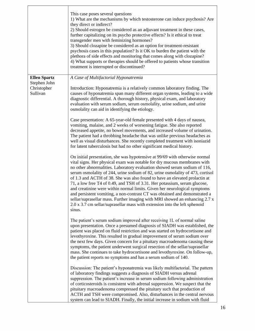
This case poses several questions
1) What are the mechanisms by which testosterone can induce psychosis? Are
they direct or indirect?
2) Should estrogen be considered as an adjuvant treatment in these cases,
further capitalizing on its psycho protective effects? Is it ethical to treat
transgender men with feminizing hormones?
3) Should clozapine be considered as an option for treatment-resistant
psychosis cases in this population? Is it OK to burden the patient with the
plethora of side effects and monitoring that comes along with clozapine?
4) What supports or therapies should be offered to patients whose transition
treatment is interrupted or discontinued?
Ellen Spartz
Stephen John
Christopher
Sullivan
A Case of Multifactorial Hyponatremia
Introduction: Hyponatremia is a relatively common laboratory finding. The
causes of hyponatremia span many different organ systems, leading to a wide
diagnostic differential. A thorough history, physical exam, and laboratory
evaluation with serum sodium, serum osmolality, urine sodium, and urine
osmolality can aid in identifying the etiology.
Case presentation: A 65-year-old female presented with 4 days of nausea,
vomiting, malaise, and 2 weeks of worsening fatigue. She also reported
decreased appetite, no bowel movements, and increased volume of urination.
The patient had a throbbing headache that was unlike previous headaches as
well as visual disturbances. She recently completed treatment with isoniazid
for latent tuberculosis but had no other significant medical history.
On initial presentation, she was hypotensive at 99/69 with otherwise normal
vital signs. Her physical exam was notable for dry mucous membranes with
no other abnormalities. Laboratory evaluation showed serum sodium of 116,
serum osmolality of 244, urine sodium of 82, urine osmolality of 473, cortisol
of 1.3 and ACTH of 38. She was also found to have an elevated prolactin at
71, a low free T4 of 0.49, and TSH of 3.31. Her potassium, serum glucose,
and creatinine were within normal limits. Given her neurological symptoms
and persistent vomiting, a non-contrast CT was obtained and demonstrated a
sellar/suprasellar mass. Further imaging with MRI showed an enhancing 2.7 x
2.0 x 3.7 cm sellar/suprasellar mass with extension into the left sphenoid
sinus.
The patient’s serum sodium improved after receiving 1L of normal saline
upon presentation. Once a presumed diagnosis of SIADH was established, the
patient was placed on fluid restriction and was started on hydrocortisone and
levothyroxine. This resulted in gradual improvement of serum sodium over
the next few days. Given concern for a pituitary macroadenoma causing these
symptoms, the patient underwent surgical resection of the sellar/suprasellar
mass. She continues to take hydrocortisone and levothyroxine. On follow-up,
the patient reports no symptoms and has a serum sodium of 140.
Discussion: The patient’s hyponatremia was likely multifactorial. The pattern
of laboratory findings suggests a diagnosis of SIADH versus adrenal
suppression. The patient’s increase in serum sodium following administration
of corticosteroids is consistent with adrenal suppression. We suspect that the
pituitary macroadenoma compressed the pituitary such that production of
ACTH and TSH were compromised. Also, disturbances in the central nervous
system can lead to SIADH. Finally, the initial increase in sodium with fluid

17
administration suggests a component of volume depletion. In conclusion, we
propose that a systematic approach is helpful when approaching patients with
hyponatremia. Laboratory evaluation can distinguish causes such as primary
polydipsia, heart failure, SIADH, and adrenal insufficiency. Additionally, in
patients presenting with neurological symptoms such as headache and visual
changes, it may be appropriate to obtain brain imaging.
Simon Yang
Dr. Austin Cudak
Dr. Peter Lund
Atypical Presentation of Mantle Cell Lymphoma Mimicking Pancreatic
Adenocarcinoma
Introduction: While the majority of solid pancreatic neoplasms are primary
exocrine pancreatic cancer or neuroendocrine tumors, a minority are rare and
can pose a diagnostic challenge. A comprehensive evaluation of extra-
pancreatic findings can speed diagnosis.
Case Presentation: A 56-year-old male with no significant medical history
presented with one month of intermittent epigastric abdominal pain, nausea
and a 10 pound weight loss. He initially presented to clinic where an
abdominal ultrasound noted a 9cm pancreatic head mass with extrahepatic
biliary dilation and borderline gallbladder wall thickening and he was referred
for admission.
On admission he was afebrile and hemodynamically stable. Laboratories were
notable for WBC 2.8, Hgb 8.3, platelet 68, CRP 2.73, lipase 85.2, elevated
metamyelocytes of 4.4, low normal B12 of 412, and positive H. pylori
antigen. CT scan confirmed a 10cm mass enveloping the pancreas. Further
evaluation of pancytopenia with bone marrow biopsy revealed mantle cell
lymphoma blastic variant with Ki-67 proliferation index of 20%. Subsequent
tissue biopsy noted effacement of pancreatic architecture with lymphoid
appearing cells positive for CD20 B-cell marker and CD5 seen in the germinal
center and mantle zone.
During his subsequent stay, he developed jaundice with elevated LFTs in an
obstructive pattern. Repeat ultrasound and PET scan showed a hypermetabolic
10-11cm pancreas-encasing mass narrowing the common bile duct with
dilated common hepatic duct and intrahepatic biliary duct as well as intense
heterogenous uptake in the bone marrow. A biliary stent was placed by ERCP
and jaundice improved. He was then placed on RCHOP (rituximab,
cyclophosphamide, doxorubicin, vincristine, and prednisone) with plans to
follow with autologous stem cell transplant.
Conclusion: Mantle cell lymphoma - a type of primary pancreatic lymphoma -
is known for its heterogenous presentations in a variety of extra-nodal.1,2
This case illustrates how uncommon types of pancreatic cancer can present
similarly to pancreatic adenocarcinoma, which represents approximately 85%
of cases.3 Although primary pancreatic lymphomas are rare, our main
takeaway is that a broad differential should be considered for pancreatic
tumors especially if there are other clinical clues, such as pancytopenia seen
with this case, as it can result in far different treatment and prognosis.
Reference
1. Arieira, Catia, et al. "Primary colon mantle lymphoma: a misleading
macroscopic appearance!." Revista Espanola de Enfermadades Digestivas
(REED) 111.12 (2019): 965-968.
2. Iqbal, Madiha, et al. "Intraocular involvement of mantle cell lymphoma: a
case report and literature review." Hematology/Oncology and Stem Cell

18
Therapy (2019).
3. Ilic, Milena, and Irena Ilic. "Epidemiology of pancreatic cancer." World
journal of gastroenterology 22.44 (2016): 9694.
Residents
Quality Improvement - Residents
Emily
Westergard
L. Patel
L. Wohlwend
M. Ornes
Improved Inpatient Appropriate VTE Prophylaxis Rates Through Use of An
Electronic Medical Record Intervention
Background: Venous thromboembolism (VTE) is a major health issue,
carrying a high mortality rate and significant economic toll within the United
States healthcare system. Despite the prevalence of VTE, clinicians have
struggled with appropriately prescribing prophylactic agents in the inpatient
setting despite multiple risk assessment models and guidelines. To help
improve appropriate VTE prophylaxis rates within the Allina Healthcare
system, a Padua Scoring Tool was incorporated into admission order sets in
September 2019.
Methods: Internal medicine resident admissions at Abbott Northwestern
Hospital were randomly sampled from both February to July 2019 and 2020.
Admission orders were then assessed for appropriate selection of VTE
prophylaxis and compared pre- and post-addition of the Padua scoring tool.
Retrospective calculation of the Padua, IMPROVE, and IMPROVE Bleed
scores was used to define appropriate VTE prophylaxis. Rates of appropriate
prophylaxis, frequency of inappropriate prophylactic agent use, and omission
of prophylaxis were then compared.
Results: Appropriate VTE prophylaxis rates increased from 64.5% to 76.7%
post-intervention. Inappropriate sequential compression device (SCD)
prescribing worsened from 51% to 78%. Inappropriate use of heparin
improved from 43% to 18%. Inappropriate use of enoxaparin remained low at
2% and 3%, respectively. Rate of inappropriate lack of prophylaxis remained
stable at 6%.
Conclusion: Introduction of the Padua scoring tool to admission order sets
was associated with a significant improvement in appropriate VTE
prophylaxis and inappropriate heparin use overall, however, there was an
unexpected increase in inappropriate SCD use.
Research - Residents
Natashay Bailey
Dr. Allyson Palmer
Dr. Laura
Greenlund
Comparison of D-dimer Values for Acute Deep Venous Thrombosis vs
Alternative Diagnoses in Patients undergoing Lower Extremity Ul
Introduction: Lower extremity deep venous thrombosis (DVT) is a common
diagnosis in ambulatory, critically ill, and post-surgical patients. Laboratory
testing, in particular d-dimer values, and ultrasonography are useful in the
diagnosis of DVTs. Typically, d-dimer is used to screen low and intermediate
pre-test probability patients to determine the need for ultrasound. There is
little in the medical literature describing the incidence of alternative diagnoses
observed on lower extremity ultrasound and the corresponding d-dimer test
results in patients being evaluated for suspected acute DVT. In this study, we
identify patients with ultrasound identified alternative diagnoses and

19
characterize the corresponding d-dimer levels in comparison to patients with
acute DVT.
Methods: We conducted a retrospective, single health system study using
chart analysis, focusing on lower extremity ultrasounds between July 2017
and July 2019. Data was collected from both ambulatory and hospitalized
patients across three Mayo Clinic regions in the United States (Arizona,
Florida, and Rochester). We assessed patient characteristics, d-dimer levels
detected by high sensitivity immunoassay, and incidence of acute DVT and
alternative diagnoses on ultrasound. Cases in which patients underwent
screening for surgical planning, had limited sonography, had known DVTs
within 6 months, and did not have d-dimer as part of the evaluation were
omitted. Overall, 2,377 cases fulfilling study criteria were analyzed.
Results: Among 2,377 patients that underwent both d-dimer and lower
extremity ultrasound, acute DVT was identified in 8.8% while alternative
diagnoses were seen in 30.2% of patients. The most common alternative
diagnoses were popliteal cyst (n= 243, 10.2%), edema (n= 149, 6.3%), post-
thrombotic changes (n= 91, 3.8%), chronic DVT (n= 78, 3.3%), SVT (n=38,
1.6%), and sub-acute DVT (n= 27, 1.1%). The mean d-dimer level in acute
DVT case was 6889.5 ng/mL (SEM 623.6 ng/mL) versus 2180.3 ng/mL
(SEM 165.7 ng/mL) with no pathology. In alternate diagnoses, mean d-dimer
values with SEM were 2217.3 +/- 257.6 ng/mL in popliteal cyst, 3286.1 ±
488.0 ng/mL in edema, 1142.6 ±181.2 ng/mL in post-thrombotic changes,
1385.4 ± 272.2 ng/mL in chronic DVT, 6099.9 ± 1782.9 ng/mL in sub-acute
DVT, and 5276.1 ± 1716.4 ng/mL in SVT. Interestingly, in those with d-
dimer values below 500 ng/mL, the typical low and intermediate pre-test
probability cutoff where patients would not require ultrasound, the incidence
of acute DVT was 2.2% (12 of 547 cases) vs 10.8% (197 of 1830 cases) in
patients with d-dimer over 500 ng/mL.
Conclusion: In patients, undergoing d-dimer testing and lower extremity
ultrasound to rule out acute DVT in the Mayo Clinic network, the incidence of
acute DVT was low compared to alternative diagnoses (8.8% vs 30.2%).
Alternative pathologies such as sub-acute DVT and SVT had d-dimer values
comparable to acute DVT. In contrast, pathologies such as post-thrombotic
changes and popliteal cyst, d-dimer values were similar to cases with no
pathology. Future studies should seek to validate these findings with a
prospective design.
Megan Covington
Dr. Philip Young
Dr. Melanie Bois
Dr. Joseph
Maleszewski
Dr. Kyle Klarich
The Clinical Impact of Cardiac Fibromas
Introduction: Cardiac fibromas are rare tumors that can present with a variety
of clinical findings. Previously they have been described as having the
potential to cause significant morbidity and mortality, including inflow and
outflow obstruction, arrhythmias, and sudden cardiac death. There has not
been a large review of cases focusing on the clinical aspects of cardiac
fibromas since 1994. The purpose of this study is to provide an updated
analysis of their clinical impact. This will allow for better understanding of
these tumors, lead to clinical awareness, and reveal important factors for
consideration when diagnosing and determining treatment options.
Methods: A retrospective case series was conducted from a tertiary care
institution to identify cases of cardiac fibroma (1970-2020). Mayo Clinic’s
radiology and pathology archives were reviewed, and patient cases were

20
included if cardiac fibroma was diagnosed via imaging results or pathology
report. Patient cases were reviewed and examined for demographics,
symptomatology, location, heart rhythm data, radiographic findings,
pathology findings, interventions, and outcomes.
Results: Twenty patients (11 female) with a median age of 23.5 years (1 day -
72 years) were identified through institutional archives as having a cardiac
fibroma. Presenting symptoms included ventricular tachycardia (VT),
dyspnea, palpitations, syncope, angina, heart failure, and emboli. VT was
noted in six patients (30%), all of whom experienced associated symptoms
such as palpitations, syncope/presyncope, and cardiac arrest. New murmur
was also common, noted in five patients (25%). Older patients were more
likely to be asymptomatic (49-72 years). One patient had Gorlin’s syndrome.
Tumors were most commonly located in the left ventricle (14/20, 70%).
Eighteen out of twenty were initially diagnosed with imaging, twelve of
whom were later confirmed via resection. Fourteen patients underwent
resection, four of whom required complex operations including artery graft,
valve repair, reconstruction of the aortic root and ventricular septum, and
amputation of LA appendage. Six of these patients had delayed intervention
ranging from 6 months to 7 years. One patient required a second surgical
resection. One operative death occurred. Six patients were managed with
clinical follow-up and imaging surveillance. Three were asymptomatic, one
experienced heart failure, and two suffered from arrhythmias. Both of the
latter patients required antiarrhythmics, and one ultimately required ablation
and pacemaker placement. One was lost to follow up.
Conclusion: Cardiac fibromas are typically considered to affect a pediatric
population; however, this study demonstrated a significant prevalence in
adults, and should therefore remain on the differential in the assessment of
cardiac masses. VT and associated symptoms were common at initial
presentation. Combined imaging modalities are sensitive in the diagnosis of
cardiac fibroma. Surgical resection is overall successful in symptomatic
patients, and clinical follow-up with imaging surveillance may be appropriate
management for asymptomatic patients.
Siva Kamal
Guntupalli
Yosuf Subat
Pavol Sajgalik
Matthew Hainy
Kim Chul-Ho
Bruce Johnson
Thomas Allison
Kaiser Lim
Alexander Niven
Aerosol and Droplet Particle Generation During Forced Expiration for Peak
Flow Testing
Introduction: Peak flow meters are routinely used by asthmatic patients to
assess airflow limitation. In order to ensure proper technique, patients are
often asked to perform peak flow maneuvers in the presence of physicians,
nurses, respiratory therapists, and other healthcare workers, potentially
creating the infection transmission risk given the ongoing COVID epidemic.
We aimed to develop a pragmatic approach to quantify particle generation
during peak flow testing to better understand the risk of this procedure with
the goal of informing institutional infection control and mitigation strategies.
Methods: For this pilot study, a tightly sealed room was connected to two
portable fans with HEPA filters, allowing for nearly particle free room. We
measured the concentrations of the generated particles after peak flow testing
on 5 healthy volunteers using five different peak flow meters (Respironics,
Philips, Clement Clarke, Respironics low range, and Mogaghan) using Fluke
and PTrak particle counters. Baseline concentrations of the existing particles
in a standard pulmonary function testing room were also measured for
comparison with the testing environment.

21
Results: Higher mean particle concentrations were observed with peak flow
testing compared to unmasked tidal breathing. Ultrafine particles (0.02-1
micron) were generated in higher proportions compared to particles of other
sizes (>1 micron). Mean particle concentration was lowest with the use of
Respironics peak flow meter (1.25±0.47 particles/cc). Comparable mean
particle concentrations were observed with Philips (3.06±1.22), Clement
Clarke (3.55±1.22), Respironics low range (3.50±1.52), and Mogaghan
(3.78±1.31) peak flow meters. When compared to the mean concentration of
the generated ultrafine particles (0.02-1 micron) by masked (0.22±0.29
particles/cc) and unmasked (0.15±0.18) tidal breathing, mean particle
concentrations were higher after use of peak flow meters.
Conclusions: We developed a pragmatic approach to quantify small particle
generation after peak flow testing. Although all peak flow meters
demonstrated increased mean particle concentration, differences were small
compared to the mean particle concentrations found in the ambient clinical
environment.
Fredrick Ogugua
Dr. Abdulrahman
Gamam
Dr. Jorge Reyes
Associations of Implantable Cardiac Defibrillator placement in Cardiac
Sarcoidosis: An insight from the National Inpatient Sample
Introduction: There is sparse literature detailing the indications for
implantable cardiac defibrillator (ICD) placement specific to hospitalized
patients with cardiac sarcoidosis (CS). Using the National Inpatient Sample
(NIS), we aim to identify patient characteristics and comorbid conditions
associated with ICD placement in patients with CS.
Methods: We queried the 2016-2017 NIS database to identify all admissions
age 18 years or older with a discharge diagnosis of CS who did not have a
prior history of ICD placement. We accounted for survey design and built a
multivariable logistic regression to identify predictors of ICD placement in CS
patients. Variables of interest included age, race, sex, and validated codes for
multiple comorbidities.
Results: An estimated 1,180 patients with CS without prior ICD placement
were hospitalized from 2016 to 2017. Among them, 35% had an ICD placed
during that hospitalization. CS patients who had ICD placement were more
likely to be male (72% v. 48%, OR 4.00 {1.92-8.36}, p<0.01). There were no
significant differences in age or race regarding ICD placement. CS patients
with ICD placement were more likely to be obese (35% v. 24%, OR
3.00{1.46-6.15},p<0.01), have hypertension (71% v. 68%, OR 2.68{1.20-
5.99}, p<0.01) and have a valvular disease (20% v. 16%, OR 4.06{1.25-
13.2}, p<0.01).
Conclusion: In our retrospective cohort of patients with CS without prior ICD
placement, we identified male sex, comorbid obesity, hypertension, and
valvular disease to be predictors of ICD placement. It is unclear if the
association with valvular disease signifies advanced cardiac sarcoidosis, as
sarcoid lesions involving cardiac papillary muscles can precipitate valvular
disease. Further research is needed to determine the clinical significance
between the aforementioned comorbidities, cardiac sarcoidosis and ICD
placement.
Jessica
Padniewski
Dr. Linh Ngo
Gazing Into The Crystal: Current Status of Gout Care Among Different
Specialties At A Tertiary Academic Center

22
Background/Purpose: Gout is the most common type of inflammatory
arthritis, affecting about 4% of the U.S. population and results in an estimated
6 billion dollars in healthcare costs yearly. Annual gout management costs
anywhere from $172 to $6179 according to a meta-analysis performed by Rai
et al. Despite available treatment guidelines published by the American
College of Rheumatology (ACR) and European League Against Rheumatism
(EULAR), deficiencies in the treatment of gout remain prevalent. The
ineffective treatment of gout results in significant functional decline,
increased morbidity, and significant economic burden. A study performed by
Harrold et al. in 2013 found that about half of primary care providers reported
optimal gout treatment practices. In an attempt to evaluate the efficacy of gout
management and adoption of the 2012 ACR gout guidelines, we performed a
retrospective cohort study at Hennepin Healthcare, a tertiary academic referral
center.
Methods: IRB approval was obtained for this retrospective cohort study. All
outpatient encounters with ICD-10 diagnosis code of M10.9x for gout
between July 1, 2018 through June 31, 2019 at Hennepin Healthcare were
pulled from a virtual data warehouse. A total of 485 outpatient encounters and
297 unique patients were identified. Patients who had greater than or equal to
8 points in the 2015 ACR Gout Classification criteria were excluded from this
descriptive analysis. A total of 226 unique patients met criteria and were
enrolled into the study. Chart review of enrolled patients was completed and
data regarding comorbidities, outpatient clinic type, basic demographics,
baseline uric acid, urate lowering therapy and follow up frequency were
recorded into a HIPAA compliant REDCap database and a descriptive
analysis was done.
Results: In summary, of patients noted to have uric acid levels > 6.0 mg/dL,
about 51.6% started uric acid prophylaxis. Of patients with gout and uric acid
levels > 6.0 mg/dL, about 44% eventually met uric acid level goals, while
about 55% did not. For those who achieved uric acid goal, the average
number of days to goal was 134 days.
Conclusion: Despite the ACR Gout Guidelines being published in 2012, our
data indicates adherence to its major principles is still lacking at our tertiary
academic center. A large percentage of patients above uric acid goals do not
receive flare prophylaxis, average days between follow up for titration of uric
acid lowering therapy is almost half a year, and the majority of patients with
gout did not reach uric acid goals. Further intervention is needed to improve
adoption of guideline principles of gout care.
Ka Bao Vang
Dr. Kathleen
Culhane-Pera
Dr. Luis Martin
Ortega
Txia Xiong
Carin Northuis
Pilar de la Parra
Dr. Kamakshi
Lakshminiarayan
Mobile Health Technology for Hypertension Management in Hmong and
Latino adults: Empowerment or Entrapment?
Background: Hypertension is a major risk factor for stroke and heart attack.
Unfortunately, more than 40% of adults with hypertension have inadequate
blood pressure control. Patients from minority groups, including Hmong and
Latino communities, have worse HTN control. Home blood pressure self-
monitoring can improve hypertension control, particularly in times of
restricted clinic visits during COVID-19 pandemic. Mobile health technology
(mHealth) can facilitate this self-monitoring. However, it is unknown if
mHealth will be accepted by minority patients.
Objective: Identify Hmong and Latino adults’ perspectives about a mHealth
based care model for high blood pressure management involving self-

23
monitoring and responsive medication adjustment by a provider team.
Methods: We enrolled 25 Hmong and 25 Latino participants with
hypertension from an urban federally qualified health center serving low
income patients. We obtained demographic data, collected responses about
hypertension knowledge, and conducted open-ended interviews to identify
participants’ perspectives about the mHealth based hypertension care model.
Results: Participants were 56% female with a mean age of 56 years. Most
(88%) were immigrants and most (96%) had lived in the United States (US)
for 10 years or longer. Half (52%) spoke and read English and 100% of
Latino and 76% of Hmong were literate in their own language. In addition to
hypertension, participants reported comorbidities including diabetes mellitus
(48%), elevated cholesterol (54%) and depression or anxiety (48%). While
most participants (98%) agreed that lowering high blood pressure decreased
the risk of strokes, heart attacks and kidney failure, there were gaps in medical
knowledge. Three major themes emerged from the interviews regarding the
perception of the mHealth based care model: 1) Using mHealth technology
could be useful especially if assistance was available to patients with
technological challenges; 2) Knowing blood pressures could be helpful to
patients who agreed with doctors’ medical diagnosis and prescribed treatment;
3) Transmitting blood pressures to the clinic and their responsive actions
could feel empowering for patients; however, the sense of increased
surveillance could feel entrapping to some patients.
Conclusion: In general, Hmong and Latino adults responded positively to the
mHealth based hypertension care model and felt that it could be empowering
since it could increase patient-provider communication without burden of
clinic visits and could increase involvement in BP control for those who agree
with the medical model of hypertension. Some participants expressed concern
that the mHealth model could be entrapping as it could breach patient privacy,
interfere with patients’ lifestyle choices, and curtail patient autonomy. In a
shared decision-making approach with patients (and possibly their family
members), health care systems and clinicians should explore potential issues
of empowerment and entrapment when offering an mHealth care model in
practice.
Clinical Vignette- Residents
Omar Abdullahi
Dr. Breanna
Zarmbinski
Dr. Clara
Zamorano
A Rare Case of Macrophage Activation Syndrome (MAS)
Case Presentation: MAS is a subtype of hemophagocytic lymphohystiocytosis
(HLH) and is a syndrome of immune hyperactivity and cytokine storm.
Primary HLH is mainly seen in pediatrics and is associated with multiple gene
mutations. In contrast, secondary HLH is often driven by secondary
inflammatory causes, most commonly viral illness, malignancy, or
autoimmune diseases. MAS has no preceding viral illness, new medication, or
malignancy. Highest incidence is with systemic juvenile idiopathic arthritis in
children and with RA in adults.
A 28-y.o. female presented with 5 day history of headache, nausea, vomiting,
fatigue, body aches and maculopapular rash not involving the palms or soles.
On admission, she was afebrile, hypotensive, and tachycardic. Labs notable
for elevated lactate, procalcitonin and LFTs. UA and CXR were
unremarkable. Her symptoms were concerning for bacterial meningitis and
multiorgan system dysfunction, and she was treated with broad spectrum

24
antibiotics and fluids. On day 2, repeat CXR showed new bilateral airspace
infiltrates with interstitial edema at the lung bases, concerning for flash
pulmonary edema. The patient subsequently went into shock and further
respiratory distress, so her fluids were stopped, pressors were started and she
was intubated. She also had worsening anemia and thrombocytopenia thought
to be secondary to acute viral illness or newly diagnosed autoimmune flare.
Autoimmune workup showed strongly positive sjogrens markers (Ro52
antibody>1,685.3, Ro60 antibody>1,374.8, SS-B antibody>1,550.0). Further
workup notable for elevated ferritin at 17,000 (lower with steroid) with
triglycerides elevated at 389. These lab findings, along with the clinical
picture, were concerning for HLH. She had a peripheral smear which showed
no hemolysis. Bone marrow biopsy was highly suggestive of HLH.
Heme/Onc consult initiated intrathecal methotrexate, IV etoposide, and
dexamethasone. She clinically improved, was successfully extubated and
discharged to home within several weeks.
Conclusion: This case illustrates that the clinical presentation of HLH can
mimic acute infection with multiorgan failure. These patients, unlike those
with infection, will be unresponsive to antibiotics. Early diagnosis is of
critical importance considering adult patients have a 31-58% survival rate if
treatment is initiated promptly. Without treatment mean survival is 3-5 weeks
with mortality approaching 100%. Hence, start workup early with
triglycerides and ferritin levels if HLH is suspected.
Nadia Akhiyat
Dr. Brett Grieb
Dr. Mustaqueem
Siddiqui
A Rare Neoplasm Masquerading as Pulmonary Embolus
Introduction: Pulmonary artery sarcoma is a rare malignant neoplasm that
may be difficult to distinguish from pulmonary arterial embolism. Initial
misdiagnosis is common and often delays therapeutic intervention.
Distinguishing key features are necessary for earlier diagnosis and improved
clinical outcomes. We present a case of pulmonary artery sarcoma presenting
as hyperfibrinolysis in the absence of identifiable clot formation.
Case Description: A 44 year old male with no known comorbidities initially
presented with recurrent gross hematuria and cough with hemoptysis
associated with 30lbs unintentional weight loss and night sweats. Initial CT
chest without contrast demonstrated scattered non-specific pulmonary
nodules. Comprehensive bleeding evaluation revealed evidence of
dysfibrinogenemia with D-dimer > 30,000, decreased alpha-2 plasmin
inhibitor, and markedly elevated soluble fibrin monomer. Other coagulation
studies were normal. Ultrasound of the extremities was negative for overt clot
formation. CT abdomen and pelvis was negative for occult malignancy.
Thorough urologic evaluation revealed an AV malformation of the right renal
artery which was embolized. BAL was negative. A tranexamic acid taper
resolved hematuria and reflected improvement in fibrinolysis labs. Our patient
however continued to experience B-symptoms. He again developed
hemoptysis 3 months following his initial presentation. A CT chest with IV
contrast was obtained and was interpreted to demonstrate an extensive right
pulmonary embolus involving the entire right pulmonary artery, proximal left
pulmonary artery, and encroachment upon the left main coronary artery. A
PET-CT revealed intense FDG uptake within the expansile mass. Pathology
from an endobronchial ultrasound biopsy revealed spindle cell sarcoma. This
patient successfully underwent radical surgical resection with planned chemo-
radiation therapy. Hyperfibrinolysis was ultimately attributed to underlying
sarcoma.

25
Discussion: Pulmonary artery sarcoma is often misdiagnosed as pulmonary
embolism. Key distinguishing features include an atypical appearance of a
pulmonary artery mass on CT imaging without peripheral emboli, cough,
hemoptysis, and clinical presentation suggesting underlying malignancy. This
case illustrates such key features in addition to the importance of considering
occult malignancy in the setting of idiopathic hyperfibrinolysis in the absence
of clot formation.
Chineze Akusoba
Dr. Brenden
Ingraham
Dr. Adam
Sawatsky
Gerbode Defect: A Rare Complication of Tricuspid Valve Endocarditis
Introduction: Infective endocarditis can be associated with a multitude of
complications including heart failure, perivalvular abscess, septic
embolization and mycotic aneurysm. The likelihood of complications depends
on a variety of factors, including comorbidities, pathogen, and duration of
illness prior to therapy. We describe a case of methicillin- resistant
staphylococcus aureus (MRSA) tricuspid valve endocarditis that resulted in a
ventricular septal defect creating a communication between the left ventricle
(LV) and right atrium (RA), also known as the Gerbode defect.
Case Presentation: A 50-year-old man with a history of metastatic colon
cancer status post sigmoidectomy and pelvic radiation complicated by ureteral
strictures requiring bilateral nephrostomy tubes was admitted with hematuria
and dyspnea following recent diagnosis of MRSA bacteremia and tricuspid
valve endocarditis.
The patient was hospitalized 3 weeks prior to admission with MRSA
bacteremia secondary to the nephrostomy tubes. He underwent a transthoracic
echocardiogram (TTE) which demonstrated a 1.5 cm vegetation on the
anterior leaflet of the tricuspid valve with moderate tricuspid regurgitation. A
CT Chest revealed multifocal thoracic septic emboli. He was discharged on a
6-week course of IV vancomycin via a peripherally inserted central catheter
(PICC). Unfortunately, he developed worsening dyspnea and significant
hematuria requiring numerous red blood cell transfusions and was re-
hospitalized.
On arrival, he was afebrile and hemodynamically stable. Exam was notable
for a 3/6 pansystolic murmur best heard at the lower left sternal border.
Laboratory evaluation revealed acute anemia with hemoglobin 6.0 (13.2 –
16.6 g/dL), leukocytosis of 10.2 (3.4 - 9.6 x 10(9)/L) and CRP 41.2 (≤ 8.0
mg/L). Blood cultures were negative. He underwent repeat TTE, which
revealed resolution of the tricuspid vegetation, however there was a new,
large atrio-ventricular mycotic aneurysm that ruptured into the RA resulting in
a left to right shunt between the LV and RA. Transesophageal echo (TEE)
confirmed this finding. He underwent right heart catheterization notable for
oxygen step-up in the low right atrium of 14% and elevated cardiac index of
4.5 (2.5 – 4.0 L/min/m2) with moderate left-to-right shunting. Cardiovascular
Surgery recommended elective surgical intervention for closure of the defect.
Discussion: The Gerbode defect is a communication between the LV and RA.
It can be congenital or acquired as the result of endocarditis, prior cardiac
surgery, myocardial infarction, or trauma. Diagnosis is generally made by
TTE. Surgical and percutaneous transcatheter closure are options for
management of the defect depending on the patient’s symptoms, magnitude of
shunt, anatomical feasibility, and comorbidities.
Hadiyah Audil Copper Deficiency-Induced Anemia and Neutropenia

26
Dr. Animesh
Pardanani
Introduction: The prevalence of obesity has been estimated at 13% worldwide
and nearly 40% in the US. Bariatric surgery is effective for obese patients and
is becoming more prevalent; in 2017, approximately 580,000 people
underwent bariatric surgery, with 228,000 cases being performed in the US
alone. Such procedures include Roux-en-Y gastric bypass (RYBG), wherein a
small proximal gastric pouch is created and anastomosed to the small
intestine. This bypassing of the alimentary tract predisposes to micronutrient
and mineral deficiencies. Guidelines therefore recommend that patients
undergo routine post-operative monitoring for nutritional deficiencies and
daily supplementation with a minerals-containing multivitamin, calcium, and
vitamins D and B12.
Case Description: A 49-year-old female presented with two weeks of
progressive dyspnea. She was status post RYGB 18 years ago, and was on
daily B12 and folate supplementation only. Labs on admission were
remarkable for macrocytic anemia (Hgb 6.2, MCV 105.2) and neutropenic
leukopenia (WBC 1.7, ANC 240); on chart review, CBC from nine months
prior was normal. Physical exam was unremarkable and review of systems
was negative for blood loss and constitutional B symptoms. She was given
one unit of packed red blood cells with appropriate response in hemoglobin
and some symptom improvement; Hematology was consulted due to concern
for myelodysplasia. Peripheral smear showed slight anisopoikilocytosis.
Evaluation for GI blood loss, hemolysis, liver disease, and hypothyroidism
was unremarkable. Although B12 and folate were normal, serum copper was
<0.10 mcg/mL (normal 0.75-1.45 mcg/mL). As she had an inappropriately-
normal reticulocyte count, a bone marrow biopsy was performed. Pathology
revealed panhyperplasia, cytoplasmic vacuolization in myeloid precursors,
increased ring sideroblasts, and no increase in blasts. She was started on IV
copper replacement and discharged with outpatient copper infusions.
Discussion: Copper deficiency occurs in 10-15% of patients following gastric
bypass surgery, but remains under-recognized. Further risk factors for
acquired hypocupremia include other malabsorptive conditions, total
parenteral nutrition lacking sufficient copper, excessive zinc ingestion, and
chronic dialysis. Clinical manifestations include anemia, neutropenia, and
neurologic deficits (such as ataxia, myeloneuropathy, and cognitive deficits),
the latter of which was not seen in our patient but often mimics symptoms of
B12 deficiency. Serum copper levels can confirm the diagnosis and bone
marrow biopsy also has diagnostic utility, with abnormalities including
erythroid hyperplasia, vacuoles in myeloid precursors, iron-containing plasma
cells, and ringed sideroblasts. Treatment typically relies on copper repletion.
Importantly, although labs may resemble iron deficiency anemia, iron
supplementation can cause excess iron to compete with copper absorption and
worsen hypocupremia. With the rising prevalence of bariatric surgeries,
copper deficiency remains an important pathophysiologic process to consider
and diagnose in the correct clinical context.
Madeline Barnes
Dr. Ronald
Reilkoff
Diagnostic Challenges in Detecting Leptospirosis
Introduction: Leptospirosis has a varied clinical course with most cases
resulting in a mild/self-limited or subclinical presentation, but some are severe
resulting in potentially fatal outcomes with liver, kidney and pulmonary
failure. The wide-ranging clinical course with the relative rareness seen in
Minnesota makes leptospirosis a diagnostic challenge.

27
Case Presentation: A previously healthy 49-year-old male presented to the
emergency department with fever, myalgias, malaise, and vomiting and found
to be in new atrial fibrillation with rapid ventricular response and hypoxic
respiratory failure with labs showing acute renal failure, acute hepatitis and
troponin elevation. He was admitted to the ICU due to pressor requirement in
presumed septic shock due to a respiratory pathogen given chest x-ray with
bilateral disease and started on vancomycin, zosyn and doxycycline. Initial
infectious workup was negative including RVP, legionella, strep pneumo and
COVID-19 thus it was broadened to include tick born illnesses, fungal and
viral infections, parasites, and rare bacterial such as leptospirosis and
tularemia given his exposure with goats, donkeys, rats and sewage. He
continued to decline requiring a bronchoscopy revealing diffuse alveolar
hemorrhage and transfer to center with liver transplant capabilities on day 3 of
hospitalization. At admission to the liver transplant center, repeat cultures
were collected and antimicrobials were broadened to penicillin, ceftriaxone,
doxycycline, and ambisome. On day 6 of hospitalization, the repeat cultures
grew yeast while all other testing continued to come back negative for
infectious causes. As more infectious tests came back negative including
leptospirosis antibody testing and the inflammatory markers finally decreased,
it was decided to discontinue penicillin on day 8. The same day the culture
growing yeast speciated to Candida albicans resulting in ambisome being
switched to micafungin. The patient continued to slowly improve from day 8
with extubation occurring on day 12. Throughout that time, he continued to
receive doxycycline, ceftriaxone and micafungin and the entire infectious
workup came back negative. It was unclear how long to keep the antibiotics
given unknown etiology leading to the decision to send the Karius testing on
day 13. The Karius test came back positive on day 15 for Leptospirosis, he
completed a 16-day course of doxycycline and ceftriaxone. He continued to
improve and was discharged on day 20 from the hospital.
Conclusion: This case illustrates the difficulty in diagnosing leptospirosis.
Specifically, that IgM can be negative for the first 7 days of illness as in this
patient’s case and repeat testing should always be consider if there is a high
enough clinical suspicion.
Scarlett Cao
Dr. Daniel Pfeifle
Dr. Timothy
Aksamit
Hodgkin’s Lymphoma; An Unusual Presentation with Budd-Chiari Syndrome
Introduction: Budd-Chiari Syndrome (BCS) is a rare group of disorders
characterized by hepatic venous outflow obstruction. It often can present non-
specifically with symptoms including abdominal pain, ascites, and
hepatomegaly. Most commonly, this is caused by hypercoagulable states
resulting in occlusion of the venous outflow tract, but in rare cases can result
from direct vascular compression or invasion associated with malignancy.
Here, we describe BCS in a 37-year-old as a complication from a mediastinal
mass due to Hodgkin’s lymphoma, resulting in symptomatic obstruction and
acute liver failure.
Case Presentation: A 37-year-old female presented to the emergency
department for acute onset shortness of breath and abdominal pain. Past
medical history was significant for a recently found large 8.3 cm mediastinal
mass, concerning for lymphoma, with biopsy results pending at presentation.
On presentation, she had acutely elevated liver function testing including ALT
of 855, AST of 1817, alkaline phosphatase of 142, total bilirubin of 4.1, and
an elevated INR of 3.4. Notably, she also had a lactate of >18. She underwent
CT of the chest and abdomen due to concern for an ischemic process, which
showed the large mediastinal mass encasing and occluding the IVC at the

28
level of the diaphragm in addition to a new large thrombus within the
intrahepatic IVC. She was started on a heparin infusion for initial treatment
and admitted to the ICU.
She was noted to have rapidly worsening liver enzymes during the first 24-
hours of hospitalization. As the obstructing mass was suspected lymphoma,
she was started on high-dose IV methylprednisolone to reduce tumor burden
and help vascular flow. She was taken urgently for angioplasty and stent
placement in the IVC, with measured improvement in hepatic to atrial
gradient.
Her liver function testing significantly improved with intervention. Biopsy
results taken previously from the mediastinal mass demonstrated nodular
sclerosing Hodgkin’s lymphoma, and with stabilization of her condition, she
subsequently started chemotherapy during her hospitalization.
Highlighted Points: This case highlights a rare etiology of Budd-Chiari
Syndrome (BCS), resulting from vascular compression and thrombus
formation secondary to Hodgkin’s lymphoma. BCS is most commonly due to
intravascular thrombosis predisposed by hypercoagulable states—
myeloproliferative disorders account for 50% of primary BCS—and less
commonly due to malignancy-associated vascular compression or invasion.
Conclusion: Symptoms of Budd-Chiari are often non-specific, requiring a
high level of clinical suspicion to identify. The overall clinical course depends
on the acuity and extent of vein occlusion, ranging from chronic
asymptomatic to fulminant disease. For management, systemic
anticoagulation remains the foundation of medical therapy. With severe
disease, interventional approaches can be considered to improve hepatic
outflow or reduce sinusoidal pressure, among them balloon angioplasty with
or without stenting, placement of TIPS, or in rare occasions, liver
transplantation.
Monazza
Chaudhry
Dr. James
Leatherman
Pulmonary Blastomycosis: A Familiar Foe in the Midwest
Introduction: Pulmonary blastomycosis is one of the endemic fungal mycoses
more commonly seen in immunocompromised individuals in the Midwestern
United States. This case describes a subclinical infection masquerading as a
lung abscess in a young, healthy individual.
Case description: A 35 year old male presented to the emergency department
with a 6 day history of mild fevers, cough, and myalgias. Initial vitals were
significant for a temperature of 38.5 degrees Celsius and a WBC of 14. CXR
was significant for a left sided opacity. Treatment for community acquired
pneumonia was initiated and he was admitted for overnight observation. Over
the next 72 hrs, he continued to be febrile and profusely diaphoretic with labs
showing an up trending WBC up to 22, despite being optimized on broad
spectrum antibiotics. Further chest imaging revealed a large necrotic nodular
infiltrate initially concerning for a lung abscess. Upon further evaluation, it
was noted that the patient had gone on a short hiking trip in Wisconsin two
months prior and he had been feeling unwell for the past 2 weeks. Initial
sputum cultures showed no growth. As the clinical suspicion was exceedingly
high for a fungal etiology, further sets of induced sputum cultures were
collected. Within 3 hours, budding yeasts were visualized.
Conclusion: This case illustrates the importance of maintaining a high index

29
of clinical suspicion and the judicious use of clinical judgement in an
underrecognized presentation of a well-known, yet highly fatal, endemic
disease.
Chansong Choi
Andrew Greenlund
An Unusual Tale of Small Bowel Edema
Introduction: Angiotensin converting enzyme inhibitors (ACEI) is a common
class of drug utilized to treat hypertension and other cardiovascular conditions
such as heart failure. (1) Its mechanism involves the renin-angiotensin-
aldosterone pathway as it inhibits the breakdown of bradykinin. This results in
vasodilation, increased postcapillary venule permeability, and plasma
extravasation to surrounding tissues. (1) Angioedema is rather infrequent
adverse effect of the use of ACEI, as it is estimated to occur in 0.1-1%
patients taking ACEI. (1,2) Classically, it is reported to involve the mucus
membranes of the oropharynx, periorbital region, and upper airway
potentially resulting in asphyxia. Less described is the manifestation of
visceral angioedema involving the gastrointestinal (GI) tract, which is an
under-recognized phenomenon. (1–3)
Case Presentation: A 99-year old female with history of HTN on lisinopril
presents to the ED with 1-day history of nausea, abdominal pain, and emesis.
Physical exam was benign at the time of presentation. Laboratory evaluation
was notable for leukocytes 11.7, hemoglobin 13.0g/dL, lipase 28, and
unremarkable chemistry panel. Plain abdominal XR showed nonspecific
bowel gas pattern. CT abdomen/pelvis revealed abdominal ascites in addition
to wall thickening and edema involving loops of distal small bowel.
Subsequent work up to assess for underling autoimmune, infectious causes
including celiac serologies, C4 and CRP level, QuantiFERON-TB were
obtained which were negative. During hospitalization, lisinopril was
discontinued in the setting of precipitous drop in hemoglobin concerning for
GI bleed. Repeat CT scan in the setting of acute anemia 28 hours later showed
decreased abdominal wall ascites with resolving ileal wall thickening and
edema. There was no evidence of acute GI bleed on imaging, and hemoglobin
drop was attributed to dilution. She was noted to have remarkable
improvement in her symptoms with resolution of ileal wall edema and
inflammation after the withdrawal of lisinopril.
Conclusion: This case highlights an interesting case of visceral angioedema
secondary to lisinopril use resulting in case of ileal edema and inflammation.
Visceral angioedema is a rare phenomenon with 44 cases reported in the
literature from 1982 to 2016. Most reported cases involve the use of either
lisinopril or enalapril. (4) Patients usually present with nonspecific symptoms
such as abdominal pain, nausea, emesis, or diarrhea. Anatomic location of
visceral edema among reported cases has been variable, from antrum of the
stomach to isolated segments of the small bowel. It is usually treated with
supportive management including intravenous fluid replacement, antiemetics,
and discontinuation of the offending agent. (2) Given medication induced
visceral angioedema is a rare entity, clinicians should consider it as a
diagnosis of exclusion and should first explore and rule out other differential
diagnoses as vasculitis, small bowel lymphoma, and inflammatory bowel
disease such as Crohn’s disease. (2)
John Christensen
Dr. Somaira
Nowsheen
Lauren Webb
Bartonella henselae endocarditis presenting as anemia, splenomegaly, and
renal failure in an immunocompetent young man
Case Presentation: A man in his mid-forties presented to our hospital with two

30
Dr. Maryam
Mahmood
months of fever with chills, severe recurrent transfusion-dependent anemia,
splenomegaly, and renal failure. He had no significant past medical history,
and took no medications; he was in his usual state of health until
approximately six months prior to presentation. He had no history of cardiac
valvular disease or immunosuppression. He had had a very thorough workup
with his local health care providers, including hematologists, nephrologists,
infectious disease specialists, and rheumatologists. Investigations at our
hospital demonstrated infection-associated glomerulonephritis and small,
dense, thickened, sessile lesions on both the atrial and ventricular surfaces of
the mitral valve; infectious workup ultimately revealed positive Bartonella
serology. The patient was treated with a prolonged course of antimicrobials
and ultimately underwent surgical replacement of his infected valves.
Conclusion: Bartonellosis, caused by gram-negative intracellular facultative
bacteria, can lead to severe complications, such as anemia, fever,
hepatosplenomegaly, and endocarditis. Mortality approaches 40% without
treatment. Therefore, prompt diagnosis and intervention are essential.
Diagnosis of Bartonellosis relies on a combination of histological and
microbiologic evaluations, including serological testing, culture, and nucleic
acid amplification testing (NAAT). Conventional culture methods are not
ideal for diagnosis as currently available tests have low sensitivity.
Our case highlights the importance of considering Bartonellosis as a potential
diagnosis even in immunocompetent individuals who have signs and
symptoms suggestive of infective endocarditis, especially as the delay in
diagnosis for our patient led to a prolonged clinical course with recurrent
transfusions, kidney injury, and profound functional impairment prior to
definitive treatment.
Molly Clark
Dr. James
Campbell
Dr. Nadeem
Chaudhary
Breast Cancer Presenting as Painful Jaundice
Introduction: Breast cancer is one of the most common forms of cancer
affecting women, with 1:8 women being affected by breast cancer in their
lifetime [3]. However, breast cancer metastasizing to the pancreas is very rare.
Case Presentation: A 46 year old female presented to the ED with jaundice
and right upper quadrant and epigastric pain radiating to her back, with 6
months of associated progressive fatigue, loss of appetite, nausea, vomiting,
dark urine, and numbness in her toes. Of note, she also reported a small lump
in her right breast that she had noticed about one year ago. Ultrasound of the
abdomen revealed a large pancreatic mass and dilation of the bile ducts and
pancreatic duct. Subsequent CT abdomen/pelvis with IV contrast done for
further characterization of the masses showed a right breast mass, pancreatic
head mass, bilateral adrenal masses, right pararenal mass, multiple mesenteric
implants, innumerable subcutaneous masses, and mild retroperitoneal
lymphadenopathy; no hepatic lesions were seen. Endoscopic ultrasound done
for tissue biopsy revealed a large hypoechoic and heterogeneous round mass
in the head of the pancreas with irregular outer margins causing obstruction
and upstream dilation of the common bile duct. A similar appearing, but
smaller, mass was seen in the left adrenal gland. Fine needle biopsy of the
adrenal and pancreatic masses revealed signet ring morphology typical of
lobular carcinoma and chromogranin expression more suggestive of
pleomorphic lobular carcinoma. The tumor cells stained positive for GATA3
(the transcription factor binding to DNA sequence “GATA”) and estrogen
receptors (ER+) supporting a breast primary. She was diagnosed with
pleomorphic lobular carcinoma of the breast. While inpatient, she underwent

31
ERCP for decompression of the biliary tree; however, the ampulla was unable
to be located due to extrinsic pressure on the duodenal wall, thus percutaneous
biliary drainage was performed for decompression of the “trapped”
gallbladder. She was treated with 10 days of ceftriaxone (and later
cefpodoxime) and metronidazole, was started on tamoxifen and goserelin, and
plans were made for palliative radiation therapy.
Conclusion: This case represents a rare case of breast cancer presenting as
painful jaundice secondary to biliary obstruction from a large pancreatic
metastasis. Invasive pleomorphic lobular carcinoma is an aggressive form of
lobular carcinoma and more likely to have distant metastases and a lower
median survival than invasive ductal carcinoma[2]; however pancreatic
metastases are very rare[1]. When distinguishing between a primary
pancreatic malignancy and a distant metastatic breast cancer, it is important to
use immunohistochemical staining for GATA3, as this has high sensitivity for
breast carcinoma[1].
Maros Cunderlik
Dr. Alec Bunting
Dr. Amelia Krug
Dr. Fredrick
Ogugua
Dr. Michele
LeClaire
D. Abby Montague
A Case of Massive Caffeine Overdose With Refractory Shock Requiring
Extracorporeal Membrane Oxygenation Support.
Caffeine is ubiquitous in our society with 62% of the US population
consuming caffeinated beverages daily and caffeine-containing compounds
being increasingly used in energy drinks and supplements due to its desirable
psychostimulant properties at low doses (< 400mg). While life-threatening
caffeine overdoses are rare, massive ingestions above the established lethal
doses (150-200mg/kg, serum concentrations above 80µg/mL) have been
reported with most deaths attributed to arrhythmias and cardiovascular
collapse due to a unique caffeine toxicity profile.
Here we present a case of a 34-year-old female with intentional massive
caffeine overdose after ingestion of a guarana supplement containing
approximately 20g of caffeine - twice the upper limit of the lethal dose.
Shortly after the ingestion, the patient was altered requiring intubation for
airway protection. She was also hypotensive with mean arterial pressures of
approximately 40 mmHg and heart rhythm of wide complex tachycardia
despite three cardioversion attempts. At the outside institution, the patient
remained hemodynamically unstable despite aggressive medical interventions
including amiodarone, intralipids, norepinephrine, vasopressin, and esmolol
drips. She was subsequently transferred to our institution for consideration of
veno-arterial extracorporeal membrane oxygenation support (VA ECMO) to
facilitate emergent hemodialysis. On arrival, the patient was in severe
multifactorial cardiogenic, vasodilatory and hypovolemic shock. She also had
multiple metabolic derangements and had experienced status epilepticus.
After stabilization with multiple pressors including angiotensin II infusion,
electrolyte replacement, volume resuscitation and antiarrhythmic infusions
she underwent VA ECMO cannulation and emergent hemodialysis with
gradually improving hemodynamics over next 24 hours. On hospital day two,
the patient was weaned off all ECMO and all vasoactive medications with
complete recovery of cardiac function. Patient’s neurologic function also
gradually improved and she was extubated on hospital day six. With
continued supportive care, the patient achieved complete cardiovascular and
neurologic recovery, she was discharged to the inpatient psychiatric unit
seventeen days after the admission.
In conclusion, we present a case of severe caffeine toxicity due to the
intentional dietary supplement overdose resulting in severe multifactorial

32
shock requiring VA ECMO resulting in eventual complete cardiovascular and
neurologic recovery. Initial serum caffeine level was found to be 425 µg/mL.
This is to our knowledge the highest reported caffeine serum level where the
patient survived the ingestion.
Michael Dietrich
Malignancy Masquerading as Sinusitis
Introduction: Malignant melanoma is extremely variable in initial presentation
which can make early diagnosis and treatment challenging. Malignant
melanoma of the nasal mucosa can present very similarly to sinusitis, which
can add further difficulty to accurate diagnosis.
Case Presentation: An 87 year old male with a past medical history of only
hypertension noticed blood in his nasal mucus after blowing his nose. He had
no significant history of allergies, atopy, epistaxis, nasal trauma or chronic
rhinosinusitis. Bleeding continued to worsen over the next few months and he
developed new nasal congestion despite multiple nasal decongestants. He
presented to an urgent care clinic and was prescribed an anti-histamine with
continued vasoconstrictors/decongestants for presumed chronic sinusitis.
However, he represented to outpatient clinic 1 week later due to worsening of
his symptoms, including new feelings of nasal obstruction.
Laboratory studies including complete blood count, coagulation studies, and
erythrocyte sedimentation rate were ordered and within normal range.
Computerized tomography (CT) sinus without contrast showed an expansile
mass in the right posterior nasal cavity. The patient was referred to
otolaryngology who performed rhinoscopy with biopsies of the mass. Follow
up magnetic resonance imaging (MRI) to better characterize the lesion
redemonstrated a mass within the posterior nasal cavity which extended into
the nasopharynx, without extension through the right sphenopalatine foramen.
Initial frozen pathology showed poorly differentiated malignant neoplasm.
Positron emission tomography (PET)-CT scan did not show evidence of
regional or metastatic disease. Therefore, patient was taken for endoscopic
resection about 7 days after initial diagnosis. Final pathology showed
extensively necrotic 4.5cm malignant mucosal melanoma of the right nasal
septum with extension into the right middle turbinate.
Resection was performed without complication and he also underwent
adjuvant chemotherapy and radiation. However, 4 months later a follow up
PET-CT scan showed right cervical nodal metastasis which was confirmed to
be malignant melanoma on biopsy. Surgical modified neck dissection was
performed, lymph nodes positive for metastatic malignant melanoma. Again
adjuvant immunotherapy and radiation were performed. Unfortunately, repeat
PET-CT scan showed recurrent disease locally in the neck and evidence of
metastatic disease in the abdomen. The patient decided not to pursue further
systemic treatment.
Conclusion: This case demonstrates the variability in presentation of
malignant melanoma. Here we see nasal mucosal malignant melanoma
initially presenting as blood tinged mucus and symptoms of nasal congestion.
Therefore, it is important to keep a wide differential for symptoms of sinusitis,
especially given that mucosal melanoma can be a very aggressive subtype of
melanoma.
Kiril Dimitrov
Dr. Veronika
Recognizing Cytokine Release Syndrome and Neurotoxicity as Side Effects of
CAR-T Therapy: Mimics of Sepsis and Stroke

33
Bachanova
Introduction: Chimeric Antigen Receptor T-cell Therapy (CAR-T) has been
an important breakthrough in treatment of several hematologic malignancies.
CAR-T cells are autologous T lymphocytes genetically modified to express
novel protein to facilitate targeting of tumor cells. Several CAR-T products
are now FDA approved and there is increasing recognition of their unique side
effects. More prevalent application of CAR-T will require hospitalists to
differentiate sequelae from other common conditions seen in cancer patients.
Case Presentation: A 56-year-old female with Diffuse Large B-Cell
Lymphoma received lymphodepleting chemotherapy followed by CAR-T19
cell therapy axicabtagene ciloleucel (Yescarta) for relapsed disease. On Day
+6 patient developed neutropenic fevers to 101.6°F and hypotension that was
responsive to IV fluids. BMP, CBC with differential, blood cultures, chest X-
ray, urinalysis/culture, and inflammatory markers (IL6, Ferritin, and CRP)
were obtained. CXR was negative for infection and blood/urine cultures
showed no growth. CRP and IL-6 levels were elevated. Differential diagnosis
of elevated temperature and hypotension in a post CAR-T patient includes
sepsis and cytokine release syndrome (CRS). Patient was treated with IV
antibiotics, but fever persisted with intermittent hypotension. In the setting of
CRS Grade 2, patient received one dose of Tocilizumab and transferred to the
ICU for potential pressor support. Fevers and hypotension resolved the same
day and she became hemodynamically stable. She then presented with mild
intention tremor and trouble writing. Patient subsequently developed acute left
forearm numbness, impaired attention, confusion, and global aphasia but her
level of consciousness was not impaired. Considerations at that time were
acute stroke vs Immune effector-Cell Associated Neurotoxicity (ICANS)
Grade 3 from CAR-T therapy. Repeat MRI was without stroke. EEG showed
mild-moderate encephalopathy. Treatment included IV dexamethasone and
prophylactic Keppra. Patient promptly responded and within 24 hours her
symptoms improved. She was discharged in the next few days on a steroid
taper and with Grade 0 CRS/ICANS.
CRS is characterized by rapid release of pro-inflammatory cytokines
including IL-6 and IFN-γ, occurring 2-5 days after CAR-T infusion, and is
triggered by activated CAR-T cells. Clinical manifestations include high
fever, myalgias/arthralgias, hypotension, dyspnea, and capillary leak. Grading
for CRS (1-4) includes varying degrees of fever, hypotension, and hypoxia.
Treatment includes supportive cares with fluids, antipyretics, and
administration of IL-6 receptor antibody, Tocilizumab. Vasopressors and
ventilatory support may be required.
Conclusion: Immune effector Cell-Associated Neurotoxicity Syndrome
(ICANS) pathophysiology is distinct from CRS. Symptoms include
headaches, tremors, impaired attention, lethargy, aphasia, agraphia, confusion,
seizures, and increased intracranial pressure. Grading for ICANS includes
varying degrees (1-4) of decreased level of consciousness, seizures, motor
weakness, and increased cerebral edema. Steroids are the mainstay of
neurotoxicity treatment and yield quick improvement if instituted early. For
grade 4 ICANS, decrease of ICP with mechanical ventilation,
mannitol/acetazolamide may be required, and neurological consultation is
recommended.
Christopher Dinh
Dr. Hannah
Nordhues
A Woman with Urothelial Carcinoma and Abdominal Pain
Case Presentation: A 73-year-old female with a past medical history
significant for metastatic high-grade urothelial carcinoma of the right kidney

34
presented with three weeks of progressive abdominal pain, dyspnea, and
urinary symptoms. Evaluation revealed total calcium of 12.4 mg/dL, ionized
calcium of 6.21 mg/dL, and elevated liver enzymes of AST 66, ALT 60, and
alkaline phosphatase 227. CT chest, abdomen, and pelvis revealed marked
progression of the liver, lung, and small bowel metastases. She was admitted
to the hospital for further management.
Additional laboratory evaluation was significant for normal vitamin D levels
of 36 ng/mL, appropriately suppressed parathyroid hormone (PTH) of 8.6
pg/mL, and elevated parathyroid hormone-related peptide (PTHrP) of 8.6
pmol/L. Her abdominal pain was multifactorial from both her significant
malignant disease burden and symptomatic hypercalcemia. Her hypercalcemia
was treated with IV fluids and zoledronic acid, with partial improvement of
her abdominal pain. She was subsequently discharged from the hospital with
close oncologic follow-up.
Discussion: Hypercalcemia is a common clinical scenario with a variety of
symptoms and etiologies. Hypercalcemia affects numerous organ systems
including renal, musculoskeletal, gastrointestinal, neuropsychiatric, and
cardiovascular.
A systematic approach to hypercalcemia is essential in making the correct
diagnosis. Although many etiologies for hypercalcemia exist, primary
hyperparathyroidism and malignancy account for 90% of all cases. Initial
evaluation includes repeat calcium (corrected total calcium or ionized
calcium), intact PTH, and vitamin D levels. Additional studies depend on
underlying clinical suspicion and may include PTHrP for humoral
hypercalcemia of malignancy, 1,25-dihydroxyvitamin D for granulomatous
diseases, or serum and urine protein electrophoresis for plasma cell disorders
including multiple myeloma. Common medication culprits include thiazide
diuretics, lithium, PTH-analogs, and excess vitamin D supplementation, so
medication reconciliation is essential.
Malignant hypercalcemia is seen in up to 20-30% of patients with malignancy
and portends a poor prognosis. The three major mechanisms of malignant
hypercalcemia include humoral production of PTHrP, osteolytic skeletal
metastases, and 1,25-dihydroxyvitamin D production via 1-alpha-
hydroxylase. In rare case reports, hypercalcemia may be driven by tumor
production of intact PTH. In this case, the patient’s progressive urothelial
carcinoma led to PTHrP production.
Regardless of the etiology, initial treatment for severe or symptomatic
hypercalcemia begins with high-volume crystalloid fluid resuscitation at
approximately 200-300 mL/hour. Depending on the underlying etiology,
additional treatment agents may include calcitonin, denosumab,
bisphosphonates, glucocorticoids, or dialysis in refractory cases. Identification
and treatment of the underlying etiology is essential.
Conclusion: Hypercalcemia is a frequently encountered clinical problem and
warrants systemic evaluation starting with repeat calcium, PTH, and vitamin
D levels. Ninety percent of cases are caused by primary hyperparathyroidism
or malignancy. Severe or symptomatic hypercalcemia is treated with
intravenous fluid resuscitation, multiple adjunctive agents, and correcting the
underlying etiology.
Michael Downey Masquerading Hearts: Stress Cardiomyopathy Masking Apical Variant

35
Dr. Brandon Boyle
Dr. Andrew Olson
Hypertrophic Cardiomyopathy
Case Presentation: A 72 year old female presented with recurrent chest pain
after an angiographic supported diagnosis of stress cardiomyopathy 1 month
prior. On readmission, evaluation was notable for new electrocardiographic
findings of T-wave inversions most pronounced in anterio-apical leads and
echocardiogram demonstrating recovered ejection fraction but new apical
hypertrophy – all consistent with hypertrophic cardiomyopathy. Cardiac MR
confirmed hypertrophy without evidence of late enhancement. The patient
was admitted for ongoing cardiac monitoring without incident and was
eventually discharged on ACEi and beta-blocker therapy with serial
echocardiogram and cardiology follow up. At 4 weeks follow-up, patient
remained asymptomatic with consistently recovered ejection fraction and
hypertrophic cardiac apex.
Discussion: This case covers two distinct cardiomyopathies both presenting in
the same patient. Intriguingly, these pathologies cover both overlapping and
distinct learning points. First, stress cardiomyopathy can have recurrent pain
following initial presentation. Second, in all patients with stress
cardiomyopathy, a follow up TTE is recommended to assess not only
recovery of ejection fraction but also for unmasking of other potential rare
cardiac abnormalities obscured by diffuse process of stress cardiomyopathy
(e.g. hypertrophic cardiomyopathy, underlying regional WMA). Third, both
stress cardiomyopathy and hypertrophic cardiomyopathy, individually, are at
risk for developing left ventricular outflow obstructive shock which must be
recognized early as it is traditional management of hypotension / distributive
shock will only worsen these patients. Finally, this case serves as a general
reminder that patients may have multiple pathologies, all at different stages,
wherein one can transiently obscure the other. Making time and serial
evaluations a powerful diagnostic tool of the internist.
Nouran Felo
Dr. John Bundrick
Parsonage-Turner Syndrome or Neurolymphomatosis?
Introduction: Neurolymphomatosis is a rare manifestation of non-Hodgkin’s
lymphoma in which malignant lymphoma cells infiltrate the central or
peripheral nervous system, mimicking Parsonage –Turner Syndrome (PTS).
PTS, or brachial neuritis, is a rare disorder with symptoms of progressive
weakness and sensory abnormalities in the proximal upper extremities
secondary to trauma, post viral illness, or multiple other etiologies. It is
important to differentiate between the two for treatment and prognosis.
Case Presentation: A 73-year-old man presented to the hospital with
progressively worsening asymmetric weakness of his upper and lower
extremities with paresthesia, difficulties performing activities of daily living,
dyspnea, and hypoxia 5 months after recovering from a viral illness. A year
prior, the patient developed weakness, increased falls, and changes in his
behavior, leading to a diagnosis of mild cognitive impairment with concern
for early-onset Alzheimer’s and was started on Aricept.
On admission, he was found to have both upper and lower motor neuron
signs, bulbar and diaphragmatic weakness, and a notable decrease in range of
motion and strength of his right upper extremity. Given his constellation of
symptoms, there was concern for PTS versus ALS. CSF panel showed
elevated protein and EMG was consistent with axonal polyradiculoneuropathy
which ruled out ALS. PET-CT showed uptake in the paranasal sinuses,
adrenal glands, brachial plexus, sacral nerves, and liver. It also showed

36
lymphadenopathy in the inguinal, pelvic sidewall, and upper retroperitoneal
stations. Additionally, he had brachial nerve enhancement on MRI.
Ground-glass opacities of the lung, enhancement of paranasal sinuses, and
polyneuropathy in the setting of a positive c-ANCA and PR3 were concerning
for a vasculitic process. Further evaluation included an immunologic workup
and a nasal cavity mucosal biopsy which were negative. The positive c-
ANCA and PR3 antibody were thought to be falsely elevated in the setting of
a potential malignancy.
Left inguinal lymph node biopsy showed Diffuse Large B-cell Lymphoma
(DLBCL) with prominent intravascular infiltrates, a germinal center B-cell
phenotype without MYC gene rearrangement. He was subsequently
transferred to the hematology service to initiate MR-CHOP chemotherapy.
Discussion: This complex case highlights the importance of maintaining a
broad differential diagnosis and enlisting a multi-disciplinary approach to
ensure best care for the patient. The patient’s presentation with asymmetric
weakness, focal neurologic deficits, and laboratory abnormalities led us in
many directions, with the ultimate diagnosis being neurolymphomatosis in the
setting of intravascular DLBCL and falsely elevated vasculitis labs.
Neurolymphomatosis is rare and frequently misdiagnosed; however, the
treatment plan is curative, whereas PTS would be treated symptomatically.
Good patient outcomes and quality of life are the goal, and although the
diagnosis was not clear at first, we were able to ensure our patient received
the right treatment for his condition.
Ellen Gardner
Dr. Fatima Hassan
Dr. Ruth Verner
Dr. David Griffin
Mad as a Hatter: the Infectious Quandadry
Introduction: Histoplasma capsulatum is a dimorphic fungus found in soil
contaminated with bird or bat droppings. It is found worldwide, most
commonly in the Central United States. Histoplasmosis is usually self-limiting
in immunocompetent individuals, typically causing isolated pneumonia.
Presented is a case of disseminated histoplasmosis in an immunocompetent
patient.
Case Presentation: 58 year old male with history of hairy cell leukemia
(treated with cladribine; in remission since 2017) presented for evaluation of
syncope. Six months prior to hospitalization, he had been evaluated for
odynophagia, which had been attributed to periodontal disease and
rhinosinusitis. He was treated with antibiotics and prednisone. After no
improvement, further imaging demonstrated epiglottitis leading to epiglottis
biopsy showed non-necrotizing granulomas with yeast consistent with
histoplasmosis, but no cultures were obtained. He was prescribed a course of
oral fluconazole. Approximately 15 weeks later, he presented to the ED with
complaints of weight loss, headache and syncope and found to be
encephalopathic. He was admitted for syncope evaluation.
On first day of hospitalization, he exhibited psychotic features and became
febrile, thus started on empiric coverage for bacterial and viral CNS infection.
CSF was obtained with glucose 25, protein 80, WBC 60 (68% PMN and 14%
mono), RBC 5; negative gram stain, cytology, infectious and paraneoplastic
panel. Infectious disease was consulted to discuss the previous epiglottitis
biopsy. Their impression was that yeast forms are common in oropharynx,
lack of sufficient evidence to diagnose histoplasmosis, and no risk factors for
disseminated histoplasmosis, thus toxicities of amphotericin outweighed

37
benefit of empiric treatment. A brain MRI showed enhancing nodules of the
cerebral and cerebellar hemispheres suspicious for brain parenchymal
involvement of leukemia. Hematology was consulted with impression that
recurrence of hairy cell leukemia was unlikely and more likely was new
primary malignancy or side effect from cladribine. No evidence of occult
neoplasm was found on further investigation.
Additional CSF obtained for flow cytometry showed protein 88; glucose 12;
WBC 256 (86% PMN) and negative for malignancy. Broad work-up
continued leading to biopsy of new oral ulcers with preliminary pathology
showing budding yeast concerning for histoplasmosis.
Patient was started on amphotericin. His CSF and serum resulted several days
after initiation of therapy with highly positive histoplasmosis. With
appropriate treatment, his mental status returned to baseline over next few
days.
Discussion: This case highlights the importance of considering disseminated
histoplasmosis in endemic areas as cause of acute encephalopathy. While
typically included on the differential of those with traditional risk factors,
dissemination fungal infections can occur in the immunocompetent patient.
With enough supportive evidence of disseminated histoplasmosis empiric
amphotericin should be initiated, as waiting for confirmatory results would
lead to delay in appropriate treatment.
Jennifer Gile
Dr. Chineze
Akusoba
Dr. Douglas
Challener
Dr. Mark Enzler
Hemophagocytic Lymphohistiocytosis Caused by Disseminated
Histoplasmosis
Case Presentation: A 43-year-old female with medical comorbidities
including multifocal choroiditis with panuveitis on adalimumab and
mycophenolate was hospitalized locally with nausea, vomiting, fevers,
headaches and night sweats of 2 weeks duration. Laboratory workup was
significant for pancytopenia, transaminitis and elevated inflammatory
markers. CT abdomen/pelvis revealed hepatosplenomegaly with diffuse
hepatosteatosis. CT Chest demonstrated a rounded 1.4 cm ground-glass
opacity in the left upper lobe and mediastinal adenopathy. A peripheral smear
demonstrated burr cells and smudge cells. Initial ferritin was >5000 mcg/L
(normal 11-307 mcg/L). Antinuclear antibody, double-stranded DNA,
antineutrophil cytoplasmic antibodies and rheumatoid factor were negative.
Lumbar puncture revealed elevated opening pressure and protein.
Cerebrospinal fluid testing including cell count, glucose, protein, JC
virus/Lyme PCR /West Nile virus/ HHV6 PCRs was negative/normal. Testing
for tick borne illness, viral hepatitis, CMV and EBV were negative. The
patient was transferred to our institution for further workup. Blastomyces,
Bartonella, and Coxiella serologies were obtained and negative. Histoplasma
urine antigen was >23 ng/mL (normal 0-0.1 ng/mL) confirming our suspicion
that the patient had disseminated histoplasmosis (DH). Subsequent fungal
blood cultures grew Histoplasma capsulatum and Histoplasma serology
antibody complement fixation serologies were positive: yeast 1:128, mycelial
1:8 and both H & M bands were present. Repeat ferritin was >11,000 mcg/L.
Hematology was consulted with concern for hemophagocytic
lymphohistiocytosis (HLH) secondary to DH infection since she fulfilled 5 of
8 diagnostic criteria for HLH: fever ≥38.5°C, splenomegaly, ≥2 cytopenias,
ferritin level >500 ng/mL, and hypofibrinogenemia. Bone marrow biopsy
demonstrated normocellular bone marrow and hemophagocytosis of red cell
precursers and white cells and yeast forms were visualized on silver stain. The
patient was diagnosed with HLH secondary to DH in the setting of immune

38
deficiency. She received high-dose IV steroids for treatment of HLH and IV
liposomal amphotericin B for induction treatment of the histoplasmosis with
an excellent clinical response.
Discussion:
HLH is a syndrome of pathologic immune activation and is classified as
primary or secondary. Secondary causes of HLH include malignancies and
infections associated with Epstein Barr Virus and H. capsulatum. Diagnosis of
HLH is suspected based on presentation and laboratory abnormalities, as no
single definitive diagnostic test exists. Initial treatment of HLH involves the
use of steroids to block the hyper-inflammatory response and treatment of the
underlying process if one is found.
DH has emerged as one the second most common infectious cause of HLH in
some case series. The patient was exposed to H. capsulatum during her
childhood in which she lived on a farm in the Midwest. Her presentation is
consistent with recrudescent histoplasmosis given her immunocompromised
status. Histoplasmosis should be suspected in patients with risk factors for
disease, fever, fatigue, weight loss, cytopenias, adenopathy, splenomegaly and
elevated liver function tests.
Stephanie Grach
Rohit Divekar
An Unfortunate Minnesotan: Diagnosis and Management of Cold Urticaria
Introduction: Cold urticaria is a rare allergic reaction. Individuals may
experience hives (urticaria), erythema, pruritis, or angioedema on exposure to
cold weather, fluids, or objects. Initial management involves use of
antihistamines scheduled and as needed. Epinephrine auto-injectors should be
prescribed to all cold urticaria patients in case of a life-threatening exposure.
Case Presentation: A 27 y.o. female presented to clinic for recurrent pruritic
rash. She first noticed itchiness in her palms 3 months prior while holding
cold cans. 1 month prior to presentation, she was holding a bottle of water in
her arm when she began to have not only itching, but also swelling and
erythema at the location of the water bottle. This phenomenon was
reproducible with pressing a cold spoon against her skin. During cooler
months, she began experiencing a generalized warmth and erythema,
accompanied by hot, red, painful ears for 1 hour on one occasion. Additional
symptoms included tingling at the lips with use of a metal straw, and chest
pain following the ingestion of ice cream. She started taking cetirizine 10 mg
daily which reduced erythema and swelling, though some pruritus persisted.
Diagnostic Findings: Cryoglobulins, protein electrophoresis, lactate
dehydrogenase, creatinine, CBC, CRP, and ALT were obtained and
unremarkable. Patient photographs confirmed the localized nature of swelling
in response to cold contact. Given her presentation and negative laboratory
workup, she was diagnosed with cold urticaria.
Management: The patient was educated on cold avoidance and high-risk
activities, including consumption of cold food or beverages, use of cold IV
fluids, and participation in cold water activities such as polar plunges.
Medical therapy for prevention was described, and she opted to continue
cetirizine 10mg daily with plan to increase to cetirizine 10mg twice daily if
needed. An epinephrine auto-injector was prescribed in case of emergencies.
Discussion: Cold urticaria is a form of inducible physical urticaria in which
exposure to cold substances, objects, or air can result in a localized or
39
systemic reaction. It is a rare condition with estimated incidence of 0.05%,
with approximately 50% of cases improving or resolving within 5 years of
onset. The diagnosis can be made via ice cube testing, in which ice is held
against the skin: if swelling and erythema arise within minutes of exposure,
the patient is deemed to have cold urticaria. In this case, the patient had
performed similar maneuvers with positive results, which was sufficient.
Antihistamines are first-line for preventative medical therapy. In cases of
refractory symptoms, prednisone, omalizumab, or cyclosporine may be
considered. Throat or whole body exposures to extreme cold may be fatal
without treatment; as such, an epinephrine auto-injector should be prescribed
on diagnosis. Patients should receive education on how to reduce risk of
exacerbations through reducing exposures.
Alexandra Hall
Broadening the Differential Diagnosis for Ahock: An atypical cause of
obstruction
Introduction: Shock represents a physiologic state on the pathway to
cardiovascular failure. As such, outcomes are highly dependent on early
detection and targeted treatment of the underlying condition. While septic
shock is the most common subset, it is imperative to consider alternative
pathologies. Here we present a patient who developed shock due to inferior
vena cava obstruction secondary to extensive DVTs.
Case Presentation: A 56 year old male with a history of stroke, prior deep vein
thromboses and pulmonary emboli (on chronic anticoagulation), hypertension,
and diabetes presented with progressive pelvic pain and hematuria. Symptoms
of straining and pelvic discomfort had begun two weeks prior. He was
prescribed antibiotics for prostatitis in clinic soon after symptoms began and
had a foley catheter placed in the following days for urinary retention. The
patient returned to seek medical care when his pain began worsening with
walking and after he experienced a single episode of hematochezia. Initial
vital signs were within normal limits: temperature 97.7, heart rate 72, blood
pressure 100/66, respiratory rate 16, oxygen saturation 96% on room air. Mild
generalized pelvic tenderness was present on exam. Labs were notable for
acute kidney injury and subtherapeutic INR. He was started on antibiotics,
anticoagulation, and IV fluids. Over the next 12 hours he developed numbness
in his lower extremities, became progressively hypotensive (76/47), and was
found to have a lactic acidosis. Subsequent CTA of his chest/abdomen/pelvis
revealed nonocclusive pulmonary emboli in his lungs and high suspicion for
IVC thrombus. Lower extremity ultrasound was notable for extensive
occlusive DVT extending from the distal IVC through both common iliac
veins to the calves. The patient underwent bilateral lower extremity and IVC
thrombectomy as well as removal of an infrarenal IVC filter with resolution of
his hypotension.
Conclusion: Undifferentiated shock warrants a rapid and thorough workup to
optimize health outcomes. While empiric treatment of sepsis, when
considered a fitting possibility, is a common initial first step, further
investigation is needed in many cases. Obstructive shock is a type of
hypodynamic shock resulting in increased systemic vascular resistance and
decreased cardiac output. While this is most commonly associated with
pulmonary embolism, pericardial tamponade, and tension pneumothorax,
catastrophic venous occlusion can cause this physiology as well. Complete
obstruction of the inferior vena cava, for instance, represents a significant
obstacle that effectively decreases venous return and preload, thus impairing
cardiac output and effective arterial blood volume. In such cases, timely

40
removal of the thrombus is crucial. Furthermore, the identification of
predisposing factors is indicated for secondary prevention.
Naima Hashi
Dr. Sagar Rakshit
Dr. Konstantinos
Leventakos
Etomidate: Bridge to Adrenalectomy in Paraneoplastic Cushing’s Syndrome
Introduction: Ectopic production of adrenal corticotropin (ACTH) can cause
Cushing syndrome. Patients typically present with indolent muscle weakness,
hypertension, central obesity, osteoporosis, hypokalemic alkalosis, and
hyperglycemia. Paraneoplastic Cushing's syndrome is a rare cause of
endogenous hypercortisolism and is seen in up to 5% of patients with small
cell lung cancer. Patients can present with severe and life-threatening
hypercortisolism Medical therapy for severe hypercortisolemia is challenging
and options are limited.
Case Description: A 67-year-old woman with known extensive-stage small
cell lung cancer was admitted to the hospital with severe hypertension,
hypernatremia, hypokalemia, and hyperglycemia. Labs revealed an elevated
ACTH of 145 (7.2 – 63 pg/mL) and morning cortisol of 46 (4.8 – 20 mcg/dL)
which were consistent with ectopic Cushing’s syndrome secondary to small
cell lung cancer.
She was started on ketoconazole 400 mg 3 times daily. Her blood pressure,
electrolytes, and blood glucose normalized within 3 days on treatment.
Morning cortisol was 20 mcg/dL at the time of discharge. Unfortunately, she
was re-hospitalized 3-weeks later with similar symptoms while still on
treatment with ketoconazole. Morning cortisol was now notably elevated at 50
mcg/dL. After failing medical therapy, adrenalectomy was recommended for
definitive management. Surgery had to be delayed by 5 days due to the use of
apixaban. There was significant concern that she would rapidly deteriorate
prior to surgery.
The decision was made to start her on an etomidate infusion as a bridge to
surgery. She was transferred to the ICU and started on etomidate 0.04
mg/kg/hr with a goal cortisol of 18-29 mcg/dL. Cortisol levels were
monitored every 6 hours. She reported no sedating effects from the etomidate
and her am cortisol after 24 hours reduced to 21 mcg/dL. Her symptoms
improved with the reduction of cortisol and she was able to undergo bilateral
adrenalectomy. She was discharged in stable condition and is currently on
treatment with hydrocortisone and fludrocortisone.
Discussion: Etomidate is an imidazole derivative similar to ketoconazole that
decreases steroidogenesis in the adrenal gland. Etomidate is an effective
medication for patients who are refractory to ketoconazole and who present
with life-threatening sequela associated with Cushing’s syndrome. Etomidate
infusion rapidly reduces cortisol level helping to stabilize patient’s symptoms,
and hopefully bridge them to surgery. We suggest starting an initial dose of
0.04 mg/kg/hr and titrating the dose based on daily morning cortisol levels.
Ashley Hickman
Dr. Carl Andersen
Dr. Kelley Dages
Is That Possible? A Presentation of Erythrocytosis
Introduction: Interpreting abnormal lab values is an everyday job of the
Internist. Erythrocytosis, an isolated elevation of red blood cells, can occur in
a variety of conditions encountered in the primary care setting. A relative
erythrocytosis is due to volume depletion. An absolute increase in red blood
cell mass can be due to a genetic mutation in red blood progenitor cells
(primary) or due to another medical condition (secondary). Secondary

41
erythrocytosis is caused by conditions including smoking, cardiopulmonary
disease, erythropoietin-producing tumors, testosterone replacement, and living
at high altitudes. This case presents a patient with a significant elevation in
hematocrit and venous thrombosis, which calls into question a primary versus
secondary cause of erythrocytosis. This case also demonstrates a laboratory
phenomenon illustrating the often multifactorial nature of a smoker’s
polycythemia.
Case Presentation: A 43 year-old male presented to clinic with a hemoglobin
of 23.9 g/dL (13.2 - 16.6 g/dL) and hematocrit of 66.4% (38.3 - 48.6 %). He
smoked 1 pack per day for 26 years, had active alcohol abuse, and a BMI of
18. He had a ruddy appearance and oxygen saturations of 96% on room air.
An erythropoietin level was 6.7 mlU/mL (2.6 - 18.5 mIU/mL). He was
suspected to have a secondary erythrocytosis due to smoking; however, it was
unclear if this alone could result in such a significant hematocrit. An
overnight oximetry showed a low baseline with periodic desaturations. One
month later, he was hospitalized with pancreatitis and found to have an acute
mesenteric vein thrombosis. Lab work on admission showed a
hemoglobin/hematocrit of 22.4 g/dL /61.2%. Due to the significant hematocrit
with the presence of thrombus, he underwent investigation for polycythemia
vera. JAK 2 V617F Mutation Analysis and JAK 2 Exons 12-15 Genetic
Alteration Analysis were negative. During the four-day hospitalization, his
hematocrit decreased by 20% with fluid resuscitation and smoking cessation.
On day of discharge, hemoglobin/hematocrit was 17.4 g/dL /49.1%.
Conclusion: This case illustrates several common causes of erythrocytosis
including smoking, pulmonary gas exchange dysfunction, volume depletion,
and sleep-related breathing disorder. Surprises seen with this case include the
degree of hematocrit elevation and abnormal overnight oximetry despite a low
suspicion for sleep-related breathing disorder. Also seen was the red herring
of venous thrombosis, which was more likely related to pancreatitis and
tobacco use rather than erythrocytosis. Smoker’s polycythemia is a commonly
recognized phenomenon that presents as a mixed relative and absolute
polycythemia. Due to the relative component, there is typically a >4%
decrease in hematocrit within days of smoking cessation. Treatment of
secondary erythrocytosis is aimed at correcting the underlying cause. Routine
phlebotomy is not recommended in secondary erythrocytosis because, in
contrast to primary erythrocytosis, the risk of thrombosis is not thought to be
elevated by the polycythemia itself.
Alexander Hines
Dr. Chansong Choi
Capecitabine Induced Vasospastic Angina
Introduction: Coronary vasospastic angina is defined as angina which is (1)
nitrate responsive (2) accompanied by transient ECG changes in at least two
contiguous leads and (3) produced by coronary artery spasm, defined as
transient total or subtotal occlusion of coronary artery (>90% constriction).
Persistent or prolonged vasospasm can induce ischemia, potentially leading to
type II myocardial infarction (MI) in some patients. Type II MI is attributed to
an imbalance between oxygen supply and demand, not due to acute
atherothrombotic coronary artery disease. Ischemic effects vary significantly
according to the magnitude of the stressor, potential underlying coronary
artery disease and cardiac structural abnormalities, and the presence of non-
cardiac comorbidities.
Case Presentation: 75 year old woman with recently diagnosed rectal
adenocarcinoma (Stage T3b, N+) presented to the Emergency Department

42
with 3 day history of pressure like substernal chest pain with radiation to her
right arm and diaphoresis. On the morning of her presentation, she developed
sudden shortness of breath and substernal chest pain upon climbing a flight of
stairs. She received her first dose of radiosensitizing capecitabine (500mg
TID) two days prior to onset of initial symptoms. Blood assay was notable for
hs-Troponin level of 12 with further elevation to 36 at 2-hour recheck. EKG
showed no aberrant rhythm and no concerning ST segment changes.
Coronary angiography revealed mild coronary artery atherosclerosis (most
notable for 50% obstruction of distal LAD) without evidence of any culprit
lesions. She was diagnosed with vasospastic angina secondary to capecitabine
and discharged home on long acting nitrates and with same-day oncology
clinic follow-up.
Conclusion: This case portrays the importance of developing a broad
differential diagnosis when encountering patients presenting with angina.
Chest pain in younger persons, those without significant coronary artery
disease risk factors, or angina triggered in the context of recent addition of
one or more medications should prompt clinicians to consider non-CAD
etiologies of angina. Finally, clinicians should be aware of effects of
capecitabine and its rare but clinically significant association with vasospastic
angina. Capecitabine, an oral prodrug of 5-fluorouracil (5-FU), is thymidine
phosphorylase-activated fluoropyrimidine carbamate which undergoes
conversion to 5-FU following three enzymatic cascades. Bioavailability after
oral administration is reported close to 100 percent. It is typically associated
with GI and dermatologic adverse effects, but cardiotoxicity has been
described in few case reports. Capecitabine is suggested to reduce the risk of
chest pain by half compared to 5-FU (approximately 1-18% in exposed
patients). Presentation can vary from resting or exertional dyspnea to
abnormal noninvasive stress testing. The chest pain often has a quick onset as
it relates to changes in molecular signaling pathways involved in vascular
smooth muscle tone with subsequent changes in vasoconstriction and vascular
reactivity.
Jason Hoard
Dr. Kristina Krohn
Dr. Stephen
Selinsky
Thromboembolism in the Setting of Systemic Bacillus Cereus Infection
Introduction: Bacillus cereus is a Gram-positive spore-forming rod widely
found in the environment. It commonly causes foodborne gastrointestinal
disease but is also known to cause more invasive disease including
endophthalmitis, endocarditis, bacteremia, and severe pneumonia.
Case Presentation: A 15-year-old male with obesity was admitted to the
hospital with four weeks of intermittent fevers, emesis, and dyspnea. He was
found to have right lower leg pain and swelling, and an ultrasound revealed a
right lower extremity deep vein thrombosis. Chest CT with angiography
demonstrated extensive bilateral pulmonary embolisms, left lower lobe
consolidation, and bilateral pleural effusions. He was started on a heparin
infusion, as well as ceftriaxone and azithromycin. Bilateral chest tubes were
placed to drain the effusions. An extensive infectious workup was initially
negative. However, a Karius molecular diagnostic test returned positive for B.
cereus in the blood, and pleural fluid was positive for B. cereus ribosomal
DNA. His antibiotics were changed to vancomycin, and he improved
clinically. The patient also underwent hematologic and rheumatologic
evaluation due to his apparent hypercoagulable state. This included a bone
marrow biopsy, which was negative for malignancy. His workup also
included genomic sequencing, which demonstrated that the patient was
heterozygous for a Factor V gene mutation. He was transitioned to enoxaparin
43
for the venous thromboembolisms and was discharged on oral linezolid to
complete a 6-week treatment course. The patient’s thromboembolisms were
ultimately attributed to his Factor V heterozygosity in the setting of obesity
and a significantly proinflammatory state due to systemic B. cereus infection
with bacteremia and complicated pneumonia.
Conclusion: This case illustrates the potential for severe, systemic disease due
to B. cereus, the role of molecular diagnostic testing to identify pathogens
when an infectious process is suspected, and the importance of testing for
hypercoagulable disorders in young patients with unexplained
thromboembolic events.
Danielle Hron
Dr. Charles
Billington
Take the High-Road: Navigating Post-Roux-en-Y Bypass Hypoglycemia and
Psychiatric Illness
Introduction: Post-Roux-en-Y gastric bypass hypoglycemia is an incompletely
understood complication. Diagnosis and treatment become increasingly
complex in the context of comorbid psychiatric conditions. As clinicians, it
can be difficult to separate bias from objective assessment, especially when
questions of somatization, illness anxiety, or factitious disorders are raised.
Case Presentation: A 47 year old woman with history of chronic pain,
depression, GERD requiring Roux-en-Y gastric bypass (RYGB) & Nissen
fundoplication, and post-RYGB hypoglycemia requiring stomach-remnant
gastric tube placement presented with recurrent abdominal pain around her G-
tube site and hypoglycemia.
Previous workup confirmed endogenous hyperinsulinemia without
insulinoma. Acarbose, a somatostatin analog, and dietary changes did not
sufficiently control her hypoglycemia. She could not take diazoxide or
calcium-channel blockers due to sulfa and nifedipine allergies. A trial of
pasireotide seemed promising, though she had difficulty accessing this
medication. Pharmacologic treatment was not only difficult in this patient, but
she also received multiple surgical procedures including transoral outlet
reduction and feeding tube placement in the gastric remnant. Despite these
efforts, she continued to have recurrent hypoglycemic episodes.
On admission, vitals were stable and labs remarkable for hypoglycemia. She
was started on IV dextrose. Imaging confirmed correct positioning of her G-
tube without signs of abscess. Shortly after admission, her G-tube
mysteriously fell out. Interventional Radiology replaced the G-tube. Labeled
as having “Munchausen Syndrome,” the interdisciplinary team believed she
was manipulating her G-tube to obtain more procedures and surreptitiously
using medications to lower her blood glucose. Her primary Medicine team
planned to discharge her the following day, instructing her to resume
continuous tube feeds for hypoglycemia.
A new team assumed care on her intended day of discharge. They reviewed
her hypoglycemia history and confirmed her endogenous hyperinsulinemia.
With the assistance of her primary Endocrinologist, they re-started
pasireotide. This allowed her to safely discharge after weaning intravenous
dextrose and tube feeds. On 6 month follow up, her glycemic control was
better than it had ever been.
Conclusion: This case demonstrates the impact that cognitive and personal
bias can have when caring for patients with post-RYGB hypoglycemia and

44
comorbid psychiatric conditions. It was easy for various medical teams to
dismiss significant health issues as psychogenic in nature and pass off
meaningful hypoglycemia as surreptitious. Diagnostic error occurred in this
case due to anchoring and confirmation cognitive biases, preventing
investment in better management strategies. Without the involvement of a
new team who objectively re-evaluated her condition, this patient might have
continued to bear the burden of tube feeding and debilitating hypoglycemic
episodes for the foreseeable future.
Danielle Hron
Knowledge Gap: Propylene Glycol Toxicity as a Rare Etiology of Severe
Anion-Gap Metabolic Acidosis
Case Presentation: A 51-year-old man with history of alcohol use, delirium
tremens, and compensated alcoholic cirrhosis presented to the emergency
department with vomiting and intoxication. On exam, he was hypotensive,
tachycardic, and had psychomotor agitation requiring sedation which lead to
intubation. Initial labs were significant for elevated blood ethanol level,
significant metabolic acidosis with anion gap out of proportion to severe lactic
acidosis, elevated osmolar gap, and acute non-uremic renal failure with no
crystals on urinalysis. He was given empiric fomepizole in the emergency
department for high suspicion of toxic alcohol ingestion. Upon admission, his
wife gave collateral history that he worked in food service but had recently
become unemployed, which lead to depression and increased alcohol intake.
She did not personally witness him ingest anything other than ethanol. A
volatile alcohol panel was sent which showed positive levels of propylene
glycol and acetone the following morning.
His propylene glycol level 12 mg/dl. Toxicity can occur in levels greater than
10mg/dl. Serum levels generally peak 1 hour following ingestion, so his initial
concentration was likely much higher given the delay in testing. Much like
ethanol, propylene glycol is thought to demonstrate zero order kinetics. Based
on an ethanol level on presentation of 0.26g/dL, that had decreased to only
0.16g/dL at the time of the toxic alcohol panel was drawn, it can be inferred
that propylene was likely also more elevated at time of presentation.
Furthermore, the administration of fomepizole likely increased the amount of
time spent with toxic serum propylene glycol levels. Unlike more common
toxic alcohols, the parent alcohol of propylene glycol is the source of toxicity
rather than metabolites. Propylene glycol is metabolized into l-lactate and d-
lactate. Most lab assays only detect l-lactate, so his unmeasured d-lactic
acidosis likely accounted for an ongoing anion gap that was out of proportion
to measured l-lactic acid level.
He met all suggested criteria for toxicity based on serum propylene glycol
level, initial bicarbonate level, anion gap, elevated osmolality, and elevated
osmolar gap. Additionally, he experienced multisystem organ failure is a
consequence of toxicity. Due to anuric renal failure, severe acidosis, and
propylene glycol toxicity, hemodialysis was indicated. His blood pressures
did not tolerate intermittent dialysis and he was transitioned to continuous
renal replacement therapy. He had worsening shock/sepsis like syndrome
which also is associated with propylene glycol toxicity that was refractory to
four pressors resulting in shock liver. His wife made the decision to transition
to comfort-oriented care, and the patient expired shortly thereafter.
Ryan Huynh
Dr. Anya Jamrozy
Lupus Incognito

45
Introduction: Systemic lupus erythematosus (SLE) can affect any organ of the
body. Perhaps due to increased physician awareness and enhanced testing, the
manifestation of neuropsychiatric symptoms related to the autoimmune
disease appears to be increasing.
Case Description: A 39-year-old woman with history of lupus and
hypertension presented with witnessed sudden abnormal limb motions with
mouth-foaming and left eye vision changes and elevated blood pressures to
233/149mmhg. Other vitals were stable; physical exam was notable for
swollen fingers/toes, confusion, and left-eye blurriness. CT head was
unremarkable; MRI head showed subacute lacunar infarcts within the
semiovale/corona-radiata bilaterally, an isolated microbleed of the central
pons, and periventricular hyperintensities. Labwork demonstrated troponin
0.148, lactate 2.4, ESR 48, and thrombocytopenia. Patient was started on
labetalol and nicardipine infusions and admitted to Neuro-ICU. EEG
monitoring revealed two subacute seizures and evidence of diffuse slowing.
Levetiracetam was initiated.
Workup for secondary hypertension was negative including non-remarkable
renal-ultrasound, urine-toxicology, metanephrines, and aldosterone.
Transthoracic-echocardiogram showed a tethered mitral valve with severe
regurgitation, concerning for endocarditis. One of two blood cultures grew
coagulase-negative-staphylococcus, but this was ultimately deemed a
contaminant and follow-up cultures had no further growth. Her vision changes
were monitored with serial evaluations without indications for intervention.
Follow-up CT angiogram head demonstrated subtle luminal narrowing in two
vessels that clinically correlated with vasculopathy. With clearing
encephalopathy, patient revealed she recently discontinued all prescribed
medications and started following a juice-oriented diet in a self-guided
treatment plan. A lumbar puncture (LP) was offered but ultimately declined.
Subsequent labs were consistent with vasculitis including positive cardiolipin
IgA, ANA, beta-2-glycoprotein (IgG, IgA), Anti-ds-DNA, and low
complement (c3, c4). Transesophageal echocardiogram showed vegetations
on the mitral valve consistent with lupus-associated Libman-Sacks
endocarditis. Hydroxychloroquine and high dose steroids were initiated for
treatment of lupus cerebritis. After further clinical improvement, the patient
was discharged in stable condition with hydroxychloroquine, amlodipine,
chlorthalidone, lisinopril and further steroid infusions.
Discussion: Although the presentation was initially consistent with
hypertensive encephalopathy, the patient’s eventual CT-angiogram and recent
history of noncompliance supported a clinical diagnosis of lupus cerebritis.
This diagnosis was a unifying etiology for the patient’s seizures, neurologic
symptoms, vasculopathy/hypertensive emergency, and optic neuropathy. An
LP, if she had consented for one, would have likely yielded further elevated
markers/neuroproteins. Other clues included nonspecific findings that in
hindsight are consistent with lupus cerebritis, including the periventricular
hyperintensities on MRI and diffuse slow-wave activity on EEG.
Conclusion: SLE may present with a multitude of neuropsychiatric symptoms,
which are often difficult to distinguish from symptoms of other concurrent
medical problems (i.e. anchoring on hypertensive encephalopathy in this case)
or functional syndromes. As there is not a singular definitive test for lupus
cerebritis, this case illustrates a diagnostic workup that supports the diagnosis.
Qiuyu Jin It's Not Always the IBD - Delayed Diagnosis of Histoplasmosis in a Patient

46
Dr. Daniel Childs
with Crohn's Disease
Introduction: Oropharyngeal lesions are commonly seen in the ambulatory
primary care setting. In patients with systemic illnesses, such as those with
Inflammatory Bowel Disease, it is always important to think of direct oral
manifestations of their disease as well as indirect complications such as
malignancy. However, given their frequent use of immunosuppressive
medications, opportunistic infections must also be kept in mind. Here we
present a case of histoplasmosis masquerading as Crohn’s Disease
progression.
Case Presentation: A 71 year old woman with Crohn’s Disease, well-
controlled on adalimumab, presented to clinic with 6 months of progressive
oral ulcerations. Initially, she had a single lesion on her right lower gingiva
and was referred to her local dentist and periodontist. An initial biopsy
showed ulceration with microbial colonies. She underwent gingival flap
surgery, but did not improve. In fact, she developed worsening ulceration
involving her lower gingivolabial sulcus and sublingual area, although did not
develop aphthous ulcers. With concern for oral manifestations of Crohn’s
Disease, she was prescribed corticosteroids but experienced worsening pain,
fevers, chills, and unintentional weight loss. She also developed anemia and
leukopenia. Ultimately, after about six months with progressive
oropharyngeal and systemic symptoms, the patient was referred to oral
dermatology. An oral mucosal swab with fungal culture was performed to rule
out Candidal infection, but grew Histoplasma capsulatum in fungal culture.
Subsequent gingival biopsy showed granulomatous inflammation with
budding yeast. Blood and urine histoplasma antigen were positive. Abdominal
MRI showed numerous splenic nodules suggestive of infection. Infectious
Diseases was consulted and the patient was diagnosed with disseminated
histoplasmosis and adalimumab discontinued. The patient underwent
treatment with liposomal amphotericin B and itraconazole. She had complete
symptom resolution and completed 1 year of itraconazole treatment.
Adalimumab continued to be held. At 1 year follow up the patient remained
asymptomatic and did not require further Histoplasmosis treatment or
immunosuppression for Crohn’s Disease.
Discussion: Here, we share a case from a patient who presented with painful
oral ulcerations that were the primary manifestation of her disseminated
histoplasmosis. This manner of presentation is at particular risk for delay in
diagnosis or misdiagnosis because of the broad differential for oropharyngeal
ulcerations. However, with more frequent use of tumor necrosis factor (TNF)
antagonists for a variety of medical conditions, we are seeing more cases of
disseminated fungal infections that can manifest in this manner. It is not
routine practice for otolaryngologists, oral and maxillofacial surgeons, or
dentists to perform biopsies with fungal stains and cultures, but more prompt
recognition of this entity by internists in the primary care setting would
certainly improve outcomes in this potentially fatal disease. Oropharyngeal
and disseminated histoplasmosis should be considered in
immunocompromised patients presenting with new oral ulcerations.
Jeremiah Johnson
Dr. Peter Lund
Feeling Like a Clot: The Consequences of Delayed Treatment and
Recognition of Thrombotic Thrombocytopenic Purpura (TTP)
Learning points:
- Thrombotic thrombocytopenic purpura (TTP) should be considered for
patients with microangiopathic hemolytic anemia (MAHA) and

47
thrombocytopenia.
- Neurologic, cardiac, gastrointestinal, and renal findings can be indications of
TTP.
- The PLASMIC score can estimate the probability of TTP and guide therapy.
- TTP is a clinical diagnosis, treatment should not be delayed while awaiting
ADAMTS13 results.
Introduction: Thrombotic Thrombocytopenic Purpura (TTP) is a
microangiopathic hemolytic anemia caused by a severe reduction in
ADAMTS13, a protease responsible for cleaving von Willebrand factor
(vWF). Uncleaved vWF can accumulate in the microvasculature, creating
platelet rich thrombi causing multi-organ system microinfarction. Without
timely recognition and intervention, TTP has a high risk of mortality.
Case Presentation: A 56-year old woman presented with chest pain, Hgb of
9.9, platelets of 56, creatinine of 1.58, and troponin I of 1.364. She was seen
the day previous with the same complaint and similar labs but EKG, CT PE,
and echocardiogram were unremarkable. She left AMA prior to additional
workup. On this second presentation, EKG showed ST elevations but CT
angiography was normal. Platelets and Hgb continued to downtrend. A
peripheral smear showed normocytic anemia with schistocytes but she again
left AMA.
One day later, she re-presented with abdominal pain, anemia, and worsening
thrombocytopenia and creatinine. A bone marrow biopsy showed normal
hematopoiesis. A repeat peripheral smear showed polychromasia and
schistocytes. Hgb and platelets decreased to 7.9 and 21, and creatinine
increased to 1.88. Concerned for TTP, hemolytic labs showed LDH of 1122,
total bilirubin of 3.2, and undetectable haptoglobin. Her PLASMIC score
estimated a 72% chance of severe ADAMTS13 deficiency. ADAMTS13
assay was ordered and plasma exchange and treatment with IV steroids,
Rituxamab, and Caplacizumab were initiated prior to receiving results.
Before plasma exchange could be started, she experienced seizure-like
activity requiring intubation. MRI brain showed diffusion abnormalities
within the right parietal lobe thought to represent small subacute infarctions.
Hgb, platelets, and LDH improved with treatment and she was extubated after
two days. ADAMTS13 returned < 5%. Cardiac MRI showed segments of
myocardial edema likely due to microangiopathic thrombosis. She displayed
no residual neurologic deficits at discharge and continued pheresis as an
outpatient.
Discussion: MAHA and thrombocytopenia should always raise clinical
suspicion for TTP. Neurologic, cardiac, gastrointestinal, and renal
abnormalities can be indications for TTP but are non-specific. The PLASMIC
score, using platelet count, evidence of hemolysis, presence of cancer, history
of organ/stem cell transplant, MCV, INR, and creatinine can be used to risk
stratify individuals into low, intermediate, or low risk groups for TTP. It is a
useful adjunct to clinical decision making as TTP is primarily a clinical
diagnosis and initiation of treatment should not be delayed while waiting for
ADAMTS13 results.
Sywia Kaeding
Irreversible Brain Injury from Hyperammonemia following Roux-en-Y
Gastric Bypass
Introduction: Roux-en-Y gastric bypass can be associated with severe, fatal
hyperammonemia. This complication is relatively rare and occurs at variable
intervals following the procedure, presenting even in the absence of

48
underlying cirrhosis.
Case Presentation: A 58 year old female, who had prior history of alcohol and
opioid use and who had undergone roux-en-y gastric bypass sixteen years
prior, presented from a rehabilitation after she was noted to be more
somnolent than normal after taking her morning medications. At baseline, she
was alert and oriented, and this decline in mental status was abrupt. On
evaluation, she displayed myoclonic movements in her upper extremities.
Whereas her ammonia levels on prior presentations had been normal
(including an ammonia level measured under two months ago), the ammonia
level on admission was over 200. Due to her severely depressed mental status
and inability to protect her airway, patient was transferred to the intensive
care unit. She underwent hemodialysis for elevated ammonia levels. Despite
the correction of ammonia levels, patient remained encephalopathic. She was
also started on rifaximin and lactulose. Hospitalization course also notable for
convulsive status epilepticus and resultant respiratory arrest. As part of her
work-up for encephalopathy, patient underwent liver biopsy and evaluation
for urea cycle disorders. There was no cirrhosis on liver biopsy, and ornithine
transcarbamylase deficiency was ruled out. She also underwent numerous
EEG and MRI brain studies. Her neurological exam at roughly six weeks
from her elevated ammonia showed her to be in unresponsive wakefulness
syndrome. She had evidence of central pontine osmotic demyelination, likely
from her elevated ammonia. Her likely chance of any further improvement
was felt to be very low, as she demonstrated minimal improvement.
Examination remained very poor; patient remained nonverbal and was unable
to follow commands. Following a care conference, patient was transitioned to
comfort cares. After a hospital stay of 59-day duration, she was discharged to
a hospice facility.
Conclusion: This case of irreversible brain injury from hyperammonemia,
occurring several years after Roux-en-Y gastric bypass, demonstrates the
rapid time course in which encephalopathy can develop. Given the poor
prognosis of this complication, conversations regarding detection and
management are worthwhile.
Brittany Kimball
Dr. Ryan Kelly
You, Me, and Hep C: An Internist's Role in the Silent Epidemic
Case Presentation: A 33-year-old man with a history of opioid use disorder
(on Suboxone) and chronic hepatitis C virus (HCV) presented to a primary
care clinic. He had contracted HCV via IV drug use. His HCV was first
discovered in 2017 via screening based on risk factors. Since then, he’d
enrolled in the clinic’s Suboxone program and had been abstinent from
injection drug use for >6 months. At the time of his visit, he had a viral load
by PCR of 3,767,337 IU/L and was found to have genotype 1A. He was HIV
negative, and had appropriate antibodies to hepatitis A and B. His fibrosis-4
score was 0.61. Based on this information, an 8 week course of
glecaprevir/pibrentasvir (Mavyret) was prescribed. 12 weeks after completing
treatment, an HCV viral load showed no detectable HCV, confirming cure.
Discussion: This case is a simple but elegant example of how HCV can—and
should—be successfully detected and treated in the primary care setting.
Hepatitis C induced cirrhosis is the leading indication for liver transplantation
in the US adult population and accounts for one third of patients who develop
hepatocellular carcinoma. 2.7 million Americans are estimated to be living
with hepatitis C virus (HCV), and despite the emergence of highly effective
direct acting antiviral agents against the disease, only 432,000 people have

49
begun treatment for chronic HCV. What primary care providers may not
know is that combatting this epidemic is in our hands. Thanks to legislative
changes in the state of Minnesota that went into effect January 1, 2020, this
disease, once relegated to the realm of specialists, can now be treated
successfully in the primary care setting under many circumstances. Through
this case, we will discuss the public health implications of this policy change
for Minnesota primary care providers, as well as the steps needed to select
appropriate patients to treat for chronic HCV in primary care.
Thomas Klink
Dr. Talitha Wilson
Dr. Michael
Aylward
Under the Guise of Edema: An unusual presentation of statin-associated
necrotizing autoimmune myopathy
Introduction: Statin-associated necrotizing autoimmune myopathy is a rare
side effect of statins that can occur after months to years of use. It is typically
characterized by progressive proximal muscle weakness, elevated creatine
kinase, and presence of anti-hydroxy-3-methylglutaryl-CoA reductase
autoantibodies. Extra-muscular symptoms are seldom reported.
Case Presentation: A 64 year old female with a seven year history of left
lower extremity lymphedema presented as a new patient to a primary care
clinic for evaluation of worsening left leg swelling, night sweats, chills, and
15 kg weight loss over the prior six months. On exam she had non-pitting
edema of her left lower extremity to her mid back, with her left leg being
approximately twice the size of her right leg. She was uninsured so a stepwise
workup was initiated to reduce cost. Initial labs demonstrated normocytic
anemia and mildly elevated transaminases. A CT abdomen and pelvis showed
no evidence of malignancy, only subcutaneous fat stranding of lower
extremities, left worse than right.
One month later she was no longer able to walk secondary to pain and
swelling of her left leg. She also reported new early satiety and epigastric
pain. Additional workup included left lower extremity doppler, chest x ray,
right upper quadrant ultrasound, thyroid studies, inflammatory markers, and
infectious studies; this was revealing only for an erythrocyte sedimentation
rate of 58 (normal 0-30). She was started on furosemide and dexlansoprazole
for symptomatic relief.
By her next visit, her left leg swelling and epigastric had significantly
improved, although she was still unable to walk. On her 5th clinic visit, she
had notable proximal muscle weakness in upper and lower extremities with
intact distal muscle strength. Creatine kinase level was 12,930. An urgent
rheumatology referral was placed for presumed polymyositis and her
atorvastatin and furosemide were stopped.
She was seen in rheumatology clinic one week later and was directly admitted
to the county hospital for pulse steroids, hydration, and muscle biopsy. Her
hospital course was complicated by new onset atrial fibrillation, but ultimately
she discharged home and muscle biopsy showed anti-hydroxy-3-
methylglutaryl-CoA reductase positive necrotizing myositis. Rheumatology
continued a treatment course of steroids and later started rituximab infusions.
Approximately five months after admission, she is now able to complete all
activities of daily living on her own, although requires some assistance with
walking.
Discussion: This case highlights the challenge of diagnosing a rare disease
based on an unusual presentation in a busy primary care setting. Her workup

50
was likely influenced by anchoring bias in the face of alarming peripheral
edema and constitutional symptoms. On a broader scale, lack of insurance and
a fragmented medical system undoubtedly affected her care.
Jonathan Knott
Dr. Alexander
Theofiles
Dr. Reema Tawfiq
The Pan-Positive Presentation: A Case of Granulomatosis with Polyangiitis
Introduction: Granulomatosis with polyangiitis (GPA) is a systemic
necrotizing vasculitis, affecting the small and medium-sized vessels and is
often associated with cytoplasmic ANCA (c-ANCA). Clinical manifestations
of GPA are broad which leads to difficulty in early diagnosis. As a result,
several sets of diagnostic criteria have been developed. Early diagnosis is
important to adequately manage the disease and avoid potentially lethal
complications.
Case Presentation: A 58-year-old female with a history of psoriasis vulgaris
and prior tobacco use presented with neck pain that radiated to her shoulders
and bilateral upper extremities, unintentional weight loss, intermittent fevers,
right-sided wrist pain, hand swelling, and left-sided upper chest pain. Further
history revealed a predilection for nose bleeds, occasional sinus congestion,
morning joint stiffness and swelling, predominantly in her hands. She also
reported red, irritated eyes with intermittent photosensitivity.
Physical exam revealed dried blood in her nasal turbinates bilaterally, bilateral
conjunctival injection and a 1 cm aphthous ulcer under her tongue. The right
hand was remarkable for erythema and edema of the 3rd MCP joint and
painful passive range of motion of the right wrist. Arm abduction was limited
by pain past 45°. During the hospitalization, she was intermittently febrile.
The patient was recently evaluated for chest pain at a different institution and
underwent a CT chest which showed a 2.1 x 1.5 cm soft tissue density at the
apex of the left lung with adjacent pleural thickening. We pursued a biopsy of
the lung mass and pathology revealed necrotizing granulomatous
inflammation. Laboratory workup revealed CRP 235.5, c-ANCA of 1:512,
and anti-PR3 antibodies >8 (positive). Anti-MPO, p-ANCA, and anti-GBM
antibodies were negative. Urinalysis revealed microscopic hematuria without
proteinuria or RBC casts. Creatinine was within normal limits. CT urogram
showed no evidence of structural causes of hematuria. A diagnosis of GPA
was supported by 14 points on the ACR/EULAR 2017 criteria with ≥5 points
needed for diagnosis. She was started on induction therapy with IV
methylprednisolone and transitioned to oral prednisone which resulted in a
significant improvement in her symptoms. Due to her evidence of renal
involvement, she was later started on rituximab as an outpatient.
Conclusion: This patient’s presentation is an excellent example of the broad
clinical manifestations of GPA. Patients typically present with nonspecific
symptoms such as fever, malaise, weight loss, arthralgia and myalgia. There
are various classification algorithms that can be used to assist with the
diagnosis of GPA. One limitation is that older algorithms lack the ability to
distinguish between GPA and microscopic polyangiitis. We employed the use
of the ACR/EULAR 2017 provisional classification criteria for GPA.
Treatment of GPA with organ involvement includes induction therapy with
glucocorticoids in addition to rituximab or cyclophosphamide.
Bryan Koenig
Dr. Amy Holbrook
When Vitamin Replacement isn’t all “Hocus Pocus”
Introduction: Anemia is one of the most commonly encountered lab

51
abnormalities, and can be associated with significant morbidity and mortality.
Cobalamin (Vitamin B12) deficiency can lead to red blood cell destruction
through a well-documented process of progressive defective DNA repair,
cytoplasmic and nuclear desynchrony, and cell maturation arrest. This process
leads to intramedullary hemolysis, a condition characterized by destruction of
red blood cells via apoptosis within the bone marrow. As an easily reversible
cause of anemia, proper recognition, identification, and differentiation from
other etiologies of bone marrow failure is imperative.
Case Presentation: A 77 year old woman with a history of Roux-en-Y gastric
bypass in 2001 presented with 3 days of chest pain and dyspnea. She denied
any signs or symptoms of obvious blood loss. Her vitals and exam were
notable only for pale skin and conjunctiva. Her CBC showed pancytopenia
with macrocytosis: Hgb 5.9, MCV 125, WBC 1.8, Plt 113. Her hemolysis labs
were positive, with an undetectable haptoglobin, LDH of 4883, and uric acid
of 11.2. Her reticulocyte count was elevated to 3%, but in the setting of her
significant anemia her reticulocyte production index of 0.52 represented
overall bone marrow hypoproduction. Initial concern was for a hematologic
malignancy, and a bone marrow biopsy was arranged for the following
hospital day.
Her vitamin B12 returned the next day and was undetectable. Peripheral
smear noted marked macrocytic anemia with associated moderate absolute
neutropenia with hypersegmented neutrophils and mild left shift, findings
consistent with vitamin B12 deficiency. Bone marrow biopsy noted
hypercellular bone marrow for age with 95% cellularity (expected ~ 30%)
with trilineage hematopoiesis. There was significant dysmorphic change in
many erythroid precursors and rare megakaryocytes, and 3% blasts.
Cytogenetic evaluation showed a normal karyotype. There was no evidence of
iron deficiency. The complete findings were most consistent with severe
vitamin B12 deficiency.
On further questioning, patient had noted not taking her recommended
vitamin supplements as she was told, “vitamins and minerals are just hocus
pocus.” She received 2 units pRBC with near complete resolution of her
symptoms. She was treated with subcutaneous vitamin B12 replacement
therapy. Her pancytopenia resolved with a hemoglobin of 12.4 within one
month following hospital discharge.
Conclusion: This case exemplifies the potential for a significant vitamin B12
deficiency to give rise to a life-threatening anemia. Roux-en-Y gastric bypass
leads to decreased vitamin B12 absorption, and without supplementation can
cause severe vitamin B12 deficiency. This in turn can cause pancytopenia,
and in extreme deficiency, can lead to intramedullary hemolysis from
apoptosis of unstable red cells that are unable to traverse into the peripheral
blood.
Robert Kraft
Dr. Floranne
Ernste
Chronic Abdominal Pain: “Sjö” Me the Diagnosis
Introduction: Sclerosing mesenteritis is a rare, inflammatory, fibrotic disease
affecting the small bowel mesentery. It is most commonly found in patients
with prior abdominal surgeries or malignancies, and, in rare occasions, a
manifestation of autoimmune diseases. This case illustrates an unusual
presentation of undiagnosed Sjögren Syndrome presenting as sclerosing
mesenteritis.

52
Case Presentation: A 60-year-old female, non-smoker, with medical
comorbidities including chronic dysphagia, hypertension, and obesity,
presented with a chief complaint of dull, constant, left upper quadrant
abdominal pain for the past eight months. Associated symptoms included
bloating and constipation. Relevant surgical history included laparoscopic
cholecystectomy and total hysterectomy. Review of systems was positive for
fatigue, dry eyes, dry mouth requiring consumption of “tons of water” daily,
and left-sided facial swelling; negative for night sweats, fevers, or
unintentional weight loss. Physical examination revealed vital signs within
normal limits, enlarged and non-tender parotid glands bilaterally, dry oral
mucous membranes, and soft abdomen with a palpable, firm, irregular mass in
the left upper quadrant. There was no hepatosplenomegaly. A CT abdomen
and pelvis with IV contrast revealed mesenteric lymphadenopathy and
mesenteric fibrosis consistent with a diagnosis of sclerosing mesenteritis. An
excisional lymph node biopsy was performed and showed a reactive lymph
node without findings of malignancy. Relevant laboratory data that returned
negative or normal included monoclonal gammopathy screen, kappa/lambda
free light chains, leukemia/lymphoma immunophenotyping by peripheral flow
cytometry, ds-DNA antibodies, complement levels, IgG4 level, and CA-125.
Further laboratory workup revealed strongly positive antinuclear antibody
titer, along with positive anti-Ro and anti-La antibodies. Because of the
clinical history and diagnostic findings, a bilateral parotid gland ultrasound
and minor salivary gland biopsy were performed. Ultrasound revealed
hypoechoic lesions throughout the parotid glands suggestive of Sjögren
Syndrome. Minor salivary gland biopsy was positive for severe lymphocytic
sialadenitis (focus score >3 per 4 mm2) confirming the diagnosis of Sjögren
Syndrome. She was started on prednisone and began to show improvement in
her symptoms.
Discussion: Sclerosing mesenteritis is a rare, inflammatory, fibrotic disease
affecting the small bowel mesentery. The pathogenesis of the disease is
unclear, but it is most commonly seen in patients with prior abdominal
surgeries, malignancies, and autoimmune diseases. The majority of patients
present with abdominal pain with nearly half having a palpable abdominal
mass. CT imaging can show several different patterns including soft tissue
mass lesions, fat ring sign, tumor pseudocapsule, calcifications, and hazy
appearance of the mesentery. Typical therapies used for treatment include
corticosteroids, azathioprine, and tamoxifen. A high index of suspicion for
malignancy should be considered if sclerosing mesenteritis is seen on
imaging; however, if malignancy workup returns negative, autoimmune
diseases such as Sjögren Syndrome as a potential etiology should be
investigated.
Brooke
McDonald
Dr. Matthew
Prekker
Pyelonephritis-Induced Septic Thrombophlebitis Leading to Cardiopulmonary
Complications in a 46-Year-Old Diabetic
Introduction: Septic thrombophlebitis can occur in any vein as a complication
of invasive medical procedures or by hematogenous spread from nearby non-
vascular structures. Typically bacteremia is caused by Fusobacterium
necrophorum or Staphylococcus aureus. Common locations include the
internal jugular vein as seen in Lemierre’s disease, but have also been
described in pelvic, portal, and sinus veins.
Case Presentation: A 46-year old male with past medical history significant
for T2DM, HTN, and prior CVA presents to outside hospital for weakness,
nausea, and elevated blood glucose for the past three weeks. He reports

53
limited access to insulin for the past eight months. He is a mechanic and
recently suffered blunt torso trauma involving a car falling onto his right flank
at work but he did not seek medical care at that time. At presentation he was
normotensive, afebrile and tachycardic with unremarkable physical exam.
Initial labs were significant for leukocyte count 38 cells/mm2, hemoglobin of
9.6 g/dL, potassium of 6.5 mEq/L, bicarbonate of 13 mEq/L, beta-
hydroxybutyrate of 5.4 mmol/L, glucose of 721 mg/dL, and troponin of
0.477ng/L. Initial imaging obtained for trauma workup included CT chest/
abdomen/pelvis with peripheral cavitary pulmonary nodules with small
bilateral effusions, bilateral pyelonephritis with intraparenchymal abscess in
left kidney, nonocculsive thrombus in left renal vein, and moderate pericardial
effusion. Diabetic ketoacidosis protocol and broad-spectrum antibiotics were
initiated and he was transferred to our hospital’s intensive care unit for
concern of developing cardiac tamponade physiology. Infectious disease and
cardiology were consulted. Blood cultures, urine cultures and
pericardiocentesis fluid were notable for growth of methicillin sensitive
staphylococcus aureus. TEE was without evidence of valvular vegetations or
endocarditis. Hemoglobin A1c was >15%. HIV, RPR, hepatitis panel, fungal
serologies were unremarkable. Quantiferon-TB gold was indeterminate.
Discussion: In this case, uncontrolled type 2 diabetes led to diabetic
ketoacidosis and likely some degree of immunosuppression, leading to MSSA
bacteremia and ascending urinary tract infection, renal abscess, and eventually
renal vein thrombosis. The septic renal vein thrombophlebitis embolized to
the lungs and also caused the rare complication of purulent pericarditis (his
pericardial fluid gram stain and culture revealed MSSA as well). He was
transitioned to cefazolin for six weeks of treatment and therapeutically
anticoagulated with unfractionated heparin. Patient has been improving
clinically and remains hospitalized as he completes 6-week course of
antibiotics and is transitioned to 3-month course of rivaroxaban for
anticoagulation.
Conclusion: When originally approaching a patient with multiple pulmonary
nodules and renal mass, it is critical to maintain a broad diagnosis. Acute
pyelonephritis leading to septic thrombophlebitis can present with several
complications including pulmonary septic emboli, pericardial effusion,
hypoxia, sepsis, or death. Treatment includes intravenous antibiotics,
anticoagulation, and possible surgical intervention to control the source of the
infection. Rapid diagnosis is necessary to prevent high mortality.
Nicole
McLaughlin
Dr. Emily Leasure
A Case for a Patient Exam
Introduction: Dizziness is a common presentation in primary care and is
described by our patients in multiple ways from vision changes to abnormal
sensations of the body to a sense of spinning. Nystagmus, or regular rhythmic
oscillation of the eyes, may be present on exam when evaluating a patient
with dizziness. When nystagmus is present, exam findings can suggest either
a central or peripheral process causing the disturbance, which guide the
differential diagnosis and next steps in evaluation.
We present a case of a 33-year-old male who presented to his primary care
clinic with vision changes for the past 6 weeks associated with a sense of
dizziness. He described a sensation of his “eyes moving back and forth.”
Exam revealed nystagmus in eccentric gaze that would eventually slow down
and stop. About 2 minutes later, the nystagmus would recur but in the
54
opposite direction. Additional exam findings included nystagmus with straight
ahead gaze and bilateral vertical skew deviation, causing concern for a central
etiology of his vertigo. No other deficits or abnormalities were noted on the
patient’s neurologic examination. He underwent further evaluation with an
MRI of the head, MRA of the neck and follow up with outpatient neurology.
Imaging did not reveal an etiology including a clear demyelinating pattern,
mass, or infarct. He was also evaluated by a specialist in Vestibular and Eye
Movement disorders where he was diagnosed with acquired periodic
alternating nystagmus (PAN). Further work up for an underlying etiology is
still underway, however lumbar puncture with CSF analysis, testicular
ultrasound, serum paraneoplastic panel, autoimmune panel, underlying
infectious markers, and oligoclonal bands have been unremarkable.
Conclusion: PAN is a rare type of horizontal nystagmus that reverses its
direction at variable intervals of seconds to minutes. It can be congenital or
acquired, and in the acquired adult form, it has a characteristic oscillation
cycle of 2 to 4 minutes. Due to the changing nature of the direction of
nystagmus, the key to diagnosis requires the clinician observe the eye
movements for several minutes or the change in direction will be missed. As
in our patient’s case, once PAN is diagnosed, evaluation for possible causes of
PAN should explore autoimmune, paraneoplastic, inflammatory,
demyelinating, metabolic and infectious conditions. Once found, treatment of
the underlying condition is a primary goal. Baclofen can be effective in
managing symptoms of idiopathic PAN. Additional treatment options include
retrobulbar botulinum toxin injections and amantadine therapy.
Morgan Mensink
Abdominal Pain in the Early Bird
Introduction: Ascariasis is one of the most common parasitic infections in
humans worldwide and caused by the Ascaris lumbricoides roundworm.
While majority of infections are found in developing countries, with
increasing immigration and worldwide travel we are likely to see more cases
in non-endemic areas such as the United States. The roundworm typically
resides in the small intestines however complications such as acute
cholecystitis, cholangitis, pancreatitis, and liver abscesses can occur when the
adult worm migrates into the biliary system.
Case Presentation: We report a case of a 50-year-old woman presenting with
progressive nausea, vomiting, and epigastric pain with initial laboratory and
imaging concerning for gallstone pancreatitis. Further investigation with EUS
revealed a roundworm in the gastric antrum, identified as Ascaris
lumbricoides. Patient was treated with albendazole. The patient had
subsequent admission for cholangitis related to distal biliary stricture
secondary to initial helminth infection.
Conclusion: This case describes an uncommon etiology of acute interstitial
pancreatitis and complications of Ascaris infection.
William Minteer
Dr. Frank
Brozovich
Don’t Trust the Troponin?
Case Presentation: A 55 year old Ethiopian male presented in December with
recent 20lb unintentional weight loss and 3 days of fevers, dyspnea, and
cough. He was febrile and hypoxic, requiring 4L O2 by nasal cannula. His
history is notable for steroid-responsive interstitial lung disease with
polyclonal hypergammaglobulinemia of unclear etiology. His disease was
previously controlled with prednisone but he had not taken prednisone for

55
over 6 months. Initial testing revealed hyperproteinemia with elevated protein
gap (7.9 g/dL), elevated AST (66 U/L), significant troponinemia (troponin T
(TnT) 1679 ng/L that up-trended to 1871 ng/L at 2 hours), and positive
influenza A PCR. CT imaging was notable for progression of diffuse bilateral
ground-glass opacities. One year prior while on high dose prednisone, the
patient had been hospitalized for syncope and was found to have elevated TnT
(173 ng/L), and underwent diagnostic angiography which was negative for
obstructive coronary artery disease. Severe influenza A myocarditis was
suspected, but cardiac MRI showed only mild pericarditis. Although serology
for HIV, HCV and HBV was positive, testing by PCR was negative. Due to
the second episode of discordance between TnT and other clinical data, as
well as discordant viral testing, troponin assay interference by a heterophile
antibody was suspected. Troponin I (TnI) was obtained and was elevated, but
to a lesser degree. Serial dilution of both TnT and TnI samples was non-
linear. Commercially available heterophile antibody blocker reagent
significantly reduced TnI with a smaller effect on TnT, and completely
corrected viral immunoassays, confirming the presence of a heterophile
antibody. In this case, the presence of a heterophile antibody resulted in
extensive, unnecessary serum testing, coronary angiography, and significant
emotional burden to the patient due to prolonged hospitalization and concern
about contracting multiple infectious diseases. Heterophile antibodies
interfere with immunoassays by binding the capture and tracer antibodies,
typically of mouse origin, producing a false positive signal. Patients suspected
to be at risk for this phenomenon are those treated with biologicals with
chimeric or humanized Fc-regions (ximab/zumab), seropositive rheumatic
disease, and those with exposure to animal antigens.
Conclusion: In this case, the patient developed interstitial lung disease after
coming to America and working at a meat processing plant. The degree of his
troponin elevation this admission was thought secondary to higher titers of
heterophile antibody from uncontrolled auto inflammatory disease with a
contribution from influenza A pericarditis.
Leah Mische
Dr. Hannah
Nordhues
Diarrhea followed by Back Pain: Atypical Presentation of an Aortic Mycotic
Aneurysm
Introduction: Abdominal aortic aneurysms are most frequently attributed to
atherosclerosis. Rarely, these aneurysms can be secondary to infections, most
commonly due to endocarditis embolic vegetations or hematogenous spread in
the setting of a diseased aorta.
Case Description: A 66-year-old woman with rheumatoid arthritis (on
Methotrexate) and dyslipidemia presented to an outside hospital with two
days of low back pain and 19 days of non-bloody diarrhea in the setting of
recent travel to Mexico. Initial evaluation of symptoms was delayed due to
COVID-19.
Her vitals were notable for mild hypothermia (35.9˚C) and subsequent fever
(38.3 ˚C). Labs were notable for WBC: 8.4, lactate: 0.5, ESR: 40 and CRP:
130.2. CT Angiogram demonstrated a 5mm luminal ulceration of the upper
abdominal aorta with extensive surrounding inflammation, concerning for a
mycotic aneurysm and mild thickening of the aortic valve.
Exam was notable for left lower quadrant pain to palpation and a I/VI systolic
murmur heard diffusely. She denied ever having used methamphetamines or
cocaine and had a 0.5 pack year smoking history. The differential diagnosis
56
included an inflammatory arteritis versus a mycotic aneurysm. Autoimmune
serologies were all negative and Rheumatology indicated low suspicion for an
active autoimmune process given the absence of recent RA flare and atypical
imaging findings.
Infectious Disease raised concern for a Salmonella mycotic aneurysm, and
subsequently stool cultures and blood cultures identified Salmonella enterica.
She was started on Ceftriaxone when susceptibilities demonstrated pan-
sensitivity. Transesophageal echocardiogram to rule out endocarditis
demonstrated a sclerotic aortic valve without vegetations.
The aneurysm continued to grow despite appropriate medical therapy and
Vascular Surgery performed an extremely high-risk surgical resection
including replacement of the affected aorta and reconstruction of the SMA,
renal arteries and celiac artery. Other than a transient AKI, she recovered well
from surgery.
Cultures of the mycotic aneurysm grew Salmonella enterica. She completed a
six-week course of IV Ceftriaxone followed by 4 months of amoxicillin-
clavulanate. Post-operative surveillance imaging showed widely patent
vessels without evidence of persistent infection.
Discussion: We describe a case of an immunocompromised woman
presenting with a mycotic aneurysm secondary to Salmonella enterica.
Bacterial aortic mycotic aneurysms are most common in patients with
atherosclerotic plaque and hematogenous or embolic spread of
Staphylococcus aureus or Salmonella enterica. Our patient likely contracted
Salmonella during her trip to Mexico and developed bacteremia in the setting
of an immunocompromised state and delayed treatment due to COVID-19
with subsequent inoculation of a diseased aorta. The treatment of aortic
mycotic aneurysms almost always includes surgical management as the risk of
death exceeds 80% in those managed medically alone. An aortic mycotic
aneurysm can present as diarrhea followed by back pain and early
identification is key.
Sneha Mohan
Dr. John Matulis
III
When HbA1c Doesn’t Tell the Whole Story: Large Vessel Vasculitis
Manifesting with Elevated Glycosylated Hemoglobin Values
Case Report: A 65 year old female with long-standing Insulin dependent
Diabetes presented for routine follow-up, reporting slight fatigue attributed to
poor sleep and a busy work schedule. Physical exam was unremarkable.
Initial laboratory results revealed elevated Hemoglobin A1c (HbA1c) of
8.9%, discordant from measured self-monitored glucose values ranging from
100-150 mg/dl (correlating with HbA1c of 6-7%). Fructosamine level of 304
mcmol/L (correlates to HbA1c of approximately 7.5%) confirmed this
discordance. An etiology for altered red blood cell turnover was sought.
Additional tests showed normocytic anemia with Hemoglobin of 10.9 g/dl,
elevated inflammatory markers suggestive of inflammatory anemia with
Erythrocyte Sedimentation Rate (ESR) of 114 mm/h (ref: 2-22 mm/h) and C-
Reactive Protein (CRP) of 75.2 mg/L (ref: <=8.0 mg/L); and mild iron
deficiency evidenced by Ferritin of 176 mcg/L (ref: 11-307 mcg/L),
Transferrin saturation of 14% (ref: 14-50%) and TIBC of 182 mcg/dl (ref:
250-400 mcg/dl). Rheumatology recommended CT Angiography (CTA) of
thoracic aorta, and autoimmune panel. Autoimmune panel, which included
mildly elevated Rheumatoid factor of 16 IU/ml (ref <15 IU/ml), Anti-nuclear
Antibody of 0.5 U (ref <1 U), cyclic citrullinated peptide at < 15.6 U (ref <20
57
U) and negative ANCA, was unrevealing. CTA of the thoracic aorta revealed
extensive mural thickening of the thoracic and abdominal aorta including the
proximal great branches, suggestive of Giant Cell Arteritis. Prednisone was
initiated at 40 mg, along with oral iron. On follow up at 1 month, she stated
improvement in fatigue with labs revealing reductions in ESR to 14 and CRP
to 16.2. HbA1c and fasting blood glucose demonstrated concordance with
correction of her inflammation.
Discussion: Interpretation of HbA1c values should consider other physiologic
factors which may alter results. Fructosamine levels can help to confirm
discordance between serum blood glucose and HbA1c. Falsely elevated
HbA1c from decreased red cell turnover may arise from iron deficiency or
reduced availability of functional iron as seen in inflammatory anemia. In
these circumstances, thorough evaluation is warranted to identify the
underlying inflammatory condition.
Jwan Naser
Dr. Sorin Pislaru
Dr. Andy Boggust
Dr. Mayra
Guerrero
Came with TIA, diagnosed with STEMI
Introduction: Elderly patients, women, and diabetics tend to have atypical
presentation of acute coronary syndrome (ACS) that can manifest as
epigastric pain, nausea, vomiting, dyspnea, confusion, syncope or other
symptoms.
Case presentation: An 87-year-old woman with history of hypertension,
hyperlipidemia, dementia, and chronic kidney disease presented to the
emergency department (ED) with syncope. While the patient does not
remember details, per report, she had transient drooping of one side of her
face as well as unilateral weakness with uncertain laterality. In the ED, she
had no focal neurological signs or symptoms. The patient was borderline
hypotensive with blood pressure of 92/57. The patient described a vomiting
episode the day before. She denied nausea, chest pain, shortness of breath, or
palpitations in the ED. Computed tomography of the head showed no
hemorrhage. Electrocardiogram (ECG) revealed ST segment elevation in the
anterolateral leads. Troponins were markedly elevated. On further questioning
about cardiac symptoms, the patient endorsed having had diffuse mild chest
“soreness” at the time of vomiting that then localized to the right side, and
“did not last long”. ST elevation myocardial infarction (STEMI) of late
presentation was diagnosed and the patient received loading dose of Aspirin
and Clopidogrel, intravenous heparin, and underwent emergency cardiac
catheterization procedure.
Coronary angiography revealed severe single vessel coronary artery disease
involving the left anterior descending artery (LAD) with 50% proximal
stenosis and 100% mid LAD occlusion with ruptured atherosclerotic plaque
and thrombus as well as 99% diffuse stenosis in the distal LAD. There were
no concerns for intracoronary embolism. Percutaneous coronary intervention
(PCI) with balloon angioplasty of mid LAD was performed successfully but
no stent was placed due to small caliber vessel and diffuse disease not suitable
for stenting. Despite revascularization with angiogplasty of culprit lesion,
flow into distal LAD was not restored due to very severe diffuse disease.
Transthoracic echocardiography revealed a new decrease of ejection fraction
to 38% as well as new regional wall motion abnormalities but no intracardiac
thrombi. Telemetry throughout the hospitalization showed sinus rhythm. The
patient was discharged on Aspirin, high dose Atorvastatin, Lisinopril and
Clopidogrel and arranged to follow up with plans of initiating Metoprolol
given low blood pressure during her hospital stay.
58
Discussion: A high index of suspicion for ACS is required in elderly patients
who present to the ED with acute symptoms that may include confusion,
syncope, focal neurological symptoms, or nausea and vomiting, and ECG
should be mandatory even in the absence of chest pain, palpitations, or
dyspnea. In patients with STEMI and focal neurological symptoms, in
addition to diffuse atherosclerosis in the coronary and carotid arteries
predisposing to both and the need to investigate for underlying atrial
fibrillation, other important considerations in the differential diagnosis include
intracardiac thrombi embolising to both arterial systems as well as massive
STEMI resulting in ventricular hypokinesia and thrombus formation with
subsequent embolization.
Nathan Nesbitt
Elizabeth Kelsey
Dr. Diego Suarez
An Unusual Case of Prostatitis
Introduction: Prostatitis is the most common urological diagnosis in men
under age fifty. Approximately 1-10% of cases are due to acute bacterial
prostatitis, with most others consisting of chronic inflammatory/non-
inflammatory prostatitis. Typical presentation of acute bacterial prostatitis
follows a urinary tract infection due to a urinary pathogen or urethritis due to
a sexually transmitted pathogen (>97% of cases together). Here, we present a
case of acute bacterial prostatitis secondary to Salmonella enterica, an
exceedingly rare cause of this disorder, precipitated by acute infectious
enteritis.
Case Presentation: A 38 year old gentleman was initially evaluated by video
visit for five days of acute diarrhea. The patient initially developed body
aches and chills, followed by the onset of diarrhea at a rate of 2-3 stool
episodes per hour with associated lower abdominal cramping and bloating and
tenesmus, small amounts of hematochezia, as well as low grade fevers. The
patient’s children had similar symptoms which had resolved. SARS
Coronavirus-2 nasopharynx testing was unremarkable. Infectious enteritis was
suspected, and enteric pathogen panel revealed Salmonella enterica.
Antibiotics were deferred.
One week later, he presented in person to primary care clinic for dysuria and
hematuria that had onset 24 hours prior. He denied any concern for sexually
transmitted infections, and his abdominal bloating had improved. No one at
home was experiencing similar symptoms. Urinalysis and urine cultures were
obtained to evaluate potential causes of urinary tract infection (UTI). He was
initiated on trimethoprim-sulfamethoxazole to cover both salmonella and the
common causes of UTI. A urine culture returned two days later revealing
Salmonella (non-Typhi), and blood cultures were negative.
The patient called in with worsening symptoms and infectious disease was
consulted. The decision was made to investigate further with a CT pelvis with
IV contrast, and switch from trimethoprim-sulfamethoxazole to levofloxacin
daily due to suspicion of prostatitis. Imaging findings revealed asymmetric
heterogeneous hyper enhancement in the left prostate without discrete fluid
collection indicative of infectious prostatitis. The infectious disease consultant
recommended continued levofloxacin therapy for six weeks for further
management.
Discussion: While acute bacterial prostatitis is frequently caused by urinary
tract pathogens, a case due to acute enteritis is extremely rare. Potential
pathophysiological explanations include lymphatic travel from the rectum or
hematological transport in the setting of systemic infection. The key point in

59
this case is recognizing that the urinary symptoms subsequent to enteritis may
be prostatic in origin, because antibiotic selection for prostatitis is important
due to the poor penetration of most antibiotics into the prostate and because a
lengthy antibiotic course is required to adequately treat bacterial prostatitis.
Thus, this case is illustrative of the value of clinical judgement in avoiding
premature closure on a condition that masqueraded as a UTI.
Daniel Pfeifle
Dr. Anna
Subramaniam
Acute-Li or Chronical-Li toxic? Why not both?
Background: Lithium remains the mainstay treatment for patients with bipolar
mood disorders, both for acute manic episodes and preventing relapses of
manic attacks. The mechanism of action for lithium in treatment of mood
disorders is not entirely known. Lithium toxicity can be due to either
excessive intake or due to impaired excretion. In this case, we present a
patient who presented with acute lithium toxicity due to sequela from chronic
lithium toxicity.
Case Presentation: A 21 year old woman presented with excessive thirst and
urination concurrent with 6 months of worsening nausea, vomiting, and
fatigue. Her past medical history was notable for autism spectrum disorder
and bipolar disorder, for which she had been managed with lithium. The
lithium dose had been recently decreased from her longstanding stable
regimen to avoid toxicity in the setting of worsening nausea and vomiting.
The patient was found to have a supratherapeutic lithium level (1.8 mmol/L)
despite the decreased dose. She was referred to the ED, where she was
tachycardic, tachypneic, and had myoclonic jerks. Labs were remarkable for
hypernatremia and low urine osmolality. Nephrology was consulted given
concern for nephrogenic diabetes insipidus (NDI). Water deprivation test was
performed, demonstrating a partial NDI likely secondary to her chronic
lithium use.
Psychiatry and Neurology were consulted for assistance in removing lithium
from her regimen. Unfortunately, due to failed trials of other medications in
the past, significant changes were not able to be made, so the lithium dose was
further decreased.
Multiple medication regimens to address the NDI were trialed during the
hospital stay. Ultimately the most effective regimen was amiloride/HCTZ
combination therapy, which was started prior to dismissal. With hypotonic
fluids, lithium dose decrease, and the addition of the amiloride/HCTZ therapy,
the patient’s tachycardia and lithium level improved, as did her nausea,
vomiting, and fatigue.
Discussion: This case highlights the importance of recognition regarding both
the acute and chronic effects of lithium on patients, as well as the treatment
options for NDI, particularly in the setting of chronic lithium use. Patients
with elevated lithium levels may be asymptomatic, or experience symptoms
of acute lithium toxicity ranging from vomiting and diarrhea to ataxia, falls,
and altered level of consciousness. Chronic toxicities from lithium use include
hypothyroidism, sinus node dysfunction, or nephrogenic diabetes insipidus
(NDI), where it is the most common cause of drug-induced NDI. In our
patient, the development of NDI from chronic lithium use lead to polyuria,
and ultimately to a dehydrated state causing acute elevations in her lithium
level. This acute lithium toxicity manifested as gastrointestinal symptoms,
further worsening her dehydration. After fluid resuscitation and additional
therapies targeting her NDI, her lithium level decreased and her acute
symptoms dissipated.
60
Kate Richards
May
Dr. Amy Holbrook
A Cryptic Cause of Ataxia
Introduction: Infections are a major cause of morbidity and mortality in
immunocompromised patients. Although the majority are due to bacterial
infections, the prevalence of invasive fungal infections is increasing. The
rising incidence should raise providers’ suspicion for these infections when
evaluating immunocompromised patients.
Case Presentation: A 70-year-old woman with multiple myeloma undergoing
treatment and paroxysmal atrial fibrillation on anticoagulation was evaluated
for two weeks of headaches, nausea and unsteady gait. Her presentation was
initially attributed to subacute cerebellar strokes found on brain MRI. The
patient was continued on warfarin therapy due to presumed cardioembolic
strokes. Several weeks later, however, the patient developed worsening
symptoms and confusion. Repeat MRI revealed numerous lesions in the
bilateral cortex, basal ganglia and cerebellar regions with diffuse luminal
irregularity of the intracranial vasculature. Lumbar puncture showed cloudy
CSF, opening pressure 20, glucose 3, WBC 22, protein 68. Cultures were
positive for yeast with CSF cryptococcal antigen titer of 1:2560. CSF analysis
and imaging were consistent with cryptococcal meningoencephalitis in the
setting of immunosuppression from multiple myeloma and treatment with
lenalidomide. HIV testing was negative. Induction therapy was initiated with
IV liposomal amphotericin B and flucytosine. Additionally, the patient
required multiple therapeutic lumbar punctures due to clinical signs of
increased intracranial pressure and ultimately required lumbar drain
placement.
Unfortunately, the patient made minimal clinical improvement despite
treatment and became progressively more somnolent. On hospital day ten the
patient developed massive bilateral strokes and passed away shortly
thereafter.
Conclusion: This case illustrates the importance of maintaining a high index
of suspicion for atypical infections in immunocompromised patients. Invasive
fungal infections like Cryptococcus can present insidiously and progress
rapidly causing significant morbidity and mortality. The prevalence of
immunocompromised patients is increasing as the indications for
immunosuppressive treatments are expanding. Therefore, it is important to
consider invasive fungal infections when evaluating the immunocompromised
patient.
Anthony
Robateau
Dr. Babusai
Rapaka
Dr. Xiao Jing
Wang
An Unusual Presentation of Acute Pancreatitis
Case Presentation: A 75-year-old woman presented to the emergency
department with 2 weeks of non-radiating epigastric pain and nausea. Her
medical history was notable for collagenous colitis treated with budesonide,
hypertension, and remote cholecystectomy for cholecystitis. She reported
approximately 6 oz of daily alcohol consumption with no tobacco or illicit
drug use.
Her vital signs were significant for a blood pressure of 215/105 mmHg and
was otherwise within normal limits. Physical exam was remarkable for
epigastric tenderness. Initial lab workup revealed Hgb of 10.0 g/dL with
unremarkable metabolic and liver chemistries. Triglyceride level was 164
mg/dL, and lipase of 101 U/L. CT imaging revealed diffuse inflammatory

61
changes of the pancreas with an inferior pancreaticoduodenal artery
pseudoaneurysm. Minutes after finishing the CT, patient became unresponsive
and hypotensive. Repeat CT imaging after fluid resuscitation revealed a large
amount of intraperitoneal blood. This prompted emergent interventional
radiology guided coil embolization. Angiogram during the procedure revealed
segmental narrowing with areas of ectasia of the right hepatic arterial
vasculature concerning for vasculitis
After the patient was stabilized, she developed melenic stools and dysphagia
with pills. Esophagogastroduodenoscopy revealed circumferential ischemic
appearing esophagitis of the distal 3rd of the esophagus as well as ischemic
appearing duodenitis with ulceration consistent with recent embolization.
With the constellation of abnormal angiography, hypertension, and ischemic
esophagus it was hypothesized the patient’s pancreatitis was secondary to
polyarteritis nodosa (PAN). Her ischemic esophagitis was likely sequelae of
her hypotensive episode, though underlying vasculitis may have contributed
to its development. IG4, antinuclear antibodies, cryoglobulins, compliment
levels and anti-neutrophil cytoplasmic antibodies were negative. HIV,
hepatitis B and C were negative as well.
Conclusion: PAN is a form of medium vessel vasculitis characterized by
transmural inflammation of the vessel walls, which can cause to luminal
narrowing or lead to formation of aneurysms. The formation of these
structures can ultimately lead to ischemia. Pancreatic presentation of PAN is
rare, and few cases have been reported and prognosis tends to be poor [1,2].
Fortunately, our patient had a remarkable recovery with conservative
measures, and PAN directed therapy was deferred to the outpatient setting.
References:
1. Suresh, E., et al., Acute pancreatitis with pseudocyst formation in a patient
with polyarteritis nodosa. J Rheumatol, 2005. 32(2): p. 386-8.
2. Flaherty J, Bradley EL 3rd. Acute pancreatitis as a complication of
polyarteritis nodosa. Int J Pancreatol. 1999 Feb;25(1):53-7.
Malvi Savani
Dr. Dr. Katti
Woerner
Dr. Lihong Bu
Dr. Mark
Birkenbach
Dr. Keith Skubitz
Pegylated liposomal doxorubicin-induced renal toxicity in retroperitoneal
liposarcoma
Introduction: Doxorubicin is one of the most active drugs for sarcoma.
Pegylated liposomal doxorubicin (PLD) is a unique formulation of
doxorubicin, which carries a more favorable toxicity profile in comparison
with free doxorubicin. The main toxicity of PLD is hand-foot syndrome.
Unlike free doxorubicin, PLD is unlikely to cause alopecia, nausea,
myelosuppression, or cardiotoxicity. Additionally, no premedications are
required.
Case Presentation: We describe the case of a 50-year-old man with advanced
retroperitoneal liposarcoma who developed irreversible PLD-associated
progressive renal failure requiring chronic hemodialysis due to a thrombotic
microangiopathy. No cardiotoxicity was noted 84 months after he first
initiated PLD.
Conclusion: This case describes a lesser-known toxicity of PLD and may be a
toxicity of long-term treatment with other liposomal drugs.
Kirsten Shaw
Dr. Anya Jamrozy
A Little Fatigue, A Lot of T3

62
Introduction: Wilson’s Temperature Syndrome (WTS) is a constellation of
nonspecific symptoms, some of which include a low-normal body
temperature, headaches, and fatigue, that was rejected by the American
Thyroid Association as a valid medical diagnosis in 2005. Originally
proposed in 1990 by Dr. E Denis Wilson, the etiology was theorized to be an
impaired ability to convert T4 to T3, although this was not corroborated in
thyroid lab abnormalities. Despite being publicly rebuffed as a true medical
diagnosis, WTS has continued to gain traction amongst certain alternative
medical groups.
Case Presentation: A 58-year old woman who had recently undergone an
extensive workup for fatigue with her primary care doctor, presented to the
ED for chest pain and lightheadedness. Her troponin was found to be elevated
to 3.7 ng/mL, and her BNP was elevated to 4,568 pg/mL. She had no prior
cardiac history, and had recently had an ECHO with normal findings. Physical
exam revealed a euvolemic appearing women without jugular venous
distention or peripheral edema. There was no clubbing or cyanosis of her
peripheral extremities. Her heart rate and rhythm were within normal limits.
Her EKG did not have any acute ST changes, and her CXR was
unremarkable. She was given aspirin and started on therapeutic heparin. Upon
admission to the medical floor, a point-of-care bedside ultrasound by internal
medicine revealed hypokinesis of the ventricular apex, raising the question of
a stress cardiomyopathy. Formal ECHO revealed an EF of 30% with
hypokinesis of the entire apex and mid ventricle, once again thought to be
suspicious for a stress cardiomyopathy. She underwent coronary angiogram
which revealed completely normal coronary arteries. Given her new
cardiomyopathy without a coronary artery culprit lesion, the patient
underwent a cardiac MRI which confirmed a stress cardiomyopathy of the
mid-ventricular variant. Meanwhile, laboratory workup was completed which
revealed a TSH that was undetectable, a T4 that was low at 0.53 ng/dL, and a
T3 that was severely elevated to 6.37 pg/mL. Patient disclosed that she had
been diagnosed with WTS by a naturopathic nurse practitioner, and had been
started on Liothyronine (T3) 5mcg BID, which was up-titrated to 40mcg BID
over the past 6-8 weeks prior to her hospital admission. The patient's stress
cardiomyopathy was presumed to be due to exogenous thyrotoxicosis. She
was strongly encouraged to cease taking her thyroid supplement, and within
four weeks, her EF had improved to 40% upon repeat ECHO.
Conclusion: This case highlights the importance of asking about natural
medicines/supplements while gathering a patient history, the significant
medical complications that supplements can cause, and the importance of
physician awareness of alternative medicine diagnoses and treatment
regimens that can cause harm to patients.
Danielle Shilling
Dr. Christine
Klassen
MRI in Occult Primary Breast Cancer (OPBC): A Case Complicated by
Paraneoplastic Syndrome
Introduction: Adenocarcinoma in one or more axillary lymph nodes without
clinical, ultrasonographic, or mammographic evidence of primary disease in
the breast is defined as occult primary breast cancer (OPBC). OPBCs account
for less than one percent of all breast cancers, and rates are declining due to
improvements in mammographic techniques, as well as the introduction of
breast MRI. This report reviews the case of a woman with OPBC presenting
with neurological paraneoplastic syndrome and current evidence on the utility
of breast MRI in detecting primary lesions in cases of biopsy proven OPBC.

63
Case Presentation: A 49-year-old female with a 2 year history of progressive
neurological symptoms found to have CSF positive for amphiphysin
antibodies presented to the breast clinic for evaluation of right axillary lymph
node biopsy demonstrating estrogen receptor (ER)/progesterone receptor (PR)
positive, HER2/Neu negative metastatic ductal carcinoma of the breast. A
screening mammogram performed 1 week prior to her biopsy was negative. A
subsequent PET/CT demonstrated hypermetabolic right axillary lymph nodes
leading to core biopsy. Subsequent breast MRI was unable to identify a
primary lesion. The patient underwent right axillary lymph node dissection
with pathology demonstrating 15 of 18 lymph nodes positive for metastasis
ranging 3-21 mm in size with extranodal extension and lymphovascular
invasion. The patient’s neurological symptoms began to improve following
this surgery, and she is currently being managed with chemotherapy
(adriamycin/cyclophosphamide followed by paclitaxel) and whole breast and
axillary radiation.
Discussion: Breasts were some of the first organs imaged with MRI, as early
as the late 1970s. Breast MRI today is perhaps most recognized as a screening
tool for patients at high risk for breast cancer or with dense breast tissue;
however, the utility of breast MRI in the setting of OPBC is still being
studied. In one case series, Stomper et. al. found that breast MRI led to
identification of primary lesions in 2 of 8 patients, while De Bresser et al. in
their systemic review found suspect lesions in 36-86% of patients with OPBC.
This review determined the pooled sensitivity of breast MRI to detect the
primary lesion in OPBC was 90%, but pooled specificity was only 31%.
While primary lesions still escape detection despite the addition of MRI, and
MRI increases the chances of false positive findings, De Bresser et al. found
that 35% of OPBC patients found to have a primary lesion on MRI were able
to be treated with breast conversing surgery over mastectomy, suggesting
MRI should be standard of care in OPBC.
Lynn Sipsey
Dr. Breanna
Zarmbinski
A Curious Case of Paralysis: Enteric infectious Botulism
Introduction: Botulism is a rare but life-threatening cause of paralysis.
Intoxication with botulinum neurotoxin naturally occurs through ingestion,
wound infection, or intestinal colonization.
Case presentation: A 40 year old female with a history of Crohn disease, on
infliximab, s/p partial colectomy and small bowel resection, with high ostomy
output, on magnesium supplement, presented with a two day history of
progressive bulbar weakness with slurred speech, difficulty managing her
secretions, and double vision. She denied recent infection, ostomy output
changes, tick bites, or ingestion of any home canned foods. Workup revealed
normal electrolytes, vitamin B12 level, extensive cerebral spinal fluid studies,
and neuroimaging. During her admission, a descending flaccid paralysis
progressed from her neck to her proximal upper extremities, diaphragm, and
eventually lower limbs, though sensorium was preserved. In week two, her
pupils became unreactive. Further workup included negative myasthenia
gravis panel and Lyme and West Nile Virus PCR. EMG was consistent with a
neuromuscular junction process. Given her presentation and history of
autoimmune disease, she was treated for seronegative myasthenia gravis with
Intravenous Immunoglobulin (IVIG) and Methylprednisolone. She did not
improve significantly and was ultimately discharged to a long-term care
facility on a ventilator with a tracheostomy and a gastrostomy tube. During
her stay she did not clinically improve. Additional workup was sent with stool
sample evaluating for botulism PCR, which resulted positive. This was

64
confirmed with a mice lethality assay and was found to have Botulinum Toxin
B and F. The case was discussed with the Center for Disease Control which
initially recommended against administration of antitoxin, however given a
suspicion of intestinal colonization, antitoxin was administered. Additionally,
a course of penicillin, metronidazole, and vancomycin enemas were
administered, per the recommendations of Infectious Disease consultants.
Fecal transplant was considered, but lack of donor material in the setting of
COVID-19 was prohibitive. She improved significantly and no longer
requires a ventilator and has progressively improving strength.
Discussion: Intestinal overgrowth by C Botulinum is a rare presentation of a
rare intoxication, but when it occurs it is almost exclusively in patients with
GI tract abnormalities. In 2017, only 1% of 141 lab confirmed cases of
botulism were suspected adult colonization. In adults, Botulism toxin
infection presents with bulbar weakness and descending paralysis secondary
to neuromuscular junction pathology, similar to myasthenia gravis. However,
myasthenia gravis does not present with autonomic symptoms such as dilated
pupils. Myasthenia gravis is treated with steroids, IVIG, and plasmapheresis.
Conclusion: CDC recommends initiating treatment at the initial clinical
suspicion before laboratory confirmation. However, it must be understood,
administration of antitoxin does not reverse paralysis, but halts further
progression.
Allison Smith
Erythema Multiforme as Presenting Rash in Kawasaki Disease in an Adult
Case Presentation: A 19 year old male without significant past medical history
presented to the emergency department after developing conjunctivitis
followed by a diffuse, full body maculopapular rash involving the palms and
soles. He was found to be febrile with associated mucositis. Biopsy of the
rash was consistent with erythema multiforme. The remainder of his
infectious work-up was negative. Overall, diagnosis was most consistent with
Kawasaki’s disease, meeting all of the major diagnostic criteria. Diagnostic
criteria include otherwise unexplained fever for 5 days and at least 4 of the
following: bilateral conjunctivitis, oral mucosa findings, erythema of the
palms and soles, polymorphic rash, and cervical lymphadenopathy. He was
treated with IVIG, methylprednisolone, and aspirin. Cardiac evaluation with
cardiac CTA revealed no coronary artery aneurysms and transthoracic echo
has remained normal with serial evaluation. He clinically improved with
treatment without further recurrence or complications.
Discussion: Kawasaki disease (also known as mucocutaneous lymph node
syndrome) is a medium-sized vessel vasculitis that is most often seen in
children under 5 years old and is rarely reported in healthy adults. There are
no diagnostic histologic findings with Kawasaki’s disease, but erythema
multiforme has been described in the literature. As this is typically a disease
of childhood, it is important to continue to recognize its incidence into
adulthood, particularly as early diagnosis and treatment predicts better
prognosis.
Nuttavut
Sumransub
Dr. Paari Murugan
Dr. Shelly Marette
Dr. Denis Clohisy
Dr. Keith Skubitz
Multiple tumors in a patient with familial chordoma
We describe a 67-year-old woman who developed an undifferentiated
pleomorphic sarcoma in her thigh. She had a very rapidly progressive course
of disease with pulmonary metastases despite the treatment with standard
chemotherapy regimens and passed away 8 months from diagnosis with

65
pulmonary complications. Her medical history was remarkable in that she had
a spheno-occipital chordoma at age 39 and later developed multiple other
tumors throughout her life including Hodgkin lymphoma, and squamous cell
carcinoma and basal cell carcinoma of the skin. She had a family history of
chordoma and her family underwent extensive genetic study in the past and
were found to have a duplication of the TBXT gene, a gene encoding
transcription factor brachyury. This transcription factor has been found to
associate with tumor progression, treatment resistance, and metastasis in
various epithelial cancers, and might play roles in tumorigenesis and
aggressiveness in this patient with multiple rare tumors; targeting this
molecule may be useful for some malignancies.
Benjamin Swart
Dr. Allison Smith,
Dr. Zaki Abou
Zahr
Not Just Another Pair of Diabetic Feet: A Severe Case of Keratoderma
Blenorrhagicum
Introduction: Reactive arthritis is one of the family of seronegative
spondyloarthropathies which can present with a variety of extra-articular
findings. This vignette highlights a severe case of keratoderma
blenorrhagicum, a psoriasiform cutaneous manifestation of reactive arthritis
which affects approximately 10% of those with the disease.
Case Presentation: A 61-year-old male was transferred from a rural hospital
to HCMC for evaluation of a traumatic cerebellar hemorrhage. Upon
admission he was found to have extensive hyperkeratotic lesions on his
bilateral feet. Further questioning revealed he had a history of reactive
arthritis, diagnosed when he was 22. He endorsed intermittent flares of mild
hyperkeratotic skin lesions on his fingertips and toes since the time of
diagnosis, some greater than 10 years apart. These had typically been
attributed to onychomycosis or diabetic dermopathy, with no consistent
subspecialty follow up. His most recent flare had started approximately 5
weeks prior to admission and was accompanied by diffuse arthritis, most
notably of his knees. Further physical examination revealed similar
hyperkeratotic lesions on his fingertips and genitals, as well as right knee
effusion with enthesitis and non-traumatic rupture of the patellar tendon.
Laboratory analysis revealed HLA-B27+, ESR 49 mm/hr, CRP 130 mg/L,
with negative ANA, ANCA, CCP, and rheumatoid factor. There was no
laboratory evidence of sexually transmitted infections or enteric pathogens.
He was started on a regimen of topical keratolytics, emollients, and
corticosteroids with oral cyclosporine and was later transitioned to
adalimumab for long-term management. The lesions were largely resolved in
one week and he remains free of flares at his most recent follow-up.
Discussion: Keratoderma blenorrhagicum is a clinical diagnosis based on
identifying the characteristic skin lesions, typically on the tips of the fingers
or the feet, in a patient with the classic symptoms of reactive arthritis.
Reactive arthritis is typically triggered by infection with sexually transmitted
or enteric pathogens, most commonly Chlamydia trachomatis or Shigella
species, though half of newly diagnosed cases have no clear infectious source.
Oral antibiotics can be used to treat the underlying infection in patients with a
clear source, otherwise NSAIDs, systemic glucocorticoids, oral DMARDs,
and injectable biologics are also used. A review of the literature and internet
image search underscores the severity of this patient’s disease – there are few
other documented photos reflecting such extensive and destructive pedal
keratoderma. This case also highlights the availability and recency bias that
can cloud a clinician’s thinking when confronted with pieces of information
that fit best into common syndromes. A brief history, even for the most

66
menial complaints, can sometimes lead to key information that can greatly
change a patient’s course of care.
Absar Tahir
Dr. Ellie Berbari
Not a Typical Atypical Pneumonia
Case Presentation: A healthy 38 year-old gentleman with no significant
medical comorbidities presented to the ED with several days of sore throat,
myalgias, cough, sputum production, dyspnea and fever. Chest X-ray initially
showed a right middle lobe infiltrate. He was treated for community acquired
pneumonia with ceftriaxone and azithromycin and sent home. He returned the
next day with ongoing symptoms in addition to a new erythematous rash on
the chest, red and matted eyes, facial swelling, and mucosal sloughing. He
was also found to have some scattered erythematous skin lesions with central
clearing. He was not exposed to any medications associated with Stevens-
Johnsons syndrome (SJS). A broad infectious workup was obtained, including
negative nasopharyngeal extended respiratory pathogen PCR panel (testing
for adenovirus, coronavirus, rhinovirus/enterovirus, metapneumovirus,
influenza, parainfluenza, RSV, Bordetella pertussis, Chalmydia pneumonia,
and Mycoplasma pneumoniae), Streptococcus pyogenes PCR, MRSA
nasopharyngeal swab, Mycoplasma pneumoniae serum IgG and IgM, HSV ½
serum IgG plus cutaneous and oral swab PCR, and HIV antibody screen.
Mycoplasma pneumoniae sputum PCR was also obtained and returned
positive.
Skin biopsy was obtained with pathology showing subepidermal separation
resulting from a vacuolar interface dermatitis with apoptotic keratinocytes.
The patient was diagnosed with Mycoplasma pneumoniae pneumonia
complicated by Mycoplasma induced rash and mucositis (MIRM). He was
treated with antibiotics, oral prednisone with taper, triamcinolone 0.1%
cream, lidocaine swish-and-spit, and artificial tears. He made a full recovery
and was discharged to home.
Conclusion: MIRM is a mucocutaneous finding associated with Mycoplasma
pneumoniae infections. It has historically been considered to be a subtype of
SJS and Toxic Epidermal Necrolysis (TEN), though it has been increasingly
recognized as a unique entity with similar pathologic and clinical appearance.
MIRM tends to be associated with less cutaneous involvement and a much
more favorable prognosis than SJS/TEN. On average, 2-3 mucosal sites are
typically affected, most commonly with oral (94%), ocular (82%), and
urogenital (63%) involvement. As in this patient, oral lesions include
hemorrhagic crusting of the lips and erosions on the tongue and buccal
mucosa. Ocular findings include bilateral, purulent conjunctivitis,
photophobia, and eyelid edema.
Treatment of MIRM is largely supportive and includes treatment of the
associated Mycoplasma pneumoniae infection. Corticosteroids may be trialed,
though there is little evidence to support their use. Dermatology and
Ophthalmology consultations can be valuable for diagnostic clarification and
treatment assistance.
Alex Tarabockia
Dr. Christopher
Wittich
Leukemia Cutis: An Extramedullary Leukemia in a Patient with Bone Marrow
Failure
Introduction: Leukemia Cutis (LC) is an extramedullary form of leukemia that
presents most often as a nodular or plaque like rash affecting the superficial
and deep dermal layers of the skin. Rarer presentations include erythematous

67
macules, blisters, and ulcers. We present a case of LC in a patient with
longstanding hematological abnormalities including thrombocythemia
diagnosed 25 years ago which has now progressed to bone marrow failure
with myelofibrosis and pancytopenia.
Case Description: The patient is a 70-year-old man with past medical history
of mild diastolic heart failure, prior tobacco use, mild COPD, hypertension,
dyslipidemia, and myelofibrosis with pancytopenia requiring supportive
transfusions who presented for evaluation of a new rash over the past one
month. Review of systems was significant for arthralgias over a several month
period treated with an increasingly higher dose of corticosteroids. Physical
exam was significant for scattered, erythematous, tender, and rubbery with
palpation randomly distributed over the upper upper extremities, scalp, chest,
and thighs. The sizes ranged from 1-4 centimeters. Given the atypical
distribution he was referred to dermatology and he underwent biopsy. The
pathology was consistent with leukemia cutis, prompting further evaluation
with bone marrow biopsy and translocation analysis. These additional tests
showed no transformation to acute leukemia, hypocellularity, and 2-3 %
blasts. He was started on ruxolitinib for myelofibrosis therapy.
Discussion: LC occurs is an extramedullary form of leukemia most commonly
seen in AML and CLL (3-30%). The majority of patients diagnosed with LC
have a known prior diagnosis of leukemia (55-77%), others are diagnosed
with leukemia simultaneously (23-38%), and a very small minority of patients
are aleukemic (7%). The latter, like our patient, almost always go on to
develop leukemia over the span of a couple of months to years. Unfortunately
the prognosis is quite grim with an average life expectancy of 1-3 months at
time of diagnosis. The most classic physical exam finding is a nodular or
plaque like rash. In rare cases it can present as erythroderma, ulcers or
blisters. The differential is vast and includes non-malignant and malignant
skin disorders. Non-malignant mimickers may include erythema nodosum,
syphilis, psoriasis, drug eruption, viral infection, ulcering of various origins,
and blistering diseases. Malignant mimickers may include Sweet’s
Syndrome, primary systemic amyloidosis, dermatomyositis,
neurofibromatosis, lymphoma, and primary tumor invasion of the skin. The
appearance of a plaque like or nodular rash that does not follow a classic
distribution of any of the above, particularly in a patient with constitutional
symptoms or a prior malignancy, should prompt a more extensive workup
including blood work, dermatological evaluation, skin biopsy, and bone
marrow biopsy if warranted. The treatment of all paraneoplastic skin
disorders, including LC, requires treatment of the underlying malignancy.
Reema Tawfiq
Dr. Jonathan Knott
Dr. Alexander
Theofiles
A Shawl Only Steroids Can Remove: A Unique Case of Dermatomyositis
Introduction: Dermatomyositis (DM) is an inflammatory myopathy
characterized by proximal skeletal muscle weakness, muscle inflammation,
and distinctive skin manifestations. Anti-nuclear matrix protein 2 (NXP-2) is
a less common and newly recognized auto-antibody specific to idiopathic
inflammatory myopathies. Anti-NXP-2 was previously associated with
dermatomyositis in children, but more recently has been identified in adult
patients with a highly characteristic clinical presentation of DM.
Case Presentation: A 61-year-old man consulted his doctor for bilateral arm
and leg pain, proximal muscle weakness, and a rash on the chest and face.
After completing a short course of oral steroids, his symptoms worsened and
he presented to urgent care. He was found to have elevated serum creatinine

68
kinase (CK) and was admitted for treatment of rhabdomyolysis. Common
autoimmune antibodies were within normal limits. Despite aggressive
hydration and pain medication, his symptoms continued to worsen. His CK
and liver enzymes continued to trend up and he was transferred for further
evaluation and management of presumed inflammatory myopathy versus
steroid-induced myopathy. He presented with severe myalgia, skeletal muscle
weakness, and skin findings that included a heliotrope rash, Gottron papules,
the shawl sign, and the V sign. Laboratory studies showed markedly elevated
CK with abnormal liver transaminases, aldolase, ESR, and CRP. Complete
blood cell count, creatinine, TSH, anti-nuclear antibodies, antibodies to
extractable nuclear antigens, and HMG-CoA reductase antibodies were
normal or negative. An extended myositis marker panel (MyoMarker 3) was
sent and pending during the hospital stay. Electromyography was positive for
evidence of myopathy, but surgical muscle biopsy was indeterminate. The
patient was diagnosed with rhabdomyolysis secondary to dermatomyositis.
Given dermatomyositis’s high association with malignancy, a thorough work-
up, including Positron-Emission Tomography, was done and was unrevealing.
Treatment was initiated immediately with aggressive IV fluid hydration and
IV methylprednisolone, which was transitioned to oral prednisone.
Unfortunately, while on oral steroids his symptoms worsened and CK
increased. He was transitioned back to IV methylprednisolone for 3-days and
treated with 5-days of intravenous immunoglobulin (IVIg). His strength and
symptoms began to improve, and his CK trended down. Before dismissal
from the hospital, CK was below 5,000, IV fluids were discontinued, and he
was transitioned to oral prednisone and mycophenolate with plans for close
rheumatology follow-up and continued outpatient malignancy surveillance.
The MyoMarker 3 panel later returned positive for anti-NXP-2, which
supported the diagnosis of a unique subtype of severe DM.
Conclusion: Autoantibodies, including anti-NXP-2, have been identified as
specific markers for DM and are associated with unique clinical subsets of
DM. Although valuable in making the diagnosis and monitoring clinical
manifestations, antibody testing is not required to diagnose dermatomyositis if
other diagnostic criteria are met.
Lauren Thornton
LAST but not the least
Introduction: Local anesthetic-induced systemic toxicity (LAST) is a rare but
well documented phenomenon. Regional pain management has become more
popular as it is a favorable option to minimize the use of opioids for pain
control. As this therapy becomes more widely utilized, it is important to know
the presentation, pathophysiology, and management of LAST.
Case description: A 44 year old woman was admitted after an out of hospital
PEA arrest with prolonged ACLS, and was intubated for airway protection.
She was found to have multiple bilateral rib fractures secondary to chest
compressions on admission imaging. Fortunately, her clinical course was
favorable and her mental status was intact. As ventilator weaning trials were
pursued, she was only able to take rapid shallow breaths due to pain. The
primary team spoke with the regional anesthesia team to pursue bilateral
erector spine plane/multiple intercostal nerve blocks with ropivacaine for
directed pain control. The patient initially tolerated the bolus doses of this
medication, however within minutes developed bradycardia and hypotension
and ultimately went into a pulseless electrical activity. ACLS was initiated,

69
and point of care ultrasound revealed dilated left atrium and left ventricle, as
well as severely decreased biventricular function compared to a formal
transthoracic echocardiogram completed two days prior which showed
preserved EF of 52% and no chamber enlargement. During the code blue, the
patient received intravenous lipid emulsion infusion as a reversal agent.
ROSC was achieved and fortunately patient’s neurologic status remained
intact. Ultimately, she received percutaneous tracheostomy and was
discharged to continue ventilator weaning.
Discussion: This report describes a severe case of local anesthetic-induced
systemic toxicity. As use of regional pain control becomes more common, it is
important for the medical team to be aware of adverse effects and
management. The use of intravenous lipid emulsion has been well studied as a
reversal agent of local anesthetics in the setting of cardiotoxicity, however
further studies are required to determine optimal timing and dosage of this
therapy.
References:
1. Butterworth JF., 4th Models and Mechanisms of Local Anesthetic Cardiac
Toxicity. Reg Anesth Pain Med. 2010; 35:167-76
2. Corman SL, Skledar SJ. Use of lipid emulsion to reverse local anesthetic-
induced toxicity. Ann Pharmacother. 2007 Nov;41(11):1873-7. Doi:
10.1345/aph.1K244. Epub2007 Sep 25. PMID: 17895327.
3. Kosh MC, Miller AD, Michels JE. Intravenous lipid emulsion for treatment
of local anesthetic toxicity. Ther Clin Risk Manag. 2010;6:449-451. Published
2010 Oct 5. Doi: 10.2147/TCRM.S11861.
Alessandra
Tomasi
Ashley Jacobson
Cameron
Wangsgard
Bannwarth Syndrome and Complete Heart Block: An Unusual Case of
Disseminated Lyme
Case Presentation: A 35-year-old man with no medical history presented with
diffuse generalized pain and trismus. Originally from Guatemala, he moved to
Minnesota one year ago and works on a dairy farm. He was in his normal state
of health until one month prior to presentation, when he noted erythema on his
left toe that subsequently spread throughout his foot. At this time he also
developed fevers. One week following resolution of the rash, he began to
experience sore throat, neck pain, and upper and mid back pain. He presented
to the Emergency Department in July for evaluation of worsening pain, new
odynophagia, and an inability to open his mouth fully which resulted in a 5-10
pound weight loss. On arrival, vital signs were notable for a heart rate
between 40-60 and a temperature of 37.8oC. Physical exam showed a
diaphoretic male in mild distress due to pain. He had generalized tenderness
to palpation of the neck, shoulders, and upper and mid back, and significantly
limited range of motion of the neck and mouth. CBC revealed a mild
neutrophil-predominant leukocytosis. CT of the soft tissues of the neck was
obtained due to odynophagia and was unremarkable. ECG showed complete
heart block. Tick-borne antibody panel showed a positive Borrelia burgdoferi
antibody serology, and confirmatory immunoblot testing was positive for
Lyme-specific IgM and IgG. Lumbar puncture was also performed. CSF
analysis showed lymphocytic-predominant pleocytosis, mildly elevated spinal
fluid protein, and reactive Lyme CSF IgG. Blood cultures remained negative.
The patient was admitted to the hospital and symptoms improved with IV
ceftriaxone and oral doxycycline. Within the initial hours of his
hospitalization after initiation of IV ceftriaxone, complete heart block
transitioned to a Mobitz II rhythm. The patient never required cardiac pacing.
His leukocytosis down-trended and he remained afebrile through the duration
70
of his hospital stay. He was discharged on day 8 in stable condition.
Subsequent ambulatory cardiac monitoring with BodyGuardian showed
complete resolution of his heart block to a normal sinus rhythm. This case
illustrates an example of Bannwarth Syndrome, an uncommon manifestation
of disseminated Lyme disease characterized by painful radiculopathy,
neuropathy, and motor and facial weakness. Our patient also exhibited signs
and symptoms more frequently associated with Lyme disease including
erythema migrans, aseptic meningitis, and complete heart block. However, his
chief complaint of diffuse pain, trismus, and weight loss suggested an
additional manifestation of this neuroinvasive infection. Interestingly,
Bannwarth Syndrome is common in Europe and is less prevalent in North
America, though clusters of the disease have previously been reported in the
Midwestern United States. It is speculated that the underlying
pathophysiology is a vasculitic process due to immune infiltration, resulting in
axonal degeneration. Treatment with parenteral antibiotics typically leads to
complete resolution.
Christine Wagner
Dr. Pezhman
Roohani
A Rare Presentation of a Rare Disease: Creutzfeldt-Jakob Disease Presenting
as Status Epilepticus
Introduction:Creutzfeldt-Jakob disease (CJD) is a rapidly progressive
spongiform encephalopathy, occurring one per 1,000,000 people worldwide
per year. Myoclonus and neuropsychiatric symptoms at presentation are
common, whereas seizures reportedly are exceedingly rare. Establishing a
clinical diagnosis prior to the advanced stages of the disease requires a
thoughtful exam and early identification of characteristic imaging
abnormalities.
Case Presentation: A 53-year-old male with no past medical history presented
to a local hospital with dysgraphia and mild cognitive changes. MRI showed
multiple abnormalities reported to appear consistent with embolic strokes.
Echocardiogram showed no abnormalities. An internal loop recorder was
placed and he was discharged. In the following weeks he presented to his
primary physician on three occasions, each time describing dysgraphia and
memory changes. He noted he had been working with occupational therapy
but felt “things had stalled and even gone backward”.
The patient presented to our hospital demonstrating a clinical picture called
Gerstmann’s Syndrome (agraphia, acalculia, finger agnosia, and inability with
left right differentiation) and right sided myoclonus. MRI showed cortical
diffusion restriction bilaterally along the cortical ribbon only, a finding that
can be described with post ictal changes. EEG revealed status epilepticus. He
was started on multiple antiepileptic drugs to which seizures were refractory.
Autoimmune encephalopathy panel and lumbar puncture were normal.
Studies for other infectious and malignant etiologies remained negative. He
progressively declined, requiring intubation for airway protection. IVIG and
corticosteroids provided no benefit. Given MRI findings and suspicion for
CJD, the CSF, RT-QuIC test to detect prion protein was sent that ultimately
returned positive. Now, MRI, EEG and CSF were consistent with CJD. He
was extubated to comfort cares and expired 3 days later.
Discussion: This case highlights an unusual presentation of CJD. The
presence of status epilepticus in this patient provided an alternative diagnosis
and explanation for the MRI findings, clinical picture, and cognitive decline,
making the true diagnosis elusive. CSF protein markers can help aid in the
diagnosis. RT-QuIC assay, for detecting prion proteins, is now included in the

71
diagnostic criteria given its high sensitivity (80%) and specificity (100%).
MRI stands as the most useful radiographic study where lesions accepted can
be found in the striatum, the thalamus, and along the cortical ribbon (cerebral
or cerebellar). This case brings about the question of whether CJD with
seizure is indeed more common than we think. Given the right clinical
context, CJD ought to be a consideration in patients presenting with focal
status epilepticus.
Jordyn Walter
Dr. Meghana
Ghattu
Dr. Lucas Ramsey
What goes around comes back around
Introduction: Reactivation of previously indolent diseases can be seen in post
viral syndromes while the immune system is recovering. Most commonly
cited are reactivation of the herpes family of viruses. In the time of the
COVID pandemic, COVID-19 has been thought to cause an
immunocompromised state leading to reactivation of previous diseases.
We report a case of a previously healthy 41-year-old female who presented
with painful cervical lymph nodes and discomfort swallowing. Weeks prior to
presenting, she developed fever and upper respiratory symptoms at which
time she tested positive COVID. She was managed conservatively and
recovered quickly without requiring hospitalization. Two weeks after her
COVID diagnosis, she presented with the aforementioned complaints. Rapid
strep and heterophile antibodies were negative, and she was diagnosed with a
general lymphadenitis and tonsillitis. She was prescribed Augmentin and,
days later, erupted in a rash thought to be a drug reaction. Antibiotics were
changed to Azithromycin. A few days later, she had progressive painful
lymphadenopathy with recurrent fevers and presented to the hospital. She had
a leukocytosis to 18 with elevated inflammatory markers. Her mononucleosis
test was negative. Infectious disease was consulted out of concern for
tuberculosis. A CT chest/abdomen/pelvis was done that did not show further
lymphadenopathy or pulmonary lesions. She then decompensated and was
transferred to the ICU for hypotension. Broad-spectrum antibiotics were
started and her condition improved. AFB smears came back negative. A
lymph node FNA was performed which eventually revealed a necrotizing
lymphadenitis with EBV inclusion bodies consistent with a reactivated EBV
infection, most likely triggered by her recent COVID infection. Her latent
disease state was supported by positive VCA IGG and EBNA IGG. She was
then discharged with infectious disease follow up in stable condition.
Conclusion: EBV can reactivate to lytic stage in immunocompetent patients,
but rarely causes symptoms. Systemic infection, including mild COVID-19
infection can cause a relative immunosuppressed state, which may lead to a
symptomatic reactivation of indolent EBV infection. The heterophile antibody
for detecting EBV is highly sensitive (85%) but has a high false negative rate
in early infection. EBV specific antibodies are highly sensitive (97%) and
specific (94%) for primary infection, although VCA-IgM may be negative in
late primary infection or reactivation. IgG avidity can help differentiate past
infection with acute infection.
Elizabeth Wendt
Subacute Presentation of Transverse Myelitis secondary to Neuromyelitis
Optica
Introduction: This case explores a subacute presentation of transverse myelitis
secondary to neuromyelitis optica (NMO) in a patient with waxing and
waning neurological symptoms throughout hospitalization. The case
emphasizes the importance of thorough and frequent neurological

72
examination by the hospitalist physician and discusses utility of frequent
measurement of inflammatory markers such as CRP in acute and subacute
presentations of possible inflammatory pathology.
Case Presentation: Patient is a 43-year-old woman with PMH of rheumatoid
arthritis, migraine with aura associated with intermittent right leg numbness,
and depression/anxiety who presented with thoracic back pain and nausea.
Initial exam with intact cranial nerves, decreased sensation to light touch over
RLE in no particular distribution, 4/5 strength in RLE, no midline spinal
tenderness. Initial laboratory workup with unremarkable CBC, chem, d-dimer,
troponin, and CRP. Over hospital days 1-4, patient with waxing and waning
RLE weakness, development of point spinal tenderness in thoracic region, and
new onset urinary retention. CT negative for cervical, thoracic, lumbar spinal
pathology. On hospital day 5, patient with strikingly abnormal neurological
examination including right gaze nystagmus, 4/5 strength with R hip
flexion/extension, absent vibratory sensation in bilateral big toes, brisk
reflexes throughout, and patchy reduced sensation up to the thorax without
clearly discernable spinal level. MRI obtained and with increased T2 signal
indicating long segment central cord involvement (C1-T10). Repeat CRP now
with marked abnormality at 39 (reference <5 mg/dL). Given the subacute
nature of symptom development, presentation most concerning for transverse
myelitis with demyelinating, infectious, or other autoimmune/rheumatologic
etiology (multiple sclerosis, systemic lupus erythematous, hypothyroid, neuro-
Bechet’s, anti-NMDA encephalitis); less likely compressive. MRI brain
without MS findings and cerebrospinal fluid with lymphocytic pelocytosis,
often consistent with NMO. Ultimately, CSF was NMO/aquaporin 4 antibody
positive. Patient started on pulse steroids and IVIG with subsequent
improvement in her neurologic examination. Patient transitioned or oral
steroids and transferred to long-term rehabilitation facility with functional
improvement and discharge in 1.5 months.
Discussion: Key lessons from this case include:
1. The importance of thorough, daily neurological exams, especially in
patients with complex or evolving neurological complaints. Brisk reflexes,
presence of a spinal level, and reduced vibratory sensation individually or
together require prompt evaluation by neurology subspecialist if available and
timely assessment with MRI.
2. Utility of serial CRP in patients with evolving neurological complaints.
CRP concentrations change rapidly within 6-8 hours of injury, peak at 48
hours. CRP is both more sensitive and specific than ESR to detect acute phase
inflammation in patients with undiagnosed conditions. Because it is an acute
phase reactant, serial measurements in the setting of progressively worsening
neurological examination with concern for inflammatory pathology may have
been helpful.
3. Waxing and waning neurological symptomology should not decrease
clinician suspicion for serious neurological pathology.
Kristen
Westenfield
Dr. Christine
Wagner
Dr. Kevin Harris
Acute Myopericarditis Due to Campylobacter Jejuni
Introduction: Campylobacter jejuni is a common cause of diarrheal illness in
the United States with more than 1.3 million people affected annually. The
most common route of transmission is fecal-oral with an incubation period of
two to three days followed by development of symptoms including diarrhea,
abdominal pain, fever and vomiting. The infection is generally self-limiting,
though complications including Guillain-Barre syndrome, reactive arthritis,
and rarely myopericarditis have been reported.

73
We report a case of C. jejuni infection in a healthy middle-aged female who
presented to the emergency department with intermittent central chest
pressure that radiated to the jaw. Five days prior to admission, she attended
the Minnesota State Fair where she ate a variety of items including chicken
quesadillas. The following day she developed abdominal pain with vomiting
and diarrhea. Laboratory data revealed an elevated Troponin-I of 4.427
ng/mL. Initial EKG showed normal sinus rhythm without significant ST
changes. She was started on a heparin infusion and transferred to a tertiary
hospital for management of non-ST segment elevation myocardial
infarction. Upon arrival, a repeat EKG demonstrated dynamic changes with
new mild ST elevation in the inferior and lateral leads with diffuse PR
depression. Repeat troponin was 5.954 ng/mL. Transthoracic echocardiogram
(TTE) showed mild hypokinesis of the septum and mid-anterior wall. Cardiac
magnetic resonance imaging (CMR) was performed and was consistent with
myopericarditis. Stool pathogen multiplex panel PCR returned positive for
campylobacter. Treatment with azithromycin was initiated. She was
discharged on colchicine for myopericarditis. At three month follow-up, CMR
with significant improvement in degree of delayed enhancement in all
affected areas with normal EF. In this case, clinical suspicion favored
pericarditis due to the infectious prodrome, lack of significant coronary risk
factors and the diffuseness of the EKG changes.
Discussion: This case demonstrates a rare complication of C. jejuni infection
resulting in myopericarditis. In this case, clinical suspicion favored
pericarditis due to the infectious prodrome, lack of significant coronary risk
factors and the diffuseness of the EKG changes. While myopericarditis is not
uncommon, C. jejuni assoicated myopericarditis is rare and identification of
this disease is important to initiate treatment and to facilitate appropriate
monitoring.
Gregory Wieland
An Uncommon Cause of Septic Emboli
Introduction: Respiratory distress with hemodynamic instability is a common
presentation for respiratory infections and sepsis. This clinical vignette details
a 55-year-old male presenting to the emergency department with respiratory
distress and hypotension following a recent sore throat.
Case Description: A 55-year-old male with unknown past medical history
presented to the emergency department from an outside hospital with eight
days of a sore throat accompanied by throat foreign body sensation, one day
of shortness of breath, and septic shock. At the outside emergency
department, blood pressure was 87/40 mmHg, heart rate 125 bpm,
temperature 38.4° Celsius, respiratory rate 40/minute, and oxygen saturation
89% on room air. Physical exam was notable for normal mentation and warm,
well perfused extremities. Initial laboratory values were notable for a white
blood cell count of 15,420 with 86% neutrophils and platelets of 11,000;
lactate of 8.0 mmol/L; venous blood gas with pH of 7.10, pCO2 of 38 mmHg,
and HCO3 of 12 mEq/L; and creatinine of 5.3 mg/dL. Upon arrival to the
outside hospital he was placed on BiPAP. He was initiated on intravenous
fluids, norepinephrine, vancomycin, and piperacillin/tazobactam and was
transferred to our hospital for a higher level of intensive care.
Upon arrival, he immediately underwent intubation due to increased work of
breathing despite BiPAP. Bedside ultrasound demonstrated hyperdynamic
cardiac function without evidence of vegetation and revealed a right internal

74
jugular venous thrombus. Neck CT confirmed this thrombus as well as a right
tonsillar phlegmon invading into the deep neck space to the carotid sheath.
Chest CT showed multiple bilateral septic emboli. These findings together
were most consistent with Lemierre syndrome. Blood cultures returned
positive for Fusobacterium necrophorum and Streptococcus constellatus.
Vancomycin and piperacillin/tazobactam were initially continued with
resolution of the septic shock. However, over the course of the next 2 weeks,
the patient continued to require high levels of oxygen support.
He developed worsening renal failure and was initiated on continuous renal
replacement therapy. His course was further complicated by a cardiac arrest
due to respiratory failure on hospital day 13, and he developed worsening
multiorgan failure after this. With his family it was decided to transition to
comfort focused cares on hospital day 18, and he expired shortly after.
Discussion: This case represents Lemierre syndrome, defined by septic
thrombophlebitis of the internal jugular vein. Though uncommon in the age of
antibiotics, it should be suspected when sepsis is present following recent
pharyngitis and when septic emboli are present without evidence of infective
endocarditis. It is most commonly caused by Fusobacterium necrophorum, but
may be caused by other organisms and can be polymicrobial. Early initiation
of broad-spectrum antibiotics with anaerobic coverage is the mainstay of
treatment, and mortality increases when this is delayed.
Talitha Wilson
Dr. Jacob Ortiz
'There is a pill for that': Encapsulating peritoneal sclerosis as a treatable
cause of intestinal obstruction and malnutrition
Introduction: Encapsulating peritoneal sclerosis (EPS) is a clinical syndrome
in which excessive peritoneal fibrous tissue forms around the intestinal tract.
It is classically associated with long term peritoneal dialysis, but rarely, it has
been seen in patients with organ transplantation, end stage liver disease, and
calcineurin inhibitor therapy. Over time EPS causes intestinal encasement and
obstruction; ultimately it can lead to deadly malnutrition and sepsis.
Treatment with immunosuppressive medications and tamoxifen has been
shown to improve survival for patients with peritoneal dialysis related EPS.
We report a case of EPS outside of the context of peritoneal dialysis that was
effectively treated with tamoxifen plus prednisone.
Case Description: A 49 year-old man presented with severe malnutrition due
to intractable vomiting and 20lb weight loss over the prior two months. Four
years ago he underwent liver transplant complicated by renal failure requiring
intermittent hemodialysis and gastrostomy tube dependence. On exam he had
diffuse mild abdominal tenderness. He was diagnosed with a partial small
bowel obstruction thought to be secondary to adhesions from prior abdominal
surgeries and recurrent clostridium difficile colitis. He went for a revision to a
gastrojejunostomy tube and was able to tolerate uptitration of jejunal feeds.
Given the degree of malnutrition and multi-organ disease, palliative care was
consulted for possible transition to hospice.
During a readmission for weakness from malnutrition, a CT showed bowel
wall thickening with encapsulated appearance without frank obstruction. The
medical teams suspected EPS but he had never received peritoneal dialysis so
it was considered less likely. He eventually tolerated feeds at goal and was
discharged after a prolonged hospital stay. During a third readmission in two
months time for sepsis secondary to colitis he failed to improve despite
antimicrobial therapy. The hospitalist and nephrology teams decided to
initiate a trial of medical therapy for EPS with prednisone 40mg and

75
tamoxifen 10mg twice daily. Over the next four months the patient’s body
mass index increased from a nadir of 16.4 to 19.5 and serum albumin
increased from 1 to 2.3 g/dL on an oral diet with nighttime tube feeds. A
repeat CT scan showed untethering of the intestines.
Discussion: This case illustrates the diagnostic challenges of EPS in the
setting of atypical risk factors, relatively low prevalence in general medicine
wards, and complicated history. However it also shows that tamoxifen and
prednisone helped minimize complications in a patient who had never
received peritoneal dialysis. Overall, intestinal obstruction management in the
hospital is common and complex. EPS is a relatively rare cause, but it
deserves the attention of clinicians as conservative medical management with
tamoxifen and prednisone can have a lasting improvement in quality of life.
Luke Wohlwend
Dr. Peter Lund
A Rare Complication from a Commonly Prescribed Drug
Introduction: Statin induced autoimmune myopathy (SIAM) is a rare but
serious complication of statin therapy that can occur during any time of use. It
should be considered in patients who develop proximal muscle weakness and
elevated creatinine kinase levels while taking statin therapy.
Case Presentation: A 59 year old man presented to the hospital with 1 month
of progressive proximal muscle weakness. His symptoms started after a fall
progressing to inability to stand from sitting, inability to climb stairs and
inability to wash his hair. He denied any skin changes, rashes, joint swelling,
fatigue, or weight loss. Exam was notable for severe proximal upper and
lower extremity weakness with preserved distal strength.
Laboratories revealed significant elevation of CK to 8967. CRP and ESR
were minimally elevated and ANA, RNP, Anti-smooth muscle antibody, and
Sjogren’s antibodies were negative.
Prior to admission, the patient was evaluated by neurology with EMG which
revealed diffuse fibrillations and muscle disease pattern. Neurology directed
the patient to admission for IVIG therapy given EMG findings and
progressive symptoms with concern for inflammatory myositis. On hospital
day 2 a muscle biopsy was obtained followed by initiation of solumedrol 1
gram daily.
Review of home medications revealed that the patient had been on
atorvastatin for the prior seven years without issue. Anti HMG-CoA
reductase IgG was collected to evaluate for statin induced autoimmune
myopathy.
Anti HMG-CoA reductase antibodies returned strongly positive and muscle
biopsy showed mild necrotizing myopathy, securing the diagnosis of SIAM.
With these findings he was started on a prolonged steroid taper, IVIG every
other week, and azathioprine as an outpatient. Slow and steady improvement
has occurred over 2 months but to date resolution has been incomplete.
Conclusion: This case represents a rare but devastating complication of statin
therapy. The more common statin induced myopathy occurs in 1 of 10,000
individuals and is treated with cessation of statin therapy. SIAM is seen in 1
of 100,000 individuals and can progress to bulbar or respiratory symptoms if
untreated. Cessation of statin therapy is inadequate for SIAM and
immunomodulation is required. Typical therapy included high dose

76
corticosteroids and IVIG with adjuvant therapies such as methotrexate,
azathioprine, mycophenolate or rituximab selected based on clinical
characteristics. Response to therapy is variable and prolonged or indefinite
therapy can be required. Though the majority of myalgias in patients on statin
therapy are mild and in many cases unrelated to the statin, severe myositis is
possible and requires thorough investigation and intensive therapy.
Adam Wolfe,
MD, MPH
Dr. Brandon
Wiley, Dr. David
M. Tierney, Dr.
Catherine A St.
Hill, Claire S.
Smith, MS, Jesse
Abelson, Dr.
Roman Melamed
Change in left ventricular outflow tract velocity time integral after tPA for
pulmonary embolism
Introduction:Pulmonary embolism (PE) causes almost 30,000 deaths annually
in the United States. Guidelines recommend tissue plasminogen activator
(tPA) for high risk and select intermediate-high risk PE. Echocardiogram
(echo) parameters have been used to risk stratify PE patients and predict
benefit from tPA. The left ventricular outflow tract velocity time integral
(LVOT VTI) is a quantitative echo measure directly related to stroke volume
that has been used to predict prognosis, inversely correlates with risk of death
and decompensation from PE, is independent of ventricular geometry and wall
motion abnormalities, and can identify normotensive patients at high risk of
decompensation. The changes in LVOT VTI before and after tPA
administration have not been described.
Methods: Retrospective observational cohort including all patients admitted
(2012-2019) to a 670-bed tertiary care center for high or intermediate-high
risk PE who received tPA and underwent echo before and after tPA
administration. Pre- and post-tPA LVOT VTI values were compared. Change
in qualitative right ventricular (RV) function and size, tricuspid annular plane
systolic excursion (TAPSE) and estimated pulmonary artery (PA) pressure
were also analyzed. Paired t-tests were used to determine significance.
Results: 34 patients had LVOT VTI measurements before and after tPA
administration. 91.2% had intermediate-high risk PEs. Median LVOT VTI
improved from 0.14m on echo 1 to 0.20m on echo 2 (35.5%, p<0.0001).
Median estimated PA pressure improved from 45.3 to 33.9mmHg (-28.0%,
p=0.0295). Median TAPSE improved from 1.65mm to 2.10mm (41.2%,
p=0.0007). RV size and function normalized in 33% and 46% of patients
respectively. LVOT VTI normalized despite ongoing RV dysfunction in 10
patients (29.4%).
Discussion:Among people with high and intermediate-high risk PE, LVOT
VTI increased significantly following tPA and mirrored improvement in RV
size and function, PA pressure and TAPSE. Normalization of LVOT VTI may
be a more sensitive indicator of recovery than visually estimated RV
dysfunction. The LVOT VTI is easily obtained and has the potential to
identify high-risk PE patients and to monitor for improvement in cardiac
function following tPA.
Qiaonan Zhong
Dr. Qiuyu Jin
Dr. Anna
Subramaniam
Dr. Mandeep
Singh
Capecitabine induced coronary vasospasm mimicking ST-elevation
myocardial infarction
Introduction: 5-fluorouracil (5-FU) is a very common chemotherapy agent
with known significant cardiac toxicity. In patients with ischemic heart
disease risk factors, acute coronary syndrome / myocardial infarction can
occur. Chest pain also occurs in patients without underlying coronary artery
disease. The exact mechanism remains under debate, but coronary vasospasm
is commonly considered. Capecitabine, an oral prodrug of 5-FU, has also had
similar adverse effects described.

77
Case Presentation: The patient is a 39 year old male with rectal cancer
undergoing neoadjuvant radiation and chemotherapy with capecitabine. He is
otherwise healthy and has no personal or family history of early coronary
artery disease. Two days after capecitabine initiation, the patient developed
left-sided chest pain and pressure, initially on exertion but later at rest as well.
Five days after capecitabine initiation, he presented to a local emergency
room. Initial ECG was unremarkable. Chest CT angiogram was negative for
pulmonary embolism. The patient was sent for stress echocardiogram. After
receiving dobutamine, he developed severe crushing chest pain with
accompanying diffuse ST elevation and T wave inversions. However, the
echocardiogram showed no wall motion abnormalities and troponins remained
normal. Nevertheless the patient was given aspirin, clopidogrel, and heparin
bolus and was admitted to cardiology. He was started on IV nitroglycerin,
which resolved his pain. Nitroglycerin was gradually weaned without
recurrence of pain. ECG changes normalized entirely. He underwent a cardiac
CT which showed widely patent, anatomically normal coronary arteries.
Overall, it was felt that given his lack of cardiac risk factors and the
reassuring echocardiogram and cardiac CT, as well as timing with exposure to
capecitabine, his chest pain was most likely similar to 5-FU related coronary
vasospasm. The patient was switched to an alternative agent and did not have
further episodes of chest pain.
Discussion: This case highlights the importance of general medical providers
to keep in mind side effects of common chemotherapeutic medications. Due
to his severe chest pain the patient was not evaluated by his oncologist, but
rather in emergency medicine, internal medicine, and cardiology. Given the
concerning ST elevation on ECG, the patient was appropriately treated for
ACS and coronary angiogram was planned. However, he was at such low
baseline risk and his other studies (echocardiogram, negative troponins) that
we did not feel his presentation would be consistent with a STEMI. We
instead ruled out ACS with cardiac CT, which is a useful tool in this setting.