يمحرلا نحمرلا الله مسب
105
رحي ارح ا ا بسدمت تب والراحة ة ا لة الوسك بذ لاصة ا بادة الراحةؤتة .. ة / جا ول يت تت اص تلخ اض الب تحان اب تاراج تا و دراسة تس ر ب.. ام اادد بإبة :ا ااوي ر رك ابا روة ب :ا ااسا وتتبا ل شف وأ ب أ ارق بدةتحسي ةراتك ااة بلحراجة اذتدي اك ت رج ائدة ، و وا ايا تب ن ل ا أ سأ ة .. الوسذ دة ج ول ؛ سخة ال ه اذ ن م أ خرى ..ص أي تلخد ت يا ةا ث والإتحدت ا وس ر الال ي جَ رُ ل // ال الَ بت ٍ جتادي ا ب1
-
Upload
khangminh22 -
Category
Documents
-
view
6 -
download
0
Transcript of يمحرلا نحمرلا الله مسب
بسم هللا الرمحن الرحمي / جامعة مؤتة .. اجلراحةمبادة اخلاصة لمك هبذه ادلوس ية جلنة الطب واجلراحةتتقدم
..بطريقة تسهل دراس هتا ومراجعهتا قبيل الامتحان لبعض املواضيع تالخيص اليت حتتوي عىل وعدادها قام الطالبة : بإ
مروة مبارك القريناوي
وأ رشف عىل طباعهتا وتنس يقها الطالب :
بدةـ طارق نظمي أ بول
نسأ ل هللا أ ن يكتب فهيا النفع والفائدة ، ونرجو منمك تقدمي التغذية الراجعة مبالحظاتمك الرامية لتحسني جودة هذه ادلوس ية ..
وسيمت التحديث والإضافة علهيا عند جتهزي تالخيص أ خرى ..علام أ ن هذه يه النسخة ال وىل ؛
بغري اجهتاٍد طلبَت احملال// فقل ملَُرّجي معايل ال مور
1
فهرس املواضيع# Topic Page
1 Appendix 3 2 Lower GI bleeding 6 3 Stomach & duodenum 8 4 Colorectal cancer 13 5 Anal condition 17 6 Thyroid gland 21 7 Gallbladder and biliary tract 26 8 Enterocutaneous fistula 30 9 The liver 34 10 Surgical site infection 38 11 Intestinal obstruction 43 12 Trauma 47 13 Abdominal trauma 50 14 Upper GI bleeding 53 15 Diabetic foot 56 16 Compartment syndrome 59 17 Colostomy 62 18 The breast 67 19 Thoracic trauma 73 20 Hydatid lung 77 21 Chest tube 79 22 Surgical jaundice 81 23 Polycystic liver disease “PCLD” 84 24 Burns and inhalational injury 88 25 Skin cancer 92 26 Malignant melanoma 94 27 Abdominal wall hernias - pediatric sur 96 28 Neonatal jaundice - pediatric sur 100 29 Corrosive esophageal injury - pediatric sur 103
2
Appendix
Anatomy:
1. Posterior medial wall of cecum \ 2 cm below iliocecal valve
2. Base → constant at McBurny point
3. Tip → mobile: retro cecal 74%, pelvic 21%, paracecal\subsecal, preilial or postilial
4. Tenia coli can be used as a land mark in the surgery to find appendix.
5. Size → 2-25 cm, average 6-9 cm
6. Blood supply from appendicular artery in mesoappendix “from iliocolic artery”
Histology:
- lined by columnar epithelium
- at childhood it’s dominant by lymphoid follicles → atrophy
- crypts → at the base → argentaffin cells → carcinoid tumor.
Appendicitis:
Epidemiology → young age “22 yrs” (2nd, 4
th decade), 16 %
Etiology → 1- obstruction of the lumen by CA, bacteria, fecolith, lymphoid enlargement, tumor
2- ↓ fiber content in diet
Pathophysiology → proximal luminal obstruction → ↑ mucus secretion from mucosa with bacterial proliferation → further distention → venous congestion (bacteria may go to the blood) + inflammation (pain; if we give Ab may
subside, causes fibrosis) → arterial congestion + go to serosa and peritoneum → inflammation at anti-mesenteric
border → may undergo perforation (distal portion of anti-mesenteric border)
Clinical →
History:
1- Pain:
- firstly around the umbilicus
- then shifted to RIF in 1-12 hrs
- colicy at start then dull.
2- Anorexia (always): due to pyloric muscle spasm as a reflex.
3- Vomiting (75%) once\twice
4- Nausea
5- Diarrhea & gastroenteritis
- Previous similar less severe attacks lead to old, healed appendix
Physical examination:
- Patient in pain, low grade pyrexia, tachycardia
- Abdominal → - inspection: limitation of respiratory movement + pointing sign
بالترتيب:
Anorexia → pain → vomiting
3
- palpation:
1- superficial (McBurny point tenderness, cutaneous hyperesthesia, guarding of perforated)
2- deep: Rovsing sign “press on LIF → pain in right due to bowel pushing the appendix”, rebound tenderness → peritoneal irritation elected by deep palpation\coughing\percussion
- Others:
1- psoas stretch sign (retrocecal) “extension of the hip on lateral position”
2- obturator sign (pelvic) “flexion of hip and knees internal rotation الحركة برا
DDx:
- Old → diverticulitis, IO after laparotomy from adhesions, colonic cancer, mesenteric infection
- Male → ureterocolic, perforation Pu “paracolic gutter area”, testicular torsion to RIF, pancreatitis, rectal sheath hematoma
- Female → mittelschmerz, ovarian cyst, ectopic pregnancy, PIP, pyelonephritis.
Investigations:
1- CBC → ↑WBC with differential shifting to the left (neutrophilia)
2- Urinalysis (hematuria, pyuria if bladder is involved), stool culture to exclude UTI + GE
3- Imaging → - plain abdominal X-ray →
- exclude intestinal obstruction, perforated PU, ureteric colic or pain referred from a right lower lobe
pneumonic process
- fecolith may be noticed (rare)
- US األهم →
- exclude the gynecological causes (ovarian cysts complications)
- graded compression sonography (non-compressible blind loop, 6 mm or more in AP direction)
- Ct → masses and abscesses
4- Laparotomy, Dx and Tx.
ALVARADO SCALE:
Symptoms:
- anorexia (1)
- migration of pain (2)
- N\V (1)
Signs:
- tenderness (2)
- rebound (1)
- T (1)
Lab: leukocytosis (2) (shifted to left (1))
9-10 → almost 7-8 → high likelihood 5-6 → less likely
4
Management:
- Within 12 hrs → emergency surgery by appendectomy + IV fluids + prophylactic Ab. - Laparoscopic: obese, woman, diagnostic for other differential
- Laparotomy:
- gridiron incision at McBurny point
- lanz incision: 2 cm below umbilicus on mid inguinal line, transverse
- lower midline → IO, complicated - Ligation of artery then remove appendix.
Complications:
1- Rupture:
- delayed presentation
- fever > 39 \ ↑WBC > 18
- majority is contained but if not (elderly, children “short omentum”, postoperative adhesions) → may lead to peritonitis and abscess.
2- Mass or abscess detected by PEx and CT.
- phlegmon (bowel and omentum adhere to inflamed appendix) → may lead to IO, resolves alone (we don’t do surgery in this case: conservative+ IV Ab)
- abscess (others intra-peritoneal or pelvic one) → long duration of symptoms (spiking fever, failure to resolve) 5-6
days, Tx by extra peritoneal percutaneous drainage “may lead to fecal fistula”, if complex: surgical drainage.
- mucocele → suspect cecal cancer
Appendicitis and pregnancy:
- Difficult to diagnose
- Suspect if new onset of vomiting, pain, ↑WBC, US
- Must be treated laparoscopy.
Tumors:
Carcinoid, adenocarcinoma, simple mucocele due to cecal cancer, malignant mucocele
Interval appendectomy by 6 weeks:
Not used any more, became fibrous tissue
5
Lower GI bleeding
Introduction:
- Bleeding from a source distal to the ligament of treitz (at the beginning of jejunum to the diaphragm).
- Mainly colon + anorectal region
- 20% lower small intestine
Clinical presentation:
1- Acute (< 3 days duration +\- hemodynamic instability that may need blood transfusion)
2- Chronic (over long duration, small\slow intermediate amount, may present as
- melena → rare (hematin) > 14 hr in the bowel → mainly from UGIB
- hematochazia (maroon stool “bright red” from Lt colon & rectum) → blood with stool (before, after→ (anorectal condition), mixed)
- occult blood → with unexplained iron deficiency anemia.
3- Rectorrhagia → without stool (rare), Lt Colon & rectum or massive upper bleeding.
Causes:
- diverticulosis (M.C one)
- angiodysplasia (2nd
M.C)
- colitis (IBD, ischemic, infectious, radiation)
- anorectal disease (fissure, hemorrhoid, rectal prolapse)
- neoplasia
- post polypectomy bleeding
- upper GI bleeding (massive)
How to differentiate from upper?
in upper GI bleeding :
1- hematemesis
2- melena
3- hematochazia in massive bleeding with hemodynamic instability
4- blood NG aspirate
5- hyperactive bowel motion + ↑blood urea nitrogen ← ↑protein absorption (AA)
6
Approach:
1- resuscitation in acute bleeding ( large bore cannula, IV fluid, cross match, CBC, coagulation Foleys)
2- find the cause :
► history:
- C.C → as in previous - past medical → HTN, DM, vascular disease→ ischemic
- associations → constipation, diarrhea → hemorrhoid\fissure, abdominal pain → diverticulitis, neoplastic IBD) - past surgical → polypectomy
- weight loss +anorexia + FHx → cancer
- radiation → radiation colitis
- Drug Hx → anti-coagulant
► Examination:
- general → BP + viral + signs of anemia
- LN → supraclavicular - abdominal ex → masses, tenderness
- anal ex: - inspection→ masses, external hemorrhoid (3rd degree), sentinel pile
- DRE → masses, diverticulosis
► Investigation + treatment
- CBC → anemia, coagulation profile, ..) - Lower sigmoidoscopy \ colonoscopy in minor bleeding <40 yr.
- endoscopy to rule out UGI bleeding in hemodynamically unstable after resuscitation .
- NG aspirate - 99Tc labeled red cell scan → localize the bleeding, inactive bleeding or not, at rate .1 ml\min or more.
- Angiography→ site of bleeding + therapeutic (embolization or infusion of vasopressin),
angiodysphagia Dx
- capsule endoscopy → مش عارفين المكان
Occult bleeding:
1- Colonoscopy especially for > 40 yr pts.
2- Upper endoscopy→ 25-40% there is finding.
3- Capsule endoscopy
72مخطط صفحة
7
Stomach & duodenum
Anatomy:
- Rugae: mucosal folds
- Located at level T1- - L3
- Fundus (Nissen fundoplication in GERD)
- Antrum (site of biopsy for H-pylori) → Angular notch made by lesser curvature
- 3 muscular layers (internal oblique, middle circular, outer longitudinal)
- Greater omentum attached to greater curvature
- Lesser omentum (hepatogastro ligament, hepatodoudenal ligament)
Blood supply:
1- Rt, Lt gastric artery
2- Splenic → Lt gastroepiploic\short gastric
3- Gastrodoudenal→ Rt gastroepiploic (from proper hepatic)
- Venous as arterial, porto-systemic shunt in lower third of esophagus from (Lt gastric, azygos, hemiazygos)
Innervation:
- Parasympathetic:
1- Lt vagus (ant.): latarjet, hepatic, pyloric
2- Rt vagus (post.): celiac
- Sympathetic: splanchnic (T5-T10)
Vagotomy:
1-truncal → from the origin
2- selective → celiac
3- parietal cell vagotomy → at body, fundus (highly selective)
- Don’t cut pyloric branch → accumulation of food in stomach. - Medical vagotomy = PPI
H-pylori infection:
- 2\3 of them → asymptomatic
- 10-15% symptomatic → +\- PU (100% of DU)
1- acute gastritis
2- chronic gastritis → gastric atrophy → metaplasia + ↑duration → CA (malt lymphoma)
- Pathogenesis: ↑gastrin, ↓ Somatostatin, ↑pepsinogen, ↓ mucosal resistance, ↑ tissue cytotoxins
8
- Tx:
1- PPI + 2 Ab or
2- H2 blocker + 2 Ab
3- Surgery: complicated case, failure of medical Tx, to reduce pepsin (acid secretion)
- Dx:
1- Rapid urease breath test →follow up
2- Serology → s, s
3- Endoscopy + biopsy: rapid urease T, culture *gold standard*, histo
Duodenal vs. gastric PU :
Duodenal: Relieved by eating → no weight loss + good appetite + late Dx (1-2 m) + uncommon vomiting, no malignant
transfusion
Gastric: Relieved by vomiting + by eating → weight loss + poor appetite + early Dx (few weeks) + vomiting, little
malignant transfusion
When to investigate for CA ?
1- old age > 50 y
2- alarming symptoms: wt loss, anorexia, hemoptysis\melena, dysphagia, vomiting
Surgical Tx:
truncal vagotomy +
1- billroth I: anterectomy + gastrodoudenostomy
2- billroth II: anterectomy + gastrojejunostomy
3- roux en-Y: anterectomy + gastrojujenostomy + jejunojejunostomy
Complications of gastrectomy:
► early dumping syndrome:
- 15 min after meal
- Anxiety, weakness, tachy cardia, diaphoresis, palpitation, borborygmi + diarrhea
- Hypertonic fluid from stomach (uncontrolled released) lead to movement of fluid from IV to IL → hypovolemia
- Tx: fluids, small meals, fluid before meals + 30 min later, Somatostatin analogue + B-blockers, roux en-Y
- MCC: billroth I
► Late dumping syndrome:
- 3 hrs later
- Anxiety, weakness, tachy cardia, diaphoresis, palpitation, NO borborygmi + diarrhea
- Firstly glucose absorption →insulin →hypoglycemia
- Tx: small snacks after meal (2 hrs)
- MCC: billroth I or roux en-Y
9
► Blind loop syndrome (bacterial overgrowth in duodenum)
► Afferent loop obstruction
► Post vagotomy diarrhea
► Alkaline reflux gastritis :
- recurrent ulcers
- gastric atony
PU complications:
1- Bleeding: endoscopy + cauterization, adrenalin, clipping, sclerosing (venous only), surgery (open longitudinal)
+ (close transversely to prevent stricture)
2- Perforation → chemical peritonitis → dilution → purulent peritonitis
PU: graham patch surgical technique (omental patch)
3- Gastric outlet obstruction: edema + fibrosis, recurrent (Tx conservative IVF, surgery B1,2)
Duodenum:
1- Adenocarcinoma: most commonly found in duodenum
- periambullary in site (2nd
part)
- late DX
- clinical: obstruction, bleeding, jaundice, wt loss, pain
- Dx → endoscopy + biopsy
- CT for staging
- Tx: 1, 2nd
portions → doudenopancreatomy, 3,4th
portions → resection + doudenojejunostomy
unresectable → gastroenterostomy, post op radio
- +ve nodes → poor prognosis
2- Duodenal lymphoma:
- rare in duodenum
- not specific
Gastric cancer
Types:
- 1°: adenocarcinoma 95%, lymphoma, GIST
- 2°:
- by blood: melanoma, breast
- direct: colon, pancreas
- peritoneal: ovarian
- M.C in Jordan, china
- R.F:
1- predisposing: pernicious anemia, atrophic gastritis, smoking, gastric resection (metaplasia)
2- Environmental: H-pylori infection, low socioeconomic, Japan, diet (salted fish, ↑nitrate, smoked meat ) 3- Genetic: blood group A, HNPCC
4- Precancerous: atrophic gastritis M.C 10
Clinical presentation of adenocarcinoma:
1- Asymptomatic
2- Early: epigastric discomfort, ingestion, pain (constant, non-radiating, unrelieved by food)
late: mass, jaundice, ascites
3- Constitutional: wt loss, anorexia, fatigue, emesis
4- Complicated: bleeding, obstruction
PEx:
1- abdominal mass
2- mets:
- Virchow’s LN (Lt supraclavicular) - sister Mary joseph (peri umbilical LN)
- krukenberg’s tumor (ovarian masses) - hepatomegaly
Bormann classification:
- polypoid
- ulcerative
- infiltrative
- diffuse infiltrative
Intestinal
- M > F
- ↑With age
- Gland formation
- Well differentiated (good prognosis)
- More distal\localized
- Hematogenous
- APC mutation (adenomatous, polyposis coli)
Investigations:
1- CBC for anemia: bleeding, liver dysfunction, poor nutrition
2- Tumor marker: CEA, CA 19-9, CA 724
3- Endoscopy + biopsy from sides of lesion
4- Double contrast: apple core sign, lenities plastic (leather flask)
Staging:
1- Endoluminal US : tumor penetration, LN, adjacent structures
2- CXR, CT scan (abdomen, pelvis)
3- PET scan (unexpected mets)
4- Staging laparoscopy: curative or not, liver, peritoneal mets.
Diffuse
- F > M
- Younger
- Signet ring
- Poor differentiation (poor prognosis)
- Transmural\proximal
- Lymphatic
- E-cadherin ↓
11
Tx:
1- Remove the tumor + safe margin (5-6 cm) + LN + resume continuity of the bowel
→ proximal + midbody: total gastrectomy + roux en-Y
→ distal: partial gastrectomy + billroth II
2- Lymphadenectomy:
- D1 → LN within 3 cm
- D2 → D1 + splenic, hepatic, celiac
- D3 → D2 + para aortic - no evidence to increase survival rate
3- Chemo\radio (adjuvant, neoadjuvant)
Gastric lymphoma: 5%
1- Stomach is the commonest site for extranodal primary site for non-Hodgkin lymphoma (MALT lymphoma)
2- 2° → may
3- Clinical: anemia, mass
4- 60 y
5- Tx: resection, H-pylori eradication (PPI, 2 Ab) → chemotherapy\surgery
6- Dx: endoscopy, EVS → biopsy
GIST: gastrointestinal stromal tumors
- From intestinal cells of cajal (regulate the peristalsis)
- Larger tumor\greater mitotic activity → more likely malignant
- Stomach is M.C site for GIST
- Clinical: bleeding, (hematemesis, melena), mass
- Investigations: endoscopy, EVS\biopsy
- Tx: excision, no need for lymphadenectomy, chemo, Imitinib دراساتعليه
12
Colon cancer
Blood supply of colon:
1- superior mesenteric artery → supply the structures of midgut (from 2nd part of duodenum to proximal 2\3 of
transverse colon)
- iliocecal → iliocecal region + appendix + cecum
- right colic → ascending + hepatic flexure
- muddle colic → transverse + splenic flexure
2- Inferior mesenteric artery → hindgut (distal 1\3 of transverse to dentate line “upper 2\3 of cecum”) - lift colic artery
- sigmoid artery
- superior rectal artery (Lt, Rt, upper 2\3)
-Fusion of the branches to form → marginal artery of Drummond
Venous drainage: to portal system
Nerve supply:
- parasympathetic: up to transverse by vagus, later pelvic splanchnic nerves”S2, 3, 4”
- sympathetic: general and lesser splanchnic nerve
Lymphatic: epicolic (wall of bowel) → paracolic (between wall & marginal artery) → intermediate (main vessels) → principle nodes (inferior, superior mesenteric vessels)
Rectum: anorectal موجود بمحاضرة ال
Function of the colon:
1- absorption of water, salt and nutrients (glucose, FA, AA, vitamins)
2- fermentation of dietary fibers
After meal: 4 hrs 24 hrs 4 days
cecum Rectum Stool
Polyps at risk of malignancy:
Adenoma:
1- size → > 5mm should be excised, >2 cm ↑ risk of malignancy
2- type: ↑ in villous
3- shape → ↑ in sessile
Colon cancer:
2nd most common cause of cancer death in Jordan with increased incidence
Risk factors:
- dietary: ↓ fiber diet\↑ fatty meals, alcohol\smoking, ↓folic acid
- ↑BMI - male > female - IBD - FHx - uretrosigmoidostomy
- personal Hx (colorectal, endometrial, breast,..) - cholecystectomy
- neoplastic polyps - adenoma - hereditary conditions (FAP, HNPCC)
1- Pedunculated → colonoscopic
polypectomy
2- larger sessile → endoscopic mucosal resection
3- larger → trans-anal endoscopic
intestinal polyps:
1- inflammatory → UC (pseudopolyps) 2- metaplastic → hyper plastic, metaplastic
3- hamartomatous → Peutz Jegher’s, juvenile
4- adenoma → tubular, tubulovillous, villous
- adenocarcinoma
- carcinoid
So
13
Adenoma :
hyperplasia → (tumor suppressor gene APC loss\mutation) → early adenoma → (kras mutation “proto-oncogenic → oncogenic”) → intermediate → (DDC loss) → late → (P53 loss) → cancer
familial adenomatous polyposis:
- AD, APC gene mutation
- 1% of colorectal cancers
- 100% risk
- rectosigmoid region (M.C in all)
- congenital hypertrophy of retinal pigment epithelium (for screening)
- associated with duodenal adenoma + mesodermal tumors like (osteomas, desmoids)
- we do colonoscopy from (10-20 yrs) annually مهم
hereditary non-polyposis colorectal cancer (lynch syndrome):
- AD, MLH1 + MSH2 mutations
- 10% colorectal cancer
- 80% risk
- Rt side
- Amsterdam II criteria:
* 3 or more with HNPCC related cancers and one of them primary degree relative
* 2 successive generations
* one at least diagnosed before 50 yrs
* FAP excluded
* confirmed by pathological examination
- associated with ovarian, endometrial, breast, small intestine, stomach
IBD:
Ulcerative colitis mainly:
- duration → 10 yrs → 1%, 20 yrs → 10%, 30 yrs → 20%
- early age < 15 yrs
- pan colitis
- we take multiple biopsies (rectum, sigmoid, transverse, cecum)
Clinical features:
1- 50 yrs to eight decade of life
2- Lt sided CA (rectosigmoid) → emergent intestinal obstruction \ change in bowel habit \ rectal bleeding \ rectal
mass on Ex
3- Rt sided CA → abdominal ass \ iron deficiency anemia (pallor) \ constitutional symptoms
4- Mets → M.C to the liver → jaundice, nodular liver, ascites → then to the lung
Diagnosis:
- Hx, Ex
- CBC, LFT, KFT
- tumor markers (CEA for follow up)
- confirm the diagnosis (gold standard) by: colonoscopy + biopsy
- staging (CXR, CT with contrast* “pelvic & abdominal”, MRI, colonoscopy,.. - any pt had distal tumor, we must do colonoscopy for proximal colon (5% → CA)
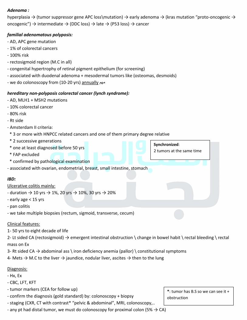
Synchronized:
2 tumors at the same time
*: tumor has B.S so we can see it +
obstruction
14
Spread:
1- direct 2- Hematogenous 3- lymphatic (staging) 4- transcoelomic (more in gastric CA)
Screening: at 50 yrs for male + female by colonoscopy
- other recommendations:
1- flexible sigmoidoscopy (FSIG) every 5 yrs (left lateral decubitus position)
2- colonoscopy every 10 yrs (gold standard, if –ve repeat after 10 yrs)
3- double contrast barium enema every 5 yrs
- CT colonography every 5 yrs in pts refuse colonoscopy and enema, if +ve → colonoscopy for biopsy
Staging: TNM
- Tx → can’t be assisted
- T0 → no evidence of tumor
- Tis → intraepithelial or lamina propria
- T1 → submucosa
- T2 → muscularis propria
- T3 → submucosa
Treatment of colon cancer:
surgery (gold standard):
- curative
- palliative → to prevent IO
- Lt\Rt hemicolectomy (with 5 cm safe margin + LN)
In FAP + IBD → 1- total proctolectomy and ileal pouch anal anastomosis (IPAA)
2- colectomy + mucosectomy of rectum + IPAA (rectal)
Prognosis: stages and LN
Chemo\radiotherapy:
stage I → surgery II, III → surgery + chemo IV → palliative + chemo
Adjuvant chemo → control micro mets , ↑ survival adjuvant radio → control local recurrence
Neoadjuvant chemo → stage, inoperable → operable
Neoadjuvant radio → prevent local invasion
Rectum:
symptoms:
1- bleeding 2- change in bowel habits \ mucus discharge 3- tenesmus 4- prolapse
Rectal polyps:
- adenoma - villous → ↑ risk of malignancy
- all polyps excised by endoscopic or major surgery
- all pts should undergo colonoscopy
- T4 → a: penetrate visceral peritoneum (serosa)
b: other organs
- N1 → 1-3 LN
- N2 → 4 or more LN
Safe margin:
- upper rectum → 5 cm
- lower rectum and anal → 2 cm
15
Rectal CA Tx:
1- resection of tumor + LNs
2- Pt unfit for surgery → trans-anal excision, laser destruction, intestinal radiation
3- sphincter saving surgery (anterior resection) → 2 cm above anal tumors
- tumors of upper part of rectum → high anterior resection + colorectal anastomosis
- tumors of middle part of rectum → low anterior resection
- tumors of lower part of rectum (or the sphincter in the safe margin) → abdominoperineal resection * here we can’t preserve the sphincter so we remove it + permanent colostomy
Investigations → for staging of tumors
- anoscope → 12 cm
- Endorectal US (14 cm)
→ for staging: we see layers of colonic wall
→ fistula
- pelvic MRI, CT
PET scan → distant mets
Lloyd-Davies position → modified lithotomy (lower limb is low)
16
Anal condition
15cm in length (rectum).
Anatomy:
1- anorectal ring: between Rectum and anus made by puborectalis sling (part of levator ani)
(Acute angle)
2- internal sphincter – part of muscularis external / involuntary – autonomic
3- external sphincter – skeletal muscles / voluntary - peudendal nerve
4- above levator ani pelvirectal space
below levator ani ischiorectal space
perianal space
Intersphincteric space - where the glands sets. (Between internal & external sphincter).
5-Mucosal lining:
Rectum / Dentate line (1-2 cm above the verge.) / ATz / Hilton’s / anal verge
Columnar / Metaplasia / Stratified squamous
6 - Columns of morgagni :
- surrounded longitudinal mucosal folds in dentate line
- anal glands empty in.
7 - Internal hemorrhoid and cushions at (3, 7, 11) or It Lateral, RT Ant, RT Post.
(Where venous plexus found)
External hemorrhoid inferior hemorrhoid plexus
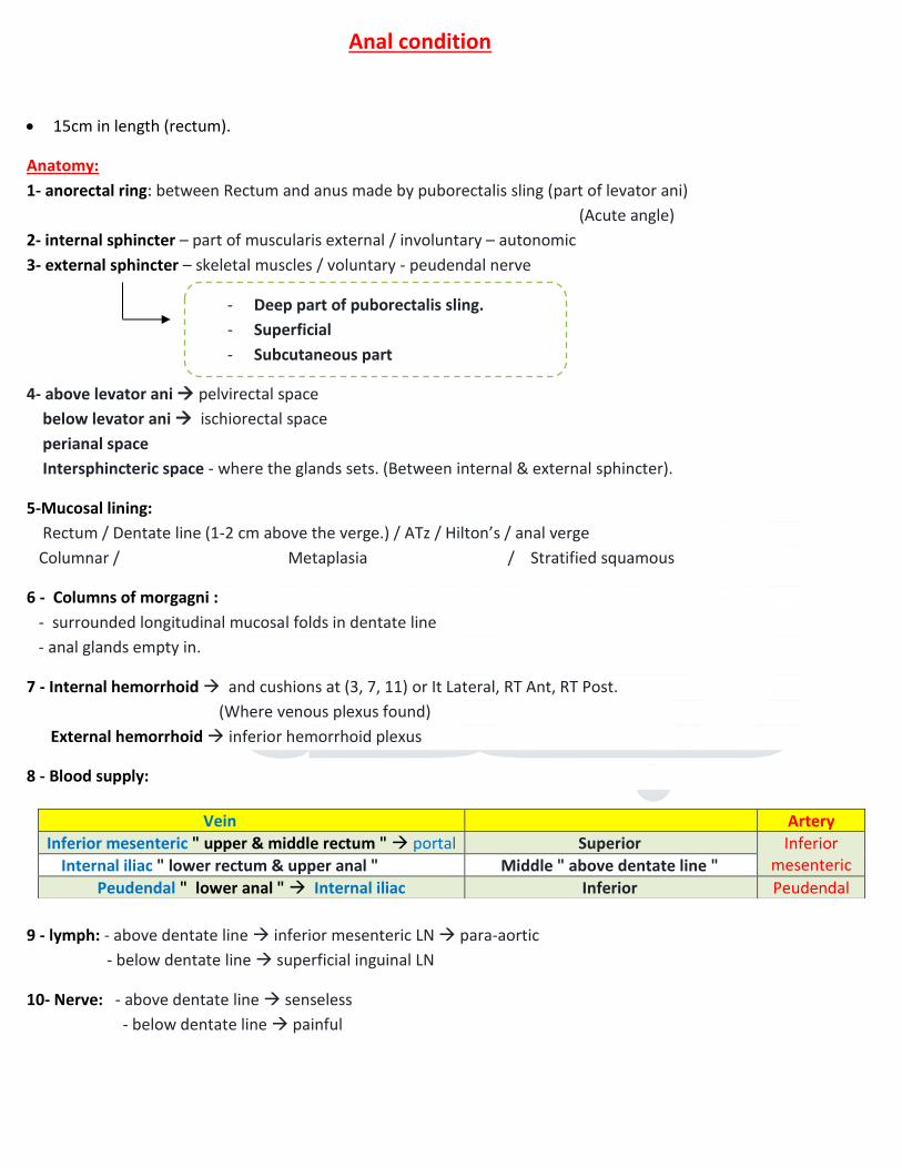
8 - Blood supply:
9 - lymph: - above dentate line inferior mesenteric LN para-aortic
- below dentate line superficial inguinal LN
10- Nerve: - above dentate line senseless
- below dentate line painful
Vein Artery
Inferior mesenteric " upper & middle rectum " portal Superior Inferior
mesenteric Internal iliac " lower rectum & upper anal " Middle " above dentate line "
Peudendal " lower anal " Internal iliac Inferior Peudendal
- Deep part of puborectalis sling.
- Superficial
- Subcutaneous part
17
Control of Continence:
- Tonic Contraction by contraction of internal sphincter
- Voluntary contraction of external sphincter.
- Acute angle of puporectalis ring
- Anal cushions
- Levator ani stabilizes the rectum in position.
Hemorrhoid:
Etiology:
1 - increased intra-abdominal pressure: constipation / diarrhea, obesity, heavy weight lifting, pregnancy.
2 - Portal hypertension 3 - colon CA 4 - anal intercourse
Investigation:
1 - CBC for anemia 2 – Anoscope (Proctoscopy) 3 – Sigmoidoscopy (> 40 years CA)
Management: - only when symptomatic
Grade I - Local cream (steroid, local anesthesia / laxative in constipation / bulk laxative in diarrhea
- Barrone's Bander (hemorrhoidectomy) or sclerotherapy or photocoagulation
Grade III & IV:
- open hemorrhoidectomyremoved the pile and skin or mucosa over it and leave the wound open
secondary intention
- closed hemorrhoidectomy All of the above + suture the wound
Complicated by anal stenosis
Complication of hemorrhoid:
1 - Thrombosis of external hemorrhoids (Bleeding) acute painful mass. (Edematous, congested)
excision immediately, If late (> 48 h) non-surgical management
2 - Strangulated internal prolapsed one by the sphincter muscles. Necrotic or ulcerated.
3 - Major hemorrhage
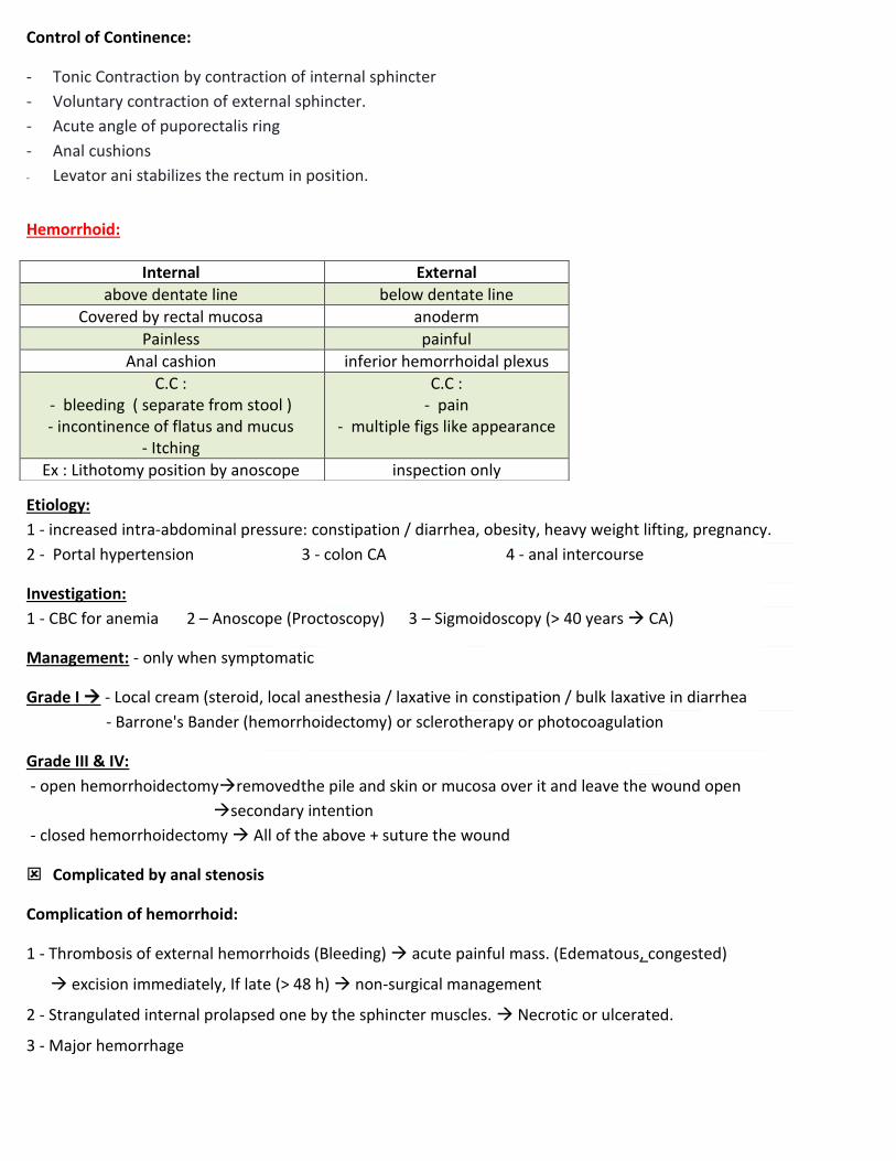
Internal External
above dentate line below dentate line
Covered by rectal mucosa anoderm
Painless painful
Anal cashion inferior hemorrhoidal plexus
C.C :
- bleeding ( separate from stool )
- incontinence of flatus and mucus
- Itching
C.C :
- pain
- multiple figs like appearance
Ex : Lithotomy position by anoscope inspection only
18

Anal fissure:
- M > F
- Acute Sites: 90% posterior midline , 10% anterior midline (in women after child birth)
- Chronic ( > 6 weeks ) , Other sites : think of Crohn's disease or immunodeficiency )
+ chronic fissures, Impaired healing : sentinel pile
Internal hypertrophied anal papilla
Pathophysiology:
Ischemia due to anal spasm due to increase intra-anal pressure
Clinical features:
- pain on defecation
- rectal bleeding ( separated from stool )
- Constipation.
Management
- Acute fissures: - resolve spontaneously.
- sitz paths (15g Na + water ) relaxation of internal sphincter
- stool softeners
- Chronic fissures: - Stool softeners have no curative value.
- sitz paths
- medical sphencterotomy is the first-line treatment of choice, By :
CCB , nitrates , botulinum toxin
- Surgical sphencterotomy : controlled division of the lower half of the internal
sphincter at the lateral position ( Lateral sphencterotomy )
Anorectal abscess
1- Intersphincteric space:
- obstructeal and glands
- m>f
- acute anal pain and tenderness during RE + pea sized lumps. EVA
- DDx – fissure
2- Perineal: M.C
- dawn wards
- tender swelling of the anal verge may discharge spontaneously
- 2-3 days pain (throbbing)
Constipation
Pain Fissure
Impaired healing
19
- DDx:
1- folliculitis
2- pilonidal abscess
3- hidradenitis suppurativa (- simple: skin flap with removal of sweat glands by knife \ - supportive: removal of all
area
(M.C in folds + axilla groin))
4- periprostatic or Bartholin gland abscess
5- perianal hemorrhage
3- ischio-rectal abscess:
- if Intersphincteric one tract from sphincter to reach this space
- wide space may lead to horseshoe abscess → toxic pyric patient. - DM patient
- pain for several days+ difficulty to sitting, painful fluctuant brawny swelling.
4- high super levator (intermuscular, pelvirectal)
- may encircle the anus → major systemic upset
management: incision + drainage under GA or spinal (S2,3,4,5) → cruciate incision with division of septor with finger
- anal, rectal approach
- complicated by fistula
- sets paths after surgery within 24 hrs (3 D)
- adequate analgesia (pethedine, “morphine → urine retention)
fistula in ano:
communication between 2 epithelial lining. (Perianal skin + anal canal)
causes:
1- after chronic inflammatory process
2- 50% of cases develop after anorectal abscess
3- Crohn’s disease, syphilis, lymphogranuloma venereum, actinomycosis, rectal CA, TB (multiple fistulas), malignancy,
foreign body
clinical: itching, irritation, discharge, (pain relieved by discharge)
- if the discharge was feaces + flatus → suggests rectal opening
Classification: high, low (according to anorectal ring)
Tracts of fistula: by probing & methylene blue
- anterior → radial tract - posterior (3, 9 o’clock) → circumferential tract
Investigations:
- examination under anesthesia
- MRI, andoanal ultrasound
- follow through + colonoscopy (if suspected IBD)
Tx:
- low: heal spontaneously, anal fistulotomy
- high: risk of incontinence
*per seton → gradual removing
High fistula surgeries:
1- Seton:
- threading
- cutting (staged fistulotomy \ fistulectomy)
2- Glue (closing the fistula by special glue )
3- Endorectal advancement flap
4- Ligation of duct near the rectum
20
Thyroid gland
Anatomy:
- 2 lobes + isthmus
- Lies 2, 3 tracheal rings
- Covered by pre-tracheal fascia + (sternohyoid, sternohyoid)
Blood supply:
- Superficial → external carotid, inferior → thyrocervical trunk → subclavian artery
- Venous: superior, middle → internal jugular, inferior + innominate → brachiocephalic trunk
Innervation:
Motor:
- recurrent (all the intrinsic)
- cricothyroid → external branch of superior laryngeal nerve
Sensory:
- recurrent (below vocal cords)
- internal branch of superior laryngeal nerve (above the vocals)
*recurrent laryngeal nerve may lie (between\inferior\posterior) to the branches of inferior thyroid artery
Histology:
- follicles (24-40 → lobule) lined by thyrocytes (cuboid epithelium) secretes T3, T4
- parafollicular C cells (secrete calcitonin that decrease Ca in blood)
approach:
history:
1- swelling (when, size, skin over)
2- pressure symptoms: dysphagia, dyspnea, hoarseness of voice, engorged neck vein, ear pain
3- hyper: heat intolerance, wt loss, ↑appetite, sweating, tremor, palpitation, diarrhea, amenorrhea, irritability
→ hyper-reflexia, tachycardia, eyes symptoms, tremors, hot moist palms
- hypo: cold intolerance, wt gain, ↓appetite, myxedema, sluggishness, constipation, menorrhagia,depression
→ dry skin
4- FHx
5- DHx
6- radiation
Examination:
1- General examination:
- eye → lid lag, lid retraction, exophthalmos “swelling of extraocular muscles. - hands
- reflexes
- lower limb skin
21
2- Neck examination:
- inspection: swelling, tongue protrusion
- palpation: - Front: tenderness, temperature, retrosternal percussion (for extension)
- back: swelling (edges, size, consistency, skin, mobility “if fixed could be Reidel’s, anaplastic CA, infection, scar, radiotherapy”
- LN
- auscultation: carotid artery bruit and upper border of thyroid
Investigations:
- TFT → TSH, T3, T4
- in pregnancy become ↑ TBG, we need to use free\bound hormone ratio
- Calcitonin level – medullary Ca
- Antibodies:
- thyroid stimulating immunoglobulin → graves
- thyroid peroxidase antibody → hashimoto
- US: lesion (cystic, solid)
- Radioisotope scan → Tc 99: hot, worm (normal), cold
- CT: retrosternal masses, asses malignancy
- MRI: vascular invasion
- PET: mets
- FNAC: not differentiate between adenoma & CA
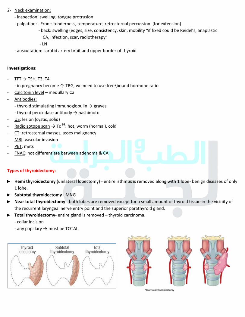
Types of thyroidectomy:
► Hemi thyroidectomy (unilateral lobectomy) - entire isthmus is removed along with 1 lobe- benign diseases of only
1 lobe.
► Subtotal thyroidectomy - MNG
► Near total thyroidectomy - both lobes are removed except for a small amount of thyroid tissue in the vicinity of
the recurrent laryngeal nerve entry point and the superior parathyroid gland.
► Total thyroidectomy- entire gland is removed – thyroid carcinoma.
- collar incision
- any papillary → must be TOTAL
22
Complications of surgery:
1- Hemorrhage →edema of vocal cords → tracheostomy 2- Damage to the nerves → superficial , recurrent laryngeal (uni: hoarseness of voice “cadaveric position cords” \ bi:
aphonia + SOB on exertion)
3- Hypothyroidism (late) after 1 week
4- Hypoparathyoridism (Paresthesia around the mouth)
5- Scar complications (hypertrophic, keloid)
6- Post op pyrexia: early → hematoma, late → hypocalcaemia
Simple goiter:
1. Multinodular:
(diffuse hyperplastic goiter → hemorrhage → necrosis → re-activation → nodular forms) - endemic due to iodine deficiency (.1 - .15 mg daily)
- sporadic → enzymes in thyroid synthesis, drugs, food (cabbage) - Hx → diffuse goiter, nodular later on, smooth firm, pressure symptoms, painful if hemorrhage occurs, childhood
- investigation → TFT, US
- Tx → thyroxin (.15-2 mg daily) for low M.
if it’s multinodular → irreversible
- indications for surgery: cosmetic, local pressure symptoms, patient anxiety due to thyroxin
-total, subtotal, lobectomy, may be complicated by hypothyroidism after 1 week, 1\2 thyroxin = 7 days
2. Physiological diffuse goiter: in puberty and pregnancy (↑ demand)
3. Solitary
4. Thyroiditis:
1- autoimmune thyroiditis: Hashimoto’s
- anti TPO - anti microsomal - anti thyroglobulin
2- sub acute thyroiditis: De Quervian’s
- associated with influenza - painful\diffuse goiter
- Tx conservative, surgery in recurrent thyroiditis episodes
3- Riedel’s thyroiditis: - replaced by fibrous tissue - firm painless swelling → tracheal compression (surgery)
Toxic goiter:
1. Gravis disease:
- primary thyrotoxicosis, young age
- thyroid stimulating immunoglobulin : ↑ T3, T4
- hyperthyroid symptoms and signs → opthalmopathy (may lead to opthalmoplagia and chemosis), graves
dermopathy (pretibial myxedema “hyaluronic acid”)
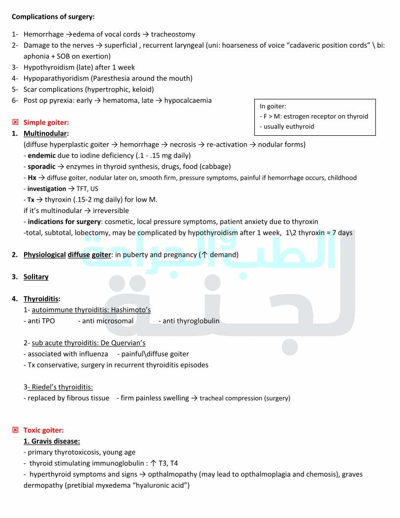
In goiter:
- F > M: estrogen receptor on thyroid
- usually euthyroid
23
- Investigations: TFT, US, radioisotope: diffuse uptake
- Tx:
1. antithyroid drugs:
* carbimazole → rash + agranulocytosis (serial CBC) * propylthiouracil
2. Radioactive iodine abrasion
3. Thyroidectomy
2. Toxic multinodular goiter: older patients
- inactive → active
- TFT : ↑T3, T4, ↓TSH - Tx: medical, subtotal thyroidectomy
3. Toxic adenoma = Plummer’s disease: - one toxic nodule → TFT , solitary (hot nodule) with suppression of residual gland
- > 3 cm → hyperthyroidism
- Tx: RAI ablation, surgery (lobectomy + isthmusectomy.
Malignant goiter:
1. Papillary: Most common (40 yrs)
- radiation R.F, good prognosis, Lymphatic spread, Psammoma bodies
- Multifocal, multi-centric
- Clinical → slow growing swelling, lymphadenopathy (early) الغدةممكن قبل
- Investigation: FNA
- Tx: total thyroidectomy + LN + hormone replacement
2. Follicular: 2nd
most common (30-50 yrs)
- Vascular + capsular invasion (distinguished from adenoma), FNA X
- Hematogenous to lungs, bones, liver
- Tx: microinvasion of capsule → lobectomy, gross invasion (mets) → total thyroidectomy + RAI ablation of mets
- Prognostic Fx: age, extrathyroid invasion, mets, histology \ grade, size of original lesion
3. Aplastic:
- Highly malignant, rapidly growing
- old age
- Invasion to : recurrent laryngeal, esophagus, trachea, cervical sympathetic ganglion (Horner’s), pulmonary mets. - Death within 6 months
24
4. Medullary:
- Hard enlargement + LN
- Parafollicular cells → calcitonin for follow up after surgery
- Sporadic 70%, familial with MEN-B2 (Rat oncogene - dominant), if present we must do elective thyroidectomy for
all relatives until the 3rd
degree.
- Tx: total thyroidectomy + LN dissection
5. Lymphoma: old age
- Hashimoto or normal thyroid
- Dx: FNA or true cut biopsy
staging by CT, bone marrow aspirate
- thyroid alone → thyroidectomy + chemo\radio therapy
- lymphoma → chemo alone
Post thyroidectomy complications:
1- reactionary bleeding (at the day of surgery)
- R.F: ↑blood pressure due to pain, agitation, hypervolemia
2- Respiratory distress, due to:
- hematoma → evacuation
- bilateral recurrent laryngeal nerve injury tracheostomy
- trauma to the larynx (vocal cords injury) in difficult intubation if severe; re-intubate the patient
3- Thyroid crises:
- pre-op: due to high manipulation in thyrotoxicosis patient most sensitive indicator is sleeping pulse
- intra-op: in completely controlled thyrotoxicosis
- post op
Tx: Propranolol, fluids (due to hyperpyrexia + sweating) + glucose
25
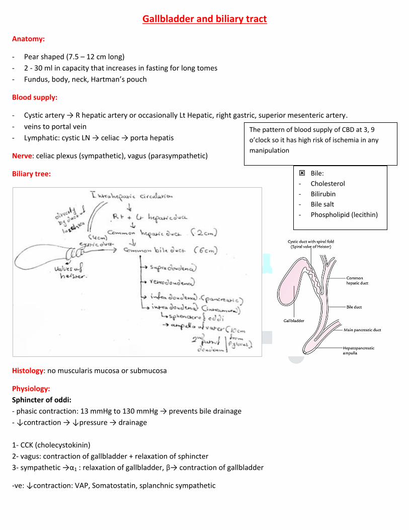
Gallbladder and biliary tract
Anatomy:
- Pear shaped (7.5 – 12 cm long)
- 2 - 30 ml in capacity that increases in fasting for long tomes
- Fundus, body, neck, Hartman’s pouch
Blood supply:
- Cystic artery → R hepatic artery or occasionally Lt Hepatic, right gastric, superior mesenteric artery.
- veins to portal vein
- Lymphatic: cystic LN → celiac → porta hepatis
Nerve: celiac plexus (sympathetic), vagus (parasympathetic)
Biliary tree:
Histology: no muscularis mucosa or submucosa
Physiology:
Sphincter of oddi:
- phasic contraction: 13 mmHg to 130 mmHg → prevents bile drainage - ↓contraction → ↓pressure → drainage
1- CCK (cholecystokinin)
2- vagus: contraction of gallbladder + relaxation of sphincter
3- sympathetic →α1 : relaxation of gallbladder, β→ contraction of gallbladder
-ve: ↓contraction: VAP, Somatostatin, splanchnic sympathetic
The pattern of blood supply of CBD at 3, 9
o’clock so it has high risk of ischemia in any manipulation
Bile:
- Cholesterol
- Bilirubin
- Bile salt
- Phospholipid (lecithin)
26
Types of gallstones:
1. Cholesterol 10 %
2. Pigment stone (bilirubin*) 15% β black (hemolytic “as spherocytosis” + cirrhosis), brown (infection) 3. Mixed 75%
Types of biliary stones:
1. Primary (black, brown)
2. Secondary (from gallbladder)
Risk factors:
1. Forty, fatty, female, fertile
2. DM → dyskinesia
3. Liver cirrhosis, hemolytic
4. Vagotomy
5. TPN → no CCK
6. Short bowel syndrome, IBD, ileum disorder (↓ absorption)
7. Congenital anomalies (stasis) + hereditary \ ethnic
8. OCP
9. Somatostatin therapy
10. Hyperlipidemia not hyper-cholesterolemia
Clinical picture:
1- Most are asymptomatic (80%)
2- Biliary colic:
- RUQ or Epigastric pain lasts for hrs, radiates to the back
- rapid increase in intensity then resolve
- increase fat content food
- associated with dyspepsia, flatulence
3- Acute cholecystitis:
- long standing obstruction
- begin as chemical reaction (,.C) without bacteria then to bacterial infection (E-coli M.C)
- as biliary colic but the pain not subside (several days) with radiation to subscapular area (diaphragmatic
irritation) → [ Boas sign] - associated with pyrexia, jaundice, anorexia, N & V
- murphy’s sign (9th subcostal area) → respiratory arrest with deep palpation
4- Complicated by:
- gangrene\necrosis → perforation
- fistula
- empyema & mucocele (long Hx for RUQ pin > 2 w)
- emphysematous cholecystitis (gas forming bacteria)
- cholangitis
- Mirizi stone in infundibulum cause compression to CBD.
*Unconjugated bilirubin + Ca phosphate + Ca(HCO3)
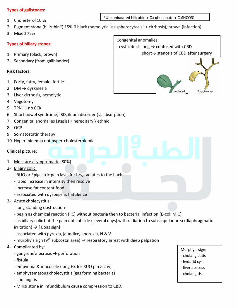
Congenital anomalies:
- cystic duct: long → confused with CBD
short→ stenosis of CBD after surgery
Murphy’s sign: - cholangistitis
- hydatid cyst
- liver abscess
- cholangitis
27
5- Choledocholithiasis:
- primary →infection, stasis due to tumor, striction \ secondary → migration
- clinical: pain, transient jaundice “if moved to duodenum”, may be associated with cholangitis or
pancreatitis (obstruction of sphincter of oddi → ↑ pressure of biliary tree → bile to pancreas → activation of pro-enzymes)
- investigations: US : dilated biliary tree (if CBD > 8mm) + gallbladder stone, MRCP, ERCP → Dx & Tx by removing the stone by dromia basket or fogarty balloon cath.
- may insert t-tube
Investigation:
- CBC → leukocytosis
- Bilirubin level → mild or marked elevation
- US → gallstones + ↑ size, thickened wall, pericholecystic fluid (halo sign), ↑ caliber of CBD or not, fibrosis
- HIDA scan
Management:
1- IV fluid + antibiotic + analgesia
2- Cholecystectomy → early (3-5 days), elective (4-6 w)
laparoscopy, laparotomy (Kocher incision)
* -ve: colon CA , PU → ↓ bile
3- Lithotripsy
4- Cholecystectomy (anesthetic C.I) or percutaneous aspiration
5- Medical: chenodeoxycholic acid, ursodeoxycholic acid, ↑ risk of recurrence after stopping
Main cause of pneumobilia nowadays: ERCP then cholecystoenteric fistula.
When to remove asymptomatic stone:
1- > 2 cm 2- Porcelain gallbladder 3- Typhoid carrier 4- Polyp 5- Sickle cell disease 6- child
Complications of cholecystectomy (lap):
1- injury to CBD, hepatic artery
2- cystic duct leak → biloma
DDx of post cholecystectomy jaundice:
1- M.C is slipped GBS
2- iatrogenic stricture
Gallbladder cancers:
Epidemiology: female, old (60-7-(, adenocarcinoma (90%)
Cause:
1- gallstones causes metaplasia → squamous cell carcinoma
2- typhoid carriers
3- porcelian gallbladder
How to differentiate between GBS and calcified
polyps on US?
- GBS → mobile - polyps → fixed
28
Clinical: pain, jaundice, n & v, wt loss, ascites, anorexia
Investigations: US, CT, laparoscopy for staging, MRI
Spread → direct extension to liver + CBD, Hematogenous, or lymphatic
Staging: we use endoscopy US to stage all GI tumors
I → mucosa, submucosa
II → muscle layer
III → serosa
IV → cystic LN
V → liver + others
Carcinoma of bile duct:
Epidemiology: male, sixty
Cause:
1- parasitic infection → clonorchis sinensis
2- typhoid carriers
3- gallstones ↓
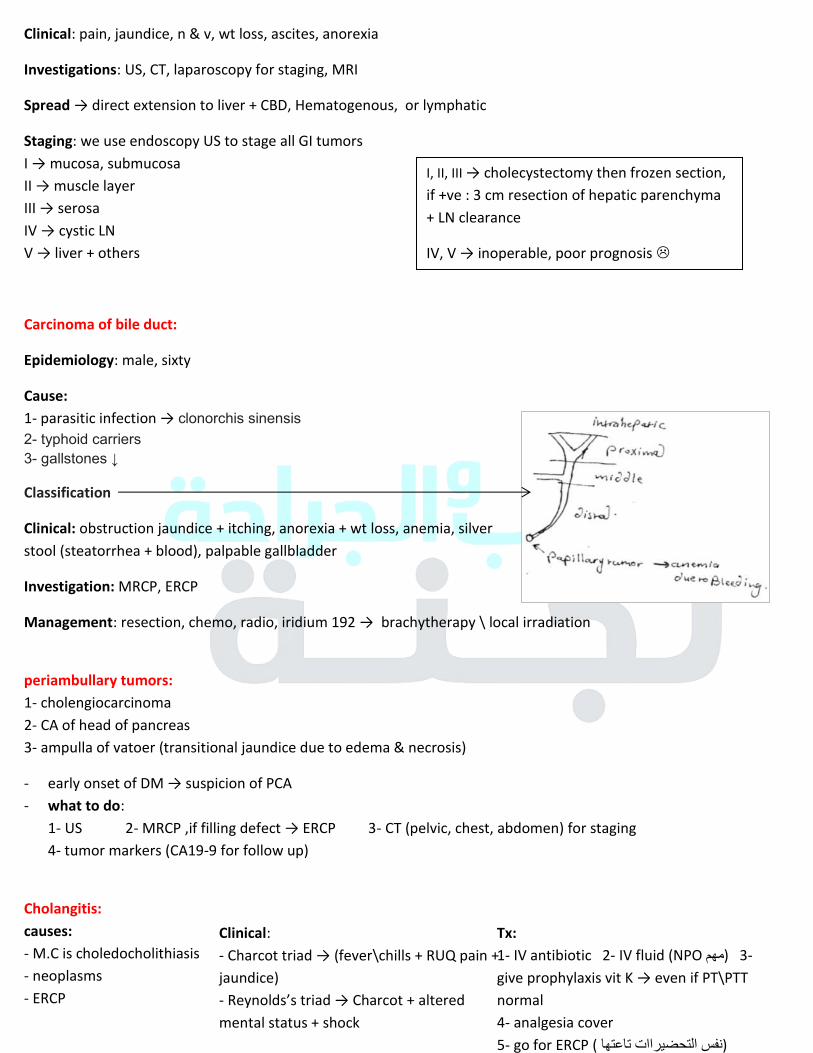
Classification
Clinical: obstruction jaundice + itching, anorexia + wt loss, anemia, silver
stool (steatorrhea + blood), palpable gallbladder
Investigation: MRCP, ERCP
Management: resection, chemo, radio, iridium 192 → brachytherapy \ local irradiation
periambullary tumors:
1- cholengiocarcinoma
2- CA of head of pancreas
3- ampulla of vatoer (transitional jaundice due to edema & necrosis)
- early onset of DM → suspicion of PCA
- what to do:
1- US 2- MRCP ,if filling defect → ERCP 3- CT (pelvic, chest, abdomen) for staging
4- tumor markers (CA19-9 for follow up)
Cholangitis:
causes:
- M.C is choledocholithiasis
- neoplasms
- ERCP
I, II, III → cholecystectomy then frozen section,
if +ve : 3 cm resection of hepatic parenchyma
+ LN clearance
IV, V → inoperable, poor prognosis
Clinical:
- Charcot triad → (fever\chills + RUQ pain +
jaundice)
- Reynolds’s triad → Charcot + altered
mental status + shock
Tx:
1- IV antibiotic 2- IV fluid (NPO مهم) 3-
give prophylaxis vit K → even if PT\PTT
normal
4- analgesia cover
5- go for ERCP ( نفس التحضيراات تاعتها) 29
Enterocutaneous fistula
Definition:
Duodenum, jejunum, ileum (M.C), colon, rectum ↔ communicate with skin
Classification:
Anatomic:
- site of fistula origin
- drainage point
- external \ internal
Physiological: fistula output in 24 hrs
- low output (<200 ml\day)
- moderate output (2000-500 ml\day)
- high output (>500 mg\day)
Etiology:
► Post-operative (75-90%)
o disruption of anastomosis:
- improper anastomosis with leak → abscess → disruption
- improper vascular supply
- under tension
o Inadvertent enterotomy
o Inadvertent small bowel injury
Ex 1: Gastrodoudenal fistula: DU perforation:
- large\extensive contamination
- late intervention after perforation → lateral duodenal fistula
Ex 2: surgery for appendicitis, perforated appendix, after appendicular abscess drainage → colocutaneous fistula
► Traumatic:
o iatrogenic
o after RTA
► Spontaneous:
o Malignancy
o Radiation enteritis + perforation → colonic fistula
o Intra-abdominal sepsis
o IBD (Crohn’s disease) o Diverticulitis → colonic fistula (M.C)
30
History \ PEx:
- Post-operative pain\tenderness
- Abdominal distention
- Enteric content from a drain site
- Generalized\localized peritonitis → tachycardia + pyrexia → toxic, guarding, rigidity, rebound tenderness
Complications:
- Sepsis:
- direct tract: bowel content drain directly to skin (minimal sepsis)
- indirect tract: bowel content drain to an abscess then into skin (severe sepsis)
- Dehydration \ electrolyte disturbance \ malnutrition: due to leakage of protein rich enteric content, sepsis,
paralytic ileus → hypokalemic hypochloremic metabolic alkalosis
- Skin excoriation: in fluid content that makes it difficult to put collecting bag → more in enteroatmospheric fistula
LAB: CBC → leukocytosis, electrolyte, albumin ↓→ malnutrition, CRP ↑, serum transfusion <200 mg\Dl → indicates poor healing
Favorable factors for spontaneous closure:
- End fistula
- Jejunal fistula
- Colonic fistula
- Continuity maintained fistula
- Small defect fistula
- Long tract fistula (↑ resistance + ↓rate of epithelization “this ↑in short fistula”) - Bowel wall disruption is partial
Unfavorable factors: HIS FRIEND
- High output
- Intestinal destruction >50% of circumference
- Short segment fistula
- Foreign body (drain, ..)
- Radiation
- IBD\infection
- Epithelization of tract
- Neoplasm
- Distal obstruction
- Lateral duodenal fistula
31
Treatment: SSNAP
stabilization, sepsis\skin care, nutrition, define underlying anatomy (imaging), plan
Stabilization:
1- RL (for acidosis)\NS administration
2- albumin administration
3- UO monitoring
Sepsis: antibiotic cover, abscess drainage
Skin care: containing of effluent by:
A:
1- solid wafers (pectin based “wet, weepy”) → good barrier before ulceration
2. Powder → severe skin maceration paste افضل منه 3. Paste \ spray \ ointment and creams → zinc oxide
B: pharmacological:
- Somatostatin analogue → ↓fistula output
+ TPN (more effective)
- PPI, H2 blocker in proximal fistula (↓fistula output) - cyclosporine → refractory fistula in Crohn’s disease
infliximab → multiple lesion → closure in 50%
- excessive fistula output → TPN, NG tube
C: vacuum assisted closure:
- -ve pressure application
- help in drainage
- size of wound
- frequency of dressing and protect the skin → healing
- chronic edema → improve blood flow → granulation tissue
Nutritional
A: enteric: at least 20% of whole nutrition to:
- protect and maintain the intestinal mucosal barrier
- stimulating of hepatic protein synthesis
by gastrostomy \ NG tube, fistulodyalisis beyond the fistula
- at least 1.2 – 1.5 m of functional bowel should be present
B: TPN : BW loss > 20%, gradually initiated to prevent refeeding syndrome
Indications of TPN:
1- gastric, duodenal, small bowel fistula
2- high output fistula
3- ileus, obstructed distal end
4- inability to obtain internal access \ GI intolerance with it
Laparotomy:
1- extensive cellulitis + necrotizing fasciitis
2- incomplete drainage and collection
3- disruption of anastomosis
32
complications of TPN:
1- catheter tip malposition
2- arterial laceration
3- SVC \ subclavian vein thrombus
4- thrombophlebitis
5- catheter embolism
6- hydro, pneumo, hemothorax due to central line.
7- sepsis \ fluid overload
normal requirement:
- Na\K = 80-100 mEq\day
- Ca\Mg = 15-20 Meq\day
- water= 30ml\kg\day → 2.5 L
Imaging: (anatomy of fistula*) after 7-10 days
1- fistulography (water-soluble contrast gasteografin) → injected into the fistula
2- CT scan: above + intra-abdominal abscess, foreign body → aspiration under CT guidance
3- endoscopy → delayed till acute inflammation get reduced
4- water soluble contrast enema → to detect different types of fistula tracts
- simple, short, blind ending, <2 cm
- continuous, long, linear single, > 2cm
- continuous, complex, multiple linear
The definite treatment:
- spontaneous closure usually at first month up to 2 months
- After 2 months → no spontaneous closure
- time for surgery (2-5 m later) → at least 6 weeks
remove the fistula and tract + small bowel segment (to reduce rate of recurrence) + re-anastomosis
Indications of surgery:
1- high output
2- lateral duodenal or ligament of treitz fistula
3- ileal fistula
4- ECF + adverse factors
5- disease of bowel, distal obstruction
6- enteroatmospheric fistula
*- location of fistula and its tract
- bowel continuity or disrupted
- distal obstruction
- abscess
Enteroatmospheric:
- floating stoma
- bowels open to skin directly
- use fibrin glue and plugs
endoclips → acute fistula
33
The liver
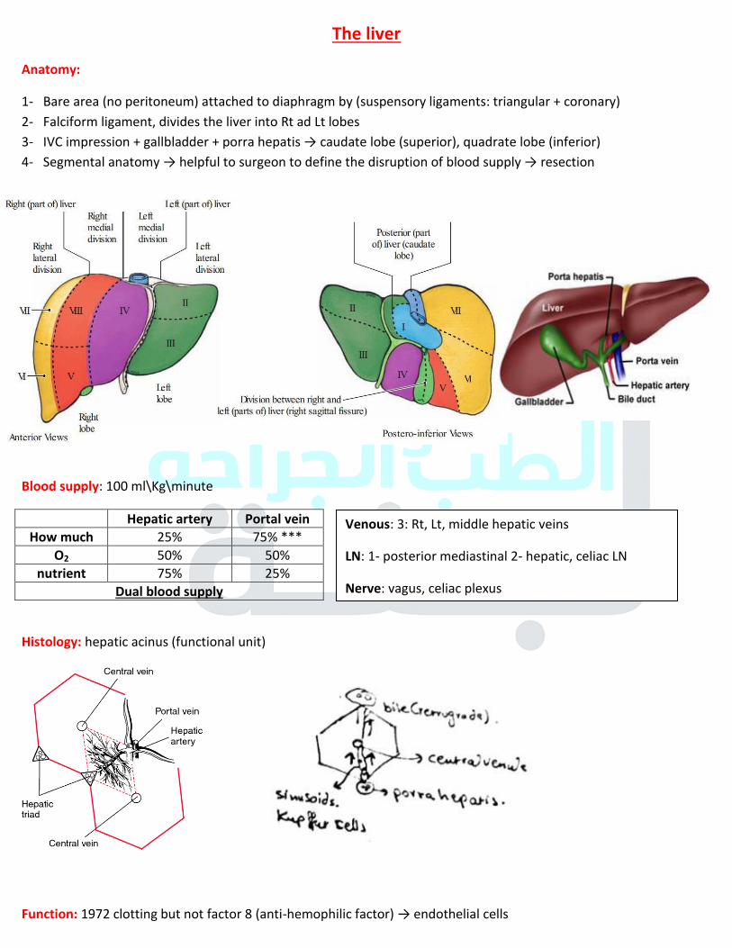
Anatomy:
1- Bare area (no peritoneum) attached to diaphragm by (suspensory ligaments: triangular + coronary)
2- Falciform ligament, divides the liver into Rt ad Lt lobes
3- IVC impression + gallbladder + porra hepatis → caudate lobe (superior), quadrate lobe (inferior)
4- Segmental anatomy → helpful to surgeon to define the disruption of blood supply → resection
Blood supply: 100 ml\Kg\minute
Hepatic artery Portal vein
How much 25% 75% ***
O2 50% 50%
nutrient 75% 25%
Dual blood supply
Histology: hepatic acinus (functional unit)
Function: 1972 clotting but not factor 8 (anti-hemophilic factor) → endothelial cells
Venous: 3: Rt, Lt, middle hepatic veins
LN: 1- posterior mediastinal 2- hepatic, celiac LN
Nerve: vagus, celiac plexus
34
Liver abscess:
bacterial (pyogenic), amebic, parasitic, fungal
► pyogenic:
. Route of infection:
1- ascending infection (biliary system) → M.C due to radiologic endoscopy
2- portal vein (appendicitis, diverticulitis)
3- hepatic artery (anywhere)
4- adjacent organ (gallbladder)
5- 1\3 → cryptogenic (unknown) 6- lymphatic
7- liver trauma (gunshot wound)
. M.C bacteria: E-coli, staph. aureus, anaerobes, Klebsiella, Proteus
. Clinical feature: RUQ pain + tenderness, n & v, fever, jaundice (pressure), enlarged tender liver, pleural effusion,
atelectasis
. Investigations:
- CBC (leukocytosis, anemia) - ↑ LFT - blood culture - ERCP (in biliary obstruction)
- CXR (elevated hemi-diaphragm, pleural effusion, atelectasis) - US (irregular thick-walled mass)
. Tx:
- percutaneous aspiration by needle or catheter
- surgery for complex\multiple abscesses \ not respond to percutaneous drainage for 7 days \ viscous
content
associated intra-abdominal disease
- IV antibiotic (2 w IV, 4 w oral)
- antibiotic alone (multiple small)
► Amebic: Entameba Histolytica
. Started as amebic colitis → trophozoits penetrate the mucosa to portal vein → go to liver → abscess (thin walled, solitary, Rt lobe, large, contains brown sterile pus “anchovy\chocolate sauce)
. Clinical: as pyogenic + chronic > 2 w
. Investigations: same as pyogenic + stool examination for ameba trophozoits + serology ELIA for amebic protein
. Tx:
- metronidazole + chloroquine phosphate → 500 – 700 mg, oral for 7 days
- aspiration if :
1- large one 2- super infection 3- not respond up to 72 hrs
35
Cavernous hemangioma
- M.C solid benign mass
- Consists of endothelial lined vascular spaces → blood supply from hepatic artery
- ↑ Growth with estrogen (puberty, pregnancy, OCP) and androgens
- Small < 10 cm → asymptomatic \ incidental finding
large > 10-25 cm → non-specific abdominal symptoms
both +\- necrosis, thrombosis, infarction, hemorrhage “rare”
- Grossly → flat, red-blue, well defined, soft, easily compressible mass
- NO biopsy → life threatening hemorrhage
Kasabach-Merritt syndrome (hemangioma + thrombocytopenia + fibrogenopania)
- consumptive coagulopathy in giant hematoma
Dx:
- US → well demarcated homogenous, hyperechoic masses with hypoechoic lesions (hemorrhage, fibrosis,
calcification)
- contrast + CT → centripetal enhancement (gradual enhancement of lesion from periphery to the central)
→ T2 view → hyperintense
- MRI
Tx: mostly observation
if symptomatic, complicated, can’t exclude malignancy:
1- enucleation under vascular control (continuous hepatic artery proper occlusion, intermittent inflow occlusion of
portal triad) with intermittent Pringle maneuver
2- formal anatomic resection
3- low dose radiation or embolization, in large, unresectable one, hemorrhagic one.
Hepatic adenoma:
- M,C in pre-menopausal women (30 yes), solitary mainly
R.f:
1- current use of estrogen (OCP) األهم 2- glycogen storage diseases
grossly: well circumscribed (unencapsulated, pseudocapsule), round
clinical: abdominal pain, intraperitoneal hemorrhage (10-25%): >5 cm, pregnancy, men in steroid users
malignant risk: 10% → hepatocellular carcinoma (in large\multiple \ in men)
imaging:
- US → can’t be differentiated from adenoma + FNH or malignant
- CT → heterogeneous (fat, necrosis, hemorrhage) - MRI → the best
Tx: small : stop the OCP → regress
large >5 cm \ bleed \ painful \ rupture → surgical resection without wide margin
Histology
36
Follicular nodular hyperplasia
- 2nd
M.C
- neoplastic hyperplasia in response to hyperperfusion from congenital arterial malformation
- in women, childbearing age +\- OCP
- don’t have malignant potential
Grossly: well circumscribed unencapsulated, solitary
Clinical: mainly asymptomatic, rarely: pain\mass, hemorrhage
Histo: contain hepatocytes, kupffer cells, bile duct
Imaging: CT, MRI → central scarring and hypervascular lesion
FNH α HCC : large, eccentric central scar, with fibrous bands and calcification
FNH α adenoma: sulfur colloid scan, biopsy
- FNH +ve kupffer cells
Tx: observation
→malignant \ adenoma, large, complicated → surgery
- stop the OCP
37
Surgical site infection
Definition: infection related to operative procedure occurs within 30 days or 1 year in implants.
- Nosocomial infection
- Surgical ward infection
- Surgical site infection
Classification:
Superficial incisional SSI:
- invade the skin + SC tissue
- ate least one of the following:
1-Purulent drainage (no need for culture)
2- organism isolated from fluid \ tissue
3- signs of inflammation (pain, swelling, erythema, heat)
4- the wound is deliberately opened by surgeon (no dehiscence)
NOT: - abscess formation - epistomy, circumcision burn - extend to muscles \ fascia
Deep incisional SSI:
- involve muscles \ fascia
- at least one of the following:
1- purulent drainage
2- fascial dehiscence or deliberately by surgeon
3- deep abscess, by (Ex, histopathology, radio
4- fever > 38 c, localized pain, tenderness
Organ\space SSI:
- deep to any space involved in the surgery
- at least one of the following:
1- purulent drainage by drain or by stab wound
2- organism is isolated
3- abscess, by Ex, histo, radio, reoperation
Clean:
1- no inflammation
2- not in respiratory, GI, genitourinary system
ex: breast, thyroid
infection rate: 1-2% → prophylactic Ab if it with prosthetics
Clean contaminated:
1- respiratory, ..
2- Without significant spillage
< 3% prophylactic Ab
Complications:
1- wound dehiscence→ hernia, fistula, sinus
2- poor healing → abnormal scar (hypertrophic) 3- sepsis
38
Contaminated 5-10%:
1- acute inflammation (without pus)
2- visible contamination of the wound (gross spillage)
3- compound\open injuries operated within 4 hrs
ex: appendectomy
- prophylactic Ab
Dirty 30-40%:
1- presence of pus
2- perforated viscous
3- compound\open injuries more than 4 hrs old
- therapeutic Ab
Timing:
- early: within 30 days - intermediate: 1m-3m - late: > 3m
Severity:
- minor: discharge without: cellulitis, deep tissue destruction, systemically ill
- major: pus discharge, need drainage, tissue breakdown (dehiscence), systemic symptoms
Source of infection:
- endogenous (M.C): staphylococcus aureus (G +ve), perforated PU\bowel, mucous membrane
- exogenous: surgical instrument, team, air, ..
. Enteric gram –ve more in GI surgeries. → pre op antibiotic if needed
. Vascular & orthopedic → staph. Epidermidis, staph. Aureus → Flucloxcillin +\- gentamycin, vancomycin or
rifampicin \ broad spectrum cephalosporin
. GI → enterobacteriacea, enterococci, anaerobes (bacteroids) → 2nd generation Cephalosporin
Risk factors:
Systemic:
1- age 2- smoking 3- iatrogenic: radio\chemo\steroids
4- disseminated disease (cancer, autoimmune)
5- metabolic (malnutrition, diabetes, uremia, jaundice)
6- hypovolemia \ hypothermia \ hypo-perfusion
Local:
1- classification 3- non-viable tissue\hematoma 3- foreign material (suture, drains)
4- poor skin preparation (local infection, shaving)
Operative:
1- emergency 2- blood transfusion 3- long duration >2hrs
4- intraoperative contamination 5- site of operation
6- poor technique 7- prolonged hospital stay
39

Prevention:
Why?
- increase mortality twice more \ ICU stay
- increase the length of stay (5-7 days)
- cost effective
pre-operative:
1- short pre op stay
2- identify \ treat all remote infections
3- patient advised to take shower \ dress with the theater wear
4- hair removal: not be removed \ removed only by clipper, why ? :
- give adequate exposure \ skin markings
- suturing - good wound dressing
5- prophylactic antibiotics:
- IV → at introduction & anesthesia
- repeated: long operation, excessive blood loss
- continue: unexpected contamination, prosthesis is implanted in patient with septic source
- prosthesis: give prophylactic Ab before: 1- dental working 2- urethral instrumentation 3- visceral surgery
- not continued > 48 hrs:
1- masks the symptoms of infection 2- increase resistance 3- serious hypersensitivity
- in case of lower limb amputation give benzyl penicillin to cover C.perfringes (gas-forming bacteria)
6- mechanical bowel preparation:
- reduce the risk in elective colorectal surgery
- we use cathartics: 1- poly ethylene glycol 20 sodium phosphate
7- enhanced nutritional support in malnourished patients:
- combination of arginine \ glutamine \ omega 3 \ nucleotides \ micronutrients
8- IV fluids: improve tissue perfusion & arterial oxygenation → good wound healing
9- perioperative blood glucose control: stress of surgery
10- maintaining normal body temperature.
Intraoperative:
- staff hygiene (hand washing to elbow \ scrubbing, ..)
- application of alcoholic antiseptic to skin
- avoid dead spaces \ hematomas
- abscess → keep open
- oxygenation: 100% for 30s -2 min → before intubation, 80% hypo-pyrexia, 30-35% normoxia
- use close suction → through separate incision
- wound irrigation → hydration \ remove debris, …
-Clean + prosthesis
-clean contaminated
-contaminated
40
Post op:
1- dressing for 24-48 hrs
2- advanced dressing: physical barrier, absorb exudate, keep wound dry.
Management of SSI:
4 lines:
1- empirical antibiotic → switch to specific one according to culture. - According to what?
1- Gram stains 2- organism most often cultured from similar infections in previous patients.
Examples:
- clean contaminated surgery → metronidazole, co-amoxiclav
- increase risk for candida infection (DM, immunocompromised) → antifungal → fluconazole, amphotericin
- immunity acquired infections → cephalosporin (intra-abdominal \ soft-tissue infections), ampicillin
- appendectomy and it’s complications → ciprofloxacin + metronidazole (anaerobes, aerobes)
- cephalosporin + aminoglycoside to cover anaerobes
Specific antibiotics:
- E-coli: penicillin, cephalosporins (all generations), aminoglycoside, ciprofloxacin
- MRSA (methicillin resistant staph. Aureus) → vancomycin, daptomycin, clindamycin
- MSSA (sensitive) → penicillin + B-lactamate inhibitor combination → ampicillin-sulbactam, cephalosporin,
ciprofloxacin
- strep. Pyogenes → penicillin, cephalosporin, tetracycline
- staph. Epidermidis → vancomycin, ceftaroline, ciprofloxacin, piperacillin-teizobactam
- enterococcus → vancomycin, gentamycin
- pseudomonas → piperacillin
When to stop antibiotic?
- Clinical improvement
- normal WBC, no bands of PMNs
- no fever (<38 c)
2- Incision and drainage:
- ↓ pressure on some no. of bacteria
- how? 1- Incision + drain 2- deep→ catheter with CT\US guide 3- inaccessible → operative drainage
- abscess+ systemic symptoms → surgical emergency →
- fluctuation → late stage
3- Debridement: removal of dead tissue, its importance:
- help healthy tissue grow
- minimize the scarring
- reduce the complications of infections
41
Methods of debridement:
1- biological:
- use sterile larvae of the Lucillia Sericata
- large wound – painless
- mechanism of action:
1. Bactericidal 2. Inhibit bacterial growth – ammonia (↑PH) 3. Ingest the necrotic tissue
- C.I: if the wound reaches the intraperitoneal cavity + immunosuppression therapy \ if the wound approximate a
septic arthritis
2- Enzymatic:
- selective (only dead tissue) \ used in combination with other types
- exogenous proteolytic enzyme (collagenase)
- disadvantages:
1- costly, need prolonged tome (-30 days)
2- may be inactivated by heavy metals (zinc, silver)
3- risk of maceration + infection
4- require frequent dressing (3\day)
3- Autolytic:
- recruit endogenous phagocytic cells and proteolytic enzymes
- by using moisture-retentive dressing (hydro-colloid, hydrogels, hypertonic gels)
- indicated in non-infected wounds
- advantages: selective, effective, low cost, painless \ disadvantage: slow
4- mechanical:
- non-selective (necrotic + viable tissue)
- when? If there is a large amount of necrotic tissue
- C.I: granulation tissue > necrotic, inability to control pain, poor perfusion
5- surgical:
- uses curettes, scalers, ..
- Disadvantages: bleeding, complications of anesthesia
- C.I: intact eschar, no clinical evidence of underlying infection
- how? Start from the base to periphery until red bleeding margins are seen, then irrigation with NS then dressing and
leave it for secondary intention.
42
Intestinal obstruction
Definition: dynamic (partial, complete), adynamic
Dynamic obstruction:
- initially there is increase in peristalsis to overcome the obstruction then it will decrease (muscle wasting and
ischemia)
- Small bowel: high → acute symptoms due to small lumen to be obstructed, low
- adhesion (60%) then hernia then malignancy
Large bowel → chronic because the lumen is large and mostly partial. - Malignancy then complicated diverticular disease and volvulus.
- Simple (no ischemia) \ strangulated (ischemia)
Causes:
external:
1- adhesions: M.C → peritonitis \ post op, due to decrease in plasminogen activating activity or drying.
2- Hernia 3- malignancy (LN or itself)
Intramural:
1- inflammatory (Crohn’s, diverticulitis) → adhesion 2- tumor
Intraluminal:
1- impacted feaces: M.C in elderly
2- swallowed foreign body → children, or hair bezoars in psychiatric (multiple levels) 3- gallstones ileus (cholecystosoudenal fistula) Dx by X-ray
* when the stone go to stomach not to the ileum → Bouveret syndrome
Pathophysiology:
obstruction → dilated proximal loop
1- due to intestinal secretion & air
2- edematous wall due to venous obstruction
3- electrolyte disturbance (impaired absorption, vomiting, anorexia)
4- bacterial overgrowth
- if obstruction not relieved → ischemia → perforation
Intussusception: m.c in 3 months – 1.5 yrs, m.c in ileocecal
Volvulus: rotation of bowel (80 – 360 degree) around itself.
- Causes:
1- adhesions 2- narrow band between bowel & abdominal wall 3- congenital malrotation
- sigmoid → elderly + constipation (sausage mass) - cecal → increased risk of ischemia (mass in the left side) 43
Closed loop: from two points
- like in ascending colon (in complete iliocecal valve + annular CA in distal segment) which increase pressure
that lead to perforation through a clear cut hole (pistol-shot perforation)
- volvulus
Mesenteric ischemia: occlusion of blood supply by thrombo-embolic event lead to ischemia and edema that impairs
the bowel motility (elderly patient, vascular disease), pain is post-prandial
Clinical feature:
1- pain:
- colicky (central “small bowel” or suprapubic “colonic”) - decrease in severity later on due to relaxation.
- constant\sudden with pyrexia, peritoneal rigidity & tenderness → infarction
2- Vomiting:
- high obstruction → food then bile stained then feculeus
- low obstruction → late features
3- Abdominal distention: mainly in low obstruction due to gas & fluids
4- Constipation:
- absolute → complete - relative → partial * after passage of the contents of distal segment
partial:
1- receptive relaxation → passage of fluid, mucus that lead to diarrhea 2- tumor 3- air in rectum on X-ray
4- although there is no increase in pressure significantly but the venous obstruction lead to ischemia and perforation.
Complete: no air on rectum
Management:
general:
1- decompression by NG tube
- used to calculate fluid replacement
- decompression of proximal segment (↓ perfusion, aspiration ↓ )
2- electrolyte \ fluid therapy:
- normal saline + 5% dextrose (we neutralize acidic contents by alkaline: Ringer’s lactate)
3- Antibiotics (not important)
Normal bowel sounds (3-10\min)
< 3 for 3 min after stimulation and
changing the position → hypoactive
small → more vomiting & pain
large → distention + constipation
44
Definitive: according to the cause:
- Adhesion: conservative mainly or adhesiolysis
- gallstone ileus + impacted feaces + bezoars + bolus obstruction→ remove it - inflammatory stricture → resection + anastomosis or stricturoplasty
- hernia + tumor → surgery
- volvulus:
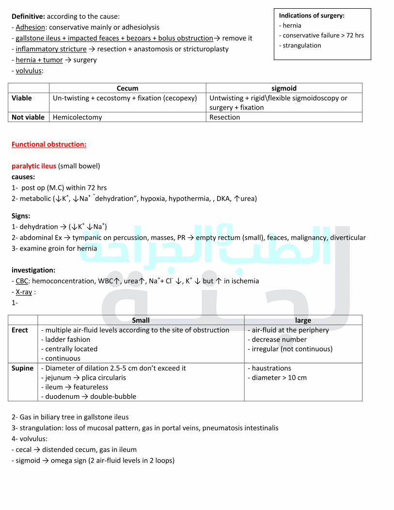
Cecum sigmoid
Viable Un-twisting + cecostomy + fixation (cecopexy) Untwisting + rigid\flexible sigmoidoscopy or
surgery + fixation
Not viable Hemicolectomy Resection
Functional obstruction:
paralytic ileus (small bowel)
causes:
1- post op (M.C) within 72 hrs
2- metabolic (↓K+, ↓Na+ “dehydration”, hypoxia, hypothermia, , DKA, ↑urea)
Signs:
1- dehydration → (↓K+ ↓Na+
)
2- abdominal Ex → tympanic on percussion, masses, PR → empty rectum (small), feaces, malignancy, diverticular
3- examine groin for hernia
investigation:
- CBC: hemoconcentration, WBC↑, urea↑, Na++ Cl
- ↓, K+
↓ but ↑ in ischemia
- X-ray :
1-
Small large
Erect - multiple air-fluid levels according to the site of obstruction
- ladder fashion
- centrally located
- continuous
- air-fluid at the periphery
- decrease number
- irregular (not continuous)
Supine - Diameter of dilation 2.5-5 cm don’t exceed it - jejunum → plica circularis
- ileum → featureless
- duodenum → double-bubble
- haustrations
- diameter > 10 cm
2- Gas in biliary tree in gallstone ileus
3- strangulation: loss of mucosal pattern, gas in portal veins, pneumatosis intestinalis
4- volvulus:
- cecal → distended cecum, gas in ileum
- sigmoid → omega sign (2 air-fluid levels in 2 loops)
Indications of surgery:
- hernia
- conservative failure > 72 hrs
- strangulation
45
Barium enema:
- cecal volvulus: bird beak feature
- follow though is C.I → perforation
Clinical:
- no pain, no bowel sound, distention
- X-ray: air-fluid levels, recto-sigmoid gas
Tx: conservative
Pseudo-obstruction (colon) → Ogilvie’s syndrome
- mainly in elderly
Causes: neurological, hypnotic\sedation, lead toxicity, hypothyroidism
Clinical: abdominal distention that may lead to perforation + peritonitis
X-ray: distention, no air-fluid level
Tx: conservative
46
Trauma
Causes:
1- motor vehicle accidents 2- violence (9-15%) 3- falls (9%)
4- burn: thermal, electrical, chemical, corrosive (alkaline, acidic solution), drowning, blast
The accidents (un-intentional) are the 5th
leading cause of death worldwide in young adults and 30% of ICU
admissions.
Death from trauma:
1- immediate → 50% at the time of accident hemorrhage from great vessels →
CNS trauma (brain stem), respiratory arrest
2- early death (golden hour) 30% → hemorrhage, hypoxia
3- late death (1- days) 20% → sepsis, PE, multiple organ failure
Risk factor according to cause:
RTA:
- cars speed -rolled over car - dead passenger - car indentation >30 cm - extraction time > 20 min
Falling down:
- height - ground - way of fall
Burn:
- temperature - time of contact - flame with close space → inhalational injury - associated trauma (falling, ..)
Trauma system:
1- injury prevention 2- access to car 3- pre-hospital 4- hospital 5- rehabilitation
Approach:
- primary survey → Tx life threatening events
- secondary survey → head to toe exam to define other non-lethal injuries
- definitive management
Primary survey:
- detect and treat immiedetly life threatening problems
- should take few min
- don’t proceed to secondary until the ABC-stable
- repeat it when: changes in mental status, changes in vital signs.
- Airway: + C-spine stabilization (ask the pt what’s your name→ phonate, mentate)
*relieve any obstruction (tongue “in case of bilateral Fx of mandible”, foreign body, aspirated material, blood, vomiting, tissue, edema, teeth, denture)
*tracheal intubation for any hemodynamically unstable pt and injury to the face (with depresses Fx) and neck
* but we must do CXR to rule out tension pneumothorax that develop from minor\small pneumothoracies 47
Ex: inspection of chest\ oropharynx:
- palpate the trachea and anterior neck for any laceration, hemorrhage, swelling
- any noisy breathing sound → obstruction
What to do?
1- Remove any tight clothes at neck
2- suction for any secretion, blood, foreign body
3- O2 → bag valve mask
4- cricothyroid kit
5- endotracheal tubes
Cervical spine protection:
1- highly suspicious history
2- avoid rough manipulation of head and neck:
- holding the head in neutral position facing forward
- secure it by hard cervical collar
3- then radiological evaluation after pt stabilization
Breathing and ventilation:
Start with examination:
1- palpate tracheal deviation
2- crepitus in fractures, air → surgical emphysema
3- inspect asymmetrical chest movement
4- auscultate breath sound bilaterally
Life threatening conditions:
Tension pneumothorax → 1- needle insertion at mid Clavicular line in 2
nd or 3
rd intercostal space
2- chest tube
Massive hemothorax → chest tube
Flail chest: paradoxical chest movement:
- intubate in elderly pt and multi-traumatic one- if there is hypoxia or respiratory distress do ABG + pulse
oxymeter
Open pneumothorax: there is a sucking wound in the chest wall that flow the air into the pleural space, as the trachea
to the lung (during inspiration)
Circulation: BP, HR, evidence of bleeding
- control the bleeding
- 2 large bore cannulas, Foleys catheter, cross match, IV fluid + blood, ..
Adjuncts to primary surveys:
1- pulse oxymeter, BP, cardiac monitor
2- ECG
3- X-ray → cervical, chest, pelvis
4- blood work
5- ABG
48
Disability:
1- Glasgow coma scale or APVU (alter, response to painful stimulation, verbal stimulus, unconscious)
2- pupil size and reactivity
3- gross motor and sensation (spinal cord injury)
Exposure:
1- the pt is completely undressed to reveal any hidden injury.
- missed\neglected regions: posterior scalp, abdominal folds (obese), axillary \ groin \ perineum
2- examine the back
Quick history:
AMPLE: allergy, medication, past medical, last meal, \drink, event
Secondary survey:
- head to toe examination
- special diagnostic test
- including (limb radio, US, CT)
Head:
- skull palpation and inspection
- check for face deformity
- check for eyes → discoloration, pupils, contact lenses
- check for nose → bleeding, CSF leak
- check ears
Neck:
- check for any swelling, wounds
(JVD, accessory respiratory muscles, tracheal shift)
back\cervical spine: wound, tenderness, swelling, bruising
Chest, abdomen
Pelvis: deformity → scrotal or perineal \ bleeding per urethra
Arms and legs + pulse sensation + movement
49
Abdominal trauma
Special characteristics:
1- large amount of blood may reach 4 L → we have a space
2- liver, spleen bleed profusely as major abdominal vessels
3- increased infection (bowel injury)
4- 3rf cause of traumatic death after head\chest → (hemorrhage, sepsis)
Causes: RTA (M.C)
Classification: 1- intra-peritoneal 2- extra-peritoneal
Blunt Penetrating
Cause 1-motor vehicle:
مدى الدمار, حزام االمان, في وفيات؟, مكانه
→ auto to auto \ auto to pedestrian
2-direct blow to the abdomen
3-fall from height → height, ground
1-low → depend on the mass move
high velocity missiles (>100 m\s)
cavitation → shattering
KE = 1\2 M (V1-V2)
2-stabs →
no. of stabs, distance, size
mechanism Deceleration:
- fixed points as ligament of treitz, iliocecal valve,
phrenocloic ligament
compression with crushing of abdominal content to the
abdominal wall and posteriorly (vertebral column
posterior thoracic cage)
compression with rupture:
- 2nd
part of duodenum
- iliocecal valve
recto with intraperitoneumنقطة وصل
- Width, size of knife
- velocity \ mass
other factors:
- colon is less tolerable to the high
velocity missile than small bowel (fecal
content)
Velocity \ mass \solidity of the organ α liberation of energy → ↑damage
Ex Fully exposed patient:
1- ecchymotic skin, abrasions
2- steering wheel \ seat belt sign (1\3 abdominal injury)
3- abdominal distention
→ Where the injury according to the abdominal boundaries.
(5th
intercostal) space:
- inferior gluteal fold post
- inguinal ligament ant.
anything with these boundaries indicate
abdominal injury
Examination:
Palpation:
- peritoneal irritation \ guarding \ rigidity → maybe evisceration - abdominal distention
- per-digital exam
- pelvic instability
- crepitus at lower thoracic cage 50
Follow up + investigation:
1- serial vital \ serial physical examination
2- US → to check if there is abdominal fluid or not (normally up to 50 ml “5-20 ml”) 3- CT → in hemodynamically stable pt → retroperitoneal organ injury in blunt trauma
4- diagnostic peritoneal lavage:
- why ? To know if there is blood in peritoneum, injury to intestine
- when?
1. CT\US unavailable (difficulty in assessing the pt)
2. Equivocal clinical exam
3. Multiple injuries
4. Persistent hypotension despite adequate resuscitation
- how? Sub-umbilical longitudinal small incision → aspiration
+ve → 1. > 5 ml of blood 2. Bloody irrigated fluid 3. Presence of bile, enteric content 4. Fluid analysis: RBC >100,00 \cmm, WBC > 500\cmm, amylase > 175 U
Spleen injury:
signs & symptoms:
1- asymptomatic <=> non-specific ex
2- RUQ or diffuse pain and tenderness
3- referred pain to the Lt shoulder (Kehr’s sign) 4- syncope \ hypotension \ tachycardia \ tachypnea
5- dullness in LtUQ (Baltance’s sign)
when to suspect with plain X-ray:
- chest:
1- lower left rib Fx
2- Lt pleural effusion
3- Lt lower lobe atelectasis
- abdomen:
1- elevated left hemi-diaphragm
2- shifted to: gastric bubbles medially, splenic flexure gas inferiorly
Scaling:
I → subscapular hematoma <10% of surface, laceration <1 cm deep
II → subscapular hematoma 10-50% of surface, laceration 1-3 cm deep
III → subscapular hematoma >50% of surface, laceration >3 cm deep
IV → laceration >25% of parenchyma or involving hilum
V → shattered spleen, hilar vessel injury + de-vascularization
Treatment:
I, II, III → conservative or selective splenic artery immobilization:
- acute dilation of stomach die to small gastric artery ligation, risk of infection, risk of ischemia
IV, V → surgery \ newly maybe treated conservatively → risk of developing abscess (blood in peritoneum) - splenic preservation → rraphy, partial spleenectomy
- spleenectomy
51
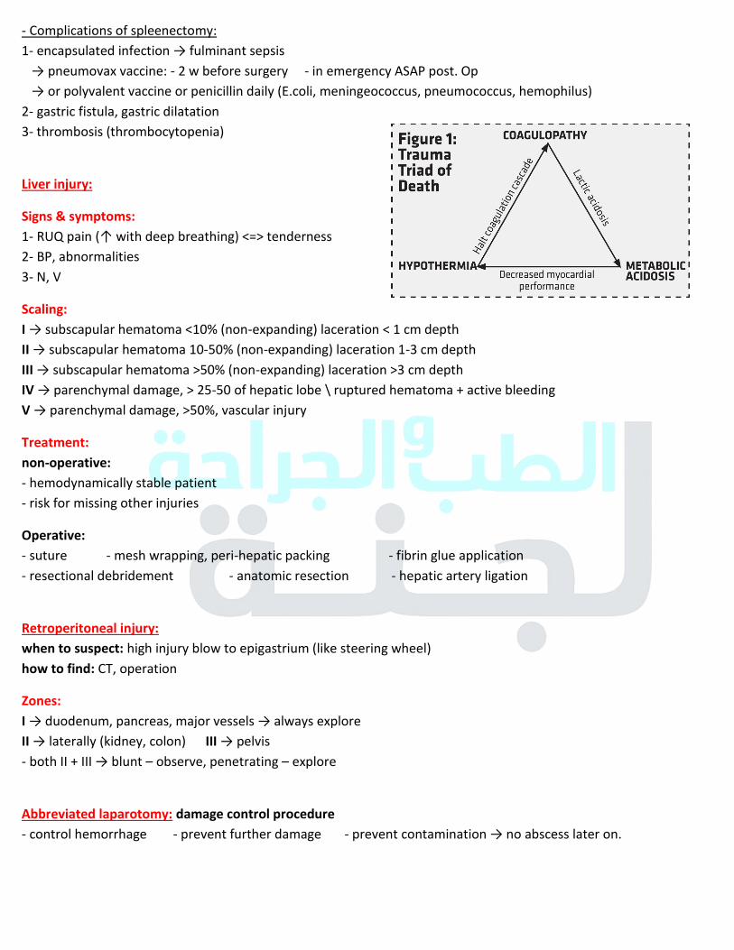
- Complications of spleenectomy:
1- encapsulated infection → fulminant sepsis
→ pneumovax vaccine: - 2 w before surgery - in emergency ASAP post. Op
→ or polyvalent vaccine or penicillin daily (E.coli, meningeococcus, pneumococcus, hemophilus) 2- gastric fistula, gastric dilatation
3- thrombosis (thrombocytopenia)
Liver injury:
Signs & symptoms:
1- RUQ pain (↑ with deep breathing) <=> tenderness
2- BP, abnormalities
3- N, V
Scaling:
I → subscapular hematoma <10% (non-expanding) laceration < 1 cm depth
II → subscapular hematoma 10-50% (non-expanding) laceration 1-3 cm depth
III → subscapular hematoma >50% (non-expanding) laceration >3 cm depth
IV → parenchymal damage, > 25-50 of hepatic lobe \ ruptured hematoma + active bleeding
V → parenchymal damage, >50%, vascular injury
Treatment:
non-operative:
- hemodynamically stable patient
- risk for missing other injuries
Operative:
- suture - mesh wrapping, peri-hepatic packing - fibrin glue application
- resectional debridement - anatomic resection - hepatic artery ligation
Retroperitoneal injury:
when to suspect: high injury blow to epigastrium (like steering wheel)
how to find: CT, operation
Zones:
I → duodenum, pancreas, major vessels → always explore
II → laterally (kidney, colon) III → pelvis
- both II + III → blunt – observe, penetrating – explore
Abbreviated laparotomy: damage control procedure
- control hemorrhage - prevent further damage - prevent contamination → no abscess later on.
52
Upper GI bleeding
Definition : any bleeding above ligament of treitz
Etiology :
- peptic ulcer disease
- gastroesophageal erosions
- Mallory weiss tear
- esophageal varices
- Tumors
- vascular lesions ( angiodysplasia )
- small intestine bleeding ( Meckel's diverticulum , aortoenteric fistula )
Clinical :
- Hematemesis
- Melena ( IUH in Bowel )
- hematochezia ( in large amount )
- coffee ground vomiting
- occult blood in stool
Physical examination :
1 - we start from vitals ( blood pressure and heart rate ) then postural hypotension decrease > 20 points
indicate 10 to 20% blood loss
postural tachycardia increase > 10 points
2 - digital rectal examination ==> if there is blood --> indicate brisk bleeding
3 - signs of liver disease + portal hypertension ==> varicose veins
Treatment :
1 - resuscitation : 2 large bore canula + cross matching + coagulation profile
folly's catheter --> central venous catheter --> NG tube : to confirm Dx & prevent aspiration
16% --> non the bloody aspiration
other according to the cause ( 80% resolve spontaneously )
2 - CBC hematocrit should kept > 30% in elderly ( coronary artery disease )
3 - increase PT give FFP
4 - decrease platelet give platelets
5 - Endoscopy in hemodynamically unstable to control the bleeding
Peptic Ulcer :
gastric ulcer bleeding from the artery and the base of ulcer
duodenal ulcer bleeding from gastroduodenal artery posteriorly located ulcer
- May the 1st presentation 25%
- Mortality rate 40%
53

Management :
A - Endoscopy :
- electrocoagulation
- laser therapy
- sclerotherapy
- cauterization
- arterial banding
- vasopressin injection
- increase risk to rebleed :
uncleare ulcer { visible vessel , blood , clots }
should stay 3 days in hospital
B - Surgery :
1 - unable to control the bleeding
2 - patient is elderly and unfit
3 - 6 unite blood transfusion
Doudenal ulcer { under run with sutures to through doudentorotomy }
Gastric ulcer a - young patient --> excision of the ulcer
b - elderly patient --> under run with sutures + biopsy --> if Malignant --> treatment as CA
C - PPI { omeprazole , 80 mg bolus , 8 mg infusions } :
increase pH 1 - increase stability of clot
2 - decrease further bleeding
GE erosions :
- NSAIDs use
- erosive esophagitis
- gastritis
Stress Related :
- curing ulcer { trauma , burn , major surgery }
- Cushing ulcer { intracranial disease }
- severe medical illness { ventilator-dependent , coagulopathy }
treatments : H2 Blocker + sacralfate
Mallory wiess treat :
- history of vomiting , itching
- alcoholic
- under GEJ
- resolve spontaneously ( 80 to 90% ) , not endoscopic treatments , not :
1 - angiographic treatment ( vasopressin )
2 - surgery treatment
a - Active bleeding
b - Non bleeding visible vessels
Eradicate H-pylori
Avoid NSAIDs
not cause significant bleeding
not cause significant bleeding
54
Esophageal varices :
most common bleeding from varicose veins
treatment :
- vasopressor { decrease portal inflow by splanchnic artery constriction }
- Octerotide/ somatostatin / terliprisson
- life-threatening conditions before endoscopy { sengstaken - Blakemore tube }
- Endoscopy : 1 – ligation
2 – Sclerotherapy { may cause stricture }
- Long term prophylaxis by non-selective beta blocker
- Persistent or recurrent bleeding
Transjugular intrahepatic portosystemic shunts { TIPS } expandable metal shunt from Portal system " بندخل منه "
to hepatic vein
contraindication in severe liver disease , transplant is anticipated
milder cases splenorenal shunt
Dieulafoy lesion :
= angiodysplasia = arteriovenous malformation
- Large tortous submucosal artery
- can cause severe bleeding
- missed in endoscopy unless if the bleeding { under normal mucosa }
- treatment : - endoscopy { sclerotherapy }
- angiographic embolization
- surgery { suture or reception }
Obscure GIB :
- usually from small intestine
- push endoscopy or video capsule enteroscopy
- technetium-99 labeled
- aortoenteric fistula - history of aortic graft replacement
- untreated aortic aneurysm
Prognosis of UGIB :
according to :
- Underlying illness
- Age
- Comorbidity
- Hemodynamic compromise
acute bleeding
55
Diabetic foot
Pathogenesis:
1- Diabetic neuropathy
- sensory: distal symmetrical polyneuropathy, caused by untreated minor injuries (mechanical, thermal, pressure)
- autonomic: ↓ sweating →dryness, crackled skin
- motor: clawing of toes, high arch*, prominent metatarsal head* (1st
, 5th
), tip of fingers
* pressure points → ulcers + callus
2- Diabetic osteoarthropathy:
- Charcot joint (mid planter area) → ulceration in the medial aspect of foot
- stretching of ligament → loss of arch
- minor trauma → healing v trauma → deformity
3- Vascular insufficiency (microangiopathy) → delay of healing
4- ↓Immunity: affects the chemotactic activity of T-cell
Wager classification of diabetic foot:
- Grade 1 → no ulcer, but high risk foot
- Grade 2 → superficial ulcer (skin not underlying tissue) - Grade 3 → deep ulcer (cellulitis, abscess, bone involvement by osteoarthritis) - Grade 4 → localized gangrene (toe, heel) - Grade 5 → extensive gangrene involving the whole foot
Why foot not others (hand, others) ?
- Compartments of foot separated by dense fibrous tissue → any ulcer + infection → edema → ↑pressure → ↓blood → ischemia
When pt come to ER (C.C):
1- Cellulitis, abscess
2- Osteomyelitis
3- Ulcer
4- Impeding gangrene
5- Deformity (Charcot joint)
History:
1- DM and its control: duration, complication (eye, heart, renal, peripheral “numbness, claudication”, medication or insulin.
2- Social Hx: smoking***, alcohol, diet, occupation
3- Medical Hx: HTN, surgical Hx
4- Cultural habits: wear socks or not, wet foot on work, foot care and hygiene, daily activity
5- Chief complain of foot complain: duration, pain, numbness, when and where he noted the ulcer, deformity,
previous surgery
56
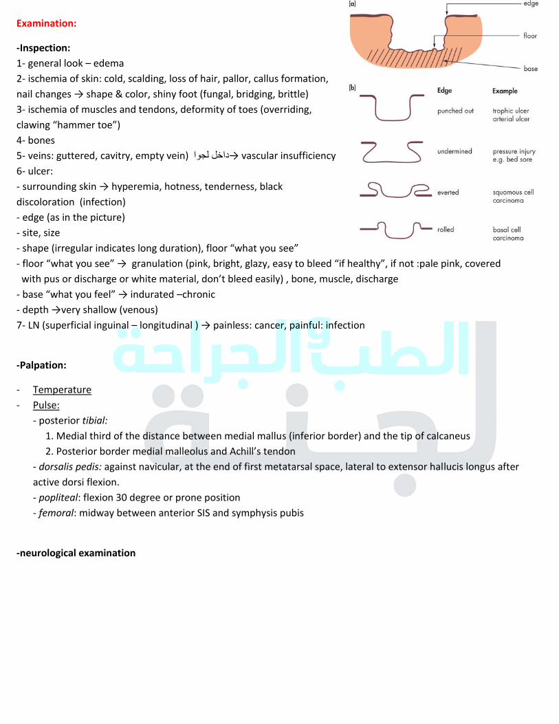
Examination:
-Inspection:
1- general look – edema
2- ischemia of skin: cold, scalding, loss of hair, pallor, callus formation,
nail changes → shape & color, shiny foot (fungal, bridging, brittle) 3- ischemia of muscles and tendons, deformity of toes (overriding,
clawing “hammer toe”) 4- bones
5- veins: guttered, cavitry, empty vein) داخل لجوا→ vascular insufficiency
6- ulcer:
- surrounding skin → hyperemia, hotness, tenderness, black discoloration (infection)
- edge (as in the picture)
- site, size
- shape (irregular indicates long duration), floor “what you see”
- floor “what you see” → granulation (pink, bright, glazy, easy to bleed “if healthy”, if not :pale pink, covered with pus or discharge or white material, don’t bleed easily) , bone, muscle, discharge - base “what you feel” → indurated –chronic
- depth →very shallow (venous) 7- LN (superficial inguinal – longitudinal ) → painless: cancer, painful: infection
-Palpation:
- Temperature
- Pulse:
- posterior tibial:
1. Medial third of the distance between medial mallus (inferior border) and the tip of calcaneus
2. Posterior border medial malleolus and Achill’s tendon
- dorsalis pedis: against navicular, at the end of first metatarsal space, lateral to extensor hallucis longus after
active dorsi flexion.
- popliteal: flexion 30 degree or prone position
- femoral: midway between anterior SIS and symphysis pubis
-neurological examination
57
lines of management:
1. metabolic care: blood sugar, HbA1C, CBC, KFT, ESR+CRP, fundoscopy
2. microbial care:
- mixed infection
- best method for microbiological study is tissue culture after taking a sample of healthy tissue
- broad spectrum (empirical) → Flagil, 3rd
cephalo
- x-ray: foreign body, gas, osteomyelitis
3. dressing: 1- simple washing 2- better drainage 3- debridement 4- amputation
4. vascular care: Doppler US + surgery if macrovascular
5. Foot care (shoes..)
1- claudication:
severity:
1. Distant 2- surface of stress 3- cold environment 4- drugs as B-blockers 5- smoking 7- walking against wind
2- Rest pain: in the foot in distal metatarsophalangeal → hanging the leg on bed.
3- Rest pain + ulcer → pre-gangrene
4- gangrene
We use scaling system to give insulin??
→ We use short acting insulin to get better control.
58
Compartment syndrome
Definition: excessive pressure inside enclosed muscle space → leads to tissue ischemia → necrosis
- Excessive pressure → content: bleeding, edema due to trauma
- pressure from outside that size: bandage\cast, lying on limb
Epidemiology\causes:
- M.C.C (fracture)
- leg M.C → due to tibial fracture (anterior compartment) - Forearm- 2
nd M.C due to supracondylar fracture (flexor, ant-compartment)
- M>F
Pathophysiology: muscle perfusion pressure = diastolic pressure – intramuscular pressure (0-4 mmHg, >30 indicates
compartment)
Types:
► Chronic:
- excertional compartment
- especially when it involve repetitive motion
- recurrent pain + disability (↑with ↑ intensing and ↓ within 30 minutes after stopping: burning, aching, pressure
like)
► Acute: due to major injury → crush injury, cast\bandage, from heavy drinking or drugs use.
Symptoms: 6Ps
. Pain: out of proportion (doesn’t improve after Tx) - deep ache\ burning +\- bulge of affected muscle
- first symptom
. Pressure: >30-40 mmHg “diagnostic”
. Paresthesia: numbness → early to one compartment, late to entire limb
. Pallor (rare - late stage) → sign of vascular injury + ischemia
. Pulseless: late stage, if present not exclude compartment
. Paralysis: very late, indicate nerve damage
-Volkmann’s contracture (fibrosis after ischemia)
Complications:
. Muscle scarring, contracture → Volkmann’s contracture (at wrist, hand & fingers), claw toes + weak dorsiflexors
. Infection
. Permanent nerve damage → sensory loss
. Rhabdomyolysis → ↑myoglobin + ↑K+ → kidney damage “may need dialysis”
. Chronic pain
. Amputation
59
Compartments of the upper limb:
► Arm:
. Anterior : musculocutaneous nerve\biceps, bronchials
. Posterior: radial nerve\triceps
► Forearm:
. Superficial volar: median and ulnar nerve → pain with passive extension + weakness of flexion + sensory loss at
median index and ulnar nerve (little finger)
. Deep volar: anterior interosseous nerve→ weakness\pain , no sensory defect (motor only)
. Dorsal compartment: posterior interosseous nerve
. Henrey’s mobile wad: superficial radial nerve
► Hand: 10 compartments
. 4 dorsal interossei
. 3 volar
. One adductor pollicis
. One hypothenor
. One thenor
Compartment of lower limb:
► Thigh:
. Anterior: M.C in thigh due to femoral fracture\blunt trauma
- weakness in knee extension, pain in passive flexion
- femoral nerve\saphenous nerve: numbness at medial knee
. Medial: obturator nerve\adductor muscles, gracills.
. Posterior: sciatic\biceps, semi membranous, semi tendinous
► Leg:
. Anterior: anterior tibial artery\deep peroneal nerve → numbness in first web space.
. Lateral: superficial peroneal nerve→ numbness in dorsum of foot
. Superficial dorsal: sural nerve → numbness at lateral border of foot
. Deep dorsal: tibial nerve → numbness at planter surface of foot
► Foot: 9 compartments:
. 4 interossei
. One adductor halluces
. One lateral
. One medial
. Two central
Swelling, tenderness at the dorsum of the foot
60
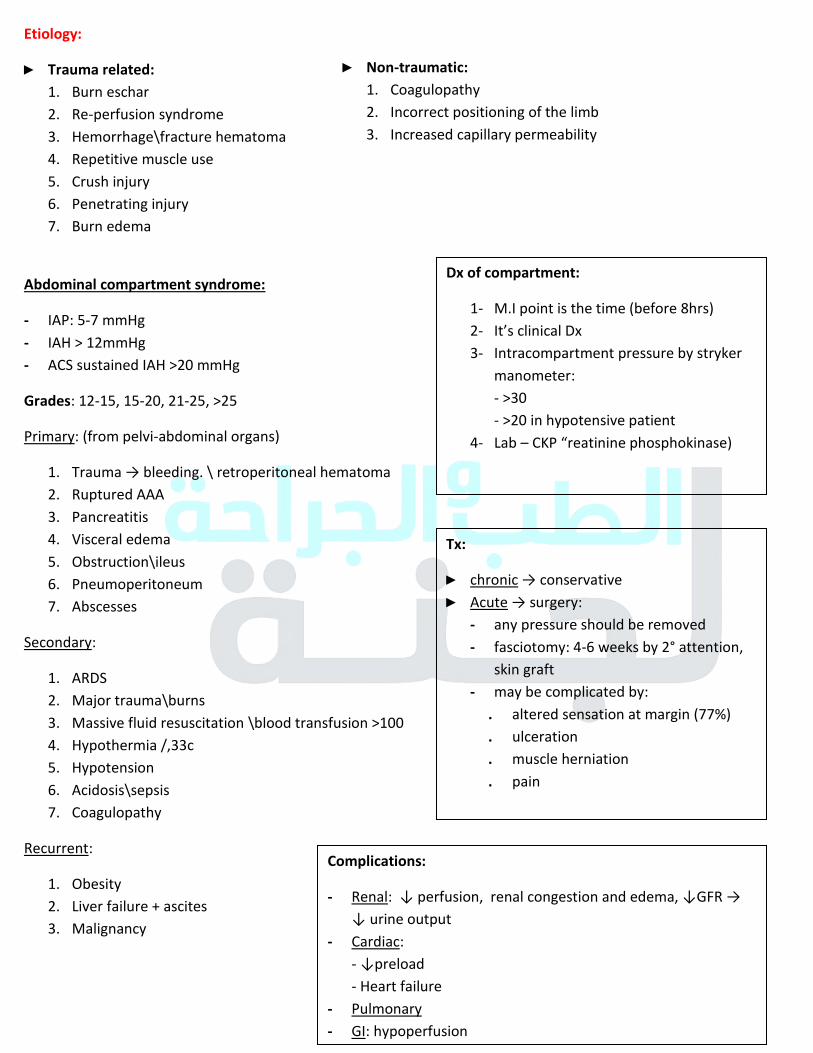
Etiology:
► Trauma related:
1. Burn eschar
2. Re-perfusion syndrome
3. Hemorrhage\fracture hematoma
4. Repetitive muscle use
5. Crush injury
6. Penetrating injury
7. Burn edema
Abdominal compartment syndrome:
- IAP: 5-7 mmHg
- IAH > 12mmHg
- ACS sustained IAH >20 mmHg
Grades: 12-15, 15-20, 21-25, >25
Primary: (from pelvi-abdominal organs)
1. Trauma → bleeding. \ retroperitoneal hematoma
2. Ruptured AAA
3. Pancreatitis
4. Visceral edema
5. Obstruction\ileus
6. Pneumoperitoneum
7. Abscesses
Secondary:
1. ARDS
2. Major trauma\burns
3. Massive fluid resuscitation \blood transfusion >100
4. Hypothermia /,33c
5. Hypotension
6. Acidosis\sepsis
7. Coagulopathy
Recurrent:
1. Obesity
2. Liver failure + ascites
3. Malignancy
► Non-traumatic:
1. Coagulopathy
2. Incorrect positioning of the limb
3. Increased capillary permeability
Dx of compartment:
1- M.I point is the time (before 8hrs)
2- It’s clinical Dx
3- Intracompartment pressure by stryker
manometer:
- >30
- >20 in hypotensive patient
4- Lab – CKP “reatinine phosphokinase)
Tx:
► chronic → conservative
► Acute → surgery: - any pressure should be removed
- fasciotomy: 4-6 weeks by 2° attention,
skin graft
- may be complicated by:
. altered sensation at margin (77%)
. ulceration
. muscle herniation
. pain
Complications:
- Renal: ↓ perfusion, renal congestion and edema, ↓GFR → ↓ urine output
- Cardiac:
- ↓preload
- Heart failure
- Pulmonary
- GI: hypoperfusion 61

Colostomy
Definition: surgical procedure at which the one end of large intestine brings out through the abdominal wall, with
incision to create a stoma.
Normal appearance:
- At first it’s swollen → 8 weeks it shrinks
- Red, moist, painless on touch
Indications:
- Congenital malformations:
- anal atresia
- hirschsprung disease
- Neoplastic:
- rectal
- anal
- Injury to colon\rectum
- Inflammatory process: IBD, diverticulitis
- Bowel obstruction
- Wound\fistula in perineum
Choosing a stoma site:
avoid:
1- bony prominences, umbilicus, skin folds or creases
2- hernia, old wounds or scars
► Should be assessed preoperatively, in sitting, lying, standing positions.
Classification:
Time:
Permanent:
- disease affects the end part of colon\rectum
- end colostomy\ileostomy
Temporary:
- to give a chance for the bowel to heal\rest
- hartman’s procedure (end-colostomy + rectal stump)
- loop transverse colostomy (defunctioning stoma “in obstruction” , bowel rest – pericolic abscess or anorectal fistula
62
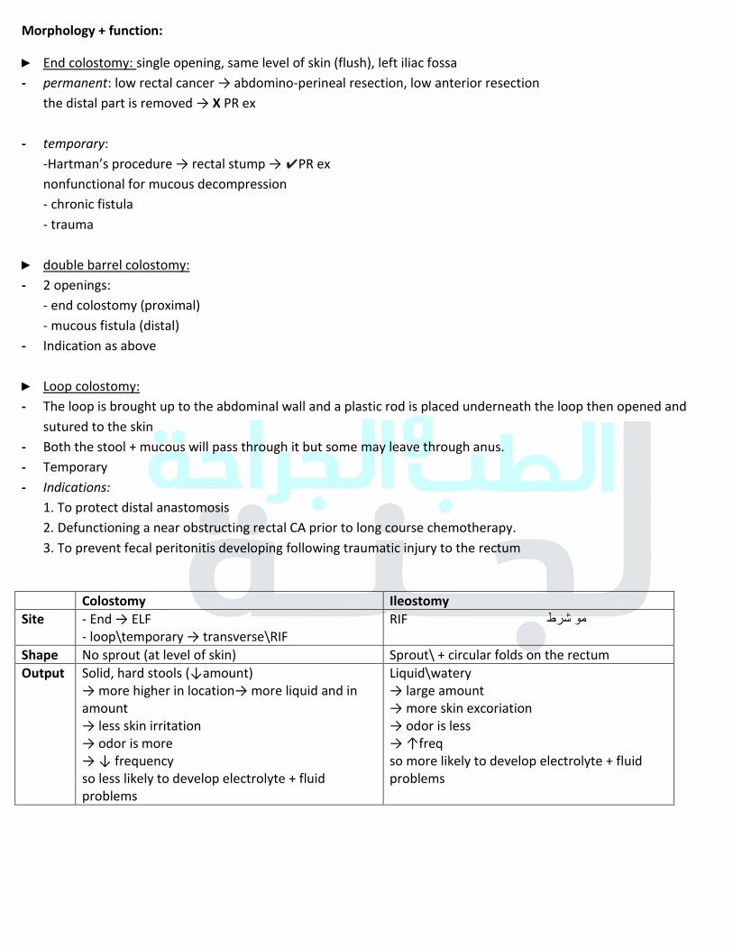
Morphology + function:
► End colostomy: single opening, same level of skin (flush), left iliac fossa
- permanent: low rectal cancer → abdomino-perineal resection, low anterior resection
the distal part is removed → X PR ex
- temporary:
-Hartman’s procedure → rectal stump → ✔PR ex
nonfunctional for mucous decompression
- chronic fistula
- trauma
► double barrel colostomy:
- 2 openings:
- end colostomy (proximal)
- mucous fistula (distal)
- Indication as above
► Loop colostomy:
- The loop is brought up to the abdominal wall and a plastic rod is placed underneath the loop then opened and
sutured to the skin
- Both the stool + mucous will pass through it but some may leave through anus.
- Temporary
- Indications:
1. To protect distal anastomosis
2. Defunctioning a near obstructing rectal CA prior to long course chemotherapy.
3. To prevent fecal peritonitis developing following traumatic injury to the rectum
Colostomy Ileostomy
Site - End → ELF
- loop\temporary → transverse\RIF
RIF مو شرط
Shape No sprout (at level of skin) Sprout\ + circular folds on the rectum
Output Solid, hard stools (↓amount) → more higher in location→ more liquid and in amount
→ less skin irritation
→ odor is more
→ ↓ frequency
so less likely to develop electrolyte + fluid
problems
Liquid\watery
→ large amount
→ more skin excoriation
→ odor is less
→ ↑freq
so more likely to develop electrolyte + fluid
problems
63
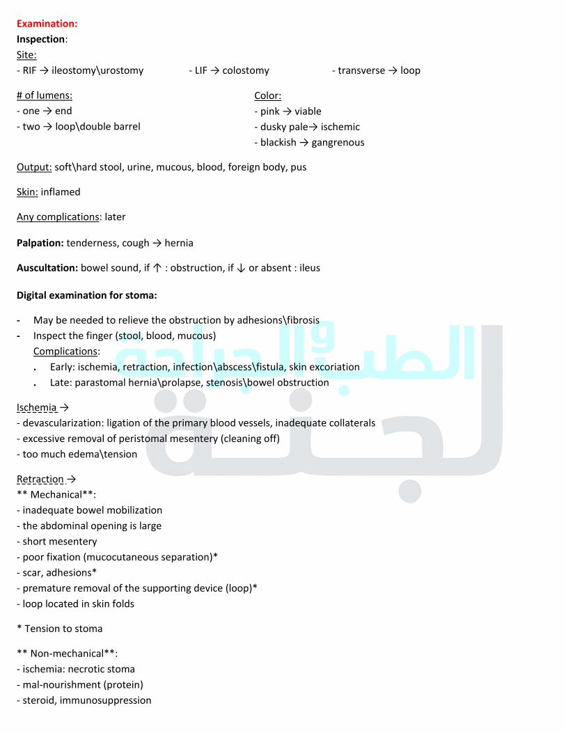
Examination:
Inspection:
Site:
- RIF → ileostomy\urostomy - LIF → colostomy - transverse → loop
# of lumens:
- one → end
- two → loop\double barrel
Output: soft\hard stool, urine, mucous, blood, foreign body, pus
Skin: inflamed
Any complications: later
Palpation: tenderness, cough → hernia
Auscultation: bowel sound, if ↑ : obstruction, if ↓ or absent : ileus
Digital examination for stoma:
- May be needed to relieve the obstruction by adhesions\fibrosis
- Inspect the finger (stool, blood, mucous)
Complications:
. Early: ischemia, retraction, infection\abscess\fistula, skin excoriation
. Late: parastomal hernia\prolapse, stenosis\bowel obstruction
Ischemia → - devascularization: ligation of the primary blood vessels, inadequate collaterals
- excessive removal of peristomal mesentery (cleaning off)
- too much edema\tension
Retraction →
** Mechanical**:
- inadequate bowel mobilization
- the abdominal opening is large
- short mesentery
- poor fixation (mucocutaneous separation)*
- scar, adhesions*
- premature removal of the supporting device (loop)*
- loop located in skin folds
* Tension to stoma
** Non-mechanical**:
- ischemia: necrotic stoma
- mal-nourishment (protein)
- steroid, immunosuppression
Color:
- pink → viable
- dusky pale→ ischemic
- blackish → gangrenous
64
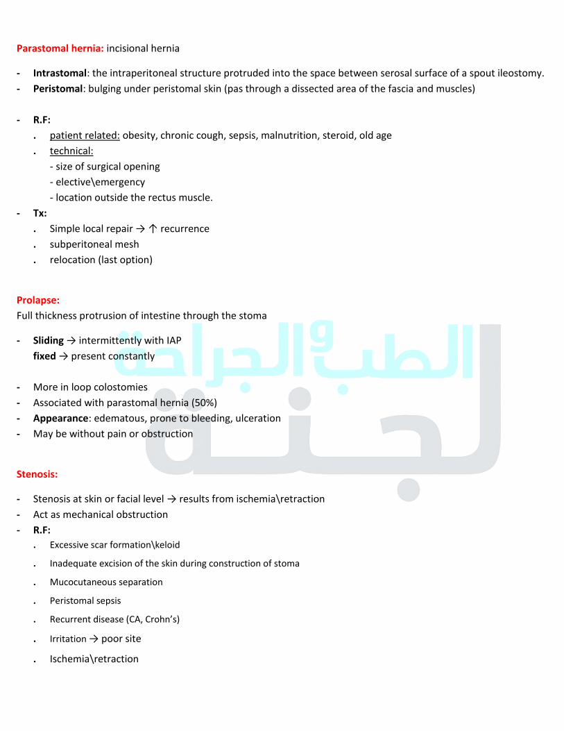
Parastomal hernia: incisional hernia
- Intrastomal: the intraperitoneal structure protruded into the space between serosal surface of a spout ileostomy.
- Peristomal: bulging under peristomal skin (pas through a dissected area of the fascia and muscles)
- R.F:
. patient related: obesity, chronic cough, sepsis, malnutrition, steroid, old age
. technical:
- size of surgical opening
- elective\emergency
- location outside the rectus muscle.
- Tx:
. Simple local repair → ↑ recurrence
. subperitoneal mesh
. relocation (last option)
Prolapse:
Full thickness protrusion of intestine through the stoma
- Sliding → intermittently with IAP
fixed → present constantly
- More in loop colostomies
- Associated with parastomal hernia (50%)
- Appearance: edematous, prone to bleeding, ulceration
- May be without pain or obstruction
Stenosis:
- Stenosis at skin or facial level → results from ischemia\retraction
- Act as mechanical obstruction
- R.F:
. Excessive scar formation\keloid
. Inadequate excision of the skin during construction of stoma
. Mucocutaneous separation
. Peristomal sepsis
. Recurrent disease (CA, Crohn’s)
. Irritation → poor site
. Ischemia\retraction
65
Types of bags:
One piece:
- Bag + adhesive base (one)
- Zero chance for bag and flange coming apart*
- less expensive*
- recurrent change of flange**
- Causes skin tenderness, stomal irritation**
Classification:
Type 1:
- 90-95 %
- Dilatation of CBD
A → entire CBD
b → segment CBD
d → fusiform dilatation of CBD
Type 2: Diverticulum of CBD with NO dilatation of CBD + intra\extrahepatic ducts
Type 3: choledococele (cystic dilatation of distal BD)
- Intra duodenal,intrapancreatic
- Usually stenotic in its opening due to chronic inflammation
Type 4: multiple cysts (intra + extra hepatic)
Type 5: single or multiple intrahepatic (not extra hepatic) + hepatic fibrosis (caroli disease)
Complications: Choledocholithiasis, Rupture, Cirrhosis\portal HTN, Biliary cancer → long term monitoring, Stricture
Dx:
Lab:
- CBC (anemia, prior surgery)
- AST, ALT, GT, ALP
- coagulation profile
- bilirubin
Tx: radical excision of cyst (risk of CA) + reconstruction of biliary tract by Roux en-Y loop of jejunum “hepato-
jejunostomy” (incidence of stricture + cholangitis)
Type 5:
- conservative
- percutaneous drainage + medical management
Two pieces
- Two
- More skin friendly*
- Better accommodation for special needs*
- Bag and flange can come apart and leak **
- More expensive**
Imaging: ***
- US: gold standard
- CT: anatomy
- ERCP: the best diagnostic
- MRCP: superior to ERCP, non-invasive, no post ERCP pancreatitis
*=advantage
**=disadvantage
66
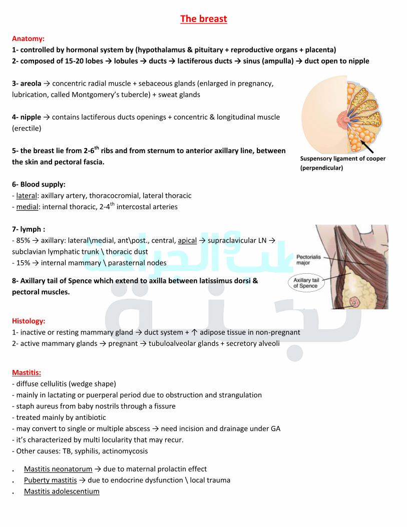
The breast
Anatomy:
1- controlled by hormonal system by (hypothalamus & pituitary + reproductive organs + placenta)
2- composed of 15-20 lobes → lobules → ducts → lactiferous ducts → sinus (ampulla) → duct open to nipple
3- areola → concentric radial muscle + sebaceous glands (enlarged in pregnancy, lubrication, called Montgomery’s tubercle) + sweat glands
4- nipple → contains lactiferous ducts openings + concentric & longitudinal muscle (erectile)
5- the breast lie from 2-6th
ribs and from sternum to anterior axillary line, between
the skin and pectoral fascia.
6- Blood supply:
- lateral: axillary artery, thoracocromial, lateral thoracic
- medial: internal thoracic, 2-4th
intercostal arteries
7- lymph :
- 85% → axillary: lateral\medial, ant\post., central, apical → supraclavicular LN → subclavian lymphatic trunk \ thoracic dust
- 15% → internal mammary \ parasternal nodes
8- Axillary tail of Spence which extend to axilla between latissimus dorsi &
pectoral muscles.
Histology:
1- inactive or resting mammary gland → duct system + ↑ adipose tissue in non-pregnant
2- active mammary glands → pregnant → tubuloalveolar glands + secretory alveoli
Mastitis:
- diffuse cellulitis (wedge shape)
- mainly in lactating or puerperal period due to obstruction and strangulation
- staph aureus from baby nostrils through a fissure
- treated mainly by antibiotic
- may convert to single or multiple abscess → need incision and drainage under GA
- it’s characterized by multi locularity that may recur. - Other causes: TB, syphilis, actinomycosis
. Mastitis neonatorum → due to maternal prolactin effect
. Puberty mastitis → due to endocrine dysfunction \ local trauma
. Mastitis adolescentium
Suspensory ligament of cooper
(perpendicular)
67
. Fibroadinoma: aberration of normal development and involution ANDI
- breast mouse (M.C\ reproductive years)
- characterized by → develop from a whole lobule + normal epithelium under hormonal effect
- giant fibroadenoma> 5 cm (α phyloid tumor: recurrence, need surgical removal, metastasize according to
the # of mitosis)
- investigation → triple assessment - management: small → X, giant → excised
.
Ductectasia:
- major sub-areolar duct dilatation and shortening
- symptoms:
1- discharge (cheesy +\- bloody
2- retraction: slit like \ partial retraction *(CA → circumferential retraction) - don’t need treatment unless the retraction is terrible to the pt → surgical Tx
. Cystosarcoma phylloides:
- serocysts disease of brodie
- benign ANDI
. symptoms:
- large \ massive mass +\- ulceration of skin (pressure necrosis)
- mobile on underlying chest wall
. histology: (low – high malignant potential)
. Tx: surgery (wide local excision\mastectomy): if 1- large mass 2- recurrent tumors 3- worrying histology
.
fibrocystic disease:
- fibroadenomatosis. ANDI
- under hormonal changes throughout menstrual cycle.
- signs & symptoms: lumpness (rice gray feeling), tenderness (cyclical mastalgia), discharge (green)
*non-cyclical mastalgia → periductal mastitis
.
Breast cyst: 15%
- s & s: smooth discrete lump +\- pain
- US to reveal the content (fluid or mixed...)
- what to do → 1- aspiration → not blood stained → blood stained, mixed tissue on US, recurrence after aspiration , residual lump after aspiration
2- mixed tissue on US
3- recurrence after aspiration (now)
4- residual lump after aspiration
.
DDx of cyst:
1- galactorrhea
2- lymphatic cyst
3- hydatid
4- ANDI + phylloides
5- intracystic papilliferous CA, colloid
degeneration of cancer, papillary cystadenoma
68
. Breast discharge
non-bloody → ductectasia, fibrocystic
bloody → - ductectasia
- duct papilloma (M.C), ductal CA 5% → both from multiple ducts + unilateral . - US → triple assessment to rule out CA
- Tx: microdochectomy by periareolar incision
.
Breast cancer:
. R.F:
1- age: 35→ very rare then every 5 yrs increase (double), 50→ 1\50, 80→ 1\10
2- race + geographic distribution (increase in north America)
3- hereditary causes 5-10%
a. BRCA1, BRCA2 → (to develop breast cancer at 65) + ↑ risk of ovarian CA
b. P53 mutation
c. ataxia telangiectasia
d. hereditary non-polyposis coli genes
4- estrogen exposure:
- early menarche + late menopause
- nulliparous
- contraceptive pills → in COCP
- HRT after 5 yrs
5- radiation: ionizing
- depending on: dose, time of exposure, age
6- lifestyle: fat intake, alcohol, decreased physical activity
.
Non-invasive:
. Ductal carcinoma in-sito:
1- premalignant
2- non-palpable
3- mammogram Microcalcifications
4- risk of developing CA according to the grade
5- Tx: lumpectomy with 1 cm rim +\- radiotherapy
.
Invasive cancer:
. Ductal: 85%
1- hard mass + unilateral well defined vein
2- desmoplasic response on mammogram (sun rays appearance)
3- grade 1-3 according to:
1- mitotic index 2- hyperchromatosis + polymorphism 3- tubule formation
4- 2\3 express estrogen & progesterone receptors
.
??:
1- four affected relatives
2- young age < 40
3- bilateral breast CA
4- +ve ovarian CA
5- male breast
↓ risk in:
- early age in first delivery
- breast feeding
Lobular carcinoma in-situ:
1- less likely to develop to CA than ductal
2- no palpable masses or mammographic
findings
3- incidental on biopsy
4- Tx close monitoring
69
. Lobular: 10%
1- less likely to be detected in mammogram → no microcalcificatoin
2- could ne bilateral + multifocal 20%
3- all express estrogen receptors
. Colloid, medullary, tubular → most are well differentiated + good prognosis
.
→ may adhere to: - underlying pectoral muscle
- overlying skin: dimpling (dors), tethering ( كل المساحة نازلة )
. → involvement of lymph may cause peau de orange appearance
. Clinical picture:
1- breast lump: 70%, may have axillary swelling
- usually painless, hard, .. – (M.C) in upper outer Q (60%)
2- nipple changes: discharge (bloody, ..), retraction (circumferential), erosions, itching, enlargement
3- skin changes: dimpling, tethering, peau de orange, scaliness
4- breast pain (rare)
.
Paget’s disease: - dry scaling and weeping appearance of nipple
- due to spread of tumors (mainly ductal) to the nipple
- well demarcated (α eczema), X steroid .
Inflammatory breast cancer: = anaplastic ductal CA
- erythematous, swollen (enlarged) breast, without fever or leukocytosis or palpable mass(X US OR MAMO)
- underlying poorly differentiated carcinoma
- Dx by biopsy (core-cut)
- no true inflammation
. Assessment:
1- clinical: Hx + PEx:
- age, menarche, FH, gynecological and obstetric Mx
- drug Hx
. 2- radiological: US
- < 35 Yrs, solid\cystic
. 3- pathological:
a. FNA:
- diagnosis of benign, if it’s cyst → drainage
- 5% false –ve due to sampling error
- if we find atypical cells → biopsy & follow up b. true-cut biopsy*: to find out → grade, invasive, estrogen receptor
.
α Mastitis:
- young age
- constitutional symptoms
Mammogram:
- > 35 yrs → increased risk of CA after one
exposure (1%)
- benign → well differentiated mass with
surrounding halo.
- malignant → speculation + architectural
distortion, microcalcification
*- atypical and fluoride hyperplasia → CA
- metaplasia and mild hyperplasia → no
risk
70
Others:
-MRI :
advantages:
- pick up carcinoma in situ - differentiates local recurrence from fibrosis - staging
- young women - multi-focal lesions
disadvantages:
- not detect calcifications - C.I in coronary cath.
-PET → multifocal lesions
Spread and staging:
1- blood extension: bone (lumbar, femoral), liver, lung, brain, kidney, suprarenal
2- prognostic factors:
- M.I one is # of axillary LN involved: (0→ 80%”5 yrs survival”, 3→ 50%, >3→ 25%)
- stage, grade, histological type
- HER2 (epidermal growth factor)
- hormonal receptors
Stage I, II → early \ III, IIII → advanced
Management:
1- breast conserving treatment: lumpectomy or wide excision (1 cm margin) + axillary LN sampling
(sentinel LN biopsy) or axillary clearance + radiotherapy
2- mastectomy : + radiotherapy to high risk)
- simple: removal of all breast tissue with nipples but leave chest wall muscles intact →
1- multiple foci 2- large CA 3- cancer involving nipple → ↑ recurrence
- radical: remove breast + axillary clearance + pectoralis major & minor
-modified radical: remove breast + axillary clearance + intercostal branches are divided →
post-op Paresthesia \ preservation of axillary vein, long thoracic nerve & latissmous dorsi
Both lumpectomy and mastectomy has the same survival rate but the recurrence is more in lumpectomy so we
add radiotherapy
3- chemotherapy + radiotherapy
4- hormonal treatment:
- oophorectomy, tamoxifen → more effective in pt who have estrogen receptors
- anastrozole (aromatase inhibitor)
5- biological agent:
in HER2 +ve pt: trastuzumab, heparin
others: bevacizumab → VGFR inhibitor, lantimab → GFR inhibitor
Investigations:
- liver enzymes + alkaline phosphatase.
- CXR, US, bone scan
71
Cancer en-cuirasse
- recurrence of CA locally after mastectomy
- may associated with swollen arm
- poor prognosis
- Tx: palliative
Lymphangisarcoma:
- complicated lymphedema
- as subcutaneous nodules in upper limb
- poor prognosis , x: chemo & radio
Male breast:
Gynecomastia:
- mostly benign
- causes : alcohol intake, liver cirrhosis, hypogonadism, testicular tumor, drugs: finasteride, spirinolactone,
cannabis
- rapid progressive indicates hormonal investigation
- persistent indicated surgery
Cancer:
- very rare 5%
- age: 5–10 yrs later than women
- presented as: eccentric mass, retraction of the skin
- investigation: mammo, FNA, core-cut biopsy
- Tx: mastectomy+ radiotherapy + chemo → ↓recurrence
Breast surgeries:
1- Lumpectomy \ quadrantectomy
2- Simple mastectomy
3- Modified radical mastectomy (cutting the tendon of pectoralis minor to remove the LN)
4- Radical mastectomy
5- Bilateral:
- lobular carcinoma (multifocal lesions)
- BRCA1, 2 → prophylactic
How to deal with the LN in axilla? Complicated lymphedema ↓
1- sentinel LN (we inject a dye then remove the involving LN only)
2- sampling
3- axillary clearance (remove level 3)
72
Thoracic trauma
Anatomy:
1- first 6 ribs attached to the sternum
2- later 4 form lower costal margin
3- last 2 floating ribs
4- Rt lung → 3 lobes, Lt lung → 2 lobes
Epidemiology:
1- 1\3 of RTA have chest trauma → 20-25% mortality rate
2- M.C.C is blunt trauma + mostly associated with other injuries
3- M.C blunt thoracic injury → rib fracture 30-35%
Rib fractures:
Clinical:
- pain, difficulty on breathing
- tenderness, crepitus, bruises → may be subcutaneous emphysema
. most common ribs fracture 4-10
. 8-12 raised suspension for hepatic, splenic injury
. 1,2nd
→ raised suspension for vascular injury + ↑ thoracic trauma
Indications of admission:
1- unable to cough → ↑ pneumonia
2- underlying pulmonary disease → COPD
3- age > or = 65 (↑ hypoventilation → hypercapnia \ atelectasis → pneumonia)
Associations:
- hemothorax, pneumothorax → >3 ribs unilateral - pulmonary contusions
Tx:
1- pain relief + adequate ventilation: pain killer, intercostal nerve block, epidural analgesia (multiple ribs)
2- early mobilization
3- pulmonary toilet
prognosis depends on:
1- age 2- underlying pulmonary status 3- # of ribs
Flail chest
multiple consecutive rib fractures from 2 levels or disruption of costocondral junction.
Clinical: chest pain, SOB, paradoxical motion (late sign, -ve → doesn’t exclude flail)
73
Causes:
- M.C cause: RTA
- falling in elderly (weak bones + more impacted by falls)
Association: pneumothorax, pulmonary contusion →RF
Tx:
- admit to ICU + ABG
- oxygen + painkiller (to promote cough and clear the secretion)
- aggressive pulmonary physiotherapy
- deep coughing
- bronchoscopy → to remove the secretion
- intubation if: RR > 30\MINUTE, PaO2 < 60, PaCO1 > 45
- In hypoventilation + hypercapnia (impeding RF) →
Intubation + PEEP, IV antibiotic, adhesive material (X paradoxical)
Clavicular fractures:
Broken collar bone
Clinical: pain with movement and may extend to surrounding muscles, swelling
M.C site: middle one third 80% > lateral 15% > medial 5%
Causes: falling on shoulder, FOSH, direct trauma
Associations: pneumothorax (apical lung), vascular + neural injury
Tx:
- spontaneous healing
→ immobilization by (figure of eight bandage) - surgery :
1- neurovascular injury 2- open Fx 3- multiple pieces, shortening 4- ununion↑ 3-6 m \ malunion
5- distal third Fx (risk of ununion)
Healing process depend on: age, complexity, location of fracture, displacement
Scapular Fx:
1- indicate energy trauma → high speed vehicle accident
2- associated with severe injuries → chest + others, that indicate admission
3- Tx → spontaneous
Sternal Fx: 4%
- M.C location → upper, middle third \ transverse
- Dx: lateral CXR, CT
- Clinical: pain, swelling
- Associations (55-70%) → rib Fx, long bone Fx, closed head injury, blunt cardiac injury (<20%)
- Tx: pain killer, spontaneous within 6-8%
74
Tracheobronchial trauma:
Mechanism:
1- rupture of membranous portion of trachea
2- disruption of trachea at point of fixation (carina & cricoid) due to sheeting or rapid deceleration force.
3- Laceration and transection
clinical: subcutaneous emphysema, dyspnea, dysphonia, and hemoptysis
CXR: fallen sign
Complication: empyema, clotted hemothorax, bronchoplural fistula, bronchial stenos, chylothorax.
Diaphragmatic injury: 1-7 %
Associated injury in 80-100% of cases
When to suspect:
1- severe chest trauma, lower rib Fx
2- penetrating injury of chest & upper abdomen
3- rapid deceleration, direct crush to upper abdomen.
Clinical: SOB: late diagnosis with chest symptoms that increase on lying, bowel sound on auscultation of chest.
Dx:
1- CXR → hemi-diaphragmatic elevation \ stomach, colon, small bowel gases on chest
2- CT → may lost in absence of herniation 3- laparotomy or laparoscopy or thoracoscopy → GOLD standard
Complication: strangulation, sepsis
penetrating injury:
1- contusion → bruise 2- laceration: more serious, Tx: O2, ventilation, drainage
3- hemo\pneumothorax
Pneumothorax:
Classification:
1- open sucking wound
2- closed laceration of trachea or bronchi
3- tension
Clinical:
- severe respiratory distress (SOB)
- distended neck veins
- deviated trachea and apex pulse
Dx:- CXR → collapsed lung, mediastinal shift, and absence of lung marking
Tx: chest tube → 5th intercostal space at upper margin of 6
th rib (prevent NV bundle) at anterior axillary line
- hyper-resonance on percussion + absent breath sound
- hemodynamic instability (hypotension- cause of death)
75
Hemothorax:
From where the bleed:
- internal mammary artery
- intercostal artery
- pulmonary parenchyma
- may: pulmonary vessels
- may: great vessels and heart
Tx: chest tube
Indications for thoracotomy:
1- initial chest tube output > 1500
2- hourly > 200 cm for 2-4 hrs
3- progressive opacification on CXR
CXR:
- homogenous opacity
- obliteration of costophrenic angle
- concave upper border
76
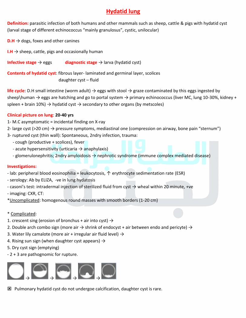
Hydatid lung
Definition: parasitic infection of both humans and other mammals such as sheep, cattle & pigs with hydatid cyst
(larval stage of different echinococcus “mainly granulosus”, cystic, unilocular)
D.H → dogs, foxes and other canines
I.H → sheep, cattle, pigs and occasionally human
Infective stage → eggs diagnostic stage → larva (hydatid cyst)
Contents of hydatid cyst: fibrous layer- laminated and germinal layer, scolices
daughter cyst – fluid
life cycle: D.H small intestine (worm adult) → eggs with stool → graze contaminated by this eggs ingested by
sheep\human → eggs are hatching and go to portal system → primary echinococcus (liver MC, lung 10-30%, kidney +
spleen + brain 10%) → hydatid cyst → secondary to other organs (by metscoles)
Clinical picture on lung: 20-40 yrs
1- M.C asymptomatic = incidental finding on X-ray
2- large cyst (>20 cm) → pressure symptoms, mediastinal one (compression on airway, bone pain “sternum”) 3- ruptured cyst (thin wall): Spontaneous, 2ndry infection, trauma:
- cough (productive + scolices), fever
- acute hypersensitivity (urticaria → anaphylaxis) - glomerulonephritis; 2ndry amyloidosis → nephrotic syndrome (immune complex mediated disease)
Investigations:
- lab: peripheral blood eosinophilia + leukocytosis, ↑ erythrocyte sedimentation rate (ESR)
- serology: Ab by ELIZA, -ve in lung hydatosis
- casoni’s test: intradermal injection of sterilized fluid from cyst → wheal within 20 minute, +ve - imaging: CXR, CT:
*Uncomplicated: homogenous round masses with smooth borders (1-20 cm)
* Complicated:
1. crescent sing (erosion of bronchus + air into cyst) → 2. Double arch combo sign (more air → shrink of endocyst + air between endo and pericyte) →
3. Water lily camalote (more air + irregular air fluid level) →
4. Rising sun sign (when daughter cyst appears) →
5. Dry cyst sign (emptying)
- 2 + 3 are pathognomic for rupture.
→ → → →
Pulmonary hydatid cyst do not undergoe calcification, daughter cyst is rare.
77
Management:
1- Aspiration → risk of rupture and anaphylaxis + recurrence
2- Pharmacological → mebendazole, albendazole (3-6 m):
- multiple cysts\recurrence
- small cysts
- poor surgical risk\refused
- after intra operative spillage of fluids
- C.I : large cysts, inactive or calcified, bone marrow suppression, pregnancy (primary term)
3- Surgical: the best curative method.
- albendazole + posteriolateral thoracotomy:
remove the cyst, lobectomy >50% occupying + post op albendazole
CASE: 2 hydatid cysts on Rt & Lt lungs, one ruptured and the other didn’t, from which one I start?
From the ruptured one to avoid complications like abscess, after 6 w I can do the other one.
CASE: 3 cysts (brain, lung, liver) from which one I start?
The one in brain due to pre-symptoms in skull (bony), then lung (high risk of rupture), then liver.
78
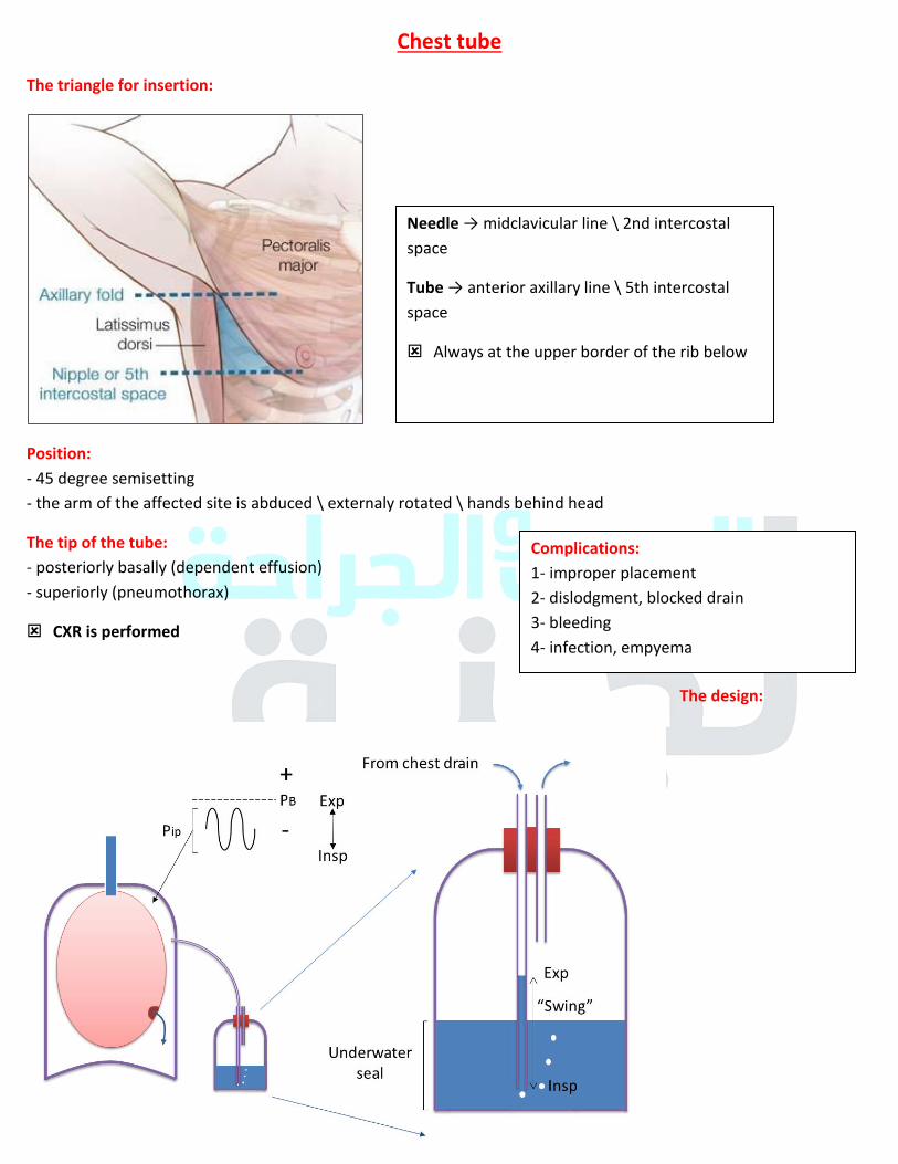
Chest tube
The triangle for insertion:
Position:
- 45 degree semisetting
- the arm of the affected site is abduced \ externaly rotated \ hands behind head
The tip of the tube:
- posteriorly basally (dependent effusion)
- superiorly (pneumothorax)
CXR is performed
The design:
Needle → midclavicular line \ 2nd intercostal
space
Tube → anterior axillary line \ 5th intercostal
space
Always at the upper border of the rib below
Complications:
1- improper placement
2- dislodgment, blocked drain
3- bleeding
4- infection, empyema
79
Varicose veins examination:
Inspection:
1- diameter and tortuosity → indications for the severity
2- skin color:
- red: thrombophlebitis
- brown: Lipodermatosclerosis
3- ulcers
Palpation:
1- edema (non-pitting)
2- tenderness (thrombophlebitis)
To detect the level of incompetent valve → (3 tunicate test) \ Trendelenburg test
- raising the leg and putting 3 tunicates (above & below knee, below inguinal ligament) and gradual release of
these turnicates
To examine deep vein patency → (Parthes test)
- raising the leg and putting the turnicate as high as possible then walking → if the symptoms of DVT appear → +ve
To examine the perforator vein at fixed sites → 5 – 10 – 15 – 20 cm above the medial malleolus (from the ankle)
80
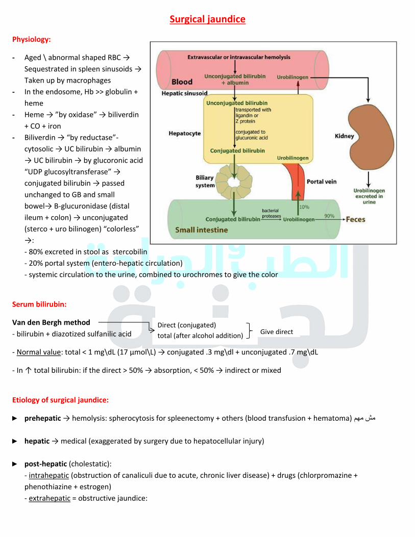
Surgical jaundice
Physiology:
- Aged \ abnormal shaped RBC →
Sequestrated in spleen sinusoids →
Taken up by macrophages
- In the endosome, Hb >> globulin +
heme
- Heme → ”by oxidase” → biliverdin + CO + iron
- Biliverdin → “by reductase”-
cytosolic → UC bilirubin → albumin → UC bilirubin → by glucoronic acid “UDP glucosyltransferase” → conjugated bilirubin → passed unchanged to GB and small
bowel→ B-glucuronidase (distal
ileum + colon) → unconjugated (sterco + uro bilinogen) “colorless” →: - 80% excreted in stool as stercobilin
- 20% portal system (entero-hepatic circulation)
- systemic circulation to the urine, combined to urochromes to give the color
Serum bilirubin:
Van den Bergh method
- bilirubin + diazotized sulfanilic acid
- Normal value: total < 1 mg\dL (17 μmol\L) → conjugated .3 mg\dl + unconjugated .7 mg\dL
- In ↑ total bilirubin: if the direct > 50% → absorption, < 50% → indirect or mixed
Etiology of surgical jaundice:
► prehepatic → hemolysis: spherocytosis for spleenectomy + others (blood transfusion + hematoma) مش مهم
► hepatic → medical (exaggerated by surgery due to hepatocellular injury)
► post-hepatic (cholestatic):
- intrahepatic (obstruction of canaliculi due to acute, chronic liver disease) + drugs (chlorpromazine +
phenothiazine + estrogen)
- extrahepatic = obstructive jaundice:
Direct (conjugated)
total (after alcohol addition) Give direct
81
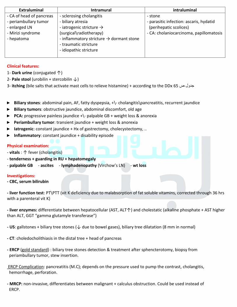
Extraluminal Intramural intraluminal
- CA of head of pancreas
- periambullary tumor
- enlarged LN
- Mirizi syndrome
- hepatoma
- sclerosing cholangitis
- biliary atresia
- iatrogenic stricture →
(surgical\radiotherapy)
- inflammatory stricture → dormant stone
- traumatic stricture
- idiopathic stricture
- stone
- parasitic infection: ascaris, hydatid
(perihepatic scolices)
- CA: cholaniocarcinoma, papillomatosis
Clinical features:
1- Dark urine (conjugated ↑) 2- Pale stool (urobilin + stercobilin ↓) 3- itching (bile salts that activate mast cells to relieve histamine) + according to the DDx 65جدول ص
► Biliary stones: abdominal pain, AF, fatty dyspepsia, +\- cholangitis\pancreatitis, recurrent jaundice
► Biliary tumors: obstructive jaundice, abdominal discomfort, old age
► PCA: progressive painless jaundice +\- palpable GB + weight loss & anorexia
► Periambullary tumor: transient jaundice + weight loss & anorexia
► Iatrogenic: constant jaundice + Hx of gastrectomy, cholecystectomy, ..
► Inflammatory: constant jaundice + disability episode
Physical examination:
- vitals : ↑ fever (cholangitis) - tenderness + guarding in RU + hepatomegaly
- palpable GB - ascites - lymphadenopathy (Virchow’s LN) - wt loss
Investigations:
- CBC, serum bilirubin
- liver function test: PT\PTT (vit K deficiency due to malabsorption of fat soluble vitamins, corrected through 36 hrs
with a parenteral vit K)
- liver enzymes: differentiate between hepatocellular (AST, ALT↑) and cholestatic (alkaline phosphate + AST higher than ALT, GGT “gamma glutamyle transferase”)
- US: gallstones + biliary tree stones (↓ due to bowel gases), biliary tree dilatation (8 mm in normal)
- CT: choledocholithiasis in the distal tree + head of pancreas
- ERCP (gold standard) : biliary tree stones detection & treatment after sphencterotomy, biopsy from
periambullary tumor, stew insertion.
ERCP Complication: pancreatitis (M.C); depends on the pressure used to pump the contrast, cholangitis,
hemorrhage, perforation.
- MRCP: non-invasive, differentiates between malignant + calculus obstruction. Could be used instead of
ERCP.
82
Treatment:
1- fluids
2- vit K
3- antibiotic
4- surgery to treat the cause
5- post-op mannitol to prevent hepatorenal shut-down due to bilirubin → RTN
- The initial investigation of RUQ pain is always US , even if there is suspension of cholangitis.
- When The LF is impaired: always the PT prolongs before PTT → factor 7 has the shortest half-life.
- Dilated CBD starts from 6mm or equal to portal vein diameter.
83
Polycystic liver disease “PCLD”
- Genetic disease, AD, 3rd
decade of life, mutation in PRD1, 2 gene
- 50% associated with polycystic kidney disease
- Development of multiple hepatic cysts (↑ in # and severity in females, advanced age, ↑with renal dysfunction)
Clinical:
- small cysts < 2 cm →asymptomatic
- large cyst → pain, distention, SOB, enlarged liver
Liver function is usually preserved unless in massive disease → ascites, variceal hemorrhage, encephalopathy.
Complications:
1- intra-cystic hemorrhage, infection, ruptures
2- RF → hemodialysis
Associations:
1- cerebral aneurysm
2- diverticulosis
3- mitral valve prolapse
4- inguinal hernia
Investigations:
- ↑ γ-glutamytransferase level, MRI, CT
Tx: the aim is to reduce liver volume:
1- medical – on study (Somatostatin analogue: Octreotide, lanreotide, ..)
2- sclerotherapy, laparoscopic fenestration
3- surgical:
- liver resection → if the cysts found in ta particular area (majority of them) - liver transplantation → definitive Tx
Hepatic cystadenoma:
- rare 5%, mainly in Rt lobe
- presented as abdominal pain\mass
- imaging: thick with soft tissue nodules and septation
- Tx: liver resection → risk of malignancy
84
Hydatid disease: Echinococcus granulosus
clinical:
1- asymptomatic
2- RUQ pain due to stretching with enlargement of liver
3- ruptured cyst:
- anaphylaxis → foreign hydatid protein
- secondary infection
- biliary obstruction: daughter cyst, scolices, pressure from outside
4- pulmonary symptoms: hydatid lung
Investigations:
1- eosinophilia 2- serology: ELIZA 3- casoni (not used anymore)
4- imaging:
- plain film of calcified cysts → inactive one
- US, CT: thick wall cyst +\- multiple daughter cysts (multiloculated cysts), floating membrane within cyst on
CT.
Management:
1- Albendazole, Mebendazole (15 mg\Kg\day) for at last 4 m.
2- PAIR (puncture, aspiration, installation of hypertonic saline\alcohol, re-aspiration)
3- surgical excision (the best):
- >10 cm \ complicated - daughter vesicles - superficial cyst → rupture
if ruptured: 1- post operative\pre\ through: Hydrocortisone therapy 2- post op Albendazole
Simple cystic disease
- M.C benign lesion in liver, F > M, solitary
- Biliary malformation (not communicate with biliary tree)
- Asymptomatic mainly → large mass, RUQ pain, Epigastric fullness
Dx:
- US, CT → well defines, no solid component, filled with homogenous fluid. - Laparoscopy → blue hue appearance
Tx:
- asymptomatic
- symptomatic: sclerotherapy → after aspiration \ rule put biliary, peritoneal communication.
Surgical excision then for pathology to exclude CA
85
Malignant lesions in liver
- M.C is secondary
Hepatocellular carcinoma:
M.C primary
without cirrhosis (poor prognosis)
R.F: any cause of liver cirrhosis (US every 6 months):
- HBV, HCV
- alcoholism, steatohepatitis (non-alcoholic)
- congenital biliary atresia, IEOM (Wilson, ..), tyrosinemia.
- aflatoxin
Signs & symptoms:
- enlarged abdomen (hepatomegaly \ ascites)
- pain in RUQ
- obstructive jaundice (pruritus, clay colored stool or melena)
- upper GI bleeding, hepatic encephalopathy
- wt loss
- Leser-Trelat sign: multiple seborrhoeic keratosis
- dermatomyocytis
- acute symptoms in peritonitis \ hypovolemic shock (ruptured)
Investigations:
- CBC (anemia from melena & GI bleeding, thrombocytopenia)
- KFT (prereanl failure, hepatorenal syndrome)
- LFT
- AFP → inversely correlate with survival rate
- electrolyte (Na → ascites) - bilirubin
- imaging: US, spiral CT, MRI, angiogram
Staging: TNM →
1→ solitary without vascular invasion 2 → solitary with vascular invasion
3 →multiple > 5 cm or invade major branch portal\hepatic vein
4 → to adjacent organ
Tx:
1- treat the symptoms of failure (ascites, HEP, GI bleeding)
2- resection: partial with 1-2 cm safe margin or total + transplantation
3- radio\chemo therapy
4- Sorafenib (tyrosine kinase inhibitor)
Milan criteria for liver transplant:
single < 5 cm or 2-3 small ones < 3 cm AND no vascular extra-hepatic invasion 86
Fibromellar carcinoma:
- Arising in non-cirrhotic liver
- Dx: biopsy, tumor marker CD68, Hep Par-1, cytokeratin 7.
- Don’t response to chemotherapy
Chlangiocarcinoma:
- 2nd
most common primary \ old aged women (obstructive jaundice + wt loss)
- Arise from epithelium of biliary tree from any site (typically from Rt, Lt hepatic duct “Klatskin tumors”)
R.F:
- primary sclerosing cholangitis
- hepatolithiasis
- parasitic → Clonorchis sinesis
- congenital abnormalities in biliary tree
- biliary papillomatosis
- HCV
- lynch syndrome
- thorotrast exposure
Tx:
- surgical excision: proximal: partial hepatectomy + reconstruction of biliary tree, distal:
pancreaticodoudectomy “whipple” (better prognosis) - chemo \ radio
Secondary \ metastatic: M.C
- From a primary colorectal (50%), stomach, breast, pancreas, lung, ..
Investigations:
- LFT, CEA, AFP, CA 19-9
- CT, MRI, US → multiple lesions involving both lobes.
Tx:
- surgical resection: local, segmental, hemi hepatectomy
- if diffuse → incurable
87
Burns and inhalational injury
Etiology:
- All age groups are involved
- M.C burn at home
- 50% of mortality due to inhalational injury
- <3 yrs → scalding (hot liquids) - 3 yrs → scalding + clothes catching fire + contact + electrocution + chemicals
- Teenagers + 17-65 yrs → domestic flames + electrocutions + industrial accidents
- Elderly → scalding
Mortality:
- incidence of burn-mortality accurately known and ↓ with burn centers
- Depends on: site, extent, age, depth, general condition
- M.C direct cause → uncontrolled shock + uncontrolled septicemia
Pathophysiology:
Local effect of burns: zones
1- 3 zones:
- coagulation zone = dead tissue
- zone of stasis = in between, if /(infection, prolonged hypotension) → dead
- zone of hyperemia = recovers for sure
2- Fluid loss:
- least severe forms → capillary dilatation + erythema
- deep zone → capillary permeability ↑ + damage → plasma to outside or encircled by blisters.
Systemic effect:
1- Hypotension, hypothermia
2- Sepsis
3- ↓Na+ (loss by plasma) → “later” ↑ Na
↑K+ (cell damage) → ↓K
4- Hemolysis
5- Renal failure (hypotension → RTN, hemoglubinemia + myoglubenemia) 6- Curling’s (stress) ulcer, erosive gastritis
7- Venous thrombosis
8- Respiratory failure (inhalation injury)
88
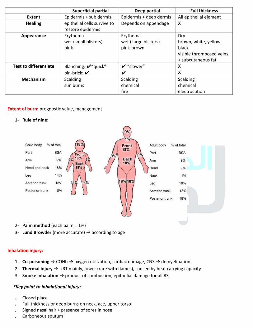
Superficial partial Deep partial Full thickness
Extent Epidermis + sub dermis Epidermis + deep dermis All epithelial element
Healing epithelial cells survive to
restore epidermis
Depends on appendage X
Appearance Erythema
wet (small blisters)
pink
Erythema
wet (Large blisters)
pink-brown
Dry
brown, white, yellow,
black
visible thrombosed veins
+ subcutaneous fat
Test to differentiate Blanching: ✔”quick”
pin-brick: ✔
✔ “slower” ✔
X
X
Mechanism Scalding
sun burns
Scalding
chemical
fire
Scalding
chemical
electrocution
Extent of burn: prognostic value, management
1- Rule of nine:
2- Palm method (each palm = 1%)
3- Lund Browder (more accurate) → according to age
Inhalation injury:
1- Co-poisoning → COHb → oxygen utilization, cardiac damage, CNS → demyelination
2- Thermal injury → URT mainly, lower (rare with flames), caused by heat carrying capacity
3- Smoke inhalation → product of combustion, epithelial damage for all RS.
*Key point to inhalational injury:
. Closed place
. Full thickness or deep burns on neck, ace, upper torso
. Signed nasal hair + presence of sores in nose
. Carboneous sputum 89
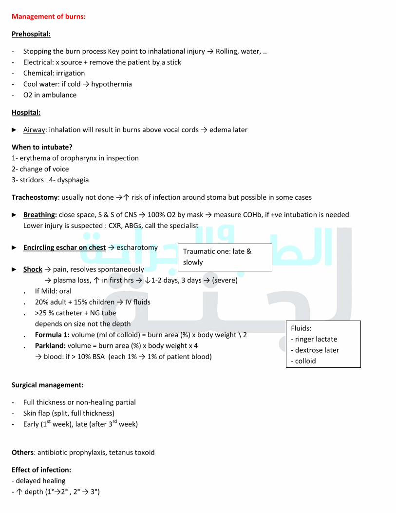
Management of burns:
Prehospital:
- Stopping the burn process Key point to inhalational injury → Rolling, water, ..
- Electrical: x source + remove the patient by a stick
- Chemical: irrigation
- Cool water: if cold → hypothermia
- O2 in ambulance
Hospital:
► Airway: inhalation will result in burns above vocal cords → edema later
When to intubate?
1- erythema of oropharynx in inspection
2- change of voice
3- stridors 4- dysphagia
Tracheostomy: usually not done →↑ risk of infection around stoma but possible in some cases
► Breathing: close space, S & S of CNS → 100% O2 by mask → measure COHb, if +ve intubation is needed
Lower injury is suspected : CXR, ABGs, call the specialist
► Encircling eschar on chest → escharotomy
► Shock → pain, resolves spontaneously → plasma loss, ↑ in first hrs → ↓1-2 days, 3 days → (severe)
. If Mild: oral
. 20% adult + 15% children → IV fluids
. >25 % catheter + NG tube
depends on size not the depth
. Formula 1: volume (ml of colloid) = burn area (%) x body weight \ 2
. Parkland: volume = burn area (%) x body weight x 4
→ blood: if > 10% BSA (each 1% → 1% of patient blood)
Surgical management:
- Full thickness or non-healing partial
- Skin flap (split, full thickness)
- Early (1st
week), late (after 3rd
week)
Others: antibiotic prophylaxis, tetanus toxoid
Effect of infection:
- delayed healing
- ↑ depth (1°→2° , 2° → 3°)
Traumatic one: late &
slowly
Fluids:
- ringer lactate
- dextrose later
- colloid
90
Local Tx:
1- Exposure method:
- cleaning by antiseptics continuously in dry, coolness, light exposure conditions
- dryness + crusts formation → bacterial invasion
- when to use: uncovered areas (perineum, face, one side) + hyperpyrexia patient.
2- Dressing:
- provide mechanical barrier to prevent bacteria
- to absorb the exudate + applying antibacterial agents
- how? Apply initial cleansing, antiseptics, non-adherent dressing
- reviewed every day and left in place 8-10 days
- when to remove?
1- ↑ amount of exudate
2- bad smelling
3- hotness + painful
3- Circumferential: encircling eschar → trunk (respiration) or limbs (circulation) 1. Monitoring the circulation by Doppler in the neck → escharotomy under anesthesia ** analgesia only in 3
rd degree
4- Bacteriological studies + infection
- collection of samples at admission, 3rd
, 4th
, pre-grafting, septicemia
- the count of bacteria ∝ with infection
- s & s of infection: purulent smelly discharge, pain, red margin
. Angry and un-healing looking burn → hemolytic strep
. Bluish green pus → pseudomonas (the most dangerous)
. Foul smelling discharge → proteus
Antiseptics:
- NCP (neomycin, chlorhexidine, polymyxin)
- sulfamylon
- flamazine (silver sulphadiazine)
- silver nitrate 5%
- soframycin
91
Skin cancer
Basal cell carcinoma:
- Most common one
- 40-8- yrs
- Head & neck
- Most common in eyelid
Risk factors:
1- Sun exposure
2- Advanced age
3- Immunosuppression: AIDS, organotransplant, medications
4- Carcinogen exposure: UV + ionizing radiation, arsenic, hydrocarbons
5- Albinism
6- Defective production of melanin (vetiligo, ..)
7- Fitz Patrick skin type: I, II, III
- I – white: always burns never tan
- II – white: usually burns, tan difficulty
- III – white: sometimes burns, tan average
- IV – moderate: brown rarely burn, tan with ease
- V – dark brown: very rarely burn, tan very easily
- VI – black: don’t burn, tan very easily
8- Genetic mutations: PTCH, P53, Ras
9- Gorlin’s syndrome: AD, multiple basal cells + odontogenic keratocysts mainly in mandible
10- Xeroderma pimentosum: (R.F for all skin cancers)
Originate: pluripoential epithelia cells of epidermis and hair follicles.
Types:
1- Nodular: head & neck, M.C type → exophytic, well defined borders, flesh colored. + {telangiectasia} مهمة
2- Superficial spreading: endophytic “flat”, pink, scaly patches on trunk
3- Micronodular: infiltrative, pigmented (زي العضة)
Diagnosis:
1- history (R.F) – examination
3- biopsy: excisional (small one), incisional (large & in sensitive area), taken from 4 regions.
- in both biopsies we should take a part of normal margins
Treatment:
- Medical: imiquimod, 5- fluorouracil
- Radiotherapy: rare to be used 92
- Destructive: curettage and electrodesiccation, cryosurgery, later phototherapy, photodynamic therapy.
. Primary surgical excision with safe margin → (4mm) if low risk, (10 mm) if high “recurrent, biopsy with poor features”
. after excision we send the tissue to pathology → margin involvement: if +ve we re-excision
. No need for LN removal
. Follow up 3 months later, then 6 months, by examination, LN ex.
Risk of recurrence:
Low risk High risk
Location, size < 20mm, trunk
<10 mm, check\scalp, forehead, neck
<6mm, central face, genitalia, hands, feet
>20
>10
>6
Defined borders Well defined Poorly defined
Primary\recurrent Primary Recurrent
Immunosuppression - +
Radiotherapy - +
Pathology Nodular Morpheaform, sclerosing (worse
prog.
Perineural involvement
“diagnosed by pathology”
- +
Rapidly growing - +
Depth <2mm >2 mm
Lymphovascular invasion - +
Degree of differentiation Well differentiated Poorly
Squamous cell carcinoma:
R.F as BCC
+ viral infection (HPV 16, 18 →mouth), HSV
+ marjolin’s ulcer
+ premalignant lesions: actinic keratosis, Bowen’s disease (10 % risk of malignancy)
Types: verrucous, ulcerative, marjolin’s ulcer*, subungual
Diagnosis:
- History (R.F)
- Examination + LN: CT scan for staging (> 2cm = T2, risk of regional mets)
- Biopsy subungual pathology
LN dissection:
- if there is no evidence for LN involvement we do sentinel LN biopsy, if +ve → dissection & radiotherapy. - if there is evidence → dissection
Treatment: - Surgical excision* - Frozen section (70% accuracy) -Follow up
Mohs surgery:
used in sensitive areas.
not available in Jordan
*marjolin’s ulcer → arise from chronic non-healed wound (burn scar, fistula,
chronic inflammation..), latent
period 1 yr-30 yrs.
*Surgical excision + LN with safe
margin in (4-6 mm up to 1 cm)
grossly (macroscopy)
-microscopy: +ve (10%) we remove
more from margins
93
Malignant melanoma
Congenital naevus cell naevi:
Giant hairy neavi:
- 20 cm, 5% of body surface area, 40% risk of malignancy so we need biopsy (incisional) + follow up
- Tx: follow up (difficult to remove)
Special types of naevi:
► Dysplastic naevi:
- >5 mm in diameter
- Irregular outline
► Malignant melanoma:
- Skin mainly → the least common in eye
- Female predominance (35-54 yrs)
- R.F:
1- history of changing mole \ multiple or atypical nevi
2- Large navus > 15 cm
3- White
4- Melanoma before age of 40 yrs
5- Regular tanning before the age of 30 yrs
6- History of non-melanotic skin cancer
**we should do them biopsy or dermatoscopy
- ABCDE of melanoma:
A: asymmetry B: border (irregular, not well defined) C: color (in the same nevi is mixed)
D: diameter > 6mm E: evolving (changes in size, color, shape)
- Types:
1- Superficial spreading melanoma: rapid spreading, M.C one
2- Nodular: vertical growth (depth), worst prognosis, risk of LN mets.
3- Lentigo malignant melanoma: sun damaged kin, irregular margin, slow development, dark in color
4- Acral lentinigos melanoma: poor prognosis, mainly in palms and soles
5- Amelanotic melanoma
- Diagnosis:
1- History (R,F)
2- Ex (ABCDE) + LN
3- Staging
4- Biopsy: excisional, punch (like a pencil, take biopsy from the depth), shave is not used
94
Breslow depth:
≤ 1.0 mm, > 1.0 -2.0 mm, >2-4 mm, > 4 mm
( I ) ( II ) ( III ) ( IIII )
Distance between glandular layer to the deepest point of melanoma
Directly related to survival
TNM staging:
1- AT as breslow (1-4)
T1a → without ulceration, mitosis < 1\mm2
T1b → with ulceration, mitosis > 1\mm2
mitosis - مش الulceration الباقي بس بال
2- Stage 3 → LN involvement
- Treatment:
. Surgical excision with safe margin according to the depth:
- .5 – 1 cm ←1 mm or less
- 1-2 cm ←1-2 mm
- 2 cm ←> 2mm
+ LN dissection if there is evidence of mets by ex, CT, US
.75 mm in depth without evidence of LN involvement by Ex, US, CT → sentinel LN biopsy → if +ve → dissection
- Melanoma can cause small bowel obstruction
by mets.
- Trunk, perineum, mucosa, scalp → have worse prognosis
95
Abdominal wall hernias
Definition: protrusion of viscous or part of viscous through abdominal opening.
Consists of sac (fundus, neck, body, mouth) , covering, content .
Epigastric hernia contains fat mainly.
Contents:
- Intestine (enterocele)
- Omentum (omentocele)
- Ovary +\- fallopian\bladder → urinary symptoms
- When the intestine involved in only portion of circumference → Richter
- Mickle’s diverticulum → Litter’s
- More than one loop → maydl’s - Sliding hernia → if the wall is part of content (like visceral peritoneum that contact with
By Ex:
- tympanic → intestine
- dull → feaces, omentum, fat.
According to reducibility:
1. Reducible: by surgeon, patient, itself
2. Irreducible: due to adhesion ns to the wall .
- Incarceration → when the lumen of colon occupying hernia blocked with feaces. 3. Obstructed: obstructed bowel without ischemia.
4. Strangulation: ischemia (-ve cough impulse, redness, hotness, pain)
According to the site:
- Epigastric → fat - paraumbilical\ umbilical.
Anatomy of abdominal wall:
1- Layers:
- skin, subcutaneous fascia (superficial “fatty” → camper’s \ deep “membranous” → scarpa’s → colle’s in the perineal region )
- muscles:
- external oblique → aponurosis → inguinal ligament
- internal oblique → cremastric muscle
- transversus abdominis
- rectus muscle
- pyramidal
- fascia transversalis \ peritoneal lining (parietal)
Arcuate line: where he rectus sheath in posterior wall of rectus muscle is absent so it will be in direct contact with
fascia transversalis.
Nerve supply: T7-T12 + L1 (iliohypogastric + ilioinguinal) 96
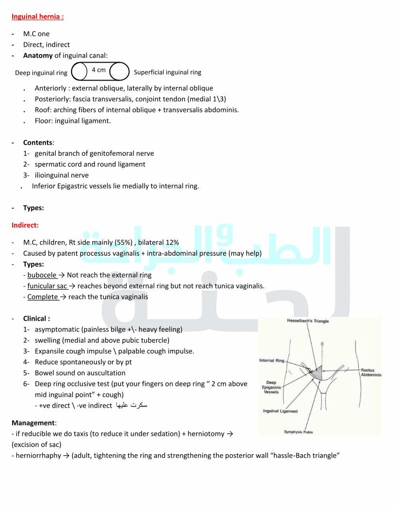
Inguinal hernia :
- M.C one
- Direct, indirect
- Anatomy of inguinal canal:
. Anteriorly : external oblique, laterally by internal oblique
. Posteriorly: fascia transversalis, conjoint tendon (medial 1\3)
. Roof: arching fibers of internal oblique + transversalis abdominis.
. Floor: inguinal ligament.
- Contents:
1- genital branch of genitofemoral nerve
2- spermatic cord and round ligament
3- ilioinguinal nerve
. Inferior Epigastric vessels lie medially to internal ring.
- Types:
Indirect:
- M.C, children, Rt side mainly (55%) , bilateral 12%
- Caused by patent processus vaginalis + intra-abdominal pressure (may help)
- Types:
- bubocele → Not reach the external ring
- funicular sac → reaches beyond external ring but not reach tunica vaginalis.
- Complete → reach the tunica vaginalis
- Clinical :
1- asymptomatic (painless bilge +\- heavy feeling)
2- swelling (medial and above pubic tubercle)
3- Expansile cough impulse \ palpable cough impulse.
4- Reduce spontaneously or by pt
5- Bowel sound on auscultation
6- Deep ring occlusive test (put your fingers on deep ring “ 2 cm above mid inguinal point” + cough) - +ve direct \ -ve indirect سكرت عليها
Management:
- if reducible we do taxis (to reduce it under sedation) + herniotomy → (excision of sac)
- herniorrhaphy → (adult, tightening the ring and strengthening the posterior wall “hassle-Bach triangle”
Superficial inguinal ring Deep inguinal ring 4 cm
97

Epigastric hernia:
- Hernia through the linea alba above the umbilicus due to sudden strain tearing
- May be :
1- fatty hernia of linea alba (extra-peritoneal fat)
2- true Epigastric hernia (omentum)
- Clinical: asymptomatic \ some Epigastric discomfort.
Umbilical hernia:
- More likely in infants and children
- Symptomless, increase with crying, conical shape
- Tx usually it disappear at 3 yrs, if above → surgery
Paraumbilical :
- F>M, multiparous, obese
- Defect of the linea alba just above the umbilicus
- Symptomatic → dragging pain, transient attacks of colic due to (adhesion between wall and loop), skin may
become reddened, excoriated, ulcerated, may have fistula. (thin skin and we may see the loop)
- Tx: it may become irreducible thus surgery advised in all cases.
1- Small→ herniorrhaphy
2- large → hernioplasty
3- additional lipectomy in case of large abdominal wall fatty.
Incisional hernia: 10%
- R.F:
1- cough, constipation, early retaining to strenuous exercise, difficult micturition
2- Infection, irradiation, malignancy, chemotherapy, steroid
3- Hematoma, seroma
4- Malnutrition, chronic systemic illness
5- Early post-op pregnancy
- Tx:
1- Wear abdominal support
2- Surgery → repair the defect \ mesh
Spigelian hernia:
- Occur in linea alba semi-lunaris (outer border of rectus muscle)
- Liable to strangulation → surgery
- May press the inferior Epigastric vessels.
98
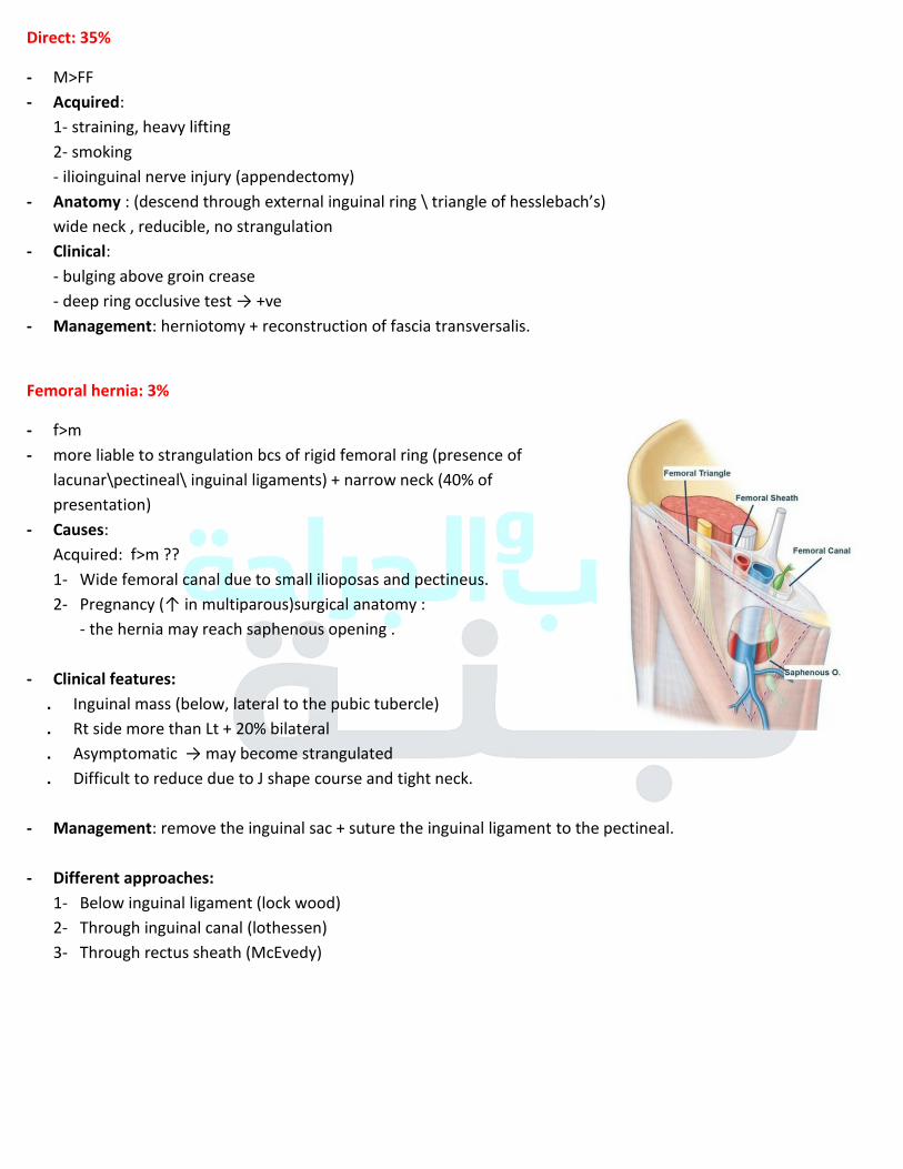
Direct: 35%
- M>FF
- Acquired:
1- straining, heavy lifting
2- smoking
- ilioinguinal nerve injury (appendectomy)
- Anatomy : (descend through external inguinal ring \ triangle of hesslebach’s) wide neck , reducible, no strangulation
- Clinical:
- bulging above groin crease
- deep ring occlusive test → +ve
- Management: herniotomy + reconstruction of fascia transversalis.
Femoral hernia: 3%
- f>m
- more liable to strangulation bcs of rigid femoral ring (presence of
lacunar\pectineal\ inguinal ligaments) + narrow neck (40% of
presentation)
- Causes:
Acquired: f>m ??
1- Wide femoral canal due to small ilioposas and pectineus.
2- Pregnancy (↑ in multiparous)surgical anatomy : - the hernia may reach saphenous opening .
- Clinical features:
. Inguinal mass (below, lateral to the pubic tubercle)
. Rt side more than Lt + 20% bilateral
. Asymptomatic → may become strangulated
. Difficult to reduce due to J shape course and tight neck.
- Management: remove the inguinal sac + suture the inguinal ligament to the pectineal.
- Different approaches:
1- Below inguinal ligament (lock wood)
2- Through inguinal canal (lothessen)
3- Through rectus sheath (McEvedy)
99
Neonatal jaundice
Epidemiology: 60% of term, 80% of preterm
- Mostly due to unconjugated bilirubin → medical condition
- R.F : as mentioned in pediatric lectures
- Physiological jaundice → 2-3rd
day → “relieved” 5-7 days
- Pathological jaundice → 1st day, beyond 2 weeks, direct
Causes:
1- Obstructive:
- biliary atresia (M.I), surgical cause
- choledochal cyst
- inspissated bile syndrome (cystic fibrosis, hemolysis, parenteral nutrition)
2- Hemolytic (ABO, rhinocompitaility, spherocytosis)
3- Metabolic disorders (α1 antitrypsin, galactosemia)
4- Congenital infection (syphilis, TORCH)
Biliary atresia:
- Rare
- Characterized by fibroproleferation of biliary tree, may be : (hepatofibrosis, cirrhosis, ESLF)
- Involve any part of biliary tree
- Causes: multifactorial:
- arrest in development
- fc gene (BA + splenic malformation)
- perinatal exposure to REO\ROTAVIRUS
- Association:
1- Intestinal malrotation
2- Predoudenal portal vein (posteriorly to duodenum → may lead to rare duodenal obstruction)
3- Polysplenia (multiple small accessory spleen)
4- Interrupted IVC (below hepatic vein which directly drain to Rt atrium) with azygos continuation (drain the
lower limb).
- Classification: according to JAP:
I → CBD
IIA → common hepatic duct (CHD) IIB → CBD, CHD, cystic duct
III → (>90% M.C) ALL at level of parahepatics.
100
- Signs & symptoms:
. Pale stool
. Dark urine
. Progressive jaundice
. Hepatomegaly
. when advanced → (hepatospleenomegaly, ascites, varices)
- histology: inflammatory changes + fibrous tissue of parenchyma + duodenal proliferation
- DDx: biliary atresia, choledochal cyst, inspissated bile syndrome, neonatal hepatitis.
- Dx:
1- Lab tests:
- bilirubin (total, direct) → direct + indirect ↑
- ALP → obstruction
- GGTP → more specific ↑
- LFT → albumin\clotting → normal - infection work up → TORCH
- α1 antitrypsin screening
2- Imaging:
- US:
a) absent gallbladder (highly suggestive)
b) intrahepatic biliary tree (not dilated) مهم
- nuclear medicine scan \ HIDA scan (99-Te) “hepatobiliary iminodiacetic acid” : normal hepatic reuptake but no excretion even after 24 hrs + bladder (isotope go to circulation)
- cholangiogram: hypoplasia of extrahepatic biliary system.
3- Surgical exploration (END Diagnostic)
- Tx:
. Type I → patent proximal hepatic bile duct → direct Roux en-Y- hepato jejunostomy (hepatic duct to jejunum)
. Types II \ III → X → kasai procedure (before age of 8 weeks)
- radical excision of all bile tree up to liver capsule
- Roux en-Y
- portoenterostomy
- liver biopsy
+ antibiotics, drugs to promote the flow, 10 days hospital stay.
- Complications post op: bacterial cholangitis, attacks of hepatic fibrosis, portal HTN.
. Liver transplant: if all above not successful
101

Choledochal cyst:
- Congenital cystic dilatation of intra +\- enterohepatic biliary system.
- F>M
- More common in Asia
- Majority diagnosed at first decade of life and others later. 20 % prenataly.
- Cause: pancreatobiliary maljunction
- Clinical:
- Adult form: jaundice, pain, palpable mass.
- Prenatal: cystic mass in abdomen
- infant: jaundice\hepatomegaly
- adulthood\childhood: ascending cholangitis, OJ.
102
Corrosive esophageal injury
Anatomy of esophagus :
- 25 cm in length
- Striated at the beginning smooth
- Squamous epithelium
- Sphincters : upper & lower
- Three strictures ( foreign body may stuck on it ) :
1 – cricopharyngeus 2 – aortic arch 3 – lower esophagus
- Nerve supply :
vagus that has synaptic connections to myenteric ( Auerbach's ) plexus
xx Meissner's submucosal plexus
- Blood supply :
1 – Inferior thyroid artery cervical esophagus
2 – Bronchial arteries Proximal descending thoracic aorta
3 – Branches from aorta distal descending thoracic aorta
4 – Inferior phrenic artery + left gastric artery abdominal aorta
Epidemiology of injury ( corrosive ) :
- Most common in children < 3 yr. , Boys .
- May in > 5 yr. & adolescent girls ( intentional )
Cause :
according to the physical form of the substance site / type of the esophagus injury
Alkali Acidic
Ph > 7 ( > 12 sever ) Ph < 7 ( < 2 sever )
Hydrates , Carbonates ( Na , K+ ) organic / inorganic
Mainly injures upper esophagus alkali will
neutralized in stomach , so this limit the injury
More lower esophagus + gastric injury
Tasteless , odorless larger amounts Pungent odor and Nexus taste
Liquefecation necrosis direct extention +
saponification of fats + dehydration &thrombosis of
blood vessel , deeper injuries
Coagulative necrosis coagulum that limit the depth
of injury
no eschar formation or afriable eschar Hard eschar that limited injury
Severity :
amount , concentration , form ( solid , liquid ) , duration of contact with mucosa .
TZ
103
Clinical features :
Common retrosternal or epigastric pain
oropharyngeal injury
dysphagia , odynophagia
Hypersalivation
burn to epiglottis + Larynx Hoarseness , Stridor , Aphonia , Respiratory distress
Perforation / Peritonitis Tenderness rigidity
Phases :
1 ( Acute necrosis ) 1 – 4 Ds
2 ( Ulceration + granulation ) 4 – 12 Ds
3 ( Cicatrization + scarring ) 3W – 6 M
Investigation ( Imaging ) : + ( CBC , electrolyte , BUN , ABG , KFT , LFT )
1 – CXR : مالحظة : شوفوا الصور من الساليد - Pneumomediastinum
- pleural effusion , aspiration pneumonitis - foreign body ( button battery …. ) - signs of mediastinum
2 - Abdominal radiograph : pneumoperitoneum , foreign body
3 – Esophagram fluoroscope ( water soluble contrast ) :
- Acute mild Atonic dilated Aperistaltic + multiple shallow ulcers
- Acute severe extensive ulceration + narrowing
- Chronic Large or short segments strictures + proximal part of stomach pulled to chest
4 - CT scan : - circumferential esophagus wall thickening > 5 mm
- pnemomediastinum / plural effusion
- stricture in chronic phase
Grades :
- 1 no swelling
- 2 edematous wall without periesophageal soft tissue involvement
- 3 edematous wall with periesophageal soft tissue involvement but well demarcated tissue interface
- 4 edematous wall with periesophageal soft tissue involvement but blurred interface Or collection of fluid
around descending aorta + esophagus
5 – Endoscopy :
- must in first 24 hours after digestion
- extent of damage , progress , guide the therapy
- risk of perforation
- Contraindication : 1 - hemodynamic instability
2 - evidence of perforation
3 - severe respiratory distress
4 - oropharyngeal or edema intubation
- Staging : 0 normal
I mucosal edema + hyperemia
II A - superficial ulcers , bleeding , exudate
B - deep ulcers ( circumferential / Local )
III multiple deep ulcers + necrosis
Perforation mortality rate 65% surgery
6 – Endoscopic US : no evidence to use it but :
- if the muscle layer is interact no strictures will forms
- if involved strictures so we do balloon dilatation
Good prognosis / not need treatment
Need Tx , developed strictures :
70 to 100%
104
* PH testing of saliva product to know if it alkaline or acidic + how much PH
( but neutral pH not rule out ingestion )
Management :
For all :
- ABC
- NPO + IV fluids + PPI ( in acidic ) + painkillers
- CXR , abdominal radiograph
- Endoscope in first 24-hour
- observation
* Steroid / antibiotic are contraindicated .
* Asymptomatic / low concentration and amount / tolerating a normal diet Follow up in OPD
Avoid :
- emetic ( re exposes ) - neutralization agent :thermal injury
- gastric lavage : esophageal perforation , aspiration
- activated charcoal : poor absorption + endoscopy interference
Complications and its treatment :
Early : - airway edema and obstruction intubation
- upper GI bleeding
- Perforation mediastinitis , pericarditis , pleuritis , TE fistula , E Aortic fistula , peritonitis
Late : strictures , CA
Strictures :
- Balloon / Bougies dilators in tight and fibroticon
Gradual deterioration until reach effective safe dilation ( 3 - 4 W interval ) - Esophageal stent : - SEMS metal
- plastic
- biodegradable
- Surgery : in non-responsive cases / harmful dilatation
parietal , total esophagoectomy + gastric pull up or colonic interposition
Cancer :
- at 1 – 3 decades
- Adeno / squamous cell carcinoma - need endoscopy screening
Esophageal foreign bodies :
Symptoms : dysphagia , poor feeding , drooling , irritability , Stridor , choking , cough
Worke up : - CXR + cervical spine + soft tissue ( AP , lateral View )
- Abdominal x-ray
- Contrast esophageogram in non-opaque objects .
Treatment : - removal by forceps basket + intubation or over tube placed in esophagus
to prevent aspiration
- button batteries / sharp objects endoscopy
105