PROTEIN - Web UPI Official
51
PROTEIN
Transcript of PROTEIN - Web UPI Official

PROTEIN
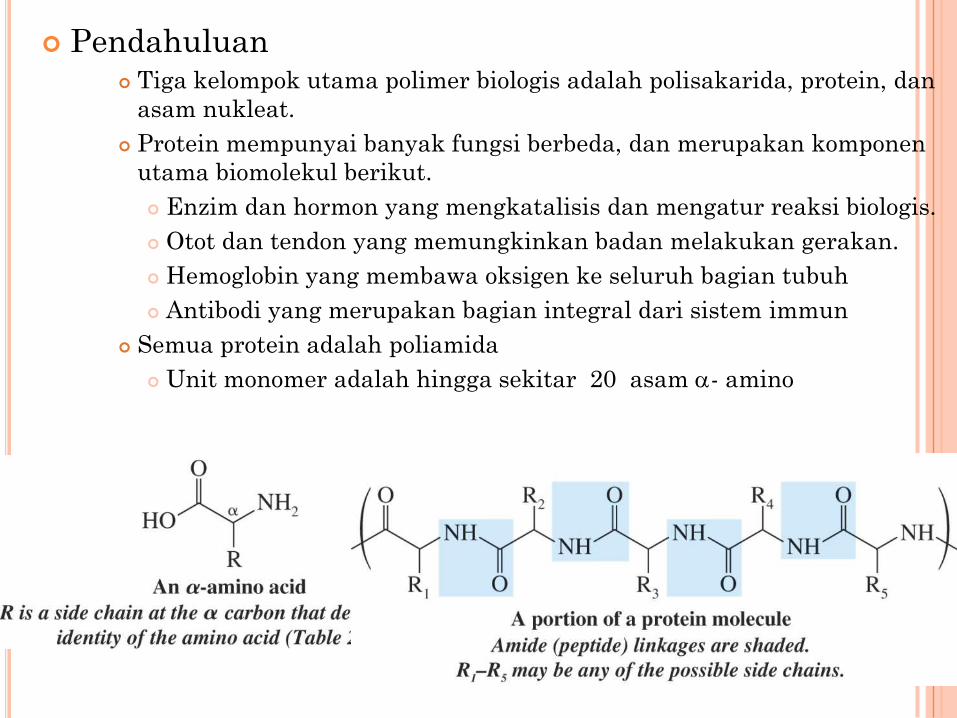
Pendahuluan Tiga kelompok utama polimer biologis adalah polisakarida, protein, dan
asam nukleat.
Protein mempunyai banyak fungsi berbeda, dan merupakan komponen
utama biomolekul berikut.
Enzim dan hormon yang mengkatalisis dan mengatur reaksi biologis.
Otot dan tendon yang memungkinkan badan melakukan gerakan.
Hemoglobin yang membawa oksigen ke seluruh bagian tubuh
Antibodi yang merupakan bagian integral dari sistem immun
Semua protein adalah poliamida
Unit monomer adalah hingga sekitar 20 asam a- amino
2

Proteins mempunyai beberapa level struktur
Struktur primer merujuk pada urutan asam amino sepanjang rantai
protein.
Struktur sekunder dan tersier merujuk pada bengkokan dan lipatan
struktur primer.
Struktur kuarterner merujuk pada aggregasi lebih dari satu rantai
protein.
Semua asam amino kecuali glisin adalah kiral dan mempunyai
konfigurasi L (sesuai gliseraldehida) pada karbon a.
3
Asam Amino
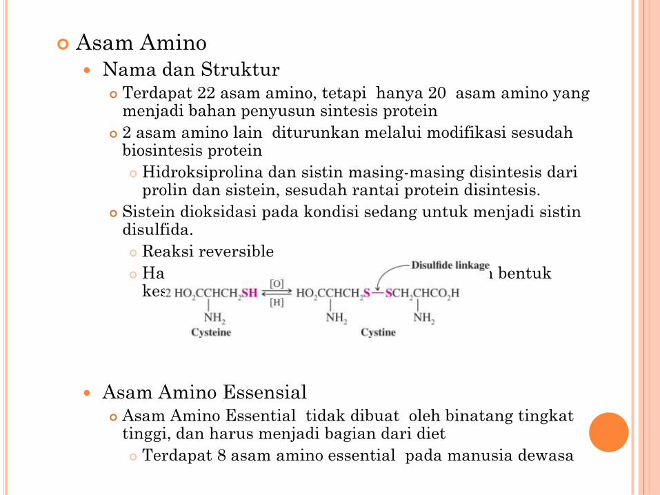
Nama dan Struktur Terdapat 22 asam amino, tetapi hanya 20 asam amino yang
menjadi bahan penyusun sintesis protein
2 asam amino lain diturunkan melalui modifikasi sesudahbiosintesis protein
Hidroksiprolina dan sistin masing-masing disintesis dariprolin dan sistein, sesudah rantai protein disintesis.
Sistein dioksidasi pada kondisi sedang untuk menjadi sistindisulfida.
Reaksi reversible
Hal tersebut penting untuk mempertahankan bentukkeseluruhan protein.
Asam Amino Essensial Asam Amino Essential tidak dibuat oleh binatang tingkat
tinggi, dan harus menjadi bagian dari diet
Terdapat 8 asam amino essential pada manusia dewasa4
5

6

7
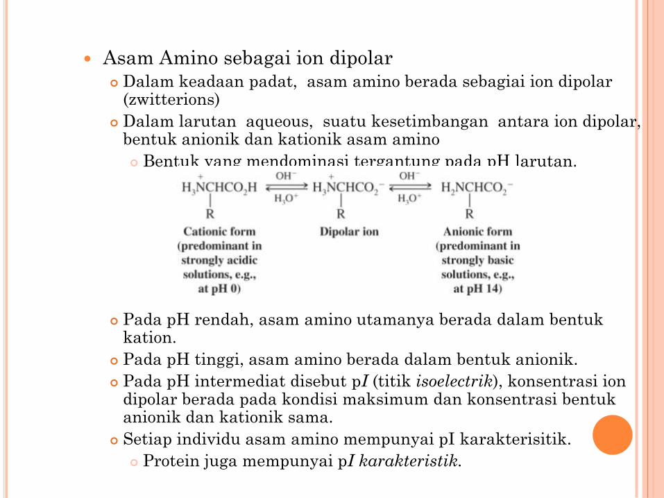
Asam Amino sebagai ion dipolar Dalam keadaan padat, asam amino berada sebagiai ion dipolar
(zwitterions)
Dalam larutan aqueous, suatu kesetimbangan antara ion dipolar, bentuk anionik dan kationik asam amino
Bentuk yang mendominasi tergantung pada pH larutan.
Pada pH rendah, asam amino utamanya berada dalam bentukkation.
Pada pH tinggi, asam amino berada dalam bentuk anionik.
Pada pH intermediat disebut pI (titik isoelectrik), konsentrasi ion dipolar berada pada kondisi maksimum dan konsentrasi bentukanionik dan kationik sama.
Setiap individu asam amino mempunyai pI karakterisitik.
Protein juga mempunyai pI karakteristik.8

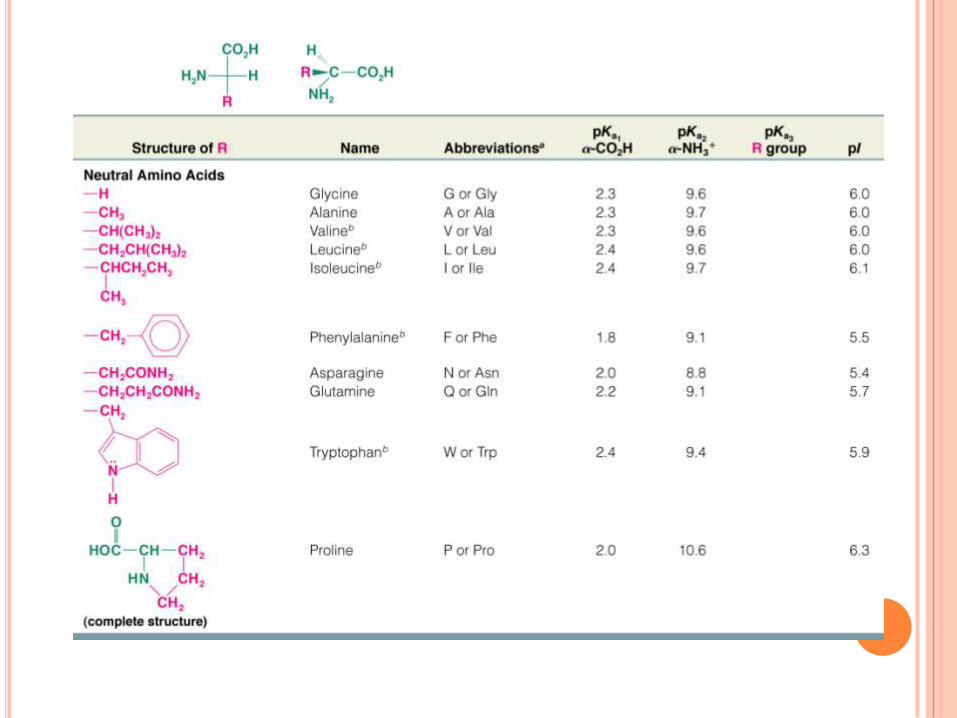
Asam amino alanina mempunyai rantai samping netral dan dapatdigunakan untuk menggambarkan sifat-sifat dasar asam amino pada berbagai pH.
Pada pH rendah alanin ada sebagai kation.
pKa1 alanina (untuk ionisasi proton asam karboksilat) adalah2,3, pertimbangkan lebih rendah dari pKa asam karboksilatnormal.
pKa2 alanina (untuk ionisasi proton dari asam amino terprotonasi) adalah 9,7
Titik Isoelektrik, pI, untuk alanina adalah rata-rata dari duanilai, yaitu (pKa1 + pKa2)/2
9

Jika basa perlahan-lahan ditambahkan ke alanina terprotonasipenuh, pH yang dicapai adalah setengah gugus asam karboksilatterdeprotonasi.
pH 2.3 adalah nilai pKa1
Persamaan Henderson-Hasselbach memperkirakan hasil ini.
Makin banyak basa ditambahkan, pI tercapai dan molekulbermuatan listrik netral; titik tersebut tercapai jika satu ekivalenbasa secara eksak ditambahkan.
Makin banyak basa ditambahkan dan pH 9,7 tercapai, gugusamonium akan terdeprotonasi.
Penambahan lebih banyak basa akan menghasilkan asam amino anionik.
10
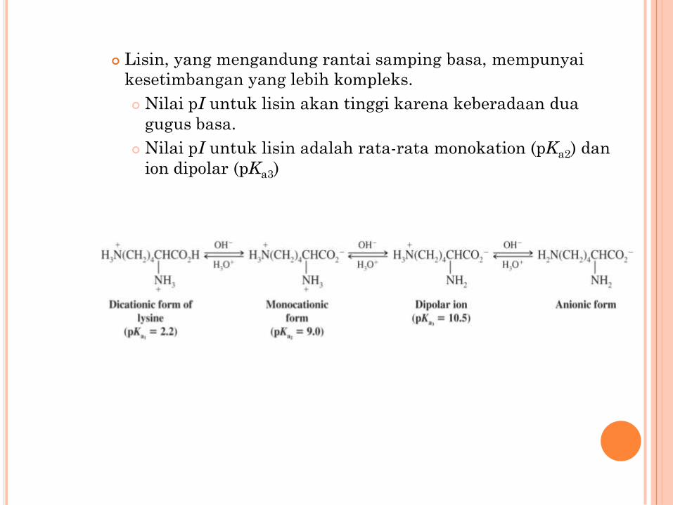
Lisin, yang mengandung rantai samping basa, mempunyai
kesetimbangan yang lebih kompleks.
Nilai pI untuk lisin akan tinggi karena keberadaan dua
gugus basa.
Nilai pI untuk lisin adalah rata-rata monokation (pKa2) dan
ion dipolar (pKa3)
11

Sintesis Asam a-Amino
Tiga metode pertama menghasilkan campuran rasemik.
Amminolisis langsung asam a-halo
Hasil cenderung rendah.
Dari Kalsium Phthalimida
Ini adalah variasi sintesis Gabriel dan hasilnya
biasanya tinggi.
12

Sintesis Strecker
Reaksi aldehida dengan ammonia dan hidrogen sianida
menghasilkan a-aminonitril yang dapat terhidrolisis menjadi
asam a-amino
Reaksi berlangsung melalui suatu intermediet imina
13
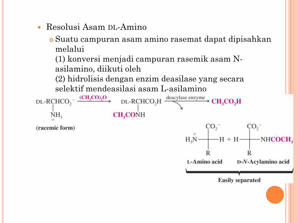
Resolusi Asam DL-Amino
Suatu campuran asam amino rasemat dapat dipisahkan
melalui
(1) konversi menjadi campuran rasemik asam N-
asilamino, diikuti oleh
(2) hidrolisis dengan enzim deasilase yang secara
selektif mendeasilasi asam L-asilamino
14
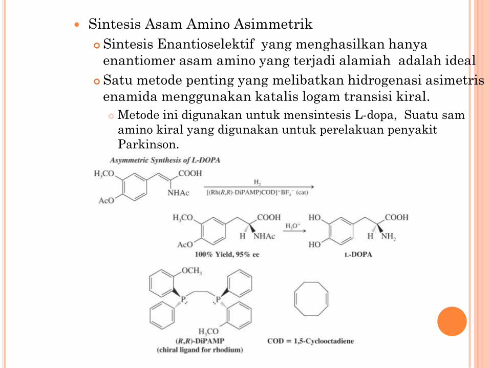
Sintesis Asam Amino Asimmetrik
Sintesis Enantioselektif yang menghasilkan hanya
enantiomer asam amino yang terjadi alamiah adalah ideal
Satu metode penting yang melibatkan hidrogenasi asimetris
enamida menggunakan katalis logam transisi kiral.
Metode ini digunakan untuk mensintesis L-dopa, Suatu sam
amino kiral yang digunakan untuk perelakuan penyakit
Parkinson.
Chapter 24 15

Suatu metode serupa digunakan untuk mensintesis (S)-
fenilalanina, yang diperlukan untuk pembuatan Aspartam
16

Polypeptida dan Protein Enzim mempolimerisasi asam amino melalui pembentukan
hubungan amida.
Polimer disebut suatu peptida dan hubungan peptida disebut
ikatan peptida.
Setiap asam amino dalam peptida disebut residu asam amino.
17

Polipeptida biasanya ditulis dengan residu N-terminal ke kiri.
Tiga huruf awal biasanya digunakan sebagai singkatan
asam amino yang menunjukkan urutan polipeptida.
18

Hidrolisis
Suatu polipeptida dapat dihidrolisis melalui proses refluks dengan
HCl 6 M selama 24 jam.
Masing-masing asam amino dapat dipisahkan satu sama lain
menggunakan resin penukar kation.
Suatu larutan asam amino yang bersifat asam dilewatkan
melalui kolom pertukaran kation, kekuatan adsorpsi bervariasi
tergantung kebasaan setiap asam amino (yang paling basa
bertahan paling kuat).
Pencucian kolom dengan larutan buffer berurutan menyebabkan
asam amino bergerak dengan kecepatan berbeda.
19
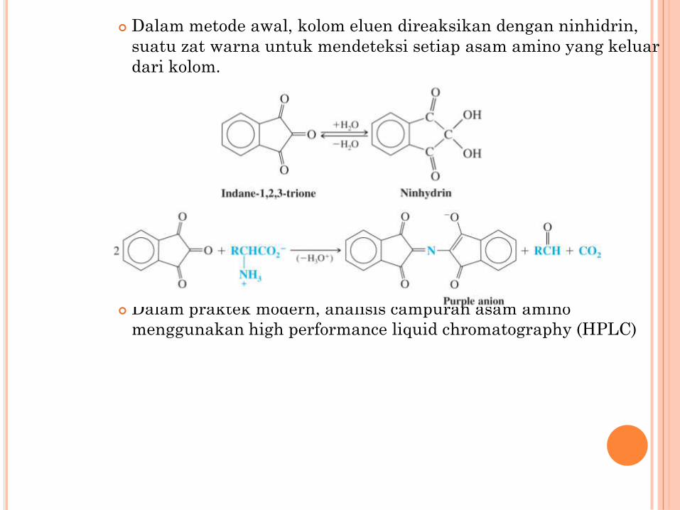
Dalam metode awal, kolom eluen direaksikan dengan ninhidrin,
suatu zat warna untuk mendeteksi setiap asam amino yang keluar
dari kolom.
Dalam praktek modern, analisis campuran asam amino
menggunakan high performance liquid chromatography (HPLC)
20
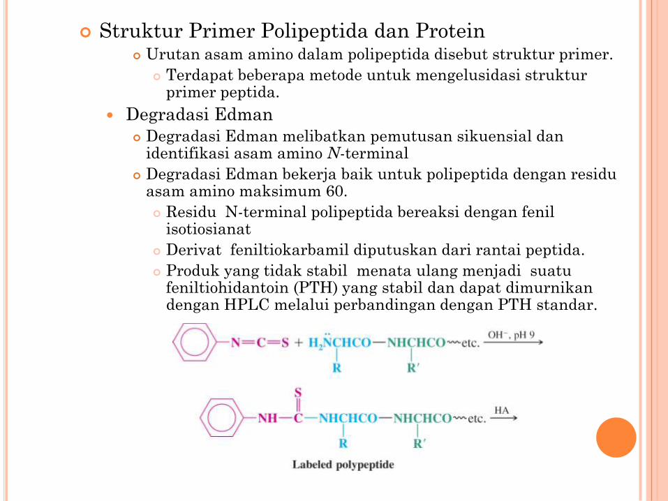
Struktur Primer Polipeptida dan Protein Urutan asam amino dalam polipeptida disebut struktur primer.
Terdapat beberapa metode untuk mengelusidasi strukturprimer peptida.
Degradasi Edman
Degradasi Edman melibatkan pemutusan sikuensial danidentifikasi asam amino N-terminal
Degradasi Edman bekerja baik untuk polipeptida dengan residuasam amino maksimum 60.
Residu N-terminal polipeptida bereaksi dengan fenilisotiosianat
Derivat feniltiokarbamil diputuskan dari rantai peptida.
Produk yang tidak stabil menata ulang menjadi suatufeniltiohidantoin (PTH) yang stabil dan dapat dimurnikandengan HPLC melalui perbandingan dengan PTH standar.
21

Analisis N-Terminal Sanger Ujung N-terminal polipeptida dilabeli dengan 2,4-
dinitrofluorobenzena, dan polipeptida dihidrolisis.
Asam amino N-terminal yang terlabeli dipisahkan daricampuran dan diidentifikasi.
Metode Sanger tidak digunakan secara luas seperti metodeEdman.
22

Analisis C-Terminal
Enzim karboksipeptida menghidrolisis asam amino C-terminal
secara selektif.
Enzim melanjutkan untuk membebaskan setiap asam
amino C-terminal yang terhidrolisis; perlu untuk
memonitor asam amino C-terminal sebagai fungsi waktu
untuk mengidentifikasinya.
23

Complete Sequence Analysis
The Sanger and Edman methods of analysis apply to short polypeptide sequences (up to about 60 amino acid residues by Edman degradation)
For large proteins and polypeptides, the sample is subjected to partial hydrolysis with dilute acid to give a random assortment of shorter polypeptides which are then analyzed
The smaller polypeptides are sequenced, and regions of overlap among them allow the entire polypeptide to be sequenced
Example: A pentapeptide is known to contain the following amino acids:
Using DNFB and carboxypeptidase, the N-terminal and C-terminal amino acids are identified
The pentapeptide is subjected to partial hydrolysis and the following dipeptides are obtained
The amino acid sequence of the pentapeptide must be:
Chapter 24 24

Larger polypeptides can also be cleaved into smaller sequences using site-specific reagents and enzymes
The use of these agents gives more predictable fragments which can again be overlapped to obtain the sequence of the entire polypeptide
Cyanogen bromide (CNBr) cleaves peptide bonds only on the C-terminal side of methionine residues
Mass spectrometry can be used to determine polypeptide and protein sequences
“Ladder sequencing” involves analyzing a polypeptide digest by mass spectrometry, wherein each polypeptide in the digest differs by one amino acid in length; the difference in mass between each adjacent peak indicates the amino acid that occupies that position in the sequence
Mass spectra of polypeptide fragments from a protein can be compared with databases of known polypeptide sequences, thus leading to an identification of the protein or a part of its sequence by matching
Chapter 24 25

Examples of Polypeptide and Protein Primary
Structure
Oxytocin and Vasopressin
Oxytocin stimulates uterine contractions during childbirth
Vasopressin causes contraction of peripheral blood vessels and
a resultant increase in blood pressure
The two polypeptides are nonapeptides and differ in only 2
amino acid residues
Chapter 24 26

Chapter 24 27

Insulin
Insulin is a hormone which regulates glucose metabolism
Insulin deficiency in humans is the major cause of diabetes
mellitus
The structure of bovine insulin (shown below) was
determined in 1953 by Sanger
Human insulin differs from bovine insulin at only three
amino acids in its sequence
Chapter 24 28

Cha
pte
r 24
29
Polypeptide and Protein SynthesisLaboratory synthesis of polypeptides requires orchestration of
blocking and activating groups to achieve selective amide bond
formation Amino groups must be blocked until their reactivity as a nucleophile is desired
Carboxylic acid groups must be activated for acyl substitution at the appropriate
time
Amino groups are usually blocked using one of the following: A benzyloxycarbonyl group (a “Z” group)
A di-tert-butyloxycarbonyl group (a “Boc” group)
An 9-fluorenylmethoxycarbonyl group (an “Fmoc” group)
Methods for installing and removing Z, Boc, and Fmoc groups are
shown below:

Cha
pte
r 24
30
Methods for installing and removing Z, Boc, and Fmoc groups are
shown below:

Cha
pte
r 24
31
Carboxylic acid groups are usually activated by
conversion to a mixed anhydride: Ethyl chloroformate can be used

Cha
pte
r 24
32
An Example of Laboratory Peptide Synthesis:Synthesis of Ala-Leu

Cha
pte
r 24
33
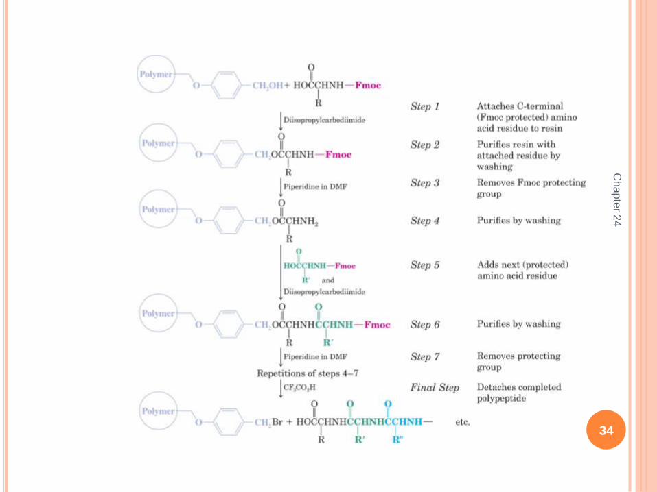
Automated Peptide SynthesisSolid Phase Peptide Synthesis (SPSS) was invented by R. B.
Merrifield, for which he earned the Nobel Prize in 1984
SPSS involves „growing‟ a peptide on a solid polymer bead by
sequential cycles of amide bond formation
The peptide is cleaved from the bead when the synthesis is
complete
SPSS is used in commercial peptide synthesis machines Peptides dozens of residues in length can be synthesized automatically
A landmark example is synthesis of ribonuclease, having 124 amino acid residues
Cha
pte
r 24
34

Secondary, Tertiary, and Quaternary Structures of
Proteins
Secondary Structure
The secondary structure of a protein is defined by local
conformations of its polypeptide backbone
These local conformations are specified in terms of regular
folding patterns such as helices, pleated sheets, and turns
The secondary structure of a protein is determined by the
sequence of amino acids in its primary structure
Key to secondary structure is that peptide bonds assume a
geometry in which all 6 atoms of the amide linkage are trans
coplanar
Chapter 24 35

Coplanarity results from contribution of the second resonance
form of amides, in which there is considerable N-C double
bond character
The carbon with attached R groups between the amide
nitrogen and the carbonyl group has relatively free rotation
and this leads to different conformations of the overall chain
Two common secondary structure are the b-pleated
sheet and the a-helix
In the b-pleated sheet, a polypeptide chain is in an extended
conformation with groups alternating from side to side
Chapter 24 36

The extended polypeptide chains in b-pleated sheets form
hydrogen bonds to adjacent polypeptide chains
Slight bond rotations are necessary between amide groups
to avoid unfavorable steric interactions between peptide
side chains, leading to the pleated structure
The b-pleated sheet is the predominant structure in silk
fibroin
Chapter 24 37

The a-helix is the most important protein secondary structure
a-Helices in a polypeptide are right-handed with 3.6 amino
acid residues per turn (See figure 24.11 page 1198)
The amide nitrogen has a hydrogen bond to an amino acid
carbonyl oxygen that is three residues away
The R groups extend away from the axis of the helix
a-Helices comprise the predominant secondary structure of
fibrous proteins such as myosin (in muscle) and a-keratin (in
hair and nails)
There are other secondary structures that are more difficult to
describe
Examples are coil or loop conformations and reverse turns
or b bends
Chapter 24 38

Carbonic Anhydrase
The structure of the enzyme carbonic anhydrase is shown in
Figure 24.12,page 1198
Alpha helices are in magenta and strands of b-pleated
sheets are in yellow
The mechanism of carbonic anhydrase reaction was
discussed in Chapter 3
Chapter 24 39

Tertiary Structure
The tertiary structure of a protein is the three-dimensional
shape which results from further folding of its polypeptide
chains
This folding is superimposed on the folding caused by its
secondary structure
In globular proteins, the folding in tertiary structures exposes
the maximum number of polar (hydrophilic) side chains to the
aqueous environment, making most globular proteins water
soluble
The folding also serves to enclose a maximum number of
nonpolar (hydrophobic) side chains within the protein
interior
Tertiary structures are stabilized by forces including hydrogen
bonding, disulfide bonds, van der Waals forces, and ionic
attractions
Chapter 24 40

Myoglobin
The globular protein myoglobin transports oxygen within
muscle tissues (See Figure 24.13, page 1200)
Myoglobin has an associated non-polypeptide molecule
called heme (shown in gray)
The heme group is the site of oxygen binding
Chapter 24 41
Cha
pte
r 24
42
Quaternary Structure
The overall structure of a protein having multiple subunits is
called its quaternary structure Not all proteins have quaternary structure
Hemoglobin
Hemoglobin is a globular protein that transports oxygen in the
blood
Hemoglobin contains four polypeptide subunits (2 designated a,
and 2 designated b) (See Figure 24.21, page 1210) The a subunits are shown in blue and green; b subunits are shown in yellow and
cyan
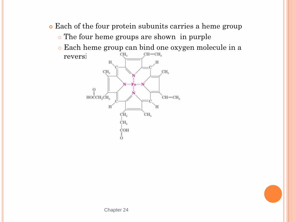
Each of the four protein subunits carries a heme group
The four heme groups are shown in purple
Each heme group can bind one oxygen molecule in a
reversible complex
Chapter 24 43

Cha
pte
r 24
44
Introduction to Enzymes
Most enzymes are proteins
Enzymes can catalyze reactions by a factor of 106-1012
Enzymes have very high specificity for their respective substrates
(reactants)
Enzymatic reactions take place in the active site of each enzyme The structure of the active site facilitates binding and catalysis
Enzymes sometimes require a cofactor or coenzyme A cofactor can be a metal ion (e.g., Zn+2, Mg+2) bound at the active site
A coenzyme is a small organic molecule bound at the active site that becomes
chemically changed during the enzymatic reaction (e.g., NAD+)
Cha
pte
r 24
45
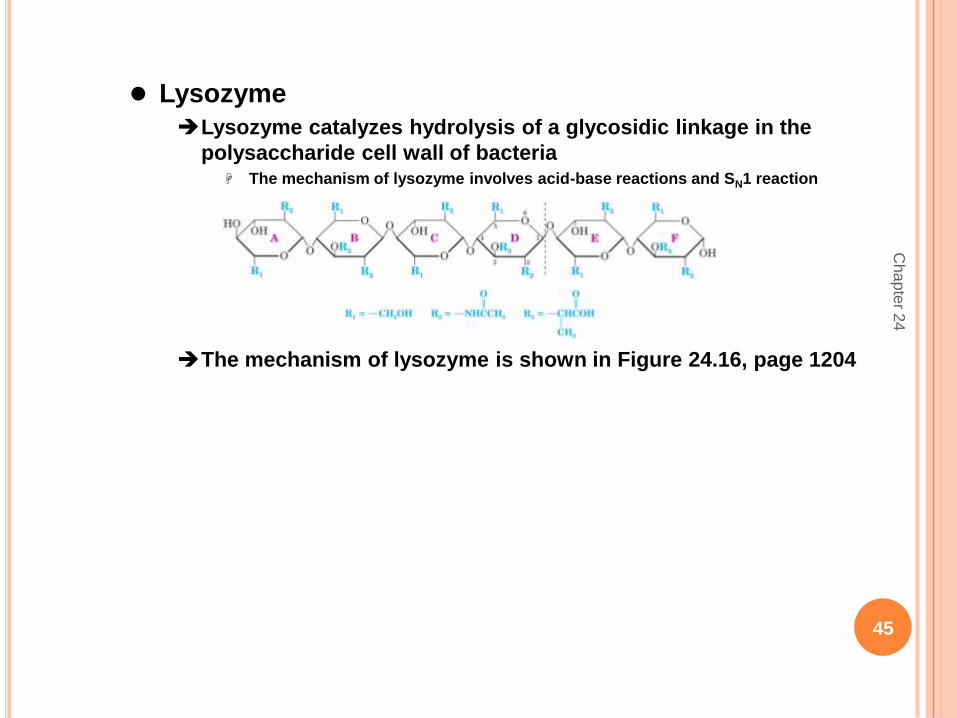
Lysozyme
Lysozyme catalyzes hydrolysis of a glycosidic linkage in the
polysaccharide cell wall of bacteria The mechanism of lysozyme involves acid-base reactions and SN1 reaction
The mechanism of lysozyme is shown in Figure 24.16, page 1204

Cha
pte
r 24
46
Serine Proteases
Proteases hydrolyze amide bonds in proteins
Chymotrypsin, trypsin, and elastin are serine proteases
Serine proteases have a serine hydroxyl group that is involved in
the mechanism of amide bond hydrolysis A “catalytic triad” involving the side chains of specific aspartic acid, histidine, and
serine residues catalyze the amide hydrolysis
The serine hydroxyl attacks the amide carbonyl group, forming a tetrahedral
intermediate
The aspartic acid and histidine side chains form an acid-base relay system to
assist with protonation and deprotonation steps
The serine tetrahedral intermediate releases the amine, leaving an acylated serine
A water molecule attacks the carbonyl group of the acylated serine
A new tetrahedral intermediate forms
When this tetrahedral intermediate collapses to the carboxylic acid, the serine
hydroxyl is released for a new catalytic cycle
See the following slide for the mechanism of trypsin
The Active Site Catalytic Triad of Trypsin This is shown figure 24.17, page 1205

Cha
pte
r 24
47
The Catalytic Mechanism of Trypsin

Cha
pte
r 24
48
Purification and Analysis of Polypeptides and Proteins
Proteins are purified initially by precipitation, column
chromatography, and electrophoresis
HPLC is the method of choice for final purification of a protein
Analysis of proteins
Molecular weight can be estimated by gel electrophoresis and size
exclusion chromatography
Mass spectrometry is used to determine protein molecular
weights with high accuracy and precision Electrospray ionization (ESI) mass spectrometry is one way to create protein ions
for mass spectrometry
Matrix-assisted laser desorption ionization (MALDI) mass spectrometry is another
technique for generating protein ions for mass spectrometry
The 2002 Nobel Prize in Chemistry was awarded in part for development of ESI (by
Fenn, et al) and MALDI (by Tanaku) for mass spectrometry
Cha
pte
r 24
49
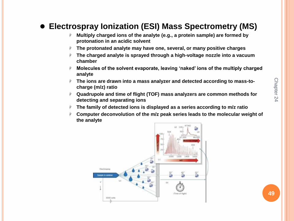
Electrospray Ionization (ESI) Mass Spectrometry (MS) Multiply charged ions of the analyte (e.g., a protein sample) are formed by
protonation in an acidic solvent
The protonated analyte may have one, several, or many positive charges
The charged analyte is sprayed through a high-voltage nozzle into a vacuum
chamber
Molecules of the solvent evaporate, leaving „naked‟ ions of the multiply charged
analyte
The ions are drawn into a mass analyzer and detected according to mass-to-
charge (m/z) ratio
Quadrupole and time of flight (TOF) mass analyzers are common methods for
detecting and separating ions
The family of detected ions is displayed as a series according to m/z ratio
Computer deconvolution of the m/z peak series leads to the molecular weight of
the analyte
Cha
pte
r 24
50
Proteomics
Proteomics involves identification and quantification of all of the
proteins expressed in a cell at a given time Proteins expression levels vary in cells over time
Proteomics involves identification and quantification of all of the proteins
expressed in a cell at a given time
Proteomics data can shed light on the health or life-cycle stage of a cell
Tools for Proteomics Polyacrylamide gel electrophoresis (2D-PAGE) is a low resolution technique for
separating protein mixtures
Two-dimensional (2D) microcapillary HPLC coupled with mass spectrometry is a
high resolution technique for separating and identifying proteins in a cell extract
Cha
pte
r 24
51
Multidimensional Protein Identification Technology
MudPIT (Multidimensional protein identification technology)
involves: Lysis of intact cells
Digestion of the proteins to a mixture of smaller peptides
Separation of the peptide mixture by 2D HPLC using a strong cation exchange
column in tandem with a reversed-phase (hydrophobic) column)
Direct introduction of the 2D HPLC eluent into a mass spectrometer
Comparison of mass spectra with a database of mass spectral data for known
proteins
Data matching can lead to identification of >1000 proteins in one integrated
analysis