Bahasa
Halaman
Hukum


www.wjpps.com Vol 3, Issue 4, 2014.
113
Ifeanyi et al. World Journal of Pharmacy and Pharmaceutical Sciences
ISOLATION AND COMPARISM OF FATTY ACID AND
ACYLGLYCEROL CONTENTS IN LOCAL HYBRID
COCONUTS(COCOS NUCIFERA)
Aloh Godwin Sunday1, Obeagu Emmanuel Ifeanyi*2, Eze Obioma Benedict L1
1Lecturer,Department of Biochemistry,Michael Okpara University of
Agriculture,Umudike,Abia State,Nigeria. 2.Diagnostic Laboratory Unit,University Health Services Department, Michael Okpara University of
Agriculture,Umudike,Abia State,Nigeria.
ABSTRACT
INTRODUCTION
The coconut belongs to the palm family, palmae (Gill, 1988). The
coconut is found throughout the west tropical low lands (Anochili and
Tindall. 1986). It probably originated in South-East Asia of the
pacific region from where it was taken to the Atlantic coasts of the
America and tropical areas of Africa where suitable conditions for their
growth existed (Purseglove, 1972). However, the considerable
extension in acreage during the past 100 years is due to the demand for
coconut oil in temperate countries. In West Africa, coconut are grown
along the coastal and inland areas where there is good drainage.
Coconut has different local names. Benin – Ovi oibo, Yoruba - agbon,
Hausa – Kwakara itagara, Igbo – aki oyibo, Efik – isip mbakara and
Fante – kbve (Gill, 1988).
Coconuts are grouped into two main categories: the dwarf and tall varieties, based on
flowering and fruiting habit, and tree height (Tindall and Anochili, 1986).
1. Dwarf varieties (Var. Nana
These are probably mutations from tall types and have occurred in several countries (Brook,
1982). This variety starts bearing fruits earlier than the tall variety. It flowers as early as three
years after four to five years when the trees have reached a height of about 1.5m. They are
normally self-pollinated due to overlapping of the male and female phases, and are therefore
WWOORRLLDD JJOOUURRNNAALL OOFF PPHHAARRMMAACCYY AANNDD PPHHAARRMMAACCEEUUTTIICCAALL SSCCIIEENNCCEESS
VVoolluummee 33,, IIssssuuee 44,, 111133--113322.. RReesseeaarrcchh AArrttiiccllee IISSSSNN 2278 – 4357
Article Received on 19 February 2014, Revised on 12 March 2014, Accepted on 31 March 2014
*Correspondence for Author
Obeagu Emmanuel Ifeanyi
Diagnostic Laboratory
Unit,University Health
Services,Michael Okpara
University of
Agriculture,Umudike,Abia
State, Nigeria

www.wjpps.com Vol 3, Issue 4, 2014.
114
Ifeanyi et al. World Journal of Pharmacy and Pharmaceutical Sciences
fairly homozygous (Tindall and Anochili, 1986). This quality makes it possible for it to
maintain its dwarf character. The nuts are small but numerous, and 6000 – 8000 nuts are
required to produce 1 ton of copra. They have a short productive life of 30-35 years, although
white head (1966) records plantations, which are still in good bearing after 40 years. They
require better soil and climatic conditions, and in Ceylon have been found to be readily
susceptible to pests and disease. They cross with the tall and the hybtid show considerable
promise (Harries, 1978).
2. Tall Varieties (Var. typica)
This variety is sometimes referred to Var. typical (Purseglove, 1972). They are the most
commonly planted for commercial production, growing to a height of 20 – 30m. They are
slow-maturing, first flowering 6 – 10 years after planting. They are long-lived and may attain
an age of 80 – 100 years (Luntungan, 1978). They are normally cross-pollinated, as there is
usually no overlapping of the male and female phases of the protandrous flowers (Harries,
1978).
The nut is medium to large in size and 4000 -6000 nuts usually yield 1 ton of copra. They are
hardy and will thrive on different soil types and varying environmental conditions (Liyanage,
1967) however, tall palms which have developed in various regions are variable and distinct.
But the common forms may be referred to by the local names of the countries in which they
are grown e.g Jamaica Tall; have an average fruit weight of about 1.7kg and nut weight of
about 0.7kg, of which 50% is endosperm producing 02.kg of copra. They are susceptible to
lethal yellowing.
3. The King Coconut (Var. aurantiaca)
In coconut, the manifestation of hybrid vigor has been observed in growth as well as yield
characteristics (Luntungan, 1982). Inter-varietal hybrid obtained between the tall and dwarf
varieties are more promising than intra-varietal hybrids (Gill, 1989). The inter-varietal
hybrids with the tall variety as the pollen parent are precocious, high yielding and produce
nuts with copra quality, equal to the rill parent. The production of a large number of inter-
varietal hybrids by artificial pollination is discouraged by several factors;
a. The absence of suitable markers to identify the hybrids;
b. The inability of the hybrids to perform well except under favourable management
conditions.
www.wjpps.com Vol 3, Issue 4, 2014.
115
Ifeanyi et al. World Journal of Pharmacy and Pharmaceutical Sciences
c. The considerable labour and expenditure involved in the production of hybrids (Robbelen
and Downey, 1989).
The coconut can be adapt to a range of climatic conditions and thrives well between latitude
20o North and 20o South of the equator. The highest yields are generally obtained from trees
grown at 600m to 900m above sea level. The coconut frows well in areas, which have an
evenly distributed rainfall of about 100 to 225cm per annum. It is not suited to regions, which
have a long dry season. A mean annual temperature of about 27oC is required, while
temperature below 20oC cause fruit abnormalities (Luntungan, 1982). The coconut requires
warm humid conditions with about 70% relative humidity. Coconut palm grows well on
drained, rich alluvial or loamy soils which permit unrestricted root development and aeration.
Violent storms such as cyclones, hurricanes or typhones are hazards in several coconut
growing countries.
From the base of the stem arise numerous adventitious roots, some of which appears above
the ground level. The first roots tend to penetrate the soil vertically and subsequent roots
usually spread horizontally (Anthony, 1988). The roots are widely and superficially spread to
absorb nutrients from the upper layer of the soil. The trunk carries a crown of leaves at the
top, and each leaf is compound with a mid—rib (rachis) which bears pinnately arranged
leaflets measuring about one metre (Adams and Bamford, 1999). No visible trunk is formed
until the palm is several years old and a trunk can only be formed when the apical meristem
has attained its full diameter since there is no cambium (Tandall and Anochili, 1986). The
palm like other monocotyledons is composed of a series of joints, each having a nod, a leaf
and internodes flowers appear in an inflorescence which is enclosed in a boat shaped sheath,
or spathe. The inflorescence is composed of many male (staminate) flowers at that apex of
the inflorescence stalk and a few female (pistillate), flowers at its base, making the plant
monoecious (Gill 1988). The mature fruit is a fibrous drupe, about 20 – 30cm long weighing
1.2 – 2.0kg and consisting of:
1. Epicarp or outer skin which is tough, smooth, hard and may be green, yellow, orange, red
or brown.
2. Mesocarp or fibrous layer which is pale brown.
3. Endocarp or shell which is ovoid, hard, stony and dark brown and
4. S single seed with a thin brown testa closely oppressed to endocarp and adhering firmly to
endocarp or “meat”, which is firm, white, oily, 1 – 2cm thick and supply copra and oil.

www.wjpps.com Vol 3, Issue 4, 2014.
116
Ifeanyi et al. World Journal of Pharmacy and Pharmaceutical Sciences
(Purseglove, 1972). In center of the seed is a large cavity partially iled with coconut water,
which is completely absorbed about 6 months after harvesting. The endocarp contains sugar
or carbohydrate, fats, oil and protein as food reserves (Robbelen, 1989). See table 1.1 and the
figure below.
Table 1.1 : CHEMICAL COMPOSITION OF COCONUT
Source: (Robbelen and Downey, 1989)
Source: Luntungan, H.T. (1982).
Rhinoceros beetle or coconut black beetle, Oryctes rhinoceros found throughout South – east
Asia burrows into the terminal bud, damaging the unopen leaves and may kill young palms
(Bock, et al, 1970). The larvae of palm weevils, Rhynchoporus spp, of which the most
important are R. Ferrugineous and R. Schach in Malaya damage the trunk and crown,
especially those of youmg palms (Purseglove, 1872). Others like termites, red ring nematodes
and rats are destructive bur controls are achieved with insecticides e.g DDT BHG. Coconuts
suffer from a number of diseases such as Bud disease, caused by phytophthora palmivora
which causes wilting and collapse of the youngest leaves, followed by similar symptoms
successive lower leaves recently described lethal bole rot caused Maraemiellus cocophilus
pegler along the East Africa coast and lethal yellowing, which was formerly called shedding
of young and maturing nuts (Bock et al., 1970).
It provides food, drink, oil, medicine, fibre, timber, thatch, mats, fuel, and domestic utensils
(Purseglove, 1972). The main producers are Philippines and Indonesia. Coconut is essentially
an oil crop though every part of the palm is useful to mankind. Coconut oil due to its high
content of lauric and myristic acid, has a high saponification value, a low iodine number, and
is used extensively for edible and industrial purposes (Varner and Bonna, 1976). Coconut oil
ENDO
– SPERM
COPRA COCONUT CAKE
DRINKING COCONUT
WATER
SWEET TODDY
Mill Expeller Chekku Water 36.3 6.8 11.0 11.0 13.3 95.4 84.4 Protein 4.5 7.8 19.1 19.1 14.3 0.1 0.1
Fat 41.6 63.7 6.0 10.0 26.7 0.1 0.1 Carbohydrate 13.0 16.1 45.3 43.8 32.8 4.0 15.1
Fibre 3.6 3.8 12.2 11.8 8.9 - - Minerals 1.0 2.0 5.7 5.3 4.1 0.4 0.3

www.wjpps.com Vol 3, Issue 4, 2014.
117
Ifeanyi et al. World Journal of Pharmacy and Pharmaceutical Sciences
is used in margarine, biscuit and cooking oil industries, in the production of toilet articles
such as hair oil, shaving creams, shampoos, soap and cosmetic. The oil is also used in
synthetic resins as well as rubber substitutes detergent, lubricant and fuel production of
petrol, kerosene and oil substitutes is possible from coconut oil (Purseglove, 1972). The cake
obtained after oil extraction can be used as fertilizer for field crops, or fed to cattle, poultry or
young growing swine. The meat of the ripe coconut us in cooking and for household oil
production. The milk or cream obtained from gratings of the meat is used as substitute for
cow’s milk. The liquid or coconut water in the tender nut, is a refreshing drink. Is often
recommended as a medicine in cases of gastroenteritis where it is considered for a substitute
for saline glucose (Purseglove, 1972). Coconut water is a major ingredient in coconut
vinegar, sauce and Lemonade, Sweet toddy obtained by tapping the unopened inflorescence
of the palm, is an excellent beverage and palm sugar or jiggery is obtained by boiling and
evaporating fresh toddy (Tindall and Anochili, 1986). The hard shells of coconut are used in
the manufacture of shell charcoal, activated carbon and shell flour. The fibrous husk of
coconut yields a fibre known as coir which is board. The stem of palm, are used for thatching
houses, fencing and for making basket (Purseglove, 1982).
AIMS AND OBJECTIVES
The aims of this project was to compare the fatty acid and acylglycerol contents of loca and
hybrid coconuts since coconut is one of the edible foods consumed in the whole world for its
fatty acid and acylglycerol contents.
{MATERIALS AND METHODS
MATERIALS USED FOR THE EXPERIMENT
1. Coconut (Cocos nucifera) endosperm
2. Reagents
i. Chloroform – M&B, AnalaR
ii. Hexane-BDH, GPR
iii. Diethyl ether -BDH, GPR
IV. Methanol – M&B, GPR
V. Calcium Oxide -BDH, GPR
vi. Sodium sulpjate –BDH, AnalaR
vii. Ethanol -BDH, GPR
viii. Acetic acid – glacial -BDH, GPR

www.wjpps.com Vol 3, Issue 4, 2014.
118
Ifeanyi et al. World Journal of Pharmacy and Pharmaceutical Sciences
ix. Isopropyl either -BDH, GPR
x. Isopropyl alcohol -BDH, GPR
xi. Potassium hydroxide pellets – BDH< AnanaR
xii. Concentration Sulphuric acid -BDH, GPR
xiii. Glycerol – BDH, AnalaR
xiv. Silica gel G, type 60 – MERCK, AnalaR
xv. Lipid standards – Oleic acid, Linoleic acid, palmitic acid, myristic acid, lauric acid,
stearic acid, Glycerol tristearate glycerol trioleate, and glycerol tripalmitae -BDH, GPR
xvi. Sodium thiosulphate -BDH, GPR
xvii. Salicyclic acid -BDH, GPR
xviii.Periodic acid -BDH, GPR
xix. Starch indicator _CVI, GPR
xx. Triolein standard -BDH, GPR
xxi. Periodic solution -BDH, GPR
xxii. Cupric nitrate -BDH, GPR
xxiii.Triethanol amine -BDH, AnalaR
xxiv. Diethyldithiocarbamate -BDH, GPR
3. Instruments, Containers, Equipment Used
i. Deep freezer – model MK3 novum freezer, Britain.
ii. Centrifuge – Haraeus Christ GMDH centrifuge, London.
iii. Spectrophometer – Nova spectrophotometer by Biochrome, London.
iv. Mettler H 35 & P 1000 Electrical balances. London.
v. Conical flask, test tubes funnel, burette.
METHODS
EXTRACTION OF COCONUT OIL (TRIACYLGLYCEROL).
The coconut oil was obtained by freeze thawing following the method described by
Robledeno and Luzuriagal.
Procedure: 120g of fresh coconut kernel from the local and hybrid varieties were grated fine
with an improvised fat grater, placed in a white cloth and the “milk” pressed and wrung out
by hand. The milk was washed thrice in a separating funnel with tap water and only the
floating “cream” was recovered during each washing. The washed cream in a conical flask
was allowed to freeze in a deep freezer – thaw operation gad to be repeated four times but
www.wjpps.com Vol 3, Issue 4, 2014.
119
Ifeanyi et al. World Journal of Pharmacy and Pharmaceutical Sciences
this time, at intervals of four hours to get the oil liberated. The oil was next decanted into
another flask and centrifuged briefly at low speed to remove further solid impurities. The
weight and volume of the oil were determined and stored for use.
Separation Of Triacyglycerol (Neutral Lipid) From Probable Polar Lipids Present.
Neutral lipids were separated from polar phospho–lipids using an activated alumina
(triacyglycerol purifier) column, a method that is also employed in the purification of
phospholipids (Merinetti, 1962).
Procedure: A clean, drawn out tipped burette was fitted with glass wool at bottom and
mounted vertically with a cramp.
20gm of activated alumina was activated at 12o for 30minutes and cooled. This was poured
into the burette and dissolved in chloroform to form a 10cm column. On solidification, it was
washed with 50ml of chloroform after which 2.5ml of the lipid extract was applied. The
extract was allowed to enter the column and then eluted with 50ml of chloroform for the
natural lipids. Elution with 50ml of methanol on a similar column would have extracted the
polar lipids. The extract was concentrated to about 5ml and stored for use, this was done on
each of the variety.
Extraction Of Free Fatty Acids.
Free fatty acids were extracted from the triacyglycerol by the Dole method (Dole, 1956). The
Dole’s extraction mixture consist of isopropyl alcohol; Hexane; INH2SO4 (40: 10: 1 v/v)
Procedure: 5ml of Dole’s extraction mixture was pipetted into a clean test tube, 1ml of lipids
extract was added and the mixture vigorously shaken using an incubator shaker. The mixture
was placed in an ice-bath for 10minutes after which 2ml of hexane and 3ml of distilled water
were added. The contents of the tube were again shaken with the shaker for 5minutes. On
standing, the phase separated and 3ml of the upper hexane phade was pipette into another
clean dry test tube. To this was added 1ml of chloroformhexane mixture (5:1 v/v) and
centrifuged at 2,500 rpm for 15 minutes. The upper chloroform-hexane phase was pipette into
another bottle and used for free fatty acid analysis.
Qualitative Analysis Of Triacylglycerol
i. Preparation of plates:
The plates were prepared according to (Boyer and King, 1977) with slight modifications
www.wjpps.com Vol 3, Issue 4, 2014.
120
Ifeanyi et al. World Journal of Pharmacy and Pharmaceutical Sciences
Procedure: Silica gel G, type 60 and water used in a 1:2 ratio to prepare the TLC plates. 60gm
of the gel and 120ml of water in a 250ml reagent bottle were shaken vigorously for 10
minutes and used in preparing the plates, were washed, dried and cleaned with acetone
soaked in cotton wool. The gel was spread with Shandon TLC plate spreader for TLC plates.
After a brief air-drying, the plates were activated in the oven ar 121oc for 45 minutes, they
were cooled and used.
ii. One Dimensional TLC of Triacylglycerol Fatty Acids and Fatty Acids Standards
A shandon TLC tank was saturated with a solvent system of Hexane-ether-acetic acid
(60:40:1) for 30 minutes after it had been made airtight with Vaseline or grease along the
edges. 2.0% solutions of these lipids standard in chloroform were prepared, mytistic, lauric,
palmitic, oleic, stearic and lioleic acids. A drop in each of the lipids standards was applied
alongside that form coconut at a distance 2cm from the base of the plate. It took 1 hour 30
minutes for the solvent front to reach 2cm marked point above the plates. The plates was
briefly air-dried and visualized by iodine vapour as a chromogenic reageant, the Rf values
were calculated, the spots encircled and the plates photographed (fig. 4.1)
iii. One Dimensional TLC of Tricylglycerol Fatty Acids and Fatty Acids Standards
A Shandon TLCtank was saturated with a solvent system of Hexane-ether-acetic acid
(60:40:1) for 30 minutes after it had been made airtight with Vaseline or grease along the
edges. 2.0% solutions of these lipids standard in chloroform were prepared, mytistic, lauric,
palmitic, oleic, stearic and lioleic acids. A drop in each of the lipids standards was applied
alongside that form coconut at a distance 2cm from the base of the plate. It took 1 hour 30
minutes for the solvent front to reach 2cm marked point above the plates. The plates was
briefly air-dried and visualized by iodine vapour as a chromogenic reageant, the Rf values
were calculated, the spots encircled and the plates photographed (fig. 4.1)
iv. One-Dimensional TLC of Triacylglycerol and Triacylglycerol Standards.
The plates and the Shandon TLC Tank were prepared as in (i) and (ii) above but with a
solvent system of hexanedithylether-acetic acid (80: 20: 1).
The triacylglycerol standards (glycerol tripalmitate, glycerol trioleate and glycerol
tristearate),all in chloroform were spotted alongside the coconut sample triglyceride also in
chloroform at a distance of 2cm from the base of the plate. It took 1hour 20 minutes for the
solvent front to reach the marked point from the top of the plate. It was removed, briefly air-

www.wjpps.com Vol 3, Issue 4, 2014.
121
Ifeanyi et al. World Journal of Pharmacy and Pharmaceutical Sciences
dried and visualized in the iodine vapour. The plate photographed (fig. 4.2) the spots
encircled and the Rf values calculated.
MANUAL QUANTIFICATION OF TRIACYLGLYCEROL CONTENT.
This was carried out according to (Kessler Lederer, 1965). As well as Fletchers (1968)
method for the measurement of serum triacylglycerol. The method involves the oxidation of
glycerol to formaldehyde which is measured calorimetrically with colour reagent. The lipid
extract must therefore be free from other sources of glycerol, particularly phosphoslipid,
Activated alumina was used to absorb these interfering substances.
Reagents
1. Triacylglyceride purifier (Activated alumina)
2. Isopropanol
3. Triolein standard, contains 300mg triolein (glycerol trioleate) dissolve in 100ml
anhydrous isopropanol.
4. Potassium hydroxide 1 N
5. Periodic solution
6. Colour Reagent
METHOD
Test: 0.4g of activated alumina was placed in a test tube and 2.5ml of isopropanol added,
followed by 0-1ml of the triglyceride sample. The mixture is shaken manually for at least 5
minutes.
Standard: 0.4g of activated alumina, 2.4ml of isopropanol, 0.1ml of water and 0.1ml of
triolein standard were mixed and shaken manually for at least 5 minutes.
Blank: 0.4g of activated alumina 2.5ml of isopropanol and 0.1ml of water were mixed and
shaken manually for at least 5 minutes.
Centrifuge the tubes at about 3000 rpm for 5 minutes to obtain a clear supernatant. Carefully
transfer 0.1ml of clear supernatant into a similar test tube, taking care not to vary over
absorbent 0.25ml of Potassium hydroxide at 60oC for 5 minutes in a water bath to soporific
the triglyceride. After cooling the tubes, 0.25ml of periodic solution was added and mixed
immediately after each addition. After 20 minutes, 0.1ml of colour reagent was added and
each in a 60oc water bath for 30 minutes.

www.wjpps.com Vol 3, Issue 4, 2014.
122
Ifeanyi et al. World Journal of Pharmacy and Pharmaceutical Sciences
After cooling, the absorbance of the standard and test verse the blank were read in a
spectrophometer of 410nm.
The triglyceride content in mg/ 100ml was calculated from the relation.
Coconut triglyceride in mg/ 100ml was calculated from the relation.
Coconut triglyceride mg/ 100m= x 300
PREPARATION OF MONOACYLGLYCEROL FROM TRIACYLGLYCEROL
Monoacylglycerols were prepared from the triacylglycerols by a transesterification reaction
between coconut oil and glycerol according to the optimum condition found by Arida,
(1973).
Procedure: Two grams of coconut oil was heated to approximately 180oC. 0.4g of glycerol,
preheated to 120oC was added at once followed by 0.001g of Calcium oxide (CaO). The
mixture was stirred for two hours at 200oC. The reaction mixture was cooked and allowed to
stand for twenty four hours.
Removal of Free Glycerol
One gram of the reaction mixture was melted at about 60oC and washed three times with 5ml
portion of aqueous 1% Sodium sulphate previously heated to 60oC.
Extraction of Monoacylglycerol
The glycerol free product was dissolved in hexane in the ratio of 6g hexane to 1g of
monoglyceride reaction mixture. The monoglyceride fraction was extracted from the solution
several times (6 times) with portion of aqueous ethanol (65%)
Ethyl alcohol from the combined monoglyceride extracts was removed using a water bath at
60oC instead of Rotary evaporationwhich was not available.the residue was allowed to dry in
a vacuum oven at 800c to constant weight.
EXTRACTION OF FREE FATTY ACID FROM MONOACYLGLYCEROLS.
Free fatty acids extracted by the Dole’s method (1956) as described in 3.4 before.
QUALITATIVE ANALYSIS OF MONOGLYCERIDES FOR FATTY ACIDS BY
ONE-DIMENSIONAL TLC.
The free fatty acids extracted above was spotted on the TLC plate alongside the fatty acid
standards – oleic acid, linoleic acid, stearic acid, palmitric acid, myristic acid lauric acid. The

www.wjpps.com Vol 3, Issue 4, 2014.
123
Ifeanyi et al. World Journal of Pharmacy and Pharmaceutical Sciences
run lasted 2 hours in a solvent system consisting of hexane-diethyl-acetic acid (80: 1 v/v).
The plate was briefly air-dried and visualized in iodine vapour. The plate photographed (fig.
4.3), the spots encircled and the Rf- values were calculated.
3.10 Quantitative Estimation of Total Monoacylglycerol contents prepared from coconut
oil.
The total monoacylglyceride contents were estimated by the method described Bartman,
(1956).
Method
Reagents
1. Periodic acid -1.35g dissolved in 25ml of H2O, 190ml acetic acid added mixed store in
dark.
2. Sodium thiosulphate (Na2S2O2) 0.1N – 2g dissolved in 25ml of H2O.
3. Salicyclic acid- 0.25g dissolved in 200 of H2O.
4. 10 NH2SO4 – 27.8ml of conc. H2SO4 were made up to 10ml of H2O.
5. Potassium iodide -7.5g were dissolved in 50ml of water.
6. Starch indicator solution -1.0g were dissolved and made up to 100ml of water
7. Chlorform.
1.0g of the sample were dissolved in 190ml of chloroformand transferred to the separating
flask, 19ml of water was added stopper and shaken vigorously for 1 minute. The mixture is
allowed stand for 30 minutes for clear separation and the bottom chloroform sample layer is
collected. This was done for each of the sample 5ml of periodic acids (HLO4) solution were
pipetted into six beakers. 5ml of chloroform were added to the second beaker and beaker and
5ml of water to the third as blanks. 5ml of different chloroform samples used for the test were
added to the fourth, fifth and sixth beakers. Shake gently and stand the mixture for 30
minutes.
Add 2ml of Potassium iodide shake gently and stand for less than 2 minutes away from
strong sunlight. After which 10ml of H2O were added and filtrated with Sodium thiosulphate,
using 0.2ml of starch solution as indicator.
Coconut monoglyceride in mg/ 100ml
=
Where, B = titer of CHCL3 blank.

www.wjpps.com Vol 3, Issue 4, 2014.
124
Ifeanyi et al. World Journal of Pharmacy and Pharmaceutical Sciences
S = titer of sample.
N = normally of Na2S2O3.
W = g sample in CHCL3 aliquot.
M = molecular weight of monoglyceride
QUALITATIVE ESTIMATION OF TOTAL NON-ESTERIFIED FATTY CONTENT
IN COCONUT OIL
Standard curve
The estimation of the total fatty acid content in coconut oil was done by meand of standard
curve for the standard free fatty acid-palmitric. 100mg of palmitric acid were dissolved in
100ml of chloroform. The fatty acid standard solutions were diluted serially to form
concentration range of 20- 100Nm as in table 3.1
Table 3.1: Serial Dilution For Free Fatty Acid Standard Curve
The fatty acid standards treated according to Nucombe’s modification of the method for the
determination of non-esterified fatty acids developed for the analysis of free fatty acid in the
body fluid or plasma. The method is based on the selective transfer of Copper salt of fatty
acid into chloroform. The free fatty acids transferred from the aqueous phase into chloroform
in which the copper complex fatty acid formed is monitored colorimetrically.
Reagents
Copper Reagent: This is prepared by mixing the following solution A, B, C (10: 1: 9 v/v).
Solution A: 6.45g of cupric nitrate (Cu(NO3)2H2O) was made up to 100ml in water.
Solution B: 20ml acetic acids was made up to 100ml in water.
Tubes Standard solution (ml) Chloroform (ml) Final volume
(ml) Final conc. (g/ml)
1. 0.50 4.50 5.00 10.00 2. 1.00 4.00 5.00 20.00 3. 1.50 3.50 5.50 30.00 4. 2.00 3.00 5.00 40.00 5. 2.50 2.50 5.00 50.00 6. 3.00 2.00 5.00 60.00 7. 3.50 1.50 5.00 70.00 8. 4.00 1.00 5.00 80.00 9. 4.50 0.50 5.00 90.00 10. 50.00 0.00 5.00 100.00
www.wjpps.com Vol 3, Issue 4, 2014.
125
Ifeanyi et al. World Journal of Pharmacy and Pharmaceutical Sciences
Solution C: 14.92g of triethanolamine was made up to 100ml in water.
(2) Diethyldithiocarbamate (the colour reagent) 0.05 was made to 50ml in butanol or butan-
01.
Procedure: 5ml of chloroform were pipetted into each of the test tubes as in table 3.1, to this
were added 0.5ml of fatty acid, a 2.5ml of fatty acid standard and 2.5ml of copper reagent. In
the blank are placed 5ml of chloroform, 0.5ml of water and 2.5ml of copper reagent solution.
The tubes were stopped, shaken vigorously for 2 minutes and centrifuged for 5 minutes. After
centrifuge, the aqueous and chloroform layers are separated. 3ml of chloroform layer solution
were pippetted into test tubes and 0.5ml of diethiocarbamate reagent added. After mixing, the
coloured sample solution is measured at 440nm against the blank in a spectrophometer.
Analysis Of Fatty Acid Content In The Samples Analysis of the fatty acid content in the samples was treated according to Ducombe’s
modification of the method for the determination of non-esterified fatty acid content in
plasma.
Reagent
1.Copper Reagent Solution: This was made by missing solution A, B and C (10: 1: 9, v/v).
Solution A: 6.45g of Cu(NO3)23H2O was made up to 100 in water.
Solution B: 20ml of acetic acid in 100ml of H2O.
Solution C: 14.92g of Triethanolamine in 100ml of H2O.
Diethyldithiocabamate: 0.05g of Sodium diethyldithiocabamate in 50ml of butanol 2-01.
Procedure: 5ml of the samples were pipetted into different test tubes, followed by 5ml of
chloroform and 2.5ml of copper reagent. In a tube serves as the blank were placed 5ml of
chloroform, 0.5ml of water, and 2.5ml of copper reagent. The tubes were closed, shaken for 2
to 3 minutes and then centrifuged for 5 minutes. After centrifuging, for a few minutes, the
aqueous and chloroform layers were separated. Three (3ml) of the chloro form solution were
pipetted into different test tubes. Then 0.5ml of diethyldithiocarbamate reagent was added.
After mixing, the OD of the coloured sample solution was read off at 440nm against the
blank.
By extrapolation, the OD obtained was read off (in terms of concentration) on the fatty acid
standard curve.

www.wjpps.com Vol 3, Issue 4, 2014.
126
Ifeanyi et al. World Journal of Pharmacy and Pharmaceutical Sciences
RESULTS
THIN LAYER CHROMATOGRAPHY (TLC) OF STANDARD FATTY ACIDS AND
SAMPLES (KING AND DWARF HYBRID)
(FIG. 4.1).
PLATE I
Rf values Spot 1 (Linoleic acid) = 0.61
Spot 2 (Stearic acid) = 0.53
Spot 3 (Oleic acid) = 0.60
Spot 4 (Palmitic acid) = 0.59
Spot 5 (Myristic acid) = 0.60
Spot 6 (Lauric acid) = 0.60
King hybrid
Spot 1 = 0.59
Spot 2 = 0.60
Spot 3 = 0.61
King hybrid coconuts contain palmitic acid, linoleic acid oleic and myristic acid or lauric
acid.
Dwarf hybrid
Spot 1 = 0.60
Spot 2 = 0.61
Spot 3 = 0.67
Dwarf hybrid coconuts contain linoleic acid or lauric acid.
Thin Layer Chromatography (Tlc) Of Standard Fatty Acids And Samples (King And
Dwarf Hybrid)
(FIG 4.1)
PLATE 1
FIG. 4.1
Keys:
Lin = Linoleic acid
St = Stearic acid
Ole = Oleic acid
Pal = Palmitic acid
www.wjpps.com Vol 3, Issue 4, 2014.
127
Ifeanyi et al. World Journal of Pharmacy and Pharmaceutical Sciences
Myr = Myristic acid
Lau = Lauric acid
K.h = King Hybrid coconut
D.h = Dwarf hybrid coconut
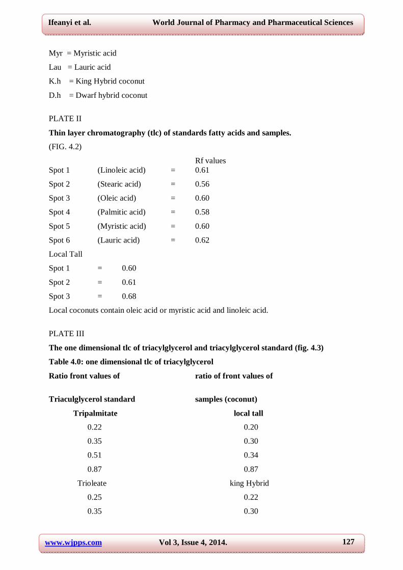
PLATE II
Thin layer chromatography (tlc) of standards fatty acids and samples.
(FIG. 4.2)
Rf values Spot 1 (Linoleic acid) = 0.61
Spot 2 (Stearic acid) = 0.56
Spot 3 (Oleic acid) = 0.60
Spot 4 (Palmitic acid) = 0.58
Spot 5 (Myristic acid) = 0.60
Spot 6 (Lauric acid) = 0.62
Local Tall
Spot 1 = 0.60
Spot 2 = 0.61
Spot 3 = 0.68
Local coconuts contain oleic acid or myristic acid and linoleic acid.
PLATE III
The one dimensional tlc of triacylglycerol and triacylglycerol standard (fig. 4.3)
Table 4.0: one dimensional tlc of triacylglycerol
Ratio front values of ratio of front values of
Triaculglycerol standard samples (coconut)
Tripalmitate local tall
0.22 0.20
0.35 0.30
0.51 0.34
0.87 0.87
Trioleate king Hybrid
0.25 0.22
0.35 0.30

www.wjpps.com Vol 3, Issue 4, 2014.
128
Ifeanyi et al. World Journal of Pharmacy and Pharmaceutical Sciences
0.51 0.35
0.87 0.85
Tristearate Dwarf Hybrid
0.22 0.22
0.34 0.30
0.51 0.35
0.87 0.85
The table above shows that the different breeds of coconut contain the standard
Triacylglycerol (tripalmitate, trioleate and tristearate) and some other impurities.
The one dimensional of triacylglycerol, triacylglycerol, standards and samples (local
tall, king hybrid and dwarf hybrid coconut).
PLATE 3
FIG. 4.3
Keys:
L.T = Local Tall coconut
Tp = Tripalmitate
To = Triolein
Ts = Tristearate
K.h = King Hybrid coconut
D.h = Dwarf Hybrid coconut
4.3 ONE DIMENSIONAL TLC MONOACYLGLYCEROL FOR FREE FATTY ACID.
First spot (Local Tall) = 0.80
Second spot (King hybrid) = 0.80
Third spot (Dwarf hybrid) = 0.80
Fourth spot (Linoleic acid) = 0.79
Fifth spot (Oleic acid) = 0.80
Sixth spot (Palmitic acid) = 0.82
Seventh spot (Stearic acid) = 0.81
Eighth spot (Myristic acid) = 0.80
Ninth spot (Lauric acid) = 0.80
www.wjpps.com Vol 3, Issue 4, 2014.
129
Ifeanyi et al. World Journal of Pharmacy and Pharmaceutical Sciences
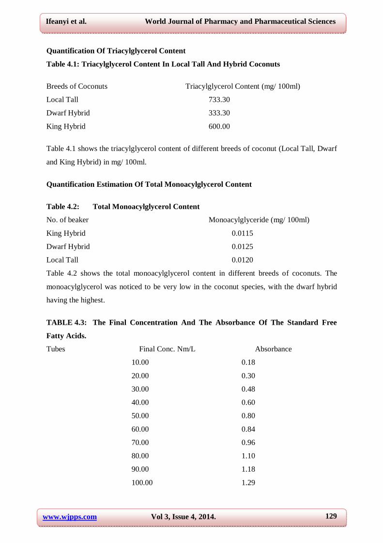
Quantification Of Triacylglycerol Content
Table 4.1: Triacylglycerol Content In Local Tall And Hybrid Coconuts
Breeds of Coconuts Triacylglycerol Content (mg/ 100ml)
Local Tall 733.30
Dwarf Hybrid 333.30
King Hybrid 600.00
Table 4.1 shows the triacylglycerol content of different breeds of coconut (Local Tall, Dwarf
and King Hybrid) in mg/ 100ml.
Quantification Estimation Of Total Monoacylglycerol Content
Table 4.2: Total Monoacylglycerol Content
No. of beaker Monoacylglyceride (mg/ 100ml)
King Hybrid 0.0115
Dwarf Hybrid 0.0125
Local Tall 0.0120
Table 4.2 shows the total monoacylglycerol content in different breeds of coconuts. The
monoacylglycerol was noticed to be very low in the coconut species, with the dwarf hybrid
having the highest.
TABLE 4.3: The Final Concentration And The Absorbance Of The Standard Free
Fatty Acids.
Tubes Final Conc. Nm/L Absorbance
10.00 0.18
20.00 0.30
30.00 0.48
40.00 0.60
50.00 0.80
60.00 0.84
70.00 0.96
80.00 1.10
90.00 1.18
100.00 1.29
www.wjpps.com Vol 3, Issue 4, 2014.
130
Ifeanyi et al. World Journal of Pharmacy and Pharmaceutical Sciences
The above table shows the final concentration of the standard free fatty acids and the
absorbance.
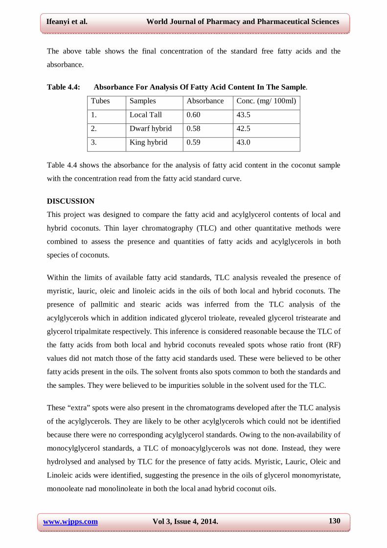
Table 4.4: Absorbance For Analysis Of Fatty Acid Content In The Sample.
Tubes Samples Absorbance Conc. (mg/ 100ml)
1. Local Tall 0.60 43.5
2. Dwarf hybrid 0.58 42.5
3. King hybrid 0.59 43.0
Table 4.4 shows the absorbance for the analysis of fatty acid content in the coconut sample
with the concentration read from the fatty acid standard curve.
DISCUSSION
This project was designed to compare the fatty acid and acylglycerol contents of local and
hybrid coconuts. Thin layer chromatography (TLC) and other quantitative methods were
combined to assess the presence and quantities of fatty acids and acylglycerols in both
species of coconuts.
Within the limits of available fatty acid standards, TLC analysis revealed the presence of
myristic, lauric, oleic and linoleic acids in the oils of both local and hybrid coconuts. The
presence of pallmitic and stearic acids was inferred from the TLC analysis of the
acylglycerols which in addition indicated glycerol trioleate, revealed glycerol tristearate and
glycerol tripalmitate respectively. This inference is considered reasonable because the TLC of
the fatty acids from both local and hybrid coconuts revealed spots whose ratio front (RF)
values did not match those of the fatty acid standards used. These were believed to be other
fatty acids present in the oils. The solvent fronts also spots common to both the standards and
the samples. They were believed to be impurities soluble in the solvent used for the TLC.
These “extra” spots were also present in the chromatograms developed after the TLC analysis
of the acylglycerols. They are likely to be other acylglycerols which could not be identified
because there were no corresponding acylglycerol standards. Owing to the non-availability of
monocylglycerol standards, a TLC of monoacylglycerols was not done. Instead, they were
hydrolysed and analysed by TLC for the presence of fatty acids. Myristic, Lauric, Oleic and
Linoleic acids were identified, suggesting the presence in the oils of glycerol monomyristate,
monooleate nad monolinoleate in both the local anad hybrid coconut oils.
www.wjpps.com Vol 3, Issue 4, 2014.
131
Ifeanyi et al. World Journal of Pharmacy and Pharmaceutical Sciences
pQuantitatively, the local coconut was found to have much higher triacylglycerol content-
733.30mg/ 100ml in comparison with 466.70mg/ 100ml in the hybrid coconut. This is
surprising since the hybrid species was thought to be endowed with higher biosynthetic
capacity. However, this can be rationalized on the basis of the generally greater access to
more sunlight for enhanced photosynthesis. Since the hybrid species are generally shorter,
they may have been shielded from sunlight by taller plants hence lower photosynthetic
activity resulting in lower acylglycerol content. It is also possible that the local breed is
genetically endowed to synthesize more tracylglycerol in their endosperms than the hybrid
ones. Generally, monoacylglycerol levels were found to be low in both local and hybrid
coconuts constituting 0.011mg/ 100ml and 0.012mg/ 100ml of the oils from both species.
This finding is consistent with what is known of plant oils, i.e, they are mainly triacylglycerol
with only trace amountsof fatty acids, mono-and diacylglycerols.
Although the triacylglycerol contents of the local and hybrid coconuts differed markedly
(733.30mg/ 100ml and 466.67mg/ 100ml respectively), their total free fatty acids were almost
equal 43.50mg/L and 43.00mg/L respectively. This would suggest that the fatty acids in the
local coconuts might be in a form that it is easily hydrolysable.
CONCLUSION
The local and hybrid coconuts contain the same types of fatty acids but not in equal amounts.
Since the local species contain more triacylglycerol per mg endosperm, it is more desirable to
use it as a source of coconut oils. The only disadvantage may be difficulty of harvesting its
fruits since it is usually taller and requires climbing the tree with its associated dangers. If
they could be modified (genetically) to bear fruits without growing very tall, they will be
economically and nutritionally better than the hybrid ones we have today.
REFERENCES
1. Adams, C.R., Bamford, K.M. and M.D. (1999). Principles of Horticulture, 3rd Edition,
Butter Worth-Heinmann, 192-193.
2. Anthony, Y. (1988). Introduction to Tropical Agriculture 1st Edition, Longman. 139-140.
3. Arthur, C. G. And John E.H. (2000) biochemical facts of dietary fats.
4. Medical physiology 10: 782-784.
5. Bartman, I. (1956). Analysis of technical monoglyceride with Potassium periodate.
Journal of Analytical Biochemistry. 81: 67-68.

www.wjpps.com Vol 3, Issue 4, 2014.
132
Ifeanyi et al. World Journal of Pharmacy and Pharmaceutical Sciences
6. Bock, K. R., Ivory, H. M and Adam, B. R. (1970). Lethal Bole Root diseases of Coconut
In East Africa. Annual Applied biology. 66: 455-464.
7. Boyer, D. E. and King J.P. (1977). Analysis of heart associated lipids. Journal of
Chromatography. 143: 473-480.
8. Brook, R. M. (1982). The hybrid coconut project in Papua New Guinea: its past, present
and Future. Indian Coconut Journal. 12: 3-8.
9. Dole, V. P. (1956). Extraction of free fatty acids Journal of Clinical Investigation. 35:
150-151.
10. Fletcher, M. J, (1968). A colorimetric method for estimating serum triacylglycerols.
Clinical chim Acta. 12: 393-394.
11. Gill, L. S. (1988). Taxonomy of flowering plants. 1st Edition Africn Feb. Publishers. 276-
279
12. Harpers, (2000). Harpers Biochemistry. 25th Edition, Appleton and Lange. 160-165.
13. Harries, H. C. (1978). The evolution, Dissemination and Classification of
14. Cocos Nucifera Laboratory Bot. Review. 44: 265-319.
15. Kessler, G. and Lederers, H. (1965). Flurometric measurement of Triglyceride. 3rd
Edition University of London press. 341-344.
16. Liyanage, D. V. (1967). Identification of Genotype of coconuts suitable for breeding.
Experimental Agriculture. 3: 205-210
17. Luntungan, H. T. (1982). Progress Report on coconut breeding in Indonesia. Food and
Agriculture Organization of the United Nations. 14: 2-3.
18. Marinethi, G. V. (1962). Metabolism of lipids. Journal lipid Research 3: 1-4.
19. Purseglove, J. W. (1972). Monocotyledon, Tropical Crops. 1: 440-476.
20. Robbelen, G. and Downey, R. K. (1989). Oil crops of the world. New York. 2: 4-
13Tindall, H. D. and Anacholi, B. C. (1989). Tropical Agriculture handbook, Cash crops.
1st Edition, Machmillan Publishers. 41-46.
21. Varner, E. J. and Banna, J. (1976). Plant Biochemistry. 3rd Edition, Academic press 423-
424.
Copyright © 2022 FDOKUMEN

