


Bahasa
Halaman
Hukum

ORIGINAL INVESTIGATION
β-Arrestin 2 knockout mice exhibit sensitizeddopamine release and increased reward in response to a lowdose of alcohol
Karl Björk & Valeria Tronci & Annika Thorsell &Gianluigi Tanda & Natalie Hirth & Markus Heilig &
Anita C. Hansson & Wolfgang H. Sommer
Received: 24 January 2013 /Accepted: 30 May 2013# Springer-Verlag Berlin Heidelberg 2013
AbstractRationale The rewarding effects of alcohol have been attributedto interactions between opioid and dopaminergic system withinthe mesolimbic reward pathway.We have previously shown thatablation of β-arrestin 2 (Arrb2), a crucial regulator of μ-opioidreceptor function, attenuates alcohol-induced hyperlocomotionand c-fos activation in the nucleus accumbens.Objectives Here, we further investigated the role of Arrb2 inmodulating alcohol-induced dopamine (DA) release and con-ditioned place preference (CPP). We also assessed the
functional importance of Arrb2 for μ-opioid receptor surfaceexpression and signaling following an acute alcohol challenge.Methods Alcohol-evoked (0.375, 0.75, and 1.5 g/kg intraper-itoneally) DA release was measured by in vivo microdialysisin the shell of nucleus accumbens. Reward was assessed bythe CPP paradigm. Receptor function was assessed by μ-receptor binding and [35S]GTP-γ-S autoradiography.Results In Arrb2 knockout mice accumbal DA levels reachmaximum response at a lower dose compared to wild-type(wt) animals. In line with these results, Arrb2 knockout micedisplay increased CPP for alcohol as compared to wt mice.Finally, Arrb2 mutant mice display increased μ-opioid re-ceptor signaling in the ventral and dorsal striatum and amyg-dala in response to a low dose of alcohol, indicating impaireddesensitization mechanisms in these mice.Conclusions Our results show that Arrb2 modulates theresponse to low doses of alcohol on various levels includingμ-opioid receptor signaling, DA release, and reward. Theyalso reveal a clear dissociation between the effects of Arrb2on psychomotor and reward behaviors.
Keywords Arrestin . Opioid . Dopamine . Alcohol .
Reward . Nucleus accumbens
Introduction
The positively reinforcing effects of alcohol have been main-ly attributed to the opioid and dopaminergic systems.Alcohol ingestion causes a release of endogenous opioidsthat disinhibits the mesolimbic reward pathway, resulting indopamine (DA) overflow in the shell of the nucleusaccumbens (Di Chiara and Imperato 1988; Johnson andNorth 1992; Spanagel et al. 1992; Di Chiara et al. 1996;Tanda and Di Chiara 1998; Acquas et al. 2002). In addition,
Electronic supplementary material The online version of this article(doi:10.1007/s00213-013-3166-x) contains supplementary material,which is available to authorized users.
K. BjörkTranslational Neuropharmacology, Department of ClinicalNeuroscience, Karolinska Institute, Stockholm, Sweden
V. Tronci :G. TandaPsychobiology Section, NIDA, National Institutes of Health,Baltimore, MD, USA
K. Björk :A. Thorsell :M. Heilig :A. C. Hansson :W. H. SommerLaboratory of Clinical and Translational Studies, NIAAA,National Institutes of Health, Bethesda, MD, USA
A. ThorsellDepartment of Clinical and Experimental Medicine, LinköpingUniversity, Linköping, Sweden
N. Hirth :A. C. Hansson :W. H. SommerInstitute of Psychopharmacology at Central Institute of MentalHealth, Medical Faculty Mannheim, University of Heidelberg,Mannheim, Germany
K. Björk (*)Karolinska Institute, Center for Molecular Medicine, Section forTranslational Neuropharmacology, L8:01 Karolinska UniversityHospital, 171 76 Stockholm, Swedene-mail: [email protected]
PsychopharmacologyDOI 10.1007/s00213-013-3166-x
opioids can exert rewarding properties through direct effectsin the nucleus accumbens, independent of opioid receptoractivation in the ventral tegmental area (VTA) (Vaccarinoet al. 1986; Simmons and Self 2009). The framework foralcohol reward outlined above stems from preclinical studiesin rodents. As of yet, human studies have not conclusivelybeen able to establish the same sequence of events. However,a recent 11C-carfentanil displacement study showed thatalcohol intake indeed results in release of endogenous opi-oids (Mitchell et al. 2012). Meanwhile, opioid-dependentDA release in response to alcohol in humans is supportedby the finding that genetic variation at the μ-opioid receptorgene locus moderates alcohol-induced DA release in thenucleus accumbens measured by 11C-raclopride displace-ment (Ramchandani et al. 2011).
The components of the opioid and dopaminergic systems,i.e., neurotransmitters and corresponding receptors are sub-ject to complex regulation on multiple levels, includinggenetic, transcriptional, and posttranscriptional modulation.In addition, opioid and DA receptor function is affected byprotein–protein interactions with adaptor or scaffolding pro-teins. These proteins bind to intracellular portions of G-protein coupled receptors (GPCRs) and modulate severalfacets of receptor function including trafficking, G-protein-dependent and -independent signaling (Bockaert et al. 2010;Bjork and Svenningsson 2011). Notably, several adaptorproteins have been observed to modulate addiction-relatedphenotypes (Ron and Messing 2013)
β-arrestin 2 (Arrb2) is an adaptor protein that is importantfor the regulation of receptors belonging to both dopaminer-gic and opioid systems (Schmid and Bohn 2009; Skinbjerget al. 2009). It is ubiquitously expressed throughout mam-malian cell types, and together with its homologue, β-arrestin 1, it is responsible for the ligand-induced internali-zation and desensitization of most if not all, GPCRs (Shenoyand Lefkowitz 2005; Schmid and Bohn 2009). A series ofpapers have demonstrated that Arrb2 is crucial for desensi-tization of μ-opioid receptors, by facilitating internalizationof the receptor and uncoupling of the associated G-protein(Bohn et al. 1999; Bohn et al. 2000; Bohn et al. 2003). Theimportance of this interaction in vivo is shown by the obser-vation that Arrb2 knockout mice display prolonged analgesiain response to the prototypical μ-opioid receptor agonist,morphine, compared to wild-type (wt) animals. These micealso show an increased sensitivity to the rewarding effectsand enhanced accumbal DA release following morphineadministration (Bohn et al. 2003).
We have previously reported that rats selectively bred foralcohol preference show altered Arrb2 mRNA levels inseveral brain regions compared to their non-preferring coun-terparts, and mice lacking the Arrb2 gene exhibit reducedalcohol-induced locomotion and c-fos activation in the shellof nucleus accumbens in response to a low dose of alcohol,
suggesting impaired alcohol reward (Arlinde et al. 2004;Bjork et al. 2008). These results are contrary to what wasinitially expected. Given the role of Arrb2 in regulation of theμ-opioid receptor, its deletion was expected to augment re-ward from alcohol, through increased opioid tone in the VTAand disinhibition of DA neurons projecting to the nucleusaccumbens. To elucidate this apparent discrepancy, weassessed accumbal DA release in Arrb2 knockout mice fol-lowing increasing doses of alcohol. To obtain a more directmeasure of alcohol reward, we also tested the Arrb2 knockoutmice for conditioned place preference (CPP) for alcohol.
Experimental procedures
Animals
Arrb2 knockout mice were generously provided by Prof.Robert J Lefkowitz, Duke University, Chapel Hill, NorthCarolina (Bohn et al. 1999). They were bred and maintainedat the National Institute on Alcohol Abuse and Alcoholism(NIAAA) in accordance with NIH recommendations (Bjorket al. 2008). All experiments were approved by the NIAAAAnimal Care and Use Committee.
In vivo microdialysis
As previously published (Tanda et al. 2009; Loland et al.2012), anesthetized mice (ketamine, 60.0 mg/kg intraper-itoneally (i.p.), and xylazine,12.0 mg/kg i.p.) were randomlyimplanted in the right or the left nucleus accumbens shell witha concentric dialysis probe (AN69 dialyzing membranes,Hospal Dasco,Bologna, Italy), under continuous perfusion,according to the mouse brain atlas by Paxinos and Franklin(Paxinos and Franklin 2004) (anterior=+1.5, lateral=±0.6,vertical=−5.2; mm relative to the bregma). The exposed dia-lyzing surface of the membrane was limited to the lowest1.0 mm portion of the probes. After surgery, mice wereallowed to recover overnight in square cages equipped withoverhead quartz-lined fluid swivels (Instech Laboratories Inc.,PlymouthMeeting, PA) for connections to the dialysis probes.All subsequent studies were conducted in these cages.Microdialysis test sessions started approximately 24 h afterthe surgical procedures in freely moving mice. Collection ofdialysate samples (10 μl) started after about 30 min followingperfusion with Ringer’s solution, and samples collected every10 min were immediately analyzed for DA content. Micereceived ethanol (0.375, 0.75, and 1.5 g/kg i.p.) or salineinjections only when stable DA values (less than 15 % vari-ability) were obtained for at least three consecutive samples(approximately after about 1 h). Sample collection continuedevery 10 min for about 150 min. Dialysate samples (10 μl)were injected without purification into a high-performance
Psychopharmacology

liquid chromatography apparatus to quantify DA. Potentialsfor the oxidation and reduction electrodes of the analytical cell(5014B; ESA, Chelmsford, MA) were set at +125 and−125 mV, respectively. The mobile phase, containing100 mM NaH2PO4, 0.1 mM Na2EDTA, 0.5 mM n-octylsulfate, and 18 % (v/v) methanol (pH adjusted to 5.5 withNa2HPO4), was pumped by an ESA 582 (ESA, Chelmsford,MA) solvent delivery module at 0.50 ml/min. Assay sensitiv-ity for DA was 2 fmol per sample. At the end of the experi-ment, mice were euthanized by pentobarbital overdose, andbrains were removed and left to fix in 4 % formaldehyde insaline solution. Brains were sliced, using a vibratome(Vibratome Plus, The Vibratome Company, St. Louis, MO,USA), in serial coronal slices in order to identify the locationof the probes. Only data from animals for which probe trackswere within the nucleus accumbens shell boundaries wereused for results described in the manuscript. Differences inbasal levels of DA between genotypes and experimentalgroups were analyzed by one- or two-way ANOVA.Statistical analysis of experimental results was done after datawere expressed as a percentage of basal DAvalues and carriedout with Statistica 6 software using a repeated measure overtime three-way ANOVA (genotype, drug dose, and time asfactors), with results from treatments showing overall changessubjected to post hoc Tukey’s test. Results were consideredsignificant at p<0.05. Area under the curve was also estimatedand compared by two-way ANOVA. The wt n=29 and forArrb2 knockout mice n=25.
CPP for alcohol
CPP was carried out as described previously (Thorsell et al.2010). However, we chose a dose of 1.0 g/kg alcohol whichis half of that which is normally used for CPP (2.0 g/kg). Thisdose is too low to induce CPP in wt mice (Tzschentke 2007).The reason for this choice of dose is that we wanted to test ifthe Arrb2 knockout mice were more sensitive to the reward-ing effects of alcohol in the CPP paradigm. A brief descrip-tion of the CPP protocol: the apparatus (MedAssociates,Burlington, VT, USA) consisted of two equally illuminatedcompartments, one compartment had black walls with a gridfloor and the other had white walls with a wire mesh floorhoused connected by an opening equipped with a guillotine-door. The conditioning apparatus was placed in a sound-attenuated chamber equipped with a fan for noise reduction.Time spent and locomotor activity within each compartmentwas measured by photo beams. During the habituation andtest session, animals were allowed access to both compart-ments, while during the conditioning sessions, the guillotine-door was closed and the animals only allowed access to oneside of the apparatus. During the habituation session, the gridand mesh floors were covered with solid Plexiglas to preventany conditioning to the floor texture. Mice were given an i.p.
saline injection and allowed to freely explore the apparatusfor 5 min. Twenty-four hours later, the mice began condi-tioning trials using an unbiased design in which the drug-paired side was counterbalanced within and between groups.On alternating days, animals were injected with alcohol(1.0 g/kg, CS+ trials) or saline (CS− trials) and placed intothe appropriate compartment for 5 min. One complete trialcomposed a CS+ trial and a CS− trial. After eight condition-ing trials, mice received a preference test in which theyreceived a saline injection and were placed into the centerof the apparatus and allowed to freely explore the wholeapparatus for 30 min. (n=10–13/group). Data were analyzedusing repeated measurement ANOVA.
Receptor autoradiography
Wt and Arrb2 knockout mice were administered either salineor alcohol (0.75 g/kg i.p., n=3–5/group). After 45 min, micewere sacrificed by decapitation and brains were quicklyremoved and frozen in isopentane at −40 °C. The brainswere stored at −80 °C until further usage. Twelve-micrometer sections were cut on a cryostat at Bregma level+1, −1.3, and −3.5 mm according to Paxinos and Franklin(Paxinos and Franklin 2004) and thaw-mounted ontogelatin-coated slides. For [3H]-[D-Ala2, NMe-Phe4, Gly5-ol]-enkephalin (DAMGO) autoradiography, sections werebrought up to room temperature, incubated for 15 min atroom temperature in 50 mM Tris–HCl buffer (pH 7.4) con-taining 5 mM MgCl2 and 1 mM Ethylenediaminetetraaceticacid (EDTA, Sigma-Aldrich, St. Louis, MO, USA). Thisincubation step was repeated with a fresh buffer. Sectionswere then transferred into humidified chambers and 800 μlof reaction mix was applied to each slide and sections wereincubated for 2 h at 30 °C. Reaction mix contained 1 nM[3H]-DAMGO (Sp. Act. 51 Ci/mmol, PerkinElmer, MA,USA) prepared in a buffer containing 50 mM Tris–HCl(pH 7.4), 5 mM MgCl2, 1 mM EDTA, 0.1 mM bacitracin(Sigma-Aldrich) and 0.1 % bovine serum albumin (Sigma-Aldrich). Non-specific binding was measured on adjacentsections with addition of 1 μM D-Phe-Cys-Tyr-D-Trp-Orn-Thr-Pen-Thr-NH(2) (CTOP) (Tocris Bioscience, Bristol,UK). Incubation was stopped by washing the slides for2 min in ice-cold buffer (50 mM Tris–HCl, pH 7.4); this stepwas repeated two more times and followed by a dip in ice-cold deionized water. Sections were dried under a stream ofcold air and exposed against FUJI imaging plates (StoragePhosphor Screen BAS-IP TR2025 E Tritium Screen, GEHealthcare Life Sciences, Pittsburgh, USA) for 10 days andscanned in a phosphorimager (Fuji BAS-5000 Phosphorimager,GE Healthcare Life Sciences). Densitometry analysis wasperformed using the MCID program (InterFocus ImagingLtd., Cambridge, UK). Signal density was measured asphotostimulable luminescence per square millimeter, compared
Psychopharmacology
against standard curves generated using [3H]-Microscales(Amersham, GE Healthcare Life Sciences), and data (innanocurie per milligram) were converted to femtomole receptorper milligram protein tissue equivalence. Specific binding wasdefined as a difference between total and non-specific binding.Values were statistically analyzed within a region by two-wayANOVA (genotype×treatment). To correct for multiple testingwith a family-wise error rate of 0.05, Holm’s correctedBonferroni procedure was applied. If a regional p value forgenotype or genotype×treatment interaction survived this pro-cedure, the effect of alcohol within a genotype was analyzed byTukey’s post hoc test.
[35S]GTP-γ-S autoradiography
Sections were prepared as described above. Slides werebrought up to room temperature and incubated for 15 minat room temperature in 20 mM Tris–HCl pH 7.2, 5 mMMgCl2, 1 mM EDTA. Incubation was repeated once more,and slides were transferred into humidified chambers.Eight hundred microliters of incubation buffer (20 mMTris–HCl pH 7.2, 5 mM MgCl2, 1 mM EDTA, 100 mMNaCl, 1 mM Dithiothreitol, 0.1 % BSA) containing 1 mMGDP (Sigma-Aldrich) was applied to each slide, andslides were incubated for 15 min at room temperature.Solution was discarded and a mix of 50pM [35S]GTP-γ-S(PerkinElmer), 2 mM GDP, and 10 μM DAMGO (TocrisBioscience) or vehicle (30 % acetonitrile/water) in incu-bation buffer were applied; slides were incubated for 60 minat 30 °C. Agonist-stimulated and baseline GTP-γ-S bind-ings were measured on adjacent sections. Incubation wasstopped by washing the slides for 2 min in ice-coldbuffer (20 mM Tris–HCl pH 7.2, 100 mM NaCl); thisstep was repeated two more times and followed by a dipin ice-cold deionized water. Sections were dried under astream of cold air and exposed against FUJI imagingplates (Storage Phosphor Screen BAS-IP SR2025 Screen,GE Healthcare Life Sciences) for several hours and scannedusing a phosphorimager (Fuji BAS-5000). Densitometryanalysis was performed using the MCID program;measurements were compared against standard curvesgenerated using [14C]-Microscales (Amersham, GE HealthcareLife Sciences). Agonist-stimulated GTP-γ-S binding wascalculated as percent of baseline value in the sameregion and animal. Values were expressed as percent stimula-tion. Statistical analysis was as described for the bindingexperiment.
Results
Baseline DAvalues did not differ between the genotypes (wt,20.13±2.29 fmol/sample±SEM, n=29; Arrb2 knockout
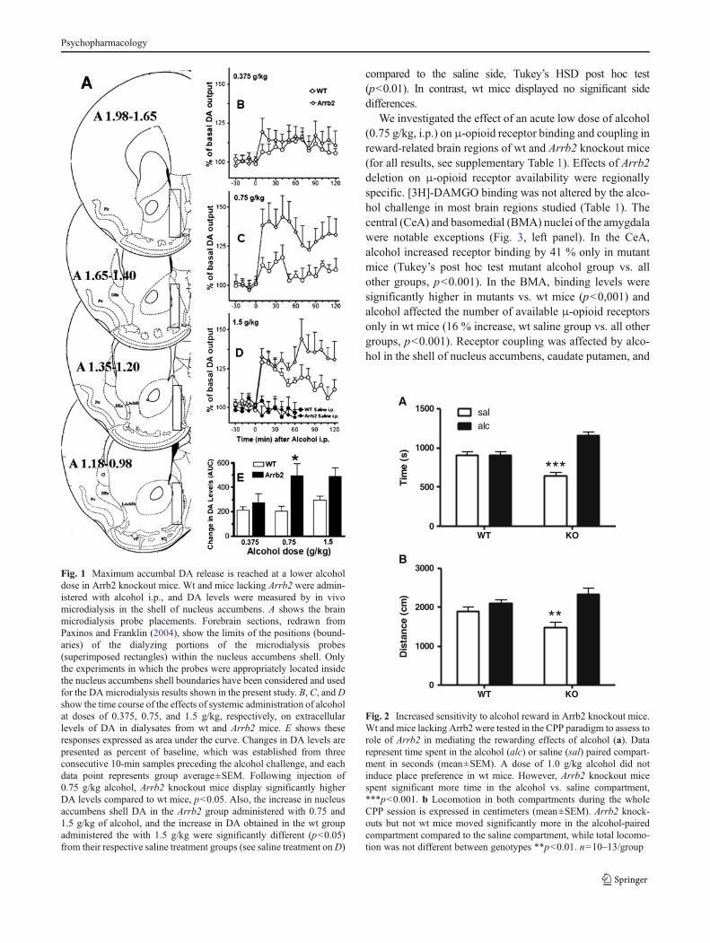
mice, 20.65±2.47 fmol/sample±SEM, n=25), as shown byANOVA F[1,52]=0.023, p>0.8, and between the differentexperimental treatment groups as shown by two-wayANOVA, dose: F[3,46]=0.759, p>0.5; interaction: F[3,46]=0.039, p>0.9. Wt and Arrb2 knockout mice were assessed forsaline- and alcohol-induced DA release at three different alco-hol doses (0.375, 0.75, and 1.5 g/kg). Figure 1 shows the timecourse for these effects (B, C, and D in the figure), and thesame effects displayed as changes in DA expressed as areaunder the curve (E). A three-way repeated measures over timeANOVA showed a significant main effect of genotype(F[1,47]=6.66, p<0.05), alcohol dose (F[3,47]=14.24,p<0.05), and time (F[6,282]=12.798, p<0.05), significantgenotype×dose interaction F[3,47]=2.84, p<0.05, time×doseinteraction (F[18,282]=3.41, p<0.05) and non-significant in-teractions of time×genotype, and time×dose×genotype. TheTukey’s post hoc test showed that alcohol-evoked DA levelsbetween genotypes were not different at the 0.375 and 1.5 g/kgdose. However, the acute challenge with the intermediatealcohol dose (0.75 g/kg) showed a significantly greater in-crease in DA levels within the nucleus accumbens shell regionof Arrb2 knockout mice compared to wt mice (p<0.05). TheTukey’s post hoc test also identified the Arrb2 group thatreceived the 0.75 g/kg dose as significantly different from itssaline control group (p<0.05), and the 1.5 g/kg wt and Arrb2groups as significantly different from their respective salinecontrol groups (p<0.05), while the other groups did not differsignificantly from their respective control groups.
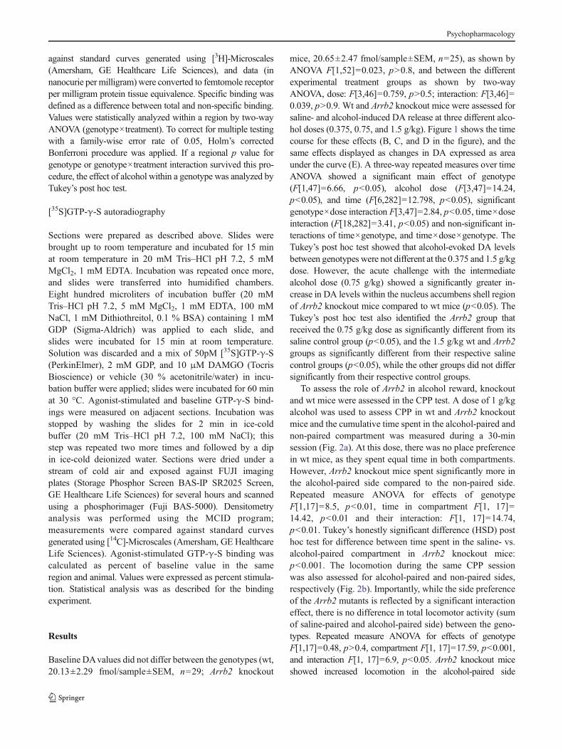
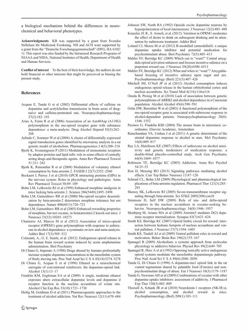
To assess the role of Arrb2 in alcohol reward, knockoutand wt mice were assessed in the CPP test. A dose of 1 g/kgalcohol was used to assess CPP in wt and Arrb2 knockoutmice and the cumulative time spent in the alcohol-paired andnon-paired compartment was measured during a 30-minsession (Fig. 2a). At this dose, there was no place preferencein wt mice, as they spent equal time in both compartments.However, Arrb2 knockout mice spent significantly more inthe alcohol-paired side compared to the non-paired side.Repeated measure ANOVA for effects of genotypeF[1,17]=8.5, p<0.01, time in compartment F[1, 17]=14.42, p<0.01 and their interaction: F[1, 17]=14.74,p<0.01. Tukey’s honestly significant difference (HSD) posthoc test for difference between time spent in the saline- vs.alcohol-paired compartment in Arrb2 knockout mice:p<0.001. The locomotion during the same CPP sessionwas also assessed for alcohol-paired and non-paired sides,respectively (Fig. 2b). Importantly, while the side preferenceof the Arrb2 mutants is reflected by a significant interactioneffect, there is no difference in total locomotor activity (sumof saline-paired and alcohol-paired side) between the geno-types. Repeated measure ANOVA for effects of genotypeF[1,17]=0.48, p>0.4, compartment F[1, 17]=17.59, p<0.001,and interaction F[1, 17]=6.9, p<0.05. Arrb2 knockout miceshowed increased locomotion in the alcohol-paired side
Psychopharmacology
compared to the saline side, Tukey’s HSD post hoc test(p<0.01). In contrast, wt mice displayed no significant sidedifferences.
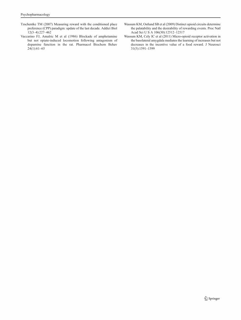
We investigated the effect of an acute low dose of alcohol(0.75 g/kg, i.p.) on μ-opioid receptor binding and coupling inreward-related brain regions of wt and Arrb2 knockout mice(for all results, see supplementary Table 1). Effects of Arrb2deletion on μ-opioid receptor availability were regionallyspecific. [3H]-DAMGO binding was not altered by the alco-hol challenge in most brain regions studied (Table 1). Thecentral (CeA) and basomedial (BMA) nuclei of the amygdalawere notable exceptions (Fig. 3, left panel). In the CeA,alcohol increased receptor binding by 41 % only in mutantmice (Tukey’s post hoc test mutant alcohol group vs. allother groups, p<0.001). In the BMA, binding levels weresignificantly higher in mutants vs. wt mice (p<0,001) andalcohol affected the number of available μ-opioid receptorsonly in wt mice (16 % increase, wt saline group vs. all othergroups, p<0.001). Receptor coupling was affected by alco-hol in the shell of nucleus accumbens, caudate putamen, and
Fig. 1 Maximum accumbal DA release is reached at a lower alcoholdose in Arrb2 knockout mice. Wt and mice lacking Arrb2 were admin-istered with alcohol i.p., and DA levels were measured by in vivomicrodialysis in the shell of nucleus accumbens. A shows the brainmicrodialysis probe placements. Forebrain sections, redrawn fromPaxinos and Franklin (2004), show the limits of the positions (bound-aries) of the dialyzing portions of the microdialysis probes(superimposed rectangles) within the nucleus accumbens shell. Onlythe experiments in which the probes were appropriately located insidethe nucleus accumbens shell boundaries have been considered and usedfor the DAmicrodialysis results shown in the present study. B, C, andDshow the time course of the effects of systemic administration of alcoholat doses of 0.375, 0.75, and 1.5 g/kg, respectively, on extracellularlevels of DA in dialysates from wt and Arrb2 mice. E shows theseresponses expressed as area under the curve. Changes in DA levels arepresented as percent of baseline, which was established from threeconsecutive 10-min samples preceding the alcohol challenge, and eachdata point represents group average±SEM. Following injection of0.75 g/kg alcohol, Arrb2 knockout mice display significantly higherDA levels compared to wt mice, p<0.05. Also, the increase in nucleusaccumbens shell DA in the Arrb2 group administered with 0.75 and1.5 g/kg of alcohol, and the increase in DA obtained in the wt groupadministered the with 1.5 g/kg were significantly different (p<0.05)from their respective saline treatment groups (see saline treatment onD)
WT KO0
500
1000
1500 salalc
***
Tim
e (s
)
WT KO0
1000
2000
3000
**
Dis
tan
ce (
cm)
A
B
Fig. 2 Increased sensitivity to alcohol reward in Arrb2 knockout mice.Wt and mice lacking Arrb2 were tested in the CPP paradigm to assess torole of Arrb2 in mediating the rewarding effects of alcohol (a). Datarepresent time spent in the alcohol (alc) or saline (sal) paired compart-ment in seconds (mean±SEM). A dose of 1.0 g/kg alcohol did notinduce place preference in wt mice. However, Arrb2 knockout micespent significant more time in the alcohol vs. saline compartment,***p<0.001. b Locomotion in both compartments during the wholeCPP session is expressed in centimeters (mean±SEM). Arrb2 knock-outs but not wt mice moved significantly more in the alcohol-pairedcompartment compared to the saline compartment, while total locomo-tion was not different between genotypes **p<0.01. n=10–13/group
Psychopharmacology
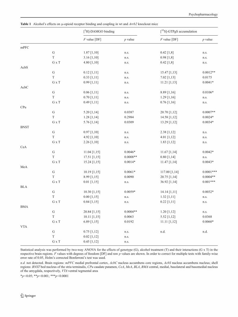
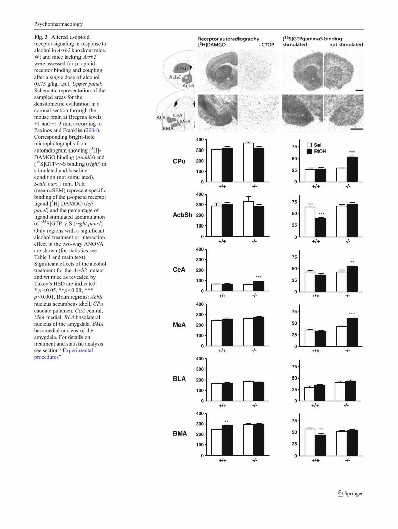
Table 1 Alcohol’s effects on μ-opioid receptor binding and coupling in wt and Arrb2 knockout mice
[3H]-DAMGO binding [35S]-GTPgS accumulation
F value [DF] p value F value [DF] p value
mPFC
G 1.87 [1,10] n.s. 0.42 [1,8] n.s.
T 3.16 [1,10] n.s. 0.98 [1,8] n.s.
G x T 4.80 [1,10] n.s. 0.42 [1,8] n.s.
AcbS
G 0.12 [1,11] n.s. 15.47 [1,13] 0.0012**
T 0.33 [1,11] n.s. 7.02 [1,13] 0.0175
G x T 0.99 [1,11] n.s. 11.21 [1,13] 0.0041*
AcbC
G 0.06 [1,11] n.s. 8.89 [1,16] 0.0106*
T 0.70 [1,11] n.s. 1.29 [1,16] n.s.
G x T 0.49 [1,11] n.s. 0.76 [1,16] n.s.
CPu
G 5.20 [1,14] 0.0387 20.70 [1,12] 0.0007**
T 1.28 [1,14] 0.2984 14.58 [1,12] 0.0024*
G x T 5.76 [1,14] 0.0309 13.29 [1,12] 0.0034*
BNST
G 0.97 [1,10] n.s. 2.38 [1,12] n.s.
T 4.92 [1,10] n.s. 4.01 [1,12] n.s.
G x T 2.26 [1,10] n.s. 1.83 [1,12] n.s.
CeA
G 11.04 [1,15] 0.0046* 11.67 [1,14] 0.0042*
T 17.51 [1,15] 0.0008** 0.80 [1,14] n.s.
G x T 15.24 [1,15] 0.0014* 11.47 [1,14] 0.0043*
MeA
G 10.19 [1,15] 0.0061* 117.00 [1,14] 0.0001***
T 8.99 [1,15] 0.0090 20.75 [1,14] 0.0004**
G x T 0.01 [1,15] n.s. 36.92 [1,14] 0.001***
BLA
G 10.30 [1,15] 0.0059* 14.14 [1,11] 0.0032*
T 0.00 [1,15] n.s. 1.32 [1,11] n.s.
G x T 0.84 [1,15] n.s. 0.22 [1,11] n.s.
BMA
G 20.84 [1,15] 0.0004** 1.20 [1,12] n.s.
T 10.11 [1,15] 0.0063 5.52 [1,12] 0.0368
G x T 6.89 [1,15] 0.0192 11.11 [1,12] 0.0060*
VTA
G 0.75 [1,12] n.s. n.d. n.d.
T 0.02 [1,12] n.s.
G x T 0.45 [1,12] n.s.
Statistical analysis was performed by two-way ANOVA for the effects of genotype (G), alcohol treatment (T) and their interactions (G x T) in therespective brain regions. F values with degrees of freedom [DF] and raw p values are shown. In order to correct for multiple tests with family-wiseerror rate of 0.05, Holm’s corrected Bonferroni’s test was used.
n.d. not detected. Brain regions: mPFC medial prefrontal cortex, AcbC nucleus accumbens core regions, AcbS nucleus accumbens nucleus; shellregions: BNST bed nucleus of the stria terminalis, CPu caudate putamen, CeA,MeA, BLA, BMA central, medial, basolateral and basomedial nucleusof the amygdala, respectively, VTA ventral tegmental area
*p<0.05; **p<0.001; ***p<0.0001
Psychopharmacology
0
100
200
300
400
+/+ -/-0
25
50
75
+/+ -/-
***
0
100
200
300
400
+/+ -/-0
25
50
75
+/+ -/-
***
SalEtOH
0
100
200
300
400
+/+ -/-
***
0
25
50
75
+/+ -/-
**
0
100
200
300
400
+/+ -/-0
25
50
75
+/+ -/-
***
0
100
200
300
400
+/+ -/-0
25
50
75
+/+ -/-
0
25
50
75
+/+ -/-
**
0
100
200
300
400
+/+ -/-
**
AcbSh
CPu
CeA
MeA
BLA
BMA
Fig. 3 Altered μ-opioidreceptor signaling in response toalcohol in Arrb2 knockout mice.Wt and mice lacking Arrb2were assessed for μ-opioidreceptor binding and couplingafter a single dose of alcohol(0.75 g/kg, i.p.). Upper panel:Schematic representation of thesampled areas for thedensitometric evaluation in acoronal section through themouse brain at Bregma levels+1 and −1.3 mm according toPaxinos and Franklin (2004).Corresponding bright-fieldmicrophotographs fromautoradiogram showing [3H]-DAMGO binding (middle) and[35S]GTP-γ-S binding (right) instimulated and baselinecondition (not stimulated).Scale bar: 1 mm. Data(mean±SEM) represent specificbinding of the μ-opioid receptorligand [3H] DAMGO (leftpanel) and the percentage ofligand stimulated accumulationof [35S]GTP-γ-S (right panel).Only regions with a significantalcohol treatment or interactioneffect in the two-way ANOVAare shown (for statistics seeTable 1 and main text).Significant effects of the alcoholtreatment for the Arrb2 mutantand wt mice as revealed byTukey’s HSD are indicated:* p <0.05, **p<0.01, ***p<0.001. Brain regions: AcbSnucleus accumbens shell, CPucaudate putamen, CeA central,MeA medial, BLA basolateralnucleus of the amygdala, BMAbasomedial nucleus of theamygdala. For details ontreatment and statistic analysissee section “Experimentalprocedures”
Psychopharmacology
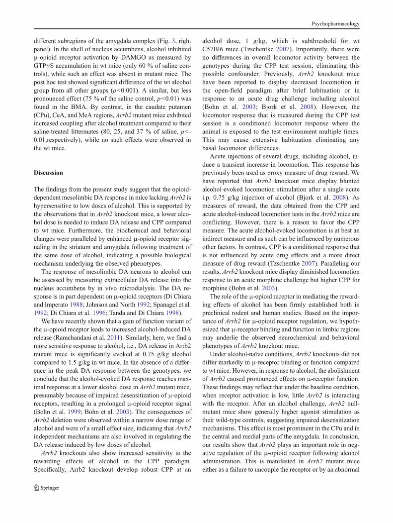
different subregions of the amygdala complex (Fig. 3, rightpanel). In the shell of nucleus accumbens, alcohol inhibitedμ-opioid receptor activation by DAMGO as measured byGTPγS accumulation in wt mice (only 60 % of saline con-trols), while such an effect was absent in mutant mice. Thepost hoc test showed significant difference of the wt alcoholgroup from all other groups (p<0.001). A similar, but lesspronounced effect (75 % of the saline control, p<0.01) wasfound in the BMA. By contrast, in the caudate putamen(CPu), CeA, and MeA regions, Arrb2mutant mice exhibitedincreased coupling after alcohol treatment compared to theirsaline-treated littermates (80, 25, and 37 % of saline, p<-0.01,respectively), while no such effects were observed inthe wt mice.
Discussion
The findings from the present study suggest that the opioid-dependent mesolimbic DA response in mice lacking Arrb2 ishypersensitive to low doses of alcohol. This is supported bythe observations that in Arrb2 knockout mice, a lower alco-hol dose is needed to induce DA release and CPP comparedto wt mice. Furthermore, the biochemical and behavioralchanges were paralleled by enhanced μ-opioid receptor sig-naling in the striatum and amygdala following treatment ofthe same dose of alcohol, indicating a possible biologicalmechanism underlying the observed phenotypes.
The response of mesolimbic DA neurons to alcohol canbe assessed by measuring extracellular DA release into thenucleus accumbens by in vivo microdialysis. The DA re-sponse is in part dependent on μ-opioid receptors (Di Chiaraand Imperato 1988; Johnson and North 1992; Spanagel et al.1992; Di Chiara et al. 1996; Tanda and Di Chiara 1998).
We have recently shown that a gain of function variant ofthe μ-opioid receptor leads to increased alcohol-induced DArelease (Ramchandani et al. 2011). Similarly, here, we find amore sensitive response to alcohol, i.e., DA release in Arrb2mutant mice is significantly evoked at 0.75 g/kg alcoholcompared to 1.5 g/kg in wt mice. In the absence of a differ-ence in the peak DA response between the genotypes, weconclude that the alcohol-evoked DA response reaches max-imal response at a lower alcohol dose in Arrb2 mutant mice,presumably because of impaired desensitization of μ-opioidreceptors, resulting in a prolonged μ-opioid receptor signal(Bohn et al. 1999; Bohn et al. 2003). The consequences ofArrb2 deletion were observed within a narrow dose range ofalcohol and were of a small effect size, indicating that Arrb2independent mechanisms are also involved in regulating theDA release induced by low doses of alcohol.
Arrb2 knockouts also show increased sensitivity to therewarding effects of alcohol in the CPP paradigm.Specifically, Arrb2 knockout develop robust CPP at an
alcohol dose, 1 g/kg, which is subthreshold for wtC57Bl6 mice (Tzschentke 2007). Importantly, there wereno differences in overall locomotor activity between thegenotypes during the CPP test session, eliminating thispossible confounder. Previously, Arrb2 knockout micehave been reported to display decreased locomotion inthe open-field paradigm after brief habituation or inresponse to an acute drug challenge including alcohol(Bohn et al. 2003; Bjork et al. 2008). However, thelocomotor response that is measured during the CPP testsession is a conditioned locomotor response where theanimal is exposed to the test environment multiple times.This may cause extensive habituation eliminating anybasal locomotor differences.
Acute injections of several drugs, including alcohol, in-duce a transient increase in locomotion. This response haspreviously been used as proxy measure of drug reward. Wehave reported that Arrb2 knockout mice display bluntedalcohol-evoked locomotion stimulation after a single acutei.p. 0.75 g/kg injection of alcohol (Bjork et al. 2008). Asmeasures of reward, the data obtained from the CPP andacute alcohol-induced locomotion tests in the Arrb2mice areconflicting. However, there is a reason to favor the CPPmeasure. The acute alcohol-evoked locomotion is at best anindirect measure and as such can be influenced by numerousother factors. In contrast, CPP is a conditioned response thatis not influenced by acute drug effects and a more directmeasure of drug reward (Tzschentke 2007). Paralleling ourresults, Arrb2 knockout mice display diminished locomotionresponse to an acute morphine challenge but higher CPP formorphine (Bohn et al. 2003).
The role of the μ-opioid receptor in mediating the reward-ing effects of alcohol has been firmly established both inpreclinical rodent and human studies. Based on the impor-tance of Arrb2 for μ-opioid receptor regulation, we hypoth-esized that μ-receptor binding and function in limbic regionsmay underlie the observed neurochemical and behavioralphenotypes of Arrb2 knockout mice.
Under alcohol-naïve conditions, Arrb2 knockouts did notdiffer markedly in μ-receptor binding or function comparedto wt mice. However, in response to alcohol, the abolishmentof Arrb2 caused pronounced effects on μ-receptor function.These findings may reflect that under the baseline condition,when receptor activation is low, little Arrb2 is interactingwith the receptor. After an alcohol challenge, Arrb2 null-mutant mice show generally higher agonist stimulation astheir wild-type controls, suggesting impaired desensitizationmechanisms. This effect is most prominent in the CPu and inthe central and medial parts of the amygdala. In conclusion,our results show that Arrb2 plays an important role in neg-ative regulation of the μ-opioid receptor following alcoholadministration. This is manifested in Arrb2 mutant miceeither as a failure to uncouple the receptor or by an abnormal
Psychopharmacology
coupling compared to wt mice, both of these processespresumably lead to a sustained μ-opioid receptor signaling.
Enhanced μ-opioid receptor signaling is a plausible mech-anism for the hypersensitivity to alcohol’s rewarding effectsobserved in Arrb2 knockout mice. Even though the VTA isconsidered the primary locus for this receptors action onalcohol reward, we do not find changes on μ-opioid receptorbinding in this region. However, there is an emerging literaturesupporting the notion that the increases in μ-opioid receptorfunction we observed in the striatum and amygdala may beequally important. Intra-accumbal injections of μ-opioid re-ceptor agonists, including the endogenous ligandβ-endorphin,promote cocaine reinstatement in rats. Pretreatment with an-tagonists specifically in the nucleus accumbens abolished thiseffect (Simmons and Self 2009). Furthermore, a recent posi-tron emission tomography study in humans showed that a0.5 mg/kg oral dose of amphetamine induced displacementof radiolabeled carfentanil in frontal cortex, putamen, caudatethalamus anterior cingulate cortex, and insula (Colasanti et al.2012). A possible interpretation of this study is that accumbalDA release in the striatum may serve as an antecedent signalfor μ-opioid receptor activation.
On a functional level, highly regionally specific actions ofμ-opioid receptors mediate different aspects of reward.Wassum et al. reported that receptor blockade in the ventraland dorsal striatum attenuates encoding of food palatabilityfollowing deprivation in rats, whereas the number of reward-seeking actions was unaffected (Wassum et al. 2009). In thebasolateral amygdala, the relationship was found to be thereverse; site-specific μ-opioid receptor blockade in this re-gion did not influence food palatability but abolishedreward-seeking behavior. Similar results were obtained in amore recent study were the μ-opioid agonist DAMGO wasinjected in the basolateral amygdala (Wassum et al. 2011).The dual role of μ-opioid receptor neurotransmission inmediating emotional and motivational aspects of rewardbehavior has been extensively studied by the Berridge labo-ratory. In their conceptualization, they distinguish betweenhedonic effects of reward (typically referred to as “liking”)and its motivational value or incentive salience (“wanting”),the later potentially contributing to addictive behaviors(Robinson and Berridge 2003). Originally, hedonic effectswere thought to be mediated by opioids and salience attribu-tion by dopamine. However it is becoming clear that μ-opioidreceptors are involved in both aspects of reward. In a recentstudy, the group identified a hedonic hotspot in the posteriorventral pallidum that upon local μ-opioid receptor stimulationincreased both hedonic reactions to sucrose and eating behav-ior. Furthermore, they identified an additional site in themedial shell of nucleus accumbens and showed that thesetwo hotspots reciprocally interact via opioid neurotransmis-sion (Smith and Berridge 2007; Smith et al. 2009). Alongthese lines, a role of μ-opioid receptor activation in the CeA
for incentive salience has also been suggested (Mahler andBerridge 2009; Mahler and Berridge 2012). Based on thesestudies, it has been proposed that μ-opioid receptors in twodistinct anatomical loci facilitate different aspects of reward.Receptors in the striatum mediate hedonic effects of a rewardor its associated cue, whereas μ-opioid receptors within theamygdala encode the incentive value of these stimuli.
Our results suggest that differences in Arrb2 levels mayunderlie innate differences in alcohol reward specifically atlow doses. This effect occurs within a narrow dose rangearound 0.75–1 g/kg, whereas at a dose of 1.5 g/kg, alcoholinduces a similar DA release in both Arrb2 knockouts and wtmice. Dose-dependent responses are quite common inregards to alcohol; it is for example well known that low tomoderate doses stimulate locomotion whereas higher dosescause sedative responses (Spanagel 2009). A plausible ex-planation for such dose-dependent differences in effect isthat alcohol might target different receptors or neurotrans-mitter systems depending on dose. Neurochemical supportfor this idea has for example been provided in a study byFranklin et al., where they show that specifically a dose of1.0 but not 0.5 or 2.0 g/kg alcohol increases DA release in theshell of nucleus accumbens by acting on the D2 receptor(Franklin et al. 2009). Similarly, the Arrb2-dependent effectsobserved here on DA levels and reward may be due toengagement of the μ-opioid receptor that is restricted toalcohol doses around 0.75–1 g/kg.
Whether such differences translate into an increased riskfor developing alcohol addiction in humans remain unclear.A recent genetic study failed to show an association betweengenetic variants in the human ARRB2 gene and alcoholism,suggesting that this may not be the case (Oneda et al. 2010).However, genetic factors have been shown to influence theresponse to medications used in the treatment of alcoholism,most notably exemplified by A118G variant in the humanOPRM1 gene (Oslin et al. 2003; Ray and Hutchison 2007).Although this variant has inconsistently been associated withalcoholism, there is a strong correlation between genotypeand treatment response to naltrexone in human alcoholics(Oslin et al. 2003; Arias et al. 2006; Ray and Hutchison2007; Heilig et al. 2011; Chamorro et al. 2012; Kranzleret al. 2012). Given that this variant also modulates the DAresponse to alcohol (Ramchandani et al. 2011), it would be ofgreat interest to test the hypothesis that in similarity to theA118G variation in the human μ-opioid receptor gene, dif-ferent genetic variants of the human ARRB2 gene could actas predictors of treatment outcome in the treatment of alco-holics with naltrexone.
In summary, our data show that Arrb2 negatively regu-lates DA release in the nucleus accumbens specifically at lowdoses of alcohol. Furthermore, Arrb2 also appear to dampenthe rewarding behavioral output of such doses of alcohol. Wealso observed alterations in μ-receptor signaling that may be
Psychopharmacology
a biological mechanism behind the differences in neuro-chemical and behavioral phenotypes.
Acknowledgments KB was supported by a grant from SvenskaStiftelsen för Medicinsk Forskning. NH and ACH were supported bya grant from the “Deutsche Forschungsgemeinschaft” (DFG, HA 6102/1). This report was also funded by the Intramural Research Programs ofNIAAA and NIDA, National Institutes of Health, Department of Healthand Human Services.
Conflict of interest To the best of their knowledge, the authors do nothold financial or other interests that might be perceived as biasing thepresent study.
References
Acquas E, Tanda G et al (2002) Differential effects of caffeine ondopamine and acetylcholine transmission in brain areas of drug-naive and caffeine-pretreated rats. Neuropsychopharmacology27(2):182–193
Arias A, Feinn R et al (2006) Association of an Asn40Asp (A118G)polymorphism in the mu-opioid receptor gene with substancedependence: a meta-analysis. Drug Alcohol Depend 83(3):262–268
Arlinde C, Sommer W et al (2004) A cluster of differentially expressedsignal transduction genes identified by microarray analysis in a ratgenetic model of alcoholism. Pharmacogenomics J 4(3):208–218
Bjork K, Svenningsson P (2011) Modulation of monoamine receptorsby adaptor proteins and lipid rafts: role in some effects of centrallyacting drugs and therapeutic agents. Annu Rev Pharmacol Toxicol51:211–242
Bjork K, Rimondini R et al (2008) Modulation of voluntary ethanolconsumption by beta-arrestin 2. FASEB J 22(7):2552–2560
Bockaert J, Perroy J et al (2010) GPCR interacting proteins (GIPs) inthe nervous system: Roles in physiology and pathologies. AnnuRev Pharmacol Toxicol 50:89–109
Bohn LM, Lefkowitz RJ et al (1999) Enhanced morphine analgesia inmice lacking beta-arrestin 2. Science 286(5449):2495–2498
Bohn LM, Gainetdinov RR et al (2000) Mu-opioid receptor desensiti-zation by beta-arrestin-2 determines morphine tolerance but notdependence. Nature 408(6813):720–723
Bohn LM, Gainetdinov RR et al (2003) Enhanced rewarding propertiesof morphine, but not cocaine, in beta(arrestin)-2 knock-out mice. JNeurosci 23(32):10265–10273
Chamorro AJ, Marcos M et al (2012) Association of micro-opioidreceptor (OPRM1) gene polymorphism with response to naltrex-one in alcohol dependence: a systematic review and meta-analysis.Addict Biol 17(3):505–512
Colasanti, A., G. E. Searle, et al. (2012). Endogenous opioid release inthe human brain reward system induced by acute amphetamineadministration. Biol Psychiatry
Di Chiara G, Imperato A (1988) Drugs abused by humans preferentiallyincrease synaptic dopamine concentrations in the mesolimbic systemof freely moving rats. Proc Natl Acad Sci U S A 85(14):5274–5278
Di Chiara G, Acquas E et al (1996) Ethanol as a neurochemicalsurrogate of conventional reinforcers: the dopamine-opioid link.Alcohol 13(1):13–17
Franklin KM, Engleman EA et al (2009) A single, moderate ethanolexposure alters extracellular dopamine levels and dopamine dreceptor function in the nucleus accumbens of wistar rats.Alcohol Clin Exp Res 33(10):1721–1730
Heilig M, Goldman D et al (2011) Pharmacogenetic approaches to thetreatment of alcohol addiction. Nat Rev Neurosci 12(11):670–684
Johnson SW, North RA (1992) Opioids excite dopamine neurons byhyperpolarization of local interneurons. J Neurosci 12(2):483–488
Kranzler, H. R., S. Armeli, et al. (2012). Variation in OPRM1moderatesthe effect of desire to drink on subsequent drinking and its atten-uation by naltrexone treatment. Addict Biol
Loland CJ, Mereu M et al (2012) R-modafinil (armodafinil): a uniquedopamine uptake inhibitor and potential medication forpsychostimulant abuse. Biol Psychiatry 72(5):405–413
Mahler SV, Berridge KC (2009) Which cue to “want?” Central amyg-dala opioid activation enhances and focuses incentive salience on aprepotent reward cue. J Neurosci 29(20):6500–6513
Mahler SV, Berridge KC (2012) What and when to “want”? Amygdala-based focusing of incentive salience upon sugar and sex.Psychopharmacology (Berl) 221(3):407–426
Mitchell JM, O’Neil JP et al (2012) Alcohol consumption inducesendogenous opioid release in the human orbitofrontal cortex andnucleus accumbens. Sci Transl Med 4(116):116ra116
Oneda B, Preisig M et al (2010) Lack of association between geneticpolymorphisms of ARRB2 and alcohol dependence in a Caucasianpopulation. Alcohol Alcohol 45(6):590–591
Oslin DW, Berrettini W et al (2003) A functional polymorphism of themu-opioid receptor gene is associated with naltrexone response inalcohol-dependent patients. Neuropsychopharmacology 28(8):1546–1552
Paxinos G, Franklin KBJ (2004) The mouse brain in stereotaxic co-ordinates. Elsevier Academic, Amsterdam
Ramchandani VA, Umhau J et al (2011) A genetic determinant of thestriatal dopamine response to alcohol in men. Mol Psychiatry16(8):809–817
Ray LA, Hutchison KE (2007) Effects of naltrexone on alcohol sensi-tivity and genetic moderators of medication response: adouble-blind placebo-controlled study. Arch Gen Psychiatry64(9):1069–1077
Robinson TE, Berridge KC (2003) Addiction. Annu Rev Psychol54:25–53
Ron D, Messing RO (2013) Signaling pathways mediating alcoholeffects. Curr Top Behav Neurosci 13:87–126
Schmid CL, Bohn LM (2009) Physiological and pharmacological im-plications of beta-arrestin regulation. Pharmacol Ther 121(3):285–293
Shenoy SK, Lefkowitz RJ (2005) Seven-transmembrane receptor sig-naling through beta-arrestin. Sci STKE 2005(308):cm10
Simmons D, Self DW (2009) Role of mu- and delta-opioidreceptors in the nucleus accumbens in cocaine-seeking be-havior. Neuropsychopharmacology 34(8):1946–1957
Skinbjerg M, Ariano MA et al (2009) Arrestin3 mediates D(2) dopa-mine receptor internalization. Synapse 63(7):621–624
Smith KS, Berridge KC (2007) Opioid limbic circuit for reward: inter-action between hedonic hotspots of nucleus accumbens and ven-tral pallidum. J Neurosci 27(7):1594–1605
Smith KS, Tindell AJ et al (2009) Ventral pallidum roles in reward andmotivation. Behav Brain Res 196(2):155–167
Spanagel R (2009) Alcoholism: a systems approach from molecularphysiology to addictive behavior. Physiol Rev 89(2):649–705
Spanagel R, Herz A et al (1992) Opposing tonically active endogenousopioid systems modulate the mesolimbic dopaminergic pathway.Proc Natl Acad Sci U S A 89(6):2046–2050
Tanda G, Di Chiara G (1998) A dopamine-mu1 opioid link in the ratventral tegmentum shared by palatable food (Fonzies) and non-psychostimulant drugs of abuse. Eur J Neurosci 10(3):1179–1187
Tanda G, Newman AH et al (2009) Combinations of cocaine with otherdopamine uptake inhibitors: assessment of additivity. J PharmacolExp Ther 330(3):802–809
Thorsell A, Schank JR et al (2010) Neurokinin-1 receptors (NK1R:s),alcohol consumption, and alcohol reward in mice.Psychopharmacology (Berl) 209(1):103–111
Psychopharmacology
Tzschentke TM (2007) Measuring reward with the conditioned placepreference (CPP) paradigm: update of the last decade. Addict Biol12(3–4):227–462
Vaccarino FJ, Amalric M et al (1986) Blockade of amphetaminebut not opiate-induced locomotion following antagonism ofdopamine function in the rat. Pharmacol Biochem Behav24(1):61–65
Wassum KM, Ostlund SB et al (2009) Distinct opioid circuits determinethe palatability and the desirability of rewarding events. Proc NatlAcad Sci U S A 106(30):12512–12517
Wassum KM, Cely IC et al (2011) Micro-opioid receptor activation inthe basolateral amygdala mediates the learning of increases but notdecreases in the incentive value of a food reward. J Neurosci31(5):1591–1599
Psychopharmacology
Top Related
Copyright © 2022 FDOKUMEN

