


Bahasa
Halaman
Hukum

Role for the pleckstrin homology domain-containing protein CKIP-1 in AP-1 regulationand apoptosis
Lingqiang Zhang1, Guichun Xing1, Yi Tie2,Ying Tang1, Chunyan Tian1, Li Li1,Libo Sun1, Handong Wei1, Yunping Zhu1
and Fuchu He1,3,*1Department of Genomics and Proteomics, Beijing Institute of RadiationMedicine, Chinese Human Genome Center of Beijing, Beijing, PR China,2Department of Biochemistry and Molecular Biology, Beijing Institute ofRadiation Medicine, Chinese Human Genome Center of Beijing, Beijing,PR China and 3Institutes of Biomedical Sciences, Fudan University,Shanghai, PR China
The oncogenic transcription factor c-Jun plays an impor-
tant role in cell proliferation, transformation and differ-
entiation. All identified c-Jun-interacting proteins are
localized to the nucleus or cytoplasm and function in
their intact forms. Here we show that the pleckstrin
homology domain-containing protein CKIP-1 (casein ki-
nase 2-interacting protein-1) functions as a plasma mem-
brane-bound AP-1 regulator. During apoptosis, CKIP-1 is
cleaved by caspase-3 and translocated to the cytoplasm
and then to the nucleus. C-terminal fragments of cleaved
CKIP-1 strongly repress AP-1 activity. Importantly, CKIP-1
overexpression promotes apoptosis by forming a positive
feedback loop between CKIP-1 and caspase-3. RNA inter-
ference of CKIP-1 or overexpression of c-Jun attenuates the
sensitivity to apoptosis, indicating a novel role of CKIP-1 in
apoptosis. CKIP-1 is the first case of a c-Jun-interacting
protein that regulates AP-1 activity via caspase-3-depen-
dent cleavage and translocation.
The EMBO Journal (2005) 24, 766–778. doi:10.1038/
sj.emboj.7600532; Published online 10 February 2005
Subject Categories: signal transduction; differentiation
& death
Keywords: AP-1; apoptosis; caspase-3; CKIP-1
Introduction
c-Jun, the core component of the AP-1 family of transcription
factors (Shaulian and Karin, 2001, 2002), is regulated by post-
translational modifications, for example, phosphorylation,
acetylation and ubiquitination by interacting proteins (Karin
et al, 1997; Nateri et al, 2004; Wertz et al, 2004). More than
60 proteins, including bZIP factors, non-bZIP DNA-binding
proteins, coactivators that do not bind DNA directly and
structural components of the nucleus, have been demon-
strated to interact with c-Jun (Chinenov and Kerppola, 2001).
Unlike other transcription factors, such as STATs, NF-kB,
Notch and tubby, which are localized outside of the nucleus
(cytoplasmic or plasma membrane-associated) until particu-
lar signaling events trigger their nuclear translocation, c-Jun/
AP-1 family members are nuclear proteins constitutively
bound to DNA at most times (Brivanlou and Darnell, 2002).
All of the identified c-Jun-interacting proteins are localized to
the nucleus or cytoplasm and function in their intact forms.
For example, Smad and JAB1 translocate from the cytoplasm
to the nucleus, where they bind to and regulate c-Jun/AP-1
activity (Chinenov and Kerppola, 2001). However, whether
plasma membrane-associated proteins could transduce sig-
nals directly from the cell surface to the nuclear c-Jun/AP-1 is
unknown. We have evaluated if the plasma membrane
pleckstrin homology (PH) domain-containing protein CKIP-
1 (casein kinase 2-interacting protein-1) would play such
a role.
CKIP-1 contains a C-terminal putative c-Jun-interacting
region comprising 72 amino acids (aa 338–409, named
C-term1), which was first isolated in a two-hybrid screen as
an interacting partner of the c-Jun leucine zipper (Chevray
and Nathans, 1992), implying CKIP-1 as a novel c-Jun/AP-1
regulator. Full-length CKIP-1 was identified in a two-hybrid
screen as an interacting partner of the CK2 catalytic subunit
and shown to include an N-terminal PH domain (Bosc et al,
2000). CKIP-1 is implicated in phosphatidylinositol 3-kinase-
regulated muscle differentiation (Safi et al, 2004) as well as in
the recruitment of CK2 to the plasma membrane (Olsten et al,
2004).
CKIP-1 might mediate a novel regulatory mechanism for
AP-1 activity. The C-term1 domain of CKIP-1 possesses a
leucine zipper motif (aa 352–380) predicted to be capable of
forming coiled coils. However, CKIP-1 lacks either an adja-
cent basic region typical of bZIP proteins to the leucine zipper
or any other DNA-binding motifs, indicating that CKIP-1 is
not a transcription factor and distinct from other c-Jun bZIP
region-binding proteins. Indeed, proteins with both a PH
domain and a leucine zipper motif have been rarely observed,
that is, only in proteins such as AFAP-110, b-Pix, APPL and
Vav (Romero and Fischer, 1996; Mitsuuchi et al, 1999;
Baisden et al, 2001; Kim et al, 2001). None of these proteins
have been functionally related to AP-1. Hence, the PH domain
of CKIP-1 predicts plasma membrane localization, whereas
the leucine zipper may retain CKIP-1 within the nucleus.
Here we propose that CKIP-1 functions as a plasma mem-
brane-bound AP-1 regulator that translocates to the nucleus.
We provide evidence that CKIP-1 has the capacity to interact
with c-Jun and JunD, but not JunB, c-Fos or ATF2, in the
yeast-two hybrid system. Although full-length CKIP-1 is
localized to the plasma membrane in mammalian cells, we
observed that the CKIP-1-C-term1 domain localizes to the
nucleus, where it is colocalized and can be co-immunopreci-
pitated with c-Jun. We further demonstrated that the C-term1Received: 28 June 2004; accepted: 3 December 2004; publishedonline: 10 February 2005
*Corresponding author. Beijing Institute of Radiation Medicine, 27Taiping Road, Beijing 100850, PR China. Tel./Fax: þ 86 10 681 712 08;E-mail: [email protected]
The EMBO Journal (2005) 24, 766–778 | & 2005 European Molecular Biology Organization | All Rights Reserved 0261-4189/05
www.embojournal.org
The EMBO Journal VOL 24 | NO 4 | 2005 &2005 European Molecular Biology Organization
EMBO
THE
EMBOJOURNAL
THE
EMBOJOURNAL
766
domain and another truncated CKIP-1 construct named
C-term2 (aa 308–409) are potent negative regulators of AP-
1 activity. Furthermore, C-term1 and C-term2 fragments
could be released during apoptosis via caspase-3-dependent
cleavage. The cleavage of CKIP-1 promotes apoptosis via
amplified caspase-3 activation. Knockdown of CKIP-1 by
RNA interference (RNAi) or overexpression of c-Jun attenu-
ates the sensitivity to apoptotic stimuli, indicating a novel
role of CKIP-1 in apoptosis. Therefore, CKIP-1 represses
AP-1 activity via caspase-3-dependent cleavage and translo-
cation, and this repression contributes to the execution of
apoptosis.
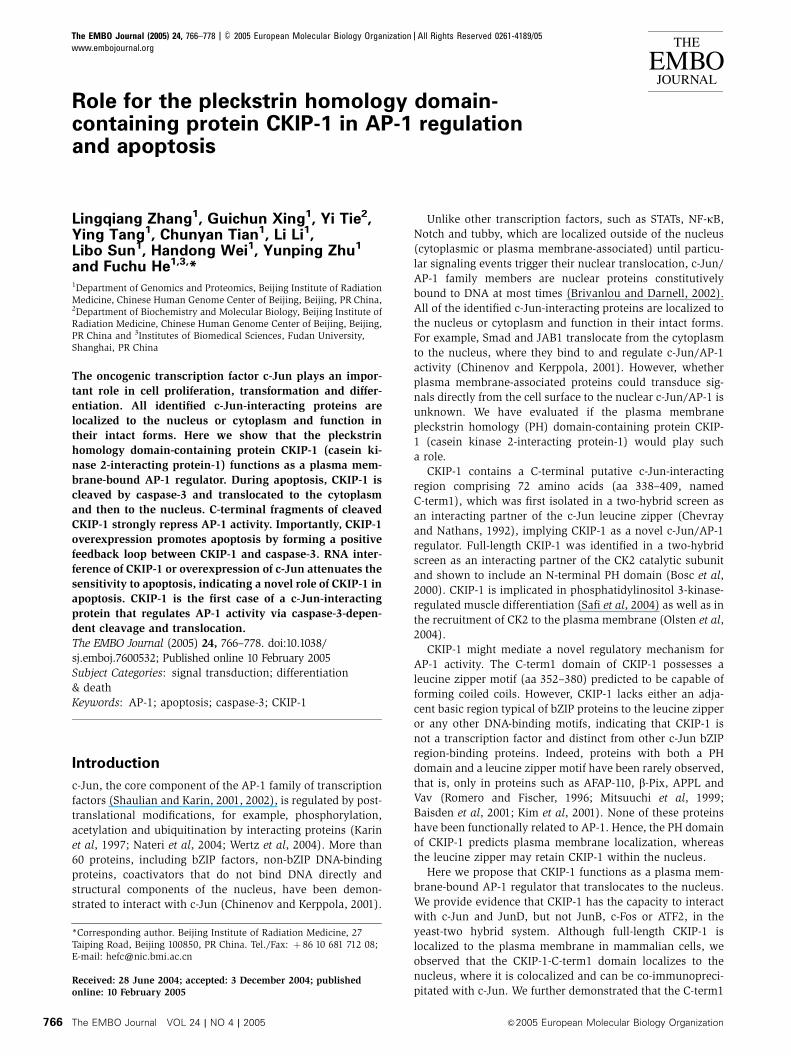
Figure 1 CKIP-1 interacts with c-Jun in vitro and in vivo. (A) To examine the interactions between CKIP-1 and c-Jun, cDNAs encoding full-length CKIP-1 or deletion mutants (C-term1, DC-term1) were expressed in yeast as fusions with the DNA activation domain (AD) of GAL4,using the plasmid pGADT7. Constructs encoding GAL4 DNA-binding domain fusions of c-Jun or deletion mutants (bzJun 247–331, lzJun 277–331, DbzJun 1–246, DlzJun 1–276) were cotransformed along with CKIP-1 constructs. The binding activity was quantified by a liquidb-galactosidase assay (n¼ 3, mean7s.d. indicated on the histograms). (B) C-term1 specially interacts with c-Jun and JunD, but not JunB, c-Fosand ATF2 among the AP-1 family (n¼ 3, mean7s.d.). (C) In vitro-translated 35S-labeled CKIP-1 proteins and purified GSTor GST-c-Jun proteinsexpressed in E. coli BL21 were used in pull-down assays. Proteins bound to GST fusion proteins were eluted from beads using Laemmli samplebuffer. CKIP-1 proteins were detected by autoradiography (upper panel), and CKIP-1 input was loaded as a control (lane 3). GST fusion proteinswere detected on immunoblots. (D) c-Jun was colocalized with C-term1 within the nucleus, but not with the plasma membrane-associated full-length CKIP-1. GFP-c-Jun and RFP-C-term1 or RFP-CKIP-1 were cotransfected into COS7 cells and visualized after 24 h. Scale bar, 10mm.(E) Flag-c-Jun was coexpressed in COS7 cells together with Myc-tagged C-term2, C-term1 or vector alone for 24 h followed by immunopre-cipitation with anti-Myc and immunoblotting with anti-Flag or anti-Myc. The arrows indicate the mobility of c-Jun, IgG (L), C-term2 and C-term1. The experiment was repeated three times with similar results observed. IP, immunoprecipitate; IB, immunoblot; lys, lysate; IgG (L), thelight chain of IgG.
Role for CKIP-1 in AP-1 regulation and apoptosisL Zhang et al
&2005 European Molecular Biology Organization The EMBO Journal VOL 24 | NO 4 | 2005 767
Results
CKIP-1 interacts with c-Jun in vitro and in vivo
We first confirmed the interaction in the yeast two-hybrid
system. C-term1 of CKIP-1 was sufficient and was required
for binding to c-Jun. The region of c-Jun required for inter-
action with CKIP-1 was mapped to the leucine zipper domain,
whereas the adjacent basic region was not required
(Figure 1A). C-term1 specifically interacts with c-Jun and
JunD, but not JunB, c-Fos and ATF2 (Figure 1B). It is of
interest that C-term1 was capable of self-interaction with
CKIP-1, probably mediated by the leucine zipper region
(aa 352–380) (Figure 1B). The interaction with c-Jun was
also confirmed by in vitro GST pull-down assays, in which
in vitro-translated CKIP-1 protein specifically bound to GST-c-
Jun but not to GST alone (Figure 1C).
We next asked whether CKIP-1 could associate with c-Jun
in vivo. In mammalian cells, CKIP-1 is predominantly loca-
lized to the plasma membrane and therefore unable to
interact with nuclear c-Jun proteins (Figure 1D). In contrast,
C-term1 was partially colocalized with c-Jun in a speckle
pattern (Figure 1D). Furthermore, c-Jun was partially co-
immunoprecipitated with C-term1 (Figure 1E). As a control,
C-term2 (aa 308–409), which was previously shown to be
cytoplasmic (Bosc et al, 2000), did not associate with c-Jun
in vivo although it possesses all the sequences present in
C-term1. These results suggested that CKIP-1 could interact
with c-Jun in vitro and in vivo.
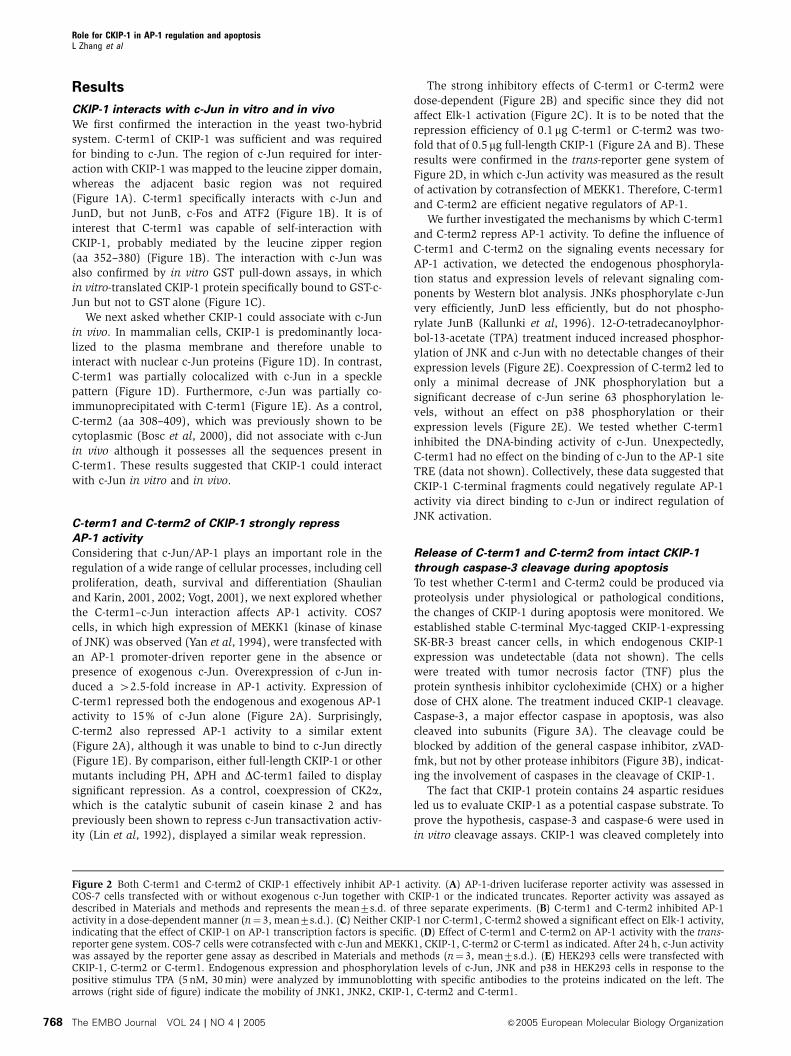
C-term1 and C-term2 of CKIP-1 strongly repress
AP-1 activity
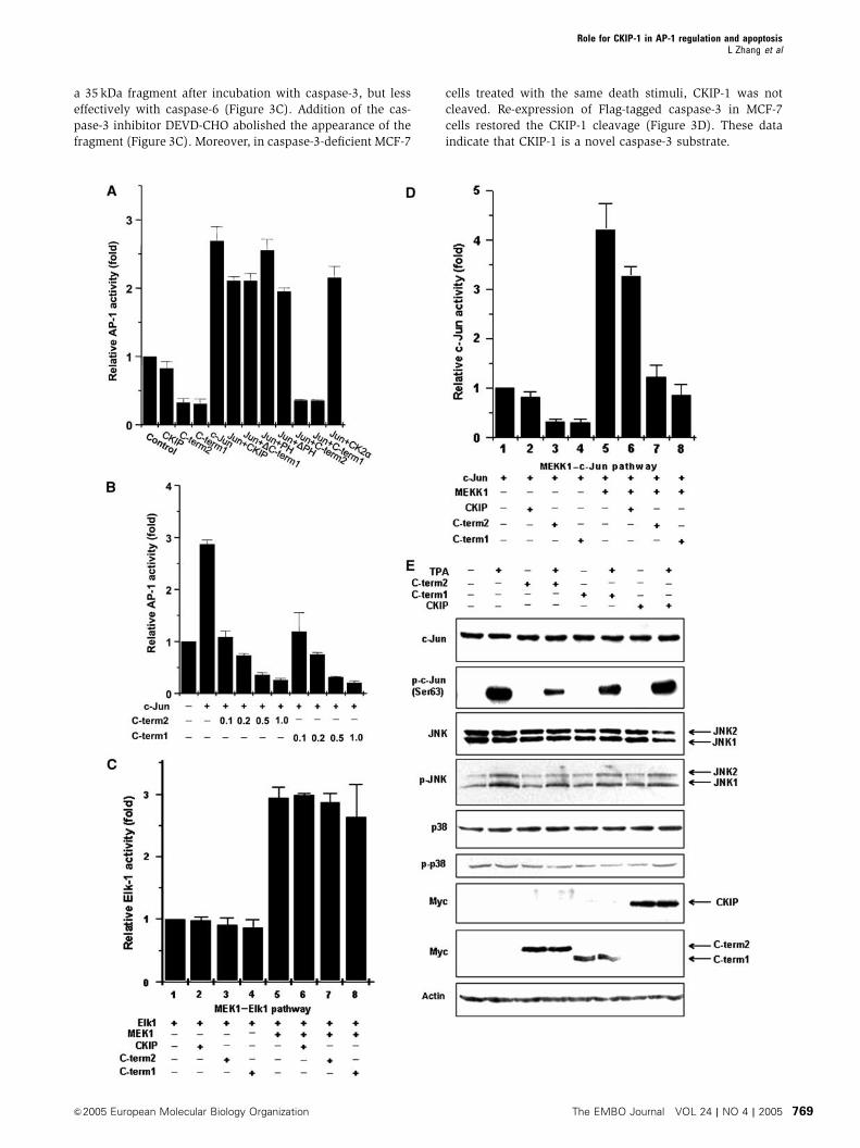
Considering that c-Jun/AP-1 plays an important role in the
regulation of a wide range of cellular processes, including cell
proliferation, death, survival and differentiation (Shaulian
and Karin, 2001, 2002; Vogt, 2001), we next explored whether
the C-term1–c-Jun interaction affects AP-1 activity. COS7
cells, in which high expression of MEKK1 (kinase of kinase
of JNK) was observed (Yan et al, 1994), were transfected with
an AP-1 promoter-driven reporter gene in the absence or
presence of exogenous c-Jun. Overexpression of c-Jun in-
duced a 42.5-fold increase in AP-1 activity. Expression of
C-term1 repressed both the endogenous and exogenous AP-1
activity to 15% of c-Jun alone (Figure 2A). Surprisingly,
C-term2 also repressed AP-1 activity to a similar extent
(Figure 2A), although it was unable to bind to c-Jun directly
(Figure 1E). By comparison, either full-length CKIP-1 or other
mutants including PH, DPH and DC-term1 failed to display
significant repression. As a control, coexpression of CK2a,
which is the catalytic subunit of casein kinase 2 and has
previously been shown to repress c-Jun transactivation activ-
ity (Lin et al, 1992), displayed a similar weak repression.
The strong inhibitory effects of C-term1 or C-term2 were
dose-dependent (Figure 2B) and specific since they did not
affect Elk-1 activation (Figure 2C). It is to be noted that the
repression efficiency of 0.1 mg C-term1 or C-term2 was two-
fold that of 0.5 mg full-length CKIP-1 (Figure 2A and B). These
results were confirmed in the trans-reporter gene system of
Figure 2D, in which c-Jun activity was measured as the result
of activation by cotransfection of MEKK1. Therefore, C-term1
and C-term2 are efficient negative regulators of AP-1.
We further investigated the mechanisms by which C-term1
and C-term2 repress AP-1 activity. To define the influence of
C-term1 and C-term2 on the signaling events necessary for
AP-1 activation, we detected the endogenous phosphoryla-
tion status and expression levels of relevant signaling com-
ponents by Western blot analysis. JNKs phosphorylate c-Jun
very efficiently, JunD less efficiently, but do not phospho-
rylate JunB (Kallunki et al, 1996). 12-O-tetradecanoylphor-
bol-13-acetate (TPA) treatment induced increased phosphor-
ylation of JNK and c-Jun with no detectable changes of their
expression levels (Figure 2E). Coexpression of C-term2 led to
only a minimal decrease of JNK phosphorylation but a
significant decrease of c-Jun serine 63 phosphorylation le-
vels, without an effect on p38 phosphorylation or their
expression levels (Figure 2E). We tested whether C-term1
inhibited the DNA-binding activity of c-Jun. Unexpectedly,
C-term1 had no effect on the binding of c-Jun to the AP-1 site
TRE (data not shown). Collectively, these data suggested that
CKIP-1 C-terminal fragments could negatively regulate AP-1
activity via direct binding to c-Jun or indirect regulation of
JNK activation.
Release of C-term1 and C-term2 from intact CKIP-1
through caspase-3 cleavage during apoptosis
To test whether C-term1 and C-term2 could be produced via
proteolysis under physiological or pathological conditions,
the changes of CKIP-1 during apoptosis were monitored. We
established stable C-terminal Myc-tagged CKIP-1-expressing
SK-BR-3 breast cancer cells, in which endogenous CKIP-1
expression was undetectable (data not shown). The cells
were treated with tumor necrosis factor (TNF) plus the
protein synthesis inhibitor cycloheximide (CHX) or a higher
dose of CHX alone. The treatment induced CKIP-1 cleavage.
Caspase-3, a major effector caspase in apoptosis, was also
cleaved into subunits (Figure 3A). The cleavage could be
blocked by addition of the general caspase inhibitor, zVAD-
fmk, but not by other protease inhibitors (Figure 3B), indicat-
ing the involvement of caspases in the cleavage of CKIP-1.
The fact that CKIP-1 protein contains 24 aspartic residues
led us to evaluate CKIP-1 as a potential caspase substrate. To
prove the hypothesis, caspase-3 and caspase-6 were used in
in vitro cleavage assays. CKIP-1 was cleaved completely into
Figure 2 Both C-term1 and C-term2 of CKIP-1 effectively inhibit AP-1 activity. (A) AP-1-driven luciferase reporter activity was assessed inCOS-7 cells transfected with or without exogenous c-Jun together with CKIP-1 or the indicated truncates. Reporter activity was assayed asdescribed in Materials and methods and represents the mean7s.d. of three separate experiments. (B) C-term1 and C-term2 inhibited AP-1activity in a dose-dependent manner (n¼ 3, mean7s.d.). (C) Neither CKIP-1 nor C-term1, C-term2 showed a significant effect on Elk-1 activity,indicating that the effect of CKIP-1 on AP-1 transcription factors is specific. (D) Effect of C-term1 and C-term2 on AP-1 activity with the trans-reporter gene system. COS-7 cells were cotransfected with c-Jun and MEKK1, CKIP-1, C-term2 or C-term1 as indicated. After 24 h, c-Jun activitywas assayed by the reporter gene assay as described in Materials and methods (n¼ 3, mean7s.d.). (E) HEK293 cells were transfected withCKIP-1, C-term2 or C-term1. Endogenous expression and phosphorylation levels of c-Jun, JNK and p38 in HEK293 cells in response to thepositive stimulus TPA (5 nM, 30 min) were analyzed by immunoblotting with specific antibodies to the proteins indicated on the left. Thearrows (right side of figure) indicate the mobility of JNK1, JNK2, CKIP-1, C-term2 and C-term1.
Role for CKIP-1 in AP-1 regulation and apoptosisL Zhang et al
The EMBO Journal VOL 24 | NO 4 | 2005 &2005 European Molecular Biology Organization768
a 35 kDa fragment after incubation with caspase-3, but less
effectively with caspase-6 (Figure 3C). Addition of the cas-
pase-3 inhibitor DEVD-CHO abolished the appearance of the
fragment (Figure 3C). Moreover, in caspase-3-deficient MCF-7
cells treated with the same death stimuli, CKIP-1 was not
cleaved. Re-expression of Flag-tagged caspase-3 in MCF-7
cells restored the CKIP-1 cleavage (Figure 3D). These data
indicate that CKIP-1 is a novel caspase-3 substrate.
Role for CKIP-1 in AP-1 regulation and apoptosisL Zhang et al
&2005 European Molecular Biology Organization The EMBO Journal VOL 24 | NO 4 | 2005 769
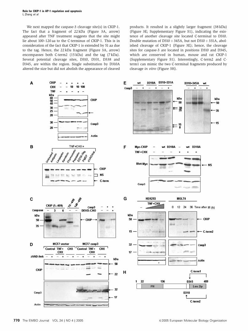
We next mapped the caspase-3 cleavage site(s) in CKIP-1.
The fact that a fragment of 22 kDa (Figure 3A, arrow)
appeared after TNF treatment suggests that the site might
lie about 100–120 aa to the C-terminus of CKIP-1. This is in
consideration of the fact that CKIP-1 is extended by 51 aa due
to the tag. Hence, the 22 kDa fragment (Figure 3A, arrow)
encompasses both C-term2 (15 kDa) and the tag (7 kDa).
Several potential cleavage sites, D310, D331, D338 and
D345, are within the region. Single substitution by D310A
altered the size but did not abolish the appearance of cleaved
products. It resulted in a slightly larger fragment (38 kDa)
(Figure 3E; Supplementary Figure S1), indicating the exis-
tence of another cleavage site located C-terminal to D310.
Double mutation of D310þ 345A, but not D310þ 331A, abol-
ished cleavage of CKIP-1 (Figure 3E); hence, the cleavage
sites for caspase-3 are located in positions D310 and D345,
which are conserved in human, mouse and rat CKIP-1
(Supplementary Figure S1). Interestingly, C-term2 and C-
term1 can mimic the two C-terminal fragments produced by
cleavage in vitro (Figure 3H).
Role for CKIP-1 in AP-1 regulation and apoptosisL Zhang et al
The EMBO Journal VOL 24 | NO 4 | 2005 &2005 European Molecular Biology Organization770
Mutant CKIP-1-D310A was resistant to cleavage in TNF-
induced apoptosis, proving D310 to be the major site in vivo
(Figure 3F). Not that only one C-terminal fragment of 22 kDa
consistent with the fragment of 15 kDa in vitro was observed
in TNF-induced apoptosis due to the fact that proteolysis of
CKIP-1 at D345 requires phosphorylation by CK2 at S346,
followed by sequential phosphorylation by glycogen synthase
kinase 3 (GSK3) at S342 (unpublished data).
More importantly, endogenous CKIP-1 could also be
cleaved in response to serum withdrawal or TNF treatment
in HEK293 cells, TNF treatment in HeLa cells and ionizing
radiation in MOLT-4 or Jurkat cells (Figure 3G, data not
shown). Taken together, these results strongly suggest that
both in vivo (at least for the D310 cleavage) and in vitro,
CKIP-1 is cleaved by caspase-3 in positions D310 and D345.
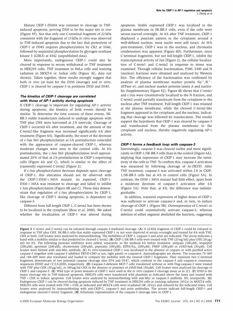
The kinetics of CKIP-1 cleavage are correlated
with those of AP-1 activity during apoptosis
If CKIP-1 cleavage is important for regulating AP-1 activity
during apoptosis, the kinetics of both events should be
similar. To determine the time courses of these events, SK-
BR-3 stable transfectants induced to undergo apoptosis with
TNF plus CHX were harvested at 2 h intervals. Cleavage of
CKIP-1 occurred 4 h after treatment, and the amount of the
C-term2-like fragment was increased significantly 6 h after
treatment (Figure 4A). Significantly, the onset of the decrease
of c-Jun S63 phosphorylation at 6 h postinduction coincided
with the appearance of caspase-cleaved CKIP-1, whereas
moderate changes were seen in the control cells. At 8 h
postinduction, the c-Jun S63 phosphorylation level approxi-
mated 20% of that at 2 h postinduction in CKIP-1-expressing
cells (Figure 4A and C), which is similar to the effect of
transiently expressed C-term2 (Figure 2).
If c-Jun phosphorylation decrease depends upon cleavage
of CKIP-1, this alteration should not be observed with
the CKIP-1-D310þ 345A mutant. As expected, CKIP-1-
D310þ 345A was resistant to cleavage and failed to inhibit
c-Jun phosphorylation (Figure 4B and C). These data demon-
strate that regulation of c-Jun phosphorylation by CKIP-1,
like cleavage of CKIP-1 during apoptosis, is dependent on
caspase-3.
Different from full-length CKIP-1, C-term2 has been shown
to be localized in the cytoplasm (Bosc et al, 2000). We asked
whether the localization of CKIP-1 was altered during
apoptosis. Stably expressed CKIP-1 was localized to the
plasma membrane in SK-BR-3 cells, even if the cells were
serum-starved overnight. At 4 h after TNF treatment, CKIP-1
displayed a punctate pattern in the cytoplasm around a
well-defined nucleus; most nuclei were still intact. At 10 h
post-treatment, CKIP-1 was in the nucleus, and chromatin
condensation was apparent (Figure 4D). Furthermore, since
C-terminal fragments, but not full-length CKIP-1, inhibit the
transcriptional activity of Jun (Figure 2), the cellular localiza-
tion of C-term1 and C-term2 in response to stress was
examined. Through cellular fractionation, P100, S100 and N
(nuclear) fractions were obtained and analyzed by Western
blot. The efficiency of the fractionation was confirmed by
analysis of plasma membrane marker protein Naþ/Kþ-
ATPase a1, and nuclear marker proteins lamin A and nucleo-
lin (Supplementary Figure S2). Figure 4E shows that C-term1
and c-Jun were constitutively localized to the N fraction, and
C-term2 could partially translocate from the cytoplasm to the
nucleus after TNF treatment. Full-length CKIP-1 was retained
at the plasma membrane, while the cleaved C-term2-like
fragment appeared in the cytoplasm and the nucleus, indicat-
ing that cleavage was followed by translocation. The results
support the hypothesis that CKIP-1 was cleaved by caspase-3
and translocated from the plasma membrane to the
cytoplasm and nucleus, thereby negatively regulating AP-1
activity.
CKIP-1 forms a feedback loop with caspase-3
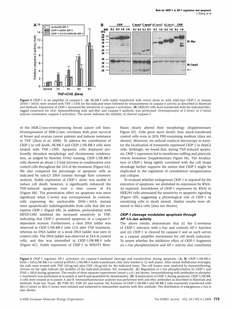
Interestingly, caspase-3 was cleaved earlier and more signifi-
cantly in CKIP-1/SK-BR-3 cells than in the control (Figure 4A),
implying that expression of CKIP-1 may increase the sensi-
tivity of the cells to TNF. To confirm this, caspase-3 activation
was measured by following cleavage of Ac-DEVD. After
TNF treatment, caspase-3 was activated within 2 h in CKIP-
1/SK-BR-3 cells but at 4 h in control cells (Figure 5A). In
contrast, the D310þ 345A mutant had little effect, apart from
a moderate decrease of caspase-3 activation after 6 h
(Figure 5A). Note that, at 8 h, the difference was indistin-
guishable.
In addition, transient expression of higher doses of CKIP-1
was sufficient to activate caspase-3 and, in turn, to induce
cleavage of CKIP-1 (Figure 5B). Overexpression of C-term1 or
C-term2 could constitutively activate caspase-3, whereas
deletion of either segment abolished the function, suggesting
Figure 3 C-term1 and C-term2 can be released through caspase-3-mediated cleavage. (A) A 22 kDa fragment of CKIP-1 could be released inresponse to TNF plus CHX. SK-BR-3 cells that stably expressed CKIP-1 or not were deprived of serum overnight and treated for 8 h with TNF,CHX or both. Cell lysates were analyzed by immunoblotting. The mobilities of CKIP-1, caspase-3 and actin are indicated. The arrow indicates aband with a mobility similar to that predicted for cleaved C-term2. (B) CKIP-1/SK-BR-3 cells were treated with TNF (20 ng/ml) plus CHX (10 mg/ml) for 8 h. The following protease inhibitors were added, separately, to the medium 4 h before treatment: antipain (100mM), leupeptin(200mM), aprotinin (200 mM), chymostatin (200mM), pepstatin (100mM), EDTA-Na2 (200 mM), PMSF (200mM) or zVAD-fmk (50mM). Celllysates were blotted with anti-Myc antibody. (C) In vitro-translated CKIP-1 was incubated in the absence of caspase or with purified activecaspase-3 (together with caspase-3 inhibitor DEVD-CHO or not, right panel) or caspase-6. Autoradiographs are shown. The truncates 75–409and 148–409 were also translated and loaded to compare the mobility with the cleaved CKIP-1 fragments. They represent two C-terminalfragments downstream of two potential caspase cleavage sites D74 and D147, which conform to the caspase-3 and caspase-6 consensussequences DXXD and (V/I/L)EXD, respectively. (D) Caspase-3-deficient MCF-7 cells transfected without or with Flag-caspase-3 were treatedwith TNF plus CHX (10 mg/ml), or 100mg/ml CHX in the absence or presence of zVAD-fmk (50mM). Cell lysates were analyzed for cleavage ofCKIP-1 and caspase-3. (E) Wild type or point mutants of CKIP-1 were used in the in vitro caspase-3 cleavage assay as in (C). (F) D310 is themajor cleavage site in TNF-induced apoptosis. HEK293 cells were transfected with plasmids as indicated above the lanes and treated withTNFþCHX to induce apoptosis. Cell lysates were analyzed by immunoblotting with anti-Myc or caspase-3 antibody. NS, nonspecific. (G)Endogenous CKIP-1 can be cleaved during apoptosis induced by TNF treatment in HEK293 cells or ionizing radiation (10 Gy) in MOLT-4 cells.HEK293 cells were treated with TNFþCHX as indicated and MOLT4 cells were irradiated (IR, 10 Gy) and cultured for the indicated times. Celllysates were analyzed by immunoblotting with anti-CKIP-C, caspase-3 and actin antibodies. The arrows indicate full-length CKIP-1 andendogenous cleaved C-term2 fragment. (H) Schematic representation of the caspase-3 cleavage sites in CKIP-1.
Role for CKIP-1 in AP-1 regulation and apoptosisL Zhang et al
&2005 European Molecular Biology Organization The EMBO Journal VOL 24 | NO 4 | 2005 771
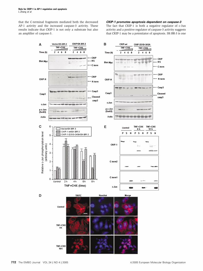
that the C-terminal fragments mediated both the decreased
AP-1 activity and the increased caspase-3 activity. These
results indicate that CKIP-1 is not only a substrate but also
an amplifier of caspase-3.
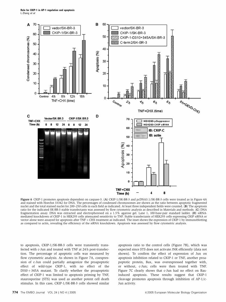
CKIP-1 promotes apoptosis dependent on caspase-3
The fact that CKIP-1 is both a negative regulator of c-Jun
activity and a positive regulator of caspase-3 activity suggests
that CKIP-1 may be a potentiator of apoptosis. SK-BR-3 is one
Role for CKIP-1 in AP-1 regulation and apoptosisL Zhang et al
The EMBO Journal VOL 24 | NO 4 | 2005 &2005 European Molecular Biology Organization772
of the HER-2/neu-overexpressing breast cancer cell lines.
Overexpression of HER-2/neu correlates with poor survival
of breast and ovarian cancer patients and induces resistance
to TNF (Zhou et al, 2000). To address the contribution of
CKIP-1 to cell death, SK-BR-3 and CKIP-1/SK-BR-3 cells were
treated with TNFþCHX. Apoptotic cells displayed pro-
foundly shrunken morphology and chromosome condensa-
tion, as judged by Hoechst 33342 staining. CKIP-1/SK-BR-3
cells showed an about 1.5-fold increase in condensation over
control cells throughout the 16 h of the treatment (Figure 6A).
We also compared the percentage of apoptotic cells as
indicated by sub-G1 DNA content through flow cytometry
analysis. Stable expression of CKIP-1 alone was unable to
induce cell death; however, it significantly enhanced the
TNF-induced apoptosis over a time course of 8 h
(Figure 6B). The potentiation effect on apoptosis was more
significant when C-term2 was overexpressed. In contrast,
cells expressing the uncleavable D310þ 345A mutant
were apoptotically indistinguishable from cells that did not
express CKIP-1 (Figure 6B). In addition, preincubation with
DEVD-CHO inhibited the increased sensitivity to TNF,
indicating that CKIP-1 promoted apoptosis in a caspase-3-
dependent manner. Furthermore, a clear DNA ladder was
observed in CKIP-1/SK-BR-3 cells 12 h after TNF treatment,
whereas no DNA ladder or a weak DNA ladder was seen in
control cells. The DNA ladder was observed at 24 h in control
cells, and this was intensified in CKIP-1/SK-BR-3 cells
(Figure 6C). Stable expression of CKIP-1 in NIH3T3 fibro-
blasts clearly altered their morphology (Supplementary
Figure S3). Cells grew more slowly than mock-transfected
control cells even in 20% FBS-containing medium (data not
shown). Moreover, we utilized confocal microscopy to moni-
tor the localization of transiently expressed CKIP-1 in HepG2
cells. Strikingly, we found that, during TNF-induced apopto-
sis, CKIP-1 expression led to membrane ruffling and pinocytic
vesicle formation (Supplementary Figure S4). The localiza-
tion of CKIP-1 being tightly correlated with the cell shape
shrinkage further supports the notion that CKIP-1 might be
implicated in the regulation of cytoskeleton reorganization
and collapse.
To evaluate whether endogenous CKIP-1 is required for the
execution of apoptosis, we abolished its expression by RNAi.
As expected, knockdown of CKIP-1 expression by RNAi in
HEK293 cells attenuated the sensitivity to apoptotic signaling
(Figure 6D), suggesting a physiological role of CKIP-1 in
sensitizing cells to death stimuli. Similar results were ob-
tained in HeLa cells (data not shown).
CKIP-1 cleavage modulates apoptosis through
AP-1/c-Jun activity
The above results demonstrate that (i) the C-terminus
of CKIP-1 interacts with c-Jun and controls AP-1 function
and (ii) CKIP-1 is cleaved by caspase-3 and as such serves
as a caspase amplifier mechanism for cell death induction.
To assess whether the inhibitory effect of CKIP-1 fragments
on c-Jun phosphorylation and AP-1 activity also contributes
Figure 4 CKIP-1 regulates AP-1 activation via caspase-3-mediated cleavage and translocation during apoptosis. (A, B) CKIP-1/SK-BR-3,D310þ345A/SK-BR-3 or control pcDNA3.1/SK-BR-3 stable transfectant cells were seeded in 12-well plates. After serum withdrawal overnight,the cells were treated with TNF (20 ng/ml) plus CHX (10mg/ml) for the indicated times. The cell lysates were analyzed by immunoblotting.Arrows on the right indicate the mobility of the indicated proteins. NS, nonspecific. (C) Regulation of c-Jun phosphorylation by CKIP-1 andD310þ345A during apoptosis. The results of three separate experiments (mean7s.d.) are shown. Immunoblotting with antibodies to phospho-c-Jun(Ser63) was performed as in panels A and B and quantified by densitometry. (D) Translocation of CKIP-1 during apoptosis. CKIP-1/SK-BR-3 cells were treated as in panels A and B. Immunofluorescence analysis was performed with anti-Myc antibodies as described in Materials andmethods. Scale bar, 10 mm. (E) P100 (P), S100 (S) and nuclear (N) fractions of CKIP-1/SK-BR-3 and SK-BR-3 cells transiently transfected withMyc-C-term1 or Myc-C-term2 were isolated and subjected to immunoblot analysis with Myc antibody. The distribution of endogenous c-Jun isalso shown.
Figure 5 CKIP-1 is an amplifier of caspase-3. (A) SK-BR-3 cells stably transfected with vector alone or with wild-type CKIP-1 or mutant(D310þ345A) were treated with TNFþCHX for the indicated times followed by measurements of caspase-3 activity as described in Materialsand methods. Expression of CKIP-1 increased the sensitivity to caspase-3 activation. (B) HEK293 cells were transfected with the indicated Myc-tagged constructs for 24 h. Immunoblotting with anti-Myc and caspase-3 antibody was performed. Overexpression of C-term1 or C-term2induces constitutive caspase-3 activation. The arrow indicates the mobility of cleaved caspase-3.
Role for CKIP-1 in AP-1 regulation and apoptosisL Zhang et al
&2005 European Molecular Biology Organization The EMBO Journal VOL 24 | NO 4 | 2005 773
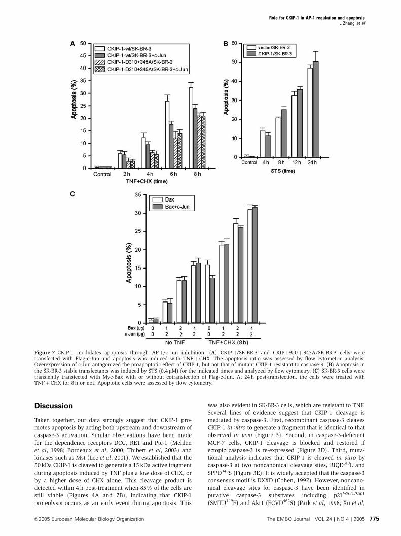
to apoptosis, CKIP-1/SK-BR-3 cells were transiently trans-
fected with c-Jun and treated with TNF at 24 h post-transfec-
tion. The percentage of apoptotic cells was measured by
flow cytometric analysis. As shown in Figure 7A, coexpres-
sion of c-Jun could partially antagonize the proapoptotic
effect of wild-type CKIP-1, with no effect of the
D310þ 345A mutant. To clarify whether the proapoptotic
effect of CKIP-1 was limited to apoptosis priming by TNF,
staurosporine (STS) was used as another potent cell death
stimulus. In this case, CKIP-1/SK-BR-3 cells showed similar
apoptosis ratio to the control cells (Figure 7B), which was
expected since STS does not activate JNK efficiently (data not
shown). To confirm the effect of expression of Jun on
apoptosis inhibition related to CKIP-1 or TNF, another proa-
poptotic protein, Bax, was overexpressed together with,
or without, c-Jun; cells were then treated with TNF.
Figure 7C clearly shows that c-Jun had no effect on Bax-
induced apoptosis. These results suggest that CKIP-1
cleavage promotes apoptosis through inhibition of AP-1/c-
Jun activity.
Figure 6 CKIP-1 promotes apoptosis dependent on caspase-3. (A) CKIP-1/SK-BR-3 and pcDNA3.1/SK-BR-3 cells were treated as in Figure 4Aand stained with Hoechst 33342 for DNA. The percentages of condensed chromosomes are shown as the ratio between apoptotic fragmentednuclei and the total stained nuclei for 200–250 cells in each field as indicated. At least three independent fields were counted. (B) The apoptosisratio for the indicated SK-BR-3 stable transfectants was assessed by flow cytometric analysis as described in Materials and methods. (C) DNAfragmentation assay. DNA was extracted and electrophoresed on a 1.5% agarose gel. Lane 1, 100-base-pair standard ladder. (D) siRNA-mediated knockdown of CKIP-1 in HEK293 cells attenuated sensitivity to TNF. Stable transfectants of HEK293 cells expressing CKIP siRNA orvector alone were assayed for apoptosis after TNFþCHX treatment as indicated. The inset shows the expression of CKIP-1 by immunoblottingas compared to actin, revealing the efficiency of the siRNA knockdown. Apoptosis was assessed by flow cytometric analysis.
Role for CKIP-1 in AP-1 regulation and apoptosisL Zhang et al
The EMBO Journal VOL 24 | NO 4 | 2005 &2005 European Molecular Biology Organization774
Discussion
Taken together, our data strongly suggest that CKIP-1 pro-
motes apoptosis by acting both upstream and downstream of
caspase-3 activation. Similar observations have been made
for the dependence receptors DCC, RET and Ptc-1 (Mehlen
et al, 1998; Bordeaux et al, 2000; Thibert et al, 2003) and
kinases such as Mst (Lee et al, 2001). We established that the
50 kDa CKIP-1 is cleaved to generate a 15 kDa active fragment
during apoptosis induced by TNF plus a low dose of CHX, or
by a higher dose of CHX alone. This cleavage product is
detected within 4 h post-treatment when 85% of the cells are
still viable (Figures 4A and 7B), indicating that CKIP-1
proteolysis occurs as an early event during apoptosis. This
was also evident in SK-BR-3 cells, which are resistant to TNF.
Several lines of evidence suggest that CKIP-1 cleavage is
mediated by caspase-3. First, recombinant caspase-3 cleaves
CKIP-1 in vitro to generate a fragment that is identical to that
observed in vivo (Figure 3). Second, in caspase-3-deficient
MCF-7 cells, CKIP-1 cleavage is blocked and restored if
ectopic caspase-3 is re-expressed (Figure 3D). Third, muta-
tional analysis indicates that CKIP-1 is cleaved in vitro by
caspase-3 at two noncanonical cleavage sites, RIQD310L and
SPPD345S (Figure 3E). It is widely accepted that the caspase-3
consensus motif is DXXD (Cohen, 1997). However, noncano-
nical cleavage sites for caspase-3 have been identified in
putative caspase-3 substrates including p21WAF1/Cip1
(SMTD149F) and Akt1 (ECVD462S) (Park et al, 1998; Xu et al,
Figure 7 CKIP-1 modulates apoptosis through AP-1/c-Jun inhibition. (A) CKIP-1/SK-BR-3 and CKIP-D310þ345A/SK-BR-3 cells weretransfected with Flag-c-Jun and apoptosis was induced with TNFþCHX. The apoptosis ratio was assessed by flow cytometric analysis.Overexpression of c-Jun antagonized the proapoptotic effect of CKIP-1, but not that of mutant CKIP-1 resistant to caspase-3. (B) Apoptosis inthe SK-BR-3 stable transfectants was induced by STS (0.4mM) for the indicated times and analyzed by flow cytometry. (C) SK-BR-3 cells weretransiently transfected with Myc-Bax with or without cotransfection of Flag-c-Jun. At 24 h post-transfection, the cells were treated withTNFþCHX for 8 h or not. Apoptotic cells were assessed by flow cytometry.
Role for CKIP-1 in AP-1 regulation and apoptosisL Zhang et al
&2005 European Molecular Biology Organization The EMBO Journal VOL 24 | NO 4 | 2005 775
2002). These findings support the notion that CKIP-1 is a
novel plasma membrane-localized target for caspase-3 and
plays a role in sensitizing cells to extracellular apoptotic
signals such as TNF.
It is important to emphasize that we observed only one
C-terminal fragment of 15 kDa (or 22 kDa if the long tag is
included) corresponding to the D310-cleaved product in TNF-
induced apoptosis. The proteolysis of CKIP-1 at D345 requires
phosphorylation by CK2 at S346 followed by sequential
phosphorylation by GSK3 at S342 (unpublished data). The
motif encompassing D345 is very attractive since the S346ESE
motif conforms to the consensus CK2 substrate site SXX(D/E)
or SXX(pS/pY) (pS, phosphoserine; pY, phosphotyrosine).
The S342PPDS346 motif conforms to the GSK3 site SXXXpS;
the best GSK3 substrates appear to have several proline
residues between the two serines (Kennelly and Krebs,
1991; Litchfield and Luscher, 1993). A similar dual-kinase
mechanism has been found in the Hedgehog signaling effec-
tor Cubitus interruptus and b-catenin (Price and Kalderon,
2002; Liu et al, 2002).
The fact that CKIP-1 is broadly expressed indicates that its
cleavage is likely to be a feature of apoptosis in many types of
cells. In this respect, endogenous CKIP-1 could also be
cleaved in response to serum withdrawal or TNF treatment
in HEK293 cells, TNF treatment in HeLa cells and in response
to ionizing radiation in MOLT-4 or Jurkat cells (Figure 3G;
data not shown). These findings suggest that CKIP-1 cleavage
may be a general feature of the apoptotic response of diverse
cells to a wide variety of stimuli. Intriguingly, CKIP-1 is
generally expressed at low levels or short-lived in tumor
cell lines and serves as a growth suppressor of tumor cells
(unpublished data).
Caspase cleavage of CKIP-1 probably represents an initiat-
ing step for CKIP-1-accelerated apoptosis by allowing
the release of C-terminal fragments of CKIP-1, which then
redistribute and effectively repress AP-1. The latter step
may also contribute to apoptosis, since both JNK/JunD
and JNK/c-Jun can mediate cell survival signaling
(Shaulian and Karin, 2002; Lamb et al, 2003). CKIP-1 expres-
sion in SK-BR-3 cells shortened the duration of c-Jun
phosphorylation (partially representing JNK activity) and
promoted apoptosis (Figures 4 and 6), providing evidence
for the antiapoptotic role of JNK/AP-1 pathway at least in
SK-BR-3 breast cancer cells. This is consistent with some
recent reports (Lamb et al, 2003; Yu et al, 2004) and in
contrast to others (Tournier et al, 2000; Deng et al, 2003;
Sakon et al, 2003). We noted that in SK-BR-3 cells, TNF-
induced JNK activation was sustained for at least 8 h
(Figure 4A and B), whereas in MEFs it lasts only 1 h (Sakon
et al, 2003). This may be caused by different cellular contexts.
It also suggests that JNK is mediating an antiapoptotic signal
in SK-BR-3 cells and that c-Jun phosphorylation plays an
important role in regulating cell death. Our model is consis-
tent with a study using c-jun�/� cells, which demonstrated
that c-Jun protects fibroblasts against UV, depending on its
phosphorylation on Ser63 and Ser73 (Wisdom et al, 1999).
However, it disagrees with the work of Behrens et al (1999),
who showed that fibroblasts in which the c-jun gene was
replaced by a mutant c-jun allele with Ser63 and Ser73
mutated to Ala displayed stress-induced apoptotic defects.
Overexpressing c-Jun in cells is likely to have distinct effects
from modulating the levels of c-Jun phosphorylation. This is
particularly important since C-term2 does not affect the level
of expression of c-Jun.
We established direct links between caspase cleavage,
C-term1/2 release and AP-1 inhibition. On the one hand,
CKIP-1 represses AP-1 activity and promotes apoptosis in a
caspase-3-dependent manner; on the other hand, CKIP-1
promotes apoptosis via inhibition of AP-1 activity. When
caspase-3 mediated CKIP-1 cleavage was blocked by the
D310þ 345A mutations, both the c-Jun phosphorylation
level and the apoptosis ratio were indistinguishable in
CKIP-1-expressing and control cells (Figures 4B and 6B).
This mechanism is distinct from known c-Jun-interacting
proteins (Chinenov and Kerppola, 2001), all of which are
localized to the cytoplasm or nucleus and function in their
intact forms. To the best of our knowledge, CKIP-1 is the first
PH domain-containing protein or plasma membrane-asso-
ciated protein that efficiently regulates nuclear AP-1 activity.
Recently, it was found that the Notch-1 intracellular domain
(NIC) represses AP-1-mediated transactivation following
cleavage by a furin-like enzyme and TACE (a membrane-
bound metalloprotease) and nuclear translocation of NIC
(Chu et al, 2002). This pattern of cleavage-mediated control
of AP-1 is similar to CKIP-1.
CKIP-1 C-term1 can interact directly with c-Jun (and JunD)
both in vitro and in vivo. The leucine zipper of c-Jun mediates
the interaction with C-term1. This interaction is specific since
other members of the AP-1 family were unable to interact
with C-term1 although they also contain a conserved leucine
zipper domain. C-term1 itself contains a putative leucine
zipper motif (aa 352–380), which has been widely found in
c-Jun-interacting bZIP transcription factors. Like these bZIP
proteins, the leucine zipper motif of CKIP-1 also mediates
interaction with CKIP-1 itself in the yeast two-hybrid system
(Figure 1B; Olsten et al, 2004), and homodimerization and
trimerization in vivo (unpublished data).
Using phospho-specific antibodies to different phosphory-
lated forms of c-Jun, Morton et al (2003) demonstrated that
JNK is the MAPK required for stress-induced phosphorylation
of c-Jun on Ser63 and Ser73. Consistent with this study, we
propose a model in which C-term2 inhibits c-Jun phosphor-
ylation by preventing the binding of c-Jun to JNK. This
mechanism has already been implicated for Jip-1-dependent
regulation of the JNK pathway (Whitmarsh and Davis,
1999). Furthermore, the regulatory function of CKIP-1 in
AP-1 activity is activated upon caspase-3-mediated cleavage
and, in turn, CKIP-1 functions as part of a feedback loop
that contributes to amplifying the apoptotic response.
Although the mechanisms by which CKIP-1 might
influence caspase-3 activity and apoptosis are unclear, the
ability of CKIP-1 to regulate AP-1 activity suggests that
one of them may involve the JNK/AP-1 pathway.
Coexpression of c-Jun inhibits the proapoptotic effect of
CKIP-1 in TNF-primed apoptosis, but had no effect on the
cell death induced by Bax expression or STS (Figure 7).
However, we cannot rule out other possibilities. More re-
cently, we observed that CKIP-1 overexpression significantly
induced Akt/PKB kinase cleavage at the C-terminal tail
(position D462), which is also mediated by caspase-3
(unpublished data). Considering that Akt/PKB kinase med-
iates a major cell survival pathway, the negative regulation of
Akt/PKB by CKIP-1 may be one of the consequences of the
amplified caspase-3 activation by CKIP-1. Further studies will
Role for CKIP-1 in AP-1 regulation and apoptosisL Zhang et al
The EMBO Journal VOL 24 | NO 4 | 2005 &2005 European Molecular Biology Organization776
be required to determine the precise role of the JNK/AP-1
pathway and the Akt/PKB pathway in CKIP-1-promoted
apoptosis.
Materials and methods
Plasmid constructs, antibodies and reagentsPlasmids of CKIP-1, c-Jun and others including deletion and pointmutants were constructed by PCR or recombinant PCR, followedby subcloning into various vectors. For detailed information, seeSupplementary data. Rabbit antiserum CKIP-1-N was raised againstthe N-terminus of CKIP-1 (aa 1–136) containing the PH domain.Rabbit antiserum CKIP-1-C against aa 308–409 of CKIP-1 was a kindgift from Dr David W Litchfield. Anti-Myc antibody and the caspaseinhibitors, zVAD-fmk and DEVD-CHO, were purchased fromClontech. Anti-Flag M2 antibody, CHX, TPA, leupeptin, antipain,aprotinin, chymostatin, pepstatin and phenylmethylsulfonyl fluor-ide (PMSF) were from Sigma. TNF-a was purchased fromChemicon. Antibodies to c-Jun (H-79), phospho-c-Jun-Ser63 (KM-1),JNK, phospho-JNK, p38, phospho-p38, caspase-3 (H-277), b-actin,Lamin A (C-20), nucleolin (MS-3) and Naþ/Kþ -ATPase a1 (N-15)were obtained from Santa Cruz.
Yeast two-hybrid and GST pull-down assaysCotransformation of pGBKT7 and pGADT7 carrying various cDNAsinto the yeast strain Y190 and the liquid o-nitrophenyl b-D-galactoside assay were performed according to the manufacturer’sinstructions (Clontech). The experiments were performed threetimes independently (mean7s.d.). GST-c-Jun proteins were ex-pressed in Escherichia coli BL21 (DE3) and purified with glutathioneSepharose 4B (Pharmacia). In vitro GST pull-down was performedas previously described (Matsuzawa and Reed, 2001).
Immunoprecipitation, immunoblotting and cell fractionationFor general cell lysis and co-immunoprecipitation of CKIP-1-C-term1 and c-Jun, cell lysates were prepared in lysis buffer (100 mMTris–HCl (pH 7.5), 300 mM NaCl, 2% (v/v) Tween 20, 0.4% NP-40,20% glycerol) supplemented with protease inhibitor cocktail(Roche) and phosphatase inhibitors (10 mM NaF and 1 mMNa3VO4). Immunoprecipitations were performed using anti-Mycor anti-Flag antibodies and protein A/G-agarose (Santa Cruz) at41C. The lysates and immunoprecipitates were detected using theindicated primary antibodies and then the appropriate secondaryantibody, followed by detection with SuperSignal chemilumines-cence kit (Pierce). Cell fractionation was performed as describedpreviously (Safi et al, 2004).
Fluorescence analysis of CKIP-1For immunofluorescence detection (Figure 4D), pcDNA3.1-Myc-CKIP-1/SK-BR-3 cells were fixed with 4% paraformaldehyde andpermeabilized in 0.2% Triton X-100 (PBS). CKIP-1 proteins werestained with anti-Myc antibody followed by TRITC-conjugatedsecondary antibody. The nuclei were stained with Hoechst 33342(Sigma) and images were visualized with an Olympus IX70fluorescence microscope and captured with a cooled charge-coupled device color digital camera (Diagnostic). For colocalizationof CKIP-1-C-term1 and c-Jun (Figure 1D), GFP-c-Jun together withRFP-C-term1 or RFP-CKIP-1 was cotransfected into COS7 cells andfluorescence was visualized 24 h later.
Reporter gene assayCOS7 cells were seeded into a 24-well plate and transfected with10 ng pRL-CMV (Renilla control luciferase, Promega) and 0.1mgpAP-1-luc (firefly luciferase, Stratagene) reporter gene plasmids andconstructs encoding various deletion mutants of CKIP-1 (0.5mg),together with c-Jun plasmid (0.1mg) or not. After 24 h, luciferaseactivity was measured and normalized to the cotransfected Renillaluciferase activity (mean7s.d., n¼ 3). In some cases (Figure 2B),various amounts of C-term1 or C-term2 plasmids (0.1–1.0mg) werecotransfected with c-Jun (0.1 mg) and the luciferase activity wasmeasured. To detect the specificity of CKIP-1 on AP-1 activity(Figure 2C), pAP-1-luc was substituted by pFA-Elk1 (0.1mg) in theabsence or presence of MEK1 (0.1 mg). The plasmids of the trans-
reporter gene system of MEKK1-c-Jun and MEK1-Elk1 pathwayswere a gift from Dr Xiaoming Yang.
In vitro translation and caspase cleavage assayWild-type CKIP-1 or various mutants of CKIP-1 were transcribedand translated using the T7 TNT system (Promega) in the presenceof [35S]methionine (Pharmacia). Purified active human recombi-nant caspase-3 and caspase-6 were purchased from Pharmingen.Caspase-mediated cleavage assays were performed as describedpreviously (Mehlen et al, 1998).
Apoptosis analysisCKIP-1/SK-BR-3 or control cells were deprived of serum overnightand treated with TNF (20 ng/ml) plus CHX (10 mg/ml) or a higherdose of CHX (100 mg/ml) alone for the indicated times to induceapoptosis. The percentage of apoptotic cells was quantified byanalysis of sub-G1 DNA. Briefly, cells were harvested by trypsiniza-tion and washed with PBS, followed by centrifugation at 2000 r.p.m.for 10 min. The cell pellet was resuspended in 70% ethanol, andstored at �201C for 24 h. Cells were washed and incubated in PBScontaining 50 mg/ml RNase A (Roche) for 30 min at 371C. Cells werethen stained with 50mg/ml propidium iodide (Sigma). DNA contentwas analyzed by flow cytometry using a cell sorter (FACSCalibur)and CellQuest software (Beckton Dickinson). Caspase-3 activitywas assessed using the ApoAlert Caspase-3 Activity Assay System,which uses the Ac-DEVD-AFC substrate (Clontech), according tothe manufacturer’s instructions. Caspase-3 activation is shown asthe ratio between the caspase activity of the sample and thatmeasured in pcDNA3.1-transfected cells without TNF treatment.DNA fragmentation assays were performed as described previously(Cheung et al, 2003).
RNA interferenceA total of 10 siRNA sequences derived from the human CKIP-1 RNAsequence were designed to contain a sense strand of 18–19nucleotide sequences of CKIP-1 followed by a short spacer(GAGTACTG), the reverse complement of the sense strand, andfive thymidines as an RNA polymerase III transcriptional stopsignal. These sequences were cloned into the pSuppressor vector(Imgenex) following the manufacturer’s instructions and confirmedby sequencing. The pSuppressor-CKIP-1-siRNA plasmids weretransfected into HEK293 or HeLa cells and then CKIP-1 expressionwas assayed by immunoblotting. Of the sequences tested, the twomost efficient sequences were selected for stable transfection.Paired forward and reverse oligonucleotides were (i) 50-tcgaGTTGACGCCCACAGAGAAAgagtactgTTTCTCTGTGGGCGTCAACttttt-30;50-ctagaaaaaGTTGACGCCCACAGAGAAAcagtactcTTTCTCTGTGGGCGTCAAC-30 and (ii) 50-tcgaGGAACCAACCTCTTGTGCTgagtactgAGCACAAGAGGTTGGTTCttttt-30; 50-ctagaaaaaGAACCAACCTCTTGTGCTcagtactcAGCACAAGAGGTTGGTTCC-30. The forward and reverseoligonucleotides included an XhoI and an XbaI cleavage site,respectively. Stable HEK293-CKIP-1 siRNA cells and HEK293-pSuppressor cells were constructed. G418 resistant clones wereassayed with immunoblotting. Both the above two paired siRNAswere used in the experiments, and similar results were obtained.
Statistical analysisStatistical evaluation was carried out using Student’s t-test.
Supplementary dataSupplementary data are available at The EMBO Journal Online.
Acknowledgements
We thank Mr Bo Dong for the flow cytometry analysis, Dr LishengWang for the caspase-3 antibody, Dr David W Litchfield for theCKIP-1-C antiserum, Dr Wen Yue for the pSuppressor plasmid andDrs Xiaodong Wang, Yi Rao, Kunliang Guan, John JM Bergeron, BaiLu, Lin Mei and Yong Li for critical reading of the manuscript. Thiswork was partially supported by Chinese National Natural ScienceFoundation Projects (30321003, 30100090), the Chinese State KeyProgram in Basic Research (2001CB510204, G1999053903,G1998051012) and the Chinese National High-tech Program(2004AA221100).
Role for CKIP-1 in AP-1 regulation and apoptosisL Zhang et al
&2005 European Molecular Biology Organization The EMBO Journal VOL 24 | NO 4 | 2005 777
References
Baisden JM, Qian Y, Zot HM, Flynn DC (2001) The actin filament-associated protein AFAP-110 is an adaptor protein thatmodulates changes in actin filament integrity. Oncogene 20:6435–6447
Behrens A, Sibilia M, Wagner EF (1999) Amino-terminal phosphor-ylation of c-Jun regulates stress-induced apoptosis and cellularproliferation. Nat Genet 21: 326–329
Brivanlou AH, Darnell Jr JE (2002) Signal transduction and thecontrol of gene expression. Science 295: 813–818
Bordeaux MC, Forcet C, Granger L, Corset V, Bidaud C, Billaud M,Bredesen DE, Edery P, Mehlen P (2000) The RET proto-oncogeneinduces apoptosis: a novel mechanism for Hirschsprung disease.EMBO J 19: 4056–4063
Bosc DG, Graham KC, Saulnier RB, Zhang C, Prober D, Gietz RD,Litchfield DW (2000) Identification and characterization of CKIP-1, a novel pleckstrin homology domain-containing protein thatinteracts with protein kinase CK2. J Biol Chem 275: 14295–14306
Cheung WL, Ajiro K, Samejima K, Kloc M, Cheung P, Mizzen CA,Beeser A, Etkin LD, Chernoff J, Earnshaw WC, Allis CD (2003)Apoptotic phosphorylation of histone H2B is mediated by mam-malian sterile twenty kinase. Cell 113: 507–517
Chevray PM, Nathans D (1992) Protein interaction cloning in yeast:identification of mammalian proteins that react with the leucinezipper of Jun. Proc Natl Acad Sci USA 89: 5789–5793
Chinenov Y, Kerppola TK (2001) Close encounters of many kinds:Fos–Jun interactions that mediate transcription regulatory speci-ficity. Oncogene 20: 2438–2452
Chu J, Jeffries S, Norton JE, Capobianco AJ, Bresnick EH (2002)Repression of activator protein-1-mediated transcriptional activa-tion by the Notch-1 intracellular domain. J Biol Chem 277:7587–7597
Cohen GM (1997) Caspases: the executioners of apoptosis. BiochemJ 326: 1–16
Deng Y, Ren X, Yang L, Lin Y, Wu X (2003) A JNK-dependentpathway is required for TNFa-induced apoptosis. Cell 115: 61–70
Kallunki T, Deng T, Hibi M, Karin M (1996) c-Jun can recruit JNK tophosphorylate dimerization partners via specific docking interac-tions. Cell 87: 929–939
Karin M, Liu ZG, Zandi E (1997) AP-1 function and regulation. CurrOpin Cell Biol 9: 240–246
Kennelly PJ, Krebs EG (1991) Consensus sequences as substratespecificity determinants for protein kinases and protein phospha-tases. J Biol Chem 266: 15555–15558
Kim S, Lee SH, Park D (2001) Leucine zipper-mediated homodimer-ization of the p21-activated kinase-interacting factor, bPix.Implication for a role in cytoskeletal reorganization. J BiolChem 276: 10581–10584
Lamb JA, Ventura J, Hess P, Flavell RA, Davis RJ (2003) JunDmediates survival signaling by the JNK signal transduction path-way. Mol Cell 11: 1479–1489
Lee KK, Ohyama T, Yajima N, Tsubuki S, Yonehara S (2001) MST, aphysiological caspase substrate, highly sensitizes apoptosis bothupstream and downstream of caspase activation. J Biol Chem276: 19276–19285
Lin A, Frost J, Deng T, Smeal T, al-Alawi N, Kikkawa U, Hunter T,Brenner D, Karin M (1992) Casein kinase II is a negative regulatorof c-Jun DNA binding and AP-1 activity. Cell 70: 777–789
Litchfield DW, Luscher B (1993) Casein kinase II in signal transduc-tion and cell cycle regulation. Mol Cell Biochem 127/128: 187–199
Liu C, Li Y, Semenov M, Han C, Baeg GH, Zhang Z, Lin X, He X(2002) Control of b-catenin phosphorylation/degradation by adual-kinase mechanism. Cell 108: 837–847
Matsuzawa S, Reed JC (2001) Siah-1, SIP, and Ebi collaborate in anovel pathway for b-catenin degradation linked to p53 responses.Mol Cell 7: 915–926
Mehlen P, Rabizadeh S, Snipas SJ, Assa-Munt N, Salvesen GS,Bredesen DE (1998) The DCC gene product induces apoptosis
by a mechanism requiring receptor proteolysis. Nature 395:801–804
Mitsuuchi Y, Johnson SW, Sonoda G, Tanno S, Golemis EA, Testa JR(1999) Identification of a chromosome 3p14.3–21.1 gene, APPL,an adaptor molecule that interacts with the oncoprotein-serine/threonine kinase AKT2. Oncogene 18: 4891–4898
Morton S, Davis RJ, McLaren A, Cohen P (2003) A reinvestigation ofthe multisite phosphorylation of the transcription factor c-Jun.EMBO J 22: 3876–3886
Nateri AS, Riera-Sans L, Costa CD, Behrens A (2004) The ubiquitinligase SCFFbw7 antagonizes apoptotic JNK signaling. Science 303:1374–1378
Olsten ME, Canton DA, Zhang C, Walton PA, Litchfield DW (2004)The pleckstrin homology domain of CKIP-1 is required for inter-actions and recruitment of protein kinase CK2 to the plasmamembrane. J Biol Chem 279: 42114–42127
Park JA, Kim KW, Kim SI, Lee SK (1998) Caspase 3 specificallycleaves p21WAF1/CIP1 in the earlier stage of apoptosis in SK-HEP-1 human hepatoma cells. Eur J Biochem 257: 242–248
Price MA, Kalderon D (2002) Proteolysis of the Hedgehog signalingeffector Cubitus interruptus requires phosphorylation byglycogen synthase kinase 3 and casein kinase 1. Cell 108:823–835
Romero F, Fischer S (1996) Structure and function of vav. Cell Signal8: 545–553
Safi A, Vandromme M, Caussanel S, Valdacci L, Baas D, Vidal M,Brun G, Schaeffer L, Goillot E (2004) Role for the pleckstrinhomology domain-containing protein CKIP-1 in phosphatidylino-sitol 3-kinase-regulated muscle differentiation. Mol Cell Biol 24:1245–1255
Sakon S, Xue X, Takekawa M, Sasazuki T, Okazaki T, Kojima Y, PiaoJH, Yagita H, Okumura K, Doi T, Nakano H (2003) NF-kB inhibitsTNF-induced accumulation of ROS that mediate prolonged MAPKactivation and necrotic cell death. EMBO J 22: 3898–3909
Shaulian E, Karin M (2001) AP-1 in cell proliferation and survival.Oncogene 20: 2390–2400
Shaulian E, Karin M (2002) AP-1 as a regulator of cell life and death.Nat Cell Biol 4: E131–E136
Thibert C, Teillet MA, Lapointe F, Mazelin L, Douarin NM, Mehlen P(2003) Inhibition of neuroepithelial Patched-induced apoptosis bySonic hedgehog. Science 301: 843–846
Tournier C, Hess P, Yang DD, Xu J, Turner TK, Nimnual A, Bar-SagiD, Jones SN, Flavell RA, Davis RJ (2000) Requirement of JNK forstress-induced activation of the cytochrome c-mediated deathpathway. Science 288: 870–874
Vogt PK (2001) Jun, the oncoprotein. Oncogene 20: 2365–2377Wertz IE, O’Rourke KM, Zhang Z, Dornan D, Arnott D, Deshaies RJ,
Dixit VM (2004) Human De-etiolated-1 regulates c-Jun by assem-bling a CUL4A ubiquitin ligase. Science 303: 1371–1374
Whitmarsh AJ, Davis RJ (1999) Signal transduction by MAPkinases: regulation by phosphorylation-dependent switches. SciSTKE 1: PE1
Wisdom R, Johnson RS, Moore C (1999) c-Jun regulates cell cycleprogression and apoptosis by distinct mechanisms. EMBO J 18:188–197
Xu J, Liu D, Songyang Z (2002) The role of Asp-462 in regulatingAkt activity. J Biol Chem 277: 35561–35566
Yan M, Dai T, Deak JC, Kyriakis JM, Zon LI, Woodgett JR, TempletonDJ (1994) Activation of stress-activated protein kinase byMEKK1 phosphorylation of its activator SEK1. Nature 372:798–800
Yu C, Minemoto Y, Zhang J, Liu J, Tang F, Bui TN, Xiang J, Lin A(2004) JNK suppresses apoptosis via phosphorylation of theproapoptotic Bcl-2 family protein Bad. Mol Cell 13: 329–340
Zhou BP, Hu MC, Miller SA, Yu Z, Xia W, Lin SY, Hung MC (2000)HER-2/neu blocks tumor necrosis factor-induced apoptosis viathe Akt/NF-kB pathway. J Biol Chem 275: 8027–8031
Role for CKIP-1 in AP-1 regulation and apoptosisL Zhang et al
The EMBO Journal VOL 24 | NO 4 | 2005 &2005 European Molecular Biology Organization778
Top Related
Copyright © 2022 FDOKUMEN

