

![[Distribution of blaOXA genes in Acinetobacter baumannii strains: a multicenter study]](https://static.fdokumen.com/doc/165x107/6337b5c66f78ac31240eb601/distribution-of-blaoxa-genes-in-acinetobacter-baumannii-strains-a-multicenter.jpg)


Bahasa
Halaman
Hukum

Int.J.Curr.Microbiol.App.Sci (2018) 7(11): 3020-3029
3020
Original Research Article https://doi.org/10.20546/ijcmas.2018.711.346
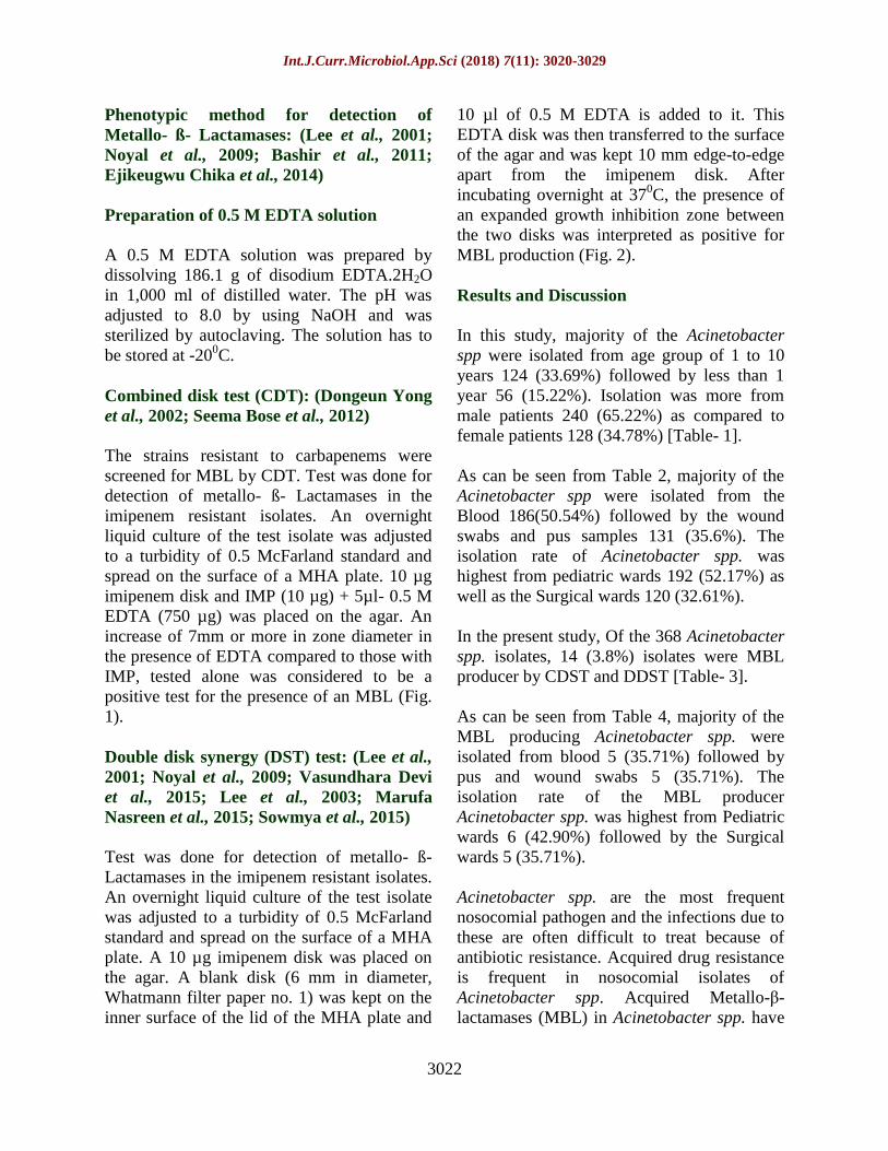
Prevalence of Metallo Β-Lactamase Producing Acinetobacter in Clinical
Specimens from S.S.G. Hospital, Vadodara, India
Jignasha Tadvi1*
, Jigna Karia2, Rachna Bhavasar
3 and Hiral Patel
4
1Department of Microbiology, Zydus Medical College & Hospital, Dahod, Gujarat, India
2Department of Microbiology, Baroda Medical College & SSG Hospital, Vadodara,
Gujarat, India 3Dr. M.K. Shah Medical College & Research Centre, Ahmedabad, Gujarat, India
4GMERS Medical College, Valsad, India
*Corresponding author
A B S T R A C T
Introduction
Acinetobacter species are the common
pathogens causing nosocomial infections.
Infections caused by Acinetobacter are either
exogenous or endogenous origin, depending
on several factors such as use of
immunosuppressant agents, injudicious use of
antimicrobial agents, prolonged surgical
procedures and inadequate instrumentations.
In recent years due to moderate and observed
use of antibiotics, non-fermentative gram
negative bacilli have emerged as an important
health care associate pathogen. They have
International Journal of Current Microbiology and Applied Sciences ISSN: 2319-7706 Volume 7 Number 11 (2018) Journal homepage: http://www.ijcmas.com
Acinetobacter species are the commonest pathogens causing nosocomial infections.
Acinetobacter is essentially resistant to many antibiotics and they are known to produce
extended spectrum beta lactamase and metalo beta lactamase. Aim of the study is to detect
Metalo beta lactamase producing Acinetobacter spp. from clinical samples in tertiary care
hospital. Between January 2015 and June 2015, total 368 Acinetobacter spp were isolated
from different clinical samples. Antimicrobial susceptibility was done as per CLSI
guideline. All imipenem resistant isolates were tested for MBL production by Imipenem -
EDTA double disc synergy test (DDST) and Imipenem- EDTA combined disc test (CDT).
Of 368 samples, majority of the Acinetobacter spp were isolated from blood 186(50.54%)
followed by wound and pus samples 131 (35.6%). The isolation rate was highest from
pediatric wards 192 (52.17%) and surgical wards 120 (32.61%). Total 368 samples, 14
(3.8%) isolates were MBL producer by CDST and DDST. Majority of the MBL producing
Acinetobacter spp. were isolated from blood 5 (35.71%) followed by pus and wound 5
(35.71%). The isolation rate was highest from Pediatric wards 6 (42.90%) followed by the
Surgical wards 5 (35.71%). Metallo-β-lactamase positive isolates of Acinetobacter spp. are
important to identify because it poses therapeutic problems and serious concern for
infection control management. There is also a need to emphasize on the rational use of
antimicrobials and strictly adhere to the concept of “reserve drugs” to minimize the misuse
of available antimicrobials.
K e y w o r d s Metalo beta lactamase
(MBL), Acinetobacter
species
Accepted:
22 October 2018
Available Online:
10 November 2018
Article Info

Int.J.Curr.Microbiol.App.Sci (2018) 7(11): 3020-3029
3021
been incriminated in infections such as
septicemia, pneumonia, urinary tract infection
and surgical site infection. Acinetobacter is
essentially resistant to many antibiotics and
they are known to produce extended spectrum
beta lactamase and metallo-beta lactamase.
Acquired drug resistance is frequent in
nosocomial isolates of Acinetobacter spp.
(Hemalatha et al., 2005; Butt et al., 2005)
Acquired Metallo β-Lactamase (MBL) in
Acinetobacter spp. have recently emerged as
one of the most troublesome resistance
mechanism because of their capability to
hydrolyze all beta-lactam antibiotic including
penicillins, cephalosporins and carbapenams,
with the exception of Aztreonam. (Hemalatha
et al., 2005; Butt et al., 2005; Ami Varaiya et
al., 2008; Debasrita Chakraborty et al., 2010;
Gian Maria Rossolini) Now a days resistance
to Aztreonam producing Metallo β-Lactamase
(MBL) is also revealed.
Currently, there are no recommendations
available from CLSI (Clinical and Laboratory
Standard Institute) for the detection of MBL.
Several phenotypic methods are available for
MBL detection. All these methods are based
on the ability of metal chelators, such as
EDTA and THIOI compounds to inhibit the
activity of MBL.
In present study, two phenotypic methods are
used for the detection of MBL producing
Acinetobacter species. The present study
includes the Imipenem- EDTA combined disc
synergy test (CDST) and Imipenem- EDTA
double- disc synergy test (DDST). (Ami
Varaiya et al., 2008; Debasrita Chakraborty et
al., 2010; Gian Maria Rossolini; Horieh Saderi
et al.,)
The aim of this study to determine MBL
producing Acinetobacter spp. from clinical
isolates in a tertiary care hospital setting.
The objectives of this study include, to isolate
and identify Acinetobacter from various
clinical specimens (Blood, Body fluids,
Sputum, Throat swab etc.)
To determine antibiotic sensitivity of the
isolates to various antibiotics by Kirby-Bauer
disc diffusion method
To screen for MBL producing isolates by
detecting resistance to Imipenem (IPM).
To confirm MBL production in MBL screen
test positive by:
Imipenem EDTA combined disc synergy test
Imipenem EDTA double disc synergy test
To study sensitivity and resistance pattern
among isolates of Acinetobacter species from
patients admitted in hospital.
Materials and Methods
Study Design: Cross- sectional
Study Setting: Department of Microbiology
Study Subject
The study was carried out over a period of 6
months from January 2015 to June 2015. A
total non-repetitive of 368 Acinetobacter spp
were isolated from different clinical samples
like blood, pus and wound swabs, urine, body
fluids, sputum, endo tracheal tube and
secretions from the patients attending the
hospital.
Antimicrobial susceptibility test of all the
isolates was performed by the disc-diffusion
(Kirby Baur disc diffusion method) according
to CLSIs guidelines. All imipenem resistant
isolates were tested for MBL production by
Imipenem - EDTA double- disc synergy test
(DDST) and Imipenem- EDTA combined disc
test (CDT).
Int.J.Curr.Microbiol.App.Sci (2018) 7(11): 3020-3029
3022
Phenotypic method for detection of
Metallo- ß- Lactamases: (Lee et al., 2001;
Noyal et al., 2009; Bashir et al., 2011;
Ejikeugwu Chika et al., 2014)
Preparation of 0.5 M EDTA solution
A 0.5 M EDTA solution was prepared by
dissolving 186.1 g of disodium EDTA.2H2O
in 1,000 ml of distilled water. The pH was
adjusted to 8.0 by using NaOH and was
sterilized by autoclaving. The solution has to
be stored at -200C.
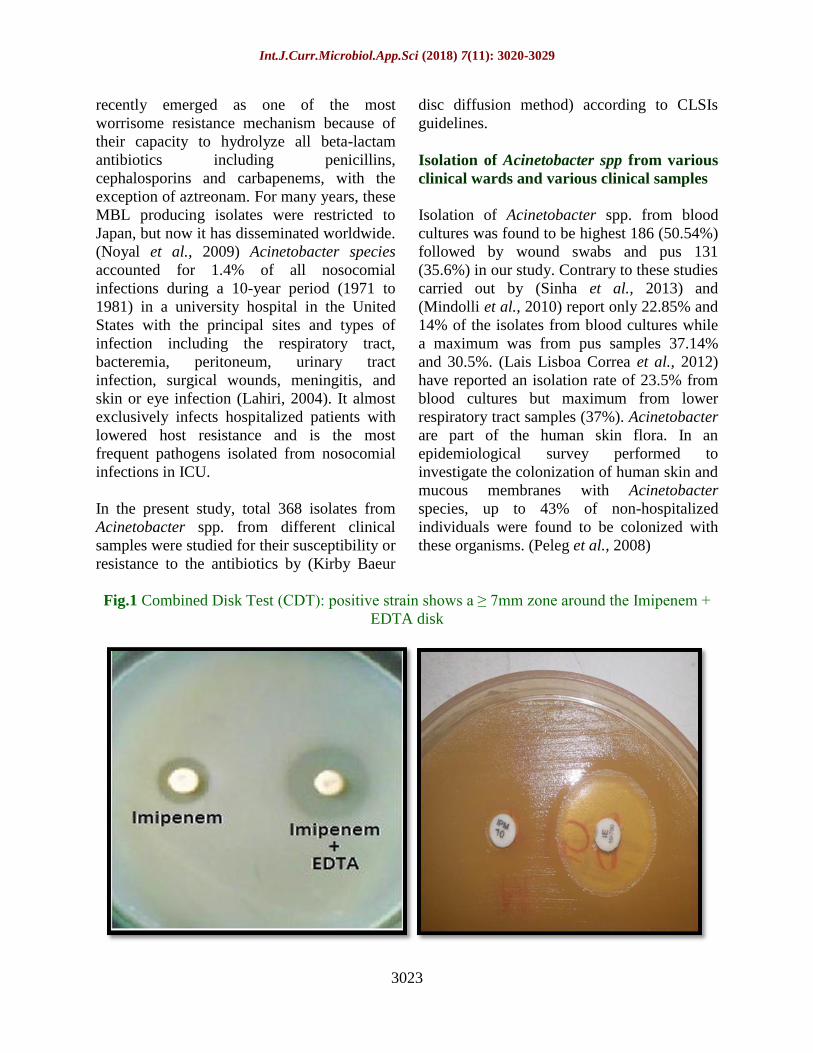
Combined disk test (CDT): (Dongeun Yong
et al., 2002; Seema Bose et al., 2012)
The strains resistant to carbapenems were
screened for MBL by CDT. Test was done for
detection of metallo- ß- Lactamases in the
imipenem resistant isolates. An overnight
liquid culture of the test isolate was adjusted
to a turbidity of 0.5 McFarland standard and
spread on the surface of a MHA plate. 10 µg
imipenem disk and IMP (10 µg) + 5µl- 0.5 M
EDTA (750 µg) was placed on the agar. An
increase of 7mm or more in zone diameter in
the presence of EDTA compared to those with
IMP, tested alone was considered to be a
positive test for the presence of an MBL (Fig.
1).
Double disk synergy (DST) test: (Lee et al.,
2001; Noyal et al., 2009; Vasundhara Devi
et al., 2015; Lee et al., 2003; Marufa
Nasreen et al., 2015; Sowmya et al., 2015)
Test was done for detection of metallo- ß-
Lactamases in the imipenem resistant isolates.
An overnight liquid culture of the test isolate
was adjusted to a turbidity of 0.5 McFarland
standard and spread on the surface of a MHA
plate. A 10 µg imipenem disk was placed on
the agar. A blank disk (6 mm in diameter,
Whatmann filter paper no. 1) was kept on the
inner surface of the lid of the MHA plate and
10 µl of 0.5 M EDTA is added to it. This
EDTA disk was then transferred to the surface
of the agar and was kept 10 mm edge-to-edge
apart from the imipenem disk. After
incubating overnight at 370C, the presence of
an expanded growth inhibition zone between
the two disks was interpreted as positive for
MBL production (Fig. 2).
Results and Discussion
In this study, majority of the Acinetobacter
spp were isolated from age group of 1 to 10
years 124 (33.69%) followed by less than 1
year 56 (15.22%). Isolation was more from
male patients 240 (65.22%) as compared to
female patients 128 (34.78%) [Table- 1].
As can be seen from Table 2, majority of the
Acinetobacter spp were isolated from the
Blood 186(50.54%) followed by the wound
swabs and pus samples 131 (35.6%). The
isolation rate of Acinetobacter spp. was
highest from pediatric wards 192 (52.17%) as
well as the Surgical wards 120 (32.61%).
In the present study, Of the 368 Acinetobacter
spp. isolates, 14 (3.8%) isolates were MBL
producer by CDST and DDST [Table- 3].
As can be seen from Table 4, majority of the
MBL producing Acinetobacter spp. were
isolated from blood 5 (35.71%) followed by
pus and wound swabs 5 (35.71%). The
isolation rate of the MBL producer
Acinetobacter spp. was highest from Pediatric
wards 6 (42.90%) followed by the Surgical
wards 5 (35.71%).
Acinetobacter spp. are the most frequent
nosocomial pathogen and the infections due to
these are often difficult to treat because of
antibiotic resistance. Acquired drug resistance
is frequent in nosocomial isolates of
Acinetobacter spp. Acquired Metallo-β-
lactamases (MBL) in Acinetobacter spp. have
Int.J.Curr.Microbiol.App.Sci (2018) 7(11): 3020-3029
3023
recently emerged as one of the most
worrisome resistance mechanism because of
their capacity to hydrolyze all beta-lactam
antibiotics including penicillins,
cephalosporins and carbapenems, with the
exception of aztreonam. For many years, these
MBL producing isolates were restricted to
Japan, but now it has disseminated worldwide.
(Noyal et al., 2009) Acinetobacter species
accounted for 1.4% of all nosocomial
infections during a 10-year period (1971 to
1981) in a university hospital in the United
States with the principal sites and types of
infection including the respiratory tract,
bacteremia, peritoneum, urinary tract
infection, surgical wounds, meningitis, and
skin or eye infection (Lahiri, 2004). It almost
exclusively infects hospitalized patients with
lowered host resistance and is the most
frequent pathogens isolated from nosocomial
infections in ICU.
In the present study, total 368 isolates from
Acinetobacter spp. from different clinical
samples were studied for their susceptibility or
resistance to the antibiotics by (Kirby Baeur
disc diffusion method) according to CLSIs
guidelines.
Isolation of Acinetobacter spp from various
clinical wards and various clinical samples
Isolation of Acinetobacter spp. from blood
cultures was found to be highest 186 (50.54%)
followed by wound swabs and pus 131
(35.6%) in our study. Contrary to these studies
carried out by (Sinha et al., 2013) and
(Mindolli et al., 2010) report only 22.85% and
14% of the isolates from blood cultures while
a maximum was from pus samples 37.14%
and 30.5%. (Lais Lisboa Correa et al., 2012)
have reported an isolation rate of 23.5% from
blood cultures but maximum from lower
respiratory tract samples (37%). Acinetobacter
are part of the human skin flora. In an
epidemiological survey performed to
investigate the colonization of human skin and
mucous membranes with Acinetobacter
species, up to 43% of non-hospitalized
individuals were found to be colonized with
these organisms. (Peleg et al., 2008)
Fig.1 Combined Disk Test (CDT): positive strain shows a ≥ 7mm zone around the Imipenem +
EDTA disk

Int.J.Curr.Microbiol.App.Sci (2018) 7(11): 3020-3029
3024
Fig.2 Double Disk Synergy Test (DDST)/ EDTA Disk Synergy Test: Positive strain shows a
synergistic zone of inhibition between Imipenem and EDTA disc
Table.1 Age and Sex distribution of isolated Acinetobacter spp. (n=368)
Age in Years Male Female Total
< 1 31 25 56 (15.22%)
1 to 10 84 40 124 (33.69%)
11 to 20 17 16 33 (8.97%)
21 to 30 20 19 39 (10.60%)
31 to 40 26 9 35 (9.51%)
41 to 50 26 6 32 (8.69%)
51 to 60 19 5 24 (6.52%)
61 to 70 12 4 16 (4.35%)
71 to 80 5 4 9 (2.45%)
Total 240 (65.22%) 128 (34.78%) 368
Int.J.Curr.Microbiol.App.Sci (2018) 7(11): 3020-3029
3025
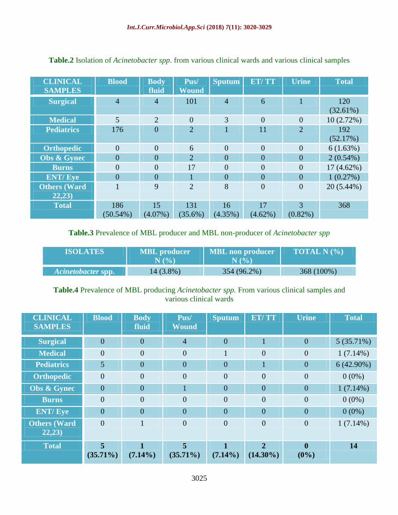
Table.2 Isolation of Acinetobacter spp. from various clinical wards and various clinical samples
Table.3 Prevalence of MBL producer and MBL non-producer of Acinetobacter spp
ISOLATES MBL producer
N (%)
MBL non producer
N (%)
TOTAL N (%)
Acinetobacter spp. 14 (3.8%) 354 (96.2%) 368 (100%)
Table.4 Prevalence of MBL producing Acinetobacter spp. From various clinical samples and
various clinical wards
CLINICAL
SAMPLES
Blood Body
fluid
Pus/
Wound
Sputum ET/ TT Urine Total
Surgical 0 0 4 0 1 0 5 (35.71%)
Medical 0 0 0 1 0 0 1 (7.14%)
Pediatrics 5 0 0 0 1 0 6 (42.90%)
Orthopedic 0 0 0 0 0 0 0 (0%)
Obs & Gynec 0 0 1 0 0 0 1 (7.14%)
Burns 0 0 0 0 0 0 0 (0%)
ENT/ Eye 0 0 0 0 0 0 0 (0%)
Others (Ward
22,23)
0 1 0 0 0 0 1 (7.14%)
Total 5
(35.71%)
1
(7.14%)
5
(35.71%)
1
(7.14%)
2
(14.30%)
0
(0%)
14
CLINICAL
SAMPLES
Blood Body
fluid
Pus/
Wound
Sputum ET/ TT Urine Total
Surgical 4 4 101 4 6 1 120
(32.61%)
Medical 5 2 0 3 0 0 10 (2.72%)
Pediatrics 176 0 2 1 11 2 192
(52.17%)
Orthopedic 0 0 6 0 0 0 6 (1.63%)
Obs & Gynec 0 0 2 0 0 0 2 (0.54%)
Burns 0 0 17 0 0 0 17 (4.62%)
ENT/ Eye 0 0 1 0 0 0 1 (0.27%)
Others (Ward
22,23)
1 9 2 8 0 0 20 (5.44%)
Total 186
(50.54%)
15
(4.07%)
131
(35.6%)
16
(4.35%)
17
(4.62%)
3
(0.82%)
368

Int.J.Curr.Microbiol.App.Sci (2018) 7(11): 3020-3029
3026
Table.5 Antibiogram pattern of isolates of Acinetobacter spp. to different
Antibiotics are as follow
Name of Drugs Sensitive Resistance
Piperacillin + Tazobactam (100µg/ 10 µg) 331 (89.95%) 37 (10.05%)
Gentamicin (10 µg) 202 (54.89%) 166 (45.11%)
Levofloxacin (5 µg) 354 (96.19%) 14 (3.81%)
Cefepime (30 µg) 130 (35.33%) 238 (64.67%)
Cefotaxime (30 µg) 127 (34.51%) 241 (65.49%)
Amikacin(30 µg) 203 (55.18%) 165 (44.84%)
Imipenem (10 µg) 354 (96.19%) 14 (3.81%)
Table.6 Prevalence of MBL producers among Acinetobacter spp
Years of study Place of study Authors MBL
Producers
2008 Pakistan S Irfan et al., 25
96.60%
2009 Puducherry Noyel et al.,8 6.50%
2010 Mumbai Anuradha S De 26
36%
2011 Punjab Mahajan G et al.,27
19%
2011 Tamil Nadu John & Balagurunathan28
27.70%
2012 Maharashtra Simit H kumar.29
14.29%
2012 South India Dheepa M and Appalaraju 30
10%
2013 kolkata Rit K et al., 31
22%
Present study Vadodara 3.8%
When distribution of clinical samples from
which Acinetobacter spp. were isolated, was
studied in different locations of hospital, it
was found that majority of the samples were
from Pediatric wards 192 (52.17%), followed
by Surgical ward 120 (32.61%). A second
important group of patients may consist of
neonates. (Lahiri et al., 2004) The
predisposing risk factors for septicemia are
low birth weight, previous antibiotic therapy,
mechanical ventilation, and the presence of
neonatal convulsions.
Acinetobacter spp. should be added to the list
of organisms capable of causing severe
nosocomial infection in neonatal ICUs.
(Lahiri et al., 2004) Studies carried out by
different authors like (Lias Lisboa Correa et
al., 2012) (57.1%), (Sinha et al., 2013)
(22.14%) (Amudhan et al., 2011) (95.68%),
(Anupuraba et al., 2005) (20.8%) and
(Mindolli et al., 2010) (27%) have all
reported a majority of their Acinetobacter spp
to be isolated from the ICUs followed by the
pediatric wards. Contaminated hands and
gloves of the wards staff seem to have an
important role in patient-to-patient
transmission of Acinetobacter spp. (Tankovic
et al., 1994). In our study, 3.8% isolates of
Acinetobacter spp. were found to be MBL
producers. It was low as compared to the
other studies as shown in the following table:
Antibiotic resistance pattern of MBL
producing Acintobacter spp.
All MBLs were resistant to important groups
of antibiotics tested, including third

Int.J.Curr.Microbiol.App.Sci (2018) 7(11): 3020-3029
3027
generation cephalosporins, Aminoglycosides
and Quinolones – a feature of MBL
producers. (De et al., 2010; Kumar et al.,
2012) For MBLs, limited treatment options
are available and the only therapeutic option
may be polymyxin, but it should never be
used as monotherapy. Combination therapy
often employed in treatment of MBL –
producing Acinetobacter spp. with imipenem/
Imipenem combined with Ampicillin-
sulbactum. (Kumar et al., 2012) In their
study, (John and Balagurunathan, 2011)
reported resistance to Amikacin (56.1%),
Piperacillin (57.1%), Ciprofloxacin (72.1%),
and Gentamicin (77%) among the
A.baumannii.
In his study, (Kumar et al., 2012) reported
that all the MBL positive isolates of
A.baumannii were resistant to all the major
antibiotics except for Piperacillin/Tazobactum
to which only 16.67% isolates were
susceptible.
MBL (metallo-β-lactamase) positive isolates
of Acinetobacter spp. are important to identify
because it poses not only therapeutic problem,
but also a serious concern for infection
control management.
There is also a need to emphasize on the
rational use of antimicrobials and strictly
adhere to the concept of “reserve drugs” to
minimize the misuse of available
antimicrobials. In addition, regular
antimicrobial susceptibility surveillance is
essential.
References
Agrawal J, Singh M, Sinha N, Shrivastava S,
Analysis of carbapenem resistant
Acinetobacter from tertiary care setting
in North India. Indian J Med Microbiol.
Jan 2013; 31(1): 60-63.
Ami Varaiya, Nikhil Kulkarni, Manasi
Kulkarni, Pallavi Bhalekar & Jyotsana
Dogra. Incidence of metallo beta
lactamase producing Pseudomonas
aeruginosa in ICU patients. Indian J
Med Res 127, April 2008, pp 398-402.
Amudhan SM, Sekar U, Arunagiri K, Sekar
B. OXA beta- lactamase-mediated
carbapenem resistance in Acinetobacter
baumannii. Indian J Med. Microbiol,
2011; 29(3):269-74.
Anupuraba S, Sen MR. Antimicrobial
resistance profile of bacterial isolates
from ICU: Changing trends. J Commun
Dis., March 2005; 37(1):58-65.
Bashir D, Thakor MA, Bashir AF, Bashir G,
Danish Z, Shabir A and Toboli AS.
Detection of metallo- b-lactamase
(MBL) producing Pseudomonas
aeruginosa at a tertiary care hospital in
Kashmir. Afr. J. Microbiol. Res., Jan
2011; 5(2): 164-172.
Butt T, Usman M, Ahmed RN, Saif I.
Emergence of Metallo-β-lactamase: The
quiet before the storm? Clinical
Microbiology Reviews. 2005; 18: 306-
25.
De AS, Kumar SH and Baveja SM. Prevalence of
metallo- Beta- lactamases producing
Pseudomonas aeruginosa and
Acinetobacter species in intensive care
areas in a tertiary care hospital. Indian J Crit
Care Med., Oct 2010; 14(4): 217-219.
Debasrita Chakraborty, Saikat Basu and
Satadal Das. A Study on Infections
Caused By Metallo Beta Lactamase
Producing Gram Negative Bacteria in
Intensive Care Unit Patients. American
Journal of Infectious Diseases 6 (2): 34-
39, 2010.
Dongeun Yong, Kyungwon Lee, Jong Hwa
Yum, Hee Bong Shin, Gian Maria
Rossolini and Yunsop Chong.
Imipenem-EDTA Disk Method for
Differentiation of Metallo-β-Lactamase-
Producing Clinical Isolates of
Int.J.Curr.Microbiol.App.Sci (2018) 7(11): 3020-3029
3028
Pseudomonas spp. and
Acinetobacter spp. J Clin Microbiol.
2002 Oct; 40(10): 3798–3801.
Ejikeugwu Chika, Ugwu Malachy, Iroha
Ifeanyichukwu, Eze Peter, Gugu
Thaddeus, Esimone Charles. Phenotypic
Detection of Metallo-β-Lactamase
(MBL) Enzyme in Enugu, Southeast
Nigeria. American Journal of
Biological, Chemical and
Pharmaceutical Sciences Vol. 2, No. 2,
June 2014, pp. 1 - 6, ISSN: 2328 –
6814.
Gian Maria Rossolini. Acquired Metallo-b-
Lactamases: An Increasing Clinical
Threat. Peleg et al., on pages 1549–56.
Hemalatha V, Sekar U & Kamat V. Detection
of metallo betalactamase producing
Pseudomonas aeruginosa in
hospitalized patients. Indian J Med Res
122, August 2005, pp 148-152.
Horieh Saderi, Zohreh Karimi, Parviz qwlia,
Mohmmad Ali. Phenotypic detection of
Metallo-Beta-Lactamase producing
Ps.aerugenosa strain isolated in burned
patients. Iranian J of Pathology. 20-24.
Irfan, S., A Zafar, D Guhar, T Ahsan, R
Hasan. Metallo- β-Lactamase producing
clinical isolates of Acinetobacter
species and Pseudomonas aeruginosa
from Intensive care unit patients of a
Tertiary care hospital. Indian J Med
Microbiol. 2008; 26(3): 243-245.
John, S., and Balagurunathan R. Metallo-
Beta-Lactamase producing
Pseudomonas aeruginosa and
Acinetobacter baumannii. Indian J Med
Microbiol. Dec 2011; 29(3):302-304.
Kumar SH, De AS, Baveja SM, Gore MM.
Prevalence and risk factor of metallo-
Beta- lactamases producing
Pseudomonas aeruginosa and
Acinetobacter species in burns and
surgical wards in a tertiary care
hospital. J Laboratory Physician. Jan
2012; 4 (1):39-42.
Laís Lisboa Corrêa, Larissa Alvarenga Batista
Botelho, Lívia Carvalho Barbosa,
Claudio Simões Mattos, Jupira Miron
Carballido, Carmem Lúcia Teixeira de
Castro, Pedro Juan Jose Mondino,
Geraldo Renato de Paula, Silvia Susana
Bona de Mondino, Claudia Rezende
Vieira de Mendonc¸ a-Souza. Detection
of blaOXA-23 in Acinetobacter spp.
isolated from patients of a university
hospital. braz j infect dis. 2012; 16(6):
521–526.
Lee K, Chong. Y, Shin H. B, Kim Y.A, Yong
D and Yum V. Modified Hodge and
EDTA- disk synergy tests to screen
metallo- β- Lactamase- producing
strains of Pseudomonas and
Acinetobacter species. Clin Micro
Infect. Feb 2001; 7(2): 88-91.
Lee, K., Y. S. Lim, D. Yong, J. H. Yum, and
Y. Chong. Evaluation of the Hodge test
and the Imipenem-EDTA double-disk
synergy test for differentiating metallo-
lactamase-producing isolates of
Pseudomonas spp. and Acinetobacter
spp.; Journal of clinical microbiology
Oct. 2003, 41(10): 4623–4629.
Lt Col KK Lahiri, Lt Col NS Mani, Lt Col SS
Purai. Acinetobacter spp as Nosocomial
Pathogen: Clinical Significance and
Antimicrobial Sensitivity. MJAFI 2004;
60(1):7-10.
Mahajan G, Sheemar S, Chopra S, Kaur J,
Chowdhary D, Makhija SK.
Carbapenem resistance and phenotypic
detection of carbapenemases in clinical
isolates of Acinetobacter baumannii.
Indian J Med Scienc 2011; 65(1):18-25.
Marufa Nasreen, Animesh Sarker, M. A.
Malek, Md. Ansaruzzaman, Mahububur
Rahman Prevalence and Resistance
Pattern of Pseudomonas aeruginosa
Isolated from Surface Water Advances
in Microbiology, 2015; 5: 74-81.
Mindolli PB, Salmani MP, Vishwanath G and
Hanumanthappa A.R. Identification and
Int.J.Curr.Microbiol.App.Sci (2018) 7(11): 3020-3029
3029
speciation of Acinetobacter and their
antimicrobial susceptibility testing. Al
Ameen J Med Sci 2010; 3(4):345-349.
Muthusamy D, Boppe A. Phenotypic Methods
for the Detection of Various
Betalactamases in Carbapenem
Resistant Isolates of Acinetobacter
baumannii at a Tertiary Care Hospital in
South India. Journal Clin Diagno Res.,
Aug 2012; 6(6): 970-973.
Noyal M.J., Menezes G.A, Harish B.N.,
Sujatha S. and Parija S.C. Simple
screening tests for detection of
carbapenemases in clinical isolates of
nonfermentative Gram-negative
bacteria. Indian J Med Res, June 2009;
129(6): 707-712.
Peleg AY, Seifert H, and Paterson DL.
Acinetobacter baumannii: Emergence
of a Successful Pathogen. Clinical
Microbiology Reviews. July 2008;
21(3):538-582.
Rit K, Chakraborty B, Dey R, Chakraborty P,
Naha A, Saha A. Prevalence of
Pseudomonas aeruginosa and
Acinetobacter spp producing metallo-
beta-Lactamase in a tertiary care
hospital. Jouranl of Dr. NTR University
of Health Sciences. July 2013; 2(1): 18-
21.
Seema Bose, Atindra Krishna Ghosh, Rekha
Barapatre. Incidence of metallo beta
lactamases producing Pseudomonas
aeruginosa in burn ward of a tertiary
care rural hospital. International Journal
of Biomedical Research. 2012; 3(05):
233‐ 238.
Sowmya G. Shivappa, Dr. Ranjitha
Shankaregowda, Dr. Raghavendra Rao
M, Dr. Rajeshwari K G, Dr. Madhuri
Kulkarni. Detection of Metallo-beta
lactamase production in clinical isolates
of Nonfermentative Gram negative
bacilli. IOSR Journal of Dental and
Medical Sciences (IOSR-JDMS) e-
ISSN: 2279-0853, p-ISSN: 2279-0861;
14(10), Oct. 2015: 43-48.
Tankovic J, Legrand P, Gatines Gd,
Chemineau V, Brun-Buisson C, and
Duval J. Characterization of a Hospital
Outbreak of Imipenem-Resistant
Acinetobacter baumannii by Phenotypic
and Genotypic Typing Methods. J. Clin.
Microbiol., Nov 1994; 32 (11): 2677-
2681.
Vasundhara Devi, P., P.Sreenivasulu Reddy
and Maria Sindhura John. Prevalence of
Metallo- -Lactamases Producing
Pseudomonas aeruginosa among the
Clinical isolates: A study from tertiary
care hospital. Int.J. Curr. Microbiol.
App.Sci (2015) 4(4): 955-961.
How to cite this article:
Jignasha Tadvi, Jigna Karia, Rachna Bhavasar and Hiral Patel. 2018. Prevalence of Metallo Β-
Lactamase Producing Acinetobacter in Clinical Specimens from S.S.G. Hospital, Vadodara,
India. Int.J.Curr.Microbiol.App.Sci. 7(11): 3020-3029.
doi: https://doi.org/10.20546/ijcmas.2018.711.346
Top Related
Copyright © 2022 FDOKUMEN

