

Bahasa
Halaman
Hukum

534 | JULY 2004 | VOLUME 5 www.nature.com/reviews/neuro
R E V I E W S
Perhaps the most surprising feature of the motor systemis the ease with which humans and other animals canmove. It is only when we observe the clumsy movementsof a child, or the motor challenges faced by individualswith neurological disorders, that we become aware ofthe inherent difficulties of motor control.
The efforts of systems neuroscientists to understandhow the brain controls movement include studies on the physics of the musculoskeletal system, neuro-physiological studies to explore neural control, andinvestigations of motor behaviour (BOX 1). As knowl-edge continues to grow in each area, it becomes morechallenging to link these levels of the motor system andto maintain a cohesive framework within which todescribe motor function or to interpret the role of abrain region.
Take, for example, the primary motor cortex (M1).It has been known for more than 100 years that M1 isimportant for controlling volitional movements, butmore detailed statements on its function vary greatly1.Studies of neural activity in M1 tend either to relateneural activity to details of motor output, thereby connecting motor cortical function to the motorperiphery, or to relate neural activity to hand motion,thereby connecting motor cortical function to the goalsof motor behaviour. Which view is correct? Are bothcorrect, and if so, how?
The goal of this review is to bring all three levels ofthe motor system together, to illustrate how M1 islinked to limb physics and motor behaviour. The keyingredient is the use of optimal feedback control as amodel of motor control in which sophisticated behav-iours are created by low-level control signals (BOX 2).I begin with a brief review of each level of the motorsystem, followed by a more detailed description of howoptimal feedback control predicts many features ofneural processing in M1.
Limb mechanicsThe peripheral motor system is a complex filter that converts patterns of muscle activity into purposefulmovement. The basic building block of motor output isthe motor unit — a motor neuron and the muscle fibresit innervates. The conversion of patterns of motor unitactivity into muscle force depends on muscle fibre length,velocity, histochemical type and history-dependenciessuch as fatigue2–6. Muscle force is also influenced by architectural features, including tendon and fasciclelength, the orientation of muscle fibres (pennation angle)and passive muscle elasticity7,8. Muscle morphometryvaries widely even across synergistic muscles9,10. Theeffective joint torque that is generated by a muscledepends on its mechanical advantage (moment arm)about that joint, which often varies with joint angle11.
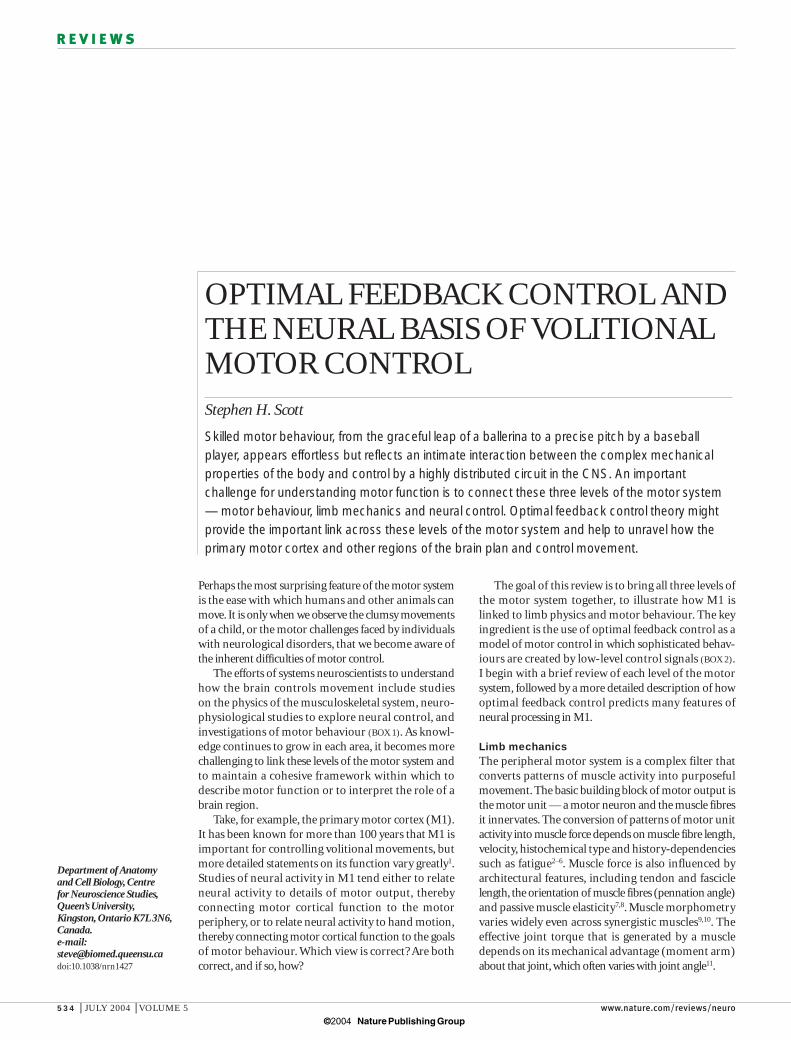
OPTIMAL FEEDBACK CONTROL ANDTHE NEURAL BASIS OF VOLITIONALMOTOR CONTROLStephen H. Scott
Skilled motor behaviour, from the graceful leap of a ballerina to a precise pitch by a baseballplayer, appears effortless but reflects an intimate interaction between the complex mechanicalproperties of the body and control by a highly distributed circuit in the CNS. An importantchallenge for understanding motor function is to connect these three levels of the motor system— motor behaviour, limb mechanics and neural control. Optimal feedback control theory mightprovide the important link across these levels of the motor system and help to unravel how theprimary motor cortex and other regions of the brain plan and control movement.
Department of Anatomyand Cell Biology, Centre for Neuroscience Studies,Queen’s University,Kingston, Ontario K7L 3N6,Canada.e-mail:[email protected]:10.1038/nrn1427
© 2004 Nature Publishing Group
NATURE REVIEWS | NEUROSCIENCE VOLUME 5 | JULY 2004 | 535
R E V I E W S
where 1
and 2
reflect properties of the upper arm andforearm/hand, respectively, and s and e denote shoulderand elbow, respectively. Muscular torque at a jointdepends on several parameters that relate to each segment’s moment of inertia (I), length (l), mass (m)and centre of mass (c)1.
These equations of motion mean that there is nolonger a one-to-one mapping between joint motion andmuscular torque, so that torque at one joint can generatemotion at other joints12,13. The mechanics of multi-jointmovements cannot be predicted from the physics ofsingle-joint movements14,15 (BOX 3). The equations of motion expand when more joints are involved andwhen joints have multiple degrees of freedom.Furthermore, a hand-held object or environmentalforces such as ground reaction forces during walking
Skeletal organization has a profound influence onthe conversion of muscle forces into limb motion. Limbmechanics are relatively straightforward when move-ment is constrained to occur at only a single joint andwith only one degree of mechanical freedom (flexion orextension). MUSCULAR TORQUE (T) is defined simply as
, where I equals the moment of inertia, and isthe angular acceleration of the joint. This angular version of the familiar equation, force = mass × linearacceleration, means that there is a direct relationshipbetween joint motion and torque. This simple relation-ship disappears when movement involves more thanone joint. The equations of motion to describe musculartorque at the shoulder (Ts) and elbow (Te) are (equations 1,2):
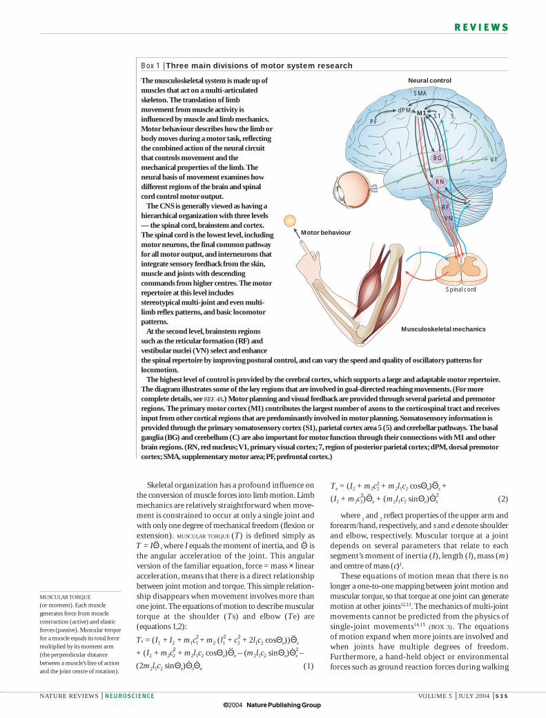
Box 1 | Three main divisions of motor system research
The musculoskeletal system is made up ofmuscles that act on a multi-articulatedskeleton. The translation of limbmovement from muscle activity isinfluenced by muscle and limb mechanics.Motor behaviour describes how the limb orbody moves during a motor task, reflectingthe combined action of the neural circuitthat controls movement and themechanical properties of the limb. Theneural basis of movement examines howdifferent regions of the brain and spinalcord control motor output.
The CNS is generally viewed as having ahierarchical organization with three levels— the spinal cord, brainstem and cortex.The spinal cord is the lowest level, includingmotor neurons, the final common pathwayfor all motor output, and interneurons thatintegrate sensory feedback from the skin,muscle and joints with descendingcommands from higher centres. The motorrepertoire at this level includesstereotypical multi-joint and even multi-limb reflex patterns, and basic locomotorpatterns.
At the second level, brainstem regionssuch as the reticular formation (RF) andvestibular nuclei (VN) select and enhancethe spinal repertoire by improving postural control, and can vary the speed and quality of oscillatory patterns forlocomotion.
The highest level of control is provided by the cerebral cortex, which supports a large and adaptable motor repertoire.The diagram illustrates some of the key regions that are involved in goal-directed reaching movements. (For morecomplete details, see REF. 48.) Motor planning and visual feedback are provided through several parietal and premotorregions. The primary motor cortex (M1) contributes the largest number of axons to the corticospinal tract and receivesinput from other cortical regions that are predominantly involved in motor planning. Somatosensory information isprovided through the primary somatosensory cortex (S1), parietal cortex area 5 (5) and cerebellar pathways. The basalganglia (BG) and cerebellum (C) are also important for motor function through their connections with M1 and otherbrain regions. (RN, red nucleus; V1, primary visual cortex; 7, region of posterior parietal cortex; dPM, dorsal premotorcortex; SMA, supplementary motor area; PF, prefrontal cortex.)
Musculoskeletal mechanics
Motor behaviour
Neural control
Spinal cord
PFS1
V1
75dPM M1
SMA
RN
BG
CRF
VN
Ts = (I1 + I2 + m1c1 + m2 (l1 + c2 + 2l1c2 cosΘe))Θs
+ (I2 + m2c2 + m2l1c2 cosΘe)Θe – (m2l1c2 sinΘe)Θe –
(2m2l1c2 sinΘe)ΘsΘe
2 2
2 2 2
.. .
..
. .(1)
T = IΘ..
2Te = (I2 + m2c2 + m2l1c2 cosΘe)Θs +
(I2 + m2c2)Θe + (m2l1c2 sinΘe)Θs
..
...2 2(2)
MUSCULAR TORQUE
(or moment). Each musclegenerates force from musclecontraction (active) and elasticforces (passive). Muscular torquefor a muscle equals its total forcemultiplied by its moment arm(the perpendicular distancebetween a muscle’s line of actionand the joint centre of rotation).
Θ..
© 2004 Nature Publishing Group
536 | JULY 2004 | VOLUME 5 www.nature.com/reviews/neuro
R E V I E W S
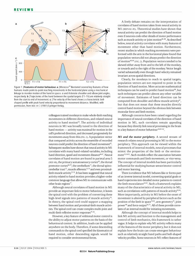
Motor behaviourBody movements are smooth, despite the complexitiesof the peripheral motor system. For example, hand trajectories remain relatively straight from start to end,and hand velocity follows a smooth, bell-shaped pro-file19,20 (FIG. 1). This smoothness at least partially reflectsthe low-pass filter properties of muscle21. Perturbationsof the hand during slow movements are corrected backtowards the unperturbed trajectory, indicating thatfeedback is used to maintain a relatively straight handtrajectory, at least under these conditions22. Such simplefeatures of hand motion mean that the CNS compen-sates for the complexities of limb mechanics.
The motor system can also adapt to changes in the mechanical environment. Lackner and DiZio23
observed how subjects performed reaching movementsbefore, during and after they sat in a room that rotatedat 6 rpm, creating a coriolis force on the limb. Whensubjects performed their first reaching movements with the right arm in the rotating room, after the otolithorgans no longer sensed room rotations, the move-ments were curved to the right. However, after severaltrials, reaching movements returned to relativelystraight trajectories, similar to those seen before theroom began to rotate. When the room stopped rotating,initial reaches were curved to the left, and subjects perceived that a strange force had pushed their limb.Again, reaching movements quickly returned to nearstraight trajectories. When a hand-held robot appliedloads during reaching, the results were similar24.Many studies have shown that relatively straight handtrajectories are preserved after various perceptual andmechanical perturbations25–28.
Although movements are smooth, motor perfor-mance shows considerable trial-to-trial variability,which partially reflects inherent noise in the systemrelated to both sensory and motor signals29–31. However,some features of motor performance, particularly task-relevant features, are tightly controlled32–34.For example, fluctuations in joint configurations thatinfluence the orientation of a subject pointing a laser ata spatial target are reduced, whereas patterns that do notinfluence laser orientation are more variable35. There is agrowing body of literature that illustrates how themotor system considers the influence of noise and variability in motor planning and control36–40.
Motor behaviour shows several key features.Movements are smooth, highly adaptable and showselective patterns of variability that reflect economy oftask-relevant features of motor performance. In spite of the complexities of limb mechanics, a hallmark ofmotor performance is smooth and relatively straighthand trajectories.
Neural centres of sensorimotor controlSensorimotor function is created from a highly distributed circuit that includes the spinal cord,brainstem and cerebral cortex (BOX 1). The spinal level supports the ‘most automatic’ movements, includingreflexes, as well as more complex multi-joint and multi-limb sensorimotor responses. The cortex supports the
can markedly influence limb mechanics13. This articlelargely focuses on proximal-arm movements, but thereare more challenging mechanical problems for hand16,17
and orofacial18 motor function.
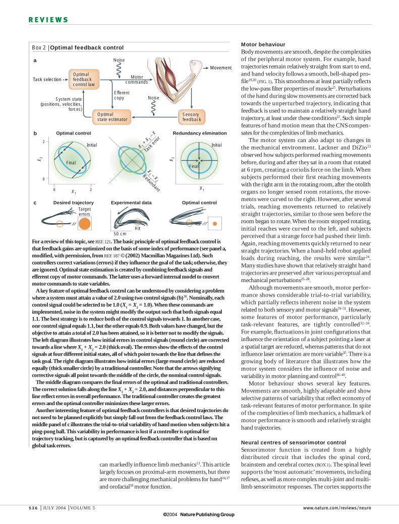
Box 2 | Optimal feedback control
For a review of this topic, see REF. 121. The basic principle of optimal feedback control isthat feedback gains are optimized on the basis of some index of performance (see panel a,modified, with permission, from REF. 187 © (2002) Macmillan Magazines Ltd). Suchcontrollers correct variations (errors) if they influence the goal of the task; otherwise, theyare ignored. Optimal state estimation is created by combining feedback signals andefferent copy of motor commands. The latter uses a forward internal model to convertmotor commands to state variables.
A key feature of optimal feedback control can be understood by considering a problemwhere a system must attain a value of 2.0 using two control signals (b)38. Nominally, eachcontrol signal could be selected to be 1.0 (X
1= X
2= 1.0).When these commands are
implemented, noise in the system might modify the output such that both signals equal1.1. The best strategy is to reduce both of the control signals towards 1. In another case,one control signal equals 1.1, but the other equals 0.9. Both values have changed, but theobjective to attain a total of 2.0 has been attained, so it is better not to modify the signals.The left diagram illustrates how initial errors in control signals (round circle) are correctedtowards a line where X
1+ X
2= 2.0 (thick oval). The errors show the effects of the control
signals at four different initial states, all of which point towards the line that defines thetask goal. The right diagram illustrates how initial errors (large round circle) are reducedequally (thick smaller circle) by a traditional controller. Note that the arrows signifyingcorrective signals all point towards the middle of the circle, the nominal control signals.
The middle diagram compares the final errors of the optimal and traditional controllers.The correct solution falls along the line X
1+ X
2= 2.0, and distances perpendicular to this
line reflect errors in overall performance. The traditional controller creates the greatesterrors and the optimal controller minimizes these larger errors.
Another interesting feature of optimal feedback controllers is that desired trajectories donot need to be planned explicitly but simply fall out from the feedback control laws. Themiddle panel of c illustrates the trial-to-trial variability of hand motion when subjects hit aping-pong ball. This variability in performance is lost if a controller is optimal fortrajectory tracking, but is captured by an optimal feedback controller that is based onglobal task errors.
Task selection
Optimalstate estimator
Sensoryfeedback
MovementOptimalfeedbackcontrol law
Noise
Motorcommands
NoiseSystem state(positions, velocities,
forces)
Efferentcopy
Task
erro
r
Redundant
2
0
0 2
X 2 X 2
X1X1
X 1 +
X 2 –
X
X1 – X
2
Initial
FinalFinal
Initial
Optimal control
Desired trajectory Experimental data Optimal control
Redundancy elimination
Targeterrors
50 cmHit
a
b
c
© 2004 Nature Publishing Group
NATURE REVIEWS | NEUROSCIENCE VOLUME 5 | JULY 2004 | 537
R E V I E W S
The activities of the cerebral cortex can be dividedinto two general problems in motor control — planningand execution. Motor planning reflects a range of issuesthat are related to the identification and selection ofgoals and strategies. Several cortical regions, includingmany parietal and frontal regions, participate in motorplanning47–51.
By contrast, M1 is more important for the executionof goal-directed and skilled motor tasks42,43. Lesions ofM1 in monkeys initially cause severe difficulties in voluntary movement, which remain permanent formore challenging distal limb motor tasks52. M1 has anintimate relationship with the motor periphery. Itreceives a rich mixture of sensory feedback from themotor periphery, with many neurons respondingstrongly to passive joint movements or skin contact.Most descending signals from the cortex pass throughspinal interneurons53. However, some neurons in M1(corticomotor (CM) neurons) form synaptic connec-tions directly onto spinal interneurons54–56, allowing M1to have a more direct and selective influence on muscleactivity. CM neurons are more prevalent for distal limbmusculature related to the hand42,57, and their numbersand influence increase with the level of dexterity acrossprimate species58,59.
Bridging the gapsThe discussion above describes three features of themotor system. First, the physics of moving even twojoints is complex. Second, humans can generate a rangeof skilled motor tasks. In reaching tasks, the trajectory ofthe hand tends to be conserved across conditions, butthere is also considerable trial-to-trial variability in thepath of the hand. Third, motor control is created by adistributed and interconnected circuit in which M1 hasa crucial role for volitional, goal-directed tasks. Animportant problem is to understand the links betweenmotor behaviour, limb mechanics and neural control.How do neural circuits create purposeful movementsfrom the complex, nonlinear musculoskeletal system?Does the neural activity of M1 reflect the control of high-level features related to behavioural goals, or oflow-level features related to the motor periphery? Ineffect, this question reflects the age-old problem: doesthe primary motor cortex code muscles or movements?
M1 and motor behaviour. Relatively straight hand trajectories and bell-shaped velocity profiles duringreaching indicate that the motor system might directlycontrol hand motion20,22,60,61. Neural signals in somebrain region(s) would explicitly signal hand trajectory,and these commands would be converted into patternsof muscle activity, potentially through intermediate representations62,63. For the online control of handmotion, proprioceptive signals would need to be converted from muscle to hand space, but it would be relatively simple to compute from vision.
The idea that hand trajectory is controlled online is consistent with electrophysiological recordings from M1 in non-human primates during whole-limbmovements. More than 20 years ago, Georgopoulos and
‘most voluntary’ motor tasks, such as reaching for anobject of interest, and learned associations, such as stepping on the car brake when a traffic light turns red.Voluntary behaviours often include more automaticcomponents — for example, a voluntary reach of thehand to a spatial target invokes automatic posturaladjustments to stabilize the body.
Neural recordings from monkeys are often used toexamine how the activity of individual neurons relatesto sensorimotor function. The anatomical and physio-logical properties of the limbs9,41 and CNS are similaracross primates42,43, and species such as Macaca mulattacan learn sophisticated motor behaviours (see, forexample, REFS 44–46).
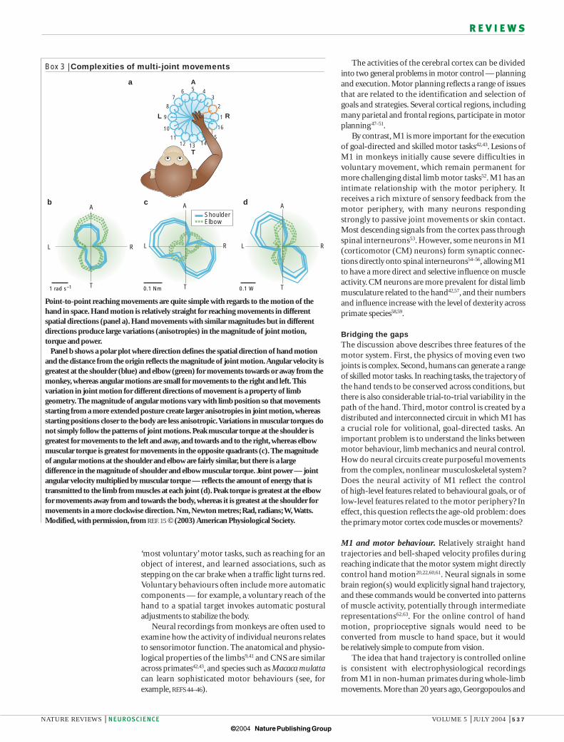
Box 3 | Complexities of multi-joint movements
Point-to-point reaching movements are quite simple with regards to the motion of thehand in space. Hand motion is relatively straight for reaching movements in differentspatial directions (panel a). Hand movements with similar magnitudes but in differentdirections produce large variations (anisotropies) in the magnitude of joint motion,torque and power.
Panel b shows a polar plot where direction defines the spatial direction of hand motionand the distance from the origin reflects the magnitude of joint motion.Angular velocity isgreatest at the shoulder (blue) and elbow (green) for movements towards or away from themonkey, whereas angular motions are small for movements to the right and left. Thisvariation in joint motion for different directions of movement is a property of limbgeometry. The magnitude of angular motions vary with limb position so that movementsstarting from a more extended posture create larger anisotropies in joint motion, whereasstarting positions closer to the body are less anisotropic.Variations in muscular torques donot simply follow the patterns of joint motions. Peak muscular torque at the shoulder isgreatest for movements to the left and away, and towards and to the right, whereas elbowmuscular torque is greatest for movements in the opposite quadrants (c). The magnitudeof angular motions at the shoulder and elbow are fairly similar, but there is a largedifference in the magnitude of shoulder and elbow muscular torque. Joint power — jointangular velocity multiplied by muscular torque — reflects the amount of energy that istransmitted to the limb from muscles at each joint (d). Peak torque is greatest at the elbowfor movements away from and towards the body, whereas it is greatest at the shoulder formovements in a more clockwise direction. Nm, Newton metres; Rad, radians; W,Watts.Modified, with permission, from REF. 15 © (2003) American Physiological Society.
345
9
12 13
15
16
1
2
67
8
10
1114
A
T
L R
0.1 Nm
A
T
L R
cb
1 rad s–1
A
T
L R
0.1 W
A
T
L R
d
ShoulderElbow
a
© 2004 Nature Publishing Group
538 | JULY 2004 | VOLUME 5 www.nature.com/reviews/neuro
R E V I E W S
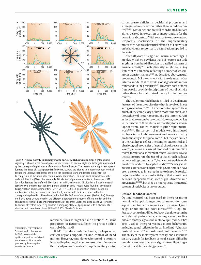
A lively debate remains on the interpretation ofcorrelates of hand motion taken from neural activity inM1 (REFS 82–85). Theoretical studies have shown thatneural activity can predict the direction of hand motioneven if neurons code other details of motor performancesuch as muscle activity or joint motion86–88.As describedbelow, neural activity correlates with many features ofmovement other than hand motion. Furthermore,recent studies in which reaching movements were per-formed with the arm in the horizontal plane found thatpopulation vectors did not always predict the directionof motion89,90 (FIG. 2). Population vectors tended to beskewed either away from and to the left of the monkey,or towards and to the right of the monkey.Vector lengthvaried substantially even though hand velocity remainedinvariant across spatial directions.
Clearly, for monkeys to reach to spatial targets,population vectors are not required to point in thedirection of hand motion. More accurate mathematicaltechniques can be used to predict hand motion88, butsuch techniques can predict almost any other variable of movement. Estimates of hand direction can be computed from shoulder and elbow muscle activity87,but that does not mean that these muscles directly control hand motion beyond the obvious link betweenmuscular force and limb motion.
Although concerns have been raised regarding theimportance of neural correlates of the direction of handmotion in M1, such correlates remain appealingbecause they directly link neural processing in the brainto a key feature of motor behaviour34,60–62.
M1 and the motor periphery. A second stream ofresearch relates neural processing in M1 to the motorperiphery. This approach can be viewed within theframework of internal models, neural processes thatmimic the properties of the limb or the environ-ment23,24,91. Such models reflect the association betweenmotor commands and limb movement, or vice versa.The concept of internal models has been particularlyinfluential for studying human sensorimotor controland motor learning.
There is evidence that M1 behaves like or forms partof an inverse internal model, converting spatial goals orhand trajectories into detailed motor patterns to controlthe limb musculature50,92. Such a framework explainsmany of the characteristics of neural activity in M1,such as correlations with patterns of muscle activity93,94.More importantly, activity in M1 before the onset ofmovement is altered by peripheral factors such as theposition of the limb in space70,95, arm geometry76, jointpower89 and force output45,75. All of these provide corre-lates of an internal model for initiating movements.
Although the concept of internal models helps tolink M1 activity and function to the management andcontrol of limb mechanics, this framework is rathervague. It helps to explain why M1 activity reflects manyof the features of the motor periphery, but it does notexplain how the brain can create emergent behavioursuch as relatively straight hand paths and bell-shapedvelocity profiles. Some neurons in M1 reflect features of
colleagues trained monkeys to make whole-limb reachingmovements in different directions, and related neuralactivity to hand motion64. The activity of individual neurons in M1 was broadly tuned to the direction ofhand motion — activity was maximal for motion in thecell’s preferred direction, and decreased progressively formovements away from this (FIG. 2). A population vectorthat compared activity across the ensemble of recordedneurons could predict the direction of hand movement65.Subsequent studies have shown that neural activity in M1correlates with many hand-related variables, includinghand direction, speed and movement distance66,67. Neuralcorrelates of hand motion are found in parietal area 5(REF. 68), the primary somatosensory cortex69, the dorsalpremotor cortex66,70, the cerebellum71, the dorsal spino-cerebellar tract72, muscle afferents73,74 and even proximal-limb muscle activity75,76. It has been suggested that neuralactivity related to hand motion provides a higher-ordercommon language that allows M1 to communicate withother brain regions77.
Although neural correlates of hand motion in M1provide an important link to motor behaviour, it leavesthe spinal cord with the problem of converting thesehigh-level signals into patterns of muscle activity78.In theory, the spinal cord could support a mappingbetween hand motion and proximal-limb muscle activi-ties. The spinal cord can create complex multi-joint andmulti-limb reflexive motor responses79–81.
However, a key feature of volitional motor control isthe ability to adjust motor patterns on the basis of thebehavioural context. In addition, loads can be appliedanywhere on the body. Therefore, if some descendingcommands to the spinal cord specified the kinematics ofhand motion, other descending signals would berequired to consider environmental forces.
T3T3
T2
T2
T1
T1
T4
T4
T5
T5
T6
T6
800
400
0Han
d sp
eed
(mm
s–1
)
a
c
b
T1 T4 T1 T5 T2 T50.5 s
α
θ
Figure 1 | Features of motor behaviour. a | Morasso19 illustrated several key features of howhumans made point-to-point reaching movements in the horizontal plane using a mechanicallinkage to monitor motion of the hand in space. α and θ denote shoulder and elbow joint angles,respectively. b | Trajectories of the hand between the spatial targets (T1–T6) are relatively straightfrom the start to end of movement. c | The velocity of the hand shows a characteristic bell-shaped profile with peak hand velocity proportional to movement distance. Modified, withpermission, from REF. 19 (1981) Springer-Verlag.
© 2004 Nature Publishing Group
NATURE REVIEWS | NEUROSCIENCE VOLUME 5 | JULY 2004 | 539
R E V I E W S
cortex create deficits in decisional processes and strategies of motor action rather than in online con-trol97–101. Motor actions are still coordinated, but areeither delayed in execution or inappropriate for thebehavioural context. With regards to online control,temporary inactivation of the supplementary motor area has no substantial effect on M1 activity oron behavioural responses to perturbations applied tothe wrist101.
After 40 years of single-cell neural recordings inmonkey M1, there is evidence that M1 neurons can codeanything from hand direction to detailed patterns ofmuscle activity90. Such diversity might be a key feature of M1 function, reflecting a number of sensori-motor transformations62,63. As described above, neuralprocessing in M1 is consistent with its role as part of aninternal model that converts global goals into motorcommands to the periphery50,92. However, both of theseframeworks provide descriptions of neural activityrather than a formal control theory for limb motor control.
The oculomotor field has identified in detail manyfeatures of the motor circuitry that is involved in eyeand gaze control102,103. The oculomotor system lacksmuch of the complexity of limb motor function, andthe activity of motor neurons and pre-interneurons in the brainstem can be recorded. However, another keyto the success of these studies is that they took advan-tage of formal control models to guide experimentalwork104,105. Similar control models were introduced to characterize limb movement and neural circuitry predominantly in the spinal cord106, but they are limitedin their ability to reflect the complex anatomical andphysiological properties of neural circuits even at thislevel107, let alone as a useful model of brain functionrelated to volitional movement control. EQUILIBRIUM POINT
MODELS incorporate the use of spinal stretch reflexes in descending commands108, but cannot explain end-point errors induced by applied loads23,24,109 and again donot consider supraspinal processing. Some models havebeen developed to interpret the role of specific corticalregions and the patterns of activity of their constituentneurons for specific tasks, such as goal-directed limbmovements110,111, but they do not replicate the naturalpatterns of variability in motor output.
Optimal feedback controlOptimal control has been used to interpret motorbehaviour by optimizing motor commands for someaspect of motor performance (such as maximal jumpheight or minimal end-point errors)30,112–116. Optimalfeedback control modifies feedback signals to optimizean index of performance, creating a complex linkbetween sensory signals and motor output (BOX 2). It hasbeen used to interpret various motor behaviours,including spinal reflexes in the cat hindlimb117, humanpostural balance118 and volitional motor control38,119.The ability of the motor system to adapt and use varioussensory signals for feedback control is exemplified byour ability to use cutaneous signals from light fingercontact to stabilize standing posture120.
movement such as target or hand direction95,96. Is thisproportion of neurons sufficient to provide online control of the hand?
If M1 considers limb mechanics, perhaps other cortical regions support on-line control of handmotion. However, candidate regions tend to be moreinvolved in planning than motor execution. Lesions inthe dorsal premotor cortex or supplementary motor
EQUILIBRIUM POINT MODELS
A class of models that assumethe CNS can control theequilibrium position establishedby the balance of force that isgenerated by the spring-likebehaviour of muscle.
b c
a
–500 0 500 1,0000
45
90
135
180
0
45
90
135
180
0
45
90
135
180
Time (ms)
Firin
g ra
te(s
pike
s s–
1 )Fi
ring
rate
(spi
kes
s–1 )
Firin
g ra
te(s
pike
s s–
1 )
–500 0 500 1,0000
45
90
135
180
0
45
90
135
180
Time (ms)–500 0 500 1,000
–500 0 500 1,000 –500 0 500 1,000 –500 0 500 1,000
–500 0 500 1,000 –500 0 500 1,000
0
45
90
135
180
0
45
90
135
180
0
45
90
135
180
Time (ms)
PD
Figure 2 | Neural activity in primary motor cortex (M1) during reaching. a | Mean handtrajectory is shown in the central panel for movements to each of eight spatial targets surroundedby the corresponding response of the neuron for each target. The rasters at the top of each panelillustrate the times of action potentials for five trials. Data are aligned to movement onset (verticaldashed line). Below each raster are the mean (blue) and standard deviation (green) of thedischarge rate of the neuron for each movement direction. The large black arrow denotes thepreferred direction (PD) of the neuron. b | Distribution of preferred directions of neurons in M1.Each dot denotes the preferred direction of an individual neuron. Distribution is based on neuralactivity only during the reaction time period, although similar results were found for any epochduring reaction and movement time. (n = 154, P < 0.001.) c | Population vectors based onreaction time activity of neurons are denoted by arrows with their base attached to thecorresponding direction of hand motion for the initial 100 ms of movement (dashed line). Orangeand blue arrows denote whether the difference between the direction of hand motion and thepopulation vector is significant or insignificant, respectively. Under each population vector is thedispersion of vectors formed by random resampling of the cell population with replacements.Modified, with permission, from REF. 90 (2003) Elsevier Science.
© 2004 Nature Publishing Group
540 | JULY 2004 | VOLUME 5 www.nature.com/reviews/neuro
R E V I E W S
can vary with time, as observed for peripheral feedbackduring locomotion124. Todorov and Jordan38 have proposed that such flexibility in the properties of the con-troller might be a valuable conceptual framework forinterpreting volitional motor behaviour such as reachingand grasping. They show that such a controller capturesmany of the common features of human movement,including goal-directed corrections, multi-joint synergiesand variable but successful motor performance. Severalfeatures of motor performance emerge despite not beingexplicitly defined in the feedback controller.
The observation that reaching movements are relatively straight with bell-shaped velocity profiles provides strong circumstantial evidence that the CNS directly controls hand trajectory. However, handtrajectory does not have to be directly controlled if thebrain behaves like an optimal feedback controller.Behavioural goals (such as reaching to a spatial target)can be converted directly into feedback laws to convertstate variables into motor commands. Hand motionsimply falls out as the optimal controller adjusts motoroutput on the basis of statistical variations in state variables created by external perturbations and systemnoise. Errors that influence the goal of the task are corrected, those that do not are ignored. Even if handtrajectory itself becomes the goal of a task such as slowreaching22, it does not need to be explicitly defined inthe controller.
An optimal feedback controller has several key components121. First, optimal control needs an optimal estimate of the state of the system (STATE VARIABLES), whichis generated from afferent feedback from sensors combined with efferent copy of motor signals. Inhumans, both afferent feedback and efferent copy areused to estimate ongoing motor performance122.Support for the use of efferent copy in motor control isprovided by observations that motor commands canundergo rapid compensation before sensory feedbackcan influence them123. State variables can reflect not onlythe properties of the body, but also information relatedto grasped objects116.
Second, feedback gains to convert these state variablesinto motor signals are not fixed, but are adjusted basedon the specific goals of a behaviour. This is essentially anoptimization problem that manipulates feedback gainsto maximize or minimize some index of performance.Aproperty of optimal feedback controllers is that sensedvariations in state variables lead to corrections if theyadversely affect motor performance, but are ignored if they do not. Todorov and Jordan38 define this as a‘minimum intervention’ principle. This selective correc-tion of errors is particularly important for a system withnoise, which is prevalent in both motor output30 andsensory signals29,31.
By its nature, optimal control modifies feedbackgains to suit the overall goals of the system. These gains
STATE VARIABLES
Estimates of the position of thelimb or forces acting within oron the limb (or their derivative).State variables are transformedby corresponding feedback gainsto generate motor outputcommands.
Hz
Hz
128 128 128 128 128
128 128 128 128 128
Unperturbed small Torque pulse holding Preballistic torque Unperturbed ballisticSmall + torque
Pronation
Supination
20°
1 sec
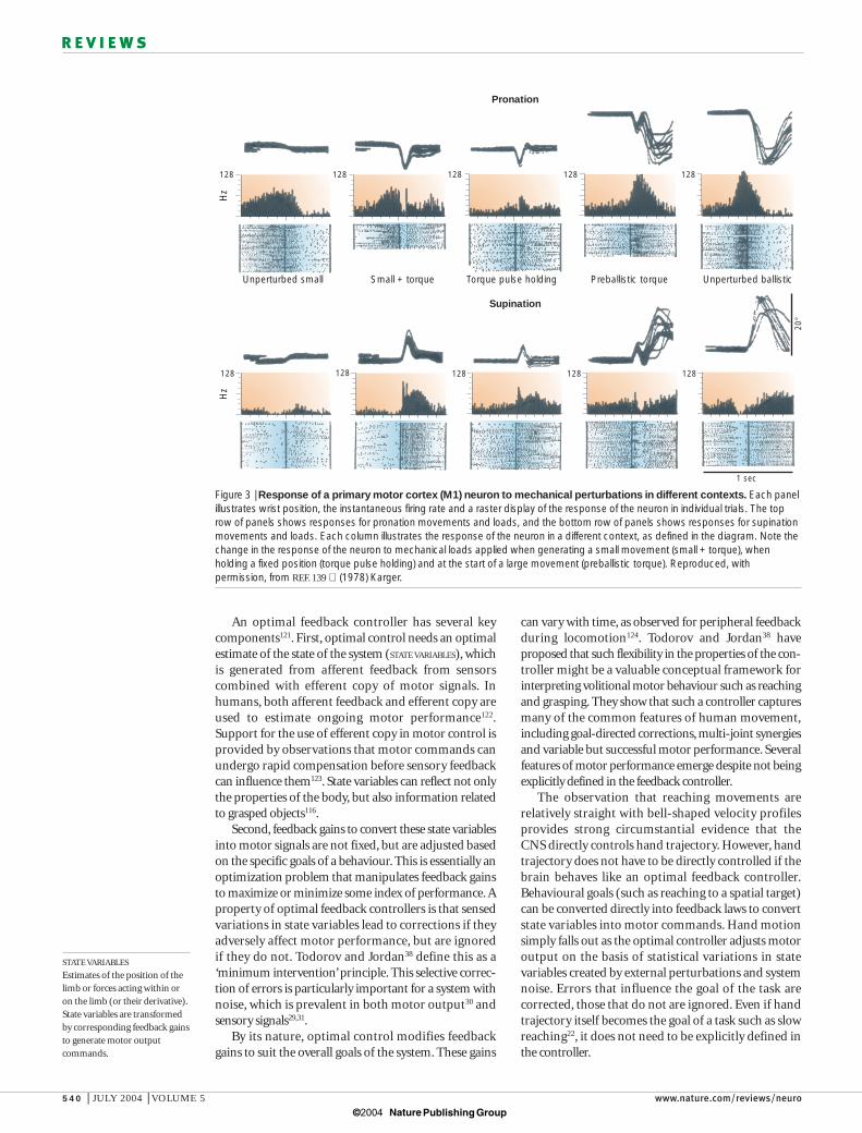
Figure 3 | Response of a primary motor cortex (M1) neuron to mechanical perturbations in different contexts. Each panelillustrates wrist position, the instantaneous firing rate and a raster display of the response of the neuron in individual trials. The toprow of panels shows responses for pronation movements and loads, and the bottom row of panels shows responses for supinationmovements and loads. Each column illustrates the response of the neuron in a different context, as defined in the diagram. Note thechange in the response of the neuron to mechanical loads applied when generating a small movement (small + torque), whenholding a fixed position (torque pulse holding) and at the start of a large movement (preballistic torque). Reproduced, withpermission, from REF. 139 (1978) Karger.
© 2004 Nature Publishing Group
NATURE REVIEWS | NEUROSCIENCE VOLUME 5 | JULY 2004 | 541
R E V I E W S
Optimal feedback control that is based on low-dimensional state variables is consistent with the mostobvious feature of M1 — a coarse somatotopic repre-sentation of the motor periphery with neurons relatedto one or a few joints126,127. Selective changes in mechani-cal loads applied to the elbow or shoulder joints duringposture and movement illustrate that some neurons aresensitive to loads at both joints, whereas others respondto loads at only one joint45,128. Corticomotor neuronssynapse on motor neurons from a few muscles that spanone or more joints54,57. These observations indicate thatneurons are exclusively associated neither with the entirelimb nor with a single joint. Rather, neurons reflect aportion of the motor periphery that might or might notspan multiple joints.
Like M1, an optimal feedback controller shouldreceive a rich mix of sensory signals (for review, see REFS 42,43). Many neurons in M1 respond to passivemovement of one or more joints76,129, and this sensoryfeedback often overlaps with their motor output76,130,131.Many neurons respond to passive and active movementsat multiple joints, but the association between these sensory and motor representations remains poorlyunderstood. Neurons related to the distal limb oftenrespond to passive movements of the wrist and digits, orto cutaneous stimulation on the hand, reflecting theimportance of cutaneous input for hand function132–134.Neurons in shoulder-related regions of cat M1 oftenhave cutaneous receptive fields on the paw135, reflectingthe link between walking surface stability and proximalmuscle control for quadrupedal locomotion.
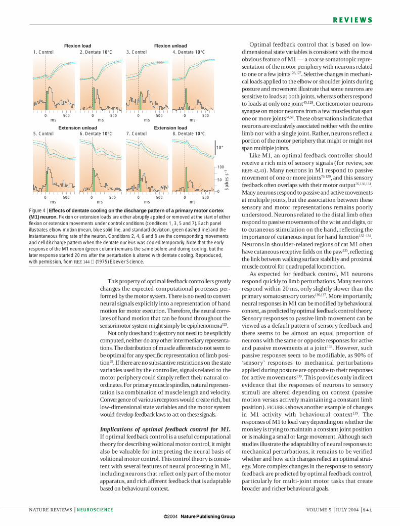
As expected for feedback control, M1 neuronsrespond quickly to limb perturbations. Many neuronsrespond within 20 ms, only slightly slower than the primary somatosensory cortex136,137. More importantly,neural responses in M1 can be modified by behaviouralcontext, as predicted by optimal feedback control theory.Sensory responses to passive limb movement can beviewed as a default pattern of sensory feedback andthere seems to be almost an equal proportion ofneurons with the same or opposite responses for activeand passive movements at a joint138. However, such passive responses seem to be modifiable, as 90% of‘sensory’ responses to mechanical perturbationsapplied during posture are opposite to their responsesfor active movements139. This provides only indirectevidence that the responses of neurons to sensory stimuli are altered depending on context (passivemotion versus actively maintaining a constant limbposition). FIGURE 3 shows another example of changesin M1 activity with behavioural context139. Theresponses of M1 to load vary depending on whether themonkey is trying to maintain a constant joint positionor is making a small or large movement. Although suchstudies illustrate the adaptability of neural responses tomechanical perturbations, it remains to be verifiedwhether and how such changes reflect an optimal strat-egy. More complex changes in the response to sensoryfeedback are predicted by optimal feedback control,particularly for multi-joint motor tasks that createbroader and richer behavioural goals.
This property of optimal feedback controllers greatlychanges the expected computational processes per-formed by the motor system. There is no need to convertneural signals explicitly into a representation of handmotion for motor execution. Therefore, the neural corre-lates of hand motion that can be found throughout thesensorimotor system might simply be epiphenomena125.
Not only does hand trajectory not need to be explicitlycomputed, neither do any other intermediary representa-tions. The distribution of muscle afferents do not seem tobe optimal for any specific representation of limb posi-tion29. If there are no substantive restrictions on the statevariables used by the controller, signals related to themotor periphery could simply reflect their natural co-ordinates. For primary muscle spindles, natural represen-tation is a combination of muscle length and velocity.Convergence of various receptors would create rich, butlow-dimensional state variables and the motor systemwould develop feedback laws to act on these signals.
Implications of optimal feedback control for M1.If optimal feedback control is a useful computationaltheory for describing volitional motor control, it mightalso be valuable for interpreting the neural basis ofvolitional motor control. This control theory is consis-tent with several features of neural processing in M1,including neurons that reflect only part of the motorapparatus, and rich afferent feedback that is adaptablebased on behavioural context.
1. Control 2. Dentate 10°C 3. Control 4. Dentate 10°C
5. Control 6. Dentate 10°C 7. Control 8. Dentate 10°C
Flexion load Flexion unload
Extension unload Extension load
0 500 0 500 0 500 0 500ms ms ms ms
0 500 0 500 0 500 0 500ms ms ms ms
100
50
0
10°
Spi
kes
s–1
Figure 4 | Effects of dentate cooling on the discharge pattern of a primary motor cortex(M1) neuron. Flexion or extension loads are either abruptly applied or removed at the start of eitherflexion or extension movements under control conditions (conditions 1, 3, 5 and 7). Each panelillustrates elbow motion (mean, blue solid line, and standard deviation, green dashed line) and theinstantaneous firing rate of the neuron. Conditions 2, 4, 6 and 8 are the corresponding movementsand cell discharge pattern when the dentate nucleus was cooled temporarily. Note that the earlyresponse of the M1 neuron (green column) remains the same before and during cooling, but thelater response started 20 ms after the perturbation is altered with dentate cooling. Reproduced,with permission, from REF. 144 (1975) Elsevier Science.
© 2004 Nature Publishing Group
542 | JULY 2004 | VOLUME 5 www.nature.com/reviews/neuro
R E V I E W S
the importance of sensory feedback to M1 processingwith more recent advances on multi-joint mechanics,motor behaviour and motor planning.
Optimal feedback control beyond M1. A completedescription of how optimal feedback control can beapplied to other brain regions is beyond the scope ofthis review, but there are a few issues worth noting.Optimal feedback control makes an important distinc-tion between motor execution and motor planning.This segregation between control and goals seems to bereflected in the cortex, with M1 being more involved inthe former, and other frontal and posterior parietalregions being more involved in the latter50. However, thesegregation is not complete; neural activity duringmotor preparation can be observed in M1 (REFS 152,159)
and at the spinal level160.Visual signals transmitted through the posterior
parietal cortex are important for motor planning andthe online control of movement153,154,161,162. Therefore, theposterior parietal and premotor cortex might beinvolved in both planning and online control, with individual neurons participating in both processes163.
State variables that are based on visual feedback(and probably other sensory signals) seem to be modi-fiable. For example, after a monkey has been trained to use a rake to grab food morsels, neurons in the intraparietal sulcus that normally respond to visualstimuli near the hand now also respond to stimuli nearthe rake46. Such plastic changes in vision-related neuralresponses might explain how humans and monkeyscan easily use computer-based visual feedback to control motor actions.
Subcortical networks through the basal ganglia andcerebellum are also important for sensorimotor control.In particular, the cerebellum has long been associatedwith motor control, coordination and learning91,164–167,and almost certainly has a crucial role in online feed-back control. Damage to this structure leads to motorproblems for tasks that involve multiple joints164,168.The anatomical and physiological properties of thecerebellum are consistent with several aspects ofoptimal feedback control. The interpositus nucleus andintermediate cerebellum receive proprioceptive feed-back on motor performance from the ascending spinocerebellar tracts and also receive a strong projec-tion from M1 through the pontine nuclei. This mixtureof afferent signals and efferent copy provides the idealconditions for optimal state estimation related to themotor periphery91,169. The dentate nucleus and lateralcerebellum are also probably involved as part of an optimal feedback controller. Several frontal and parietalcortical regions project to and receive input from thedentate nucleus through the pontine and thalamicnuclei, respectively165. However, each cortical regionprojects to largely separate regions of the dentatenucleus and cerebellar cortex, creating distinct cerebro-cerebellar loops170,171. Each of these loops might participate in distinct processes including task selection(motor planning), optimal state estimation and feed-back control. Monkeys trained either to assist or to resist
Long-latency muscle responses (> 60 ms), which aregenerated largely through the transcortical pathway140,illustrate the potential capability of this feedback system141. When the limb is perturbed from a stationaryposition, the short-latency muscle response (< 60 ms)that is generated at the spinal level parallels the patternof joint motions (the simple stretch reflex). By contrast,the long-latency response produces the requisite motor patterns to oppose the load, indicating that the transcortical pathway considers the influence of intersegmental dynamics in converting sensed limb motion into compensating motor responses.Furthermore, this long-latency response is modified toincorporate the influence of mechanical loads duringmotor learning142,143.
Several brain regions project to M1 and probably provide feedback from the motor periphery, includingthe primary somatosensory cortex, posterior area 5 and thalamic input from the cerebellum through theinterpositus and dentate nuclei42. The earliest response inM1 during mechanical perturbations seems to be pro-vided by the primary somatosensory cortex, as dentatecooling does not influence these early responses144–146.However, interpositus neurons respond to mechanicalloads within 20 ms (REF. 147). Later responses in M1,starting about 60 ms after a perturbation, seem to bestrongly influenced by the cerebellum145,148 (FIG. 4). Howthese different pathways contribute to feedback controlthrough M1 and the brainstem regions remains animportant problem.
The description above integrates feedback from themotor periphery into motor cortical function, but visual feedback is also important for volitional motor control149–151. A proportion of neurons in M1 signalmovement or target direction independent of arm configuration76,95,96,152. They are often assumed to providea higher-level representation of movement related to thespatial direction of movement, but such activity mightalso signal visual feedback of motor performance. Suchfeedback signals of hand motion are computationallyequal and not hierarchically above feedback signals fromthe motor periphery that are ‘muscle-like’. Furthermore,visual feedback is highly task dependent. For example,when writing with the tip of the elbow in space, visualfeedback of motor performance would reflect elbow andnot hand motion. Although vision is important foronline feedback153,154, loss of proprioception has a moreprofound effect on coordinated body movements155–157.
The ‘transcortical servo’ hypothesis that was put forward by Phillips more than 30 years ago emphasizedthe importance of feedback signals in motor controland was influential in the 1970s for interpreting motor cortical function on the basis of single-jointmovements158. The predominant use since the 1980s ofa whole-limb reaching paradigm to study motor behav-iour, and the practice of relating neural activity to handmotion, opened up issues related to the use of vision foraction, motor planning and the early, feedforward stageof motor execution. The value of optimal feedback control as a computational theory is that it brings theselargely distinct fields of study back together, recognizing
© 2004 Nature Publishing Group
NATURE REVIEWS | NEUROSCIENCE VOLUME 5 | JULY 2004 | 543
R E V I E W S
signals will have crucial consequences in one behaviouralcontext and be irrelevant in another. If the long-latencymuscle response reflects the feedback laws of an optimalfeedback controller, then transient perturbations duringdifferent tasks should elicit behaviourally relevant motorresponses. Neural recordings in various brain regionswill help to disseminate how such feedback control lawsare created by the highly distributed motor system.
The mathematics of optimal feedback control areparticularly challenging. The brain does not implementthe formal mathematical methods that are available tocompute gains of optimal feedback controllers, but howneural networks create and learn these properties is aninteresting and important process177–179. M1 is intimatelyinvolved in motor learning, and many studies haveinvestigated plasticity and changes in neural processingin M1 during learning and adaptation180–183. Optimalfeedback control requires substantive learning at twopoints in the controller, one for optimal state estimationand the other for optimal control laws. The learningrules and mechanisms are different for these twoprocesses, with the former optimizing estimates of thestate of the system, independently of behavioural goals .By contrast, the latter must also use more global rewardsthat are related to behavioural success or failure.
The motor system is not just one big feedback loop;rather, it is highly distributed and provides multiple path-ways through which feedback can influence behaviour.Besides M1, several other brain regions contribute todescending signals to influence spinal processing53. Tworegions that might be of particular interest for feedbackcontrol are the magnocellular red nucleus, which projectsto the spinal cord and receives substantive input fromboth M1 and the cerebellum165,184, and area 3a in the primary somatosensory cortex, which receives substantialinput from muscle proprioceptors and projects to theintermediate and ventral horn of the spinal cord137,185.
Where and how visual and proprioceptive signals areintegrated for estimating state variables and feedbackcontrol laws at the single-cell level remains poorlyunderstood. Clearly there is substantive integration of different sensory systems for position sense andkinaesthesia186. Visual feedback is assumed to take predominantly a cortical path to M1 through the parietal and premotor cortex48. There are several poten-tial pathways for proprioceptive feedback to reach M1,including through the primary somatosensory cortex,posterior parietal area 5 and the cerebellum42. As statedearlier, somatosensory feedback could be integratedwith visual signals in posterior parietal regions and thentransmitted through the premotor cortex. However, it isnot clear how each of these pathways is involved inmotor execution, learning or both.
Summary and conclusionsThe aim of this review was to bring together three levelsof research on limb motor function — the motorperiphery, motor behaviour and the neural basis ofmovement. Each level provides a unique perspective onthe characteristics of the motor system, and an impor-tant challenge in systems neuroscience is to connect
a perturbing flexor load applied to the wrist show context-dependent changes in neural activity in M1. Aneuron might fire in a rapid burst when a load is appliedif the behavioural condition was to resist the load, butwould be unresponsive when instructed to assist theapplied load172. Similar coupling to instructional cues is also observed in the dentate nucleus147, indicating that the dentate nucleus might be involved in rapidlyswitching from one context to another.
Descending commands from M1 and other brainregions must consider more than just ALPHA MOTOR
NEURON activity during motor function90. GAMMA
MOTOR NEURON activity and the inflow of sensory signalsfor motor output, and transmission to supraspinal cen-tres for both control and perception are also important.A substantial proportion of corticospinal axons terminatein the intermediate horn and even the dorsal horn42.These other features of spinal processing might accountfor half of the descending signals from the cortex, but little is known about the nature of such signals173. If thebrain behaves like an optimal feedback controller,it might be best to view descending commands as controlling the spinomusculoskeletal system, rather thanthe musculoskeletal system174.
Things to do and not to doThere might be many ways to use optimal feedback control to guide neurophysiological research, althoughseveral challenges remain. First, the mathematics that isrequired to identify optimal feedback control laws is extremely challenging even for the simplest of linearsystems. This limits the conditions under which formalsolutions can be used to predict the properties of anoptimal feedback controller, although recent mathemat-ical advances might extend this approach for nonlinearsystems175. Further theoretical work is also required tobreak down the processes of optimal feedback controlinto more biologically plausible algorithms andprocesses176 that can help to guide experimental studies.However, it is unlikely that such efforts will attain thelevel of detail that is present in oculomotor models ofbrainstem circuitry.
Identifying state variables would be a logical start forexamining neurophysiological correlates of optimalfeedback control. On its own, this is probably the least informative exercise and simply continues the basicpractice of correlating neural activity in M1 and elsewhere with engineering-inspired variables. The richmix of sensory signals (cutaneous, muscle propriocep-tors and vision) that are used to guide motor functionobfuscate any simple unified representation. Furtherdiversity is expected in a region such as M1 owing to itsinteraction with various cortical and subcortical brainregions90. Although neural activity must be quantifiedrelative to some measured (or estimated) variable,relative changes within and across task conditions are far more informative than interpreting absolute levels of neural activity.
The important feature of optimal feedback con-trollers is that they are malleable systems defined bybehavioural goals so that variations in sensory or motor
ALPHA MOTOR NEURON
Motor neurons that innervateextrafusal muscle fibres thatgenerate force.
GAMMA MOTOR NEURON
Motor neurons that innervateintrafusal muscle fibresassociated with muscle spindles.
© 2004 Nature Publishing Group
544 | JULY 2004 | VOLUME 5 www.nature.com/reviews/neuro
R E V I E W S
be of value for interpreting the neural basis of move-ment and in particular, neural processing in M1.Optimal feedback control is consistent with severalaspects of neural processing in M1. Individual neuronscontribute to the control of a portion of the motorperiphery and receive rich, adaptable sensory feedback.The link between M1 and motor behaviour emergesthrough its contribution to the entire neural circuit.Therefore, the role of M1 is not ‘muscles’ versus ‘movement’, but muscles and movement.
these domains. Activity in M1 has been linked to motorbehaviour or to the motor periphery, but it has been dif-ficult to reconcile a dual role for representing high-levelaspects of motor performance such as hand trajectoryand low-level details of motor execution.
Optimal feedback control, with its selective andhighly adaptable feedback laws, provides an interestingmodel for describing how coordinated motor behaviourcan be created by the motor system. The argument putforward here is that optimal feedback control can also
1. Scott, S. H. Role of motor cortex in coordinating multi-jointmovements: is it time for a new paradigm? Can. J. Physiol.Pharmacol. 78, 923–933 (2000).
2. Hill, A. V. The heat of shortening and the dynamic constantsof muscle. Proc. R. Soc. Lond. B 126, 136–195 (1938).
3. Gordon, A. M., Huxley, A. F. & Julian, F. J. The variation inisometric tension with sarcomere length in vertebratemuscle fibres. J. Physiol. (Lond.) 184, 170–192 (1966).
4. Burke, R. E., Levine, D. N., Tsairis, P. & Zajac, F. E.Physiological types and histochemical profiles in motor unitsof the cat gastrocnemius. J. Physiol. (Lond.) 234, 723–748(1973).
5. Scott, S. H., Brown, I. E. & Loeb, G. E. Mechanics of felinesoleus: I. effect of fascicle length and velocity on forceoutput. J. Muscle Res. Cell Motil. 17, 207–219 (1996).
6. Cheng, E. J., Brown, I. E. & Loeb, G. E. Virtual muscle: acomputational approach to understanding the effects ofmuscle properties on motor control. J. Neurosci. Methods101, 117–130 (2000).
7. Zajac, F. E. Muscle and tendon: properties, models, scaling,and application to biomechanics and motor control. Crit.Rev. Biomed. Eng. 17, 359–411 (1989).
8. Otten, E. in Exercise and Sport Sciences Reviews (ed.Pandolf, K. B.) 89–137 (Williams & Wilkins, Baltimore, 1989).
9. Cheng, E. J. & Scott, S. H. Morphometry of Macaca mulattaforelimb. I. Shoulder and elbow muscles and segmentinertial parameters. J. Morphol. 245, 206–224 (2000).
10. Singh, K., Melis, E. H., Richmond, F. J. & Scott, S. H.Morphometry of Macaca mulatta forelimb. II. Fiber-typecomposition in shoulder and elbow muscles. J. Morphol.251, 323–332 (2002).
11. Graham, K. M. & Scott, S. H. Morphometry of Macacamulatta forelimb. III. Moment arm of shoulder and elbowmuscles. J. Morphol. 255, 301–314 (2003).
12. Hollerbach, J. M. & Flash, T. Dynamic interactions betweenlimb segments during planar arm movement. Biol. Cybern.44, 67–77 (1982).
13. Zajac, F. E. & Gordon, M. E. in Exercise and Sport SciencesReviews (ed. Pandolf, K. B.) 187–230 (Williams & Wilkins,Baltimore, 1989).
14. Mussa-Ivaldi, F. A., Hogan, N. & Bizzi, E. Neural,mechanical, and geometric factors subserving arm posturein humans. J. Neurosci. 5, 2732–2743 (1985).
15. Graham, K. M. et al. Kinematics and kinetics of multi-jointreaching in non-human primates. J. Neurophysiol. 89,2667–2677 (2003).
16. Schieber, M. H., Gardinier, J. & Liu, J. Tension distribution tothe five digits of the hand by neuromuscular compartmentsin the macaque flexor digitorum profundus. J. Neurosci. 21,2150–2158 (2001).
17. Valero-Cuevas, F. J., Johanson, M. E. & Towles, J. D.Towards a realistic biomechanical model of the thumb: thechoice of kinematic description may be more critical thanthe solution method or the variability/uncertainty ofmusculoskeletal parameters. J. Biomech. 36, 1019–1030(2003).
18. Munhall, K. G. in Dynamic Models of Motor Planning (ed.Culicover, P.) 192–198 (Nature Publishing Group, London,2002).
19. Morasso, P. Spatial control of arm movements. Exp. BrainRes. 42, 223–227 (1981).
20. Flash, T. & Hogan, N. The coordination of arm movements:an experimentally confirmed mathematical model. J. Neurosci. 5, 1688–1703 (1985).
21. Krylow, A. M. & Rymer, W. Z. Role of intrinsic muscleproperties in producing smooth movements. IEEE Trans.Biomed. Eng. 44, 165–176 (1997).
22. Won, J. & Hogan, N. Stability properties of human reachingmovements. Exp. Brain Res. 107, 125–136 (1995).
23. Lackner, J. R. & DiZio, P. Rapid adaptation to coriolis forceperturbations of arm trajectory. J. Neurophysiol. 72,299–313 (1994).
24. Shadmehr, R. & Mussa-Ivaldi, F. A. Rapid adaptation tocoriolis force perturbations of arm trajectory. J. Neurosci. 14,3208–3224 (1994).References 23 and 24 are classic studies thatillustrate how subjects modify motor commands tocompensate for mechanical loads applied to the limbduring reaching.
25. Flanagan, J. R. & Rao, A. K. Trajectory adaptation to anonlinear visuomotor transformation: evidence of motionplanning in visually perceived space. J. Neurophysiol. 74,2174–2178 (1995).
26. Sainburg, R. L., Ghez, C. & Kalakanis, D. Intersegmentaldynamics are controlled by sequential anticipatory, errorcorrection, and postural mechanisms. J. Neurophysiol. 81,1045–1056 (1999).
27. Flanagan, J. R., Vetter, P., Johansson, R. S. & Wolpert, D. M.Prediction precedes control in motor learning. Curr. Biol. 13,146–150 (2003).
28. Singh, K. & Scott, S. H. A motor learning strategy reflectsneural circuitry for limb control. Nature Neurosci. 6, 399–403(2003).
29. Scott, S. H. & Loeb, G. E. The computation of positionsense from spindles in mono- and multiarticular muscles. J. Neurosci. 14, 7529–7540 (1994).
30. Harris, C. M. & Wolpert, D. M. Signal-dependent noisedetermines motor planning. Nature 394, 780–784 (1998).This study illustrates how optimal strategies forminimizing signal-dependent noise lead to bell-shaped velocity profiles and relatively straight handtrajectories.
31. Vallbo, A. B. Human muscle spindle discharge duringisometric voluntary contractions. Amplitude relationsbetween spindle frequency and torque. Acta Physiol.Scand. 90, 319–336 (1974).
32. Winter, D. A. Kinematic and kinetic patterns in human gait:variability and compensating effects. Hum. Mov. Sci. 3,51–76 (1984).
33. Lacquaniti, F. & Maioli, C. Coordinate transformations in thecontrol of cat posture. J. Neurophysiol. 72, 1496–1515(1994).
34. Scholz, J. P. & Schoner, G. The uncontrolled manifoldconcept: identifying control variables for a functional task.Exp. Brain Res. 126, 289–306 (1999).
35. Scholz, J. P., Schoner, G. & Latash, M. L. Identifying thecontrol structure of multijoint coordination during pistolshooting. Exp. Brain Res. 135, 382–404 (2000).
36. Sabes, P. N., Jordan, M. I. & Wolpert, D. M. The role ofinertial sensitivity in motor planning. J. Neurosci. 18,5948–5957 (1998).
37. Kording, K. P. & Wolpert, D. M. Bayesian integration insensorimotor learning. Nature 427, 244–247 (2004).
38. Todorov, E. & Jordan, M. I. Optimal feedback control as atheory of motor coordination. Nature Neurosci. 5,1226–1235 (2002).This study illustrates how optimal feedback controlcan predict many features of volitional motor control,including muscle synergies, motor coordination andvariable but accurate movements.
39. Todorov, E. Cosine tuning minimizes motor errors. NeuralComput. 14, 1233–1260 (2002).
40. van Beers, R. J., Haggard, P. & Wolpert, D. M. The role ofexecution noise in movement variability. J. Neurophysiol. 91,1050–1063 (2004).
41. Christel, M. I. & Billard, A. Comparison between macaques’and humans’ kinematics of prehension: the role ofmorphological differences and control mechanisms. Behav.Brain Res. 131, 169–184 (2002).
42. Porter, R. & Lemon, R. N. Cortical Function and VoluntaryMovement (Clarendon, Oxford, 1993).
43. Hepp-Reymond, M. C. Functional organization of motorcortex and its participation in voluntary movements. Comp.Primate Biol. 4, 501–624 (1988).
44. Wise, S. P., Di Pellegrino, G. & Boussaoud, D. The premotorcortex and nonstandard sensorimotor mapping. Can. J.Physiol. Pharmacol. 74, 469–482 (1996).
45. Gribble, P. L. & Scott, S. H. Overlap of internal models inmotor cortex for mechanical loads during reaching. Nature417, 938–941 (2002).
46. Iriki, A., Tanaka, M., Iwamura, Y. Coding of modified bodyschema during tool use by macaque postcentral neurones.Neuroreport 7, 2325–2330 (1996).The first in a series of studies to show how neuralactivity related to the hand is modified when monkeysare trained to use a rake to retrieve food rewards.
47. Wise, S. P., Boussaoud, D., Johnson, P. B. & Caminiti, R.Premotor and parietal cortex: corticocortical connectivityand combinatorial computations. Annu. Rev. Neurosci. 20,25–42 (1997).
48. Battaglia-Mayer, A., Caminiti, R., Lacquaniti, F. & Zago, M.Multiple levels of representation of reaching in the parieto-frontal network. Cereb. Cortex 13, 1009–1022 (2003).
49. Rizzolatti, G. & Luppino, G. The cortical motor system.Neuron 31, 889–901 (2001).
50. Kalaska, J. F., Scott, S. H., Cisek, P. & Sergio, L. E. Corticalcontrol of reaching movements. Curr. Opin. Neurobiol. 7,849–859 (1997).
51. Tanji, J. Sequential organization of multiple movements:involvement of cortical motor areas. Annu. Rev. Neurosci.24, 631–651 (2001).
52. Hoffman, D. S. & Strick, P. L. Effects of a primary motorcortex lesion on step-tracking movements of the wrist. J. Neurophysiol. 73, 891–895 (1995).
53. Dum, R. P. & Strick, P. L. Spinal cord terminations of themedial wall motor areas in macaque monkeys. J. Neurosci.16, 6513–6525 (1996).
54. Fetz, E. E. & Cheney, P. D. Postspike facilitation of forelimbmuscle activity by primate corticomotoneuronal cells. J. Neurophysiol. 44, 751–772 (1980).
55. Shinoda, Y., Yokota, J. & Futami, T. Divergent projection ofindividual corticospinal axons to motoneurons of multiplemuscles in the monkey. Neurosci. Lett. 23, 7–12 (1981).
56. Lawrence, D. G., Porter, R. & Redman, S. J.Corticomotoneuronal synapses in the monkey: lightmicroscopic localization upon motoneurons of intrinsicmuscles of the hand. J. Comp. Neurol. 232, 499–510(1985).
57. McKiernan B. J., Marcario J. K., Karrer J. H. & Cheney P. D.Correlations between corticomotoneuronal (CM) cellpostspike effects and cell-target muscle covariation. J. Neurophysiol. 83, 99–115 (2000).
58. Heffner, R. & Masterton, B. Variation in form of the pyramidaltract and its relationship to digital dexterity. Brain Behav.Evol. 12, 161–200 (1975).
59. Nakajima, K., Maier, M. A., Kirkwood, P. A. & Lemon, R. N.Striking differences in transmission of corticospinal excitationto upper limb motoneurons in two primate species. J. Neurophysiol. 84, 698–709 (2000).
60. Gordon, J., Ghilardi, M. F. & Ghez, C. Accuracy of planarreaching movements. I. Independence of direction andextent variability. Exp. Brain Res. 99, 97–111 (1994).
61. Moran, D. W. & Schwartz, A. B. Motor corticalrepresentation of speed and direction during reaching. J. Neurophysiol. 82, 2676–2692 (1999).
62. Soechting, J. F. & Flanders, M. Moving in three-dimensionalspace: frames of reference, vectors, and coordinatesystems. Annu. Rev. Neurosci. 15, 167–191 (1992).
63. Kalaska, J. F. & Crammond, D. J. Cerebral corticalmechanisms of reaching movements. Science 255,1517–1523 (1992).
64. Georgopoulos, A. P., Kalaska J. F., Caminiti, R. & Massey, J. T.On the relations between the direction of two-dimensionalarm movements and cell discharge in primate motor cortex.J. Neurosci. 2, 1527–1537 (1982).
© 2004 Nature Publishing Group
NATURE REVIEWS | NEUROSCIENCE VOLUME 5 | JULY 2004 | 545
R E V I E W S
65. Georgopoulos, A. P., Caminiti, R., Kalaska, J. F. & Massey, J. T.Spatial coding of movement: a hypothesis concerning thecoding of movement directions by motor corticalpopulations. Exp. Brain Res. (Suppl.) 7, 327–336 (1983).
66. Fu, Q. G., Flament, D., Coltz, J. D. & Ebner, T. J. Temporalencoding of movement kinematics in the discharge ofprimate primary motor and premotor neurons. J. Neurophysiol. 73, 836–854 (1995).
67. Schwartz, A. B. Motor cortical activity during drawingmovements: population representation during sinusoidtracing. J. Neurophysiol. 70, 28–36 (1993).
68. Kalaska, J. F., Caminiti, R. & Georgopoulos, A. P. Corticalmechanisms related to the direction of two-dimensional armmovements: relations in parietal area 5 and comparison withmotor cortex. Exp. Brain Res. 51, 247–260 (1983).
69. Cohen, D. A., Prud’homme, M. J. & Kalaska, J. F. Tactileactivity in primate primary somatosensory cortex duringactive arm movements: correlation with receptive fieldproperties. J. Neurophysiol. 71, 161–172 (1994).
70. Caminiti, R., Johnson, P. B., Galli, C., Ferraina, S. & Burnod, Y.Making arm movements within different parts of space: thepremotor and motor cortical representation of a coordinatesystem for reaching to visual targets. J. Neurosci. 11,1182–1197 (1991).
71. Fortier, P. A., Smith, A. M. & Kalaska, J. F. Comparison ofcerebellar and motor cortex activity during reaching:directional tuning and response variability. J. Neurophysiol.69, 1136–1149 (1993).
72. Bosco, G. & Poppele, R. E. Modulation of dorsalspinocerebellar responses to limb movement. II. Effect ofsensory input. J. Neurophysiol. 90, 3372–3383 (2003).
73. Jones, K. E., Wessberg, J. & Vallbo, A. B. Directional tuningof human forearm muscle afferents during voluntary wristmovements. J. Physiol. (Lond.) 536, 635–647 (2001).
74. Ribot-Ciscar, E., Bergenheim, M., Albert, F. & Roll, J. P.Proprioceptive population coding of limb position inhumans. Exp. Brain Res. 149, 512–519 (2003).
75. Kalaska, J. F., Cohen, D. A., Hyde, M. L. & Prud’homme, M.A comparison of movement direction-related versus loaddirection-related activity in primate motor cortex, using atwo-dimensional reaching task. J. Neurosci. 9, 2080–2102(1989).
76. Scott, S. H. & Kalaska, J. F. Reaching movements withsimilar hand paths but different arm orientations: I. Activity ofindividual cells in motor cortex. J. Neurophysiol. 77,826–852 (1997).
77. Georgopoulos, A. P. Current issues in directional motorcontrol. Trends Neurosci. 18, 506–510 (1995).
78. Georgopoulos, A. P. On the translation of directional motorcortical commands to activation of muscles via spinalinterneuronal systems. Cogn. Brain Res. 3, 151–155 (1996).
79. Fukson, O. I., Berkinblit, M. B. & Feldman, A. G. The spinalfrog takes into account the scheme of its body during thewiping reflex. Science 209, 1261–1263 (1980).
80. Kargo, W. J. & Giszter, S. F. Afferent roles in hind limb wipe-reflex trajectories: free-limb kinematics and motor patterns.J. Neurophysiol. 83, 1480–1501 (2000).
81. D’Avella, A., Saltiel, P. & Bizzi, E. Combinations of musclesynergies in the construction of a natural motor behavior.Nature Neurosci. 6, 300–308 (2003).
82. Georgopoulos, A. P. & Ashe, J. One motor cortex, twodifferent views. Nature Neurosci. 3, 963–965 (2000).
83. Moran, D. W. & Schwartz, A. B. One motor cortex, twodifferent views. Nature Neurosci. 3, 963–965 (2000).
84. Todorov, E. Reply to ‘One motor cortex, two different views’.Nature Neurosci. 3, 963–964 (2000).
85. Scott, S. H. Reply to ‘One motor cortex, two different views’.Nature Neuroscience 3, 964–965 (2000).
86. Mussa-Ivaldi, F. A. Do neurons in the motor cortex encodemovement direction? An alternative hypothesis. Neurosci.Lett. 91, 106–111 (1988).
87. Todorov, E. Direct cortical control of muscle activation involuntary arm movements: a model. Nature Neurosci. 3,391–398 (2000).
88. Sanger, T. D. Neural population codes. Curr. Opin.Neurobiol. 13, 238–249 (2003).
89. Scott, S. H., Gribble, P. L., Graham, K. M. & Cabel, D. W.Dissociation between hand motion and population vectorsfrom neural activity in motor cortex. Nature 413, 161–165(2001).
90. Scott, S. H. The role of primary motor cortex in goal-directedmovements: insights from neurophysiological studies onnon-human primates. Curr. Opin. Neurobiol. 13, 671–677(2003).
91. Miall, R. C., Weir, D. J., Wolpert, D. M. & Stein, J. Is thecerebellum a smith predictor? J. Motor Behav. 25, 203–216(1993).
92. Scott, S. H. & Norman, K. E. Computational approaches tomotor control and their potential role for interpreting motordysfunction. Curr. Opin. Neurol. 16, 693–698 (2003).
93. Holdefer, R. N. & Miller, L. E. Primary motor cortical neuronsencode functional muscle synergies. Exp. Brain Res. 146,233–243 (2002).
94. Bennett, K. M. & Lemon, R. N. Corticomotoneuronalcontribution to the fractionation of muscle activity duringprecision grip in the monkey. J. Neurophysiol. 75,1826–1842 (1996).
95. Sergio, L. E. & Kalaska, J. F. Systematic changes in motorcortex cell activity with arm posture during directionalisometric force generation. J. Neurophysiol. 89, 212–228(2003).
96. Kakei, S., Hoffman, D. S. & Strick, P. L. Muscle andmovement representations in the primary motor cortex.Science 285, 2136–2139 (1999).
97. Passingham, R. E. Premotor cortex: sensory cues andmovement. Behav. Brain Res. 18, 175–185 (1985).
98. Passingham, R. E., Chen, Y. C. & Thaler, D. in NeuralProgramming (ed. Ito, M.) 13–24 (Karger, Basel, 1989).
99. Kurata, K. & Hoffman, D. S. Differential effects of muscimolmicroinjection into dorsal and ventral aspects of thepremotor cortex of monkeys. J. Neurophysiol. 71,1151–1164 (1994).
100. Kurata, K. & Hoshi, E. Reacquisition deficits in prismadaptation after muscimol microinjection into the ventralpremotor cortex of monkeys. J. Neurophysiol. 81,1927–1938 (1999).
101. Schmidt, E. M., Porter, R. & McIntosh, J. S. The effects ofcooling supplementary motor area and midline cerebralcortex on neuronal responses in area 4 of monkeys.Electroencephalogr. Clin. Neurophysiol. 85, 61–71 (1992).
102. Sparks, D. L. The brainstem control of saccadic eyemovements. Nature Rev. Neurosci. 3, 952–964 (2002).
103. Scudder, C. A., Kaneko, C. S. & Fuchs, A. F. The brainstemburst generator for saccadic eye movements: a modernsynthesis. Exp. Brain Res. 142, 439–462 (2002).
104. Robinson, D. A. in Basic Mechanisms of Ocular Motility andTheir Clinical Implications (eds Lennerstand, G. & Bach-y-Rita, P.) 337–374 (Pergamon, Oxford, 1975).
105. Galiana, H. L. & Guitton, D. Central organization andmodeling of eye-head coordination during orienting gazeshifts. Ann. NY Acad. Sci. 656, 452–471 (1992).
106. Houk, J. C. in Cerebral Motor Control in Man: Long LoopMechanisms. (ed. Desmedt, J. E.) 193–215 (Karger, Basel,1978).
107. Loeb, G. E. Hard lessons in motor control from the mammalianspinal cord. Trends Neurosci. 10, 108–113 (1987).
108. Feldman, A. G. & Levin, M. The origin and use of positionalframes of reference in motor control. Behav. Brain Sci. 18,724–807 (1995).
109. Hinder, M. R. & Milner, T. E. The case for an internaldynamics model versus equilibrium point control in humanmovement. J. Physiol. (Lond.) 549, 953–963 (2003).
110. Bullock, D., Grossberg, S. & Guenther, F. H. A self-organizing neural model of motor equivalent reaching andtool use by a multi-joint arm. J. Cogn. Neurosci. 5, 408–435(1993).
111. Cisek, P., Grossberg, S. & Bullock, D. A cortico-spinal modelof reaching and proprioception under multiple taskconstraints. J. Cogn. Neurosci. 10, 425–444 (1998).
112. Pandy, M. G. & Zajac, F. E. Optimal muscular coordinationstrategies for jumping. J. Biomech. 24, 1–10 (1991).
113. Uno, Y., Kawato, M. & Suzuki, R. Formation and control ofoptimal trajectory in human multijoint arm movement.Minimum torque-change model. Biol. Cybern. 61, 89–101(1989).
114. Hoff, B. & Arbib, M. A. Models of trajectory formation andtemporal interaction of reach and grasp. J. Motor Behav. 25,175–192 (1993).
115. Hogan, N. An organizing principle for a class of voluntarymovements. J. Neurosci. 4, 2745–2754 (1984).
116. Dingwell, J. B., Mah, C. D. & Mussa-Ivaldi, F. A.Experimentally confirmed mathematical model for humancontrol of a non-rigid object. J. Neurophysiol. 91,1158–1170 (2004).This study illustrates how the motor systemcalculates state variables not only related to bodymotion, but also related to motion of grasped non-rigid objects.
117. He, J., Levine, W. S. & Loeb, G. E. Feedback gains forcorrecting small perturbations to standing posture. IEEETrans. Automatic Control 36, 322–332 (1991).One of the first examples of the use of optimal controlmethods to identify feedback gains, in this case, forspinal reflexes in the cat hindlimb.
118. Kuo, A. D. An optimal control model for analyzing humanpostural balance. IEEE Trans. Biomed. Eng. 42, 87–101(1995).
119. Meyer, D. E., Abrams, R. A., Kornblum, S., Wright, C. E. &Smith, J. E. Optimality in human motor performance: idealcontrol of rapid aimed movements. Psychol. Rev. 95,340–370 (1988).
120. Jeka, J. J. & Lackner, J. R. Fingertip contact influences humanpostural control. Exp. Brain Res. 100, 495–502 (1994).
121. Stengel, R. F. Optimal Control and Estimation (Dover, NewYork, 1994).
122. Wolpert, D. M., Ghahramani, Z. & Jordan, M. I. An internalmodel for sensorimotor integration. Science 269,1880–1882 (1995).
123. Gordon, J. & Ghez, C. Trajectory control in targeted forceimpulses. III. Compensatory adjustments for initial errors.Exp. Brain Res. 67, 253–269 (1987).
124. Pearson, K. G. Proprioceptive regulation of locomotion.Curr. Opin. Neurobiol. 5, 786–791 (1995).
125. Scott, S. H. Population vectors and motor cortex: neuralcoding or epiphenomenon? Nature Neurosci. 3, 307–308(2000).
126. Sanes, J. N. & Schieber, M. H. Orderly somatotopy inprimary motor cortex: does it exist? Neuroimage 13,968–974 (2001).
127. Park, M. C., Belhaj-Saif, A., Gordon, M. & Cheney, P. D.Consistent features in the forelimb representation of primarymotor cortex in rhesus macaques. J. Neurosci. 21,2784–2792 (2001).
128. Cabel, D. W., Cisek, P. & Scott, S. H. Neural activity inprimary motor cortex related to mechanical loads applied tothe shoulder and elbow during a postural task. J. Neurophysiol. 86, 2102–2108 (2001).
129. Wong, Y. C., Kwan, H. C., MacKay, W. A. & Murphy, J. T.Spatial organization of precentral cortex in awake primates.I. Somatosensory inputs. J. Neurophysiol. 41, 1107–1119(1978).
130. Scott, S. H. Comparison of onset time and magnitude ofactivity for proximal arm muscles and motor cortical cellsprior to reaching movements. J. Neurophysiol. 77,1016–1022 (1997).
131. Murphy, J. T., Wong, Y. C. & Kwan, H. C. Sequentialactivation of neurons in primate motor cortex duringunrestrained forelimb movement. J. Neurophysiol. 53,435–445 (1985).
132. Lemon, R. N. Functional properties of monkey motor cortexneurones receiving afferent input from the hand and fingers.J. Physiol. (Lond.) 311, 497–519 (1981).
133. Strick, P. L. & Preston, J. B. Sorting of somatosensoryafferent information in primate motor cortex. Brain Res. 156,364–368 (1978).
134. Picard, N. & Smith, A. M. Primary motor cortical activityrelated to the weight and texture of grasped objects in themonkey. J. Neurophysiol. 68, 1867–1881 (1992).
135. Armstrong, D. M. & Drew, T. Topographical localization in themotor cortex of the cat for somatic afferent responses andevoked movements. J. Physiol. (Lond.) 350, 33–54 (1984).
136. Wolpaw, J. R. Correlations between task-related activity andresponses to perturbation in primate sensorimotor cortex. J. Neurophysiol. 44, 1122–1138 (1980).
137. Fromm, C. in Motor Control Mechanisms in Health and Disease(ed. Desmedt, J. E.) 329–345 (Raven, New York, 1983).
138. Lemon, R. N. & Porter, R. Afferent input to movement-related precentral neurones in conscious monkeys. Proc. R.Soc. Lond. B 194, 313–339 (1976).
139. Evarts, E. V. & Fromm, C. in Cerebral Motor Control in Man:Long Loop Mechanisms (ed. Desmedt, J. E.) 178–192(Karger, Basel, 1978).
140. Matthews, P. B. The human stretch reflex and the motorcortex. Trends Neurosci. 14, 87–91 (1991).
141. Soechting, J. F. & Lacquaniti, F. Quantitative evaluation ofthe electromyographic responses to multidirectional loadperturbations of the human arm. J. Neurophysiol. 59,1296–1313 (1988).
142. Burdet, E., Osu, R., Franklin, D. W., Milner, T. E. & Kawato, M.The central nervous system stabilizes unstable dynamics bylearning optimal impedance. Nature 414, 446–449 (2001).
143. Wang, T., Dordevic, G. S. & Shadmehr, R. Learning thedynamics of reaching movements results in the modificationof arm impedance and long-latency perturbation responses.Biol. Cybern. 85, 437–448 (2001).
144. Meyer-Lohmann, J., Conrad, B., Matsunami, K. & Brooks, V. B.Effects of dentate cooling on precentral unit activity followingtorque pulse injections into elbow movements. Brain Res.94, 237–251 (1975).
145. Conrad, B. in Cerebral Motor Control in Man: Long LoopMechanisms (ed. Desmedt, J. E) 123–140 (Karger, Basel,1978).
146. Vilis, T., Hore, J., Meyer-Lohmann, J. & Brooks, VB. Dualnature of the precentral responses to limb perturbationsrevealed by cerebellar cooling. Brain Res. 117, 336–340(1976).
147. Strick, P. L. The influence of motor preparation on theresponse of cerebellar neurons to limb displacements. J. Neurosci. 3, 2007–2020 (1983).
148. Strick, P. L. in Cerebral Motor Control in Man: Long LoopMechanisms. (ed. Desmedt, J. E) 85–93 (Karger, Basel,1978).
© 2004 Nature Publishing Group
546 | JULY 2004 | VOLUME 5 www.nature.com/reviews/neuro
R E V I E W S
149. Pellison, D., Prablanc, C., Goodale, M. A. & Jeannerod, M.Visual control of reaching movements without vision of thelimb: II. Evidence of fast, unconscious processes correctingthe trajectory of the hand to the final position of a double-stepstimulus. Exp. Brain Res. 62, 303–311 (1986).
150. Prablanc, C. & Martin, O. Automatic control during handreaching at undetected two-dimensional targetdisplacements. J. Neurophysiol. 67, 455–469 (1992).
151. Desmurget, M. & Grafton, S. Forward modeling allowsfeedback control for fast reaching movements. Trends Cogn.Sci. 4, 423–431 (2000).
152. Shen, L. & Alexander, G. E. Neural correlates of a spatialsensory-to-motor transformation in primary motor cortex. J. Neurophysiol. 77, 1171–1194 (1997).
153. Desmurget, M. et al. Role of the posterior parietal cortex inupdating reaching movements to a visual target. NatureNeurosci. 2, 563–567 (1999).
154. Grea, H. et al. A lesion of the posterior parietal cortex disruptson-line adjustments during aiming movements.Neuropsychologia 40, 2471–2480 (2002).
155. Bossom, J. Movement without proprioception. Brain Res. 71,285–296 (1974).
156. Taub, E., Goldberg, I. A. & Taub, P. Deafferentation inmonkeys: pointing at a target without visual feedback. Exp.Neurol. 46, 178–186 (1975).
157. Sainburg, R. L., Ghilardi, M. F., Poizner, H. & Ghez, C. Controlof limb dynamics in normal subjects and patients withoutproprioception. J. Neurophysiol. 73, 820–835 (1995).
158. Desmedt, J. E. Cerebral Motor Control in Man: Long LoopMechanisms (Karger, Basel, 1978).
159. Tanji, J. & Evarts, E. V. Anticipatory activity of motor cortexneurons in relation to direction of an intended movement. J. Neurophysiol. 5, 1062–1068 (1976).
160. Prut, Y. & Fetz, E. E. Primate spinal interneurons show pre-movement instructed delay activity. Nature 401, 590–594(1999).
161. Goodale, M. A. & Milner, A. D. Separate visual pathways forperception and action. Trends Neurosci. 15, 20–25 (1992).
162. Cohen, Y. E. & Andersen, R. A. A common reference framefor movement plans in the posterior parietal cortex. NatureRev. Neurosci. 3, 553–562 (2002).
163. Cisek, P. & Kalaska, J. F. in The Handbook of Brain Theoryand Neural Networks (ed. Arbib, M. A.) 945–948 (The MITPress, Cambridge, 2003).
164. Thach, W. T., Goodkin, H. P. & Keating, J. G. The cerebellumand the adaptive coordination of movement. Annu. Rev.Neurosci. 15, 403–442 (1992).
165. Ito, M. The Cerebellum and Neural Control (Raven, New York,1984).
166. Kawato, M. in The Handbook of Brain Theory and NeuralNetworks (ed. Arbib, M. A) 190–195 (The MIT Press,Cambridge, 2003).
167. Houk, J. C., Keifer, J. & Barto, A. G. Distributed motorcommands in the limb premotor network. Trends Neurosci.16, 27–33 (1993).
168. Bastian, A. J. Cerebellar limb ataxia: abnormal control of self-generated and external forces. Ann. NY Acad. Sci. 978,16–27 (2002).
169. Wolpert, D. M. & Kawato, M. Multiple paired forward andinverse models for motor control. Neural Netw. 11,1317–1329 (1998).
170. Dum, R. P. & Strick, P. L. An unfolded map of the cerebellardentate nucleus and its projections to the cerebral cortex. J. Neurophysiol. 89, 634–639 (2003).The use of retrograde transneuronal labelling withherpes virus from injections in different areas of thecerebral cortex shows that the dentate nucleuscontains anatomically separate regions for motor andnon-motor domains.
171. Kelly, R. M. & Strick, P. L. Cerebellar loops with motor cortexand prefrontal cortex of a nonhuman primate. J. Neurosci. 23,8432–8444 (2003).
172. Evarts, E. V. & Tanji, J. Reflex and intended responses inmotor cortex pyramidal tract neurons of monkey. J. Neurophysiol. 39, 1069–1080 (1976).
173. Seki, K., Perlmutter, S. I. & Fetz, E. E. Sensory input to primatespinal cord is presynaptically inhibited during voluntarymovement. Nature Neurosci. 6, 1309–1316 (2003).
174. Loeb, G. E., Brown, I. E. & Cheng, E. J. A hierarchicalfoundation for models of sensorimotor control. Exp. BrainRes. 126, 1–18 (1999).
175. Todorov, E. & Li, W. Optimal control methods suitable forbiomechanical systems. Proc. 25th Ann. Int. Conf. IEEE Eng.Biol. Med. Soc. 2, 1758–1761 (2003).
176. Marr, D. Vision (Freeman, San Francisco, 1982).177. Barto, A. in The Handbook of Brain Theory and Neural
Networks (ed. Arbib, M. A.) 968–972 (The MIT Press,Cambridge, 2003).
178. Kawato, M. Internal models for motor control and trajectoryplanning. Curr. Opin. Neurobiol. 9, 718–727 (1999).
179. Doya, K. Complementary roles of basal ganglia andcerebellum in learning and motor control. Curr. Opin.Neurobiol. 10, 732–739 (2000).
180. Sanes, J. N. Neocortical mechanisms in motor learning. Curr.Opin. Neurobiol. 13, 225–231 (2003).
181. Li, C. S., Padoa-Schioppa, C. & Bizzi, E. Neuronal correlatesof motor performance and motor learning in the primarymotor cortex of monkeys adapting to an external force field.Neuron 30, 593–607 (2001).
182. Paz, R., Boraud, T., Natan, C., Bergman, H. & Vaadia, E.Preparatory activity in motor cortex reflects learning of localvisuomotor skills. Nature Neurosci. 6, 882–890 (2003).
183. Wise, S. P., Moody, S. L., Blomstrom, K. J. & Mitz, A. R.Changes in motor cortical activity during visuomotoradaptation. Exp. Brain Res. 121, 285–299 (1998).
184. Kuypers, H. G. J. M. in Handbook of Physiology Sec. 1 Vol. II(ed. Brooks, V. B.) 597–666 (American Physiological Society,Chicago, 1981).
185. Murray, E. A. & Coulter, J. D. Organization of corticospinalneurons in the monkey. J. Comp. Neurol. 195, 339–365 (1981).
186. Lackner, J. R. & DiZio, P. A. Aspects of body self-calibration.Trends Cogn. Sci. 4, 279–288 (2000).
187. Scott, S. H. Optimal strategies for movement: success withvariability. Nature Neurosci. 5, 1110–1111 (2002)
AcknowledgementsThis research was supported by research grants from the CanadianInstitutes of Health Research and the Natural Sciences andEngineering Research Council of Canada. I am also supported by aCIHR Investigator Award. I would also like to acknowledge experttechnical support from K. Moore and J. Swaine, as well as constructive criticisms on this manuscript from P. K. Rose, D. P. Munoz, I. Kurtzer, T. Herter and other members of the CIHRGroup in Sensorimotor Research.
Competing interests statementThe author declares competing financial interests: see web versionfor details.
Online links
FURTHER INFORMATIONEncyclopedia of Life Sciences: http://www.els.net/motor neurons and spinal control of movement | motor outputfrom the brain and spinal cord | motor system organization |proprioceptive sensory feedbackScott’s laboratory homepage:http://anatomy.queensu.ca/faculty/scott.cfmAccess to this interactive links box is free online.
© 2004 Nature Publishing Group
Top Related
Copyright © 2022 FDOKUMEN

