


Bahasa
Halaman
Hukum

Lasers in Surgery and Medicine 44:726–735 (2012)
Low-Level Laser Therapy (808 nm) Reduces InflammatoryResponse and Oxidative Stress in Rat Tibialis AnteriorMuscle After Cryolesion
Lıvia Assis, MS, PhD,1 Ana I.S. Moretti, MS, PhD,2,3� Thalita B. Abrahao, PhD,4 Vivian Cury, MS,1
Heraldo P. Souza, MD, PhD,2 Michael R. Hamblin, PhD,5,6,7 and Nivaldo A. Parizotto, MS, PhD1
1Laboratory of Electrothermophototherapy, Department of Phisiotherapy, University of Sao Carlos, Sao Carlos,SP, Brazil
2Emergency Medicine Division, Faculdade de Medicina da Universidade de Sao Paulo, Sao Paulo, SP, Brazil3Post-Graduate Health Sciences Program, Instituto de Assistencia Medica ao Servidor Publico Estadual—IAMSPE,Sao Paulo, SP, Brazil
4Laboratory of Vascular Biology, Department of Cardiopneumology, Heart Institute,Faculdade de Medicina da Universidade de Sao Paulo, Sao Paulo, SP, Brazil
5Wellman Center for Photomedicine, Massachusetts General Hospital, Boston, Massachusetts6Department of Dermatology, Harvard Medical School, Boston, Massachusetts7Harvard-MIT Division of Health Sciences and Technology, Cambridge, Massachusetts
Background and Objective: Muscle regeneration is acomplex phenomenon, involving coordinated activation ofseveral cellular responses. During this process, oxidativestress and consequent tissue damage occur with a severi-ty that may depend on the intensity and duration ofthe inflammatory response. Among the therapeuticapproaches to attenuate inflammation and increase tissuerepair, low-level laser therapy (LLLT) may be a safe andeffective clinical procedure. The aim of this study was toevaluate the effects of LLLT on oxidative/nitrative stressand inflammatory mediators produced during a cryolesionof the tibialis anterior (TA) muscle in rats.Material and Methods: Sixty Wistar rats were random-ly divided into three groups (n ¼ 20): control (BC), injuredTA muscle without LLLT (IC), injured TA muscle submit-ted to LLLT (IRI). The injured region was irradiated dailyfor 4 consecutive days, starting immediately after thelesion using a AlGaAs laser (continuous wave, 808 nm,tip area of 0.00785 cm2, power 30 mW, application time47 seconds, fluence 180 J/cm2; 3.8 mW/cm2; and totalenergy 1.4 J). The animals were sacrificed on the fourthday after injury.Results: LLLT reduced oxidative and nitrative stress ininjured muscle, decreased lipid peroxidation, nitrotyro-sine formation and NO production, probably due toreduction in iNOS protein expression. Moreover, LLLTincreased SOD gene expression, and decreased the in-flammatory response as measured by gene expression ofNF-kb and COX-2 and by TNF-a and IL-1b concentration.Conclusion: These results suggest that LLLT couldbe an effective therapeutic approach to modulate oxida-tive and nitrative stress and to reduce inflammation ininjured muscle. Lasers Surg. Med. 44:726–735, 2012.� 2012 Wiley Periodicals, Inc.
Key words: low-level laser therapy; photobiomodulation;muscle cryolesion; inflammatory mediators; nitrativestress; oxidative stress
INTRODUCTION
Skeletal muscle injuries are common consequences ofsport and labor activities. Depending on the severity ofthe injury, they can affect muscle function, leading toatrophy, contracture, pain, and increased likelihood ofre-injury [1–3].Muscle repair is very complex and involves several
highly organized molecular and cellular processes. Imme-diately following the disruption of the myofibers, neutro-phils, and macrophages infiltrate to the lesion area,producing pro-inflammatory cytokines and proteases re-sponsible for necrotic tissue removal and further propaga-tion of the inflammatory response [4–6]. These processes
Conflict of Interest Disclosures: All authors have completedand submitted the ICMJE Form for Disclosure of Potential Con-flicts of Interest and none were reported.
Contract grant sponsor: NIH; Contract grant number:R01AI050875; Contract grant sponsor: Emergency MedicineDivision; Contract grant number: LIM 51; Contract grant spon-sor: Fundacao de Amparo a Pesquisa do Estado de Sao Paulo(FAPESP); Contract grant number: 2006/01096-8, 2009/01990-9;Contract grant sponsor: Conselho Nacional de DesenvolvimentoCientıfico (CNPQ); Contract grant number: 473537/2008-7,151747/2007-5.
*Corresponding to: Ana Iochabel Soares Moretti, MS, PhD,Laboratory of Medical Research, Emergency Medicine Division,Faculdade de Medicina, Universidade de Sao Paulo, Sao Paulo,Brazil Av. Dr. Arnaldo, 455 sala 3189, 01246-903 Sao Paulo, SP,Brazil. E-mail: [email protected]
Accepted 24 August 2012Published online 21 September 2012 in Wiley Online Library(wileyonlinelibrary.com).DOI 10.1002/lsm.22077
� 2012 Wiley Periodicals, Inc.
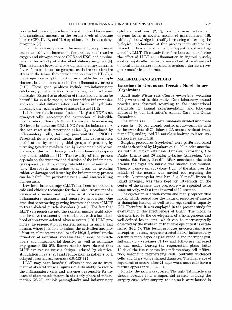
is reflected clinically by edema formation, local hematomaand significant increase in the serum levels of creatinekinase (CK), IL-1b, and IL-6 cytokines, and lactate dehy-drogenase [7].The inflammatory phase of the muscle injury process is
accompanied by an increase in the production of reactiveoxygen and nitrogen species (ROS and RNS) and a reduc-tion in the activity of antioxidant defense enzymes [8].This imbalance between pro-oxidants and antioxidants, infavor of pro-oxidants, can generate oxidative and nitrativestress in the tissue that contributes to activate NF-kB, apleiotropic transcription factor responsible for multiplechanges in gene expression in the inflammatory process[9,10]. These gene products include pro-inflammatorycytokines, growth factors, chemokines, and adhesionmolecules. Excessive production of these mediators can beharmful for muscle repair, as it intensifies inflammationand can inhibit differentiation and fusion of myoblasts,impairing the regeneration of muscle tissue [11,12].It is known that in muscle lesions, IL-1b and TNF-a act
synergistically increasing the expression of induciblenitric oxide synthase (iNOS) and consequently increasingNO levels in the tissue [13,14]. NO from the inflammatorysite can react with superoxide anion (O2
�) produced byinflammatory cells, forming peroxynitrite (ONOO�).Peroxynitrite is a potent oxidant and may cause proteinmodifications by oxidizing thiol groups of proteins, bynitrating tyrosine residues, and by increasing lipid perox-idation, nucleic acid damage, and mitochondrial respira-tory chain inhibition [15]. The severity of this processdepends on the intensity and duration of the inflammato-ry response [8]. Thus, during rehabilitation of muscle in-jury, therapeutic approaches that focus on avoidingoxidative damage and lessening the inflammatory processcan be helpful for promoting repair and reestablishinghomeostasis.Low-level laser therapy (LLLT) has been considered a
safe and efficient technique for the clinical treatment of avariety of diseases and injuries as it possesses anti-inflammatory, analgesic and reparative properties. Onearea that is attracting growing interest is the use of LLLTto treat skeletal muscle disorders [16–18]. The fact thatLLLT can penetrate into the skeletal muscle could allownon-invasive treatment to be carried out with a low likeli-hood of treatment-related adverse events [19]. LLLT pro-motes the regeneration of skeletal muscle in animal andhuman, where it is able to induce the activation and pro-liferation of quiescent satellite cells [20,21], stimulate theformation of myotubes, increase the number of musclefibers and mitochondrial density, as well as stimulateangiogenesis [22–25]. Recent studies have showed thatLLLT can reduce muscle fatigue induced by electricalstimulation in rats [26] and reduce pain in patients withdelayed onset muscle soreness (DOMS) [27].LLLT may have beneficial effects in the acute treat-
ment of skeletal muscle injuries due its ability to reducethe inflammatory cells and enzymes responsible for re-lease of chemotactic factors in the early phase of inflam-mation [28,29], inhibit prostaglandin and inflammatory
cytokine synthesis [2,17], and increase antioxidantenzyme levels in several models of inflammation [16].Although knowledge is steadily increasing concerning thebiological mechanisms of this process more studies areneeded to determine which signaling pathways are trig-gered by LLLT. This study therefore focused on exploringthe effect of LLLT on inflammation in injured muscle,evaluating its effect on oxidative and nitrative stress andon local inflammatory mediators produced during a cryo-genic muscle lesion in rats.
MATERIALS AND METHODS
Experimental Groups and Freezing Muscle Injury(Cryolesion)
Adult male Wistar rats (Rattus norvegicus) weighing300 g were used in this study. Good laboratory animalpractice was observed according to the internationalstandards for animal experimentation and followingapproval by our institution’s Animal Care and EthicsCommittee.
The animals (n ¼ 60) were randomly divided into threegroups (n ¼ 20 per group): control group—animals withno interventions (BC); injured TA muscle without treat-ment (IC); and injured TA muscle submitted to laser irra-diation treatment (IRI).
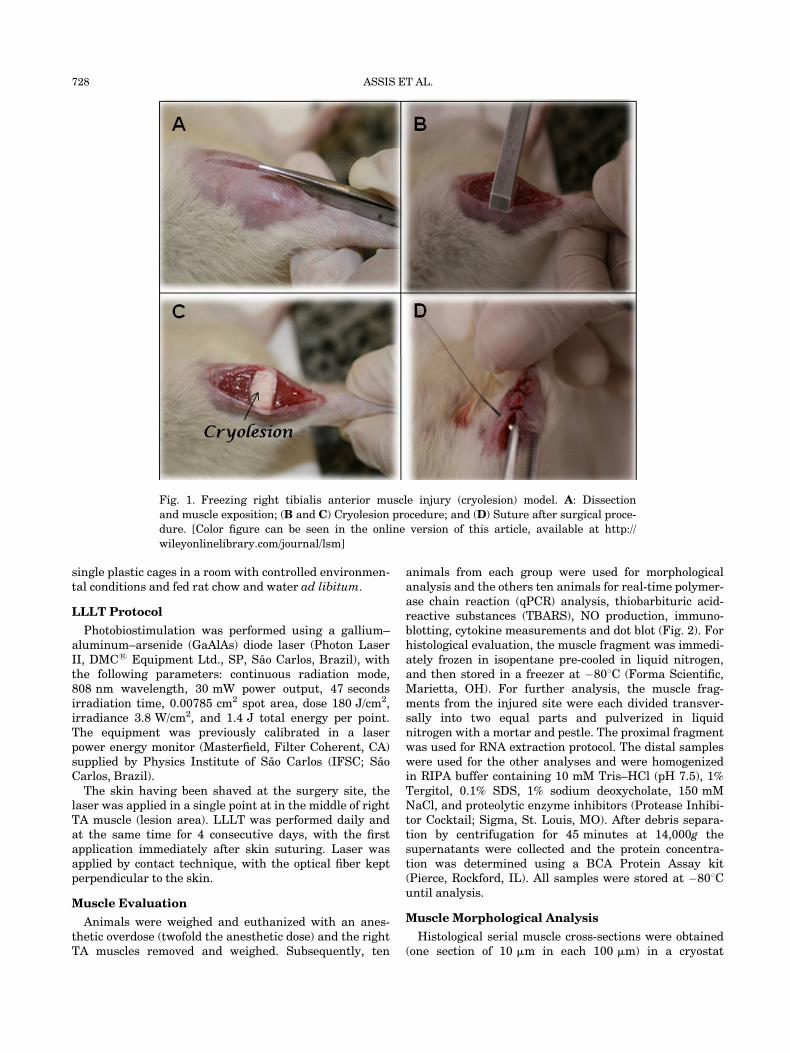
Surgical procedures (cryolesion) were performed basedon those described by Miyabara et al. [30], under anesthe-sia with 40 mg/kg ketamine (Dopalen; Vetbrands, SaoPaulo, Brazil) and 20 mg/kg xylazine (Anasedan; Vet-brands, Sao Paulo, Brazil). After anesthesia the skinaround the right TA muscle was shaved and cleaned.Then, a transversal cut (about 1 cm) of the skin over themiddle of the muscle was carried out, exposing themuscle. A rectangular iron bar (6 � 30 mm2), frozen inliquid nitrogen, was then kept for 10 seconds on thecenter of the muscle. The procedure was repeated twiceconsecutively, with a time interval of 30 seconds.
The cryolesion is a well-known and highly reproduciblemodel, which reproduces the natural response of muscleto damaging lesions, as well as its regeneration capacity[30]. Therefore, it was employed in the present study forevaluation of the effectiveness of LLLT. The model ischaracterized by the development of a homogeneous andwell-defined lesion area, which can be macroscopicallyobserved by the white color that develops once it is estab-lished (Fig. 1). This lesion produces myonecrosis, tissuedisruption, edema, hypercontracted fibers, inflammatorycell infiltration (especially neutrophils and macrophages).Inflammatory cytokines TNF-a and TGF-b are increasedin this model. During the regeneration phase (after10 days) the tissue shows less inflammatory cell infiltra-tion, basophilic regenerating cells, centrally nucleatedcells, and fibers with enlarged diameter. The final stage ofregeneration occurs after 21 days when most cells have amature appearance [17,30,31].
Finally, the skin was sutured. The right TA muscle waschosen because it is a superficial muscle, making thesurgery easy. After surgery, the animals were housed in
LLLT REDUCES INFLAMMATION AND OXIDATIVE STRESS 727
single plastic cages in a room with controlled environmen-tal conditions and fed rat chow and water ad libitum.
LLLT Protocol
Photobiostimulation was performed using a gallium–aluminum–arsenide (GaAlAs) diode laser (Photon LaserII, DMC1 Equipment Ltd., SP, Sao Carlos, Brazil), withthe following parameters: continuous radiation mode,808 nm wavelength, 30 mW power output, 47 secondsirradiation time, 0.00785 cm2 spot area, dose 180 J/cm2,irradiance 3.8 W/cm2, and 1.4 J total energy per point.The equipment was previously calibrated in a laserpower energy monitor (Masterfield, Filter Coherent, CA)supplied by Physics Institute of Sao Carlos (IFSC; SaoCarlos, Brazil).
The skin having been shaved at the surgery site, thelaser was applied in a single point at in the middle of rightTA muscle (lesion area). LLLT was performed daily andat the same time for 4 consecutive days, with the firstapplication immediately after skin suturing. Laser wasapplied by contact technique, with the optical fiber keptperpendicular to the skin.
Muscle Evaluation
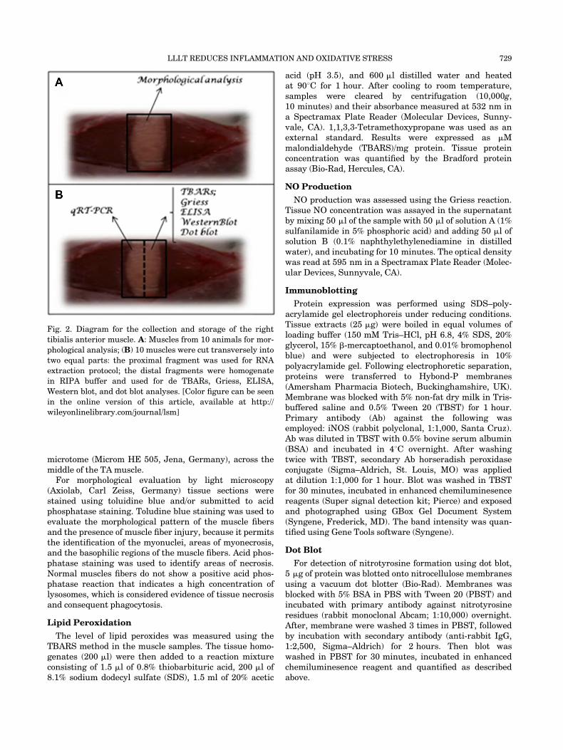
Animals were weighed and euthanized with an anes-thetic overdose (twofold the anesthetic dose) and the rightTA muscles removed and weighed. Subsequently, ten
animals from each group were used for morphologicalanalysis and the others ten animals for real-time polymer-ase chain reaction (qPCR) analysis, thiobarbituric acid-reactive substances (TBARS), NO production, immuno-blotting, cytokine measurements and dot blot (Fig. 2). Forhistological evaluation, the muscle fragment was immedi-ately frozen in isopentane pre-cooled in liquid nitrogen,and then stored in a freezer at �808C (Forma Scientific,Marietta, OH). For further analysis, the muscle frag-ments from the injured site were each divided transver-sally into two equal parts and pulverized in liquidnitrogen with a mortar and pestle. The proximal fragmentwas used for RNA extraction protocol. The distal sampleswere used for the other analyses and were homogenizedin RIPA buffer containing 10 mM Tris–HCl (pH 7.5), 1%Tergitol, 0.1% SDS, 1% sodium deoxycholate, 150 mMNaCl, and proteolytic enzyme inhibitors (Protease Inhibi-tor Cocktail; Sigma, St. Louis, MO). After debris separa-tion by centrifugation for 45 minutes at 14,000g thesupernatants were collected and the protein concentra-tion was determined using a BCA Protein Assay kit(Pierce, Rockford, IL). All samples were stored at �808Cuntil analysis.
Muscle Morphological Analysis
Histological serial muscle cross-sections were obtained(one section of 10 mm in each 100 mm) in a cryostat
Fig. 1. Freezing right tibialis anterior muscle injury (cryolesion) model. A: Dissection
and muscle exposition; (B and C) Cryolesion procedure; and (D) Suture after surgical proce-
dure. [Color figure can be seen in the online version of this article, available at http://
wileyonlinelibrary.com/journal/lsm]
728 ASSIS ET AL.
microtome (Microm HE 505, Jena, Germany), across themiddle of the TA muscle.For morphological evaluation by light microscopy
(Axiolab, Carl Zeiss, Germany) tissue sections werestained using toluidine blue and/or submitted to acidphosphatase staining. Toludine blue staining was used toevaluate the morphological pattern of the muscle fibersand the presence of muscle fiber injury, because it permitsthe identification of the myonuclei, areas of myonecrosis,and the basophilic regions of the muscle fibers. Acid phos-phatase staining was used to identify areas of necrosis.Normal muscles fibers do not show a positive acid phos-phatase reaction that indicates a high concentration oflysosomes, which is considered evidence of tissue necrosisand consequent phagocytosis.
Lipid Peroxidation
The level of lipid peroxides was measured using theTBARS method in the muscle samples. The tissue homo-genates (200 ml) were then added to a reaction mixtureconsisting of 1.5 ml of 0.8% thiobarbituric acid, 200 ml of8.1% sodium dodecyl sulfate (SDS), 1.5 ml of 20% acetic
acid (pH 3.5), and 600 ml distilled water and heatedat 908C for 1 hour. After cooling to room temperature,samples were cleared by centrifugation (10,000g,10 minutes) and their absorbance measured at 532 nm ina Spectramax Plate Reader (Molecular Devices, Sunny-vale, CA). 1,1,3,3-Tetramethoxypropane was used as anexternal standard. Results were expressed as mMmalondialdehyde (TBARS)/mg protein. Tissue proteinconcentration was quantified by the Bradford proteinassay (Bio-Rad, Hercules, CA).
NO Production
NO production was assessed using the Griess reaction.Tissue NO concentration was assayed in the supernatantby mixing 50 ml of the sample with 50 ml of solution A (1%sulfanilamide in 5% phosphoric acid) and adding 50 ml ofsolution B (0.1% naphthylethylenediamine in distilledwater), and incubating for 10 minutes. The optical densitywas read at 595 nm in a Spectramax Plate Reader (Molec-ular Devices, Sunnyvale, CA).
Immunoblotting
Protein expression was performed using SDS–poly-acrylamide gel electrophoreis under reducing conditions.Tissue extracts (25 mg) were boiled in equal volumes ofloading buffer (150 mM Tris–HCl, pH 6.8, 4% SDS, 20%glycerol, 15% b-mercaptoethanol, and 0.01% bromophenolblue) and were subjected to electrophoresis in 10%polyacrylamide gel. Following electrophoretic separation,proteins were transferred to Hybond-P membranes(Amersham Pharmacia Biotech, Buckinghamshire, UK).Membrane was blocked with 5% non-fat dry milk in Tris-buffered saline and 0.5% Tween 20 (TBST) for 1 hour.Primary antibody (Ab) against the following wasemployed: iNOS (rabbit polyclonal, 1:1,000, Santa Cruz).Ab was diluted in TBST with 0.5% bovine serum albumin(BSA) and incubated in 48C overnight. After washingtwice with TBST, secondary Ab horseradish peroxidaseconjugate (Sigma–Aldrich, St. Louis, MO) was appliedat dilution 1:1,000 for 1 hour. Blot was washed in TBSTfor 30 minutes, incubated in enhanced chemiluminesencereagents (Super signal detection kit; Pierce) and exposedand photographed using GBox Gel Document System(Syngene, Frederick, MD). The band intensity was quan-tified using Gene Tools software (Syngene).
Dot Blot
For detection of nitrotyrosine formation using dot blot,5 mg of protein was blotted onto nitrocellulose membranesusing a vacuum dot blotter (Bio-Rad). Membranes wasblocked with 5% BSA in PBS with Tween 20 (PBST) andincubated with primary antibody against nitrotyrosineresidues (rabbit monoclonal Abcam; 1:10,000) overnight.After, membrane were washed 3 times in PBST, followedby incubation with secondary antibody (anti-rabbit IgG,1:2,500, Sigma–Aldrich) for 2 hours. Then blot waswashed in PBST for 30 minutes, incubated in enhancedchemiluminesence reagent and quantified as describedabove.
Fig. 2. Diagram for the collection and storage of the right
tibialis anterior muscle. A: Muscles from 10 animals for mor-
phological analysis; (B) 10 muscles were cut transversely into
two equal parts: the proximal fragment was used for RNA
extraction protocol; the distal fragments were homogenate
in RIPA buffer and used for de TBARs, Griess, ELISA,
Western blot, and dot blot analyses. [Color figure can be seen
in the online version of this article, available at http://
wileyonlinelibrary.com/journal/lsm]
LLLT REDUCES INFLAMMATION AND OXIDATIVE STRESS 729
Cytokine Measurements
Tissue was prepared as described above and cytokinemeasurements was performed in the homogenatesby enzyme-linked immunosorbent assay (ELISA). Cyto-kine measurement kits were used according to the manu-facturer’s instructions (R&D Systems, Minneapolis, MN)using a Spectramax Plate Reader (Molecular Devices,Sunnyvale, CA). The results are expressed in pg per mg ofprotein.
Total RNA Isolation and Real Time PolymeraseChain Reaction
Tissues fragments were homogenized in 1 ml TRIzol1
reagent according to the manufacturer’s instructions(Invitrogen, Carlsbad, CA). RNA integrity was assess-ed by 260/268 nm ratio and on a 1% agarose gel electro-phoresis stained with ethidium bromide. Two nanogramsmRNA was used to real-time PCR. The amplificationwas performed in a thermal cycler (Applied BiosystemsStepOneTM) at 508C for 10 minutes, 958C for 5 minutes,and then 958C for 15 seconds followed by 608C for 30 sec-onds, and 728C for 30 seconds for 40 cycles. Real-timePCR was performed in a 15 ml reaction mixture contain-ing 7.5 ml 2� SYBR Green Reaction Mix (Invitrogen),0.3 ml each primer, 0.3 ml Super Script III RT/Platinum Taq Mix (10 pmol/ml), 0.15ml ROX ReferenceDye, and 5 ml sample in water. Quantification was per-formed by 2�DDCt method, using glyceraldehyde-3-phos-phate dehydrogenase (GAPDH) as housekeeping gene.
This gene was chosen after genome analysis (http://medgen.ugent. be/�jvdesomp/genorm/) of 5 housekeepinggenes. The following primers were used: SOD forward:GGCAAGCGGTGAACCAGTTG, reverse: TGCCCAGGT-CTCCAACATGC; COX-2 forward: TGTATGCTACCAT-CTGGCTTCGG, reverse: GTTTGGAACAGTCGCTCGTC-ATC; NF-kb forward: TCCGAGATAATGACAGCGTGTG,reverse: GGTCCATCCTGCCCATAATTG; and GAPDHforward: ATGATTCTACCCACGGCAAG, reverse: CTG-GAAGATGGTGATGGGTT.
Statistical Analysis
Data are expressed as the mean � standard error of themean (SEM). Shapiro–Wilk’s and Levene’s test wereapplied to evaluate the normality and homogeneity of theresults, respectively. Comparisons between experimentalgroups were performed by analysis of variance (one-wayANOVA), and the Tukey post-test used to compare indi-vidual groups. A P-value <0.05 was considered signifi-cant. All analyzes were performed using Sigma StatStatistical Software (v.3.1).
RESULTS
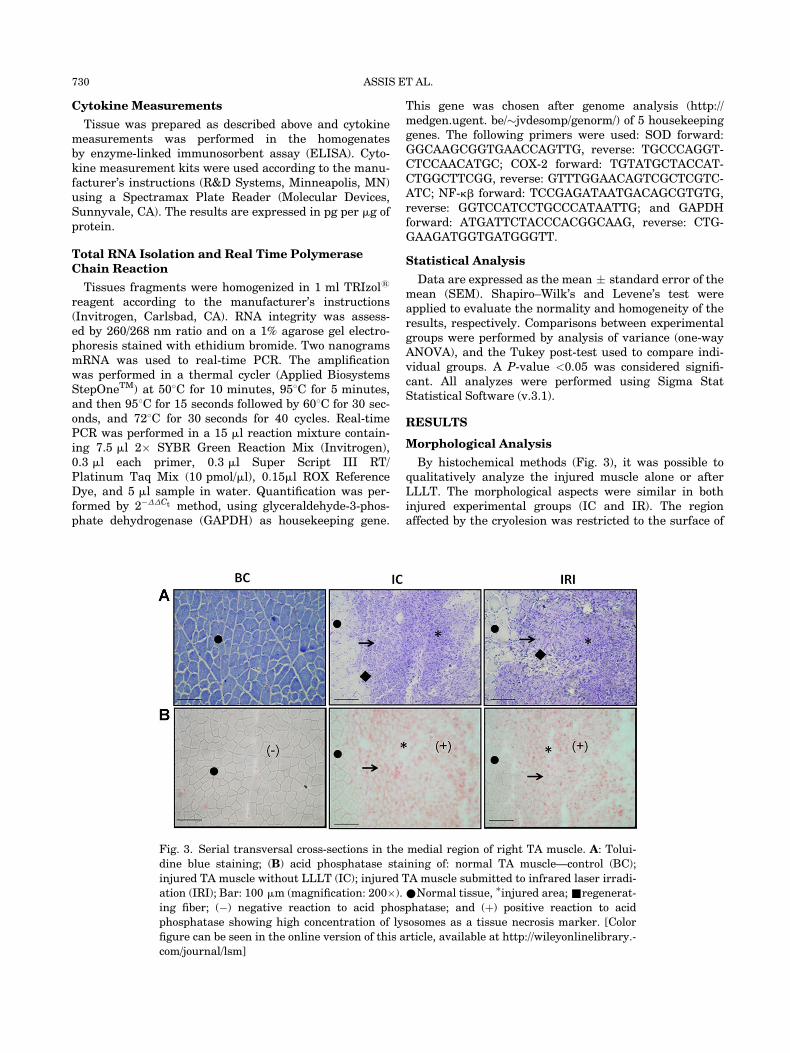
Morphological Analysis
By histochemical methods (Fig. 3), it was possible toqualitatively analyze the injured muscle alone or afterLLLT. The morphological aspects were similar in bothinjured experimental groups (IC and IR). The regionaffected by the cryolesion was restricted to the surface of
Fig. 3. Serial transversal cross-sections in the medial region of right TA muscle. A: Tolui-
dine blue staining; (B) acid phosphatase staining of: normal TA muscle—control (BC);
injured TA muscle without LLLT (IC); injured TA muscle submitted to infrared laser irradi-
ation (IRI); Bar: 100 mm (magnification: 200�).*Normal tissue, �injured area;®enerat-
ing fiber; (�) negative reaction to acid phosphatase; and (þ) positive reaction to acid
phosphatase showing high concentration of lysosomes as a tissue necrosis marker. [Color
figure can be seen in the online version of this article, available at http://wileyonlinelibrary.-
com/journal/lsm]
730 ASSIS ET AL.
the TA muscle and could be visualized in the TB-stainedcross-sections. The interface between the intact area andthe lesion site is clearly defined. The fibers from the intactregion are typical of healthy skeletal muscle tissue withpolygonal shape, peripheral nucleus, fascicular organiza-tion, and negative reaction to acid phosphatase staining.While in the lesion site next to the interface, recentformed fibers are characterized by centralized nuclei anda smaller transverse section. In the superficial region ofthe lesion, it was possible to identify a high concentrationof mononuclear cells (inflammatory and/or myogenic),absence of newly formed muscle fibers, abundant extra-cellular matrix and numerous lysosomes, indicating tis-sue necrosis and phagocytosis (positive acid phosphatasereaction). Moreover, the non-injured groups (BC) pre-sented no alteration in fiber morphology and had negativereaction for acid phosphatase activity.
Lipid Peroxidation
Lipid peroxidation, evaluated by TBARS, increasedin the injured muscle groups (IC and IRI), compared tocontrol group (BC; P < 0.01, Fig. 4). TBARS levels in theinjured muscle significantly decreased after LLLT (IRI,P < 0.05 vs. IC).
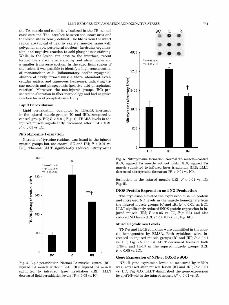
Nitrotyrosine Formation
Nitration of tyrosine residues was found in the injuredmuscle groups but not control (IC and IRI; P < 0.01 vs.BC), whereas LLLT significantly reduced nitrotyrosine
formation in the injured muscle (IRI; P < 0.01 vs. IC;Fig. 5).
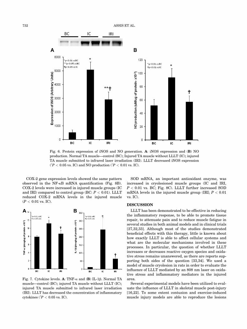
iNOS Protein Expression and NO Production
The cryolesion elevated the expression of iNOS proteinand increased NO levels in the muscle homogenate fromthe injured muscle groups IC and IRI (P < 0.01 vs. BC).LLLT significantly reduced iNOS protein expression in in-jured muscle (IRI; P < 0.05 vs. IC, Fig. 6A) and alsoreduced NO levels (IRI; P < 0.01 vs. IC, Fig. 6B).
Muscle Cytokines Levels
TNF-a and IL-1b cytokines were quantified in the mus-cle homogenates by ELISA. Both cytokines were in-creased in injured muscle groups (IC and IRI; P < 0.01vs. BC; Fig. 7A and B). LLLT decreased levels of bothTNF-a and IL-1b in the injured muscle groups (IRI;P < 0.05 vs. IC).
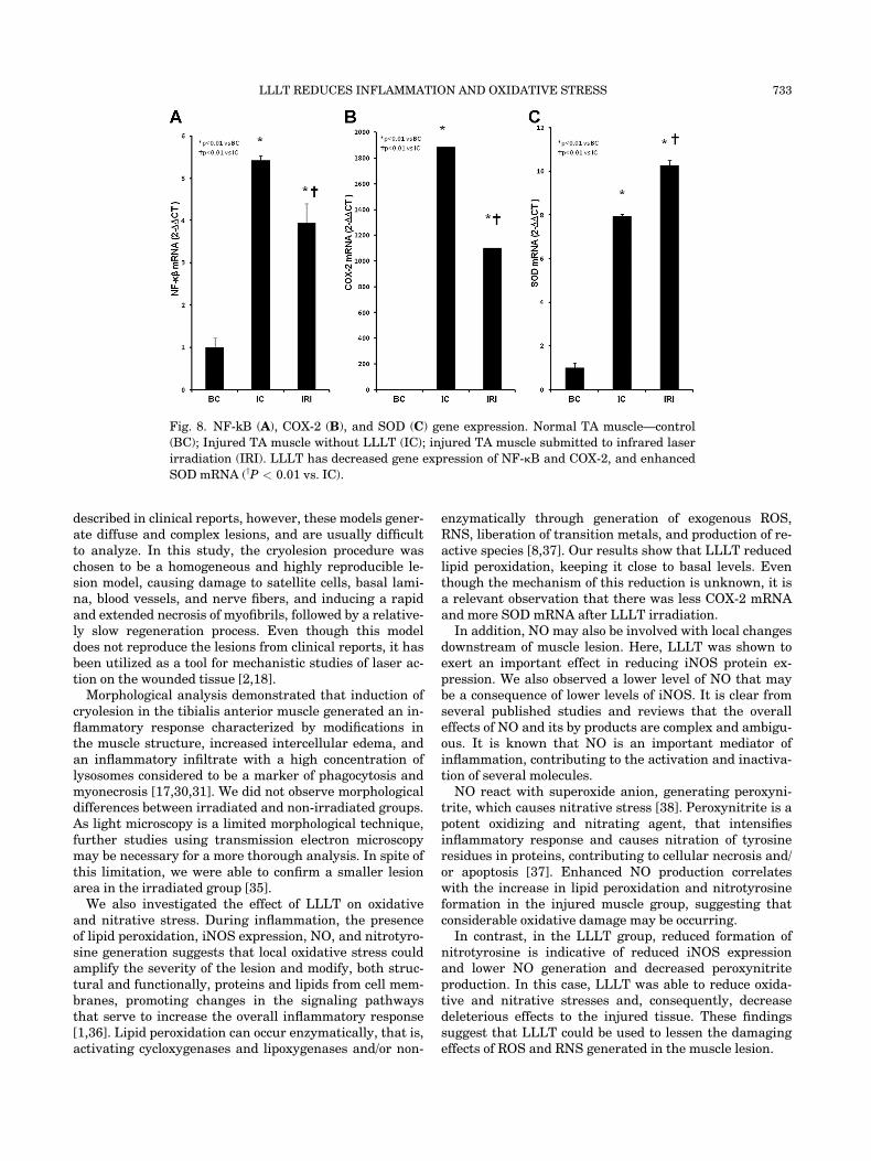
Gene Expression of NFk-b, COX-2 e SOD
NF-kB gene expression levels as measured by mRNAwas increased after muscle lesion (IC and IRI; P < 0.01vs. BC; Fig. 8A). LLLT diminished the gene expressionlevel of NF-kB in the injured muscle (P < 0.01 vs. IC).
Fig. 4. Lipid peroxidation. Normal TA muscle—control (BC);
injured TA muscle without LLLT (IC); injured TA muscle
submitted to infra-red laser irradiation (IRI); LLLT
decreased lipid peroxidation levels (yP < 0.05 vs. IC).
Fig. 5. Nitrotyrosine formation. Normal TA muscle—control
(BC); injured TA muscle without LLLT (IC); injured TA
muscle submitted to infrared laser irradiation (IRI); LLLT
decreased nitrotyrosine formation (yP < 0.01 vs. IC).
LLLT REDUCES INFLAMMATION AND OXIDATIVE STRESS 731
COX-2 gene expression levels showed the same patternobserved in the NF-kB mRNA quantification (Fig. 8B).COX-2 levels were increased in injured muscle groups (ICand IRI) compared to control group (BC: P < 0.01). LLLTreduced COX-2 mRNA levels in the injured muscle(P < 0.01 vs. IC).
SOD mRNA, an important antioxidant enzyme, wasincreased in cryolesioned muscle groups (IC and IRI,P < 0.01 vs. BC; Fig. 8C). LLLT further increased SODmRNA levels in the injured muscle group (IRI; P < 0.01vs. IC).
DISCUSSION
LLLT has been demonstrated to be effective in reducingthe inflammatory response, to be able to promote tissuerepair, to attenuate pain and to reduce muscle fatigue inseveral studies in both animal models and in clinical trials[27,32,33]. Although most of the studies demonstratedbeneficial effects with this therapy, little is known abouthow exactly LLLT is able to affect cellular systems andwhat are the molecular mechanisms involved in theseprocesses. In particular, the question of whether LLLTincreases or decreases reactive oxygen species and oxida-tive stress remains unanswered, as there are reports sup-porting both sides of the question [33,34]. We used amodel of muscle cryolesion in rats in order to evaluate theinfluence of LLLT mediated by an 808 nm laser on oxida-tive stress and inflammatory mediators in the injuredarea.Several experimental models have been utilized to eval-
uate the influence of LLLT in skeletal muscle post-injury[16,22]. To some extent contusion and exercise-inducedmuscle injury models are able to reproduce the lesions
Fig. 6. Protein expression of iNOS and NO generation. A: iNOS expression and (B) NO
production. Normal TA muscle—control (BC); Injured TA muscle without LLLT (IC); injured
TA muscle submitted to infrared laser irradiation (IRI). LLLT decreased iNOS expression
(yP < 0.05 vs. IC) and NO production (yP < 0.01 vs. IC).
Fig. 7. Cytokine levels. A: TNF-a and (B) IL-1b. Normal TA
muscle—control (BC); injured TA muscle without LLLT (IC);
injured TA muscle submitted to infrared laser irradiation
(IRI). LLLT has decreased the concentration of inflammatory
cytokines (yP < 0.05 vs. IC).
732 ASSIS ET AL.
described in clinical reports, however, these models gener-ate diffuse and complex lesions, and are usually difficultto analyze. In this study, the cryolesion procedure waschosen to be a homogeneous and highly reproducible le-sion model, causing damage to satellite cells, basal lami-na, blood vessels, and nerve fibers, and inducing a rapidand extended necrosis of myofibrils, followed by a relative-ly slow regeneration process. Even though this modeldoes not reproduce the lesions from clinical reports, it hasbeen utilized as a tool for mechanistic studies of laser ac-tion on the wounded tissue [2,18].Morphological analysis demonstrated that induction of
cryolesion in the tibialis anterior muscle generated an in-flammatory response characterized by modifications inthe muscle structure, increased intercellular edema, andan inflammatory infiltrate with a high concentration oflysosomes considered to be a marker of phagocytosis andmyonecrosis [17,30,31]. We did not observe morphologicaldifferences between irradiated and non-irradiated groups.As light microscopy is a limited morphological technique,further studies using transmission electron microscopymay be necessary for a more thorough analysis. In spite ofthis limitation, we were able to confirm a smaller lesionarea in the irradiated group [35].We also investigated the effect of LLLT on oxidative
and nitrative stress. During inflammation, the presenceof lipid peroxidation, iNOS expression, NO, and nitrotyro-sine generation suggests that local oxidative stress couldamplify the severity of the lesion and modify, both struc-tural and functionally, proteins and lipids from cell mem-branes, promoting changes in the signaling pathwaysthat serve to increase the overall inflammatory response[1,36]. Lipid peroxidation can occur enzymatically, that is,activating cycloxygenases and lipoxygenases and/or non-
enzymatically through generation of exogenous ROS,RNS, liberation of transition metals, and production of re-active species [8,37]. Our results show that LLLT reducedlipid peroxidation, keeping it close to basal levels. Eventhough the mechanism of this reduction is unknown, it isa relevant observation that there was less COX-2 mRNAand more SOD mRNA after LLLT irradiation.
In addition, NO may also be involved with local changesdownstream of muscle lesion. Here, LLLT was shown toexert an important effect in reducing iNOS protein ex-pression. We also observed a lower level of NO that maybe a consequence of lower levels of iNOS. It is clear fromseveral published studies and reviews that the overalleffects of NO and its by products are complex and ambigu-ous. It is known that NO is an important mediator ofinflammation, contributing to the activation and inactiva-tion of several molecules.
NO react with superoxide anion, generating peroxyni-trite, which causes nitrative stress [38]. Peroxynitrite is apotent oxidizing and nitrating agent, that intensifiesinflammatory response and causes nitration of tyrosineresidues in proteins, contributing to cellular necrosis and/or apoptosis [37]. Enhanced NO production correlateswith the increase in lipid peroxidation and nitrotyrosineformation in the injured muscle group, suggesting thatconsiderable oxidative damage may be occurring.
In contrast, in the LLLT group, reduced formation ofnitrotyrosine is indicative of reduced iNOS expressionand lower NO generation and decreased peroxynitriteproduction. In this case, LLLT was able to reduce oxida-tive and nitrative stresses and, consequently, decreasedeleterious effects to the injured tissue. These findingssuggest that LLLT could be used to lessen the damagingeffects of ROS and RNS generated in the muscle lesion.
Fig. 8. NF-kB (A), COX-2 (B), and SOD (C) gene expression. Normal TA muscle—control
(BC); Injured TA muscle without LLLT (IC); injured TA muscle submitted to infrared laser
irradiation (IRI). LLLT has decreased gene expression of NF-kB and COX-2, and enhanced
SOD mRNA (yP < 0.01 vs. IC).
LLLT REDUCES INFLAMMATION AND OXIDATIVE STRESS 733
Regarding the antioxidant effects of irradiation, SODmRNA increased after LLLT, and this increase may con-tribute to preventing the additional formation of superox-ide that leads to peroxynitrite and other reactive speciesproduction [16,29]. Therefore, results shown here suggestthat LLLT had a positive biological effect in modulatingthe redox balance, reducing nitrative and oxidative stressand lessening oxidative damage that would delay the re-covery of the injured tissue. Similar results were obtainedby others: Rizzi et al. [39], described that LLLT inhibitedoxidative stress, iNOS expression and the activation ofNF-kb in a model of muscle trauma. Studies utilizing dif-ferent experimental models have suggested that LLLT isable to induce SOD expression, decreasing the availablesuperoxide anion and, as a result, reduce peroxynitriteproduction [16,40].
The inflammatory response involves multiple mediatorsand LLLT could influence overall NF-kB activation dur-ing this process. NF-kB is a transcription factor rapidlyactivated in response to oxidative stress and which partic-ipates in the induction of many inflammation-relatedgenes such as COX-2, iNOS, and pro-inflammatory cyto-kines [9,10]. We found trauma was able to elicit NF-kBmRNA activation, leading to production of inflammatorymediators such as COX-2, iNOS, TNF-a, and IL-1b.LLLT, however, decreased NF-kB gene expression, sug-gesting that this effect could be responsible for the re-duced amount of pro-inflammatory mediators in thismodel. As discussed above, LLLT mediated anti-inflam-matory effects could be a consequence of NF-kB reductionor inactivation [41–43].
In summary, our results indicate that near-infraredlaser (808 nm) at the dose selected was able to attenuateoxidative and nitrative stress in the skeletal muscle,decrease NO production, lipid peroxidation, nitrotyrosineformation, and iNOS and could increase SOD expressionin an experimental animal model. Additionally, TNF-a,IL1-b, and COX-2 were reduced in the lesion area, leadingto an anti-inflammatory effect. Accordingly, these experi-mental data may contribute to further understandingbeneficial effects of LLLT on muscle inflammation.
ACKNOWLEDGMENTS
We acknowledge CAPES, CNPQ and FAPESP for finan-cial support. M.R. Hamblin was supported by NIH (grantR01AI050875). Emergency Medicine Division (LIM 51),Faculdade de Medicina da Universidade de Sao Paulo toprovide technical support in biochemical and molecularbiology analyses and NUPEN (Nucleo de Pesquisa eEnsino em Fototerapia nas Ciencias da Saude) for sup-porting and calibrating the laser equipment.
REFERENCES
1. Filippin LI, Cuevas MJ, Lima E, Marroni NP, Gonzalez-Gallego J, Xavier RM. The role of nitric oxide during healingof trauma to the skeletal muscle. Inflamm Res 2011;60(4):347–356.
2. Renno AC, Toma RL, Feitosa SM, Fernandes K, Bossini PS,de Oliveira P, Parizotto N, Ribeiro DA. Comparative effects
of low-intensity pulsed ultrasound and low-level laser thera-py on injured skeletal muscle. Photomed Laser Surg 2011;29(1):5–10.
3. Li Y, Cummins J, Huard J. Muscle injury and repair. CurrOpin Orthop 2001;12:6.
4. Tidball JG. Inflammatory processes in muscle injury and re-pair. Am J Physiol Regul Integr Comp Physiol 2005;288(2):R345–R353.
5. Huard J. Li Y. Fu FH. Muscle injuries and repair: Currenttrends in research. J Bone Joint Surg Am 2002;84-A(5):822–832.
6. Jarvinen TA, Jarvinen TL, Kaariainen M, Kalimo H, Jarvi-nen M. Muscle injuries: Biology and treatment. Am J SportsMed 2005;33(5):745–764.
7. Buford TW, Cooke MB, Shelmadine BD, Hudson GM, ReddL, Willoughby DS. Effects of eccentric treadmill exercise oninflammatory gene expression in human skeletal muscle.Appl Physiol Nutr Metab 2009;34(4):745–753.
8. Droge W. Free radicals in the physiological control of cellfunction. Physiol Rev 2002;82(1):47–95.
9. Adams V, Nehrhoff B, Spate U, Linke A, Schulze PC, BaurA, Gielen S, Hambrecht R, Schuler G. Induction of iNOS ex-pression in skeletal muscle by IL-1beta and NFkappaB acti-vation: An in vitro and in vivo study. Cardiovasc Res 2002;54(1):95–104.
10. van den Berg R, Haenen GR, van den Berg H, Bast A.Transcription factor NF-kappaB as a potential biomarker foroxidative stress. Br J Nutr 2001;86(Suppl 1)S121–S127.
11. Barnes PJ, Karin M. Nuclear factor-kappaB: A pivotal tran-scription factor in chronic inflammatory diseases. N Engl JMed 1997;336(15):1066–1071.
12. Langen RC, Van Der Velden JL, Schols AM, KeldersMC, Wouters EF, Janssen-Heininger YM. Tumor necrosisfactor-alpha inhibits myogenic differentiation through MyoDprotein destabilization. FASEB J 2004;18(2):227–237.
13. Kaminski HJ, Andrade FH. Nitric oxide: Biologic effects onmuscle and role in muscle diseases. Neuromuscul Disord2001;11(6–7):517–524.
14. Filippin LI, Moreira AJ, Marroni NP, Xavier RM. Nitric ox-ide and repair of skeletal muscle injury. Nitric Oxide2009;21(3–4):157–163.
15. El-Remessy AB, Al-Shabrawey M, Platt DH, Bartoli M,Behzadian MA, Ghaly N, Tsai N, Motamed K, Caldwell RB.Peroxynitrite mediates VEGF’s angiogenic signal and func-tion via a nitration-independent mechanism in endothelialcells. FASEB J 2007;21(10):2528–2539.
16. Liu XG, Zhou YJ, Liu TC, Yuan JQ. Effects of low-level laserirradiation on rat skeletal muscle injury after eccentric exer-cise. Photomed Laser Surg 2009;27(6):863–869.
17. Mesquita-Ferrari RA, Martins MD, Silva JA, Jr., da SilvaTD, Piovesan RF, Pavesi VC, Bussadori SK, Fernandes KP.Effects of low-level laser therapy on expression of TNF-alphaand TGF-beta in skeletal muscle during the repair process.Lasers Med Sci 2011;26(3):335–340.
18. Ramos L, Leal Junior EC, Pallotta RC, Frigo L, Marcos RL,de Carvalho MH, Bjordal JM, Lopes-Martins RA. Infrared(810 nm) low-level laser therapy in experimental model ofstrain-induced skeletal muscle injury in rats: Effects onfunctional outcomes. Photochem Photobiol 2012;88(1):154–160.
19. Hamblin MR. Introduction to experimental and clinical stud-ies using low-level laser (light) therapy (LLLT). Lasers SurgMed 2010;42(6):447–449.
20. Ben-Dov N, Shefer G, Irintchev A, Wernig A, Oron U, HalevyO. Low-energy laser irradiation affects satellite cell prolifer-ation and differentiation in vitro. Biochim Biophys Acta1999;1448(3):372–380.
21. Shefer G, Oron U, Irintchev A, Wernig A, Halevy O. Skeletalmuscle cell activation by low-energy laser irradiation: A rolefor the MAPK/ERK pathway. J Cell Physiol 2001;187(1):73–80.
22. Iyomasa DM, Garavelo I, Iyomasa MM, Watanabe IS, IssaJP. Ultrastructural analysis of the low level laser therapyeffects on the lesioned anterior tibial muscle in the gerbil.Micron 2009;40(4):413–418.
734 ASSIS ET AL.
23. Amaral AC, Parizotto NA, Salvini TF. Dose-dependency oflow-energy HeNe laser effect in regeneration of skeletalmuscle in mice. Lasers Med Sci 2001;16(1):44–51.
24. Nakano J, Kataoka H, Sakamoto J, Origuchi T, Okita M,Yoshimura T. Low-level laser irradiation promotes the recov-ery of atrophied gastrocnemius skeletal muscle in rats. ExpPhysiol 2009;94(9):1005–1015.
25. Silveira PC, Silva LA, Fraga DB, Freitas TP, Streck EL,Pinho R. Evaluation of mitochondrial respiratory chainactivity in muscle healing by low-level laser therapy.J Photochem Photobiol B 2009;95(2):89–92.
26. Lopes-Martins RA, Marcos RL, Leonardo PS, Prianti AC,Jr., Muscara MN, Aimbire F, Frigo L, Iversen VV, BjordalJM. Effect of low-level laser (Ga–Al–As 655 nm) on skeletalmuscle fatigue induced by electrical stimulation in rats.J Appl Physiol 2006;101(1):283–288.
27. Douris P, Southard V, Ferrigi R, Grauer J, Katz D, Nasci-mento C, Podbielski P. Effect of phototherapy on delayed on-set muscle soreness. Photomed Laser Surg 2006;24(3):377–382.
28. Cressoni MD, Dib Giusti HH, Casarotto RA, Anaruma CA.The effects of a 785-nm AlGaInP laser on the regeneration ofrat anterior tibialis muscle after surgically-induced injury.Photomed Laser Surg 2008;26(5):461–466.
29. Dourado DM, Favero S, Baranauskas V, da Cruz-HoflingMA. Effects of the Ga–As laser irradiation on myonecrosiscaused by Bothrops moojeni snake venom. Lasers Surg Med2003;33(5):352–357.
30. Miyabara EH, Martin JL, Griffin TM, Moriscot AS, MestrilR. Overexpression of inducible 70-kDa heat shock proteinin mouse attenuates skeletal muscle damage induced by cry-olesioning. Am J Physiol Cell Physiol 2006;290(4):C1128–C1138.
31. Baptista J, Martins MD, Pavesi VC, Bussadori SK,Fernandes KP, Pinto Junior Ddos S, Ferrari RA. Influence oflaser photobiomodulation on collagen IV during skeletalmuscle tissue remodeling after injury in rats. PhotomedLaser Surg 2011;29(1):11–17.
32. Leal Junior EC, Lopes-Martins RA, Frigo L, De Marchi T,Rossi RP, de Godoi V, Tomazoni SS, Silva DP, Basso M,Filho PL, de Valls Corsetti F, Iversen VV, Bjordal JM.Effects of low-level laser therapy (LLLT) in thedevelopment of exercise-induced skeletal muscle fatigue andchanges in biochemical markers related to postexercise re-covery. J Orthop Sports Phys Ther 2010;40(8):524–532.
33. Servetto N, Cremonezzi D, Simes JC, Moya M, Soriano F,Palma JA, Campana VR. Evaluation of inflammatory bio-markers associated with oxidative stress and histologicalassessment of low-level laser therapy in experimental myop-athy. Lasers Surg Med 2010;42(6):577–583.
34. Chen AC, Arany PR, Huang YY, Tomkinson EM, SharmaSK, Kharkwal GB, Saleem T, Mooney D, Yull FE, BlackwellTS, Hamblin MR. Low-level laser therapy activates NF-kBvia generation of reactive oxygen species in mouse embryon-ic fibroblasts. PloS ONE 2011;6(7):e22453.
35. Assis L, Moretti AI, Abrahao TB, de Souza HP, Hamblin MR,Parizotto NA. Low-level laser therapy (808 nm) contributesto muscle regeneration and prevents fibrosis in rat tibialisanterior muscle after cryolesion. Lasers Med Sci 2012.
36. Shi X, Garry DJ. Muscle stem cells in development, regener-ation, and disease. Genes Dev 2006;20(13):1692–1708.
37. Beckman JS, Koppenol WH. Nitric oxide, superoxide, andperoxynitrite: The good, the bad, and ugly. Am J Physiol1996;271(5 Pt 1):C1424–C1437.
38. Kroncke KD, Fehsel K, Suschek C, Kolb-Bachofen V. Induc-ible nitric oxide synthase-derived nitric oxide in gene regula-tion, cell death and cell survival. Int Immunopharmacol2001;1(8):1407–1420.
39. Rizzi CF, Mauriz JL, Freitas Correa DS, Moreira AJ, ZettlerCG, Filippin LI, Marroni NP, Gonzalez-Gallego J. Effects oflow-level laser therapy (LLLT) on the nuclear factor (NF)-kappaB signaling pathway in traumatized muscle. LasersSurg Med 2006;38(7):704–713.
40. Kim YG, Pak SC, Lee SR. Hairless mouse epidermal antiox-idants and lipid peroxidation assessed by He–Ne laser.Lasers Surg Med 2000;27(5):420–426.
41. Pallotta RC, Bjordal JM, Frigo L, Leal Junior EC, TeixeiraS, Marcos RL, Ramos L, de Moura Messias F, Lopes-MartinsRA. Infrared (810-nm) low-level laser therapy on rat experi-mental knee inflammation. Lasers Med Sci 2012;27(1):71–78.
42. Bjordal JM, Lopes-Martins RAB, Joensen J, Iversen VV. Theanti-inflammatory mechanism of low level laser therapy andits relevance for clinical use in physiotherapy. Phys TherRev 2010;15(4):7.
43. Fillipin LI, Mauriz JL, Vedovelli K, Moreira AJ, Zettler CG,Lech O, Marroni NP, Gonzalez-Gallego J. Low-level lasertherapy (LLLT) prevents oxidative stress and reduces fibro-sis in rat traumatized Achilles tendon. Lasers Surg Med2005;37(4):293–300.
LLLT REDUCES INFLAMMATION AND OXIDATIVE STRESS 735
Top Related
Copyright © 2022 FDOKUMEN

